Author Archives: author

First Case Report of Pancreatitis in Lyme disease

DOI: 10.31038/IMROJ.2019423

Short Abstract

We report a case of Lyme disease, revealed by pancreatic damage in a 49-year-old man without any medical history. The Lyme disease was revealed by repeated abdominal pain for 4 weeks, a skin lesion of quadricipital region, biological and radiological results showing pancreatic abnormalities.

Case Report

A 49-year-old man, non-alcoholic forest worker, with no past medical history, consulted to the Emergency Department for fever and persistent abdominal pain for a week. The biological results including, C – reactive protein (CRP), lipase, hepatic assessment were normal as well as contrast-enhanced abdominal Computed Tomography (CT). On the day after, the evolution was favorable under symptomatic treatment including nefopam and paracetamol and the patient was discharged from the hospital. One week later, the patient was admitted to the Emergency Department with an identical symptomatology. A posterior quadricipital peeling skin lesion, appeared two weeks earlier according to the patient, was observed (Figure 1a.) A gastroscopy, a colonoscopy, other abdominal CT and biological tests were performed. An inflammation biomarker elevation was observed (CRP: 180 mg/L and hyperleukocytosis: 13.3 G/L) without other biological abnormalities (lipase: 48 UI/L, ALAT: 48 UI/L). The endoscopic examinations and abdominal CT were normal. The patient was discharged from the hospital without any treatment. Half a month later, the patient was admitted to the Emergency Department for the third time and recurrence of the abdominal pain. The clinical examination found a hemodynamic stability, an abdominal pain of the left hypochondrium associated with a cutaneous ulcerative and non-progressive skin lesion in the same region as previously mentioned (Figure 1b.). The biological assessment found a very mild inflammatory syndrome (CRP 86 mg/L, Procalcitonin < 0.2 ng/mL, leukocytes 9.5 G/L), a high lipase level at 1714 IU/L without hepatocellular abnormalities. The third abdominal CT revealed an aspect of pancreatic necrosis with a pseudocyst (6 cm) at the tail of the pancreas, in contact with the splenic hile and the posterior wall of the stomach (Figure 1c.). The patient was hospitalized in Intensive Care Department with the diagnosis of pancreatitis.

On admission, the work-ups looking for the usual causes of pancreatitis (alcohol, gallstones, medications induced, hypercalcemia, traumatic, chemical exposures, hereditary diseases, infections) were negative. Regarding the skin patient’s lesion and anamnesis, the diagnosis of Lyme disease was evoked. His Lyme serology was strongly IgM positive and confirmed by Western Blot. He was treated with ceftriaxone associated with effective analgesic therapy. The clinical and biological course was uneventful and the patient was discharged from the hospital after 3 weeks. The relationship between Lyme disease and acute pancreatitis was strongly suspected.

Discussion

Lyme disease is an endemic zoonosis, transmitted to humans by a tick bite causing a multisystemic impairment due to a Gram-negative bacillus, Borrellia burgdorferi [1]. The disease schematically includes two phases and a polymorphism in clinical manifestations: a primary phase with chronical migrans erythema and articular signs (80% of cases), a secondary phase of heterogeneous and lymphatic dissemination, inaugurated by flu-like symptoms and associating neurological, cardiac or articular signs that could become chronic [2]. Each of these attacks could be inaugural or/and isolated [3]. Concerning the anamnesis, only 30% of patients remember a tick bite [4].

The heterogeneity of presentation in Lyme disease includes the serodiagnosis as a central investigation for confirmation [5]. Hepatic impairment due to Lyme disease, including hepatitis and hepatomegaly, is inconsistent, commonly found in early stage but often asymptomatic and with plasmatic manifestations [6]. A moderate hypertransaminasemia (2 to 3 N) could be noted, predominating on the ALAT. This hepatic biologic involvement is present in 27 to 66% of cases [7]. This can be explained by a systemic, lymphatic migration of the incriminated bacteria and a secondary hepatic sequestration [8]. To our knowledge, this physiopathological evolution to explain liver disorders has never been described for pancreas but is probably similar.

IMROJ 19 - 139_N Pichon_F1

Figure a Skin lesion, 2 weeks after supposed tick bite. b Skin lesion, 4 weeks after supposed tick bite. c Abdominal CT scan showing pancreas (arrow) and the pseudocyst at the tail (head arrows).

Regarding the treatment of Lyme disease, the cycline are recommended for the uncomplicated forms. An antibiotic treatment with cephalosporins could be considered for cardiac, neurological or complicated cases [2]. The evolution is favorable in 85% of patients, including hepatic acute injuries [9].

In our case, the skin lesion associated with a supposed tick bite, the anamnesis, the absence of other cause of pancreatitis, the favorable evolution under antibiotic treatment and especially the strong positivity of the serology are in favor of a Borrelia burgdoferi infection.

Conclusion

The authors report the first case of pancreatitis revealing a Lyme disease. Clinical, biological and evolutionary findings support the responsibility of Lyme disease in the pathogenesis of our pancreatitis case.

Acknowledgment

The authors thank Dr Sommabere from the department of bacteriology for his contribution on the project.

References

  1. Steere AC, Grodzicki RL, Kornblatt AN, Barbour AG, Burgdorfer W, et al. (1983) The spirochetal etiology of Lyme disease. N Engl J Med 308: 733–740.
  2. Sanchez JL (2015) Clinical Manifestations and Treatment of Lyme Disease. Clin Lab Med 35: 765–778.
  3. Sanchez E, Vannier E, Wormser GP, Hu LT (2016) Diagnosis, Treatment, and Prevention of Lyme Disease, Human Granulocytic Anaplasmosis, and Babesiosis: A Review. JAMA 315: 1767–1777.
  4. Cameron DJ, Johnson LB, Maloney EL (2014) Evidence assessments and guideline recommendations in Lyme disease: the clinical management of known tick bites, erythema migrans rashes and persistent disease. Expert Rev Anti Infect Ther 12: 1103–1135.
  5. Kim B (2017) Western Blot Techniques. Methods Mol Biol Clifton NJ 1606: 133–139.
  6. Goellner MH, Agger WA, Burgess JH, Duray PH (1988) Hepatitis due to recurrent Lyme disease. Ann Intern Med 108: 707–708.
  7. Steere AC, Bartenhagen NH, Craft JE, Hutchinson GJ, Newman JH, et al. (1983) The early clinical manifestations of Lyme disease. Ann Intern Med 99: 76–82.
  8. Imai DM, Samuels DS, Feng S, Hodzic E, Olsen K, et al. (2013) The early dissemination defect attributed to disruption of decorin-binding proteins is abolished in chronic murine Lyme borreliosis. Infect Immun 81: 1663–1673.
  9. Horowitz HW, Dworkin B, Forseter G, Nadelman RB, Connolly C, et al. (1996) Liver function in early Lyme disease. Hepatol Baltim Md 23: 1412–1417.

Ultrasonographic aspect of Pulmonary Emboli

DOI: 10.31038/IMCI.2019221


The availability of thoracic ultrasonography (TUS) for the diagnosis of pulmonary infarct was demonstrated more than 15 years ago [1]. With so, the typical TUS aspect of the embolic lesion remain largely unknown by physicians that otherwise use this tool in an every-day basis. More than two triangular or rounded hypo-echogenic areas bigger than 5 mm, occasionally accompanied by small pleural effusion are suggested TUS criteria for pulmonary infarct diagnosis [2]. Computed tomography angiography (CTA) is the gold standard for the diagnosis [3].

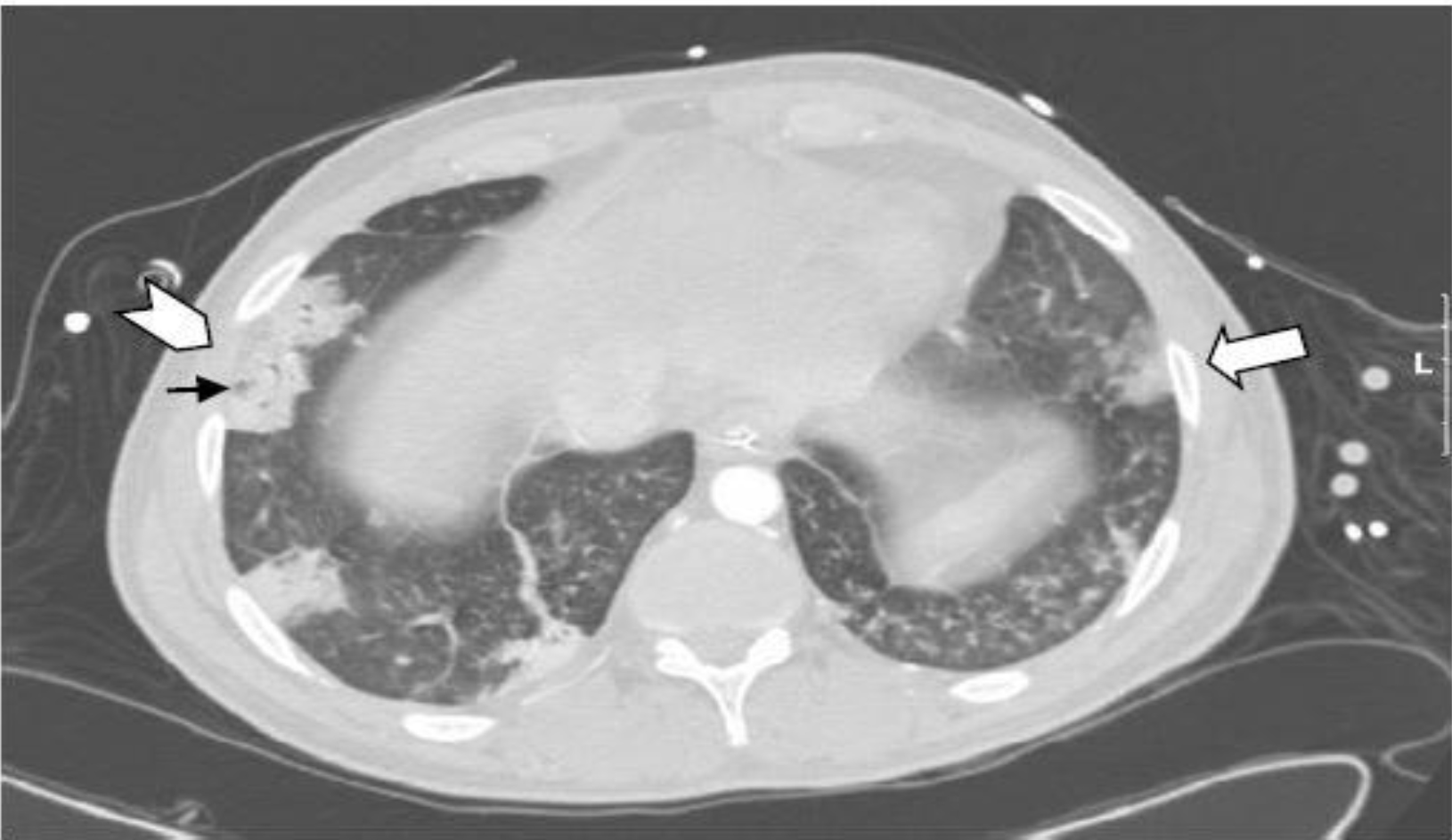
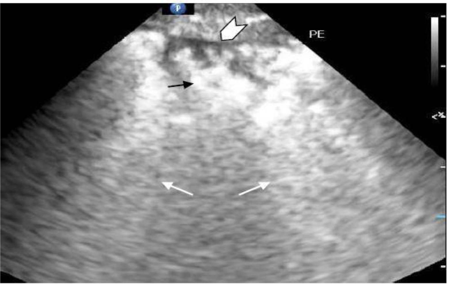
The ultrasonographic aspect of pulmonary embolism is shown here correlating to the accompanied computed tomography of the same lesion.

A 28-year-old man, affected by Smith-Lemli-Opitz syndrome, was admitted to the intensive care unit because of a severe respiratory failure and hemodynamic shock requiring mechanical ventilation and vasopressors. Two weeks before admission, ambulatory antibiotic treatment was started for suspected pneumonia. The CTA study (Panel A) shows multiple pulmonary infarcts on both lungs (arrows). TUS examination (Panel B) of the right thorax shows the corresponding sonogram image for the tomography finding (arrowhead).

Multiple sub-pleural lesion occupied the entire space between two ribs. The lung parenchyma behind the lesion is hyperechoic and shows typical B lines of interstitial lung (small arrows). The central hyperechogenic rounded image at the ultrasonography matches the hypodense rounded bronchi (black arrows) at the CTA.  The limit between the hypo-echogenic area and the surrounding lungs is shattered. A deeper bronchial reflex with air-flow Doppler signal can be seen at times when the lung tissue becomes consolidated [4].

Thrombotic occlusion of the right peroneal vein and posterior tibial vein found at duplex ultrasonography was the source of the embolism.

The patient condition responded to heparin treatment.

IMCI 19 - 113_Daniel J. Jakobson_F1

Panel A. Computed Tomography image.

IMCI 19 - 113_Daniel J. Jakobson_F2

Panel B. Ultrasound image.

References

  1. Mathis G, Wolfgang B, Reissig A, et al. (2005) Thoracic ultrasound for diagnosing pulmonary embolism: a prospective multicenter study of 352 patients. Chest  128: 1531–1538.
  2. Reissing A, Heyne JP, Kroegel C (2001) Sonography of lung and pleura in pulmonary embolism: sonomorphologic characterization and comparison with spiral CT scanning. Chest 120: 1977–1983.
  3. RathbunSW, Raskob GE, Whitsett L. (2000) Sensitivity and specificity of helical computed tomography in the diagnosis of pulmonary embolism: a systematic review. Ann Intern Med 132: 227–232.
  4. Blancas R, Ballesteros-Ortega D, Martinez-Gonzales O (2018) Central bronchial reflex finding in pulmonary infarct. Med Intensiva 42: 69.

Novel Pathology-Related Hub Genes in Focal Segmental Glomerulosclerosis

DOI: 10.31038/JCRM.2019252

Abstract

Objectives: Focal Segmental Glomerulosclerosis (FSGS) is a progressive glomerular disease. The pathogenesis of this disease, however, remains unclear. Here, we attempted to identify key candidate genes in FSGS through stringent bioinformatic analysis.

Methods: We systematically searched the Gene Expression Omnibus database for gene expression microarrays derived from human glomeruli tissues with FSGS. First, we identified differentially expressed genes (DEGs) by using the Limma package in R. Then, we subjected these DEGs to Gene Ontology (GO) analysis for further analysis. Finally, we constructed Protein–Protein Interaction networks (PPI) through four different methods and performed intersection analysis to further refine our results.

Results: A total of 627 DEGs were identified between the FSGS and control groups, among which 534 were up-regulated and 93 were down-regulated. GO analysis revealed that the DEGs were enriched in mRNA processing, cell adhesion molecule binding, and cadherin binding. Furthermore, via PPI, 7 DEGs overlapped in the four groups constructed through different analytical approaches. We also validated the overlapped 7 hub genes in in vitro experiments, including RBM5 and HNRNPF, with potentially important roles in the development of FSGS.

Conclusion: Our study provides a valuable resource for novel biomarkers and therapeutic targets for FSGS.

Keywords

Focal segmental glomerulosclerosis; Bioinformatic analysis; RBM5; HNRNPF

Introduction

Focal Segmental Glomerulosclerosis (FSGS) is a primary glomerular disease that manifests with heavy proteinuria [1]. It is the leading cause of the development of end-stage renal disease. Typically, FSGS lesions present a segmental manifestation that includes parietal cell migration, hyaline deposition, capillary collapse, and intracapillary thrombi.  Recent studies suggest that podocyte injury may play a key role in FSGS lesions [2]. Injury and loss of podocytes result in foot process effacement and protein loss [3]. However, the pathogenesis of FSGS remains unclear, and the present diagnostic and therapeutic methods for this disease remain inadequate. Oxidative stress has been implicated in the development and progression of this FSGS [4,5]. Nuclear factor E2-related factor 2 (Nrf2) is a transcription factor that can potently induce the production of numerous antioxidants and prevent the generation of oxidative stress in renal fibrosis and inflammation [6–8]. Furthermore, apoptosis and the renin–angiotensin system are strongly involved in FSGS-related injury
[9–12]. Nevertheless, a considerable amount of important FSGS genes remain unidentified given the lack of global analysis.

With the development of bioinformatic analysis technology, gene expression profiling analysis has been increasingly used to explore molecular mechanisms and identify novel biomarkers in various diseases [13–16]. Bioinformatic analysis is mainly used to predict novel diagnostic biomarkers and therapeutic targets associated with tumors, such as bladder cancer [17], meningioma [18], and hepatocellular carcinoma [19]. The application of bioinformatic analysis in renal diseases, such as renal cell carcinoma [20], lupus nephritis [21], IgA nephropathy [22], and chronic kidney disease [23], has begun to develop gradually. However, up to now, no study has subjected FSGS to bioinformatic analysis. Thus, exploring the underlying crucial genes and effective therapeutic targets for FSGS through bioinformatic analysis is necessary.

In this study, we downloaded the gene expression profile datasets of FSGS from the Gene Expression Omnibus (GEO) database and investigated Differentially Expressed Genes (DEGs) between FSGS and control samples by using the Limma package in R. We performed Gene Ontology (GO) enrichment analysis for all DEGs. In addition, we constructed protein–protein interaction (PPI) networks, identified novel hub genes, and validated them in in vitro experiments. Our study aimed to predict novel diagnostic biomarkers and potential therapeutic targets for FSGS.

Materials and Methods

Data collection

Gene expression profiles were retrieved from NCBI’s GEO database (http://www.ncbi.nlm.nih.gov/geo/) by using the key words “focal segmental glomerulosclerosis” with the following criteria: 1) the study type is expression profiling by array, 2) the attribute name is tissue, 3) the organism of interest is Homo sapiens, and 4) the platform used is the Affymetrix Human Genome U133A Array. Ultimately, on the basis of the above criteria, we selected dataset GSE47185 of FSGS. Original CEL files were used for further bioinformatic analysis.

Data preprocessing

CEL files were normalized and converted to expression profiles by using the Affy package of R [24]. In brief, the original data were read using the Affy Bioconductor package and preprocessed for normalization through the robust multiarray analysis method, which includes background correction, normalization, expression calculation and batch effects removal. After obtaining the gene expression value, genes were annotated with the hgu133A.db and annotate software packages.

DEG analysis

The Limma package of R [25] was used to analyze DEGs after preprocessing. The linear fit method, Bayesian analysis, and t-test algorithm were used to calculate the P and FC values. DEGs were screened by setting a cut-off value of |log 2 Fold Change (FC)| > 1 and P < 0.05. The ggplot2 software package was used to visualize results. Moreover, to identify and visualize the DEGs between FSGS and normal samples, we generated a heat map of the top 10% DEGs by using the heatmap package (Version 1.0.8).

GO enrichment analysis for DEGs

The GO consortium includes three independent branches: Biological Process (BP), Cellular Component (CC), and Molecular Function (MF). In this study, we subjected the identified DEGs to GO enrichment analysis by using R and the clusterProfiler package [26]. P < 0.05 was used as the threshold for the identification of significant GO terms.

PPI Network Construction

To explore the relationships among the top 30% DEGs, we used the online tool STRING (Search Tool for the Retrieval of Interacting Genes/Proteins; http://string.embl.de/) database for the construction of PPI networks. The minimum required interaction score of 0.4 was used as the significant cut-off threshold. Then, the obtained PPI interaction networks were visualized by using Cytoscape software (version 6.3).

Hub-gene screening

On the basis of the STRING results, we introduced the four methods Degree, EPC, Maximal Clique Centrality (MCC), and DMNC [27] to rank the importance of nodes in the PPI networks and to further identify hub genes from the top 30% DEGs. Nodes with high centrality scores were considered hub genes. We applied the R package Venn diagram (version1.6.17, https://cran.r-project.org/web/packages/VennDiagram/) [28] to identify overlapping DEGs among these hub genes.

Degree (Deg(v) = |N(v)|) is a computing tool in Cytoscape software [29]. The default filter “in and out” was between 7 and 42 in the present study.

MCC is a topological analysis method in CytoHubba [27]. Given a node v, the MCC of v is defined as MCC(v) = ∑CÎS(v)(|C|−1)!.

Maximum Neighborhood Component (MNC) is another computing tool in cytoHubba. MNC(v) = |V(MC(v))|, where MC(v) is a maximum connected component of G[N(v)], and G[N(v)] is the induced subgraph of G by N(v). On the basis of MNC, Lin et al. proposed that DMNC(v) = |E(MC(v))|/ |V(MC(v))| ε , where ε = 1.7.

Cell culture

Conditionally immortalized human podocytes (LY893) were kindly provided by Dr. Lan Ni and Moin Saleem(Bristol, U.K.). Podocytes were cultured in RPMI 1640 medium(Gibco)supplemented with 10% Fetal Bovine Serum (Gibco) and 1% Insulin-Transferrin-Selenium(Invitrogen) at 33°C under 5% CO2 for propagation, then were thermo switched to 37°C under 5% CO2 when at 60% confluency for differentiation. The differentiated podocytes were incubated with adriamycin to construct an in vitro model for FSGS [30].

Quantitative Real-Time PCR

Total RNA was extracted using the RNeasy Plus Mini Kit (BioTeke RP1202) in accordance with the manufacturer’s instructions. The cDNA was obtained by reverse transcription, amplified and detected using a SYBR Green Supermix kit (Takara). Then, a BIO-RAD CFX-96 Real-Time PCR system (Bio-Rad) was used for PCR analysis under the following conditions: 95 °C for 3 min, followed by 40 cycles of 95 °C for 10 s and 51 °C for 30 s. The primer sequences used for PCR are listed in Table 1. Statistical differences were determined by Student’s t-test using R “ggpubr” package(version 0.1.8, https://CRAN.R-project.org/package=ggpubr).

Table 1. Primer sequences for RT-PCR.

Gene

Forward primer

Reverse primer

FUS

5’ GCAGGAGTTTGTGGAGTG 3’

5’ TGAGTACAGGCAGGATGTG 3’

DHX15

5’ CTTTACAAGCAGGGACTA 3’

5’ TCAAGAACAGTAGAGGGAT 3’

PRPF31

5’ TGTCGGGCTTCTCGTCTA 3’

5’ CACCTTCCCTTCTGTGCTCT 3’

PQBP1

5’ CAAGAAGGCAGTAAGCCGAAAG 3’

5’ TGTGGTGTCAGCGCCAGTC 3’

RBM5

5’ GGTGCGAAATGGAGATGA 3’

5’ AGAGTTGCTGGTGCCTGA 3’

HNRNPR

5’ AAGTCCCACAGAACCGAGAT 3’

5’ AACCCTGAGAAGAACTGAACAA 3’

TRA2B

5’ CACATACGCCAACACCAG 3’

5’ TCCTCCACCTCCTCCTCT 3’

GAPDH

5’ CTTTGGTATCGTGGAAGGACTC 3’

5’ GTAGAGGCAGGGATGATGTTCT 3’

Results

DEGs identification

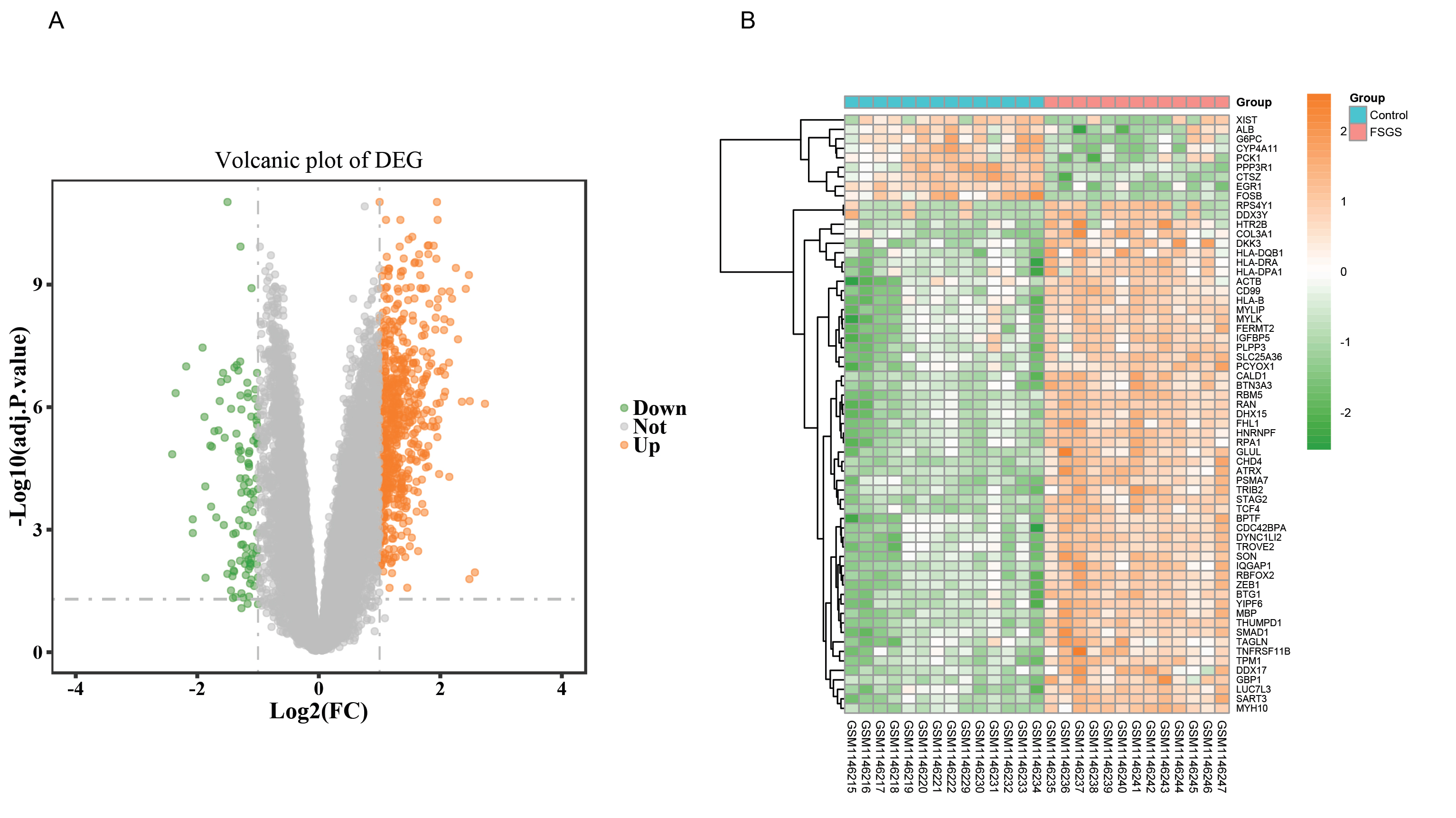
This dataset GSE47185 contains the mRNA expression profiles of 13 FSGS samples and 14 control samples (normal tissue of renal tumor excision). Under the threshold of |log 2 fold change (FC)| > 1 and adj.P value < 0.05, 627 DEGs were identified between the FSGS and control groups. These DEGs included 534 up-regulated and 93 down-regulated DEGs. The results of expression-level analysis are presented as a volcano plot in Fig. 1A. As shown in Table 2, RPS4Y1, PLPP3, DDX3Y, SART3, TCF4, TROVE2, IQGAP1, MBP, CALD1, and RBFOX2 are the 10 most significantly up-regulated genes, whereas CYP4A11, FOSB, EGR1, G6PC, ALB, CTSZ, PPP3R1, XIST, PCK1, and HPGD are the 10 most significantly down-regulated genes. The more information of all DEGs is listed in supplementary Table 1.

Fig1

Figure 1.
Visualization of DEGs
A, The volcano plot of differentially expressed genes between FSGS and healthy tissues. The red plots represent up-regulated genes, green plots represent down-regulated genes, while grey plots represent non-significant genes. The volcano plot was constructed using the ggplot2 package of the R language; B, A heatmap of the top 10% DEGs. The horizontal axis denotes the different samples, and the vertical axis denotes different DEGs.blue, normal samples; red, FSGS samples; purple clusters represent up-regulated DEGs and green clusters represent downregulated DEGs. Color key represents the Z-score based on the Gene expression value.

Table 2. The Most Significant 10 Up-Regulated Genes And Down-Regulated Genes.

Gene Symbols

Log FC

Average Expression level

Adj.P. Value

RPS4Y1

2.569009

9.570289

1.11E-02

PLPP3

2.485089

9.251834

7.13E-07

DDX3Y

2.478844

7.003028

1.62E-02

SART3

2.470577

7.660357

5.82E-10

TCF4

2.41899

7.335133

1.27E-09

TROVE2

2.355365

9.457713

7.39E-07

IQGAP1

2.290646

8.145384

2.19E-08

MBP

2.255968

7.587945

3.91E-10

CALD1

2.192563

8.074506

2.21E-09

RBFOX2

2.164463

8.698168

8.22E-09

CYP4A11

-2.41227

9.128665

1.42E-05

FOSB

-2.35626

8.074382

4.53E-07

EGR1

-2.18278

10.28014

1.01E-07

G6PC

-2.07454

6.566343

5.57E-04

ALB

-2.0735

9.571928

1.21E-03

CTSZ

-1.91281

8.256097

3.49E-08

PPP3R1

-1.88153

7.627366

1.74E-06

XIST

-1.86386

7.615666

1.50E-02

PCK1

-1.7723

10.99439

2.73E-04

HPGD

-1.7174

10.07267

3.87E-06

The heatmaps of the top 10% DEGs are shown in Fig. 1B. The data are presented in a matrix format, in which rows represent individual genes, and columns represent individual samples. The purple and green colors indicate up-regulated and down-regulated genes, respectively. The hierarchy cluster analysis indicated that FSGS and control groups could be distinguished from each other on the basis of their different expression patterns.

Functional enrichment analysis

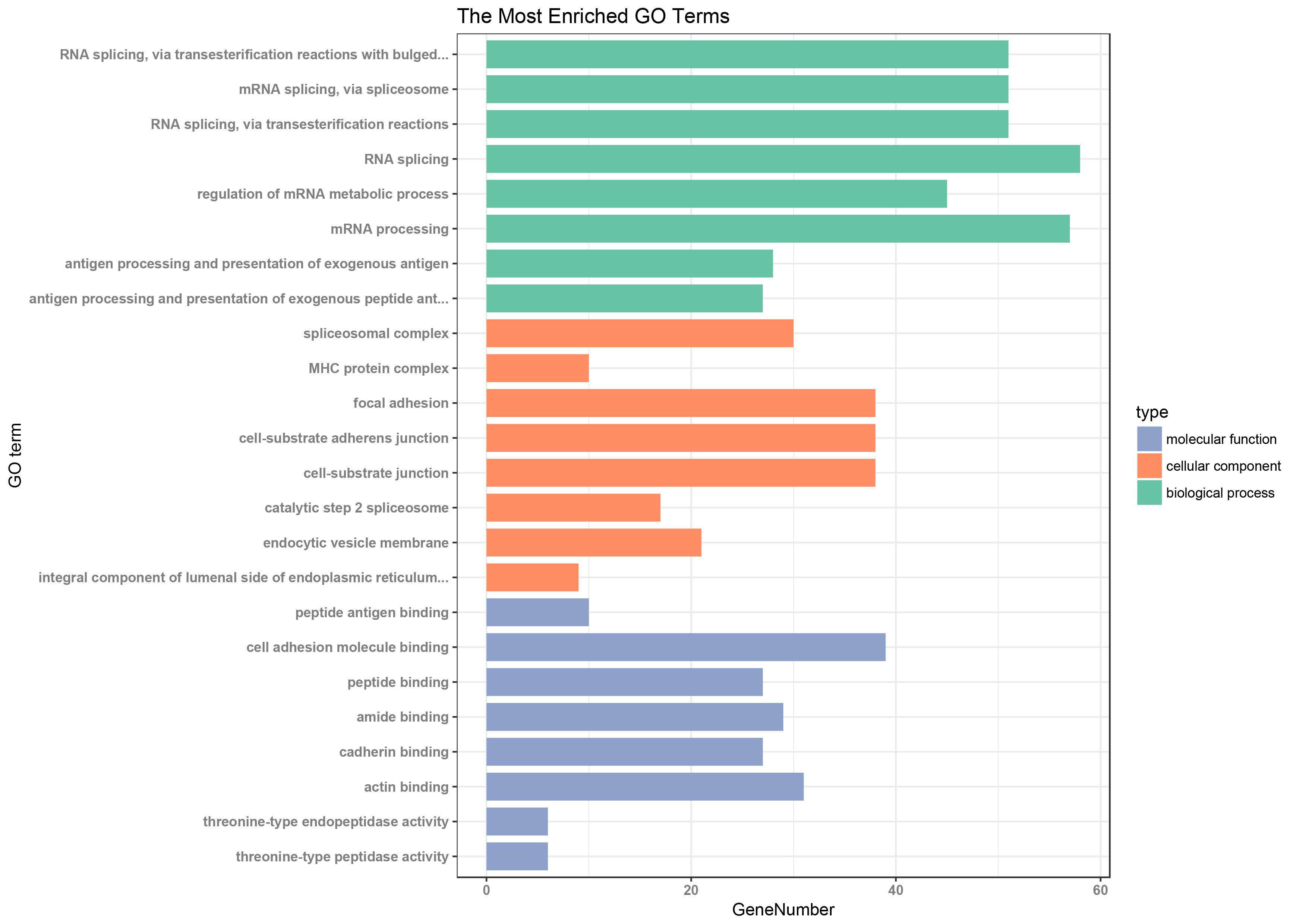
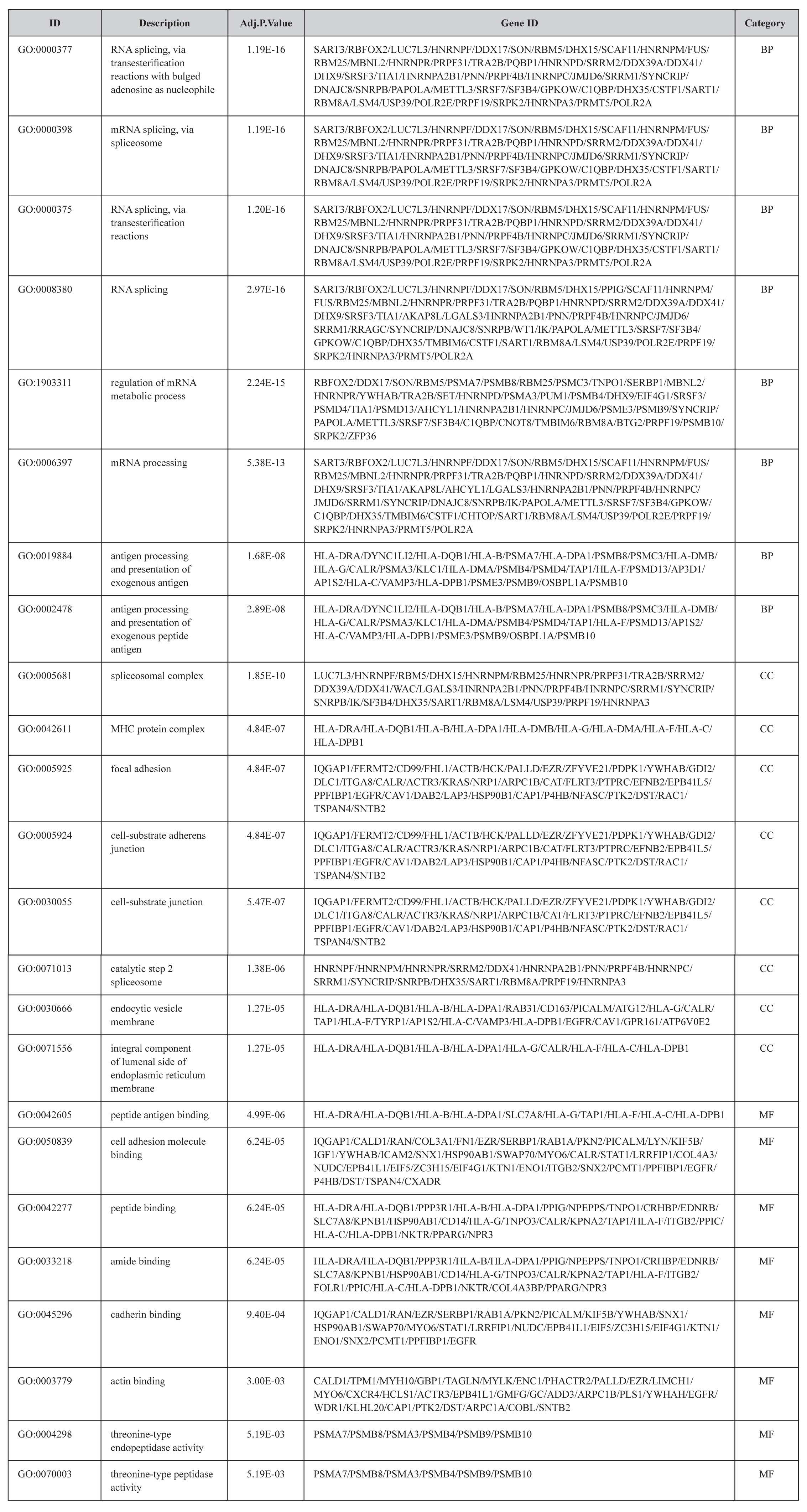
To reveal the biological functions of DEGs, we used the clusterProfiler package for GO analysis. We set adj.P value < 0.01 to identify significantly enriched GO terms. The top eight GO terms for the DEGs enriched in the BP, CC, and MF are shown in Figure 2. The DEGs were mainly involved in GO terms that included mRNA processing, regulation of mRNA metabolic process, antigen processing and presentation of exogenous antigen, focal adhesion, cell adhesion molecule binding, cadherin binding, and actin binding. Among these terms, the MFs related to focal adhesion (GO:0005925) [31,32], cell adhesion molecule (GO:0050839) [33,34], cadherin (GO:0045296) [35], and actin binding (GO:0003779) [36] were all confirmed to be involved in glomerulosclerosis. Detailed information on the top eight GO terms is shown in Table 3.

JCRM_Zhongying Huang_F2

Figure 2. The 8 most significant enriched GO terms of DEGs
The adj.P value< 0.01 was used as the threshold for the identification of significant GO terms. The Gene ontology covers the biological process, cellular component, and molecular function.The horizontal axis represents the gene counts, the vertical axis represents GO terms.Green column graphs represent biological process(BP) GO term; orange column graphs represent cellular component(CC) GO term; and blue column graphs represent molecular function (MF) GO term.

Table 3. The Most Significantly Enriched GO Terms In BP ,CC and MF.

JCRM_Zhongying-Huang_F5

PPI Network Construction and Hub-Gene Screening

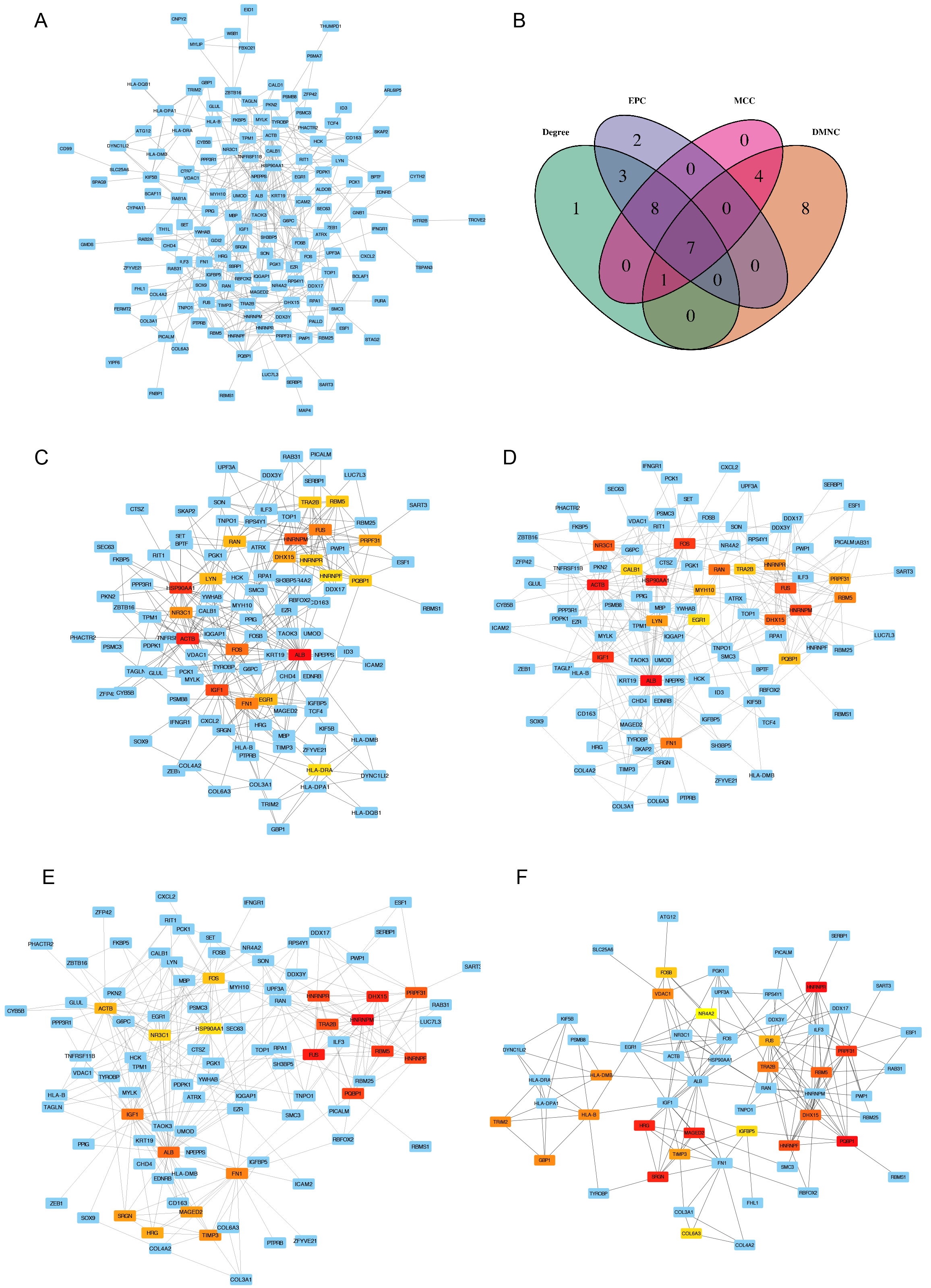
The major part of the constructed PPI network is presented in Figure 3A. To further reduce the scope for analysis, we analyzed the PPI network by using the four analysis methods in CytoHubba based on the R package Venn diagram. We used the top ranked 20 DEGs to identify seven overlapping hub genes screened through the four CytoHubba methods (Degree, EPC, MCC, and DMNC) in cytoscape software (Figure 3B–F). The seven overlapping hub genes included FUS, DHX15, PRPF31, PQBP1, RBM5, HNRNPR, and TRA2B. Strikingly, the identified hub DEGs in our study have never been reported in literature related to FSGS. In addition, these genes simultaneously ranked to the high position by the four different CytoHubba methods suggests they may play important roles in the development of FSGS.

Fig3

Figure 3: Protein-protein interaction (PPI) networks of DEGs and screening of hub genes
A: The major part of PPI network; B: Venn diagram of differentially expressed genes based on four screening methods including “Degree”, “EPC”, “MCC”, and “DMNC”; C: PPI of DEGs screened by the method “Degree” in Cytohubba; D: PPI of DEGs screened by the method “EPC” in Cytohubba; E: PPI of DEGs screened by the method “MCC” in Cytohubba; F: PPI of DEGs screened by the method “DMNC” in Cytohubba. The depth of red represents the rank of the hub genes.

Gene Expression Validation in In -Vitro Experiments

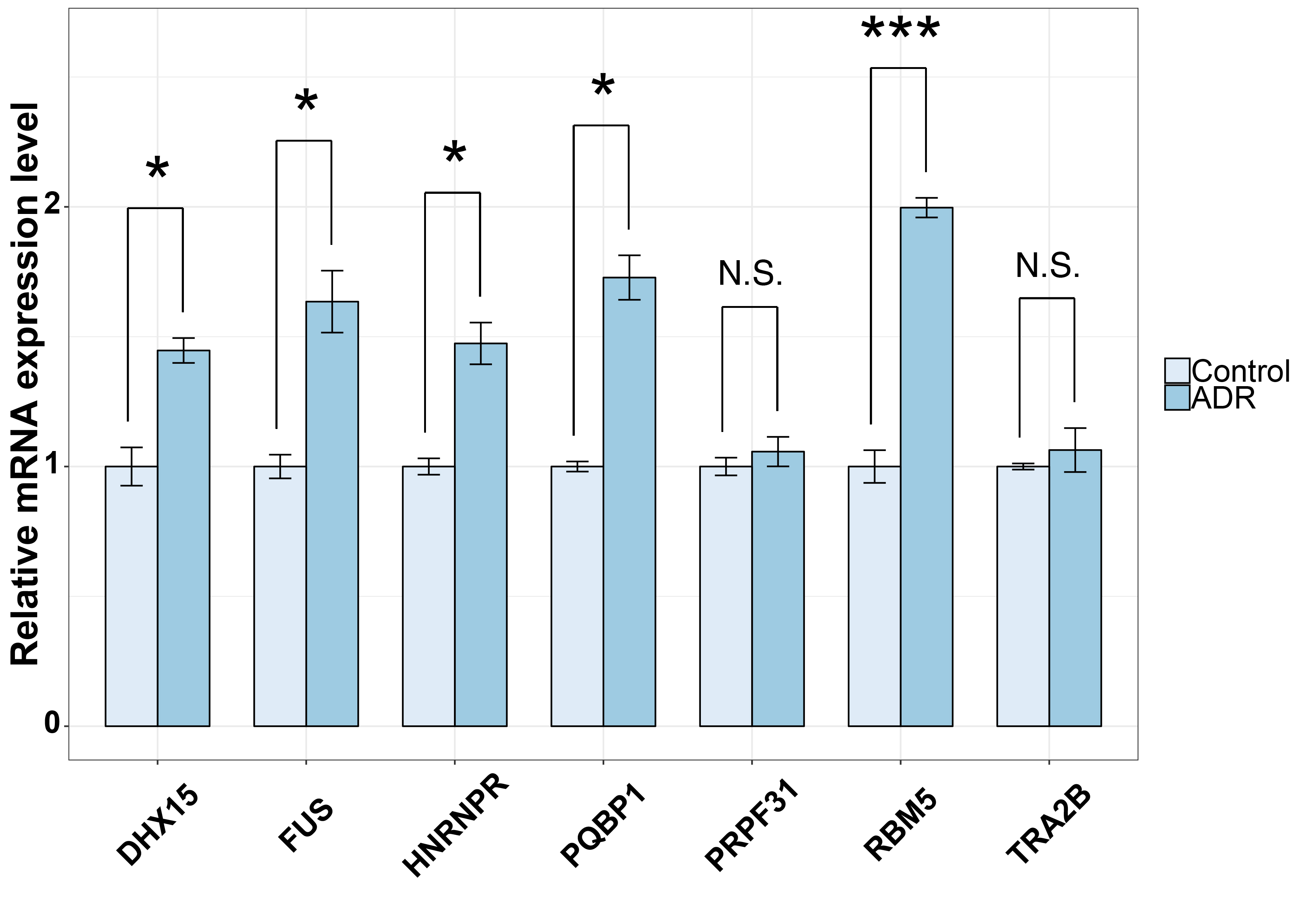
We validated the 7 top ranked hub genes expression in the FSGS model in vitro (Figure 4). Quantitative real-time PCR indicated that the mRNA levels of FUS, DHX15, PQBP1, RBM5, and HNRNPR were up-regulated after they were stimulated by adriamycin (ADR). The changes in PRPF31 and TRA2B mRNA were statistically insignificant. Except for PRPF31 and TRA2B, the changes in all of the other gene expression levels were consistent with the bioinformatic analysis results (Supplementary Table 1), with the accordance rate reaching 70% approximately.

R Graphics Output

Figure 4. In vitro validation for the novel hub genes
Adriamycin (ADR) (0.125 ug/ml) was used to stimulate confluent conditionally immortalized human podocytes (LY893) for 0 (control) and 48 h. The mRNA expression levels of 7 novel top hub genes were measured by quantitative real-time PCR. The mRNA expression levels of the target genes were normalized to that of GAPDH. The data in three separate experiments were presented as mean ± SD (n=3). *Significantly changed expression levels in ADR-stimulated cells compared with the controls (P<0.05). N.S., no significant difference (P>0.05)

Extended information on Potential Hub Genes

On the basis of the above results, we used the abbreviations of the seven hub genes and FSGS as keywords to search the NCBI database for identifying the potential relationship between these hub genes and FSGS. Search results revealed that the seven hub genes have never been reported in literature related to FSGS. Then, we carefully collated information relevant to the biological functions and signaling pathways that involve these hub genes on Gene Cards website (https://www.genecards.org). The results indicate that heterogeneous nuclear ribonucleoprotein F (HNRNPF) is closely related to Nrf2 gene expression, renal angiotensinogen gene expression, the TGF-β1 signaling pathway, and oxidative stress. Moreover, RNA-binding motif protein 5 (RBM5) is involved in apoptosis induction in many tumors. Ultimately, in accordance with the accepted pathogenesis of FSGS, we selected HNRNPF and RBM5 as representative targets for further discussion. Extended information on HNRNPF and RBM5 are shown in Table 4.

Table 4. Extended Information of The Potential MN-Related Hub Genes.

Gene

Function

Disease/Cells

DOI

Authors

HNRNPF

Stimulates renal Ace-2 gene expression and prevents TGF-β1-induced kidney injury

Diabetes

10.1007/s00125-015-3700-y

Lo CS, Shi Y, Chang SY

Mediate renal angiotensinogen gene expression and prevention of hypertension and kidney injury

Diabetes

10.1007/s00125-013-2910-4

Abdo S, Lo CS, Chenier I

Inhibits Nrf2 Gene Expression

Diabetic mice

10.1210/en.2016-1576

Ghosh A, Abdo S, Zhao S

Against oxidative stress

Diabetic mice

10.2337/db16-1588

Lo CS, Shi Y, Chenier

Suppresses angiotensinogen gene expression

Diabetic mice

10.2337/db11-1349

Lo CS, Chang SY, Chenier I

Modulate the alternative splicing of the apoptotic mediator Bcl-x

Human HeLa cells

10.1074/jbc.M501070200

GarneauD,Revil T, Fisette JF

Modulates angiotensinogen gene expression

Diabetes

10.1681/ASN.2004080715

Wei CC, Guo DF, Zhang SL

RBM5

Inhibition of Wnt/β-catenin signaling and induction of apoptosis

Gliomas

10.1186/s12957-016-1084-1

Jiang Y, Sheng H, Meng L

Impacts cell proliferation and apoptosis

Lung cancer

10.1615/JEnvironPatholToxicolOncol.2017019366

Prabhu VV, Devaraj N

Regulates the activity of Wnt/β-catenin signaling

Alveolar epithelial injury

10.3892/or.2015.3828

Hao YQ, Su ZZ, Lv XJ

Promotes caspase activation

Human neuronal cells

10.1038/jcbfm.2014.242

Jackson TC, Du L,Janesko-Feldman K

Promotes neuronal apoptosis

Spinal cord injury

10.1016/j.biocel.2014.12.020

Zhang J, Cui Z, Feng G

Inhibits cell growth and induces apoptosis

Lung adenocarcinoma

10.1186/1477-7819-10-160

Shao C, Zhao L, Wang K

Discussion

Bioinformatics is a newly developed interdisciplinary subject that combines biological science and computer science. Over the past few years, a growing body of research has used gene expression profiles to explore key genes in the pathogenesis of numerous diseases [15,16,40,41]. To our knowledge, our study is the first work that subjected FSGS to bioinformatic analysis. We identified 627 DEGs between the FSGS and control groups. These DEGs included numerous DEGs that have not been previously reported to be involved in FSGS. Then, we predicted DEG functions on the basis of GO annotations. The GO terms we identified included focal adhesion [31,32], cell adhesion molecule [33,34], cadherin [35], and actin binding [36]. These processes are associated with glomerulosclerosis. For example, the genetic deletion of Epb41l5, a podocyte-specific focal adhesome component, results in podocyte detachment, severe proteinuria, and focal segmental glomerulosclerosis development [31]. In immortalized human podocytes, the overexpression of R431C mutant ANLN, an F-actin binding cell cycle gene, enhances podocyte motility [36]. Next, we identified seven overlapping hub genes by constructing PINs through four different analytical methods. Through an accurate search of the NCBI database, we identified HNRNPF and RBM5 as the representative targets for further elaboration.

HNRNPF is a protein-coding gene associated with gene expression. However, no research has been conducted on the role and mechanisms of HNRNPF in FSGS. In this work, we found that HNRNPF is an important DEG among the seven overlapping hub genes in the PPI networks. In addition, HNRNPF is deeply involved with Nrf2 [42], a renal angiotensinogen gene that is expressed in the kidney [43]. Furthermore, in diabetic mice, HNRNPF participates in the TGF-β1 signaling pathway [44] and oxidative stress [45]. These genes and pathways have been confirmed to play vital roles in the pathogenesis of FSGS [4,11,46]. One research suggested that osthole could improve FSGS by activating the Nrf2 antioxidant pathway [47]. TGF-β1 reduces WT1 expression in mouse podocytes and cultured human podocytes before overt glomerulosclerosis begins [46]. In addition, damage to podocytes stimulates TGF-β1 and TGF-βIIR expression in glomerular epithelial cells; this effect eventually leads to extracellular matrix overproduction [48]. On the basis of our analytical results, we conclude that HNRNPF likely participates in FSGS through oxidative stress-associated genes and pathways and is a potential biomarker for this disease.

RBM5 is a nuclear RNA-binding protein that is often genetically deleted in renal cancer49. Unfortunately, the role of RBM5 in FSGS remains unreported. In our study, we identified RBM5 as an up-regulated hub gene in FSGS. Moreover, RBM5 actively participates in apoptosis induction in tumors [50,51]. Apoptosis-induced podocyte damage is a key factor in the pathogenesis of FSGS [12,52]. On the basis of previous findings combined with our present bioinformatic analysis results, we speculate that RBM5 may participate in apoptosis promotion during FSGS progression.

Conclusion

Our study provides a fast, powerful, and effective strategy for the discovery of novel diagnostic biomarkers and therapeutic targets for FSGS. Our results suggest that HNRNPF and RBM5 are molecular candidates for the diagnosis and treatment of FSGS. However, our results are preliminary, and further work is needed to decipher these candidate genes.

Author contributions

Q.M and Z.H designed the research; Z.H analyzed the data and performed the research; D.Z wrote the manuscript. All authors read and approved the final manuscript.

Qianhong Ma and Dongmei Zhang contributed equally to this work and should be considered co-first authors.

Funding

This work was supported by grants from the National Natural Science Foundation of China (Grant No. 81200453).

References

  1. Pollak MR (2008) Focal segmental glomerulosclerosis: recent advancesyper. Curr Opin Nephrol H tens 17: 138–142.
  2. Wiggins RC (2007) The spectrum of podocytopathies: a unifying view of glomerular diseases. Kidney Int 71: 1205–1214.
  3. Kriz W, Lemley KV (2015) A potential role for mechanical forces in the detachment of podocytes and the progression of CKD. J Am Soc Nephrol 26: 258–269.
  4. Lin EY, Bayarsengee U, Wang CC, Chiang YH, Cheng CW (2018) The natural compound 2,3,5,4’-tetrahydroxystilbene-2-O-beta-d glucoside protects against adriamycin-induced nephropathy through activating the Nrf2-Keap1 antioxidant pathway. Environ Toxicol 33: 72–82.
  5. Raij L, Tian R, Wong JS, He JC, Campbell KN, et al. (2016) Podocyte injury: the role of proteinuria, urinary plasminogen, and oxidative stress. Am J Physiol Renal Physiol 311: 1308–1317.
  6. Yang SM, Ka SM, Hua KF, Wu TF, Chuang YP, et al. (2013) Antroquinonol mitigates an accelerated and progressive IgA nephropathy model in mice by activating the Nrf2 pathway and inhibiting T cells and NLRP3 inflammasome. Free Radic Biol Med 61: 285–297.
  7. Oh CJ, Kim JY, Choi YK, Lee IK, Kim HJ, et al. (2012) Dimethylfumarate attenuates renal fibrosis via NF-E2-related factor 2-mediated inhibition of transforming growth factor-beta/Smad signaling. PloS one 7: 45870.
  8. Tsai PY, Ka SM, Chao TK, Chang JM, Lin SH, et al. (2011) Antroquinonol reduces oxidative stress by enhancing the Nrf2 signaling pathway and inhibits inflammation and sclerosis in focal segmental glomerulosclerosis mice. Free Radic Biol Med 50: 1503–1516.
  9. Schiffer M, Bitzer M, Roberts IS, Kopp JB, Mundel P, et al. (2001) Apoptosis in podocytes induced by TGF-beta and Smad7. J Clin Invest 108: 807–816.
  10. Schachter AD, Strehlau J, Zurakowski D, Kim YS, Strom TB, et al. (2000) Increased nuclear factor-kappaB and angiotensinogen gene expression in posttransplant recurrent focal segmental glomerulosclerosis. Transplantation 70: 1107–1110.
  11. Luther Y, Bantis C, Ivens K, Fehsel K, Kolb-Bachhofen V, et al. (2003) Effects of the genetic polymorphisms of the renin-angiotensin system on focal segmental glomerulosclerosis. Kidney Blood Press Res 26: 333–337.
  12. Liu L, Lin W, Zhang Q, Cao W, Liu Z (2016) TGF-beta induces miR-30d down-regulation and podocyte injury through Smad2/3 and HDAC3-associated transcriptional repression. J Mol Med (Berl) 94: 291–300.
  13. Xue Y, Yin P, Li G, Zhong D (2018) Genome-wide Integration Study of Circulating miRNAs and Peripheral Whole-Blood mRNAs of Male Acute Ischemic Stroke Patients. Neuroscience 380: 27–37.
  14. Li H, Zhen Y, Geng Y, Feng J, Wang J (2018) Exploring the Molecular Mechanism of Thoracic Aortic Aneurysm via Bioinformatics Analysis. Med Sci Monit 24: 1533–1539.
  15. Dong B, Wang G, Yao J, Yuan P, Kang W, et al. (2017) Predicting novel genes and pathways associated with osteosarcoma by using bioinformatics analysis. Gene 628: 32–37.
  16. Luo Y, Zhang C, Tang F, Zhao J, Shen C, et al. (2015) Bioinformatics identification of potentially involved microRNAs in Tibetan with gastric cancer based on microRNA profiling. Cancer Cell Int 15: 115.
  17. Wang JP, Leng JY, Zhang RK, Zhang L, Zang B, et al. (2018) Functional analysis of gene expression profiling-based prediction in bladder cancer. Oncol Lett 15: 8417–8423.
  18. Dai J, Ma Y, Chu S, Le N, Cao J, et al. (2018) Identification of key genes and pathways in meningioma by bioinformatics analysis. Oncol Lett 15: 8245–8252.
  19. Lin P, Wen DY, Dang YW, He Y, Yang H, et al. (2018) Comprehensive and Integrative Analysis Reveals the Diagnostic, Clinicopathological and Prognostic Significance of Polo-Like Kinase 1 in Hepatocellular Carcinoma. Cell Physiol Biochem 47: 925–947.
  20. Yao T, Wang Q, Zhang W, Bian A, Zhang J (2016) Identification of genes associated with renal cell carcinoma using gene expression profiling analysis. Oncol Lett 12: 73–78.
  21. Shu B, Fang Y, He W, Yang J, Dai C (2018) Identification of macrophage-related candidate genes in lupus nephritis using bioinformatics analysis. Cell Signal 46: 43–51.
  22. Cui Y, Liu S, Cui W, Gao D, Zhou W, et al. (2017) Identification of potential biomarkers and therapeutic targets for human IgA nephropathy and hypertensive nephropathy by bioinformatics analysis. Molecular medicine reports 16: 3087–3094.
  23. Zhou LT, Qiu S, Lv LL, Li ZL, Liu H, et al. (2018) Integrative Bioinformatics Analysis Provides Insight into the Molecular Mechanisms of Chronic Kidney Disease. Kidney Blood Press Res 43: 568–581.
  24. Gautier L, Cope L, Bolstad BM, Irizarry RA (2004) affy–analysis of Affymetrix GeneChip data at the probe level. Bioinformatics 20: 307–315.
  25. Ritchie ME, Phipson B, Wu D, Hu Y, Shi, et al. (2015) limma powers differential expression analyses for RNA-sequencing and microarray studies. Nucleic Acids Res 43: 47.
  26. Yu G, Wang LG, Han Y, He QY (2012) Cluster Profiler: an R package for comparing biological themes among gene clusters. OMICS 16: 284–287.
  27. Chin CH, Chen SH, Wu HH, Ho CW, Ko MT, et al. (2014) cytoHubba: identifying hub objects and sub-networks from complex interactome. BMC Syst Biol 8: 11.
  28. Chen H, Boutros PC (2011) VennDiagram: a package for the generation of highly-customizable Venn and Euler diagrams in R. BMC Bioinformatics 12: 35.
  29. Shannon P, Markiel A, Ozier O, Wang JT, Amin N, et al. (2003) Cytoscape: a software environment for integrated models of biomolecular interaction networks. Genome Res 13: 2498–2504.
  30. Kachurina N, Chung CF, Benderoff E, Babayeva S, Bitzan M, et al. (2016) Novel unbiased assay for circulating podocyte-toxic factors associated with recurrent focal segmental glomerulosclerosis. Am J Physiol Renal Physiol 310: 1148–1156.
  31. Schell C, Rogg M, Suhm M, Helmstadter M, Sellung D, et al. (2017) The FERM protein EPB41L5 regulates actomyosin contractility and focal adhesion formation to maintain the kidney filtration barrier. Proc Natl Acad Sci U S A 114: 4621–4630.
  32. Sever S, Schiffer M (2018) Actin dynamics at focal adhesions: a common endpoint and putative therapeutic target for proteinuric kidney diseases. Kidney Int 93: 1298–1307
  33. Gong W, Chen C, Xiong F, Yang Z, Wang Y, et al. (2016) CKIP-1 ameliorates high glucose-induced expression of fibronectin and intercellular cell adhesion molecule-1 by activating the Nrf2/ARE pathway in glomerular mesangial cells. Biochem Pharmacol 116: 140–152.
  34. Huang J, Filipe A, Rahuel C, Bonnin P, Mesnard L, et al. ( 2014) Lutheran/basal cell adhesion molecule accelerates progression of crescentic glomerulonephritis in mice. Kidney Int 85: 1123–1136.
  35. Naves MA, Requiao-Moura LR, Soares MF, Silva-Junior JA, Mastroianni-Kirsztajn G, et al. (2012) Podocyte Wnt/ss-catenin pathway is activated by integrin-linked kinase in clinical and experimental focal segmental glomerulosclerosis. Journal of nephrology 25: 401–409.
  36. Gbadegesin RA, Hall G, Adeyemo A, Hanke N, Tossidou I, et al. (2014) Mutations in the gene that encodes the F-actin binding protein anillin cause FSGS. Journal of the American Society of Nephrology: JASN 25: 1991–2002.
  37. Papazachariou L, Papagregoriou G, Hadjipanagi D, Dwmosthenous P, Voskarides K, et al. (2017) Frequent COL4 mutations in familial microhematuria accompanied by later-onset Alport nephropathy due to focal segmental glomerulosclerosis. Clin Genet 92: 517–527.
  38. Johnstone DB, Zhang J, George B, Leon C, Gachet C, et al. (2011) Podocyte-specific deletion of Myh9 encoding nonmuscle myosin heavy chain 2A predisposes mice to glomerulopathy. Mol Cell Biol 31: 2162–2170.
  39. Boyer O, Benoit G, Gribouval O, Nevo F, Tete MJ, et al. (2011) Mutations in INF2 are a major cause of autosomal dominant focal segmental glomerulosclerosis. J Am Soc Nephrol 22: 239–245.
  40. Li WX, He K, Tang L, Dai SX, Li GH, et al. (2017) Comprehensive tissue-specific gene set enrichment analysis and transcription factor analysis of breast cancer by integrating 14 gene expression datasets. Oncotarget 8: 6775–6786.
  41. Cui H, Zhang Y, Zhang Q, Chen W, Zhao H, et al. (2017) A comprehensive genome-wide analysis of long noncoding RNA expression profile in hepatocellular carcinoma. Cancer Med 6: 2932–2941.
  42. Ghosh A, Abdo S, Zhao S, Wu CH, Shi Y, et al. (2017) Insulin Inhibits Nrf2 Gene Expression via Heterogeneous Nuclear Ribonucleoprotein F/K in Diabetic Mice. Endocrinology 158: 903–919.
  43. Wei CC, Guo DF, Zhang SL, Ingelfinger JR, Chan JS (2005) Heterogenous nuclear ribonucleoprotein F modulates angiotensinogen gene expression in rat kidney proximal tubular cells. J Am Soc Nephrol 16: 616–628.
  44. Lo CS, Shi Y, Chang SY, Abdo S, Chenier I, et al. (2015) Overexpression of heterogeneous nuclear ribonucleoprotein F stimulates renal Ace-2 gene expression and prevents TGF-beta1-induced kidney injury in a mouse model of diabetes. Diabetologia 58: 2443–2454.
  45. Lo CS, Shi Y, Chenier I, Ghosh A, Wu CH, et al. (2017) Heterogeneous Nuclear Ribonucleoprotein F Stimulates Sirtuin-1 Gene Expression and Attenuates Nephropathy Progression in Diabetic Mice. Diabetes 66: 1964–1978.
  46. Sakairi T, Abe Y, Kopp JB (2011) TGF-beta1 reduces Wilms’ tumor suppressor gene expression in podocytes. Nephrol Dial Transplant 26: 2746–2752.
  47. Yang SM, Chan YL, Hua KF, Chang JM, Chen HL, et al. (2014) Osthole improves an accelerated focal segmental glomerulosclerosis model in the early stage by activating the Nrf2 antioxidant pathway and subsequently inhibiting NF-kappaB-mediated COX-2 expression and apoptosis. Free Radic Biol Med 73: 260–269.
  48. Kim JH, Kim BK, Moon KC, Hong HK, Lee HS (2003) Activation of the TGF-beta/Smad signaling pathway in focal segmental glomerulosclerosis. Kidney Int 64: 1715–1721.
  49. Bechara EG, Sebestyen E, Bernardis I, Eyras E, Valcarcel J (2013) RBM5, 6, and 10 differentially regulate NUMB alternative splicing to control cancer cell proliferation. Mol Cell 52: 720–733.
  50. Jiang Y, Sheng H, Meng L, Yue H, Li B, et al. (2017) RBM5 inhibits tumorigenesis of gliomas through inhibition of Wnt/beta-catenin signaling and induction of apoptosis. World J Surg Oncol 15: 9.
  51. Prabhu VV, Devaraj N (2017) Regulating RNA Binding Motif 5 Gene Expression- A Novel Therapeutic Target for Lung Cancer. J Environ Pathol Toxicol Oncol 36: 99–105.
  52. Nafar M, Kalantari S, Samavat S, Rezaei-Tavirani M, Rutishuser D, et al. (2014) The novel diagnostic biomarkers for focal segmental glomerulosclerosis. Int J Nephrol 2014: 574261.

Supplementary Table 1

gene.symbols

logFC

AveExpr

ts

P.Value

adj.P.Val

B

RPS4Y1

2.569009422

9.570289361

3.101358611

0.004371381

0.011115768

-2.711721859

PLPP3

2.485089122

9.251833831

7.62668606

2.66E-08

7.13E-07

9.068379301

DDX3Y

2.478844353

7.003027727

2.920309189

0.006843408

0.016208722

-3.132862184

SART3

2.470577198

7.660356798

12.25209245

9.40E-13

5.82E-10

19.16340462

TCF4

2.418989646

7.335132517

11.59356528

3.44E-12

1.27E-09

17.89787797

CYP4A11

-2.412274749

9.12866545

-6.123580669

1.33E-06

1.42E-05

5.192258061

FOSB

-2.356258176

8.074381933

-7.877904555

1.42E-08

4.53E-07

9.690625188

TROVE2

2.355365338

9.457713493

7.605950945

2.80E-08

7.39E-07

9.016659035

IQGAP1

2.290645788

8.14538406

9.69432624

1.94E-10

2.19E-08

13.93671972

MBP

2.255967535

7.587944516

12.62989302

4.56E-13

3.91E-10

19.86587003

CALD1

2.192562666

8.074505734

11.18804096

7.85E-12

2.21E-09

17.09171526

EGR1

-2.182777112

10.2801361

-8.747798502

1.72E-09

1.01E-07

11.78050827

RBFOX2

2.164462681

8.69816763

10.33705244

4.72E-11

8.22E-09

15.33081151

IGFBP5

2.148130254

9.995294632

5.537086859

6.45E-06

5.09E-05

3.626766637

CDC42BPA

2.079395261

9.770721553

7.925115034

1.26E-08

4.20E-07

9.806647308

SMAD1

2.076607382

7.949965987

8.465377555

3.38E-09

1.61E-07

11.11324049

G6PC

-2.074536589

6.566342687

-4.459033616

0.000121983

0.000557225

0.736400459

ALB

-2.073497949

9.571928342

-4.114808392

0.000309678

0.001209773

-0.171567409

ZEB1

2.066318697

8.614493251

10.45925661

3.63E-11

7.03E-09

15.5895635

TPM1

2.060897076

9.232448331

10.04753441

8.88E-11

1.30E-08

14.70977983

FERMT2

2.05930113

7.350506265

7.771402732

1.85E-08

5.49E-07

9.427823748

LUC7L3

2.047718235

8.759118544

8.718232863

1.85E-09

1.06E-07

11.71116708

RAN

2.025548667

10.29196832

11.46235083

4.49E-12

1.47E-09

17.63930778

SLC25A36

2.003665861

8.269066322

8.586494779

2.53E-09

1.31E-07

11.40073865

HLA-DRA

1.992802428

11.12961293

7.574703865

3.03E-08

7.84E-07

8.938615772

HTR2B

1.980142562

6.49870054

5.606952454

5.34E-06

4.35E-05

3.814138409

PCYOX1

1.966411418

7.433879894

7.068593108

1.10E-07

2.03E-06

7.65787493

ATRX

1.961414528

6.588331245

11.16381872

8.25E-12

2.27E-09

17.04289981

DYNC1LI2

1.955508721

9.605304116

10.85559765

1.57E-11

3.64E-09

16.41511633

CD99

1.9554319

9.927197771

8.57318363

2.61E-09

1.33E-07

11.36923982

CHD4

1.94490416

8.814477542

16.59170115

5.35E-16

9.51E-12

26.31762235

MYLIP

1.938154662

9.419703765

7.548038454

3.24E-08

8.23E-07

8.871918527

HNRNPF

1.934376435

8.42993207

13.0216741

2.19E-13

2.32E-10

20.57678872

HLA-DQB1

1.933559554

9.283173095

6.877515473

1.81E-07

2.96E-06

7.16657257

FHL1

1.916965148

7.704294928

10.06888924

8.47E-11

1.27E-08

14.75597408

CTSZ

-1.912814074

8.256096924

-9.422794947

3.59E-10

3.49E-08

13.3308037

DDX17

1.900816911

7.156016292

6.849768897

1.94E-07

3.11E-06

7.094896981

THUMPD1

1.891011353

7.141971356

13.73428793

6.01E-14

1.12E-10

21.82576576

PPP3R1

-1.881530742

7.627366273

-7.148865697

8.96E-08

1.74E-06

7.863044486

TNFRSF11B

1.87836792

7.853323079

7.02736233

1.23E-07

2.20E-06

7.552206366

XIST

-1.863860797

7.615665673

-2.956875261

0.006256155

0.015025427

-3.048847061

SON

1.851258175

7.997946847

8.840334385

1.39E-09

8.53E-08

11.99675806

GLUL

1.846739708

7.937765075

9.235752198

5.51E-10

4.67E-08

12.90751377

DKK3

1.838529383

7.235094143

6.88002716

1.80E-07

2.95E-06

7.173056725

BTG1

1.820054032

7.907833291

11.00485807

1.15E-11

2.94E-09

16.72066783

TRIB2

1.81921404

8.182950746

8.768912824

1.64E-09

9.74E-08

11.82995473

RBM5

1.808667325

8.306181873

13.85226391

4.87E-14

1.09E-10

22.02721576

HLA-B

1.803285216

12.34984324

6.340923899

7.42E-07

8.94E-06

5.766672368

MYH10

1.802983311

6.632652763

13.31027341

1.29E-13

1.80E-10

21.08934934

STAG2

1.793115173

5.860191501

13.76494033

5.69E-14

1.12E-10

21.87824888

GBP1

1.783205635

6.97780633

8.494851598

3.15E-09

1.53E-07

11.18338801

RPA1

1.779726763

9.056001662

9.084532378

7.82E-10

5.91E-08

12.56176241

BPTF

1.776426474

7.907083176

7.356751223

5.27E-08

1.17E-06

8.390867161

PCK1

-1.772301574

10.99438749

-4.775458625

5.15E-05

0.000272763

1.580947098

BTN3A3

1.772184439

6.173764331

8.425121851

3.73E-09

1.74E-07

11.01724161

TAGLN

1.764332395

9.007016683

5.779780851

3.34E-06

3.00E-05

4.276795382

PSMA7

1.763068956

6.889044422

11.69687487

2.80E-12

1.22E-09

18.09994223

HLA-DPA1

1.762375082

10.82315093

6.098738113

1.42E-06

1.50E-05

5.126369472

MYLK

1.75861114

10.39524956

6.152944852

1.23E-06

1.34E-05

5.270080435

YIPF6

1.747988074

9.294461389

8.337256865

4.61E-09

2.03E-07

10.80694239

COL3A1

1.741079028

10.03488916

6.880350494

1.80E-07

2.95E-06

7.173891393

ACTB

1.739818254

10.62946301

4.630671948

7.64E-05

0.000379292

1.193646749

DHX15

1.737775319

10.02003843

9.85519139

1.36E-10

1.72E-08

14.29091385

PPIG

1.731145889

7.407036172

7.251707604

6.89E-08

1.43E-06

8.124803038

SCAF11

1.721893208

9.371324456

10.2275776

5.99E-11

9.60E-09

15.09731143

HPGD

-1.717401682

10.07266901

-6.740627536

2.59E-07

3.87E-06

6.812168581

DPP8

1.715249149

9.638588643

8.318534824

4.82E-09

2.09E-07

10.76199707

WSB1

1.714938423

8.596432117

6.91877743

1.62E-07

2.75E-06

7.273007517

ENC1

1.714016384

6.351234133

9.286980973

4.90E-10

4.30E-08

13.02392792

FN1

1.713084538

9.852710944

6.048869862

1.62E-06

1.68E-05

4.993973035

UPF3A

1.706676789

8.950115438

11.59802708

3.41E-12

1.27E-09

17.90663237

KRT19

1.703164941

8.133804492

4.840602977

4.31E-05

0.000235133

1.755562232

BCLAF1

1.697469525

8.139217001

9.830297038

1.44E-10

1.80E-08

14.23633277

FBXO21

1.697287783

9.28818289

5.462538041

7.90E-06

5.94E-05

3.426663378

IFNGR1

1.695558168

9.158556204

8.094628782

8.32E-09

3.11E-07

10.22081389

HCK

1.695111511

7.690905947

7.857688804

1.49E-08

4.67E-07

9.640854784

CXCL2

-1.686811585

6.477878156

-4.50798237

0.000106777

0.000500487

0.866554554

PHACTR2

1.679845963

7.782456361

7.807529456

1.69E-08

5.14E-07

9.517134367

HNRNPM

1.673764753

11.0821542

11.69753948

2.80E-12

1.22E-09

18.10123784

CAMK2N1

-1.651602298

10.19208306

-6.765897326

2.42E-07

3.70E-06

6.877740372

FUS

1.650968802

9.619854977

13.27029455

1.39E-13

1.82E-10

21.01889984

PLPBP

1.650135185

5.59234436

12.81694912

3.21E-13

2.98E-10

20.20749556

PSMB8

1.646543823

10.456135

7.886039857

1.39E-08

4.48E-07

9.71063892

SEC63

1.640469887

7.954060099

6.382735607

6.64E-07

8.19E-06

5.876729688

PALLD

1.635448714

8.943061698

7.556440719

3.17E-08

8.09E-07

8.892944454

ZBTB16

1.635125895

7.66230392

3.653696968

0.001057166

0.003348994

-1.358707431

RBM25

1.63291277

6.324655258

8.650201845

2.17E-09

1.18E-07

11.5511557

TYROBP

1.630051539

7.893243583

5.141218255

1.90E-05

0.00012023

2.563038692

DACH1

1.623091259

7.101855154

6.801703043

2.20E-07

3.44E-06

6.970537686

PSMC3

1.612543512

9.366600505

5.93353245

2.21E-06

2.14E-05

4.68711502

RBMS1

1.6103609

8.650805194

4.65105936

7.23E-05

0.000362333

1.248105923

PGK1

1.607870056

10.36413941

5.403307466

9.28E-06

6.73E-05

3.267574125

NPEPPS

1.606521068

8.421329051

7.835158094

1.58E-08

4.88E-07

9.585322216

PTPRB

1.604906513

9.444331687

7.330988837

5.62E-08

1.23E-06

8.325736785

TNPO1

1.599343907

9.775968964

8.87594367

1.27E-09

8.10E-08

12.07966014

EZR

1.591497713

8.757810465

5.97334531

1.98E-06

1.97E-05

4.793136085

MAGED2

1.586605561

10.30790428

12.89378949

2.78E-13

2.81E-10

20.3466641

ANKRD12

1.580638115

7.392354398

6.693843176

2.92E-07

4.26E-06

6.690596121

FNBP1

1.575997647

8.157390697

8.31499427

4.86E-09

2.10E-07

10.75349206

NR4A2

-1.57481871

6.24079609

-8.527861329

2.91E-09

1.45E-07

11.26180965

SERBP1

1.573844316

8.951883468

11.22792569

7.23E-12

2.19E-09

17.17193154

SPAG9

1.573771613

5.928584131

12.82512531

3.16E-13

2.98E-10

20.22233593

SCAMP1

1.572326984

7.135740239

7.680340099

2.32E-08

6.50E-07

9.201956772

RAB1A

1.571530153

8.448144883

6.477980274

5.16E-07

6.71E-06

6.126856918

TAOK3

1.570396428

7.085559648

9.345798665

4.28E-10

3.85E-08

13.15714066

SRGN

1.568368396

9.423138514

5.18175814

1.70E-05

0.000109899

2.672037194

ZFYVE21

1.5676445

6.846082627

7.262978403

6.69E-08

1.40E-06

8.153414092

HRG

-1.562922635

8.320562127

-4.315974661

0.000179859

0.000770138

0.357348521

PWP1

1.561834997

8.446947472

8.857117309

1.33E-09

8.24E-08

12.03585221

SKAP2

1.561530224

9.277742191

7.33501641

5.57E-08

1.22E-06

8.335924267

UBXN4

1.551349919

9.105993907

6.390043816

6.51E-07

8.06E-06

5.895950838

PECAM1

1.550870521

9.054536986

6.663178499

3.17E-07

4.53E-06

6.610791348

MBNL2

1.550572397

7.066502239

10.39994319

4.12E-11

7.62E-09

15.46422432

SOX9

-1.548564432

8.004071342

-6.562764085

4.13E-07

5.60E-06

6.348811078

TRIM2

1.546572067

8.578770031

8.831695492

1.41E-09

8.68E-08

11.97661948

HNRNPR

1.545721471

9.080452193

10.58685737

2.76E-11

5.70E-09

15.85761598

MYOF

1.54258444

8.617870178

7.043673577

1.18E-07

2.13E-06

7.594032778

NR3C1

1.537314952

8.658438842

7.183782173

8.20E-08

1.62E-06

7.95205499

TFPI2

1.530489929

8.411366859

3.999919431

0.000421606

0.001565513

-0.471022596

TMEM47

1.524753951

8.263583054

6.004266035

1.83E-06

1.84E-05

4.875407299

PURA

1.522929493

7.624254878

6.128405127

1.31E-06

1.41E-05

5.20504847

RAB31

1.519313807

8.597342752

6.411935462

6.15E-07

7.72E-06

5.95349937

CYB5B

1.515075529

7.815221603

11.7624269

2.46E-12

1.16E-09

18.22746762

RIT1

1.514871553

7.235874533

7.407198303

4.63E-08

1.07E-06

8.518169631

PRPF31

1.51015233

6.970023854

7.948526057

1.19E-08

4.05E-07

9.864072564

ALDOB

-1.504568555

8.472159452

-3.061872507

0.004824275

0.012090824

-2.80464753

MAP4

1.503555867

8.031894594

5.975611845

1.97E-06

1.96E-05

4.799168802

RAB2A

-1.502751043

8.315889782

-16.2138848

9.61E-16

9.51E-12

25.7672033

BTN3A2

1.502705438

7.877942556

6.982291597

1.38E-07

2.40E-06

7.436477371

GMDS

1.497770885

9.599429406

7.605970724

2.80E-08

7.39E-07

9.016708396

CD163

1.496518651

7.363641477

3.780358703

0.000756817

0.002538255

-1.036824644

PKN2

1.492252244

7.396561746

9.26254208

5.18E-10

4.46E-08

12.96843728

PICALM

1.48883338

7.452309182

7.882640103

1.40E-08

4.50E-07

9.702276208

LYN

1.48458898

7.748906756

6.995137981

1.33E-07

2.35E-06

7.469486452

ARL6IP5

1.481819394

9.42435093

6.181264534

1.14E-06

1.26E-05

5.345073096

SH3BP5

1.480757339

9.948617496

7.175018224

8.38E-08

1.65E-06

7.929726958

CYTH2

1.478569815

8.695293012

14.0977443

3.17E-14

7.84E-11

22.44167418

HSP90AA1

1.475101446

10.7822491

5.488461362

7.36E-06

5.62E-05

3.49626449

PDPK1

1.474624621

8.188917333

5.243240876

1.44E-05

9.60E-05

2.837346192

SMC3

1.470872089

7.133595994

11.12401077

8.96E-12

2.41E-09

16.96251038

MBTPS1

1.465393655

8.496997806

7.766899015

1.87E-08

5.53E-07

9.416678054

TIMP3

1.463150322

9.290336056

7.335248955

5.56E-08

1.22E-06

8.336512413

SSRP1

1.462015221

7.615804716

9.043612741

8.60E-10

6.37E-08

12.46766016

NAA35

1.458486577

6.726259229

8.092465915

8.37E-09

3.11E-07

10.21555345

KIF5B

1.454182588

8.394875765

5.616323045

5.20E-06

4.27E-05

3.839255868

DHRS7

1.454116053

7.744616405

9.572938924

2.56E-10

2.65E-08

13.66709981

CNPY2

1.453764841

8.534477403

8.634059418

2.26E-09

1.20E-07

11.51309488

FKBP5

1.452723718

7.291698701

4.149986734

0.000281682

0.001116854

-0.079470163

COL6A3

1.447501816

7.827000817

4.905985588

3.61E-05

0.000203504

1.930987853

COL4A2

1.446665268

7.456526747

6.876736022

1.81E-07

2.96E-06

7.164560203

CALB1

-1.44533189

9.757638848

-2.605850418

0.01452899

0.030801016

-3.831208449

IGF1

-1.437115619

9.406113438

-3.336824653

0.002407335

0.006723822

-2.146238464

NELFCD

1.434489749

9.91051469

11.67139035

2.95E-12

1.22E-09

18.0502204

YWHAB

1.431615777

8.640142735

5.544355542

6.32E-06

5.00E-05

3.646268215

RGCC

1.429769752

7.679603868

4.732902335

5.78E-05

0.000299666

1.466986783

SLC25A6

1.428984166

11.61631244

9.101576376

7.52E-10

5.75E-08

12.60089001

ID3

1.428233521

10.14589451

8.266105018

5.47E-09

2.29E-07

10.63587854

TRA2B

1.424948669

9.497698712

8.624014607

2.31E-09

1.22E-07

11.48939306

GDI2

1.424531564

9.161869349

6.615965347

3.59E-07

4.97E-06

6.48773575

CRHBP

1.423223574

10.85750131

3.501852173

0.001572242

0.004679346

-1.739506163

EID1

1.422600712

9.21370437

7.376173329

5.01E-08

1.13E-06

8.439915301

SET

1.420718447

10.19887727

8.55531067

2.73E-09

1.37E-07

11.32690802

FOS

-1.420440832

9.273302043

-3.000916158

0.005612259

0.013756238

-2.946942453

VDAC1

1.415009019

8.172894871

7.118018567

9.71E-08

1.85E-06

7.784289799

WASHC4

1.412990786

7.331446343

8.01490027

1.01E-08

3.56E-07

10.02649009

UMOD

-1.412526572

11.37016849

-2.38566243

0.024069305

0.047104894

-4.291378203

SERTAD2

1.411604795

8.175798374

8.513812449

3.01E-09

1.48E-07

11.22845173

PQBP1

1.411256434

7.748400335

6.769612323

2.40E-07

3.68E-06

6.887374717

CEP350

1.409862497

6.337432566

7.135793044

9.27E-08

1.79E-06

7.829682626

GLYAT

-1.407015938

9.579743485

-4.089844356

0.000331191

0.001276585

-0.236812106

ICAM2

1.406910331

9.771163793

5.247150209

1.42E-05

9.52E-05

2.84785686

GNB1

1.402945544

9.130430125

10.649444

2.42E-11

5.13E-09

15.98830239

HLA-DMB

1.401194865

10.17298113

8.848947598

1.36E-09

8.38E-08

12.01682652

EDNRB

1.398466757

8.373823696

4.94196731

3.27E-05

0.000188026

2.02759004

ESF1

1.398047621

7.471743775

13.05457765

2.06E-13

2.30E-10

20.63569798

AIDA

1.397041007

7.692499594

8.873300307

1.28E-09

8.11E-08

12.07351212

TOP1

1.396150489

8.310821776

7.122657614

9.59E-08

1.83E-06

7.796140615

SNX1

1.393315129

7.77005298

9.617845685

2.31E-10

2.46E-08

13.76708049

CLDND1

1.389278478

8.356235731

7.353059136

5.32E-08

1.18E-06

8.381538064

TSPAN3

1.38922393

9.719295375

4.512799487

0.000105387

0.000495607

0.879374553

SLC7A8

1.388681022

9.335582412

4.354999431

0.000161804

0.000706402

0.460535324

ARHGAP5

-1.388500976

8.373041869

-8.710601093

1.88E-09

1.07E-07

11.69324854

ILF3

1.386329503

6.940492403

8.230768777

5.97E-09

2.45E-07

10.55066888

ATG12

1.384776561

7.242819619

7.607340868

2.79E-08

7.39E-07

9.020127669

ZNF148

1.382618168

8.099582442

12.45022932

6.42E-13

4.62E-10

19.53391159

DLC1

1.379754481

9.408529743

8.406044121

3.90E-09

1.80E-07

10.97166939

MYDGF

1.379051924

8.854924998

9.767015768

1.65E-10

1.97E-08

14.09720669

PSD3

1.379024578

6.766600545

5.210698818

1.57E-05

0.000103008

2.749850991

CDC37

1.377078992

9.468182931

10.45472383

3.67E-11

7.04E-09

15.58000157

KPNB1

1.376929789

9.458847188

9.024827414

8.99E-10

6.56E-08

12.4243824

FOXN3

1.375967793

6.83901797

8.165674647

6.99E-09

2.76E-07

10.39326148

LIMCH1

1.375409137

9.097684708

5.56555133

5.97E-06

4.77E-05

3.703125677

MLEC

1.374524107

9.243909884

6.009168559

1.80E-06

1.82E-05

4.888445732

FABP5

1.370563815

7.473351905

3.638781888

0.001099405

0.003461152

-1.396367326

SCNN1A

-1.367945371

9.167955994

-4.114464255

0.000309965

0.001210427

-0.172467464

TPR

1.367879082

8.216314258

4.77899159

5.10E-05

0.000270724

1.590412014

HSP90AB1

1.36640902

9.19132412

5.419142441

8.89E-06

6.51E-05

3.310113519

ABCF2

1.364898619

7.12749747

8.687031396

1.99E-09

1.11E-07

11.63785903

HNRNPD

1.36405605

9.094619756

8.738208165

1.76E-09

1.02E-07

11.75802893

TGFBR2

1.362070619

7.153715484

5.381838143

9.84E-06

7.03E-05

3.209890462

CD14

1.361767811

8.618764289

5.636868061

4.92E-06

4.09E-05

3.894313936

FRZB

1.361170602

9.126704666

5.082735903

2.22E-05

0.000137296

2.405816869

BBS4

1.360150632

7.202996413

8.971308938

1.02E-09

7.08E-08

12.30081903

HLA-G

1.359808391

10.72098347

5.244182836

1.43E-05

9.58E-05

2.839878762

SWAP70

1.359427598

9.113593244

5.306026158

1.21E-05

8.33E-05

3.006138059

TNPO3

1.358231729

7.693616767

10.42872351

3.88E-11

7.26E-09

15.52510066

LACTB2

-1.355852383

7.530616

-6.671617676

3.10E-07

4.45E-06

6.632763722

CEP57

1.352993373

5.052119046

12.37324173

7.44E-13

4.88E-10

19.39050197

MYO6

1.352421261

9.176047271

5.255177235

1.39E-05

9.36E-05

2.869438127

EPRS

1.351852256

8.607022753

8.649814499

2.17E-09

1.18E-07

11.55024283

LRBA

-1.349799955

8.811192382

-8.795602403

1.54E-09

9.30E-08

11.8923698

CXCR4

1.347649061

8.222517485

3.684201964

0.000975644

0.003135087

-1.281519314

SRRM2

1.347568684

8.460042382

5.001654871

2.78E-05

0.000165066

2.187913815

RPLP0

1.34749233

11.59881981

12.16742107

1.11E-12

6.17E-10

19.00364445

ITGA8

1.343209501

8.187995584

5.506346749

7.01E-06

5.43E-05

3.544273858

DDX39A

1.342371076

8.373749992

14.91487814

7.84E-15

2.61E-11

23.77683811

SSBP2

1.341694376

8.032258834

10.36379012

4.46E-11

8.01E-09

15.38759602

HMGCR

1.340708611

7.991412194

7.949010066

1.19E-08

4.05E-07

9.865259037

STX11

1.338253755

6.131744045

8.963225837

1.04E-09

7.12E-08

12.28212241

TTF1

1.335503511

7.454129952

7.565990872

3.10E-08

7.95E-07

8.916832104

AZIN1

1.331515063

6.892036517

13.60307467

7.59E-14

1.17E-10

21.59996308

OAS1

1.328744906

7.180037476

4.581369291

8.74E-05

0.000424389

1.062067683

PLEKHB1

-1.325016546

7.570574431

-8.601436826

2.44E-09

1.27E-07

11.43606785

TMEM230

1.322637972

8.978392691

6.664841119

3.15E-07

4.52E-06

6.615120736

BMPR2

1.321446254

6.459157792

7.530440342

3.39E-08

8.48E-07

8.827852131

ZNF207

1.320509797

9.748881671

7.924554461

1.27E-08

4.20E-07

9.805271384

RAD21

1.320165778

7.728846175

7.217996731

7.51E-08

1.53E-06

8.039137581

HCLS1

1.318686529

9.088478783

7.557331729

3.17E-08

8.09E-07

8.895173607

COG7

1.318315987

7.957007308

6.947663903

1.51E-07

2.60E-06

7.34740937

TSPAN5

1.317168741

7.574432985

7.172334408

8.44E-08

1.66E-06

7.922887565

CALR

-1.316887257

9.46952319

-6.187797543

1.12E-06

1.24E-05

5.362364282

CASP1

1.315406814

7.280061706

6.917554285

1.63E-07

2.75E-06

7.269855086

CD53

1.314626339

9.310209635

4.82374315

4.51E-05

0.000244358

1.710352793

STAT1

1.311254404

7.989702392

8.164628307

7.01E-09

2.76E-07

10.39072663

NASP

1.311023485

8.658422087

8.195333579

6.50E-09

2.59E-07

10.46505182

ATF3

-1.309777911

9.32422844

-4.436888056

0.000129551

0.000586505

0.677587657

LRRFIP1

1.308595239

9.842768068

6.02460185

1.73E-06

1.77E-05

4.929480606

BLVRA

1.307890164

7.463070636

7.883905699

1.40E-08

4.49E-07

9.705389497

TRAM2

1.307573251

8.603172154

6.048923045

1.62E-06

1.68E-05

4.994114324

SPTLC1

1.306960571

7.365444445

8.208877143

6.29E-09

2.54E-07

10.49779515

COL4A3

1.303589773

10.63769098

5.004570724

2.75E-05

0.000164118

2.195748058

DDX41

1.303536766

9.473571199

11.00669283

1.14E-11

2.94E-09

16.72440575

ACTR3

1.302268329

10.75060111

7.097878642

1.02E-07

1.93E-06

7.732811998

ACLY

1.302204054

8.910503391

7.337433307

5.53E-08

1.22E-06

8.342036709

FNTA

1.301806985

9.235960273

8.985880851

9.85E-10

6.90E-08

12.33450181

FCN1

1.301737217

7.825729707

4.507676388

0.000106866

0.000500799

0.865740298

GPM6B

1.30102056

7.554873441

5.23789948

1.46E-05

9.70E-05

2.822985149

NUDC

1.299825767

10.1129032

9.124297995

7.13E-10

5.57E-08

12.65298921

PHB

1.299280352

9.036698753

8.34467426

4.52E-09

2.00E-07

10.82473593

USP1

1.299122208

5.712328518

6.903628133

1.69E-07

2.81E-06

7.233951504

UGCG

1.299100428

7.637255196

6.564098659

4.11E-07

5.59E-06

6.352299401

NUP88

1.298049189

7.778731314

11.63242519

3.19E-12

1.26E-09

17.97404068

LEPROT

1.297113978

11.60207857

4.961828441

3.10E-05

0.000180137

2.080928206

SORD

-1.296373291

9.712182519

-3.408042698

0.002004522

0.00576643

-1.971684138

RNASEH1

1.296211957

8.062974187

7.743815272

1.98E-08

5.73E-07

9.359509858

BGN

-1.293749031

10.83426956

-5.497336355

7.19E-06

5.53E-05

3.520088593

EPB41L1

-1.293565904

8.131970301

-8.910275853

1.18E-09

7.65E-08

12.15942346

NT5DC2

1.290854598

7.006657669

5.49509333

7.23E-06

5.55E-05

3.514067608

EIF5

1.290509517

9.992535565

5.777169499

3.37E-06

3.01E-05

4.26981525

JUND

-1.288819693

11.17357526

-13.61921949

7.38E-14

1.17E-10

21.62784642

PLK2

1.287415552

7.339410911

5.759288171

3.53E-06

3.14E-05

4.222009283

M6PR

1.286522924

7.51082701

7.771500991

1.85E-08

5.49E-07

9.428066888

DHRS7B

1.286003609

9.123234779

8.514469828

3.01E-09

1.48E-07

11.23001323

TCF7L1

-1.284417857

9.471134171

-6.21840816

1.03E-06

1.16E-05

5.443338468

TMEM140

1.284344829

9.254354193

10.94233816

1.31E-11

3.20E-09

16.59303621

CYP27B1

-1.28385958

8.032205684

-5.997251595

1.86E-06

1.87E-05

4.856749376

KRAS

1.281495706

8.377790663

6.362344392

7.01E-07

8.55E-06

5.823074564

GMFG

1.279945687

9.288317601

7.459463711

4.06E-08

9.68E-07

8.64973168

PSMA3

1.279565875

10.16490647

5.83933803

2.85E-06

2.64E-05

4.435894253

CNOT2

1.279300816

8.782442522

6.434254514

5.79E-07

7.35E-06

6.01212757

KLC1

1.277054189

9.104980272

7.524681574

3.44E-08

8.56E-07

8.813423521

PLPPR1

-1.276165848

8.240622771

-3.816088742

0.000688405

0.002344447

-0.945391009

ABCG1

1.275777921

7.186008684

6.811441439

2.15E-07

3.37E-06

6.995753313

DAZAP2

1.274142925

9.739018005

7.637559175

2.59E-08

7.01E-07

9.095478672

ERLIN2

-1.273846883

9.305200067

-7.370850264

5.08E-08

1.14E-06

8.426477123

GC

-1.272445074

6.332951466

-2.524545429

0.017546408

0.036095513

-4.004098064

RNF13

1.268534955

10.26760703

5.355043954

1.06E-05

7.48E-05

3.137888261

COL1A2

1.266974585

7.179573515

3.501641194

0.001573104

0.004681133

-1.740031084

ZC3H15

1.266150743

9.172149178

5.200299793

1.61E-05

0.000105414

2.721890801

KMT5B

1.265913904

8.649285188

7.46494521

4.00E-08

9.61E-07

8.663510146

PUM1

1.263073454

8.134411951

6.955641137

1.48E-07

2.55E-06

7.367939869

ADD3

1.258422615

8.997988364

5.947988367

2.12E-06

2.08E-05

4.725622462

KPNA2

1.255126625

8.907075287

6.720095976

2.73E-07

4.03E-06

6.758843409

HLA-DMA

1.254424027

10.37397051

5.785073702

3.29E-06

2.96E-05

4.290942076

THEMIS2

1.253798071

7.019422688

6.789113515

2.28E-07

3.53E-06

6.937924834

GNL3L

1.252325393

7.124510447

10.81838227

1.69E-11

3.89E-09

16.33847971

PSMB4

1.249957625

10.72159802

9.265970538

5.14E-10

4.44E-08

12.97622687

AGMAT

-1.248270761

10.8536785

-3.824270959

0.000673611

0.002304987

-0.924415444

GIPC2

-1.247317538

7.723359098

-4.309932572

0.000182827

0.000781047

0.341387838

DHX9

1.246849962

6.351974134

7.128339548

9.45E-08

1.81E-06

7.810652181

EWSR1

1.246648172

8.535866582

10.08327007

8.21E-11

1.24E-08

14.78704752

SETD5

1.245898909

7.482287092

7.89652774

1.36E-08

4.40E-07

9.736427478

EIF4G1

1.244109972

7.486954023

8.936556557

1.10E-09

7.39E-08

12.22037107

SRSF3

1.243986139

9.803111657

7.286475154

6.30E-08

1.34E-06

8.21301214

ILF2

1.243442066

9.552958146

6.68055437

3.03E-07

4.37E-06

6.656023645

CYP26B1

-1.242450416

8.634347541

-2.397548309

0.02343442

0.046015965

-4.26720407

SERPINE1

1.240736407

6.725458286

4.521128924

0.000103025

0.000486376

0.901546668

PSMD4

1.240423225

10.38557871

10.02544818

9.32E-11

1.33E-08

14.66193831

PDLIM3

1.239425245

7.796445864

8.153301921

7.21E-09

2.81E-07

10.36327807

TMEM87A

1.238174918

8.636838914

8.683893986

2.01E-09

1.12E-07

11.63048026

PRRC2C

1.238003417

7.756011399

7.200125227

7.86E-08

1.58E-06

7.993668362

SNX4

1.237495753

7.321680798

7.530106662

3.39E-08

8.48E-07

8.827016208

NRP1

1.236313178

9.129569229

5.883209216

2.53E-06

2.39E-05

4.552965336

WAC

1.235001654

7.03036538

7.197894999

7.90E-08

1.58E-06

7.98799151

SASH1

1.234739955

8.294239034

7.261823684

6.71E-08

1.40E-06

8.15048352

TAP1

1.234734308

9.251835212

8.342764658

4.55E-09

2.01E-07

10.82015571

CHMP2A

1.234514701

10.38085677

5.726260811

3.86E-06

3.38E-05

4.133668805

ARPC1B

1.23312388

9.847594921

6.629826451

3.46E-07

4.86E-06

6.523885973

C1QA

1.232874166

7.267276992

3.49242546

0.001611225

0.004776231

-1.762948256

ZFPM2

-1.231930712

8.037949772

-5.308203625

1.20E-05

8.29E-05

3.011991311

APLP2

1.231910612

10.32603877

7.606109272

2.80E-08

7.39E-07

9.017054163

UTP14A

1.231347458

7.661719061

7.121605911

9.62E-08

1.84E-06

7.793454173

CAT

1.229093195

8.87973752

6.872999304

1.83E-07

2.99E-06

7.15491194

TIA1

1.227747542

7.247584903

9.010951208

9.29E-10

6.61E-08

12.39238301

NAGK

1.22754899

9.926020105

10.36793433

4.42E-11

8.00E-09

15.39638877

TM9SF1

1.227465595

8.623773028

8.253859905

5.64E-09

2.34E-07

10.60636978

GLG1

1.226621678

9.826971817

5.632513248

4.98E-06

4.12E-05

3.882644978

XPNPEP1

1.225389014

8.466825777

7.86771117

1.46E-08

4.60E-07

9.665536137

HYAL2

1.22355501

9.281267332

6.500981316

4.86E-07

6.40E-06

6.18713754

ASF1A

1.223294692

7.739507727

7.51950823

3.48E-08

8.66E-07

8.80045816

WARS

1.221822477

9.608411649

7.31487298

5.86E-08

1.27E-06

8.284953181

TMCO3

1.22011721

9.102616418

4.979257633

2.95E-05

0.000173536

2.127743623

KTN1

1.218156632

6.898784102

5.40406927

9.26E-06

6.72E-05

3.269620769

NNT

1.215568091

6.952880722

5.386246015

9.72E-06

6.96E-05

3.221734225

XPNPEP2

-1.215559549

8.851730714

-3.325159474

0.002480356

0.006893212

-2.174682385

FLRT3

1.210792751

10.14839181

4.168211768

0.000268176

0.001070866

-0.031686858

FADS3

1.209980177

7.375563699

8.103163063

8.15E-09

3.06E-07

10.24156454

ENO1

1.209953356

7.883385651

6.223548255

1.01E-06

1.15E-05

5.45692833

HLA-F

1.209909743

11.83477086

8.124091232

7.74E-09

2.96E-07

10.29240912

PTPRC

1.208351307

5.832030673

8.047207456

9.35E-09

3.38E-07

10.10533478

SYT1

-1.208255658

7.30889895

-4.705882758

6.23E-05

0.000319624

1.394681992

COMT

1.206977299

9.312304131

7.752820425

1.94E-08

5.66E-07

9.381819803

PLS1

-1.206303455

8.261543055

-5.496977729

7.19E-06

5.53E-05

3.519125937

ITGB2

1.205909695

7.91890843

5.254326121

1.39E-05

9.37E-05

2.867149862

SMARCE1

1.205666484

8.972747998

7.373668132

5.04E-08

1.13E-06

8.433591315

SEC14L1

1.205033056

7.939442936

6.08339441

1.48E-06

1.55E-05

5.085651874

POSTN

1.201856865

10.82658185

3.379886179

0.00215532

0.006130585

-2.040877409

DEFB1

-1.200644494

10.99447884

-3.02989907

0.005223435

0.012932645

-2.879463619

TYRP1

-1.200351272

6.207994431

-3.539545898

0.001425307

0.004302374

-1.645531253

IMP3

1.199878035

9.403506224

8.37029393

4.25E-09

1.92E-07

10.88613742

SMARCA2

1.199515299

6.947329006

7.267526219

6.61E-08

1.38E-06

8.164954491

AKAP8L

1.199316813

6.916908369

7.807966077

1.69E-08

5.14E-07

9.518212724

PSMD13

1.198610121

8.052040062

10.25115059

5.69E-11

9.46E-09

15.14772671

COPS8

1.1970076

9.204275007

7.044000749

1.17E-07

2.13E-06

7.594871427

AKAP13

1.19666266

7.792140231

6.987365814

1.36E-07

2.38E-06

7.449517889

RRAD

1.196066763

6.953293655

7.077275531

1.08E-07

2.01E-06

7.680102108

PLEKHO1

1.195459638

7.723639306

7.159751859

8.72E-08

1.70E-06

7.89081119

FOLR1

-1.193436488

6.956916716

-3.734922458

0.000853477

0.002814152

-1.152704059

PPIC

1.192607632

8.477886659

6.960597292

1.46E-07

2.53E-06

7.380691668

SNX2

1.192127734

8.610500524

6.157301546

1.21E-06

1.33E-05

5.281621255

MBD4

1.185770843

8.009202546

7.069175367

1.10E-07

2.03E-06

7.659365794

AP3D1

1.18500723

8.6135603

7.725903422

2.07E-08

5.94E-07

9.315102851

HBB

1.184947119

11.93377932

3.343143234

0.002368653

0.006631573

-2.130813886

RNF114

1.183206283

9.30418479

9.465288756

3.26E-10

3.26E-08

13.42629734

ABHD17A

1.181106471

9.170196843

8.329715797

4.69E-09

2.06E-07

10.78884453

LRRC42

1.180209467

7.573503367

8.941836312

1.09E-09

7.34E-08

12.23260386

DDAH1

-1.180008114

11.46433716

-5.151767722

1.84E-05

0.000117466

2.591402158

TSC22D3

1.179816234

8.262843013

4.224652868

0.000230275

0.000942027

0.116582968

GYG1

1.177529499

9.834468492

6.016373152

1.77E-06

1.80E-05

4.907603688

UQCRC2

1.176924599

9.899057439

7.23706376

7.15E-08

1.47E-06

8.087606913

FKBP11

1.176919235

8.727123226

6.266120256

9.06E-07

1.04E-05

5.569402926

TM4SF1

1.176815697

8.331708401

5.239153764

1.45E-05

9.68E-05

2.826357468

AHCYL1

1.176691914

8.745386211

5.330391797

1.13E-05

7.91E-05

3.071632149

SLC28A1

-1.176464801

6.772070338

-8.212773139

6.23E-09

2.52E-07

10.50720966

LGALS3

1.176104581

9.941755013

7.549802161

3.23E-08

8.21E-07

8.876332784

CAVIN2

1.174754505

6.982038785

5.495846891

7.22E-06

5.54E-05

3.516090419

SERPINH1

1.174275681

9.648430496

6.407521754

6.22E-07

7.79E-06

5.941900078

ACTA2

1.173648029

10.38204081

5.932733912

2.21E-06

2.15E-05

4.684987509

HNRNPA2B1

1.173592582

11.06475641

9.665793594

2.07E-10

2.28E-08

13.87352619

AP1S2

1.172535127

8.523906337

5.252624432

1.40E-05

9.41E-05

2.862574763

PNN

1.17087381

7.970795555

7.747188801

1.97E-08

5.70E-07

9.367868879

HLA-C

1.169664398

10.16761106

7.595608208

2.88E-08

7.54E-07

8.990840477

PRPF4B

1.169551009

6.935475927

5.67669604

4.42E-06

3.75E-05

4.000998961

USP48

-1.168630772

9.038096545

-7.747244509

1.97E-08

5.70E-07

9.368006903

MCM7

1.168505141

8.351995021

8.050385686

9.28E-09

3.36E-07

10.11308366

TUG1

1.168335892

9.165666062

6.054292701

1.60E-06

1.66E-05

5.008378733

CASP4

1.167446728

7.679803702

10.35532363

4.54E-11

8.03E-09

15.36962558

FOXO3

1.167411973

8.567526997

5.725633219

3.87E-06

3.38E-05

4.131989636

VAMP2

-1.166211897

7.442364851

-7.873871112

1.43E-08

4.56E-07

9.680699271

PDIA6

1.165260403

10.67248004

5.899377478

2.42E-06

2.31E-05

4.596082594

DDOST

1.164865434

10.9394614

6.470185623

5.27E-07

6.83E-06

6.10641779

PYCARD

1.163566225

7.46929959

5.864762597

2.66E-06

2.50E-05

4.503753551

ZNF24

1.162861739

7.210438195

7.357297992

5.26E-08

1.17E-06

8.392248586

TYRO3

1.161757961

8.189575049

5.5953718

5.51E-06

4.46E-05

3.783092394

ADH6

-1.16166635

8.207644366

-3.436207702

0.001863907

0.005412229

-1.902237878

ACSL3

1.16124775

8.408959315

3.546281515

0.001400494

0.004237233

-1.628698861

PARP2

1.161143625

8.581427075

11.19875359

7.68E-12

2.21E-09

17.11328056

MCCC2

-1.160816294

10.45719644

-5.897428416

2.43E-06

2.32E-05

4.590885687

ZNF22

1.159131186

7.261181636

12.5812987

5.00E-13

4.05E-10

19.77645675

RALBP1

1.158724753

8.274230454

7.859219723

1.49E-08

4.66E-07

9.644625716

NUP85

1.156176785

8.422135666

9.538080515

2.77E-10

2.81E-08

13.5892997

CPD

1.156112532

6.819716927

8.154881627

7.18E-09

2.81E-07

10.36710738

HCP5

1.155541743

8.967916149

5.892526241

2.47E-06

2.34E-05

4.577813709

AGTR1

1.155149645

6.968562585

5.121576314

2.00E-05

0.000125603

2.510230811

LIN37

1.155105526

6.577595527

9.667828073

2.06E-10

2.28E-08

13.87803581

CCT6A

1.154913313

9.14834035

7.235166054

7.18E-08

1.48E-06

8.08278478

HNRNPC

1.154717025

8.873047157

6.813932366

2.13E-07

3.35E-06

7.002201462

SLC13A1

-1.15376563

7.5476979

-3.18681201

0.003526296

0.009294589

-2.508681404

VAMP3

1.152730555

7.405947549

4.957816762

3.13E-05

0.000181512

2.070153776

MAP4K3

-1.150486671

7.339441604

-6.152728191

1.23E-06

1.34E-05

5.269506464

JMJD6

1.150291091

7.191119689

12.55420458

5.27E-13

4.05E-10

19.726484

HLA-DPB1

1.150218342

11.36426215

5.525706581

6.65E-06

5.21E-05

3.596230432

SRRM1

-1.149363305

9.137180405

-5.391189648

9.59E-06

6.88E-05

3.235017125

RRAGC

1.149352733

8.837854366

6.475336686

5.20E-07

6.75E-06

6.119925528

ZNF804A

-1.149172983

8.504688414

-5.863390141

2.67E-06

2.50E-05

4.500091338

PSME3

-1.148471143

9.262437167

-5.79437545

3.21E-06

2.90E-05

4.315800219

RBP4

-1.148117964

8.285701455

-2.430483745

0.021754392

0.043227525

-4.199808903

NMD3

1.147678273

7.096738478

5.993365837

1.88E-06

1.89E-05

4.846412134

PSMB9

1.144615141

10.39353049

6.638465727

3.38E-07

4.76E-06

6.546407914

SYNCRIP

1.144133474

7.533673599

5.254346869

1.39E-05

9.37E-05

2.867205645

YWHAH

1.142268582

8.609758108

3.448365406

0.001806203

0.005267323

-1.872190252

DNAJC8

1.141702145

9.890908337

12.41268851

6.90E-13

4.67E-10

19.46406954

ARF4

1.141135381

9.861513946

4.629461092

7.67E-05

0.000380207

1.190413168

CYP1B1

1.139251767

8.983971841

6.038927934

1.66E-06

1.71E-05

4.967557162

FGL2

1.139176467

10.00389177

5.396769049

9.45E-06

6.81E-05

3.250007688

TAX1BP1

1.137965

10.06609394

7.996955328

1.06E-08

3.69E-07

9.982636271

DNAJC7

1.136922569

8.975904344

5.350564296

1.07E-05

7.55E-05

3.125849226

SNRPB

1.13575687

10.70918168

8.092302578

8.37E-09

3.11E-07

10.21515617

WT1

1.132916354

9.05457011

4.948629513

3.21E-05

0.000185305

2.045480572

SLC13A3

-1.132662093

10.63519462

-3.356064493

0.002291407

0.006445268

-2.099233019

EFNB2

1.132430918

8.078269009

5.104816376

2.09E-05

0.000130601

2.465173592

GATM

-1.132347483

9.378659276

-4.820073926

4.56E-05

0.000246052

1.700515384

CNIH4

1.131683601

8.479595575

5.273080806

1.32E-05

8.97E-05

2.917571743

EPHX1

-1.131399427

8.499935775

-2.800395449

0.00915647

0.020779471

-3.404434478

SLC12A1

-1.131269099

9.982695474

-3.262062692

0.002913818

0.007915227

-2.327789665

DDX18

1.12754023

7.44385403

6.968940574

1.43E-07

2.48E-06

7.402152136

ALAS1

1.125667398

9.259205057

10.4312198

3.86E-11

7.26E-09

15.5303756

PIPOX

-1.123197227

9.810383723

-3.323828884

0.002488819

0.006915008

-2.177924155

TMEM204

1.123075986

10.94367823

5.734623996

3.78E-06

3.30E-05

4.156043341

IFI16

1.121716104

6.377629496

6.221519431

1.02E-06

1.15E-05

5.451564585

FDPS

1.121519611

9.476641456

5.462769647

7.89E-06

5.94E-05

3.427285292

ADSL

1.120968528

9.354870139

11.7975847

2.29E-12

1.13E-09

18.29564463

C21orf59

1.120241495

9.349721825

8.079662341

8.63E-09

3.19E-07

10.18440035

PLXDC2

1.119961822

6.674408432

6.694948461

2.91E-07

4.25E-06

6.693470854

IK

1.119516839

11.08344752

11.17279646

8.10E-12

2.26E-09

17.06100157

EPB41L5

1.119432184

8.719882514

5.112636291

2.05E-05

0.000128284

2.486196259

PAPOLA

1.11940591

9.203346945

6.556215488

4.20E-07

5.67E-06

6.331691821

BABAM1

1.118621088

9.799168898

13.13465146

1.78E-13

2.09E-10

20.77854969

EIF3A

1.118546997

8.851059563

7.373548512

5.05E-08

1.13E-06

8.433289332

10-Sep

1.11477609

7.162778757

5.1308438

1.95E-05

0.000123054

2.535146373

METTL3

1.114576495

9.512185519

10.75712554

1.93E-11

4.32E-09

16.21193997

RYBP

1.114362488

8.074681305

5.558649075

6.08E-06

4.84E-05

3.684612117

SRSF7

1.114068517

7.812810523

6.78434274

2.31E-07

3.57E-06

6.925561917

SPCS2

1.113642182

10.67916436

10.02665475

9.29E-11

1.33E-08

14.66455361

IL6ST

1.112439922

7.249378474

5.2191725

1.53E-05

0.000101131

2.772634323

CASP3

1.112233366

7.060212477

6.02808801

1.71E-06

1.75E-05

4.938747575

SLC22A8

-1.111645351

8.172587127

-2.906495713

0.007078547

0.016675257

-3.164457325

SLC34A1

-1.111436435

8.827172212

-3.462040019

0.001743365

0.005105454

-1.838343558

SLCO3A1

1.110078679

7.643092094

5.674934307

4.44E-06

3.76E-05

3.996281323

H2AFZ

1.109787786

10.36972324

7.309395678

5.94E-08

1.29E-06

8.271084918

TRAM1

1.109674164

9.330891829

5.475848882

7.62E-06

5.78E-05

3.462403688

CPM

1.108350817

6.344952054

4.511146811

0.000105862

0.000497192

0.874975993

SF3B4

-1.106855958

8.447412686

-11.66922686

2.96E-12

1.22E-09

18.04599558

GPKOW

1.105821203

9.402386711

15.05894645

6.17E-15

2.61E-11

24.00537577

COMMD4

1.105582864

8.633374558

9.573721267

2.55E-10

2.65E-08

13.66884399

NKTR

1.105103578

7.436472493

6.31627032

7.93E-07

9.41E-06

5.701708637

GZMB

1.104615964

6.213304274

4.943201366

3.26E-05

0.000187539

2.030903852

PRKAR1A

1.103349619

9.085496498

6.930113803

1.58E-07

2.69E-06

7.302217123

GAR1

1.103322681

8.867905942

11.69566488

2.81E-12

1.22E-09

18.09758328

PCMT1

1.10293129

9.090122013

8.57858399

2.58E-09

1.32E-07

11.38202189

OSBPL1A

1.101591227

7.294578344

6.029552935

1.71E-06

1.75E-05

4.942641418

CSNK1A1

1.101429726

9.136865726

8.271107725

5.41E-09

2.27E-07

10.64792844

FRY

1.101325533

7.416761028

4.840073415

4.32E-05

0.000235359

1.75414204

ETS2

1.10079836

9.955899787

6.80950633

2.16E-07

3.38E-06

6.990743531

C1QBP

1.099436575

9.443174021

7.90432057

1.33E-08

4.35E-07

9.755579866

CAMK1

1.098714967

7.277320139

8.659802769

2.12E-09

1.16E-07

11.57377593

TIGAR

1.098198376

7.2916035

5.49588839

7.21E-06

5.54E-05

3.516201816

ELF1

1.097652061

9.011134077

8.423523203

3.74E-09

1.74E-07

11.01342471

PPFIBP1

1.096172641

6.181551302

6.404621461

6.27E-07

7.83E-06

5.934277118

TCF25

1.096054149

8.091775108

6.227690486

1.00E-06

1.13E-05

5.467878422

CDC27

1.095817747

8.505153326

4.91940844

3.48E-05

0.000197882

1.967020368

SLA

1.094863831

8.203268999

6.487175988

5.04E-07

6.58E-06

6.150962717

NEU1

1.093178847

8.947170302

7.96855995

1.13E-08

3.91E-07

9.913156724

MTDH

1.092673248

9.333952516

4.191473779

0.000251861

0.001016156

0.029369908

RALYL

-1.091884713

8.179522241

-3.770225925

0.0007774

0.002597903

-1.062705647

EGR2

-1.090102058

6.469861827

-4.641895549

7.41E-05

0.000369589

1.223623989

SELENOW

1.089407334

10.83413383

8.564446583

2.67E-09

1.35E-07

11.34855174

DHX35

1.088913326

7.875257997

8.366012273

4.30E-09

1.94E-07

10.87588196

H2AFY

1.088300954

8.945648275

7.534738013

3.35E-08

8.41E-07

8.838617254

FEZ2

1.088132355

9.829802339

6.601202262

3.73E-07

5.13E-06

6.449212407

CXCL1

-1.08797836

7.890182363

-3.583978395

0.001269183

0.003906784

-1.534276712

TUBG1

1.086695059

8.745692451

9.151344631

6.70E-10

5.33E-08

12.71491244

EGFR

1.086214902

6.754578363

6.745928219

2.55E-07

3.84E-06

6.82592863

DERL1

1.086001096

6.89012709

6.279613636

8.74E-07

1.01E-05

5.605021567

CAV1

1.084584181

7.441861966

7.66312002

2.43E-08

6.70E-07

9.159125496

IDH3G

1.082946039

9.572242915

4.996077021

2.82E-05

0.000166977

2.172927883

DDIT4

1.082694616

9.431563963

4.287981287

0.000194025

0.000818704

0.283437599

MT1G

-1.082597887

12.96743712

-5.150025681

1.85E-05

0.000117877

2.586718443

TUBB3

1.081470203

10.81289795

9.271887653

5.07E-10

4.40E-08

12.98966696

EFHC1

1.08144924

8.498565098

9.503313964

2.99E-10

3.03E-08

13.51153807

MPHOSPH8

1.081120204

8.222430403

6.647821917

3.30E-07

4.67E-06

6.570790457

SLC39A6

1.080520811

8.612275262

5.321066886

1.16E-05

8.09E-05

3.046567941

GMPR2

1.080419808

10.1835269

9.982494926

1.02E-10

1.40E-08

14.56870652

PLPP1

1.079549675

12.22298887

4.588629924

8.57E-05

0.000417251

1.081433867

C3AR1

1.079533021

7.464575983

4.520362847

0.00010324

0.000487288

0.899507206

PKP4

1.078988705

7.219341697

4.508110069

0.00010674

0.000500419

0.86689438

SCG5

1.078692914

6.420091104

3.471874341

0.001699488

0.00500128

-1.81396971

CNOT8

1.078685586

8.779292263

6.264621562

9.09E-07

1.05E-05

5.565445885

MKNK2

1.07867287

9.762189026

11.32712576

5.91E-12

1.88E-09

17.37056083

ZNF721

1.077745797

8.69368259

4.543383862

9.70E-05

0.000462503

0.960815239

STK32B

-1.076764142

8.278102207

-6.487474074

5.03E-07

6.58E-06

6.151743993

DAB2

1.076679738

9.010118077

5.160270103

1.80E-05

0.000115318

2.614262168

WDR1

1.075060578

10.70569774

12.48277575

6.04E-13

4.48E-10

19.59432742

DDX50

1.072849739

10.42516752

5.249666541

1.41E-05

9.46E-05

2.854622248

CLDN3

1.07236717

6.08859028

4.843722356

4.27E-05

0.000233435

1.763928086

ZBTB20

1.071588517

9.17400408

4.031253406

0.000387637

0.001460553

-0.389561913

ASNA1

1.071351132

9.062987359

4.634014168

7.58E-05

0.000376438

1.202572636

COL4A3BP

1.069228532

7.496292481

6.771068462

2.39E-07

3.67E-06

6.891150626

DBT

1.068894976

10.81364504

6.479610391

5.14E-07

6.70E-06

6.131130702

KLF6

1.068595042

8.694058607

3.923383883

0.000517366

0.001851367

-0.669284058

KLHL20

1.068504768

7.038412418

10.21355215

6.17E-11

9.83E-09

15.06727982

EED

1.06808641

8.464217814

7.919227461

1.28E-08

4.24E-07

9.792194251

MTF2

1.067468895

7.289425208

9.731404613

1.79E-10

2.08E-08

14.01867335

USE1

1.067373964

8.080264229

8.78600915

1.57E-09

9.41E-08

11.86994673

DIP2C

-1.064411186

8.287020133

-4.950915171

3.19E-05

0.000184439

2.051618708

GUCY1A3

1.064369955

8.391717113

7.722954073

2.09E-08

5.98E-07

9.307786878

FABP1

-1.063475899

8.139323754

-2.732116105

0.010784021

0.023903519

-3.556239515

NMI

1.063418627

8.913174785

6.061674729

1.56E-06

1.63E-05

5.027985704

FNDC3B

1.062680278

7.298362048

6.320892845

7.83E-07

9.33E-06

5.713893204

C21orf33

1.062481266

7.988234956

3.425838621

0.001914528

0.005546208

-1.92783153

LAP3

1.062459662

10.15323608

5.869684864

2.62E-06

2.47E-05

4.516887066

HSP90B1

1.06186157

8.503549506

5.777879179

3.36E-06

3.01E-05

4.271712257

ING3

1.061456364

7.638573932

4.825058581

4.50E-05

0.000243541

1.71387968

C1QB

1.061188708

6.688390333

3.127770653

0.004091421

0.010523969

-2.649244412

TMBIM6

1.060676668

12.20049452

4.027508929

0.00039155

0.001473553

-0.39930522

GPR161

1.059734032

6.501724924

6.01425427

1.78E-06

1.81E-05

4.901969665

NAA15

1.057880748

6.432962831

6.629466788

3.46E-07

4.86E-06

6.522948198

SULF1

1.05757773

9.843063851

6.231652537

9.93E-07

1.12E-05

5.478350927

WIPI1

1.056510973

8.27825346

6.246765604

9.54E-07

1.09E-05

5.518286309

ZBTB38

1.056169824

7.302859849

7.766411617

1.87E-08

5.53E-07

9.415471696

CSTF1

1.055434663

7.337081595

10.68718219

2.23E-11

4.83E-09

16.06685271

CX3CR1

1.055309203

8.117188378

3.623156632

0.001145417

0.003583227

-1.435762734

NFYC

1.054756655

6.448423523

8.20428046

6.36E-09

2.56E-07

10.48668483

APOL3

1.054037856

10.02998628

7.311646662

5.91E-08

1.28E-06

8.276784737

PALMD

1.053740224

7.735488403

6.012628847

1.78E-06

1.81E-05

4.897647525

CHTOP

1.052486772

7.220378721

6.679260814

3.04E-07

4.38E-06

6.652657347

SEC11A

1.051884002

10.29217746

4.251936424

0.000213903

0.000885124

0.188406866

VAMP5

1.050721926

10.52172068

4.438313791

0.00012905

0.000584474

0.681372633

ANKLE2

1.05042763

7.196105217

8.876060405

1.27E-09

8.10E-08

12.07993162

CAP1

1.050024945

10.95712377

7.506067683

3.61E-08

8.86E-07

8.766758041

P4HB

1.049010802

10.47300414

4.458270788

0.000122236

0.000558178

0.734373822

PLEKHA1

1.048977766

7.19454498

7.575881532

3.02E-08

7.84E-07

8.941559368

SART1

1.047328783

8.1137404

8.376187797

4.19E-09

1.90E-07

10.90025038

ERBB4

-1.046679912

10.46327143

-7.702647823

2.20E-08

6.23E-07

9.257386132

CPQ

1.046010336

10.54802267

5.912742818

2.33E-06

2.24E-05

4.631713331

FXR1

1.045839377

7.714915221

6.751506073

2.51E-07

3.80E-06

6.840405057

NFASC

-1.043772901

11.00079661

-7.995409742

1.06E-08

3.70E-07

9.978857179

RBM8A

1.043747898

7.545132469

4.96501949

3.07E-05

0.00017886

2.089498913

AATF

1.043262199

9.350762714

7.721186391

2.10E-08

6.00E-07

9.303401537

RNF146

1.042920236

7.254295397

5.404368353

9.25E-06

6.72E-05

3.270424275

PTK2

1.042474572

7.441602272

7.416573273

4.52E-08

1.05E-06

8.541792951

NSFL1C

1.042373942

7.852911437

6.47816252

5.16E-07

6.71E-06

6.127334735

FBXW7

1.041338605

7.484615862

5.840734365

2.83E-06

2.63E-05

4.439622073

NSDHL

1.040858898

7.542287566

9.071986684

8.06E-10

6.00E-08

12.53293586

LSM4

1.040785139

7.644681813

7.310557642

5.92E-08

1.28E-06

8.274027254

FBXO9

1.038692099

7.796898194

5.341521258

1.10E-05

7.71E-05

3.101545225

PPARG

1.037896309

6.88201037

7.302296857

6.05E-08

1.30E-06

8.253105673

GLT8D1

1.037533949

8.106135801

7.178182333

8.31E-08

1.64E-06

7.937789238

HMGN4

-1.03747284

9.403636016

-6.593807899

3.80E-07

5.22E-06

6.429909286

PTGER4

1.037231193

6.884584114

5.215070337

1.55E-05

0.000102088

2.761604788

MED6

1.035682324

5.366963402

7.930582401

1.25E-08

4.18E-07

9.820064745

DST

1.034952212

8.875164537

5.651911373

4.72E-06

3.95E-05

3.934617402

IFT27

1.034430229

8.164343121

9.736350061

1.77E-10

2.08E-08

14.02958994

MTUS1

1.033651385

7.914456679

6.513891394

4.69E-07

6.21E-06

6.220950438

ACO2

1.033548604

10.80339959

5.484032163

7.45E-06

5.68E-05

3.484373888

SP3

-1.031245916

9.087202589

-3.605371867

0.001200071

0.003731848

-1.480530029

VDAC3

1.030703765

8.940345624

6.468148347

5.30E-07

6.84E-06

6.101074729

USP39

1.029451601

8.677413981

9.982258621

1.02E-10

1.40E-08

14.56819292

CMTM6

1.02805724

9.525157136

4.774766756

5.16E-05

0.000273084

1.579093631

XYLT1

-1.027723777

8.518221821

-5.498716221

7.16E-06

5.52E-05

3.523792515

ARHGAP19

1.02763613

9.56426488

7.028513894

1.22E-07

2.19E-06

7.55516027

POLR2E

1.027421342

7.924388244

3.308300088

0.002589674

0.00715863

-2.215716541

BTG2

-1.026399219

10.23337218

-7.052038998

1.15E-07

2.10E-06

7.615472284

PRPF19

1.026183615

8.826847728

8.122731764

7.77E-09

2.96E-07

10.2891081

ANKRD11

1.025729915

7.978900966

7.642598687

2.56E-08

6.94E-07

9.108033698

PSMB10

1.024330336

9.421279003

3.339953151

0.002388107

0.006680164

-2.138602883

SRPK2

1.024123484

9.338732967

5.693785151

4.22E-06

3.62E-05

4.04675383

COL4A1

1.024118647

8.992245418

5.104326235

2.10E-05

0.000130711

2.46385594

NRN1

1.024086017

9.095218377

4.759145776

5.38E-05

0.000282366

1.537252436

RSRC2

1.023758956

8.613657235

6.01119626

1.79E-06

1.82E-05

4.893838006

RAC1

1.0232867

11.63257747

4.154851659

0.000278012

0.001103875

-0.066719657

HADHA

1.022601138

10.74473959

6.326166993

7.72E-07

9.21E-06

5.727793185

AUP1

1.021101769

9.246258609

11.719411

2.68E-12

1.22E-09

18.14384431

TXNIP

1.02026765

11.46235662

5.062303643

2.35E-05

0.000143623

2.350896021

TUBB

1.019856466

11.1030305

5.384868531

9.76E-06

6.98E-05

3.218033023

LPL

-1.019304987

9.954464295

-3.318786443

0.002521146

0.006987401

-2.190204228

NPR3

1.019147185

7.765826952

6.380158117

6.69E-07

8.24E-06

5.869949575

GNA11

1.018301382

7.135455751

3.567078203

0.001326501

0.00405132

-1.576653085

ARPC1A

1.018110026

10.05733879

5.427722254

8.68E-06

6.39E-05

3.333160252

TSPAN4

1.017903455

9.153133398

3.286599685

0.002737228

0.007501373

-2.268400912

HNRNPA3

1.017675658

7.627507212

5.584619489

5.67E-06

4.57E-05

3.75426261

JUN

-1.01630629

10.14683512

-8.526608216

2.92E-09

1.45E-07

11.25883533

COBL

-1.016074994

9.359404055

-7.163224111

8.64E-08

1.68E-06

7.899664761

EIF3B

1.015917612

7.916407277

7.295506558

6.16E-08

1.32E-06

8.235902161

COPS6

1.01571967

10.73984087

6.871235397

1.84E-07

3.00E-06

7.150356983

PTPRO

-1.015461858

12.12826016

-4.113981359

0.000310368

0.001211407

-0.173730401

ZFP36

-1.015055803

9.582243452

-6.92601257

1.59E-07

2.71E-06

7.2916514

BUD23

1.014932236

9.485752254

6.221938523

1.02E-06

1.15E-05

5.452672596

PBLD

-1.014740962

10.58094791

-3.724016801

0.000878415

0.002881032

-1.180450248

SNTB2

1.014417654

7.961457348

6.282527181

8.67E-07

1.01E-05

5.612710524

CTDSPL

1.01439347

10.43635804

4.266936434

0.000205397

0.000857252

0.22793438

RNF7

1.01409318

9.590065207

9.476640922

3.18E-10

3.19E-08

13.45176623

PTPRN2

1.013187332

9.817821348

9.100558946

7.54E-10

5.75E-08

12.59855544

ZBTB18

-1.012739735

9.549857266

-6.564108062

4.11E-07

5.59E-06

6.352323976

SERINC3

1.012252699

9.515098643

5.004169961

2.76E-05

0.000164243

2.194671288

EPHA4

1.012059148

7.692051937

8.221126207

6.11E-09

2.48E-07

10.52738762

EXPH5

-1.011374294

9.267532038

-6.395702776

6.42E-07

7.99E-06

5.910831112

SLC14A1

1.010232523

8.216419273

4.925155609

3.42E-05

0.00019565

1.982449863

AMPH

-1.008900528

7.734124024

-3.684116541

0.000975864

0.003135087

-1.281735767

HLA-J

1.00871551

10.19609554

8.597452642

2.46E-09

1.28E-07

11.42665058

PRMT5

1.007933207

9.428092233

7.939886786

1.22E-08

4.11E-07

9.842889558

PON2

1.007800835

10.09079366

3.81949425

0.000682209

0.002328987

-0.936662463

FBXW2

1.007654936

7.868340028

7.861953607

1.48E-08

4.63E-07

9.651359007

STX18

1.006898222

10.37920307

10.5061697

3.28E-11

6.42E-09

15.68836623

MRPL9

1.00575951

8.56785435

10.39823871

4.14E-11

7.62E-09

15.4606155

NCOA1

1.005421619

9.05825609

6.916782293

1.63E-07

2.76E-06

7.267865336

EIF3D

1.004960463

10.76774901

9.588105769

2.47E-10

2.61E-08

13.70089836

ZNF45

1.004939996

5.556368284

6.771883066

2.38E-07

3.67E-06

6.893262879

RBL2

1.004173461

7.21346767

8.632505051

2.27E-09

1.21E-07

11.50942809

PTP4A2

1.003941839

7.140081341

8.279182405

5.30E-09

2.24E-07

10.66737061

CXADR

1.003893604

9.64625798

3.484927272

0.001642901

0.004854007

-1.781577202

TTC37

1.001622392

7.444769781

5.465907256

7.83E-06

5.91E-05

3.435710333

ATP6V0E2

-1.001198901

9.83254098

-7.451280242

4.14E-08

9.82E-07

8.629154547

UROD

1.001043527

9.325183612

16.03116183

1.28E-15

9.51E-12

25.49653811

THSD7A

1.001019085

9.626288581

3.889413906

0.00056643

0.001995535

-0.756941013

NFIL3

-1.000765264

8.473403841

-5.219220674

1.53E-05

0.000101131

2.77276385

POLR2A

-1.000463151

8.643383429

-6.408934128

6.20E-07

7.77E-06

5.945612006

Hedyotis diffusa and Indigo naturalis lack of clinical evidences for the anti-leukemia efficacy but possess gastrointestinal, hepatic, and kidney toxicity (白花蛇舌草和青黛缺乏抗白血病的临床证据且有胃肠道和肝肾毒性)

DOI: 10.31038/CST.2019451

Abstract

Although modern evidence-based medicine provides relatively effective medicines or therapies for cancer treatment, there are still many cancer patients in China who are treated with Chinese herbal medicine. Many formulas of the traditional Chinese medicines used to treat leukemia contain Hedyotis diffusa and Indigo naturalis, but their efficacy and side effects are quite vague. The authors have systematically searched and reviewed the relevant Chinese and English literatures of the past 40 years. The relevant research information has demonstrated that, although Hedyotis diffusa and Indigo naturalis can inhibit cancer or leukemia in vitro and in limited animal experiments, there were no clinical evidences to support the therapeutic efficacy of Hedyotis diffusa and Indigo naturalis in treating leukemia. Moreover, Indigo naturalis and Hedyotis diffusa showed gastrointestinal, hepatic, and kidney toxicity in clinical applications and are of allergic risk. Therefore, the net benefit of using Hedyotis diffusa and Indigo naturalis in the treatment of leukemia cannot be ascertained. Clinicians cannot use Hedyotis diffusa and Indigo naturalis as placebo, patients should not take risk to try them out.

Keywords

Indigo naturalis, Hedyotis diffusa, Indirubin, Leukemia, Cancer, Side effects

摘要

尽管现代循证医学为癌症的治疗提供了相对有效的治疗药物或方法, 目前在中国仍有许多癌症病人用中草药治疗。用于治疗白血病的中药方剂中很多都含有白花蛇舌草和青黛, 但其疗效和毒副作用都很模糊。笔者比较系统地查阅了相关的近40年的中外文献资料, 所得研究信息表明, 尽管白花蛇舌草和青黛在体外实验和有限的动物实验中有抑制癌症或白血病的作用, 但没有临床证据支持白花蛇舌草和青黛有治疗白血病的功效。而且, 青黛和白花蛇舌草在临床应用中显示有胃肠道和肝肾毒性, 有过敏的风险。因此, 白花蛇舌草和青黛在白血病治疗上的净效益无法确定, 医生不能把它们当作安慰剂, 患者不要冒险试用。

关键词

青黛, 白花蛇舌草, 靛玉红, 白血病, 癌症, 毒副作用

引言

自从靶向治疗[1]和免疫治疗[2]成功用于临床, 癌症治疗有了巨大的进步, 有些癌症变成了可治疗的慢性病 [3, 4]。尽管现代循证医学为癌症的治疗提供了相对有效的治疗药物或方法,目前在中国仍有许多癌症病人使用疗效和毒副作用都很模糊中草药治疗 [5–8]。其原因是多方面的, 主要包括经济承受能力, 病情轻重(如病急乱投医), 医生的习惯, 大众对中医中药的盲目信赖等等 [9]。这些信赖和习惯的背后, 是大众或社会对中医中药在癌症治疗中的作用缺乏全面、正确的认识。普通病人及病人家属认为中医中药是中国几千年流传下来的,应该有效且几乎没有什么副作用 [9]。中医药界基本上没有系统地研究和更新中药的疗效和毒副作用信息,盲从中药治病之本的宣称,套用祖传的中药配伍禁忌,忽略了或者无视了中草药的毒副作用。长期以来,中药的临床应用缺乏疗效和毒副作用评估。医患双方对中药临床应用的态度经常趋向于懈怠,病情轻时不在乎结果, 试试中医中药无所谓; 病重没有别的办法时或病重负担不起费用时, 试试中医中药也是一线希望。但寻医问药的最终目的是治愈疾病或减轻病痛,一个药物或一种办法能否治病, 不能靠信心或传说,必须有事实依据。中草药在癌症治疗中的疗效和毒副作用必须认真梳理、总结、和更新。

很多宣称能治疗白血病的中药处方都包含有青黛和白花蛇舌草 [5–8],它们常作为其药方中治疗白血病的“君药”或“臣药” [8]。治疗“热毒炽盛型” 白血病的常用中药包括青黛、大青叶、和白花蛇舌等 [5–7], 青黛被报道为改善白血病的解毒良药[7]。一个题为“一种治疗白血病的中药”的专利 (CN103272054B)[8]包含青黛和白花蛇舌草等十几味中药。白花蛇舌草和青黛是否确实有治疗白血病的疗效、 有何毒性和副作用?笔者比较系统地查阅了相关信息, 并将相关信息整理归纳如本文, 以其为医生和患者提供参考, 并引起医药界的警示。

白花蛇舌草的功效缺乏抗白血病的临床证据

白花蛇舌草(Hedyotis diffusa, or Oldenlandia diffusia) 为茜草科一年生草本植物。白花蛇舌草的成分复杂,有效成分不确切,不稳定,含量少 [10–13]。其记录在《中国药典》,《中药大辞典》, 及《中华本草》上的功效包括清热解毒、消痛散结、利尿除湿。主治肺热喘咳、咽喉肿痛、肠痈、疖肿疮疡、毒蛇咬伤、热淋涩痛、水肿、痢疾、肠炎、湿热黄疸 [11, 14, 15]。

体外和动物实验显示白花蛇舌草有细胞毒性和抗炎抗肿瘤作用 [11–17], 在中国使用的抗癌草药配方中, 约15% 含有白花蛇舌草 [16]。文献宣称白花蛇舌草能治疗多种癌症(包括白血病), 能增强常规化疗的功效并减少化疗的不良反应 [5, 6, 16, 18] 。这些文献中,白花蛇舌草作为方剂中的一味,加入常规化疗中,一起用于白血病病人,一般无对照组 [5, 16, 18] 。这些文献报道的白花蛇舌草在临床上的应用和结果都是模糊的 [5, 6, 11, 16, 18], 没有对照组,因此缺乏有效的临床数据。

山东中医药大学学报1998年报道了中西医结合治疗急性非淋巴细胞白血病 152 例的情况 [18],病人按中医分成四型,治疗方案是联合化疗加中药。不同中医型的急性非淋巴细胞白血病用了不同的中药方剂,其中的热毒炽盛型的方剂不含白花蛇舌草,其余三型的中药方剂包含白花蛇舌草。没有对照组。结果是完全缓解109 例(72%) , 部分缓解28例(18%), 总缓解率90% 。完全缓解的患者生存期122- 5475天, 平均634天 [18]. 因为常规化疗是已经被证实的治疗白血病的有效方法,这个回顾性报道中的化疗加中药且无对照组的结果是不能说明中药或白花蛇舌草有效的。

中医杂志1998年报道了中药配合化疗治疗急性白血病的疗效的回顾性观察 [6]。试验组采用中药辨证配合化疗,对照组接受常规化疗,试验组与对照组均为38 例。试验组按中医分为三型,不同的中药方剂用于不同的中医型白血病,只有用于热毒炽盛型的方剂含有白花蛇舌草。治疗结果是 试验组的总缓解率及1, 3, 5, 和5 年以上生存率均明显高于对照组,而且 试验组的恶心呕吐, 胸闷心慌, 肝功能异常等毒副反应发生率及感染, 出血,弥散性血管内凝血发生率明显明显低于对照组 [6]。除了样本小以外,这是一个成功的中药有利于白血病治疗的报告。但白花蛇舌草只出现在三个方剂中的一个,这个含白花蛇舌草的方剂用于热毒炽盛型白血病,白花蛇舌草在此报告中的作用不确定。而且前面提到的152 例急性非淋巴细胞白血病的报道中, 用于热毒炽盛型白血病的方剂不含白花蛇舌草 [18],白花蛇舌草是否用于中药辨证型的同一型白血病或是否进入一个方剂似乎是随机的,这就否定了用白花蛇舌草的必要性。这样的文献[6, 18]也就不能提供白花蛇舌草有利于白血病治疗的证据。

2015年获批的 题为“一种治疗白血病的中药” 的专利(CN103272054B)[8]说明书上陈叙,白血病病人接受常规化疗 或接受常规化疗加上此“发明”配方制成的水丸剂, 口服, 一次15g, 一日3次。每组60名病人, 一个疗程为3个月。从临床疗效,西医医疗效,中医医疗效,及安全性评价/不良事件的角度对两组进行了比较,认为常规化疗加此发明中药组的临床疗效优于常规化疗组。但是这个专利说明书[8]中列出的结果表明, 常规化疗及常规化疗加此发明中药配方在白血病治疗的疗效和不良反应方面没有区别。所列数字之间的差异没有统计学和生物学意义。换句话说,此发明中药对白血病治疗无效。此发明中药配方中包括青黛和白花蛇舌草等十几味中药。这是一个很好的否定青黛和白花蛇舌草治疗白血病的案例。出现这样一个无疗效的中药配方专利说明专利申请人和专利审批人都缺乏临床试验数据分析和统计学常识。

因为这些白花蛇舌草治疗白血病的宣称缺乏有效的临床数据支持, 故未得到一般医院的认同。Memorial Sloan Kettering癌症中心的网站上明确指出,白花蛇舌草的抗癌作用缺乏人的数据[19]。

从中文医学文献,中国的专利,百度,和美国Memorial Sloan Kettering 的网站上的信息来分析,白花蛇舌草的抗癌/白血病作用仍处于试验阶段,它在人体内很可能是无效的。

青黛的功效缺乏抗白血病的临床证据

青黛为爵床科植物马蓝、蓼科植物蓼蓝、十字花科植物菘蓝的叶或茎叶经加工制得的干燥粉末、团块或颗粒 [20, 21]。 中医认为青黛具清热解毒, 凉血消斑, 泻火定惊等功效 [20]。青黛的主要成分是靛蓝(5–8%), 靛玉红(0.05–0.4%), 异靛蓝, 色胺酮, 青黛酮, 青黛素及大量无机盐等。现代研究宣称青黛有抗肿瘤作用 [22–31]。靛玉红临床治疗协作组[23]于1980年在中华血液学杂志上发表了靛玉红用于治疗慢性粒细胞白血病的信息, 第一次有效地报告了与青黛有关的临床试验结果: 314例慢性粒细胞白血病患者, 口服青黛有效成分靛玉红片剂 150–200 毫克 (少数达300–400毫克), 每天两次, 持续1–6个月。82例(26%)完全缓解, 106例 (33%) 部分缓解, 87例 (28%) 改善, 40例 (13%) 无效。除了体重增加外, 大多数患者在服药开始一周后主观症状改善。其中靛玉红最低口服剂量 “150 毫克, 每天两次” 相当于青黛粉至少每天75克,这是临床上不可能达到的青黛粉剂量。此报道不能证明临床常用剂量(1.5–6 g) [32]的青黛或青黛粉有治疗白血病的疗效。

随后, 多篇文章报道了复方青黛片 [24, 25] 和黄黛片[26–28] 应用于急性早幼粒细胞白血病的治疗。黄世林等 [24] 1995年报道了复方青黛片对60例急性早幼粒细胞白血病患者的治疗情况。60例患者分为三组: 复方青黛片组10人, 复方青黛片+泼尼松组34人, 及复方青黛片+小剂量化疗组16人。疗程30–60天。此三组完全缓解率分别为100, 100, 和93.8%; 达完全缓解所需时间分别为28–55, 30–57, 和36–60天。三个组的疗效无显巨差异,复方青黛片+泼尼松组的肝损伤和肠道副作用较轻[24]。十多年后,潘登等[25]报道了复方青黛片联合全反式维甲酸治疗急性早幼粒细胞白血病的临床观察结果。全反式维甲酸组(22例), 复方青黛片(21例), 全反式维甲酸加复方青黛片组(18例)的缓解率及达缓解所需时间分别是86, 90, 94%及 29–42, 35–50, 24–35天。三个组的缓解率相近, 全反式维甲酸加复方青黛片组达缓解的时间似乎较短, 但肝肾功能损害及胃肠道反应发生率显著增高[25]. 复方青黛片含雄黄, 青黛, 太子参, 和丹参等,其中的含氧化砷的雄黄是治疗白血病的必须成分“君药“, 氧化砷联合全反式维甲酸可能缩短治疗白血病的起效时间, 但增加肝肾功能损害及胃肠道反应发生率却会影响病人的耐受力而降低治疗依从性。青黛等可能是此复方青黛片的“佐或使药”, 但其减轻氧化砷毒性的作用不如泼尼松 [24], 反式维甲酸加复方青黛片的肝肾及胃肠道毒性也没有因为有青黛的存在而减轻 [25]。

向阳等[26]2003年报道了复方黄黛片与化疗交替应用于62例急性早幼粒细胞白血病患者的长期生存情况, 尽管没有对照组, 此文认为复方黄黛片与化疗交替应用是有效可行的缓解后的治疗方案。 2006年, 中华血液学杂志发表了复方黄黛片II期临床试验协作组的试验结果 [27].120 名急性早幼粒细胞白血病患者随机分组入试验组口服复方黄黛片或对照组口服全反式维甲酸。用药达到完全缓解后停药, 用药时间最长到60天。 黄黛片(最高剂量每天7.5克)组的疗效为49±9天达到81%缓解 (59/73), 副作用反应率40%包括胃肠道反应,皮 疹, 和肝功能异常等; 全反式维甲酸 (30毫克, 每天三次)组的疗效为42±9天达到76%缓解 (56/74), 副作用反应率33%包括胃肠道反应, 肌肉关节疼痛, 骨骼疼痛, 皮疹, 和发热等 [27]。因此, 此II期临床试验的结论是黄黛片和全反式维甲酸在急性早幼粒细胞白血病的治疗中的疗效和副作用相当。

上述报道中, 尽管黄黛片和复方青片黛有治疗白血病的疗效[24–27], 但这些报道并不能证明青黛或青黛粉有治疗白血病的疗效。黄黛片和复方青黛片的主要成分是雄黄和青黛。雄黄和青黛的有效成分分别是二硫化二砷[28]和靛玉红[29]。砷剂有抗白血病作用, 这是中国70年代开始研究用砒霜精制成三氧化二砷制剂治疗白血病的基础 [28]。在复方青黛片或黄黛片中, 雄黄是治疗白血病的“主药”。青黛的有效成分靛玉红对慢性粒细胞白血病有效[23], 但靛玉红在复方青黛片或黄黛片中无法达到治疗白血病的有效剂量, 青黛在复方青黛片或黄黛片中的作用并不清楚。青黛被认为是黄黛片中的”佐药“, 即协助主药 (砷剂) 治疗兼症或消减主药 (砷剂) 的烈性、毒性 [30, 31], 但其减轻砷剂毒性的作用不如泼尼松[24]。如果不是复方青黛片或黄黛片, 或方剂里不含烈性毒药砷剂, 常规剂量青黛就失去了应用的基础, 也就很难起效。

前面已经提到的 “一种治疗白血病的中药“(CN103272054B)的专利[8]证实了那个包括青黛(“君药”)的中药配方对白血病无效 。Memorial Sloan Kettering 癌症中心网站上明确指出, 尽管大青叶和靛玉红多年来在中国被用于治疗白血病, 但没有临床证据显示它们能防癌或治疗癌症[33, 34]。

由此, 中外文献资料所涉及的近40年的研究表明, 中药方剂中的白花蛇舌草和青黛没有被证明有治疗白血病的功效。

白花蛇舌草和青黛的毒副作用

中草药在中国的应用历史悠久,人们多数想当然地认可中草药的有效性和安全性。但近些年中草药引起毒副反应的报道越来越多,众多文献表明中草药是中国导致药物性肝肾损害的重要因素甚至是首要因素[9,35–37]。白花蛇舌草和青黛在中国医药界和民间应用较广泛,对其临床应用的安全性应予以重视。关注常用中草药的安全使用,是医药界不容忽视的课题。

近年的文献中有关于白花蛇舌草和青黛毒副作用的报道。医药导报前不久报道了1例白花蛇舌草致急性肾损伤 [38]。社交网站上也有抱怨或指控白花蛇舌草导致肝肾毒副作用的贴子[39, 40]。陶志广2015年在浸大中医药上强调, “癌症病人不宜轻易自用半枝莲白花蛇舌草”[41]。 认为“寒底人”经常饮用半枝莲白花蛇舌草可能受其寒涼之害[41]。中国以外的网站上列出的白花蛇舌草毒副作用包括呼吸困难, 皮疹, 剧烈瘙痒[42],这些可能与过敏有关。此文还指出了白花蛇舌草的可能的抑制精子生成影响生育作用, 药物相互作用, 及潜在肝损伤风险[42]。

闵志强的小组对单味青黛的急性胃肠道作用和亚慢性毒副作用做了可靠的临床前研究[43, 44]。 急性胃肠道实验中[43],小鼠单次灌胃给予青黛饮片加水配制成的混悬液,青黛1 g /kg促进小鼠的肠推进及排便,对小鼠胃排空有促进趋 势。因此青黛可能存在一定的胃肠毒性[43]。小鼠青黛灌胃剂量1 g/kg等于按体表面积计算的人类的等效剂量 [45, 46] 4.9 g (按人的体重60 kg计算),这个等效剂量4.9 g 青黛是在临床常用剂量范围内 (1.5–6 g) [32]。

在90天的亚慢性毒副作用实验中[44],大鼠每日灌胃给予青黛饮片加水配制成的混悬液。所用青黛剂量 0.6, 1.2, 2.4 g生药/kg/天 分别等于人类等效剂量5.8、11.6、23.2 g/天。实验中的剂量相关性毒副反应包括高、中、低剂量组大鼠大便变软, 排便量均较对照组多,高剂量组个别动物出现稀便;各给药组体重增加明显低于对照组;各给药组动物摄食量均小于对照组 [44]。因此,正常大鼠亚长期灌胃给予青黛≥0.6g/kg/day的主要毒性反应为胃肠道反应,导致摄食量下降,体重增长缓慢。大鼠灌胃剂量0.6g/kg/day的人类等效剂量是5.8 g/天。这个等效剂量5.8 g 青黛接近临床常用剂量范围 (1.5–6 g) [32]。

青黛在临床应用中常是复方制剂中的一味,因此青黛在临床应用中的毒副作用常列在复方制剂下 [9, 27, 47–54]。张莉等[47]报道, 6例口服含青黛成分的中药1个月内发生消化道出血。首发症状为下腹痛 , 随后出现血便, 早期有血白细胞升高。内镜下直肠黏膜充血、水肿, 点片状糜烂 , 纵形或不规则形溃疡。病理显示有黏膜萎缩、退行性变,和小血管内纤维素性血栓形成 [47]。索宝军等[48]也报道了13例患者口服含青黛成分中成药后出现缺血性结肠黏膜损伤。临床表现为腹痛及血便, 肠镜下病变形态及病理活检符合缺血性损伤的表现, 病变较重, 呈慢性炎表现 [48]。

2013年4月12日,中国国家食品药品监督管理局发布第54期《药品不良反应信息通报》,通报了复方青黛丸(胶丸、胶囊、片)引起的消化系统不良反应,严重者表现为药物性肝损害和消化道出血 [54]。该通报中的复方青黛丸由青黛、乌梅、蒲公英等14味中药组成,不含雄黄。此通报例举了典型药物性肝炎病例和典型胃肠出血病例,并报告,2004年至2012年6月,中国国家药品不良反应监测中心病例报告数据库中有关复方青黛丸(胶丸、胶囊、片)病例报告344例,不良反应/事件主要累及消化系统、皮肤及其附件、精神系统等,临床主要表现如腹泻、腹痛、肝炎、肝功能异常、头晕等;严重病例报告23例,临床主要表现为药物性肝损害和胃肠出血 [54]。

尽管青黛作为复方制剂中的一味,复方青黛制剂的毒副反应不能全部归咎于青黛,但大鼠小鼠实验显示灌胃给人类等效剂量的青黛引起胃肠道不良反应,导致摄食量下降,体重增长缓慢 [43, 44]。因此口服青黛的胃肠道毒性是不能否认的。

也有报道,口服青黛能有效治疗中度溃疡性结肠炎[55, 56]。青黛在胃肠道无明显消化代谢,出现在大便中的青黛可能为已损伤的粘膜提供保护涂层,可能促进结肠黏膜愈合 [56]。但不同疾病情况下青黛的疗效[55, 56]不能否定青黛对胃肠道的刺激作用以及由此导致的毒副作用[43, 44]。

综上所叙,白花蛇舌草和青黛具有胃肠道及肝肾毒性,口服常规剂量的白花蛇舌草和/或青黛是可能导致胃肠道,肝,和/或肾毒副反应的。

白花蛇舌草和青黛在白血病治疗中的效益/风险比

考虑药物在疾病治疗中的效益/风险比是药物研发和批准的原则, 也是临床用药的原则。去除药品中可疑、无效、甚至也害的成分是减少风险、增加治疗效果的有效手段。近代中药的研究中,抗疟疾的青蒿素和治疗白血病的三氧化二砷 (砒霜)的发现和应用是两个剔除无关成分、提高效益/风险比的典范[28, 57]。从中药中发现的青蒿素的抗疟疾作用和三氧化二砷 (砒霜)治疗白血病的效应并不需要“臣佐使”。用于治疗白血病的中药也应该尽量去除方剂中可疑、无效、甚至也害的成分。

黄黛片和复方青中国黛有治疗白血病的疗效[24–27], 但药片中的靛玉红无法达到有效剂量[26],二硫化二砷是其治疗白血病的有效成分。复方青黛片或黄黛片中的青黛在的治疗白血病中的作用并不清楚, 但其毒副作用不能排除[34, 47, 48]。在2018年版“中国急性早幼粒细胞白血病诊疗指南”中, 静脉滴注三氧化二砷 0.16 mg/kg/day与口服复方黄黛片(主要含四硫化四砷的复方制剂) 60 mg/kg/day是可以互相替代的[58], 显然没有考虑青黛的胃肠道毒副作用[43, 44, 54], 没有考虑最大化效益/风险比。尽管青黛对胃肠道的影响可能依病情而不同, 对于患白血病的病人, 由于他们抵抗力极低, 在没有疗效的情况下, 青黛导致腹痛腹泻, 恶心呕吐是有害的。如急性胃肠道炎症增加菌血症/败血症的风险; 呕吐可能增加体弱病人吸入性肺炎的风险。白花蛇舌草也可能导致过敏和肝肾损伤 [19, 38]。从效益/风险比来看, 用含白花蛇舌草和青黛的中药治疗白血病是有害无益的#

众所周知,用于白血病的化疗、靶向治疗、和免疫治疗都有毒副作用,有些毒副作用还很严重。但是这些用于白血病的化疗、靶向治疗、和免疫治疗已经被证明有明确的临床疗效, 能延长病人的生命。在考虑效益/风险比之后,相关的化疗、靶向治疗、和免疫治疗成为循证医学对白血病的治疗手段 [1–4, 58]。目前, 白花蛇舌草和青黛治疗白血病的疗效不肯定。对急、重病人用不明疗效的治疗方法很可能延后有效的治疗手段,危及病人生命。最近发表的一项研究使用了美国国家癌症数据库的数据,一共研究了近200万名癌症患者,结果是使用补充治疗 [包括中医 (中药、针灸、指压、气功)、印度医学、食疗、芳香治疗、维生素治疗、精神或心理治疗、温泉治疗、氧气治疗等等]的癌症患者死亡率是常规癌症治疗的2倍 [59, 60]。其背后的原因,是接受补充治疗的癌症病人更倾向于拒绝进一步的常规癌症治疗 [59, 60]。这是用白花蛇舌草和青黛治疗白血病另一个风险,医生、患者、及患者家属都应该考虑。

结语

根据搜索查阅到的资料, 经思考、分析、整理,总结如下: 1) 没有临床证据显示白花蛇舌草和青黛在白血病治疗上有效; 2) 白花蛇舌草和青黛在临床应用中显示有胃肠道和肝肾毒性, 有过敏的风险; 3)白花蛇舌草和青黛在白血病的治疗上的净效益无法确定, 医生不能把它们当作安慰剂, 白血病患者不要冒险试用。

致谢

作者感谢Xuan Chi博士和Norman L Stockbridge博士的评论和建议。#谨以本文纪念因患白血病于2018年11月8日离世的钟政强先生。

免责声明

Disclaimer

本文反映了作者的观点, 不应该被解释为代表食品药品监督管理局(FDA) 的观点或政策。

参考文献

  1. National Cancer Institute. Targeted Cancer Therapies. Available: https://www.cancer.gov/about-cancer/treatment/types/targeted-therapies/targeted-therapies-fact-sheet (updated: September 23, 2019) (accessed Sept 23, 2019)
  2. National Cancer Institute. Immunotherapy to Treat Cancer. Available: https://www.cancer.gov/about-cancer/treatment/types/immunotherapy (updated: November 28, 2018) (accessed Sept 23, 2019)
  3. 2009 Lasker~DeBakey Clinical Medical Research Award – Molecularly targeted treatments for chronic myeloid leukemia. Available: http://www.laskerfoundation.org/awards/show/molecularly-targeted-treatments-for-chronic-myeloid-leukemia/ (accessed Sept 23, 2019)
  4. WebMD. Cancer as a Manageable Disease. Available: https://www.webmd.com/cancer/cancer-manageable-disease#1 (accessed Sept 23, 2019)
  5. 郝 晶, 崔永春, 王伟涛, 孙伟正。中医药治疗白血病的研究进展。天津中医药。2003; 20: 84–86。

    Hao J, Ciu YC, Wang WT, Sun WZ. Research Progress of traditional Chinese medicine in treating leukemia [J]. Tianjin Traditional Chinese Medicine. 2003; 20: 84–86.

  6. 肖倩。中药配合化疗治疗急性白血病38例疗效观察。中医杂志。1998; 39: 283-285

    Xiao Q. Clinical observation on the treatment of acute leukemia with traditional Chinese medicine combined with chemotherapy. Journal of Traditional Chinese Medicine. 1998; 39: 283-285.

  7. 施仁潮. 解毒良药青黛 改善白血病。中时电子报2017年5月27日。Available: https://www.chinatimes.com/cn/newspapers/20170527000891-260307 (accessed Sept 23, 2019

    Shi RC. Good detoxificating medicine Qingdai improves leukemia. Chinatimes. May 27, 2017. Available: https://www.chinatimes.com/cn/newspapers/20170527000891-260307 (accessed Sept 23, 2019)

  8. 袁军清, 袁六妮。一种治疗白血病的中药。Available: https://patents.google.com/patent/CN103272054B/zh and https://patentimages.storage.googleapis.com/86/56/d8/cf85fccffb3e41/CN103272054B.pdf (accessed Sept 23, 2019)

    Yuan JQ, Yuan LN. A traditional Chinese medicine for the treatment of leukemia. Available: https://patents.google.com/patent/CN103272054B/zh and https://patentimages.storage.googleapis.com/86/56/d8/cf85fccffb3e41/CN103272054B.pdf (accessed Sept 23, 2019)

  9. 杨景林, 王敏, 周素芳。中()药与药物性肝病. 2013年贵州省医学会消化及内镜学术年会暨贵州省中西医结合消化学术年会论文汇编》2013: 435-440

    Yang JL, Wang M, Zhou SF. Chinese (herbal) medicine and drug-induced liver disease. “Document Compilation for 2013 Digestive and Endoscopic Academic conference of Guizhou Medical Association and digestion Academic Conference of Guizhou province integrated Chinese and Western medicine”. 2013: 435-440.

  10. 朱大诚, 高永涛, 马晓鹏。白花蛇舌草化学成分的研究进展. 江西中医药大学学报。2011; 23: 84-88.

    Zhu DC, Gao YT, Ma XP. Research progress on chemical components of Hedyotis Diffusa [J]. Journal of Jiangxi University of Traditional Chinese Medicine. 2011; 23:84-88.

  11. 方晓立, 龙晓英。白花蛇舌草成分, 药理和临床应用研究概况。中药材。1996; (3): 152-153.

    Fang XL, Long XY. Overview of research on the composition, pharmacology, and clinical application of Hedyotis Diffusa. Chinese herbal medicine.1996; (3): 152-153.

  12.  纪宝玉,范崇庆,裴莉昕,马瑜等。白花蛇舌草的化学成分与药理作用研究进展。中国实验方剂学杂志。2014; 20: 235-240.

    Ji BQ, Fan CQ, Pei LX, Ma Y, et al. Research progress on chemical components and pharmacology of Hedyotis Diffusa. Chinese Journal of Experimental Formulation. 2014; 20: 235-240.

  13. 李波。白花蛇舌草的化学成分和药理作用研究进展。天津药学。2016; 28: 75-78.

    Li B. Research progress on chemical components and pharmacology of Hedyotis Diffusa. Tianjin Pharmacy. 2016, 28: 75-78.

  14. 白花蛇舌草。Available: https://baike.baidu.com/m/%E7%99%BD%E8%8A%B1%E8%9B%87%E8%88%8C%E8%8D%89/500604 (accessed Sept 23, 2019)

    Hedyotis Diffusa. Available: https://baike.baidu.com/item/%E7%99%BD %E8%8A%B1%E8%9B%87%E8%88%8C%E8%8D%89/500604 (accessed Sept 23, 2019)

  15. 白花蛇舌草。《中国药典》,《中药大辞典》,《中华本草》在线阅读。Available: http://zhongyaocai360.com/b/baihuasheshecao.html#733 (accessed Sept 23, 2019)

    Hedyotis Diffusa. “Chinese Pharmacopoeia”, “Dictionary of Traditional Chinese Medicine”, “Chinese Herbal” online. Available: http://zhongyaocai360.com/b/baihuasheshecao.html#733 (accessed Sept 23, 2019)

  16. Shao J, Gong G, and Trombetta L. An Evidence-based Perspective of Hedyotis Diffusa or Oldenlandia Diffusa (Spreading Hedyotis) for Cancer Patients. In: Cho W. (eds) Evidence-based Anticancer Materia Medica. Springer, Dordrecht. 2011: 179-192. Available: https://link.springer.com/chapter/10.1007%2F978-94-007-0526-5_9 (accessed Sept 23, 2019)
  17. 李文婷, 戴紫函, 程海波, 周红光等。白花蛇舌草活性成分及其协同抗肿瘤机制研究进展。世界科学技术中医药现代化。2015; 17: 670-674.

    Li WT, Dai ZH, Cheng HB, Zhou HG, et al. Research progress on active ingredients and cooperative antitumor mechanism of Hedyotis Diffusa. World Science and technology – modernization of traditional Chinese medicine. 2015; 17: 670-674.

  18. 唐由君, 顾振东, 焦中华, 宋茂美等。中西医结合治疗急性非淋巴细胞白血病 152 例。山东中医药大学学报。1998; 22: 436.

    Tang YJ, Gu ZD, Jiao ZH, Song MM, et al. Treatment of 152 cases of acute non-lymphoblastic leukemia by combination of traditional Chinese and western medicine. Journal of Shandong University of Traditional Chinese medicine. 1998; 22: 436.

  19. Oldenlandia diffusa. Available: https://www.mskcc.org/cancer-care/integrative-medicine/herbs/oldenlandia-diffusa (Updated: February 13, 2018) (accessed Sept 23, 2019)
  20. 青黛 (中药) 。Available: https://baike.baidu.com/item/%E9%9D%92%E9% BB%9B/9829339#5 (accessed Sept 23, 2019)

    Indigo Naturalis (Chinese medicine). Available: https://baike.baidu.com/item/%E9%9D%92%E9%BB%9B/9829339#5 (accessed Sept 23, 2019)

  21. Indigo Naturalis. Available: https://tcmwiki.com/wiki/indigo-naturalis (accessed Sept 23, 2019)
  22. Zhu Y. Qing Dai. Chinese Materia Medica – Chemistry, Pharmacology and Applications. USA: CRC Press LLC, 1998: 187–189. Available: https://books.google.com/books?id=Ht9X7ElgAG8C&pg=PA189&lpg=PA189&dq=Qingdai+toxicity&source=bl&ots=7YrpnzjL53&sig=4IyuShxGR4zU-bKFLsgMjSQYdsA&hl=en&sa=X&ved=2ahUKEwigtraTotLfAhWytVkKHdhFD1c4ChDoATAHegQIAxAB#v=onepage&q=Qingdai%20toxicity&f=false (accessed Sept 23, 2019)
  23. 靛玉红临床治疗协作组。靛玉红治疗314例慢性粒细胞白血病的临床研究。中华血液学杂志。1980; 1: 132–135.

    Collaboration Group for Indirubin Clinical Therapy. Clinical trial on Indirubin Treatment of 314 patients with chronic myeloid leukemia. Chinese Journal of Hematology. 1980; 1: 132–135.

  24. 黄世林, 郭爱霞, 向阳, 王晓波等。复方青黛片为主治疗急性早幼粒细胞白血病的临床研究。中华血液学杂志。1995; 16: 26–28.

    Huang SL, Guo AX, Xiang Y, Wang XB, et al. Clinical study of composite Qingdai tablets in the treatment of acute promyelocytic leukemia. Chinese Journal of Hematology. 1995; 16: 26–28.

  25. 潘登, 黄明, 时新颖。复方青黛片联合全反式维甲酸治疗急性早幼粒细胞白血病的近期临床疗效观察。临床血液学杂志。2007; 20: 344–346.

    Pang D, Huang M, Shi XY. Short term clinical efficacy of composite Qingdai tablets combined with all-trans retinoic acid in the treatment of acute promyelocytic leukemia. Journal of Clinical Hematology. 2007; 20: 344–346.

  26. 向阳, 黄世林, 郭爱霞, 魏艾红等。复方黄黛片与化疗交替应用对急性早幼粒细胞白血病患者长期生存的影响。临床血液学杂志。2003; 16: 204–206.

    Xiang Y, Huang SL, Guo AX, Wei AH, et al. Effects of application of composite huangdai tablets and chemotherapy in turns on long-term survival in patients with acute promyelocytic leukemia. Journal of Clinical Hematology. 2003; 16: 204–206.

  27. 复方黄黛片II期临床试验协作组。复方黄黛片治疗初诊急性早幼粒细胞白血病的II 期临床试验。中华血液学杂志。2006; 27: 801–804.

    Collaboration Group for Phase II Clinical Trial with Composite Huangdai Tablets. Phase II clinical trial of composite huangdai tablets in the treatment of acute promyelocytic leukemia. Chinese Journal of Hematology. 2006; 27: 801–804.

  28. 专题笔谈。含砷中药治疗白血病研究。中国中西医结合杂志。1998; 18: 581–584.

    Special issue. Research on the treatment of leukemia with arsenic-containing traditional Chinese herbal medicine. Chinese Journal of integrated Chinese and Western medicine. 1998; 18: 581–584.

  29. Hoessel R, Leclerc S, Endicott JA, Nobel ME, et al. Indirubin, the active constituent of a Chinese antileukaemia medicine, inhibits cyclin-dependent kinases. Nat Cell Biol. 1999; 1: 60–67.
  30. Wang L, Zhou GB, Liu P, Song JH, et al. Dissection of mechanisms of Chinese medicinal formula Realgar-Indigo naturalis as an effective treatment for promyelocytic leukemia. PNAS, 2008; 105: 4826–4831. https://doi.org/10.1073/pnas.0712365105
  31. 科学网。首次用现代方法阐明中药复方治疗白血病机理。2008–3-12。Available: http://news.sciencenet.cn/htmlpaper/2008312134447441404.html (accessed Sept 23, 2019)

    Science Network. The mechanism of treating leukemia with composite traditional Chinese medicine was elucidate by modern method for the first time. March 12, 2008. Available: http://news.sciencenet.cn/htmlpaper/2008312134447441404.html (accessed Sept 23, 2019)

  32. 中华康网。青黛的用法用量是多少呢。2018–6-19。Available:
    http://www.cnkang.com/cm/201806/1610214.html (accessed Sept 23, 2019)

    China Kang Net. What is the dosage of Qingdai? June 19, 2018. Available: http://www.cnkang.com/cm/201806/1610214.html (accessed Sept 23, 2019)

  33. Indirubin. Available: https://www.mskcc.org/cancer-care/integrative-medicine/herbs/indirubin (updated: May 9, 2013) (accessed Sept 23, 2019)
  34. Isatis Leaf. Available: https://www.mskcc.org/cancer-care/integrative-medicine/herbs/isatis-leaf (updated: February 7, 2019) (accessed Sept 23, 2019)
  35. 肖小河, 唐健元, 茅益民, 李秀惠等。中药药源性肝损伤临床评价技术指导原则 (国家药品监督管理局 发布)。药学学报。2018; 53: 1931 −1942.

    Xiao XH, Tang JY, Mao YM, Li XH, et al. Technical guidelines for clinical evaluation of drug-induced liver injury of Traditional Chinese medicine (issued by the State Drug and Food Administration). Acta Pharmaceutica Sinica. 2018; 53: 1931–1942.

  36. 令红艳。中草药肾毒性及防治。四川生理科学杂志。2004; 26: 155–157.

    Ling HY. Chinese herbal medicine-induced renal toxicity and related prevention and treatment. Sichuan Journal of Physiology and Science. 2004; 26: 155–157.

  37. 郭晓, 王萌, 朱彦, 刘洋等。中药肾毒性机制研究现状及评价方法研究进展。中草药。2015; 46: 3581–3591.

    Guo X, Wang M, Zhu Y, Liu Y, et al. Current status of mechanistic research on traditional chinese medicine-induced renal toxicity and Research progress of the relevant evaluation methods. Chinese Traditional and Herbal Drugs. 2015; 46: 3581–3591.

  38. 胡梦云, 黄德红, 付烊, 莫天栩。白花蛇舌草致急性肾损伤1例。医药导报。2018, 37: 1424–1425.

    Hu MY, Huang DH, Fu Y, Mo TX. One case of acute renal injury caused by Hedyotis Diffusa. Herald of Medicine. 2018; 37: 1424–1425.

  39. 御守相遇。骗人的白花蛇舌草半枝莲, 毒性超强送患者快速死亡。2016–12-20。Available: http://bbs.tianya.cn/post-100–2024400-1.shtml (accessed Sept 23, 2019)

    Yu shou xiang yu. Deceiving Hedyotis Diffusa Scutellaria barbata, super toxic and accelerate patient death. 2016–12-20. Available: http://bbs.tianya.cn/post-100–2024400-1.shtml (accessed Sept 23, 2019)

  40. 博谈網。恨他,就逼他喝白花蛇草水。2019–2-14。Available: https://botanwang.com/articles/201902/%E6%81%A8% E4%BB%96%EF%BC%8C%E5%B0%B1%E9%80%BC%E4%BB% 96%E5%96%9D%E7%99%BD%E8%8A%B1%E8%9B%87%E8 %8D%89%E6%B0%B4.html (accessed Sept 23, 2019)

    Botanwang. If you hate him, force him to drink Hedyotis Diffusa soup. Feb 14, 2019. Available: https://botanwang.com/articles/201902/%E6%81%A8%E4% BB%96%EF%BC%8C%E5%B0%B1%E9%80%BC%E4%BB%96%E5%96% 9D%E7%99%BD%E8%8A%B1%E8%9B%87% E8%8D%89%E6%B0%B4.html (accessed Sept 23, 2019)

  41. 陶志广。癌症病人不宜轻易自用半枝莲白花蛇舌草。浸大中医药。2015–10-7。Available: https://hk.news.yahoo.com/blogs/hkbu/%E7%99%8C%E7%97%87%E7%97%85%E4% BA%BA%E4%B8%8D%E5%AE%9C%E8%BC%95%E6%98% 93%E8%87%AA%E7%94%A8%E5%8D%8A% E6%9E%9D%E8%93%AE%E7 %99%BD%E8%8A%B1%E8%9B%87%E8%88%8C%E8%8D%89–005753488.html (accessed Sept 23, 2019)

    Tao ZG. Cancer patients should not readily treat them self with Scutellaria barbata Hedyotis Diffusa. 2015–10-7. Available: https://hk.news.yahoo.com/blogs/hkbu/%E7%99%8C%E7%97% 87%E7%97%85%E4%BA%BA%E4%B8%8D%E5%AE%9C%E8%BC% 95%E6%98%93%E8%87%AA%E7%94%A8%E5%8D%8A%E6% 9E%9D%E8%93%AE%E7%99%BD%E8%8A%B1%E8%9B% 87%E8%88%8C%E8%8D%89–005753488.html (accessed Sept 23, 2019)

  42. Kelli Cooper. Oldenlandia Diffusa Side Effects. Available: https://www.livestrong.com/article/495691-oldenlandia-diffusa-side-effects/ (accessed Sept 23, 2019)
  43. 石娅萍,蒋淼,闵志强。青黛单次给药胃肠毒性研究。中药与临床。2015; 6: 12–13 (+p42).

    Shi YP, Jiang M, Min ZQ. Study on gastrointestinal toxicity of single dose Qingdai. Pharmacy and Clinics of Chinese Materia Medica. 2015; 6: 12–13 (+p42).

  44. 朱璋佩,石娅萍,闵志强。青黛大鼠给药90天胃肠毒性研究。中药与临床。2016; 7: 45–47.

    Zhu ZP, Shi YP, Min ZQ. Study on rat gastrointestinal toxicity of 90-day repeat Qingdai dosing. Pharmacy and Clinics of Chinese Materia Medica. 2016; 7: 45–47.

  45. 霍勇,陈明。 心血管病实验动物学,人与动物用药量换算。2017年02月17日。Available: https://www.lascn.net/Item/25555.aspx (accessed Sept 23, 2019)

    Huo Y and Chen M. Conversion of human and animal dosage. In “Experimental zoology of cardiovascular disease”. Feb 17, 2017. Available: https://www.lascn.net/Item/25555.aspx (accessed Sept 23, 2019)

  46. Nair AB and Jacob S. A simple practice guide for dose conversion between animals and human. J Basic Clin Pharm. 2016; 7: 27–31.
  47. 张莉, 段丽萍, 杨卫红, 吕愈敏等。含青黛成分中药导致便血的临床特点及可能致病机制。 胃肠病学和肝病学杂志。2004; 13: 161–164.

    Zhang L, Duan LP, Yang WH, Lu YM, et al. Clinical characteristics and possible pathological mechanism of blood stool induced by qingdai-containing traditional Chinese medicine. Journal of Gastroenterology and Hepatology. 2004; 161–164.

  48. 索宝军, 周丽雅, 丁士刚, 吕愈敏等。青黛相关缺血性结肠黏膜损伤的内镜及临床特点分析。中华内科杂志。2011; (8): 646–649. DOI: 10.3760/cma.j.issn.0578–1426.2011.08.010

    Suo BJ, Zhou LY, Ding SG, Lu YM, et al. Analysis on endoscopic and clinical characteristics of Qingdai related ischemic colon mucosal injury. Chinese Journal of Internal Medicine. 2011; (8): 646–649. DOI: 10.3760/cma.j.issn.0578–1426.2011.08.010

  49. 李国良, 梁洪彦。复方青黛丸致便血2例。中国农村医学。1996; 21: 49.

    Li GL, Liang HY. One case of composite Qingdai-induced blood stool. Chinese Rural Medicine. 1996; 21: 49.

  50. 张良胜。复方青黛丸引起肠道出血1例。皮肤病与性病。1994; 16: 59.

    Zhang LS. One case of intestinal hemorrhage caused by composite Qingdai pill. Dermatology and sexually transmitted diseases. 1994; 16: 59.

  51. 范慧英, 杜玉兰, 栾红。复方青黛丸致胃出血3例。中华皮肤科杂志。1995; 28: 328.

    Fan FY, Du YL, Luan H. Three cases of gastric hemorrhage caused by composite Qingdai pill. Chinese Journal of Dermatology. 1995; 28: 328.

  52. 李思明, 王道田, 吴彩云。口服复方青黛丸致消化道出血9例。中国皮肤性病学杂志。2003; 17: 55.

    Li SM, Wang DT, Wu CY. Nine cases of oral composite Qingdai pill-induced Digestive tract hemorrhage. Chinese Journal of Dermatology and venereal diseases. 2003; 17: 55.

  53. 陈子钦, 毛碧芳。复方青黛丸引起黏液血便。海峡药学。1995; 7: 51.

    Chen ZQ, Mao BF. Composite Qingdai pill caused mucous blood stool. Strait Pharmaceutical J. 1995; 7: 51.

  54. 国家食品药品监督管理总局。药品不良反应信息通报 (54), 关注复方青黛丸 (胶丸、胶囊、片) 引起的消化系统不良反应。2013–5-30。 Available: http://www.cdr-adr.org.cn/xxtb_255/ypblfyxxtb/201305/t20130530_5481.html (accessed Sept 23, 2019)

    State Food and Drug Administration. Information Bulletin on adverse drug reactions (Issue 54), Attention on composite Qingdai pills (Glue pills, capsules, tablets) caused adverse reactions of the digestive system. May 30, 2013. Available: http://www.cdr-adr.org.cn/xxtb_255/ypblfyxxtb/201305/t20130530_5481.html (accessed Sept 23, 2019)

  55. Sugimoto S, Naganuma M, Kiyohara H, Arai M, et al. Clinical Efficacy and Safety of Oral Qing-Dai in Patients with Ulcerative Colitis: A Single-Center Open-Label Prospective Study. Digestion. 2016; 93: 193–201
  56. Suzuki H, Kaneko T, Mizokami Y, Narasaka T, et al. Therapeutic efficacy of the Qing Dai in patients with intractable ulcerative colitis. World J Gastroenterol. 2013; 19: 2718–2722.
  57. 张盖伦。屠呦呦: 铸就青蒿素抗疟传奇。科技日报。 2017–01-11。Available: http://www.xinhuanet.com//science/2017–01/11/c_135970102.htm (accessed Sept 23, 2019)

    Zhang GL. Tu YY: Cast the legend of artemisinin antimalarial. Science and Technology daily. Jan 11, 2017. Available: http://www.xinhuanet.com//science/2017–01/11/c_135970102.htm (accessed Sept 23, 2019)

  58. 中华医学会血液学分会, 中国医师协会血液科医师分会。中国急性早幼粒细胞白血病诊疗指南(2018年版)。中华血液学杂志。2018; 39: 179–183. DOI: 10.3760/cma.j.issn.0253–2727.2018.03.002

    Chinese Medical Association Hematology Branch, Chinese Physicians Association hematology Physician Branch. Guidelines for diagnosis and treatment of acute promyelocytic leukemia in China (Edition 2018). Chinese Journal of Hematology. 2018; 39: 179–183. DOI: 10.3760/cma.j.issn.0253–2727.2018.03.002

  59. Johnson SB, Park HS, Gross CP, and Yu JB. Complementary Medicine, Refusal of Conventional Cancer Therapy, and Survival Among Patients with Curable Cancers. JAMA Oncol. 2018; 4: 1375–1381. doi:10.1001/jamaoncol.2018.2487
  60. 瑞得生。最新研究发现, 中医治疗癌症死亡率升高! 为什么会这样? 2018–08-20。 Available: https://zhuanlan.zhihu.com/p/42551108 (accessed Sept 23, 2019)

    Rui DS. Newest research findings, death rate higher in TCM-treated cancer patients! Why would that happen? Aug 20, 2018. Available: https://zhuanlan.zhihu.com/p/42551108 (accessed Sept 23, 2019)

This Better Method for Diagnosing Shunts with a Spectrophotometric Oximeter Requires Discarding a Longstanding Misconception about Diagnostic Criteria

DOI: 10.31038/JCCP.2019213

Abstract

Objectives: A decades-old misconception about the oximetric diagnosis of shunts is that “the same shunt giving the same blood O2 saturation step-up would give markedly different blood O2 content step-ups if the blood hemoglobin concentration varied significantly.” One goal of this study was to disprove that misconception.

Background Although that misconception was disproven in 1996 and 1997, it still being published in a major cardiac catheterization textbook.

Methods: One purpose of this project was to reprogram and retest a mathematical model that disproved that misconception. The second purpose of this project has been to apply statistical principles and determine the accuracy of the oxygen content that the AVOXimeter®1000E calculates from measurements of hemoglobin concentration and %HbO2. The third purpose of this project has been to develop a statistically sound protocol for using the AVOXimeter®1000E to make the correct diagnosis from step-ups in oxygen content.

Results: The mathematical model showed conclusively that O2 saturation step-ups vary with hemoglobin concentration and that O2 content step-ups do not. However, the oxygen content that the AVOXimeter calculates for one particular blood sample is too inaccurate, but using the average of the hemoglobin concentration measurements from a shunt run will enable you to use O2 content step-ups to diagnose shunts.

Conclusions: If the protocol we recommend for a shunt run is used, the result should be a better probability for a correct diagnosis regardless of what the patient’s hemoglobin concentration is.

Keywords

Abnormalities, Cardiovascular Congenital, Diagnostic Techniques

Introduction

Early in the history of oximetric instruments to diagnose intracardiac and great-vessel shunts, two devices were used that physically or chemically extracted oxygen from blood samples: the Lex-02-Con [1] and the Van Slyke and Neill technique [2]. Both measured the total of dissolved and hemoglobin-bound oxygen. Those methods were later abandoned simply because they were too slow and laborious by today’s standards. For example, it took the Lex-02-Con about 2 or 3 minutes to analyze one blood sample.

When those oxygen-extracting devices were used, the standard method for diagnosing left-to-right shunts was to look for “step-ups” in oxygen content [O2] between two cardiovascular locations being investigated. Step-ups in the percent oxyhemoglobin (%HbO2) began to be used simply because the spectrophotometric oximeters and co-oximeters were much faster and easier to use. For example, the AVOXimeter® 1000E (Instrumentation Laboratory, Bedford, MA) takes less than 10 seconds to analyze a blood sample [3, 4]. In 1980, Antman et al. [5] published an important article about diagnosing shunts, but it unfortunately contained a misconception that has lasted for decades. That misconception, as stated in Grossman’s textbook [6, 7] is this: “…the same shunt giving the same blood O2 saturation step-up would give markedly different blood O2 content step-ups if the blood hemoglobin concentration varied significantly.”

Table 1 illustrates that misconception. The values in our Table I are all exactly the same as the table in Grossman’s textbook [8]. We generated those O2 content values [O2] by using hemoglobin concentration [Hb] and oxyhemoglobin saturation (%HbO2) as independent variables in a familiar equation that does not include dissolved oxygen: [O2] = [Hb] × (%HbO2/100) × 1.36. There is no variable in that equation for the rate of blood flow through a shunt, nor is there a variable for the rate at which blood is transporting oxygen through a shunt. That equation has nothing to do with a cause-and-effect relationship regarding shunts and the step-ups they cause in either %HbO2 or [O2]. Even though Shepherd and McMahan in 1996 [10] disproved that assertion we just quoted, it has been in and continues to be in every edition of Grossman’s textbook [6, 7] and other publications. Therefore, one purpose of this project has been to reprogram and reevaluate Shepherd and McMahan’s mathematical model of shunts and the step-ups they cause. Shepherd and McMahan [10] concluded that “Step-ups in oxygen content are potentially preferable to step-ups in saturation because a content step-up of a given amount is an unambiguous measure of a particular magnitude of shunting, whereas step-ups in saturation vary not only with shunting but also with the total hemoglobin concentration”. However, they also concluded that the detection of shunts should continue to be made on the basis of step-ups in saturation rather than oxygen content because the accuracy of oxygen content calculated by the multiwavelength, spectrophotometric AVOXimeter® 1000E is unknown, as Table 2 shows [3, 4].

Table 1. A classic misconception about shunts and the “step ups” they cause, i.e. step ups in O2 content supposedly vary with total Hb, but step ups in %HbO2 supposedly do not.

%HbO2

[O2] Content in mL O2/dL

    5

0.68 0.82 1.02

    10

1.36 1.63 2.04

    15

2.04 2.45 3.06

    20

2.72 3.26 4.08

Total Hb (g/dL)

 10    12     15

Table 2. Specifications for the AVOXimeter 1000E

Measurement

Operating Range

Accuracy

Precision

Fractional O2 Saturation

0–100%

1%

0.5%

Total Hemoglobin (tHb, g/dL)

4–25

 tHb <10:  0.35

tHb >10:  0.45

0.3

Oxygen Content (O2 mL/dL)

0–35

N/A

N/A

N/A = not available

In this publication the name AVOXimeter, refers to the AVOXimeter 1000E not the AVOXimeter 4000. The AVOXimeter 1000E is used in cardiac catheterization labs all over the world because it was designed specifically for that purpose. Its spectrophotometric measurements do not include the normally small amount of dissolved O2 in the blood of a patient breathing room air, e.g. 0.3 ml O2 /dL in arterial blood. Unlike simple two-wavelength spectrophotometric oximeters, the AVOXimeter 1000E makes accurate measurements of %HbO2, even if significant concentrations of carboxyhemoglobin, methemoglobin, and bilirubin are present [4]. The total hemoglobin concentration that the AVOXimeter reports is the sum of the concentrations of oxy-, deoxy-, carboxy-, and methemoglobin:  [THb] = [HbO2] + [Hb]+ [HbCO] + [MetHb].  Even though the AVOXimeter does not report the concentrations of carboxy-, and methemoglobin, the displayed value for %HbO2 is this: %HbO2 = 100 * [HbO2] / ([Hb] + [HbO2] + [HbCO] + [HbMet]). The AVOXimeter 1000E uses Hüfner’s number (Hn), the volume of oxygen that can be carried by one gram of hemoglobin, to calculate the oxygen content of a blood sample: [O2] = [Hb] × (%HbO2/100) × Hn.

The AVOXimeter’s default value of Hn is 1.39 mL O2/g of Hb, and it does not need to be adjusted because of the levels of carboxy-, and methemoglobin, but the person operating the AVOXimeter can choose any value for Hn from 1.30 to 1.39 and let the AVOXimeter calculate the oxygen content of each sample [3, 4]. Various publications have evaluated the AVOXimeter [8–9], but none have reported its [O2] accuracy. Therefore, the second purpose of this project has been to apply statistical principles and determine the accuracy of the oxygen content that the AVOXimeter® 1000E calculates from measurements of [Hb] and %HbO2.

Because the AVOXimeter® 1000E was designed specifically for use in cardiac catheterization labs and because we understand how it works (see Conflict of Interest), the third purpose of this project has been to develop a statistically sound protocol for using the AVOXimeter® 1000E to make the correct diagnosis from step-ups in oxygen content.

Materials and Methods

Shunt Simulation Model

In 1996 and 1997, Shepherd et al. [10, 11] published two mathematical models of shunts and the shifts they cause in the percent oxyhemoglobin and oxygen content. The 1996 model was used to simulate left-to-right shunts. The 1997 model could simulate left-to-right shunts and right-to-left shunts flowing simultaneously. A video of that simulation of bi-directional shunting can be seen on YouTube:

https://www.youtube.com/watch?v=ac283O1IEws&t=1s

For this project, we studied the 1996 model and tested it by reprogramming it with an up-to-date version of the graphic, data-flow programming language called LabVIEW® (National Instruments, Austin, TX).

To create this shunt simulation model, we begin by letting the patient’s oxygen consumption rate (VO2) be an independent variable. In this model, VO2 can be set equal to any desired value. For example, the normal resting value in the textbook man is this:

VO2 = 250 ml O2 per minute. Eq. 1

Treating VO2 as an independent variable is supported by studies showing that the rate of oxygen consumption becomes dependent on blood flow only when blood flow falls to critically low levels [12–13].

In the model, we also let shunt flow (Qshunt) and systemic blood flow (Qs) be independent variables:

Qshunt = 0 ml per minute or any desired value. Eq. 2

Qs = 5,000 ml per minute or any desired value. Eq. 3

To simulate a left-to-right shunt, we let pulmonary blood flow (Qp) be the sum of systemic blood flow and shunt flow.

Qp = Qshunt. + Qs Eq. 4

Assuming lung function is adequate, we can use a normal pulmonary venous oxyhemoglobin saturation (%HbO2pv) and use the total hemoglobin concentration [Hb] to calculate the oxygen contents of pulmonary venous blood ([O2]pv). Thus,

 [O2]pv = %HbO2pv × [Hb] × Hn/100. Eq. 5

The convective flux of oxygen in the pulmonary vein is simply the product of blood flow and oxygen content:

JO2pv = Qp × [O2]pv Eq. 6

In the absence of any right-to-left shunting, we simply let the systemic arterial oxygen content equal the oxygen concentration in pulmonary venous blood. Thus,

[O2]a = [O2]pv Eq. 7

The percent saturation in systemic arterial blood (%HbO2a) is given by

%HbO2a = (100 × [O2]a) / ([Hb] × Hn) Eq. 8

With values for the oxygen consumption rate, the arterial oxygen concentration, and systemic blood flow, we can solve the Fick Equation to obtain the oxygen concentration in mixed venous blood:

[O2]v = [O2]a – (VO2 / Qs ) Eq. 9

The rate of oxygen transport in mixed, systemic venous blood, i.e. the O2 flux (JO2v), is

JO2v = [O2]v × Qs Eq. 10

and the percent saturation in mixed venous blood is

%HbO2v = (100 × [O2]v) / ([Hb] × Hn). Eq. 11

The flux of oxygen through the shunt is

J02shunt = [O2]pv × Qshunt. Eq. 12

Adding the flux of oxygen through the shunt to the oxygen carried in mixed systemic venous blood gives the oxygen flux in the pulmonary artery:

JO2pa = JO2v + J02shunt Eq. 13

Dividing the oxygen flux in the pulmonary artery by pulmonary blood flow yields the concentration of oxygen in the pulmonary artery:

[O2]pa = JO2pa / Qp.Eq. 14

The percent saturation in pulmonary arterial blood is given by

%HbO2pa = (100 * [O2]pa) / ([Hb] * Hn). Eq. 15

We now have percent saturation at each of the four sites necessary to calculate the ratio of pulmonary to systemic blood flow. Because both Qp and Qs are already known, computing their ratio from the familiar shunt equation confirms the internal consistency of the model:

Qp / Qs = (%HbO2a – %HbO2v) / (%HbO2pv – %HbO2pa) Eq. 16

The “step-up” that would occur with a given magnitude of shunting can be computed both for oxyhemoglobin saturation and for oxygen content:

%HbO2 Step-up = (%HbO2pa – %HbO2v) Eq. 17

[O2] Step-up = 100 X ([O2]pa – [O2]v ).Eq. 18

Using the equations presented thus far, we can simulate shunting by specifying desired values for systemic blood flow, shunt flow, oxygen consumption rate, and total hemoglobin concentration. The model will then generate the oxygen saturations at the sites of interest and the step-ups that would occur with various initial conditions and magnitudes of shunting. In addition, because LabVIEW has built-in statistical calculators such as a cumulative distribution function, we can use various values for the inaccuracy of the AVOXimeter’s measurements of %HbO2, [Hb], and [O2] and calculate the probabilities of false-positive or false-negative diagnoses. Appendix I is an example.

Results

The first assessment of this shunt simulation model is shown in Figure 1. In this simulation of a shunt, the rate of systemic blood flow was left at 5,000 mL/min, the shunt flow was set at 2,000 mL/min, and hemoglobin concentration was treated as an independent variable. As the lower graph shows, this simulated left-to-right shunt flow caused an oxygen content step-up of 1.42 mL O2/dL. As the hemoglobin concentration was set at 5, 10, 15, and 20 g Hb/dL, the oxygen content step-up did not change. By contrast, as the upper graph shows, the same shunt flow caused step-ups in %HbO2 that declined in a nonlinear manner as the hemoglobin concentration took the same steps from 5 up to 20 g Hb/dL. These results are literally the opposite of what that longstanding misconception contends [5, 7].
JCCP 2019-103 - Shepherd USA_f1

Figure 1. Simulation of a left-to-right shunt with a systemic blood flow of 5,000 mL/min and a shunt flow of 2,000 mL/min. Lower graph shows that a shunt flow of that magnitude caused an oxygen content step-up of 1.42 mL O2 /dL, and that [O2] step-up was not changed by hemoglobin concentrations of 5, 10, 15, and 20 g/dL. The upper graph shows the step-ups in %HbO2 were inversely related to the same hemoglobin concentrations.

Figures 2 and 3 show another way to illustrate the mathematical relationships in this shunt simulation model. In Figure 2, to the right of the zero flow line, simulated left-to-right shunts are causing step-ups in %HbO2, and to the left of the zero flow line, simulated right-to-left shunts are causing the %HbO2 to step down. And as shown previously in Figure 1, these shifts in %HbO2 depend on the oxygen-carrying capacity of blood, i.e. hemoglobin concentration. Furthermore, regardless of the direction in which blood is flowing, the magnitude of the %HbO2 shift is inversely related to the hemoglobin concentration.
JCCP 2019-103 - Shepherd USA_f2

Figure 2. Simulated left-to-right and right-to-left shunts. In both directions, the magnitude of shifts in %HbO2 depend on the hemoglobin concentration.
JCCP 2019-103 - Shepherd USA_f3

Figure 3. Simulated left-to-right  and right-to-left shunts. For the same range of hemoglobin concentrations as shown in Figure 1, there is a single, nonlinear relationship between shunt flow and the magnitude of the shifts in oxygen content.

Having proven again [10] that shifts in oxygen content could potentially be a better diagnostic tool than steps in %HbO2, we now need to apply statistical principles and determine the accuracy of the oxygen content that the AVOXimeter 1000E calculates from measurements of [Hb] and %HbO2.

Accuracy of the AVOXimeter’s Calculated Oxygen Content [O2]

As mentioned earlier, the AVOXimeter 1000E calculates, for each blood sample, the oxygen content as the product of three variables: %HbO2, TotalHb, and a value for Hüfner’s number (3, 4). Shown below is the equation for the inaccuracy of the calculated oxygen content. Its derivation (Appendix II) requires assuming that the measurements of %HbO2 and TotalHb are statistically independent.

JCCP 2019-103 - Shepherd USA_Eq1

For one particular blood sample measured one time that assumption may not be valid. When the AVOXimeter 1000E is operating, it is turning on and off five different LEDs and recording the incident intensities of those five different wavelengths passing through five different monochromatic optical filters before reaching the light detector. Then when a blood-filled cuvette is inserted into the instrument, the intensities of those five wavelengths passing through the blood and those five stored incident intensities are used to calculate the %HbO2 and hemoglobin concentration and to make corrections for the light scattering caused by red blood cells. Therefore, the %HbO2 and the total hemoglobin concentration reported for one blood sample measured one time are probably not statistically independent. However, for two different blood samples or even for one blood sample measured twice, two different sets of incident intensities are recorded. The light reaching the detector will not be passing through the very same erythrocytes, there may be two possibly different assessments of the light scattered by red blood cells, and there could be a “drift in the wavelengths of the emitted light” [15]. Thus, the repeated measurements of %HbO2 and hemoglobin concentration are statistically independent. If the person running the AVOXimeter uses the average values of %HbO2 and total Hb from two or more measurements, that equation should yield a valid estimate of the [O2] inaccuracy.

When %HbO2 is 97%, total Hb is 15 g/dL, the %HbO2 inaccuracy is 1%, the Hb inaccuracy is 0.45 g/dL, and Hn = 1.36, the [O2] inaccuracy would be 0.627 mL O2/dL. For an otherwise identical sample of venous blood in which %HbO2 is 70%, the [O2] inaccuracy would be 0.474 mL O2/dL. If you look at Table 3, you will see that Dexter’s maximum normal step-up in [O2] to diagnose a right-ventricle-to-pulmonary-artery stent is an [O2] step-up of 0.5 ml O2/dL [16–18]. If you compare that with those two examples of [O2] inaccuracy, the oxygen content that the AVOXimeter automatically calculates for each blood sample is probably too inaccurate to use with Dexter’s diagnostic criteria. For example, if we use an [O2] inaccuracy of 0.47 mL O2/dL and Dexter’s [O2] step-up of 0.5 ml O2/dL, our model calculates a 22.8% probability of a false-positive diagnosis, whereas if an [O2] step-up of 0.5 ml O2/dL at a total Hb of 15 g/dL happened to be equivalent to a diagnostic criterion of 2.46 %HbO2, the probability of a false-positive diagnosis would be only 4%.

Table 3. Dexter’s (16–18) maximum normal step-ups in oxygen content. If the differences exceed the values shown, they are diagnostic criteria for a left-to-right shunt., as explained by Boehrer et al. (19).

|right atrium – superior vena cava|

 > 1.9 ml O2/dL

|right ventricle – right atrium|

    > 0.9 ml O2/dL

|pulmonary artery – right ventricle|

 > 0.5 ml O2/dL

Figure 4 compares the effects of the AVOXimeter’s multiple-sample [O2] inaccuracy and its %HbO2 inaccuracy. To calculate those probabilities of a false-positive diagnosis, LabVIEW’s cumulative distribution function treated Dexter’s diagnostic criterion (0.5 mL O2/dL) as the step-up at which the probability of a shunt would be 50%, used a %HbO2 inaccuracy of 1%, and a [Hb] inaccuracy of 0.45 g/dL, i.e. the values shown in Tables II and III. What Figure 4 shows is if the equation at the end of Appendix II can be applied to the AVOXimeter’s calculated [O2], it would probably be too inaccurate if the average of only a few hemoglobin measurements were used.
JCCP 2019-103 - Shepherd USA_f4

Figure 4 . Effects of the AVOXimeter’s [O2] inaccuracy and its %HbO2 inaccuracy on the probability of a false-positive diagnosis when either the step-up in [O2] or %HbO2 is used as the diagnostic criterion.

Figure 5 illustrates the effectiveness of calculating the mean value of multiple measurements of the hemoglobin concentration and using step-ups in [O2] to diagnose shunts. The diagnostic approach is the same as that described for Figure 4 and a systemic blood flow of 5,000 mL/min was also used, but shunt flow (Qshunt) was set to 1,000 mL/min. As Figure 5 shows, the probability of a false-negative diagnosis based on step-ups in [O2] goes down remarkably if the mean value of multiple measurements of the hemoglobin concentration is used. This is simply an application of the fundamental statistical principle called the law of large numbers.
JCCP 2019-103 - Shepherd USA_f5

Figure 5. The probability of a false-negative diagnosis based on step-ups in [O2] is plotted as a function of the number of measurements of hemoglobin concentration used to calculate oxygen content. On that curve, the probability of a false-negative diagnosis goes from 32% down to 1%.

Discussion

Because the derivation of the equation at the end of Appendix II requires assuming that the measurements of %HbO2 and TotalHb are statistically independent, the inaccuracy of the [O2] that the AVOXimeter automatically calculates for each blood sample is still not known for certain. The AVOXimeter’s single-sample [O2] inaccuracy probably will not be known until its measurements are compared with a Lex-02-Con [1] or the Van Slyke and Neill technique [2]. Even if the AVOXimeter’s single-sample [O2] inaccuracy is never known for certain, we can describe an oximetry protocol that should give the staff of a cardiac catheterization laboratory a better chance to make a correct diagnosis by using shifts in oxygen content, rather than the shifts in %HbO2 that vary with hemoglobin concentration.

Conclusion

A Better Oximetry Protocol for Diagnosing Shunts

If the person operating the AVOXimeter puts in the patient’s ID number, and after inserting a cuvette and analyzing a blood sample, labels that measurement with the anatomical location from which it was drawn, the AVOXimeter stores the data and calculates the %HbO2 step-ups for each of the pairs of sites shown in Table 4.

Table 4. Six pairs of adjacent cardiovascular sites at which the Avoximeter® 1000E calculates “step-ups” in %HbO2 by subtracting the average %HbO2 at one site from the average %HbO2 at the other. The data from sub-sites are used in those calculations (3, 4).

Right atrium

superior vena cava

Right ventricle

right atrium

Pulmonary artery

right ventricle

Pulmonary vein

left atrium

Left atrium

left ventricle

Left ventricle

aorta

Because the AVOXimeter takes only 9 seconds to analyze a blood sample and because it calculates the mean of multiple %HbO2 measurements at each site, the person operating the AVOXimeter should reanalyze each blood sample a few times simply by reinserting the same cuvette containing the blood sample from that site. Doing so would not take much time because labeling the data with the anatomical site is similar to clicking on the answer to a multiple-choice question. Furthermore, Bailey et al. [9] put blood samples in disposable cuvettes and read them repeatedly at 1-min intervals and found that if the readings were started as soon as the cuvette was filled, accurate readings could be obtained for several minutes. However, filling multiple cuvettes with the same syringe is also possible even in a pediatric case because filling a cuvette takes only 50 μL of blood [20]. Even if you do a conventional shunt run (Table IV) and analyze each sample only one time, you will have 12 measurements of total hemoglobin concentration that you can average.

As Figure 5 shows, doing so would lower the probability of a false-negative diagnosis to 5%. Reinserting each cuvette just once would let you calculate the mean of 24 [Hb] measurements and take the probability of a false-negative diagnosis down to 1%.

When the shunt run is over, the AVOXimeter calculates the %HbO2 step-ups using the average of the measurements at each of the sites on the Table IV list. To use the oximetry protocol we are recommending, interfacing the AVOXimeter to a computer is not necessary but could help with the calculations. A description of the available hardware and software has been published [21], and free copies of the OxyReview software can be downloaded here: http://www.accriva.com/products/data-management-and-connectivity

Or here: https://drive.google.com/open?id=1YbLgHJXs4hBwldd_g5xfOFO86sJqhhax

According to that fundamental statistical principle, called the law of large numbers, the mean value of multiple measurements is much more accurate than a single measurement. It is also well known that the blood returning to the right side of the heart is not well mixed regarding %HbO2, but it is well mixed regarding total hemoglobin. Therefore, in calculating oxygen content, the average total hemoglobin can be used as a constant. In fact, Stark et al. [22] said “hemoglobin is a fixed number across all circulations in the body”. To diagnose the presence or absence of a shunt, calculate the average value of total hemoglobin from all of those measurements, multiply it times Hüfner’s 1.39 number [24–26] and times one of the mean %HbO2 step-ups stored in the AVOXimeter. Then see if the step-up in oxygen content is greater than Dexter’s diagnostic criterion for that pair of sites (Table III). The result is likely to be a correct diagnosis that is either the same as a diagnosis based on a %HbO2 step-up or much better depending on whether the patient has anemia, polycythemia, or a normal hematocrit.

Appendix I: Probability of a False-Negative Diagnosis Calculated by LabVIEW

Define the following symbols:

JCCP 2019-103 - Shepherd USA_Eq2

δ = step-up in %HbO2

C = diagnostic criterion for a step-up

Then the probability of a false-negative diagnosis is:

JCCP 2019-103 - Shepherd USA_Eq3

Appendix II: Derivation of Equation for AVOXimeter’s [O2] Inaccuracy

Let X and X1 be the measured oxygen saturations, [%HbO2] (in %), by the clinical oximeter and  the standard reference method at a site respectively.

Let Y and Y1 be the measured total hemoglobin, [Total Hb] (in g/dL), by the clinical oximeter and the standard reference method at the site respectively.

Let Z and Z1 be the measured oxygen content, [O2] (in ml/dL), by the clinical oximeter and the standard reference method at the site respectively.

Denote a = 1.36 (ml/g) be the Hufner’s constant.

If a clinical oximeter is compared with a standard reference method, the bias of the oximeter is the mean of the differences between the measurements made with the two instruments:

bias of the oximeter in measuring [%HbO2] = μXX1 = μXμX1

bias of the oximeter in measuring [Total Hb] = μYY1 = μYμY1

The error (or accuracy or inaccuracy) of the oximeter is the standard deviation of the differences between the measurements made with the two instruments:

error of the oximeter in measuring [%HbO2] = σXX1

error of the oximeter in measuring [Total Hb] = σYY1

error of the oximeter in measuring [O2] = σZZ1

We make the following assumptions.

(A1) X, X1, Y, Y1 are independently and normally distributed:

JCCP 2019-103 - Shepherd USA_Eq4

(A2) The biases of the oximeter in measuring [%HbO2] and [Total Hb] are negligible:

JCCP 2019-103 - Shepherd USA_Eq5

(A3) The variability of the oximeter in measuring [%HbO2] is the same as that of the standard reference method:
JCCP 2019-103 - Shepherd USA_Eq6

(C1) A consequence of assumptions (A1), (A2), and (A3) is that (XX1) and (YY1) are independently and normally distributed:

JCCP 2019-103 - Shepherd USA_Eq7

(C2) A consequence of assumptions (A2) and (A3) is that:
JCCP 2019-103 - Shepherd USA_Eq8

By the Hüfner’s equation, we have:

Z = αXY (1)

Z1 = αX1Y1 (2)

Subtracting Eq. (2) from Eq. (1):

JCCP 2019-103 - Shepherd USA_Eq9

We can show that (XX1)(Y + Y1) and (X + X1)(YY1) have zero covariance:

Cov[(XX1)(Y + Y1), (X + X1)(YY1)]

= E[(XX1)(Y + Y1) (X + X1)(YY1)] – E[(XX1)(Y + Y1)] · E[(X + X1)(YY1)]

= E[(XX1)(X + X1)] · E[(Y + Y1)(YY1)] – E(XX1) · E(Y + Y1) · E[(X + X1)(YY1)]

= 0 · E[(Y + Y1)(YY1) – 0 · E(Y + Y1) · E[(X + X1)(YY1)]

= 0

(The second equality above was due to the independence of X, X1, Y, Y1.)

(The second-to-last equality was due to (A2) and (C2).)

Since (XX1)(Y + Y1) and (X + X1)(YY1) have zero covariance, taking variance on both sides of Eq. (2a) would give us:

JCCP 2019-103 - Shepherd USA_Eq10

We apply Equation 9 of Shepherd et al. [14]:

JCCP 2019-103 - Shepherd USA_Eq11

To conclude, we have derived the following equation:

JCCP 2019-103 - Shepherd USA_Eq12

Conflict of Interest

A.P. Shepherd was one of the inventors of the AVOXimeters and the disposable optical cuvettes they use, but his last patent has expired, so there is no financial conflict of interest. There is no conflict of interest regarding coauthor Wah-Kwan Ku.

Acknowldgements

Dr. Shepherd acknowledges the contributions three of his diseased colleagues made. C. Alex McMahan’s statistical methods enabled Shepherd to write the three cited publications in cardiology journals (10, 11, 14). John M. Steinke’s mathematics enabled them to invent and patent the AVOXimeters. Gary Lee Asbell designed the electronic circuits in those instruments.

References

  1. Selman BJ, White YS, Tait AR (1975) An evaluation of the Lex-0,-Con oxygen content analyser. Anaesthesia 30: 206–211.
  2. Van Slyke DD, Neill JM (1924) The determination of gases in blood and other solutions by vacuum extraction and manometric measurement. Journal of Biological Chemistry 61: 523–573.
  3. A-VOX Systems, Inc. AVOXimeter® 1000E (1998) Operator’s & Service Manual.
  4. International Technidyne Corp. AVOXimeter 1000E® (2008) Operator’s Manual.
  5. Antman EM, Marsh JD, Green LH, Grossman W (1980) Blood oxygen measurements in the assessment of intracardiac left to right shunts: a critical appraisal of methodology. Am J Cardiol 46: 265–271.
  6. Grossman W (1991) Shunt detection and measurement. Cardiac Catheterization, Angiography, and Intervention 4: 166–181.
  7. Grossman W (2013) In: Moscucci M, editor. Grossman & Baim’s Cardiac Catheterization, Angiography, and Intervention Philadelphia: Lippincott Williams & Wilkins 169.
  8. Freeman GL, Steinke JM (1993) Evaluation of two oximeters for use in cardiac catheterization laboratories. Cath and Cardiovasc Diag 30: 51–57.
  9. Bailey SR, Russell EL, Martinez A (1997) Evaluation of the AVOXimeter: Precision, long-term stability, linearity, and use without heparin. J Clin Monit 13: 191–198.
  10. Shepherd AP, McMahan CA (1996) Role of oximeter error in the diagnosis of shunts.  Cath & Cardiovas Diagnosis 37: 435–446.
  11. Shepherd AP, Steinke JM, McMahan CA (1997) Effect of oximetry error on the diagnostic value of the Qp/Qs ratio. International Journal of Cardiology 61: 247–259.
  12. Kiel JW, Riedel GL, Shepherd AP (1987) Autoregulation of canine gastric mucosal blood flow.  Gastroenterology 93: 12–20.
  13. Kvietys PR, Perry MA, Granger DN (1983) Intestinal capillary exchange capacity and oxygen delivery-to-demand ratio. Am J Physiology 245: G6354640.
  14. Shepherd AP, Terpolilli BM, Steinke JM (2007) A Hand-Held Device to Measure Oxygen Uptake: Performance Characteristics, Patient Selection, and the Propagation of its Measurement Error into Fick Cardiac Output Determinations. Journal of Invasive Cardiology 19: 113–122.
  15. Nobuhiro Yukawa, Takashi Suzuoka, Takeshi Saito, Alexander Forrest, Motoki Osawa, et.al. (1997) Data Processing in CO-Oximeters That Use Overdetermined Systems. Clinical Chemistry 43: 189–190.
  16. Dexter L, Haynes FW, Burwell CS (1947) Studies of congenital heart disease. II. The pressure and oxygen content of blood in the right auricle, right ventricle, and pulmonary artery in control patients, with observations on the oxygen saturation and source of pulmonary “capillary” blood. J Clinical Invest 26:554.
  17. Dexter L, Haynes FW, Burwell CS, Eppinger EC, Sosman MC, et al. Studies of congenital heart disease. iii. venous catheterization as a diagnostic aid in patent ductus arteriosus, tetralogy of fallot, ventricular septal defect, and auricular septal defect. J Clin Invest 26: 561–576.
  18. Dexter L, Haynes FW, Burwell CS, Eppinger EC, Sagerson RP, et al. (1947)  Studies of congenital heart disease. I. The pressure and oxygen content of blood in the right auricle, right ventricle, and pulmonary artery in control patients with observations on the oxygen saturation and source of pulmonary capillary blood. Journal of Clinical Investigation 26: 554–560.
  19. Boehrer JD, Lange RA, Willard JE, Grayburn PA, Hillis LD (1993) Advantages and limitations of methods to detect, localize, and quantitate intracardiac right-to-left and bidirectional shunting. Am Heart J 125: 215–20.
  20. Toben B (2017) Oximetry Assessment of Intracardiac and Great Vessel Shunts. Respiratory Therapy 12: 34–36.
  21. Polito F, DeHavens A, Guadagni DF, Steinke JM, Shepherd AP (2004) Electronic Archival and Compliance System for a Point-of-Care Instrument Not Connected to the Hospital Information System: PC Interface for the AVOXimeter 1000E Cath Lab Oximeter. The Journal of Near-Patient Testing & Technology 3: 187–190.
  22. Stark RJ, Shekerdemian LS (2013) Estimating intracardiac and extracardiac shunting in the setting of complex congenital heart disease. Annals of Pediatric Cardiology 6: 145–151.
  23. Baim DS (2006) Shunt detection and Quantification. In: Grossman’s Cardiac Catheterization, Angioplasty, and Intervention. 7th ed. Philadelphia: Lippincott Williams and Wilkins 169.
  24. Shimizu S, Enoki Y, Kohzuki H, Ohga Y, Sakata S (1986) Determination of Hufner’s Factor and Inactive Hemoglobins in Human, Canine, and Murine Blood. Japanese Journal of Physiology 36: 1047–1051.
  25. Lumb AB (2000) Oxygen.  In: Nunn’s Applied Respiratory Physiology. 5th ed. Oxford: Butterworth Heinemann 10: 169–202.
  26. Thomas C, Lumb AB (2012) Physiology of haemoglobin. British Journal of Anaesthesia 12: 251–256.
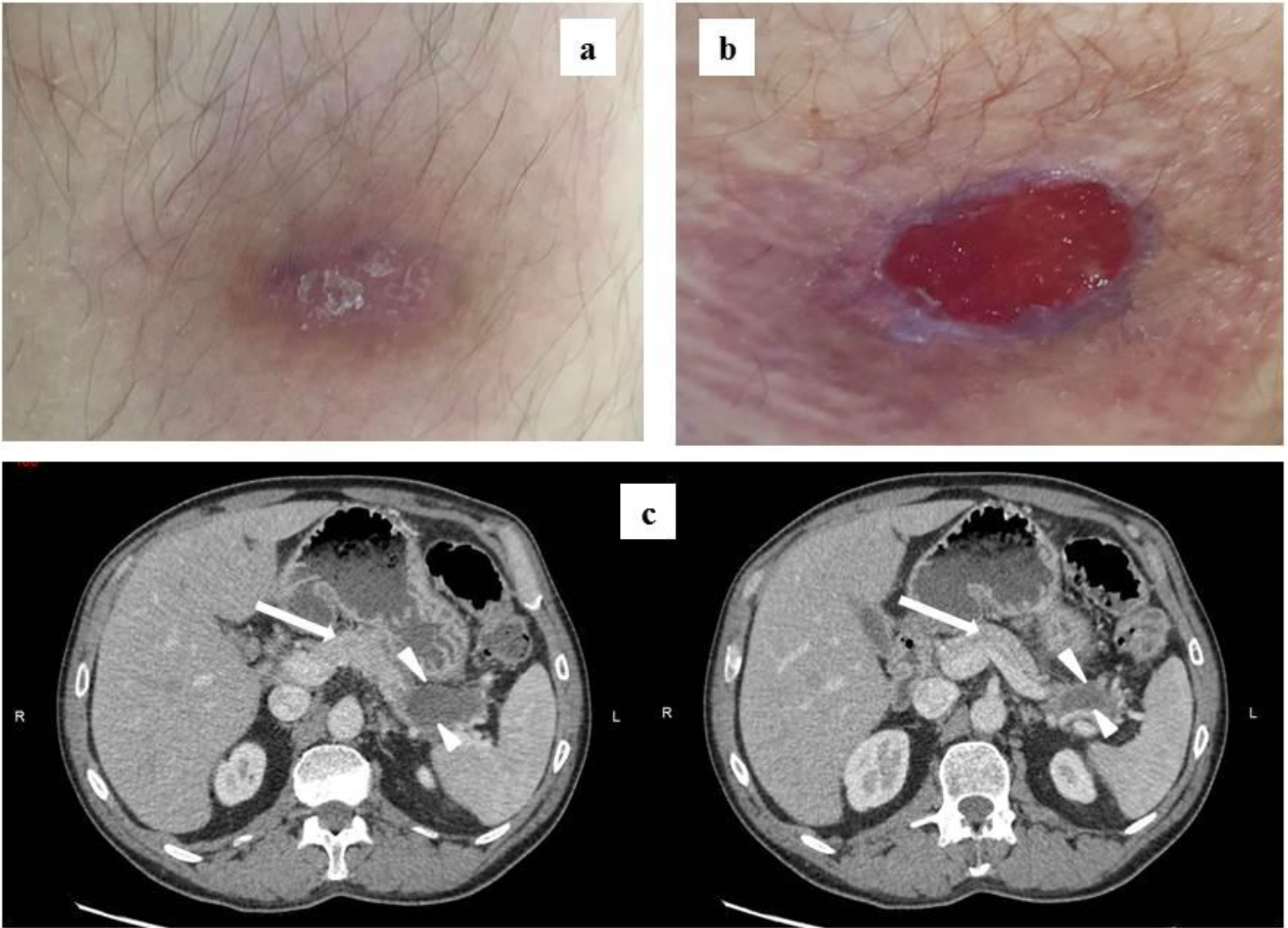
A Rare Localisation of Osteoid Osteoma in a Young Male

DOI: 10.31038/IJOT.2019254

Abstract

Osteoid Osteoma is a benign bone tumor accounting for 10-12% of all benign bone tumors. There is an evident male: female ratio of 2:1. The far most common site of location is the long bones of the lower extremities, which accounts for approximately 50% of all osteoid osteomas. Osteoid osteoma in the hand is a rare finding, accounting for only six to 13%. When found in the hand, the phalanges are the most frequent localisation followed by the carpals. The metacarpals are the least frequent site of location of OO in the hand.

We present a case of a 19-year-old male with characteristic symptoms of osteoid osteoma during a nine-month period. The patient presented with long-term pain at the tumor site with nocturnal worsening. Plain radiography and MR showed a characteristic nidus suggesting the diagnosis of osteoid osteoma.

Treatment of osteoid osteoma can be non-surgical treatment with the use of non-steroidal anti-inflammatory drugs. Since few reports have suggested a possible transformation of osteoid osteoma into malignant osteoblastoma, most patients are treated surgically with en-bloc resection either open or CT-guided depending on local resources.

Keywords

Osteoid osteoma; Metacarpal Bone; Rare Localisation

Introduction

Osteoid Osteoma (OO) accounts for approximately 10-12% of all benign bone tumors [1]. The tumors are most often localised in the long bone of the lower extremities. Localisation of the hand accounts for only 10%. Of these the phalanges are the most frequent localisation followed by the carpals. The metacarpals are the least frequent site of localisation in the hand.

We present a case of a very rare localisation of osteoid osteoma in the metacarpal bone of a young male.

Case

We present a case of osteoid osteoma localised in the first metacarpal bone of a 19-year old male initially examined in our ambulatory care unit in august 2019.

The patient presented with symptoms of long-term localised pain at the first metacarpal bone of the right hand. The patient reported increasing pain at night with intermittent awakenings due to pain. Pain had evolved during the past nine months. The patient noticed no swelling, tenderness or restricted range of motion. At the clinical examination neither swelling nor decreased range of motion was observed. There were no sensory disturbances and no reduction of strength or function. No history of trauma or family history of OO was present either.

Initially a plain radiography was obtained showing a characteristic nidus with periostal thickening raising a high suspicion of osteoid osteoma despite its rare localisation (see figure 1-2).

IJOT 19 - 129-Kristiansen LH-Fn1

Figure 1 and 2. Radiographies obtained initially showing an arrow pointing at the characteristic nidus.

An additional MR scan with contrast was obtained afterwards also suggesting the suspected diagnosis of osteoid osteoma. Final diagnosis is only possible to obtain after histological examination postoperatively.

In this case the nidus of the OO was obvious on the initial radiography and we decided to perform only an MR to exclude other malignant differential diagnosis (see figure 3-4)

IJOT 19 - 129-Kristiansen LH-Fn2

Figure 3 and 4. MR T1 sequences with intravenous contrast showing a characteristic nidus, approximately 4 mm in diameter. Thickening of the cortex in the distal 2/3 of the metacarpal bone.

The patient was discussed in a multicenter sarcoma conference and it was decided to offer the patient surgical removal of the ostoid osteoma by curretage in another hospital.

Discussion

Osteoid osteoma is a benign bone-forming tumor first described in 1935 by Jaffe [2]. Osteoid osteoma accounts for approximately 10-12% of all benign bone tumors [1]. There is an evident male predilection with a male: female ratio of 2:1. OO is most often diagnosed in the second decade of life, principally between 7 and 25 years of age, peaking around 15 years of age [3-5].

The far most common site of location for OO is the long bones of the lower extremities, particularly femur and tibia. These localisations account for approximately 50% of all osteoid osteomas [1]. 10% are localised in the spinal column [3].

Evaluation by a radiologist also suggests osteoid osteoma.

Localisation of OO in the hand is a rare finding accounting for only six to 13% of all osteoid osteomas [5]. When found in the hand, the phalanges are the most frequent localisation followed by the carpals. The metacarpals are the least frequent site of location of OO in the hand [1]. The tumor is most frequently localised in the cortical bone in the diaphysis or metaphysis [1].

The etiology for OO is unknown but some studies suggest familiar disposition and trauma as possible causes [6,8]. Exact pathogenesis of OO also remains unknown. Vasodilation and local inflammation at the tumor site is thought to be caused by prostaglandin E2 and prostacyclin found within the nidus [3,9] Former studies demonstrated bundles of nerve fibres within the nidus contributing to pain and local oedema stimulated by prostaglandins within the nidus [1].

First suspicion of OO most often occurs due to the characteristic symptoms including an initial plain radiography.

In plain radiography OO appears as an oval lytic lesions surrounded by bone thickening and sclerosis. However the central nidus is not always apparent [3]. Not all OO lesions can be diagnosed in radiography, but this is often the primary modality of choice. Further imaging modalities are necessary if there is a high suspicion of OO, although there is no finding on plain radiography [3].

Computed Tomography (CT) is considered the modality of choice in diagnosing OO to distinguish the lesion from other differential diagnosis. The nidus is visualised as a low-attenuated central zone with variable surrounding sclerosis. CT is an important imaging method when plain radiography does not reveal the lesion or with intraarticular lesions.

Visualisation of the OO lesion is very variable with MR diagnostics [1]. Compared to MR, CT is more specific in identifying a nidus [3]. MR benefits from its ability to detect soft tissue involvement. If the nidus is located close to the medullary zone MR has a greater role in diagnosing and visualising the lesion compared to CT. When using MR as the only imaging modality there is a potential of misdiagnosis or overseeing the lesion [3].

Other imaging modalities that can be used in diagnosing OO is bone scintigraphy and PET scans.

In this case the nidus of the OO was very obvious on the initial radiography and we decided to perform only an MR to exclude other malignant differential diagnosis.

Since the etiology of OO is non-malignant and the lesion has a history of spontaneous healing non-surgical treatment can be considered. Non-Steroidal Anti-Inflammatory Drugs (NSAID’s) are proven effective in relieving pain, according to the presence of prostaglandin production in the nidus. Side effects of NSAID’s are an important consideration in long time treatment. Knowledge according to prolonged medical use in treating OO lesions lacks. Studies recommend caution using NSAID’s due to lack of knowledge of long-time use [1,3].

Few reports have suggested that OO can progress to malignant osteoblastoma with prolonged NSAID use [10]. Non-surgical management is a justifiable treatment if the localisation of the lesions is for example intraarticular or difficult to remove or if the patient is not willing to go through surgery.

Surgical procedures are suitable for patients not responding to medical treatment, patients with severe pain or patients not willing to accept possible long-term effects of NSAID treatment. Children with remaining growth potential and open physes in risk of limb-length discrepancy scoliosis and osteoarthritis are also surgical candidates [3].

En bloc resection with complete resection of the total nidus is a frequently used surgical option. For complete pain relive and minimal risk of recurrence it is necessary to resect the entire nidus. Resection of the surrounding sclerotic bone is not required [3]. The use of this procedure may leave behind a bone defect possibly requiring bone grafting or internal fixation. Postoperative restrictions in activity and weight bearing are often recommended after en bloc resection [3]. Challenges in using this technique are difficulties of identifying the lesion perioperatively, possibly resulting in incomplete resection or removal of too much bone tissue [1].

CT guided percutaneous excision is an increasingly used alternative surgical procedure due to reduced morbidity. The procedures include cryoablation, radiofrequency ablation and laser thermocoagulation amongst others. The possibility of perioperative CT visualisation is a great advantage compared to the open en bloc resection. Several studies have shown good results with minimal relapses, low morbidity and few postoperative restrictions. The procedure is though not available in all hospitals [1,3].

Irrespective of what technique is used biopsies are always required to confirm the diagnosis.

This particular patient was offered open curretage of the lesion in another hospital with a sarcoma center. The patient has not yet decided whether to accept the offer of surgery.

Only a few other cases have presented OO localised in the metacarpal bone, which makes this case extraordinary. When patients present with the characteristic symptoms of localised pain and nocturnal worsening in the young male population it is important to remember that OO can present in upper extremity though very rare. Plain radiography is always a simple initial examination to perform.

The patient involved in this case report gave consent to the use of the patient history in this article.

References

  1. Atesok KI, Alman BA, Schemitsch EH, Peyser A et al. (2011) Osteoid osteoma and osteoblastoma. J Am Acad Orthop Surg 19: 678-689.
  2. JAFFE HL. Osteoid-osteoma. Proc R Soc Med 46: 1007-1012.
  3. Noordin S, Allana S, Hilal K, et al. (2018) Osteoid osteoma: Contemporary management. Orthop Rev (Pavia) 10: 7496.
  4. El Fatayri B, Djebara AE, Fourdrain A, Bulaid Y et al (2019) Resection of a rare metacarpal distal condyle osteoid osteoma. Case Rep Orthop 2019: 4542862.
  5. Brohard J, Tsai P (2019) Osteoid osteoma in the thumb of an adolescent patient. J Hand Surg Am 2019.
  6. Seker A, Unal MB, Malkoc M, Kara A et al. (2016) A rare localization of osteoid osteoma – presentation of two cases. Srp Arh Celok Lek 144: 553-556.
  7. Chronopoulos E, Xypnitos FN, Nikolaou VS, Efstathopoulos N et al. (2008) Osteoid osteoma of a metacarpal bone: A case report and review of the literature. J Med Case Rep 2: 285-1947-2-285.
  8. Kalil RK, Antunes JS (2003) Familial occurrence of osteoid osteoma. Skeletal Radiol 32: 416-419.
  9. Makley JT, Dunn MJ (1982) Prostaglandin synthesis by osteoid osteoma. Lancet 2: 42-6736(82)91174-6.
  10. Bruneau M, Polivka M, Cornelius JF, George B (2005) Progression of an osteoid osteoma to an osteoblastoma. case report. J Neurosurg Spine 3: 238-241.

Oxidative Stress and Haemolytic Anaemia In Dogs and Cats: A Comparative Approach

DOI: 10.31038/IJVB.2019331

Abstract

Oxidative stress contributes to Haemolytic Anaemia in many species including dogs and cats, as well as in humans. Red cells are exposed to a continual oxidant challenge, both endogenously from within the red cells themselves and also exogenously from other tissues, and from ingested or administered oxidants. When the oxidative challenge exceeds the antioxidant provisions of the red cell, damage occurs in the form of lipid and protein peroxidation, cytoskeletal crosslinking, oxidation of haemoglobin to methemolglobin, and precipitation of denatured sulphhaemoglobin as Heinz bodies. These deleterious sequelae produce fragile red cells with reduced lifespan, and result in poorer oxygen delivery to tissues, intravascular haemolysis, anaemia, haemoglobinuria and jaundice. A number of features increase the risk of oxidant damage in dogs and cats. Thus dog red cells have low levels of the antioxidant enzyme catalase. Cat haemoglobin has at least four times as many readily oxidizable thiol residues compared to most species, whilst their hepatic capacity for glucuronidation is much reduced, which can result in greater accumulation of oxidants. Like humans, both species may also be exposed to excess oxidants from systemic diseases such as diabetes mellitus, hepatic lipidosis, hypophosphatemia and neoplasias. Iatrogenic oxidants include drugs such as acetaminophen and other non-steroidal anti-inflammatory compounds. Ingested toxins include heavy metals, particularly important in dogs with their increased propensity for scavenging. Ingestion of feeds containing products from Allium species of plants has also long been associated with red cell oxidative damage and Heinz body formation in both dogs and cats. Though less common than in humans, there are occasional congenital enzyme deficiencies which reduce the enzymatic oxidant defence of the red cells in these species. Treatment usually relies on removal of the oxidant challenge or support against the resulting anaemia. Specific antioxidants currently lack efficacy but analogy with human medicine suggests that a range possible antioxidants may be potentially beneficial.

Key words

Antioxidant Defence, Dogs and Cats, Haemolytic Anaemia, Oxidative Stress

Introduction

Red cells occupy a unique position within the vertebrate body. When mature, they are enucleated and lack cytoplasmic organelles [1]. As such, they are therefore unable to carry out ribosomal protein synthesis or mitochondrial oxidative phosphorylation. They are dependent upon glycolysis (or the Emden-Meyerhoff pathway) for whatever ATP supply is required to maintain their osmotic integrity, through various ion pumps, and for other energy requiring events, like synthesis of reduced glutathione, one of their main antioxidant defences [2, 3]. All vertebrate red cells have the main task of carriage of blood gases, oxygen from respiratory tissues and carbon dioxide from metabolically active tissues. Notwithstanding, there are some surprising species differences in function, which are significant both physiologically and pathologically [1]. For example, most vertebrate red cells contain high levels of K+ and low levels of Na+, whose gradients are maintained through the functioning of the ATP-dependent Na+/K+ pump in the red cell membrane. This pump, together with a normally low passive “leak” to Na+ and K+ prevent osmotic swelling which would otherwise occur through the large cytoplasmic load of impermeable protein, especially haemoglobin (Hb), and other molecules, notably organic phosphates [4]. By contrast, dog and cat red cells are usually low in K+ and high in Na+. When mature – but not during development – their red cells lack Na+/K+ pumping capacity and rather they use combinations of Ca2+ pumps and Na+/Ca2+ exchange proteins to maintain osmotic equilibrium [5]. An exception is high K+-containing red cells of certain Asian breeds for example, the Japanese Shibas and Akitas [6] which retain Na+/K+ pumping capacity, and also high levels of the antioxidant reduced glutathione, throughout their lifespan. There are also other differences in physiology of dog and cat red cells pertinent to the subject of this review, and which are considered later.

Dog and Cat Red Cells

In the absence of shear stress, human red cells have the classic biconcave shape with a diameter of about 8 µm. Dog and cat red cells have a similar appearance but are somewhat smaller, at 7 µm and 5.5–6.3 µm, respectively [7]. Cat red cells, in particular, show a degree of anisocytosis and also tend to lack the central pallor which is easily recognizable in the more obviously biconcave shape of dog and human red cells. The oxygen-carrying pigment Hb is found in all vertebrates with the exception of a few species of Antarctic fish [8]. The latter live at subzero temperatures and thereby survive and carry out aerobic metabolism using only the additional oxygen dissolved in plasma at these low temperatures. There are species variations in Hb, however. In this context, cat Hb is noticeable in having 8–10 readily oxidizable sulphydryl groups [9, 10] whilst most other species including humans and dogs have only two main ones, represented by the highly conserved β93 cysteines [11, 12]. Cat Hb also readily dissociates from the usual tetrameric form to dimers [13] which have a greater tendency for autoxidation [14]. Heinz bodies, denatured, precipitated sulphHb, are a special feature of oxidative stress [14]. They are also found in the circulation of healthy cats, however, at up to 5–10 % red cells, presumably because of their greater number of oxidative sites in Hb and impaired red cell antioxidant defence, together with the poor ability of the non-sinusoidal feline spleen to remove Heinz body-containing red cells [15]. Cats also have two main Hbs A and B [9, 16]. HbA is most prevalent in domestic short- and long-haired cats have HbA (98 %) but a few breeds have greater levels of HbB (eg 10 % Persians and 14 % in Abyssinians, with as much as 50 % in Devon Rexs) and geographically to occur {eg [17]. The oxygen affinity of many species is reduced by organic phosphates, especially 2,3-diphosphoglycerate (2,3-DPG or 2,3-biphosphoglycerate), but cat HbA is less responsive to the reduction in P50 whilst HbB does not respond at all [18, 19]. Cat red cells also have low levels of 2,3-DPG [20] which is understandable if it has little regulatory effect on oxygen affinity. Dogs have also several Hbs and more than twelve blood groups [21] but react like human Hb to 2,3-DPG.

Red Cell Metabolism

Mature red cells lack mitochondria and are therefore dependent on anaerobic glycolysis for ATP synthesis [3]. Compared with the citric acid (Kreb cycle) of aerobic respiration this is relatively inefficient, producing two molecules of ATP per glucose moiety (compared with thirty six in mitochondrial aerobic respiration). Glycolysis comprises ten enzymatic steps [1], although the main rate limiting enzymes are hexokinase and pyruvate kinase, at the start and end of the chain, respectively. In addition to ATP, the pathway also syntheses reducing power in the form of NADH. NADH is necessary to reduce methaemoglobin (metHb) using methaemoglobin reductase (or cytochrome b reductase) – one of the main red cell antioxidant defences. An off-shoot of the glycolytic pathway called the pentose phosphate shunt (or hexose monophosphate shunt) is used to make the reducing compound NADPH, a substrate for glutathione reductase – a second main antioxidant enzyme – which reduces oxidised glutathione (GSSG) back to reduced glutathione (GSH). Under normal conditions, glycolysis uses the majority of glucose metabolised by the red cell, with the pentose phosphate shunt accounting for only about 10 % of the flux. Inhibition of the first enzyme of the pentose phosphate shunt, glucose-6-phosphate dehydrogenase, by high NADPH / NADP ratios is responsible and this enzyme normally operates at only a low level of its maximum capacity. Under conditions of oxidative stress, however, as NADPH / NADP ratios fall, glucose is preferentially channelled along the pentose phosphate shunt. Interestingly, deoxyHb which preferentially binds to the cytoplasmic tail of the anion exchanger (or Band 3) displaces glycolytic and other enzymes so that deoxygenated red cells carry out more glycolysis, oxygenated ones produce more NADPH [22, 23] providing a physiological switch to channel glucose through one or other pathways. In addition, the red cells of some species are less permeable to glucose, eg some fish and pigs [1, 24, 25]. In these cases, the pentose phosphate shunt pathways can be used as an alternative to glycolysis for synthesis of ATP, metabolising nucleosides, such as inosine and metabolites of ribose, which enter into the distal part of the glycolytic pathway.

The third red cell metabolic pathway of note is the Rapaport-Luebering shunt (1950s). This uses the enzyme biphosphoglycerate mutase to produce 2,3-DPG (2,3-BPG) – apparently confined to cells of the erythroid lineage and placental cells [26] and accounts for about 20 % of the glucose passing through glycolysis. There is a metabolic cost to this, as the Rapaport-Luebering shunt bypasses phosphoglycerate kinase with the loss of one ATP of the two molecules of ATP from metabolism of glucose. Congenital enzyme deficiencies in the red cell metabolic pathways have been well described in humans [2, 27]. Some genetic deficiencies have also been described in dogs and cats [Table 1]. Whilst oxidative threat is not the root of these conditions, a defect in antioxidant defences will accompany the inadequacies in glucose metabolism which underlie the loss of ATP, and which represents the main cause of red cell instability.

Table 1. Some inherited causes of haemolytic anaemia in dogs and cats.

Catalase

American foxhound, beagle [55]

Hereditary elliptocytosis

Band 4,1 deficiency [56]

Hereditary spherocytosis

Autosomal recessive trait in chondrodysplastic Alaskan malamute dwarf dogs

Hereditary stomatocytosis

Schnauzers [57,59]

Methaemoglobin redutase

Dogs (toy Alaskan Eskimo, miniature poodle, cocker/poodle cross) and cats – domestic short hair [60,61,62]

Osmotic fragility syndrome

Abyssinian, Somali, Siamese and domestic short hair cats [63–64]

Phosphofructokinase (PFK) deficiency

English springer spaniels, American cocker spaniels, whippets [65–66]

Pyruvate kinase (PK) deficiency

Basenjis, Cairn terrier, West Highland white terriers, beagles, cairn terriers, miniature poodles, dachshunds, Chihuahus, American Eskimo toy dogs, pugs, American Labrador retrievers; Abyssinian, Somali and domestic shorthaired cats [67]

Oxidative Challenge

Red cells are also subject to considerable oxidative stress throughout their lifespan. Oxidative challenges arise from several underlying conditions and sources [28, 29]. First, oxygen is potentially toxic and their function as the main oxygen-carrying cell of the body exposes them continually to the threat of oxygen damage. Whilst in other tissues, there is always some slippage of oxygen away from its mitochondrial function in aerobic respiration, which generates superoxide anion and other free radicals, in red cells, the iron-containing Hb is the major source of reactive oxygen species [29, 30]. The ferrous Fe2+ in heme groups is potentially unstable and liable to autoxidation to ferric Fe3+, generating superoxide and, through dismutation, hydrogen peroxide [31] which may be removed by one of the important red cell antioxidant enzymes, catalase. Heme iron is also able to take part in the Fenton and Haber-Weiss reactions to generate hydroxyl and other free radicals [2, 32]. Red cell NADPH oxygenase is a further source of endogenous oxidants [28, 33]. Around 0.5–3 % red cell haemoglobin is oxidized daily [34], producing a constant source of methaemoglobin, although levels are usually kept below 1 % through the reducing action of methaemoglobin reductase [35]. In addition, there is the threat from exogenous oxidants which may enter the circulation from other tissues, for example following ischaemia / reperfusion [36], or the action of xanthine oxidase on hypoxanthine [37] or also via ingested or iatrogenic oxidants [7]. Cat Hb more susceptible to oxidants (Harvey & Kaneko 1976), especially feline HbB cf feline HbA. Counterintuitively, dogs with red cells containing high levels of K+, and also high levels of the antioxidant reduced glutathione notably Japanese breeds [38] appear more susceptible to oxidative damage than the more common low K+ ones. A number of systemic diseases are associated. Some of these include diabetes mellitus, hepatic problems, hyperthyroidism (especially in cats), neoplasia, severe hypophosphataemia (eg refeeding syndrome in cats) and uraemic syndrome.

Oxidative red cell damage from ingestion of products from Allium species (onions, garlic and related plants – see [39] for a list of plants) are particularly heavily implicated in the case of dogs and cats. Onion poisoning in dogs has been recognised since the 1930s [40] and is due mainly to sulphur-containing organic compounds, which give the characteristic odour of these foods [39]. These compounds are not destroyed by cooking or spoilage. Metabolites particularly propylsulphides are implicated in onion-induced oxidant damage of red cells in dogs and cats [41]. Animals probably need to consume about 0.5 % of their body weight in onions to be affected [42], though of course the wet weight and the concentration of the active ingredient will be very variable between feedstuffs. Cats are less frequently affected by Allium spp. toxicity because of their dietary preferences though cases do occur, for example in ill animals fed on human baby food [43]. Ironically, the same sulphur-containing organic compounds which cause harm to dogs and cats are associated with the therapeutic benefits of Allium spp. in humans [44]. Cats also have low hepatic glucuronidation capacity. They lack many uridine diphosphate glucuronyltransferases (UGTs) which makes them particularly susceptible to a number of iatrogenic drugs. They thus have a very poor ability to metabolise compounds such as acetaminophen and salicylic acid [45], for which there is no safe dose. In both dog and cat, overdoses with acetaminophen leads to the accumulation of metabolites such as p¬-aminophenol (PAP) in their red cells, which lack N¬-acetyltransferase 2 (NAT2) to remove it. The result is methaemoglobinaemia [46]. Overdose in other species including humans, by comparison, is associated with hepatic toxicity induced by the metabolite N-acetyl-p-benzoquinoneimine (NADPQI) rather than oxidative damage to red cells. Heavy metals are also implicated in oxidative damage to red cells, particularly in dogs. Commoner causes include zinc toxicity (through ingestion of toys, bolts or coins containing high levels of zinc) [47] or iron overload. The latter is usually iatrogenic through iron injections or repeat transfusions. Some other common iatrogenic oxidants and toxins are listed in [Table 2], with a more complete list is provided in Haematology texts eg [7].

Table 2. Some toxins and iatrogenic oxidants causing haemolytic anaemia in dogs and cats.

Acetaminophen (paracetamol)

Acetylsalicylic acid (aspirin)

Allium spp.

Benzocaine

Carprofen and other non-steroidal anti-inflammatories

Copper

Iron overload

DL-methionine

Methylene blue

Phenylhydrazine

Propylene glycol

Vitamin K and vitamin K antagonists

Zinc

Red Cell Antioxidant Defence

Notwithstanding the potential oxidative peril and their limited capacity for repair by protein synthesis, red cells must survive for some one hundred and twenty days in the case of humans and dogs, and about seventy days in the case of cats. Although the red cell is well equipped with antioxidant defences, problems arise when oxidative challenge exceeds the red cell antioxidant capacity. The result is oxidative damage to membrane lipids and proteins, and to haemoglobin itself. Oxidised haemoglobin, methaemoglobin (heme Fe3+ instead of the normal Fe2+), is unable to carry oxygen and is also liable to denaturation and precipitation as insoluble sulphHb containing Heinz bodies, or to form eccentrocytes in which the Hb is restricted to one side of the cell [13]. Other changes include crosslinking of the cytoskeleton, thiol oxidation, depletion of reduced glutathione and cation imbalance. The result is a fragile red cells with impaired rheology liable to intravascular haemolysis with anaemia, haemoglobinuria and poor oxygen-carrying capacity [48].

Antioxidant provision of red cells is provided by both enzymatic and non-enzymatic pathways. Five enzymes are heavily involved: catalase which reduces hydrogen peroxide to oxygen and water, glutathione reductase uses NADH to reduce oxidised methaemoglobin, superoxide dismutase scavenges superoxide anions generating hydrogen peroxide and oxygen in the process, and glutathione peroxidase uses NADPH to remove both red cell hydrogen peroxide and organic peroxides [49], as does membrane-associated perioxiredoxin-2 which can be reduced via reduced glutathione, vitamin C or thioredoxin. Activities of these enzymes do vary between species [50–52]. Catalase activity in the red cells of difference species is very variable [50, 53, 54]. Expression in dog red cells occurs at about a tenth of the amount in humans whilst its specific activity is around a third that of human catalase [55]. As a result, overall catalase activity in dog red cells is a thirtieth that in humans [53, 55]. Non-enzymatic defence includes reduced glutathione, vitamin C and vitamin E. Therapeutic antioxidants include dosing with N-acetyl cysteine, vitamin C and E. None are particularly effective for rapid protection [39]. There is a need for more efficacious compounds. These must be effective in the short term and protect red cells from further oxidative damage and haemolysis without the requirement for prolonged metabolism. Some human compounds are listed in [Table 3].

Table 3. Antioxidants used in chemoprophylaxis of sickle cell disease in humans.

Therapy

Effect

References

Acetyl-L-carnitine

Protects red cells from peroxidative damage and maintains normal shape at lower oxygen tensions

[68]

N-Acetylcysteine

Increases levels of reduced glutathione and decreases haemolysis

[69,70]

Flavonoids (quercetin, rutin & morin

Show inhibitory effect on haemolysis due to thiol group oxidation

[71]

Glutamine

Increases NAD redox potential and NADH levels

[72,73]

Hydroxyurea

Reduces markers of oxidative stress, decreases lipid peroxidation and increases level of antioxidant enzymes

[74,75]

Iron chelators: deferiprone & deferasirox

Remove iron from the membrane of red cells, decrease lipid peroxidation and increase antioxidant capacity

[76,77]

α-lipoic acid

Protects red cells from peroxyl radical induced haemolysis, increases levels of reduced glutathione and increased antioxidant gene expression

[78,79]

Melatonin

Increases levels of antioxidants and reduces rate of haemolysis

[80]

Statins

Protects against oxidative damage by increasing nitric oxide metabolites and C-reactive protein

[81,82]

Vitamin C and E

Decreases production of reactive oxygen species, increases levels of reduced glutathione and reduces haemolysis

[83]

References

  1. Nikinmaa M (1990) Vertebrate red blood cells. Zoophysiology ed. S.D. Bradshaw. Berlin Heidelberg: Springer-Verlag.
  2. Arese P and E Schwarzer (2003) Metabolic disorders in Red cell membrane transport in health and disease, I. Bernhardt and JC Ellory, Editors. Springer: Berlin. Pg No: 525–548.
  3. Kaneko JJ, JW Harvey, ML Bruss (2008) Clinical Biochemistry of Domestic Animals. Academic Press: New York.
  4. Tosteson DC and JF Hoffman (1960) Regulation of cell volume by active cation transport in high and low potassium sheep red cells. Journal of general physiology 44: 169–194.
  5. Parker JC (1997) Solute and water movement in dog and cat red blood cells., in Membrane Transport in Red cells, JC Ellory and VL Lew, Editors. Academic Press: London. Pg No: 427–465.
  6. Fujise H (1997) K+-Cl- cotransport and volume regulation in the light and the dense fraction of high K+ dog red blood cells. American Journal of Physiology 273: R2991-R2998.
  7. Weiss D and J Wardrop (2010) Schalm’s Veterinary Hematology. Wiley-Blackwell: Iowa, USA.
  8. Sidell BD and KM O’Brien (2006) When bad things happen to good fish: the loss of hemoglobin and myoglobin expression in Antarctic ice fishes. Journal of Experimental Biology 209: 1791–1802.
  9. Taketa F, MR Smits, JL Lessard (1968) Hemoglobin heterogeneiry in the cat. Biochemical and Biophysical Research Communications 30: 219–226.
  10. Harvey JW and JJ Kaneko (1977) Mammalian erythrocyte metabolism and oxidant drugs. Toxicological and Applied Pharmacology 42: 253–261.
  11. Reischl E (2007) Distribution, adaptation and physiological meaning of thiols from vertebrate hemoglobins. Comparative Biochemistry and Physiology Part C 146: 22–53.
  12. Vitturi DA (2013) Antioxidant functions for the hemoglobin beta-93 cysteine residue in erythrocytes and in the vascular compartment in vivo. Free Radical Biology and Medicine 55: 1–23.
  13. Hamilton MN and SJ Edelstein (1973) Cat hemoglobin: pH dependence of cooperativity and ligand binding. Journal of Biological Chemistry 249: 1323–1329.
  14. Christopher MM, JG White, JW Eaton (1990) Erythrocyte pathology and mechanisms of Heinz body-mediated hemolysis in cats. Veterinary Pathology 27: 299–310.
  15. Blue JT, L Weiss (1981) Vascular pathways in nonsinusoidla red pulp: an electron microscope study of the cat spleen. American Journal of Anatomy 161: 135–168.
  16. Lessard JL and F Taketa (1969) Multiple hemoglobins in fetal, newborn and adult cats. Biochemical et Biophysical Acta 175: 441–444.
  17. Giger U (1991) Geographical variation of the feline blood type frequencies in the United States. Feline Practice 19: 21–27.
  18. Bunn HF (1971) Differences in the interaction of 2,3-diphosphoglycerate with certain mammalian hemoglobins. Science 172: 1049–1050.
  19. Mauk AG and F Taketa (1972) Effects of organic phosphates on oxygen equilibria and kinetics of -SH reaction in feline hemoglobins. Archives of Biochemistry and Biophysics 150: 376–381.
  20. Rapoport S and GM Guest (1941) Distribution of acid-soluble phosphorus in the blood cells of various vertebrates. Journal of Biological Chemistry 138: 269–282.
  21. Zaldivar-Lopez S (2017) Comparative genomics of canine hemoglobin genes reveals primary of beta subunit delta in adult carnivores. BMC Genomics 18: 141.
  22. Low PS, P Rathinavelu, ML Harrison (1993) Regulation of glycolysis via reversible enzyme binding to the membrane protein, band 3. Journal of Biological Chemistry 268: 14627–14631.
  23. Chu H (2016) Reversible binding of hemoglobin to band 3 constitutes the molecular switch that mediates O2 regulation of erythrocyte properties. Blood 128: 2708–2716.
  24. Kim HD and MG Luthra (1976) Pig reticulocytes: I. Transitory glucose permeability and metabolism. American Journal of Physiology 230: 1668–1675.
  25. Young JD, ARP Paterson, PJF Henderson (1985) Nucleoside transport and metabolism in erythrocytes from the Yucatan miniature pig. Evidence that inosine functions as an in vivo energy substrate. Biochimica et Biophysica Acta 842: 214–224.
  26. Pritlove DC (2006) Novel placental expression of 2,3-biphosphoglycerate mutase. Placenta 27: 924–927.
  27. Van Wijk R and WW van Solinge (2005) The energy-less red blood cell is lost: erythrocyte enzyme abnormalities of glycolysis. Blood 106: 4034–4042.
  28. Mohanty JG, E Nagababu, JM Rifkind (2014) Red blood cell oxidative stress impairs oxygen delivery and induces red blood cell aging. Frontiers in Physiology 5: 1–6.
  29. Fibach E (2014) Involvement of oxidative stress in hemolytic anemia, in Systems Biology of Free Radicals and Antioxidants, I. Laher, Editor. Springer-Verlag: Berlin Heidelberg 2499–2516.
  30. Sies H (1997) Oxidative stress: oxidants and antioxidants. Experimental Physiology 82: 291–295.
  31. Hebbel RP (1982) Spontaneous oxygen radical generation by sickle erythrocytes. Journal of Clinical Investigation 70: 1253–1259.
  32. Sadrzadeh SMH (1984) Hemoglobin: a biologic Fenton reagent. Journal of Biological Chemistry 259: 14354–14356.
  33. George A (2013) Erythrocyte NADPH oxidase activity modulated by Rac GTPases, PKC, and plasma cytokines contributes to oxidative stress in sickle cell disease. Blood 121: 2099–2107.
  34. Jaffe ER (1974) The formation and reduction of methemoglobin in human erythrocytes, in Cellular and molecular biology of erythrcoytes, H. Yoshikawa and S.M. Rapaport, Editors. University Park Press: Baltimore. 345–376.
  35. Edwards CJ and J Fuller (1996) Oxidative stress in erythrocytes. Comparative Haematology International 6: 24–31.
  36. Zweier JL and MA Talukder (2006) The role of oxidants and free radicals in reperfusion injury. Cardiovascular Research 70: 181–190.
  37. Balagopalakrishna C (1996) Production of superoxide from hemoglobin-bound to oxygen under hypoxic conditions. Biochemistry 35: 6393–6398.
  38. Yamoto O and Y Maede (1992) Susceptibility to onion-induced hemolysis in dogs with hereditary high erythrocyte reduced glutathione and potassium concentrations. American Journal of Veterinary Research 53: 134–137.
  39. Salgado BS, LN Monteiro, NS Rocha (2011) Allium species poisoning in dogs and cats. Journal of Venomous Animals and Toxins including Tropical Diseases 17: 4–11.
  40. Harvey JW and D Rackear (1985) Experimental onion-induced hemolytic anemia in dogs. Veterinary Pathology 22: 387–392.
  41. Zhao J (2017) Therapeutic effect of hydrogen injected subcutaneously on onion poisoned dogs. Journal of Veterinary Research 61: 527–533.
  42. Cope RB (2005) Allium species poisoning in dogs and cats. Veterinary Medicine Pg No: 562–566.
  43. Robertson JE, MM Christopher, QR Rogers (1998) Heinz body formation in cats fed baby food containing onion powder. Journal of the American Veterinary Medical Association 212: 1260–1266.
  44. Augusti KT (1996) Therapeutic values of onion and garlic. Indian Journal of Experimental Biology 34: 634–640.
  45. Court MH (2013) Feline drug metabolism and disposition: pharmacokinetic evidence for species differences and molecular mechanisms. Veterinary Clinics of North America Small Animal Practice 43: 1039–1054.
  46. McConkey SEM, DM Grant, AE Cribb (2009) The role of Para-aminophenol in acetaminophen-induced methaemogloinaemia in dogs and cats. Journal of Veterinary Pharmacology and Therapeutics 32: 585–595.
  47. Bexfield N, J Archer, M Herrtage (2007) Heinz body haemolytic anaemia in a dog secondary to ingestion of a zinc toy: a case report. Veterinary Journal 174: 414–417.
  48. Hasegawa S (1993) Impaired deformability of Heinz-body-forming red cells. Bio rheology 30: 275–286.
  49. Nagababu E, FJ Chrest, JM Rifkind (2003) Hydrogen-peroxide-induced heme degradation in red blood cells: the protective roles of catalase and glutathione peroxidase. Biochimica et Biophysica Acta 1620: 211–217.
  50. Maral J, K Puget, AM Michelson (1977) Comparative study of superoxide dismutase, catalase and glutathione peroxidase levels in erythrocytes of different animals. Biochemical and Biophysical Research Communications 77: 1525–1535.
  51. Stolk JN and RP Smith (1966) Species differences in methaemoglobin reductase activity. Biochemical Pharmacology 15: 343–351.
  52. Owen JL and JW Harvey (2012) Hemolytic anemia in dogs and cats due to erythrocyte enzyme deficiences. Veterinary Clinics of North America Small Animal Practice 42: 73–84.
  53. Richardson M (1952) Study of catalase in erythrocytes and bacteria. II. Catalase activity in erythrocytes from different species of normal animals and from normal humans. Archives of Biochemistry and Biophysics 42: 124–134.
  54. Allison AC, W Rees, GP Burn (1957) Genetically-controlled differences in catalase activity of dog erythrocytes. Nature 180: 649–650.
  55. Nakamura K (1998) A low catalase activity in dog erythrocytes is due to a very low content of catalase protein despite having a normal specific activity. International Journal of Biochemistry and Cell Biology 30: 823–831.
  56. Smith JE (1983) Hereditary elliptocytosis with protein band 4.1 deficient in the dog. Blood 61: 373–377.
  57. Fletch SM and PH Pinkerton (1973) Animal model for human disease: inherited hemolytic anemia in stomatocytosis in the Alaskan Malamute dog. Journal of Pathology 71: 477–480.
  58. Slappendel RJ, W Renooji, JJ de Bruijne (1994) Normal cations and abnormal membrane lipids in the red blood cells of dogs with familial stomatocytosis-hypertrophic gastritis. Blood 84: 904–909.
  59. Bonfanti U (2004) Stomatocytosis in 7 related standard Schnauzers. Veterinary Clinical Pathology 33: 234–249.
  60. Harvey JW (1991) Methaemoglobin reductase deficiency in dogs. Comparative Haematology International 1: 55–59.
  61. Harvey JW, M Dahl, ME High (1994) Methemoglobin reductase deficiency in a cat. Journal of the American Veterinary Medical Association 205: 1290–1291.
  62. Harvey JW (2006) Inherited methaemoglobin redutase deficiency in dogs and cats. Veterinary Clinical Pathology 35: 144–156.
  63. Kohn B, MH Goldschmidt, AE Hohenhaus (2000) Anemia, splenomegaly, and increased fragility of erythrocytes in Abyssinian and Somali cats. Journal of the American Veterinary Medical Association 217: 1483–1491.
  64. Tritschler C (2016) Increased erythrocytic osmotic fragility in anemic domestic shorthair and purebred cats. Journal of Feline Medicine and Surgery 18: 462–470.
  65. Giger U (1985) Inherited phosphofructokinase deficiency in dogs with hyperventilation-induced haemolysis: increased in vitro and in vivo alkaline fragility of erythrocytes. Blood 65: 345–351.
  66. Harvey JW and JE Smith (1994) Haematology and clinical chemistry of English springer spaniel dogs with phosphofructokinase deficiency. Comparative Haematology International 4: 70–75.
  67. Juvet F (2013) Erythrocyte pyruvate kinase deficiency in three West Highland white terriers in Ireland and the UK. Irish Veterinary Journal 66: 12.
  68. Ronca F (1994) Effect of L-proprionyl carnitine on in-vitro membrane alteration of sickle-cell anaemia erythrocytes. International Journal of Tissue Reaction 16: 187–194.
  69. Pace BS (2003) Effects of N-acetyl cysteine on dense cell formation in sickle cell disease. American Journal of Hematology 73: 26–32.
  70. Nur E (2012) N-acetyl cysteine reduces oxidative stress in sickle cell patients. Annals of Hematology 91: 1097–1105.
  71. Asgary S, G Naderi, N Askari (2005) Protective effects of flavonoids against red blood cell hemolysis by free radicals. Experimental Clinical Cardiology 10: 88–90.
  72. Niihara Y (2005) L-glutamine therapy reduces endothelial adhesion of sickle red blood cells to human umbilical vein endothelial cells. BMC Blood Disorders 5: 1–7.
  73. Niihara Y (2018) A phase 3 trial of L-glutamine in sickle cell disease. New England Journal of Medicine 379: 226–235.
  74. Ghatpande SS (2008) Pharmaco-proteomic study of hydroxyurea-induced modifications in the sickle red blood cell membrane proteome. Experimental Biology and Medicine 233: 1510–1517.
  75. Torres LdS (2012) The influence of hydroxyurea on oxidative stress in sickle cell anemia. Reviews of Brazilian Hematology and Hemoter 34: 421–425.
  76. Shalev O (1995) Deferiprone (L1) chelates pathologic iron deposits from membrane of intact thalassemic and sickle red blood cells both in vitro and in vivo. Blood 86: 2008–2013.
  77. Belini Jr E (2012) Oxidative stress and antioxidant capacity in sickle cell anaemia patients receiving different treatments and medications for different periods of time. Annals of Hematology 91: 479–489.
  78. Maragnon K (1999) Comparison of the effect of alpha-lipoic acid and alpha-tocopherol supplementation on measures of oxidative stress. Free Radical Biology and Medicine 27: 1114–1121.
  79. Vichinsky E (2012) Emerging ‘A’ therapies in hemoglobinopathies: agonists, antagonists, antioxidants, and arginine. ASH Education Programme 1: 271–275.
  80. da Silva DG (2015) Potential utility of melatonin as an antioxidant therapy in the management of sickle cell anemia. Journal of Pineal Research 58: 178–188.
  81. Hoppe C (2011) A pilot study of the short term use of simvastatin in sickle cell disease: effects on markers of vascular dysfunction. British Journal of Haematology 153: 655–663.
  82. Davnignon J (2004) Beneficial pleiotropic effects of statins. Circulation 15: 11139–11143.
  83. Amer J (2006) Red blood cells, platelets and polymorphonuclear neutrophils of patients with sickle cell disease exhibit oxidative stress that can be ameliorated by antioxidants. British Journal of Haematology 132: 108–113.

Akt Inhibitors and COL11A1 in Epithelial Ovarian Carcinoma: A Short Note

DOI: 10.31038/IGOJ.2019244

Short Commentary

Epithelial ovarian carcinoma (EOC) is the most lethal gynecologic malignancy. Currently, the treatment of patients with EOC usually includes surgery and chemotherapy [1]. The survival rate of patients with EOC remains low despite advances in surgical techniques and chemotherapy. One of the obstacles to the use of chemotherapy is drug resistance. To improve the survival rate, efforts must be made to overcome chemoresistance.

Akt, a key protein in the Akt/PI3K signaling pathway, is a serine/threonine protein kinase that, once activated by phosphorylation, plays an important role in the process of malignant transformation [2]. The phosphorylated form of Akt (p-Akt) has been implicated in the induction of signals that affect cell apoptosis and the promotion of cell proliferation and invasiveness through mammalian target of rapamycin (mTOR) activation [3]. Investigations have shown that overexpressed p-Akt is associated with a poor prognosis of human cancer [4–6] that includes ovarian cancers [7–9]. Our recent report showed that patients with tumors overexpressing p-Akt had a poorer survival rate, and the p-Akt overexpression was associated with high-grade tumors and cancer death [10]. In addition, more patients with high p-Akt levels were allocated to the group of clinically defined chemoresistance, although this difference did not achieve statistical significance [10]. Therefore, p-Akt overexpression may be a common prognostic factor shared by multiple types of human cancers, and thus has the potential to be a therapeutic target of clinical significance.

Collagen type XI alpha 1 (COL11A1) belongs to the collagen family, which is the major component of the interstitial extracellular matrix. We previously found that COL11A1 plays an important role in EOC. Our results indicated that COL11A1 promotes tumor progression by up regulating the transforming growth factor-β1 (TGF-β1)/matrix metalloproteinase-3 (MMP3) axis, through the involvement of the nuclear transcription factor Y subunit alpha (NF-YA) binding site in the COL11A1 promoter, and predicts a poor clinical outcome in ovarian cancer patients [11]. We also found that COL11A1 promotes cancer cell sensitivity to anticancer drugs via activation of the Akt/c/EBPβ (CCAAT/enhancer-binding protein beta) pathway and attenuates phosphoinositide-dependent kinase 1 (PDK1) ubiquitination and degradation [12]. In addition, COL11A1 reduced chemotherapy-induced apoptosis through up regulating Twist-related protein 1 (TWIST1)-mediated induced myeloid leukemia cell differentiation protein (Mcl-1) and growth arrest-specific 6 (GAS6) expression [13]. Our recent report indicated that SC66, an inhibitor of Akt and mTOR, inhibited COL11A1 expression and enhanced the sensitivity of cells to anticancer drugs through the dual suppression of c/EBPβ and NF-YA binding to the COL11A1 promoter [10].

A previous study [14] described that the Akt inhibitor MK-2206 enhances the efficacy of anticancer drugs in ovarian cancer cells. However, our results showed that COL11A1 mRNA expression and COL11A1 promoter activity were regulated by SC66, but not by MK-2206 [10]. We also found out that the expression of PDK1 was inhibited by SC66, but not by MK-2206 [10]. These results suggest that Akt inhibitors might exert their effect on Akt signaling through different mechanisms. Further investigation is required to explore the precise molecular mechanisms underlying Akt inhibitor-regulated Akt-related signaling.

Conclusion

The PI3K/Akt signaling pathway has become the focus of interest as a critical regulator of cancer cell survival, and a number of Akt pathway inhibitors with different efficacy and specificity have been identified. In our opinion, Akt inhibitors might exert their effect on Akt signaling through different mechanisms, and evaluation of PI3K/Akt/mTOR pathway inhibitors is required to confirm the patterns of sensitivity observed in preclinical studies before they can be applied in the clinic.

Keywords

Akt inhibitor, Chemoresistance, Cisplatin, COL11A1, Epithelial Ovarian Carcinoma, Paclitaxel

References

  1. Siegel R, Naishadham D, Jemal A (2012) Cancer statistics. CA Cancer J Clin 62:  10–29.
  2. Nicholson KM and Anderson NG (2002) The protein kinase B/Akt signaling pathway in human malignancy. Cell Signal 14: 381–395.
  3. Vivanco I and Sawyers CL (2002) The phosphatidylinositol 3-kinase AKT pathway in human cancer. Nat Rev Cancer 2: 489–501.
  4. Perez-Tenorio G, Stal O (2002) Group SSBC. Activation of AKT/PKB in breast cancer predicts a worse outcome among endocrine treated patients. Br J Cancer 86: 540–545.
  5. Massarelli E, Liu DD, Lee JJ, El-Naggar AK, Lo Muzio L, et al. (2005) Akt activation correlates with adverse outcome in tongue cancer. Cancer 104: 2430–2436.
  6. Yoshioka A, Miyata H, Doki Y, Yasuda T, Yamasaki M, et al. (2008) The activation of Akt during preoperative chemotherapy for esophageal cancer correlates with poor prognosis. Oncol Rep 19:1099–1107.
  7. Jia W, Chang B, Sun L, Zhu H, Pang L, et al. (2014) p-AKT over-expression may predict poor prognosis in ovarian cancer. Int J Clin Exp Pathol  7: 5940–5949.
  8. Huang J, Zhang L, Greshock J, Colligon TA, Wang Y, et al. (2011) Frequent genetic abnormalities of the PI3K/AKT pathway in primary ovarian cancer predict patient outcome. Genes Chromosomes Cancer 50: 606–618.
  9. Tanaka Y, Terai Y, Tanabe A, Sasaki H, Sekijima T, et al. (2011) Prognostic effect of epidermal growth factor receptor gene mutations and the aberrant phosphorylation of Akt and ERK in ovarian cancer. Cancer Biol Ther 11: 50–57.
  10. Wu YH, Huang YF, Chen CC, Chou CY (2019) Akt inhibitor SC66 promotes cell sensitivity to cisplatin in chemo resistant ovarian cancer cells through inhibition of COL11A1 expression. Cell Death Dis 10: 322.
  11. Wu YH, Chang TH, Huang YF, Huang HD, Chou CY (2014) COL11A1 promotes tumor progression and predicts poor clinical outcome in ovarian cancer. Oncogene 33: 3432–3440.
  12. Wu YH, Chang TH, Huang YF, Chen CC, Chou CY (2015) COL11A1 confers chemoresistance on ovarian cancer cells through the activation of Akt/c/EBPβ pathway and PDK1 stabilization. Oncotarget 6: 23748–23763.
  13. Wu YH, Huang YF, Chang TH, Chou CY (2017) Activation of TWIST1 by COL11A1 promotes chemoresistance and inhibits apoptosis in ovarian cancer cells by modulating NF-κB-mediated IKKβ expression. Int J Cancer 141: 2305–2317.
  14. Lin YH, Chen BY, Lai WT, Wu SF, Guh JH, et al. (2015) The Akt inhibitor MK-2206 enhances the cytotoxicity of paclitaxel (Taxol) and cisplatin in ovarian cancer cells. Naunyn Schmiedebergs Arch Pharmacol  388: 19–31.

Emergency in False-Electrical Storm in Patients with Implanted Cardioverter Defibrillator

DOI: 10.31038/JCCP.2019212

Abstract

The Electrical Storm (ES) indicates cardiac electrical instability manifested by several episodes of ventricular tachyarrhythmias within a short time. False-ES is defined as recurrent inappropriate Implantable Cardioverter-Defibrillator (ICD) discharges over 24 hours. Far from being a minor complication, False-ES is usually physical and psychological harmful and potentially lethal. The most common causes of inappropriate ICD shock include supraventricular tachycardia with high ventricular response and oversening of peaked T waves or R wave, myopotentials or electrical noise. Appropriate diagnosis and treatment are critical in Emergency Department. To approach these patients systematically, it is important to understand that in general, there are four causes of shock. Modern ICD incorporate sophisticated tachycardia detection algorithms within their programming designed to minimize detection mistakes by the device and ICD-related information can also be checked using remote home monitoring systems. They are often not utilized to their full benefit. Thus, careful attention should be paid to the programming of the device. Fine tuning of the detection and differentiation algorithms is critical, and best done by a practitioner who understands the subtle differences among the different manufacturers. The approach to this problems is reviewed.

Keywords:

False-Electrical Storm, Implanted cardioverter defibrillator, Inappropriate Shock

Background

Current definition of ES is the occurrence of three or more episodes of sustained VT or Ventricular fibrillation (VF) within 24 h requiring appropriate medical intervention. The same definition applies in ICD carriers in which ES is defined by three or more appropriate and separate (at least 5 min) device interventions in 24 h, either with Antitachycardia Pacing (ATP) or shock [1]. Current guidelines recommend ICD implantation for secondary prevention of Sudden Cardiac Death (SCD) in survivors of cardiac arrest with no correctable causes and in patients with sustained symptomatic VT of different etiology. They also recommend ICD implantation for primary prevention in patients with ischemic or non-ischemic dilated cardiomyopathy and ejection fraction equal or lower than 35 % after at least 3 months of optimized medical therapy [2] and in other less frequent inherited arrhythmogenic syndromes. For these reasons, ES is an increasingly frequent cause of access to Emergency Department (ED). It is estimated that about 25 % of ICD carriers experience at least one ES episode per year follow-up [3,4]. Sometimes multiple recurrent ICD discharges are not associated with ES but are due to device malfunctioning. False-ES is defined as recurrent inappropriate ICD discharges over 24 hours. Far from being a minor complication, False-ES is usually physical and psychological harmful and potentially lethal. The most common causes of inappropriate ICD shock include supraventricular tachycardia with high ventricular response, device oversensing and mechanical malfunctions. Recurrent ICD shocks can cause myocardial injury by direct electrocution cell injury and by activation of signaling pathways in the molecular cascade of Heart Failure (HF), the most important of all are adrenergic neurohormonal system. Adrenergic iperactivity may then synergize with recurrent ventricular arrhythmias in exacerbating ventricular dysfunction and worsening HF. Sweeney et al. [5] demonstrated that electrical shocks were associated with an increased risk of death independently of underlying ventricular arrhythmia. Authors esteemes that for every delivered shock, whether appropriate or not, the risk of death increases by 20%. On the other hand, no increased risk was associated with Antitachycardia Pacing (ATP) therapies. False-ES does not only cause myocardial damage, but can deplete a full device battery within hours, potentially leaving the patient unprotected from life-threatening arrhythmic events. False-ES should be treated by immediate intervention to suppress ICD shocks. Moreover, inappropriate discharges from ICD should be avoided at all cost by an optimal device programming [6].

Implantable Device

The ICD is a implantable device able to monitor cardiac rhythm and terminate potentially life-threatening arrhythmias. It consists of two main components: the generator that contains the battery, all the circuits that run the device, and the operator communicating system; the leads that reach heart chambers through the venous system and allow the device to monitor heart electrical activity and to deliver therapies. The ICD has a lead implanted in the right ventricle apex able to record ventricular activity and release therapies like pacing and/or direct current shock. In adjunct, some ICD has another lead implanted in the right atrium to record atrial electrical activity, improving discrimination between Supraventricular Arrhythmias (SVA) and ventricular arrhythmias and to pace the atrium (ICD-DR). ICD with Cardiac Resynchronization Therapy (CRT-D) has a third lead that paces the left ventricle (through the coronary venous system) synchronously to the right ventricle improving contractility. ICD uses mathematical algorithms defined by the manufacturer to discriminate life-threatening ventricular arrhythmias from supraventricular arrhythmias and to deliver appropriate therapy. Modern ICD stores information from various diagnostic features including intracardial ECG registrations during arrhythmia and can transmit these data using remote monitoring technology. Furthermore, the ICD can generate audible alarms in the case of device malfunction, low battery capacity and lead failure. Sometimes correct recognition fails and, in this case, the therapy delivered is defined inappropriate. In other cases the delivered therapy may not be able to terminate the ventricular arrhythmia, and it is defined ineffective. VT recognition is primarily based upon tachycardia cycle length and duration. Both of these parameters are tailored on the patient’s characteristics. Thus, ICD uses ventricular rate zones for rhythm classification. The boundaries between zones are defined by two main principles: the recognition of unstable fast VT/VF must be highly sensitive even at the cost of inappropriate rapid SVA treatment; the recognition of slower VT has to be more specific to avoid inappropriate therapies even at the cost of some delay in detection. The ICD treats ventricular tachyarrhythmias with two modalities: Antitachycardia Pacing (ATP) and Direct current shock. ATP is a brief ventricular pacing (6–8 beats) with a cycle length slightly lower (thus at a faster rate) of the arrhythmia, in the attempt of resetting the reentrant circuit and interrupting the arrhythmia; sometimes the paced cycle shortens from beat to beat and in this case it is referred as ATP ramp. Direct current shock is a biphasic electrical shock provided between the generator case and the coil localized on the right ventricular lead; the energy released may vary, reaching up to 41 J with the latest generation high-energy devices. Basing on several studies [8–19], ICD programming should empirically involve the use of three rate zones: a slow VT zone up to 320 ms cycle length (<188 bpm); a fast VT zone from 320 to 240 ms (188–250 bpm); a VF zone from 240 ms (>250 bpm). In VT zones a variable number of ATP attempts precedes the shocks delivery. In the slow VT zone, a greater number than in fast VT zone are usually programmed, as fast arrhythmias are usually less tolerated. In the VF zone, the hemodynamic instability of the arrhythmia and its high life-threatening potential require an immediate shock delivery. In modern devices an ATP during capacitor charging is delivered, avoiding the shock in the case of arrhythmic interruption. VT/VF detection isn’t only based on ventricular rate but also requires a programmable duration of the arrhythmia to avoid detection of non-sustained episodes. Usually a VT/VF is detected when a certain percentage of ventricular sensed beats meets cycle length criteria. The type of counting used varies between detection zones and between manufacturers. In order to improve sensibility, according to some manufacturers, the arrhythmia is detected when a certain percentage of beats falls in VF zone, while consecutive interval counting is required in the VT zone to increase specificity. The time to detection in the VT zone should be longer enough to allow spontaneous termination of non-sustained episodes.

Inappropriate Therapies Due To Supraventricular Tachycardia

Inappropriate therapies (especially shocks) are one of the main issues to be avoided because they cause patient discomfort, are potentially proarrhythmic and reduce battery life. The two main causes of inappropriate shock are failure in discriminating SVA and signal misinterpretation (Tab. 1) [11–20]. Frequently SVA are associated with a fast ventricular response leading ventricular rate to fall into VT/VF detection zone causing inappropriate therapy release. This problem occurs more frequently with single-chamber ICD that hasn’t atrial sensing capabilities. Current guidelines don’t provide a clear stepwise approach to managing patients at high risk for recurrent shock. Appropriate diagnosis and treatment are critical. Modern ICD incorporates sophisticated tachycardia detection algorithms inside their programming designed to minimize detection mistakes (Figures 1–5). Thus, careful attention should be paid the programming of the device. Fine tuning of the detection and differentiation algorithms is critical and best done by a practitioner who understands the subtle differences among the different manufacturers. Placing an atrial pacing lead and upgrading a single-chamber system to a dual-chamber system for improved SVT discrimination is sometimes necessary and points out the significance of carefully screening for any history of SVT prior to initial ICD implant. ICD uses a variety of algorithms to discriminate SVA from VT. Major ones are listed below: -Atrio ventricular rate comparison : applies only in dual-chamber ICDs; when the ventricular rate is faster, the diagnosis is VT. When atrial and ventricular rates are equal, additional criteria are required for discrimination. -Onset: useful for discrimination of gradually accelerating sinus tachycardia from sudden-onset VT; it applies when the RR interval shortens by a programmed percentage if compared with the average number of preceding beats. May fail in case of VT occurring during sinus tachycardia. -Stability: useful for discrimination of fast response Atrial Fibrillation (AF). When RR interval variability is greater than a programmed percentage, AF is supposed. It may fail in the case of very fast AF in which there is a pseudo-regularization of ventricular rate, in atrial flutter or in irregular VT. -Morphology: it compares endocavitary electrocardiograms recorded during sinus rhythm and during VT. It is useful in single-chamber ICD lacking of atrial information, but may fail in intraventricular conduction delays and in rate-dependent conduction delays. -Rate duration: it is an extreme lifesaving measure. It results in shock delivery after a programmable time interval even if the episode is classified as SVA; this algorithm is usually activated when there is a high risk of undertreatment of VT erroneously recognized as SVA, but it increases the risk of overtreatment.

JCCP_2019-Maurizio Santomaur_F1

Figure 1. SMART Algorithm- based reduction of inappropriate defibrillation shock

JCCP_2019-Maurizio Santomaur_F2

Figure 2. RHYTM ID Algorithm- based reduction of inappropriate defibrillation shock

JCCP_2019-Maurizio Santomaur_F3

Figure 3. PR Logic Algorithm- based reduction of inappropriate defibrillation shock Sorin: PARAD+Rhythm DiScrimination

JCCP_2019-Maurizio Santomaur_F4

Figure 4. Sorin: PARAD+Rhythm DiScrimination Algorithm- based reduction of inappropriate defibrillation shock

JCCP_2019-Maurizio Santomaur_F5

Figure 5. Rate Branch Algorithm- based reduction of inappropriate defibrillation shock

Inappropriate Shock Due To Oversensing

Signal misinterpretation is other big deal leading to inappropriate shocks. It may depend on some programmed easily editable variables and external and farfield interferences and lead fracture that usually requires an interventional approach [21–23] (Table 1). Major ones are listed below: -T wave oversensing: it happens when a high amplitude T wave is erroneously recognized as an R wave. It may happen because the low ventricular sensing threshold necessary to recognize even low-amplitude VF. This problem can be solved by increasing sensing threshold, lengthening refractory period or changing sensing decay parameters to suppress T wave detection. – Double-counted R waves: it may occur as a result of local ventricular delay in the baseline state or conduction delay caused by drugs or electrolyte abnormalities. It may also occur in patients with a double or triple lead ICD, long PR interval and loss of RV pacing capture. The ICD may count both the paced ventricular event and the spontaneous R wave conducted from the atrium. Finally, another common cause of double counting is loss of RV capture in CRT-Ds: the device counts both the paced ventricular event and the RV depolarization originating from the LV lead. R-wave double counting results in alternation of 2 ventricular cycle lengths. The second component of the R wave is usually sensed as soon as the blanking period terminates and is always classified in the VF zone. The classification of the first one depends on the programming of the tachy-zones and on the heart rate. The double counting can manifest during sinus rhythm, only during Precocious Ventricular Complex (PVC) or during slow VT with a misclassification as VF, the true rate being overestimated and possibly leading to shocks. Prolongation of the ventricular blanking period from the nominal value corrects ventricular double counting in the majority of cases and must be proposed as the first step when possible, keeping in mind that a common concern is true VF undersensing when the blanking period is over-extended. Similarly, decreasing the programmed ventricular sensitivity may resolve the problem in a certain number of cases but this option requires that reliable sensing of VF is confirmed at the reduced level of sensitivity. Moreover, lowering ventricular sensitivity may be dangerous and useless since the amplitude of the 2 signals may be as high. Programming of very high VF zone to solve the problem seems also inappropriate. Atrial far-field sensing: generated by inappropriately detecting an atrial paced event in the ventricular chamber related to the sensing of events from one chamber in another chamber. Cross-chamber blanking periods are an integral part of the ICD and CRT-D sensing systems. They are used to suppress detection of device-generated artifact as well as certain intrinsic signal artifacts. Events that occur during refractory and cross-chamber blanking periods are ignored for the purposes of pacing timing cycles and ventricular tachycardia detection. Each refractory and fixed cross-chamber blanking period includes a re-triggerable noise window, which helps to detect and classify persistent noise. Cross-chamber blanking periods are designed to promote appropriate sensing of in chamber events and prevent oversensing of activity in another chamber. Cross-chamber blanking periods are initiated by paced and/or sensed events in an adjacent chamber. Residual energy on the defibrillation lead after shock delivery can increase the likelihood of cross-talk / far-field sensing. As this residual energy dissipates with time after shock delivery, the potential for cross-talk / far-field sensing also decreases. To reduce oversensing after shock delivery, a longer fixed value is automatically applied for all cross blanking periods during the Post-Therapy Period. -Electromagnetic Interference (EMI): is fortunately fairly infrequent with bipolar leads, but still occurs. There are many causes of EMI, the most common of which include Magnetic Resonance Imaging (MRI), large magnetic fields, arc welding, improper copper wiring in a shower, carrying stereo speakers, working on a running car engine, and lingering in a store’s surveillance gating. To prevent shock from EMI often involves a certain amount of detective work. Once the cause of the EMI is identified, the patient must avoid the culprit, or in some cases, the device can be reprogrammed to prevent recognition of the EMI; -Pectoral Myopotentials: farfield myopotential recording may lead to inappropriate arrhythmic detection. This problem occurred in the past with unipolar leads using large sensing fields and is now largely avoided with the modern bipolar leads, recording more localized signals only. This high-frequency, variable amplitude signals are prominent on electrograms that include the ICD can, including shock electrograms and leadless ECG. They may be reproduced by pectoral muscle exercise. However, because ICD do not use these signals as primary sensing channels, pectoral myopotentials do not cause oversensing if the lead is intact. However, they may cause misclassification of exercise-induced sinus tachycardia as VT because algorithms that discriminate VT from SVT based on ventricular electrogram morphology use the RV coil-can vector as the default signal. Pectoral myopotentials might also interfere with algorithms that evaluate lead integrity by comparing near-field and far-field signals; -Diaphragmatic Myopotentials: these low-amplitude, high-frequency signals are more prominent on the sensing electrogram than the shock electrogram because the sensing bipole is closer to the source. Their amplitude varies with respiration, but not the cardiac cycle. Oversensing is most common with integrated bipolar sensing at the RV apex and rare with dedicated bipolar sensing or leads in the RV outflow tract. It occurs when sensitivity is maximal, after long diastolic intervals or ventricular paced events, and often ends with a sensed R wave, which reduces sensitivity abruptly. Thus, it commonly occurs in pacemaker-dependent patients, in whom inhibition of pacing maintains high ventricular sensitivity, resulting in persistent oversensing and inappropriate detection of VF. It may present as syncope because of inhibition of pacing followed by an inappropriate shock. With chronically implanted leads, oversensing may first occur after the dominant rhythm changes from ventricular sensed to ventricular paced, such as upgrade to CRT-D or AV junction ablation.Oversensing may be reproduced by monitoring real-time electrograms during deep breathing or straining in different positions, after programming VF detection off. -Lead failure: has many causes, but some of the most common include fractured leads, dislodged leads, loss of capture after ICD shock, redundant loops of endocardial leads, chatter in active fixation lead, loose set screew or adapter. Management of this category of shock involves fixing the implanted system, either with device reprogramming or reoperation. In these cases lead extraction and/or new lead insertion is the only choice. Modern devices usually provide alerts for lead integrity. The patient should be questioned about positional muscle twitching suggesting possible lead malfunction. If present, or if nonphysiologic noise is seen on the interrogation strips, active manipulation of the arm and device pocket should be performed while recording a rhythm strip with device channel markers through the interrogation box to determine if it is reproducible.

JCCP_2019-Maurizio Santomaur_F6

Figure 6. Stepwise algorithm for patients with frequent or repetitive ICD shocks

Table 1. Common Causes of Inappropriate Shock

Supraventricular tachycardia with rapid ventricular response rate

Atrial fibrillation

Atrial flutter

Atrial tachycardia

Sinus tachycardia

Device oversensing

Intracardiac signals

T-wave oversensing

R-wave double counting

Atrial far-field sensing

Extracardiac signals

Electromagnetic interference

Pectoral myopotentials

Diaphragmatic myopotentials

Mechanical malfunctions

Lead fracture

Insulation break

Lead dislodgement

Device Reprogramming

Several studies demonstrated that repeated ICD shocks are associated with increased mortality as well as a reduction of quality of life [4]. For these reasons optimization of ICD programming in order to avoid unnecessary shock is mandatory in patients experiencing False-ES. As stated above, arrhythmic detection and treatment by ICD is a step process including several variables such as heart rate threshold, number of intervals to detect, discrimination of SVA, and type and number of therapies released. Each of these steps can be tailored upon patient characteristics to avoid unnecessary treatment. A patient who receives multiple shocks is not difficult to identify by ispecting data stored in the ICD. They will present to an ED with the specific complaint that their ICD has fired several times. At that point in time, it is critical to define the etiology of the shocks. Perform initial evaluation as above. The device needs to be fully interrogated, with careful analysis of all of the stored EGM recorded from the recent therapies and performing specific troubleshooting (Fig 6). The single most important diagnostic test is interrogation of the patient’s device. If device malfunction is suspected, therapy (antitachycardia pacing and shock) can be immediately suspended by placing a magnet over the ICD can (Fig 6). Unlike a pacemaker, this will not alter the device’s pacing capabilities. Should a true ventricular arrhythmia subsequently declare itself, removing the magnet will immediately reactivate all device therapies. Subsequent treatment will depend on the determined underlying cause. Device safety alerts are fortunately a reality and are more common with ICD than pacemakers. Prophylactic removal or replacement of a generator or lead on alert is generally not recommended unless the patient is pacer dependent. All device manufactures with products on alert have published management guidelines to physicians, which should be updated as new data is collected. The response to a safety alert must be individualized to each patient and balance the patient’s risk of death from malfunction vs. the likelihood of malfunction and the known risk associated with going back in the pocket in terms of infection, perforation and bleeding.

Conclusion

The two main causes of False-ES are failure in discriminating SVA and signal misinterpretation. False-ES are a life-threatening syndrome and the appropriateness of acute management determines the patient’s survival. Despite the difficulties associated with a comprehensive evaluation of this critical condition, a diagnostic approach based on the type of arrhythmia and the signals of device malfunction facilitates the mechanism-directed of inappropriate shocks. Recent advances in ICD reprogramming algorithm have greatly improved the clinical outcomes.

References

  1. Pedersen CT, Kay GN, Kalman J, Borggrefe M, Della-Bella P, et al. (2014) EHRA/HRS/APHRS expert consensus on ventricular arrhythmias. Heart Rhythm 11: e166-96Sep.
  2. McMurray JJV, Adamopoulos S, Anker SD, Auricchio A, Böhm M, et al. (2012) ESC Guidelines for the diagnosis and treatment of acute and chronic heart failure 2012: The Task Force for the Diagnosis and Treatment of Acute and Chronic Heart Failure 2012 of the European Society of Cardiology. Developed in collaboration with the the Heart Failure Association (HFA) of ESC. Eur Heart J 33: 1787–847.
  3. Santomauro M, Duilio C, Tecchia LB, Di Mauro P, Auricchio L, et al. (2010) Management of electrical storm in implantable cardioverter-defibrillator recipients. G Ital Cardiol 11: S37-S41.
  4. Dorian P, Al-Khalidi HR, Hohnloser SH, Brum JM, Dunnmon PM, et al. (2008) Azimilide Reduces Emergency Department Visits and Hospitalizations in Patients With an Implantable Cardioverter Defibrillator in a Placebo-Controlled Clinical Trial. JACC 52: 1076–1083.
  5. Sweeney MO, Sherfesee L, DeGroot PJ, Wathen MS, Wilkoff BL, et al. (2010) Differences in effects of electrical therapy type for ventricular arrhythmias on mortality in implantable cardioverter defibrillator patients. Heart Rhythm 7: 353–360.
  6. Guerra F, Shkoza M, Flori M, Capucci (2012) A Electrical Storm. Cardiac Arrhythmias – New Considerations 2012: 377–394. Available : http://www.intechopen.com/books/cardiac-arrhythmias-new-considerations/electricalstorm
  7. Wathen MS (2004) Prospective randomized multicenter trial of empirical antitachycardia pacing versus shocks for spontaneous rapid ventricular tachycardia in patients with implantable cardioverter- defi brillators: Pacing Fast Ventricular Tachycardia Reduces Shock Therapies (PainFREE Rx II) trial results. Circulation. 110: 2591–2596.
  8. Cao J, Gillberg JM, Swerdlow CD (2012) A fully automatic, implantable cardioverter-defibrillator algorithm to prevent inappropriate detection of ventricular tachycardia or fibrillation due to T-wave oversensing in spontaneous rhythm. Heart Rhythm 9: 522–530.
  9. Wilkoff BL, Williamson BD, Stern RS, Moore SL, Lu F, et al. (2008) Strategic programming of detection and therapy parameters in implantable cardioverter-defibrillators reduces shocks in primary prevention patients. J Am Coll Cardiol. 52: 541–550.
  10. Gunderson BD, Abeyratne AI, Olson WH, Swerdlow CD (2007) Effect of programmed number of intervals to detect ventricular fibrillation on implantable cardioverter-defi brillator aborted and unnecessary shocks. Pacing Clin Electrophysiol. 30: 157–165.
  11. Tan VH, Wilton SB, Kuriachan V, Sumner GL, Exner DV (2014) Impact of programming strategies aimed at reducing nonessential implantable cardioverter defi brillator therapies on mortality: a systematic review and meta-analysis. Circ Arrhythm Electrophysiol 7: 164–170.
  12. Gasparini M, Proclemer A, Klersy C, Kloppe A, Lunati M, et al. (2013) Effect of long-detection interval vs standard detection interval for implantable cardioverter-defi brillators on antitachycardia pacing and shock delivery: the ADVANCE III randomized clinical trial. JAMA 309: 1903–1911.
  13. Martins RP, Blangy H, Muresan L, Freysz L, Groben L, et al. (2012) Safety and effi cacy of programming a high number of antitachycardia pacing attempts for fast ventricular tachycardia: a prospective study. Europace. 14: 1457–1464.
  14. Madhavan M, Friedman PA (2013) Optimal programming of implantable cardiac-defibrillators. Circulation 128: 659–672.
  15. Moss AJ, Schuger C, Beck CA, Brown MW, Josef Kautzner, et al. (2012) Reduction in Inappropriate Therapy and Mortality through ICD Programming. N Engl J Med 367: 2275–2283.
  16. Auricchio A, Schloss EJ, Kurita T, Meijer A, Steven Zweibel, et al. (2015) Low inappropriate shock rates in patients with single- and dual/triple-chamber implantable cardioverter-defibrillators using a novel suite of detection algorithms: PainFree SST trial primary results. Heart Rhythm. 12: 926–936.
  17. Saeed M, Hanna I, Robotis D, Styperek R, Polosajian L, et al. (2014) Programming implantable cardioverter-defibrillators in patients with primary prevention indication to prolong time to first shock: results from the PROVIDE study. J Cardiovasc Electrophysiol. 25: 52–59.
  18. Scott PA, Silberbauer J, McDonagh TA, Murgatroyd FD (2014) Impact of prolonged implantable cardioverter-defibrillator arrhythmia detection times on outcomes: a meta-analysis. Heart Rhythm 25: 52–59.
  19. Peterson PN, Greenlee T, Go A S, Magid DJ, Cassidy-Bushrow A, et al. (2017) Comparison of Inappropriate Shocks and Other Health Outcomes Between Single- and Dual- Chamber Implantable Cardioverter- Defibrillators for Primary Prevention of Sudden Cardiac Death: Results From the Cardiovascular Research Network Longitudinal Study of Implantable Cardioverter- Defibrillators. J Am Heart Assoc 6(11).
  20. Ruiz-Granell R, Dovellini EV, Dompnier A, Khalighi K, García-Campo E, et al. (2019) Algorithm-based reduction of inappropriate defibrillator shock: Results of the Inappropriate Shock Reduction wIth PARAD+ Rhythm DiScrimination–Implantable Cardioverter Defibrillator Study. Heart Rhythm in press 16: 1429–1435.
  21. Swerdlow CD, Asirvatham SJ, Ellenbogen KA, Friedman PA (2014) Troubleshooting Implanted Cardioverter Defibrillator Sensing Problems I Circulation: Arrhythmia and Electrophysiology. 7:1237–1261.
  22. Powell BD, Asirvatham SJ, Perschbacher DL, Jones PW, Cha YM, et al. (2012) Noise, artifact, and oversensing related inappropriate ICD shock evaluation: ALTITUDE noise study. Pacing Clin Electrophysiol 35: 863–869.
  23. Mozes A, DeNofrio D, Pham DT, Homoud MK (2011) Inappropriate implantable cardioverter-defibrillator therapy due to electromagnetic interference in patient with a HeartWare HVAD left ventricular assist device. Heart Rhythm 8: 778–780.

Bowel endometriosis treatment with robotic assisted laparoscopic resection – Is it a feasible alternative to laparoscopic approach?

DOI: 10.31038/NAMS.2019235

Introduction

Endometriosis is a gynecologic disorder defined by the presence of the endometrial gland and stroma outside the uterus. Deep infiltrating pelvic endometriosis with bowel involvement is one of the most aggressive forms and can cause infertility, chronic pelvic pain, pain at defecation, and altered quality of life.

Bowel endometriosis involvement is estimated to occur in 5.3% to 12% of women with endometriosis. In specialized centers, its prevalence can reach 35% among women with deep infiltrating endometriosis. The rectum and sigmoid together account for 70% to 93% of all intestinal endometriotic sites.

Rectovaginal and recto-sigmoid endometriosis are generally associated with severe progressively debilitating abdominal and pelvic pain, which markedly affects the quality of life in most the patients. Currently available medical approaches are equally effective in the treatment of endometriosis-associated pain, producing temporary relief of symptoms, but none has yet been shown to achieve a long-term cure. For these reasons, surgery needs to be considered the first treatment of choice [1].

Since the first case of laparoscopic sigmoid resection for endometriosis published by Redwine and Sharpe, few studies have confirmed the feasibility of laparoscopic colorectal resection for endometriosis.

The management of intestinal endometriosis depends on the depth of the bowel wall invasion [superficial, partial, or full-thickness invasion], leading to different surgical options [from disc excision to segmental resection]. It has been reported that the best results in terms of recurrence rates and improvement of symptoms are achieved by intestinal resection when the muscularis is compromised.

On the other hand, robotic technology and telemanipulation systems represent the latest developments in minimally-invasive surgery. They offer improved ergonomic position of the surgeon, three-dimensional visualization of the operating field, fine instrumentation and increased maneuverability of the instruments. These key features allow complex minimally invasive procedures to be performed more easily than with conventional laparoscopic surgery. The feasibility of a variety of robotic-assisted surgical procedures in gastrosurgery such as cholecystectomy, colorectal resection, cardiomyotomy, and even esophagectomy has been demonstrated in many papers in the last decade. Several limitations of conventional endoscopic tools, such as limited instrument mobility or decreased ergonomics, have been partially overcome with the use of robotics.

Results

From September 2009 to January 2019, we have selected 134 patients with colorectal endometriosis referred to our private clinic [Centro de Endometriose São Paulo, São Paulo, Brazil] for the robotic approach. All patients had clinical and imaging diagnosis of deep infiltrating colorectal endometriosis evolving at least the muscularis of rectum or sigmoid. All these women were submitted to a robotic assisted retosigmoidectomy with a mean operative time of 120 minutes. Regarding complications blood loss was insignificant [near zero] in all cases and there weren’t any intra-operative or post-operative complications [as pneumonia, anastomotic or rectovaginal fistula, abdominal collections, long term ileus, intestinal adhesions]. None of the patients had ileostomy or colostomy and mean hospital stay was 3 days.

Sixty one patients had infertility before surgery, with a mean infertility time of 2 years. After 12 months of follow-up period, 28 [46%] women conceived naturally, and in 120 [90%] women symptoms as dysmenorrhea, dyspareunia and dyschezia, intestinal cramping, diarrhea or constipation completely disappeared.

Discussion

Deep infiltrating endometriosis is a challenge for laparoscopic pelvic surgeons. This series demonstrates that deep infiltrating endometriosis is a condition that requires interdisciplinary approach in order to obtain optimal clinical and medical results.

Deep infiltrating endometriosis cases are difficult to manage and require specific skills in laparoscopic, robotic and colorectal surgery. These procedures are relatively safe and in the context of close collaboration between gynaecologists and surgeons, it presents low morbidity and mortality.

Important issue is that these procedures require adequate training and also short and long term results after the treatment of deeply infiltrating lesions are strictly operator-dependent. A multidisciplinary approach to manage deep pelvic endometriosis is mandatory in order to offer patients the best possible treatment using the combined skills of the colorectal and gynecologic surgical teams. [2]

As we know, the risk of complications depends on clinical conditions, vascular preservation, nerve preservation, the extension of endometriosis infiltration, and the surgeon’s experience.

The use of robotic assistance provided a very precise dissection of the pelvic area, allowing good visualization of the pelvic plexus nerves, thus providing resection without nerve injury. The stable camera and the freedom of movement allow a very delicate and accurate dissection, as well as identification and preservation of the superior hemorrhoidal artery, providing good irrigation to the rectal stump and diminishing the incidence of rectal fistula. We did not have any complications in this series, such as fistula, local pain, nerve injury, or fecal or urinary incontinence, due to our previous large series in laparoscopic treatment for endometriosis and the association of the robotic technology in these cases. [3]

The main concern about robotic surgery is the cost, including the capital and ongoing maintenance charges. Robotic rectal surgery is constantly increasing over the years. Previous reviews have already demonstrated its safety and feasibility [4-6], although there are not published studies demonstrating its superiority over the laparoscopic approach mainly due to the lack of randomized control trials. This lack of evidence about the effectiveness of robotic rectal surgery is in contrast with the overall opinion of surgeons that report an easier surgical approach especially to narrow and difficult anatomic spaces such as the pelvis [7].

Conclusions

In conclusion, results from the present study demonstrate that robotic surgery is as feasible and safe as conventional laparoscopy in the treatment of colorectal endometriosis. The magnified view, the improved ergonomics and dexterity might improve the diffusion of minimally invasive approach in the treatment of deep infiltrating endometriosis, mainly evolving recto sigmoid area.

Further randomized studies should address the role of robotics for the treatment of deep infiltrating endometriosis.

References

  1. Pierre Collinet , Pierre Leguevaque,  Rosa Maria Neme,  Vito Cela,  Peter Barton-Smith, et al. (2014) Robot-assisted laparoscopy for deep infiltrating endometriosis: international multicentric retrospective study. Surg Endosc 28: 2474–9.
  2. Sparić R, Hudelist G Keckstein J (2011) Diagnosis and treatment of deep infiltrating endometriosis with bowel involvement: a case report. Srp Arh Celok Lek139: 531–5.
  3. Neme RM, Schraibman V, Okazaki S, Maccapani G, Chen WJ, et al. (2013) Deep infiltrating colorectal endometriosis treated with robotic-assisted rectosigmoidectomy. JSLS 17: 227–34.
  4. Mirnezami AH, Mirnezami R, Venkatasubramaniam AK, Chandra­ kumaran K, Cecil TD, et al. (2010) Robotic colorectal surgery: hype or new hope? A systematic review of robotics in colorectal surgery. Colorectal Dis 12: 1084­–1093
  5. Scarpinata R, Aly EH (2013) Does robotic rectal cancer surgery offer im­ proved early postoperative outcomes? Dis Colon Rectum  56: 253­–262
  6. Mak TW, Lee JF, Futaba K, Hon SS, Ngo DK, Ng SS (2014) Robotic surgery for rectal cancer: A systematic review of current practice. World J Gastrointest Oncol 6: 184­–193
  7. Fabio Staderini, Caterina Foppa, Alessio Minuzzo, Benedetta Badii, Etleva Qirici, et al. (2016) Robotic rectal surgery: State of the art. World J Gastrointest Oncol 8: 757–771.