Monthly Archives: August 2017

Study of the Prognostic Marker Microrna-Mir-216a/217 in Hepatocellular Carcinoma Patients and Development of an Autologous Vaccine with Tumor-Lysate Pulsed Dendritic Cells, Genetically Modified for the Expression of the Mir-216a/217 and Hab18g/CD147 Antigen

DOI: 10.31038/CST.2017245

Proposal Description

Abstract

Objectives and task definition, based on the state of the art in terms of technology and knowledge. Detailed description of the problem which shall be solved, description of the beneficiary and user of the project results.

Study of the prognostic marker microRNA-Mir-216a/217 in hepatocellular carcinoma patients and development of an autologous vaccine with tumorlysate pulsed dendritic cells, genetically modified for the expression of the Mir-216a/217 and HAb18G/CD147 antigen.

Aim

The aim of the present protocol is the development of the techniques for the detection and quantification of the prognostic marker microRNA-Mir- 216a/217 in serum and liver tissue from hepatocellular carcinoma patients submitted to hepatectomy. Based on the above, an experimental (in vitro) protocol will be developed for the production and differentiation of dendritic cells derived from the peripheral blood of mononuclear cells, genetically modified for the expression of the microRNA-Mir-216a/217 and HAb18G/CD147 antigen and their maturation by the cell lysate from tumor resections of hepatocellular carcinoma patients. The ultimate goals of the present study are the early diagnosis of recurrence of hepatocellular carcinoma and the development of an autologous dendritic cell vaccine for additional immunotherapy of these patients.

Introduction

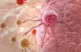
Hepatocellular carcinoma (HCC) is the third leading cause of cancer deaths worldwide with over 600,000 patients dying from this disease annually [1]. The age standardized incidence rates (ASR) of HCC in men in Europe, adjusted to the European Standard Population, is about 8 per 100,000, with a peak in Southern Europe of 10.5 per 100,000 (http://globocan.iarc.fr/). Liver transplantation and tumor resection have proven to be the most effective standard therapies and provide 5-year survival rates of 70% for patients within the Milan criteria, i.e., single tumour < 5 cm in size or up to three tumors < 3 cm in size [2]. These rates reach 50% with radiofrequency ablation and transarterial chemoembolization, which are the next preferred lines of therapy. However, these therapeutic procedures most often do not provide a complete cure, as half of the treated patients experience tumor recurrence within 3 years. A further major problem is that only 30–40% of patients are eligible for the above-described treatments due to the fact that HCC frequently remains undiagnosed until an advanced stage has been reached and occurs in the setting of advanced liver disease due to cirrhosis. These patients have a median survival time ranging from 3 to 16 months [3]. Thus, there is currently ongoing development and testing of alternative drug-based therapies for HCC that target tumor signalling pathways and vasculature. However, so far, the only drug that significantly prolonged survival (by nearly 3 months) in patients with advanced HCC is sorafenib, a multi-targeted tyrosine kinase inhibitor [4]. In view of these facts, new strategies are required. In this study we focus on the identification of predictive molecular factors for HCC recurrence and the development of immunotherapy methods for these patients.

Early Diagnosis of HCC Recurrence with Micro-RNA Techniques

MicroRNAs (miRNAs) are a class of small endogenously expressed non-coding RNAs. The miRNAs in blood, called circulating miRs (cmiRNAs) are potential biomarkers for detecting and monitoring cancer progression [5]. The ability of some miRNAs to function as tumor promoters (miR-30d, miR-151 and miR-210) or suppressors (miR-122, let-7g, miR-29b, miR-193b, miR-194, miR-139 and miR-124) in hepatocarcinogenesis have led to new insights into the molecular pathways involved in HCC [6]. Recently, upregulation of the miR-216a/217 cluster identified to be associated with the early HCC recurrent disease, by comparing the miRNA expression profiles of HCC liver tissue from patients with early-recurrent and non-recurrent disease [7]. In this protocol we intend to develop a technique for the detection and quantification of miR-216a/217 in the blood of HCC patients submitted to liver resection in order to identify recurrence of the disease.

Specific Description of the Proposed Solution

A sensitive and predictable method for the detection and identification of the microRNA216a/217 as the ideal biomarker for prognosis of Hepatocellular Carcinoma

Paraffin-embedded tissue analysis

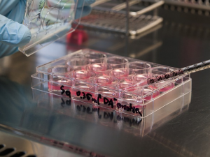
Paraffin-embedded Hepatocellular Carcinoma (PE-HCC) Tissue samples of the primary tumor are obtained from HCC patients who underwent surgical treatment or biopsy at our centers.

RNA Extraction from serum samples

Total RNA was extracted from 500 μL of serum by using TRI reagent BD (Molecular Research Center). Ten sections, each 10- μm thick, were cut from each PE-HCC block. Deparaffinized tissue sections were digested by using proteinase K, and RNA was extracted by using a modified protocol of the RNAWiz Isolation kit (Applied Biosystems). The RNA was quantified and assessed for purity by using ultraviolet spectrophotometry and the Quant-iT RiboGreen RNA assay kit (Invitrogen) [8,9].

RT-qPCR assay with extracted RNA

10 ng of total RNA extracted from Hepatocellular tumor tissue, normal tissue, or serum are dissolved in 5 μL H2O (2 ng/μL) for reverse transcription with the addition of 5 μL of a reaction mixture containing 5Χ first strand buffer, 10 mmol/L deoxynucleoside-5’- triphosphate, RNasin, reverse transcriptase, and miR-specific RT primers (Exiqon). After the mixture was incubated in 37°C for 2 h, the transcribed specific cDNA was diluted 10-fold with molecular grade H2O before use as a qPCR template. Each qPCR contained 2.5 μL of diluted cDNA, 5 μL of 2ΧPerfeCTa SYBR Green FastMix for iQ (Quanta Bioscience), miR-specific, locked nucleic-acid– based forward primer targeting the specific microRNA216a/217 and universal reverse primer (Exiqon).

RT-qPCR directly in serum

This assay requireσ only a small aliquot of serum. To deactivate or solubilize proteins that might inhibit the RT-qPCR, 2.5_L of each serum sample are mixed with 2.5 μL of a preparation buffer that contained 2.5% Tween 20 (EMD Chemicals), 50 mmol/L Tris (Sigma- Aldrich), and 1 mmol/L EDTA (Sigma-Aldrich). 5 μL ofRT reagent mixture are added which contain the same RT reagents used for RT-qPCR with extracted RNA RNA, directly to 5 μL of serum in preparation buffer; a 2-h incubation at 37°C is followed by a 5-min enzyme inactivation at 95°C. The transcribed cDNA is diluted 10- fold by H2O and then centrifuged at 9000 g for 5 min to eliminate the protein precipitant. A 2.5-μL volume of the supernatant cDNA solution is used as the template for qPCR. qPCR conditions, primers and reagents, and data analysis were duplicated for those described in RT-qPCR with extracted RNA section.

Real time quantitative PCR (qPCR)

Real-time qPCRs were performed using SYBR Green PCR Master Mix and 7300 Realtime PCR System (Applied Biosystems, Foster City, CA USA). Real-time qPCRs were also used to detect miRs, as reported [10]. Sequences of miR-specific primers for cDNA synthesis and reverse primers for miR-216a were: 5′-CATGATCAGCTGGGCCAAGACACAGTTGCCAGCTG-3′ and 5′-TAATCTCAGCTGGCAA-3′. Primers for miR-217 were: 5′ CATGATCAGCTGGGCCAAGAATCCAGTCAGTT-3′ (cDNA synthesis) and 5′-TACTGCATCAGGAACT-3′ (reverse primer). miR expression levels were also confirmed by poly-adenylation of mature miR and cDNA synthesis primed by oligo-dT primer tagged with universal primer sequence (miScript System, Qiagen). cDNA was amplified using miR specific and universal primers. For miR-216a and miR-217 PCRs, the same reverse primers mentioned above and the primers provided by Qiagen were used as miR-specific primers. 5S RNA or 18S RNA served as internal control (Ambion) [7]. Genetically modified dendritic cells.

The limited improvements in clinical outcomes with dendritic cells loaded with tumor associated antigens (TAAs) led to trials with genetically modified DCs to further enhance antigen presentation and immunostimulation N. Recent studies have shown that the miR- 216A/217 cluster is consistently and significantly up-regulated in HCC tissue samples and in cell lines associated with early tumor recurrence, poor disease free survival and an epithelial mesenchymal transition (EMT) phenotype [7-31]. Stable over-expression of miR-216a/217- induced EMT, increased the stem like cell population, migration and metastatic ability of epithelial HCC cells. PTEN and SMAD7 are subsequently identified as two functional targets of miR-216a/217, and both PTEN and SMAD7 are down regulated in HCC. Ectopic Expression of PTEN or SMAD7 partially rescued miR216a/217 mediated EMT phenotype, cell migration and stem-like properties in HCC cells. Previously was shown that SMAD7 is a TGF-β1 antagonist. Recently, it has been also shown that Epithelial–mesenchymal transition (EMT) induced by transforming growth factor-β (TGF-β) is implicated in hepatocarcinogenesis and hepatocellular carcinoma (HCC) metastasis [32]. On the other hand, HAb18G/CD147, which belongs to the CD147 family, is an HCC-associated antigen that has a crucial role in tumor invasion and metastasis. Upregulation of HAb18G/CD147 is induced by TGF-β coupled promoting the idea that CD147 may be a potential target for the treatment and prevention of HCC. Other studies have also shown that overexpression of miR216a/217 act a positive feedback regulator for the TGF-b pathway and the canonical way involved in the activation of the PI3K/Akt/ Mtor in HCC cells [33,34].

These facts led us to the second proposal of our protocol:

Experimental (in vitro) protocol for the production and differentiation of dendritic cells derived from the peripheral blood fraction of mononuclear cells, genetically modified for the expression of the microRNA-Mir-216a/217 and HAb18G/CD147 antigen and their maturation by the cell lysate from tumor resections of hepatocellular carcinoma patients

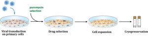
1. MS2 VLP-based delivery of microRNA-216a/217 as a potential delivery approach for the production of clinical grade genetically modified monocyte derived dendritic cells for the immunization of patients suffered from Hepatocellular Carcinoma

Recent studies suggest that microRNA-216a/217 microRNA (miR-216a/217) plays an essential role in immunoregulation and may be involved in the pathogenesis of Hepatocellular Carcinoma. Therefore, it is of interest to investigate the potential therapeutic application of miR-216a/217-induced dendritic cells in Hepatocellular Carcinoma, a concept that has not been thoroughly investigated thus far. Virus-like particles (VLPs) are a type of recombinant nanoparticle enveloped by certain proteins derived from the outer coat of a virus. Herein, we describe a novel miRNA-delivery approach via bacteriophage MS2 VLPs and investigate the therapeutic effects of miR–216a/217-induced dendritic cells against patients suffering from Hepatocellular Carcinoma.

2. An Innovative Methodology for the miR216a/217 MicroRNA Delivery in peripheral blood monocyte derived antigen presenting dendritic cells via magnetic nanoparticles

Peripheral Blood Monocyte derived dendritic cells show promising potential in the vaccination of HCC patients. Recently, gene silencing strategies using microRNAs (miR) emerged with the aim to expand the therapeutic potential of genetically modified dendritic cells. However, researchers are still searching for effective miR delivery methods for clinical applications. Therefore, we aimed to develop a technique to efficiently deliver miR216a/217 microRNA into patient’s dendritic cells with the help of a magnetic non-viral vector based on cationic polymer polyethylenimine (PEI) bound to iron oxide magnetic nanoparticles (MNP), whose uptake efficiency and cytotoxicity will be determined by flow cytometry. The Present Proposal is directed in different magnetic complex compositions and determined. Additionally, the present proposal is focusing on the monitoring of the release, processing and functionality of delivered miR216a/217 microRNA with confocal laser scanning microscopy, real-time PCR and live cell imaging, respectively. On this basis, we will focus on the established parameters for construction of magnetic non-viral vectors with optimized uptake efficiency (~75%) and moderate cytotoxicity in patients peripheral blood monocyte derived dendritic cells. Furthermore, we aim to introduce the magnetic non-viral vector based on cationic polymer polyethylenimine (PEI) transfection as a long term beneficial strategy for the successful genetic modification of the HCC-TAAs presenting dendritic cells against Hepatocellular Carcerous cells for future in vivo applications.

Generation of peripheral blood monocyte-derived miR-216a/217 induced DCs

Monocyte-derived DCs from Hepatocellular Carcinoma Patients (obtained with following informed consent and approved by our institutional review board) are generated. In brief, peripheral blood mononuclear cells (PBMCs) are prepared from whole blood by Ficoll density-gradient centrifugation. The PBMCs are suspended in tissue culture flask in RPMI 1640 supplemented with 1% heat inactivated autologous serum for 60 minutes at 37°C to allow for adherence. The nonadherent cells are removed and the adherent cells are cultured overnight. To generate immature miR-216a/217 induced DCs (DCs), the nonadherent and loosely adherent cells are collected the following day and placed in RPMI 1640 medium containing 1% heat-inactivated autologous serum, 1000 U/ml recombinant human GM-CSF (Becton Dickinson, Bedford, MA, USA), and 500 U/ml recombinant human IL-4 (Becton Dickinson) for 6 days. In order to assess the effects of HCCsp on miR-216a/217 induced DCs generation, we are focusing on the creation of four types of miR-216a/217 induced preparation: 1) miR-216a/217 induced DCs; 2) miR-216a/217 induced s generated in the presence of HCCsp during the entire culture period (DCs/sp); 3) miR-216a/217 induced DCs exposed to 0.1 KE/ml (0.1 KE equals of 0.01 mg of dried streptococci) penicillin-inactivated Streptococcus pyogenes (OK-432) (Chugai Pharmaceutical) for 3 days (OK- miR- 216a/217 induced DCs) as described previously; 4) OK- miR-216a/217 induced s generated in the presence of HCCsp during the entire culture period (OK-DCs/sp). Four types of miR-216a/217 induced are generated in the presence of equal amounts of GM-CSF and IL-4 during the entire culture.

To generate monocyte-derived miR-216a/217 induced DCs for vaccination, PBMCs derived from the HCC patient are freshly isolated (obtained with following informed consent and approved by our institutional review board). Autologous miR-216a/217 induced s are generated in RPMI 1640 medium containing 1% heat-inactivated autologous serum, 1000 U/ml recombinant human GM-CSF, 500 U/ml recombinant human IL-4, and 10 ng/ml recombinant TNF-α (Becton Dickinson) [30]. On day 6 of culture, DCs harvested from the nonadherent and loosely adherent cells are used for fusion. The firmly adherent monocytes are harvested and used as an autologous target for the CTL assays.

HCC Patient selection

The clinical trial protocol will be approved by the Institutional Review Boards of our University Hospitals. Patients will be informed of the investigative nature of this study, and written consent in accordance with institutional regulations is obtained prior to study entry. Eligibility criteria included HCC patients submitted to liver resection or biopsy and transarterial chemoembolization (TACE) with radiological diagnosis of primary HCC by computed tomography (CT), classified in stage II and III according to the tumor-node-metastasis (TNM) classification; age over 20 years/both male and female; Eastern Cooperative Oncology Group scale 0–1; and indicators of acceptable hematological (hemoglobin ≥8.5 g/dl, white blood cells ≥2,000/mm3, platelet ≥50,000/mm3), hepatic (Child Pugh score ≤7, alanine aminotransferase, aspartate aminotransferase ≤5x upper normal limit) and renal (creatinine ≤1.5 mg/dl) function. Important exclusion criteria consisted of organ transplantation; a medical history of autoimmune disease, immunodeficiency, or autoimmune disease that might be aggravated by immunotherapy; not exceeding 2 weeks after antibiotic treatment needed due to a serious infectious disease; seropositivity for human immunodeficiency virus antigen; use of immunosuppressive drug such as cyclosporin A and azathioprine; any cardiopulmonary disability judged by the investigator; a medical history of psychological disease or epilepsy; and evidence of another active malignant neoplasm.

Autologous DC generation

DCs are generated from blood monocytes, as reported previously, with modifications. White blood cells obtained from the HCC patients through leukapheresis. DCs are prepared in a GMP-compliant facility at our hospitals. Peripheral blood mononuclear cells (PBMCs) are separated from WBC by Ficoll-Paque™ PLUS (Amersham Biosciences, Uppsala, Sweden) density gradient centrifugation. PBMCs are stored in a liquid nitrogen tank until necessary for DC generation. PBMCs thawed, ished with Hanks’ Balanced Salt Solutions, resuspended in RPMI-1640 medium (Lonza, Basel, Switzerland) supplemented with autologous heat-inactivated plasma, and then incubated in CellSTACK Culture Chambers (Corning, Corning, NY, USA). After 0.5–1 h incubation at 37°C in a 5% CO2 incubator, non-adherent cells are removed by gentle ishes.

The adherent monocytes are cultured in X-VIVO15 (Cambrex, East Rutherford, NJ, USA) supplemented with 100 ng/ml of granulocyte macrophage-colony stimulating factor (GMP grade: LG Life Science, Seoul, Korea) and 300 ng/ml of interleukin (IL)-4 (JW CreaGene Inc., Seongnam, Korea) for 5 days. On day 5, nonattached immature DCs are harvested and pulsed with CTP-fused human AFP, MAGE-1 and GPC-3 recombinant proteins at a final concentration of 5 μg/ml each. Antigen-pulsed dendritic cells are matured in the presence of cytokine cocktail, IL-6 (Peprotech, Rocky Hill, NJ, USA), IL-1β (Peprotech), tumor necrosis factor (TNF)-α (Peprotech), prostaglandin E2 (PGE2) (Sigma Chemical Co., St. Louis, MO, USA), interferon (IFN)-γ (LG Life Science), OK432 (Chugai Pharmaceutical Co., Tokyo, Japan), and poly I: C (Sigma) for 1 or 2 days depending on surface phenotypes and cell population. On day 6–7, the DCs are harvested, ished, and resuspended in 1.2 ml of cryopreserving solution containing 5% dimethyl sulfoxide (Bioniche Pharma USA, Lake Forest, IL, USA). Finally fully equipped DCs are packed into a sterile glass vial (4×10⁷ cells/vial), sealed with a snap-cap, and stored at an ultralow freezer for >12 h.

Quality control of dendritic cell vaccine

Safety test

For safety, endotoxin, germ-free and mycoplasma-free tests are performed according to the KFDA-approved JW CreaGene standard and test guidelines. Endotoxin is evaluated using gel-clot techniques. The endotoxin of the product should be less than 10 EU/ml per 1.2- ml vial. Mycoplasma test is performed by both direct culture and PCR methods using e-Myco™ Mycoplasma PCR detection kit (Intron Biotechnology, Seongnam, Korea), which contains primer sets specifically designed to detect major contaminants of Mycoplasma in cell cultures such as M. arginini, M. faucium, M. fermentans,M. hyorhinis, M. orale, and A. laidlawii as well as other broad spectrum of mycoplasma.

Cell size and granularity

During the differentiation from monocytes to miR-216a/217 induced dendritic cells, cell size and granularity increase. Based on these principles, the cell size and granularity of each miR-216a/217 induced DC vaccine are assessed by flow cytometric analysis. PBMCs are used for gating control.

Phenotypic analysis

The phenotype of miR-216a/217 induced DC vaccine is determined by flow cytometry using a FACSCalibur™ flow cytometer (Becton Dickinson, Franklin Lakes, NJ, USA). The following monoclonal antibodies are used: i) fluorescein isothiocyanate-conjugated mouse antihuman IgG2a isotype control; ii) phycoerythrin-conjugated mouse antihuman IgG1 isotype control; iii) anti-CD14, anti-CD19, anti-CD40, anti-CD80, anti-D86, anti-HLA-ABC, and anti-HLA-DR (BD Pharmingen, San Diego, CA, USA).

Viability

The viability of miR-216a/217 induced DC vaccine is assessed by propidium iodide (PI) staining. PI (BD Pharmingen) is added to a sample and kept in the dark at room temperature for 20 min. Cell viability is examined by flow cytometry using a FACSCalibur™ (Becton Dickinson). Viability is represented as 100-[(PI+ of sample)−(PI+ of control)] (%).

Lymphocyte proliferation assay

One vial from each miR-216a/217 induced DC vaccine lot is used to test of T cell stimulation capacity according to the standard lymphocyte proliferation assay. T cells are isolated from cryopreserved PBMC using nylon wool column (Polysciences, Warrington, PA, USA). Purified T cells (1×10⁵) are cultured with serially diluted DC vaccine (starting from 1×10⁴ cells to 0.33×10³ cells) at 37°C for 5 days. T cell proliferation is assessed by 3-(4,5-di-methylthiazol-2-yl)-2,5- diphenyltetrazolium bromide, yellow tetrazole: MTT) assay following manufacturer’s protocol (CellTiter 96 Non-Radioactive proliferation assay kit; Promega, Madison, WI, USA). R2 represent the standard curve of MTT assay for the validation of a data set.

Cytokine production assay

Either culture supernatant of each antigen-pulsed miR-216a/217 induced DC or co-cultured medium of T cells/ miR-216a/217 induced DC at the ratio of 10: 1 is collected and stored at −80°C until this assay. The concentration of IL-12p70, IL-10, IFN-γ, and IL-4 is measured with corresponding human immunoassay kits (BD OptEIA™ kit, BD Pharmingen) based on the manufacturer’s instruction. Each experiment is performed 3 times and the result is described as the mean ± standard deviation.

Treatment protocol of Hepatocellular Carcinoma Patients

The screening evaluation shall be performed in 3 weeks before the start of dendritic cell-based immunotherapy and consisted of the following: complete history, thorough physical examination, chest X-ray, electrocardiogram, urine analysis, hematological and immunological parameters, serum chemistry, tumor markers [AFP and protein induced by vitamin K absence or antagonists-II (PIVKA-II)], ultrasonography and abdominal CT scan. Eligible HCC patients underwent liver resection or TACE and biopsy, 2 weeks before the start of the vaccination. PBMC collection by leukapheresis is performed 1 week before the first planned vaccination. Tumor antigen-pulsed miR-216A/217 Professional Antigen Presenting DCs are injected intravenously in 20 mL of phosphate-buffered saline over 10 minutes on day 12. Patients were observed for 2 hours after each vaccination to assess any immediate complications. During the first cycle, 6 vaccinations are administered at biweekly intervals. Medical history and standard blood tests and urine analysis are performed at each vaccination. Vital signs are monitored during and after each injection. Response evaluation is performed 4 weeks after fourth vaccination (10 weeks after first vaccination). Two further vaccinations are administered at biweekly intervals, and final response evaluation is performed at 18 weeks after first vaccination. Tumor markers, including qRT-PCR miR-216A/217, and serological tests for autoantibodies, including anti-nuclear antibody, are evaluated every 4 weeks.

Clinical response and toxicity assessment

Clinical responses to vaccination are evaluated according to the Response Evaluation Criteria in Solid Tumors (RECIST) criteria. Complete response is defined as disappearance of all target lesions. Partial response is defined as 30% decrease in the sum of the longest diameter of target lesions. Progressive disease is 20% increase in the sum of the longest diameter of target lesions. Stable disease is defined as small changes that do not meet above criteria. Toxities are classified according to the National Cancer Institute Common Toxicity Criteria.

Description of the German and Greek partners from higher education institutions, research establishments and commercial companies who are involved in the project (key competencies, capabilities, infrastructure, etc).

The Division of Transplantation, Department of Surgery, Aristotle University of Thessaloniki Medical School is the leading center in Greece, performing liver transplantation and liver surgery in patients with hepatocellular carcinoma and has the ability to perform clinical studies on the prognosis and therapy on this subject.

The Department of Surgery, Essen University Hospital, is a leading center worldwide in liver transplantation and liver surgery in patients with hepatocellular carcinoma and has performed many multi institutional clinical studies on the prognosis and therapy on this subject.

Justify the scope of the collaboration between the partners of both countries and the mutual benefit provided through this collaboration. Overview of previous contacts in and collaborations with the partner country.

Immunotherapy of HCC

Immunotherapy aims to provide a more efficient and selective targeting of tumor cells by inducing or boosting the existing tumor-specific immune response. The rationale for immunotherapy for HCC is based on several lines of evidence of a protective role the immune system, e.g., by controlling tumor growth. First, HCC patients with an intratumoral accumulation of lymphocytes had a superior 5-year survival rate and a prolonged recurrence-free survival after liver transplantation or resection [11,12]. A certain level of protection was especially conferred by cytotoxic CD8+ T cells [11]. Of note, these tumor infiltrating lymphocytes (TILs) were associated with an inflammatory microenvironment that was a predictor of overall patient survival, indicating a protective role of TILs in HCC [13,14]. Furthermore, a strong CD8+ T cell response against several tumor-associated antigens (TAAs) was found to coincide with improved recurrence-free survival after liver resection [15]. The important role of CD8+ T cells in HCC control is further supported by a study in mice. It was shown that interferon γ (IFNγ) produced by CD8+ TILs could be one effector mechanism for apoptosis of hepatoma cells [16]. These data imply a central role of T cells in modulating tumor progression and provide strong justification for T cell immunotherapy.

Immunotherapy approaches for HCC

A limited number of immunotherapy trials for HCC have been conducted based on several strategies, with yet modest results. Cytokines have been used to activate subsets of immune cells and/or increase the tumor immunogenicity [17,18]. Further strategies have been based on infusion of tumor infiltrating lymphocytes or activated peripheral blood lymphocytes [19-21]. Alternatively, direct delivery of genetically modified or designer T cells (dTc) into the hepatic artery has been recently proposed as a promising novel strategy and is currently evaluated in a phase I human clinical trial (ClinicalTrials. gov Identifier: NCT01373047). Indeed, the latter strategy has recently been successfully used for treatment of different cancers and several human clinical trials are currently ongoing [22].

Alternatively, considering active immunotherapy strategies (i.e. therapeutic vaccination), the number of human clinical trials published to date is extremely small. The first HCC vaccine clinical trial was conducted by Butterfield et al. based on CD8+ T cell epitopes specific for alpha fetoprotein (AFP), showing the generation of AFP-specific T cell responses in vaccinated subjects [23]. To improve the immune response, the same authors performed a subsequent phase I/II trial administering AFP epitopes presented by autologous DCs loaded ex vivo. This treatment, however, resulted only in transient CD8+ T cell responses, possibly caused by the lack of CD4+ help [24,25]. To overcome this limitation and to increase the number of tumor associated antigens (TAAs) targeted by the immune response elicited by the vaccine, few vaccine approaches based on autologous DCs pulsed ex vivo with a lysate of the autologous tumor [26] or of hepatoblastoma cell line HepG2 [27,28] have been evaluated in human clinical trials, showing limited improvements in clinical outcomes . The last clinical trial in the literature is based on a combination of low dose cyclophosphamide treatment followed by a telomerase peptide (GV1001) vaccination which did not show antitumor efficacy [29].

Prospects for the success of the proposed measures and implementation concepts describing how the project outcomes can be utilized after the funding period (utilization plan)
Dendrigenea: A manufacturing patient-specific cell therapy product

Overview and case study of dendrigenea’s cell therapy technology: Our Dendrigenea cellular therapy product is currently progressing through clinical development with the potential to address unmet medical needs affecting millions of patients suffered from cancer –tumor related diseases. Dendrigenea is an autologous cell-based therapeutic product which has already received regulatory approval and reach the market. Our primary challenge is to quickly become a Leader dendritic cell manufacturing facility of such products in sufficient volume to meet patients demand.

Biogenea Pharmaceuticals Ltd has developed a cancer-specific dendritic cell therapy technology for use in an autologous patient-specific vaccine therapy and is conducting late-stage clinical trials both in Greece and other Countries from Southeaster Europe. Dendritic cells are derived from a patient’s own peripheral blood and processed through a short four-week isolation, maturation, culture and fusion procedure under media perfusion conditions that lend potent functional anti-cancer vaccination properties to the final vaccine-cell product.

Development and clinical use of a Peripheral Monocyte Derived Cell Therapy Product (PMDCTP) such as Dendrigenea has raised unique manufacturing challenges that we have addressed through a series of patented innovative solutions, many of which could have broad application in the field of cancer immunotherapy for the production of commercial-scale dendritic cell based manufacturing Vaccines.

Peripheral Blood as a source of dendritic vaccines has key advantages for patients. In particular, autologous (PMDCTP) (treatments derived from a patient’s own monocytes) has a favorable safety and risk profile not available from allogeneic (universal donor) therapies.

Concerns over immune rejection and disease transmission are minimal. In addition, short-term culture and monocyte maturation procedures reduce the risk of tumorgenic transformation, which is possible in universal donor cell products typically generated from longer-term serially passaged cultures. Dendrigenea has demonstrated a high level of safety in more than 250 patients treated successfully for a variety of tumor-cancer related medical indications, with both local and systemic administration, showing no sign of cell-related adverse events.

Our dendritic cells are expanded from cell populations existing within patient’s own peripheral blood that are associated with natural cell immune response and tissue homeostasis as well as healing. Local transplantation of these expanded, patient-specific peripheral blood derived dendritic cells are expected to immunize patients suffering from Tumor related diseases. They are currently under clinical evaluation for a range of cancer cell treatments including Melanoma, Leiomyosarcoma, Glioma, Glioblastoma, Neuroblastoma and Ovarian, Breast Cancer.

Dendrigenea goes through of an ex-vivo, culture, maturation and cell fusion process have been developed to expand specific subpopulations of primary monocyte derived dendritic cells found within patient’s own peripheral blood, including early stem and progenitor cells, without triggering cell differentiation to other malignant tissue specific cell-lines . The result is a mixed population of dendritic cells genetic or not modified targeted directly to patient’s own cancer cells.

References

  • Schütte K, Bornschein J, Malfertheiner P (2009) Hepatocellular carcinoma–epidemiological trends and risk factors. Dig Dis 27: 80-92. [crossref]
  • Llovet JM, Bruix J (2008) Molecular targeted therapies in hepatocellular carcinoma. Hepatology 48: 1312-1327. [crossref]
  • Llovet JM, Burroughs A, Bruix J (2003) Hepatocellular carcinoma. Lancet 362: 1907-1917.
  • [crossref]
  • Llovet JM, Ricci S, Mazzaferro V, Hilgard P, Gane E, et al. (2008) Sorafenib in advanced hepatocellular carcinoma. N Engl J Med 359: 378-390. [crossref]
  • Asaga S, Hoon DS (2013) Direct serum assay for microRNA in cancer patients. Methods Mol Biol 1024: 147-155. [crossref]
  • Giordano S, Columbano A (2013) MicroRNAs: new tools for diagnosis, prognosis, and therapy in hepatocellular carcinoma? Hepatology 57: 840-847. [crossref]
  • Xia H, Ooi LL, Hui KM (2013) MicroRNA-216a/217-induced epithelial-mesenchymal transition targets PTEN and SMAD7 to promote drug resistance and recurrence of liver cancer. Hepatology 58: 629-641. [crossref]
  • Asaga S, Kuo C, Nguyen T, Terpenning M, Giuliano AE, et al. (2011) Direct serum assay for microRNA-21 concentrations in early and advanced breast cancer. Clin Chem 57: 84-91. [crossref]
  • Takeuchi H, Morton DL, Kuo C, Turner RR, Elashoff D, et al. (2004) Prognostic significance of molecular upstaging of paraffin-embedded sentinel lymph nodes in melanoma patients. J Clin Oncol 22: 2671-2680. [crossref]
  • Xia H, Cheung WK, Sze J, Lu G, Jiang S, et al. (2010) miR-200a regulates epithelial-mesenchymal to stem-like transition via ZEB2 and beta-catenin signaling. J Biol Chem 285: 36995-37004. [crossref]
  • Unitt E, Marshall A, Gelson W, Rushbrook SM, Davies S, et al. (2006) Tumour lymphocytic infiltrate and recurrence of hepatocellular carcinoma following liver transplantation. J Hepatol 45: 246-253. [crossref]
  • Gao Q, Qiu SJ, Fan J, Zhou J, Wang XY, et al. (2007) Intratumoral balance of regulatory and cytotoxic T cells is associated with prognosis of hepatocellular carcinoma after resection. J Clin Oncol 25: 2586-2593. [crossref]
  • Hirano S, Iwashita Y, Sasaki A, Kai S, Ohta M, et al. (2007) Increased mRNA expression of chemokines in hepatocellular carcinoma with tumor-infiltrating lymphocytes. J Gastroenterol Hepatol 22: 690-696. [crossref]
  • Chew V, Tow C, Teo M, Wong HL, Chan J, et al. (2010) Inflammatory tumour microenvironment is associated with superior survival in hepatocellular carcinoma patients. J Hepatol 52: 370-379. [crossref]
  • Hiroishi K, Eguchi J, Baba T, Shimazaki T, Ishii S, et al. (2010) Strong CD8 (+) T-cell responses against tumor-associated antigens prolong the recurrence-free interval after tumor treatment in patients with hepatocellular carcinoma. J Gastroenterol 45: 451-458. [crossref]
  • Komita H, Homma S, Saotome H, Zeniya M, Ohno T, et al. (2006) Interferon-gamma produced by interleukin-12-activated tumor infiltrating CD8+T cells directly induces apoptosis of mouse hepatocellular carcinoma. J Hepatol 45: 662-672. [crossref]
  • Reinisch W, Holub M, Katz A, Herneth A, Lichtenberger C, et al. (2002) Prospective pilot study of recombinant granulocyte-macrophage colony-stimulating factor and interferon-gamma in patients with inoperable hepatocellular carcinoma. J Immunother 25: 489-499. [crossref]
  • Sangro B, Mazzolini G, Ruiz J, Herraiz M, Quiroga J, et al. (2004) Phase I trial of intratumoral injection of an adenovirus encoding interleukin-12 for advanced digestive tumors. J Clin Oncol 22: 1389-1397. [crossref]
  • Shi M, Zhang B, Tang ZR, Lei ZY, Wang HF, et al. (2004) Autologous cytokine-induced killer cell therapy in clinical trial phase I is safe in patients with primary hepatocellular carcinoma. World J Gastroenterol 10: 1146-1151. [crossref]
  • Takayama T, Makuuchi M, Sekine T, Terui S, Shiraiwa H, et al. (1991) Distribution and therapeutic effect of intraarterially transferred tumor-infiltrating lymphocytes in hepatic malignancies. A preliminary report. Cancer 68: 2391-2396. [crossref]
  • Takayama T, Sekine T, Makuuchi M, Yamasaki S, Kosuge T, et al. (2017) Adoptive immunotherapy to lower postsurgical recurrence rates of hepatocellular carcinoma: a randomised trial. Lancet 356:802-7.
  • Essand M, Loskog AS (2013) Genetically engineered T cells for the treatment of cancer. J Intern Med 273: 166-181. [crossref]
  • Butterfield LH, Ribas A, Meng WS, Dissette VB, Amarnani S, et al. (2003) T-cell responses to HLA-A*0201 immunodominant peptides derived from alpha-fetoprotein in patients with hepatocellular cancer. Clin Cancer Res 9: 5902-5908. [crossref]
  • Butterfield LH, Ribas A, Dissette VB, Lee Y, Yang JQ, et al. (2006) A phase I/II trial testing immunization of hepatocellular carcinoma patients with dendritic cells pulsed with four alpha-fetoprotein peptides. Clin Cancer Res 12:2817-25.
  • Bray SM, Vujanovic L, Butter field LH (2011) Dendritic cell-based vaccines positively impact natural killer and regulatory T cells in hepatocellular carcinoma patients. Clin Dev Immunol 2011: 249281. [crossref]
  • Lee WC, Wang HC, Hung CF, Huang PF, Lia CR, et al. (2005) Vaccination of advanced hepatocellular carcinoma patients with tumor lysate-pulsed dendritic cells: a clinical trial. J Immunother 28: 496-504. [crossref]
  • Palmer DH, Midgley RS, Mirza N, Torr EE, Ahmed F, et al. (2009) A phase II study of adoptive immunotherapy using dendritic cells pulsed with tumor lysate in patients with hepatocellular carcinoma. Hepatology 49: 124-132. [crossref]
  • El Ansary M, Mogawer S, Elhamid SA, Alwakil S, Aboelkasem F, et al. (2013) Immunotherapy by autologous dendritic cell vaccine in patients with advanced HCC. J Cancer Res Clin Oncol 139: 39-48. [crossref]
  • Greten TF, Forner A, Korangy F, N’Kontchou G, Barget N, et al. (2010) A phase II open label trial evaluating safety and efficacy of a telomerase peptide vaccination in patients with advanced hepatocellular carcinoma. BMC Cancer 10: 209. [crossref]
  • Morrison BJ, Steel JC, Gregory M, Morris JC (2009) Genetically Modified Dendritic Cells in Cancer Immunotherapy. In Shurin MR, Salter RD, “Dendritic Cells in Cancer”, Springer 23: 347-363.
  • Blechacz B, Mishra L (2013) Hepatocellular carcinoma biology. Recent Results Cancer Res 190: 1-20. [crossref]
  • Wu J, Ru NY, Zhang Y, Li Y, Wei D, et al. (2011) HAb18G/CD147 promotes epithelial-mesenchymal transition through TGF-β signaling and is transcriptionally regulated by Slug. Oncogene 30: 4410-4427. [crossref]
  • Shen J, Stass SA, Jiang F (2013) MicroRNAs as potential biomarkers in human solid tumors. Cancer Lett 329: 125-136. [crossref]
  • Gao Q, Shi Y, Wang X, Zhou J, Qiu S, et al. (2012) Translational medicine in hepatocellular carcinoma. Front Med 6: 122-133. [crossref]

Immortalomunea TM: Advnaced lentivirus transduced biocomplexes for the generation of semi-autologous immortalized cell line vaccines for cancerous diseases and HIV infections

DOI: 10.31038/CST.2017244

Short Commentary

Dendritic cells are the professional antigen presenting cells of innate immunity and key players in maintaining the balance of immune responses. Studies with dendritic cells are mainly limited by their low numbers in vivo and their difficult maintenance in vitro. Dendritic cells are the professional antigen presenting cells of innate immunity and key players in maintaining the balance of immune responses. Studies with dendritic cells are mainly limited by their low numbers in vivo and their difficult maintenance in vitro. In summary, in “biogenea pharmaceuticals ltd” we successfully generated several immune T and dendritic cell line living vaccines using conditional immortalization where the generated dendritic cell lines demonstrate the characteristic immunophenotype of primary dendritic cells to facilitate further studies on immunomodulatory properties of dendritic cells.

ImmortalomuneaTM Figure1

Genea-Pancretovimortal24585

A Mixture of two allogeneic human pancreatic derived immature dendritic cancerous immortalized cell lines stably transduced with a retroviral vector for the endogenous encoding of the CEA, Muc-1, TRICOM, CO1 7-1 A, OC 125 and B72.3, DU-PAN-2 respectively on Allogeneic human immature dendritic cell lines derived from a class I & II HLA3-A3-B44 CD34+ progenitor cell.

ImmortalomuneaTM Figure2

Numerous clinical trials have demonstrated the safety of dendritic cells vaccines, and more than 1000 patients have received dendritic cell vaccines with no serious adverse events associated with the therapy and clinical responses in one half of patients The CA19-9, CA125, DU-PAN-2, and B72.3 antigens have been shown to be expressed in many human pancreatic cancer cells, and C01 7-1A and B72.3 have being used for immunotherapy. Here, in Biogenea Pharmaceuticals Ltd we provide a method of enhancing immunity by modifying a dendritic cell (DC) in vivo or ex vivo to produce an immature immortalized dendritic cell line enhancing immunity in pancreatic cancer patients. OurGenea-Pancretovimortal24585 composition is a pancreatic cancer patient derived tumor lysate pulsed with a immortalized cell line mixture dedicated to the treatment of pancreatic cancer by the use of lentivirus of the CEA, Muc-1, TRICOM, CO1 7-1 A, OC 125 and B72.3, DU-PAN-2 antigen transfected immature transduced dendritic cell lines as an advanced semi-autologous living (DC)-vaccine. Genea-Cordimmortalun24874: Immortalized Human Cord Blood- Derived Stem Cells for the generation of conditionally immortalized universal progenitor cell lines with multiple lineage potential and Immunosuppressive Characteristics.

ImmortalomuneaTM Figure3

Cell banking of mesenchymal stem cells (SCs) from various human tissues has significantly increased the feasibility of SC-based therapies. Sources such as adipose tissue and amnion offer outstanding possibilities for allogeneic transplantation due to their high differentiation potential and their ability to modulate immune reaction. Limitations, however, concern the reduced replicative potential as a result of progressive telomere erosion, which hampers scaleable production and long-term analysis of these cells. In Biogenea Pharmaceuticals Ltd for the first time we incorporated methods for preparing multi-potential immortalized stem cells having a pre-selected expression of MHC antigens. Our Genea-Cell lines consisting of two human cord blood-derived immortalized somatic stem cell linesgenerate by ectopic expression of the catalytic subunit of human telomerase (hTERT). hTERT overexpression resulted in continuously growing SC lines that were largely unaltered concerning surface marker profile, morphology, karyotype, and immunosuppressive capacity with similar or enhanced differentiation potential for up to 87 population doublings. can be Our universal stem cell lines can used to generate histocompatible tissues/organs for transplantation. The process comprises the use of targeting vectors capable of gene knockout, insertion of site-specific recombination cassettes, and the replacement of histocompatibility alleles in the stem cell. We incorporated novel knockout vectors which are used to delete designated HLA-B44, HLA-B7, HLA-B8, HLA-B35, HLA-B52, HLA-B60, HLA-B39, and HLA-B48 HLA-DR7, HLA-DR4, HLA-DR13, HLA-DR15, HLA-DR3, HLA-DR1, HLA-DR11, HLA-DR8, HLA-DR9, HLA-DR12, HLA-DR14 and HLA-DRBL, HLA-A allele selected from the group consisting of HLA-A2, HLA-A1, HLA-A3, HLA-A24, HLA-A29, and HLA-A33 regions of one chromosome. Recombination cassette vectors were used to delete the same region on the second chromosome and deposit a site-specific recombination cassette which can be utilized by replacement vectors for inserting the new MHC genes on the chromosome of the engineered cell. Our advanced methodology pertains to cells, tissues, for the generation of conditionally immortalized progenitor cell lines with multiple lineage potential.

Genea-Cellgeneroglimmortal737

A Combinations of Transgenes in LV for Reprogramming Immune Precursors into two Antigen-Loaded immortalized Dendritic Cell lines for the DC-endogenous expression of the cALLa/NEP, ABCC3, GPNMB, NNMT, and SEC61γ trasnduced antigens on immortalized class I & II HLA3-A3B-44-Dendritic Cells lines (iDC) for T Cell Expansion after Stem Cell Transplantation.

ImmortalomuneaTM Figure4

Malignant brain tumors carry a poor prognosis even in the midst of surgical, radio-, and chemotherapy. With the poor prognosis of brain tumors the available therapeutic treatments, there exists a significant need for more effective therapies to treat such tumors. Our Genea- Cellgeneroglimmortal737 is based, on the discovery that vaccines based on cancer stem cell antigens are exceptionally useful for therapy of cancer. Immunization of patients with endogenous expressed of the cALLa/NEP, ABCC3, GPNMB, NNMT, and SEC61γ trasnduced dendritic cell lines pulsed with autologous tumor lysate from isolated cancer circulated stem cells provided a significant survival benefit as compared to immunization with dendrit The principle of this approach is to educate immortalized immature antigen-presenting cells, such as dendritic cells, to recognize tumor antigens by fusing them on pulsed differentiated tumor cells. Cancer stem cells were found to express major histocompatibility (MHC), indicating that they can display antigens. Our advanced cell fusions can be useful in providing antigenic compositions for treatment of cancers (e.g., neural cancers such as gliomas).

Genea-Hivotranceral437856

A cord blood derived Non-Transformed, transduced, Immortalized Human double negative il-2 depended T-tropic lymphocyte cell-line for the expression of the CD4D1D2CAR and HTLV-1 p30II oncoprotein.

ImmortalomuneaTM Figure5

Genea-Hivotranceral437856 advanced medicinal services include a composition comprising a cord blood derived Non-Transformed, transduced, non-transformed, immortalized T-lymphocyte cell-line, wherein the T lymphocytes are IL-2 dependent and interact with an extracellular matrix and supports productive infection and replication by T-tropic HIV. Our T-lymphocyte cell-line cells are polyclonal. In another aspect, the T-lymphocyte cell-line are infected with a lentivirus that expressed THE CD4D1D2CAR and an HTLV- 1 p30II oncoprotein. Our Genea-Hivotranceral437856 T cell line composition endogenously express the chimeric antigen receptor of CD4 extracellular, transmembrane domains and a CD3 zeta signaling domain where the CD4 extracellular domain binds gpl20 expressed on the surface of cells infected with HIV.

Biogenea PharmaceuticalsTM is the first Inter-Balkan Pharmaceutical Biotechnology Company since leading the way since 2005 in Red Biotechnology applications, in Cryobiology and in Autologous Cellular Therapy of Degenerative Diseases (cardiological diseases, neurological conditions and metabolic disorders).

Biogenea Pharmaceuticals focuses

  • on the collection, processing, cryopreservation and cGMP (according to Good Manufacturing Practice) production -for solely autologous use – of cellular therapeutical solutions from blood (bone marrow, peripheral blood, cord blood) or blood compounds for human use.
  • in collaboration with Regenetech on stem cell expansion technologies, which were created in the research laboratories of NASA (National Aeronautics and Space Administration).
  • on the cGMP production of advanced medicinal products (1394/2007/ΕC) for solely autologous use from skin, dental pulp, cord tissue). (In preclinical-research phase: 2008-2009).
  • on certified genetic analyses in collaboration with International Referral Centers.
  • on copyright protection according to the American and/or European Copyright Agency
  • Preventive Cryopreservation Insurance of Primordial Cells & Therapeutic Applications.
  • Continuous update and prompt training of interested Donors and Clinicians of Public and Private Health Centers and Hospitals, about the progenitor cells applications.
  • Immediate availability of the stored samples 365 days a year, 7 days a week, 24 hours a day.
  • Complete attunement with the specifications set by the European Accreditation Organization for cellular therapy (FACT/JACIE/NETCORD), the American Association of Blood Banks (AABB) and the relevant Greek Presidential Decretal 2004/23 for the tissue/cell banks, with the use of cGMP methods.
  • Control of Plasticity of progenitor cells based on the detailed Validation Master Plan of the Standard Operating Procedures (SOPs) regarding their hematopoietic origin (Methocult).
  • Fully Automated Viability Control of the cryopreserved progenitor cells with automatic luminometric device, which increases the reproducibility and the accuracy of the results in contrast to the common laboratory techniques for detecting dead cells microscopically (Trypan Blue staining).
  • Validated Molecular Diagnostics service provision (detection of HIV1/2, HBV, HCV, CMV, Syphillis, Toxoplasma) applying Real-Time PCR technology (Roche LightCycler).
  • Validated ex-vivo cellular and/or tissue expansion of the recently processed cells/tissues, as well as of the cryopreserved cell/tissues in advanced technology Bioreactors, created in the NASA research laboratories, offering unique micro-gravity conditions in alternating electromagnetic field! Cellgenea is the UNIQUE company in Greece able to expand and to use ex-vivo expanded cord blood and adult progenitor cells for therapeutic reasons and clinical trials thanks to the relevant know-how transfer from the research laboratories of NASA and Regenetech Biotechnology Company.
  • Storing of progenitor cells in two-chambered bags and cryovials in complete (24 hours a day) controlled and automated liquid nitrogen tanks.
  • Continuous control and cross-tracking of the cryopreserved samples with validated LIS-ERP software which ensures the ability to track down and to identify the sample during all the steps of its supply, the processing, the control, the storage and the distribution. The tracking is also used for controlling and identifying all the relevant data about the products and the materials that come in contact with these samples (2004/23/ΕΚ).
  • Immediate availability and distribution of the sample inside a validated cryotank, in case of therapeutic application.
  • Strict security concerning personal data, confidentiality and safety according to the Personal Data Protection Agency.
  • Complete bacteriological and serological control of the samples using the automatic analyzers BacT/ALERT and Architect i1000 without any extra rate.
  • Life insurance provided to all the members of the different programs of preventive cryopreservation (Cellgenea, Dentogenea, Dermigenea, Angiogenea, DΝΑgenea, Neurogenea, Cardiogenea) in cooperation with insurance companies.
  • DNA & pharmacogenetic control services for personalized treatment without side effects.

Medicinal and Economic Values of Forest products in the Treatment of Cancer in Southwest Nigeria

DOI: 10.31038/CST.2017243

Abstract

Medicinal plants are used to address the twin problems of promoting sustainable livelihoods and treatment of numerous illnesses in Nigeria. The study examined the medicinal value of forest products in the treatment of cancer in South-west Nigeria. Primary data was obtained in a cross section survey of 327 respondents comprising 127 Traditional Medicine Practitioners (TMPs), 100 Orthodox Medicine Practitioners (OMPs) and 100 respondents from the General Public drawn by multistage sampling technique from the study area. Interview schedule was used in collection of data on the effectiveness of forest products in cancer treatment. The result showed that seven species were identified belonging to seven different families; Rutaceae, Asteraceae, Anarcardiaceae, Annonaceae, Meliaceae, Guttiferaceae and Leguminaceae topped the TMPs priority list. Result of economic analysis shows minimal competition in the anti-cancer forest product market and a high level of monopoly with a Gini coefficient of 0.83. The rate of return on investment was 180 .08% indicating that the TMPs were making profit. Five of the plants were tested against cancer cell lines MCF7 and Hs578T while Doxorubicin (a synthetic anticancer drug) was used as the control treatment. Three plants; Saccharum offinarum (Stem), Sucurinega virosa (Root) and Piper guineensii (Seed) produced no result; Garcinia kola (Bark) did not exhibit any anticancer effect even at a concentration of 10u1/m1 while only one plant species was effective against the cancer cell line at 1u1/m1. It is therefore concluded that forest products are effective in the treatment of cancer.

Keywords

medicinal plants; cancer; traditional medicine practitioners (TMPs); forest products and southwest Nigeria

Introduction

Medicinal plants are important for a number of reasons. A large proportion of the world’s rural population depends on these plants for their health care needs (Largo) [1]. They also provide the basic raw material for the production of traditional medicines (FAO, 1995, 2005) [2, 3]. The collection and processing of medicinal plants provide employment and income opportunities for a large number of people in rural areas (Marshall, et al.) [4]. The importance of traditional medicinal plants in conservation of biological diversity also merits attention (Okoli) [5].

WHO has been conducting studies on medicinal plants. These studies prompted the initial identification of 20000 species of medicinal plants and a more detailed investigation of a short list of 200 (WHO, 2002, WHO, 2006, Olopade, Odugbemi) [6-9] reported that a great number of these plants have their origins in the world’s tropical forests and their present use is largely rooted in traditional medicines which play a major part in maintaining the health and welfare of both rural and city dwellers in developing countries.

More than 60% of world’s total new annual cases occur in Africa, Asia and Central and South America. These regions account for 70% of the world’s cancer deaths. It is expected that annual cancer cases will rise from 14 million in 2012 to 22 million within the next two decades (IARC 2003, WHO 2008) [7,10]. Consequently, there is need to institute measures that will ensure the availability of anticancer forest products in the forest of Southwest Nigeria and ensure the sustainability of the practice of the TMPs who use forest products to treat cancer.

It has been estimated that as many as 75% to 90% of the world’s rural people rely on herbal traditional medicine as their primary health care (WHO, 2006) [11] and this is a source of income for the growers of such plants and the TMPs (USAID, 2013) [12]. African flora is potential for new compounds with pharmacological activities. Such efforts have led to the isolation of several biologically active molecules that are in various stages of development as pharmaceuticals.

The main objective of this study is to evaluate the economic and medicinal value of forest products in the treatment of cancer in southwest Nigeria, particularly Ogun State and the specific objectives are:

i. To determine the availability of medicinal plants used for the treatment of cancer in Southwest Nigeria.
ii. To determine the efficacy of some of the forest products used for the treatment of cancer in Southwest Nigeria.
iii. To investigate the stakeholders’ socioeconomic characteristics and their involvement in the usage of forest products for the treatment of cancer in Southwest Nigeria.
iv. To determine the factors that affect the income of the TMPs in the study area and the market structure of forest products used for the treatment of cancer in Southwest Nigeria.

Sampling Method, Sample Selection and Data Collection

Data sources and collection

For the purpose of data collection in this study, field trips, collection of available medicinal plant species used for the treatment of cancer, determination of their species type, oral interviews of Traditional Medicine Boards officials, administration of structured questionnaires on relevant target groups, that is, Traditional Medicine Practitioners (TMPs), Orthodox Medicine Practitioners (OMPs) and the General Public (GP) were carried out. Ethno medicinal surveys were also conducted in the study area for collection of data related to the medicinal use of forest products in the treatment of cancer in addition to the pharmacological screening of the plants to determine the level of their efficacy in the treatment of cancer and to validate the claims of the TMPs. To identify the locations with high concentration of TMPs in the Study Area, primary data were obtained through oral interviews of the officials of the Hospital Management Department of the Federal Ministry of Health, Federal college of Complementary and Alternative Medicine (FEDCAM), Abuja and the Nigeria Natural Medicine Development Agency, Lagos. Multistage sampling technique was employed. The South Western Nigeria was first stratified into six states to produce primary units namely: Ekiti, Lagos, Ogun, Ondo, Osun and Oyo. Out of these primary units, Ogun State was purposively sampled because of the high concentration of TMPs in the State (Figure 1).

Figure 1. Map of Southwest Nigeria

Figure 1. Map of Southwest Nigeria
Inset: Lagos and Ogun States

Results

Availability of medicinal plants used for the treatment of cancer in South-Western Nigeria

Thirty eight species of Medicinal Plants were identified from the information supplied by the TMPs. (Table 1) shows the distribution of the species in relation to the source, availability status, parts of the plant used, form of the plant used, products and the species regeneration in the study area.

The life forms of these plants (Table 1) shows that the trees constituted the highest number (66%), followed by shrubs (20%), herb (11%) and rhizome (3%) In all, the family Leguminosae was dominant with 4 species. This was followed by Annonaceae, Anacardiaceae Euphorbiaceae, and Caesalpinioideae (3 species each).The existence of other plant families in (Table 3) demonstrates the rich forest diversity in Southwest Nigeria. This also shows the dynamism in ecosystem maintenance. A number of them also serve economic purposes and are consumed as food in one way or the other (Malik) [13]. Some of these include: Anacardium occidentalis, A, Mangiferaindica, Musa sapientum, Citrus medica, Vernoniaamygdalina, etc.

(Table 1) show that majority of the TMPs source their medicinal plants from free areas and rarely cultivate them. Table 1 shows that some of the plants are already scarce and species regeneration is by wilding. According to the reports by Gbile et al. [14] and Oguntala et al. [15] the Nigerian ecosystems are at greater risk of extinction if urgent attention is not given to the cultivation of medicinal plants. Table 1 shows that 90% of the TMPs use the whole plant for treatment that is, they make use of the fruits, stems, barks and leaves at the same time. Table 1 also shows that the forest products used for the treatment of cancer are multipurpose; they are used as firewood, medicine, foods, chewing sticks and animal feeds (Agerantum conyzoides).This corroborate the works of Adekunle [16].

Table 2 projects the second objective of this work, it shows that 90% of the TMPs use the green and dry forms of the forest products; afterwards they use water to soak or boil them. Also, using water the TMPs make juices from plants like Citrus medica, Morinda lucida, Vernonia amygdalina, Sida acuta and Agerantum conyzoides. Table 2 shows that 65% of the TMPs administer their medications twice daily while 23% of the TMPs adopt the thrice daily dosage. This helps to ensure frequent interactions and effective communication between the TMPs and their clients unlike the orthodox physicians. This was also reported by Adodo in 2003, 2004 and 2005 [17-19, 20]. Weekly wash is employed by 14% of the TMPs.

Inferential Statistics Results for TMPs in Southwest Nigeria

Inferential Statistics is used to further achieve objectives three and four. Table 3 is the result of the regression analysis showing the relationship between the profit of the Traditional Medicine Practitioners (TMPs) and their demographic data. Three (3) functional forms of production model including linear, semi-log and Cobb-Douglas (double-log) functions were fitted for the regression analysis. This was done to select the function which gave the result with the best fit. The estimated functions were evaluated in terms of the statistical significance of the coefficient of multiple determination (R2) as indicated by F value, the significance of the coefficients and the magnitude of the standard errors. The R2 is the coefficient of multiple determinations which measures the extent to which the variation in the dependent variable is explained by the explanatory variables. The F-value measures the goodness of fit of the model. Based on these statistical and economic criteria, Cobb-Douglas functional form was selected as the lead equation. The coefficient of multiple determination (R2) obtained for the Cobb-Douglass, that is, 0.437 shows that 43.7% of the variation in the profit of the TMPs were explained by the included explanatory variables, while the remaining 56.3% unexplained was due to the variables not included in the model which was the error term. Number of patients received, total cost of production, age of the practitioners and their years of experience are the significant factors influencing the profit of the practitioners; each of these variables has positive sign, which suggests that an increase in these variables would lead to an increase in the profit of the practitioners.

Table 4 gives the regression analysis result showing the relationship between the profit of the Traditional Medicine Practitioners (TMPs) and some selected variables other than the demographic data of the practitioners. Number of patients per year, duration of treatment, remedy shelf-life, daily application, and time of harvest are shown to have significant positive influence on the profit of the TMPs, which suggests that an increase in these variables would lead to an increase in the profit of the TMPs. However, number of people referred is shown to have a significant negative influence on the profit suggesting that the more that number of people referred by the TMPs the lesser their profits just as it would be expected.

Table 5 is the result of the t-test analysis showing comparison of some selected parameters of the Traditional Medical Practitioners (TMPs) and the Orthodox Medical Practitioners (OMPs). The result shows that there is significant difference in the number of patients recovered, number of deaths recorded, number of referral and the cost of production between the two groups of practitioners with the mean values estimated as follows: number of patients recovered – TMPs (11.92), OMPs (1.99); number of deaths recorded – TMPs (1.75), OMPs (6.61); number of referral – TMPs (3.32), OMPs (8.26) and cost of production – TMPs (N17,246.58), OMPs (N106,750.00). However, the result shows that there is no significant difference in the number of patients treated by the two groups of practitioners.

Result of the economic analysis shows minimal competition in the anti-cancer forest product market and a high level of monopoly with a Gini coefficient of 0.83 (Table 8). Net profit was N650,769.98 (Table 7).Table 7 also shows Rate of Return (280.08%) and the Rate of Return on Investment (180.08%)indicating that the TMPs are making profit.

Table 9 shows the test result against cancer cell lines Hs578T while Doxorubicin (a synthetic anticancer drug) was used as the control treatment. Garcinia kola (Bark) did not exhibit significant anticancer effect even at a concentration of 10u1/m1 while Erythropleum sauveoleons was effective against the cancer cell line at 1u1/m1.i

Table 10 shows the Test result against cancer cell lines MCF7 while Doxorubicin (a synthetic anticancer drug) was used as the control treatment. Garcinia kola (Bark) did not exhibit significant anticancer effect even at a concentration of 10u1/m1 while Erythropleum sauveoleons was effective against the cancer cell line at 1u1/m1.i

Conclusion

Forest products are effective in treatment of cancer; therefore inorder to achieve the millennium development goals on health; there is need for government to ensure the uniformity of herbal medicine practices. Factors such as, sources and identity of the plant, physical characteristics, chemical constituents, the pharmacological and biological activities of the crude drug and method of preparation, uses and storage, amongst others, need to be identified and documented. This study has justified the importance of plant species in the maintenance of ecosystem and as a source of livelihood for man.

References

  • Largo M (2014) The Big, Bad Book of Botany: The World’s Most Fascinating Flora Out now from William Morrow, an imprint of HarperCollins Publishers. Slate’s animal blog.
  • FAO (1995) Report of the International Expert Consultation on NTFP. Rome.
  • FAO (2005) The Support Role: The Use of Forest Resources in other Production Sectors. World Bank Publication.
  • Marshall E, Newton AC, Schreckenberg K (2003) Commercialisation of non-timber products: First steps in analysing the factors influencing success. International Forestry Review 5:128–137.
  • Okoli RI, Aigbe O, Ohaju-Obodo JO,  Mensah JK (2007)  Medicinal herbs used for managing some common ailments among esan people of edo state, Nigeria. Pak J Nutr 6: 490-496.
  • WHO (2007) Cancer control: knowledge into action: WHO guide for effective programmes: early detection.
  • WHO (2008) The global burden of disease: 2004 update.
  • Olapade EO (2002) The herbs for good health. The 50thAnniversary Lecture of University of Ibadan. Nature cureseries Vol 3.230p.
  • Odugbemi T (2008) A Textbook of Medicinal Plants from Nigeria. University of Lagos Press, Lagos, Nigeria.
  • IARC (2003) World cancer report 2003. Lyon, International Agency for Research on Cancer.
  • WHO (2006) Report on traditional medicine, my documents/ WHO traditional medicine.
  • USAID (2013) Nigeria biodiversity and tropical forests 118/119 assessment. USDA Forest Service Office of International Programs,
  • Mallik RM and Panigrahi N (1998) Study of Domestic and Commercial Use of, including Marketing of NTFPs. SCANDIA CONSULT NATURA, Sweden.
  • Gbile ZO, Ola-Adams BA and Soladoye MS (1981) Endangered species of the Nigerian flora. Niger J For 8: 14-20
  • Oguntola AB, Soladoye MO, Ugbogu OA, Fasola TR (1996) A review of endangered tree species of cross river state and environs. Proceedings of the Workshop on Rain Forest of South Eastern Nigeria and South Western Cameroon Calabar, Nigeria 120-125.
  • Adekunle AA (2001) Ethnobotanical studies of some medicinal plants from Lagos State, Nigeria. Nigerian Journal of Botany.
  • Adodo A (2004) Nature and Power: A Christian Approach to Herbal Medicine. Benedictine Pub Nigeria 289p
  • Adodo A (2005) New Frontiers in African Medicine.Pax Herbal clinic Nig. Ltd. Ewu Edo State
  • Adodo A (2003) The Healing Radiance of the Soul: A Guide to Holistic Healing. Agelex Publications
  • FAO (1987) Forest Products Yearbook, 1987.

The Study of Somatic Mutations in Human Uterine Leiomyosarcoma

DOI: 10.31038/CST.2017233

Commentary Article

Uterine sarcomas comprise a group of rare tumors with differing tumor biology, natural history and response to clinical treatment. Diagnosis is often made following surgery for presumed benign disease. Currently pre-operative imaging does not reliably distinguish between benign leiomyomas (LMAs) and other malignant pathology. Human uterine leiomyosarcoma (Ut-LMS) is neoplastic malignancy that typically arises in tissues of mesenchymal origin. The identification of novel molecular mechanism leading to human Ut-LMS formation and the establishment of new clinical therapies has been hampered by several critical points. Our research group earlier reported that mice with a homozygous deficiency for Proteasome beta subunit (Psmb)9/ β1i, an interferon (IFN)-γ inducible factor, spontaneously develop Ut-LMS. The use of research findings obtained from the research experiments with mouse model has been successful in increasing our knowledge and understanding of how alterations, in relevant oncogenic, tumor suppressive, and signaling pathways directly impact sarcomagenesis. The IFN-γ pathway is physiological important for control of tumor progression, and has been implicated in several malignant tumors. In this study, the experiments with human clinical materials revealed a defective expression of PSMB9/β1i in human UtLMS that was traced to the IFN-γ pathway and the specific effect of somatic mutations of Janus kinase (JAK) 1 molecule and/or promoter region on the locus cording PSMB9/β1i gene. Understanding the biological characters of human Ut-LMS may lead to identification of new diagnostic candidates or therapeutic targets against human Ut-LMS.

Uterine mesenchymal tumors have been traditionally divided into benign tumor leiomyoma (LMA) and malignant tumor, i.e. leiomyosarcomas (LMS) based on cytological atypia, mitotic activity and other criteria. Uterine LMS (Ut-LMS), which is some of the most common neoplasms of the female genital tract, is relatively rare uterine mesenchymal tumor, having an estimated annual incidence of 0.64 per 100,000 women [1]. They account for approximately one-third of uterine sarcomas, of only 53% for tumors confined to the uterus [2, 3]. Generally, patients with Ut-LMS typically present with vaginal bleeding, pain, and a pelvic mass. Gynecological tumors, e.g. breast cancer and endometrial carcinomas, are strongly promoted by female hormones, but the rate of expression of hormone receptor in human Ut-LMS is reported to vary in comparison with normal myometrime. Importantly, in case of elder patients, low expressions of hormone receptors were found to unclearly correlate with the promotion of initial disease or with the overall survival of patients with Ut-LMS.

As Ut-LMS is resistant to chemotherapy and radiotherapy, and thus surgical intervention is virtually the only means of clinical treatment for this malignant tumor, however, molecular targeting therapies against tumors have recently shown remarkable achievements [4-8]. It is noteworthy that, when adjusting for stage and mitotic count, human Ut-LMS has a significantly worse prognosis than carcinosarcoma; developing an efficient adjuvant therapy is expected to improve the prognosis of the disease [9]. A trend towards prolonged diseasefree survival is seen in patients with matrix metalloproteinase (MMP)-2-negative tumors [10]. Although typical presentations with hypercalcemia or eosinophilia have been reported, this clinical abnormality is not an initial risk factor for human Ut-LMS. To the best of our knowledge, little is known regarding the biology of human UtLMS; therefore, the risk factors that promote the initial development of human Ut-LMS and regulate their growth in vivo remain poorly understood.

The mice with a targeted disruption of proteasome beta-subunit 9 (PSMB9)/β1i, which is interferon (IFN)-γ-inducible proteasome subunit, exhibited a defect in tissue- and substratedependent physiological function of immune-proteasome, and female PSMB9/ β1i-deficient mice shown to develop Ut-LMS, with a disease prevalence of 37% by 14 months of age [11,12]. Defective expression of PSMB9/β1i is likely to be one of the risk factors for the development of human Ut-LMS, as it is in PSMB9/β1i-deficient mice [12]. Recent report shows that stable expression of PSMB9/β1i contributes to cell proliferation, which directly correlates to the progressive deterioration with increasing stage and the tumor aggressive grade. As the importance and involvement of the IFN-γ pathway for the activation of shared-promoter of PSMB9/β1i and the transporter associated with antigen processing (TAP) 1 have been established, it is demonstrated that the defective expression of PSMB9/β1i is attributable to G871E somatic mutation in the adenosine triphosphate (ATP)-binding region of JAK1 molecule in SKN cell line, which is established from patient with Ut-LMS. It is furthermore likely that the expressions of PSMB9/β1i are significantly down regulated in human Ut-LMS tissues such like human Ut-LMS cell line. Our research group demonstrates that there are serious mutational defects in the factors on the IFN-γ pathway, which is the key signal cascade for PSMB9/β1i expression and promoter region of PSMB9/β1i gene, in human Ut-LMS. The somatic mutational defects in the IFN-γ pathway may induce the initial development of Ut-LMS. Recent advances in our understanding of the biological characters of Ut-LMS have concentrated on the impaired IFN-γ pathway. It is clear that somatic mutations in key regulatory genes alter the behavior of cells and can potentially lead to the unregulated growth seen in malignant tumor. Therefore, continued improvement of our knowledge of the molecular biology of Ut-LMS may ultimately lead to novel therapies and improved outcome.

The effects of IFN-γ on expression of PSMB9/β1i was examined using five cell lines [13]. Expressions of PSMB9/β1i were not markedly induced by IFN-γ treatment in human Ut-LMS cell lines, although cervical epithelial adenocarcinoma cell lines and normal human uterus smooth muscle cells underwent strong induction of PSMB9/β1i following IFN-γ treatment [13]. Furthermore, the immunohistochemistry (IHC) experiments revealed a serious loss in the ability to induce expression of PSMB9/β1i in human Ut-LMS tissues in comparison with normal myometrium tissues located in same tissue sections and 4 various mesenchymal tumor types. Of 58 Ut-LMS, 50 cases were negative for PSMB9/β1i, 4 cases were focally positive, 2 cases were weakly positive, and 2 cases were positive. IHC analyses showed positivity for ki-67/MIB1 and differential expression of estrogen receptor (ER), progesterone receptor (PR), tumor protein 53 (TP53), and calponin h1. In addition, the expression level of PSMB9/β1i was also examined in the skeletal muscle metastasis from human Ut-LMS, the histological diagnosis was consistent with metastatic LMS for skeletal muscle lesions. Pathological study of surgical human samples showed presence of a mass measuring 3 cm at largest diameter in lumbar quadrate muscle without a fibrous capsule. All lymph nodes were negative. In western blotting and RTPCR experiments, PSMB9/β1i was expressed in normal myometrium, LMA, and IFN-γ-post-treated HeLa cells, but not in human Ut-LMS.

The both research experiments strongly supported the research findings obtained from IHC experiments.

Most frequently, human Ut-LMS have appeared in the uterus, retroperitoneum or extremities, and although histologically indistinguishable, they have different clinical courses and chemotherapeutic responses. The molecular basis for these differences remains unclear, in addition, physiological significance of mutational defect is reportedly associated with progression of malignant tumors. Therefore, the molecular examinations of 23 human Ut-LMS tissue regions and normal tissue regions located in the tissue sections obtained from individual patients were performed to detect somatic mutations in the IFN-γ pathway, i.e. JAK1, JAK2, signal transducer and activator of transcription 1 (STAT1) and promoter region of PSMB9/β1i gene (Figure 1). As the catalytic domains of these IFN-γ signal molecules are most likely to harbor mutations that inactivate the gene product, we focused on stretches (exons) containing the kinase domains, transcriptional activation domains and enhancer/ promoter region. Over all, nearly 43.5% (10/23) of human Ut-LMS tissues had serious mutations in the ATP binding region or kinasespecific active site of JAK1; furthermore, 43.5% (10/23) of human Ut-LMS tissues had serious mutations in transcriptional activation sites of the promoter region of PSMB9/β1i gene, which is required for transcriptional activation of PSMB9/β1i gene. No somatic mutation in essential sites, e.g. Tyr701 and Ser727, which are required for physiological function of STAT1 as transcriptional activator, was elucidated in human Ut-LMS. Nearly 21.7% (5/23) of human Ut-LMS tissues unexpectedly had somatic mutations in the intermolecular region of STAT1, which is not yet reported to be important for biological function as transcriptional activation. No somatic mutation in the ATP-binding region and kinase-active site of JAK2 molecule was detected in human Ut-LMS. MOTIF Search profiling [14] and NCBI’s Conserved Domain Database and Search Service, v2.17 analysis also revealed that somatic mutations, which were identified in the catalytic domains of these genes, resulted in impaired physiological functions of tyrosine kinases or transcriptional factor [15]. In a recent report, a comparative genomic hybridization (CGH)-based analysis of human Ut-LMS using a high resolution genome-wide array gave genome-level information about the amplified and deleted regions that may play a role in the development and progression of human Ut-LMS. Other reports showed that among the most intriguing changes in genes were losses of JAK1 (1p31-p32) and PSMB9/β1i (6p21.3) [16,17]. It has also been demonstrated that a correlation exists between the development of malignant tumors and ethnic background, so we conducted CGH experiments with tissue samples obtained from Japanese patients in order to obtain genome-level information. Our results showed that human Ut-LMS having a clear functional loss at JAK1 (1p31-p32) and PSMB9/β1i (6p21.3) also harbored one nonsense mutation and one deletion, suggesting a possible homozygous loss of function. The discovery of these mutational defects in a key signal pathway may be important in understanding the pathogenesis of human Ut-LMS.

CST 2017-210 Fig1

Uterine LMS are relatively rare mesenchymal tumors, having an estimated annual incidence of 0.64 per 100 000 women. They account for approximately one-third of uterine sarcomas and 1.3% of all uterine malignancies. They are the disease with extremely poor prognosis, considering aggressive malignancies with a 5-year survival rate of only 50% for tumors confined to the uterus. At present, surgical intervention is virtually the only means of treatment for Ut-LMS [4- 8]. Although adjuvant pelvic irradiation appears to decrease the rate of local recurrence, adjuvant therapy does not appear to significantly improve survival. Furthermore, gynecological cancer, for instance breast cancer and endometrial carcinomas, are strongly promoted by female hormones, but the rate of expression of estrogen receptor and progesterone receptor is reported to vary in human Ut-LMS compared with normal myometrium. In case of elder patients, low receptor expressions were found to not correlate with the promotion of initial disease or with the overall survival of patients with Ut-LMS; however, molecular targeting therapies against tumors have recently shown remarkable achievements [18]. To improve the prognosis of human Ut-LMS, research experiments were performed to identify the key role of pro- or anti-oncogenic factors that have an important function in their pathogenesis and that could serve as molecular targets for tumor treatment. For this purpose, several research facilities conducted a microarray procedure between human UtLMS and normal myometrium and showed that several known prooncogenic factors, such as brain-specific polypeptide PEP-19 and a transmembrane tyrosine kinase receptor, c-KIT, may be associated with the pathogenesis of human Ut-LMS [19-21]. However, in terms of the sarcomagenesis of human Ut-LMS, merely comparing the expression of potential pro-oncogenic factors between normal and malignant tissues is not sufficient because the results obtained may be the consequence of malignant transformation and, therefore, not necessarily the cause. In addition, dysregulation of apoptotic cascade has also been implicated in many human malignancies. Although the significant differential expression of apoptotic and cell cycle regulators in human Ut-LMS, such as B-cell Lymphoma-2 (BCL-2), BCL-2- Associated X protein (BAX), p16 inhibits CDK4 (P16/INK4a), p21 cyclin-dependent kinase inhibitor 1 (P21/CIP1), p27 kinase inhibitor protein 1 (P27/KIP1), cellular v-KIT Hardy-Zuckerman 4 Feline Sarcoma Viral Oncogene Homolog (c-KIT), mitogen-inducible gene-2 (MIG-2), MDM2, tumor protein 53 (TP53), have all been reported and compared to normal myometrium, there exists no scientific evidence to show that abnormal expression of these factors directly correlates to the initiation and promotion of human Ut-LMS. PSMB9/β1i-dificient mice were reported to be prone to the development of Ut-LMS, but not in their parental mice, C57BL/6 mice [12]. The percentage of mice with overt tumors increased with age after six months, with a cumulative prevalence of Ut-LMS in female mice of 37% by 14 months of age and no apparent plateau at this late observation time. Histopathological examinations of PSMB9/β1i-deficient uterine neoplasms revealed common characteristic abnormalities of Ut-LMS. In addition, recent research reports show the loss in the IFN-γ-inducible ability of PSMB9/β1i expressions in SKN cell line and other primary Ut-LMS cells established from patients. The histopathological experiments revealed serious loss in the ability to induce the expression of PSMB9/ β1i in human Ut-LMS tissues in comparison with normal myometrim tissues located in same tissue sections. IFN-γ treatment markedly induced the expression of PSMB9/β1i, a subunit of the proteasome, which alters the proteolytic specificity of proteasomes. Sequence analysis demonstrated that the loss of IFN- γ responsiveness in the human Ut-LMS cell line was attributable to the inadequate kinase activity due to a G781E somatic mutation in the ATP-binding region of JAK1 molecule [13]. The defect was localized to JAK1 activation, which acts upstream in the IFN-γ pathway since IFN-γ treatment could not strongly induce JAK1 kinase activity in human Ut-LMS cell lines. Genetic alterations in tyrosine kinases have previously been firmly implicated in tumorigenesis, but only a few serine/threonine kinases are known to be mutated in human malignant tumors [22-27]. For instance, mice carrying homozygous deletion of Phosphatase and tensin homolog deleted from chromosome 10 (Pten) alleles developed widespread smooth muscle cell hyperplasia and abdominal LMS [28], and JUN oncogene amplification and over-expression block adipocytic differentiation in highly aggressive sarcomas. Most frequently, LMS have appeared in the uterus, retroperitoneum or extremities, and although histologically indistinguishable, they have different clinical courses and chemotherapeutic responses. The molecular basis for these differences remains unclear, therefore, the examination of human Ut-LMS tissues (23 Ut-LMS tissue sections and normal tissue sections located in the same tissue) was performed to detect somatic mutations in the IFN-γ signal molecules. In a recent report, highresolution genome wide array comparative genomic hybrydization (CGH) analysis of human Ut-LMS cases gave gene-level information about the amplified and deleted regions that may play a role in the development and progression of human Ut-LMS. Among the most intriguing genes, whose copy number sequence was revealed by CGH, were loss of JAK1 (1p31-p32) and PSMB9/β1i (6p21.3) [16,17]. The discovery of these mutational defects in a key cell-signaling pathway may be an important development in the pathogenesis of human UtLMS. The growth of JAK1-deficient cell lines is reportedly unaffected; similarly, the cell cycle distribution pattern of freshly explanted tumor cells derived from JAK1-deficient tumors shows no response to IFN-γ signaling [29]. The growth of the original SKN cells, which had defective JAK1 activity, was unaffected by IFN-γ treatment. In contrast, the growth of JAK1-transfected SKN cells, which had strong exogenous JAK1 activity, was prevented by IFN-γ treatment. Interestingly, when PSMB9/β1i-transfected SKN cells, which have marked the expression of PSMB9/β1i, were analyzed, expression of exogenous PSMB9/β1i resulted in cell growth inhibition. Conversely, the growth of PSMB9/β1i-transfected SKN cells was unaffected by IFN-γ signal pathway. Taken together, IFN-γ response to cell growth inhibition may be attributable to the physiological significance of PSMB9/β1i.

In conclusion, it is clear that in this challenging clinical group of diseases early recognition and diagnosis of human Ut-LMS is critical in order to improve patient outcomes. The down regulation of expression of major histocompatibility complex (MHC)-related factors, including the TAP1 and PSMB9/β1i genes, is one of the biological mechanisms tumor cells use to evade host immune surveillance [30- 32]. Recently, the incidence of IFN-γ unresponsiveness in human tumors was examined in several malignant tumors, and revealed that approximately 33% of each group exhibited a reduction in IFN-γ sensitivity [33]. Nevertheless, the expression of PSMB9/β1i, rather than providing an escape from immune surveillance, seems to play an important role in the negative regulation of human Ut-LMS cell growth. Defective expression of PSMB9/β1i is likely to be one of the risk factors for the development of human Ut-LMS, as it is in the PSMB9/β1i-deficient mouse. Thus, gene therapy with PSMB9/β1i expression vectors may be a new clinical treatment for Ut-LMS that exhibits a defect in the expression of PSMB9/β1i. Because there is no effective therapy for unresectable human Ut-LMS, our results may bring us to specific molecular therapies to treat this disease [34-39].

Disclosure: The Authors report no conflicts of interest.

Acknowledgments

We sincerely appreciate the generous donation of PSMB9/β1ideficient breeding mice and technical comments by Dr. Van Kaer L, Vanderbilt University Medical Center. We thank Isamu Ishiwata for his generous gift of the Ut-LMS cell lines. This work was supported by grants from the Ministry of Education, Culture, Science and Technology, the Japan Science and Technology Agency, the Foundation for the Promotion of Cancer Research, Kanzawa Medical Research Foundation, and The Ichiro Kanehara Foundation.

References

  • Zaloudek C, Hendrickson MR (2002) Mesenchymal tumors of the uterus, in Kurman RJ. (ed): Blaustein‘s Pathology of the Female Genital Tract (ed 5). Springer-Verlag: 561-578.
  • Gadducci A, Landoni F, Sartori E, Zola P, Maggino T, et al. (1996) Uterine leiomyosarcoma: analysis of treatment failures and survival. Gynecol Oncol 62: 25-32. [crossref]
  • Nordal RR, Thoresen SO (1997) Uterine sarcomas in Norway 1956-1992: incidence, survival and mortality. Eur J Cancer 33: 907-911. [crossref]
  • Brooks SE, Zhan M, Cote T, Baquet CR (2004) Surveillance, epidemiology, and end results analysis of 2677 cases of uterine sarcoma Gynecol Oncol 93: 204-208.
  • Dusenbery KE, Potish RA, Argenta PA, Judson PL (2005) On the apparent failure of adjuvant pelvic radiotherapy to improve survival for women with uterine sarcomas confined to the uterus. Am J Clin Oncol 28: 295-300. [crossref]
  • Wu TI, Chang TC, Hsueh S, Hsu KH, Chou HH, et al. (2006) Prognostic factors and impact of adjuvant chemotherapy for uterine leiomyosarcoma. Gynecol Oncol 100: 166-172.
  • Leitao MM, Soslow RA, Nonaka D, Olshen AB, Aghajanian C. et al. (2004) Tissue microarray immunohistochemical expression of estrogen, progesterone, and androgen receptors in uterine leiomyomata and :leiomyosarcoma. Cancer 101: 1455-1462.
  • Perez EA, Pusztai L, Van de Vijver M (2004) Improving patient care through molecular diagnostics. Semin Oncol 31: 14-20. [crossref]
  • Miettinen M, Fetsch JF (2006) Evaluation of biological potential of smooth muscle tumours. Histopathology 48: 97-105. [crossref]
  • Bodner-Adler B, Bodner K, Czerwenka K, Kimberger O, Leodolter S, et al. (2005) Expression of p16 protein in patients with uterine smooth muscle tumors: an immunohistochemical analysis. Gynecol Oncol 96: 62-66.
  • Van Kaer L, Ashton-Rickardt PG, Eichelberger M, Gaczynska M, Nagashima K, et al. (1994) Altered peptidase and viral-specific T cell response in LMP2 mutant mice. Immunity 1: 533-541. [crossref]
  • Hayashi T, Faustman DL (2002) Development of spontaneous uterine tumors in low molecular mass polypeptide-2 knockout mice. Cancer Res 62: 24-27. [crossref]
  • Hayashi T, Kobayashi Y, Kohsaka S, Sano K (2006) The mutation in the ATP binding region of JAK, identified in human uterine leiomyosarcomas, results in defective interferon-gamma inducibility of TAP1 and LMP2. Oncogene 25: 4016-4026.
  • MOTIF Search profiling. http://motif.genome.jp
  • NCBI’s Conserved Domain Database and Search Service, v2.17 analysis.
  • http://www.ncbi.nlm.nih.gov/Structure/cdd/cdd.shtml
  • Larramendy ML, Kaur S, Svarvar C, Bo¨hling T, Knuutila S (2006) Gene copy number profiling of soft-tissue leiomyo- sarcomas by array comparative genome hybridization. Cancer Genet Cytogen 169: 94-101.
  • Svarvar C, Larramendy ML, Blomqvist C, Gentile M, Koivisto-Korander R, et al. (2006) Do DNA copy number changes differentiate uterine from nonuterine leiomyosarcomas and predict metastasis? Modern Pathol 19: 1068-1082.
  • Hayashi T, Horiuchi A, Sano K, Hiraoka N, Ichimura T, et al. (2014) Potential diagnostic biomarkers: LMP2/?1i and Cyclin B1 differential expression in human uterine mesenchymal tumors. Tumori 100: 509-516.
  • Kanamori T, Takakura K, Mandai M, Kariya M, Fukuhara K, et al. (2003) PEP-19 overexpression in human uterine leiomyoma. Mol Hum Reprod 9: 709-717. [crossref]
  • Wang L, Felix JC, Lee JL, Tan PY, Tourgeman DE, et al. (2003) The proto-oncogene c-kit is expressed in leiomyosarcomas of the uterus. Gynecol Oncol 90: 402-406. [crossref]
  • Ylisaukko-oja SK, Kiuru M, Lehtonen HJ, Lehtonen R, Pukkala E, et al. (2006) Analysis of fumarate hydratase mutations in a population- based series of early onset uterine leiomyosarcoma patients. Int J Cancer 119: 283-287.
  • Futreal PA, Coin L, Marshall M, Down T, Hubbard T, et al. (2004) A census of human cancer genes. Nat Rev Cancer 4: 177-183. [crossref]
  • Jones PA, Baylin SB (2007) The epigenomics of cancer. Cell 128: 683-692. [crossref]
  • Lengyel E, Sawada K, Salgia R (2007) Tyrosine kinase mutations in human cancer. Curr Mol Med 7: 77-84. [crossref]
  • Pajares MJ, Ezponda T, Catena R, Calvo A, Pio R, et al. (2007) Alternative splicing: an emerging topic in molecular and clinical oncology. Lancet Oncol 8: 349-357. [crossref]
  • Ludmil B, Alexandrov LB, Ju YS, Haase K, Van Loo P, Martincorena I, et al. (2016) Mutational signatures associated with tobacco smoking in human cancer. Science 354: 618-622.
  • Hayashi T, Kawano M, Ichimura T, Kasai M, Ida K, et al. Gene analysis of Interferon-? signal molecules in human uterine leiomyosarcoma. Wulfenia journal 23: 01-18.
  • Post SM (2012) Mouse models of sarcomas: critical tools in our understanding of the pathobiology. Clin Sarcoma Res 2: 20. [crossref]
  • Sexl V, Kovacic B, Piekorz R, Moriggl R, Stoiber D, et al. (2003) Jak1 deficiency leads to enhanced Abelson-induced B-cell tumor formation. Blood 101: 4937-4943. [crossref]
  • Singal DP, Ye M, Ni J, Snider DP (1996) Markedly decreased expression of TAP1 and LMP2 genes in HLA class I-deficient human tumor cell lines. Immunol Lett 50: 149-154.
  • Dovhey SE, Ghosh NS, Wright KL (2000) Loss of interferon-gamma inducibility of TAP1 and LMP2 in a renal cell carcinoma cell line. Cancer Res 60: 5789-5796. [crossref]
  • Cabrera CM, Jimenez P, Cabrera T, Esparza C, Ruiz-Cabello F, et al. (2003) Total loss of MHC class I in colorectal tumors can be explained by two molecular pathways: beta2-microglobulin inactivation in MSI-positive tumors and LMP7/TAP2 downregulation in MSI-negative tumors. Tissue Antigens 61: 211-219.
  • Kaplan DH, Shankaran V, Dighe AS, Stockert E, Aguet M, et al. (1998) Demonstration of an interferon gamma-dependent tumor surveillance system in immunocompetent mice. Proc Natl Acad Sci USA 95: 7556-7561.
  • Parmar S, Platanias LC (2005) Interferons. Cancer Treat Res 126: 45-68. [crossref]
  • Platanias LC (2005) Mechanisms of type-I- and type-II-interferon-mediated signalling. Nat Rev Immunol 5: 375-386. [crossref]
  • Bardelli A, Parsons DW, Silliman N, Ptak J, Szabo S, et al. (2003) Mutational analysis of the tyrosine kinome in colorectal cancers. Science 300: 949. [crossref]
  • Futreal PA, Coin L, Marshall M, Down T, Hubbard T, et al. (2004) A census of human cancer genes. Nat Rev Cancer 4: 177-183. [crossref]
  • Hayashi T, Horiuchi A, Sano K, Hiraoka N, Kanai Y, et al. (2011) Potential role of LMP2 as tumor-suppressor defines new targets for uterine leiomyosarcoma therapy. Scientific Reports 1:180.

Clinical Case of Malignant Acrospiroma, Treatment Results Evaluation

DOI: 10.31038/CST.2017242

Introduction

Malignant acrospiroma (MA) – is rather rare tumor with eccrine ductal and secretory differentiation, including clear cell component. This type of tumor appears more often in elderly people (average male patient age is 51 years, female patient – 55 years). The tumor corresponds to solitary intradermal, exophytic or mixed type node 0.5-2 cm or more in diameter, hemispheric, tight-elastic texture, on wide base, covered with unchanged skin, sometimes with ulcers. Small percent of cases have clear discharge from tumor. Considering the same architecture of malignant nodular hydradenoma and benign analogue, it is difficult to find the difference between them, although undisputed manifestation of malignance is vessel and perineural invasion, lymphogenic metastasis.

The tumor is very aggressive and liable to metastasis. It is important to notice that there are no direct histological and clinical signs, predicting biological behavior of the tumor. Despite chemotherapy, patients with metastasis has fatal outcome vary rapidly. There no proved data of chemotherapy impact on malignant acrospiroma.

Description of MA clinical case

Patient, 58 years, applied to Dnipropetrovsk Multi-field Clinical hospital #4 in Jan 2017 to the Department of Oncology and Medical Radiology of Dnipropetrovsk Medical Academy with complaints to fatigue and tumor formation.

Anamnesis

Patient consider himself sick since Autumn 2016, when above mentions complaint appeared for the first time. Incisional biopsy was performed in Nov 2016, and then the patient was sent to the Department of Oncology and Medical Radiology of the Dnipropetrovsk Multiffield Clinical Hospital #4

Pathohistological conclusion as of 16 Nov 2016:

Tumor has the structure of malignant eccrine acrospiroma with ulceration.

Life history

Catarrhal diseases are 1-2 times per year. No addictions. No allergies. No hemotrasfusions, traumas and surgeries. Diabetes mellitus type II, more than 2 years. No cancer family history.

Physical examination

Formation in left parietal region, round in shape with indeterminate boundaries, 2 cm in diameter, with tissue lysis and perifocal inflammation.

Investigation data

1. Pathohistological conclusion №10291-96/16 as of 16 Nov 2016: Tumor has the structure of malignant eccrine acrospiroma with local ulceration.

2. X-ray assessment of the chest as of 11 Jan 2017: Radiological signs of metastasis in lungs.

3. Endocrinologist assessment as of 13 Jan 2017: Diabetes mellitus type II, compensated.

4. Therapeutist assessment as of 13 Jan 2017: Essential hypertension II gr. Coronary heart disease: atherosclerotic cardiosclerosis, Heart failure I.

5. CT assessment of chest, abdominal cavity and pelvis as of 17 Jan 2017: CT-signs of multiple secondary changes on lungs, increased mediastinal lymph nodes, metastasis in liver S4. Cholelithiasis. Concernments in right kidney. Mass lesion in left lobe of thyroid gland.

6. CT assessment of brain as of 17 Jan 2017: CT-signs of encephalopathy.

7. Echocardiography as of 20 Jan 2017: Satisfactory myocardial contractility, LVEF – 60%.

8. Punctate from thyroid gland as of 26 Jan 2017: Accumulation of polymorphic cells of follicular epithelial tissue, stroma elements.

9. CA results as of 20 Jan 2017: 19.05 U/mL.

10. Hematology as of 13 Jan 2017

– HB 120 g/L

– RBC 4.08 × 10¹²/L

– WBC 7.56 × 10⁹/L

– SOE 28 mm/h

– lymphocytes 28.7 %</p

Hematology was done 2 days before chemotherapy and 5 days after. Patient had chemotherapy-induced leucopenia and neutropenia grade I-II CTC AE, recovered after drug administration. No chemotherapy cycles delayed.

11. Hematology as of 03 Jul 2017

– HB 134 g/L

– RBC 4.58 x 10^12/L

– WBC 5.34 x 10^9/L

– SOE 8 mm/h

– lymphocytes 28.7 %

– monocytes 6%

12. Chemistry as of 13 Jan 2017

– total bilirubin – 7.3 mmol/L

– ALT – 21 IU/L

– AST – 23 IU/L

– total protein – 55.8 g/L

– alkaline phosphatase – 74 IU/L

13. RW as of 13 Jan 2017 – negative

14. AID as of 13 Jan 2017 – negative

15. Hepatitis B and C as of 13 Jan 2017 – negative

Diagnosis

Malignant acrospiroma, T2N0M1 stage IV (mts in lungs and liver), clinical group 2

Clinical case was discussed on board of doctors. Chemotherapy was prescribed.

Received treatment:

1. Carboplatin – VISTA AUG 6

2. Docetaxel – VISTA 75 mg/m2 IV

3. Fluorouracil – VISTA 500 mg/m2 IV

Visual clinical dynamics in the course of the treatment.

Recommendation at discharge

1. Family doctor supervision

2. Control blood analysis in 7-21 days.

3. CT assessment of chest, abdominal cavity and pelvis after 3months:

Complete diagnosis at discharge

Malignant acrospiroma, T2N0M1 stage IV (mts in lungs and liver), condition after non-radical surgery, after 8 cycles of chemotherapy clinical group II.

Patient had positive dynamics after chemotherapy treatment, complete response as per RECIST 1.1. Patient had not complaints.

CT assessment of chest, abdominal cavity and pelvis with IV contrast as of 28 Jun 2017: CT-signs of stable size of thyroid gland left lobe formation, positive dynamics due to disappearance of lesions in lungs, mediastinal lymph nodes, liver lesions. No new lesions.

Patient has fully active lifestyle, and he is under family doctor and oncologist supervision [Figure 1].

CST 2017-223 Figure1
CST 2017-223 Figure2
CST 2017-223 Figure3

Figure 1. Clinical Case of Malignant Acrospiroma

Systemic Treatment of Breast Cancer Depending on BMI using L-Carnitine

DOI: 10.31038/CST.2017241

Abstract

Aim: The aim of this retrospective study the effect of body mass index on the efficiency of treatment of breast cancer, improve treatment outcomes for breast cancer by individualization of treatment measures taking into account the characteristics of the metabolism of the patient.

Keywords:

body mass index, breast cancer, obesity, overall survival

Background

The incidence of breast cancer in the world in general and in Ukraine in particular is growing. In 2015, in Ukraine the incidence reached 70.0 per 100 thousand female populations.

According to the Ministry of Health in Ukraine 26% of the female population for 2015 was overweight or obese. Obesity – a chronic metabolic character, which is the result of the interaction of the endogenous factors, environmental conditions and lifestyle. Endogenous factors could be considered a violation of the genetic and hormonal balance. The external conditions include irregular rhythm nutrition, use of substandard products. By disorders include sedentary lifestyle lifestyles.

Obesity is the first risk factor for metabolic syndrome, diabetes type II, cardiovascular disease and some forms of cancer, including breast cancer.

Since overweight is a risk factor for breast cancer, there is reason to believe that among patients with breast cancer the percentage of obese women is higher than in the population. The risk of breast cancer in postmenopausal women by 30% more than in premenopausal, women with obesity – 50%. Furthermore it was proven that obesity is associated with poor prognosis in patients with breast cancer, regardless of menopausal status. [1]

The leading role in achieving long-term results of treatment with systemic methods, such as chemotherapy or hormone therapy. The purpose of systemic therapy is the eradication of micro metastases in the case of radical surgical treatment or reduction of tumor load in case of treatment of locally advanced or metastatic cancer. The calculation of the dose of chemotherapy conducted mainly in the area of the body. [2] Thus to avoid complications associated with overdose of chemotherapy, the standard practice is to calculate the dose of 2.0 m2 patients whose body area more than this. Preparations hormonal action used in standard dosage for an adult without constitutional features. Along with this recent literature there is information that women are overweight effectiveness of systemic treatments may be lower than expected. Other data refute this information. [3]

In view of the above, the study on the impact of body mass index on the effectiveness of systemic treatment for breast cancer is an actual scientific problem and promising area of research.

Overexpression of Her-2/neu in ER-positive breast cancer cells can cause Tamoxifen to behave as an agonist and stimulate cell growth. Implicit in this mechanism for resistance is cross-talk activation between the ER and the epidermal growth factor receptor (EGFR/ Her-2/neu) pathways [3]. Treatment with various signal transduction inhibitors has been used in combination with endocrine therapy to overcome resistance, such as Gefitinib, which targets the internal tyrosine kinase domain of EGFR, and Trastuzumab, which blocks the external domain of Her-2/neu [4].

Recently, complementary and alternative medicine (CAM) is widely accepted among patients with breast cancer, which may provide several beneficial effects including reduction of therapy-associated toxicity, improvement of cancer-related symptoms, fostering of the immune system, and even direct anticancer effects [5]. Carnitine is a trimethylated amino acid, naturally synthesized in the liver, brain and kidney from protein-bound lysine and methionine. Several factors such as sex hormones and glucagon may impact on Carnitine distribution and level in tissues [6]. L-Carnitine plays an important role in cell energy metabolism through mediating the transport of long chain fatty acids across the inner mitochondrial membrane. Carnitine has a modulating effect on the function of acetylcholine excitatory neurotransmitter, glutamate excitatory amino acid,insulin growth factor-1 (IGF-1) and nitric oxide (NO). L-Carnitine may have a dual protective effect by enhancing the energy dynamics of the cell and inhibiting cell membrane hyper excitability [15], which make it an ideal nutrient for cancer prevention and treatment [7]. ex hormones, especially estrogens, have been implemented in the development of breast cancer. Breast cancer risk increases after menopause, where aromatization of androgens to estrogens in adipose tissue is the most important source of estrogen in blood and peripheral tissues [11]. Weight increase and obesity subsequent to menopause have been identified as the most important risk and negative prognostic factors for breast cancer in postmenopausal women. Obesity results in increased circulating levels of insulin and insulin-like growth factor, which by acting as mitogens for epithelial breast cells, stimulate their growth and neoplastic degeneration. Mechanisms may combine to explain the association which links together menopause, the subsequent body weight increase, and hormone-dependent breast cancer [12]. Body mass index of Letrazol-treated breast cancer patients included in the present study was positively correlated with estrogen level (E2) which is consistent. [11], who showed that the increased breast cancer risk seen in postmenopausal women with adiposity might be related to elevated sex hormone level.

Materials and Methods

The study included 754 patients with breast cancer between the ages of 30 and 77 (57.6 ± 1) years of age who were treated according to our clinic, department of oncology and medical radiology.

Dnipropetrovsk medical academy at Municipal Institution “Dnipropetrovsk City Multi-field Clinical Hospital #4”, Dnepropetrovsk state medical academy from 2005-2016. All patients were evaluated according to the following data: stage of the disease, age and BMI at the time of diagnosis, the size, histological type and metastases. IHC type, MRI methods, Bioelectrical impedance analysis, Ultrasounds analysis.

Tumor size was evaluated after measuring its maximal diameter and distributed in accordance with the International TNM-classification (7th edition, 2009). The histological type and degree of differentiation of the tumor was evaluated respectively by the National Standards of diagnostics and treatment of malignant neoplasms, reflecting the recommendations of leading international organizations. BMI is calculated by the formula: I = m×h2, where m – body weight (kg); h – height (m). According to these calculations the patients were divided in accordance with the WHO criteria into the following groups: those with a BMI 30 kg/m2 – obese. The material for the histopathological study was obtained during surgery. We examined the relative risk of relapse and death with regard to the BMI categories adjusting for eight factors known to be predictors of disease-free survival (DFS) and overall survival (OS): menopausal status, nodal status tumor size, vessel invasion, estrogen receptor (ER) status, progesterone receptor status, tumor grade and treatment regimens, ECOG.

By analyzing archival material to consider the particular response to systemic treatment of breast cancer women with deficiency of body weight, normal, high and overweight. Explore options for determining the individual characteristics of lipid metabolism of patients with breast cancer and their possible use for predicting the effectiveness of treatment. To determine the lipid metabolism will be applied anthropological research methods, bioimpedansnoho measurement, CT [13,14].

Results

In this retrospective study, among 754 patients with breast cancer, 45% were identified with excess body weight, and 31% – of various obesity degree. Patients with a BMI 30 kg/m2, 10 % more often associated with metastatic RLN, which is an indirect sign of higher metastatic potentials. Patients with normal BMI had significantly longer overall survival (OS) and disease-free survival (DFS) than patients with intermediate or obese BMI in pairwise comparisons adjusted for other factors. We found a strong correlation between obesity and lymph node involvement These observations suggest that obesity may potentiate the metastatic spread of breast tumors. Distant metastases were also found more often in obese patients in bone or visceral sites in patients <45 years of age at diagnosis. Patients with normal mass by IHC with triple negative cancer 45% and 20% with BRCA + and patients with obesity 55% that’s with IHC luminal A.B but 2 group receive L Carnitine in group with L carnitine by ECOG better and calendar Chemotherapy was as planed and less Adverse Advents than group Patients without support L Carnitine And less hematological complication.

Conclusions

In conclusion, this retrospective investigation of our patient demonstrates that BMI is an independent prognostic factor for OS in patients with breast cancer. We have supporting evidence that obese BMI represents a poor risk feature for outcome, especially in pre-/premenopausal patients, most of whom received chemotherapy without hormonal therapy.A lifestyle intervention reducing dietary fat intake, with modest influence on body weight, may improve relapsefree survival of breast cancer patients receiving conventional cancer management. Longer, ongoing nonintervention follow-up will address original protocol design plans, which requires 3 years of follow-ups after completion of recruitment. The prominent role of L–Carnitine in the present study belongs to the level of Her-2/neu, Ki67, which were significantly reduced after L-Carnitine supplementation. Thus, L-CAR as add on therapy to TAM, in addition to its ability to foster the immune system and improve the patients` fatigue and quality of life, may offer better cancer prognosis, which may be, in part, a prospective trial to overcome Chemotherapy and Letrazol resistance.

References

  • Jordan VC, O’Malley BW (2007) Selective estrogen-receptor modulators and antihormonal resistance in breast cancer. J Clin Oncol 25: 5815-5824. [crossref]
  • Samaddar JS, Gaddy VT, Duplantier J, Thandavan SP, Shah M, et al. (2008) A role for macroautophagy in protection against 4-hydroxytamoxifen-induced cell death and the development of antiestrogen resistance. Mol Cancer Ther 7: 2977-2987. [crossref]
  • Baneshi MR, Warner P, Anderson N, Edwards J, Cooke TG, et al. (2010) Tamoxifen resistance in early breast cancer: statistical modelling of tissue markers to improve risk prediction. Br J Cancer 102: 1503-1510. [crossref]
  • Johnston SR (2009) Enhancing the efficacy of hormonal agents with selected targeted agents. Clin Breast Cancer 9 Suppl 1: S28-36. [crossref]
  • Porter NS, Jason LA, Boulton A, Bothne N, Coleman B (2010) Alternative medical interventions used in the treatment and management of myalgic encephalomyelitis/chronic fatigue syndrome and fibromyalgia. J Altern Complement Med 16: 235-249. [crossref]
  • Kraemer WJ, Volek JS, Dunn-Lewis C (2008) L-carnitine supplementation: influence upon physiological function. Curr Sports Med Rep 7: 218-223. [crossref]
  • Schubert C, Hong S, Natarajan L, Mills PJ, Dimsdale JE (2007) The association between fatigue and inflammatory marker levels in cancer patients: a quantitative review. Brain Behav Immun 21: 413-427. [crossref]
  • Thomson DM, Krupey J, Freedman SO, Gold P (1969) The radioimmunoassay of circulating carcinoembryonic antigen of the human digestive system. Proc Natl Acad Sci U S A 64: 161-167. [crossref]
  • Gion M, Plebani M, Mione R, Penzo C, Meo S, et al. (1994) Serum CA549 in primary breast cancer: comparison with CA15.3 and MCA. Br J Cancer 69: 721-725. [crossref]
  • Passing H, Bablok (1983) A new biometrical procedure for testing the equality of measurements from two different analytical methods. Application of linear regression procedures for method comparison studies in clinical chemistry, Part I. J Clin Chem Clin Biochem 21: 709-720. [crossref]
  • Mahabir S, Baer DJ, Johnson LL, Dorgan JF, Campbell W, et al. (2004) The effects of moderate alcohol supplementation on estrone sulfate and DHEAS in postmenopausal women in a controlled feeding study. Nutr J 3: 11. [crossref]
  • Mahabir S, Baer DJ, Johnson LL, Hartman TJ, Dorgan JF, et al. (2006) Usefulness of body mass index as a sufficient adiposity measurement for sex hormone concentration associations in postmenopausal women. Cancer Epidemiol Biomarkers Prev 15: 2502-2507. [crossref]
  • Macciò A, Madeddu C (2011) Obesity, inflammation, and postmenopausal breast cancer: therapeutic implications. ScientificWorldJournal 11: 2020-2036. [crossref]
  • Steiber A, Kerner J, Hoppel CL (2004) Carnitine: a nutritional, biosynthetic, and functional perspective. Mol Aspects Med 25: 455-473. [crossref]

Surrogate Markers of Liver Fibrosis in Primary Sclerosing Cholangitis (PSC)

Introduction

Primary Sclerosing Cholangitis (PSC) is a chronic inflammatory cholangiopathy that results in fibrotic strictures and dilations of the intra- and extrahepatic bile ducts. The pathogenesis of PSC has not been fully elucidated, the disease is uncommon, occurs predominantly in young males and has a strong association with Inflammatory Bowel Disease (IBD). There are significant variation in clinical course of PSC associated with age at diagnosis, sex, and ductal and IBD subtypes [1]. There is no medical treatment of proven benefit on survival; most liver-related morbidity and mortality is the results of portal hypertension and chronic liver failure. However, the course of PSC is highly variable, and so far no prognostic markers have been shown to predict outcomes in asymptomatic, early-stage patients.

The prognosis of chronic cholestatic liver disease depends at least in part on the extent of fibrosis in the liver parenchyma [2]. Semi-quantitative evaluation of nodular size and fibrotic septal width in respect to hepatic venous pressure gradient (HVPG) were proposed by Laennec based on the original histological description of the cirrhosis [3-6]. HVPG is the gold standard to estimate the severity of portal hypertension in liver cirrhosis. It correlates with structural and functional changes in liver parenchyma and gives valuable prognostic information to stratify the mortality risk [7]. Liver cirrhosis should be regarded as a multistage liver disease [8]; it can be accurately sub-classified using quantification of fibrosis with collagen proportionate area (CPA) as the predictor of clinical decompensation [9].

Liver biopsy remains the “gold standard” in evaluation of necroinflammation activity and fibrosis of the liver parenchyma. However, it has limitations due to invasiveness, small tissue samples, patchy distribution of fibrotic areas in parenchyma and inter- and intra-observer error. Moreover, liver biopsy is not appropriate to regularly monitor fibrosis progression or response to treatment [10]. Thus, ultrasound-based shear wave elastography methods enabling liver stiffness measurements (LSM) have been implemented for noninvasive evaluation of fibrosis of the liver, with biopsy reserved for uncertain cases.

The various elastography methods differ with respect to what they do with these displacement data to create an elastogram or elasticity measurement. There are three options for the property to be displayed:

1. Display of displacement without further processing, as in acoustic radiation force impulse (ARFI) imaging. Tissue displacement is associated with shear deformation. The greater the force, the greater the displacement, but stiff tissues are displaced less than soft tissues. ARFI remains the proprietary imaging technology Siemens Virtual Touch™, and it is not used for assessment of diffuse liver conditions.

2. Display of tissue strain or strain rate, calculated from the spatial gradient of displacement or velocity,

3. Display of shear wave speed, calculated by using the time varying displacement data to measure the arrival time of a shear wave at various locations. All such methods are grouped under the heading shear wave elastography (SWE), and include transient elastography (TE), point shear wave elastography (pSWE) and multidimensional shear wave elastography (2D‑SWE and 3D-SWE).

Shear wave elastography (SWE) is a method that use shear wave speed and includes:

1.Transient elastography (TE, FibroScan, Echosens, France): shear wave elastometry by measurement of the speed of a shear wave that has been generated using a surface impulse,

2.Point shear wave elastography (pSWE): shear wave elastometry at a location by measurement of the speed of a shear wave generated using acoustic radiation force,

3.Multidimensional shear wave elastography (2D-SWE, 3DSWE): quantitative SWE imaging (and elastometry) by measuring the speed of shear waves generated using acoustic radiation force.

The major potential confounding factors (liver inflammation indicated by AST and/or ALT elevation >5 times the normal limits, obstructive cholestasis, liver congestion, acute hepatitis and infiltrative liver diseases) should be excluded before performing LSM with SWE, in order to avoid overestimation of liver fibrosis [11].

Ultrasound-based methods

In chronic liver disease LSM accurately reflects liver fibrosis, which is the major component of increased intrahepatic vascular resistance leading to portal hypertension. LSM improves the noninvasive risk stratification of patients with compensated advanced chronic liver disease as a possible surrogate for portal hypertension [12]. More than 90% of patients with an LSM > 20-25 kPa ( evaluated by transient elastography ) will have clinically significant portal hypertension. In advanced chronic liver disease of non-cholestatic aetiology, endoscopy can be safety avoided by using LSM and platelet count in combination: LSM of < 20 kPa and PLT > 150 g/L pointed to < 5% risk of esophageal varices needing treatment [12].

Transient elastography (TE) (FibroScan, Echosens, France) is currently the most widely used technique, validated in chronic hepatitis C [13], in primary biliary cholangitis (PBC) [14, 15] and primary sclerosing cholangitis [16]. TE measures the speed of propagation of an elastic shear wave in the liver, and the harder the tissue, the faster the shear, which is measured in kilopascals (kPa). The examination is performed on the right lobe of the liver, and the measurement depth trough intercostal space is 25-65 mm using standard M-probe, and 35-75 mm with XL-probe (Figure 1). Liver stiffness measurement based on TE has been shown to correlate with histological fibrosis stage and severity of portal hypertension [17, 18]. TE seems to be a predictor of clinical outcomes in relationship to liver-related complications and mortality [19, 20]. Additionally, TE is able to predict clinically significant portal hypertension in patients with compensated chronic liver disease or cirrhosis [21]. However, early compensated liver cirrhosis can be overlooked in up to 30% of patients and transient elastography seems to be better at excluding advanced fibrosis rather than confirming liver cirrhosis. Fibrosis stage F > 2 is diagnosed with 84-87% accuracy, and F> 3 with 88-89%. Diagnostic accuracy is excellent – 93-96% for the diagnosis of liver cirrhosis, with sensitivity and specificity of 70-79%, 78-84% for F > 2 and 83-87% and 89-95% for the diagnosis of F = 4. Cut-offs were in the range of 7.3-7.9 kPa for F > 2, and 13.0-15.6 kPa for the diagnosis of liver cirrhosis.

IMROJ 2017-208 Figure1A

IMROJ 2017-208 Figure1B

Figure 1. Transient elastrography

In the newest study of Krawczyk et al. TE correlated with Laennec stages of fibrosis, collagen contents and with diameter of thickest septa in explanted livers in PSC patients. In multivariate model liver fibrosis according to either Leannec score or collagen contents was significantly associated with TE. PSC cirrhotics patients had increased liver stiffness and the TE cut-off of 13.7 kPa showed the best predictive value (AUC=0.90, 95%CI 0.80–1.00, P<0.0001) for detecting liver cirrhosis [57].

The measurement failure rate is low (5-10%) with obesity (BMI > 30 kg/m2), ascites, congestive heart failure, postprandial time and the presence of narrow intercostal space considered to be limiting factors. However, obstructive cholestasis also influenced the results of TE [22].

Newer elastography methods based on the measurements of shear wave velocity include point share wave elastography (pSWE) and two-dimensional SWE (2D- SWE). SWE is usually integrated into conventional ultrasonography system (Figure 2). The region of interest (ROI) can be positioned under brightness-modulation (B-mode), and a single acoustic impulse is used to induce a share wave within a ROI of 1.0 x 0.5 cm or 2 x 2 cm in 2D SWE. The examination should be performed at least 1 cm below the liver capsule on the right lobe, and can be displayed in m/s and/or kPa. ROI can be positioned manually in different depths of the liver. However, there are no clear interpretation of point SWE and 2D SWE recommended to date.

IMROJ 2017-208 Figure2A

IMROJ 2017-208 Figure2B

Figure 2. Shear-wave elastography

The probability of correctly diagnosing EV following a positive measurement did not exceed 70% [21]. Thus, LSM-spleen diameter to platelet ratio score and simplified combination of LSM and platelet count were also assessed with good results of ruling out varices needing treatment [23, 24]. LSM can be also used to predict clinical decompensation in the patients with compensated cirrhosis of the liver. On the other hand, spleen undergoes parenchymal modeling in patients with portal hypertension, and spleen stiffness measurement (SSM) is closely associated with portal hypertension, its severity and complications [25]. SSM is promising parameter for use in predicting the presence and size of EV [12]. Validated cut-off values in PSC are not available yet.

Magnetic-resonance based method

With magnetic resonance elastography (MRE) mechanical shear waves are sent into the tissue and displayed as elastograms using phase-contrast image sequences. MRE can examine the very large areas of the right lobe of liver. The limitation of MRE are obesity, claustrophobia and iron overload. Recently, in the study of Wang et al. the performance of MRE was significantly better than laboratory tests for detection of advanced fibrosis, and cirrhosis and better than conventional MRI for diagnosis of cirrhosis in patients with autoimmune hepatitis [26]. In a retrospective review of 266 PSC patients to examine whether liver stiffness (LS) was associated with the primary endpoint of hepatic decompensation (ascites, variceal hemorrhage and hepatic encephalopathy), MRE was able to detect cirrhosis with high specificity and LS obtained by MRE was predictive of hepatic decompensation in PSC patients in Eaton et al study. Liver stiffness of 4.93 kPa was the optimal point to detected F4 fibrosis, with sensitivity 1.00 (95% confidence interval (CI), 0.40-1.00) and specificity of 0.94 (95%CI, 0.68-1.00). LS was associated with the development of decompensated liver disease (Hazard ratio, 1.55; 95%CI, 1.41-1.70). The optimal LS thresholds that stratified patients at a low, medium and high risk for hepatic decompensation were <4.5, 4.5-6.0 and >6.0 kPa, respectively [27]. However, MRE seems to be promising modality for detection of advanced fibrosis and liver cirrhosis, with superior diagnostic accuracy compared to laboratory assessment and MRI, but not precirrhotic stages of chronic liver diseases. On the other hand, MRE is very expensive and time-consuming.

Serum biomarkers

Prospective studies demonstrated that single markers e. g., α2-macroglobulin [28], procollagen III N-peptide [29], apolipoprotein A1 [28], haptoglobin [30], hyaluronic acid [31], metalloproteinases [32] allow discrimination between advanced and absent fibrosis.

The enhanced liver fibrosis (ELF) test is a promising panel, incorporating three direct serum markers of fibrosis in an algorithm: hyaluronic acid, tissue inhibitor of metalloproteinases-1 (TIMP-1), and amino-terminal pro-peptide of type III pro-collagen (PIIINP) [33]. The ELF test accurately predicted significant liver fibrosis and furthermore predicted clinical outcome in several independent populations and in patients with various aetiologies of chronic liver disease [34] as well as with PSC. The ELF test consistently predicted liver transplant-free survival in PSC patients independently of other risk factors or risk scores [35]. The ELF test distinguished between mild and severe disease defined by clinical outcome (transplantation or death) with an area under the curve of 0.81 (95% confidence interval [CI] 0.73-0.87) and optimal cutoff of 10.6 (sensitivity 70.2%, specificity 79.1%). In multivariate Cox regression analysis ELF score was associated with transplant-free survival independently of the Mayo risk score. The ELF test correlated also with ultrasound elastography in separate assessments [35]. In a large multicenter cohort, EFL test predicts prognosis in PSC and may be used for risk stratification in clinical follow up; optimally together with clinical prognostic scores may add incremental prognostic value [36].

Placental growth factor (PLGF), growth differentiation factor-15 (GDF-15) and hepatic growth factor (HGF) are involved in hepatic fibrogenesis. The panel of these three serum markers was useful for the detection of patients with advanced fibrosis and the risks described by the combinations of these markers were independent from other classical fibrosis risk factors. The set of markers may be a useful tool to monitor patients with chronic liver diseases during and after therapy [37] .

Inflammatory protein, i.e. IL-8 in bile and serum was an important indicator of disease severity and prognosis in patients with primary sclerosing cholangitis, and associated with transplant-free survival in multivariable analyses independently of age and disease duration, indicating an independent influence on PSC progression [38]. This is also in line with the results of the study of Buck et al [39]. Hepatic venous pressure gradient (HVPG) can reflect progression of disease in the precirrhosis stage. Portal hypertension is pathogenically related to liver injury and fibrosis [40] and that in turn these are associated with the activation of inflammatory pathways [41]. The novel inflammatory serum biomarkers (e.g. Il-1, Fas-R, VCAM, CD163) were significantly correlated with HVPG in patients with compensated cirrhosis in this study.

Autotaxin (ATX), which is involved in the synthesis of lysophosphatidic acid, is not only associated with pruritus but also indicates impairment of other health-related quality of life (HRQoL) aspects, liver dysfunction, and can serve as a predictor of survival [42]. Impairment of HRQoL might be also associated with vitamin D receptor (VDR) gene polymorphisms (rs1544410-BsmI; rs7975232-ApaI). ApaI polymorphisms in VDR may exert an effect on disease-related symptoms and quality of life in the study of 275 patients with PSC [43].

However, none of the proposed markers or panels have gained as much acceptance as the invasive approach [44]. This may be due to relatively high costs of marker measurements, and low sensitivity to discriminate between fibrotic, cirrhotic or steatotic liver lesions. As a result, no scores based on serum levels of hepatic fibrosis markers are actually regarded as definite methods upon which therapeutic decisions can be based. It might be that the combination of markers reflects the presence of significant liver fibrosis detected by elastography and histology and may also identify patients at risk presenting with low elastography values as proofed by Krawczyk M, et al. [37].

Simple laboratory tests

Laboratory-based methods for staging liver fibrosis include the FibroTest® [45], the serum aspartate aminotransferase/platelet ratio index (APRI) [46], the Fibrosis 4 (FIB-4) test [47], and the enhanced liver fibrosis test [48]. AST/ALT ratio [49] can also allow to discriminate between advanced and absent fibrosis.

However, these tests may detect cirrhosis, but their ability to reflect the stages of fibrosis in AIH is uncertain [50-54]. The result of the recent study of Anastasiou et al. showed that TE, NAFLD fibrosis score and FibroQ might help in evaluation of liver fibrosis in AIH, but without differentiating mild form from advanced stages of fibrosis in autoimmune hepatitis [55].

In the study of Krawczyk et al. TE correlated with Laennec stages of fibrosis, and with serum indices of liver injury, namely AST, bilirubin as well as FIB-4 and APRI scores in patients with PSC [57].

Conclusion

Primary sclerosing cholangitis (PSC) is a progressive biliary disease lacking medical treatment with currently no established tools to predict prognosis in the individual patient. The lack of biomarkers for risk stratification is an important obstacle to the development of therapy.

Liver fibrosis seems to be the strongest predictor of liver stiffness assessed with TE. TE correlates with liver fibrosis, markers of liver injury and portal hypertension in patients with PSC. It might be that TE is a reliable tool for non-invasive monitoring of PSC. It seems also that the combination of serum profibrotic biomarkers with evaluation of liver fibrosis with elastography may improve the non-invasive diagnostic utility for clinically significant fibrosis [56]. However, still the Enhanced Liver Fibrosis (ELF®) test and Mayo risk score proved to be stronger predictors of transplant-free survival in PSC [38].

Conflict of interest: Nothing to declare

References

  • Weismuller TJ, Trivedi PJ, Bergquist A, Imam M, Lenzen H, Ponsioen CY, et al. (2017) Patient Age, Sex, and Inflammatory Bowel Disease Phenotype Associate With Course of Primary Sclerosing Cholangitis. Gastroenterology 152:1975-84 e8.
  • Dickson ER, Murtaugh PA, Wiesner RH, Grambsch PM, Fleming TR, Ludwig J, et al. (1992) Primary sclerosing cholangitis: refinement and validation of survival models. Gastroenterology 103:1893-901.
  • Kim G, Lee SS, Baik SK, Cho YZ, Kim MY, Kwon SO, et al. (2016) The need for histological subclassification of cirrhosis: a systematic review and meta-analysis. Liver Int 36:847-55.
  • Kim SU1, Oh HJ, Wanless IR, Lee S, Han KH, et al. (2012) The Laennec staging system for histological sub-classification of cirrhosis is useful for stratification of prognosis in patients with liver cirrhosis. J Hepatol 57: 556-563. [crossref]
  • Rastogi A, Maiwall R, Bihari C, Ahuja A, Kumar A, Singh T, et al. (2013) Cirrhosis histology and Laennec staging system correlate with high portal pressure. Histopathology 62:731-41.
  • Wang W, Li J, Pan R, A S, Liao C (2015) Association of the Laennec staging system with degree of cirrhosis, clinical stage and liver function. Hepatol Int 9:621-6.
  • La Mura V, Nicolini A, Tosetti G, Primignani M (2015) Cirrhosis and portal hypertension: The importance of risk stratification, the role of hepatic venous pressure gradient measurement. World J Hepatol 7:688-95.
  • Garcia-Tsao G, Friedman S, Iredale J, Pinzani M (2010) Now there are many (stages) where before there was one: In search of a pathophysiological classification of cirrhosis. Hepatology 51:1445-9.
  • Tsochatzis E, Bruno S, Isgro G, Hall A, Theocharidou E, Manousou P, et al. (2014) Collagen proportionate area is superior to other histological methods for sub-classifying cirrhosis and determining prognosis. J Hepatol 60:948-54.
  • Rockey DC, Caldwell SH, Goodman ZD, Nelson RC, Smith AD; American Association for the Study of Liver Diseases (2009) Liver biopsy. Hepatology 49: 1017-1044. [crossref]
  • Dietrich CF, Bamber J, Berzigotti A, Bota S, Cantisani V, Castera L, et al. (2017) EFSUMB Guidelines and Recommendations on the Clinical Use of Liver Ultrasound Elastography, Update 2017 (Long Version). Ultraschall Med.
  • Berzigotti A (2017) Non-invasive evaluation of portal hypertension using ultrasound elastography. J Hepatol 67: 399-411. [crossref]
  • Ziol M, Handra-Luca A, Kettaneh A, Christidis C, Mal F, et al. (2005) Noninvasive assessment of liver fibrosis by measurement of stiffness in patients with chronic hepatitis C. Hepatology 41: 48-54. [crossref]
  • Corpechot C, Carrat F, Poujol-Robert A, Gaouar F, Wendum D, Chazouilleres O, et al. (2012) Noninvasive elastography-based assessment of liver fibrosis progression and prognosis in primary biliary cirrhosis. Hepatology 56:198-208.
  • Corpechot C1, El Naggar A, Poujol-Robert A, Ziol M, Wendum D, et al. (2006) Assessment of biliary fibrosis by transient elastography in patients with PBC and PSC. Hepatology 43: 1118-1124. [crossref]
  • Corpechot C, Gaouar F, El Naggar A, Kemgang A, Wendum D, Poupon R, et al. (2014) Baseline values and changes in liver stiffness measured by transient elastography are associated with severity of fibrosis and outcomes of patients with primary sclerosing cholangitis. Gastroenterology 146:970-9; quiz e15-6.
  • Berzigotti A, Seijo S, Arena U, Abraldes JG, Vizzutti F, Garcia-Pagan JC, et al. (2013) Elastography, spleen size, and platelet count identify portal hypertension in patients with compensated cirrhosis. Gastroenterology 144:102-11 e1.
  • Talwalkar JA, Kurtz DM, Schoenleber SJ, West CP, Montori VM (2007) Ultrasound-based transient elastography for the detection of hepatic fibrosis: systematic review and meta-analysis. Clin Gastroenterol Hepatol 5:1214-20.
  • Vergniol J, Foucher J, Terrebonne E, Bernard PH, le Bail B, et al. (2011) Noninvasive tests for fibrosis and liver stiffness predict 5-year outcomes of patients with chronic hepatitis C. Gastroenterology 140: 1970-1979. [crossref]
  • Singh S, Fujii LL, Murad MH, Wang Z, Asrani SK, Ehman RL, et al. (2013) Liver stiffness is associated with risk of decompensation, liver cancer, and death in patients with chronic liver diseases: a systematic review and meta-analysis. Clin Gastroenterol Hepatol 11:1573-84 e1-2; quiz e88-9.
  • Shi KQ, Fan YC, Pan ZZ, Lin XF, Liu WY, Chen YP, et al. (2013) Transient elastography: a meta-analysis of diagnostic accuracy in evaluation of portal hypertension in chronic liver disease. Liver Int 33:62-71.
  • Millonig G, Reimann FM, Friedrich S, Fonouni H, Mehrabi A, Buchler MW, et al. (2008) Extrahepatic cholestasis increases liver stiffness (FibroScan) irrespective of fibrosis. Hepatology 48:1718-23.
  • Abraldes JG, Bureau C, Stefanescu H, Augustin S, Ney M, Blasco H, et al. (2016) Noninvasive tools and risk of clinically significant portal hypertension and varices in compensated cirrhosis: The “Anticipate” study. Hepatology 64:2173-84.
  • Augustin S, Millan L, Gonzalez A, Martell M, Gelabert A, Segarra A, et al. (2014) Detection of early portal hypertension with routine data and liver stiffness in patients with asymptomatic liver disease: a prospective study. J Hepatol 60:561-9.
  • Colecchia A, Montrone L, Scaioli E, Bacchi-Reggiani ML, Colli A, Casazza G, et al. (2012) Measurement of spleen stiffness to evaluate portal hypertension and the presence of esophageal varices in patients with HCV-related cirrhosis. Gastroenterology 143:646-54.
  • Wang J, Malik N, Yin M, Smyrk TC, Czaja AJ, Ehman RL, et al. (2017) Magnetic resonance elastography is accurate in detecting advanced fibrosis in autoimmune hepatitis. World J Gastroenterol 23:859-68.
  • Eaton JE, Dzyubak B, Venkatesh SK, Smyrk TC, Gores GJ, Ehman RL, et al. (2016) Performance of magnetic resonance elastography in primary sclerosing cholangitis. J Gastroenterol Hepatol 31:1184-90.
  • Imbert-Bismut F, Ratziu V, Pieroni L, Charlotte F, Benhamou Y, Poynard T, et al. (2001) Biochemical markers of liver fibrosis in patients with hepatitis C virus infection: a prospective study. Lancet 357:1069-75.
  • van Zanten RA, van Leeuwen RE, Wilson JH (1988) Serum procollagen III N-terminal peptide and laminin P1 fragment concentrations in alcoholic liver disease and primary biliary cirrhosis. Clin Chim Acta 177:141-6.
  • Adams LA, Bulsara M, Rossi E, DeBoer B, Speers D, George J, et al. (2005) Hepascore: an accurate validated predictor of liver fibrosis in chronic hepatitis C infection. Clin Chem 51:1867-73.
  • Wong VS, Hughes V, Trull A, Wight DG, Petrik J, Alexander GJ (1998) Serum hyaluronic acid is a useful marker of liver fibrosis in chronic hepatitis C virus infection. J Viral Hepat 5:187-92.
  • Walsh KM, Timms P, Campbell S, MacSween RN, Morris AJ (1999) Plasma levels of matrix metalloproteinase-2 (MMP-2) and tissue inhibitors of metalloproteinases -1 and -2 (TIMP-1 and TIMP-2) as noninvasive markers of liver disease in chronic hepatitis C: comparison using ROC analysis. Dig Dis Sci 44:624-30.
  • Xie Q, Zhou X, Huang P, Wei J, Wang W, et al. (2014) The performance of enhanced liver fibrosis (ELF) test for the staging of liver fibrosis: a meta-analysis. PLoS One 9: e92772. [crossref]
  • Parkes J, Roderick P, Harris S, Day C, Mutimer D, et al. (2010) Enhanced liver fibrosis test can predict clinical outcomes in patients with chronic liver disease. Gut 59: 1245-1251. [crossref]
  • Vesterhus M, Hov JR, Holm A, Schrumpf E, Nygard S, Godang K, et al. (2015) Enhanced liver fibrosis score predicts transplant-free survival in primary sclerosing cholangitis. Hepatology 62:188-97.
  • de Vries EM, Farkkila M, Milkiewicz P, Hov JR, Eksteen B, Thorburn D, et al. (2017) Enhanced liver fibrosis test predicts transplant-free survival in primary sclerosing cholangitis, a multi-centre study. Liver Int.
  • Krawczyk M, Zimmermann S, Hess G, Holz R, Dauer M, Raedle J, et al. (2017) Panel of three novel serum markers predicts liver stiffness and fibrosis stages in patients with chronic liver disease. PLoS One 12:e0173506.
  • Vesterhus M, Holm A, Hov JR, Nygard S, Schrumpf E, Melum E, et al. (2017) Novel serum and bile protein markers predict primary sclerosing cholangitis disease severity and prognosis. J Hepatol 66:1214-22.
  • Buck M, Garcia-Tsao G, Groszmann RJ, Stalling C, Grace ND, Burroughs AK, et al. (2014) Novel inflammatory biomarkers of portal pressure in compensated cirrhosis patients. Hepatology 59:1052-9.
  • Bellot P, Garcia-Pagan JC, Frances R, Abraldes JG, Navasa M, Perez-Mateo M, et al. (2010) Bacterial DNA translocation is associated with systemic circulatory abnormalities and intrahepatic endothelial dysfunction in patients with cirrhosis. Hepatology 52:2044-52.
  • Frances R, Rodriguez E, Munoz C, Zapater P, De la ML, Ndongo M, et al. (2005) Intracellular cytokine expression in peritoneal monocyte/macrophages obtained from patients with cirrhosis and presence of bacterial DNA. Eur J Gastroenterol Hepatol 17:45-51.
  • Wunsch E, Krawczyk M, Milkiewicz M, Trottier J, et al. (2016) Serum Autotaxin is a Marker of the Severity of Liver Injury and Overall Survival in Patients with Cholestatic Liver Diseases. Sci Rep 6: 30847. [crossref]
  • Kempinska-Podhorodecka A, Milkiewicz M, Jablonski D, Milkiewicz P, Wunsch E (2017) ApaI polymorphism of vitamin D receptor affects health-related quality of life in patients with primary sclerosing cholangitis. PLoS One 12:e0176264.
  • Poynard T (2011) First-line assessment of patients with chronic liver disease with non-invasive techniques and without recourse to liver biopsy. J Hepatol 54:586-7.
  • Poynard T, Imbert-Bismut F, Ratziu V, Chevret S, Jardel C, Moussalli J, et al. (2002) Biochemical markers of liver fibrosis in patients infected by hepatitis C virus: longitudinal validation in a randomized trial. J Viral Hepat 9:128-33.
  • Angulo P, Bugianesi E, Bjornsson ES, Charatcharoenwitthaya P, Mills PR, Barrera F, et al. (2013) Simple noninvasive systems predict long-term outcomes of patients with nonalcoholic fatty liver disease. Gastroenterology 145:782-9 e4.
  • Shah AG, Lydecker A, Murray K, Tetri BN, Contos MJ, et al. (2009) Comparison of noninvasive markers of fibrosis in patients with nonalcoholic fatty liver disease. Clin Gastroenterol Hepatol 7: 1104-1112. [crossref]
  • Parkes J, Guha IN, Roderick P, Harris S, Cross R, Manos MM, et al. (2011) Enhanced Liver Fibrosis (ELF) test accurately identifies liver fibrosis in patients with chronic hepatitis C. J Viral Hepat 18:23-31.
  • Sheth SG, Flamm SL, Gordon FD, Chopra S (1998) AST/ALT ratio predicts cirrhosis in patients with chronic hepatitis C virus infection. Am J Gastroenterol 93: 44-48. [crossref]
  • Gutkowski K, Hartleb M, Kacperek-Hartleb T, Kajor M, Mazur W, Zych W, et al. (2013) Laboratory-based scoring system for prediction of hepatic inflammatory activity in patients with autoimmune hepatitis. Liver Int 33:1370-7.
  • Abdo AA (2006) Clinical presentation, response to therapy, and predictors of fibrosis in patients with autoimmune hepatitis in Saudi Arabia. Saudi J Gastroenterol 12: 73-76. [crossref]
  • Abdollahi M, Pouri A, Ghojazadeh M, Estakhri R, Somi M (2015) Non-invasive serum fibrosis markers: A study in chronic hepatitis. Bioimpacts 5: 17-23. [crossref]
  • Anastasiou J, Alisa A, Virtue S, Portmann B, Murray-Lyon I, Williams R (2010) Noninvasive markers of fibrosis and inflammation in clinical practice: prospective comparison with liver biopsy. Eur J Gastroenterol Hepatol 22:474-80.
  • Luth S, Herkel J, Kanzler S, Frenzel C, Galle PR, Dienes HP, et al. (2008) Serologic markers compared with liver biopsy for monitoring disease activity in autoimmune hepatitis. J Clin Gastroenterol 42:926-30.
  • O EA, Buchter M, H AB, Korth J, Canbay A, Gerken G, et al. (2016) Performance and Utility of Transient Elastography and Non-Invasive Markers of Liver Fiibrosis in Patients with Autoimmune Hepatitis: A Single Centre Experience. Hepat Mon 16:e40737.
  • Friedrich-Rust M, Poynard T, Castera L (2016) Critical comparison of elastography methods to assess chronic liver disease. Nat Rev Gastroenterol Hepatol 13: 402-411. [crossref]
  • Marcin Krawczyk, Joanna Ligocka, Mariusz Ligocki, Joanna Raszeja-Wyszomirska, Małgorzata Milkiewicz, et al. (2017) Does transient elastography correlate with liver fibrosis in patients with PSC? Laennec score-based analysis of explanted livers. DOI: 10.1080/00365521.2017.1370009

Infection Trend, Distribution, and Factors Associated with Hepatitis B Virus Infection in Delaware, 2005-2015

Abstract

Background: Hepatitis B virus (HBV) infection is a global health problem. Immigrants to the United States have a high prevalence of HBV infection. Understanding the HBV infection trends and its distribution can improve prevention and control strategies. This study was to determine the infection trends, distribution, and factors associated with HBV infection in Delaware.

Methods: We performed a retrospective study on persons suspected of having HBV infection reported to Delaware Division of Public Health’s Surveillance System during January 1, 2005-December 31, 2015. The charts of 4, 981 persons were reviewed and included in the analysis.

Results: Of these 4, 981 persons, 2, 119 (42.5%) had HBV infection. During 2005-2015, acute and chronic HBV infection declined 80.9% and 60%, respectively for an overall reduction of 62.2%.

Males had a higher yearly infection rate. Rates declined 63.5% among males and 60.1% among females. There was an increase of 13.4% in the HBV infection in females during 2010-2015. HBV infection declined in all racial groups. Asians had a higher yearly infection rate and it increased 40.0% during 2010-2015. HBV infection declined in all age groups. However, an increase of 12.2% was seen among those 15-39 years during 2010-2015. Sixty-six percent of infected patients were in five cities: Wilmington, Newark, New Castle, Dover, and Bear.

In a multivariable logistic model, significant predictors for HBV infection included being male [adjusted odds ratio (aOR): 1.6, 95% CI: 1.4-1.8], age 15-39 years and 40-59 years (aOR: 3.7, 95% CI: 2.3-5.9 and 2.4, 95%CI: 1.5-3.8). Asian, black, and other race had a greater risk compared with white, with aOR of 5.8 (95% CI: 4.8-7.0), 1.7 (95% CI: 1.4-1.9), and 1.4 (95% CI: 1.1-1.9), respectively.

Conclusions: HBV infection is significant in Delaware and concentrated mainly in a few cities. Despite an overall decline, increases were seen among females, in the 15-39 age group, and in the Asian population during 2010-2015. Further studies should be conducted to identify factors contributing to these increases

Keywords

Hepatitis B virus (HBV), hepatitis B virus infection, incidence, prevalence, epidemiology, surveillance

Introduction

Hepatitis B virus (HBV) infection remains a major global health problem with an estimated 257 million chronic HBV-infected persons worldwide in 2017 [1]. In the United States, despite a comprehensive vaccination program to eliminate HBV transmission since 1991 [2], the estimated prevalence of current active HBV infection during 2011-2014 was 0.4% among U.S. adults age 18 years and over [3], with an estimate of 850, 000-2.2 million HBV-infected persons [4-6]. HBV infection is a vaccine preventable disease that is transmitted by percutaneous or mucosal exposure to infectious blood or body fluids. It is among the top 10 causes of infectious disease-related mortality in the world, with over 887, 000 deaths annually [1]. Delaware is a small state with a population of 945, 934 people in 2015 and home to 76, 768 immigrants in 2013 [7]. Immigrants to the United States have a high prevalence of viral hepatitis B surface antigen (HBsAg); it was 4.9% during 2004-2008 [8] and around 71.3% of chronic HBV infections were among persons born outside the United States [9]. Since individuals with chronic HBV infection are often unaware of their infection status, they are a major source of ongoing HBV transmission [10]. An understanding of HBV epidemiology is important for targeted public health efforts. This study aimed to determine HBV infection trends, identify its distribution and factors associated with HBV infection in Delaware during the period 2005-2015.

Methods

Data and patient population

HBV data reported by hospitals, clinics, and laboratories to the Delaware Division of Public Health (DPH) through the Delaware Electronic Reporting and Surveillance System (DERSS) were obtained for the years 2005-2015 (11-year period). Data reported to DERSS include information on laboratory testing results of suspected HBV infection persons. In addition, information collected by epidemiologists during the disease investigation process was reviewed, including data on the persons’ demographics, diagnosis, hospitalization, and vaccination status.

Study design

A retrospective study on persons suspected of having HBV infection was conducted. All reported persons to DERSS and information gathered during the disease investigation were included for review and analysis. The rate of HBV infection was the principal study outcome. HBV infection was defined based upon the Center for Disease Control and Prevention’s (CDC) clinical case definitions and laboratory criteria [11]. For acute HBV infection: a case was confirmed if met the clinical case definition, was laboratory confirmed, and was not known to have chronic hepatitis B. Clinical description includes an acute illness with a discrete onset of any sign or symptom consistent with acute viral hepatitis, and either a) jaundice, or b) elevated serum alanine aminotransferase (ALT) levels >100 IU/L. Laboratory criteria include hepatitis B surface antigen (HBsAg) positive, and Immunoglobulin M (IgM) antibody to hepatitis B core antigen (IgM anti-HBc) positive (if done). For chronic HBV infection: clinically, no symptoms are required. Persons with chronic HBV may have no evidence of liver disease or may have a spectrum of disease ranging from chronic hepatitis to cirrhosis or liver cancer. Laboratory criteria include IgM anti-HBc negative and a positive result on one of the following tests: HBsAg, hepatitis B e antigen (HBeAg), or nucleic acid test for hepatitis B virus DNA, or HBsAg positive or nucleic acid test for HBV DNA positive or HBeAg positive two times at least 6 months apart. A case was classified as a probable case if a person has a single HBsAg positive or HBV DNA positive or HBeAg positive and does not meet the case definition for acute hepatitis B, and a confirmed case if a person who meets either of the above laboratory criteria for diagnosis [11].

Statistical analysis

Descriptive statistics such as frequencies, means, medians, inter-quartile range, and cross-tabulation were used for patient characteristics. Between-group differences were evaluated using the chi-square test or Fisher’s exact test for categorical data or a Mann-Whitney test for continuous data. The yearly cumulative incidence of acute HBV infection and the yearly prevalence rate of chronic HBV infection per 100, 000 population were determined for the 2005-2015 period. Calculation of the yearly cumulative incidence was based on the number of newly-diagnosed patients and the number of people at risk for HBV infection within each year. The yearly prevalence rate of chronic HBV infection was estimated based upon the yearly number of chronic HBV-infected cases divided by the number of people in the population in the same year. In addition, the yearly rate of HBV infection per 100, 000 population was calculated by population characteristics (sex, age, and race). The yearly infection rate was estimated based on the yearly number of HBV-infected cases and the Delaware population in the same year stratified by sex, age group, and race. To identify distribution of HBV infection, patient characteristics were described and established by geographical location. Risk factors associated with HBV infection were analyzed by logistic regression models. Hosmer and Lemeshow stepwise strategies were applied for model building: potential independent variables with P-value <0.25 were included in the initial full model. Data analyses were performed using the Stata software program (version 13; STATA Corp., College Station, TX). P-values less than 0.05 (two tailed) were considered statistically significant.

Results

A total of 4, 981 people suspected of having HBV infection were identified and included in the analysis. Baseline and demographic characteristics, by HBV infection status, are presented in Table 1. HBV infection was identified in 2, 119 patients (42.5%, 232 acute and 1, 887 chronic HBV-infected patients), including 1, 988 (39.9%) and 131 (2.6%) cases of confirmed and probable HBV infection, respectively. Of this study population, a significantly larger number of reported persons were males compared with females [55.0% versus (vs.) 44.8%, P<0.001]. The overall study population’s mean age was 45.3 years [inter-quartile range (IQR): 34-56]. A majority (79.2%) were 15-59 years old; and white, black, and Asian races were observed in 38.5%, 34.1%, and 16.0%, respectively. Only 10.6% of the population had received one or more doses of HBV vaccination. Compared with the non-HBV infection group, the HBV-infected patients were younger [mean age: 42.7 years (IQR: 32-52) vs. 47.2 years (IQR: 36-58)] and had a significant larger number of patients in the 15-39 age group (43.1% vs. 27.3%, P<0.001). In addition, the HBV-infected patients had significantly fewer whites (26.4% vs. 47.4%), more persons of Asian origin (26.8% vs. 7.9%, P<0.001), and fewer patients who had received one or more doses of HBV vaccination, compared with the non-HBV infection group (6.1% vs. 14.0%, P < 0.001).

Table 1. Population characteristics

Characteristics HBV Infection(N = 2,119) Non-HBVInfection(N = 2,862) Total(N = 4,981) P-value
Gender; N (%)
Male 1246 (58.8) 1495 (52.2) 2741 (55.0)  <0.001
Female 870 (41.1) 1363 (47.6) 2233 (44.8)
Missing/Unknown  3 (0.1)  4 (0.2)  7 (0.2)
Age, N (%)   mean:45.3 years, IQR: 34-56 years)
 <15 27 (1.3) 94 (3.3) 121 (2.4) <0.001
15-39 914 (43.1) 782 (27.3) 1696 (34.1)
40-59 907 (42.8) 1341 (46.8) 2248 (45.1)
≥60 271 (12.8) 646 (22.6) 917 (18.4)
Race/Ethnicity, N (%)
White 560 (26.4) 1358 (47.4) 1918 (38.5) <0.001
Black 703 (33.2) 997 (34.8) 1700 (34.1)
Asian 568 (26.8) 227 (7.9) 795 (16.0)
Others* 82 (3.8) 128 (4.5) 210 (4.2)
Unknown 14 (0.7) 68 (2.4) 82 (1.7)
Missing 192 (9.1) 84 (3.0) 276 (5.5)
Received ≥01 dose of hepatitis B virus vaccination
Yes 130 (6.1) 399 (14.0) 529 (10.6) <0.001
No 1987 (93.8) 2463 (86.0) 4450 (89.3)
Unknown/Missing 2 (0.1) 0  2 (0.1)

* American Indian/Alaska Native, Pacific Islander, Hispanic, Multiracial

Hepatitis B virus infection trend

Between 2005 and 2015, 2, 119 patients (232 acute, 1, 887 chronic) infected with HBV were identified. Figure 1 shows the incidence of acute HBV infection and the prevalence rate of chronic HBV infection per 100, 000 population from 2005 through 2015. The incidence of acute HBV per 100, 000 population declined 80.9%, from 4.2 (34 cases in 2005) to 0.8 (8 cases in 2015). Similarly, chronic HBV infection per 100, 000 population declined 60% from 36.0 (295 cases in 2005) to 14.4 (136 cases in 2015), making the overall reduction (acute and chronic) of 62.2% from 40.2 (329 cases) to 15.2 (144 cases) per 100, 000 population. During a period of 2010-2012, there was a moderate spike of 28% in the prevalence of chronic HBV infection, from 13.4 (in 2010) to 17.1 cases (in 2012) per 100, 000 population; and then a slight increase of approximately 7%, from 13.5 (in 2013) to 14.4 cases (in 2015) per 100, 000 population.

IMROJ2017-210-NhiemVietLuongUSA_f1

Figure 1. Hepatitis B virus infection trend, Delaware, 2005-2015

Hepatitis B virus infection by gender

Of the 2, 119 patients infected with HBV, males accounted for 58.8% (1, 246 cases) compared with 41.1% (870 cases) among females. In the acute HBV-infected group, 66.0% (153 cases) were in males compared with 33.6% (78 cases) in females. Similarly, in the chronic HBV-infected group, 57.9% (1, 093 cases) were in males compared with 42.0% (792 cases) in females, Table 1. Figure 2 presents the HBV infection trend by gender per 100, 000 population during the period 2005-2015: Generally, males had a higher yearly HBV infection rate in comparison with females. Between 2005 and 2015, the HBV infection rate among males declined 63.5%, from 49.1 (195 cases) to 17.9 (82 cases) per 100, 000 population; and the HBV infection rate among females declined 60.1%, from 31.8 (134 cases) to 12.7 (62 cases) per 100, 000 population. Interestingly, in the period 2005-2010, the HBV infection declined 64.8% among females, which was higher than the 56.2% decline for males. However, in the period 2010-2015, while we observed a decline of 16.7% in males (from 21.5 to 17.9 cases per 100, 000 population), the HBV infection rate increased 13.4% in females (from 11.2 to 12.7 cases per 100, 000 population).

IMROJ2017-210-NhiemVietLuongUSA_f2

Figure 2. Hepatitis B virus infection by gender, Delaware, 2005-2015

Hepatitis B virus infection by age group

Of those infected with HBV, 85.9% (1, 821/2, 119 cases) were in the age groups of 15-39 and 40-59 years old, Table 2. Figure 3 presents the HBV infection trend per 100, 000 population by age group: In general, all age groups had a huge reduction between 2005 and 2015. The highest reduction (100%) was seen in the age group <15 years, from 2.5 (4 cases in 2005) to 0.6 (1 case in 2014) and 0.0 case (0 case in 2015) per 100, 000 population. The smallest reduction (63.6%) was observed in the age group of 15-39 years, from 51.7 (141 cases in 2005) to 23.8 (73 cases in 2015) per 100, 000 population. Approximately 88.9% reduction was seen in the age group of ≥60 years, from 19.7 (29 cases in 2005) to 7.2 (16 cases in 2015) per 100, 000 population; and 65.9% reduction was seen in the age group of 40-59 years, from 65.4 (155 cases in 2005) to 22.3 (55 cases in 2015) per 100, 000 population. Interestingly, in the period of 2010-2015, there was an increase of 12.2% in the HBV infection rate in the age group of 15-39 years, from 20.9 (62 cases in 2010) to 23.8 (73 cases in 2015) per 100, 000 population.

IMROJ2017-210-NhiemVietLuongUSA_f3

Figure 3. Hepatitis B virus infection by age group, Delaware, 2005-2015

Table 2. Characteristics of patients infected with Hepatitis B virus, Delaware, Period 2005-2015

Characteristics Acute HBVInfection(N=232) Chronic HBVInfection (N=1,887) Total (N=2,119)
Gender; N (%)
Male 153 (66.0) 1, 093 (57.9) 1, 246 (58.8)
Female 78 (33.6) 792 (42.0) 870 (41.1)
Missing/Unknown 1 (0.4) 2 (0.1) 3 (0.1)
Age, N (%) mean: 42.7 years, IQR: 32-52 years old)
<15 0 27 (1.4) 27 (1.3)
15-39 103 (44.4) 811 (43.0) 914 (43.1)
40-59 110 (47.4) 797 (42.2) 907 (42.8)
≥60 19 (8.2) 252 (13.4) 271 (12.8)
Race/Ethnicity, N (%)
White 101 (43.5) 459 (24.3) 560 (26.4)
Black 94 (40.5) 609 (32.3) 703 (33.2)
Asian 16 (6.9) 552 (29.3) 568 (26.8)
Others* 3 (1.3) 79 (4.2) 82 (3.9)
Unknown/Missing 18 (7.8) 188 (9.9) 206 (9.7)
County (N, %) and City** (zip code)
New Castle Wilmington
(19801-19810)
84 (36.2) 507 (26.9) 591 (27.9)
Smyrna
(19977)
3 (1.2) 25 (1.3) 28 (1.3)
Newark
(19702, 19711, 19713)
19 (8.1) 360 (19.0) 379 (17.9)
New Castle
(19720)
28 (12.0) 131 (6.9) 159 (7.5)
Middletown
(19709)
2 (0.8) 46 (2.4) 48 (2.2)
Hockessin
(19707)
1 (0.4) 51 (2.7) 52 (2.4)
Claymont
(19703)
6 (2.5) 58 (3.1) 63 (3.0)
Bear
(19701)
10 (4.3) 104 (5.5) 114 (5.3)
Kent Dover
(19901, 19904)
13 (5.6) 139 (7.3) 152 (7.1)
Smyrna
(19977)
4 (1.7) 51 (2.7) 55 (2.6)
Sussex Georgetown
(19947)
6 (2.5) 38 (2.0) 44 (2.0)
Lewes
(19958)
6 (2.5) 33 (1.7) 39 (1.8)
Millsboro
(19966)
3 (1.2) 27 (1.4) 30 (1.4)
Rehoboth Beach
(19971)
6 (2.6) 28 (1.4) 34 (1.6)
Seaford
(19973)
4 (1.7) 44 (2.3) 48 (2.2)

*: American Indian/Alaska Native, Pacific Islander, Hispanic, Multiracial

**: Only cities with a number of cases ≥25

Hepatitis B virus infection by race

Of the entire study population (4, 981 persons), white and black population accounted for a larger number of reported persons in comparison with Asian population (38.5% and 34.1% versus 16.0%, Table 1). However, in the group of HBV-infected patients (2, 119 HBV-infected persons, Table 2): the largest infected number was seen in black (33.2%), then Asian (26.8%), and white (26.4%). Particularly, in the acute HBV-infected patients, the largest number of cases was identified in white (43.5%), then black (40.5%, Asian (6.9%), and others (1.3%). In the chronic HBV-infected patients, the largest number was identified in black (32.3%), then Asian (29.3%), white (24.3%), and others (4.2%). Figure 4 presents the HBV infection trend per 100, 000 population by racial/ethnic group from 2005 to 2015: Generally, the decline was seen in all racial/ethnic groups. Asian population had a higher yearly infection rate per 100, 000 population in comparison with other populations: Compared with white, it was 25.1-fold and 31.5-fold higher in 2005 and 2015, respectively; and it was 5.9-fold and 6.4-fold higher in comparison with black in 2005 and 2015, respectively. In addition, Asian population had the lowest decline at 54.7%, from 348.6 (78 cases in 2005) to 157.8 (57 cases in 2015) per 100, 000 population; other race had the highest decline of 89.9%, from 38.8 (12 cases in 2005) to 3.9 (2 cases in 2015) per 100, 000 population; blacks had the second lowest decline of 57.9%, from 58.3 (95 cases in 2005) to 24.5 (50 cases in 2015) per 100, 000 population; and white obtained a decline of 64.0%, from 13.9 (84 cases in 2005) to 5.0 (33 cases in 2015) per 100, 000 population. Interestingly, regardless of a decline in all racial/ethnic groups, Asian group had an increase of 40.0% in the HBV infection rate, from 94.6 (26 cases in 2010) to 157.8 (57 cases in 2015) per 100, 000 population, during a period of 2010-2015.

IMROJ2017-210-NhiemVietLuongUSA_f4

Figure 4. Hepatitis B virus infection by race, Delaware, 2005-2015

Geographical distribution of HBV infection

Delaware state consists of three counties (New Castle, Kent, and Sussex counties), with a total of 56 cities. Table 2 presents characteristics of HBV-infected patients and their geographic distribution. Of the 2, 119 HBV-infected people, 66% (1, 395 cases) were identified in five cities: Wilmington (27.9%, 591 cases), Newark (17.9%, 379 cases), New Castle (7.5%, 159 cases), Dover (7.1%, 152 cases), and Bear (5.3%, 114 cases). Figure 5 presents the trend of HBV infection for these top five cities for the period 2005-2015 versus the remaining 51 other cities combined. The top-ranking city for the number of HBV-infected patients in 2005 was Wilmington, which also achieved the largest reduction of 69.9%, from 93 cases in 2005 to 28 cases in 2015. Newark ranked second in 2005 and during the period 2005-2010, its HBV cases declined 71.4%, from 70 cases in 2005 to 20 cases in 2010; however, between 2010 and 2015, the case count increased 45%, from 20 cases in 2010 to 29 cases in 2015. The City of New Castle ranked third for HBV cases in 2005 and its case count fell 64%, from 25 cases in 2005 to 9 cases in 2015. The City of Dover’s HBV cases declined 26.3% between 2005 (19 cases) and 2006 (14 cases), and then it fluctuated up and down, maintaining around 13-15 cases per year. All other cities combined (51 cities) obtained an overall decline of 53.5%, from 112 cases in 2005 to 46 cases in 2010 and to 52 cases in 2015.

IMROJ2017-210-NhiemVietLuongUSA_f5

Figure 5. HBV infection in top five and other cities, Delaware, 2005-2015

Factors associated with HBV infection

Potential risk factors associated with HBV infection were examined in univariate and multivariate logistic regression models. These include gender, age, and race. Table 3 shows the selected demographic predictors in the univariate and multivariable logistic regression analyses. Results from the multivariable analysis indicate that males had a greater risk for HBV infection than females [adjusted odds ratio (aOR): 1.6, 95% CI: 1.4-1.8); those 15-39 years and 40-59 years had a greater risk of HBV infection (aOR: 3.7, 95% CI: 2.3-5.9 and 2.4, 95% CI: 1.5-3.8, respectively) than those in the age group less than 15 years. Interestingly, compared with whites, Asians had a 5.8-fold (aOR: 5.8, 95% CI: 4.8-7.0) greater risk of HBV infection; black and other racial groups also had a greater risk, its aOR was 1.7 (95% CI: 1.4-1.9) and 1.4 (95% CI: 1.1-1.9) for black and other racial groups compared with white, respectively.

Table 3. Factors associated with Hepatitis B virus infection

Predictor Univariate
Odds ratio (95% CI)
Multivariate
Odds ratio (95% CI)
Gender
Female 1 1
Male 1.3 (1.2-1.5) 1.6 (1.4-1.8)
Age, years
<15 1 1
15-39 4.1 (2.6-6.3) 3.7 (2.3-5.9)
40-59 2.4 (1.5-3.6) 2.4 (1.5-3.8)
≥ 60 1.5 (0.9-2.3) 1.5 (0.9-2.4)
Race/Ethnicity
White 1 1
Black 1.7 (1.4-1.9) 1.7 (1.4-1.9)
Asian 6.1 (5.1-7.3) 5.8 (4.8-7.0)
Others 1.5 (1.1-2.1) 1.4 (1.1-1.9)

Discussion

Understanding HBV infection trends and the epidemiologic characteristics of those infected with HBV are key to inform improvements in prevention and control strategies. While there are reliable data about the relationship between HBV vaccination and HBV infection, there are no published data on infection trends and epidemiologic characteristics of persons infected with HBV in Delaware. Over the past 11 years, our data suggest that HBV infection remains a significant public health issue in Delaware. During the period 2005-2015, although Delaware achieved a 62.2% overall reduction in HBV infection, its yearly infection rate exceeded the national rate and rates in many other states, including Maryland, California, New Jersey, New York, and Pennsylvania [9, 12]. The Centers for Disease Control and Prevention reported the yearly national rate of acute HBV infection per 100, 000 population at 1.9 cases for 2005, 1.1 cases for 2010, and around 0.9 cases for the period of 2011-2014 [9]. Delaware’s yearly infection rate for acute HBV infection per 100, 000 population was much higher at 4.2 cases in 2005, 2.8 cases in 2010, 1.4-1.6 cases for the period 2011-2013, and 1.0 case for 2014. In regards to chronic HBV infection, although Delaware achieved a large decline of 62.7%, from 36.0 (in 2005) to 13.4 cases (in 2010), it experienced spikes to 18.5 cases in 2011, 17.1 cases in 2012, then remained at 13.5-14.4 cases per 100, 000 population for the period 2013-2015. With the infection rate of 14.0 cases per 100, 000 population in 2014, Delaware’s infection rate was higher in comparison with the 2014 rates as reported by CDC: Massachusetts, 3.3 cases; Michigan, 4.9 cases; New York, 5.3 cases; the City of Philadelphia, 6.0 cases; and Washington, 1.3 cases, all per 100, 000 population [9].

New HBV infections in the United States are increasingly concentrated among certain populations such as injection drug users, prison inmates, and persons with sexual risk behaviors such as multiple sex partners, sex partners of HBV-infected persons, and men who have sex with men [13]. The spikes in rates of HBV infection we observed may probably be related to a rising trend of heroin use in Delaware [14]: During 2010-2014, we observed a spike in HBV infection that coincided with a spike in the number of people seeking heroin treatment. For example, in 2011, 1, 263 people in Delaware sought heroin treatment; that number accelerated to 1, 845 people in 2012, 2, 750 in 2013, and 3, 182 in 2014 [15].

Hepatitis B vaccination is the most effective measure to prevent HBV infection. In Delaware, the hepatitis B vaccination requirement for children going to public school began in the 1999-2000 school year, and by the 2005-2006 school year, all children from kindergarten to grade 12 must have the hepatitis B vaccine series. Our data showed that almost 90% of the study subjects (4, 981 persons) had no HBV vaccination, and among those infected with HBV (2, 119 persons), almost 94% had no HBV vaccination. Ongoing HBV transmission occurs primarily among unvaccinated persons with high risk behaviors for HBV transmission [16]. Our finding suggests that there is still a large proportion of Delawareans who may not have received the hepatitis B vaccine series.

We found the Asian population not only have a higher yearly infection rate in comparison to all other populations, but they also had the lowest decline in HBV infection: Compared with whites, Asians had a 5.8 fold increased risk for HBV infection; and interestingly, the Asian population had a 40% increase in HBV infection rate during a period of 2010-2015. Our findings are consistent with findings from the CDC and other studies from New York City, San Francisco, and Minnesota that Asians were at higher risk for HBV infection and the majority of chronic HBV infections in the United States were among Asians [9, 16-18].

France et al. reported that more than 93% of chronic HBV cases from January 1, 1999 to December 31, 2008 in New York City were among persons born outside the United States [19]. Recent studies also found that persons born outside of the United States, especially immigrants, had a high prevalence of chronic HBV infection and since they were often unaware of their infection status, were sources of infection [8-10]. Higher rates of HBV infection in Delaware and a recent increase in HBV infection among its Asian population may be attributed to a large number of immigrants. In 2013, Delaware was home to 76, 768 immigrants (8.3% of Delaware’s population); Asians accounted for 33, 639 persons (3.6% of the 2013 Delaware population); and around 34, 625 immigrants were naturalized U.S. citizens in Delaware in 2013. Unauthorized immigrants comprised roughly 20, 000 people (2.4% of the Delaware population) in 2012 [7], a group that may have limited access to health care. A large burden of HBV infection among certain populations suggest a need for the hepatitis B program targeting these populations to identify the infected and link them to care.

Chronic HBV was more common among males than females [20, 21]. We found males had a higher yearly rate of HBV infection, they had a 1.6 fold increased risk for HBV infection compared to females; our finding was consistent with CDC reports and other studies [5, 9, 12]. Interestingly, during the period 2010-2015, we observed an increase of 13.4% in HBV infection among females. The reasons for this increase are unknown, elucidating it would provide important insight into potential trends or behaviors that may affect Delaware’s HBV prevention efforts, such as whether Delaware females have experienced an increase using heroin or practicing risky sexual behaviors. In the United States, most infections occur among adolescents and adults due to sexual and injecting drug use exposures [16]. Adolescents and young adults are the most vulnerable subjects to risky sexual behaviors and injecting drug use. We found the young age group of 15-39 years had the least overall reduction in HBV infection compared with other age groups, and infection increased 12.2% in this age group during 2010-2015. Our finding suggests that more prevention efforts are needed to target this young age group e.g. education on HBV prevention and risky behaviors, screening for HBV, and HBV vaccination.

The geographical distribution of HBV infections provides an important hint in terms of where the HBV prevention efforts should be targeted. Delaware consists of 56 cities, however 66% of HBV-infected persons identified were in five cities: Wilmington, Newark, New Castle, Dover, and Bear. We observed different levels of reduction in these cities. Our finding suggests that there may be benefit to targeting HBV prevention activities in those five cities, especially in Newark, where HBV infection increased 45% in 2010-2015; and Wilmington, where around 60% of the state’s population lives, to reduce Delaware’s HBV infection rate.

Our study has some limitations. First, our study design was a retrospective with information obtained through chart review, we may have missed asymptomatic patients who might not be detected or documented by treating physicians; hence, have underestimated the infection rate. Nonetheless, because HBV infection is a reportable condition in Delaware, it is likely that the database captured the majority of identified HBV-infected cases. Second, our data were from the state surveillance data for hepatitis B virus infection, the study subjects were more likely to have HBV infection. Finally, DERSS is a state passive surveillance system. Although epidemiologists had tried to gather all necessary information on a case during the investigation process, it was obvious that lots of information (e.g. risky health behaviors, immigration status, comorbidities) was not captured in the system, thus, not allowing us the obtain data that definitely identify subsets of local population with higher risk for infection.

Conflict of interest: All authors have no conflict of interest to declare. This work was presented at the 2017 Council of State and Territorial Epidemiologists Annual Conference.

References

  1. World Health Organization. Hepatitis B: Fact Sheet, updated July 2017. July 2016. Accessed September 20, 2017. http: //www.who.int/mediacentre/factsheets/fs204/en/.
  2. Centers for Disease Control and Prevention (1991) Hepatitis B virus: a comprehensive strategy for eliminating transmission in the United States through universal childhood vaccination. Recommendations of the Immunization Practices Advisory Committee (ACIP). MMWR Recomm Rep 1991: 1–25.
  3. Kruszon-Moran D, Paulose-Ram R, Denniston M, McQuillan G (2015) Viral Hepatitis among Non-Hispanic Asian Adults in the United States, 2011-2014. NCHS Data Brief 2015: 1–8. [Crossref]
  4. Roberts H, Kruszon-Moran D, Ly KN, Hughes E, Iqbal K, et al. (2016) Prevalence of chronic hepatitis B virus (HBV) infection in U.S. households: National Health and Nutrition Examination Survey (NHANES), 1988-2012. Hepatology 63: 388–397.
  5. Wasley A, Kruszon-Moran D, Kuhnert W, Simard EP, Finelli L, et al. (2010) The prevalence of hepatitis B virus infection in the United States in the era of vaccination. J Infect Dis 202: 192–201.
  6. Kowdley KV, Wang CC, Welch S, Roberts H, Brosgart CL (2012) Prevalence of chronic hepatitis B among foreign-born persons living in the United States by country of origin. Hepatology 56: 422–433.
  7. American Immigration Council. New Americans in Delaware: The Political and Economic Power of Immigrants, Latinos, and Asians in the First State. 2015. Accessed January 4, 2017. https: //www.americanimmigrationcouncil.org/research/new-americans-delaware.
  8. Mitchell T, Armstrong GL, Hu DJ, Wasley A, Painter JA (2011) The increasing burden of imported chronic hepatitis B–United States, 1974–2008. PLoS One 6(12): e27717.
  9. Centers for Disease Control and Prevention (2016) Viral Hepatitis Surveillance-United States, 2014. Accessed 12/23, 2016. https: //www.cdc.gov/hepatitis/statistics/2014surveillance/index.htm.
  10. Weinbaum CM, Williams I, Mast EE, Wang SA, Finelli L, et al. (2008) Recommendations for identification and public health management of persons with chronic hepatitis B virus infection. MMWR Recomm Rep 19: 1–20. [Crossref]
  11. Centers for Disease Control and Prevention (CDC). National Notifiable Diseases Surveillance System (NNDSS). Case Definition. 2016; Accessed 12/17, 2016.https: //wwwn.cdc.gov/nndss/conditions/.
  12. Centers for Disease Control and Prevention. Viral Hepatitis Surveillance-United States, 2009. Accessed Jan 6, 2017. https://www.cdc.gov/hepatitis/statistics/2009surveillance/index.htm.
  13. Shepard CW, Simard EP, Finelli L, Fiore AE, Bell BP (2006) Hepatitis B Virus Infection: Epidemiology and Vaccination. Epidemiol Rev 28: 112–125.
  14. DEA Philadelphia Field Division. The Drug Situation in Delaware. 2016.
  15. Taylor A (2016) Delaware’s Heroin Crisis-Special report. Accessed March 13, 2017. http://www.delawareonline.com/story/news/local/heroindelaware/2014/06/14/delaware-heroin-problems/10468289/.
  16. Mast EE, Weinbaum CM, Fiore AE, Alter MJ, Bell BP, et al. (2006) A comprehensive immunization strategy to eliminate transmission of hepatitis B virus infection in the United States: recommendations of the Advisory Committee on Immunization Practices (ACIP) Part II: immunization of adults. MMWR Recomm Rep 8: 1–33.
  17. Centers for Disease Control and Prevention (CDC) (2012) Surveillance for chronic hepatitis B virus infection – New York City, June 2008-November 2009. MMWR Morb Mortal Wkly Rep 61: 6–9. [Crossref]
  18. Centers for Disease Control and Prevention (CDC) (2007) Characteristics of persons with chronic hepatitis B–San Francisco, California, 2006. MMWR Morb Mortal Wkly Rep 11: 446–448.
  19. France AM, Bornschlegel K, Lazaroff J, Kennedy J, Balter S (2012 ) Estimating the prevalence of chronic hepatitis B virus infection–New York City, 2008. J Urban Health 89: 373–383.
  20. Kim WR (2009) Epidemiology of hepatitis B in the United States. Hepatology 49: S28–34. [Crossref]
  21. Blumberg BS (2006) The curiosities of hepatitis B virus: prevention, sex ratio, and demography. Proc Am Thorac Soc 3: 14–20. [Crossref]