Monthly Archives: May 2020

A comparison of external pelvic chemoradiation and high dose-rate conventional brachytherapy (BT) and image-guided adaptive brachytherapy (IGABT) in treatment of advanced cervical carcinomas

DOI: 10.31038/AWHC.2020331

Abstract

Purpose: External pelvic chemo-radiotherapy and brachytherapy were studied in a consecutive series of advanced cervical carcinomas. Conventional brachytherapy and image-guided adaptive brachytherapy were compared.

Material and Methods: From a single regional cancer center 272 consecutivepatients with advanced cervical cancer were recruited. One hundred thirty-four patients were treated with external beam radiotherapy and conventionalconformal brachytherapy (BT) and 138 patients with image-guided adaptive brachytherapy (IGABT). A comprehensive dosimetric study was performed in the IGABT-group.Predictive and prognostic factors were defined. Toxicity of the organs at risk were evaluated by the CTCAE-grading system.

Results: The mean follow-up was 59 months. Tumor size was in mean 43 mm. The mean external dose was 52 Gy and the total dose to the clinical target volume was 78 Gy. Sixty-five percent of the patients received weekly cisplatin. The mean overall treatment time was 44 days. The median number of brachytherapy fractions was four and in 86 patients in the IGABT-group interstitial needles were applied. The primary local control was 98%. The overall pelvic control was 86%. The overall recurrence rate was 29%. The overall 5-year survival rate was 65% and cancer-specific survival rate 69%. Prognostic factors were O-stage, pelvic and distant control of the disease. Late serious toxicity of the bladder and intestine were rare with only 3% in the IGABT-group.

Conclusion: The local and pelvic controls were excellent. The IGABT was an important part of the treatment schedule with regard to large tumors and adenocarcinomas. Late toxicity was significantly lower after treatment with IGABT compared with BT.

Keywords

Cervical cancer; Conventional brachytherapy (BT); Image-guided adaptive brachytherapy (IGABT); Chemo-radiotherapy; Local tumor control; Survival; Toxicity

Introduction

Cervical cancer is the fourth most frequently diagnosed cancer and the fourth leading cause of death among women worldwide [1]. Human papillomavirus (HPV) infection is an important and common risk factor for developing cervical carcinomas [2]. Other factors, e.g. immunosuppression (HIV), smoking and a higher number of full-term pregnancies also contribute to increased risk [1].

The histological subtypes are divided into three types: squamous cell carcinomas in 70-80%, adenocarcinomas in 20-25%, and other epithelial cancers the remaining cases [2].

Cervical cancer incidence has decreased during the last decades, mainly due to screening programs. In the Nordic countries, among them Sweden, screening may have prevented 40-50% of the expected cases [3]. Screening programs also detect premalignant lesions and global variation in cervical cancer in part reflects the variation for women to take part in these programs [2]. New HPV vaccines may further reduce cervical cancer incidence [2].

FIGO stage is one of the most important prognostic factors [2].Surgery is the main treatment in early stages (FGIO I-IIA). In FIGO stage IB2-IVA, considered as locally advanced disease, the standard of treatment is a combination of radiochemotherapy and brachytherapy. Cisplatin, given once-week in the dose 40 mg/m2, is the most common chemotherapy used [2]. Addition of concomitant chemotherapy has increased the survival rate [4, 5, 6].

Brachytherapy has improved management of cervical carcinomas, allowing a higher dose to the tumor without increasingtoxicity of the risk organs (bladder,recto-sigmoid and vagina).Intracavitary brachytherapy in combination with external beam therapy and concurrent chemotherapy was shown to have good results regarding local control [7]. A previous study showed an increase in overall survival rate of this combination compared to external beam therapy with an external boost [8].

Early radiation reactions were less prevalent among those treated with brachytherapy compared to those given an external boost, but late toxicity was similar [8].

While comparing image guided adaptive brachytherapy (IGABT) given with only intracavitary applicator (ring or ovoid applicators) with a combination of intracavitary and interstitial applicators, side effects regarding bladder and bowel were similar, but late vaginal side effects were slightly more prevalent among those who had received interstitial applications [9].

Among predictive factors for radiation side effects there seemed to be anassociation between the width and lateral extent of the external pelvic radiotherapy fields and early side effects, especially early symptoms from the bladder and bowel. For late side effects the same predictive factors regarding external radiotherapy was found, but also, it was shown that prior abdominal surgery increased the risk for late side effects. However, concomitant cisplatin did not increase the risk of side effects [10].

The aim of the present study was to compare an older conformal brachytherapy (BT) technique with a newer image-guided adaptive brachytherapy (IGABT) based on MRI image planning with regard to treatment outcome (local control and cancer-specific survival rate) and side effects of the organs at risk.

Material and methods

Patients and tumors

Two consecutive series of 134 (BT-group) and 138 (IGABT-group) cervical carcinomas treated with combined external (± chemotherapy) and intracavitary radiotherapy during the period January 1, 1993 and December 31, 2016 were included in this prospective series (Table 1). Patients treated with surgery or only external radiotherapy were not included in this study. In an older series (1993-2006) conventional 2-D conformal brachytherapy (BT) was used and in a later series (2010-2016) 3-D image-guided adaptive brachytherapy (IGABT). In the IGABT-group all clinical data were available for analysis together with extensive dosimetric data from the brachytherapy treatment [11]. MRI was used for dose planning with the applicators in situ. In 86 patients interstitial needles were used as part of the image-guided adaptive brachytherapy (IGABT). In 178 patients (65.4%) concurrent chemotherapy (weekly cisplatin 40 mg/m2) was administered during radiation therapy. Twenty-one patients (7.7%) received neoadjuvant chemotherapy before irradiation.

Table 1. Patient and tumor characteristics of the complete series (n = 272).

Factor

No. of patients n (%)

Mean age (years)

59.4 (range 23-90)

Prior diseases (cardiovascular, diabetes, GI, gyn)

131 (48.2)

Prior abdominal surgery (GI, urol, gyn)

123 (45.2)

FIGO stage

IB

53 (19.5)

IIA

50 (18.4)

IIB

112 (41.2)

IIIA

6 (2.2)

IIIB

33 (12.1)

IVA

11 (4.0)

IVB

7 (2.6)

Histology

Squamous cell carcinoma

223 (82.0)

Adenocarcinoma

40 (14.7)

Adenosquamous cell carcinoma

6 (2.2)

Other

3 (1.1)

Grade

Well differentiated (grade 1)

22 (8.1)

Moderately well differentiated (grade 2)

104 (38.2)

Poorly differentiated (grade 3)

132 (48.5)

Not graded

14 (5.1)

Tumor size

Maximum width at diagnosis (mm)

42.5 (range 15-80)

Nodal status (IGABT-group)

N+

37 (26.8)

Level 1 (internal, external iliac, obturator)

22 (15.9)

Level 2 (+ common iliac, aortic bifurcation)

8 (5.8)

Level 3 (+ para-aortic)

7 (5.1)

N-

101 (73.2)

Concomitant chemotherapy

Yes

178 (65.4)

No

94 (34.6)

The mean follow-up time for patients alive (n = 154) was 70.8months (range 3–229 months). In the compete series the mean follow-up time was 55.5 months (range 3-229 months).Seventy-three patients were dead of cervical cancer(26.8%) and 45 patients dead due to other diseases(16.5%) at the time of last follow-up. The schedule for follow-up was the following: 1 month after the end of radiotherapy, every 3 month the first year, every 4 months the second and third year, every 6 months the fourth year and then annually until five or for some patients until ten years.

FIGO stage distribution was stage I 53/272 (19.5%), stage II 162/272 (59.6%), stage III 39/272 (14.3%), and stage IV 18/272 (6.6%). The largest mean size of the tumors was 42.5 mm (range 15-90 mm) measured on CT or MRI image.Type of histology was squamous cell carcinoma in 223/272 (82.0%) cases, adenocarcinomas in 40/272 (14.7%) cases, adenosquamous cell carcinomas in 6/272 (2.2%) cases, and other types in 3/272 (1.1%) cases.

Nodal stage was assessed by imaging (CT or MRI) in the IGABT-group. Laparoscopic nodal staging was not used. Pathologicallymph nodes were defined as lymph nodes > 1 cm in size, loss of oval shape on imaging, or positive on PET/CT imaging.Thirty-seven patients (26.8%) had positive lymph nodes in this subgroup. Positive lymph nodes were classified into three levels, lower (22/37, 59.5%) or upper (8/37, 21.6%) pelvic and para-aortic (7/37, 18.9%) sites. Data on lymph node status was not available in the BT-group.

The mean age of the patients was 59.4 (range 23-90) years. A prior history of intercurrent diseases (cardiovascular, diabetes, gastrointestinal or other types) was recorded in 131 patients (48.2%), and prior abdominal (gastrointestinal, urological or gynecological) surgery in 123 patients (45.2%).

The study was approved by the ethics committee (Dnr 2018/482) of Uppsala-Örebro region.

External beam radiotherapy (EBRT)

The radiotherapy treatment consisted of external beam pelvic radiotherapy (EBRT) and conventional brachytherapy (BT) or image-guided adaptive brachytherapy (IGABT). A conventional (standard 3-D conformal) 4-field box-technique was used in 240 cases (88.2%), intensity modulated radiotherapy(IMRT)in 21 cases (7.7%), and volumetric modulated arc therapy (VMAT)in 11 cases (4.0%). The mean total external dose was 52.2 (range 45-68.4) Gy. The mean dose per fraction was 1.82 (range 1.8-2.3) Gy. The mean overall treatment time (OTT) was 44.1 (range 30-93) days.

Brachytherapy (BT) – the old series

A high-dose rate brachytherapy technique (Ir-192) was used (Micro-Selectron HDR; Elekta Instruments AB, Sweden) in the old series. A ring applicator set was used with 26 mm (n = 32) or 30 mm (n = 102) diameter of the ring, 20-60 mm intrauterine tandem with 60◦ angel. Absorbed doses and volumes were defined according to ICRU 38 (31-33). The reference dose (6.0 Gy per fraction) was specified as a minimum dose to the surface of the target isodose volume. The mean total brachytherapy dose was 26.3 Gy (SD 5.5 Gy). The central pelvic mass including cervix and the tumor (visualized by CT-scan) was used to define the gross tumor volume (GTV). The clinical target volume (CTVB) was equal with the gross tumor volume (GTVd) at start of radiotherapy. In case of more advanced tumors a new tumor evaluation (examination under anesthesia and pelvic CT) was done after 45-50 Gy of external irradiation for dose planning purposes. The shrunken gross tumor volume (GTVB) was then set equal to the clinical target volume (CTVB) of brachytherapy. Point doses in point A and B, at the bladder reference point (BRP), and at the rectal reference points were calculated. A bladder catheter with 7 cc contrast medium in the balloon was used to define the bladder reference point. The brachytherapy sessions were given once-a-week in parallel with the external beam therapy. In 91 patients (67.9%) five fractions (30 Gy; EQD2 = 40 Gy) were given and in 43 patients (32.1%) three fractions (18 Gy; EQD2 = 24 Gy) were administered. On the brachytherapy day both an external and an intracavitary fraction was given with a minimum of 6 hours apart. In case of smaller tumors in stage IB, IIA and early IIB five fractions of 6 Gy each were given (total EQD2 = 90 Gy for α/β = 10), and in cases with more advanced tumors (late IIB-III-IV) three fractions of 6 Gy were given in parallel with 60 Gy of external beam therapy (total EQD2 = 84 Gy for α/β = 10). A CT-based 3-D dose planning system was used for external beam therapy (TMS, Elekta Instruments AB, Sweden) and for brachytherapy planning (NPS and PLATO, Elekta Instruments AB, Sweden). MRI of the pelvis was not used for planning purposes in this series.

Brachytherapy (IGABT) – the new series

The median number of fractions was 4.0 (range 1-5). The median dose (EQD2, α/β = 10) per fraction was 8.0 (range 8.0-9.9) Gy. A ring applicator set was used in all cases. Two different ring diameters were used: 26 mm in 45 cases (33.8%) and 30 mm in 88 cases (66.2%). The length of the intrauterine tube varied between 20 and 60 mm, and the angle between the tube and the shaft of the applicator was 60°. The high-risk clinical target volume (HRCTV) was significantly larger in fractions 1-2 (55-57 cm3) compared with fractions 3-4 (48-49 cm3) (dependent t-test; p = 0.0014). In 86/138 patients (62.3%) interstitial needles were used. The number of needles varied between two and nine and the median number was six. The intracavitary / interstitial HDR brachytherapy was performed as follows: At the time of implantation an Interstitial Ring Applicator (Elekta, Stockholm, Sweden) was inserted with the ring positioned in the vaginal vault and the tube located intra-uterine. Interstitial needles were added when deemed necessary in order to cover the target volume with adequate dose. After implantation, the application was fixed by packing of the vagina. A Foley catheter was placed in the bladder and pulled towards the bladder base and fixed. The Foley catheter in the bladder also acted as a stabilization of the geometry. The patient was then transported to the MRI/CT for imaging. In the image study, High Risk Clinical Target Volume (HRCTV) was defined according to European recommendations from the GEC-ESTRO GYN working group [12]. Organs at risk (OAR), such as bladder, rectum and sigmoid were also defined. In the same image set the applicators were reconstructed and an optimized dose distribution based on the dose constraints (Table 2) for the HRCTV and OAR were created using the dose planning system OncentraBrachy (Elekta, Stockholm, Sweden). This procedure was repeated for all (four) fractions in two consecutive days, separated by two weeks. One implant was performed per fraction. The first and third fractions were based on an MRI-study, and fractions two and four were based on a CT-study. The CT-study was co-registered with the MRI-study to visualize the MRI-target in the CT-study, as was analyzed by Nesvacil et al. [13]. Before imaging and dose delivery, the bladder was emptied and refilled to a fixed liquid volume of 50 cm3and a catheter was inserted into rectum to prevent any gas filling.

Table 2. Significant background factors. BT = brachytherapy and IGABT = image-guided adaptive brachytherapy.

Factor

No. of patients n (%)


BT-group         IGABT-group            p value

n = 134            n = 138


Hemoglobin level

128.0               127.1                           0.607**


Prior abdominal surgery

  60 (44.8)         63 (45.7)                   0.885*

FIGO stage

                                                              0.130*

I-II

111 (82.8)      104 (75.4)

III-IV

  23 (17.2)        34 (24.6)

Histology

                                                               0.379*

Squamous cell carcinoma

110 (82.1)       113 (81.9)

Adenocarcinoma

  21 (15.7)          19 (13.8)

Adenosquamous cell carcinoma

    3 (2.2)               3 (2.2)

Other

    0 (0.0)               3 (2.2)

Tumor size

Maximum width at diagnosis (mm)

44.1 (mean)       41.0 (mean)             0.052**

Gross tumor volume (GTVd) (cm3)

35.4 (mean)       33.7 (mean)             0.650**

Pearson chi-square test * and t-test **

Toxicity evaluation

Late toxicity was evaluated at or after 3 months from completion of radiotherapy using the Common Toxicity Criteria v. 3.0 (CTCAE) [14].

Statistics

In the statistical analyses the Pearson chi-square test, the t-test (independent and dependent groups), binary logistic regression analysis (univariate and multivariate), Kaplan-Meier technique for survival analysis and the log-rank test,Cox F-test, or Gehan´s Wilcoxon tests (small numbers) for test of differences.Cox proportional hazard regression analysis (univariate and multivariate) was usedfor analysis of prognostic factors. A p value < 0.05 was regarded as statistically significant. The Statistica 64 (version 13.0.159.0, 2015) software package (Dell Statistica, Dell Inc.,USA) and IBM SPSS Statistics Version 25.0 (IBM Corp., Armonk, NY, USA) were used in the statistical analyses.

Results

Overall treatment time (OTT)

The mean overall treatment time of the complete series was 44.1 days (range 30-93 days). In 44 patients (16.5%) the OTT > 50 days. The OTT was not a significant predictive (local control, pelvic control, tumor recurrences) or prognostic (cancer-specific survival rate) factor in this study.In the BT-series OTT was significantly (t-test; p < 0.0001) shorter (40.2 days) than in the IGABT-group (47.9 days).

Concomitant chemotherapy

In 178 patients (65.4%) concomitant chemotherapy was given in the complete series. In the BT-group chemotherapy was given in 35.8% and in the IGABT-group in 94.2% (Pearson chi-square; p < 0.0001). In 106patients (39.0%) ≥ 5 cycles were administered. Two groups (≤ 4 cycles and ≥ 5 cycles) were compared with regard to cancer-specific survival rate and there was no significant (log-rank test; p = 0.705) difference between the survival curves. There was no significant association between the number of chemotherapy cycles administered and the total recurrence rate (p = 0.483) or the rate of distant recurrences (p = 0.349). This was true in the complete series, but also in a high-risk subgroup (FIGO-stage III-IV). Late toxicity (bladderand intestinal) was not increased with the number of chemotherapy cycles given. Neoadjuvant chemotherapy was given in 21 patients in the IGABT-group and it was associated with a significantly worse survival rate.

Overall tumor control after primary therapy

In 244/272 (89.7%) patients there were no evidence of disease (complete remission) after completed radiochemotherapy. In the BT-group complete remission was achieved in 124/134 (92.5%) and in the IGABT-group in 120/138 (87.0%) of the patients (Pearson chi-square test; p = 0.130). In the group with FIGO-stage I-III tumors the overall control rate was 232/254 (91.3%).Seventeen patients had loco-regional persistent disease and 11patients’distant disease. (Tables 3 and 4)

Table 3. Dose constraints to HRCTV and organs at risk (OAR) in the IGABT-group.

D90 HRCTV EQD210

> 85 Gy

Bladder D2cm3 EQD23

< 90 Gy

Rectum D2cm3 EQD23

< 75 Gy

Sigmoid D2cm3 EQD23

< 75 Gy

Table 4. Disease outcome and morbidity in the complete series (n = 272).

Local control after primary treatment

Percent

Overall

267/272 (98.2%)

GTVd ≤ 30 cm3

142/144 (98.6%)

GTVd> 30 cm3

119/122 (97.5%)

Stage IB-IIA

102/103 (99.0%)

Stage IIB

111/112 (99.1%)

Stage III

37/39 (94.9%)

Stage IVA

11/11 (100.0%)

Local control (at last follow-up)

Overall

258/272 (94.9%)


Pelvic control (at last follow-up)

Overall

234/272 (86.0%)


Systemic control (excluding para-aortic failures)

Overall (after primary therapy)

254/272 (93.4%)

Overall (at last follow-up)

197/272 (72.4%)


Cancer-specific survival rate (5-year)

Overall

68.5% [95% CI: 62.4-74.6]

Stage I+II

75.7% [95% CI: 69.2-82.2%]

Stage III+IVA

45.9% [95% CI: 30.0-61.2%]


Overall survival rate (5-year)

Overall

58.0% [95% CI: 51.5-64.5%]

Stage I+II

64.3% [95% CI: 57.2-71.4%]

Stage III+IVA

41.6% [95% CI: 27.1-56.1%]


Morbidity

Bladder CTCAE ≥ G1

53/272 (19.5%)

Bladder CTCAE ≥ G2

14/272 (5.1%)

Bladder CTCAE ≥ G3

6/272 (2.2%)

Rectum-sigmoid CTCAE ≥ G1

127/272 (46.7%)

Rectum-sigmoid CTCAE ≥ G2

53/272 (19.5%)

Rectum-sigmoid CTCAE ≥ G3

19/272 (7.0%)

Vaginal CTCAE ≥ G2 (IGABT-group)

30/138 (21.7%)

Vaginal CTCAE ≥ G3 (IGABT-group)

0/138 (0.0%)

Bone CTCAE ≥ G1 (IGABT-group)

12/138 (8.7%)

Bone CTCAE ≥ G2 (IGABT-group)

3/138 (2.2%)

Local control

The local control rate at the end of therapy was 98.2% (267/272). Five patients had persistent local disease. During the period of follow-up nine(3.3%) pure local recurrences occurred resulting in 258/272 (94.9%) overall local control.The local control rate in the BT-group was 95.5% and in the IGABT-groupit was 94.2%. During the same period16(5.9%) regional recurrences occurred resulting in a crude loco-regional control rate of 88.6% (241/272). Patients with loco-regionalrecurrences had synchronous distant recurrences in 5 out of 25recurrences (20.0%). The local control rate at the end of follow-up was 96.2% in stage I, 96.9% in stage II, 89.7% in stage III, and 83.3% in stage IV. Squamous cell carcinomas were locally controlled in 96.0% and adenocarcinomas and adenosquamous carcinomas in 89.8% (Pearson chi-square; p = 0.077). Local control rate was similar in grade 1-2 and grade 3 tumors. Concurrent chemotherapy had no statistically significant (Pearson chi-square test; p = 0.926) impact on local control rate.Overall treatment time (OTT) was not significantly associated with local control rate. FIGO-stage and lymph node status were significantly (p = 0.035, p = 0.001) associated with overall local control rate. Total brachytherapy dose and total external and brachytherapy dose had no significant impact on local tumor control after primary therapy.

Addition of interstitial needles (n = 93, 34.2%) had no significant effect on local control rate after end of radiotherapy or on crude local control rate (p = 0.649). The local control rate was 94.4% without needles and 95.7% with needles.

Needles were not used in the BT-group,but in 62.3% in the IGABT-group. In the latter group addition of needles significantly (t-test; p = 0.025) increased the mean HRCTV-volume from 48.1 cm3 to 58.5 cm3. The HRCTV D90 dose from all brachytherapy treatments increased in mean from 31.4 Gy to 40.3 Gy (t-test; p < 0.0001), and the total dose to the HRCTV volume from 82.5 Gy to 91.2 Gy (t-test; p < 0.0001). The bladder 2.0 cm3 dose (α/β = 3) increased from 6.3 Gy to 7.5 Gy (t-test; p = 0.006), the rectal 2.0 cm3 dose from 3.7 Gy to 4.3 Gy (t-test; p = 0.070), and the sigmoid 2.0 cm3 from 3.7 Gy to 5.4 Gy (t-test; p < 0.0001).

Pelvic control

The crude pelvic control at the end of the follow-up was 234/272, 86.0% in the complete series. In the BT-group the control rate was 82.8% and in the IGABT-group it was 89.1%. Thus, a trendto improved pelvic control, but not statistically significant (Pearson chi-square test; p = 0.134).

Systemic control

After completed primary therapy, 18 cases had distant residual disease and five cases local or loco-regional disease. Therefore, the primary systemic (distant) tumor control was 254/272 (93.4%). During the time of follow-up further 57 distant recurrences (21.0%) were recorded resulting in 197/272 (72.4%) overall distant tumor control. Distant metastases were significantly (Pearson chi-square; p = 0.004) associated with tumor stage:22.6% in stage I, 15.4% in stage II, 28.2% in stage III and 50.0% in stage IV. Among squamous cell carcinomas 19.7% distant recurrences were recorded and among adenocarcinomas and adenosquamous carcinomas 26.5% distant recurrences (Pearson chi-square; p = 0.290). Tumor grade was not significantly associated with distant recurrences. The frequency of distant recurrences was similar (21.7% vs. 20.2%) in the two brachytherapy groups.

Recurrences

The overall recurrence rate of the complete series was 78/272 (28.7%). In the two BT-groups the corresponding rates were 32.1% and 25.4%, respectively (not significant). Local recurrence was 9/272 (3.3%), regional recurrences 17/272 (6.3%), and distant recurrences 57/272 (21.0%). In 16 patients (5.9%) multiple sites of recurrences were recorded. The mean time from diagnosis to recurrence was 17.7 months (range 3-104 months). FIGO-stage and hemoglobin value at start of therapy were significant and independent predictive factors. The total brachytherapy and external doses were not significantly associated with the overall or distant recurrence rate.

Survival

There were 118 deaths (73 cases due to cancer and 45 cases due to other diseases) during the study period giving a 5-year overall survival of 58.0% [95% CI: 57.4-58.6%] and cancer-specific survival of 68.5% [95% CI: 62.3-74.7] (Figure 1). The cancer-specific survival rate was similar in the two BT-groups (68.5% vs. 68.6%) in the complete series. FIGO stage had a significant impact on both overall and cancer-specific survival rate (Figure 2). Tumor size was highly significantly (Cox proportional regression analysis; p <0.001) associated with survival in the complete series. The cancer-specific survival rate was significantly higher in patients with large tumors (GTVd > 60 cm3) in the IGABT-group than in the BT-group (log-rank test; p < 0.05).Adenocarcinomas and adenosquamous carcinomas had a worse prognosis than squamous cell carcinomas. The difference was statistically significant in the BT-group but not in the IGABT-group. Five-year cancer-specific survival was 27.1% in the BT-group but 51.2% in the IGABT-group (Cox F-test; p = 0.021) for adenocarcinomas. No significant difference was noted for squamous cell carcinomas. Overall treatment time (OTT) was not significantly associated with cancer-specific survival rate. However, concomitant chemotherapy significantly (Cox F-test; p = 0.031) improved the 5-year cancer-specific survival rate.

AWHC-3-2-317-g001

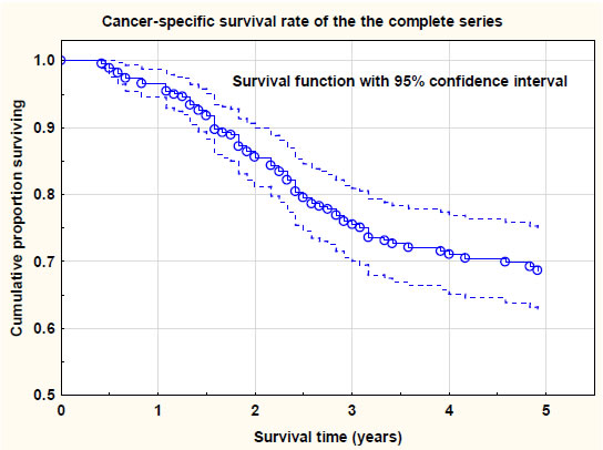
Figure 1. Cancer-specific survival rate of the complete series (n = 272). Survival probability with 95% confidence levels.

AWHC-3-2-317-g002

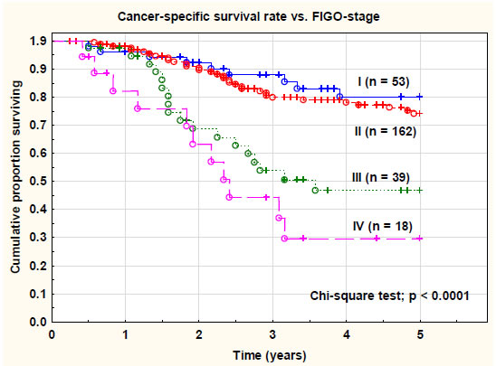
Figure 2. Cancer-specific survival rate of the complete series (n = 272) versus FIGO-stage. There was a statistically highly significant (chi-square test; p < 0.0001) difference between the tumor stages.

Prognostic factors

Ten significant prognostic factors for cancer-specific survival rate were identified in univariate Cox proportional regression analyses. However, of these factors only two: (1) pelvic control, and (2) distant tumor control (no distant recurrences) were significant and independent of each other in a Cox multivariate regression analysis (Table 5). In a multivariate analysis restricted only to factors available at the end of primary therapy (distant control was deleted) there were three significant and independent prognostic factors for cancer-specific survival rate:  (1) primary cure rate (HR 0.196; p < 0.001), (2) overall local control (HR 0.200; p < 0.03), and (3) pelvic control (HR 0.180; p < 0.001).GTV at diagnosis (GTVd) and total brachytherapy dose (p < 0.001) were significant and independent prognostic factors.The number of brachytherapy fractions was also significantly associated with the cancer-specific survival rate. Four to five fractions seemed to be an optimal fractionation schedule.

Table 5. Disease outcome and morbidity versus type of brachytherapy.

Brachytherapy groups

BT-group  IGABT-group  p value


n = 134  n = 138

Local control after primary treatment


Overall

133 (99.2%)             134 (97.1%)              0.186

Local control (at last follow-up)

Overall

128 (95.5%)             130 (94.2%)              0.622


Pelvic control (at last follow-up)

Overall

111 (82.8%)             123 (89.1%)              0.134


Systemic control

Overall (after primary therapy)

125 (93.3%)             124 (89.9%)               0.130

Overall (at last follow-up)

  98 (73.1%)               94 (68.1%)               0.365


Cancer-specific survival rate (5-year)

Overall

68.5% [60.3-76.7]    68.6% [59.3-77.8]   0.895


Overall survival rate (5-year)

Overall

53.4%  [45.0-61.8]    65.2% [55.6-74.8]   0.072


Morbidity

Bladder CTCAE ≥ G1

23/134 (17.2%)          24/138 (17.4%)       0.965

Bladder CTCAE ≥ G2

10/134 (7.5%)              7/138 (5.1%)          0.415

Bladder CTCAE ≥ G3

  5/134 (3.7%)              1/138 (0.7%)          0.090

Rectum-sigmoid CTCAE ≥ G1

66/134 (49.3%)         61/138 (44.2%)        0.404

Rectum-sigmoid CTCAE ≥ G2

34/134 (25.4%)         19/138 (13.8%)        0.016

Rectum-sigmoid CTCAE ≥ G3

17/134 (12.7%)           2/138 (1.5%)       < 0.001

Vaginal CTCAE ≥ G2 (IGABT-group)

NA                               30/138 (21.7%)

Vaginal CTCAE ≥ G3 (IGABT-group)

NA                                 0/138 (0.0%)

Bone CTCAE ≥ G1 (IGABT-group)

NA                               12/138 (8.7%)

Bone CTCAE ≥ G2 (IGABT-group)

NA                                 3/138 (2.2%)

Late bladder and intestinal toxicity

Bladder toxicity (CTCAE ≥ G1) was recorded in 53/272 patients (19.5%). There were no significant (p = 0.518) differences between the two brachytherapy groups.

Bladder toxicity (CTCAE ≥ G2) was recorded in 14 out of 272 patients (5.1%). In the BT-group it was 10/134 (7.5%) and in the IGABT-group it was 4/138 (2.9%) (Pearson chi-square test; p = 0.089). Bladder toxicity was highly significantly (t-test; p = 0.002) associated with the dose (EQD2,α/β = 3) to 2.0 cm3 of the bladder in the IGABT-group. However, the total external dose (EQD2, α/β = 3) was not significantly (p = 0.266) associated with toxicity of the bladder. Age was not a risk factor for late bladder toxicity (p = 0.641).A history of prior diseases was not associated (p = 0.871) with late bladder toxicity.Concurrent chemotherapy did not increase the risk of late bladder toxicity.

Bowel toxicity (CTCAE ≥ G1) was recorded in 127/272 patients (46.7%). There were no significant (p = 0.404) differences between the two brachytherapy groups. Bowel toxicity (CTCAE ≥ G2) was recorded in 53 out of 272 patients (19.5%). In the BT-group it was 34/134 (25.4%) and in the IGABT-group it was 19/138 (13.8%) (Pearson chi-square test; p = 0.016).

Bowel CTCAE ≥ G3 toxicity was noted in 19/272 (7.0%) in the complete series. In the BT-group it was noted in 17/134 (12.7%), but in the IGABT-group bowel CTCAE ≥ G3 was noted in only in 2/138 (1.5%). This difference was statistically highly significant (Pearson chi-square test; p < 0.0003).In the IGABT-group bowel toxicity (CTCAE ≥ G1) was significantly (Pearson chi-square; p = 0.005) associated with a prior history of abdominal surgery (gastrointestinal, urological or gynecological). Prior diseases (cardiovascular, diabetes, gastrointestinal and gynecological) also increased the risk of late bowel toxicity CTCAE ≥ G2 (p = 0.046).

Vaginal toxicity (only recorded in the IGABT-group)

Vaginal toxicity was significantly (Pearson chi-square; p = 0.022) associated (increased) with the number of brachytherapy fractions administered. The size of the HRCTV was also associated with the rate of vaginal toxicity (Pearson chi-square; p = 0.047).The total external dose (EQD2,α/β = 3) was only weakly (binary logistic regression analysis; p = 0.072) associated with the risk of vaginal toxicity.The rectal 2.0 cm3 dose (EQD2, α/β = 3) was not significantly (binary logistic regression; p = 0.143) associated with the risk of vaginal toxicity (grades 1-2).The vaginal 2.0 cm3 dose was not measured in this study [15]. The mean dose to the recto-vaginal reference point was 65.7 Gy (range 58-73 Gy). However, this dose was not significantly (logistic regression analysis; p = 0.468) associated with late vaginal toxicity in this series. Data of doses in this reference point was only available in 56 patients.

Vaginal toxicity (all grades) was more common (61/87, 70.1%) in the group treated with needles compared with the group without needles (30/52, 57.7%). However, the difference was not statistically significant (Pearson chi-square test; p = 0.136). Grade 2 toxicity was also more frequent in the needle group (21/86, 24.4%) than in the non-needle group (9/52, 17.3%), but still not significant (p = 0.326).The rate of vaginal toxicities was significantly higher (p = 0.007) during the later period (2013-2016) compared with the first period (2010-2013) of the study. (Tables 6 and 7)

Table 6. Prognostic factors for cancer-specific survival rate. Cox proportional regression analyses (univariate and multivariate analyses).

Factor univariate analyses

Hazard ratio (95% CI)            p value

FIGO-stage (I-II vs. III-IV)

0.300 [0.185-0.476]               < 0.0001

Histology (squamous cell vs. adenocarcinoma)

0.328 [0.203-0.531]               < 0.0001

Lymph node metastases (pelvic or para-aortic)

2.719 [1.410-5.243]               < 0.01

Primary cure (complete remission)

0.146 [0.084-0.255]               < 0.0001

Overall local control

0.308 [0.153-0.602]               < 0.001

Pelvic control

0.343 [0.206-0.571]               < 0.0001

Distant control

0.272 [0.170-0.433]               < 0.0001

Hemoglobin level at start of treatment

0.974 [0.961-0.987]               < 0.0001

Total extern dose (Gy) – negative impact

1.017 [1.017-1.106]               < 0.01

Total brachytherapy dose (Gy) – positive impact

0.903 [0.866-0.942]               < 0.0001

Factor multivariate analysis

Pelvic control

0.133 [0.044-0.402]               < 0.001

Distant control

0.088 [0.036-0.218]               < 0.0001


All other factors

Non-significant  > 0.05

Primary cure, overall local control and pelvic control were significant and independent if distant control was deleted

Table 7. Predictive factors for overall tumor recurrences. Logistic regression analyses (univariate and multivariate analyses).

Factor univariate analyses

Hazard ratio (95% CI)  p value

FIGO-stage (III-IV vs. I-II)

2.894 [1.576-5.313]               < 0.001

Lymph node metastases (pelvic or para-aortic)

2.696 [1.204-6.036]               < 0.02

Histology (adeno- vs. squamous cell carcinoma)

1.700 [0.922-3.362]               < 0.09

Hemoglobin value at start of therapy

0.970 [0.952-0.988]               < 0.002

Number of brachytherapy fractions

0.095 [0.010-0.862]               < 0.04

Total dose BT HRCTV D90 (α/β = 10) (Gy)

0.950 [0.902-1.000]               < 0.06

Total EBRT + BT-dose

0.960 [0.925-0.997]               < 0.04

Other factors

Non-significant in univariate

analysis

Factor multivariate analysis

FIGO-stage (III-IV vs. I-II)

4.076 [1.387-11.977]             < 0.01

Hemoglobin value at start of therapy

0.962 [0.935-0.989]               < 0.01

Other factors

Non-significant in multivariate

analysis

Discussion

In this study, the importance of modern 3-D image-guided brachytherapy was emphasized. Two different series of advanced cervix carcinomas were studied with regard to the type of brachytherapy used. An older series (n = 134) using conventional 2-D conformal brachytherapy (BT) was compared with a newer series (n = 138) using 3-D image-guided adaptive brachytherapy (IGABT). All patients were also treated with external pelvic beam irradiation (± chemotherapy). The external beam technique and doses (EQD2 51-52 Gy) were rather similar in the two series. Tumor stage, tumor size, gross tumor volume, and histology were comparable during the study period. Concurrent chemotherapy was used more frequently in the later study group since it was introduced as part of the standard therapy in 1999 [6].

In the conventional brachytherapy-group MRI was not used as part of the planning process and interstitial needles were not an option during this period.

The mean number of brachytherapy fractions was significantly higher in the BT-group than in the IGABTT-group. A comprehensive set of dosimetric data was only available for the IGABT-group according to the Vienna protocol [11].

In the IGABT-group the number of brachytherapy fractions and the total brachytherapy dose (D90, α/β = 10) was significantly associated with the overall recurrence rate, distant recurrences, and cancer-specific survival rate. The optimal number of fractions seemed to be 4-5. The importance of the brachytherapy dose to the high-risk clinical target volume (HRCTV) was shown, in a multivariate analysis (logistic regression analysis), to be significant and independent together with the primary cure rate (complete remission) and the hemoglobin value at start of therapy. A total brachytherapy dose greater than 45 Gy had a significantly positive impact on cancer-specific survival rate. However, the total external dose (after correction for the brachytherapy dose) was not significantly associated with treatment outcome and negatively correlated with the brachytherapy dose. Thus, an increased external pelvic dose willprobably not compensate for exclusion of brachytherapy treatment. Prior studies have also confirmed the advantage of combined external and intracavitary therapy compared with external therapy alone [8, 16, 17]. The total dose (D90) to the HRCTV (external plus intracavitary) was significantly higher in the group treated with needles. This is in agreement with the study by Fokdal et al. 2016 [9].

The most important predictive factor for tumor recurrences was the dose to the HRCTV (total and from brachytherapy) but not the size of the HRCTV or the use of needles. Increasing the external dose, and then decreasing the brachytherapy dose, had a negative prognostic impact on tumor recurrences and on cancer-specific survival rate. The crude local control rate in this series was 94.2% which was superior of that reported in the RetroEMBRACE study (90.6%), presented by Sturdza et al. 2016 [7]. However, the crude pelvic control rate was the same (87.0%, 86.9%) in both studies.

The overall recurrence rate in this series was 28.7% which is comparable with our earlier study with 30% recurrences [18]. In the RetroEMBRACE-study the overall recurrence rate was 30.4%, [7] similar to our older data. The recurrence rate in the BT-group was 32.1% and in the IGABT-group 25.4%, not significantly different.

The 5-year cancer-specific survival in our series was 69% compared to 73% in the retro-EMBRACE series [7]. The cancer-specific survival rate was similar (69%) in the BT- and the IGABT-group. In a prior study with standard brachytherapy from our institution the cancer-specific survival rate was 65% (Sorbe et al. 2010) [18]. Thus, in our institution we have had a slight improvementof the 5-year cancer-specific survival rate. In a study from the Netherlands by Rijkmans et al. 2014 [19] showed a highly significant improved 3-year overall survival in the group with image-guided brachytherapy (86%) compared with conventional brachytherapy (51%). The 3-year overall survival in our IGABT-group was 75% for comparison.

The mean overall treatment time (OTT) of the complete series was 44 days and in 16.5% of the patients the overall treatment time was longer than 50 days. The OTT was significantly shorter (40 days) in the BT-group than in the IGABT-group (48 days). The OTT was not a significant predictive or prognostic factor in our study. However, in the retro-EMBRACE study on 488 patients presented by Tanderup et al. 2016 [20], the OTT was significantly associated with local tumor control in both univariate and multivariate analyses.The recommendation was to keep OTT shorter than 50 days.

In our study 74 patients (54%) in the IGABT-group received ≥ 5 cycles of chemotherapy. In the EMBRACE I study the corresponding figure was 70% [21]. The number of chemotherapy cycles was neither a predictive nor a prognostic factor for therapy outcome in our study. In a study by Schmid et al. 2014 [22] they found the number of chemotherapy cycles to be of prognostic importance in a high-risk group (positive lymph nodes and stage III-IV) for distant recurrences. We could not confirm this finding in our study.

Bladder toxicity (CTCAE ≥ G2) was recorded in 5% and CTCAE G3 in 2%, and it was highly significantly associated with the dose to 2.0 cm3 of the bladder [23]. The CTCAE ≥ G3 toxicity was more frequent in the BT-group (3.7%) than in the IGABT-group (0.7%). (Figure 3)

AWHC-3-2-317-g003

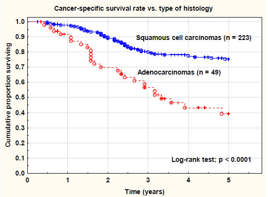
Figure 3. Cancer-specific survival rate of the complete series (n = 272) versus type of histology (squamous cell carcinomas versus adenocarcinomas/adenosquamous cell carcinomas/other types. There was a statistically highly significant (log-rank test; p < 0.0001) difference between the histological types.

Bowel toxicity (CTCAE ≥ G2) was noted in 19.5%, and bowel CTCAE ≥ G3 in 7.0% in the complete series. A significant favor of the IGABT-group was seen with regard to CTCAE ≥ G2 toxicity (13.8% vs. 25.4%) and CTCAE ≥ G3 late recto-sigmoid toxicity (1.5% vs. 12.7%). This difference was an important finding in this study. Bowel toxicity (all grades) was significantly associated with a prior history of abdominal surgery. This is a finding we also reported in a prior study (Bohr Mordhorst et al) [10]. Prior diseases (cardiovascular disease and diabetes) also increased the risk of late bowel toxicity. The brachytherapy fraction dose also increased the risk of late intestinal reactions.On the other hand, very few serious late reactions (grade 3-4) were noted in our study. In a study by Mazeron et al. [24] including 960 patients (EMBRACE I) a dose-volume effect was noted for D2cm3 ≤ 65 Gy and D2cm3 ≥ 75 Gy regarding minor or major late rectal toxicity [23]. In a prior study published from our institution serious late intestinal reactions (grade 3-4) occurred in 14% after a combined treatment of external beam pelvic radiotherapy and conventional brachytherapy [8]. This was an important improvement in the present study using MRI-guided IGABT compared with our older data and the BT-group not using this technique [10, 19]. (Figure 4)

AWHC-3-2-317-g004

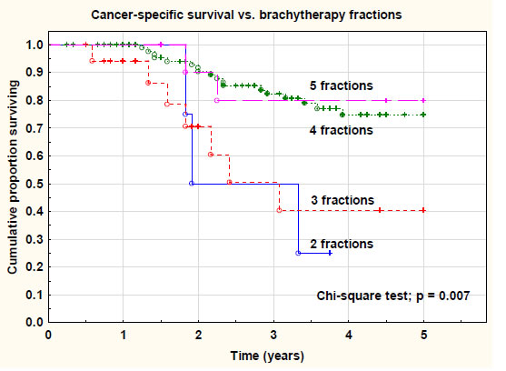
Figure 4. Cancer-specific survival rate versus number of brachytherapy fractions. There was a significant difference (chi-square test; p = 0.007) between 2-3 fractions and 4-5 fractions.

Vaginal toxicity was only registered in the IGABT-group. It was significantly associated with the number of brachytherapy fractions administered. The size of the HRCTV was also associated with the rate of vaginal toxicity. Vaginal toxicity (all grades) was more common, but not significant, in the group treated with needles compared with the group without needles. (Figure 5 and 6)

AWHC-3-2-317-g005

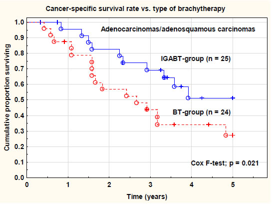
Figure 5. Cancer-specific survival rate in adenocarcinomas/adenosquamous carcinomas versus type of brachytherapy. IGABT = image-guided adaptive brachytherapy. BT = conventional brachytherapy. There was a significant (Cox F-test; p = 0.021) difference in
the cancer-specific survival rate between the two groups.

AWHC-3-2-317-g006

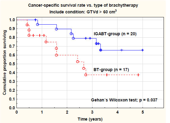
Figure 6. Cancer-specific survival rate in large tumors (GTVd < 60 cm3) versus type of brachytherapy. IGABT = image-guided adaptive brachytherapy. BT = conventional brachytherapy. There was a significant (Gehan´s Wilcoxon test; p = 0.037) difference in the cancer-specific survival rate between the two groups. GTVd = gross tumor volume at diagnosis.

The individually designed external pelvic irradiation and adaptive brachytherapy (with or without needles) is probably an explanation for this improvement. The dose to the organs at risk was carefully evaluated and limited based on the well-known tolerability of these organs. The image-guided brachytherapy technique, using MRI before and during treatment, is of importance to achieve these results. This is a significant improvement compared with older data on conventional brachytherapy both from our institution and from other centers [13].

In the IGABT-group analyses of late toxicity (CTCAE ≥ G2) of the intestine and the bladder gave a clear impression of the importance of the total dose to the HRCTV (> 90 Gy, α/β = 3) and not the HRCTV-volume. All intestinal toxicity (CTCAE ≥ G2), except three cases, and all bladder toxicity (CTCAE ≥ G2), except two cases, occurred in patients who had received a D90 dose > 90 Gy to the HRCTV-volume. Five patients had both late bladder and intestinal toxicity and all of them had received a D90 dose> 90 Gy to HRCTV. However, analysis of all late toxicity (CTCAE ≥ G1) showed another pattern where both the dose level and the volume of HRCTV seemed to be of importance.

Recently, the problem with distal parametrial and pelvic wall invasion was addressed in a two-institutial study [25] using a newly developed applicator allowing the use of both parallel and oblique needles (Vienna-II). The treatment outcome seemed to be comparable to our results (local control, recurrences and survival data) when analyzed in FIGO-stages IIB-IVB, but with a substantial higher rate of serious late side effects (20% grade 3-4 toxicity, including 4 fistulas) and acute treatment related complications (active bleeding in 27%). In a smaller series of 10 patients and total 40 fractions another new hybrid applicator (Venezia) was tested and was found to be feasible and allowed improved dose coverage and sparing organs at risk [26].

Conclusion

Data from our study showed a local control rate > 94% for a D90 dose > 95 Gy to the HRCTV. To reduce late bladder and intestinal toxicity the recommended D90 dose was < 90 Gy to the HRCTV. These results are in agreement with data presented from the retroEMBRACE study. The reported data are excellent for local and loco-regional tumor controland similar in the BT- and IGABT-groups. However, for adenocarcinomas and tumors with large GTV the cancer-specific survival rate was significantly improved in the IGABT-group. Late toxicity from the organs at risk were most favorable after treatment with modern IGABT-technique. Still, there was a problem with distant recurrences (21%) negatively influencing the cancer-specific survival rate. A more individualized and tumor-specific and biologically oriented therapy is probably needed in the future.

Declaration of competing interest

The authors declare that there were no personal financial interests or other relationships that could influence the work reported in this paper.

Acknowledgments

We acknowledge the support of Hanna Rapp, MD, for collection of missing and follow-up clinical data from part of the medical region. The oncology nurses BeritBermark and Helené Johansson also took part in data collection.

References

  1. Bray F, Ferlay J, Soerjomataram I, Siegel RL, Torre LA, et al. (2018) Global cancer statistics 2018: GLOBOCAN estimates of incidence and mortality worldwide for 36 cancers in 185 countries. CA Cancer J Clin68: 394-424.[crossref]
  2. Marth C, Landoni F, Mahner S, McCormack M, Gonzalez-Martin A, et al. (2017) Cervical cancer: ESMO Clinical Practice Guidelines for diagnosis, treatment and follow-up. Ann Oncol 28: iv72-iv83.
  3. Vaccarella S, Franceschi S, Engholm G, Lonnberg S, Khan S, et al. (2014) 50 years of screening in the Nordic countries: quantifying the effects on cervical cancer incidence. Br J Cancer 111: 965-969.[crossref]
  4. Green JA, Kirwan JM, Tierney JF, Symonds P, Fresco L, et al. (2001) Survival and recurrence after concomitant chemotherapy and radiotherapy for cancer of the uterine cervix: a and radiotherapy. Systematic review and meta-analysis. Lancet 358:781-786.[crossref]
  5. Thomas GM (1999) Improved treatment for cervical cancer–concurrent chemotherapy and radiotherapy. N Engl J Med 340:1198-1200.[crossref]
  6. Rose PG, Ali S, Watkins E, Thigpen JT, Deppe G, et al. (2007) Long-term follow-up of a randomized trial comparing concurrent single agent cisplatin, cisplatin-based combination chemotherapy, or hydroxyurea during pelvic irradiation for locally advanced cervical cancer: a Gynecologic Oncology Group Study. J ClinOncol 25:2804-2810.[crossref]
  7. Sturdza A, Pötter R, Fokdal LU, Haie-Meder C, Tan LT, et al. (2016) Image guided brachytherapy in locally advanced cervical cancer: Improved pelvic control and survival in RetroEMBRACE, a multicenter cohort study. RadiotherOncol 120: 428-433.[crossref]
  8. Karlsson J, Dreifaldt AC, Mordhorst LB, Sorbe B (2017) Differences in outcome for cervical cancer patients treated with or without brachytherapy. Brachytherapy 16:133-140.[crossref]
  9. Fokdal L, Sturdza A, Mazeron R, Haie-Meder C, Tan LT, et al. (2016) Image guided adaptive brachytherapy with combined intracavitary and interstitial technique improves the therapeutic ratio in locally advanced cervical cancer: Analysis from the retroEMBRACE study. RadiotherOncol 120:434-440.[crossref]
  10. Bohr Mordhorst L,Karlsson L, Barmark B, Sorbe B (2014) Combined external and intracavitary irradiation in treatment of advanced cervical carcinomas: predictive factors for treatment outcome and early and late radiation reactions. Int J Gynecol Cancer 24:1268-1275.[crossref]
  11. Pötter R, Georg P, Dimopoulos JC, Grimm M, Berger D, et al. (2011) Clinical outcome of protocol based image (MRI) guided adaptive  combined with 3D conformal radiotherapy with or without chemotherapy in patients with locally advanced cervical cancer. RadiotherOncol 100:116-123.[crossref]
  12. Dimopoulos JCA, Petrow P, Tanderup K,Petric P, Berger D, et al. (2012) Recommendations from Gynaecological (GYN) GEC-ESTRO working group (IV): Basic principles and parameters for MR imaging within the frame of image based adaptive cervix cancer brachytherapy. RadiotherOncol 103:113-122.[crossref]
  13. Nesvasil N, Tanderup K, Lindegaard JG, Richard Pötter, Christian Kirisits(2016) Can reduction of uncertainties in cervix cancer brachytherapy potentially improve clinical outcome? RadiotherOncol 120:390-396.[crossref]
  14. Auth B (2006) Therapy evaluation program, common terminology criteria for adverse events, version 3.0. DCTD, NCI, NIH, DHMS: 2003 (http://ctep.cancer.gov), publish date: August 9.
  15. Fidarova EF, Berger D, Schussler S, Dimopoulos J, Kirisits C, et al. (2010)Dose volume parameter D2cc does not correlate with vaginal side effects in individual patients with cervical cancer treated within a defined treatment protocol with very high brachytherapy doses. RadiotherOncol 97:76-79.[crossref]
  16. Yang J, Cai H, Xiao Z-X, Wang H, Yang P(2019) Effect of radiotherapy on the survival of cervical cancer patients. An analysis based on SEER database. Medicine98: e16421.[crossref]
  17. Delgado D, Figueiredo A, Mondonca V, Jorge M, Abdulrehman M, et al. (2019) Results from chemoradiotherapy for squamous cell cervical cancer with or without intracavitary brachytherapy. J Contemp Brachytherapy 11:417-422.
  18. Sorbe B, Bohr L, Karlsson L, Bermark B (2010) Combined external and intracavitary irradiation in treatment of advanced cervical carcinomas: predictive factors for local tumor control and early recurrences. Int J Oncol 36:371-378.[crossref]
  19. Rijkmans EC, Nout RA, Rutten IHHM,Ketelaars M, Neelis KJ, et al. (2014) Improved survival of patients with cervical cancer treated with image-guided brachytherapy compared with conventional brachytherapy. GynecolOncol 135:231-238.[crossref]
  20. Tanderup K, Fokdal LU, Sturdza A,HaieMeder C, Mazeron R, et al. (2016) Effect of tumor dose, volume and overall treatment time on local control after radiochemotherapy including MRI guided brachytherapy of locally advanced cervical cancer. RadiotherOncol 120:441-446.[crossref]
  21. Pötter R, Tanderup K, Kirisits C,Leeuw A, Kirchheiner K, et al. (2018) The EMBRACE II study: The outcome and prospect of two decades of evolution within the GEC-ESTRO GYN working group and the EMBRACE studies. ClinTranslRadiatOncol 9:48-60.[crossref]
  22. Schmid MP, Franckena M, Kirchheiner K,Sturdza A, Georg P, et al. (2014) Distant metastasis in patients with cervical cancer after primary radiotherapy with or without chemotherapy and image guided adaptiv brachytherapy. GynecolOncol 133:256-262.[crossref]
  23. Georg P, Pötter R, Georg D, Lang S, Dimopoulos J, et al. (2012) Dose effect relationship for late side effects of the rectum and urinary bladder in magnetic resonance image-guided adaptive cervix cancer brachytherapy. Int J RadiatOncolBiolPhys 82:653-657.[crossref]
  24. Mazeron R, Fokdal LU, Kirchheiner K, Georg P, Jastaniyah N, et al. (2016) Dose-volume effect relationships for late rectal morbidity in patients treated with chemoradiation and MRI-guided adaptive brachytherapy for locally advanced cervical cancer: Results from the prospective multicenter EMBRACE study. RadiotherOncol 120:412-419.[crossref]
  25. Mahantshetty U, Sturdza A, Naga CH  P, Berger D, Fortin I, et al. (2019) Vienna-II ring applicator for distal parametrial/pelvic wall disease in cervical cancer brachytherapy: An experience from two institutions: Clinical feasibility and outcome. RadiotherOncol141: 123-129. [crossref]
  26. Walter F, Maihöfer C, Schüttrumpf L, Well J, Burges A, et al. (2018) Combined intracavitary and interstitial brachytherapy of cervical cancer using the novel hybrid applicator Venezia: Clinical feasibility and initial results. Brachytherapy17:775-781.[crossref]

Latina and Black Women: Narratives on the Path to Homelessness

DOI: 10.31038/AWHC.2020325

Abstract

An increasing number of Americans are experiencing homelessness, with the latest count estimating that over one-half million people were living in the street or occupying areas not meant for human occupation in one single night. While a general portrait of the homeless population tends to highlight black or older men, almost 40 percent of the homeless population are now women. Among these are the growing numbers of Latina and Black women.

In this manuscript, we present a community based participatory research study approach designed to explore the experiences of Latina and Black women living in skid row Los Angeles, frequently recognized as the “homeless capital. Of the U.S.The finding from the mixed quantitative and qualitative study reveals similarities and salient differences on the factors that the women perceive led them on a path to homelessness. Included in the narratives are how the Black women have learned to navigate various support systems, in contrast to how the Latina women have struggled to gain entryinto the system. The women’s narratives present a portrait of structural and cultural inequities, and a need for interdisciplinary and intersectoral collaboration with diverse teams in order to develop programs the serve the needs of these new homeless populationsThe findings call for urgent need to address systems of inequity and bias, along with needed policy changes.

Keywords

Latinas, Blacks, homeless, racism, gender

Background to the Study

The first author for this study has been providing health care and allied services for underserved local and global communities for several decades; as a registered nurse and nurse practitioner, university educator and researcher, along most prominently as a clinician, community advocate, mentor, and volunteer. At times, the services were on the periphery, volunteering for services with agencies servicing farmworkers in hard to reach areas, or simply assisting with food services to homeless groups of families. Most recently, my nursing background came full circle with the opportunity to develop public health nursing projects while supervising nursing students at a major university; primarily focused in the Skid Row area of Los Angeles. Through these endeavors, the nursing student groups witnessed the changing demographics among the homeless population; including the racial/ethnic, gender, and age differences visible. The increasing number of women and “people of color” we encountered through our service projects did not fit the portrayals of homeless individuals in this country; as the faces of men are what students most frequently recalled. Instead, we encountered groups of Latina and Black women who reached out for the health education and social interaction opportunities.

The interactions with these women led to the development of this study, as the literature review suggested a gap in research as women experiencing homelessness have not been fully included in the discourse and policy arenas.

Introduction: What do we know about the homeless population?

In general, it is difficult to get a reliable estimate on the homeless individuals and family. Some of the difficulty in gathering data is the lack of consensus on how to define and measure homelessness. Additionally, how does one identify and locate “homeless” individuals? As the National Law Center on Homelessness and Poverty suggests, [1] there are different definitions of homelessness used across the U.S. For example, some of the major issues include the varying definitions used by the U.S. Department of Housing and Urban Development, or HUD, the states, and the U.S. Department of Education. In these examples, HUD relies on stricter guidelines for counting individuals located in shelters, transitional units or publicly visible places. Unlike HUD, the Department of Education guidelines includefamilies who are “doubling” or “tripling” up in the homeless count, as these families are precariously housed and are on the verge of becoming homeless.

Others have also pointed out the unreliability of the methodology used to count the number of homeless individuals, or the point-in-time homeless count conducted over one-to-two nights; and then applying these figures for service, funding, and policy development [2]. Moreover, the literature suggests a simple typology of economics, employment and lack of affordable housing as primary factors contributing to homelessness. This simple typology of job loss and lack of affordable housing may not fully explain these individual’s pathway to homelessness.

Homeless in the U.S.

In the most recent U.S. national “homeless count” conducted over one to two nights in January, slightly more than one half million people were counted as homeless (U.S. Department of Housing and Urban Development [3].Of these, a little over one-third (37%) were “unsheltered” meaning staying overnight in the streets, doorways, cars, tents, parks, under bridges, in storage or collection bins, abandoned buildings, parks, or various other places not meant for human habitation [4]. These figures suggest that four out of every ten homeless individuals are unsheltered, living in the streets; visible but mostly invisible to society.

The demographics also reveal that almost two-thirds of the individuals experiencing homelessness were men (61%), while women represented almost 39% of the population; an 8% increase between 2018 and 2019, as the numbers continue to grow. Two states had the largest numbers of individuals identified as homeless during the annual count (California and New York). Although California had over half (53%) of the unsheltered population in the nation.Almost half of the overall homeless population were white comprising over half (57%) of the unsheltered population. Yet the largest increase among the unsheltered racial/ethnic groups was foundto be among African Americans, followed by Hispanics [4].

This is a best estimate of a serious human rights and public health issue, as various reports suggest the figures may be much higher, with anywhere from 1.6 million to over three million individuals experiencing homelessness over one year’s time (National Law Center on Homelessness & Poverty [NLCHP], [5]. These are outstanding figures for a first world country as the U.S.

Homeless in Los Angeles

In Los Angeles County (LAC),estimates suggest that almost 47,000 of people were homeless in 2016. In contrast to the national data, the LAC homeless population is more diverse with widening disparities, as the majority (75%) of homeless individuals continue to be unsheltered (Los Angeles Homeless Service Authority [LAHSA] [6].

Additionally, one in three of homeless individuals were women, representing a 55% increase from the 2013 figures. Since a growing number of women are homeless and unsheltered, we can expect to see more women surviving in tents and other street make-shift encampments [6]. These are staggering figures that further compounds health risks for the female homeless population, as women experiencing homelessness suffer additional burdens when compared to homeless men; including physical and mental trauma, as well as sexual assaults [7].

Demographically, African-Americans comprise 39% of the homeless population within the greater LAC area, although they represent less than 10% of the population; while Hispanic/Latinos account for 27% of the homeless population. Additionally, almost one-fourth (18%) of the total homelesspopulation had a history of physical or sexual abuse, including domestic or intimate partner violence [6]. At the same time, a report suggests that getting overnight housing in shelters does not necessarily offer protection, as almost one-fourth of women reported they were victimized while staying in shelters [7]. Another study also suggests that women living in the streets, meaning unsheltered, were more likely to remain homeless for longer terms,had greater odds of poorer physical health, and over 12 times greater odds of poor mental health at greater risk for alcohol use, multiple sexual partners, and a history of physical assault, but less likely to access needed health services [8].

Locally and nationally, the homeless population is increasingly diversifying, yet there is a paucity of research on racial/ethnic minority women dealing with homelessness. In the parent study, we became familiar with the growing number of Latinas and Black women in skid row; but their growing presence did not fit the general characterization of Latino families. Among Latinos,familismois recognized as a core feature, where solidarity and the collectivist spirit of extended multigenerational families provide enduring support through the lifespan[9]. Earlier well recognizedstudies also suggested that intergenerational familismoplayed a protective role against poor health outcomes. This epidemiolocalparadox posits that despite disadvantages, Latinos demonstrated longevity and better health outcomesthan anticipated [10-12]. Along with familial-based support, the cohesiveness of ethnic enclaves may play a role for caregiving and maintaining wellness, despite high risk for community members [13]. Today, some core features of familismohave been associated with intergenerational caregiving, especially among foreign-born Latinos who retain their Spanish language and cultural links via ethnic enclaves and extended family ties [14].

Among Black families, as with other racial/ethnic minority populations, extended family household live-in arrangements have been found to be supportive when intergenerational solidarity- cultural orientations are aligned. Filial assistance is thus recognized as embraced by Black and Hispanic families [15]. Fictive kin, functioning as extendedsocial networks,also suggeststhat these extended social networks support collaboration, cooperation,and solidarity that nurtures and supports moving beyond simply coping in harsh environments [16-17].

The changing demographics among the homeless population and general portrayals of women cared for and supported by large networks of kin led to conceptualizing this study for exploring how race/ethnicity, gender and social class plays out among this homeless population. Because this study sampled both Latina and Black women (as a comparison group), this study will be able to note similarities and disparities for these groups of racial/ethnic minority women.

Aims

The overriding goal of this study was to explore Latina and Black women’s perceptions of factors in their lives that may have contributed to living in skid row (the “homeless capital”) in the U.S. By exploring their lived experiences in skid row and surviving in this environment, we anticipated that their narratives would provide insight on the issues faced by women necessary for expanding the narratives on homelessness so necessary for program and policy development.

Methods

Some of the data used in the analysis for this project were collected as part of a study on Older Latinos Aging in Skid Row. For that first study with an older population, the focus was on older homeless Latino men and women over 50 years of age seeking services in skid row (Ruiz & Contreras, under review). Included in the parent study were 6 self-identified Latina women. As that first study progressed, we noticed a small, but noticeable growing number of women of various ages and diverse racial/ethnic backgrounds living in skid row. The brief encounters with the “women of skid row” led us to conceptualizing the follow up study with Latina and Black women. This present study allows us to gain further insight on the intersection of race/ethnicity and gender, specifically on Latina and Black women’s experiences surviving in Skid Row.

Research team

As with the parent study that explored older Latinos path to homelessness (in review), the study team for this project included a diverse team of research assistants from multidisciplinary programs, including Chicano Studies, Anthropology, and Nursing; all spoke Spanish and identified experiences with Hispanic/Latino communities. Additionally, a doctoral nursing student focused on studies the intersection of race and healthissuescontributed to the present study, adding an additional layer of expertise and perspective.

The multidisciplinary personal and professional experiences of the authors as well as the research assistants, was instrumental for assisting the team in gathering data and capturing cultural nuances. These multiple layers of expertise added to the cross-cultural analysis for this manuscript.

Study population and recruitment of participants

The parent study was expanded; allowing us to recruit more an additional group of Latina and to add Black women over 18 years of age dependent on services in the skid row area of Los Angeles.

For this present study, the research team utilized the same study design utilized in the original study; as the mixed quantitative survey and qualitative open interview format worked well and captured the cultural nuances and findings we had not located in published studies we reviewed. Thus, this study includes 6 Latina participants from the parent study, plus an additional 6 newly recruited Latina participants (total of 12); along with 13 newly recruited Black women.

As in the parent study, participants were recruitedthrough snowball methods and assured that they could stop the survey or interview at any point of time. Participants were compensated for their time and provided an envelope with 20 dollars cash at the end of their participation. The study was approved by the University Institutional Review Board and followed guidelines for ethical research.

By focusing on Latina and Black women, the study allowed us to compare and contrast the path to homeless for these groups of marginalized women, their lived experiences pre-homeless, their perception of the pathways to becoming homeless, along with networks and family connections, as well as their recommendations for ameliorating experiences faced by groups of homeless women.

Results

Sociodemographics

The sociodemographicand health related table (Table 1) highlights some salient differences between the Latina and Black women participants. In contrast to the Latina participants, the Black U.S. born women, were all English speakers; they represented a younger cohort (mean age of 48 years) versus the Latinas (mean age of 76 years); had greater years of formal schooling (average of 12 years versus 6 years); and the majority had experienced fewer years of homelessness (less than 5 years). In contrast, all but two Latinas were born in Latino/Hispanic countries and several arrived in the U.S. unaccompanied or with limited to acquaintances, and most were primarily Spanish speakers. In regard to health matters, the majority of Latinas (85%) rated their health as fair to poor, in contrast to the majority of Black women (66%) who rated their health as good to excellent. As noted in Table 1, both groups of women reported a history of various debilitating chronic health conditions. Health issues are a major concern, as the Latinas expressed limited access to health care, including linguistic, cultural, and other biases that limited their access to resources that others benefitted from.

Table 1. Sociodemographics-Health Issues for Latina and Black Women (N=25)

Characteristics

Latinas
N=12     No. (%)

Black/African American
N=13     No. (%)

Age (years)
20-29
30-39
40-49
50-59
60-69
70-79

0
1  (8%)
0
4    (33%)
2   (17%)
5     (42%)
Range: 33-77
Mean: 76.2

2    (15%)
4    (31%)
3    (23%)
4    ((31%)
1    (.07%)
2    (1.5%)
Range: 25-70
Mean: 48.46

Country of Origin
Mexico
U.S.
El Salvador
Puerto Rico
Guatemala

4    (33%)
2    (17%)
2    (17%)
2   (17%)
1    ( 08%)

13 (100%)

Primary Language
English
Spanish
Spanish/English

3 (%)
8    (17%)
1    (08%)

13    (100%)

Education: Years completed
None
1-5 years
6-10 years
12 years
Over 12
Mean

2     (17%)
4      (25%)
2     (17%)
4     (25%)

Mean 6.2 years schooling

0
0
1      (07%)
6      (46%)
6      (46%)

Mean: 12 years

Years of Homelessness
1-5 years
6-10
11-20
over 20
Self-Rated Health
Excellent
Good
Fair
Poor

Missing

6 (50%)
4     (33%)
2      (17&)
0

0
2    (17%)
5    (42%)
4    (33%)

1

9    (69%)
3.   (23%)
1    (8%)
0

2      (16%)
6     (50%)
4      (33%)
0

1

Medical-other related Issues

High blood pressure, diabetes, heart problems, overweight, back problem, psoriasis, osteoarthritis asthma, arthritis, cataracts,
prior alcohol abuse (1), drug use (1), prison history (1).

High blood pressure,  pneumonia, overweight, hospitalization, asthma, COPD, depression, manic depressive, PTSD (1), drug use (2), youth guidance center history (1).

Generally, all of the womenexpressedhow a myriad of complex and interrelated issues contributed to them becoming homeless; including changes in family structure, dissolution of a marriage, loss of employment, limited or lack of social networks and family support contributed to ending up living in skid row. However, while the women may share some similarities in their narratives, there are some salient differences between the Latina and Black women. The women’s narratives were analyzed using content analysis allowing us to code, identify labels and categories for the development of major themes and subthemes.

Theme 1.Moving away for survival; Moving towards a better life

This theme was prominent among several Latinas only as 10 out of the 12 participants were born outside of the U.S.; while all Black women were born in the U.S. and thus did not have the immigration experience to reflect upon.  Several Latinas spent a good amount of time sharing their immigration experiences and how they had to leave their home country due to war or threats of abuse, fear, or hunger.

Their comments of “coming to the U.S. for economic necessity and to survive” and “I came for a better way of living, maybe just surviving, but you can’t starve here” exemplify their need to move away in order to survive.

For others, migrating to the U.S. was seen as necessary in order to find jobs to as a means to support themselves or families back home, or in order to find medical care not available in their home countries for physical conditions including arthritis, heart ailments, diabetes, or other several ailments. One Latina expressed that in skid row, “being around people helps with depression”, although it was not clear if she was expressing sadness, or if she suffered from depression. Also, as with other descriptive studies, researchers are not able to assess what came first; was it the state of becoming homeless that contributed to the emotional or mental status, or the reverse.

Theme 2.Learning to navigate the system: The haves versus have-nots

Depending on immigration status, language, and networks in skid row, the Latina and black women diverged on their ability to navigate the system for accessing housing, meals and other resources available.For the Latina women, their stories revealed multiple barriers, including being dependent on others to become familiar with services available, language and cultural barriers, limited social skills for gaining entry to clinics, social workers and others who could provide referrals for service access.

A Spanish speaking Latina described how she immigrated to the U.S. with her sister, so that she could serve as her babysitter. As her sister earned very little, she was paid a small mount and left the home when the brother-in-law “wanted to have sex with me, so I had to leave but nowhere to go.” This woman ended up in Olvera Street (perhaps a sanctuary church), and was able to meet with a social worker in skid row.  This Latina # 8 described how she “just follows instructions, they tell me where to go” without asking questions or saying much due to the language limitations.

Another Latina participant expressed frustration at her inability to access any health or education sessions, “they always give out flyers, with special talks, like for HIV or lots of other talks…but they’re all in English.”Another Latina shared how losing the job led to anxiety caused feelings “like depression and ended up leaving the house; “I didn’t want to be a burden for my brothers, and that’s why I decided to live in the streets.”

For the Black women participants, instead of immigration status and cultural or language impacting access to services, several of the Back women expressed frustration with some hotel and shelter managers, the limited housing available to women without children, and the “welfare system.”

One woman expressed that “there’s free clinics or minimal cost” and there’s various treatment programs for alcohol and drug abuse. Another Black woman expressed that “I went crazy after I lost my kids” and became homeless, and this led to her drug use. This woman also shared that she did not encounter any difficulty accessing resources and programs in skid row; “there’s programs here, and if they don’t have what you need, they give it to you.” The woman’s statements imply that she could easily get what she asked for, unlike the Latina participants who expressed a lack of attention and access to services or resources.

The discordant between the Latina and Black women narratives reveal varying perceptions regarding the system of services, the availability and access to basic health and service needs. This led to uncovering a subtheme for the have-and have-nots.

Subtheme 2: On being privileged-Latina women’s narratives on Non-Latinos

Some Latinas expressed that Latinos with “los papeles” (individuals holding citizenship or established residency, as Puerto Ricans), and Black people have greater support and access to services, including the ability to communicate with individuals that may help them to navigate housing, meal sources, and “better” health care (meaning out of the skid row area, as they may have health coverage as Medicaid or Medicare). As one Latina stated, “no papers, no help.” A few Latinas also expressed frustration and dislike for “the roaches, drug dealers and drug use” as they perceive that “those with benefits” may spend their money foolishly; money that is not available for those “without los papeles.”

Theme 3: Homelessness: Separation of mind from physical conditions

Among both Latina and Black women their narratives reveal a separation of mind and body; as mental health issues were not perceived as affecting the physical well-being. Although several described various chronic health conditions potentiated by the harsh homeless environment; they separated stories on their physical ailments from the mental health aspects as loss of self-esteem, loss of control, and complacency; without noting the connecting between mental and physical well-being (or not being well). Although some men in the parent study (older Latino men and women) expressed that “it’s depressing here; it’s not motivational, and you become trapped and you suffer.”

None of the women in this study expressed a similar sense of connection with the mind, body, and mental health.

Instead, a Latina blamed herself for an incident of sexual abuse. This woman expressed that she may have contributed to the abuse, as “maybe because you are a drunk and you want to be…” This woman’s discussion suggested that the physical trauma was not relevant to her mental health. At the same time, this Latina described how she needed medication for “my bones, for walking, stomach problems and other ailments.” In this case, there was no consideration for the association between physical and mental health.

Theme 4.The long road to becoming homeless-where do familismo and fictive kin fit in?

There were salient differences between the Latina and the Black participants on the path leading to becoming homeless. Among Latinas, almost all had migrated to the U.S. (11 out of 12) and each described experiences of being dependent on others for getting to the U.S., housing, employment, and various geographical moves before arriving in Los Angeles. Additionally, over half (8) described living as homeless for over 5 years (one reported being homeless over 16 years). For several of these Latinas, it was difficult for them to identify factors that led to becoming homeless. For example, many of these women had been moving into various homes; dependent on family and friends for a space to sleep in. These Latinas had been “doubling up,” meaning sharing crowded homes that were not meant to accommodate a large number of individuals. People that are “doubling up” are at high risk for becoming homeless if anyone becomes unable to contribute, or even if a slight change occurs in the household. For many individuals, this meant that they had no established residence, as they moved repeatedly until they ended up in skid row. However, “doubling up” is not recognized as a state of being homeless by the federal government, and this leads to an undercount in the homeless count. In contrast, this sample of Black women had not “lived on the edge” (meaning doubling up); and in contrast to the Latina group, only a small number of these Black women [4]reported being homeless for five years or more. The number of years for dealing with homelessness is important, as living in the harsh environment could play into worsening health conditions, along with the loss of family connections for support and emotional well-being.

The narratives from the Latina women also contributed a cultural layer for exploring the state of familismo and filial social support, as the majority described minimum contact with family members, one-half of the participants [6] could not think of anyone they could count on for help in case of emergency. Two of these women felt they could count on a “friend”, although they had not previously reported having such a friend. Interestingly, one woman reported she would count on the paramedics; thus implying she considered first responders to be the only people that she can depend on. When asked about family contact and family based networks, several Latinas expressed that they longed to at least have another woman nearby “I could talk with in Spanish.”

In contrast, with a lifelong history of living in the U.S., the majority of Black women (10 of 13) reported they had family near the city, including mothers, children, and friends they could reach out to if needed. However, the social networks both groups of women alluded to for support was not fully addressed by both the Latina and Black women, and several did not wish to provide further information. Still, one Black woman shared that her physical conditions do not affect her mental status by her remarking “physically I’m fine…we have all the food here; mentally I’m a mess.”

Overall, among the Black women, conversations with other women described as friends were mostly for navigating the system, speaking with social workers, housing managers, or simply carrying on conversations with others residing in skid row. It appears that familismo and fictive kin networks became a hardship and slowly fade with time and the impact of living in the homeless environment.

Discussion

As the demographic and findings reveal, there are salient differences between the sample of Latina and Black women. The one tying thread, or similarity is the lackof family connection and supportive networks frequently attributed to Latino and Black families; especially as social support networks are perceived as contributing to resilience and longevity among these populations. While both the Latina and Black women appeared to be resilient and surviving among the homeless, none seemed to have established functional social networks, much less a supportive network that they could count on. Although several Latinas lamented, “others get to depend on the government and available services granted to citizens” but this does not equate to having a social supportive network that they could count on.

In regard to health, both the Latina and Black women shared that they suffer from a myriad of serious debilitating chronic health conditions, including hypertension, diabetes, asthma, arthritis, previous hospitalization, along with various other debilitating conditions. Among the Black participants, conditions as chronic obstructive pulmonary disease (COPD), manic depression, and post-traumatic stress disorder (PTSD) were mentioned. This suggest that unlike the Latina participants, the Black women had access to medical and mental health services that provided these advanced diagnoses; access and follow-up care that the Latina group did not have access to.

Similar to the issue of health matters, drug use among these women warrants further exploration. For example, unlike previous studies that mention high rates of drug use among some homeless populations, the researchers noted that illicit drug use was mentioned by only one Latina and two Black women participants. This low rate of drug use among the women conflict with previous studies that most often utilize samples of homeless men.

Overall, the narrative from these women were sorrowful and call for aggressive changes and policy development. Future research would expand our lenses on the plight of women who experience homelessness. We suggest that a morecomplete portrait could be gatheredby utilizing amixed quantitative and qualitative approach, with multicultural, multilingual and multidisciplinary teams of researchers and community collaborators. If our goal is to capture the voices of thewomen left alone to live in a harsh, biased, and never ending cycle of homelessness, the changes needed require a more nuanced and social justice systemic approach for dealing with the epidemic of homelessness.

References

  1. National Law Center on Homelessness and Poverty (2017). Don’t count on it. How the HUD point-in-time count underestimates the homelessness crisis in America.
  2. Schneider M, Brisson D, Burnes D (2016) Do we really know how many are homeless?: An analysis of the point-in-time homeless count. Families in Society: The journal of Contemporary Social Services 97: 323-329.
  3. The U.S. Department of Housing and Urban Development (2020).
  4. The 2019 Annual Homeless Assessment Report (AHAR) to Congress. Part 1: Point-In-Time Estimates of the Homeless.
  5. National Law Center on Homelessness & Poverty 2019. Housing and Homelessness in the United States of America.
  6. Los Angeles Homeless Service Authority (2016) 2016 Homeless Count Result Los Angeles County and LA Continuum of Care.
  7. Wenzel S, Tucker J, Hambarsoomian K, Elliot M (2006) Toward a more comprehensive understanding of violence against impoverished women. Journal of Interpersonal Violence 21: 820-839. [Crossref]
  8. Nyamathi AM, Leake B, Gelberg L (2000) Sheltered versus nonsheltered homeless women differences in health, behavior, victimization, and utilization of care. Journal of General Internal Medicine 15: 565-572.[Crossref]
  9. Ruiz ME, Ransford HE (2012) Latino elders reframing familismo: Implications for health and caregiving support. Journal of Cultural Diversity 19: 50-57. [Crossref]
  10. Angel JL, Angel RJ (1992) Age at migration, social connections, and well-being among elderly Hispanics. Journal of Aging and Health 4: 480-499. [Crossref]
  11. Hayes-Bautista D (2004) La Nueva California, Latinos in the Golden State. London: University of California Press.
  12. Markides KS, Coreil J (1986) The health of Hispanics in the Southwestern united States: An Epidemiologic paradox. Public Health Reports 101: 253-265. [Crossref]
  13. Rote S, Angel JL, Markides KS, Hill TD (2018) Neighborhood cohesion and caregiver well-being among the Mexican-origin population. In Contextualizing Health and Aging in the Americas, Eds. WA Vega, JL, Angel LM, Gutierrez Robledo KS, Markides. 295-310.
  14. Almeida J, Morlnar BE, Kawachi I, Subramanian SV (2009) Ethnicity and nativity status as determinants of perceived social support: Testing the concept of families. Social Science & Medicine 68: 1852-1858.[Crossref]
  15. Reyes AM (2018) The economic organization of extended family households by race/ethnicity and socioeconomic status. Journal of Marriage & Family 80: 119-133.[Crossref]
  16. Cook D, Williams TJ (2015) Expanding intersectionality: Fictive kinship networks as supports for the educational aspirations of black women. Western Journal of Black Studies 39: 157-166.
  17. Taylor R, Chatters L, Woodward A, Brown E (2013) Racial and Ethnic Differences in Extended Family, Friendship, Fictive Kin and Congregational Informal Support Networks.
  18. Ruiz ME, Contreras C (in review). Older homeless Latinos: Aging in skid Row.
  19. Perez GK, Cruess D (2011) The impact of familism on physical and mental health among Hispanics in the United States. Health Psychology Review 8: 95-127.

COVID-19 Related Lung Inflammation and Oxidative Stress – a Role for Cannabidiol ?

Keywords

Cannabidiol, CBD, COVID-19, Cytokine release syndrome, Cytokine storm, SARS-CoV-2

Opinion Article

During the recent outbreak of coronavirus SARS-CoV-2,differences in susceptibilities of subjects to the infection with the virus have been observed; not all people exposed to SARS-CoV-2 become infected and not all infected patients develop severe respiratory illness. One of the biggest but still unanswered questions is why some develop severe disease, whilst others do not. Although activation of the endocannabinoid system (ECS) as well as polymorphism of the cannabinoid receptor CB2 influence susceptibility to infections with rhinoviruses [1], the role of the ECS in COVID-19 is unknown. CB2 controls the immune response and its mutant Q63R is known to be less functional and overrepresented in several populations of patients with autoimmune disease. While it increased the risk of severe acute respiratory tract infection (ARTI) in children [2], an eventually increased risk for COVID-19 is currently unknown. Age, sex and comorbidities (or comedications) seem to play a role, including medications taken early at the beginning of symptoms. Whereas risks related to comorbidities have been repeatedly described, assessments of comedications as potential risk factors are still very rare. About 60% of the subjects remain more or less asymptomatic but are carriers who can easily transmit the virus [3]. This percentage varies widely between less than 40% and up to 80% [4,5]; it reflects the lack of reliable data on asymptomatic carriers on one hand, and differences in populations concerning their sensitivity of getting diseased on the other. A minority of subjects develops apparent clinical symptoms after a mean, although variable, incubation period of about one to two weeks. A two-phase division of this clinical symptomatic stage is very important as it will influence the management of patients. The first clinical phase of roughly one week’s duration can be described as a non-severe, immune defence-based, protective phase during which the development of antibodies against the virus particles is of vital importance. This phase is characterised by fever and cough as the two main symptoms, and signs of pneumonia on both lungs. During this period, measures should try to reduce virus load and to boost immune responses or at least avoid drugs that may have a negative impact. Although respective epidemiological data are still missing for SARS-CoV-2,canonical antipyretics and antiinflammatory drugs such as paracetamol (acetaminophen) or aspirin, most of which are available without prescription and taken by patients as self medication (often as first line treatment for viral infections) must be seen with caution. A clinical benefit of these drugs for COVID-19 patients has not been demonstrated. On the contrary, preclinical studies point to an increased risk of mortality in influenza-infected animals, likely reflecting an impaired development of protective immunity [6,7,8]. In humans infected with influenza and rhinovirus, an increased duration of sickness and viral shedding, or at least no clinical benefit has been observed after intake of aspirin and paracetamol but also after a number of other medications such as COX-2 inhibitors [9,10]. Increased risks of complications of community-acquired pneumonia following prior use of non-steroidal antiiflammatory drugs (NSAIDs) have been reported also in numerous recent retrospective studies [11,12,13,14]. In addition, anticoagulants, benzodiazepines and statins which are more frequently used by an elderly population, have also been reported to reduce the virus-specific antibody response [15]. Whether ACE-inhibitors increase the risk for severe COVID-19 or not is still an ongoing debate [16,17]. Intriguingly, some patients treated for mild COVID-19 infection still had coronavirus for up to one week after symptoms disappeared, although this may be much longer in rare cases [18]. Currently, an increasing range of products with widely differing properties and mechanisms are used as experimental treatments to combat SARS-CoV-2, e.g., neuraminidase inhibitors, protease inhibitors (e.g., ritonavir, lopinavir), nucleoside inhibitors (e.g., ribavirin), inhibitors of virus replication (remdesivir), anti-sera, pegylated IFNα or (hydroxy-)chloroquine [19].

A small percentage of patients eventually progress to the second, inflammation-driven, damaging phase with a sudden deterioration around one to two weeks after symptoms onset. This phase is the most severe and characterised by an immune overreaction, with a more or less destroyed immune system, marked lymphocytopenia and an excessive, uncontrolled release of pro-inflammatory cytokines, called cytokine release syndrome or “cytokine storm”. With increasing severity, not only lungs, but multiple organs are involved, including spleen and hilar lymph nodes, heart and blood vessels, liver and gallbladder, kidney, adrenal gland, oesophagus, stomach and intestines. A potential participation of the brain and neuroinvasion of SARS-CoV-2 may easily be overlooked in this phase. According to a retrospective case series of 214 COVID-19 patients, up to 36.4% had neurological symptoms manifested as acute cerebrovascular diseases, consciousness impairment and skeletal muscle symptoms [20,21]. Less severe manifestations are anosmia or, although rarely, ageusia; smell dysfunction are observed in up to 98% of cases [22,23]. The final stage is accompanied by rapid virus replication, a large number of inflammatory cell infiltration, acute lung injury, acute respiratory distress syndrome (ARDS), extrapulmonary systemic hyperinflammation syndrome with damage of the vascular endothelium, and disseminated intravascular coagulation (DIC) which can progress to gangrene at the extremities and death. This has already been observed before in SARS and MERS [24]. At the very end, the cause of death by the virus is the body’s own immune response to the viral infection.Whereas in the first stage of disease anti-viral and supportive treatments are very important, it is evident that at some point during the progress and exacerbation of disease an anti-inflammatory and immunosuppressive intervention can save lives. Virus infection of cells induces oxidative stress: large amounts of highly reactive oxygen species (ROS) are generated in the infected cell, even in the absence of viral replication. This is a common and major pathogenic mechanism for inflammatory response and tissue injury caused by viruses but also by other infectious agents [25,26]. Oxidative stress is associated with oxidative modifications of proteins, nucleic acids and lipids by free radical chain reactions with catastrophic consequences for a normal molecular functioning within cells. Oxidative stress activates a cascade of inflammatory cytokines, notably IL-1, IL-6, IL-8, IL-12, IFN-γ, IL-18 and TNF, of which IL-6 is a protagonist since it predominately induces pro-inflammatory signalling and regulates massive cellular processes. Janus-kinases significantly contribute to this cytokine-induced pro-inflammatory signalling. Cytokines can stimulate more cytokine production and cause many more cytokine receptors to awaken. Uncontrolled, this becomes a “cytokine storm”. Many drugs are known that can interfere with steps of this inflammatory chain reaction such as corticosteroids, cyclosporine, IL-6 inhibitors or Janus-kinase inhibitors (JAK-inhibitors), each having its own mechanism.

Although corticosteroids are known for their anti-inflammatory effects since many decades and have been widely used during the SARS 2003 epidemic in the early acute phase, there is mixed evidence from case series in COVID-19 patients; actually, the WHO does not support their use. Concerns for corticosteroids are that they may delay virus clearance and/or increase the risk of secondary infections [27]. JAK-inhibitors such as baricitinib, ruxolitinib or tofacitinib inhibit autoimmune response to ease inflammation. However, they also inhibit IFN-alpha which is a naturally released cytokine to combat virus infections. A big concern is therefore the decreased resistance to infections. The most preferable treatment method is monotherapy. Among possible side effects JAK-inhibitors may cause anaemia and lymphopenia, although less likely after short exposure. Results of randomised clinical trials for COVID-19 are still lacking.IL-6 inhibitors (e.g., tocilizumab) are monoclonal antibodies that target the pro-inflammatory cytokine IL-6 which is consistently increased in severely ill COVID-19 patients. It is approved in the United States for severe life-threatening cytokine release syndrome and may be an effective treatment also in COVID-19 cytokine storm. Another potential target is the Vascular Endothelial Growth Factor (VEGF); it is responsible for pulmonary oedema and can be suppressed with bevacizumab, a drug, approved by the FDA and widely used in clinical oncotherapy. A naturally occurring substance with a distinctly different mechanism of action is cannabidiol (CBD). Similar to other experimental treatments, it has not yet been used in COVID-19 patients. After almost 50 years of research in man, CBD is recognised as a well tolerated drug with a broad spectrum of activity, and anti-inflammatory, anti-oxidant properties. This has been demonstrated in an animal models of acute lung inflammation [28,29] and in a viral model of multiple sclerosis (Theiler’s murine encephalomyelitis virus-induced demyelinating disease [30], but also in infectious disease models of prion disease [31] and malaria [32]. In another model, CBD reversed oxidative stress parameters, cognitive impairment and mortality in rats submitted to sepsis by coecal ligation and puncture [33].Instead of acting “downstream” on the cytokine cascade such as IL-6 inhibitors or JAK-inhibitors, CBD is an agonist on peroxisome-proliferator activated receptor gamma (PPARg) exerting a dual role as agonist of the nuclear factor erythroid 2 (Nrf2) which plays a key role in cytoprotection against ROS, and as antagonist of the nuclear factor NFκB which mediates the transcription of pro-inflammatory genes (e.g., those coding for inflammatory cytokines) and proteins such as COX-2 [34,35]. The induction of the Nrf2 downstream genes is able to protect the infected cells against virus-induced cellular injury. By the same Nrf2 pathway, lung inflammation induced by lipopolysaccharide (LPS) is also alleviated by activation of PPARg resulting in improved lung function [28]. CBD inhibits also the VEGF [36]. To note, some patients demonstrated a pathological autoimmune response with high titer of antiphospholipid and other auto-antibodies. At this stage, the initiation of immunosuppressive, anti-inflammation therapy is critical for reducing death rate of COVID-19 patients. Attenuation of oxidative stress and inflammation by a pharmacological measure is therefore highly beneficial for lessening a virus-induced lung injury and exacerbation of existing respiratory diseases [35].

Overall, this means that CBD normalises the physiologic redox balance which is disturbed by the virus-induced cellular injury and restores the self-defence mechanism of the cell. As CBD is a multi-target drug, direct effects on other receptors (GPR55, 5-HT1A, A2A) and on ion channels (notably TRPV1) as well as indirect effects on endocannabinoid levels (notably AEA) that interact on their turn with a number of targets, contribute to the overall restoration and normalisation of physiologic processes in cells [37]. In contrast to IL-6 inhibitors given as example, CBD does not act just on one target but protects the host cells by multiple mechanisms. Simply described, instead of interfering as mailman with receptors, CBD manages the post office.Moreover, because of its chemical structure, CBD has also an immediate direct, strong antioxidant effect exceeding vitamin E and vitamin C, capturing free radicals or transforming them into less active forms. This considerably reduces the destruction of biological molecules by highly reactive oxygen species (ROS) generated in the virus-infected cell. CBD has already been used in a daily dose of 300mg combined with standard Graft-versus-Host-Disease (GVHD) prophylaxis consisting of cyclosporine and a short course of methotrexate in the prevention of GVHD [38]. In comparison to a historic control group which did not receive CBD, acute GVHD was significantly delayed and tolerance significantly improved. Various animal models demonstrate that doses of 5mg CBD/kg and above are effective as antioxidant. Moreover, CBD easily crosses the blood-brain barrier and is able to combat a potential neuroinvasion of SARS-CoV-2 and neuroinflammation; recently, a case of a patient who was diagnosed with viral encephalitis in Beijing Ditan Hospital has already been described [39].In addition to mitigating lung inflammation, beneficial effects of CBD have been demonstrated on other aspects related to COVID-19 such as on endothelial cells (vasorelaxation [40], diabetes [41,42,43] and stress-induced hypertension [44,45]. CBD has already been proposed previously as possible treatment for individuals with post Ebola sequelae [46].All depends on a prompt diagnose and application of such therapy. CBD is safe to use, can be administered already early in the inflammatory phase and can be combined with a large number of other medications such as antibiotics. Recently, a 10% oral solution of CBD containing 7.9% ethanol has received marketing authorisation. Pharmacy preparations based on crystalline CBD can be prepared as capsules (up to 200mg) or as suppositories (up to 300mg) for those patients that need intubation and assisted breathing (e.g., >99.8% phyto-CBD, BSPG Laboratories Ltd, Sandwich, UK or CannPico Research & Marketingservice GmbH, Vienna, Austria).Based on its pharmacological effects and favourable safety profile, CBD of known purity, composition and stability should be considered as a potential treatment for individuals with SARS-CoV-2infections.

References

  1. Tahamtan A, Tavakoli-Yaraki M, Shadab A, Rezaei F, Marashi SM, et al. (2018) The role of cannabinoid receptor 1 in the immunopathology of Respiratory Syncytial Virus. Viral Immunol31: 292-298. (Crossref)
  2. Tahamtan A, Samieipoora Y, Nayerib FS, Rahbarimaneshc AA, Izadic A, et al. (2018) Effects of cannabinoid receptor type 2 in respiratory syncytial virus infection in human subjects and mice. Virulence 9: 217-230. (Crossref)
  3. Li P, Fu JB, Li KF, Chen Y, Wang HL, et al. (2020) Transmission of COVID-19 in the terminal stage of incubation period: a familial cluster. Int J Infectious Diseases. (Crossref)
  4. Javid B, Weekes MP, Matheson NJ (2020) Covid-19: should the public wear face masks? Yes—population benefits are plausible and harms unlikely. BMJ.
  5. Li G, Li W, He X, Cao Y (2020) Asymptomatic and Presymptomatic Infectors: Hidden Sources of COVID-19 Disease. Clin Infect Dis.
  6. Eyers S, Weatherall M, Shirtcliffe P, Perrin K, Beasley R (2010) The effect on mortality of antipyretics in the treatment of influenza infection: systematic review and meta-analyis.J R Soc Med103: 403-411. (Crossref)
  7. Prymula R, Esposito S, Zuccotti GV, Xie F, Toneatto D, et al. (2014) A phase 2 randomized controlled trial of a multicomponent meningococcal serogroup B vaccine (I): Effects of prophylactic paracetamol on immunogenicity and reactogenicity of routine infant vaccines and 4CMenB. Hum Vaccin Immunother10: 1993-2004. (Crossref)
  8. Saleh E, Moody MA, Walter EB (2016) Effect of antipyretic analgesics on immune responses to vaccination. Human Vaccines & Immunotherapeutics12: 2391-2402. (Crossref)
  9. Graham NMH, Burrell CJ, Douglas RM, Debelle P, Davies L (1990) Adverse effects of aspirin, acetaminophen, andibuprofen on immune function, viral shedding, and clinical status in rhinovirus-infected volunteers. J Infect Dis162: 1277-1282. (Crossref)
  10. Jefferies S, Braithwaite I, Walker S, Weatherall M, Jennings L, et al. (2015) Randomized controlled trial of the effect of regular paracetamol on influenza infection. Respirology.
  11. Basille D, Thomsen RW, Duhaut P, Andrejak C, Jounieaux V, et, al. (2018) NSAIDs-Outcomes of Community-acquired Pneumonia. American J Respiratory and Critical Care Medicine198: 128-131.
  12. Kotsiou OS, Zarogiannis SG, Gourgoulianis KI (2017) Prehospital NSAIDs use prolong hospitalization in patients with pleuro-pulmonary infection. Respiratory Medicine123: 28-33.(Crossref)
  13. Le Bourgeois M, Ferroni A, Leruez-Ville M, Varon E, Thumerelle C, et al. (2016) Nonsteroidal anti-inflammatory drug without antibiotics for acute viral infection increases the empyema risk in children: A Matched Case-Control Study. J Pediatrics175: 47-53.
  14. Voiriot G, Philippot Q, Elabbadi A, Elbim C, Chalumeau M, et al. (2019) Risks related to the use of non-steroidal anti-inflammatory drugs in community-acquired pneumonia in adult and pediatric patients. J Clin Med.
  15. Huemer HP (2015) Possible immunosuppressive effects of drug exposure and environmental and nutritional effects on infection and vaccination. Mediators Inflamm.
  16. Bean DM, Kraljevic Z, Searle T, Bendayan R, Pickles A, et al. (2020) Treatment with ACE-inhibitors is associated with less severe disease with SARS-Covid-19 infection in a multi-site UK acute Hospital Trust. (preprint).
  17. Fang L, Karakiulakis G, Roth M (2020) Are patients with hypertension and diabetes mellitus at increased risk for COVID-19 infection?. The Lancet.
  18. Shi Y, Wang Y, Shao C, Huang J, Gan J, et al. (2020) COVID-19 infection: the perspectives on immune responses. Cell Death & Differentiation 27: 1451-1454.
  19. Agrawal S, Goel AD, Gupta N (2020) Emerging prophylaxis strategies against COVID-19. Monaldi Archives for Chest Disease 90: 169-172. (Crossref)
  20. Chen N, Zhou M, Dong X, Qu J, Gong F, et al. (2020) Epidemiological and clinical characteristics of 99 cases of 2019 novel coronavirus pneumonia in Wuhan, China: a descriptive study. Lancet 395: 507-513.
  21. Mao L, Wang M, Chen S, He Q, Chang J, et al.(2020) Neurological Manifestations of Hospitalized Patients with COVID-19 in Wuhan, China: a retrospective case series study. JAMA Neurol.
  22. Lüers JC, Klußmann JP, Guntinas-Lichius O (2020) The Covid-19 pandemic and otolaryngology: What it comes down to?.Laryngo-Rhino-Otol. (Crossref)
  23. Moein ST, Hashemian SMR, Mansourafshar B, Khorram-Tousi A, Tabarsi P, Doty RL (2020) Smell Dysfunction: A Biomarker for COVID-19. Int Forum Allergy Rhinol. (Crossref)
  24. Zhang W, Zhao Y, Zhang F, Wang Q, Lib T, et al. (2020) The use of anti-inflammatory drugs in the treatment of people with severe coronavirus disease 2019 (COVID-19): The Perspectives of clinical immunologists from China. Clinical Immunology. (Crossref)
  25. Kaul P, Biagioli MC, Singh I, Turner RB (2000) Rhinovirus-Induced Oxidative Stress and Interleukin-8 Elaboration Involves p47-phox but Is Independent of Attachment to Intercellular Adhesion Molecule–1 and Viral Replication. J Infectious Diseases 181:1885–1890. (Crossref)
  26. Schwarz KB (1990) Oxidative stress during viral infection: a review. Free Radical Biology & Medicine21: 641-649.(Crossref)
  27. Russell B, Moss C, Rigg A, van Hemelrijck M (2020) COVID-19 and treatment with NSAIDs and corticosteroids: should we be limiting their use in the clinical setting?.ecancermedicalscience. (Crossref)
  28. Ribeiro A, Almeida VI, Costola-de-Souza C, Ferraz-de-Paula V, Pinheiro ML, et al. (2015) Cannabidiol improves lung function and inflammation in mice submitted to LPS-induced acute lung injury. Immunopharmacol Immunotoxicol37: 35-41. (Crossref)
  29. Vuolo F, Petronilho F, Sonai B, Ritter C, Hallak JEC, et al (2015) Evaluation of serum cytokines levels and the role of cannabidiol treatment in animal model of asthma. Mediators of Inflammation. (Crossref)
  30. Mecha M, Feliu A, Inigo PM, Mestre L, Carillo-Salinas FJ, et al. (2013) Cannabidiol provides long-lasting protection against the deleterious effects of inflammation in a viral model of multiple sclerosis: a role for A2A receptors. Neurobiol Dis.
  31. Dirikoc S, Priola SA, Marella M, Zsürger N, Chabry J (2007) Nonpsychoactive Cannabidiol Prevents Prion Accumulation and Protects Neurons against Prion Toxicity. The Journal of Neuroscience27: 9537-9544. (Crossref)
  32. Campos AC, Brant F, Miranda AS, Machado FS, Teixeira AL (2015) Cannabidiol increases survival and promotes rescue of cognitive function in a murine model of cerebral malaria. Neuroscience 289:166-180. (Crossref)
  33. Cassol OJ, Comim CM, Silva BR, Hermani FV, Constantino LS, et al. (2010) Treatment with cannabidiol reverses oxidative stress parameters, cognitive impairment and mortality in rats submitted to sepsis by cecal ligation and puncture. Brain Res1348: 128-138. (Crossref)
  34. Atalay S, Jarocka-Karpowicz I, Skrzydlewska E (2020) Antioxidative and anti-inflammatory properties of cannabidiol. Antioxidants (Basel).
  35. Lee C (2018) Therapeutic Modulation of Virus-Induced Oxidative Stress via the Nrf2-Dependent Antioxidative Pathway. Oxidative Medicine and Cellular Longevity.(Crossref)
  36. Tagne AM, Pacchetti B, Sodergren M, Cosentino M, Marino F (2020) Cannabidiol for viral diseases: hype or hope?.Cannabis and Cannabinoid Research.
  37. Nahler G, Jones T, Russo EB (2019)Cannabidiol and contributions of major hemp phytocompounds to the “Entourage Effect”; possible mechanisms.J Altern Complement Integr Med.
  38. Yeshurun M, Shpilberg O, Herscovici C, Shargian L, Dreyer J, et al. (2015) Cannabidiol for the prevention of Graft-versus-Host-Disease after allogeneic hematopoietic cell transplantation: Results of a phase II study. Biol Blood Marrow Transplant21:1770-1775. (Crossref)
  39. Li Z, Huang Y, Guo X (2020) The brain, another potential target organ, needs early protection from SARS-CoV-2 neuroinvasion. Sci China Life Sci63: 771-773.(Crossref)
  40. Stanley CP, Hind WH, O’Sullivan SE (2013) Is the cardiovascular system a therapeutic target for cannabidiol? Brit J Clinical Pharmacol75: 313-322. (Crossref)
  41. Ehud Z, Lola W L, Itamar R, Natan Y, Zhanna Y, Ruth G (2012) Islet protection and amelioration of diabetes type 2 in Psammomys obesusby treatment with cannabidiol. J Diabetes Mellitus2: 27-34.
  42. Horvath B, Mukhopadhyay P, Hasko G, Pacher P (2012) The Endocannabinoid System and Plant-Derived Cannabinoids in Diabetes and Diabetic Complications. The American Journal of Pathology180:432-442. (Crossref)
  43. Wheal AJ, Jadoon K, Randall MD, O’Sullivan SE (2017) in vivo cannabidiol improves endothelium-dependent vasorelaxation in mesenteric arteries of Zucker diabetic fatty rats. Front Pharmacol. (Crossref)
  44. Bitencourt RM, Takahashi RN (2018) Cannabidiol as a therapeutic alternative for post-traumatic stress disorder: From bench research to confirmation in human trials. Front Neurosci. (Crossref)
  45. Jadoon KA, Tan GD, O’Sullivan SE (2017) A single dose of cannabidiol reduces blood pressure in healthy volunteers in a randomized crossover study. JCI Insight. (Crossref)
  46. Reznik SA, Gardner EL, Ashby CR (2016) Cannabidiol: a potential treatment for post Ebola syndrome?.Int J Infect Diseases 52: 74-76. (Crossref)

When two pandemics meet

DOI: 10.31038/EDMJ.2020423

Abstract

The COVID-19 pandemic has emerged in the middle of another pandemic which is far from under control: the cardiometabolic syndrome pandemic. Recently published data suggests patients with obesity are at a higher risk of being hospitalized and placed on a mechanical ventilator for COVID-19 than patients with a normal body weight. We discuss the pathophysiology behind this relationship and the implications in the global fight against COVID-19.

Keywords

COVID-19; coronavirus; obesity; cardiometabolic syndrome.

No one single mechanism is responsible for disease progression into severity in COVID-19 cases as in almost all diseases -chronic or not, transmissible or not-. We as scientists are trained to observe, identify differences and similarities between cases and arrive at possible explanations called hypothesis that can help the scientific community to develop effective strategies to combat the illness.

To this day several factors have been identified and when put together they tell a storyline that sums up the pathophysiology of severity in COVID-19 shown in Figure 1.

EDMJ-4-2-405-g001

Figure 1. Schematic representation of shared pathophysiology in COVID-19 cases with underlying metabolic illness. [1]. DIC: Disseminated Intravascular Coagulation.

But how does this scenario come to be? The answer comes from a previous pandemic that has been around for many years: the Cardiometabolic Syndrome (CMS) pandemic. CMS is defined by a combination of metabolic disorders that include diabetes mellitus, systemic arterial hypertension, central obesity, and dyslipidemia. All these conditions lead to elevated heart disease risk, which in turn is the leading cause of death in first world countries and doesn´t fall far behind in the rest of the world as well. This global epidemic to some doesn´t seem so scary being that it cannot be transmitted through droplets or by touching “infected” surfaces. Thisidea, however, isn´t completely true. The first risk factor for this group of diseases is being overweight or obesity, and this is in a sense “transmitted”. Eating habits are a cultural phenomenon, and from one generation to the next, families and communities pass on grocery lists, recipes and pantry contents. As of 2019 the global mean prevalence of obesity was measured at 19.5%. This number has almost tripled since 1975 and is currently the number one risk factor associated with premature death. Obesity as a risk factor for disease usually means it leads to chronic diseases such as the ones previously mentioned, but nowadays we are observing a different consequence of being overweight. An elevated body mass index has become a high-risk factor for severity in COVID-19 cases. [2]

Table 1 shows the evidence on the previous statement. A study by Zheng et al of 214 patients in Wuhan, China with laboratory confirmed COVID-19 showed that the presence of a Body Mass Index (BMI) >25 kg/m2 was associated with a near-6 fold increased risk of severe illness, even after adjusting for age and other comorbidities. [3] Of 4,103 COVID-19 cases in New York City the chronic condition which conferred the strongest association with critical illness was obesity, with 39.8% of hospitalized patients having obesity. [4]

Table 1. Epidemiological studies on COVID-19 outcomes and obesity related risk-factors

Author, Region and Date

Subjects

Findings

Z. Wu [7]
Mainland China
Updated Feb 11, 2020

72,314 suspicious cases of COVID-19
44,672 lab-confirmed cases

2.3% Case-Fatality Rate
Mild cases 81%
Severe cases 14%
Critical cases 5%

S. Garg [8]
USA (COVID-NET)
March 1-30, 2020

1,482 hospitalized patients

89% of patients had one or more underlying conditions:
Hypertension 49.7%
Obesity 48.3%
Chronic lung disease 34.6%
Diabetes Mellitus 28.3%
Cardiovascular disease 27.88%

Among patients 18-49 years-old obesity was the most prevalent underlying condition (59%).

P. Goyal [9]
New York City, US
Mar 3-27, 2020

First 393 cases of COVID-19 adults hospitalized in New York

Patients who required invasive mechanical ventilation were more likely to be male, have obesity and elevated liver-function and inflammatory markers.

S. Richardson [10]
New York, USA
Mar 1 – Apr 4, 2020

5,700 hospitalized patients

Most common comorbidities among hospitalized patients:
Hypertension 56.6%
Obesity 41.7% – (Morbid obesity 19%)
Diabetes Mellitus 33.8%

G. Grasselli [11]
Milan, Italy
Feb 20 – Mar 18, 2020

73 patients in intensive care unit

Over 80% of patients in ICU were overweight or had obesity.
Normal weight – 19%
Overweight – 51.9%
Obesity 1 – 15.4%
Obesity 2 – 11.5%
Obesity 3 – 1.9%

Zheng [3]
Wenzhou, China
Jan 1 – Feb 29, 2020

214 patients with lab confirmed COVID-19
Ages 18-75

A BMI equal to or greater than 25 kg/m2 was associated with a 6-fold increased risk of severe illness.
This risk remained significant even after adjusting for age and other comorbidities.

Petrilli [4]
New York
Mar 1 – Apr 7, 2020

4,103 cases of COVID-19
1,999 hospitalized

The chronic condition with the strongest association to critical illness was obesity.
39.8% of hospitalized patients had obesity.

Qingxian [12]
Mainland China
Jan 11 – Feb 16, 2020

383 patients admitted to a hospital in Shenzen

After adjusting for age, sex, disease history and treatment the overweight group was 2.42 times more likely to develop severe pneumonia.

A. Simonnet [5]
Lille, France
Feb 27 – Apr 5, 2020

124 patients admitted to ICU for COVID-19.
Compared to control group from 2019

Obesity was significantly more frequent among cases of COVID-19 (47.6%) compared to control group (25.2%).
The median BMI of patients requiring intubation was 31.1 kg/m2 compared to 27 kg/m2 in the patients who did not require intubation.
In individuals with a BMI ³35 kg/m2 the odds ratio for intubation was 7.36 compared to individuals with a normal BMI.

Among 124 patients admitted for COVID-19 to a hospital in Lille, France 47.6% had obesity. Patients with a BMI of greater than 35 kg/m2 were 7.36 times more likely to require a ventilator than patients with a BMI of less than 25 kg/m2. [5] In Milan more than 80% of 73 patients treated in an ICU were overweight or had obesity, when the rates of overweigh and obesity in Italy are only 35.4% of the population. [6]

Two main explanations play a role in this complicated infectious disease in association with weight problems. The first one is the chronic inflammatory state it conveys. Recent studies have found that adipose tissue secretes extracellular vesicles that function as vectors which can modify cellular function in the recipient through the information they carry. Data suggests that this mechanism is used by fat to induce monocyte differentiation into active macrophages and high secretion of IL-1 and TNF-α among other cytokines. [13] The second one is the fact that patients with obesity have been found to have higher concentrations of pro-thrombotic factors as compared to normal-weight controls. Some of these altered parameters include higher D-dimer, fibrinogen and factor VII; as well as lower fibrinolysis because of higher plasminogen activator inhibitor-1. [14]

Besides increased inflammatory cytokines, obesity englobes several pathophysiological factors which affect the risk and outcomes of patients with COVID-19. In the respiratory tract obesity may cause pulmonary restriction, decreased pulmonary volumes and ventilation-perfusion mismatching. Patients with obesity are more likely to present diabetes mellitus and atherosclerosis which may be complicated by COVID-19. Additionally, there is limited data on the right dosing of antimicrobials in obesity and bioavailability of drugs used to treat patients with this disease may be affected by altered protein binding, metabolism and volume of distribution. [15]

On the other hand, new information is developing every day concerning COVID-19 cases and more data is suggesting that bad prognosis is linked to thromboembolic events caused by inflammation, hypoxia and coagulation abnormalities. One study by Klok et alstudied 184 Intensive Care Unit (ICU) patients with confirmed COVID-19, and found that 31% showed thrombotic complications, of which 81% was due to pulmonary embolism. [16] When we put two and two together, the relationship becomes apparent. Obesity is a clear catalyzer for severe COVID-19 cases. In a country like Mexico, where the prevalence for overweight and obesity in over 20-year-olds is 75.2%, this relationship is very threatening. [17]

It seems that the best way to prevent bad outcomes from this novel disease (as well as from infectious diseases in general) is to be in good health prior to contracting it in the first place. As for those patients who already suffer from CMS or one of its components, preventive treatment is our main recommendation. These patients should be at optimal glycemic, systemic arterial pressure and cholesterol level goals. A study by Carter et al also suggests that vitamin D deficiencies (also more common in patients with obesity) have been linked to worse cytokine storms. To this end, physical activity as well as sun exposure is effective ways to boost vitamin D levels. [18]

This sound reasonable, right? Well, reasonable doesn´t always mean achievable in all populations. Vulnerable communities around the world are struggling every day just to have access to general medical attention. These communities are also at an increased risk of exposure to COVID-19. Working from home is a privilege that is unavailable for many people from a lower socio-economic status. Social distancing is considerably more difficult for people living in overcrowded neighborhoods. Emerging epidemiological studies in the U.S. suggest a disproportionate burden of illness and higher death rates among minority groups. [9]

Currently there is no gold standard treatment for COVID-19, however, all this data suggests that global efforts need to be directed towards prevention and education. Pre-existing conditions need to be under control and lifestyle habits should be aimed towards getting enough exercise and a proper nutrition. [19,20]

References

  1. Xiong M, Liang X, Wei YD (2020) Changes in Blood Coagulation in Patients with Severe Coronavirus Disease 2019 (COVID‐19): A Meta‐Analysis. Br J Haematol.[crossref]
  2. Blüher M (2019)Obesity: global epidemiology and pathogenesis. Nat Rev Endocrinol15:288-298.
  3. Zhou F, Yu T, Du R, Fan G, Liu Y et al. (2020) Clinical course and risk factors for mortality of adult inpatients with COVID-19 in Wuhan, China: a retrospective cohort study. The Lancet.395: 1054-1062. [crossref]
  4. Petrilli CM, Jones SA, Yang J, Rajagopalan H, O’Donnell LF, Chernyak Y, Horwitz LI (2020) Factors associated with hospitalization and critical illness among 4,103 patients with COVID-19 disease in New York City. medRxiv.
  5. Simonnet A, Chetboun M, Poissy J, Raverdy V, Noulette J et al. (2020) Intensive Care COVID‐19 and Obesity study group. High prevalence of obesity in severe acute respiratory syndrome coronavirus‐2 (SARS‐CoV‐2) requiring invasive mechanical ventilation. Obesity.[crossref]
  6. Zangrillo A, Beretta L, Scandroglio AM, Monti G, Fominskiy E (2020) Characteristics, treatment, outcomes and cause of death of invasively ventilated patients with COVID-19 ARDS in Milan, Italy. Crit Care Resusc.[crossref]
  7. Wu Z, McGoogan JM (2020) Characteristics of and important lessons from the coronavirus disease 2019 (COVID-19) outbreak in China: summary of a report of 72 314 cases from the Chinese Center for Disease Control and Prevention. Jama.[crossref]
  8. Garg S (2020) Hospitalization rates and characteristics of patients hospitalized with laboratory-confirmed coronavirus disease 2019—COVID-NET, 14 States, March 1–30, 2020. MMWR. Morbidity and Mortality Weekly Report 69.
  9. Goyal P, Choi JJ, Pinheiro LC, Schenck EJ, Chen R, Jabri A, Hoffman KL (2020) Clinical Characteristics of Covid-19 in New York City. New England Journal of Medicine..[crossref]
  10. Richardson S, Hirsch JS, Narasimhan M, Crawford JM, McGinn T et al. (2020) Presenting Characteristics, Comorbidities, and Outcomes Among 5700 Patients Hospitalized With COVID-19 in the New York City Area. JAMA.[crossref]
  11. Grasselli G, Zangrillo A, Zanella A, Antonelli M, Cabrini L et al. (2020) Baseline characteristics and outcomes of 1591 patients infected with SARS-CoV-2 admitted to ICUs of the Lombardy Region, Italy. Jama. [crossref]
  12. Qingxian C, Fengjuan C, Fang L, Xiaohui L, Tao W et al. (2020) Obesity and COVID-19 severity in a designated hospital in Shenzhen, China. China.
  13. Kim A, Shah AS, Nakamura T (2018) Extracellular Vesicles: A Potential Novel Regulator of Obesity and Its Associated Complications. Children 5: 152.[crossref]
  14. De Pergola G, Pannacciulli N (2002) Coagulation and fibrinolysis abnormalities in obesity. J Endocrinol Invest25: 899-904.[crossref]
  15. R Huttunen, J Syrja¨nen (2013) Obesity and the risk and outcome of infection. International Journal of Obesity37: 333-340.[crossref]
  16. Klok FA, Kruip MJHA, van der Meer NJM, Arbous MS, GommersDAMPJ et al. (2020) Incidence of thrombotic complications in critically ill ICU patients with COVID-19. ThrombosisResearch.[crossref]
  17. Romero-Martínez M, Shamah-Levy T, Vielma-Orozco E, Heredia-Hernández O, Mojica-Cuevas J et al. (2019) Encuesta Nacional de Salud y Nutrición (Ensanut 2018): metodología y perspectivas. salud pública de México 61: 917-923.
  18. Carter SJ, Baranauskas MN, Fly AD (2020) Considerations for obesity, vitamin D, and physical activity amidst the COVID‐19 pandemic. Obesity.
  19. Smith JA, Judd J (2020) COVID‐19: Vulnerability and the power of privilege in a pandemic. Health Promotion Journal of Australia 31: 158.[crossref]
  20. Ahmed F, Ahmed NE, Pissarides C, Stiglitz J (2020) Why inequality could spread COVID-19. The Lancet Public Health5: 240.[crossref]

Treatment of Patella Infera Following Closed Trauma by a Combination of Patella Tendon Tenotomy and Tuberositas Tibia Osteotomy and Augmentation with Semitendinosus Tendon Autograft

DOI: 10.31038/IJOT.2020315

Abbreviations

ROM = Range of Motion

Keywords

Patella infera, patella baja, patella tendon tenotomy, Z-plasty, tuberositas tibia osteotomy, tendon autograft augmentation, semitendinosus.

Introduction

Patella Infera (PI) is a rare condition presenting with shortening of the patella tendon. PI can be seen as a congenital abnormality, or as a complication secondary to trauma or kneesurgery [1-5]. It was first described in 1982 by Caton et al. and was defined as a Caton-Deschamps index £ 0.66. The Caton-Deschamps index is assessed on a knee x-ray in sagittal view where the length of the patella tendon, defined as the distance from the lower patella pole to the superior part of the tibial tubercle, is divided by the length of the patella, defined as the greatest diagonal length measured[6]. A normal Caton-Deschamps index is defined as an index in the range > 0.6 or < 1.26. Clinically the condition presents with symptoms of decreased ROM, lach of knee flexion, patellofemoral knee pain and accelerated progression of patellofemoral osteoarthritis [3].

There is no consensus on treatment method of patella infera, it has been proposed that surgery is indicated, when the Caton-Deschamps index is lower than or equals 0.6(6). The two main surgical treatment modalities include a z-plasty of the patella tendon or an osteotomy of the tibial tuberositas. It has been suggested to use a z-plasty, when the Caton-Deschamps index is ≤ 0.6 and the patella tendon is < 25 mm, and to use the tuberositas osteotomy when the Caton-Deschamps index is ≤ 0.6 and the patella tendon is > 25 mm(6). Immediate postoperative mobilization and physiotherapy is required to learn to activate the quadriceps muscle, and focus on strengthening exercises of the muscle has been described to be the most important post-operative regime to prevent relapse of patella infera[5, 6]. This case describes a severe secondary developed patella infera after conservative treatment of a patella fracture. Surgical technic is done with a combination of patella tendon tenotomy and tuberositas tibia osteotomi and augmentation with semitendinosus allograft. Previous reports of surgical treatment of patella infera is done with either a z-plasty or tuberositas tibia osteotomy, no previous reports have to our knowledge described the surgical treatment of patella infera with a combination of the two treatment modalities.

Case Report

A healthy fifty-five year old woman slipped and fell onto her left knee while walking. She reported pain and difficulties in both flexion and extension of the knee. X-ray revealed a comminuted non-displaced fracture of the inferior pol of the patella (Figure 1). The knee was immobilized in full extension for 6 weeks and weight-bearing was allowed within the limits of pain. X-ray made 3 months after the injury revealed early union of the fracture. Five months following the fracture she presented with limited range of motion with reduced knee-flexion to 95 degrees despite a specialized physiotherapy rehabilitation training program. The rehabilitation program was continued but 8 months following the fracture she presented with pain, aggravation of the affected knee flexion and inability to follow her rehabilitation. Clinical examination revealed atrophy of the quadriceps muscle, knee-flexion reduced to 30 degrees, full knee-extension but inability to raise the leg in full extension. A MR-scan and x-ray showed severe patella infera with a Caton-Deschamps index of 0.3 and a patella tendon measuring only 13 mm (Figure 2a + Figure 2b ).

IJOT-3-1-306-g001

Figure 1. X-ray showing a comminute non-displaced fracture of the inferior patella pol of after closed trauma to the left knee.

IJOT-3-1-306-g002

Figure 2. A: Pre-operative x-ray of the left knee with distinct patella-infera with a Caton-index of 0.3 B: Pre-operative x-ray of the right healthy knee, no patella infera. C: X-ray of the left knee after osteotomy, Caton index = 0.56. D: X-ray after osteotomy and Z-plasty with a Caton-Deschamps index of 0.8 as a final result.

The Caton-Deschamps index was at time of injury 0,9 as well as 9 days later. After 45 days it reduced to 0,44 . After 10 months the measurement was 0,3 reduced to 0,2 after one year. Because of the severity of the patella infera it was planned to preform both a osteotomy of the tibial tuberositas and a z-plasty of the patella-tendon following augmentation with the semitendinosus-tendon. Pre-operative an Oxford 12-item Knee Score was made resulting in a score of 7.

Results

First, a osteotomy was preformed (Figure 3a + Figure 3b) and the tibial tuberositas was inserted 18 mm proximal to the origin resulting in a Caton-index of 0.56 (0.25 prior) (Figure 2c). Thereafter, the patella-tendon was extended by 1 cm to a full length of 23 mm(13 mm prior) by a z-plasty (Figure 3c) resulting in a final Caton-Deschamps index of 0.8 (Figure 2d). Finally the tendon was augmented by the semitendinosus-tendon (Figure 3d). After the procedure it was possible for the operator to flex the knee 110 degree (30 degree pre-operative). Post-operative regime was planned which included 1) mobilization with don-joy-bandage in 0-30 degree and only partial weight bearing in full extension for 2 weeks, then 0-60 degrees for 2 weeks and full weight-bearing and then  0-90 degrees for 2 weeks, 2) CMP-machine with mobilization 0-60 degrees without the bandage 4 times per day for 4 weeks, 3) early training instructed by physiotherapist and 4) EL-stimulation of the quadriceps muscle. 8 weeks after the operation she was able to perform knee flexion to 95 degrees and had a good knee stability and she was given no limitation in range of motion. 3 months after the operation she was capable of performing 100 degrees knee flexion and full extension at clinical examination. The Oxford 12-item Knee Score was repeated resulting in a score of 42 which indicates satisfactory joint function.

IJOT-3-1-306-g003

Figure 3. A and B shows the osteotomy of the tuberositas tibia. C: Illustrates the z-plasty of the patella-tendon, note the markedly sclerosis of the tendon. D: Final result after osteotomy, z-plasty and augmentation with the semitendinosus-tendon.

Discussion

This case report describes a sucessefull treatment of PI with a combination of two well established surgical methods; a tibial tubercle transfer and a Z-plasty of the patella tendon.Because of the poor quality of patella tendon after post traumatic patella infera it was decided to   reinforces  it  with the semitendinosus-tendon. It assures a safe heeling of the Z-plasty and allows  a postoperative early mobilization by ROM as well as an aggressive rehabilitation that is in this case extremely important to get a good final result.

The literature has no clear consensus on optimal treatment of PI and the rarity of PI does not allow any RCT study to be performed. To our knowledge it is the first time the two surgical approaches has been combined, and the obtained Caton-Dechampes index and high Oxford knee score confirms our theory of a succesfull combination. However our weakness could be the short follow up period mainly do to relapse as previous described in the literature. In conclusion the combined surgical methods could be an alternative in treating patella infera.

References

  1. Morshed S, Ries MD (2002) Patella infera after nonoperative treatment of a patellar fracture: A case report. J Bone Joint. Surg Am84: 1018-1021.[Crossref]
  2. Jiang X, Zhang YM, Liu JY (2013) Patella infera following patellar tendon contracture after closed trauma. ChinMed J (Engl)126: 3990-3991.[Crossref]
  3. Kennedy MI, Aman Z, DePhillipo NN, LaPrade RF (2019) Patellar tendon tenotomy for treatment of patella baja and extension deficiency. Arthrosc Tech8: 317-320.
  4. Guido W, Christian H, Elmar H, Elisabeth A, Christian F (2016) Treatment of patella baja by a modified Z-plasty. Knee Surgery, Sport Traumatol Arthrosc24: 2943-2947.[Crossref]
  5. Bruhin VF, Preiss S, Salzmann GM, Harder LP (2016)Frontal tendon lengthening plasty for treatment of structural patella baja. Arthrosc Tech5: 1395-1400.
  6. Caton JH (2010) The management of patella infera in current practice. Eur J Orthop Surg Tramatol20: 265-271.

“Thrower’s Fracture”- A Humeral Fracture

DOI: 10.31038/IJOT.2020314


A 24-year-old right dominant male threw a softball extremely hard from outfield. A loud, crackingnoise was accompanied by pain and inability to lift his right arm. Radiographs performed in ED revealed a severe displaced, comminuted fracture of the right humerus known as a “thrower’s fracture” (Figures 1,2). The patient was a healthy, recreational athlete. A blood test, bone scan, and MRI all revealed negative results for bone pathology prior to surgical open reduction internal fixation (ORIF) of the right humerus (Figure 3).

IJOT-3-1-305-g001

Figure 1. A-P Radiograph with comminuted fracture of right humerus.

IJOT-3-1-305-g002

Figure 2. IR-AP of Right humerus.

IJOT-3-1-305-g003

Figure 3. Post-surgical A-P radiograph.a

Thrower’s fracture, somewhat rare, is an acute fracture of the mid-to-distal third of the humeral diaphysis during a forceful throwing motion, usually involving an excessive torque during the cocking and acceleration phase of motion [1, 2]. The internal rotation of the latissimus dorsi, subscapularis, and pectoralis major all contribute to the strong internal rotation force. During the cocking and throwing phase, a torsional force is applied to the humeral insertion of these muscles while the distal humerus is external rotation, causing a spiral type fracture. Thrower’s fracture is seen more in ages 20s-30s or recreational athletes, who often lack cortical hypertrophy seen in professional pitchers [1, 2] rarely is the radial nerve, involved, but if injured, usually heals without surgical involvement [1, 2].

No post-op complications or radial nerve involvement were noted. A sling was worn for 2 weeks and ROM began at 4 weeks. He resumed light duty work at 2 months. Full ROM, less 3ºextension was obtained. At 3 months post-surgery, he resumed gym workouts, but no recreational softball.

References

  1. Colapinto MN, Schemitsch EH, Wu L (2006) Ball-thrower’s fracture of the humerus. Canadian Med Assc J 175:33.
  2. Miller A, Dodson CC, Ilyas AM (2014) Thrower’s fracture of the humerus. Orthop Clin North Am 45:565-569.

The Fight against COVID-19: The Role of Drugs and Food Supplements

Abstract

SARS-CoV-2 diffuses quite easily among humans, causing a variety of symptoms from a mild flu to a fatal illness mostly involving the lungs and sometimes the kidneys or the heart, organs that express high concentration of the ACE2 viral receptor. No vaccine is available, although several are under scrutiny. From the therapeutic side, many different products are being tested, from antiviral to anti inflammatory drugs taken from the repertoire of other diseases, however with variable success. In fact, the death toll of this viral infection remains quite high. Containment of the infection is based on mechanical devices (goggles and masks) that shield the entrance doors of the virus (eyes, nose, mouth), and on tight social restrictions to limit the possibility of contact among people living in a community. Nonetheless, the virus apparently survives for hours on different surfaces and in droplets suspended in the air and dispersed by the micro particulate that is so abundant in industrialized towns, thus reaching further away from the originator, and tricking human defenses. In this situation, a possible complementary – however unspecific – approach to limit the infectivity of the virus could be based on a range of natural compounds which may interfere with the diffusion of the virus within the body, and increase the efficacy of the immune defenses of the organism. This is meant to be a non-toxic, preventive or adjuvant treatment so that in case of infection, the symptoms might not develop to full scale, giving the organism more time and strength to fight it.

Keywords

Covid-19, Computational chemistry, Drugs, Epidemiology, Food supplements, Probiotics, Therapy.

Introduction

The virus SARS-CoV-2 is the cause of the most recent pandemic of flu-like disease COVID-19. Italy has been among the European country most severely hit by the pandemy, with an amount of infected and dead patients even higher than the originator China. The facility of viral diffusion in Italy (but the rest of the world does not seem to behave much differently) and the relative inefficiency of containment measures and of the available drugs to treat Covid-19, has prompted us to figure out alternative and complementary possibilities to approach the diffusion of this viral pandemy, which might apply also to future epidemies. The treatment suggestions that follow are the result of such effort. The mechanism of infection by the SARS-CoV class of viruses apparently occurs via specific interactions between the SARS-CoV spike protein (S) and the host receptor angiotensin-converting enzyme 2 (ACE2), which regulates both cross-species and human-to-human transmission of SARS-CoV [1]. Once the virus has gained entry into the human body, it starts spreading, usually through the respiratory tract, causing sympotms that can be mild, if it stays in the upper respiratory tract, or more severe such to be fatal to the host, if it reaches the lungs and the deep alveoli network [2]. In order to progress into the respiratory tract, the virus has to move against the inverse flow of the mucus, which is continuosly produced by the epithelial cells lining the airways and pushed by their cilia towards the larinx [3]. In this way inhaled pathogens and particulate matter trapped by the airway mucus can be removed by swallowing or coughing. This process is an important self-defense mechanism of the respiratory system and its failure may lead to chronic infections and impaired lung function [4].Furthermore, the virus has to survive to the immune surveillance of the host. Natural and adaptive immunity are alerted, and will start mounting an immune response to the invasive guest. A struggle develops between the speed of virus replication and diffusion, and the inflammatory response trying to contain it. Sometimes the inflammatory response gets out of control, and a cytokine storm may happen, adding further damage to the viral infection, causing acute lung injury and leading the patient to death [5, 6].

The infective process

The SARS-CoV-2 and its interactions

CoVs have a complex organization (Figure 1) containing four or five structural proteins mixed with some minor components that include nonstructural and host cell-derived proteins [7]. All viral particles display on their surface Spike (S), Envelope (E) and Membrane (M) structural proteins [8] (Insert Fig 1).

JPPR-3-1-310-g001

Figure 1. Virus structure. Schematic structure of a Corona Virus, with the surface proteins spike (S), membrane (M) and envelope (E). The nucleocapsid (N) protein stabilizing the single strand RNA molecule is shown inside.

These surface proteins interact with host cell membranes at the beginning of infection, and the S protein is responsible for the fusion process between viral and host membranes [9], thus defining tissue tropism and host range (Figure 2A). The S protein contains two subunits (Figure 2B): the S1 at the N-terminus has the receptor binding function and the S2 at the C-terminus confers the fusion activity [9]. Host cell proteases cleave the subunits from the S protein. Once the S1 has bound the host cell receptor followed by the uptake into a vesicle, then S2 works to bring in close proximity viral and cellular membranes so that fusion may occur [10] (Insert Fig 2 A & 2B).

JPPR-3-1-310-g002

Figure 2. Infection mechanism. A: The infective process of SARS-CoV-2. ACE-2 appears to be the host cell receptor responsible for mediating the Covid-19 infective process. B: The spike protein S contains two moieties, S1 and S2. The trimeric S1 moiety contains the receptor binding domain (RBD) responsible of the specific interaction with the ACE2 host cell receptor.

Entrance doors of the virus: an eye on the ocular tissue

While researchers are certain that Corona viruses (CoVs) spread through mucus and droplets expelled by coughing or sneezing, it is likely that the virus can also diffuse via other body fluids, such as tears. Since early 2000, CoVs infection was known to be associated with conjunctivitis and in 2004, CoV RNA has been detected for the first time in tears of SARS-CoV patients, suggesting the possibility of virus transmission through ocular tissues and tears [11]. How CoV eventually gets to the eye from infected droplets (directly, or through the nasolacrimal duct, or the lacrimal gland, etc) remains an unsolved problem. Infact, the end of SARS-CoV epidemic turned off the interest on possible involvement of the ocular tissue in virus infection. The recent SARS-CoV-2 epidemic and the similarity in the receptor that binds both SARS-CoV-2 and SARS-CoV renewed much attention on research into ocular infection as a possible route of SARS-CoV-2 transmission [11,12]. Further investigations concluded that COVID-19 could be indeed transmitted through the ocular route, as suggested by SARS-CoV-2 isolation in the tears of a patient at Rome’s infectious-disease Spallanzani Hospital. The study recently published indicates that the eyes are not only an entrance door for the virus but also “a potential source of contagion” [13]. The additional finding that SARS-CoV-2 is present in conjunctival specimens [14, 15] and that ACE2 has been detected in different eye compartments [16-17] is indicative of the possibility that ocular tissues might represent a source of spread, particularly when higher viral loads are present at the acute stage of ocular complications.

Inflammation: a double edged sword

Once the virus has reached the airways, the first line of defense is the respiratory epithelium [18]. The human respiratory epithelial layer is made of ciliated cells intermingled by some secretory and basal cells. Secretory cells produce mucins and anti-microbial molecules. Ciliated cells generate a mucin flow helping the removal of foreign particles (micro-organisms included) and debris, sweeping mucus and trapped particles upwards and helping to expel them from the respiratory tract. Different immune cell types are resident in the epithelium, including T lymphocytes and dendritic cell populations acting as sentinel cells. Other immune cell populations including innate lymphoid cells and natural killer cells (NK) are found lining the epithelium. Alveolar macrophages are resident in the alveolar space. Recognition of invading SARS-CoV by intracellular sensors induces rapid production of antiviral interferons and other proinflammatory cytokines. In particular, when leukocytes recognize virus-infected cells or tissues damaged by the virus, these sentinels rapidly initiate an innate immune response that involves cellular activation, signaling cascades and the release of cytokines to guide leukocytes to mount an effective response. Among immune responses against SARS-CoV infection, activation of inflammation and host cell death are crucial in limiting viral infections, replication, and associated pathological damage. On the other hand, inflammatory cascade triggered by viral infection can exacerbate the pathological damage or contribute to viral clearance depending on the context of the infection [5]. There are multiple aspects of inflammation associated with viral infections. In particular, mechanisms underlying the excessive cytokine response deserve further investigations in order to develop strategies to minimize detrimental tissue damage associated with strong inflammation, while maximizing their beneficial anti-viral features.

Respiratory disease due to alveoli failure

When the infection reaches the respiratory tract, then the lining of the respiratory tree becomes injured thus causing an inflammatory state that may spread to the air sac – the gas exchange unit – which becomes unable to get enough oxygen from the blood stream and to efficiently release carbon dioxide. The first organism reaction is to activate the immune system, triggering an inflammatory response able to destroy the virus and limit its replication, however with the risk that an excessive inflammation (either in terms of intensity or duration) may exacerbate the pathological damage. Such mechanisms are very active and ready to respond in young and healthy individuals, but can be impaired in elderly people in which additional pathologies and a weakened immune system, often due to vitamin D deficiency, make it harder to fight the disease, thus accounting for the high number of deaths in elderlies [19]. Although comparably infected, pneumonia-induced death rate in men is higher than in women. This is in line with disproportional affection during additional epidemics caused by CoV. There are several factors that may confer more protection to women: stronger immune system, which, on the other hand, renders women more susceptible to autoimmune diseases, sex hormone estrogen, which appears to play a role in immunity, less prevalent strong smoking habits, less incidence of hypertension and diabetes to mention just a few differences between women and men [20]. In addition to the sex difference revealing that infection in males is more aggressive than in females, another question concerns children, which in fact contract the virus as often as adults, however developing much milder symptoms. Conversely, an inverse relationship has been noted with age, and the younger the child, the higher chance they have of winding up in severe or critical condition. (A. Balbarini, personal communication). Why COVID-19 affects children differently remains unknown although some hypotheses have been proposed. Children may have a more efficient and responsive immune system, and a better protection of the airways by an active production of mucins, rapidly flowing towards the larynx for secretion, all of which could be contributing to a milder disease. The fact that children are susceptible to SARS-CoV-2 infection, but frequently do not develop a symptomatic disease, raises the possibility that children could be facilitators of viral transmission [21]. This whole, though synthetic, picture of the infection pathway indicates nonetheless the possible target mechanisms that should be tackled by drug molecules or by natural products to prevent or at least limit viral infections, including the one by the SARS-CoV-2: i. receptor binding; ii. virus diffusion; iii. inefficient or deranged immune reaction. We will describe now some of these approaches, with a major emphasis on non-pharmacologic ones, explaining their rationale in this context.

Pharmacological approach

Much effort is presently given to the characterization of some therapeutic compounds that could be potentially active against the currently emerging novel coronavirus SARS-CoV-2. New treatments are being added day by day and their list includes, among others, repurposed flu treatments, malaria treatments, failed ebola drugs, anti-HIV drug combination, immune suppressants and anti-hypertensive drugs. This paragraph is aimed to provide a summary of therapeutic compounds that show potential in fighting the SARS-CoV-2 infection.

Antiviral products

Scientists around the world are racing with time to find a cure for the COVID-19 pandemic. Characterization of the viral structure and physiology is critical to develop effective antiviral drugs. Presently, the virus capsid S and M proteins, the serine protease TMPRSS2 used for S protein maturation, the RNA-dependent RNA polymerase (RdRp) necessary for virus replication and the cell receptor ACE2 are the primary targets. Among the antiviral drugs, Favipiravir or Avigan was developed in Japan as an anti-viral agent that inhibits the RdRp of RNA viruses. Its effects appear to improve the lung condition by preventing virus replication, thus shortening the time of virus infection. This drug has been approved as an experimental treatment for mild COVID-19 infections and has been tested with success in 340 individuals from Wuhan and Shenzhen. However a comprehensive picture about the mechanisms underlying its efficacy is still lacking [22]. Chloroquine (CQ) and hydroxychloroquine (HCQ) are drugs approved for the treatment of malaria, and inflammatory autoimmune diseases like lupus an rheumatoid arthritis. CQ is a weak base that becomes entrapped in membrane-enclosed low pH organelles thus leading to an increase of lysosomal pH. SARS-CoV-2 entry into the cell requires a correct endocytic trafficking whose impairment, as after CQ administration, would interfere with viral infection [23]. Both CQ and its derivative HCQ are being used against COVID-19 and clinical trials are being organized both in U.S. and China. However, some attention against potential side effects including cardiac arrhythmias has been recently turned on [24]. Due to the severe side effects that can be caused by CQ, HCQ might be preferred, since it shows an antiviral effect comparable to that of CQ, and appears to be able to blunt the severe progression of COVID-19, by decreasing T cell activation, thus inhibiting the dangerous cytokine storm. Beside a safer clinical profile it is also suitable for pregnant patients [25]. Much attention has been recently raised about the efficacy of remdesivir, a drug formerly used against Ebola and now repurposed to conteract COVID-19 infection [26]. Remdesivir belongs to the class of nucleotide analogs known to display some antiviral activity against single stranded RNA viruses. Although used against some cases of the African Ebola epidemic, laboratory experiments with blood sample analysis have failed to demonstrate a correlation between drug assumption and drop in the concentration of viral particles. In addition, serious side effects restrict drug prescription only to severely affected CoVID-19 patients. The antiviral drug kaletra, a combination of lopinavir (LPV) and ritonavir (RTV), is used for the treatment and prevention of HIV/AIDS. Both compounds are protease inhibitors. In particular, RTV acts by slowing down the breakdown of LPV, but both components have been shown to interact with other medications against important diseases as for instance cardiovascular diseases. The antiviral activity of this drug combination has generated early excitement for its use in COVID-19 patients [27], although recent data from chinese patients failed to detect major benefits. In addition, the rather important side effects of this drug combination seems to complicate the possibility of its use although some studies are still ongoing to evaluate drug efficacy. Recently, combination of LPV/RTV with types I and II interferons (IFNbs) has been suggested to efficiently counteract both virus replication and host inflammatory responses [28]. 
In this respect, clinical trials have been launched to determine whether the combination of LPV/RTV and IFNbs could improve clinical outcomes in MERS-CoV infections (MIRACLE Trial in South Arabia) and in SARS-CoV-2 infections (ChiCTR2000029308 in China).

Anti inflammatory and immune-regulatory products

An interesting therapeutic alternative is to target the cellular components involved in the host inflammatory response to the infection that may trigger the cytokine outburst resulting in acute lung injury which can damage COVID-19 patients even more than the infection itself. Blocking the cellular toll-like receptor 4 (TLR4) with specific antibodies that prevent the activation of NF-κB intracellular signaling is a possibility. The TL4 pathway leads to the production of inflammatory cytokines which activate the innate immune system. In this respect, sarilumab and tocilizumab used to treat rheumatoid arthritis are used to quiet the cytokine storm. They are IL-6 inhibitors, and work by blocking the inflammatory cell response to IL-6, thus preventing the inflammatory cascade triggered by its over-abundant release by inflammatory cells [28]. Another immune-active interesting drug, not yet in clinical trials for Covid-19, is Pidotimod. It is a peptide drug active on the stimulation and regulation of the cellular immune response [29]. Pidotimod has shown the ability to decrease the need for antibiotics during respiratory tract infections, increasing the production of immunoglobulins (IgA, IgM, IgG) and T-lymphocytes (CD3+, CD4+) endowed with immunomodulatory activity and involving both innate and adaptive immunity. In vitro studies have shown that Pidotimod triggers in immune cells higher expression of TLR2 and HLA-DR receptor molecules, stimulates dendritic cell maturation and T lymphocyte proliferation and differentiation, thus increasing their release of pro-inflammatory cytokines, as well as an increase of phagocytosis. All these activities are potentially useful for recurrent respiratory tract infections [30]. Its clinical efficacy in children with or without asthma, and in elderlies in terms of reduced reinfection rates and a lesser need for antibiotics has been reported [31, 32]. The overdrive of the immune system following virus infection can damage COVID-19 patients even more than the infection itself. In this respect, immunosuppresants (sarilumab and tocilizumab) used to treat rheumatoid arthritis are used to quiet the cytokine storm. They are IL-6 inhibitors, and work by blocking inflammatory cell response to IL-6, thus preventing the inflammatory cascade triggered by its over-abundant release by inflammatory cells [5, 33, 34].

Anti-hypertensive products

The fact that SARS-CoV-2 binds ACE2 receptor and that ACE2 receptor plays a critical role in regulating blood pressure has raised the possibility to use anti-hypertensive drugs such as losartan to protect target cells from virus infection. Losartan is an agiotensin II receptor antagonist and acts by reducing the response to angiotensin II, ultimately decreasing blood pressure by lowering vessel peripheral resistance and cardiac venous return. Blocking ACE2 receptors might possibly prevent the virus from infecting cells by locking the doorway to virus entrance. There are, however, conflicting opinions on the use of anti-hypertensive drugs against virus infection. Complicating things are the recent findings that losartan and other angiotensin II receptor blockers may actually stimulate ACE2 production, thus increasing the possibility of the virus to enter the cells [35]. Therefore, on the one hand ACE2 antagonists could compete with the binding of the virus spike protein, but on the other hand the increase of ACE2 expression stimulated by the antagonist drug could increase susceptibility to virus infection and spreding [36]. In this respect, hypertension has been considered a risk factor for SARS-CoV-2 infection and mortality [37, 38] and a Chinese study on cardiopatic patients affected by Covid-19 found a higher mortality risk among this cohort [39], and an Italian study on 355 patients dead for COVID-19 found that most of them had hypertension, thus associating their anti-hypertensive medication with their increased susceptibility (A. Balbarini, personal communication). All of the above medications were first developed years ago for different diseases. New drugs and vaccines are strongly and urgently needed. Their development strictly depends on basic research aimed to clearly identify and exploit the receptor binding domain (RBD) within the spike protein, that allows the fusion of viral and host membranes. Similarly to SARS-CoV, also the S spike coat protein of SARS-CoV-2 recognizes ACE2 as its host receptor. Therefore, univocal molecular modeling of the RBD in SARS-CoV-2 spike protein is a critical step for the development of new inhibitors of virus attachment and entry, either neutralizing antibodies or vaccines [40].

Tailored drug design by computational chemistry

A much better and more detailed view of the structure of the S protein and its possible interactions with the host receptor is given by the emerging techniques of Computational Chemistry and Molecular Modeling. These techniques have raised exponentially during the last decades and showed their power in accelarating the discovery of new drugs with target specificity. In fact, they are widely used for rational drug design and discovery processes, where the molecular interaction mechanism must be deeply understood and the structural factors related with the bioactivity of each inhibitor must be clearly defined. Therefore, in order to design specific targeting drugs using these in silico techniques, the full knowledge of the three-dimensional (3D) structure of the macromolecular targets is the first step. This necessary information determines the success or failure of the further computational study. Luckily, the structure-solving of even the highest complex molecular targets can take advantage of the dramatic progress of spectroscopic techniques such as high resolution X-ray crystallography and Cryo-Electron Microscopy (Cryo-EM). This latter technique allows to easily solve huge and complex macromolecular structures such as membrane receptors and other supramolecular associations. Indeed, it also strongly contributed to the elucidation of the structural molecular features of the SARS-CoV-2 spike (S) protein, which is presently the elective target for the development of monoclonal therapeutic antibodies, inhibitors of virus entry into cells and vaccines. The S protein is densely glycosylated and can be classified as a trimeric class I fusion protein that exists in a metastable prefusion conformation, able to change its spatial disposition to promote the fusion of the viral membrane with the host cell membrane [41, 42].The S1 protein domain (Figure 2B) 3D structure has been resolved by Cryo-EM and deposited in the RCS protein data bank in February 2020 [43] (http://www.rcsb.org/pdb/:pdb code 6VSB) (Figure 3). Moreover, also the Receptor Binding Domain (RBD) of the SARS-CoV-2 S protein has been elucidated, showing that it binds tightly to either the human or bat ACE2 receptors [44], with a binding affinity significantly higher than the one determined for the SARS-CoV RBD [45, 46]. The kinetics of this interaction has been quantified by surface plasmon resonance showing that ACE2 binds to the SARS-CoV-2 S ectodomain with ~15 nM affinity, which is ~10 to 20 fold higher than ACE2 binding to the SARS-CoV S protein [43, 47]. The 3D structure of the complex of ACE2 bound to the SARS-CoV-2 RBD (pdb code 6M17) has been elucidated by high resolution Cryo-EM and resembles the complex formed between SARS-CoV S and ACE2 (pdb code 2AJF) [48]. In order to engage the host cell receptor, the RBD of the S1 moiety of the spike protein (Figure 2B) undergoes hinge-like conformational movements that transiently hide or expose the determinants of receptor binding. These two states are referred to as the “down” conformation and the “up” conformation, where down corresponds to the receptor-inaccessible state and up corresponds to the receptor accessible state, which is thought to be less stable (Figure 3: images obtained by the CHIMERA software [49]) (Insert Fig 3).

JPPR-3-1-310-g003

Figure 3. Conformational analysis. Structure of the spike S protein of the SARS-CoV-2 in the prefusion conformation. A: protomer with the RBD up (green); N terminal domain in blue. B: protomer with the RDB up and down (green); N terminal domain in blue. C: Spike trimer complex; two protomers with RBD down (shown by molecular surface) and one with RBD up (shown by ribbons); N terminal domain in blue. All structures are referred to 6VSB pdb code. The CHIMERA software has been used for molecular visualization and analysis [49].

The overall structure of SARS-CoV-2 S and SARS-CoV S proteins (pdb code 5WRG) [50] is quite similar, with a root mean square deviation (RMSD) of 3.8 Å over 959 Ca atoms [43]. A minor difference between these two structures is the position of the RBDs in their respective down conformations. Despite this, the alignment of the individual structural domains of the SARS-CoV-2 S and the corresponding one from SARS-CoV S, show a high degree of structural homology, with the exception of some aminoacidic changes located on the subdomain that binds to the ACE2-receptor, thus justifying the observed differences in the binding affinities (Figure 4) [43] (Insert Fig 4).

JPPR-3-1-310-g004

Figure 4. Conformational analysis. The RBD of the spike S1 moiety is shown in a complex with its receptor ACE2. A: Ribbons, S-protein RBD in green and ACE2 in blue. B: RBD by molecular surface showing charge distribution (blue positive, red negative), and ACE2 by grey ribbons. All structures are referred to 6M17 pdb code. The CHIMERA software has been used for molecular visualization and analysis [49].

Because of the indispensable function of the S protein in the infection process, it represents a target for antibody-mediated neutralization (Figure 2A), and characterization of the prefusion S structure would provide atomic-level information to guide both vaccine design and drug design development. Starting from these structural considerations, we have begun to study in silico a strategy to “capture” the S-protein RBD domain in its up conformation using natural-derived molecules; this involves the design of conformational restricted compounds that can “trap” the binding domain in an antibody-antigen fashion. Another strategy involves the search for ligands able to tightly bind and cross-link the RBD and the flexible part of the protein that controls the changes between its “up” and “down” spatial orientations; this should lead the RBD conformation to a permanent inactive state, unable to bind to its “natural” host cell receptor ACE2. However, the binding of the spike S protein to its ACE2 target is not optimal and it appears to be even less efficient than the binding ability shown by the SARS-Cov S protein [51]. Most recent evidence suggests that sialic acids (abundantly present in the respiratory tract) are also necessary for SARS-CoV-2 binding and infection [52]. Therefore, other computationally-driven strategies are being developed considering another CoV surface protein named M-protein (pdb code 6lu7) [53], as a possible drug-target. High Throughput Virtual Screening (HTVS) from libraries of natural compounds or other databases including FDA approved drugs, aim to identify lead-compounds with inhibitory activity and low toxicity. A specific project has already started with funds by the European Commission within the H2020 framework. Within this project, it is worth mentioning the Exscalate (EXaSCale smArt pLatform Against paThogEns). Exscalate (exscalate.eu) has the power to screen a “chemical library” of 500 billion molecules, thanks to a processing capacity of more than 3 million molecules per second and using the proprietary software LiGen.

The role of food supplements

While research is working hard to find and produce specifically tailored pharmaceutical solutions (natural or synthetic vaccines and drugs) to fight this new pest, an easily approachable and already marketed possibility is given by food supplements. Food supplements do not pretend to cure the disease, but they can boost the organism to give it the necessary strength to mount an efficient and sometimes resolutive response to the infection, either preventing it from becoming a serious illness, or collaborating with pharmaceutical treatments to help the organism to finally get rid of the infective agents. Here follows the description of some natural products that have been chosen among many possible ones, based on the available literature and our own familiarity with the field. It is not and it cannot be an exhaustive list, but it gives an idea of what natural food supplements may contribute to our wellbeing also in the fighting against this pandemic infection.

Probiotics

Beside working on the outside, interfering with viral infectivity, it is also possible to work on the inside, for instance by strengthening the immune system. Epidemiological data show that the majority of Covid-19 infected people (likely more than 80%), especially the young, develop only very mild disease (ECDC, Corona virus disease 2019 in the EU/EEA and the UK; ninth update, 23 April 2020), most likely because their immunity can efficiently control the infection. We know that both genetic and environmental factors- mostly influenced by the lifestyle- may affect the function of the immune system, and the microbiota is a prominent one among these factors. Recent research has shown that the gut microbiota plays an essential role in the body’s immune response to infection and in maintaining overall health [54]. Normal development of the immune system and maturation of immune cells are dependent on signals coming from the microbiota [55]. For instance, in mucosal immunity the secretory IgA response involved in virus inactivation is stimulated by the microbiota [56]. Moreover, the microbiota releases signaling molecules actively shaping the host systemic immune response by regulating haematopoesis, hence potentiating the response to infection [57]. As well as mounting a response to infectious pathogens like coronavirus, a healthy gut microbiome also helps to avoid potentially dangerous immune over-reactions that might damage the lungs and other vital organs. Such deranged immune responses can cause respiratory failure and death. Therefore, it is important to use strategies that “support” rather than “boost” the immune system, because an overactive immune response can be as deleterious as an underactive one. The molecular mechanism governing the interactions between the gut microbiota and the immune system are only partially understood. For instance, it is known that the gut microbiota can metabolize hormones, and thus it may contribute to the regulation of cortisol levels in blood [58], which is tightly linked to the functioning of the immune system, since too much cortisol decreases the immunity. Moreover, a link between diet, microbiota and inflammation is evident [59]. In order to nourish an heterogeneous and thus efficient microbiota, the best way is eating a wide range of fiber-rich plant-based foods, avoiding refined, ultra-processed foods. The Mediterranean diet (based on the eating of plenty of fruit, vegetables, nuts, seeds and whole grains; healthy fats like high-quality extra virgin olive oil; and lean meat or fish) is known to improve the gut microbiota diversity and reduce inflammation. Such diet-modulated microbiota was associated with an increase in short/branch chained fatty acid (SCFA) production [60], and many studies have indicated that SCFAs possess immune regulatory functions in different tissues and organs, and may thus influence the outcome of micro-organism infections [61]. However, the relationship between the intestinal microbiota and the lungs is not yet fully understood. The respiratory tract has its own microbiota, but patients with respiratory infections generally have gut dysfunction or secondary gut dysfunction complications, which are related to a more severe clinical course of the disease, thus indicating gut–lung crosstalk [62, 63]. This occurrence has also been reported in COVID-19 patients [64]. It has been shown that modulating the gut microbiota can reduce enteritis and ventilator-associated pneumonia, because the gut microbiota may increase IFNα/β receptor expression in lung epithelia thus making the lung environment refractory to influenza virus replication [65]. A direct effect of probiotics administration on viral infection has been reported in several instances. Probiotics containing Lactobacillus plantarum (Lp) and Leuconostoc mesenteroides (Lm) showed efficacy in infected mice against the seasonal and avian influenza viruses H1N1 and H7N9. The plaque size reduction in treated mice was evidence of significantly restrained viral replication in lungs, with the effect of increasing the mean days and rates of survival of infected mice [66]. Oral administration of lyophilized Lactobacillus rhamnosus GG (LGG) and Lactobacillus gasseri TMC0356 (TMC0356) to BALB/c mice 15 days before and 4 days after intranasal infection with the flu virus H1N1 resulted in a significant improvement of clinical symptom scores and reduction of pulmonary virus titres compared to those of control mice [67]. Lactobacillus plantarum Probio-38 and Lactobacillus salivarius Probio-37 isolated from the porcine gastrointestinal tract were found to inhibit replication in vitro of the transmissible gastroenteritis (TGE) coronavirus without any cytopathic effect [68]. The potential antiviral activity of lactic acid bacteria (LAB) was tested in vitro on human and animal intestinal and macrophage cell line models challenged with rotavirus (RV) and transmissible gastroenteritis virus (TGEV). Results indicated that the best protection was obtained with Lactobacillus rhamnosus GG and Lactobacillus casei Shirota against both virus types. A less specific, but still detectable antiviral activity was also found with Enterococcus faecium, Lactobacillus fermentum, Lactobacillus pentosus and Lactobacillus plantarum [69]. Finally, a probiotic with Lactobacillus plantarum DK119 [70] showed protective antiviral effects on influenza virus infected mice. Intranasal or oral administration of this strain resulted indose-dependent protection against further lethal infection with influenza A viruses, lowering the lung viral load. Bronchoalveolar lavage fluids of virally infected mice previously treated with DK119 showed high levels of cytokines IL-12 and IFN-γ and a low degree of inflammatory elements. The protective effect of DK119 apparently depended on modulation of dendritic and macrophage cells belonging to the host innate immunity. In fact, depletion of these elements in lungs and bronchoalveolar lavages completely abrogated cytokine production and the protection elicited by DK119 administration [70]. Although no clinical trials have been reported concerning the use and the effects of probiotics on the Covid-19 infection, clinically significant results on Covid-19 infected patients have been obtained by an integrated, multidisciplinary, personalized approach coupling pharmacological therapy and traditional chinese medicine, also including nutritional support and application of prebiotics and probiotics [71]. Therefore, even though the antiviral effect cannot be guaranteed, it is possible to support the intestinal microbiota by regularly eating natural yoghurt and artisan cheeses, which contain live microbes. Another source of natural probiotics are bacteria and yeast-rich drinks like kefir (fermented milk) or kombucha (fermented tea). Fermented vegetable-based foods, such as Korean kimchi (and German sauerkraut) are other good options. Alternatively, many different brands of probiotics containing a wide collection of bacteria that have been shown to produce beneficial effects on the organism, also on the antiviral side, are available on the market. Some of these commercial products also contain prebiotics (facilitating their engraftment in the intestines), or group B vitamins, that contribute to the reinforcing of the organism resistance to infections (see below).

Fatty acids

No specific probiotic indications exist as yet to improve the immune system performance to fight the Covid-19 infectious disease. However, food supplements containing a patented mixture of poly-unsaturated-fatty-acids (PUFAs), referred as Fatty Acid Group (FAG®), have been used to blunt the chronic inflammatory response generated by the immune system in animal models of macular degeneration [72] and optic nerve neuropathy [73]. The acronym FAG indicates diverse different mixtures produced by a calibrated mixing of long and short chain FAs given to sustain the metabolism of macrophages involved in the inflammatory reaction with the aim of facilitating its resolution and the shift of macrophages to the non-pro-inflammatory phase [74]. These products are commercially available in Italy under the trade name of Macular-FAG and Neuro-FAG. Given their ability to control inflammatory cytokine production and the activation state of macrophages, it is likely that they might also beneficially influence and control the inflammatory state due to the over-reactive immune response in the lungs of Covid-19 patients.

Colostrum

Colostrum is the first nutrient secretion spilling from the mammary glands during the first hours after delivery of the newborn [75]. Since the newborn does not have efficient immune defenses, colostrum delivers the major components of the innate immune system, such as lactoferrin, lysozyme, lactoperoxidase and complement [76]. Several cytokines can also be found in colostrum, such as interleukins and tumor necrosis factor [77, 78]. Lactoferrin and lactoperoxidase contained in colostrum used as functional food are very promising, naturally occurring antimicrobials. Moreover, colostrum contains lipids (which generate during the digestion process degradation products with anti-infective capacity) and antimicrobial peptides present in casein molecules [79]. Colostrum also contains a collection of immunoglobulins (IgA, IgG and IgM) among which neutralizing antiviral IgA against the poliovirus and the reovirus have been described [80]. Mice fed for 14 days with bovine colostrum and subsequently infected with the human respiratory syncytial virus (hRSV) developed a milder disease with a lower lung titer of the virus with respect to saline fed mice. Such response correlated with a higher CD8 T lymphocyte titer in colostrum fed mice [81].Therefore, bovine colostrum, which is commercially available, could be used through different ways of administration (usually orally for systemic effects, but formulations would be possible also for eye or nose administration, to catch the virus at its entrance doors).

Micronutrients and vitamins

Many nutrients are involved in the normal functioning of the immune system and a healthy balanced diet should be enough to support the immune function. Micronutrients such as vitamins A, group B, C, D, E, zinc, iron, selenium, copper and magnesium are necessary for a correct and efficient immune response [82, 83, 84]. However, if there is a serious or even marginal deficiency of these micronutrients, this can negatively affect the immune function and decrease the resistance against infections [84]. Oxidative stress largely occurs during the inflammatory reaction to pathogen invasion and immune system activation, and represents a mechanism by which the organism gets rid of the undesired guests, however inflicting some damage to its own structures as well. Antioxidants enzymes are necessary to keep the phenomenon under control, and avoid excessive damage to the organism itself. All antioxidant enzymes have metal ions at their catalytic site (Mn++, Cu++, Zn++, Fe++ and Se++). All vitamins are essential for the correct development of innate and adaptive immunity in the body. Moreover, vitamins A, C, and E are required to maintain the skin epithelium barrier function [85]. Vitamin A sustains mucin production in the respiratory tract contributing to its barrier function versus pathogen infections [86]. Vitamin A is also important in the process of antibodies manufacturing. It plays an important role in the correct migration of T lymphocytes to the site of inflammation or infection, allowing a correct immune response of IgA producing cells localized in the mucous membranes [87]. Recently, it has been shown that retinoic acid (a derivative of vitamin A) can blunt the attack of hepatitis C virus (HCV) to the liver, and it does so by cooperating with interferons in the activation of immune defense genes [88, 89]. Vitamins C and E are antioxidants, and mop up the free radicals generated by the inflammatory process; free radicals and lipid peroxidation are immune-suppressive, hence these vitamins act to maintain or even to enhance – when necessary – the immune response. Vitamin C stimulates human immunity against viral infections by increasing phagocytosis, lymphocyte proliferation and neutrophil chemotaxis. Its high concentration within leukocytes falls rapidly due to its utilization during infections, and restores back to normal after healing, thus proving its involvement in taking care of infective agents during the response against exogenous pathogens [90, 91]. Vitamin E also plays a relevant role in enhancing immune reactions by inactivation and inhibition of free radicals [85]. Vitamin E oral supplementation improves T cell response and macrophage activity against infective agents [92, 93], and decreases the risk of upper respiratory tract infections in the elderly [94].

Members of B group vitamins are: thiamine (B1), riboflavin (B2), niacin (B3), pantothenic acid (B5), pyridoxine (B6), biotin (B7), folic acid (B9) and cobalamins (B12). The vitamins B6, B9 and B12 have a key role in enhancing the reactivity of the immune system, and influence the production and activity of natural killer (NK) cells [95]. Vitamin B6 contributes to the correct functioning of the immune response and antibodies production, by improving the communication between immune cells, cytokines and chemokines, and the gut microbiota [96]. Vitamin B6 deficiency impairs lymphocyte growth and proliferation, T-cell activity and antibody formation [97], and a clinical trial on 51 critically ill patients hospitalized in the intensive care unit has shown that vitamin B6 supplementation may help to increase their immune reactivity[98]. Vitamin B9 is relevant for the maintenance of immunologic homeostasis. Vitamin B9 is a survival factor for regulatory T cells (Treg), which express high levels of vitamin B9 receptor [96]. Treg cells have a critical role in the prevention of excessive immune response [99]. Therefore, a deficiency of vitamin B9 may result in an insufficient Treg cell population, thus increasing the organism susceptibility to paroxystic inflammation [100], as it appears to happen during the fatal illness of Covid-19 patients. Vitamin B12 cannot be naturally synthesized by human cells, but is produced by the gut microbiota. In terms of host immunity, in case of dietary vitamin B12 deficiency, the amount of cytotoxic T cells is decreased, so as NK cell activity; such condition can be improved with vitamin B12 dietary supplementation [101], thus indicating that this vitamin sustains the immune response via cytotoxic T cells and NK cells. A prominent role among vitamins is played by vitamin D (25 hydroxy vitamin D). Indeed, several lines of evidence support the role of vitamin D(normal circulating values between 20 and 40 ng/ml) in helping the organism to fight infections. Low values of vitamin D increase the risk of osteoporosis in the elderly, and are associated with a series of pathological conditions (tumors, cardiovascular, neurological and auto-immune diseases, diabetes, hypertension, chronic respiratory diseases) [102, 103] that make the individual less resistant to infections, and the organism unable to fight properly the infective state, thus increasing the morbidity of the infection and its mortality, as it happens in the case of Covid-19 disease. Epidemiological and clinical studies indicate that a deficit of vitamin D increases the risk of influenza and respiratory tract infection and the susceptibility to HIV infection. Individuals with low vitamin D status have been reported to have a higher risk of respiratory tract viral infections [104]. In vitro experiments with receptive cells suggest that vitamin D has direct anti-viral effects predominantly against enveloped viruses. Such effect might be linked to the ability of vitamin D to trigger macrophages to synthesize the anti-microbial peptides LL-37 and human beta defensin 2, and to stimulate macrophages and polymorphonuclear (PMN) leukocytes to produce cathelicidins, a family of lysosomal polypeptides functioning in innate immune defense, and contributing to the suppression of several pathogens infection, including URIs. The increased winter incidence of common cold and pneumonia has been related, at least partially, to decreased synthesis of vitamin D because of decreased exposure to sunlight [105]. An interesting study recently published, has linked Vitamin D, URIs and bowel disease. In patients affected by inflammatory bowel disease and with vitamin D below 20 ng/ml, the oral supplementation of 500 U/day of vitamin D while not decreasing the incidence of influenza, significantly decreased the incidence of URIs [106]. More evidence derives from an epidemiological study showing that vitamin D values higher than 38 ng/ml correlate with a two-fold decrease of the risk of getting acute respiratory tract infections, and with a shorter duration of the disease in those infected [107]. A meta analysis of 25 randomized, controlled clinical trials evaluating more than 10,000 subjects, concluded that oral supplementation of vitamin D to individuals with values lower than 26 ng/ml may decrease by 2/3 the incidence of acute respiratory infections [108]. A very recent, still unpublished paper [109] debates the three possible ways by which vitamin D might work in the prevention and treatment of viral infectious disease, including the Covid-19: i. Maintenance of epithelial tight junctions and the pulmonary barrier [110]; ii. Killing of enveloped viruses through the induction of cathelicidin and defensins [111, 112]; iii. Decreased production by the innate immune system of proinflammatory cytokines [113, 114], thus reducing the risk of the cytokine storm that may lead to severe pneumonia, insufficient blood oxygenation and death, such as it happens in Covid-19 disease [2].

Food supplements with antiviral effects

1 Echinacea

Three species are commonly used medicinally: Echinacea purpurea, E. angustifolia and E. pallida. Preparations of the root and of the aerial parts of the 3 Echinacea species are all used as immune stimulants. It has been suggested that Echinacea preparations may be useful in the treatment of URIs: (e.g. colds and flu). In healthy individuals, natural immunity appears to be potentiated, due to a significant (21%) increase in complement properdin [115]. However, more than for prevention, E. purpurea extracts are most often used to relieve colds and other URIs symptoms. Numerous randomized controlled clinical trials have examined the role of Echinacea preparations in the treatment of acute URIs after the onset of symptoms. Several of these studies have shown a significant reduction of the duration and/or severity of URIs following Echinacea treatment [116]. A systematic review of clinical studies with Echinacea extracts including both treatment and prevention designs corroborated the efficacy of treatments, although the lack of their standardization represented a serious bias to the conclusion [117]. A more recent meta analysis evaluating the effect of Echinacea on the incidence and duration of the common cold in randomised placebo-controlled studies confirmed Echinacea’s benefit in decreasing the incidence and duration of the common cold [118]. Echinacea extracts are best known as immune stimulant, increasing both innate and specific immunity [116]. The molecular characterization has shown that E. purpurea polysaccharide enriched extracts trigger phenotypic and functional maturation of dendritic cells by modulation of p38 MAPK, NF-kB and JNK pathways [119, 120] and the modulation of the latter can favour M1 macrophage polarization [121]. Moreover, also direct anti-inflammatory and anti-viral activities of Echinacea extracts have been reported [122, 123, 124]. Finally, a recent review has analyzed 82 clinical reports on the efficacy of micronutrients and Echinacea during common cold disease, extrapolating the useful doses, and reaching the conclusion that current evidence of efficacy for zinc, vitamins D and C, and Echinacea is so appealing that patients may be encouraged to use them in the treatment or prevention of their viral disease [124]. Apart from allergic reactions, in a recent large clinical trial Echinacea treatment has been shown to be safe, with a favorable risk to benefit ratio [125].

Ginseng

Panax ginseng is the most prominent and best-studied among the 3 known ginseng species. It has shown immunomodulatory properties in preclinical studies. Ginseng activated macrophages in vitro to produce cytotoxic reactive nitrogen species [126] and in vivoto defend mice from Candida albicans infection [127]; it also enhanced basal immunity, by stimulating NK cells activity in immune suppressed mice [128, 129]. In a clinical study, ginseng extracts improved the phagocytic activity and chemotaxis of peripheral blood mononuclear cells [130]. Although different immune functions may be activated by ginseng [131], it looks that the immunologic effects are mainly mediated by NK cell activity [129, 132]. For instance, the efficacy of a flu vaccine was significantly improved if an oral ginseng extract was co-administered, and the effect on the reduction of URIs was apparently to be ascribed to an increased amount of NK cell activity [133]. Very low level of adverse reactions are known for ginseng. It is not advised in case of hypertension and use of warfarin because of drug interaction [134, 135]. Because of its anti-fatigue effects it might interfere with sleeping when taken in the evening [136].

Astragalus

Astragalus is a widely used plant in Traditional Chinese Medicine. In recent years, particularly some species of the Astragalus family have been exploited in folk medicine for their pharmacological properties such as anti-inflammatory, immunostimulant, antioxidative, anti-cancer, antidiabetic, cardioprotective, hepatoprotective, and antiviral. The active constituents for the above-mentioned effects were proved to be polysaccharides, saponins, and flavonoids [137]. Astragalus polysaccharides have been shown to exhibit antiviral activities against the avian coronavirus and it has been suggested that they may represent a potential therapeutic agent for inhibiting its replication and to treat the avian infectious bronchitis [138].

Curcumin

Curcumin is the major component and the main bioactive substance of the rhizome of the plant Turmeric (Curcuma longa, belonging to the family of ginger: Zingiberaceae). It is present in the Indian and Chinese Traditional Medicine, where the curcuma longa rhizome has been used as antimicrobial agent as well as an insect repellant. Several studies have reported the broad-spectrum antimicrobial activity for curcumin including antibacterial, antiviral, antifungal, and antimalarial activities [139,140]. More specifically, antiviral activity was observed against several different viruses including parainfluenza virus type 3 (PIV-3), feline infectious peritonitis virus (FIPV), vesicular stomatitis virus (VSV), herpes simplex virus (HSV), flock house virus (FHV), and respiratory syncytial virus (RSV), hepatitis viruses, influenza viruses and emerging arboviruses like the Zika virus (ZIKV) or chikungunya virus (CHIKV). Interestingly, it has also been reported that the molecule inhibits the sexually transmitted human immunodeficiency virus (HIV), herpes simplex virus 2 (HSV-2) and human papillomavirus (HPV). A molecular target for this potent antiviral activity appears to be the inosine monophosphate dehydrogenase (IMPDH), which is a rate-limiting enzyme in the de novo synthesis of guanine nucleotides [141]. Most interestingly, curcumin is also an inhibitor of the 3CL protease activity (necessary for virus replication) of the SARS-CoV [142], and therefore shows inhibitory effects on this type of viruses, tightly related to the present pandemic infection by the SARS-CoV-2. Moreover, curcumin also possesses potent anti-oxidative and anti-inflammatory properties [143], which may turn useful in controlling the strong inflammatory reaction happening in the lungs of patients infected with corona viruses. Curcumin oral supplementation has very low toxicity, and phase I clinical studies have indicated that curcumin doses up to 3.6-8.0 g/day for 4 months did not result in discernible toxicities except occasional mild nausea and diarrhea [144].

Ginger

Zingiber officinale belongs to the same family of Zingiberaceae that includes Curcuma longa. Like curcumin, it is also endowed with properties that might be useful to fight the Covid-19 infection. It contains diverse chemical components, such as phenolic derivatives, terpenes, lipids, polysaccharides, organic acids, and raw fibers. It is mainly the amount of phenolic compounds (gingerols and shogaols) that promotes the health benefits of ginger[145, 146]. Several studies have revealed the multiple biological activities of ginger root extract. These include immune modulation of lymphocytic (T and B) and macrophage response [147], antioxidant and anti-inflammatory [148], antimicrobial [149], cardiovascular [150] and respiratory [151] protective effects, all specifically relevant to the Covid-19 infection process. The antiviral efficacy of ginger has been further shown by in vitro experiments with the human respiratory syncytial virus (HRSV). Viral attachment and internalization of the virus into receptive cells was inhibited in a dose-dependent fashion by fresh ginger extracts, which could also stimulate interferon-beta secretion by mucosal cells, thus giving a further contribute to counteract viral infection. Therefore, HRSV-induced plaque formation on airway epithelium might be blocked by fresh, but not dried ginger extracts [152].

Elderberry

The most common elderberries are Samubucus nigra. A standardized elderberry liquid extract showed antimicrobial activity against both Gram-positive bacteria of Streptococcus pyogenes and group C and G Streptococci, and the Gram-negative bacterium Branhamella catarrhalis in liquid cultures. The liquid extract also displayed inhibitory effects on the propagation of human pathogenic influenza viruses [153]. Elderberry’s antiviral activity may be due to its high concentration of flavonoids, specifically the anthocyanins cyanidin 3-glucoside (C3G) and cyanidin 3-sambubioside, which have been shown to regulate the immune function and exhibit anti-viral effects [154]. A study recently published [155] addressed the efficacy of elderberry and its prevalent anthocyanin compound, C3G, on influenza virus infectivity. Study results showed that the whole elderberry extract, but not C3G alone, had inhibitory effects at all stages of influenza infection, though significantly stronger effects were most evident at late rather than at early stage of infection. Furthermore, the antiviral activity of elderberry was strongest when used during the whole course of the infection, rather than when used solely during the acute phase. The study confirmed that elderberry exerts its antiviral activity on influenza through several mechanisms of action, including suppressing the entry of the virus into cell (interfering with cell receptor binding), modulating the inflammatory post-infectious phase, and preventing viral diffusion to neighbouring cells. Elder berry also upregulated IL-6, IL-8 and TNF alpha, thus suggesting an effect on the immune response. Black elderberry extract has been shown to inhibit human influenza A (H1N1) infection in vitro by binding to H1N1 virions, thereby blocking the ability of the viruses to infect host cells. Ten more strains of influenza virus were also similarly inhibited [156]. Clinical evidence of the effects of elderberry supplementation on acute URIs derives from a meta-analysis study of 4 randomized controlled trials evaluating a total of 180 participants considering both the vaccination status of participants and the cause of their upper respiratory symptoms. Results showed that supplementation with elderberry significantly reduced upper respiratory symptoms [157].

Licorice

Glycyrrhiza glabra and Glycyrrhiza uralensis (licorice) are members of the pea family (Leguminosae). Licorice has well-documented immune-stimulant and antiviral, antibacterial and antifungal properties [158]. In Traditional Chinese Medicine it is used for a multitude of conditions, such as alleviating pain, tonifying spleen and stomach, eliminating phlegm, and relieving cough [159]. Among the 20 triterpenoids and almost 300 flavonoids contained in licorice, the triterpenoids glycyrrhizin (GL) and glycyrrhizic acid (GA) are those mainly active against viral infections [158]. Recent studies have shown that GL may inhibit hepatitis C virus (HCV) infection by interfering with its propagation. GL appears to be endowed with a membrane-stabilizing effect thus reducing cell membrane fluidity. Since HCV needs to use the host cell membrane in its lifecycle, it could be speculated that this could be the mechanism by which licorice stops the diffusion of the virus [160]. When GL was given by chronic intravenous injection to treat hepatitis C in Japan, few side effects and a marked reduction of the progression toward cirrhosis and hepatocarcinoma was reported [161]. GL and GA are known to have other useful pharmacological effects, including anti-inflammatory, anti-tumor and anti-allergic. Mechanisms of the GL-induced anti-inflammatory effect appear to result from inhibition of thrombin-induced platelet aggregation, inhibition of prostaglandin E2 production and inhibition of phospholipase A2 (PLA2) [162]. Such anti-inflammatory properties become evident with the efficacy of GL in alleviating allergic asthma in the experimental mouse model, by increasing IL-4 and IL-5 levels, whilst decreasing eosinophil counts and IgE levels, finally upregulating total IgG2a in serum. GL administration resulted in decreased hyper-reactivity of the immune system and pulmonary inflammation, hence in relief of airway constriction [163]. Flavonoids extracted from licorice root quenched pulmonary inflammation by inhibiting the recruitment of neutrophils, macrophages, and lymphocytes, and by suppressing the mRNA expression of TNF-α [164]. Moreover, lung inflammation and mucus production also resulted attenuated by GL [165]. Finally, the isolated licorice root flavonoid, isoliquiritigenin, was shown to relax the tracheal smooth muscle of guinea pigs both in vitro and in vivo[166]. Among the other viruses shown to be responsive to licorice treatment are: herpes simplex type 1 (HSV-1), varicella-zoster virus (VZV), hepatitis A virus (HAV), hepatitis B virus (HBV), human immunodeficiency virus (HIV), severe acute respiratory syndrome (SARS) coronavirus, Epstein–Barr virus (EBV), human cytomegalovirus (CMV) and influenza virus [158]. As a final caveat, it has to be noted that licorice is a potent inhibitor of the metabolic pathway breaking down cortisol in liver cells, thus increasing its level in circulation, and chronic licorice ingestion is associated with an increase in blood pressure and a drop in plasma potassium, even at modest doses [167]. This is of particular relevance for individuals with hypertension and cardiovascular disease, who should definitely limit their consumption of licorice.

Mullein flower

Verbascum thapsus L. is the most important species of its genus [168]. Anti-inflammatory, antioxidant, anticancer, antimicrobial, antiviral, antihepatotoxic and anti-hyperlipidemic activity have been ascribed to this plant. In traditional medicine, it is used to treat tuberculosis, ear-ache and bronchitis. In the ancient Rome and Ireland it was called “lungwort” because it was used to treat lung disease in humans and farm animals [169, 170]. Different pharmaceutical forms prepared from the extract of V. thapsus, such as capsules, tablets, or infusions have been used for the treatment of lung conditions or other age-related degenerative conditions because of their antioxidant activities [171, 172]. The in vitro antiviral activity against Herpes simplex virus type 1 (HSV-1) and influenza virus A have been reported [173, 174].

Conclusion

By the end of April 2020, the WHO global report of the SARS-CoV-2 pandemic registers almost 3,000,000 positive people worldwide, with a mortality rate of approximately 7% (https://www.worldometers.info/coronavirus/ ), which we know is mostly due to individuals with pre-existing health problems. Women – though equally affected – appear to be less susceptible to develop a serious or fatal disease. During the worst days in Italy the death toll of the Covid-19 infection has almost touched 1,000 individuals per day, over a positive infected population of 100,000. A recent epidemiological study [175] has calculated that the rate of asymptomatic individuals (healthy carriers of the SARS-CoV-2) is around 50%, though it is suspected that they might be even higher than 80% [176]. These numbers prompt some thoughts. The existing drug therapies show very little effect on previously unhealthy patients infected with the SARS-CoV-2, thus resulting in the observed high death rate. Young and/or healthy individuals have much better chances of either being asymptomatic, or to develop only mild, treatable symptoms. Healing from the infection is finally due to the immune system wiping out all the infective agents (drugs may facilitate this task).Therefore, considering all the evidence illustrated in this paper, we might speculate that those people, either young, or – if aged – living a healthy life, eating a healthy diet providing at the right time and the right age all the nutrients necessary to maintain an excellent homeostasis of their organism, possess an immune system and defense mechanisms able to control and fight properly most of the infections, developing mild symptoms, or none at all. However, since the present lifestyle in the majority of world countries (though for different, sometimes opposite reasons) is often far from healthy standards, there is an increased risk of getting serious infections hardly treatable by existing drugs. Moreover, it is highly probable that the present Covid-19 disease will indeed recede in few months, however it will not disappear for long, with periodic recurrent epidemic peaks, like the seasonal flu. Therefore, although no direct clinical data with the Covid-19 yet exist to support the hypothesis, we want to suggest a possible preventive strategy in order to enable those people who cannot run a perfectly healthy lifestyle, to reduce their risk of developing a serious or fatal illness due to either this SARS-CoV-2 pandemic, but also to different viruses or ailments that might arise in the future. In fact, nurturing a healthy organism is a general, unspecific defense against different kind of pathologies, which may weaken our immune system and our ability to respond to various infective diseases. Such strategy is based on the cultivation of a proper and various gut microbiota, using, when needed, the adequate pre- and probiotics, and searching for advise, when required, by diet specialists. Integration of even a normal and varied diet with food supplements providing extra doses of micronutrients, and/or with a varied mix of the natural products listed above, might give additional protection, either direct or indirect, to prevent or limit the diffusion of infective micro-organisms.

Final note

The COVID-19 emergency is prompting a huge research effort all over the world, tackling the many different aspects linked to a viral epidemy. In order to stay up-to-date on the different related topics, it is possible to freely access online the growing reference book by Bernd Sebastian Kamps and Christian Hoffmanneng “COVID reference” on the web site: www.CovidReference.com.

References

  1. Wan Y, Shang J, Graham R, Baric RS, Li F (2020) Receptor recognition by the novel coronavirus from Wuhan: an analysis based on decade-long structural studies of SARS coronavirus. J Virol.
  2. Huang C, Wang Y, Li X, Ren L, Zhao J, et al. (2020) Clinical features of patients infected with 2019 novel coronavirus in Wuhan, China. Lancet 395: 497-506. [crossref]
  3. Matsui H, Randell SH, Peretti SW, Davis CW, Boucher RC (1998) Coordinated clearance of periciliary liquid and mucus from airway surfaces. J Clin Invest 102: 1125-1131. [crossref]
  4. Houtmeyers E, Gosselink R, Gayan-Ramirez G, Decramer M (1999) Regulation of mucociliary clearance in health and disease. Eur Respir J 13 :1177-1188. [crossref]
  5. Mehta P, McAuley DF, Brown M, Sanchez E, Tattersall RS, et al. (2020) HLH Across Speciality Collaboration, UK. COVID-19: consider cytokine storm syndromes and immunosuppression. Lancet.
  6. Fu Y, Cheng Y, Wu Y (2020) Understanding SARS-CoV-2-Mediated Inflammatory Responses: From Mechanisms to Potential Therapeutic Tools. Virol Sin. [crossref]
  7. Dent SD, Xia D, Wastling JM, Neuman BW, Britton P, et al. (2015) The proteome of the infectious bronchitis virus Beau-R virion. J Gen Virol 96: 3499-3506. [crossref]
  8. Masters PS (2006) The molecular biology of coronaviruses. Adv Virus Res 66: 193-292. [crossref]
  9. Belouzard S, Millet JK, Licitra BN, Whittaker GR (2012) Mechanisms of coronavirus cell entry mediated by the viral spike protein. Viruses 4: 1011-1033 [crossref]
  10. J Alsaadi EA, Jones IM (2019) Membrane binding proteins of coronaviruses. Future Virol 14: 275-286. [crossref]
  11. Seah I, Agrawal R (2020) Can the Coronavirus Disease 2019 (COVID-19) Affect the Eyes? A Review of Coronaviruses and Ocular Implications in Humans and Animals. Ocul Immunol Inflamm. [crossref]
  12. Reviglio VE, Osaba M, Reviglio V, ChiaradiaP, Kuo IC, et al. (2020) 2019-nCoV and Ophthalmology: A New Chapter in an Old Story. Med Hypothesis Discov Innov Ophthalmol 9: 71-73.
  13. Colavita F, Lapa D, Carletti F, Lalle E, Bordi L, et al. (2020) SARS-CoV-2 Isolation From Ocular Secretions of a Patient With COVID-19 in Italy With Prolonged Viral RNA Detection. Ann Intern Med. [crossref]
  14. Xia J, Tong J, Liu M, Shen Y, Guo D (2020) Evaluation of coronavirus in tears and conjunctival secretions of patients with SARS-CoV-2 infection. J Med Virol. [Crossref]
  15. Wu P, Duan F, Luo C, Liu Q, Qu X, et al. (2020) Characteristics of Ocular Findings of Patients With Coronavirus Disease 2019 (COVID-19) in Hubei Province, China. JAMA Ophthalmol. [crossref]
  16. Senanayake Pd, Drazba J, Shadrach K, Milsted A, Rungger-Brandle E , et al. (2007) Angiotensin II and its receptor subtypes in the human retina. Invest Ophthalmol Vis Sci 48: 3301-3311. [crossref]
  17. Sun Y, Liu L, Pan X, Jing M (2006) Mechanism of the action between the SARS-CoV S240 protein and the ACE2 receptor in eyes. International Journal of Ophthalmology 6: 783-786.
  18. Denney L, Ho LP (2018) The role of respiratory epithelium in host defence against influenza virus infection. Biomed J 41: 218-233. [crossref]
  19. Mendes MM, Charlton K, Thakur S, Ribeiro H, Lanham-New SA (2020) Future perspectives in addressing the global issue of vitamin D deficiency. Proc Nutr Soc [crossref]
  20. Cai H (2020) Sex difference and smoking predisposition in patients with COVID-19. Lancet Respir Med.
  21. Brodin P (2020) Why is COVID-19 so mild in children?.  Acta Paediatr.
  22. Dong L, Hu S, Gao J (2020) Discovering drugs to treat coronavirus disease 2019 (COVID-19). Drug Discov Ther 14:58–60. [crossref]
  23. Hu TY, Frieman M, Wolfram J (2020) Insights from nanomedicine into chloroquine efficacy against COVID-19. Nat Nanotechnol.
  24. Gawałko M, Balsam P, Lodziński P, Grabowski M, Krzowski B, et al. (2020) Cardiac Arrhythmias in Autoimmune Diseases. Circ J. [crossref]
  25. Zhou D, Dai SM, Tong Q (2020) COVID-19: a recommendation to examine the effect of hydroxychloroquine in preventing infection and progression. J Antimicrob Chemother.
  26. Al-Tawfiq JA, Al-Homoud AH, Memish ZA (2020) Remdesivir as a possible therapeutic option for the COVID-19. Travel Med Infect Dis. [crossref]
  27. Callaway E, Cyranoski D (2020) China coronavirus: Six questions scientists are asking. Nature 577:605–607. [crossref]
  28. Martinez MA (2020) Compounds with Therapeutic Potential against Novel Respiratory 2019 Coronavirus. Antimicrob Agents Chemother. [crossref]
  29. Ferrario BE, Garuti S, Braido F, Canonica GW (2015) Pidotimod: the state of art. Clin Mol Allergy [crossref]
  30. Puggioni F, Alves-Correia M, Mohamed MF, Stomeo N, Mager R et al. (2019) Immunostimulants in respiratory diseases: focus on Pidotimod. Multidiscip Respir Med. [crossref]
  31. Mahashur A, Thomas PK, Mehta P, Nivangune K, Muchhala S, et al. (2019) Pidotimod: In-depth review of current evidence. Lung India 36: 422-433. [crossref]
  32. Zhao N, Liu C, Zhu C, Dong X, Liu X (2019) Pidotimod: a review of its pharmacological features and clinical effectiveness in respiratory tract infections. Expert Rev Anti Infect Ther 17: 803-818. [crossref]
  33. Conti P, Ronconi G, Caraffa A, Gallenga CE, Ross R, et al. (2020) Induction of pro-inflammatory cytokines (IL-1 and IL-6) and lung inflammation by Coronavirus-19 (COVI-19 or SARS-CoV-2): anti-inflammatory strategies. J Biol Regul Homeost Agents. [crossref]
  34. Favalli EG, Ingegnoli F, De Lucia O, Cincinelli G, Cimaz R, et al. (2020) COVID-19 infection and rheumatoid arthritis: Faraway, so close!. Autoimmun Rev.
  35. Li XC, Zhang J, Zhuo JL (2017) The vasoprotective axes of the renin-angiotensin system: Physiological relevance and therapeutic implications in cardiovascular, hypertensive and kidney diseases. Pharmacol Res. 125: 21-38. [crossref]
  36. Wan Y, Shang J, Graham R, Baric RS, Li F (2020) Receptor Recognition by the Novel Coronavirus from Wuhan: an Analysis Based on Decade-Long Structural Studies of SARS Coronavirus. J Virol.
  37. Hanff TC, Harhay MO, Brown TS, Cohen JB, Mohareb AM (2020) Is There an Association Between COVID-19 Mortality and the Renin-Angiotensin System-a Call for Epidemiologic Investigations. Clin Infect Dis.
  38. Fang L, Karakiulakis G, Roth M (2020) Are patients with hypertension and diabetes mellitus at increased risk for COVID-19 infection?. Lancet Respir Med. [crossref]
  39. Shi S, Qin M, Shen B, Cai Y, Liu T et al. (2020) Association of Cardiac Injury With Mortality in Hospitalized Patients With COVID-19 in Wuhan, China. JAMA Cardiol. [crossref]
  40. Tai W, He L, Zhang X, Pu J, Voronin D, et al. (2020) Characterization of the receptor-binding domain (RBD) of 2019 novel coronavirus: implication for development of RBD protein as a viral attachment inhibitor and vaccine. Cell Mol Immunol. [crossref]
  41. Li F (2016) Structure, Function, and Evolution of Coronavirus Spike Proteins. Annu. Rev. Virol 3: 237-261. [crossref]
  42. Bosch B. J., van der Zee R., de Haan C. A., Rottier P. J. (2003) The Coronavirus Spike Protein Is a Class I Virus Fusion Protein: Structural and Functional Characterization of the Fusion Core Complex. J Virol 77: 8801-8811. [crossref]
  43. Wrapp D, Wang N, Corbett KS, Goldsmith JA, Hsieh CL, et al. (2020) Cryo-EM structure of the 2019-nCoV spike in the prefusion conformation. Science 367: 1260-1263. [crossref]
  44. Tai W., He L., Zhang X, Pu J, Voronin D, et al. 2020. Characterization of the receptor-binding domain (RBD) of 2019 novel coronavirus: implication for development of RBD protein as a viral attachment inhibitor and vaccine. Cell Mol Immunol.
  45. Hoffmann M, Kleine-Weber H, Krüger N, Müller M, Drosten C, et al. (2020) Pöhlmann S. The novel coronavirus 2019 (2019-nCoV) uses the SARS-coronavirus receptor ACE2 and the cellular protease TMPRSS2 for entry into target cells. BioRxiv.
  46. Wan J, Shang R, Graham R S, Baric F, Li J (2020) Receptor Recognition by the Novel Coronavirus from Wuhan: an Analysis Based on Decade-Long Structural Studies of SARS Coronavirus. Virol J .
  47. Kirchdoerfer RN, Wang N, Pallesen J, Wrapp D, Turner HL et al. (2018) Stabilized coronavirus spikes are resistant to conformational changes induced by receptor recognition or proteolysis. Sci Rep. [crossref]
  48. Song W, Gui M, Wang X, Xiang Y (2018) Cryo-EM structure of the SARS coronavirus spike glycoprotein in complex with its host cell receptor ACE2. PLOS Pathog. [crossref]
  49. Pettersen EF, Goddard TD, Huang CC, Couch GS, Greenblatt DM (2004) UCSF Chimera–a visualization system for exploratory research and analysis. J Comput Chem 25: 1605-1612. [crossref]
  50. Gui M, Song W, Zhou H, Xu J, Chen S, et al. (2017) Cryo-electron microscopy structures of the SARS-CoV spike glycoprotein reveal a prerequisite conformational state for receptor binding. Cell Res.
  51. Andersen KG, Rambaut A, Lipkin WI, Holmes EC, Garry RF (2020) The proximal origin of SARS-CoV-2. Nat Med. [crossref]
  52. Milanetti E, Miotto M, Rienzo L, Monti M, Gosti G, et al. (2020) In-Silico evidence for two receptors based strategy of SARS-CoV-2. bioRxiv.
  53. Jin Z, Du X, Xu Y, Yongqiang Deng, Meiqin Liu, et al. 2020. Structure of Mpro from COVID-19 virus and discovery of its inhibitors. bioRxiv.
  54. Lazar V, Ditu LM, Pircalabioru GG, Gheorghe I, Curutiu C,et al. (2018) Aspects of Gut Microbiota and Immune System Interactions in Infectious Diseases, Immunopathology, and Cancer. Front Immunol.
  55. Kosiewicz MM, Zirnheld AL, Alard P (2011) Gut microbiota, immunity, and disease: a complex relationship. Front Microbiol. [crossref]
  56. Rolhion N, Chassaing B (2016) When pathogenic bacteria meet the intestinal microbiota. Philos Trans R Soc Lond B Biol Sci. [crossref]
  57. Van Den Elsen LWJ, Poyntz HC, Weyrich LS, Young W, Forbes-Blom EE (2017) Embracing the gut microbiota: the new frontier for inflammatory and infectious diseases. Clin Transl Immunology. [crossref]
  58. Farzi A, Fröhlich EE, Holzer P (2018) Gut Microbiota and the Neuroendocrine System. Neurotherapeutics. 15: 5-22. [crossref]
  59. Telle-Hansen VH, Holven KB, Ulven SM (2018) Impact of a Healthy Dietary Pattern on Gut Microbiota and Systemic Inflammation in Humans. Nutrients. [crossref]
  60. Ghosh TS, Rampelli S, Jeffery IB, Santoro A, Neto M, et al. (2020) Mediterranean diet intervention alters the gut microbiome in older people reducing frailty and improving health status: the NU-AGE 1-year dietary intervention across five European countries. Gut. [crossref].
  61. Ohira H, Tsutsui W, Fujioka Y (2017) Are Short Chain Fatty Acids in Gut Microbiota Defensive Players for Inflammation and Atherosclerosis?. J Atheroscler Thromb. 24:  660-672. [crossref]
  62. Budden KF, Gellatly SL, Wood DL, Cooper MA, Morrison M, et al. (2017) Emerging pathogenic links between microbiota and the gut-lung axis. Nat Rev Microbiol. 15: 55-63. [crossref]
  63. Dumas A, Bernard L, Poquet Y, Lugo-Villarino G, Neyrolles O (2018) The role of the lung microbiota and the gut-lung axis in respiratory infectious diseases. Cell Microbiol. [crossref]
  64. Gao QY, Chen YX, Fang JY (2020) 2019 Novel coronavirus infection and gastrointestinal tract . J Dig Dis [crossref]
  65. Bradley KC, Finsterbusch K, Schnepf D, Crotta S, Llorian M, et al. (2019) Microbiota-Driven Tonic Interferon Signals in Lung Stromal Cells Protect from Influenza Virus Infection. Cell Rep 28: 245-256. [crossref]
  66. Bae JY, Kim JI, Park S, Yoo K, Kim IH, et al. (2018) Effects of Lactobacillus plantarum and Leuconostoc mesenteroides Probiotics on Human Seasonal and Avian Influenza Viruses. J Microbiol Biotechnol 28: 893-901. [crossref]
  67. Kawase M, He F, Kubota A, Harata G, Hiramatsu M (2010) Oral administration of lactobacilli from human intestinal tract protects mice against influenza virus infection. Lett Appl Microbiol 51: 6-10. [crossref]
  68. Kumar R, Seo BJ, Mun MR, Kim CJ, Lee I, et al. (2010) Putative probiotic Lactobacillus spp. from porcine gastrointestinal tract inhibit transmissible gastroenteritis coronavirus and enteric bacterial pathogens. Trop Anim Health Prod 42: 1855-1860. [crossref]
  69. Maragkoudakis PA, Chingwaru W, Gradisnik L, Tsakalidou E, Cencic A (2010) Lactic acid bacteria efficiently protect human and animal intestinal epithelial and immune cells from enteric virus infection. Int J Food Microbiol. [crossref]
  70. Park MK, Ngo V, Kwon YM, Lee YT, Yoo S, et al. (2013) Lactobacillus plantarum DK119 as a probiotic confers protection against influenza virus by modulating innate immunity. PLoS One. [crossref]
  71. Xu K, Cai H, Shen Y, Ni Q, Chen Y, et al. (2020) [Management of corona virus disease-19 (COVID-19): the Zhejiang experience]. Zhejiang Da Xue Xue Bao Yi Xue Ban. [crossref]
  72. Cammalleri M, Dal Monte M, Locri F, Lardner E, Kvanta A, et al. (2017) Efficacy of a Fatty Acids Dietary Supplement in a Polyethylene Glycol-Induced Mouse Model of Retinal Degeneration. Nutrients. [crossref]
  73. Dal Monte M, Cammalleri M, Locri F, Amato R, Marsili S, et al. (2018) Fatty Acids Dietary Supplements Exert Anti-Inflammatory Action and Limit Ganglion Cell Degeneration in the Retina of the EAE Mouse Model of Multiple Sclerosis. Nutrients. [crossref]
  74. Locri F, Cammalleri M, Pini A, Dal Monte M, Rusciano D, et al. (2018) Further Evidence on Efficacy of Diet Supplementation with Fatty Acids in Ocular Pathologies: Insights from the EAE Model of Optic Neuritis. Nutrients. [crossref]
  75. Ballard O, Morrow AL (2013) Human milk composition: nutrients and bioactive factors. Pediatr Clin North Am 60: 49-74. [crossref]
  76. Palmeira P, Carneiro-Sampaio M (2016) Immunology of breast milk. Rev Assoc Med Bras. 62: 584-593. [crossref]
  77. Hagiwara K, Kataoka S, Yamanaka H, Kirisawa R, Iwai H (2000) Detection of cytokines in bovine colostrum. Vet Immunol Immunopathol 76: 183-190. [crossref]
  78. Rudloff HE, Schmalstieg FC Jr, Mushtaha AA, Palkowetz KH, Liu SK, et al. (1992) Tumor necrosis factor-alpha in human milk. Pediatr Res 31: 29-33. [crossref]
  79. van Hooijdonk AC, Kussendrager KD, Steijns JM (2000) In vivo antimicrobial and antiviral activity of components in bovine milk and colostrum involved in non-specific defence. Br J Nutr. [crossref]
  80. Palmer EL, Gary GW Jr, Black R, Martin ML (1980) Antiviral activity of colostrum and serum immunoglobulins A and G. J Med Virol 5: 123-129. [crossref]
  81. Xu ML, Kim HJ, Wi GR, Kim HJ (2015) The effect of dietary bovine colostrum on respiratory syncytial virus infection and immune responses following the infection in the mouse. J Microbiol 53: 661-666. [crossref]
  82. Chandra RK (1997) Nutrition and the immune system: an introduction. Am J Clin Nutr 66: 460S–463S. [crossref]
  83. Calder PC (2013) Feeding the immune system. Proc Nutr Soc 72: 299-309. [crossref]
  84. Smith AD, Panickar KS, Urban JF Jr, Dawson HD (2018) Impact of Micronutrients on the Immune Response of Animals. Annu Rev Anim Biosci 6: 227-254. [crossref]
  85. Maggini S, Wintergerst ES, Beveridge S, Hornig DH ( 2007) Selected vitamins and trace elements support immune function by strengthening epithelial barriers and cellular and humoral immune responses. Br J Nutr [crossref]
  86. Fan X, Liu S, Liu G, Zhao J, Jiao H, et al. (2015) Vitamin A Deficiency Impairs Mucin Expression and Suppresses the Mucosal Immune Function of the Respiratory Tract in Chicks. PLoS One. [crossref]
  87. Mora JR, von Andrian UH (2006) T-cell homing specificity and plasticity: new concepts and future challenges. Trends Immunol. 27: 235-243. [crossref]
  88. Schoggins JW, Rice CM (2011) Interferon-stimulated genes and their antiviral effector functions. Curr Opin Virol. 1: 519-525. [crossref]
  89. Saito T, Owen DM, Jiang F, Marcotrigiano J, Gale M Jr (2008) Innate immunity induced by composition-dependent RIG-I recognition of hepatitis C virus RNA. Nature 454: 523-527. [crossref]
  90. Carr AC, Maggini S (2017) Vitamin C and Immune Function. Nutrients. [crossref]
  91. Hume R, Weyers E (1973) Changes in leucocyte ascorbic acid during the common cold. Scott Med J 18: 3-7. [crossref]
  92. Meydani SN, Han SN, Wu D (2005) Vitamin E and immune response in the aged: molecular mechanisms and clinical implications. Immunol Rev 205: 269-284. [crossref]
  93. Pae M, Meydani SN, Wu D (2012) The role of nutrition in enhancing immunity in aging. Aging Dis 3: 91-129. [crossref]
  94. Meydani SN, Leka LS, Fine BC, Dallal GE, Keusch GT, et al. (2004) Vitamin E and respiratory tract infections in elderly nursing home residents: a randomized controlled trial. JAMA. 292: 828-836. [crossref]
  95. Salio M, Gasser O, Gonzalez-Lopez C, Martens A, Veerapen N, et al. (2017) Activation of Human Mucosal-Associated Invariant T Cells Induces CD40L-Dependent Maturation of Monocyte-Derived and Primary Dendritic Cells. J Immunol 199:  2631-2638. [crossref]
  96. Kunisawa J, Kiyono H (2013) Vitamin-mediated regulation of intestinal immunity. Front Immunol. [crossref]
  97. Rall LC, Meydani SN (1993) Vitamin B6 and immune competence. Nutr Rev 51: 217-225 [crossref]
  98. Cheng CH, Chang SJ, Lee BJ, Lin KL, Huang YC (2006) Vitamin B6 supplementation increases immune responses in critically ill patients. Eur J Clin Nutr 60: 1207-1213. [crossref]
  99. Sakaguchi S, Wing K, Onishi Y, Prieto-Martin P, Yamaguchi T (2009) Regulatory T cells: how do they suppress immune responses?. Int Immunol 21: 1105-1111. [crossref]
  100. Kinoshita M, Kayama H, Kusu T, Yamaguchi T, Kunisawa J, et al. (2012) Dietary folic acid promotes survival of Foxp3+ regulatory T cells in the colon. J Immunol 189: 2869-2878. [crossref]
  101. Tamura J, Kubota K, Murakami H, Sawamura M, Matsushima T, et al. (1999) Immunomodulation by vitamin B12: augmentation of CD8+ T lymphocytes and natural killer (NK) cell activity in vitamin B12-deficient patients by methyl-B12 treatment. Clin Exp Immunol 116: 28-32. [crossref]
  102. Bischoff-Ferrari HA, Orav EJ, Abderhalden L, Dawson-Hughes B, Willett WC (2019) Vitamin D supplementation and musculoskeletal health. Lancet Diabetes Endocrinol. [crossref]
  103. Manson JE, Cook NR, Lee IM, Christen W, Bassuk SS, et al. (2019) Vitamin D Supplements and Prevention of Cancer and Cardiovascular Disease. N Engl J Med 380: 33-44. [crossref]
  104. Beard JA, Bearden A, Striker R (2011) Vitamin D and the anti-viral state. J Clin Virol 50: 194-200. [crossref]
  105. Borella E, Nesher G, Israeli E, Shoenfeld Y (2014) Vitamin D: a new anti-infective agent?. Ann N Y Acad Sci 1317:  76-83. [crossref]
  106. Arihiro S, Nakashima A, Matsuoka M, Suto S, Uchiyama K, et al. (2019) Randomized Trial of Vitamin D Supplementation to Prevent Seasonal Influenza and Upper Respiratory Infection in Patients With Inflammatory Bowel Disease. Inflamm Bowel Dis. 25: 1088-1095. [crossref]
  107. Sabetta JR, DePetrillo P, Cipriani RJ, Smardin J, Burns LA, et al. (2010) Serum 25-hydroxyvitamin d and the incidence of acute viral respiratory tract infections in healthy adults. PLoS One. [crossref]
  108. Martineau AR, Jolliffe DA, Hooper RL, Greenberg L, Aloia JF, et al. (2017) Vitamin D supplementation to prevent acute respiratory tract infections: systematic review and meta-analysis of individual participant data. BMJ.
  109. Grant WB, Lahore H, McDonnell SL, Baggerly CA, French CB, et al. (2020) Vitamin D Supplementation Could Prevent and Treat Influenza, Coronavirus, and Pneumonia Infections. Preprints.
  110. Chen H, Lu R, Zhang YG, Sun J (2018) Vitamin D Receptor Deletion Leads to the Destruction of Tight and Adherens Junctions in Lungs. Tissue Barriers 6: 1-13. [crossref]
  111. Liu PT, Stenger S, Li H, Wenzel L, Tan BH, Krutzik SR, et al. (2006) Toll-like receptor triggering of a vitamin D-mediated human antimicrobial response. Science 311: 1770-1773. [crossref]
  112. Wang TT, Nestel FP, Bourdeau V, Nagai Y, Wang Q, et al. (2004) Cutting edge: 1,25-dihydroxyvitamin D3 is a direct inducer of antimicrobial peptide gene expression. J Immunol 173: 2909-2912. [crossref]
  113. Tripathi S, Wang G, White M, Qi L, Taubenberger J, et al. (2015) AntiviralActivity of the Human Cathelicidin, LL-37, and Derived Peptides on Seasonal and Pandemic Influenza A Viruses. PLoS One. [crossref]
  114. Park MS, Kim JI, Lee I, Park S, Bae JY, et al. (2018) Towards the Application of Human Defensins as Antivirals. Biomol Ther (Seoul) 26: 242-254. [crossref]
  115. Kim LS,Waters RF, Burkholder PM (2002) Immunological activity of larch arabinogalactan and Echinacea:a preliminary, randomized, double-blind, placebo-controlled trial. Altern Med Rev 7: 138-149. [crossref]
  116. Block KI, Mead MN (2003) Immune system effects of Echinacea, ginseng, and astragalus: a review. Integr Cancer Ther 2: 247-267. [crossref]
  117. Giles JT, PalatCT3rd, Chien SH, Chang ZG, KennedyDT (2000) Evaluation of Echinacea for treatment of the common cold. Pharmacotherapy 20: 690-697. [crossref]
  118. Shah SA, Sander S, White CM, Rinaldi M, Coleman CI (2007) Evaluation of Echinacea for the prevention and treatment of the common cold: a meta-analysis. Lancet Infect Dis 7: 473-480. [crossref]
  119. Li Y, Wang Y, Wu Y, Wang B, Chen X, et al. (2017) Echinacea pupurea extracts promote murine dendritic cell maturation by activation of JNK, p38 MAPK and NF-κB pathways. Dev Comp Immunol 73: 21-26. [crossref]
  120. Wang CY, Chiao MT, Yen PJ, Huang WC, Hou CC, et al. (2006) Modulatory effects of Echinacea purpurea extracts on human dendritic cells: a cell- and gene-based study. Genomics 88: 801-808. [crossref]
  121. Fu A, Wang Y, Wu Y, Chen H, Zheng S, et al. (2017) Echinacea purpurea Extract Polarizes M1 Macrophages in Murine Bone Marrow-Derived Macrophages Through the Activation of JNK. J Cell Biochem 118: 2664-2671. [crossref]
  122. Percival SS (2000) Use of Echinacea in medicine. Biochem Pharmacol 60: 155-158. [crossref]
  123. Sultan M, Buttxs MS, Qayyum MMN, Suleria HAR (2014) Immunity: Plants as effective mediators. Crit. Rev. Food Sci. Nutr. 54: 1298-1308. [crossref]
  124. Rondanelli M, Miccono A, Lamburghini S, Avanzato I, Riva A, et al. (2018) Self-care for common colds: The pivotal role of vitamin D, vitamin C, zinc and Echinacea in three main immune interactive clusters (physical barriers, innate and adaptive immunity) involved during an episode of common colds—Practical advice on dosages and on the time to take these nutrients/botanicals in order to prevent or treat common colds. Evid Based Complement Alternat Med. [crossref]
  125. Ross SM (2016) Echinacea purpurea: A Proprietary Extract of Echinacea purpurea Is Shown to be Safe and Effective in the Prevention of the Common Cold. Holist Nurs Pract 30: 54-57. [crossref]
  126. Lee YS, Chung IS, Lee IR, Kim KH, Hong WS, et al. (1997) Activation of multiple effector pathways of immune system by the antineoplastic immunostimulator acidic polysaccharide ginsan isolated from Panax ginseng. Anticancer Res 17: 323-331. [crossref]
  127. Akagawa G, Abe S, Tansho S, Uchida K, Yamaguchi H (1996) Protection of C3H/HE J mice from development of Candida albicans infection by oral administration of Juzen-taiho-to and its component, Ginseng radix: possible roles of macrophages in the host defense mechanisms. Immunopharmacol Immunotoxicol 18: 73-89. [crossref]
  128. Kim JY, Germolec DR, Luster MI (1990) Panax ginseng as a potential immunomodulator: studies in mice. Immunopharmacol Immunotoxicol 12: 257-276. [crossref]
  129. Jie YH, Cammisuli S, Baggiolini M (1984) Immunomodulatory effects of Panax Ginseng C.A. Meyer in the mouse. Agents Actions 15: 386-391. [crossref]
  130. Scaglione F, Ferrara F, Dugnani S, Falchi M, Santoro G, et al. (1990) Immunomodulatory effects of two extracts of Panax ginseng C.A. Meyer. Drugs Exp Clin Res 16: 537-542. [crossref]
  131. Anonymous (1995) Herbal roulette. Consumer Reports 60: 698-705.
  132. Yun YS, Moon HS, Oh YR, Jo SK, Kim YJ, et al. (1987) Effect of red ginseng on natural killer cell activity in mice with lung adenoma induced by urethan and benzo(a)pyrene. Cancer Detect Prev Suppl. [crossref]
  133. Scaglione F, Cattaneo G, Alessandria M, Cogo R (1996) Efficacy and safety of the standardised Ginseng extract G115 for potentiating vaccination against the influenza syndrome and protection against the common cold. Drugs Exp Clin Res 22: 65-72. [crossref]
  134. Chang YS, Seo EK, Gyllenhaal C, Block KI (2003) Panax ginseng: a role in cancer therapy?. Integr Cancer Ther 2: 13-33. [crossref]
  135. Mancuso C, Santangelo R (2017) Panax ginseng and Panax quinquefolius: From pharmacology to toxicology. Food Chem Toxicol 107: 362-372. [crossref]
  136. Arring NM, Millstine D, Marks LA, Nail LM (2018) Ginseng as a Treatment for Fatigue: A Systematic Review. J Altern Complement Med 24: 624-633. [crossref]
  137. Li X, Qu L, Dong Y, Han L, Liu E, et al. (2014) A review of recent research progress on the astragalus genus. Molecules 19: 18850-18880. [crossref]
  138. Zhang P, Liu X, Liu H, Wang W, Liu X, et al. (2018) Astragalus polysaccharides inhibit avian infectious bronchitis virus infection by regulating viral replication. Microb Pathog 114: 124-128.
  139. Moghadamtousi SZ, Kadir HA, Hassandarvish P, Tajik H, Abubakar S, et al. (2014) A review on antibacterial, antiviral, and antifungal activity of curcumin. Biomed Res Int.
  140. Praditya D, Kirchhoff L, Brüning J, Rachmawati H, Steinmann J, (2019) Anti-infective Properties of the Golden Spice Curcumin. Front Microbiol. [crossref]
  141. Dairaku I, Han Y, Yanaka N, Kato N (2010) Inhibitory effect of curcumin on IMP dehydrogenase, the target for anticancer and antiviral chemotherapy agents. Biosci Biotechnol Biochem 74: 185-187. [crossref]
  142. Wen CC, Kuo YH, Jan JT, Liang PH, Wang SY, et al. (2007) Specific plant terpenoids and lignoids possess potent antiviral activities against severe acute respiratory syndrome coronavirus. J Med Chem 50: 4087-4095. [crossref]
  143. He Y, Yue Y, Zheng X, Zhang K, Chen S, et al. (2015) Curcumin, inflammation, and chronic diseases: how are they linked?. Molecules. 20: 9183-9213. [crossref]
  144. Hsu CH, Cheng AL (2007) Clinical studies with curcumin. Adv Exp Med Biol 595: 471-480. [crossref]
  145. Semwal RB, Semwal DK, Combrinck S, Viljoen AM (2015) Gingerols and shogaols: Important nutraceutical principles from ginger. Phytochemistry 117: 554-568. [crossref]
  146. Mao QQ, Xu XY, Cao SY, Gan RY, Corke H,et al. (2019) Bioactive Compounds and Bioactivities of Ginger (Zingiber officinale Roscoe). Foods. [crossref]
  147. Jafarzadeh A, Nemati M (2018) Therapeutic potentials of ginger for treatment of Multiple sclerosis: A review with emphasis on its immunomodulatory, anti-inflammatory and anti-oxidative properties. J Neuroimmunol 324: 54-75. [crossref]
  148. Nile SH, Park SW (2015) Chromatographic analysis, antioxidant, anti-inflammatory, and xanthine oxidase inhibitory activities of ginger extracts and its reference compounds. Ind. Crop. Prod. 70: 238-244.
  149. Kumar NV, Murthy PS, Manjunatha JR, Bettadaiah BK (2014) Synthesis and quorum sensing inhibitory activity of key phenolic compounds of ginger and their derivatives. Food Chem.159: 451-457. [crossref]
  150. Akinyemi AJ, Thome GR, Morsch VM, Stefanello N, Goularte JF, et al. (2015) Effect of dietary supplementation of ginger and turmeric rhizomes on angiotensin-1 converting enzyme (ACE) and arginase activities in L-NAME induced hypertensive rats. J. Funct. Foods 17: 792-801.
  151. Townsend EA, Siviski ME, Zhang Y, Xu C, Hoonjan B, et al. (2013) Effects of ginger and its constituents on airway smooth muscle relaxation and calcium regulation. Am J Respir Cell Mol Biol. 48: 157-163. [crossref]
  152. Chang JS, Wang KC, Yeh CF, Shieh DE, Chiang LC (2013) Fresh ginger (Zingiber officinale) has anti-viral activity against human respiratory syncytial virus in human respiratory tract cell lines. J Ethnopharmacol 145: 146-151. [crossref]
  153. Krawitz C, Mraheil MA, Stein M, Imirzalioglu C, Domann E, et al. (2011) Inhibitory activity of a standardized elderberry liquid extract against clinically-relevant human respiratory bacterial pathogens and influenza A and B viruses. BMC Complement Altern Med. [crossref]
  154. Knudsen BF, Kaack KV (2015) A review of human health and disease claims for elderberry (Sambucus nigra) Fruit. Acta Horticulturae 1061: 121-131.
  155. Torabian G, Valtchev P, Adil Q, Dehghani F (2019) Anti-influenza activity of elderberry (Sambucus nigra). Journal of Functional Foods 54: 353-360.
  156. Roschek B Jr, Fink RC, McMichael MD, Li D, Alberte RS (2009) Elderberry flavonoids bind to and prevent H1N1 infection in vitro. Phytochemistry. 70: 1255-1261. [crossref]
  157. Hawkins J, Baker C, Cherry L, Dunne E (2019) Black elderberry (Sambucus nigra) supplementation effectively treats upper respiratory symptoms: A meta-analysis of randomized, controlled clinical trials. Complement Ther Med 42: 361-365. [crossref]
  158. Wang L, Yang R, Yuan B, Liu Y, Liu C (2015) The antiviral and antimicrobial activities of licorice, a widely-used Chinese herb. Acta Pharm Sin B 5: 310-315. [crossref]
  159. Zeng L, Li SH, Lou ZC (1988) Morphological and histological studies of Chinese licorice. Acta Pharm Sin 23: 200-208.
  160. Matsumoto Y, Matsuura T, Aoyagi H, Matsuda M, Hmwe SS, et al. (2013) Antiviral activity of glycyrrhizin against hepatitis C virus in vitro. PLoS One. [crossref]
  161. van Rossum TG, Vulto AG, de Man RA, Brouwer JT, Schalm SW (1998) Review article: glycyrrhizin as a potential treatment for chronic hepatitis C. Aliment Pharmacol Ther 12: 199-205. [crossref]
  162. Xie C, Li X, Wu J, Liang Z, Deng F, et al. (2015) Anti-inflammatory Activity of Magnesium Isoglycyrrhizinate Through Inhibition of Phospholipase A2/Arachidonic Acid Pathway. Inflammation. 38: 1639-1648. [crossref]
  163. Ram A, Mabalirajan U, Das M, Bhattacharya I, Dinda AK, et al. (2006) Glycyrrhizin alleviates experimental allergic asthma in mice. Int Immunopharmacol 6: 1468-1477.
  164. Xie YC, Dong XW, Wu XM, Yan XF, Xie QM. (2009) Inhibitory effects of flavonoids extracted from licorice on lipopolysaccharide-induced acute pulmonary inflammation in mice. Int Immunopharmacol 9: 194-200. [crossref]
  165. Menegazzi M, Di Paola R, Mazzon E, Genovese T, Crisafulli C, et al. (2008) Glycyrrhizin attenuates the development of carrageenan-induced lung injury in mice. Pharmacol Res. 58: 22-31. [crossref]
  166. Liu B, Yang J, Wen Q, Li Y (2008) Isoliquiritigenin, a flavonoid from licorice, relaxes guinea-pig tracheal smooth muscle in vitro and in vivo: role of cGMP/PKG pathway. Eur J Pharmacol 587: 257-266. [crossref]
  167. Penninkilampi R, Eslick EM, Eslick GD (2017) The association between consistent licorice ingestion, hypertension and hypokalaemia: a systematic review and meta-analysis. J Hum Hypertens 31: 699-707. [crossref]
  168. Turker Arzu, Ekrem Gurel (2005) Common mullein (Verbascum thapsus L.): recent advances in research. Phytotherapy research 19: 733-739.
  169. Muenscher (1935) Weeds WC. Macmillan Publishing Company, Inc., New York, USA.
  170. Debray L (1978) The wild garden. Mayflower Books, Inc., New York, USA.
  171. Intelisano, J. Food supplement/herbal composition for health enhancement. 2002. U.S. US Patent 6,440,448 Cl. 424-439 A61K47/00.
  172. Kogje KK, Jagdale VK, Dudhe SS, Phanikumar G, Badere RS (2010) Antioxidant property and phenolic compounds of few important plants from trans-himalayan regions of north India. J. Herb. Med. Toxicol. 4: 145-151.
  173. Mccutcheon AR, Roberts TE, Gibbons E, Ellis SM, Babiuk LA, et al. (1995) Antiviral screening of British Columbian medicinal plants. J. Ethnopharmacol. 49: 101-110. [crossref]
  174. Rajbhandari ., Mentel R., JHA PK, Chaudhary RP, et al. (2009) Antiviral activity of some plants used in Nepalese traditional medicine. Evid-Based. Compl. Alt. 6: 517-522. [crossref]
  175. Nishiura H, Kobayashi T, Suzuki A, Jung S-Mok, Hayashi K, et al. (2020) Estimation of theasymptomatic ratio of novel coronavirus infections (COVID-19). International Journal of Infectious Diseases.
  176. Day M (2020) Covid-19: four fifths of cases are asymptomatic, China figures indicate. BMJ.

Amantadine for the Treatment of Traumatic Brain Injury and its Associated Cognitive and Neurobehavioral Complications

Abstract

Modifications of pro-inflammatory processes and neurotransmitter changes underpin the cognitive and neuro-behavioural consequences of traumatic brain injury (TBI). Amantadine has the potential to promote dopaminergic activity via multiple mechanisms involving facilitation of synaptic dopamine (DA) release, blockade of presynaptic DA re-uptake and increased DA synthesis via stimulation of Dopa Decarboxylase. Amantadine is also a non-competitive antagonist of glutamatergic (NMDA) receptors. Evidence from randomized controlled trials (RCTs) together with systematic reviews suggest that treatment with amantadine [100-300mg/d] is effective for improvements in level of consciousness and cognitive function in both acute and chronic care phases for up to 6 months post-TBI resulting, for example, in functional recovery in patients with TBI-related MCS or VS/UWS over 4 weeks of treatment. The majority of good-quality RCT’s also provide evidence for efficacy of amantadine in the treatment of the major neuro-behavioural sequelae of TBI such as agitation, irritability and aggression. These findings have resulted in updates of clinical practice guidelines for disorders of consciousness including those of the American Academy of Neurology which recommends that amantadine (100-200mg bid) be prescribed for adults with traumatic VS/UWS or MCS [4-16 weeks post-injury] to hasten recovery and reduce disability early in recovery [Level B].

Keywords

Amantadine, Disorders of consciousness, Neuro-behavioural outcomes, Practice guidelines, Traumatic brain injury

Background

Traumatic brain injury (TBI) has wide-ranging consequences for survivors’ quality of life. Disabilities include decreased level of consciousness (LoC) as well as cognitive, neuropsychiatric (anxiety, depression) and neurobehavioral sequelae the latter often taking the form of irritability, hyperexcitability, disinhibition, poor impulse control, agitation and aggression. Amantadine has the potential to increase the concentrations of dopamine (DA) in the brain and the agent is one of the most commonly prescribed medications for the management and treatment of patients with disorders of consciousness undergoing neurorehabilitation following TBI. The current review was initiated in order to (A) clarify current opinion relating to the mechanism of action of amantadine as an agent for the treatment of TBI and its associated CNS disorders and (B) to critically review the evidence in support of the efficacy of amantadine for the treatment of TBI and its associated cognitive and neuro-behavioural complications. Findings from individual published randomized controlled trials (RCTs) as well as related systematic reviews and meta-analyses are compiled and compared and some implications of the findings for the updating of practice guidelines are reviewed.

Mechanisms of action of amantadine in TBI

TBI and its attending alterations of central functional and chemical imbalances lead to region-selective modifications of pro-inflammatory processes and neurotransmitter changes that underpin the cognitive and neuro-behavioural consequences of the injury. The acute phase of recovery from severe TBI is characterized by a brief period of hyperexcitability followed by a longer period of hypo-excitability resulting from the depletion of multiple neurotransmitters one of which is dopamine (DA). [1] Amantadine has the capacity to promote dopaminergic activity via multiple mechanisms including the facilitation of the synaptic release of DA together with the blockade of DA re-uptake. Furthermore, amantadine has the capacity to stimulate the enzyme L-Dopa decarboxylase (DDC) resulting in increased DA synthesis, a process that is functionally-related to the antagonism of NMDA receptors. Stimulation of DDC activity secondary to NMDA receptor antagonism has been demonstrated in humans by the technique of Positron Emission Tomography (PET). [2] Moreover, PET studies in TBI patients lend credence to the notion that amantadine has the potential to improve CNS function via actions on the dopaminergic system that include significant improvements in prefrontal energy metabolism and function indicated by increased F18-deoxyglucose-PET with concomitant increases in dopamine-D2 receptor availability [3].

Evidence-based review of the efficacy of amantadine for the treatment of TBI and its associated loss of consciousness and cognitive dysfunction

Evidence from systematic reviews and meta-analyses

Amantadine continues to find widespread use in TBI as a means of increasing the speed and efficacy of cognitive recovery and rehabilitation. Results of systematic reviews of clinical trials have helped to fuel the debate on the comparative efficacy and safety of amantadine. Such reports include the following:

A report published in 2009 described the results of a review of the impact of pharmacological agents on cognitive outcomes in early stages post-TBI based upon reports published between January 1980 and May 2008 following searches of PubMed and PsycINFO databases using appropriate keywords and inclusion criteria. Amantadine treatment produced marked benefits by assessment of Glasgow Coma Scale (GCS); drug dosage and choice of outcome measures appeared to influence the probability of treatment benefit [4].

A study from The University of Toronto reviewed evidence of efficacy of pharmacological interventions for TBI based upon available published literature. Multiple studies found that amantadine (100-300mg/d) was effective in both the acute and chronic care phase post-TBI particularly for cognitivedifficulties and for improvement in level of consciousness as measured by GCS [5].

A focussed report described the results of a systematic review of the efficacy of medications for cognitive disorders post-TBI. Articles were searched via the Medline database from 1990 to 2012 along PRISMA guidelines. 89 references were analysed for a total of 1306 cases of TBI, 295 of which were treated with amantadine (50-400mg/d) leading to improvements in the level of vigilance, orientation, attention, processing speed and motor learning. Results of the review resulted in recommendations for good practise under the auspices of The French High Authority for Health [HAS] in collaboration with The French Society for Physical and Rehabilitation Medicine [SOFMER] [6].

In a review aimed at determining the efficacy of amantadine for improvement of cognitive function post-TBI, PubMed and CINAH databases were searched for articles published in the 1994-2004 period included a Cochrane review, a meta-analysis and several RCTs. Key points and recommendations included the effective use of amantadine leading to increased arousal and cognition compared to placebo leading to the conclusion that amantadine therapy(100mg/d) may be beneficial from 3 days to 6 months post-TBI [7].

A comprehensive review of the literature relating to the diagnosis, natural history, prognosis and treatment of disorders of consciousness (DoC) lasting more than 28 days was conducted with a view to updating American Academy of Neurology (AAN) practice guidelines [8]. The natural history of recovery from prolonged vegetative state/unresponsive wakefulness syndrome (VS/UWS) was found to be better in traumatic compared to non-traumatic cases and prognosis followed a similar pattern. Amantadine hastened functional recovery in patients with minimally conscious state (MCS) or VS/UWS secondary to severe TBI over 4 weeks of treatment. These findings led to an update of the practice guidelines for the treatment of patients with prolonged DoCs. It is recommended that clinicians prescribe amantadine (100-200mg bid) for adults with traumatic VS/UWS or MCS (4-16 weeks post-injury) to hasten functional recovery and reduce disability early in recovery (level B evidence) [9].

Evidence from the individual RCTs

An international RCT was undertaken in 184 patients who were in a VS or MCS 4 to 16 weeks following severe TBI. Patients received amantadine or placebo for 4 weeks followed by a 2-week washout period post treatment. The rate of functional recovery was assessed using the Disability Rating Scale (DRS). During the 4-week treatment period, recovery was significantly faster in the amantadine group compared to placebo. Post-hoc analysis of the distribution of DRS scores by outcome category revealed that more patients in the amantadine group had favourable outcomes on DRS compared to placebo with fewer remaining in a VS and a greater percentage manifesting recovery of key behavioural indices on the Coma Recovery Scale-Revised (CRS-R) at the end of the 4-week treatment period. It was concluded that amantadine is effective in accelerating the pace of recovery during acute rehabilitation in patients with prolonged post-TBI DoC [10], (Figures 1 A, B).

JPPR-3-1-305-g001a

Figure 1a. Rate of functional recovery (DRS score) as a function of duration of treatment with amantadine compared to placebo in patients with severe TBI. DRS scores were improved significantly more rapidly following amantadine during the 4-week treatment period compared to placebo. On weeks 5 and 6 (washout interval), recovery rate in the amantadine group were significantly slower. Error bars indicate mean values ± SE.

JPPR-3-1-305-g001b

Figure 1b. Effects of amantadine treatment compared to placebo on the distribution of scores on DRS as a function of the category of functional disability (DRS score). After 4 weeks of treatment, the proportion of patients in a vegetative-to-extreme vegetative state was significantly lower in the amantadine group by post-hoc analysis.

In order to evaluate the efficacy of amantadine sulphate for improvement of outcome, 90 patients with moderate to severe TBI were randomly assigned to one of two groups (n=45each). Group A received standard ICU protocol; Group B received additionally amantadine sulphate infusions (200mg/12h for 14 days). LoC was assessed by GCS 1, 2 and 4weeks post-injury with patient outcome assessed after 4 weeks by Glasgow Outcome Scale (GOS) Patients in Group A (amantadine) showed better improvement in GCS compared to group B (p<0.005) together with better outcome at the end of week 4 by GOS [11].

To evaluate the effects of amantadine on cognition in individuals with a history of TBI, a multi-site, parallel-group RCT of amantadine (100mg/d, twice daily for 60d) was made in 119 individuals with chronic TBI (> 6months post-injury). Cognitive function was measured on treatment days 0, 28 and 60 using a battery of psychological tests. Composite indices were generated for General Cognitive, Learning Memory and Attention/Processing Speed Indices. Repeated measures ANOVA revealed statistically-significant between-group differences favouring placebo for General Cognitive (p<0.002) and Learning Memory (p<0.001) Indices at day 28. Consequently, in contrast to the general consensus of opinion expressed in the studies described above, the use of amantadine for enhancement of cognitive function in chronic TBI was not supported by the findings of this trial [12].

Evidence-based review of the efficacy of amantadine for the treatment of TBI and associated neurobehavioral disorders

Systematic Reviews

A Canadian study reviewed evidence of efficacy of pharmacological interventions for TBI based upon published literature. Multiple studies found that amantadine (100-300mg/d) was effective in both the acute and chronic care phase post-TBI for the treatment of neuro-behavioural sequelae (agitation, anxiety) [5].

A Systematic review of RCTs aimed at determining the efficacy of dopaminergic agents on apathy, psychomotor retardation and behavioural management post-brain injury made use of searches of Medline, EMBASE, PsychInfo and Cochrane Clinical Trials databases. Six trials and 150 patients met inclusion criteria. Results suggested benefit for treatment ofagitation and aggression, but trial quality was compromised by faulty design, small numbers and heterogeneous outcome measures. One good quality trial demonstrated efficacy of amantadine for behavioural management [13].

A focussed report described the results of a systematic review of medications for behavioural disorders after TBI. Articles were searched via the Medline database from 1990 to 2012 along PRISMA guidelines. Eighty-nine references were analysed for a total of 1306 cases of TBI, 295 of which were treated with amantadine (50-400mg/d) leading to improvements in the level of vigilance, orientation, attention, processing speed and motor learning but insufficient evidence for the treatment of agitation, aggressiveness or anxiety. A note added in proof subsequently withdrew this latter statement. Results of this systematic review resulting in recommendations for good practise under the auspices of The French High Authority for Health [HAS] in collaboration with The French Society for Physical and Rehabilitation Medicine [SOFMER] [6].

The aim of a subsequent systematic review to critically evaluate evidence on the efficacy of pharmacological interventions for the treatment of aggression (primary outcome) following TBI in adults making use of databases from Medline, PubMed, CINSHL, EMBASE, PsychInfo and Central with use of the Cochrane Risk of Bias Tool. Ten studies were included, 5 of which were RCTs 2 of which reported evidence of efficacy of amantadine for the treatment of irritability with a further two positives for treatment of aggression [14].

Individual RCTs

An international RCT was undertaken in 184 patients who were in a VS or MCS 4 to 16 weeks after severe TBI.Patients received amantadine or placebo for 4 weeks followed by a 2-week washout period post treatment. The rate of functional recovery was assessed using the Disability Rating Scale (DRS). Clinically-relevant behavioural benchmarks were assessed by CRS-R. During the 4-week treatment period, recovery was significantly faster in the amantadine group compared to placebo in terms of key behavioural benchmarks including consistent command following, intelligible verbalization, reliable yes/no communication and other related tasks [10].

To evaluate a priori the hypothesis that amantadine reduces irritability and aggression in individuals more than 6 months post-TBI, 76 subjects were enrolled in a parallel group RCT of amantadine (100mg twice daily, n=38) versus placebo (n=38). Symptoms of irritability and aggression were assessed using NPI-I and NPI-A respectively as well as NPI-Distress domains. Amantadine resulted in 3-point improvements on NPI-I compared to placebo (p<0.0016) [15].

To further test the hypothesis that amantadine reduces irritability in TBI of greater than 6 months duration, 168 patients were enrolled in a multi-site RCT of amantadine versus placebo. Participants received amantadine hydrochloride (100mg bid) versus placebo for 28 and 60 days. Symptoms of irritability were measured before and after treatment using the Neuropsychiatric Inventory Irritability (NPI-1) domain as well as the NPI-Distress. In the amantadine group, significant improvements were observed compared to placebo on NP-1 (p<0.04) and NP-1 Distress (p<0.04). Results were not significantly different following correction for multiple comparisons. CGI scale demonstrated greater improvements for amantadine compared to placebo (p<0.04). It was concluded that amantadine 100mg every morning and noon to reduce irritability was not positive from the observer perspective although there were indications of benefit at day 60 from the perspective of patients with TBI and their clinicians that may warrant further study [16].

A subsequent report from the same group of investigators described un # 3.2.3 (above) described findings related to the potential benefits of amantadine 100mg twice daily on anger and aggression in 168 patients with chronic TBI. Measurements of anger and aggression were made using State-Trait Anger Inventory Expression-2 (STAXI-2) and NPI-A Most Problematic and Distress scores. Amantadine 100mg bid appeared to be beneficial in decreasing aggression from the patient with TBI standpoint but had no impact on anger [17].

Implications for the updating of national practice guidelines for disorders of consciousness

The results of two high quality systematic reviews summarized under sections 3.1 and 3.3 of the current review provide the basis for the updating of clinical practice guidelines relating to disorders of consciousness. The first one from France resulted in recommendations for good practice (RGP) under the auspices of The French High Authority for Health (HAS) in collaboration of the SOFMER Scientific Society of Physical and Rehabilitation Medicine. The second one from the United States appeared in the form of a report of the Guideline Development, Dissemination and Implementation Subcommittee of The American Academy of Neurology (AAN), the American Congress of Rehabilitation Medicine (ACRM) and the National Institute on Disability, Independent Living and Rehabilitation Research (NIDLRR).

The specific recommendations based on the findings of these reviews are as follows:

United States (American Academy of Neurology) 2018 [8]

A. Patients with traumatic VS/UWS or MCS who are from 4-16 weeks post-injury should be prescribed amantadine 100-200mg bid to hasten functional recovery and reduce degree of disability in the early stages of recovery providing there are no medical contraindications or other case-specific risks for use [level B].

B. Amantadine (100-200mg bid) when administered over a period of 4 weeks in patients aged 16-65 yr with traumatic DoC between 4-16 weeks post-injury probably hastens functional recovery in the early stages. Faster recovery reduces the burden of disability, lessens health care costs and minimizes psychosocial stressors in both patients and caregivers.

C. No identified therapeutic studies have enrolled paediatric populations so far. The only therapeutic intervention shown to have efficacy in adults (16-65 yr) is amantadine. A retrospective case-controlled study of amantadine use in patients with TBI reported that 9% of children taking this treatment had side effects but methodological concerns limit therapeutic conclusions in this study.

France (French Society of Physical and Rehabilitation Medicine SOFMER) 2016 [6]

A. There is insufficient evidence of the efficacy of amantadine in the treatment of agitation, aggressiveness and anxiety after TBI. Improvement of apathy, the decision-making process or motivational disorders was reported in case studies with amantadine (300mg/d). Amantadine has no marketing authorization (MA) to treat apathy. The prescription of this drug should be evaluated for each individual case according to the criteria associated with treatments prescribed outside the MA on top of the precautions of use [Expert consensus (EC)].

B. In a note added after 2012, the end of the time-line of their systematic review, the authors added the following note: Two articles published in 2014 and 2015 contradict recommendation (A) above. The work of Hammond et al., 2014 [15] tends to demonstrate with a high level of evidence [grade A] the efficacy of amantadine (200mg/d) for treatment of irritability and aggressiveness associated with chronic TBI. Thus, the frequency and severity of these symptoms are decreased. It was a single centre study and extension to a multicentre level [16] did not validate the result. A strong placebo effect (observation bias) was underlined in both studies. No different adverse events were reported compared to placebo.

C. Amantadine was well tolerated.

Health Canada indications of use: Recommendations on the pharmacological management of TBI-related impairments [18]

A. Consider amantadine to improve attention in individuals with TBI who are out of post-traumatic amnesia and who have not responded to other medications. Recommendation # J 3.3, level: B

B. May be considered to enhance arousal and consciousness and accelerate the pace of functional recovery in individuals in negative or minimally-responsive state following TBI.Recommendation Priority # J 3.4, level: A

C. The use of amantadine 100mg can be considered for individuals with TBI when impaired arousal and attention are suspected as a factor in agitation. Recommendation Priority # R 10.5 (New), level: B.

Brasil 2018 (Tratamento farmacológico do traumatismo cranioencefálico: recomendações)[19]

Making use of the methodological strategies advocated by the Appraisal of Guidelines for Research & Evaluation [AGREE II] and evidence strengths from A to D it was determined that:

A. Amantadine was safe and effective in reducing frequency and severity of irritability (p<0.0085) and aggression (p<0.046) post-TBI.

B. In patients in a persistent VS of MCS 4-16 weeks post-TBI, amantadine accelerated the rate of functional recovery during the 1st 4 weeks of treatment compared to placebo (0<007).

C. Consequently, the overall conclusion was that amantadine was recommended to improve functionality between 4 and 16 weeks post-TBI with a degree of recommendation and strength of evidence: level A .

Conclusion

The present review serves to identify multiple studies in both acute and chronic care phases of TBI in which significant benefit of amantadine (100-300mg/d) are recorded and it has been suggested that the agent is particularly useful for cases of diffuse, frontal or right-sided brain injury. Improvements in arousal and level of consciousness as determined by GCS were accompanied by improvements in the level of vigilance, orientation, attention and cognition that were beneficial from 3 days to 6 months post-TBI in many cases. Moreover, amantadine treatment was found to hasten functional recovery from prolonged VS/UWS particularly in traumatic cases. In contrast to the general consensus in most studies, one study failed to find benefit of amantadine for the enhancement of cognitive function in chronic TBI. Surprisingly, as of March 2020, there have been no meta-analyses conducted on the results of RCTs cited in the present review and elsewhere relating specifically to the efficacy of amantadine for the treatment of TBI or its associated disorders of consciousness.

In contrast to the consensus of opinion on the efficacy of amantadine for the treatment of levels of consciousness and cognitive function post-TBI, studies of the effects of the agent for the treatment of neuro-behavioural complications such as irritability, agitation and aggression gave inconsistent results. This was apparent from the results of both the RCTs themselves and in systematic reviews assessing these trials. Possible sources of variance raised in discussions of the findings of these trials include design issues and heterogeneity of outcome measures as well as statistical procedures. Further studies are clearly warranted in order to resolve these issues.

References

  1. Giacino JT, Whyte J, Bagiella E,Kathleen Kalmar, Nancy Childs, et al. (2012) Placebo-controlled trial of amantadine for severe traumatic brain injury. N Engl J Med 366: 819-826.
  2. Deep P, Dagher A, Sadikot A, Gjedde A, Cumming P (1999)Stimulation of dopa decarboxylase activity in striatum of healthy human brain secondary to NMDA receptor antagonism with a low dose of amantadine. Synapse 34: 313-318. (Crossref)
  3. Kraus MF, Smith GS, Butters M, Donnell AJDixon E et al. (2005) Effects of the dopaminergic agent and NMDA receptor antagonist amantadine on cognitive function, cerebral glucose metabolism and D2 receptor availability in chronic traumatic brain injury: a study using positron emission tomography (PET). Brain Inj 19: 471-479. (Crossref)
  4. Wheaton P, Mathias JL, Vink R (2009) Impact of early pharmacological treatment on cognitive and behavioral outcome after traumatic brain injury in adults: a meta-analysis. J ClinPsychopharmacol29: 468-477. (Crossref)
  5. Talsky A, Pacione LR, Shaw T, Wasserman L, Lenny AM, Verma A et al. (2011) Pharmacological interventions for traumatic brain injury. BC Medical Journal 53: 26-31.
  6. Plantier D, Luauté J. SOFMER group (2016)Drugs for behavior disorders after traumatic brain injury: Systematic review and expert consensus leading to French recommendations for good practice. Ann PhysRehabilMed 59: 42-57. (Crossref)
  7. Stelmaschuk S, Will MC, Meyers T (2015) .J Trauma Nurs 22: 194-203. (Crossref)
  8. Giacino JT, Katz DI, Schiff ND,Ashman EJAshwal S et al. (2018) Practice guideline update recommendations summary: Disorders of consciousness: Report of the Guideline Development, Dissemination, and Implementation Subcommittee of the American Academy of Neurology; the American Congress of Rehabilitation Medicine; and the National Institute on Disability, Independent Living, and Rehabilitation Research. Neurology 91:450–460. (Crossref)
  9. Giacino JT, Katz DI, Schiff ND,Whyte JAshman EJ et al. (2018) Comprehensive systematic review update summary: Disorders of consciousness: Report of the Guideline Development, Dissemination, and Implementation Subcommittee of the American Academy of Neurology; the American Congress of Rehabilitation Medicine; and the National Institute on Disability, Independent Living, and Rehabilitation Research. Neurology. 91: 461-470. (Crossref)
  10. Giacino JT, Whyte J, Bagiella E,Kathleen Kalmar, Nancy Childs, et al. (2012) Placebo-controlled trial of amantadine for severe traumatic brain injury. N Engl J Med. 366: 819-826.
  11. Gafaar T, SaadElDin S, Salem H, οkasha F (2016) Amantadine sulfate effects on the outcome of patients with moderate and severe traumatic brain injury. Zagazig University Medical Journal  22: 1-8.
  12. Hammond FM, Sherer M, Malec JF,Zafonte RDDikmen S et al. (2018) Amantadine Did Not Positively Impact Cognition in Chronic Traumatic Brain Injury: A Multi-Site, Randomized, Controlled Trial.J Neurotrauma35: 2298-2305. (Crossref)
  13. Sami MB, Faruqui R (2015)The effectiveness of dopamine agonists for treatment of neuropsychiatric symptoms post brain injury and stroke. ActaNeuropsychiatr27: 317-326. (Crossref)
  14. Hicks AJ, Clay FJ, Hopwood M, James ACJayaram M et al.  (2019)The Efficacy and Harms of Pharmacological Interventions for Aggression After Traumatic Brain Injury-Systematic Review. Front Neurol. (Crossref)
  15. Hammond FM, Bickett AK, Norton JH, Pershad R (2014) Effectiveness of amantadine hydrochloride in the reduction of chronic traumatic brain injury irritability and aggression. J Head Trauma Rehabil 29: 391-399. (Crossref)
  16. Hammond FM, Sherer M, Malec JF, Zafonte RDWhitney M,  et al. (2015) Amantadine Effect on Perceptions of Irritability after Traumatic Brain Injury: Results of the Amantadine Irritability Multisite Study. J Neurotrauma32: 1230-1238. (Crossref)
  17. Hammond FM, Malec JF, Zafonte RD,Sherer MBogner J, et al. (2017) Potential Impact of Amantadine on Aggression in Chronic Traumatic Brain Injury. J Head Trauma Rehabil. 32: 308-318. (Crossref)
  18. Ontario Neurotrauma Foundation. Clinical Practice Guideline for the rehabilitation of adults with moderatetosevereTBI2017.
  19. Anghinah R, Amorim RLO, Paiva WS, Schmidt MT, Ianof JN (2018)Traumatic brain injury pharmacological treatment: recommendations. ArqNeuropsiquiatr 76: 100-103.

Listening to our Environments: Soundscape Analysis in Geographic Research

DOI: 10.31038/GEMS.2020212

Keywords

Acoustic ecology, Biogeography, Geography, Marine habitats, Soundscapes, sound ecology, soundscape analysis

Introduction

The study of sound and the soundscape is a burgeoning arena that is taking hold in the fields of earth science research. The sensory experiences of our studies often disregard the sonic elements of our environments. The visual is privileged over the other senses, which means that we miss an important aspect of our surroundings that can participate in our research [1].I argue that we need to incorporate more of our sensory experiences. I will discuss the history of the field of sound/soundscape research in geography, focusing on the introduction of sound studies into cultural geography. I will explore the development of sound studies into various branches of geography and the new methods that are being utilized for studying ecosystems, specifically marine ecosystems. I will conclude with some thoughts on the use of soundscapes to benefit our research and its usefulness in our future endeavors.

Soundscapes in Geography

The work of sound studies and acoustic ecology began with R. Murray Schafer’s [2], establishment of soundscape studies at Simon Fraser University. In the geographic literature that examines sound, scholars have established a foundation from the landscape tradition [3, 4]. The word landscape is often used in reference to landscape painting, but is geographically a portion of the earth’s surface, which includes all the natural and human entities that fall within that landscape. The artistic creation is simultaneously a depiction of the already known as well as a place to reimagine the landscape [3, 5]. Landscape as a cultural production is important to the depiction of place and the intricate detail that is provided in the painting of a place. The method to landscape in geography, through its many different approaches, opened the way for geographers of sound to explore the role of sound and the soundscape as a way of hearing a portion of the natural and cultural environment. Soundscapes are ephemeral and are ever changing; they are not as permanent as the visual of the landscapes and can provide insight into the reconstruction of place and the environment. The ephemerality of the soundscapes is often overlooked. Sound can bring attention of the ephemeral to the experience of the landscape where the visual of the landscape is more material and formal.The soundscape tradition in geography draws from the works of landscape geographers and incorporates the scholarship of sound studies, beginning with the soundscape [6-8]. According to Schafer [2], the soundscape is a merger of the word landscape and sound that focuses on the sounded aspect of an environment beyond just traditional ideas of music and musical performance. The soundscape is the total sound environment that can be perceived in any given moment in any place and includes three types of sounds: (1) geophony, natural sounds such as the wind, water, and earth, (2) biophony, sounds of animals such as birds, whales, and insects, and (3) anthrophony, humanly produced sounds [2, 9-10]. His book, The Soundscape: Our Sonic Environment and the Tuning of the World, provides a starting place to discuss how individuals identify soundmarks, or sounds from a place that signify a particular environment, within a soundscape and use them to evoke place. Schafer discusses the importance of hearing as a special sense that is often overlooked, but one that provides important information that can tell us about society and the environment.Recently, sound artists have discussed the importance of the sonic landscape and the role of environmental sounds in forming our sense of place and understanding our environments [11]. According to the sound artists interviewed by Bianchi and Manzo[11], developing a “counterpoint to visual thinking” we sharpen our other senses and increase our ability to think with our ears. The ways in which individuals hear places and locate their sense of place is directly affected by the sounds of the places they inhabit (Bianchi and Manzo, 2016)-11. Dr. Ximena Alarcon, a sound artist specializing in migratory spaces that are in between the departures and destinations of a traveler, argues that as individuals move their “memory and senses look for references that help [them] to accept and understand a new place” [11]. Individuals listen for acoustical markers of places as a way of describing new places as compared to their old environments. Sound is now recognized as an important means to comprehend the world. As such, further study of sound and their connections to a place is a timely pursuit. Building upon soundscape studies provides a link between geography and music.The sense of hearing and the information it provides offers building blocks for examining the environment [12]. Lily Kong has argued that geographers should be exploring at sound to study society and the environment because“…just as [music] is a medium for conveying myriad experiences, music is also the outcome of environmental experience. Musicians write their music as a consequence of their experiences. Music can thus be said to possess a dual structure: as both the medium and the outcome of experience, it serves to produce and reproduce social systems” [12-14] contend that sound contributes to human interactions with the environment. Sound had been studied for its ability to change one’s perception of the natural world and an individual’s imagined creation of places through feelings, emotions, and atmospheres [2]. Soundscapes were also an early concern of geographers, including George Revill [15] and Lily Kong [12], who studied sound within the landscape to explain what was considered pleasant in place and what counted as noise or being out of place or disruptive to an environment. The concept of soundscape provided geographers a way to study human environments as places [13]. Researchers can use this sonic knowledge of place to consider how individuals and communities reflect and inhabit their places and interact with their environments.Another way to study sound is to focus on listening. “Listening points to a theorization of place and people as intertwined as sound passes through and into the body” [16]. Scholars need to reconsider the human-environment relationship, there are many sounds in place and many interpretations of that place. Everyone experiences sound differently and they listen differently, therefore individuals’ perceptions of sound can be read differently into a collected whole articulating meaning in place and the stability and health of an environment [16].

Soundscape and Ecosystem Analysis

The recording of the soundscape to examine the sounds of an environment can add to our studies of the environment beyond the study of place or our traditional studies of ecosystems measurement and biodiversity. According to Jachowski, soundscape analysis not only supports a qualitative analysis of place but offers researchers a way to examine ecosystem health through sound. In the past, traditional ecological assessment such as soil analysis, worm density surveys, and vegetation surveys, have been key in examining ecosystem health. Present studies have taken from the cultural geography of sound the importance of incorporating sound into our research. Soundscape ecology, as part of the development in sonic geography, provides a way to examine environmental sounds toassess ecosystem health. One method in the measurement of ecosystem health is taking recordings that are comprised of biophonic, anthrophonic, and geophonic sounds. This process incorporates all elements of a site, offering a more holistic view of a study area [10]. The biophonic data that is collected from a study area can be an indicator of ecosystem health, just as soil analysis, tagging, or vegetation can be used to examine that systems health. As Krause [10] examines, soundscape recordings can reveal changes in the environment. Recordings taken over multiple years of the same site offer a way to study alterations in the environment. Collecting the sound data of a place can help researchers explore developments such as the decline in some species or the movement of different species to or from other areas. The anthrophonic sounds are also useful when collected as part of the soundscapes. These sounds in the collective soundscape established the role that ambient anthrophonic sounds have on the non-human natural environment [17,18]. The workings of the soundscape extend beyond the terrestrial world for the study of ecosystem health and habitat restoration. Soundscape ecology offers new avenues for the study of underwater habitats and the effects of anthropogenic sounds on marine life. Soundscapes have been used for some time to analyze the inhabitants of a marine ecosystem [17, 19]. Soundscape recording has been a more efficient way to explore biodiversity, ecosystem health, and environmental degradation because sound contains a plethora of data. Sound recording helps with the issues of data collecting in a marine environment. For example, who studying the population of mussels, fish, and other invertebrates that traditional methods of catch and release, tagging, and observation can be time consuming, costly, and difficult. Sound recording requires a few recording devices, microphones, and if equipment is available remote monitors. Once the data is recorded and either sent via wireless connections or collected from memory cards in the field, that data can then be displayed in a spectrogram. Depending on the memory sources, data can be collected for days and months constantly, providing researchers a vast amount of data for an ecosystem; data can also be easily collected over periods of years and compared. The use of soundscape recordings in the fields of biogeography, marine biology, soundscape ecology, and other earth sciences can provide new methods of examining ecosystem health and habitation as well as biodiversity through listening to the places we are studying.The availability and accessibility of recording equipment has helped scientists and citizen scientists with the collection of vast amounts of data. With the increase in the availability of tools for data collection, a push for more analysis tools have come to the fore as the listener and the visual and auditory analysis of a spectrogram is not enough for the mass amounts of data we have from the field. The tools for the analysis of soundscapes and acoustic habitats have grown steadily. In the statistical software, R, there now exists packages designed for the purpose of soundscape ecology [20]. These soundscape packages are helpful in analyzing spectrograms for the acoustic complexity, diversity, and evenness of a habitat. The packages also assist in differentiating between signals such as different species of birds and marine life, which assists in the analysis of the number of species in a location.

Conclusion

The future of research in geography, ecosystem health, and marine habitats will continue to benefit from the use of soundscape analysis. As I have discussed above, soundscapes provide another dimension to our traditional research that not only deepens our knowledge of our study sites, but it can also provide a more holistic view of our research areas. Using field recordings, researcherscan evaluate the impacts of humans and human sounds on fish habitats and various fish species. The soundscapes of underwater habitats contain much more information than can often be collected by traditional means. Examining the sounds of an underwater system exposes the impacts of human generated sound on the environment. It can also display the biodiversity of a site that often cannot be visually examined [18]. The sounds that are produced in or around a marine ecosystem can cause changes in that system. The study of the sound recordings can help researchers better understand the role of sound on species development such as the productivity and growth of mussels, the health of sponge habitats, and the sounds of tropical habitats based on acoustic signals [17, 21]. In the future as scholars and researchers we need to look toward using soundscapes for analysis of ecosystems, habitats, and human-environment relationships. We can also use soundscapes as a method to involve our communities and collect more data from citizen scientists, by getting more people involved and aware of their environments. The new recording methods and analysis of soundscapes will shed new light on our research of ecosystems health and assist in the preservations of those environments. As researchers, we might take these methods to answer questions such as: what causes change in an ecosystem from year to year? Does sound affect marine life, and can it cause the degradation of a marine habitat? Do anthropogenic sounds influence or change a marine environment and influence biological life development? Can we track the changes in a habitat to see the influence of severe weather on an ecosystem and will these new methods provide accurate results beyond our traditional methods? Finally, will soundscape data and analysis help scholars and citizens create awareness, preservation and restoration of marine habitats?.

References

  1. Atkinson R (2007) Ecology of Sound: The Sonic Order of Urban Space. Urban Studies 44: 1905-1917.
  2. Schafer RM (1993). The Soundscape: Our Sonic Environment and the Tuning of the World. Rochester: Destiny Books.
  3. Cosgrove D (1985) Prospect, Perspective and the Evolution of the Landscape Idea. Transactions of the Institute of British Geographers 1: 45-62.
  4. Feld S, Basso KH (1996). Senses of place. Santa Fe: School of American Research Press.
  5. Duncan J (1995) Landscape geography, 1993-94. Progress in Human Geography 19: 414-422.
  6. Hilmes M (2008) Foregrounding sound: new (and old) directions in sound studies. Cinema Journal 48: 115-117.
  7. Pinch T, Bijsterveld K (2012) The Oxford handbook of sound studies. New York: Oxford University Press.
  8. Polli A (2012). Soundscape, sonification, and sound activism. AI & SOCIETY 27: 257-268.
  9. Truax B (1992). Composing with Time-Shifted Environmental Sound. Leonardo Music Journal 2: 37-40.
  10. Krause B (2016). Wild Soundscapes. New Haven: Yale University Press.
  11. Bianchi FW, Manzo VJ (2016) Environmental sound artists: In their own words. New York: Oxford University Press.
  12. Kong L (2006) Music and moral geographies: Constructions of “nation” and identity in Singapore. GeoJournal, 65: 103-111.
  13. Leyshon A, Matless D, Revill G (1995) The Place of Music. Transactions of the Institute of British Geographers 20: 423-433.
  14. Matless D (2005) Sonic geography in a nature region. Social & Cultural Geography 6: 745-766.
  15. Revill G (2000). Music and the Politics of Sound: Nationalism, Citizenship, and Auditory Space. Environment and Planning D: Society and Space 18: 597-613.
  16. Duffy M, Waitt G (2011) A Method for Listening to Place, 18.
  17. Butler J, Stanley JA, Butler MJ (2016). Underwater soundscapes in near-shore tropical habitats and the effects of environmental degradation and habitat restoration. Journal of Experimental Marine Biology and Ecology, 479: 89-96.
  18. Coquereau L, Lossent J, Grall J, Chauvaud L (2017). Marine soundscape shaped by fishing activity. Royal Society Open Science.
  19. Harris SA, Radford CA (2014) Marine soundscape ecology. INTER-NOISE and NOISE-CON Congress and Conference Proceedings. Institute of Noise Control Engineering pp. 5003-5011.
  20. Villanueva Rivera LJ, Pijanowski BC (2018). Soundscape Ecology. CRAN.
  21. Vazzana M, Celi M, Maricchiolo G, Genovese L, Corrias v, et al. (2016). Are mussels able to distinguish underwater sounds? Assessment of the reactions of Mytilus galloprovincialisafter exposure to lab-generated acoustic signals. Comparative Biochemistry and Physiology 201: 61-70. [crossref]

Menstrual Restrictions and Its Impact on Learning and Education: A Case from Jumla, Nepal

DOI: 10.31038/AWHC.2020324

Abstract

The study entitled `menstrual restrictions and its impact on education’ has done in the accessible village of Chandanath municipality in Jumla where employed qualitative approach, post positivist world view by using multiple methods; history talking/timeline, participatory observation and In-Depth Interview. Participants followed the restrictions during menstruation no matter whether the participants male or female, educated or uneducated or any characteristics. The restrictions grouped in three categories: touch, eat and mobility/participation. All kinds of restrictions have direct and indirect negative impacts on learning and education in many ways: embarrassing learning condition at school, home and huts, constraints of time, facilities at school, home and hut, fear of leaking, deprived from studying, poor performance and school dropped out. The menstrual restrictions and its impact on education has overlapped with the empowerment. Thus, it has significance value to improve the policy on education specially for girls. This study has done for the academic purpose and completed with limited resources.

Introduction

Globally, having menstruation considered some forms of an impure, dirty, contaminated, bad and matter of silence, stigma, taboo. Thus, the girls miss the classes at school for few days during menstruation because they have to travel in school as well as they cannot focus on class [1]. Because the girls were teased or harassed by the boys, boys somehow know about that she is menstruating (Lawrick, n.d.). Increase in a year of secondary education helped not only increase the annual per capita income but also improve the maternal and children health by marrying later and eventually improve the decision making process [2]. Many researches revealed that the menstrual practices heavily impacted on acquiring quality education. Mugambi & Georgas, (n.d.) found that the adolescent girls discourage to go school, dropped from school and lose 3.5. Million learning days per month due to poor management in school for menstruation in Kenya. In rural Malawi, one third school girls remined absent at least one day during menstruation it is associated with school infrastructure specially toilet [3].

In Afghanistan, Bangaladesh, Bhutan, Sri-Lanka, girls are missing classes at least one to two days during menstruation [4].

In Nepal, parents denied to continue their study during menstruation by assuming that the school is holy places and menstruation is sin, found in focus group discussion (FGD) and eventually they do not only failing in class but also dropped out from school due to stigma and lack of menstrual friendly school environment [4]. Pandey further added that the girls couldn’t pay adequate attention in preparation of exams due to their menstruation. In the same vein, among 5609 participants, 12.1 percentages reported school absenteeism due to menstrual stigma and 33.6% reduced their regular work including education. Among the In-School and Out- School Adolescents girls, the menstrual restriction is recognized as an important barrier for development of self-efficacy and collective efficacy [5].

Nepali communities, regardless of class, caste, religion, region, the restrictions during menstruation is common. It has immediate and long-term negative impacts on the life of girls and women including education. During menstruation, girls deprived from going to schools though the number of days vary from place to place. Despite having lots of women’s movement raised up in Nepal, such as equal citizenship rights, stop rape etc. but the most feminists, activists and organizations remained silent around the menstruation which resonates the women’s oppression as well as menstruation as matter of taboo.

In this connection, this study took place to provide an evidence to all relevant actors specially for educators on how does menstrual restriction impacted on education by addressing following questions;

i) what are restrictions practised during menstruation, and

ii) to what extent, the education affects by restrictions during menstruation?

Methodology

This study aligned with qualitative study, post positivist world’s view where the feminist ethnography employed in order to explore the answers of restrictions during menstruation and its impact on education [6]. The cautions took place for consideration of reflexivity through explained the purposes, process and outcomes with gate keepers and participants throughout the field work. Because of the knowledge of having practice of restrictions during menstruation and accessibility with transport, Kartikswami village of Chandanath Municipality, Jumla selected for this study [7]. The ethical procedures undertook before, during and after the collection of the data: approval took from National Health Research Council, August 2017, took verbal and written consents and maintained safety and security of the data.

By considering the principles of Elana D. Buch & Karen M. Staller [4] this research employed following methods for data collection;

Life history/Timeline: The four menstrual participants identified through consultation meeting with gate keepers then snow ball, who represented the age between 28-40 years. All participants married, Hindus and mixed caste (two were from higher caste and the remaining belonged with Dalit and Chhetri respectively). Regards to education, none of them been to school.

Participatory Observation (non- participant to participatory): The total six participants identified for participatory observation for three days. They, all were married and four been to school. The checklist prepared for observation and enquired in between the observation. Specially, the observation had done around the menstrual women such as their place of living, their clothing, their physical gesture and sitting/sleeping, their interactions (family and social), their physical appearance, their personal behaviours/emotions/feelings, their key influencers, their food/liquid intake, their restrictions (private, public), their mobility, their used and management of sanitary materials, and their cleanliness observed [8]. The observation checklist prepared for the observation.

Mass observation: Researcher observed two mass activities; Teej celebration (Annual festival specific for women called red colour festival as well) and Temple observation where enquired with the women who were observed from distance.

In-Depth Interview with Key Informants: The total 17 participants: female 8, male 9, identified for In-Depth Interview, age ranged 14 to 80. Both female and male have experienced of restrictions during menstruation though they represented teacher, health workers, traditional healers, media workers and housewife. Except three females, all were married. Regards to education, except one participant, all were educated. In terms of caste, only two participants were belonged with Dalit. The interview guide prepared for administration of In-Depth Interview.

In order to triangulate the data, the varieties of methods used, probed the questions and followed up the visits to get deeper level of discussions. Further, the field diary, logbook, footage of videos, photos used. In this study, the data analysis is ongoing process started from data collection. The recorded data transcribed, coded, developed patterns and generated themes. The multiple attempts of peer review, consultation with supervisor had done for ensuring the accuracy, grounded of data, logical inferences, appropriate themes, justified decisions and methods, credibility and biasness.

Results

Three types of restrictions during menstruation and its impact on education discussed in upcoming paragraphs.

Restrictions on dring Menstruation:

The place, person and things are restricted during menstruation due to consideration of menstrual blood as an impure. The place comprised of: foundation of house, house, kitchen, temple, toilet, person comprised of: male member, faith healers, seniors, priest, things comprised of tap, river, intercourse, book, cattle, clothes, and plants of vegetables and fruits. Likewise, girls and women not allow eating rice, vegetables, fruits, milk products, prasad, meat products, beans, sour foods during menstruation. They also are prohibited in mobility, cannot work inside the house, same road or place as seniors walk/work, around the temple, meetings and cultural celebrations.

Impact on Learning and Education

Menstrual Taboo:

The elder participants had not had the opportunity to go school during their days. After marriage to date, they cannot join any formal and informal gatherings at home and community during menstruation by thinking of something would go wrong due to contamination with `impure’ blood.

Often, informal gatherings took place either at the roof or yard of the house. The house owner scared and made announcement for not joining the meeting to those who have menstruation because their god was so strict or something would go wrong at family.

Sometimes, the gatherings lead or participated by the men or faith or religious leaders or seniors who would sick if any menstruating women touch or contaminated them and place. Sometimes, few women do announce that they do not prefer to contaminate with menstrual girls and women in meetings/gathering. During the field work, a meeting was organized at the yard of house and the other menstruating women denied to join. Menstruating women said that this house owner could blame her if something would go wrong on her health and family in future therefore, they did not like to join in meetings.

Sometimes, menstruating women do not like to go there by thinking any possible sickness on their body and family due to contaminations with others.

The participants who went to school, they remained absent in school 3-5 days (will discuss later in details). The participants who are in school and college are also do have taboo on menstruation as well as have to follow the restrictions as other participants are practicing. Because of this restriction, the `fear’, `inferior’ complexity and participants started to remain far, tried to avoid the direct interaction with them.

Shyness and Embarrassment against Menstruation:

All female participants experienced shyness/embarrassment while they started to know the state of `purity’ and `impurity’ due to menstruation. Without having menstruation, they started to experience the feeling of shyness and embarrassment if someone or specially boy and men member talked about menstruation. Usually, these kinds of discussion started if someone absent at their surroundings or work. They learned such culture of shyness and embarrassment from seniors, mothers, sisters at home and community. However, they felt more shyness and embarrassment right after the first menstruation. Despite knowing little bit about menstruation, they had feeling of extreme form of shyness, embarrassment and losing something in the family and community. It is very deep and vague feeling that hard to explain. Suddenly, they do not like appear in front of the any men members such as grandfather, father, elder brother etc. at home and other at neighbourhood. They also do not like to mix up with their contemporary boys or class mates.

In other hand, the men members at home and community keep asking about their age, education and linked the discussion with maturity or marriage. The boys from their class, often from senior class also started to tease while meeting. They often make jokes if they see something wet on the back of butt or bench. They also asked haphazardly if they feel bulky bags too.

Gradually, they become familiar and started to cope with all sorts of teasing, comments and difficulties though some sorts of shyness and embracement is existing within themselves. Therefore, they are trying to hide such discomfort on their body as well as on face. In this scenario, they are more focus on menstruation instead of their overall learning and study.

`I have observed leaking. We shared with close friends only. Even we do not like to share all girlfriends. Four of us know about her leaking. Therefore, we four stay in class room in between the classes or break. We are trying to engage in gossip or something related with study as we pretending that we are there because of reason. At the end of the day, we let go all friends first. If others asked to go together, we just try to persuade them that we have work. When all go out, we simply use the shawl or sweater to wrap on her lumber to hide the blood. Meantime, few friends bring the water in noodle’s cover to wash the bench’.

IDI_Adolescent Girl_17

Because of shyness and embarrassment, menstruating girls and women do limit themselves to participate in other activities such as playing with siblings even with girls. They feel discomfort in playing or going anywhere so just living in single place where no one come without let them know.

Parent Discourage Menstruating Girls Read Books at Home

During the first menstruation, parents asked not to touch any reading materials absolutely due to fear from god of wisdom. It is a form of deprivation and violation of human right (right to education) from education or learning which is violation of human right for education.

A young girl, she disobeyed the norms imposed by her mother and from neighborhood and continued her education during her first menstruation. She persuaded her siblings and friends to bring the essential reading materials and continue her study.

Children are deprived from Study at Home

Because of menstrual restrictions followed by menstruating mothers and sisters at home, the young children’s study is compromised in many ways. No matter whether the children are male or female, they have to cook the food as instructed by their mothers.

` (short smile…) sister, during menstruation, we, menstruating women could not enter in to house. The seniors or men members usually do not like to cook food. Thus, we need to ask for our children. If they are grown up well, they cook whatever they know. If not, we keep instruct them to cook from outside of the house during regular menstruation. Nowadays, the young children do not like to cook, they get angry, they cry and keep denying to cook because of not able to enough time to do homework or reading. My daughter simply cooked the rice to me as well. She always created dispute if I asked to cook handmade bread instead of rice. Rice supposed not to eat during menstruation as our culture.’

Lifehistory/Timeline_Housework_Bramin_1

Besides of cooking, they also need to engage for supplying the essential materials to their mothers, sisters, and aunts. Sometimes, they have to serve the foods, sometimes bring the soap, cloths, or any others materials as they asked. Sometimes, they also have to go here and there as asked by menstrual mothers, sisters and aunts where their concentration for study is not enough. Sometime, they have to go at field to serve food, water and sometimes have to go up to their mothers and sisters to consult with them on what to cook, how do cook or what should do or what should not.

`I was shocked when I knew that my sister could not fetch the water even after entering in to house. She had no more blood in her body but she still considers impure and dirty. I got so irritated when she kept asking like for water, cloth, food etc. because I was distracted so much from my own task.’

IDI_Healthworker_Female_11

In few families, because of menstruation, women and young women who could do significant work inside and outside of the house, other family members such as grandparents, men members projected their anger and frustration with young children. Sometimes, they scared and unbale to concentrate regards to their work and study. Young children experienced so challenging and feeling so long for five days due to constant pressure from their family members.

Those who do not follow the restrictions, they have more confidence to deal every day and learn anything. In many cases, even outsiders do not notice whether the girl or women have menstruation or not.

`……..(big smile) ……you know didi (sister), this is big hotel in Jumla. In our hotel, we do not follow the any kinds of restrictions except Puja. Our customers are minister, billionaire, dalit, Hindu, Muslim etc. I am and serving food during menstruation too’. I never feel scared in front of them. I am feeling that I have confidence to do anything’.

Informal Meeting_Feamle_Lama

Limited Time for Study due to the Restrictions on Water, Toilet, Bath, and Washing clothes

Almost all participants, who were in school, did not go to the school during first menstruation therefore there was no worry about the homework. They were not allowed to touch any kinds of reading materials too.

Only one young participant went to school during her first menstruation. However, her learning and study also compromised at home because she has to ask her essential materials for reading either from mother or siblings due to unable to enter in to house. She shared that she had to please her siblings either giving some gifts or money to get the materials as her need and interest. They kept denying and arguing so many things including letting her mother about her asking to get such restricted materials.

In regular menstruation, the learning and study is compromised in two ways. First, they have to engage in heavy works outside of the house which is planned for the time of bleeding so they have limited time to focus on their personal learning and study. Secondly, they are busy to manage the water, bath, toileting because of not allow to touch and use the water source, toilet, place for washing and drying the cloths.

During the course of field, participants are approaching towards the stream first. The stream was disturbed with rain (flooded), they changed their plan and went to the irrigational canal. For defecation, they have to wake up early in the morning to get bath, wash the clothes including cloth pad and prepare for the day. These all activities are not only distracted from concentration for reading but also waste the time that allocated for study. Participants shared that they often were late in school due to preparing for management of blood at menstrual hut during menstrual days.

` ………..silence………..I am mad with rain? I went to stream early in the morning to finish my fourth day’s purification. Because I was planning to go school from today. I wanted to go to my friend’s house to enquire about the progress of study as well as homework. But I could not take bath there due to flooded. I was so confused what to do? Two aunts were already gone to the irrigational canal.

Participant’s Observation_Bramin_1

Additionally, sometimes, they also need to wait and becoming late while walking for school if there were any faith healers, seniors or any men member walking through same road.

Those participants who never been to school, or adult female participants, they also missed the opportunity to interact and learn from common place for fetching water and other activities.

Fear of Leaking Blood

Elder women participants smiled with me when I asked the questions of participation in formal and informal interactions during menstruation. They even could not imagine to participate though they missed a lot of opportunities of meeting friends, relatives and other activities such as singing, dancing, eating etc.

Female participants who are working in formal jobs, they are used to and managed the pad including extra though they keep thinking about leaking while they are working. They are more conscious and more worried if they are working along with men and strangers. In addition to, they keep going to the toilet to check their leaking as well as asking with their female friends whether they have signs of leaking or not. During the menstrual days specially first three days, they lose the confidence to concentrate to their work due to menstruation and stigma around menstruation.

`….sister, indeed, I do not like to go school while I have period. When taking

class, often, I think about the leaking. I managed to go backside of the class and look at the back side of mine. The story of leaking noticing by students swimming over my mind and keep reminding myself to check. Sometimes, when I was responsible for extracurricular activities, I must sit in chair for a while. Then I had feeling of suffocation due to fear of leaking.

IDI_Teacher_Female_11

Among all female participants, young girls are in more stressed during menstruation. Menstruation considered as bothersome assignment from the god, shared by the school/college going participants. During menstruation, they keep awake up at night, due to intense thinking about leaking. Sometimes, they dreamed of leaking and sometimes they just feel cold or wet so they just distracted from the idea of leaking.

As like at night, they are worried about leaking in classroom. They keep thinking about leaking. Menstrual days determined the place in classroom. They prefer to sit in back and wall side so they can hide from letting their friends about menstruation in case of leaking. They also asked to close friends to monitoring their leaking as well. They keep changing their sitting positions. They also denied to participate in extracurricular activities including sports during menstrual days due to fear of leaking. I also do the same during my school and college days. I did not care about lecture of teacher; I just concentrate on my butt and blood. At school, my cloth pad fell off in to the toilet then I terribly nervous about my leaking.

The young participants also do not like to join any social cultural gatherings due to fear of leaking during menstruation as well as consideration of `impurity’ for menstrual blood.

Lack of Girls friendly Sanitation Facilities

All female participants, limit to drink water in order to avoid the using the toilet during menstruation. They all had experienced of holding for a long to urinate and changing the cloths and pad in past during the menstruation due to not having appropriate sanitation facilities at home, school and community.

During the earlier days, when the professional worker female participants were in school, there was no toilet in school at all for girls. There was only a toilet which was occupied by boys. Girls usually had to go the bush and forest always. During menstruation, they have to go further bush and forest because of stigma as well as have to follow the stream to clean it. Thus, they discourage to go to the school during menstruation. Most of the participants either remained absent for few days or left the class after the school break or later due to no toilet for girls, no soap, no water, no facility of the emergency menstruation and leaking of the blood.

Among young participants, they have separate toilet in school but no soap, water supply, no dust bin. They discourage to stay for a whole day in class due to lack of facilities while they like to change the cloth/ market pad.

Lack of Facilities for Managing Health Problems

During menstruation, all female participants experienced varieties of health problems especially abdominal and back pain. Most of them, because of stigma, neither they like to share nor get any support. But they missed or left the activities whatever they are doing such as class or other interaction. The young participants asked their friends to manage stripes for binding as well as asking for massage where both were missing the class or any activities they involved. The class or activity is not matter to anyone in the family therefore, they allow to leave the class or any activity including the cultural activities once they noticed that they have pain and blood.

Not Possible to Read and Write in Small Hut/Cowshed during Menstruation

During the first menstruation (menarche), all participants were in menstrual hut/cowshed not allowing to touch any reading materials.

Participants who went to school, they had not brought the books by themselves due to concept of contamination. There books were carrying by their friends except in class. There learning and overall study so much distracted, they shared.

In regular menstruation, most of the participants continue to stay in same or similar menstrual hut/cowshed or separate room. These locations are not suitable for study at all because of not enough space (floor) to spread the books and her body, no light. Usually in menstrual hut/cowshed, I saw the small candle or Jharo (inner piece of pine firewood). They had not had enough reading materials in hut. They also struggled with bad smell from cowshed, attacking by insects. Sometimes, they also lose concentration because of not having food and water when they are hungry while living in shed.

During field work, a young participant was found in the cowshed where there was very little space to sleep for two adult menstruating women and her. Two adult women were smoking and talking about their families till mid night. Therefore, the young participant also just follows their gossip and sidelined the study. She shared that she had to continue her education but there was no enabling environment at all.

In other menstrual hut, menstruating women brought two young children who are already joined school. There learning and study also compromised because of the practice of restriction.

Likewise, another young participant was lying on floor. She also remained absent in class and I did not see any materials related any reading. She simply rounded by few pieces of rags clothes, utensils, broken gallon for water, pine firewood with traditional (use three stone for making oven) oven for firing during night for making warm and wipe away the mosquitoes.

More importantly, the emotional and physical environment for learning is further deteriorating due to living in separation where no adequate lights for reading at night and even during day. There is no smooth, enough and clean space to spread the reading materials Additionally, they keep distracting from focusing on study or work because of receiving strange noises from insects, dogs and scared by thinking anything wrong like stranger man or wild animal or anything bad.

Poor Performance and School Dropouts

Because of consideration of menstrual blood is impure, dirty, contaminated and matter of shyness, low status, stigma, taboo, educated participants except one remained absent during first menstruation.

In addition, there are restrictions for eating, touching and mobility/ participation that also deprived from learning and education directly and indirectly. For example, not eating and drinking adequate food and water accelerate the depression, dizziness during menstruation whereas restrictions for touching and mobility further wreck the enabling environment for learning.

All educated participants recalled their childhood where they remained absent during regular menstruation from two to four hours to one to three days in school. It depends the nature of menstruation. Participants who were in school, they could not go school for at least seven days which showed immediate impact on education so started to get lower marks. Few participants are missing classes during regular menstruation. Few participants who continue the class but failed to pay attention in class due to thinking about the leaking of blood in class and assuming the situation of embarrassing in front of friends, teachers and others. In addition, they further scared from potential shyness because of not having infrastructure (separate toilet, water, soap, waste management, and washing, drying) for leaking, unexpected menstruation. Therefore, few of them, did not like to go school and few of them left the school at the middle of the day.

Later, they started to failing in subjects. In year or two, they failed to school and did not like to go school due to dismissal of their dream for life as well as interested to do voluntary marriage. In other hand, the parents also broke their hearts due to failing their daughters and started to think to arrange marriage. Thus, the restriction for not touching books and reading materials pushed girls for forced or voluntary child marriage at the immediate level but huge loss in future due to losing the economic opportunities.

Few participants did not go school for three to five days every month because they had not had confidence to go school. Personally, they felt with low energy to go school because of dirty, lazy and also have deep fear with friends specially with boys. They might tease and harassed if the menstrual blood leaked out.

“Remained absent in school for three to five days in months due to work and menstruation. I do not like to go school by many reasons: feeling lazy, dirty, fear with girls and boys for teasing, no concentration for study. I failed in subject in math, science that I cannot cover from self -study as well as from friends’’.

IDI_Adolescent Girl_Dalit_17

Almost all female participants whether they go to school or not, they lost their confidence and scared from teasing from seniors, boys, friends at home, school and community. Because of suddenly absence in school or work, others knew about their menstruation that hinders to participate in school or work as like before. Most of girls found themselves strange by themselves and limited from many opportunities, they added.

“I did not go school for five days during my first menstruation. The male teachers also understand without telling directly because this is our culture. I felt so humiliation since then’’.

IDI_Healthworker_Female_5

Male participants who worked as teacher in school, also noticed about the same situation since childhood. Teenagers girls kept saying that they were sick or they had urgent work at home, their parents were sick etc. then remained absent in school in regular interval.

“None of the girls tell that she has menstruation in school. Teachers also do not talk or discuss about it. I do not know about leaking yet. I do not know notice complaint at office not know at classroom. One female teacher is also Dhami. She doesn’t tell her menstruation but keep staying far from us while eating snacks. Girl students are remaining absent in school because of sick and work at home but not say that because of menstruation. I felt sad and sorrow that girls do not have any interest to study when they enter in to puberty in my 11 years’ experience of teaching. They attracted with opposite sex and do the marriage. Just recently, a five graded, 12 years girl got marriage. Girls can’t tell their health problems directly. Usually, girls made binding with shawl at their back. They replied that they use due to abdominal or back pain with shy mixing smile then we guess that they have menstruation. Menstruation is not issue of discussion in school. When I was in school, we teased girls if they remained absent in school for 5 days and girls came school without holding the books. They don’t touch books by themselves’’.

Then, in regular menstruation few remained absent for three days as well as drop of the class after having menstruation or scared of leaking or menstrual pain.

`I just feel lazy and discomfort to go the school during my menstruation. I tried couple of times before but found so exhaustive and suffocative and did not understand almost anything. Thus, nowadays, I remained absent for three days at least. I am worried about my study though for me managing menstruation is important than study. More importantly, too many things have to ask from inside the house with family members which needed for study, that made me more bored and frustrated.‘

Participant’s Observation_Dalit_6

As belief towards menstrual blood, girls and women are excluded from at all. It has direct and indirect impact for quality education for school/college going girls and impacted for the learning to others. First, all participants remained absent in school during menarche.

The feeling of enthusiasm was distracted because of confusion on idea of whether touching reading materials or not. The seniors and faith healers are not allowed to touch the reading materials as considering the goddess of wisdom (god of Sarswoti) for books.

Limiting Themselves from Every day’s learning

Like across the country, due to patriarchal mindset and culture, men possess the superiority in family and community. Because of access and decision-making authority, they have more knowledge and skills at home, school and everywhere. Inaccessible with men also fuels to deprive from the information and knowledge. The participants who were going school/college and work, they limited themselves for avoiding touching and interacting with men. The male participants especially elder also limit to have interaction with menstrual girls and women due to concept of impurity. Few female participants did not like to go school/college or work due to scare of touching with their family men members who were working there too.

Discussion

Because of deprivation from the formal and informal interactions, gatherings, learning and classes at family, school and community, menstruating girls are unable to build analytical skills, missing the opportunities, decrease the sense of control, confidence and eventually failed in learning. In line with this, the empowerment theory defined as process of learning through participation in different activities including schools and other activities [9].

Menstrual understanding and practices impacted in learning and education directly and indirectly. Directly, girls and women are deprived from interaction and discussion takes place at kitchen or dining table. The deprivation not only confined in kitchen but also apply all activities take place at home. They also equally deprived from the interaction and participation in all kinds of socio-cultural gatherings, celebrations and meetings take place outside of the family or community. These formal and informal interactions provide the exposure as well as build confidence of girls and women. In opposition to, girls and women feel anger, stress, frustrated, isolated, lower, inferior, powerlessness within themselves and it is a form of violation of human right.

These above-mentioned feelings, experiences and beliefs is stronger as they grown up and started to limit themselves at home, school and community. In school, they started to miss classes to days, from one to three days then failing in classes. Eventually, they failed in class. They further demoralize to continue their education and attracted with field work and marriage. Indirectly, once, they do not have proper education, their employment also will affect in future and the entire vicious circle of poverty and illness start. Those girls and women who are not in school, they do not like to discuss or appear in front of the men and seniors and they would confine within house, field and forest.

First and foremost, menstruation associated practices constructed the power at an individual level of boy and girl and at institutional. Both girls and boys started to learn the menstruation since young childhood. Without knowing any logic, girls see themselves as like mother who have to work hard, dominated by the men members, powerless due to the state of impurity of menstrual blood. It has similarity with the believe of Rappaport (1987) where the empowerment as process of gaining mastery by people, organizations and Communities, happening at multiple levels [10]. Since childhood menstruating girls and women kept actively engage with their community and an understanding the socio-political dimension around them instead of having observations or self-perceptions regards to menstruation. In this vein, [10] emphasized that that psychological empowerment is more than self-perceptions.

The socialization process of power construction is dominating where girls and women limit themselves from the opportunities of learning. In alignment with this, the close tie was revealed between restrictions during menstruation and gender based violence including rape [11]. The state of powerlessness is constructed and learned by menstruating girls and women through the observation, past experience, ongoing practices, behaviour and thinking patterns before, and during the menstruation. Feminist believed that the powerlessness or oppression or deprivation is the outcome of both socio-economic and psychological factors [12]. Further, this study emphasised for understanding the material reality of oppression. In contrary this, powerlessness considered as more than lacking power including inability to cope with emotions, skills, knowledge, lack of self-esteem including lack of external supports [13].

In this vein, the Garg et al., [14] Johnston-Robledo & Chrisler, [15] agreed that the segregation due to impurity and restriction regarding touch, the girls considered themselves inferior, negative feelings towards their body. As Rembeck et al., [16] believed the girls and boys self-esteemed and self-agency built since childhood where the family played a vital role for that and influenced by and from menstrual practice. In this vein, Johnston-Robledo & Chrisler, [15] argued that the lower status of women was determined by menstrual stigma and taboo in the family and community.

Menstruating girls and women lose their sense of and motivation to control, skills for decision making and problem solving and critical awareness on socio-political environment as impact of psychological disempowerment. In this vein [10] described as constructs; interpersonal, interactional and behavioural component under the nomological framework of psychological empowerment. Additionally, limiting or exclusion due to menstruation also affects women legal rights and freedom of women in public sphere.

The impact of education and health are overlapping here (Figures 1 and 2). Girls and women have low self-esteem, feeling of inferior, humiliation, hopelessness, powerlessness because of compromising the needs and rights related with food, water, shelter, environment, education, health and eventually dignity. Dignity is such a right which includes all rights and offers right to all aspects of life of girl, womenv and any individual. Poor education, poor health is the status of poor human right and status of disempowerment.

AWHC-3-2-311-g001

Figure 1. Menstrual restriction and its impact on Education

AWHC-3-2-311-g002

Figure 2. Menstrual restriction and its impact on Education

Conclusion

Restrictions during menstruation, is one of the factors for poor learning and education among girls and women. In order to expediate their learning and education, the dialogue on menstruation for unpacking the varieties restrictions, rumours around menstruation. In addition, the public spaces specially schools and college to play crucial role for educating on dignity during menstruation in curriculum and menstrual girl friendly facilities including awareness raising activities.

Limitation

This study has done for the purpose of academic fulfilment with limited resources.

Fund

There was no funding support from anywhere and no any conflict of interest too.

References

  1. Deepanjali Behera, Shalini Bharat and Nilesh Chandrakant Gawde (2015) Induced Abortion Practices in an Urban Indian Slum: Exploring Reasons, Pathways and Experiences, J Family Reprod Health 9: 129–135.
  2. Education About Menstruation Changes Everything (2017) Newsletter.
  3. Grant et al., ( 2013) Women’s Gynecologic Health.
  4. Elana D Buch, Karen M Staller (2007) The Feminist Practice of Ethnography. Sage.
  5. McLeod S (2013) Maslow’s Hierarchy of Needs. Highgate Counselling Centre.
  6. Reinharz S (1992) Feminist Methods in Sociology. Oxford University Press.
  7. Action Works Nepal (2013) Chhaupadi among Adolecent Girls in Jumla and Kalikot
  8. Kathy C (2012) Constructing Grounded Theory
  9. Zimmerman MA (2012) Psychological, organizational and community level Analysis. University of Michigan
  10. Zimmerman MA (1995) Psychological Empowerment: Issues and Illustrations. American Journal of Community Psychology.
  11. Paudel R, Adhikari M, Wenzel T and Maria KP (2019) The Construction of Power in Nepal: Menstrual Restriction and Rape. ARCH Women Health Care 2: 1–7.
  12. Carr ES (2003) Rethinking Empowerment Theory Using a Feminist Lens: The Importance of Process. Affilia 18: 8–20.
  13. Paudel D and Paudel L (2014). Perceived behaviour and practices of adolescents on sexual and reproductive health and associated factors in Kathmandu, Nepal. Muller Journal of Medical Sciences and Research.
  14. Lukes S (2004) Power: A radical view (2nd ed). Houndmills, Basingstoke, Hampshire: New York: Palgrave Macmillan.
  15. Joan C. Chrisler (2013) Teaching Taboo Topics: Menstruation, Menopause, and the Psychology of Women. Psychology of Women Quarterly 37 [1].
  16. Rembeck, Gun Möller, Margareta Gunnarsson, Ronny (2006) Attitudes and feelings towards menstruation and womanhood in girls at menarche. Acta paediatrica 95. 707-14.