Monthly Archives: November 2016

Rare Cause of Pacemaker Lead Interference: Tricuspid Valve Myxoma

DOI: 10.31038/CST.2016123

Abstract

Introduction: Primary tumors of the heart are a rare finding, and the most common benign tumors are cardiac myxomas, accounting for up to 80% of cases. They are of endocardial origin therefore the prevalence of myxomas in the atrial septum is highest. Among myxomas left atrial ones are the most common (75%), followed by right atrial ones (18%).

Case Presentation: We present a case of a cardiac myxoma of the tricuspid valve with a very unusual clinical presentation of ventricular pacemaker leads interference. A 71-year-old male with history of chronic atrial fibrillation, acute coronary artery syndrome, heart failure with ICD-CRT(Implantable cardioverter Defibrillator-Cardiac Resynchronization Therapy) implantation and mitral regurgitation. A transthoracic echocardiogram showed a right atrial filamentous mass originating from the posterior leaflet of the tricuspid valve and causing inappropriate sensing of the ventricular pacemaker leads. He underwent resection of the mass which on pathology was revealed to be a cardiac myxoma.

Conclusions: Cardiac masses are relatively rare findings and the clinical presentation is characterized by nonspecific signs and symptoms, such as embolism, dyspnea, pulmonary edema, fever, fatigue and weight loss, which lead to a wide differential diagnosis. It is crucial for clinicians to consider cardiac myxomas among the possible diagnoses. This case report described an unusual clinical presentation of cardiac myxomas.

Key words

Cardiac myxoma, tricuspid valve myxoma, pacemaker leads

Introduction

Primary tumors of the heart are a rare finding, with an incidence of 0.0017 to 0.33% at autopsy [1]. The most common benign tumors are cardiac myxomas, accounting for up to 80% of cases 2. Cardiac myxomas are defined as neoplasms composed of stellate to plump cytologically bland mesenchymal cells set in a myxoid stroma [2]. They are of endocardial origin, and project from the endocardium into the cardiac chambers; cells giving rise to such tumors are thought to be multipotent mesenchymal cells that persist as embryonal residues [3]. As a result, the prevalence of myxomas in the atrial septum is highest.

We are reporting a case of cardiac myxoma of the right side of the heart with an unusual initial presentation of pacemaker leads interference.

Case Report

A 71-year-old male with past medical history of chronic atrial fibrillation, acute coronary artery syndrome treated with multiple stents on the circumflex coronary artery, heart failure with ICD-CRT implantation and mitral regurgitation treated with the percutaneous MitraClip system, was presented to our department due to repeated episodes of noise sensed from the right ventricular electrode recognized inappropriately as ventricular fibrillation (no shock delivered due to the short-lasting episode).

The goals of the initial evaluation were to ascertain whether the documented episode could be reproduced and whether the ICD-CRT leads were correctly positioned within the right ventricle. A trial was done and upon deep breathing a long episode of noise interference was detected and inappropriately sensed as an R wave, with consequent pacing deficit and asystole, in a pacemaker-dependent patient.

Both chest x-ray (CXR) and transthoracic echocardiography (TTE) were later performed to assess the pacemaker catheters and cardiac function.

TTE showed good results of the previous MitraClip implant, with mild mitral regurgitation; dilated left atrium and left ventricle (DTD 60mm, EF 50%); presence of a filiform mass attached to the atrial aspect of the tricuspid valve posterior leaflet of 12mm in dimensions, consistent with a hypothesis of fibroelastoma; moderate tricuspid valve regurgitation (++) and presence of pacemaker catheter within the right ventricle.

Laboratory findings were as follows: WBC 10.5 x10⁹/L, Hb 13.8 g/dL, Hct 41.2%, Platelets 170 x109/L. Electrolyte panel showed: Na+ 139.2 mmol/L, K+ 4.60 mmol/L, Mg2+ 0.92 mmol/L, Creatinine 1.23 mg/dL, indirect bilirubin 0.62 mg/dL, LDH 210 U/L, and glucose 97 mg/dL.

A diagnosis of tricuspid valve fibroelastoma was made and the patient underwent surgery with resection of the right atrial mass (reported intraoperative dimensions of 15mm), reconstruction of the posterior leaflet of the tricuspid valve, tricuspid valve annuloplasty due to annular dilation resulting in tricuspid insufficiency, and closure of an atrial septal defect (ostium secundum type).

Postoperative EKG showed normal pacemaker-dependent rhythm, with good function and positioning of the ICD-CRT catheters (on chest x-ray and TTE). The patient recovered well and was discharged on post-operative day 4. Pre-discharge transthoracic echocardiography showed good result of mass excision, no residual tricuspid valve regurgitation or stenosis, and a left ventricular ejection fraction of 50%.

Unexpectedly, tissue biopsy of the mass came back positive for cardiac myxoma.

Discussion

Cardiac myxomas are rare benign tumors accounting for 45% of primary cardiac tumors in adults, and among myxomas left atrial ones are the most common (75%), followed by right atrial (18%), left and right ventricular masses (2.5-4%) [4].

Two types of macroscopic appearance are observed: polypoid and papillary types [5]. The former is the most common, usually compact, round or oval with a smooth or gently lobulated surface. The less common papillary myxomas have a surface with multiple fine villous extensions; these tend to be gelatinous and fragile and are at increased risk of breaking off [5]. The rate of growth of myxomas is unknown, however they are thought to grow rather quickly [6].

Myxomas can be detected in any age group but are particularly frequent between the third and sixth decade, and mainly in females [7]. Most commonly occurring sporadically, familial cardiac myxomas have been reported as part of a the Carney complex syndrome; a disorder of young (mean age 24 years) men (66%), often multicentric and associated with other rare conditions, such as skin myxomas, skin pigmented lesions and endocrine tumors [8].

Clinical Characteristics

Clinical features of myxomas are determined by their location, size and mobility. They may be completely asymptomatic, especially in the case of small masses (20% of cases). However, the most common triad of presentation is embolism, intracardiac obstruction and constitutional symptoms, such as fatigue, fever, exanthematous rash, myalgia, weight loss and laboratory abnormalities. Embolism occurs in approximately 30 to 40 % of patients, and being the majority located in the left atrium, systemic embolism to cerebral arteries is particularly frequent [9].

Myxomas give rise to signs of obstructed filling of the left and right ventricles with subsequent dyspnea, pulmonary edema and heart failure. Furthermore, the rocking back and forth of the mass on the atrioventricular valves may be responsible for damage to the leaflets or to the subvalvular apparatus, resulting in chordal rupture and valve insufficiency [5].

Imaging techniques

Among the various diagnostic tests available, echocardiography, computed tomography (CT) scan and magnetic resonance imaging (MRI) are of primary importance for the detection of cardiac masses.

Echocardiography is the gold standard, readily available and non-invasive tool. Both transthoracic and transesophageal echocardiography are used to determine location, size and shape; however, the latter is particularly useful in detecting the site of insertion and morphologic features, such as cysts and calcification [10]. However, the technique is operator-dependent and lacks the ability of discriminating specific tissue qualities.

Cardiac tumors can be identified by CT and MRI; both have emerged as alternative techniques, non-invasive, operator independent and capable of providing sectional and orthogonal views. MRI is capable of demonstrating tissue characteristics therefore can demonstrate masses of various etiologies [11]. As a consequence, MRI is better suited for suggesting etiology, delineating the extent, the relationship to adjacent structures and presence of any hemodynamic effects, despite the possible limitations related to cardiac and respiratory motion artifacts [12]. CT scan is another important, non-invasive tool with a higher density resolution to distinguish soft tissue mass and measure both fatty content and calcifications. This allows not only measurement of the morphologic character of cardiac myxomas (solid, liquid, hemorrhagic, fatty) but also the tumor pedicle diameter and its modification over time [13].

Differential diagnosis

Whenever an intracardiac mass is detected, differential diagnosis takes into account benign and malignant primary heart tumors, metastasis, thrombi and vegetations. Secondary or metastatic tumors, via lymphatic or hematogenous spread, are 20-40 times more frequent than primary cardiac tumors. Both are accompanied by constitutional symptoms, like fever, anemia, weight loss, leukocytosis and elevated erythrocyte sedimentation rate (ESR) values [14].

The formation of thrombi, instead, mainly occurs in patients with regional or global wall-motion abnormalities, like dilated cardiomyopathy, myocardial infarction and atrial fibrillation. Left atrial thrombi are generally attached to the posterior left atrial wall, while ventricular thrombi are rare in patients with normal left ventricular function [15]. Vegetations are another important aspect to be evaluated and ruled out.

Treatment

Treatment of choice is surgical excision performed promptly to avoid embolic complications. The root of the pedicle should be excised and in the case of atrial septal defect it should be corrected via direct suture or pericardial patch closure. Reported short and long-term prognosis is generally very good, with an operative mortality as low as 0-3% [16]. Postoperative atrial arrhythmias or atrioventricular conduction abnormalities have been described in the literature [17]. Moreover, patients may have increased risk of developing recurrence of myxomas or other cardiac masses, as high as 5%, posing indication to thorough follow-up [18].

Conclusion

Cardiac masses are relatively rare findings and the clinical presentation is characterized by nonspecific signs and symptoms which lead to a wide differential diagnosis. However, it is critical for clinicians to consider cardiac myxomas, the most common benign cardiac mass, as part of the possible diagnoses. This is a rare case of ventricular pacemaker lead interference by a tricuspid valve myxoma.

Disclosures

Authors have no conflicts of interest or financial ties to disclose.

References

  • Wold LE, Lie JT (1980) Cardiac myxomas: a clinicopathologic profile. Am J Pathol 101: 219-240. [crossref]
  • Travis WD, Brambilla E, Müller-hermelink HK, Harris CC (2004) World Health Organization Classification of Tumours WHO Classification Tumours of the Lung , Pleura , Thymus and Heart Pathology & Genetics Tumours of the Lung , Pleura , Thymus and Heart.
  • Vito A Di, Mignogna C, Donato G, A DV, Mignogna C, Donato G, Vito A Di (2015) The mysterious pathways of cardiac myxomas?: a review of histogenesis , pathogenesis and pathology. Histopathology. 66:321–332.
  • MacGowan SW1, Sidhu P, Aherne T, Luke D, Wood AE, et al. (1993) Atrial myxoma: national incidence, diagnosis and surgical management. Ir J Med Sci 162: 223-226. [crossref]
  • Reynen K1 (1995) Cardiac myxomas. N Engl J Med 333: 1610-1617. [crossref]
  • Malekzadeh S1, Roberts WC (1989) Growth rate of left atrial myxoma. Am J Cardiol 64: 1075-1076. [crossref]
  • Wang JG, Li YJ, Liu H, Li NN, Zhao J, et al. (2012) Clinicopathologic analysis of cardiac myxomas: Seven years’ experience with 61 patients. J Thorac Dis 4: 272-283. [crossref]
  • Carney JA (1985) Differences between nonfamilial and familial cardiac myxoma. Am J Surg Pathol 9: 53-55. [crossref]
  • Blondeau P1 (1990) Primary cardiac tumors–French studies of 533 cases. Thorac Cardiovasc Surg 38 Suppl 2: 192-195. [crossref]
  • Engberding R, Daniel W, Erbel R, Kasper W, Lestuzzi C, et al. (1993) Diagnosis of heart tumours by transesophageal echocardiography: a multicentre study in 154 patients. Eur Heart J 14:1223–8.
  • Kaminaga T, Takeshita T, Kimura I (2003) Role of magnetic resonance imaging for evaluation of tumors in the cardiac region. Eur Radiol 4:L1–10.
  • Gulati G, Sharma S, Kothari SS, Juneja R, Saxena A, et al. (2004) Comparison of echo and MRI in the imaging evaluation of intracardiac masses. Cardiovasc Intervent Radiol 27: 459-469. [crossref]
  • Quan H, Liang P, Tan Y. The Value of Multi-slice CT Imaging in Cardiac Myxomas in Comparison with Follow-Up Screening in Thoracoscopic Surgery. Cell Biochem Biophys. 2015;73:565–569.
  • Chan O, Igwe M, Breburda CS, Amar S (2016) Burkitt Lymphoma Presenting as an Intracardiac Mass: Case Report and Review of Literature. Am J Case Rep 17: 553-558. [crossref]
  • Lee WC, Huang MP, Fu M (2015) Multiple intracardiac masses: myxoma, thrombus or metastasis: a case report. J Med Case Rep 9: 179. [crossref]
  • Rahmanian PB, Castillo JG, Sanz J, Adams DH, Filsoufi F (2010) Cardiac myxoma?: preoperative diagnosis using a multimodal imaging approach and surgical outcome in a large contemporary series. Interact Cardiovasc Thorac Surg 6:479–483.
  • Pinede L, Duhaut P, Loire R (2011) Clinical presentation of left atrial cardiac myxoma. A series of 112 consecutive cases. Medicine (Baltimore) 80:159–72.
  • Vohra HA, Vohra H, Patel RL (2002) Cardiac myxoma with three recurrences. J R Soc Med 95: 252-253. [crossref]
Tyrosine Kinase Inhibitors in Advanced Adenocarcinoma of Lung Cancer: Are able to fight the disease or not?

DOI: 10.31038/CST.2016122

Abstract

Cancer of the lungs is among the leading causes of cancer in the world. It has two forms; small cell lung cancer (SCLC), and non-small-cell lung cancer (NSCLC). NSCLC constitutes about 85% of cases of lung cancer. Epidermal growth factor receptor (EGFR) and its mutations are found to have an important role in this cancer. Therefore, EGFR tyrosine kinase inhibitors (TKIs) can work effectively against NSCLC. Gefitinib, which is a first generation TKI, and Afatinib, which is a second-generation TKI, are effective as a first-line therapy for advanced NSCLC. Erlotinib is effective as a second-line therapy for advanced NSCLC. However, further studies are required in cases of combination of TKIs with chemotherapeutic agents as some studies show negative outcomes while others show better outcomes. Patients of advanced NSCLC can also develop resistance to TKIs, and in that case, some other therapeutic strategies such as radiotherapy can help. This paper deals with several aspects of NSCLC, EGFR mutations, TKIs, and their resistance. It also gives future guidelines in the use of TKIs against NSCLC.

Key words

Lung cancer, NSCLC, Target therapy, EGFR, TKI

Introduction

Lung cancer is among the leading causes of cancer in both genders in the U.S. The median five-year survival rate for the cancer is about 5% in the world. There are two main categories of the lung cancer based on their histological characteristics; one is Small Cell Lung Cancer (SCLC) and the other is Non-Small Cell Lung Cancer (NSCLC) [1].

SCLC constitutes about 15% of the cases of lung cancer and NSCLC constitutes about 85% of the cases of lung cancer. Most of the patients of NSCLC have unresectable and advanced disease (in the stage of IIIB or stage IV). Median survival of the patients of NSCLC is below 6 months, if it is not properly treated. The preliminary therapeutic strategy usually involves the use of platinum agents along with taxane.

Another highly accepted therapeutic strategy in the treatment of the patients of advanced NSCLC is to target the epidermal growth factor receptor (EGFR) [2].

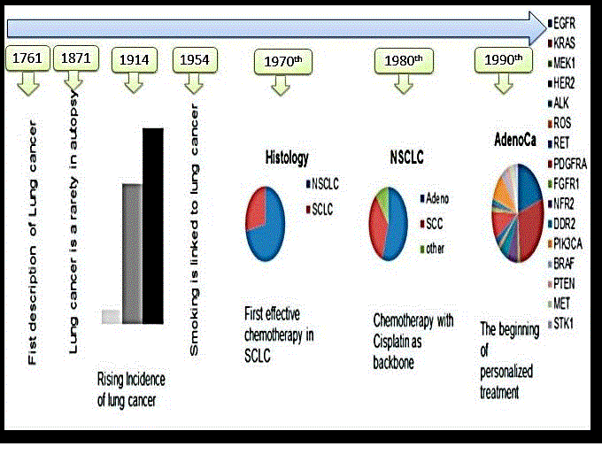
Recently, the NSCLC classified as squamous cell carcinoma and non-squamous which include adenocarcinoma and large cell type [Figure 1].

Figure 1. Story of lung cancer diagnosis

Figure 1. Story of lung cancer diagnosis

Epidermal Growth Factor Receptor (EGFR)

Epidermal growth factor was initially studied by Stanley Cohen and collaborators [3], who got Nobel Prize in 1986 for this discovery, and in 1988, Mendelsohn and collaborators obtained the receptors showing that EGFR can be a promising anticancer target [Table 1].

In May 2004, researchers found that the somatic mutations in the kinase domain of EGFR are positively related to the potent response of EGFR Tyrosine Kinase Inhibitors (TKIs) against advanced NSCLC [2].

EGFR, also known as ErbB1, and it belongs to receptors commonly referred to as receptor tyrosine kinases (RTKs) of the family of ErbB. Among the other members of the family of these receptors are ErbB2 (also known as HER2), ErbB3 (also known as HER3), and ErbB4 (also known as HER4) [4].

All of these receptors share a structural architecture consisting of a transmembrane domain, an extracellular ligand-binding domain, as well as an intracellular domain having tyrosine kinase activity to transducer the signals. The attachment of the ligand to EGFR starts a series of intracellular signaling that finally results in the appearance of cellular effects as cell proliferation as well as survival [2].

Table 1. EGFR TKI in the first line treatment of NSCLC compared with chemotherapy: phase III trials

Study No. patients TKI Control arm Median PFS P value
IPASS 1217
216 mutant EGFR
176 non mutant
Gefitinib Carbo/paclitaxel 9.8 vs 6.4 significant
WJTOG-3405 177 all Mutant Gefitinib Cisplatin/Docetaxel 9.2 vs 6.3 significant
NEJ-02 230 all mutant Gefitinib Carbo/paclitaxel 10.8 vs 5.4 significant
First signal 313
42 Mutant EGFR
Gefitinib Cisplatin/Gem 8 vs 6.4 significant
OPTIMAL 165 all mutant Erlotinib Carbo/Gem 13.1 vs 4.6 significant
EURTAC 173 all mutant Erlotinib Platinum based +
(Gem or Doct.)
9.7 vs 5.2 significant
LUX-lung 3 345 all mutant Afatinib Cisplatin/pemetrexed 11.1 vs 6.9 significant
LUX-lung 6 364 all mutant Afatinib Cisplatin/Gem 11 vs 5.6 significant

IPASS: Iressa Pan-Asia Study
NEJ: North East Japan
FIRST-SIGNAL: First-line Single Agent Iressa Versus Gemcitabine and Cisplatin Trial in Never-Smokers with Adenocarcinoma of the Lung;
OPTIMAL: Randomised Phase III Study Comparing First-line Erlotinib versus Carboplatin Plus Gemcitabine in Chinese Advanced Non-Small-Cell Lung Cancer Patients with EGFR Activating Mutations;
EURTAC: Erlotinib versus Standard Chemotherapy as First-line Treatment for European Patients with Advanced EGFR Mutation-Positive Non-Small-Cell Lung Cancer;
LUX-Lung 3: Phase III Study of Afatinib or Cisplatin Plus Pemetrexed in Patients With Metastatic Lung Adenocarcinoma With EGFR Mutations;
LUX-Lung 6: a Randomized, Open Label, Phase III Study of Afatinib Versus Gemcitabine/Cisplatin as First-line Treatment for Asian Patients With EGFR Mutation-Positive Advanced Adenocarcinoma of the Lung;

EGFR Mutations

EGFR mutations were first considered as important cancer causing factors when gefitinib, which is among the first TKIs developed to work on the EGFR intracellular tyrosine kinase domain, showed significant decrease in the size of tumor in some patients having EGFR mutations. Mutations in the EGFR tyrosine kinase are found in nearly 15% of NSCLC adenocarcinomas in the U.S., and it is most commonly found in women and non-smokers. However, incidences of the disease in East Asian populations range from 22% to 62% [2].

In NSCLC, two most commonly encountered EGFR mutations include L858R mutation in exon 21 as well as the exon 19 deletions. Both of these mutations are drug sensitizing and represent over 85% of EGFR mutations. Research shows that purified intracellular domain of EGFR L858R and the representative deletion mutant show a huge difference in sensitivity to EGFR TKIs as compared to wild-type receptor [2, 5].

It has been found that exons 18-21 results in the coding of a part of the EGFR tyrosine kinase domain and T to G mutation in exon 21 is considered as the most frequently found alteration resulting in the replacement of arginine with leucine at the position of 858 (L858R). It has also been found that in the exon 19 deletion (del.), there is a removal of four amino acids [2, 6].

EGFR mutations with L858R and del 19 can activate EGFR signalling pathway in the mutant EGFR-positive cancer causing cells. Some of the mutations also result in higher level of sensitivity to TKIs as compared to the cases having wild-type EGFR. On the other hand, resistance mutations can also be found either in the start of the mutations or after sustained exposure to TKIs. Some of the most important examples of EGFR mutations resulting in resistance are PTEN, KRAS, and BRAF mutations [7] that are commonly involved in developing resistance to EGFR TKIs in cases of NSCLC.

Other common resistance mutations are T790M in the EGFR gene, which can be primary or acquired, and also epithelial-mesenchymal transition (EMT) and MET amplification, which are usually acquired.

Some other EGFR mutations of unidentified clinical significance can also occur in the advanced NSCLC. However, they are small in number as compared to the well-known EGFR mutations, which are of clinical importance. These mutations involve the substitution of amino acid in G719, E709, L861, and S768. Their connection to the efficacy of EGFR TKIs needs further studies.

The mutation divided into favorable and un-favorable in which the mutation in L861 and G719 are rare but it can result in favorable efficacy of EGFR TKIs, whereas other mutations can result in poor responses to EGFR TKIs.

Use of EGFR Tkis to Treat NSCLC

Gefitinib, which is a first generation EGFR TKI, got accelerated approval from the U.S. Food and Drug Administration (FDA), in 2003, for the treatment of advanced NSCLC as a second-line treatment. Studies showed the efficacy of the drug in the form of response rate (RR) of over 9% in Caucasian participants and over 25% in Japanese participants. In the year 2004, erlotinib got approval for the treatment of the cancer.

It was founded that the erlotinib monotherapy resulted in 2-month survival advantage in comparison to best supportive care in cancer patients having chemotherapy-refractory NSCLC in the advanced stages. Erlotinib monotherapy gave a RR of about 9% while placebo gave RR below 1% [2, 6].

Nodaway TKIs used both as a first-line therapy in advanced stages of NSCLC as well as second-line therapy and also as third-line therapy for EGFR mutation-positive cancer [Table 1].

Use of EGFR Tkis as a First-Line Therapy For NSCLC

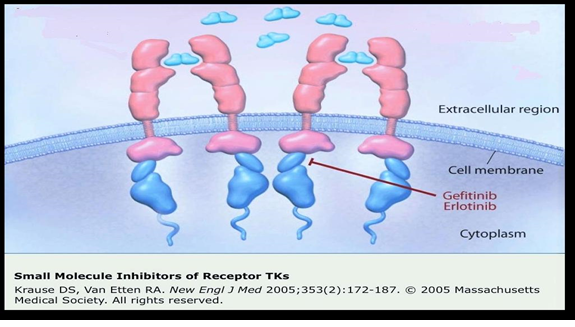
Gefitinib has been found effective as a first-line treatment in patients having EGFR-mutated NSCLC in advanced stages [8] [Figure 2].

Figure 2. Site of action of 1st Generation TKI

Figure 2. Site of action of 1st Generation TKI

In a study on patients having active EGFR mutations, the tumor samples of the patients were checked retrospectively for EGFR mutations as these mutations functioned as an important biomarker to know about the working of EGFR TKIs. Researchers found that tumor RRs were about 71% with gefitinib in patients having EGFR activating mutations as compared to about 41% in the chemotherapy group. Researchers found significant effect by considering the prolongation of life, i.e. 9.4 months in gefitinib treatment group as compared to 6.4 months in the other group. During the study, most of the patients, who were previously getting first-line chemotherapy, were moved to the gefitinib treatment, as the drug showed significant benefits [8] [Table 2].

Table 2. EGFR TKI in treatment NSCLC combined with chemotherapy as 1st line: phase III

Study No. of patients TKI+ chemo Type of chemotherapy Primary end point outcome
INTACT1 1093 Unselected (EGFR) Gefitinib Cisplatin/Gem. OS Negative
9.9 vs 10.9
months
INTACT 2 1037 Unselected (EGFR) Gefitinib Carboplatin/paclitaxel OS Negative
9.8 vs 9.9
months
TRIBUTE 1079 Unselected (EGFR) Erlotinib Carboplatin/paclitaxel OS Negative
Positive in
nonsmoker
TALENT 1172 Unselected (EGFR) Erlotinib Cisplatin/Gem. OS Negative
10.8 vs 11

INTACT: The Iressa NSCLC Trial Assessing Combination Treatment
TRIBUTE: Tarceva responses in conjunction with paclitaxel and carboplatin
TALENT: Tarceva Lung Cancer Investigation

Subsequent multiple trials, in which patients having EGFR mutations were considered, also showed the efficacy of EGFR TKIs as compared to standard doublet chemotherapy that was platinum-based. Randomized studies show substantially higher RRs as well as prolonged progression free survival (PFS), further showing the effectiveness of EGFR TKIs as a first-line therapy for patients having advanced stages of NSCLC with EGFR mutations [2].

In 2013, FDA approved afatinib, a second-generation EGFR TKI. It is an irreversible TKI and is helpful as a first-line therapeutic option in patients of advanced metastatic NSCLC with EGFR mutations [6].

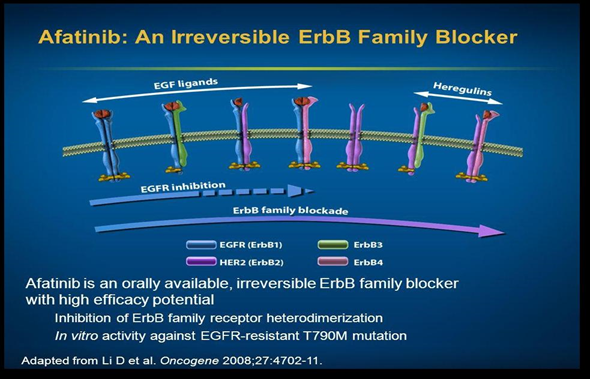
This drug binds with ATP attachment sites on the tyrosine kinases resulting in long lasting inhibitory effect on HER2 receptor. First-line afatinib has been found effective in improving the overall survival (OS) of patients having advanced stages of NSCLC with EGFR exon 19 deletion. Moreover, this improvement in the OS of patients was independent of race of patients. Studies consisting of a worldwide population showed that a median OS was over 30 months with the use of afatinib that is more than the median OS with chemotherapy. Although researchers found no considerable difference between the afatinib group and chemotherapy group in OS in patients having L858R mutations, but still afitinib can be a better treatment option for patients of L858R mutations [2] [Figure 3].

Figure 3. Site of action of 2nd Generation TKI

Figure 3. Site of action of 2nd Generation TKI

Use of EGFR Tkis as a Second-Line Therapy for NSCLC

Studies on erlotinib also show the effectiveness of the drug against wild-type EGFR NSCLC. In a study, researchers compared the effectiveness of docetaxel with erlotinib as a second-line treatment in patients having progressive wild-type EGFR NSCLC, who were initially treated with a platinum-based substances as a first-line therapeutic regimen. Researchers found that the median OS was about 8.2 months in patients using docetaxel while the median OS was about 5.4 months in patients using erlotinib. Moreover, PFS was substantially better in patients using docetaxel (i.e. 2.9 months) as compared to patients using erlotinib (i.e. 2.4 months). This showed that in spite of the efficacy of erlotinib, chemotherapy shows more effectiveness in the treatment of patients having advanced stages of wild-type EGFR NSCLC [2] [Table 3].

Table 3. EGFR TKI in NSCLC as 2nd or 3rd line (monotherapy): phase III

Study No. of patients TKI+ chemo Type of chemotherapy Primary end point outcome
ISEL 1129 Non selected Gefitinib Supportive care OS Negative trial
BR.21 731 Non selected Erlotinib Supportive care OS Positive
6.7 vs 4.7
months
INTEREST 1466 Non selected Gefitinib Docetaxel OS (non-inferior) Positive
7.6 vs 8 months
DELTA 301
50 EGFR
M+
Erlotinib Docetaxel PFS Negative
TITAN 424 unselected Erlotinib Docetaxel or
pemetrexed
OS Negative
5.3 vs 5.5
TAILOR 222
EGFR
wild
Erlotinib Docetaxel OS Negative
5.4 vs 8.2

ISEL trail: Iressa Survival Evaluation in Lung Cancer
INTEREST: Iressa NSCLC Trial Evaluating Response and Survival Versus Taxotere
Delta: The Docetaxel and Erlotinib Lung Cancer Trial
TaILOr: Tarceva Italian Lung Optimization tRial

Combination of EGFR Tkis with Chemotherapy in the Treatment of Advanced NSCLC

Combination of EGFR TKIs with chemotherapy show poor outcomes in the treatment of advanced NSCLC. Several randomized studies show that the platinum-based regimen along with EGFR TKI has no or reduced benefits as compared to chemotherapy alone, thereby requiring further studies [9].

Studies have also been done on finding the negative effects of EGFR TKIs on chemotherapy, and researchers are of opinion that EGFR TKIs protect G1 phase of the cell cycle from the action of chemotherapy, thereby affecting the overall action of the combination therapy. It has also been found that concurrent administration of erlotinib with M phase-specific taxane results in decreased levels and a prolonged shorter apoptosis duration. In another study, it has been found that patients having wild-type EGFR tumors may show elevated rates of progressive disease as well as inferior survival on receiving combination of erlotinib with chemotherapy as compared to chemotherapy alone. The similar outcomes were reported for patients having activating EGFR mutations. On the other hand, some studies on Asian population have shown better median PFS in case of combining chemotherapy with erlotinib as compared to chemotherapy alone [2].

Resistance to Tkis

Researchers have found that tumor having exon 20 insertions show insensitivity to EGFR TKIs. However, this problem has been found in about 4% of the cases of NSCLC. Approximately 20% of the cases of NSCLC show primary resistance caused by alteration in the KRAS signaling protein, which is commonly found in former as well as current smokers. Some other mutations that can result in primary resistance to TKIs include MEK, PTEN, and ALK-fusion [10].

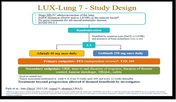
Resistances can also develop in patients having advanced EGFR mutation–positive NSCLC getting gefitinib or erlotinib as a treatment strategy. Disease progression can appear after nearly one year of therapy with any of these drugs. Most commonly found acquired resistance is due to mutation in T790M in which alteration occurs in exon 20 resulting in the replacement of methionine with threonine at the position 790. The 790M residue could disturb the attachment capacity of the TKIs with the ATP binding site. The EGFR exon 20 T790M mutations may result in up to 65% of cases of acquired resistance to TKIs [2, 10] [Figure 4].

Figure 4. LUX-Lung 7 study

Figure 4. LUX-Lung 7 study

Amplification of MET is also found to be an important mechanism behind the development of acquired resistance. Studies show that MET amplification may occur in about 10% of cases. Some other types of acquired resistance, which are in need of further studies, are caused by transformations to SCLC, 3CA mutation, epithelial-mesenchymal transition (EMT), and stimulation of insulin-like growth-factor receptor pathway [10].

Management of the condition with acquired resistance to Tkis

The resistant is either primary, secondary (usually involves exon 20 (T790M), or tertiary (C797S mutation). Other mechanisms including: (1) amplification of c-met 5-20%, (2) amplification of her-2 in 12%, (3) mutation in BRAF 1 %,( 4) transformation to small cell lung ca in 3-14%.Resistance occurs usually with median of 9 – 13 months and commonly due to T790M mutation.

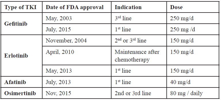
On November 13, 2015, the U. S. Food and Drug Administration granted accelerated approval to osimertinib once daily tablets, for the treatment of patients with metastatic epidermal growth factor receptor (EGFR) T790M mutation-positive non-small cell lung cancer (NSCLC), as detected by an FDA-approved test, who have progressed on or after EGFR tyrosine kinase inhibitor (TKI) therapy [Table 4].

Table 4. Approval of different EGFR TKI

Table 4. Approval of different EGFR TKI

The approval was based on two multicenter, single-arm, open-label clinical trials in patients with metastatic EGFR T790M mutation-positive NSCLC who had progressed on prior systemic therapy, including an EGFR TKI (Study 1 and 2). All patients were required to have EGFR T790M mutation-positive NSCLC as detected by the cobas® EGFR mutation test and received osimertinib 80 mg once daily. The major efficacy outcome measure was objective response rate (ORR) according to RECIST v1.1 as evaluated by a Blinded Independent Central Review (BICR). Duration of response (DOR) was an additional outcome measure.

It has been found that doublet chemotherapy, which is platinum-based, can be a standard choice for treatment of patients of advanced stages of NSCLC, who are EGFR TKI-resistant. On the other hand, when there is metastasis of the cancer to the brain, treatment of choice is radiotherapy [2].

Concluding Remarks and Future Directions

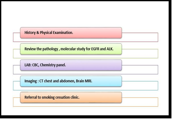
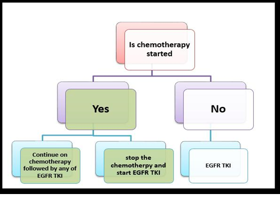
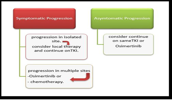
In the last few years, EGFR TKIs have been found to be among the most helpful treatment options for advanced NSCLC. Those substances were studied after the discovery of EGFR mutation, which is an important determinant of NSCLC. Moreover, the study of mutations helps the scientists to work on personalized medicine [Figures 5,6,7].

Figure 5. work up for advance NSCLC (adenocarcinoma)

Figure 5. work up for advance NSCLC (adenocarcinoma)

Figure 6. Treatment algorithm of metastatic NSCLC (adenocarcinoma) with sensitizing EGFR mutation

Figure 6. Treatment algorithm of metastatic NSCLC (adenocarcinoma) with sensitizing EGFR mutation

Figure 7. Treatment of metastatic NSCLC progressed on EGFR TKI

Figure 7. Treatment of metastatic NSCLC progressed on EGFR TKI

Finally, we can conclude that; in the era of molecular study and personalized therapy, TKIs nowadays are able to fight with the disease more effectively. However, further studies are required in reducing the resistance of the cancer against TKIs.

Furthermore, optimization of the combination therapy is required, so that patients would be either fully cured or live longer because of the better treatment.

References

  • Holland JC, Breitbart W S, Jacobsen PB, Loscalzo MJ, Butow PN, et al. (2015) Psycho-Oncology: Oxford University Press.
  • Ogunleye F, Ibrahim M, Stender M, Kalemkerian G, Jaiyesimi I (2015) Epidermal Growth Factor Receptor Tyrosine Kinase Inhibitors in Advanced Non-Small Cell Lung Cancer. The American Journal of Hematology/Oncology 11: 16-25.
  • Taylor JM, Cohen S, Mitchell WM (1970) Epidermal growth factor: high and low molecular weight forms.Proc Natl Acad Sci U S A 67: 164-171. [crossref]
  • Lappano R, De Marco P, De Francesco EM, Chimento A, Pezzi V, et al. (2013) Cross-talk between GPER and growth factor signaling. J Steroid Biochem Mol Biol 137: 50-56. [crossref]
  • Lee VH, Tin VP, Choy TS, Lam KO, Choi CW, et al. (2013) Association of exon 19 and 21 EGFR mutation patterns with treatment outcome after first-line tyrosine kinase inhibitor in metastatic non-small-cell lung cancer.J Thorac Oncol 8: 1148-1155. [crossref]
  • Reungwetwattana T, Dy GK2 (2013) Targeted therapies in development for non-small cell lung cancer.J Carcinog 12: 22. [crossref]
  • Carneiro JG, Couto PG2, Bastos-Rodrigues L2, Bicalho MA3, Vidigal PV4, et al. (2014) Spectrum of somatic EGFR, KRAS, BRAF, PTEN mutations and TTF-1 expression in Brazilian lung cancer patients. Genet Res (Camb) 96: e002. [crossref]
  • Maemondo M, Minegishi Y, Inoue A, Kobayashi K, Harada M, et al. (2012) First-line gefitinib in patients aged 75 or older with advanced non-small cell lung cancer harboring epidermal growth factor receptor mutations: NEJ 003 study. J Thorac Oncol 7: 1417-1422. [crossref]
  • Cufer T, Ovcaricek T, O’Brien M E (2013) Systemic therapy of advanced non-small cell lung cancer: major-developments of the last 5-years. European journal of cancer 49: 1216-1225.
  • Gainor JF, Shaw AT (2013) Emerging paradigms in the development of resistance to tyrosine kinase inhibitors in lung cancer. J Clin Oncol 31: 3987-3996. [crossref]

Programming Of Transcription and HPA Responses to Stress

DOI: 10.31038/CST.2016121

Abstract

The signaling pathways link neuronal activity to transcription, revealing both the transcription factors that mediate this process and the neuronal activity-regulated genes. The neuronal activity regulates a complex program of gene expression involved in many aspects of neuronal development. Human genetic studies have revealed that the disruption of the activity-regulated gene expression program in humans gives rise to neurological disorders. Social states can affect health in further life. It is a completely revolutionary idea. Stress changes methylation and influence the whole life.

Introduction

The central aim is to formulate results based on studies in the fields of neurobiology and genetics to understand more human behavior at the level of neuropsychology. We have now a detailed molecular mechanism by which is possible to understand why social states can affect health in further life. It is a completely revolutionary idea.

The cellular and molecular mechanisms underlie to the experience-driven changes in neural connectivity. Sensory experience results in neurotransmitter release at synapses within a neural circuit and leads to membrane depolarization, calcium influx into individual neurons. Which triggers a wide variety of cellular changes with these neurons capable of altering synaptic connectivity of the circuit. Changes such as the activation of calcium-sensitive signaling cascades lead to posttranslational modifications of proteins, at the regulation of mRNA translation [1]. It’s resulting in the production of new proteins locally at the sites of calcium entry and play critical roles in altering synaptic function in a synapse-specific manner.

Materials and Methods

We have used the studies cited in the references to make a review from the latest results at the field of neurobiology, genetics, and neuropsychology to analyze what are the mechanisms regulating human behavior at neural and psychological level under conditions of stress. We try to formulate how sensory information influences response behavior by semi-analytical, information theoretical, statistical and neuropsychological methods.

To understand more human behavior in the psychological conditions of stress we must start from the underlying principles of neurobiology and genetics. It can be done by the method of relating neurobiological models to behavioral models of signaling pathways.

Calcium influx can alter cellular function by activating new gene transcription

Calcium influx into the postsynaptic neuron can alter cellular function by activating new gene transcription. Calcium influx activates a number of signaling pathways converging on transcription factors within the nucleus, which in turn control the expression of a large number of neuronal activity regulated genes. Signaling pathways mediate activity-dependent transcription in experience-dependent neural development and plasticity. This neuronal activity regulates by the signal transduction pathways the activity-dependent gene expression program. On the other side, neuronal activity-regulated genes showing how this activity-regulated program controls neuronal development [1, 2].

The c-fos mRNA is induced by synaptic activity resulting from sensory experience due the Fos protein with Jun family members comprised the AP-1 transcriptional complex, which is critical for the organism’s adaptive responses to experience. A brain-specific deletion of the c-fos gene displays deficits in synaptic plasticity and defects in learning and memory. Loss of Fos-dependent transription gives raise to additional behavioral deficits. [3]

The activity-regulated transcriptional program uncovered a mechanism by which calcium-dependent gene induction alters the function of specific synapses. The translation of select mRNAs can occur at individual synapses through the actions of microRNAs (miRNAs) which inhibit the translation of mRNAs having nucleotide sequences closely matching the miRNAs. The level of miR-134 is increased by neuronal activity. The miRNA could be a component of the local mRNA translation machinery allowing proteins to be translated in a synapse-specific manner. This transcriptional program is critical in coordinating both dendritic and synaptic remodeling.

The transcriptions of c-fos and other immediate early genes

The transcriptions of c-fos and other immediate early genes (IEGs) increases in many cells of the body in response to extracellular factors inducing proliferation or differentiation of the cells. IEGs mediate cellular responses to changes in the cell’s environment. Recent studies have identified a subset of genes that is activated specifically in response to excitatory synaptic transmission that triggers calcium influx into the postsynaptic neuron. One gene is specifically induced by neuronal activity in neurons: bdnf encodes a neurotrophin important in neural development. The level of the bdnf mRNA increases in neurons in response to physiological stimuli, such as fear conditioning and seizure induction. The induction of the bdnf mRNA is due to an increase in transcription of the bdnf gene [4].

Transcripts of these promoters splice from their first exon to a common downstream exon, which contains the entire open reading frame encoding the BDNF protein. This diversity could explain how BDNF can control such a large number of distinct processes during nervous system development. Neuronal activity sharply increases the rate of transcription initiation with most transcripts ending within the central intron. These coordinate transcriptional events rapidly convert a constitutive gene to an IEG and regulate the expression of functionally different Homer 1 proteins. The short forms modulate the properties of the long forma and are critically involved in activity-dependent alterations of synaptic structure and function. [1- 3]

The switch from constitutive to activity-dependent expression

The switch from constitutive to activity-dependent expression entails intronic to exonic sequence conversion, transcript termination within the central intron of the Homer 1 gene. Homer proteins play key roles in signal transduction in the brain [1-3]. Hypothalamic-pituitary-adrenal (HPA) responses to stress suggesting a causal relation among epigenetic state, glucocorticoid receptor (GR) expression and the maternal effect on stress responses in the later offspring. There are increasing number of the results confirming that an epigenomic state of a gene may be established through forms of an environmental and programming and this is potentially reversible. [4]

Variations in maternal behavior are connected with development of individual differences in behavioral and HPA responses to stress in the offspring. They serve as a mechanism for the nongenomic transmission of individual differences in stress reactivity across generations. Recent findings suggest that the mechanisms of these maternal effects, or other forms of environmental programming, remain sustained over the lifespan. [5]

Maternal behavior in the rat permanently alters the development of HPA responses to stress through tissue-specific effects on gene expression. The magnitude of the HPA response to stress is a function of hypothalamic corticotropin-releasing factor (CRF) release, thus activating the pituitary-adrenal system. There are also some modulatory influences, like glucocorticoid negative feedback, which inhibits CRF synthesis and release, dampening HPA responses to stress. [4]

Epigenetic programming

The changes in Avp expression were restricted to the parvocellular subpopulation of neurons in the hypothalamic paraventricular nucleus (PVN) in those neurons that drive the HPA axis. Research data verify the critical role of arginine vasopressin (AVP) in driving the disturbed endocrine phenotype in stressed mice. This hypothesis was supported by the observation that the methyl CpG-binding protein 2 (MeCP2) phosphorilation was prominently increased in parvocellular AVP-expressing neurons in the PVN. Phosphorilation of MeCP2 at S438 is critical for MeCP2 function as a reader and interpreter of the DNA methylation signal at the Avp enhancer. MeCP2 serves as an epigenetic integration platform on which synergistic cross-talk between histone deacyclation, K3K9 methylation and DNA methylation act to confer gene silencing. [1,2,4]

Research data suggests that stress tilts the balance toward persistent hypomethylation and Avp overexpression by inducing reductions in MeCP2 binding. Phosphorilation of MeCP2 appears to be a carrier of experience-driven changes in gene expression, as a important mediator of the persistent effects of stress. By DNA methylation, there are evidence for postmitotic epigenetic modifications in neuronal functions. Modifications can facilitate or disfavor physiological and behavioral adaptations [2]. Epigenetic marks and their initiators, mediators and readers (MeCP2) bring new evidences for understanding the molecular basis of stress-related disorders of the brain.

Glucocorticoid programming

Genetic background might predispose to early-life events as maternal care, which can change the genetic profile through epigenetic signaling pathways. The programming effect of maternal behavior is associated with a single gene: the glucocorticoid (GR) gene. The offspring of caring mothers had higher hippocampal GR expression, owing to demethylation of a cysteine residue at the 5’NGF1A binding region in the exon 1, promoter [1]. Corticosteroids operate in both stress-system modes through mineralcorticoid (MR) and GR receptors co-expressed in the neurons of limbic structures. MR acts in the appraisal process and the onset of the stress response. GR is only activated by large amounts of corticosteroid, terminates the reactions to competition (the stopping rule). GR also promotes memory storage in preparing for future events [4, 6].

Behavioral programming

In vivo studies suggest that the effect of maternal behavior on GR gene expression is accompanied by an increased hippocampal expression of nerve growth factor-inducible protein A (NGFI-A). The non-coding exon 1 region of the hippocampal GR includes a promoter region, exon , containing a binding site for NGFI-A. Splice variants of the GR mRNA containing the exon sequence are found predominantly in the brain.

Use of promoter is enhanced as a function of maternal care, what explain the increased GR expression in the neonate. Maternal care alters DNA methylation of the GR exon promoter, and these changes are stably maintained into adulthood, associated with differences in GR expression and HPA responses to stress. Variations in maternal care directly alter the methylation status of the exon promoter of the GR gene. DNA methylation pattern can be established also through a behavioral programming without germ line transmission [4]. Postnatal de novo methylation of the Hoxa5 and Hoxb5 genes in development was documented also in another study [7].

Thus, maternal programming of the exon GR promoter involves DNA methylation, histone H3-K9 acetylation and alterations in NGFI-A binding [4, 6]. The afferent input from limbic networks converts purely psychological stress reactions to the HPA axis. Above interplay of limbic inputs from the hippocampus, amygdala and prefrontal cortex with HPA axis activity may lead to a vulnerable phenotype for mental illness [4, 5].

Environmental programming

We have now evidence that maternal behavior produces stable alterations of DNA methylation and chromatine structure, providing a mechanism for the long-term effects of maternal care on gene expression in the offspring. Such a gene-environment interactions during development result in the sustained environmental programming of gene expression and function of defensive responses through increased HPA activity over the lifespan [4, 5]. Natural selection shaped offspring to respond to subtle variations in parental behavior as forecast of the environmental conditions. They serve as a major source of epigenetic variations in gene expression and mediating such maternal effects. Effects on chromatine structure serve as an intermediate process imprinting dynamic environmental experience on the fixed genome with stable variations in phenotype [1, 2,5].

Environment-assisted invariance

The state of composite object (consisting of the system S and the environment E) can be ignorant of the state of S alone. Environment-assisted invariance, or envariance based on symetry allows observer to use perfect knowledge of SE as a proof of his ignorance of S: when a US acting on S alone, can be undone by a transformation acting solely on E, and the joint state of SE is unchanged. This state is said „envariant“ with respect to US. Envariant properties not belong S alone. Entanglement between S and E enables envariant and implies ignorance about S. Envariance is associated with phases of the Schmidt decomposition of the state representing SE. It anticipates the consequences of environment – induced superselection („einselection“) of the preferred set of pointer states, they remain unperturbed to immersion of the system in the environment. The state of combined SE expressed in the Schmidt form is: |ψSE 〉 = ∑ ∝kk 〉|Ek〉 . Schmidt states are in an intimate relationship with the pointer states and have been regarded as „instantaneous pointer states“ [8]. Quantum Darwinism brings new focus on the environment as a communication channel. This explains the emergence of objectivity. Even hazy environment will communicate a very clear image [9].

Adaptational programming

Limbic pathways activated by psychological stressors of competition are parts of the afferent pathways activating the CRH neurons in the PVN. The interface between incoming sensory information and the appraisal is converted by limbic brain structures (the hippocampus, amygdala and prefrontal cortex-PFC).

Not only homeostatic disturbance, but purely psychological code can determine the stress response to competition. Its determinants include the ability predict upcoming events and getting control over the situation. The adaptive competition stress-related processes take place in limbic brain regions. An inappropriate response to the winner-take-all instabilities (WTAIs) produces a vulnerable phenotype leaving genetically predisposed individuals at an increased risk of stress-related brain disorders. [5, 10].

Multiple peaks of activity appear simultaneously within a single frontal or parietal region, they compete against each other through inhibitory antagonism. This can be seen in biased competition mechanism of visual attention. During colour-cue period preferring the given colour pushes group of cells towards stronger activity than others and causes the competition in dorsal premotor cortex (PMd) to become unbalanced, because one peak increases its activity, while the other is suppressed. Since neural activities are noisy, competition between distinct peaks of activity cannot follow a simple winner-take-all rule, or random fluctuations will determine the winner each time. If activity of a given choice becomes sufficiently strong, than it should be allowed to suppress its opponent and conclusively win the competition. But the cost of reinstating homeostasis also might become too high, causing through WTAIs an allostatic load with increased risk of mental illness [5,11-19].

Dynamics of the winner-take-all instability

To derive an equation for the dynamics of the winner-take-all instability, we express the dynamical variables as x = xSS + xY(T) + … where Y represents the slow dynamics along the critical eigenvector and T is a slow time scale. The reflection symmetry of the system implies the dynamics of Y should be invariant under the transformation Y → – Y and this switches the identity of x1 and x2. The increase in input I is common to both x1 and x2 leads to the developing decision in the winner-take-all system and is thus the bifurcation parameter. The linear growth rate of the spontaneous state must be proportional to the difference between the presynaptic input and the value of the input at the bifurcation with an unknown prefactor, i.e. μ(I – I). The difference in inputs I1 – I2 breaks the reflection symmetry thereby introducing a constant term which, to first approximation, must be proportional to that difference with an unknowvn prefactor, i.e. η(I1 – I2). These two facts, coupled with the reflection symmetry, lead to the form of the equation describing the time evolution of Y: δTY = η(I1 – I2) + μ(I – I)Y + ϒY3, where I = I only when ∝ = β identically, i.e. at point of instability, and δT is a time derivative with respect to the slow time T. For I1 – I2 the equation is invariant under Y → – Y as it should be, Y3 is the lowest order nonlinearity which obeys reflection symmetry. For more complex systems, which exhibit winner-take-all behavior, above euation captures the qualitative dynamics of the system near the bifurcation in general [20].

Concluding Remarks

During adaptation sensory experience driven changes in neural connectivity, transcription, and HPA axis responses to stress are complex and multifactorial: they cannot be attributed to mutations in single gene, or to a single external event, but rather, result from the concerted actions of many subtle genetic polymorphisms and external events, the effects of which might accumulate over time. Once traumatic life events, in combination with genetic disposition, have engrained long-lasting changes in MR and GR signaling, a vulnerable phenotype emerges [5].

DNA methylation is behind the changes associated with stress. It is based on differences in the gene encoding AVP, a hormone associated with mood and cognitive behavior. After stress, there was lover level of methylation in the regulatory region of the Avp gene in the brain. This hypomethylation was specific to a subset of neurons in the hypothalamic paraventricular nucleus – a brain area involved in regulating hormones linked to stress [1, 2].

The decreases in methylation in stressed subjects result from the inactivation of a protein MeCP2, involed in the start of the DNA methylation. It is a detailed molecular mechanism by which is possible to understand why social states as sensory experience can affect health in further life. It is a completely revolutionary idea [2]. Stress changes methylation and influence the whole life. Depression may be facilitated by a failure in competition to contain the biological stress response to challenge of unemployment at the time of the trauma, resulting in a cascade of alterations leading to recollections of the WTAIs, avoidance of the reminders to event and symptoms of hyperarousal [5]. From psychological and biological data we may hypothesize that the pathological mechanism of stress-related brain disorders depend on distress connected with inhibitory antagonism produced by winner-take-all instabilities. Mechanism is triggered by interactive behavior of an appraisal of unit P probabilities trade-off with environment. Stressors can kill with information itself through probabilities. Probabilities are the killer by information [9].

Sensory information itself, as first communication of diagnosis, may act as psychic stressor, psychological weapon (of mass destruction) due stress-related brain disorders [11-18]

It is well documented in recent large population-based study about men newly diagnosed with prostate cancer, they were at higher risk of cardiovascular events and suicide. The excess risks were highest during the first week after diagnosis, suggesting that stress of diagnosis itself plays a critical role. The emotional stress as an information itself caused a cardiovascular morbidity increase immediately after communication of the diagnosis [18].

Emotionally stressful competition events may lead to altered function of the heart, a stress-related left ventricular dysfunction [15]. Increased risk of myocardial infarction was documented following the Athen earthquake in 1983 [14]. Emotional stress brought on by viewing a World cup soccer match was reported to raise the risk for cardiovascular morbidity and mortality [17]. Being informed about diagnose of prostate cancer may also serve as a stressor of substantial weight. About 20% of the prostate cancer patients were reported as having no one to confide in Fall K, et al. [18]. On the basis of above results bring a hypothesis of the weights function in a framework of feedback paradigm as the psychological code. Possible mechanism may be the emotional shock caused by the information of diagnosis, anxiety, together with emotional isolation.

References

  • Bottai D, Guzowski JF, Schwarz MK, Kang SH, Xiao B, et al. (2002) Synaptic activity-induced conversion of intronic to exonic sequence in Homer 1 immediate early gene expression. J Neurosci 22: 167-175. [crossref]
  • Cisek P (2007) Cortical mechanisms of action selection: the affordance competition hypothesis. Philos Trans R Soc Lond B Biol Sci 362: 1585-1599. [crossref]
  • Fall K, Fang F, Mucci LA, Ye W, Andrén O, et al. (2009) Immediate risk for cardiovascular events and suicide following a prostate cancer diagnosis: prospective cohort study. PLoS Med 6: e1000197. [crossref]
  • Flavel SW and Greenberg ME (2009) Ann Rev Neurosci 2008:31:583-590
  • Hershko AY, Kafri T, Fainsod A, Razin A (2003) Methylation of HoxA5 and HoxB5 and its relevance to expression during mouse development. Gene 302: 65-72. [crossref]
  • Leor J, Poole WK, Kloner RA (1996) Sudden cardiac death triggered by an earthquake. N Engl J Med 334: 413-419. [crossref]
  • Li J, Hansen D, Mortensen PB, Olsen J (2002) Myocardial infarction in parents who lost a child: a nationwide prospective cohort study in Denmark. Circulation 106: 1634-1639. [crossref]
  • Li J, Laursen TM, Precht DH, Olsen J, Mortensen PB (2005) Hospitalization for mental illness among parents after the death of a child. N Engl J Med 352: 1190-1196. [crossref]
  • Katsouyanni K, Kogevinas M, Trichopoulos D (1986) Earthquake-related stress and cardiac mortality. Int J Epidemiol 15: 326-330. [crossref]
  • Meisel SR, Kutz I, Dayan KI, Pauzner H, Chetboun I, et al. (1991) Effect of Iraqi missile war on incidence of acute myocardial infarction and sudden death in Israeli civilians. Lancet 338: 660-661. [crossref]
  • Murgatroyd C, Patchev AV, Wu Y, Micale V, Bockmühl Y, et al. (2009) Dynamic DNA methylation programs persistent adverse effects of early-life stress. Nat Neurosci 12: 1559-1566. [crossref]
  • de Kloet ER, Joëls M, Holsboer F (2005) Stress and the brain: from adaptation to disease. Nat Rev Neurosci 6: 463-475. [crossref]
  • Roxin A, Ledberg A (2008) Neurobiological models of two-choice decision making can be reduced to a one-dimensional nonlinear diffusion equation. PLoS Comput Biol 4: e1000046. [crossref]
  • Schairer C, Brown LM, Chen BE, Howard R, Lynch CF, et al. (2006) Suicide after breast cancer: an international population-based study of 723,810 women. J Nat Cancer Inst 98: 1416-1419. [crossref]
  • Seckl JR, Meaney MJ (2004) Glucocorticoid programming. Ann N Y Acad Sci 1032: 63-84. [crossref]
  • Wilbert-Lampen U, Leistner D, Greven S, Pohl T, Sper S, et al. (2008) Cardiovascular events during World Cup soccer. N Engl J Med 358: 475-483. [crossref]
  • Wittstein IS, Thiemann DR, Lima JA, Baughman KL, Schulman SP, et al. (2005) Neurohumoral features of myocardial stunning due to sudden emotional stress. N Eng J Med 352: 539-548. [crossref]
  • Weaver IC, Cervoni N, Champagne FA, D’Alessio AC, Sharma S, et al. (2004) Epigenetic programming by maternal behavior. Nat Neurosci 7: 847-854. [crossref]
  • Zurek WH (2002) arXiv: 0211.037 v1 [quant-ph]
  • Zwolak M, Quan HT, Zurek WH (2009) arXiv: 0904.0418v2 [quant-ph]