Monthly Archives: June 2019

The Short-Term Results of Pyrocarbon Lunate Implants in Patients with Advanced Kienböck’s Disease

DOI: 10.31038/IJOT.2019232

Abstract

Purpose: The purpose of this study was to evaluate the short-term results of pyrocarbon lunate implants (PLI) (Ascension Orthopaedics, Austin, Texas).

Methods:  Patients with advanced Kienböck’s disease who received PLI were prospectively followed for one year. The implant outcomes were assessed by pre- and postoperative questionnaires and physical examination.

Results: six patients (six implants) with Kienböck’s disease grade IV were included in this study. All implants remained in situ at one-year follow-up. Pain was satisfactorily reduced in five patients. Grip strength improved slightly in three patients and worsened in two. The DASH scores improved in four patients with mean 26 points. Five patients returned to their previous jobs and the two patients whom did sport could resume it. Three patients had to be mobilized under general anaesthesia because of a severely stiffened wrist.

Conclusions: The short-term results of the PLI are suboptimal, probably largely due to severe stiffening of the wrist. Nonetheless, long-term results are necessary to adequately assess the longevity and functionality of this implant and to assess which postoperative treatment would provide the optimal clinical result. Type of study/level of evidence: Therapeutic, level IV.

Keywords: Lunate Implant, Pyrocarbon, Results

Introduction

In 1910, Kienböck [1] was the first to describe the radiological signs of isolated lunatomalacia, currently known as Kienböck’s disease. Despite many years of clinical experience and research, the cause of this disorder still remains unclear. Morphological variations such as the negative ulnar variance, the particular pattern of vascularity and repetitive trauma may be predisposing factors, suggesting a multifactorial aetiology [2]. Kienböck’s disease is divided into 4 stages and can be diagnosed by conventional radiography [3] and Magnetic Resonance Imaging (MRI) [4].

Kienböck’s disease can be surgically treated. Patients with negative ulnar variance, without (radio) carpal osteoarthritis and Kienböck’s disease stage I-IIIa can be treated by a radial osteotomy [5]. Patients with neutral ulnar variance, no (radio) carpal osteoarthritis and Kienböcks disease stage I-IIIa can be treated by revascularisation [6–8]. Patients with Kienböck’s disease stage IIIb-IV can be treated by proximal row carpectomy, partial or total wrist arthrodesis or arthroplasty: resulting in limited mobility and function of the wrist. The lunate implant was developed to maintain wrist function in patients with severe Kienböck’s disease.

There are only few reports in the literature that report on the clinical outcomes of pyrocarbon lunate implants [9]. We present a case series with short-term outcomes of patients with this type of pyrocarbon lunate implant.

Patients and Methods

Study Design

All patients with advanced Kienböck’s disease who received this pyrocarbon lunate implant (Pyrocarbon Lunate prosthesis, Ascension Orthopedics, Inc, 8700 Cameron Road, Suite 100, Austin, TX 78754 USA) were identified at the Amphia Hospital in Breda, The Netherlands. The inclusion criteria were patients with wrist pain as a result of Kienböck’s disease grade IIIb or IV. The exclusion criteria were patients who performed heavy labour, patients with osteoarthritis of the radio- or midcarpal joint other than the lunate fossa and lunate-capitate joint and patients who received a prior surgical treatment for their wrist complaints. Informed consent was obtained before implant insertion. Relevant data were extracted from the medical records: demographics, medical history, profession, affected wrist and result of the Magnetic Resonance Imaging (MRI), This study’s level of evidence is IV based on the absence of a control group and was approved by the Medical Ethical Committee at the Amphia Hospital in Breda, The Netherlands.

Surgical Technique

Surgery was performed under general anaesthesia and tourniquet control. A longitudinal incision on the dorsum of the wrist was made. The sensory branches of the radial and ulnar nerves were preserved. After identification of the third compartment of the extensor retinaculum, the compartment was opened to identify the extensor pollicis longus tendon, which was held to the radial side. The fourth extensor compartment was elevated from the radius and capsule and held ulnarly. The dorsal capsule was then incised, creating a distally based capsule flap. The degenerative lunate was identified and removed. Two K-wires fixated the triquetrum and scaphoid to the capitate. A 3.5 mm hole was drilled from the ulnar side of the scaphoid towards the volar aspect of the scaphoid. A 2 mm hole was drilled from the radial side of the triquetrum towards the dorsal side of the triquetrum. On the volar aspect of the wrist, two small incisions were made to reach the flexor carpi radialis tendon. The radial third of the Flexor Carpi Radialis (FCR) tendon was harvested leaving the distal attachment inserted. The FCR tendon graft was tunnelled through the scaphoid. Two Mitek-anchors were inserted, one in the scaphoid and one in the triquetrum. The tendon was guided through the volar hole of the prosthesis and through the hole in the triquetrum towards the ulnar side. Two wires of the anchors (one of each anchor) were guided through the volar hole of the prosthesis too. These wires were attached to each other. The position of the prosthesis was controlled by fluoroscopy. The other wires were put through the dorsal hole of the prosthesis and attached to each other while closing the ‘gaps’ radialy and ulnarly of the lunate. A third Mitek-anchor was placed into the scaphoid. Two gutters were created on the dorsum of the triquetrum en scaphoid. The remaining FCR tendon graft on the dorsal side of the triquetrum was positioned into the gutters towards the scaphoid and attached to the scaphoid using the wires of the third Mitek-anchor. The position of the prosthesis was checked again with fluoroscopy to assure an anatomical position. The capsule and skin were closed by sutures. A forearm cast was worn for six weeks. The K-wires were removed after six weeks and active mobilization of the wrist was started. For another six weeks a removable forearm splint was worn during stressful moments.

Clinical Evaluation

The PLI outcome was assessed by questionnaires and physical examination. Grip strength (measure by the Jamar hand-dynamometer), range of motion and the Disabilities of the Arm, Shoulder and Hand (DASH) questionnaire [10] were measured preoperatively and at one-year follow-up. Furthermore, return to work and sport, complications and patient satisfaction on pain reduction were evaluated.

Radiological Evaluation

X-rays were performed postoperatively and at one-year follow-up to evaluate the pyrocarbon lunate implant position, intercarpal distance and progression of disease.

Results

Six patients (six implants, four females and two males) with Kienböck’s disease grade IV were included in this study (Table 1). The mean age was 35 years (range 23 to 47 years). The dominant hand was involved in one patient. Preoperatively, all patients experienced wrist pain at rest that worsened after activities.

Clinical Evaluation

All six implants remained in situ at one-year follow-up. Five patients were satisfied with the pain relief. Grip strength improved slightly in three patients and worsened in two patients. Range of motion improved in two of the six patients. The DASH scores improved in four patients with mean 26 points and worsened in two patients with mean nine points. Five patients returned to their previous jobs and the two patients whom did sport could resume it.

Complications and Revisions

A K-wire was infected in one patient. The K-wire was removed and the patient was successfully treated with oral antibiotics. The wrists of three patients were severely stiffened and were remobilized under general anaesthesia. None of the implants needed revision.

Radiological Evaluation

Analysis of radiographs showed that the scapho-lunate distance increased after removal of the K-wires in all six six patients. However, at one-year follow-up, this distance did not increase and there was no progression of disease (Figure 1). Furthermore, five wrists developed dorsal intercalated segment instability and one developed volar intercalated segment instability.

IJOT 19 - 111 -Beumer A_F1

Figure 1. The anterolateral view of the pyrocarbon lunate implant at one-year follow-up.

Discussion

The aim of this study was to determine the short-term results of the pyrocarbon lunate implant. Lunate implant arthroplasty has been used as treatment for advanced Kienböck’s disease for more than 60 years. In contrast to the silicon lunate implant, with a high incidence of silicone cysts (78% after 27 years follow-up) [11], the titanium lunate implant has promising long-term results [12]. Although not mentioned by these authors, titanium might give rise to tissue reactions. Compared to titanium, pyrocarbon is more similar to cortical bone and transfers the load more effectively, potentially limiting bone resorption. Although Pyrocarbon implants might break, they are biologically inert and biocompatible resulting in a low tendency to wear and tissue reactions. Furthermore, pyrocarbon is less susceptible for wear when compared to titanium [13].

Table 1. Characteristics, clinical outcome and complications of patients with the pyrocarbon lunate implant.

Case no.

Age

Kienböck’s disease stage

Grip strength (kg)

Flexion/extension

DASH

Complications

Patient satisfied with pain reduction

Return to work

Return to sport

Follow-up (mo)

Preop

Postop*

Preop

Postop*

Preop

Postop*

1

42

IV

37

42

60/50

60/80

56

14

None

Yes

Yes

Yes

12

2

47

IV

42

14

45/30

70/25

38

50

K-wire infection

Yes

No

Yes

14

3

28

IV

21

13

45/45

10/25

68

37

MUA

Yes

Yes

na

12

4

23

IV

12

18

60/50

20/20

30

35

MUA

No

Yes

No preop sport

14

5

23

IV

9

19

70/70

35/35

40

28

MUA

Yes

Yes

No preop sport

20

6

45

IV

na

na

45/45

30/45

51

32

None

Yes

Yes

No preop sport

15

Mean

35

IV

24

21

54/48

38/38

47

33

66%

83%

83%

100%

15

Abbreviations: preop, preoperatively; postop, postoperatively; na, non-available; K-wire, Kirschner wire; MUA, mobilisation under anesthesia.
*1 year postoperatively

This study has many limitations. First, and perhaps most importantly, only six patients were included in this study. Statistical analyses were not performed due to this small sample size. Secondly, there is no control group. Thirdly, the postoperative results aren’t complete.

The PLI outcome was assessed by implant survival, physical examination and questionnaires. Implant survival was 100% at one-year follow-up. The main indication for treatment was wrist pain, which was satisfactorily reduced in five patients. However, grip strength improved only slightly in three patients and moreover range of motion decreased in four patients at one-year follow-up. Despite these limitations, DASH scores reduced in four patients and five patients of the six patients could return to their previous jobs. These short-term results are suboptimal, especially considering that three out of six patients needed to be remobilized under general anaesthesia, probably largely due to severe stiffening of the wrist. The decreased range of motion could be the result of our postoperative treatment and might be avoided by a different postoperative treatment that would allow a shorter immobilization period and earlier range of motion exercises [9,12]. Although a higher risk of implant dislocation (one of 17 patients) [9] and malposition (2 of 11 patients) [12] could occur. Nonetheless, long-term results are necessary to adequately assess the longevity and functionality of this implant and to assess which postoperative treatment would provide the optimal clinical results. Until these results are published, we have abandoned this implant in our Hospital.

Declaration

Ethics approval and consent to participate: This study was approved by the Medical Ethical Committee at the Amphia Hospital in Breda, The Netherlands.

Availability of data and material: The datasets used and/or analysed during the current study are available from the corresponding author on reasonable request.

References

  1. Kienbock R (1910) Uber traumatische Malazie des Monatbeins und ihre Folgezustande: Entartungsformen und Kompressionsfrakturen. Fortschrit Rontgenstrallen 16: 77.
  2. Green DP, Hotchkiss RN, Pederson WC, Wolfe SW (2005) Green’s Operative Hand Surgery, 5th edition. Philadelphia: Churchill Livingstone 2005:744–745.
  3. Schmitt R, Krimmer H (2007) Osteonecrosis of the hand skeleton. In: Schmitt R, Ulrich L (Eds.), Diagnostic imaging of the hand. (1stedn), Stuttgart, New York, Georg Thieme Verlag Pg No: 351–64.
  4. Luo J and Diao E (2006) Kienböck’s disease. An approach to treatment. Hand Clin 22:465–73.
  5. Lichtman DM, Lesley NE, Simmons SP (2010) The classification and treatment of Kienböck’s disease: the state of the art and a look at the future. J Hand Surg [Eu] 35: 549–54.
  6. Frangen TM, Konieczny MR, Gaggl AJ, Struewer J, Müller EJ, et al (2012) Semilunar bone necrosis (Kienböck’s disease) – first clinical results after free microvascularised bone graft from the distal femur. Z Orthop Unfall. 
  7. Arora RLutz MZimmermann RStruve P, Pechlaner S, et al (2010) Free vascularised iliac bone graft for Kienböck’s disease stage III. Handchir Mikrochir Plast Chir 42: 198–203.
  8. Mathoulin C, Haerle M, Vandeputte G (2005) Vascularized bone graft in carpal bone reconstruction. Ann Chir Plast Esthet 50: 43–48.
  9. Visser NJ, de Wijn RS, Moojen TM, Feitz R (2017) Lunate implant arthroplasty: analysis of physical function and patient satisfaction. Eur J Plast Surg 40: 229–34.
  10. Kennedy CA, Beaton DE, Solway S, McConnell S, Bombardier C (2011) Disabilities of the Arm, Shoulder and Hand (DASH). The DASH and QuickDASH Outcome Measure User’s Manual. Third Edition. Toronto, Ontario: Institute for Work & Health 2011.
  11. Viljakka T, Tallroth K, Vastamaki M (2014) Long-term outcome (22–36 years) of silicone lunate arthroplasty for Kienbock’s disease. J Hand Surg Eur Vol 39: 405–415.
  12. Viljakka T, Tallroth K, Vastamaki M (2018) Long-Term Clinical Outcome After Titanium Lunate Arthroplasty for Kienbock Disease. J Hand Surg Am 43:  945.
  13. Cook SD, Beckenbaugh RD, Redondo J, Popich LS, Klawitter JJ et al (1999) Long-term follow-up of pyrolytic carbon metacarpophalangeal implants. J Bone Joint Surg Am 81: 635–648.

The Effect of a Postpartum Smoking Relapse Prevention Education Program on Perinatal Nurses’ Counseling Behavior

DOI: 10.31038/AWHC.2019234

Introduction

In the United States, smoking is the largest preventable risk factor for pregnancy-related mortality and morbidity [1, 2]. While evidence-based, pregnancy specific, smoking cessation interventions increase the rate of quitting, half of those who quit will resume smoking within a few weeks of delivery and 90% will be smoking within 12 months [3, 4]. The unique pregnancy specific factors motivating women to abstain from cigarettes while pregnant are time limited and diminish after giving birth [5]. Assisting women to remain tobacco free after childbirth is a high priority in healthcare [6]. Quitting long term improves life expectancy, reduces health risks in future pregnancies, and protects children from second-hand smoke (SHS) exposure.

The U.S. Public Health Service (USPHS) clinical practice guidelines recommend that health care providers assess patients’ tobacco use at each clinical encounter using a five-step strategy referred to as the 5A’s: ask about tobacco use, advise smokers to quit, assess interest in quitting, assist with treatment, and arrange follow-up [7]. This method has proven effective in increasing cessation rates and is a standard component of prenatal care [8]. However, continuity during the postpartum hospital period is limited.

There is little research on perinatal nurses providing relapse prevention interventions for postpartum women during the hospital stay. Nurses’ role in the postpartum period is to ensure new mothers have the education they need to care for themselves and their babies. By helping them remain tobacco free, nurses can reduce women’s health risks associated with smoking and provide lifelong benefits for newborns, allowing them to grow up in tobacco free environments [1, 8]. The aim of this study, therefore, was to explore the effectiveness of a smoking cessation and relapse prevention education program on perinatal nurses’ knowledge, attitude, self-efficacy and behavior regarding tobacco use counseling.

Methods

Design

This study used a one group pretest-post-test design exploring the effectiveness of the education program “Helping Patients Stop Smoking During Pregnancy and Beyond.” Nurses who care for women in the postpartum period attended the program.

Sample

The study was conducted at four hospitals in New York and Pennsylvania. The obstetrical (OB) department of each hospital in the study had more than 1200 deliveries a year, had a neonatal intensive care unit (NICU), and employed over 100 nurses. The final sample consisted of 162 nurses.

Intervention

The intervention was developed to promote nurses’ awareness and utilization of evidence-based treatments. The theoretical perspective underlying this research draws on Ajzen’s Theory of Planned Behavior and Bandura’s Social Cognitive Theory. These theories supported the study’s assumption that, for nurses to learn and practice new behaviors, they need to have: knowledge of effective counseling behavior, an attitude or belief that the counseling will have positive consequences and self-efficacy in their ability to provide the counseling.

 “Helping Patients Stop Smoking During Pregnancy and Beyond” was the education program developed specifically for this study [9]. It was based on: 1) a review of the literature; 2) the Tobacco Use Clinical Guidelines [7], results of focus group research with pregnant smokers and their health care providers [10], and 4) interventions used in the Forever Free for Baby and Me booklet series [11, 12]. The significance of the problem was highlighted by a review of health effects that tobacco use during pregnancy has on the entire life cycle. Prevalence rates of smoking in pregnancy, postpartum relapse rates and the unique circumstances in the postpartum period that make relapse likely were reviewed. Counseling interventions presented were based on the tobacco cessation clinical practice guidelines [7]. The 5As were outlined with information on quit-line referral as an option for the 5th A: arranging follow up. Basic mental and behavioral coping mechanisms from the Forever Free booklet series were also outlined [12].

Measures

The questionnaires were based on two previously tested surveys, the Helping Smokers Quit (HSQ) survey and the Smoking Cessation Counseling (SCC) survey with minor changes made to reflect use with postpartum women [13–15]. The pre-test consisted of 35 questions; the first 17 were related to demographics and nurses’ characteristics. The remaining 18 questions were divided into construct subscales: knowledge, attitude, self-efficacy, and behavior. The questions were answered on an 11-point Likert scale ranging from 0 (not at all) to 10 (most possible). The post-test consisted of the subscales of knowledge, attitude, and self-efficacy. The one-month follow-up test included all 4 subscales, since it was postulated that by this point nurses would have had a chance to change their counseling behavior. Cronbach’s alpha values on the adapted surveys were robust: five item knowledge scale (.88 – .91), four item attitude scale (.73-.81), four item self-efficacy scale (.89 – .95), and five item behavior scale (.87-.91).

Procedure

The study protocol was approved by the institutional review board of each hospital and the authors’ University. Recruitment of nurses was done through flyers and an announcement letter. Verbal and written consents were obtained from all participants. The principal investigator offered the education program several times at each institution. Completion of demographic information and the pre-test questionnaire took approximately 10 minutes. The program lasted 45 minutes, and completion of the post-test took 5 minutes. Follow-up questionnaires were mailed to participants 1 month after completing the education program.

Data Analysis

Data were analyzed using descriptive statistics to characterize respondents. One-way repeated ANOVAs were used to evaluate differences in scores on attitude, self-efficacy, and knowledge. Paired sample t tests were used to evaluate differences in behavior and quit-line referrals. Analysis of data were performed using SPSS for Windows 20 (IBM Corp. Armonk, NY).

Results

Sample Characteristics

One hundred and sixty-two participants attended the education program and completed pre and post-tests. Seventy-one percent returned one-month follow-up tests. Demographic and professional characteristics of participants are listed in (Table 1).

Table 1: Demographic and Professional Characteristics of Participants.

Variable

Category

Total

Percentage

Level of nursing education

Associate

39

24.1

Diploma

47

29

Bachelors

55

34

Masters

15

9.3

Doctorate

0

0

Years of experience

0–5

38

23.8

6–10

25

15.6

11–15

10

6.3

16–20

11

6.9

20+

76

47.5

Nursing position

Staff nurse

140

86.4

Nurse manager

6

3.7

Nurse practitioner

7

4.3

Educator

9

5.5

Unit

Obstetrics

88

54.3

Neonatal

74

45.7

Tobacco cessation training

Yes

37

22.8

No

125

77.2

Tobacco cessation training in past 24 months

Yes

15

9.3

No

147

90.7

Ever smoked

Yes

43

26.5

No

119

73.5

Current smoke

Yes

6

3.7

No

156

96.3

Knowledge, attitude and self-efficacy changes

There was a significant effect on knowledge, F (2, 111) = 76.75, p < .001, and on self-efficacy, F (2, 111) = 75.38, p < .001. Pairwise post-hoc comparisons indicated a significant increase in knowledge and self-efficacy from pretest to the one-month follow-up test (p< .001). A significant effect was also noted for attitude, F (2, 111) = 30.17, p < .001, but the increase in mean attitude score of 4.5 points from pre- to post-test was not maintained at the one-month follow-up. Listed in (Table 2).

Table 2. Mean Scores of Construct Subscales at Each Time Point.

Pre-test

Post-test

Significance Pre to Post

Follow-up

Significance Pre to F/U

M

(SD)

M

(SD)

M

(SD)

Knowledge

14.54

(9.9)

26.55

(8.80)

p < .001

25.48

(8.82)

p < .001

Attitude

36.27

(7.68)

40.74

(7.20)

p < .001

37.18

(7.43)

p = .20

Self-efficacy

18.40

(9.28)

18.40

(9.28)

p < .001

25.27

(8.38)

p < .001

Behavior

25.30

(13.35)

30.99

(13.34)

p < .001

Quit-line Referral

1.76

(2.67)

4.00

(3.61)

p < .001

Counseling Behavior

One-month follow-up counseling behavior test score (M = 30.99, SD = 13.34) was significantly greater than the pre-test score (M = 25.30, SD = 13.35), t (113) = -4.96, p < .001, and the specific behavior of referring to the quit-line also showed a significant increase from pre-test (M = 1.76, SD = 2.67) to one-month follow-up (M = 4.0, SD = 3.61), t (113) = -6.91, p < .001. However, initial scores were low and remained low and are listed in Table 2.

Associations among participant characteristics and scores

There were no significant correlations among age, education, years of nursing experience, place of residence (rural vs. urban) and pretest scores for attitude, knowledge, self-efficacy or behavior at baseline (pre-test). Although there were only six nurses who reported they currently smoked, they had significantly higher pre-test knowledge scores than those who did not smoke. Nurses who worked on obstetric units (labor and delivery, postpartum and nursery) had significantly higher pre-test scores on all constructs than nurses who worked in

NICU as well as significantly higher change in scores than NICU nurses in: knowledge, F (1, 111) = 8.821, p = .004, self-efficacy, F (1, 111) = 8.250, p = .005, and behavior scores, F (1, 111) = 10.925, p = .001.

Discussion

Results of this study indicated a significant improvement in all constructs immediately after the education program, and a significant improvement in knowledge, self-efficacy and behavior scores, but not in attitude at one month follow up.

Knowledge

According to the Theory of Planned Behavior, knowledge precedes action and clinical education programs are a first step in knowledge translation, the complex process of applying knowledge to practice [16]. The significant improvement in knowledge scores from pre-test to one-month follow-up test indicates that the education intervention was effective in improving perinatal nurses’ perceived knowledge toward smoking cessation and relapse prevention counseling. These results are consistent with other studies done with other types of health care providers in which smoking cessation counseling education improved perceived knowledge.

The significant association found between unit where nurse worked and knowledge could be related to a lack of educating NICU nurses about the USPHS’s “Treating Tobacco Use and Dependence: Clinical Practice Guideline”. A large percentage of participants (77%) reported that they had never had any tobacco cessation training, and before participating in this study were unaware that tobacco use assessment and intervention was an expected part of NICU nursing care.

Attitude and self-efficacy

According to Puffer and Rashidian [17], who explored the utility of the Theory of Planned Behavior in explaining the variance in community nurses use of clinical guidelines, if a person feels that a behavior will produce a desired effect, they will have a positive attitude about performing the behavior. Attitude towards a behavior is closely related to the value a person places on the behavior [18]. Pre-test attitude scores were high, indicating participants started out with positive attitudes towards smoking cessation counseling. This finding is important in that the non-significant increase in attitude score from pre-test to follow-up may be related to the fact that nurses already had positive attitudes toward smoking cessation counseling. This confirms results of a survey of 387 staff nurses from four hospitals, in which most nurses had positive attitudes regarding their role in providing smoking cessation interventions [18]. Likewise, the significant improvement in self-efficacy scores among these perinatal nurses is consistent with other studies involving nurses employed in other specialties [19].

Behavior

The goal of improving perinatal nurses’ knowledge, attitude and self-efficacy toward smoking cessation and relapse prevention counseling is to increase the behavior of counseling postpartum women. Like other health care provider groups, perinatal nurses who attended the brief smoking cessation education program demonstrated significant increases in counseling behavior [20]. Use of the interventions outlined in the HSS “Treating Tobacco Use and Dependence, Smoking Cessation Clinical Practice Guidelineneeds to be a standard of care for all postpartum women [7]. However, our results show that few nurses adhere to the 5th A, especially referral to the Quit Line.

Strengths and Limitations

The strength of this study is that it focused on the importance of the postpartum period, a critical time for nurses to take advantage of “teachable moments” to help prevent smoking relapse. The education intervention, although brief, was evidenced based and proved to be easily delivered and well received by nursing administration and staff.

A major limitation of the study was the use of self-reported data with nurses possibly misreporting their level of counseling behavior. Health care providers may over-report the amount of counseling they engage in representing hoped-for rather than actual behavior. There is no objective evidence that the intervention resulted in an actual increase in smoking cessation counseling. Verification of the nurses’ self-report with patient interviews or chart audits would have increased the validity and accuracy of self-report, but this was not feasible due to logistical and budgetary constraints.

Implications for future research

This study’s findings suggest that the program “Helping Patient’s Stop Smoking in Pregnancy and Beyond” improved perinatal nurses’ knowledge, self-efficacy and behavior. Additional research is needed to evaluate long term effectiveness of the educational program by assessing change in number of documented quit-line referrals. Results also indicate a need for education developed specifically for NICU nurses. Finally, long-term patient outcome studies are needed to evaluate the effectiveness of nurse counseling and utilization of quit-lines in the immediate postpartum period.

Conclusion

Perinatal nurses are in the perfect position to provide postpartum women with effective strategies to help them remain tobacco free. The findings in this study are preliminary, but a first step in developing an effective continuing care approach to help women maintain smoking cessation. Reducing postpartum relapse rates not only ensures improvement of women’s and their children’s health, but also changes the culture of tobacco use being passed on to the next generation.

Funding

This research was supported by the Nurse Practitioner Healthcare Foundation Scholarship and Award Program through an educational grant from Astellas.

Acknowledgements

One of the authors, Ann Feeney, was a participant in the National League for Nursing Scholarly Writing Program, sponsored by the NLN Chamberlain College of Nursing Center for the Advancement of the Science of Nursing Education.

References

  1. Lumley J, Chamberlain C, Dowswell T, Oliver S, Oakley L, et al. (2009) Interventions for promoting smoking cessation during pregnancy. Cochrane Database Syst Rev 8: CD001055. [crossref]
  2. Warren GW, Alberg AJ, Kraft AS, Cummings KM (2014) The 2014 Surgeon General’s report: “The health consequences of smoking-50 years of progress”: A paradigm shift in cancer care. Cancer 120: 1914–1916. [crossref]
  3. Meernik C, Goldstein AO (2015) A critical review of smoking, cessation, relapse and emerging research in pregnancy and post-partum. Br Med Bull 114: 1–12. [crossref]
  4. Rockhill KM, Tong VT, Farr SL, Robbins CL, D’Angelo DV, et al. (2016) Postpartum Smoking Relapse After Quitting During Pregnancy: Pregnancy Risk Assessment Monitoring System, 2000–2011. J Women’s Health 25: 480–488. [crossref]
  5. Orton S, Coleman T, Lewis S, Cooper S, et al. (2016) “I Was a Full Time Proper Smoker”: A Qualitative Exploration of Smoking in the Home after Childbirth among Women Who Relapse Postpartum. PLoS One 11: e0157525. [crossref]
  6. Jha P, Ramasundarahettige C, Landsman V (2013) 21st-Century hazards of smoking and benefits of cessation in the United States. J Vasc Surg 57: 1448. [crossref]
  7. Fiore M (2008) Treating Tobacco Use and Dependence: 2008 Update, Clinical Practice Guideline. Diane Publishing. http://www.ncbi.nlm.nih.gov/books/NBK63952
  8. Jordan T, Dake J, Price J (2006) Best practices for smoking cessation in pregnancy: Do Obstetrician/Gynecologists use them in practice? J Womens Health 15: 400–441. [crossref]
  9. Feeney A, Britton G (2016) Counseling women on smoking relapse prevention during postpartum. MCN Am J Matern Child Nurs 41: 287–292. [crossref]
  10. Britton GR, Collier R, McKitrick S, Sprague, LM, Rhodes-Keefe J, et al. (2017) The Experiences of Pregnant Smokers and Their Providers: Study findings suggest ways to better address tobacco use. Am J Nurs 117: 24–45. [crossref]
  11. Brandon T, Simmons V, Meade, Quin G, Lopez Khoury EN, et al. (2012) Self-Help Booklets for Preventing Postpartum Smoking Relapse: A Randomized Trial. Am J Public Health 102: 2109–2115. [crossref]
  12. Brandon T (2018) Smoke Free Women Tools: Forever Free Baby and Me Booklets. 2004; https://www.moffitt.org/research-science/research-teams/tobacco-research-and-intervention-program-trip/trip-research/forever-free-self-help, Retrieved November 3, 2018.
  13. Sarna L, Brown J, Lillington L, Rose M, Wewers ME, et al. (2000) Tobacco interventions by oncology nurses in clinical practice: report from a national survey. Cancer 89: 881–889. [crossref]
  14. Sheffer C, Barone C, Anders M (2009) Training health care providers in the treatment of tobacco use and dependence: pre- and post-training results. J Eval Clin Pract 15: 607–613. [crossref]
  15. Sheffer C, Barone C, Anders M (2011) Training nurses in the treatment of tobacco use and dependence: pre- and post-training results. J Adv Nurs. 67: 176–183. [crossref]
  16. Herie M, Connolly H, Voci S, Dragonetti R, Selby P (2012) Changing practitioner behavior and building capacity in tobacco cessation treatment: The TEACH project. Patient Educ Couns 86: 49–56. [crossref]
  17. Puffer S, Rashidian A (2004) Practice nurses’ intentions to use clinical guidelines. J Adv Nurs 47: 500–509. [crossref]
  18. McCarty M, Hennrikus D, Lando H, Vessey J (2001) Nurses’ attitudes concerning the delivery of brief cessation advice to hospitalized smokers. Prev Med 33: 674–681. [crossref]
  19. Preechawong S, Vanthesathogkit K, Suwanratsamee S (2011) Effects of Tobacco Cessation Counseling Training on Thai Professional Nurses’ Self-efficacy and Cessation Counseling Practices. Pac Rim Int J Nurs Res 15: 3–13.
  20. Tremblay M, O’Loughlin J, Comtois D (2012) Respiratory therapists’ smoking cessation counseling practices: A comparison between 2005 and 2010. Respir Care 58: 1299–1306. [crossref]

Delayed Cord Clamping: Attitudes, Knowledge and Intention of Delivery Room Staff Before and After Training

DOI: 10.31038/AWHC.2019233

Abstract

Background: Delayed Cord Clamping (DCC) has a known beneficial effect on the newborn, resulting primarily in an increase in iron levels of the newborn up to the age of six months. Despite the acknowledged advantages of delayed cord clamping, many surveys show that Early Cord Clamping (ECC) is in fact the technique most commonly used.

Goals: Identification of attitudes, knowledge and the behavior of midwives and gynecologists as regards cord clamping before and after a seminar on the subject.

Methods: The subjects were 62 midwives and 17 gynecologists who responded anonymously to a 24 item questionnaire examining their attitudes, knowledge and practice as regards cord clamping. The answers were on a scale of 1 to 4, where 4 represented “strongly agree” and 1 represented “do not agree”.

The questionnaires were distributed to the subjects at two points in time: a month before a programmed staff seminar on the subject, and a month after the aforementioned seminar. The results were analyzed by theoretical statistical methods, and relations were examined with Spearman’s coefficient.

Results: There was a significant difference (p = 0.03) between the perceived advisable time of delayed cord clamping before and after the seminar (3.14 minutes before and 4.52 minutes after). In clinical practice the average time of cord clamping was 2.7 minutes before the seminar and 4.1 minutes after (p = 0.02). A significantly statistical positive relationship (p = 0.000???) was found between the recommended time for cord clamping and the practice in the field.

Discussion: The seminar was effective in changing the attitudes of both midwives and doctors. This was especially true in clinical practice. A protocol for work in the delivery room was written on the basis of this research.

Scientific Background

Cord clamping is the most common intervention performed in the course of delivery. Until this occurs, placental blood flows from the placenta to the newborn in what is termed a “placental transfusion” [1]. This placental transfusion can increase the fetal blood volume [2]. In the course of the last decade, the question as to when to perform cord clamping has become a subject of great interest with disagreements on the optimal time to perform the clamping [1,3,4].

Already at the start of the nineteenth century a British physician by the name of Erasmus Darwin wrote:

Another thing very injurious to the child is the tying and cutting of the navel string too soon which should always be left not only until the child has repeatedly breathed, but till all pulsations in the cord cease. As otherwise the child is much weaker than it ought to be, a portion of the blood being left in the placenta, which ought to have been in the child.”

 At the start of the 1950s “Early” Cord Clamping (ECC) was defined as clamping within the first minute after birth. “Delayed” or “late” cord clamping was defined as clamping five minutes after delivery [5]. In 2013 the American Health Organization defined DCC as clamping performed one minute or more after delivery, or after the cessation of a pulse. Since 2014 the World Health Organization (WHO) has adopted this definition [6, 3] on the basis of the fact that this is the minimal amount of time necessary to improve maternal and fetal health [3].

This definition is based on the research of Ferrai et al, who found that 60% of the placental blood flows to the newborn in the first 60 seconds after birth. Most of the placental blood is transferred in the first 2 minutes after birth, and the flow of blood from the placenta to the newborn ceases within 3–5 minutes after the delivery. Thus the “placental transfusion” is completed in this time period. In the case of a term newborn, the placental transfusion adds from a quarter to one third of the child’s potential blood volume. The benefits, both immediate and long term, for the newborn have been reported in numerous studies [2]. The most obvious of these in term newborns is the increase in iron levels, which continues until the age of 6 months. The infusion of placental blood increase in the number of red blood cells [8], which in turn causes an increase in hemoglobin, hematocrit [9, 10] and ferritin [11]. This increase adds 50 mg/kg of iron to the baby, which is preserved for the first half year of life [12]. This prevents the development of anemia until it is possible to start feeding the child with iron rich foods [9, 10, 13].

The WHO’s recommendation on the optimal timing of cord clamping extends also0 to premature newborns. There are researchers who maintain that the benefits of DCC are especially true for premature babies [3] since DCC reduces the risk of intracranial bleeding by approximately 50% [14,15, 5]. Furthermore, the increase in blood volume is proportionately more since a premature baby’s blood volume is less to begin with, thus reducing even more the need to transfuse blood as treatment for anemia [14–16]. Furthermore, in the 25th to 31st week of pregnancy cord blood is rich in hematopoietic, or stem cells, and therefore ECC will lead to a low level of stem cells, thus increasing the chance of infection. As opposed to this, DCC will have a protective effect against infection in low birth rate newborns [17].

In research by Mercer et al which included prematures at 32 weeks of pregnancy, the relationship between DCC and the development of sepsis was examined. The results show that DCC reduces the risk of sepsis. They hypothesized that even a small amount of blood supplied in DCC provides enough stem cells to assist the immune system [17].

There are disadvantages to DCC such as the fact that the increase in hemoglobin caused by the intervention increases the risk of development of physiological jaundice in the newborn. This jaundice occurs when the general bilirubin levels in the blood of the newborn are higher than 5 mg per dl due to the breakdown of the red blood cells. In other words, the more red blood cells there are, the higher the risk of developing jaundice [3]. A review of the literature which examined 1,762 newborns supports this. Their results show that in babies with DCC there is a significant rise in the number requiring light therapy as a result of jaundice [11]. However, in another review that examined 1,912 newborns in 15 clinical studies, no difference was found in the incidence of light therapy between the groups with DCC or ECC. Furthermore, in the DCC group there was significantly lower incidence of anemia along with an increase of the hematocrit, ferritin and iron [9]. Anderson et al reached similar conclusions in 400 term infants divided into 2 groups: the first with ECC (less than 10 seconds) and the second with DCC (more than 180 seconds). The results showed no relationship between time of cord clamping and the development of jaundice in the children.

A further disadvantage attributed to DCC is polycythemia, defined in newborns as a hematocrit above 65% [18]. The increase in the blood volume which is a result of DCC is likely to cause an increase in the hematocrit and the viscosity of the blood. This viscosity is likely to cause a decrease in the oxygen transportation and thus lead to respiratory distress in the newborn [11]. This hypothesis found no support in the research. In a Cochrane review from 2013 which included 5 papers with 2,000 subjects, the effect of DCC on the mother was examined, in particular in regard to bleeding after delivery. The results showed no significant differences in the incidence of postpartum hemorrhage (more than 1,000 ml) or regular postpartum bleeding (less than 500 ml) between groups with DCC or with ECC. Furthermore, there were no significant differences between the two groups in the incidence of manual lysis of the placenta, the need for blood transfusion or the length of the 3rd stage of delivery [19].

In spite of the benefits of DCC for the newborn, it appears from the examination of research that in fact ECC is practiced more frequently [20].

For example, in a review that examined the attitudes of 148 obstetrical staff members on the optimal time for cord clamping, the majority actually practiced cord clamping 20–40 seconds after delivery, and a minority performed cord clamping more than 2 minutes after delivery [21]. In another study 101 observations of midwives, obstetricians and family physicians showed that the obstetricians performed the highest rate of ECCs (1).

A survey of 43 doctors showed that they did not perform DCCs on premature newborns and reported that they were either not aware of the evidence on the advantages of DCC or that they thought that DCC would interfere with the care that the neonatologists needed to supply [22].

Aims

The purpose of this study was to examine the knowledge and attitudes of midwives as far as regards DCC before and after a seminar to improve their knowledge on the subject.

Methods

The population of the study included 79 midwives and obstetricians working in the delivery rooms of a large medical center in Israel. 78.5% 62 were midwives, and the rest (21.5%, [23]) were obstetricians. The average age was 43.6 years (range 31–67 years). The average professional experience was 11.7 years (range 3 months-43 years). Only 17.7% (14) had taken a course in natural delivery.

Research Tools

Due to a lack of existing questionnaires on the subject, we built one on the basis of the existing literature. This comprised 24 questions addressing knowledge of cord clamping (for example, “Immediate cord clamping is likely to cause anemia”), attitudes (for example, “I do not practice DCC because I don’t believe it has many advantages”) and practice (for example, “I practice DCC only if the woman requests it”). The answers were on a scale of 1–4, 1 being “do not agree” and 4 being “agree.” A further part of the questionnaire examined the recommended time of cord clamping compared to the actual time practiced. In this part the subjects were asked to estimate the time of cord clamping of their low risk deliveries in the past week. They were also asked what they believed to be the best time for cord clamping in these infants. The questionnaire was given validity by midwife experts in the field and a researcher knowledgeable and experienced in the building of questionnaires.

The questionnaire was presented to the subjects at 2 points in time: one month before the seminar on the subject and one month after. The seminar consisted of lectures together with slide shows and presentation of articles on the subject.

Process

After receiving permission from the Medical Center’s Helsinki Committee, the questionnaires were provided to the subjects. Anonymity was promised, and the process was repeated one month after the seminar.

Statistical Analysis

The results from the questionnaire were recorded on SPSS. They were examined by theoretical analysis and statistics which included comparison of the groups with a

Results

In a comparison of the desired time for cord clamping, we found that before the seminar the subjects thought that 3.14 minutes was the optimal time and after the seminar 4.52 minutes. This is a significant difference (p = 0.03). In fact, the average time at which they performed cord clamping before the seminar was 2.7 minutes and after the seminar 4.1 minutes (p = 0.02).

Before the seminar we found a positive significant relationship (P = 0.000) of average strength/power (r = 0.61) between the recommended time of cord clamping and the time of performance of cord clamping. It is interesting to note that after the seminar the relationship between the desired time of cord cutting and the actual time of cord cutting became even stronger (p = 0.000 and r = 0.85). Before the seminar about half of the participants reported that they did not have enough knowledge on the subject. As a result of the seminar there was significant increase in their knowledge of the subject: For example, before the seminar 42% answered incorrectly as to the position of a newborn in relation to the mother at the time of cord cutting. After the seminar 92% answered correctly (p = 0.003). Before the seminar only one half of the participants knew that DCC improves the immune system and after the seminar most of the participants were aware of this (p = 0.02, see graph1).

AWHC-19-132 - Michal Rassin_ Israel_F1

Graph 1

No differences were found in knowledge before and after the seminar as far as regards the length of the 3rd stage of labor and the development of neonatal jaundice. Levels of knowledge of this subject were high before and after the seminar. Before the seminar more than one third stated they did not perform DCC since they did not ascribe advantage to it, and after the seminar no participant answered thus (see graph 2).

AWHC-19-132 - Michal Rassin_ Israel_F2

Graph 2

AWHC-19-132 - Michal Rassin_ Israel_F3

Graph 3

In terms of knowledge, attitudes and time of cord clamping, no significant differences were found between the two groups of midwives and the physicians at the two points in time. The satisfaction of participants from the seminar on a scale of 1–10 was high (x = 9 S.D. = 1.09).

Discussion

The picture received from our research was that despite the large body of research presenting the advantages of DCC to the newborn [7], still many of the participants in our research did not ascribe importance of DCC before participation in the seminar. They also were not aware of the fact that DCC is responsible for increase in the iron of a newborn up to the age of 6 months [8, 12]. These results reinforce the results of many surveys that report on the fact that ECC is the most common practice [22]. However, there was no difference in the attitudes and knowledge as far as regards the possibility of the development of neonatal jaundice [3, 9, 11]. This finding is in accordance with the professional literature which presents conflicting results as to the timing of the cord clamping and the development of jaundice in the newborn [24, 25]. Participation in the seminar caused a significant change in understanding of the fact that DCC would cause respiratory distress of the newborn.

To summarize, the seminar was very effective in changing the attitude of midwives and obstetricians with regard to DCC. Furthermore, these new attitudes were then expressed in their clinical practice. One of the expressions of this was in the delivery rooms protocol for management of the 3rd stage of delivery, which was changed to reflect the new approach to DCC.

Recommendation

In light of the fact that research and our own experience shows that the common practice in deliveries is ECC, we recommend that ongoing education in a forum such as this seminar be considered for professionals in the field.

References

  1. Hutton E.K.,Stoll K., Taha N (2013) An Observational Study of Umbilical Cord Clamping Practices of Maternity Care Providers in A Tertiary Care Center. BIRTH 40: 39–451. [crossref]
  2. Farrar D, Airey R, Law G , et al (2011) Measuring placental transfusion for term birth : Weighing babies with cord intact. Br J Obstet. Gynecol 118:70–75
  3. World Health Organization (2014) Guideline: Delayed umbilical cord clamping: for improved maternal and infant health and nutrition outcomes.
  4. Jessica L. Bechard (2015) MSN RN : Delayed Umbilical Cord Clamping: Is It Necessary to Wait? International Journal Of Childbirth Education 30: 14 -16.
  5. Dunn PM. Dr Erasmus Darwin (1731–1802) of Litchfield and placental respiration. Arch Dis Child Fetal Neonatal Ed 88: 346–8.
  6. The American College of Obstetricians and Gynecologists (2012) Timing of Umbilical Cord Clamping After the Birth. Committee Opinion. 2012 Number 543.december 2012.
  7. Pan American Health Organization and World Health Organization Regional Office for the Americas. Beyond survival: integrated delivery care practices for long-term maternal and infant nutrition, health and development, 2nd ed. Washington, DC: Pan American Health Organization 2013.
  8. Strauss RG, Mock DM, Johnson K J, Cress GA, Burmeister L F, Zimmerman M, et al(2008) A randomized clinical trial comparing immediate versus delayed clamping of the umbilical cord in preterm infants: Short-term clinical and laboratory endpoints Transfusion, 658–665.48.
  9. Hutton E K, Hassan ES (2007) Late vs early clamping of the umbilical cord in full term neonates. Systematic review and meta- analysis of controlled trials. JAMA 297: 1241 -1252.
  10. Raju T, Singhal N (2012) Optimal timing for clamping the umbilical cord after birth. Clinics In Perinatology 39: 889 -900.
  11. McDonald SJ, Middleton P (2008) Effect of timing of umbilical cord clamping of term infants on maternal and neonatal outcomes. Cochrane Database of Systematic Reviews 2: CD004074.
  12. Eichen Eichenbaum-Pikser G, Zasloff, JS (2009) Delayed clamping of the umbilical cord: A review with implications for practice. Journal of Midwifery Women’s Health 54: 321–326.
  13. Van Rheenen P, Brabin BJ (2004) Late umbilical cord-clamping as an intervention for reducing iron deficiency anemia in term infants in developing and industrialized countries: a systematic review. Annals of Topical Paediatrics 24: 3–16
  14. Rabe H, Reynolds G, Diaz-Rossello J (2008) A systematic review and meta-analysis of a brief delay in clamping the umbilical cord of preterm infants. Neonatology 93: 138–44.
  15. Rabe H, Diaz-Rossello JL, Duley L, Dowswell T (2012) Effect of timing of umbilical cord 15. Clamping and other strategies to influence placental transfusion at preterm birth on maternal and infant outcomes. Cochrane Database of Systematic Reviews 8: CD003248.
  16. Kugelman A., Borenstein-Levin L .,Kessel A., Riskin A., Toubi E., Bader D (2009) Immunologic and infectious consequences of immediate versus delayed umbilical cord clamping in premature infants :A prospective, randomized, controlled study. J. Perinat. Med 37: 281–287
  17. Mercer JS , Vohr BR, McGrath MM, Padbury JF, Wallach M ,Oh M. (2006) Delayed cord clamping in very preterm infants reduces the incidence of intraventricular hemorrhage and late-onset sepsis :A randomized, controlled trial. Pediatrics 117: 1235–42.
  18. Sankar M, Agarwal R, Deorari A, Paul, V. (2010). Management of polycythemia in neonates. Retrieved from http://www.newbornwhocc.org pdf/Polycythemia_2010_200810.pdf
  19. McDonald SJ, Middleton P, Dowswell T, Morris PS (2013) Effect of timing of umbilical cord clamping of term infants on maternal and neonatal outcomes. Cochrane Database Syst Rev 7: CD004074.
  20. Winter C, Macfarlane A, Deneux-Tharaux C, Zhang W-H, Alexander S, Brocklehurst P et al. (2007) Variations in policies for management of the third stage of labor and the immediate management of postpartum hemorrhage in Europe. BJOG 114: 845–54.
  21. Sivaraman T & Arulkumaran S (2011) Delayed umbilical cord clamping : potential for change in obstetric practice. BJOG 118: 767.
  22. Ononeze ABO, Hutchon DJR (2009) Attitude of obstetricians towards delayed cord clamping: A questionnaire based study. J Obstet Gynecol 29: 223–224
  23. Haneline LS, Marshall KP,ClappDW (1996) The highest concentration of primitive hematopoietic progenitor cell in cord blood is found in extremely premature infants. Pediatric Res 39: 820–25.
  24. Jelin AC , Kupermann M, Erickson K, Clyman R, Schulkin J (2014) Obstetrician’s attitudes and beliefs regarding umbilical cord clamping. The Journal of Maternal-Fetal & Neonatal Medicine 27 : 1457–1461.
  25. Andersson O,Hellstrom- Westas L, Andersson D, Domellof M (2011) Effect of delayed versus early umbilical cord clamping on neonatal outcomes and iron status at 4 month : A randomized controlled trial. BMJ 343 : 7157

Blood Flow and Arterial Infusion by Implanted Port in In- 111 Octreotide Therapy

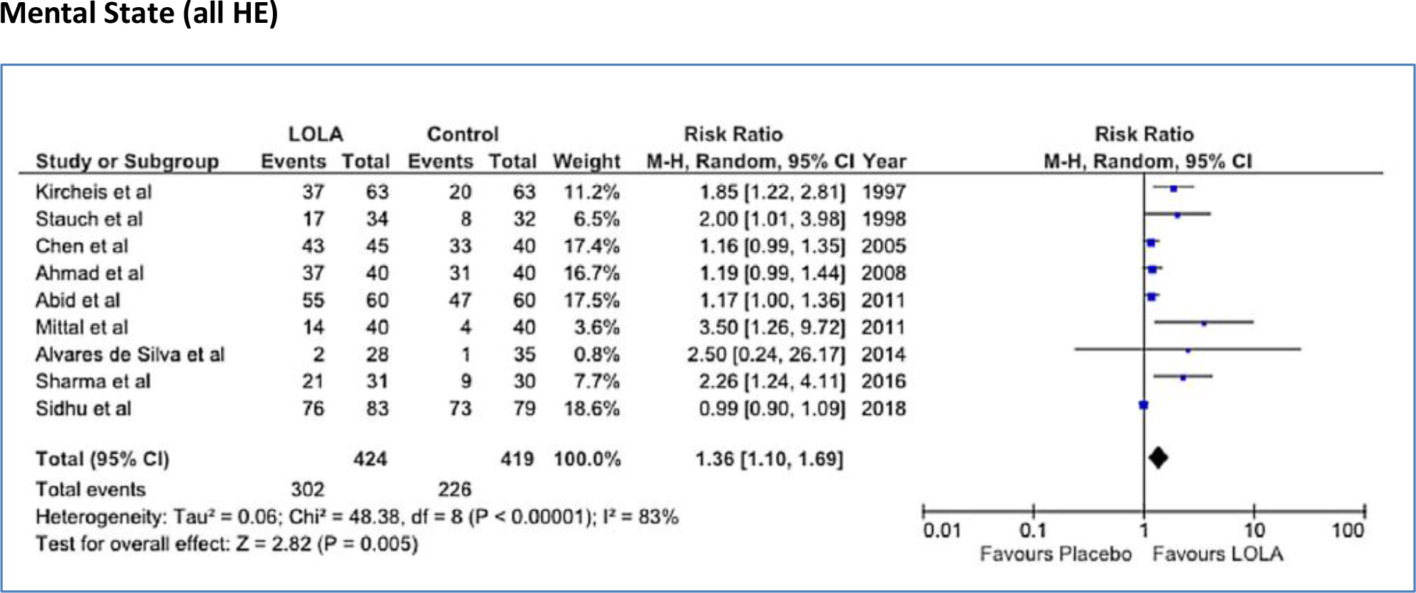
DOI: 10.31038/IMCI.2019215

Abstract

In-111-Octreotide infusion, via intrahepatic catheterization is well established technique in our Institution in hepatocellular carcinoma and neuroendocrine tumors treatment. In order to facilitate repetitive infusions of our patients, a method of implanted ports use, gave a simpler therapeutic way but also improved therapy results. Our aim is to show that radiopharmaceutical fluid flow through implanted port is rich; the absorbed dose in the tumor increased for best therapy results.

Surgically implanted ports have been used in repetitive intra-arterial In-111 radiolabeled Octreotide infusions for 22 patients with hepatocellular carcinoma and similarly 18 patients with neuroendocrine tumors in a continuous base. A percutaneous implantation procedure facilitates safe and less invasive radiopharmaceutical infusions for the treatment. We have focused on the interventional techniques for percutaneous implantation of a vascular access device, consisting of an implantable port, to perform In-111 Octreotide infusions. Hepatic arterial infusion radiotherapy employs a hepatic artery catheter as a conduit to achieve a high concentration of radiolabeled agent to liver tumors. It is performed using less-invasive percutaneous image guided procedures. Various techniques were used to ensure high concentration of radiopharmaceutical in liver tumors, as there are many anatomical hepatic arterial variations and complicated blood flow patterns. These techniques are composed of arterial redistribution by embolization, percutaneous catheter placement, evaluation and management of flow patterns that reflect In-111 Octreotide distribution.

Using fluid flow theory, we describe blood flow alterations that could be performed to obtain selective radiopharmaceutical distribution to the target area and avoid side effects caused by the accumulation of the radiolabeled agent into non tumor areas. By steady, laminar and disturbed flow equations, the rich distribution of our agent in the scintigraphy imaging of the tumor, by the implanted ports technique, can be explained.

The factors affecting hepatic arterial flow in tumor feeding artery were analyzed. The patency rate of the hepatic artery was significantly higher in patients with catheter placement using fixed port method than those undergo fully interventional catheterization. A ratio of 5: 1 to 3: 1 flow increase was calculated through poiseuille flow and Reynolds number for circular pipe.

We consider that in continuous therapy, it is important to use the simplest fixed port method for percutaneous catheter placement instead of interventional catheterization, in order to increase absorbed dose into tumor for best response of radionuclide therapy.

Keywords

Blood flow, Laminar flow, Poiseuille law, Implanted Port, Arterial infusion, In-111-Octreotide Therapy

Blood Flow in Arteries

Blood flow in arteries principally is an unsteady phenomenon. Normal arterial flow is laminar but secondary flows are generated at curves and branches. Arteries may change with the varying hemodynamic conditions. Unusual hemodynamic conditions could create an abnormal biological response. Velocity profile skewing creates pockets, in which the direction of the wall shear stress oscillates.

The study of arterial blood flow will lead to the prediction of individual hemodynamic flows in any patient, the development of diagnostic tools to quantify disease and the design of devices that mimic or alter blood flow.

The blood flow in human arterial system can be considered as a fluid dynamics problem. Simulation of blood flow in the arterial network system will provide a better understanding of the physiology of human body. Simulation studies of blood flow in the diseased condition can diagnose the health problem easily. There are two distinctly different types of blood fluid flow. The first type is known as laminar, the second as turbulent flow.

In laminar flow the fluid particles move along smooth paths in layers with every layer (lamina) sliding smoothly over its neighbor. Laminar flow becomes unstable at high velocities and breaks down into turbulent flow. In turbulent flow the particles follow very irregular and erratic paths, their velocity vectors varying continually both in magnitude and direction [1, 2].

Steady Flow

Steady flow is that one where all conditions, such as velocity and pressure, at any point in a stream remain constant with respect to time. The definition is usually expanded to include flow in which the conditions fluctuate equally on both sides of a constant average value. Arterial flow is pulsatile and each flow may be analyzed in terms of a steady flow together with a number with superimposed sinusoidal components.

Steady flow of Newtonian fluids in rigid vessels is well understood and can serve as a starting point in the study of blood flow in arteries. Blood flow is also relevant as pulsatile flow and can be considered to be the sum of a steady component and a number of oscillatory components which interact neither with each other nor with the steady component [2].

IMCI - 110_Georgosopoulou ML_F1

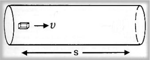
Figure 1. An element of fluid in steady flow at velocity υ takes a time t to traverse a distance s.

Laminar Flow in a Vessel

Flow is laminar when the velocity gradient is smooth and continuous.

Cross section of the vessel shows the laminae moving at different speeds; closest to the edge of the vessel, fluid moves slowly, though near the center moves quickly (2).

IMCI - 110_Georgosopoulou ML_F2

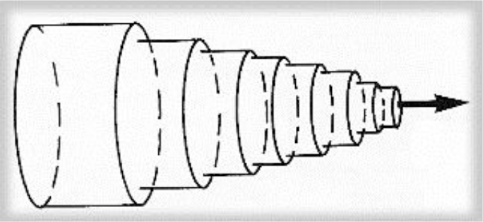
Figure 2. Schema of the laminar flow in a cylindrical vessel. The laminae slide over each other, binded by friction from within the fluid.

Poiseuille Flow

Steady laminar flow established in long cylindrical pipes is sometimes referred to as Poiseuille flow. This is a basic type of flow and blood moves in a series of concentric shells such that the velocity profile across the vessel is parabolic. Blood in the center of the vessel is moving most rapidly, though blood in contact with the vessel wall does not move. The velocity of any lamina at radius r from the center of the vessel may be expressed by

υ(r) = υmax (1-r2/R2) [1]

 Where υmax is the velocity of the center stream and R the radius of the vessel.

IMCI - 110_Georgosopoulou ML_F3

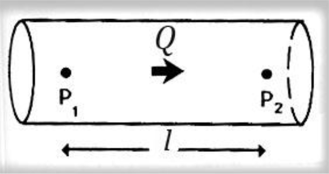
Figure 3. Poiseuille law, viscosity of fluid.

Steady flow discharge Q is sustained in a long rigid tube. The pressure over a length l is P2-P1. Poiseuille described how the steady flow of a fluid is influenced by its viscosity, showing that for a pressure difference P2–P1 along a length l of the vessel, the pressure and the flow discharge Q are related to, in the following way

P2–P1 = (8 l v/πr4) × Q [2]

Where v is the viscosity and r the radius of the vessel lumen.

Consequently, the fluid resistance increases with the fourth power of decreasing radius of the vessel lumen. So, halving the radius means that a 16-fold increase in pressure is required to maintain the same flow.

According to Poiseuille law the variation of velocity across the lumen of a long cylindrical pipe in steady viscous flow, is smooth and has the form of a paraboloid of rotation. (1–4)

Turbulent Flow

Laminar flow becomes unstable at high velocities and breaks down to turbulent flow. The point at which the transition between the two flow regimes occurs cannot be predicted exactly, but it is largely determined by the Reynolds number, Re, which for a circular pipe is defined as:

Re = υ.D/ν [3]

Where, υ is the average velocity of flow across the pipe, D the diameter of the pipe and v the kinematic viscosity.

Reynolds number (Re) is a dimensionless number that characterizes different flow regimes.

Laminar flow occurs at low Re, as a smooth, constant fluid motion, though turbulent flow because of flow instabilities occurs at high Re.

Turbulent flow may never be described as steady as there are continual fluctuations in both velocity and pressure at every point. 2.25 to 4.45 folds flow increase was calculated through poiseuille flow and Reynolds number, for cylindrical pipe. (4).

Blood Flow Energy

Fluid with sufficient kinetic energy is capable of flowing up a pressure gradient. The property of a fluid that determines the direction and speed of flow is its total fluid energy. Fluid energy can take several forms as the following. In real conditions these energy forms may be combined.

a. Pressure energy (P)

Pressure in a fluid may be thought of as the ability to do work, e.g. overcome viscous resistance; so pressure is a form of potential energy.

b. Kinetic energy (K)

Each volume of moving fluid has energy by virtue of its mass and motion, determined as ½ ρ.υ2, where ρ is the density and υ the velocity of flow.

c. Gravitational potential energy (G)

Fluid at a higher level than an arbitrary point is also capable of doing work; its weight imparts a potential energy equal to ρ.g.h, where, ρ the density, g the acceleration of gravity and h is the height. This gravitational energy may be transformed into kinetic energy by, e.g., allowing the fluid to flow down.

d. Viscous energy (V)

Flow against a viscous resistance described above may also be seen as a form of energy loss, in which kinetic or potential energy of the fluid is transferred to heat. With laminar flow the magnitude of this loss depends on vessel geometry.

In a single streamline of flow, the principle of conservation of energy holds that the sum of these individual energies, the total fluid Energy E, must remain constant at each point along the path of flow.

It is a difference in blood fluid energy that determines the direction and character of flow.

Mathematically, blood flow is described by Darcy’s law

F = ΔP/R [4]

Where F = blood flow, ΔP = pressure gradient, R = resistance

It is also described approximately by the following Hagen-Poiseuille equation

R = (v.L/r4(8/π) [5]

Where ν = blood fluid viscosity, L = length of tube, r = radius of tube

The viscosity of normal blood is about three times as great as the viscosity of water.

It is important to note that resistance to flow changes dramatically, with respect to the radius of the tube as already is referred for fluid flow in Poiseuille law [2–4].

In large arteries blood can be approximated as an homogeneous Newtonian fluid because the vessel size is much greater than the size of particles; particle interactions have a negligible effect on the flow. In smaller vessels, blood behavior is expressed as non-Newtonian [5].

Materials and Method

The treatment of hepatocellular carcinoma via the hepatic artery is based on the existence of arterial hyper vascularization of the tumor. For tumors bigger than 2 cm in diameter, more than 80% of their blood supply is drawn from the hepatic artery. Normal liver parenchyma draws more than 80% of blood from the portal vein. By delivery of radioactive agents into the hepatic artery, which represents almost exclusively the arterial supply to liver tumors, highly selective tumor uptake can be achieved [6].

IMCI - 110_Georgosopoulou ML_F4

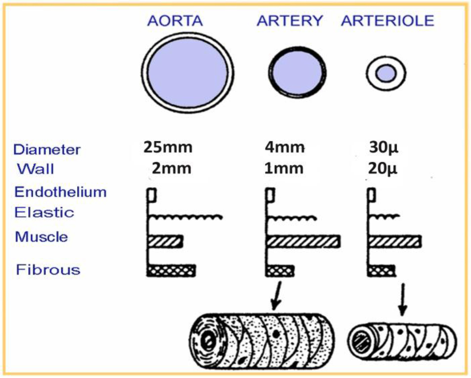
Figure 4. Human vessels sizes (aorta, artery, arteriole)

The theoretical rationale for hepatic artery infusion is based on the observation that hepatocellular carcinoma and neuroendocrine tumors are hyper vascular tumors and derive the majority of their blood supply from the hepatic artery.

In general the radiopharmaceutical is delivered at the lobar artery level to be distributed to numerous tumors in that lobe.

IMCI - 110_Georgosopoulou ML_F5

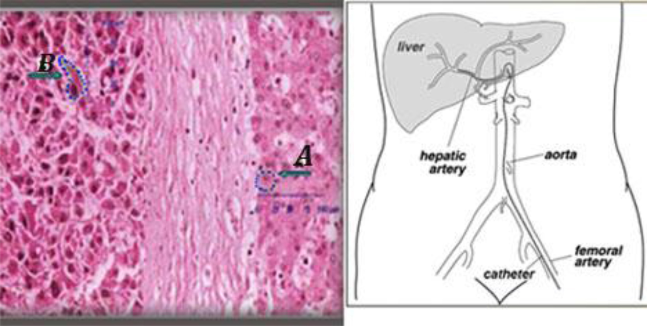
Figure 5. (Left) an histologic sample of normal (A) and tumor liver cells (B).Cell dimensions and distances between cell surface and nuclei show that DNA lies within the micrometer range of In-111 emissions. (Right) Hepatic artery supplied the tumor is the liver entrance for infusion.

Individually determined patient-specific activities are used. In the case of main hepatic artery injection, radiation is distributed to both lobes of the liver. If the lesions are limited to one lobe, the catheter can be selectively inserted either into the left or right lobar artery supplying the affected lobe, thus sparing the contralateral.

In selected cases, hyper selective, single segment treatments can be considered.

Gentle infusion, by a steady low pressure, should be used to strictly avoid backflow (fig 6).

IMCI - 110_Georgosopoulou ML_F6

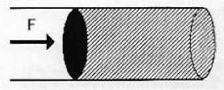
Figure 6. In order the flow be maintained against viscosity, a force F is applied to the fluid.

Gentle infusion (no excessive pressure) should be used to strictly avoid backflow.

Indium -111- Octreotide

Indium-111 (111In) is a radioisotope that was introduced for hepatocellular carcinoma and neuroendocrine tumor cancer cells diagnosis. It is also successfully used for neuroendocrine tumor radio-immunotherapy.

a) Production and Physical Characteristics of In-111

In-111 is produced by cyclotron from Cd-112 collision with protons of energy 2.8 MeV according to the nuclear reaction Cd-112 (p, 2n) In-111. Radioactive In-111 decays by physical half-life time of 2.83 days. The type, energy and emission ratio for In-111 decay, are displayed in Table 1.

Table 1. Indium-111(In111) Decay Chart

In-111 Decay Chart

Type of decay

Energy (keV)

Emission ratio

(Bq·s) – 1

Photons

150.8

3·10–5

Photons

171.3

0.906

Photons

245.4

0.941

Electrons IT*

145 – 170

0.1

Electrons IT*

218 – 245

0.06

Auger electrons

19 – 25

0.16

Auger electrons

2.6 – 3.6

1.02

Auger electrons

0.5

1.91

IT, Internal Transform

Purity of the final product of In-111 is affected by the undesired isotopes In-110m, In-110 and In-114m that are not possible to spare from In-111 due to the similar chemical characteristics of these isotopes with In-111.

Isotopes In-110m and In-110, do not affect dosimetry of radioisotopes labeled with In-111, as these undesired isotopes have minor presence and short half-life time (4.9h and 1.1h, respectively). On the contrary, In-114m that is produced from Cd-114 according to a (p, n) nuclear reaction, has 49.51 days half-life time and decays by internal transition (96.9%) and electron capture (3.2%) with emission of photons at (192, 558 and 725) keV. In-114m affects dosimetry due to its long half-life time [7].

 The Flow Discharge Q depends on the volume V of the fluid that passes in time interval t

That is Q = V/t [6a]

Flow Discharge is also determined by the multiplication of the cross section of the object with mean velocity

Q = S × υ[6b]

Then mean velocity depends on flow discharge and is inversely proportional to the cross section of the tube

υ = Q / S [6c]

Angiographic catheter cross section SA is smaller than Port catheter cross section SP, (SP > SA) consequently the Port flow QP is greater than Angiographic flow QA.

QP/QA = SP/SA > 1 [6d]

Assuming that the velocity in angiographic tube is obtained equal to the velocity in port tube vA = vP, we consider (by equation 6d) that Port Flow QP > Angiographic Flow QA.

b) In-111 uptake and biokinetic properties

The radiopharmaceutical In-111-DTPA-D-Phe-1 octreotide is a peptide composed of 8 amino acids and is an analogue of the active part of the peptide hormone somatostatin In-111-octreotide. It is used for radio immunotherapy for neuroendocrine tumors of the gastro hepatic system. Octreotide is a somatostatin analogue used for labeling In-111. Somatostatin is a peptide of the gastro enteric system that inhibits the production of the grow hormone.

There is an over expression of the somatostatin receptors at the surface of the neuroendocrine tumor cancer cells. In-111-octreotide is bounded to the somatostatin inhibitors and transferred into the cancer cell. Auger electrons that are emitted from In-111 can damage the DNA of the cancer cell.

In palliative treatment use, the radiopharmaceutical entrance into the tumor cell and its destructive effect to DNA by emission of Auger and internal conversion electrons has been exploited earlier [8, 9].

The Peptide Receptor Radionuclide Therapy (PRRT), a somatostatin-analogue-based targeted therapy, is a field in the palliative treatment of Neuro Endocrine Tumors (NET) with very encouraging data. The treatment modality utilizes radioactive substances that are conjugated with various somatostatin analogues such as octreotide.

Intravenously administrated, these drugs bind to the somatostatin receptor localized on the tumor cells. The ligand–receptor complex is internalized and the attached radiation has the potential to destroy the tumor cells.

The side effects are few and mostly mild, in particularly when treatment protocols consider renal protective agents. Initially used In-111-DTPA-[D-Phe1]-octreotide achieved positive results, whereas stable disease was seen in 42% to 81% of patients and partial remission rates up to 8%. However, because of the limited tissue penetration range of In-111, In-111 coupled peptides are rather ineffective for PRRT and their utilization should be restricted for hepatocellular carcinoma therapeutic purposes.

Auger electrons with particle ranges (.02–10) μm and Internal Conversion electrons with ranges (200–550) μm that In-111 emits during its transformation procedure are extremely short. In large arteries blood can be approximated as homogeneous Newtonian fluid because the vessel size is much greater than the size of particles; particles’ interactions have a negligible effect on the flow [5, 6, 8, 13].

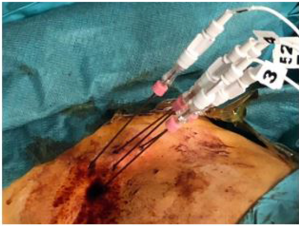
Implanted Port /Arterial Infusion

Surgically implanted ports have been used in repetitive intra-arterial In-111 radiolabeled Octreotide infusions for 22 patients with hepatocellular carcinoma and similarly 18 patients with neuroendocrine tumors in a continuous base. A percutaneous implantation procedure facilitates safe and less invasive radiopharmaceutical infusions for the treatment. We have focused on the interventional techniques for percutaneous implantation of a vascular access device consisting of an implantable port, to perform In-111-Octreotide infusions.

Hepatic arterial infusion radionuclide therapy employs a hepatic artery catheter as a duct to achieve a high concentration of radiolabeled agent to liver tumors. Various techniques were used to ensure high concentration of radiopharmaceutical in liver tumors as there are many anatomical hepatic arterial variations and complicated blood flow patterns. They are composed of arterial redistribution by embolization, percutaneous catheter placement and evaluation and management of flow patterns reflecting In-111 octreotide distribution.

Using fluid flow theory we describe details of the alteration of blood flow by first pass embolization that can be performed to obtain selective radiopharmaceutical distribution to the target area and to avoid side effects caused by the accumulation of the radiolabeled agent into non tumor areas.

Different hypothetical flow mechanisms lead to different patterns of strain relaxation with time. Representative tissue properties show blood fluid drainage into the local microvasculature to be the dominant flow-related stress/strain relaxation mechanism; due to drainage into the microvasculature is on the order of 5–10 sec. This is the relaxation time of strain in solid tumors under realistic applied pressure magnitudes. The magnitude of the strain relaxation can be as high as approximately 0.4% strain, well within the range of strains measurable by elastography.

A rich distribution of the In-111 radiopharmaceutical that is recorded in the scintigraphy imaging of the tumor, by the implanted ports technique, is explained by steady, laminar and disturbed flow equations [10, 11].

a) Tip-Fixation method

In order to facilitate repetitive infusions of our patients, a method of implanted ports, gave a simpler therapeutic way but also improved therapy results.

Our aim is to show that radiopharmaceutical fluid flow through implanted port is rich and the absorbed dose in the tumor increased for best therapy results.

Implanted ports have been used in repetitive intra-arterial In-111 Octreotide infusions.

The catheter should be placed directly into the artery supplying the tumor. Since the position of the catheter tip cannot be changed after placement, hemodynamic alterations must be taken into consideration.

Patients, with histologically confirmed hepatocellular carcinoma or neuroendocrine tumors lesions located in liver and with normal kidney function, were infused with mean activity of 4500 MBq In-111 – octreotide.

The technique is composed of arterial redistribution, percutaneous catheter placement applying “tip-fixation method, ” evaluation & management of flow patterns that reflect radiopharmaceutical distribution.

The infusion was performed through a port attached to the hepatic artery. The hepatic artery port makes the therapy more comfortable for the patient as in this way the hepatic artery angiography catheterization, at each therapeutic session, is avoided.

Port chamber is made of titanium & silicone membrane (diameter12mm). It is 28 mm long and 13.5 mm wide. Inner diameter of port catheter is 1.2 mm and outer diameter is 2.5mm [12].

IMCI - 110_Georgosopoulou ML_F7

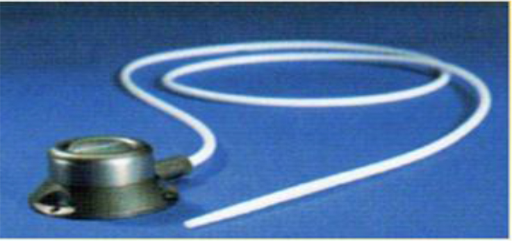
Figure 7. Percutaneously implantable catheter-port system.

The “tip-fixation method uses the gastroduodenal artery. Hepatic arterial infusion radiotherapy employs a hepatic artery catheter as a duct to achieve a high concentration of radiolabeled agent to liver tumors.

For the “tip-fixation method, a distal branch of the hepatic artery could also be used to fix the catheter tip. Sometimes, in cases of lower tumor load, a more selective delivery at the segmental artery level is possible.

This is proved to be the best method to give better uptake of the radiopharmaceutical by the tumor. The infusion of the radiopharmaceutical was performed directly to patient liver through the port that was attached to the hepatic artery.

IMCI - 110_Georgosopoulou ML_F8

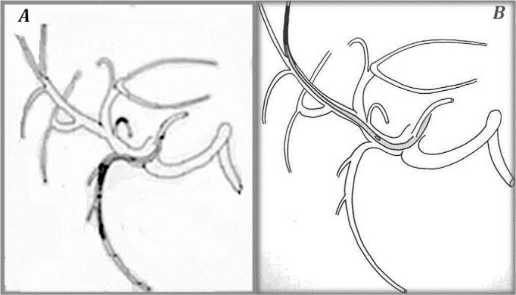
Figure 8. The “tip-fixation method” using the gastroduodenal artery (A) or a distant hepatic artery (B)

To fix the tip of the indwelling catheter, the gastroduodenal artery (GDA) is most commonly used. When using the GDA, an indwelling side-holed catheter is inserted into the GDA. However, if necessary, it is possible to use other arteries.

Scintigraphy

Patient specific dose estimation for the tumor and healthy tissue is very important for radiopharmaceutical therapeutic applications as this is the most accurate way to assign an administered dose to the best therapeutic result. It is also assistance to the physician in order to avoid side effects of the therapy and compare the treatment results to other related therapies as external radiotherapy [13].

Scintigraphy is performed immediately after radiopharmaceutical infusion and at 24h and at 48h post-infusion. Individually determined patient-specific activities are used. Quantitative whole-body scintigraphy imaging one week post therapy is recommended to confirm the distribution of the radiopharmaceutical. This is proved to be the best method to give better uptake of the radiopharmaceutical by the tumor.

Gamma camera is calibrated in order to estimate source organ activity considering count rate, patient’s body diameter and source organ size [14].

Average activity (6.3+-2.3) GBq per session was administered at monthly intervals. Infusion repetition did not exceed the 12th fold.

Results

The patient individualized dosimetry calculations were based on anterior and posterior scintigraphy images that were acquired immediately after radiopharmaceutical infusion, through hepatic arterial port and at 24 and 48 h post-infusion.

The results showed that the tumor absorbed dose ranged from 2.5mGy/MBq to 18.4mGy/MBq depending on the lesion size [13]. The average absorbed dose per session to a tumor for a spherical mass of 10 gr was estimated to be 10.8 mGy/MBq, depending on the histotype of the tumor.

The organ average radiation dose estimation after In-111-DTPA-Phe1-octreotide trans-hepatic infusion was found as follows:

(a) Liver Tumor

10.80

mGy/MBq

(b) Liver organ

0.14

mGy/MBq

(c) Kidneys

0.41

mGy/MBq

(d) Spleen

1.40

mGy/MBq

(e) Pancreas

0.13

mGy/MBq

(f) Bone marrow

0.003

mGy/MBq

IMCI - 110_Georgosopoulou ML_F9

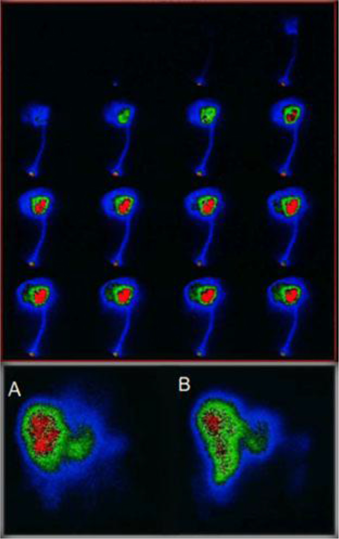
Figure 9. Above First pass infusion for selective In-111 Octreotide distribution to the target area. below (A) 1h post infusion via arterial port, (B) 1h post angiography thin catheter infusion.

IMCI - 110_Georgosopoulou ML_F10

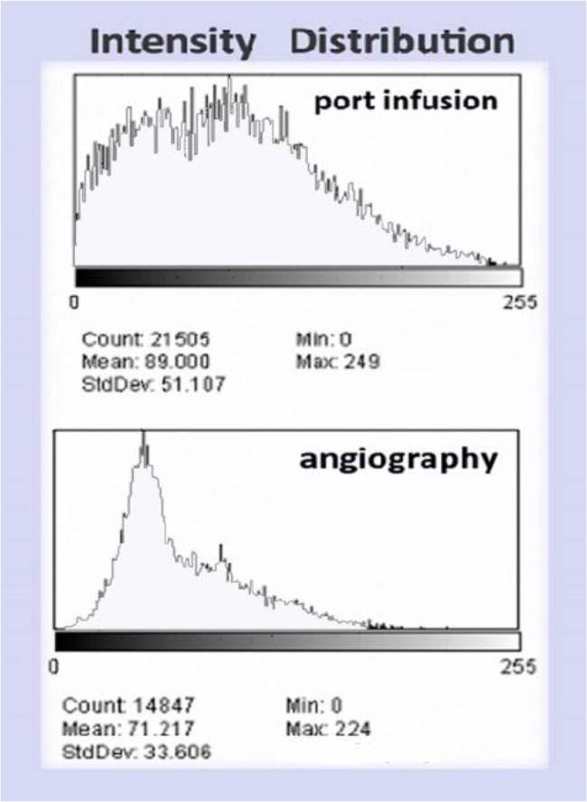
Figure 10. (Above) Intensity histogram of In111 Octreotide distribution by port infusion (catheter diameter 1.2mm) and (Below) Intensity histogram of In111-Octreotide distribution by angiography (catheter diameter 0.8mm)

IMCI - 110_Georgosopoulou ML_F11

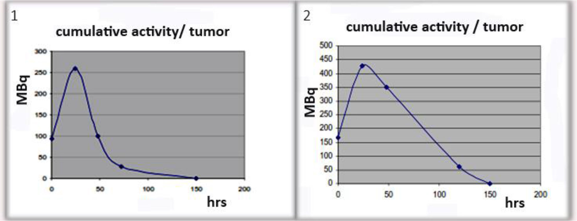
Figure 11. Examples of cumulative activity to tumor

IMCI - 110_Georgosopoulou ML_F12

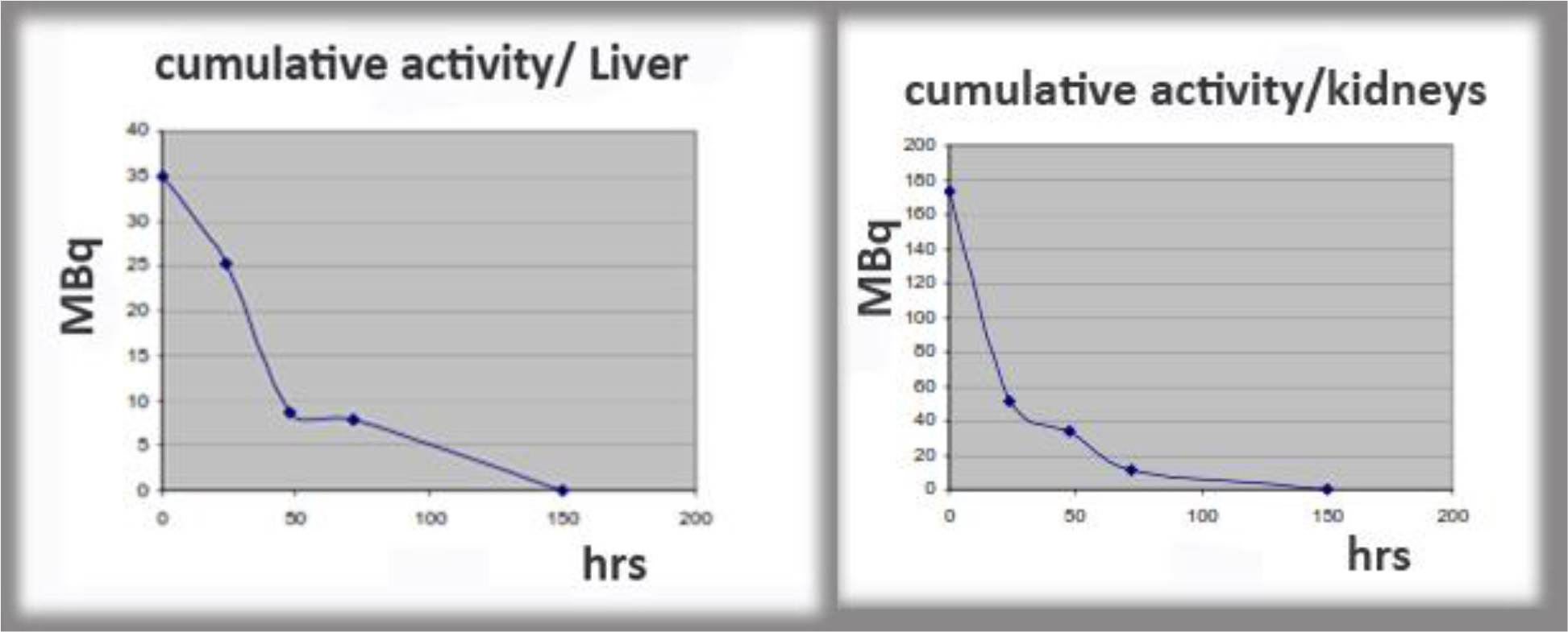
Figure 12. Examples of cumulative activity to critical organs.

Discussion

The factors affecting hepatic arterial flow in tumor feeding artery were analyzed. The patency rate of the hepatic artery was significantly higher in patients with catheter placement using fixed port method than those undergo fully interventional catheterization. A ratio of 5: 1 to 3: 1 flow increase was calculated through poiseuille flow and Reynolds number for circular pipe.

The infusion of the radiopharmaceutical was performed directly to patient liver through a port that was attached to the hepatic artery. Percutaneously implantable catheter-port system placement is safe and technically feasible for use in the hepatic artery.

This is proved to be the best method to give better uptake of the radiopharmaceutical by the tumor. Patient specific dose estimation for the tumor and healthy tissue is very important for radiopharmaceutical therapeutic applications as this is the most accurate way to assign an administered dose to the best therapeutic result [13–15]. It is also assistance to the physician in order to avoid side effects of the therapy and compare the treatment results to other related therapies as external radiotherapy.

In the case of main hepatic artery injection, radiation is distributed to both lobes of the liver. If the lesions are limited to one lobe, the catheter can be selectively inserted either into the left or right lobar artery supplying the affected lobe, thus sparing the contralateral. In selected cases, hyper selective (i.e. single segment) treatments can be considered.

Patient specific dosimetry calculations help the physician to optimize the planning of the treatment, avoid side effects to healthy tissue and assign administered dose to treatment results.

Conclusion

We consider that in continuous therapy, it is important to use the simplest fixed port method for percutaneous catheter placement instead of interventional catheterization, in order to increase absorbed dose into tumor for best response to radionuclide therapy. A percutaneous implantation procedure facilitates safe and less invasive radiopharmaceutical infusions for multiple treatments. Implanted ports have been used in repetitive intra-arterial In-111 Octreotide infusions. The patency rate of the hepatic artery was significantly higher in patients with catheter placement using fixed port method than those undergo fully interventional catheterization. Percutaneously implantable catheter-port system placement is safe and technically feasible for use in the hepatic artery.

Advantages of the use of intra-arterial radionuclide agents are the ability to deliver high doses of radiation to small target volumes, the relatively low toxicity profile, the possibility to treat the whole liver including microscopic disease and the feasibility of combination with other therapy modalities. Disadvantages are mainly due to radioprotection problems.

Percutaneous image-guided catheter placement for hepatic arterial infusion radionuclide therapy is technically safe and provides a viable alternative to traditional methods of hepatic artery access. However, a thorough knowledge of hepatic arterial anatomy, potential of complicated blood flow patterns and management of the infusion devices will ensure a high likelihood of tumor-only delivery of the radionuclide agent.

In summary, we modified the technique of radiologic placement of a catheter and port system for hepatic arterial infusion internal radiotherapy by fixing the catheter tip to the vessel and infusing radionuclide agents from the side hole of the catheter. We conclude that this technique is a safe and not unduly time-consuming method that obviates surgical laparotomy and represents a significant therapeutic advance in the medical oncology field for patients with liver malignancies.

Our intention is to study further, the behavior of the radionuclide fluid as is inserting in arteries and arterioles.

References

  1. Evans, DH, McDicken, WN, Skidmore, R., and Woodcock, JP, 1989, Doppler ultrasound: Physics, instrumentation, and clinical applications, John Wiley & Sons
  2. Taylor, Kenneth JW, Burns Peter N, Wells Peter NT (1987) Clinical Applications of Doppler Ultrasound, Raven Press 1988
  3. Ku David N (1997) Blood flow in arteries, Annual Review of Fluid Mechanics 29: 399–434
  4. Sankar DS, Hemalatha K (2007) A non-Newtonian fluid flow model for blood flow through a catheterized artery—Steady flow, Applied Mathematical Modelling 31: 1847–1864.
  5. Blessy Th, Sumam KS (2016) Blood Flow in Human Arterial System-A Review, Procedia Technology 24: 339–346.
  6. Giammarile F, Bodei L, Chiesa C, Flux G, et al (2011) Therapy, Oncology and Dosimetry Committees, EANM procedure guideline for the treatment of liver cancer and liver metastases with intra-arterial radioactive compounds, EJNMMI 38: 1393–406.
  7. Lyra ME Andreou M. Georgantzoglou A et al, (2013) Radionuclides Used in Nuclear Medicine Therapy – From Production to Dosimetry, Current Medical Imaging Reviews. Bentham Science Publishers 9: 51–75.
  8. Limouris GS, Chatziioannou A, Kontogeorgakos D. et al., 2008, Selective hepatic arterial infusion of In-111-DTPA-Phe1-octreotide in neuroendocrine liver metastases ejnmmi 35: 1827–1837.
  9. Virgolini I, Szilvasi I, Kurtaran et al. (1998) Indium-111-DOTA-Lanreotide: biodistribution safety and radiation absorbed dose in tumor patients. JNM 39: 1928–1936.
  10. Arai Y, Takeuchi Y, Inaba Y, Yamaura H, et al. (2007) Percutaneous catheter placement for hepatic arterial infusion chemotherapy, Techniques in vascular and interventional radiology 10: 30–37.
  11. Tanaka T, Arai Y, Inaba Y, Matsueda K, et al (2003) Radiologic placement of side-hole catheter with tip fixation for hepatic arterial infusion chemotherapy. Journal of vascular and interventional radiology 14: 63–68.
  12. Lyra ME, Limouris GS (2009) In-111 radiolabelled Octreotide therapy -Arterial infusion strategy using implanted ports versus Artery Catheterization- EJNMMI 36: 260–80.
  13. Vamvakas I, Lagopati N, Andreou M, et al, (2009) Patient specific computer automated dosimetry calculations during therapy with 111In Octreotide. European J of Radiography 1: 180–183.
  14. EANM Guideline (2010) Ellinor Busemann Sokole, Anna Płachcínska, Alan Britten with contribution from the EANM Working Group Maria Lyra Georgosopoulou, Wendy Tindale, Rigobert Klett, Routine quality control recommendations for nuclear medicine instrumentation, EJNMMI 37: 662–671.
  15. Maecke H R, Reubi Jean-Claude (2011) Somatostatin Receptors as Targets for Nuclear Medicine Imaging and Radionuclide Treatment JNM 39: 1928–1936.

Cellular Adaptation to Ischemia; Ischemic Conditioning to Confer Neuroprotective Benefits in Mild Cognitive Impairment and Alzheimer’s disease

DOI: 10.31038/ASMHS.2019325

Abstract

Small, controlled doses of ischemia induced in a healthy limb has been demonstrated to strengthen the body’s tolerance to larger more toxic doses. Ischemic conditioning utilising protocols of remote ischemic conditioning (RIC) and physiological ischemic training (PIT) trigger mechanisms of cellular adaptation to ischemia. These protocols may represent practical and translatable therapies for neurological diseases, such as mild cognitive impairment and Alzheimer’s disease, that have an ischemic or inflammatory basis. Whilst the current literature supports the neuroprotective and anti-hypertensive effects of RIC and PIT, to date there has been no investigation into the effects of PIT utilising isometric exercise training (IET) on cognitive performance outcomes in an elderly neuropathological cohort. However, it seems feasible that the anti-hypertensive effects elicited through IET might be a stimulus for improvements in systemic and neurovascular circulation and as a result, enhanced cognitive performance.

Keywords

Alzheimer’s Disease, Blood Pressure, Hypertension, Ischemic Conditioning, Isometric Exercise Training, Mild Cognitive Impairment, Reactive Hyperaemia, Remote Ischemic Conditioning, Vascular Risk Factors

Ischemia is thought to play a pivotal role in the aetiology and progression of Alzheimer’s disease 1–3] with reduced cerebral blood flow (CBF) the manifestation of this [4]. Ischemia arises as a consequence of restricted blood flow and compromises the efficient circulation of oxygen, nutrients, other important blood borne factors, and the removal of toxic metabolic waste. Cellular dysfunction arising from ischemia will often result in cell death. Ischemia may be partial as in the case of hypoperfusion which occurs in conditions such as mild cognitive impairment (MCI) and AD or total as in the case of heart attack and stroke. Ischemia is associated with serious physiological implications. However, through a process of ischemic conditioning, small controlled doses of ischemia delivered to a healthy limb is able to strengthen the body’s tolerance to larger more toxic doses [5]. This type of treatment is currently administered to patients to improve the outcomes of heart attack [6] and ischemic stroke [7, 8]. Two methods of inducing remote limb ischemia are remote ischemic conditioning (RIC) and physiological ischemic training (PIT). RIC involves inflating a blood pressure cuff to above systolic blood pressure (SBP) to induce total ischemia via full occlusion of the brachial artery, followed by reperfusion after cuff deflation [9]. PIT involves subjecting skeletal muscle to intense contraction via the application of isometric contraction using handgrip dynamometer or tourniquet to induce partial occlusion of the brachial artery followed by reperfusion [10].

Ischemic Conditioning

The practice of ischemic conditioning to elicit cellular adaptation in the myocardium has been utilised for approximately 30 years [6]. Research investigating the neuroprotective potential of RIC began approximately10 years later with initial findings reporting RIC as an effective method of limiting neural tissue damage after stroke [11]. Since then there has only been a small amount of research in this area, with exciting results, Mouse models of vascular cognitive impairment have shown increased CBF, reduction in inflammation, reduced amyloid-beta deposition and improved cognition [12]. Human trials have shown increased CBF [9], the enhancement of neuroplasticity [13], and motor and cognitive learning enhancements in healthy adults [14].

As with RIC, PIT has been shown to stimulate collateral formation in the myocardium [15], upregulate vascular endothelial growth factor (VEGF) production, and promote angiogenesis [16]. More recent research reported that the application of PIT using isometric exercise training (IET), at 50% Maximal Voluntary Contraction (MVC), to patients with acute cerebral infarction promoted brain collateral formation via the increased expression of VEGF and recruitment of endothelial progenitor cells [17]. The same researchers also reported a positive correlation between increased CBF and improved motor function. More recently, improvement in cognitive performance and reduction in systemic arterial stiffness was demonstrated in a cohort of non-elderly, non-neuropathological adults after 8 weeks of IET at 30% MVC [18]. Traditionally, IET using handgrip dynamometer has been successfully demonstrated as an anti-hypertensive therapy [19]. IET can elicit blood pressure (BP) reductions greater than those achieved through aerobic exercise and equivalent to those achieved through monotherapy with beta-blocker [20]. Furthermore, there is a considerable amount of research that links hypertension to conditions of cognitive decline such MCI and AD [21].

The Role of Hypertension

Hypertension, a hallmark of aging, has been linked with cerebrovascular pathology, hypoperfusion [22], and cognitive decline [23]. Imaging studies have demonstrated an association between brain atrophy and untreated hypertension [24] and positive correlations have been demonstrated among SBP, diastolic blood pressure, and burden of neural AD pathology [21, 25]. Hypertension is associated with structural and functional changes in cerebrovascular pathways. These deleterious alterations are potentially reversible [26]. Subsequently, improving CBF may also lead to improvements in cognitive performance. The treatment and management of hypertension has been observed to slow cognitive decline in individuals with AD [27] and to reduce the risk of progression from MCI to AD Li et al. [17].

Change Mechanisms of Ischemic Training

Whilst the signalling mechanisms involved in ischemic conditioning are not fully understood, evidence obtained through animal models and clinical trials suggest that signalling initiation occurs via autacoids such as adenosine, bradykinin, and calcitonin gene-related peptide. To varying extents each of these autacoids are involved in neuromodulation, inflammatory mediation, and vasodilation [28]. Signal transmission to the brain is believed to occur through an interaction between humoral and neural pathways such as peripheral and autonomic nervous systems, blood borne factors (IL-10, and nitrite), immune and anti-inflammatory factors (IL-6, IL-10, IL-1ra), and endogenous carbon monoxide [9]. Abnormalities in the metabolism of endogenous carbon monoxide have been linked to a variety of diseases including neurodegenerations, hypertension, heart failure, and inflammation [29]. Mitochondria play a prominent role in signal transduction with most pathways that are triggered by ischemic conditioning converging on the mitochondria [5].. Mitochondrion are the energy packages of the cell and are responsible for regulating cell metabolism. Nitrite protects mitochondria from oxidative stress and is upregulated after the application of RIC, as is CBF [9]. Inadequate CBF is a contributor to oxidative stress in brain cells and has significant implications in neurological diseases such as AD where the mitochondria are ravaged by oxidative stress [30, 4 ].

Considerations for Future Investigation

Dementia is Australia’s second leading cause of death in adults over 65 years [31] and is the greatest cause of disability among the elderly. Both RIC and PIT/IET represent practical and translatable therapies for neurological diseases that have an ischemic or inflammatory basis. Whilst the current literature supports the neuroprotective and anti-hypertensive effects of PIT/IET, to date there has been no investigation into the effects of this protocol on cognitive performance outcomes in an elderly neuropathological cohort. However, it seems feasible that the anti-hypertensive effects elicited through IET might be a stimulus for improvements in systemic and neurovascular circulation and as a result, enhanced cognitive performance.

The potential for limb ischemia to trigger neuroprotective physiological responses to support and repair the brain has been demonstrated via RIC and PIT/IET protocols and introduces exciting therapeutic potential for individuals with MCI and AD. The application of RIC and PIT/IET may create an ischemic event that is adequate to confer neuroprotective benefits and anti-hypertensive effects in elderly adults with cognitive impairment or AD. Further investigations within this domain have the potential to yield life-changing results for many individuals.

References

  1. de la Torre JC (2002) Alzheimer disease as a vascular disorder nosological evidence. Stroke 33: 1152–1162. [crossref]
  2. de la Torre, J C (2010) Vascular risk factor detection and control may prevent Alzheimer’s disease. Ageing Res Rev 9: 218–225. [crossref]
  3. de la Torre JC, Mussivand T (1993) Can disturbed brain microcirculation cause Alzheimer’s disease? Neurol Res 15: 146. [crossref]
  4. Zlokovic BV (2011) Neurovascular pathways to neurodegeneration in Alzheimer’s disease and other disorders. Nat Rev Neurosci 12: 723–738. [[crossref]
  5. Hess DC, Blauenfeldt RA, Andersen G, Hougaard KD, Hoda MN, et al. (2015) Remote ischaemic conditioning mdash] a new paradigm of self-protection in the brain. Nat Rev Neurol 11: 698–710. [crossref]
  6. Murry CE, Jennings RB, Reimer KA (1986) Preconditioning with ischemia: a delay of lethal cell injury in ischemic myocardium. Circulation 74: 1124–1136. [crossref]
  7. Hougaard KD, Hjort N, Zeidler D, Sørensen L, Nørgaard A,et al. (2014) Remote Ischemic Perconditioning as an Adjunct Therapy to Thrombolysis in Patients With Acute Ischemic Stroke A Randomized Trial. Stroke 45: 159–167. [crossref]
  8. Meng R, Ding Y, Asmaro K, Brogan D, Meng L, et al. (2015) Ischemic conditioning is safe and effective for octo-and nonagenarians in stroke prevention and treatment. Neurotherapeutics 12: 667–677. [crossref]
  9. Hess DC, Hoda MN, Khan MB (2016) Humoral mediators of remote ischemic conditioning: important role of eNOS/NO/nitrite. Acta Neurochir Suppl 121: 45–48. [crossref]
  10. Ni J, Lu H, Lu X, Jiang M, Peng Q, et al. (2015) The evolving concept of physiological ischemia training vs ischemia preconditioning. J Biomed Res 29: 445–450. [crossref]
  11. Kitagawa K, Matsumoto M, Tagaya M, Hata R, Ueda H, et al. (1990) ‘Ischemic tolerance’phenomenon found in the brain. Brain Res 528: 21–24. [crossref]
  12. Bink DI, Ritz K, Aronica E, van der Weerd L, Daemen MJ (2013) Mouse models to study the effect of cardiovascular risk factors on brain structure and cognition. J Cereb Blood Flow Metab 33: 1666–1684. [crossref]
  13. Cherry-Allen KM, Gidday JM, Lee JM, Hershey T, Lang CE (2015) Remote limb ischemic conditioning enhances motor learning in healthy humans. J Neurophysiol 113: 3708–3719. [crossref]
  14. Cherry-Allen KM, Gidday JM2, Lee JM, Hershey T, Lang CE (2017) Remote limb ischemic conditioning at two cuff inflation pressures yields learning enhancements in healthy adults. J Mot Behav 49: 337–348. [crossref]
  15. Lin S, Lu X, Chen S, Ye F, Zhang J, et al. (2012) Human coronary collateral recruitment is facilitated by isometric exercise during acute coronary occlusion. J Rehabil Med 44: 691–695. [crossref]
  16. Zhao Y, Li J, Lin A, Xiao M, Xiao B, et al. (2011) Improving angiogenesis and muscle performance in the ischemic limb model by physiological ischemic training in rabbits. Am J Phys Med Rehabil 90: 1020–1029. [crossref]
  17. Wan C, Li J, Yi L (2011) Enhancement of homing capability of endothelial progenitor cells to ischaemic myocardium through physiological ischaemia training. J Rehabil Med 43: 550–555. [crossref]
  18. Dempster KS, McGowan CL, Wade TJ, O’Leary DD (2018) Effects of Isometric Handgrip Exercise Training on Systemic Arterial Stiffness, Cardiovagal Baroreflex Sensitivity, and Cognition in Treated Adults with Hypertension: A Pilot Study. Critical Reviews™ in Physical and Rehabilitation Medicine 30.
  19. Carlson DJ, Dieberg G, Hess NC, Millar PJ, Smart NA (2014) Isometric exercise training for blood pressure management: a systematic review and meta-analysis. Mayo Clin Proc 89: 327–334. [crossref]
  20. Wong GW, Wright JM (2014) Blood pressure lowering efficacy of nonselective beta-blockers for primary hypertension. Cochrane Database Syst Rev. 28: CD007452 [crossref]
  21. Hess NC, Smart NA (2017) Isometric exercise training for managing vascular risk factors in mild cognitive impairment and Alzheimer’s disease. Front Aging Neurosci 9: 48. [crossref]
  22. Brown,W, Thore C (2011) Review: cerebral microvascular pathology in ageing and neurodegeneration Neuropathol Appl Neurobiol 37: 56–74. [crossref]
  23. Reitz C, Tang MX, Manly J, Mayeux R, Luchsinger JA (2007) Hypertension and the risk of mild cognitive impairment. Arch Neurol 64: 1734–1740 [crossref]
  24. Launer LJ, Masaki K, Petrovitch H, Foley D, Havlik RJ (1995) The association between midlife blood pressure levels and late-life cognitive function. JAMA 274: 1846–1851. [crossref]
  25. Korf ES, White LR, Scheltens P, Launer LJ (2004) Midlife Blood Pressure and the Risk of Hippocampal Atrophy The Honolulu Asia Aging Study. Hypertension 44: 29–34. [crossref]
  26. Lipsitz LA, Gagnon M, Vyas M, Iloputaife I, Kiely DK, et al. (2005) Antihypertensive therapy increases cerebral blood flow and carotid distensibility in hypertensive elderly subjects. Hypertension 45: 216–221. [crossref]
  27. Deschaintre Y, Richard F, Leys D, Pasquier F (2009) Treatment of vascular risk factors is associated with slower decline in Alzheimer disease. Neurology 73: 674–680. [crossref]
  28. Steensrud T, Li J, Dai X, Manlhiot C, Kharbanda RK, Tropak M, et al. (2010) Pretreatment with the nitric oxide donor SNAP or nerve transection blocks humoral preconditioning by remote limb ischemia or intra-arterial adenosine. Am J Physiol Heart Circ Physiol 299: H1598-H1603. [crossref]
  29. Wu L, Wang R (2005) Carbon Monoxide: Endogenous Production, Physiological Functions, and Pharmacological Applications. Pharmacol Rev 57: 585–630. [crossref]
  30. Iadecola C, Park L, Capone C (2009) Threats to the mind aging, amyloid, and hypertension Stroke 40: 40–44. [crossref]
  31. Australian Bureau of Statistics (2017) 33030 – Causes of death, Australia, 2016: Australian Bureau of Statistics Canberra, Australia.

Ablative Treatment of Hepatic Recurrence of Lung Cancer Using Electrochemotherapy : A Case Report

DOI: 10.31038/IMCI.2019214

Abstract

Introduction: Electrochemotherapy (ECT) is a locally enhanced chemotherapy that combines the administration of chemotherapeutic drugs with well-dosed electric pulses for cell membrane Electroporation (EP). As opposed to thermal ablation, cell death with ECT is primarily induced using electrical energy: electrical pulses disrupt the cellular membrane integrity, resulting in cell death while sparing the extracellular matrix of sensitive structures such as the bile ducts, blood vessels, and bowel wall. This article reports the successful non-thermal ablation treatment of a hepatic recurrence of lung cancer as an individual treatment in order to achieve loco-regional tumor control.

Case Presentation: 50-year-old Caucasian woman was referred for interventional treatment of the largest of the hepatic metastases of lung cancer (6.0 × 4.8 cm), located in the left hepatic lobe and close to the left suprahepatic vein. Due to the size and the immediate proximity to the left suprahepatic vein the patient could neither undergo ablation treatments (RFA or MWA) neither TACE because of tumor size and the high risk of thermal sinks (“heat-sink effect”) potentially resulting in reduction of complete treatment of the target lesion. Due to its ablation mechanism, electrochemotherapy with use of bleomicin was deemed to be the best therapy option for the patient as loco-regional disease control.

Conclusions: Due to its more selective and non-thermal ablation effect , percutaneous ECT is a novel, potentially very effective treatment option in minimally invasive oncologic treatments, especially for hepatic metastases. We showed in this case report that a large hepatic metastatic lesion adjacent to the left suprahepatic vein can be widely ablated by ECT with ablation of a large infiltrating tissue volume.

Keywords

Electrochemotherapy, Reversible Electroporation, Hepatic Metastases.

Introduction

Electrochemotherapy (ECT) is a locally enhanced chemotherapy that combines the administration of chemotherapeutic drugs with well-dosed electric pulses for cell membrane electroporation (EP) [1].

Tissue electroporation is a novel approach to introduce molecules and genes into the cells of specific areas of the body. It employs the ability of certain electrical fields to reversibly permeabilize the cell membrane in a process known as reversible electroporation [2].

The exposure of biological membranes to a sufficiently high external electric field can lead to a rapid and large increase in electric conductivity and permeability, called membrane EP. While applying an electric field on cell membranes, their surface tension will be destabilized and nonpermanent molecules can diffuse into the cytosol. According to the theory of aqueous pore formation, pores are able to form spontaneously, when the bilayer is exposed to an electrical field (>50 V) [2,3].

Mass transfer can now occur through these channels (pores), which in reversible electroporation persist for a period of a few seconds. Typically, in reversible electroporation, the permeabilization pulses are delivered on a time scale of microseconds, and the pores reseal on a time scale of seconds. Macromolecules in the extracellular space can enter the cells by diffusion during the time the pores are open [4].

When electric pulses are applied to cells, 2 different phenomena are observed: reversible EP and irreversible electroporation (IRE), both used in clinical practice. Reversible EP will increase cell membrane permeability and open an access route for molecules that are too big to cross the cell membrane (DNA, RNA) or facilitates cell enter by hydrophilic molecules (bleomycin [BLM], cisplatin) [5–7]. These molecules once crossed the cell membrane exert their effect in the resealing and intact cells. In contrast, IRE (>600 V/cm) is used as non-thermal form of soft tissue ablation in clinical routine [8]. The IRE of cell membranes leads to a disturbance of the cell homeostasis and thus ultimately to apoptosis in the treated tissue [9–11].

Electrochemotherapy is a promising method to locally treat tumors regardless of its histological type with minimal adverse side effects and a high response rate [12,13].

The efficacy of ECT treatment is well demonstrated for cutaneous and subcutaneous melanomas, and the technique can be applied in a variety of malignant lesions [14,15]. The range of applications can be divided into 3 groups: (1) treatment of cutaneous and subcutaneous metastases located in head or neck, melanoma, non-melanoma skin cancer, or breast cancer metastases to the skin [14, 16–19]; (2) treatment of non-cutaneous metastases located in bone, liver, or Soft Tissue Sarcoma (STS) [20–22]; and (3) clinical trials for the treatment of primary tumors, such as ovary or colon cancer [23,24].

Its applications were described by Neumann et al nearly 3 decades ago when electrical fields were used to temporarily create pores in cell membrane to facilitate gene transfer into mouse lyoma cells [25]. The use of electroporation to increase the permeability of the cell membrane in tissue was introduced by Okino and Mohri in 1987 [26] and by Mir et al in 1991 [27], who described that combining an impermeant anticancer drug with reversibly permeabilizing electrical pulses greatly enhanced the effectiveness of the treatment compared with either therapy alone.

The liver is an organ of particular current research interests in ECT. Besides established ablation modalities like microwave ablation [28] or RFA [29] and IRE [30] there is need for a controlled non-thermal ablation modality that enhances the efficacy of chemotherapeutic agents. The benefit of non-thermal ablation, like IRE, is the ability to treat lesions without thermonecrosis. This enables the ability to ablate near-sensitive structures like vessels and nerves, as no heat will be produced which spread around the treated area. An already established loco-regional method is TACE. During TACE, embolizing agents and chemotherapeutics will be administrated via catheter to obtain tissue-specific necrosis. As both therapy concepts are based on loco-regional application of cytostatic in hepatocellular parenchyma, a similar response rate of ECT by liver lesions can be possible. Similar to TACE, ECT allows for a controlled loco-regional additional chemotherapy without marked systemic side effects. As the chemotherapeutics applied during ECT are membrane impermeant, even better results might be possible [1].

First trials of ECT by hepatic tumors were conducted in animal models. Electrochemotherapy was shown to be effective to reduce the volume of hepatic metastases of colorectal cancer in the rats [31].

Electrochemotherapy of solid organs has been evaluated in phase I and II trials in humans [21, 32]. A significant reduction of viable tumor tissue in ECT-treated metastases was observed. Infarct-like necrosis occurred presumably caused by the cytotoxic and vascular-disrupting effect on tumor cells and small tumor blood vessels [32,33].

Histopathological analysis of colorectal liver metastases after ECT treatment revealed necrotic and fibrotic changes of tumor and normal tissue in the treated area, whereas 3 months later, regeneration was observable. The analysis also revealed that after ECT treatment, most vessels (>5 mm) and biliary structures were preserved [34].

In a small clinical trial, patients received an open approach of ECT for the treatment of unresectable colorectal liver metastases. The obtained response rates 4 weeks after ECT were 55% as complete response and 45% stable disease [35].

A recent study by Gasljevic et al. [34] Demonstrated regressive changes in the whole ECT-treated area of the liver. It confirmed that ECT could be proposed for the therapy of metastases near major blood vessels in the liver to provide a safe approach with good antitumor efficacy.

The successful establishment of ECT as hepatic lesion treatment can offer an additional minimally invasive treatment in the case of malignant lesions with decreased systemic side effects.

This article reports the successful non-thermal ablation treatment of a hepatic recurrence of lung cancer as an individual treatment in order to achieve loco-regional tumor control.

Case Presentation

 A 50-year-old Caucasian woman was referred for treatment of one of the liver metastases of lung cancer. After initial diagnosis of lung cancer with multiple bone metastases and a single hepatic lesion in 2013, a systemic chemiotherapy was performed with subsequent radiotherapy with the initial result of partial tumor remission. The hepatic lesion was treated with microwave ablation and was obtained a great loco-regional tumor control.

Under systemic chemotherapy (denosumab and navelbine), after a partial remission and subsequent disease stability, in 2018 a systemic progression was observed with appearance of peritoneal carcinomatosis, multiple lymphadenopathy, brain metastases and numerical and dimensional increase of bone and hepatic metastases.

The largest of the hepatic lesions was located in the left hepatic lobe and close to the left suprahepatic vein. The tumor size was 6.0 × 4.8 cm in axial section at the abdominal CT scan (Figure 1).

IMCI 19 - 109_Grasso RF_F1

Figure 1. Contrast-enhanced axial CT scan demonstrating the large hepatic metastases (6.0 × 4.8 cm ) located in the left hepatic lobe and close to the left suprahepatic vein prior to ECT procedure. CT indicates computed tomography; ECT indicates electrochemotherapy.

The patient’s case was discussed at the multidisciplinary tumor board for therapy options: due to the size and the immediate proximity to the left suprahepatic vein the patient could neither second ablation treatments (RFA or MWA) nor TACE because of tumor size and the high risk of thermal sinks (“heat-sink effect”) potentially resulting in reduction of complete treatment of the target lesion. Electrochemotherapy with use of bleomicin was deemed to be the best therapy option for the patient as loco-regional disease control.

The concept of ECT is based on the aforementioned properties of reversible EP combined with therapeutic efficacy of chemotherapeutic agents. Due to the increased permeability of the cell membrane, chemotherapeutic agents can pass into cells and induce cell death mitosis in the targeted tissue. Electrodes located around or inside the tumor will deliver defined electric pulses which enables diffusion of otherwise membrane nonpermeant anticancer drug into the target cells [1]. This – in contrast to thermal ablation techniques like Radiofrequency Ablation (RFA) or Microwave Ablation (MWA) – potentially allows tumor cell ablation without concomitant destruction of connective tissue, blood vessels and nerves.

Due to this potentially selective cell ablation technique, ECT was offered as a therapy option because it provided the opportunity of tumor mass reduction and decrease of tumor burden with reduced risk of impairment of surrounding blood vessels. The procedure with risks and benefits was discussed with the patient and informed consent was obtained.

 The patient was put under general anesthesia and neuromuscular blocking to prevent arrhythmia. The procedure was performed using a commercially available RE system. Due to the large tumor volume a total of five needles were placed into the target area (Figure 2). The percutaneous placement of the electrodes was guided by ultrasound using a multifrequency probe (1 to 5MHz). As recommended by the manufacturer and recorded by the RE generator the following parameters were used: number of electrodes: five; type of electrodes: monopolar; distance of electrodes: 0.5m (minimum), 1.5cm (maximum); impulses per electrode: 80; voltage: 500V (minimum), 3000V (maximum); maximum deliverable current: 50A. An intravenous administration of an anticancer drug (BLM) was done so it could evenly distribute over the vascular system and extracellular space of the tissue. After that, electrodes located around or inside generated the electric pulses for a time of 8 minutes with consequent diffusion of otherwise membrane nonpermeant anticancer drug into the target cells. After a short time, a few seconds to several minutes after exposure to the electric field, the membrane permeability would return to its initial state and the specific chemotherapeutics would cause multiple DNA breaks (BLM) in the abnormal tumor cells. A stepwise ablation procedure with replacement of two electrodes at the level of the caudal portion of the lesion was performed.

IMCI 19 - 109_Grasso RF_F2

Figure 2. Percutaneous placement of five electrode needles for hepatic lesion treatment.

During the electrochemotherapy the patient did not have any cardiovascular events, in particular no supraventricular tachycardia and no atrial fibrillation. Complications, especially post-interventional bleeding, were not observed.

Follow-up imaging, after one week, showed good response to the treatment of the hepatic lesion in absence of remaining viable tumor tissue. Contrast-enhanced CT scan at 3 and 6 months after electrochemotherapy procedure showed a complete necrosis of the tumor and reduction of tumor volume (Figure 3).

IMCI 19 - 109_Grasso RF_F3

Figure 3. Follow-up imaging post-ECT procedure: contrast-enhanced axial CT scan (a) at one week demonstrating no residual or recurrent tumor; (b) at 3 and (c) 6 months showing a complete necrosis of the tumor and reduction of tumor volume. CT indicates computed tomography; ECT indicates electro chemotherapy.

Discussion

The majority of patients who are diagnosed with liver malignancies are not eligible for resection or transplantation due to inadequate functional liver function, multifocal or advanced disease, prohibitive tumor location or the presence of medical co-morbidities. In some highly selective scenarios resection appears to remain superior in terms of disease recurrence rates and overall and disease-free survival. For curative intentions in term of local tumor control there are reports of equivalent results following local-regional ablation treatments when compared to resection in selected patients [36–38].

Image-guided tumor ablation techniques have significantly broadened the treatment possibilities for primary and secondary hepatic malignancies. A tumor resection leads to immediate absence of tumor tissue, in contrast (thermo)ablative procedures will induce necrotic damages whereas chemoablative procedures induce cell apoptosis [1].

 Monopolar RFA is an established technique for the treatment of tumors that are limited in number (3 or less) and size (3 cm or less) and are located 1 cm or more from critical structures and vessels. MWA appears to have the potential to improve the rate of complete ablation achieved with RFA in tumors that are larger than 3 cm or when multiple and seems to have the potential to overcome the limitations of RFA in the treatment of tumors in perivascular locations [39].

 A new ablation technique, Reversible Electroporation (RE) with use of chemiotherapy (as BLM), is recently added to the treatment armamentarium. As opposed to thermal ablation, cell death with ECT is primarily induced using electrical energy: electrical pulses disrupt the cellular membrane integrity, resulting in cell death while sparing the extracellular matrix of sensitive structures such as the bile ducts, blood vessels, and bowel wall. The preservation of these structures makes electrochemotherapy attractive for liver metastases that are unsuitable for resection and thermal ablation owing to their anatomical location. In contrast to chemoembolization techniques, ECT relies on membrane non-permeant chemotherapeutics, which need EP for cell uptake. Improvements need to be done to achieve homogeneous EP in the treated area, as well as steady concentrations of cytostatic drug in big lesions [1].

The establishment and expansion of ECT in deep-seated tumors (eg, liver, bone metastases) will open up new opportunities for minimally invasive treatment of metastases and carcinomas. Even if only few experiences have been published for colorectal liver metastases treated by ECT, considering the proven safety and the promising results, this treatment option deserves further attention. ECT can induce tumor volume shrinkage, suggesting an implementation in clinical routine as neoadjuvant treatment to enhance future tumor resection. In most cases, it is used in treatment of advanced neoplastic lesions in which radical surgical treatment is not possible (eg, due to lesion location, size, and/or number) [1].

Even though percutaneous ablation techniques are used as possibly curative therapies, palliative tumor ablation can be useful to achieve loco-regional control of tumor growth, pain relief or pain control, especially in patients with unresectable tumor manifestations [40]. Indeed, electrochemotherapy is usually applied in palliative settings for patients with unresectable tumors, resulting in amelioration of quality of life.

Electrochemotherapy allows treating tumor nodules in the proximity of important structures like vessels and nerves and the safety profile of ECT is favorable. Due to heat dissipation to adjacent structures there is an inherent risk of thermal damage of adjacent organs, blood vessels and nerves. Thus, lesions close to adjacent structures with high risk of unintended heat destruction still pose a challenge for percutaneous thermal ablation techniques [41]. In particular, ECT – in contrast to thermal ablation – would allow tumor cell ablation without concomitant destruction of connective tissue, blood vessels and nerves, which means ablation of tumor cells in those areas where thermal ablation was not possible before. In the proximity of larger blood vessels thermal ablation techniques are also hindered by the heat-sink effect. Due to its cooling effect blood flow is an important determinant as much as a limiting factor of thermal ablation techniques [42,43]. ECT seems to be unaffected by the blood flow and conversely does not potentially affect the macro vascularization of the ablation zone.

Most of the observed adverse events are local and transient, including moderate local pain, erythema, edema, and muscle contractions during ECT. No serious adverse events or deaths related to ECT have been reported. Limitations of ECT are the need for interventional individual electric pulse generating systems, individual electrode needles, and complex preinterventional planning. As the success of difficult interventions in deep-seated tumors will rely on accurate needle placement, robotic navigated systems as well as image guidance improve successful ECT treatment and minimize reintervention [1].

Conclusion

Percutaneous ECT of solid organs is a novel, potentially very effective treatment option in minimally invasive oncologic treatments, especially for hepatic metastases.

Due to its more selective and non-thermal ablation effect ECT widens the field of minimally invasive treatable lesions. We showed in this case report that a large hepatic metastatic lesion adjacent to the left suprahepatic vein can be widely ablated by ECT with ablation of a large infiltrating tissue volume.

References

  1. Probst U, Fuhrmann I, Beyer L et al (2018) Electrochemotherapy as a New Modality in Interventional Oncology: A Review. Technol Cancer Res Treat 17:1533033818785329.
  2. Weaver J, Chizmadzhev Y (1996) Theory of electroporation: a review. Bioelectrochem Bioenerg 41: 135–160.
  3. Pliquett U, Langer R, Weaver J (1995) Changes in the passive electrical properties of human stratum corneum due to electroporation. Biochim Biophys Acta 1239: 111–121.
  4. Yair Granot and Boris Rubinsky (2008) Mass Transfer Model for Drug Delivery in Tissue Cells with Reversible Electroporation. Int J Heat Mass Transf 51: 5610–5616.
  5. Belehradek M, Domenge C, Luboinski B, et al. (1993) Electrochemotherapy, a new antitumor treatment. First clinical phase I–II trial. Cancer 72: 3694–3700.
  6. Calvet C, Andre F, Mir L. (2014) Dual therapeutic benefit of electroporation-mediated DNA vaccination in vivo. Oncoimmunology 3: 28540.
  7. Ivanov MA, Lamrihi B, Szyf M, et al. (2003) Enhanced antitumor activity of a combination of MBD2-antisense electrotransfer gene therapy and bleomycin electrochemotherapy. J Gene Med 5: 893–899.
  8. Onik G, Mikus P, Rubinsky B (2007) Irreversible electroporation: implications for prostate ablation. Technol Cancer Res Treat 6: 295–300.
  9. Miller L, Leor J, Rubinsky B (2005) Cancer cells ablation with irreversible electroporation. Technol Cancer Res Treat 4: 699–705.
  10. Wiggermann P, Bruenn K, Baeumler W (2017) Irreversible electroporation (IRE): a minimally invasive therapeutic option in prostate cancer [in German]. Radiologe 57: 637–640.
  11. Niessen C, Thumann S, Beyer L, et al. (2017) Percutaneous irreversible electroporation: long-term survival analysis of 71 patients with inoperable malignant hepatic tumors. Sci Rep 7: 43687.
  12. Jaroszeski MJ, Gilbert R, Nicolau C, et al. (1999) In vivo gene delivery by electroporation. Advanced Drug Delivery Reviews 35: 131–137.
  13. Dev SB, Rabussay DP, Widera G, et al. (2000) Medical Applications of Electroporation. IEEE Transactions on Plasma Science 28: 206–223.
  14. Marty M, Sersa G, Garbay J, et al. (2006) Electrochemotherapy—an easy, highly effective and safe treatment of cutaneous and subcutaneous metastases: results of ESOPE (European Standard Operating Procedures of Electrochemotherapy) study. EJC Suppl 4: 3–13.
  15. Mali B, Jarm T, Snoj M, et al. (2013) Antitumor effectiveness of electrochemotherapy: a systematic review and metaanalysis. Eur J Surg Oncol 39: 4–16.
  16. Quaglino P, Mortera C, Osella-Abate S, et al. (2008) Electrochemotherapy with intravenous bleomycin in the local treatment of skin melanoma metastases. Ann Surg Oncol 15: 2215–2222.
  17. Eggermont A (1996) Treatment of melanoma in-transit metastases confined to the limb. Cancer Surv 26: 335–349.
  18. Sersa G, Cufer T, Cemazar M, et al. (2000) Electrochemotherapy with bleomycin in the treatment of hypernephroma metastasis: case report and literature review. Tumori 86: 163–165.
  19. Campana L, Mocellin S, Basso M, et al ( 2009) Bleomycin-based electrochemotherapy: clinical outcome from a single institution’s experience with 52 patients. Ann Surg Oncol 16: 191–199.
  20. Fini M, Salamanna F, Parrilli A, et al. (2013) Electrochemotherapy is effective in the treatment of rat bone metastases. Clin Exp Metas 30: 1033–1045.
  21. Edhemovic I, Gadzijev E, Brecelj E, et al. (2011) Electrochemotherapy: a new technological approach in treatment of metastases in the liver. Technol Cancer Res Treat 10: 475–485.
  22. Linnert M, Iversen H, Gehl J (2012) Multiple brain metastases—current management and perspectives for treatment with electrochemotherapy. Radiol Oncol 46: 271–278.
  23. Perrone A, Cima S, Pozzati F, et al. (2015) Palliative electrochemotherapy in elderly patients with vulvar cancer: a phase II trial. J Surg Oncol 112: 529–532.
  24. Pellegrino A, Damiani G, Mangioni C, et al. (2016) Outcomes of bleomycin-based electrochemotherapy in patients with repeated loco-regional recurrences of vulvar cancer. Acta Oncol 55: 619–624.
  25. Neumann E, Schaefer-Ridder M, Wang Y, et al. (1982) Gene transfer into mouse lyoma cells by electroporation in high electric fields. EMBO J 1:841–845
  26. Okino M, Mohri H (1987) Effects of a high-voltage electrical impulse and an anticancer drug on in vivo growing tumors. Jpn J Cancer Res 78: 1319–1321
  27. Mir LM, Orlowski S, Belehradek J Jr, et al. (1991) Electrochemotherapy potentiation of antitumour effect of bleomycin by local electric pulses. Eur J Cancer 27: 68–72.
  28. Simon C, Dupuy D, Mayo-Smith W (2005) Microwave ablation: principles and applications. Radiographics 25: 69–83.
  29. Gazelle G, Goldberg S, Solbiati L, et al. (2000) Tumor ablation with radio-frequency energy. Radiology 217: 633–646.
  30. Rubinsky B, Onik G, Mikus P (2007) Irreversible electroporation: a new ablation modality—clinical implications. Technol Cancer Res Treat 6: 37–48.
  31. Chazal M, Benchimol D, Baque P, et al. (1998) Treatment of hepatic metastases of colorectal cancer by electrochemotherapy: an experimental study in the rat. Surgery 124: 536–540.
  32. Edhemovic I, Brecelj E, Gasljevic G, et al. (2014) Intraoperative electrochemotherapy of colorectal liver metastases. J Surg Oncol 110: 320–327
  33. Sersa G, Jarm T, Kotnik T, et al. (2008) Vascular disrupting action of electroporation and electrochemotherapy with bleomycin in murine sarcoma. Br J Cancer 98: 388–398.
  34. Gasljevic G, Edhemovic I, Cemazar M, et al. (2017) Histopathological findings in colorectal liver metastases after electrochemotherapy. PLos One 12: 0180709.
  35. Coletti L, Battaglia V, De Simone P, et al. (2017) Safety and feasibility of electrochemotherapy in patients with unresectable colorectal liver metastases: a pilot study. Int J Surg 44: 26–32.
  36. American Association for the Study of Liver Diseases – Practice Guidelines.
  37. Diaz-Nieto R, Fenwick S, Malik H, et al. (2017) Defining the Optimal Use of Ablation for Metastatic Colorectal Cancer to the Liver Without High-Level Evidence. Curr Treat Options Oncol 18: 8.
  38. Alice Gillams, Nahum Goldberg, Muneeb Ahmed, et al. (2015) Thermal ablation of colorectal liver metastases: a position paper by an international panel of ablation experts, the interventional oncology sans frontières meeting 2013. Eur Radiol 25: 3438–3454.
  39. Riccardo Lencioni, Thierry de Baere, Robert C. Martin, et al. (2015) Image-Guided Ablation of Malignant Liver Tumors: Recommendations for Clinical Validation of Novel Thermal and Non-Thermal Technologies – A Western Perspective Liver Cancer. 4: 208–214.
  40. Mylona S, Karagiannis G, Patsoura S, et al. (2012) Palliative treatment of rectal carcinoma recurrence using radiofrequency ablation. Cardiovasc Intervent Radiol 35: 875–882.
  41. Christoph Niessen, Ernst-Michael Jung, Andreas G Schreyer, et al. (2013) Palliative treatment of presacral recurrence of endometrial cancer using irreversible electroporation: a case report. Journal of Medical Case Reports 7:128
  42. Patterson EJ, Scudamore CH, Owen DA, et al. (1998) Radiofrequency ablation of porcine liver in vivo: effects of blood flow and treatment time on lesion size. Ann Surg 227: 559–565.
  43. Charpentier KP (2012) Irreversible electroporation for the ablation of liver tumors: are we there yet? Arch Surg – Chicago 147: 1053–1061.

Sequential Changes in Activity of Hip Abductor Muscles after Side-lying Hip Abduction Exercise with Different Directions using Muscle Functional Magnetic Resonance Imaging

DOI: 10.31038/IJOT.2019231

Abstract

Study Design: Controlled laboratory cross-sectional study.

Background: Hip abductor muscle weakness is associated with various lower extremity injuries. Side-lying hip abduction exercises to strengthen the hip abductor muscles is frequently used in rehabilitation and injury prevention programs without scientific evidence regarding their ability to activate the targeted muscles. In addition, previous studies have not quantified the activity of hip abductor muscles during side-lying hip abduction exercises in different directions.

Objectives: To measure the T2 values of hip abductor muscles during side-lying hip abduction exercises in different directions using magnetic resonance imaging and to clarify variations in the activity of each segment of the gluteus medius, upper fiber of the gluteus maximus, gluteus minimus, and tensor fasciae latae.

Methods: The T2 values measured using magnetic resonance imaging were used to quantify the activity level of the hip abductor muscles in 10 healthy young males during side-lying hip abduction with different directions (neutral hip, internal rotation and flexion, external rotation, and extension). The two-way repeated measures analysis of variance analysis was used to determine differences between the groups over time.

Results: The T2 values of all muscles, excluding the upper fiber of the gluteus maximus, significantly increased after exercise with all motor tasks over time. The anterior segment of the gluteus medius was significantly increased with side-lying abduction with internal rotation and flexion compared to that with side-lying abduction with external rotation and extension. In contrast, the posterior segments of the gluteus medius and upper fiber of the gluteus maximus were significantly increased with side-lying abduction with external rotation and extension compared to that during other tasks.

Conclusions: The results suggest that side-lying hip abduction exercise with different directions influences the difference in muscle activity between hip abductor muscles and reflects differences in the function of the hip abductor muscles.

Keywords

Hip Abductor Muscles, Transverse Relaxation Times, Muscle Activity, Side-Lying Hip Abduction, Magnetic Resonance Imaging

Introduction

The hip abductor muscles play an important role in maintaining normal movement patterns of the pelvis and lower extremities. They are considered one of the primary stabilizers in the pelvic region [1]. These muscles build a powerful triangular ensemble spanning between the anterior superior iliac spine, the posterior superior iliac spine, and the greater trochanter region of the femur [2]. Hip abductor muscle weakness has been associated with several lower extremity injuries, including patellofemoral pain syndrome [3–6], iliotibial band friction syndrome [7], anterior cruciate ligament sprains [8–10], and chronic ankle instability [11]. Weakness of the gluteus medius and maximus may contribute to lower extremity injury by influencing joint-loading patterns and lower extremity control [4,12,13]. As the hip abductor muscles resist possible injurious motions, such as dynamic knee valgus resulting from excessive hip adduction and internal rotation, improvement of hip abductor muscle strength and activation may be a critical aspect of rehabilitation and injury prevention programs [14].

Although hip abductor muscles at the anatomic site have not been defined completely, several studies have reported that the varying anatomic structures of the hip abductor muscle fibers translate to differences in function [7,15–20]. Many reports about rehabilitation programs for the hip abductor muscles based on the presence of the functional subdivisions are available.

Electromyography (EMG) is one of the most reliable ways to evaluate skeletal muscle activity. Recent studies have sought to determine which exercises are the best to activate the hip abductor muscles, consisting of the Gluteus Medius (GMED), Gluteus Minimus (GMIN), Tensor Fasciae Latae (TFL), and Gluteus Maximus [21–28]. Distefano et al. reported that gluteus medius activity is significantly greater during side-lying hip abduction compared with that during other exercises such as clam exercises, lunges, and hop exercises [29]. Side-lying abduction exercise is frequently used clinically in rehabilitation sessions because it can be performed early in a rehabilitation program to generate proper neuromuscular control and strength since it is less demanding than an open kinematic chain exercise [29]. While EMG is a valuable instrument, significant limitations also exist in its use as an indicator of muscle function. EMG recorded from surface electrodes may be contaminated by crosstalk from the surrounding muscles [30, 31]. EMG signal detected using fine-wire electrodes is specific to the target/sampled muscle, and the normalized intramuscular signal is representative of the entire muscles [32]. However, this method for measuring muscle activation is invasive.

Muscle Functional Magnetic Resonance Imaging (mfMRI) can quantify all muscle activities within the imaging range by exploiting the process by which exercise induces signal changes that result primarily from increases in the transverse relaxation time (T2) of water in the tissues. T2 changes measured with mfMRI have high validity[33, 34] and reliability.[35, 36] Therefore, because the prolongation in T2 relaxation time could easily be used as a noninvasive, quantitative measurement for muscle activity of the deep muscles, this technique is an excellent tool for assessing the extent of muscle activation following the performance of a task [37].

Kumagai et al [38] investigated the activity of the GMIN and that of the deep and superficial layers of the GMED. They demonstrated using the T2 values that the activity levels of these differing portions of the abductor muscles are influenced by the degree of hip abduction angle during isometric hip abduction exercise. Recently, Mitomo et al. reported that side-lying hip abduction exercise immediately increases the rate change in the T2 value of the TFL, GMIN, and the anterior and middle segments of the GMED but delays the activation of the upper fiber of the Gluteus Maximus (UGM) and posterior segment of the GMED [39]. These reports demonstrated that the activity levels of the muscles with hip abduction action were not homogeneous, and a functional difference existed between the hip abductor muscles. However, the exact role of functional subdivisions of hip abductor muscle remains poorly understood. Isotonic exercise strengthens the muscle involved in joint movement and can be effective in strengthening muscles at various angles, which are often used in clinical practice. Although many studies on changes in hip abductor activity during isometric side-lying hip abduction exercise have been reported, there has been no study on the muscle activity of the hip abductor during isotonic side-lying hip abduction exercise in different directions using the T2 values. Therefore, this study aimed to measure the T2 values of the hip abductor muscles after side-lying hip abduction exercise with different directions over time and to clarify variations in the activity of the hip abductor muscles.

Methods & Materials

Subjects

Ten young healthy males with a mean age of 26.1 (range 24−32) years, a mean (SD) height of 1.72 (0.04) m, and a mean weight of 63.3 (8.1) kg participated in the study. Subjects were excluded if they reported any musculoskeletal disorders of the trunk or lower extremities or any neurological conditions. Written informed consent was obtained from all subjects after the aims of the study and its protocol had been explained to them in detail. The study protocol was reviewed and approved by the Ethics Committee of Tokyo Metropolitan University.

Acquisition of Magnetic Resonance Images

A 3.0-T MRI system (Achieva 3.0T; Philips, Tokyo, Japan) was used for all patients. T2 mapping was performed in addition to routine T2-weighted imaging. T2 measurement with single-slice acquisition was performed on the upper part of the acetabulum using a turbo spin echo sequence. The turbo spin echo scanning parameters were as follows: 6 echo times of 13–78 ms; repetition time, 4200 ms; field of view, 350 × 350 mm; matrix size, 269 × 269; slice thickness, 5.0 mm; and number of slices, 7.

T2 Measurement

The images were processed using a DICOM viewer (OsiriX Lite, Pixmeo Sàrl, Geneva, Switzerland) to determine the relaxation times. A water capsule was placed along each segment of the GMED to distinguish each type of fiber. The T2 values were measured for the TFL; GMIN; anterior, middle, and posterior segments of the GMED; and UGM. Four regions of interest (ROIs), each located in the TFL and GMIN and anterior, middle, and posterior segments of the GMED, were determined to investigate the changes in signal intensity. For the UGM, five ROIs were identified to assess the changes in signal intensity. The ROIs were manually selected using a computer mouse, after which the mean of the T2 values between the pixels of the ROI was automatically calculated using the OsiriX Lite software
(Figure 1). Care was taken to exclude subcutaneous and intramuscular fat, aponeuroses, and vessels from the selected regions. The T2 values for each muscle were taken as the mean value of T2 for the selected ROIs, and the workload for each muscle was expressed as a T2 value in milliseconds.

IJOT 19 - 107_Mitomo S_F1

Figure 1. T2 measurement of hip abductor muscles on T2 calculated map. GMED, gluteus medius; GMIN, gluteus minimus; TFL, tensor fascia latae; UGM, upper fiber of the gluteus maximus.

Exercise Protocol

The subjects performed 5 sets of 40 repetitions of a hip abduction exercise at 30% maximum voluntary contraction with the right leg during each task. Isometric maximum voluntary contraction of each task was measured, from which we calculated the 30% maximum voluntary contraction of each task. The pelvis was fixed with a belt to avoid compensatory movements as much as possible. MRI scans were performed before exercise, at intervals during exercise, and at the end of the exercise. The exercise was performed inside the magnet bore of the MRI scanner, and the subjects were then immediately moved into the magnet for imaging.

Task 1: Side-lying hip abduction with neutral hip (Figure 2A)

IJOT 19 - 107_Mitomo S_F2

Figure 2. Subject performing the exercise task.

A: Side-lying hip abduction with neutral hip.

B: Side-lying hip abduction with internal rotation and flexion.

C: Side-lying hip abduction with external rotation and extension.

Subjects lay on their sides with the upper trunk and pelvis aligned in a straight line on the treatment table. The bottom side of the hip joint was flexed at 45°, and the knee joints were flexed at 90° for stabilization. A plastic target bar was placed at 20° of the hip abduction range of motion. The movement direction was indicated using a plastic plate, which was placed vertical to the floor. The subjects abducted the hip joint along the plate. In each subject, the hip was abducted 20° over 1 s and then returned to its initial position over 1 s. No rest periods were allowed during exercise. The subjects were cued to point their toes forward by abduction from the hip as much as they could without rotating their pelvis forward or backward.

Task 2: Side-lying hip abduction with internal rotation and flexion (Figure 2B)

Subjects performed this task in the same manner as in side-lying abduction with neutral hip, excluding the hip internal rotation and flexion. The movement direction was indicated using a plastic plate. The plastic plate was placed to tilt 30° forward from the vertical plane on the floor. Subjects performed side-lying hip abduction exercise up to abduction 20° in a forward direction of 30° with internal rotation along the plate to avoid pressing the plate as much as possible. The subjects were cued to point their toes toward the floor by rotating from the hip as much as they could, without rotating their pelvis forward or backward.

Task 3: Side-lying hip abduction with external rotation and extension (Figure 2C)

Subjects performed this task in the same manner as in side-lying abduction with neutral hip, excluding the hip external rotation and flexion. The movement direction was indicated using a plastic plate. The plastic plate was placed to tilt 10° backward from the vertical plane on the floor. The subjects performed side-lying hip abduction exercise up to 20° abduction in a backward direction of 10° with external rotation along the plate to avoid pressing the plate as much as possible. The subjects were cued to point their toes toward the ceiling by rotating from the hip as much as they could, without rotating their pelvis forward or backward.

Statistical Analysis

For each muscle, we used the two-way repeated measures analysis of variance (ANOVA) with the main effect being task (task 1, task 2, and task 3) and exercise set (pre-exercise, 1, 2, 3, 4, and 5 sets). All data were analyzed at an alpha level of .05. Significant differences from the ANOVA were further examined using Bonferroni post hoc analysis, with the alpha level corrected for multiple comparisons of less than .05. All statistical analyses were performed using SPSS version 22 (IBM Corporation, Armonk, NY), and outcome data were presented as mean (range) or mean (SD).

Results

No significant exercise task-by-exercise set interactions were found for the T2 values of the TFL, GMIN, and middle segment of the GMED (Table 1). However, there were main effects in these muscles for the exercise set (Table 1). Bonferroni post hoc analysis comparing the exercise set revealed that the T2 values of these muscles had a significant increase after exercise (Table 2, 3, 5).

Table 1. Two-way repeated measures analysis of variance for comparisons between the exercise task and exercise set.

TFL

GMIN

GMED (anterior)

GMED (middle)

GMED (posterior)

UGM

F values

P values

F values

P values

F values

P values

F values

P values

F values

P values

F values

P values

Task

0.731

0.495

1.127

0.346

3.624

0.048

2.386

0.120

15.074

0.000

7.650

0.000

Exercise set

27.678

0.000

118.849

0.000

92.689

0.000

44.310

0.000

38.312

0.000

9.465

.0.002

Task×exercise set

0.671

0.749

0.606

0.805

2.031

0.039

0.735

0.690

4.228

0.000

2.972

0.003

TFL, tensor fasciae latae; GMIN, gluteus minimus; GMED, gluteus medius; UGM, upper fiber of the gluteus maximus

Table 2. Comparison of the T2 values of the TFL (mean (SD), ms) according to exercise task and exercise set

Pre-exercise

1 set

2 sets

3 sets

4 sets

5 sets

Multiple comparisons

(exercise set)

Task 1

31.8 (0.8)

35.3 (2.4)

36.7 (3.0)

38.8 (3.1)

38.9 (3.0)

39.1 (2.9)

Pre-exercise<1, 2, 3, 4, 5 sets*

1 set<3, 4, 5 sets

2 sets<3, 4, 5 sets

Task 2

32.1 (0.9)

36.2 (3.8)

37.5 (4.7)

39.0 (5.5)

38.9 (5.1)

38.6 (4.7)

Task 3

32.0 (1.3)

36.8 (3.5)

38.3 (4.4)

39.2 (5.1)

39.6 (4.6)

39.6 (4.0)

Multiple comparisons (exercise set): the result of comparing the T2 values of each exercise set during all exercise tasks using Bonferroni post hoc analysis.
*p=0.017, 0.019, 0.008, 0.004, 0.003, respectively; p=0.013, 0.006, 0.001, respectively; p=0.001, 0.006, 0.002, respectively

Table 3. Comparison of the T2 values of the GMIN (mean (SD), ms) according to exercise task and exercise set

Pre-exercise

1 set

2 sets

3 sets

4 sets

5 sets

Multiple comparisons (exercise set)

Task 1

35.2 (1.6)

40.2 (1.6)

41.4 (1.5)

42.7 (2.2)

41.7 (1.6)

40.6 (1.3)

Pre-exercise<1, 2, 3, 4, 5 sets*
1 set<2, 3 sets

4 sets>5 sets

Task 2

35.5 (1.6)

41.0 (2.0)

42.4 (2.1)

42.7 (2.0)

42.5 (2.4)

41.1 (2.4)

Task 3

35.3 (1.3)

41.1 (1.7)

41.5 (2.6)

41.9 (1.7)

41.8 (1.2)

41.1 (1.4)

Multiple comparisons (exercise set): the result of comparing the T2 values of each exercise set during all exercise tasks using Bonferroni post hoc analysis
*p=0.000, for all sets; p=0.009, 0.001, respectively; p=0.001

Table 4. Comparison of the T2 values of the anterior segment of the GMED (mean (SD), ms) according to exercise task and exercise set

Pre-exercise

1 set

2 sets

3 sets

4 sets

5 sets

Multiple comparison

(exercise set)

Task 1

34.7 (1.1)

39.8 (2.1)

40.4 (2.3)

40.7 (2.0)

40.2 (1.8)

40.1 (1.9)

Pre-exercise<1, 2, 3, 4, 5 sets*

Task 2

35.0 (1.5)

39.8 (2.2)

41.6 (2.8)

42.2 (3.3)

41.7 (2.7)

40.9 (3.0)

Pre-exercise<1, 2, 3, 4, 5 sets

1 set < 2 set

Task 3

34.6 (1.1)

39.5 (2.6)

39.9 (3.2)

40.0 (2.6)

39.8(2.0)

39.1 (2.0)

Pre-exercise<1, 2, 3, 4, 5 sets§

Multiple comparisons

(exercise task)

n.s.

n.s.

n.s.

n.s.

Task 2> Task 3**

Tasks 2 > Task 3††

Multiple comparisons (exercise set): the result of comparing the T2 values of each exercise set at each exercise task using Bonferroni post hoc analysis.
Multiple comparisons (exercise task): the result of comparing the T2 values of each exercise task at each exercise set using Bonferroni post hoc analysis
*p=0.000, for all sets; p=0.000, for all sets; p=0.000; §p=0.001, 0.003, 0.000, 0.000, 0.000, respectively; **p=0.046; ††p=0.041

Table 5. Comparison of the T2 values of the middle segment of the GMED (mean (SD), ms) according to exercise task and exercise set.

Pre-exercise

1 set

2 sets

3 sets

4 sets

5 sets

Multiple comparisons (exercise set)

Task 1

35.0 (1.4)

38.6(2.7)

39.0 (2.0)

40.0 (2.1)

39.6 (2.0)

39.7 (2.0)

Pre-exercise<1, 2, 3, 4, 5 sets*

Task 2

35.8 (1.7)

39.0 (1.7)

40.1 (3.1)

40.7 (4.0)

40.8 (3.3)

40.3 (3.5)

Task 3

34.8 (1.7)

38.7 (3.3)

38.6 (2.9)

39.0 (2.7)

38.9 (2.2)

39.2 (2.1)

Multiple comparisons (exercise set): the result of comparing the T2 values of each exercise set during all exercise tasks using Bonferroni post hoc analysis
* p=0.000, for all sets

In contrast, a significant exercise task-by-exercise set interaction was found for the T2 values of the anterior and posterior segments of the GMED and UGM (Table 1).

For the anterior segment of the GMED, Bonferroni post hoc analysis comparing each exercise task revealed that the T2 values at task 2 was significantly increased compared with task 3 at 4 and 5 sets (Table 4). In addition, the T2 values of all tasks were significantly increased after exercise.

For the posterior segment of the GMED, Bonferroni post hoc analysis comparing each task revealed that task 3 was significantly higher than task 1 after all exercise sets, and was significantly higher than task 2 after 2, 3, 4, and 5 sets (Table 6). In addition, the T2 values of all tasks were significantly increased after exercise (Table 6).

Table 6. Comparison of the T2 values of the posterior segment of the GMED (mean (SD), ms) according to exercise task and exercise set.

Pre-exercise

1 set

2 sets

3 sets

4 sets

5 sets

Multiple comparisons (exercise set)

Task 1

34.8 (1.4)

36.6 (2.1)

37.1 (1.8)

37.1 (1.8)

37.2 (2.1)

37.1 (1.6)

Pre-exercise<3, 4, 5 sets*

Task 2

35.5 (2.1)

36.7 (1.5)

37.1 (1.5)

37.1 (1.5)

37.5 (1.0)

37.6 (1.9)

Pre-exercise<4 set

Task 3

35.1 (1.4)

38.1 (1.5)

38.9 (1.1)

39.3 (1.3)

39.7 (1.7)

40.2 (1.3)

Pre-exercise<1, 2, 3, 4, 5sets

2 sets<5 sets§

Multiple comparisons (exercise task)

n.s.

Task 3>Task 1**

Task 3>Tasks 1, 2††

Task 3>Tasks 1, 2‡‡

Task 3>Tasks 1, 2§§

Task 3>Tasks 1, 2***

Multiple comparisons (exercise set): the result of comparing the T2 values of each exercise set at each exercise task using Bonferroni post hoc analysis
Multiple comparisons (exercise task): the result of comparing the T2 values of each exercise task at each exercise set using Bonferroni post hoc analysis.
*p=0.043, 0.023, 0.020, respectively; p=0.045 p=0.009, 0.000, 0.001, 0.001, 0.000, respectively; §p=0.006; **p=0.020; ††p=0.009, 0.004, respectively; ‡‡ p=0.017, 0.000, respectively; §§p=0.009, 0.001, respectively; ***p=0.002, 0.002, respectively

For the UGM, Bonferroni post hoc analysis comparing each task revealed that task 3 was significantly higher than task 1 after 5 sets and was significantly higher than task 2 after 4 sets and 5 sets (Table 7). In addition, only the T2 values of task 3 were significantly increased after exercise (Table 7).

Table 7. Comparison of the T2 values of the UGM (mean (SD), ms) according to exercise task and exercise set.

Pre-exercise

1 set

2 sets

3 sets

4 sets

5 sets

Multiple comparisons (exercise set)

Task 1

37.4 (1.4)

38.1 (1.2)

38.4 (1.4)

38.5 (1.2)

38.6 (1.2)

38.1 (1.1)

n.s.

Task 2

38.0 (2.0)

38.4 (1.4)

38.6 (1.9)

38.6 (1.7)

38.8 (1.5)

38.5 (2.0)

n.s.

Task 3

37.2 (1.6)

38.8 (1.4)

39.3 (1.4)

40.1 (1.4)

40.6 (1.5)

40.3 (1.0)

Pre-exercise<2, 3, 4, 5 sets*

1 set<3, 4, 5 sets

2 sets<5 sets

Multiple comparisons (exercise task)

n.s.

n.s.

n.s.

n.s.

Task 3>Task 2§

Task 3>Tasks 1, 2**

Multiple comparisons (exercise set): the result of comparing the T2 values of each exercise set at each exercise task using Bonferroni post hoc analysis
Multiple comparisons (exercise task): the result of comparing the T2 values of each exercise task at each exercise set using Bonferroni post hoc analysis.
*p=0.000, 0.001, 0.001, 0.000, respectively; p=0.040, 0.016, 0.000, respectively; p=0.045; §p=0.043; **p=0.013, 0.025, respectively.

Discussion

In this study, the T2 values of the hip abductors increased with an increasing load of exercise. The results of this study demonstrated that the T2 values could be used to assess muscle activity. Many factors could contribute to the changes in T2, including increases in intracellular and extracellular water content, accumulation of diamagnetic ions (e.g., lactate, phosphate, and sodium), and a decrease in pH [40, 41]. T2 shift measures provide a powerful technique to assess muscle function during specific exercise/rehabilitation protocols [37]. The muscle activation data evaluated using the T2 values associated with exercise in the present study were consistent with those in a previous study caused by the factors described above. Our results demonstrated that the hip abductor muscles were activated differently between the side-lying hip abduction exercise variations examined.

The T2 values of the TFL in all tasks were increased over time, and no significant difference was found between each task. The TFL is located in the superficial layer [15], and its primary role is abduction of the hip joint as well as flexion and internal rotation [17, 42]. Gottschalk et al.[17] proposed in their muscle modeling studies that the main function of the TFL is hip abduction. Sidorkewicz et al.[43] found that the activity of the TFL does not vary significantly during hip abduction exercise with neutral hip and internal and external rotations, which was in agreement with our findings.

The T2 values of the GMIN in all tasks were increased over time, and no significant difference was found between each task. The GMIN is located in the deepest layer, and its muscle belly adheres directly to the superior joint capsule [44], which enables this muscle to augment and protect joint stability [44, 45]. Based on anatomic and EMG studies, the primary function of the entire GMIN is to stabilize the head of the femur in the acetabulum [17]. Therefore, the GMIN was activated in all tasks to stabilize the head of the femur in the acetabulum during exercise because of its anatomical structure and function.

No significant difference was found between the tasks in the T2 of the middle segments of the GMED; this result is probably attributable to its anatomical structure. Middle fascicles have been reported to be more vertically oriented, which appears to be a better position to abduct the hip [1]. Therefore, it is suggested that the middle segment of the GMED contracts due to the element of hip abduction of the exercise task performed in this study.

The activity of the anterior segments of the GMED was increased in all tasks over time. Additionally, the T2 values of the anterior segment of the GMED were increased in task 2 compared to those in task 3. On the other hand, the activity of the posterior segment of the GMED in task 3 was increased immediately, and the T2 values in task 3 were increased compared to those in other tasks. Our results appear to reflect the concept in which task-dependent activation differences of various segments of the GMED indicate a functional subdivision within the muscle. Cadaveric and anatomical studies suggest that the GMED comprises three structurally unique regions (anterior, middle, and posterior) [18,46–48], the activity of which may be independent of the central nervous system control [18,19]. The patterns of orientation and insertion of the anterior and posterior portions of the gluteus medius appear to reflect their probable role in internal and external rotations, respectively, and are in line with the findings of EMG studies [1,16]. Semciw et al. [49] studied the activity of each segment of the GMED during hip exercise and demonstrated using fine-wire EMG that muscle activation in the posterior GMED during the clam maneuver is higher than that in other segments of GMED. Hip movement of the clam maneuver is abduction with extension and external rotation, and side-lying hip abduction with extension and external rotation in the current study is similar to the clam maneuver. Therefore, the current study showed that the activity of the anterior and posterior segments of the GMED was in agreement with a previous research. O’Sullivan et al.[50] noted that the presence of these subdivisions may require exposure of the degree of muscle activity for each subdivision during a variety of clinically used strengthening exercises. Therefore, given these results, we suggest performing side-lying hip abduction with extension and external rotations as an effective method to activate the posterior segment of the GMED.

The T2 values of the UGM were increased in task 3 compared to those in other tasks, similar to the posterior segment of the GMED. The UGM is located in the superficial layer [15], and because of its anatomical structure, its primary role is abduction of the hip joint as well as extension and external rotation [42]. Our findings are in agreement with those of Selkowitz et al.[51] who reported that the superior gluteus maximus EMG activity is greater than the incorporated hip abduction and/or external rotation movements. Thus, the results of the T2 values in the UGM showed that side-lying hip abduction with external rotation and extension activated compared with the side-lying hip abduction with neutral hip or internal rotation and flexion.

This study has several limitations. First, real-time muscle activity during exercise could not be evaluated using the T2 values. However, in this study, the T2 values were measured immediately after exercise. Thus, interpretation of the change in the T2 values is related to all the work performed by the muscle and not just to a single activity. An exercise-induced shift in T2 is detectable after a few as two contractions and increases to a work-rate-dependent plateau within a few minutes [40]. Recovery after exercise takes at least 20 min [52], which should have enabled us to measure exercise-induced shifts in T2 after exercise. Second, this study did not evaluate muscle activity using EMG. Even if there was no significant change in the intensity of the MRI signal, the work of the muscle may possibly be observed on EMG. Third, as the exercise load increases, a synergistic contraction of other hip joint muscles exists during hip abduction exercise, but the T2 values of other hip joint muscles were not measured. In addition, the exercise task had only 3 conditions, and the variation of other hip abduction motion was not considered. However, this study confirmed the movement of free water inside and outside of muscle cells when the activity level increased in the hip abductor muscles. The results of our study suggest that the variation in changes in activity observed between the hip abductor muscles was attributable to the differences in their anatomic structure and was indicative of intramuscular variation of activity within the hip abductor muscles.

Hip-focused neuromuscular exercise interventions have gained considerable attention for addressing a myriad of lower extremity injuries [53]. Deficits in proximal hip strength or neuromuscular control may lead to lower extremity valgus [9]. Dynamic lower extremity valgus is operationally defined as a combination of motions and rotations in the lower extremity, including hip adduction and internal rotation, knee abduction, and tibial external rotation [8]. Therefore, the posterolateral hip musculature has hip abduction and external rotation, play a central role in controlling the dynamic alignment of the lower extremity. The current study demonstrated that posterolateral hip musculature, such as the posterior segment of the GMED and UGM, was activated during side-lying hip abduction with extension and external rotation. Thus, this knowledge will allow physical therapists to develop specific and targeted rehabilitation programs for these muscles and clinical condition. However, this suggestion needs validation through further research involving people with lower extremity conditions. Whether activation of the posterior segment of the GMED and UGM could improve lower limb kinematics and athletic performance should also be validated.

Acknowledgment

We thank the subjects and the other members of the study group for their participation. This study would not have been possible without them.

References

  1. Conneely M, O’Sullivan K (2008) Gluteus maximus and gluteus medius in pelvic and hip stability: isolation or synergistic activation? Physio Ireland 29: 6–10.
  2. Anders C, Patenge C, Sander K, Layher F, et al. (2017) Detailed spatial characterization of superficial hip muscle activation during walking: a multi-electrode surface EMG investigation of the gluteal region in healthy older adults. PLoS One 1: 1–24.
  3. Cichanowski HR, Schmitt JS, Johnson RJ, Niemuth PE (2007) Hip strength in collegiate female athletes with patellofemoral pain. Med Sci Sports Exerc 39: 1227–1232. [crossref]
  4. Ireland ML, Willson JD, Ballantyne BT, Davis IM (2003) Hip strength in females with and without patellofemoral pain. J Orthop Sports Phys Ther 33: 671–676. [crossref]
  5. Robinson RL, Nee RJ (2007) Analysis of hip strength in females seeking physical therapy treatment for unilateral patellofemoral pain syndrome. J Orthop Sports Phys Ther. 37: 232–238.
  6. Rowe J, Shafer L, Kelley K, West N, Dunning T, et al. (2007) Hip strength and knee pain in females. N Am J Sports Phys Ther 2: 164–169. [crossref]
  7. Fredericson M, Cookingham CL, Chaudhari AM, Dowdell BC, Oestreicher N, et al. (2000) Hip abductor weakness in distance runners with iliotibial band syndrome. Clin J Sport Med 10: 169–175. [crossref]
  8. Hewett TE, Myer GD, Ford KR (2006) Anterior cruciate ligament injuries in female athletes: Part 1, mechanisms and risk factors. Am J Sports Med 34: 299–311.
  9. Hewett TE, Myer GD, Ford KR, Heidt RS Jr, et al. (2005) Biomechanical measures of neuromuscular control and valgus loading of the knee predict anterior cruciate ligament injury risk in female athletes: a prospective study. Am J Sports Med 33: 492–501.
  10. Ireland ML (2002) The female ACL: why is it more prone to injury? Orthop Clin North Am 33: 637–651. [crossref]
  11. Friel K, McLean N, Myers C, Caceres M (2006) Ipsilateral hip abductor weakness after inversion ankle sprain. J Athl Train 41: 74–78.
  12. Fulkerson JP (2002) Diagnosis and treatment of patients with patellofemoral pain. Am J Sports Med 30: 447–456. [crossref]
  13. Powers CM (2003) The influence of altered lower-extremity kinematics on patellofemoral joint dysfunction: a theoretical perspective. J Orthop Sports Phys Ther 33: 639–646.
  14. Distefano LJ, Blackburn JT, Marshall SW, Padua DA (2009) Gluteal muscle activation during common therapeutic exercises. J Orthop Sports Phys Ther 39: 532–540.
  15. Grimaldi A, Richardson C, Hides J, Donnelly W, et al. (2009) The association between degenerative hip joint pathology and size of the gluteus maximus and tensor fascia lata muscles. Man Ther 14: 611–617.
  16. Earl JE (2005) Gluteus medius activity during three variations of isometric single-leg stance. J Sport Rehabil 14: 1–11.
  17. Gottschalk F, Kourosh S, Leveau B (1989) The functional anatomy of tensor fasciae latae and gluteus medius and minimus. J Anat 166: 179–189. [crossref]
  18. Schmitz RJ, Riemann BL, Thompson T (2002) Gluteus medius activity during isometric closed-chain hip rotation. J Sport Rehabil 11(3): 179–188.
  19. Soderberg G, Dostal W (1978) Electromyographic study of three parts of the gluteus medius muscle during functional activities. Phys Ther 58: 691–696.
  20. Wilson G, Capen E, Stubbs N (1976) A fine-wire electromyographic investigation of the gluteus medius and minimus muscles. Res Q 47: 824–828.
  21. Arokoski JP, Kankaanpää M, Valta T, Juvonen I, et al. (1999) Back and hip extensor muscle function during therapeutic exercises. Arch Phys Med Rehabil 80: 842–850.
  22. Ayotte NW, Stetts DM, Keenan G, Greenway EH (2007) Electromyographical analysis of selected lower extremity muscles during 5 unilateral weight-bearing exercises. J Orthop Sports Phys Ther. 37: 48–55.
  23. Bolgla LA, Uhl TL (2005) Electromyographic analysis of hip rehabilitation exercises in a group of healthy subjects. J Orthop Sports Phys Ther 35: 487–494.
  24. Bolgla LA, Uhl TL (2007) Reliability of electromyographic normalization methods for evaluating the hip musculature. J Electromyogr Kinesiol 17: 102–111.
  25. Boudreau SN, Dwyer MK, Mattacola CG, Lattermann C, Uhl TL, et al. (2009) Hip-muscle activation during the lunge, single-leg squat, and step-up-and-over exercises. J Sport Rehabil 18: 91–103. [crossref]
  26. Ekstrom RA, Donatelli RA, Carp KC (2007) Electromyographic analysis of core trunk, hip, and thigh muscles during 9 rehabilitation exercises. J Orthop Sports Phys Ther 37: 754–762.
  27. McBeth JM, Earl-Boehm JE, Cobb SC, Huddleston WE (2012) Hip muscle activity during 3 side-lying hip-strengthening exercises in distance runners. J Athl Train 47: 15–23.
  28. Lee JH, Cynn HS, Kwon OY, Yi CH, et al. (2014) Different hip rotation influence hip abductor muscles activity during isometric side-lying hip abduction in subjects with gluteus medius weakness. J Electromyogr Kinesiol 24: 318–324.
  29. Distefano LJ, Blackburn JT, Marshall SW, Padua DA (2009) Gluteal muscle activation during common therapeutic exercises. J Orthop Sports Phys 39: 532–540.
  30. Johnson VL, Halaki M, Ginn KA (2011) The use of surface electrodes to record infraspinatus activity is not valid at low infraspinatus activation levels. J Electromyogr Kinesiol 21: 112–118.
  31. Perry J, Easterday CS, Antonelli DJ (1981) Surface versus intramuscular electrodes for electromyography of superficial and deep muscles. Phys Ther 61: 7–15.
  32. Bogey RA, Perry J, Bontrager EL, Gronley JK (2000) Comparison of across-subject EMG profiles using surface and multiple indwelling wire electrodes during gait. J Elecromyogr Kinesiol. 10: 255–259.
  33. Adams GR, Duvoisin MR, Dudley GA (1992) Magnetic resonance imaging and electromyography as indexes of muscle function. J Appl Physiol (1985) 73: 1578–1583. [crossref]
  34. Dickx N, D’Hooge R, Cagnie B, Deschepper E, et al. (2010) Magnetic resonance imaging and electromyography to measure lumbar back muscle activity. Spine (Phila Pa 1976) 35: 836–842.
  35. Elliott JM, O’Leary SP, Cagnie B, Durbridge G, et al. (2010) Craniocervical orientation affects muscle activation when exercising the cervical extensors in healthy subjects. Arch Phys Med Rehabil 91: 1418–1422.
  36. Cagnie B, Dickx N, Peeters I, Tuytens J, et al. (2008) The use of functional MRI to evaluate cervical flexor activity during different cervical flexion exercises. J Appl Physiol 104: 230–235.
  37. Cagnie B, Elliott JM, O’Leary S, D’hooge R, Dickx N, et al. (2011) Muscle functional MRI as an imaging tool to evaluate muscle activity. J Orthop Sports Phys Ther 41: 896–903. [crossref]
  38. Kumagai M, Shiba N, Higuchi F, Nishimura H, et al. (1997) Functional evaluation of hip abductor muscles with use of magnetic resonance imaging. J Orthopaedic Res. 15: 888–893.
  39. Mitomo S, Usa H, Hata M, Ogawa D, et al. (2018) Sequential changes in activity of hip abductors seen on muscle functional magnetic resonance imaging after hip abduction exercises. Bone Muscle 1: 1–4.
  40. Jenner G, Foley JM, Cooper TG, Potchen EJ, et al. (1994) Changes in magnetic resonance images of muscle depend on exercise intensity and duration, not work. J Appl Physiol 76: 2119–2124.
  41. Meyer RA, Prior BM (2000) Functional magnetic resonance imaging of muscle. Exerc Sport Sci Rev 28: 89–92. [crossref]
  42. Neumann DA (2010) Kinesiology of the hip: a focus on muscular actions. J Orthop Sports Phys Ther 40: 82–94. [crossref]
  43. Sidorkewicz N, Edward DJ, Cambridge, McGill SM (2014) Examining the effects of altering hip orientation on gluteus medius and tensor fascae latae interplay during common non-weight-bearing hip rehabilitation exercises. Clin Biomech 29: 971–976.
  44. Beck M, Sledge JB, Gautier E, Dora CF, Ganz R (2000) The anatomy and function of the gluteus minimus muscle. J Bone Joint Surg Br 82: 358–363. [crossref]
  45. Al-Hayani A (2009) The functional anatomy of hip abductors. Folia Morphol (Warsz) 68: 98–103. [crossref]
  46. Semciw AI, Green RA, Pizzari T, Briggs C (2013) Verification of a standardized method for inserting intramuscular EMG electrodes into uniquely oriented segments of gluteus minimus and gluteus medius. Clin Anat 26: 244–252.
  47. Pfirrmann CW, Chung CB, Theumann NH, Trudell DJ, et al. (2001) Greater trochanter of the hip: attachment of the abductor mechanism and a complex of three bursae – MR imaging and MR bursography in cadavers and MR imaging in asymptomatic volunteers. Radiology 221: 469–477.
  48. O’Dwyer C, Sainsbury D, O’Sullivan K (2011) Gluteus medius muscle activation during isometric muscle contractions. J Sport Rehabil 20: 174–186.
  49. Semciw AI, Pizzari T, Murley GS, Green RA (2013) Gluteus medius: an intramuscular EMG investigation of anterior, middle and posterior segments during gait. J Electromyogr Kinesiol 23: 858–864.
  50. O’Sullivan K, Smith SM, Sainsbury D (2010) Electromyographic analysis of the three subdivisions of gluteus medius during weight-bearing exercises. Sports Med Arthros Rehabil Ther Technol 2: 1–9.
  51. Selkowitz DM, Beneck GJ, Powers CM (2016) Comparison of Electromyographic Activity of the Superior and Inferior Portions of the Gluteus Maximus Muscle During Common Therapeutic Exercises. J Orthop Sports Phys Ther 46: 794–799. [crossref]
  52. Fisher MJ, Meyer RA, Adams GR, Foley JM, et al. (1990) Direct relationship between proton T2 and exercise intensity in skeletal muscle MR images. Invest Radiol 25: 480–485.
  53. Ford KR, Nguyen AD, Dischiavi SL, Hegedus EJ, et al. (2015) An evidence-based review of hip-focused neuromuscular exercise interventions to address dynamic lower extremity valgus. Open Access J Sports Med 6: 291–303.

The Impact of SPY Angiography on Intraoperative Decision Making and Outcomes for Post-Mastectomy Reconstruction

DOI: 10.31038/IMCI.2019213

Abstract

Objective: While the use of intraoperative laser angiography (SPY) is increasing in mastectomy patients, its impact in the operating room to change the type of reconstruction performed has not been well described. The purpose of this study is to investigate whether SPY angiography influences post-mastectomy reconstruction decisions and outcomes.

Materials and Methods: A retrospective analysis of mastectomy patients with reconstruction at a single institution was performed from 2015–2017. All patients underwent intraoperative SPY after mastectomy but prior to reconstruction. SPY results were defined as ‘good’, ‘questionable’, ‘bad’, or ‘had skin excised’. Complications within 60 days of surgery were compared between those whose SPY results did not change the type of reconstruction done versus those who did. Preoperative and intraoperative variables were entered into multivariable logistic regression models if significant at the univariate level. A p-value <0.05 was considered significant.

Results: 267 mastectomies were identified, 42 underwent a change in the type of planned reconstruction due to intraoperative SPY results. Of the 42 breasts that underwent a change in reconstruction, 6 had a ‘good’ SPY result, 10 ‘questionable’, 25 ‘bad’, and 2 ‘had areas excised’ (p<0.01). After multivariable analysis, predictors of skin necrosis included patients with ‘questionable’ SPY results (p<0.01,OR:8.1,95%CI:2.06 – 32.2) and smokers (p<0.01,OR:5.7,95%CI:1.5 – 21.2). Predictors of any complication included a change in reconstruction (p<0.05,OR:4.5,95%CI:1.4–14.9) and ‘questionable’ SPY result (p<0.01,OR:4.4,95%CI:1.6–14.9).

Conclusion: SPY angiography results strongly influence intraoperative surgical decisions regarding the type of reconstruction performed. Patients most at risk for flap necrosis and complication post-mastectomy are those with questionable SPY results.

Background

In recent years, intraoperative laser (SPY) angiography has been shown to be effective in identifying areas of ischemic tissue and predicting skin or nipple areolar necrosis during mastectomies [1–5]. One of the most significant complications following a skin or nipple sparing mastectomy with reconstruction is flap necrosis [6,7]. Consequently, SPY angiography has been found to be a useful adjunct to clinical assessment in identifying and potentially preventing complications such as skin necrosis [2].

While studies have demonstrated the ability of SPY angiography to predict mastectomy flap necrosis, none have investigated the impact of SPY angiography on intraoperative decision making, such as changing the type of reconstruction performed. In order to identify the independent predictive value of SPY angiography for postoperative complications, prior studies have not allowed SPY results to impact intraoperative reconstruction decisions [1]. Other studies have described the usefulness of SPY in identifying areas of flap ischemia intraoperatively so that compromised skin could be excised, resulting in decreased complication rates compared to those who did not use SPY [4]. To date, there are no studies describing whether SPY angiography affects surgical decision making regarding the type of breast reconstruction performed. Nor are there studies evaluating whether SPY angiography results can predict other complications, such as seroma or infection. These complications can result from skin necrosis, but independent predictive values have not been evaluated.

Our study aims to describe the impact of SPY angiography on intraoperative decision making regarding type of breast reconstruction. Additionally, we aim to investigate the utility of SPY in predicting other postoperative complications.

Materials and Methods

Patients

After receiving institutional review board approval, a retrospective analysis was performed of a single institution breast care center from 2015–2017. Adult female patients age 18 or older who underwent Nipple Sparing Mastectomy (NSM) or Skin Sparing Mastectomy (SSM), with or without sentinel lymph node biopsy (SLNB) and/or Axillary Lymph Node Dissection (ALND) were identified. The study included patients with a diagnosis of breast cancer and patients undergoing prophylactic surgery. All mastectomies were performed by one of three breast surgical oncologists at our institution. All patients underwent immediate reconstruction with Tissue Expander (TE) or fixed volume implant during the same procedure by one of three plastic surgeons, and all had intraoperative indocyanine green (ICG, standard dose of 2.5mg/ml with 4ml) SPY angiography using the SPY Elite System to evaluate skin perfusion prior to reconstruction.

Variables

Preoperative patient variables including age, smoking status (defined as current smoker at the time of surgery), diabetes, obesity (BMI >/= 30kg/m2), breast weight, and exposures (history of chest wall radiation or chemotherapy) along with intraoperative variables including type of surgery (NSM vs SSM) and ALND were compared. SPY results were defined as described by the plastic surgeon in their operative report as ‘good,’ ‘questionable,’ ‘bad ’or‘ areas excised.’ Documentation of planned reconstruction was noted in the preoperative clinic note and the performed reconstruction was identified in the final operative report. A change in intraoperative reconstruction was either placement of an expander rather than implant, minimal expansion of an expander, or no reconstruction at all. Complications assessed included necrosis (full or partial flap or Nipple-Aerola Complex (NAC) necrosis, dehiscence, or those requiring reoperation), infection (abscess, cellulitis or sepsis), seroma (requiring aspiration or surgical intervention), or explantation of implant within 60 days of surgery. These outcomes were compared between those who had SPY results that changed the type of reconstruction performed and those who did not.

Statistics

Univariate analyses were performed using chi-square tests, Fisher’s exact test, independent sample t-tests, and F-tests in ANOVA for categorical and quantitative variable analysis, respectively. A change in reconstruction was used as the predictor variable with each outcome of interest being the dependent variable tested for significant univariate association. Patient demographics and intraoperative variables were tested for univariate association with our predictor variable to identify possible confounders. These variables were adjusted for in multivariable logistic regression models when the respective univariate p-value was less than 0.1. Covariates in the final multivariable logistic model were considered statistically significant if the p-value was less than 0.05. All statistical analysis was done using SAS version 9.3 (Cary, NC).

Results

Of the 267 mastectomies identified, 42 breasts from 25 patients (15.7%) underwent a change in the type of reconstruction intraoperatively due to SPY results. Of the 42 changes in reconstruction type, 6 breasts had ‘good’ SPY results, 10 had ‘questionable’ SPY results, 25 had ‘bad’ SPY results, and 2 breasts ‘had areas excised’ (p<0.0001) (Table 1). Of the patients who underwent a change in reconstruction, 39 of 42 breasts (92.8%) had a TE placed instead of implant or a TE placed with lower volume, while 3 breasts (7.1%) did not undergo any reconstruction based on intraoperative assessment.

Table 1. SPY results and frequency of intraoperative decision change.

All Subjects
N=267

Good
N=165 (61.8)

Questionable
N=25
(9.4)

Bad
N=25
(9.4)

Areas excised
N=52
(19.5)

p-value

N (%)

Change in Reconstruction

42
(15.7)

6
(3.6)

10
(40)

25
(100)

1
(1.9)

<.0001*

The patient demographics that were statistically significant on univariate analysis in relation to those who had no change versus those who had a change in reconstruction included smoking (p<0.001), obesity (p<0.01), and breast weight (p<0.0001). Age, diabetes, and history of chemotherapy or chest wall radiation were not statistically significant (Table 2). Patients who did not have a change in reconstruction were more likely to have undergone a SSM versus a NSM (p<0.01) and have a ‘good’ SPY result (p<0.0001) compared to those who underwent a change in reconstruction (Table 2). There was a statistically significant increase in complications including necrosis (p<0.01), infection (p<0.01), and seroma (p<0.0001) for patients who had a change in reconstruction based on SPY results compared to those who did not (Table 3).

Table 2. Demographic and intraoperative variables for patients with no change in reconstruction compared to change in reconstruction.

No Change in Reconstruction
(N=225)

Change in Reconstruction
(N=42)

p-value

Demographics

N (%) or mean +/- SD

Age

45.6 ± 10.7

47.3 ± 12.20

0.36

Smoker

10 (4.4)

9 (21.4)

<0.001*

Obesity (kg/m2)

44 (19.6)

0 (0)

<0.01*

Diabetes

11 (4.9)

0 (0)

0.15

Breast weight (gm)

607.0 ± 377.8

403.4 ± 190.8

<.0001*

History of Chemo

56 (24.9)

9 (21.4)

0.63

History of Radiation

13 (5.8)

2 (4.8)

0.79

Intraoperative variables

SSM

69 (30.7)

4 (9.5)

<0.01*

ALND

19 (8.4)

6 (14.3)

0.25

SPY Result

<.0001*

Good

159 (70.7)

6 (14.3)

Questionable

15 (6.67)

10 (23.8)

Bad

0 (0)

25 (59.5)

Areas Excised

51 (22.7)

1 (2.4)

Table 3. 60-day outcomes for no change in reconstruction compared to change in reconstruction.

Outcome

No Change in Reconstruction
(N=225)

Change in Reconstruction
(N=42)

p-value

Demographics

N (%)

Necrosis

15 (6.7)

9 (21.4)

<0.01*

Infection

6 (2.7)

5 (11.9)

<0.01*

Seroma

29 (12.9)

17 (40.5)

<.0001*

Explantation

3 (1.3)

3 (7.1)

0.052

A multivariable analysis was performed adjusting for significant co-variates including preoperative factors and intraoperative factors if a variable produced a p<0.1. Predictors of necrosis within 60 days of surgery included those who had a ‘questionable’ SPY result (p<0.01 OR: 8.1 95% CI 2.1–32.2) and current smoker (p<0.01 OR: 5.7 95% CI 1.5 – 21.2) (Table 4). Predictors of infection included those who underwent a change in reconstruction (p<0.01 OR: 34.6 95% CI 2.7 – 448) and obesity (p<0.001 OR: 79 95% CI 6.1 – 1000) (Table 5). Predictors of seroma included those who underwent a change in reconstruction (p < 0.05 OR: 4.3 95% CI 1.2–14.7) (Table 6). There were no significant predictors of explantation after multivariable analysis (Table 7). Predictors of one or more complications were significant for patients who had a change in type of reconstruction (p<0.05 OR: 4.5 95% CI 1.4–14.9) and those who had a “questionable” SPY result (p<0.01, OR: 4.4 95% CI 1.6 – 12.1) (Table 8). There were no mortalities within 60 days.

Table 4. Multivariable logistic regression model predicting necrosis within 60 days of surgery.

Variable

OR (95% CI)

p-value

Change in Reconstruction

3.1 (0.67 – 14.4)

0.15

Questionable SPY

8.1 (2.1 – 32.2)

<0.01*

Bad SPY

1.2 (0.15 – 9.5)

0.86

Areas Excised SPY

3.4 (0.97 – 11.8)

0.06

Smoker

5.7 (1.5 – 21.2)

<0.01*

Obesity

2.04 (0.54 – 7.7)

0.30

SSM

0.44 (0.11 – 1.7)

0.24

Breast weight (grams)

1.001 (1.000 – 1.002)**

0.19

Table 5. Multivariable logistic regression model predicting infection within 60 days of surgery.

Variable

OR (95% CI)

p-value

Change in reconstruction

34.6 (2.7 – 448)

<0.01*

Questionable SPY

1.6 (0.19 – 13.5)

0.66

Bad SPY

0.21 (0.01 – 3.3)

0.26

Areas Excised SPY

0.82 (0.12 – 5.7)

0.84

Smoker

0.80 (0.08 – 8.2)

0.84

Obesity

79 (6.1 – 1000)

<0.0001*

SSM

1.3 (0.20 – 8.9)

5

Breast weight (gm)

0.99 (0.99 – 1.0)

0.09

Table 6. Multivariable logistic regression model predicting seroma within 60 days of surgery.

Variable

OR (95% CI)

p-value

Change in reconstruction

4.3 (1.2 – 14.7)

<0.05*

Questionable SPY

1.5 (0.45 – 4.7)

0.53

Bad SPY

1.2 (0.28 – 5.4)

0.79

Areas Excised SPY

0.92 (0.36 – 2.4)

0.87

Smoker

1.1 (0.35 – 3.4)

0.85

Obesity

0.91 (0.30 – 2.7)

0.86

SSM

2.3 (0.89 – 5.7)

0.09

Breast weight (grams)

1.001 (0.99 – 1.001)**

0.70

Table 7. Multivariable logistic regression model predicting explantation within 60 days of surgery

Variable

OR (95% CI)

p-value

Questionable SPY

7.4 (0.37 – 146.2)

0.19

Areas Excised SPY

0.94 (0.06 – 15.7)

0.97

SSM

1.5 (0.09 – 24.5)

0.79

Breast weight (gm)

1.0 (0.991 – 1.003)**

0.83

Table 8. Multivariable logistic regression model predicting any complication within 60 days of surgery.

Variable

OR (95% CI)

p-value

Change in reconstruction

4.5 (1.4 – 14.9)

<0.05*

Questionable SPY

4.4 (1.6 – 12.1)

<0.01*

Bad SPY

0.82 (0.19 – 3.5)

0.79

Areas Excised SPY

1.2 (0.56 – 2.7)

0.61

Smoker

1.3 (0.44 – 3.7)

0.66

Obesity

2.1 (0.87 – 4.9)

0.10

SSM

1.7 (0.74 – 3.7)

0.22

Breast weight (gm)

1.000 (0.999 – 1.001)**

0.87

* = significant, p < 0.05
** = 3 decimal places needed to accurately show OR and CI
OR = odds ratio
CI = confidence interval
SSM – skin sparing mastectomy, ALND – axillary lymph node dissection

Discussion

To date, this has been the first study describing how SPY angiography impacts intraoperative decision making with respect to reconstruction after mastectomy. In this study, nearly 20% of patients underwent excision of compromised tissue and 16% had a change in reconstruction due to findings on SPY angiography (Table 1). Our study also found that SPY results strongly affected the plastic surgeon’s intraoperative decision making, where 100% of ‘bad’ SPY results resulted in a change in type of reconstruction and 40% in the ‘questionable’ SPY group (Table 1). Furthermore, a change in reconstruction type was predictive of infection, seroma, and any complication, while established risk factors such as smoking and obesity increased risk of necrosis and infection. Interestingly, ‘questionable’ SPY results were an independent risk factor for postoperative necrosis and other complications while ‘bad’ results were not.

Studies have shown that Immediate Breast Reconstruction (IBR) has increased complication rates compared to delayed reconstruction with flap necrosis being reported as the most common complication [8–10]. Flap necrosis rates after IBR have been noted to range anywhere from 3.8% up to 42% [8,9,11]. However, morbidity rates for IBR have decreased over time even with nipple sparing technique [12]. This improvement is likely multifactorial and has been largely attributed to increased surgeon experience and technique modification. Our study found a necrosis complication rate of 8.9% for all patients undergoing mastectomy with reconstruction, which is consistent with prior studies [8,9,11]. While preoperative risk factors for complications have been well studied, the intraoperative evaluation for necrosis with SPY angiography is the next potential area of intervention to reduce morbidity [13].

Our study found that 15.7% of breasts with planned IBR ultimately underwent a change in reconstruction intraoperatively based on SPY results either by undergoing TE placement rather than implant, TE with less volume, or no reconstruction at all. While patients who had a change in reconstruction were at increased risk of a complication on univariate analysis, our study also shows that patients who are smokers or had a ‘questionable’ SPY result are at greater risk for necrosis on multivariate analysis (Table 4). Smoking has been established as a known independent risk factor for skin and flap necrosis [7,10,13]. This was seen within our patient population as well and stresses the importance of SPY for these smokers who undergo IBR. Those patients with a ‘questionable’ SPY were more likely to have necrosis compared to those with a ‘good’ result, while those with a ‘bad’ or ‘had areas excised’ result were not. This is likely because excision of skin for a ‘questionable’ spy was not performed. This confirms the utility of SPY intraoperatively in identifying ischemic areas that can be excised in order to reduce postoperative complications and suggests that a more aggressive approach for ‘questionable’ areas should be taken. After multivariable analysis, patients with SPY results that were not clearly identified as “under-perfused/bad” or “well-perfused/good” were at the greatest risk for necrosis complications.

The objective methods by which SPY can be reported have varied in the literature, with studies investigating anatomic blood flow patterns and the quantitative measurements of perfusion including intensity of fluorescence (also known as absolute perfusion or relative perfusion.) [3,5,14] These studies were limited by small sample size, and because SPY angiography is an “instantaneous index of perfusion” it can be impacted by variations in blood pressure or possibly during the operation [1,3,4,5,14]. In addition, because images are black and white with shades of gray defining areas of perfusion, SPY angiography may be subject to user interpretation and operator experience.6 Overall, most studies have made a consensus that SPY should be used in conjunction with clinical assessment to assess perfusion [3–6,14]. Our study confirms SPY is helpful in assessing flap perfusion but there continues to be a need for standardization of perfusion measurements. While 100% of patients with a ‘bad’ SPY result underwent a downgrade in reconstruction, only 40% of those with a ‘questionable’ SPY result had a change, suggesting that surgeons should be more vigilant in downgrading reconstruction options, delaying reconstruction for patients, or excising areas of skin that are compromised with a questionable SPY result. This is further supported by the finding that intraoperative change in reconstruction was not an independent risk factor for necrosis (Table 4).

Obesity is another known risk factor for postoperative complications [10]. In this study, obesity and a change in reconstruction were independent risk factors for infection. Those who underwent a change in reconstruction were at higher risk for infection as well as seroma formation (Table 4, Table 5). The increased risk of infection in those who underwent a change in reconstruction may have been related to ischemia or necrosis while the increased risk for seroma formation may have been due to placement of a TE with minimal expansion instead of placement of an implant.

SPY angiography continues to be an important adjunct in assessing tissue perfusion and can guide intraoperative decision making including excision of ischemic tissue and change in reconstruction options. While changing reconstruction may result in increased seroma formation, it may reduce other complications when there is indeterminate or ‘questionable’ SPY imaging result. There are multiple limitations to our study. The single institution and retrospective nature of our study are limitations as well as the small sample size. As with many other studies, the subjective nature of a SPY result interpretation by the surgeon continues to be present. Since SPY was introduced at our institution in 2014, operator experience may have affected our study as other studies have demonstrated that there is a learning curve for surgeons [6]. In addition, long term and oncologic outcomes were not assessed. Further prospective studies using a standardized measurement to assess tissue perfusion with SPY angiography are needed.

Conclusions

SPY angiography can influence intraoperative decision making for reconstruction, and whether direct to implant reconstruction is possible or expanders are necessary. The patients who were at greatest risk for flap necrosis or other complications in this study were those with ‘questionable’ SPY results as interpreted by the surgeon. Further studies are needed using a SPY angiography standardized perfusion measurement to identify patients who are at risk for post-mastectomy complications.

References

  1. Venturi ML, Mesbahi AN, Copeland-Halperin LR, Suh VY, and Yemc L (2017) SPY Elite’s Ability to Predict Nipple Necrosis in Nipple-Sparing Mastectomy and Immediate Tissue Expander Reconstruction. Plastic and Reconstructive Surgery. Global Open 5: 1334.
  2. Diep GK, Hui JYC, Marmor S, et al (2016) Postmastectomy Reconstruction Outcomes After Intraoperative Evaluation with Indocyanine Green Angiography Versus Clinical Assessment. Annals of Surgical Oncology 23: 4080.
  3. Newman MI, Jack MC, and Samson MC (2013) SPY-Q analysis toolkit values potentially predict mastectomy flap necrosis. Annals of Plastic Surgery 70: 595–598.
  4. Komorowska-Timek E, Gurtner GC (2019) Intraoperative perfusion mapping with laser-assisted indocyanine green imaging can predict and prevent complications in immediate breast reconstruction. Plastic and Reconstructive Surgery 125:1065–1073.
  5. Duggal CS, Madni T, Losken A (2014) An outcome analysis of intraoperative angiography for postmastectomy breast reconstruction. Aesthetic Surgery Journal 34: 61–65.
  6. Sood M and Glat P (2013) Potential of the SPY intraoperative perfusion assessment system to reduce ischemic complications in immediate postmastectomy breast reconstruction. Annals of Surgical Innovation and Research 7: 9.
  7. Munabi NCO, Olorunnipa OB, Goltsman D, et al. (2014) The ability of intra-operative perfusion mapping with laser-assisted indocyanine green angiography to predict mastectomy flap necrosis in breast reconstruction: a prospective trial. Journal of Plastic, Reconstructive & Aesthetic Surgery : JPRAS 67: 449–455.
  8. Alderman AK, Wilkins EG, Kim HM, and Lowery JC (2002) Complications in postmastectomy breast reconstruction: two-year results of the Michigan Breast Reconstruction Outcome Study. Plastic and Reconstructive Surgery 109: 2265–2274.
  9. Sullivan SR, Fletcher DRD, Isom CD, and Isik FF (2002) True incidence of all complications following immediate and delayed breast reconstruction. Plastic and Reconstructive Surgery 122: 19–28.
  10. McCarthy CM, Mehrara BJ, Riedel E, et al (2008) Predicting complications following expander/implant breast reconstruction: an outcomes analysis based on preoperative clinical risk. Plastic and Reconstructive Surgery 121: 1886–1892.
  11. Phillips BT, Lanier ST, Conkling N, et al. (2012) Intraoperative perfusion techniques can accurately predict mastectomy skin flap necrosis in breast reconstruction: results of a prospective trial. Plastic and Reconstructive Surgery 129:778–88.
  12. Wang F, Peled AW, Garwood E, et al. (2014) Total skin-sparing mastectomy and immediate breast reconstruction: an evolution of technique and assessment of outcomes. Annals of Surgical Oncology. 21: 3223–3230.
  13. Mlodinow AS, Fine NA, Khavanin N, and Kim JYS (2014) Risk factors for mastectomy flap necrosis following immediate tissue expander breast reconstruction. Journal of Plastic Surgery and Hand Surgery. 48: 322–326.
  14. Moyer HR and Losken A (2012) Predicting mastectomy skin flap necrosis with indocyanine green angiography: the gray area defined. Plastic and Reconstructive Surgery 129: 1043–1048.

Immediate Function of 3.25 mm Diameter Implants in Aesthetic Regions. An 18-month Clinical, Radiographic and Resonance Frequency Analysis (RFA) Study

DOI: 10.31038/JDMR.2019223

Abstract

The aim of the study was to evaluate the use of narrow implants (3.25 mm) to replace upper lateral and lower incisors according to an established immediate function protocol. A total of 49 narrow implants (Neoss Proactive 3.25 mm implants, Neoss Ltd, Harrogate, UK) in 35 patients were evaluated. Thirty-one implants were placed in the mandible and 18 in the maxilla. The mean insertion torque was measured in Ncm. Thirty-six implants were placed in fresh extraction sockets. Implant stability measurements were performed at baseline, after 2, 4, 6 weeks and 3, 6 months using resonance frequency analysis (RFA) measurements (Osstell ISQ™, Osstell AB, Gothenburg, Sweden) expressed in ISQ units (Implant Stability Quotient). The patients were followed with clinical and radiographic examinations for 18 months. One implant failed after 4 weeks giving a cumulative survival rate of 98.0 % and the marginal bone loss amounted to 0.7 + 1.0 mm after 18 months. The mean insertion torque was 36 + 9.1 Ncm. The mean ISQ values indicated firm stability at baseline in both mesial-distal and buccal-lingual directions (i.e. above 65 ISQ). The ISQ curve presented a significant drop after 2–4 weeks where after the stability recovered progressively up to 6 months.

It is concluded that upper lateral and lower incisors can be replaced with 3.25 mm implants according to an immediate loading protocol with high survival rate and minimal marginal bone loss. Moreover, the immediately loaded implants showed an initial dip of stability during the first 4 weeks followed by an an increase with time.

Keyword

Immediate Loading, Implant Stability, Narrow Implants, Resonance Frequency Analysis

Introduction

Immediate function was originally used to treat edentulous mandibles by placing conventional diameter (>3.75 mm) dental implants in the mandibular symphysis region, which is an area of dense bone offering high implant stability [1, 2]. Later, the concept of immediate function has successfully been applied to areas with lower bone density, in part depending on the development of new implant designs and surfaces aiming at high primary stability and rapid integration [3–5]. However, there are areas of the jaws in which it is difficult to place implants with a conventional diameter because of the small size of the teeth to be replaced. These areas are located the lower incisors and upper lateral incisors. In these cases, an Implant with standard diameter may result in an excessive proximity with neighbouring teeth, with possible damage to the teeth themselves or with lack of space for the osseointegration process [6]. Even the emergence profile from the soft tissues and the morphology of the papilla may be adversely affected by a diameter of the implant too large compared to the size of the origin tooth. Hence, the use of Implants with reduced diameter and reduced platform is a viable solution for the treatment of the lower incisors and upper lateral incisors, where the available space does not allow for the use of conventional diameter implants. An implant is considered of small diameter when this it is less than 3.5mm. The installations of small diameter (SDI) should not be confused with the mini-implants, characterized by a diameter of less than 3mm and a structure in one piece, and are generally used in orthodontics as anchorage [7]. The reliability of small diameter implants has been demonstrated in numerous clinical studies [8]. However, fractures of the implant body due to long-term fatigue following the load have been described for some implant types [9, 10]. Narrow 3.3 mm implants have been reported to be successful when loaded 6–10 weeks after surgery [11] as well as when loaded within 48 hours [12].

The purpose of the present work was to evaluate the implant survival rate of 3.25mm diameter implants with reduced platform, positioned in areas of the lower incisors and upper lateral incisors, and subject to a previously evaluated immediate function protocol [13–16]. Further aims were to analyse the marginal bone resorption and the behaviour of the implant stability during loading and healing as assessed by Resonance Frequency Analysis (RFA) measurements.

Material and Methods

Patient selection

A total of 35 patients (12 females and 23 males; mean age 57 years, range 16 – 87) treated with a previously established immediate implant function protocol were included in the study [13–16]. The inclusion criteria were: (i) need of implant-supported crown or bridge in the mandible incisor area or single restoration at the lateral incisor in maxilla, (ii) need, for aesthetic reasons, of the immediate restoration of the lacking teeth, (iii) available bone for at least 11 mm long and 3.25 mm wide implants. The exclusion criteria were: (i) non-compensated general diseases, (ii) poor oral hygiene, (iii) presence of acute inflammation at the teeth expected to extract. Smoking, bruxism and periodontal disease were considered as risk factors and recorded. Patients with active periodontitis were treated before implant surgery according to conventional periodontal therapy. Immediate placement of implants in extraction sockets was allowed. The study was conducted in full accordance with ethical principles, including the World Medical Association Declaration of Helsinki. All patients were carefully informed about the procedure and gave their written consent to participate and to follow a maintenance and observation program for 18 months. They could at any time point refuse further participation.

Implants

A total of 49 narrow diameter (3.25 mm) implants (Proactive Straight™, Neoss Ltd, Harrogate, UK) had been inserted in the 35 patients; 31 in mandible and 18 in maxilla (Table 1). Apart from small diameter (3.25mm) this implant has a small prosthetic platform. The implant is characterized by a positive tolerance, signified by a slightly tapered geometry. The surface (Proactive™) is prepared by blasting with titanium particles followed by acid etching and chemically modified to reduce surface tensions and to exhibit electro-wetting in contact with fluids. The Sa value at the implant body is some 0.8–1 μm for the Proactive surface. According to the manufacturer, the roughness is higher on the body and less at the neck of the implant.

Table 1. Position and length of implants.

Position and length of implants

Maxilla (n = 18)

Position

12

22

11mm

1

13mm

5

7

15mm

2

3

Mandible (n = 31)

Position

42

41

31

32

13mm

5

1

1

6

15mm

9

1

1

7

Surgical and prosthetic procedures

The patients were given one gram of amoxicillin prior to implant surgery. After local anaesthesia, a mid-crestal incision was performed in edentulous sites or, in case of presence of residual teeth, a para-marginal incision was carried out in order to eliminate the internal portion of the gingival sulcus. A full thickness flap, without any releasing incisions, was elevated, and the positions of the implants were marked with a round bur. Then, the receiving sites were prepared with cylindrical burs of increasing diameter, according to the recommendations of the manufacturer (2.2 mm and 2.85 mm as the last burr). In the presence of soft bone, an under-preparation technique was used with 2.2mm as final diameter. In order to preserve as much cortical bone as possible, the use of countersink was avoided. In selected cases a flapless procedure was adopted. The implants were placed with the implant collar “below the crest” (BC), “flush to the crest” (FC) or “above the crest” (AC), depending on width and high of the gingival tissues. In immediate post extractive sites, careful curettage of the socket was performed just after the extraction of the tooth in order to remove any residual inflammatory tissue or periodontal ligament. For this purpose round burr or piezosurgery (Piezosurgery, Mectron, Genova, Italy) device were used. The residual gaps adjacent to the implants were classified as “closed defect”, if all socket bone walls were conserved, or “open defect”, if one or more bone walls were lacking. Closed defects, as they are containing defects, were treated only with auologous bone graft, whereas open defects, when they were “non spacemaking”, with a combination of grafts and resorbable membranes.

After the complete seating of the implants, healing caps were screwed on and the flaps sutured using interrupted sutures. Then, the healing cap were unscrewed and the titanium components for the temporary prosthesis screwed. A temporary resin prosthesis, arranged previously from the laboratory, was then adapted to the components and relined by self-curing resin. After final finishing the prosthesis was screwed on the implants. No occlusal contacts, centrally, laterally and in protrusion, were allowed on the temporary prostheses. The patients received postsurgical antibiotic therapy (amoxicillin, Zimox®, Pfizer Italia Srl, Latina, Italy ), 1g, twice a day for 6 days, starting just before surgery, an anti inflammatory therapy, (nimesulide, Aulin®,Helsinn Birex Pharmaceuticals Ltd, Dublin, Ireland)), twice a day, for 4 days and they were instructed to rinse with a solution of chlorexidine at 2%, twice a day for 10 days.

Radiographic examination

Intraoral radiographs were taken after insertion of the implant (baseline), and then after 1 month, 6 months and 18 months from the installation of the implant using a paralleling technique (Dentsply RINN, Elgin, Il. USA). The technique to make correct radiographies was the following: a pin (the transfer’s one) was screwed to the implant, the Rinn collimator was positioned and supported by cotton rolls to avoid any incorrect inclination, then the x-ray was taken. The radiographs were examined by an independent radiologist. The upper corner of the coronal shoulder of the implant was used as reference point. Measurements from the reference point to the first bone contact at the mesial and distal aspects of the implant were performed. A mean value was calculated for each implant and time point.

Resonance frequency analysis (RFA)

 Implant stability measurements were performed at baseline, after 2, 4, 6 weeks and 3, 6 months using resonance frequency analysis (RFA) measurements (Osstell ISQ™, Osstell AB, Gothenburg, Sweden) expressed in ISQ units (Implant Stability Quotient). For each implant, two measurements, one in mesio-distal and one in bucco-lingual direction were made.

Implant survival criteria

An implant was considered a survival if clinically stable and complying with the function of supporting the prosthesis and causing no discomfort to the patient. Failure was defined as removal of an implant due to any reason.

Results

Clinical findings

All 49 implants, installed in 35 patients, were followed for 18 months with no drop-outs (Figures 1a to e). Thirty-six implants were positioned in immediate post-extraction sites. Seven of these filled almost completely the sockets and did not require any regenerative procedure. The remaining 29 implants presented with an adjacent bone defect after placement. All “closed defects” (n=17) were filled with autologous bone particles collected in the neighbouring areas. Part of the “open defects” (n=5), since they were containing, were treated only with autologous graft, whereas the remaining (n=6) with a combination of autologous graft and resorbable membrane or with (n=1) a mix of autologous bone, bovine bone and resorbable membrane. One of the 49 implants failed after 4 weeks giving a cumulative survival rate of 98.0 % after 18 months. The failed implant was placed in an immediate extraction socket at lower lateral incisor. The baseline ISQ value was 79, but after 2 weeks had already fallen to 66 and at 4 weeks the implant was symptomatic, with swelling and pain. At that point the implant was removed and the bone reconstructed by autologous bone and resorbable membrane. During the following healing period the contralateral implant supported a four elements temporary bridge, ensuring an adequate aesthetics. Three months later another implant was placed in the same position of the failed one, it was successful and could be used for the final restoration.

JDMR-19-119_ Lars_F1

Figure 1. a/Lower incisors suffering from severe periodontal disease with extensive loss of bone support. b/Teeth were extracted and 3.25 diameter implants placed into extraction sockets. c/The remaining gap adjacent to the implants were filled with autologous bone particles. d/Temporary bridge were immediately connected to the implants. e/The final restoration. f/ Radiographs after 18 months.

Radiographic findings

The radiographic measurements showed the bone level at baseline to be 0.7 + 0.6 mm apical to the implant platform and 1.2 + 0.9 mm and 1.4 mm + 0.8 after 6 and 18 months, respectively (Table 2 and 3). The marginal bone loss after 18 months amounted to 0.7 mm + 1.0 and 88% of the total number of implants presented a marginal bone loss not exceeding 1.9mm after 18 months. Only six implants showed a bone resorption more than 2mm. (Table 4). Most of the marginal bone resorption occurred during the first 6 months (0.5mm), with only 0.2 mm for the remaining 12 months (Figure 2).

Table 2. Marginal bone level measurements.

Bone level (mm)

Baseline

(mm + SD)

0.7 + 0.6

6 months

(mm + SD)

1.2 + 0.9

18 months

(mm + SD)

1.4 + 0.8

Table 3. Frequency distribution of marginal bone levels.

Baseline

18 months

Bone level (mm + SD)

0.7 + 0.6 (n=49)

1.4 + 0.8 (n=49)

Frequency distribution (mm)

No (%)

No (%)

0

9 (18.0)

0

0.1–0.9

25 (50.0)

20 (40.0)

1.0–1.9

14 (28.0)

18 (36.0)

2.0–2.9

1 (2.0)

10 (20.0)

>3.0

0

2 (4.0)

Table 4. Frequency distribution of marginal bone loss measurements.

Bone loss baseline to 18 m

 (mm + SD)

0.7 + 1.0 (n=49)

Frequency distribution (mm)

No (%)

<0

10 (20.0)

0–0.9

22 (44.0)

1.0–1.9

12 (24.0)

>2.0

6 (12.0)

JDMR-19-119_ Lars_F2

Figure 2. Distribution of insertion torque at implants placement.

Implant stability

The mean insertion torque was 36 (SD 9.1) Ncm (range 25–60 Ncm) (Figure 3). The mean ISQ values indicated firm stability at baseline in both mesial-distal and buccal-lingual directions (i.e. above 65 ISQ. The ISQ curve presented a significant drop after 2–4 weeks, then the stability recovered progressively up to 6 months (Figure 4).

JDMR-19-119_ Lars_F3

Figure 3. Time-stability curve based on ISQ measurements taken in mesio-distal and bucco-lingual directions.

Discussion

The present study demonstrated the possibility of immediate function in aesthetic areas with limited interproximal space and narrow alveolar crests (lower incisors and upper lateral incisors) using 3.25 mm implants. Only one of 49 implants were lost (2%) and minimal bone marginal bone resorption was seen during the 18 months of follow up. This is in line with Lambert and co-workers, who reported a 97.4% one-year survival rate for 39 narrow implants (3.3 mm) in 20 patients in both anterior and posterior areas with reduced thickness (< 6 mm) of the alveolar crest [12]. In a multicentre study, 97 narrow implants (3 mm) were placed in 69 patients and loaded after 6–10 weeks with a permanent fixed prosthesis [11]. The survival rate was 95.5 % after 3 years with stable marginal bone levels, which is in line with the findings from the present study. A systematic review of the literature showed an overall survival rate of 97.2 % for 672 narrow implants with a diameter of 3.0 to 3.25 mm, which further supports the idea that the use of narrow implants is an effective treatment option [8].

Firm primary implant stability is considered to be the most important factor for successful osseointegration [17]. Insertion torque (IT) is commonly used as a parameter of stability but gives only one measurement at placement surgery. The RFA technique on the other hand is a non-invasive method to assess implant stability at any time of implant treatment and follow-up as supported by numerous publications [18, 19]. In the present study, a series of RFA measurements were obtained at different time points following surgery (baseline, 2,4,6 weeks, 3,6 months). Hence, a stability curve could be plotted, which represents the stability conditions for each implant over the whole healing period. The mean baseline value in mesial-distal direction was 67.5 ISQ in the present study, which is similar to the 68 ISQ achieved in a previous study where standard diameter Neoss implants (4mm) were used [16]. During the weeks following implant installation the ISQ curve showed a drop followed by a recovery after 6-7 weeks up to the initial values, which is in line with previous studies [15, 16, 20, 21]. In the further period the stability continued to raise over the observation period (18 months). Also the shape of the stability curve was similar to that recorded in the mentioned above study with standard Neoss implants. The fall of stability after implant installation can be attributable to the surgery-induced inflammatory process and initial bone resorption, which is part of the repair process. When the inflammation decreases and the new bone formation takes place and stabilizes the interface, the ISQ values augment progressively. A previous study demonstrated the implant surface to be important for the development of stability during immediate loading [20]. The failed implant in the present study showed a significant reduction of stability after 2 weeks but the values were still in the security range. Unfortunately, the measurements after 4 weeks, along with an evident symptomatology, showed a rapid a dramatic loss of stability that led to implant failure. Thus, in this case the stability curve could not be used to save the implant by unloading it due to the rapid progress of stability loss, which was the case in a previous study [15], probably due infection.

The possibility to apply a temporary prosthesis to an implant placed in a fresh extraction socket to immediately or early after surgery has been previously demonstrated in many studies. For instance, Vanden Bogaerde (2005) placed 50 oxidized surface implants directly into fresh extraction sockets and applied a function the same day (immediate function) or within 7 days (early function) [15]. None of the 50 installed implants had failed at the end of the 18-month observation period, giving an implant survival rate of 100%. At the end of the observation period the mean marginal bone resorption in the total group was 0.9 mm. Of the 38 implants regularly examined by RFA, 19 showed no significant variations in stability from baseline to the 6-month follow-up, whereas 15 showed an increasing stability over time. Early loaded implants inserted into fresh extraction sockets were retrospectively analysed in a study by Nordin et al. [22]. The authors placed 116 implants, 77 of which into fresh, extraction sockets and 39 in healed bone. One hundred and ten implants were loaded by permanent fixed complete dentures within 10 days after placements and six after 14 days. Two implants were lost, giving a 98% of implant survival rate. The radiographic measurements after 2–3 years did not reveal any difference in marginal bone height at the implants placed in extraction sockets vs. in healed bone.

It is concluded that upper lateral and lower incisors can be replaced with 3.25 mm implants according to an immediate loading protocol with high survival rate and minimal marginal bone loss. Moreover, the immediately loaded implants showed an initial dip of stability during the first 4 weeks followed by an an increase with time.

References

  1. Henry P, Rosenberg J (1994) Single-stage surgery for rehabilitation of the edentulous mandible: preliminary results. Pract Periodontics Aesthet Dent 6: 1–8. [crossref]
  2. Ericsson I, Randow K, Nilner K, Petersson A (2000) Immediate functional loading of Brånemark dental implants. A 5-year clinical follow-up study. Clin Implant Dent Relat Res 2: 70–77. [crossref]
  3. Glauser R, Rèe A, Lundgren AK, Gottlow J, Hämmerle C, Schärer P (2001) Immediate occlusal loading of Brånemark implants applied in various jawbone regions: a prospective, 1-year clinical study. Clin Implant Dent Relat Res 4: 204–213. [crossref]
  4. Grunder U (2001) Immediate functional loading of immediate implants in edentulous arches: two-year results. Int J Periodontics Restorative Dent 21:545–551. [crossref]
  5. De Bruyn, H, Raes S, Östman PO, Cosyn J (2014) Immediate loading in partially and completely edentulous jaws: a review of the literature with clinical guidelines. Periodontol 2000 66: 153–87. [crossref]
  6. Froum SJ, Cho SC, Cho YS, Elian N, Tarnow D (2007) Narrow-diameter implants: a restorative option for limited interdental space. Int J Periodontics Restorative Dent 27: 449–455. [crossref]
  7. Motoyoshi M, Hirabayashi M, Uemura M, Shimizu N (2006) Recommended placement torque when tightening an orthodontic mini-implant. Clinical Oral Implants Research 17:109–114. [crossref]
  8. Klein MO, Schiegnitz E, Al-Nawas B (2014) Systematic review on success of narrow-diameter dental implants. Int J Oral Maxillofac Implants 29 Suppl: 43–54. [crossref]
  9. Zinsli B, Sagesser T, Mericske E, Mericske-Sterne R (2004) Clinical evaluation of small diameter ITI implants: a prospective study. Int J Oral Maxillofac Implants 19: 92–99. [crossref]
  10. Quek CE, Tan KB, Nicholls JI (2006) Load fatigue performance of a single-tooth implant abutment system: effect of diameter. Int J Oral Maxillofac Implants 21: 929–936. [crossref]
  11. Maiorana C, King P, Quaas S, Sondell K, Worsaae N, Galindo-Moreno P (2015) Clinical and radiographic evaluation of early loaded narrow-diameter implants: 3 years follow-up. Clin Oral Implants Res 26:77–82. [crossref]
  12. Lambert FE, Lecloux G, Grenade C, Bouhy A, Lamy M, et al. (2015) Less Invasive Surgical Procedures Using Narrow-Diameter Implants: A Prospective Study in 20 Consecutive Patients. J Oral Implantol 41: 693–699. [crossref]
  13. Vanden Bogaerde L, Pedretti G, Dellacasa P, Mozzati M, Rangert B (2003) Early function of splinted implants in maxillas and posterior mandibles using Brånemark System® machined-surface implants: an 18-month prospective clinical multicenter study. Clin Implant Dent Relat Res 5: 21–28. [crossref]
  14. Vanden Bogaerde L, Pedretti G, Dellacasa P, Mozzati M, Rangert B, et al. (2004) Early function of splinted implants in maxillas and posterior mandibles, using Brånemark System® TiUnite™ implants: an 18-month prospective clinical multicenter study. Clin Implant Dent Relat Res 6: 121- 129.
  15. Vanden Bogaerde L., Rangert B, Wendelhag I (2005) Immediate early function of Brånemark System® TiUnite™ implants in fresh extraction sockets in maxillae and posterior mandibles: an 18-month prospective clinical study. Clin Implant Dent Relat Res 7: 121–130. [crossref]
  16. Vanden Bogaerde L, Pedretti G, Sennerby L, Meredith N (2010) Immediate/Early Function of Neoss Implants Placed in Maxillas and Posterior Mandibles: An 18-Month Prospective Case Series Study Clin Implant Dent Relat Res 12: 83–94. [crossref]
  17. Gapski R, Wang HM, Mascarenhas P, Lang NP (2003) Critical review of immediate implant loading. Clin Oral Implants Res 14: 515–527. [crossref]
  18. Meredith N, Alleyne D, Cawley P (1996) Quantitative determination of the stability of the implant-tissue interface using resonance frequency analysis. Clin Oral Implants Res 7: 261–267. [crossref]
  19. Sennerby L, Meredith N (2008) Implant stability measurements using resonance frequency analysis: biological and biomechanical aspects and clinical implications. Periodontol 2000 47: 51–66. [crossref]
  20. Vanden Bogaerde L, Sennerby L (2016) A Randomized Case-Series Study Comparing the Stability of Implant with Two Different Surfaces Placed in Fresh Extraction Sockets and Immediately Loaded. Int J Dent2016: 8424931. [crossref]
  21. Glauser R, Lundgren AK, Gottlow J, Sennerby L, Portmann M, et al. (2003) Immediate occlusal loading of Brånemark TiUnite implants placed predominantly in soft bone: 1-year results of a prospective clinical study. Clin Implant Dent Relat Res 5 Suppl 1: 47–56. [crossref]
  22. Nordin T, Graf J, Frykholm A, Helldén L (2007) Early functional loading of sand-blasted and acid-etched (SLA) Straumann implants following immediate placement in maxillary extraction sockets. Clinical and radiographic result. Clin Oral Implants Res 18: 441–451. [crossref]

L-Ornithine L-Aspartate: Multimodal Therapeutic Agent for Hyperammonemia and Hepatic Encephalopathy in Cirrhosis

DOI: 10.31038/JPPR.2019234

Abstract

L-Ornithine L-aspartate (LOLA) is a 1:1 stable salt of naturally-occurring amino acids L-ornithine and L-aspartic acid. Following oral administration, LOLA is rapidly absorbed dependent on the Na + ion gradient. The elimination half-life is estimated to be in the 30–45 min range with bioavailability of 82.2%. LOLA has the proven capacity to cause lowering of blood ammonia and it does so as a result of multiple established mechanisms. Being a urea cycle intermediate and, more specifically, an activator of carbomyl phosphate synthetase, L-ornithine stimulates ammonia removal as urea by periportal hepatocytes. Both L-ornithine and L-aspartate are substrates for transamination reactions resulting in formation of glutamate, the obligate substrate for glutamine synthetase located in perivenous hepatocytes, skeletal muscle and brain. Increases of brain glutamine correlate with severity of Hepatic Encephalopathy (HE) in patients with cirrhosis. In cirrhosis, the normal pattern of inter-organ trafficking is modified and skeletal muscle replaces the liver as the major ammonia-removing organ. Muscle wasting (sarcopenia) occurs in cirrhosis as a result of exposure to ammonia and this seriously limits its ammonia-lowering capacity lead into a vicious cycle and worsening of hyperammonemia. Trials demonstrate that treatment with LOLA improves muscle function in patients with cirrhosis. There is also evidence to suggest that LOLA also has direct hepato-protective actions in these patients via mechanisms related to the production of antioxidants and the synthesis of nitric oxide leading to improved hepatic microcirculation. Over 20 randomized controlled trials together with systematic analyses with meta-analyses have demonstrated that LOLA is effective for the prevention and treatment of HE in cirrhosis where improvements in mental state occurred as a consequence of the lowering of circulating ammonia.

Keywords

L-Ornithine L-Aspartate, LOLA, Ammonia, Hyperammonemia, Cirrhosis, Hepatic Encephalopathy, Muscle, Sarcopenia, Meta-analysis

Introduction

The ammonia molecule exists in biological systems as an equilibrium between ammonia gas (NH3) and the ammonium ion (NH4)+ dependent upon pH so that, at normal physiological pH, 96% of ammonia is in the ionic form and blood ammonia concentrations are in the 30–50uM range.

A key function of the liver is the removal of excess blood-borne ammonia generated primarily from protein digestion in the intestines and carried to the liver via the portal vein. Hepatic ammonia detoxification occurs by two mechanisms as a function of the identity of the liver cell. In humans, the sites for the synthesis of urea and glutamine are differentially located in the liver acinus. Incorporation ammonia into the molecule of urea takes place in periportal hepatocytes that are known to express genes associated with constituent enzymes of the urea cycle. Scavenging of remaining ammonia then occurs by incorporation into the molecule of glutamine by perivenous hepatocytes expressing the gene coding for Glutamine Synthetase (GS) [1]. Location of these important enzymatic steps in relation to inter-organ trafficking of ammonia between the intestines, liver, skeletal muscle and brain is depicted in a simplified schematic form in Figure 1A.

In chronic liver disease, loss of hepatic parenchyma results in increases in vascular resistance and portal hypertension leading to portal-systemic shunting of ammonia-rich venous blood. Concomitantly, a significant loss of up to 85% of functional periportal and perivenous hepatocytes occurs resulting in severe impairments of hepatic ammonia detoxification (Figure 1B).

JPPR 19 - 118_Roger F Butterworth_F1

Figure 1. Simplified schematic representation of the steps involved in inter-organ trafficking of ammonia between the gut, liver, skeletal muscle, brain and kidney in A: normal individuals compared to B: patients with chronic liver disease and HE.

Recent studies using neuroimaging and spectroscopic techniques confirm the long-held view that ammonia plays a key role in the pathogenesis of Hepatic Encephalopathy (HE) in cirrhosis and, consequently, ammonia-lowering strategies remain the mainstay for the prevention and treatment of HE. Such treatments fall into one of two general types namely those aimed at the reduction of ammonia absorption from the gastrointestinal tract (non-absorbable disaccharides, probiotics and antibiotics) and those aimed at ammonia removal. L-Ornithine L-Aspartate (LOLA) belongs to the latter category.

Pharmacokinetics/Pharmacodynamics of LOLA

LOLA is a 1:1 stable salt of the naturally-occurring amino acids L-ornithine and L-aspartic acid. Orally-administered LOLA is rapidly absorbed by active transport across the brush border of the intestinal epithelium largely dependent on the Na+ ion gradient [2]. L-aspartate is transported by the dicarboxylic amino acid transporter. In the upper gut, LOLA is readily cleaved into its constituent amino acids. The elimination half-life of the constituent amino acids of LOLA has been estimated to be relatively short, in the 30–45 min range with a bioavailability of 82.2% following either oral or intravenous administration.

Mechanisms Responsible for the Ammonia-Lowering Actions of LOLA

Optimization of Ammonia-Removing Metabolic Pathways in Residual Periportal and Perivenous Hepatocytes

Results of studies in isolated hepatocytes have established that urea synthesis from ammonia is limited by the supply of L-ornithine and that L-ornithine requirements for the synthesis of urea are increased as a function of the supply of ammonia [3].

LOLA removes ammonia by supplying L-ornithine , a urea cycle intermediate and activator of the enzyme carbamoyl phosphate synthetase (Figure 2A) leading to increased synthesis of urea and this occurs in cirrhosis in the residual 15–20% of functional periportal hepatocytes.

Both L-ornithine and L-aspartate are substrates for transamination reactions (Figures 2B, 2C) both of which result in increased synthesis of L-glutamate, the obligate substrate for GS (Figure 2D) that is located in perivenious hepatocytes as well as in skeletal muscle and in brain and increased flux through GS in these organs leads to increased glutamine production in patients with cirrhosis and HE where increased brain glutamine signals from Magnetic Resonance Spectroscopic studies are predictors of HE grade in these patients [4].

JPPR 19 - 118_Roger F Butterworth_F2

Figure 2. Metabolic conversions of L-ornithine and L-aspartate via A: elements of the urea cycle, B: ornithine aminotransferase (OAT), C: aspartate aminotransferase (AAT), D: glutamine synthesis (GS).

In a randomized double-blind, placebo-controlled trial, 10 patients with cirrhosis and hyperammonemia were treated with infusions of LOLA (5–40g over 8h). Venous blood ammonia concentrations were lowered in a dose-dependent manner compared to placebo [3].

Prevention of Sarcopenia and Stimulation of Ammonia Removal by Skeletal Muscle

Under normal physiological conditions, skeletal muscle plays a minor role in the process of ammonia removal. However, studies of Arterio-Venous (A-V) differences across the forearm of patients with cirrhosis reveal significant increases of the fractional extraction of ammonia with concomitantly increased release of glutamine [5]. These findings were subsequently confirmed in a study of the dynamics of ammonia metabolism in patients with cirrhosis using 13NH3 Positron Emission Tomography in which increased trapping of ammonia was observed [6].

Studies in experimental animal models of chronic liver disease suggest that the mechanism responsible for the activation of the skeletal muscle pathway for ammonia removal in cirrhosis is underpinned by a post-translational induction of the GS gene [7] but increases in expression of ammonia transporters could also be implicated [8].

Clearly, the physiological and functional integrity of skeletal muscle represents a potential limitation on its capacity to remove blood-borne ammonia and in cirrhosis severe muscle wasting (sarcopenia) is a common complication that is associated with increased mortality and poor post-transplant outcomes [9]. Notably, the fractional extraction of ammonia is significantly decreased in sarcopenic (compared to non-sarcopenic) patients with cirrhosis [5] resulting in the aggravation of hyperammonemia.

To make matters worse, there is emerging evidence to suggest that sarcopenia in cirrhosis is the consequence of exposure of the muscle to ammonia itself [10]. Evidence for this includes the results of in vitro studies and in studies in an experimental animal model of chronic liver disease. For example, exposure of differentiated myotube preparations to ammonia leads to decreases of myotube diameters and protein synthesis as well as increased expression of autophagy markers [11]. Portacaval anastomosis (PCA) in the rat resulted in reduced muscle mass, muscle fibre diameter and grip strength as a function of increases in muscle and blood ammonia.

Based upon the above reports it was suggested that a “vicious cycle” occurs in chronic liver disease whereby hyperammonemia attributed to its decreased hepatic removal leads to muscle dysmetabolism and autophagy typical of sarcopenia which, in turn limits the capacity of skeletal muscle to fulfil its task as alternative pathway for ammonia removal in the form of glutamine resulting in worsening of hyperammonemia and the cycle goes around [12]. A simplified schematic representation of the steps involved in the cycle is provided in Figure 3.

JPPR 19 - 118_Roger F Butterworth_F3

Figure 3. a. Schematic representation of the vicious cycle whereby hyperammonemia resulting from decreased ammonia removal by the liver leads to muscle damage/autophagy and sarcopenia. Sarcopenia results in a serious diminution of the capacity of muscle to remove blood-borne ammonia leading to worsening of hyperammonemia and the vicious cycle continues. b. Schematic representation of the vicious cycle whereby treatment with LOLA results in the lowering of hyperammonemia by multiple mechanisms described in the text which, in turn, relieves the damage to skeletal muscle/sarcopenia, the muscle’s capacity to remove blood-borne ammonia is restored.

LOLA is commonly employed for the treatment of HE in cirrhosis by virtue of its efficacy for lowering of blood ammonia as summarized in a systematic review and meta-analysis [13]. Making use of the PCA rat model of chronic liver failure described above with LOLA and rifaximin results in significant improvements in skeletal muscle mass and muscle fibre diameters as well as grip strength and muscle protein synthesis rates as a function of reduced concentrations of both blood and skeletal muscle concentrations of ammonia [12].

These results add to a growing body of evidence suggesting a role for LOLA in the prevention and treatment of sarcopenia in cirrhosis. More direct evidence is provided by the results of a trial in 16 patients with cirrhosis-related sarcopenia who were randomized to receive LOLA or placebo. Muscle protein synthesis rates in biopsies of anterior tibalis muscle improved markedly in the LOLA treatment group who also manifested such improvements in response to feeding [14].

Direct Hepatoprotective Properties of LOLA

This area of research was founded following the publication of reports of improvements in liver enzymes, total bilirubin and improved mental state following treatment with a large range of doses of the oral formulation of LOLA in large cohorts of patients with fatty liver or cirrhosis [15, 16]. Although uncontrolled and observational in nature, these reports were the first to suggest beneficial effects of LOLA on both liver function and severity of HE in chronic liver diseases. Hepatoprotective properties of LOLA in cirrhosis were subsequently confirmed in Randomized Controlled Trials (RCTs) in patients with cirrhosis and a range of subtypes and degrees of severity of HE where either intravenous or oral formulations of LOLA were found to be effective. Some examples of these trials are:

In an RCT of 120 patients with cirrhosis of predominantly non-alcoholic etiology and mild-to-severe overt HE, treatment with intravenous LOLA (20g/d, 3days) resulted in lowering of blood ammonia, improvements in HE severity and decreases of serum bilirubin together with improvements in Prothrombin Time (PT) [17] suggesting that improved liver function played a significant role. In fact, multivariate analysis showed that Improvement in PT was an independent factor associated with improvement of mental state in grades II-IV HE.

In an RCT of 64 patients with cirrhosis and minimal HE treated with oral LOLA (5g/d tid, 60 days), all showed improvements in psychometric test scores and a slowing of progression to overt HE six months post-treatment [18]. In this trial, significant improvements in Child-Pugh and MELD scores in patients receiving LOLA led the authors to conclude that the lowering of blood ammonia and delayed progression of MHE to OHE was the consequence of improvements in hepatic function. It has been suggested that improvements in MELD scores following treatment with LOLA could have a potential positive impact on liver transplant priority and outcomes [9]. A recent systematic review with meta-analysis demonstrated that LOLA was effective in patients with cirrhosis for OHE prevention and prophylaxis over a range of clinical presentations [19].

In an RCT of 40 patients with cirrhosis having received successful TIPSS placements treated with intravenous LOLA (30g/d, 7 days), lowering of blood ammonia along with improvements in mental state were observed on days 1, 4 and 7 post-TIPSS as well as a slower progression of MHE to OHE. [20] These beneficial effects were accompanied by lowering of blood transaminases and bilirubin together with stabilization of MELD scores. It was suggested that a 7-day prophylactic use of intravenous LOLA would be sufficient to alleviate hepatocellular damage due to TIPSS.

Role of anti-oxidants

Studies of the potential mechanisms responsible for the hepatoprotective properties of LOLA are few in number and are currently focused on the synthesis of agents derived from the conversion of the constituent amino acids of LOLA to agents with established anti-oxidant properties such as glutathione (GSH) and glutamine [21].

Conversion of L-ornithine or L-aspartate to glutamate occurs by way of metabolic conversions depicted in Figures 2A, B above and the synthesis of glutamine from glutamate occurs readily in liver and skeletal muscle via the enzyme GS. There has been a recent upsurge of interest in the role of glutamine in anti-oxidant pathways in general and as a hepato-protective agent in chronic liver disease. In an experimental animal model of non-Alcoholic Fatty Liver Disease (NAFLD), oral glutamine supplementation was found to be hepato-protective via inhibition of NF-kB p65 expression and improvement of hepatic steatosis [22, 23]. The hepatoprotective effect of glutamine did not appear to be mediated via increased conversion of glutamine to GSH [22].

Synthesis of GSH from glutamate (Figure 4), cysteine and glycine is catalyzed by two enzymes g-glutamylcysteine synthetase and GSH synthetase acting in sequence [24]. In studies of HE in animals with toxic liver injury characterized by increased liver transaminases and bilirubin, levels of GSH were found to be significantly reduced [25]. Treatment with LOLA resulted in attenuation of the increased transaminases and bilirubin concomitant with normalization of GSH levels.

JPPR 19 - 118_Roger F Butterworth_F4

Figure 4. Possible mechanisms whereby L-ornithine L-aspartate (LOLA) exerts hepatoprotection properties mediated by the conversion of L-ornithine to L-glutamate followed by the synthesis of established antioxidants L-glutamine and glutathione (GSH). In parallel, the conversion of L-ornithine to L-arginine via the urea cycle provides the substrate for production of nitric oxide (NO) via the enzyme nitric oxide synthase (NOS) with the potential to improve hepatic microcirculation.

Role of nitric oxide

An alternative (or additional) mechanism implicated in the hepato-protective properties of LOLA involves the increased production of Nitric Oxide (NO). Studies in experimental animals and in patients with cirrhosis have consistently shown that LOLA treatment results in increased synthesis of L-arginine [3, 26] and L-arginine is the obligate substrate for Nitric Oxide Synthase (NOS) the enzyme responsible for the synthesis of NO (Figure 4). Moreover, the administration of L-arginine to animals with experimental steatosis has been shown to result in improvements in hepato-vascular perfusion [27]. Improved hepatic micro-perfusion has the potential to provide a second possible mechanism whereby LOLA treatment results in hepato-protection.

Clinical Efficacy of LOLA for The Prevention and Treatment of HE in Cirrhosis: The Evidence Based Upon the Results of Randomized Controlled Trials, Systematic Reviews and Meta-Analyses

Beneficial effects of LOLA on blood ammonia and mental state have been reported in over 20 randomized controlled clinical trials (RCTs), the findings from the majority of which have been published in peer-reviewed biomedical journals. In the 2000–2017 period results of systematic reviews with meta-analysis started to appear. [28–31] However, most of these analyses gave were performed using data from limited numbers of trials and/or were published in abstract form only resulting in limited information with respect to trial quality and risk of bias assessments that are essential for the interpretation of their findings. Moreover, the findings themselves were inconsistent with reports of efficacy of LOLA for the treatment of a range of HE presentations including MHE and OHE [30] but no such efficacy of LOLA on MHE by other investigators [29, 31]. Assessment of the effects of LOLA on blood ammonia was made in only 2 of the 4 studies, one in fasting blood samples [30], the other following post-prandial sampling [28]. No efforts were made in these studies to separately assess the efficacy of intravenous and oral formulations of LOLA on either the lowering of blood ammonia or on mental state.

In view of these inconsistent findings and generally poor quality of the studies noted above, it was not surprising that the AASLD/EASL committee responsible for the preparation of guidelines for the use of various agents for use in the treatment of HE in cirrhosis (published in 2014) expressed rather limited enthusiasm for LOLA compared to alternative agents [32]. In fact, the Guidelines Committee had managed to identify only a single RCT upon which to base their recommendations.

Consequently, a new systematic review with meta-analysis was undertaken with the objectives of assessment of the evidence base with respect to the efficacy of LOLA for the prevention and treatment of HE in cirrhosis based upon the results of RCTs [13, 33]. Efficacy was defined by two parameters; firstly, the ability of LOLA to cause significant reductions of blood ammonia and secondly, LOLA‘s effects on improvement in mental status. Subgroup analysis was used to assess efficacy in patients with MHE or OHE and for prevention of deterioration of MHE to OHE in suitably-designed trials. Efficacy of intravenous and oral formulations of LOLA was independently assessed by subgroup analysis.

Ammonia-lowering action of LOLA in Patients with Cirrhosis-Related HE

Figure 5 represents Forest plots showing the pooled effect of LOLA compared to placebo/no intervention on blood ammonia in 709 patients with cirrhosis-related HE (either MHE or OHE) [17, 18, 34–39]. LOLA was found to be consistently effective with MD of -17.50, [95% CI: -27.73 to -7.26], test for overall effect: Z = 3.35, p = 0.0008.

JPPR 19 - 118_Roger F Butterworth_F5

Figure 5. Forest plot indicating the pooled effect of LOLA (either oral or intravenous formulation) versus placebo/no intervention for the efficacy of lowering of hyperammonemia in patients with cirrhosis and HE. RR: Risk Ratio, CI: Confidence interval, SD: standard deviation.

Moreover, as shown by the analysis of the data in Table 1, both intravenous and oral formulations were found to be effective for lowering of blood ammonia in these trials.

Table 1. Pooled effects of intravenous (iv) or oral formulations of LOLA compared to placebo/no intervention (control) on blood ammonia concentrations in patients with cirrhosis and HE

Trial endpoint

Number of patients

MD

95% CI

Z score

p-value

LOLA

Control

NH3 lowering (total)

355

354

–17.5

[–27.73, –7.25]

3.35

0.0008

NH3 lowering (iv LOLA)

262

258

–27.16

[–44.77, –9.56]

3.02

0.002

NH3 lowering (oral LOLA)

93

95

–8.44

[–12.42, –4.46]

4.16

<0.0001

Effect of LOLA on Improvement of Mental State in Patients with Cirrhosis-Related HE

Figure 6 represents Forest plots indicating the pooled effect of LOLA compared to placebo/no intervention on improvement of mental state in 843 patients diagnosed with MHE or OHE according to psychometric test procedures or Westhaven criteria respectively [17, 18, 34–41]. In 8/9 trials, treatment effect favored LOLA with RR of 1.36 [95% CI: 1.10, 1.69], test for overall effect, Z = 2.82, p<0.005.

JPPR 19 - 118_Roger F Butterworth_F6

Figure 6. Forest plot indicating the pooled effect of LOLA (either oral or intravenous formulation) versus placebo/no intervention for the efficacy of improvement of mental state in patients with cirrhosis and MHE or OHE. RR: Risk Ratio, CI: Confidence Interval.

Subgroup analysis revealed that LOLA was effective for improvement of mental state in trials of patients with MHE or OHE [13] and, in the case of MHE trials, the oral formulation of LOLA (4 trials) appeared to be superior to that of the intravenous formulation (2 trials) for improvement of mental state (Table 2).

Table 2. Pooled effects of intravenous (iv) or oral formulations of LOLA compared to placebo/no intervention (control) on mental state improvement in patients with cirrhosis and HE

Trial endpoint

number of patients

RR

95% CI

Z score

p-value

LOLA

Control

Mental state (all HE)

424

419

1.36

[1.10, 1.69]

2.82

0.005

Mental state (OHE)

282

269

1.19

[1.01, 1.39]

2.14

0.03

Mental state (MHE)

142

150

2.15

[1.48, 3.14]

3.98

<0.0001

Mental state (MHE, iv)

32

33

1.67

[0.90, 3.08]

1.64

0.10/ns

Mental state (MHE, oral)

110

117

2.54

[1.54, 4.18]

2.54

0.0002

Efficacy of LOLA Compared to other Therapeutic Agents

In a head-to-head RCT comparing LOLA with the non-metabolizeable dissaccharide lactulose, decreases in blood ammonia that were comparable in magnitude were reported [42] but only patients in the LOLA treatment arm of the trial showed significant improvements in psychometric test scores, Westhaven criteria scores, asterixis grades and EEG activity. Subsequent RCTs confirmed the comparable efficacies of LOLA with other agents including lactulose, riraximin and probiotics for improvements in psychometric test and Critical Flicker Frequency (CFF) test scores as well as for the prevention of deterioration from MHE to OHE [38, 41].

A particular type of meta-analysis known as network meta-analysis has been employed on two occasions in which the efficacy of LOLA was compared with other commonly-prescribed agents used for the treatment of HE in cirrhosis [43, 44]. In the first such analysis, only treatment with LOLA or Branched-Chain Amino Acids (BCAAs) were effective in improving OHE with trends towards improvement reported for lactulose, neomycin and rifaximin. Only LOLA treatment resulted in significant lowering of blood ammonia [43]. In a second network analysis, rifaximin, LOLA and BCAAs were found to be superior to lactulose or probiotics and LOLA was found to reduce the risk of deterioration of MHE to OHE [44].

LOLA for OHE prophylaxis and future indications

OHE occurs in up to 50% of patients with cirrhosis following the Transjugular Intrahepatic Portosystemic Stent Shunt (TIPSS) procedure for the management of complications of portal hypertension and refractory ascites. An RCT of 40 TIPSS patients demonstrated that intravenous LOLA (30g/d, 7 days) was effective for the lowering of blood ammonia leading to improvements in psychometric test scores and a slowing of progression to OHE. Moreover, in this study, improvements in liver enzymes, bilirubin and MELD scores were also reported consistent with improvements in liver function [45].

The effectiveness of LOLA for secondary OHE prophylaxis in patients with cirrhosis has been demonstrated in a double-blind RCT of 150 patients [46] in which the probability of developing OHE was reduced and the time to breakthrough of HE was significantly slowed in LOLA-treated patients compared to placebo. These benefits were accompanied by significant reductions of blood ammonia and improvements in CFF scores.

More recently, in a placebo-controlled RCT in patients with cirrhosis and variceal bleeding, treatment with LOLA or rifaximin was found to be effective in preventing the primary development of HE in these patients [46]. If confirmed, these interesting findings may herald the start of the more widespread use of LOLA for primary HE prophylaxis in patients with cirrhosis.

Funding: Financial support for research conducted in the author’s Research Unit and related publications were provided by The Canadian Institutes of Health Research.

References

  1. Kaiser S, Gerok W, Häussinger D (1988) Ammonia and glutamine metabolism in human liver slices: new aspects on the pathogenesis of hyperammonaemia in chronic liver disease. Eur J Clin Invest 18: 535–542.
  2. Kircheis, G and Lüth, S (2019) Pharmacokinetic and Pharmacodynamic Properties of L-Ornithine L-Aspartate (LOLA) in Hepatic Encephalopathy. Drugs 79: 23–29.
  3. Staedt U, Leweling H, Gladisch R, Kortsik C (1993) Effects of ornithine aspartate on plasma ammonia and plasma amino acids in patients with cirrhosis. A double-blind, randomized study using a four-fold crossover design. J Hepatol 19: 424–430.
  4. Laubenberger J, Haussinger D, Boyer S, Gufler H (1997) Protein magnetic resonance spectroscopy of brain in symptomatic and asymptomatic patients with liver cirrhosis. Gastroenterology 112: 1610–1616.
  5. Ganda OP, Ruderman NB (1976) Muscle nitrogen metabolism in chronic hepatic insufficiency. Metabolism  25: 427–435.
  6. Lockwood AH, Weissenborn K, Butterworth RF (1997) An image of the brain in patients with liver disease. Curr Opin Neurol 10: 525–533. [crossref]
  7. Desjardins P, Rao KV, Michalak A, Rose C (1999)  Effect of portacaval anastomosis on glutamine synthetase protein and gene expression in brain, liver and skeletal muscle. Metab Brain Dis 14: 273–280.
  8. Takeda K, Takemasa T (2015) Expression of ammonia transporters Rhbgand RHcg inmouse skeletalmuscle and the effects of 6-week training on these proteins. Physiol Rep 3: 12596.
  9. Butterworth, RF (2019) L-ornithine L-aspartate for the Treatment of Sarcopenia in Cirrhosis: Potential Impact on the Outcome of Liver Transplantation. Ann Gastroenterol Dig Dis 2: 006–009.
  10. Qiu J, Tsien C, Thapalaya S, Narayanan A (2012) Hyperammonemia mediated autophagy in skeletal muscle contributes to sarcopenia of cirrhosis. Am J Physiol Endocrinol Metabol  303: 983–993.
  11. Kumar A, Davuluri G, Silva RNE, Engelen MPKJ (2017) Ammonia lowering reverses sarcopenia of cirrhosis by restoring skeletal muscle proteostasis. Hepatology 65: 2045–2058.
  12. Butterworth RF (2019) L-Ornithine L-Aspartate for the Treatment of Sarcopenia in Chronic Liver Disease: The Taming of a Vicious Cycle. Can J Gastroenterol Hepatol Article ID 8182195.
  13. Butterworth RF, Kircheis G, Hilger N, McPhail MJW (2018) Efficacy of l-ornithine l-aspartate for the treatment of hepatic encephalopathy and hyperammonemia in cirrhosis: systematic review and meta-analysis of randomized controlled trials. J Clin Exp Hepatol 8: 301–313.
  14. Reynolds N, Downie S, Smith K, Kircheis G (1999) Treatment with L-ornithine-L-aspartate infusion restores muscle protein synthesis responsiveness to feeding in patients with cirrhosis. J Hepatol 30: 65.
  15. Gruengreiff, K, Lambert-Baumann J (2001) Efficacy of L-ornithine L-aspartate granules in chronic liver diseases. MedWelt 52: 219–226.
  16. Butterworth RF, Gruengreiff K (2018) L-ornithine L-aspartate (LOLA) for the treatment of hepatic encephalopathy in cirrhosis: evidence for novel hepatoprotective mechanisms. J Liver Clin Res 5: 1044.
  17. Abid S, Jafri W, Mumtaz K, Islam M (2011) Efficacy of l-ornithine-l-aspartate as an adjuvant therapy in cirrhotic patients with hepatic encephalopathy. J Coll Phys Surg Pak 21: 666–671.
  18. Alvares-da-Silva MR, de Araujo A, Vicenzi JR, da Silva GV (2014) Oral L-ornithine L-aspartate in minimal hepatic encephalopathy: randomized double-blind placebo-controlled trial. Hepatol Res 44: 956–963.
  19. Butterworth RF (2019) Beneficial effects of L-Ornithine L-Aspartate for prevention of overt hepatic encephalopathy in patients with cirrhosis: a systematic review with meta-analysis. Metab Brain Dis under review.
  20. Bai M, He C, Yin Z, Niu J (2014) Randomised clinical trial: L-ornithine-L-aspartate reduces significantly the increase of venous ammonia concentration after TIPSS. Aliment Pharmacol Ther 40: 63–71.
  21. Butterworth RF, Canbay A (2019) Hepatoprotection by L-Ornithine L-Aspartate in Non-Alcoholic Fatty Liver Disease. Dig Dis 37: 63–68. [crossref]
  22. Lin Z, Cai F, Lin N, Ye J (2014) Effects of glutamine on oxidative stress and nuclear factor-?B expression in the livers of rats with non-alcoholic fatty liver disease. Exp Ther Med 7: 365–370.
  23. Sellmann C, Jin CJ, Degen C, De Bandt JP et al. (2015) Oral glutamine supplementation protects female mice from non-alcoholic steatohepatitis. J Nutr 145: 2280–2286.
  24. Wu G, Fang YZ, Yang S, Lupton JR, Turner ND (2004) Glutathione metabolism and its implications for health. J Nutr 134: 489–492. [crossref]
  25. Najmi AK, Pillai KK, Pal SN, Akhtar M (2010) Effect of L-ornithine L-aspartate against thioacetamide-induced hepatic damage in rats. Ind J Pharmacol 42: 384–387.
  26. Rose C, Michalak A, Pannunzio P, Therrien G (1998) L-Ornithine L-aspartate in experimental portal-systemic encephalopathy: therapeutic efficacy and mechanism of action. Metab Brain Dis 13: 147–157.
  27. Ijaz S, Yang W, Winslet MC, Seifalian AM (2005) The role of nitric oxide in the modulation of hepatic microcirculation and tissue oxygenation in an experimental animal model of hepatic steatosis. Microvasc Res 70: 129–136.
  28. Delcker AM, Jalan R, Schumacher M, Comes G (2000) L-ornithine-L-aspartate versus placebo in the treatment of hepatic encephalopathy: a meta-analysis of randomised placebo-controlled trials using individual data. Hepatology  4: 604.
  29. Jiang Q, Jiang XH, Zheng MH, Chen YP (2009) L-Ornithine-l-aspartate in the management of hepatic encephalopathy: a meta-analysis. J Gastroenterol Hepatol 24: 9–14. [crossref]
  30. Bai M, Yang Z, Qi X, Fan D (2013) L-Ornithine L-aspartate for hepatic encephalopathy in patients with cirrhosis: a meta-analysis of randomized controlled trials. J Gastroenterol Hepatol 28: 783–792.
  31. Goh ET, Stokes CS, Vilstrup H, Gluud LL (2017) L-Ornithine L-aspartate for hepatic encephalopathy: a systematic review with meta-analysis of randomised controlled trials. J Hepatol 66: 131.
  32. Vilstrup H, Amodio P, Bajaj J, Cordoba J (2014) Hepatic encephalopathy in chronic liver disease: 2014 practice guidelines by the American Association for the study of Liver Disease and the European Association for the Study of the Liver. Hepatology 60: 715–735.
  33. Butterworth RF, McPhail MJW (2019) L-Ornithine L-aspartate for hepatic encephalopathy in cirrhosis: results of randomized controlled trials and meta-analysis. Drugs 79: 31–37.
  34. Kircheis G, Nilius R, Held C, Berndt H (1997) Therapeutic efficacy of L-ornithine L-aspartate infusions in patients with cirrhosis and hepatic encephalopathy: results of a placebo-controlled, double-blind study. Hepatology 25: 1351–1360.
  35. Stauch S, Kircheis G, Adler G, Beckh K (1998) Oral L-ornithine L-aspartate therapy of chronic hepatic encephalopathy: results of a placebo-controlled, double-blind study. J Hepatol 28: 856–864.
  36. Chen M, Li R, Chen C, Gao X (2005) Observation of clinical effect of L-ornithine L-aspartate therapy on liver cirrhosis complicated by hepatic encephalopathy. Chin Libr Class 2588: 06-718-02.
  37. Schmid M, Peck-Radosavljevic M, König F, Mittermaier C (2010) A double-blind randomized, placebo-controlled trial of intravenous L-ornithine L-aspartate on postural control in patients with cirrhosis. Liver Int 30: 574–582.
  38. Mittal VV, Sharma BC, Sharma P, Sarin SK (2011) A randomized controlled trial comparing lactulose, probiotics, L-ornithine L-aspartate in treatment of minimal hepatic encephalopathy. Eur J Gastroenterol Hepatol  23: 725–732.
  39. Sidhu SS, Sharma BC, Goyal O, Kishore H (2018) L-Ornithine L-aspartate in bouts of hepatic encephalopathy. Hepatology 67: 700–710.
  40. Ahmad I, Khan AA, Alam A, Dilshad A, Butt AK, et al. (2008) L-ornithine-L-aspartate infusion efficacy in hepatic encephalopathy. J Coll Physicians Surg Pak 18: 684–687. [crossref]
  41. Sharma K, Pant S, Misra S, Dwivedi M (2014) Effect of rifaximin, probiotics and L-ornithine L-aspartate on minimal hepatic encephalopathy: a randomized controlled trial. Saudi J Gastroenterol 20: 225–232.
  42. Poo JL, Gongora J, Sanchez-Avila F, Aguilar-Castillo S (2006) Efficacy of oral L-ornithine L-aspartate in cirrhotic patients with hyperammonemic hepatic encephalopathy. Results of a randomized, lactulose-controlled study. Ann Hepatol 5: 281–288.
  43. Zhu GQ, Shi KQ, Huang S, Wang LR (2015) Systematic review with network meta-analysis: the comparative effectiveness and safety of interventions in patients with overt hepatic encephalopathy. Aliment Pharacol Ther 41: 624–635.
  44. Thumburu KK, Dhiman RK, Chopra M, Dutta U (2017) Comparative effectiveness of different pharmacological interventions for the treatment of minimal hepatic encephalopathy: a systematic review with network meta-analysis. J Clin Exp Hepatol 7: 6–7.
  45. Varakanahalli S, Sharma BC, Srivastava S, Sachdeva S (2018) Secondary prophylaxis of hepatic encephalopathy in cirrhosis of liver: a double-blind randomised controlled trial of L-ornithine L-aspartate versus placebo. Eur J Gastroenterol Hepatol.
  46. Higuera-de-la-Tijera F, Servin-Caamano AI, Salas-Gordillo F, Perez-Hernandez JL (2018) Primary prophylaxis to prevent the development of hepatic encephalopathy in cirrhotic patients with acute variceal bleeding. Can J Gastroenterol Hepatol 3015891.