Monthly Archives: January 2017

Field Deployment of Loop-Mediated Isothermal Amplification for Centralized Mass-Screening of Asymptomatic Malaria in Zanzibar: A Pre-Elimination Setting

DOI: 10.31038/IMROJ.2017221

Abstract

Background: Molecular tools for detection of low-density asymptomatic Plasmodium infections are needed in malaria elimination efforts. This study reports results from the hitherto largest implementation of loop-mediated isothermal amplification (LAMP) for centralized mass screening of asymptomatic malaria in Zanzibar.

Methods: Healthy individuals present and willing to participate in randomly selected households in 60 villages throughout Zanzibar were screened for malaria by rapid diagnostic tests (RDT). In 50 % of the study households, participants were asked to provide 60 μL of finger-prick blood for additional LAMP screening. LAMP was conducted in two centralized laboratories in Zanzibar, by trained technicians with limited or no previous experience of molecular methods. The LAMP assay was performed with LoopampTM MALARIA Pan/Pf Detection Kit (Eiken Chemical Company, Japan). Samples positive for Plasmodium genus (Pan)-LAMP were re-tested using Plasmodium falciparum-specific LAMP kits.

Results: Paired RDT and LAMP samples were available from 3983 individuals. The prevalence of asymptomatic malaria was 0.5 % (CI 95 % 0.1-0.8) and 1.6 % (CI 95 % 1.1-2.2) by RDT and Pan-LAMP, respectively. LAMP detected 3.4 (CI 95 % 2.2-5.2) times more Plasmodium positive samples than RDT. DNA contamination was experienced, but solved by repetitive decontamination of all equipment and reagents.

Conclusions: LAMP is a simple and sensitive molecular tool, and has potential in active surveillance and mass-screening programmes for detection of low-density asymptomatic malaria in pre-elimination settings. However, in order to deploy LAMP more effectively in field settings, protocols may need to be adapted for processing larger numbers of samples. A higher throughput, affordable closed system would be ideal to avoid contamination.

Keywords

Plasmodium, Malaria, Low-density, Asymptomatic, Loop-mediated isothermal amplification, Mass screening, DNA contamination

Background

Asymptomatic Plasmodium infections are an important reservoir for continued malaria transmission that needs to be addressed in the context of malaria elimination [1]. Detection of asymptomatic infections, which are often sub-patent, i.e., fall beneath the threshold of detection of both microscopy and rapid diagnostic tests (RDT), requires highly sensitive molecular tools. The use of polymerase chain reaction (PCR)-based assays in field settings is, however, limited due to the need for a cold chain, specialized equipment and know-how [2]. Loopmediated
isothermal amplification (LAMP) offers several advantages over PCR in field settings. LAMP requires minimal equipment, has short time-to-result (30 min-1 h), with an analytical sensitivity similar to PCR, and results that can be read by eye using UV fluorescence [3–5].

The Loopamp™ MALARIA Pan/Pf Detection Kit (Eiken Chemical Company, Japan) has been developed as a
field-friendly kit, comprising strips of reaction tubes containing vacuum-dried and temperature-stable reaction
components for either Plasmodium genus (Pan)-specific or Plasmodium falciparum-specific malaria detection. The
kit has been evaluated both in laboratory and field settings [6–8], and was piloted on a small scale in Zanzibar as a health facility-based, point-of-care, diagnostic tool for mass screening and treatment in 2013 [9].

This study reports results from the hitherto largest implementation of LAMP in the field, for scaled-up,
centralized mass screening of asymptomatic malaria in Zanzibar, a pre-elimination setting.

Methods

Study sites and study design

Zanzibar, located 35 km off the coast of mainland Tanzania, consists of two main islands, Unguja and Pemba, with respective populations of approximately 900,000 and 400,000. This study was performed as part of a larger knowledge, attitude, practice, and behavior (KAPB) malaria survey, conducted in Zanzibar April-May 2014. Household visits were carried out in 60 villages in ten districts (six in Unguja and four in Pemba) covering
the whole of Zanzibar. A proportional number of households were sampled from each village to reach a sample
size of 2162 households, powered for the KAPB study. Healthy individuals present and willing to participate in
the randomly selected households were screened for malaria by RDT. In 50 % of study households (in even
house numbers), participants were asked to provide 60 μL of finger-prick blood for additional LAMP screening.
Nexus seven tablet computers were used to conduct questionnaires as part of the KAPB survey. All participants or
guardians provided written informed consent prior to blood sampling. Ethical approvals were obtained from the ethical committees in Zanzibar (ZAMREC/0002/FEBRUARY/014) and the Regional Ethics Committee in Stockholm (2009/387-31).

Training of field enumerators and sample collection

Household visits were conducted by 40 field enumerators in teams of two, together with four field supervisors with prior experience of similar studies. All enumerators attended five days of training for RDT performance, blood sample collection for LAMP, and use of tablet computers. There were 14 teams in Unguja and six teams in Pemba, and each team visited six or seven households per day. RDT screening was conducted with either SD-Bioline Malaria Ag P.f/PanW (Standard Diagnostic, Inc, USA) (used for >90 % of screening) or First ResponseW Malaria Ag Combo (pLDH/HRP2) (Premier Medical Corporation Limited India). Results were recorded on the tablet computer during household visits, and RDT positive indi-viduals were referred to the closest health facility for treat-ment and registration in the Zanzibar malaria surveillance system. In 50 % of study households, 60 μL of finger-prick blood was collected using a plastic capillary tube (Dropstir, Medical Precision Plastics, USA), dispensed into a 1.5-ml pre-labelled sample collection tube containing 60 μL of pre-aliquoted DNA extraction buffer (400 nM NaCl, 40 mM Tris pH 6.5, 0.45 SDS), and mixed by flicking. Blood samples were collected in microtube storage racks with lids and transported at the end of each day to two centralized laboratories, one on each island, where they were stored at 4 °C overnight.

Training of laboratory technicians

Four technicians, two for each laboratory, with limited or no experience of LAMP were trained over three-and-a-half days. Training included a theoretical introduction to LAMP and the LAMP protocol, hands-on practical sessions with malaria positive blood samples diluted to different known concentrations, how to record results on tablet computers, and a half-day field trial with sam-ples collected the same day by the field enumerators.

Screening by LAMP in centralized laboratories

LAMP procedures were similar to the pilot study [9], with some modifications for scale-up of sample sizes. One cen-trifuge, three heat-blocks (1.5-ml block at 95 °C, 0.2-ml block at 65 °C and a 0.2-ml block at 95 °C) and a UV lamp were required in each laboratory. All samples collected in Pemba and half of the samples collected in Unguja (see below) were processed within 24 h of sampling. To reduce the risk of mix-up of samples and contamination, sets of pre-labelled sample collection tubes (containing 60 μL of aliquoted DNA extraction buffer) and pre-labelled DNA dilution tubes (containing 300 μL of aliquoted sterile water) were prepared prior to the start of the study. DNA extraction and the LAMP assays were performed in separ-ate areas to avoid contamination. DNA was extracted by the boil and spin method [10] and 26 μL of the super-natant was transferred to the DNA dilution tubes. The LAMP assay was performed with Loopamp™ MALARIA Pan/Pf Detection Kit (Eiken Chemical Company) as per protocol [10]. Samples positive for Pan-LAMP were retested using P. falciparum-LAMP specific kits. LAMP positive individuals (who were not positive by RDT) were visited by malaria surveillance officers and provided treat-ment within 48 h of sampling where possible.

Freezing of samples

Due to a delay in the shipment of LAMP kits, half of the LAMP samples collected in Unguja (N = 1414) were stored at −20 °C after DNA extraction and dilution, until the remaining reaction tubes arrived five weeks later. Dilution tubes from LAMP-positive samples in Unguja were also stored at −20 °C, for quality control of frozen DNA.

Statistical

Results are reported from individuals for which both RDT and LAMP were conducted (i.e., where paired data are available). Statistical analyses were conducted using Stata/SE 12.1 (StataCorp LP, Texas, USA). The survey de-sign was taken into consideration when calculating 95 % confidence intervals (CI 95 %) for prevalence estimations, using the survey [svy] command in Stata accounting for household and village sampling/stratification. The sensi-tivity and specificity of RDT was calculated using LAMP as the gold standard. McNemar’s test was used to compare the methods. Statistical significance was determined as p < 0.05.

Results

Study population

Participation was high; informed consent was given by 96.9 % of those present at the time of the survey (Fig. 1). Both RDT and LAMP results were available for 3983/4085 (97.5 %) of the individuals willing to participate. The remaining 102 (2.5 %) were excluded from further ana-lysis. The study population consisted of all ages (median: 18 years, range 0–98), with a higher proportion of females (59.0 %). Sample collection was conducted during a total of 19 days with an average of 220 samples processed per day in the two laboratories combined.

Fig. 1 Flow chart of study

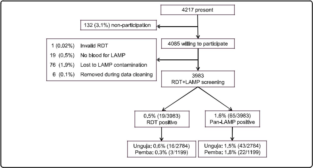
Fig.1 Flow chart of study

Prevalence of malaria by RDT and LAMP

The prevalence of asymptomatic malaria was 0.5 % (CI 95 % 0.1-0.8) and 1.6 % (CI 95 % 1.1-2.2) by RDT and Pan-LAMP, respectively (Table 1). Pan-LAMP detected 3.4 (CI 95 % 2.2-5.2) times more Plasmodium positive samples than RDT. Out of the Pan-LAMP positive sam-ples 64.6 % (42/65) were also positive by P. falciparum-LAMP. RDT had a sensitivity of 24.6 % (14.7-36.9) and specificity of 99.9 % (99.7-100.0) when compared to Pan-LAMP. Comparison by McNemar’s test showed a signifi-cant difference between the two methods (p <0.001).

Table 1. Prevalence of malaria detected by RDT and LAMP

RDT LAMP
Overall prevalence (%; CI 95 %a) 0.5; 0.1-0.8
19/3983
1.6; 1.1-2.2
65/3983
Relative proportion positive in:
Only Panb (%; CI 95 %) 5.3; 0.0-16.4
1/19
35.4; 23.4-47.4
23/65
Pan + P. falciparumc (%; CI 95 %) 31.6; 8.5-54.6
6/19
64.6; 52.6-76.6
42/65
Only P. falciparumd (%; CI 95 %) 63.2; 39.2-87.1
12/19
NDe

Both RDT brands used for malaria screening are two-band RDTs detecting P. falciparum HRP2 and Pan-Plasmodium LDH simultaneously, although with different detection limits (50–100 parasites/μL for P. falciparum HRP2 and 200–500 parasites/μL for Pan-Plasmodium LDH). In contrast, only the Pan-LAMP positive samples were assessed for P. falciparum during the LAMP screening, with a detection limit of 2–5 parasites/μL for both Pan-Plasmodium
and P. falciparum

aConfidence intervals for prevalences were calculated using the survey [svy] command in Stata, accounting for household and village sampling/stratification
bPositive for Plasmodium genus only
cPositive for Plasmodium and P. falciparum
dPositive for P. falciparum only
eND = not determined

Discrepancies in LAMP after freezing of samples

DNA extracted from half of the samples (N = 1414) in Unguja was stored at −20 °C prior to LAMP testing due to a delay in the shipment of LAMP kits. Among these samples, 32 (2.3 %) were positive by Pan-LAMP, out of which 12 was also positive by RDT. However, amongst the frozen samples there were also three RDT positive samples that were found negative by Pan-LAMP. These three samples were positive for P. falciparum HRP2 only, Pan-Plasmodium LDH only, and both P. falcip-arum HRP2 and Pan-Plasmodium LDH, respectively. Among the samples from Unguja that were screened be-fore freezing (N = 1370), 11 (0.8 %) were positive by Pan-LAMP, out of which one was also positive by RDT. The 11 Pan-LAMP positive samples were stored at −20 °C, as a quality control of freezing DNA, however only 7/11 (63.6 %) were positive when re-tested after thawing.

LAMP-amplified DNA contamination

During the study DNA contamination of LAMP arose in the central laboratory in Pemba [see Additional file 1 for flow chart of events]. The contamination resulted from using a heat block with a heated pressurized lid during the 95 °C enzyme inactivation stage, and not allowing the samples to cool to room temperature before removing the strips for recording of results. ‘Fizzing’ was observed around the lid of the LAMP strips resulting in leakage of LAMP-amplified DNA. All equipment and reagents were subjected to repetitive decontamination with 5 % sodium hypochlorite over three days, and moved away from the epicentre of the contamination to a laboratory space avail-able in another building. The final enzyme inactivation step of the protocol [10] was removed as this was thought to be the source of contamination; instead results were read and recorded immediately after the amplification re-action. A negative control was included in each strip of eight reaction tubes, and any Pan-LAMP positive samples were repeated and only recorded as positive if positive in both runs. During the first few days following the contam-ination there were some samples that were considered false positive, but the numbers declined and reached zero within one week after the contamination.

Discussion

This is the hitherto largest reported implementation of LAMP for detection of asymptomatic malaria in a field setting. In order to scale-up the breadth of sampling, LAMP testing was centralized in two laboratories, meaning samples could be collected from all over the islands with fewer resources. The time-to-result was ap-proximately 24 h, compared with three hours in the pilot study where LAMP was used as a health facility-based, point-of-care, diagnostic tool for mass screening and treatment [9].

The results confirm the improved sensitivity of LAMP over RDT, as has been shown previously [3, 9]. The MALARIA Pan/Pf Detection Kit has a detection limit of 2–5 parasites/μL [3, 6], for both Pan-Plasmodium and P. falciparum. This is comparable to PCR, and substantially lower than the detection limits of P. falciparum-specific HRP2 (50–100 parasites/μL) and Pan-Plasmodium LDH (200–500 parasites/μL) in combo RDTs. The proportion of samples detected only by Pan-LAMP (35.4 %) sug-gests the presence of species other than P. falciparum. Similarly, other studies in Zanzibar have shown that up to 40 % of PCR-detectable malaria infections contained non-falciparum species [11, 12]. Non-falciparum infec-tions tend to be of lower parasite densities than P. falcip-arum infections [13], emphasizing the need for more sensitive species-specific methods for non-falciparum Plasmodium detection. The sensitivity (83.8 %) and speci-ficity (99.7 %) of Pan-LAMP, calculated using PCR as the reference standard, was high in the pilot study conducted in Zanzibar [9]. This is similar to previously reported sen-sitivities and specificities [6–8, 14, 15] and, together with the results of this study, suggests malaria LAMP is a use-ful molecular tool sensitive enough for detection of low-density asymptomatic malaria infections in field settings.

Importantly, some discrepancies were shown amongst samples screened following freezing of diluted DNA. RDT false positivity due to recently cleared infections has been well documented when detecting P. falciparum HRP2 [16], although none of the three study participants who were RDT positive/LAMP negative reported receiv-ing malaria treatment within the previous two weeks, and two of the RDTs were positive for Pan-Plasmodium LDH suggesting ongoing infections. The lack of repro-ducibility of results following freezing of samples sug-gests that DNA extracted by simple methods such as boil and spin may not be suitable for long-term storage and should be amplified by LAMP within a short period of time [17]. Alternatively, low reproducibility of PCR for detection of low-density infections has been reported [18] and parasite densities close to the LAMP detection limit could also explain the lack of reproducibility.

The potential risk of contamination with LAMP is large, due to the high efficiency of the reaction, although the risk is reduced when using a closed system [3, 19]. The MAL-ARIA Pan/Pf Detection Kit is manufactured with tubes that cannot be re-opened once closed, in order to avoid contamination with amplified DNA. However, as demon-strated in this study, the exposure of such tubes to high temperatures, as during enzyme inactivation, results in softening of the plastic and leakage of the contents. While removing the inactivation step solved this problem in this case, contaminations have been experienced in other research settings [8, 20] and these issues are important to report. Although MALARIA Pan/Pf Detection Kit is a field-friendly option, three days’ training is not sufficient for dealing with such events. Successful decontamination requires a larger understanding of molecular techniques and rigorous repetitive methods to ensure that the area is free of contamination.

Standard malaria diagnostic tools including microscopy and RDT are not sensitive enough to detect low-density asymptomatic infections [12]. Nucleic acid amplification-based methods provide the, to date, most sensitive and ac-curate tools to detect and identify pathogens [21]. Recently published, highly sensitive quantitative PCR methods state detection limits as low as 0.02 and 0.03 parasites/μL blood [22, 23]. However, these methods lack the field applicability that LAMP offers. Furthermore, the cost of LAMP is estimated to be a tenth of that of conven-tional PCR [15], although the cost of the field friendly kit is still at 5.3 US$ per reaction i.e., considerably more expensive than RDTs [3].

The high cost and risk of contamination may yet limit the implementation LAMP at a point-of-care level, but LAMP will be valuable for research purposes and for evaluating malaria elimination efforts. LAMP may, for ex-ample, be useful in mass/focal screening and treatment (MSAT/FSAT) programmes, for which the deployment of RDTs, perhaps due to their low sensitivity, has had varying results [11, 24]. In any case it will be important to evaluate the impact and cost effectiveness of deploying LAMP, in comparison to the deployment of standard diagnostic tools as well as in comparison to alternative molecular methods.

Conclusions

LAMP is a simple and sensitive molecular tool, and has potential in active surveillance and mass-screening pro-grammes for detection of low-density asymptomatic mal-aria in pre-elimination settings. However, in order to deploy LAMP more effectively in field settings, protocols may need to be adapted for processing larger numbers of samples. A higher throughput, affordable closed system would be ideal to avoid contamination.

Competing interests

IG is an employee of the Foundation for Innovative New Diagnostics (FIND), a co-developer of the Loopamp™ MALARIA Pan/Pf Detection Kit. All other authors declare no competing interest.

Authors’ contributions

MIM, IJG, AM, ASA, AB, and JC conceived and designed the study. UM, MK, BAS, AKA, and JC carried out the training and were responsible for the fieldwork in Zanzibar. UM, BAS and MHN were responsible for the training and conducting of LAMP. UM and JC analysed the data, and drafted the manuscript. All authors read and approved the final manuscript.

Acknowledgments

We would like to thank all participants, staff members, and ZAMEP employees involved in the KAPB survey for their dedicated participation. We would also like to acknowledge Colin Sutherland, Spencer Polly, Michelle Hsiang and Alanna Schwartz for their intellectual input regarding experience of dealing with DNA contamination. This work was supported by Global Fund [Grant number ZAN-809-G07–M, work plan GFRD 8 phase 2 2013]; President’s Malaria Initiative (PMI) [Implementation letter # 45, work plan for FY 2013]; the Swedish Medical Research Council (VR) [grant numbers 2009–3785 and 2013–6594]; the Foundation for Innovative New Diagnostics (FIND) with funds from the German Federal Ministry of Education and Research (BMBF) through the KfW Entwicklungsbank; and the Einhorn foundation. In memoriam of Ali K Abass, a much missed friend and colleague.

References

  • Bousema T, Okell L, Felger I, Drakeley C. Asymptomatic malaria infections: detectability, transmissibility and public health relevance. Nat Rev Microbiol. 2014;12:833–40.
  • Hänscheid T1, Grobusch MP (2002) How useful is PCR in the diagnosis of malaria? Trends Parasitol 18: 395-398. [crossref]
  • Han ET1 (2013) Loop-mediated isothermal amplification test for the molecular diagnosis of malaria. Expert Rev Mol Diagn 13: 205-218. [crossref]
  • Oriero EC, Jacobs J, Van Geertruyden JP, Nwakanma D, D’Alessandro U. Molecular-based isothermal tests for field diagnosis of malaria and their potential contribution to malaria elimination. J Antimicrob Chemother. 2014;70:2–13.
  • Notomi T, Okayama H, Masubuchi H, Yonekawa T, Watanabe K, Amino N, et al. Loop-mediated isothermal amplification of DNA. Nucleic Acids Res. 2000;28:E63.
  • Aydin-Schmidt B, Xu W, Gonzalez IJ, Polley SD, Bell D, Shakely D, et al. Loop mediated isothermal amplification (LAMP) accurately detects malaria dna from filter paper blood samples of low density parasitaemias. PLoS One. 2014;9:e103905.
  • Hopkins H, Gonzalez IJ, Polley SD, Angutoko P, Ategeka J, Asiimwe C, et al. Highly sensitive detection of malaria parasitemia in a malaria-endemic setting: performance of a new loop-mediated isothermal amplification kit in a remote clinic in Uganda. J Infect Dis. 2013;208:645–52.
  • Polley SD1, González IJ, Mohamed D, Daly R, Bowers K, et al. (2013) Clinical evaluation of a loop-mediated amplification kit for diagnosis of imported malaria. J Infect Dis 208: 637-644. [crossref]
  • Cook J, Schmidt B, Gonzalez IJ, Bell D, Edlund E, Nassor MH, et al. Loop-mediated isothermal amplification (LAMP) for point-of-care detection of asymptomatic low-density malaria parasite carriers in Zanzibar. Malar J. 2015;14:43.
  • FIND: Manual of standard operating procedures for malaria LAMP [http://www.finddiagnostics.org/export/sites/default/programs/malaria-afs/ docs/SOPs_LAMP_Malaria_AUG12.pdf]
  • Cook J, Xu W, Msellem M, Vonk M, Bergstrom B, Gosling R, et al. Mass screening and treatment using a Plasmodium falciparum-specific rapid diagnostic test did not reduce malaria incidence in Zanzibar. J Infect Dis. 2014;211:1476–83.
  • Morris U, Xu W, Msellem MI, Schwartz A, Abass A, Shakely D, et al. Characterising temporal trends in asymptomatic Plasmodium infections and transporter polymorphisms during transition from high to low transmission in Zanzibar, 2005–2013. Infect Genet Evol. 2015. doi:10.1016/j.meegid.2015.04.018.
  • Cotter C, Sturrock HJ, Hsiang MS, Liu J, Phillips AA, Hwang J, et al. The changing epidemiology of malaria elimination: new strategies for new challenges. Lancet. 2013;382:900–11.
  • Polley SD, Mori Y, Watson J, Perkins MD, Gonzalez IJ, Notomi T, et al. Mitochondrial DNA targets increase sensitivity of malaria detection using loop-mediated isothermal amplification. J Clin Microbiol. 2010;48:2866–71.
  • Poon LL, Wong BW, Ma EH, Chan KH, Chow LM, Abeyewickreme W, et al. Sensitive and inexpensive molecular test for falciparum malaria: detecting Plasmodium falciparum DNA directly from heat-treated blood by loop-mediated isothermal amplification. Clin Chem. 2006;52:303–6.
  • Mayxay M, Pukrittayakamee S, Chotivanich K, Looareesuwan S, White NJ. Persistence of Plasmodium falciparum HRP-2 in successfully treated acute falciparum malaria. Trans R Soc Trop Med Hyg. 2001;95:179–82.
  • Sattabongkot J, Tsuboi T, Han ET, Bantuchai S, Buates S. Loop-mediated isothermal amplification assay for rapid diagnosis of malaria infections in an area of endemicity in Thailand. J Clin Microbiol. 2014;52:1471–7.
  • Costa DC, Madureira AP, Amaral LC, Sanchez BA, Gomes LT, Fontes CJ, et al. Submicroscopic malaria parasite carriage: how reproducible are polymerase chain reaction-based methods? Mem Inst Oswaldo Cruz. 2014;109:21–8.
  • Hsiang MS, Greenhouse B, Rosenthal PJ. Point of care testing for malaria using LAMP, loop mediated isothermal amplification. J Infect Dis. 2014;210:1167–9.
  • Paris DH1, Imwong M, Faiz AM, Hasan M, Yunus EB, et al. (2007) Loop-mediated isothermal PCR (LAMP) for the diagnosis of falciparum malaria. Am J Trop Med Hyg 77: 972-976. [crossref]
  • Abdul-Ghani R, Al-Mekhlafi AM, Karanis P. Loop-mediated isothermal amplification (LAMP) for malarial parasites of humans: would it come to clinical reality as a point-of-care test? Acta Trop. 2012;122:233–40.
  • Hofmann N, Mwingira F, Shekalaghe S, Robinson LJ, Mueller I, Felger I. Ultra-sensitive detection of Plasmodium falciparum by amplification of multi-copy subtelomeric targets. PLoS Med. 2015;12:e1001788.
  • Imwong M, Hanchana S, Malleret B, Renia L, Day NP, Dondorp A, et al. High throughput ultra-sensitive molecular techniques to quantify low density malaria parasitaemias. J Clin Microbiol. 2014;52:3303–9.
  • Sutcliffe CG, Kobayashi T, Hamapumbu H, Shields T, Mharakurwa S, Thuma PE, et al. Reduced risk of malaria parasitemia following household screening and treatment: a cross-sectional and longitudinal cohort study. PLoS One. 2012;7:e31396.

H Pylori Infection as Risk Factor for GIT Bleeding in Haemophilic Patients

DOI: 10.31038/IMROJ.2016126

Abstract

Background: Helicobacter pyelori is endemic in Egypt and present a main cause of gastrointestinal bleeding.

Aim: Aim of this study is to evaluate the prevalence of Hpylori infection in hemophilic patients, and to assess its impact on gastrointestinal bleeding associated with this infection in such patients.

Methods: We prospectively investigated the prevalence of H. pylori infection in 40 Egyptian patients with hemophilia A, B and von Willebrand syndrome and 20 normal male subjects were included. Every patient and control subject in the study were tested one time for H. pylori stool antigen by ELISA. All patients and control subjects were tested for occult blood using Guaiac-based fecal occult blood test.

Results: Twenty eight out of 40 patients (70%) are H. pylori positive ; and 12 out of 20 control (60%) are H. pylori positive. The odds ratio is ‎‎1.55, 95% CI (0.6162 to 3.9269), ‎Significance level P = 0.3497. Among 28 H.pylori positive patients, 5 patients (17.9 %) tested positive for occult blood. Among the 12 H.pylori positive subjects ‎ in the‎ control group, only one tested positive for occult blood (8.3%). Odds ratio for Occult bleeding in H pylori positive patients and control was 2.39: P= 0.4504. None of the H. pylori negative patients or control subjects had a positive occult blood disease.

Conclusion: Patients with hemophilia, H. pylori should not be considered as an important cause of GI bleeding. The recurrence of the infection and GI bleeding could be prevented with eradication of H. pylori. Screening tests for H. pylori would not be needed in patients with hemophilia in endemic areas.

Keywords

Haemophilia, Hpyelori

Introduction

In Egypt, it is estimated that 5050 people with haemophilia and , 499 with vWD are registered within the six hemophilia treatment centers (www.wfh.org annual report 2013)

More than 50% of the world’s population harbor Helicobacter pylori in their upper gastrointestinal tract. Infection is more prevalent in developing countries, and incidence is decreasing in Western countries. [1]

According to world gastroenterology organization report in 2010, H.pylori incidence in adult Egyptians was 90% (www.worldgastroenterology.org)

It has been hypothesized that the host immunologic response against H. pylori plays a main role in determining gastric mucosal injury, through the release of cytokines and the action of autoantibodies against H1/K1-adenosine triphosphatase of gastric epithelial cells. [2]

Infection with H pylori is the main etiological factor for erosive gastritis and duodenal or gastric peptic ulcers that often complicat with life-threatening bleeding in patients with coagulation disorders. [3]

The Egyptian government provides approximately 38% of healthcare costs of treating haemophilic patients (www.wfh.org annual report 2013). Most hemophilia patients are treated with plasma or cryoprecipitate, resulting in a high risk of transfusing blood-borne diseases like HIV and hepatitis B and C. Factor concentrate is available only in limited quantities, and patients must travel long distances to get it.

There is a contradictory in the previous studies between prevalence of H pyelori and risk of GIT of bleeding, we prospectively investigated the prevalence of H. pylori infection in patients with hemophilia A or B or von Willebrand syndrome and it is impact on risk of bleeding in those patients and compare our results with others’.

Patients and methods

This is a case control study. All participants are adult patients ‎visiting the hematology clinic of kasr Al Aini hospital and ‎the Egyptian society of Hemophilia. The study received research ethical committee approval from Kasr Al Aini ‎medical school and all participants gave written consents. Forty patients with bleeding disorders (mean age 24); categorized as: 30 patients with Hemophilia A, 6 patients with Hemophilia B and 4 patients with VWD. Patients who received triple therapy before or Patients with liver cirrhosis, thrombocytopenia, any platelet or vascular defects or patients with peptic ulcer were excluded. Control group included 20 subjects (mean age 27) of the same socioeconomic level and the same exclusion criteria of the patients included in the study. Each Patient and control subject in the study were tested for H. pylori stool antigen by ELISA, This technique is non-invasive, rapid, easy-to-use, and shows good performance characteristics for diagnosis of H. pylori infections, with sensitivity, specificity, Positive predictive value (PPV),negative predictive value (NPV), and efficiency were 100%, 90.0%, 96.9%, 100%, and 97.6% respectively).[4] All Patients and control subjects were tested for occult blood using guaiac-based fecal occult blood test. [5] All diet and drug precautions were applied before obtaining samples to prevent false positive results as dietary restriction of both hemoglobin and vegetable peroxidase containing substances is essential for valid screening for occult blood.[6]

Statistics

Using SPSS V.16.0 for calculation of the means and z test for comparison between the prevalence of H. pylori infection among ‎Hemophilia A, Hemophilia B and von Willibrand patients and the prevalence of H. pylori infection among the control group to investigate whether there is a ‎difference in prevalence between both groups or not.

Comparison between the odds ratio (OR) of developing occult GI bleeding (indicted by positive guaiac test) among Hemophilia A, Hemophilia B and von Willibrand patients who test positive for H. pylori and the odds ratio of developing occult GI bleeding ‎ among the control group who test positive for H. pylori to investigate whether there is a relation between the possible risk and the outcome.

Results

The patients’ group included 40 male patients (‎30‏‎ patients with Hemophilia A (75%), ‎‏6 patients with Hemophilia B (15%), and 4 patients with VWD (10%)) with age ranged from 12 to 52 years (mean age 24 years).

Twenty eight out of 40 patients (70%) are H. pylori positive; and 12 out of 20 controls (60%) are H. pylori positive. Odds ratio: 1.55 ‎) 95% Confidence Interval: ‎0.6162 to 3.9269‎; P = ‎0.3497)

Among 28 H. pylori positive patients, 5 patients tested positive for occult blood (17.9 %). all patients positive for occult blood were also positive for H. pylori stool antigen. Among the 12 subjects positive for H. pylori in the control group subjects, only one tested positive for occult blood (8.3 %), and he was also H. pylori positive. Odds ratio was 2.3913 (95% Confidence Interval: 0.2485 to 23.0104; P = 0.4504). None of the H. pylori negative patients or control subjects had a positive occult blood disease.

Table 1 and Figure 1 & Figure 2 demonstrate the prevalence of H.pylori and occult bleeding in patients and control.

Figure.1 Prevalence of H.pylori in patients and control Prevalence of occult bleeding in patients and control

Figure1.Prevalence of H.pylori in patients and control    Figure2.Prevalence of occult bleeding in patients and control

Table 1. Prevalence of H.pylori and occult bleeding in patients and control

Diagnosis Occult Blood Positive Occult blood negative
H. pylori Positive H. pylori Negative H. pylori Positive H. pylori Negative
Hemophilia A 5 0 15 10
Hemophilia B 0 0 4 2
VWD 0 0 4 0
Total 5 0 23 12

Discussion

The prevalence of H. pylori infection in patients with hemophilia A or B or von ‎Willebrand syndrome was investigated and compared to the prevalence of H. pylori infection in normal control subjects in many studies with variable results. In our study, we didn’t find significant difference between prevalence of H. pylori among hemophilia and VWD patients (70 %) n=40 and its prevalence among normal control subjects (60 %) n=20, the odds ratio is ‎‎1.55, 95% CI (0.6162 to 3.9269), z statistic 0.935, ‎Significance level P = 0.3497).The Prevalence of H. pylori revealed by this study doesn’t differ from the reported prevalence in ‎general population: 72.38% .[7] While the odd ratio of H. pylori infection is about 1.5; It is insignificantly higher for occult ‎bleeding detected by FOBT in absence of history of frank ‎bleeding; Odd ratio is 2.39, 95% Confidence Interval: 0.2485 to ‎‎23.0104, , P= 0.4504. Our case and control were matched for geographic distribution, age, socio economic status and educational level to avoid misleading results as described previously by wang.[8] Our results are also concomitant with Braden et al who found similar prevalence of Helicobacter pylori Infection in seventy patients with haemophilia and WVD and 100 age-related volunteers P value were statistically insignificant.[9] Also Eleftheriadis studied thirty-seven patients with hereditary haemorrahgic diseases and 26 control. ELISA was used to detect IgG, anti-CagA, and IgA antibodies to H. pylori in the serum and saliva. Result of this study revealed that 64.8% of the patients and 65.4% of the controls had H. pylori IgG antibodies in serum (P= 0.1 (. [10] Szczepanik also found that the prevalence of H. pylori infection in hemophilic patients in Poland was comparable to that in patients without coagulation disorders (49.3% vs 39% respectively P = 0.11).[3]

However our results were inconsistent with the study of upper gastrointestinal bleedings in patients with hereditary coagulation disorders in Northwest of Iran. The prevalence of H. pylori infection was investigated by stool antigen test, and serum serologic tests including immunoglobulin G and anti-CagA. Results Among 90 patients (81 men, nine women, mean age 31.30 ±10.72 years), 66 patients with hemophilia A, 10 patients with hemophilia B, six patients with Von Willebrand disease, five patients with platelet function disorders, and three patients with other factor deficiencies were evaluated. About 46.7% of patients in group A, and 23.3% of patients in group B were anti-CagA–positive (P = 0.02), whereas 76.7% of patients in group A and 51.7% of patients in group B had H. pylori immunoglobulin G antibodies (P= 0.02). H. pylori antigen in stool was positive in 76.7% in group A and 55% in group B (P = 0.03). [11]

Our study also found statistically insignificant increased risk of fecal occult ‎bleeding in absence of history of frank ‎bleeding in patients with hemophilia A; Odds ratio was ‎‎2.39, 95% (Confidence Interval: 0.2485 to ‎‎23.0104, z statistic: 0.755, Significance level: ‎P= 0.4504).‎

Though the prevalence of H pylori in Szczepanik et al. study were statistically insignificant however the gastrointestinal bleeding was reported in 46 patients (31.5%) with hemophilia and in two control group patients (2.0%) (P < 0.0001). Gastrointestinal bleeding was significantly more frequent in patients with hemophilia infected with H. pylori (33/46; 71.7%) than in patients with no H. pylori infection (13/46; 28.3%; P = 0.0002). Concluding that Helicobacter pylori infection is a risk factor for duodenal and gastric ulcer bleeding in hemophilia patients. [3]

Eyster et al detected antibodies against H. pylori in 14 (35%) of 40 subjects who had upper gastrointestinal bleeding event compared with 7 (17%) of 41 controls who were closely matched for age and other factors. Bleeding was substantially but not significantly increased (OR: 4.6; 95% CI: 0.3-83.9) with H. pylori seropositivity. [12]

Though Choe studied a group of haemophilic children but his results concluded significant role of H pylori in inducing GIT bleeding. Helicobacter pylori infection was found in four of six (66.7%) patients with GI bleeding (3, duodenal ulcer; 1, H. pylori associated chronic gastritis). The patients with H. pylori infection had an eradication treatment of triple therapy and no recurrence happened. [13]

According to our results screening for H. pylori should be routine work-up for all patients with hemophilia A, and all patients with positive stool antigen test should be treated immediately to prevent gastrointestinal bleeding, and to repeat testing to ensure eradication of the bacteria which may definitely lower the costs as was described by Schulman.[14]

Conclusion

Patients with hemophilia, H. pylori should not be considered as an important cause of GI bleeding. The recurrence of the infection and GI bleeding could be prevented with eradication of H. pylori. Screening tests for H. pylori would not be needed in patients with hemophilia in endemic areas.

Ethical approval

All procedures performed in studies involving human participants were in accordance with the ethical standards of the institutional and/or national research committee and with the 1964 Helsinki declaration and its later amendments or comparable ethical standards.

Informed consent was obtained from all individual participants included in the study.

Authors declare no conflict of interest. Authors declare that the results have not been published previously and are not under submission elsewhere.

References

  • Brown LM, (2000) Helicobacter pylori: epidemiology and routes of transmission.Epidemiol Rev 22: 283-297. [crossref]
  • Emilia G, Longo G, Luppi M, Gandini G, Morselli M, et al. (2001) Helicobacter pylori eradication can induce platelet recovery in idiopathic thrombocytopenic purpura. Blood 97:812-4.
  • Szczepanik AB, Zaleska M, Wiszniewski A, Wislawski S, Misiak A, et al. (2005) Helicobacter pylori infection in patients with haemophilia in Poland: prevalence and risk of upper gastrointestinal bleeding. Haemophilia 11:376-9
  • Kim PS, Lee JW, Pai SH, Kim YB, Cho JK, et al. (2002) Detection of Helicobacter pylori antigen in stool by enzyme immunoassay. Yonsei Med J 43: 7-13. [crossref]
  • Green BT, Rockey DC (2004) Gastrointestinal endoscopic evaluation of premenopausal women with iron deficiency anemia. J Clin Gastroenterol 38:104-9
  • Syed AA, Khatoon BA, Silwadi MF (2001) New reagents for detection of faecal occult blood. J Pharm Biomed Anal 24: 581-586. [crossref]
  • Mohammad MA, Hussein L, Coward A, Jackson SJ (2007) Prevalence of Helicobacter pylori infection among Egyptian children: impact of social background and effect on growth. Public Health Nutrition 11: 230–236.
  • Wang F, Meng W, Wang B, Qiao L (2014) Helicobacter pylori-induced gastric inflammation and gastric cancer. Cancer Lett 345: 196-202. [crossref]
  • Braden B, Wenke A, Karich HJ, Dietrich CF, Scharrer I, et al. (1998) Risk of gastrointestinal bleeding associated with Helicobacter pylori infection in patients with hemophilia or von Willebrand’s syndrome.Helicobacter 3: 184-187. [crossref]
  • Eleftheriadis N, Makri S, Aggouridaki C, Pithara E, Makris P (2002) Helicobacter pylori infection in upper gastrointestinal bleeding in patients with hereditary hemorrhagic disorders. Eur J Intern Med 13:480-4.
  • Dolatkhah R, Khoshbaten M, Asvadi Kermani I, Reza Bonyadi M, Ghojazadeh M, et al. (2011) Upper gastrointestinal bleedings in patients with hereditary coagulation disorders in Northwest of Iran: prevalence of Helicobacter pylori infection. Eur J Gastroenterol Hepatol 23:1172-7.
  • Eyster ME, Asaad SM, Gold BD, Cohn SE, Goedert JJ (2007) Upper gastrointestinal bleeding in haemophiliacs: incidence and relation to use of non-steroidal anti-inflammatory drugs. Haemophilia 13:279-86.
  • Choe BH, Kim JY, Lee JH, Kim JM, Chu MA, Cho SM (2010) Upper gastrointestinal bleeding in children with haemophilia: a clinical significance of Helicobacter pylori infection. Haemophilia 16:277-80
  • Schulman S, Rehnberg AS, Hein M, Hegedus O, Lindmarker P, Hellström PM (2003) Helicobacter pylori causes gastrointestinal hemorrhage in patients with congenital bleeding disorders. Thromb Haemost 89:741-6.

Surgical Reduction of Visceral versus Subcutaneous Fat and Effect on Features of the Metabolic Syndrome

DOI: 10.31038/IMROJ.2016125

Abstract

Background

Obesity results in both health and financial tolls for individuals and society. Despite the great efforts to increase awareness, the obesity epidemic continues at an alarming rate. Subcutaneous fat represents 85% of the fat mass in obese patients, while intra-abdominal fat, including both visceral and retroperitoneal adipose, represent about 15%. Both subcutaneous and visceral fat have a negative effect on individual health and promote metabolic diseases. Logic would thus suggest that adipose tissue removal by liposuction and/or visceral tissue removal could effect an improvement in metabolic diseases such as Type 2 diabetes?

Methods

The scientific literature was searched to illustrate and describe the effect of adipose tissue on metabolism and the effect of its removal on inflammatory and metabolic markers.

Results

Adipose tissue is an endocrine organ and in obesity serves as a source of systemic proinflammatory signals arising from stressed adipocytes and/or infiltrating macrophages. These signals are associated with insulin resistance, dyslipidemia and hypertension (metabolic syndrome). The benefits of fat removal on metabolic syndrome remain controversial.

Conclusion

Visceral and subcutaneous fat is associated with high risk of coronary heart disease, insulin resistance, and other metabolic risk factors. Abdominal lipectomy is a well-known cosmetic procedure and used widely for its benefits on improving body image. However, its benefit on the metabolic disorder remains inconclusive. On the other hand, current evidence would suggest that omentectomy offers no benefit in relation to improvements in systemic inflammation and metabolic control. Removal of visceral adipose tissue in and of itself would thus appear to have limited efficacy as an approach to the treatment of the obesity-related metabolic syndrome.

Key words

Visceral and subcutaneous fat, Liposuction, Omentectomy, Metabolic Syndrome

Introduction

Obesity is defined as a systemic disease that shows excessive and abnormal accumulation of body fat leading to adverse health effects. Obesity has negative effects on health and exerts significant financial tolls on individuals and society. Despite significant efforts to increase awareness, the obesity epidemic continues at an alarming rate [1]. More than 50% of the European population is overweight and up to 30% is obese with prevalence worldwide doubling since 1980 [World Health Organization 2011] [2].

Abdominal obesity is defined as increased visceral fat and trunk subcutaneous fat which leads to increased waist circumference [3]. The contribution of abdominal subcutaneous fat mass and the visceral fat mass to the pathogenesis of metabolic disease is controversial, but it is associated with high risk of coronary heart disease, insulin resistance, and other metabolic risk factors[3, 4].

Subcutaneous fat represents 85% of the fat mass in obese patients, while intra-abdominal fat, including both visceral and retroperitoneal adipose, represent about 15%[5]. Visceral obesity is presumed to predispose individuals to hepatic insulin resistance based on its anatomical site and venous drainage to the liver through the passage of adipocytes products into the portal vein[6-8].

Waist circumference [WC] and waist to hip circumference ratio [WHR] are used to measure abdominal obesity [9, 10], Body mass index [BMI] which equals the ratio of weight in kilograms divided by height in meters squared [kg/m2] is used to measure general obesity [11]. Each of these three parameters can be used to measure the association of obesity and metabolic risk factors, but the combination of the three [BMI, WC, and WHC] appears more useful than the use of BMI alone [12, 13].

Theoretically, WC cut-off results are >88 cm in women and >102 cm in men but actually, it is difficult to apply in all populations due to the marked difference in the average levels of measurement. Asians are characterized by higher morbidity at lower cut-off for WC than other populations [14].

The metabolic effects of visceral and subcutaneous adipose tissue

Although body fat mass distribution is characterized by marked individual variations it can generally be classified into the following four types, a]Abdominal subcutaneous: most of the fat stored subcutaneously around the stomach and chest, b]Lower body: fat storage around the thighs, hip, and buttocks, c] Overall coverage: fat accumulation in the arms, breast, thighs, buttocks, lower back and breast, d] Visceral: fat deposition within visceral cavity surrounding the viscera including stomach, intestine, liver and pancreas [Figure 1] [15].

Figure 1. Types of body fat mass distribution [15]

  Figure 1. Types of body fat mass distribution [15]

More specifically, upper body fat distribution and increased visceral fat is more associated with metabolic dysregulation[16-18] than lower body and abdominal subcutaneous fat accumulation [19, 20]. Metabolic disorders associated with subcutaneous and visceral obesity include insulin resistance, type 2 diabetes [21, 22], dyslipidemia [23], and hypertension [24, 25].

Adipose tissue is an endocrine organ producing proinflammatory molecules from adipocytes and/or infiltrating macrophages in patients with high BMI, WC, and WHC [26]. These include tumor necrosis factor-alpha [TNF-α], C-reactive protein [CRP],interleukin-18 [IL-18], and interleukin-6 [IL-6] [27-30]. These cytokines have been linked to impairments in insulin action in liver, muscle, and adipose tissue [31-33].

TNF-α in obesity is increased in both systemic and portal circulation [32], which affects insulin sensitivity within adipocytes and stimulates secretion of IL-6 [34] Also, IL-6 concentration is 50% higher in the portal vein than in the peripheral circulation for patients with high visceral obesity [32].

Leptin and adiponectin expression is higher in subcutaneous fat compared with visceral adipose tissue [35, 36], but cytokine expression such as IL-6, IL-8 appears to be higher in visceral adipose tissue compared with subcutaneous fat [37].

Excess visceral and subcutaneous fat mass are associated with an elevation in postprandial [38] and post-absorptive fatty acid concentration in portal vein and systemic circulations [39]. Chronic exposure of the liver to the high concentration of free fatty acids promotes liver gluconeogenesis facilitating hepatic glucose production, thus providing a continuous source of energy and substrate that tends to raise fasting glycaemia [8, 40]. Increased insulin resistance and reduced fatty acid oxidation increase fat storage and synthesis in the liver [41-43].

Adipose tissue removal surgeries

Liposuction

Liposuction is one of the most common plastic surgeries and aims to remove subcutaneous adipose tissue from different body areas so as to improve body image and create more physical balance [44]. Since its introduction by Illouz 30 years ago, its popularity has rapidly increased, making it the second most common cosmetic surgical procedure in 2012 [313,011 patients] [45]. nearly 400,000 procedures are performed annually in USA [45].

The removal of subcutaneous fat with a blunt cannula attached to a suction generating device was first popularized in Europe in the late 1970s. The procedure of liposuction has undergone many refinements and evolved with improvement in techniques and technology since its introduction by Illouz in 1982 [46-48].

a) Traditional liposuction

Liposuction was first practiced without any preparation of the fat before suctioning it from the subcutaneous tissue and this was called ‘Dry liposuction’ which was associated with a high incidence of hemorrhage and hematoma. Then Illouz developed the wet technique by injecting normal saline, water, and hyaluronidase to create a weak hypotonic solution to destroy the fat cell wall [49]. Hetter added lidocaine and dilute epinephrine to the injected solution [50], then the super wet and tumescent techniques were developed by injecting the standard wetting solution [1 ml of epinephrine and 50ml of 1% xylocaine for each 1 liter of lactated Ringer solution] [51].

The ratio of the volume of wetting solution infused to the volume of fat aspirate is 1:1 in wet technique but 2 or 3:1 in the tumescent. Although, wetting and tumescent techniques are different in this ratio, they both involve infusion of the wetting solution to the point of tissue turgor or a “peau d’orange” of the overlying skin followed by suction of the subcutaneous tissue [51].

b) Ultrasound-assisted liposuction[UAL]

Zocchi is credited for the application of ultrasonic energy in liposuction to allow more selective tissue targeting [52]. It works by having expansion and compression parts of the cycle as a sound wave,which exerting a negative pressure that overcomes molecular forces results in cellular fragmentation with the intracellular content release, which leading to interstitial cavities hence termed as cavitation.Subsequently, a low-power suction can be performed [52, 53]. In this technique, sufficient amount of wetting solution must be used to limit the effect of heat generated by Us probe. Cellular disruption has been confirmed by the homogenous, macroscopically-acellular aspirated fat which contains a high level of adipocyte-specific intracellular glycerol 3-phosphate dehydrogenase isozyme [54]. UAL is highly efficient in removal of fat in fibrous areas such as the upper back, the hypogastrium, and the breast. UAL has been shown to cause less disruption of vasculature than SAL and hence less bruising [55].

c) Power assisted liposuction[PAL]

In the late 1990s, PAL was developed to subside some of UAL side effects. Actually,it is traditional liposuction with a special reciprocating cannula [Figure 2]. It is useful for fibrous rich areas liposuction and easy for the surgeon to use. No heat generation considers its main advantage over PAL [56].

Figure 2. Power assisted liposuction cannula

Figure 2. Power assisted liposuction cannula

d) Laser-assisted liposuction [LAL]

LAL is defined as, the simultaneous use of a bare laser fiber as a free beam or a confined beam to lyse, liquefy the fat while simultaneously coagulating small blood vessels at the cannula fat interface. Harness in 1990 described the use of 1064 nm Nd-YAG laser for liposuction with promising results such as minimal incision and skin contraction produced by its photothermal effect [47]. Recently, the use of 1,320-, 1,440- and 2,100-nm wavelengths also have been proposed, with claims of less bleeding, faster healing, and better tissue tightening using laser lipolysis [57-60].

e) Radiofrequency-assisted liposuction [RFAL]

Radiofrequency-assisted liposuction [RFAL] means usage of bipolar radiofrequency energy [similar to that of diathermy] to disrupt the fat cell membrane and facilitate lipolysis with a lesser requirement for suction power [61, 62]. A controlled subdermal thermal injury produced by this energy leads to healing by contraction which produces a secondary effect on skin contraction such as LAL [61].

Omentectomy

Omentectomy [OM] defined as, surgical removal of the whole greater omentum which is a thin fold of abdominal tissue that encases the stomach, large intestine, and other abdominal organs. There are two main types of omentectomy: completely removing the omentum which is called total or supra colic omentectomy and removing a portion of the omentum which refers to partial omentectomy.

The procedure may be performed traditionally or laparoscopically, the traditional approach involves mini-laparotomy with a median supraumbilical incision of 8 to 10 cm to release the omentum from the large gastric curvature while preserving the gastroepiploic artery [63].

Laparoscopic omentectomy technique: Four trocars are used to reach the abdominal cavity at the following positions, under the umbilicus, right lower quadrant, and two at left lower quadrant [63]. After inflation of the abdominal cavity, body and fundus of the stomach are released from the greater omentum and short gastric vessels by harmonic scalpel with remaining of gastroepiploic vessels intact. After detaching the omentum from the transverse colon pulled out through the infra-umbilical incision [64].

Effect of adipose tissue removal on metabolic disease

Liposuction

A study by Giugliano et al, examined the effect of subcutaneous fat suction on insulin resistance and vascular inflammatory markers in 30 obese women by comparing the pre and postoperative [6 months] HOMA [fasting plasma glucose [mmol/l] x fasting serum insulin [mU/ml] divided by 25] as an index of peripheral insulin resistance., High HOMA scores denote low insulin sensitivity. Levels of IL-6, IL-8, TNF-α, CRP and adiponectin were also assessed. Results demonstrated that liposuction was associated with significant decrease in all parameters, except adiponectin, which significantly increased [Table 1] [65]. The risk of cardiovascular death and the incidence of insulin resistance was reduced at follow-up in association with improvements in metabolic and inflammatory markers.

Table 1. Results of Giugliano et al, study [65].

Baseline Six months postoperative P-value
Weight 88 85 <0.001
HOMA 4.1 3.08 <0.05
IL-6 pg/ml 4.1 3.2 <0.05
IL-18 pg/ml 246 219 <0.05
TNF-a pg/ml 5.1 4.1 <0.02
CRP ml/l 2.9 2.4 <0.02
Adiponectin ug/ml 5.1 6.4 <0.02

In a study by Ramos et al, patients underwent abdominoplasty from October 2010 to September 2011. Total cholesterol, high-density lipoprotein [HDL], low-density lipoprotein [LDL], very low-density lipoprotein [VLDL], triglycerides, glucose, insulin, and HOMA index were measured preoperatively and 3 months post-operative. The results showed a significant reduction in triglyceride and LDL and a non-significant trend for improvements in HOMA, cholesterol,glucose, insulin and HDL[Table 2] [66]. The findings of this study were more or less similar to the Swanson study, which included 322 patients and demonstrated a significant reduction in triglycerides only [67].

Table 2. Results of Ramos et al., study [66].

Baseline Three months postoperative P-value
Weight kg 69.1 68.6 0.79
Glucose mg/dl 91.45 90.71 0.81
Insulin Ul/ml 17.11 11.79 0.28
HOMA 3.96 2.58 0.22
Cholesterol mg/dl 224 220 0.84
Triglyceride mg/dl 193 133 0.03
HDL mg/dl 44 49 0.18
VLDL mg/dl 43 39.1 0.55
LDL mg/dl 137 79.61 0.04

In the cohort study of Marfella et al, 20 patients underwent abdominoplasty and 28 patients were exposed to diet, exercise, and behavioral counseling. Authors compared preoperative and 2-month postoperative measurements of triglycerides, insulin, insulin sensitivity, IL-6, TNF-α, and HDL-cholesterol. A significant reduction in triglycerides and insulin and improved insulin sensitivity was observed in both groups, with significant decreases in IL-6 and TNF-α occurring only in abdominoplasty group. There were no changes in HDL-cholesterol during the study period [68]. In a prospective cohort study, 12 obese patients that underwent abdominoplasty were followed up and plasma triglycerides, HDL, cholesterol, and insulin sensitivity measured during the preoperative period and at 50 days postoperative [69]. In difference to other studies in the field, no improvements were observed in any of the parameters studied.

An overall survey of the literature suggests that on balance, besides the cosmetic results of liposuction, a positive impact on lipid profile and other metabolic markers can arise as a secondary benefit of the procedure. However diet, lifestyle, and exercise modification subsequent to liposuction are likely to significantly enhance the magnitude and duration of beneficial effects by preventing new fat deposition [66]. Whether liposuction can be advocated primarily for its metabolic effects remains however rather controversial and there is still an insufficient evidence base to support its use in this context.

Omentectomy

Fabbrini et al, conducted a randomized controlled trial to test the hypothesis of the effect of visceral fat removal on metabolic diseases. Eleven patients underwent laparoscopic combined Roux-en-y [RYGB] with omentectomy, but RYGB alone was performed to the same number. Leptin, blood glucose, HbA1c, insulin, CRP, cholesterol, LDL, HDL, and triglyceride were measured preoperatively, 6 months and 12 months postoperative. Results of comparing pre and postoperative investigations demonstrated that hepatic insulin sensitivity increased 4-fold and skeletal muscle insulin sensitivity approximately doubled at 12 months after surgery in both groups, Critically no significant augmentation of improvements in these parameters was observed in patients undergoing omentectomy. So, metabolic variables improved after RYGB surgery but visceral fat removal has no additive effect on these variables . [70]. However, these data did not preclude the possibility that stand-alone omentectomy could be of metabolic benefit. To examine this, the same trial incorporated a treatment arim in which 10 patients underwent laparoscopic omentectomy alone. Results demonstrated that metabolic variables and minimal model-derived indices of insulin sensitivity, glucose effectiveness, and Beta-cell function did not change significantly 3 months after omentectomy compared with baseline [70].

Lima et al, and Herrera et al, are two prospective randomized trials looking at omentectomy[71, 72]. Both studies randomized patients equally into two groups, patients of the first group underwent RYGB with omentectomy but RYGB alone was performed to patients of the other group. Both studies used blood samples to measure metabolic and inflammatory markers for patients as in the study of Fabbrini and drew the same conclusions, i.e. omentectomy did not confer any additional benefit on top of RYGB.

From the previous studies and trials, it is thus clear that omentectomy does not induce any improvement in the components of metabolic syndrome and inflammatory mediators. Given the constitutive protective function of the omentum in the abdomen, there appears to be no basis for pursuing omentectomy as a viable intervention for metabolic disease.

Conclusion

Theoretically, increased visceral and subcutaneous adiposity are major risk factors for insulin resistance, dyslipidemia, and other metabolic disorders. Abdominal lipectomy is a well-known cosmetic procedure and used widely for its benefits on improving body image. However, its benefit on the metabolic disorder remains controversial. On the other hand, Omentectomy also does not induce any improvement in the components of inflammatory mediators but its effect on metabolic diseases considered an arguable issue. The currently available literature on visceral fat removal and liposuction are characterized by a small number of patients, we need more powerful randomly controlled trials to provide additional evidence. Intriguingly the fact that removal of subcutaneous rather than visceral fat, [at least the omentum] confers metabolic benefits, challenges current orthodoxy regarding the involvement of the two major fat depots in metabolic syndrome.

References

  • Mokdad AH, Ford ES, Bowman BA, Dietz WH, Vinicor F, et al. (2003) Prevalence of obesity, diabetes, and obesity-related health risk factors, 2001.JAMA 289: 76-79. [crossref]
  • Lee WJ, Yu PJ, Wang W, Chen TC, Wei PL, et al. (2005) Laparoscopic Roux-en-Y versus mini-gastric bypass for the treatment of morbid obesity: a prospective randomized controlled clinical trial.Ann Surg 242: 20-28. [crossref]
  • Kissebah AH, Vydelingum N, Murray R, Evans DJ, Hartz AJ, et al. (1982) Relation of body fat distribution to metabolic complications of obesity.J Clin Endocrinol Metab 54: 254-260. [crossref]
  • Abate N, Garg A, Peshock RM, Stray-Gundersen J, Grundy SM (1995) Relationships of generalized and regional adiposity to insulin sensitivity in men. J Clin Invest 96: 88-98. [crossref]
  • Klein S, Allison DB, Heymsfield SB, Kelley DE, et al. (2007) Waist Circumference and Cardiometabolic Risk: a Consensus Statement from Shaping America’s Health: Association for Weight Management and Obesity Prevention; NAASO, the Obesity Society; the American Society for Nutrition; and the American Diabetes Association. Obesity 15:1061-7.
  • Hotamisligil GS, Peraldi P, Budavari A, Ellis R, et al. (1996) IRS-1-mediated inhibition of insulin receptor tyrosine kinase activity in TNF-alpha- and obesity-induced insulin resistance. Science 271:665-8.
  • Korec R (2001) –to: Bergman RN (2000) non-esterified fatty acids and the liver: why is insulin secreted into the portal vein? Diabetologia 43: 946-952. Diabetologia 44: 784. [crossref]
  • Williamson JR, Kreisberg RA, Felts PW (1966) Mechanism for the stimulation of gluconeogenesis by fatty acids in perfused rat liver.Proc Natl Acad Sci U S A 56: 247-254. [crossref]
  • Ardern CI, Katzmarzyk PT, Janssen I, Ross R (2003) Discrimination of health risk by combined body mass index and waist circumference. Obes Res 11: 135-142. [crossref]
  • Meisinger C, Döring A, Thorand B, Heier M, Löwel H (2006) Body fat distribution and risk of type 2 diabetes in the general population: are there differences between men and women? The MONICA/KORA Augsburg cohort study. Am J Clin Nutr 84: 483-489. [crossref]
  • [No authors listed] (2000) Obesity: preventing and managing the global epidemic. Report of a WHO consultation. World Health Organ Tech Rep Ser 894: i-xii, 1-253. [crossref]
  • Wang Y, Rimm EB, Stampfer MJ, Willett WC, Hu FB (2005) Comparison of abdominal adiposity and overall obesity in predicting risk of type 2 diabetes among men.Am J Clin Nutr 81: 555-563. [crossref]
  • Després JP, Lemieux I (2006) Abdominal obesity and metabolic syndrome.Nature 444: 881-887. [crossref]
  • Misra A, Vikram NK, Gupta R, Pandey RM, et al. (2006) Waist circumference cutoff points and action levels for Asian Indians for identification of abdominal obesity. Int J Obes (Lond) 30:106-11.
  • Foster MT, Pagliassotti MJ (2012) Metabolic alterations following visceral fat removal and expansion: Beyond anatomic location. Adipocyte 1:192-9.
  • Björntorp P (1991) Metabolic implications of body fat distribution. Diabetes Care 14: 1132-1143. [crossref]
  • Kissebah AH, Krakower GR (1994) Regional adiposity and morbidity. Physiol Rev 74: 761-811. [crossref]
  • Bjorntorp P (1990) “Portal” adipose tissue as a generator of risk factors for cardiovascular disease and diabetes. Arteriosclerosis 10:493-6.
  • Cefalu WT, Wang ZQ, Werbel S, Bell-Farrow A, Crouse JR 3rd, et al. (1995) Contribution of visceral fat mass to the insulin resistance of aging. Metabolism 44: 954-959. [crossref]
  • Kuk JL, Katzmarzyk PT, Nichaman MZ, Church TS, Blair SN, et al. (2006) Visceral fat is an independent predictor of all-cause mortality in men. Obesity (Silver Spring) 14: 336-341. [crossref]
  • Carey VJ, Walters EE, Colditz GA, Solomon CG, Willett WC, et al. (1997) Body fat distribution and risk of non-insulin-dependent diabetes mellitus in women. The Nurses’ Health Study.Am J Epidemiol 145: 614-619. [crossref]
  • Chan JM, Rimm EB, Colditz GA, Stampfer MJ, Willett WC (1994) Obesity, fat distribution, and weight gain as risk factors for clinical diabetes in men.Diabetes Care 17: 961-969. [crossref]
  • Kissebah AH, Alfarsi S, Adams PW, Wynn V. Role of insulin resistance in adipose tissue and liver in the pathogenesis of endogenous hypertriglyceridaemia in man. Diabetologia 12:563-71.
  • Cassano PA, Segal MR, Vokonas PS, Weiss ST (1990) Body fat distribution, blood pressure, and hypertension. A prospective cohort study of men in the normative aging study.Ann Epidemiol 1: 33-48. [crossref]
  • Seidell JC, Cigolini M, Deslypere JP, Charzewska J, Ellsinger BM, et al. (1991) Body fat distribution in relation to serum lipids and blood pressure in 38-year-old European men: the European fat distribution study. Atherosclerosis 86: 251-260. [crossref]
  • Weisberg SP, McCann D, Desai M, Rosenbaum M, Leibel RL, et al. (2003) Obesity is associated with macrophage accumulation in adipose tissue.J Clin Invest 112: 1796-1808. [crossref]
  • Bastard JP, Jardel C, Bruckert E, Blondy P, et al. (2007) Elevated levels of interleukin 6 are reduced in serum and subcutaneous adipose tissue of obese women after weight loss. J Clin Endocrinol Metab 85:3338-42.
  • Ziccardi P, Nappo F, Giugliano G, Esposito K, et al. (2002) Reduction of inflammatory cytokine concentrations and improvement of endothelial functions in obese women after weight loss over one year. Circulation 105:804-9.
  • Esposito K, Pontillo A, Ciotola M, Di Palo C, Grella E, et al. (2002) Weight loss reduces interleukin-18 levels in obese women. J Clin Endocrinol Metab 87: 3864-3866. [crossref]
  • Yudkin JS, Stehouwer CD, Emeis JJ, Coppack SW (1999) C-reactive protein in healthy subjects: associations with obesity, insulin resistance, and endothelial dysfunction: a potential role for cytokines originating from adipose tissue? Arterioscler Thromb Vasc Biol 19:972-8.
  • Fernandez-Veledo S, Nieto-Vazquez I, Vila-Bedmar R, Garcia-Guerra L, et al. (2009) Molecular mechanisms involved in obesity-associated insulin resistance: therapeutical approach. Arch Physiol Biochem 115:227-39.
  • Fontana L, Eagon JC, Trujillo ME, Scherer PE, Klein S (2007) Visceral fat adipokine secretion is associated with systemic inflammation in obese humans. Diabetes 56: 1010-1013. [crossref]
  • Hoene M, Weigert C (2008) The role of interleukin-6 in insulin resistance, body fat distribution and energy balance. Obes Rev 9: 20-29. [crossref]
  • Sethi JK, Hotamisligil GS (1999) The role of TNF alpha in adipocyte metabolism. Semin Cell Dev Biol 10: 19-29. [crossref]
  • Fain JN, Madan AK, Hiler ML, Cheema P, et al. (2004) Comparison of the release of adipokines by adipose tissue, adipose tissue matrix, and adipocytes from visceral and subcutaneous abdominal adipose tissues of obese humans. Endocrinology 145:2273-82.
  • Van Harmelen V, Reynisdottir S, Eriksson P, Thörne A, Hoffstedt J, et al. (1998) Leptin secretion from subcutaneous and visceral adipose tissue in women. Diabetes 47: 913-917. [crossref]
  • Mirza MS (2011) Obesity, Visceral Fat, and NAFLD: Querying the Role of Adipokines in the Progression of Nonalcoholic Fatty Liver Disease. ISRN Gastroenterol 2011: 592404. [crossref]
  • Roust LR, Jensen MD (1993) Postprandial free fatty acid kinetics are abnormal in upper body obesity.Diabetes 42: 1567-1573. [crossref]
  • Jensen MD, Haymond MW, Rizza RA, Cryer PE, Miles JM (1989) Influence of body fat distribution on free fatty acid metabolism in obesity.J Clin Invest 83: 1168-1173. [crossref]
  • Williamson JR (1966) Mechanism for the stimulation in vivo of hepatic gluconeogenesis by glucagon.Biochem J 101: 11C-14C. [crossref]
  • Xu J, Nakamura MT, Cho HP, Clarke SD (1999) Sterol regulatory element binding protein-1 expression is suppressed by dietary polyunsaturated fatty acids. A mechanism for the coordinate suppression of lipogenic genes by polyunsaturated fats. J Biol Chem 274:23577-83.
  • Insulin resistance induced by chronic high-fat feeding. Diabetes 46: 1768-1774. [crossref]
  • Boden G1 (1997) Role of fatty acids in the pathogenesis of insulin resistance and NIDDM.Diabetes 46: 3-10. [crossref]
  • Shridharani SM, Broyles JM2, Matarasso A1 (2014) Liposuction devices: technology update.Med Devices (Auckl) 7: 241-251. [crossref]
  • No authors listed] (2013) Cosmetic surgery National Data Bank: statistics 2012.Aesthet Surg J 33: 1S-21S. [crossref]
  • Lewis CM. Early history of lipoplasty in the United States. Aesthetic Plast Surg. 1990;14(2):123-6.
  • Apfelberg DB, Rosenthal S, Hunstad JP, Achauer B, Fodor PB (1994) Progress report on multicenter study of laser-assisted liposuction.Aesthetic Plast Surg 18: 259-264. [crossref]
  • Jewell ML, Fodor PB, de Souza Pinto EB, Al Shammari MA. Clinical application of VASER–assisted lipoplasty: a pilot clinical study. Aesthet Surg J 22:131-46.
  • Dolsky RL (1984) Body sculpturing by lipo-suction extraction. Aesthetic Plast Surg 8: 75-83. [crossref]
  • Hetter GP (1984) The effect of low-dose epinephrine on the hematocrit drop following lipolysis. Aesthetic Plast Surg 8: 19-21. [crossref]
  • Gingrass MK (2000) Ultrasound-Assisted Lipectomy Using the Solid Probe: A Retrospective Review of 100 Consecutive Cases. Plast Reconstr Surg 105:2175-7.
  • Zocchi M (1992) Ultrasonic liposculpturing. Aesthetic Plast Surg 16: 287-298. [crossref]
  • Zocchi ML (1996) Ultrasonic assisted lipoplasty. Technical refinements and clinical evaluations. Clin Plast Surg 23:575-98.
  • Rohrich RJ, Morales DE, Krueger JE, Ansari M, et al. (2000) Comparative lipoplasty analysis of in vivo-treated adipose tissue. Plast Reconstr Surg 105:2152-8; discussion 9-60.
  • Kenkel JM, Robinson JB Jr, Beran SJ, Tan J, Howard BK, et al. (1998) The tissue effects of ultrasound-assisted lipoplasty. Plast Reconstr Surg 102: 213-220. [crossref]
  • Ablaza VJ, Gingrass MK, Perry LC, Fisher J, et al. (1998) Tissue temperatures during ultrasound-assisted lipoplasty. Plast Reconstr Surg 102:534-42.
  • Mordon S, Blanchemaison P (2008) “Histologic evaluation of interstitial lipolysis comparing a 1064, 1320 and 2100 nm laser in an ex vivo model” Lasers Surg Med 40:519.
  • DiBernardo BE, Reyes J, Chen B (2009) Evaluation of tissue thermal effects from 1064/1320-nm laser-assisted lipolysis and its clinical implications. J Cosmet Laser Ther 11:62-9.
  • Wassmer B, Zemmouri J, Rochon P, Mordon S (2010) Comparative study of wavelengths for laser lipolysis.Photomed Laser Surg 28: 185-188. [crossref]
  • Katz B, McBean J (2008) Laser-assisted lipolysis: a report on complications. J Cosmet Laser Ther 10: 231-233. [a href=”http://www.ncbi.nlm.nih.gov/pubmed/19016103″>crossref]
  • Paul M, Mulholland RS (2009) A new approach for adipose tissue treatment and body contouring using radiofrequency-assisted liposuction. Aesthetic Plast Surg 33:687-94.
  • Blugerman G, Schavelzon D, Paul MD (2010) A safety and feasibility study of a novel radiofrequency-assisted liposuction technique. Plast Reconstr Surg 125:998-1006.
  • Milleo FQ, Campos AC, Santoro S, Lacombe A, et al. (2011) Metabolic effects of an entero-omentectomy in mildly obese type 2 diabetes mellitus patients after three years. Clinics (Sao Paulo) 66:1227-33.
  • Santoro S, Velhote MC, Malzoni CE, Milleo FQ, et al. (2006) Preliminary results from digestive adaptation: a new surgical proposal for treating obesity, based on physiology and evolution. Sao Paulo Med J 124:192-7.
  • Giugliano G, Nicoletti G, Grella E, Giugliano F, et al. (2004) Effect of liposuction on insulin resistance and vascular inflammatory markers in obese women. Br J Plast Surg 57:190-4.
  • Ramos-Gallardo G, Pérez Verdin A, Fuentes M, Godínez Gutiérrez S, Ambriz-Plascencia AR, et al. (2013) Effect of abdominoplasty in the lipid profile of patients with dyslipidemia.Plast Surg Int 2013: 861348. [crossref]
  • Swanson E (2011) Prospective clinical study reveals significant reduction in triglyceride level and white blood cell count after liposuction and abdominoplasty and no change in cholesterol levels. Plast Reconstr Surg 128:182e-97e.
  • Marfella R, Grella R, Rizzo MR, Barbieri M, et al. (2011) Role of subcutaneous abdominal fat on cardiac function and proinflammatory cytokines in premenopausal obese women. Ann Plast Surg 63:490-5.
  • Martinez-Abundis E, Molina-Villa CA, Gonzalez-Ortiz M, Robles-Cervantes JA, et al. (2007) Effect of surgically removing subcutaneous fat by abdominoplasty on leptin concentrations and insulin sensitivity. Ann Plast Surg 58:416-9.
  • Fabbrini E, Tamboli RA, Magkos F, Marks-Shulman PA, et al. (2010) Surgical removal of omental fat does not improve insulin sensitivity and cardiovascular risk factors in obese adults. Gastroenterology 139:448-55.
  • Herrera MF, Pantoja JP, Velazquez-Fernandez D, Cabiedes J, et al. (2010) Potential additional effect of omentectomy on metabolic syndrome, acute-phase reactants, and inflammatory mediators in grade III obese patients undergoing laparoscopic Roux-en-Y gastric bypass: a randomized trial. Diabetes Care 33:1413-8.
  • Lima MM, Pareja JC, Alegre SM, Geloneze SR, et al. (2013) Visceral fat resection in humans: effect on insulin sensitivity, beta-cell function, adipokines, and inflammatory markers. Obesity (Silver Spring) 21:E182-9.

Restraint Use in the Management of the Elderly with Dementia in Hospital

DOI: 10.31038/IMROJ.2016124

Abstract

There is widespread use of physical restraints among the elderly with dementia in residential setting and acute hospitals. Physical restraints are means to limit a person’s freedom of movement. The commonest indications for restraining an elderly are to manage agitated and aggressive elderly at risk of harming themselves or others, reduce falls risk and avoid dislodgement of medical devices. Physical restraints have not been proven to benefit the patients and have been reported to be associated with injuries, falls and deaths. The ethical dilemma associated with restraint use is often conflicting. There are active moves to reduce/ remove physical restraints use in institutions among the elderly with dementia and challenging behaviours. The use of restraints should be considered a last resort when there is imminent danger and where other means of management have failed and patients being restrained should be reviewed regularly to have the restraint removed at the earliest opportunity.

Key words

physical restraints, behavioral and psychological symptoms, dementia, elderly with dementia

Introduction

With the rapid aging population in the world, there is an increasing trend of people living with dementia. Reports have estimated that there will be about 131.5 million people with dementia by 2050. Dementia has a huge economic impact, costing US $818 billion in total worldwide, and it will become a trillion dollar disease by 2018. In many parts of the world, there is a growing awareness of dementia, but a diagnosis of dementia can bring with it stigma and social isolation. Today, it is estimated that 94% of people living with dementia residing in low and middle income countries are cared for at home. These are regions where health and care systems often provide limited or no support to people living with dementia or to their families. [1]

In an acute hospital setting, people with dementia have more than three times hospital stays per year compared to the elderly without dementia. Utilisation of healthcare resources for chronic medical conditions, such as stroke, cancers, diabetes, coronary heart disease is increased among the elderly with dementia. [2]

The elderly patients with dementia in an acute hospital are at high risk of being restrained, especially if they require assistance with their activities of daily living (ADLs) and the most frequently cited reasons for restraint use are for the protection of the patients themselves, and to prevent interference with medical therapies. Physical restraint usage is between 33-68% among the elderly in hospitals. [3]

The elderly with dementia and behavioural symptoms

Dementia is a group of prolonged, debilitating neuropsychiatric disorders which affect the patients and their family for years after diagnosis. The presence of behavioural and psychological symptoms of dementia occurs among 98% of individuals with dementia at some point during their disease progression. Behavioral and Psychological Symptoms of Dementia (BPSD) has been associated with more rapid decline in cognition, greater impairment of ADLs, caregiver burden leading to caregiver burnout, diminished quality of life for caregivers and patients and early institutionalisation. [4-7] The caregiver for a person with dementia has been described as living a 36 hour day by Mace and Rabins, resulting in physical, emotional and mental fatigue. [8]

The spectrum of behavioural abnormalities in BPSD can be divided into behavioural or psychological abnormalities, as shown in (Table 1). Currently, there is no recommended single treatment for BPSD. Clinicians use a combination of drugs such as Cholinesterase inhibitors, anti-depressants, anti-convulsants with mood stabilising properties, antipsychotics, benzodiazepines and N-Methyl-D-aspartate receptor antagonist with varying degree of success. The elderly are more susceptible to side effects of these medications, including anticholinergic side effects with agitation and sedation, extrapyramidal side effects and orthostatic hypotension contributing to fall risk. [9] The non-pharmacological treatment of BPSD with an aim to reduce medication side effects have been studied with music therapy, art therapy, aromatherapy, touch therapy, orientation therapy, physical exercises and tailored activities have been tried with variable success rate due to heterogeneity of the study designs and further research is required. [10]

Table 1. Spectrum of BPSD

Behavioural Psychological
Agitation-e.g. restless, pacing, disrobing inappropriately. Mood disorders- anxiety, depression
Aggression-hitting, biting, scratching, pushing, throwing objects, destroying property, tearing items. Changes in personality
Verbal aggression- cursing, swearing, shouting, screaming Psychosis- delusions, hallucination
Wandering Pathological crying
Repetition Apathy
Sexual disinhibition Irritability
Urination/ defecation-at inappropriate time and place Mood lability
Hoarding Elation

Behavioural symptoms in dementia suggest the presence of underlying unmet needs and must therefore be looked at as means of communication as cognitive abilities decline. The unmet needs include physical causes like medical illness and pain or psycho social and emotional needs. Agitation signifies progression of dementia. At the middle stages of dementia where verbal communication is diminishing, verbally agitated behaviours such as repetitions, cursing or screaming, are common. In severe stages of dementia, physically agitated behaviours predominate because they have lost the abilities to communicate verbally. [11] Agitated behaviours may be a reflection of others’ behaviour where the elderly with dementia does not comprehend or does not want. Agitation is associated with poor outcome for health and general wellbeing. Agitated behaviour places an elderly at risk of harm on themselves, caregivers and often leads to early institutionalisation. Nurses often see an agitated elderly as a challenge and feel helpless to intervene. [12]

Causes of BPSD- Theoretical models

Cohen-Mansfield applied theoretical models to analyse the causes of BPSD. The problematic behaviours in dementia may arise from various causes such as 1.) unmet needs, 2.) behavioural/ learning models and 3.) environmental factors.

Unmet needs among the person with dementia are frequently not obvious to the caregivers. Among some of these include inadequately treated pain, toileting needs, thirst, hunger, sensory deprivation, boredom and loneliness. Use of restraints causes restriction in independence, social isolation and may worsen behaviour. Assisting the person with dementia with proper eye wear and hearing aids, regular toilet rounds, assistance for physical exercise, meaningful activities and meals, providing sensory stimulation like pet therapy, music therapy, social interactions may reduce agitation.

The ABC model of behaviours consists of antecedent events which are the stimuli leading to the behaviour and consequences of the behaviours. The consequences reinforce certain behaviours in response to the antecedents. Many problem behaviours are learned through reinforcements by staff who paid attention when problem behaviours are displayed. To modify the behaviours requires new learning experiences which change the antecedent and behaviour.

The environmental theory suggests that persons with dementia are more vulnerable to environmental stimuli and they have a lower threshold at which their behaviour changes in response. The persons with dementia lose their coping abilities progressively and find the environmental changes increasingly more stressful. The threshold for stress also lowers progressively. When environmental stimuli exceed the stress threshold, they are more likely to show anxiety and inappropriate behaviours. [13]

Cohen-Mansfield suggested that the different models may interact and complement each other. For example, an environmental stimulus (unfamiliar surrounding) may cause an unmet need to surface (getting lost looking for toilets, bedroom) which may account for different behaviours (resulting in pacing, agitation, incontinence, etc) among different people. The different models provide the basis for intervention and the effectiveness of interventions indicates the usefulness of these models.

Indications for Physical restraints

The commonest reasons for restraints use are prevention of falls, protection of medical devices, means to control behaviour like aggression and wandering and to stabilise patient’s position. The traditional management for an agitated elderly is to restrain them either physically or chemically or ignored. [12] A physical restraint is any physical or mechanical method attached or adjacent to the body which restricts one’s freedom or movement or normal access to one’s body. [14] There are various types of physical restraints available, among the commoner ones used in the hospitals and nursing homes include body vest, pelvic vest, limb ties, mittens, lap belts, bed rails and tray tables. The most commonly used restraints are bed rails and belts. The predictors for restraints usage are, poor mobility, cognitive impairment, high physical dependency, organisational characteristics and high fall risk. [15]

Fall prevention and restraints- is there a role?

The elderly with dementia are more likely to be put on physical restraints because of poor memory for recent events, behavioural symptoms, delirium, language dysfunction with impaired abilities to communicate needs. Falls risk increases in dementia due to unsteady gait, poor safety awareness and poor judgement. [16] In a hospital setting, inpatient falls are considered a risk management issue and carries with it guilt, self-blame and possible litigations. There are measures in place in hospitals to reduce falls such as early fall risk assessment with policies for fall precautions and provision of a safe environment. Among some of the data published on nurses’ attitude towards restraint usage among the elderly, most nurses feel negatively towards restraining the elderly. However, they do believe there is a need for restraints mainly to reduce falls. This causes moral conflicts. Generally, when in doubt, most nurses were in favour of restraints. [17]

Staffs frequently have a sense of false security when they put an elderly on restraints to protect them from falls. Physical restraints have not been shown to reduce falls. In fact, restraints like body vests have been associated with higher fall risk and fractures. Tinetti showed that usage of physical restraints resulted in three fold increase likelihood of serious fall-related injures compared to the unrestrained elderly, after adjusting for other factors. [18, 19] Patients being restrained often struggle to get out and in doing so, they often become more agitated with reports of patients getting trapped between mattress and bed rail, some of the patients become more restless and attempt to climb over bed rails resulting in falls from greater height. The struggling also causes fatigue and prolonged restraints imposed immobility causes significant muscular atrophy which is accelerated compared to the younger patients, leading to falls, functional decline and needing longer periods of rehabilitation to restore. Muscle strength reduces by up to 5% a day. Repeated episodes of atrophy and recovery may lead to permanent loss of skeletal muscle mass and strength with disability. [20, 21]

Once a physical restraint has been deemed unnecessary, removal of physical restraints has not been shown to increase falls among nursing home residents. In fact, restraint removal has positive effects on the welfare and independence of the elderly, with changes in behaviour and reductions in the number of antipsychotic prescriptions. [22, 23]

Harm associated with physical restraints

Despite the widespread use of physical restraints in Nursing homes and hospitals, the safety and efficacy have not been well studied. Currently, there is no evidence that restraint prevent falls or secondary injuries. The types of restraint related injuries reported include direct injuries where the physical restraint causes direct physical damage to skin, with skin tear and haematomas being the commonest. Other reported direct injuries include nerve injuries, asphyxiation and sudden death. Sudden deaths occurred among elderly patients with cardiac conditions who struggled to be free of physical restraints. Vests have been associated with asphyxiation leading to death, the mechanisms included patients hanging by vest over bed rails, with vest caught against the neck. Retrospective reviews of death certificates identified deaths associated with restraints use among people in beds or chairs. Bed rails have been associated with getting heads trapped between mattress and bed rails. [24]

Indirect injuries associated with restraint use include increased mortality, falls, longer hospital length of stay, physical deconditioning, contractures, nutritional impairment, pressure ulcers, bowel and urinary incontinence. Patients put on restraints for more than 4 days were at higher risks of developing pressure sores and nosocomial infections. [24]

Apart from physical injuries, restraining the elderly with no or moderate cognitive impairment in a residential setting has been associated with greater decline in cognition. The elderly with severe cognitive impairment seemed to be unaffected. [25] Physical restraints have also been reported to be associated with more unsociable behaviours, depression, fear and regression. [26]

The ethics of using physical restraints

There will be occasions where the patient may be of danger to himself or others around them, and there is a need of using physical restraints to limit harm by restricting patient’s movement. However, sometimes physical restraints are misused in circumstances for staff’s convenience or punishing patient for their bad behaviour.

Autonomy

Respect for autonomy is the belief in individual freedom. The individual has a right to make his/her own decisions and intentionally act upon them, without being coerced or manipulated. The individual also has a right to liberty or self-determination, without controlling influence or interference from others. In people with dementia, there is progressive loss in the decision making capacity. For making a decision on medical treatment, we as healthcare providers need to ensure that the medical information provided is clear and understood. There is capacity to make a decision without coercion or deception. In the cases where the patient has limited decision making capacity, principles of beneficence and non-maleficence outweighs autonomy. [27]

In the situation of treating elderly with behavioural issues and dementia, there is often coercion or deception involved in the behavioural management, like hiding medications in food, putting up seat belts or bed rails when the elderly with dementia are not complying with treatment and yet failed to understand the risk to their own safety if they fail to comply with instructions. The paternalistic view that healthcare professionals are specialists who know best and the patients under their care, in their sick roles are expected to comply. Compliance itself suggests a requirement to yield in the context of cure. The patients are expected to believe their caregivers have the best knowledge, determine the best outcome and act in the patient’s best interest, and often, the interventions are beyond question. Nursing staffs are often faced with the dilemma of weighing the patient’s autonomy and their safety especially when there is a shortage of staff to provide better supervision. In nursing and medical practice, when the expectation is for patient to comply, there may be coercion or deception involved and autonomy is often compromised. [28]

Beneficence and nonmaleficence

In health care, healthcare workers are to act for the patients’ benefits, maximising utility and taking into account risks and cost incurred in doing a procedure or action. Among patients who may have difficulties making decisions, autonomy may be constrained by beneficence. Beneficence is a continuum from preventing or removing harm to doing good or promoting a person’s welfare.

Nonmaleficence in medical ethics means do no harm, remove harm and facilitate good. In the case of physical restraint use among the elderly, there is evidence to show that restraints cause more harm than benefits. [24-26] Since there is no evidence for effectiveness, it is questionable to classify restraint as therapeutic. It is therefore important to ask if restraint use violate the principle of nonmaleficence.

The principle of beneficence to an agitated elderly is rarely absolute where safety is concerned. It is unclear whether restraint actually confers safety to the patients. Instances where immediate safety of patient/ staff is threatened, beneficence is in conflict with autonomy. Other than in those instances where safety is really a legitimate concern, we need to consider the principles of beneficence and nonmaleficence carefully. [29]

The ethical struggle- clinicians left with “dirty hands”

In situations where the patient is at risk of imminent danger to himself or staff, restraining them may be the unavoidable and right thing to do. This may give rise to conflict in professional practice. The argument goes that patient may benefit from restraint and this justifies the risk of harm. This paradox constitutes the philosophical dilemma of dirty hands which essentially means to commit a moral wrong in order to do what is right. Healthcare workers often have to use coercive methods to treat patients with dementia, or getting them to comply with treatment, leaving them with a complex moral stain in order to do “right” for the patients. [30]

Is it possible to have a restraint free environment?

The Federal Drug Authority put forth warnings of safety and alert on vests, limb restraints and bedrails in 1992 and 1995. The legal standard has changed their stand from liability from failure to restrain to one that presumes appropriate care relying on interventions other than restraints.

In the United States, moves to care for the elderly without using restraints include, better tolerance of behavioural symptoms, complete ban of restraints in homes, nurses’ low acceptance for restraint use, improving staff knowledge about restraint hazards, minimising falls risk, understanding and responding to behaviours. Education and leadership of a gerontological trained nurse was helpful in reducing not only restraint use, there was no increase in staff number, psychoactive drug prescription or serious falls-related injuries. [31]

Individualised care plans with psychosocial interventions like anticipation of needs, physiological needs like pain management, planned activities and environmental interventions such as low beds, contoured chairs, based on the individual patient’s needs were also effective in reducing restraint use. The individual’s needs to assist with activities of daily living with walking aids, sensory aids are helpful to determine the changes in functional abilities from baseline. Premorbid toileting habits were adhered to minimise agitation from discomfort. Medical interventions like oxygen tubes or feeding tubes are minimised or disguised to reduce discomfort and distract the patient. Behavioural patterns and psychosocial needs were explored with the family to provide an idea of changes from baseline and to reduces the stresses of environmental changes in causing agitation and restlessness. [32] Environmental modifications like contoured chairs, low beds are safer and more comfortable for the elderly. Bedside alarms and commodes are recommended to reduce injuries and reduce restraint use. [33]

Summary

Physical restraints should be eliminated for the care of elderly with dementia. The risks of harm for physical restraints far outweigh the benefits. Careful, individualised assessment and individualised care plans addresses needs which are often unmet among the elderly with dementia. Education and guidance from a specialist trained nurse has been shown to be successful. A restraint free care environment is possible only if there is support from the organisation.

References

  • World Alzheimer Report 2015. The analysis of prevelance, incidence, cost and trends. Published By Alzheimer’s Disease International (ADI), London. August 2015.
  • Alzheimer’s Association (2015) Alzheimer’s disease facts and figures. Alzheimers Dement 11: 332-384. [crossref]
  • Gallinagh R, et al. (2002) the use of physical restraints as a safety measure in the care of the older people in four rehabilitation wards: findings from an exploratory study. Int J Nurs Stud 39: 147-156.
  • Lyketsos CG, Carrillo MC, Ryan JM, Khachaturian AS, Trzepacz P, et al. (2011) Neuropsychiatric symptoms in Alzheimer’s disease.Alzheimers Dement 7: 532-539. [crossref]
  • Stern Y, Mayeux R, Sano M, Hauser WA, Bush T (1987) Predictors of disease course in patients with probable Alzheimer’s disease. Neurology 37: 1649-1653. [crossref]
  • González-Salvador T, Lyketsos CG, Baker A, Hovanec L, Roques C, et al. (2000) Quality of life in dementia patients in long-term care. Int J Geriatr Psychiatry 15: 181-189. [crossref]
  • Hersch EC, Falzgraf S (2007) Management of the behavioral and psychological symptoms of dementia.Clin Interv Aging 2: 611-621. [crossref]
  • Mace NL, Rabins PV (2000) The 36-hour Day: A family guide to Caring for People Who have Alzheimer Disease, Related dementias and Memory Loss.
  • Zaudig M (2000) A risk-benefit assessment of risperidone for the treatment of behavioural and psychological symptoms in dementia.Drug Saf  23: 183-195. [crossref]
  • Martini de Oliveira A, et al. (2015) Non pharmacological interventions to reduce behavioural and psychological symptoms of dementia: A systematic review. BioMed Research International.
  • Cohen-Mansfield J (2001) Nonpharmacologic interventions for inappropriate behaviors in dementia: a review, summary, and critique.Am J Geriatr Psychiatry 9: 361-381. [crossref]
  • Dewing J (2010) Responding agitation in people with dementia. Nurs Older People 22: 18-25. [crossref]
  • Hall GR (1994) Caring for people with Alzheimer’s disease using the conceptual model of progressively lowered stress threshold in the clinical setting. Nurs Clin North Am 29: 129-141. [crossref]
  • Healthcare financing Administration, 42 CFR Section 483.1 and 42 CFR.13 €; 2000.
  • Hamers JP, Huizing AR (2005) Why do we use physical restraints in the elderly? Z Gerontol Geriatr 38: 19-25. [crossref]
  • Cotter VT (2005) Restraint free care in older adults with dementia. Keio J Med 54: 80-84. [crossref]
  • Mohler R, Meyer G (2014) Attitudes of nurses towards the use of physical restraints in geriatric care: a systematic review of qualitative and quantitative studies.Int J Nurs Stud 51: 274-288. [crossref]
  • Huabin Luo, et al. (2011) Physical restraint use and falls in nursing homes: A comparison between residents with and without dementia. Americal Journal of Alzheimer’s Disease & Other Dementias 26(1): 44-50.
  • Tinetti ME, Liu WL, Ginter SF (1992) Mechanical restraint use and fall-related injuries among residents of skilled nursing facilities.Ann Intern Med 116: 369-374. [crossref]
  • Creditor MC (1993) Hazards of hospitalization of the elderly. Ann Intern Med 118: 219-223. [crossref]
  • Kasper CE (2003) Sketetal muscle atrophy. In: Carrieri-Kohlman V et al. Pathophysiological phenomena in nursing: human responses to illness. Louis: Saunders 389-412.
  • Capezuti E, Strumpf NE, Evans LK, Grisso JA, Maislin G (1998) The relationship between physical restraint removal and falls and injuries among nursing home residents. J Gerontol A Biol Sci Med Sci 53: M47-52. [crossref]
  • Tortosa MÁ, Granell R2, Fuenmayor A2, Martínez M3 (2016) Effects of a physical restraint removal program on older people with dementia in residential care. Rev Esp Geriatr Gerontol 51: 5-10. [crossref]
  • Evans D, Wood J, Lambert L (2003) Patient injury and physical restraint devices: a systematic review. J Adv Nurs 41: 274-282. [crossref]
  • Burton LC, German PS, Rovner BW, Brant LJ (1992) Physical restraint use and cognitive decline among nursing home residents. J Am Geriatr Soc 40: 811-816. [crossref]
  • Castle NG (2006) Mental health outcomes and physical restraint use in nursing homes.Adm Policy Ment Health 33: 696-704. [crossref]
  • Woods B, Pratt R (2005) Awareness in dementia: ethical and legal issues in relation to people with dementia.Aging Ment Health 9: 423-429. [crossref]
  • Lejman E, Westerbotn M, Pöder U, Wadensten B (2013) The ethics of coercive treatment of people with dementia. Nurs Ethics 20: 248-262. [crossref]
  • Mohr WK (2010) Restraints and the code of ethics: An uneasy fit. Arch Psychiatr Nurs 24: 3-14. [crossref]
  • De Wijze S (2005) Tragic remorse- The anguish of Dirty Hands. Ethical Theory and Moral Practice.
  • Patterson JE, Strumpf NE, Evans LK (1995) Nursing consultation to reduce restraints in a nursing home. Clin Nurse Spec 9: 231-235. [crossref]
  • Strumpf, et al. (1998) Restraint free care: Individualised approaches for frail elders. NY: Springer.
  • Hospital Bed Safety Workgroup (2003) Clinical guidance for the assessment and implementation of bed rails in hospitals, long term care facilities, and home care settings. Crit Care Nurs Q 26: 244-262. [crossref]

Obesity and Kidney Disease: Hidden Consequences of the Epidemic

DOI: 10.31038/EDMJ.2017121

Abstract

Obesity has become a worldwide epidemic, and its prevalence has been projected to grow by 40% in the next decade. This increasing prevalence has implications for the risk of diabetes, cardiovascular disease and also for Chronic Kidney Disease. A high body mass index is one of the strongest risk factors for new-onset Chronic Kidney Disease. In individuals affected by obesity, a compensatory hyperfiltration occurs to meet the heightened metabolic demands of the increased body weight. The increase in intraglomerular pressure can damage the kidneys and raise the risk of developing Chronic Kidney Disease in the long-term. The incidence of obesity-related glomerulopathy has increased ten-fold in recent years. Obesity has also been shown to be a risk factor for nephrolithiasis, and for a number of malignancies including kidney cancer. This year the World Kidney Day promotes education on the harmful consequences of obesity and its association with kidney disease, advocating healthy lifestyle and health policy measures that makes preventive behaviors an affordable option.

Keywords

obesity, chronic kidney disease, nephrolithiasis, kidney cancer, prevention

Abbreviations and Acronyms

Normal weight: BMI 18.5 to 24.9 kg/m2
overweight: BMI 25.0 to 29.9 kg/m2
class I obesity: BMI 30.0 to 34.9 kg/m2
class II obesity: BMI 35.0 to 39.9 kg/m2
class III obesity: BMI ≥40 kg/m2
BMI: body mass index
CKD: chronic kidney disease
DM: diabetes mellitus
eGFR: estimated glomerular filtration rate
ESRD: end stage renal disease
HR: hazard ratio
OR: odds ratio
UACR: urine albumin-creatinine ratio

Introduction

In 2014, over 600 million adults worldwide, 18 years and older, were obese. Obesity is a potent risk factor for the development of kidney disease. It increases the risk of developing major risk factors for Chronic Kidney Disease (CKD), like diabetes and hypertension, and it has a direct impact on the development of CKD and end-stage renal disease (ESRD). In individuals affected by obesity, a (likely) compensatory mechanism of hyperfiltration occurs to meet the heightened metabolic demands of the increased body weight. The increase in intraglomerular pressure can damage the kidney structure and raise the risk of developing CKD in the long-term.

The good news is that obesity, as well as the related CKD, are largely preventable. Education and awareness of the risks of obesity and a healthy lifestyle, including proper nutrition and exercise, can dramatically help in preventing obesity and kidney disease. This article reviews the association of obesity with kidney disease on the occasion of the 2017 World Kidney Day.

Epidemiology of obesity in adults and children

Over the last 3 decades, the prevalence of overweight and obese adults (BMI ≥25 kg/m2) worldwide has increased substantially [1]. In the US, the age-adjusted prevalence of obesity in 2013-2014 was 35% among men and 40.4% among women [2]. The problem of obesity also affects children. In the US in 2011-2014, the prevalence of obesity was 17% and extreme obesity 5.8% among youth 2-19 years of age. The rise in obesity prevalence is also a worldwide concern [3,4] as it is projected to grow by 40% across the globe in the next decade. Low- and middle-income countries are now showing evidence of transitioning from normal weight to overweight and obesity as parts of Europe and the United States did decades ago [5]. This increasing prevalence of obesity has implications for cardiovascular disease (CVD) and also for CKD. A high body mass index (BMI) is one of the strongest risk factors for new-onset CKD [6,7].

Definitions of obesity are most often based on BMI (i.e. weight [kilograms] divided by the square of his or her height [meters]). A BMI between 18.5 and 25 kg/m2 is considered by the World Health Organization (WHO) to be normal weight, a BMI between 25 and 30 kg/m2 as overweight, and a BMI of >30 kg/m2 as obese. Although BMI is easy to calculate, it is a poor estimate of fat mass distribution, as muscular individuals or those with more subcutaneous fat may have a BMI as high as individuals with larger intraabdominal (visceral) fat. The latter type of high BMI is associated with substantially higher risk of metabolic and cardiovascular disease. Alternative parameters to more accurately capture visceral fat include waist circumference (WC) and a waist hip ratio (WHR) of >102 cm and 0.9, respectively, for men and >88 cm and >0.8, respectively, for women. WHR has been shown to be superior to BMI for the correct classification of obesity in CKD.

Association of obesity with CKD and other renal complications

Numerous population based studies have shown an association between measures of obesity and both the development and the progression of CKD (Table 1). Higher BMI is associated with the presence [8] and development [9-11] of proteinuria in individuals without kidney disease. Furthermore, in numerous large population-based studies, higher BMI appears associated with the presence [8,12] and development of low estimated GFR, [9,10,13] with more rapid loss of estimated GFR over time,[14] and with the incidence of ESRD [15-18] Elevated BMI levels, class II obesity and above, have been associated with more rapid progression of CKD in patients with pre-existing CKD [19]. A few studies examining the association of abdominal obesity using WHR or WC with CKD, describe an association between higher girth and albuminuria, [20] decreased GFR [8] or incident ESRD [21] independent of BMI level.

Table 1. Studies examining the association of obesity with various measures of CKD

Study Patients Exposure Outcomes Results Comments
Prevention of Renal and Vascular End-Stage Disease (PREVEND) Study8 7,676 Dutch individuals without diabetes Elevated BMI (overweight and obese*), and central fat distribution (waist-hip ratio) -Presence of urine albumin 30-300 mg/24h

-Elevated and diminished GFR


-Obese + central fat: higher risk of albuminuria

-Obese +/- central fat: higher risk of elevated GFR

-Central fat +/- obesity associated with diminished filtration

Cross sectional analysis
Multinational study of hypertensive outpatients20 20,828 patients from 26 countries BMI and waist circumference Prevalence of albuminuria by dip stick Higher waist circumference associated with albuminuria independent of BMI Cross sectional analysis
Framingham Multi-Detector Computed Tomography (MDCT) cohort22 3,099 individuals Visceral adipose tissue (VAT) and subcutaneous adipose tissue (SAT) Prevalence of UACR >25 mg/g in women and >17 mg/g in men VAT associated with albuminuria in men, but not in women Cross sectional analysis
CARDIA (Coronary Artery Risk Development in Young Adults) study11 2,354 community-dwelling individuals with normal kidney function aged 28-40 years -Obesity (BMI >30 kg/m2)

-Diet and lifestyle-related factors

Incident microalbuminuria Obesity (OR 1.9) and unhealthy diet (OR 2.0) associated with incident albuminuria Low number of events
Hypertension Detection and Follow-Up Program10 5,897 hypertensive adults Overweight and obese BMI* vs. normal BMI Incident CKD (1+ or greater proteinuria on urinalysis and/or an eGFR <60 mL/min/1.73 m2) Both overweight (OR 1.21) and obesity (OR 1.40) associated with incident CKD Results unchanged after excluding diabetics
Framingham Offspring Study9 2,676 individuals free of CKD stage 3 High vs. normal BMI* -Incident CKD stage 3

-Incident proteinuria

-Higher BMI not associated with CKD3 after adjustments

-Higher BMI associated with increased odds of incident proteinuria

Predominantly white, limited geography
Physicians’ Health Study13 11,104 initially healthy men in US -BMI quintiles

-Increase in BMI over time (vs. stable BMI)

Incident eGFR <60 mL/min/1.73 m2 -Higher baseline BMI and increase in BMI over time both associated with higher risk of incident CKD Exclusively men
Nation-wide US Veterans Administration cohort14 3,376,187 US veterans with baseline eGFR ≥60 mL/min/1.73 m2 BMI categories from <20 to >50 kg/m2 Rapid decline in kidney function (negative eGFR slope of >5 mL/min/1.73 m2) BMI >30 kg/m2 associated with rapid loss of kidney function Associations more accentuated in older individuals
Nation-wide population-based study from Sweden12 926 Swedes with moderate/advanced CKD compared to 998 controls BMI ≥25 vs. <25 kg/m2


CKD vs. no CKD Higher BMI associated with 3x higher risk of CKD -Risk strongest in diabetics, but also significantly higher in non-diabetics

-Cross sectional analysis

Nation-wide population based study in Israel17 1,194,704 adolescent males and females examined for military service Elevated BMI (overweight and obesity) vs. normal BMI* Incident ESRD Overweight (HR 3.0) and obesity (HR 6.89) associated with higher risk of ESRD Associations strongest for diabetic ESRD, but also significantly higher for non-diabetic ESRD
The Nord-Trøndelag Health Study (HUNT-1)15 74,986 Norwegian adults BMI categories* Incidence of ESRD or renal death BMI >30 kg/m2 associated with worse outcomes Associations not present in individuals with BL <120/80 mmHg
Community-based screening in Okinawa, Japan16 100,753 individuals >20 years old BMI quartiles Incidence of ESRD Higher BMI associated with increased risk of ESRD in men, but not in women Average BMI lower in Japan compared to Western countries
Nation-wide US Veterans Administration cohort19 453,946 US veterans with baseline eGFR<60 ml/min per 1.73 m2 BMI categories from <20 to >50 kg/m2 -Incidence of ESRD

-Doubling of serum creatinine

-Slopes of eGFR

Moderate and severe obesity associated with worse renal outcomes Associations present but weaker in patients with more advanced CKD
Kaiser Permanente Northern California18 320,252 adults with and without baseline CKD Overweight, class I, II and extreme obesity; vs. normal BMI* Incidence of ESRD Linearly higher risk of ESRD with higher BMI categories Associations remained present after adjustment for DM, hypertension and baseline CKD
REGARDS (Reasons for Geographic and Racial Differences in Stroke) Study21 30,239 individuals Elevated waist circumference or BMI Incidence of ESRD BMI above normal not associated with ESRD after adjustment for waist circumference

-Higher waist circumference associated with ESRD

Association of waist circumference with ESRD became on-significant after adjustment for comorbidities and baseline eGFR and proteinuria


Higher visceral adipose tissue measured by computed tomography has been associated with a higher prevalence of albuminuria in men [22] The observation of a BMI-independent association between abdominal obesity and poorer renal outcomes is also described in relationship with mortality in patients with ESRD [23] and kidney transplant, [24] and suggests a direct role of visceral adiposity. In general, the associations between obesity and poorer renal outcomes persist even after adjustments for possible mediators of obesity’s cardiovascular and metabolic effects, such as high blood pressure and diabetes mellitus, suggesting that obesity may affect kidney function through mechanisms in part unrelated to these complications (vide infra).

The deleterious effect of obesity on the kidneys extends to other complications such as nephrolithiasis and kidney malignancies. Higher BMI is associated with an increased prevalence [25] and incidence [26,27] of nephrolithiasis. Furthermore, weight gain over time, and higher baseline WC were also associated with higher incidence of nephrolithiasis [27] Obesity is associated with various types of malignancies, particularly cancers of the kidneys. In a population-based study of 5.24 million individuals from the UK, a 5 kg/m2 higher BMI was associated with a 25% higher risk of kidney cancers, with 10% of all kidney cancers attributable to excess weight [28] Another large analysis examining the global burden of obesity on malignancies estimated that 17% and 26% of all kidney cancers in men and women, respectively, were attributable to excess weight [29]. The association between obesity and kidney cancers was consistent in both men and women, and across populations from different parts of the world in a meta-analysis that included data from 221 studies (of which 17 examined kidney cancers) [30]. Among the cancers examined in this meta-analysis, kidney cancers had the third highest risk associated with obesity (relative risk per 5 kg/m2 higher BMI: 1.24, 95%CI 1.20-1.28, p<0.0001) [30].

Mechanisms of action underlying the renal effects of obesity

Obesity results in complex metabolic abnormalities which have wide-ranging effects on diseases affecting the kidneys. The exact mechanisms whereby obesity may worsen or cause CKD remain unclear. The fact that most obese individuals never develop CKD, and the distinction of up to as many as 25% of obese individuals as “metabolically healthy” suggests that increased weight alone is not sufficient to induce kidney damage [31] Some of the deleterious renal consequences of obesity may be mediated by downstream comorbid conditions such as diabetes mellitus or hypertension, but there are also effects of adiposity which could impact the kidneys directly, induced by the endocrine activity of the adipose tissue via production of (among others) adiponectin, [32] leptin [33] and resistin [34] (Figure 1). These include the development of inflammation, [35] oxidative stress, [36] abnormal lipid metabolism, [37] activation of the renin-angiotensin-aldosterone system,[38] and increased production of insulin and insulin resistance [39,40].

Figure 1. Putative mechanisms of action whereby obesity causes chronic kidney disease

Figure 1. Putative mechanisms of action whereby obesity causes chronic kidney disease

These various effects result in specific pathologic changes in the kidneys [41] which could underlie the higher risk of CKD seen in observational studies. These include ectopic lipid accumulation [42] and increased deposition of renal sinus fat, [43,44] the development of glomerular hypertension and increased glomerular permeability caused by hyperfiltration-related glomerular filtration barrier injury, [45] and ultimately the development of glomerulomegaly, [46] and focal or segmental glomerulosclerosis [41] (Figure 2). The incidence of the so-called obesity-related glomerulopathy (ORG) has increased ten-fold between 1986 and 2000. [41] Importantly, ORG often presents along with pathophysiologic processes related to other conditions or advanced age, conspiring to result in more accentuated kidney damage in patients with high blood pressure [47] or in the elderly. [14-39].

Figure 2. Obesity-related perihilar focal segmental glomerulosclerosis on a background of glomerulomegaly. Periodic Acid-Schiff stain, original magnification 400x

Figure 2. Obesity-related perihilar focal segmental glomerulosclerosis on a background of glomerulomegaly. Periodic Acid-Schiff stain, original magnification 400x

Obesity is associated with a number of risk factors contributing to the higher incidence and prevalence of nephrolithiasis. Higher body weight is associated with lower urine pH [48] and increased urinary oxalate,[49] uric acid, sodium and phosphate excretion [50] Diets richer in protein and sodium may lead to a more acidic urine and decrease in urinary citrate, also contributing to kidney stone risk. The insulin resistance characteristic of obesity may also predispose to nephrolithiasis [51] through its impact on tubular Na-H exchanger [52] and ammoniagenesis, [53] and the promotion of an acidic milieu [54]. Complicating the picture is the fact that some weight loss therapies result in a worsening, rather than an improvement in the risk for kidney stone formation; e.g. gastric surgery can lead to a substantial increase in enteral oxalate absorption and enhanced risk of nephrolithiasis [55].

The mechanisms behind the increased risk of kidney cancers observed in obese individuals are less well characterized. Insulin resistance, and the consequent chronic hyperinsulinemia and increased production of insulin-like growth factor 1 and numerous complex secondary humoral effects may exert stimulating effects on the growth of various types of tumor cells [56] More recently, the endocrine functions of adipose tissue, [57] its effects on immunity, [58] and the generation of an inflammatory milieu with complex effects on cancers 59, 60] have emerged as additional explanations.

Obesity in patients with advanced kidney disease: The need for a nuanced approach

Considering the above evidence about the overwhelmingly deleterious effects of obesity on various disease processes, it is seemingly counterintuitive that obesity has been consistently associated with lower mortality rates in patients with advanced CKD [19,61] and ESRD [62,63] Similar “paradoxical” associations have also been described in other populations, such as in patients with congestive heart failure [64], chronic obstructive pulmonary disease [65], rheumatoid arthritis, [66] and even in old individuals [67]. It is possible that the seemingly protective effect of a high BMI is the result of the imperfection of BMI as a measure of obesity, as it does not differentiate the effects of adiposity from those of higher non-adipose tissue. Indeed, studies that separated the effects of a higher waist circumference from those of higher BMI showed a reversal of the inverse association with mortality [23,24]. Higher muscle mass has also been shown to explain at least some of the positive effects attributed to elevated BMI [63, 68]. However, there is also evidence to suggest that higher adiposity, especially subcutaneous (non-visceral) fat, may also be associated with better outcomes in ESRD patients [62] Such benefits may indeed be present in patients who have very low short term life expectancy, such as most ESRD patients [69]. Indeed, some studies that examined the association of BMI with time-dependent survival in ESRD have shown a marked contrast between protective short term effects vs. deleterious longer term effects of higher BMI [70]. There are several putative short term benefits that higher body mass could portend, especially to sicker individuals. These include a benefit from the better nutritional status typically seen in obese individuals, and which provides better protein and energy reserves in the face of acute illness, and a higher muscle mass with enhanced antioxidant capacity [63] and lower circulating actin and higher plasma gelsolin levels, [71] which are associated with better outcomes. Other hypothetically beneficial characteristics of obesity include a more stable hemodynamic status with mitigation of stress responses and heightened sympathetic and renin-angiotensin activity [72] increased production of adiponectines [73] and soluble tumor necrosis factor alfa receptors[ 74] by adipose tissue neutralizing the adverse effects of tumor necrosis factor alfa; enhanced binding of circulating endotoxins [75] by the characteristically higher cholesterol levels seen in obesity; and sequestration of uremic toxins by adipose tissue [76].

Potential interventions for management of obesity

Obesity engenders kidney injury via direct mechanisms through deranged synthesis of various adipose tissue cytokines with nephrotoxic potential, as well as indirectly by triggering diabetes and hypertension, i.e. two conditions that rank among the strongest risk factors for CKD. Perhaps due to the survival advantage of obesity in CKD, the prevalence of end stage kidney disease is on the rise both in the USA [77] and in Europe [78]. Strategies for controlling the obesity related CKD epidemic at population level and for countering the evolution of CKD toward kidney failure in obese patients represent the most tantalizing task that today’s health planners, health managers and nephrologists face.

Countering CKD at population level

Calls for public health interventions in the community to prevent and treat CKD at an early stage have been made by major renal associations, including the International Society of Nephrology (ISN), International Federation of the Kidney Foundation (IFKF), the European renal association (ERA-EDTA) and various national societies. In the USA, Healthy People 2020, a program that sets 10-year health targets for health promotion and prevention goals, focuses both on CKD and obesity. Surveys to detect obese patients, particularly those with a high risk of CKD (e.g. hypertensive and/or diabetic obese people) and those receiving suboptimal care to inform these patients of the potential risk for CKD they are exposed to, is the first step towards developing public health interventions. Acquiring evidence that current interventions to reduce CKD risk in the obese are efficacious and deployable, is an urgent priority to set goals and means for risk modification. Appropriate documentation of existing knowledge distilling the risk and the benefits of primary and secondary prevention interventions in obese people, and new trials in this population to fill knowledge gaps (see below) are needed. Finally, surveillance programs that monitor progress on the detection of at-risk individuals and the effectiveness of prevention programs being deployed [79] constitute the third, fundamental element for establishing efficacious CKD prevention plans at population level.

A successful surveillance system for CKD has already been implemented in some places such as the United Kingdom (UK [80]. A campaign to disseminate and apply K-DOQI CKD guidelines in primary care within the UK National Health Service was launched. This progressively increased the adoption of K-DOQI guidelines and, also thanks to specific incentives for UK general physicians to detect CKD, led to an impressive improvement in the detection and care of CKD, i.e. better control of hypertension and increased use of angiotensin-converting enzyme and angiotensin receptor blockers [80]. This system may serve as a platform to improve the prevention of obesity-related CKD. Campaigns aiming at reducing the obesity burden are now at center stage worldwide and are strongly recommended by the WHO and it is expected that these campaigns will reduce the incidence of obesity-related complications, including CKD. However obesity-related goals in obese CKD patients remain vaguely formulated, largely because of the paucity of high-level evidence intervention studies to modify obesity in CKD patients [81].

Prevention of CKD progression in obese people with CKD

Observational studies in metabolically healthy obese subjects show that the obese phenotype unassociated with metabolic abnormalities per se predicts a higher risk for incident CKD [82] suggesting that obesity per se may engender renal dysfunction and kidney damage even without diabetes or hypertension (vide supra). In overweight or obese diabetic patients, a lifestyle intervention including caloric restriction and increased physical activity compared with a standard follow up based on education and support to sustain diabetes treatment reduced the risk for incident CKD by 30%, although it did not affect the incidence of cardiovascular events [83]. Such a protective effect was partly due to reductions in body weight, HbA1c, and systolic BP. No safety concerns regarding kidney-related adverse events were seen [83]. In a recent meta-analysis collating experimental studies in obese CKD patients, interventions aimed at reducing body weight showed coherent reductions in blood pressure, glomerular hyper-filtration and proteinuria [81]. A thorough post-hoc analysis of the REIN study showed that the nephron-protective effect of ACE inhibition in proteinuric CKD patients was maximal in obese CKD patients, but minimal in CKD patients with normal or low BMI [84]. Of note, bariatric surgical intervention have been suggested for selected CKD and ESRD patients including dialysis patients who are waitlisted for kidney transplantation [85-87].

Globally, these experimental findings provide a proof of concept for the usefulness of weight reduction and ACE inhibition interventions in the treatment of CKD in the obese. Studies showing a survival benefit of increased BMI in CKD patients, however, remain to be explained [88]. These findings limit our ability to make strong recommendations about the usefulness and the safety of weight reduction among individuals with more advanced stages of CKD. Lifestyle recommendations to reduce body weight in obese people at risk for CKD and in those with early CKD appear justified, particularly recommendations for the control of diabetes and hypertension. As the independent effect of obesity control on the incidence and progression of CKD is difficult to disentangle from the effects of hypertension and type 2 diabetes, recommendation of weight loss in the minority of metabolically healthy, non-hypertensive obese patients remains unwarranted. These considerations suggest that a therapeutic approach to overweight and obesity in patients with advanced CKD or other significant comorbid conditions has to be pursued carefully, with proper considerations of the expected benefits and potential complications of weight loss over the life span of the individual patient.

Conclusions

The worldwide epidemic of obesity affects the Earth’s population in many ways. Diseases of the kidneys, including CKD, nephrolithiasis and kidney cancers are among the more insidious effects of obesity, but which nonetheless have wide ranging deleterious consequences, ultimately leading to significant excess morbidity and mortality and excess costs to individuals and the entire society. Population-wide interventions to control obesity could have beneficial effects in preventing the development, or delaying the progression of CKD. It is incumbent upon the entire healthcare community to devise long-ranging strategies towards improving the understanding of the links between obesity and kidney diseases, and to determine optimal strategies to stem the tide. The 2017 World Kidney Day is an important opportunity to increase education and awareness to that end.

References

  • GBD 2013 Risk Factors Collaborators, Forouzanfar MH, Alexander L, Anderson HR, Bachman VF, et al. (2015) Global, regional, and national comparative risk assessment of 79 behavioural, environmental and occupational, and metabolic risks or clusters of risks in 188 countries, 1990-2013: a systematic analysis for the Global Burden of Disease Study 2013. Lancet 386: 2287-2323. [crossref]
  • Flegal KM, Kruszon-Moran D, Carroll MD, Fryar CD, Ogden CL1 (2016) Trends in Obesity Among Adults in the United States, 2005 to 2014. JAMA 315: 2284-2291. [crossref]
  • Cattaneo A, Monasta L, Stamatakis E, Lioret S, Castetbon K, et al. (2010) Overweight and obesity in infants and pre-school children in the European Union: a review of existing data. Obes Rev 11: 389-398. [crossref]
  • Olaya B1,2, Moneta MV3,4, Pez O5, et al. (2015) Country-level and individual correlates of overweight and obesity among primary school children: a cross-sectional study in seven European countries. BMC Public Health 15: 475. [crossref]
  • Subramanian SV, Perkins JM, Özaltin E, Davey Smith G (2011) Weight of nations: a socioeconomic analysis of women in low- to middle-income countries. Am J Clin Nutr 93: 413-421. [crossref]
  • Tsujimoto T, Sairenchi T, Iso H, Irie F, Yamagishi K, et al. (2014) The dose-response relationship between body mass index and the risk of incident stage >/=3 chronic kidney disease in a general japanese population: the Ibaraki prefectural health study (IPHS). J Epidemiol 24: 444-45.
  • Elsayed EF, Sarnak MJ, Tighiouart H, Griffith JL, Kurth T, et al. (2008) Waist-to-hip ratio, body mass index, and subsequent kidney disease and death. Am J Kidney Dis 52: 29-38. [crossref]
  • Pinto-Sietsma SJ, Navis G, Janssen WM, de Zeeuw D, Gans RO, et al. (2003) A central body fat distribution is related to renal function impairment, even in lean subjects. Am J Kidney Dis 41: 733-741. [crossref]
  • Foster MC, Hwang SJ, Larson MG, Lichtman JH, Parikh NI, et al. (2008) Overweight, obesity, and the development of stage 3 CKD: the Framingham Heart Study. Am J Kidney Dis 52: 39-48. [crossref]
  • Kramer H, Luke A, Bidani A, Cao G, Cooper R, et al. (2005) Obesity and prevalent and incident CKD: the Hypertension Detection and Follow-Up Program. Am J Kidney Dis 46: 587-594. [crossref]
  • Chang A, Van HL, Jacobs DR, Liu K, Muntner P, et al. (2013) Lifestyle-related factors, obesity, and incident microalbuminuria: the CARDIA (Coronary Artery Risk Development in Young Adults) study. Am J Kidney Dis 62:267-275.
  • Ejerblad E, Fored CM, Lindblad P, Fryzek J, McLaughlin JK, et al. (2006) Obesity and risk for chronic renal failure. J Am Soc Nephrol 17: 1695-1702. [crossref]
  • Gelber RP, Kurth T, Kausz AT, Manson JE, Buring JE, et al. (2005) Association between body mass index and CKD in apparently healthy men. Am J Kidney Dis 46: 871-880. [crossref]
  • Lu JL, Molnar MZ, Naseer A2, Mikkelsen MK, Kalantar-Zadeh K3, et al. (2015) Association of age and BMI with kidney function and mortality: a cohort study. Lancet Diabetes Endocrinol 3: 704-714. [crossref]
  • Munkhaugen J, Lydersen S, Widerøe TE, Hallan S (2009) Prehypertension, obesity, and risk of kidney disease: 20-year follow-up of the HUNT I study in Norway. Am J Kidney Dis 54: 638-646. [crossref]
  • Iseki K, Ikemiya Y, Kinjo K, Inoue T, Iseki C, et al. (2004) Body mass index and the risk of development of end-stage renal disease in a screened cohort. Kidney Int 65: 1870-1876. [crossref]
  • Vivante A, Golan E, Tzur D, Leiba A, Tirosh A, et al. (2012) Body mass index in 1.2 million adolescents and risk for end-stage renal disease. Arch Intern Med 172: 1644-1650. [crossref]
  • Hsu CY, McCulloch CE, Iribarren C, Darbinian J, Go AS (2006) Body mass index and risk for end-stage renal disease. Ann Intern Med 144: 21-28. [crossref]
  • Lu JL, Kalantar-Zadeh K2, Ma JZ3, Quarles LD, Kovesdy CP4 (2014) Association of body mass index with outcomes in patients with CKD. J Am Soc Nephrol 25: 2088-2096. [crossref]
  • Thoenes M, Reil JC, Khan BV, Bramlage P, Volpe M, et al. (2009) Abdominal obesity is associated with microalbuminuria and an elevated cardiovascular risk profile in patients with hypertension. Vasc Health Risk Manag 5: 577-585. [crossref]
  • Kramer H, Gutiérrez OM2, Judd SE3, Muntner P4, Warnock DG5, et al. (2016) Waist Circumference, Body Mass Index, and ESRD in the REGARDS (Reasons for Geographic and Racial Differences in Stroke) Study. Am J Kidney Dis 67: 62-69. [crossref]
  • Foster MC, Hwang SJ, Massaro JM, Hoffmann U, DeBoer IH, et al. (2011) Association of subcutaneous and visceral adiposity with albuminuria: the Framingham Heart Study. Obesity (Silver Spring) 19:1284-1289.
  • Postorino M, Marino C, Tripepi G, Zoccali C; CREDIT (Calabria Registry of Dialysis and Transplantation) Working Group (2009) Abdominal obesity and all-cause and cardiovascular mortality in end-stage renal disease. J Am Coll Cardiol 53: 1265-1272. [crossref]
  • Kovesdy CP, Czira ME, Rudas A, Ujszaszi A, Rosivall L, et al. (2010) Body mass index, waist circumference and mortality in kidney transplant recipients. Am J Transplant 10: 2644-2651. [crossref]
  • Scales CD Jr, Smith AC, Hanley JM, Saigal CS; Urologic Diseases in America Project (2012) Prevalence of kidney stones in the United States. Eur Urol 62: 160-165. [crossref]
  • Curhan GC, Willett WC, Rimm EB, Speizer FE, Stampfer MJ (1998) Body size and risk of kidney stones. J Am Soc Nephrol 9: 1645-1652. [crossref]
  • Taylor EN, Stampfer MJ, Curhan GC (2005) Obesity, weight gain, and the risk of kidney stones. JAMA 293: 455-462. [crossref]
  • Bhaskaran K, Douglas I2, Forbes H2, dos-Santos-Silva I2, Leon DA2, et al. (2014) Body-mass index and risk of 22 specific cancers: a population-based cohort study of 5·24 million UK adults. Lancet 384: 755-765. [crossref]
  • Arnold M, Pandeya N2, Byrnes G3, Renehan AG4, Stevens GA5, et al. (2015) Global burden of cancer attributable to high body-mass index in 2012: a population-based study. Lancet Oncol 16: 36-46. [crossref]
  • Renehan AG, Tyson M, Egger M, Heller RF, Zwahlen M (2008) Body-mass index and incidence of cancer: a systematic review and meta-analysis of prospective observational studies. Lancet 371:569-578.
  • Blüher M (2010) The distinction of metabolically ‘healthy’ from ‘unhealthy’ obese individuals. Curr Opin Lipidol 21: 38-43. [crossref]
  • Sharma K (2009) The link between obesity and albuminuria: adiponectin and podocyte dysfunction. Kidney Int 76: 145-148. [crossref]
  • Wolf G, Ziyadeh FN (2006) Leptin and renal fibrosis. Contrib Nephrol 151: 175-183. [crossref]
  • Ellington AA, Malik AR, Klee GG, Turner ST, Rule AD, et al. (2007) Association of plasma resistin with glomerular filtration rate and albuminuria in hypertensive adults. Hypertension 50:708-714.
  • Bastard JP, Maachi M, Lagathu C, Kim MJ, Caron M, et al. (2006) Recent advances in the relationship between obesity, inflammation, and insulin resistance. Eur Cytokine Netw 17:4-12, 2006
  • Furukawa S, Fujita T, Shimabukuro M, Iwaki M, Yamada Y, et al. (2004) Increased oxidative stress in obesity and its impact on metabolic syndrome. J Clin Invest 114: 1752-1761. [crossref]
  • Ruan XZ, Varghese Z, Moorhead JF (2009) An update on the lipid nephrotoxicity hypothesis. Nat Rev Nephrol 5: 713-721. [crossref]
  • Ruster C, Wolf G (2013) The role of the renin-angiotensin-aldosterone system in obesity-related renal diseases. 33:44-53, 2013
  • Oterdoom LH, de Vries AP, Gansevoort RT, de Jong PE, Gans RO, et al. (2007) Fasting insulin modifies the relation between age and renal function. Nephrol Dial Transplant 22: 1587-1592. [crossref]
  • Reaven GM (1988) Banting lecture 1988. Role of insulin resistance in human disease. Diabetes 37: 1595-1607. [crossref]
  • Kambham N, Markowitz GS, Valeri AM, Lin J, D’Agati VD (2001) Obesity-related glomerulopathy: an emerging epidemic. Kidney Int 59: 1498-1509. [crossref]
  • de Vries AP, Ruggenenti P, Ruan XZ, Praga M, Cruzado JM, et al. (2014) Fatty kidney: emerging role of ectopic lipid in obesity-related renal disease. Lancet Diabetes Endocrinol 2:417-426.
  • Foster MC, Hwang SJ, Porter SA, Massaro JM, Hoffmann U, et al. (2011) Fatty kidney, hypertension, and chronic kidney disease: the Framingham Heart Study. Hypertension 58:784-790.
  • Henegar JR, Bigler SA, Henegar LK, Tyagi SC, Hall JE (2001) Functional and structural changes in the kidney in the early stages of obesity. J Am Soc Nephrol 12: 1211-1217. [crossref]
  • Knight SF, Quigley JE, Yuan J, Roy SS, Elmarakby A, et al. (2008) Endothelial dysfunction and the development of renal injury in spontaneously hypertensive rats fed a high-fat diet. Hypertension 51: 352-359. [crossref]
  • Tsuboi N, Utsunomiya Y, Kanzaki G, Koike K, Ikegami M, et al. (2012) Low glomerular density with glomerulomegaly in obesity-related glomerulopathy. Clin J Am Soc Nephrol 7: 735-741. [crossref]
  • Ribstein J, du Cailar G, Mimran A (1995) Combined renal effects of overweight and hypertension. Hypertension 26: 610-615. [crossref]
  • Maalouf NM, Sakhaee K, Parks JH, Coe FL, Adams-Huet B, et al. (2004) Association of urinary pH with body weight in nephrolithiasis. Kidney Int 65: 1422-1425. [crossref]
  • Lemann J, Jr., Pleuss JA, Worcester EM, Hornick L, Schrab D, et al. (1996) Urinary oxalate excretion increases with body size and decreases with increasing dietary calcium intake among healthy adults. Kidney Int 49:200-208.
  • Siener R, Glatz S, Nicolay C, Hesse A (2004) The role of overweight and obesity in calcium oxalate stone formation. Obes Res 12: 106-113. [crossref]
  • Taylor EN, Stampfer MJ, Curhan GC (2005) Diabetes mellitus and the risk of nephrolithiasis. Kidney Int 68: 1230-1235. [crossref]
  • Klisic J, Hu MC, Nief V, Reyes L, Fuster D, Moe OW, et al. (2002) Insulin activates Na(+)/H(+) exchanger 3: biphasic response and glucocorticoid dependence. Am J Physiol Renal Physiol 283: F532-F539.
  • Chobanian MC, Hammerman MR (1987) Insulin stimulates ammoniagenesis in canine renal proximal tubular segments. Am J Physiol 253: F1171-F1177.
  • Daudon M, Lacour B, Jungers P (2006) Influence of body size on urinary stone composition in men and women. Urol Res 34: 193-199. [crossref]
  • Sinha MK, Collazo-Clavell ML, Rule A, Milliner DS, Nelson W, et al. (2007) Hyperoxaluric nephrolithiasis is a complication of Roux-en-Y gastric bypass surgery. Kidney Int 72:100-107.
  • Calle EE, Kaaks R (2004) Overweight, obesity and cancer: epidemiological evidence and proposed mechanisms. Nat Rev Cancer 4: 579-591. [crossref]
  • Dalamaga M, Diakopoulos KN, Mantzoros CS (2012) The role of adiponectin in cancer: a review of current evidence. Endocr Rev 33: 547-594. [crossref]
  • Lamas O, Marti A, Martínez JA (2002) Obesity and immunocompetence. Eur J Clin Nutr 56 Suppl 3: S42-45. [crossref]
  • Lim C, Savan R (2014) The role of the IL-22/IL-22R1 axis in cancer. Cytokine Growth Factor Rev 25: 257-271. [crossref]
  • Grivennikov SI, Greten FR, Karin M (2010) Immunity, inflammation, and cancer. Cell 140: 883-899. [crossref]
  • Kovesdy CP, Anderson JE, Kalantar-Zadeh K (2007) Paradoxical association between body mass index and mortality in men with CKD not yet on dialysis. Am J Kidney Dis 49: 581-591. [crossref]
  • Kalantar-Zadeh K, Kuwae N, Wu DY, Shantouf RS, Fouque D, et al. (2006) Associations of body fat and its changes over time with quality of life and prospective mortality in hemodialysis patients. Am J Clin Nutr 83: 202-210. [crossref]
  • Beddhu S, Pappas LM, Ramkumar N, Samore M (2003) Effects of body size and body composition on survival in hemodialysis patients. J Am Soc Nephrol 14: 2366-2372. [crossref]
  • Curtis JP, Selter JG, Wang Y, Rathore SS, Jovin IS, et al. (2005) The obesity paradox: body mass index and outcomes in patients with heart failure. Arch Intern Med 165: 55-61. [crossref]
  •  Wilson DO, Rogers RM, Wright EC, Anthonisen NR (1989) Body weight in chronic obstructive pulmonary disease. The National Institutes of Health Intermittent Positive-Pressure Breathing Trial. Am Rev Respir Dis 139:1435-1438.
  • Escalante A, Haas RW, del Rincón I (2005) Paradoxical effect of body mass index on survival in rheumatoid arthritis: role of comorbidity and systemic inflammation. Arch Intern Med 165: 1624-1629. [crossref]
  • Kalantar-Zadeh K, Kilpatrick RD, Kuwae N, Wu DY (2005) Reverse epidemiology: a spurious hypothesis or a hardcore reality? Blood Purif 23: 57-63. [crossref]
  • Noori N, Kopple JD, Kovesdy CP, Feroze U, Sim JJ, et al. (2008) Mid-arm muscle circumference and quality of life and survival in maintenance hemodialysis patients. Clin J Am Soc Nephrol 5:2258-2268.
  • Dekker FW, de Mutsert R, van Dijk PC, Zoccali C, Jager KJ (2008) Survival analysis: time-dependent effects and time-varying risk factors. Kidney Int 74: 994-997. [crossref]
  • Snyder JJ, Foley RN, Gilbertson DT, Vonesh EF, Collins AJ (2003) Body size and outcomes on peritoneal dialysis in the United States. Kidney Int 64: 1838-1844. [crossref]
  • Lee PS, Sampath K, Karumanchi SA, Tamez H, Bhan I, et al. (2009) Plasma gelsolin and circulating actin correlate with hemodialysis mortality. J Am Soc Nephrol 20: 1140-1148. [crossref]
  • Horwich TB, Fonarow GC, Hamilton MA, MacLellan WR, Woo MA, et al. (2001) The relationship between obesity and mortality in patients with heart failure. J Am Coll Cardiol 38: 789-795. [crossref]
  • Stenvinkel P, Marchlewska A, Pecoits-Filho R, Heimburger O, Zhang Z, et al. (2004) Adiponectin in renal disease: relationship to phenotype and genetic variation in the gene encoding adiponectin. Kidney Int 65:274-28.
  • Mohamed-Ali V, Goodrick S, Bulmer K, Holly JM, Yudkin JS, Coppack SW (1999) Production of soluble tumor necrosis factor receptors by human subcutaneous adipose tissue in vivo. Am J Physiol 277: E971-E975.
  • Rauchhaus M, Coats AJ, Anker SD (2000) The endotoxin-lipoprotein hypothesis. Lancet 356: 930-933. [crossref]
  • Jandacek RJ, Anderson N, Liu M, Zheng S, Yang Q, et al. (2005) Effects of yo-yo diet, caloric restriction, and olestra on tissue distribution of hexachlorobenzene. Am J Physiol Gastrointest Liver Physiol 288: G292-299. [crossref]
  • Kramer HJ, Saranathan A, Luke A, Durazo-Arvizu RA, Guichan C, et al. (2006) Increasing body mass index and obesity in the incident ESRD population. J Am Soc Nephrol 17: 1453-1459. [crossref]
  • Postorino M, Mancini E, D’Arrigo G, Marino C, Vilasi A, et al. (2016) Body mass index trend in haemodialysis patients: the shift of nutritional disorders in two Italian regions. Nephrol Dial Transplant.
  • Chimeddamba O, Peeters A, Walls HL, et al. (2015) Noncommunicable Disease Prevention and Control in Mongolia: A Policy Analysis. BMC Public Health 15: 660. [crossref]
  • O’Donoghue DJ, Stevens PE (2012) A decade after the KDOQI CKD/guidelines: a perspective from the United Kingdom. Am J Kidney Dis 60: 740-742. [crossref]
  • Bolignano D, Zoccali C (2013) Effects of weight loss on renal function in obese CKD patients: a systematic review. Nephrol Dial Transplant 28 Suppl 4: iv82-iv98.
  • Chang Y, Ryu S, Choi Y, Zhang Y, Cho J, et al. (2016) Metabolically Healthy Obesity and Development of Chronic Kidney Disease: A Cohort Study. Ann Intern Med 164:305-312.
  • Wing RR, Bolin P, Brancati FL, Bray GA, Clark JM, et al. (2013) Cardiovascular effects of intensive lifestyle intervention in type 2 diabetes. N Engl J Med 369:145-154.
  • Mallamaci F, Ruggenenti P, Perna A, Leonardis D, Tripepi R, et al. (2011) ACE inhibition is renoprotective among obese patients with proteinuria. J Am Soc Nephrol 22: 1122-1128. [crossref]
  • Friedman AN, Wolfe B2 (2016) Is Bariatric Surgery an Effective Treatment for Type II Diabetic Kidney Disease? Clin J Am Soc Nephrol 11: 528-535. [crossref]
  • Chang AR, Chen Y2, Still C3, Wood GC3, Kirchner HL4, et al. (2016) Bariatric surgery is associated with improvement in kidney outcomes. Kidney Int 90: 164-171. [crossref]
  • Jamal MH, Corcelles R, Daigle CR, Rogula T, Kroh M, et al. (2015) Safety and effectiveness of bariatric surgery in dialysis patients and kidney transplantation candidates. Surg Obes Relat Dis 11:419-423.
  • Ahmadi SF, Zahmatkesh G, Ahmadi E, Streja E, Rhee CM. et al. (2015) Association of Body Mass Index with Clinical Outcomes in Non-Dialysis-Dependent Chronic Kidney Disease: A Systematic Review and Meta-Analysis. Cardiorenal Med 6:37-49.