Monthly Archives: April 2021

fig 3

Quantitative Sensory Testing to Assessment Objective Changes by Spinal Cord Stimulation In-Patient with Complex Regional Pain Syndrome

DOI: 10.31038/JNNC.2021413

Abstract

The “complex regional pain syndrome” (CRPS) is characterized by continued pain, debilitating affliction, sensory abnormalities, vaso- and sudomotor disturbances as well as trophic changes. CRPS is often difficult to treat. Spinal cord stimulation (SCS) as a measure to provide adequate pain relief, improve the quality of life and physical function has been employed for that purpose. The sensory profile of the patients with CRPS, including sensory signs: hyperalgesia, allodynia, or hypoesthesia may be linked with the damage and surviving afferent nerve fibres, ectopic impulse generation, peripheral and central sensitization. For comprehensive assessing, the sensory profiles of a patient and results of treatment were verified with quantitative sensory testing (QST) in accordance with the protocol of the German Research Network on Neuropathic Pain (DFNS).

Keywords

Complex regional pain syndrome (CRPS), Quantitative sensory testing (QST), Spinal cord stimulation (SCS)

Methods

Quantitative sensory testing according to the DFNS protocol assesses 13 parameters: cold detection threshold and warm detection threshold (WDT), thermal sensory limen, paradoxical heat sensations (PHS), cold pain and heat pain thresholds, mechanical pain threshold and mechanical pain sensitivity (MPS), dynamical mechanical allodynia (DMA), pressure pain threshold (PPT), wind-up ratio, tactile (mechanical) detection threshold, and vibration detection threshold. Primary outcome parameters, should show comparable difference between active and passive SCS (switched off), after the usage of ‘data of the sensitivity with Quantitative Sensory Testing (QST)’ of the patient with neuropathic pain by CRPS in at least two distinct mechanisms: sensitization or deafferentation, also baseline demographics, CRPS signs, symptoms, and phenotype (inflammatory, vasomotor, dystonia, edema or neuropathic). Secondary outcome obtaining the full somatosensory phenotype of a patient, including +/-signs (Z-score), for all types of primary afferents, cutaneous and deep pain, peripheral and central sensitization.

Case Report

A sixty-four-years-old patient was suffering from CRPS I on the left hand. The carpal tunnel syndrome was presented in September 2000 based on distal pain and sensory disturbances in the area of the left hand, with paraesthesia in the first three fingers. In the nerve, examination of the area of the median nerve there was described a distally prolonged latency (4.7 msec). Since August 2002 after 3 Surgeries, there was an intense feeling of cold in the area of the wrist, hypoesthesia in particular in the area of the IV and V finger suffered painful sensations on the radial side in the area of the Metacarpophalangeal joint.

Nerve conduction velocity (NCV): Nerve conduction velocity of the N. Ulnaris of each side was in the normal range. The distal latency of the median nerve of the left hand was prolonged. Previous therapies such as local treatment with Isosorbidedinitraat (Isoket®) therapy, laser therapy, warm Carbonic baths, ultrasound treatment and lymphatic drainage, ultrasound-guided sympathetic blockade, concomitant drug therapy with Celecoxib (Celebrex ®) 200 mg Tbl. 1-0-1-0, Amitriptyline (Tryptizol®) 75 mg Tbl. 0-0-0-1, Gabapentin (Neurontin®) 300 mg Kps. 2-2-0-2, Tramundal (Tramal®) ret. 200 mg Tbl. 1-0-0-1, Prednisolone 25 mg Tbl. 1-0-0-0, CT guided radio-frequency Denervation of the ganglion stellatum, had no relevant changes for massive hand back edema, for complete restriction of movement of all fingers, and severe long-term pain. A multidisciplinary pain conference decided to try Spinal Cord Stimulation. One epidural stimulation electrode was implanted on 16.9.03. The tip of the electrode was projected towards the middle of the HWK 3. The stimulation immediately resulted in a surprisingly strong effect with complete recovery of the edema within 24 hours, freedom from pain under-stimulation, and more complete restoration of finger agility. During follow-up checks, drug therapy reduction became possible.

In 2009 the first battery of the implant system was empty. Under this stopped therapy, the pain came back and also the swelling in the left area of the hand. This was the indication for the exchange of the battery system. A secondary battery change was necessary again in 2018.

Discussion

In this case we verified the sensibility with Quantitative Sensory Testing (QST for obtaining the full somatosensory phenotype of this CRPS I patient. The tests were carried out three times. 1. Testing with active stimulation, 2. With stimulation paused for 24 hours and 3. With stimulation reactivated (after 24 hours after activation).

Patient has also shown topical changes on the left hand. With active SCS the patient has no swelling on the left hand (Figure 1), in 24 hours with the SCS switched off the hand was clearly swollen (Figure 2) and after 24 hours the reactivation of the SCS, the swelling was again significantly reduced (Figure 3).

fig 1

Figure 1: Pat. With CRPS, SCS active.

fig 2

Figure 2: Pat. With CRPS, SCS break, 24H.

fig 3

Figure 3: Pat. With CRPS, SCS Active.

Significant side differences between the affected and unaffected side were not found in thermal hypoesthesia, mechanical hypoesthesia, and hyperalgesia (Figure 4).

fig 4

Figure 4: Differences-Score Data between the affected and unaffected side.

During the test trial of affected side, significant differences were noted when comparing QST results with inactive and active SCS. Patient without SCS (during 24 H SCS break) exhibited the presence of paradoxical heat sensations (PHS), that indicates a disturbance of Ad-cold fiber function (or central pathways encoding for cold sensation), the non-presence of heat hyperalgesia gives evidence for non-peripheral sensitization, whereas the isolated presence of static mechanical hyperalgesia or dynamic mechanical allodynia (C- and Ad-fiber), hyperalgesia by pressure pain thresholds (PPT) (Figure 5), the Hyperalgesia by Vibration detection threshold (VDT) (Figure 6), the wind-up ratio (WUR) (Figure 7) gives us an indication about central sensitization.

fig 5

Figure 5: Pressure pain threshold (PPT).

fig 6

Figure 6: Vibration detection threshold (VDT).

fig 7

Figure 7: The wind-up ratio (WUR).

After activating the SCS, the QST data returned closer to the original data. The results are shown in and the figures (Figures 4- 7). Quantitative sensory testing allows for standardization of measurement when measuring different Sensation qualities of the skin and peripheral nerve functions. QST is currently used in patients with polyneuropathy of diabetes mellitus or hyperuricemia, after nerve injury and in chronic neuropathic pain syndromes, such as postherpetic neuralgia, trigeminal neuralgia, and post-stroke pain [1]. Comparing these results of this present investigation to data from the literature reveal that there are only few data published concerning sensory measurements during active neurostimulation of the spinal cord. Ruppolt MA and Kress B published data of 7 patients with chronic unilateral radicular neuropathic pain and active Spinal Cord Stimulation [2]. Using measure two consecutive QST measurements for thermal, tactile-static, tactile-dynamic, vibratory and pain sensation of the lower limbs. Measurements were performed when SCS was turned off and once again during SCS and subsequently reduced pain levels.

In contrast, our data Baseline QST demonstrated significantly increased thresholds for warm and cold detection in the pain area. With SCS active, a significant reduction of the cold and warm perception and mechanical detection thresholds was found on the painful side. Youn Y, et al. presented quantitative sensory testing, measured thermal detection and pain thresholds and mechanical detection and pressure pain thresholds, as well as vibratory detection, in 20 SCS patients off stimulation, on traditional stimulation, and HFS in a randomized order [3]. Equal to our results was found non-significant differences between OFF, ON, and HFS states were seen in thermal and thermal pain detection. Kemler MA, et al. described in a first randomized controlled trial SCS in CRPS I patients. He demonstrated that with strict selection procedures and successful test stimulation, SCS reduces pain and improves health-related quality of life. “The fact that SCS does not relieve allodynia should be clearly communicated to potential candidates for this treatment”- reported the authors [4]. Our data from our patient showed that SCS does not reduce the intensity of allodynia. In our case, with active stimulation, allodynia by QS Testing was not identified.

Conclusion

Nociceptor input can trigger a prolonged but reversible increase in the excitability and synaptic efficacy of neurons in central nociceptive pathways, a phenomenon called central sensitization. It manifests as pain hypersensitivity, particularly dynamic tactile allodynia, a secondary pinprick or pressure hyperalgesia, aftersensations, and enhanced temporal summation. Triggering of tingling paresthesia via A-ß fibers is a prerequisite for a pain-relieving effect. We had compared the sensitivity of small and long neurons in the skin of the patient with active SCS and stopped SCS. We have seen that patients without stimulation did show a difference in how sensitive they were to things that should hurt, but also ordinary pressure and touch when compared to the same Patient with active SCS. After observing the patient a possible mechanisms of SCS based on the Gate Control Theory could be a peripheral stimulation of A-ß fibers that leads a activation of inhibitory interneurons and subsequent inhibition of second-order nociceptive neurons in the dorsal horn and also expand of electrical stimulation of the dorsal column with a production of paresthesia.

We suppose that this may realistically explains the working procedure of SCS, because Allodynia (A-ß fibers) decreases immediately after starting with the stimulations, but pressure pain (C, A δ Fiber) and wind-up ratio (C, A δ Fiber) change later, when the inhibitions response is activated. This phenomenon could also have observed under SCS stimulation and stopped after failing of the battery.

Authorship Statement

Dr. Nino Ninidze conducted this case data collection, and data analysis, prepared the manuscript draft. Dr. Nino Ninidze had complete access to the study data and approved the final manuscript.

Conflict of Interest

Dr. Nino Ninidze and Sabine Sator-Katzenschlager are in no way financially involved or in relationships that might lead to conflict of interests.

References

  1. Baron R, Binder A, Wasner G (2010) Neuropathic pain: diagnosis, pathophysiological mechanisms, and treatment. The Lancet Neurology 9: 807-819. [crossref]
  2. Rasche D, Ruppolt MA, Kress B, Unterberg A, Tronnier VM (2006) Quantitative Sensory Testing in Patients with Chronic Unilateral Radicular Neuropathic Pain and Active Spinal Cord Stimulation. Neuromodulation 9: 239-247. [crossref]
  3. YounY, Smith H, Morris B, Argoff C, Pilitsis JG (2015) The Effect of High-Frequency Stimulation on Sensory Thresholds in Chronic Pain Patients. Stereotact Funct Neurosurg 93: 355-359. [crossref]
  4. Kemler MA, Reulen JP, Barendse GA, van Kleef M, de Vet HC, et al. (2001) Impact of Spinal Cord Stimulation on Sensory Characteristics in Complex Regional Pain Syndrome Type I: A Randomized Trial Anesthesiology 95: 72-80. [crossref]

The Emergence of Jewish Ghettos During the Holocaust, Dan Michman, (New York: Cambridge University Press, 2011), viii + 191 pp., Hardback, $89.00

DOI: 10.31038/JMG.2021413


Dan Michman’s work on The Emergence of the Jewish Ghettos During the Holocaust employs what for him is a significant change of methodology. Through semantic and linguistic analysis and their cultural context he tries to understand the emergence of the term “ghetto” and the various ways in which it was used over the centuries and later by the Nazis. He seeks to understand not only the emergence of the ghetto but the various iterations of the ghetto in areas under German occupation, with path-breaking results. The word was used in many different contexts to signify rather different living arrangements and policy goals.

Holocaust scholars have often referred to the process of ghettoization as if it were a single phenomenon.” No so fast, Michman argues. Ghettos existed in certain areas of Poland, but they were not established all at once, and lasted for varying lengths of time. Moreover, they were used for different purposes.

Michman’s shows that when we consider which officials were engaged in the establishment of the ghetto, for what purpose it was established, and how long it remained in existence, then we begin to see ghettos as local phenomena, created in response to local needs, and initiated at the local level ghetto system, he argues. was inconsistent, different in character and concept depending upon location. There is no evidence linking Hitler to ghettoization. The decision-making did not go that high in the Nazi bureaucracy; local officials responding to local needs had the necessary authority to create ghettos as they saw fit.

The ghetto “was not an essential component of German anti-Jewish policy”. He thus forces us to examine not only the mechanism of destruction but also the motivation for the destruction of the Jews, focusing quite properly on the nature of Nazi antisemitism.

The introduction of the ghettos, Michman concludes, was “hesitant , geographically limited, and incomplete in its distribution” (p. 160). Unlike that of concentration camps and death camps, the creation of ghettos was not systematic. And unlike the “Final Solution,” ghettos were a transitory, preliminary stage in the killing process.

The lasting impact of this work is that all future scholars working in this field will treat ghettos not as a single undifferentiated phenomenon but will root their understanding of each ghetto in its location and duration.

Genetic Polymorphism of 17 Y-Chromosomal STR Loci in the “On To’rt Urıw” Tribes of Karakalpak Population

DOI: 10.31038/JMG.2021411

Abstract

Haplotypes and allele frequencies for the 17 Y-chromosomal short tandem repeat (Y-STR) loci, DYS456, DYS389I, DYS390, DYS389II, DYS458, DYS19, DYS385a/b, DYS393, DYS391, DYS439, DYS635, DYS392, Y GATA H4, DYS437, DYS438 and DYS448 were determined in a sample of “On To’rt Urıw” tribes 100 unrelated Karakalpak males living in the regions of Xojeli (20), Taxiatosh (20), Shimbai (20), Konirat (20) and Moynaq (20) using the Y-filer PCR Amplification Kit (Thermo Fisher Scientific). The gene diversity was 0.9998 (standard error: 0.005). The haplotype diversity calculated from the 17 Y-STR loci was 0.9977 and the discrimination capacity was 0.8890. The DYS385 locus showed the highest gene diversity value (0.8765), while the DYS391 locus showed the lowest gene diversity value (0.5033).

Keywords

DNA analysis, Y chromosome, Population data

Introduction

There have been few reports regarding genetic polymorphisms at the Y-STR loci in Karakalpak population [1-4]. The Karakalpaks are a heterogenous people, their appearance ranging from European to Mongoloid. The Karakalpaks as a whole are divided into two divisions, known as arıs, the Qon’ırat and the On To’rt Urıw. The term On To’rt Urıw, which means fourteen tribes, is somewhat misleading since the On To’rt Urıw are actually composed of just four tribes: the Qıtay, Qıpshaq, Keneges, and the Man’g’ıt. Also, The four tribes of the On To’rt Urıw are also divided into clans: the Qıtay into 12, the Qıpshaq into 13, the Keneges into 8, and the Man’g’ıt into 4. The difference between a tribe and a clan is defined by matrimonial alliances. The Karakalpaks practice exogamy. It is not possible to marry within one’s own clan, but it is possible to marry into another clan within one’s own tribe.

The world population of Karakalpaks is probably less than 600,000, making them one of the smallest Turkic groups in Central Asia. About 80% live in the Autonomous Republic of Karakalpakstan, the remainder being mainly located in other parts of Uzbekistan and Kazakhstan. For comparison, the Qazaqs may number up to 14 million worldwide, of which only 8½ million (about 60%) live in Kazakhstan. Surprisingly the Karakalpaks are not the dominant population of Karakalpakstan – they make up less than one third of the population and are just outnumbered by Uzbeks, some of whom have moved into southern Karakalpakstan from other parts of Uzbekistan in recent years. This study aimed to investigate the haplotypes and allele frequencies for the 17 Y-STR loci in population and establish forensic DNA database.

Materials and Methods

Objects of the Research

The subjects of the study were blood samples and dried saliva on sterile gauze tampons, selected from 100 individuals.

DNA Extraction

Genomic DNA was extracted from peripheral blood and dried saliva samples using the phenol-chloroform-isoamyl alcohol method.

DNA Quantification

After isolation, the quantity of genomic DNA of each sample was determined by quantitative real-time polymerase chain reaction (PCR) using the Quantifiler™ Human Male DNA Quantification kit (Thermo Fisher Scientific), which includes internal positive control to test for the presence of PCR inhibitors in the DNA extracts. Quantitative real-time PCR was performed on 7500 Real-Time PCR System (Applied Biosystems).

PCR Amplification and Detection

To ensure successful amplification, 0.5 ng to 1 ng of DNA was used for each multiplex amplification reaction. All thermal cycling was conducted on Applied Biosystems® GeneAmp® PCR System 9700 thermal cyclers. PCR amplification using Y-filer PCR Amplification Kit (Thermo Fisher Scientific) was performed as recommended by the manufacturer, although half of the recommended reaction volume (12.5 μl) was used.

Separation and detection of the 17 Y-STR loci were performed using the 3130xl Genetic Analyser (Applied Biosystems) 16-capillary array system and filter set G5. Each sample was prepared by adding 1 mL PCR product to 14 mL of Hi-Di™ formamide and 0.4 mL GeneScanTM-500 LIZ™ internal size standard (Thermo Fisher Scientific).

The sample run data were analyzed, together with an allelic ladder and positive and negative controls, using GeneMapper ID-X v3.2 (Applied Biosystems) software.

Table 1:

B_DYS456 B_DYS389I B_DYS390 B_DYS389II G_DYS458 G_DYS19 Y_DYS393 Y_DYS391 Y_DYS439 Y_DYS635 Y_DYS392 R_Y_GATA_H4 R_DYS437 R_DYS438 R_DYS448

G_DYS385

8 0,0108 0,0430
9 0,0753 0,0323 0,0968 0,0376
10 0,6667 0,3118 0,0215 0,0215 0,3656 0,0108
11 0,2258 0,3333 0,5914 0,4409 0,4624 0,1505
12 0,1828 0,2903 0,0323 0,2473 0,0538 0,4301 0,0645 0,1774
13 0,5591 0,0645 0,6129 0,0538 0,1398 0,0753 0,0215 0,2097
14 0,0968 0,2581 0,0215 0,3118 0,0968 0,0538 0,1720 0,6452 0,1452
15 0,5269 0,1075 0,3333 0,0108 0,2151 0,0645
16 0,2473 0,2473 0,2151 0,0108 0,1183 0,0376
17 0,0968 0,3656 0,0753 0,0538
18 0,0323 0,1398 0,0860 0,0376
19 0,0430 0,0860 0,0215 0,2366 0,0054
20 0,0323 0,0645 0,4839 0,0215
21 0,2688 0,0645 0,0054
22 0,0968 0,1505 0,1290
23 0,2581 0,2903
24 0,2473 0,1828
25 0,3441 0,0215
27 0,0108 0,0215
28 0,1183
29 0,3978
30 0,2258
31 0,1613
32 0,0753
Gene diversity (D) 0,6484 0,5937 0,7506 0,7527 0,7735 0,7436 0,5365 0,5033 0,7326 0,7908 0,6040 0,6201 0,5288 0,6459 0,6891 0,8765

Table 2:

ID DYS456 DYS389I DYS390 DYS389II DYS458 DYS19 DYS385 DYS393 DYS391 DYS439 DYS635 DYS392 YGATAH4 DYS437 DYS438 DYS448
Sample 1_Xojeli 17 13 25 29 16 16 11, 13 13 10 11 23 11 12 14 11 20
Sample 2_Xojeli 15 13 23 32 16 13 15, 17 14A 10 13 22 14 11 13 11 19
Sample 3_Xojeli 15 14 19 30 17 14 13, 13 13 10 13 24 13 11 15 10 19
Sample 4_Xojeli 15 13 25 31 16 15 11, 15 13 11 10 23 11 12 14 11 20
Sample 5_Xojeli 15 13 25 31 16 15 11, 15 13 11 10 23 11 12 14 11 20
Sample 6_Xojeli 15 13 22 29 19 15 9, 16 12 10 12 21 14 11 16 10 18
Sample 7_Xojeli 15 13 24 29 18 14 13, 18 12 10 11 20 14 12 15 11 20
Sample 8_Xojeli 15 12 24 27 17 14 11, 14 12 11 13 23 14 11 15 12 19
Sample 9_Xojeli 16 14 24 30 17 15 11, 14 13 10 11 23 13 12 14 11 18
Sample 10_Xojeli 14 14 23 30 17 14 12, 13 14 10 10 22 14 12 14 10 19
Sample 11_Xojeli 16 14 24 30 17 15 11, 14 13 10 11 23 13 12 14 11 18
Sample 12_Xojeli 15 13 25 29 18 17 12, 12 13 10 10 21 11 11 14 10 22
Sample 13_Xojeli 15 12 23 28 19 15 13, 18 12 10 12 22 11 11 15 9 22
Sample 14_Xojeli 14 13 23 29 17 14 11, 13 14 10 10 22 14 12 14 10 19
Sample 15_Xojeli 16 14 24 29 16 15 12, 15 12 10 14 24 14 13 14 12 18
Sample 16_Xojeli 15 13 25 29 17 17 12, 13 13 10 10 21 11 11 14 10 22
Sample 17_Xojeli 15 13 25 31 15 16 11, 14 13 11 10 24 11 12 14 11 20
Sample 18_Xojeli 15 13 25 29 17 17 12, 12 13 10 10 21 11 11 14 10 21
Sample 19_Xojeli 15 13 25 29 17 17 12, 12 13 10 10 21 11 11 14 10 21
Sample 20_Xojeli 15 14 23 30 15 15 11, 21 14 10 11 21 11 11 14 10 21
Sample 1_Taxiatosh 15 13 22 30 17 14 13, 13 12 10 11 24 11 11 15 9 20
Sample 2_Taxiatosh 18 12 22 29 16 15 13, 13 12 10 11 22 11 11 14 12 20
Sample 3_Taxiatosh 15 12 24 29 20 14 13, 18 12 10 13 19 14 12 16 11 20
Sample 4_Taxiatosh 16 13 23 28 17 14 11, 14 13 11 11 23 13 12 15 12 19
Sample 5_Taxiatosh 15 13 23 29 18 14 12, 18 14 10 10 24 10 12 16 11 19
Sample 6_Taxiatosh 15 13 24 29 17 14 13, 18 12 10 11 20 14 12 15 11 20
Sample 7_Taxiatosh 15 13 25 29 18 16 12, 13 13 10 10 22 11 11 14 10 22
Sample 8_Taxiatosh 15 13 22 30 15 14 13, 13 13 11 13 23 13 11 15 10 20
Sample 9_Taxiatosh 16 14 24 30 17 15 11, 14 13 10 11 23 13 12 14 11 18
Sample 10_Taxiatosh 14 12 23 28 16 14 13, 15 13 10 11 21 11 11 16 10 20
Sample 11_Taxiatosh 16 14 25 32 15 15 11, 12 13 11 10 23 11 14 14 11 18
Sample 12_Taxiatosh 15 13 22 29 18 17 9, 17 12 10 12 21 14 11 16 10 19
Sample 13_Taxiatosh 14 13 22 31 19 14 13, 15 12 10 12 22 11 11 16 10 19
Sample 14_Taxiatosh 14 13 22 31 19 14 13, 15 12 10 12 22 11 11 16 10 19
Sample 15_Taxiatosh 15 13 24 30 20 14 13, 16 12 10 11 21 11 11 15 10 19
Sample 16_Taxiatosh 15 14 24 32 14 16 11, 12 13 9 10 23 11 12 14 11 20
Sample 17_Taxiatosh 16 13 23 29 16 14 12, 14 13 10 11 21 15 12 14 10 19
Sample 18_Taxiatosh 18 13 23 31 16 15 13, 13 12 10 12 22 11 11 16 9 20
Sample 19_Taxiatosh 14 14 23 30 17 14 11, 13 15 10 10 22 16 11 14 11 19
Sample 20_Taxiatosh 16 13 25 29 16 16 11, 14 13 10 11 23 11 12 14 11 20
Sample 1_Shimbai 15 14 23 31 17 14 15.2, 15.2 12 10 12 24 11 11 14 9 21
Sample 2_Shimbai 15 12 25 29 18 16 11, 14 13 11 10 23 11 13 14 11 20
Sample 3_Shimbai 15 13 25 29 18 16 12, 13 13 10 12 21 11 11 14 10 22
Sample 4_Shimbai 14 12 24 27 17 17 11, 14 13 10 12 21 11 12 15 11 20
Sample 5_Shimbai 15 12 23 29 17 13 17.2, 17.2 13 10 12 21 11 9 14 10 20
Sample 6_Shimbai 15 12 23 29 18 15 12, 12 12 10 12 24 12 12 14 11 20
Sample 7_Shimbai 15 13 24 29 18 15 12, 12 13 10 11 21 11 11 14 10 22
Sample 8_Shimbai 15 13 25 29 17 17 12, 12 13 10 10 21 11 11 14 10 22
Sample 9_Shimbai 16 13 25 30 15 15 11, 14 13 11 10 24 11 12 14 11 20
Sample 10_Shimbai 15 13 24 29 19 15 12, 12 13 10 11 21 11 11 14 10 22
Sample 11_Shimbai 16 13 24 32 16 15 11, 15 12 10 10 24 11 12 14 11 20
Sample 12_Shimbai 16 13 24 32 16 15 11, 15 12 10 10 24 11 12 14 11 20
Sample 13_Shimbai 17 13 27 31 15 15 11, 14 13 12 11 23 11 11 14 11 20
Sample 14_Shimbai 15 13 25 31 16 15 11, 15 13 11 10 23 11 12 14 11 20
Sample 15_Shimbai 15 12 23 29 17 13 16, 17 13 10 12 21 11 9 14 10 20
Sample 16_Shimbai 16 14 25 30 16 15 11, 11 13 10 12 21 7 11 14 11 19
Sample 17_Shimbai 18 13 22 31 16 15 13, 13 12 10 12 22 11 11 16 9 20
Sample 18_Shimbai 15 12 24 28 17 16 12, 19 12 10 11 22 13 11 15 10 19
Sample 19_Shimbai 14 14 25 31 16 15 12, 14 13 11 10 23 14 11 14 11 20
Sample 20_Shimbai 15 13 25 31 16 15 11, 15 13 11 10 23 11 12 14 11 20
Sample 1_Konirat 15 13 25 29 18 16 12, 13 13 10 10 21 11 11 14 10 22
Sample 2_Konirat 16 14 23 30 17 14 11, 12 13 11 12 24 14 12 15 12 20
Sample 3_Konirat 15 14 19 30 17 14 13, 13 13 10 14 24 13 11 15 10 19
Sample 4_Konirat 16 13 25 29 17 16 11, 14 13 11 10 23 11 13 14 11 20
Sample 5_Konirat 16 13 25 31 15 17 11, 13 13 10 11 23 11 13 14 11 20
Sample 6_Konirat 15 13 24 28 15 15 13, 17 13 10 11 21 13 12 14 10 18
Sample 7_Konirat 15 12 24 28 19 15 11, 16 12 10 12 20 12 12 15 10 19
Sample 8_Konirat 16 13 25 30 19 16 11, 14 13 11 10 23 11 12 14 11 20
Sample 9_Konirat 15 12 22 28 17 15 12, 15 14 10 11 21 11 10 16 10 22
Sample 10_Konirat 15 14 24 30 18 13 13, 14 13 9 10 21 11 12 14 10 20
Sample 11_Konirat 17 13 23 28 19 13 13, 16 13 10 11 23 16 11 14 11 22
Sample 12_Konirat 16 14 25 32 16 15 11, 14 13 10 10 23 12 11 14 11 20
Sample 13_Konirat 15 14 23 31 17 14 15.2, 15.2 12 10 12 24 11 11 14 9 21
Sample 14_Konirat 15 12 24 28 17 16 12, 19 12 10 11 22 13 11 15 10 19
Sample 15_Konirat 16 13 25 31 17 16 12, 14 13 11 11 24 11 13 14 11 20
Sample 16_Konirat 16 13 23 30 16 13 15, 17 14 11 12 23 14 11 13 11 19
Sample 17_Konirat 15 13 24 29 16 14 13, 17 12 10 12 21 11 11 14 10 20
Sample 18_Konirat 15 13 23 29 17 14 13, 18 12 10 11 20 14 12 15 11 20
Sample 19_Konirat 16 13 25 29 16 15 13, 14 13 10 12 20 11 12 14 11 20
Sample 20_Konirat 17 14 25 32 15 16 11, 14 13 11 10 23 11 12 14 11 21
Sample 1_Moynaq 15 12 24 28 18 14 12.2, 12.2 13 10 12 20 14 12 15 11 20
Sample 2_Moynaq 17 13 23 28 19 13 13, 16 13 10 11 23 16 11 14 11 22
Sample 3_Moynaq 14 13 23 28 15 14 13, 16 12 9 11 21 11 13 15 9 20
Sample 4_Moynaq 15 14 19 30 17 14 13, 14 13 11 14 24 13 11 15 10 19
Sample 5_Moynaq 15 14 19 30 17 14 13, 13 13 10 14 24 13 11 15 10 19
Sample 6_Moynaq 17 13 25 29 16 16 11, 14 13 10 14 23 11 12 14 11 20
Sample 7_Moynaq 17 12 25 29 17 16 10, 14 13 11 11 23 11 12 14 11 21
Sample 8_Moynaq 16 13 23 29 18 14 12, 18 14 10 10 24 10 12 16 11 19
Sample 9_Moynaq 16 13 25 29 17 16 11, 14 13 12 10 23 11 13 14 11 20
Sample 10_Moynaq 15 13 22 28 19 16 9, 17 12 10 11 22 13 11 16 10 18
Sample 11_Moynaq 15 12 23 30 17 15 12, 17 12 10 12 19 13 12 15 10 19
Sample 12_Moynaq 17 13 25 29 16 16 11, 14 13 10 11 23 11 12 14 11 20
Sample 13_Moynaq 16 14 25 32 16 15 11, 14 13 10 10 23 12 11 14 11 20
Sample 14_Moynaq 15 14 24 31 20 15 12, 12 13 9 11 22 11 10 14 10 20
Sample 15_Moynaq 14 14 23 30 16 14 11, 13 14 11 10 22 14 12 14 10 19
Sample 16_Moynaq 17 12 24 29 17 15 12, 14 13 10 12 21 11 11 14 11 20
Sample 17_Moynaq 15 13 24 29 14 14 13, 17 12 10 12 21 11 11 14 10 20
Sample 18_Moynaq 16 13 25 31 17 16 12, 12 13 11 11 24 11 12 14 11 20
Sample 19_Moynaq 17 13 25 29 16 16 11, 14 13 10 11 23 11 12 14 11 20
Sample 20_Moynaq 17 12 25 29 17 16 10, 14 13 11 11 23 11 12 14 11 21

Statistical Analysis

Comparison information of the sample data was generated using an in-house software program involving DNA-expert macros designed to check for allele sharing across all loci. For all analyses the DYS385 locus was treated as a single haplotype and not two separate alleles. The gene diversity (D) was calculated as

formula

where pi is the frequency of the ith haplotype [3]. The discriminatory capacity was determined by dividing the number of different haplotypes by the number of samples in that population. The discrimination capacity (DC) was determined by the formula n/N where n = the number of observed haplotypes divided by the number of samples [1].

Results and Discussion

This population was demonstrated 100 haplotypes, of which 93 were unique. The gene diversity was 0.9998 (standard error: 0.005). The haplotype diversity calculated from the 17 Y-STR loci was 0.9977 and the discrimination capacity was 0.8890. The DYS385 locus showed the highest gene diversity value (0.8765), while the DYS391 locus showed the lowest gene diversity value (0.5033).

Funding

This work was supported by Ministry of Innovation Development of The Republic of Uzbekistan, Grant No. А-2-089+(А-2-056).

References

  1. Coble MD, Hill CR, Butler JM (2013) Haplotype data for 23 Y-chromosome markers in four US population groups. Forensic Sci Int Genet 7: 66-68. [crossref]
  2. Kurganov S, Axmedova D, Filatova V, Muxamedov R, Axmedov B (2018) Genetic Polymorphisms at 17 Y-STR loci in Uzbek Population. Peer Re J Foren & Gen Sci.
  3. Nei M, Tajima F (1981) DNA polymorphism detectable by restriction endonucleases. Genetics 97: 145-163. [crossref]
  4. Raphaelle Chaix, Fre´de´ric Austerlitz, Tatyana Khegay, Svetlana Jacquesson, Michael F. Hammer, et al. (2004) The Genetic or Mythical Ancestry of Descent Groups: Lessons from the Y Am J Hum Genet 75:1113-1116. [crossref]
fig 1

Stock Assessment of the Green Sea Urchin (Strongylocentrotus droebachiensis) in Southern Breiðafjörður West Iceland

DOI: 10.31038/AFS.2021323

Abstract

A dredge survey was conducted in September 2015 to provide the first assessment of sea urchin resources in southern Breiðafjörður (65°07´N, 22°31´W). An underwater photography of the sea floor was undertaken as well. The efficiency of the dredge was assessed by comparing the number of sea urchin/m2 seen on photos to the number/m2 caught by the dredge. The efficiency varied between sub-areas investigated with average of 29%. The whole area investigated was 9.7 km2, consisting of seven sub-areas. Sea urchins were found in significant concentration in all sub-areas with the average density ranging from 1.7 to 6.9 ind/m2. The density was correlated with depth and bottom topography. The mean density for all areas combined was 3.5 ind/m2, giving a stock size of 2.700 tonnes.

Introduction

In Icelandic waters the green sea urchin (Strongylocentrotus droebachiensis) is the only targeted urchin species. It is common around Iceland but its distribution is very pachy. Harvesting started in 1983 by divers which was not economically feasible and stopped in 1989. In 1993 the fishing started again and now by dredging and peaked in 1994 when 1 500 tonnes were landed. After that the fishery diclined extremely and stopped 1997. More than half of the catches in these years came from Breiðafjörður west Iceland, but the fishery was conducted wiedly. In 2004 exploitation of the stock started again and now only in Breiðafjörður. The landings were minimal (<50 t) until 2007 when it reached 134 tonn. Since then the landings have been 130-400 tonn [1]. The main fishery has alway been in the the southern part of Breiðafjörður and focused on small hot spots. Since 1993 sea urchins have only been harvested by dredging but the selectivity and efficiency of the dredges used in Iceland is unknown.

The information on geographical distibution and size of the stock around Iceland is limited and no estimates of biomass, trends in relative abundance or assessments of sutainable yield existed before 2015. No fishery-indipendent survey has been done until now (2015) and the only data (location, landed catch, fishing effort) that have been abailable are from the fishery. However, some investigations on densities (ind./m2) and population structures in very small areas off Iceland have been carried out by diver sampling. The results have shown patchy distribution, either low densities or high with grate range at the same locations at different time [2-4].

The main objective of the present study was to assess the stock size and distribution in the main fishing area in Breiðafjörður west Iceland and the efficiency of the dredge used.

Material and Methods

In September 2015 a survey was conducted to assess the usable sea urchin stock in southern Breiðafjörður the main fishing area in west Iceland at depth of 8-60 m. The survey was carried out on a commercial sea urchin fishing vessel using a commercial dredge measuring 250 cm in width and with 150 cm long bag. The mask size of the bag was 100 mm. The main fishing area in the fjord was divided into seven smaller sub-areas differing in size, depth and bottom type.

In order to determine the distribution and biomass/abundances two methods were used, bottom photogrpahy and area swept method, conducted at the same time at the same site. An underwater photocamera was used to estimate the density of urchins from photograps. Photographs were taken at 22 sites within four of the seven investigated areas (I, II, VI, and VII) (Figure 1). A total of 160 photos were captured and the sea urchins from the photos were counted. When the area swept method was used, each catch was weighed and the distance covered by the dredge was caluclated. The total catch weight was divided by the size of the area covered in each tow to give abundance in kg/m2. Biomass estimates in a given area were calculated from the mean biomass in that area multiplied by the total size of the area. The density of the green sea urchins (no/m2) was calculated by dividing the mean wet weight of the individuals in an area (which differed between areas) into the abundance (kg/m2) of the area. This was carried out for all subareas except area V where the abundance was assessed from previous fishing surveys.

fig 1

Figure 1: A map of the seven fishing subareas (shaded) investigated in Breiðafjörður. The red dots denote the dredge stations and the blue are photo stations.

The density (no/m2) of sea urchins from the photos and the results from the dredge survey (no/m2) from the same area at the same time were compared and the dredge efficiency assessed as a percent of captured individuals of what was observed from the photographs. The assessed efficiency of the dredge was then used to calculate the stock size within an area and as a whole for all areas combined.

Results

The mean number of sea urchins counted from the bottom photographs within an area, range and the mean number caught by the dredge as well as the efficiency of the dredge is shown in Table 1. The highest mean density (6.6 ind/m2) from photos as well as the smallest catch by the dredge 0.5 ind/m2 was in area VI at 55 m depth, resulting in the lowest efficiency (8%). The efficiency was highest (57%) in area VII at 20 m depth, where the lowest density was observed from the photos (1.5 ind/m2). The mean efficiency of the dredge was 29%, ranging from 8-57% between areas (Table 1).

Table 1: Mean number of urchins/m2 from photos, range, percentage of photos without urchins, mean number of urchins/m2 from catch and mean efficiency of the dredge.

Area

No. of No. of Mean no/m2 Range % zero Mean no/m2

Mean efficiency

stations

photos no/m2 photos Dredge

of dredge

I

7 54 2.8 1.3-3.8 2 0.9

32

II

5 45 5.4 2.9-8.6 7 1

20

VI

5 31 6.6 3.9-13.7 10 0.5

8

VII

3 30 1.5 1.2-2.0 70 0.8

57

All areas

20 160 4.1 1.2-13.7 18 0.9

29

The whole area investigated measured 9.7 km2 and contained seven smaller harvesting areas (sub-areas) that differed in size, depth and bottom topography (Figure 1). Sea urchins were widely distributed and found at all stations sampled (91) at 8-60 m depth. The estimated mean abundance, assessed by the area swept method corrected for the dredge efficiency, ranged from 0.14-0.41 kg/m2 and the density from 1.7-6.9 ind./m2. The standing stock for all areas combined was assessed to be 2.700 tonnes (Table 2).

Table 2: Mean wet weight ± SD of sea urchin in each area. Depth, area size, estimated mean abundance and range (kg/m2), density (no/m2) and total standing stock/biomass (wet weight t) for green sea urchin at 8-60 m depth. The abundance, density and biomass is corrected with the efficiency of the dredge which was calculated as 29%.

Area

No. of Weight (g) Depth range Area Abundance Range Denstiy Standing
no samples mean ± SD mean (m) km2 kg/m2 kg/m2 no/m2

stock (mt)

I

21 90 ± 32 28-55 0.7 0.28 0.14-0.69 3.1 196
  mean=35

II

28 81 ± 33 18-60 0.3 0.28 0.14-0.52 3.4 84
  mean=32

III

14 78 ± 32 8-14 1.4 0.41 0.07-0.72 5.1 574

mean=11

IV

7 46 ± 26 8-13 2.7 0.31 0.14-0.48 6.9 837
  mean=11

V

0 0.8 0.28 Estimated 3.4 224
VI 2 85 ± 33 14-55 3.4 0.14 0.07-0.21 1.7

476

VII

19 113 ± 46 14-33 0.4 0.24 0.07-0.38 2.1 96
  mean=19

All areas

91 93 ± 38 8-60 9.7 0.28 0.24-0.72 3.5

2716

*The abundance in area was estimated from previous fishing surveys but the size of the area was measured in the study.

The density of the green sea urchin in the areas differed, but the mean density for all areas combined was 3.5 ind/m2 . The maximum was found at the lowest mean depth (11m) at stations III (5.1 ind/m2) and IV, (6.9 ind/m2) at gravel bottom with kelp beds. The size of these areas were moderate giving the maximum biomass in these areas. Average density was observed in area I (3.1 ind/m2) and II (3.4 ind/m2) where the mean depth was 35 and 33 m respectively. Here the bottom was covered with gravel and drifting kelp was observed. These were the smallest areas in the investigation. The lowest density was observed at area VI (1.7 ind. m-2) (only 2 tows taken) and VII (2.1 ind. m-2), where the depth varied greatly between stations with a mean of 19 m at area VII. The bottom was gravel and rocky with kelp beds. The size of area V was measured but the density was estimated from previous fishing as beeing 3.4 ind. m-2 (Table 2).

Discussion

The total green sea urchin stock investigated in the main fishing area in southern Breiðafjörður was estimated to be around 2.700 tonnes. The distribution of the urchins was patchy in seven sub-areas observed, differing in depth, size and density of the urchins. Most of the tows (88%) were taken at 8-35 m depth and the most common bottom type was sand and gravel, although rocky substrata and mud were also observed. The maximum density (area III and IV) was observed at the lowest depth (mean 11 m) in kelp beds at gravel bottom. Kelp beds might indicate a high food supply but nutrition influences growth and reproduction [5]. At greater depths as 35 m (area I and II) the density was rather low, kelp was observed probably drifting, as kelp beds occur at maximum 30 m depth [6]. The lowest density (area VI) was observed in a single tow taken at the gratest depth (50m) on muddy bottom where food supply might be a limited factor.

In Icelandic waters the green sea urchin is most common in the shallow subtidal zone at depts above 50 m but have been observed down to 600 m [7]. The density generally decreases with depth to about 20-30 m which in many areas corresponds to the distribution of kelp [6]. The green sea urchin is most often distributed on a rocky bed but is also found on gravel and sandy bottoms [8] especially where there are strong currents and good food supply [6,9]. Upper depth limits vary with season and wave action that can dislodge the urchins or limit their change to graze on macroalgae. On sedimentary bottoms urchins rely on drift algae and are more sparsely distributed [8]. Sea urchin mainly feed on kelp but are also known for feeding on various bottom species, dead animals and even on lime algae scraping from rocks [10,11]. In this study, the greatest distribution was observed at lower depths in kelp beds. However, at greater depths, drifting algae [12] and detrital kelp [13] can supply food enough for sea urchin to grow and reproduce.

In the present study an indirect method was used to estimate the efficiency of the dredge. The number of sea urchins seen on bottom photographs were compared to the number that was fished by the dredge at the same site just after photographing. The mean efficiency of the dredge for all areas combined was estimated 29%. The efficiency differed between all areas and was related to depth, highest at the moderate depth. Efficiency and selectivity of dredges are influenced by numerous factors such as their design, on operational factors i.e. towing speed, the ratio of warp length versus water depth, duration of the tow, and on environmental factors such as depth, current speed and bottom type. The efficiency can be estimated by various methods as by comparing the abundance, size and biomass of urchins in the dredge catch with those remaining in tracks after dredging (direct method) [14-17] or with that of un-dredged sediments (indirect method) [18]. Efficiency and selectivity can also be assessed by repeatedly fishing the same area until the target species is markedly reduced [19]. Capture efficiency for dry dredges has been estimated for several bottom-dwelling commercial bivalves, primarily scallops with different methods giving different efficiency from 1-40% depending on investigations [14,17,20-22].

References

  1. Hafrannsóknastofnun (2019) ÍGULKER – SEA URCHIN Strongylocentrotus droebachiensis. Ástand nytjastofna sjávar og ráðgjöf 2019. Hafrannsóknastofnun júní 2019. (In Icelandic with English summary).
  2. Einarsson ST (1991) Ígulkerarannsóknir. Hafrannsóknastofnun 29. (In Icelandic).
  3. Ásbjörnsson HP (2011) Management and utilization of Green sea urchin (Strongylocentrotus droebachiensis) in Eyjafjördur, nothern Iceland. Master´s Thesis University of Akureyri Iceland
  4. Hjörleifsson E, Kaasa Ö, Gunnarsson K (1995) Grazing of kelp by green sea urchins in Eyjafjörður, North Iceland. In: Skjöldal HR, C Hopkins, KE Erikstad, HP Leinaas (eds.) Ecology of Fjords and Coastal Waters. Amst., Elsevier, 593-598.
  5. Minor M, Scheibling R (1997) Effects og food ration and feeding regime on growth and reproduction of the sea urchin Strongylocentrotus droebachiensis. Marine Biology 129: 159.
  6. Himmelman JH (1986) Population biology of green sea urchins on rocky barrens. Marine Ecology Prog Series 33: 295-306.
  7. Botndýragrunnur (2018) Gagnagrunnur um botnlægar tegundir sjávardýra á Íslandsmiðum. A vegum Hafrannsóknastofnunar, Náttúrufræðistofnunar Íslands og Líffræðistofnunar Háskóla Íslands. (In Icelandic).
  8. Filbee-Dexter K, Scheibling RE (2012) Hurricane mediated defoliation of kelp beds and pulse delivery of kelp detritus of offshore sedimentary habitats. Marine Ecology Prog Series 445: 51-64.
  9. Scheibling RE, Raymond BG (1990) Community dynamics on a subtidal cobble bed following mass mortalities of sea urchins. Marine Ecology Prog Series 63: 127-145.
  10. Himmelman JH, Steele DH (1971) Foods and predators of the green sea urchin Strongylocentrotus droebachiensis in Newfoundland waters. Marine Biology 9: 315-322.
  11. Briscoe CS, Sebens KP (1988) Omnivory in Strongylocentrotus droebachiensis (Müller) (Echinodermata, Echinoidea): Predation on subtidal mussels. Journal of Experimental Marine Biology and Ecology 115: 1-24.
  12. Kelly JR, Krumhansl RE, Scheibling E (2012) Drift algal subsides to sea urchins in low productivity habitats. Marine Ecology Prog Series 452: 145-157.
  13. Filbee-Dexter K, Scheibling RE (2014) Detrial kelp subsidy supports high reproductive condition of deep living sea urchins in a sedimenatry basin. Aquatic Biology 23: 71-86.
  14. Caddy JF (1968) Underwater observations on scallop (Placopecten magellanicus) behaviour and dredge efficiency. Journal of fisheries Research Board Can 25: 2123-2141.
  15. Caddy JF (1971) Efficiency and selectivity of the Canadian offshore scallop dredge C.M. 1971/K: 25. 8.
  16. Medcof JC, Caddy JF (1971) Underwater observations on performance of clam dredges of three types. ICES, C.M. 1971/B:10, 7.
  17. Mason J, Chapman CJ, Kinnear JAM (1979) Population abundance and dredge efficiency studies on the scallop, Pecten maximus (L.). Rapp Pv Réun Cons Int Explor Mer 175: 91-96.
  18. Fifas S (1991) Analyse et modélisation des paramétres d´exploitation du stock du coquilles Saint- Jacques (Pecten maximus, L) en baie de Saint-Brieuc (Manche quest, France). PhD thesis, Universite de Bretagne Occidentale, Brest. 422.
  19. DeLury AB (1947) On the estimates of biological populations. Biometrics 3: 145-164.
  20. Fifas S, Berthou P (1999) An efficiency model of a scallop (Pecten maximus, L.) experimental dredge: Sensitivity study. ICES Journal of Marine Science 56: 489-499.
  21. Beukers‐Stewart BD, Jenkins SR, Brand AR (2001) The efficiency and selectivity of spring‐toothed scallop dredges: A comparison of direct and indirect methods of assessment. Journal of Shellfish Research 20: 121-126.
  22. Einarsson ST (1993) Lífríki sjávar, Skollakoppur. Reykjavík, Námsgagnastofnun‐Hafrannsóknastofnun. 6pp. (In Icelandic).

Misinformation Concerning Face Masks and the Wuhan Lab Leak

DOI: 10.31038/JNNC.2021412


Leading authorities including the CDC continue to state that face masks are effective for reducing the transmission of the SARS-CoV-2 virus in public. For example, Guy, Massetti and Sauber-Schatz [1] state that, “Universal and proper masking results in substantial community benefits.” Their reference in support of this statement is a report [2] that cites no data or studies showing that face masks reduce viral transmission in public. In a similar vein, Brooks and Butler [3] state with absolute confidence and authority that face masks are effective for reducing transmission of the coronavirus in public. In support of this claim they dismiss the relevance of a Danish randomized controlled trial [4] and over-ride its finding that face masks do not work in public by citing small observational reports. Leading public health authorities fail to reference the available randomized controlled trials of face masks versus no face masks for reducing viral transmission in public [1-3,5,6], as previously reviewed [7-10]. The one exception [3] does cite the Danish study but dismisses it. None of the available meta-analyses of these RCTs found a single trial in which face masks provided any protective effect [11-16]. The science, then, is replicated, clear, conclusive and unambiguous: face masks do not reduce viral transmission in public. That being the case, why do all leading public health authorities state with great confidence that face masks in public are necessary? In no other area of medicine are multiple meta-analyses of RCTs over-ridden by anecdotal and uncontrolled evidence [7] – for example, hydroxychloroquine for COVID-19 has been rejected by the medical establishment and the American Medical Association because an RCT was negative and the RCT over-rode the prior anecdotal, uncontrolled reports. With face masks, the situation is reversed, and anecdotal evidence outweighs multiple negative RCTs.

The fiction that face masks are effective for reducing viral transmission in public is legislated and reinforced by governments and corporations. For example, one cannot fly on commercial airlines without a face mask. Why? Why have politicians, physicians and governments bought into this misinformation? An investigative reporter should study the money trail for face mask manufacturers, whose revenue must have increased exponentially during the pandemic. Why, before the COVID-19 pandemic, did no one wear masks inside hospitals, except in operating rooms? Why did the Surgeon General of the United States make a 180-degree turn concerning facemasks from February to April, 2020, in the absence of any new scientific evidence? In early February he stated that wearing face masks in public is unnecessary and ineffective. The World Health Organization stated that face masks in public do not reduce viral transmission well into the pandemic. What is going on here? The social control mechanisms that keep the misinformation in place are clear: these are illustrated by a recent paper that won an award for the best paper in the journal that published it in 2020 [17]. The author of that paper provided a detailed analysis of why ‘anti-maskers’ and ‘anti-vaxxers’ are suffering from groupthink. This is how the propaganda works. Anyone who questions the dogma that face masks are necessary in public is described as a conspiracy nut, anti-scientific, likely a far right-wing Trump supporter, and an enemy of the public. Much of the public has bought into the misinformation and excludes ‘anti-maskers’ from the ranks of the decent and civilized.

In fact, in reality, the groupthink goes in the opposite direction: the belief that face masks in public work is entirely based on groupthink. The groupthink is irrefutably disproven by science in the form of multiple RCTs. It is the ‘pro-maskers’ who are anti-scientific. An opinion that face masks are ineffective does not mean that the person must hold similar opinions on vaccines or social distancing. Groupthink can make a person be either pro or anti-face masks/social distancing/vaccines – the three are often treated as a package. It is obvious that social distancing will reduce viral transmission – if no one came within 100 feet of another person, there could be no viral transmission, and no pandemic. The only point of debate is the necessary social distance required to reduce transmission by a major amount.

The Wuhan Virology Lab ‘Conspiracy Theory’

Another charge of ‘conspiracy theorist’ is commonly levelled at individuals who believe that SARS-CoV-2 initially leaked out of the virology lab in Wuhan. Actually, this is a rational, reasonable, and scientifically grounded theory. What are the facts? You would think that blaming the pandemic on China would be palatable in the United States and the rest of the western world. Why the reluctance to do so? It’s not a matter of scientific caution. Many public health officials who dismiss the Wuhan lab leak as an unfounded conspiracy theory endorse the theory that the virus jumped from bats or other animals with no supporting evidence. Robert Redfield, former head of the CDC recently stated that he thinks a Wuhan lab leak is likely the origin of the pandemic [18]:

“I still think the most likely aetiology of this pathogen in Wuhan was from a laboratory, escaped. The other people don’t believe that,” said Redfield, who led the CDC under former President Donald Trump. “That’s fine. Science will eventually figure it out. It’s not unusual for respiratory pathogens that are being worked on in a laboratory to infect the laboratory worker.”

It is not plausible to dismiss a former head of the CDC as an uninformed conspiracy nut. Notice that Redfield states that he views the Wuhan lab leak as the most likely origin of the pandemic, but he does not say that it is a proven fact. He does not attack people who have a different opinion, and he states his belief that the origin of the pandemic is a scientific question. This is the attitude that should prevail concerning the effectiveness of face masks. It is a scientific question. The science weighs overwhelmingly in favor of the conclusion that face masks do not reduce viral transmission in public. Leaks from high security labs are common and this has been known for years [19-21]. For example, there is substantial evidence that the H1N1 virus escaped from a lab in China in 1977 [20]. The SARS coronavirus leaked from the Chinese National Institute of Virology in 2004 [20]:

“In April 2004, China reported a case of SARS in a nurse who had cared for a researcher at the Chinese National Institute of Virology. While ill, the researcher had traveled twice by train from Beijing to Anhui province, where she was nursed by her mother, a physician, who fell ill and died. The nurse in turn infected five third-generation cases, causing no deaths. Subsequent investigation uncovered three unrelated laboratory infections in different researchers at the NIV. At least two primary patients had never worked with live SARS virus. Many shortcomings in biosecurity were found at the NIV, and the specific cause of the outbreak was traced to an inadequately inactivated preparation of SARS virus that was used in general (that is, not biosecure) laboratory areas, including one where the primary cases worked. It had not been tested to confirm its safety after inactivation, as it should have been.”

Similarly, [20]:

“From 1963-78 the U.K. saw only four cases of smallpox (with no deaths)… that were imported by travelers from areas where smallpox was endemic. During this same period at least 80 cases and three deaths resulted from three separate escapes from two different accredited smallpox laboratories.”

A leak from the Wuhan Virology lab is a plausible theory and there have been officially confirmed leaks of coronaviruses from Chinese labs. While evaluating the evidence for and against a Wuhan lab leak, and efforts to ridicule and dismiss that theory, one should bear in mind that research at the Wuhan Institute of Virology on genetic enhancement of coronaviruses to make them more virulent has been funded by the NIH and NIAID and published in a leading medical journal, Nature Medicine [22]. Authors of the paper reporting the US-funded gain of function research on coronaviruses includes authors from the University of North Carolina, Harvard Medical School and the Wuhan Institute of Virology. Commentators who express horror at the fact that the Chinese military has been conducting biological warfare experiments on viruses at the Wuhan Institute of Virology generally fail to mention that the research has been funded by the NIAID. Despite a highly effective misinformation campaign stating that face masks must be worn in public, it is a scientific fact that they don’t work. Understanding how this misinformation campaign was planned, carried out and reinforced should be a priority of the medical profession and governments. The successful worldwide sale of this misinformation must have happened due to groupthink or deliberate disinformation, or a combination of the two.

Concluding Thoughts

Why do all leading public health authorities state with great confidence that face masks in public are necessary, when the science proves they are not? There are two possible explanations: 1) the statements are misinformation, or 2) they are disinformation. If scenario (1) is the case, then one must conclude that leading public health officials are incapable of reading the scientific literature and accurately evaluating and summarizing it. If scenario (2) is the case, then these officials are knowingly stating as facts what they know to be falsehoods (the falsehood that face masks work). Both these scenarios are extremely troubling. As part of either a misinformation or a disinformation campaign, the medical profession, public health officials and leading medical journals have been lying to the public – or, reporting mistaken beliefs about face masks that they honestly believe to be true. Whichever scenario is the case, the medical profession is undermining professional confidence in physicians. Instead of accusing members of the public of groupthink, maybe we should get our own house in order.

References

  1. Guy GJ, Massetti GM, Sauber-Schatz E (2021) Mask mandates, on-premises dining, and COVID-19. [crossref]
  2. Honein MA, Christie A, Rose DA, Brooks JT, Meaney-Delman D, et al. (2020) CDC COVID-19 Response Team. Summary of guidance for public health strategies to address high levels of community transmission of SARS-CoV-2 and related deaths, December 2020. MMWR Morb Mortal Wkly Rep 69: 1860-1867. [crossref]
  3. Brooks JT, Butler JC (2021) Effectiveness of mask wearing to control community spread of SARS-CoV-2. JAMA 325: 998-999. [crossref]
  4. Bundgaard H, Bundgaard, JS, Raaschou-Pedersen DET, von Buchwald C, Todsen T, et al. (2020) Effectiveness of adding a mask recommendation to other public health measures to prevent SARS-CoV-2 infection in Danish mask wearers: A randomized controlled trial. Annals of Internal Medicine 174: 335-343. [crossref]
  5. Brooks JT, Butler JC, Redfield RR (2020) Universal masking to prevent SAR-CoV-2 transmission – the time is now. [crossref]
  6. Miller BL (2020) Science denial and COVID conspiracy theories Potential neurological mechanisms and possible responses. JAMA 324: 2255-2256. [crossref]
  7. Ross CA (2020) Differences in evaluation of hydroxychloroquine and face masks for SARS-CoV-2. Journal of Neurology and Neurocritical Care 3: 1-3.
  8. Ross CA (2020) Thoughts on COVID-19. Journal of Neurology and Neurocritical Care 3: 1-3.
  9. Ross CA (2020) Facemasks are not effective for preventing transmission of the coronavirus. Journal of Neurology and Neurocritical Care 3: 1-2.
  10. Ross CA (2020) How misinformation that facemasks are effective for reducing COVID-19 is transmitted. Journal of Neurology Neurocritical Care 3: 1-2.
  11. Chou R, Dana T, Jungbauer R, Weeks C, McDonagh MS (2020) Masks for prevention of respiratory virus infections, Including SARS-CoV-2, in health care and community settings: A living rapid review. Annals of Internal Medicine 173: 542-555. [crossref]
  12. Brainard J, Jones N, Lake I, Hooper L, Hunter PR (2020) Face masks and similar barriers to prevent respiratory illness such as COVID-19: A rapid systematic review.
  13. Aggarwhal N, Dwarakananthan V, Gautham N, Ray A (2020) Facemasks for prevention of viral respiratory infections in community settings: A systematic review and meta-analysis. Indian Journal of Public Health 64: 192-200. [crossref]
  14. Cowling BJ, Zhou Y, Ip DK, Leung GM, Aiello AE (2010) Face masks to prevent transmission of influenza virus: a systematic review. Epidemiology of Infection 138: 449-456. [crossref]
  15. Xiao J, Shiu EYC, Gao H, Wong JY, Fong MW, et al. (2020) Nonpharmaceutical measures for pandemic influenza in non healthcare settings – personal protective and environmental measures. Emerging Infectious Diseases 26: 967-975.
  16. Pezzolo E, Cazzaniga S, Gallus S, et al. (2020) Evidence from randomized controlled trials on the surgical masks’ effect on the spread of respiratory infections in the community. Annals of Internal Medicine.
  17. Forsyth DR (2020) Group-level resistance to health mandates during the COVID-19 pandemic: A groupthink approach. Group Dynamics. Theory, Research and Practice 24: 139-152.
  18. Redfield R (2021) Interview: https://news.yahoo.com/former-cdc-director-redfield-says-130733234.html.
  19. Wertheim JO (2010) The re-emergence of H1N1 influenza virus in 1977: A cautionary tale for estimating divergence times using biologically unrealistic sampling dates. PLOS One 5: 1-4. [crossref]
  20. Furmanski M (2015) A brief, terrifying history of viruses escaping from labs: 70s Chinese pandemic was a lab mistake. National Post https: //nationalpost.com/news/a-brief-terrifying-history-of-viruses-escaping-from-labs-70s-chinese-pandemic-was-a-lab-mistake.
  21. Piper K (2019) How deadly pathogens have escaped the lab-over and over again. VOX https://www.vox.com/future-perfect/2019/3/20/18260669/deadly-pathogens-escape-lab-smallpox-bird-flu.
  22. Menachery VD, Yount BL, Debbink K, Agnihothram S, Gralinksi LE, et al. (2015) SARS-like cluster of circulating bat coronaviruses shows potential for human emergence. Nature Medicine 21: 1508-1513.
fig 1

Biochemical Composition of Warty Crab (Eriphia verrucosa) in the Post-reproductive Period in the Black Sea

DOI: 10.31038/AFS.2021322

Abstract

In the study, changes in biochemical composition of warty crabs, Eriphia verrucosa between female and male individuals after the reproductive period were evaluated. The warty crabs used as study material were freshly obtained from fishermen after the breeding season (end of July). According to the data obtained in the study where the female and male individuals were evaluated separately, the average moisture, crude protein, crude lipid and crude ash contents in female E. verrucosa were 77.89%, 20.96%, 0.91% and 2.66%, respectively. In male E. verrucosa, it was found to be 76.30%, 21.44%, 0.79% and 2.47%, respectively. In the study, it was determined that the difference between male and female individuals in crude protein and crude lipid values was significant (p<0.05). Although the crude protein content was low in females, the crude lipid content was found to be higher. In other words, it was determined that while the protein ratio decreases in female individuals after the reproductive period, the lipid ratio increases. This suggests that the energy lost due to ovulation during the reproductive period may have been met by the accumulation of fat in the body.

Keywords

Eriphia verrucosa, Post reproductive, Biochemical

Introduction

The vast majority of crab production comes from hunting. Hunting is generally carried out with crab baskets, nets, bottom trawls, dredges and ladles, depending on the environment in which the species lives [1]. Although various parts of the body can be eaten in crabs, white meat in its pincers is most preferred. Crabs are a fishery product that finds very high value in developed countries in terms of edible meat quality and economic value. There is a crab industry in developed countries, and crabs that pass through various processing stages are produced in different products in this industry [2].

Crab meat is indicated as an important nutrient in a balanced diet because it is rich in protein, mineral substances and vitamins and low in fat [3]. Crabs contain high amounts of iron, potassium, selenium, zinc and vitamin B1, which are beneficial for human health [4]. Although sea crabs, which have become an industry loved and consumed in various countries, are among the least known fisheries in terms of human consumption in our country [5], they are consumed with pleasure and intensely by the people of the region.

However, although Eriphia verrucosa is a commercially used and fondly consumed fishery product in Mediterranean countries, it is not preferred in our country due to our traditional nutrition culture [6]. In this study, by investigating the post-reproduction biochemical composition of warty crab (E. verrucosa), which is not economically evaluated in our country but has high economic and nutritional value, and it aimed to promote the increase of the consumption and use of this species in Turkey.

Materials and Methods

Thirty female (average 62 g and 50 cm) and male (average 85 g and 53 cm) warty crabs used in the study were freshly obtained from fishermen at the end of July. The samples were transported immediately to Sinop University, Faculty of Fisheries, Feed Technology and Aquaculture laboratory in ice-filled containers. After length and weight measurements were made, the meats in the pincers were separated manually, homogenized and stored at -80°C until analyzed.

All biochemical analyses in crab meats were made on a wet basis. All analyses [Dry matter (DM), crude protein (CP), crude lipid (CL), and crude ash (CA)] were performed according to the standard methods of the Association of Official Analytical Chemists [7]. Dry matter was detected by drying the samples at 105°C until a constant weight was achieved. Crude lipid (CL) content was determined according to the procedure of the Soxhlet method. Crude protein (CP) content was determined as total nitrogen content by the Kjeldahl method. Crude ash content was measured after samples were treated in a muffle furnace at 550°C for 6 h. All analyses were performed in triplicate.

All analysis results were presented as mean values ± SE. Anderson-Darling and Levene’s tests were used for homogeneity of variances and equality of variance of groups, respectively. The differences between the results were analyzed using one-way analysis of variance (ANOVA), followed by Tukey’s method for multiple comparisons. Arcsine square root transformations of percentage data were conducted for homogeneity of variances before statistical analysis. Differences were considered significant when p<0.05. Statistical analysis was done by using the Minitab 17 statistical software for Windows.

Results and Discussion

In the study, it was aimed to determine the post-breeding biochemical properties of crabs. In the research, male and female crabs were taken from fishermen in July, and biochemical analyzes were made and evaluated. Proximate composition of female and male warty crabs (E. verrucosa) used in the study are given in Table 1. Changes in moisture, protein, fat and ash contents (%) of female and male wart crabs are shown in Figure 1.

Table 1: Proximate composition (%) of female and male warty crabs (E. verrucosa) (wet weight).

Female

Male

Moisture (%)

77.89±0.12a

76.30 ± 0.07b

Protein (%)

20.96 ± 0.53b

21.44 ± 0.31a

Lipid (%)

0.91 ± 0.13a

0.79 ± 0.01b

Ash (%)

2.66 ± 0.09a

2.47 ± 0.17a

Data are reported as mean ± standard errors of three replicate (3). Rows values with the same superscript or no superscript are not significantly different (p>0.05).

fig 1

Figure 1: Changes in moisture, protein, fat and ash contents (%) of female and male warty crabs.

In the present study, moisture and ash contents of female and male crabs were determined as 77.89 ± 0.12%, 2.66 ± 0.09% and 76.30 ± 0.07%, 2.47 ± 0.17%, respectively. In terms of moisture content, it was determined that the statistical difference between female and male individuals was significant (p<0.05), while the difference between ash contents was not statistically significant (p>0.05). [4] found that warty crabs had a lower moisture content of 74.4% and ash content of 1.8% than the findings of the present study. [8] found moisture and ash contents of warty crab as 78.03% and 1.81%, respectively, in the comparative analysis of biochemical compositions of warty crabs, blue crabs (Callinectes sapidus) and crabs (Cancer pagurus). In terms of moisture, although they show similarity with the present study, reported that the ash content was lower. [9] reported warty crabs moisture and ash contents as 76.13 ± 0.04% and 2.35 ± 0.01%, respectively, and similar results were obtained. [10] determined the moisture and ash content of female and male warty crabs as 75.44%-2.08% and 75.44%-3.41%, respectively. While showing similarities with the present study in terms of moisture content, it was determined that ash content was lower in female individuals and higher in male individuals. Again, [11] reported that female and male warty crabs determined the moisture and ash content as 74.30%-2.00% and 75.47%-1.85%, respectively. [5] reported that in their study comparing the nutritional composition between male and female members of blue crab (Callinectes sapidus) and sand crab (Portunus pelagicus), moisture and ash contents in female and male individuals of blue and sand crabs was 82.03%-2.20% and 80.35%-1.68%, and 80.13%-2.66% and 79.84%-2.36%, respectively.

In the present study, the protein contents of female and male crabs were determined as 20.96%, and 21.44%, respectively. In terms of protein content, the difference between male and female individuals was determined to be statistically significant (p<0.05). [10] reported that they determined the protein contents in female and male wart crabs as 21.29% and 20.80%, respectively. [9] reported that the amount of crude protein was 19.66 ± 0.02 g/100 g in their studies to determine the nutritional composition of 20 warty crabs (154.91 g), regardless of gender. [6] determined the protein content as 17.12% in their study in which they determined the close composition of wart crab during cold storage. [4] reported the crude protein amount as 21.30 ± 0.01% in July in a study they conducted on 113 crabs in order to determine the biochemical compositions of the crabs they obtained from the Karakum region of Sinop during the reproductive period. It has been determined that the findings of this study are consistent with the findings of the present study. [11] reported that female and male warty crabs determined the protein contents as 22.45% and 21.40%, respectively. When the values they obtained were compared with the findings of our study, it was determined that the amount of crude protein was high. This difference is thought to be due to the difference in the research area and the number of samples. [5] reported that the protein contents of male and female blue and sand crabs were 14.26%-16.81% and 15.83%-17.55%, respectively. The difference between the current study and the study is thought to be due to differences in the research region and species.

In the present study, the crude lipid content of female and male crabs was determined as 0.91% and 0.79%, respectively. In terms of lipid content, the difference between female and male individuals was determined to be statistically significant (p<0.05). Ayas and Özoğul [11] reported that female and male warty crabs determined the lipid contents as 0.96% and 1.11%, respectively. Durmus et al. [10] reported that they determined the lipid contents in female and male wart crabs as 0.95% and 0.92%, respectively. Kaya et al. [9] the amount of crude oil 0.66 ± 0.01 g/100 g, Demirbaş et al. [4] determined the amount of crude lipid as 0.40 ± 0.01% in July. It was determined that the lipid contents determined in both studies were lower than in the current study. It is thought that the difference determined between the current study and the study of Demirbaş et al. [4] may be related to the method of obtaining the material to be analyzed. Türeli et al. [5] reported that the protein contents of male and female blue and sand crabs were %1.51-%1.16 and %1.38-%1.33, respectively. The difference between the current study and the study is thought to be due to differences in the research region and species.

Conclusion

As a result, these creatures, which have an important place in the ecosystem and are an important food source due to their high protein value and low fat ratio, need to be investigated more in today’s world where food sources are decreasing day by day and alternative food sources are tried to be created. Efforts should be made to introduce and spread these creatures, which are consumed only in coastal areas in our country, to consumers in every region as food. In addition, basic research should be done about our natural resources in terms of sustainable fisheries and these studies should be made widespread.

References

  1. Selimoğlu AŞ (1997) Trabzon kıyı sularında bulunan yengeç türlerinden Liocarcinus vernalis (Risso, 1816) ve Pachygrapsus marmoratus (Fabricius, 1787)’un bazı biyo-ekolojik özelliklerinin belirlenmesi. Yüksek Lisans Tezi, Karadeniz Teknik Üniversitesi, Fen Bilimleri Enstitüsü, Trabzon.
  2. Paul A, Haefner JR (1985) The biology and eploitation of crabs. The Biology of Crustacea 10: 111-163.
  3. Erkan M, Balkıs H, Kurun A, Tunalı Y (2008) Seasonal variations in the ovary and testis of Eriphia verrucosa (Forskal, 1775) (Crustacea:  Decapoda) from Karaburun, SW Black Sea.  Pakistan J. Zool 40: 217-221.
  4. Demirbaş A, Eyüboğlu B, Baki B, Sarıipek M (2013) Eriphia verrucosa (Forsskal, 1775) Yengecinin Üreme Dönemi Biyokimyasal Özelliklerinin Belirlenmesi. Yunus Araştırma Bülteni 4: 15-19.
  5. Türeli C, Çelik M, Erdem Ü (2000) İskenderun Körfezi’ndeki mavi yengeç (Callinectes sapidus Rathbun, 1896) ve kum yengeçlerin (Portunus  pelagicus Linne, 1758)’de et kompozisyonu ile  veriminin araştırılması. Türk J. Vet. Anim. Sci 24: 195-203.
  6. Altinelataman C, Dincer T (2007) Proximate composition and freshness parameters in refrigerator stored warty crab meat (Eriphia verrucosa Forskal, 1775). Archiv für Lebensmittelhygiene 58: 132-135.
  7. AOAC (1995) Animal Feed. W. Horwitz (Ed.). Official Methods of Analysis of the Association of Analytical Chemists, 13th Edition 7: 125. USA.
  8. Zotto M, Del Coco L, De Pascali SA, Migoni D, Vizzini S, et al. (2016) Comparative analysis of the proximate and elemental composition of the blue crab Callinectes sapidus, the warty crab Eriphia verrucosa, and the edible crab Cancer pagurus. Heliyon
  9. Kaya Y, Turan H, Erdem ME (2009) Determination of nutritional quality of warty crab (Eriphia verrucosa Forsskal, 1775). Journal of Animal and Veterinary Advances 8: 120-124.
  10. Durmus M, Ayas D, Aydin M, Kosker AR, Ucar Y, et al. (2018) The effects of sex and seasonality on the metal levels of warty crab (Eriphia verrucosa) in the Black Sea. Journal of Aquatic Food Product Technology 749-758.
  11. Ayas D, Özoğul Y (2011) The chemical composition of carapace meat of sexually mature blue crab (Callinectes sapidus, Rathbun 1896) in the Mersin Bay. Journal of FisheriesSciences.com 5: 262-269.
fig 5

Clinical Sequelae of the Novel Coronavirus: Does COVID-19 Infection Predispose Patients to Cancer?

DOI: 10.31038/PEP.2021221


As cancer patients are clinically known to be predisposed to COVID-19 infection, a corollary question of whether COVID-19 infection predisposes to cancer is explored. This article seeks to establish an association between novel coronavirus sequelae and cancer. A literature review on COVID-19 mechanisms of action, molecular responses it elicits upon infection and tumorigenesis pathways is conducted to establish this association. Major signaling pathways implicated in aberrant cellular growth are activated, the ensuing cytokine storm weakens the immune system response to tumors, and patients may develop cancer as a result of superimposed mutagenic and/or carcinogenic events. Future work needs to be performed to support this hypothesis, both in in vitro models and preclinical studies. COVID-19 patients may need to be monitored post-infection for developing cancer.

Keywords

Clinical sequelae, Consequences of infection, COVID-19, Global pandemic, Innate and adaptive immune response, Novel coronavirus, Oncology, SARS-CoV-2

The novel coronavirus, and the COVID-19 infection it causes, has led to a global pandemic. From its first cases in Wuhan, China in December 2019, it has spread to United Kingdom, Italy, Spain and now the USA, among other countries, with devastation. A robust scientific and medical literature emerged to provide information on patients vulnerable to succumbing to the infection. Clinicians warned that cancer patients were particularly susceptible to the novel coronavirus and they, along with their treating oncologists, should remain vigilant [1]. Lung cancer patients are especially forewarned and should be considered a priority group in terms of COVID-19 prevention. The protection provisions and control measures aiming to protect lung cancer patients from COVID-19 pose increasing concerns [1]. During the COVID-19 outbreak period, differential diagnosis for fever and respiratory symptoms for lung cancer patients receiving antitumor treatment should take place, in order to evaluate the risk of COVID-19  [1]. Lung cancer patients require meticulous attention in their clinical management in order to protect them from COVID-19 [1]. Additionally, the American Society of Clinical Oncology released a series of statements urging appropriate care for oncology patients at risk for viral infection, and to treat cases with caution (email communication).

This review considers a differently posed question: what are the clinical sequelae of patients who are infected with the novel coronavirus and what medical problems might they face in the future post-infection? These patients are now faced with pneumonia, tissue damage and even multiple organ failure. While these symptoms may lead to morbidity, there is an asymptomatic population who will recover that may possess more health conditions. Even those who have mild-to-moderate clinical presentation may be at risk for other illnesses. The focus in this article is on oncology, and is timely since the virus mediates an immune response, weakens the immune system and causes inflammation, and genetically enters and lyses host cells, and releases its genome into the cytoplasm. According to a recent review, survivors of COVID-19 infection endure long-term complications, but they have yet to be elucidated, however global mortality rates range between 1 and 2% [2]. While this currently remains the case, along with considering some of the future clinical consequences of being infected with SARS-CoV-2, this article calls for subsequent studies into what remains in store for this distinct health population especially with regards to oncology.

COVID-19 Infection: Epidemiology & Clinical Presentation

As of 7 April 2021, worldwide there were more than 133 million confirmed cases of COVID-19 with a mortality of about 2,891,875. In the USA, there were more than 31 million confirmed cases of COVID-19 and more than 570,000 deaths [3]. The complete clinical manifestation is not clear yet, as the reported symptoms range from mild-to-severe, and may result in the mortality just indicated [4]. Fever, cough, myalgia or fatigue, pneumonia, and complicated dyspnea are the most commonly reported symptoms; headache, diarrhea, hemoptysis, runny nose and phlegm- producing cough are less commonly reported [4]. Symptoms of SAR-CoV-2 infection range in severity, from asymptomatic to mild to exhibiting aggressive lung disease, which sometimes results in death, and is more frequent in middle-aged and elderly patients with co-morbidities including “chronic respiratory disease, cancer, tumor surgery, cirrhosis, hypertension, coronary heart disease, diabetes and Parkinson’s disease.” [4]. Risk factors include being elderly, having poor immune function, having chronic co-morbidities, long-term usage of immunosuppressive agents and having a prior history of surgery before admission [4]. Fever, decrease in white blood cells, and abnormal chest X-ray revealing pulmonary infiltration and lack of disease resolution post-antibiotic treatment are detailed in case definition guidelines [4]. Our understanding of co-morbidities and risk factors is evolving as clinical data emerges. A recent study in the Journal of the American Medical Association considered the clinical outcomes and presentation of hospitalized COVID-19 patients in the New York City region, and that hypertension, diabetes and obesity were the most prevalent co-morbidities. 2634 patients were either discharged or died, and within this group, ICU treatment (14.2%), invasive mechanical ventilation (12.2%) and kidney replacement therapy (3.2%) were major interventions and ultimately 21% of patients died [5].

Additionally, heart damage, neurologic symptoms, kidney damage and blood clots have been observed in COVID-19 patients [6]. In China, it was observed that 40% of patients who remained seriously ill as a result of COVID-19 infection presented with arrhythmias, while another 20% harbored other types of cardiac dysfunctions  [7]. In comparison to 4.5% of patients who recovered from infection and experienced no cardiac injury, 51% who exhibited cardiac damage died [7]. Additionally it was observed by Chinese clinicians in another study that approximately one-third of the 214 hospitalized patients exhibited neurological symptoms, experienced mainly as headaches, dizziness, and loss of taste and smell [8]. Seizures and strokes were also reported, prompting clinical guidance to perform neurological exams on COVID-19 infected patients [6]. Loss of normal kidney function requiring dialysis occurred in 14-30% of ICU patients, reported in one study, while another found acute kidney injuries and presentation of the virus in the kidneys in 9 of 26 patients who died of infection [9]. Pulmonary embolism as a result of COVID-19 infection was reported to have higher incidence among patients who died of the disease when compared to survivors. [6]. A study of 81 patients residing in Wuhan found that 20 of them exhibited pulmonary embolism accompanied by death in eight of them. [10]. “Clinicians and researchers have yet to determine whether the new coronavirus is directly attacking those organs, or whether the injuries are caused by the patients’ immune responses to infection. Additionally, there is variation in recovery in patients and evidence of long-term persistence of the virus that may be the etiology of lung inflammation and pneumonitis, and instances of hypoxia” [5]. Patients may be predisposed to cancer as a result of the organ damage the virus is associated with.

Studies have shown that post-viral infection is associated with neuropsychological and mental health sequelae, including depression and anxiety that have been speculated as a result of viral impact on the immune system, manifesting mainly as a viral-induced cytokine storm [11]. Viral invasion of the central nervous system, including dysfunction of the cytokine network, may lead to encephalitis, or acute seizures [11]. The Centers for Disease Controls reports that serious complications are associated with patients post infection, including myocardial inflammation, pulmonary function abnormalities and olfactory and gustatory dysfunction [12]. Long-term COVID-19 sequelae have also been observed in healthcare workers with viral reinfection, including peripheral neuropathy and autonomic instability associated with overwhelming fatigue, compelling specific return-to work guidance for healthcare personnel [13]. A recent study characterized COVID-19 sequelae in into two categories: symptomatic abnormalities existing 4-12 weeks after infection, and those sequelae occurring beyond 12 weeks of original onset of the virus. Reports have observed that symptoms such as dyspnea, fatigue and PTSD have persisted in the patients with “long COVID-19,” and are associated with distinct pathophysiology and immunological abnormalities. Imminent hematological sequelae included pulmonary embolism and stroke, with viral induced coagulopathy associated with a “hyperinflammatory and hypercoagulable state” [14].

Many of these reports are observational, and sequelae that are distinctly associated and implicated with signaling pathways turned on by the virus may not be elucidated. This paper serves to show a distinct association between the severe immunological outbreak that the virus induces and correlate it with cancer, through an understanding of these pathways and how they may potentially lead to cancer, or oncologic sequelae.

Structure & Pathophysiology of SARS-CoV-2

As shown in Figure 1, the virus is surrounded by an envelope and houses positive stranded RNA “with a nucleocapsid. For addressing pathogenetic mechanisms of SARS-CoV-2, its viral structure and genome must be considered” [2]. As shown in Figure 2, the ssRNA houses the genomic structure of the virus, that is 30 kb in length, and is considered the largest of known human RNA viruses, and is accompanied by a 5′-cap and 3′-poly A tail. The polyprotein 1a/1b is produced in host cells from the RNA. Figure 2  additionally shows that the virus houses the replication-transcription complex in double-membrane vesicles derived from the subgenomic RNA [2]. “Open reading frames serve as templates for the production of the subgenomic mRNAs. In between these open reading frames, transcription termination occurs at transcription regulatory sequences [2]. At least six open reading frames are present, including a frameshift able to block host innate immune response. The viral envelope, a structural protein, confers viral pathogenicity since it promotes viral assembly and release. However, many of these features (e.g., those of nonstructural protein 2, and 11) have not yet been described.” [2].

fig 1

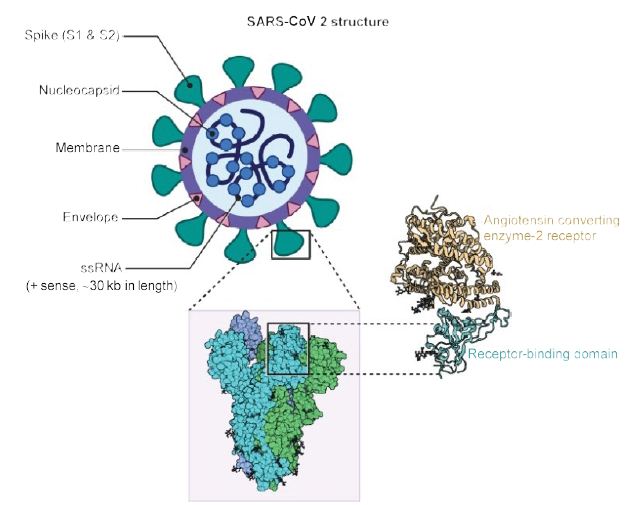
Figure 1: Structure of SARS-CoV2. A positive single-stranded RNA genome with a nucleocapsid is surrounded by a membrane with spike proteins. Reproduced with permission from [2] QC StatPearls (2020).

fig 2

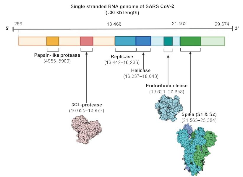
Figure 2: The SARS CoV-2 genomic structure. A papain-like protease, 3CL-protese and endoribonuclease are produced to lyse host cells. The replicase gene codes for replicative apparatus to create more virus to infect host cells. Reproduced with permission from [2], QC StatPearls (2020).

“The spike glycoproteins of CoV are composed of two subunits S1 and S2. The S2 protein is highly conserved and contains a fusion peptide, a transmembrane domain and a cytoplasmic domain [2]. The S subunits consists of homotrimers that compose the spikes on the viral surface which can transduce through host receptors. Thus, it could be a target for antiviral (anti-S2) compounds. On the contrary, the spike receptor-binding domain presents only a 40% amino acid identity with other SARS-CoVs [2]. Other structural elements on which research must necessarily focus are the ORF3b that has no homology with that of SARS-CoVs and a secreted protein (encoded by ORF8), which is structurally different from those of SARS-CoV” [2].

The presence of mutations and additional strains were recently reported, and a spike mutation may have jumped to humans in late 2019, triggered by these mutations. “Researchers analyzed the transmembrane helical segments in the ORF1ab encoded 2 (nsp2) and nsp3 and found that position 723 presents a serine instead of a glycine residue, while the position 1010 is occupied by proline instead of isoleucine” [2]. The matter of viral mutations is key for explaining potential disease relapses.

COVID-19 Infection & the Immune Response it Elicits

Immune Response against the Novel Coronavirus

The basis for how a disrupted immune reaction leads to lung immunopathology resulting in adverse clinical outcomes after “pathogenic hCoV infection” was reported in prior studies. As early as 2017, Channappanavar and Perlman stated that the likelihood of the virus continuing to cross species barriers is high since SARS like coronovirus was identified in bats and MERS CoV was identified in domesticated camels. Thus there is higher probability of outbreaks occurring in humans [15]. A diverse variety of clinical presentation is observed in humans, with most patients exhibiting short periods of “moderate clinical illness” and a smaller but substantial fraction developing severe disease presenting with acute lung inflammation and acute respiratory disease [15].

fig 3

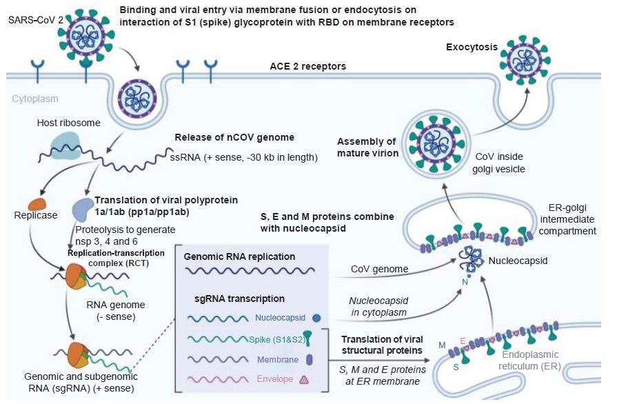
Figure 3: Coronavirus Infection Course. How the novel coronavirus infects host cells. E: Envelope; ER: Endoplasmic reticulum; M: Membrane; S: Spike. Reproduced with permission from [3].

There are certain inflammatory responses that may be conducive to cancer. IFN-α/β or inflammatory monocyte- macrophage-derived pro-inflammatory cytokines impede the clearance of the virus by sensitizing T cells to die and undergo apoptosis [15]. According to one recent report, “[t]he loss of TIR-domain-containing adapter-inducing IFN-β (TRIF), an adapter molecule for TLR-3 and TLR-4 signaling, resulted in a distinct inflammatory signature characterized by neutrophil and other inflammatory cell infiltration” [11]. “A dysregulated immune response to SARS-CoV in TRIF-deficient mice was associated with aberrant antiviral IFN (IFN-α and IFN-β), pro-inflammatory cytokine and chemokine (IL-6, TNF, IFN-γ and monocyte chemoattractant CCL5), and interferon-stimulated gene (RSAD2, IFIT1 and CXCL10) responses”, another possible indicator of cancer risk [15].

Interferon response is antagonized by the multiple proteins, both structural and nonstructural of SARS-CoV [5]. This antagonism may lead to delay in immune response or even evasion, which occurs in the early infection stage when the virus reaches high titer and inhibits interferon response as a result of viral sequestration of proteins [11]. In turn, apoptosis of T cells results, accompanied by a “dysfunctionally regulated inflammatory response” [15].

Some consequences of cytokine storm and immunopathology are epithelial and endothelial cell apoptosis and vascular leakage, suboptimal T-cell response, in other words. One of the key factors in viral clearance is CoV specific T cells that prevent more damage occurring to the host. These distinct T cell responses have a dampening effect on the overactive innate immune systemic response [15]. T cell response is mitigated as result of massive inflammatory effects resulting from pathogenic infection. T-cell death mediated by TNF leads to immunological infiltration by activated macrophages, aberrant homeostasis within tissues, “and acute respiratory distress syndrome.” [15].

Figure 4 demonstrates the enormous immune response against the novel coronavirus [12]. The innate immune response and adaptive immunity have distinct responses to coronaviruses infection [16]. Macrophages are first infected by the CoV virus, which then present the viral antigens to T cells, which are then activated and further differentiate. Cytokines are then produced and released in a massive storm, leading to amplification of the immune response. In turn these mediators that are produced as a result of viral persistence mitigate the production of natural killer cells and activation of CD8+ T cells. This is a harmful outcome of viral proliferation since it is in fact these very same CD8+ T cells that are able to clear CoV through the production of effective mediators [16].

fig 4

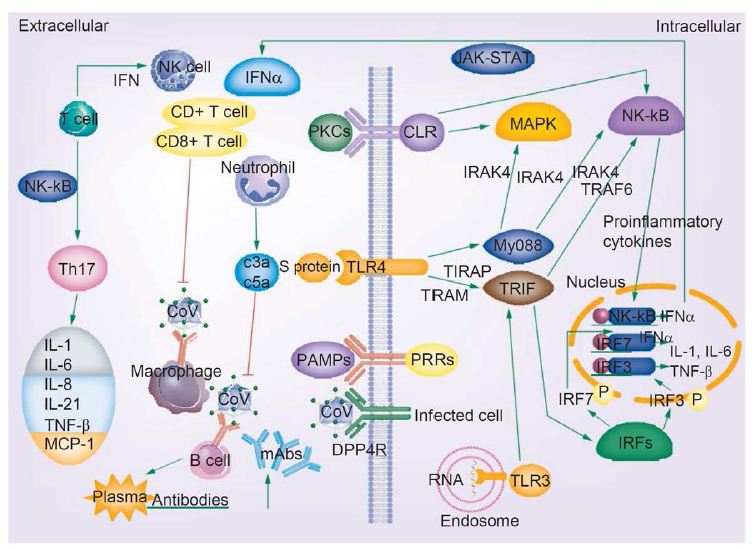
Figure 4: COVID-19 infection and the immune response. Activated signaling pathways such as JAK–STAT and MAPK, cytokine storm, T-cell depletion, humoral responses and high levels of inflammation may predispose patients infected with novel coronavirus to cancer. Reproduced with permission from [16].

The release of genomic RNA into the cytoplasm occurs as a result of DPP4R attachment on the host cell, mediated by the S protein, leading to immunological response to the dsRNA generated during CoV replication. The dsRNA in turns sensitizes TLR-3 resulting in the activation of IRFs and NF-κB signaling pathways, which result in the production of “type I interferons and pro-inflammatory cytokines” Type I interferon is especially significant since it protects uninfected cells from “release of antiviral proteins.” [16]. “Sometimes, accessory proteins of CoV can interfere with TLR-3 signaling and bind the dsRNA of CoV during replication to prevent TLR-3 activation and evade the immune response.” [16].

The MyD88-dependent signaling pathway is activated when TLR-4 binds to the S protein, thus causing the release of pro-inflammatory cytokines. Immune mediators are elicited in intense fashion due to viral-cell interactions. In short, a robust cytokine and chemokine response results in the form of secretion of IL-1, IL-6, IL-8, IL-21, TNF-β and MCP-1, which in turn recruit more white blood cells to the infection site [16].

Novel Coronavirus & Oncologic Sequelae: How Cancer may be Implicated?

PRRs (TLR, RIG-I-like receptor, NOD-like receptor, C-type lectin-like receptors and free-molecule receptors in the cytoplasm, such as cGAS, IFI16, STING and DAI) ensure that pathogen-associated molecular patterns (PAMPs) are recognized in various cellular compartments upon detection by the host innate immune system; PAMPs include bacterial and parasite lipoproteins and proteins. In turn additional pathogenic mechanisms are produced by different TLRs through the activation of adaptor proteins, (MyD88, TIRAP, TRIP and TRAM) that house similar TRR structure [16].

Viral infection plays a key role in signaling factor activation, that may have relevance for the initiation of cancer since a number of processes occur that lead to cellular growth. First, the adaptor protein MyD88, a member of the TIR family, “activates the transcription factors NF-κB and MAPKs pathways to induce inflammatory factors expression” and after activation, leads to the recruitment of receptor-related kinases IRAK4, IRAKI, IRAK2 and IRAK-M [16]. Then, IRAK-TRAF6 interaction occurs, K-63 and NEMO ubiquitination takes place and serves as a further activator for NF-κB. In short, these TRIF-dependent activities lead to the activation of IRF3 and NF-κB as result of the recruitment of TRIF molecules to the TLR4 receptor, and the MyD88 adaptor protein to the TLR2 and TLR4 receptors, all happening through the function of TRAM and TIRAP [16].

Additionally, a “strong immune-mediated pneumonitis and delayed clearance of SARS-CoV from lungs” result as a result of depleted CD4+ T cells through infiltration by white blood cells and cytokine production. The NF-κB signaling pathway leads to the production of pro-inflammatory cytokines due to the increase in T helper cells. Other blood cells, such as monocytes and neutrophils, infiltrate the site of infection due to the production of IL-17 cytokine, inducing inflammation and activation of “other downstream cytokine and chemokine cascades, such as IL-1, IL-6, IL-8, IL-21, TNF-β and MCP-1” [16].

There may be a distinct association between novel coronavirus infection and the onset of cancer through the activation of the MAPK and JAK–STAT signaling pathways and the NF-κB transcription factor. The MAPK signaling pathway, activated upon COVID-19 infection, is involved in the tumorigenesis of a number of cancers, including hepatocellular carcinoma, adrenocortical cancer, endometrial cancer, colorectal cancer and pituitary adenomas. 50-100% of hepatocellular carcinomas display activation of the Ras/MAPK pathway, which is also associated with poor prognosis [17]. “The MAPK signaling pathway is also implicated in pituitary adenomas and because of the important roles of MAPK signaling pathways in tumorigenesis, the use of the MAPK signaling pathways as therapeutic targets has continuously been considered as a promising strategy for pituitary adenoma therapy.” [18]. In the context of the role of two of the MAPK signaling pathways (ERKs 1/2 and p38), adrenocortical tumor genesis is implicated by data suggesting that the MEK MAPK ERK signaling plays as a role, leading to potential clinical use as a diagnostic biomarker for malignancies and the development of targeted therapies [19]. Cholesterol in the form of LDL enhances intestinal inflammation and colorectal cancer progression via activation of reactive oxygen species (ROS). LDL cholesterol also has been shown to enhance intestinal inflammation leading to progression to colorectal cancer via activation of reactive oxygen species and the MAPK signaling pathway [20] Endometrial cancer is promoted through the activation of the MAPK signaling pathway by the estrogen receptor (α) [21]. The oncologic sequelae of COVID-19 may include these cancers upon viral activation of the MAPK signaling pathway. These tumorigenic pathways may play a role in cancer initiation and progression in COVID-19 infection.

The viral immune response is also implicated in other tumorigenic pathways, including those leading to lung cancer, breast cancer and T cell lymphomas.  Type I interferon synthesis as a result of IRF3 and IRF7 activation, in turn activates the JAK-STAT signaling pathway involved in these tumorigenic processes, ultimately through PRRs that recognize viral RNA [22]. “JAK–STAT signaling pathways are mutationally activated in many extranodal T-cell lymphomas, such as natural killer/T-cell and hepatosplenic T-cell lymphomas.” [23] “Since the JAK–STAT pathway is considered to be a central player in inflammation-mediated tumorigenesis, the implication of JAK–STAT signaling and the therapeutic potential of JAK1/2 inhibition was investigated in K-RAS-driven lung adenocarcinoma, and data showed that JAK1 and JAK2 are activated in human lung adenocarcinoma and that increased activation of JAK–STAT signaling correlated with disease progression and K-RAS activity in human lung adenocarcinoma” [24]. “Dysregulated JAK/STAT signaling has been implicated in breast cancer metastasis, which is associated with high relapse risks, which may be mediated through GRAM1b” [25].

NF-κB, and TRIF-dependent pathways are also activated; and, IRF3 and IFN-β are activated in turn [16]. Transcription factors IRF3 and NF-κB induce the gene expression of type I interferon through their activation by adaptor protein TRIF of TLRs. TLRs classify these types of signaling pathways induced by viral infection: “The TLR signaling pathways are classified as the MyD88-dependent pathway, which functions to activate immune inflammatory factors and the TRIF-dependent pathway, which then in turn functions to activate the type I interferons and inflammatory factors” [16]. NF-κB-driven gene products include cytokines/chemokines, growth factors, anti-apoptotic factors, angiogenesis regulators and metalloproteinases, and drive oncogenesis. For instance, many of the genes transcribed by NF-κB promote gastric carcinogenesis. Since it has been shown that chemotherapy-caused “cellular stress could elicit activation of the survival factor NF-κB, which leads to acquisition of chemoresistance, the NF-κB system is recommended for therapeutic targeting” [26]. “Prosaposin, a neurotrophic factor, promotes the proliferation and tumorigenesis of glioma through TLR4-mediated NF-κB signaling pathways” [27].

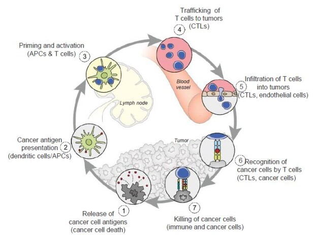
Dendritic cells (DCs) also respond to the virus, and their differentiation from precursor cells to mature cells may be inhibited as is the case with HIV-1. If their maturation is blocked, by perhaps the lack of GM-CSF, IL-4 and TNF-α, the adaptive immune response, which plays a key role in the cancer-immunity cycle may be inhibited (Figure 5). DC’s are the major antigen-presenting cells in the organism, and contribute both types of immunity by activating T cells and B cells. “Immature DCs have strong migration ability, and mature DCs can effectively activate T cells in the central link of start-up, regulation and maintenance of immune responses [16]. Thus, if the maturation process of DCs is blocked, it directly affects the initiation of subsequent adaptive immune responses” that may need to be mobilized in attacking cancer [16].

fig 5

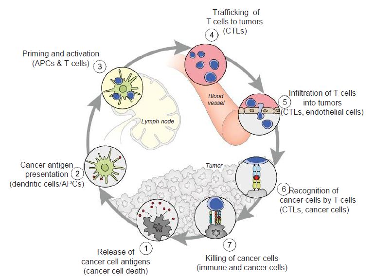
Figure 5: The cancer-immunity cycle. Dendritic cells present cancer antigens to T cell, which are honed to the cancer site. T cells infiltrate tumors and recognize cancer cells upon their destruction. Each point in this cycle may be points of vulnerability upon novel coronavirus infection. APC: Antigen-presenting cell. Reproduced with permission from [28].

Other types of viruses have ostensible effects on adaptive immunity. DC precursor cells are unable to differentiate into mature DCs under the state of HIV-1 Nef protein infection when inducers are in the environment during HIV infection [16]. Thus, the Nef protein prevents the maturation of DCs from DC precursor cells [16]. Three molecules which exist on the surface of human peripheral blood mononuclear precursor cells (PBMCs), CD1a, CD1b, and DC-SIGN, are inhibited by the core protein and NS3 protein in HCV, and are central in the development of DCs from these PBMCs [16]. The viral antigen presentation process through DCs is reduced in HIV-1 infection since MHC-I attentuation occurs on the DCs surface [16]. DC-SIGN expression, or DC specific intracellular adhesion molecule 3 grabbing nonintegrity, is facilitated by HIV-1 infection, and CC chemokine receptor 7 and MHC II molecules, two receptors which play key roles in the DC homing process, are inhibited. All of these processes accumulate to lead to an overall dysfunction of DCs, including their maturation and differentiation, hindering the crucial role they play in the adaptive immune response and allowing for successful viral evasion of the adaptive immune system [16]. If these DC events are occurring, cancer may co-opt normal cellular function and evade immune responses in a viral infection such as COVID-19.

A ‘cytokine storm’, an excessive immune reaction in the host, also results from viral mechanisms that results in extensive tissue damage, mediated by IL-produced by activated leukocytes. B lymphocyte differentiation, cell growth stimulation and pathogenesis of some types for cancer may result. These effects are linked to the function of structural and nonstructural proteins of the virus [2]. Cytokine release syndrome, an immune-related adverse effect of chimeric antigen receptor T-cell therapies, is also implicated in the potential emergence of cancer. Additionally, as tissue damage envelopes the body’s physiological functioning, and cellular resources are increasingly devoted to compensating for it, cancer may emerge as a risk factor. Lung tissue damage, a prevalent and major consequence of infection, may be particularly at risk for developing into lung cancer, and patients who experience these symptoms, may need to be monitored for these sequelae.

Relevance of the Cancer-immunity Cycle & Tumor Microenvironment

In the cancer-immunity cycle (Figure 5), the cancer presents antigens which antigen-presenting cells present to MHC to activate cytotoxic CD8+ T cells, to ultimately destroy cancer cells in a cyclic process [28]. In cancer, the cancer-immunity cycle is dysfunctional with the accumulation of inhibitors of T-cell response and promoters of cancer cell growth. In SARS-CoV infection, cytotoxic T cells kill virus, and lead to the production of antibodies specific to the virus through the activation of T dependent B cells. Further, lung inflammation leads to injury to the immune system as a result of activation of cytotoxic T cells. Immunosuppression, a risk factor for novel coronavirus,  may emerge, conversely, as a risk for cancer in the context of viral infection.

From an understanding of the cancer-immunity cycle, are there are any elements of novel coronavirus infection that would make the cycle vulnerable to cancer at any point and prevent the generation of immunity to cancer? The presence of checkpoint inhibitors to immune responses, such as cytotoxic T-lymphocyte associated protein-4, programmed death-1 and programmed death-ligand-1, may inure the development of cancer with viral infection. In a depleted T-cell environment postinfection, the broad T-cell response to cancer may be undermined. This inflammatory response concomitant with immune cell depletion, whether temporary or not, may lead to oncologic sequelae if accompanied by a superimposed mutagenic and/or carcinogenic event potentially occurring in a time- adjacent manner. Figure 6 displays the tumor microenvironment in the context of breast cancer and illustrates major inflammation and cytokine release, marks of viral infection which may predispose to cancer.

fig 6

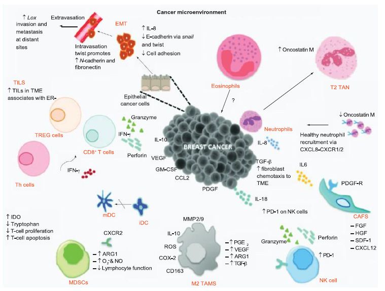
Figure 6: Tumor microenvironment of breast cancer. The tumor microenvironment progresses through cytokine release. Reproduced with permission from [29].

Somatic Mutations, Drug Resistance & COVID-19

The implications for novel coronavirus infection on genomic variability are unknown at this point. If the virus through its replicative [30] and recombination capabilities [31] leads to genetic alterations that may predispose to cancer, another risk factor may be revealed.

The administration of antiviral agents and other proposed treatments such as the antimalarial drug hydroxy- chloroquine may induce drug resistance to cancer therapies, such as targeted agents and immunotherapies. Even further, the status of targeted therapies may be jeopardized by the infection of the virus, since many personalized agents target growth factors that are possibly affected by viral transduction; however, this remains to be investigated.

Countervailing Viewpoints

It is generally known that viruses can cause cancer by integration of the genomes into host cell chromosomes and the expression of oncoproteins. While both of these mechanisms are not implicated in SARS-CoV-2, these are not exclusive mechanisms. The turning on of oncogenic signaling pathways and the acute inflammatory response that results upon coronavirus infection can be hypothesized as being cancer inducing, or leading to the risk of developing cancer, especially if the patient has a superimposed mutagenic or carcinogenic event occurring concomitantly, even if the virus does not cause a chronic infection like viruses such as HCV, HCV and EBV. Additionally, there exist limited resources and research as a result of the pandemic that may damage the prognosis of cancer patients. Novel coronavirus can destroy T cells, but unlike HIV, does not lead to persistent infection of T cells. Data suggest however that there is a general depletion of T cells in the COVID-19 infection environment, and this may affect the healthy progression of the cancer-immunity cycle. Also, there exist gaps in current knowledge on the novel coronavirus’ impact on the immune system during co-infection by other viruses, such as HCV, HIV and EBV. Finally, the overwhelming organ dysfunction that novel coronaviruses results in some cases should be further investigated, and as possibly having an association with cancer. These should remain areas of research for future study.

Future Perspective

As the population infected with the novel coronavirus grows, and the infection spreads, its clinical sequelae may pose an issue of concern for physicians, and oncologists in particular. Future studies should focus on the predisposition of these recovering patients for cancer, and if these patients need to be monitored for the disease. It is the cumulative effect of many distinguishable aspects of coronavirus infection that leads to the increased predisposition to cancer, which then warrants closer follow-up in the future. The organ damage, inflammatory response and signaling pathways activation that occur upon viral infection may sum to significantly increase patients’ predisposition to cancer, and this may be compounded by a superimposed mutagenic or carcinogenic event. The oncology field may evolve by investigating the potential tumor microenvironment in the context of SARS-CoV-2 infection, and if the virus can be considered an etiological agent in the development of cancer. Clinical sequelae may involve physicians needing to refer COVID-19 cases to oncologists for the confirmation of cancer, either benign and malignant cases. The caveat must be mentioned however that knowledge surrounding coronavirus is constantly being updated and this review must be interpreted in light of any recently updated knowledge.

Executive Summary

  • The novel coronavirus is leading to a global pandemic and is causing worldwide clinical and economic devastation. The infection may pose medical risk factors for patients, both symptomatic and asymptomatic, who recover. There will be a distinct health population in the future that will need to deal with clinical sequelae, and cancer may be one of them.

Oncologic Sequelae of the Novel Coronavirus

  • Viral infection induces a robust immune response, a ‘cytokine storm’ leading to tissue damage and inflammation, which may predispose to cancer. Signaling factors, such as MAPK and JAK-STAT, promoted by viral infection may lead to aberrant cellular growth that marks cancer. The maturation of dendritic cells may be inhibited, as shown in patients infected with HIV-1.

The Cancer-immunity Cycle & Tumorigenesis

  • The cancer-immunity cycle may be impaired upon viral infection, since T-cell apoptosis also occurs as a result of viral infection, leading to a state of immunosuppression. If concomitant with a mutagenic or carcinogenic event, a cancerous state may result. The ‘cytokine storm’ poses a risk factor for cancer in the tumor microenvironment.

Summary

  • In conclusion, patients infected and recovered from novel coronavirus, particularly those with lung tissue damage, may need to be monitored for cancer, among other diseases.

References

Papers of special note have been highlighted as: • of interest; •• of considerable interest

    1. Xu Y, Liu H, Hu K, Wang (2020) Clinical management of lung cancer patients during the outbreak of 2019 novel coronavirus disease. Zhongguo Fe Ai Za Zhi 23(3), 136-141. [crossref]
    2. Cascella M, Rajnik M, Cuomo A, Dulebohn SC, Di Napoli Features (2020) evaluation and treatment coronavirus (COVID-19). In: StatPearls. StatPearls Publishing, FL, USA. [crossref]

Excellent overview of clinical symptoms and progression of COVID-19 infection patients. Excellent graphics on coronavirus genome and structure.

    1. Worldometers info/coronavirus/ Accessed 7 April 2021.
    2. Adhikari SP, Meng S, Wu YJ et al (2020) Epidemiology, causes, clinical manifestation and diagnosis, prevention and control of coronavirus disease (COVID-19) during the early outbreak period: a scoping Infect Dis Poverty 9(1), 29. [crossref]

Very informative review article encompasses the epidemiology and clinical presentation of the coronavirus

    1. Richardson S, Hirsch JS, Narasimhan M et al (2020) Presenting characteristics, comorbidities, and outcomes among 5700 patients hospitalized with COVID-19 in the New York City JAMA e206775 (Epub ahead of print). [crossref]

One of the first major studies conducted at a hotspot of the pandemic on characteristics of coronavirus patients and their

    1. Advisory Board: it’s not just lungs: COVID-19 may damage the heart, brain, and kidneys (2020). https://www.advisory.com/daily-briefing/2020/04/17/organ-damage.
    2. Shi S, Qin M, Shen (2020) Association of cardiac injury with mortality in hospitalized patients with COVID-19 in Wuhan, China. JAMA Cardiol. doi:10.1001/jamacardio.2020.0950 (Epub ahead of print). [crossref]
    3. Mao L, Jin H, Wang M et al (2020) Neurologic manifestations of hospitalized patients with coronavirus disease 2019 in Wuhan, China. JAMA Neurol. e201127 doi:10.1001/2020.1127 (Epub ahead of print). [crossref]
    4. Su H, Yang M, Wan C et al (2020) Renal histopathological analysis of 26 postmortem findings of patients with COVID-19 in Kidney Int. doi:10.1016/j.kint.2020.04.003 (Epub ahead of print). [crossref]
    5. Cui S, Chen S, Li X, Liu S, Wang F. (2020) Prevalence of venous thromboembolism in patients with severe novel coronavirus pneumonia. J Thromb Haemost. doi:10.1111/14830 (Epub ahead of print). [crossref]
    6. Troyer EA, Kohn JN, Hong S. (2020) Are we facing a crashing wave of neuropsychiatric sequelae of COVID-19? Neuropsychiatric symptoms and potential immunological mechanisms. Brain Behav Immub 87:34-39. [crossref]
    7. https://www.cdc.gov/coronavirus/2019-ncov/hcp/clinical-care/late-sequelae.html Accessed 7 Apr 2021.
    8. Praschan N, Josephy-Hernandez S, Kim DD, Kritzer MD, Mukerji S et al (2021) Implications of COVID-19 sequelae for health-care personne. The Lancet Respiratory Medicine. 9(3): 230-231. [crossref]
    9. Nalbandian A, Sehgal K, Gupta A, Madhavan MV, McGroder C et al. (2021): Post-acute COVID-19 syndrome. Nature Medicine.
    10. Channappanavar R, Perlman (2017). Pathogenic human coronavirus infections: causes and consequences of cytokine storm and immunopathology. Semin Immunopathol 39(5), 529-539. [crossref]

An early article on the molecular responses to coronavirus infection and possibility of pandemics.

    1. Li G, Fan Y, Lai Y et al (2020) Coronavirus infections and immune J Med Virol 92(4), 424-432. [crossref]

An excellent overview with graphics on the immune-mediated responses to coronavirus.

  1. Delire B, Starkel (2015) The Ras/MAPK pathway and hepatocarcinoma: pathogenesis and therapeutic implications. Eur J Clin Invest 45(6), 609-623. [crossref]
  2. Lu M, Wang Y, Zhan (2019) The MAPK pathway-based drug therapeutic targets in pituitary adenomas. Front Endocrinol 10, 419-430. [crossref]
  3. Pereira SS, Monteiro MP, Costa MM et al (2019) MAPK/ERK pathway inhibition is a promising treatment target for adrenocortical J Cell Biochem 120, 894-906. [crossref]
  4. Wang C, Peiwei L, Xuan J et al (2017). Cholesterol enhances colorectal cancer progression via ROS elevation and MAPK signaling pathway Cell Physiol Biochem 42, 728-742. [crossref]
  5. Liu A, Zhang D, Yang X, Song Y. (2019). Estrogen receptor alpha activates MAPK signaling pathway to promote the development of endometrial J Cell Biochem 120, 17593-17601. [crossref]
  6. Scott L, Gandhi (2015) Deregulated JAK/STAT signalling in lymphomagenesis, and its implications for the development of new targeted therapies. Blood Rev 29, 405-415.
  7. van Arnam JS, Lim MS, Elenitoba-Johnson (2018) Novel insights into the pathogenesis of T-cell lymphomas. Blood 24, 2320-2330. [crossref]
  8. Mohrherr J, Haber M, Breitnencecker K et al (2019) JAK-STAT inhibition impairs K-RAS-driven lung adenocarcinoma Int J Cancer 145, 3376-3388. [crossref]
  9. Khanna P, Lee JS, Sereemaspun A et al (2018) GRAMD1B regulates cell migration in breast cancer cells through JAK/STAT and Akt Sci Rep 22, 1-10.
  10. Sokolova O, Naumann (2017) NF-κB signaling in gastric cancer. Toxins (Basel) 9(4), E119. [crossref]
  11. Jiang Y, Zhou J, Luo P et al (2018) Prosaposin promotes the proliferation and tumorigenesis of glioma through toll-like receptor 4 (TLR4)-mediated NF-κB signaling EbioMedicine 37, 78-90. [crossref]
  12. Chen DS, Mellman (2013) Oncology meets immunology: the cancer-immunity cycle. Immunity 39(1), 1-10. [crossref]

    Seminal article on the cancer-immunity cycle, which may be implicated in novel coronavirus infection.

  13. Barriga V, Kuol N, Nurgali K, Apostolopoulos (2019) The complex interaction between the tumor micro-environment and immune checkpoints in breast cancer. Cancers (Basel) 11(8), E1205. [crossref]
  14. Chen Y, Liu Q, Guo (2020) Emerging coronaviruses: genome structure, replication and pathogenesis. J Med Virol 92, 418-423. [crossref]
  15. Bentley K, Evans (2018) Mechanisms and consequences of positive-strand RNA virus recombination. J Gen Virol 99(10), 1345-1356. [crossref]
fig 7

Nootropic, Neuroprotective and Anti-oxidant Role of Apocynin in Scopolamine Induced Memory Deficit in a Zebrafish Model

DOI: 10.31038/JPPR.2021414

Abstract

Four decades of search in quest of an effective therapeutic option for Alzheimer’s disease (AD) has led to a complete clinical failure, putting pre-clinical models, their evaluation in spotlight. The pre-clinical stage if lead with a systemic approach, than jumping straight into clinical trials, will reduce financial burden opening avenues for effective AD therapeutics. Preliminary screening therefore shall be fast, ensuring use of efficient, easy, and less time consuming pre-clinical models for screening new entities. Aim of present research was therefore development of a quick, effective, and easy zebrafish model for preliminary screening of probable AD therapeutics. The present research used total of 72 zebrafish divided into six groups, control, negative control, vehicle control, and 3 groups for apocynin (10 mg/kg, 30 mg/kg, and 70 mg/kg). We investigated memory retention, long term memory retention using passive avoidance paradigm, free radical generation using DCFDA assay, and glial cell proliferation using H and E histological staining. Intraperitoneally administered scopolamine (0.025 mg/kg) induced memory deficits hindering memory formation. Apocynin at all doses prevented scopolamine-induced amnesia. Apocynin was found to be capable of reducing free radical load and curbing the inflammation due to scopolamine administration. Moreover; long term memory retention was observed with 10 mg/kg apocynin. Our revelations designate the fact that passive avoidance model in zebrafish could be used as an acute model in order to screen potential entities for treatment of AD. Our findings highlight that a single dose of apocynin could reverse memory deficits induced by single scopolamine injection by acting on cholinergic transmission pathway. Apocynin being an anti-oxidant reduced the free radical generation, thus engaging one of the crucial pathways toward AD pathophysiology. In addition it offered neuroprotection by reducing glial cell proliferation. In conclusion apocynin exhibited multifaceted pharmacological activities like being nootropic, neuroprotective, and anti-oxidant in preclinical AD model.

Keywords

Alzheimer’s disease (AD); Scopolamine; Passive avoidance; Apocynin; Zebrafish; Nootropic; Neuroprotective

Introduction

The cost of bringing a medicine from invention to pharmacy shelves is $2.8 billion [1]. A lot of molecules are failing miserably in the clinical phase of AD. Therefore a systematic approach in the initial drug discovery process is of utmost importance. A number of models are being developed to mimic the AD pathogenesis and the existing ones are being fine tuned to meet the demand. To curtail down the search for a therapeutic option from a humongous number i.e. to generate a lead from various hits will require use of an efficient, fast, and effective in-vivo pre-clinical model. Zebrafish as a pre-clinical model is equipped with all these characteristics.

The zebrafish (Danio rerio) is fast emerging as a promising model organism to study various central nervous system (CNS) disorders, including AD [2]. It has recently become a focus in neurobehavioral studies since it displays quantifiable neuropathological and behavioural phenotypes [3,4]. Zebrafish have a fully characterized genome, and exhibit significant physiological homology to mammals, including humans [5]. Zebrafish is an ideal model for the study of human diseases because they present a number of features that make it unique as an animal model. Zebrafish brain is highly conserved of the basic brain organization, with similar key neuroanatomical and neurochemical pathways of relevance to human diseases [6,7]. In the recent years zebrafish has been proposed as a valid experimental paradigm to study AD [8]. Scopolamine (SCP) is a well known and extensively studied muscarinic cholinergic antagonist which has several effects in central and peripheral nervous system [9,10]. Both clinical [11,12] and rodent [13,14] studies point towards its ability to induce cognitive deficits. Substantial evidences also indicate the potential of acute scopolamine to develop memory deficits in zebrafish [15-20]. Current research strategies target the unused potential of phytoactives and marine actives as therapeutic moieties in AD. Apocynin an inhibitor of NADPH oxidase is a naturally occurring methoxy-substituted catechol [21]. It is isolated from the roots of Apocynin cannabinum (Canadianhemp), Picrorhizakurroa (Scrophulariaceae) and Jatropha multifia (Euphorbiaceae) [22-24]. It is known to possess various biological activities like anti-inflammatory, anti-oxidant and is useful in many disorders such as neurodegeneration, asthma, cancer, arteriosclerosis, hypertension etc. [21]. Apocynin was found to be effective in 1-methyl-4-phenyl-1, 2, 3, 6-tetrahydropyridine (MPTP) marmoset model. It compensated for the loss of dopamine, mitigated the parkinsonian signs, symptoms, and improved motor function [25]. All this literature encouraged us to investigate apocynin in zebrafish passive avoidance paradigm as a model for AD. In the current research we demonstrate the development of an acute zebrafish model which is easy, quick, and useful for screening new potential entities for AD, to arrive at possible lead in the drug discovery process. We have also explored the passive avoidance paradigm to study the memory deficits induced by a single scopolamine administration and its subsequent reversal by apocynin.

Materials and Methods

Reagents and Chemicals

Apocynin, (-)-Scopolamine hydrobromide trihydrate, 2’, 7’-dichlorofluorescin diacetate (DCF-DA) procured from sigma Aldrich, USA, Tween 80 from S D Fine Chemicals ltd, India. All other chemicals used were of analytical grade.

Animals

Indigenous transgenic male adult zebra fish strain Danio rerio (3-4 months old) were used for this study (obtained from Dolphin Aquaculture, Mumbai, India). The fish chosen were of uniform size in length (3 cm ± 0.5 cm) and weight (500 ± 50 mg). They were maintained in 25 litres glass storage tank containing purified dechlorinated water to neutralize heavy metals, chlorine, and chloramines present in the water that could be harmful to fish. Fish tank temperature was maintained at 25°C ± 2°C under a 12:12-hr light: dark cycle and were continuously aerated. Fish were fed thrice daily with brine shrimp (marketed as Instincts®). The temperature, oxygen, and water quality was maintained throughout the experimental procedures and the fishes were allowed to acclimatize to the laboratory conditions for 9 days [26]. All solutions administered were prepared fresh at room temperature.

Procedure

The dose for scopolamine administration was chosen based on previous literature [27]. Scopolamine was administered intraperitoneally in zebrafish after the training sessions using 6 mm X 31 G needle attached to 26-G 10 µl Hamilton syringe with help of a catheter of size 24-G using Kinkel MD protocol with modifications [28]. Briefly, the zebra fish were cryoanesthetized prior to intra peritoneal injection until the fish shows reduced respiration and lack of motor coordination. The anesthetized fish was gently put in a water-soaked sponge with the abdomen facing up and the head of the fish positioned at the hinge of the sponge. The needle was inserted into the midline of the abdomen parallel to the spine and posterior to the pectoral fins. The procedure was mastered to complete the injection within 10 sec. Following injection, the fishes were kept in separate tanks to facilitate the recovery post anaesthesia with water maintained at 25 ± 2°C along with proper aeration. Apocynin was administered orally. In order to guarantee the doses administered, apocynin was suspended in Tween 80 (1%) in distilled water. Apocynin was administered orally after 1 hour of scopolamine administration using 24-G catheter fitted into the hypodermic needle of 26-G 10 µl Hamilton Syringe using Collymore C protocol with modifications [29]. Briefly, the fish was cryoanesthetized and held in water soaked sponge, in such a way that the fish was held vertically upward with its back positioned in the hinge of the sponge. The flexible tubing, for oral gavage was lowered into the oral cavity of the fish, approximately 1 cm, until the tip of the tubing extended past the gills. The drug solution was then injected slowly into the intestinal tract. The volume of drug administered was 2 µl. The injection procedure was mastered to complete the injection procedure within 10 sec. Following injection, the fishes were kept in a separate tank to facilitate the recovery post anaesthesia, with water maintained at 25 ± 2°C along with proper aeration. After every administration the fishes were transferred into a new clean tank. Tween 80 was used as the vehicle. The vehicle, scopolamine, apocynin were prepared freshly on the experimental day. Post injection and oral administration fishes which did not recover within 2 to 3 min were excluded from the experiment. The fishes were divided into six groups, namely control, negative (SCP 0.0025 mg/kg ip), vehicle (tween 80 po), apocynin 3 groups; APO 10 mg/kg po, APO 30 mg/kg po, APO 70 mg/kg po; with 12 fishes in each group.

Passive Avoidance Experimental Chamber

Individually zebrafish were trained and tested for the long-term memory retention in the passive avoidance task. The apparatus consists of 18 cm length × 9 cm width × 7 cm height glass tank divided in two equal half compartments designated as dark and white (differentiated by the colour of the tank), by a manually operated sliding door (9 cm × 7 cm). The tank water level was maintained at three cm and the partition was raised one cm above the tank floor to allow zebrafish to swim freely from one side to the other [15,16,30].

Passive Avoidance Paradigm

On training session, fish was placed in the white side of the tank with the partition between compartments being closed. The fish was allowed to acclimatize in it for three min and the partition was then raised till one cm height. The fish was given five min to enter the dark compartment through this partition. The partition door was closed once the fish entered the dark compartment completely with its entire body. A marble was dropped in front of the fish head; three sec after it entered the dark compartment. The fish was then immediately removed from the dark compartment and placed in a separate tank. This whole procedure is considered as one training session. The marble dropped in front of the fish served as shock for the fish, so that it would be reluctant to enter the dark compartment. Each fish was trained thrice with an interval of three min between each training session [15, 16, 30]. Each training session hereafter will be addressed as a trial. After the three trial session, a test session was conducted after two hr of the last trial (documented as day 1) for control group fishes. The test session consisted of repeating of the trial session except, the marble was not dropped when the fish entered the dark compartment. For negative control group, SCP was administered ip after 1 hr of the last trial. The test session was conducted one hr post SCP administration, documented as day 1. For the vehicle and test drug groups, the vehicle (1% tween 80) or the test drug (apocynin 10 mg/kg, 30 mg/kg, and 70 mg/kg) were administered per oral one hr after SCP administration. The test session was conducted one hr after the vehicle or test drug administration, documented as day 1. For all the groups only the test session was repeated after 24 hrs, documented as day 2; and after 48 hrs documented as day 3 after the first test session (day 1); to assess the long term memory retention. The flowchart for the passive avoidance test for all the groups is depicted in Figure 1. The latency to completely enter the dark compartment was measured for the three trials and the three test sessions; used as an index of memory retention.

fig 1

Figure 1: Flowchart for passive avoidance test followed for all the groups.

Reactive Oxygen Species Formation

The fishes in all the groups, after 48 hrs or after the test 3 of the study were subjected to cryoanaesthesia, according to the NIH guidelines. The fishes were first immobilised, by keeping them in ice water [5:1 (ice: water)] for at least 10 minutes or until cessation of opercular movement. The fishes were left for 20 minutes in ice cold water until cessation of all movements. After the fishes were euthanized, brains were isolated. The brains were homogenized with equal volume of ice-cold 0.1 M phosphate buffer saline (pH 7.4). The homogenate was then centrifuged at 4°C (10,000 rpm; R-248Mof CPR-24 plus Instrument, Remi, India) for 15 min, and aliquots of homogenate were used for estimation of free radicals. The total concentration of free radicals, especially reactive oxygen species, was detected using 2’, 7’-dichlorofluorescin diacetate (DCFDA) with slight modification of Pereira AG et al. protocol [31]. The 30 µl brain homogenate was treated with 100 µl of 100 µM DCFDA in 96 well plate. The plate was incubated at 37°C for 30 min in the dark. The whole protocol was performed in dark. The formation of the oxidized fluorescent derivative dichlorofluorescein (DCF) was monitored at 488 nm excitation and 525 nm emission using a fluorescence microplate spectrophotometer (Epoch, Biotech, USA).

Histopathological Analysis

After the completion of the inhibitory avoidance task, fishes were subjected to cryoanaesthesia. The brain was then isolated and stored in 4% paraformaldehyde solution until histopathological analysis using hematoxylin–eosin (H and E) staining for evaluation of neuronal damage by examining the glial cell proliferation. The stained sections were evaluated by a histopathologist unaware of the treatments.

Statistical Analysis

Inhibitory avoidance memory data are presented as mean ± S.E.M. Statistical analyses were performed using GraphPad Prism 5 software for Windows (GraphPad Software, San Diego, CA, USA). Latencies of individual group and between multiple groups were compared using one way ANOVA followed by Newman-Keuls test (post-hoc analysis). The level of significance was set as ###P<0.001 when compared with control group, ***P<0.001 when compared with scopolamine treatment group. The inter day latencies between multiple groups were compared using Two-way ANOVA followed by Bonferroni test as a post-ANOVA test. The level of significance was set as # P<0.05 when compared with control group, *P<0.05 when compared with scopolamine treatment group. The generation of free radical was analysed using one-way ANOVA followed by Post hoc Dunnett’s test. The level of significance was set as ### P<0.001 when compared with control group and ***P < 0.001 when compared with scopolamine treatment group.

Results

Effect of SCP on Zebrafish in Passive Avoidance Paradigm

The effectiveness of scopolamine in altering the memory was evaluated in the inhibitory avoidance task, by training the fishes in 3 consecutive trials followed by a test session. As evident in Figure 2b, the test latency for SCP decreased significantly compared to the third trial in the SCP group (P<0.001), whereas the test latency significantly increased in the control group compared to the first trial (P<0.05), evident in Figure 2a. The test latency for the vehicle control group also decreased when compared to the third trial, though the decrease was not significant (Figure 2c).

Effect of Apocynin on Zebrafish in Passive Avoidance Paradigm

Apocynin at all three doses was able to reverse the SCP induced memory deficits. The test latency for apocynin at 10 mg/kg was significantly higher than trial 1 with P<0.001 and trial 2, with P<0.01 (Figure 2d). The test latency for apocynin at 30 mg/kg was significantly higher than trial 1 and trial 2, with P<0.001 for both (Figure 2e). However, for 70 mg/kg the test latency was significantly higher than the first trial, with P<0.001 (Figure 2f).

fig 2

Figure 2: Individual training session of fishes in control (a), negative control (b), vehicle control (c), AL (d), AM (e) and AH (f) group on day 1. Data expressed as mean ±SEM. *P<0.05, **P<0.01, and ***P<0.001 when compared to the test using One-way ANOVA followed by Post hoc Student –Newman Keuls Test (n~12).

The inter group test latencies were also compared and are depicted in Figure 3. SCP significantly reduced the escape latency when compared to the control group latency with P<0.001. Apocynin significantly increased the test latencies at all three doses when compared with the SCP group, with P<0.001 at all doses. The escape latency in the vehicle control group was reduced when compared to the latency for control group, but the decrease was not significant.

fig 3

Figure 3: Comparison of day 1 test latencies between all the groups with respect to apocynin. Data expressed as mean ±SEM. ###P<0.001, when compared with Control group, ***P<0.001 when compared with Negative Control group using one-way ANOVA followed by Post hoc Student –Newman Keuls Test (n~12).

Long-Term Memory Retention

Our intent was to explore the effect of single dose administration of both scopolamine and the test entity apocynin on long term memory retention of the learned response in passive avoidance paradigm. The results are depicted in Figure 4. On day one the fishes in control group showed the highest escape latency, the observation being persistent for next two days (day 2 and 3) as well. The negative control group showed lower escape latencies for all three days, with P<0.05 for day one when compared to the control group. The vehicle control group also showed lower escape latency for all three days, but the results were not significant. Apocynin at all three doses showed improved escape latencies compared to the SCP group for day one and day two. Apocynin at 30 mg/kg showed significantly higher escape latency with P<0.05 on day 1. On day three the dose of 10 mg/kg showed betterment in the latency amongst the three doses, although not significant.

fig 4

Figure 4: Inter day comparison between all groups with respect to apocynin. Data expressed as mean ±SEM. #P<0.05 when compared with Control group and *P<0.05 when compared to the Negative Control group. Data analyzed by using Two-way ANOVA followed by Bonferroni test as a post-ANOVA test.

Free Radical Generation (DCF-DA Assay)

The effect of SCP administration on the generation of free radicals was assessed using the DCF-DA assay and depicted in Figure 5. SCP administration significantly increased the relative fluorescence intensity with P<0.001 when compared with the fluorescence intensities in control group. A similar increase is seen in vehicle control group, though not significant. Apocynin at all three doses alleviated the fluorescence intensity significantly with P<0.001 for all doses when compared with the SCP group.

fig 5

Figure 5: Effect on levels of ROS (directly proportional to RFU) in different experimental groups. Data expressed as mean ± SEM ###P<0.001 compared with Control group and ***P<0.001 compared with Negative Control group using one-way ANOVA followed by Post hoc Dunnett’s test (n=2).

Histopathological Analysis (H and E Staining)

The effect of SCP treatment on the inflammatory response was studied and the histopathological changes observed in the zebrafish brain have been presented in Figures 6 and 7. The control group did not showcase any glial cell proliferation with the score being zero. The negative control and vehicle control group exhibited significant glial cell proliferation with a score of +++, being the severe damage depicted in Figure 6 (Control, NC, VC) and scored in Table 1. Apocynin at dose of 10 mg/kg, and 30 mg/kg (Figure 7 AL, AM) showed zero or nil glial cell proliferation. Apocynin at 70 mg/kg (Figure 7 AH) showcased the score of ++ denoted as moderate change.

fig 6

Figure 6: Histopathological analysis using H and E staining of zebrafish brains for control, negative control SCP 0.025 mg/kg (NC) and vehicle control (VC) group. The lower row indicates magnified images of the images in the above row, at magnification of 100X. The black circles indicate the glial cell proliferation.

fig 7

Figure 7: Histopathological Analysis using H and E staining of zebrafish brains for Apocynin Low dose 10 mg/kg (AL), Apocynin Medium dose 30 mg/kg (AM) and Apocynin High dose 70 mg/kg (AH) group. The lower row indicates magnified images of the images in the above row, at magnification of 100X. The black circles indicate the glial cell proliferation.

Table 1: Histopathological scoring for zebra fish brain images :

Group

Scoring

Control

Nil

Negative Control (NC)

+++

Vehicle Control (VC)

+++

Apocynin 10mg/kg (AL)

0

Apocynin 30mg/kg (AM)

0

Apocynin 70mg/kg (AH)

++

Note: – += Mild Change; ++= Moderate change/Damage; +++= Severe change /Damage

Discussion

Scopolamine, a non-selective muscarinic receptor antagonist induces learning deficits in zebrafish is a well-established fact [30]. It is a centrally acting anti-cholinergic involved in learning and memory impairment; particularly short-term memory formation [32,33]. Therefore; the memory deficits due to scopolamine are regarded as cholinergic deficit or ‘cholinergic amnesia’ [34]. Also, zebra fish has been widely explored as an animal model in AD. In our study, we examined the above facts for screening apocynin in a passive avoidance paradigm. Our results demonstrate that scopolamine hampers the learning process as the latency to cross the partition of the sliding door reduced significantly in the negative control group as seen in the individual training result as compared to the control group. Our data is in accordance with the available literature on rodents and supports scopolamine’s effect in evaluating memory enhancing drugs in zebrafish model. The SCP administration has induced memory deficits as evident with the decreased escape latencies. Apocynin was capable of ameliorating the effects of SCP, showcased by the comparison of test latencies for day one. The effect for apocynin however was not dose dependent as at 70 mg/kg, decrease in the escape latency is observed. This may attributed to the ceiling effect at 30 mg/kg. A decrease in the test latency for the vehicle control group is also observed. In the vehicle control group, the vehicle (tween 80) was administered after SCP administration, thus the decreased test latency demonstrates the effect of SCP that is responsible for the decrease in latency. Tween 80 itself does not impart any pharmacological effect in reversing the memory deficits. It merely acts a vehicle for administering apocynin. The effect of SCP administration persisted for 48 hrs indicating the deficit in memory retention due to single dose of SCP as seen in the negative control group. Since we wanted to explore the effect of single dose administration of scopolamine and apocynin on long term memory retention, we did not administer the drug on daily basis or for 2 consecutive days. Also the training sessions of three trials was not repeated on day 2 or day 3, only the test. Apocynin mitigated the effect of SCP and was capable of maintaining the effect for 24 hrs (day 2) and 48 hrs (day 3) for 10 mg/kg. SCP causes memory impairments by affecting cholinergic transmission; apocynin reversed the memory deficit implying that it works on the cholinergic neurotransmission pathway [34,35]. Therefore; apocynin has a potential as an anti-Alzheimer drug and can be explored further in a rodent model of AD. There is ocean of evidence entailing that oxidative damage induced due to free radical may play a role in AD pathogenesis. SCP administration generates free radicals and damages the anti-oxidant defence mechanism by damaging the anti-oxidant enzymes [36]. This fact is well established in rodent models [37] as well as in zebrafish [38]. Our study demonstrates that even single dose of scopolamine significantly induces the generation of free radicals. Apocynin being an anti-oxidant reduces the free radical generation [39]. This reduction however was not dose dependent, the 70 mg/kg dose showed a slight increase in the fluorescence intensity compare to the 30 mg/kg dose. This may be due to the ceiling effect at the 30 mg/kg dose. The reduction in free radical formation has undeniably ameliorated the associated damage to the neuronal environment. Thus apocynin could help in halting the disease progression by alleviating the free radical production.

Glial cells are one of the most abundant cells of the brain, performing various biological functions and play a major role in inflammation. In acute phase after injury inflammation is tightly controlled and is a normal and a necessary process maintaining the homeostasis. However, in chronic phase it is detrimental, as seen in AD. It may not be the initiating factor but it significantly contributes to disease progression. Neuroinflammation induces a complex and dynamic change in glial cell phenotypes. Microglial cells are the first cell types to respond post injury they retract their processes and migrate towards the site of injury, where they release pro-inflammatory cytokines such as interleukin-1β (IL-1β), tumor necrosis factor- α (TNF-α), and IL-6 [40]. The histopathological data attests the role of SCP in setting up an inflammatory response capable of damaging the neuronal environment. Administration of single dose of SCP demonstrates the highest score for glial cell proliferation and hence the severe neuronal damage. The damage was totally reversed by apocynin 10 mg/kg and 30 mg/kg as evident with nil glial cell proliferation. The other dose of apocynin 70 mg/kg was effective compared to the negative control group, but could not completely reverse the damage. Apocynin thus bestowed neuroprotection by mitigating the neuronal damage, ultimately maintaining the neuronal homeostasis against the mulct of scopolamine administration. Our study reveals and advocates that passive avoidance test in zebrafish can be used as an acute preliminary in-vivo model to screen potential anti-Alzheimer entities. Our findings emphasize that the phytoactive apocynin reversed the memory deficits and retained the memory hampered by scopolamine administration. Apocynin thus is a nootropic with the probable mechanism being through the action on cholinergic transmission pathway. Apocynin being an anti-oxidant reduced the free radical generation, thus engaging one of the crucial pathways toward AD pathophysiology. Additionally it conferred neuroprotection by reducing the glial cell proliferation. In summary apocynin exhibited multifaceted pharmacological activities like nootropic, anti-oxidant, and neuroprotective in preventing the devastatingly progressive nature of Alzheimer disease. The study also urges for exploring the potential of apocynin in further in-vivo animal models to warranty its use in clinical trials.

Acknowledgment

None.

Competing Interests

The author(s) declare(s) that they have no competing interests.

Funding Information

The authors thank the UGC-BSR fellowship for the financial support.

References

  1. Wouters OJ, McKee M, Luyten J (2020) Estimated research and development investment needed to bring a new medicine to market, 2009-2018. JAMA 323: 844-853. [crossref]
  2. Santana S, Rico EP, Burgos JS (2012) Can zebrafish be used as animal model to study Alzheimer’s disease? American Journal of Neurodegenerative Disease
  3. Bowman TV, Zon LI (2010) Swimming into the future of drug discovery: in vivo chemical screens in zebrafish. ACS Chem Biol 19: 159-161. [crossref]
  4. Rico EP, Rosemberg DB, Seibt KJ, Capiotti KM, Da Silva RS, et al. (2011) Zebrafish neurotransmitter systems as potential pharmacological and toxicological targets. Neurotoxicology and Teratology 33:608-617. [crossref]
  5. Barbazuk WB, Korf I, Kadavi C, Heyen J, Tate S, et al. (2000) The syntenic relationship of the zebrafish and human genomes. Genome Research 10:1351-1358. [crossref]
  6. Mussulini BH, Leite CE, Zenki KC, Moro L, Baggio S, et al. (2013) Seizures induced by pentylenetetrazole in the adult zebrafish: a detailed behavioral characterization. PloS ONE [crossref]
  7. Mueller T, Vernier P, Wullimann MF (2004) The adult central nervous cholinergic system of a neurogenetic model animal, the zebrafish Danio rerio. Brain Research 1011:156-69. [crossref]
  8. Newman M, Verdile G, Martins RN, Lardelli M (2011) Zebrafish as a tool in Alzheimer’s disease research. Biochimica et Biophysica Acta (BBA)-Molecular Basis of Disease 1812: 346-352. [crossref]
  9. Schmäl F (2013) Neuronal mechanisms and the treatment of motion sickness. Pharmacology 91: 229-241.
  10. Golding JF, Gresty MA (2015) Pathophysiology and treatment of motion sickness. Current Opinion in Neurology 28: 83-8 [crossref]
  11. Atri A, Sherman S, Norman KA, Kirchhoff BA, Nicolas MM, et al. (2004) Blockade of central cholinergic receptors impairs new learning and increases proactive interference in a word paired-associate memory task. Behavioral Neuroscience [crossref]
  12. Green A, Ellis KA, Ellis J, Bartholomeusz CF, Ilic S, et al. (2005) Muscarinic and nicotinic receptor modulation of object and spatial n-back working memory in humans. Pharmacology Biochemistry and Behavior 81: 575-584. [crossref]
  13. Lee JC, Park JH, Ahn JH, Park J, Kim IH, et al. (2018) Effects of chronic scopolamine treatment on cognitive impairment and neurofilament expression in the mouse hippocampus. Molecular Medicine Reports 17: 1625-32. [crossref]
  14. Skalicka-Wozniak K, Budzynska B, Biala G, Boguszewska-Czubara A (2018) Scopolamine-induced memory impairment is alleviated by xanthotoxin: role of acetylcholinesterase and oxidative stress processes. ACS Chemical Neuroscience 9:1184-1194. [crossref]
  15. Kim YH, Lee Y, Kim D, Jung MW, Lee CJ (2010) Scopolamine-induced learning impairment reversed by physostigmine in zebrafish. Neuroscience Research 67:156-161. [crossref]
  16. Richetti SK, Blank M, Capiotti KM, Piato AL, Bogo MR, et al. (2011) Quercetin and rutin prevent scopolamine-induced memory impairment in zebrafish. Behavioural Brain Research 217: 10-15. [crossref]
  17. Cognato GD, Bortolotto JW, Blazina AR, Christoff RR, Lara DR, et al. (2012) Y-Maze memory task in zebrafish (Danio rerio): the role of glutamatergic and cholinergic systems on the acquisition and consolidation periods. Neurobiology of Learning and Memory 98: 321-328. [crossref]
  18. Bortolotto JW, de Melo GM, de Paula Cognato G, Vianna MR, Bonan CD. (2015) Modulation of adenosine signaling prevents scopolamine-induced cognitive impairment in zebrafish. Neurobiology of Learning and Memory 118: 113-119.
  19. Rajesh V, Ilanthalir S (2016) Cognition enhancing activity of sulforaphane against scopolamine induced cognitive impairment in zebra fish (Danio rerio). Neurochemical Research 41: 2538-2548. [crossref]
  20. Zanandrea R, Abreu MS, Piato A, Barcellos LJ, Giacomini AC. (2018) Lithium prevents scopolamine-induced memory impairment in zebrafish. Neuroscience Letters 18: 664: 34-37.
  21. Stefanska J, Pawliczak R. (2008) Apocynin: molecular aptitudes. Mediators of Inflammation. [crossref]
  22. Romanini CV, Ferreira ED, Soares LM, Santiago AN, Milani H, et al. (2015) 4‐hydroxy‐3‐methoxy‐acetophenone‐mediated long‐lasting memory recovery, hippocampal neuroprotection, and reduction of glial cell activation after transient global cerebral ischemia in rats. Journal of Neuroscience Research 93:1240-1249. [crossref]
  23. t Hart BA, Copray S, Philippens I (2014) Apocynin, a low molecular oral treatment for neurodegenerative disease. BioMed Research International. [crossref]
  24. Engels F, Renirie BF, ‘Hart BA, Labadie RP, Nijkamp FP (1992) Effects of apocynin, a drug isolated from the roots of Picrorhiza kurroa, on arachidonic acid metabolism. FEBS Letters 305: 254-256. [crossref]
  25. Philippens IH, Wubben JA, Finsen B, A‘t Hart B (2013) Oral treatment with the NADPH oxidase antagonist apocynin mitigates clinical and pathological features of parkinsonism in the MPTP marmoset model. Journal of Neuroimmune Pharmacology 8: 715-726. [crossref]
  26. OECD (2019), Test No. 203: Fish, Acute Toxicity Test, OECD Guidelines for the Testing of Chemicals, Section 2, OECD Publishing, Paris. https://doi.org/10.1787/9789264069961-en
  27. Braida D, Ponzoni L, Martucci R, Sparatore F, Gotti C, Sala M (2014) Role of neuronal nicotinic acetylcholine receptors (nAChRs) on learning and memory in zebrafish. Psychopharmacology 231:1975-1985. [crossref]
  28. Kinkel MD, Eames SC, Philipson LH, Prince VE (2010) Intraperitoneal injection into adult zebrafish. JoVE (Journal of Visualized Experiments). [crossref]
  29. Collymore C, Rasmussen S, Tolwani RJ (2013) Gavaging adult zebrafish. Journal of visualized experiments: JoVE.
  30. Manuel R, Gorissen M, Piza Roca C, Zethof J, Vis HV, et al. (2014) Inhibitory avoidance learning in zebrafish (Danio rerio): effects of shock intensity and unraveling differences in task performance. Zebrafish 11: 341-352. [crossref]
  31. Pereira AG, Jaramillo ML, Remor AP, Latini A, Davico CE, et al. (2018) Low-concentration exposure to glyphosate-based herbicide modulates the complexes of the mitochondrial respiratory chain and induces mitochondrial hyperpolarization in the Danio rerio brain. Chemosphere 209: 353-362. [crossref]
  32. Kwon SH, Lee HK, Kim JA, Hong SI, Kim HC, Jo TH, Park YI, Lee CK, Kim YB, Lee SY, Jang CG. (2010) Neuroprotective effects of chlorogenic acid on scopolamine-induced amnesia via anti-acetylcholinesterase and anti-oxidative activities in mice. European Journal of Pharmacology 649: 210-217. [crossref]
  33. Ghumatkar PJ, Patil SP, Jain PD, Tambe RM, Sathaye S (2015) Nootropic, neuroprotective and neurotrophic effects of phloretin in scopolamine induced amnesia in mice. Pharmacology Biochemistry and Behavior 135: 182-191. [crossref]
  34. Falsafi SK, Deli A, Höger H, Pollak A, Lubec G (2012) Scopolamine administration modulates muscarinic, nicotinic and NMDA receptor systems. PloS ONE [crossref]
  35. Yang W, Yu J, Zhao L, Ma N, Fang Y, et al. (2015) Polysaccharides from Flammulina velutipes improve scopolamine-induced impairment of learning and memory of rats. Journal of Functional Foods 18: 411-422.
  36. Kim MS, Lee DY, Lee J, Kim HW, Sung SH, et al. (2018) Terminalia chebula extract prevents scopolamine-induced amnesia via cholinergic modulation and anti-oxidative effects in mice. BMC Complementary and Alternative Medicine 18:1-1. [crossref]
  37. Budzynska B, Boguszewska-Czubara A, Kruk-Slomka M, Skalicka-Wozniak K, Michalak A, Musik I, Biala G. (2015) Effects of imperatorin on scopolamine-induced cognitive impairment and oxidative stress in mice. Psychopharmacology 232: 931-942. [crossref]
  38. Todirascu-Ciornea E, El-Nashar HA, Mostafa NM, Eldahshan OA, Boiangiu RS, et al. (2019) Schinus terebinthifolius essential oil attenuates scopolamine-induced memory deficits via cholinergic modulation and antioxidant properties in a zebrafish model. Evidence-Based Complementary and Alternative Medicine.
  39. Sun AY, Wang Q, Simonyi A, Sun GY (2008) Botanical phenolics and brain health. Neuromolecular Medicine 10: 259-274.
  40. Jha MK, Jeon S, Suk K (2012) Glia as a link between neuroinflammation and neuropathic pain. Immune Network 12: 41-47. [crossref]

Theoretical Basements for a Clinical Trial on COVID-19 Patients with Systemic Ozone Therapy

DOI: 10.31038/JNNC.2021411

Abstract

Systemic ozone treatment has proved, in different clinical studies, to enhance exchange of gases and blood circulation, improve lung function in chronic pulmonary diseases, reduce viral load in patients infected by Herpes virus, Hepatitis B and C virus, Human Immunodeficiency virus and reduce significantly IL-6 and other proinflammatory cytokines in chronic inflammation diseases. All these results support the rationale to set up a clinical trial for patients suffering COVID-19 as an adjuvant treatment until we found an eventual cure.

Keywords

COVID-19, Ozone therapy, SARSCoV2, Systemic ozone treatment

Proposed Hypothesis

Due to the extreme world situation caused by COVID19 pandemic we consider unethic not to try any treatment option with a justified rationale. We have explained that medical ozone therapy has a clear scientific basement thanks to all preclinical investigation already published. It can be classified as chemical stressor that produces a modulation of the redox balance and immunity. Moreover, it is easy and safe to administer [1]. The efficacy in viral diseases have been published together the modulation of IL-6 and other proinflammatory cytokines that could potentially help in COVID19 patients. We proposed to carry out a randomized control trial to evaluate the safety and efficacy of systemic ozone (indirect endovenous and rectal) in these patients.

Introduction

Coronavirus

The new Severe Acute Respiratory Syndrome (SARS) produced by the new coronavirus, SarsCoV2, has been expanding since past December and declared by WHO as pandemic. Today (March, 27th, 2020) there are 465915 confirmed patients and in Spain the number of cases is 65719 [2]. Mortality rate is around 3.7% and there is no proved treatment [3]. From a clinical point of view, it produces an acute respiratory distress with hemophagocytic lymphohystiocytosis that induces a fatal increase of blood cytokines. The patients also show increased ferritin, interleukyne 6 (IL-) and decrease of platelets as markers of a starting huge inflammation process that can lead to heart failure [4]. In severe ill patients, we found increased prothrombin time, partial thromboplastin time, D-dimer, lactate-dehydrogenase, procalcitonine, albumin, C-reactive protein and aspartate aminotransferase [5].

Medical Ozone Therapy

Medical ozone is a mixture of ozone and medical oxygen produced by a trustable and accurate medical device. Ozone therapy is the use of medical ozone, a safe therapeutic agent, to treat pain and other diseases. Due to the growing interest on these techniques, the World Federation of Ozone Therapy – WFOT published in 2015 a scientific review devoted to health professionals interested in knowing and understanding the biochemistry, pharmacology and indications of medical ozone [6].

Ozone Germicidal Effect

Ozone has proved its efficacy against virus, bacteria (gram positive as well as gram negative), fungus and spores. This is due to its high oxidant capacity that cannot be handled by the classical microbial resistance mechanisms and damages the microbial membranes irretrievably [7]. Its effect is universal but selective. Universal, because it is effective in all microbes, even for Pseudomonas aeruginosa and Escherichia coli, both with a high antibiotic resistance. Selective, because it respects eukaryot healthy cells, due to the huge antioxidant capability of them and their environment. We can find papers about this effect on MS2 bacteriophage virus, Norwalk Virus, poliovirus 1, hepatitis A and Coxsackievirus [8-11].

The inner mechanism of germicidal effect is due to:

-Double bounds in polyunsaturated fatty acids (PUFA) of the membrane being broken by ozone.

-Amino acids (cysteine, methioine, histidine) reacting with ozone in their thiols groups.

Over viruses, apart from the membrane damage, lipid peroxides from the membrane reaction interfere the reverse transcriptase, basic for the virus replication [12]. This antimicrobial effect has nothing to do with the in vivo mechanisms of action that contribute to the infection cure and that could be useful for COVID-19 patients.

Biological Effects of Medical Ozone

The administration of medical ozone in order to protect and repair organic damage has demonstrated to be an effective and safer stress than the ischemic one. Ozone has proved to be effective against hepatic damage induced by ischemia/reperfusion [13-17], partial hepatectomy [18] or toxicity by carbon tetrachloride [19] or methotrexate [20]. It has also been proved that ozone enhances the ketamine hepatoprotection in septic rats [21]. Similar findings have been reported for renal damage in ischemia/reperfusion models [22-28], toxicity due to radiological contrast [29], adriamicine [30], partial nephrectomy [31], diabetes [32] or methotrexate [33].

Also, in heart and skeletal muscle damage due to ischemia/reperfusion or toxicity by doxorubicin [34-38]. Intestinal damage induced by methotrexate [39], lung irradiation damage [40], fecal peritonitis [41] and endotoxic shock [42]. From these papers about ozone oxidative preconditioning, we know the biological mechanisms underneath the tissue restoration induced by the ozone mild controlled oxidative process. The biochemical reaction of medical ozone on the PUFA transported by albumin generates (Criegee’s reaction) alpha-hydroxy-hydroperoxydes, hydrogen peroxide, and aldehydes, like 4-hydroxynonenal. These last are well known signaling molecules modulating inflammation, proliferation, growing and cellular death (necrotic or apoptotic) [43,44].

The mild acute oxidative stress induced by ozone also modulates the activation of different nuclear transcription factors (NF) [45]: Nuclear Factor of Activated T-cells and Activated Protein-1, Goth related with immunity, Hypoxia Inducible Factor-1a, related with vascular degeneration and NRF2 (Nuclear factor Erythroid-2-Related Factor-2), that regulates the synthesis of mediators of inflammation and antioxidant enzymes (SOD, GPx, GSTr, CAT, HO-1, NQO-1, NADPH). Also modulates the release of heat shock proteins (HSP) that have a protective effect [46] especially in oncological [47,48] and infectious diseases [49]. Pecorelli and Bocci checked an increase of NRF2 in plasma from healthy volunteers after ozone administration to cell cultures. The increase was dose related with a positive increase as ozone dose increased from 20 to 80 μg/mL [50,51]. Similar results were published by Re and cols. that also observed an increase in several heat shock proteins: HSP-60, HSP-70 y HSP-90 [52]. Related with this NRF2 modulation, a decrease in proinflammatory cytokines in multiple sclerosis patients has been published [53].

In erythrocytes, mainly through hydrogen peroxide that accelerates intra erythrocytes glycolysis and so, produces more ATP and an increase of 2, 3-DPG, two changes are induced that help to improve blood circulation:

1) The increase of 2, 3-DPG produces a shift to the right in the oxygen/hemoglobin dissociation’s curve (Bohr effect) [54]. There is an increase in the exchange of gases in lungs and peripheral tissues because of this.

2) Improvement of the Na/K2+ membrane pump, ATP dependent, that restores the membrane function usually affected in chronic illness [55]. This effect improves the blood rheology and microcirculation.

Moreover, ozone lipid peroxides induce the release of endothelial nitric oxide and nitrosotyhyols [56] that induce local and remote vase dilatation, antithrombosis and regulation of heart contractility [57,58].

All these effects produce a great improvement of peripheral tissues oxygenation [59].

Clinical Studies

Clinical applications of medical ozone started at the beginning of the last century. In 1911 Dr. Noble Eberhart, chief of the department of physiology in University of Loyola (Chicago, Illinois, USA) published the book “A Working Manual of High Frequency Currents” that promotes the use of medical ozone for TBC, anemia, asthma, bronchitis, diabetes and others [60]. Today, in PubMed we can find more than 3000 papers on ozone therapy and more than 1200 are clinical studies [61]. Medical ozone therapy is used in Pain Medicine since 80s [62,63] having presently the highest level of evidence for specific indications; also, some dental applications have also a high level of evidence, mainly due the germicidal effect already commented.

Cardio and Cerebrovascular Diseases

The generic improvement of the blood circulation caused by medical ozone and the specific effect on the atheromatous plaque will be especially useful for this kind of diseases [64-66]. Giunta and cols. checked that 27 patients suffering of peripheral occlusive arterial disease treated with systemic medical ozone improved, not also the antioxidant capability but also the blood’s perfusion and viscosity, hematocrit and fibrinogen, with no side effect [67]. A Cuban study recruited 120 patients with risks factors for heart attack and randomized them in 2 groups: control and treatment (ozone rectal insufflation). During one year, each 3 months, several clinical and biochemical parameters were registered. Ozone group was quite more stable both clinical and biologically. No side effects were found [68]. Other Cuban team treated 22 patients post heart attack with systemic indirect endovenous ozone application daily for 3 weeks and showed an improvement in the lipid metabolism and antioxidant capability. No side effects were detected [69]. The same team treated 120 patients with acute, subacute and chronic cerebrovascular disease. After 20 applications of rectal ozone, 86% of the patients improved clinically, specially the acute ones [70].

Neumology

A published clinical trial proved the efficacy of systemic ozone at different doses and ways of administrations in asthmatic patients. Improved in Ig E and antioxidant status together a decrease in inflammation markers. Indirect endovenous ozone was more effective at the same dose [71]. These findings were also found in a group of patients with emphysema treated with 2 cycles of 20 applications of rectal ozone insufflation. They found no side effect [72]. Borrelli and Bocci randomized 50 patients suffering chronic obstructive pulmonay disease in 2 groups: control and treatment with systemic indirect endovenous ozone. There found no improvement in basal oxygenation or lung function but they found improvement in effort tests: 6MWT, Borg dyspnoea scale, SGRQ, concentration and memory capability. No side effects were observed [73].

Immunomodulation

In 1990, Bocci and Paulesu studied the in vitro effects of ozone on human blood’s leukocytes in concentrations from 2.2 and 108 mcg/mL for 30 seconds. These authors found that concentrations around 42 mcg/mL were optimal for increasing interferon [74]. Years later, another in vitro studies showed that 40mcg/mL produced optimal modulation on NFKβ and pro inflammatory cytokines without any side effect [75,76]. Primary IgA deficiency patients found more improvement with systemic ozone that with Transfer Factor 1 [77]. In secondary immunodeficiency pediatric patients treated with systemic ozone also improved clinically with a decrease in the infection rate and without side effects [78].

Chronic Inflammation and Autoimmune Diseases

Medical ozone has shown efficacy and safety in the treatment of chronic inflammatory intestinal diseases [79,80] and rheumatoid arthritis [81,82]. All parameters improved in the groups treated also with systemic ozone.

Clinical trials have validated a decrease in IL-6 and other proinflammatory cytokines in diabetes mellitus [83], multiple sclerosis [53] and lumbar disc herniation patients [84]. This decrease was correlated with a clinical improvement and a stabilization in the course of the diseases.

Viral Infections

Herpes Virus. Medical ozone has found to be effective for herpes virus through local injections, topical ozonized oil and water and also in systemic administration. In postherpetic neuralgia, clinical enhancement has been detected through clinical studies with control groups using injected ozone around the dorsal root ganglium alone [85], combined with pregabaline [86] or with retrovirals and acupuncture [87] and epidural injected [88]. Also, ozone injections have been tested with and without pulsed radiofrequency showing the superiority of combining both treatments [89]. Trigeminal postherpetic neuralgia also improved with local injections of ozone around Gasser ganglium [90]. In mouth pathology, ozonized oil has found to be useful for cold sores [91]. Other studies have been published about topical ozonized oil and water in cutaneous herpetic neuralgia with positive results [92,93].

Systemic ozone has proved to reduce significantly viral load in Herpes 1, 2 and Citomegalovirus [94]. Other controlled trials showed improvement not only for viral load but also for pain and quality of life. No side effects were found [95].

HIV. Based on in vitro preclinical studies [96] some authors have proposed and tried the efficacy and safety of systemic ozone in HIV. Bocci tested indirect endovenous approach [97] in 12 patients; after 7 months with 50 applications for patient and no side effect, he found no change in viral load [98]. Garber did 2 clinical studies (phase I and II) using indirect endovenous ozone in 10 HIV patients. Ozone therapy was well tolerated but did not improve either any analytic parameter; although some clinical improvement was found for concomitant pathologies [99]. We want to mention Carpendale publication that obtained good results in reducing the diarrhea of these patients with rectal ozone insufflations [100]. Recently, Cespedes and cols. have published good results with a significant decrease of viral load and increase in CD4 and CD8 in 32 patients with 15 applications of systemic indirect endovenous ozone. No side effects were reported and quality of life improved [101].

Viral Hepatitis. Both, systemic indirect endovenous or rectal insufflation application of ozone have found to be effective. In 2009, Neronov published his experience on chronic B hepatitis. He concluded that there was an improvement in clinical and biochemical parameters and a decrease in gallstone rate [102]. These results were confirmed later by Chemishev [103]. A randomized control trial was published in 2008 treating 40 hepatitis B patients with conventional treatment and 20 of them also with systemic endovenous ozone. The improvement was significantly greater in the ozone group [104]. Last study on hepatitis B showed that 28 patients with clinical stability under retroviral treatments were submitted for systemic indirect endovenous ozone treatment. After 15 applications, HBs Ag and viral load decreased [105]. For hepatitis C, a similar study was done founding a greater decrease in ALT, AST and viral load; the decrease was proportional to the number of applications [106].

Gu and cols studied patients with severe chronic hepatitis C and renal failure. The randomized trial showed an improvement in liver and renal function with also an increase survival rate in the systemic ozone group [107].

Safety of Medical Ozone Therapy

Medical ozone therapy, properly applied, has been found safe thanks to all preclinical toxicological test performed according to Food and Drug Administration (FDA), World Health Organization and Cuban Regulatory Agency rules [108]. Acute and chronic toxicological tests have been carried out for rectal and intraperitoneal administrations. No adverse reaction was related to ozone. For rectal insufflation, also irritation test was performed with no side effect registered neither in acute or chronic administration.

The safety of ozone on blood was thoroughly studied by Bocci and cols [57]. Moreover, no adverse reaction was found for mutagenic, carcinogenic and teratogenic tests, in vitro and in vivo. However, ozone breathing was found to be extremely toxic, due to the minimal antioxidant capability of the alveolar fluid [109].

References

  1. Bocci V, Zanardi L, Michaeli D, Travagli V (2009) Mechanisms of action and chemical-biological interactions between ozone and body compartments: A critical appraisal of the different administration routes. Curr Drug Ther 4: 159-173.
  2. Ministerio de Sanidad, Consumo y Bienestar Social – Professionals – Situación actual Coronavirus.
  3. Coronavirus disease 2019 (COVID-19) situation report – 52.
  4. Mehta P, McAuley DF, Brown M, Sanchez E, Tattersall RS, et al. (2020) COVID-19: consider cytokine storm syndromes and immunosuppression. Lancet 395: 1033-1034 [crossref]
  5. Wan S, Xiang Y, Fang W, Zheng Y, Li B, et al. (2020) Clinical Features and Treatment of COVID- 19 Patients in Northeast Chongqing. J Med Virol 92: 797-806 [crossref]
  6. Baeza-Noci J, Cabo-Soler JR, Moraleda-Gómez M, Menéndez-cepero S, Re L. Revisión WFOT sobre Ozonoterapia Basada en Evidencias. World Federation of Ozone Therapy – WFOT. Bolonia: WFOT
  7. Razumovskii SD, Zaikov GE (1984) Ozone and its reactions with organic compounds. New York: Elsevier.
  8. Kekez MM, Sattar SA (1997) A new ozone-based method for virus inactivation: preliminary study. Phys Med Biol 42: 2027-2039. [crossref]
  9. Shin GA, Sobsey MD (2003) Reduction of Norwalk virus, poliovirus 1, and bacteriophage MS2 by ozone disinfection of water. Appl Environ Microbiol 69: 3975-3978. [crossref]
  10. Herbold K, Flehmig B, Botzenhart K (1989) Comparison of ozone inactivation, in flowing water, of hepatitis A virus, poliovirus 1, and indicator organisms. Appl Environ Microbiol 55: 2949-2953. [crossref]
  11. Emerson MA, Sproul OJ, Buck CE (1982) Ozone inactivation of cell-associated viruses. Appl Environ Microbiol 43: 603-608.
  12. Gupta G, Mansi B (2012) Ozone therapy in periodontics. J Med Life 5: 59-67.
  13. Peralta C, Xaus C, Bartrons R, León OS, Gelpi E, et al. (2000) Effect of ozone treatment on reactive oxygen species and adenosine production during hepatic ischemia- reperfusion. Free Rad Res 33: 595-605. [crossref]
  14. Ajamieh HH, Menéndez S, Martínez-Sánchez G, Candelario-Jalil E, Re L, et al. (2004) Effects of ozone oxidative preconditioning on nitric oxide generation and cellular redox balance in a rat model of hepatic ischaemia-reperfusion. Liver Int 24: 55-62. [crossref]
  15. Ajamieh HH, Berlanga J, Merino N, Sánchez GM, Carmona AM, et al. (2005) Role of protein synthesis in the protection conferred by ozone oxidative- preconditioning in hepatic is chaemia/reperfusion. Transpl Int 18: 604-612. [crossref]
  16. León OS, Ajamieh HH, Berlanga J, Menéndez S, Viebahn-Hánsler R, et al. (2008) Ozone oxidative preconditioning is mediated by A1 adenosine receptors in a rat model of liver ischemia/ reperfusion. Transplant Int 21: 39-48. [crossref]
  17. Ajamieh H, Merino N, Candelario-Jalil E, Menéndez S, Martinez-Sanchez G, et al. (2002) Similar protective effect of ischaemic and ozone oxidative preconditionings in liver ischaemia/reperfusion injury. Pharmacol Res 45: 333-339. [crossref]
  18. Gultekin FA, Cakmak GK, Turkcu UO, Yurdakan G, Demir FE, et al. (2013) Effects of ozone oxidative preconditioning on liver regeneration after partial hepatectomy in rats. J Invest Surg 26: 242-252. [crossref]
  19. Candelario-Jalil E, Mohammed-Al-Dalain S, Fernández OS, Menéndez S, Pérez-Davison G, et al. (2001) Oxidative preconditioning affords protection against carbon tetrachloride-induced glycogen depletion and oxidative stress in rats. J Appl Toxicol 21: 297-301. [crossref]
  20. Aslaner A, Çakır T, Çelik B, Doğan U, Akyüz C, et al. (2015) The protective effect of intraperitoneal medical ozone preconditioning and treatment on hepatotoxicity induced by methotrexate. Int J Clin Exp Med 8: 13303-13309. [crossref]
  21. Sun W, Pei L (2012) Ozone preconditioning and exposure to ketamine attenuates hepatic inflammation in septic rats. Arch Med Sci 8: 918-923.
  22. Chen H, Xing B, Liu X, Zhan B, Zhou J, et al. (2008) Ozone oxidative preconditioning protects the rat kidney from reperfusion injury: the role of nitric oxide. J Surg Res 149: 287-295. [crossref]
  23. Chen H, Xing B, Liu X, Zhan B, Zhou J, et al. (2008) Ozone oxidative preconditioning inhibits inflammation and apoptosis in a rat model of renal ischemia/reperfusion injury. Eur J Pharmacol 581: 306-314. [crossref]
  24. Chen H, Xing B, Liu X, Zhan B, Zhou J, et al. (2008) Similarities between ozone oxidative preconditioning and ischemic preconditioning in renal ischemia/reperfusion injury. Arch Med Res 39: 169-178. [crossref]
  25. Wang L, Chen H, Liu XH, Chen ZY, Weng XD, et al. (2014) The protective effect of ozone oxidative preconditioning against hypoxia/reoxygenation injury in rat kidney cells. Ren Fail 36: 1449-1454. [crossref]
  26. Barber E, Menéndez S, León OS, Barber MO, Merino N, et al. (1999) Prevention of renal injury after induction of ozone tolerance in rats submitted to warm ischaemia. Mediators Inflamm 8: 37-41.
  27. Wang L, Chen H, Liu XH, Chen ZY, Weng XD, et al. (2014) Ozone oxidative preconditioning inhibits renal fibrosis induced by ischemia and reperfusion injury in rats. Exp Ther Med 8: 1764-1768. [crossref]
  28. Xing B, Chen H, Wang L, Weng X, Chen Z, et al. (2015) Ozone oxidative preconditioning protects the rat kidney from reperfusion injury via modulation of the TLR4-NF-κB pathway. Acta Cir Bras 30: 60-66. [crossref]
  29. Kurtoglu T, Durmaz S, Akgullu C, Gungor H, Eryilmaz U, et al. (2015) Ozone preconditioning attenuates contrast-induced nephropathy in rats. J Surg Res 195: 604-611. [crossref]
  30. Calunga-Fernández JL, Bello-Ferro M, Chaple-la-Hoz M, Barber-Gutiérrez E, Menéndez- Cepero S, et al. (2004) Ozonoterapia en la glomerulonefritis tóxica experimental por adriamicina. Rev Cubana Invest Biomed 23: 139-143.
  31. Calunga JL, Zamora ZB, Borrego A, Río SD, Barber E, et al. (2005) Ozone therapy on rats submitted to subtotal nephrectomy: role of antioxidant system. Mediators Inflamm 2005: 221-227.
  32. Morsy MD, Hassan WN, Zalat SI (2010) Improvement of renal oxidative stress markers after ozone administration in diabetic nephropathy in rats. Diabetol Metab Syndr 2: 29. [crossref]
  33. Aslaner A, Çakır T, Çelik B, Doğan U, Mayir B, et al. (2015) Does intraperitoneal medical ozone preconditioning and treatment ameliorate the methotrexate induced nephrotoxicity in rats? Int J Clin Exp Med 8: 13811-13817. [crossref]
  34. Delgado-Roche L, Hernández-Matos Y, Medina EA, Morejón DÁ, González MR, et al. (2014) Ozone-Oxidative Preconditioning Prevents Doxorubicin-induced Cardiotoxicity in Sprague-Dawley Rats. Sultan Qaboos Univ Med 14: 342-348 [crossref]
  35. Ahmed LA, Salem HA, Mawsouf MN, Attia AS, Agha AM (2012) Cardioprotective effects of ozone oxidative preconditioning in an in vivo model of ischemia/reperfusion injury in rats. Scand J Clin Lab Invest 72: 345-354. [crossref]
  36. El-Sawalhi MM, Darwish HA, Mausouf MN, Shaheen AA (2013) Modulation of age-related changes in oxidative stress markers and energy status in the rat heart and hippocampus: a significant role for ozone therapy. Cell Biochem Funct 31: 518-525. [crossref]
  37. Koca K, Yurttaş Y, Yıldız C, Caycı T, Uysal B, et al. (2010) Effect of hyperbaric oxygen and ozone preconditioning on oxidative/nitrosative stress induced by tourniquet ischemia/reperfusion in rat skeletal muscle. Acta Orthop Traumatol Turc 44: 476-483. [crossref]
  38. Ozkan H, Ekinci S, Uysal B, Akyildiz F, Turkkan S, et al. (2015) Evaluation and comparison of the effect of hypothermia and ozone on ischemia-reperfusion injury of skeletal muscle in rats. J Surg Res 196: 313-319. [crossref]
  39. Kesik V, Uysal B, Kurt B, Kismet E, Koseoglu V (2009) Ozone ameliorates methotrexate- induced intestinal injury in rats. Cancer Biol Ther 8: 1623-1628. [crossref]
  40. Bakkal BH, Gultekin FA, Guven B, Turkcu UO, Bektas S, et al. (2013) Effect of ozone oxidative preconditioning in preventing early radiation-induced lung injury in rats. Braz J Med Biol Res 46: 789-796. [crossref]
  41. Rodríguez ZZ, Guanche D, Alvarez RG, Rosales FH, Alonso Y, et al. (2009) Preconditioning with ozone/oxygen mixture induces reversion of some indicators of oxidative stress and prevents organic damage in rats with fecal peritonitis. Inflamm Res 58: 371-375. [crossref]
  42. Alvarez RG, Zamora ZB, Borrego A, Delgado R, Schulz S, et al. (2009) Ozone oxidative preconditioning reduces nitrite levels in blood serum in LPS: induced endotoxic shock in mice. Inflamm Res 58: 441-443. [crossref]
  43. Awasthi YC, Ansari GA, Awasthi S (2005) Regulation of 4-hydroxynonenal mediated signaling by glutathione S-transferases. Methods Enzymol 401: 379-407. [crossref]
  44. Poli G, Schaur RJ, Siems WG, Leonarduzzi G (2008) 4-hydroxynonenal: a membrane lipid oxidation product of medicinal interest. Med Res Rev 28: 569-631.
  45. Sagai M and Bocci V (2011) Mechanisms of Action Involved in Ozone Therapy: Is healing induced via a mild oxidative stress? Med Gas Res 1: 29.
  46. Morimoto RI, Kline MP, Bimston DN, Cotto JJ (1997) The heat-shock response: regulation and function of heat-shock proteins and molecular chaperones. Essays Biochem 32: 17-29. [crossref]
  47. Jolly C, Morimoto RI (2000) Role of the heat shock response and molecular chaperones in oncogenesis and cell death. J Natl Cancer Inst 92: 1564-1572. [crossref]
  48. Kliková K, Pilchova I, Stefanikova A, Hatok J, Dobrota D, et al. (2016) The role of heat shock proteins in Leukemia. Klin Onkol 29: 29-38. [crossref]
  49. Kiang JG, Tsokos GC (1998) Heat shock protein 70 kDa: molecular biology, biochemistry, and physiology. Pharmacol Ther 80: 183-201. [crossref]
  50. Pecorelli A, Bocci V, Acquaviva A, Belmonte G, Gardi C, et al. (2013) NRF2 activation is involved in ozonated human serum upregulation of HO-1 in endothelial cells. Toxicol Appl Pharmacol 267: 30-40. [crossref]
  51. Bocci V, Valacchi G (2015) Nrf2 activation as target to implement therapeutic treatments. Front Chem 3: 4.
  52. Re L, Martínez-Sánchez G, Bordicchia M, Malcangi G, Pocognoli A, et al. (2014) Is ozone pre-conditioning effect linked to Nrf2/EpRE activation pathway in vivo? A preliminary result. Eur J Pharmacol 742: 158-162. [crossref]
  53. Delgado-Roche L, Riera-Romo M, Mesta F, Hernández-Matos Y, Barrios JM, et al. (2017) Medical ozone promotes Nrf2 phosphorylation reducing oxidative stress and pro-inflammatory cytokines in multiple sclerosis patients. Eur J Pharmacol 811: 148-154. [crossref]
  54. Valacchi G, Bocci V (2000) Studies on the biological effects of ozone: 11. Release of factors from human endothelial cells. Mediators Inflamm 9: 271-276.
  55. Upchurch GR, Welch GN, Loscalzo J (1996) The vascular biology of S-nitrosothiols, nitrosated derivatives of thiols. Vasc Med 1: 25-33. [crossref]
  56. Shinriki N, Suzuki T, Takama K, Fukunaga K, Ohgiya S, et al. (1998) Susceptibilities of plasma antioxidants and erythrocyte constituents to low levels of ozone. Haematologia (Budap) 29: 229-239. [crossref]
  57. Bocci V (2011) Ozone: A new medical drug. Netherlands: Springer.
  58. Joyner MJ, Dietz NM (1997) Nitric oxide and vasodilation in human limbs. J Appl Physiol 83: 1785-1796. [crossref]
  59. Molinari F, Rimini D, Liboni W, Acharya UR, Franzini M, et al. (2017) Cerebrovascular pattern improved by ozone autohemotherapy: an entropy-based study on multiple sclerosis patients. Med Biol Eng Comput 55: 1163-1175. [crossref]
  60. Pressman S y Warburg OH (2000) The story of ozone. 6th ed. San Diego: Plasmafire Intl.
  61. National Library of Medicine. Pubmed – NCBI Search.
  62. Verga C (1989) Nuovo approccio terapeutico alle ernie protusioni discali lombari. Riv Neuroradiol 2: 148.
  63. Bocci V (2002) Oxygen-Ozone Therapy. A critical evaluation. Dordrecht, The Netherlands: Kluwer Academic Publishers.
  64. Bocci V, Travagli V, Zanardi I (2009) May oxygen-ozone therapy improves cardiovascular disorders? Cardiovasc Hematol Disord Drug Targets 9: 78-85. [crossref]
  65. Clavo B, Pérez JL, López L, Suárez G, Lloret M, et al. (2003) Effect of ozone therapy on muscle oxygenation. J Altern Complement Med 9: 251-256. [crossref]
  66. Clavo B, Catalá L, Pérez JL, Rodríguez V, Robaina F (2004) Ozone therapy on peripheral blood flow: A preliminary report. Evid Based Complement Alternat Med 1: 315-319. [crossref]
  67. Giunta R, Coppola A, Luongo C, Sammartino A, Guastafierro S, et al. (2001) Ozonized autohemotransfusion improves hemorheological parameters and oxygen delivery to tissues in patients with peripheral occlusive arterial disease. Ann Hematol 80: 745-748. [crossref]
  68. Borroto RV, Lima HLB, Lima GS, Marín DME, Castellanos SJC, et al. (2013) Prevention of the stroke with the application of ozone therapy. Rev Cub Med Fis Rehab 5: 3-16.
  69. Hernández F, Menéndez S, Wong R (1995) Decrease of blood cholesterol and stimulation of antioxidative response in cardiopathy patients treated with endovenous ozone therapy. Free Radic Biol Med 19: 115-119. [crossref]
  70. Rodríguez MM, García JR, Menendez S, Devesa E, Valverde S (1998) Ozonoterapia en la enfermedad cerebro-vascular isquémica. Revista CENIC Ciencias Biológicas 29: 145-148.
  71. Hernández Rosales FA, Calunga Fernández JL, Turrent Figueras J, Menéndez Cepero S, Montenegro Perdomo A (2005) Ozone therapy effects on biomarkers and lung function in asthma. Arch Med Res 36: 549-554. [crossref]
  72. Calunga JL, Paz Y, Menendez S, Martínez A, Hernández A (2011) Rectal ozone therapy for patients with pulmonary emphysema. Rev Med Chile 139: 439-447. [crossref]
  73. Borrelli E, Bocci V (2014) Oxygen ozone therapy in the treatment of chronic obstructive pulmonary disease: An integrative approach. Am J Clin Exp Med 2: 9-13.
  74. Bocci V, Paulesu L (1990) Studies on the biological effects of ozone 1. Induction of interferon gamma on human leucocytes. Haematologica 75: 510-515 [crossref]
  75. Bocci V, Valacchi G, Corradeschi F, Aldinucci C, Silvestri S, et al. (1998) Studies on the biological effects of ozone: 7. Generation of reactive oxygen species (ROS) after exposure of human blood to ozone. J Biol Regul Homeost Agents 12: 67-75. [crossref]
  76. Larini A, Bocci V (2005) Effects of ozone on isolated peripheral blood mononuclear cells. Toxicol In Vitro 19: 55-61. [crossref]
  77. Diaz-Luis J, Menendez-Cepero S, Macias-Abraham C (2015) Ozone therapy immunomodulatory effect in the selective immunoglobulin A deficiency. J Ozone Ther 1: 2-9.
  78. Puga R, Rodrigues R, Gonzales C, Munoz J (1997) Ozone therapy in treatment of patients with secondary mmunodeficiencies. In: CENIC. 2nd International Symposium on Ozone Applications, Havana, Cuba: CNIC; 1997: 54-55.
  79. D’Ambrosio CM (2002) Trattamento delle malattie infiammatorie croniche dell’intestino mediante ossigeno-ozonoterapia, Ital. Ossigeno Ozonoterapia 1: 155-158.
  80. D’Ambrosio CM (2002) Terapia delle IBD mediante ozonoterapia per via rettale, Ital. Ossigeno Ozonoterapia 1: 159-163.
  81. Menendez F, Díaz G, Menendez S (1989) [Ozone therapy in rheumatoid arthritis]. Rev CENIC Cienc Biol 20: 144-151.
  82. Esperanza, S., Ortellado, M (2011) [Ozone therapy in the treatment of rheumatoid arthritis, monitoring and evolution from September 2006-April 2011]. In: ABOZ. III Iberoamerican Congress of Ozonetherapy. II. Brazilian Congress of Ozonetherapy. I. Brazilian Congress of Hydro-Ozonetherapy, Rio de Janeiro: ABOZ.
  83. Vinnik IS, Salmina AB, Tepliakova OV, Drobushevskaia AI, Pozhilenkova EA, et al. (2015) [The results of combined ozone therapy using in complex treatment of soft tissues infections in patients with diabetes mellitus type II]. Khirurgiia (Mosk) 2015: 63-69.
  84. Niu T, Lv C, Yi G, Tang H, Gong C, et al. (2018) Therapeutic effect of medical ozone on lumbar disc herniation. Med Sci Monit 24: 1962-1969. [crossref]
  85. Lin SY, Zhang SZ, An JX, Qian XY, Gao XY, et al. (2018) The effect of ultrasound-guided percutaneous ozone injection around cervical dorsal root ganglion in zoster-associated pain: a retrospective study. J Pain Res 11: 2179- 2188. [crossref]
  86. Wang X, He P, Zhou Z, Ouyang X (2013) Curative analysis of pregabalin combined with medical ozone in treating old patients with postherpetic neuralgia. J Mod Med Health 10: 1475-1478.
  87. Wang L, Zhang Z (2016) Therapeutic observation of acupuncture plus ozone injection for neuralgia due to acute Herpes Zoster. Shanghai J Acup Moxib 35: 527-529.
  88. Yang M (2011) Clinical observation on efficacy of ozone combined with epidural anesthesia in treatment of Herpes Zoster neuralgia. J Xianning University 2: 110-112.
  89. Jiang L, Li Y (2017) Clinical observation of treating PHN by dorsal root ganglia radio frequency combined ozone. J Med Res 46: 138-142.
  90. An JX, Liu H, Chen RW, Wang Y, Zhao WX, et al. (2018) Computed tomography-guided percutaneous ozone injection of the Gasserian ganglion for the treatment of trigeminal neuralgia. J Pain Res 11: 255-263. [crossref]
  91. Kumar T, Arora N, Puri G, Aravinda K, Dixit A, et al. (2016) Efficacy of ozonized olive oil in the management of oral lesions and conditions: A clinical trial. Contemp Clin Dent 7: 51-54. [crossref]
  92. Pereira J, Sargento P, Faria M, Teixeira M (2017) Ozone therapy for Herpes Zoster: case report. In: Ribeiro J, Lima E. Book of minutes of the II National Meeting of New Health Researchers & II International Meeting of New Health. Leiria: Politécnico de Leiria; 2017.
  93. Huang J, Huang J, Xiang Y, Gao L, Pan Y, et al. (2018) [Topical ozone therapy: An innovative solution to patients with herper zoster]. Zhong Nan Da Xue Xue Bao Yi Xue Ban 43: 168- 172. [crossref]
  94. Mandzhgaladze NR, Kharebava ER, Didia TsG, Ardzhevanishvili MD, Gudzhabidze MV, et al. (2006) [Influence of intravenous ozone treatment on the level of different specificity antibodies]. Georgian Med News 138: 93-5. [crossref]
  95. Hu B, Zheng J, Liu Q, Yang Y, Zhang Y (2018) The effect and safety of ozone autohemotherapy combined with pharmacological therapy in postherpetic neuralgia. J Pain Res 11: 1637- 1643. [crossref]
  96. Carpendale MT, Freeberg JK (1991) Ozone inactivates HIV at noncytotoxic concentrations. Antiviral Res 16: 281-92.
  97. Bocci V (1994) A reasonable approach for the treatment of HIV infection in the early phase with ozonetherapy (autohaemotherapy). How ‘Inflammatory’ cytokines may have a therapeutic role. Mediators Inflamm 3: 315-321. [crossref]
  98. Bocci V, Venturi G, Catucci M, Valensin PE, Zazzi M (1998) Lack of efficacy of ozone therapy in HIV infection. Clin Microbiol Infect 4: 667-669. [crossref]
  99. Garber GE, Cameron DW, Hawley-Foss N, Greenway D, Shannon ME (1991) The use of ozone- treated blood in the therapy of HIV infection and immune disease: a pilot study of safety and efficacy. AIDS 5: 981-984. [crossref]
  100. Carpendale MT, Freeberg J, Griffiss JM (1993) Does ozone alleviate AIDS diarrhea? J Clin Gastroenterol 17: 142-145.
  101. Cespedes-Suarez J, Martin-Serrano Y, Carballosa-Peña MR, Dager- Carballosa DR (2018) The immune response behavior in HIV-AIDS patients treated with Ozone therapy for two years. J Ozone Ther 2: 1-9.
  102. Neronov VA (2009) [Experience with the use of ozone for the treatment of chronic viral hepatitis]. Vopr Kurortol Fizioter Lech Fiz Kult 6: 14-17. [crossref]
  103. Chernyshev AL, Filimonov RM, Karasev AV, Neronov VA, Maksimov VA (2008) [Combined treatment including ozonetherapy of patients with viral hepatitis]. Vopr Kurortol Fizioter Lech Fiz Kult 3: 19-22. [crossref]
  104. Jiao XJ, Peng X (2008) [Clinilal study of medical ozone therapy in chronic hepatitis B of 20 patients]. Zhonghua Shi Yan He Lin Chuang Bing Du Xue Za Zhi 22: 484-485. [crossref]
  105. Cespedes-Suarez J, Martin-Serrano Y, Carballosa-Peña MR, Dager-Carballosa DR (2018) Response of patients with chronic Hepatitis B in one year of treatment with Major Autohemotherapy. J Ozone Ther 2: 1-7.
  106. Zaky S, Kamel SE, Hassan MS, Sallam NA, Shahata MA, et al. (2011) Preliminary results of ozone therapy as a possible treatment for patients with chronic hepatitis C. J Altern Complement Med 17: 259-263. [crossref]
  107. Gu XB, Yang XJ, Zhu HY, Xu YQ, Liu XY (2010) Effect of medical ozone therapy on renal blood flow and renal function of patients with chronic severe hepatitis. Chin Med J (Engl) 123: 2510-2513.
  108. Menendez-Cepero S, Zamora Z, Hernandez F (2018) Importance of the toxicological tests in the application and safety of ozone therapy. J Ozone Ther 2018: 2.
  109. Menéndez S, Weisser M (2016) Advances of Ozone Therapy in Medicine & Dentistry. Havanna: Havanna University 2016.

Commentary: Women’s Sexual and Reproductive Health and Public Health Crisis

DOI: 10.31038/AWHC.2021432


Soon after the Zika virus hit Brazil, in 2015, cases of Congenital Zika Syndrome (CZS), for which the most known consequence is microcephaly, exploded. One unique feature of this virus is its capability of being transmitted by both the Aedes aegypti mosquito, a long known vector for Brazilians, and by unprotected sexual intercourse. The epidemic was soon declared a Public health Emergency of International Concern and amidst informal recommendations for pregnancy delays, little was known about how women were navigating the crisis. Knowing that half of the pregnancies are unplanned in Brazil, it was difficult to imagine that most women would be able to comply with the recommendation.

Our qualitative research, in the field in mid-2016, illuminated women’s reproductive intention and behavior during those first months and revealed a general desire for pregnancy postponement [1-3]. Nevertheless, major sociodemographic differences were also noticed as older women revealed not being able to wait any longer to conceive due to biological reasons, and while socioeconomically advantaged women report having the tools to minimize the risk of infection, socioeconomically vulnerable women reported higher exposure to mosquito bites (due to inadequate infrastructure, an intermittent water supply that mandates water storage inside the households and lack of sanitation network). The latter also reported lower power of negotiation to enforce condom use and barriers to contraceptive access.

When the data on live births finally became available, the decline in births we observed following the 9 month period after the PAHO declaration could not be ignored. Our research explored the data and found that in fact, highly educated, especially younger, were the group for whom fertility rates declined more steeply. We also found geographic variation suggesting proximity to microcephaly cases was linked to higher declines. Nevertheless, in our focus groups, we noticed women were generally not aware of sexual transmission and complained about being the solo responsible for preventing mosquito bites.

Digging into public health communication campaigns to raise awareness about Zika, we found campaigns largely focused on mosquito transmission and on women´s responsibility for applying measures of personal care against bites, such as wearing long-sleeved clothing, plants, and repellents. Although those measures are important, women could get infected through their sexual partners during unprotected intercourse even if they followed all measures of care. Besides, non-pregnant women were not a focus of campaigns regardless of the high number of unplanned pregnancies registered in the country. Unplanned pregnancies, in the case of Zika, could delay the point at which women start to apply measures of protection. Men and other persons who share a household with a pregnant woman or women at risk of pregnancy were completely neglected from the responsibility of caring for themselves. Contraceptive management, especially the importance of condom negotiation, was mentioned in only 3 out of 94 communication pieces. No conversation about gender inequality in the allocation of responsibilities was established despite the scientific call made by gender scholars when the epidemics were still ongoing.

In early 2020, Covid-19 started to spread. While vaccines were still being prepared or until it is not a reality for the majority of the population, social distancing and lockdown proved to be the most effective measures to contain the spread of the disease and prevent the collapse of health systems due to an excess of demand for care and hospitalization. The prevalence of modern contraceptives in developed countries and countries with low fertility – such as Brazil – is too high to expect large proportion s of unplanned pregnancies due to new living arrangements brought up by Covid-19. Nevertheless, for some women, loss of access to sexual and reproductive health services, and possible increase of gender violence could contribute to an increase in unplanned pregnancies. As well as happened during the Zika epidemics, this is a result that is unfortunately expected for the low-income, low-educated women, women in regional areas, indigenous and black women, the most vulnerable in the social scale.

The first babies made during Covid-19 are now being born. As we are anxiously waiting for the official data on live births to be released, we count the number of deaths in Brazil: 287,795 in March 19th 2021 and increasing levels of infections, despite the start of the vaccination. Research questions that we will pursue in the next months are: will growth rates be impacted by declining fertility rates as well as by the death rates? Who are the women whose reproductive plans were most affected by Covid-19? How and for what means were they affected? How are these differences felt by women of different races, SES, and age groups?

We are observing the changing natures of the public health crisis, but a perpetuation of unique consequences for women, especially the most vulnerable, due to negligence and lack of priority in politics of sexual and reproductive health. We urge for politics that protect the health and wellbeing of women of reproductive age and their offsprings.

References

  1. Coutinho RZ, Montalvo AV, Weitzman A, et al. (2021) Zika virus public health crisis and the perpetuation of gender inequality in Brazil. Reprod Health 18: 40.
  2. Marteleto LJ, Weitzman A, Coutinho RZ, Alves SV (2017) Women’s reproductive intentions and behaviors during the Zika epidemic in Brazil. Popul Dev Rev 43: 199-227. [crossref]
  3. Marteleto LJ, Guedes G, Coutinho RZ, Weitzman A (2020) Live births and fertility amid the zika epidemic in Brazil. Demography 57: 843-872. [crossref]