Monthly Archives: September 2020

Differences between 5-Minute and 15-Minute Measurement Time Intervals of the CGM Sensor Glucose Device Using GH-Method: Math-Physical Medicine (No. 281)

Introduction

This paper describes the research results by comparing the glucose data from a Continuous Glucose Monitor (CGM) sensor device collecting glucose at 5-minute (5-min) and 15-minute (15-min) intervals during a period of 125 days, from 2/19/2020 to 6/23/2020, using the GH-Method: math-physical medicine approach. The purposes of this study are to compare the measurement differences and to uncover any possible useful information due to the different time intervals of the glucose collection.

Methods

Since 1/1/2012, the author measured his glucose values using the finger-piercing method: once for FPG and three times for PPG each day. On 5/5/2018, he applied a CGM sensor device (brand name: Libre) on his upper arm and checked his glucose measurements every 15 minutes, a total of ~80 times each day. After the first bite of his meal, he measured his Postprandial Plasma Glucose (PPG) level every 15 minutes for a total of 3-hours or 180 minutes. He maintained the same measurement pattern during all of his waking hours. However, during his sleeping hours (00:00-07:00), he measured his Fasting Plasma Glucose (FPG) in one-hour intervals.

With his academic background in mathematics, physics, computer science, and engineering including his working experience in the semiconductor high-tech industry, he was intrigued with the existence of “high frequency glucose component” which is defined as those lower glucose values (i.e. lower amplitude) but occurring frequently (i.e.. higher frequency). In addition, he was interested in identifying those energies associated with higher frequency glucose components such as the various diabetes complications that would contribute to the damage of human organs and to what degree of impact. For example, there are 13 data-points for the 15-minute PPG waveforms, while there are 37 data-points for the 5-minute PPG waveforms. These 24 additional data points would provide more information about the higher frequency PPG components.

Starting from 2/19/2020, he utilized a hardware device based on Bluetooth technology and embedded with customized application software to automatically transmit all of his CGM collected glucose data from the Libre sensor directly into his customized research program known as the eclaireMD system, but in a shorter time period for each data transfer. On the same day, he made a decision to transmit his glucose data at 5-minute time intervals continuously throughout the day; therefore, he is able to collect ~240 glucose data within 24 hours.

He chose the past 4-months from 2/19/2020 to 6/19/2020, as his investigation period for analyzing the glucose situation. The comparison study included the average glucose, high glucose, low glucose, waveforms (i.e. curves), correlation coefficients (similarity of curve patterns), and ADA-defined TAR/TIR/TBR analyses. This is his secondresearch report on the 5-minute glucose data. His first paper focused on the most rudimentary comparisons [1].

References 2 through 4 explained some example research using his developed GH-Method: math-physical medicine approach [2,3].

Results

The top diagram of Figure 1 shows that, for 125 days from 2/19/2020 – 6/23/2020, he has an average of 259 glucose measurements per day using 5-minute intervals and an average of 85 measurements per day using 15-minute intervals. Due to the signal stability of using Bluetooth technology, for the 5-min, it actually has 259 data instead of the 240 data per day.

IMROJ-5-3-516-g001

Figure 1. Daily glucose, 30-days & 90-days moving average glucose of both 15-minutes and 5-minutes.

The middle diagram of Figure 1 illustrates the 30-days moving average of the same dataset as the “daily” glucose curve. Therefore, after ignoring the curves during the first 30 days, we focus on the remaining three months and can detect the trend of glucose movement easier than “daily” glucose data chart. There are two facts that can be observed from this middle diagram. First, the gap between 5-min and 15-min is wider in the second month, while the gap becomes smaller during the third and fourth month. This means that the 5-min results are converging with the 15-min results.Secondly, both curves of 5-min and 15-min are much higher than the finger glucose (blue line). This indicates that the Libre sensor provides a higher glucose reading than the finger glucose. From the listed data below, the CGM sensor daily average glucoses are about 8% to 10% higher than the finger glucose.

5-min sensor: 118 mg/dL (108%)

15-min sensor: 120 mg/dL (110%)

Finger glucose: 109 mg/dL (100%).

The bottom diagram of Figure 1 is the 90-days moving average glucose. Unfortunately, his present dataset only covers 4 months due to late start of collecting his 5-min data; however, the data trend of the last month, from 5/19-6/23/2020, can still provide a meaningful trend indication. As time goes by, additional data will continue to be collected, his 5-min glucose’s 90-days moving trend will be seen more clearly.

Figure 2 shows the synthesized views of his daily glucose, PPG, and FPG.Here, “synthesized” is defined as the average data of 125 days.For example, the PPG curve is calculated based on his 125×3=375 meals. Listed below is a summary of his primary glucose data (mg/dL) in the format of “average glucose/extreme glucose”. Extreme means either maximum or minimum, where the maximum for both daily glucose and PPG due to his concerns of hyperglycemic situation, and the minimum for FPG due to his concerns of insulin shock. The percentage number in prentice is the correlation coefficients between the curves of 15-min and 5-min.

Daily (24 hours):15-min vs. 5-min

117/143vs. 119/144(99%)

PPG (3 hours):15-min vs. 5-min

126/135vs. 125/134(98%)

FPG (7 hours):15-min vs. 5-min

102/95 vs. 105/99 (89%).

Those primary glucose values between 15-min and 5-min are close to each other in the glucose categories. It is evident that the author’s diabetes conditions are under well control for these 4 months. However, by looking at Figure 2 and three correlation coefficients %, we can see that daily glucose and PPG have higher similarity of curve patterns (high correlation coefficients of 98% and 99%) between 15-min and 5-min, but FPG curves have a higher degree of mismatch in patterns (lower correlation coefficient of 89%). This signifies that his FPG values during sleeping hours have a bigger difference between 15-min and 5-min.

IMROJ-5-3-516-g002

Figure 2. Synthesized daily glucose, PPG, and FPG of both 15-minutes and 5-minutes.

Figure 3 are the results using candlestick model [4,5]. The top diagram is the 15-min candlestick chart and the bottom diagram is the 5-min candlestick chart. Candlestick chart, also known as the K-Line chart, includes five primary values of glucoses during a particular time period; “day” is used in this study. These five primary glucose data are:

Start: beginning of the day.

Close: end of the day.

Minimum: lowest glucose.

Maximum: highest glucose.

Average: average for the day.

Listed below are five primary glucose values of both 15-min and 5-min.

15-min: 108/116/86/170/120.

5-min: 111/116/84/173/118.

IMROJ-5-3-516-g003

Figure 3. Candlestick charts of both 15-minutes and 5-minutes.

By ignoring the first two glucoses, start and close, let us focus on the last three glucoses: minimum, maximum, and average. The 5-min method has a lower minimum and a higher maximum than the 15-min method. This is due to the 5-min method capturing more glucose data; therefore, it is easier to catch the lowest and highest glucoses during the day. The difference of 2mg/dL between 15-min’s average 120 mg/dL and 5-min’s average 118 mg/dL is only a negligible 1.7%.

Again, it is also obvious from these candlestick charts that the author’s diabetes conditions are under well control for these 4 months.

Conclusion

In summary, the glucose differences between 5-min and 15-min based on simple arithmetic and statistical calculations are not significant enough to draw any conclusion or make any suggestion on which are the “suitable” or better measurement time intervals. However, the author will continue his research to pursue this investigation of energy associated with higher-frequency glucose components in order to determine the glucose energy’s impact or damage on human organs (i.e. diabetes complications).

The author has read many medical papers about diabetes. The majority of them are related to the medication effects on glucose symptoms control, not so much on investigating and understanding “glucose” itself. This situation is similar to taming and training a horse without a good understanding of the temperament and behaviors of the animal. Medication is like giving the horse a tranquilizer to calm it down. Without a deep understanding of glucose behaviors, how can we truly control the root cause of diabetes disease by only managing the symptoms of hyperglycemia?

References

  1. Hsu, Gerald C. eclaireMD Foundation, USA (2020) Analyzing CGM sensor glucoses at 5-minute intervals using GH-Method: math-physical medicine (No. 278).
  2. Hsu, Gerald C. eclaireMD Foundation, USA(2020) Predicting Finger PPG by using Sensor PPG waveform and data via regression analysis with three different methods using GH-Method: math-physical medicine (No. 249).
  3. Hsu, Gerald C. eclaireMD Foundation, USA (2019) Applying segmentation pattern analysis to investigate postprandial plasma glucose characteristics and behaviors of the carbs/sugar intake amounts in different eating places using GH Method: math-physical medicine (No. 150).
  4. Hsu, Gerald C. eclaireMD Foundation, USA (2019) A case study of the impact on glucose, particularly postprandial plasma glucose based on the 14-day sensor device reliability using GH-Method: math-physical medicine (No. 124).
  5. Hsu, Gerald C. eclaireMD Foundation, USA. Comparison study of PPG characteristics from candlestick model using GH-Method: Math-Physical Medicine (No. 261).
fig 2A

Inverse Association between Serotonin 2A Receptor Antagonist Medication Use and Mortality in Severe COVID-19 Infection

DOI: 10.31038/EDMJ.2020443

Abstract

Advanced age and medical co-morbidity are strong predictors of mortality in COVID-19 infection. Yet few studies (to date) have specifically addressed risk factors associated with COVID-19 mortality in a high-risk subgroup of older US adults having one or more chronic diseases. Our hypothesis is that medications having ‘off-target’ anti-inflammatory effects may play a role in modulating the immune response in COVID-19 infection. We analyzed baseline risk factors associated with respiratory failure or death in 55 older adult US military veterans hospitalized for COVID-19 infection during (March-June 2020) the peak of the pandemic in New Jersey. Fifty-three percent (29/55) of patients experienced respiratory failure and thirty-one percent (17/55) died. In adjusted logistic regression analysis, baseline neutrophil to lymphocyte ratio (NLR) (P=0.0035) and body mass index (P=0.03) were significant predictors of the risk for respiratory failure. Age (P=0.05) and non-use (vs. use) of psychotropic medications having serotonin 2A receptor antagonist properties (odds ratio 5.06; 95% confidence intervals 1.18-21.7; P= 0.029) was each a significant predictor of an increased risk of death. There was a significant interaction effect of age and non-use (vs.. use) of psychotropic serotonin 2A receptor antagonist medications on the odds ratio (OR) for death (P=0.011). In selected, ventilator-dependent COVID-19 pneumonia patients treated with psychotropic serotonin 2A receptor antagonist medications to control agitation and ICU delirium, there was an apparent positive association between medication use and significant rise in the absolute lymphocyte count and decrease in the neutrophil: lymphocyte ratio. Taken together, these data are the first to suggest that certain psychotropic medications used in the treatment of chronic psychiatric illness and/or for acute delirium are inversely associated with mortality in severe COVID-19 infection by unknown mechanism which may involve (in part) immunomodulatory effects.

Introduction

According to the Centers for Disease Control [1], medical co-morbidity was associated with a substantially increased risk of admissions to the ICU due to severe COVID-19 infection. Hypertension, diabetes and advanced age were among the risk factors associated with increased mortality due to severe COVID-19 infection in a recent large study from metropolitan New York [2]. Older adult U.S. military veterans carry a substantial burden of medical co-morbidities including type 2 diabetes mellitus and hypertension [3]. Yet few studies to date have examined risk factors predictive of severe COVID-19 infection in patients having one or more chronic illnesses. The aim of the present study was to evaluate risk factors associated with poor outcome in COVID-19 infection requiring hospitalization in older adult US military veterans. The SARS-Cov-2 virus mediates hyper-inflammation and dysregulated immunity leading to ‘cytokine storm’ [4]. Inflammation predisposes to hypercoagulability and platelet-derived serotonin (5-HT) promotes neutrophil infiltration at sites of injury, each process is recognized as a ‘bad actor’ in severe COVID-19 infection [5]. The 5-hydroxytryptamine 2A receptor is expressed on platelets, innate and adaptive immune cells [6,7] and it was reported to ‘drive’ chronic inflammation in animal models of autoimmune diseases [8,9]. A secondary aim of the present study was to test whether psychotropic medications belonging (in part) to the class of 5-hydroxytryptamine 2A receptor antagonists (in current use as anti-depressant or atypical antipsychotic medications) may modify the risk of death in severe COVID-19 infection.

Patients and Methods

The retrospective study was reviewed and approved by the local Veterans Affairs New Jersey Healthcare System Institutional Review Board. Patients were consecutively selected from among those inpatients admitted to an acute medical ward or intensive care unit (ICU) at the Veterans Affairs New Jersey Healthcare System (VANJHCS), East Orange campus between late March 2020 and early June 2020, i.e. during the peak of the pandemic in New Jersey. Nearly all patients tested COVID-positive (n=53) by polymerase chain reaction of an oropharyngeal or nasopharyngeal swab. Two patients had a clinical picture consistent with COVID-19 pneumonia, but a negative COVID-19 PCR test and were included in the analysis. Ninety-six percent of patients were men. Forty-one of 55 patients (75%) had respiratory symptoms on admission. Thirty-four percent of patients were 74 years or older, nearly all patients had at least one co-morbidity, including forty percent of patients who had a baseline history of cardiovascular disease (Table 1).

Table 1: Baseline clinical characteristics in the study patients.

Risk Factor

Mean (SD)

Age (years)

70.6 (11.5)

BMI (kg/m2)

29.0 (7.5)

Race AA/NHW/H (%)

47/35/11

Co-morbidities

(%)

Hypertension (%)

71

Diabetes (%)

58

Cardiac disease (%).

40

COPD/Asthma (%)

20

ESRD (%)

17

Psychiatric illness (%)

20

Medications/Treatments

(%)

Psychotropic having Serotonin 2A receptor Antagonist activity (%)

45

ACEi/ARB (%)

44

Glucocorticoids (%)

33

Insulin (%)

25

Remdesivir (%)

5

Convalescent plasma (%)

2

N=55 patients; BMI: Body Mass Index; ACEi: Angiotensin Converting Enzyme Inhibitor; ARB: Angiotensin Receptor Blocker Medication; AA: African-American, NHW: Non-Hispanic White, H: Hispanic; COPD: Chronic Obstructive Pulmonary Disease; ESRD: End Stage Renal Disease.

Endpoint(s)

Twenty-nine of fifty-five patients (52.7%) experienced respiratory failure and there were 17 deaths which generally occurred within 2-3 months of the acute COVID-19 hospitalization. Unless a patient was readmitted to the same institution (VANJHCS), or had subsequent entries in the VANJHCS medical record, he or she was presumed to have survived acute episode of COVID-19 infection following his or her successful discharge home or to a lower-intensity, rehabilitative-type care facility.

Respiratory failure is defined as requiring intubation and mechanical ventilation or high-flow, concentrated oxygen, e.g. 50% oxygen via nasal cannula at 15 liters/min or > 50% oxygen via a non-rebreather mask.

Diabetic microvascular complications – retinopathy – macular edema, or proliferative retinopathy, nephropathy – >300 mg/g creatinine albuminuria; painful neuropathy – evidenced by treatment with gabapentin and clinical diagnosis determined by trained neurology staff. Neutrophil/Lymphocyte ratio (NLR) – is the average on two consecutive days of the absolute neutrophil count/absolute lymphocyte count which occurred at the nadir of the absolute lymphocyte count.

Clinical and other laboratory data was extracted from retrospective chart reviews. Body weight index (BMI), glycosylated hemoglobin value was based on prior results closest to the date of inpatient admission. Baseline insulin use or the use of angiotensin converting enzyme inhibitor or angiotensin receptor blocker medications was determined from baseline outpatient medication lists.

Patients Treated with Antipsychotic or Antidepressant Medications

Treatment with an atypical, second-generation antipsychotic medication (SGA) or an antidepressant medication that has antagonist activity at the 5-hydroxytryptamine (serotonin)2A receptor was evaluated as a possible risk factor for the outcome of death vs. survival after acute COVID-19 infection. Both atypical antipsychotics and certain anti-depressants (tricyclic antidepressants, trazadone, mirtazapine) share high-affinity antagonism on the 5-hydroxytryptamine 2A receptor. Total twenty patients were treated with a SAG antipsychotic medication (risperidone, olanzapine, quetiapine, aripiprazole) and five were treated with one of the 5-HT2AR antagonist antidepressant medications (trazadone, mirtazapine) either for an underlying mental health disorder (schizophrenia, schizoaffective disorder, major depressive disorder) or to manage acute agitation/delirium.

Statistics

Logistic regression analysis was used to model possible baseline risks factors associated with the occurrence of respiratory failure or death. Age and body mass index were included as covariates in the model when testing for an effect of other risk variables. In univariate regression analysis, other risk variables [NLR, diabetes mellitus, baseline use of various medication classes, baseline cardiac disease] were tested for association with the endpoint, and were included in the model if it reached 0.05 of significance level. Two way-interactions were checked among significant risk variables. Logistic regression was conducted using SAS 9.4 (SAS Institute Inc, Cary, NC).

Results

The baseline clinical characteristics in the study patients are shown in Table 1. The mean age was 70.6 ± 11.5 years old. More than half of the patients had diabetes mellitus (nearly all type 2 DM) and one in four patients were treated with insulin. Mean glycosylated hemoglobin was 7.7 ± 2.0% among diabetic patients and one-third had at least one microvascular complication (not shown in Table 1).

Lymphopenia is a characteristic laboratory feature in severe COVID-19 disease. The neutrophil to lymphocyte ratio (NLR) was reported to be a prognostic marker in COVID-19 infection: a threshold NLR value >5.0 vs. ≤5.0 demonstrated high sensitivity and specificity for differentiating between severe and mild COVID-19 infection [10]. In univariate regression analysis, the neutrophil: lymphocyte ratio (Odds ratio: 1.232; 95% confidence intervals 1.079-1.406; P=0.002) was a significant predictor of the risk for respiratory failure (Table 2A).

In multivariate logistic regression that adjusted for age and body mass index, baseline NLR (OR 1.2554; 95% CI: 1.0776-1.4624; P=0.0035) and BMI (OR 1.1429; 95% CI: 1.0131-1.12893; P=0.030) was each a significant predictor of the risk of respiratory failure in COVID-19 infection (Table 2B).

Table 2: Univariate (A) and multivariate (B) logistic regression of risk factors associated with Covid-19 respiratory failure.

Variable Odds Ratio 95% CI P-value
NLR 1.232 1.079-1.406 0.002
Age (years) 1.039 0.355-3.038 0.944
BMI (kg/m2) 1.081 0.991-1.179 0.231
Diabetes (yes/no) 1.031 0.983-1.081 0.212
Insulin (yes/no) 1.890 0.540-6.617 0.319
ACEi/ARB (yes/no) 1.108 0.381-3.224 0.851

N=55; NLR: Neutrophil to Lymphocyte Ratio; CI: Confidence Interval.

Variable

Odds Ratio

95% CI

P-value

NLR

1.255

1.078-1.462

0.0035

BMI (kg/m2)

1.143

1.013-1.129

0.030

Age (years)

1.052

0.993-1.134

0.079

N=55; NLR: Neutrophil to Lymphocyte Ratio; CI: Confidence Interval.

We next evaluated risk predictors of COVID-19 mortality. In univariate logistic regression analysis, the use of an antipsychotic or antidepressant medication having 5-HT2A receptor antagonist properties (OR 0.198; 95% CI:0.038-0.634; P=0.0094) was a significant predictor of a decreased risk of death (Table 3). Age (OR 1.076; 95% CI 1.015-1.141; P=0.0139) was a significant predictor of an increased risk of death (Table 3). Baseline cardiac disease (OR 3.095; 95% CI 0.948-10.109; P=0.0613) was a nearly significantly predictive of an increased risk of death (Table 3).

Table 3: Univariate logistic regression analysis of risk factors associated with Covid-19 death.

Variable

Odds Ratio

95% CI

P-value

Serotonin 2A R

Blocker med* (yes/no).


0.156


0.038-0.634


0.0094

Age (years)

1.076

1.015-1.141

0.014

Cardiac disease (yes/no)

3.095

0.948-10.109

0.061

NLR

1.031

0.992-1.072

0.125

African-Am vs. other race or

ethnicity


0.700


0.220-2.225


0.546

Diabetes (yes/no)

0.734

0.232-2.350

0.599

Glucocorticoids (yes/no)

2.489

0.753-8.223

0.135

Acei/ARB (yes/no)

0.606

0.186-1.975

0.406

Insulin (yes/no)

0.289

0.057-1.469

0.101

BMI (kg/m2)

0.994

0.921-1.072

0.876

BMI: Body Mass Index; dz: Disease; *Second generation atypical antipsychotics, tricyclic antidepressants, trazadone or mirtazapine; CI: Confidence Interval, N=55 patients.

In multi-variate logistic regression analysis, non-use of antipsychotic or antidepressant medications having 5-HT2AR antagonist properties (OR 5.058; 95% CI: 1.180-21.689; P=0.029) and age (OR 1.062; 95% CI: 1.000-1.128; P=0.050) was each a significant predictor of an increased risk of death in COVID-19 infection (Table 4A).

There was a significant interaction effect of age and 5-HT2A receptor-blocking classes of medication on the risk of death in COVID-19 infection (P=0.0112) (not shown in Table 4). Use of 5-HT2A receptor blocking medications was associated with low mortality rate (0/18) in younger patients (≤72 years old) and the overall mortality rate (3/8) increased among patients aged 73-95 years old treated with a 5-HT2A receptor blocking class of medication (not shown in Table 4).

Table 4: Multivariate logistic regression analysis of risk factors associated with Covid-19 death.

Variable

Odds Ratio

95% CI

P-value

Serotonin 2A R

Blocker med* (no/yes)


5.058


1.180-21.689

0.029

Age (years)

1.062

1.000-1.128

0.050

N=55 patients; *Second generation atypical antipsychotics, tricyclic antidepressants, trazadone or mirtazapine; CI: Confidence Interval.

In representative patients having hypoxemic respiratory failure, treatment with psychotropic medications having 5-HT2A receptor blocking activity (to control agitation and/or delirium on the ventilator in the ICU) was associated with an abrupt increase in the absolute lymphocyte count and a decrease in the NLR.

Case 1

A 59-year old female with systemic lupus erythematosus developed hypoxemic respiratory failure. She manifested a high level of systemic inflammation, an initial C-reactive protein level > 320 mg/L, normal (1-10) and 2 weeks later it was still 189 mg/L. She was treated for 3 weeks with a nightly dose of an oral SGA to manage agitation/delirium on the ventilator in the ICU. Her absolute lymphocyte count increased from 0.9 to 3.7 K/cm2 in association with SGA treatment (solid line, Figure 1). She was discharged to a rehabilitation center in stable condition.

fig 1.

Figure 1: A 59-year old woman with systemic lupus erythematosus who developed hypoxemic respiratory failure. Solid line indicates nightly treatment with an oral second-generation anti-psychotic medication. The absolute lymphocyte count increased from 0.9 to 3.7 K/cmm in association with SGA treatment. The patient was discharged to a rehabilitation center in stable condition.

Case 2

A 55-year old man with T2DM experienced hypoxemic respiratory failure. The initiation of nightly atypical antipsychotic medication was associated with an abrupt rise in absolute lymphocyte count (solid bar, Figure 2A). The absolute lymphocyte count was significantly higher (Figure 2B) and the NLR was significantly lower (Figure 2C) during 7 days on SGA treatment compared to corresponding mean level 4 days before initiation of SGA treatment.

fig 2A

fig 2B, 2C

Figure 2: A 55-year old man with type 2 diabetes mellitus (T2DM) who developed hypoxemic respiratory failure. A) The initiation of treatment with nightly atypical, second-generation antipsychotic medication was associated with an abrupt rise in absolute lymphocyte count (solid bar). B) The absolute lymphocyte count was significantly higher and C) the NLR was significantly lower during 7 days on SGA treatment (Medication) compared to corresponding mean level on 4 consecutive days before initiation of SGA treatment (Medication).

Case 3

A 76-year old man with T2DM and polyneuropathy developed hypoxemic respiratory failure and was intubated. During the initial 15 days he was treated with a combination of D2 receptor blockers (Haldol) and benzodiazepines to control agitation/delirium in the ICU on the ventilator (dashed line, Figure 3A). Three days after successful extubation (arrow, Figure 3A) the drug regimen was changed and he received an 18-day course of an oral SGA medication nightly to control agitation/delirium (solid line, Figure 3A). He was later discharged (in stable condition) from the hospital. The mean absolute lymphocyte count was significantly higher during a 16-day period on daily treatment with an SGA compared to the mean level during a comparable, preceding 16-day period on intermittent D2 receptor antagonist and/or benzodiazepine medications (Figure 3B).

The mean neutrophil/lymphocyte ratio (NLR) was significantly lower (6.6 vs. 18.0; P < 0.001) during the period on treatment with an SGA (atypical antipsychotic medication) compared to the corresponding preceding period on D2R antagonist and/or benzodiazepine medications to control agitation and delirium (Figure 3C).

fig 3A

figure 3B, 3C

Figure 3: A 76-year old man with T2DM who developed hypoxemic respiratory failure and was intubated. A) During initial 15 days he was treated with a combination of D2 receptor blockers (Haldol) and benzodiazepines to control agitation/delirium in the ICU on the ventilator (dashed line). Following extubation (arrow) the drug regimen was changed to a nightly dose (for 18 consecutive days) of an oral SGA medication to control agitation/delirium (solid line). He was later discharged (in stable condition) from the hospital. B) The mean absolute lymphocyte count was significantly higher during a 16-day period on daily treatment with an SGA compared to the mean level during a comparable, preceding 16-day period on intermittent D2 receptor antagonist and/or benzodiazepine medications. C) The mean neutrophil/lymphocyte ratio (NLR) was significantly lower (6.6 vs. 18.0; P < 0.001) during the period on treatment with an SGA (atypical antipsychotic medication) compared to the corresponding preceding period on D2R antagonist and/or benzodiazepine medications to control agitation and delirium.

Discussion

Biomarkers that predict an increased risk of severe COVID-19 infection can help guide therapy in selected patients. The present finding that the baseline neutrophil to lymphocyte ratio was a significant predictor of the risk for respiratory failure in older adult hospitalized patients suffering with COVID-19 pneumonia is consistent with other reports of the prognostic value of the NLR [10]. Yet, to our knowledge, this is the first report that treatment with certain psychotropic medications having 5-HT2A receptor blocking properties was associated with substantially lower mortality in severe COVID-19 infection. The apparent association between the use of these classes of medications (to control delirium and agitation in the ICU) and higher absolute lymphocyte count and lower NLR suggests a possible immunomodulatory role for 5-HT2AR antagonists. In a mouse model of autoimmune hepatitis, treatment with the 5-HT2AR blocking anti-depressant medication mirtazapine substantially lowered hepatic inflammation [8]. Other evidence derived from in vitro and animal studies [6,7,9] suggests that 5-hydroxytryptamine 2A receptor antagonism may mediate an anti-inflammatory effect in part by interfering with the elaboration of pro-inflammatory cytokines from innate immune cells, e.g. macrophages. Despite unexplained lymphopenia, T-cell activation is another characteristic immunologic feature in severe COVID-19 infection [4]. The 5-hydroxytryptamine 2A receptor is widely expressed on peripheral immune cells (B cells, T cells) and vascular cells and among the diverse reported effects of 5-HT and/or the 5-HT2A receptor are: T-cell activation [11] and increases in pro-inflammatory cytokines IL-6, interferon-gamma, and interleukin-2 [7,8].

Second-generation, atypical antipsychotic medications, tricyclic antidepressants and other antidepressant medications useful in treatment-resistant depression (trazadone, mirtazapine) all share high affinity for the 5-HT2A receptor. Several of the SGA medications also target the D2 dopamine receptor, the H1 histamine receptor, the alpha-1 adrenergic receptor, and the muscarinic acetylcholine receptor-making it difficult to ascribe a putative beneficial effect solely to actions at the 5-HT2A receptor. Yet (in our study) even administration of low doses of the antidepressant trazadone (relatively selective for 5-HT2AR antagonist activity) appeared to significantly upregulate lymphocyte count and downregulate the NLR within a time period (7-8 hours) consistent with the half-life of the medication.

Many of the patients in the present study having co-morbid psychiatric illness were treated (before and during COVID-19 infection) with an SGA or anti-depressant medication having serotonin 2A receptor antagonist properties. These patients tended to experience a less severe form of illness in which the baseline NLR was generally < 10. Mean baseline NLR did not differ significantly between patients who were treated or not treated with psychotropic medication having serotonin 2A receptor antagonist properties. In our multi-variate logistic regression model that included adjustment for baseline NLR, 5-HT2AR antagonist medication use (vs.. non-use) was still a significant predictor of a lower risk of mortality. Taken together, there did not appear to be a selection bias toward use of 5-HT2AR antagonist medications in patients with less severe form of COVID-19 infection.

The present retrospective study was not designed to test for a possible causal relationship between the use of 5-HT2AR antagonist-like medications and mortality in severe COVID-19 infection. There are caveats important in the interpretation of the present findings: first, patients having co-morbid schizophrenia or major depression were generally younger compared to the overall patient cohort and second, certain genetic and/or environmental factors (including unknown immune factors) previously associated with an increased risk of psychiatric disorders may have independently affected mortality risk in COVID-19 pneumonia.

The study has several limitations. It was small and the results may only apply to the experience of older adult men having a number of different co-morbidities (diabetes, advanced age, hypertension, cardiovascular disease) previously associated with a higher risk of death in COVID-19 infection. More study in a larger group of patients (including women and patients having fewer co-morbidities) is needed to determine the generalizability of the findings.

In summary, advanced age and the non-use of certain antipsychotic and anti-depressant medications having shared antagonist activity on the 5-HT2A receptor was each a significant predictor of an increased risk of death in a small cohort of hospitalized, older adults who experienced COVID-19 infection requiring hospitalization. A future randomized trial may help determine whether an apparent association between the use of these classes of ‘anti-inflammatory’ medications and improved immunological parameters may translate into lower COVID-19 mortality in a subset of severely-affected patients.

Acknowledgements

We thank the dedicated and caring nurses, physicians, hospital workers and administrators at the Veterans Affairs New Jersey Healthcare System for their tireless and compassionate efforts on the part of the veterans and other patients affected by the COVID-19 pandemic. The views expressed here are solely those of the authors and do not represent the official position of the U.S. Department of Veterans Affairs or the US Government.

References

      1. CDC, National Diabetes Fact Sheet (2011) Centers for Disease Control and Prevention [crossref].
      2. Richardson S, Hirsch JS, Narasimhan M, et al. (2020) Presenting Characteristics, Comorbidities, and Outcomes Among 5700 Patients Hospitalized With COVID-19 in the New York City Area. JAMA. [crossref]
      3. Selim A, Fincke G, Ren X, et al. (2004) Comorbidity Assessments Based on Patient Report: Results From the Veterans Health Study, Journal of Ambulatory Care Management 27(3): 281-295. [crossref]
      4. Buszko, M, Park, J, Verthelyi, D. et al. (2020) The dynamic changes in cytokine responses in COVID-19: a snapshot of the current state of knowledge. Nat Immunol.
      5. Duerschmied D, Suidan GL, Demers M, et al. (2013) Platelet serotonin promotes the recruitment of neutrophils to sites of acute inflammation in mice. Blood 121(6): 1008-15. [crossref]
      6. Herr N, Bode C, Duerschmied D, et al. (2017) The effects of Serotonin in immune Cells. Front Cardiovas Med [crossref]
      7. Ding WM, Wang L, Han D, Gao J Pujun (2020) Serotonin: A Potent Immune Cell Modulator in Autoimmune Diseases. Frontiers in Immunology. [crossref]
      8. Almishri W, Shaheen AA, Sharkey KA, Swain MG, et al. (2019) The antidepressant mirtazapine inhibits hepatic innate immune networks to attenuate immune-mediated liver injury in mice Front Immunol 10: 803. [crossref]
      9. Xiao J, Shao L, Shen J, Jiang W, Feng Y, Zheng P, Liu F, et al. (2016) Effects of ketanserin on experimental colitis in mice and macrophage function Int J Mol Med 37(3): 659-668. [crossref]
      10. Liu J, Li S, Liu J, et al. (2020) Longitudinal characteristics of lymphocyte responses and cytokine profiles in the peripheral blood of SARS-CoV-2 infected patients EBioMedicine 55: 102763. [crossref]
      11. Inoue M, Okazaki T, Kitazono T, Mizushima M, Omata M, Ozaki S. et al. (2011) Regulation of antigen-specific CTL and Th1 cell activation through 5-Hydroxytryptamine 2A receptor. Int Immunopharmacol 11(1): 67-73. [crossref]
Featured Image

Health Care Provider’s Knowledge on Snakes and Snakebites – A Study in the Three Tongu Districts of the Volta Region, Ghana

DOI: 10.31038/IMROJ.2020534

Abstract

Objectives: According to the World Health Organization, out of the 5 million snakebites that occur annually, 2.7 million results in envenomation out of which between 81,000 and 139,000 leads to death. Since snakebite is more prevalent in developing countries, it is imperative that their healthcare professionals should be knowledgeable on snakes and snakebites to enable them provide the optimal management of snakebite. This study therefore assessed health professionals’ preparedness by way estimating their knowledge on snakes and snakebites three Tongu districts in Ghana.

Method: Using a de novo semi-structured questionnaire, data was collected from 186 health workers using a google form whose link was sent via WhatsApp platforms on their Android phones. Data was analyzed using Statistical Package for the Social Sciences (SPSS) Version 23. Results were presented in the form of tables and association between variables determined using the appropriate tools at a confidence interval of 95%.

Results: The study showed the respondents’ overall mean knowledge score on snakes was lower than their knowledge on snakebites [(8.31 ± 2.95/18 (46.2%)] vs. [13.78 ± 4.0/22 (62.6%)]. Respondents’ sex and a previous training on snakebite were significantly associated with their knowledge on snakes. Their knowledge on snakebites, was significant associated with type of health facility, professional grouping and training experience (p<0.05).

Conclusions: The knowledge of health workers in the Tongu districts in Ghana on snakes and snakebite was inadequate. Since previous in-service training was associated with respondents’ knowledge on snakes and snakebite, educational intervention is imperative, especially the nursing professionals who are frontline health workers.

Keywords

Knowledge, Healthcare providers, Snakes, Snakebites, Tongu districts

Introduction

According to the World Health Organization, (2010) when venomous snakes bite, it may not introduce venom into the tissue referred to as dry bite or non-venomous bite [1]. Dry bites may or may not be associated with local inflammation but do not present with systemic manifestations. Following a dry bite, the victim may present with presence of fang marks, local swelling, pain, redness and bleeding from the bite sites as well as reduced function of the affected body part resulting in anxiety [1]. The systemic effects produced after injection of large amount of venoms into the victim may cause haemotoxicity, neurotoxicity, nephrotoxicity, and cardiotoxicity.

In West Africa, most of the snakebites occur in the savanna regions with the saw-scaled or carpet viper (Echis ocellatus) being the snake most implicated in causing morbidity and mortality [2]. Other venomous snakes in the West African region include the spitting cobras (Naja nigricollis and N. katiensis), the puff adders (Bitis arietans) and (Dendroaspis spp) the manbas [1].

Although, the exact number of snakebites globally is not known, the WHO (2019a) estimated that about 5.4 million snake bites occur each year, resulting in 1.8 to 2.7 million cases of envenoming [3]. There are between 81 410 and 137 880 deaths and around three times as many amputations and other permanent disabilities each year. Most snake envenoming and fatalities occur in South Asia, Southeast Asia, and sub-Sahara Africa, with India reporting the most snakebite deaths of any country [4]. In sub-Saharan Africa, about a million people are estimated to be bitten by snake each year, with estimated 7000-20 000 deaths occurring with West Africa bearing an annual snakebite deaths of 3,557 to 5,450 [5]. Europe, Australia, and North America statistically have the lowest incidence of envenoming [5]. Many people who survive bites nevertheless suffer from permanent tissue damage caused by venom, leading to disability [6]. Despite the number of deaths caused by snakebite annually, it was until June, 2017 that the World Health Organization formally listed snakebite envenoming as a highest priority neglected tropical disease [7]. Worldwide, snakebites occur most frequently in the summer or dry seasons when snakes are active and humans are undertaking outdoors related activities such as agriculture [8].

Injuries, disabilities and deaths from snakebites are something that happens daily in most parts of the world particularly in the poorest communities. Knowledge in relation to the management of snakebite patients is therefore very important. If clinicians are unfamiliar with the different species of snakes and unable to distinguish between venomous and non-venomous snakes, as well as the characteristics of snakebites, it can be difficult to know how to respond appropriately in terms of management in the event of a bite. Therefore, it is important for healthcare providers especially those in rural environments to be well equipped in terms of knowledge on snakes and snakebites which is required for effective management of victims who get bitten by snakes hence the need for this study in three rurally situated Tongu districts of the Volta region of Ghana. Again, this study is important since a search on the internet found no publication on the level of knowledge of health care professionals on snakes and snake bites in Ghana although some studies were done in some African countries.

Methodology

Study Design

A descriptive cross-sectional design was used to conduct this study between May to July 2019 among health care providers comprising of medical doctors, pharmacists, pharmacy technicians, physician/medical assistants, nurses of various categories and midwives working in selected Community-Based Health Planning and Service (CHPS) compound, health centres and hospitals.

Study Location

The study was undertaken in selected health facilities across the three neighbouring districts in the Volta region of Ghana namely South Tongu, North Tongu and Central Tongu. The selected health facilities include the Sogakope district and Comboni hospitals, Kpotame, Dabala, and Adutor health centres, Sasekope and Agbakope CHPS compounds from the South Tongu district; the Battor Catholic hospital and Volo health centre in the North Tongu district; the Adidome hospital, Mafi-Kumase and Mafi Dove health centres in the Central Tongu district. The Tongu districts are located in the South eastern part of the Ghana and are mainly inhabited by people of the Ewe tribe speaking the Tongu dialect. The total population of these districts in the 2010 population census was 237, 138 with agriculture as the main occupation of the people [9].

Sample Size

The sample size for this study was calculated using the Cochran formula, formula 1

Where t = value for selected alpha level of 0.025 in each tail = 1.96, d = acceptable margin of error for proportion being estimated = 0.05, p is the estimated proportion of an attribute that is present in the population, which is considered as 0.5 and q is 1-p. The knowledge of health care providers about snakes, snakebites and the management of snakebites was estimated at 50%.

formula 2

Since 384 exceeds the 5% (537 x 0.05 = 26.9) of the eligible study population of 537 having excluded the 20 who took part in the piloting, there was the need to use the Cochran correction formula to get adjusted sample size formula 5

formula 3

Assuming a response rate of 90%, the actual sample size = formula 4 = 244

The total response received was 186 giving a response rate of 76.2% (186/244*100).

Sampling Technique

A total of 186 respondents across the three Tongu districts participated in the study. The selection of respondents for this study was done through the use of both census and convenience sampling techniques. Effort was made to take a census sample of all the pharmacists (5), physician/medical assistants (26), medical doctors (17), and pharmacy technicians (6) because of their small numbers in the selected health facilities. However, for the nurses and midwives who were about 483, convenience sampling technique was applied to select the respondents.

Collection Instrument and Technique

Data for the study was collected in the period between May and June, 2019 through the use of a self-administered semi-structured questionnaire designed using google form. Section one of the questionnaire consists of eight questions on sociodemographic characteristics of the respondents; section two, eighteen questions assessing the knowledge on snakes and section three, sixteen questions assessing the knowledge on snakebites. The questionnaire development was guided by the WHO (2010) publication on Guidelines for the prevention and clinical management of snakebites in Africa. The questionnaire was administered through the WhatsApp accounts of the respondents using the link https://forms.gle/iV5NtKzdjbg5LTSc9 which they submitted online after the completion of the form.

Data Analysis

Microsoft Excel spreadsheet was generated from the google form. The data was processed and cleaned after which analysis was done using Statistical Package for the Social Sciences (SPSS) Version 23. Descriptive data were presented as frequencies, percentages and means in tables. Association between variables was determined using a confidence interval of 95%. Significance was assumed when p<0.05.

Data Measurement

The level of knowledge of respondents on snakes and snakebite were assessed by scoring the answers provided by the respondents which are compared with literature sources. A score of 1 mark was awarded for any correctly answered question with options to choose from. Besides the wrong answer, an ‘I don’t know’ option also attracts no mark. For open-ended questions that requires the respondent to provide a specified number of answers, each correct answer scores 1 mark, hence the maximum total score for a question requiring three answers is 3. The total scores for the knowledge on snakes and snakebites of the respondents were 18 and 22 respectively.

Ethical Consideration

Permission was sought from the District Health Directorates, Medical Superintendents and the Administrators of the South Tongu, North Tongu and Central Tongu hospitals before the data was collected. Study tool used for the study was approved by the Ethic Committee of the School of Medicine and Health Sciences of the University for Development Studies. Consent was obtained once the respondents agree to take part in the study, completed the form and submitted it. The preamble on the questionnaire explained the purpose of the research and stating clearly that submitting the form after completion is indicative of giving consent. They were also assured of confidentiality of all the information they were to provide.

Results

Socio-Demographic Characteristics of Respondents

Table 1 shows the socio-demographic characteristics of respondents in this study. Majority, 95 (51.1%) were males, and were within the age group 30-39 years, 98 (52.7%). Most respondents, 87 (46.8%) were from the South Tongu district, followed by the North Tongu, 54 (29.0%) with Central Tongu having the least number of respondents, 45 (24.2%). For the number of years of practice, majority, 112 (60.2%) had served for less than five years with the least number, 3 (1.6%) working for more than ten years. Majority of respondents, 146 (78.5%) work at various hospitals, while those from health centres and CHPS zones were 25 (13.4%) and 15 (8.1%) respectively. Registered General Nurses, 80 (43.3%) formed the largest number of health professional group with the pharmacists, 4 (2.2%) being the least.

Table 1: Socio-demographic characteristics of respondents.

Variable Subgroup Frequency Percentage (%)
Sex Male 95 51.1
Female 91 48.9
Age (years) 20-29 81 43.5
30-39 98 52.7
>39 7 3.7
District South Tongu 87 46.8
Central Tongu 45 24.2
North Tongu 54 29.0
Number of years of practice (years) <5 112 60.2
5-10 71 38.2
>10 3 1.6
Level of health facility CHPS compounds 15 8.1
Health Centre 25 13.4
Hospital 146 78.5
Profession category Registered General Nurse 80 43.0
Enrolled/Community Nurse 37 26.3
Midwife 15 8.1
Medical Doctor 14 7.5
Pharmacy Technician 5 2.7
Pharmacist 4 2.2
Physician/Medical assistant 19 10.2

Knowledge of Respondents about Snakes

Table 2 shows knowledge of respondents on snakes. The top five best answered questions on the knowledge of respondents about snakes were; Snakes being reptiles (99.0%), Cobra being venomous (92.0%), not all snakes are venomous (85%), identification of Cobra (82.0%), Cobra being the snake that spit into the eyes of the perceived enemies and Puff adder being venomous (67.0%). Five questions about snakes which were most poorly scored were; Identification of Savanna egg eater (2.0%), identification of Boomslang (3.0%), identification of Africa beauty snake (4.0%), Savanna egg eater being nonvenomous (5.0%) and Africa beauty snake being partially venomous (10.0%). The overall average knowledge score of respondents about snakes was 8.31 ± 2.950/18 (46.2%).

Table 2: Knowledge of respondents on snakes.

Question Responses Mean knowledge score Percentage knowledge score
Correctness Frequency Percentage
What type of animals are snakes? (Reptiles) Incorrect 2 1.1 0.99 ± 0.103 99.0
Correct 184 98.9
All snakes are carnivorous, i.e. feed on other animals. (Yes) Incorrect 78 41.9 0.58 ± 0.495 58.1
Correct 108 58.1
All snakes are venomous, i.e. inject “toxins” or venom into a person after a bite? (No) Incorrect 28 15.1 0.85 ± 0.359 85.0
Correct 158 84.9
All snakes have fangs in front of their mouth. (No) Incorrect 86 46.2 0.54 ± 0.500 54.0
Correct 100 53.8
Snakes pick sounds using their ears? (No) Incorrect 133 71.5 0.28 ± 0.453 29.0
Correct 53 28.5
Name of snake that spits venom towards the eyes’ enemies? (Cobra) Incorrect 62 33.3 0.67 ± 0.473 67.0
Correct 124 66.7
Identify the snake A

{Boomslang/green tree snake)

Incorrect 181 97.3 0.03 ± 0.162 3.0
Correct 5 2.7
Is snake A venomous, partially venomous (V) or nonvenomous (NV)? (NV) Incorrect 94 50.5 0.49 ± 0.501 50.0
Correct 92 49.5
Identify snake B (Cobra)


Incorrect 34 18.3 0.82 ± 0.388 82.0
Correct 152 81.7
Is snake B venomous, partially venomous or nonvenomous? (V) Incorrect 15 8.1 0.92 ± 0.273 92.0
Correct 171 91.9
 Identify snake C (Python)


Incorrect 76 40.9 0.59 ± 0.493 59.0
Correct 110 59.1
Is snake C venomous, partially venomous or nonvenomous? (NV) Incorrect 114 61.3 0.39 ± 0.488 39.0
Correct 72 38.7
Identify snake D (Savanna egg eater) Incorrect 183 98.4 0.02 ± 0.126 2.0
Correct 3 1.6
Is snake D venomous, partially venomous or nonvenomous? (NV) Incorrect 176 94.6 0.05 ± 0.226 5.0
Correct 10 5.4
Identify snake E (Puff adder) Incorrect 133 71.5 0.28 ± 0.453 29.0
Correct 53 28.5
Is snake E venomous, partially venomous or nonvenomous? Incorrect 62 33.3 0.67 ± 0.473 67.0
Correct 124 66.7
Identify snake F (Africa beauty snake) Incorrect 178 95.7 0.04 ± 0.203 4.0
Correct 8 4.3
Is snake F venomous, partially venomous (PV) or nonvenomous? (PV) Incorrect 167 89.8 0.10 ± 0.304 10.0
Correct 19 10.2
Overall mean score 8.31 ± 2.950/18 46.2%

Correct answers are in parenthesis ( ) at the end of the question.

Association between Socio-Demographic Characteristics and Knowledge on Snakes

Table 3 shows association between socio-demographic characteristics and knowledge on snakes. For knowledge of respondents about snakes, males significantly scored better than females (9.04 vs. 7.55; p < 0.001). Respondents working at the lowest part of the health system, the CHPS compound obtained the best scores (9.27), followed by those in the health centres (9.24) while respondents in hospitals scores the least of 8.05 but the differences were not significant. Respondents from the Central Tongu district obtained the best mean score (9.04) followed by North Tongu (8.67) and South Tongu recorded the lowest (7.17) but the differences were not significant. There was no significant association between area of profession and knowledge on snakes but pharmacists had the highest mean score (10.25) and the Registered General Nurses (RGNs) had the lowest mean score (7.80). The prescribers and pharmacy groups had a better knowledge mean scores of 9.67 and 9.27 respectively while the nurses and the midwives group scored 8.01 but there were no significant differences. Respondents who had training on snakebite management significantly scored better than those who did not received training (9.93 vs. 7.60; p < 0.0001). Respondents with more than 10 years of practice scored better with mean score of 12.50, followed by respondents with < 5years (8.48) and 5-10 years (7.93) in that order but there were no significant differences.

Table 3: Association between socio-demographic characteristics and knowledge on snakes.

Variable Sub group Mean score ± standard deviation (SD) p-value
Sex Male 9.04 ± 2.982 < 0.001*
Female 7.55 ± 2.730
Level of health facility CHPS compound 9.27 ± 2.963 0.75
Health centre 9.24 ± 3.551
Hospital 8.05 ± 2.803
District of health facility South Tongu 7.71 ± 2.753 0.27
Central Tongu 9.04 ± 3.398
North Tongu 8.67 ± 2.706
Area of profession RGN 7.80 ± 2.528 0.35
Pharmacist 10.25 ± 4.924
Medical officer 9.64 ± 3.455
Physician assistant 9.00 ± 3.448
CHN/ENa 7.82 ± 2.855
Pharmacy technician 9.20 ± 3.114
Registered midwife 9.73 ± 2.890
Professional groups Nursing and midwifery group 8.01 ± 2.726 0.30
Prescriber group 9.67 ± 3.755
Pharmacy group 9.27 ± 3.412
Training No training 7.60 ± 2.600 <0.001*
Received training 9.93 ± 3.076
Number of years of practice <5 years 8.48 ± 2.825 0.060
5-10 years 7.93 ± 3.073
>10 years 12.50 ± 2.121

aCHN/EN – Community Health Nurse/Enrolled Nurse, * Statistically significant.

Knowledge of Respondents on Snakebite

The top five best answered questions on the knowledge of respondents about snakebite were; the local signs and symptoms of snakebite (93.0%), ways of preventing snakebite (84.0%), handling of a death snake not being safe enough (78.0%), signs and symptoms of snakebite being determined by the type of snake responsible for the bite (78.0%), the rainy season being the season with most snakebite incidence in Ghana (74.3%), The five most poorly scored questions on knowledge about snakebite were; percentage of snakebite (out of hundred percent) that may come from venomous snakes (1.1%), walking on a log of wood being the best thing to do to prevent snakebite when you come across a log of wood on your path in the forest (11.2%), number of times a venom will be injected into a victim out of hundred bites (13.4%), sleeping under mosquito nets preventing snakebites (32.1%) and the day being the most common time of snakebite (37.0%). The overall average knowledge score of the respondents on snakebite was 13.78 ± 4.000/22 (63%). Table 4 shows knowledge of respondents on snakebite.

Table 4: Knowledge of respondents on snakebite.

Question

Responses

Mean knowledge score ± SD Percentage knowledge score
Sub-group/

Correctness

Frequency Percentage
State 3 ways a person can prevent snake bitesa 0/3 3 1.6 2.53 ± 0.758 84.0
1/3 21 11.2
2/3 36 19.3
3/3 126 67.4
Handling a dead snake’s head is safe enough? (No) Incorrect 41 21.9 0.78 ± 0.416 78.0
Correct 145 77.5
Fang marks can always be seen or found on the victim after every snake bite? (No) Incorrect 57 30.5 0.69 ± 0.462 69.0
Correct 129 69.0
Can a person report at the hospital with symptoms of snake bite toxin injection without actually being bitten by a snake after he or she might have been pricked by an object he or she suspected to be a snake? (Yes) Incorrect 70 37.4 0.62 ± 0.486 62.0
Correct 116 62.0
Sleeping under mosquito nets can prevent snakebites. (Yes) Incorrect 126 67.4 0.32 ± 0.469 32.1
Correct 60 32.1
Do you think every time a venomous (“poisonous”) snake bites, it always injects venom (poison) into the victim? (No) Incorrect 93 49.7 0.50 ± 0.501 50.0
Correct 93 49.7
Which of the following is best used to determine if a person bitten by a snake had venom actually being injected into him or her by the snake?b Incorrect 64 34.2 0.66 ± 0.476 65.2
Correct 122 65.2
Signs and symptoms of snake bites are determined by the type of snake responsible for the bite. (Yes) Incorrect 41 21.9 0.78 ± 0.416 78.0
Correct 145 77.5
State 3 local symptoms and signs you will see on the part of the human body bitten by a snake.c 0/3 4 2.1 2.78 ± 0.612 93.0
1/3 7 3.7
2/3 14 7.5
3/3 161 86.1
State 3 general or systemic signs and symptoms that may be exhibited by a venomous snake bite victim.d 0/3 20 10.7 2.12 ± 1.030 71.0
1/3 28 15.0
2/3 47 25.1
3/3 91 48.7
Out of ONE HUNDRED (100) snake bites, what percent may come from venomous snakes? (30) Incorrect 184 98.4 0.01 ± 0.103 1.1
Correct 2 1.1
Out of ONE HUNDRED (100) bites by venomous snakes, how many times do you think venom will be injected into the victim? (50) Incorrect 161 86.1 0.13 ± 0.342 13.4
Correct 25 13.4
The signs and symptoms of snake bite depends on the amount of venom injected by the snake. (Yes) Incorrect 73 39.0 0.61 ± 0.490 60.4
Correct 113 60.4
What time of the day do you think snake bites are most common? (During the day) Incorrect 117 62.6 0.37 ± 0.484 37.0
Correct 69 36.9
Which season in Ghana is snake bites most common? (Rainy) Incorrect 47 25.1 0.75 ± 0.436 74.3
Correct 139 74.3
When you are walking in the forest or farm and you come across a log of wood across your path, what would be the best thing to do prevent being bitten possibly by a snake? e Incorrect 165 88.2 0.11 ± 0.317 11.2
Correct 21 11.2
Overall mean score 13.78 ± 4.000/22 (63%)

aKeep grass short or the ground clear around your house; Clear underneath low bushes to close to the house; Avoid keeping livestock in the house; Store food in rat- proof containers; Do not have tree branches touching your house; Use a light and proper shoe when walking at night; Clear heaps of rubbish from near your house. bSigns and symptoms. cPain, swelling, fang marks, blisters formation, swollen lymph nodes draining the site, local bruising and bleeding, redness of the site. dBleeding and clotting disorders, dizziness, blurred vision, and syncope which may occur as a result of hypotension after the bite, Transient paraesthesiae of the tongue and lips, heaviness of the eyelid, nausea and vomiting, bilateral ptosis, respiratory and generalized flaccid paralysis. eStep/walk on it. Correct answers are in parenthesis ( ) at the end of the question.

Association between Socio-Demographic Characteristics and Knowledge on Snakebite

Table 5 shows association between socio-demographic characteristics and knowledge on snakebite. Male respondents had a better means knowledge score on snakebites than their female counterparts (14.49 vs. 13.03) but the difference was not significant. Respondents from the CHPS compound significantly scored better than respondents from the health centres and hospitals (16.6 > 14.84 > 13.31; p < 0.03). Respondents from Central Tongu obtained the highest mean score, (14.36) on knowledge about snakebite, followed by North Tongu (14.28) and South Tongu scored the lowest (13.17) but the differences were not significant. Pharmacists and medical doctors significantly scored better than Physicians assistants (PAs), Pharmacist technicians, Community health nurses/Enrolled nurses (CHN/ENs) and Registered general nurses (RGNs) on knowledge on snakebite (p 15.09 > 13.35; p < 0.020). Respondents who had training on snakebite management significantly scored better than those who had not received training on snakebite management (15.60 vs. 12.98; p < 0.0001). There was no significant association between number of years of practice and knowledge on snakebite management. Respondents with more than 10 years of practice obtained the best mean score of 15.50, followed by < 5 years (14.13) and lastly 5-10 years (13.18) but the differences were not significant.

Table 5: Association between socio-demographic characteristics and knowledge on snakebite.

Characteristic Sub-group Mean Score + SD P-value
Sex Male 14.49 ± 3.670 0.12
Female 13.03 ± 4.210
Type of health facility CHPS compound 16.60 ± 4.290 0.03*
Health centre 14.84 ± 5.088
Hospital 13.31 ± 3.621
District of health facility South Tongu 13.17 ± 3.593 0.152
Central Tongu 14.36 ± 4.107
North Tongu 14.28 ± 4.444
Area of profession RGN 12.80 ± 3.107 0.023*
Pharmacist 16.50 ± 3.416
Medical officer 16.50 ± 2.739
Physician assistant 14.05 ± 3.908
CHN/EN 13.96 ± 4.528
Pharmacy technician 15.40 ± 6.693
Registered midwife 14.27 ± 5.325
Professional group Nursing and midwifery group 13.35 ± 3.921 0.020*
Prescribers 15.89 ± 5.207
Pharmacy group 15.09 ± 3.625
Training No training 12.98 ± 3.686 <0.001*
Received training 15.60 ± 4.118
Number of years of practice <5 years 14.13 ± 4.186 0.240
5-10 years 13.18 ± 3.386
>10 years 15.50 ± 2.121

*Statistically significant.

Discussion

The findings from the study showed that, majority, 95 (51.1%) of the respondents were males to similar studies in Nigeria and Cameroun but different from the study in Laos [10-12]. This could be because in Ghana, health care professional groups (medical doctors, pharmacists and pharmacy technicians, physician assistants) in exception of the nursing and midwifery profession are dominated by males although presently there are more males in the nursing profession than it used to be some years ago. Most of the respondents were within the age groups 30-39 years (52.7%)) and 20-29 years (43.3%). This age groups fall within the group considered to be the working age group (15 years and above) in Ghana by the Ghana Statistical Service (GSS) [9]. It is therefore not surprising that majority of health care professionals in the three districts are of a youthful age group. The result of the study also showed that the South Tongu district recorded the highest number of respondents, 87 (46.8%) in the study. This was because the South Tongu district has two main hospitals (the District hospital and Comboni hospital) and more health centres and CHPS compounds combined than the North and South Tongu districts. The results on the number of years of practice revealed that 112 (60.2%) of the respondents form the majority with less than five years of practice. This result corresponded with the findings of Michael et al. (2018) where 66.3% of the respondents were with < 10 years working experience. This could be due to the reason that most professionals who had served for more than five years had gone to further their studies as health professionals in Ghana are granted study leave after working for a period of 3 to 5 years. The study also revealed that majority, 146 (78.5%) worked at the hospitals while to 25 (13.4%) and 15 (8.1%) worked in the health centres and CHPS compound respectively. This is because the hospitals have larger number of health care professionals than the lower level health facilities. The nursing group (RGN: 43% and EN/CHN: 26.3%) formed the most common group of health workers in the study because of their dominance in terms of numbers in every health facility in Ghana. This result is in contrast with a similar study conducted in Lao People Democratic Republic (Lao PDR) and Cameroun where physicians formed the majority of the respondents [11,12]. The difference clearly could be the difference in the settings of the different studies and target groups involved in these studies.

The overall average knowledge score rated by the respondents on knowledge on snakes in the study was 46.2%. This score shows that the health care providers performed below average in the assessment of their knowledge about snakes. Health professionals were least knowledgeable about identity of various species of snakes. Similar study done in India [13] revealed that 65% of the respondents had poor information about snake identification. Similar studies in Lao, Nigeria and Cameroun also found health professional exhibiting poor knowledge on identification of snakes [10-12]. This means the health training schools in many countries do not have enough materials on snakes and snakebites and even after graduation, not many health authorities organize in-service training on snakes and snake bite management. This is of a great concern because, to be able to determine whether envenoming could occur or had occurred after a snakebite, the health care professionals need to identify the type of snake involved in the bite if brought along or upon description by victim or relatives. Knowing that the offending snake species is venomous, partially venomous or nonvenomous will guide the health care professional on how best to manage the condition and whether it will be necessary to administer anti-snake venom which is usually difficult to get in many countries. Despite the low overall level of knowledge, male health workers possess a significantly better knowledge on snakes than females. The difference in knowledge score between the sexes on snakes could possibly be because women generally fear snakes and would do everything to avoid issues concerning snakes [14]. The reason why health care providers working in the lowest part of the health system (CHPS zones and health centres) had better score on knowledge on snakes than those working in the higher level (hospitals) could be because since they work in more rural communities where snakes are more common, they will invariably be more familiar with snake species than those from the facilities located in the bigger towns where encounters with snakes are less common. The results of this study also showed that the prescribers (medical officers and physician assistants) and the pharmacy group (pharmacists and pharmacy technicians) had better knowledge on snakes than the nurses and midwives just as reported in the Cameroonian study [11]. These differences in knowledge between these groups of health care professionals could be because the prescribers and the pharmacy professionals may have had more training on snakebite management than the nurses and the midwives. Again, the prescribers also play important role in the management of snakebites while the pharmacy personnel supply the medications but the involvement of a nurse in the snakebite issues depended on his or her area of work. The study showed a high level of knowledge among the health care professionals on the local signs and symptoms (93.0%) and preventive measures (84.0) of snakebites. These results corroborated the findings in a similar study in Northern Nigeria where respondents scored 62.3% on clinical features of snakebite and 97.1% on preventive measures [10]. The reason that could account for the high level of knowledge among the health care professional on the local sign and symptoms of snakebite could be that most of them have severally seen victims of snakebite reporting to the health facility presenting with these signs and symptoms. The study also revealed that majority (74.3%) of the respondents knew that snake bites occur more often during the rainy season in Ghana. The high knowledge on the season with the most prevalence of snakebite may be because during the rainy season, the number of snake bite cases reporting to the health facilities increases as compared to the dry season. The overall average knowledge score of 63.0% on the knowledge about snakebite is an indication of some deficit in knowledge of health care professionals in the Tongu districts about snakebites. The overall knowledge score on snakebite is a little higher (63.0% vs. 52.9%) than what was recorded in the study conducted among physicians in Northern Nigeria [10]. A study in Cameroon also recorded poor knowledge on snake bites among health professionals [11]. This study therefore reveals a yawning gap between what our health care professionals should know and what they know about snakes and snake bites which will compromise their management of victims of snake bites. If health workers in rural environments where more snake bites will be reported seem to possess such low level of knowledge about consequences and management of effects of human snake conflict, then it can be extrapolated to mean than health practitioners in urban areas will be more deficient in snake bite management. This study however had some limitations worth noting. This study was conducted in only three out of about two hundred and sixty districts of Ghana so may not represent the situation across the country. Again, since convenience sampling was used in the selection of the nursing professionals, there may be some biases in their selection which can affect the generalization of the results of this study. Despite these limitations, the outcomes of this study being the first of its kind in Ghana, should cause health policy-makers to provide more in-service training on snakes and snake bites to all health workers so as to bridge the gap of knowledge deficit. Again, health training institutions should include snake bites issues in their academic curricula so that their trainees will be adequately equipped to help reduce morbidity and mortality associated with snake bites after graduation.

Conclusion

This study had shown some inadequacies in knowledge regarding snakes and snakebites among health care professional in the three Tongu districts of the Volta region of Ghana. Since in-service training was associated with respondents’ knowledge on snakes and snakebite, there is a clear need for improvement in knowledge about snakes and snakebites among health workers in the three Tongu districts and across the Ghana.

Acknowledgement

We wish to acknowledge the support of heads of health facilities where the data was collected for granting the permission for the study to be conducted in their health institutions. We also acknowledge the support given the team of researchers by health workers in the North, Central and South Tongu districts of the Volta region. The authors had no conflict of interest in this research since the study was funded by the researchers themselves.

References

    1. WHO Regional Office for Africa. Guidelines for the prevention and clinical management of snakebite in Africa: Mauritius. 2010. Retrieved April 15 2019 from
      http://afrolib.afro.who.int/documents/2010/En/Snakebite_guidelines.pdf.
    2. Habib AG. Public health aspects of snakebite care in West Africa: perspectives from Nigeria. Journal of Venomous Animals and Toxins including Tropical Diseases. 2013;19:1-4.
      https://doi.org/10.1371/journal.pntd.000671
    3. Prevalence of snakebite envenoming. 2019a. Retrieved June 5, 2019 from
      https://www.who.int/snakebites/epidemiology/en/
    4. Kasturiratne A, Wickremasinghe AR, de Silva N, Gunawardena NK, Pathmeswaran A, Premaratna R, Savioli L, Lalloo DG, de Silva HJ. The global burden of snakebite: a literature analysis and modelling based on regional estimates of envenoming and deaths. PLoS Med. 2008 Nov 4;5(11):e218.
    5. Snakebite envenoming. 2019b. Retrieved May 21, 2019 from
      https://www.who.int/snakebites/treatment/Diagnostic_tests_and_tools/en/
    6. Williams D, Gutiérrez JM, Harrison R, Warrell DA, White J, Winkel KD, Gopalakrishnakone P. The Global Snake Bite Initiative: an antidote for snake bite. The lancet. 2010 Jan 2;375(9708):89-91.
    7. Chippaux JP. Snakebite envenomation turns again into a neglected tropical disease! Journal of Venomous Animals and Toxins including Tropical Diseases. 2017 Dec;23(1):38.e218.
    8. Wingert WA, Chan L. Rattlesnake bites in southern California and rationale for recommended treatment. Western Journal of Medicine. 1988 Jan;148(1):37.
    9. Ghana Statistical Service.  Population and Housing Census Summary Results of Final. 2012. Retrieved July 17, 2019 from
      http://statsghana.gov.gh/gssmain/storage/img/marqueeupdater/Census2010_Summary_report_of_final_results.pdf
    10. Michael GC, Grema BA, Aliyu I, Alhaji MA, Lawal TO, Ibrahim H, Fikin AG, Gyaran FS, Kane KN, Thacher TD, Badamasi AK. Knowledge of venomous snakes, snakebite first aid, treatment, and prevention among clinicians in northern Nigeria: a cross-sectional multicentre study. Transactions of The Royal Society of Tropical Medicine and Hygiene. 2018 Feb 1;112(2):47-56.
    11. Taieb F, Dub T, Madec Y, Tondeur L, Chippaux JP, Lebreton M, Medang R, Foute FN, Tchoffo D, Potet J, Alcoba G. Knowledge, attitude and practices of snakebite management amongst health workers in Cameroon: Need for continuous training and capacity building. PLoS neglected tropical diseases. 2018 Oct 25;12(10):e0006716.
    12. Inthanomchanh V, Reyer JA, Blessmen J, Phrasisombath K, Yamamoto E, Hamajima N. Assessment of knowledge about snakebite management amongst healthcare providers in the provincial and two district hospitals in Savannakhet Province, Lao PDR. Nagoya journal of medical science. 2017 Aug;79(3):299.
    13. Pandve HT, Makan A, Kulkarni TA. Assessment of awareness regarding snakebites and its related issues among rural communities. SF J Pub Health. 2017;1(1).
    14. Rakison DH. Does women’s greater fear of snakes and spiders originate in infancy?. Evolution and Human Behavior. 2009 Nov 1;30(6):438-444.

How does Transvaginal Sonography Influence Surgical Strategies in Deep Endometriosis – The Role of the Classification System ENZIAN

DOI: 10.31038/IGOJ.2020322


In 1860, a Viennese pathologist named Carl Rokitansky first described histopathological features of adenomyosis by examination of histological sections of the uterus and the rectovaginal space (RVS) [1]. First descriptions of radical surgical interventions reach back until 1903 when the German gynecologist H. Füth published a noteworthy case of a rectal shaving procedures including a radical hysterectomy procedure performed for a patient with extensive rectovaginal DE on the Lofot Islands, reporting on a “depressed vaginal vault and ulcerated surface the size of a half-a-crown” as well as a “….fixed mass the size of a fist stuck above the cervix just posterior to the uterus” [2].

Interestingly, the main surgical principles of radical resection of rectovaginal DE are similar to those described by the pioneer surgeons of the early 20th century. However, significant advances in minimally invasive surgical techniques have lead to the more widespread use of surgical treatment since then and surgical morbidity and mortality are – obviously – by far lower then a century ago. Today the treatment of endometriosis remains the same as it was when first described: the resection of all identified endometriotic lesions and preservation of reproductive function in patients wishing to conceive. However, besides minimal invasive approaches, the use of antibiotics and the armory of high-tech medicine it is first and foremost surgical skill and knowledge about the extent of the disease which will decide on optimal or suboptimal outcomes of surgical interventions. The lack of information about the extent of the disease before and during the procedure often leads to incomplete resections and pseudo-recurrences. So how can knowledge on the extent of endometriosis, especially DE be increased before embarking on surgery?

The most cost effective, easy-at-hand and highly accurate non-invasive imaging method is Transvaginal Sonography (TVS), which is nowadays regarded as the first line diagnostic tool for patients with suspected endometriosis. Within this, 2 issues should be discussed: the accuracy of TVS for detection of DE and the need and applicability of a easy to use classification system that serves surgeon and sonographers to guide surgical therapies and plan interdisciplinary procedures.

Several meta-analysis have demonstrated that TVS accurately detects DE affecting the rectosigmoid, urinary bladder and uterosacral ligaments (USL`s) in experienced hands with – for example – overall, likelihood ratio pooled sensitivity, specificity, positive likelihood ratio (LR+) and negative likelihood ratio (LR-) for detecting DIE in the rectosigmoid up to 91% (95%CI, 85-94%), 97% (95%CI, 95-98%), 33.0 (95%CI, 18.6-58.6) and 0.10 (95%CI, 0.06-0.16), respectively [3-5]. In addition, surgical risk factors such as low and deep rectal endometriosis, consequently low anastomotic height and therefore the elevated risk of anastomotic leakage (AL) following bowel resection may be correctly predicted by TVS [6].

Secondly, sonographers and surgeons need to speak the same language when describing DE with the aim to adequately stage DE preoperatively and facilitate optimal interdisciplinary surgery. Some attempts have been made to correlate the most widely used rASRM score with the results of TVS showing high accuracy for prediction of rASRM stages I-IV using TVS [7].

However, the rASRM score primarily describes the extent of intra-abdominal, tubo-ovarian adhesions and is of limited value when describing the extent and localization of severe DE. Extraperitoneal localization of the foci and/or severe adhesions may also obscure anatomical spaces and DE, which will only be correctly staged when surgery is expanded and hidden compartments and DE are exposed. As a result, another staging system named the ENZIAN classification has gained increasing acceptance by surgeons and diagnosticians since it is based on anatomical compartments and DE [8,9]. Recommended by several guidelines [10,11] there is now also evidence that – in contrast to the rASRM score – DE described by the ENZIAN score does indeed significantly correlate with type and severity of symptoms in women with DE [12]. This knowledge opens up future new insights into the disease, which were not possible with the application of the rASRM classification. Furthermore, there is increasing evidence that the ENZIAN classification is also applicable to MRI and TVS underlining its use in surgical staging and diagnostic workup with surgical planning [13-17].

So how does TVS influence surgical therapy? The answer is simple – by detailed knowledge on the true extent and localization of the disease. Endometriosis surgery used to be determined primarily by decisions made during surgery. By using non-invasive tools such as TVS can now provide the surgeon with a detailed image of the localization and extent of DE, which is essential and makes complete and safe endometriosis surgery much easier.

Routinely performed and widespread use of non-invasive imaging with TVS can, as a consequence and in the ideal scenario, lead to triage of women with suspected endometriosis to a surgical “low-risk” and “high risk” group. In the authors opinion, women with DE exhibiting colorectal, vaginal or ureteral involvement should – similar to oncological patients – be dealt as “high risk” patients and be treated by skilled and well-trained high-volume gynecological surgeons in tertiary referral centers with colorectal and urological surgeons in a team setting. This may increase optimal surgical outcomes and minimize severe complications [18]. TVS is the optimal tool to guide this referral strategy. The additional use of a generally applicable classification score such as the ENZIAN system has the potential to enable clinicians dealing with endometriosis to communicate with one common language, independent on the diagnostic technique or surgical treatment method. Bona diagnosis, bona curatio.

References

    1. Rokitansky K (1860) Ueber Uterusdruesen-Neubildung. Zeitschrift der kaiserl königl Gesellschaft der Aerzte zu Wien 37: 578-581.
    2. Hudelist G, Keckstein J, Wright JT (2009) The migrating adenomyoma: past views on the etiology of adenomyosis and endometriosis. Fertil Steril 92: 1536-1543. [crossref]
    3. Hudelist G, English J, Thomas AE, Tinelli A, Singer CF, et al. (2011) Diagnostic accuracy of transvaginal ultrasound for non-invasive diagnosis of bowel endometriosis: systematic review and meta-analysis. Ultrasound Obstet Gynecol 37: 257-263. [crossref]
    4. Guerriero S, Ajossa S, Orozco R, Perniciano M, Jurado M, et al. (2016) Accuracy of transvaginal ultrasound for diagnosis of deep endometriosis in the rectosigmoid: systematic review and meta-analysis. Ultrasound Obstet Gynecol 47: 281-289. [crossref]
    5. Guerriero S, Ajossa S, Minguez JA, Jurado M, Mais V, et al. (2015) Accuracy of transvaginal ultrasound for diagnosis of deep endometriosis in uterosacral ligaments, rectovaginal septum, vagina and bladder: systematic review and meta-analysis. Ultrasound Obstet Gynecol 46: 534-545. [crossref]
    6. Aas-Eng MK, Dauser B, Lieng M, Condous G, Hudelist G (2020) Transvaginal sonography accurately predicts lesion to anal verge distance in women with deep endometriosis of the rectosigmoid. Ultrasound Obstet Gynecol.
    7. Leonardi M, Espada M, Choi S, Chou D, Chang T, et al. (2020) Transvaginal Ultrasound Can Accurately Predict the American Society of Reproductive Medicine Stage of Endometriosis Assigned at Laparoscopy. J Minim Invasive Gynecol 4650: 30117-30125. [crossref]
    8. Tuttlies F, Keckstein J, Ulrich U, Possover M, Schweppe KW, et al. (2005) [ENZIAN-score, a classification of deep infiltrating endometriosis]. Zentralbl Gynakol 127: 275-281. [crossref]
    9. Keckstein J, Ulrich U, Possover M, Schweppe KW (2003) ENZIAN-Klassifikation der tief infiltrierenden Endometriose. Zentralbl Gynäkol 125: 291.
    10. Ulrich U, Buchweitz O, Greb R, Keckstein J, von Leffern I, et al. (2013) Interdisciplinary S2k Guidelines for the Diagnosis and Treatment of Endometriosis. Geburtshilfe Frauenheilkd 73: 890-898.
    11. Working group of Esge E, Wes, Keckstein J, Becker CM, Canis M, et al. (2020) Recommendations for the surgical treatment of endometriosis. Part 2: deep endometriosis. Human Reproduction Open 2020: 002. [crossref]
    12. Montanari E, Dauser B, Keckstein J, Kirchner E, Nemeth Z, et al. (2019) Association between disease extent and pain symptoms in patients with deep infiltrating endometriosis. Reprod Biomed Online 39: 845-851. [crossref]
    13. Burla L, Scheiner D, Samartzis EP, Seidel S, Eberhard M, et al. (2019) The ENZIAN score as a preoperative MRI-based classification instrument for deep infiltrating endometriosis. Arch Gynecol Obstet 300: 109-116. [crossref]
    14. Di Paola V, Manfredi R, Castelli F, Negrelli R, Mehrabi S, et al. (2015) Detection and localization of deep endometriosis by means of MRI and correlation with the ENZIAN score. Eur J Radiol 84: 568-574. [crossref]
    15. Haas D, Chvatal R, Habelsberger A, Schimetta W, Wayand W, et al. (2013) Preoperative planning of surgery for deeply infiltrating endometriosis using the ENZIAN classification. Eur J Obstet Gynecol Reprod Biol 166: 99-103. [crossref]
    16. Hudelist G, Montanari E, Dauser B, Nemeth Z, Keckstein J (2020) Comparison between sonography-based and surgical extent of deep endometriosis (DE) using the Enzian classification, Abstract, Meeting of the Stiftung Endometrioseforschung, Weissensee.
    17. Thomassin-Naggara I, Lamrabet S, Crestani A, Bekhouche A, Wahab CA, et al. (2020) Magnetic resonance imaging classification of deep pelvic endometriosis: description and impact on surgical management. Hum Reprod 35: 1589-1600.
    18. Bendifallah S, Roman H, Rubod C, Leguevaque P, Watrelot A, et al. (2018) Impact of hospital and surgeon case volume on morbidity in colorectal endometriosis management: a plea to define criteria for expert centers. Surg Endosc 32: 2003-2011. [crossref]
fig 1 414

Unusual Sella Mass: Pituitary Abscess (PA)

DOI: 10.31038/EDMJ.2020442

Abstract

Pituitary abscess (PA) caused by an infectious process is a rare cause of Sellar mass. The clinical features and radiological appearance of PA as an intra- or supra-sellar mass are similar to many other pituitary lesions, and so they are often misdiagnosed as pituitary tumor.

70% of cases occur in a previously healthy pituitary gland. These are classified as primary pituitary abscesses, persumbly secondary to either hematogenous spread or as an extension from an adjacent infective focus such as meningitis, sphenoid sinusitis, Cavernous sinus thrombophlebitis or contaminated cerebrospinal fluid (CSF) fistula.

The rest are secondary abscesses, and arise from pre-existing lesions, such as an adenoma, apoplexy in a tumor, a craniopharyngioma, or a complicated Rathke’s cleft cyst and lymphoma. The risk factors are for PA are immunosuppression, previous irradiation or surgical procedures to the pituitary gland [1].

In almost 50% of cases, the pathogenic microorganism causing the infection is not isolated. A history of recent meningitis sinusitis or head surgery can be the source [2].

Correct diagnosis before surgery is difficult and is usually confirmed intra- or post-operatively. The early surgical intervention allows appropriate antibiotic therapy and hormone replacement resulting in reduced mortality and morbidity. A long term follow-up is recommended because of the high risk of recurrence and of postoperative hormone deficiencies.

Keywords

Pituitary abscess, Papilledema, Panhypopituitarism, Rathke’s cleft cyst, Propionibacterium acnes

Introduction

A pituitary abscess (PA) represents 0.2%-0.6% of all pituitary lesions and can be life threatening. It can have a prolonged disease course. The first case was reported by Heslop in 1848, and so far, <300 cases have been reported worldwide [3]. It is an infectious process that presents as a mass in the Sella. Clinical features and the radiological appearance of the PA as an intra or suprasellar mass are similar to many other pituitary lesions, so it is often misdiagnosed as a cystic pituitary tumor, craniopharyngioma, and Rathke’s cyst. It can be life-threatening if not appropriately diagnosed or treated, and the outcome is difficult to predict. Fortunately, the majority of the cases have a chronic course. The disease has a higher prevalence in females between the age of 12 to 76 years. The average period it takes to diagnose from the onset of symptoms is around 8 years.

PA can occur as a primary disease or can be secondary to infections caused by either hematogenous spread or as an extension from an adjacent infected tissue such as meningitis, sphenoid sinusitis, Cavernous sinus thrombophlebitis or contaminated cerebrospinal fluid fistula 70% of cases occur in a previously healthy pituitary gland. These are classified as primary pituitary abscesses, and the rest are secondary abscesses that arise from pre-existing lesions, such as an adenoma, apoplexy in a tumor, a craniopharyngioma or a complicated Rathke’s cleft cyst and lymphoma [4].

In almost 50% of cases, the pathogenic microorganism causing the infection cannot be isolated. A history of recent meningitis sinusitis or head surgery can be the source [2].

Correct diagnosis before surgery is difficult and is usually confirmed intra- or post-operatively. The early surgical intervention allows appropriate antibiotic therapy and hormone replacement resulting in reduced mortality and morbidity. A long term follow-up is recommended because of the high risk of recurrence and postoperative hormone deficiencies.

We present 2 cases of pituitary abscess in young women. One presented with bilateral papilledema and the other with panhypopituitarism. Both had a sellar mass on an MRI scan, and the diagnosis was made intra-operatively. Microbiological culture in both cases was positive for Propionibacterium acnes (P.acnes). P.acnes is a gram-positive organism, a part of the normal skin microbe. This organism is most commonly isolated from wounds following craniotomies after Staphylococcus aureus and streptococcus epidermidis. Low-grade infections can manifest between 3-36 months.

Case 1

A 14-year old South Asian girl presented with a one-month history of worsening frontal headaches that occurred daily, associated with vomiting, nausea, lethargy, photophobia, and sleep disturbance. Aside from well-controlled asthma, she has been previously healthy. There was no recent travel history or infectious contacts. On examination, she appeared alert and active. She had bilateral papilledema, suggesting raised intracranial pressure (ICP). She was apyrexial and systemically well. Her cerebral magnetic resonance imaging (MRI) scan revealed a soft tissue mass in the pituitary fossa extending up towards the optic chiasm, with mild edema in the optic nerve and tracts. The scan also showed an enlarged pituitary gland and thickened stalk. The findings suggest an inflammatory process like hypophysitis, particularly Langerhans cell histiocytosis (LCH) because of her age. There were no other features of LCH. She had a normal liver US and skeletal survey. She had no symptoms of Diabetes insipidus. Her pituitary hormones were normal, including the stimulated cortisol. Her Prolactin was elevated. Her serum sodium and osmolality were normal. Her ESR was slightly raised, but autoantibodies, serum tumor markers, ACE, and the Quantiferon tuberculosis test were negative. Her IgG4 subclass was normal (Table 1). The formal ophthalmology review did not show evidence of bilateral papilledema. Her symptoms improved with oral analgesics, and steroid treatment was not initiated.

Table 1: Results at initial presentation.

                    Short Synacthen test

Time T=0 T-30 T=60
Cortisol (nmol/L) 186 452 594

                   Baseline tests

Test Result Normal range
IGF-1(nmol/L) 47.9 18.3 to 63.5
TSH (mU/L) 1.62 0.51-4.3
T4 (pmol/L) 13.3 10.8-19
LH (IU/L) 4.3 Follicular phase 2-13

Mid cycle 14-6

Luteal phase 1-11

FSH (IU/L) 1.8 Follicular phase 4-13

Mid cycle 5-22

Luteal phase 2-8

Postmenopause>25

ACTH (ng/L) <3 0-50
Prolactin (mU/L) 806 102-496
Serum Na mmol/L 144 133-146
Serum Osmolality mOsmo/Kg 293 282-300
Random Urine Osmolality mOsmo/Kg 475 100-1400
LDH (u/L) 188 120 to 300
HCG (IU/L) <1 0-1
alpha Fetoprotein (kU/L) 1 0-10
C-Reactive protein (mg/L) 1.5 0-5
ESR (mm/h) 28 1-12
Complement C3 (g/l) 1.1 0.75-1.65
Complement C4 (g/l) 0.29 0.14-0.54
Antinuclear antibodies Negative
Angiotensin convert enzyme (U/L) 42 16-85
IgG4 (g/L) 0.04 0-1.3


A repeat MRI scan 3 months later discussed in a multidisciplinary meeting was reported to suggest Rathke’s cleft cyst abscess/ Pituitary abscess (Figures 1 and 2). She underwent a trans-sphenoidal endoscopic pituitary biopsy for diagnosis. The appearances suggested a Rathke’s left cyst and a pituitary abscess. Immunostaining for ACTH, FSH, LH, growth hormone, TSH and Prolactin, chromogranin, synaptophysin, and collagen IV was consistent with anterior pituitary tissue. Microbiological culture on prolonged incubation was positive for Propionibacterium with no acid-fast bacilli growth. TB culture was also negative. She received a 6-week course of antibiotics, including 2 weeks of intravenous ceftriaxone and oral metronidazole followed by 4 weeks of oral co-amoxiclav. Her headaches and vomiting deteriorated after biopsy with a peak CRP of 218 mg/L, which resolved following medical treatment. Imaging with MRI and baseline pituitary function blood tests has since been repeated following the 6 weeks to assess the management’s effectiveness, which showed normal results. The patient reported the resolution of headaches and able to resume full-time schooling.

fig 1 414

Figure 1: MRI at presentation.

fig 2 414

Figure 2: MRI 3 months after transphenoidal surgery.

Case 2

29 years old Caucasian fine arts student presented to the emergency department with fever, headaches, profuse sweating, tiredness, and blurring of vision. Her symptoms, particularly headaches, had worsened over the last 12 months. She had noticed polydipsia and polyuria. She also had amenorrhoea for twelve months. She was treated at her local hospital twice in the preceding 3 years with symptoms of headaches, fever, weight loss, and vomiting. She had a lumbar puncture 3 times to rule out a possibility of central nervous system infection. On both occasions, she was discharged home after empirical treatment with antibiotics for suspected meningitis. There was no other past medical history. There was no recent travel history or infectious contacts. She was not on any regular medications.

The initial pituitary MRI and contrast-enhanced MRI scan revealed the absence of the posterior pituitary bright spot and a thickened pituitary stalk with a deviation of infundibulum to the right. There was a homogenous hyperintense area within the pituitary gland with no discernable pituitary tissue. This area was hypointense on T2 (Figures 3-5). The differential diagnosis was apoplexy, hypophysitis or a proteinaceous cystic lesion replacing or compressing the pituitary gland. The optic nerves and the chiasm appeared normal. Her investigations confirmed her to have hypopituitarism with Diabetes insipidus (Table 2). Her lumbar puncture showed no CSF abnormality. Her tumor markers and Quantiferon for tuberculosis were negative. The case was discussed in multidisciplinary meeting (MDT) and with empirical diagnosis of hypophysitis, she was started on prednisolone with the replacement of deficient hormones, including Desmopressin. She showed no improvement in her clinical symptoms. A 3 month interval scan showed an increase in the size of the pituitary gland with further thickening of the stalk and optic chiasm displaced superiorly. After the second discussion in MDT, she had a pituitary biopsy. During surgery, soft yellow-white pus-like material was drained after dural incision. The microscopy showed necrotic material with a little amount of compressed anterior pituitary gland, chronic inflammation, and no evidence of adenoma or granuloma or giant cells was found. No acid-fast bacilli or organisms were seen on gram staining, and the culture for TB was negative. There was scanty growth of Propionibacterium acneformis. Her interval scan 3 months later showed complete resolution of the non-enhancing T1 hypertense pituitary tissue with a further decrease in the size of the pituitary gland. She remains on full hormones replacement. She had an insulin tolerance test that confirmed her growth hormone deficiency, and she is now on growth hormone replacement. She remains on hydrocortisone, Thyroxine, female hormone replacement, and Desmopressin.

fig 3 414

Figure 3: MRI at presentation.

fig 4 414

Figure 4: MRI 3 months later.

fig 5 414

Figure 5: MRI post-surgery.

Table 2: Results at initial presentation.

                   Short Synacthen test

Time T=0 T-30
Cortisol (nmol/L) 148 169

                   Baseline tests

Test Result Normal range
IGF-1(nmol/L) 12.7 11.9-40.7
TSH (mU/L) 1.35 0.27-4.20
T4 (pmol/L) 5.3 10.8-25.5
LH (IU/L) 3.1 Follicular phase 2-13

Mid cycle 14-96

Luteal phase 1-11

FSH (IU/L) 5.1 Follicular phase 4-13

Mid cycle 5-22

Luteal phase 2-8

Postmenopause>25

Oestradiol (pmol/L) <92 92-1462
Prolactin (mU/L) 577 102-496
Serum Na mmol/L 142 133-146
Serum Osmolality mOsmo/Kg 301 275-295
Random Urine Osmolality mOsmo/Kg 154 100-1400
CSF-b HCG (IU/L) <2 <2
CSF-alpha fetoprotein (µg/L) <1 <1
C-reactive protein (mg/L) 1.4 0-5
ESR (mm/h) 3 1-12
Antinuclear antibodies Negative
IgG4 (g/L) <0.01 0-1.3

Discussion

A pituitary abscess is an infectious process characterized by the accumulation of purulent material in the sella turcica. It is rare, and can be a life-threatening condition unless promptly diagnosed and treated. We report 2 cases of secondary pituitary abscess in young women. The first case was due to abscess in the Rahtke’s cleft cyst (RCC), and the second was Pituitary gland abscess with a history of otitis media and repeated lumbar punctures for presumed meningitis.

The clinical presentation of PA is nonspecific, such as headaches, pituitary hypofunction, and visual disturbances, whereas the infection can be discreet and inconstant [5,6]. Symptoms can be acute, subacute, or chronic, explaining the late diagnosis; in some cases. Visual disturbance, including hemianopia, can be present in 50% of cases. Headache without a particular pattern is a regular feature (70-90%) and can be debilitating. Anterior pituitary hypofunction due to destruction and necrosis of the gland is the commonest presentation resulting in fatigue and amenorrhoea (54-85%). In one series, 28 out of the 33 patients had anterior pituitary hypofunction. Pituitary hormone deficiencies persist in the majority of patients following treatment Up to 70% of patients with PA can have central Diabetes insipidus. In contrast, fever with signs of meningeal irritation is reported in 25% of cases [5].

MRI is the imaging of choice for the pituitary lesions. PA can present as a suprasellar mass (65%) or as an intrasellar mass (35%). A typical PA appears as a single cystic or partially cystic mass that is hypointense on T1-weighted image and hyperintense in T2-weighted image. It can show a rim of enhancement after contrast gadolinium. The posterior pituitary bright spot is mostly absent in majority of the cases (Wang et al.). The lesion’s signal depends on protein, water, lipid content, and whether there is hemorrhage. Imaging can also show the invasion of an adjacent anatomical structure, peripheral meningeal enhancement, thickening of the pituitary stalk, and paranasal sinus enhancement [6].

Diffusion-weighted magnetic resonance imaging (DWI) is widely used to differentiate cerebral abscess from other necrotic masses. Brain abscesses typically show high intensity on DWI with decreased apparent diffusion coefficient (ADC) value in their central region. The high intensity on DWI is useful but not specific to PA because pituitary apoplexy can also exhibit high intensity on DWI [7]. The accuracy of DWI in PA remains controversial. In the Wang et al. case series, PA was misdiagnosed in one-third of the case [6]. The radiological differential diagnosis includes, Rathke’s cleft cyst, cystic pituitary adenoma, arachnoid, and dermoid cysts, metastases, glioblastoma multiforme, chronic hematoma, and multiple sclerosis [8]. Rathke’s cleft cyst mainly can mimic a pituitary abscess [9]. RCC is the second most common incidentaloma after adenomas and accounts for 20% of incidental pituitary lesions at autopsy. The incidence of RCCs in children was reported to be much lower than in adults. However, the prevalence is now believed to be much higher, especially among those with the endocrine-related disorder [10]. Gunes et al. reported the radiological appearance of RCC on MRI in 13.5% of the children who underwent MRI for the investigation of endocrine-related disorders. Patients with RCC are usually asymptomatic, but symptomatic RCC is more common in females in both adult and pediatric populations [11]. RCC can cause significant morbidity such as headache, visual disturbances, chemical meningitis, endocrine dysfunction (hypothyroidism, menstrual abnormalities, diabetes insipidus, adrenal dysfunction, and very rarely apoplexy). Short stature, growth deceleration, delayed puberty are also reported in children and adolescents.

The diagnosis of PA in most cases can only be confirmed after surgical exploration, due to overlapping of clinical signs, symptoms, imaging, and laboratory findings with other sellar lesions. Signs of inflammation are present in less than a third of the patients. The PA should be included in the differential diagnosis of patients with headaches or signs of pituitary dysfunction and patients with pituitary mass who develop signs of meningeal inflammation.

The main treatment for PA in patients with mass effect is Transsphenoidal excision (TSS) with decompression of sella and antibiotic therapy. This can result in the resolution of visual abnormalities. Treatment is effective for typical symptoms such as fever, headache, and visual changes. Patients with shorter duration of symptoms and those with primary abscess have better improvement in their pituitary dysfunction. Majority of the patients remain with pituitary dysfunction even after the treatment.

Antibiotic therapy should to started promptly even in the patients who are waiting for microbiology and histological confirmation for about 4–6 weeks [1,12]. Empirical treatment with ceftriaxone is indicated until the results are available. Hormone replacement is commenced depending on the hormone deficits including stress dose glucocorticoid therapy. Hypocortisolemia should be recognized among patients presenting with sellar masses, as early diagnosis and treatment improve survival and endocrinological outcome. Patients who suffer from the pituitary abscess may eventually have a good quality of life if they are diagnosed and treated early. A craniotomy is reserved for larger lesions with the suprasellar extension or where transsphenoidal surgery is ineffective [13]. In a series published with 66 patients, 81.8% of patients recovered completely, 12.1% of patients had at least one operation for recurrence, and only one patient had died [14].

There are widespread pathogenic microorganisms in abscesses. These include Gram-positive bacteria, Gram-negative bacteria, anaerobes, and fungi [8,11]. Streptococcus and Staphylococcus are the most predominant Grampositive bacteria, whereas Escherichia coli, Mycobacterium, and Neisseria have also been reported [3,10,11]. Aspergillus fumigatus is mostly isolated in cases of secondary PA. Immunosuppressed patients mostly have Candida and Histoplasma. Cultures are positive only in 50% of cases; therefore, broad-spectrum antibiotics are given as empirical treatment. The pathogen identification is important for the therapeutic management [15].

Both patients had culture-positive for Propionibacterium acnes (P. acnes). This organism is seated deeply in the pilosebaceous glands, mainly in the scalp and face. It is a slow-growing, pleomorphic, non-spore-forming gram-positive anaerobic bacillus that is a universal component of the normal skin microbiota. It is usually considered a contaminant of blood cultures but occasionally can cause serious infections, including postoperative central nervous system (CNS) infections. P. acnes are the most commonly isolated organism after Staphylococcus aureus and Staphylococcus epidermidis following craniotomies. In the presence of heavy infiltrates, the Gram stain is not reliable. Gram stain is only positive in about 10.5% of clinically significant infections with moderate growth. P. acnes behave in a less aggressive manner than other postsurgical organisms and only accounts for a small fraction of CNS infections [16]. P. acnes abscesses typically follow craniotomy, shunts, access to reservoirs, trauma, and foreign bodies. Granulomatous responses have been documented in the CNS following P. acnes infections.

P. acnes grow slowly in the laboratory. This can cause in a delay in diagnosis, missed diagnosis, or delay in treatment if specimens are not cultured for an extended period. Cultures may not grow for as long as 14 days, so samples should be held beyond the usual 5 to 7 days. Gram stain may not be a reliable technique for the rapid diagnosis of P. acnes infections. When there is evidence of an abundant inflammatory response in the Gram-stained smear, a more careful evaluation of cultures must be performed. Polymerase chain reaction for the 16S rRNA or mass spectrometry can be a useful tool for rapid identification and typing of P. acnes following recovery in culture. Propionibacterium is susceptible to antibiotics used for the treatment of anaerobic infections, including penicillin, erythromycin, lincomycin, and clindamycin, but not metronidazole, which is notably ineffective against P. acnes [17].

Patients with PA should be followed up with serial MRI of the pituitary, hormonal profile and visual fields at 3, 6, and 12 months after surgery. The recurrence rate is variable and depends on the nature of the abscess (primary or secondary. The majority of relapses are associated with either an immunological defect or previous pituitary surgery [12,18].

Conclusion

We presented 2 cases of unusual sellar mass from an abscess in an adolescent and a young adult due to P. acnes, both responded well to treatment.

The pituitary abscess should be included as the differential diagnosis of patients with a sellar or a suprasellar mass, headaches, pituitary dysfunction, and meningeal inflammation.

The diagnosis is difficult before surgery because of overlapping clinical signs, radiological and laboratory findings with other sellar lesions.

Broad-spectrum antibiotics should be started empirically even before the culture results are available.

Culture is positive only in 50% of cases, and in case of unusual bacteria like P. acnes, an extended culture is required for the confirmation of the diagnosis.

Pituitary dysfunction should be recognized and appropriately treated particularly glucocorticoid replacement.

Transsphenoidal surgery is the treatment of choice and this is followed by pronged 4-6 weeks of broad-spectrum antibiotic therapy.

Early and efficient surgical and medical management results in lower mortality and higher recovery of pituitary hormone function.

Patients should be followed up with MRI imaging, assessment of the hormone replacement if required, and visual field assessment because of a chance of recurrence.

References

    1. Lin Y, Lin F, Liang Q, Li Y, Wnag Z, et al. (2017) Pituitary abscess: report of two cases and review of the literature. Neuropsychiatr Dis Treat 13: 1521-1526. [crossref]
    2. Furnica RM, Lelotte J, Duprez T, Maiter D, Alexopoulou O, et al. (2018) Recurrent pituitary abscess: case report and review of the literature. Endocrinol Diabetes Metab Case Rep 17-0162. [crossref]
    3. Kummaraganti S, Bachuwar R, Hundia V, et al. (2013) Pituitary abscess: A rare cause of pituitary mass lesion. Endocrine Abstracts 31: 1. [crossref]
    4. Al Salamn JM, Al Agha RAMB, Helmy M, et al. (2017) Pituitary abscess. BMJ Case Rep 2016-217912. [crossref]
    5. Nordjoe YE, Igombe SRA, Laamrani FZ, Jroundi L, et al. (2019) Pituitary abscess: two case reports. J Med Case Rep 13: 342.
    6. Wang Z, Gao L, Zhou X, Guo X, Wang Q, Lian W, Wang R, Xing B, et al. (2018) Magnetic resonance imaging characteristics of pituitary abscess: a review of 51 cases. World Neurosurg 114: e900-e902. [crossref]
    7. Xu XX, Li B, Yang HF, Du Y, Li Y, Wang WX, et al. (2014) Can diffusion-weighted imaging be used to differentiate brain abscess from other ring-enhancing brain lesions? A meta-analysis. Clin Radiol 69: 909-915. [crossref]
    8. Corsello SM, Paragliola1 RM, et al. (2017) Differential diagnosis of pituitary masses at magnetic resonance Imaging. Endocrine 58: 1-2.
    9. Coulter IC, Mahmood S, Scoones D, Bradey N, Kane PJ, et al. (2014) Abscess formation within a Rathke’s cleft cyst. J Surg Case Rep 11: 105. [crossref]
    10. Vasilev V, Rostomyan1 L, Daly AF, Potorac J, Zacharieva S, et al. (2016) Bonneville JF and Becker A. Pituitary ‘incidentaloma’: neuroradiological assessment and differential diagnosis. European Journal of Endocrinology 175: R171-R18. [crossref]
    11. Güneş A, Güneş SO (2020) The neuroimaging features of Rathke’s cleft cysts in children with endocrine-related diseases. Diagn Interv Radiol 1: 61-67. [crossref]
    12. Vates GE, Berger MS, Wilson CB, et al. (2001) Diagnosis and management of pituitary abscess: a review of twenty-four cases. J Neurosurg 95: 233-241. [crossref]
    13. Karagiannis AKA, Dimitropoulou F, Papatheodorou A, Lyra S, Seretis A, Vryonidou A, et al. (2016) Pituitary abscess: a case report and review of the literature. Endocrinol Diabetes Metab Case Rep [crossref]
    14. Ling X, Zhu T, Luo Z, Zhang Y, Chen Y, Zhao P, Si Y (2017) A review of pituitary abscess: our experience with surgical resection and nursing care. Transl Cancer Res 6(4): 852-859.
    15. Achermann Y, Goldstein EJC, Coenye T, Shirtliff ME, et al. (2014) Propionibacterium acnes: from Commensal to Opportunistic Biofilm-Associated Implant Pathogen. Clin Microbiol Rev 27: 419-440. [crossref]
    16. Chung S, Kim JS, Seo SW, Ra EK S, Joo SI, Kim SY, Park SS, Kim EC, et al. (2011) A Case of Brain Abscess Caused by Propionibacterium acnes 13 Months after Neurosurgery and Confirmed by 16S rRNA Gene Sequencing. Korean J Lab Med 31(2): 122-126. [crossref]
    17. Yacoub AT, Khwaja S, Daniel L, et al. (2015) Propionibacterium acnes Causing Central Nervous System Infections: A Case Report and Review of Literature. Infectious Diseases in Clinical Practice 23: 60-65. [crossref]
    18. Batool SM, Mubarak F, Enam SA, et al. (2019) Diffusion-weighted magnetic resonance imaging may be useful in differentiating fungal abscess from malignant intracranial lesion: Case report. Surg Neurol Int 10: 13. [crossref]
fig 1

Safety and Efficacy of a Spirulina-Based Dietary Supplement, in Patients with Long-Term Tendinopathy: An Observational Preliminary Study

DOI: 10.31038/IJOT.2020323

Abstract

Background: In vitro data demonstrated that TOL-19-001, a spirulina-based dietary supplement, improves tendon healing in IL-1β and ciprofloxacin induced tendinopathies

Aim: To obtain, from real life conditions, preliminary data on efficiency and safety of TOL19-001, before designing a high quality double-blind, controlled clinical trial.

Patients and methods: Cross-sectional survey including 300 consecutive subjects treated at least 3 months with TOL-19-001 for a tendinopathy. Patients were asked to fulfill a 37-item questionnaire including demographic data, previous and current sports activity, location and duration of the tendinopathy, previous and current treatments for tendinopathy, patient assessment of efficacy, safety, and satisfaction. Patients were classified according to the tendinopathy duration (<and> 6 months).

Results: 219 patients (73%) fulfilled the questionnaire (mean age 61, range 21-88). The most frequently involved tendons were rotator cuff (44%), lateral epicondyle (19%) and Achilles (16%) tendons. Before taking TOL19-001 most patients have been treated with NSAIDs and/or analgesics (56%), rehabilitation/physiotherapy (58%), corticosteroid injection (40%), and shock waves (21%). Patients with tendinopathy 6 months (65%) were not significantly different regarding demographics, sports activity and tendinopathy location. 88% of patients reported improvement in pain and function after TOL19-001 treatment. Time for achieving improvement was 8 weeks in 28%. There was no difference in patient’s satisfaction according to the involved tendon and the tendinopathy duration. 78% of patients who had to stop professional or sports activity were able to return to sport or work. The percentage of patients who returned to sport was lower in long-lasting tendinopathies (77% vs. 93%; p=0.004). There was no safety concern.

Conclusion: Due to its original mechanism of action, TOL19-001 might be helpful in patients with long-lasting tendinopathy, in whom the conventional treatment has failed. Randomized controlled trials are mandatory to confirm or refute these preliminary data.

Introduction

Tendinopathies account for a substantial percentage of overuse injuries in sports and are very common causes of consultation with General Practitioners (GPs), rheumatologists and sports medicine doctors [1,2]. The medical management of tendinopathy includes non-pharmacological approach (i.e., rest, rehabilitation with eccentric exercises and stretching, low laser therapy, extracorporeal shock waves, cryotherapy…) and pharmacological treatments [3-5]. The latter are mainly symptomatic and include analgesics, Non-Steroidal Anti-Inflammatory Drugs (NSAIDs), corticosteroid, prolotherapy, and hyaluronic acid [6] or Platelet-Rich Plasma (PRP) injections. NSAIDs, in the absence of an inflammatory process, do not change the course of chronic tendinopathy [5] and should be used with caution, because of a possible negative effect on the regenerative processes of tissue [7]. Corticosteroid injections are very commonly used but have a short-term effect on pain and their effectiveness at long term has not been evidenced [4]. However, despite the adequate use of the available therapies, tendinopathies can result in chronic disability and ultimately in the most severe cases, in tendon rupture most often requiring surgery.

Histologic abnormalities of tendinopathy include tenocyte apoptosis, disorganization of the collagen matrix, and acute or chronic inflammation mediated through an up regulation of pro-inflammatory cytokines, enhancing Matrix Metalloproteinases (MMPs) expression, and Prostaglandin E2 (PGE2) production [5,8,9]. Despite an appropriate management, after injury the tendon structure and mechanical properties can no more reach those of healthy tendons, especially because of higher concentrations of type III collagen, thinning of fibrils and hypercellularity [8]. All these histologic abnormalities result in tendon weakening that predispose to injury recurrences. However, most of the tendinopathies have a spontaneously favorable prognosis, and cure in a few weeks to a few months, even in absence of treatment, provided that relative rest is maintained.

TOL19-001 is a food supplement composed of spirulina, glucosamine sulfate and several antioxidants (ginseng, selenium, silicon, iron, vitamin E and zinc). Due to its mechanisms of action TOL19-001 might improve tendon healing by inhibiting the abnormal production of type III collagen. In an in vitro study Baugé et al. [8] showed that, in IL-1β stimulated human tenocytes, co-treatment with TOL19-001 reduced the effects of IL-1β by inhibiting by about 40% the induction of matrix metalloproteases MMP1, MMP2 and MMP3. Interestingly, TOL19-001 reduced dramatically (by 80%) type III collagen expression, suggesting a potential effect on tendon healing.

Before considering the design of a high quality clinical controlled trial, aimed to demonstrate the clinical effectiveness of TOL19-001 in patients with chronic or remitting tendinopathies, it seemed to us of mandatory to obtain data from real life conditions, on both efficacy and safety of TOL19-001. Hence, we conducted the present study by collecting data on safety and efficacy, in subjects suffering from a tendinopathy who have been treated with TOL19-001 in daily practice conditions. We particularly studied those whose tendinopathy had been evolving for more than 12 months. Indeed, if one can imagine that most of the patients, treated for a recent tendinopathy would have cured anyway in a few weeks or months, it is much unlikely that those suffering from a tendinopathy for more than 1 year, despite adequate care, can heal within a few weeks.

Patients and Methods

Design: Observational Cross-sectional Survey

Patients

300 consecutive patients who have been treated within the previous year with TOL19-001 (marketed under the brand name Cicatendon®, Labrha SAS, Lyon, France) at a dosing regimen of 2 capsules by day for at least 3 months, for a symptomatic tendinopathy, were asked to fulfill a 37-item standardized questionnaire. Patients were fully informed of the trial objectives and methodology and were required to give their oral informed consent on the scientific and anonymous usage of the data collected in the questionnaire. The survey was achieved in compliance with the reference methodology of the French “Conseil National Informatique et Libertés” (CNIL MR-001).

Patients were registered under a single number, in increasing order of inclusion (neither initials nor birth dates were registered). According to the French regulatory, no IRB number was necessary due to the non-drug status of the studied food-supplement. The questionnaire was administered by a single research nurse, blinded to the patient’s identity. Demographic data (gender, age, professional activity), previous and current sports activity, frequency of sports practice, location of the tendinopathy, history of recurring tendinopathy, time since diagnosis, analgesics and NSAIDs consumption, previous and current pharmacological and non-pharmacological treatments for tendinopathy, patient self-evaluation of efficacy, satisfaction with the treatment and tolerability were all collected on a 4-point Likert scale (4 pt-LS). Patients were classified according to the disease duration (<and> 6 months).

Statistics

A descriptive analysis was performed on the collected data. Qualitative variables were described using frequencies and percentages. Quantitative variables were described using mean, standard deviation and characteristics of their distribution (minimum, maximum and median). Univariate analysis was performed using chi-2 test or Fischer’s exact test, or Mann-Whitney test as appropriate. All statistical tests were carried out two tailed at the 5% level of significance. The statistical analysis was carried out using XLstats© software version 2019.3.2 (Addinsoft).

Results

Patients’ Characteristics

Two hundred nineteen patients (73%) accepted to fulfill the questionnaire: 59% were women, and 41% men. The mean age was 61, ranging from 21 to 88. Most patients were retired (56%) and practiced a regular sporting activity (61%), mostly twice a week. In 66% the present tendinopathy was the first one involving this tendon, but 51% had history of previous tendinopathy at other(s) tendon(s). The most frequently involved tendons were rotator cuff (44%), lateral epicondyle (19%) and Achilles (16%) tendons. Before taking TOL19-001 most patients have been treated with NSAIDs and/or analgesics (56%), rehabilitation and physiotherapy (58%) Forty percent received at least one corticosteroid injection, and 21% were treated with shock wave therapy. Prescribers were mostly rheumatologists (45%) and sports medicine practitioners (14%). The tendinopathy was attributed by patients to a transient overuse (26%), sports practice (24%), work (21%) and injury (13%). In 16% no etiology was mentioned. At time of TOL19-001 prescription, the tendinopathy has been evolving for less than 3 months in 20% of patients, from 3 to 6 months in 15%, from 6 to 12 months in 11% and for more than 1 year in 54%.

The 76 patients with recent tendinopathy (<6 months) and the 143 with long-lasting tendinopathy (>6 months) were not significantly different regarding gender, age, body mass index, sports activity, tendinopathy location (all p>0.05). Patients with long-lasting tendinopathy were more often treated by rheumatologists, whereas those with recent tendinopathy were more likely treated by sport medicine practitioners (p=0.04). Patients with long-lasting tendinopathy attributed it more frequently to their professional activity (27%) but the difference with patients with recent tendinopathy (12%) did not reach the statistical significance (p=0.07). Unsurprisingly the 2 subgroups differed in the treatments used before resorting to TOL19-001 (p=0.001): In 38% of patients with recent tendinopathy, TOL19-001 was the first-line treatment versus only 17% in patients with long-lasting tendinopathies. Long-lasting tendinopathies were more frequently treated with corticosteroid injection (44% versus 30%). The full patients’ characteristics are given in Table 1.

Table 1: Patients characteristics.

Items Units All <6 Months >6 Months P Value
Number of patients N 219 76 143
Gender: M/F % 41/59 43/57 39/61 0.66
Age (SD) range year 61(13) 21-88 61 (13) 28-88 61(13) 21-86 0.99
BMI (SD) range kg/m2 25(4) 18-35 24(4) 18-35 25(4) 18-38 0.70
Activity

  • Active/Retired or inactive
 

%


44/56


51/49


42/58


0.26

Regular sport practice: Yes/No % 61/39 67/33 58/42 0.24
Weekly sport practice

  • 1 time
  • 2 times
  • 3 times or more


%


29

42

30


32

47

21


25

39

35


0.08

First tendinopathy: Yes/No % 66/34 73/27 61/39 0.07
History of other tendonitis Yes/No % 49/51 49/51 49/51 1
Involved tendon

  • Rotator cuff tendinopathy
  • Epicondylitis
  • Calcaneus tendinopathy
  • Other


%


44

19

16

21


42

22

18

18


45

16

15

24


0.64

Previous treatment for current tendinopathy

  • None
  • Rehabilitation
  • Shock waves
  • NSAIDS/analgesics
  • Corticosteroid injection
  • PRP injection
  • Hyaluronic acid injection
  • Acupuncture


%


25

58

21

56

40

4

5

16


38

55

19

53

30

2

0

6


17

58

22

57

44

4

7

19


0.001

Prescriber of TOL19-001

    • Rheumatologist
    • Sport medicine
    • Orthopedic surgeon
    • General practitioner
    • Other


%


45

14

4

4

33


36

22

1

4

37


50

10

5

4

31


0.04

Probable cause of tendinopathy

    • Sport
    • Work
    • Injury
    • Transient overuse
    • No detectable cause


%


24

21

13

26

16


29

12

11

29

19


21

27

14

24

14


0.07


Efficacy Outcomes

Eighty-eight percent of patients reported pain and function improvement after TOL19-001 treatment. Time needed to achieve improvement was <4 weeks in 36% of patients, 4 to 8 weeks in 37% and more than 8 weeks in 28%. After TOL19-001 treatment, 78% of patients who had to stop their professional of sport activity because of the tendinopathy, were able to return to sport or go back to work. Overall, 83% of patients expressed satisfaction with the treatment (24% very satisfied, 59% satisfied, 10% little satisfied, 7% not satisfied). There was no difference in the patient’s satisfaction according to the involved tendon (χ2, P=0.54; Fisher’s test, P=0.48) and gender (P=0.24). Satisfaction was positively correlated with age (Figure 1).

fig 1

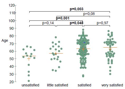
Figure 1: Self-rated patient’s satisfaction according to age.

Differences between Recent and Long-Lasting Tendinopathies

Even more interestingly, there was no significant difference in improvement (P=0.89), time before onset of improvement (P=0.12) and patient satisfaction (P=0.15) (Table 2), according to the tendinopathy duration. However, the percentage of patients who returned to sport or professional activities was lower in long-lasting tendinopathies (P=0.004) and the treatment duration was significantly longer (P=0.001) in the latter than in those with recent ones. All details are given in Table 3.

Table 2: Improvement, patient’s satisfaction and return to sport and professional activities, according to the tendinopathy duration (6 months).

Items Units All <6 Months >6 Months P Value
Number of patients N 219 76 143
Improvement Yes/No % 88 93 86 0.89
Percentage of improvement

  • <25%
  • [25-50[%
  • [50-75[%
  • [75-100[%
  • 100%
 

12.5

22.8

37.2

21.9

5.6


9

21

35

23

9


16

25

37

21

2


0.15

Time before improvement

    • <2 weeks
    • [2-4[ weeks
    • [4-8[ weeks
    • > 8 weeks


%


8

28

37

28


9

25

45

21


7

29

31

33


0.12

TOL19-001 treatment duration

    • <3 months
    • [3-6[ months
    • >6 months


%


11

36

53


16

42

42


9

33

58


0.001

Satisfaction

    • Very satisfied
    • Satisfied
    • Little satisfied
    • Not satisfied


%


24

59

10

7


22

68

7

4


26

54

12

8


0.19

Return to sport or work

    • Without limitation
    • With some limitations
    • No recovery


%


49.5

33

17.5


59

34

7


44.4

32.3

23.3


0.004


Table 3: Percentage of satisfied and unsatisfied patients according to the tendinopathy duration before TOL19-001 prescription (χ2 test: P=0.23; Fisher test: P=0.25).

Time Before TOL19-001 (month=M)

<1 M [1 M-3 M[ [3 M-6 M[ [6 M-12 M[

≥12 M


Level of satisfaction

(% of patients)

1-Very satisfied

54.5 12.5 19.3 20.8 26.8

2-Satisfied

45.5 81.2 61.3 62.5

52.7

1+2

100 93.7 80.6 83.3

79.5

3-Little satisfied

0 6.3 9.7 12.5

11.6

4-Not satisfied

0 0 9.7 4.2

8.9

3+4 0 6.3 19.4 16.7

19.5


Safety

Neither severe nor serious Adverse Events (AEs) were reported by the patients. Safety was rated as very good or good by 97% of subjects. Only 7 patients reported minor gastrointestinal discomfort, possibly related to treatment. Three patients (1.4%) discontinued TOL19-001 for AEs.

Discussion

First of all it must be stressed that the present study has not been designed to demonstrate the efficacy of TOL19-001 in tendinopathies, but for giving information that could be useful to design, at best, a controlled trial. It should also be emphasized that, in the absence of a control group, we do not assert that TOL19-001 is an effective treatment for relieving pain and “accelerate” healing of tendinopathies. Although the magnitude of the clinical benefit may be overestimated due to a possible placebo effect, our results suggest that TOL19-001 may be helpful in many patients with long-lasting tendinopathies, in whom conventional therapies have failed. Indeed, it is unlikely that a chronic tendinopathy, lasting for more than 6, and even more than 12 months (i.e., 54% of our cohort), despite a well-conducted treatment (including rehabilitation and corticosteroid injection), spontaneously cures in less than two months, as we observed in several of our patients. On the contrary, as mentioned above, it is probable that most patients suffering from a tendinopathy for less than 3 to 6 months would have healed within 2 or 3 months anyway, even without treatment. It is the reason why we particularly focused attention on patients with tendinopathy evolving for many months (>12 months). Most of them had been forced to stop any sport or professional activity because of the tendinopathy. Among those patients, a large majority could return to sport or work after only a few weeks, most of the time without any limitation. More than 8 out of 10 patients considered TOL19-001 as very effective or effective and were satisfied with the treatment, whatever the tendon involved or the duration of the condition. Yet most of them had already received multiple treatments, without getting healing.

Our survey suffers from several limitations. The present data are patient self-reported and have been obtained retrospectively. Only 73% of TOL19-001 treated patients answered the questionnaire. So it cannot be presumed what the missing 27% would have answered. The retrospective nature of the survey made impossible to measure the decrease of pain over time. Furthermore, we were not able to obtain data about the anatomical severity of the condition and did not have any imaging data. Lastly, satisfaction with a treatment, is a subjective and patient-dependent data, involving many and varied parameters.

It has been shown that the mechanism of action of TOL19-001 is mainly mediated through its antioxidant properties [9]. TOL19-001 is mainly constituted of spirulina (Arthrospira maxima), an undifferentiated multicellular filamentous cyanobacterium, also called blue-green alga. Spirulina is known, since a long time, for being a potential source of vitamins and essential amino acids. Indeed, Spirulina contains a wide spectrum of nutrients including B-complex vitamins, minerals, essential amino-acids, beta-carotene, vitamin E. Spirulina is also noteworthy for its high contents (7%) in fatty acids (α-linolenic acid, linoleic acid, stearidonic acid, docosahexaenoic acid, and arachidonic acid) and proteins, making it desirable as a food supplement [10]. Spirulina has hypolipidemic, hypoglycemic, and antihypertensive properties. It is claimed to have also anti-cancer and immune-suppressing properties [11,12]. Spirulina also exerts a strong Radical Oxygen Species (ROS) scavenging activity and can reduce oxidative damage by decreasing free oxygen radical accumulation, through activation of several antioxidant enzyme systems (i.e., Superoxide Dismutase (SOD), glutathione peroxidase, catalase) [13]. The anti-inflammatory activity of Spirulina is mainly due to phycocyanin as demonstrated in several in vitro and in vivo animal models of arthritis [13].

Spirulina is also an important source of essential amino acids. A strong relationship between leucine and collagen synthesis has been demonstrated in several animal models [5]. A positive effect on collagen synthesis was demonstrated in malnourished mice teated with a leucine-enriched diet. Leucine increases hydroxyproline levels which plays a role in collageb fiber stability [14]. Glycine also stimulates the synthesis of both hydroxyproline and glycosaminoglycans, resulting in higher collagen fiber strength [15]. These properties could also support recovery from tissue damage [5].

TOL19-001® is mainly composed of Spirulina maxima (about 60%), but also contains significant amounts of glucosamine sulfate (~20%), and ginseng (~15%). Consequently, it is impossible to assert which of these components are responsible for TOL19-001 effect on the tendon. A systematic review [16] including preclinical and clinical data from 46 articles suggested that several nutraceuticals (i.e. curcumin, vitamin C, bromelain, glucosamine, chondroitin…) demonstrated beneficial effects on normal and pathological tendons through a possible role on collagen synthesis, inflammation, mechanical properties, oxidative stress, and analgesia. We previously published that TOL19-001® reduces the expression of MMPs, PGE2 and type III collagen in IL-1β stimulated cells. By reducing MMPs, TOL19-001 might reduce the deterioration and favor regenerative processes and tendon healing [8]. The downregulation of PGE2, likely plays a role in relieving pain due to tendon inflammation.

Glucosamine may also contribute to the beneficial effect of TOL-19-001 on tendon. Animal studies [17-19] have shown positive effects of glucosamine and chondroitin sulfate on collagen synthesis, tendon ultrastructural organization and biomechanical properties, probably as a consequence of the lower tissue inflammation and greater collagen synthesis [19]. Lastly, another reason for TOL19-001 effectiveness in long-lasting tendinopathies may be its ability in reducing type III collagen production, since type III collagen-rich scar tissue impairs tendon function and makes the tendon susceptible to re-injury [8]. This particular property might be beneficial effect on tendon healing, especially in long-lasting or recurrent tendinopathies.

Conclusion

This pilot cross-sectional study, carried-out in real life conditions gave interesting information on tendinopathies clinical evolution in patients taking TOL19-001. It suggested that TOL19-001 might benefit patients with long-lasting tendinopathy, in whom the conventional treatment failed. Further randomized controlled trials, focused on patients with long-lasting tendinopathies and using tendon imaging methods, are mandatory for confirming these preliminary results, and to assess the in vivo mechanisms of action of TOL19-001 on tendon tissue.

Authors Contribution

TC performed data analysis, contributed to the drafting the article, gave final approval of the version to be published, and agreed to be accountable for all aspects of the work.

PL wrote the article, gave final approval of the version to be published, and agreed to be accountable for all aspects of the work.

EN contributed to the drafting the article, gave final approval of the version to be published, and agreed to be accountable for all aspects of the work.

HB contributed to the drafting the article, gave final approval of the version to be published, and agreed to be accountable for all aspects of the work.

Funding

No funding.

None of the authors have received fees related to the present study.

LABRHA SAS paid only for the article publication.

Ethics Declarations

Conflict of Interest

HB declares having received fees from LABRHA SAS as a board member.

TC declares having received fees from LABRHA SAS as a scientific and medical consultant and board member.

EN and PL declare that they have no conflict of interests related to the present study.

Informed Consent

Due to the retrospective nature of the study, the patient consent was not required. We make sure to keep patient data confidential and compliance with the Declaration of Helsinki.

Availability of Data and Materials

All data generated or analyzed during this study are available at Laboratoire de rhumatologie appliqué; 19 place Tolozan, Lyon, France.

References

    1. Andarawis-Puri N, Flatow EL, Soslowsky LJ (2015) Tendon Basic Science: Development, repair, regeneration, and healing. J Orthop Res 33: 780. [crossref]
    2. McCormick A, Charlton J, Fleming D (1995) Assessing health needs in primary care. Morbidity study from general practice provides another source of information. BMJ 310: 1534. [crossref]
    3. Andres BM, Murrell GAC (2008) Treatment of tendinopathy: What works, what does not, and what is on the horizon. Clin Orthop Relat Res 466: 1539-1554. [crossref]
    4. Childress MA, Beutler A (2013) Management of chronic tendon injuries. Am Fam Physician 87: 486-490. [crossref]
    5. Loiacono C, Palermi S, Massa B, Belviso I, Romano V, et al. (2019) Tendinopathy: Pathophysiology, therapeutic options, and role of nutraceutics. A narrative literature review. Medicina (Kaunas) 55: 447. [crossref]
    6. Abate M, Schiavone C, Salini V (2014) The use of hyaluronic acid after tendon surgery and in tendinopathies. Biomed Res Int 2014: 783632. [crossref]
    7. Zhang K, Zhang S, Li Q, Yang J, Dong W, et al. (2014) Effects of celecoxib on proliferation and tenocytic differentiation of tendon-derived stem cells. Biochem Biophys R Commun 450: 762-766.
    8. Baugé C, Leclercq S, Conrozier T, Boumediene K (2015) TOL19-001 reduces inflammation and MMP expression in monolayer cultures of tendon cells. BMC Complement Altern Med 15: 217. [crossref]
    9. Riley G (2008) Tendinopathy-from basic science to treatment. Nat Clin Pract Rheumatol 4: 82-89. [crossref]
    10. Nicoletti M (2016) Microalgae nutraceuticals. Foods 5. [crossref]
    11. Khan Z1, Bhadouria P, Bisen PS (2005) Nutritional and therapeutic potential of spirulina. Curr Pharm Biotechnol 6: 373-379. [crossref]
    12. Finamore A, Palmery M, Bensehaila S, Peluso I (2017) Antioxidant, immunomodulating, and microbial-modulating activities of the sustainable and ecofriendly spirulina. Oxid Med Cell Longev  2017: 3247528. [crossref]
    13. Romay C, Armesto J, Remirez D, González R, Ledon N, et al. (1998) Antioxidant and anti-inflammatory properties of C-phycocyanin from blue-green algae. Inflamm Res Off J Eur Histamine Res Soc Al 47: 36-44. [crossref]
    14. Barbosa AWC, Benevides GP, Alferes LMT, Salomão EM, Gomes-Marcondes MCC, et al. (2012) A leucine-rich diet and exercise affect the biomechanical characteristics of the digital flexor tendon in rats after nutritional recovery. Amino Acids 4: 329-336. [crossref]
    15. Vieira CP, De Oliveira LP, Guerra FDR, De Almeida MDS, Marcondes MCCG (2015) Glycine improves biochemical and biomechanical properties following inflammation of the achilles tendon. Anat Rec 298: 538-545. [crossref]
    16. Fusini F, Bisicchia S, Bottegoni C, Gigante A, Zanchini F, et al. (2016) Nutraceutical supplement in the management of tendinopathies: A systematic review. Muscles Ligaments Tendons J  6: 48-57. [crossref]
    17. Taşkesen A, Ataoğlu B, Özer M, Demirkale İ, Turanli S (2015) Glucosamine-chondroitin sulphate accelerates tendon-to-bone healing in rabbits. Jt Dis Relat Surg 26: 77-83. [crossref]
    18. Lippiello L (2007) Collagen synthesis in tenocytes, ligament cells and chondrocytes exposed to a combination of glucosamine hcl and chondroitin sulfate. Evid Based Complement Alternat Med 4: 219-224. [crossref]
    19. Ozer H, Taşkesen A, Kul O, Selek HY, Turanlı S, et al. (2011) Effect of glucosamine chondroitine sulphate on repaired tenotomized rat achilles tendons. Eklem Hastalik. Cerrahisi 22: 100-106. [crossref]
fig 1

Utilization of a Combined Adductor Canal and IPACK (Interspace between the Popliteal Artery and the Capsule of the Posterior Knee) Block for Pediatric Anterior Cruciate Ligament Repair

DOI: 10.31038/IJOT.2020322


The incidence of Anterior Cruciate Ligament (ACL) reconstruction amongst pediatric patients has risen over the last decade [1]. Injuries to the ACL can result in ligament rupture or, less commonly, traumatic avulsion fracture at the ACL insertion site on the proximal tibia. Postoperative pain control after ACL reconstruction or repair usually involves opioids. Adult literature demonstrated daily opioid consumption of 35-45 mg morphine equivalents in the first 2 weeks after ACL reconstruction [2]. Regional anesthesia has proven to reduce nociceptive pain and opioid requirements following ACL surgery, thereby facilitating earlier hospital discharge and improved patient satisfaction [3-5].

Widely used for knee surgery, the Adductor Canal Block (ACB) provides motor-sparing analgesia to the anteromedial knee and distal medial leg [2,4,6]. However, its lack of posterior knee and anterolateral coverage makes it non-ideal as a sole agent. The ultrasound-guided deposition of local anesthetic to the Interspace between the Popliteal Artery and The Posterior Knee Capsule (IPACK) in combination with blockade of the anterior knee with an ACB has gained significant interest for such surgery [7,8]. Herein we describe the utilization of the IPACK combined with an ACB for arthroscopic ACL repair in a pediatric patient.

A 10-year old, 41kg, healthy male presented following a motor vehicle injury. Imaging revealed left ACL avulsion from its tibial attachment, anterior-lateral meniscus tear and avulsion of the popliteofibular ligament. He underwent arthroscopic assisted repair of the proximal tibial avulsion fracture. Proceeding general anesthesia, the patient was positioned supine with the left lower extremity externally rotated and flexed 40 at the knee. A high-frequency linear ultrasound probe (15MHz) was placed transverse to the medial thigh, and rotated medially to visualize the adductor canal for ACB. A 22-gauge 50 mm echogenic needle was advanced in-plane from the lateral to medial direction, deep to the sartorius and juxtaposed to the vastus medialis. Following negative aspiration, 6ml of bupivacaine 0.25% with preservative-free clonidine 20mcg was deposited in the adductor canal. A curvilinear, low-frequency transducer (2-5MHz) was used for the IPACK to maximize visualization. With the patient in the same position, the probe was placed in the popliteal fossa identifying the popliteal artery and femoral condyles. The probe was then advanced cephalad until the shaft of the femur was visualized. A 22-gauge 80 mm echogenic needle was advanced in-plane, deep to the popliteal artery and above the joint capsule. Following negative aspiration, 10 ml of bupivacaine 0.25% with preservative-free clonidine 20mcg was deposited (Figure 1). Both ACB and IPACK single shot nerve blocks were performed in 6 minutes.

fig 1

Figure 1: IPACK blockade with arrow highlighting needle in-situ. Femoral Artery (FA), Popliteal Nerve (PN), Local Anesthetic (LA).

After the patient was prepped and draped, the anatomic landmarks of the anteromedial and anterolateral portals were identified. Additional short-acting local anesthetic (lidocaine 1% with epinephrine 1:200k) was administered by the surgeon at the anterolateral and anteromedial port sites. The arthroscope was introduced through the anterolateral portal. Systematic arthroscopic evaluation revealed a displaced tibial spine fracture with the ACL attached to a fragment and hinging of the anterior aspect of the fracture with intermeniscal ligament interposition. The intermeniscal ligament was extracted and the fracture bed was debrided. The sutures were secured to the ACL base and then secured to the donor site of the fracture with push lock anchors. There was successful fixation after anchor placement. The patient was maintained on 1 MAC of sevoflurane and received a total of 1.5 mg/kg of ketamine given in divided doses; no intraoperative opioids were required. The total operative time was 89 minutes. The procedure was uneventful and there were no apparent complications. The estimated blood loss was minimal.

In the immediate post-operative period the child’s numeric pain score was 4/10. Pain was isolated to the anterior lateral port site, for which 0.02 mg/kg (1mg) of morphine was administered. Physical exam revealed no sensation to light touch on the anteromedial or posterior knee with preserved plantar-dorsiflexion at the ankle. On 24-hour outpatient follow-up, parents reported good pain control without motor weakness. He received one dose of an oral acetaminophen-opioid preparation approximately16 hours from discharge. There was no evidence of block complication.

Arthroscopic tibial spine fracture repair usually involves medial and lateral parapatellar portals. The tibial spine is fixed via sutures or screws. The ACL emerges from the medial and anterior tibial plateau coursing superiorly to its posterior insertion site along the posterior lateral femoral condyle. As such, surgical manipulation can impose both anterior and posterior capsule knee pain. The ACB reproducibly offers similar anterior knee analgesia when compared to a femoral nerve block while avoiding quadriceps weakness, which allows faster ambulatory recovery and discharge [6,7]. IPACK targets the articular branches originating from the tibial and obturator nerves that travel through a tissue space between the popliteal artery and the femur. It provides posterior knee capsule analgesia by blockade of the obturator, tibial and common peroneal sensory innervation while sparing the motor effect observed with a sciatic nerve block, thereby maintaining the sensorimotor function of the leg and foot.

As evident in a cadaveric study, local anesthetic delivered via IPACK spreads throughout the popliteal fossa without proximal sciatic involvement [8], albeit extension to the common peroneal and tibial nerves was plausible. Combining the ACB and IPACK, opioid-sparing effect can be achieved for ACL repair or reconstruction while preserving motor function. This concept harmonized with the adult literature where reduction of morphine equivalent consumption and faster ambulatory discharge were noted with the addition of an IPACK block for ACL repair [9]. Our experience demonstrated motor-sparing effect and adequate analgesia with the IPACK and ACB combination for a pediatric patient undergoing ACL repair. While the adult literature and our observation suggests promising results, there are no studies that have investigated the safety and efficacy of IPACK block among pediatric patients undergoing knee surgery. Future investigation into the role of the IPACK block for ACL repair in pediatric patients is required.

References

    1. Ellis HB, VandenBerg C, Beck J, Pennock A, Pennock A, Cruz AI, et al. (2019) Trends in pediatric anterior cruciate ligament reconstruction: A review of surgeon fellowship, geography, and meniscus surgery in the ABOS part 2 database. Orthopc J Sports Med. [crossref]
    2. Barnett S, Murray MM, Liu S (2020) Resolution of pain and predictors of postoperative opioid use after bridge-enhanced anterior cruciate ligament repair and anterior crucial ligament reconstruction. Arthrosc Sports Med Rehabil 2: 219-228.
    3. Iskandar H, Benard A, Ruel-Raymond J, Cochard G, Manaud B, et al. (2003) Femoral block provides superior analgesia compared with intra-articular ropivacaine after anterior cruciate ligament reconstruction. Reg Anesth Pain Med 28: 29-32. [crossref]
    4. Abdallah FW, Whelan DB, Chan VW, Prasad GA, Endersby RV, et al. (2016) Adductor canal block provides noninferior analgesia and superior quadriceps strength compared with femoral nerve block in anterior cruciate ligament reconstruction. Anesthesiology 124: 1053-1064. [crossref]
    5. Koh IJ, Chang CB, Seo ES, Kim SJ, Seong SC, et al. (2012) Pain management by periarticular multimodal drug injection after anterior cruciate ligament reconstruction: A randomized, controlled study. Arthroscopy 28: 649-657. [crossref]
    6. Ludwigson JL, Tillmans SD, Galgon RE, Chambers TA, et al. (2015) A comparison of single shot adductor canal block versus femoral nerve catheter for total knee arthroplasty. J Arthroplasty 30: 68-71. [crossref]
    7. Kim D, Beathe J, Lin Y, Goytizolo E, Oxendine J, et al. (2019) The addition of ACB and IPACK to PAI enhances postoperative pain control in TKA: A randomized controlled trial. Anesth Analga 129: 526-535.
    8. Niesen AD, Harris DJ, Johnson CS, Stoike DE, et al. (2019) Interspace between Popliteal Artery and Posterior Capsule of the Knee (IPACK) injectate spread: A cadaver study. J Ultrasound Med 38: 741-745. [crossref]
    9. Huang A, Singh PA, Woon KL (2020) Interspace between the popliteal artery and the capsule of the knee (IPACK) block for anterior cruciate ligament reconstruction surgery: A two case series. Open Journal of Anesthesiology 10: 134-143.
fig 1

Circulating Agonist Autoantibody to 5-Hydroxytryptamine 2A Receptor in Lean and Diabetic Fatty Zucker Rat Strains

DOI: 10.31038/EDMJ.2020435

Abstract

Aims: Circulating neurotoxic autoantibodies to the 5-hydroxytryptamine 2A receptor were increased in older adult type 2 diabetes in association with certain neurodegenerative complications. The male Zucker diabetic fatty (ZDF) rat is a model system for studies of obese, type 2 diabetes mellitus. The aim of the current study was to test for (and compare) circulating neurotoxic autoantibodies to the 5-hydroxytryptamine 2A receptor in the Zucker diabetic fatty rat and age-matched lean Zucker rat strains.

Methods: Plasma from lean and Zucker diabetic fatty rat (obtained at different developmental stages) was subjected to protein G affinity chromatography. The resulting immunoglobulin G fraction was tested for neurotoxicity (acute neurite retraction, accelerated neuron loss) in N2A mouse neuroblastoma cells and for binding to a linear synthetic peptide corresponding to the second extracellular loop of the 5-hydroxytryptamine 2A receptor.

Results: The male Zucker diabetic fatty rat (fa/fa) and two Zucker lean strains (+/?) and (fa/+) harbored autoantibodies to the 5-hydroxytryptamine 2A receptor which appeared spontaneously around 7-8.5 weeks of age. The circulating autoantibodies persisted until at least 25 weeks of age in the Zucker diabetic fatty rat and in the Zucker heterozygote (fa/+), but were no longer detectable in 25-week-old lean (+/?) Zucker rats. Autoantibody-induced acute neurite retraction and accelerated loss in mouse neuroblastoma N2A cells was dose-dependently prevented by selective antagonists of the 5-hydroxytryptamine 2A receptor. It was also substantially prevented by co-incubation with antagonists of RhoA/Rho kinase-mediated signaling (Y27632) or Gq11/phospholipase C/inositol triphosphate receptor-coupled signaling.

Conclusions: These data suggest that neurotoxic 5-hydroxytryptamine 2A receptor-targeting autoantibodies increase in the aging male Zucker diabetic fatty rat and in male Zucker lean rats harboring a heterozygous mutation, but not in age-matched, older Zucker lean rats lacking a known leptin receptor mutation. The Zucker genetic strain may be useful in studies of the role of humoral and/or innate immunity in late neurodegeneration.

Introduction

Type 2 diabetes mellitus is associated with an increased risk of late neurodegeneration [1,2] via mechanisms which may involve (in part) increased peripheral and central inflammation. Increased pro-inflammatory cytokines and innate immunity have been associated with early Parkinson’s disease [3] and dementia [4] through complex mechanisms. Leptin is a hormone released by fat cells that is important in energy metabolism. Dysfunction of the leptin system results in classic signs of type 2 diabetes mellitus, such as obesity, insulin resistance and high circulating insulin. Leptin is also a member of the type 1 cytokine family that includes interleukin-6 [5,6]. The leptin receptor is normally expressed on hematopoietic cells where it stimulates helper- T cell, and effector T cell functions and inhibits regulatory T cell function [7].

The male Zucker Diabetic Fatty rat (fa/fa) (ZDF) is a well-recognized animal model of obese type 2 diabetes mellitus and hypertension [8,9] in which hyperphagia causes morbid obesity leading to high level of pro-inflammatory adipocytokines [10]. In the ZDF rat, pro-inflammatory cytokines cause enhanced innate immunity [11], however, owing to its complete leptin receptor deficiency, helper T-cell function is reduced [12]. The lean heterozygous Zucker rat (fa/+) has haplo-sufficiency at the leptin receptor and a two-fold increased plasma leptin concentration [13] compared to the wild-type (+/+) lean Zucker rat. Since hyperleptinemia promoted helper-T cell-mediated autoantibody formation and worsened clinical outcome(s) in a mouse model of systemic lupus erythematosus [14], the lean heterozygote Zucker rat might be expected to exhibit higher autoantibody expression than in the ZDF (fa/fa) rat strain.

In prior studies in older adult (human) obese type 2 diabetes, patients harboring increased circulating autoantibodies to the 5-hydroxytryptamine 2A receptor (5-HT2AR) had an increased prevalence of co-morbid neurodegenerative complications including Parkinson’s disease and dementia [15]. Human plasma autoantibodies targeting the 5-HT2A receptor promoted long-lasting (agonistic) activation of Gq11/phospholipase C/inositol triphosphate receptor/Ca2+ signaling causing accelerated endothelial cell and neuronal cell death [16]. Since the 5-hydroxytryptamine 2A receptor is normally expressed on arterial smooth muscle cells [17] and on neurons in specific brain regions involved in the regulation of mood, thinking, perception, and sleep [18], altered signaling at the 5-HT2A receptor could have diverse peripheral and central effects.

The aim of the present study was to test for spontaneously-occurring autoantibodies to the 5-HT2A receptor in the circulation in male ZDF rat (fa/fa) and age-matched male lean Zucker rats either lacking a known leptin receptor mutation (+/?) or harboring a heterozygous leptin receptor mutation (fa/+). The developmental expression of autoantibody was compared in each genetic strain, and the mechanism of neurotoxicity downstream of 5-HT2AR binding was evaluated in mouse neuroblastoma N2A cells.

Materials and Methods

Animals

All procedures were conducted according to the National Institutes of Health (NIH) Guide for the Care and Use of Laboratory Animals and approved by the Institutional Animal Care and Use Committee of the Veterans Affairs Medical Center (East Orange, New Jersey). Male ZDF (N=12) and lean (+/?) Zucker rats (N=12) were obtained from Charles River Laboratories (Kingston, NY) at approximately 6 weeks of age. All rats were single housed upon arrival, with modest enrichment (a PVC tube). Rats were provided ad libitum access to food and water and maintained in a 12 h light/dark cycle with lights on at 0630. All procedures occurred during the light phase of the cycle.

Blood Drawing

EDTA plasma from 10-week ZDF rat and 10-week lean, heterozygous (fa/+) Zucker rats was obtained from Charles River Laboratories. For all other time points, blood was collected from the retro-orbital plexus of ZDF, and lean (+/?) Zucker rats using a retro-orbital blood procedure. The blood was centrifuged at 1400 g x 10 minutes at 4 degrees C. The resulting plasma was stored at -4oC for up to 2-3 weeks prior to protein G affinity chromatography.

Tail-Nick

Capillary blood glucose was determined from a drop of blood obtained after tail nick at sequential time intervals. A glucose dehydrogenase method (Accucheck Aviva, Roche Diagnostics, Inc) was used for the determination of blood glucose.

Protein G Affinity Chromatography

Protein-G affinity chromatography was carried out as previously reported for protein-A affinity chromatography in human plasma [16]. The pH of the 10 mm Tris binding buffer (6.5) and the 0.1 M citrate elution buffer (2.7) was adjusted to optimize binding and elution of rat immunoglobulin G. Protein-G eluate fractions were stored at 0-4 degrees C.

Mouse N2A Neuroblastoma Cells

Cells were cultured in DMEM with 10% fetal calf serum as previously reported [16].

Neurite Retraction and N2A Cell Survival Assays

Assays were performed as previously described [16].

Enzyme-Linked Immunoassay (ELISA)

The ELISA procedure was carried out as previously reported using an 18-meric linear synthetic peptide Q..N18 corresponding to the second extracellular loop of the human 5-HT2AR as the solid phase antigen [15].

Chemicals

All chemicals were obtained from Sigma Co., Inc. (St. Louis, MO).

Protein determination-Protein levels were determined using a bichinchonic assay method (BioRad, Inc.).

Statistics

Statistical analysis was performed using an unpaired Student’s t-test with an α cutoff level for significance of < 0.05.

Results

Occurrence of Circulating 5-HT2A Receptor Autoantibody in the ZDF Rat: Relation to Obesity and Diabetes

Mean body mass in ZDF rats significantly exceeded that in lean (+/?) Zucker rat at week 7, week 8.5, and week 12 (Figure 1A). There was no longer a significant weight difference between the strains at week 25 (Figure 1A). Diabetes (hyperglycemia) emerged between week 8-10 in the ZDF rat, but at week 7 the ZDF rat was still normoglycemic (Figure 1B). In contrast, lean (+/?) Zucker rats were normoglycemic at all ages tested (Figure 1B), not tested at week 7.

fig 1 mark

Figure 1: Age-dependent change in body mass(A) and capillary blood glucose concentration (B) in male Zucker fatty (ZDF) vs lean (+/?) (ZDL) rats. A) Body weight increased significantly in the male ZDF rat (starting at week 7) compared to control lean Zucker rat B) capillary blood glucose was determined by tail nick using glucose dehydrogenase method as described in Materials and Methods. Glucose was normal at week 7 in the ZDF rat, but increased significantly compared to ZDL rat starting at week 8.

Detection of plasma immunoreactive 5-HT2AR autoantibody had a similar trajectory to diabetes in ZDF rats. At 7 weeks of age, ZDF rat had no detectable plasma immunoreactive 5-HT2AR autoantibody (Figure 1C). At 10-weeks of age, plasma from ZDF rat contained a two-fold, higher binding to 5-HT2AR (0.14 AU, N=2) compared to background levels (0.07 AU; Figure 2a). Plasma from lean ((+/?) Zucker rats harbored two-fold or slightly higher level of autoantibody binding to 5-HT2AR between 8.5-15 weeks of age which declined to undetectable levels at 25 weeks of age (Figure 2B).

fig 2

Figure 2: Developmental expression of autoantibody binding to 5-HT2AR synthetic peptide in three Zucker rat strains. A 2 ug/mL concentration of the protein G eluate was tested for binding to the linear synthetic 18-meric 5-HT2A receptor peptide as described in Materials and Methods. Results are “mean +/- SE” in A) ZDF rat: 7-week (N=2); 8.5-week (N=3); 10-week (N=2); 25-week (N=3) B) lean (+/?) Zucker rat: 7-week (N=3); 8.5 week (N=3); 15-week (N=10); 25-week (N=6). C) lean heterozygote (fa/+) Zucker rat: 7-week (N=2); 10-week (N=5); 25-week (N=2).

Spontaneous IgG autoantibody to the 5-HT2A receptor was already present at 7 weeks of age in the heterozygous lean (fa/+) Zucker rat (Figure 2C) and levels at 10-weeks old and 25-weeks old were substantially higher than in age-matched ZDF rat (Figure2C). Autoantibody to the 5-HT2A receptor persisted into older age, i.e. 25 weeks old, in male ZDF and lean heterozygous Zucker rats, but disappeared at 25 weeks of age in the lean (+/?) Zucker rat lacking a known leptin receptor mutation. In a recent report [19], diabetes, obesity and hypertension were associated with substantially increased hazard rates for the occurrence of dementia, Parkinson’s disease and severe depression following traumatic brain injury in older adult veterans.

The present data suggest that male ZDF and heterozygous lean rat strains may be useful in modeling the effects of heightened innate and/or adaptive immunity, respectively, on the occurrence of neurodegeneration.

Zucker Diabetic Fatty Rat Plasma 5-HT2AR Autoantibody Inhibits N2A Cell Survival

We next tested for neurotoxicity in the autoantibody (from 10- week-old ZDF rat plasma) that displayed increased binding to the 5-HT2A receptor peptide. Autoantibody in plasma from the 10-week old ZDF rat caused dose-dependent inhibition of survival in N2A mouse neuroblastoma cells (Figure 3). A two and one-half microgram per milliliter concentration of the IgG caused approximately 40% neuroblastoma cell loss (after 24 hours incubation) compared to control, untreated cells (P< 0.01, Figure 3).

fig 3

Figure 3: Dose-dependent neurotoxicity in the protein G eluate fraction from Zucker diabetes fatty rat plasma. The protein G-eluate fraction from 10-week-old male Zucker diabetic fatty rat plasma was incubated with N2a mouse neuroblastoma cells (at the indicated concentrations) for 24 hours. % N2a cell survival was determined with an MTT assay as described in Materials and Methods. Each point represents the mean + SE of quadruplicate determinations; * P < 0.01 compared to cell survival in untreated cells.

Zucker Diabetic Fatty Rat Plasma 5-HT2AR Autoantibody Causes Acute N2A Neurite Retraction

A 120 nanomolar concentration (1.8 g/mL IgG) of the protein G- eluate fraction of plasma from 10-week-old male ZDF fatty rat caused significant 37% acute neurite length-shortening (after 15 minutes of incubation) of N2A mouse neuroblastoma cells (Figure 4A). Co-incubation with a 200 nanomolar concentration of M100907, a highly selective, potent antagonist of the 5-HT2AR, afforded 86% significant (P < 0.01) protection against autoantibody-induced neurite retraction (Figure 4A).

fig 4A-4B

Figure 4: Neurite outgrowth inhibition (A) and accelerated N2A neuroblastoma cell loss (B) in autoantibody from ZDF rat plasma: neutralization by M100907, a highly selective 5-HT2AR antagonist. A) A 120 nM concentration of the protein-G eluate fraction of 10-week-old male ZDF rat plasma was incubated with N2A mouse neuroblastoma cells in the presence or absence of a 200 nM concentration of the selective 5-HT2AR antagonist M100907. % Basal neurite length was determined after 15 minutes as described in Materials and Methods B) A 160 nM concentration of the protein G-eluate fraction from 10-week-old male Zucker diabetic fatty rat plasma was incubated with N2a mouse neuroblastoma cells in the presence or absence of the indicated concentration of M100907 for 24 hours at 37 deg C. Percent N2A cell survival was determined with an MTT assay as described in Materials and Methods. Each point represents the mean + SE of triplicate determinations.

Pharmacologic Profile of Zucker Diabetic Fatty Rat Plasma 5-HT2AR Autoantibody Toxicity

A ~170 nanomolar concentration (2.5 µg/mL) of the protein G-eluate fraction of plasma from 10-week-old male ZDF rat was incubated with N2A mouse neuroblastoma cells in the presence or absence of a (500 or 1000) nanomolar concentration of M100907 for 24 hours at 37 deg C. M100907 caused significant (dose-dependent) protection against accelerated N2A cell loss, after 24 hours incubation with the ZDF rat IgG (Figure 4B).

Spiperone is a potent, but less selective 5-HT2AR antagonist. Spiperone at 500-1000 nanomolar concentrations afforded significant dose-dependent protection against ZDF rat IgG-induced accelerated N2A cell loss (Figure 5). Ketanserin is a less potent 5-HT2AR antagonist compared to M100907 or spiperone. Substantially higher concentration (2.5 µM) of ketanserin was required to significantly protect against ZDF rat IgG-induced N2A cell loss (Figure 6).

fig 5

Figure 5: Potent, less selective 5-HT2AR antagonist (spiperone) dose-dependently prevented accelerated neuron loss induced by ZDF rat plasma autoantibody. A 160 nM concentration of the protein G-eluate fraction from 10-week-old male Zucker diabetic fatty rat plasma was incubated with N2a mouse neuroblastoma cells in the presence or absence of the indicated concentration of spiperone for 24 hours at 37 deg C. Percent N2a cell survival was determined with an MTT assay as described in Materials and Methods. Each point represents the mean + SE of triplicate determinations.

fig 6A-6B

Figure 6: Higher concentration of ketanserin, a less potent, but selective 5-HT2AR antagonist dose-dependently prevented accelerated neuron loss induced by ZDF rat plasma autoantibody A-B) 120 nM concentration of the protein G-eluate fraction from 10-week-old male Zucker diabetic fatty rat plasma was incubated with N2a mouse neuroblastoma cells in the presence or absence of the indicated concentration of ketanserin for 24 hours at 37 deg C. Percent N2a cell survival was determined with an MTT assay as described in Materials and Methods. Each point represents the mean + SE of triplicate determinations.

The receptor antagonist profile and concentration associated with significant protection against ZDF IgG autoantibody-induced neuroblastoma cell loss is consistent with relative affinity constants on the 5-HT2A receptor (M100907 < spiperone<< ketanserin).

A one micromolar concentrations of SB-20471, a selective antagonist to the 5-HT2B receptor did not significantly prevent ZDF autoantibody-induced neurite retraction (Table 1). A ten micromolar concentration of either bosentan (an endothelin-1 receptor) or losartan (an angiotensin II type 1 receptor) antagonist did not significantly protect against ZDF IgG-induced acute N2A neurite retraction (Table 1). An 850 nanomolar concentration of prazosin (alpha 1 adrenergic R antagonist) did not afford significant protection against ZDF IgG autoantibody-induced acute neurite retraction (Table 1). Taken together, these data suggest that neurotoxicity in the 10-week old male ZDF rat IgG appears selective for the 5-HT2A receptor.

Table 1: Receptor specificity of prevention of ZDF rat IgG autoantibody-induced acute N2A neurite retraction.

Antagonist

Conc GPCR

% of ZDF IgG-induced Neurite Retraction

M100907

200 nM 5HT2A

13% + 15%

SB-20471

1 µM 5HT2B

89% + 20%

Bosentan

10 µM ET-1

95% + 20%

Losartan

10 µM AT-1

81% + 5%

Prazosin

850 nM A1-A

89% + 11%

A 160 nanomolar concentration of 10-week-old male Zucker fatty rat IgG autoantibodies was incubated in the presence or absence of the indicated GPCRs. Acute N2A neurite retraction was determined after 15 minutes as described in Materials and Methods. Results are mean +/- SD.

Involvement of Gq11/Phospholipase C/Inositol Triphosphate and Rho Kinase Signaling Pathways

Co-incubation of N2A cells with ZDF plasma IgG autoantibodies together with individual selective antagonists of Gq11 (YM-254890), phospholipase C (U73122), inositol triphosphate receptor (2-APB) signaling or Rho A/Rho kinase (Y27632) mediated signaling completely or nearly completely protected against acute neurite retraction (Table 2) consistent with involvement of Gq11/phospholipase C/inositol triphosphate and Rho kinase signaling pathways in mediating IgG neurotoxicity downstream of 5-HT2A receptor binding.

Table 2: Effect of signaling pathway inhibitors on ZDF rat IgG autoantibody-induced acute N2A neurite retraction.

Antagonist

Conc  Signaling  Intermediate % of ZDF IgG-induced Neurite Retraction

U73122

10 µM PLC

14% + 13%

YM-254890

1 µM Gq11

14% + 5%

2-APB

50 µM IP3R

  0% + 0%

Y27632

10 µM Rho kinase

  0% + 0%

A 160 nanomolar concentration of 10-week-old male Zucker fatty rat IgG autoantibodies was incubated in the presence or absence of the indicated GPCRs. Acute N2A neurite retraction was determined after 15 minutes as described in Materials and Methods. Results are mean +/- SD as described in Materials and Methods.

Level and Titer of Autoantibody to 5-HT2A Receptor in Male Zucker Heterozygote (fa/+)

An identical (1.5 µg/mL concentration) of the IgG from Zucker heterozygote rat plasma (N=5) displayed two-fold significantly higher binding (to the 5-HT2A receptor peptide) compared to IgG in plasma from five ZDF rat plasmas (N=2, 10-week-old and N=3, 25-week-old rats) (Figure 7A). The titer of autoantibody to the 5-HT2A receptor peptide was more than two-fold higher in the 10-week old lean Zucker heterozygous rat compared to age-matched, 10-week old ZDF rat (Figure 7B).

fig 7 mark

Figure 7: A) 2 ug/mL concentration of the protein G eluate of ZDF rat and lean Zucker heterozygote plasma was tested for binding to linear, synthetic 18-meric 5-HT2A receptor peptide.
B) Titer of autoantibody binding to 5-HT2A receptor peptide was tested at the indicated concentrations in 10-week lean Zucker heterozygote (N=5) or 10-week ZDF (N=2) protein G eluate. Results are “mean +/- SE”. * P< 0.01

Bioactivity in the Antibody Fraction of Male Zucker Heterozygous (fa/+) Plasma

Dose-dilution curves of the protein-G eluate fraction from three, 10-week-old lean heterozygous Zucker heterozygous plasmas demonstrated potent inhibition of N2A cell survival at all IgG concentrations tested in two of three rats (Figure 8A). In one of the three rats, lean heterozygous Zucker rat #3, a component of significant N2A survival-promoting activity were observed at high IgG dilution (Figure 8A). ‘Functional selectivity’ is a unique property of the 5-hydroxytryptamine 2A receptor [20]-it refers to the ability of different 5-HT2AR ligands to preferentially activate alternative signaling pathways, e.g. PLC/IP3/Ca2+ signaling or a beta-arrestin 2/PI3-kinase survival pathway. Since the selective 5-HT2AR antagonist M100907 significantly protected against lean heterozygous Zucker rat IgG autoantibody-induced accelerated N2A cell loss (Figure 8B) the neurotoxicity is likely due to Gq11/PLC/IP3/Ca2+-mediated signaling occurring downstream of 5-HT2AR binding. More study is needed, however, to determine whether N2A survival-promoting activity in a subset of lean heterozygous Zucker rat IgG autoantibodies may result from selective activation of beta arrestin-2/PI3-kinase-mediated signaling. In a prior report [21], highest level of 5-HT2A receptor-binding occurring in the autoantibodies from a patient with lupus. The lupus IgG autoantibodies were not only neurotoxic, but (at certain concentrations) promoted N2A neuroblastoma cell survival dependent on PI3 kinase-mediated signaling [21].

fig 8A-8B

Figure 8: A) Dose-dilution curves of three Zucker lean heterozygous protein G eluates on survival in N2A mouse neuroblastoma cells; B) Co-incubation of a representative lean heterozygous protein G eluate fraction with 1 µM concentration of the selective 5-HT2AR antagonist M100907. * P< 0.05.

Apparent MW of the Active Component in ZDF Protein G Eluates

The active component in the ZDF protein G eluate (which caused accelerated N2A cell loss) had an apparent MW > 10 kD (Figure 9A) consistent with an antibody or antibody fragment. There was no significant N2A cell survival-inhibitory activity present in the flow-through fraction following membrane dialysis of the ZDF protein G eluate on a 10 kilodalton Molecular Weight (MW) cutoff membrane (Figure 9A). The freshly-isolated, lean heterozygous Zucker rat (fa/+) protein G eluate fraction (SM, starting material) caused significantly increased N2A cell survival. Following dialysis on a 10 kD MW cutoff membrane, more than 90% of the N2A survival stimulatory activity was recovered in the fraction having an apparent MW > 10 kD, i.e. retained on the membrane (Figure 9B). The low MW (< 10 kD) flow-through fractions following membrane dialysis of the ZDF or lean heterozygous Zucker rat protein G eluate did not cause significant inhibition or stimulation of N2A survival (Figure 9A and 9B).

fig 9A-9B

Figure 9: Apparent MW of peak neuroblastoma survival-inhibitory (A) and – stimulatory (B) activity in the plasma protein G eluate from ZDF or heterozygous lean ZDL rat. Apparent MW of peak neuroblastoma survival-inhibitory (A) or -stimulatory (B) activity in the protein G eluate from a representative ZDF (A) and lean heterozygote # 3 Zucker rat (B) plasma. A 160 nM concentration of the protein G-eluate fraction from 10-week-old male Zucker rat plasma was dialyzed on a 10kD MW cutoff membrane. The resulting retained and flow-through fractions or starting material (SM) were incubated with N2a mouse neuroblastoma cells for 24 hours as described in Materials and Methods. Each point represents the mean + SE of triplicate determinations. * P < 0.05 compared to control N2A cell without added test fractions.

Discussion

The male ZDF rat is a well-studied model of human obese type 2 diabetes mellitus. The animals undergo progressive gain in fat mass, with obesity arising around 4-weeks of age [9]. Hyperglycemia commences around week 8- 9, and chronic marked elevation in blood glucose concentrations (400-500 mg/dL) is associated with early hyperinsulinemia followed by later relative insulin deficiency [10]. Hypertension and severe hypertriglyceridemia manifest around week 9 in the ZDF rat [9,10]. In adult, long-standing human obese type 2 diabetes mellitus from the Veterans Affairs Diabetes Trial, the baseline presence of anti-endothelial cell autoantibodies was significantly associated with lack of baseline insulin therapy (i.e. relative insulin deficiency) and lack of baseline triglyceride-lowering medication [22]. Endothelial cell heparanase expression is upregulated in insulin deficiency [23] and by high level of pro-inflammatory cytokines [24]. Heparanase cleaves heparan sulfate side chains abundant on neurons and vascular cells, and heparan sulfate proteoglycan is a known target of humoral autoimmunity [25]. In older adult diabetes patients evidence suggested that autoantibodies targeting the 5-HT2AR were in many cases cross-reactive with heparan sulfate proteoglycan [26]. Taken together it is possible that certain metabolic derangements in the ZDF rat strain, (insulin deficiency, and high level of pro-inflammatory cytokines) may contribute in part to the long persistence of circulating 5-HT2A receptor autoantibodies at older ages.

Although the heterozygous (fa/+) Zucker rat is lean and non-diabetic, it demonstrated substantially higher titer of 5-HT2A receptor autoantibodies compared to age-matched ZDF rat. One possibility is that enhanced helper T-cell immunity in the lean heterozygote Zucker rat drives autoantibody formation which includes a subset (of autoantibodies) targeting the 5-HT2A receptor. Neurotoxicity in the lean heterozygote Zucker IgG autoantibodies (i.e. acute neurite retraction and accelerated N2A cell loss) was as high or higher than in ZDF IgG autoantibodies consistent with a prior report of a significant correlation between antibody binding level and level of acute neurite retraction in N2A cells [15]. Higher level of 5-HT2AR targeting autoantibodies in the lean heterozygote Zucker rat supports a role for upregulated adaptive humoral immunity (in part) due to a heterozygous leptin receptor mutation causing hyperleptinemia. More study is needed to determine whether high titer of autoantibodies in the lean heterozygous Zucker rat may predispose to immune complex formation that could bias signaling pathway activation downstream of 5-HT2A receptor binding.

In summary, 5-HT2A receptor autoantibodies increased in the circulation in male hypertensive ZDF rat between 8.5-10-weeks of age and persisted up until 25-weeks old or longer. The ZDF rat 5-HT2A receptor-targeting autoantibodies caused acute neurite retraction and accelerated mouse neuroblastoma cell loss by a mechanism involving activation of Gq11/PLC/IP3R pathway and RhoA/Rho kinase coupled signaling. The ZDF and lean heterozygous Zucker rat strains may be suitable animal models for investigations of the roles of obesity-associated inflammation and dysregulated humoral immunity, respectively, in the etiology of certain forms of late-onset neurodegeneration.

Funding

This project was supported by grants from the New Jersey Commission on Brain Injury Research (CBIR19PIL007) to MBZ and the Biomedical Laboratory Research and Development Service of the VA Office of Research and Development (I1BX004561) to KCHP. The views expressed in this article are those of the authors and do not necessarily reflect the position or policy of the Department of Veterans Affairs or the United States government.

References

    1. Hu G, Pekka J, Siamak B, Riitta A, Jaakko T (2007) Type 2 Diabetes and the Risk of Parkinson’s Disease. Diabetes Care 30(4): 842-847. [crossref]
    2. Xu WL, Qiu CX, Wahlin A, Winblad B, Fratiglioni L (2004) Diabetes mellitus and risk of dementia in the Kungsholmen project: a 6-year follow-up study. Neurology 63(7): 1181-1186. [crossref]
    3. Prieto GA, Tong L, Smith ED, Cotman CW (2019) TNFα and IL-1β but not IL-18 Suppresses Hippocampal Long-Term Potentiation Directly at the Synapse. Neurochem Res 44(1): 49-60. [crossref]
    4. Qin XY, Zhang SP, Cao C, Loh YP, Cheng Y (2016) Aberrations in Peripheral Inflammatory Cytokine Levels in Parkinson Disease: A Systematic Review and Meta-analysis. JAMA Neurol 73(11): 1316-1324. [crossref]
    5. Baumann H, Morella KK, White DW, Dembski M, Bailon PS, Kim H, Lai CF, Tartaglia LA (1996) The full-length leptin receptor has signaling capabilities of interleukin 6-type cytokine receptors. Proc Natl Acad Sci U S A 93(16): 8374-8. [crossref]
    6. Dozio E, Ruscica M, Galliera E, Corsi MM, Magni P (2009) Leptin, ciliary neurotrophic factor, leukemia inhibitory factor and interleukin-6: class-I cytokines involved in the neuroendocrine regulation of the reproductive function. Curr Protein Pept Sci 10(6): 577- 584. [crossref]
    7. Lord, G., Matarese, G., Howard, J. et al. (1998) Leptin modulates the T-cell immune response and reverses starvation-induced immunosuppression. Nature 394, 897-901. [crossref]
    8. Clark JB, Palmer CJ, Shaw WN (1983) The diabetic Zucker fatty rat. Proc Soc Exp Biol Med 173(1): 68-75. [crossref]
    9. Kurtz TW, Morris RC, Pershadsingh HA (1989) The Zucker fatty rat as a genetic model of obesity and hypertension. Hypertension 13: 896-901. [crossref]
    10. Shibata T, Takeuchi S, Yokota S, Kakimoto K, Yonemori F, Wakitani K (2000) Effects of peroxisome proliferator-activated receptor-alpha and -gamma agonist, JTT-501, on diabetic complications in Zucker diabetic fatty rats. Br J Pharmacol 130(3): 495-504. [crossref]
    11. Ruth MR, Taylor CG, Zahradka P, Field CJ (2008) Abnormal immune responses in fa/fa Zucker rats and effects of feeding conjugated linoleic acid. Obesity Silver Spring 16(8): 1770-9. [crossref]
    12. Tanaka SI, Isoda F., Yamakawa T., Ishihara M, Sekihara H (1998) T lymphopenia in genetically obese rats. Clinical Immunology and Immunopathology 86(2): 219-225. [crossref]
    13. Imai T, Cho YM, Takahashi M, Kitahashi T, Takami S, Nishikawa A, Ogawa K (2013) High susceptibility of heterozygous (+/fa) lean Zucker rats to 7,12-dimethylbenz(a)anthracene-induced mammary carcinogenesis. Oncol Rep 29(5): 1914-1922. [crossref]
    14. Lourenço EV, Liu A, Matarese G, La Cava A (2016) Leptin promotes systemic lupus erythematosus by increasing autoantibody production and inhibiting immune regulation. Proc Natl Acad Sci U S A 13(38): 10637-10642. [crossref]
    15. Zimering MB (2019) Autoantibodies in Type-2 Diabetes having Neurovascular Complications Bind to the Second Extracellular Loop of the 5-Hydroxytryptamine 2A Receptor. Endocrinol Diabetes Metab J 3(4): 1-8. [crossref]
    16. Zimering MB (2017) Diabetes Autoantibodies Mediate Neural- and Endothelial Cell- Inhibitory Effects Via 5-Hydroxytryptamine- 2 Receptor Coupled to Phospholipase C/Inositol Triphosphate/Ca2+ Pathway. J Endocrinol Diabetes 4(4): 10.15226/2374-6890/4/4/00184. [crossref]
    17. Xu T, Pandey SC (2000) Cellular localization of serotonin (2A) (5HT (2A)) receptors in the rat brain. Brain Res Bull 51: 499-505. [crossref]
    18. Alsip NL, Harris PD, Durrani GE (1991) Multiple Serotonin Receptors on Large Arterioles in Striated Muscle. J Vasc Res 28: 537-541. [crossref]
    19. Zimering MB, Patel D, Bahn G (2019) Type 2 Diabetes Predicts Increased Risk of Neurodegenerative Complications in Veterans Suffering Traumatic Brain Injury. J Endocrinol Diabetes 6(3). [crossref]
    20. Schmid CL, Raehal KM, Bohn LM (2008) Agonist-directed signaling of the serotonin 2A receptor depends on beta-arrestin-2 interactions in vivo. Proc Natl Acad Sci U S A 105: 1079-1084. [crossref]
    21. Zimering MB, Nadkarni SG (2019) Schizophrenia Plasma Autoantibodies Promote ‘Biased Agonism’ at the 5-Hydroxytryptamine 2A Receptor: Neurotoxicity is Positively Modulated by Metabotropic Glutamate 2/3 Receptor Agonism. Endocrinol Diabetes Metab J [crossref]
    22. Zimering MB, Anderson RJ, Moritz TE, Ge L (2009) Investigators for the VADT. Endothelial cell inhibitory autoantibodies are associated with laser photocoagulation in adults from the Veterans Affairs Diabetes Trial. Metabolism 58(6): 882-7. [crossref]
    23. Han J, Woytowich AE, Mandal AK, Hiebert LM (2007) Heparanase upregulation in high glucose-treated endothelial cells is prevented by insulin and heparin. Exp Biol Med (Maywood) 232(7): 927-34. [crossref]
    24. Chen G, Wang D, Vikramadithyan R, Yagyu H, Saxena U, Pillarisetti S, Goldberg IJ (2004) Inflammatory cytokines and fatty acids regulate endothelial cell heparanase expression. Biochemistry 43(17): 4971-7. [crossref]
    25. Faaber P, Rijke TP, van de Putte LB, Capel PJ, Berden JH (1986) Cross-reactivity of human and murine anti-DNA antibodies with heparan sulfate. The major glycosaminoglycan in glomerular basement membranes. J Clin Invest 77(6): 1824-30. [crossref]
    26. M.B. Zimering, A.T. Pulikeyil, CE Myers, KC Pang (2020) Serotonin 2A Receptor Autoantibodies Increase in Adult Traumatic Brain Injury In Association with Neurodegeneration. J Endocrinol Diab 7(1): 1-8. [crossref]
Featured Image

How Misinformation that Facemasks are Effective for Reducing COVID-19 is Transmitted

DOI: 10.31038/JNNC.2020323


The evidence from randomized controlled trials (RCTs) that surgical facemasks and cloth facemasks are ineffective for preventing transmission of respiratory viruses in public is conclusive [1-8]. In all the meta-analyses of all the existing RCTs [4-7], not a single trial was found in which facemasks provided any protection against virus transmission in public [1,2]. Consistent with the findings of all RCTs comparing transmission rates in public with and without facemasks, Leung et al. compared the rates of detection of three types of viruses in exhalations by infected individuals with and without facemasks: they concluded that, with facemasks, there was: “no significant reduction in detection of influenza virus in aerosols;” “For rhinovirus there were no significant differences between detection of virus with or without facemasks, both in respiratory droplets and in aerosols;” and, for coronavirus there was “no significant reduction in detection in aerosols [3].” They did observe a reduction in detection of viruses in respiratory droplets for influenza virus and coronavirus. There is no doubt that facemasks can reduce the transmission of droplets, but droplets are not the concern for transmission of viruses in public. Significant numbers of droplets are not exhaled by asymptomatic carriers because they are not coughing or sneezing in public. Symptomatic carriers who are coughing and sneezing should be quarantined. The rationale for both recommended and mandated facemasks in public is to reduce transmission by asymptomatic carriers. Since all the RCTs in the literature show no reduction in transmission of viruses in public due to facemasks, one wonders why the CDC, NIH and virtually all medical authorities are stating that there is conclusive scientific evidence that facemasks reduce viral transmission in public.

For example, Brooks, Butler and Redfield [9] stated in the July 14, 2020 issue of JAMA that, “At this critical juncture when COVID-19 is resurging, broad adoption of cloth face covering is a civic duty.” Redfield is the Director of the CDC. Similarly, Gostin, Cohen and Koplan [10] stated in the August 11, 2020 issue of JAMA that, “The ethical justification for face coverings is their utility in preventing transmission of serious disease to community members.” Gostin et al. recommend delivering a public health message of, “When we all mask up, we are all safer [10].” The scientific facts are that facemasks have no effect on the transmission of viruses in public. This is proven by multiple RCTs. And yet, the CDC, writers in JAMA, and many medical authorities, state that it is scientifically proven that facemasks will protect the public from COVID-19. What is the evidence these authorities cite as scientific proof, while ignoring and never citing the RCTs?

Brooks et al. have 14 references that provide their evidence [9]. Their first reference is a study in health care settings that provides no data on transmission in public. The second and third references are to the Massachusetts Department of Public Health website and provide no data. The fourth paper describes a web app for screening healthcare workers for COVID-19. The fifth reference, published in 2019, provides guidance for how to respond to pandemics. The sixth reference says that asymptomatic carriers can transmit the virus to others but does not discuss or provide any data on facemasks. The seventh reference says that undocumented coronavirus infections can result in transmission to others but does not discuss or provide evidence on facemasks. The eighth reference is to a 1994 physics paper that describes reduction of “respiratory jets” by facemasks but provides no evidence on viral transmission. The ninth reference discusses the filtration efficiency of facemasks but provides no data on transmission in public. The tenth reference discusses the “potential utilities” of hand washing and facemasks but provides no evidence that they work. The eleventh reference is a paper by Greenlagh et al. that provides the authors’ responses to their critics but does not provide data on transmission in public [8]. The twelfth reference describes factors affecting whether people use facemasks but no data or evidence on whether facemasks work to reduce transmission in public. The thirteenth paper describes a single situation in which two COVID-positive hair stylists wore masks at work and none of their 104 clients followed up two weeks later reported symptoms of COVID-19; 102 of the 104 clients wore facemasks themselves while at the salon. None of these 104 individuals were tested for COVID-19 and many of them could have been asymptomatic carriers. The fourteenth paper is a discussion of facemasks and Gross National Product in a Goldman Sachs Research publication.

Thus, of Brooks et al.’s fourteen references, none provide any controlled data on the effects of facemasks on transmission of viruses in public [9]. Gostin et al. provide ten references to support their assertion that it is a civic duty to wear facemasks during the COVID-19 pandemic [10]. The first reference is to a CDC website statement that facemasks work in public and should be worn in public [11]. The CDC website provides 19 references to support their recommendation, but those references consist of small or single case uncontrolled studies, discussions of asymptomatic transmission that provide no data on facemasks, papers about infections in residents of long-term care facilities, and papers about the filtration properties of facemasks. None of the CDC references provide any evidence or data that facemasks work in public to reduce the rate of viral transmission.

Returning to the Gostin et al. [10] paper, their second reference is Brooks et al. [9]. Their third reference is to a study that provides no data on reduction of viral transmission rates due to wearing facemasks in public. Their fourth reference is to a study of health care workers not of transmission in public. Their fifth reference is to a study by Lyu et al. [11] that has been previously discussed by Ross and will be discussed below [2]. Their sixth, seventh and eighth references are to single legal cases. Their ninth reference is to a 2007 book by the Institute of Medicine. Their tenth reference discusses the administrate duties of the Social Security Commissioner.

Thus, the CDC website, Brooks et al. and Gostin et al. provide a total of 43 references, none of which provide any controlled evidence that facemasks worn in public reduce the rate of coronavirus transmission [9,10]. None of these three authorities reference a single one of the RCTs, all of which found zero evidence that facemasks reduce viral transmission when worn in public. The authorities in these three examples (the CDC, Brooks et al, and Gostin, et al.), and authorities throughout the United States are virtually unanimous in saying that facemasks work, that this has been proven scientifically, and that the public should wear them. This is put forward as a recommendation at the least, and often as a mandate.

The only paper out of the 43 that provides any data on facemasks in public is a study by Lyu et al. [12]. These authors tracked the rates of COVID-19 infections in 15 states and D.C. from March 31 to May 22, 2020. They found that infection rates declined more in the states that instituted mask wearing in public compared to states that did not. The difference in rates varied from 0.9% to 2.0%. If we assume that during this time period 5% of the population became infected, this would mean that facemasks could have reduced the number of infections in the population by a maximum of (0.05 x 0.02 = 0.001) 0.1%. However, during that time period the COVID-19 curve was flattening in most of the country. More importantly, multiple variables were contributing to the rate of infections including social distancing, hand washing, quarantines, and business and school closures, so facemasks likely contributed only a fraction of 0.1%, which is not clinically meaningful and is well within the margin of error for viral disease epidemiology in the United States. The CDC’s estimate of the number of flu deaths in the United States in a given year is usually stated as a being within a range of tens of thousands of cases. A fluctuation of 0.1% in the infection rate when the best CDC estimate for annual flu deaths is +/-10,000-20,000 is completely meaningless.

If single cases and small uncontrolled observational studies with weak methodology were cited to prove the effectiveness of an alternative treatment for pneumonia, a paper reporting those results would never get published in any mainstream journal; the claim that the alternative treatment was effective would be rejected by medical authorities. Yet this is the quality of evidence cited by the CDC, medical authorities and leading medical journals that facemasks are effective for reducing coronavirus transmission in public. That claim has been disproven by four meta-analyses of RCTs. In any case, there is no reason to expect surgical facemasks to work because their pore size is in the range of 50-100 microns, while the coronavirus is about 0.1 microns and aerosols are about 2-3 microns in size [1,2]. The available science shows conclusively that public facemasks don’t work. This is not a conspiracy theory, an anti-masker ideological statement, or an anti-medical or anti-authority statement. It is a statement of scientific fact. Facemasks may make people feel safer, and may confer a sense of solidarity, but this is just a feeling. Medically and scientifically, facemasks do not protect the public during the COVID-19 pandemic.

Ignoring these facts will not change them. It is not possible to create confidence in organized medicine by mandating public policies that are proven to be ineffective by medical science. The medical profession is putting itself at risk for blowback by endorsing and mandating public wearing of facemasks. Once statements that facemasks work are made enough times by medical authorities, they become common knowledge, and are transmitted throughout the culture. Hopefully, at some point science, data and sound medicine will prevail. The statement that public wearing of facemasks is ineffective for reducing coronavirus infection rates has been made twice by the same group from Harvard in recent issues of the New England Journal of Medicine: “We know that wearing a mask outside health care facilities offers little, if any, protection from infection [13,14].” In conclusion, it should be noted that the author’s mother is in her nineties and living in a long-term care facility. The author is fully supportive of all the COVID-19 precautions instituted at this facility including facemasks, social distancing, quarantining and restriction of visitors during the height of the pandemic and rigorous daily screening of employees. The author is also fully in support of wearing facemasks inside operating rooms. The evidence just does not support wearing facemasks in public.

References

    1. Ross CA (2020) Thoughts on COVID-19. Journal of Neurology and Neurocritical Care 3: 1-3.
    2. Ross CA (2020) Facemasks are not effective for preventing transmission of the coronavirus. Journal of Neurology and Neurocritical Care 3: 1-2.
    3. Leung NHL, Chu DKW, Cowling BY (2020) Respiratory virus shedding in exhaled breath and efficacy of facemasks. Nature Medicine 26: 676-680.
    4. Brainard JS, Jones N, Lake I, Hooper L, Hunter P (2020) Face masks and similar barriers to prevent respiratory illness such as COVID-19: A rapid systematic review. Medrxiv. doi:10.1101/2020.04.01.20049528.
    5. Cowling BJ, Zhou Y, Ip DK, Leung GM, Aiello AE (2010) Face masks to prevent transmission of influenza virus: a systematic review. Epidemiology of Infections138: 449-456.
    6. Xiao J, Shiv EYC, Gao H, Wong JY, Fong MW, Ryu S, et al. (2020) Nonpharmaceutical measures for pandemic influenza in nonhealthcare settings – personal protective and environmental measures. Emerging Infectious Diseases 26: 967-975.
    7. Aggarwhal N, Dwarakananthan V, Gautham N, Ray A (2020) Facemasks for prevention of viral respiratory infections in community settings: A systematic review and meta-analysis. Indian Journal of Public Health64: 192-200.
    8. Greenhalgh T, Schmid MB, Czypionka T, Bassler D, Gruer L (2020) Facemasks for the public during the COVID-19 crisis. British Medical Journal. doi:10.10.1136/bmj.m1435.
    9. Brooks JT, Butler JC, Redfield RR (2020) Universal masking to prevent SARS-CoV-2 transmission – the time is now. JAMA. doi.10.1001/jama.2020.13107.
    10. Gostin LO, Cohen IG, Koplan JP (2020) Universal masking in the United States. The role of mandates, health education and the CDC. JAMA10.1001/jma.2020.15271.
    11. Centers for Disease Control and Prevention (2020) https://www.cdc.gov/coronavirus/ 2019-ncov/prevent-getting-sick/cloth-face-cover- guidance.html.
    12. Lyu W, Wehby GL (2020) Community use of face masks and COVID-19: Evidence from a natural experiment of state mandates in the US. Health Affairs. doi.org/10.1337/hlthaff.2020.00828.
    13. Klompas M, Morris CA, Sinclair J, Pearson M, Shenoy ES (2020) universal masking in hospitals in the COVID-19 era. New England Journal of Medicine 382: e62-63.
    14. Klompas M, Morris CA, Shenoy ES (2020) Universal masking in the COVID-19 era. New England Journal of Medicine 383: e9.


fig 1 JCRM

Liver Stiffness Using Transient Elastography Predicts Worse Survival in Patients with Chronic Heart Failure

DOI: 10.31038/JCRM.2020341

Abstract

Background: Transient elastography using FibroScan is a noninvasive and reliable method to assess liver stiffness. Liver stiffness is influenced not only by fibrosis but also by liver congestion, inflammation and cholestasis. This study aimed to investigate the correlation between liver stiffness, liver congestion and liver fibrosis, and to elucidate the utility of liver stiffness measurement (LSM) in patients with chronic heart failure (CHF).

Methods: We investigated 42 patients with chronic heart failure undergoing right heart catheterization (RHC) from November 2015 to November 2016.LSM was performed with FibroScan. Patients underwent right arterial pressure (RAP) measurement by RHC.

Results: LSM was 10.9 ± 12.6 kPa. RAP was 8.0 ± 5.7 mmHg, and 18 patients had RAP>8 mmHg. LSM was correlated with FIB-4 (r=0.67, p=0.002), HA (r<0.57, p<0.001) and RAP (r=0.67, p9.65kPa) was significantly associated with shorter survival (mean OS; 19.9 vs. 29.9 months, p<0.001).

Conclusion: LSM was directly influenced by liver congestion and liver fibrosis in patients with CHF. Moreover, high LSM was demonstrated to be related with worse survival in patients with CHF.

Keywords

Fibroscan, Hyaluronic acid, Right arterial pressure, Survival

Introduction

Heart failure is a pathologic condition in which impaired pumping function reduces blood flow and leads to congestion of blood and fluids in many organs. Cardiac dysfunction causes liver damage. In particular, right-sided heart failure causes liver congestion, which is known as congestive hepatopathy [1]. Chronic liver congestion progresses to liver fibrosis [2,3]. Histological examination of liver congestion shows sinusoidal engorgement, degeneration, and variable degrees of hemorrhagic necrosis. In patients with chronic or recurrent heart failure, reticulin and collagen accumulation cause liver fibrosis [4].

Despite the seriousness of this condition, only a few studies have reported the progression of liver fibrosis in congestive hepatopathy [5]. Liver biopsy is the gold standard for fibrosis identification. However, it cannot be used as a routine screening tool to detect or monitor liver fibrosis progression due to its inherent shortcomings, which include its invasive nature and the concomitant rare potential risks of bleeding and sampling variability [6].

Because of these disadvantages, several serologic markers that can evaluate the degree of hepatic fibrosis have been used. Hyaluronic acid (HA), which is a highly evolutionarily conserved glycosaminoglycan component of the extracelluar matrix, is generally used as a serum biomarker of liver fibrosis [7]. Moreover, combined assays of multiple markers to improve the predictive ability of liver fibrosis have been also been developed. Among them, fibrosis index based on four factors (FIB-4) is a non-invasive test to stage liver fibrosis in patients with co-infected human immunodeficiency virus (HIV) and hepatitis C virus (HCV) [8]. FIB-4 relies on patient age, aspartate aminotransferase (AST) and alanine aminotransferase (ALT) levels, and platelet counts, which are routinely measured and available. Recently, FIB-4 has come to be applied to various liver diseases for its convenience and cost effectiveness [9,10].

In addition to laboratory tests, the method of non-invasive transient elastography (TE) for assessing liver fibrosis has been developed and widely used in the routine clinical setting. FibroScan is a rapid, non-invasive, and reproducible approach for assessing liver fibrosis by measuring liver stiffness [11,12], and has been approved for clinical use in Japan. In various liver diseases such as viral hepatitis, alcoholic liver disease and non-alcoholic fatty liver disease, liver stiffness measurement (LSM) has been found to be strongly associated with the degree of liver fibrosis [13-15]. However, LSM is considerably influenced by liver congestion, inflammation and cholestasis, independent of the degree of fibrosis [16,17]. Colli et al. showed increased LSM in most patients with acute decompensated heart failure in the absence of parenchymal liver disease [18]. Therefore, LSM can not properly reflect liver fibrosis in patients with congestive heart failure [19]. Little is known about LSM in patients with CHF.

The purposes of this study were to assess the relationship between liver stiffness, volume status and liver fibrosis in patients with chronic heart failure (CHF), and to elucidate the utility of TE in those patients.

Methods

Patient Enrollment

We prospectively investigated LSM using transient elastography in 47 patients who were admitted to our hospital with CHF and scheduled for right heart catheterization (RHC) in the Department of Cardiology of Mie University Hospital from November 2015 to November 2016. RHC was performed in patients who required accurate hemodynamic monitoring because of clinically indeterminate volume and in patients who were refractory to initial therapy. The diagnosis of CHF was made clinically based on signs and symptoms derived from patient history and examination. Exclusion criteria included history of alcohol abuse, known chronic liver disease with an etiology other than heart failure, positivity for hepatitis B surface antigen or hepatitis C antibody, severe obesity, and ascites. The study protocol conformed to the ethical guidelines of the 1975 Declaration of Helsinki as reflected in a priori approval by the institution’s human research committee (Authorization Number 2271).

Laboratory Tests and FIB-4

Laboratory data were obtained beginning at the closest date to RHC. FIB-4 was calculated using the formula: FIB-4  =  age (years) × AST (IU/l)/[platelet count (Plt) (109/l) × ALT1/2 (IU/l)].

Echocardiography

Transthoracic echocardiography was performed using AplioTM 500 (Toshiba medical systems, Tokyo, Japan). Non-invasive RAP was estimated from diameter of inferior vena and its respiratory motion.

Cardiac Catheterization

RHC was performed in the cardiac catheterization laboratory using a flow-directed pulmonary artery catheter. Pressure calibration was performed before and after pressure measurements. All readings were referenced to the midaxillary line with the patient in the supine position. Pressure measurements were determined at the end-expiratory period, with an average of 3 to 5 cycles obtained. The physician performing the cardiac catheterization was unaware of LSM results.

Liver Stiffness Measurement

LSM was measured with FibroScan (Echosens, Paris, France) in the hepatology unit of our hospital. LSM was obtained within 24 hours before or after RHC. The tip of the probe transducer was placed on the skin between intercostal spaces and the level of the right lobe of the liver. The measurement depth was between 25 and 65 mm below the skin surface. Ten validated measurements were performed on each patient with success rates of at least 60%. The results were expressed in kilopascals (kPa). The only procedures considered reliable were those with at least 10 validated measurements and interquartile range <30% of the median value.

Statistics Analysis

Results are presented as the mean ± standard error of the mean or n, as appropriate. The means or percentages were compared by using independent Student’s t-test or the Mann-Whitney U test for continuous variables. Relationships between variables were determined using the two-sided Pearson’s correlation coefficient. Receiver operator characteristic (ROC) curves and the corresponding area under the curve (AUC) were used to obtain cut-offs for the outcomes. The Youden index was applied to calculate the optimal cutoff point. Overall survival (OS) was measured using the Kaplan-Meier method and compared using the log-rank test. Differences were considered significant at p.

Results

Patient Characteristics

In total, 47 patients were screened for this study. Five patients were excluded before scanning: 5 patients with history of alcoholic abuse, and one patient with positivity for hepatitis C antibody. A total of 42 patients (27 men and 15 women) were enrolled in this study. The distribution of the individual forms of cardiac disease is shown in Table 1. The enrolled patients represented a wide spectrum of cardiac disease. The most dominant form was valvular heart disease. The subjects’ baseline clinical and laboratory characteristics are shown in Table 2. The average duration of heart disease was 79.8 ± 85.7 months. The mean FIB-4 was 2.77 ± 1.72. In the individual components of FIB-4, the mean values were as follows: age (70.0 ± 13.9 yrs), AST (25.6 ± 9.3 IU/l), ALT (17.8 ± 8.9 IU/l), and Plt (205 ± 86 109/l). Aminotransferase levels were only slightly elevated in 2 patients. Mean HA was 102 ± 104 ng/ml, and 25 patients had abnormal HA (>50 ng/ml). In echocardiograph, the mean IVC diameter was 20.5 ± 16.4 mm. IVC did not collapse in only 4 patients. In RHC indications, the mean RAP was 8.0 ± 5.7 mmHg. Successful LSM was obtained in 42 patients. The median IQR was 0.9 and the median IQR/median of liver measurement was 20%, showing reliable results. The mean LSM was 10.9 ± 12.6 kPa, and 28 patients with chronic heart failure were higher than the normal range reported previously (normal, <5.5 kPa).

Table 1: Clinical diagnoses.

Clinical Diagnosis

n=42

Valvular heart disease

18

Ischemic cardiomyopathy

8

Dilated cardiomyopathy

5

Cardiac sarcoidosis

4

Atrial septal defect

3

Pulmonary hypertension

2

HF with preserved ejection fraction

1

Hypertrophic cardiomyopathy

1

HF, heart failure

Table 2: Clinical and laboratory characteristics.

Variable

n=42

Age (years)

70.0 ± 13.9

Sex (F/M)

15/27

Body mass index (kg/m2)

22.3 ± 6.0

Duration of heart disease (months)

79.8 ± 85.7

Laboratory tests
Alb (mg/dl)

3.9 ± 0.5

T-Bil (mg/dl)

0.8 ± 0.3

AST (IU/l)

25.6 ± 9.3

ALT (IU/l)

17.8 ± 8.9

ALP (IU/l)

239.0 ± 76.6

BUN (mg/dl)

22.5 ± 11.3

Cre (mg/dl)

1.07 ± 0.38

WBC

6055 ± 1718

Hb

12.8 ± 2.2

Plt (109/l)

205 ± 86

BNP (pg/ml)

253 ± 326

HA (ng/ml)

102 ± 104

FIB-4

2.77 ± 1.72

Echocardiography
Ejection fraction (%)

51.8 ± 19.5

IVC diameter (mm)

20.5 ± 16.4

Hemodynamics
Systolic aortic pressure (mmHg)

123.5 ± 20.7

Diastolic aortic pressure (mmHg)

68.0 ± 11.8

Heart rate (beats/min)

76 ± 13

Pulmonary capillary wedge pressure (mmHg)

14.7 ± 8.4

Mean pulmonary artery pressure (mmHg)

22.2 ± 11.5

Mean RAP (mmHg)

8.0 ± 5.7

Cardiac index (l/min/m2)

2.83 ± 0.84

Transient elastography
LSM (kPa)

10.9 ± 12.6

Interquartile range/median

15 ± 6

Alb, albumen; T-Bil, total bilirubin; AST, aspartate aminotransferase; ALT, alanine aminotransferase; ALP, alkaline phosphatase; WBC, white blood cells count; Hb, hemoglobin; Plt, platelet count; BUN, blood Urea nitrogen; Cre, creatinine ; Plt, platelet count; BNP, brain natriuretic peptide; HA, hyaluronic acid; FIB-4, fibrosis index based on four factors; IVC, inferior vena cava; RAP, right arterial pressure; LSM, liver stiffness measurement.

Correlation between LSM, FIB-4, HA and RAP

LSM was correlated with FIB-4 (r=0.57, p<0.001, Figure 1A), HA (r=0.67, p<0.001, Figure 1B) and RAP (r=0.67, p<0.001, Figure 1C). FIB-4 was correlated with HA (r=0.68, p<0.001, Figure 1D).

fig 1 JCRM

Figure 1: LSM was correlated with fibrotic markers and RAP. Correlation of LSM with FIB-4 (A). Correlation of LSM with HA (B). Correlation of LSM with RAP (C). Correlation of FIB-4 with HA (D). LSM, liver stiffness measurement; FIB-4, fibrosis index based on four factors; HA, hyaluronic acid; RAP, right arterial pressure.

Patient Outcome

Our study period had 8 out of 42 patients (19%) who died in the average follow-up period of 30.0 months. The cause of death for 7 patients included the following conditions related to CHF: heart failure, respiratory failure, and sudden death. One patient died of lymphoma. ROC analyses concerning predictors of survival yielded AUC values of 0.745 for LSM (p=0.04), 0.575 or RAP (p=0.511) (Figure 2A). We calculated the cut-off value of LSM, 9.65 (sensitivity: 0.625, and specificity: 0.647), from our ROC analysis of survival curves. Mean OS was significantly longer in patients with LSM≥9.65 versus those patients with LSM<9.65 (mean OS; 19.9 vs. 29.9 months, p<0.001; Figure 2B). Factors associated with LSM higher than 9.65 kPa are outlined in Table 3. Patients with low LSM were significantly younger (p=0.034) and had lower duration of heart disease, BNP, HA, FIB-4 and RAP but higher hemoglobin and Plt. However, Albumen, T-Bil, AST, ALT and IVC diameter had no significant differences.

Table 3: Characteristics of subjects stratified by LSM.

Variable

LSM>9.65kPa

(n=17)

LSM≤9.65kPa

 (n=25)

p value

Age (years)

75.1 ± 10.2

66.6 ± 15.3

0.037

Duration of heart disease (months)

123 ± 100

50 ± 61

0.013

Alb (mg/dl)

3.8 ± 0.6

4.0 ± 0.4

0.394

T-Bil (mg/dl)

0.9 ± 0.3

0.8 ± 0.3

0.142

AST (IU/l)

26.6 ± 7.9

24.9 ± 10.3

0.543

ALT (IU/l)

17.2 ± 10.0

18.2 ± 8.3

0.746

ALP (IU/l)

259 ± 65

225 ± 82

0.146

BUN (mg/dl)

25.2 ± 12.0

20.7 ± 10.7

0.216

Cre (mg/dl)

1.18 ± 0.40

0.99 ± 0.36

0.131

WBC

5728 ± 2086

6278 ± 1420

0.352

Hb

11.7 ± 2.1

13.5 ± 2.1

0.011

Plt (109/l)

156 ± 68

239 ± 82

0.001

BNP (pg/ml)

418 ± 423

141 ± 172

0.019

HA (ng/ml)

150 ± 130

70 ± 68

0.029

FIB-4

3.89 ± 1.67

2.01 ± 1.30

<0.001

IVC diameter (mm)

20.5 ± 5.3

20.5 ± 21.0

0.998

RAP (mmHg)

11.3 ± 6.4

5.7 ± 3.9

0.004

LSM, liver stiffness measurement; Alb, albumen; T-Bil, total bilirubin; AST, aspartate aminotransferase; ALT, alanine aminotransferase; BUN, blood Urea nitrogen; Cre, creatinine ; WBC, white blood cells count; Hb, hemoglobin; Plt, platelet count; BNP, brain natriuretic peptide; HA, hyaluronic acid; FIB-4, fibrosis index based on four factors; IVC, inferior vena cava; RAP, right arterial pressure;

Figure 2: Survival was worsened with LSM>9.65 in patients with chronic heart failure. ROC curves for identification of survival of LSM and RAP (A). Kaplan-Meier curve in patients with chronic heart failure according to LSM (B). LSM, liver stiffness measurement; ROC, receiver operating characteristic; RAP, right arterial pressure.

Discussion

In this study, there was a higher level of liver stiffness in patients with CHF without known pre-existing liver disease. The mean LSM reached 9.2 kPa in those patients, which is a significantly higher value than the normal range reported previously [20]. Several studies have investigated the influence of increased RAP in LSM during heart failure [18,21,22]. Taniguchi et al. showed a close correlation between RAP and LSM with a curvilinear regression equation in patients with heart failure [21]. We also found a good correlation between LSM and RAP in the present study, which is consistent with previous reports. The use of a non-invasive tool for evaluation of RAP should be a great interest for clinical management of patients with CHF. The evaluation of RAP has the potential to improve the hemodynamic profiling of patients, which can lead to better patient management and outcomes.

Millonig et al. showed that the central venous pressure (CVP) value reversibly controls LSM in an animal model [23]. However, Colli et al. used diuresis to demonstrate a small reduction in LSM in patients with chronic heart failure, from 8.8 to 7.2 kPa in 27 patients, with a median reduction of 1.2 kPa [18]. The explanation for the small change in LSM in patients with CHF before and after diuresis was not clear, but the elevated LSM following attainment of euvolemia may be due to the presence of the underlying fibrosis. In the present study, we found that FIB-4 index was increased in patients with CHF. FIB-4 was important biomarkers of liver fibrosis in patients with CHF.

Although little is known about the mechanism of liver fibrosis in patients with CHF, the pathogenesis of congestive hepatic fibrosis is thought to be a reaction of stellate cells following prolonged congestive heart failure or hepatic outflow obstruction. The stellate cells are transformed into alpha-smooth muscle actin-positive myofibroblasts, and these myofibroblasts produce extracellular matrix proteins in centrilobular sinusoidal areas under congestive condition [24]. Fujimoto et al. showed that HA was increased in right heart failure rat model as well as in patients with liver cirrhosis [25]. In the present study, we found that HA was increased in patients with CHF.

As mentioned, liver stiffness is influenced by liver congestion independent of the degree of liver fibrosis. In addition, inflammatory infiltration, tissue edema, and cholestasis can also affect LSM [16,17]. However, total bilirubin, AST, and ALT were within normal range in almost all the patients and there was no significant correlation between LSM and these laboratory parameters in the present study.

Previous studies demonstrated LSM is associated with risk of decompensation, liver cancer, and death in patients with chronic liver disease [26]. In the present study, we found that LSM was also associated with OS in patients with CHF. Moreover, our results showed that LSM was a better prognostic indicator than RAP for OS of CHF patients. The reason is that LSM could be affected not only by RAP but also by liver fibrosis.

There were several limitations in the present study. Firstly, the liver histological data, which would lead to a better proof of the utility of LSM, was insufficient. However, liver biopsy is invasive and considered inappropriate for patients with CHF because of the greater possibility of bleeding complications. Secondly, it is not clear whether the FIB-4 is valuable in patients with CHF. Following congestive heart failure, AST levels also may be routinely increased; however, the liver test abnormalities in our patients were very small, indicating that liver injuries were not severe in our patients. Thirdly, the sample size was not large enough for definitive conclusions. Therefore, further multicenter studies are needed to confirm the present results. Despite these limitations, our findings could be relevant in the future and will stimulate further research in this field.

In conclusion, we suggest the possibility that LSM obtained via Fibroscan may be associated with RAP, but also with the underlying liver fibrosis and survival in patients with CHF. LSM might be used in risk stratification in patients with CHF.

Author Contributions

Concept of the study (K.S, Y.T.), extraction of data (Y.T., K.S.), drafting of the manuscript (Y.T.), writing of the manuscript (Y.T., K.S., K.D), revision for important intellectual content (all authors). All authors approved the final version of the manuscript.

Funding: No external financial support was received.

Conflict of interest: Nothing to declare for all authors.

References

    1. Kubo SH, Walter BA, John DH, Clark M, et al. (1987) Liver function abnormalities in chronic heart failure. Influence of systemic hemodynamics. Arch Intern Med 147(7): 1227-30. Epub 1987/07/01. [crossref]
    2. Sherlock S (1951) The liver in heart failure; relation of anatomical, functional, and circulatory changes. Br Heart J 13(3): 273-93. Epub 1951/07/01. [crossref]
    3. Gelow JM, Desai AS, Hochberg CP, Glickman JN, et al. (2010) Clinical predictors of hepatic fibrosis in chronic advanced heart failure. Circ Heart Fail 3(1): 59-64. Epub 2009/10/16. [crossref]
    4. Myers RP, Cerini R, Sayegh R, Moreau R, et al. (2003) Cardiac hepatopathy: clinical, hemodynamic, and histologic characteristics and correlations. Hepatology 37(2): 393-400. Epub 2003/01/24. [crossref]
    5. Simonetto DA, Yang HY, Yin M, de Assuncao TM, et al. (2015) Chronic passive venous congestion drives hepatic fibrogenesis via sinusoidal thrombosis and mechanical forces. Hepatology 61(2): 648-59. Epub 2014/08/22. [crossref]
    6. Cadranel JF, Rufat P, Degos F (2000) Practices of liver biopsy in France: results of a prospective nationwide survey. For the Group of Epidemiology of the French Association for the Study of the Liver (AFEF). Hepatology 32(3): 477-81. Epub 2000/08/29. [crossref]
    7. Suzuki A, Angulo P, Lymp J, Li D, et al. (2005) Hyaluronic acid, an accurate serum marker for severe hepatic fibrosis in patients with non-alcoholic fatty liver disease. Liver Int 25(4): 779-86. Epub 2005/07/07. [crossref]
    8. Sterling RK, Lissen E, Clumeck N, Sola R, et al. (2006) Development of a simple noninvasive index to predict significant fibrosis in patients with HIV/HCV coinfection. Hepatology 43(6): 1317-25. Epub 2006/05/27. [crossref]
    9. Vallet-Pichard A, Mallet V, Nalpas B, Verkarre V, et al. (2007) FIB-4: an inexpensive and accurate marker of fibrosis in HCV infection. comparison with liver biopsy and fibrotest. Hepatology 46(1): 32-6. Epub 2007/06/15. [crossref]
    10. Shah AG, Lydecker A, Murray K, Tetri BN, et al. (2009) Comparison of noninvasive markers of fibrosis in patients with nonalcoholic fatty liver disease. Clin Gastroenterol Hepatol 7(10): 1104-12. Epub 2009/06/16. [crossref]
    11. Martinez SM, Crespo G, Navasa M, Forns X (2011) Noninvasive assessment of liver fibrosis. Hepatology 53(1): 325-35. Epub 2011/01/22. [crossref]
    12. Castera L, Forns X, Alberti A (2008) Non-invasive evaluation of liver fibrosis using transient elastography. J Hepatol 48(5): 835-47. Epub 2008/03/13. [crossref]
    13. Castera L, Vergniol J, Foucher J, Le Bail B, et al. (2005) Prospective comparison of transient elastography, Fibrotest, APRI, and liver biopsy for the assessment of fibrosis in chronic hepatitis C. Gastroenterology 128(2): 343-50. Epub 2005/02/03. [crossref]
    14. Fernandez M, Trepo E, Degre D, Gustot T, et al. (2015) Transient elastography using Fibroscan is the most reliable noninvasive method for the diagnosis of advanced fibrosis and cirrhosis in alcoholic liver disease. Eur J Gastroenterol Hepatol 27(9): 1074-9. Epub 2015/05/27. [crossref]
    15. Hashemi SA, Alavian SM, Gholami-Fesharaki M (2016) Assessment of transient elastography (FibroScan) for diagnosis of fibrosis in non-alcoholic fatty liver disease: A systematic review and meta-analysis. Caspian J Intern Med 7(4): 242-52. Epub 2016/12/22. [crossref]
    16. Arena U, Vizzutti F, Corti G, Ambu S, et al. (2008) Acute viral hepatitis increases liver stiffness values measured by transient elastography. Hepatology 47(2): 380-4. Epub 2007/12/21. [crossref]
    17. Millonig G, Reimann FM, Friedrich S, Fonouni H, et al. (2008) Extrahepatic cholestasis increases liver stiffness (FibroScan) irrespective of fibrosis. Hepatology 48(5): 1718-23. Epub 2008/10/07.
    18. Colli A, Pozzoni P, Berzuini A, Gerosa A, et al. (2010) Decompensated chronic heart failure: increased liver stiffness measured by means of transient elastography. Radiology 257(3): 872-8. Epub 2010/10/12. [crossref]
    19. Lebray P, Varnous S, Charlotte F, Varaut A, et al. (2008) Liver stiffness is an unreliable marker of liver fibrosis in patients with cardiac insufficiency. Hepatology 48(6): 2089. Epub 2008/11/13.
    20. Alsebaey A, Allam N, Alswat K, Waked I (2015) Normal liver stiffness: A study in living donors with normal liver histology. World J Hepatol 7(8): 1149-53. Epub 2015/06/09. [crossref]
    21. Taniguchi T, Sakata Y, Ohtani T, Mizote I, et al. (2014) Usefulness of transient elastography for noninvasive and reliable estimation of right-sided filling pressure in heart failure. Am J Cardiol 113(3): 552-8. Epub 2013/12/10. [crossref]
    22. Hopper I, Kemp W, Porapakkham P, Sata Y, et al. (2012) Impact of heart failure and changes to volume status on liver stiffness: non-invasive assessment using transient elastography. Eur J Heart Fail 14(6): 621-7. Epub 2012/04/24. [crossref]
    23. Millonig G, Friedrich S, Adolf S, Fonouni H, et al. (2010) Liver stiffness is directly influenced by central venous pressure. J Hepatol 52(2): 206-10. Epub 2009/12/22. [crossref]
    24. Seki E, Schwabe RF (2015) Hepatic inflammation and fibrosis: functional links and key pathways. Hepatology 61(3): 1066-79. Epub 2014/07/30. [crossref]
    25. Fujimoto Y, Urashima T, Shimura D, Ito R, et al. (2016) Low Cardiac Output Leads Hepatic Fibrosis in Right Heart Failure Model Rats. PLoS One 11(2): e0148666. Epub 2016/02/11. [crossref]
    26. Singh S, Fujii LL, Murad MH, Wang Z, et al. (2013) Liver stiffness is associated with risk of decompensation, liver cancer, and death in patients with chronic liver diseases: a systematic review and meta-analysis. Clin Gastroenterol Hepatol 11(12): 1573-84 e1-2; quiz e88-9. Epub 2013/08/21. [crossref]