Monthly Archives: August 2021

A Current Practice in Nursing Care: Virtual Reality Glasses

DOI: 10.31038/AWHC.2021443

Abstract

Aim: Some nursing approaches are required to combat the problems that occur in patients due to the physical and psychosocial effects of diseases. It is aimed to address the areas where virtual reality glasses, which are frequently used as a distraction, are used as a current approach in nursing care.

Methods: The studies about virtual reality glasses in nursing care between 2008-2019 were scanned and the definition, purpose of use and the areas of use of virtual reality glasses were evaluated in the context of the role / responsibilities of the nurse and presented in the article.

Results: Diseases cause physical, social and psychological problems by affecting the daily life activities of individuals and decrease the quality of life. Current nursing care applied to control the negative effects of diseases; it increases the quality of life of individuals, their compliance with treatment, their ability to cope with the disease, and reduces the negative effects of treatment. Since the negative effects experienced by individuals during the disease cause changes in life, effective coping methods are required. As a coping method; With the use of virtual reality glasses, it is possible for the individual to move away from the situation, not to think about the disease and to feel in a different place. Virtual reality goggles can enable individuals to cope with symptoms associated with illnesses, as they are an effective tool in addressing more than one sensation at the same time and reducing sensations from around.

Conclusion: Nurses should guide the determination of the symptoms affecting patients and patients in care, and the determination of current methods to reduce these symptoms.

Keywords

Virtual reality glasses, Maintenance, Nursing

Introduction

Technological developments in health in recent years have necessitated new practices in nursing care. In care practices, nurses primarily tend to practices that will increase the quality of care and create the continuity of care. In our age, with the increase in the use of technology in the health system, different service areas have emerged and it has made it compulsory for nurses to innovate in the care they apply in order to be effective in solving health-related problems. Similar to the world, nursing practices in our country; Factors such as the increasing use of technology in the health system, innovations in information, the increase in the number of chronic patients, the symptoms caused by diseases and the elderly population [1,2]. Nurses have to follow current approaches in care and use them in practice in order to improve the quality of care they apply. For this purpose, it will contribute to the planning of nursing interventions aimed at increasing the time allocated to care, providing individualized care, ensuring the adaptation process to the disease, reducing the symptoms caused by the treatments and increasing the quality of life in health services provided by using virtual reality glasses in nursing practices [3-5].

Method of Research

Inclusion Criteria

  • The results are research on the use of virtual reality glasses in nursing care,
  • The publication language is Turkish or English,
  • Published in the last ten years (2008-2018),
  • Access to the full text. Randomized Controlled studies (RCTs) and quasi-experimental studies (YÇ) were included in the study.

Reasons for not Including in the Study

Studies examining virtual reality applications outside of nursing, observation studies and reviews were not included in the study. Due to the determination of up-to-date information, the studies carried out in the last eleven years were included in the research, and the studies conducted before 2008 were not included. Studies whose titles or abstracts are not clear, whose full texts cannot be accessed and whose publication language is not Turkish/English are not included.

Researching and Selecting Studies

Sample Definition

The studies were selected by scanning the databases of May 2019- June 2020 “Cochrane”, “OVID”, “Pubmed”, “Medline”, “Wiley Online Library”, “Google Scholar” and “YÖKSİS Theses”. Virtual reality glasses (VR), virtual reality glasses, nursing keywords were used. Master’s and doctoral theses, studies in nursing journals were also included in the research. The titles and abstracts of all studies found by electronic scanning were impartially examined by the researchers. Each of the selected articles covers research on the evaluation of methods in different areas of nursing care related to virtual reality glasses.

Discussion

The disease, which forces all the balances and harmony of individuals, appears both acutely and chronically. Both the disease and its side effects adversely affect the daily life activities of individuals and cause many problems. Especially psychosocial problems are among the problems that have the most negative impact on the individual.

Psychosocial problems that cannot be noticed in the early stages of the disease; It can negatively affect the individual’s satisfaction, quality of life, adherence to treatment, and coping with the disease and its symptoms. In addition, because of the psychosocial problems experienced by the individual, the meaning of his life, can affect their functionality and satisfaction with the place where they live [6,7].

Psychosocial problems experienced by individuals during the illness require effective coping methods because they create life changes. Current nursing interventions should be applied as a coping method in order to control and reduce the problems experienced by the individual [8]. Virtual reality glasses, one of the current nursing initiatives; It is widely used in nursing care to reduce the side effects of diseases by providing the opportunity to control health/disease conditions and to create therapeutic environments [3,9,10].

Virtual reality glasses is a computer simulation technique that allows individuals to hear and feel the sounds and stimuli with the headset they watch [9-11].

With virtual reality glasses, consisting of a pair of glasses connected to a mobile phone from a head-mounted screen, the individual gets away from the environment and concentrates his attention on the image and perceives himself as if he is in another place with this five-dimensional glasses [9,12]. The most basic feature that distinguishes virtual reality glasses from similar applications is that they make individuals feel real. Virtual reality glasses are a method of watching images taken from the computer in order to isolate the individual from real life for a while [12,13].

With the virtual reality glasses, different types of content such as 360-degree videos, games, movies and animation can be watched. Since these videos are created in 360 degrees, no matter which direction a glasses wearer looks, the feeling of being in the virtual world and being there lives. Thus, by concentrating his attention in a different place, the individual moves away from the visual, tactile and sensory stimuli around him.

It is thought that virtual reality applications reduce the physical and psychosocial effects of diseases [10]. Changing the user’s attention away from the symptoms of the disease interprets an incoming signal as well as changing the pathway, reducing symptom-related brain activity (Sil et al., 2014) [14]. In addition, using virtual reality can target cognitive and emotional pain pathways, thereby reducing pain intensity, distress, and anxiety by changing how pain signals are processed in the central nervous system. This distraction is achieved by a number of mechanisms such as editing the virtual reality image and reduced pain [15,16].

The use of virtual reality glasses in the field of health, which was originally designed for entertainment purposes, has recently increased [17,18]. With the developments in computer technologies, virtual reality is a new technique of focusing attention in another direction, which is frequently used to reduce physical and psychosocial symptoms during some applications (_nal and Canbulat, 2015; Sil et al., 2014; Guo et al.)

In the studies examined in the literature, virtual reality glasses; It is used as a current care approach in orthopedic surgery, dressing changes in patients with burns, invasive procedures, perioperative period, pain-inducing interventions, interventions such as lumbar puncture, endoscopy, labor pain, breast biopsy, arteriovenous fistula, cystoscopy and chemotherapy [3,19-24]. Virtual reality glasses, which are cheap to apply and use, have no side effects, and are effective in physical, psychological, social, emotional and spiritual healing, are a current application that can be preferred in nursing care with these features.

Sander et al (2002), in their study to determine the effect of virtual reality glasses applied during lumbar puncture in adolescents on the level of pain; virtual reality glasses during lumbar puncture for 17 patients in the study group; Only lumbar puncture was performed in 13 patients in the control group. As a result of the study, the level of pain was found to be significantly lower in the adolescents who were applied virtual reality glasses compared to the adolescents in the control group [25].

Wolitsky et al. (2005) in their study to determine the effect of virtual reality glasses on pain and anxiety levels during painful interventions in children; 10 children in the study group watched videos with virtual reality glasses for less than 5 minutes; Only the procedure was applied to 10 children in the control group. As a result of the study, it was determined that the pain and anxiety levels of the patients who were applied virtual reality glasses were significantly reduced [19].

In the study of Schneider and Hood (2007), they examined the effect of virtual reality glasses applied to patients with breast, colon and lung cancer who received first-cycle chemotherapy treatment on symptom distress, fatigue and anxiety levels; Videos were watched with virtual reality glasses during chemotherapy treatment (45-90 minutes) to 123 patients. It was observed that the application of virtual reality glasses in patients was effective in changing the perception of time and reducing the level of anxiety and fatigue, but it was not effective in relieving symptom distress [20].

Sharar et al. (2007) found that there was a significant amount of relief in their research in which they looked at the effect of virtual reality glasses on pain during physical therapy after burns [26].

Morris et al (2010) examined the effect of virtual reality glasses on pain and anxiety levels in adult burn patients receiving physiotherapy treatment; Two sessions, one in which virtual reality glasses were applied and one that was not applied, were given to 11 patients. As a result of the study, it was determined that the application of virtual reality glasses significantly reduced the level of pain and anxiety [27].

In the study of Schmitt et al. (2011) in which they examined the effect of virtual reality glasses on pain level in pediatric burn treatment,.  patients were treated with virtual reality glasses for 5 sessions. As a result of the study, it was determined that the application of virtual reality glasses significantly reduced the level of pain [28].

Schneider et al. (2011) determined that the virtual reality glasses applied to 137 breast, lung and colon cancer patients who received chemotherapy treatment is an application that attracts attention, as well as a tool that reduces the anxiety and fatigue level of the patients who are applied virtual reality glasses and makes the time pass faster in the treatment process [29].

Espinoza et al. (2012) 41-85 It was determined that virtual reality glasses applied to 33 cancer patients receiving ambulatory chemotherapy were effective in reducing depression and anxiety levels and increasing the level of happiness [30].

Banos et al. (2013) It was determined that virtual reality glasses applied to 33 cancer patients hospitalized in the oncology service increased positive emotions and decreased negative emotions [13].

JahaniShoorab et al (2015) in their study to examine the effect of virtual reality glasses on pain level during episiotomy repair in women who gave birth for the first time; Videos were watched with virtual reality glasses during episiotomy to 15 women in the study group, and only episiotomy was applied to 15 women in the control group. As a result of the study, it was determined that the level of pain in women who applied virtual reality glasses was statistically significantly reduced [31].

Guo et al (2015) in their study to examine the effect of virtual reality glasses on pain level during dressing change in patients with hand injuries; Videos were watched with virtual reality glasses during dressing change to 49 patients in the study group, and only dressings were changed to 49 patients in the control group. As a result of the study, it was determined that the pain level of patients who were applied virtual reality glasses was statistically significantly reduced [14].

In the study conducted by Karaman (2016) to examine the effect of virtual reality glasses applied during breast biopsy in women on pain and anxiety levels; During the breast biopsy procedure, 30 women in the study group watched videos with virtual reality glasses, and 30 women in the control group underwent routine breast biopsy. As a result of the study, pain and anxiety levels of women who were applied virtual reality glasses were found to be significantly lower than women in the control group [23].

Ryu et al (2018) in their study to examine the effect of virtual reality glasses applied in the preoperative period on the level of anxiety in children; A 4-minute virtual reality video showing the operating room and describing the perioperative process was watched by 43 patients in the study group, and a routine explanation was given to 43 patients in the control group about the perioperative period. As a result of the study, the anxiety level of children who were applied virtual reality glasses was found to be significantly lower than the children in the control group [32].

In the study conducted by Chen et al. (2019) to examine the effect of virtual reality glasses applied during intravenous injection in the emergency room on the level of fear and pain in school-age (7-12 years old) children; In the study group, 18 children were watched by virtual reality glasses during the intravenous injection procedure, and only intravenous injection was applied to 18 children in the control group. As a result of the study, the fear and pain scores of the children who were applied virtual reality glasses were found to be significantly lower than the children in the control group [33-37].

Conclusion

Nursing; While it used to be a profession only for providing care and comfort, the changes in nursing have focused on preventing diseases and promoting health, and it has increasingly expanded roles. In parallel with the changing innovations and the system, it has become necessary to apply current approaches in nursing care in order to fulfill the expectations of the world. Since virtual reality glasses, which is one of the current approaches, is a tool that provides the ability to stimulate more than one sense at the same time and reduce the senses coming from the environment, it can help individuals to cope with the symptoms of the disease by diverting their attention.

References

  1. Birol L (2013) Hemşirelik Süreci Hemşirelik Bakımında Sistematik Yaklaşım. İzmir: Berke Ofset Matbaacılık.
  2. Karaca A, Durna Z (2018) Hemşirelik Bakım Kalitesi ve İlişkili Faktörler. Sağlık ve Toplum 28.
  3. Malloy KM, Milling LS (2010) The Effectiveness of virtual reality distraction for pain reduction: A systematic Review. Clinical Psychology Review 30: 1011-1018. [crossref]
  4. Akgün Şahin Z, Kardaş Özdemir F (2014) Hastaların hemşirelik bakımından memnuniyet düzeyleri. İzm Üniv Tıp Derg 2: 1-10.
  5. Cerit B (2016) Hastaların hemşirelik bakımından memnuniyet düzeyi. Hacettepe Üniversitesi Hemşirelik Fakültesi Dergisi 27-36.
  6. Townsend CM (2016) Ruh Sağlığı ve Psikiyatri Hemşireliğinin Temelleri Kanıta Dayalı Uygulama Bakım Kavramları (6. Baskı) içinde (C. T. Özcan, N. Gürhan, çev.) (s. 608-643). Ankara: Akademisyen Tıp Kitabevi.
  7. Aksan AT, Gizir CA (2017) Sağlık psikolojik danışmanlığı çerçevesinde meme kanserli kadınların yaşantıları ve psiko-sosyal gereksinimleri. Mersin Üniversitesi Eğitim Fakültesi Dergisi 13: 977-999.
  8. Özveren H (2011) Ağrı kontrolünde farmakolojik olmayan yöntemler. Hacettepe Üniversitesi Sağlık Bilimleri Fakültesi Hemşirelik Dergisi 1: 83-92.
  9. İnal S, Canbulut N (2015) Çocuklarda işlemsel ağrı yönetiminde dikkati başka yöne çekme yöntemlerinin kullanılması. The Journal of Current Pediatrics 13: 116-121.
  10. Youngjun K, Hannah K, Yong OK (2017) Virtual reality and augmented reality in plastic surgery: a review. Arch Plast Surg 44: 179-187. [crossref]
  11. Schneider SM, Paul MP, Allen MJ, Silverman P, Talaba D (2004) Virtual reality as a distraction ıntervention for women receiving chemotherapy. Oncology Nursing Forum 31: 81-88. [crossref]
  12. Freitas DMDO, Spadoni VS (2019) Is virtual reality useful for pain management in patients who undergo medical procedures? Einstein (Sao Paulo) 17. [crossref]
  13. Banos RM, Espinoza M, Palacios AG, Esquerdo G, Barrajón E, et al. (2013) A positive psychological intervention using virtual reality for patients with advanced cancer in a hospital setting: a pilot study to assess feasibility. Support Care Cancer 21: 263-270. [crossref]
  14. Guo C, Deng H, Yang J (2015) Effect of virtual reality distraction on pain among patients with hand injury undergoing dressing change. J Clin Nurs 24: 115-120. [crossref]
  15. Lambert V, Matthews A, Hicks P, et al. (2013) Virtual reality simulation for reducing pain in children. The Cochrane Library 1-11.
  16. Pratiw IG, Husin F, Ganiem AR, Susiarno H, Arifin A, et al. (2017) The Effect of virtual reality on pain in primiparity women. International Journal of Nursing and Health Science 4: 46-50.
  17. Won AS, Bailey J, Bailenson J, Tataru C, Yoon IA, et al. (2017) Immersive virtual reality for pediatric pain. Children (Basel) 234: 52. [crossref]
  18. Atzori B, Hoffman HG, Vagnoli L, Messeri A, Grotto RL (2019) Virtual reality as distraction technique for pain management in children and adolescents. In Advanced Methodologies and Technologies in Medicine and Healthcare 483-494.
  19. Wolitzky K, Fivush R, Zimand E, Hodges L, Rothbaum B (2005) Effectiveness of virtual reality distraction during a painful medical procedure in pediatric oncology patients. Psychol Health 20: 817-824.
  20. Schneider SM, Hood LE (2007) Virtual reality: a distraction intervention for chemotherapy. Oncology Nursing Forum 34: 39-46. [crossref]
  21. Schneider SM, Kisby C, Flint E (2011) Effect of virtual reality on time perception in patients receiving chemotherapy. Support Care Cancer 19: 555-564. [crossref]
  22. Walker MR, Kallingal GJ, Musser JE, Folen R, Stetz MC, et al. (2014) Treatment Eficacy of virtual reality distraction in the reduction of pain and anxiety during cystoscopy. Military Medicine 179: 891-896. [crossref]
  23. Karaman D (2016) Meme biyopsisi sırasında sanal gerçeklik uygulamasının ağrı ve anksiyete üzerine etkisi. Yüksek lisans tezi, Bülent Ecevit Üniversitesi, Zonguldak.
  24. Chan PY, Scharf S (2017) Virtual reality as an adjunctive non pharmacological sedative during orthopedic surgery under regional anesthesia. İnternational Anesthesia Research Society 30: 1-3. [crossref]
  25. Sander WS, Eshelman D, Steele J, Guzzetta CE (2002) Effects of distraction using virtual reality glasses during lumbar punctures in adolescents with cancer. Oncol Nurs Forum 29: E8-E15. [crossref]
  26. Sharar SR, Carrougher GJ, Nakamura D, Hoffman HG, Blough DK, et al. (2007) Factors influencing the efficacy of virtual reality distraction analgesia during postburn physical therapy: Preliminary results from 3 ongoing studies. Arch Phys Med Rehabil 88: 43-49. [crossref]
  27. Morris LD, Louw QA, Crous LC (2010) Feasibility and potential effect of a low-cost virtual reality system on reducing pain and anxiety in adult burn injury patients during physiotherapy in a developing country. BURNS 36: 659-664. [crossref]
  28. Schmitt YS, Hoffman HG, Blough DK, Patterson DR, Jensen MP, et al. (2011) A randomized, Controlled Trial Of İmmersive Virtual Reality Analgesia, During Physical Therapy For Pediatric Burns. BURNS 37: 61-68. [crossref]
  29. Schneider SM, Kisby C, Flint E (2011) Effect of virtual reality on time perception in patients receiving chemotherapy. Support Care Cancer 19: 555-564. [crossref]
  30. Espinoza M, Baños RM, García-Palacios A, Cervera JM, Esquerdo G, et al. (2012) Promotion of emotional wellbeing in oncology inpatients using VR. Stud Health Technol Inform 181: 53-57. [crossref]
  31. JahaniShoorab N, Ebrahimzadeh Zagami S, Nahvi A, Mazluom SR, Golmakani N, et al. (2015) The Effect of Virtual Reality on Pain in Primiparity. Iran J Med Sci 40: 219-224. [crossref]
  32. Ryu JH, Oh AY, Yoo HJ, Kim JH, Park JW, et al. (2018) The effect of an immersive virtual reality tour of the operating theater on emergence delirium in children undergoing general anesthesia: A randomized controlled trial. Pediatric Anesthesia 29: 98-105. [crossref]
  33. Chen YJ, Cheng SF, Lee PC, Lai CH, Hou IC, et al. (2019) Distraction using virtual reality for children during intravenous injections in an emergency department: A randomized trial. J Clin Nurs 1: 1-8. [crossref]
  34. Allday E (2016) For children in pain, virtual reality offers dolphins and hope. San Francisco Chronicle.
  35. Arane K, Behboudi A, Goldman RD (2017) Virtual reality for pain and anxiety management in children. Can Fam Physician 63: 932-934. [crossref]
  36. Bukola IM, Paula D (2018) The Effectiveness of distraction as procedural pain management technique in pediatric oncology patients: a meta-analysis and systematic review. Journal of Pain and Symptom Management 54: 589-600.e1. [crossref]
  37. Chan E, Foster S, Sambell R, Leong P (2018) Clinical efficacy of virtual reality for acute procedural pain management: A systematic review and meta-analysis. Plos One 1-13. [crossref]
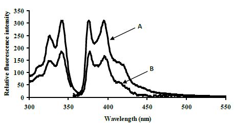
fig 3

Estimation of CYP3A4*1B Single Nucleotide Polymorphism Using Target-Assembled In-Situ Detection by Synthetic DNA-Mounted Excimers

DOI: 10.31038/MIP.2021212

Abstract

CYP3A4*1B is a single nucleotide polymorphism of CYP3A4 and is associated with prostate cancer which exhibits higher nifedipine oxidase activity in liver. This research provides details of the effects of structural variation and medium effects for the recently reported split-oligonucleotide (tandem) probe system for excimers-based fluorescence detection of DNA. In this approach the detection system is split at a molecular level into signal-silent components, which must be assembled correctly into a specific 3-dimensional structure to ensure close proximity of the excimer partners and the consequent excimer fluorescence emission on excitation. The model system consists of two 11-mer oligonucleotides, complementary to adjacent sites of a 22-mer DNA target. Each oligonucleotide probe is equipped with functions able to form an excimer on correct, contiguous hybridization. The extremely rigorous structural demands for excimer formation and emission required careful structural design of partners for excimer formation, which are here described. This study demonstrates that the excimer formed emitted at ~480 nm with a large Stokes shift (~130-140 nm).

Keywords

DNA probe systems, Excimers, DNA detection, Fluorescence, CYP3A4*1B, Stokes shift

Introduction

Reversible hybridisation of complementary polynucleotides is essential to the biological processes of replication, transcription, and translation. Physical studies of nucleic acid hybridisation are required for understanding these biological processes on a molecular level. The physical characterisation of nucleic acid hybridisation is essential for predicting the performance of nucleic acids in vitro, for instance, in hybridisation assays used to detect specific polynucleotide sequences.

Fluorescence measurements present an improved sensitive measure of nucleic acid concentration compared to conventional solution-phase detection techniques. Additionally, the sensitivity of fluorophores to their environments offers a means by which to differentiate hybridised from unhybridised nucleic acids without resorting to separation techniques. This was first demonstrated by attaching different fluorescent labels to the termini of oligonucleotides, which hybridise to adjacent regions on a complementary strand of DNA. Appropriate selection of fluorophores led to a detectable signal between the labels on hybridisation of the two-labeled strands to their complementary strand. For example, split-probe systems based on excimer fluorescence were first described by Ebata [1-3], who attached pyrene to the 5′-terminus of one oligonucleotide probe and to the 3′-terminus of the other oligonucleotide probe. The probes bound to adjacent regions of the target, bringing the pyrene molecules into close proximity, forming an excimer [4,5]. Excimer emission from oligonucleotides containing 5-(1-pyrenylethynyl)uracil [6], trans-stilbene [7], and perylene [8] have also been reported.

Numerous genetic diseases have been found to result from a change of a single DNA base pair. These single nucleotide polymorphisms (SNP) may cause changes in the amino acid sequence of important proteins [9,10]. Methods sensitive to single base-pair mutations for the fast screening of patient samples to identify disease-causing mutations will be essential for diagnosis, prevention and treatment. Usually hybridisation analysis is used, where a short, probe oligonucleotide (15-20 base pairs) bearing some kind of label (e.g. fluorophore) hybridises to complementary base pairs in DNA or RNA. The nucleic acids required for analysis can be recovered from a variety of biological samples including blood, saliva, urine, stool, nasopharyngeal secretions or tissues [11-13]. Highly specific, simple, and accessible methods are needed to meet the accurate requirements of single nucleotide detection in pharmacogenomic studies, linkage analysis, and the detection of pathogens. Recently there has been a move away from radioactive labels to fluorescence.

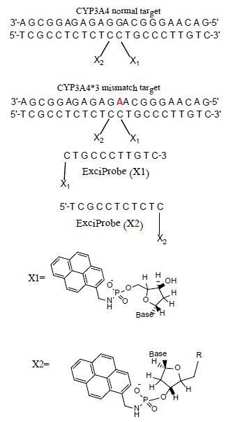
It has been reported [14] that an emissive exciplex can be formed by juxtaposition of two different externally oriented exciplex-forming partners (pyrene and naphthalene) at the interface (nick region) of tandem oligonucleotides forming a duplex of some kind on hybridization with their complementary target strand. We have been mainly interested in using excimer fluorescence signals to study the hybridisation between two fluorophore-labeled complementary DNA strands, as shown in Figure 1. Attaching fluorescent labels (pyrene and pyrene in Figure 1) to the probe of complementary DNA strands showed strong interactions between particular fluorophore pairs on hybridisation. Split-probe systems based on two 11 mer probe strands were investigated in this paper using the base sequences shown in Figure 1.

In the split-probe model system (Figure 1) two 11-mer probe oligonucleotides labelled with 1-pyrenylmethylamine (Pyrene) attached to 3′ and 5′ terminal phosphate groups. Hybridization of these probes to a complementary 22-mer oligonucleotide target resulted in correct orientation of the two pyrenes excimer-partners. Excitation of the pyrenylmethylamino partner at 350 nm led to the structure of an excited-state complex (excimer) with the pyrene partner. This excimer emitted at a longer wavelength of 480 nm (Stokes shift 130 nm) as compared with a mixture of unhybridisedsplit-probes. The excimer emission was particularly preferred by the use of trifluoroethanol as co-solvent (80 % v/v) [14].

fig 1

Figure 1: Base sequences of the split-probe systems.

The split-probe model system used in this study containing 22-mer target sequence which corresponds to a region of the CYP3A4 major genome (3′-AGCGGAGAGAGGACGGGAACAG-5′) and complementary 11 mer probes(5′-TCGCCTCTCTC-pyrene and pyrene-CTGCCCTTGTC-3′). CYP3A4*1B (-392A>G, rs2740574) is a CYP3A4 polymorphism and it is the frequently studied proximal promoter variant which occurs in White human populations at around 2-9% but at elevated frequencies in Africans including Libyans [15-17]. We now report how this excimer strategy can permit detection of an allelic variant of the human CYP3A4*1B gene sequence.

Single-nucleotide polymorphisms (SNPs) in genes coding for cytochrome P450 (CYP) enzymes have been linked to many diseases and to inter-individual differences in the efficiency and toxicity of many drugs. Thirty seven CYP3A4 variants, with amino-acid changes located in coding regions, have been identified among the different ethnic populations (www.pharmvar.org/gene/CYP3A4). For example, CYP3A4*1B allele (CYP3A4-V, rs2740574), a −392A>G transition in the promoter region, has been reported to be considerably connected with HIV infection [18], increased threat of hormone negative breast cancer(missing estrogen, progesterone receptors) [19], prostate cancer [20] and increased risk for developing leukemia after epipodophyllotoxin therapy [21]. Also, theCYP3A4*1B allele causes amino acid substitution affecting the metabolism of a range of drugs such as Nifidipine and Carbamazepine which leads to altered enzyme activity and drug sensitivity, e.g. the mutant enzyme results in impaired metabolism [22,23].

Materials and Methods

The excimer constructs used standard DNA base/sugar structures in both complementary probes. The targets were a part of the CYP3A4 chromosome 7 sequence band q22.1 (Genbankcode[ENSG00000160868 nucleotides r=7:99354604-99381888]: 3′-AGCGGAGAGAGGACGGGAACAG-5′ (the bold base provided the SNP location G>A). The ExciProbes had the sequence (X1) p-5′-CTGCCCTTGTC-3′ and (X2) 5′-TCGCCTCTCTC-3′-p. The probes were supplied with a free 3’ or 5’-phosphate group (p). Reagents of the highest quality available and DNA probes and DNA targets were purchased from Sigma-Aldrich (Paris France,). Distilled water was further purified by ion exchange and charcoal using a MilliQ system (Millipore Ltd, UK). Tris buffer was prepared from analytical reagent grade materials. pH was measured using a Hanna(Lisbon, Portugal)HI 9321 microprocessor pH meter, calibrated with standard buffers (Sigma-Aldrich) at 20°C.

HPLC

HPLC purification of probes was performed on an Agilent 1100 Series HPLC system (California, USA), consisting of a quaternary pump with solvent degasser, a diode-array module for multi-wavelength signal detection using an Agilent 1100 Series UV-visible detector and an Agilent 1100 Series fluorescence detector for on-line acquisition of excitation/emission spectra. The system had a manual injector and thermostatted column compartment with two heat exchangers for solvent pre-heating. The HPLC system was operated by Agilent HPLC 2D ChemStation Software. Depending on the purification performed, the columns used were: Zorbax Eclipse X DB-C8 column (California, USA) (length 25 cm, inner diameter 4.6 mm, particle size 5μm), or a Luna C18 (2) column (California, USA) (length 25 cm, inner diameter 4.6 mm, particle size 5 μm) with elution using an increasing gradient (0–50%) of acetonitrile in water (fraction detection at 260, 280, and 340 nm).

UV-Visible Spectrophotometry

UV–visible absorption spectra were measured at 20°C on a Cary-Varian 1E UV–visible spectrophotometer (London, UK.) with a Peltier-thermostatted cuvette holder and Cary 1E operating system/2 (version 3) and CARY1 software. Quantification of the oligonucleotide components used millimolar extinction coefficients (e260) of 99.0 for ExciProbe (X1), 94.6 for ExciProbe (X2). The extinction coefficients were calculated by the nearest neighbour method [24] and the contribution of the exci-partners was neglected.

Spectrophotofluorimetry

Fluorescence emission/excitation spectra were recorded in 4-sided quartz thermostatted cuvettes using a Peltier-controlled-temperature Cary-Eclipse, spectrofluorophotometer (London, UK). All experiments were carried out at 5°C. Hybridisation: Duplex formation was induced by sequential addition of ExciProbe(X1) and ExciProbe (X2). The mole ratio of all oligonucleotides ExciProbe (X1)and ExciProbe (X2) used were 1:1, the concentration of each component was2.5 µM. Tris buffer was added either with or without 80% TFE and thevolume made up to 1000 µl with deionized water. Excitation wavelengths of 340 nm (for the pyrene monomer) and 350 nm (for the full two probes and the target) were used, at slit width of5 nm and recorded in the range of 350-650 nm. Emission spectra were recorded after each sequential addition of each component to record the change in emission of each addition. A baseline spectrum of buffer and water or buffer, water and 80% TFE was always carried out before start of the measurement. After each addition the solution was left to equilibrate for 6 minutes in the fluorescence spectrophotometer and emission spectra were recorded until no change in the fluorescence spectra was seen to ensure it had been reached. The sequence of experiments was first using ExciProbe (X1) then ExciProbe (X2). Control experiments were conducted using firstly ExciProbe (X1) followed by the 3’-free oligonucleotide probe and finally the complementary target. All spectra were buffer corrected.

Control Experiments

Control experiments were carried out in 80% TFE/Tris buffer as for the experimental systems using the standard method described above. The control experiment was performed to confirm whether the obtained excimer emission is a result of such background effects or arise from the hypothesised excimer structures. Then fluorescence melting curve experiments (based on excitation 350 nm and emission 480 nm for the excimer) were performed using a Cary Eclipse fluorescence spectrophotometer by measuring the change in fluorescence intensity for the excimer with melting temperature(Tm). Tm was also determined spectrophotometrically by measuring the change in absorbance at 260 nm with temperature. Tm was determined either by taking the point at half the curve height or using the first derivative method.

Synthesis and Oligonucleotide Modification

Attachments of 1-pyrenemethylamine to oligonucleotide probes were as described in [14,25]. One equivalent of 1-pyrenemethylamine was attached via phosphoramide links to the terminal 5’-phosphate of (X1) p-5′-CTGCCCTTGTC-3′ probe and to the 3’-phosphate of (X2) 5′-TCGCCTCTCTC-3′-p. To the cetyltrimethyl ammonium salts of the oligonucleotides (~1 micromole) dissolved in N, N-dimethylformamide (200 µl) were added triphenyl phosphine (80 mg, 300 µmol) and 2,2′-dipyridyl disulfide (70 mg, 318 µmol), and the reaction mixture was incubated at 37°C for 10 min. 4-N’,N’-Dimethylaminopyridine (40 mg, 329 µmol) was added, the reaction mixture incubated for a further 12 minutes at 37°C and 1-pyrenemethylamine hydrochloride (4 mg, 14.9 μmol, dissolved in 100 µl of N, N-dimethylformamide and three microliter triethylamine) added. The mixture of the reaction was incubated at 37°C for full day (24 hours) product then was purified using reverse-phase HPLC (eluted by 0.05 M LiClO4 with a gradient from 0 to 60 % acetonitrile).

CYP3A4*1B Single Nucleotide Polymorphism

Split-probe systems were used to investigate the effect of SNP in the CYP3A4*1B target sequence on excimer emission compared to the normal-type target. Experiment was carried out in 80% TFE/Tris buffer at 5°C. The sequence of addition was: ExciProbe (X1), ExciProbe (X2), and finally 22 mer mutant-target oligonucleotide (CYP3A4*1B). All spectra were buffer-corrected.

Results

Excimer Formation Using Terminally Located Probe Systems

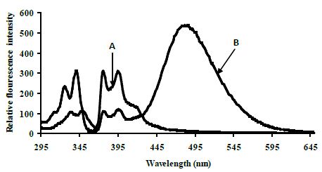
Fluorescence studies were made for solutions of ExciProbe (X1) and ExciProbe (X2) oligonucleotides with both probes complementary to each other (Figure 1). Figure 2 shows the excitation and emission spectra for (A) the ExciProbe (X1) and ExciProbe (X2) in 80% TFE/Tris buffer (0.01 M Tris, 0.1 M NaCl, pH 8.4), at5°C, (B) ExciProbe (X1) and ExciProbe (X2) hybridised to the 22-mer target oligonucleotide. On excitation at 350 nm, the 3′-pyrenyl ExciProbe (X1) and 5′-pyrenyl ExciProbe (X2) showed fluorescence typical of pyrene LES emission (lmax = 376, 395 nm). Addition of the complementary target resulted in immediate quenching of the LES emission at 395 nm to less than one-third of its original value and the appearance of a new, broad emission band (lmax = 480 nm) characteristic of pyrene excimer fluorescence after the full terminally located system had formed)[1,2,26]. Addition of the two probes to the target also caused a slight red shift in both excitation (from 342 nm to 349 nm) and emission (from 376 nm to 378 nm; λex 350 nm) spectra, consistent with duplex formation [1,2,26].

fig 2

Figure 2: Excitation and emission spectra of the split-oligonucleotide (tandem) probe system A 5′-pyrenyl ExciProbe (X1) and 3′-pyrenyl ExciProbe (X2), B ExciProbe (X1), 3′-pyrenyl ExciProbe (X2) and the complementary target (full system)in 80% TFE/0.01 M Tris, 0.1 M NaCl, pH 8.4) at 5°C. Component concentrations were 2.5 μM (equimolar).

Control Experiments for the Terminally Located Excimer System

Control experiments on a 1:1 mixture of 5′-pyrenyl ExciProbe (X1) and 3′-pyrenyl ExciProbe probe (X2) oligonucleotides were carried out in 80% TFE/0.01 M Tris, 0.1 M NaCl, pH 8.4 to determine if the fluorescence was from pyrene interacting as an excimer with the intended pyrene exci-partner, or an interaction with bases of the oligonucleotides. The 5′-pyrenyl ExciProbe (X1) showed no band at 480 nm in the absence of the target oligonucleotide (Figure 3). Addition of the complementary oligonucleotide target to ExciProbe (X1) resulted in a slight shift in λmax of LES emission to 379 nm, consistent with hybridisation of the probe with the complementary target. However, no marked 480 nm band was seen, even after heating the system to 70°C and re-annealing by slowly cooling back to 5°C. The weak fluorescence emission at 480 nm for the control duplex (before and after heating cooling, Figure 3) on duplex formation appeared real and could be related to exciplex formation, due to intra-molecular interaction of pyrene within the assembled duplex. However, relative to the full system with both 3′- and 5′-pyrenyl groups (Figure 3) the emission at 480 nm is insignificant.

fig 3

Figure 3: Emission spectra for control terminally located system A 5′-pyrenyl ExciProbe (X1) oligonucleotide, B 3′-pyrenyl ExciProbe (X2) and the 22 mer target in 80% TFE/10 mM Tris, 0.1 M NaCl, pH 8.4 at 5°C. Excitation wavelength 350 nm, slitwidth 5 nm. Equimolar component concentration was 2.5 μM.

CYP3A4*1B Single Nucleotide Polymorphism

The excimer emission was detected (broadband at ~480 nm) for normal target and showed strong emission at 480 nm (545 relative fluorescence intensity) compared to the mutated target (220 relative fluorescence intensity) around 2.5 fold (Figure 4).

fig 4

Figure 4: Emission spectra comparing the normal target A, CYP3A4) with the mutated target B, CYP3A4*1B) in 80% TFE/ Tris buffer (10 mM Tris, 0.1 M NaCl, pH 8.4) at 5°C after heating the samples to 90°C. Spectra were recorded when emission intensity had reached a maximum after 10 minutes at 5°C. Excitation was at 350 nm, slitwidth 5 nm. Spectra, buffer-corrected, are scaled to LES emission (378.9 nm).

Melting Temperatures of SNP

Melting curve experiments were performed spectrophotometrically at A260 and estimated by using the first derivative method. The melting temperatures (Tm) for normal CYP3A4 target was 76.9 ±0.8°C and 75.0 ±0.8°C for CYP3A4*1B, respectively. The melting temperature at 260 nm for systems was performed in 80% TFE/Tris buffer (10 mM Tris, 0.1 M NaCl, pH 8.4). Control experiments for Tm were carried out in 80% TFE/Tris buffer. In addition, similar thermal results were obtained using fluorescence melting curve experiments based on excitation 350 nm and emission 480 nm for the excimer. The fluorescence thermal study was performed using a Cary Eclipse fluorescence spectrophotometer by measuring the change in fluorescence intensity for the excimer with temperature.

Discussion

Confirmation of Duplex Formation

In our experiments of hybridising the two 11 mer probes to the complementary target in phosphate buffer (pH 7.0) containing 0.1 M NaCI, the pyrene moieties of the two probes came into close proximity, and an excimer band at 480 nm was generated. This result is consistent with results obtained by [27] who used a system that incorporated a pyrene-modified nucleotide at the 5′-end of one probe and a pyrene-modified nucleotide at the 3′-end of the other [27]. Figure 2 shows fluorescence typical of pyrene local excited state (LES) emission (lmax = 376, 395 nm) for a 5′-pyrenyl ExciProbe (X1) labelled oligonucleotide alone. The emission spectrum obtained is similar to that obtained in the literature using10 mM phosphate buffer (pH 7.0) 20 % v/v DMF, 0.2 M NaCl at 25°C and gave lmax = 377, 396 nm [1,2,26]. Addition of the 5′-pyrenyl ExciProbe (X1) to the 3′-pyrenyl ExciProbe (X2) target resulted in immediate quenching of the LES emission at 395 nm to less than one-third of its original value and the appearance of a new, broad emission band atlmax = 480 nm characteristic of pyrene excimer fluorescence (Figure 2).

Melting experiments provide further strong evidence of duplex formation. The split-probe systems showed sigmoid single-transition melting curves spectrophotometrically (A260 or A350) or spectrofluorometrically from fluorescence intensity at 340 nm for the LES (lex) and 376 nm (lem) for thepyrene monomer and at 350 nm for LES (lex) and 480 nm (lem) for the excimer (data not shown). Additional evidence of duplex formation comes from the emission spectra, as one probe oligonucleotide alone did not give an excimer signal in the absence of the other complementary probe. Further evidence of duplex formation and reversibility came from experiments using a heating and cooling cycle. Experiments of terminally located probe systems at different temperatures showed that the excimer intensity decreased when the temperature increased and eventually disappeared. This process is reversible, providing further evidence of duplex formation. A better-formed duplex structure probably enables the exci-partners to be better positioned for excimer formation. The reappearance of the excimer spectra on re-cooling indicates that no destruction of the components occurs on heating the system.

Evidence of Excimer Formation

The red-shifted structureless band at ~480 nm is characteristic of excimer emission, but could be due to interaction of the exci-partners with each other or nucleobases as pyrene are able to form an exciplex with certain nucleotide bases, especially guanine and to a lesser extent thymidine [28,29]. Also some oligonucleotide sequences show weak exciplex emission from pyrene attached to their 5′-termini in the absence of any added (complementary) oligonucleotide [30]. Thus, it is important to establish for the terminally located system the origin of the emission at 480nm. Heating the system caused the excimer emission intensity to decrease due to dissociation of the duplex structure. On re-cooling the system excimer emission reappeared. The Tm values by fluorescence and UV-visible methods were similar and of the magnitude expected for such a system (22-mer duplex) [31].

CYP3A4*1B Single Nucleotide Polymorphism

The search for sequences that differ in only one or two nucleobases needs tools to detect nucleic acid sequences that have high performance, speed, simplicity, and low cost. There have been many different techniques developed to identify the mutations in nucleic acid sequences. Techniques based on matched/mismatched-duplex stabilities, restriction cleavage, ligation, nucleotide incorporation, mass spectrometry and direct sequencing have been reviewed [32,33]. The DNA split-probe system of CYP3A4*1B was able to discriminate between perfectly matched CYP3A4 and mismatched CYP3A4*1B targets. Several split-oligonucleotide systems have been reported to discriminate between SNPs. These include the ligation method of Landegen [34], nanoparticle probes[35,36] and the template-directed ligation method [37,38]. The split-probe excimer system of Paris [39] was found to be sensitive to a single-base mutation in the target, positioned four base pairs from the 3′-junction. In the Paris study the addition of the unmutated target to the pyrene probes resulted in an increase in 490 nm emission as well as a 4.7-fold decrease in 398 nm monomer emission. The resulting excimer:monomer ratio was 0.04, very different to that for the sequence with a single-base point mutation which was 2.7 [39].

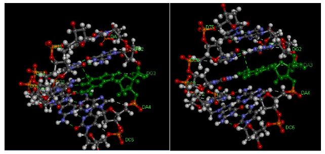
In the present study the duplexes containing GAGAACG/CTCCTGC mismatch is significantly destabilized compared with its correctly paired parent. Amber and Znosko [40] studied the thermodynamics of A/G mismatches in different nearest-neighbour contexts. They found a penalty (energy loos) of 1.2 kcal/mol for replacing a G-C base pair with either an A-U or G-U base pair. For both CYP3A4 (normal target) and CYP3A4*1B (mismatched target) showed a sigmoidal melting profile, typical of the dsDNA to ssDNA transition, providing further evidence of tandem duplex formation. The Tm values of CYP3A4*1B are less to those of the fully matched, consistent with literature studies performed on different sequences under identical conditions[25]. Duplexes of CYP3A4*1B (mismatched target) with mismatches of G/A in the twelve position from the 3′ and 5′ ends, respectively, showed significantly lower Tm than CYP3A4 (normal target). These results indicate that the ∆G contribution of a single G/A mismatch and the position of the mismatch are crucial to duplex stability and consistent with the literature [41,42]. The ∆G contribution of a single G/A mismatch to duplex stability was studied by [43] who found that ∆G is dependent on the neighbouring base pairs and ranges from +1.16 kcal/mol (for the context TGA/AAT) to -0.78 kcal/mol (for the context GGC/CAG). Allawi [43] also showed that the nearest neighbour model is applicable to internal G/T mismatches in DNA. In their study of G/T mismatches, the most stable trimer sequence containing a G/T mismatch was -1.05 kcal/mol for CGC/GTG and the least stable was +1.05 kcal/mol for AGA/TTT. On average, when the closing Watson-Crick pair on the 5′ side of the mismatch is an A/T or a G/C pair, G/A mismatches are more stable than G/T mismatches by about 0.40 and 0.30 kcal/mol, respectively [43,44]. When the 5′ closing pair is a T/A or a C/G, then G/T mismatches are more stable than G/A mismatches by 0.54 and 0.75 kcal/mol, respectively. Evidently, the different hydrogen-bonding and stacking in G/T and G/A mismatches results in different thermodynamic trends and the energy and structural information are the compositions of the following variables, such as bond angle energies, bond energies, planarity energies, dihedral angle energies, Van der Waals energies or/and electrostatic energies. These results indicate that duplexes containing mismatches are considerably destabilized (Figure 5) compared with their correctly paired parent the extent being dependent on the base composition and sequence of the oligonucleotide as well as on the type and location of the mismatch. The mismatch of DNA leads to alterations of amino acid properties and can cause a change in protein structure [45,46]. Consequently, SNP may affect enzyme activity through the modification of protein structure and function [47].

fig 5

Figure 5: Pentamer of DNA Duplexes of theCYP3A4 gene. A: matched CYP3A4 target (AGGAC/TCCTG), B: CYP3A4*1B target (AGAAC/TCCTG). The hydrogen bonds are represented using green broken lines. The figure was obtained with the help ofthe molecular visualization tool (Discovery Studio Visualizer software 4.1).

Conclusion

Our results evaluate the first case of an oligonucleotide split probe system based on excimer fluorescence emission for detection of CYP3A4*1B single nucleotide polymorphism. Further studies will be necessary to understand the details of the split probe system structure which determine the formation of the excimer for CYP3A4 single nucleotide polymorphism. Based on fluorescence and spectrophotometric results, the split probe system is selective enough to detect single base mutations of CYP3A4*1B with good sensitivity and therefore could be used to detect other mutations using an excimer system.

Acknowledgement

The authors gratefully acknowledge the support and valuable suggestions obtained from Sir Khaled AB Diab (Judicial Expertise and Research Centre, Tripoli, Libya Tripoli, Libya).

References

  1. Ebata K, Masuko M, Ohtani H, Jibu M (1995) Excimer formation by hybridization using two pyrene-labeled oligonucleotide probes. Nucl Acids Symp Ser 34: 187-188. [crossref]
  2. Ebata K, Masuko M, Ohtani H, Kashiwasake-Jibu M (1995) Nucleic acid hybridization accompanied with excimer formation from two pyrene-labeled probes. Photochem Photobiol 62: 836-839. [crossref]
  3. Masuko M, Ebata K, Ohtani H (1996) Optimization of pyrene excimer fluorescence emitted by excimer-forming two-probe nucleic acid hybridization method. Nucl Acids Symp Ser 1996 35: 165-166. [crossref]
  4. Ibele LM, Sánchez-Murcia PA, Mai S, Nogueira JJ, GonzÃlez L (2020) Excimer Intermediates en Route to Long-Lived Charge-Transfer States in Single-Stranded Adenine DNA as Revealed by Nonadiabatic Dynamics. J Phys Chem Lett 11: 7483-7488. [crossref]
  5. Conlon P, Yang CJ, Wu Y, Chen Y, Martinez K, et al. (2008) Pyrene Excimer Signaling Molecular Beacons for Probing Nucleic Acids. J Am Chem Soc 130: 336-342. [crossref]
  6. Balakin KV, Korshun VA, Mikhalev II, Maleev GV, Malakhov AD, et al. (1999) Conjugates of oligonucleotides with polyaromatic fluorophores as promising DNA probes. Biosensors & Bioelectronics 14: 597. [crossref]
  7. Letsinger RL, Wu T (1994) Control of Excimer Emission and Photochemistry of Stilbene Units by Oligonucleotide Hybridization. J Am Chem Soc 116: 811-812.
  8. Shimadzu A, Ohtani H, Ohuchi S, Sode K, Masuko M (1998) Perylene excimer formation by excimer-forming two-probe nucleic acid hybridization method. Nucl Acids Symp Ser 39: 45-46.
  9. Vallejos-Vidal E, Reyes-Cerpa S, Rivas-Pardo JA, Maisey K, Yajez JM, et al. (2019) Single-Nucleotide Polymorphisms (SNP) Mining and Their Effect on the Tridimensional Protein Structure Prediction in a Set of Immunity-Related Expressed Sequence Tags (EST) in Atlantic Salmon (Salmo salar). Front Genet 10: 1406. [crossref]
  10. Xiong LL, Xue LL, Al Hawwas M, Huang J, Niu RZ, et al. (2020) Single-nucleotide polymorphism screening and RNA sequencing of key messenger RNAs associated with neonatal hypoxic-ischemia brain damage. Neural Regen Res 15: 86-95.
  11. Bertolini F, Scimone C, Geraci C, Schiavo G, Utzeri VJ, et al. (2015) Next Generation Semiconductor Based Sequencing of the Donkey (Equus asinus) Genome Provided Comparative Sequence Data against the Horse Genome and a Few Millions of Single Nucleotide Polymorphisms. PLoS One 10.
  12. Parahuleva MS, Schieffer B, Klassen M, Worsch M, Parviz B, et al. (2018) Expression of the Marburg I Single Nucleotide Polymorphism (MI-SNP) and the Marburg II Single Nucleotide Polymorphism (MII-SNP) of the Factor VII-Activating Protease (FSAP) Gene and Risk of Coronary Artery Disease (CAD): A Pilot Study in a Single Population. Med Sci Monit 24: 4271-4278.
  13. Liu J, Zhang L, Feng L, Xu M, Gao Y, et al. (2019) Association between single nucleotide polymorphism (rs4252424) in TRPV5 calcium channel gene and lead poisoning in Chinese workers. Mol Genet Genomic Med 7.
  14. Bichenkova EV, Gbaj A, Walsh L, Savage HE, Rogert C, et al. (2007) Detection of nucleic acids in situ: novel oligonucleotide analogues for target-assembled DNA-mounted exciplexes. Org Biomol Chem 5: 1039-1051.
  15. Wilson RT, Masters LD, Barnholtz-Sloan JS, Salzberg AC, Hartman TJ (2018) Ancestry-Adjusted Vitamin D Metabolite Concentrations in Association with Cytochrome P450 3A Polymorphisms. Am J Epidemiol 187: 754-766. [crossref]
  16. Serin I, Pehlivan S, Gundes I, Fidan OY, Pehlivan M. A new parameter in multiple myeloma: CYP3A4*1B single nucleotide polymorphism. Ann Hematol 1-7.
  17. Zeigler-Johnson CM, Spangler E, Jalloh M, Gueye SM, Rennert H, et al. (2008) Genetic susceptibility to prostate cancer in men of African descent: implications for global disparities in incidence and outcomes. Can J Urol 15: 3872-3882. [crossref]
  18. Berno G, Zaccarelli M, Gori C, Tempestilli M, Antinori A, et al. (2014) Analysis of single-nucleotide polymorphisms (SNPs) in human CYP3A4 and CYP3A5 genes: potential implications for the metabolism of HIV drugs. BMC Med Genet 15: 76. [crossref]
  19. Johnson N, De Ieso P, Migliorini G, Orr N, Broderick P, et al. (2016) Cytochrome P450 allele CYP3A7*1C associates with adverse outcomes in chronic lymphocytic leukemia, breast and lung cancer. Cancer Res 76: 1485-1493. [crossref]
  20. Reyes-Hernández OD, Vega L, Jiménez-Ríos MA, Martínez-Cervera PF, Lugo-García JA, et al. (2014) The PXR rs7643645 Polymorphism Is Associated with the Risk of Higher Prostate-Specific Antigen Levels in Prostate Cancer Patients. PLoS One 9: 99974. [crossref]
  21. Johnson N, De Ieso P, Migliorini G, Orr N, Broderick P, et al. (2016) Cytochrome P450 allele CYP3A7*1C associates with adverse outcomes in chronic lymphocytic leukemia, breast and lung cancer. Cancer Res 76: 1485-1493. [crossref]
  22. Puranik YG, Birnbaum AK, Marino SE, Ahmed G, Cloyd JC, et al. (2013) Association of carbamazepine major metabolism and transport pathway gene polymorphisms and pharmacokinetics in patients with epilepsy. Pharmacogenomics 14: 35-45. [crossref]
  23. Åšwiechowski R, JeleÅ A, Mirowski M, Talarowska M, GaÅ‚ecki P, et al. (2019) Estimation of CYP3A4*1B single nucleotide polymorphism in patients with recurrent Major Depressive Disorder. Mol Genet Genomic Med 7.
  24. Cantor CR, Warshaw MM, Shapiro H (1970) Oligonucleotide interactions. 3. Circular dichroism studies of the conformation of deoxyoligonucleotides. Biopolymers 9: 1059-1077. [crossref]
  25. Bichenkova EV, Savage HE, Sardarian AR, Douglas KT (2005) Target-assembled tandem oligonucleotide systems based on exciplexes for detecting DNA mismatches and single nucleotide polymorphisms. Biochem Biophys Res Commun 332: 956-964. [crossref]
  26. Masuko M, Ohtani H, Ebata K, Shimadzu A (1998) Optimization of excimer-forming two-probe nucleic acid hybridization method with pyrene as a fluorophore. Nucl Acids Res 26: 5409-5416. [crossref]
  27. Yamana K, Nunota K, Nakano H, Sangen O (1994) A new method for introduction of a pyrene group into a terminal position of an oligonucleotide. Tet Lett 35: 2555-2558.
  28. Geacintov NE, Moller W, Zager E (1976) The triplet state of benzo[a]pyrene as a probe of its microenvironment in DNA complexes. Biol Mol Proc Int Conf 199-206.
  29. Manoharan M, Tivel K, Zhao M, Nafisi K, Netzel TL (1995) Sequence dependence of emission lifetimes for DNA oligomers and duplexes covalently labeled with pyrene: Relative electron-transfer quenching efficiencies of A, G, C and T nucleosides toward pyrene. J Phys Chem 99: 17461-17472.
  30. Hannah Savage (2003) Studies of Fluorescence Detection of Nucleic Acid Sequences. The University of Manchester. (GENERIC)Ref Type: Thesis/Dissertation.
  31. Lewis FD, Wu T, Burch EL, Bassani DM, Yang JS, et al. (1995) Hybrid Oligonucleotides Containing Stilbene Units. Excimer Fluorescence and Photodimerization. J Am Chem Soc 117: 8785-8792.
  32. Landegren U, Nilsson M, Kwok PY (1998) Reading bits of genetic information: methods for single-nucleotide polymorphism analysis. Genome Res 8: 769-776.
  33. Shi MM (2001) Enabling large-scale pharmacogenetic studies by high-throughput mutation detection and genotyping technologies. Clin Chem 47: 164-172. [crossref]
  34. Landegren U, Kaiser R, Sanders J, Hood L (1988) A ligase-mediated gene detection technique. Science 241: 1077-1080. [crossref]
  35. Cao Y, Jin R, Mirkin CA (2001) DNA-modified core-shell Ag/Au nanoparticles. J Am Chem Soc 123: 7961-7962. [crossref]
  36. Seife C (2002) Analytical chemistry. Light touch identifies wisps of rogue DNA. Science 297: 1462. [crossref]
  37. Chen X, Kwok PY (1999) Homogeneous genotyping assays for single nucleotide polymorphisms with fluorescence resonance energy transfer detection. Genetic Anal-Biomol E 14: 157-163. [crossref]
  38. Chen X, Kwok PY (1997) Template-directed dye-terminator incorporation (TDI) assay: a homogeneous DNA diagnostic method based on fluorescence resonance energy transfer. Nucleic Acids Res 25: 347-353.
  39. Paris PL, Langenhan JM, Kool ET (1998) Probing DNA sequences in solution with a monomer-excimer fluorescence color change. Nucl Acids Res 26: 3789-3793. [crossref]
  40. Davis AR, Znosko BM (2007) Thermodynamic characterization of single mismatches found in naturally occurring RNA. Biochemistry 46: 13425-13436. [crossref]
  41. Sagi J (2017) In What Ways Do Synthetic Nucleotides and Natural Base Lesions Alter the Structural Stability of G-Quadruplex Nucleic Acids? J Nucleic Acids 2017: . [crossref]
  42. Kent JL, McCann MD, Phillips D, Panaro BL, Lim GF, et al. (2014) Non-nearest-neighbor dependence of stability for group III RNA single nucleotide bulge loops. RNA 20: 825-834.
  43. Allawi HT, SantaLucia J Jr (1998) Nearest Neighbor Thermodynamic Parameters for Internal G.A Mismatches in DNA. Biochemistry 37: 2170-2179. [crossref]
  44. Allawi HT, SantaLucia J Jr, (1997) Thermodynamics and NMR of Internal G.T Mismatches in DNA. Biochemistry 36: 10581-10594. [crossref]
  45. Pei J, Kinch LN, Otwinowski Z, Grishin NV (2020) Mutation severity spectrum of rare alleles in the human genome is predictive of disease type. PLoS Comput Biol 16:1007775. [crossref]
  46. Lyons DE, Yu KP, Vander Heiden JA, Heston L, Dittmer DP, et al. (2018) Mutant Cellular AP-1 Proteins Promote Expression of a Subset of Epstein-Barr Virus Late Genes in the Absence of Lytic Viral DNA Replication. J Virol 92: 01062-01018. [crossref]
  47. Jia P, Zhao Z (2017) Impacts of somatic mutations on gene expression: an association perspective. Brief Bioinform 18: 413-425. [crossref]
fig 1

Could Ultrasound Inactivate COVID-19 in the Environment and in the Air, Thus Preventing the Spread of the Pandemic?

DOI: 10.31038/IMROJ.2021635

Abstract

The novel severe acute respiratory syndrome coronavirus 2 (SARS-CoV-2), also called COVID-19, started by Wuhan, China, in 2019, has caused a pandemic which has quickly involved the entire world and raised public health concerns. The scientific community is actively exploring treatments that would potentially be effective in combating COVID-19. Viral infections, especially those that are transmitted by air, are the most contagious infectious diseases and cause major biological, clinical and socioeconomic problems worldwide. COVID-19 spreads in the air and therefore rapidly contagious, thus finding appropriate and timely treatment to cure the entire population and above all to prevent transmission between individuals, is extremely necessary in a short time. Considerable efforts are being made to seek therapy on the infected human being, in fact several drugs already used in the past for SARS or HIV-1 are tested, in what is called a drug repositioning or label off strategy, but it is very important to prevent contagion before other individuals are infected, and especially the most fragile ones, even to decongest the structures of the health system also to avoid neglecting other pathologies as unfortunately is happening in this pandemic period. High intensity ultrasound is becoming important and more widely used in the food industry for microorganisms decontamination and is one of the new technologies that have been suggested as an alternative to current heat treatments for microbial inactivation, including viruses. Also for synergistically enhanced elimination of organic pollutants and pathogenic microorganisms from water since the early 2000s, dual-frequency ultrasound has received much attention. Here, my hypothesis consist to use ultrasound, as a valid prevention clean mean, which propagates well in the air, such as respiratory viruses, to fight the COVID-19 expansion in the air environment, and thus block the transmission from one individual to another, especially indoors and also from objects to individuals. Ultrasoud are also used in the Protein misfolding cyclic amplification (PMCA) technique used to amplify prions. The growing chain of misfolded protein is then blasted with ultrasound by sonication, breaking it down into smaller chains and so rapidly increasing the amount of abnormal protein available to cause conversions. Ultrasounds are waves with frequencies between 20 kHz and 20 MHz, well known have effects on molecular structures of various microorganisms, even viruses, but are not harmful to humans, and so could potentially damage also the new Coronavirus, COVID-19 especially by damaging the superficial spike S-glycoprotein that the virus uses to enter cells and infect them, and block or slow down the epidemic which is destroying many human lives. Experimentally, COVID-19 and its variants, can be treated with ultrasonic waves at different exposure times, then Vero E6 cells (African green monkey kidney cells) permissive to the SARS-CoV-2, are infected in vitro with the treated virus with ultrasounds and tested for efficacy of on the suppression of activity of COVID-19 and therefore for the decrease or block of infectivity. After a possible positive outcome of the laboratory experimentation, ultrasounds can be applied in closed environments frequented by people including homes, offices, supermarkets, schools, buses, subways to purify the air and avoid the spread of contagion.

Keywords

COVID-19, Virology, Ultrasound, Enviroments, Prevention

Introduction

The COVID-19 pandemic represents the greatest global public health crisis since the pandemic influenza outbreak of 1918, a bit more than a century ago. The novel coronavirus disease 2019 (COVID-19) has resulted in the deaths of more than 248 000 persons worldwide as of May 4, 2020, on the same date in New York more than 19 400 individuals have died [1]. Research is under way to identify vaccines and therapeutics for COVID-19, including repurposing of medications. Facing a new and unknown virus, antiviral agents previously used to treat other infections such as SARS and Middle East Respiratory Syndrome (MERS), have been considered as the first potential candidates for first-line therapy to treat COVID-19. Chloroquine and hydroxychloroquine, old drugs used in the treatment of malaria and inhibitors of protease of HIV-1 have been previously studied, where evidence of efficacy has been found and they have anti coronavirus characteristics in vitro [2,3]. The findings support the insight that chloroquine/hydroxychloroquine have efficacy in the treatment of COVID-19 [4]. Therefore, based on evidence from in vitro studies on the suppression of activity of severe acute respiratory syndrome coronavirus 2 (SARS-CoV-2) and other coronavirus strains, interest increased in the use of hydroxychloroquine and chloroquine with the possible addition of azithromycin for the treatment of COVID-19 [5,6]. At the same time, a great number of clinical trials have been launched to investigate the potential efficacy therapies for COVID-19 highlighting the urgent need to get as quickly as possible high-quality evidence. This new virus, COVID-19, such as respiratory viruses, spreads in the air, and therefore also rapidly contagious therefore, finding effective and timely treatment to cure the entire population or better still, prevent contagion, is a very difficult task. For so much we must find alternative and effective methods also for the prevention of global spread as well as for the cure in order not to collapse the health system.

Hypothesis

My hypothesis here consist to use ultrasound, as a valid prevention mean, which propagates well in the air, to fight the SARS-CoV-2 in environments, especially indoors, and it is well known have effects on molecular structures of various microorganisms, including viruses, but they are not harmful to humans. Ultrasound is one of the new technologies that have been suggested as an alternative to current heat treatments for microbial inactivation [7,8]. Ultrasound is defined as a sonic wave at frequencies over the threshold human hearing. Ultrasonic waves are generally classified by their frequency and their wavelength. Waves with frequencies between 20 and 40 kHz are considered as high-energy or high-power ultrasound Figure 1 [8].

fig 1

Figure 1: Diagram of the approximate frequencies corresponding to ultrasounds according to their field of application. Source Wikipedia.

High intensity ultrasound is becoming important and more widely used in the food industry for microorganisms decontamination. This sterilization technique has been evaluated to improve food safety and to replace common processing with chemical additive compounds. The efficiency of a horn-type power ultrasound treatment (300 W and 600 W, 28 kHz, 10-30 min) on Listeria monocytogenes, Bacillus cereus, Escherichia coli, Salmonella typhimurium bacteria suspensions and phytoviruses was examined in this study [9]. The results of this study showed that ultrasonic treatment can be used to eliminate vegetative cells of gram-positive and gram-negative bacteria from 1.59 to 3.4 log in bacterial suspensions and some phytoviruses in fruits [9]. Also for treatment and disinfection of water dual-frequency ultrasound since the early 2000s, dual-frequency ultrasound (DFUS) has received much interest [10]. Elimination of organic pollutants and pathogenic microorganisms from water occurs indirectly by generating reactive oxygen species (ROS) induced through exposure the water to DFUS [10]. The nonlinear dynamics of microbubbles upon DFUS exposure produces additional frequencies, such as harmonics, subharmonics, ultraharmonics and combination frequencies. These increase the probability of bubbles collapse, thereby enhancing cavitation and generating more reactive oxygen species for advanced oxidation processes (AOPs) [10]. The protein misfolding cyclic amplification (PMCA) mimics in vitro the process of prion propagation, which occurs in vivo [11]. PMCA requires the incubation of prions with an excess of Pr(PC) (a normal glycophosphatidyl-anchored glycoprotein) in a test tube, which is placed in a dedicated sonicator and subjected to a process of cyclically repeated phases of sonication and incubation. Brains of healthy animals are used as a source of Pr(PC) [12]. During the incubation phase, PrP(Sc) (abnormal form of the prion protein) forces PrPC to change conformation and aggregate. The sonication fragments these aggregates into small species that act as seeds able to promote further PrPC conversion. Therefore the ultrasounds act at the molecular level to determine the cleavage of the aggregates. Based on these reasons my hypothesis consist of using ultrasounds to counteract the new pandemic from COVID 19, which could weaken, the viral envelope constituted by a phospholipid bilayer of pericapsid and therefore through, for example, causing a conformational change of the spike S-glycoprotein receptor, which it uses to bind to the human ACE2 receptor to enter cells. S glycoprotein is fundamental to mediate the membrane fusion required for virus entry and cell fusion. Ultrasound travels in the air, like COVID-19 does, so above all in closed environments, including homes, offices, supermarkets, schools, buses, subway, where the greatest contact occurs among the people, it could prevent the transmission from one individual to another in case of close contact, sneezing, coughing or breathing, thus avoiding contagion and the spread of the pandemic. The hypothesis is therefore to destroy the COVID-19 in the air contained in the microdroplets or in fine dust from environmental pollution, and also as soon as it comes into contact with the upper airways of humans, in such a way as to block or decrease its replication and therefore the viral load.

Experimental Protocol

Undergo the virus COVID-19 to a determined TCID50, to ultrasound at different exposure times: from a few seconds to a minute to test its immediate effectiveness of the ultrasound waves to inactivate the virus, then 10 min, and 15 min (the latter, estimated minimum time for the infection to occur between individuals at a distance of less than one meter). Then 3 h, 6 h, 12 h, 24 h, 48 h and 72 h of exposure of the virus to ultrasound.

The Vero E6 cells (African green monkey kidney cells) [13] permissive to the SARS-CoV-2, will be respectively infected to a certain MOI, with the different samples virus treated with ultrasounds at the different times listed above. Then infected Vero E6 cells will be analyzed with MTT assay for to test the eventual inhibition of the cytopathic effect as well as a control of uninfected cells (mock infection), and a control virus of cells infected with the virus not treated with ultrasonic waves.

Consequences of the Hypothesis and Discussion

The transmission of infection can happen by main route, one is from the virus on the surfaces and other is infection from the droplets from the sneeze and cough skin flakes [14-16]. Each of these processes generates aerosol droplets of different size and initial speed. When a person sneeze or coughs, talks loud, ejecting the droplets is released which is of 1 mm in diameter which falls on the ground within a minute [13], but the microdroplets remain through the air for several hours which are smaller than 10 micrometer. They are small and light drifting through the air and stays and does not drift from air for a period of time [17]. Have been observed that there is high risk of spread of infection in closed room or in a class room. For example, in a closed room of 10 people, if a person coughs once there is spread of 1,000 droplet with most of them fall on the ground in a minute whereas the microdroplet spreads in a very high volume for long period. Accordingly my hypothesis to use ultrasounds could be taken into consideration as they spread through the air where there are also the viral particles, but they are not harmful to human health, instead they could damage the molecular structures of pathogenic microorganisms including viruses and in particular potentially, also COVID-19 cause of the ongoing pandemic. The ultrasounds could therefore also be used to heal the environment in closed places because it has been seen that the virus remains in the air for several hours through microdroplets so in places where there are people it could act as a barrier to avoid contagion from a person to another in case of sneezing, coughing or just breathing but also from objects with virus to individuals. Ultimately, the use of ultrasound as an alternative and supportive means, even at relatively low costs, could prevent the further spread of COVID-19 and therefore block the pandemic curve, for a desirable recovery from the collapse of the world health system and from immense efforts of health workers. The application of this hypothesis could be of considerable impact and inspiration if developed and solve several health and socio-economic problems that unfortunately we are witnessing.

References

  1. Wang C, Horby PW, Hayden FG, Gao GF (2020) A novel coronavirus outbreak of global health concern. Lancet 395: 470-473. [crossref]
  2. Savarino A, Buonavoglia C, Norelli S, Di Trani L, Cassone A (2006) Potential therapies for coronaviuses. Expert Opinion Ther.Patents 16: 1269-128. [crossref]
  3. J Oscanoa T, Romero-Ortuno R, Carvajal A, A Savarino A (2020) A pharmacological perspective of chloroquine in SARS-CoV-2 infection: An old drug for the fight against a new coronavirus? Int J Antimicrob Agents 56: 106078. [crossref]
  4. Cortegiani A, Ingoglia G, Ippolito M, Giarratano A, Einav S (2020) A systematic review on the efficacy and safety of chloroquine for the treatment of COVID-19. J Crit Care 10: 57: 279-283. [crossref]
  5. Liu J, Cao R, Xu M, et al. (2020) Hydroxychloroquine, a less toxic derivative of chloroquine, is effective in inhibiting SARS-CoV-2 infection in vitro. Cell Discov 6: 16. [crossref]
  6. Gautret P, Lagier JC, Parola P, et al. (2020) Hydroxychloroquine and azithromycin as a treatment of COVID-19: results of an open-label non-randomized clinical trial. Int J Antimicrob Agents 105949. [crossref]
  7. US Food and Drug Administration Report, 2000 US Department of Agriculture. Kinetics of microbial inactivation for alternative food processing technologies: ultrasound US Food and Drug Administration Report Published 2 June 2000.
  8. S Condón-Abantoa, S Pedros-Garrido, M Marcen, V Ruiz, S Condón (2018) Synergistic effect of ultrasonic waves under pressure at mild temperatures (MTS) in yeast inactivation. International Journal of Food Microbiology 284: 56-62. [crossref]
  9. Sarkinas A, Sakalauskiene K, Raisutis R, Zeime J, Salaseviciene A, Puidaite E, et al. (2018) Inactivation of some pathogenic bacteria and phytoviruses by ultrasonic treatment. Microb Pathog 123:144-148. [crossref]
  10. Galina Matafonova , Valeriy Batoev (2020) Dual-frequency ultrasound: Strengths and shortcomings to water treatment and disinfection Laboratory of Engineering Ecology, Baikal Institute of Nature Management, Siberian Branch of Russian Academy of Sciences, Ulan-Ude, Russia Water Research 182: 116016.
  11. Saborio GP, Permanne B, Soto C (2001) Sensitive detection of pathological prion protein by cyclic amplification of protein misfolding. Nature 411: 810-813.
  12. Giaccone G, Moda F (2020) PMCA Applications for Prion Detection in Peripheral Tissues of Patients with Variant Creutzfeldt-Jakob Disease. Biomolecules 10: 405. [crossref]
  13. Zhang Qinfen, Cui Jinming, Huang Xiaojun, Zheng Huanying, Huang Jicheng, et al. (2004) The life cycle of SARS coronavirus in Vero E6 cells. J Med Virol 73: 332-337. [crossref]
  14. Lin L, Lu L, Cao W, Li T (2020) Hypothesis for potential pathogenesis of SARS-CoV-2 infection–a review of immune changes in patients with viral pneumonia. Emerg Microbes Infect 9: 727-732. [crossref]
  15. Morawska L (2006) Droplet fate in indoor environments, or can we prevent the spread of infection?. Another type of spread through closed personnel contact within the home, community centers, daycares, school classroom, University through the microdroplets. Indoor Air 16: 335-347. [crossref]
  16. Barker J, Stevens D, Bloomfield SF (2001) Spread and prevention of some common viral infections in community facilities and domestic homes. J Appl Microbiol 91: 7-21. [crossref]
  17. Fahad Al Qahtani, Modi Fahd Al Qahtani, Ahad Fahd Al Qahtani, Nagesh Bhat J (2020) Family Microdroplets are more infectious of COVID-19 spread in a closed door. Med Prim Care. 9: 3776-3777. [crossref]

Multiple Target Repetitive Transcranial Magnetic Stimulation (rTMS) Combined with Neurofeedback for Complete Resolution of Severe OCD, Bipolar Depression, and Anxiety

DOI: 10.31038/JNNC.2021433


Obsessive compulsive disorder (OCD) is a prevalent disabling condition and often comorbid with depression, anxiety, and high suicide rates. First line treatments of OCD such as Selective Serotonin Reuptake Inhibitors (SSRIs) and dopamine antagonists, and mood stabilizers fail to treat OCD in up to 60% of patients [1]. Transcranial magnetic stimulation has emerged as a non-pharmacological alternative for treatment of OCD and several case reports and randomized trials have shown positive response although the efficacy of r TMS remains low at approximately 35% with little positive effects on reversing comorbities including symptoms of depression and anxiety [2]. The most effective stimulation parameters, cortical targets, and type of coil (figure of eight, H-coil, cone, or deep) for rTMS have not been established. Investigators report positive outcomes with stimulation of the right Orbitofrontal Cortex (OFC), Supplementary Motor Area (SMA), and Anterior Cingulate Cortex (ACC) [2,3]. Recently, deep r tms directed at ACC has show significant benefit [4]. In addition, neurofeedback, also known as ‘EEG biofeedback’, has been established as an effective treatment of various psychological and neuropsychiatric disorders including OCD, anxiety, and depression [5,6]. Accordingly, I hypothesize that a more formidable outcome can be achieved in patients suffering from OCD accompanied by comorbidities when: 1) multiple cortical targets are stimulated under one treatment regimen; 2) multiple stimulation parameters are utilized in response to patient report of outcome; and 3) neurofeedback is used in conjunction with rTMS. This ‘combination approach’ was in fact, found to be significantly effective in treatment of pervasive spectrum disorder, e.g. autism and early signs of dementia [7,8]. Here, I present the first case report of patient with severe OCD, anxiety, and depression who showed marked improvement and resolution of OCD, anxiety, and depression following completing total of 41 sessions of rTMS and 15 sessions of ‘Z-score neurofeedback’.

Patient is 33-year-old male diagnosed with over 15 years of OCD, generalized anxiety, and bipolar depression. Patient has undergone trials of Serotonin Reuptake Inhibitors (SSRIs), mood stabilizers including lithium and valproic acid, and benzodiazepines including lorazepam. At time of presentation to my clinic, patient’s primary symptoms included obsessive thoughts with facial and skin contamination, recurrent feeling of self-guilt, marked difficulty with anxiety in dark environments, agoraphobia, poor self-worth, and extreme spells of depression, manic, and hypomanic episodes. In addition, patient displayed severe hypochondriasis with respect to various disorders including motor neuron disease, multiple sclerosis, and infections. Moreover, patient experienced various somatic symptoms including non-specific vibratory sensations of mouth, trunk, and extremities. At onset, Yale Brown Obsessive Compulsive Score (Y-BOCS) was 19, Burn’s anxiety score was 36, Burn’s depression score was 22, and PHQ-9 of 10. Risks and benefits of rTMS including off-label use of rTMS parameters were discussed in detail with patient and written signed consent obtained. A brain MRI was obtained to include skin fiduciary markers for navigation software (The Neural Navigator, Brain Science Tools, Utrechet, Netherlands) 1mm sagittal and axial T1 MRI images were processed and segmented to identify several cortical targets including: left and right Dorsolateral Prefrontal Cortex (DLPFC), right OFC, left Dorsomedial Prefrontal Cortex (DMPFC), and ACC. On initial visit and every 5-6 visits, the Motor Threshold (MT) was determined as the intensity required to active the left Abductor Policis Brevis (APB) and/or the First Dorsal Interosseus (FDI) on the contralateral hand on average of 5 of 10 single pulse trials directed at left motor cortex per visual inspection. Patient was interviewed and underwent daily rTMS sessions using varying targets and stimulation parameters (see table). The rTMS stimulation parameters were chosen based on current trial evidence for treatment of OCD, depression, and anxiety and patient’s daily report of signs and symptoms. All stimulations were performed using figure-of-eight coil (Neurosoft, Ltd. Ivanovo, Russian Federation) rTMS machine. Cortical targets included SMA, left DLPFC, right DLPFC, left DMPFC, ACC, and right OFC. Cortical targets were identified and marked using navigation software.

table

Neurofeedback was done using Neuroguide ‘Z-Score LORETA’ neurofeedback software by collecting and editing 4 minutes of baseline EEG and creating a ‘symptoms checklist match’ (for detail of procedure see Thatcher & Lubar, Z Score Neurofeedback: Clinical Applications, 2015; Thatcher, Latest Developments in Live Z-Score Training: Symptom Check List, Phase Reset, and Loreta Z-Score Biofeedback, 2013) [9,10]. Each session consistent of five 5-minute rounds for total of approximately 25-30 minutes. Dry, wireless, headset (DSI-24, Wearable Sensing, San Diego, CA, USA) was used to gather EEG recordings and conduct neurofeedback sessions. Patient underwent neurofeedback approximately two times per week, usually prior to or after r TMS sessions. Patient showed progressive and marked improvement in, OCD, anxiety, and depression. Y-BOCS scores showed 63% improvement (19 to 7), Burn’s depression showed 86% improvement (22 to 3), Burn’s anxiety showed 72% improvement (36 to 10), and PHQ-9 showed 70% improvement (from 10 to 3). Patient reported complete reversal of agoraphobia, self-contamination delusions, and depression. He reported feeling quite comfortable with going to large dark movie theater, avoiding washing rituals, denied panic attacks in provocative environments (e.g. driving), and developed markedly improved mood. The clinical improvements were noticeable by father and other close relatives. Although we delivered higher intensity of stimulation and overall larger total daily pulses, patient reported no significant adverse effects and did not experience seizures. To our knowledge, this is first report of 1) applying rTMS to multiple (e.g. more than 2) cortical targets on one patient with each utilizing separate stimulation protocols, and 2) adding neurofeedback to treatment regimen leading to marked improvement and resolution of OCD together with anxiety and depression.

The rationale behind use of various cortical targets is to modulate several circuits that may be contributing to OCD and comorbidities including abnormal connectivity and/or neuronal hyperactivity within cortical-striate-thalamic-cortical circuits–currently the leading working model regarding the pathophysiology of OCD [4]. One randomized, sham-controlled study showed modest improvements in OCD (29% reduction on Y-BOCS) and depression (48% on HAM-D) symptoms after 14 sequential 1 Hz stimluation sessions targeting left DLPFC and SMA [11,12]. Additional randomized sham-controlled studies applying a multi-target approach and in combination with neurofeedback will be helpful and can potentially significanly increase the efficacy rate of treatment.

Keywords

Generalized anxiety disorder, Multiple targets rTMS, Neurofeedback, Obsessive compulsive disorder, Repetitive transcranial magnetic stimulation

References

  1. Pallanti S, Quercioli L (2006) Treatment-refractory obsessive-compulsive disorder: Methodological issues, operational definitions and therapeutic lines. Progress in Neuro-psychopharmacology & Biological Psychiatry 30: 400-412. [crossref]
  2. Berlim MT, Neufeld NH, Van den Eynde F (2013) Repetitive transcranial magnetic stimulation (rTMS) for obsessive–compulsive disorder (OCD): An exploratory meta-analysis of randomized and sham-controlled trials. Journal of Psychiatric Research 47: 999-1006. [crossref]
  3. Nauczyciel C, Le Jeune F, Naudet F, Douabin S, Esquevin A, et al. (2014) Repetitive transcranial magnetic stimulation over the orbitofrontal cortex for obsessive-compulsive disorder: a double-blind, crossover study. Translational Psychiatry 4: e436. [crossref]
  4. Blom RM, Figee M, Vulink N, Denys D (2011) Update on Repetitive Transcranial Magnetic Stimulation in Obsessive-Compulsive Disorder: Different Targets. Current Psychiatry Reports 13: 289-294. [crossref]
  5. Sürmeli T, Ertem A (2011) Obsessive Compulsive Disorder and the Efficacy of qEEG-Guided Neurofeedback Treatment: A Case Series. Clinical Eeg and Neuroscience 42: 195-201. [crossref]
  6. Hammond DC (2003) QEEG-Guided Neurofeedback in the Treatment of Obsessive Compulsive Disorder. Journal of Neurotherapy 7: 25-52.
  7. Sokhadze EM, El-Baz AS, Tasman A, Sears LL, Wang Y, et al. (2014) Neuromodulation integrating rTMS and neurofeedback for the treatment of autism spectrum disorder: an exploratory study. Applied Psychophysiology and Biofeedback 39: 237-257. [crossref]
  8. Rabey JM, Dobronevsky E, Aichenbaum S, Gonen O, Marton RG, et al. (2013) Repetitive transcranial magnetic stimulation combined combined with cognitive training is a safe and effective modality for treatment of Alzheimer’s diseas: a randomizeed double blind study. J Neural Transm 120: 813–819. [crossref]
  9. Thatcher RW (2013) Latest Developments in Live Z-Score Training: Symptom Check List, Phase Reset, and Loreta Z-Score Biofeedback. Journal of Neurotherapy 17: 69-87.
  10. Thatcher RW, Lubar JF (2015) Z Score Neurofeedback: Clinical Applications. Elsevier Inc.
  11. Kang JI, Kim CH, Namkoong K, Lee CI, Kim SJ (2009) A randomized controlled study of sequentially applied transcranial magnetic stimulation obsessive compulsive disorder. Clin Psychiatry 70: 1645-51. [crossref]
  12. Lubar JF (2015) Optimal Procedures in Z-Score Neurofeedback: Strategies for Maximizing Learning for Surface and LORETA Neurofeedback.
fig 2

Automated Localization and Segmentation of Mononuclear Cell Aggregates in Kidney Histological Images Using Deep Learning

DOI: 10.31038/MIP.2021211

Abstract

Background and objectives: Allograft rejection is a major concern in kidney transplantation. Inflammatory processes in patients with kidney allografts involve various patterns of immune cell recruitment and distributions. Lymphoid aggregates (LAs) are commonly observed in patients with kidney allografts and their presence and localization may correlate with severity of acute rejection. Alongside with other markers of inflammation, LAs assessment is currently performed by pathologists manually in a qualitative way, which is both time consuming and far from precise. In this work we aim to develop an automated method of identifying LAs and measuring their densities in whole slide images of transplant kidney biopsies.

Materials and Methods: We trained a deep convolutional neural network based on U-Net on 44 core needle kidney biopsy slides, monitoring loss on a validation set (n=7 slides). The model was subsequently tested on a hold-out set (n=10 slides).

Results: We found that the coarse pattern of LAs localization agrees between the annotations and predictions, which is reflected by high correlation between the annotated and predicted fraction of LAs area per slide (Pearson R of 0.9756). Furthermore, the network achieves an auROC of 97.78 ± 0.93% and an IoU score of 69.72 ± 6.24% per LA-containing slide in the test set.

Conclusions: Our study demonstrates that a deep convolutional neural network can accurately identify lymphoid aggregates in digitized histological slides of kidney. This study presents a first automatic DL-based approach for quantifying inflammation marks in allograft kidney, which can greatly improve precision and speed of assessment of allograft kidney biopsies when implemented as a part of computer-aided diagnosis system.

Keywords

Kidney, Machine learning, Segmentation, Inflammation, Lymphoid aggregates

Introduction

End-to-end deep learning (DL) methods have approached human performance in multiple computer vision tasks [1,2] and have more recently been successfully utilized in many biomedical domains [3-5]. Convolutional neural networks (CNN) are a class of artificial neural networks commonly used in DL approaches for image analysis and are composed of multiple layers of convolutional filters and nonlinear activation units to extract meaningful features. These features are then used to produce a classification for each input image. CNNs are especially well equipped for image classification tasks due to the spatial invariance of the learned features and the presence of nonlinear activation units that allow for the learning of complex features [2]. In recent years, deep learning using CNNs has been shown to achieve high performance in image segmentation tasks [6,7], in which each pixel of an image is assigned a discrete class. By assigning each image pixel a discrete class label, the image can be segmented into distinct regions of interest. The use of CNNs have thus recently gained traction in biomedical segmentation [6] and digital pathology tasks, such as the detection and localization of breast cancer and its metastases in lymph nodes [8-10]. Based on such successful applications, a number of DL-based approaches have been approved for clinical use in the USA and other countries and are already contributing to higher precision and efficiency in many domains of diagnostics and treatment, such as diabetes care, oncology, cardiology, and radiology to name a few [11].

DL has been successfully applied for several routine nephrology-related evaluation procedures [12-14], which promises to improve speed and precision of pathologic workup of renal patients. Averitt et al. used CNNs to predict kidney survival, kidney disease stage, and various kidney function measures such as estimated glomerular filtration rate (eGFR) percentages from histological images [13]. Marsh et al. similarly used CNNs to predict glomerulosclerosis rates, i.e. the percent of glomeruli that are normal and sclerotic from frozen kidney transplant slides [14]. Both models were able to achieve performance on par with renal pathologists, thus demonstrating the potential of DL systems to serve as a pathologist’s assistant. Yet there are many other kidney pathologies where no automatic quantification procedures exist, but would undoubtedly be of great benefit to patients, clinicians, and the entire health system.

In kidney allografts, inflammation is the defining feature of acute cellular rejection with various patterns of immune cell infiltrates. Lymphoid aggregates (LAs) are characterized by the recruitment of T, B, and dendritic cells and are visible in histological sections as a collection of distinct large cells with irregular nuclei [15]. LAs are commonly observed in patients with kidney allografts and their presence and localization may correlate with severity of acute rejection [16,17]. Patchy interstitial mononuclear cell infiltrates may be indicative of a milder form of alloimmune injury [18]. Mononuclear cell aggregates localized to the sub-capsular area or fibrotic tissue are usually interpreted as “non-specific” inflammation [18]. In addition to cellular rejection, various types of inflammatory infiltrates can also be seen in acute pyelonephritis, virus infections, and drug-induced interstitial nephritis [18]. Assessment of LAs is a time-consuming process with no accepted quantification standard. Despite a number of recent efforts in the domain of automatic quantification of renal features [12-14] automated detection and quantification of inflammatory marks such as LAs in the kidney has not been previously attempted. Integration of digital pathology DL algorithms is especially crucial in situations where expert personnel are limited for expedient diagnosis such as in the setting of the interpretation of transplant kidney biopsies. Therefore, accurate automated diagnosis or flagging of inflammatory features could prove to be a valuable tool for pathologists to assess the underlying causes of renal dysfunction both in native and transplant kidneys, and has the potential to improve precision and efficiency of renal biopsy analysis. In this study, for the first time, we present an efficient, accurate, and automated method of localizing LAs and measuring their densities in whole slide digital images of transplant kidney biopsies using convolutional neural networks. By helping to improve diagnostic accuracy and speed, we envision our method to be of great benefit to physicians supervising kidney transplantations.

Materials and Methods

Pathology Material

A sample of transplant kidney biopsies collected between the years of 2010 and 2016 at the UCSF, Department of Pathology from patients (n=61) presenting for evaluation of renal allograft dysfunction was analyzed. Demographic data is shown in Table S1. Hematoxylin and eosin-stained slides of the biopsies were digitized at 40x magnification with a Leica Aperio CS2 whole slide scanner, with final resolution of approx. 3960 pixels per mm, 15.68 μm2/pixel. The research using the retrospective data was approved by IRB #17-22317.

Data Preparation

The 61 cases were split into a training set (n=44), a validation set (n=7), and a test set (n=10) as shown in Table 1. The ground-truth labels for the LAs in five of the 10 test slides were provided by a board-certified renal pathologist. The other five test slides contained no LAs. The ground-truth labels for the training set and validation set were provided by an appropriately trained medical student. The training set was used to train the neural network and update the model parameters. The validation set was not used to update the model parameters, but was used to evaluate the model after each training epoch and identify the best generalizing model. The test set was used to gauge the final performance and generalizability of the trained model on slides not evaluated during training. Average slide size was 4.102 ± 0.259 billion pixels. To increase training efficiency and due to computational limitations, 1024×1024 pixel patches were obtained by grid-sampling from each slide only where kidney tissue was present. The training set thus consisted of 7669 patches, the validation set 1112 patches, and the test set 2200 patches after the grid-sampling. In the test set, 1239 patches were derived from the five LA-free slides and 961 patches were from the five LA-containing slides. To further increase training efficiency, our model was trained on 256×256 patches by down-sampling the 1024×1024 patches by a factor of four. During the evaluation on the test set, we performed test-time data augmentation ensembling to improve performance by averaging prediction over original and three flipped versions of images.

Table 1: Number of biopsy slides, number of grid-sampled patches, number of patches with LAs, number of patches without any LAs, and qualification of the ground-truth label generator for the training, validation, and test sets.

Set

n slides

n patches n positive patches n negative patches

Expert Annotator

Training

44

7669 180 7489

Medical student

Validation

7

1112 34 1078

Medical student

Test

10

2200 155 2045

Renal pathologist

Model Architecture and Training

The slides were read and pre-processed using Openslide [19] and Slideslicer [20] python packages. For semantic segmentation, we used a modified version of U-Net [6,21] which consists of a sequence of contracting convolutional layers followed by a sequence of expanding convolutional layers. This allows for the final output to have the same resolution as the input while having a reduced number of parameters compared to architecture without contracting and expanding paths. Unlike other encoder-decoder-like architectures, U-Net also combines feature maps from the contracting layers to the inputs for the corresponding expanding layers in a symmetric fashion. As shown in Figure S1, our implementation used a VGG16 [2] head (including 13 convolutional layers with respective max-pooling layers) pre-trained on ImageNet dataset [1] and a custom decoder, similar to the model described in the work by Balakrishna et al. [21] The network was trained for 40 epochs, with each training epoch consisting of 7,669 iterations. The model was trained using a binary cross-entropy loss and the Adam optimizer with learning rate 10-5 and default parameters otherwise [22]. Implementation is available on Github [23].

Statistical Analysis

Based on the predicted segmentation probability map, a density score for each patch was calculated by counting the number of pixels where probability of LAs is higher than the probability of normal tissue and dividing it by the total number of pixels in a patch. The intersection over union (IoU), Dice score, and area under the ROC curve were calculated using standard formulas described elsewhere [6,8,21]. As these metrics are either undefined or not meaningful when no true positive areas are present, they were first evaluated on LA-containing slides only, and mean and standard error across slides was reported. Next, these metrics were evaluated on the results from all slides combined together. The compactness of annotated and predicted regions of interest (ROIs) was evaluated using Polsby-Popper index (PPI) [24] with formula:

PPI = 4 π area / circumference

Unpaired predicted and annotated ROIs were compared using Mann-Whitney U test, and paired (true positive predicted and annotated) ROIs were compared using paired Wilcoxon signed rank test. The statistics are reported as mean ± standard error unless otherwise specified.

Results

Using the trained model, we were able to accurately segment lymphoid aggregate regions, achieving an average area under the ROC curve of 97.78 ± 0.93%, an IoU score of 69.72 ± 6.24% and Dice score of 81.47 ± 4.69% across LA-containing slides in the test set. When aggregated predictions from both LA-containing and LA-free slides were considered, overall area under the ROC curve reaches 98.21%, IoU score 72.62%, and Dice score 84.14%. High-level inspection of predicted and observed LAs in whole slides in Figure 1 reveals good overall agreement between predicted and observed patterns. This is further corroborated by high correlation between the predicted LA area fraction (2.40% ± 1. 08% of total tissue area in test set slides) and the annotated LA area fraction (2.45% ± 0. 98%) with Pearson R of 0. 9756 (p=5.874e-8) as shown in Figure 2. On a more granular level of image patches, annotated and predicted proportions of LAs per patch are also highly correlated (slide-level Pearson R = 0.9624 ± 0.0209 for LA-containing slides, Figure S2).

fig 1

Figure 1: A coarse-level visualization of three representative core needle biopsy slides (left) alongside with LAs annotations provided by a renal pathologist (middle) and neural network prediction (right). Predictions with area less than 15,000 pixels are removed. The black horizontal bar indicates scale of 1 mm.

fig 2

Figure 2: Performance metrics of the segmentation algorithm within the test set. A. Correlation plot of the area of LAs ROIs (as fraction of total tissue area) in annotations (x-axis) and predictions (y-axis), Pearson R of 0.9756 (p=1.5e-6). The regression line is shown in black dotted line, and regression equation is shown (p=5.874e-8). Note that 5 LA-free samples are densely clustered near the origin (0.0, 0.0). B. ROC curve for predicted probability of LAs-class pixels. ROC for all pixels aggregated across all samples is shown in black solid line (AUC=98.21, 95% confidence interval: 98.20-98.22% using DeLong method), and ROC for 5 individual LA-containing slides are shown in colored dotted lines. Diagonal grey line indicates ROC for random predictions.

Predicted LA areas were of wider range of sizes than annotated ones. Particularly, many small LAs were predicted compared to annotated ones. In order to further analyze the distribution of sizes and shapes of annotated and predicted LAs, we calculated areas and Polsby-Popper indices (PPI) characterizing the shape irregularity on the range from above 0 (highly irregular) to 1 (circular). The visualization of obtained metrics (Figure 3) reveals the presence of multiple small regular-shaped false-positive LAs predictions. An average area of the annotated LAs was 1,009,140 ± 134,419 pixels (64,339 ± 857μm2), while for predicted ones 301,600 ± 51,621 pixels (19,229 ± 3,291 μm2) in the test set. On the other hand, when only true positive predictions were matched with the annotations, the true positive areas were slightly larger than respective annotations, with regression equation: predicted area = 1.033 × annotated area (p<2.2e-16, Figure 3C). The shape irregularity was similar in both groups on average (PPI = 0.4397 ± 0.0225 for annotated and 0. 4418 ± 0.0098 for predicted, p=0.5376 in unpaired test). However, when only true positive predicted LAs were evaluated and paired with the annotations, the predicted LA regions have significantly less regular shape (PPI = 0.3165 ± 0.0180, p=2.557e-6 in paired Wilcoxon test), with regression equation (p<2.2e-16, Figure 3C):

fig 3

Figure 3: Comparison of size and shapes of annotated and predicted ROIs. A. Distribution of area and Polsby-Popper index (PPI) for annotation ROIs (red triangles) and prediction ROIs (blue dots). True positive predicted ROIs are connected to respective annotation ROIs with yellow lines. Note that false negative ROIs (with no connecting lines) mostly have small area. B. Correlation between the PPI of predicted and annotated ROIs (Pearson R=0.4184, p=0.0012). C. Correlation between the area of predicted and annotated ROIs (Pearson R=0.7961, p=1.3e-13).

Predicted PPI = 0.6707 × Annotated PPI

As the minimal shape of annotated LAs was 33,782 pixels (2,153 μm2), with 5% percentile at 131,433 pixels (8,380 μm2), we sought to improve the performance of the segmentation by removing small predicted LA areas. To this end, we calibrated IoU performance as a function of low-pass area threshold within the validation set (Figure S3) and selected an optimal threshold for the area of predicted LAs that leads to maximal improvement in median IoU, at the value of 15,000 pixels (956 μm2). This thresholding improved the agreement between the counts of annotated and predicted LAs, thus reducing the mean absolute error from 13.0 to 6.57 LA regions per slide in validation set and from 14.1 to 6.7 LA regions per slide in the test set. At the same time, thresholding produced only a slight increase in IoU in LA-containing slides of the test set from 71.64% to 72.04%.

We show detailed visualization of correctly segmented, false negative, and false positive patches in Figure 4A, 4B, and 4C respectively. In most cases, the segmentation outline produced by the model had high overlap with the human annotation (IoU = 69.72 ± 6.24%), but contained more spatial detail than the annotation. Oftentimes, smaller areas of fibrotic tissue or other non-lymphoid tissue that were included by the annotator into LA segmentation were excluded by the model, thus leading to false negative predictions (Figure 4B). We saw that the model detected several smaller areas of lymphoid aggregates missed by the annotator initially (Figure 4C, columns 1&2). Dense nuclear areas of small atrophic tubules or tangentially cut tubules that expose multiple nuclei in the same plane were sometimes mis-classified as LAs, which is especially common for predictions with small area (under 33,000 pixels; Figure 4C, columns 3&4).

fig 4a

fig 4b

fig 4c

Figure 4: Segmentation of LAs in representative patches are shown with the original patch images and overlaid ground truth masks (green contour) and predictions (blue contour) in the top rows and segmentation probability heatmaps in the bottom rows. The IoU score, ground truth LA percentage, predicted LA percentage, slide ID, and patch coordinates are also displayed above each patch. A. Patches with good agreement between the annotation and prediction. B. Patches with false negative areas. C. Patches with false positive areas.

Discussion

Our study is the first to demonstrate that a deep convolutional neural network can accurately identify lymphoid aggregates and provide a quantitative measure of inflammation in digitized histological slides. It shows that inflammatory markers can be efficiently and robustly quantified automatically using DL, and thus shows potential of DL algorithms in improving efficiency and precision of renal pathology workup. Our model produces annotations of a higher spatial detail than present in typical manual annotations, while generally agreeing with the pathologist’s annotations, as seen in Figure 3A, and as indicated by a lower PPI index of true positive predicted LAs. Still the model produces numerous false positives of smaller size, that can be effectively removed by a low-pass area threshold filter. This shows that thorough expert-guided error analysis is necessary to keep medical DL algorithms unbiased, precise, and relevant to the problem at hand. On the other hand, our model detected areas of LAs initially missed by a pathologist (Figure4C, first column). This showcases the robustness and the power of the neural network model compared to human annotator, given sufficient amount of training data. Additionally, the increased speed at which LAs can be detected with our method will free up time for the already encumbered clinician to focus on other necessary tasks of the procedure.

Visualization tools used in this work to display density of LAs, such as in Figure 1, may be of potential use as a computer-aided decision support tool for pathologists and researchers investigating inflammatory processes in kidney allografts. As a next step, co-localization of lymphoid tissue with fibrotic and capsular tissue need to be learned as it is necessary for differential diagnosis of LA-associated conditions [18] similarly to systems developed to score inflammatory and fibrotic processes in lungs [25] and liver [26]. Furthermore, the scope of future work will need to expand to assigning and predicting Banff scores for various types of kidney pathologies [27]. We believe our model, fine-tuned with respective labels, would be able to accurately predict Banff lesions and provide basis for score estimation once we have adequate ground truth labels. Such an algorithm, when implemented as a part of a computer-aided diagnosis (CAD) system, could drastically speed up and simplify renal pathology analysis, as well as improve precision in clinics where specialized renal pathologists are not available.

Conclusions

Our study demonstrates that a deep convolutional neural network can accurately identify lymphoid aggregates in digitized histological slides of kidney. This study presents a first automatic DL-based approach for quantifying inflammation marks in allograft kidney, which can greatly improve precision and speed of assessment of allograft kidney biopsies when implemented as a part of computer-aided diagnosis system.

Funding

Research reported in this publication was supported by the National Institute of Health grants UH2CA203792, 1U01LM012675 (PI: DH), and U24AI118675 (PI: ZGL). AB is supported by a National Institute of General Medical Sciences training grant (5T32GM008440, PI: Judith Hellman). JHS is supported by T32 funding T32EB001631. The content is solely the responsibility of the authors and does not necessarily represent the official views of the National Institutes of Health.

Acknowledgment

In this section you can acknowledge any support given which is not covered by the author contribution or funding sections. This may include administrative and technical support, or donations in kind (e.g., materials used for experiments).

References

  1. Jia Deng, Wei Dong, Socher R, Li-Jia Li, Kai Li, et al. (2009) ImageNet: A Large-Scale Hierarchical Image Database. In 2009 IEEE Conference on Computer Vision and Pattern Recognition 2009: 248-255.
  2. Simonyan K, Zisserman A (2014) Very Deep Convolutional Networks for Large-Scale Image Recognition.
  3. Esteva A, Robicquet A, Ramsundar B, Kuleshov V, DePristo M, et al. (2019) A Guide to Deep Learning in Healthcare. Nat Med 25: 24-29.
  4. Topol EJ (2019) High-Performance Medicine: The Convergence of Human and Artificial Intelligence. Nat Med 25: 44-56.
  5. Ching T, Himmelstein DS, Beaulieu-Jones BK, Kalinin AA, Do BT, et al. (2018) Opportunities and Obstacles for Deep Learning in Biology and Medicine. J R Soc Interface 15: 20170387. [crossref]
  6. Ronneberger O, Fischer P, Brox T (2015) U-Net: Convolutional Networks for Biomedical Image Segmentation; Springer Cham. 234-241.
  7. Badrinarayanan V, Kendall A, Cipolla R (2017) SegNet: A Deep Convolutional Encoder-Decoder Architecture for Image Segmentation. IEEE Trans. Pattern Anal Mach Intell 39: 2481-2495.
  8. Litjens G, Bandi P, Ehteshami Bejnordi B, Geessink O, Balkenhol M, et al. (2018) 1399 H&E-Stained Sentinel Lymph Node Sections of Breast Cancer Patients: The CAMELYON Dataset. Gigascience 7: 65. [crossref]
  9. Liu Y, Kohlberger T, Norouzi M, Dahl GE, Smith JL, et al. (2019) Artificial Intelligence–Based Breast Cancer Nodal Metastasis Detection: Insights Into the Black Box for Pathologists. Arch Pathol Lab Med 143: 859-868.
  10. Campanella G, Hanna MG, Geneslaw L, Miraflor A, Werneck Krauss Silva et al. (2019) Clinical-Grade Computational Pathology Using Weakly Supervised Deep Learning on Whole Slide Images. Nat Med 25: 1301-1309. [crossref]
  11. Meskó B, FDA Approvals For Smart Algorithms In Medicine In One Giant Infographic.
  12. Ledbetter D, Ho L, Lemley KV (2017) Prediction of Kidney Function from Biopsy Images Using Convolutional Neural Networks.
  13. Averitt AJ, Natarajan K (2018) Going Deep: The Role of Neural Networks for Renal Survival and Beyond. Kidney. Int Reports 3: 242-243. [crossref]
  14. Marsh JN, Matlock MK, Kudose S, Liu TC, Stappenbeck TS, et al. (2018) Deep Learning Global Glomerulosclerosis in Transplant Kidney Frozen Sections. IEEE Trans Med Imaging 37: 2718-2728. [crossref]
  15. Pei G, Zeng R, Han M, Liao P, Zhou X, et al. (2014) Renal Interstitial Infiltration and Tertiary Lymphoid Organ Neogenesis in IgA Nephropathy. Clin J Am Soc Nephrol 9: 255-264. [crossref]
  16. Kayler LK, Lakkis FG, Morgan C, Basu A, Blisard D, et al. (2007) Acute Cellular Rejection with CD20-Positive Lymphoid Clusters in Kidney Transplant Patients Following Lymphocyte Depletion. Am J Transplant 7: 949-954. [crossref]
  17. Girlanda R, Kleiner DE, Duan Z, Ford EAS, Wright E C, et al. (2008) Monocyte Infiltration and Kidney Allograft Dysfunction During Acute Rejection. Am J Transplant, 8: 600-607. [crossref]
  18. Nadasdy T, Satoskar A, Nadasdy G (2017) Pathology of Renal Transplantation. In Silva’s Diagnostic Renal Pathology; Zhou, X. J. (Joseph), Laszik, Z. G., Nadasdy, T., D’Agati, V. D.
  19. Goode A, Gilbert B, Harkes J, Jukic D, Satyanarayanan M () OpenSlide: A Vendor-Neutral Software Foundation for Digital Pathology. J Pathol Inform 2013, 4 (1), 27. [crossref]
  20. Lituiev DS slideslicer: a toolbox for manipulation of digital pathology slides and annotations.
  21. Balakrishna C, Dadashzadeh S, Soltaninejad S (2018) Automatic Detection of Lumen and Media in the IVUS Images Using U-Net with VGG16 Encoder.
  22. Kingma DP, Ba J (2014) Adam: A Method for Stochastic Optimization.
  23. Lituiev DS, Cha SJ, Chin A, Glicksberg BS, Bishara A, et al () Source Code for Automated Localization and Segmentation of Mononuclear Cell Aggregates in Kidney Histological Images Using Deep Learning.
  24. Polsby DD, Popper R (1991) The Third Criterion: Compactness as a Procedural Safeguard Against Partisan Gerrymandering. Yale Law Policy Rev 9: 301-353.
  25. Heinemann F, Birk G, Schoenberger T, Stierstorfer B (2018) Deep Neural Network Based Histological Scoring of Lung Fibrosis and Inflammation in the Mouse Model System. PLoS One 13: 202708. [crossref]
  26. Heinemann F, Birk G, Stierstorfer B (2019) Deep Learning Enables Pathologist-like Scoring of NASH Models. Sci Rep, 9: 1-10.
  27. Roufosse C, Simmonds N, Clahsen-van Groningen M, Haas M, Henriksen KJ, et al. (2018) 2018 Reference Guide to the Banff Classification of Renal Allograft Pathology. Transplantation 102: 1795-1814. [crossref]
Featured Image2

Recent Updates on Intravenous Thrombolysis and Endovascular Theraphy for Hyperacute Manegement of Ischemic Strokes

DOI: 10.31038/JNNC.2021432

Abstract

Acute ischemic stroke (IAS) care has recently been revolutionized with the advent of acute reperfusion therapies. During recent decades, a growing number of randomized controlled clinical trials (RCTs) have helped expand a set of tools for emergency management of acute ischemic stroke, specially advanced imaging that has allowed the expansion of the therapeutic window by various mismatch assessments. Currently two available reperfusion therapies that have been shown to improve outcome in AIS patients, intravenous thrombolysis (IVT) and endovascular therapy (EVT), scilicet mechanical clot removal, both of which are highly time dependent. They have been shown to improve outcome in patients with wake-up strokes or symptom onset beyond 4.5 hours for intravenous thrombolysis and beyond 6 hours for endovascular treatment. Next-generation trials are attempting to expand the time window to benefit additional patients who present in the late time window for both intravenous thrombolysis and endovascular reperfusion therapies. However, they require advanced neuroimaging to select stroke patients safely. Modern trials extended the use of IVT and EVT in time windows that have been originally considered dangerous, in increasingly broad categories of patients during an extended time window. Under current guidelines, IVT is used to treat acute stroke only if it can be ascertained that the time since the onset of symptoms was less than 4.5 hours and 6 hours for EVT using standard neuroimaging such as computed tomography for IVT and CT plus CT angiography for EVT. It is true that recent randomized controlled trials (RCTs) have extended the therapeutic time window, however the studies have used different imaging and clinical inclusion criteria for patient selection during the initiation of this new stroke treatment paradigm, enroll patients based on the presence of a target mismatch on multimodal imaging. Furthermore, for those patients in expanded time windows, guidelines now recommend the use of “advanced” imaging techniques in the acute setting, including CT perfusion and MRI, to guide therapeutic decision-making. Advanced imaging such as perfusion-weighted imaging – core mismatch and conventional imaging such as diffusion-weighted image – fluid-attenuated inversion recovery mismatch played a role in expanding the therapeutic window to wake-up strokes or unwitnessed strokes. This article aims to review the current approaches using neuroimaging techniques to expand eligibility for IVT and EVT in acute ischemic stroke patients with stroke of unknown symptom onset, with an emphasis on new updates to qualifying patient populations and time periods for treatment. The current state of AIS reperfusion treatments in extended time windows it comes to us from high quality data from RCTs that used advanced neuroimaging (CT / MRI perfusion or MRI) to select patients.

Keywords

advanced neuroimaging, endovascular treatment, intravenous thrombolysis, ischemic stroke, large vessel occlusion, thrombectomy

Introduction

Acute stroke is an emergency, that may lead to permanent disability and death. Therefore, the foundation of care is based on choosing the optimal treatment option for the patient based on the best scientific evidence. Every year, about 22 million people around the world suffer from stroke [1]. Roughly, 87% of cerebrovascular events are due to artery occlusion as opposed to hemorrhage [2]. Acute ischemic stroke (AIS), result when a vessel supplying blood to the brain is occluded, and impairs blood flow to part of the brain. When acute cerebral ischemia occurs due to the occlusion of an intracranial artery, the part of the brain that is irreversibly lost (ischemic core) is surrounded by brain parenchyma that can be salvaged (penumbra) if prompt recanalization takes place [2]. In fact, it’s so, ischemic penumbra denotes the part of an acute ischemic stroke that is at risk of progressing to infarction but is still salvageable if reperfused. During the last decade, the management of acute ischemic stroke has changed dramatically, from an expectant bedside attitude towards active treatment, thanks to the continuous improvement of new therapeutic options. Neuroimaging information has demonstrated efficacy to visualize effects that neurological examination cannot be seen, such as penumbra, and advanced imaging in stroke has significantly influenced and explored the pathological conditions of the ischemic blood flow. At present intravenous thrombolysis (IVT) and endovascular therapy (EVT), are the only approved treatments for acute ischemic stroke but must be administered in narrow therapeutic window of up to 4.5 and 6 hours, respectively. There were randomized controlled trials already published emphasizing the use of intravenous recombinant tissue plasminogen activator (IV rTPA) beyond the approved time frame in acute ischemic stroke. Important, in the pre-hospital setting, first responders should focus especially on the last time patients were known not to have stroke symptoms, as last known well. Advanced neuroimaging may help us overcome time constraints and expand the implementation of acute reperfusion therapies. Both computed tomography (CT) and Magnetic Resonance (MRI), provides valuable information in the management of patients with AIS, with diagnostic, therapeutic, and prognostic implications. The primary purposes of neuroimaging in patients with AIS are to determine the extent of initial ischemic core and identify the location and extent of intravascular clot as well as the presence and extent of penumbra, constituted by hypoperfused tissue at risk for infarction. Furthermore, over the past few years, advanced imaging protocols for acute stroke patients, mainly used in large institutions and comprehensive stroke centers, now include parenchymal imaging non-contrast head CT (NCT) or diffusion-weighted MRI (DWI) plus fluid attenuated inversion recovery (FLAIR) and Gradient echo MR imaging (GRE) or susceptibility-weighted imaging (SWI) on MRI, parenchymal imaging is meant to assess the volume of infarct.  vascular imaging CT Angiography (CTA) or MR angiography (MRA), Vascular imaging is performed to detect the site of occlusion, and to characterize the collateral circulation. Penumbral imaging as well as Perfusion CT, Perfusion weighted imaging (PWI) aims at determining the volumes of salvageable penumbra [4]. This advanced neuroimaging helped overcome time constraints and expand the implementation of acute reperfusion therapies, for both EVT candidates in the late time window 6–24 hours and for IVT candidates 4.5–9 hours and wake-up patients [5]. There is no doubt that neuroimaging has played a crucial part in the assessment of patients with AIS over the past decades, as several parameters should be accurately individualized, either by CT, MR or angiography, to carefully select the patients that are eligible to IVT or IVT as outcome is dependent on that. The “2018 Guidelines for Management of Acute Ischemic Stroke” from the American Heart Association/American Stroke Association has a new recommendation that CT perfusion (CTP), diffusion-weighted imaging (DWI)-MRI, and/or MRI perfusion (MRP) be included as part of a standard imaging evaluation for patients within 6–24 h of symptom onset [6]. Thanks to recent RCTs, the therapeutic time window for both IVT and EVT in AIS has been extended, but also increased treatment algorithm complexity. In the event of AIS patients, acute reperfusion therapies with unknown time of symptom onset or in extended time windows are now possible. The present review provides an overview of the latest developments in the management of acute ischemic stroke, with an emphasis on thrombolytic therapy and intra-arterial intervention, summarizes the current evidence from randomized trials about its efficacy and safety of acute stroke care. Summarizes the current evidence from randomized trials about its efficacy and safety of acute stroke care, and that may assist clinicians in the selection of those late presenters that will most likely benefit from acute reperfusion therapies.

Current concepts in intravenous thrombolysis treatment of acute ischemic stroke

The intravenous administration of recombinant tissue plasminogen activator was introduced into acute stroke therapy in the mid-1990s [7-8], and there is no doubt that tPA improves outcome after stroke on AIS [9]. CT scan of the head has become routine in evaluating patients with signs and symptoms suggestive of AIS. Besides, pretreatment brain imaging with noncontrast CT is appropriate, mainly to exclude intracranial hemorrhage (ICH) and, to a lesser extent, reveal early ischemic changes. With the advent of thrombolysis, it becomes important to assess early ischemic changes in CT to predict the benefits of therapy. Regrettably, it is difficult to recognize and quantify these changes, which is why was born the Alberta stroke program early CT score (ASPECTS). The program was developed to offer the reliability and utility of a standard CT examination with a reproducible grading system to assess early ischemic changes, <3 hours from symptom onset, on pretreatment CT studies in patients with acute ischemic stroke of the anterior circulation [10]. ASPECTS is a systematic, robust, and practical method that can be applied to different axial baselines and is superior to that of the 1/3 MCA rule. ASPECTS is a 10-point scoring system that reliably predicts the extent of early ischemic changes from CT scans. At present, intravenous thrombolysis (IV rtPA) with 0.9 mg/kg alteplase, maximum dose 90mg over 60min with initial 10% of dose given as bolus over 1min) is given to all patients that present within 4.5 hours of ischemic stroke independently of stroke etiology if all inclusion and exclusion criteria are fulfilled [11]. Certainly, that treatment with intravenous alteplase within 4.5 hours of acute ischemic stroke onset is associated with an increased early risk of intracerebral hemorrhage, but this risk is offset by later benefit in the form of reduced disability [12]. Since the 4.5 hours’ time restriction is a major cause of IVT treatment failure, much effort has been made to select AIS patients who could safely undergo thrombolysis over extended time windows or with unknown time onset of symptoms. In fact, it’s so that in recent years, the research focus has been on extending the time window for intravenous thrombolysis in acute ischemic stroke using new imaging techniques-based patient selection. Extending the time window for thrombolytic therapy is an important goal as it would increase the number of patients who are able to receive acute treatment. Consequently therefore, there have been attempts to expand time window for the benefit of additional patients presenting in the late hour window, thanks to new RCTs that have revolutionized acute ischemic stroke care by extending the use of intravenous thrombolysis therapy in time windows. Some trials have suggested that the treatment window may be extended in patients who are shown to have ischemic but not yet infarcted brain tissue on imaging. Trials that have suggested that the treatment window may be extended in patients who have been shown to have ischemic brain tissue but not yet infarcted on imaging are described below.

The EXTEND trial

Treatment with IV tPA outside the standard 4.5 hours window was evaluated by the Extending the Time for Thrombolysis in Emergency Neurological Deficits (EXTEND) trial. The trial was the first phase III randomized, placebo-controlled study to show benefit of IVT for patients presenting in the late hour window [13]. However, it must be premised that the rate of symptomatic intracerebral hemorrhage (sICH) in the EXTEND trial was among the highest reported. In this trial, thrombolysis was guided by perfusion imaging up to 9 hours after the onset of stroke. In effect in these cases advanced neuroimaging was used to select patients with CT perfusion or perfusion-diffusion MRI. The trial randomized 225 patients to either alteplase or placebo within 4.5 to 9.0 hours of symptoms onset or within 9 hours from the midpoint of sleep, if presenting with a wake-up stroke. Patients were included if they fulfilled three criteria: a) ischemic core <70ml, measured on CT  perfusion as brain volume with cerebral  blood flow <30% of normal brain regions  (rCBF) or on MRI as apparent diffusion  coefficient (ADC) <620μm²/s, b) Critically hypoperfused brain volume >10ml from ischemic core volume, measured on CT or magnetic resonance (MR) perfusion as delayed arrival of an injected racer agent (time to maximum of the residue function, Tmax >6s), c) Perfusion lesion–ischemic core mismatch ratio >1.2. The primary outcome was a score of 0 or 1 on the modified Rankin scale, on which scores range from 0 (no symptoms) to 6 (death), at 90 days. The trials demonstrated that among the patients who had ischemic stroke and salvageable brain tissue, the use of alteplase between 4.5 and 9.0 hours after stroke onset or at the time the patient awoke with stroke symptoms resulted in a higher percentage of patients with no or minor neurologic deficits than the use of placebo. It showed that favorable outcome was more likely for the IV-rtPA group after adjustment for age and clinical severity at baseline, but not in unadjusted analysis. These results were obtained prior the early termination of the study, terminated due to loss of equipoise after publication of the WAKE-UP trial.

EXTEND-IA TNK, EXTEND-IA TNK part 2 trial and NOR-TEST

Current treatment with tissue plasminogen activator (tPA) requires an intravenous infusion of 1 hour because the clearance of tPA from the circulation is rapid (t 1/2 approximately 6 min). There have been multiple studies evaluating the safety and efficacy of alternative thrombolytic agents. Today, attention drawn to the benefits of intravenous (IV) tenecteplase (TNK) for acute stroke reperfusion therapy, particularly given the ease of administration and affordability. This thrombolytic medication is a genetically modified variant of alteplase with greater fibrin specificity and a longer half-life that permits bolus administration.

The Tenecteplase versus Alteplase before Endovascular Therapy for Ischemic Stroke (EXTEND-IA TNK) trial only involved patients with large vessel occlusions and compared 0.25 mg/kg of tenecteplase with standard dose IV-tPA [14]. The trial enrolled patients presenting within 4.5 hours of symptom onset that were eligible for mechanical thrombectomy, that is randomized tPA- and mechanical thrombectomy–eligible patients into tenecteplase and alteplase groups. The tenecteplase group had a higher incidence of reperfusion and better functional outcome, with similar adverse events before IVT than did intravenous alteplase. The recently concluded EXTEND-IA TNK part-2 trial compared 2 different doses of TNK (0.25 mg/kg vs. 0.4 mg/kg) prior to thrombectomy [15]. The trial authors randomly assigned patients with ischemic stroke who had occlusion of the internal carotid, basilar, or middle cerebral artery and who were eligible to undergo thrombectomy to receive tenecteplase (at a dose of 0.25 mg per kilogram of body weight; maximum dose, 25 mg) or alteplase (at a dose of 0.9 mg per kilogram; maximum dose, 90 mg) within 4.5 hours after symptom onset.  The results showed that a dose of 0.40 mg/kg of TNK, compared with 0.25 mg/kg, did not significantly improve cerebral reperfusion in patients with large vessel occlusion ischemic stroke in whom endovascular thrombectomy is planned. The Tenecteplase versus alteplase for management of acute ischemic stroke (NOR-TEST) trial is the first randomized controlled phase 3 trial to investigate the safety and efficacy of tenecteplase in acute ischemic stroke, moreover the first clinical endpoint trial of tenecteplase at a high dose versus alteplase [16]. The study enrolled adults with suspected acute ischemic stroke who were eligible for thrombolysis and admitted within 4.5 hours of symptom onset or within 4.5 hours of awakening with symptoms, or who were eligible for bridging therapy before thrombectomy. A total of 1100 patients who fulfilled standard thrombolysis eligibility criteria and were randomly assigned; the conventional primary endpoint of excellent outcome, defined by scores 0 to 1 occurred in 354 (64%) patients in the tenecteplase group and 345 (63%) patients in the alteplase group at 3 months. The frequency of symptomatic intracerebral hemorrhage was similar between groups. The study failed to test the hypothesis under investigation that is whether tenecteplase is superior to alteplase, due to insufficient statistical power from the expectation of a large treatment effect, and why about 80% of participants present with mild neurological damage, a transient ischemic attack, or a mimic of stroke. However, the NOR-TEST investigators proved the safety and efficacy of using a dose of 0.4 mg/kg of tenecteplase with similar rates of symptomatic intracerebral hemorrhage and functional independence when compared with IV-tPA.  It should be noted that the use of tenecteplase for the treatment of acute stroke is currently off label.

WAKE-UP TRIAL

In 14 to 27% of strokes, the time of symptom onset is not known, frequently because stroke symptoms are recognized when the patient awakes from sleeping [17-18]. Therefore, many patients with stroke are precluded from thrombolysis treatment because the time from onset of their symptoms is unknown. For this reason, different advanced neuroimaging MRI sequences sensitive to different aspects of tissue pathophysiology in acute cerebral ischemia have been used. DWI/FLAIR mismatch has been shown to predict stroke onset <4.5 h with a sensitivity of 78% and positive predictive value of 83%, even up to 87% for middle cerebral artery (MCA) [19]. Using this concept, in another example of expanding indications for IV thrombolysis, the Efficacy and Safety of MRI-Based Thrombolysis in Wake-Up Stroke (WAKE-UP) trial showed that patients who presented within 4.5 hours of awakening with stroke symptoms had a small benefit with IV-tPA if MRI DWI showed a diffusion-restricting lesion without matching hyperintense signal on fluid attenuated inversion recovery (FLAIR) sequence [20]. In these trial patients with wake-up stroke (WUS) with unknown symptom onset time or patients that woke up with symptoms, presenting within 4.5 hours from awakening with positive DWI MRI of the brain and absence of signal FLAIR imaging were randomized to either IVT with alteplase 0.9 mg/kg or placebo. AIS patients treated with IVT had higher rates of 3-month favorable functional outcome FFO) 53% compared to placebo 42% despite a greater risk for parenchymal hematoma. The WAKE-UP trial protocol paved the way in expanding the indications for safe and effective delivery of IV-tPA, but it was not aimed at extending the time window of the IVT. The demonstration of a “mismatch” between an abnormal diffusion-weighted imaging (DWI) signal and a normal fluid-attenuated inversion recovery (FLAIR) on brain MRI plays a central role in the new guidelines. So much so that the recommendations from the American Heart Association / American Stroke Association (AHA / ASA) represented a marked change in the role of imaging for AIS, particularly for “awakening” stroke, guidelines updated in 2019 to reflect new data favoring the use of the DWI-FLAIR mismatch as a decision-making tool for stroke upon awakening [21]. However, they are “moderate” recommendation for alteplase treatment guided by magnetic resonance imaging (MRI) in patients with unknown time of symptom onset.

Randomized, Multicenter Trial of ARTSS-2, and multimodal CT or MRI for IV thrombolysis in ischemic stroke with unknown time of onset.

Interestingly, a recent phase-II randomised controlled trial ARTSS-2 (Argatroban With Recombinant Tissue Plasminogen Activator for Acute Stroke) assessing the adjunctive use of a direct thrombin inhibitor (argatroban) in addition to IVT shows no increase in the risk of sICH. ARTSS-2 is the first randomized trial of concurrent IV thrombolysis and anticoagulation [22]. In this trial, patients treated with standard-dose r-tPA, not receiving endovascular therapy, were randomized to receive no argatroban or argatroban (100 μg/kg bolus) followed by infusion of either 1 (low dose) or 3 μg/kg per minute (high dose) for 48 hours. Safety was incidence of symptomatic intracerebral hemorrhage. Probability of clinical benefit (modified Rankin Scale score 0–1 at 90 days) was estimated using a conservative Bayesian Poisson model (neutral prior probability centered at relative risk, 1.0 and 95% prior intervals, 0.33–3.0)  Despite the limited number of patients studied, conservative Bayesian analyses indicated a 79% probability that adjunctive argatroban, a direct thrombin-inhibitor, increased the percent of patients with a score of 0–1 on the modified Rankin scale (the scale runs from 0-6 with “0” being perfect health without symptoms to “6” being death. 0 – No symptoms) at 90 days. Macha et al [23]. Compared 2 different protocols to enable thrombolysis in the extended or unknown time window after stroke onset with either multimodal CT or MRI.  IV thrombolysis was performed in 100 patients (54.3%) based on multimodal CT imaging and in 84 patients (45.7%) based on MRI, with evidence of salvageable brain tissue at risk on CT or MR perfusion imaging (mismatch between hypoperfusion versus infarcted core, mismatch quotient > 1.4), and unknown time of onset and >4.5 hours from last known well, including patients with wake-up stroke and patients with known time of onset >4.5 hours (unknown and extended window, respectively), with no upper limit of time, is feasible, with safety and efficacy outcomes comparable to previous randomized trials. The authors reported that IV thrombolysis in IAS in the unknown or extended time window appeared safe in CT and MRI selected patients, while the use of CT imaging led to faster door-to-needle times.

Current concepts in endovascular treatment of acute ischemic stroke

Until recently, intravenous recombinant tissue-type plasminogen activator was the only evidence-based treatment option. In general, but terminology may vary, acute occlusion of the intracranial internal carotid artery (ICA), proximal posterior cerebral artery, middle cerebral artery (MCA), anterior cerebral arteries, and/or basilar artery are commonly referred to as large vessel occlusions (LVOs). This class of AIS has larger infarct sizes, more severe presentation deficits, and worse clinical outcomes, so it is conceivable that the endovascular EVT may disproportionately benefit stroke-related dependence and death [24]. However, the past several years has witnessed dynamic developments in the field of EVT with respect to AIS, to the point of prove that EVT is effective in treating ischemic strokes due to large vessel occlusion (LVO). Only after the 2015 publication of five clinical trials, EVT has become the standard of care in patients with acute ischemic stroke with large-vessel occlusion (LVO) presenting within 6 hours from symptom onset. The five studies (MR CLEAN, ESCAPE, REVASCAT, SWIFT PRIME and EXTEND IA) were conducted between December 2010 and December 2014 [25]. These studies collectively established the overall safety and efficacy of interventional endovascular treatment for acute ischemic stroke. The HERMES (Highly Effective Reperfusion evaluated in Multiple Endovascular Stroke Trials) collaboration was formed to pool patient data from these 5 trials. The HERMES trial concluded that EVT reduced disability from anterior circulation stroke with LVO, and benefits could be seen in most patients, irrespective of patient characteristics including age or geographical locations. Furthermore, from the meta-analysis and other studies, we know that overall, the risk of reperfusion hemorrhage is relatively low, 4.4% in the EVT arm versus 4.3% in the control arm [26]. Guidelines for Management of Acute Ischemic Stroke from the American Heart Association/American Stroke Association has a new recommendation that CT perfusion (CTP), diffusion-weighted imaging (DWI)-MRI, and/or MRI perfusion (MRP) be included as part of a standard imaging evaluation for patients within 6–24 h of symptom onset [6].

DAWN (DWI or CTP Assessment with Clinical Mismatch in the Triage of Wake-Up and Late Presenting Strokes Undergoing Neurointervention with Trevo) trial

This multi-centre, prospective, open-label trial with blinded outcome assessment trial proved the efficacy of EVT up to 24 h after symptom onset [27]. Patients with LVO in the anterior circulation on CTA or magnetic resonance angiogram and who had a determined mismatch between the radiological core infarct and clinical deficits, that is clinical-core mismatch, were randomized to EVT or conservative treatment. It should be noted that the MRI or CT perfusion with the use of RAPID software was only used to estimate infarct core. Indeed, in these trials, the need for fast and accurate interpretation of brain and vascular imaging from the emergency radiologist has never been more important, by identifying patients with a relatively small core infarct, the radiologist can assist the stroke neurologist in selecting patients who are most likely to benefit from EVT. The DAWN study’s included patients with MCA and/or ICA occlusion presenting within 6–24 hours of last known well with NIHSS scores of 10 or more and a relatively small volume of core infarct as assessed by diffusion-weighted MRI or perfusion CT, ideally less than 30 cm3. Effectively, the aim was to establish whether subjects considered to have substantial areas of salvageable brain based on age-adjusted clinical core mismatch who can undergo endovascular treatment within 6-24 h from time last seen well (TLSW) have better outcomes at three months compared to subjects treated with standard medical therapy alone. Age-adjusted clinical core mismatch is defined by age; indeed, patients were stratified into 3 groups: Group A ≥ 80 years of age, baseline National Institutes of Health Stroke Scale NIHSS ≥ 10, and infarct volume <21 cm3; Group B <80 years of age, NIHSS ≥ 10, and infarct volume <31 cm3; and Group C <80 years of age, NIHSS ≥ 20, infarct volume 31 to <51 cm3. The results of the reported DAWN trial were highly in favor of EVT. Pre-specified interim analysis indicated a high probability of benefit with thrombectomy over standard medical management, resulting in early termination of the trial, indeed, 3-month functional independence: 49% in the EVT group versus 13% in the best medical management group) and analogous to the positive results of early time window EVT trials. In summary, the time window for endovascular treatment may be extended to 24 hours after the patient was last known to be well if patients are carefully selected based on a disproportionately severe clinical deficit in comparison with the size of the stroke on imaging.

DEFUSE 3 (The Endovascular Therapy Following Imaging Evaluation for Ischemic Stroke)

Concurrently, another multicenter, randomized, open-label trial, with blinded outcome assessment, commonly named Endovascular Therapy Following Imaging Evaluation for Ischemic Stroke (DEFUSE 3), examined the outcomes for endovascular intervention for a similar population of patients with anterior circulation LVO between 6 and 16 hours of last known well, with slightly distinct qualifying criteria [28]. While the DAWN trial used a selection paradigm that assigned a pre-treatment core infarct threshold (maximum of 50ml) based on patient age and presenting NIHSS, DEFUSE 3 trial was more inclusive. In this trial, 182 patients, between 6 and 16 hours after the time last known well, had been randomized to thrombectomy plus medical therapy versus medical therapy alone, prior to early trial termination for efficacy. Patients were eligible if they had an initial infarct volume of less than 70 ml, a ratio of volume of ischemic tissue to initial infarct volume of 1.8 or more, and an absolute volume of potentially reversible ischemia of 15 ml or more, assessed from CT perfusion or MR diffusion. Importantly, DEFUSE3 included patients with NIHSS between 6 and 9 in addition to those with NIHSS >10, patients over 90 years old, and those with baseline mRS of 2. Ultimately, in this trial, endovascular thrombectomy for ischemic stroke 6 to 16 hours after a patient was last known to be well plus standard medical therapy resulted in better 90-day functional outcomes than standard medical therapy alone among patients who had evidence of salvageable tissue based on a formula that incorporated early infarct size and the volume of hypoperfused tissue on perfusion imaging

Endovascular treatment of acute ischemic stroke in posterior circulation

Posterior circulation strokes are known to lead to worse clinical outcomes compared to strokes in the anterior circulation [29]. Basilar Artery Occlusion (BAO), which represent most of the posterior circulation LVO, tend to have devastating outcomes. The basilar artery is generally the primary artery that supplies blood flow to the posterior circulation including the brain stem, occipital lobes, and part of the cerebellum and thalami. As the territory of the posterior circulation is very small compared with anterior circulation, even a small infarct can lead to life-threatening complications. It carries a high mortality of 85–95% if recanalization does not occur, and a substantial part of survivors suffer severe disability, some being in locked in state [30]. Despite predominant evidence for endovascular therapy in anterior circulation ischemic stroke due to large-vessel occlusion, data regarding the treatment of acute BAO are still equivocal, especially regarding revascularization therapies. The recent trials excluded patients with posterior circulation LVO. Intravenous thrombolysis is the conventional standard-of-care in eligible patients with acute BAO and other strokes of the posterior circulation; however, thrombolysis alone may not yield satisfactory reperfusion in many patients in acute BAO, pharmacological thrombolysis, was adjuncted or replaced with invasive, endovascular thrombectomy procedures. It is widely agreed that meaningful survival after BAO requires rapid access to revascularization, but a considerable proportion of successful recanalizations do not translate into clinical benefit; this defines the term “futile recanalization”. Futile recanalization was defined as successful recanalization with no clinical benefit demonstrated as an mRS score 4–6 at 3 months. There is abundant, data from the registries and retrospective series showing benefit from mechanical thrombectomy (MT), with successful reperfusion in selected patients, most notably to exclude victims of already extended ischemia, would assist in translating excellent recanalization rates into improved clinical outcomes and more acceptable futility rates. Currently, treatments using intravenous thrombolytic agents or intra‐arterial treatments for BAO remain unclear of its efficacy whereas mechanical thrombectomy (MT) is thought to be the most effective treatment. About this a recent large meta-analysis of 1,358 patients showed MT is the most effective method of treatment of acute basilar artery occlusion. The efficacy of IV or IA thrombolytic therapy in BAO remains unclear; despite now the ‘gold-standard’ method of revascularization has not been definitively established, evidence strongly favor MT as the most effective treatment [31]. A recent systematic review comparing intravenous thrombolysis with mechanical thrombectomy suggests that the introduction of mechanical thrombectomy techniques, including aspiration and stent retriever thrombectomy, resulted in improved clinical and angiographic outcomes and safety profile. Therefore, we can conclude that endovascular mechanical approaches have been reported to provide superior outcomes over pharmacological thrombolysis in basilar artery occlusion [31]. The REVASK (Revascularization in Ischemic Stroke Patients) registry from Germany, suggests that MT in posterior circulation stroke (PCS) shows a lower risk of symptomatic intracranial hemorrhage and similar effectiveness compared to anterior circulation stroke (ACS). PCS patients also seem to benefit from MT started beyond 6 h after symptom onset. In any case mechanical thrombectomy (MT) following intravenous administration of IV-rt-PA is considered an effective treatment for the occlusion of the internal carotid artery or the M1 segment of the middle cerebral artery.

The Clinical Usefulness Hypothermia after Endovascular Thrombectomy

Despite the remarkable achievement of EVT from acute ischemic stroke with emergent large-vessel occlusion, many stroke patients still experience disabilities despite the high reperfusion rate, as in the case of characteristic stroke involving the entire middle cerebral artery (MCA) territory, also ‘malignant MCA infarction,’ can be catastrophic with a mortality rate of up to 70% [33]. Advances in thrombectomy techniques have led to a high rate of reperfusion for carotid terminus occlusions. With these improvements in EVT outcomes, ICA terminus occlusions can now usually be categorized along with MCA M1 occlusions under the term anterior circulation LVO. However, there is still a population where outcomes are universally grave despite best EVT. The potential benefit of EVT is likely less and the risks of hemorrhage greater in patients who present late or have very large core sizes (>100 mL) [34]. These patients may be candidates for combining EVT with future therapeutic advances, such as hypothermia. Indeed, targeted temperature management (TTM) may be more beneficial after endovascular treatment (EVT) in patients with a large ischemic core [35]. These results may provide useful direction in the design of future clinical trials.

Conclusions

The recent advancements in EVT and IVT have revolutionized treatment of AIS. The two therapies constitute in combination the standard of care for patients with acute ischemic stroke with anterior circulation large vessel occlusion. The therapeutic window has shown an expansion in recent years for these two therapies, evaluating various mismatches using advanced imaging in AIS. Still, the status of AIS reperfusion treatments in extended time windows is dominated by high-quality data from RCTs that used advanced neuroimaging (CT/MR perfusion or MRI) to select patients. The recanalization therapies in late time windows are not investigational nor a luxury for modern acute stroke care. Far from it, they are life-saving treatment modalities for a major cause of disability and among the leading causes of mortality worldwide, and their widespread use in clinical practice is an urgent need. New treatment algorithms for AIS need for AIS in extended time windows, and advanced neuroimaging capability acquisition in stroke centers, which is not an easy task. Further clinical trials are underway to broaden the horizon of acute stroke treatment, and we must also aim to have even more advanced imaging to achieve a broader therapeutic window and better clinical outcomes soon. Timely reperfusion in AIS is the most effective treatment available, new schemes and patients await us for which interventions maybe applied. We wait that advanced imaging focused on assessing collateral circulation and cerebral hemodynamics can help select patients who are most likely to benefit from IVT and EVT, even beyond the times studied to date. Advanced imaging focused on assessing collateral circulation and cerebral hemodynamics can help select patients who are most likely to benefit from IVT and MT, even beyond the times studied to date Finally, I think the key challenge that we will face is the organization of stroke care which aims to get the right patient to the right hospital as fast as possible.

References

  1. Song S, Liang L, Fonarow GC, Smith EE, Bhatt DL, et al. (2019) Comparison of Clinical Care and In-Hospital Outcomes of Asian American and White Patients With Acute Ischemic Stroke. JAMA Neurol 76: 430-439. [crossref]
  2. Mozaffarian D, Benjamin EJ, Go AS, Arnett DK, Blaha MJ, et al. (2016) Heart disease and stroke statistics-2016 update: a report from the American Heart Association. Circulation 133: 38–360. [crossref]
  3. Rowley HA (2005) Extending the time window for thrombolysis: evidence from acute stroke trials. Neuroimaging Clin North Am 15: 575-587. [crossref]
  4. Shams M, Shams S, Wintermark M (2020) What’s new in imaging of acute stroke?. Intensive Care Med 46: 1453-1456. [crossref]
  5. Nogueira RG, Jadhav AP, Haussen DC, Bonafe A, Budzik RF, et al. (2018) Thrombectomy 6 to 24 hours after stroke with a mismatch between deficit and infarct. N Engl J Med 378: 11-21.
  6. Powers WJ, Rabinstein AA, Ackerson T, Adeoye OM, Bambakidis NC, et al. (2018) 2018 Guidelines for the Early Management of Patients With Acute Ischemic Stroke: A Guideline for Healthcare Professionals From the American Heart Association/American Stroke Association. Stroke 49: 46-110. [crossref]
  7. Thomalla G, Rossbach P, Rosenkranz M, Siemonsen S, Krützelmann A, et al. (2009) Negative fluid-attenuated inversion recovery imaging identifies acute ischemic stroke at 3 hours or less. Ann Neurol 65: 724-732. [crossref]
  8. Aoki J, Kimura K, Iguchi Y, Shibazaki K, Sakai K, et al. (2010) FLAIR can estimate the onset time in acute ischemic stroke patients. J Neurol Sci 293: 39-44. [crossref]
  9. National Institute of Neurological Disorders and Stroke rt-PA Stroke Study Group (1995) Tissue plasminogen activator for acute ischemic stroke. N Engl J Med 333: 1581-7. [crossref]
  10. Barber PA, Demchuk AM, Zhang J, Buchan AM (2000) Validity and reliability of a quantitative computed tomography score in predicting outcome of hyperacute stroke before thrombolytic therapy. ASPECTS Study Group. Alberta Stroke Programme Early CT Score. Lancet 355: 1670-4. [crossref]
  11. Powers WJ, Rabinstein AA, Ackerson T, Adeoye OM, Bambakidis NC, et al. (2019) Guidelines for the early management of patients with acute ischemic stroke: 2019 update to the 2018 guidelines for the early management of acute ischemic stroke: a guideline for healthcare professionals from the American Heart Association/American Stroke Association. Stroke 50: 344–418. [crossref]
  12. Emberson J, Lees KR, Lyden P, Blackwell L, Albers G, et al. (2014) Effect of treatment delay, age, and stroke severity on the effects of intravenous thrombolysis with alteplase for acute ischaemic stroke: a meta-analysis of individual patient data from randomised trials. Lancet 384: 1929-35. [crossref]
  13. Seners P, Turc G, Maïer B, Mas JL, Oppenheim C, et al. (2016) Incidence and predictors of early recanalization after intravenous thrombolysis: a systematic review and meta-analysis. Stroke 47: 2409–2412. [crossref]
  14. Campbell BCV, Mitchell PJ, Churilov L, Yassi N, Kleinig TJ, et al. (2018) Tenecteplase versus Alteplase before Thrombectomy for Ischemic Stroke. N Engl J Med 378: 1573-1582.
  15. Campbell BCV, Mitchell PJ, Churilov L, Yassi N, Kleinig TJ, et al. (2020) Effect of Intravenous Tenecteplase Dose on Cerebral Reperfusion Before Thrombectomy in Patients With Large Vessel Occlusion Ischemic Stroke: The EXTEND-IA TNK Part 2 Randomized Clinical Trial. JAMA 323: 1257-1265. [crossref]
  16. Logallo N, Novotny V, Assmus J, Kvistad CE, Alteheld L, et al. (2017) Tenecteplase versus alteplase for management of acute ischaemic stroke (NOR-TEST): a phase 3, randomised, open-label, blinded endpoint trial. Lancet Neurol 16: 781–788. [crossref]
  17. Fink JN, Kumar S, Horkan C, Linfante I, Selim MH, et al. (2002) The stroke patient who woke up: clinical and radiological features, including diffusion and perfusion MRI. Stroke 33: 988-993. [crossref]
  18. Mackey J, Kleindorfer D, Sucharew H, Moomaw CJ, Kissela BM, et al. (2011) Population-based study of wake-up strokes. Neurology 76: 1662-1667. [crossref]
  19. Aoki J, Kimura K, Iguchi Y, Shibazaki K, Sakai K, et al. (2010) FLAIR can estimate the onset time in acute ischemic stroke patients. J Neurol Sci 293: 39–44. [crossref]
  20. Thomalla G, Simonsen CZ, Boutitie F, Andersen G, Berthezene Y, et al. (2018) MRI-guided thrombolysis for stroke with unknown time of onset. N Engl J Med 379: 611– 622.
  21. Powers WJ, Rabinstein AA, Ackerson T, Adeoye OM, Bambakidis NC, et al. (2019) Guidelines for the early management of patients with acute ischemic stroke: 2019 Update to the 2018 Guidelines for the Early Management of Acute Ischemic Stroke: a guideline for healthcare professionals from the American Heart Association/American Stroke Association. Stroke 50: 344–418. [crossref]
  22. Barreto AD, Ford GA, Shen L, Pedroza C, Tyson J, et al. (2017) Randomized, multicenter trial of ARTSS-2 (Argatroban with recombinant tissue plasminogen activator for acute stroke). Stroke 48:1608–16. [crossref]
  23. Macha K, Hoelter P, Siedler G, Knott M, Schwab S, et al. (2020) Multimodal CT or MRI for IV thrombolysis in ischemic stroke with unknown time of onset. Neurology 95: 2954-2964. [crossref]
  24. Malhotra K, Gornbein J, Saver JL (2017) Ischemic Strokes Due to Large-Vessel Occlusions Contribute Disproportionately to Stroke-Related Dependence and Death: A Review. Front Neurol 8: 651. [crossref]
  25. Hong JM, Lee JS, Song HJ, Jeong HS, Choi HA, et al. (2014) Therapeutic hypothermia after recanalization in patients with acute ischemic stroke. Stroke 45: 134–40. [crossref]
  26. Sharobeam A, Jones B, Walton-Sonda D, Lueck CJ (2021) Factors delaying intravenous thrombolytic therapy in acute ischaemic stroke: a systematic review of the literature. J Neurol 268: 2723-2734. [crossref]
  27. Nogueira RG, Jadhav AP, Haussen DC, Bonafe A, Budzik RF, et al. (2018) Thrombectomy 6 to 24 hours after stroke with a mismatch between deficit and infarct. New Engl J Med 378: 11–21.
  28. Albers GW, Marks MP, Kemp S, Christensen S, Tsai JP, et al. (2018) Thrombectomy for Stroke at 6 to 16 Hours with Selection by Perfusion Imaging. N Engl J Med 378: 708-718. [crossref]
  29. Sommer P, Posekany A, Serles W, Marko M, Scharer S, et al. (2018) Is Functional Outcome Different in Posterior and Anterior Circulation Stroke? Stroke 49:2728–2732. [crossref]
  30. Lindsberg PJ, Sairanen T, Strbian D, Kaste M (2012) Current treatment of basilar artery occlusion. Ann NY Acad Sci 1268: 35–44. [crossref]
  31. Wyszomirski A, Szczyrba S, Tomaka D, Karaszewski B (2017) Treatment of acute basilar artery occlusion: systematic review and meta-analysis. Neurol Neurochir Pol 51: 486–96. [crossref]
  32. Weber R, Minnerup J, Nordmeyer H, Eyding J, Krogias C, et al. (2019) Thrombectomy in posterior circulation stroke: differences in procedures and outcome compared to anterior circulation stroke in the prospective multicentre REVASK registry. Eur J Neurol 26:299–305. [crossref]
  33. Hacke W, Schwab S, Horn M, Spranger M, De Georgia M, et al. (1996) ‘Malignant’ middle cerebral artery territory infarction: clinical course and prognostic signs. Arch Neurol 53: 309–15. [crossref]
  34. Sarraj A, Hassan AE, Savitz S, Sitton C, Grotta J, et al. (2019) Outcomes of Endovascular Thrombectomy vs Medical Management Alone in Patients With Large Ischemic Cores: A Secondary Analysis of the Optimizing Patient’s Selection for Endovascular Treatment in Acute Ischemic Stroke (SELECT) Study. JAMA Neurol 76: 1147-1156. [crossref]
  35. Choi MH, Gil YE, Lee SJ, Lee JS, Hong JH, et al. (2021) The clinical usefulness of targeted temperature management in acute ischemic stroke with malignant trait after endovascular thrombectomy. Neurocrit Care 34: 990-999.  [crossref]
Featured Image2

COVID-19 and Its Impact on Men’s Sexual and Reproductive Health: A Review

DOI: 10.31038/IGOJ.2021424

Abstract

Considering the medical, economic and social importance of the COVID-19 disease occurring in men, we have as objectives in this manuscript to contribute to knowledge of the impact of this viral disease on men’s sexual and reproductive health.

Keywords

Coronavirus, COVID-19, SARS Coronavirus 2, SARS-Co2, Men’s sexual and reproductive health

Introduction

COVID-19 is a viral disease whose causative agent was identified in Wuhan-China, as a novel coronavirus, severe acute respiratory syndrome coronavirus2 (SARS-CoV-2) [1]. After, 15 April 2020, COVID-19 has caused more than two million confirmed cases and more than 128,000 deaths globally, including 82,295 confirmed cases and 3342 deaths in China [2]. The Chinese government has locked Wuhan city, since 23 January 2020, and implemented a series of social distancing measures such as: strict traffic restrictions, prohibition of social gatherings; and closure of residential communities [3]. In [4] the authors have referred to “the epidemiological data in China that have shown that most cases had mild symptoms, with an overall case fatality rate of 2, 3%. Although, SARS-CoV-2 appears to be less virulent than 2 previous zoonotic coronavirus, SARS-CoV and Middle East respiratory syndrome coronavirus (MERS-CoV), it is far more efficient in transmitting between people in close contact”.

In [5] we found a review on “Male genital damage in COVID-19 patients”, that, in our opinion, is an excellent review with interest for scientists and public in general. In this review, where the authors have indicated “over the past few weeks, we have observed increasing concern about the possible impact of coronavirus disease 2019 (COVID-19) which is caused by severe acute respiratory syndrome coronavirus 2 (SARS-CoV.2 virus) on male fertility. Thus, we examined available data including published and unpublished articles to assess the potential risk of COVID-19 in particular on the male reproductive system. “We emphasize that the authors have concluded: “all preliminary findings mentioned suggest that COVID-19 could impact men´s reproductive health inducing spermatogenic failure. In conclusion, even though it seems to us that it is premature to make definitive conclusions at present, this should alert to the possible impact of COVID-19 on the male reproductive system. Further investigations of the potential male genital damage are warranted.”

In [6] we have a review on male sexual and reproductive health in the wake of COVID-19 outbreak that, in our opinion, is an excellent publication with interest for scientists and the public in general.

The authors have made “a literature research on the possible mechanisms involved in the development of Erectile Dysfunction (ED) in COVID-19 survivors was performed.” In our results have indicated that: (i) “COVID-19 might exacerbate cardiovascular conditions; therefore, further increasing of risk of ED; (ii) testicular function in COVID-19 patients requires careful investigations for the unclear association with testosterone deficiency and the possible consequences for reproductive health”.

The authors have concluded that “COVID-19 survivors might develop sexual and reproductive health tissue. Andrological assessment and tailored treatments should be considered in the follow-up.

Final Conclusions

  1. We think that it was here demonstrated that COVID-19, has an impact on the men’s sexual and reproductive health.
  2. We hope that with the attention that is being given to this viral disease is possible, in a short/medium time, to obtain more knowledge, concerning the virus, the treatment and the vaccines, so that with this knowledge is possible a control of all viral variants circulating in the world.
  3. To combat COVID-19, it is necessary:

(i) To have persons specialized for the different types of combat;

(ii) The collaboration between countries at world level;

(iii) The collaboration of the person, in general, for the execution of the rules established by health services of their countries;

(iv) The collaboration between different governmental services;

(v) The collaboration between different community services such as town halls and, hospitals.

References

  1. Archived: WHO Timeline – COVID-19, 27 April 2020- who.int World Health Organization. Novel coronavirus – China. Geneva, Switzerland: Word Health Organization httpps.//www.who.int/csr/don/12-january-2020-novel-coronavirus-china/en/.[2020-01-12]
  2. COVID-19 – Global Health, COVID-19: What you need to know about the coronavirus pandemic in 15 April. weform.org
  3. Wuham lockdown: a year of Chinas fight against the COVID pandemic – 22 January, Corona virus pandemic bbc.com
  4. World Health Organization. The epidemiological characteristics of an outbreak of 2019 novel coronavirus disease (COVID-19)- China 2020. And ourphn.org.au
  5. Kharbach Y, Khallouk A, Male genital damage in COVID-19 patients: Are available data relevant?, Asian Journalof Urology, https://doi.org/10.1016/j.ajur.2020.06.005https://doi.org/10.1016/j.ajur.2020.06.0052214-3882/ª2020 Editorial Office of Asian Journal of Urology. Production and hosting by Elsevier B.V. This is an open access article underthe CC BY-NC-ND license (http://creativecommons.org/licenses/by-nc-nd/4.0/).Available online at sciencedirect.comScienceDirectjournal homepage: www.elsevier.com/locate/ajurAsian Journal of Urology xxx (xxxx) xxx
  6. Sansone A, Mollaioli D, Ciocca G, Limoncin E, Colonnello E, et al. (2021) Addressing male sexual and reproductive health in the wake of COVID-19 outbreak. J Endocrinol Invest 44: 223-231. [crossref]

Comparative Study of Women’s Self-Reporting Stress during Pregnancy and Post-Partum across Disparate Immigrant and Socioeconomic Strata

DOI: 10.31038/IGOJ.2021423

Introduction

Women experience many different types of stresses during their lifetimes. Child bearing is a known life event that although often considered joyous, may be a stressful time in a woman’s life. Given that approximately 50% of pregnancies are unplanned, this is especially applicable to recent immigrants because of the known additional stresses such as language and cultural barriers. In these situations, the pregnant women may perceive variable and/or unavailable support from family and/or friends.

Psychological stress is known to have negative effects on maternal mental health, and may result in adverse outcomes such as depression and anxiety [1,2]. Most studies assessing effects of stress on pregnancy outcomes are nonspecific and confusing. Cohen et al. [3] described the results of a Perceived Stress Scale which was administered to different groups based on gender, race, age, demographics, and income. Women, regardless of income, were more likely to score high and describe feeling stressed. Alderdice et al. [4] describe the results of a Prenatal Distress Questionnaire, which was administered to 263 low risk pregnant women during the second trimester. Only one of the top three factors of concern pertained to worry about care for baby after birth. The other two factors were about concerns regarding physical effects of pregnancy, specifically weight gain, nausea, vomiting, swelling and backache.

Objective

To study whether women from disparate cultural, immigrant and Socioeconomic (SEC) strata have differences in their self-perceptions of stress levels during pregnancy and post-partum.

Materials and Methods

Patients receiving care from a “Non-Private” clinic in an inner city Hospital, serving indigent Chinese recent immigrants, with low Socioeconomic [SEC] status and language/cultural barriers were recruited for the study. Their outcomes were compared to those from patients who were seen in a “Private” clinic setting. These women had higher SEC status, higher levels of education, were employed in the financial industry in a large metropolitan area, and had minimal to no language/cultural barriers.

Each patient completed a questionnaire which was adapted from American Psychiatric Association guidelines and assessed self-perceived stress (Figure 1). The answers were scored from 1 to 5, with 1 being the highest level of coping, thus reflecting a low stress level. The lowest level of coping, which represented a high stress level, was assigned a score of 5. Answers that received a score of >/=3 were classified as “poor coping/high stress”. All patients completed the questionnaires during the first and third trimesters, and at the post-partum visit. Although the questionnaire was in English, translation services were available for the patients who requested it.

fig

Figure 1: Questionnaire

The first and third trimesters of pregnancy were chosen because the assessments were intended to assess the patients’ excitement or apprehension towards childbirth and becoming a parent. The post-partum questionnaire was intended to assess the reality of the baby’s arrival, including mother’s sleep deprivation and concerns about parenting/coping.

Statistical analysis of the outcomes in the 2 Groups was by Chi square test. Statistical significance was a p value of <0.05.

Results

During the study period of one year, 3192 women with low risk, singleton pregnancies were enrolled into the study. All reported the presence of a partner/support person in their lives.

Of these, 487 subjects were recruited from the “Non-Private” clinics and 519 women were recruited from the “Private” clinics.

The questionnaires were completed by 1006 patients (58%) at the 3 study points, i.e. 1st and 3rd trimesters and at the post-partum visit. The patients could self-select to remain anonymous if they so desired, as this was unlikely to affect the outcomes.

The results of the demographics are shown in Table 1. There were statistically significantly increased patients of Chinese ethnicity in Group I (recent immigrants, low SEC status and language/cultural barriers). There were no significant differences between the two Groups regarding maternal age, gestational age at delivery, and neonatal birth weight.

Table 1: Demographic Data

Group 1

Group 2

N=1006

Non-Private Clinic

N=487

Private Clinic

N=519

Age (years)

25 ± 3 y

31 ± 2 y

Gestational age at delivery

39 ± 2 weeks

38 ±2 weeks

Race
Caucasian

9 (1.8%)

269 (51.8%)

Hispanic

21 (4.3%)

86 (16.6%)

African American

2 (0.4%)

61 (11.8%)

Chinese*

455 (93.4%)

61 (11.8%)

Parity

1.3

1.1

High School Diploma*

236 (48.5%)

504 (97.1%)

English Fluency*

200 (41%)

519 (100%)

Neonatal Weight

3396 ± 241g

3511 ± 331g

*P<0.05

The results of the self-reporting of moderate/high stress scores are shown in Table 2. Although the majority of patients in both Groups self-reported low coping/high stress scores (>/=3) during the 1st and 3rd trimesters, there were more women in Group I who reported these findings, 67% and 63% in both the trimesters, versus 59% and 56% of the women in Group 2 reporting the same. This was not statistically significant. During the post-partum period, only 19% of the women in Group 1 self-reported scores >/=3 (low coping, high stress), versus those in Group 2, where 71% reported scores >/=3, reflecting low coping/high stress. This was statistically significant.

Table 2: Results of the Questionnaires for the 2 trimesters and Post-Partum: Moderate/High Stress (>/=3)

Group 1

Group 2
Non-Private Clinic

N = 487

Private Clinic

N = 519

1st Trimester

67%

59%

3rd Trimester

63%

56%

Post-Partum

19%*

71%*

*P<0.05

Only 13.2% of the optional comments submitted by the women were positive, with statements such as “pregnancy is easier than I thought, work is not a problem, my partner/support person is very helpful”. These findings were similar in both Groups.

Conclusions

In general, most attempts at assessing the effects of stress on pregnancy across diverse ethnicities and socio-economic strata have revealed mixed results. Cohen et al. [3] reported that stress was higher in women versus men and increased with decreasing age, education and income. However, these women were not selected for pregnancy. Our questionnaire allowed a comparison of self-reported stress in women with disparate SEC and immigrant status, and across the duration of the pregnancy.

The patients in Group 1 who accessed healthcare from the “Non Private” Clinic were more likely to see multiple providers (Attendings, Residents, Fellows and Allied Health Personnel), which may have adversely affected the continuity of care that was provided and may have resulted in the patients’ describing low coping/high stress levels during the pregnancy.

The patients in Group 2, accessed healthcare from the “Private” Clinic and saw the same clinicians throughout, thus enhancing continuity of care and allowing improved communication and relationships with the providers. This may have resulted in more of these women describing high coping/low stress levels.

Interestingly, equivalent numbers of patients across both Groups attributed their stress levels and coping abilities to the work/life stresses versus their SEC or immigrant status and any language/cultural barriers.

During the postpartum period, only 19% of the women in Group 1 continued to express low coping/high stress scores. We hypothesize that, in this predominantly Chinese population of recent immigrants, the availability of cultural support systems (family, friends, neighbours) within that community likely provided help to them regarding child rearing and post-partum recovery issues.

Conversely, 71% of the women in Group 2, reported low coping/high stress scores during the postpartum period. We hypothesize that this may be due to the absence of support systems (family, friends, neighbours), thus leaving the burdens of child rearing and post-partum recovery on the women themselves. This caused their stress levels to remain high and was similar to the levels noted during the pregnancy.

Discussion

Our knowledge of women’s coping/stress levels during pregnancy and postpartum in various ethnic, culturally diverse and disparate SEC groups is limited. The concept of a “Sense of Coherence” (SOC), also called salutogenesis by Antonovsky [5] reflects how people manage stress and stay well and thus plays an essential role in a person’s satisfaction with life events. SOC is based on three components, ability to understand (comprehensibility), ability to control (manageability), and significance (meaningfulness). The more evident these feelings are, the higher a person’s SOC. It is hypothesized that a high SOC is a prerequisite for successfully coping with a stressful situation, leading to better well-being.

Sjostrom et al. [6] used a SOC scale to study pregnant women’s self-perception of well-being during pregnancy and postpartum. Of the 120 participants who completed the survey, women who were in the third trimester gave significantly higher scores (>/=3), reflecting increased stress during this period versus their results during the first trimester. When the SOC ratings were high, the women’s sense of well-being was also high and they reported less anxiety and depression regarding the pregnancy. Although their subjects were of a uniform ethnicity (Scandinavian) and included complicated pregnancies, the findings were similar to those of our study.

During the postpartum period however, women in Group 2 continued to report low coping/high stress levels versus those in Group 1, whose low coping/high stress levels had markedly decreased. This was contrary to the findings by Sjogstrom et al. who found that high SOC levels (reflecting high coping/low stress) during the first and third trimesters, also predicted similar results in the postpartum period.

Other factors that are well known to affect mental status and mood, such as work load, substance and emotional, psychological, physical abuses [6] were not assessed in this study. The focus of our study was mainly to assess the women’s self-perception of coping/stress levels during pregnancy and postpartum in disparate ethnic and SEC groups.

Another potentially important factor of whether the pregnancy was planned or not, was also not addressed in our study. The adverse impact of an unintended pregnancy regardless of SEC and immigrant status, education level, and cultural/language barriers, on a woman’s coping/stress level is well known [7]. That confounding variable was not addressed in this study.

Our findings may have future perinatal implications for the wellbeing of mothers and their unborn babies by helping to address and promote the need for mental health support and thus decrease known maternal stress related complications such as preterm delivery, small for gestational age and intrauterine growth restriction [8]. Larger studies are needed in order to better understand coping/stress levels in women with disparate ethnicities and SEC strata during these very important life events.

References

  1. ACOG Committee Opinion #630 (2015) Screening for Perinatal Depression. Obstetrics and Gynecology 125: 1268-1271.
  2. Swanson LM, Pickett SM, Flynn H, Armitage R (2011) Relationships among depression, anxiety and insomnia symptoms in perinatal women seeking mental health treatment. Journal of Women’s Health 20: 553-538. [crossref]
  3. Cohen S, Janicki-Deverts D (2012) Who’s stressed? Distributions of Psychological Stress in the US in Probability Samples from 1983, 2006, and 2009. J of Applied Social Psychology 42: 1320-1334.
  4. Alderdice F, Lynn F (2011) Factor structure of the Prenatal Distress Questionnaire. Science Direct 27: 553-559. [crossref]
  5. Antonovsky A (1979) Health, Stress and Coping. San Francisco. Jossey-Bass Publishers.
  6. Sjostrom H, Langius E, Klof A (2004) Wellbeing and sense of coherence during pregnancy. Acta Obstet Gynecol Scand 83: 1112-1118. [crossref]
  7. Russo NF (2014) Abortion, unwanted childbearing and mental health. Salud Mental 37: Julio-Augosto.
  8. Peacock JL, Bland JM, Anderson HR (1995) Preterm delivery: effects of socioeconomic factors, psychological stress, smoking, alcohol and caffeine. BMJ 311: 531-535. [crossref]
fig

Targeting Viral mRNA Translation Control as a New Concept for Anti-Virus Therapeutic Strategies

DOI: 10.31038/IDT.2021221


It was long thought that the ribosome and mRNA translation were not suitable targets for treatments against specific diseases. This was partly due to the fact that antibiotics targeting the protein synthesis machinery are not mRNA specific and it was also thought that regulation of gene expression was mainly regulated on the level of transcription and that mRNAs were, indeed, just messengers. These views have now changed and we now know that mRNA translation is a highly sophisticated and regulated process and that drugs can target the synthesis of specific proteins [1-3]. Biotech companies and also larger pharmaceutical companies are now investing more in trying to control gene expression on a post transcriptional level. The signalling pathway with, for example, mTOR has been a target for translation control but the selective regulation of individual messenger RNAs is less developed.

Viruses have evolved highly specialised factors to specifically interfere with their host cell environment. The specificity of these interactions is well illustrated by the potential fatal effects that can occur when a viruses jump species. The virus-host interaction is also well illustrated by the specificity by which latent viruses chose not only the host but also the tissue type. For example, the two gamma herpes viruses Epstein-Barr (EBV) and Kaposi sarcoma virus (KSHV), also referred to as human herpes virus 8 (HHV-8) are quite similar but have slightly different host cell preferences and cause different diseases [4-6]. Both viruses establish asymptomatic latency in the B cell population and in the case of EBV this is, from the virus point of view, a complete success story with approximately 90% of the population worldwide infected, making it practically a part of our genome. EBV infection from adolescence an onwards can cause mononucleosis and in the case of the endemic form of Burkitt’s lymphoma the EBV is present in 100% of the cases. EBV infection of epithelial cells of the nasopharynx can also cause cancers and KSHV-infected endothelial cells are linked to Kaposi sarcoma in immune suppressed patients.

Latent viruses, in particular, have evolved ingenious mechanisms to evade the immune system. Normally, antigenic peptide substrates are processed by the cells and presented on major histocompatibility class I (MHC-I) molecules. These are scanned by cytotoxic T cells and if recognised, the antigen peptide-producing cell is eliminated [7]. More recent works have shown that what the immune system actually is focused on in terms of detecting virus infection is the presence of viral mRNAs and not the corresponding full length proteins [8]. Furthermore, it is not even the full length proteins encoded by these messages that are used by the immune system but smaller open reading frames translated by a non-canonical mRNA translation event. The benefit of this for the host is that the first peptides derived from an mRNA are destined for the class I pathway, giving the viruses less opportunities to produce factors that interfere with the antigen presentation pathway. Hence, mRNA translation is the key for non-self recognition. Viruses, such as EBV and KSHV, have evolved clever mechanisms to adapt and circumvent in the everlasting cat and mouse game between parasites and the immune system, so that they can express the essential latent gene products EBNA1 and LANA1, respectively, for viral genome maintenance without attracting the attention of the immune system [9,10]. This immune evasion is achieved by cis-acting mechanisms whereby the viral mRNAs suppress their own translation to minimize the production of antigenic peptide substrates and, at the same time, the encoded proteins have a low turnover rate in order to support viral functions. The molecular mechanisms of this cis-acting suppression of EBNA1 and LANA1 synthesis are based around RNA structures within the coding sequences [11]. Both RNAs contain G quadruple (G4) structures that control RNA processing as well as how the RNAs are translated. Nucleolin is a RNA-binding protein that plays a key role in suppressing synthesis of these RNAs and reducing nucleolin levels in the cells increase the expression of EBNA1 and stimulates the amount of antigenic peptide substrates produced for the MHC-I pathway. In line with this, treating cells with small chemical G4 ligands (PhenDC3 or PhenD2) that displace nucleolin stimulate EBNA1 translation. Deletion of the G4-encoding sequence in the EBNA1 message diminishes the effect of the drug, illustrating that these compounds do not have a general effect on protein expression. Interestingly, there are several G4 ligands developed and the PDS, or Pyridostatin, has different chemical structure (see below). PDS does not displace nucleolin and has no effect on the translation of the EBNA1 mRNA or on the production of antigenic peptides for the class I pathway [12]. There is no apparent toxicity of PhenDC3 and PhD2 compounds in animals and they have no general effect on protein synthesis, suggesting that this could be a therapeutic approach to increase the immunogenicity of cancer cells harbouring EBV or KHSV. Even if these compounds are less suitable for drug development they have served the proof of concept, demonstrating that translation of specific viral mRNAs can be a drug target with the aim to overcome viral immune evasive mechanisms.

fig

Figure : The G4 ligands PhenDC3 (left) displaces nucleolin from the EBNA1 mRNA, promotes nuclear export and stimulates EBNA1 mRNA translation and the synthesis of EBNA1-derived antigenic peptides. The G4 ligand Pyridostatin (PDS) (right) has no effect. If the G4 structure of the EBNA1 message is removed, PhenDC3 does not affect EBNA1 synthesis.

If this strategy of immune evasion adapted by EBV and KSHV is successful, the question is if other latent viruses are using similar strategies and mechanisms to target mRNA translation in cis. Human cytomegalovirus (CMV) is another herpesvirus linked to various forms of human disease and, like EBV, it causes problems in patients undergoing immunosuppression, for example, in connection with organ transplantation. However, CMV differs from EBV and KSHV and it latency program and gene expression pattern during latency is complicated. A vaccine has been developed against the more transforming variants of human papilloma virus (HPV) to prevent against HPV-derived cancers. Vaccines against the HPV-encoded E6/E7 are also being developed for cancer treatments, illustrating how important the immune system is to prevent and suppress these types of cancers. It is thus conceivable that HPV also employs immune suppressing strategies to reduce its visibility [13]. There are no obvious similarities between the sequences of E6/E7 on the one hand and EBNA1 and LANA1 on the other, suggesting that even though the concept used by the different viruses might be similar, the mechanisms might differ. These are just a few examples of human virus for which it would be interesting to know more about cis-acting mechanisms of immune evasion and potential opportunities for therapeutic interference but it is also worthwhile keeping in mind that viruses are also a major health problem in domesticated farm animals and pets.

Acknowledgments

This work was partially supported by Inserm, European Regional Development Fund (ENOCH, CZ.02.1.01/0.0/0.0/16_019/0000868), MH CZ – DRO (MMCI, 00209805), Cancerforskningsfonden Norr, Cancerfonden (160598), Vetenskapsradet and by the International Centre for Cancer Vaccine Science within the International Research Agendas program of the Foundation for Polish Science co-financed by the European Union under the European Regional Development Fund.

Keywords

MHC class I pathway, mRNA translation, Therapeutics, Viral immune evasion

References

  1. Yu AM, Choi YH, Tu MJ (2020) RNA Drugs and RNA Targets for Small Molecules: Principles, Progress, and Challenges. Pharmacological reviews 72: 862-898.
  2. Pal I, Safari M, Jovanovic M, Bates SE, Deng C (2019) Targeting Translation of mRNA as a Therapeutic Strategy in Cancer. Current hematologic malignancy reports 14: 219-227. [crossref]
  3. Malina A, Mills JR, Pelletier J (2012) Emerging therapeutics targeting mRNA translation. Cold Spring Harbor perspectives in biology 4: a012377. [crossref]
  4. Daskalogianni C, Pyndiah S, Apcher S, Mazars A, Manoury B, et al. (2015) Epstein-Barr virus-encoded EBNA1 and ZEBRA: targets for therapeutic strategies against EBV-carrying cancers. The Journal of pathology 235: 334-341. [crossref]
  5. Young LS, Yap LF, Murray PG (2016) Epstein-Barr virus: more than 50 years old and still providing surprises. Nature reviews Cancer 16: 789-802. [crossref]
  6. Cesarman E, Damania B, Krown SE, Martin J, Bower M, et al. (2019) Kaposi sarcoma. Nature reviews. Disease primers 5: 9. [crossref]
  7. Shastri N, Schwab S, Serwold T (2002) Producing nature’s gene-chips: the generation of peptides for display by MHC class I molecules. Annu Rev Immunol 20: 463-493. [crossref]
  8. Yewdell JW, Dersh D, Fahraeus R (2019) Peptide Channeling: The Key to MHC Class I Immunosurveillance? Trends in cell biology 29: 929-939.
  9. Kwun HJ, da Silva SR, Shah IM, Blake N, Moore PS, et al. (2007) Kaposi’s sarcoma-associated herpesvirus latency-associated nuclear antigen 1 mimics Epstein-Barr virus EBNA1 immune evasion through central repeat domain effects on protein processing. J Virol 81: 8225-8235. [crossref]
  10. Yin Y, Manoury B, Fahraeus R (2003) Self-inhibition of synthesis and antigen presentation by Epstein-Barr virus-encoded EBNA1. Science 301: 1371-1374. [crossref]
  11. Lista MJ, Martins RP, Billant O, Contesse MA, Findakly S, et al. (2017) Nucleolin directly mediates Epstein-Barr virus immune evasion through binding to G-quadruplexes of EBNA1 mRNA. Nature communications 8: 16043. [crossref]
  12. Reznichenko O, Quillevere A, Martins RP, Loaec N, Kang H, et al. (2019) Novel cationic bis(acylhydrazones) as modulators of Epstein-Barr virus immune evasion acting through disruption of interaction between nucleolin and G-quadruplexes of EBNA1 mRNA. European journal of medicinal chemistry 178: 13-29. [crossref]
  13. Roden RB, Ling M, Wu TC (2004) Vaccination to prevent and treat cervical cancer. Human pathology 35: 971-982. [crossref]


Featured Image2

OR and ICU Teams “Running in Parallel” at the End of Surgery to Improve Handoffs

DOI: 10.31038/IJNM.2021232


Handoffs immediately following cardiothoracic surgery (CTS) involve the transfer of the patient and patient information between the teams working in the operating room (OR) and the intensive care unit (ICU) [1]. This process is important for patient safety yet it is often characterized by poor communication and teamwork [2,3]. OR-ICU handoffs that are weak increase the risk for incorrect treatment plans, diagnostic delays, and morbidity [4,5]. The main mechanism through which poor handoffs cause these problems is by hindering the process of situational awareness. Situational awareness is critical for safety in the face of any potentially hazardous activity but often misunderstood. The concept is best explained using a metaphor. Imagine that you are alone and walking in the woods on a beautiful sunny day. You have a map which indicates that there is a lake off the trail you are taking so you decide to veer off the beaten path. The weather is perfect with occasional bright beams of sunlight peaking through the dense forest. There are no bugs or any other wildlife to bother you other than an amazing golden eagle flying in the trees above. You continue to walk towards where you think the lake is while keeping your attention towards the incredible bird in the sky. Eventually, it flies out of view. Now you look down and start to realize that you are a considerable distance away from the trail you left and there is no lake in sight. On your left and right, in front and behind, for as far as you can see are nothing but trees. It is hard to recall exactly from which direction you have been walking. You are undeniably lost in the woods. Any experienced cardiac surgical team has had the analogous situation happen in the ICU after a patient hand-off. Consider the team that has just finished a challenging heart operation in which the patient developed a new heart block being treated by temporary epicardial pacing. The case was full of crucial steps and key moments in which everyone had to be at maximal concentration in order to complete their tasks successfully. Once the case was over, there is a tremendous relief among team members that the patient escaped without harm. As per the routine, the surgeon leaves the OR and the anesthesiologist gets the lines and tubes ready for transfer to the ICU. Others in this room read the cues as if they were in a beautiful sunny forest with no bugs where nothing bad could happen. Then, right after transfer to the ICU, the arterial line shows a flat line. The first and most obvious reaction is to think the arterial line is not working. It is. No reasonable explanations come to mind how such a sharp downward turn of events could happen. At this moment, the team starts CPR and has become lost in the woods. No one employs the simple solution: reconnect the dislodged pacemaker cable. In both scenarios, the culprit was losing situational awareness. This means that perceptions about what was going on did not line up with reality. The reality is that almost all major problems are preceded by warning signs. If there was a lake just a few hundred yards off of the trail, it would not have been hard to find the way back. But there was neither a lake where it was expected nor sufficient awareness of the direction that was being taken to be able to get back easily. Similarly, if anyone in the OR team had maintained continued vigilance about the predictable problems that occur with pacing during transport, they would have picked up on several (probably not so subtle) signs that the patient was headed towards a crash.

Getting lost occurs the way that Ernest Hemingway describes going bankrupt: gradually, then suddenly (“A Sun Also Rises”). The trick in the ICU is to prevent problems or to steer things back on track during the gradual stage before its too late. A more accurate map, GPS device and/or compass are to the hiker are what better processes for the ICU-OR handoff are to the cardiac surgical team. Several groups have targeted the OR-ICU handoff using various techniques including standardized checklists and structured protocols to improve communication and facilitate the required tasks. Interventions have been modeled after pit stops in a Formula 1 race [1] or on checklists based on process improvement techniques from the finance industry [3]. They have led to improvements in handoff effectiveness, as measured by decreased technical errors, fewer interruptions, and improvements in communication. Some showed clinical outcomes such as reductions in postoperative hemodynamic and respiratory complications and earlier extubation times [6,7]. Many of these positive results have not been reproduced at other institutions. An unstated assumption of current thinking about the OR-ICU handoff is that it must fit within the limited time that the two teams are together in the ICU after transporting the patient out of the OR, which at our institution is usually <10 minutes. This approach overlooks the fact that the final part of surgery (e.g. while weaning cardiopulmonary bypass and securing hemostasis) often predicts the patient’s early course in the ICU but can be hard to appreciate without first-hand observation. In addition, an increasing number of teams have adopted the OR checklist protocol mandated by the World Health Organization (WHO) which includes a debrief at the end of the case while still in the OR to review intraoperative events. The rationale for this debrief is to improve situational awareness about those key events and predict how that might influence the postoperative course. For these reasons, a handoff limited only to the ICU and not in the OR represents a lost opportunity to gain better situational awareness and improve patient outcomes. Our institution tested a novel handoff method between the OR and ICU teams: face-to-face while the patient was still in the OR [1]. We asked the ICU team members to be present in the OR with approximately 30 minutes remaining in a surgical case. Once in the OR, the ICU nurse and intensivist would receive a handoff from the circulating nurse and then directly seek out relevant information in real time during their 30-minute observation period (e.g. findings of the intraoperative echo, responses to inotropes/vasopressors, cerebral oximetry tracing during the case, amount of blood products given, need for pacing). Finally, the ICU nurse and intensivist would participate in the end-of-case debrief discussions with the surgeons, anesthesiologists, perfusionists, and OR staff and then help transport the patient to the ICU.

Our rationale for proposing this protocol is best explained using the metaphor of a relay race. An OR-ICU handoff that happens face-to-face only in the ICU – the status quo method – is analogous to a relay race with the sprinters coming to a complete stop prior to passing the baton. Indeed, prior authors have used the model of a Formula 1 pit crew to illustrate the need for a quick and abrupt stop prior to the handoff. Our novel method is more consistent with what is done in an actual relay race. The transition includes a period of “running in parallel” that hopefully enables the sprinter receiving the baton to reach full speed more efficiently. High reliability organizations (i.e. the crews in nuclear power plants, airplanes and aircraft carriers) are faced with situations every day that despite everyone’s best efforts can suddenly get out of control. For these teams, 99% prevention is not enough. Their survival depends on well-established plans for resolving the other 1% of the cases. Fortunately, others that have gotten lost in the woods have come up with effective strategies that can be taught and learned. Experienced hikers that get lost know that staying frozen in your tracks is not usually an effective way to get out of trouble. Often the best way to tell where you are is to start walking somewhere else, ideally the high ground. From that vantage point it is easier to pick up on important landmarks that let you know where you are. If this is not helpful, a good general rule is to start moving downhill while keeping careful attention to the exact path that is taken. The surgical team that gets lost is also helped by having clear protocols ahead of time for what to do and who is responsible for doing it. Our novel handoff structure better enables the team to stake out “high ground” by sharing all the complex, time-sensitive information that is critical for decision making on arrival to the ICU. These data enable the ICU team to walk downhill by agreeing to a tentative diagnosis about what happened even as further diagnostic testing is being obtained. They carefully mark their trail by re-evaluating their initial diagnosis based on how the patient responds to specific treatments. In the end, changing old habits is hard. However, we hope that improving our handoffs will build the type of program where we would allow our parents to have surgery.

References

  1. Hamid S, Joyce F, Burza A, Yang B, Le A, et al. (2021) OR and ICU teams ‘running in parallel’ at the end of cardiothoracic surgery improves perceptions of handoff safety. BMJ Open Quality 10.
  2. Catchpole KR, Leval MRD, Mcewan A, Nick P, Martin JE, et al. (2007) Patient handover from surgery to intensive care: using Formula 1 pit-stop and aviation models to improve safety and quality. Paediatr Anaesth 17: 470-478. [crossref]
  3. Mistry KP, Landrigan CP, Goldmann DA, Christopher P, Donald A et al. (2005) Communication error during post-operative patient hand off in the pediatric intensive care unit. Care Med 33.
  4. Lingard L, Espin S, Whyte S, RegehrG, Baker GR (2004) Communication failures in the operating room: an observational classification of recurrent types and effects. Qual Saf Health Care 13: 330-334. [crossref]
  5. Nagpal K, Vats A, Lamb B, Hutan A, Nick S, et al. (2010) Information transfer and communication in surgery. Surg 252: 225-239. [crossref]
  6. Agarwal HS, Saville BR, Slayton JM, Brian SD, Suanne D, et al. (2012) Standardized postoperative handover process improves outcomes in the intensive care unit. Care Med 40: 2109-2115. [crossref]
  7. Breuer RK, Taicher B, Turner DA, Ira MC, Kyle JR, et al. (2015) Standardizing postoperative PICU handovers improves handover metrics and patient outcomes. Pediatr Crit Care Med 16: 256-263. [crossref]