Monthly Archives: December 2016

Epidemiological, Diagnostic, Therapeutic and Prognosis Aspects of Soft Tissues Sarcomas at Dakar Cancer Institute

DOI: 10.31038/CST.2016125

Abstract

Purpose: The purpose of this work was to specify the epidemiological profile of soft tissue sarcomas at the Dakar Cancer Institute and to evaluate their diagnostic and therapeutic management.

Patients and Methods: This was a prospective study involving 40 patients with soft tissue sarcomas treated at the Dakar Cancer Institute during a 3-year period from August 1, 2009 to July 30, 2012.

Results: Soft tissue sarcoma represents 0.4% of cancer cases at the Dakar Cancer Institute. The average age was 41.2 years. The sex ratio was 0.6. There was a family history of cancer in 5 patients. History of type 1 neurofibromatosis was found in 2 patients and irradiation for pelvic cancer in 1 patient. The average time for consultation was 17.2 months. The most frequent localizations were the thigh (17.5%) and the shoulder (12.5%). The mean lesion size was 14.3 cm. Imaging showed metastasis in 9 patients (24.4%) and extension to neighboring organs in 17 patients (42.5%). The most common histological type was Rhabdomyosarcoma (20%) followed by Dermatofibrosarcoma (17.5%). High grade sarcoma was found in 5 patients (12.5%). Excision was the most common type of surgery (30%). Chemotherapy was performed in 13 patients (32.5%) including 8 cases of palliative chemotherapies. Radiotherapy was performed in 5 patients (12.5%). The goal was neo adjuvant in 2 patients, adjuvant in 1 patient and palliative in 2 patients. After a 52-month follow-up period, 1 patient presented a local recurrence and 18 patients died.

Keywords

sarcoma; surgery; chemotherapy; radiotherapy; recurrence.

Introduction

Soft tissue sarcomas represent all malignant tumors with mesenchymal differentiation in all extra-bone tissues with the exception of lymph nodes and glial tissue [1]. It is a rare cancer. The prognosis depends on the initial stage, the histological type, the grade, the location and the initial treatment [2]. The objective of this work was to specify the epidemiological profile of soft tissue sarcomas at the Dakar Cancer Institute and to evaluate their diagnostic and therapeutic management.

Patients and methods

We carried out a descriptive prospective study involving 40 patients with soft-tissue sarcomas treated at the Dakar Cancer Institute for a period of 3 years from August 1, 2009 to July 30, 2012.

Results

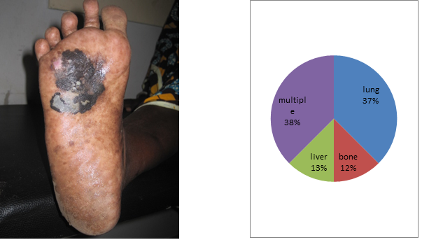
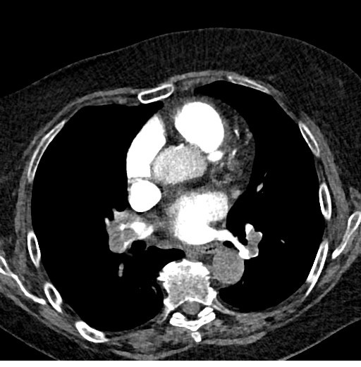
Soft tissue sarcoma represents 4 patients per 1000 cases at the Dakar Cancer Institute. The mean age was 41.2 years with extremes of 9 and 81 years. There was a female predominance with a sex ratio of 0.6. There was a family history of cancer in 5 patients. History of type 1 neurofibromatosis was found in 2 patients and irradiation for pelvic cancer in 1 patient. Mean follow-up was 17.2 months after onset of symptomatology. The most frequent localizations were the thigh (n = 7) [Figure 1] and the shoulder (n = 5) [Figure 2]. The size of the lesions varied from 5 to 35 cm with an average of 14.3 cm. The majority of patients (n = 37) had a single lesion. The initial lesion was bifocal in 2 patients and 1 patient had 4 lesions. The imaging assessment used CT for 20 patients, MRI for 11 and ultrasound in 6 cases. It showed the presence of metastasis in 9 patients (24.4%) at the time of diagnosis and an extension to neighboring organs in 17 patients. Surgical biopsy was the most common modality (26 cases, 67%) followed by ultrasound guided biopsy (6 cases, 15%). The most common histological type was Rhabdomyosarcoma (20%) followed by Dermatofibrosarcoma (17.5%) [Table 1]. A high-grade sarcoma was found in 19 patients (47.5%). Surgery was performed in 24 patients (60%). Wide resection was the most common type of surgery (n = 12). Chemotherapy was performed in 13 patients including 8 palliative chemotherapies. The Adriamycin-Cisplatin chemotherapy protocol was the most widely used (n = 11). Radiotherapy was performed in 5 patients. The goal was neo adjuvant in 2 patients, adjuvant in 1 patient and palliative in 2 patients. After a 52-month follow-up period, 2 patients had local recurrence and 18 patients died [Table 2].

Figure 1. deep limb localization Figure 2. shoulder localization of a rhabdomyosarcoma

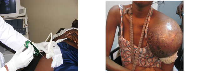
Fig1.deep limb localization                    Fig2.shoulder localization of a rhabdomyosarcoma

Table 1. Different histologic types of STS

Histologic  type Frequencies Percentage (%)
Angiosarcoma 1 2,5
Chondrosarcomea 1 2,5
Dermatofibrosarcoma protuberans 7 17,5
Fibrosarcoma 5 12,5
Atypical Fibroxantoma 1 2,5
Atypical Hémangioendothelioma 1 2,5
Endemic  Kaposi Sarcoma 1 2,5
Leimyosarcoma 1 2,5
Liposarcoma 2 5,0
Neurosarcoma 3 7,5
Rhabdomyosarcoma 8 20
Fusiform Cells Sarcoma 3 7,5
Phyllod Sarcoma 1 2,5
Pleomorph Sarcoma 2 5
Synovialosarcoma 3 7,5
Total 40 100

Table 2. Results of follow up

Evolution Frequencies Percentage(%)
Décès 18 45
Récidive 2 5
Patients en cours de traitement 7 17,5
Patients traités  sans récidive 13 32,5
Total 40 100

Discussion

Soft tissue sarcoma (STS) accounts for 0.5-1% of malignant tumors in adults. Their incidence is estimated at 30 cases per million inhabitants [1]. The frequency of soft tissue sarcomas increases in adults with age, and half of the patients are older than 50 years [2]. There is a slight male predominance especially after 60 years [3]. Several genetic factors including Gardener syndrome, Li Fraumeni syndrome and type 1 neurofibromatosis predispose to the onset of STS [4]. The sarcomas of the limbs represent approximately 60% of the sites, 45% of which are in the lower limbs [5, 6]. They are characterized by their large sizes [7,8]. It is a very lymphophilic tumor in the higher grades, in the associated forms in particular carcinosarcomas and in certain histological types such as synovialosarcomas and rhabdomyosarcomas [9]. One patient in four is metastatic at the time of diagnosis and secondary lesions are predominantly located at lymph nodes and lungs [10, 11]. MRI is the main method of imaging in STS. In association with PET-SCAN, its role is increased in the diagnosis of recurrences [12, 13]. The scanner is only used in the local assessment if MRI is contraindicated. Ultrasound is of limited utility except in the superficial small masses. The delay of consultation, very long in case of limited resources are bad prognoses factors [2]. The most frequent histological types are liposarcomas, leiomyosarcomas and malignant histiocytofibromas [14].

The treatment need a multidisciplinary approach and is better considered in a reference center [2]. Surgery regardless of location is the basic treatment. It is more functional and more conservative thanks to multidisciplinary teams and the development of new techniques like the isolated perfusion of the limb. The objective is to minimize functional sequelae and amputation rates [15, 16]. Chemotherapy occupies an important place in neo adjuvant situation in the locally advanced sarcomas and the high grades [2]. The question of adjuvant chemotherapy is generally left to the appreciation of multidisciplinary consultations [17]. Radiotherapy, in particular radio chemotherapy, has produced encouraging results in the neo-adjuvant period for locally advanced tumors [18]. The surgical excision followed by complementary radiotherapy is the standard loco regional treatment of limbs and operable and localized STS [19]. Abstinence from adjuvant radiotherapy is possible for superficial or strictly intramuscular tumors for which surgery showed clear margins [20].

The best survival rate, obtained in the reference centers, is of the order of 60 to 70% at 5 years in the early stages and of 2 months in the advanced stages [2, 11].

Conclusion

Soft tissue sarcoma is a rare cancer. It occurs sporadically or on specific genetic situations. Histological forms and grade determine susceptibility to treatment and prognosis. High grade and long delay are the cause of poor prognosis in limited resources situations. The main goal is first conservative and functional treatment. It needs multidisciplinary approach associating mainly surgery and radiotherapy. High grade sarcomas and metastatic stages have very poor prognosis.

Conflict of interest: None declared.

Source of funding: None.

Consent: Written informed consent was obtained from the patients.

Acknowledgments: None.

Author Contribution: Ka conceived this presentation while Diouf and Dem participated in quality control of this manuscript. All authors read and approved the final manuscript.

References

  • Fletcher DM, Unnik K and Mertens F (2002) World Health Organization classification of tumors. Pathology and genetics of tumors of soft tissue and bone. Lyon: IARC Press.
  • Honoré C, Méeus P, Stoeckle E, Bonvalot S (2015) Soft tissue sarcoma in France in 2015: Epidemiology, classification and organization of clinical care. J Visc Surg 152: 223-230. [crossref]
  • Jørgensen PH, Lausten GS, Pedersen AB (2016) The Danish Sarcoma Database. Clin Epidemiol 8: 685-690. [crossref]
  • Ducimetière F, Lurkin A, Ranchère-Vince D, Decouvelaere AV, Isaac S, et al. (2010) [Incidence rate, epidemiology of sarcoma and molecular biology. Preliminary results from EMS study in the Rhône-Alpes region].Bull Cancer 97: 629-641. [crossref]
  • Chang AE, Rosenberg SA (1988) Clinical evaluation and treatment of soft tissue tumors. In: Enzinger FM, Weiss SW. Soft tissue tumors (2nd ed) St Louis, Toronto, London: CV Mosby Co 19–42.
  • Coindre JM, Terrier P, Bui NB, Bonichon F, Collin F, Le Doussal V (1996) Prognostic factors in adult patients with locally controlled soft tissue sarcoma. A study on 546 patients from the French Federation of Cancer Centers Sarcoma Group. J Clin Oncol 14:869–77.
  • Mazeron JJ, Suit HD (1987) Lymph nodes as sites of metastases from sarcomas of soft tissue. Cancer 60: 1800-1808. [crossref]
  • Pisters PW, Harrison LB, Leung DH, Woodruf JM, Casper ES (1996) Long-term results of a prospective randomized trial of adjuvant chemotherapy therapy in soft tissue sarcoma. J Clin Oncol 14:859–68.
  • Davis CH, Yammine H, Khaitan PG, Chan EY, Kim MP (2016) A multidisciplinary approach to giant soft tissue sarcoma of the chest wall: A case report. Int J Surg Case Rep 28:211-213.
  • Christie-Large M, James SL, Tiessen L, Davies AM, Grimer RJ (2008) Imaging strategy for detecting lung metastases at presentation in patients with soft tissue sarcomas. Euro Journ Cancer 44: 1841-45.
  • Marudanayagam R, Sandhu B, Perera MT, Bramhall SR, Mayer D, et al. (2011) Liver resection for metastatic soft tissue sarcoma: an analysis of prognostic factors.Eur J Surg Oncol 37: 87-92. [crossref]
  • Choi YY, Lee IS, Kim SJ, Kim JI, Choi KU, et al. (2013) Analyses of short-term follow-up MRI and PET-CT for evaluation of residual tumour after inadequate primary resection of malignant soft-tissue tumours.Clin Radiol 68: 117-124. [crossref]
  • Shapeero LG (1990) MRI of the foot and ankle. In: Gooding CR editor, Diagnostic radiology. San Francisco: University of California Press 454–66.
  • Ferté C, Pascal LB, Penel N (2009) [Prognostic and predictive factors of soft tissue sarcoma: a daily use of translational research].Bull Cancer 96: 451-460. [crossref]
  • Stoeckle E, Italiano A, Stock N, Kind M, Kantor G, et al. (2008) [Surgical margins in soft tissue sarcoma]. Bull Cancer 95: 1199-1204. [crossref]
  • Aisen AM, Martel W, Braunstein EM, McMillin KI, Phillips WA, et al. (1986) MRI and CT evaluation of primary bone and soft-tissue tumors. AJR Am J Roentgenol 146: 749-756. [crossref]
  • Fayette J, Blay JY, Ray-Coquard I (2006) [Soft tissues sarcomas: good medical practices for an optimal management]. Cancer Radiother 10: 3-6. [crossref]
  • Casali P, Jost L, Sleijfer S, Verweij J, Blay JY (2008) Soft tissue sarcomas: ESMO clinical recommendations for diagnosis, treatment and follow-up. Ann Oncol 19:89-93.
  • Cheng EY, Dusenbery KE, Winters MR, Thompson RC (1996) Soft tissue sarcomas: preoperative versus postoperative radiotherapy. J Surg Oncol 61: 90-99. [crossref]
  • Suit HD, Mankin HJ, Wood WC, Proppe KH (1985) Preoperative, intraoperative, and postoperative radiation in the treatment of primary soft tissue sarcoma. Cancer 55:2659-67.

Epidemiological, Diagnostic, Therapeutic and Prognostic Aspects of Melanoma of Black Skin in an African Cancer Institute

DOI: 10.31038/CST.2016124

Abstract

Objectives: To describe the epidemiological, clinic, histologic, therapeutic and prognosis aspects of cutaneous melanoma in African black at the Dakar cancer Institute.

Materials and methods: This was a 6 years retrospective study that included all cases of cutaneous melanoma at the Institute of Dakar cancer. The clinical, epidemiological, clinical and histological parameters as well as the treatment and prognosis were analyzed.

Results: There were 21 black skin patients with malignant melanoma. The sex ratio is 0.75. The average age of our patients was 60.8 years and the average time of consulting, 32.8 months. The plantar melanoma accounted for 76% of cases (n = 16). The mean tumor size was 7.1 cm. The presence of inguinal lymphadenopathy was noted in 12 patients or 57% of cases. Pathology showed an acral lentiginous melanoma in 18 patients or 85.7%, and a nodular melanoma in 2 patients or 9.5%. The Breslow thickness was more than 5 in half of the patients. Staging showed at thoraco abdominal CT, secondary locations in 9 patients (43%). Surgical excision was performed in 8 patients while inguinal lymph node dissection was performed in 12 patients. We performed chemotherapy in 5 patients (23.8%). Palliative haemostatic radiotherapy was successfully performed in 1 patient. The mean time follow-up was 16.1 months with extremes of 1 and 66 months. We recorded 10 deaths (47.6%) during the period of study.

Keywords

melanoma; black skin; stage; surgery; prognosis.

Introduction

Malignant melanoma is the most aggressive skin cancer with a high metastatic potential. There are 200 000 new cases each year and it is the leading cause of death from skin cancer [1]. It is a rare in black people about which the skin is better protected by melanin. Surgery is the best treatment at early stage. The advent of targeted therapies and immunotherapy has improved survival in advanced cases [2]. Because of the low incidence in Africa melanoma of the black skin is understudied. The objective of this study was to report the epidemiology, diagnostic, therapeutic characteristics and outcomes of malignant melanoma of black skin at the Dakar Cancer Institute.

Materials and Methods

This was a retrospective study over a period of 6 years from January 2008 to December 2013. We evaluated the epidemiologic, clinical, histological, therapeutic and oncologic results.

Results

It was about 21 patients with malignant melanoma skin cancer of 136 cases or 15.4% of cases. They found 12 women to 9 men for a ratio of 0.75. The average age of our patients was 60.8 years, ranging from 29 years to 85 years. The average time of consultation was 32.8 months with extremes of 4 to 108 months. All patients were black subjects. No family history of melanoma was found. Plantar melanoma accounted for 76% of cases (n = 16) [Figure 1]. The trunk was the second site of localization [Table 1]. The average tumor size was 7.1 cm with extremes of 2.5 and 15 cm. Inguinal lymphadenopathy was noted in 12 patients (57%). Pathotology showed an acral lentiginous melanoma in 18 patients (85.7%) and a nodular melanoma in 2 patients (9.5%). The Breslow thickness was more than 5 in half of the patients. Staging showed at thoraco abdominal CT secondary locations in 43% of patients (n = 9), 3 in lungs (37.5%), 1 in liver (12.5%), 1 in bones (12.5%) and 3 cases of multiple locations (37.5%) [Figure 2]. Surgery was performed in 14 patients or 66.7%. The excision was carried out in 8 patients with macroscopic margins of 3 cm. Amputation or dislocation was performed in 6 patients. The surgical margins were healthy in 8 patients 57% of cases. The inguinal lymph node dissection was performed in 12 patients. The average number of lymph nodes removed was 7.16 nodes with a range of 2 to 10 nodes. Node-metastasis was founds in 5 patients (41.7%). Chemotherapy was given to 5 patients (23.8%). Palliative haemostatic radiotherapy was successfully performed in 1 patient with melanoma of the chest wall, at a dose of 8 Gy in one session. Among the 8 patients who had secondary locations, the coup average time was 12 months. The mean follow-up was 16.1 months with extremes of 1 and 66 months. We recorded 10 deaths or 47.6% during the study period.

Table 1. Distribution by tumor site

  Nombre Percentage (%)
Foot plant                                                   16 76
Trunk                                         3 14,4
Right Hallux                                           1 4,8
3rd toe                                            1 4,8
TOTAL 21 100
Figure 1. Plantar melanoma Figure 2. Different locations of metastases

Figure 1. Plantar melanoma                              Figure 2. Different locations of metastases

Discussion

Malignant melanoma is the most common skin cancer in light skin populations in areas where there is strong sunlight [3]. The reasons are genetic and environmental. The redhead phenotype depends on the MC1R gene and some areas like Australia are most exposed [4]. In Africa, it is less common than squamous cell carcinomas [5]. The occurrence of non-melanoma skin cancer depends on the immune status and many co-factors such as UV and HPV exposure [6]. It is an ubiquitous cancer for people aged 50 to 60. Family history plays an important role in the occurrence of melanoma [7]. Feet localizations are more frequent on black skin. It is essentially sporadic. Surveillance of plantar zone is difficult. That’s probably why plantar melanoma stages are advanced and are characterized by important tumor sizes, ulceration and budding. At resection, it has a clue high Breslow. The most common histological type is acral lentiginous melanoma [1,8]. It is the most common form in non-caucasian, hispanic and oriental populations [5]. The cause of its preferential localization at foot plant is not yet clear. Extension of this melanoma uses lymphatic, blood and step by step ways. The extension can be done to the bones, joints and muscles of the feet; typically we can found skip metastasis along the lower limb or regional, inguinal and popliteal lymphadenopathy and even retro and under peritoneum lymph nodes. Metastases are frequently seen at diagnosis. They are most commonly lung, brain and bones [9]. The later stages characterized the diagnosis in Africa. Most of the plantar location, by diagnostic errors and traditional treatments before medical care and the limited resources of patients are seen lately. The treatment is local, loco regional and systemic and depends on the stage of the disease. The surgical resection is more effective when the Breslow thickness is low. Amputation is frequent in our conditions. The node involvement varies. The prognosis depends more on local excision in early stage of the treatment of lymph node and distant metastases [10]. In early stages, the sentinel node biopsy reduces intra operative surgical risk, post-operative complications and distance lymphedema of the lower limb [9,11]. Lymph node dissection in advanced stages decreases inguinal recurrence but does not change the prognosis [12]. Iliac dissection using under peritoneal route without a formal indication in non-metastatic patients, seems to improve the prognosis [13]. Advanced stages raise the problem of therapeutic choice between palliative surgery sometimes mutilating such as limb amputations in patients with poor prognosis and systemic treatment alone or no treatment. Chemotherapy slightly modified melanoma prognosis. Radiotherapy is considered rather palliative. It is preconized in adjuvant situation for large lesions and after inguinal lymphadenectomy [14]. The melanoma-related mortality in advanced stages is very high. The low socioeconomic level in Africa is therefore indirectly a poor prognosis factor. Immunotherapy with the advent of anti PD1 and anti CTLA 4 increases significantly survival of melanoma [2,15].

Conclusion

Melanoma remains the most aggressive skin cancer with high metastatic potential. Its frequency in developed countries is a major public health problem. Its specific locations and socioeconomic level in black people in Africa makes the mortality very high. Surgery is more palliative than curative. Choosing a mutilating radical treatment is frequently an option. The advent of targeted therapies and immunotherapy revolutionizes its treatment and improves survival rates. Their availability is a priority in Africa.

Conflict of interest: None declared.

Source of funding: None.

Consent: Written informed consent was obtained from the patients.

Acknowledgments: None.

Author Contribution: Ka conceived this presentation while Diouf and Dem participated in quality control of this manuscript. All authors read and approved the final manuscript.

References

  • Erdei E, Torres SM (2010) A new understanding in the epidemiology of melanoma. Expert Rev Anticancer Ther 10: 1811-1823. [crossref]
  • Philips GK, Atkins M2 (2015) Therapeutic uses of anti-PD-1 and anti-PD-L1 antibodies. Int Immunol 27: 39-46. [crossref]
  • Grange F1 (2005) [Epidemiology of cutaneous melanoma: descriptive data in France and Europe]. Ann Dermatol Venereol 132: 975-982. [crossref]
  • Giles GG, Armstrong BK, Burton RC, Staples MP, Thursfield VJ (1996) Has mortality from melanoma stopped rising in Australia? Analysis of trends between 1931 and 1994. BMJ 312: 1121-1125. [crossref]
  • Pitche P, Tchamdja S, Napo koura G, Bakonde P et al. (1997)Les cancers cutanés en consultation dermatologique à Lomé(Togo). Méd Afr Noir 44:15-17.
  • Reusser NM, Downing C2, Guidry J3, Tyring SK4 (2015) HPV Carcinomas in Immunocompromised Patients. J Clin Med 4: 260-281. [crossref]
  • Payette MJ, Katz M 3rd, Grant-Kels JM (2009) Melanoma prognostic factors found in the dermatopathology report. Clin Dermatol 27: 53-74. [crossref]
  • Phan A, Touzet S, Dalle S, Ronger S, Balme B, Thomas L (2007) Acral lentiginous melanoma: histopathological pronostic feature of 121 cases. Br J Dermatol 157:311-318.
  • Homolak D, Šitum M, Cupic H (2015) Clinico-pathological features of patients with melanoma and positive sentinel lymph node biopsy: a single institution experience. Acta Dermatovenerol Croat 23:122-9.
  • Arcangeli F, Catrani S, Greco I, Pierleoni M, Landi G (2010) Prevention and early diagnosis of melanoma: a regional experience in Italy. Jour Eur Acad Dermatol Venereo 2:207-210.
  • Duvernay A, Henault B, Danino MA, Trost O, Dalac S, et al. (2012) [Complications associated with sentinel lymph node biopsy for cutaneous melanoma. A retrospective study of 127 patients]. Ann Chir Plast Esthet 57: 151-157. [crossref]
  • Mozzillo N, Caracò C, Marone U, Di Monta G, Crispo A, et al. (2013) Superficial and deep lymph node dissection for stage III cutaneous melanoma: clinical outcome and prognostic factors. World J Surg Oncol 11: 36. [crossref]
  • Giudice G, Robusto F, Nacchiero E (2016) The surgical treatment of a melanoma patient with macroscopic metastasis in peri and retrocaval lymph nodes and with a positive sentinel lymph node in the groin. Ann Ital Chir 4: 87.
  • Burmeister BH, Henderson MA, Ainslie J, Fisher R, et al. (2012) Adjuvant radiotherapy versus observation alone for patients at risk of lymph-node field relapse after therapeutic lymphadenectomy for melanoma: a randomised trial. Lancet Oncol 13:589-97.
  • Blank CU, Enk A (2015) Therapeutic use of anti-CTLA-4 antibodies. Int Immunol 27: 3-10. [crossref]

Re-igniting the PE debate

DOI: 10.31038/IMROJ.2016123

Case Report

Abstract: We describe the case of patient with a sub-massive pulmonary embolus and the evidence based management.

Learning points: The decision to thrombolyse or not to thrombolyse a sub-massive PE is difficult and several clinical factors need to be taken into consideration.

Key words: sub-massive, pulmonary embolus, thrombolysis

Joseph Mackenzie (JM): A 78 year old female patient presented 4 days ago with 3 day history of sudden onset breathlessness and dizziness. She was known to have chronic unspecified gromerulonephritis and had a WHO performance score of 1. Her regular medications included anti-hypertensives and quinine for night cramps. She was a never smoker with no dust exposure.

Initially, her pulse rate was 76 beats per minute, her oxygen saturations 95 per cent on air and blood pressure 105 over 76 mm of mercury. Her inflammatory markers were normal but her urea and creatinine had risen to 27.9 mmol per litre and 238 ummol per litre. Her chest radiograph and electrocardiogram was normal.

She was admitted under the care of the elderly team, was rehydrated and her nephrotoxics stopped. On the second day of admission, she had a syncopal episode with a spontaneous return to circulation: An arterial blood gas done on air showed a pCO2 of 3.2 KPascals, a pO2 of 8.9 KPascals, a base excess of -10mmol/L and oxygen saturations of 94 per cent.

The next day, a D-dimer was checked- that was more than 20.00 mg/L (normal being <0.50). Her hypotension was fluctuating. An echocardiogram was done- it showed a flattened septum throughout the cardiac cycle suggesting right ventricular (RV) overload, mildly impaired systolic function, moderate tricuspid regurgitation with a pulmonary arterial systolic pressure (PASP) estimated at 70mm mercury added to the right atrial pressure and a tricuspid annular plane systolic excursion of 1cm.

A computed tomography pulmonary angiogram was then performed which showed massive pulmonary embolism(PE) bilaterally in upper, middle and lower lobe pulmonary arteries, bilateral main pulmonary arteries (Fig 1) with significant right ventricular strain noted with reflux of contrast into IVC and hepatic veins.

She was started on full dose low molecular weight heparin and warfarin. Her current International Normalised Ratio (INR) is 1.8

Figure 1. CT scan showing massive bilateral pulmonary emboli and RV strain

Figure 1. CT scan showing massive bilateral pulmonary emboli and RV strain

Avinash Aujayeb (AA): Her initial Wells score was 31(only scored yes for PE being likely diagnosis), which puts her in a moderate risk group and combined with a high D dimer initially, she certainly warranted further investigation.

Her alveolar–arterial oxygen tension difference (PA–aO2) was 7 kPa.

The PA–aO2 is a measure of the difference between the alveolar concentration (A) of oxygen and the arterial (a) concentration of oxygen. In room air (inspiratory oxygen fraction of 0.21) at sea level (atmospheric pressure of 760 mmHg) assuming 100% humidity in the alveoli, a simplified version of the equation is 21−((PaCO2/0.8)−PaO2), 0.8 being the respiratory quotient. Whilst her peripheral saturations might appear normal, her high tension difference suggests a ventilation-perfusion mismatch and in a never smoker with a normal chest radiograph, a vascular event needs excluding.

The echocardiographic findings are worrisome. The main cause of death in acute PE is RV failure due to pressure overload. The abrupt increase in pulmonary vascular resistance results in RV dilation, which alters the contractile properties of the RV myocardium via the Frank-Starling mechanism. The increase in the RV afterload also causes the tricuspid valve to fail and the PASP is a marker of that. TAPSE is a simple echocardiographic measure of RV ejection fraction and any value below 2 cm is considered normal. Echocardiographic examination is not recommended as part of the diagnostic work-up in haemodynamically stable, normotensive patients with suspected (not high-risk) PE2, but in a suspected high-risk PE, a normal echocardiogram can exclude it.

Joseph Mackenzie (JM): The patient has had no further syncopal episodes but has been more breathless today, and is now requiring 2litres via nasal cannulae to maintain oxygen saturations at 95%. Her respiratory rate has increased to 26 per minute and her blood pressure chart is as in Figure 2. A troponin T is 114 nanograms/L (normal range 0-14) and her lactate is 3.3 millimol/L (normal 0.5-2.2)- that was previously 1.9. This is now almost 7 days after she developed the initial symptoms. I note her INR of 1.8 but wonder if she would benefit from thrombolysis.

Figure 2. Observation chart showing fluctuations in blood pressure

Figure 2. Observation chart showing fluctuations in blood pressure

Avinash Aujayeb (AA): Patients with PE and shock or hypotension are at high risk of death, particularly in the first few hours after admission. The clinical classification an acute PE is based on estimated early mortality risk (in-hospital or 30-day)2 and shock or hypotension in PE is defined as a systolic blood pressure less than 90mm of mercury or a systolic sustained drop of more than 40mm of mercury over more than 15 minutes, in the absence of arrythmia, hypovolemia and sepsis. Hence, by strict definition, at the moment, she has a submassive PE (confirmed PE in a normotensive patient with evidence of RV dilatation and/or RV dysfunction and/or pulmonary hypertension) but I note the fluctuation hypotension.

There have been excellent debates of the pros and cons of thrombolysis in submassive PE recently.

The con arguments3 are that large registries suggest 90 day mortality in thrombolysed patients to be around 3% and in the heparin only group to be around 2%, that RV dilatation is a dynamic process and some studies have shown that 93% of such patients have normalised their RV at 6 months and that there is no evidence proving that early haemodynamic improvements has survival benefits, prevents recurrence and development of chronic thromboembolic pulmonary hypertension. The PETHIO trials2 also suggested statistically significant differences in bleeding complications (2% incidence of haemorrhagic stroke after thrombolytic treatment and 6% risk of major non-intracranial bleeding events).

However, I think that her initial presenting symptoms are important and I agree with thrombolysis. There is a relative contraindication that her INR is 1.8 but I think her risk of death is high.

I have sought a second opinion on this from a cardiologist who agrees with thrombolysis.

She has markers of significant myocardial necrosis and RV dysfunction. Her initial presentation with syncope and hypotension is an adverse prognostic sign and even though her outward haemodynamics have normalised now, her rising lactate suggests otherwise. Thrombolysis can quickly restore pulmonary perfusion and resolve pulmonary resistance to improve RV function4. The greatest benefit occurs when the agent is administered within 48 hours of the primary event, but benefit has been proven for patients up to 14 days down the line2.

The ESC guidelines2 would classify her to be in the intermediate high risk group, where thrombolysis should not be routinely considered, unless there is haemodymanic decompensation. A rising lactate is a proven maker of this5.

Joseph Mackenzie (JM): The patient provided written consent to thrombolysis.

10mg of alteplase was given over 2 minutes followed by 90mg over 2 hours. Continuous haemodynamic and neurological monitoring was performed with no anomalies detected. There was a small brisk episode of epistaxis which was self limiting and subcutaneous bruising appeared. Within 4 hours of administration, oxygen levels increased to 100 per cent on 2L nasal cannulae and remained at 96% on air afterwards. The patient’s breathlessness disappeared and full dose low molecular weight heparin was restarted as well as warfarin loading.

After a brief period of rehabilitation, she has now been discharged and will have follow up in the thrombosis clinic with a repeat echocardiogram in 3 months.

References

  • http://www.mdcalc.com/wells-criteria-for-pulmonary-embolism-pe/ (Accessed 3.3.16)
  • http://www.escardio.org/Guidelines-&-Education/Clinical-Practice-Guidelines/Acute-Pulmonary-Embolism-Diagnosis-and-Management-of (Accessed 3.3.16)
  • Simpson AJ (2014) Thrombolysis for acute submassive pulmonary embolism: CON viewpoint. Thorax 69: 105-107. [crossref]
  • Howard LS (2014) Thrombolytic therapy for submassive pulmonary embolus? PRO viewpoint Thorax 69:103-5.
  • Fuller, Brian M, Phillip Dellinger R (2012) “Lactate as a Hemodynamic Marker in the Critically Ill.” Current opinion in critical care 18.3 267–272.

Evidence-Based Medicine (EBM) and Clinical Practice

DOI: 10.31038/IMROJ.2016122

Editorial

The interest around EBM was born from the belief that it might reduce the concerns raised in recent years about health care. Such concerns involve the quality of medical practice, the unwarranted variation in the use of medical procedures, and the risk of decreasing quality of care of physicians as they progress in their practice, as outlined in the following paragraphs.

There is evidence that the quality of medical practice is not consistent with the ongoing development of the medical knowledge [1,2]. Diagnostic and therapeutic practices of proven effectiveness are often underused, whereas other practices are overused in contrast with trustworthy clinical practice guidelines, and their improper use can result in.

A pointer of such inconsistencies is the well-demonstrated existence of considerable variation of care in the clinical practice, not explained by patients’ characteristics or preferences, and instead related to local clinical routine, physicians’ specialties, training and opinions, and other factors [3,4].

Finally, there is evidence that doctors frequently perform their practice as a series of automatic interventions according to the standard formula [if…then…], a practice resulting in lower professional skills and in providing lower quality of care as they progress in their medical career [5,6].

Can EBM contribute to overcome these concerns?

“Within 5 years of the first proposal [in 1992], evidence-based medicine (EBM) has received enthusiastic endorsement from editors of prominent medical journals, achieved the publicational outlet of its own new journal, and acquired the sanctity often accorded to motherhood, home, and the flag” [7]. Though ironic, this statement by Feinstein and Horwitz provides an exact account of the fervent acceptance of EBM in the medical literature. According to the precepts of EBM, clinicians should identify and adopt methodologically sound published evidence when deciding on the treatments or diagnostic procedures for their patients. However, EBM has been conceived according to two different approaches: EBM as a new paradigm of clinical practice, or EBM as a component of the physician’s expertise in the care of an individual patient.

EBM as the new paradigm of clinical practice. According to the Evidence-Based Medicine Working Group (chaired by Gordon Guyatt): “A new paradigm for medical practice is emerging. Evidence-based medicine de-emphasizes intuition, unsystematic clinical experience, and pathophysiologic rationale as sufficient grounds for clinical decision making and stresses the examination of evidence from clinical research” [8]. In Kuhnian terms, EBM should replace the “no longer tenable paradigm of traditional medical practice,” as re-affirmed and expanded in the three editions of the Users’ Guides to the Medical Literature published up to date [9-11]. This concept of EBM disregards the clinical expertise of physicians in caring individual patients, acquired through a lifelong habit of learning and reflection at the workplace. Population-derived research evidence has its role but cannot overlook the physician’s approach to the care to the individual patient.

EBM as a component of the expertise of clinicians and of the preferences and values of patients can contribute to approach the current concerns on the quality of medical practice. This EBM model was introduced by David Sackett “Evidence-based medicine involves the conscientious, explicit and judicious use of current best evidence in making decisions about the care of individual patients. The practice of evidence-based medicine consists of integrating individual clinical expertise with the best available external evidence from systematic research. By individual clinical expertise we mean the proficiency and judgement that individual clinicians acquire through clinical experience and clinical practice.” [13].

However, EBM is not of help to approach two cardinal components of the clinical expertise, ie diagnosis and patient-doctor relationship.

EBM and diagnosis

The diagnosis and the diagnostic process are weak points of EBM. In the publications by the Sackett’s group the chapter on diagnosis is fully dedicated to diagnostic tests. The Users’ Guides to the Medical Literature (3rd edition) report the standard distinction between “pattern recognition” and “probabilistic diagnostic reasoning”, the latter representing an inadequate and partial definition of the analytic diagnostic process [14]. Neither the series of publications by the Sackett’s group nor the Users’ Guides contain any information on the cognitive aspects of the diagnostic process (e.g. generating hypotheses, comparing the information provided by patients with memorized illness scripts, or the important issue of diagnostic errors). Eventually, the Fowler’s statement that, “evidence-based medicine only follows when a correct diagnosis has been made” appears to be appropriate [15].

EBM and the patient-doctor relationship

The physicians’ attitude towards establishing a sound relationship with the patients represents a key element of good practice [16, 17]. As written by Osler: “Medicine is more than the sum of our knowledge about diseases. Medicine concerns the experiences, feelings and interpretations of human beings in often extraordinary moments of fear, anxiety and doubt.” The seeds of this concept should be conveyed to students in the medical school, and then developed in their professional career. Instead, there is evidence that the natural empathy and patient-centered approach of the medical students tends to decline as they progress in their clinical curriculum [18], and that patients frequently complain about inappropriate behavior of physicians, stressing disrespect, misinformation and perceived unavailability [19]. Although this aspect clearly would require special attention, there is no element in the EBM-related educational initiatives to foster a positive and compassionate relationship of physicians with their patients.

Outside EBM: deliberate practice

Another citation from Osler is relevant here: “To study the phenomena of disease without books is to sail an uncharted sea, while to study books without patients is not go to sea at all.” Beyond and before the search and use of population-derived evidence from the literature and in contrast with a practice performed routinely by means of automatic interventions, the performance of “deliberate practice” [20], is a key for a sound approach to the medical profession. “Deliberate practice” i.e. a practice associated with reflection and continuous learning at the workplace is a key factor of the medical profession as shown by the relationship between large volume of medical practice and improved outcome in many clinical areas (e.g. myocardial infarction, heart failure, pneumonia, and surgery [21, 22]). The EBM movement should not bring about the unintentional effect of distracting young trainees from deliberate practice and continuous learning in the workplace.

Moving towards a tentative conclusion: EBM can be conceived as the search, evaluation and use of literature evidence to support the approach to clinical problems. EBM, i.e. the search and use of published evidence, is only a component and not a new paradigm of physicians’ professional skills and clinical expertise.

References

  • McGlynn EA, Asch SM, Adams J, Keesey J, Hicks J, et al. (2003) The quality of health care delivered to adults in the United States. N Engl J Med 348: 2635-2645. [crossref]
  • Mangione-Smith R, DeCristofaro AH, Setodji CM, Keesey J, Klein DJ, et al. (2007) The quality of ambulatory care delivered to children in the United States. N Engl J Med 357: 1515-1523. [crossref]
  • Chassin MR, Brook RH, Park RE, Keesey J, Fink A, et al. (1986) Variations in the use of medical and surgical services by the Medicare population. N Engl J Med 314: 285-290. [crossref]
  • Wennberg JE (2005) Variation in the use of Medicare Services among regions and selected academic medical centres: is more better? Duncan W. Clark Lecture, New York Academy of Medicine.
  • Choudhry NK, Fletcher RH, Soumerai SB (2005) Systematic review: the relationship between clinical experience and quality of health care. Ann Intern Med 142: 260-273. [crossref]
  • Southern WN, Bellin EY, Arnsten JH (2011) Longer lengths of stay and higher risk of mortality among inpatients of physicians with more years in practice. Am J Med 124: 868-874. [crossref]
  • Feinstein AR, Horwitz RI (1997) Problems in the “evidence” of “evidence-based medicine”. Am J Med 103: 529-535. [crossref]
  • Evidence-Based Medicine Working Group (1992) Evidence-based medicine. A new approach to teaching the practice of medicine. JAMA 268: 2420-2425. [crossref]
  • Guyatt G, Rennie D (2002) Users’ guides to the medical literature. A manual for evidence-based clinical practice. JAMA & Archives Journals.
  • Guyatt G, Rennie D, Meade MO, Cook DJ (2008) Users’ guides to the medical literature. A manual for evidence-based clinical practice. Second Edition. JAMA & Archives Journals, the McGraw Hill Companies.
  • Guyatt G, Rennie D, Meade MO, Cook DJ (2015) Users’ guides to the medical literature. A manual for evidence-based clinical practice. McGraw Hill Education.
  • Kahneman D (2012) Pensieri lenti e veloci. A. Mondadori Editore,
  • Sackett DL, Rosenberg WMC, Gray JAM, Haynes RB, Richardson WS (1996) Evidence-based Medicine. What it is and what it isn’t. It’s about integrating individual clinical expertise and the best external evidence. BMJ 312:71-72
  • Pagliaro L, Bobbio M, Colli A (1997) La Diagnosi in Medicina. Raffaello Cortina Editore. Milano
  • Fowler PBS (1997) Evidence-based diagnosis. J Eval Clin Pract 3:153-59
  • Ridd M, Shaw A, Lewis G, Salisbury C (2009) The patient-doctor relationship: a synthesis of the qualitative literature on patients’ perspectives. Br J Gen Pract 59: e116-133. [crossref]
  • Truog RD, 200th Anniversary article. Patients and doctors – the evolution of a relationship. N Engl J Med 2012; 366: 581-85
  • Hojat M, Mangione S, Nasca TJ, Rattner S, Erdmann JB, et al. (2004) An empirical study of decline in empathy in medical school. Med Educ 38: 934-941. [crossref]
  • Wofford MM, Wofford JL, Bothra J, Kendrick SB, Smith A, et al. (2004) Patient complaints about physician behaviors: a qualitative study. Acad Med 79: 134-138. [crossref]
  • Ericsson KA (2008) Deliberate practice and acquisition of expert performance: a general overview. Acad Emerg Med 15: 988-994. [crossref]
  • Birkmeyer JD, Siewers AE, Finlayson EV, Stukel TA, Lucas FL, et al. (2002) Hospital volume and surgical mortality in the United States. N Engl J Med 346: 1128-1137. [crossref]
  • Davoli M, Amato L, Minozzi S, Bargagli AM, Vecchi S, et al. (2005) [Volume and health outcomes: an overview of systematic reviews]. Epidemiol Prev 29: 3-63. [crossref]

Cystatin C is not Useful to Predict Approaching Acute Kidney Injury in Unstable Critical Care Patients

DOI: 10.31038/IMROJ.2016121

Editorial

The Concept of Acute Kidney Injury (AKI) has changed very much lately. Some decades ago, it was considered a benign condition and needed only supportive treatment. But it is proven now that it may have devastating consequences. Study reported that, in the community; the patients who recovered from AKI have increased risk of death (HR:1.5) also they have increased risk to become a chronic renal failure patients (HR:1.91) in the United States of America [1]. Therefore, in the community, in the hospitals or in the Intensive Care Units patient with risk must be protected from developing of AKI. To do this we should have better biomarkers than conventional ones which are considered serum creatinine and several other urinary markers. The most important reasons of these unwanted outcomes should be delayed diagnosis of AKI. Better biomarkers should alert us beforehand should be practical and applicable in any conditions.

More than 30 different definitions were used for the definition of AKI hitherto which both caused difficulties to interpret and compare the studies. These definitions were developed based on the serum creatinine level which was considered late marker of AKI because it was not start to increase unless kidney functions decline 50% or more. It was suggested that re-evaluation of the definition of AKI was mandatory. For the consensus of the definition and improvement of the quality of studies on AKI, Acute Dialysis Quality Initiative (ADQI) group was developed. They recommended the term of AKI instead of ARF, and indicated that spectrum of AKI is broader and covers different degrees of severity of the disease. In 2002, for a uniform definition of AKI, they described three categories for severity (Risk of ARF, Injury of the kidney, and Failure of kidney function) and two classes for kidney outcome (Loss of kidney function and ESRD), which is called shortly RIFLE criteria [2].

Later, they excluded outcome categories and made some corrections and developed AKIN criteria[3] . Finally, in 2013 guideline of AKI definition was improved and took the final version; accepted by the nephrologists’ in almost all around the world. But in any case, these definitions were based on the serum creatinine level so, they were good for established AKI, but not as early as to prevent and not useful to warn the upcoming AKI threat.

Many researches had being going on during the last decade to discover new biomarkers for AKI, since the conventional biomarkers were not sensitive enough to diagnose AKI beforehand. NGAL(Neutrophyl Gelatinase Associated Lipocaline) and CysC (Cystatin C) were the most studied ones among the others. Many investigators have proposed that CysC may be more sensitive to early AKI development and small changes in the GFR than conventional markers, such as creatinine.[4] On the contrary, a large multicenter study has revealed that CysC is less sensitive than creatinine for the early diagnosis of AKI [5]. We intend to investigate comparing these two biomarkers recently in Intensive Care Unit patients in point of the time of AKI developed.

The sNGAL, uNGAL and sCysC levels were determined at 48 hours of admission and surprisingly we found that sNGAL , uNGAL(AUC-ROC: 0.77, p = 0.005; 0.78, p = 0.002) but not CysC (0.54, p = 0.657)were useful for predicting of the development of AKI following 3-7 days in the ICU[6].

CysC was not found as efficient as serum and urine NGAL to show AKI risk in ICU in this Study. So, we thought that it was wise to detect urine and/or serum NGAL at the 48 hours in ICU admission to estimate AKI risk, even though this biomarker might be affected by so many factors in ICU.

References

  • Bucaloiu ID, Kirchner HL, Norfolk ER, Hartle JE 2nd, Perkins RM (2012) Increased risk of death and de novo chronic kidney disease following reversible acute kidney injury. Kidney Int 81: 477-485. [crossref]
  • Bellomo R, et al. (2004) Acute renal failure – definition, outcome measures, animal models, fluid therapy and information technology needs: the Second International Consensus Conference of the Acute Dialysis Quality Initiative (ADQI) Group. Crit Care 4: R204-12.
  • Joannidis M, Metnitz B, Bauer P, Schusterschitz N, Moreno R, et al. (2009) Acute kidney injury in critically ill patients classified by AKIN versus RIFLE using the SAPS 3 database. Intensive Care Med 35: 1692-1702. [crossref]
  • Herget-Rosenthal S, Marggraf G, Hüsing J, Göring F, Pietruck F, et al. (2004) Early detection of acute renal failure by serum cystatin C. Kidney Int 66: 1115-1122. [crossref]
  • Spahillari A, Parikh CR, Sint K, Koyner JL, Patel UD, et al. (2012) Serum cystatin C- versus creatinine-based definitions of acute kidney injury following cardiac surgery: a prospective cohort study. Am J Kidney Dis 60: 922-929. [crossref]
  • Kamis F, et al. (2016) Neutrophil gelatinase-associated lipocalin levels during the first 48 hours of intensive care may indicate upcoming acute kidney injury. J Crit Care 34: p. 89-94.