Monthly Archives: October 2020

Telemedicine: Enabling Patients with Arrhythmias in Self-Care Behaviors

Abstract

The study, Telemedicine: Enabling Patients with Arrhythmias in Self-Care Behaviors study is designed for early recognition and treatment of an arrhythmia and optimizing patients’ medication, activity, and arrhythmia self-efficacy. Telemedicine is a method which allows health care professionals to evaluate, diagnose, and treat patients within their homes and remote locations [1]. Connecting with patients via video and telephone visits allows the caregiver access and assists the patient in improving self-care behaviors and self-efficacy in managing arrhythmias [1,2]. This pilot telemedicine study provides earlier diagnosis of abnormal arrhythmias and increased patient involvement and self-efficacy of one’s health care solutions [2]. The Telemedicine: Enabling patients in Self-Care Behaviors study started in February 2020, prior to the onset of the Covid 19 pandemic. The study has been placed on hold since March 17, 2020. In a response to the Covid-19 pandemic (separate from this study) multiple medical and nursing practices have adopted telemedicine to maintain ongoing care appointments [3]. The study displays the complementing use of three survey tools (Medication Understanding and Self-Efficacy Tool, Functioning Self Efficacy Scale, and Arrhythmia Specific questionnaire in Tachycardia an Arrhythmia) with monitoring devices (loop recorders, Kardia-TM, pacemakers and cardioverter defibrillators-ICDs) coupled with telephone and video visits to pinpoint arrhythmia changes and exact patient reactions and discussion to reinforce self-efficacy behaviors.

Keywords

Telemedicine, arrhythmia, self-efficacy, behavior.

Introduction

The purpose of the study, Telemedicine: Enabling Patients with Arrhythmias in Self-Care Behaviors (T:EPASB) is to provide an alternative to in person visits, decreased the time of diagnosis and treatment of an arrhythmia via the internet, and enable patients to improve self-efficacy of arrhythmia care behaviors. Self-efficacy can be defined as the individual’s belief in oneself to handle a set of circumstances or changes in physical or mental well-being [2].

The first outcome of the study is to determine if subjects in a telemedicine program for the care of cardiac arrhythmias have  any difference in [1] time of arrhythmia recognition [2], time of arrhythmia diagnosis by a healthcare provider, and [3] time of treatment initiation compared with patients enrolled in standard care for cardiac arrhythmias. The second outcome of the study examines subjects’ self-efficacy of medication use, functional self-efficacy, and arrhythmia self-efficacy. A data collection tool was utilized with a simplistic check off system used to mark when one recognized changes in symptoms such as increased palpitations, fatigue, activity intolerance, shortness of breath, and any other change in symptoms associated with an arrhythmia. The tool allowed for quick responses to these symptoms with self-initiated blood pressure check, heart rate check, increased fluids, or taking an additional beta blocker, sitting down and resting, and calling the electrophysiology (EP) office for advice (Appendix A).

Background Information

University based tertiary care clinics, which  treat  irregular heart rhythms, are known as arrhythmia clinics and formally called electrophysiology departments [4]. These departments have been in existence prior to the early 1960’s and their technology has continued to evolve over time. The need to meet with patients and discuss  their abnormal and irregular heart rhythms has entailed prescribing medications to slow the heart rate, prescribing medications to eliminate abnormal heart rhythms, and implanting devices to further control the heart rhythms, known as pacemakers (PPM) and implantable cardioverter-defibrillators (ICDs) [4]. The continued improvement in technology and expansion of such departments has led to a need for increased numbers of patient appointments, dual appointments for arrhythmia management and pacemaker or ICD management, and coordinated appointments with other cardiology sub-specialties [5]. This increased frequency and duration of appointments places stress upon patients with longer drives, wait times, financial stressors of parking, food, and gas costs in reaching such appointments [6]. With such stressors, a need for computer assisted video visits has evolved [6]. The monitoring  of  arrhythmias  involves  home  monitoring via external disposable monitors which are affixed on  the chest wall, small implanted monitors (loop recorders), and utilizing the monitoring features of permanent pacemakers (PPMs) or implantable cardioverter defibrillators (ICDs).

Review of literature

The T:EPASB is based upon studies showing improved clinical outcomes with the use of telemedicine. The TRUST trial compares the use of a telephone video conference to conventional in person visits with individuals with ICDs. The TRUST trial determined the efficacy and safety for monitoring ICDs and the reduction of in person visits [7,8]. This study displayed an adverse event rates of 10.4 for each group [7,10] and no difference in the telemedicine versus the in person visit group.

The Poniente trial determined there was no difference in arrhythmia detection and functional capacity in monitoring elderly patients with pacemakers via home monitoring compared with in person monitoring [9]. The CHOICE AF was a pilot study to test the feasibility of brief telephone-based program to target improving cardiovascular risk factors and health related quality of life in patients with atrial fibrillation [11], showing great potential for a telephone- based program.

A study by Ryan et. al. (2018) [13] verified the efficacy of theory based Integrated Theory of Health Behavior Change (ITHBC) intervention utilizing a cellular phone application to increase women’s initiation and long-term maintenance of osteoporosis self-management behaviors. This study takes a chronic disease state, osteoporosis, and combines ITHBC prompted behaviors with a cellular phone application to assist women in behavior change. Suter et. al. (2011) [14] used self-efficacy as a key component in managing one’s health noting patient empowerment in the management of chronic disease conditions such as diabetes mellitus and heart failure. The study identified the essence of telemedicine in its ability to empower patients with skills in managing one’s chronic health condition.

Theoretical Framework

Integrated Theory of Health Behavior Change (ITHBC) was used in guiding this study as it notes the importance in assisting individuals in becoming increasingly involved in their own health care [2]. This theory links a relationship between the way one views one’s own health care and an overall sense of wellness. Dr. Ryan’s study of those with chronic health care diagnosis’ and improving specific health care behaviors highlighted the need to 1) have a change in how one reacts followed by 2) one’s resultant behavior with an improved sense of wellness (when assisted with behavior changes). Essential components for behavior change include a desire to change, self- reflection, positive social-influence and support required in creating the change [2].

Methods and Materials

The study is a prospective randomized controlled study, in which informed consent was obtained. Randomization included subjects picking from sealed envelopes which were numbered, labelled with a folded card within each envelop stating either standard versus telemedicine visits. The University of Michigan Hospital IRB number: IRB00001995.

Inclusion/Exclusion Criteria:

JCRM-3-2-307-g001

Methods

The study was introduced to the subjects during an initial meeting with an explanation of the study and an explanation of the consent. After the informed consent was obtained, the subjects were randomized into telemedicine or standard in person six- month visits.

With the initial visit, surveys were completed with telemedicine and standard visit groups. Telemedicine subjects received monthly visits for three consecutive months and standard received a six month return visits (Appendix B-study schematic). Interventions provided to the telemedicine group included discussion and reinforcement of medication, functional activity and arrhythmia self-efficacy, guided discussion, and social support.

The surveys utilized were the Medication Understanding and Self-Efficacy Tool, Functioning Self Efficacy Scale, and Arrhythmia Specific questionnaire in Tachycardia an Arrhythmia (MUSE, FSES, ASTA) surveys. All three surveys were provided on the first day of the study to each study group subject and on the last day of the study for each study group subject. Key questions were compared with a calculation of the mean for these questions, comparing the standard group with the telemedicine group. (Appendix C, D, E– MUSE, FSES, and ASTA surveys).

There were chart reviews and analysis of monitored data from devices  revealing  onset  of  arrhythmias,  times  of  diagnosis’  and treatments in the telemedicine group compared with the standard group. The T:EPASB utilizes the null hypothesis to demonstrate no difference in time of recognition of an arrhythmia, time to diagnosis and treatment of the arrhythmia, between the telemedicine group as compared with the standard group. The null hypothesis is be used in the Medication Understanding and Use Self- Efficacy (MUSE), ASTA (Arrhythmia Specific questionnaire in Tachycardia and Arrhythmia) and FSES (Shortened Functional Self Efficacy Scale) surveys. A paired T test with the difference in the means of answers to survey questions was utilized in calculating a P value for select survey questions (Appendix C).

Measures

Arrhythmias can be multifactorial and can cause no perceived symptoms versus serious symptoms such as palpitations, fast and pounding heart beats, sweating, chest pain and or pressure, anxiety, fear, and depression [15]. One single survey may not capture the data experienced by the subject and not every survey relates to the self- efficacy of these perceived events. The MUSE survey gives information on medication compliance, cost barriers, number of medications, physicians and pharmacies and hospitalizations. The FSES gives a scale of the subject’s self- efficacy to cope with the arrhythmia and day to day functioning. The ASTA survey is the most specific survey to arrhythmias and the symptoms associated with arrhythmias; but does not reflect the medications or functional capabilities.

The MUSE survey was tested for validity and reliability in measuring   patients’ self-   efficacy   in   understanding   and   using prescription medications [12]. FSES displayed good internal consistency and satisfactory criterion and convergent validity in assessing the degree of confidence self-functioning while facing decline in health and function [16]. ASTA, displayed content validity for all items, and internal consistency [17].

Data Collection Sheet and Demographics:

JCRM-3-2-307-g002

Pilot Study Results

From late February 2020 to March 2020, 9 patients were enrolled in the Telemedicine study and randomized to either standard visits or telemedicine visits. Three patients declined the study, one patient noted he would join the study, but only if he received a Kardia monitoring device (he was not enrolled in the study as this could not be guaranteed and he noted his intention of simply gaining the Kardia device) and was not enrolled due to ethical concerns.

All subjects signed the informed consent and received copies of the protocol and consent, including the clause that they may drop out of the study. Each subject was given instruction on filling out surveys and were given the opportunity to answer questions by the nurse practitioner (NP) in clinic and the research assistant. The surveys were reviewed and scored by the research assistant and double scored with the author of the study. The surveys pinpointed the overall arrhythmia burden, degree to which subjects felt the arrhythmia, and physical and mental coping levels in relation to the arrhythmia. The surveys gave the caregiver (Nurse Practitioners and Physician Assistants within the EP clinic) specific areas to discuss, reinforce, and empower the subject in arrhythmia behavior change. The time of recognition, diagnosis and treatment of arrhythmias was deferred due to the Covid 19 pandemic and this monitoring data is attained only for daily arrhythmia management.

Appendix D gives the overview of all survey results for T:EPASB. The overview gives the researcher a quick glimpse of any problem areas such with decreased self-efficacy of medication use, or functional capacity or arrhythmia knowledge and understanding.

Results of the MUSE survey show near complete compliance in medication use with only one missed dose of medications from one subject. MUSE tallied results also show no financial constraints to medication obtainment in all nine subjects. There was a 0.8679:1 ratio with number of number of physicians treating to number of medical diagnosis; with a mean of 2.89 physicians prescribing medications to a mean of 3.33 medical diagnosis for the 9 subjects evaluated (Appendix B). When combining the surveys, key data becomes clear. The subjects scoring the lowest functional status self-efficacy, subject 5a and subject 9a with scores of 43 and 44 respectively, scored 37 and 54 respectively on the ASTA arrhythmia burden survey. One can note poor functional self-efficacy, but not necessarily related to arrhythmia burden in subject 5a, while poor functional self-efficacy may be related to a higher arrhythmia burden in subject 9a. Another complementing data point will be specific arrhythmia logs within monitoring- devices; once this deferred data is allowed within the study. Another interesting finding is the subject 8a who has a high arrhythmia burden noted with ASTA of 46, but a very good FSES score of 64.

Discussion

The T:EPASB pilot study has shown the importance of offering an alternative to conventional in person visits, offering counseling in  managing  one’s  self-efficacy  for  arrhythmia  care,  and  providing reinforcement and social support in managing one’s arrhythmia care. The study illustrates the importance of gathering complementing data on arrhythmia management including medication use and understanding, functional self-efficacy, and arrhythmia self-efficacy. The triad of these surveys provides an excellent overview of one’s arrhythmia self-efficacy. With such data, the medical and nursing provider may offer patient specific counseling. There is an advantage of having a specifically timed event, match a corresponding subject’s complaint. The use of implanted and portable monitoring data gives an excellent overview of the subject’s associated heart rhythm abnormality. The MUSE survey used alone gives a false sense that there may not be any need for any reinforcement of self-efficacy of medication or arrhythmia understanding. When this survey is coupled with the FSES and ASTA, trends begin to develop and specific areas of intervention, such as improved daily activity levels, decreased arrhythmia burdens via medications or activity, utilization of beta or calcium channel blockers, increased fluids, and or activity training to improve arrhythmias may be discussed. The surveys when used  together  display  specific  areas  to  improve  subject’s  knowledge, one’s confidence and self-efficacy in arrhythmia management. Those with higher arrhythmia burdens in which the subject feels palpitations, fatigue, and side effects have a greater need for intervention which strengthen self-efficacy and social support for medication use, functional activities and arrhythmia management [16,17]. This pilot T:EPASB study continues to show great potential and will likely mimic the TRUST, CHOICE-AF and Poinete trials in identifying, diagnosing, and treating arrhythmias with no difference in timing of these events with telemedicine compared with in person follow up visits with prompt device monitoring. The study has already helped to pinpoint areas of difficulties with arrhythmias, medications, and functional capacity. Via interventions such as affirming knowledge, counselling medication usage, and validating activity and exercise efforts and knowledge, the caregiver  may  help  improve  the  subjects’  overall  functional  capacity in coping with one’s arrhythmia. The study’s evaluated questions have not reached significant p values, as the study has been Anecdotally, the overall response to telemedicine has been very positive with comments like, “this is so much better”, “I can concentrate on what you are teaching me, without the long drive” and “this information seems to stick much better, when I learn it at home” and “can we make more telemedicine appointments”. The T:EPASB study can in no way be fully assessed at this early point, but its potential to assist in improving patients’ self-efficacy via increased patient interaction, reinforcement of arrhythmia details, and social support will surely lead to further studies using telemedicine and a triad of survey tools.

Authorship:

Kathleen Fasing, DNP-c, MS,

ACNP Madonna University,

Livonia MI

University of Michigan, Staff ACNP, Ann Arbor, Michigan

kfasing@med.umich.edu

DNP Project Chair: Patricia Clark, DNP, RN, ACNP-BC,

ACNS- BC, CCRN, Madonna University,

pclark@madonna.edu

DNP Project Member: Rachel Mahas, PhD, MS, MPH,

Madonna University, rmahas@madonna.edu

DNP Project Member: Vicki Ashker, DNP, MSA, RN,

CCRN, Madonna University, vashker@madonna.edu

Milwaukee- Self Management Science for your great research on self- efficacy and your ITHBC theory and allowing its use.

Sara Carmel- Ben Gurion University of Negev- Public Health Faculty Member- for your behavior research and functional self- efficacy tool and allowing its use.

Ulla Walfridsson RN, PhD-Division of Nursing Science; Dept of Medicine & Health Sciences, Linkping, Sweeden- for sharing your incredible arrhythmia assessment tool and allowing its use.

Sangeeta Lathkar-Pradhan- Research Assistant for ongoing support and patience.

Rachel Wessel- Research Assistant for exacting perseverance.

Hakan Oral, MD- University of Michigan EP Director- thanks for believing in me.

Dr. Patricia Clark- DNP committee lead and advisor and patience extraordinaire.

My husband- Gregory Fasing BSN, RCIS- for his forever support.

Acknowledgements

Thank you so much for all who assisted in this project including and in equal acknowledgement.

Polly Ryan PhD, RN, CNS-BC – University of Wisconsin

References

  1. Kay, Misha, Santos, Takane (2010) Telemedicine opportunities and development in member states, Global observatory for eHealth series, virtualhospital.org.uk. (1,2). [crossref]
  2. Ryan, P. (2009) Integrated theory of health behavior change: Background and intervention development, Clinical Nurse Specialist, 23 (3): 161-172. (3, 5,18,19). [crossref]
  3. Lovett-Rockwell, K. & Gilroy, A. (2020) Incorporating telemedicine as part of COVID-19 Outbreak response systems, The American Journal of Managed Care, 26 (4): 147-148. Doi.org/10.37765/ajmc.,(4). [crossref]
  4. Fozzard (2011) History of basic science in cardiac electrophysiology, Cardiac electrophysiologycinics, 3, 1, 1-10. Doi:10.1016/j.ccep.2010.10.010,(6,7). [crossref]
  5. Phend, C. (2020) Telehealth shaping up for Covid-19- Cardiology illustrates what specialties can do to be ready, Medpage today.,(8).
  6. Maffei, R., Hudson, Y. & Skim Dunn, M. (2008) Telemedicine for urban uninsured: A pilot Framework for specialty care planning sustainability, J E health, 14(9), 925- 931 [crossref]
  7. Dalouk,  K.,  Gandhi,  N.,  Jessel,  P.,  MacMurdy,  K.  et.  al.  (2017)  Outcomes   of telemedicine  videoconferencing  clinic  versus   in-person   clinic   follow-up for implantable cardioverterdefibrillator recipients, Circulation arrhythmia electrophysiology, 10, (11,13).
  8. Varma, N., Epstein, A., Irimpen, A., Schweikert, R., & Love, C. (2010) Efficacy and safety of automatic remote monitoring for implantable cardioverter-defibrillator follow-up: The Lumos-T safely reduces routine office device follow-up, TRUST trial, Circulation, 122: 325332.,(12). [crossref]
  9. Lopez-Villegas, A., Catalan-Matamoros, D., Robles-Musso, E., & Peiro, S. (2015) Effectiveness of pacemaker tele-monitoring on quality of life, functional capacity, event detection and workload: The PONIENTE trial, Geriatrics and gerontology international, 16 (11).,(14). [crossref]
  10. Varma, N. & Ricci, R. (2013) Telemedicine and cardiac implants: what is the benefit? European heart journal, 34 (25), 1885-1895.(15).
  11. Lowres, N., Redfern, J., & Freedman, S. (2014) Choice of health options in prevention of cardiovascular events for people with  atrial  fibrillation  (CHOICE  AF):  A  pilot study, European journal of cardiovascular nursing, doiorg.proxy.lib.umich. edu/10.1177/14745114549687. (16). [crossref]
  12. Cameron, K., Ross, E., Clayman, M., Bergeron, A., et. al. (2010) Measuring patients’ self-efficacy in understanding and using prescription medication, PatientEducation Couns, 80 (3); 372376. Doi: 10.1016/j.pec.2010.06.029.,(17,21). [crossref]
  13. Ryan, P., Papanek, P., Csuka, M., Brown, M., et. al. (2018) Background and method of the striving to be strong study, a RCT test the efficacy of a mhealth self-management intervention, Contemporary Clinical Trials, 71, 80-87.,(16). [crossref]
  14. Suter, B., Suter, W. N., & Johnston, D. (2011) Theory-based telehealth and patient empowerment, Population Health Management, 14 (2).,(19). [crossref]
  15. Withers, K., Wood, K., Carolan-Rees, G, Patrick, H., et.al. (2015) Living on a knife edge- the daily struggle of coping with symptomatic cardiac arrhythmias, Health quality life outcomes, 13:86.,(20). [crossref]
  16. Tovel, H. & Carmel, S. (2015) Functional Self-Efficacy Scale- FSES: Development, evaluation, and contribution to well-being, Research on aging, 1-22. Doi: 10.1177/0164027515596583. (16,22,24). [crossref]
  17. Walfridsson, U., Arestedt, K., & Stromberg, A. (2012) Development and validation of a new arrhythmia-specific questionnaire in tachycardia and arrhythmia (ASTA) with focus on symptom burden, Health quality life outcomes, 10-44, doi: 10.1186/1477- 7525-10-44.,(23,25). [crossref]

Appendix A. Data Collection Tool- Aid for patient at home.

JCRM-3-2-307-g003

2 points each for every activity noted along the vertical axis; showing an activity taken due to the symptom noted on the horizontal axis. Subject to keep weekly log of number of symptoms and number of points for response activities.

Appendix B: Study Schematic

JCRM-3-2-307-g004

Appendix C: Three Surveys

JCRM-3-2-307-g005

JCRM-3-2-307-g006

Walfridsson, U., Arestedt, K., & Stromberg, A. (2012) Development and validation of a new arrhythmia-specific questionnaire in tachycardia and arrhythmia (ASTA) with focus on symptom burden, Health quality life outcomes, 10-44, doi: 10.1186/1477- 7525-10-44.,(23,25).

JCRM-3-2-307-g007

JCRM-3-2-307-g008

JCRM-3-2-307-g009

JCRM-3-2-307-g010

JCRM-3-2-307-g011

JCRM-3-2-307-g012

JCRM-3-2-307-g013

JCRM-3-2-307-g014

JCRM-3-2-307-g015

JCRM-3-2-307-g016

JCRM-3-2-307-g017

JCRM-3-2-307-g018

Appendix D: Scores of completed surveys:

Survey Scores: Key- Muse– Shows any difficulty in taking; or understanding medications. FSES

highest the better functional status; highest possible =65. ASTA– the highest the worse arrhythmia burden, symptoms, and mental and physical QOL.

JCRM-3-2-307-g019

Appendix D. – Answers to the above scores

Muse Survey Answers

Question 1– How many prescriptions medications do you take regularly?

Mean= 5.33 medications; Median 4 Medications; Mode 5 medications with a low answer of 2 and high answer of 19 medications.

Question 2– During the past have you forgotten to take any medication? “Only 1 yes.

Question 3– In the past did you not fill or stop taking the prescription due to cost? All answered no.

Question 4– In a typical month how many pharmacies do you use; including mail order? Six subjects answered 1/ 3 subjects answered 2.

Question 5– Have you been admitted to the hospital in the past six months? –Three subjects -yes.

Question 6– How many physicians prescribed medications for you in the past year? – mean answer 2.89.

Question 7– How many medical conditions which you are receiving treatment? – mean answer 3.33

FSES Shortened Survey * Higher scores showing better functional status

This survey gained very differing results for patient. Two subjects scored the total possible of 65 points indicating the best functional status and answered 5 (maximal score) for all 13 questions. One subject answered 3 for each of the 13 questions with a score of 39 and indicating day to day function was exactly in the middle of the survey. Other subjects gave a variable scoring with specific areas and gave a scattered response, depending upon the question. Two of the subjects gave responses in the 2 range, or lower level of functional capabilities. Scores listed in 1a-9a order: 65/ 57/ 53/ 65/ 43/ 39/ 63/ 64/ 44.

ASTA Survey The survey is scored into three categories- presence of arrhythmia, symptoms associated with the arrhythmia and Health related Quality of life (QOL)- both mental and physical. Higher the score- the higher the arrhythmia burden, more symptoms and more impact on the health related QOL. Scores listed in 1a- 9a order: 39/39/38/21/37/50/21/46/54.

Appendix E.

Average Scores Pre and Post (n=9)

JCRM-3-2-307-g020

Stroke Survivors’ Experiences of Early Person- Centered Rehabilitation at Home – Living in Sparsely Populated Areas

DOI: 10.31038/JNNC.2020331

Abstract

Aim: The aim was to illuminate the situation of patients with stroke in sparsely populated areas and their experience of person-centred rehabilitation at home.

Methods: Fourteen persons with stroke who had received Early Supported Discharge home- rehabilitation following stroke were interviewed. The data were analyzed with a qualitative content analysis method.

Results: The analysis resulted in one overall theme: Living a life with a new version of me 2.0, built up from three categories: A new strengthen sense of self, Being at home creates trust and self-management and Environmental factors essential for rehabilitation at home.

Conclusion: The results exposed that early home rehabilitation in sparsely populated area following stroke influences the person’s possibilities to return to the life they lived before. Living a life with the new version of me 2.0 includes conditions related to prerequisites of own empowerment and the importance of both relativities and professionals.

Keywords

Early supported discharge, Home, Multidisciplinary team, Person-centered rehabilitation, Rehabilitation, Stroke

Stroke is an important cause of disability and mortality worldwide [1,2]. In Sweden, approximately 25,000-30,000 are affected by stroke annually and stroke is one of the major causes of hospital admissions. People with stroke in the north of Sweden can be offered home-based rehabilitation, also known as Early Supported Discharge (ESD). Stroke patients may therefore be discharged from inpatient care to their home, if they meet the functional, cognitive and social criteria for ESD. Early rehabilitation at home with a multidisciplinary team is recommended for patients with minor to moderate symptoms after a stroke [3,4]. A systematic review evaluated the effectiveness of home vs. centre based rehabilitation and found that interventions should shift towards more home-based rehabilitation following stroke [5]. It has also been reported that persons with stroke often select home-based rehabilitation, when getting the option [6].

Rehabilitation at home stimulates the patient to participate actively in their rehabilitation and set goals for the future [7]. Rehabilitation at home also improves patient satisfaction and the number of days in hospital are reduced by several days [4,8]. Although, a stroke often causes motor impairments and a range of highly variable cognitive impairments [9-11]. The first year is a challenging for both stroke patients and informal caregivers [12,13]. The patient may suffer negative psychosocial effects because of the body being unreliable and the patient’s self-identity, role and social relations may also be negatively affected [14,15]. A person with stroke may have an altered life instead of going back to life as it was before the stroke. The altered life may have an impact on the person’s capacity to learn and adapt to a change in the environment [16]. Overall, a person-centred rehabilitation needs to include biopsychosocial and spiritual aspects, preferences, experience’s and the right to make decisions about one´s treatment [17,18]. It is about respecting, differences and deviation and having an inclusive attitude to the ways people view their own embodiment [19]. Living in a sparsely populated area, includes specific challenges such as long distances to receive rehabilitation. These conditions place high demands on the person, family members and their social network. Rehabilitation at home enables adaptation of the environment and provides opportunities for the patient to practice skills that are important for them. There are no studies regarding stroke patient’s own perspective, of undergoing home-based rehabilitation in sparsely populated areas from true person-centred perspective [20]. Such knowledge may support health care providers to develop individual interventions at home and support transition from rehabilitation units to the home in this specific area.

Context

The study involved patients in two municipalities in the northernmost county in Sweden. Norrbotten comprises about a quarter of the land area of Sweden but has only 250 000 inhabitants. Most of the participants in this study live in sparsely populated areas with long distance to facilities where rehabilitation is available.

Aim

The aim was to illuminate the situation of patients with stroke in sparsely populated areas and their experience of person-centred rehabilitation at home.

Method

Participants

Fourteen stroke patients participated in the study. Interviews were held with 8 women and 6 men with an average age of 78 years (Table 1). The inclusion criteria’s were that;(i) the person had a first time injury from a stroke, (ii) the person had received home based rehabilitation through the team from the hospital in northern Sweden; (iii) at least three months had elapsed since the person had the stroke, (iv) the person was able to share his/her experiences during an interview.

Table 1: Description of the participants in the study.

Participant

Sex Age (years) Marital status Accommodation

Severity of stroke

1

Female

83 Cohabitating Apartment

Mild

2

Female

53 Single House

Moderate

3

Female 77 Cohabitating House Moderate

4

Female 76 Cohabitating House

Moderate

5

Female 74 Single Apartment

Moderate

6

Female 53 Cohabitating House

Moderate

7

Female 85 Single Apartment

Moderate

8 Male 88 Single House

Mild

9

Male 74 Cohabitating Apartment

Severe

10

Female 69 Cohabitating House

Moderate

11

Male 90 Single House

Moderate

12

Male 83 Cohabitating Apartment

Mild

13

Male 76 Cohabitating House

Moderate

14

Male 82 Cohabitating House

Moderate

Study Design

A qualitative interview study with a content analysis approach was performed [21].

Procedures

To obtain participants the first author contacted the professionals in stroke rehabilitation at the hospital. A purposive sampling was made and the patients who fulfilled the inclusion criteria were selected by health care professionals at the rehab units. The persons who were interested received more information about the study from the first author. The patients were given oral and written information about the study and thereafter, written consent was given by the patient. The study was approved by the Ethical Review Board in Umeå, Sweden (Reg. no. 2017/512-31).

Program

The ESDs were carried out from one hospitals in northern Sweden. The goals for the rehabilitation were established in collaboration between the professionals and the patient. An individually designed intervention plan was constructed and all interventions were performed in patients’ homes. The rehabilitation team consisted of: occupational therapist, physiotherapist, counselor, physician and nurse. The intensity and length of the rehabilitation period were based on the patients’ needs and progress.

Data Collection

Individual interviews were conducted in the participants’ homes by the first author who was not involved in the rehabilitation. The interview started with the question “Could you please tell me what happened when you had your stroke”? The interviews were semi-structured and covered a variety of topics (Table 2). The participants were encouraged to talk about their experiences regarding their stroke. The narration was supported with questions such as “Can you please explain more?” or “What happened then?” Each interview lasted about 30 minutes.

Table 2: Main topics of the interview.

1. The day you had your stroke
2. Perceived consequences of the stroke
3. Thoughts about rehabilitation at hospital
4. Thoughts about rehabilitation at home
5. Any other topics not already covered

Data Analysis

The verbatim transcriptions were analyzed using qualitative content analysis inspired by Graneheim and Lundman [21] to discover the underlying meaning of the text. All authors were involved in all steps of the analysis of the text. Each interview text was read through to gain an initial understanding of the material as a whole. As a first step, the interview text was divided into meaning units related to the aim of the study. Thereafter the meaning units were condensed using a description close to the text to maintain the meaning of the original text (Table 2). The condensed meaning units were coded and sorted into different categories identified through discussion and reflection among the authors. Throughout the analysis, the authors went back to the original text to validate the findings in the categories.

Results

The analysis of the interviews resulted in one overall theme: Living my life with a new version of me 2.0. The theme represents the participants’ development of their empowerment by having rehabilitation at home. The participants described how the stroke had changed them and how the rehabilitation was customized to suit their individual needs. Rehabilitation at home strengthened them as a person and made them take greater responsibility for their rehabilitation. The contextual factors such as collaboration and partnership with others were also highlighted. This theme was made up of three categories (1) A new strengthened sense of self, (2) Being at home promotes confidence and self-management, (3) Contextual factors are essential for rehabilitation at home. The categories with underlying sub-categories are all presented in Table 3.

Table 3: Illustration of the analysis process from meaning unit to category.

Meaning unit

 Condensed meaning unit Code

Category

Because I continually told them “I can do this now”, they could see that I could do this (walk down the steps to the basement). And I felt a sense of achievement and that my self-confidence was coming back and I dared to let go Demonstrating one’s progress and feeling growing self-confidence Feeling competent A new strengthened sense of self

Category 1: A New Strengthened Sense of Self

The category: A new strengthened sense of self was created from the subcategories; Time for reflection; Increased confidence in one’s own ability and Person-centered approach. Rehabilitation at home gave the participants a sense of mental strength and stamina and the participants felt involved in their own rehabilitation and they expressed greater self-determination (Table 4).

Table 4: Illustration of the results.

Theme: Living my life with the new version of me 2.0

Category

Subcategory

A new strengthened sense of self ·         Time for reflection

·         Increased confidence in one’s own ability

·         Person-centred approach

Being at home promotes confidence and self-management ·         Goal directed training of body functions and activities in a real                                  environment

·         Development of new strategies

·         Empowerment and own responsibility

Contextual factors are essential for home rehabilitation ·         Partnership between clients, relatives and professionals                                            strengthens the outcome

·         Collaboration between all parties is a prerequisite for                                                successful rehabilitation

Category 1:1 Time for Reflection

The participants reflected over how their functionality had changed. One participant said: “Maybe my brain needed to rest after the stroke because everything was gone. Oh, my God! I had been working with economy and now everything was gone!” (person 14). Initially, the participants were overwhelmed that they had survived, now they were trying to return to their life as it was before. They talked about listening to signals from the body and not be controlled by all the “musts”. “I started to plan in my head…thinking about the things I had to do…but I have tried to let thoughts like that go away and instead relax, just consciously stop thinking about all demands” (person 8). Another participant talked about struggling with thinking in a different way when her power was limited: “You see, I have not cut the grass yet which I usually do but I thought …what beautiful flowers, there’s nothing wrong with just looking at the flowers” (person 6). They reflected on their own role when the rehabilitation was performed at home. Some participants reflected on the possibility of seeing a counsellor if needed. One participant described the general anxiety he experienced following his stroke: “I suffer from anxiety at times and I have medication for that but I don’t think it works very well…it is latent all the time and I’m not worried about anything specific … it’s general anxiety” (person 11).

Category 1:2 Increased Confidence in One’s Own Ability

The participants described how the professionals encouraged them to do things they did not believe they were able to do. “Rehabilitation at home is worth so much. My husband said to me: ‘You must use the wheelchair when you are at home and you must not walk without a cane’. Then the occupational therapist and physiotherapist came home and put a walking belt on me and said: We are not going to hold you because you can manage yourself now” (person 1). The same person described how the professionals literally lifted her from the wheelchair to an upright position on her feet. Another participant felt that the stroke had made her feel like he was not good enough for anything. However, she also expressed how she felt increasingly better over time. “The professionals told me ‘You can do everything by yourself” (person 2). The professionals seemed to have the ability to get the participants to challenge their boundaries and feel more confident about their abilities. “I held on with my left hand and it worked out so I have walked up and down several times and that made my self-confidence coming back and I dared to let go” (person 1). Another participant expressed similar thoughts: “Being in control of my own rehabilitation gives mental strength” (person 11).

Category 1:3 Person Centered Approach

The participants emphasized that they were seen as being the experts on themselves and what was important for them. “I felt that they saw me… I felt that I was important. I am a V.I.P …so they are coming to my home” (person 1.) The feeling of being seen and acknowledged as a person was mentioned many times in the interviews. “They [health care professional]asked me what was important to me and how I wanted things to be, I said that I wanted to be able to do things that I had done before” (person 6). Another participant expressed it as follows: “At home, you are in the right environment and you can see what you need to train so you can manage at home…what is best for me” (person 12). The analysis revealed that the patients really appreciated being asked what was important to them. The support from the professionals was invaluable in the patients’ efforts to become independent and return to a good life. However, sometimes it was necessary to limit the amount of training. “She said it was enough for me to train three times a week because it wasn’t good for me to exercise too much, but I’m that kind of person, unfortunately” (person 4).

Category 2: Being at Home Promotes Confidence and Self-Management

This category was made up of the following subcategories: Goal directed training of body functions and activities in a real environment, Development of new strategies and Empowerment and own responsibility. The category revealed that home rehabilitation supported the participants’ self-efficacy. The support from professional healthcare providers was perceived as a privilege. Performing activities and exercising in one’s own home gave a feeling of harmony and security. The participants also talked about taking responsibility themselves, as well as being given alternatives from the healthcare professionals.

Category 2:1 Goal Directed Training of Body Functions and Activities in a Real Environment

The participants decide what goals were most important for them and together with the professionals, they drew up a feasible schedule: “The first plan is to manage to walk to the grocery store, even if they are walking behind me with the wheelchair…however, I will at least be able to walk there. Then I will try to be more sociable and part of the community again” (person 4). Rehabilitation at home was described as exercises aiming to improve balance, the ability to walk, training of hand function, training the memory and so on. The participants talked about how they trained different body functions with and without assistive devices. “We practiced throwing a ball to each other; we trained our sense of balance. I had a walker that I had borrowed and taken home; I think I used it twice” (person 8). Regarding the home environment one participant said “I think it’s different and better when I’m at home because I am in control of things myself”. (person 8). Rehabilitation at home was seen as being a good start but one participant said that training together with others was even better “It is nice to share experiences with others” (person 5). Not all participants understood the purpose of having rehabilitation at home. One participant said that rehabilitation could be more effective at hospital while another thought that it was enough with natural recovery: “Yes, it [the function] has returned and it’s getting better and better so I don’t know if there is any point training any more”(person 14). It came up that it was difficult for the participants to know whether the staff came from the municipality or from the county council and what the training they should do consisted of: “The girls helped me to train there [at the hospital] and they came home and watched me exercising…yes, those straps, I was supposed to pull them 40 times and I said that I usually pulled them 140 times” (person 9).

Category 2:2 Development of New Strategies

Many participants described how they had to create new strategies after their body functions had changed after the stroke. “I was wondering if I would be able to use my baking machine again. I have it in a wardrobe … but then I found out that if I take my walker …. I can lift it up on it, I think I will manage that and then I can roll the walker to the kitchen and lift the machine up on the worktop in the kitchen” (person 2). The participants also expressed the importance of being able to rest after exercising and that was easier at home. ”At home, I am able to rest whenever I want” (person 3). If they had to go to the hospital for rehabilitation, it would have entailed long, tiring journeys by car or bus. When they have rehabilitation in their home, new strategies developed based on the needs in daily life. Several participants pointed out the importance of being independent in daily activities at home. They described how they had trained and developed strategies for being able to perform activities at home “I had to practice grabbing and holding things with my left hand. Then I knew I would be able to chop vegetables” (person 4). Some participants mentioned that new strategies arose when they got new assistive devices.

Walking with a walker required space which implied changes in the environment.“When I came home, it was so cluttered…I had several armchairs. I thought, oh dear, how I can walk here? Now I have rearranged things”… (person 6).

Category 2:3 Empowerment and Own Responsibility

The participants talked about being in charge of their own rehabilitation and responsible for ensuring that the exercises were done. One participant said:”The purpose of rehabilitation is to do the exercises by myself. I make a schedule and report what I have done and also what I should do” (person 11). At the start of the rehabilitation process, it was obvious that the participants did not have much confidence and faith in their own abilities. Some participants described they were afraid of falling as a result of their deteriorated balance. Being able to train body functions with the support of the professionals was described as a valuable contribution to improved function and thus it increased the participants’ confidence in their own ability. One of the participants expressed it like this: “They have taken a real interest in me, this is what I can do now and I am able to say: We don’t need to train that, I can already do it. Being able to tell them that I have filled the dishwasher and that I try to train my left hand all the time by, for instance, hanging up laundry, it has not been so easy, clothes pegs and my fingers get stuck everywhere” (person 1).

Category 3: Contextual Factors are Essential for Home Rehabilitation

This category is made up of the subcategories: Partnership between clients, family members and professionals strengthens the outcome and collaboration between all parties is a prerequisite for successful rehabilitation. The category represents factors which the participants mentioned had had an impact on the implementation of rehabilitation in the home, for example, support from family members and social and professional support.

Category 3:1 Partnership between Clients, Family Members and Professionals Strengthens the Outcome

The participants were happy to be able to return home after a period at hospital but they described being tired and in need of various kinds of help, for example, help with household chores, cooking, shopping and fixing things in the home. Some of the participants had help from municipal homecare services. Professionals from the healthcare sector were important because, in addition to rehabilitation, they provided security and safety. Many participants said they had support from family members and friends as well. One participant said: “After all, I have had a cohabitant who has done everything” (person 13). One participant described how much she appreciated the help she had been given by her daughter, both with cooking and help with going to the toilet during the night. “My greatest happiness was when I was able to get in and out of bed at night and go to the toilet by myself so my daughter no longer had to get up twice a night to help me” (person 4). The participants also talked about the importance of taking advantage of the professionals’ competence and working together with them to get as much as possible out of the rehabilitation at home: “The professionals knew what kind of rehabilitation I needed, namely, training hand functions and balance” (person 3).

Category 3:2 Collaboration between all Parties is a Prerequisite for Successful Rehabilitation

Some participants had a number of different healthcare providers involved in their care and rehabilitation, for example, both municipal and county council professionals. Some of the participants also had municipal homecare with staff who were involved in walking exercises, for example. Sometimes, the different cultures and different laws, rules, and budgets could clash and therefore, a coordinated individual plan was drawn up with all the organizations’ involved and the patient was able to express his/her own desires and needs: “We had a meeting at the hospital before I was discharged and sent home… so we knew that they (the rehabilitation professionals) were coming to our home and we had some information about that but …” The participant who had his partner as a prompter because of verbal difficulties said: “It has been difficult, they (the homecare professionals) have not been involved… it is stated in the decision taken by the care assistance officer… the help given was sporadic and they asked: Do you want to walk with the walker?” but there was no regularity with that either… it did not work” (person 10). Another participant said that it worked very well when he came home: “I don’t remember exactly but as far as I can remember, the homecare services worked well right from the start. They came in the morning to see if I was alive: I learned to walk, leave the … walk without support”(person 7).

Discussion

The main results revealed that rehabilitation at home following a stroke helped the patients to become a new version of themselves, including a strengthened sense of self and greater confidence with regard to self-management. The results showed that contextual factors were perceived as being significant for successful home rehabilitation. The results indicated that the stroke and the rehabilitation given at home after the stroke influenced the participant’s thoughts about who they were and who they wanted to become. Many of the participants reflected on how their life had changed and they thought a lot about what is really important in life Was it a matter of being independent and having a body with the same functionality as before the stroke or was it something else? In rehabilitation, the physical body is referred to in medical terms which can lead to all the other values of the body and its potential being neglected [19]. A human being is not only capable of performing motor tasks through bodily processes; humans are those processes and that is what constitutes a person. Together, the motor system, the proprioceptive system and the sensorial system provide bodily functions, and at the same time form the foundation of one’s self-identity [22]. During the period of rehabilitation at home, the participants highlighted the support provided by the professionals and how it strengthened their confidence in their own capability. Another outcome was that several participants pointed out that the professionals had encouraged them to believe in their own abilities.

The participants in the study described that they had been seen as playing an active role in their own rehabilitation which is in line with the ICF where patient participation is a core component of rehabilitation as well as the person (patient) centered approach [23-25]. In the person/patient centered approach, the professionals are supposed to confirm the experiences and interpretation of illness that the patients depict. The approach is based on mutual respect and understanding, and the promotion of patients’ right to self-determination [26]. The goal of person centered care is to provide medical care and rehabilitation that is concordant with the patients’ cultural values, needs and preferences. Living in a rural areas brings challenges that differ from those when living in urbanized areas. Public transport is infrequent, the distances to health care and other services can be considerable. Therefore, early stroke rehabilitation at home is an opportunity for persons living in rural areas to receive high quality rehabilitation, but it also demands efforts from relatives, friends and the surrounding environment. It is also an important factor linked to the quality of personal, professional and organizational relationships [27]. Patient centered care is now reported as one of six elements of highquality care in the Institute of Medicine’s quality chasm report [28]. According to NICE guidance regarding a safe rehabilitation after stroke a personcentered approach is recommended. Individualized goal setting is also recommended as well as involving the patient and his/her family members or careers in discussions [29].

The participants appreciated having rehabilitation at home. For them, it was positive to have the opportunity to train in their home environment and to be able to rest when they needed to. Coming home, the participants had to decide which functions they felt were the most important for them to train. A goal directed approach to training is meant to increase the person’s ability to be engaged in meaningful activities [30]. The person’s ability, the goal and the environment are the core components in how movement patterns emerge [31]. This is in line with occupational therapy knowledge which means that performance of activities is an interaction between the person, the environment and the occupation [32,33]. This coincides well with the purpose of home based rehabilitation after a stroke where the participants had customized training which matched the goals the patients had chosen in the environment where they were living their everyday lives. A study in Lancet showed that for therapy-based rehabilitation for persons after a stroke, living at home could reduce the risk of deterioration in ability to perform activities in daily life [34]. It is also concluded that patients are more likely to improve if they practice tasks at home in convenient blocks of 20 minutes a few times a day when they select tasks they are motivated to add to their daily activities. Persons who had rehabilitation at home even decreased sitting time, added more steps and walking time compared with the time when they were in hospital [31].

Rehabilitation at home highlighted the need for new strategies for coping with tasks that had been easy to do previously. The concept of self-management, which is a process that develops skills and strategies to manage the physical, psychological and social effects of chronic illness [35,36] corresponds well with this. Self-efficacy is important in the construction of self-management, which emphasizes how important it is that the individuals believe in their own abilities [37]. According to a study, stroke survivors and physiotherapists view the meaning of self-management as being about ‘doing things for yourself and ‘looking after yourself’, ‘taking an active role in rehabilitation’ and ‘managing one’s recovery and health’. The individual’s role of taking responsibility was also highlighted in the study [38]. A stroke often has an impact on the health and quality of life of many persons, not only the person who experienced the stroke. The family members, friends and people who care for the survivor may be involved. According to Lou et al, patients in collaboration with their partners willingly took responsibility for their own rehabilitation regarding physical, emotional and biographical recovery [39]. Even if they saw themselves as being responsible for the rehabilitation they also pointed out that the professionals in the rehabilitation team were a valuable safety net who provided them with information, advice and support for their recovery process. These experiences are in line with the experiences of the participants in our study who also valued their family and home rehabilitation team highly. It is important to identify the needs of the family members taking care of the stroke survivors at home. Doing so will help to develop a greater understanding of home felt needs and the home rehabilitation for stroke survivors. Fulfilling the home felt needs of family members may promote the successful home rehabilitation of stroke survivors. To achieve integration and effectiveness, collaboration among professionals, family members and organizations is essential. The prerequisites are both a well performing team and an integrated rehabilitation. The participants in this study revealed that they were not always aware of which professionals were involved in the rehabilitation but it was not important to them either. What was important was collaboration and the partnership between all the parties the patients interacted with in different contexts. A well-performing team is based on interprofessional communication of clear roles and goals, shared team identity, team tasks, interdependence, integration and shared responsibility [40]. Integrated rehabilitation means health professionals working together with patients and family members, using a systematic approach to provide person-centered rehabilitation with interconnectedness of interventions of rehabilitation to address the whole person [40].

Methodological Considerations

Throughout the analysis of the interviews, all authors worked together. The authors moved between the original and the analysed data during the whole process which is in line with Morse et al. [41]. Who highlighted the importance of focusing on the processes of ensuring rigor during the study, rather than waiting until the end when it might be too late to correct errors. The participants were chosen through a strategic selection with the aim of gaining an insider perspective from the stroke survivors. The aim of qualitative studies is to describe different variations of an area, not to generalize a specific phenomenon from a selection to a wider population. To achieve transferability, the authors provided a description of the participants, the context, data collection and the analysis process as well as appropriate quotations [21]. However, one limitation of the study was not known which part of home rehabilitation was the most important for the perceived positive outcome. On the other hand, this was not the aim of the study, which could be a suggestion for further studies.

Conclusion

In conclusion, the consequences of a stroke contributed to the development of a new life, version 2.0. This study showed that person-centered ESD rehabilitation at home after a stroke enhanced the person’s self-management, empowerment and strategies. Social support from family members and guidance from a multi professional team were essential for home rehabilitation. ESD rehabilitation at home following a stroke strengthened important psychosocial aspects as well as independence in different biomechanical activities as a first glance.

Acknowledgments

We would like to thank all the participants in this study. We also thank Region Norrbotten, Sweden for financial support.

Declaration of interest: The authors report no declaration of interest.

References

    1. Feigin VL, Krishnamurthi R (2011) Stroke prevention in the developing world. Stroke 42: 3655-58. [crossref]
    2. Thrift AG, Thayabaranathan T, Howard G, Howard VJ, Rothwell PM, et al. (2017) Global stroke statistics. Int J Stroke12: 13-32. [crossref]
    3. Langhorne P, Bernhardt J, Kwakkel G (2011) Stroke rehabilitation. The Lancet 377: 1693-1702.
    4. Langhorne P, Dey P, Woodman M, Hamrin E, Kalra L, et al. (2005) Is stroke unit care portable? A systematic review of the clinical trials. Age and Ageing 34: 324-30.
    5. Hillier S, Inglis-Jassiem G (2010) Rehabilitation for community-dwelling people with stroke: Home or centre based? A systematic review. Int J Stroke 5: 178-86. [crossref]
    6. Weiss Z, Snir D, Zohar R, Klein B, Eyal P, et al. (2004) Allocation and preference of patients for domiciliary or institutional rehabilitation after a stroke. Int J Rehabil Res 27: 155-58. [crossref]
    7. Reunanen MA, Järvikoski A, Talvitie U, Pyöriä O, Härkäpää K (2016) Individualised home‐based rehabilitation after stroke in eastern F inland–the client’s perspective. Health Soc Care Communit 24: 77-85. [crossref]
    8. Fearon P, Langhorne P, Trialists ESD (2012) Services for reducing duration of hospital care for acute stroke patients. Cochrane Database Syst Rev. [crossref]
    9. Gray V, Rice CL, Garland SJ (2012) Factors that influence muscle weakness following stroke and their clinical implications: A critical review. Physiother Can 64: 415-26. [crossref]
    10. Hoffmann T, Bennett S, Koh C, McKenna K (2010) A systematic review of cognitive interventions to improve functional ability in people who have cognitive impairment following stroke. Top Stroke Rehabil 17: 99-107. [crossref]
    11. Patel M, Coshall C, Rudd AG, Wolfe CD (2003) Natural history of cognitive impairment after stroke and factors associated with its recovery. ClinRehabil 17: 158-66.
    12. Jönsson A, Delavaran H, Iwarsson S, Ståhl A, Norrving B, et al. (2014) Functional status and patient-reported outcome 10 years after stroke: The lund stroke register. Stroke 45: 1784-90.
    13. Green TL, King KM (2010) Functional and psychosocial outcomes 1 year after mild stroke. JJ Stroke Cerebrovasc Dis 19: 10-16. [crossref]
    14. Merleau-Ponty M (2013) Phenomenology of perception. London: Routledge.
    15. Pallesen H (2014) Body, coping and self-identity. A qualitative 5-year follow-up study of stroke. Disabil Rehabil 36: 232-41. [crossref]
    16. Taule T, Råheim M (2014) Life changed existentially: A qualitative study of experiences at 6–8 months after mild stroke. Disabil Rehabil 36: 2107-19. [crossref]
    17. McCormack B (2003) A conceptual framework for person‐centred practice with older people. Int J Nurs Pract 9: 202-209. [crossref]
    18. Morgan S, Yoder LH (2012) A concept analysis of person-centered care. J Holist Nurs 30: 6-15. [crossref]
    19. Nicholls DA, Gibson BE(2010) The body and physiotherapy. Physiother Theory Pract 26: 497-509. [crossref]
    20. Yun D, Choi J (2019) Person-centered rehabilitation care and outcomes: A systematic literature review. Int J Nurs Stud 93: 74-83.
    21. Graneheim UH, Lundman B (2004) Qualitative content analysis in nursing research: Concepts, procedures and measures to achieve trustworthiness. Nurse Educ Today 24: 105-12. [crossref]
    22. Gallagher S, Zahavi D (2013) The phenomenological mind. Routledge.
    23. World Health Organization (2009) International classification of functioning, disability and health (ICF); 2001. Geneva: WHO.
    24. Buhl I, Pallesen H (2015) Early rehabilitation of patients with severe acquired brain injury: Strategies to promote participation. Scand J OccupTher 22: 181-195. [crossref]
    25. Angel S, Kirkevold M, Pedersen BD (2011) Rehabilitation after spinal cord injury and the influence of the professional’s support (or lack thereof). J ClinNurs 20: 1713-22. [crossref]
    26. McCance T, McCormack B, Dewing J (2011) An exploration of person-centredness in practice.
    27. Epstein RM, Street RLJr (2011) The values and value of patient-centered care. Ann Fam Med 9: 100-03. [crossref]
    28. Wolfe A (2001) Institute of medicine report: Crossing the quality chasm: A new health care system for the 21st century. Policy, Politics, & Nursing Practice 2: 233-235. [crossref]
    29. Dworzynski K, Ritchie G, Fenu E, MacDermott K, PlayfordED, et al. (2013) Rehabilitation after stroke: Summary of NICE guidance. BMJ 346: f3615. [crossref]
    30. Mastos M, Miller K, Eliasson A, Imms C (2007) Goal-directed training: Linking theories of treatment to clinical practice for improved functional activities in daily life. ClinRehabil 21: 47-55. [crossref]
    31. Simpson DB, Breslin M, Cumming T, Zoete SD, Gall SL, et al. (2018) Go home, sit less: The impact of home versus hospital rehabilitation environment on activity levels of stroke survivors. Arch Phys Med Rehabil 99: 2216-2221. [crossref]
    32. Kielhofner G (2008) Model of human occupation. theory and application. 4th ed. Baltimore: Lippincott Williams & Wilkins (eds.).
    33. Persson D, Erlandsson L, Eklund M, Iwarsson S (2001) Value dimensions, meaning, and complexity in human occupation-a tentative structure for analysis. Scand J Occup Ther 8: 7-18.
    34. Legg L, Langhorne P (2004) Rehabilitation therapy services for stroke patients living at home: Systematic review of randomised trials. The Lancet 31: 352-6. [crossref]
    35. Hinder S, Greenhalgh T (2012)” This does my head in”. ethnographic study of self-management by people with diabetes. BMC Health Services Research 12: 83.
    36. Lorig KR, Holman HR (2003) Self-management education: History, definition, outcomes, and mechanisms. Ann Behav Med 26: 1-7. [crossref]
    37. Bandura A (2006) Guide for constructing self-efficacy scales. Self-efficacy beliefs of adolescents 5: 307-337.
    38. Sadler E, Wolfe CD, Jones F, McKevitt C (2017) Exploring stroke survivors’ and physiotherapists’ views of self-management after stroke: A qualitative study in the UK. BMJ Open 7:e011631-2016-011631. [crossref]
    39. Lou S, Carstensen K, Møldrup M, Shahla S, Zakharia E, et al. (2017) Early supported discharge following mild stroke: A qualitative study of patients’ and their partners’ experiences of rehabilitation at home. Scand J Caring Sci 31: 302-311. [crossref]
    40. Reeves S, Zwarenstein M, Goldman J, Barr H, Freeth D, et al. (2010) The effectiveness of interprofessional education: Key findings from a new systematic review. J Interprof Care 24: 230-41. [crossref]
    41. Morse JM, Barret M, Olson K, Mayan M, Spiers J (2002) Verification strategies for establishing reliability and validity in qualitative research. Int J Qual Methods 1: 13-22.
fig 1

Amino Acids as the Main Energy Source in Fish Tissues

DOI: 10.31038/AFS.2020223

Abstract

Amino acids (AAs) and proteins are major macronutrients in feedstuffs and fish. Due to a paucity of information about tissue-specific AA nutrition in aquatic animals, this review highlights the important energetic roles and functions of AAs (particularly glutamate, glutamine, and aspartate) in some tissues (particularly skeletal muscle, liver, kidney, and intestine) of fed fish. Although AAs have long been known to provide most of the energy utilized by fish, roles of specific AAs in certain tissues are unknown. Recent studies with hybrid striped bass and largemouth bass indicated that glutamate, glutamine and aspartate are major metabolic fuels in the proximal intestine, liver, kidney and skeletal muscle. Although these amino acids are abundant in both plant- and animal-source feedstuffs, their provision in compound feeds may be inadequate for optimum intestinal health or optimum growth performance of fish. Dietary supplementation with of glutamate, glutamine and aspartate may be crucial for the health of the organs and the whole body of fish, particularly under stress conditions. Knowledge of AA nutrition in fish will help to improve the growth, development and health of fish, as well as the efficiency of aquaculture worldwide.

Keywords

Amino acids, Energetic source, Protein metabolism, Fish tissues

Introduction

Amino acids (AAs) are indispensable nitrogen sources for living animals and, therefore, are a topic of utmost interest in the field of animal nutrition. In general, AAs can be grouped according to the chemical nature of their side chains, transport affinities, role in animal nutrition, or the catabolic fate of their carbon skeletons [1]. For instance, leucine, isoleucine, and valine are referred to as branched-chain AAs (BCAAs), whereas tryptophan, phenylalanine, and tyrosine are categorized as aromatic AAs. Other common groups include sulfur AAs, such as methionine and cysteine, and excitatory AAs, such as glutamate and aspartate. AAs are indispensable for protein biosynthesis and serve as the precursors of special products such as nitric oxide, polyamines, catecholamines, porphyrins, creatine, and melanin [1,2].

The first studies demonstrating the importance of AAs as an energy source for fish were performed about 50 years ago. Although several reasons have been proposed to explain the high fish requirements for dietary protein, it seems very important to focus on what has been done so far to understand this “apparent high” requirement and the greater contribution of dietary proteins for energy purposes [3,4]. From the comparison of protein efficiency ratios in a number of farmed animals, it could be observed that fish and terrestrial animals differ only in the relative dietary protein concentration required to achieve maximum growth rate. Previous studies highlight minimal or inexistent differences in protein requirements [5,6]. However, dietary protein contributes not only to body growth via protein synthesis, but a regular intake of proteins or AAs is required because fish continually use these biomolecules to replace existing proteins for tissue maintenance and to build new proteins during growth and reproduction [2].

At present, there is a paucity of information about the mechanisms for AA utilization in the energetic metabolism. Usually, animals have many different cell types and this diversity is matched by a complex system of AA transporters that operate on basis of substrate specificity and ion requirements [7]. Furthermore, the fish body has a common pool of total AAs, which is divided into the intracellular pool and extracellular pool. The intracellular pool is organ- and cell-specific [1]. Furthermore, intracellular pools of different organs are linked to the extracellular pool through the circulatory system. The AA composition of the intracellular pool is strongly influenced by the cell enzymes and the manner in which AAs are transported across the cell membrane. The same factors also control the AA distribution among different cellular compartments, such as the cytosol and mitochondria [8]. Moreover, the free AA pool composition within fish body also depends on other factors, such as starvation, acclimatization, absorption time, and food protein composition [3].

In nature, feed deprivation is a typical condition for fish because of recurring seasonal fluctuations, such as reproductive conditions or the availability of prey [9]. Fish have the ability to survive under fasting conditions for short or long periods without serious detrimental physiological effects. Starvation may provide insights on nutrients mobilization without the confusing interferences that exist when monitoring feed ingestion and digestibility. Additionally, it may be possible to estimate how the lipids and proteins inside the fish body may be utilized to survive as part of starvation metabolism [10]. Besides the requirements for protein biosynthesis, AAs are deaminated, and their carbon skeletons are oxidized to CO2 and water via the tricarboxylic acid (TCA) cycle and, in some cases, converted to fat or glucose and glycogen. In fish, AAs are quantitatively more important energy substrates than glucose and lipids are [11].

The literature on AA metabolism in fish tissues is relatively scarce and this topic has not been systematically compiled under an integrative view. In this review, the metabolism of AAs in different fish tissues is discussed with special focus on their use as energy sources.

Amino Acids in Fish Metabolism

Key Metabolic Pathways

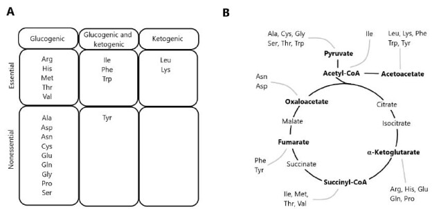
The degradation of AAs occurs via multiple pathways, including deamination and oxidation of carbon skeletons to CO2 through the TCA cycle [1,11]. Precursor substrates for gluconeogenesis include lactate and pyruvate, as well as glucogenic AAs derived from dietary protein. Since most of lactate and pyruvate are formed from glycolysis, the conversion of these metabolites into glucose via gluconeogenesis constitutes the glucose-lactate cycle. In contrast, gluconeogenesis from dietary AAs results in the formation of ammonia and represents a net transfer of carbons from proteins to carbohydrates. In herbivorous and omnivorous mammals, the recycling of glucose carbons accounts for a significant fraction of gluconeogenesis, particularly under fasting conditions [12,13]. AAs yielding acetyl-CoA or acetoacetyl-CoA are classified as ketogenic, since these two compounds are the precursors of ketone bodies. Some AAs are both ketogenic and glucogenic (Figure 1), while only leucine and lysine are exclusively ketogenic AA [14].

fig 1

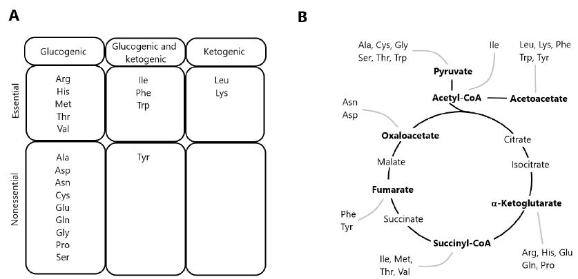
Figure 1: Classification of amino acids according to the metabolism of carbon skeletons. (A) Classification of amino acids as exclusively glucogenic, both glucogenic and ketogenic, or exclusively ketogenic. (B) Disposal of carbon skeletons as a part of the central pathways of energy metabolism.

The first enzymes to initiate AA catabolism are generally specific for a given AA or a group of structurally similar AAs (e.g., BCAAs). The general reaction types include oxidative deamination, transamination, and non-oxidative decarboxylation. Oxidative deamination is carried out by AA oxidases and dehydrogenases. D-amino acid oxidase (EC 1.4.3.3) has been detected in a variety of salmonid tissues [15] and in the common carp (Cyprinus carpio) as well [16]. Active aminotransferases for ammonia removal from specific AAs have been described in several fish tissues of salmonids [17] and Jian carp [18]. Quantitatively, the most important transaminases are aspartate aminotransferase (AST) and alanine aminotransferase (ALT), also known as glutamate-oxaloacetate transaminase (GOT) and glutamate-pyruvate transaminase (GPT), respectively. Several studies were carried out detecting these enzymatic activities in the fish blood serum and tissues. A variety of AAs act as donors for deamination reactions in fish, which use AAs as the main substrates for gluconeogenesis in their bodies, as well as main oxidative fuels [1,2]. Moreover, AAs may also undergo reactions brought on by oxidases and decarboxylases with significant physiological and nutritional consequences. Among the possible decarboxylation reactions is the conversion of glutamate into γ-aminobutyrate (GABA, a neurotransmitter), ornithine into putrescine, and arginine into agmatine [14]. The physiological significance of AA decarboxylation in fish merits systemic research.

Energy Metabolism

It has been suggested that about 50 to 70% of calories (as-fed basis) in diets consumed by trout are provided by protein, reaching about 55 to 75% on a dry-weight basis [19]. Fish consume protein to obtain AAs, which are taken up by their cells in accordance with their needs for energy or protein synthesis. The dietary protein requirements of fish ranges from 30% to 60%, depending on species, age, size, and feeding habits [2,20]. These quantities are greater than the amounts required for mammals and birds, such as swine (12-20%), dairy cows (10-18%), and chickens (14-22%) [3,21,22].

As animals get their energy needs from oxidation of the complex molecules that they eat, the energy in feed is not available until the complex molecules are broken down to simpler molecules by digestion. Furthermore, the energy needs of fish are mainly supplied by macromolecules as fats, carbohydrates, and proteins. There is little consistent information on the ability of fish to digest fats of different melting points, although fats are generally well digested and utilized by fish, providing about 8.5 kcal metabolizable energy (ME) per gram [23]. The ME values of carbohydrates for fish range from near zero for cellulose to about 3.8 kcal/g for easily digestible sugars. The value of carbohydrates in fish feeds is a controversial issue. It appears, however, that digestible carbohydrates can be well utilized as an energy source if they are provided in proper balance with other nutrients [24]. Protein has a ME value of about 4.5 kcal/g for fish, which is higher than that defined for mammals and birds [23]. Thus, it seems clear that most fish do not use carbohydrates (e.g., starch, glycogen, and simple sugars) as a major energy source [25].

Moreover, the energy metabolism in fish could be compared as similar to mammals and birds with two notable exceptions: (a) fish do not expend energy to maintain a body temperature different from that of the environment and (b) the excretion of waste nitrogen requires less energy in fish than it does in homeothermic land animals [26]. Although AAs have long been known to provide most of the energy utilized by fish, the role of specific AAs in certain tissues are unknown. Recent studies with hybrid striped bass and largemouth bass indicated that glutamate, glutamine and aspartate are major metabolic fuels in the proximal intestine, liver, kidney and skeletal muscle [11,27]. Note that these amino acids are abundant in both plant- and animal-source feedstuffs [22,28,29], but their provision in compound feeds may be inadequate for optimum intestinal health or optimum growth performance of fish [30]. Hence, the whole protein content does not vary too much among different species, and the AA profiles within the fish body is similar to that of the aforementioned animals [31,32]. However, species-specific composition in eye tissues from zebrafish (Danio rerio) and sardine (Sardina pilchardus) have been described [33].

The contribution of proteins and AAs towards meeting the energy requirements of fish is highly considered, because of the concept of “protein-sparing” through optimization of the digestible protein (DP) to digestible energy (DE) ratio addressed in a large number of species [34,35]. Moreover, the lack of control of amino acid catabolism as affected by dietary protein levels was considered as a major reason for the high protein requirements in fish [25]. As most of the fish are carnivorous, the use of protein as energy source could be compared with carnivorous mammals. Such comparison with terrestrial animals suggested that teleost fish or even shrimp use a greater proportion of proteins for energy purposes despite lower overall energy requirements due to the aquatic mode of life, which includes poikilothermy and ammoniotelism [6].

The basal energy needs of fish are lower than those of terrestrial animals due to their poikilothermic character, in which the internal temperature varies considerably according to environmental conditions. Therefore, dietary protein contributes not only to fish growth by providing AAs for protein synthesis, but also ATP production via AA catabolism. Evidence from several studies suggests that proteins and lipids are the major sources of energy for fish [36-38]. The final product of protein metabolism in teleost fish is ammonia, rather than urea and uric acid in mammals and birds, respectively. Because the synthesis of urea and uric acid requires a large amount of energy, the release of ammonia is energetically advantageous for fish [39]. Thus, the efficiency of AAs as metabolic fuels is greater in fish than in mammals and birds. High rates of AA utilization for body growth have been observed in fish as compared with other animals [8,40]. These quantitative differences have been attributed to the carnivorous/omnivorous feeding habit of fishes and their apparent preferential use of protein over carbohydrate as a dietary energy source [41].

Some cells in the nervous system can use diverse substrates, including AAs, to support oxidative energy metabolism [42]. At the same time, AA oxidation may contribute from 50 to 70% of total energy needs in marine fish embryos and yolk-sac larvae [25,43]. Indeed, it has been suggested that AAs could provide 14 to 85% of the energy requirements for teleost fish, depending on their developmental stage [44]. Energy supply is a limiting factor for physiological processes, and therefore, the study of energy metabolism associated to such processes is key to improve the growth, development and health of fish. Metabolic reorganization and alterations in intermediary metabolic pathways occur in fish to meet their increased energy demands for adaptations to changes in the aquatic environments.

Amino Acids Mobilization in Different Fish Tissues

AAs distribution and their respective concentrations into the various fish organs and tissues depend on their physiological roles (Table 1). Relatively little is known about AA metabolism in specific fish tissues. Mobilization of glycogen and lipids, as well as protein (primarily in skeletal muscle) contributes to ATP production [63,64]. Some species try to preserve liver glycogen stores by degrading proteins to support gluconeogenesis and use lipids and/or proteins as energy substrates [45,50]. In general, fish produces glycogen through gluconeogenesis during long periods of starvation, representing an energy source for muscle tissue and mobilizing it to maintain homoeostasis during the first stages of starvation [64-66]. On the other hand, an alternative pathway for de novo glucose synthesis can be through gluconeogenesis from lactate, glycerol and some AAs [67]. Fish uses AAs as the main substrates for gluconeogenesis and as main oxidative fuel. This is especially true for migratory fish, which may go for long periods without eating [68]. However, after fasting time the fish could eventually necessitate of protein catabolism to replenish glycogen stores that were exhausted during extensive starvation [49,50,67]. The anadromous Arctic char (Salvelinus alpinus) seems to find energy from protein stores into muscle tissues during the latter part of overwintering, or during migration growth and adiposity. This conclusion is supported by the reduction in muscular body mass observed during food-deprived fish from May through June [69]. Typically, fish return to standard metabolism depending on available feed after the fasting period, although this also depends on the environmental conditions, species, and age of the animal [70].

Table 1: Major roles of amino acids in fish tissues.

Tissues

Amino acids Physiological and/or metabolic role

References

Liver

Met The main role of Met is the synthesis of taurine (for bile production) [14,45,46]
Val and Ile

Val and Ile are used as glucogenic amino acids

Leu

Leu could be metabolized to ketone bodies, providing energy for tissues such as brain and heart during starvation

[14]

Ala

Ala is used as substrate for glycogen and/or glucose production in liver

[47-50]

Met, Leu and Lys

In hepatocytes, Met, Leu and Lys could regulate glycolysis, gluconeogenesis and lipogenesis; particularly relevant their involvement in regulation of metabolism related gene expression

[51,52]

Blood system

Ala, Gln, and Arg Concentrations of Ala, Gln, and Arg in plasma may be increased during long-term starvation for gluconeogenesis

[36]

Gln,Arg and Orn

Gln, Arg and Orn contribute to remove ammonia from blood during food deprivation

[53-55]

Nervous system

Glu

The main role of Glu is to provide energy for the enterocytes and precursors of biologically molecules like glutathione

[56-59]

Gly

Inhibitory neurotransmitter in the central nervous system; co-agonist with glutamate for N-methyl-D-aspartate receptor receptors; antioxidant; anti-inflammation; one-carbon metabolism; conjugation with bile acids

Muscle

Ala, Asp, Glu and leu

Ala and Asp serve as glucogenic precursors during the rest time. Moreover Ala concentration in muscle is increased to support muscular activity Leu is oxidized during food deprivation in fish to produce ATP for swimming

[40,60,61]

Gut

Glu and Gln

Glu and Gln are major energy source, and used for glutathione synthesis. They play a crucial role intestinal health of fish, by modulating intestinal structure, protecting against oxidative damage and acting as energy substrate for the enterocytes.

[11,59]

Arg

Arg has a role to improve intestinal health. Increased activity of several intestinal enzymes and changed composition of the intestinal microbiota

[62]

The low glucose turnover rate reported in fish as compared to other animals (mammals and birds) is in agreement with the fact that proteins play a large role in ATP production in most ectothermic fish [67,71,72]. This is supported by the lower nitrogen retention in ectothermic fish such as carp (30%) feeding diets with increased carbohydrate content, as compared with homeotherms like pigs (45%) and chickens (50%) [39]. AAs are not only oxidized for ATP production, but they are also used to synthesize macromolecules such as proteins in the gills or other organs, and for the osmoregulation during fish seawater acclimation [73]. Osmotic pressures in teleost fish are regulated at nearly constant levels. The non-essential AAs seem to be preferentially used for osmoregulatory proposes, rather than the ten AAs considered essential for the fish, namely arginine, histidine, isoleucine, leucine, lysine, methionine, phenylalanine, threonine, tryptophan, and valine [2]. However, the role of AAs as oxidative substrates in specific tissues has been largely neglected in previous studies that examined the effects of environmental salinity on the AA composition of fish plasma and tissues [74]. Some specific aspects of AA metabolism in different fish tissues are discussed in the following sections.

Blood System

Changes in the plasma levels of AAs at various time intervals after feeding have been monitored for several species, including rainbow trout [75], carp [76], tilapia [77], and channel catfish [78]. The enzyme glutamate dehydrogenase showed moderate activities in rainbow trout and carp erythrocytes, where glutamine was more important than glucose as an oxidative substrate [79]. Glutamate is used for the synthesis of glutamine and glutathione. Glutamine is essential for the synthesis of purines and pyrimidines, whereas glutathione protects cells from oxidative stress [1].

The metabolic reactions in the liver greatly affect the concentration of most AAs in blood and gut. Liver monitors the absorbed dietary AAs arriving from the portal blood and has the important role of controlling their catabolism and release into the general circulation. Much of the AAs taken up by the liver are rapidly degraded [80]. A rapid catabolism of excess dietary AAs was observed in rainbow trout by measuring a large increase in ammonia excretions within four hours of feeding a high protein meal [81]. In the case of BCAAs, skeletal muscles may be more important for initiating BCAA degradation than the liver [71,82].

During feed deprivation, fish appears to use catabolic energy conservation strategies to meet caloric needs while minimizing tissue energy loss [83]. It has been suggested that white muscle proteolysis is the source for increased plasma levels of free AAs observed during long-term feed deprivation, normally constituting the primary source of energy in carnivorous species [54]. Furthermore, AAs can supply glucose during periods of prolonged starvation via gluconeogenesis. In several fish species, long-term starvation mobilizes muscle protein by increasing the levels of free AA, usually alanine and glutamine, the most released AAs [36,53]. The increases in plasma AAs augment their metabolic utilization [54] and this is apparently the case for most non-essential glucogenic AAs in teleost fish [84].

AAs release via proteolysis of white muscle has been identified as an important fuel source for sockeye salmon (Oncorhynchus nerka) during periods of prolonged starvation [36]. However, the total plasma AAs in starved lake sturgeon (Acipenser fulvescens) was found to be unchanged during 45 days of feed deprivation [45]. Moreover, starved brown trout (Salmo trutta) showed a significant increase in total plasma AA levels after 15 days of feed deprivation [85]. These different responses in different studies may reflect species-specific metabolic adaptation strategies in response to feed deprivation and/or differences in body energy stores, such as lipids and glycogen. In this regard, Solea senegalensis is characterized by low body lipid stores, which supports the importance of proteolysis during prolonged feed deprivation in this species [55]. The increased levels of plasma glutamine, ornithine, and arginine observed in ureotelic fish that had been feed-deprived for 21 days may facilitate detoxification of ammonia production after AA catabolism. Glutamine is formed from glutamate and ammonia, and this reaction is a cellular mechanism for ammonia detoxification in fish. However, for every mole of ammonia detoxified, two equivalent moles of ATP are hydrolyzed [86]. Higher plasma levels of serine, asparagine, glutamine, arginine, and ornithine were observed in fish (S. senegalensis) that had been feed-deprived for 21 days [87]. This may suggest their role as important sources of carbons for gluconeogenesis, which is in line with the high rates of 14C incorporation from 14C-labeled serine and asparagine into glucose in isolated hepatocytes from feed-deprived O. mykiss. In addition, glutamine, arginine, and ornithine can be metabolized to glutamate, and deamination of glutamate is a main pathway for its oxidation to CO2 or for gluconeogenesis in the fish liver [20].

Liver

Liver plays an essential role on controlling the mobilization of energetic reserves for survival during the starvation period. In the fed state, sulfur AAs are used for the synthesis of taurine, which is required for the production of bile salts to promote lipid digestion and absorption. However, under starvation conditions, methionine is not needed to produce bile salts and, therefore, might be used as a glucogenic AA to produce glucose as a possible energy source for the central nervous system and red blood cells [45,46]. Valine and isoleucine are glucogenic, being catabolized via the TCA cycle and utilized for gluconeogenesis (Figure 1). Moreover, glutamate and glutamine are oxidized extensively in the liver of zebrafish and hybrid striped bass, with the rate of CO2 production from glutamine being greater than that from glutamate [11], while catfish hepatocytes produce five times more ammonia from glutamine than from glutamate [88]. Ketogenic AAs are converted to acetyl-CoA or ketone bodies in order to provide energy for tissues such as the brain and heart during starvation [47,89]. The analysis of stable isotopes in different tissues has been widely used in ecological studies to learn about the nutrient transfer across ecosystem boundaries and to understand trophic relationships and the migration of animals (including fish) through dietary changes that occur throughout their lives [9].

The juvenile yellowtail amberjack (Seriola lalandi) was subjected to an isotopically equilibrated diet of δ13C and δ15N AAs and proteins for 60 days and after this period, two treatments were carried out [90]. For one treatment, the fish continued to be fed and those in the other group were deprived of feed. The compound-specific isotopic analysis (CSIA) of AAs from different tissues showed significant differences between the muscle and liver samples of the control group and those fed the test diet. The CSIA for the δ15N values of liver AAs revealed the largest changes relative to the diet for non-essential AAs, whereas glycine and lysine remained constant. However, methionine was the most enriched AA within the control group, as compared to test diet. Valine and isoleucine, both essential AAs, were highly enriched in the liver of starved fish, a condition arising from either a high rate of utilization or an insufficient dietary supply. Enrichment patterns were observed for alanine, aspartate, and glutamate [90].

Salmonid fish encounter periods of little or no feed intake for many reasons, such as low feed availability during winter conditions. The role of carbohydrates and proteins as energy sources during periods of short-term fasting (days to weeks) or long-term starvation (months) in different fish species is less clear [50,91,92]. In coho salmon (Oncorhynchus kisutch), liver glycogen decreases one week after the initiation of fasting, but returns to a normal level after three additional weeks of feed deprivation. Net protein breakdown has been observed during prolonged periods of feed deprivation in salmonids, but not during the initial phase [50,91]. Juvenile salmonids are potentially more sensitive to fasting than adult fish, although there are still similarities between the different life stages in protein metabolism during fasting [93]. Alanine is likely used as a substrate for glycogen and/or glucose production in the liver [48,50], but may also be oxidized in the liver and used as a direct energy source [49]. In this scenario, decreased levels of alanine in the liver of fasted fish have been demonstrated [93].

Some genetic parameters have been considered in experiments involving food-deprived and well-fed amberjack fish, in particular changes in liver leptin (LepA1 and LepA2) expression [90]. An increase in liver leptin expression was previously observed during fasting/feed restriction, similar to the increase reported in Atlantic salmon [94,95]. The activity of enzymes involved in lipid metabolism (glucose 6-phosphate dehydrogenase and 3-hydroxyacyl-CoA dehydrogenase) and glycolysis (pyruvate kinase) appeared inversely correlated to liver leptin expression in food-deprived fish. The highest activity of these enzymes was recorded concurrently with low liver leptin expression in well-fed fish. Moreover, such interrelationships were not observed for enzymes involved in AA metabolism and gluconeogenesis [69]. Liver metabolic responses to increased dietary carbohydrates in both carnivorous fish and tilapia were also investigated as an attempt to understand the reasons for the higher metabolic use of carbohydrates in omnivorous fish than in carnivorous fish [96,97]. Some studies demonstrated that the liver of omnivorous fish responds well at the metabolic level to dietary carbohydrates, similarly to previous reports on juvenile tilapia and hybrid tilapia [98,99].

Biochemical responses to dietary nutrients in tilapia liver were previously investigated [100-102]. Gene expression studies showed no regulation at mRNA levels for metabolic enzymes related to glycolysis and gluconeogenesis, whereas mRNA levels for hepatic enzymes involved in AA catabolism were clearly dependent on the amount of dietary protein intake. This was not an expected result, as increased lipogenic and decreased gluconeogenic enzyme activities were observed in tilapia feeding carbohydrate-rich diets [103]. This indicates that enzymes in tilapia liver did not respond to dietary carbohydrates at the transcriptional level. Although the first steps of glucose utilization via the hepatic glucokinase (gck gene) were clearly higher in fish fed higher carbohydrate diets, the long-term adaptation of tilapia to carbohydrates does not necessitate persistent molecular adaptation for glucose utilization within the liver of this fish. It is unknown how the liver of carnivorous fish respond to dietary carbohydrate intake.

Intestine

Although the intestine uses both Glu and Gln as energy sources, the supply of each molecule is different. Both dietary and arterial Gln content are recruited into intestinal cells, while almost all Glu utilized in the gut comes from the lumen [104]. Glu is a non-essential AA that universally exists in living organisms. It plays various roles in enterocytes metabolism and physiology, either directly, as an energy source [56] or excitatory neurotransmitter in the enteric nervous system [105], or through conversion into bioactive molecules, such as glutathione [58]. Glu serving as a substrate for the synthesis of glutathione by the intestinal mucosa is derived from enteral Glu rather than arterial Glu, and 95% of dietary Glu is metabolized as a major energy source by the intestinal cells of piglets [59]. Although Glu can be synthesized in the body, this metabolic pathway is inadequate to meet the requirement of the piglet small intestine for glutamate [106]. These studies indicate that the utilization of dietary Glu has an important role in gut health and systemic metabolism. Examining this role may be helpful to better understand AA metabolism in the intestines of fish.

Moreover, after protein hydrolysis in the gut, the AAs are absorbed and pass along the portal system to the liver. During their passage across the intestinal wall, AAs can be incorporated into intestinal proteins (constitutive or secretory) or catabolized by the tissue [68]. The gut itself can metabolize extensive amounts of certain AAs, such as glutamate and aspartate. Indeed, in some animals, gut metabolism has a major influence on the whole body AA requirement [56].

Nervous System

Besides glucose, ketone bodies and possibly both lipids and proteins may act as energy sources in the brain of several vertebrates [107]. Astrocytes can use glutamine as an energy source and produce glutamine from glutamate (a neurotransmitter removed from the synaptic cleft), as well as from precursors, such as glucose and fatty acids [42]. The synthesis and utilization of substrates such as glutamine, ketone bodies and lactate are greatly influenced by their concentrations in the cells and the extracellular milieu [108,109]. The carbon skeletons of glutamate are mainly metabolized into CO2, lactate, or alanine, while the nitrogen of glutamate is utilized for the synthesis of other AAs such as glutamine, proline, and arginine [57,59,110]. Glutamine has various functions in cellular metabolism, such as serving as energy fuel and being a precursor for purine and pyrimidine nucleotides, NAD+, and amino sugars [57,58].

Muscular System

Proteins may play an important role in fueling muscle work in fish, but their exact contribution has yet to be established [111]. The design of reliable methods to measure substrate fluxes in fish muscle [112] has allowed researchers to start investigating how fish muscles respond to common environmental stresses. White muscle under stress is forced to produce lactate at higher rates than can be processed by aerobic tissues. However, lactate accumulation is minimized because disposal is also strongly stimulated. Trout have a much higher capacity to metabolize lactate under normoxic conditions than during hypoxia or intense swimming. The low density of monocarboxylate transporters and lack of up-regulation with exercise explain the phenomenon of lactate retention in white muscle. This tissue operates as an almost-closed system, where glycogen stores act as an “energy spring” that alternates between power release during swimming and slow withdrawal in situ from lactate during recovery [111].

To cope with exogenous glucose, trout can completely suppress hepatic production and boost glucose disposal. Without these responses, glycaemia would increase four times faster and reach dangerous levels. Therefore, the capacity of salmonids to regulate glucose levels is much better than presently described in the existing literature. However, knowledge about the use of proteins or AAs as fuel for muscle work in fish is still lacking. Glutamate and glutamine are major metabolic fuels for the skeletal muscles of zebrafish and hybrid striped bass [11]. This is contrast to mammalian muscles, where fatty acids and glucose are primary energy substrates [39].

Little is known about the use of proteins as fuel for muscular work in fish, although evidence from sockeye salmon (Oncorhynchus nerka) shows that proteins become the dominant source of fuels towards the end of migration when all the other substrates reach depletion [36]. At this point, researches also reported changes in AA and protein concentrations, as well as the activities of related enzymes. AA fluxes have not been measured in exercising fish and the only direct measurement of protein catabolism during swimming was the rate of nitrogen excretion in juvenile trout with a high growth rate. However, the high growth rate may be a destabilizing factor since significant changes in the protein composition of fish tissues occur during this stage of growth. A study examined the roles of glutamate, alanine, and aspartate as gluconeogenic precursors in resting kelp bass [60], and a further research measured the fluxes of all AAs in resting rainbow trout [113]. It is unclear whether the high rates of protein catabolism observed in migrating salmon and juvenile trout are typical of active muscles or whether they only occur under exceptional circumstances of extreme exercise or rapid growth [114].

The intramuscular metabolism of ectotherms has receive little attention, but the design of reliable methods to measure substrate fluxes in fish has allowed researchers to start investigating how fish muscles respond to common stresses [112]. For example, the mudskipper (Periophthalmodon schlosseri) is quite active and levels of total free AAs increased significantly in skeletal muscle and plasma, while alanine levels increased three-fold in the muscle, four-fold in the liver, and two-fold in plasma [61]. From these results, the authors concluded that P. schlosseri was capable of partially catabolizing certain AAs to support activity on land because of its capacity for life on sea and land. The tolerance of P. schlosseri to environmental ammonia is much higher than any other fishes because of its capability to actively excrete NH4+ and its low skin permeability to NH4+, which prevents back diffusion [115]. In this context, the amino groups of these AAs are transferred directly or indirectly to pyruvate to form alanine. The carbon chains are fed into the TCA cycle and are partially oxidized to malate, which could replenish pyruvate through the function of the malic enzyme. This favorable ATP yield from partial AA catabolism is not accompanied by a net release of ammonia [114]. Mudskippers can be very active on land. Thus, urea formation, which is energetically expensive, may not be a suitable strategy. By exposing mudskippers to terrestrial conditions, in constant darkness to minimize physical activity, the researchers reduced the rate of proteolysis and AA catabolism in response to aerial exposure [116]. In contrast, increased concentrations of alanine, BCAAs, and total free AAs were observed in the tissues of P. schlosseri exposed to terrestrial conditions for 24 h [117].

Proteins are one of the primary sources of metabolic energy in carnivorous fishes. The main storage tissue of utilizable protein is white muscle. AAs released through proteolysis can be oxidized either as energy or converted to other utilizable forms via anabolic pathways, as noted previously [11,14]. Before AAs can be oxidized through the TCA cycle, the amino group must be removed by either transamination or deamination. Ammonia is not produced during transamination, but deamination produces either NH3, which spontaneously takes up H+ to form NH4+ [118]. Certain AAs (e.g. arginine, glutamine, histidine, and proline) can be converted to glutamate, which can undergo deamination by glutamate dehydrogenase, producing NH4+ and α-ketoglutarate. The latter is fed into the TCA cycle. Glutamate can also undergo transamination with pyruvate, catalyzed by alanine aminotransferase, producing α-ketoglutarate without releasing ammonia. Continuous glutamate-pyruvate transamination would facilitate the oxidation of the carbon chains of some AAs. Under normal circumstances, the carbon chain of an AA is completely oxidized to CO2 through the TCA cycle and the electron transport chain, thus producing ATP and/or its equivalent [119]. This would cause a reduction in the efficiency of ATP production because not all AAs would fully be oxidized, allowing certain AAs to be used as energy sources, while minimizing ammonia accumulation. In fish, alanine constitutes 20 to 30% of the total AA pool [119]. Most of the free AAs could be converted into alanine and the overall quantitative energetics would appear to be quite favorable. The net conversion of glutamate to alanine would yield 20 moles of ATP per mole of alanine formed if the resultant α-ketoglutarate is completely oxidized to CO2. This favorable ATP yield from AA catabolism is accompanied by a direct release of ammonia into the living environment.

Alanine is an important substrate for hepatic gluconeogenesis and is one of the main AAs released by the skeletal muscle [1]. It is also an important source of energy for fish. However, the effect of adding alanine into diets is controversial, as dietary alanine is largely extracted by the splanchnic bed. In addition, β-alanine supplementation does not affect the growth of Japanese flounder (Paralichthys olivaceus) [120].

Skeletal muscle plays an important role in initiating BCAA degradation via transamination. There are reports that muscle tissues of goldfish [121] and trout [122] have higher activities of BCAA transaminases than mammalian muscles, which indicates a high capacity for leucine catabolism in fish muscle. In trout, the rate of leucine catabolism is higher during intense swimming than at rest [40]. The quantitative importance of leucine oxidation by fish muscle depends mainly on the use of protein as an endogenous energy source, since blood leucine does not contribute significantly to total CO2 production. This means that other substrates (e.g., glutamate, glutamine, alanine and aspartate) contribute predominantly to ATP production during exercise in fish. The oxidation of alanine occurs within the muscle, kidney and liver via glutamate-pyruvate transaminase. Alanine transport into the cells is under hormonal control during stressful conditions. Thereby, alanine is actively released at high rates by all muscle types studied, ensuring its supply to the liver and kidneys and this AA may be a major final product of muscle metabolism [40].

Important Amino Acids as Energy Sources

Lysine and Methionine

The effects of synthetic methionine and lysine on the growth and feed conversion of animals are so impressive that the use of these two AA as feed additives worldwide exceeds 700,000 metric tons annually [123,124]. Lys has a particular role in metabolism, since Lys and Leu are exclusively ketogenic AAs that are broken down to acetyl-CoA, which is oxidized to CO2 via in the TCA cycle. Unlike Lys, Met is a glucogenic AA that produces glucose as an energy source. Under methionine-limiting conditions, excesses of branched-chain AAs reduce methionine oxidation possibly due to competitive inhibition by the branched-chain ketoacids. Through the formation of S-adenosylmethionine (a donor of methyl group), methionine plays a key role in one-carbon metabolism [45,46].

Alanine

The transamination of non-essential AAs, such as alanine and aspartate, was found to be important for ATP production in fish in early investigations [71]. Alanine can stimulate the feeding response of certain fish [125] and carries nitrogen for inter-organ AA metabolism [36]. Recently, a study suggested a possible role for the hormones STC1 (a stanniocalcin homologue) and PTHrP (parathyroid hormone-relate protein) in teleost fish to safeguard liver glycogen reserves under stressful situations [126]. The strategy may involve the production of glucose via BCAA, alanine, glutamine, and glutamate and their mobilization from the muscle to the liver. Alanine is a fundamental AA that provides energy for the central nervous system during the starvation period by constant translocation from the muscle tissues through the blood system to the liver. However, under non-stressful conditions, the main energy source mainly comes from glutamate and glutamine [11].

Glycine

Glycine participates in gluconeogenesis, sulfur AA metabolism, one-carbon metabolism, and fat digestion [127]. It also stimulates feed intake in many fish [125]. In sturgeon, increased levels of glycine and a reduction of glucogenic AAs occur in response to feed deprivation. Glycine represents almost 30% of collagen, the major structural protein of connective tissues, such as tendons, skin, and ligaments [128]. Moreover, glycine might be reserved for the synthesis of creatine and, thus, the generation of creatine phosphate, a high-energy molecule used as an energy source for overcoming extreme conditions, like running away from predators [129]. In this regard, glycine plays an important role in energy metabolism during periods of feed deprivation and for activities requiring rapid use of high quantities of energy.

Arginine

Arginine is classified as an essential AA in young animals, including young fish, and is necessary for optimal growth [2]. Arginine plays various physiological roles in animal cells, such as serving as a component of proteins, an oxidative energy substrate, a stimulator of hormone secretion (e.g. growth hormone, insulin, glucagon), and a precursor of polyamine and nitric oxide (NO), which is vital for the vasodilation and immune responses [130]. In most mammals (e.g., humans, pigs and rat), the small intestine is the site for endogenous synthesis of citrulline and arginine from glutamine, glutamate and proline [21]. However, endogenous synthesis of arginine has not been demonstrated in most teleost fish [131]. In mammalian liver, arginine is essentially catabolized by arginase via the urea cycle [21]. The embryos of salmonids seem to have a functional urea cycle for ammonia detoxification, as researchers observed relatively higher activity of five urea cycle enzymes. This situation is quite different from adult fish.  Arginase is ubiquitous in fish tissues, with the highest activity in the liver and kidney [132]. The dietary requirement for arginine among various fish species may differ because of differences in metabolic and enzymatic efficiency [131]. Previous growth studies suggested that the fish arginine requirement might range from 4 to 6% of dietary protein. Salmon have the highest requirement (about 6% of dietary protein), whereas this number ranges from 4 to 5% in other species [2]. Arginine is a nutritionally essential AA for fish not only as a precursor for protein synthesis, but also for its metabolic role in the production of diverse metabolites, including nitric oxide (NO), polyamines, urea, proline, and glutamate [130,133].

Conclusions

To improve the knowledge about the use of AAs as a major energy source in fish, it is important to understand the bioavailability of each dietary AA to be absorbed and retained. The estimated bioavailability of AAs could be indirectly determined by the digestibility of dietary proteins. However, in aquatic organisms, leaching of water-soluble nutrients from both feed and feces is always a factor contributing to inaccuracy when determining the amounts of available AAs that are actually absorbed. Apart from those AAs retained for anabolic processes (i.e. protein deposition during growth), there is also a need to determine the amounts required to meet the demands of metabolic processes. Therefore, the amounts of dietary AAs that enter the portal circulation cannot be determined precisely. It is important to distinguish those AAs used in metabolic processes (e.g., ATP production) from those retained by fish under starvation conditions. Clearly, more research is needed on the metabolism of AAs in swimming fish to solve this intriguing problem. Muscular performance depends critically on the adequate supply of metabolic fuels and disposal of final products. Therefore, knowing how metabolite fluxes are regulated is necessary to understand the strategies whereby fish survive, grow, and develop. The ATP used for contraction can be generated through various pathways of energy metabolism that catabolize carbohydrates, lipids, or proteins. It can be suggested that under both fed and food-deprived conditions, AAs are major metabolic fuels for the intestine, liver, skeletal muscle, kidneys, and possibly other tissues.

References

    1. Wu G (2013) Amino Acids: Biochemistry and Nutrition. Boca Raton.
    2. Wilson RP (2002) Amino acids and proteins. San Diego, CA: Elsevier 144-179.
    3. Kaushik SJ, Seiliez I (2010) Protein and amino acid nutrition and metabolism in fish: current knowledge and future needs. Aquac Res 41: 322-332.
    4. Kim JD, Lall SP (2000) Amino acid composition of wholebody tissue of Atlantic halibut (hippoglossus hippoglossus), yellowtail flounder (pleuronectes ferruginea) and Japanese flounder (paralichthys olivaceus). Aquaculture 187: 367-373.
    5. Bowen SH (1987) Dietary protein requirements of fishes – A reassessment. Can J Fisher Aquatic Sci 44: 1995-2001.
    6. Cowey CB (1994) Amino acid requirements of fish – a critical appraisal of present values. Aquaculture 124: 1-11.
    7. Matthews JC (2000) Amino acid and peptide transport systems. Wallingford, UK: CAB International 3-23.
    8. Jürss K, Bastrop R (1995) Amino acid metabolism in fish. Biochemistry and Molecular Biology of Fishes 4: 159-189.
    9. Karasov WH, Martinez del Rio C (2007) Physiological Ecology: How Animals Process Energy, Nutrients, and Toxins. Princeton, NJ: Princeton University Press.
    10. Rossi A, Cazenave J, Bacchetta C, Campana M, Parma MJ (2015) Physiological and metabolic adjustments of Hoplosternum littorale (Teleostei, Callichthyidae) during starvation. Ecol Indic 56: 161-170.
    11. Jia S, Li X, Zheng S, Wu G (2017) Amino acids are major energy substrates for tissues of hybrid striped bass and zebrafish. Amino Acids 49: 2053-2063. [crossref]
    12. Perdigoto R, Rodrigues TB, Furtado AL, Porto A, Geraldes C, et al. (2003) Integration of [U-13C] glucose and H2O for quantification of hepatic glucose production and gluconeogenesis. NMR Biomed 16: 189-198.
    13. Coelho M, Nunes PM, Mendes V, Manadas B, Heerschap A, et al. (2015) Effect of Global ATGL knockout on murine fasting glucose kinetics. J Diabet Res 2015: 542029. [crossref]
    14. Wu G (2009) Amino Acids: Metabolism, functions, and nutrition. Amino Acids 37: 1-17. [crossref]
    15. Fickeisen DH, Brown Jr GW (1977) D‐Amino acid oxidase in various fishes. Fish Biol 10: 457-465.
    16. Deng J, Zhang X, Tao L, Bi B, Kong L, et al. (2011) D‐lysine can be effectively utilized for growth by common carp (Cyprinus carpio). Aquac Nutr 17: 467-475.
    17. Samsonova MV, Lapteva TI, Yu BF (2005) Aminotransferases in early development of salmonid fish. Russian J Dev Biol 36: 70-74.
    18. Jiang J, Feng L, Tang L, Liu Y, Jiang W, et al. (2015) Growth rate, body composition, digestive enzymes and transaminases activity, and plasma ammonia concentration of different weight Jian carp (Cyprunus carpio var. Jian). Anim Nutr 1: 373-377.
    19. Lee JK, Cho SH, Park SU, Kim KD, Lee SM (2003) Dietary protein requirement for young turbot (Scophthalmus maximus L.). Aquac Nutr 9: 283-286.
    20. Ballantyne JS (2001) Amino acid metabolism. Fish Physiol 19: 77-107.
    21. Wu G (2014) Dietary requirements of synthesizable amino acids by animals: a paradigm shift in protein nutrition. J Anim Sci Biotechnol 5: 34.
    22. Li P, Wu G (2020) Composition of amino acids and related nitrogenous nutrients in feedstuffs for animal diets. Amino Acids 52: 523-542.
    23. Jobling M (1983) A short review and critique of methodology used in fish growth and nutrition studies. J Fish Biol 23: 685-703.
    24. Hart, Paul JB (2002) Handbook of fish biology and fisheries. New York, NY: Blackwell.
    25. Cowey CB, Walton MJ (1988) Studies on the uptake of (14C) amino acids derived from both dietary (14C) protein and dietary (14C) amino acids by rainbow trout, Salmo gairdneri Richardson. J Fish Biol 33: 293-305.
    26. Van de Pol I, Flik G, Gorissen M (2017) Comparative physiology of energy metabolism: Fishing for endocrine signals in the early vertebrate pool. Front Endocrinol 8: 36. [crossref]
    27. Li XY, Wu G (2019) Oxidation of energy substrates in tissues of Largemouth bass (Micropterus salmoides). J Anim Sci 97: 68-69.
    28. Hou YQ, He WL, Hu SD, Wu G (2019) Composition of polyamines and amino acids in plant-source foods for human consumption. Amino Acids 51: 1153-1165. [crossref]
    29. Li XL, Rezaei R, Li P, Wu G (2011) Composition of amino acids in feed ingredients for animal diets. Amino Acids 40: 1159-1168. [crossref]
    30. Li XL, Zheng SX, Wu G (2020) Nutrition and metabolism of glutamate and glutamine in fish. Amino Acids 52: 671-691. [crossref]
    31. Latshaw JD, Bishop BL (2001) Estimating body weight and body composition of chickens by using noninvasive measurements. Poult Sci 80: 868-873. [crossref]
    32. Ramseyer LJ (2002) Predicting whole-fish nitrogen content from fish wet weight using regression analysis. North Am J Aquac 64: 195-204.
    33. Falco F, Barra M, Cammarata M, Cuttitta A, Jia S, et al. (2016) Amino acid composition in eyes from zebrafish (Danio rerio) and sardine (Sardina pilchardus) at the larval stage. SpringerPlus 5: 519.
    34. Cho CY, Kaushik SJ (1990) Nutritional energetics in fish: energy and protein utilization in rainbow trout (Salmo gairdneri). World Rev Nutr Dietet 61: 132-172. [crossref]
    35. Cho CY, Bureau DP (2001) A review of diet formulation strategies and feeding systems to reduce excretory and feed wastes in aquaculture. Aquac Res 32: 349-360.
    36. Mommsen TP, French CJ, Hochachka PW (1980) Sites and patterns of protein and amino acid utilization during the spawning migration of salmon. Can J Zool 58: 1785-1799.
    37. Hemre GI, Mommsen TP, Krogdahl A (2002) Carbohydrates in fish nutrition: Effects on growth, glucose metabolism and hepatic enzymes. Aquac Nutr 8: 175-194.
    38. Miao S, Nie Q, Miao H, Zhang W, Mai K (2016) Effects of dietary carbohydrate-to-lipid ratio on the growth performance and feed utilization of juvenile turbot (Scophthalmus maximus). J Ocean Univ China 15: 660-666.
    39. Wu G (2018) Principles of Animal Nutrition. Boca Raton, FL: CRC Press.
    40. Van den Thillart G (1986) Energy metabolism of swimming trout (Salmo gairdneri). Oxidation rates of palmitate, glucose, lactate, alanine, leucine and glutamate. J Comp Physiol B 156: 511-520.
    41. Tacon AGJ, Cowey CB (1985) Fish energetics. Dordrecht: Springer 155-183.
    42. McKenna MC, Dienel GJ, Sonnewald U, Waagepetersen HS, et al. (2012) Basic Neurochemistry. San Diego, CA: Academic Press 200-231.
    43. Rønnestad I, Thorsen A, Finn RN (1999) Fish larval nutrition: Recent advances in amino acid metabolism. Aquaculture 177: 201-216.
    44. Van Waarde A (1983) Aerobic and anaerobic ammonia production by fish. Comp Biochem Physiol B 74: 675-684.
    45. Gillis TE, Ballantyne JS (1996) The effects of starvation on plasma free amino acid and glucose concentrations in lake sturgeon, Acipenser fulvescens. J Fish Biol 49: 1306-1316.
    46. Hassel B (2001) Pyruvate carboxylation in neurons. J Neurosci Res 66: 755-762. [crossref]
    47. Robinson AM, Williamson DH (1980) Physiological roles of ketone bodies as substrates and signals in mammalian tissues. Physiol Rev 60: 143-187. [crossref]
    48. Canals P, Gallardo MA, Blasco J, Sanchez J (1992) Uptake and metabolism of L-alanine by freshly isolated trout (Salmo trutta) hepatocytes – the effect of fasting. J Exp Biol 169: 37-52.
    49. Pereira C, Vijayan MM, Moon TW (1995) In vitro hepatocyte metabolism of alanine and glucose and the response to insulin in fed and fasted rainbow trout. J Exp Zool 271: 425-431.
    50. Sheridan MA, Mommsen TP (1991) Effects of nutritional state on in vivo lipid and carbohydrate metabolism of coho salmon, Oncorhynchus kisutch. Gen Comp Endocrinol 81: 473-483. [crossref]
    51. Lansard MS, Panserat E, Plagnes J, Seilez I, Skiba Cassy S (2010) Integration of insulin and amino acid signals that regulate hepatic metabolism-related gene expression in rainbow trout: role of TOR. Amino Acids 39: 801-810. [crossref]
    52. Lansard MS, Panserat E, Plagnes J, Dias K, Sieliez I, et al. (2011) L-leucine, L-methionine, and L-lysine are involved in the regulation of intermediary metabolism-related gene expression in rainbow trout hepatocytes. J Nutr 141: 75-80. [crossref]
    53. Moon TW (2011) Metabolic reserves and enzyme activities with food deprivation in immature American eels, Anguilla rostrata (LeSueur). Can J Zool 61: 802-811.
    54. Sánchez-Muros MJ, Garcı́a-Rejón L, Garcı́a-Salguero L, de la Higuera M, Lupianez JA (1998) Long-term nutritional effects on the primary liver and kidney metabolism in rainbow trout. Adaptive response to starvation and a high-protein, carbohydrate-free diets on glutamate dehydrogenase and alanine amino transferase kinetics. Biochem Cell Biol 30: 55-63. [crossref]
    55. Dias J, Rueda-Jasso R, Panserat S, Conceição LEC, Gomes EF, et al. (2004) Effect of dietary carbohydrate to lipid ratios on growth, lipid deposition and metabolic hepatic enzymes in juvenile Senegal sole (Solea senegalensis). Aquac Res 35: 1122-1130.
    56. Burrin DG, Stoll B (2009) Metabolic fate and function of dietary glutamate in the gut. Am J Clin Nutr 90: 850-856. [crossref]
    57. Blachier F, Boutry C, Bos C, Tomé D (2009) Metabolism and functions of L-glutamate in the epithelial cells of the small and large intestines. Am J Clin Nutr 90: 814-821. [crossref]
    58. Newsholme P, Procopio J, Lima MMR, Pithon-Curi TC, et al. (2003) Glutamine and glutamate -their central role in cell metabolism and function. Cell Biochem Funct 21: 1-9.
    59. Reeds PJ, Burrin DG, Stoll B, Jahoor F (2000) Intestinal glutamate metabolism. J Nutr 130: 978-982. [crossref]
    60. Bever K, Chenoweth M, Dunn A (1981) Amino acid gluconeogenesis and glucose turnover in kelp bass (Paralabrax sp). Am J Physiol 240: 246-252. [crossref]
    61. Ip YK, Lem CB, Chew SF, Wilson JM, Randall DJ (2001) Partial amino acid catabolism leading to the formation of alanine in Periophthalmodon schlosseri (mudskipper): a strategy that facilitates the use of amino acids as an energy source during locomotory activity on land. J Exp Biol 204: 1615-1624. [crossref]
    62. Andersen SM, Waagbø R, Espe M (2016) Functional amino acids in fish health and welfare. Front Biosci 8: 143-169. [crossref]
    63. Navarro I, Gutiérrez J (1995) Fasting and starvation. Biochemistry and Molecular Biology of Fishes 4: 393-434.
    64. Metón I, Fernández F, Baanante V (2003) Short and long-term effects of re-feeding on key enzyme activities in glycolysis-gluconeogenesis in the liver of gilthead sea bream. Aquaculture 225: 99-107.
    65. Figueiredo-Garutti ML, Navarro I, Capilla E, Souza R, Mouraes G, et al. (2002) Metabolic changes in Brycoon cephalus (Teleostei, Characidae) during post-feeding and fasting. Comp Biochem Physiol A Mol Integr Physiol 132: 467-476. [crossref]
    66. Furné M, Morales AE, Trenzado CE, García-Gallego M, Carmen Hidalgo M, et al. (2012) The metabolic effects of prolonged starvation and re-feeding in sturgeon and rainbow trout. J Comp Physiol 182: 63-76. [crossref]
    67. Polakof S, Panserat S, Soengas J, Moon T (2012) Glucose metabolism in fish: A review. J Comp Physiol B 182: 1015-1045. [crossref]
    68. Bequette BJ (2003) Amino acid metabolism in animals: an overview. Amino Acids in Animal Nutrition 2: 103-124.
    69. Jørgensen EH, Martinsen M, Strøm V, Hansen KER, Ravuri CS, et al. (2013) Long-term fasting in the anadromous Arctic charr is associated with downregulation of metabolic enzyme activity and upregulation of leptin A1 and SOCS expression in the liver. J Exp Biol 216: 3222-3230. [crossref]
    70. Liang X, Wang J, Xue M, Gong G, Dong Y, et al. (2017) Gluconeogenesis during starvation and refeeding phase is affected by previous dietary carbohydrates levels and a glucose stimuli during early life in Siberian sturgeon (Acipenser baerii). Anim Nutr 3: 284-294.
    71. Walton MJ, Cowey CB (1982) Aspects of intermediary metabolism in salmonid fish. Comp Biochem Physiol B 73: 59-79.
    72. Garin D, Rombaut A, Fréminet A (1987) Determination of glucose turnover in sea bass Dicentrarchus labrax. Comparative aspects of glucose utilization. Comp Biochem Physiol B 87: 981-988. [crossref]
    73. Yancey PH (2001) Fish Physiology. Nitrogen Excretion 20: 309-341.
    74. Banerjee B, Gitalee B, Nirmalendu S (2014) Influence of environmental hypertonicity on the induction of ureogenesis and amino acid metabolism in air-breathing walking catfish (Clarias batrachus, Bloch). Indian J Exp Biol 52: 728-738. [crossref]
    75. Rolland M, Feekings JP, Dalsgaard J, Holm J, Skov P (2016) Modelling the effects of dietary methionine level and form on postprandial plasma essential amino acid profiles in rainbow trout (Oncorhynchus mykiss). Aquac Nutr 22: 1185-1201.
    76. Plakas SM, Katayama T, Tanaka Y, Deshimaru O (1980) Changes in the levels of circulating plasma free amino acids of carp (Cyprinus carpio) after feeding a protein and an amino acid diet of similar composition. Aquaculture 21: 307-322.
    77. Yamada S, Tanaka Y, Katayama T, Sameshima M, Simpson KL (1982) Plasma amino acid changes in Tilapia nilotica fed a casein and a corresponding free amino acid diet. Bull Jpn Soc Sci Fish 48: 1783-1787.
    78. Wilson RP, Gatlin DM, Poe WE (1985) Postprandial changes in serum amino acids of channel catfish fed diets containing different levels of protein and energy. Aquaculture 49: 101-110.
    79. Ferguson RA, Storey KB (1991) Glycolytic and associated enzymes of rainbow trout (Oncorhynchus mykiss) red cells: in vitro and in vivo studies. J Exp Biol 155: 469-485.
    80. Bae JY, Ok IH, Lee S, Hung SS, Min TS, et al. (2011) Reevaluation of dietary methionine requirement by plasma methionine and ammonia concentrations in surgically modified rainbow trout, Oncorhynchus mykiss. J Appl Ichthyol 27: 887-891.
    81. Brett JR, Zala CA (1975) Daily pattern of excretion and consumption of sockeye salmon under controlled conditions. J Fish Res Board Can 33: 2479-2486.
    82. Walton MJ, Wilson R (1986) Postprandial changes in plasma and liver free amino acids of rainbow trout fed complete diets containing casein. Aquaculture 51: 105-115.
    83. Costas B, Aragao C, Jarabo IR, Chacoff LV, Arjona FJ, et al. (2011) Feed deprivation in Senegalese sole (Solea senegalensis Kauo, 1858) juveniles: Effects on blood plasma metabolites and free amino acid levels. Fish Physiol Biochem 37: 495-504. [crossref]
    84. Suarez RK, Mommsen TP (1986) Gluconeogenesis in teleost fishes. Can J Zool 65: 1869-1882.
    85. Navarro I, Blasco J, Baños N, Gutiérrez J (1997) Effects of fasting and plasma amino acid levels in brown trout. Fish Physiol Biochem 16: 303-309.
    86. Randall DJ, Tsui TKN (2002) Ammonia toxicity in fish. Mar Pollut Bull 45: 17-23.
    87. French CJ, Mommsen TP, Hochachka PW (1981) Amino acid utilisation in isolated hepatocytes from rainbow trout. Eur J Biochem 113: 311-317. [crossref]
    88. Legate NJ, Bonen A, Moon TW (2001) Glucose tolerance and peripheral glucose utilization in rainbow trout (Oncorhynchus mykiss), American eel (Anguilla rostrata), and black bullhead catfish (Ameiurus melas). Gen Comp Endocrinol 122: 48-59. [crossref]
    89. Kopio RR, Murray DM, Story DL, Brunengraber H (1984) The shunt pathway of mevalonate metabolism in the isolated perfused rat kidney. J Biol Chem 259: 372-377. [crossref]
    90. Barreto-Curiel F, Focken U, D’Abramo LR, Viana MT (2017) Metabolism of Seriola lalandi during starvation as revealed by fatty acid analysis and compound-specific analysis of stable isotopes within amino acids. PloS One 12: 0170124. [crossref]
    91. Navarro I, Gutierrez J, Planas J (1992) Changes in plasma-glucagon, insulin and tissue metabolites associated with prolonged fasting in brown trout (Salmo trutta fario) during two different seasons of the year. Comp Biochem Physiol Comp Physiol 102: 401-407. [crossref]
    92. Pottinger TG, Rand-Weaver M, Sumpter JP (2003) Overwinter fasting and re-feeding in rainbow trout: plasma growth hormone and cortisol levels in relation to energy mobilisation. Comp Biochem Physiol B biochem Mol Biol 136: 403-417. [crossref]
    93. Kullgren A, Samuelsson LM, Larsson DGJ, Björnsson BT, Bergman EJ (2010) A metabolomics approach to elucidate effects of food deprivation in juvenile rainbow trout (Oncorhynchus mykiss). Am J Physiol Regul Integr Comp Physiol 299: 1440-1448. [crossref]
    94. Rønnestad I, Nilsen TO, Murashita K, Angotzi AR, Gamst Mohn AG, et al. (2010) Leptin and leptin receptor genes in Atlantic salmon: Cloning, phylogeny, tissue distribution and expression correlated to long-term feeding status. Gen Comp Endocrinol 168: 55-70. [crossref]
    95. Trombley S., Schmitz M (2013) Leptin in fish: Possible role in sexual maturation in male Atlantic salmon. Fish Physiol Biochem 39: 103-106. [crossref]
    96. Boonanuntanasarn S, Kumkhong S, Yoohat K, Plagnes-Juan E, Burel C, et al. (2018) Molecular responses of Nile tilapia (Oreochromis niloticus) to different levels of dietary carbohydrates. Aquaculture 482: 117-123.
    97. Kamalam BJ, Medale F, Panserat S (2017) The utilisation of dietary carbohydrates in farmed fishes: new insights on influencing factors, biological limitations and future strategies. Aquaculture 467: 3-27.
    98. Wang Y, Liu YJ, Tian LX, Du ZY, Wang JT, et al. (2005) Effects of dietary carbohydrate level on growth and body composition of juvenile tilapia, Oreochromis niloticus×O. aureus. Aquac Res 36: 1408-1413.
    99. Azaza MS, Khiari N, Dhraief MN, Aloui, N, Kraiem MM, et al. (2015). Growth performance, oxidative stress indices and hepatic carbohydrate metabolic enzymes activities of juvenile Nile tilapia, Oreochromis niloticus L., in response to dietary starch to protein ratios. Aquac Res 46: 14-27.
    100. Gaye-Siessegger J, Focken U, Becker K (2006) Effect of dietary protein/carbohydrate ratio on activities of hepatic enzymes involved in the amino acid metabolism of Nile tilapia, Oreochromis niloticus (L.). Fish Physiol Biochem 32: 275-282.
    101. Chen MY, Ye JD, Yang W, Wang K (2013) Growth, feed utilization and blood metabolic responses to different amylose-amylopectin ratio fed diets in tilapia (Oreochromis niloticus). Asian Australas J Anim Sci 26: 1160-1171.
    102. Chen YJ, Zhang T, Chen HY, Lin S, Luo L, et al. (2017) An evaluation of hepatic glucose metabolism at the transcription level for the omnivorous GIFT tilapia, Oreochromis niloticus during the postprandial nutritional status transition from anabolism to catabolism. Aquaculture 473: 375-382.
    103. Figueiredo-Silva AC, Saravanan S, Schrama JW, Panserat S, Kaushik S, et al. (2013) A comparative study of the metabolic response in rainbow trout and Nile tilapia to changes in dietary macronutrient composition. Br J Nutr 109: 816-826. [crossref]
    104. Wu G (1998) Intestinal mucosal amino acid catabolism. J Nutr 128: 1249-1252. [crossref]
    105. Kirchgessner AL (2001) Glutamate in the enteric nervous system. Curr Opin Pharmacol 1: 591-596. [crossref]
    106. Rezaei R, Knabe DA, Tekwe CD, Dahanayaka S, Ficken MD, et al. (2013) Dietary supplementation with monosodium glutamate is safe and improves growth performance in postweaning pigs. Amino Acids 44: 911-923. [crossref]
    107. Gibbs SR, DeRoos RM (1991) Plasma levels of glucose, ketone bodies, lactate, and alanine in the vascular supply to and from the brain of the adult American bullfrog (Rana catesbeiana). J Exp Zool 258: 14-23. [crossref]
    108. Edmond J, Auestad N, Robbins RA, Bergstrom JD (1985) Ketone body metabolism in the neonate: development and the effect of diet. Fed Proc 44: 2359-2364. [crossref]
    109. Dienel GA, Cruz NF (2003) Neighborly interactions of metabolically activated astrocytes in vivo. Neurochem Int 43: 339-354.
    110. Nakamura H, Kawamata Y, Kuwahara T, Torii K, Sakai R (2013) Nitrogen in dietary glutamate is utilized exclusively for the synthesis of amino acids in the rat intestine. Am J Physiol Endocrinol Metab 304: 100-108. [crossref]
    111. Weber JM, Choi K, Gonzalez A, Omlin T (2016) Metabolic fuel kinetics in fish: swimming, hypoxia and muscle membranes. J Exp Biol 219: 250-258.
    112. Haman F, Powell M, Weber JM (1997) Reliability of continuous tracer infusion for measuring glucose turnover rate in rainbow trout. J Exp Biol 200: 2557-2563. [crossref]
    113. Robinson J, Yanke D, Mirza J, Ballantyne JS (2011) Plasma free amino acid kinetics in rainbow trout (Oncorhynchus mykiss) using a bolus injection of 15N-labeled amino acids. Amino Acids 40: 689-696. [crossref]
    114. Weber M (2011) Metabolic fuels: regulating fluxes to select mix. J Exp Biol 214: 286-294. [crossref]
    115. Ip YK, Randall DJ, Kok TKT, Barzaghi C, Wright PA, et al. (2004) The giant mudsipper Periophthalmodon schlosseri facilitates active NH4+ excretion by increasing acid excretion and decreasing NH3 permeability in the skin. J Exp Biol 207: 787-801. [crossref]
    116. Lim CB, Chew SF, Anderson PM, Ip YK (2001) Reduction in the rates of protein and amino acid catabolism to slow down the accumulation of endogenous ammonia: a strategy potentially adopted by mudskippers (Periophthalmodon schlosseri and Boleophthalmus boddaerti) during aerial exposure in constant darkness. J Exp Biol 204: 1605-1614. [crossref]
    117. Ip YK, Lee CY, Chew SF, Low WP, et al. (1993) Differences in the responses of two mudskippers to terrestrial exposure. Zool Sci 10: 511-519.
    118. Mommsen TP, Walsh PJ (1992) Biochemical and environmental perspectives on nitrogen metabolism in fishes. Experientia 48: 583-593.
    119. Hochachka PW, Guppy M (1987) Metabolic arrest and the control of biological time. Cambridge, MA: Harvard University Press: 253-284.
    120. Kim K, Wang X, Bai SC (2003) Reevaluation of the dietary protein requirement of Japanese flounder Paralichthys olivaceus. J World Aquac Soc 34: 133-139.
    121. Van Waarde A (1981) Nitrogen metabolism in goldfish, Carassius auratus (L.). Activities of transamination reactions, purine nucleotide cycle and glutamate dehydrogenase in goldfish tissues. Comp Biochem Physiol B 68: 407-413.
    122. Teigland M, Kkungsoyr L (1983) Accumulation of alpha-ketoisocaproate from leucine in homogenates of tissues from rainbow trout (Salmo gairdnerii) and rat. An improved method for determination of branched chain keto acids. Comp Biochem Physiol B 75: 703-705.
    123. Fontaine J (2003) Amino Acids in Animal Nutrition. Wallingford, UK: CABI Publishing: 15-40.
    124. Nwanna LC, Lemme A, Abdallah M, Schwarz FJ (2012) Response of common carp (Cyprinus carpio L.) to supplemental DL-methionine and different feeding strategies. Aquaculture 356: 365-370.
    125. Shamushaki VAJ, Kasumyan AO, Abedian A, Abtahi B (2007) Behavioural responses of the Persian sturgeon (Acipenser persicus) juveniles to free amino acid solutions. Mar Freshwater Behav Physiol 40: 219-224.
    126. Palma PFS, Bock C, Silva TS, Guerreiro PM, Power DM, et al. (2019) STC1 and PTHrP modify carbohydrate and lipid metabolism in liver of a teleost fish. Sci Rep 9: 723.
    127. Fang YZ, Yang S, Wu G (2002) Free radicals, antioxidants, and nutrition. Nutrition 8: 872-879. [crossref]
    128. Li P, Wu G (2018) Roles of dietary glycine, proline and hydroxyproline in collagen synthesis and animal growth. Amino Acids 50: 29-38. [crossref]
    129. Lin CY, Huang LH, Deng DF, Lee SH, Liang HJ, et al. (2019) Metabolic adaptation to feed restriction on the green sturgeon (Acipenser medirostris) fingerlings. Sci Total Environ 684: 78-88. [crossref]
    130. Chen C, Shengyao LF, Kuang S, Liu Y, Jiang J, et al. (2012) Effect of dietary arginine on growth, intestinal enzyme activities and gene expression in muscle, hepatopancreas and intestine of juvenile Jian carp (Cyprinus carpio var. Jian). Br J Nutr 108: 195-207. [crossref]
    131. Dabrowski K, Terjesen BF, Zhang Y, Phang JM, Lee KJ (2005). A concept of dietary dipeptides a step to resolve the problem of amino acid availability in the early life of vertebrates. J Exp Biol 208: 2885-2894. [crossref]
    132. Wright PA, Campbell A, Morgan RL, Rosenberger AG, Murray BW (2004) Dogmas and controversies in the handling of nitrogenous wastes: Expression of arginase Type I and II genes in rainbow trout: influence of fasting on liver enzyme activity and mRNA levels in juveniles J Exp Biol 207: 2033-2042.
    133. Jobgen WS, Fried SK, Fu WJ, Meininger CJ, Wu G (2006) Regulatory role for the arginine–nitric oxide pathway in metabolism of energy substrates. J Nutr Biochem 17: 571-588. [crossref]
fig 3

Performance Analysis Involving Inductive and Capacitive Load of a 7.5 kVA Inverter for Laboratory Use in an Institution

DOI: 10.31038/NAMS.2020323

Abstract

This project is therefore aimed at designing and construction of a pure sine wave inverter system of 7.5 kVA analyzed in performance and can be used to convert electrochemical energy into an alternating current (AC) supply. The major tests that were carried out all met the expected specifications with negligible deviation or tolerance. One thing was peculiar about the results; each of the tests that were carried out in each of the subsystems that make up the inverter system was done in relation to the next subsystem that was connected to it. The outputs from the inverter system were all as expected as shown by the final results. When the final installation was made, the system was tested by gradually loading it to see that it responds to the load increase as expected; and after the load test we observed that batteries voltage dropped slightly due to the loading effect and that was normal. Based on the pattern of tests and observations used in this project, it is expected that the system performs its intended duty throughout its useful life as long as it is used as prescribed.

Keywords

Battery, Energy, Load, Performance, Power

Introduction

Electrical energy or electrical power can be generated from primary energy sources which include geothermal power, mechanical power, solar power, kinetic energy of flowing water and wind, etc. this was discovered by Michael Faraday, a British scientist in the 1820s and 1830s. According to the law of conservation of energy also known as the law of science which states that energy can neither be created nor destroyed but can be transformed from one form to another. Series of researches have been carried out to fascinate the development on the technology on energy generation from different primary sources [1]. Despite all these technologies, due to the fact that everyone needs electrical energy as a result of high population and the slow rate of technological development in the country, the amount of energy distributed becomes insufficient for people and the need keeps pressing to generate an alternative supply from primary sources of energy such as hydro, wind, solar and chemical energy. In order to generate electrical power from direct current (DC) to Alternating current (AC), a device called Inverter is employed. While a rectifier circuit is used on the other hand to convert electricity from Alternating current (AC) back to Direct current (DC) [2]. This project is therefore aimed at designing and construction of a pure sine wave inverter system of 7.5 kVA analysed in performance and can be used to convert electrochemical energy into an alternating current (AC) supply. According to the Authoritative Dictionary of IEEE Standards Terms (IEEE, 2000), inverter is an electrical power converter that changes direct current (DC) to alternating current (AC). The converted AC can be at any required voltage and frequency with the use of appropriate transformers, switching, and control circuits. The inverter performs the opposite function of a rectifier [3]. The electrical inverter is a high-power electronic oscillator. It is so named because early mechanical AC to DC converters was made to work in reverse, and thus was “inverted”, to convert DC to AC. Inverters do not suffer much from all these except that the batteries are consumed very fast as the load increases calling for constant recharging of the batteries after each use [4].

Literature Review

Osuwa and Peter, 2014, gave the brief idea about the production of solid state inverters which provides environmentally friendly alternative for uninterruptible power supply for the working of different gadgets and for sustainable economy. This study is thus anchor on the making of 1 kVA inverter for provision of power using locally sourced 80 Ah 12 volts deep cycle battery, oscillator determined MOSFETs and a transformer along with other electronic components [5]. In build an inverter for the conversion of DC to AC at a normal frequency of 50 Hz, due consideration is given to the switching speed of the oscillator used to make sure that the MOSFETs in their two channels operate in their saturation and cut off states when appropriately driven by oscillator outputs in a way to complement each other.

Omitola et.al, 2014 discuss that researchers proposed that in the modern society, electricity has great control over the most daily activities for instance in domestic and industrial utilization of electric power for operations. Electricity can be generated from public supply to users in different ways including the use of water, wind or steam energy to drive the turbine as well as more recently the use of gas generators, astral energy and nuclear energy are as well sources of electricity [6,7].

An inverter is an electronic device that converts electrical power from DC form to AC form. Its typical application is to convert battery voltage (stored D.C voltage) into a normal house A.C voltage to power electrical devices such as TV, fridge etc. when an A.C power from the national grid is not available (wikipedia).

Chan and Bowler, 1974, reveals the more up-to date types of inverters are two or more transformer coupled inverters, which might be either connected in series and/or in parallel, to bring desirable result, but the only issue is the reduced time duration because of its high power consumption when working at full capacity. (Gottles, 1985). The other drawback of the above named inverters are reduced efficiency absence of dc power energy restoration (that is, chargers were not included for charging back-up batteries in the presence of public power supply). Also there is no capacity to switch from dc source to ac source when power is restored. Although, the latest products of solar energy to electrical energy converters include battery charging circuits but does not have the capacity to switching to public power supply (PPS).

Methodology

System Operation

The batteries are the back-up source for power generation conversion of chemical energy into electricity. The power produced by the battery bank was then transferred to the inverter unit. The battery monitors in the inverter monitors the rate at which electric current were drawn in and out of the battery. It turns off charge when the battery reaches the optimum charging point and turns it on when it goes below a certain level. It fully charges the battery without permitting overcharge. The batteries are the key component in this power system. It provided energy storage for the system. The energy stored in the batteries was then used to power the load but it was first converted to AC voltage by the use of an inverter due to they were AC loads. The photovoltaic ally produced direct current was commuted periodically by controlled oscillatory system and feed to power electronic semiconductor switches such as JFET which were connected the power transformer. Here the voltage was stepped up to the desired ac voltage. The inverter could also charge the battery when there is public power supply (Figure 1).

figure 1

Figure 1: Block diagram of operational principle of a solar inverter system.

Battery Bank

A battery bank is a group of batteries connected together using series or parallel wiring. This allows more power to be stored than using a single battery. A battery bank is the result of joining two or more batteries together for a single application. What does this accomplish? Well, by connecting batteries, you can increase the voltage, amperage, or both. When you need more power, instead of getting yourself a massive super tanker of an RV battery for example, you can construct a battery bank. A battery bank is a group of batteries connected together using series or parallel wiring. This allows more power to be stored than using a single battery. A battery bank allows you to store electricity generated by solar PV system for use at any time. (Solarmango.com)

Battery Connection Scheme

The first thing you need to know is that there are two primary ways to successfully connect two or more batteries: The first is via a series and the second is called parallel [8].

Series Connection. A series connection adds the voltage of the two batteries, but it keeps the same amperage rating (also known as Amp Hours). For example, these two 6-volt batteries joined in series now produce 12 volts, but they still have a total capacity of 10 amps. To connect batteries in a series, use jumper wire to connect the negative terminal of the first battery to the positive terminal of the second battery. Use another set of cables to connect the open positive and negative terminals to your application. Never cross the remaining open positive and open negative terminals with each other, as this will short circuit the batteries and cause damage or injury (Figure 2).

fig 2

Figure 2: Battery connection in series.

Be sure the batteries you’re connecting have the same voltage and capacity rating. Otherwise, you may end up with charging problems, and shortened battery life.

Parallel Connections. The other type of connection is parallel. Parallel connections will increase the current rating, but the voltage will stay the same. In the Parallel diagram, we’re back to 6 volts, but the amps increase to 20 AH. It’s important to note that because the amperage of the batteries increased, you may need a heavier-duty cable to keep the cables from burning out. To join batteries in parallel, use a jumper wire to connect both the positive terminals, and another jumper wire to connect both the negative terminals of both batteries to each other. Negative to negative and positive to positive. You can connect your load to one of the batteries, and it will drain both equally. However, the preferred method for keeping the batteries equalized is to connect to the positive at one end of the battery pack, and the negative at the other end of the pack. It is also possible to connect batteries in what is called a series/parallel configuration, but it may sound confusing, but this is the way you can increase your voltage output and Amp/Hour rating. To do this successfully, you need at least 4 batteries (Figure 3).

fig 3

Figure 3: Battery connection in parallel.

This is a combination of the above methods and is used for 2 V, 6 V or 12 V batteries to achieve both a higher system voltage and capacity. For example; 4 × 6 V 150 Ah batteries wired in series/parallel will give you 12 V at 300 Ah. 4 × 12 V 150 Ah batteries can be wired in series/parallel to give you 24 V with 300 Ah capacity (Figure 4) [9-36].

fig 4

Figure 4: Battery connection of series and parallel.

Results

This chapter presents the test results of the works that were carried out in this project. At the end of the installation, the system was tested to ensure that it meets the desired stated objectives and specifications that guided the entire project work. The results of the test carried out are as below;

Test Carried Out

     1. Physical examination

     2. Is it fully charged?

     3. How much charge is left in it?

     4. Does it meet the manufacturer specification?

     5. Device testing

     6. Continuity test

     7. Short circuit test

Device Testing

To test the Inverter, a load of up to 6000 VA was connected to the device to test if the device can carry up to the power stipulated for it to bear.

Continuity Test

The continuity test is carried out to avoid any form of an open circuit. The presence of an open circuit in any electrical system will create an open circuit fault in the system and the system will not function. The wires have to be continuous all the way from one terminal to another. The lead used in the soldering of the components must be well soldered and there should be no any form of partial contact as this might initiate an open circuit in the system.

Short Circuit Test

A short circuit occurs when the live and the neutral wires touch each other. When this happens, the current goes infinitely high and can blow up the entire system [6]. There should be no form of short circuit, be it on the legs of the integrated circuits or along the wires. Short circuit fault is a very costly fault as it can blow up the whole system and should be avoided as much as possible, so this test is very important prior to the powering of the inverter system. To determine the system failure rate, the part count analysis of the various components is required.

Performance Evaluation Test on the Inverter Battery

The inverter battery was subjected to two types of test;

     1. No load test; and

     2. Load test.

No Load Performance Test

A no-loads test was done on the inverter initially after completion, the output of the inverter was measured using a voltmeter (Table 1).

Table 1: No-Load Test on the Inverter.

Description

Values

Input Voltage

120 V

Output Voltage

230 V

Current from Battery

220 A
Frequency

50 HZ

Calculation of the Real Power for the Inverter:

           P(kw)=P(kva) × P.F

           Where P.F=0.8;

           P(kw)=7500 × 0.8

           P(kw)=6000 w

Formula calculation, to obtain the maximum current to be demanded by the inverter from the battery; Power (P); P=IV

           I=p/v

           When; P=6000 w, V=120 v

                                       I=7500/120

                                       I=50 A

Calculation for the battery scheme:

  • Using analytical approach;
  • Terminal battery voltage before charge:


Using a series method,

   Having 10 batteries rated:

   12.5+12.4+12.4+12.4+12.5+12.5+12.4+12.5+12.4+12.5=124.5 v.

   In a battery, there are 6 cells,

   Per cell we have 2 v, 2 × 6=12 v

   Maximum for a cell is 2.2 v, 2.2 × 6=13.2 v.

   Transient/tolerance value=±13.4.


Terminal battery voltage after charge:

   Using a series method,

   Having 10 batteries rated:

   12.7+12.6+12.8+12.7+12.7+12.7+12.7+12.6+12.8+12.7=126.4 v


Calculation for battery running hour:

   Analytical approach:

   T(hr)=volt × AH × E/load


where E is power efficiency AH is ampere per hour of the battery capacity

   a. when load L=1000 w, V=120 v, battery capacity=220 AH

       T(hr)=120 × 220 × 0.9/1000=23.76 hours

   b. when load L=2000 w, V=120 v, AH=220 A

       T(hr)=120 × 220 × 0.9/2000=11.88 hours

   c. when load L=3000 w, V=120 v, AH=220 A

       T(hr)=120 × 220 × 0.9/3000=7.92 hours

   d. when load L=4000 w, V=120 v, AH=220 A

       T(hr)=120 × 220 × 0.9/4000=5.94 hours

   e. when load L=5000 w, V=120 v, AH=220 A

       T(hr)=120 × 220 × 0.9/5000=4.75 hours (Table 2).

Table 2: Calculation for battery running hour Analytical approach.

Load (w)

Time (H) (Approximated value)

1000

24

2000

12

3000

8

4000

6

5000

5

Load Performance Test

The constructed inverter was subjected to different kinds of loads to determine the efficiency, how long the inverter systems can power the loads.

In carrying out the load test, the following loads were used:

  • A megger tester
  • Multi-meter
  • Load
  • Clamp-meter.

The test results and performance tests are shown in the sub-sections below:

  • Inductive load performance is shown in Table 3 and Figure 5.
  • Resistive load performance is shown in Figures 6, 7 and Tables 4-6.

Table 3: Inductive loads test for the inverter.

S/N

Load (W) Time (1 hour interval) Battery discharge rate (V)

1

450 11:30 am 122.6

2

450 12:30 pm

122.4

3 450 1:30 pm

122.4

4 450 2:30 pm

122.3

fig 5

Figure 5: Load Performance Analysis of Inductive Load.

figure 6

Figure 6: Graphical representation of load performance analysis of resistive calculation of Load.

fig 7

Figure 7: Graphical representation of the Inverter battery charging with respect to time.

Table 4: Inductive loads test for the inverter.

S/N

Item P (w) Duration (30 Mins Interval)
     

DC (V) input

AC (V) output

I (A) output

1

Electric bulb 200 122.1 230

2.2

2 Electric Iron 1000 121.5 229

6.9

Table 5: Load Description.

Item

Description

Quantity Rating (W)

1

Television 1 62
2 Monitor 1

193

3

Fan 3 450
4 Bulb 1

200

5 Air condition 1

750

6

Iron 1 1000
Total=

2655

Table 6: Load Performance Analysis.

Time

Load (W) Current (I) Battery (V) Output voltage (V) Output current (I)

Load (%)

11.00

TV (63)

Monitor (193)

0.1 123.4 240 0.5

3

12.00

Fan (150) 1.9 123.3 236 1.1

5

1.00

Fan (150) 2.4 122.3 233 1.7

8

2.00

Bulb (200) 2.6 122.1 230 2.2

9

3.00

A.C (750) 3.6 121.8 230 2.9

12

4.00

Electric Iron (1000) 6.7 121.5 229 6.9

15.3

16

The inverter was subject to two kinds of loads to determine the efficiency, how long the inverter systems can power the loads.

  • The resistive loads which are; energy saving bulbs and soldering iron.
  • The inductive load which are; electric fans and Air Conditions.

The tests carried out on the inverter and the readings taken with the use of multi-meter are as follows:

Discussion

The results of the tests that were carried out throughout the whole determination were all gotten through systematic checks and observations, and using the appropriate test tools and equipment where necessary. The major tests that were carried out all met the expected specifications with negligible deviation or tolerance. One thing was peculiar about the results; each of the tests that were carried out in each of the subsystems that make up the inverter system was done in relation to the next subsystem that was connected to it.

The outputs from the inverter system were all as expected as shown by the final results. When the final installation was made, the system was tested by gradually loading it to see that it responds to the load increase as expected; and after the load test we observed that batteries voltage dropped slightly due to the loading effect and that was normal.

Before final installation, the different sections that make the whole system were tested individually. This pattern was adopted to make troubleshooting, analysis and testing easy and reliable. It is expected that all the results of the tests that were carried out continuously conform to the specified standards as long as the system is used within its capacity and under the standard test conditions. Based on the pattern of tests and observations used in this project, it is expected that the system performs its intended duty throughout its useful life as long as it is used as prescribed, and this is because of the fact that the system was designed under standard operating conditions of the immediate environment.

Conclusion

The application of our knowledge of engineering in solving our local problem is one thing desperately needed in our country today. That is the opportunity this project offered us; by the implementation of this project, we have successfully made the laboratory less reliant on grid supplied energy which would boost productivity. After the implementation of the 7.5 kVA inverter installed for the department, the following were achieved:

  • We successfully did a proper load sizing of the department.
  • We successfully learnt and practiced the load on inverters in buildings.
  • We successfully determined the battery bank capacity of the 7.5 kVA pure sine wave inverter we installed in the department.
  • We as well gained great entrepreneurial skill through this project. This is of immense benefit as it would reduce our dependence on the job market for survival after school.
  • The end product of the project is the availability of a reliable and consistent power supply for the department.

Recommendations

  1. A maintenance check (e.g. periodic maintenance) should be carried out on the photovoltaic components (the solar panels, the power inverter, the charge controller, the batteries, the wires and cables, the monitors and meters) probably once a month. This will ensure that any fault is discovered and looked into on time. The components should not be tampered with in case any fault is discovered, experienced technicians should be contacted to check on the problem and proffer solutions.
  2. We would recommend that close attention be paid to the loading of the inverter. For the best interest of the life span of the inverter, it should not be run at the peak load. Members of staff should ensure that heavy duty loads are not connected to the inverter during the usage of the inverter.
  3. We recommend that students be issued their projects early enough to enable them learn in details what the project entails and projects like this (solar energy based) should be encouraged by the government to ensure optimal solutions to major issues like power failure problems.

References

  1. Abatan OA, Adewale AO, Alabi AA (2013) Constant Electricity Generation From Self-Charging Inverter. International Journal of Emerging Technology and Advanced Engineering 3: 694-698.
  2. Assad Abu-Jasser, (2010) A stand-alone photovoltaic System, Case Study: A Residence in Gaza. Journal of Applied Science in Environment Sanitation 5: 81-91.
  3. Adeoye SA and Akanni OS (2017) Development of 0.75 kVA Modified Sine Wave Inverter for a viewing Centre in Nigeria. International Journal of Advance Research and Innovation 5: 333-337.
  4. Baliunas, Sallie L, Willie Soon (2001) Washington Roundtable on Science and public Policy: Climate history and the sun.
  5. Balou (2009) what types of driver circuits are there?
  6. Battery Bank (2019) http://www.solarmango.com/dictionary/bank
  7. Bellis Mary (2019) History and Timeline of the Battery.
  8. Bird BM, Kings KG (1993) an introduction to power electronics.
  9. Chan T, Bowler P (1974) International Conference on Power Electronics, Power Semiconductors and their Applications 7-9: 237-251.
  10. Connexion France. 4 April (2017) Delayed at the station? Get pedalling to charge your phone”.
  11. Dave Etchells “The Great Battery Shootout”.
  12. Prince DC (1925) The Inverter. GE Review 28: 676-81.
  13. Electrical4u (2015) Online Electrical Engineering Study Site. Transformers types and principles 25.
  14. Emerson Network Power: Effects of AC Ripple Current on VRLA Battery Life” by https://enacademic.com/dic.nsf/enwiki/1355813
  15. Geoffrey Poulton (2007) Princeton information technology. http://www.researchgate.net
  16. IEEE Standards Terms (2000) The Authoritative Dictionary of IEEE Standards Terms (2000) Seventh Edition, IEEE Press, ISBN0-7381-2601-2: 588
  17. Osuwa JC, Peter CF (2014) Construction and implementation of 1kva Inverter. IOSR Journal of Applied Physics (IOSR-JAP) 6: 56-58.
  18. Mansoori Ali G, Enayati Nader, Agyarko L Barnie (2015) Energy: Sources, Utilization, Legislation, Sustainability, Illinois as Model State. World Scientific.
  19. Battery Charging (2018) (Pdf): LM2576,LM3420,LP2951,LP2952. ti.com
  20. Martin RG, (2002) Electronic Design from Concept to Reality, Discovery Press, India: 45-65.
  21. Martin LaMonica (2011) Motion-powered gadget charger back on track. CNET
  22. (2001) AN913: Switch-Mode, Linear, and Pulse Charging Techniques for Li+ Battery in Mobile Phones and PDAs.
  23. MIT Electric Team Vehicle December (2008) A Guide to Understanding Battery Specifications “mit.edu.
  24. Ned Mohan and William P Robinson (1989) design and Development of inverter with AVR using switch. journal.au.edu
  25. Olusegun O, Omitola Segun O, Olatinwo, Taiwo R. Oyedare (2001) Design and Construction of 1KW (1000VA).
  26. Omitola JK (2014) Design and construction of1KW (1000VA) PowerInverter. Journal of Innovative Systems Designand Engineering 2-12.
  27. Phil Weicker (2013) A Systems Approach to Lithium-Ion Battery Management. Artech House
  28. Pulse Maintenance charging:” Archived March 9, 2012, at the Wayback Machine.
  29. Recharger definition and meaning – Collins English Dictionary” (“Recharge – definition of recharge in English – Oxford Dictionaries”. Retrieved 26 June, 2019.
  30. Kayne R (2013) What is a Power Inverter.
  31. Sheu Akeem Lawal, Alade Olusope Michael: Design, Construction And Performance Evaluation Of 1kva Pure Sine Wave Power Inverter, International Journal of Scientific & Engineering Research
  32. Shuler CA (1984) Electronic Principle and Application. Glencoel McGraw Hill Columbus.
  33. Tepco Egypt (2010) Types of Loads.
  34. Theraja BL (2005) A Textbook of Electrical technology (22nd Edition) S Chand and company Ltd India Pg 2167-2176.
  35. William DS, (2001) Operation amplifier with linear integration circuit, Christopher R couty of charles E Merrill pulishing co, Chicago pp: 217-241.
  36. Windy Dankoff, (2001) How to Choose an Inverter for an Independent Energy System, Home Power 74-78.
fig 1

Assessment of Facial Injury by ‘Slock’ in Incarcerated Patients

DOI: 10.31038/OHT.2020121

Abstract

Objectives: A ‘slock’ is a padlock in a sock used as an improvised weapon in correctional facilities to induce trauma. Although assault and head injury rates are high in this setting, this is the first study to analyze facial fracture patterns by mechanism of injury. The purpose of this assessment was to examine the seemingly increasing prevalence of ‘slock’ induced facial fractures and the pattern and degree of injury caused by this device.

Methods: This quality assessment initiative was performed through retrospective analysis of incarcerated patients treated surgically for facial fractures at an academic medical center from 2011-2019. Data collection included the cause of injury, prevalence of padlock induced facial fractures and anatomical locations of fractures.

Results: Over an 8-year period, 435 inmates required surgical treatment for facial fractures. Of those, 366 reported injury by an intentional mechanism. 57 patients (16%) described involvement of a padlock and 23(6%) reported use of a ‘slock’. The prevalence of padlock induced facial fractures increased 2-fold from 2012-2017. In 57 patients with a padlock or ’slock’ injury, the most common fracture sites involved the mandible (53%), zygomatic arch (15%) and the nasal bone (13%). An average of 2.28(SD=1.33) fractures per patient occurred with padlocks.

Conclusion: A padlock is the most common cause of facial fracture besides a fist. We hope to contribute to better control of the padlock supply available to inmates to reduce the burden and severity of facial trauma. Future aims include analyzing the cost to society for treatment.

Keywords

Slock, Padlock, Lock in a sock, Facial fracture patterns, Improvised weapons

Introduction

As of 2017, the US incarcerated population comprised 1.5 million people with an adult incarceration rate of 568/100,000 [1]. In 2017, Louisiana had the highest adult incarceration rate of any state at 942/100,000 [2]. In combination with a high incarceration rate, it is thought that prisons are becoming increasingly more violent. The British Ministry of Justice reports over a 2-fold increase in serious assault rates in their incarcerated population from 2008-2018 [3]. The rate of head injury in the incarcerated population is nearly 5 times higher than the general population [4]. In addition, the hospitalization rate is about 10 times higher than that of the general population [4]. The combination of these reports shows the incarcerated population places a large financial burden on society to cover medical costs. The most common injury in this population is mandibular fracture, consisting of 46% of inmate injuries [5]. Although common, limited literature exists on the mechanisms and patterns of facial trauma. Inmates convert miscellaneous items into weapons, including toothbrushes, disposable razors, batteries, and padlocks [6]. 9% of all confiscated prison weapons are of the sap-type which is a heavy weight at the end of a flexible handle [6]. A sap-type weapon is used in 17% of inmate-on-inmate weapon induced injuries [6], many of which are a ‘slock’. A ‘slock’ is a padlock that is placed in a sock and used as an improvised weapon. Socks are distributed to prisoners and padlocks can be purchased in the prison commissary for inmates to protect their personal items. These two items are easily accessible and can be easily converted to a weapon. Prisons are a controlled environment and preventing inmate access to specific items, including padlocks, could reduce the burden of facial trauma and medical care costs for incarcerated patients. The purpose of this assessment was to examine the seemingly increasing prevalence of ‘slock’ induced facial fractures and the pattern and degree of injury caused by this ad-hoc device.

Methods

This quality assessment initiative was performed through retrospective chart review of incarcerated patients. IRB exemption was obtained as a quality improvement study. Selected patients were treated surgically for facial fractures at University Health and Ochsner LSU Health Shreveport Academic Medical Center from 2011-2019. Data collection included patient-reported mechanism of injury, prevalence of padlock induced facial fractures, anatomical locations of fractures induced by a padlock repaired surgically, age at discharge and gender. Mechanisms of injury were grouped into unintentional or intentional causes. An unintentional injury was defined as a facial fracture occurring from an accidental cause. Examples include a fall in the shower, sport associated injury, etc. An intentional injury was defined as a mechanism involving an inmate-on-inmate, staff-on-inmate, or self-induced injury. Examples include a fist to face injury, ‘slock’ to face, etc. Patients with unintentional and undocumented causes of injury were not further analyzed. Intentional injuries were categorized by specific mechanism. Padlock-induced injuries were classified by anatomical location of fracture and number of fractures per patient. Fracture location data was obtained from the imaging report in the patient chart.

Results

From 2011-2019, 435 incarcerated patients required surgical treatment for facial fractures. Of those, 366 (84%) patients reported injury by an intentional mechanism and 57 (13%) reported injury by an unintentional mechanism. As seen in Table 1, 57 patients (16%) described involvement of a padlock and 23 (6%) reported specified the use of a ‘slock.’ Padlocks were the second most common cause of facial fracture after the use of a fist, composing of 200 (55%) of all surgically repaired facial fractures. 60 (16%) fractures were vaguely described as an “assault” event in the patient chart and 14 (4%) reported being hit with an unknown object. As seen in Figure 1, the prevalence of intentionally induced facial fractures requiring surgery more than doubled from 2012 to 2017, from 29 to 67 cases. The prevalence of padlock induced facial fractures was also found to increase 2-fold from 2012 to 2017, from 7 to 14. The prevalence of fist to face injuries is included in the figure for comparison and was also found to increase substantially during our timeframe. As seen in Table 2, in the 57 patients injured by a padlock we recorded 134 total facial fractures with an average of 2.28 (SD=1.33) facial fractures per patient. The majority of fractures were observed to occur in the mandible (36%) and orbit (25%). As seen in Table 3, the most common surgically repaired fracture sites in this same group involved the mandible (53%), zygomatic arch (15%) and nasal bone (13%). As shown in Table 4, a majority of the patients involved in an intentional injury were 16 to 29 years of age. Seventy-five percent of padlock induced facial fractures occurred in patients who were 16 to 39 years of age. 98% of padlock induced facial fractures occurred in men.

Table 1: Mechanism of intentional injuries.

Mechanism of intentional injuries Number of injuries (n=366)

%

Fist to face 200

55%

Padlock

57

16%

Unknown object

14

4%

Other weapons

12

3%

Foot to face

8

2%

Fall following altercation

5

1%

Head to face

2

1%

Brass knuckles

2

1%

Knee to face

2

1%

Elbow to face 2

1%

Altercation with police 2

1%

Undefined assault

60

16%

†23 of 57 Padlock cases specifically described the use of a ‘slock’ weapon. ‡Other weapons were used only once to induce injury and included items such as a baseball bat, bowl, broom handle, phone, remote control, etc.

fig 1

Figure 1: Prevalence of facial fracture by mechanism of injury. This figure depicts the increasing prevalence of facial fracture injuries treated surgically via intentional mechanisms with fist to face and padlock-induced injuries.

Table 2: Anatomical location of padlock-induced fracture. This table illustrates the anatomical locations of facial fractures from 57 cases where a padlock was the reported mechanism of injury.

Anatomical location

Number of fractures (n=134)

%

Mandible

48

36%

Orbit

33

25%

Zygomatic bone

21

16%

Nasal bone

15

11%

Maxilla

12

9%

Alveolar ridge

3

2%

Frontal bone

2

1%

Table 3: Anatomical location of padlock-induced fractures treated surgically. This table illustrates the anatomical locations of facial fractures from 57 cases where a padlock was the reported mechanism of injury and were treated surgically.

Anatomical location

Number of surgeries (n=68)

%

Mandible

36

53%

Zygomatic arch

10

15%

Nasal bone

9

13%

Orbital floor

6

9%

Alveolar ridge

5

7%

Maxilla

1

1%

Frontal sinus

1

1%

Table 4: Age distribution of incarcerated patients (The age distribution of selected mechanisms of injury).

Age at discharge

Unintentional injuries (n=57) Intentional injuries (n=366) Fist to face (n=200)

Padlock (n=57)

16-29

29 173 110

22

30-39

20 112 49

21

40-49

5 61 32

11

50-59

3 17 7

3

60+

0 3 2

0

Discussion

Intentional facial fracture injuries have increased in the Louisiana incarcerated population from 2011-2019. We found the padlock to be the most common cause of facial fracture besides the fist. The mandible is the most common site treated surgically for a padlock-induced injury. Mandibular fractures treated surgically with intermaxillary fixation may require nutritional support for up to six weeks post-operatively [7]. This six week period is associated with airway problems, malnutrition leading to decreased wound healing, and changes in serum potassium levels that may require special monitoring or electrolyte supplementation.7 The post-operative care for these patients requires significant attention and resources from correctional facility staff. Preventing these types of injuries may help to reduce the burden placed on the correctional facility. The reliance on patient reported mechanisms of injury is a limitation to the study design. In 119 (27%) reviewed cases, we found that the self-reported mechanism of injury did not correlate with the severity of the injury. A common example included a patient reporting a fall in the shower with bilateral mandible fractures. We feel that injuries by intentional mechanisms are under reported with fear of retaliation from prison staff or inmates after returning to the correctional facility. We hope to contribute to better control of the commissary padlock supply or a built-in lock mechanism for lockers available to inmates to reduce the burden and severity of facial trauma. Future aims include analyzing the cost to society for treatment of padlock and ‘slock’ induced facial fractures.

Conclusion

Padlock induced injuries are the most common cause of facial fracture in incarcerated patients besides the fist. The prevalence of ‘slock’-induced facial fractures appears to be increasing in the Louisiana incarcerated population from 2011-2019. The mandible is the most common site treated surgically for padlock-induced fracture. Stricter regulations on the padlock supply may reduce the prevalence of facial fractures in this setting. Future studies should analyze the costs to society for treatment and the potential for an alternative from providing padlocks to inmates.

Conflict of Interest

The authors declare no conflict of interest.

Institutional Review Board

The study was judged exempt from review by the institutional review board.

Acknowledgement

This research did not receive any specific grant from funding agencies in the public, commercial, or not-for-profit sectors.

Triological Society Combined Sections Meeting Poster Presentation, Coronado, California, USA, January 23-25, 2020.

References

    1. Bronson J, Carson EA (2019) Prisoners in 2017. S. Department of Justice, Bureau of Justice Statistics.
    2. Wagner P, Sawyer W (2018) States of incarceration: the global context 2018. Prison Policy Initiative.
    3. Safety in custody statistics bulletin, England and Wales, deaths in prison custody to December 2018, assaults and self-harm to September 2018. Ministry of Justice
    4. Kuzak N, O’Connor M, Pickett W, O’Brien T, Reid K, Pearson M (2001) Impact of a prison triage system on injuries seen in emergency departments. Canadian Journal of Emergency Medicine 3: 199-204.
    5. Henning J, Frangos S, Simon R, Pachter HL, Bholat OS (2015) Patterns of traumatic injury in New York City prisoners requiring hospital admission. Journal of Correctional Health Care 21: 53-58.
    6. Lincoln JM, Chen LH, Mair JS, Biermann PJ, Baker SP (2006) Inmate-made weapons in prison facilities: assessing the injury risk. Journal of the International Society for Child and Adolescent Injury Prevention 12: 195-198.
    7. Giridhar VU (2016) Role of nutrition in oral and maxillofacial surgery patients. National Journal of Maxillofacial Surgery 7: 3-9.
fig 1

Radial as the Default Approach: Are We Overexaggerating?

DOI: 10.31038/JCCP.2020331

Abstract

Recent guidelines and reviews recommend trans radial approach (TRA) as a standard strategy for PCI unless there are overriding procedural recommendations [1-3]. The basis for these recommendations is randomised trials and metanalyses with conflicting results. We seek to unmask common misinterpretations in showing that the unfavourable results of TFA compared to TRA are most likely due to flawed study protocols with missing protocols for the TFA procedure and failure to correctly address vascular complications in the trans radial arm. Properly designed trials are mandatory before superseding TFA as a strategy that according to our combined experience of more than 25,000 coronary interventions should by no means be inferior to TRA yet with the tremendous advantage of ease of procedures, higher success-rates and less radiation.

Introduction

Trans radial access (TRA) has become the default access in Europe and Asia and its use is rapidly growing in Germany and the United States [4]. Enthusiasm for TRA has reached such a feverish pitch that criticism of TRA or praise for transfemoral access (TFA) is looked upon with disdain. With forty years’ experience including significant radial and brachial experience and a personal caseload in excess of 25,000 procedures we fail to see relevant advantage in the radial procedure. Although we agree that in most cases trans radial is a reasonable alternative to transfemoral, we have reason to doubt sufficient evidence to justify a 1A guideline recommendation (“standard approach”) for TRA [1].

Aim of the Review

Major sources of evidence were the three recent large randomised trials (RIVAL, MATRIX and SAFARI-STEMI) as well as two recent metanalyses. We are convinced that arguing for radial as default and the increasing disregard of PCI via femoral route in current guidelines and large comparative trials [5-8] needs to be challenged since it requires first and foremost more properly designed studies.

General Aspects

Prior to an analysis of the data it is important to understand that generally practiced techniques differ significantly from optimal techniques, and this is the case for both femoral and radial access. Large scale clinical trials include a spectrum of operators from highly skilled to “just learning” and none of the trials specify proper technique for either TRA or TFA. Nor do they report any uniformity of technique with either procedure. In light of the small differences and marginal p values between the TRA and TFA groups, differences in technique, especially with TFA would be sufficient to negate the findings. It is agreed that as practiced in Europe and the US, excess major bleeding associated with TFA is a problem, especially in acute coronary syndromes and possibly in elective PCI. We also agree that there may be a slight increase in all-cause mortality with TFA vs. TRA, and this difference is most likely due to bleeding complications. But we are also convinced that there would be no difference upon proper application of transfemoral approach (see appendix). However, unless there is a new trial with improved techniques included in the protocol, our persuasion remains speculative.

Bleeding aside, there are many advantages to TFA and many disadvantages to TRA. None of the randomised studies and metanalyses that demonstrate excess bleeding and higher mortality with TFA address these other factors. Properly designed randomized studies must limit themselves to one or two primary endpoints and a few secondary endpoints that can be used to generate hypotheses for future studies. Designers of clinical studies must guess at which parameters need to be evaluated to assure that the results are relevant and useful. When comparing TRA and TFA we need to look at much more than the small differences in complications and the barely demonstrable differences in mortality. TFA is technically easier. In the RIVAL trial, 7% of patients randomized to TRA had to switch to TFA because of difficulties with radial artery [9].

On the other hand, we have no doubt that TFA will result in more access site bleeding complications if not complied with proper technique of puncturing and sheath removal. In addition immediate ambulation after interventions is only possible following closure device (e.g. Angio-Seal®) [10] While most radial patients may be ambulated immediately, driving cost savings [11]. TRA is technically more difficult with lower success rates for coronary angiography and PCI [9,12], results in poorer image quality and increased radiation exposure to operators and patients [13,14] Radial artery closure might occur in 5-10%, but is generally considered benign if the ulnar artery is intact [15,16]. However due to potential endothelial damage after coronary angiography, use of the radial artery as conduit for CABG is discouraged [17]. Cath labs that use TRA as their “default” technique are less likely to properly train younger physicians in TFA. More importantly, these labs will not develop the culture that is required to prevent major bleeding in TFA cases.

Bleeding and Vascular Complications

Bleeding complications after PCI, related to the vascular access site, may increase the risk of periprocedural mortality by 1.3-2.1% [18]. In a large registry of 335,477 patients who underwent PCI in 2002 a major vascular complication occurred in 4.85% [19]. With experience however the risk of any vascular complication can be decreased from 1.7% for diagnostic procedures and 3.1% for PCI to very low numbers of 0.2% and 1.0% respectively [20], a finding that is confirmed in a large scale monitor-controlled registry reporting bleeding complications prolonging hospital stay, or requiring blood transfusion or surgical intervention, being as low as < 0.1% following diagnostic procedures (mainly with 4 F) and about 1% following PCI of ACS [21]. As for RIVAL (published in 2011), the rate of non-CABG-related major bleeding at 30 days was 0.7% in the radial group compared with 0.9% in the femoral group and only 0.2% and 0.3% of the bleeding events could be attributed to the access site [9]. In the MATRIX trial (published in 2015) bleeding complications (BARC 3-5) occurred in only 0.8% of the transfemoral patients and TIMI major or minor bleeding in 1.2% (TRA) and 1.7% (TFA) (p = 0.08). In the SAFARI-STEMI randomized trial, published in 2020, bleeding complications ware rare and did not differ (1.4% vs. 2.0%; RR, 0.71) [22].

Thus, contemporary bleeding complications after transfemoral approach in ACS-PCI are in the range of 1-2/100 and might not be different from TRA in the hands of experienced TFA operators. We strongly believe that it is possible to meaningfully decrease access site bleeding with more careful attention to technique with the femoral puncture and sheath removal. Liberal use of vascular closure devices has been shown to reduce bleeding by 50% [23], and therefore should be offered at least all high risk patients – e.g., obese patients, patients with severe hypertension, and those who have received anticoagulation as part of their procedure. It is our practice to use vascular closure devices on virtually all post PCI patients. If the access site complication rates for TFA in the general community can be reduced by 50 %, the arguments favouring TRA become superficial. Endothelial damage occurs in all patients after trans radial coronary angiography [17], and radial artery occlusion after trans radial intervention, a mostly quiescent complication, is likely to occur in 5-8% of the patients, relative to timing of assessment (7.7% after 24 hours and 5.5% at > 1 week follow-up [24]. It is often overlooked because > 50% of operators do not even assess radial artery patency before discharge [25] and it is not even mentioned in one randomised trial nor in metanalyses. It is by no means a trivial complication because future use of the radial artery as access, conduit for bypass-surgery or fistula formation in haemodialysis patients is precluded.

Major Adverse Events

Major adverse events, defined as 30 d composite of all-cause mortality, myocardial infarction, or stroke occur in about 6-10% of patients who receive PCI due to ACS. In many studies comparing TRA to TFA all-cause mortality was increased in the TFA group [8,26]. In most studies the increase did not reach statistical significance, mortality was not a prespecified endpoint, and in none was it shown to persist after controlling for bleeding. As shown by a pairwise and network meta-analysis of randomized controlled trials, the survival differences appeared patient-related and not driven by beneficial effects of TRA [27]. RIVAL, the first of the two large randomised trials, failed to show any difference in the primary endpoint (MI/stroke/severe bleeding 3.2% vs. 4%; p = 0.5) or secondary endpoint (death/MI/stroke 3.2% vs. 3.2%) [9].

With MATRIX, the second randomised comparison, there was no significant difference between radial access and femoral access in terms of the first co-primary endpoint of 30day MACE, (RR 0.85, 95% CI 0.74–0.99, two sided P = 0.031; non-significant at a pre-specified α of 0.025). All-cause mortality was 1.6% and 2.2% respectively (p = 0.045) but this was not a pre-specified endpoint [12]. The results of MATRIX must be interpreted with caution due to a strong modulating effect of operator/center experience on the efficacy [28] because the benefit of radial over femoral access obviously depends upon the operator’s expertise in the femoral technique: The MATRIX study divided patients into 3 groups based on the participating center’s proportion of radial PCIs: “low” (14.9% to 64.4%), intermediate (65.4% to 79.0%), and high (80.0% to 98.0%). The results of this stratified analysis showed that the centers with low and intermediate experience in radial approach had similar MACE rates (between 7.5% and 8.5% both for radial as well as femoral approach (differences n.s.), while those centers that were doing TRA in >80% revealed a significant absolute difference between TRA and TFA of 5.2% (p = 0.00014) Their MACE for TRA was 10.3% vs. a non-acceptable 15,5% for TFA (Figure 1). In this low level TFA group the difference in NACE (major bleeding unrelated to coronary artery bypass surgery or major adverse cardiovascular events) between TRA and TFA was even more pronounced (5,8 % (p = 0.0001), unlike similar results for TRA and TFA in the two other groups. These finding are most likely related to atrophied transfemoral skills in the centers that by enlarge perform TRA, while those who cope with both techniques don’t experience any significant difference of MACE and NACE between TRA and TFA [28].

fig 1

Figure 1: The MATRIX study divided patients into 3 groups based on the participating center’s proportion of radial PCIs: “low” (14.9% to 64.4%), intermediate (65.4% to 79.0%), and high (80.0% to 98.0%). Only TFA by radial experts resulted in a significant increase in MACE [28].

The most recent randomized trial looking at mortality was the SAFARI STEMI trial. It included almost 2300 patients before the trial was stopped half way because of futility. There were no significant differences between patients assigned to radial and femoral access in the rates of reinfarction (1.8% vs. 1.6%; RR, 1.07; 95% CI, 0.57-2.00; P = 0.83), stroke (1.0% vs. 0.4%; RR, 2.24; 95% CI, 0.78-6.42; P = 0.12), and bleeding (1.4% vs. 2.0%; RR, 0.71; 95% CI, 0.38-1.33; P = 0.28), survival or other clinical end points at 30 days after the use of radial access vs. femoral access in patients with STEMI referred for primary PCI [22].

Metanalyses

Metanalyses are often composed to overcome conflicting results of randomised trials. However, the selection process of which trials to include or exclude from analysis appears incomprehensible. In 2016 Ferrante included 24 studies that enrolled 22,843 PCI patients. Although the difference was small, radial was associated with significantly lower all-cause mortality and MACE, as well as less major bleeding and fewer vascular complications (Death of all cause: -0.6% (p = 0.001), MACE: -1.1% (p = 0.002), Maj. Bleed: -1 % (p<0.001), Maj. Vasc.Cpl.: -0.9 % (<0.001)[8]. All randomised trials, including this metanalysis failed to mention any radial artery occlusion, a vascular complication that is not trivial and that occurs in up to 10% of the patients [27,29].

Shah recently reported a metanalysis of 13 randomised trials of PCI in patients with ACS including 15,516 patients showing that following TFA the major bleeding, MACE and mortality rates of radial experts was significantly increased compared to their results of TRA and to those of transfemoral experts who had similar low complications rates for TFA and TRA. He concluded that the recently reported survival differences between TRA and TFA may have been driven by adverse events in the TFA groups, rather than by a beneficial effect of the TRA itself and that it is too early to label radial access a lifesaving procedure in invasively managed patients with ACS [27]. We do have a different explanation: To our view the inferior results of TFA of radial experts is probably attributed to insufficient skills in TFA (Figure 2).

fig 2

Figure 2: Meta-analysis of randomized controlled trials (Data from 13 trials including 15,615 patients). Only TFA performed by radial experts resulted in a significant increase in MACE [27].

Image Quality and Interventional Success Rates

After getting familiar with radial approach there is no reason to believe that it is not applicable for > 90% of diagnostic and interventional procedures. That being said, it is frequently more difficult to cannulate one or another coronary artery from a transradial approach. This invariably leads to poor quality diagnostic studies [13] and/or imperfect or failed PCI [9,12]. Transfemoral PCI is easier than radial PC because of fewer access problems and a higher likelihood of being able to use larger guides (e.g. 7F and 8F) that might be necessary to enhance support and apply devices in parallel especially with complex lesion morphology. A small randomised trial (n = 1024) showed a highly significantly increased success rate for TFA (99.8% versus 96.5%, p<0.0001) following diagnostic and interventional coronary procedures [30].

A higher trans radial failure rate is also reflected by the large randomized trials: In the RIVAL trial that randomly investigated 7021 ACS patients with PCI, crossover related to failure of initial strategy was 7·0% in the radial group and 0·9% in the femoral group. Main reasons for failure were spasm (5%), looped radial artery (1.3%) and tortuous subclavian artery (1.9%) [9]. RIFLE-STEACS reported a cross over rate from radial to femoral in 9.6% [31]. MATRIX, the largest randomised comparison (n = 8404 patients with ACS) reports a conversion to femoral of 5.8% [12]. Switching to transfemoral upon failed trans radial PCI, will result in a >90% success rate [25]. It can be assumed that significant time was wasted first trying TRA then crossing over to TFA. This is especially a problem in ACS patients.

Radiation Exposure

As long as procedure time is similar, radiation exposure to patients should not differ substantially between TRA and TFA. In the RIVAL trial, however, fluoroscopy times were significantly longer for the radial approach [32]. Due to a closer position and thus increased scattering, even highly experienced operators, receive at least twice the amount of radiation as do their femoral counterparts [14,33,34]. Therefore, compelling young operators to stick mainly with transradial approach, as put into practice in an increasing number of institutions, appears irresponsible unless the benefit for patients is crystal clear – which is not the case until today.

Patient Comfort

Most operators agree that transradial approach is more convenient for the patient, mainly because early ambulation is possible in almost all patients, and also because postinterventional compression of the femoral puncture site of up to 6 hours can be distressing. On the other hand, different from femoral access that is generally tolerated very well, transradial access may be painful especially via small arteries and when spasm occurs. Diagnostic procedures via femoral artery may in >>50 % of the patients be performed with 4 F catheters (our practise since 1997), necessitating compression of the puncture side of 1 h only, and, following interventions with 6 -8F guides, we regularly apply closure devices that limit the (moderate) femoral compression to 2 hours and that also allow to ambulate the patients the same day. Since then we very rarely experience complaints about pain or discomfort.

Conclusion

The current movement from TFA to TRA in interventional treatment of ACS is not justified by evidence, since the randomised trials are showing conflicting results. If performed properly, TFA does not cause more complications than TRA but requires overnight hospitalisation unless vascular closure devices are applied. It is not justifiable to require operators to endure twice the radiation exposure to achieve an earlier ambulation. Forthcoming comparative studies need to include access- and sheath-removal protocols and need to appreciate postinterventional radial artery occlusion as a vascular complication.

Appendix

Recommended techniques for femoral artery puncture and groin management during and after diagnostic and therapeutic procedures performed via transfemoral approach.

Puncturing the Femoral Artery

Puncturing the femoral artery is not a technique that should be left at discretion to the operator but should follow strict rules: The needle should be razor-sharp to avoid arterial compression upon entry. For diagnostic procedures a 4F sheath is sufficient in at least 80% of patients, with 5F adequate for the rest. The target is the mid common femoral artery, a point that is usually positioned at the level of the centre of the femoral head. Higher or lower entries increase the rate of bleeding complications. At skin-level the entry is 1-2 cm below the inguinal ligament and the angle 30-45 degrees. Index and middle finger of the left hand (if puncture is performed from the right side) are firmly placed 3-5 cm proximally to feel the pulse and entry direction. Occasionally, if pulse is weak, fluoroscopy is needed to identify the correct direction. The “through-and through technique” should be strictly avoided. The wire should be advanced without resistance and the sheath inserted with a slight rotational motion and maintained pressure with the left hand proximally to avoid blood exit into the tissue.

Sheath Removal and Compression

The sheath should be removed immediately on the table or on a stretcher in the Cath lab to benefit from elastic recoil of the puncture, either by the operator or at least an experienced, well trained person. ACT should by <200 seconds (we rarely apply Protamine if ACT is still > 250 sec) and the systolic blood pressure should be < 160 mmHg. Sheath removal on the ward moreover by an inexperienced individual is a particularly bad idea. In patients at higher risk for bleeding we apply a closure device (Angioseal® St. Jude Medical – US/Terumo Europe).

Manual compression requires specific training and patience as well as a second person for assistance or replacement. The 3 middle fingers are firmly compressed 3-4 cm proximal to the skin puncture. The pressure should be enough to prevent any visible bleeding and any swelling. After 3-5 min the pressure may be released gradually to allow distal circulation and clotting. A frequent mistake resulting in inferior bleeding control, often used by beginners or petite persons, is to compress the site with the ball of the thumb or the balled fist. In prolonged compressions (>20 min) we use an external pressure device (Femostop® St. Jude Medical). A pressure dressing (e.g. Safeguard®) is applied if after 3-5 min of stopped manual compression there is no sign of bleeding or swelling.

The pressure dressing may be removed after 1 hour following 4F catheterization, after 2 hours following 5 F and 6 hours with 6-8F. We do recommend closing devices in all suitable patients following 6F and larger sheaths. The compression may be removed in these patients after 2 hours, and ambulation may be allowed in ordinary patients after 4 hours. We prefer to use a closure device (Angioseal®) in all patients that had 5-8 F sheaths, after an angiographic confirmation that the punctured common femoral artery is not severely stenosed and at least 6 mm in diameter. These patients don’t need any compression and a dressing at moderate pressure for 2 hours.

References

  1. Neumann FJ, Miguel Sousa-Uva, Anders Ahlsson, Fernando Alfonso, Adrian P Banning, et al. (2018) 2018 ESC/EACTS Guidelines on myocardial revascularization. Eur Heart J 40: 87-165. [crossref]
  2. Kolkailah AA, Rabah S Alreshq, Ahmed M Muhammed, Mohamed E Zahran, Marwah Anas El-Wegoud, et al. (2018) Transradial versus transfemoral approach for diagnostic coronary angiography and percutaneous coronary intervention in people with coronary artery disease. Cochrane Database Syst Rev 4: CD012318. [crossref]
  3. Mason PJ, Binita Shah, Jacqueline E Tamis-Holland, John A Bittl, Mauricio G Cohen, et al. (2018) An Update on Radial Artery Access and Best Practices for Transradial Coronary Angiography and Intervention in Acute Coronary Syndrome: A Scientific Statement From the American Heart Association. Circ Cardiovasc Interv 11: e000035. [crossref]
  4. Reifart J, Albrecht A, Haerer W, Levenson B, Ringwald G, et al. (2020) Acceptance and safety of femoral vs. radial access for percutaneous coronary intervention(PCI): Results from a large monitor-controlled German registry (QuIK). Submitted for publication.
  5. Roffi M, Carlo Patrono, Jean-Philippe Collet, Christian Mueller, Marco Valgimigli, et al. (2016) 2015 ESC Guidelines for the management of acute coronary syndromes in patients presenting without persistent ST-segment elevation: Task Force for the Management of Acute Coronary Syndromes in Patients Presenting without Persistent ST-Segment Elevation of the European Society of Cardiology (ESC). Eur Heart J 37: 267-315. [crossref]
  6. Ibanez B, Stefan James, Stefan Agewall, Manuel J Antunes, Chiara Bucciarelli-Ducci, et al. (2018) 2017 ESC Guidelines for the management of acute myocardial infarction in patients presenting with ST-segment elevation: The Task Force for the management of acute myocardial infarction in patients presenting with ST-segment elevation of the European Society of Cardiology (ESC). Eur Heart J 39: 119-177. [crossref]
  7. Valgimigli M, et al. Transradial versus transfemoral intervention for acute myocardial infarction: a propensity score-adjusted and -matched analysis from the REAL (REgistro regionale AngiopLastiche dell’Emilia-Romagna) multicenter registry. JACC Cardiovasc Interv 5: 23-35.
  8. Ferrante G, Sunil V Rao, Peter Jüni, Bruno R Da Costa, Bernhard Reimers, et al. (2016) Radial Versus Femoral Access for Coronary Interventions Across the Entire Spectrum of Patients With Coronary Artery Disease: A Meta-Analysis of Randomized Trials. JACC Cardiovasc Interv 9: 1419-1434. [crossref]
  9. Jolly SS, Salim Yusuf, John Cairns, Kari Niemelä, Denis Xavier, et al. (2011) Radial versus femoral access for coronary angiography and intervention in patients with acute coronary syndromes (RIVAL): a randomised, parallel group, multicentre trial. Lancet 377: 1409-1420. [crossref]
  10. Hvelplund A, Raban Jeger, Remo Osterwalder, Majken Bredahl, Jan Kyst Madsen, et al. (2011) The Angio-Seal femoral closure device allows immediate ambulation after coronary angiography and percutaneous coronary intervention. EuroIntervention 7: 234-241. [crossref]
  11. Safley DM, Amit P Amin, John A House, Dmitri Baklanov, Rebecca Mills, et al. (2013) Comparison of costs between transradial and transfemoral percutaneous coronary intervention: a cohort analysis from the Premier research database. Am Heart J 165: 303-309. [crossref]
  12. Valgimigli M, Andrea Gagnor, Paolo Calabró, Enrico Frigoli, Sergio Leonardi, et al. (2015) Radial versus femoral access in patients with acute coronary syndromes undergoing invasive management: a randomised multicentre trial. Lancet 385: 2465-2476. [crossref]
  13. Chow WWK, Rong Bing, Juliana Kanawati, Jerrett Lau, Javed Sheriff, et al. (2018) A Comparison of Image Quality Using Radial vs Femoral Approaches in Patients Undergoing Diagnostic Coronary Angiography. J Invasive Cardiol 30: 411-415. [crossref]
  14. Brasselet C, Thierry Blanpain, Sophie Tassan-Mangina, Alain Deschildre, Sébastien Duval, et al. (2008) Comparison of operator radiation exposure with optimized radiation protection devices during coronary angiograms and ad hoc percutaneous coronary interventions by radial and femoral routes. Eur Heart J 29: 63-70. [crossref]
  15. Rashid M, Chun Shing Kwok, Samir Pancholy, Sanjay Chugh, Sasko A Kedev, et al. (2016) Radial Artery Occlusion After Transradial Interventions: A Systematic Review and Meta-Analysis. J Am Heart Assoc 5: e002686. [crossref]
  16. Hahalis G, Konstantinos Aznaouridis, Gregory Tsigkas, Periklis Davlouros, Ioanna Xanthopoulou, et al. (2017) Radial Artery and Ulnar Artery Occlusions Following Coronary Procedures and the Impact of Anticoagulation: ARTEMIS (Radial and Ulnar ARTEry Occlusion Meta-AnalysIS) Systematic Review and Meta-Analysis. J Am Heart Assoc 6: e005430. [crossref]
  17. Gaudino M, Alessandro Leone, Andrea Lupascu, Amelia Toesca, Andrea Mazza, et al. (2015) Morphological and functional consequences of transradial coronary angiography on the radial artery: implications for its use as a bypass conduit. Eur J Cardiothorac Surg 48: 370-374. [crossref]
  18. Kwok CS, Muhammad A Khan, Sunil V Rao, Tim Kinnaird, et al. (2015) Access and non-access site bleeding after percutaneous coronary intervention and risk of subsequent mortality and major adverse cardiovascular events: systematic review and meta-analysis. Circ Cardiovasc Interv 8: e001645. [crossref]
  19. Kugelmass AD, David J Cohen, Phillip P Brown, April W Simon, Edmund R Becker, et al. (2006) Hospital resources consumed in treating complications associated with percutaneous coronary interventions. Am J Cardiol 97: 322-327. [crossref]
  20. Applegate RJ, Matthew T Sacrinty, Michael A Kutcher, Frederic R Kahl, Sanjay K Gandhi, et al. (2008) Trends in vascular complications after diagnostic cardiac catheterization and percutaneous coronary intervention via the femoral artery, 1998 to 2007. JACC Cardiovasc Interv 1: 317-326. [crossref]
  21. Reifart J, Albrecht A, Haerer W, Levenson B, Ringwald G, et al. (2016) Femoral vs. radial access for coronary intervention (PCI) in patients with acute myocardial infarction (NSTEMI an STEMI): results from a large monitor-controlled German registry (QuIK), EuroPCR, Editor. EAPCI: Eurointerv.
  22. Le May M, George Wells, Derek So, Aun Yeong Chong, Alexander Dick, et al. (2020) Safety and Efficacy of Femoral Access vs Radial Access in ST-Segment Elevation Myocardial Infarction: The SAFARI-STEMI Randomized Clinical Trial. JAMA Cardiol 5: 126-134. [crossref]
  23. Arora N, Michael E Matheny, Carrie Sepke, Frederic S Resnic (2007) A propensity analysis of the risk of vascular complications after cardiac catheterization procedures with the use of vascular closure devices. Am Heart J 153: 606-611. [crossref]
  24. Rashid M, Chun Shing Kwok, Samir Pancholy, Sanjay Chugh, Sasko A Kedev, et al. (2016) Radial Artery Occlusion After Transradial Interventions: A Systematic Review and Meta-Analysis. J Am Heart Assoc. 5: e002686. [crossref]
  25. Bertrand OF, Sunil V Rao, Samir Pancholy, Sanjit S Jolly, Josep Rodés-Cabau, et al. (2010) Transradial approach for coronary angiography and interventions: results of the first international transradial practice survey. JACC Cardiovasc Interv 3: 1022-1031. [crossref]
  26. Peter J Mason BS, Jaqueline E Tamis-Holland, John A Bittl, Mauricio G Cohen, Jordan Safirstein, et al. (2018) An Update on Radial Artery Access and Best Practices for Transradial Coronary Angiography and INtervention in Acute Coronary Syndrome. Circulation: Cardiovascular Interventions 11: e000035. [crossref]
  27. Shah R, AR, Haji SA, Rashid A (2017) Mortality and operator experience with vascular access for percutaneous coronary intervention in patients with acute coronary syndromes: A pairwise and network meta-analysis of randomized controlled trials. Int J Cardiol 248: 114-119. [crossref]
  28. Le May MR, K Singh, GA Wells (2015) Efficacy of Radial Versus Femoral Access in the Acute Coronary Syndrome: Is it the Operator or the Operation That Matters? JACC Cardiovasc Interv 8: 1405-1409. [crossref]
  29. Zankl AR, M Andrassy, C Volz, B Ivandic, U Krumsdorf, et al. (2010) Radial artery thrombosis following transradial coronary angiography: incidence and rationale for treatment of symptomatic patients with low-molecular-weight heparins. Clin Res Cardiol 99: 841-847. [crossref]
  30. Brueck M, Dirk Bandorski, Wilfried Kramer, Marcus Wieczorek, Reinhard Höltgen, et al. (2009) A randomized comparison of transradial versus transfemoral approach for coronary angiography and angioplasty. JACC Cardiovasc Interv 2: 1047-1054. [crossref]
  31. Romagnoli E, Giuseppe Biondi-Zoccai, Alessandro Sciahbasi, Luigi Politi, Stefano Rigattieri, et al. (2012) Radial versus femoral randomized investigation in ST-segment elevation acute coronary syndrome: the RIFLE-STEACS (Radial Versus Femoral Randomized Investigation in ST-Elevation Acute Coronary Syndrome) study. J Am Coll Cardiol 60: 2481-2489. [crossref]
  32. Jolly SS, John Cairns, Kari Niemela, Philippe Gabriel Steg, Madhu K Natarajan, et al. (2013) Effect of radial versus femoral access on radiation dose and the importance of procedural volume: a substudy of the multicenter randomized RIVAL trial. JACC Cardiovasc Interv 6: 258-266. [crossref]
  33. Lange HW, H von Boetticher (2012) Reduction of operator radiation dose by a pelvic lead shield during cardiac catheterization by radial access: comparison with femoral access. JACC Cardiovasc Interv 5: 445-449. [crossref]
  34. Sciahbasi A, Christian Pristipino, Giuseppe Ambrosio, Isabella Sperduti, Enrico Vittorio Scabbia, et al. (2009) Arterial access-site-related outcomes of patients undergoing invasive coronary procedures for acute coronary syndromes (from the ComPaRison of Early Invasive and Conservative Treatment in Patients With Non-ST-ElevatiOn Acute Coronary Syndromes [PRESTO-ACS] Vascular Substudy). Am J Cardiol 103: 796-800. [crossref]

Sources of Information and Health Care Experiences Related to COVID-19 among Women Involved in Criminal Legal System in Three U.S. Cities

DOI: 10.31038/AWHC.2020351

Abstract

Women in the United States criminal legal (CL) system are at the nexus of several drivers of the COVID-19 pandemic, including incarceration, poverty, chronic illness and racism. There are 1.25 million women incarcerated or on community supervision (probation or parole) in the U.S. We present findings regarding the impact of COVID-19 on women in the CL system (N=344) during the early days of the pandemic. Participants were drawn from community settings in an ongoing study of cervical cancer risk in three U.S. cities: Birmingham, Alabama, Oakland, California and Kansas City, which straddles the states of Kansas and Missouri. Regional differences were found in COVID-19 testing and perceived susceptibility to the virus, but not in COVID-related disruptions to health care. We found differences by race/ethnicity in trusted sources of information about COVID. Black women had higher odds of choosing TV as their most trusted source of information, while White women were more likely to cite government or social service agencies as their most trusted source. Notably, 15% of women said they did not trust any source of information regarding COVID-19. COVID-19 disproportionately impacts populations with high levels of mistrust towards medical and government institutions, a result of the twin legacies of medical mistreatment and structural racism. Our findings underscore the need for innovative strategies to reach these groups with accurate and timely information.

Keywords

Health communication, COVID-19, Criminal justice, Racial disparities, Trust, Women

Introduction

As of 2017, there were 1.25 million women under control of the criminal legal (CL) system in the United States, including over 225,000 women in jails or prisons [1,2] and over a million women under community supervision (probation or parole) [3]. Women involved in the CL system live at the nexus of several drivers of the U.S. COVID-19 pandemic, including incarceration, poverty, chronic illness and racism [4]. They are predominantly low-income and disproportionately women of color [5]. They have markedly higher rates of underlying chronic health conditions, associated with poor COVID-19 outcomes, than women in the general population [6,7]. In addition, hundreds of thousands of women transition between community and carceral settings each year [8], and prisons and jails continue to be revealed as hotbeds of COVID-19 transmission [9]. Thus, COVID-19 is very much a pertinent risk for women who are involved in the CL system in the U.S [10].

In the U.S., the story of COVID-19 is one of distinct and marked racial/ethnic disparities, with Black and Hispanic/Latinx people afflicted by disproportionately high rates of infection [11-13] and death [14,15], In addition, the socioeconomic consequences of COVID-19, such as loss of employment and eviction from housing, disproportionately affect people of color [16,17] People of color are also overrepresented among those employed in jobs with high risk of exposure, such as home health aides, cashiers and meat packing workers [18]. Another central theme of the pandemic in the U.S. is the lack of a coordinated national response, leading to different policies and public health mandates in different regions of the country. The lack of a single authoritative source of guidance contributes to confusion and people relying on widely divergent sources of information about the virus. With this backdrop, we sought to understand how women with CL involvement were affected by COVID-19 early in the pandemic. Specifically, we examined how COVID-19 had affected their health care and what sources of information about the virus they relied on. The purpose was to determine whether there were regional or racial/ethnic differences in these outcomes, to help inform health care and communication efforts.

Materials and Methods

Research participants were enrolled in an ongoing, three-city study of cervical cancer risk among women involved in the CL system, funded by the National Cancer Institute (R01CA226838). Data are collected annually with a cohort of women in community settings in three U.S. cities: Kansas City (Midwest), Birmingham (South) and Oakland (West). In response to anecdotal evidence about challenges and disruptions created by the pandemic in the study population, we conducted a brief supplemental survey (5-10 mins) over eight weeks from mid-April to mid-June 2020. Interviews were conducted by telephone rather than in person due to shelter-in-place orders. Participants received a $20 incentive for responding to the survey. Regular check-ins with participants was a routine part of the research protocol and was approved in accordance with the National Institutes of Health single institutional review board policy for multisite research.

Measures

The primary independent variables were race/ethnicity and study site (city). Participants were asked “How do you identify in terms of your race or ethnicity (select all that apply)?” and read a list of several different racial/ethnic groups. We used responses to create a three-level nominal categorical variable race variable. A majority of participants endorsed one race, predominantly Black or White. Small numbers of women reported more than one race (n=10), Latinx only (n=17), American Indian or Alaska Native (n=1), or Asian or Pacific Islander (n=4). We combined these women into a single category as “Other People of Color (POC).” While useful for analytic purposes, we do not draw conclusions about this group in our findings, as we would be generalizing from numerous racial/ethnic backgrounds. Site was determined by the city in which interviews were conducted. To assess health care utilization, participants were asked “Has a health care provider canceled or postponed any regular appointments due coronavirus?” and “Have any of your health care appointments been conducted by phone or video (Telemedicine) instead of in person, due to the coronavirus?” which were both coded yes vs. no. Perceived susceptibility to COVID-19 was assessed with an item which asked, “On a scale of 1-10, how likely do you think you are to get the coronavirus, with 1 being not at all likely and 10 being certain to get it?” Dependent variables related to sources of information about COVID-19. Participants were asked, “What are your sources of information about COVID-19? (select all that apply)” and read a list of potential sources. Each source was dichotomized yes vs. no. To determine most trusted source of information, participants were asked a follow-up question, “Which single source do you trust the most?” Rather than use a multi-level variable, we dichotomized these responses (most trusted y/n) for a more precise examination of associations.

Data Analysis

Descriptive statistics were conducted for all study variables. Logistic regressions were used to determine the unadjusted odds ratios (ORs) and 95% confidence intervals (CIs) for the associations between outcomes and race. Adjusted ORs and 95% CIs for the associations between outcomes and race were examined controlling for study site, age, and other relevant factors (depending on the model). Analyses were run in STATA Version 16.1 (Stata Corp., College Station, TX, USA).

Results

We successfully reached 73% of the study cohort by telephone during the data collection period, for a sample of 344 women (Table 1). Race/ethnicity varied by site, with more White women in Kansas City and Birmingham. Mean age also varied by site, with a mean of 39 years in Kansas City, 40 in Birmingham and 46 in Oakland. All women had histories of criminal legal involvement, most having experienced both incarceration and community supervision (probation or parole). Women in Oakland were more likely to have health insurance, a result of California expanding Medicaid coverage under the Affordable Care Act in 2014. Despite this, three-quarters of all women had attended at least one health care visit by appointment in the past year. In addition, over half had sought care at a hospital Emergency Department (Table 1).

Table 1: Participant characteristics and health care by racial group.

All Black White Other POC p.
(N=344) (n=205) (n=98) (n=41)
n (%) n (%) n (%) n (%)
Oakland 181 (52.8) 146 (71.2) 16 (16.3) 19 (47.5) 0.001
Birmingham 93 (27.1) 35 (17.1) 53 (54.1) 5 (12.5)
Kansas City 69 (20.1) 24 (11.7) 29 (29.6) 16 (40.0)
Ever incarcerated 332 (96.5) 197 (97.0) 94 (96.9) 41 (100.0) 0.530
Ever probation or parole 315 (91.6) 183 (90.2) 92 (93.9) 40 (97.6) 0.205
Has health insurance 251 (73.0) 177 (86.8) 42 (43.3) 32 (78.1) 0.001
Health care by appointment past year 263 (76.5) 173 (84.4) 59 (60.2) 31 (75.6) 0.001
Medical appointment cancelled or postponed due to COVID-19 146 (42.4) 104 (50.7) 30 (30.6) 12 (29.3) 0.001
Medical care by tele-medicine due to COVID-19 152 (44.2) 108 (52.7) 30 (30.6) 14 (34.2) 0.001
Tested for COVID-19 66 (19.2) 44 (21) 12 (12) 4 (10) 0.054

Health Care Since COVID-19

Nearly half of women reported having medical appointments postponed or cancelled due to the COVID outbreak. However, many women also reported receiving health care by telemedicine (Table 2). The odd of having an appointment cancelled or postponed was significantly higher among Black women, after controlling for location, age and health insurance (Table 3). However, Black women also had higher odds of having a telemedicine appointment as a consequence of the outbreak. We found no differences by geographical region in COVID-related impacts on scheduled health care, once we controlled for race, age and health insurance (data not shown). Sixty (17%) of the women had been tested for COVID over the data collection period (April-June 2020), a timeframe in which testing resources were scarce. Two women reported a positive result. Testing was more common in Oakland, where 23% of women were tested, compared to 13% in Kansas City and 11% in Birmingham (p=0.027). Perceived susceptibility to COVID-19 was low overall: on a scale of 1 (not at all likely) to 10 (extremely likely), the mean score was 3.7 [SD 2.8]. Women in Oakland rated their susceptibility slighter higher (4.1) than women in Kansas City (3.2) or Birmingham (3.6) (p=0.041). There were no significant racial/ethnic differences in perceived susceptibility (data not shown).

Table 2: Most trusted source of information about COVID-19 by racial/ethnic group.

All Black White Other POC p.
(N=344) (n=205) (n=98) (n=41)
n (%) n (%) n (%) n (%)
Television news 147 (42.7) 104 (50.7) 31 (31.6) 12 (29.3) 0.001
Social media or websites 35 (10.2) 17 (8.3) 10 (10.2) 8 (19.5) 0.095
Friends/family 18 (5.2) 8 (3.9) 7 (7.1) 3 (7.3) 0.404
Government/social service agency 30 (8.7) 8 (3.9) 17 (17.4) 5 (12.2) 0.001
Medical provider 44 (12.8) 21 (10.2) 16 (16.3) 7 (17.1) 0.227
Other 17 (4.9) 10 (4.9) 5 (5.1) 2 (4.9) 0.996
Don’t trust any source 52 (15.1) 36 (17.6) 12 (2.2) 4 (9.8) 0.286

Table 3: Logistic regression of COVID-19 related health care experiences by race/ethnicity.

Model 1 Model 2 Model 3
Care cancelled/postponed Telemedicine visit Tested for COVID-19
AOR* (95% CI) p. AOR* (95% CI) p. AOR** (95% CI) p.
Race/ethnicity
African American Referent Ref Ref
White 0.53 (0.29-0.98) 0.045 0.50 (0.27-0.92) 0.027 0.85 (0.37-1.94) 0.698
Other POC 0.43 (0.20, 0.93) 0.033 0.56 (0.27-1.19) 0.131 0.54 (0.18-1.66) 0.181

*Adjusted for study site, insurance status and had 1> medical appointment past year.

**Adjusted for study site, age and insurance status.

Sources of Information about COVID-19

Most women reported multiple sources of information about COVID-19, with a mean of 2.4 [SD 1.1]. Television news was the most frequently cited source of information regarding COVID-19 (83%), followed by social media/websites (61%) and friends/family (43%). Other sources of information included government or social service agencies (21%), medical providers (19%) and radio (6%). When asked to identify their single most trusted source of information, over half of women chose television news (Table 2). While many women endorsed friends and family as a source of information, very few (5%) cited them as their most trusted source. Similarly, a relatively small proportion of women (13%) said medical providers were their most trusted source of information about COVID-19. Black women had higher odds of choosing TV as the most trusted source than the other groups of women, after controlling for age and study site (Table 4). White women had higher odds of citing government or social service agencies as their most trusted source of information (Table 4). It is noteworthy that fifteen percent of women said they did not trust any source of information about COVID-19. This was higher among Black women but did not reach statistical significance in regression controlling for age and site. We found no significant regional differences in information sources or most trusted sources once controlling for race and age in regression analysis (data not shown).

Table 4: Logistic regression models of most trusted source of COVID-19 information by race/ethnicity.

Model 1 Model 2 Model 3
Television News Web/social media Gov’t/social service
AOR* (95% CI) p. AOR* (95% CI) p. AOR* (95% CI) p.
Race/ethnicity
African American Referent Ref Ref
White 0.33 (0.18,0.59) 0.001 1.61 (0.62,4.21) 0.328 7.48 (2.61, 21.38) 0.001
Other POC 0.39 (0.18,0.85) 0.018 2.91 (1.10,7.69) 0.031 2.90 (0.79,10.64) 0.108

*Adjusted for study site and age.

Discussion

Our examination of health care-related effects of COVID-19 among women with CL involvement found mixed results. While over 40% of women reported having health care appointments cancelled or postponed due to COVID-19, a roughly equal proportion received care by telemedicine, and there were no differences by region. This is consistent with a rapid uptick in telehealth visits for publicly insured people in urban areas throughout the U.S. in April-June 2020 [19]. The higher prevalence of COVID-19 testing in Oakland is likely a reflection of the more aggressive stance California took towards controlling infection, compared to the Midwest (Kansas/Missouri) and Southern (Alabama) states. Given this higher level of activity to address the pandemic, it is not surprising that the mean level of perceived susceptibility to COVID-19 was also higher among women Oakland, CA. Our findings regarding trusted sources of COVID-19 information did not vary by region; however, they revealed some interesting variations by race/ethnicity. Black women were significantly less likely than White women to choose government institutions or social services agencies as their most trusted source of information about the virus. In addition, very few Black women identified health care providers as their most trusted source. The long history of racism in government and criminal justice policies in the United States likely contributes to this mistrust [20,21], as does the legacy of unequal treatment and abuse in U.S. medicine [20,22] Restorative work with communities of color is needed to address medical mistrust [23,24], particularly if a future vaccine is to be widely accepted among vulnerable groups [25]. Finally, it is striking that a notable proportion of women (15%) said they didn’t trust ANY source of information regarding COVID-19. This suggests an urgent need to investigate and implement innovative, non-traditional avenues for delivering public health information.

There are several substantial limitations to this study. While data were collected from women in different regions of the United States, the sample is not nationally representative of women involved in the CL system. Due to the exigencies of conducting data collection rapidly in the context of shelter-in-place orders, we were only able to reach three-quarters of women in the parent study. It is possible those we were unable to reach were having different experiences; for example, it is possible that some were hospitalized with the virus. The potential of socially desirable response choices is always present with self-report data, although our questions didn’t focus on typically stigmatized behaviors.

The COVID-19 pandemic has brought into sharp relief the underlying social drivers of poor health in the U.S., including racism, poverty and incarceration. In addition to affecting their health, these conditions affect the level of trust individuals put in social, medical and public health institutions. A U.S. national poll conducted in August 2020 found that, if a COVID-19 vaccine were made available, 45% of Black and 44% of Hispanic/Latinx people would not choose to be vaccinated, compared to 30% of Whites [26]. The need for accurate, trusted health communication to address this public health crisis is clear. It is incumbent on public health professionals to identify new, innovative avenues for public health messaging to vulnerable groups, and to improve the perceived trustworthiness of more traditional sources of information.

Acknowledgements

This research was supported by the U.S. National Cancer Institute (grant #R01CA226838) and the U.S. National Institute of Minority Health and Health Disparities (grant #R01MD010439). The authors thank the women who shared their experiences for the study, despite the disruption and uncertainty created by the COVID-19 pandemic.

References

    1. Bronson J, Carson E (2019) Prisoners in 2017, Bureau of Justice Statistics, (eds.). Office of Justice Programs: Washington DC.
    2. Zeng Z (2019) Jail Inmates in 2017 Bureau of Justice Statistics (eds.) Office of Justice Programs: Washington, DC.
    3. Kaebele D, Alper M (2020) Probation and Parole in the United States, 2017-2018, Bureau of Justice Statistics (eds.) Office of Justice Programs: Washington DC.
    4. Krieger N (2020) ENOUGH: COVID-19, Structural Racism, Police Brutality, Plutocracy, Climate Change—and Time for Health Justice, Democratic Governance, and an Equitable, Sustainable Future. American Journal of Public Health e1-e4. [crossref]
    5. The Sentencing Project (2020) Fact Sheet: Incarcerated Women and Gils. Available from: https://www.sentencingproject.org/publications/incarcerated-women-and-girls/.
    6. Binswanger IA, Krueger PM, Steiner JF (2009) Prevalence of chronic medical conditions among jail and prison inmates in the USA compared with the general population. J Epidemiol Community Health 63: 912-919.
    7. Hawks L, Emily A Wang, Benjamin Howell, Steffie Woolhandler, David U Himmelstein, et al. (2020) Health Status and Health Care Utilization of US Adults Under Probation: 2015-2018. Am J Public Health 110: 1411-1417. [crossref]
    8. Office of the Assistant Secretary for Planning and Evaluation (2020) Incarceration and Reentry. Available from: https://aspe.hhs.gov/incarceration-reentry.
    9. Akiyama MJ, Spaulding AC, Rich JD (2020) Flattening the Curve for Incarcerated Populations – Covid-19 in Jails and Prisons. N Engl J Med 382: 2075-2077. [crossref]
    10. Ramaswamy M, Jordana Hemberg, Alexandra Faust, Joi Wickliffe, Megan Comfort, et al. (2020) Criminal Justice-Involved Women Navigate COVID-19: Notes From the Field. Health Educ Behav 47: 544-548. [crossref]
    11. Rozenfeld Y, et al. (2020) A model of disparities: risk factors associated with COVID-19 infection. Int J Equity Health 19: 126.
    12. Millett GA, Austin T Jones, David Benkeser, Stefan Baral, Laina Mercer, et al. (2020) Assessing differential impacts of COVID-19 on black communities. Ann Epidemiol 47: 37-44. [crossref]
    13. Poteat T, Gregorio A Millett, LaRon E Nelson, Chris Beyrer (2020) Understanding COVID-19 risks and vulnerabilities among black communities in America: the lethal force of syndemics. Ann Epidemiol 47: 1-3. [crossref]
    14. Wadhera RK, Priya Wadhera, Prakriti Gaba, Jose F Figueroa, Karen E Joynt Maddox, et al. (2020) Variation in COVID-19 Hospitalizations and Deaths Across New York City Boroughs. JAMA 323: 2192-2195. [crossref]
    15. Rodriguez-Diaz CE, Vincent Guilamo-Ramos, Leandro Mena, Eric Hall, Brian Honermann, et al. (2020) Risk for COVID-19 infection and death among Latinos in the United States: Examining heterogeneity in transmission dynamics. Ann Epidemiol. [crossref]
    16. United States Bureau of Labor Statistics, Supplemental data measuring the effects of the coronavirus (COVID-19) pandemic on the labor market. 2020, United States Department of Labor: Washington DC.
    17. Institute TA (2020) The COVID-19 Eviction Crisis: an Estimated 30-40 Million People in America Are at Risk. Available from: https://www.aspeninstitute.org/blog-posts/the-covid-19-eviction-crisis-an-estimated-30-40-million-people-in-america-are-at-risk/
    18. Centers for Disease Control and Prevention (2020) Coronavirus disease: Health equity considerations and racial and ethnic minority groups. Available from: https://www.cdc.gov/coronavirus/2019-ncov/community/health
    19. ASPE (2020) Medicare beneficiary use of telehealth visits: Early data from the start of the COVID-19 pandemic, A.S.f.P.a. Evaluation, Editor. United States Department of Health and Human Services: Washington DC.
    20. Spigner C (2007) Medical Apartheid: The dark history of medical experimentation on Black Americans from colonial times to the present. Journal of the National Medical Association 99: 1074. [crossref]
    21. Binswanger IA, Nicole Redmond, John F Steiner, Leroi S Hicks (2012) Health disparities and the criminal justice system: an agenda for further research and action. J Urban Health 89: 98-107. [crossref]
    22. Arnett MJ, Thorpe RJ, Gaskin DJ, Bowie JV, LaVeist TA (2016) Race, Medical Mistrust, and Segregation in Primary Care as Usual Source of Care: Findings from the Exploring Health Disparities in Integrated Communities Study. Journal of Urban Health: Bulletin of the New York Academy of Medicine 93: 456-467. [crossref]
    23. Parsons S (2020) Addressing Racial Biases in Medicine: A Review of the Literature, Critique, and Recommendations. Int J Health Serv Pg no: 20731420940961. [crossref]
    24. Sullivan LS (2020) Trust, Risk, and Race in American Medicine. Hastings Cent Rep 50: 18-26. [crossref]
    25. Jamison AM, Quinn SC, Freimuth VS (2019) “You don’t trust a government vaccine”: Narratives of institutional trust and influenza vaccination among African American and white adults. Soc Sci Med 221: 87-94. [crossref]
    26. Marist Institute for Public Opinion (2020) NPR/PBS NewsHour/Marist Poll Results: Election 2020, Trump, & the Issues. Available from: http://maristpoll.marist.edu/npr-pbs-newshour-marist-poll-results-election-2020-trump-the-issues/#sthash.fm1RIlI9.dpbs.

How Really Secure is TOR and the Privacy it Offers?

DOI: 10.31038/NAMS.2020322

Review Article

TOR is a very popular Project, a global anonymity network loved by millions of internet users, used by people who want to express their opinion online, take malicious actions, transfer files from one location to another without these files are compromised, their location is not detected, etc. All the above actions are performed so as not to be detected by ISPs or to log their online data from the websites they want to visit, thus significantly reducing the risk to be detected, although the ISP knows when a user is connecting to the TOR network but without being able to see the contents of the packets. TOR started for another purpose and ended up being used for another purpose. Designed by the U.S Navy for the exchange of confidential data and ended up an open source project, this in itself is questionable and needs a lot of skepticism, how an anonymity project that was designed to be used for the secrecy of communications was left free to users making life difficult for the secret services worldwide to detect dangerous online transactions and prevent malicious actions, isn’t that true after all? Did the government create an anonymity project to make its life more difficult? is this whole endeavor a delusion? Is this whole project deliberately in the interest of governments?

Most internet anonymity users prefer TOR over a VPN, thinking that the VPN service provider could keep log files that could easily be passed on to governments or other stakeholders, depending on international agreements. As well as by the country of operation of the VPN service provider and the privacy and confidential communications policies it implements in accordance with its legislation. According to research I have done, below I point out some points which are a red flag for the integrity of the data circulating in the TOR network and which in some of the following ways individually or as a whole could be intercepted.

      • Fake Relays (Middle or Exit Nodes).
      • Malicious Code injected in target web sites.
      • Back doors in encryption algorithms.
      • Malicious software installed in target computer systems.
      • Fake HTTPS.

Let’s analyze the above 5 points one by one:

Fake Relays (Middle or Exit Nodes)

To understand this section there must be a substantive knowledge of the operation of the TOR. A user of the TOR network every time he/she browse the internet goes through different Relays as this means when the data reaches the TOR exit Node its IP address changes as it appears to be browsing the internet from a different location. Here comes on mind the following question, most IP detection and IP analysis systems are able to know if an IP address is a not a TOR IP address (regular IP address) or TOR exit Node. I think you know why. I return to my above analysis, think for a moment about the project 5 eyes, 9 eyes, 14 eyes and the participating countries, then ponder if fake middle relays or exit TOR nodes are installed in these countries, how easily could the packages be intercepted? The packages could have been copied without the user realizing the slightest thing, while continuing the communication of the user uninterruptedly so the user would not perceive the slightest thing [1-5].

Malicious Code Injected in Target Web Sites

Intelligence services can easily create fake web pages that are tailored to the target user’s interest, which direct the target user to other methods to visit them, or these web pages are hosted on Onion Servers that will pique the target user’s interest. The target user will then visit the fake website for lack of special monitoring software or with other words Back door which is able to monitor the activities of the target user.

Back Doors in Encryption Algorithms

Do governments and intelligence services possess master keys that can decrypt any content that has been encrypted by any encryption method? is a question that needs deeper research, but I find it unlikely that governments will not have a back door to recognized encryption algorithms and encryption methods. I do not believe that a government should allow the creation of an encryption algorithm or a method of data encryption without the existence of a security backdoor.

Malicious Software Installed in Target Computer Systems

This method works either by physically accessing a computer system or by accessing the system remotely after locating a security hole that allows remote control and installation of monitoring software such as a custom keylogger. This method does not require the interception of data from the TOR network as the data is stolen before entering it.

Fake HTTPS

The philosophy is often stated that if the user visits a website that uses encryption (https) then it is impossible to locate the real visitor behind TOR, while if a website is visited that does not support encryption method (http) it is possible to reveal the real visitor behind TOR. The above reasoning applies only to the theory, the reasons I mention this are that 1) there are many fake websites that use fake https, 2) SSL can be violated and while it seems that a website shows a secure and provide valid connection something that applies, then the transferred data is copied without the user realizing the slightest thing. The average user may not realize the difference, intelligence services that have ways to spy on data from websites that use SSL know the difference.

In conclusion, we can say that the TOR network is a secure anonymous web browsing network that offers a degree of anonymity to users who use it, but anonymity that is visible to ordinary users and not to governments and intelligence services. Who know the ways and have the methods to penetrate into it. The TOR network is a completely secure network in the eyes of ordinary users. In a world where important information is expensive, the creation and free use of an anonymity project that important people would not be able to access would never be allowed.

References

    1. Christos Beretas (2020) The role of IoT in Smart Cities: Security and Privacy in Smart World. Chronicle Journal of Engineering Science.
    2. Christos Beretas (2020) Smart Cities and Smart Devices: The Back Door to Privacy and Data Breaches. Biomedical Journal of Scientific & Technical Research.
    3. Christos Beretas (2019) Governments Failure on Global Digital Geopolitical Strategy. International Journal of Innovative Research in Electronics and Communications
    4. Christos Beretas (2018) Security and Privacy in Data Networks. Research in Medical & Engineering Sciences.
    5. Christos Beretas (2018) Internet of Things, Internet Service Providers and Unsuspecting Users. International Journal of Modern Communication Technologies & Research.
fig 1

Initial Experience with the SCHWIND ATOS and SmartSight Lenticule Extraction

DOI: 10.31038/JCRM.2020343


Ten years ago, small incision lenticule extraction (SMILE) was launched on the VisuMAX [1]. Since then, over 2 million SMILE procedures have been performed, and more than 100 peer-reviewed articles have been published (and counting).

SCHWIND eye-tech-solutions has been working on a femtosecond laser to extend its portfolio, with the aim of providing a valid and versatile system for performing corneal cuts for very different indications [2-6].

Among those, lenticule extraction was targeted from the beginning, and SmartSight has been recently introduced as alternative to an already great procedure (offered mainly by CZM VisuMAX [7,8] and more recently under the name CLEAR by Ziemer Z8) [9].

In these years, lenticule extraction has established itself as a mainstream option in laser vision correction, directly competing with alternatives, the like of LASIK and PRK. What has SmartSight (the new kid on the block) to offer as treatment of choice for refractive indications?

These are the current Clinical Guidelines specific for SmartSight with the SCHWIND ATOS as developed in the experience of the authors.

In our routine (preoperative and postoperative) examinations we include (among others):

      • Uncorrected Distance Visual Acuity (UDVA)
      • Manifest refraction (MRx)
      • Corrected Distance Visual Acuity (CDVA)
      • Corneal topography (MS-39)
      • Slit lamp examination
      • Optical Coherence Tomography (MS-39).

Initial Experiences

Small incision lenticule extraction is a commonly used technique for correction of refractive error. It involves making a small incision in the peripheral cornea and cutting a lenticule in the stroma. Both the incision and lenticule are cut with a femtosecond laser. Since its introduction, Small Incision Lenticule Extraction has received both CE and FDA approval. There are many studies, which demonstrate the clinical safety and efficacy of Small Incision Lenticule Extraction. Further, Small Incision Lenticule Extraction may be associated with less dryness, pain and faster wound healing compared to LASIK (laser assisted in situ keratomileusis) and PRK (photorefractive keratectomy).

Dr. Pradhan is an experienced corneal and refractive surgeon. He has performed over 10000 Small Incision Lenticule Extraction procedures. Once the ATOS device was available, he decided to adopt the new device able to perform SmartSight procedures.

The main technical aspects of the ATOS include:

Semi-automated centering of patient eye, including static cyclotorsion compensation imported from SCHWIND SIRIUS.

An eye-tracker for the docking procedure includes pupil recognition, enabling an objective treatment offset (from the diagnostic device), as well as cyclotorsion correction during and after docking procedure.

Other remarkable technical aspects are a high Repetition Rate (in the MHz range, providing a High Speed system), high Numerical Aperture (enabling excellent resolution), or low Pulse Energy (i.e. low dose). Finally, the optical system provides the same cutting across the whole cornea (both laterally as well as in depth).

The lenticule itself does not use any side cut, thus it does not add any minimum lenticule thickness. Further to that, the lenticule tapers towards the periphery following a refractive progressive true TZ (considering the curvature gradient), in an attempt to reduce epithelial remodeling, and inducing less regression.

Lenticules are differently shaped compared to VisuMax, thus require a modification of the techniques and a learning curve, in order to find both cutting layers quickly. The lenticule edge design of the ATOS is to make a progressive refractive transition as smooth as possible. That is elegant, but different from what other systems provide.

An important aspect is that the upper cut (cap/flap) is not parallel to PI/corneal surface but steeper (slightly bent inwards, providing some 80-100 µm extra tolerance for potential decentrations of the cap/flap) and that all surface cuts run outbounds (lower cut, cap, and flap).

As usual, the initial aims were gaining confidence with the system and developing nomograms for the subsequent experiences (this is a continuous process).

We followed typical inclusion criteria for laser vision correction, including (among others): Subjects 18 years of age or older, able to comprehend and sign an ICF, stable refraction, discontinuation of the CLs.

When we started using the system for refractive corrections, we were applying only spherical lenticules (without cylindrical component). Even if the patients showed a low-to-moderate astigmatism (typically below 0.75 D cylindrical error), the plan was based on the SEQ as spherical treatment. Further to that, to facilitate the lenticule extraction, we started with lenticule thicknesses above 100 µm (i.e. with spherical powers above -7 D of myopia). The first treated case is depicted in Figure 1.

fig 1

Figure 1: Example treatment of a high myopic treatment, including settings and POD1 topography.

Since the initial experiences were quickly encouraging, rather soon it was decided to incorporate astigmatism to the treatment spectrum (typically below 3 D cylindrical error), and reduce the lenticule thickness to 85 µm or more (maximum myopic meridian of -6 D or more). With those indications, the laser settings and the technique have been refined. An example case for a moderate compound myopic astigmatism refractive correction is shown in Figure 2.

fig 2

Figure 2: Example treatment of a moderate compound myopic astigmatism treatment, including settings and POD1 topography.

For the last 100+ treatments, we have been using the same laser settings and refined technique, and continuously decreased the attempted lenticule thickness. An example case for a low-to-moderate refractive correction is shown in Figure 3.

fig 3

Figure 3: Example treatment of a low-to-moderate myopic treatment, including settings and POD1 topography.

The outcomes in terms of UDVA can be seen in Figure 4. The first 16 treatments were performed without astigmatism correction, the next 52 treatments served for finding optimum laser settings and refinements in the technique, whereas the last 100+ treatments have been performed with the same settings and technique.

fig 4

Figure 4: UDVA for consecutive treatments reported for POD1, 1-week, and 1-month follow-up. The first 16 treatments were performed without astigmatism correction, the next 52 treatments served for finding optimum laser settings and refinements in the technique, whereas the last 100+ treatments have been performed with the same settings and technique.

As for the settings we are using now, they include:

Spot/Track distance: 4.0 µm

Pulse Energy: 120 nJ

Treatment time: 33 s for SmartSight, 26 s for flaps

Total energy: 650 mJ for SmartSight, 350 mJ for flaps

Avg. Dose: 0.65 J/cm2

Avg. Laser Power: 68 mW.

In this short term, we have successfully completed 185 SmartSight, 174 flaps with the ATOS device, along with further 267 SmartSurf procedures with the SCHWIND AMARIS.

Clinical Guidelines

In the gained experience, we have found that oversizing the cap by ~0.7 mm wider than the TZ provides sufficient room for surgical manouvers and reduces the cap diameter (7.8 mm to 8.5 mm).

High Corrections

In order to preserve RST we recommend moderate OZs (6.0-6.5 mm seem OK in our experience for corrections above -7 D) combined with thin caps (<120 µm). So that for high corrections a safer RST level can be respected after the extraction.

Thin Corneas

For the same reason, thin caps (<120 µm) are preferred for thin corneas.

Low Myopic Corrections

For low corrections (below -4 D) large OZ diameters (>7.0 mm diameter) can be used to provide better quality of vision.

Cyclotorsion

Since the cyclotorsion is acquired upon docking, the epithelium is intact, cyclotorsional/eye registration is actually improved as there is no corneal manipulation prior to treatment.

We present here the outcomes of the SmartSight procedures at POD1, 1 W, and 1 M follow-ups.

UDVA

For the overall cohort, UDVA at Day1 was 6/6 in 37% of the cases, improving to 71% and 83% at 1 W and 1 M, respectively. At 1 M 96% and 99% achieved 6/9 and 6/12 UDVA, respectively. This can be seen in Figure 5.

fig 5

Figure 5: For the overall cohort, UDVA at Day 1 was 6/6 in 37% of the cases, improving to 71% and 83% at 1 W and 1 M, respectively. At 1 M, 96% and 99% achieved 6/9 and 6/12 UDVA, respectively.

UDVA Postop vs. CDVA Preop

For the overall cohort, UDVA at Day 1 was within 1 line of preop CDVA in 72% of the cases, improving to 84% and 97% at 1 W and 1 M, respectively (Figures 6 and 7).

fig 6

Figure 6: For the overall cohort, UDVA at Day 1 was within 1 line of preop CDVA in 72% of the cases, improving to 84% and 97% at 1 W and 1 M, respectively.

fig 7

Figure 7: For the overall cohort, CDVA at Day 1 was the same or better than preop in 78% of the cases, improving to 95% and 93% at 1 W and 1 M, respectively.

Scattergram

For the overall cohort, achieved SEQ at Day 1 was overcorrected by 13%, improving to 7% and 5% at 1 W and 1 M, respectively. Whereas astigmatism was under corrected by 3% at Day 1 and 1 W, and by 6% at 1 M, respectively (Figure 8).

fig 8

Figure 8: For the overall cohort, achieved SEQ at Day 1 was overcorrected by 13%, improving to 7% and 5% at 1 W and 1 M, respectively. Whereas astigmatism was undercorrected by 3% at Day 1 and 1 W, and by 6% at 1 M, respectively.

Some Details about the Workflow

No events of suction loss (despite using a single size large diameter PI) were recorded. The dissection was always easy (even in the presence of OBLs), and the appearance of the cut really smooth. The optical zones seem larger than planned.

Surgeries went smoother and faster day by day. The final technique uses the incision half way for the upper layer, and half way for the lower layer. Followed by a dissection technique, and wiping the lenticule out through the incision.

Docking and Centration Procedure: Positive Impact and Effect on the Technique

The Eye-Tracker guided centration and docking shall provide a robust, accurate, and precise alignment of the eye to the system and of the lenticule to the eye. This is reflected in the postop topographies, showing an excellent centration of the correction from Day 1 postop [10].

Do We Need Transition Zones for SmartSight?

30 years ago TZs were deemed not necessary for ablation procedures, and yet TZs represented a huge improvement in the outcomes. TZs may be important for SmartSight since TZs improve the corneal curvature gradient postop (thus also the HOAs), improving the short term stability. TZs together with no minimum lenticule thickness may lead to less epithelial remodeling and less undercorrection/regression.

Overall Summary

Day 1 outcomes are a good metric for refraction. From 1 W Rx remained stable. UDVA was good at Day 1 with 70% in 6/9, decimal 0.6; improving to >70% in 6/6, decimal 1.0 at 1 W (only 89% preop). UDVA respect to preop CDVA was good >90% within 1 line of preop CDVA from 1 W. CDVA change is also fine, no loss of 2 lines from 1 W. System slightly overcorrects. The overcorrection reaches ~5% or ~0.4 D. Cylinder is properly corrected. Already at Day 1 >90% have a cylinder of 0.5 D or less, this remains at 1 W and 1 M (Figure 9).

fig 9

Figure 9: For the overall cohort, SEQ at Day 1 was within 0.50 D in 19% of the cases, improving to 48% and 63% at 1 W and 1 M, respectively. At 1 M, 98% of the treatments were within 1 D. Whereas astigmatism was within 0.50 D in 97% of the cases at Day 1, 1 W and 1 M. At 1 W and 1 M, all the treatments were within 1 D. As for the angle of error, it was within 15 deg in 99% of the cases at Day 1, 1 W and 1 M.

System Slightly Overcorrects?

Overcorrection shall not be overstressed. Epithelial remodel may continue until 3 M or so. This may induce some epithelial regression. The used personal clinical aim targets a low postoperative plus refraction. The patient population was mainly young, and can easily accommodate +0.75 D. Overall patients are happier with a bit of plus Rx than with insufficient correction.

Final Thoughts and Conclusion

Surprisingly ATOS can overcorrect, while cylinder is only slightly undercorrected. Rx and topos essentially do not change from Day 1 to 1 M postop. Wide Ozs and excellent centration was obtained [11].

I still lack experience in high cylinder (above 3 D) or very thin lenticules (below 45 µm). The minimum lenticule extracted so far was -1.25 D-2.00 Dx60. I have performed 359 treatments (including 174 flaps and 185 SmartSight) without major problems.

ATOS/SmartSight is here to stay and holds a lot of promise. ATOS/SmartSight has capabilities to unfold full potential and will be a strong contender in the corneal work.

I am excited to get involved in the further ATOS development and I will be part of it!

Epilogue

The SmartSight treatment offers the right candidate great features. We do anticipate a long-life and a brilliant future for SmartSight. We are aware that SCHWIND eye-tech-solutions continues to push this technique forward with further refinements, evolutions, and innovations, which would continue to strengthen its position.

References

    1. Sekundo W, Kunert KS, Blum M. Br (2011) Small incision corneal refractive surgery using the small incision lenticule extraction (SMILE) procedure for the correction of myopia and myopic astigmatism: results of a 6 month prospective study. J Ophthalmol 95(3): 335-339. [crossref]
    2. Pradhan KR, Reinstein DZ, Carp GI, Archer TJ, Gobbe M, et al. (2013) Femtosecond laser-assisted keyhole endokeratophakia: correction of hyperopia by implantation of an allogeneic lenticule obtained by SMILE from a myopic donor. J Refract Surg 29(11): 777-778. [crossref]
    3. Studer HP, Pradhan KR, Reinstein DZ, Businaro E, Archer TJ, et al. (2015) Biomechanical Modeling of Femtosecond Laser Keyhole endokeratophakia Surgery. J Refract Surg 31(7): 480-486. [crossref]
    4. Pradhan KR, Reinstein DZ, Vida RS, Archer TJ, Dhungel S, et al. (2019) Femtosecond Laser-Assisted Small Incision Sutureless Intrastromal Lamellar Keratoplasty (SILK) for Corneal Transplantation in Keratoconus. J Refract Surg 35(10): 663-671. [crossref]
    5. Reinstein DZ, Pradhan KR, Carp GI, Archer TJ, Day AC, et al. (2019) Small Incision Lenticule Extraction for Hyperopia: 3-Month Refractive and Visual Outcomes. J Refract Surg 35(1): 24-30. [crossref]
    6. Pradhan KR, Reinstein DZ, Carp GI, Archer TJ, Dhungana P (2019) Small Incision Lenticule Extraction (SMILE) for Hyperopia: 12-Month Refractive and Visual Outcomes. J Refract Surg 35(7): 442-450. [crossref]
    7. Pradhan KR, Reinstein DZ, Carp GI, Archer TJ, Gobbe M, et al. (2016) Quality control outcomes analysis of small-incision lenticule extraction for myopia by a novice surgeon at the first refractive surgery unit in Nepal during the first 2 years of operation. J Cataract Refract Surg 42(2): 267-277. [crossref]
    8. Reinstein DZ, Carp GI, Pradhan KR, Engelfried C, Archer TJ, et al. (2018) Role of laser refractive surgery in cross-subsidization of nonprofit humanitarian eyecare and the burden of uncorrected refractive error in Nepal: Pilot project. J Cataract Refract Surg 44(8): 1012-1017. [crossref]
    9. Izquierdo L Jr, Sossa D, Ben-Shaul O, Henriquez MA (2020) Corneal lenticule extraction assisted by a low-energy femtosecond laser. J Cataract Refract Surg 46(9): 1217-1221. [crossref]
    10. Reinstein DZ, Pradhan KR, Carp GI, Archer TJ, Gobbe M, et al. (2017) Small Incision Lenticule Extraction (SMILE) for Hyperopia: Optical Zone Centration. J Refract Surg 33(3): 150-156. [crossref]
    11. Reinstein DZ, Pradhan KR, Carp GI, Archer TJ, Gobbe M, et al. (2017) Small Incision Lenticule Extraction (SMILE) for Hyperopia: Optical Zone Diameter and Spherical Aberration Induction. J Refract Surg 33(6): 370-376. [crossref]
Abstract fig

Cytotoxic Meroterpenes: A Review

DOI: 10.31038/JPPR.2020324

Abstract

Meroterpenes are mixed natural products. They consist of one terpene and one polyketide skeletons. Due to their structural varieties, meroterpenoids display diverse bioactivities, like anticancer, anti-inflammatory, anti-biotic, and antifibrotic activities. Our aim is to highlight the importance of the meroterpenes which have been the most studied on and reported among the bioactive natural products last years. According to our literature survey, the three most effective meroterpene groups have been determined. One group is the meroterpenes isolated from the brown alga Sargassum siliquastrum: Sargachromanol E, D, and P have exerted strong cytotoxicity against AGS (gastric cancer cells), HT-29 (colorectal adenocarcinoma cancer cell), and HT-1080 (human fibrosarcoma cells) cell lines, with IC50 values varying from 0.5 to 5.7 μg/mL [100]. The other most effective meroterpene is Eucalypglobulusal F, which is isolated from E. globulus fruit, has shown cytotoxicity against the human acute lymphoblastic cell line (CCRF-CEM) with an IC50 value of 3.3 μM [89]. Also, the last one is 11-dehydroxy epoxyphomalin A (4), from the endophytic fungus Peyronellaea coffeae-arabicae FT238, which was obtained from the native Hawaiian plant Pritchardia lowreyana showed a strong antiproliferative effect with an IC50 of 0.5 μM against OVCAR3 (Ovarian carsinoma cells) [102]. Conclusively, meroterpenes have potential nominees as an anticancer drug. Moreover, the structure of naturally isolated meroterpenes has a moderate anticancer activity that can easliy be modified by semi-synthetic ways due to their simple structures comparing to other natural compounds such as triterpenes or phenolic compounds.

Abstract fig

Introduction

Natural products are extensively known to be a major resource of biologically active compounds that hold manifold and unusual platforms [1]. Terpenoids are structurally differing secondary metabolites with more than 40,000 reported structural diversity bearing valuable bioactive characters [2,3] Their structures are chiefly sourced from plants and microbes, which are mainly biosynthesized by the 2-C- methylerythritol 4-phosphate pathway or the mevalonate pathway [4,5]. Terpenoids were known to have potential pharmacological properties against fatal diseases, such as malaria [6], cardiovascular disease [7], and cancer [3,8]. Meroterpenoids are hybrid secondary metabolites that moderately obtain from the terpenoid pathways [9,10]. Especially, meroterpenoids derived from polyketide and terpenoid precursors have sp3-rich terpenoid scaffolds and sp2-rich polyketide scaffolds, which argue different pharmacological activities [11]. Their carbon skeletons come from intra- and intermolecular cyclizations and/or rearrangements of terpene chains to give unique polycyclic or macrocyclic structures often possessing varied functional groups [12,13]. Naturally exist meroterpenoids have been obtained from a variety of origins containing animals, plants, bacteria, and fungi [14], and are demonstrated by ubiquinone-10 (coenzyme Q10) [15], α-tocopherol (vitamin E) [16], vinblastine [17], merochlorin A [18,19], and teleocidin B-4 [10,20]. Meroterpenoids are usually isolated from fungi and marine organisms. Otherwise, plants can produce minimal groups of meroterpenoids, such as cannabinoids and polyprenylated phloroglucinols [9], despite plants are rich sources of various types of terpenoids [11]. Stemming from their structural variety, meroterpenoids show various bioactivities, like anticancer [21], anti-inflammatory [22], anti-biotic [23], and antifibrotic [24] activities. In current years, the alluring chemical structures and impressive biological activities of these compounds have appealed to considerable interest from the synthetic and pharmacological societies [14,25-27]. Considering meroterpenes which have been commonly isolated in fungi from Penicillium and Aspergillus genera [28]: Austin (Figure 1) is a good characteristic of this class, having been isolated for the first time in 1976 by Chexal et al. from a culture of Aspergillus ustus [29]. Afterward, in 1994, it was isolated, besides five other meroterpenes, from Penicillium sp. [30]. Assorted Austin-like compounds have been published from an endophyte species of Penicillium cultivated in rice: preaustinoid A and B (Figure 2) [31], 7-b-acetoxydehydroaustin, neoaustin (Figure 3), dehydro-austin (Figure 1), austinoneol (Figure 4) [32], preaustinoid A1, A2 (Figure 5) and B1 [33]. Several derivatives exhibit activity against Escherichia coli, Bacillus sp., and Pseudomonas aureginosa [33,34]. Further examples contain applanatumin A (Figure 6), a novel meroterpenoid dimer with potent antifibrotic activity from Ganoderma applanatum [24], albatrelins A–C (Figure 7), three novel dimers with cytotoxicity from Albatrelleus ovinus [35], and yaminterritrems A and B (Figure 8) with a novel skeleton and inhibition of cyclooxygenase-2 expression from Aspergillus terreus. As it has been understood from all the examples fungi are recognized as producers of meroterpenoids with novel structures and various bioactivities [36,37].

fig 1

Figure 1: The molecular structure of Austin, dehydroaustin and acetoxydehydroaustin [38].

fig 2

Figure 2: The molecular structure of Preaustinoid A and B [39].

fig 3

Figure 3: The molecular structure of Neoaustin [40].

fig 4

Figure 4: The molecular structure of Austinoneol [40].

fig 5

Figure 5: The molecular structure of Preaustinoid A2 [40].

fig 6

Figure 6: The molecular structure of Applanatumin A [41].

fig 7

Figure 7: The molecular structure of Albatrelin A [42] and C [43].

fig 8

Figure 8: The molecular structure of Yaminterritrems A and B [36].

Biosynthetically, the composite structures of fungal meroterpenoids are largely originated from plain precursors alike a linear isoprenoid or the C-2 carbon unit acetyl-CoA, via a series of chemical transformations catalyzed by two enzyme groups, terpene cyclases and polyketide synthases (PKSs) [44,45]. Also the huge structural diversity, fungal meroterpenoids have drawn wide interest from the scientific society because of their wide spectrum of pharmacological activities [10,46-49]. The meroterpenoids from endophytic fungi were classified into two major groups: polyketide–terpenoids and non-polyketide– terpenoids [9,50]. α-pyrone meroterpenoids including triketide terpenoid moieties were identified from the fungi with acetylcholinesterase inhibitors [51]. Thus these α-pyrone meroterpenoids have been appealing to chemists and pharmacologists’ appreciable attention [52]. The α-pyrone meroterpenoids form an important subset of this class and have a familiar C3-oxidized drimane unit that is connected to numerous polyketide- based pyrone fragments at C11 [51]. Members of this group show a wide range of bioactivity differing from anti-cholinesterase activity to acyl-CoA/cholesterol acyltransferase inhibition. While not, as usual, meroterpenoids having a diterpene unit have also been discovered in nature [53]. Members of this subset naturally share a typical C3-oxidized ent-isocopalane fragment that takes place in mixture with various aromatics and has been known to show anti-mycobacterial, insecticidal and cytotoxic characteristics [54]. Pyripyropenes and phenylpyropenes are subclasses of meroterpenes actual in the filamentous fungi genus Aspergillus and Penicillium. These compounds are biogenetically originated from a hybrid of polyketide and terpenoid. Their structures were contained in three parts: a pyridine/phenyl ring, an α-pyrone, and a sesquiterpene motif. Subsequently, they were first isolated in 1994, 19 pyripyropenes were exhibit to be effective as acyl-CoA/cholesterol acyltransferase (ACAT) inhibitors and are thought to be beneficial in the avoidance and treatment of hypercholesterolemia and atherosclerosis [55-60]. In the marine environment, meroterpenes are compounds of assorted biosynthesis, essentially quinone or hydroquinones bonded with a terpenoid portion differing from one to nine isoprene units. These secondary metabolites are obtained principally from brown algae such as Cystoseira [61], marine microorganisms [28], soft corals [62], or marine invertebrates, such as sponges or ascidians [63,64]. Several prenylated hydroquinones inhibit the proliferation of a panel of cancer cells [65-67]. Furthermore, three new sesqui- and diterpene hydroquinone MK2 or PI3 kinase inhibitors have been reported from demosponges [68-70]. The fungal meroterpenoids as the interesting hybrid natural products are broadly scattered in marine environments with various molecular architectures, that are brought terpene moieties together other precursors such as polyketide unit by diverse biosynthetic pathways [48,71-73]. Among the fungus-originated meroterpenoids, a polyketide-terpenoid biosynthetic pathway that has a C-alkylation of 3,5-dimethylorsellinic acid (DMOA) with farnesyl pyrophosphate (FPP) produced more than 100 secondary metabolites alongside several unique scaffolds [9,14]. The biogenetic pathways of these usual natural products have been largely investigated, disclosing a set of synthetic gene clusters and functional enzymes [74-76]. The structural diversity of the DMOA-based meroterpenoids was ascribed to sequential cyclization, complex oxidative ring rearrangement, and recyclization. Stand on the carbocyclic frameworks, the DMOA-FPP derived meroterpenoids can be grouped into seven subtypes. Andrastins having a 6,6,6,5-tetra-carbocyclic skeleton (Figure 9) are the potent inhibitors of RAS proteins, which are important for regulating cell division and the progressing of cancer [77]. Terretonin-type (Figure 10) congeners bearing a δ-lactone in ring D are derived from terrenoid (andrastin- type) by D-ring expansion and bizarre rearrangement of the methoxy group [78]. Berkeleyone- type (or protoaustinoid-type) (Figure 11) derivatives being the caspase- 1 inhibitor is the meroterpenoids defining a set of unique and functionalized chemical scaffolds, which are identified by the existence bicyclo [3.3,1]nonane or its rearranged bicyclo[3,2,1]octane unit in rings C and D [79], and are originated by the same intermediate as for andrastins with miscellaneous rearrangement. Austinol (Figure 12) and its analogs displayed a pentacyclic scaffold with a spiro-δ-lactone in ring A and a γ-lactone in ring E, that was originated from protoaustinoid through oxidation and ring rearrangement [80]. Chrysogenolides are a class of DMOA- based compounds with a rare seven-numbered ring B, which shows the inhibition of nitric oxide production [73], while anditomin analogs highlighted the presence of an uncommon and highly oxygenated bridged-ring system [81-88]. Fumigatonin and novofumigatonin (Figure 13) are an extra subtype consisting of highly oxidized and complexed condensed ring systems [82,83]. These meroterpenoids have been stated to own a range of biological activities [83-85].

fig 9

Figure 9: The molecular structure of Andrastin A and B [40].

fig 10

Figure 10: Some examples for Terretonin type-meroterpenes: isolated from the culture extract of the marine derived-fungus Aspergillus insuetus [40].

fig 11

Figure 11: The molecular structure of Berkeleyone A-C [86].

fig 12

Figure 12: The molecular structure of Austinol [40].

fig 13

Figure 13: The molecular structure of Fumigatonin and Novofumigatonin [87].

Anticancer, Cytotoxic, and Antitumor Activity

We have mentioned two studies about Psoralea sp.: One belongs to Wu et all. They have obtained two novel dimeric meroterpenoids, bisbakuchiols A (1) and B (2), along with (S)-bakuchiol (3) from the seeds of Psoralea corylifolia L. (Fabaceae) (Figure 14). Bisbakuchiols A and B consist of an extraordinary dimeric meroterpenoid skeleton in which two meroterpenes are connected through a dioxane bridge. All compounds have been examined for their potential to inhibit hypoxia-inducible factor-1 (HIF-1) activation induced by hypoxia in a HIF-1-mediated reporter gene assay in AGS human gastric cancer cells. (S)-Bakuchiol inhibited hypoxic activation of HIF-1 with an IC50 value of 6.1 μM [88].

fig 14

Figure 14: The molecular structure of bisbakuchiols A (1) and B (2), and (S)-bakuchiol (3) [88].

The other is made by Madrid and his research group. They have investigated the biological activity of the resinous exudate of aerial parts from Psoralea glandulosa, and its active components (bakuchiol (1), 3-hydroxy-bakuchiol (2) and 12-hydroxy-iso-bakuchiol (3)) (Figure 15) against melanoma cells (A2058). Also, the effect in cancer cells of bakuchiol acetate (4) (Figure 15), a semi-synthetic derivative of bakuchiol, have been examined. The results achieved show that the resinous exudate inhibited the growth of cancer cells with an IC50 value of 10.5 μg/mL after 48 h of treatment, while, for pure compounds, the most active was the semi-synthetic compound 4. Their data also proved that resin can induce apoptotic cell death, which could be associated with a complete action of the meroterpenes existing [89].

fig15

Figure 15: The molecular structure of Bakuchiol (1), 3-hydroxy-bakuchiol (2), 12-hydroxy-iso-bakuchiol (3), and semi-synthetic derivative of bakuchiol: bakuchiol acetate (4) [89].

The second most studied plant example is Psidium guajava L. (guava). Rizzo et. all have searched in vitro, in vivo and in silico anticancer and estrogen-like activity of Psidium guajava L. (guava) extracts and enriched mixture including the meroterpenes guajadial, psidial A and psiguadial A and B (Figure 16). All samples were assessed in vitro for anticancer activity against nine human cancer lines: K562 (leukemia), MCF7 (breast), NCI/ADR-RES (resistant ovarian cancer), NCI-H460 (lung), UACC-62 (melanoma), PC-3 (prostate), HT-29 (colon), OVCAR-3 (ovarian) and 786-0 (kidney). Psidium guajava‘s active compounds shown similar physicochemical characteristics to estradiol and tamoxifen, as in silico mol. docking studies displayed that they fit into the estrogen receptors (ERs). The meroterpene-enriched fraction was also appraised in vivo in a Solid Ehrlich murine breast adenocarcinoma model and exhibited to be highly active in preventing tumor growth, also showing uterus increase in comparison to negative controls. The capability of guajadial, psidial A and psiguadials A and B to decrease tumor growth and arouse uterus proliferation, they are in silico docking similarity to tamoxifen too, indicate that these compounds may act as Selective Estrogen Receptors Modulators (SERMs), hence holding important potential for anticancer treatment [90].

fig 16

Figure 16: The molecular structure of guajadial, psiguadial A-B [91] and psidial A [92].

Qin et al. also have studied Psidium guajava L fruits, it has resulted in the identification of two new meroterpenoids, psiguajavadials A (I) and B (II), along with 14 earlier defined meroterpenoids. All of the meroterpenoids have exhibited cytotoxicities against five human cancer cell lines, with guajadial B (12) being the most powerful having an IC50 value of 150 nM toward A549 cells. Additionally, biochemical topoisomerase I (Top1) assay has disclosed that psiguajavadial A, psiguajavadial B, guajadial B, guajadial C, and guajadial F (Figure 17) served as Top1 catalytic inhibitors and deferred Top1 poison-mediated DNA damage. The flow cytometric analysis has pointed out that the new meroterpenoids psiguajavadials A and B could induce apoptosis of HCT116 cells. These data indicate that meroterpenoids from guava fruit could be used for the progress of antitumor agents [93].

fig 17

Figure 17: The molecular structure of guajadial B [94], guajadial C [95] and guajadial F [96].

JNU-144 (Figure 18): a new meroterpenoid has been isolated, from Lithospermum erythrorhizon, and investigated its’ antitumor activity on all hepatoma cell lines, JNU-144 shown potent anti-tumor effects. Particularly, in SMMC-7721 cells, JNU-144 induced apoptosis by activating the intrinsic apoptosis pathway. The researchers have also discovered that JNU-144 inhibited EMT in both SMMC-7721 and HepG2 cells by reprogramming the gene expression profile. Moreover, JNU- 144 suppressed tumor growth in vivo. These results prove the potential for JNU-144 as a novel therapeutic drug for liver cancer [97].

fig 18

Figure 18: The molecular structure of JNU-144 [97].

Zhang et al. have obtained Fischernolides A-D (1-4) (Figure 19), four meroterpenoids based on diterpene and acylphloroglucinol, having an unprecedented 28-carbon skeleton with a novel scaffold, from the roots of Euphorbia fischeriana. Compound 2 exerted significant cytotoxicity and can induce the apoptosis of MCF-​7 and Bel-7402 cell lines by caspase activation [98].

fig 19

Figure 19: The molecular structure of Fischernolides A-D (1-4) [98].

The six new pairs of bibenzyl-based meroterpenoid enantiomers, (±)-rasumatranin A-D (1-4) and (±)-radulanin M and N (5 and 6), and six known compounds have been isolated from the adnascent Chinese liverwort: Radula sumatrana. Cytotoxicity tests of the obtained compounds have exhibited that 6-hydroxy-3-methyl-8-phenylethylbenzo[b]oxepin-5-one (8) showed effect against the human cancer cell lines MCF-7, PC-3, and SMMC-7721, with IC50 values of 3.86, 6.60, and 3.58 μM, in order, and induced MCF-7 cell death through a mitochondria-mediated apoptosis pathway [99]. Lee et. al. have investigated the cytotoxicity of the brown alga Sargassum siliquastrum on human cancer cells (AGS, HT-29, HT-1080, and MCF-7). Bioassay-guided fractionation of the crude extracts has demonstrated that the 85% aqueous methanol (MeOH) fraction was the most toxic. Seven known meroterpenoids (1-7) have been obtained from this cytotoxic fraction. Each compound has been tested for its cytotoxic effect on human cancer cells. Compounds 1, 2, and 4 have shown vigorous cytotoxicity against AGS, HT-29, and HT-1080 cell lines, with IC50 values varying from 0.5 to 5.7 μg/mL [100]. The six new meroterpenoids, diplomeroterpenoids A-F, two new chalcone-lignoids, diplochalcolins A and B, and 13 well-known compounds have been isolated from the root extract of Mimosa diplotricha. diplomeroterpenoids A has exerted antiproliferative activity against human hepatoblastoma HepG2 cells with a GI50 value of approximately 8.6 μM [101]. The last-mentioned research example from plant source belongs to Jin et. al. They have isolated ten new formyl-phloroglucinol-terpene meroterpenoids, eucalypglobulusals A-J (1-10), and 10 known analogs were isolated from E. globulus fruit Eucalypglobulusal F has shown cytotoxicity against the human acute lymphoblastic cell line (CCRF-CEM) with an IC50 value of 3.3 μM, while eucalypglobulusal A, eucarobustol C, macrocarpal A, macrocarpal B, and macrocarpal D (Figure 20) have exerted DNA topoisomerase I (Top1) inhibition. The compounds: eucalypglobulusal A and macrocarpal A, acted as Top1 catalytic inhibitors and delayed Top1 poison-mediated DNA double-strand damage [102].

fig 20

Figure 20: The molecular structure of Macrocarpal A [103], B [104], D [105].

The next three examples will be on the meroterpenes obtained from plant-related fungi. Liang et al. have studied on the secondary metabolites of the endophytic fungus Guignardia mangiferae from Smilax glabra and their antitumor effects. Twelve compounds have been isolated from the extract of 100 L liquid fermented broth and ten of them were elucidated as 15-hydroxyl tricycloalternarene 5b (1), guignardiaene D (2), guignardiaene C (3), guignardone A (4), guignardone B (5), 3-(4-methyl phenoxy) propanoic acid (6), nonane-2,4-diol (7), ergosterol (8), tyrosol (9), and p-hydroxybenzaldehyde (10). The inhibitory activity of compounds 1-7 on SF-268, MCF-7, and NCI-H460 cell lines was examined in vitro by SRB. The meroterpenes 1-5 exerted inhibitory effects on SF-268, while compounds. 6 and 7 showed inhibitory effects on MCF-7 selectively [106]. Long et. al. have obtained eleven new meroterpenoids, bipolahydroquinones A-C, cochlioquinones I-N, isocochlioquinones F, and G, along with 6 familiar ones from an endophytic fungus Bipolaris sp. L1-2 from Lycium barbarum. Bipolahydroquinone C, cochlioquinone I and cochlioquinones K-M have exhibited cytotoxicity against NCI-H226 and(or) MDA-MB-231 with IC50values ranging 5.5-9.5 μM [107].

Li et. al. have isolated three uncommon polyketide-sesquiterpene metabolites peyronellins A-C (1-3) (Figure 21), together with the new epoxyphomalin analog 11-dehydroxy epoxyphomalin A (4), from the endophytic fungus Peyronellaea coffeae-arabicae FT238, which was obtained from the native Hawaiian plant Pritchardia lowreyana. Compound 4 showed an antiproliferative effect with an IC50 of 0.5 μM against OVCAR3, and it also powerfully inhibited Stat3 at 5 μM [108].

fig 21

Figure 21: The molecular structure of peyronellins A-C [109].

The meroterpenes isolated from marine sources are another subject that must be pointed out. In the first example study, Imperatore et. al. have reported the synthesis of two quinones: 2-methoxy-3-(3-methylbut-2-en-1-yl)cyclohexa-2,5-diene-1,4-dione and (E)-2-(3,7-dimethylocta-2,6-dien-1-yl)-3-methoxycyclohexa-2,5-diene-1,4-dione and of their corresponding dioxothiazine fused quinones: 6-methoxy-7-(3-methylbut-2-en-1-yl)-3,4-dihydro-2H-benzo[b][1,4]thiazine-5,8- dione-1,1-dioxide and (E)-7-(3,7-dimethylocta-2,6-dien-1-yl)-6-methoxy-3,4-dihydro-2H-benzo[b][1,4]thiazine-5,8-dione-1,1-dioxide inspired to the marine natural product aplidinone A (1), a geranylquinone displaying the 1,1-dioxo-1,4-thiazine ring isolated from the ascidian Aplidium conicum. The potential effects on viability and proliferation in three diverse human cancer cell lines, breast adenocarcinoma (MCF-7), pancreas adenocarcinoma (Bx-PC3) and, bone osteosarcoma (MG-63), have been searched. The methoxylated geranylquinone exhibited the highest antiproliferative effect showing akin toxicity in all three cell lines analyzed. In an interesting way, deeper research has highlighted a cytostatic effect of quinone 5 traceable to a G0/G1 cell-cycle arrest in BxPC-3 cells after 24 h treatment. (Figure 22) [110].

fig 22

Figure 22: The molecular structure of aplidinone A and of synthetic analogs [110].

The previous A. Conicum study belonged to the same research group is on totally natural product isolation of A. Conicum. Menna et. al. have searched this marine source, and resulted in the isolation of two new meroterpenes, the conithiaquinones A (1) and B (2), in addition to two previously published chromenols (3 and 4) and conicaquinones (5 and 6) (Figure 23). Both conithiaquinones A and B exhibited significant activities on the growth and viability of cells, with 1 showing fascinating cytotoxicity against human breast cancer cells [111].

fig 23

Figure 23: The molecular structure of conithiaquinones A (1) and B (2), two previously published chromenols (3 and 4) and conicaquinones (5 and 6) [111].

Additionally, marine meroterpenes from sponges have anticancer activity. Li et. al. have isolated five new sesquiterpene hydroquinones, dactylospongins A-D (1-4) and 19-O-methylpelorol (10), four new sesquiterpene quinones too: melemeleones C-E (6-8) and dysidaminone N (9) from the marine sponge Dactylospongia sp. collected from the South China Sea, along with five known analogs, ent-melemeleone B (5), pelorol (11), 17-O-acetylavarol (12), 20-O-acetylavarol (13), and 20-O-acetylneoavarol (14). 19-O-methylpelorol (10) showed cytotoxicity against lung cancer PC-9 cell lines with an IC50 value of 9.2 μM [112]. Three new meroterpenoids, hyrtiolacton A (I), nakijinol F, and nakijinol G, along with 3 known ones, nakijinol B, nakijinol E, and dactyloquinone A, have been isolated and elucidated from a Hyrtios sp. marine sponge picked up from the South China Sea. These compounds have been tested for their protein tyrosine phosphatase (PTP1B) inhibitory and cytotoxic effects. Nakijinol G exerted PTP1B inhibitory activity with an IC50 value of 4.8 μM but no cytotoxicity against 4 human cancer cell lines [113] (Figure 24).

fig 24

Figure 24: The molecular structure of nakijinols [114].

The next two research study is about the meroterpenes from marine-related fungi. Three phenylspirodrimane-based meroterpenoids with novel scaffolds, namely chartarolides A(I)-C, have been isolated from a sponge (Niphates recondite) related fungus Stachybotrys chartarum WGC-25C-6. Chartarolides A-C (Figure 25) have shown considerable cytotoxic effects against a panel of human tumor cell lines and exhibited strong inhibitory activities against the human tumor- associated protein kinases of FGFR3, IGF1R, PDGFRb, and TrKB [115].

Farnaes et. al. have made microbial production, isolation, and structure elucidation of four new napyradiomycin congeners (I-IV) from marine-originated actinomycete. Utilizing fluorescence-activated cell sorting (FACS) analysis, napyradiomycins 1-4 (Figure 26) have detected to induce apoptosis in the colon adenocarcinoma cell line HCT-116, displaying the feasibility of a specific biochemical target for this group of cytotoxins [116].

fig 25

Figure 25: The molecular structure of Chartarolides A-C (1-3) [115].

fig 26

Figure 26: The molecular structure of napyradiomycins 1-4 [116].

The study of Sandargo et. al. is to the mushroom: Rhodotus palmatus. They have isolated Rhodatin (1) (Figure 27), a meroterpenoid having a unique pentacyclic scaffold with both spiro and spiroketal centers, and five unprecedented acorane-type sesquiterpenoids, named rhodocoranes A-E (2-6, respectively), are the first natural products isolated from the basidiomycete Rhodotus palmatus. Rhodatin powerfully has prevented the hepatitis C virus, while 4 has shown cytotoxicity and selective antifungal activity [117].

fig 27

Figure 27: The molecular structure of Rhodatin [118].

The last chapter is dealing with the microorganism sourced-meroterpenes. In the review written by Liu et al. It has been mentioned that terpene-quinone and -hydroquinone is the major bioactive members since they produce reactive oxygen species (ROS) [119]. Three quinone- and hydroquinone-type meroterpenes produce reactive oxygen species (ROS) [120]. Three quinone- and hydroquinone-type meroterpenes (122-124) (Figure 28) were obtained from a marine-derived Penicillium sp. Compounds 122 and 123 exerted considerable cytotoxicity against five cancer cell lines (A549, SKOV-3 (human ovary adenocarcinoma), SKMEL-2 (human skin cancer), XF498 (human CNS cancer), and HCT15 (human colon cancer)) with IC50 values in the range of 3-10 μg/mL, whereas compound 124 had IC50 values varying from 20 to 40 μg/mL (doxorubicin was used as a positive control with IC50 values of 0.02~0.8 μg/mL). These results prove that the quinone form prone to be less cytotoxic [121]. Penicillone A (125) (Figure 28), isolated from marine-derived Penicillium sp. F11. contains a carboxylic acid group in the place of the isoprenyl tail, which brought on mild cytotoxicity against fibrosarcoma (HT1080) and human nasopharyngeal carcinoma (Cne2) cell lines (IC50 = 45.8 and 46.2 μM, respectively) [121,122].

fig 28

Figure 28: The molecular structure of compounds 122-125 [122].

The Wnt-β-catenin signaling pathway plays a significant role in the regulation, differentiation, proliferation, and cellular death processes; therefore, modifications in this pathway are caused to numerous abnormalities of development, growth, and homeostasis in animal organisms. Wnt proteins contain a various family of secretion glycoproteins which join to Frizzled receptors and Low-Density Lipoprotein receptor-related Protein, to stabilize the crucial β-catenin protein, and to commence a complex signaling cascade, which is associated to multiple nucleocytoplasmatic systems. Modifications in the canonical Wnt-β-catenin signaling pathway have been related to variations in many proteins taking part in this route, or with activation/ inactivation of oncogenes and tumor suppressor genes, which clarify different processes of tumorigenesis, in addition to several malformations and human diseases. There are relations between the Wnt-β-catenin signaling pathway with various neoplastic processes, and its application can be used in the diagnosis and prognosis of cancer [123]. Tang et al. have isolated isopenicins A-C (1-3) (Figure 29), three novel meroterpenoids bearing two types of uncommon terpenoid-polyketide hybrid skeletons, from the cultures of Penicillium sp. sh18. The inhibitory effects of these compounds on the Wnt/β-catenin signaling pathway have been examined, and 1 has been determined as a potent inhibitor of the Wnt signaling pathway [124].

fig 29

Figure 29: The molecular structure of isopenicins A-C [124].

The latter two studies are referring to the meroterpenes obtained form two different fungus species of Neosartorya sp. Rajachan et. al. have isolated four meroterpenoids, 1-hydroxychevalone C, 1-acetoxychevalone C, 1,11-dihydroxychevalone C, and 11-hydroxychevalone C and 2 ester epimers, 2S,4S-spinosate and 2S,4R-spinosate, along with 7 known compounds, chevalones B, C (Figure 30), and E, tryptoquivaline, nortryptoquivaline, tryptoquivaline L, and quinadoline A from the fungus Neosartorya spinosa. 1-hydroxychevalone C, 1-acetoxychevalone C, 1,11-dihydroxychevalone C, and quinadoline A exhibited cytotoxicity against KB and NCI-H187 cancer cell lines with IC50 values in the range of 32.7-103.3 μM [125].

fig 30

Figure 30: The molecular structure of Chevalone C [126] and analogs [125].

A new meroterpenoid, named tatenoic acid (I) (Figure 31) from have been isolated the fungus Neosartorya tatenoi KKU-2NK23, together with five common compounds, aszonapyrones A (Figure 31) and B (2 and 3), aszonalenin (4), ergosterol (5) and D-mannitol (6). Aszonapyrone A (Figure 31), a known meroterpene, has exerted cytotoxicity against two cancer cell lines, NCI-H187 and KB [127-129].

fig 31

Figure 31: The molecular structure of Tatenoic acid and Aszonapyrone A [127].

Conclusion

Meroterpenes are pharmacologically important compounds because of providing a wide structure variability and depending on it having a huge bioactivity spectrum. This review has been focused on their anticancer activities and presented 23 anticancer activity studies of meroterpenes obtained from different sources. The anticancer activity is the power of natural and synthetic or biological and chemical agents to reverse, withhold, or block carcinogenic progression [128]. According to our literature survey, the three most effective meroterpene studies have been determined. One is about the meroterpenes isolated from the brown alga Sargassum siliquastrum: Sargachromanol E, D, and P have exerted strong cytotoxicity against AGS (gastric cancer cells), HT-29 (colorectal adenocarcinoma cancer cell), and HT-1080 (human fibrosarcoma cells) cell lines, with IC50 values varying from 0.5 to 5.7 μg/mL [100]. The other most effective meroterpene is Eucalypglobulusal F, which is one of the new formyl-phloroglucinol-terpene meroterpenoid isolated from E. globulus fruit, has shown cytotoxicity against the human acute lymphoblastic cell line (CCRF-CEM) with an IC50 value of 3.3 μM [89]. Besides, the last one is the new epoxyphomalin analog 11-dehydroxy epoxyphomalin A (4), from the endophytic fungus Peyronellaea coffeae-arabicae FT238, which was obtained from the native Hawaiian plant Pritchardia lowreyana showed a strong antiproliferative effect with an IC50 of 0.5 μM against OVCAR3 (Ovarian carsinoma cells) [102]. As a result, meroterpenes have potential candidates as an anticancer drug. Also, the structure of naturally isolated meroterpenes has a moderate anticancer activity that can easliy be modified by semi-synthetic ways due to their simple structures comparing to other natural compounds such as triterpenes or phenolic compounds.

Conflict of Interest Disclosure

The authors declare no competing financial interest.

References

    1. Wu R, Le Z, Wang Z, Tian S, Xue Y, Hyperjaponol H, et al. (2018) A New Bioactive Filicinic Acid-Based Meroterpenoid from Hypericum japonicum Thunb. ex Murray. Molecules [crossref]
    2. Gershenzon J, Dudareva N (2007) The function of terpene natural products in the natural World. Chem. Biol 3: 408-414. [crossref]
    3. Makkar F, Chakraborty K (2018) Antioxidant and anti-inflammatory oxygenated meroterpenoids from the thalli of red seaweed Kappaphycus alvarezii. Chem. Res 27: 2016-2026.
    4. Kuzuyama T, Biosynthetic studies on terpenoids produced by Streptomyces. (2017) J Antibiot 70: 811-818. [crossref]
    5. Luo XW, Chen CM, Li KL, Lin XP, Gao CH, et al. (2019) Sesquiterpenoids and meroterpenoids from a mangrove derived fungus Diaporthe sp. SCSIO 41011. Prod. Res 8: 1-7. [crossref]
    6. Parshikov IA, Netrusov AI, Sutherland JB (2012) Microbial trans- formation of antimalarial terpenoids. Biotechnol Adv 30: 1516-1523.
    7. Liebgott T, Miollan M, Berchadsky Y, Drieu K, Culcasi M, et al. (2000) Complementary cardioprotective effects of flavonoid metabolites and terpenoid constituents of Ginkgo biloba extract (EGb 761) during ischemia and reperfusion. Res. Cardiol 95: 368-377.
    8. Ebada SS, Lin WH, Proksch P (2010) Bioactive sesterterpenes and triterpenes from marine sponges: occurrence and pharmacological significance. Mar Drugs 8: 313-346. [crossref]
    9. Geris R, Simpson TJ (2009) Meroterpenoids produced by fungi. Prod. Rep 26: 1063-1094. [crossref]
    10. Li H, Sun W, Deng MC, Qi C, Chen H, et al. (2018) Asperversins A and B, Two Novel Meroterpenoids with an Unusual 5/6/6/6 Ring from the Marine-Derived Fungus Aspergillus versicolor. Drugs 16. [crossref]
    11. Kikuchi H, Kawai K, Nakashiro Y, Yonezawa T, Kawaji K, et al. (2019) Construction of a Meroterpenoid-Like Compounds Library Based on Diversity-Enhanced Extracts. Eur. J 25: 1106-1112.
    12. Zubìa E, MJ Ortega, Salvà J (2005) Natural products chemistry in marine ascidians of the genus Aplidium. Mini-Rev. Org. Chem 2: 389-399.
    13. Menna M, Aiello A, FD’Aniello, Imperatore C, Luciano P, et al. (2013) Conithiaquinones A and B, Tetracyclic Cytotoxic Meroterpenes from the Mediterranean Ascidian Aplidium conicum. J. Org. Chem 2013: 3241-3246.
    14. Matsuda Y, Abe I (2016) Biosynthesis of fungal meroterpenoids. Prod. Rep 33: 26-53.
    15. Morton RA (1958) Nature 182: 1764-1767.
    16. Fernholz E (1938) On the constitution of α-tocopherol. Am. Chem. Soc 60: 700-705.
    17. Moncrief JW, Lipscomb WN (1965) Structures of leurocristine (vincristine) and vincaleukoblastine. X-ray analysis of leurocristine methiodide. Am. Chem. Soc 87: 4963-4964. [crossref]
    18. Kaysser L, Bernhardt P, Nam SJ, Loesgen S, Ruby JG, et al. (2012) Merochlorins A–D, Cyclic Meroterpenoid Antibiotics Biosynthesized in Divergent Pathways with Vanadium-Dependent Chloroperoxidases. Am. Chem. Soc 134: 11988-11991. [crossref]
    19. Kaysser L, Bernhardt P, Nam SJ, Loesgen S, Ruby JG, et al. (2014) Correction to “Merochlorins A–D, Cyclic Meroterpenoid Antibiotics Biosynthesized in Divergent Pathways with Vanadium-Dependent Chloroperoxidases”. Am. Chem. Soc 136.
    20. Awakawa T, Zhang LH, Wakimoto T, Hoshino S, Mori T (2014) A Methyltransferase Initiates Terpene Cyclization in Teleocidin B Biosynthesis. Am. Chem. Soc 136: 9910-9913.
    21. Yin X, Yin X, Feng T, Z-H. Li Z-J. Dong Y. Li J. K. Liu (2013) Highly oxygenated meroterpenoids from fruiting bodies of the mushroom Tricholoma terreum. Nat. Prod 76:1365–1368.
    22. De los Reyes, Zbakh H, Motilva V, Zubίa E (2013) Antioxidant and anti-inflammatory meroterpenoids from the brown alga Cystoseira usneoides. Nat. Prod 76: 621-629. [crossref]
    23. Mamemura T, Tanaka N, Shibazaki A, Gonoi T, Kobayashi J (2011) Yojironibs A–D, meroterpenoids and prenylated acylphloroglucinols from Hypericum yojiroanum. Tetrahedron Lett 52: 3575-3578.
    24. Luo Q, Di L, Dai WF, Lu Q, Yan YM, et al. (2015) Applanatumin A, a new dimeric meroterpenoid from Ganoderma applanatum that displays antifibrotic activity. Lett 17: 1110-1113.
    25. Choi H, Hwang H, Chin J, Kim E, Lee J, et al. (2011) Tuberatolides, potent FXR antagonists from the Korean marine tunicate Botryllus tuberatus. Nat. Prod 74: 90-94.
    26. Tran DN, Cramer N (2014) Biomimetic synthesis of (+)-ledene, (+)-viridiflorol, (–)-palustrol, (+)-spathulenol, and psiguadial A, C, and D via the platform terpene (+)-bicyclogermacrene. Eur. J 20: 10654-10660. [crossref]
    27. Qin XJ, Yan H, Ni W, Yu MY, Khan A, et al. (2016) Cytotoxic Meroterpenoids with Rare Skeletons from Psidium guajava Cultivated in Temperate Zone. Rep 6.
    28. Cueto M, Macmillan JB, Jensen PR, Fenical W (2006) Tropolactones A-D, four meroterpenoids from a marine-derived fungus of the genus Phytochem 67.
    29. Chexal KK, Springer JP, Clardy J, RJCole, JW. Kirksey, et al. (1976) Austin a novel polyisoprenoid mycotoxin from Aspergillus ustus. Am. Chem. Soc 98.
    30. Hayashi H, Mukaihara M, Murao S, Arai M, Lee AY, Clardy J (1994) Acetoxydehydroaustin, a new bioactive compound, and related compound neoaustin from Penicillium sp. MG-11. Biotechnol. Biochem 58: 334-338.
    31. Santos RMG, Rodrigues E Filho (2002) Meroterpenes from Penicillium sp found in association with Melia azedarach. Phytochem
    32. Santos RMG, Rodrigues E Filho (2003) Structures of Meroterpenes Produced by Penicillium sp, an Endophytic Fungus Found Associated with Melia azedarach. Braz. Chem. Soc 14.
    33. Santos RMG, Rodrigues E Filho (2003) Further meroterpenes produced by Penicillium sp., an endophyte obtained from Melia azedarach. Naturforsch 58.
    34. Schü BTM rmann, Sallum WST, Takahashi JA (2010) Austin, dehydroaustin and other metabolites from Penicillium brasilianum. Nova 33: 1044-1046.
    35. Liu LY, Li ZH, Ding ZH, Dong ZJ, Li GT, et al. (2013) Meroterpenoid pigments from the basdiomycete Albatrellus ovinus. Nat. Prod 76.
    36. Liaw CC, Yang YL, Lin CK, Lee JC, Liao WY, et al. (2015) New Meroterpenoids from Aspergillus terreus with Inhibition of Cyclooxygenase-2 Expression. Lett 17 2330-2333.
    37. Wang B, Zhang Z, Guo L, Liu L (2016) New Cytotoxic Meroterpenoids from the Plant Endophytic Fungus Pestalotiopsis fici. Chim. Acta 99: 151-156.
    38. Kataoka S,Furutani S,Hirata K,Hayashi H,MatsudaK (2011) Three austin family compounds from Penicillium brasilianum exhibit selective blocking action on cockroach nicotinic acetylcholine receptors. Neurotoxicology 32: 123-129.
    39. Masi M, Nocera P, Reveglia P, Cimmino A, Evidente A (2018) Fungal Metabolites Antagonists towards Plant Pests and Human Pathogens: Structure-Activity Relationship Studies. Molecules
    40. El-Demerdash A, Kumla D, Kijjoa A (2020) Chemical Diversity and Biological Activities of Meroterpenoids from Marine Derived-Fungi: A Comprehensive Update. Drugs 18.
    41. http://www.chemspider.com/Chemical-Structure.34947476.html
    42. http://www.chemfaces.com/natural/Albatrelin-A-CFN89347.html
    43. http://www.chemfaces.com/natural/Albatrelin-C-CFN89310.html
    44. Fischbach MA, Walsh CT (2006) Assembly-line enzymology for polyketide and nonribosomal peptide antibiotics: Logic, machinery, and mechanisms. Rev 106: 3468-3496.
    45. Itoh T, Tokunaga K, Matsuda Y, Fujii I, Abe I, et al. (2010) Reconstitution of a fungal meroterpenoid biosynthesis reveals the involvement of a novel family of terpene cyclases. Chem 2: 858-864.
    46. Macias FA, Varela RM, Simonet AM, Cutler HG, Cutler SJ, et al. (2000) Novel bioactive breviane spiroditerpenoids from Penicillium brevicompactum Dierckx. Org. Chem 65: 9039-9046.
    47. Zhang YC, Li C, Swenson DC, Gloer JB, Wicklow DT, Dowd PF (2003) Novel antiinsectan oxalicine alkaloids from two undescribed fungicolous Penicillium spp. Lett 5: 773-776. [crossref]
    48. Guo CJ, Knox BP, Chiang YM, Lo HC, Sanchez JF, et al. (2012) Molecular Genetic Characterization of a Cluster in A. terreus for Biosynthesis of the Meroterpenoid Terretonin. Lett 14: 5684-5687. [crossref]
    49. Nakamura H, Matsuda Y, Abe I (2018) Unique chemistry of non-heme iron enzymes in fungal biosynthetic pathways. Prod. Rep 35: 633-645.
    50. Birch AJ (1967) Biosynthesis of polyketides and related compounds. Science 156: 202-206. [crossref]
    51. Sunazuka T, Ōmura S (2005) Total synthesis of α-pyrone meroterpenoids, novel bioactive microbial metabolites. Chem Rev 105: 4559-4580. [crossref]
    52. Ding BZ, Wang X, Huang Y, Liu W, Chen She Z (2016) Bioactive α-pyrone meroterpenoids from mangrove endophytic fungus Penicillium sp. Prod. Res 30: 2805-2812. [crossref]
    53. Macías FA, Carrera C, JCG Galindo (2014) Brevianes revisited. Rev 114: 2717-2732. [crossref]
    54. Li J, Li F, King Smith E (2020) Renata Merging chemoenzymatic and radical-based retrosynthetic logic for rapid and modular synthesis of oxidized meroterpenoids. Chem 12: 173-179.
    55. Tomoda H, Kim YK, Nishida H, Masuma R, Omura S (1994) Pyripyropenes, noval inhibitors of Acyl-CoA: cholesterol acyltransferase produced by Aspergillus fumigatus. I. Production, isolation and biological properties. Antibiot 47: 148-153. [crossref]
    56. Kim YK, Tomoda H, Nishida H, Sunazuka T, Obata R, et al. (1994) Pyripyropenes, noval inhibitors of Acyl-CoA: cholesterol acyltransferase produced by Aspergillus fumigatus. II. Structure elucidation of pyripyropenes A, B, C and D. J. Antibiot 47: 154-162. [crossref]
    57. Tomoda H, Tabata N, Yang DJ, Takayanagi H, Nishida H, et al. (1995) Pyripyropenes, noval ACAT inhibitors produced by Aspergillus fumigatus. III. Structure elucidation of pyripyropenes E to L. Antibiot 48: 495–503.
    58. Tomoda H, Tabata N, Yang DJ, Namatame I, Tanaka H, et al. (1996) Pyripyropenes, noval ACAT inhibitors produced by Aspergillus fumigatus. IV. Structure elucidation of pyripyropenes M to R. J. Antibiot 49: 292-298. [crossref]
    59. Prompanya C, Dethoup T, Bessa LJ, MMM Pinto, Gales L, et al. (2014) New isocoumarin derivatives and meroterpenoids from the marine sponge-associated fungus Aspergillus similanensis sp. nov. KUFA 0013. Drugs 12: 5160-5173. [crossref]
    60. Ding Z, Zhang L, Fu J, Che Q, Li D, et al. (2015) Phenylpyropenes E and F: new meroterpenes from the marine-derived fungus Penicillium concentricum ZLQ-69. Antibiot 68: 748-751.
    61. Fadli M, Aracil JM, Jeanty G, Banaigs B, Francisco C (1991) Novel meroterpenoids from Cystoseira mediterranea: Use of the crown-gall bioassay as a primary screen for lipophilic antineoplastic agents. Nat. Prod 54: 261-264. [crossref]
    62. Sheu JH, Su JH, Sung PJ, Wang GH, Dai CF (2004) Novel meroditerpenoid-related metabolites from the formosan soft coral Nephthea chabrolii. Nat. Prod 67: 2048-2052. [crossref]
    63. Mitome H, Nagasawa T, Miyaoka H, Yamada Y, RWM Van Soest (2002) Dactyloquinones C, D and E novel sesquiterpenoid quinones, from the Okinawan marine sponge, Dactylospongia elegans. Tetrahedron Lett 58: 1693-1996.
    64. Garrido L, Zubia E, Ortega MJ, Salva J (2002) New meroterpenoids from the ascidian Aplidium conicum. Nat. Prod 65: 1328-1331. [crossref]
    65. Shen YC, Chen CY, Kuo YH (2001) New sesquiterpene hydroquinones from a taiwanese marine sponge, Hippospongia metachromia. Nat. Prod 64: 801-803. [crossref]
    66. Aiello A, Fattorusso E, Luciano P, Menna M, Esposito G, et al. (2003) Conicaquinones A and B, two novel cytotoxic terpene quinones from the Mediterranean ascidian Aplidium conicum. J. Org. Chem 5: 898-900.
    67. De Menezes JE, FEA Machado, TLG Lemos, Silveira ER, Braz R Filho, et al. (2004) Sesquiterpenes and a Phenylpropanoid from Cordia trichotoma. Natur 59: 19-22.
    68. Marion F, Williams DE, Patrick BO, Hollander I, Mallon R, et al. (2006) Liphagal, a selective inhibitor of PI3 kinase α isolated from the sponge Aka coralliphaga: Structure elucidation and biomimetic synthesis. Org. Lett 8: 321-324.
    69. Williams DE, Telliez JB, Liu J, Tahir A, van R Soest, et al. (2004) Meroterpenoid MAPKAP (MK2) inhibitors isolated from the Indonesian marine sponge Acanthodendrilla sp. Nat. Prod 67: 2127-2129.
    70. Simon A Levert, Menniti C, Soulère L, Genevière AM, Barthomeuf C, et al. (2010) Marine Natural Meroterpenes: Synthesis and Antiproliferative Activity. Drugs 8: 347-358. [crossref]
    71. Iida M, Ooi T, Kito K, Yoshida S, Kanoh K, et al. (2008) Three new polyketide-terpenoid hybrids from Penicillium sp. Lett 10: 845-848. [crossref]
    72. Silva DE, Williams DE, Jayanetti DR, Centko RM, Patrick BO, et al. (2011) Dhilirolides A-D, meroterpenoids produced in culture by the fruit-infecting fungus Penicillium purpurogenum collected in Sri Lanka. Lett 13: 1174-1177.
    73. Qi B, Liu X, Mo T, Zhu Z, Li J, et al. (2017) 3,5-Dimethylorsellinic acid derived meroterpenoids from Penicillium chrysogenum MT-12, an endophytic fungus isolated from Huperzia serrata. Nat. Prod 80: 2699-2707.
    74. Lo HC, Entwistle R, Guo CJ, Ahuja M, Szewczyk E, et al. (2012) Two separate gene clusters encode the biosynthetic pathway for the meroterpenoids austinol and dehydroaustinol in Aspergillus nidulans. Am. Chem. Soc 134: 4709-4720.
    75. Matsuda Y, Iwabuchi T, Fujimoto T, Awakawa T, Nakashima Y, et al. (2016) Discovery of key dioxygenases that diverged the paraherquonin and acetoxydehydroaustin pathways in Penicillium brasilianum. Am. Chem. Soc 138: 12671-12677. [crossref]
    76. Mori T, T. Iwabuchi, S. Hoshino, H. Wang, Y. Matsuda, et al. (2017) Molecular basis for the unusual ring reconstruction in fungal meroterpenoid biogenesis. Chem. Biol 13: 1066-1073. [crossref]
    77. Nielsen KF, Dalsgaard PW, Smedsgaard J, Larsen TO, Andrastins AD (2005) Penicillium roqueforti metabolites consistently produced in blue-mold-ripened cheese. Agric. Food Chem 53: 2908-2913. [crossref]
    78. Matsuda Y, Iwabuchi T, Wakimoto T, Awakawa T, Abe I (2015) Uncovering the unusual D-ring construction in terretonin biosynthesis by collaboration of a multifunctional cytochrome P450 and a unique isomerase. Am. Chem. Soc 137: 3393-3401.
    79. Stierle DB, Stierle AA, Patacini B, McIntyre K, Girtsman T, et al. (2011) Berkeleyones and related meroterpenes from a deep water acid mine waste fungus that inhibit the production of interleukin 1-β from induced inflammasomes. Nat. Prod 74: 2273-2277. [crossref]
    80. Matsuda Y, Awakawa T, Wakimoto T, Abe I (2013) Spiro-ring formation is catalyzed by a multifunctional dioxygenase in austinol biosynthesis. Am. Chem. Soc 135: 10962-10965.
    81. Matsuda Y, Wakimoto W, Mori T, Awakawa T, Abe I (2014) Complete biosynthetic pathway of anditomin: nature’s sophisticated synthetic route to a complex fungal meroterpenoid. Am. Chem. Soc 136: 15326-15336.
    82. Okuyama E, Yamazaki M, Katsube Y (2008) Funigatonin, a new meroterpenoid from Aspergillus fumigatus. Tetrahedron Lett 25: 3233-3234.
    83. Rank C, Phipps RK, Harris P, Fristrup P, Larsen TO, et al. (2008) Novofumigatonin, a new orthoester meroterpenoid from Aspergillus novofumigatus. Lett 10: 401-404.
    84. Zhang Y, Li X, Shang Z, Li C, Ji N, et al. (2012) Meroterpenoid and diphenyl ether derivatives from Penicillium sp. MA-37, a fungus isolated from marine mangrove rhizospheric soil. Nat. Prod 75: 1888-1895. [crossref]
    85. Zhang J, Yuan B, Liu D, Gao S, Proksch P, et al. (2018) Brasilianoids A–F, New Meroterpenoids From the Sponge-Associated Fungus Penicillium brasilianum. Chem 6. [crossref]
    86. Skropeta D, Wei L (2014) Recent advances in deep-sea natural products. Prod. Rep 31: 999-1025. [crossref]
    87. Matsuda Y, Bai T, CBW Phippen, Nødvig CS, Kjærbølling I, et al. (2018) Novofumigatonin biosynthesis involves a non-heme iron-dependent endoperoxide isomerase for orthoester formation. Comm 9.
    88. Wu CZ, Cai XF, Dat NT, Hong SS, Han AR, et al. (2007) Bisbakuchiols A and B, novel dimeric meroterpenoids from Psoralea corylifolia. Tetrahedron Lett 48: 8861-8864.
    89. Madrid A, Cardile V, Gonzalez C, Montenegro I, Villena J, et al. (2015) Psoralea glandulosa as a potential source of anticancer agents for melanoma treatment. Int. J Mol. Sci 16: 7944-7959. [crossref]
    1. Rizzo LY, Longato GB, ALTG Ruiz, Tinti SV, Possenti A, et al. (2014) In Vitro, In Vivo and In Silico Analysis of the Anticancer and Estrogen-like Activity of Guava Leaf Extracts. Med. Chem 21: 2322-2330. [crossref]
    2. Chapman LM, Beck JC, Lacker CR, LWu, Reisman SE (2018) Evolution of a Strategy for the Enantioselective Total Synthesis of (+)-Psiguadial B. J. Org. Chem 83: 6066-6085.
    3. http://www.chemfaces.com/natural/Psidial-A-CFN99321.html
    4. Qin XJ, Yu Q, Yan H, Khan A, Feng MY, et al. (2017) Meroterpenoids with Antitumor Activities from Guava (Psidium guajava). J Agric. Food Chem 65: 4993-4999. [crossref]
    5. http://www.chemfaces.com/natural/Guajadial-B-CFN96065.html
    6. Gao Y, Lı GT, Lı Y, Haı P, Wang F, et al. (2013) Guajadials C-F, four unusual meroterpenoids from Psidium guajava. Nat. Prod. Bioprospect 3: 14-19.
    7. http://www.chemfaces.com/natural/Guajadial-F-CFN89478.html
    8. Wan H, Li J, Zhang K, Zou X, Ge L, et al. (2018) A new meroterpenoid functions as an anti-​tumor agent in hepatoma cells by downregulating mTOR activation and inhibiting EMT. Sci. Rep 8: 1-11.
    1. Zhang J, He J, Cheng YC, Zhang PC, Yan Y, et al. (2019) Fischernolides A–D, four novel diterpene-based meroterpenoid scaffolds with antitumor activities from Euphorbia fischeriana. Chem. Front 6: 2312-318.
    2. Wang X, Li L, Zhu R, Zhang J, Zhou J, et al. (2017) Bibenzyl-Based Meroterpenoid Enantiomers from the Chinese Liverwort Radula sumatrana. J Nat Prod 80: 3143-3150.
    3. Lee JI, Kwak MK, Park HY, Seo Y (2013) Cytotoxicity of mero- terpenoids from Sargassum siliquastrum against human cancer cells. Prod. Comm 8: 431-432. [crossref]
    4. Chiou CT, Shen CC, Tsai TH, Chen YJ, Lin LC (2016) Meroterpenoids and Chalcone-Lignoids from the Roots of Mimosa diplotricha. J Nat. Prod 79: 2439-2445.
    5. Jin XJ, Yin LY, Yu Q, Liu H, Khan A, et al. (2018) Eucalypglobulusals A–J, Formyl-Phloroglucinol–Terpene Meroterpenoids from Eucalyptus globulus J. Nat. Prod 81: 2638-2646.
    6. http://www.chemfaces.com/natural/Macrocarpal-A-CFN99413.html
    7. https://vevostars.com/blog/cas-142698-60-0-macrocarpal-b/
    8. http://www.chemfaces.com/natural/Macrocarpal-D-CFN99471.html
    9. Liang F, Li D, Chen Y, Tao M, Zhang W, et al. (2012) Secondary metabolites of endophytic Guignardia mangiferae from Smilax glabra and their antitumor activities. Zhongcaoyao 43: 856-860.
    10. Long Y, Tang T, Wang LY, He B, Gao K (2019) Absolute Configuration and Biological Activities of Meroterpenoids from an Endophytic Fungus of Lycium barbarum. J. Nat. Prod 82: 2229-2237.
    11. Li CS, Ren G, B-J. Yang, G. Miklossy, J. Turkson, et al. (2016) Meroterpenoids with Antiproliferative Activity from a Hawaiian-Plant Associated Fungus Peyronellaea coffeae-arabicae Org. Lett 18: 2335-2338.
    12. Gao H, Li G, Lou HX (2018) Structural Diversity and Biological Activities of Novel Secondary Metabolites from Endophytes. Molecules
    13. Imperatore C, Casertano M, Luciano P, Aiello A, Menna M, et al. (2019) In Vitro Antiproliferative Evaluation of Synthetic Meroterpenes Inspired by Marine Natural Products. Drugs 17. [crossref]
    14. Menna M, Aiello A, D’Aniello F, Imperatore C, Luciano P, et al. (2013) Conithiaquinones A and B, Tetracyclic Cytotoxic Meroterpenes from the Mediterranean Ascidian Aplidium conicum. J. Org. Chem 16: 3241-3246.
    15. Li J, Yang F, Wang Z, Wu W, Liu L, et al. (2018) Unusual anti-inflammatory meroterpenoids from the marine sponge Dactylospongia sp. Org. Biomol. Chem 16: 6773-6782.
    16. Wang J, Mu FR, Jiao WH, Huang J, Hong LL, et al. (2017) Meroterpenoids with Protein Tyrosine Phosphatase 1B Inhibitory Activity from a Hyrtios sp. Marine Sponge. J Nat Prod 80: 2509-2514. [crossref]
    17. Takeda Y, Nakai K, Narita K, Katoh T (2018) A novel approach to sesquiterpenoid benzoxazole synthesis from marine sponges: nakijinols A, B and E–G. Org. Biomol. Chem 16: 3639-3647. [crossref]
    18. Liu D, Li Y, Li X, Cheng Z, Huang J, et al. (2017) Chartarolides A-​C, novel meroterpenoids with antitumor activities. Tetrahedron Lett 58: 1826-1829.
    19. Farnaes L, Coufal NG, Kauffman CA, Rheingold AL, AGDi Pasquale (2014) Napyradiomycin derivatives, produced by a marine-derived actinomycete, illustrate cytotoxicity by induction of apoptosis. J. Nat. Prod 77: 15-21.
    20. Sandargo B, Michehl M, Praditya D, Steinmann E, Stadler M, et al. (2019) Antiviral Meroterpenoid Rhodatin and Sesquiterpenoids Rhodocoranes A–E from the Wrinkled Peach Mushroom, Rhodotus palmatus. Lett 21: 3286-3289.
    21. Hyde KD, JXu, Rapior S, Jeewon R, Lumyong S (2019) The amazing potential of fungi: 50 ways we can exploit fungi industrially. Fungal Diversity 97: 1-136.
    22. Menna M, Imperatore C, FD’Aniello, Aiello A (2013) Meroterpenes from Marine Invertebrates: Structures, Occurrence, and Ecological Implications. Drugs 11: 1602–1643.
    23. Li X, Choi HD, Kang JS, Lee CO, Son BW (2003) New polyoxygenated farnesylcyclohexenones, deacetoxyyanuthone A and its hydro derivative from the marine-derived fungus Penicillium sp. Nat. Prod 66: 1499-1500.
    24. Zhuang P, Tang XX, Yi ZW, Qiu YK, Wu ZJ (2012) Two new compounds from marine-derived fungus Penicillium sp. F11. Asian Nat. Prod. Res 14: 197-203.
    25. Liu S, Su M, Song SJ, Jung JH (2017) Marine-Derived Penicillium Species as Producers of Cytotoxic Metabolites. Drugs 15. [crossref]
    26. Ochoa-Hernández, Juárez-Vázquez CI, Rosales-Reynoso MA, Barros P-Núñez (2012) WNT-β-catenin signaling pathway and its relationship with cancer. Cır. Cır 80: 389-98. [crossref]
    27. Tang JW, Kong LM, Zu WY, Hu K, Li XN, et al. (2019) Isopenicins A-C: Two Types of Antitumor Meroterpenoids from the Plant Endophytic Fungus Penicillium sp. sh18. Org. Lett 21: 771-775. [crossref]
    28. Rajachan OA, Kanokmedhakul K, Sanmanoch W, Boonlue S, Hannongbua S, et al. (2016) Chevalone C analogues and globoscinic acid derivatives from the fungus Neosartorya spinosa KKU-1NK1. Phytochem 132: 68-75.
    29. https://www.caymanchem.com/product/25084
    30. Yim T, Kanokmedhakul K, Kanokmedhakul S, Sanmanoch W, Boonlue S (2014) A new meroterpenoid tatenoic acid from the fungus Neosartorya tatenoi KKU-2NK23. Prod. Res 28: 1847-1852. [crossref]
    31. Chanda S, Nagani KJ (2013) In vitro and in vivo Methods for Anticancer Activity Evaluation and Some Indian Medicinal Plants Possessing Anticancer Properties: An Overview. Phytochem 2: 140-152.
    32. Birringer M, Siems K, Maxones A, Frank J, Lorkowski S (2018) Natural 6-hydroxy chromanols and -chromenols: structural diversity, biosynthetic pathways and health implications. RSC Adv 8: 4803-4841.