Author Archives: author

The social inequity across the smoking social costs

DOI: 10.31038/ASMHS.2019355

Abstract

Introduction: Tobacco consumption is a demonstrated cause of growing in morbidity and mortality between smokers. Because of that it influence hardly since the social and the economic context because of smoking social costs. Consequently these costs are determining a particular inequity according to the smoking impact.

Objective: To describe the main economics characteristics that identify to smoking like inequity cause.

Materials and methods was made a descriptive research about the main characteristics that identify to smoking as social inequity cause. Were utilized the inductive deductive like theoretical method and like empiric was utilized the bibliographic research.

Results: The inequity attributable to smoking is given by the social cost attributable to it. The direct cots by morbidity determine a socio epidemiologic inequity while the indirect cost by labor productivity loses condition a socio labor inequity. Both costs are determining a contextualized form of socioeconomic inequity.

Conclusion:The economic burden attributable to smoking is a main measurer for the socioeconomic inequity attributable to smoking. The indirect costs attributable to smoking are given mainly by labor productivity lose attributable to smoking. In these cases the labor time lose in each context is a main measurer for the social inequity attributable to smoking by this way.

Keywords

Cost, Inequity, Smoking

Introduction

Smoking is an accumulative, modifiable and socioeconomic risk factor. These classifications are a strong base to understand by the society and the fiscal authority particularly about smoking to control it. That’s why the analysis about smoking cost by cost type will contribute to apply more efficient fiscal policies for the smoking control [1]. Like socioeconomic risk factor smoking have two main variables given by the smoker number and the tobacco consumption. Then, the relation saved by these variables explicates the smoking behavior too [2]. The single variation in both carries to smoking variation in the same way. Consequently the smoking social costs are in direct relation to these variables and the smoking social inequity too. As consequence of the tobacco growing it produces high social cost irreversible at short time. These costs overcharge to no smokers and thus born the smoking social inequity. The smoking social inequity form will depend from the smoking impact over the population researched but generally is possible to determine an economic cost because of the smoking social inequity too [3]. The most evident case is the passive smokers who are exposed to tobacco smoke and because of that suffers the smoking consequences agree to the exposition rate. Then, each form to measure the smoking social inequity must include these main variables and must be agree to the smoking particularities too [4].

Social inequity across smoking social cost

Tobacco consumptions carry to health disequilibrium. This is because the smoking social impact over the health population and the real health services too. This disequilibrium determines the smoking social inequity by smoking direct cost [5]. Smoking like socioeconomic risk factor is close related with poorness and the human develop. WHO had pointed the close relation between tobacco consumption and the health services demand and the economic development because of labor productivity lose too. These are the bases for the smoking social inequity by labor productivity loses attributable to smoking [5, 6]. These arguments show how important is understand the smoking social inequity like untouchable smoking impact. That’s why the objective of this research is to describe the main characteristic that identify to smoking as social inequity cause.

Materials and methods

Was made a descriptive research about the main characteristics that identify to smoking as social inequity cause. Were utilized the inductive deductive like theorical method and like empiric was utilized the bibliographic research.

Results

The social inequity because of smoking is given by the disparities in the society because of the smoking social costs. The most important social cost attributable to smoking are the direct smoking cost related with morbidity and the health services and the indirect smoking cost related with the labor productivity lose related with morbidity and mortality because of smoking. Each of them has particular forms of social inequity attributable to smoking [6].

Inequity attributable to smoking in the consumption of health services

A significant part of the health budget is utilized attending morbidities causes attributable to smoking. Then, the no existence of smoking should mean an important social save that could able for other social objectives [7]. This disparity is given by the smoking impact over active and passive smokers. This social impact it shows by the effective demand of health services because of smoking, the smokers’ number and the morbidity attributable to smoking. That is why the economic burden is a main measurer rate for the social inequity attributable to smoking for the Public Health [7].

Socio-epidemiologic and socioeconomic social inequity across the direct social attributable to smoking

To understand the social inequity attributable to smoking since the direct social costs is necessary to difference between epidemiologic burden and economic burden attributable to smoking [8, 9]. The epidemiologic burden is given by the morbidity attributable to smoking like risk factor and represents the morbidity probability´s attributable to smoking. By other side the economic burden is given by the effective demand of health services attributable to smoking. It is equivalent to the health spend probability´s attributable to smoking [8, 9]. The morbidity attributable to smoking creates disparities in the incidence of morbidity causes related with smoking. This disparities are given by the smoking impact over the health and it show in the differences between smokers morbidity and no smokers morbidity. This type of disparity explicates the socio-epidemiologic inequity attributable to smoking, where the epidemiologic burden is the main explicative variable [8, 9]. The morbidity attributable to smoking carries to disparities accessing to the health services too. These disparities should be external or internal. The external are given by the exclusion of consumer from the health services market while the internal are given by the redistribution in the health services accessing attributable to smoking. These are the main socioeconomic inequities attributable to smoking across the direct social costs attributable to smoking.

Socio-labor and socioeconomic inequity because of the labor productivity lose attributable to smoking

The labor productivity lose attributable to smoking can be absolute or relative. The absolute is related with earlier death because of smoking while the relative is associated to the morbidity attributable to smoking [10]. The earlier death of smoker reduces the life expectative and this is an important social inequity given by the difference in life expectation between smokers and no smokers because of smoking. Also, is smoker death occur before retire age is present a socio-labor inequity because of the potential work time lose because of smoking and a socioeconomic inequity because of all economic benefits no obtained because of smoking given by the smoker earlier death [11]. The relative labor lose attributable to smoking can be by touchable absenteeism or untouchable absenteeism. The touchable absenteeism occur when the smoker worker isn´t physically present at workplace because of the morbidity attributable to smoking while the untouchable absenteeism occur when the smoker worker use part from the labor time to smoke although keep physically at workplace [10–12]. Each labor productivity lose will depend from the specific characteristic of the smoking impact. In general way can be identified two main social inequity form because of labor productivity lose attributable to smoking: a socio-labor inequity and a socioeconomic inequity. The socio-labor inequity is determined by the potential labor time lose because of smoking and the socioeconomic inequity is determined by all economic costs related to each social inequity form attributable to smoking by labor productivity lose. In general way, understand the social inequity attributable to smoking by all causes may adopt better social policies agree with the particular smoking impact. That’s why the smoking control is interesting for all societies but specially for the fiscal authorities [13, 14].

Conclusion

The smoking social costs are the best measure rate for the smoking social inequity. The direct social costs by morbidity determine the socio-epidemiologic and the socioeconomic inequity attributable to smoking by this way. In this case the epidemiologic burden and the economic burden are the main rate to explain the social inequity attributable to smoking by the smoking direct costs. By other side, smoking social costs by labor productivity lose attributable to smoking determine the socio-labor inequity and the socioeconomic inequity because of labor productivity lose attributable to smoking. In this case the potential labor time lose determines this social inequity.

References

  1. Toledo Curbelo GJ (2008) Fundamentos de Salud Pública. Segunda edición.  Pág La Habana: Ciencias Médicas Pg No: 184–186.
  2. Fernández Hernández F, Sánchez González E (2018) Algorithm to calculate the smoking economical burden in active and passive smokers. MOJ Toxicol 4: 373–375.
  3. Fernández Hernández F, Sánchez González E (2019) La carga económica del tabaquismo. España: Editorial Académica Española.
  4. Varona Pérez P, García Roche G, Willams Fogarty A, Britton J (2015) Mortalidad por cáncer de pulmón y cardiopatía isquémica atribuible al tabaquismo pasivo en Cuba – 2011. Rev Cubana HigEpidemiol 53.  Disponible en: http://www.revepidemiologia.sld.cu/index.php/hie/article/view/55.
  5. Fernández Hernández F, Sánchez González E (2019) Economic Inequity Attributable to Smoking Ratio’s for the Public Health. Health Econ Outcome Re Open Access 4: 161.
  6. Fernández Hernández F, Sánchez González E (2019) The socioeconomic inequity attributable to smoking.  Journal of Medical Practice and Review 3: 559–562. Disponible en http://jmpr.info/index.php/jmpr/index,
  7. Fernández Hernández F, Sánchez González E (2017) Impacto del tabaquismo en el presupuesto sanitario de Cuba 1997–2014. Revista del Hospital Psiquiátrico de La Habana 14. Disponible en: http://revhph.sld.cu/index.php/hph/article/view/31.
  8. Sánchez González E, Fernández Hernández F (2019) A view for the morbidity attributable to smoking since the microeconomic. Trends in Research 2: 1–2.
  9. Fernández Hernández F, Sánchez González E (2017) Carga epidemiológica vs carga económica del tabaquismo por morbilidad. Rev Ciencias Médicas 21: 60–66. Disponible en: http://scieloprueba.sld.cu/scielo.php?script=sci_arttext&pid=S1561-31942017000200009&lng=es,
  10. Sánchez González E, Fernández Hernández F (2016) La pérdida de productividad laboral atribuible al tabaquismo. Revista Cubana de Salud y Trabajo17: 57–60.
  11. Sánchez González E, Fernández Hernández F (2018) Costo social por pérdida absoluta de productividad laboral. Revista Cubana de Salud y Trabajo 19: 33–39.
  12. Fernández Hernández F, Sánchez González E (2017) Pérdida de productividad por el consumo de cigarrillos en la jornada laboral. Revista Cubana de Salud y Trabajo 18: 9–12.
  13. Sánchez González E, Fernández Hernández F (2018) La relación entre la política tributaria y el control del tabaquismo en Cuba. CCM 22: 238–249. Disponible en: http://scieloprueba.sld.cu/scielo.php?script=sci_arttext&pid=S1560-43812018000200005&lng=es.
  14. Sánchez González E, Fernández Hernández F (2017) El rol de las autoridades fiscales en el control del tabaquismo. Rev Ciencias Médicas 21: 62–67. Disponible en: http://scielo.sld.cu/scielo.php?script=sci_arttext&pid=S1561-31942017000300010&lng=es.

GPs should actively ask about Symptoms of Urinary or Faecal Incontinence in Ageing Female Patients

DOI: 10.31038/AWHC.2019264

Abstract

Objectives: To investigate how common incontinence problem is and how it could be detected in an unselected population.

Methods: Cross-sectional study in primary care population. A population survey of women born in 1948 or 1950 and living in a municipality with 19,535 inhabitants in south-western Finland in 2017. Main outcome measures were incidence of urinary or faecal incontinence.

Results: After analyzing the questionnaires and research findings, we found that urinary incontinence is a common phenomenon, reported by 50.3% of participants. According to the Urinary Incontinence Severity Score (UISS), 12.7% of them believed that the degree of disability was remarkable, and according to the Visual Analogue Scale (VAS), 18.3% considered the degree of disability to be difficult. In this study obesity was the most common feature affecting urinary incontinence.

Conclusion: Urinary incontinence is a common problem and will increase as the population ages. It can deteriorate a person’s quality of life, increase her need of care and involve considerable costs. Preventing the problem and treating it as early as possible in primary health-care is both reasonable and saves time and money.

Keywords

Conservative Treatment, Lifestyle, Medication, Quality of life, Urinary incontinence

Key message

Urinary incontinence is a common problem in the ageing female population. Many women are ashamed of their incontinence and do not even mention it during the GP’s consultation. Preventing the problem and treating it as early as possible in primary healthcare is important.

Introduction

The ageing of the population has changed morbidity rates. Part of this change can be explained by lifestyle, but part is connected only to ageing. Furthermore, some of the changes can impair one’s health related quality of life. Urinary or faecal incontinence affects many women. Based on a broad population survey, Norwegian researchers estimate that the prevalence of incontinence problems is about 25%. Estimating the extent of the problem is difficult due to the embarrassment connected with incontinence. Furthermore, diverse definitions of incontinence may complicate the estimation. According to the survey, 25% of participants experienced inconvenience that lessened their quality of life while 7% had significant incontinence problems. These women should be regarded as potential patients. Those with fewer problems should be offered information and advice on self-care [1]. Traditional predisposing factors for incontinence include ageing, childbearing, obesity and menopause [2]. Examples of lifestyle factors possibly associated with incontinence include smoking and a low level of physical activity [3]. Norwegian researchers have also found a strong association between diabetes and urinary incontinence, especially for urge incontinence and a severe degree of incontinence [4]. Many drugs can also cause incontinence, such as diuretics, cholinergics, sedatives or combinations of drugs.This pilot study aims to investigate how women needing medical treatment for their symptoms of incontinence might be detected in an unselected population. According to researchers, urinary incontinence may improve considerably through conservative treatment in general practice [5], and therefore it is important to find easy and cost-effective methods to relieve women’s symptoms.

Methods

Selection of study subjects

Information for women born in 1948 or 1950 and living in a municipality with 19,535 inhabitants (as of 31.8.2017) in south-western Finland was obtained from the Population Register Centre. The researchers mailed an information letter about the study and a consent form with a return envelope to all women in these age groups. Those who accepted the invitation and provided informed consent received questionnaires about pelvic-floor symptoms and were scheduled for an appointment during consulting hours at the gynaecological clinic of Turku University Hospital.

Questionnaires and measurements

Sociodemographic data including date of birth, marital status, education and occupation, were collected. Marital status was coded as solitary (single, divorced, widowed) or in a relationship (married, cohabiting). The participants were asked about smoking, alcohol use and medication (including systemic or local hormone treatment). Also, inquiries were made about parity and possible diseases. Height and weight were measured, and body-mass index (BMI) was calculated. BMIs were classified according to WHO criteria [6]. A trained research nurse measured blood pressure and checked the questionnaires with the examinees. Symptoms related to incontinence and their degrees of inconvenience were retrieved from the following validated questionnaires: the Urogenital Distress Inventory(UDI-6) [7], the Incontinence Impact Questionnaire (IIQ-7) [7], the Urinary Incontinence Severity Score (UISS) [8] and the Detrusor Instability Score (DIS) [9]. Quality of life was classified according to the Finnish 15-dimensional measure of health-related quality of life questionnaire (15D) [10]. We classified the results of UISS as follows: <25% indicated slight disability, 25–75% indicated clear disability and >75% indicated remarkable disability. The degree of disability with regard to incontinence was also evaluated using the Visual Analogue Scale (VAS) (10), with the degree of disability being classified from 0 to 10. We considered values 0–2 as insignificant disability, values 3–5 as remarkable disability and values 6–10 as difficult disability. A gynaecologist examined all participants and gynaecological vaginal ultrasound was performed. The cough stress test was performed with a comfortably filled bladder. In addition, general muscle condition was evaluated in this research according to the chair-stand 5 test. We used Finnish population norms for 60- to 69-year-old women. The norms are based on the Health 2000 health examination survey [11].

Statistical analysis

The research data were coded in Excel format without personal identifiers and statistical analyses were performed using the SPSS program. The data are presented as counts with percentages. Statistical comparisons of the baseline characteristics between groups were made by the χ² test. The significance level of P-values was set at 0.05.

Ethics

The research is registered with the Clinical Trials gov. ID: NCT02338726. The Ethics Committee of the Hospital District of Southwest Finland approved the study.

Results

The invitation to participate in the study was sent to 242 women, of whom 143 accepted, resulting in a participation rate of 59%. Urinary incontinence was a common phenomenon, with 72 women (50.3%) reporting that they suffered from it. The characteristics of the participants are presented in (Table 1). Faecal incontinence was suffered by 18 women (12.6%) and this had a correlation to urinary incontinence (P=0.013). According to the Finnish UISS questionnaire evaluating the degree of disability, 12.7% of the participants believed that the inconvenience of their incontinence was remarkable. One questionnaire of those that reported urinary incontinence was omitted because the answers were missing to all the questions concerning the subject at issue. According to the VAS, 41 participants (57.7%) who had reported having urinary incontinence believed that the inconvenience was insignificant, while 13 participants (18.3%) described the problem as difficult.

Table 1. Characteristics of the participants (n=143) in a population survey of Finnish women at menopause in order to detect urinary or faecal incontinence.

Women with urinary Incontinence
n = 72 (50.3%)

Women without urinary incontinence
n = 71 (49.7%)

P-value

Demographics

   solitary

19 (61.3%)

12 (38.7%)

0.17

   in a relationship

53 (47.3%)

59 (52.7%)

BMI

0.40

   normal weight

28 (46.7%)

32 (53.3%)

   overweight

25 (46.3%)

29 (53.7%)

   obese

12 (60.0%)

8   (40.0%)

   severely obese

5   (83.3%)

1   (16.7%)

   morbidly obese

1   (50.0%)

1   (50.0%)

Current smokers

9   (50.0%)

9   (50.0%)

0.98

Education

0.37

   comprehensive school

22 (47.8%)

24 (52.2%)

   vocational school

46 (54.1%)

39 (45.9%)

   college

RR systolic

   normal

   high

RR diastolic

   normal

   high

Parity

  nulliparous

  1–2parturitions

  3–5parturitions

4   (33.3%)

17 (54.8%)

55 (49.1%)

42 (49.4%)

30 (51.7%)

  5 (55.6%)

52 (53.1%)

15 (41.7%)

8   (66.7%)

14 (45.2%)

57 (50.9%)

43 (50.6%)

28 (48.3%)

  4 (44.4%)

46 (46.9%)

21 (58.3%)

0.57

0.79

0.48

Forty-two percent of the participants (n=60) were of normal weight. The combined proportion of obese, very obese or morbidly obese participants was 19.7% (n=28). Obese women reported significantly more severe urinary incontinence. In our data 4 from 60 women (8.2%) of normal weight and two from 54 overweight women (4.1%) believed that the inconvenience was extreme, while five obese women (31.3%) and two severely obese women (33.3%) estimated that the inconvenience was difficult (P=0.035). In the chair-stand 5 test measuring muscle strength, 69 women (50.0%) had a result that was better than average while 28 (20.3%) had a worse than average result. Muscle condition and urinary incontinence had no significant correlation in this population, and parity also had no influence on incontinence in this research. The number of current smokers was quite small and was equal in the group that suffered from incontinence and the group that did not, with nine (50%) found in each group. According to the AUDIT-C evaluation unhealthy alcohol use was rare. Only six women consumed more than five measures of alcohol per week which is estimated to be largest safe quantity for women [12]. Medications for hypertension, diabetes, hyperlipidaemia and thyroid insufficiency were the most commonly used: 29.6% of participants used no medication, and 54.9% used from one to four medications. No statistical correlation was found between urinary incontinence and multi-medication. The effects of hormone replacement therapy (HRT) on urinary incontinence was also studied. During the time of research, 49women (34.27%) used HRT: 26 of those women (53.06%) used vaginal oestrogen, 12 (24.49%) used systemic therapy and 11 (22.45%) used both. The use of any type of HRT did not have a statistically significant influence on urinary incontinence (P=0.81).

Discussion

This article aims to describe the problem of incontinence from the GP’s point of view. Urinary incontinence is known to be a common problem in the ageing female population. However, its assessment is complicated by the nature of the problem. Many women are ashamed of it and do not even mention their complaint if they are not asked about it during the GP’s consultation [13]. Even more hidden problem is faecal incontinence, which was quite rare in our material but was correlated to urinary incontinence. Participation rates in incontinence studies vary. In a large postal survey of 29,500 women in France, Germany, the United Kingdom and Spain, the response rate varied from 45–64% [14]. Strong evidential data suggest, however, the existence of a potentially high level of expressed but unmet need [15], so further research is needed to assess the knowledge and attitudes of primary-care staff [15]. The participation rate in our research was quite good. In a semi-urban population, such as ours, many women regularly visit their private gynaecologist and perhaps consequently might not want to participate in a study of this kind. However, better participation rates of 86% and 78% were reported in the Norwegian HUNT2 and EPINCONT studies, respectively, among women of corresponding age [1]. The complete HUNT 2 survey covered many topics, for example, mental health, cardiovascular diseases, asthma and urinary incontinence [1]. The EPINCONT study is part of a large survey (HUNT 2) where women answered a questionnaire concerning urinary incontinence [1]. The patients didn’t participate any clinical examination. The participants in our study were slightly more slender than the women in the FINRISKI 2012-study [16], but otherwise the participants were representative of the ordinary Finnish female population of corresponding age. The percentage of urinary incontinence problems was quite high in our material. This could be due to selection bias. Because we wanted to study urinary incontinence, it may be that women who suffered from the problem wanted especially to participate. On the other hand, the degree of problem can be considered quite insignificant in over half of the participants. Excess weight turned out to be a significant variable. According to international studies, obesity is the condition chiefly associated with urinary incontinence, and waist circumference is also a strong predictor for the incident of urinary incontinence [17]. Weight loss may be associated with improvement of the problem and also of the patient’s quality of life [18]. Public-health professionals should bring up the problem of incontinence when they are dealing with overweight and obese patients. People may feel that the risks of overweight are distant, particularly with regard to disease, but incontinence is often a practical concern and causes deterioration of health- related quality of life.

The benefits of sport for general health are well-known. Women should learn and practice exercises for the pelvic-floor muscles for the whole of their life, not only after giving birth. Overweight and diabetic women particularly, as well as pregnant women and those who exercise regularly for high-level athletics should remember the importance of training these muscles [19]. Although manual work is less common and working life has become easier, some occupations are still dominated by women. Lower urinary-tract symptoms are reported to be a significant concern among the female nursing workforce [20]. In our study, the participants were on average in good condition. Our research data offer much material for specialists, but the family doctor must remember the problem and ask about it. Initial diagnostic testing, such as the cough stress test, can be conducted by the general practitioner in addition to a gynaecological examination. However, this does not reveal women with an overactive bladder or urgency incontinence. There are helpful questionnaires available for this purpose. Patient education is the first step in the management of urinary incontinence. The patient can be sent to a physiotherapist for instructions in pelvic-floor muscle training. Anticholinergic medication or vaginal oestrogen therapy [21] can be initiated in general practice as well as mirabegron which is a beta-3-agonist and its efficiency on urge incontinence is like anticholinergics but side effects are different [22]. A simple questionnaire could be an easy and time-saving method for detecting patients with urinary incontinence who could be helped in primary healthcare. Correspondingly, there should be simple regional guidelines for referrals to specialist healthcare. In a recent Dutch study, it has been assumed that the prevalence and incidence of urinary incontinence will rise in an ageing population. It is therefore vital to address this problem in order to reduce it and improve the quality of life of the elderly and reduce the costs and time invested in healthcare [23]. Public health nurses play a key role in this work together with general practitioners. The weakness of our pilot study is the small size of the sample. On the other hand, the strengths of the study include the random group of population-based respondents, the fact that every participant was examined objectively and the fact that the questionnaires could be completed with the assistance of the research nurse when needed.

Conclusion

Urinary incontinence is a common problem, and it will increase with the ageing of the population, especially among those who have any other chronic problem. Urinary incontinence can lessen a person´s quality of life, increase her need of care and involve considerable costs. Preventing the problem and treating it as early as possible in primary healthcare is both reasonable and time and cost effective.

Funding support

Support was provided by Turku University Hospital EVO-funding and the Päivikki and Sakari Sohlberg Foundation.

References

  1. Hannestad Y, Rortveit G, Sandvik H, Hunskaar S (2000) A community-based epidemiological survey of female urinary incontinence: The Norwegian EPINCONT study.  J Clin Epidemiol 53: 1150–1157. [crossref]
  2. MacLennan A, Taylor A, Wilson DH, Wilson D (2000) The prevalence of pelvic floor disorders and their relationship to gender, age, parity and mode of delivery. BrJ Obstet Gynaecol 107: 1460–1470. [crossref]
  3. Hannestad YS, Rortveit G, Daltveit AK, Hunskaar S (2003) Are smoking and other lifestyle factors associated with female urinary incontinence? The Norwegian EPINCONT Study.  BrJ Obstet Gynaecol 110: 247–254. [crossref]
  4. Ebbesen M, Hannestad Y, Midthjell K, Hunskaar S (2007) Diabetes and urinary incontinence – prevalence data from Norway. Acta Obstet Gynecol Scand 86: 1256–1262. [crossref]
  5. Holtedahl K, Verelst M, Schiefloe A (1998) A population based, randomized, controlled trial of conservative treatment for urinary incontinence in women.  Acta Obstet Gynecol Scand 7: 671–7. [crossref]
  6. Global Database on Body Mass Index. BMI classification. WHO 2000.
  7. Uebersax J, Wyman J, Schumaker S, McClish DK, Fantl JA (1995) Short forms to assess life quality and symptom distress for urinary incontinence in women: the Incontinence Impact Questionnaire and the Urogenital Distress Inventory. Continence Program for Women Research Group. Neurourol Urodyn 14: 131–139. [crossref]
  8. Mäkinen J, Kujansuu E, Korhinen N, Penttinen J (1992) Evaluation and care of urinary incontinence in public health organisations. Finnish Medical Journal 47: 2372–2375.
  9. Kauppila A, Alavaikko P, Kujansuu E (1982) Detrusor instability score in the evaluation of stress urinary incontinence.  Acta Obstet Gynecol Scand 61: 137–141. [crossref]
  10. Stach-Lempinen B, Kujansuu E, Laippala P, Metsänoja R (2001) Visual analogue scale, urinary incontinence severity score and 15D-psychometric testing of three different health-related quality of life instruments for urinary incontinent women.  Scand J Urol Nephrol 35: 476–483. [crossref]
  11. Aromaa A, Koskinen S [2002] Health and functional capacity in Finland. Baseline results of the Health 2000 health examination survey. Publications of the National Public Health Institute. Helsinki 2002. The permanent address of the publication is
    http://urn.fi/URN:ISBN:951-740-262-7
  12. Kriston L, Hölzel L, Weiser AK, Berner M, Härter M (2008) Meta-Analysis: Are 3 Questions Enough to Detect Unhealthy Alcohol Use? Ann Intern Med 149: 879–888. [crossref]
  13. Shaw C, Das Gupta R, Bushnell D, Assassa P, Abrams P, et al. (2006) The extent and severity of urinary incontinence amongst women in UK GP waiting rooms. Fam Pract 23: 497–506. [crossref]
  14. Hunskaar S, Lose G, Sykes D, Voss S (2004) The prevalence of urinary incontinence in women in four European countries.  BJU Int 93: 324–330. [crossref]
  15. Shaw C, Das Gupta R, Williams K, Assassa P, McGrother C (2006) A survey of help-seeking and treatment provision in women with stress urinary incontinence. BJU Int 97: 752–757. [crossref]
  16. Männistö S, Laatikainen T, Vartiainen E. National Institute for Health and Wellfare. Finnish obesity before and now. The National FINRISK study 2012. The permanent address of the publication is http://urn.fi/URN:ISBN:978-952-302-054-2
  17. Matthews C (2014) Risk factors for urinary, fecal or double incontinence in women. Curr Opin Obstet Gynecol 26: 393–397. [crossref]
  18. Pomian A, Lisik W, Kosieradzki M, Barcz E (2016) Obesity and pelvic floor disorders: A review of the literature. Med Sci Monit 22: 1880–1886 [crossref]
  19. Newman D, Cardozo L, Siever, KD (2013) Preventing urinary incontinence in women. Curr Opin Obstet Gynecol 25: 388–394. [crossref]
  20. Pierce H, Perry L, Chiarelli P, Gallagher R (2016) A systematic review of prevalence and impact of symptoms of pelvic floor dysfunction in identified workforce groups.  J Adv Nurs 72: 1718–1734. [crossref]
  21. Keller IS, Brachlow JF, Padevit C, Kurz M, John H (2014) Urinary incontinence – what can be done by the family doctor and when is the urologist needed? Praxis 103: 1181–1189. [crossref]
  22. Duckett J, Balachandran A (2016) Tolerability and persistence in a large, prospective case series of women prescribed mirabegron. Int Urogynecol J 27: 1163–1167. [crossref]
  23. Franken M, Ramos I, Los J, Al M (2018) The increasing importance of a continence nurse specialist to improve outcomes and save costs of urinary incontinence care: an analysis of future policy scenarios. BMC Fam Pract 19: 31. [crossref]

Cone Beam Computed Tomography for Detection of Intranasal Foreign Bodies

DOI: 10.31038/JDMR.2019244

Abstract

Introduction: Intranasal foreign bodies are common among curious young children and include toys and toy parts (beads, marbles), food (corn, beans, peas, seeds, nuts, hamburger, gum), and other small items (paper wads, cotton, erasers, pebbles, screws, sponges button batteries).

Case presentation: A 13-year-old girl presented to our department with a 2-day history of painful swelling in relation to the tooth 26. Orthopantomography and Cone Beam Computed Tomography (CBCT), revealed a hyperdense material in the left nasal cavity. It was a foreign body of irregular morphology, segmented with dimensions of 93×52×38 mm. Under local anesthesia and by direct rhinoscopy a piece of a metal toy were removed.

Conclusion: Cone beam computed tomography is a reliable method for the diagnosis of nasal foreign bodies by providing the exact location and composition

Keywords

Cone Beam Computed Tomography, Metal, Nasal Foreign Body

Introduction

Intranasal Foreign Bodies (FBs) are common among curious young children, with the right nostril favoured by right-hand dominant patients [1]. They are classified as organic FBs (such as nuts, legumes, seeds, or chicken) or inorganic FBs (such as toys, pen tops, battery, or stones/shells). Overall, items of jewellery are the most common foreign bodies requiring removal in children, accounting for up to 40% of cases. In the nose, jewellery is followed by paper and plastic toys, whereas in the ears, cotton buds and pencils are the most likely culprits after jewellery [2]. Although most foreign bodies in the ears and nose can be easily removed, alimentary or respiratory FBs injuries can have a fatal outcome. In the children, the most common anatomical locations of FB injuries differed according to age. The mean ages of children with various FB injuries were as follows: ear FB injuries, 3.7 years; nose, 2.7 years; alimentary system, 2.2 years; and respiratory system, 2.9 years [3]. In a review of all Emergency Department visits in a 5-year span, there were 6418 (3.2% of all visits) visits nationwide for management of nasal foreign bodies, only 214 (0.1%) of which were adults [4]. French et al recommend in their work that increased efforts should be made to restrict child access to beads, pearls, marbles, button batteries, coins and nuts and seeds [5]. In adult patients, however, the mechanism and force of entry must be considered as there is a greater chance of violation of the skull base and possible cerebrospinal fistula [6].

Intranasal foreign bodies may cause complications such as pain, swelling, inflammation, septal perforation, infection and migration to compromised territories. To prevent these complications, FBs should be detected and extracted promptly. Considering the gap of information on the diagnostic sensitivity of Cone Beam Computed Tomography (CBCT), this study was aimed to assess CBCT’s ability to differentiate between metallic foreign bodies and batteries. The button battery should be treated as a life threatening foreign body due to its electrochemical content and rapid tissue damage.

Clinical Presentation

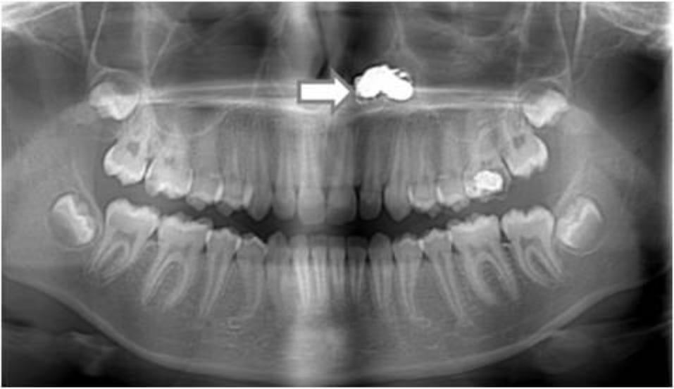
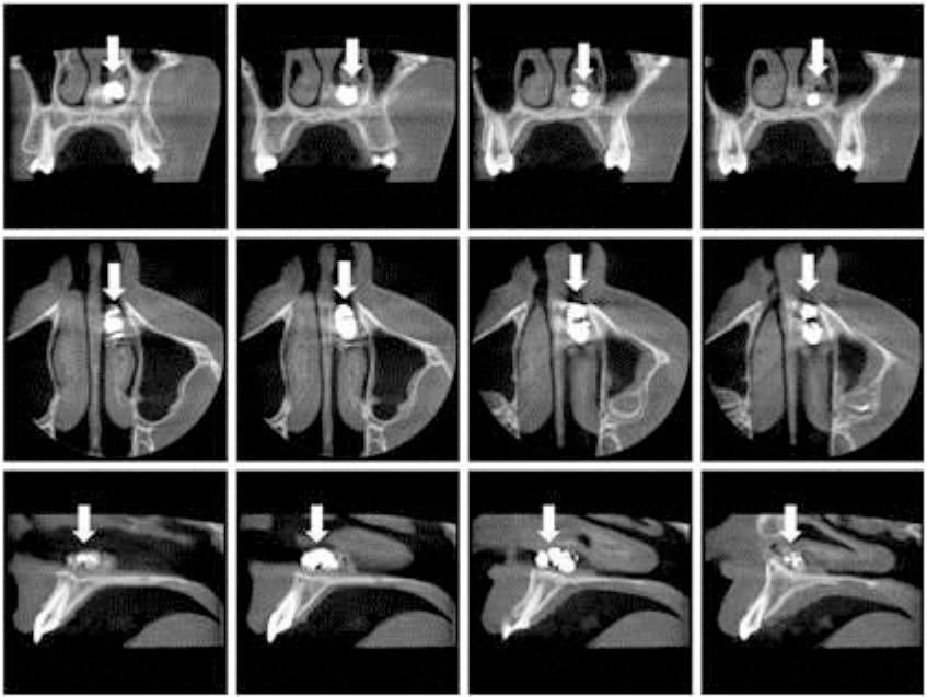
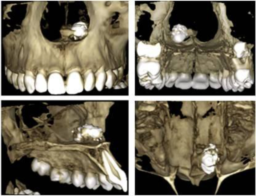
A 13-year-old girl presented to our department with a 2-day history of painful swelling in relation to the tooth 26. Orthopantomography showed caries in the first left upper molar, and incidentally the presence of a foreign body on the floor of the left nostril (Figure 1). CBCT (Planmeca ProMax 3D Mid) revealed a hyperdense material in the left nasal cavity. It was a foreign body of irregular morphology, segmented with dimensions of 93x52x38 mm (Figure 2–3). These images allowed the exact location of the foreign body and know that it was not a button battery. The girl did not remember history of foreign body insertion. He reported being asymptomatic, although he noticed a moderate left nasal obstruction for two years before orthopantomography. On examination, there was extensive edema with slough in the left side of the nasal cavity. In the operating room, under local anesthesia and by direct rhinoscopy a piece of a metal toy were removed. Septal perforation was not observed.

JDMR-19-130-Junquera_Spain_F1

Figure 1. Orthopantomography. Radiodense image of irregular contour on the floor of the left nostril.

JDMR-19-130-Junquera_Spain_F2

Figure 2. CBCT. Foreign body of irregular morphology, segmented with dimensions of 93×52×38 mm and without halo sign, common in button batteries.

JDMR-19-130-Junquera_Spain_F3

Figure 3. Three-dimensional reconstruction of the foreign body.

Discussion

Pediatric nasal obstruction is one of the most common problems seen in pediatric otolaryngologists. Typically, this is not an urgent diagnosis but is more commonly associated with reduced quality of life. Allergic rhinitis is one of the most common causes of pediatric nasal obstruction, which affects 8% to 16% of children and is immunoglobulin E mediated. In younger children, nasal foreign bodies must always be on the differential of nasal obstruction. Intervention is always needed for nasal foreign body removal in order to prevent further migration distally, potentially precipitating an airway emergency. The timing of removal is often based on the foreign body involved. Batteries are always considered an emergency because of the complications associated with prolonged exposure (septal perforation, saddle nose deformity, orbital injury, synechiae). However, nasal foreign bodies can often be removed without general anesthesia if the child is cooperative [7]. Alkaline batteries cause extensive necrosis and tissue destruction. Possible mechanisms include spontaneous electrolyte leakage with liquefactive necrosis and destruction of tissue, and generation of electrical current causing an electric burn [8–10].

In our case, unlike many other cases, the nasal foreign body may remain asymptomatic for a long time. Our patient had only a complaint of nasal stuffiness. This is an unusual case of a large chronic nasal foreign body with no known history of insertion. If the patient had indeed had symptoms for the previous two years, this suggests the foreign body was inserted when he was around age 11, which would be unusual in a child without learning disability. Identification and localization of foreign bodies are based on history, clinical and radiographic examinations. Various imaging modalities, including, periapical radiographs, plain radiography, Computed Tomography (CT), and ultrasonography, have been advocated for detecting FBs. Radiographs detected FBs generally considered radiopaque (gravel, glass, metal) in 98% of cases, but do not detected radiolucent (wood, plastic, cactus spine) bodies. The false-negative and false-positive rates for radiography are 50% and 1.6%, respectively [11,12]. Periapical radiographs are the primary diagnostic aid used in identifying the foreign bodies. However, these are not helpful in the identification of cases, in which foreign body sizes are <2 mm or in identifying the exact locations of the objects. These problems can be overcome by advanced diagnostic and imaging aids such as CT, and Cone Beam Computed Tomography. CBCT provides images at low dose with sufficient spatial resolution, which can be applied in diagnosis, treatment planning, and post-treatment evaluation. CBCT has higher spatial resolution and greater ability to detect high-density foreign bodies as small as 0.5 mm [13, 1 4]. In our case discarded the diagnosis of button batteries.

Conclusion

Within limits of this case report, Cone beam computed tomography is a reliable method for the diagnosis of nasal foreign bodies, by providing the exact location and composition.

References

  1. Grigg S, Grigg C (2018) Removal of ear, nose and throat foreign bodies: A review. Aust J Gen Pract 47: 682–685.
  2. Foltran F, Ballali S, Passali FM, Kern E, Morra B, et al. (2012) Foreign bodies in the airways: a meta-analysis of published papers. Int J Pediatr Otorhinolaryngol 14: 12–19.
  3. Park JW, Jung JH, Kwak YH, Jung JY (2019) Epidemiology of pediatric visits to the emergency department due to foreign body injuries in South Korea: Nationwide cross-sectional study. Medicine (Baltimore) 98: e15838.
  4. Svider PF, Sheyn A, Folbe E, Sekhsaria V, Zuliani G, et al. (2014) How did that get there? a population-based analysis of nasal foreign bodies. Int Forum Allergy Rhinol 4: 944–949.
  5. French MA, Lorenzoni G, Purnima, Azzolina D, Baldas S, et al. (2019) Foreign Body injuries in children in India: Recommendations for prevention from a comparative analysis with international experience. Int J Pediatr Otorhinolaryngol 124: 6–13.
  6. Gray ML, Kappauf C, Govindaraj S (2019) Management of an Unusual Intranasal Foreign Body Abutting the Cribriform Plate: A Case Report and Review of Literature. Clin Med Insights Ear Nose Throat 12: 1179550619858606.
  7. Smith MM, Ishman SL (2018) Pediatric Nasal Obstruction. Otolaryngol Clin North Am 51: 971–985.
  8. Bakshi SS (2019) A button battery in the nose. Intern Emerg Med 14: 185–186.
  9. Cheng SY, Shih CP (2018) Button battery insertion in nose manifested as infraorbital cellulitis. Ear Nose Throat J 97: 274.
  10. Dane S, Smally AJ, Peredy TR (2000) A truly emergent problem: button battery in the nose. Acad Emerg Med 7: 204–2006.
  11. Watanabe K, Hatano GY, Aoki H, Okubo K (2013) The necessity of simple X-ray examination: a case report of button battery migration into the nasal cavity. Pediatr Emerg Care 29: 209–211.
  12. Ryu CH, Jang YJ, Kim JS, Song HM (2008) Removal of a metallic foreign body embedded in the external nose via open rhinoplasty approach. Int J Oral Maxillofac Surg 37: 1148–1152.
  13. Demiralp KO, Orhan K, Kurşun-Çakmak E, Gorurgoz C, Bayrak S (2018) Comparison of Cone Beam Computed Tomography and ultrasonography with two types of probes in the detection of opaque and non-opaque foreign bodies. Med Ultrason 20: 467–474.
  14. Kaviani F, Javad Rashid R, Shahmoradi Z, Gholamian M (2014) Detection of foreign bodies by spiral computed tomography and cone beam computed tomography in maxillofacial regions. J Dent Res Dent Clin Dent Prospects 8: 166–171.

Sulfonylurea Use and Cardiovascular Safety Revisited

DOI: 10.31038/EDMJ.2019355

Abstract

Sulfonylurea use has been commonplace for the management of type 2 diabetes as an adjunct to metformin over the past decades.  Their effectiveness has been repeatedly demonstrated in terms of glycemic control in the short-term however, long-term sustainable control remains in question.  Over the years, FDA mandated cardiovascular safety trials have been completed involving most newer antidiabetic therapies to the market place however, the sulfonylurea class had not been studied until the recent head-to-head cardiovascular outcomes trial involving the comparison of linagliptin, an inhibitor of DPP-IV,  with glimepiride in the CARMELINA study where non-inferiority was demonstrated in both treatment groups.  While this finding seems to be reassuring, does it really confer safety of use of sulfonylurea drugs in the management of type 2 diabetes?

Keywords

Sulfonylurea, DPP-IV inhibitor, diabetes, cardiovascular disease, major adverse cardiovascular event, type 2 diabetes, hypoglycemia, arrhythmia

Guidelines

Since the evolution of the management of diabetes and hyperglycemia, respected societies globally have been providing guidance with respect to such management.  Since the availability of such drugs, the class of sulfonylurea was adapted and implemented.  After metformin was approved for use, the sulfonylurea was recognized as the second agent for intensification.  Over recent years and on the basis of newer agents which demonstrated safety that have become available, namely incretin agents and urinary SGL T2 inhibitors, the sulfonylurea class has found its way on the bottom of such algorithms, as per the American Association of Clinical Endocrinologists [1] on the basis of safety and efficacy, and as one of the executable options of those financially challenged, according to the joint consensus statement from the American Diabetes Association and European Association for the Study of Diabetes, recently revised in the first quarter of 2019 [2]. While these statements and guidelines are consensus or expert based, they are recommendations that are soundly based on their demonstrated safety and efficacy, but even then, the choice of not following such recommendations is routinely exercised.

Development of sulfonylurea

The first agents that were discovered in the sulfonylurea class was in 1942 where sulfonamides were noted to reduce blood sugar in non-human studies, leading way to the development of Carbutamide, which was very quickly withdrawn from the market place because of apparent hematologic disease, particularly on the bone marrow [3]. Second-generation sulfonylureas that became available differed from their first-generation counterparts because of differences in absorption and metabolism.  For this reason, the second-generation agents have been credited with fewer hypoglycemic events relative to their first-generation counterparts, but still differ greatly based on molecular formulation whereas glimepiride is noted to produce hypoglycemia in 2% to 4% patients compared to glyburide, noted to produce hypoglycemia in 20-30% of patients with the reason being  better preservation of prevention of insulin secretion and promotion of glucagon secretion [6].

Mechanism of action

The mechanism of action described as that of insulin secretion out of the pancreatic beta cell independent of what the blood glucose level in circulation may be in addition to having a decreased effect on hepatic insulin clearance.  This insulin secretory effect is largely as a consequence of blocking potassium inflow into the cells through a DPP dependent channel.  This leads to membrane repolarization leading to increase cellular inflow of calcium into these beta cells leading to filamentous contraction of actinomysin with subsequent secretion of large quantities of insulin from that beta cell.  While insulin is secreted in 2 phases largely, it appears that the effect of the sulfonylurea tends to be more so on the second phase of secretion.  However, review of the literature demonstrates that there might be down-regulation of sulfonylurea receptors on the surface of beta cells with long-term use, with increased expression of those very receptors after discontinuation of treatment with sulfonylurea over a certain period of time [3].

Sustainability

Large-scale clinical trials have been performed over the years to evaluate the development of microvascular and macrovascular complications associated with the management of patients with type 2 diabetes.  Amongst these were the United Kingdom Prospective Diabetes Study [4] and the ADOPT trial [5].  In both of these trials, different agents were studied that included insulin, the sulfonylurea group, and metformin.  The p-par gamma molecule, rosiglitazone, was also studied in the ADOPT trial.  It was interesting to note that in these 2 large-scale trials, the sulfonylurea class led to a rapid reduction in hemoglobin A1c that seemed to worsen by about the second year of therapy, or thereafter with a subsequent rise suggesting treatment failure.  Progressive dysfunction and worsening insulin secretion has been noted with sulfonylurea use despite better glycemic control in the short-term.  This phenomenon has been labeled as secondary failure and is an outcome shortly to be expected with chronic sulfonylurea use.  While not terribly well understood, and as mentioned above, is likely related to down-regulation of sulfonylurea receptors on beta cell surface membrane [3].  Thus there appears to be multiple factors that might be contributing to lack of sustainability in hemoglobin A1c control and these stem from increasing pressures that lead to accelerated apoptosis or cell death, and other mechanisms yet to be discovered that may perhaps be implemented in the future for beta cell preservation.  Thus on the basis of demonstrated lack of sustainability of hemoglobin A1c, one can assume that treatment with a sulfonylurea would offer very little on beta cell mass preservation or persistent improvement in beta cell function.

Safety

Use of any pharmacologic agent for management of chronic disease may have adverse events associated with them, even though they may be curtailing the natural history of the original disease state.  For the sulfonylurea class, however, the most worrisome challenges include progressive weight gain, as evidenced in numerous large-scale clinical trials, and the risk of developing significant hypoglycemia, which itself is challenging in diagnosing, particularly nocturnal hypoglycemia, which often goes unrecognized.  When reviewing the literature, there has been significant variability in nocturnal hypoglycemia listed ranging anywhere from 20-40% depending on which study was reviewed and with which sulfonylurea.  However, what is the cost of hypoglycemia?  From a physiologic standpoint, significant electrolyte aberrancies can occur including potassium shifting intracellularly as well as effect on the myocyte cycle with noted QT prolongation [7]. Such occurrences can lead to significant dysrhythmia and lethal arrhythmia.  It is thought that such unpredictable variability in glycemic control may have been part of the reason why an increased mortality may have been observed in the ACCORD Action to Control Cardiovascular Risk in Diabetes) trial where the forced titration hemoglobin A1c target was a value of less than or equal to 6%.  It is interesting to note that the majority of the cardiovascular events recorded were in the population of patients who is hemoglobin A1c did not change very much despite aggressive management, suggesting much glycemic variability [12].

Cardiovascular outcomes

Several studies have been published looking at particular cardiovascular adverse events with the use of sulfonylureas.  The data seems to be quite variable.  Data reviewing the UK Clinical Practice Research Data Link, published in 2017, reviewed short acting and nonspecific long-acting sulfonylurea with no significant increase in noted myocardial infarction, ischemic stroke or cardiovascular death between both long and short acting agents however, with significant risk of severe hypoglycemia in the long-acting agents [8].  In another study accepted for publication in June 2018 assessed whether adding or switching to sulfonylurea is associated with an increased risk of major adverse cardiovascular events including all-cause mortality.  This study did demonstrate an increased risk of myocardial infarction and all-cause mortality, with no differences in cardiovascular death or severe hypoglycemia [9].

As part of the management of diabetes, which is complex already to begin with, newer agents with lower hypoglycemic potential when used as monotherapy or combination therapy with metformin have gained significant traction on the basis of their safety record demonstrating no increased cardiovascular risk or reduction in cardiovascular risk.  Such therapies include DPP 4 inhibitors, GLP-1 receptor agonists, and urinary SGL T2 receptor blockers.  Only recently has a cardiovascular outcomes trial been completed where the DPP 4 inhibitor linagliptin was studied with the active comparator being the sulfonylurea glimepiride in the CAROLINA trial [10].  The purpose of this trial was to establish noninferiority between these 2 agents with respect to cardiovascular risk.  However, since no cardiovascular studies have been performed looking at glimepiride, cardiovascular safety was demonstrated with the DPP 4 Linagliptin versus placebo in the CARMELINA study where noninferiority was achieved [11]. In the active comparator CAROLINA (CARdiovascular Outcome study of LINAgliptin versus glimepiride in patients with type 2 diabetes) study, the primary endpoint defined as noninferiority of linagliptin versus glimepiride in time to first occurrence 3 point MACE was satisfied.  The study was an event driven trial involving 6979 patients with the median duration of the study being 2.2 years.  Population involved was on average 62 years of age with 34% having had established cardiovascular disease and 28.6% of those in the trial having been treated with a sulfonylurea agent for less than 5 years.  Noninferiority for major adverse cardiovascular events was indeed demonstrated, albeit with significantly greater hypoglycemia noted in the glimepiride treatment group (10.6% versus 37.7%).

Conclusion

Sulfonylurea use over the past decades has been welcomed by a sense of comfort and demonstrated rapid efficacy, although of limited benefit.  Weight gain and hypoglycemia still seems to be the most worrisome adverse events with these agents, and a myriad of physiologic effects as a consequence of those hypoglycemic events will pose significant challenges toward their continued use.  While electrolyte shifting and effects on QT intervals increase risk of cardiac arrhythmia, there was no increase in cardiovascular mortality that was noted in the glimepiride subgroup, treated to a maximum of 4 mg daily, in the CARMELINA study.  Of note was the fact that those enrolled in the clinical trial was a lower cardiovascular risk population, albeit older.  The fact that there was no increase in cardiovascular events noted in this clinical trial was reassuring but should not be translated to the sulfonylurea class in general as only glimepiride use was allowed by trial design.  The observed risk reduction cannot be and should not be extrapolated to other sulfonylureas, and while safety was demonstrated from a cardiovascular standpoint in this low risk population, there exists uncertainty of whether or not similar findings would be seen in a higher risk population.  Therefore, it’s important for the prescriber to be aware that differences in this class of agents need to be taken into consideration in order to avoid a false sense of reassurance.

List of abbreviations

UKPDS- United Kingdom Prospective Diabetes Study

ADOPT- A Diabetes Outcome Progression Trial

GLP-1 receptor agonist-glucagon-like peptide 1 receptor agonist

SGL T2 receptor inhibitor-sodium glucose transport protein 2 inhibitor

SU-sulfonylurea

DPP 4 or DPP IV-Dipeptidyl peptidase 4

MACE Major Adverse Cardiovascular Events

Declaration

Compliance with Ethics Guidelines

This article is based on previously conducted studies and does not contain any studies with human participants or animals performed by any of the offers of this publication

Consent for publication

The author has given his approval for the version of this manuscript to be published

Competing interests/Disclosures

Dr. Javier Morales is on the speakers Bureau of Novo-Nordisk, Eli Lilly and company, Boehringer Ingelheim, Janssen pharmaceuticals, Mylan pharmaceuticals, and Abbott Laboratories, and serves as consultant, as well as having participated in advisory board meetings for the above-named entities.

Authorship

The author meets the International Committee of Medical General Editors (ICMJE) criteria for authorship for this article, take responsibility for the integrity of the work as a whole

Availability of data and material

This article is based on previously conducted studies and thorough literature review was conducted during authorship of this manuscript

Funding

No funding or sponsorship was received for this publication or article processing charges

Acknowledgments

No editorial assistance was provided by any entity or company during the development of this manuscript.

References

  1. Alan J Garber, Martin J Abrahamson, Joshua I Barzilay, Lawrence Blonde, Zachary T Bloomgarden, et al. (2019) Consensus Statement By The American Association Of Clinical Endocrinologists And American College Of Endocrinology On The Comprehensive Type 2 Diabetes Management Algorithm – 2019 Executive Summary. Endocrine practice 25: 1
  2. Introduction: Standards of Medical Care in Diabetes (2019) Diabetes Care. 42: 1-2.
  3. Daniele Sola, Luca Rossi, Gian Piero Carnevale Schianca, Pamela Maffioli, Marcello Bigliocca, et al. (2015) Sulfonylureas and their use in clinical practice. Arch Med Sci 11: 840–848.
  4. UK Prospective Diabetes Study (UKPDS) Group (1998) Effect of intensive blood-glucose control with metformin on complications in overweight patients with type 2 diabetes (UKPDS 34).  The Lancet 352: 854–865.
  5. Giancarlo Viberti, Steven E. Kahn, Douglas A Greene, William H Herman, Bernard Zinman, Rury R Holman, et al. (2002) A Diabetes Outcome Progression Trial (ADOPT). Diabetes Care 25: 1737–1743.
  6. Mark T Keegan (2013) Pharmacology and Physiology for Anesthesia.
  7. Laitinen T, et al. (2008) Ann Noninvasive Electrocardiology 13: 97–105.
  8. Antonios Douros, Hui Yin, Oriana Hoi Yun Yu, Kristian B. Filion, Laurent Azoulay and Samy Suissa (2017) Pharmacologic Differences of Sulfonylureas and the Risk of Adverse Cardiovascular and Hypoglycemic Events. Diabetes Care 40: 1506–1513.
  9. Antonios Douros, Sophie Dell’Aniello, Oriana Hoi Yun Yu, Kristian B Filion, Laurent Azoulay, et al. (2018) Sulfonylureas as second line drugs in type 2 diabetes and the risk of cardiovascular and hypoglycaemic events: population based cohort study 362
  10. CAROLINA: Cardiovascular Outcome Study of Linagliptin versus Glimepiride in Patients with Type 2 Diabetes. ClinicalTrials.gov
  11. Rosenstock J, Perkovic V, Johansen OE, Cooper ME, Kahn SE, Marx N, et al. (2019) Effect of Linagliptin vs Placebo on Major Cardiovascular Events in Adults With Type 2 Diabetes and High Cardiovascular and Renal Risk: The CARMELINA Randomized Clinical Trial. JAMA 321: 69–79.
  12. Effects of Intensive Glucose Lowering in Type 2 Diabetes (2008) N Engl J Med 358: 2545–2559

Migraine, A Review of Basic, Clinical, and Translational Approaches to New Treatment

DOI: 10.31038/AWHC.2019263

Abstract

Migraine is a debilitating neurological primary headache disorder characterized by recurring unipolar headaches lasting 4–72 hours with accompaniment of nausea and sensory sensitivities. Migraine is the most common headache disorder resulting in seeking of medical care [1, 2], in addition to being one of the most debilitating chronic disease conditions in terms of both morbidity and lost economic productivity. Migraine incidence has been observed since ancient times to disproportionately affect women, and most current epidemiological assessments put current incidence estimates at 12% overall for US populations, with an incidence of 18% in women and 6% in men when stratified by sex. This dimorphism of incidence is crucial when assessing overall health of a community, specifically when concerning women’s health and therefore must be taken into consideration when developing both clinical and basic models of migraine to enact the best possible outcomes of combined translational research efforts. While major recent advances have been made in the field of pharmacologic intervention for migraine with the recent approval of the anti CGRP and anti CGRP receptor antibody medications, prohibitive cost and limited access have made older treatments, such as the triptans and NSAIDs, still the most commonly utilized medications to combat migraine attacks. Current preclinical research models are heavily interested in modulation of the neuropeptide CGRP, and the phenomenon of cortical spreading depression (CSD), believed to be the underlying trigger of migraine with aura, via pharmacological intervention. Modulations of these phenomena has found to be correlated with menstrual events in women, tying back to the overarching theme of higher morbidity in women. With the advent of pharmacogenomics and personalized medicine, a new epoch of potential customizable treatments looms on the horizon, endearing those afflicted with this severely debilitating condition a new glimmer of hope as research progresses into its next phase.

Acronyms: AMPP: American Migraine Prevalence and Prevention study. NIH: National Institute of Health. ICHD-3: International Classification of Headache Disorders, 3rd edition. NHIS: National Health Interview Survey. NSAID: Non-Steroidal Anti-Inflammatory Drug. CGRP: Calcitonin Gene Related Peptide. 5HT1: 5-Hydroxytryptamine (Serotonin) Receptor, Subfamily 1. FDA: Food and Drug Administration. CNS: Central Nervous System. MOH: Medication Overuse Headache. PGE2: Prostaglandin E2. CSD: Cortical Spreading depression. fMRI: Functional Magnetic Resonance Imaging. GWAS: Genome Wide Association Study. SNP: Single Nucleotide Polymorphism.

Background

Migraine is a highly debilitating neurological disorder characterized by recurring unipolar headaches with a duration of 4–72 hours, accompanied by nausea and sensory sensitivities [3]. Migraine is classified as a primary headache disorder, indicating no known underlying cause, and is the most common of all the headache disorders to result in patients seeking medical care [1, 2]. Migraine is also one of the most prevalent and disabling chronic disease conditions, in terms of both individual morbidity and lost economic productivity, with 18% of women and 6% of men in the US suffering from some form of migraine [1], with an incidence in women nearly triple that of men according to the National Health Survey. The economic burden of migraine has been assessed by the American Migraine Prevalence and Prevention study (AMPP) and estimates a mean direct annual healthcare cost burden of $4,144 per chronic migraineur in addition to an average of $5,392.03 of lost economic productivity annually [4]. When quantifying the high degree of morbidity associated with migraine, it quickly becomes evident that the need for better understanding of the underlying pathophysiology be accomplished through basic and clinical research models to aid in ablating the impact of this public health issue.

Migraine Types/Epidemiology

Migraine has been classified into several categories depending on the clinical presentation and description of the patient, these are 1) migraine without aura, 2) migraine with aura, this class includes hemiplegic migraine (including familial and sporadic), migraine with brainstem aura, retinal migraine and aura without headache. 3) chronic migraine (CM), which is defined by 15 or more “headache days” per month, for three months, in absence of medication overuse. Aura is described as a temporary visual disturbance appearing as zigzag lines, flashing lights, or temporary visual loss according to the NIH. Episodic migraine (EM) is classified as having less than 15 headache days per month for three months according to the ICHD-3. While precise estimates of migraine incidence can be difficult to ascertain due to differences in study methodology, the most recent and largest of these was the AMPP, a longitudinal study focused on 120,000 surveyed US households with recipients based on US census data. Overall incidence of migraine was reported as 12%, with 18% of women and 6% of men reporting experiencing at least one migraine attack in the previous year. Upon stratifying by gender, 17.4% of women and 5.7% of men had EM, while 1.29% of women and 0.48% of men meet diagnostic criteria for CM [5]. The disease burden of migraine is carried much more heavily by women.

Sexual dimorphism in migraine presentation: Often, migraine begins at menarche for girls, and continues until approximately age 40, at which symptomology levels off [1, 2]. It was also noted that women experience more severe pain intensity and associated disability when compared to men [6, 7]. This finding is highly significant as these are the most economically productive years for an individual woman and instantiates an even higher degree of cost burden when factoring in the higher observed incidence rate of migraine in women. Coupled to this phenomenon is the degree to which female sex hormone fluctuation during the menstrual cycle, pregnancy, and post-partum periods has been positively observed to impact not only incidence of migraines attacks, but also perceived severity [6, 7]. This chronic level of unpredictability can cause a severe amount of stress and disability at a time in a woman’s life when she is expected to be at her peak performance, both economically and in terms of social and familial commitments. For those women with children, the crippling effect of chronic or episodic migraine attacks is a cost burden most simply not afford, and those suffering under the severe duress from a typical migraine attack will still be expected to perform career duties, child care duties, social duties etc. The crippling morbidity of this condition cannot be understated, as the buildup of chronic stress due to migraine results in lost economic productivity, lost time to care for offspring, lost ability to invest in pleasurable activities, which can all contribute to a noticeable decline in long term mental health. It doesn’t take long to connect the dots here and realize the agonizingly debilitating effect this condition has on the nearly one fifth of women who suffer from it, and why addressing it is a grave public health concern to society. While studies in the US have found an inverse relationship between income and migraine [8], European studies have been more nebulous about this association, failing to replicate the results seen in the US [6].

Migraine and Race: Migraine incidence varies by race in the US, according to the AMPP study white women and men had the highest incidence (20.4%, 8.4%), followed by African Americans (16.2%, 7.2%), and lowest among Asian Americans (9.2%, 4.2%) [9]. Further surveys of underrepresented populations undertaken by the NHIS indicate highest prevalence in American Indians and Alaska Natives (19.2%), followed by whites (15.5%), African Americans (15%), Hispanic and Latinos (14.9%), Native Hawaiians and Pacific Islanders (13.2%) and Asians (10.1%) [6]. Overall, migraine is a functional pain disorder that disproportionally impacts women during their most productive years, regardless of race.

Clinical Aspects

Diagnostic criteria for the several subtypes of migraine are laid out in the ICHD-3 manual. For use as a type example, the diagnostic criteria for migraine with aura is as follows: Recurrent attacks, lasting minutes, of unilateral fully-reversible visual, sensory or other central nervous system symptoms that usually develop gradually and are usually followed by headache and associated migraine symptoms. Diagnoses of migraine invariably depend on self report by patients, and therefore accurate estimation of true disease prevalence can be difficult [10]. Patients suffering from migraine are currently treated with a host of pharmaceutical agents developed to either abort an acute migraine attack, or act as a prophylactic treatment against future attacks [11].

Treatment and sex sensitivity: Currently, the US headache consortium has outlined a 6 point objective plan to help guide physicians in treating acute migraine attacks. These are 1) treat attacks rapidly and consistently without recurrence; 2) restore patient’s ability to function; 3) minimize the use of back-up and rescue medications; 4) optimize self-care and reduce subsequent use of resources; 5) to be cost-effective for overall management; 6) have minimal or no adverse events, however achieving all of these is typically not possible with available treatments. Treatment criteria also differs by region, as some pharmaceuticals are approved in one are while others are not (ergotamines). NSAIDs and acetaminophen, with or without caffeine, are typically first line treatment for a migraine attack [12], due to their easy availability and lack of harmful side effects from frequent use. These are typically used for a mild to moderate migraine sufferer. Triptans are the current first line medications for treating moderate to severe migraine [12], they have agonist activity at the 5HT1 receptors and are believed to work through suppression of CGRP release. They are well-tolerated and demonstrate efficacy in 68% of patients in one meta-analysis [13, 14]. Use of triptans, however, are limited to a maximum use limit of 8–9 doses per month, due to fear of medication overuse headache and the potential of serotonin syndrome [15]. Some first line combination agents exist, such as sumatriptan-naproxen to maximize efficacy of both compounds. Recently on the market are the anti CGRP monoclonal antibodies and anti CGRP receptor monoclonal antibodies. These drugs have yet to be classified in this hierarchy of use, due to their prophylactic nature (once monthly/quarterly administration) and prohibitive cost (estimated cost for erenumab, $6900 annually). While considered safe by the FDA, utilization of any monoclonal antibody does carry some degree of risk of autoimmune induction. This is a relevant point here as CGRP has been shown to have immune function in both the CNS and periphery [16]. However due to the very recent availability of these drugs some time must pass before an in depth analyses can be made. A more cost effective and longer standing approach to migraine prophylaxis lies in the application of the beta blocker drug class. Being the first in their class in terms of application towards migraine treatment, they are still extensively utilized as a prophylactic treatment with the benefit of a much lower cost and tolerable side effect profile [17]. Second line drugs for acute migraine treatment include those of the anti-emetic class, such as promethazine and chlorpromazine. Last resort agents include opiates, barbiturates, ergotamines and valproate, due to either contradicting results, abuse potential, risk of medication overuse headache, or lack of current approval. Medication overuse headache (MOH) is a phenomenon observed from overuse of migraine and pain disorder medications [13, 14]. the ICHD-3 defines MOH as headache occurring on 15 or more days per month, for three months due to over-usage of acute or symptomatic headache medication. The prevalence of MOH is 1–2% globally, however it is one of the costliest neurological disorders known due to its extremely debilitating effects and treatment resistance [18]. A variety of medication have been observed to cause MOH, however findings have specifically found analgesics such as opioids to be the highest risk class for causing MOH, with triptans being at most equal to opiates for relative risk of developing MOH [13, 14]. Demonstration of not only lack of efficacy of classic analgesics, but the ability for them to increase relative risk for MOH demonstrates the pertinent need for new therapeutics.

Divergence in Female Treatment Response Sensitivity: While the current literature is somewhat lacking in this study metric, fitting within the narrative of this review it would be prudent to assess relevant clinical observational data that is currently available. While many studies have been performed assaying treatment efficacy, there seems to be an overall dearth of results stratified on gender in respect to observed efficacy of pharmaceutical migraine treatment. However, studies have observed a higher female preponderance to developing medication overuse headache; this could be an artifact of the higher overall incidence of migraine in women, higher usage of medication by women, and higher seeking of medical care by women vs men, and underdiagnosing of migraine in men [10]. Moreover, specific agents that induce MOH were not discussed. A recent study has outlined differences in pharmacokinetics in women vs men for the triptan drug class, particularly noting the substantially higher peak plasma concentration of triptans observed in women, which can have far reaching effects on the differences observed in treatment response in men vs women [10]. Regarding the new anti CGRP drugs, studies are under way to assess different response in women and men when treated. However, the few studies that have been done have not been able to definitively state a difference in response to the monoclonal antibodies [19].

Preclinical Research Models

While the precise mechanisms of Migraine are yet to be elucidated, numerous preclinical and basic research models are available, including in vitro, in vivo, and ex vivo models. While the complete understanding of migraine pathophysiology is beyond the scope of this review, a major recent success has arisen from focusing on the interplay of CGRP, the neurovascular unit, and the trigeminal nerve complex [20]. This has led to the development of several new treatments based on inhibiting activity of CGRP. Current preclinical models include a multitude of models to mirror physiological phenomena believed to be impacted in contributing, all or in part, to overall cellular and neurobiological states resulting in a migraine episode. The inflammatory soup model is one such example. This model is based upon the hypothesis that migraine progression is based upon abnormal functioning of neurons in several brain regions [21]. It is essentially an animal model upon which a cocktail of proinflammatory compounds are introduced into the brain, and fMRI imaging is utilized to map the alterations in brain response, cellularly and chemically, to the introduced disruption [21]. The cocktail itself is an acidic mix of bradykinin, serotonin, histamine and prostaglandin PGE2. This paradigm was conceptualized from samples of inflamed human tissue, utilized to induce a state of allodynia and hyperalgesia in an animal model [22]. Migraine has been observed to be induced by a host of triggers, and in clinical settings it was noticed cardiac angina patients undergoing nitroglycerin therapy demonstrated a high degree of headache incidence from this treatment. This observation led to the development of the use of the NO donor in preclinical studies to serve as a migraine attack trigger in animal and ex vivo models [23], due to the documented vasodilative properties of NO and NO donor chemicals. An interesting observation made in the clinic also found that over 50% of migraine without aura is highly correlated with the menstrual cycle [24]. This has translated into application of progesterone treatment in basic research models, in vitro and animal models to simulate this phenomenon in the laboratory to corroborate possible application of contraceptive medications in pursuit of alleviating menstrual associated migraine without aura with these readily available medications. In addition to many other factors, a major mechanism of action of progesterone only contraceptives are believed to down regulate expression of estrogen receptors in the trigeminal vascular system, thereby reducing nociceptive response to elevated estrogen levels associated with menstrual cycles [24]. in further exploring the myriad of possible triggers producing a migraine response, it would be prudent of the preclinical researcher to investigate inroads into possible environmental triggers of a migraine episode. One such tool developed for this purpose is umbellone, an environmental irritant that has found recent application in studying possible activation of transient receptor potential ankyrin-1 (TRPA1) channels and possible contribution to induction of a migraine event [25].

While a notable amount of current research is being focused on modeling and understanding the cortical spreading depression (CSD) event, it must be noted that it has been observed that not all CSD events result in triggering of migraine event, or any type of headache for that matter. This duality is important to note, as CSD events are hypothesized to be an underlying mechanism for migraine with aura10, direct evidence of this has not yet been fully elucidated and ongoing efforts to model it are being pursued to fully tease out the full impact of a CSD event, as pathological brain conditions other than migraine also demonstrate association with CSD [26]. all of these models and study paradigms have been essential in advancing the field of migraine research in basic and preclinical laboratories in institutions across the globe. As more research is clearly needed to elucidate the sex specific reasons women are affected at a much higher rate with migraine, sex specific models are being developed to further investigate this phenomenon. One immediate and simple method of accomplishing this is by simply including female animals, tissue, or female animal derived primary cell cultures for use in experiments. This paradigm can also be carried further into the clinic for translational studies by the usage of female participants for IRB approved studies. The Dussor research group has developed several models for investigating sex-based differences in progesterone signaling leading to higher incidence of migraines observed in females. An animal model has been developed and utilized by this group to explore the relationship between elevated estrogen levels and specific response patterns to fluctuations of these female sex hormones, further relating to the translational application of progesterone as a treatment [27]. The Dussor and Russo labs have also investigated the differences of CGRP expression in a female model. Due to the hypothesized impact of CGRP on development of migraine, it would be prudent to assess if a difference in expression patterns of this neuropeptide could be contributing to the observed difference in migraine incidence [28]. while this research is still in its infancy, the new avenues being opened by pharmacogenomic technology and the approach of personalized medicine will potentially allow for a new zenith of breakthroughs, as the apocryphal working hidden within our DNA becomes available for study.

Future Direction of Field and Precision Medicine

The recent development of the new class of anti CGRP and anti CGRP receptor antibodies has been an exciting advance in the field of migraine research, however their high cost makes access to all who could benefit from their use impractical. With the emerging concept of precision medicine and pharmacogenomics becoming more and more optimized and readily available, the possibility of applying these technologies to new treatments looms ever closer on the horizon. Due to the high degree of genetic variation within each migraineur, different variants of the enzymes, transporters, and receptors will be more or less responsive to a unique blend of polytherapy, as coding variants for each of these proteins will respond ever so slightly different to each blend of agent utilized to treat migraine [29]. GWAS analyses is proving to be an extremely powerful tool in analyzing single nucleotide polymorphism (SNP) variants across populations [30, 31]. Emerging research has indicated familial migraine contains a higher pathologic gene load associated with migraine than sporadic cases [32], while another study has begun to map possible loci containing genes involved in migraine pathology, specifically locating 38 new loci [15]. In addition to physiologic aspects, applications of high end computing are being utilized to analyze high volumes of drug safety data [33]. This approach utilizing personalized medicine has already been put into translational studies for cardiovascular anti-coagulant drugs, such as warfarin, which has highly variable therapeutic windows depending on the DNA variants encoding enzymes in its metabolic pathway. Moving forward it is hoped to be able to adapt this individual tailoring approach to create a treatment plan specifically optimized for a given patient. The urgency for this approach is highlighted by the fact that only 50% of migraineurs respond to acute or prophylactic treatment [29]. It is hoped that by moving forward with entrenched research tools in the laboratory, best practices observed in the clinic, and the wealth of knowledge and potential unlocked by pharmacogenomic technology, a new synergistic approach to migraine treatment may be made in order to alleviate this horrifically debilitating condition.

References

  1. Lipton RB, Munjal S, Alam A, Buse DC, Fanning KM, et al. (2018) Migraine in America Symptoms and Treatment (MAST) Study: Baseline Study Methods, Treatment Patterns, and Gender Differences. Headache: The Journal of Head and Face Pain 58: 1408–1426. [crossref]
  2. Lipton RB, Munjal S, Buse DC, Alam A, Fanning KM, et al. (2019) Unmet Acute Treatment Needs From the 2017 Migraine in America Symptoms and Treatment Study. Headache 59: 1310–1323. [crossref]
  3. Schwedt TJ, Alam A, Reed ML, Fanning KM, Munjal S, et al. (2018) Factors associated with acute medication overuse in people with migraine: results from the 2017 migraine in America symptoms and treatment (MAST) study. J Headache Pain 19: 38. [crossref]
  4. Messali A, Sanderson JC,  Blumenfeld AM, Goadsby PJ, Buse DC (2016) Direct and Indirect Costs of Chronic and Episodic Migraine in the United States: A Web Based Survey. Headache: The Journal of Head and Face Pain 56: 306–322. [crossref]
  5. Buse DC, Loder EW, Gorman JA, Stewart WF, Reed ML, et al. (2013) Sex differences in the prevalence, symptoms, and associated features of migraine, probable migraine and other severe headache: results of the American Migraine Prevalence and Prevention (AMPP) Study. Headache: The Journal of Head and Face Pain 53: 1278–1299. [crossref]
  6. Burch R (2019) Epidemiology and Treatment of Menstrual Migraine and Migraine During Pregnancy and Lactation: A Narrative Review. Headache: The Journal of Head and Face Pain [crossref]
  7. Burch R, Rizzoli P, Loder E (2018) The Prevalence and Impact of Migraine and Severe Headache in the United States: Figures and Trends From Government Health Studies. Headache: The Journal of Head and Face Pain 58: 496–505.
  8. Lanteri-Minet M (2014) Economic burden and costs of chronic migraine. Curr Pain Headache Rep 18: 385. [crossref]
  9. Lipton RB, Stewart WF, Diamond S, Diamond ML, Reed M (2001) Prevalence and Burden of Migraine in the United States: Data From the American Migraine Study II. Headache: The Journal of Head and Face Pain 41: 646–657. [crossref]
  10. van den Maagdenberg AMJM, Nyholt DR, Anttila V (2019) Novel hypotheses emerging from GWAS in migraine?. J Headache Pain 20: 5. [crossref]
  11. Munakata J, Hazard E, Serrano D, Klingman D, Rupnow MF, et al. (2009) Economic Burden of Transformed Migraine: Results From the American Migraine Prevalence and Prevention (AMPP) Study. Headache: The Journal of Head and Face Pain 49: 498–508. [crossref]
  12. Mayans L, Walling A (2018) Acute Migraine Headache: Treatment Strategies. Am Fam Physician 97: 243–251.
  13. Teppe SJ (2019) Acute Treatment of Migraine. Neurologic Clinics 37: 727–742. [crossref]
  14. Thorlund K, Sun-Edelstein C, Druyts E, Kanters S, Ebrahim S, et al. (2016) Risk of medication overuse headache across classes of treatments for acute migraine. J Headache Pain 17: 107. [crossref]
  15. Evans RW, Tepper SJ, Shapiro RE, Sun-Edelstein C, Tietjen GE (2010) The FDA alert on serotonin syndrome with use of triptans combined with selective serotonin reuptake inhibitors or selective serotonin-norepinephrine reuptake inhibitors: American Headache Society position paper. Headache 50: 1089–99. [crossref]
  16. Russell FA, King R, Smillie SJ, Kodji X, Brain SD (2014) Calcitonin gene-related peptide: physiology and pathophysiology. Physiol Rev 94: 1099–142. [crossref]
  17. Danesh A, Gottschalk PCH (2019) Beta-Blockers for Migraine Prevention: a Review Article. Curr Treat Options Neurol 21: 20.
  18. Gormley P, Anttila V, Winsvold BS, Palta P, Esko T, et al. (2016) Meta-analysis of 375,000 individuals identifies 38 susceptibility loci for migraine. Nat Genet 48: 856–866. [crossref]
  19. Melo-Carrillo A, Noseda R, Nir RR, Schain AJ, Stratton J, et al. (2017) Selective Inhibition of Trigeminovascular Neurons by Fremanezumab: A Humanized Monoclonal Anti-CGRP Antibody. J Neurosci 37: 7149–7163. [crossref]
  20. Loder S, Sheikh HU, Loder E (2015) The Prevalence, Burden, and Treatment of Severe, Frequent, and Migraine Headaches in US Minority Populations: Statistics From National Survey Studies. Headache: The Journal of Head and Face Pain 55: 214–228. [crossref]
  21. Becerra L, Bishop J, Barmettler G, Kainz V, Burstein R, et al. (2017) Brain network alterations in the inflammatory soup animal model of migraine. Brain Res 1660: 36–46. [crossref]
  22. De Felice M, Eyde N, Dodick D, Dussor GO, Ossipov MH, et al. (2013) Capturing the aversive state of cephalic pain preclinically. Ann Neurol 74: 257–265. [crossref]
  23. Ashina, Messoud, Hansen, Jakob Møller Dunga, Bára Oladóttir, et al. (2017) Jes Human models of migraine, short-term pain for long-term gain, Nature Reviews Neurology, Nature Publishing Group, a division of Macmillan Publishers Limited.
  24. Allais G, Chiarle G, Sinigaglia S, Airola G, Schiapparelli P, et al. (2017) Treating migraine with contraceptives. Neurol Sci 38: 85–89.
  25. Edelmayer RM, Le LN, Yan J, Wei X, Nassini R, et al. (2012) Activation of TRPA1 on dural afferents: a potential mechanism of headache pain. Pain 153:1949–1958. [crossref]
  26. Charles A, Baca S. (2013) Cortical spreading depression and migraine. Nat Rev Neurol 9: 637–644. [crossref]
  27. Sandweiss AJ, Cottier KE, McIntosh MI, Dussor G, Davis TP, et al. (2017) 17-β-Estradiol induces spreading depression and pain behavior in alert female rats. Oncotarget 8: 114109–114122. [crossref]
  28. Russo AF. (2015) Calcitonin gene-related peptide (CGRP): a new target for migraine. Annu Rev Pharmacol Toxicol 2015 55: 533–552. [crossref]
  29. Daniela Pietrobon, Michael A. Moskowitz (2013) Pathophysiology of Migraine, Journal Article, 2013 Annual Review of Physiology, Annual Review of Physiology February 75: 365.
  30. Ayata C, Lauritzen M (2015) Spreading Depression, Spreading Depolarizations, and the Cerebral Vasculature. Physiol Rev 95: 953–993. [crossref]
  31. Thorlund K, Mills EJ, Wu P, Ramos E, Chatterjee A, et al. (2014) Comparative efficacy of triptans for the abortive treatment of migraine: A multiple treatment comparison meta-analysis. J Cephalalgia 34: 258–267 [crossref]
  32. de Boer I, van den Maagdenberg AMJM, Terwindt GM. (2019) Advance in genetics of migraine. Curr Opin Neurol 32: 413–421. [crossref]
  33. Danysz K, Cicirello S, Mingle E, Assuncao B, Tetarenko  N, et al. (2019) Artificial Intelligence and the Future of the Drug Safety Professional. Drug Saf 42: 491–497. [crossref]

Disseminated Tumor Cells in Bone Marrow In gastric Cancer Patients with Obesity

DOI: 10.31038/CST.2019452

Background

Obesity is a risk factor for cancer development and is associated with poor prognosis in multiple tumor types. There is emerging evidence of a strong association between obesity and gastrointestinal cancer. The molecular mechanism underlying gastric cancer invasion and metastasis is still poorly understood. Problem of Disseminated Tumor Cells (DTCs) in gastric cancer remains to be relevant for clinics and less is known concerning this problem for patients with obesity.

Aim

This study was aimed to evaluate how incidence of DTCs in bone marrow is conditioned by excess of adipocites in tumor microenvironment of patients with gastric cancer and obesity.

Results

There was not found the associations between availability of DTCs in BM as well CXCR4-positive cells in tumor and body mass index (BMI) but incidence of DTC in BM was associated with high density of Cancer-Associated Adipocytes (CAAs) as well with high number of CXCR4-positive cells in tumor of patients with BMI<25 and BMI>25<30 but it was not true for patients with BMI>30 where frequency of DTCs finding in BM was significantly decreased and that was statistically significant.

Conclusion

In patients with BMI>30 high density of CAAs and high number of CXCR4-positive cells in tumor may create specific tumor microenvironment that prevent tumor cells to leave primary lesion.

Obesity is associated with poor prognosis in multiple tumor types [1]. There is emerging evidence of a strong association between obesity and gastrointestinal cancer [2]. In contrast to the convincing evidence that obesity (measured by body mass index, BMI) increases the risk of many different types of cancer, there is an ambiguity in the role of obesity in survival among cancer patients [3, 4]. Some studies suggested that higher BMI decreased mortality risk in cancer patients, a phenomenon called the obesity paradox [1]. Changes that occur in the obese state and the biologic mechanisms underlying the connections of these changes to increased cancer risk are poorly understood [5–6]. Many types of solid tumors grow in proximate or direct contact with adipocytes and adipose-associated stromal and vascular components. During interaction with cancer cells adipocytes dedifferentiate into pre-adipocytes or are reprogrammed into Cancer-Associated Adipocytes (CAAs) that modulate the tumor microenvironment by promoting angiogenesis, affecting immune cells and altering metabolism to support growth and survival of metastatic cancer cells [7]. Quail D et al. indicate that special consideration of the obese patient population is critical for effective management of cancer progression [8].

During tumor progression, cells can acquire the capability for invasion and metastasis to escape the primary tumor, first of all, from breast, lung, colorectal and prostate, and colonize new organs [9, 10]. Tumor cells leaving primary site can settle mainly in Bone Marrow (BM) as a common homing-organ for Disseminated Tumor Cells (DTCs) with potency to form the metastases [11–13]. The most important factors controlling cellular migration are chemokines and their receptors. Stem cell receptor CXCR4 as a transmembrane chemokine receptor and its specific ligand CXCL12 (Stromal Cell-Derived Factor 1, SDF-1α) play a vital role in dissemination of tumor cells from primary sites, transendothelial migration as well as homing of cancer stem cells. In the tumor microenvironment under hypoxic condition cells of a growing tumor are reprogrammed to express the CXCR4 receptor thereby enhancing the metastatic potential of the tumor cells. [14–17]. the molecular mechanism underlying gastric cancer invasion and metastasis is still poorly understood. Problem of DTCs in gastric cancer remains to be relevant for clinics [18] and less is known concerning this problem for patients with overweight and obesity [19]. Therefore our study was aimed to evaluate how of CAA density, CXCR4 expression in primary tumor affect presence of DTCs in BM of patients with gastric cancer according to the Body Mass Index (BMI).

Patients and Methods

Patients

A total of 94 patients (60 men and 34 women) with primary gastric cancer were diagnosed and treated at the City Clinical Oncological Center (Kiev). No patient received any pre-operative anti-cancer therapy. Tumors were classified and staged according to the 2002 version of the UICC staging system [20]. Histological types of tumor were evaluated by WHO histological classification (2000) [21]. Tissue samples were taken immediately after tumor excision. Preoperatively, 2.0–3.0 ml of BM aspirates from the sternum with conventional cautions to avoid the hit of skin epithelial cells into the sample were obtained. All patients were thoroughly informed about the study that was approved by the local ethics committee.

Immunocytochemical Examination of Bone Marrow

Detection of tumor cells (cytokeratin-positive cells, CK-positive cells) in BM cytospin preparations fixed in acetone was provided by APAAP method (alkaline phosphataseantialkaline phosphatase) and visualization system EnVision G/2 System/AP Rabbit/Mouse (Permanent Red) (Dako Cytomaiton, Denmark). Monoclonal mouse antibodies against panCK (clone AE1/AE3, Dako Cytomation, Denmark) were used as primary antibodies. Each assay was controlled negatively by staining of one cytospin preparation with nonspecific IgG1 (MOPC21, Sigma). Number of tumor cells (CK-positive cells) was expressed on 106 BM mononuclear cells. BM samples were scored “positive” if the presence of two or more CK-positive cells per 106 mononuclear cells were detected (from 6 to 12 slides per patient were screened).

Immunohistochemical Examination of Tumor Tissue

Expression Perilipin (Plin5+) as a marker for viable adipocytes as well expression of CXCR4 were provided on deparaffinized slides using specific polyclonal rabid antibodies (Perilipin-5/OXPAT Antibody, Termoscientific, USA) dilution 1:200 and specific monoclonal mouse antibodies: clone AB2074 (Abcam, UK), respectively. Slides for evaluation of Plin5+ were covered with 1% of Bovine Serum Albumin (BCA) and incubated with polyclonal antibodies during for 1hour and then washed in Phosphate-Buffered Saline (PBS). Immunoreactions were detected and visualized with the polymer-peroxidase method (EnVision+/HRP and 3, 3-diaminobenzidine; DakoCytomation, Denmark) followed by counterstaining with Mayer hematoxylin. Negative control was employed in which the primary antibody was replaced by Phosphate-Buffered Solution (PBS). Immunopositive cells were counted per 1000 cells in each slide and the number of positive cells was reported as percent. When the tumor consisted of more than 10% of CXCR4-positive cells, the case was scored as positive.

Body Mass Index (BMI, Kg M−2)

Patients were classified according to BMI, following the WHO definitions, as underweight, normal (18.5–<25.0 kg/m2), overweight (25.0–<30.0 kg/m2) or grade 1 obesity (30.0–<35.0 kg/m2).

Statistical Analysis

All statistical analyses were conducted using the NCSS 2000/PASS 2000 and Prism, version 4.03 software packages. Prognostic values of relevant variables were analyzed by means of the Cox proportional hazards model using Odds ratio and χ2 test. Two-tailed p values <0.05 were considered statistically significant.

Results

Caas in Tumors of Patients According To BMI

Individual patient data from a total 94 histological confirmed gastric cancer patients were included in this study. Median number of CAAs in tumors was 26.5%. We defined this number as the cut-off value and classified all cases into high- or low-density groups. Overall, 48.4% of tumors were characterized by a low density of CAAs and 51.6% by high CAAs during follow-up. 39.5%, 46.4%, 89.5% of patients with BMI<25, BMI>25<30, BMI>30, respectively, had high CAAs in tumors. The probability of availability of high density of CAAs in tumor of patients with BMI>30 is increased by a factor of almost 9 (OR 8.84, χ2 = 13.47, 95%CI 16.777–4.665, P<0.01) as compared with BMI<30. Data obtained demonstrate that adipocytes are as major component of the microenvironment of gastric cancer, especially under obesity.

CK-Positive Cells in Bone Marrow

 Overall, 88.3% of patients have been with M0 category. It was determined that CK-positive cells were detected in BM of 50.1% gastric cancer patients among all investigated. There was no association between DTCs in BM and clinicopathological characteristics. It makes no difference between of groups of patients according to BMI concerning the availability of DTCs in BM: 47.6%, 56.2% and 41.2% of patients with BMI<25, BMI>25<30, BMI>30 had DTCs in BM, respectively. Meanwhile, it was found the association between the presence of DTCs in BM and density of CAAs in tumors: DTCs in BM were detected in 41.3% and in 58.7% of patients when tumors characterized by low and high density of CAAs, respectively. When tumors characterized by high density of CAAs appearance of tumor cells in BM has been found in 35.7% of patients with BMI>30 as compare with 70.6% and 62.5% of patients with BMI<25 and BMI>25<30. In patients with obesity frequency of DTCs finding in BM was significantly decreased and it was statistically significant (OR 4.33, χ2 = 3.82, 95%CI 9.341–2.007, P<0.05) as compare with patients with BMI<25. It may be suggested that adipocites, namely CAAs, playing an essential role in the regulation of metabolic functions in the variety of processes involved in metastatic spread of tumor cells.

CXCR4-Positive Cells in Tumor Tissue

Overall, 83.1% of patients had tumors with CXCR4-positive cells. Statistically significant correlation between CXCR4-positivity of tumors and clinical characteristics was not found. The median number of CXCR4+ cells was 24.2% (range of 13.4–81.0%). It makes no difference between of groups of patients according to BMI concerning the CXCR4-positive cells: 66.7%, 65% and 60% of patients with BMI<25, BMI>25<30, BMI>30 had high number of CXCR4-positive cells in tumor, respectively. Meanwhile, it was found the association between high number of CXCR4-positive cells and density of CAAs in tumors. High number of CXCR4-positive cells were detected in 35.3% and in 72.7% of patients when tumors characterized by low and high density of CAAs, respectively (OR 7.3, χ2 = 12.45, 95%CI 13.654–4.208, P<0.01).

The mean number of CXCR4-cells in tumors with high density of CAAs was 47.4±1.9%, 37.8±4.1% and 48.5±2.9% in patients with BMI<25, BMI>25<30, BMI>30, respectively.When tumors characterized by high density of CAAs presence of high number of CXCR4-positive cells have been found in 77.8%, 62.5% and 71.4% of patients with BMI<25 and BMI>25<30 and. BMI>30, respectively, and presence of DTCs in BM in these groups of patients was the following: 88.9% of patients with BMI<25, in 58.3% of patients with BMI>25<30 and in 18.2% of patients with BMI>30.It is notably important to note that in patients with BMI>30 having high density of CAAs and high number of CXCR4-positive cells in primary tumor the incidence of DTCs in BM was rather low. It may be supposed that under obesity additional mechanisms may be switched off in tumor microenvironment modulated by excess of adipocites to prevent cells escaping.When tumors characterized by low density of CAAs low number of CXCR4-positive cells was detected in 33.3%, 51% and 44.1% of patients with BMI<25 BMI>25<30 and. BMI>30, respectively. Presence of DTC in BM in these groups of patients was the following: in 31.5% of patients with BMI<25, in 44.6% of patients with BMI>25<30 and in 41.1% of patients with BMI>30.

Conclusion

There was not found the associations between availability of DTCs in BM as well CXCR4-positive cells in tumor and BMI but incidence of DTC in BM and high number of CXCR4-positive cells in tumor associated with high density of CAAs of patients with BMI<25 and BMI>25<30 but it is not true for patients with BMI>30 where frequency of DTCs finding in BM was significantly decreased and that is statistically significant.In patients with BMI>30 high density of CAAs and high number of CXCR4-positive cells in tumor may create specific tumor microenvironment that prevent tumor cells to leave primary lesion. Understanding the metabolic changes that occur in obese individuals may also help to elucidate more effective treatment options for these patients when they develop cancer.

References

  1. Wang J, Yang DL, Chen ZZ, Gou BF (2016) Associations of body mass index with cancer incidence among populations, genders, and menopausal status: A systematic review and meta-analysis. Cancer Epidemiol 42: 1–8.
  2. Donohoe CL, Pidgeon GP, Lysaght J, Reynolds JV (2010) Obesity and gastrointestinal cancer. Br J Surg 97: 628–642.
  3. Renehan AG, Tyson M, Egger M, Heller RF, Zwahlen M (2008) Body-mass index and incidence of cancer: a systematic review and meta-analysis of prospective observational studies. Lancet 37: 569–578.
  4. Iyengar NM, Gucalp A, Dannenberg AJ, Hudis CA (2016) Obesity and Cancer Mechanisms: Tumor Microenvironment and Inflammation. J Clin Oncol 34: 4270–4276.
  5. Renehan AG, Zwahlen M, Egger M (2015) Adiposity and cancer risk: new mechanistic insights from epidemiology. Nat Rev Cancer 15: 484–498.
  6. Hopkins BD, Goncalves MD, Cantley LC (2016) Obesity and Cancer Mechanisms: Cancer Metabolism. J Clin Oncol 34: 4277–4283.
  7. Nieman KM, Romero IL, Van Houten B, Lengyel E (2013) Adipose tissue and adipocytes support tumorigenesis and metastasis. Biochim Biophys Acta 10: 1533–1541.
  8. Quail D, Olson OC,  Bhardwaj P, Walsh LA, Akkari L, et al. (2017) Obesity alters the lung myeloid cell landscape to enhance breast cancer metastasis through IL5 and GM-CSF. Nat Cell Biol 19: 974–987.
  9. Gupta GP, Massagué J (2006) Cancer metastasis: building a framework. Cell 127: 679–695.
  10. Valastyan S, Weinberg RA (2011) Tumor metastasis: molecular insights and evolving paradigms. Cell 147: 275–292.
  11. Pantel K, Alix-Panabières C, Riethdorf S (2009) Cancer micrometastases. Nat Rev Clin Oncol 6: 339–351.
  12. Pantel K, Hayes D (2018) Disseminated breast tumour cells: biological and clinical meaning. Nat Rev Clin Oncol 15: 129–131.
  13. Pantel K, Brakenhoff RH, Brandt B (2008) Detection, clinical relevance and specific biological properties of disseminating tumour cells. Nat Rev Cancer 8: 329–340.
  14. Sun X, Cheng G, Hao M, Zheng J, Zhou X, et al. (2010) CXCL12 / CXCR4 / CXCR7 chemokine axis and cancer progression. Cancer and Metastasis Rev 29: 709–722.
  15. Burger J, Kipps T (2006) CXCR4: a key receptor in the crosstalk between tumor cells and their microenvironment. Blood 107: 1761–1767.
  16. Zhang Z, Ni C, Chen W, Wu P, Wang Z, et al. (2014) Expression of CXCR4 and breast cancer prognosis: a systematic review and meta-analysis. BMC Cancer 14: 49.
  17. Xiang Z, Zhou Z-J, Xia G-K, Zhang XH, Wei ZW, et al. (2017) A positive crosstalk between CXCR4 and CXCR2 promotes gastric cancer metastasis. Oncogene 36: 5122–5133.
  18. O’Sullivan J, Lysaght J, Donohoe CL, Reynolds JV (2018) Obesity and
    gastrointestinal cancer: the interrelationship of adipose and tumour microenvironments. Nat Rev Gastroenterol Hepatol 15: 699–714.
  19. International Union Against Cancer (2002) TNM Classification of malignant tumors, edited by L. H. Sobin and C. Wittekind, Wiley-Liss, New York, NY, USA, 6th edition, 2002.
  20. C Fenoglio-Preiser, F Carneiro, P Correa, et al. (2000) “Gastric carcinoma,”in World Health Organization Classification of tumors. Tumours of the Stomach. In: S. R. Hamilton and L. A. Aaltonen (eds.). vol. 3, chapter 3, Pg No: 39–52, IARC Press, Lyon, France, 2000.

Synthesis of Nanolignin Following Ozonation of Lignocellulosic Biomass

DOI: 10.31038/NAMS.2019244

Introduction

The non-food valorization of biomass represents a major axis of research currently animating a large number of scientists. Whether for energy purposes, replacing fossil fuels such as oil, or as innovative strategies for access to new bio-sourced products, modern valorization approaches are above all respectful of the principles of green chemistry, and generally refer to processes or to the use of eco-compatible products. Renewable polymers have emerged as an attractive alternative to conventional metallic and organic materials for a variety of different applications, due to their biocompatibility, biodegradability and low cost of production [1].Renewable biomass can provide many industrial solutions as it is composed primarily of cellulose (30–35%), lignin (15–30%) and hemicellulose (20–35%).Among them, lignin is known as one of the main bio-resource raw material that can be used for the synthesis of environmentally friendly polymers, thus a good candidate for replacing regular industrial aromatic polymers and fine chemicals. Due to its chemical and structural diversity, lignin valorization remains a major challenge for achieving a viable biomass-based economy [2]. Lignins are composed of polymerized monolignols and their derivatives. Particularly grass lignins contain important amounts of alkenes yielding up to 10–15% by weight aldehydes [3].

Lignin extraction from lignocellulosic biomass

The lignocellulosic biomass has to be treated before any widespread utilization of its components. As a decrease in the lignin content in plants results in an increase in biodegradability, lignin removal from this biomass is a crucial pretreatment step [4,5]. A large number of chemophysical pretreatment approaches has been investigated on a wide variety of feedstock [6]. These methods require the use of hazardous materials such as acids, alkalis and/or organic solvents. They are currently four industrial processes to extract pure lignin: sulfite, kraft, organosolv and soda processes [7, 8]. Kraft pulping accounts for approximately 85% of the produced lignin. The delignification process is performed at high temperatures (170°C) and high pH 13 or 14, during which the lignin is dissolved in sodium hydroxide and sodium sulfide (white liquor) [9].

The sulfite process involves the reaction between lignin and a metal sulfite and sulfur dioxide, with calcium, magnesium or sodium acting as counter ions; the pH can vary between 2 and 12, and the temperature between 120 and 180 °C, with a digestion time of 1–5 h [10]. The soda process is typically used for the treatment of grass, straw and sugarcane bagasse, which accounts for 5% of the total pulp production [11]. The biomass is digested at temperatures that vary between 140 and 170 °C in the presence of 13–16% by weight of aqueous solution of sodium hydroxide. The soda lignin contains no sulfur which is not the case of the kraft and sulfite processes.

The organosolv process is based on the treatment of the biomass using organic solvents including ethanol, methanol, acetic and formic acid that are usually mixed with water at temperatures that range from 170 to 190°C [12]. The recovery and separation of the dissolved lignin and hemicelluloses can be done by precipitation of lignin or evaporation of the organic solvent, after adjusting the temperature, pH and concentration of the organic solvent. Organosolv pulping is one of the most efficient options for the further valorization of lignin and it also preserves the native structure of the lignin [13]. The obtained lignin is sulfur-free, with lower ash-content, and it has higher purity.

The removal of lignin by different technologies originates different product streams. Beyond the production costs and the environmental impact the lignin extraction method has to be selected depending on the use that will be given to the extracted lignin. In general, the kraft and sulfite processes allow extracting lignin at reasonable costs, while the organosolv method continues to be an expensive technology but still with high quality extracted lignin. However, the soda extraction process generates lower production costs and low environmental impact.

Ozonation of lignocellulosic biomass

Ozonation can circumvent the different issues of the above extraction processes that require the use of hazardous materials (acids, alkalis and/or organic solvents), as it is considered as a green process. Ozone (O3) is a powerful oxidizing agent (E° = 2.07 V). It is one of the most promising lignocellulosic biomass oxidative pretreatment for selective lignin degradation with minimal effects on the hemicellulose and cellulose contents [14]. It provides low production of inhibitory compounds such as furfural and HMF (Hydroxymethylfurfural), and more importantly it requires no chemical additives during all the pretreatment process. However, ozonation demands high energy generation costs but this aspect can be avoided by optimizing the ozonation process. Ozone has a high affinity for phenol and polyphenols such as lignin and tannic acid. During ozonation lignin is converted to soluble products which to a great extent are biodegradable and thus yield a useful byproduct [15].

It has been reported the ozonolysis of grass lignin to selectively cleave aromatic aldehydes by limiting the reaction residence time to a few minutes for preventing over oxidation of targeted products [16], the ozonation was done in acidic media to avoid the production of secondary ozonides usually done at neutral pH media [17]. Ozone has been used to remove lignin from different biomass such as wheat and rye straw [18], cotton stalk [19], magazine pulps [20], among others.

Lignin presents a very complex assembly which limits enormously their interaction with host polymer matrices for industrial applications. It has been reported that only 2% of the annually extracted lignin from paper and pulp industry is used for applications such as fillers, adhesives and dispersants; the remaining lignin is burned as industrial waste for energy generation [21].

Synthesis of lignin nanoparticles

One way to overcome the limitations of lignin pointed out in the above section is to reduce the size of lignin particles until the nanometric size (less than 100 nm). At this scale, new functionalities and properties of materials are observed and used for a wide range of novel applications. As the size of the particles is reduced to the nanoscale range, the surface to volume ratio of the particles gradually increases which in turn increases the reactivity of the particles and changes their mechanical, electrical and optical properties [22] (Adusei-Gyamfi and Acha, 2016).These lignin nanoparticles hold huge potential for downstream valorization due to their unique morphology and abundant multifunctional groups. Thus the idea is to prepare lignin nanoparticles, which will greatly improve their reactivity and solubility with host matrices, and will provide a morphological and structural control of these structures for different high-value applications [21].

Several different methods have been published to synthesize nanolignin. Frangville et al. reported nanolignin obtained by precipitation in HCl. The resulting nanoparticles were crosslinked with glutaraldehyde, getting good stability over a wide range of pH [23]. Gilca et al. prepared nanolignin by sonication, they identified two main reaction patterns resulting in chain cleavage (depolymerization) and oxidative coupling (polymerization), both probably promoted by the hydroxyl and superoxy radicals generated by ultrasound [24]. Hydroxypropyl lignin nanoparticles were reported to be prepared by reacting an alkaline lignin solution with propylene oxide, then acidifying and centrifuging the mixture to precipitate the nanoparticles [25]. It has been reported also the use of a solution precipitation from alkaline lignin with either ethylene glycol or alkaline solution, which resulted in smaller nanoparticles [26]. Of all the methods mentioned above, the precipitation method in HCl seems to be the easiest for the synthesis of lignin nanoparticles.

Applications of lignin nanoparticles

As for the applications for lignin nonmaterial’s, lignin shows unique properties such as antioxidant and antibacterial properties, ultraviolet absorption, and high toughness [27]. To produce novel materials with improved properties, nanolignin particles can be incorporated in polymers for use in food packaging with the properties mentioned above, such as polyvinyl alcohol/Chitosan (providing UV absorbance, antioxidant and antibacterial properties), glycidyl methacrylate grafted polylactic acid (providing UV-absorbance and antibacterial properties), polylactic acid(providing UV-absorbance and antibacterial properties) [28], among others.

Lignins are known by their free radical scavenging activity due to their complex phenolic structure, which make them recognized as efficient natural antioxidants [29]. The incorporation of natural antioxidants to food packaging materials has been widely studied in order to improve protection of light and/or oxygen sensitive products [30]. Lignins have been proposed as antioxidants for polylactic acid films [31]. Lignin nanoparticles have been reported to exhibit higher antioxidant activity than neat lignin [26, 32, 33].

Nanolignin particles have also been involved in production of antimicrobial materials. Richter et al. [34] produced nanolignin loaded with silver ions and coated with a cationic polyelectrolyte layer capable of adhering to bacterial cell membranes. The nanoparticles killed both Gram-negative and Gram-positive bacteria while using at least 10 times less silver than conventional silver nanoparticles, thus reducing the environmental impact produced by silver nanoparticles. Films based on polylactic acid [35], chitosan, and/or polyvinyl alcohol [36] added with nanoparticles of lignin presented activity against Gram-negative bacteria, indicating that the films containing nanolignin particles could be used as antibacterial food packaging.

UV radiation accelerates oxidation rates in food [37] and also photodegradation of organic polymers [38]. The UV-absorbing capacity of lignin was tested by Yearla and Padmasree [32] by monitoring survival rates of UV-irradiated E. coli. In the absence of lignin compounds, the mortality of E. Coli was 100% after 5 minutes of UV exposure, while their survival was improved in the presence of lignin in proportion to their concentration. This study was done based on the fact that Escherichia coli suffer intracellular oxidative damage and death caused by UV radiation. In addition, because lignin nanoparticles have superior UV protection over ordinary lignin, the survival rate results were much better when nanolignin was used. Bionanocomposite films made of gluten-lignin nanoparticles have been reported to absorb UV radiation [99], thus these materials could be applied in food packaging with UV protective features, such as nuts and other food products that are prone to lipid oxidation.

Conclusion

Lignin as a renewable polymer is an attractive alternative to conventional metallic and organic materials due to its biocompatibility and biodegradability. Ozone has proved its efficiency as pretreatment method of lignocellulosic biomass for removing lignin. Ozonation is a green method requiring no hazardous compounds such as acids or alkalis, and needs moderate reaction conditions such as room temperature and atmospheric pressure. Lignin is a very complex polymer limiting its applications. One way to solve this problem is to reduce the size of ordinary lignin particles at the nanoscale, where new properties are being harnessed for novel applications. Lignin nanoparticles are good candidates for the next generation functional nanocomposites as they present very interesting properties such as UV light blockers, radical scavengers, and antioxidants very promising for potential applications in the food sector such as packaging.

References

  1. Clark JH, Budarin V, Deswarte EIF, Hardy JJE, Kerton FM, et al. (2006) Green chemistry and the biorefinery: a partnership for a sustainable future. Green Chem 8: 853–860.
  2. Bule MV, Gao AH, Hiscox B, Chen S (2013) Structural modification of lignin and characterization of pretreated wheat straw by ozonation. J Agric Food Chem 61: 3916–3925.
  3. Figueiredo P, Lintinen K, Hirvonen JT, Kostiainen MA (2018) Properties and chelical modifications of lignin: Towards lignin-based nanomaterials for biomedical applications. Progress in Mater Sc 93: 233–269.
  4. Binder A, Pelloni L, Fiechter A (1980) Delignification of straw with ozone to enhance biodegradability. European J ApplMicrobiolBiotechnol 11: 1–5.
  5. Margellou A, Triantafyllidis K (2019) Catalytic transfer hydrogenolysis reactions for lignin valorization to fuels and chemicals. Catalysts 4: 43.
  6. Doherty WOS, Mousavioun P, Fellows CM (2011) Value-adding to cellulosic ethanol: lignin polymers. Ind Crops Prod 33: 259–276.
  7. Capolupo L, Faraco, V (2016) Green methods of lignocellulose pretreatment for biorefinery development. ApplMicrobiolBiotechnol 100: 9451–9467.
  8. Maurya DP, Singla A, NegiS (2015) An overview of key pretreatment processes for biological conversion of lignocellulosic biomass to bioethanol. 3 Biotech 5: 597–609.
  9. Chaturvedi V, Verma P (2013) An overview of key pretreatment processes employed for bioconversion of lingolellulosic biomass into biofuels and value added products. 3 Biotech 3: 415–431.
  10. Zakzeski J, Bruijnincx PC, Jongerius AL, Weckhuysen BM (2010) The catalytic valorization of lignin for the production of renewable chemicals. Chem Rev 110: 3552–3599.
  11. Galkin MV, Samec JS (2016) Lignin valorization through catalytic lignocellulosic fractionation: a fundamental platform for the future biorefinery. ChemSusChem 9:1544–1558.
  12. De la Torre MJ, Moral A, Hernández MD, Cabeza E, Tijero A (2013)Organosolvligninforbiofuel. IndEngChem Res 45: 58–63.
  13. Mesa L, Gonzales E, Cara C, Gonzales M, Castro E, et al. (2011) The effect of organosolv pretreatment variables on enzymatic hydrolysis of sugarcane bagasse. ChemEng J 168:1157–1162.
  14. Travaini R, Martin-Juarez J, Lorenzo-Hernando A, Bolado-Rodriguez S (2016) Ozonolysis: An advantageous pretreatment for lignocellulosic biomass revisited. BioresourTechnol 199: 2–12.
  15. Shi F, Xiang H, Li Y (2015) Combined pretreatment using ozonolysis and ball milling to improve enzymatic saccharification of corn straw. BioresourTechnol 179:444–451.
  16. Silverman JR, Danby AM, Subramaniam B (2019) Intensified ozonolysis of lignins in a spray reactor: insights into product yields and lignin structure. ReactChemEng 4:1421–1430.
  17. Danby AM, Lundin MD, Subramaniam B (2018) Valorisation of grass lignins: Swift and selective recovery of pendant aromatic groups with ozone. ACS Sustainable ChemEng 6:71–76.
  18. Garcia-Cubero MA, Gonzales-Benito G, Indacoechea I, Coca M, Bolado S (2009) Effect of ozonolysis pretreatment on enzymatic digestibility of wheat and rye straw. BioresourTechnol 100:1608–1613.
  19. Kaur U, Oberoi HS, Bhargav VK, Sharma-Shivappa R, Dhaliwal SS (2012) Ethanol production from alkali- and ozone-treated cotton stalks using thermotolerant Pichia kudriavzevii HOP-1. Ind Crops Prod 37: 219–226.
  20. Kojima Y, Yoon SL (2008) Improved enzymatic hydrolysis of waste paper by ozone pretreatment. J Mater Cycles Waste Manage 10:134–139.
  21. Tian D, Hy J, BaoJ, Chandra RP, Saddler JN, et al.(2017) Lignin valorization: lignin nanoparticles as high-value bio-additive for multifunctional nanocomposites. Biotechnol Biofuels10:192.
  22. Adusei-Gyamfi J, Acha V (2016) Carriers for nanozerovalent iron (nZVI) : synthesis, application and efficiency. RSC Adv 6: 91025–91044.
  23. Frangville C, RutkevidusM, Richter AP, Velev OD, Stoyanov SD, et al. (2012) Fabrication of environmentally biodegradable lignin nanoparticles. ChemPhysChem13: 4235–4243.
  24. Gilca IA, Popa VI, Crestini C (2015) Obtaining lignin nanoparticles by sonication. UltrasonSonochem 23: 369–375.
  25. Chen P, Zhang L, Peng S, Liao B (2006) Effects of nanoscale hydroxypropyl lignin on properties of soy protein plastics. J Appl. Polym101: 334–341.
  26. Ge Y, Wei Q, Li Z (2014) Preparation and evalauation of the free radical scavenging activities of nanoscale lignin biomaterials. Bioresources 9: 2299–6706.
  27. Laurichesse S, Avérous L (2014) Chemical modification of lignins: Towards biobased polymers. ProgPolymSci 39: 1266–1290.
  28. Beisl S, Friedl A, Miltner A (2017) Lignin from micro- to nanosize: Applications. IntJMolSc18: 2367.
  29. Dizhbite T, Telysheva G, Jurkjane V, Viesturs U (2004) Characterization of the radical scavenging activity of lignins-natural antioxidants. BioresourTechnol 95: 309–317.
  30. Gomez-Estaca J, Lopez-de-Dicastillo C, Hernandez-Munoz P, Catala R, Gavara R (2014)Advances in antioxidant active food packaging. Trends Food SciTechnol 35: 42–51.
  31. Domenek S, Louaifi A, Guinault A, Baumberger S (2013) Potential of lignins as antioxidant additive in active biodegradable packaging materials. JPolym Environ 21: 692–701.
  32. Yearla SR, Padmasree K (2015) Preparation and characterization of lignin nanoparticles: evaluation of their potential as antioxidants and UV protectants. J ExperNanosci11: 289–302.
  33. Lu Q, Zhu M, Zu Y, Liu W, Yang L, et al. (2012) Comparative antioxidant activity of nanoscale lignin prepared by a supercriticalantisolvent (SAS) process with non-nanoscale lignin. Food Chem 135: 63–67.
  34. Richter AP, Brown JS, Bharti B, Wang A, Gangwal S, et al. (2015) An environmentally benign antimicrobial nanoparticle based on a silver-infused lignin core. Nature Nanotechnol 10: 817–823.
  35. Yang W, Fortunati E, Dominici F, Giovanale G, Mazzaglia A, et al. (2016) Synergic effect of cellulose and lignin nanostructures in PLA based systems for food antibacterial packaging. Eur Poly J 79: 1–12.
  36. Yang W, Owczarek JS, Fortunati E, Kozanecki M, Mazzaglia A, et al. (2016) Antioxidant and antibacterial lignin nanostructures in polyvinyl alcohol/chitosan films for active packaging. Ind Crop Prod 94: 800–811.
  37. Shahidi F, Zhong Y (2010) Lipid oxidation and improving the oxidative stability. Soc Rev 39: 4067–4079.
  38. FengYJ, Li DQ, Wang Y, Evans DG, Duan X (2006) Synthesis and characterization of a UV absorbent-intercalated Zn-Al layered double hydroxide. PolymDegrad Stab 91: 789–794.
  39. Yang W, Kenny JM, Puglia D (2015) Structure and properties of biodegradable wheat gluten bio nanocomposites containing lignin nanoparticles. Ind Crop Prod 74: 348–356.

Structural Elucidation of Natural Polymeric Materials Treated by Ball Milling: A Mass Spectrometrist View

DOI: 10.31038/NAMS.2019243


It is well known to synthetic organic chemists that ball milling can be used for the synthesis of new compounds [1]. On the other hand, it is well known that ball milling of complex natural materials such as lignin and pollen grains can lead to alteration of their original structure and may produce new compounds that originally do not exist [2]. For example, Milled wood Lignin (MWL) contains more hydroxyl groups than the native one due to the extensive depolymerization during the ball milling process [2]. The presence of more hydroxyl groups indicated the homolytic bond cleavage between lignin monomers, which in turn produce reactive radicals that can create new compounds [2].

It is well known that sporopollenin is composed of carbon, hydrogen, and oxygen in the form of a cross-linked polymer that is amazingly stable [3]. It makes up the outer wall of pollen grains, and when extracted it is in the form of an empty exine or microcapsule [3]. During the late past century sporopollenin exine resistance against chemical treatments has been well documented [3]. It was found that the rigidity of sporopollenin restricted its practical analytical analysis to a limited number of techniques, such as Fourier transform infrared (FTIR) spectroscopy and analytical pyrolysis combined with gas chromatography–electron ionization-mass spectrometry (GC–EI-MS) [4,5]. Based on those outdated and old studies, there is still the belief that sporopollenin contains an aromatic identified as p-coumaric acid and, to a lesser extent, ferulic acid [4,5]. Some recent studies, which still champion this outdated theory was published in nature plants implying a resemblance between sporopollenin and lignin. In this study, Li et al.[6] reported the structure elucidation of sporopollenin extracted from ball-milled pollen grains. It should be noted that in 1966, Gordon Shaw, one of the earliest pioneers in sporopollenin, withdraw his proposal that sporopollenin contains lignin because it does not give any positive test for lignin’s [7].

Needless to say that the pollen grain contains proteins and genetic material that is enclosed by the intine composed of carbohydrates followed by the exine composed of sporopollenin [8]. Logically, high energy ball milling of the pollen grains could produce new artefactual compounds through the reaction between all these pollen grains components together, which in turn could alter the structure of the studied target material sporopollenin.

The effect of ball milling on the structure of these complex natural polymeric biomaterials is perhaps comparable to using specifically pyrolysis GC-MS for their structural analysis [9,10]. Pyrolysis GC-Ms can lead to the identification of compounds that initially does not exist in your sample [9,10]. As an example, pyrolysis GC-MS of unsaturated fatty acids leads to the production of aromatic compounds and, even linear saturated polymers such as polyethylene can produce aromatics during pyrolysis GC-MS analysis [9,10]. Overall,  as a mass spectrometrist, firstly, it is recommended to avoid any procedures that could alter the structure of these complex natural materials before using any mass spectrometric techniques for structure elucidation and/or sequencing purposes. Secondly, using soft ionization methods such as electrospray ionization (ESI-MS) and matrix-assisted laser desorption/ionization (MALDI-MS) is more advantageous than pyrolysis GC-MS. The use of these soft ionization methods allows the analysis of the native form of these complex materials without any alteration in their structure that could lead to misleading results [4,5,9,10]. We are in the process of reporting new finding on the structure of the hollow empty clean sporopollenin exine using state of the art analytical experiments, such as high-resolution X-ray-photoelectron spectroscopy, TOF-SIMS, TOF-SIMS, MALDI-TOF-MS, OF-SIMS (FIB) MALDI-Tandem Mass spectrometry analysis and Solid-State 1H and 13C-NMR (1D and 2D experiments) [11]. We can state that sporopollenin exine does not contain any aromatics and bear no resemblance to lignin [11].

References

  1. Stolle A, Szuppa T, Leonhardt SE, Ondruschka B (2011) Ball milling in organic synthesis: solutions and challenges. Chemical Society Reviews 40: 2317–2329.
  2. Sjöström E, Alén R (Eds.) (2013) Analytical methods in wood chemistry, pulping, and papermaking. Springer Science & Business Media Pg No: 104.
  3. Mackenzie G, Boa AN, Diego-Taboada A, Atkin SL, Sathyapalan T (2015) Sporopollenin, the least known yet toughest natural biopolymer. Frontiers in Materials 2: 66.
  4. Wehling K, Niester C, Boon JJ, Willemse MTM, Wiermann R (1989) p-Coumaric acid—a monomer in the sporopollenin skeleton. Planta 179: 376–380.
  5. Dutta S, Hartkopf-Fröder C, Wilkes H, Greenwood P, Littke R, et al. (2006) The Molecular Composition Of Sporopollenin FromFossil Megaspores As Revealed By Micro-Ftir And Pyrolysis-Gc-Ms. In23rd International Meeting on Organic Geochemistry P88-MO. Devon, UK Torquay.
  6. Li FS, Phyo P, Jacobowitz J, Hong M, Weng JK (2019) The molecular structure of plant sporopollenin. Nature plants 5: 41.
  7. J Brooks, G Shaw (1968) The Post-Tetrad Ontogeny of the Pollen Walland the ChemicalStructure of the Sporopollenin of LiliumHenryi, Grana 8: 2–3, 227–234.
  8. Punt W, Hoen PP, Blackmore S, Nilsson S, Le Thomas A (2007) Glossary of pollen and spore terminology. Review of palaeobotany and palynology 143: 1–81.
  9. Sáiz-Jiménez C (1994) Production of alkylbenzenes and alkylnaphthalenes upon pyrolysis ofunsaturated fatty acids. Naturwissenschaften 81: 451–453.
  10. Conesa JA, Font R, Marcilla A, Garcia AN (1994) Pyrolysis of polyethylene in a fluidized bed reactor. Energy & Fuels 8: 1238–1246.
  11. MikhaelAbanoub, Jurcic Kristina, Schneider Celine, carr David, Fisher Gregory L,et al. (2019) Demystifying and Unravelling the Factual MolecularStructure of the Biopolymer Sporopollenin. ChemRxiv Preprint.

Recent Research Efforts in Improving Lithium-Ion Battery Safety

DOI: 10.31038/NAMS.2019242

Abstract

The Lithium-ion battery (LIB) has been utilized in many applications for thirty years, from personal electronics to electric vehicles. Many developments during the past decades resulted in high energy density and capacity in LIBs. However, battery safety still remains an issue alongside the continuous growth in the LIB market. Recent high-profile hazardous incidents gained considerable attention from the public as well as among researchers. This short review summarizes the recent efforts in improving LIB safety on three fronts: (1) materials advancement, (2) early monitoring and detection of thermal runaway events, and (3) fault diagnosis and fault-tolerance controls.

Keywords

lithium-ion battery; battery safety; battery management system; fault diagnosis; fault-tolerance control

Introduction

Lithium-Ion Battery (LIB) technology and the industry experienced rapid development in the past three decades, due to the advantages of LIBs over other energy storage systems, including: high energy density, strong stability, low maintenance, and low self-discharge. The LIB is the predominant power source in both consumer electronics and Electric Vehicles (EVs). The global market of LIBs (Figure 1) increased from $18.8 billion in 2014 to $28.5 billion in 2018. It is expected to increase by 11.1% (compound annual growth rate, CAGR) to $53.3 billion by 2024 [1]. The continuous growth in the global EV market from 2014 to 2024, at a rate of 21.1% (CAGR), is anticipated to contribute to the growth in the LIB market [2]. Although the LIB offers many advantages, safety still remains as an issue. According to a report issued by the Federal Aviation Administration (FAA), the number of air/airline incidents involving the battery – lithium-battery-induced smoke, fire, extreme heat, or explosion – increased from 9 to 50 incidents annually from 2014 to 2018 [3]. There are many reports about EV fire accidents caused by LIBs. For example, an all-electric compact car BYD e6 (BYD Auto company, China) caught fire after being hit by a Nissan GTR, an accident that led to the deaths of three passengers in Shenzhen, China, in May 2012 [4]. The battery pack of a Tesla Model S immediately caught fire after the driver hit debris on a highway in Washington State in 2013 [5]. Improving LIB safety is an important issue that requires research into new materials for the device, as well as structure optimization, and system design.

A typical EV battery is a three-level assembly: battery cell, battery module, and battery pack (Figure 2). The battery cell is a basic unit, consisting of an anode, a cathode, a separator, and liquid electrolyte. Multiple battery cells are connected and placed into a frame, called a battery module. Finally, the several battery modules are assembled into a battery pack, along with a control system, and system protection. The battery pack is a complete system that can be installed in an EV. Fires in a battery cell occur because of physical and electrical faults [6]. A chain reaction fire in the battery cells results in an explosion of the battery pack, called a cascading thermal runaway event. As mentioned in a review by Mauger et al., a gasoline fire can only be ignited when the gas tank’s air level is between 1.4 and 7.6%, and temperature is above 200oC [7]. The gas tank design of the vehicle ensures that gasoline cannot self-ignite in a tank. Unlike gasoline, the cascading thermal runaway event can happen in the LIB. This review summarizes the recent research efforts to improve LIB safety on three fronts: (1) materials advancement, (2) early monitoring and detection of thermal runaway events, and (3) fault diagnosis and fault-tolerance controls.

Current efforts in battery safety improvements

1 . Materials advancements

Cathode (positive electrode) material can be categorized into three groups, based on crystal geometry:

  • Lamellar compounds (lithium cobalt oxide – LCO; lithium nickel oxide – LNO; lithium nickel cobalt aluminum oxide – NCA; and lithium nickel manganese cobalt oxide – NMC)
  • Spinel lithium manganese oxide – LMO
  • Olivine lithium iron phosphate – LFP

NAMS-2019_Jianyu Liang_F1

Figure 1. Global history and forecast data for lithium battery, including lithium battery global market size; electric vehicle sales globally; and numbers of problem incidents related to lithium battery reported by FAA [1–3].
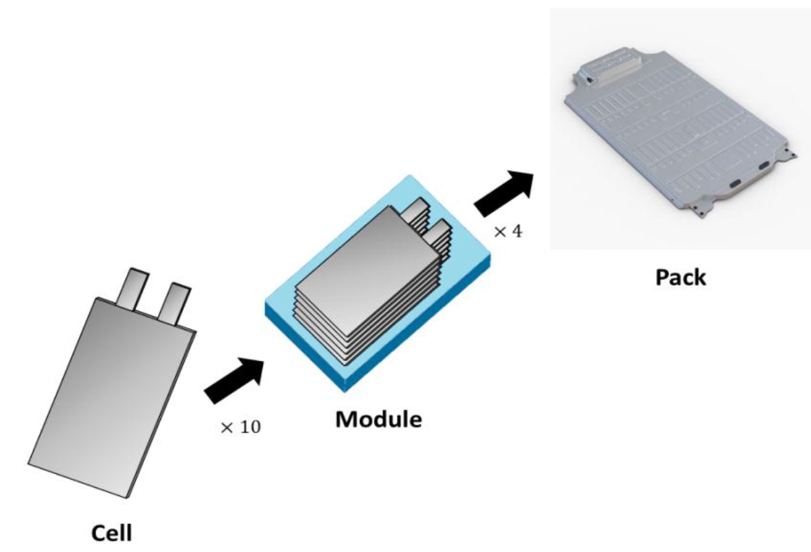
NAMS-2019_Jianyu Liang_F2

Figure 2. A typical battery pack assembly for EV.

LCO is the conventional cathode material used in the invention of LIBs by Sony. Because of materials shortages, cost, and the toxicity of cobalt (Co), transition metals (Mn and Ni) are used to partially substitute for Co. However, Huggins’ study showed that the partial pressure of oxygen at equilibrium varies exponentially, with a redox potential of the transition metal oxide vs lithium [8]. At 25oC, the equilibrium oxygen pressure for cathode materials is 1 atm, at a potential of about 3V. This oxygen pressure increases to greater than 50 atm at higher potentials. The lamellar compounds tend to lose oxygen (migrating to the counter-electrode, graphite) and produce carbon dioxide (exothermic reaction), which results in a thermal runaway and release of the emission gases.

LFP exhibits remarkable thermal stability because the oxygen is covalently bonded with the phosphorous atoms. Despite the fact that the operating voltage of the LFP with a graphite anode (3.2V) is lower than that of LCO (3.7–3.9V) and LMO (4.0V), the LFP cell passes the mechanical stress tests and short circuit tests without thermal runaway [7]. Hence, the LFP is commonly used for safety purposes in applications such as EVs and in industrial applications. Although the demand for the LFP cathode reached ~100 Gg (100,000 tons) in 2017, the forecast for LFP’s market share (along with LCO, NMC, NCA, and LMO) will decrease from 38% in 2017 to 15% in 2025 because of its low energy density [9]. Further developments of cathode materials with moderate to high energy density and safety are still needed.

Anode (negative electrode): spinel lithium titanate -LTO (theoretical capacity ~175 mAh/g) has been studied as an alternative to graphite (theoretical capacity ~372 mAh/g) [10–11]. Belharouak’s and Chen’s studies of lithiated LTO and lithiated graphite by differential scanning calorimetry showed that LTO exhibits higher exothermic onset temperature than that of graphite (130oC vs 100oC, respectively) and generates much less heat (383 J/g vs 2,750 J/g, respectively) [12–13]. In nail penetration tests, the temperature of the cells after penetration increased by only 5oC in a LIB with the LTO anode but rapidly increased by 130oC with the graphite anode [13]. Although the LTO has much lower theoretical capacity, it shows high thermal stability and capability to delay a thermal runaway event and may serve as a potentially safer alternative to graphite.

Separator is a porous polymeric membrane that prevents internal short-circuit events between the anode and cathode. Typical commercial separators are polypropylene PP (Tm ~165oC) and polyethylene PE (Tm ~130oC), configured as a single layer, or bi- and tri-layers [14]. Uniform and appropriate porosity (40–60%) is necessary to have uniform current density, retain a sufficient amount of liquid electrolyte, and maintain mechanical strength [15]. High porosity separators tend to shrink, which results in pore distortion and leads to an internal short circuit as the temperature increases. Zhang’s study showed that the mechanical properties of the PP and PE separators exhibited low punch strength and anisotropy in uniaxial tensile tests [16]. The separator failed easily under tension and punch. Recent separator improvements include surface coating of polydopamine (PDA), to inhibit Li-dendrite growth, and gamma irradiation, to enhance cross-linking of the PE polymer chains and enhance thermal stability of the PE separator, in order to improve safety [15].

Table 1. Typical materials in lithium-ion battery technology [7].

Cathode

Anode

Cell voltage (V)

Energy density (Wh/kg)

LiCoO2 (LCO)

Graphite

3.7–3.9

140

LiNiO2 (LNO)

Graphite

3.6

150

LiNi0.8Co0.15Al0.05O2 (NCA)

Graphite

3.65

130

LiNixMnyCo1-x-yO2 (NMC)

Graphite

3.8–4.0

170

LiMn2O4 (LMO)

Graphite

4.0

120

LiNi1/2Mn3/2O4 (LNM)

Graphite

4.8

140

LiFePO4 (LFP)

Li4Ti5O12 (LTO)

2.3–2.5

100

Liquid electrolyte includes ethylene carbonate (EC), diethyl carbonate (DEC), ethyl-methyl carbonate (EMC), and dimethyl carbonate (DMC). Different additives are added to improve safety, reduce gas generation, provide overcharge protection, and serve as fire-retardant. One drawback of liquid electrolyte is its flammable nature, due to the low flash point of each component. To overcome this hazard, ionic liquid (IL) has been added to reduce the flammability. However, the use of IL is limited in the commercial electrolyte because of its high cost. Current liquid electrolyte developments aim for low flammability, fire suppression, electrochemical stability, and performance. For examples, Zeng et al. reported a stable and electrode-compatible, non-flammable phosphate electrolyte by adjusting the Li salts-to-solvent molar ratio [17]. Wang et al. demonstrated that the concentrated electrolyte contains lithium salt and a flame-retardant solvent to suppress fires and permit stable charge-discharge cycling [18].

Solid-state electrolyte (SSE) is considered as a safe alternative to the organic liquid electrolyte and separator. SSE must demonstrate high ionic conductivity (greater than 10–4 S/cm at room temperature RT), negligible electron conductivity, and a broad electrochemical stability window [19]. There are three types of inorganic oxide-based SSE: a sodium super ion conductor (NASICON), with Li+ replacement; a garnet type; and a perovskite type. They have comparable bulk ionic conductivity (10–5-10–3 S/cm at RT) with the organic liquid electrolyte and show good chemical stability. However, there are several problems with the SSE: poor electrolyte/electrode interface in the NASICON-type; Li dendrite problems in the garnet type; and high interfacial resistance in the perovskite type. Sulfide-based SSE is another inorganic SSE with an ionic conductivity (10–2 S/cm) higher than that of the oxide-based SSE. However, it suffers from the generation of flammable hydrogen sulfide (H2S) upon exposure to the ambient atmosphere. The solid-state, hybrid electrolyte consists of a soft and flexible polymer electrolyte and a rigid inorganic SSE. It demonstrates good compatibility with the anode because of the intimate contacts. It is safer than the rigid inorganic SSE, because the uniform interfacial Li+ distribution also inhibits lithium dendrite formation [20]. Although the SSE has attractive properties, especially superior thermal stability and low flammability, problems such as lower bulk ionic conductivity at RT and interfacial mismatch between the solid-state electrolyte and the electrode materials still prevent widespread commercial application.

Beyond LIB: other battery chemistries – several non-lithium chemistries (Na, K, Mg, Ca, and Al) have been studied as potential alternative batteries to the LIB [21]. The Na+ and K+ in propylene carbonate (PC) exhibit higher mobility and ionic conductivity than that of Li+ because the Stoke’s radii in PC are in the order sequence of K+ < Na+ < Li+. Na-ion and K-ion batteries are expected to be a low-cost alternative to the LIB due to the abundance of Na and K resources, in addition to a similar energy density, similar battery design, and the same production process as the LIB [22]. Currently, studies of Na- and K-ion batteries and their safety are still in the development stage at research institutes.

2. Early monitoring and detection of thermal runaway events

Four different methods to monitor and detect thermal runaway events include (1) monitoring terminal voltage and surface temperatures, (2) an embedded optical fiber sensor, (3) electrochemical impedance analysis (EIS), and (4) a gas sensor monitor [23].

Terminal voltage and surface temperature monitoring method – This method uses the voltage and temperature sensors in real-time measurements of state of health (SOH), state of charge (SOC), and location of the faulty battery. Disadvantages of this method include high cost, low accuracy in thermal runaway prediction, and complexity of voltage sensor setup [23].

Embedded optical fiber sensor method – In this method, several types of fiber are used to set up optical sensors. For example, fiber Bragg grating arrays are attached on the surface of the cathode to record temperature and strain [24–26]. A nickel-coated fluorescent fiber is used for fluorescence lifetime measurements [23]. This technique can predict a thermal runaway event with high accuracy and directly monitor the internal temperature of the battery. However, the cost to set up the optical fibers and modify battery packaging is significant.

EIS method – The EIS technique uses an electrochemical impedance meter and a frequency response analyzer to determine the relationships between internal temperature and impedance phase shift or ohmic resistance [23, 27]. This method is able to predict the state of battery and thermal runaway temperatures with high accuracy. A drawback is the complex calibration process due to the fact that different battery systems have variant impedance parameters.

Gas sensing method – This simple and inexpensive method offers high accuracy and rapid detection, and functions by detecting vented gas concentration, because air flows faster than the speed of heat propagation in solid materials [23, 28]. Recently, Cai et al. validated the gas-sensing-based method by simulations [29]. In the simulation, the detection of the thermal runaway can be made at 85 seconds by the gas-sensing method, while the surface temperature measurement detected the thermal runaway propagation to neighboring cells at 710 seconds. Although the gas sensing-based method has many advantages, the sensor faults such as gas-sensor poisoning and gas cross-interference still persist.

3. Fault diagnosis and fault-tolerance control

A battery management system (BMS) consists of sensors, controllers, and computational algorithms. The BMS is designed to function in several ways: detect malfunctions and ensure battery safety, maintain accuracy and reliability, and predict and maximize battery life [30]. Battery faults are typically detected by data-driven approaches. Sensor faults are commonly diagnosed by model-based approaches, i.e. comparing the actual outputs to the estimated or nominal outputs (residual generation). A statistical cumulative-sum test is applied, rather than the selection of a fixed threshold, for high accuracy. The detected faults are then distinguished and monitored by fault-tolerant control (FTC) [31]. Most current and voltage sensors use Hall effect sensors and are usually subjected to bias and gain faults [32]. Many FTC methods for both the battery and sensors have been proposed and validated, including: re-arranging voltage measurement topology to distinguish between sensor and cell faults, without false detection and additional sensor [33–34]; nonlinear observability analysis for the sensor-biased fault-tolerance [35]; and active FTC to maintain battery temperature and deenergize the cell under faulty conditions [36].

Conclusion

Safety of LIBs has attracted considerable attention of researchers worldwide, as the incidence of LIB fires and explosions increases. SSE is a safe alternative but exhibits low ionic conductivity. Other battery chemistries have been studied but still remain in the development stage. BMS, the sensor system, and FTC are the most suitable measures to monitor and detect malfunctions of LIBs. Further developments of diagnostic schemes for fault detection and FTC for battery and sensors, together with novel materials developments, are needed to improve the safety of lithium-ion batteries.

References

  1. IMARC Group (2019) Lithium-ion Battery Market: Global Industry Trends, Share, Size, Growth, Opportunity and Forecast 2019–2024. Available at: https://www.researchandmarkets.com/reports/4775673/lithium-ion-battery-market-global-industry.
  2. Markets and Markets (2019) Electric Vehicle Market by Vehicle (Passenger Cars & Commercial Vehicles), Vehicle Class (Mid-priced & Luxury), Propulsion (BEV, PHEV & FCEV), EV Sales (OEMs/Models) Charging Station (Normal & Super) & Region – Global Forecast to 2030. Available at: https://www.marketsandmarkets.com/Market-Reports/electric-vehicle-market-209371461.html.
  3. FAA (2019) Lithium Batteries & Lithium Battery-powered Devices. Available at: https://www.faa.gov/hazmat/resources/lithium_batteries/media/Battery_incident_chart.pdf.
  4. China Autoweb (2012) Initial details on fiery crash involving BYD e6 that killed 3. Available at: https://www.greencarcongress.com/2012/05/bydcrash-20120528.html.
  5. Jensen C (2013) Tesla Says Car Fire Started in Battery. The New York Times. Available at: https://wheels.blogs.nytimes.com/2013/10/02/highway-fire-of-tesla-model-s-included-its-lithium-battery.
  6. Liu K, Liu Y, Lin D, Pei A, Cui Y (2018) Materials for lithium-ion battery safety. Sci Adv 4: 1–11.
  7. Mauger A, Julien CM (2017) Critical review on lithium-ion batteries: are they safe? Sustainable? Ionics (Kiel) 23: 1933–1947.
  8. Huggins RA (2013) Do you really want an unsafe battery? J Electrochem Soc 160: A3001-A3005.
  9. AVICENNE Energy. (2018) Current status and future trends of the global Li-ion battery market. Available at: https://www.charleshatchett.com/public/images/documents/2018/dr_christophe_pillot_current_status_and_future _trends_of_the_global_li-ion_battery_market.pdf.
  10. Writer B (2019) Anode Materials, SEI, Carbon, Graphite, Conductivity, Graphene, Reversible, Formation. In: Lithium-Ion Batteries, A Machine-Generated Summary of Current Research. Cham: Springer International Publishing,
  11. Tsai SY, Fung KZ, Ni CT (2015) Conductivity enhancement and thin-film processing of Li4Ti5O12(LTO) Spinel for Li battery applications. ECS Trans 68: 37–43.
  12. Belharouak I, Koenig GM, Amine K (2011) Electrochemistry and safety of Li4Ti5O12 and graphite anodes paired with LiMn2O4 for hybrid electric vehicle Li-ion battery applications. J Power Sources 196: 10344–10350.
  13. Chen Z, Belharouak I, Sun YK, Amine K (2013) Titanium-based anode materials for safe lithium-ion batteries. Adv Funct Mater 23: 956–969.
  14. Venugopal G, Moore J, Howard J, Pendalwar S (1999) Characterization of microporous separators for lithium-ion batteries. J Power Sources 77: 34–41.
  15. Lee H, Yanilmaz M, Toprakci O, Fu K, Zhang X (2014) A review of recent developments in membrane separators for rechargeable lithium-ion batteries. Energy and Environmental Science 7: 3857–3886.
  16. Zhang X, Sahraei E, Wang K (2016) Deformation and failure characteristics of four types of lithium-ion battery separators. J Power Sources 327: 693–701.
  17. Zeng Z, Murugesan V, Han KS, Jiang X, Cao Y et al. (2018) Non-flammable electrolytes with high salt-to-solvent ratios for Li-ion and Li-metal batteries. Nat Energy 3: 674–681.
  18. Wang J, Yamada Y, Sodeyama K, Watanabe E, Takada K et al. (2018) Fire-extinguishing organic electrolytes for safe batteries. Nat Energy 3: 22–29.
  19. Zheng F, Kotobuki M, Song S, Lai MO, Lu L et al. (2018) Review on solid electrolytes for all-solid-state lithium-ion batteries. J Power Sources 389: 198–213.
  20. Liang J, Luo J, Sun Q, Yang X, Li R et al. (2019) Recent progress on solid-state hybrid electrolytes for solid-state lithium batteries. Energy Storage Mater 21: 308–334.
  21. Li M, Lu J, Chen Z, Amine K (2018) 30 Years of Lithium-Ion Batteries. Advanced Materials 30: 1800561.
  22. Kubota K, Dahbi M, Hosaka T, Kumakura S, Komaba S et al. (2018) Towards K-Ion and Na-Ion Batteries as “Beyond Li-Ion.” Chem Rec 18: 459–479.
  23. Liao Z, Zhang S, Li K, Zhang G, Habetler TG et al. (2019) A survey of methods for monitoring and detecting thermal runaway of lithium-ion batteries. J Power Sources 436: 226879.
  24. Meyer J, Nedjalkov A, Doering A, Angelmahr M, Schade W, et al. (2015) Fiber optical sensors for enhanced battery safety. Proc. of SPIE 9487: 94800Z.
  25. Fortier A, Tsao M, Williard ND, Xing Y, Pecht MG et al. (2017) Preliminary study on integration of fiber optic bragg grating sensors in li-ion batteries and in situ strain and temperature monitoring of battery cells. Energies 10: 838.
  26. Raghavan A, Kiesel P, Sommer LW, Schwartz J, Lochbaum A et al. (2017) Embedded fiber-optic sensing for accurate internal monitoring of cell state in advanced battery management systems part 1: Cell embedding method and performance. J Power Sources 341: 466–473.
  27. Robinson JB, Darr JA, Eastwood DS, Hinds G, Lee PD et al. (2014) Non-uniform temperature distribution in Li-ion batteries during discharge – A combined thermal imaging, X-ray micro-tomography and electrochemical impedance approach. J Power Sources 252: 51–57.
  28. Wenger M, Waller R, Lorentz VRH, Marz M, Herold M et al. (2014) Investigation of gas sensing in large lithium-ion battery systems for early fault detection and safety improvement. In: IECON Proceedings 2014:5654–5659.
  29. Cai T, Stefanopoulou AG, Siegel JB (2019) Early Detection for Li-Ion Batteries Thermal Runaway Based on Gas Sensing. ECS Trans 89: 85–97.
  30. Omariba ZB, Zhang L, Sun D (2018) Review on health management system for lithium-ion batteries of electric vehicles. Electron 7: 72.
  31. Xiong R, Yu Q, Shen W (2018) Review on sensors fault diagnosis and fault-tolerant techniques for lithium ion batteries in electric vehicles. In: Proceedings of the 13th IEEE Conference on Industrial Electronics and Applications 2018: 406–410.
  32. Liu Z, He H (2015) Model-based sensor fault diagnosis of a lithium-ion battery in electric vehicles. Energies 8: 6509–6527.
  33. Xia B, Mi C (2016) A fault-tolerant voltage measurement method for series connected battery packs. J Power Sources 308: 83–96.
  34. Xia B, Nguyen T, Yang J, Mi C (2016) The improved interleaved voltage measurement method for series connected battery packs. J Power Sources 334: 12–22.
  35. Zhao S, Duncan SR, Howey DA (2017) Observability Analysis and State Estimation of Lithium-Ion Batteries in the Presence of Sensor Biases. IEEE Trans Control Syst Technol 25: 326–333.
  36. Dey S, Shi Y, Khanra M, Smith K (2019) Safer Batteries via Active Fault Tolerant Control. Am Control Conf  2019:1561–1566.
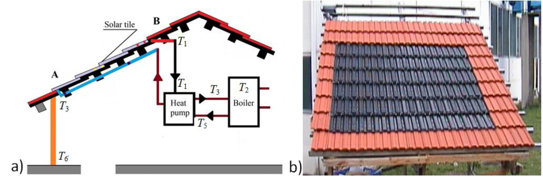
The plastic age – where and what is the future?

DOI: 10.31038/NAMS.2019241


We are currently living in the “Plastic age” as rightfully predicted in the classic movie “The graduate” in the 60’s where main character young Benjamin, just graduated and asking himself what to do next gets the famous advice: “I just say one word – Plastics. There is a great future in Plastics!” This was certainly correct; our daily life relies on synthetic polymers for all aspects of modern life. Going back in history, one of the first synthetic resins, a thermoset produced by a catalyzed reaction of phenol and formaldehyde, was introduced already 1909 under the name Bakelite. The creation of a synthetic plastic was revolutionary for its electrical non conductivity and chemical, solvent and heat-resistant properties in electrical insulators, radio and telephone casings and such diverse products as kitchenware, jewelry, pipe stems, children’s toys, and firearms. Bakelite was particularly suitable as a molding compound, an adhesive or binding agent, a varnish, and a protective coating and as such extremely useful for the emerging electrical and automobile industries. In the following years a number of new polymeric materials, thermoplastics and thermosets were developed, mainly formed by linking small molecules – monomers – together in a repetitive formation. Plastics are today available with extremely versatile properties ranging from, resistance to corrosion, low density, high strength, transparency, low toxicity, durability and a remarkable affordability and therefore used by almost every industry in the world, from food packaging to space exploration. Plastic is the ultimate commodity of convenience.

Especially hydrocarbon polymers like polyethylene and polypropylene, which account for more than half of the world plastics production and more than 90% of packaging materials are ubiquitous in single-use and short-term applications because their starting materials are abundant and inexpensive. In addition, we have learned to vary the chemical structure of the polymer chains (such as branching, molecular weight, and dispersity) through catalysis, and in such to alter their physical properties. The single-use nature of plastics is essential in sterile packaging for foods, strong-but in expensive materials for transportation and storage, and safe and disposable components in medical devices, leading to their manufacture in tremendous quantities. Three hundred and eighty million tons (380 Mt) of plastics are created worldwide each year, which corresponds to roughly 7% of crude oil and natural gas produced. Moreover, the plastic market is currently increasing, and some analysts predict quadrupled production by 2050 (~1100 to 1500 Mt per year).

However, the good properties of plastics, namely the mechanical performance connected with low density, are intrinsically connected with the existence of large molecules in the material. This long chain molecular nature turned out to be a major draw-back in the circularity and recycling. Mixtures of polymers are thermodynamically not stable; in fact only a few polymers are miscible at all and thus small impurities (not speaking of non-polymeric ones) deteriorate the functional properties significantly! Processing of plastic waste is limited by technical challenges, which include contamination from mixtures of polymers and additives as well as oxidative degradation during melt re-processing. Current recycling processes rely mainly on primary recycling (termed closed-loop recycling, reprocessing an uncontaminated, single plastic to give a product used for the same purpose as the original plastic) and secondary (mechanical) recycling –down cycling- results in lower-in-value materials with different uses compared to the original material. Both primary and secondary recycling involve sorting, grounding, washing, and extruding, which cause varying degrees of polymer degradation, resulting in a limited number of reprocessing cycles of polymers. This accounts even more for cross-linked polymers, often referred to as thermosets and another class of plastics comprising ca. 15−20% of polymers produced. The outstanding performance of conventional thermosets like the first material Bakelite originates from their covalently cross-linked networks but results directly in a limited recyclability. The available recycling techniques include mechanical, thermal, and chemical processing. These methods typically require a high energy input and do not take the recycling of the thermoset matrix itself into account but focus on retrieving the more valuable fibers, fillers, or substrates. Thermoset materials are in particular amongst the most difficult materials to recycle, and in most cases even considered impossible to recycle. Most attention is given to the recycling of fiber-reinforced composites, since the fibers are generally more valuable than the matrix material, especially when carbon fibers are used. The most favorable method of recycling, the direct re-use of components in similar or lower performance applications without any form of reprocessing is virtually impossible for the existing thermoset materials.

The downside of the plastic age is the massive quantity of waste, pollution and lost value associated with single-use plastics. Over 75% of materials produced each year, 300 Mt, are discarded after a single use. Currently, most of this waste is either lost to landfills and the environment, or inefficiently incinerated in power plants to produce electricity, generating greenhouse gases (e.g., CO2) and toxic by-products in the process. Inefficient recycling and extremely slow environmental degradation of plastics are causing increasing concern about their widespread use. After a single use, 90 % of these materials are currently treated as waste creating a global environmental crisis, despite of their inherent chemical and energy value. Plastics have got themselves a bad name, mainly for two reasons: most are made from petroleum and they persist in the environment for decades or centuries beyond their functional lifetimes and end up as litter in the environment. Durability, one of plastic’s greatest assets is now its curse. Its robustness means that plastics stay in our environment for hundreds of years turning them into a persistent part of the landscape, and more importantly of the seascape. Once discarded, bulk plastics are polluting the oceans. Converging sea currents are accumulating plastic waste in a floating island known as the Great Pacific Garbage Patch, which now covers an area larger than Greenland. The bigger bits of plastic are life-threatening to marine life and sea birds, they can strangle marine animals and birds or build up in their stomachs. More recently, the awareness of the presence and danger of micro plastics has raised concern about their presence in the food chain. If nothing changes, by 2050 there will be as much plastic in the sea as there is fish. Who wants to eat plastic then?

The switch from a linear economy with its throwaway culture to a circular economy with efficient reuse of waste plastics is therefore mandatory. An increased focus on bio-derived and degradable composites as well as recycling could lower the degree of pollution. Reduce, reuse and recycle have been embraced as the common approach to tackle the escalating plastic waste problem. The goal is to create a circular plastic economy where products are 100%recyclable, used for as long as possible, and their waste is minimized. Compared to plastics, massive recycling of much older man-made materials like iron and steel (> 70 %!), copper, glass, aluminum and paper is the current technology and already since a long time developed and optimized. For a circular economy, the vast majority of plastic materials should be recyclable and the materials entering the chain should be bio-based. Although biodegradable polymers and in particular PLA have been the focus of much research over the last decades, only ~1% of plastics are currently produced from renewable resources. Besides, polymers derived from bio-renewable resources, commonly referred to as renewable polymers, bio-based polymers, or even sustainable polymers in the literature, are not necessarily sustainable or degradable, where as degradable polymers are not necessarily recyclable. For example, 100% bio-based polythene (bio-PE) and bio-based polyethylene terephthalate (bio-PET) are not biodegradable. Dreams and reality of green polymer chemistry have no match, conflicts and competition with food production and unrealistic high CO2 production are consequences. Biopolymers are also not a realistic alternative to synthetic polymers (properties and processing). Recycling is costly, reliant on changes in human behavior and produces partially lower quality materials, in terms of both thermal and mechanical properties. Additionally, recycling does not change our plastic addiction; if we want to maintain our current lifestyles, modification to plastic manufacture needs to go hand in hand with effective recycling. So a change in mind-setting is mandatory!

A already developed way to reduce the demand for finite raw materials and to minimize the negative impact on the environment involves chemically recyclable polymers which are capable of being returned to the corresponding monomers in a depolymerization process ready for repolymerization to virgin-quality polymers. This seemingly ideal strategy has motivated the research on the exploration of chemically recyclable polymers and also the mild processes for the catalytic conversion of the recyclable polymers to monomers or new polymers, namely chemolysis (by depolymerizing or decomposing the polymer in the presence of a chemical catalyst, typically needing relatively low temperature or even ambient temperature). Consequently, to advance plastic recycling practices, improving chemical recycling selectivity and efficiency through monomer and polymer design and catalyst development is mandatory, minimizing the need for sorting and expanding recycling beyond polyesters, polyamide and polyurethanes [1]. Recent advances report on the catalytic selective hydrogenolysis of PE at moderate conditions (300 °C, 9 bar H2) on nanoparticles in a solvent free process offering an option to obtain high value re-usable materials out of waste-PE[2] showing as such an opportunity to deal with the already existing large quantities of plastic waste in an economic and ecological way. Key feature of circular economy is preventing waste by making products and materials more efficient and reusing them. The challenge in a circular economy is the development of environmentally friendly polymers with better properties and at the same time facilitating reuse of plastics. This can be e.g. realized by polymers custom designed for recycling such as e.g. all-polymer (one component) composites, self-reinforcing polymers (molecular composites) [3] or the use of reversible chemistry [4]. Starting with the already about 15 year development of instruction of self-repair functionalities [5] in plastics and as such enabling these materials to react early on a developing damage or defect and reducing over-design of systems and realizing weight and cost reduction in a new way the future for plastics and other materials is not only a design or use (self-repair) but a design for recycling. This starts with a design of products to encourage a re-use as well as in a material choice for less impacting and recyclable materials, for reduction of quantities and variations in one product and an optimalisation of process technology. In the design phase, material sorting processes, separation and dismantling considerations are as important as the introduction of extension of lifetime and preservation of functional property technologies like self-repair. Consequently, there is a need for a new generation of materials that can be reprocessed like thermoplastics that still retain the beneficial properties of high performance thermoplastic or thermoset materials4. Such a material can be realized by the incorporation of dynamic interactions and dynamic covalent bonds into linear polymers and networks. In such plastics, thermal depolymerization and solvent assisted depolymerization and especially dissociative depolymerization and adaptable cross-links can transfer the non-recyclable plastics to small thermoplastic piece and thus enabling complete recycling and re-use. Alternatively (and parallel)it is the intrinsic production of cost-, resource-, eco- and energy efficient high performance polyolefin’s using modern multisite polymerization catalysts as reported recently by Mühlhaupt et al. resulting in all polyolefin injection moldable composites with a unique combination of high toughness, stiffness and strength which show virtually no change in properties during 7 cycles of remolding one of the most promising trends3. While following these ideas, the future of Plastics and ourselves will be still great. The need is to shift now from the Design-for-Use –self repair – strategies to the Design for recycling–multiple use- strategies to guarantee a bright Future for Plastics.

References

  1. Tang X, Chen E XY (2018) Toward Infinitely Recyclable Plastics Derived From Renewable Cyclic Esters. Chem https://doi.org/10.1016/j.chempr.2018.10011
  2. GokhanCelik, Robert M Kennedy, Ryan A Hackler, MagaliFerrandon, AkalankaTennakoon,  et al. (2019) Upscaling Single-Use Polyethylene into High Quality Liquid Products. ACS Central Science DOI: 10.1021/asccentsci.9b00722.
  3. Hees T, Zhong F, StürzelM, Mühlhaupt R (2018) Tailoring Hydrocarbon Polymer and All Hydrocarbon Composites for a Circular Economy. Makrom. Rapid. Comm 1800608.
  4. Post W, Arijana Susa, Rolf Blaauw, Karin Molenveld, Rutger JIKnoop, et al. (2019) A Review on the Potential and Limitations of recyclable Thermosets for Structural Applications. Polym Rev https://doi.org/10.1080/15583724.2019.1673406.
  5. Fischer H (2010) “Self Healing Material Systems – A Dream or Reality”. Natural Science 2: 873–901.