Author Archives: author

Nucleolin: a prognostic marker in cancer?

DOI: 10.31038/CST.2019441

Abstract

Nucleolin, a major protein of the nucleolus which is required for cell proliferation is the subject of numerous studies in the field of oncology. Many studies suggest that the level of expression of nucleolin and its cellular localization could be an indication of the clinical outcome in several cancers. Quite logically for a protein required for the cancer cells to increased protein synthesis, a correlation between the high level of nucleolin expression and the poor prognosis of the patients is often reported. Unexpectedly, in several studies it was also found that a low nucleolin expression could be also associated with an unfavorable prognosis. In these commentary I discuss these studies and the possible implications for using nucleolin as a prognostic marker in oncology.

Keywords

Nucleolin, Cancer, Prognostic Marker

Nucleolin, an abundant multifunctional protein with multiple cellular localization

Nucleolin was first described as an abundant phopho-nucleolar protein [1]. The protein is heavily modified post-translationally [2] and can interact with numerous proteins [3] and nucleic acids [4, 5]. The nucleolar localization of nucleolin suggested that it could be involved in some aspects of ribosome biogenesis as the nucleolar structures are the sites of transcription of the pre-ribosomal RNA and of its assembly with ribosomal proteins to form the pre-ribosomes that are then exported in the cell cytoplasm for the mRNA translation. Indeed, it was shown that nucleolin expression was required for the formation of functional nucleolar structure [6, 7]. Nucleolin is required for RNA polymerase I transcription [8, 9] probably through its chaperone activity that facilitates transcription of chromatin [10–12]. It has been suggested that the transient interaction of nucleolin with the pre-rRNA during transcription might be involved in the correct folding of the long pre-RNA which is required for the processing in mature ribosomal RNA [13–17] but it remains to be demonstrated if the RNA binding specificity of nucleolin towards pre-rRNA is important as deletion of the RNA binding domains responsible for this specificity do not seems to be crucial for the vital function of nucleolin [7]. By analogy to this co-transcriptional role of nucleolin in pre-rRNA maturation and assembly with proteins, it has been recently suggested that acetylated nucleolin could also be involved in the assembly of pre-catalytic spliceosomes and in splicing reaction [18, 19]. Several reports suggest also that nucleolin could be involved in the regulation of transcription of some ANP polymerase II genes [20–24]. Whether there is a functional link between this RNAPII regulation and the implication in the co-transcriptional splicing of some pre-mRNA remains to be determined. In addition to this fundamental role in the regulation of gene expression, there has been a renewed interest in the study of this protein because it has been discovered that nucleolin is also present on the cell surface. Initially described as a HIV gp120 V3 loop binding protein which could be involved in the binding of HIV particles to CD4(+) cells [25, 26], it was then suggested that cell surface nucleolin could be a receptor for diverse ligands [27–29].

Cell surface nucleolin was then described as a marker of endothelial cells in angiogenic blood [30] and this specific localization was associated with cancer cells [31–36].Whether the localization of nucleolin on the cell surface is specific to the cancerous nature of the cell or is the result of a high proliferation rate of these cells remains to be determined. One major difficulty to study the function of cell surface nucleolin and its link with cancer cells is that the detection of cell surface nucleolin is difficult. Initial studies were performed by incubating cells with a monoclonal antibody (mAb D3) at room temperature before paraformaldehyde (PFA) fixation [37]. The beautiful images showing a regular localization of nucleolin on the surface of HeLa cells are very difficult to reproduce with other anti-nucleolin antibodies. Nevertheless, in other studies, immunoelectron microscopy [38] or cell fractionation [37, 39] confirmed the presence of nucleolin on the cell surface.

Is Nucleolin expression a marker for cancer cells ?

Since several decades, it is known that cells that have a high proliferation rate express more nucleolar proteins (including nucleolin) as reflected by the silver staining method (AgNOR) [40]. It is therefore not surprising that, in general, cancer cells are also characterized by a higher level of detection of AgNOR proteins [41, 42]. This can be easily explained by the need of the cancer cells to produce more proteins to support the higher proliferation of these cells, and therefore by the need to make more ribosomes to produce these proteins.

Indeed, in many studies, it is described that nucleolin is over-expressed in tumors and cancer cells as detected by RT-QPCR, western blot or immunocytochemistry. For instance, an overexpression of nucleolin was detected in glioblastoma cells [43], hepatocellular carcinoma [44], pancreatic cancer [45], non-small cell lung cancer [46], acute myeloid leukemia [47], breast cancer [48].  However, it is very rare if in these studies the different pools of nucleolin (nucleoplasmic, nucleolar, cytoplasmic and at the cell surface) were discriminated. As the nucleolar pool of nucleolin represents the vast majority of total nucleolin protein, it is very likely that the increased expression of nucleolin results in an increased of this nucleolar pool (which participates to the increased in RNA polymerase I transcription of pre-ribosomal RNA) and also to the other cellular pools. Stating that nucleolin is overexpressed in cancer cells is however an unfortunate simplification found in many research papers and reviews. The real situation is indeed more complicated and finer studies on the level of expression of nucleolin should be done especially using large series of biological samples.

In many studies, the level of expression of nucleolin at the mRNA or protein level is examined in a limited number of biological samples or could even limited to established cell lines representing models for a specific cancer. In these cases, the results should be taken with care, as the expression of nucleolin could greatly fluctuate and be very sensitive to the growth conditions of culture cells.

When large number of samples are used, a wide range of nucleolin fold change is usually observed.  Then, it can become a challenge to determine which expression level represent a “low” versus a “high” expression. The discrimination between “low” and “high” can be done only if enough samples are analyzed and if a good control group is also examined at the same time. Then a “low” nucleolin expression may represent the normal expression found in control samples, or an expression that is lower than in the control group.

In human non-small cell lung cancer (NSCLC), immunohistochemistry was used to measure nucleolin expression in tissues from 225 NSCLC patients [46]. Higher expression of nucleolin was observed in 62,8% of the patients and the authors tried to discriminate the level of nuclear versus cytoplasmic nucleolin expression. If about equal number out of the 225 samples show either a low or high expression of cytoplasmic nucleolin, about 68% of the tumors had a low expression of nuclear nucleolin (and therefore 32% had a high expression of the nuclear pool of nucleolin). In that example, higher nucleolin nuclear level was associated with better survival while high level of cytoplasmic nucleolin was associated with poorer prognosis [46].  The same conclusions was drawn in a study on gastric cancer [49]. Among 124 gastrectomy samples, 68,5 % showed a high nucleolin expression. It was found that a high nucleolar nucleolin expression was associated with a better prognosis while tumors from patients showing a high cytoplasmic detection had the worse prognosis. In most of these studies the elevated total nucleolin expression is associated with a poor prognosis of patients like in gastric cancer, hepatocellular carcinoma, аcute myeloid leukemia, non-small cell lung cancer, pancreatic ductal adenocarcinoma [44, 47, 49–51]. Interestingly, in AML, the higher nucleolin mRNA expression level, compared to normal blasts, was associated with a poor survival only in elderly patients, and was found to be an independent marker [47].

The prognostic value of nucleolin expression was analyzed in acute myeloid leukemia (AML). In this study, the 75th percentile was used to form the two group of low and high expression [47] using different series of biological samples or TGCA data sets representing 270 samples in total. In this type of cancer, the nucleolin expression was found to cover a very large dispersion. Using this patient stratification, it was found that high nucleolin expression was associated with a poor survival in elderly patients. In a study on the correlation between nucleolin expression and patient outcome with hepatocellular carcinoma, it was found in a series of 130 patients that about 60% of tumors expressed “high” nucleolin expression and 40% a “low” expression [44] with very little explanation on how the cut off was done. The “high” nucleolin expression was then associated with poor prognosis of the patient.

However, it seems that there is not always a strict correlation between the level of nucleolin expression and the aggressiveness of the tumor and the outcome of the prognosis.

In another study in breast cancer, a quartile distribution analysis was first performed and highlighted that two quartiles can be grouped. Therefore, the expression levels could be stratified in three group representing a “low”, “medium” and “high” nucleolin expression [48]. Unexpectedly, using this stratification it was found that “low” and “High” expression levels of nucleolin were both markers of poor survival in triple negative breast cancer (TNBC). Interestingly, transcriptomic analysis of these two groups of samples in the TCGA data base revealed different gene expression profiles suggesting that the tumors in these two groups are different allowing a better stratification of TNBC patients. In agreement with these data, in another independent study, it was found by immunohistochemistry on a panel of 70 TNBC that nucleolin was undetectable in about 20% of these tumor which could correspond to the “low” expression while the expression was “high” in 30% of the tumors [52]. In this study, the authors did not determine if there was a correlation between this patient stratification based on nucleolin expression and the prognostic outcome.

In a study using 69 biological samples from patients with stage II pancreatic ductal adenocarcinoma (PDAC), the authors determined nucleolin expression using immunohistochemistry and scored the nucleolin expression levels using the median nucleolin labeling index as cut off [50]. They found that low level of nucleolin expression was a marker of poor prognosis (19.5 ±3.3 months versus 65.2 ±16.3 months for patients having tumor with a higher nucleolin expression).

Conclusion

In most of the studies, the expression of nucleolin is not homogeneous in the different tumors samples. If overall, the expression of nucleolin seems higher in the majority of the tumors it remains that a substantial number of tumors show a low or intermediate expression of nucleolin. In several cases, the tumors showing a low expression have been also associated with a poor prognosis. In addition, the localization of nucleolin expression within the cells (nuclear versus cytoplasmic) seems to be also important to predict the outcome of the patient. It is therefore important to be able to discriminate the level of nucleolin expression compared to normal tissue (which cutt off is used to make the distinction between high and low expression) and to have enough biological samples to be able to draw any conclusion on the prognostic value of nucleolin expression. As recently shown with breast TNBC series, the stratification of patients according to nucleolin expression level could be an additional criteria to choose  a therapeutic strategy and to develop clinical trials with available molecules that target nucleolin.

Competing interests

The authors declare that he has no competing interests.

Funding information

The research in author team is funded by the Ligue contre le Cancer (Allier and Saone et Loire, France), the Foundation pour la recherche sur le Cancer (ARC) the CNRS, the Ecole Normale Supérieure de Lyon, the Agence Nationale de la Recherche, project ANR Theranuc, ANR-16-CE17-0023. These funding agencies have no roles in the design of the studies, analysis and interpretation of the data, in the writing of the report; and in the decision to submit the paper for publication.

References

  1. Orrick L.R, Olson M.O, Busch H. (1973) Comparison of nucleolar proteins of normal rat liver and Novikoff hepatoma ascites cells by two-dimensional polyacrylamide gel electrophoresis. Proc Natl Acad Sci 1316–1320.
  2. Ugrinova I, Petrova M, Chalabi-Dchar M, Bouvet P. (2018) Multifaceted Nucleolin Protein and Its Molecular Partners in Oncogenesis, Adv Protein Chem Struct Biol 133–164.
  3. Salvetti A., Coute Y., Epstein A., Arata L., Kraut A. (2016)  Nuclear functions of nucleolin through global proteomics and interactomic approaches, J Proteome Res.
  4. Abdelmohsen K., Tominaga K., Lee E.K., Srikantan S., Kang M.J. (2011)  Enhanced translation by Nucleolin via G-rich elements in coding and non-coding regions of target mRNAs, Nucleic Acids Res 8513–8530.
  5. Ginisty H., Sicard H., Roger B., Bouvet P. (1999) Structure and functions of nucleolin, J Cell Sci 761–772.
  6. Ugrinova I., Monier K., Ivaldi C., Thiry M., Storck S. (2007) Inactivation of nucleolin leads to nucleolar disruption, cell cycle arrest and defects in centrosome duplication, BMC Mol Biol  66.
  7. Storck S., Thiry M., Bouvet P. (2009) Conditional knockout of nucleolin in DT40 cells reveals the functional redundancy of its RNA-binding domains, Biol Cell 153–167.
  8. Cong R., Das S., Ugrinova I., Kumar S., Mongelard F. (2012) Interaction of nucleolin with ribosomal RNA genes and its role in RNA polymerase I transcription, Nucleic Acids Res 9441–9454.
  9. Durut N., Saez-Vasquez J., Nucleolin: dual roles in rDNA chromatin transcription, Gene 556 (2014) 7–12.
  10. Angelov D., Bondarenko V.A., Almagro S., Menoni H., Mongelard F. (2006) Nucleolin is a histone chaperone with FACT-like activity and assists remodeling of nucleosomes, EMBO J 25 1669–1679.
  11. Pontvianne F., Abou-Ellail M., Douet J., Comella P., Matia I. (2010)  Nucleolin is required for DNA methylation state and the expression of rRNA gene variants in Arabidopsis thaliana, PLoS Genet 6 e1001225.
  12. Rickards B., Flint S.J., Cole M.D., LeRoy G. (2007) Nucleolin is required for RNA polymerase I transcription in vivo, Mol Cell Biol 937–948.
  13. Allain F.H., Bouvet P., Dieckmann T., Feigon J. (2000) Molecular basis of sequence-specific recognition of pre-ribosomal RNA by nucleolin, EMBO 6870–6881.
  14. Bouvet P., Allain F.H., Finger L.D., Dieckmann T., Feigon J. (2001) Recognition of pre-formed and flexible elements of an RNA stem-loop by nucleolin, J Mol Biol 763–775.
  15. Bouvet P., Jain C., Belasco J.G., Amalric F., Erard M. (1997) RNA recognition by the joint action of two nucleolin RNA-binding domains: genetic analysis and structural modeling, EMBO J 16 5235–5246.
  16. Ginisty H., Amalric F., Bouvet P. (1998) Nucleolin functions in the first step of ribosomal RNA processing, EMBO J 17 1476–1486.
  17. Serin G., Joseph G., Faucher C., Ghisolfi L., Bouche G. et al. (1996) Localization of nucleolin binding sites on human and mouse pre-ribosomal RNA, Biochimie 530–538.
  18. Das S., Cong R., Shandilya J., Senapati P., Moindrot B. (2013) Characterization of nucleolin K88 acetylation defines a new pool of nucleolin colocalizing with pre-mRNA splicing factors, FEBS Lett 417–424.
  19. Ugrinova I., Chalabi-Dchar M., Monier K., Bouvet P. (2019) Nucleolin Interacts and Co-Localizes with Components of Pre-Catalytic Spliceosome Complexes, Sci 1 33.
  20. Allinne J., Pichugin A., Iarovaia O., Klibi M., Barat A. et al. (2014) Perinucleolar relocalization and nucleolin as crucial events in the transcriptional activation of key genes in mantle cell lymphoma, Blood 123 2044–2053.
  21. Cong C., Das S., Bouvet P. (2011) The Multiple Properties and Functions of Nucleolin, Protein Reviews series,  Nucleolus,  Springer Editor-in-Chief is M. Zouhair Atassi 185.
  22. Grinstein E., Du Y., Santourlidis S., Christ J., Uhrberg M. (2007) Nucleolin regulates gene expression in CD34-positive hematopoietic cells, J Biol Chem 12439–12449.
  23. Hanakahi L.A., Dempsey L.A., Li M.J., Maizels N. (1997) Nucleolin is one component of the B cell-specific transcription factor and switch region binding protein, LR1, Proc Natl Acad Sci 94 (1997) 3605–3610.
  24. Kumar S., Gomez E.C., Chalabi-Dchar M., Rong C., Das S. (2017) Integrated analysis of mRNA and miRNA expression in HeLa cells expressing low levels of Nucleolin, Sci Rep 7 9017.
  25. Callebaut C., Blanco J., Benkirane N., Krust B., Jacotot E. (1998) Identification of V3 loop-binding proteins as potential receptors implicated in the binding of HIV particles to CD4(+) cells, J Biol Chem 21988–21997.
  26. Nisole S., Krust B., Callebaut C., Guichard G., Muller S. et al. (1999) Hovanessian A.G., The anti-HIV pseudopeptide HB-19 forms a complex with the cell-surface-expressed nucleolin independent of heparan sulfate proteoglycans, J Biol Chem 27875–27884.
  27. Harms G., Kraft R., Grelle G., Volz B., Dernedde J. (2001) Identification of nucleolin as a new L-selectin ligand, Biochem J 360 531–538.
  28. Said E.A., Krust B., Nisole S., Svab J., Briand J.P. (2002) The anti-HIV cytokine midkine binds the cell surface-expressed nucleolin as a low affinity receptor, J Biol Chem 277 37492–37502.
  29. Sinclair J.F., O’Brien A.D., Cell surface-localized nucleolin is a eukaryotic receptor for the adhesin intimin-gamma of enterohemorrhagic Escherichia coli O157:H7, J Biol Chem 277 (2002) 2876–2885.
  30. Christian S., Pilch J., Akerman M.E., Porkka K., Laakkonen P. (2003) Nucleolin expressed at the cell surface is a marker of endothelial cells in angiogenic blood vessels, J Cell Biol 163 871–878.
  31. Fujiki H., Watanabe T., Suganuma M. (2014) Cell-surface nucleolin acts as a central mediator for carcinogenic, anti-carcinogenic, and disease-related ligands, J Cancer Res Clin Oncol 689–699.
  32. Galzio R., Rosati F., Benedetti E., Cristiano L., Aldi S. et al. (2012) Glycosilated nucleolin as marker for human gliomas, J Cell Biochem 113  571–579.
  33. Joo E.J., ten Dam G.B., van Kuppevelt T.H., Toida T., Linhardt R.J. et al. (2005) Nucleolin: acharan sulfate-binding protein on the surface of cancer cells, Glycobiology 1–9.
  34. Reyes-Reyes E.M., Akiyama S.K. (2008) Cell-surface nucleolin is a signal transducing P-selectin binding protein for human colon carcinoma cells, Exp Cell Res 314 2212–2223.
  35. Watanabe T., Hirano K., Takahashi A., Yamaguchi K., Beppu M. et al. (2010) Nucleolin on the cell surface as a new molecular target for gastric cancer treatment, Biol Pharm Bull 796–803.
  36. Watanabe T., Tsuge H., Imagawa T., Kise D., Hirano K. (2010) Nucleolin as cell surface receptor for tumor necrosis factor-alpha inducing protein: a carcinogenic factor of Helicobacter pylori, J Cancer Res Clin Oncol 136 911–921.
  37. Hovanessian A.G., Puvion-Dutilleul F., Nisole S., Svab J., Perret E. et al. (2000) The cell-surface-expressed nucleolin is associated with the actin cytoskeleton, Exp Cell Res 261 312–328.
  38. Dumler I., Stepanova V., Jerke U., Mayboroda O.A., Vogel F. et al. (1999) Urokinase-induced mitogenesis is mediated by casein kinase 2 and nucleolin, Curr Biol 9 1468–1476.
  39. Losfeld M.E., Khoury D.E., Mariot P., Carpentier M., Krust B. et al. (2009) The cell surface expressed nucleolin is a glycoprotein that triggers calcium entry into mammalian cells, Exp Cell Res 315 357–369.
  40. Derenzini M., Sirri V., Pession A., Trere D., Roussel P. et al. (1995) Hernandez-Verdun D., Quantitative changes of the two major AgNOR proteins, nucleolin and protein B23, related to stimulation of rDNA transcription, Exp Cell Res 219 276–282.
  41. King J., Thatcher N., Pickering C., Hasleton P. (2006) Sensitivity and specificity of immunohistochemical antibodies used to distinguish between benign and malignant pleural disease: a systematic review of published reports, Histopathology 49 561–568.
  42. Pich A., Chiusa L., Margaria E.  (2000) Prognostic relevance of AgNORs in tumor pathology, Micron 31  133–141.
  43. Balca-Silva J., do Carmo A., Tao H., Rebelo O., Barbosa M. (2018) Nucleolin is expressed in patient-derived samples and glioblastoma cells, enabling improved intracellular drug delivery and cytotoxicity, Exp Cell Res 370 68–77.
  44. Guo X., Xiong L., Yu L., Li R., Wang Z. (2014) Increased level of nucleolin confers to aggressive tumor progression and poor prognosis in patients with hepatocellular carcinoma after hepatectomy, Diagn Pathol 175.
  45. Gilles M.E., Maione F., Cossutta M., Carpentier G., Caruana L., et al. (2016) Nucleolin Targeting Impairs the Progression of Pancreatic Cancer and Promotes the Normalization of Tumor Vasculature, Cancer Res 76 7181–7193.
  46. Xu J.Y., Lu S., Xu X.Y., Hu S.L., Li B., et al. (2016)  Prognostic significance of nuclear or cytoplasmic nucleolin expression in human non-small cell lung cancer and its relationship with DNA-PKcs, Tumour Biol 37 10349–10356.
  47. Marcel V., Catez F., Berger C.M., Perrial E., Plesa A. et al. (2017) Expression Profiling of Ribosome Biogenesis Factors Reveals Nucleolin as a Novel Potential Marker to Predict Outcome in AML Patients, PLoS One 12 170160.
  48. Nguyen Van Long F., Lardy-Cleaud A., Bray S., Chabaud S., Dubois T. et al. (2018) Druggable Nucleolin Identifies Breast Tumours Associated with Poor Prognosis That Exhibit Different Biological Processes, Cancers (Basel) 10.
  49. Qiu W., Zhou F., Zhang Q., Sun X., Shi X. et al. (2013)  Over expression of nucleolin and different expression sites both related to the prognosis of gastric cancer, APMIS 121 919–925.
  50. Peng L., Liang J., Wang H., Song X., Rashid A. et al. (2010) High levels of nucleolar expression of nucleolin are associated with better prognosis in patients with stage II pancreatic ductal adenocarcinoma, Clin Cancer Res 16 3734–3742.
  51. Zhao H., Huang Y., Xue C., Chen Y., Hou X. et al. (2013) Prognostic significance of the combined score of endothelial expression of nucleolin and CD31 in surgically resected non-small cell lung cancer, PLoS One 8 e54674.
  52. Pichiorri F., Palmieri D., De Luca L., Consiglio J., You J. et al. (2013) In vivo NCL targeting affects breast cancer aggressiveness through miRNA regulation, J Exp Med 210 951–968.

Open Space Knowledge and Data and Their Influence on Natural Products Based Self-Medication Trends

DOI: 10.31038/CST.2019435

Introduction

Contemporary academic (conventional) medicine, based on current advancements in life sciences and applying evidence based efficacy and safety principles, has evolved from ancient cultures of well-being and healing practices, which originated in prehistoric times, and it co-exists with their ethno-medicine remnants conveniently described as complementary and alternative medicine (CAM) [1, 2]. Global healthcare crisis, which has led to acceptance and even encouragement of CAM practices as an aid to conventional medicine, is a phenomenon of great complexity, comprising processes of various speed and dynamics. In this mini-review we’ll limit discussion to influence of IT and novel scientific knowledge dissemination systems, such as Open Science, to self-medication applied with legally available biologically active substances, but not limited to the OTC drugs.

Discussion

The 21st century societies differ dramatically from an earlier populations in respect to availability of sound scientific knowledge and specific technical information in every field imaginable, due to widespread IT access (www) to primary source scientific publications and reasonable coverage of scientific achievements through the media. Considerable rise in general education level and computer literacy allowed for massive, profiled individual internet searches, and using their results for informed choice of medicinal and/or CAM treatments. Consequently, a new type of medical patients and pharmacy customers have been created, with considerable potential of modifying future trends on medicinal product markets. Inefficiency of state healthcare systems became notorious not only in the “poor South” regions, but also in the most affluent societies of the industrialized West. In the first case, even relatively small number of WHO selected essential drugs are scarcely available; in the second – thousands of innovative, proprietary active pharmaceutical ingredients (API of the ethical or brand drugs), supplemented by experimental therapies with new molecules in advanced clinical trials, are extremely expensive, with their costs only fractionally covered by insurance. Moreover, efficacy of modern medicines in general is far from satisfactory. This is well exemplified by mortality and morbidity statistics from such principal non-transmittable disease killers as cardiovascular disorders, tumors, and metabolic syndrome, including diabetes [3]. It should be reminded that API substances, estimated as a collection of a few thousand molecules of low molecular weight (biological drugs like vaccines or antibody conjugates form much smaller assembly), consist of a very small fraction of known molecules (ca. 108 chemical entities recorded by CAS database), while natural product collection (e.g.: alkaloids, antibiotics, flavonoids, glycosides, isoprenoids, phenolics, vitamins, etc.) is estimated at 200 thousand, with a fair chance of further growth with advancement on microbial, plants and aquatic organisms metabolome studies. It is therefore not surprising that natural products and especially secondary metabolites (SM), which were developed and genetically encoded as evolutionary traits which provide hosts with some marginal environmental advantages, formed at first a solid base for ethnopharmacology and specifically herbal medicine, and in more recent times afforded countless leads for modern drugs [4,5]. Particularly, among drugs applied in oncology, SM-derived substances account for approximately 60 % of APIs (well-known examples include: taxanes, dimeric indole alkaloids from Catharantus (former Vinca), anthracyclines, camptothecins, podophylotoxins, etc.). These compounds feature selective toxicities, but in general they are biocompatible and follow predictable metabolic pathways, in contrast to many synthetic compounds obtained by combinatorial synthesis for high-throughput screening. Pharmacognosy, a science of SM medical application, which started at the beginning of 19th century with discovery of alkaloids, was for a long time very close to herbal medicine, which concerned itself mainly with whole plants extracts and concoctions, while individual chemical entities pharmacology development was considerably limited by progress in chemical separation techniques and advancement in analytical chemistry. Currently, legal access to biologically active compounds for nonprofessionals is widely differentiated: from ban on tightly controlled markets of the prescription drugs, through regulations concerning registered over the counter medicines (OTC), to herbal medicines and materials, including high chemical purity isolates of SM, for which various local regulations may apply. The last category is particularly interesting from the point of view of CAM and self-medication, in view of general availability. The herbal medicine markets are already well developed and characterized by dynamic growth. In the US alone, the value of herbal medicines market topped 8 billion $ in 2017, with over 12% growth from year 2016, while the global market value is currently estimated at ca. 100 bln $ [6]. Herbal materials can be segmented into: herbal pharmaceuticals (phytopharmaceuticals; the dominant category), herbal functional foods, herbal dietary supplements and herbal cosmetic products. From the formulation point of view, extracts accounts for the largest markets, and are likely to retain the leading position for a while. In terms of the botanical classification, products derived from the following plants are considered the most significant: Marrubium vulgare, Cinnamomum spp., Vaccinium macrocarpon, Echinacea, Camellia sinensis, Curcuma longa, Actaea racemosa, Aloe vera, Zingiber officinale, Cocos nucifera, Silybum marianum, Gingko biloba, Malpighia punicifolia. Hospital and retail pharmacies are the main distribution channels thus far (over 55% market share) but the internet (e-commerce) sector already shows great potential.

The leader of the herbal products market value classification is, by a large margin – curcumin (turmeric), which is unique in many respects and therefore deserves few comments. The main phenolic constituent of Curcuma longa rhizome (traditional Indian spice and medicine deeply rooted in Ayurveda system and used throughout Asia under various names) was identified as diferuloylmethane at the beginning of 20th century, and its synthesis was completed thereafter to study a new type of plant pigments capable of dyeing cellulose fibers directly [7]. Turmeric powder, a spice and pigment known as a constituent of food additive popularized in the West as curry, which contain some closely related curcuminoids, is produced in India in multi-ton quantities and distributed by major producers of herbal medicines (encoded E 100 as a food additive and granted the GRAS status by Joint FAO/WHO Expert Committee on Food Additives). High chemical purity curcumin, which became a very popular biochemical and pharmacological molecular probe after initial pharmacological screening, is also freely available. First recognized as an efficient antioxidant, inhibiting COX, LOX and cytochrome P450 promoted metabolism of arachidonic acid, soon attracted attention as an agent which performed extremely well in antiproliferative and antitumor tests [8, 9].

An extensive discussion of curcumin molecular targets, which include transcription factors, growth factors, protein kinases, apoptosis-related proteins, etc., based on the compound structure and chemical reactivity was expertly summarized by concluding that nearly all human cell lines can be inhibited by the compound in vitro; however a big gap exists between its unquestionable pharmacodynamics potential and its feeble pharmacokinetic efficiency, which results from poor solubility, limited metabolic stability and rapid biotransformation in biological media [10]. Such conclusions are in keeping with results of numerous clinical trials, recorded by NIH and reported on PubMed. Meanwhile, search for “curcumin” on Google Scholar (July, 2019) returns ca. 375 000 hits in a small fraction of a second, reflecting a hype of great expectations generated by an avalanche of investigations which suggest medicinal applications of the compound, in particular for prevention and treatment of cancer related ailments. This, obviously is not without influence on a hypothetical well informed customer, interested in a self-care continuum activities, which consists not only of self-managed ailments in minor cases, but includes lifestyle choices (e.g. diet and its supplementation) aimed at long term disease prevention, or intervention in an acute condition. Curcumin seems to provide a good example to illustrate thesis, that an interest in natural products (SM), supplied in form of high purity chemical entities, may soon transform herbal material markets beyond recognition, since all technical means are in place to satisfy such demands. In particular, for many SM with less complicated structures, synthetic chemistry alternatives to isolation exists; next, biotechnology is in line, with growing assembly of gene sequences and clusters which can be expressed in microorganisms already validated for industrial use. In case of a therapeutic indication applications, various sources of the active ingredient may evoke some formal controversies, calling for high-tech arguments based on sophisticated analytical technologies [11] but for “established use” natural products marketing authorizations are usually more freely available. “Adulteration” of natural products with the same substances of synthetic origin are likely to be treated case by case since precedences exist: e. g. some of the curcumin clinical trials were carried out with use of synthetic origin substance.

While the curcumin story stands out among other herbal medicines likely to be applied in CAM procedures, in terms of great abundance of pharmacological results already amassed, it certainly is not the only one plagued with problems of low bioavailability which results in lack of in vivo efficacy. To this point, some facts from the rich history of ascorbic acid (Vit. C) medical applications should be reminded. Recognition of antiscorbutic action of Vit. C isolated from citrus fruits, was followed by Albert Szent-Győrgyi observation that it worked much better in the presence of green pepper flavonoids, temporarily advanced to a vitamin status [12]. The value of ascorbic acid for fighting cancers is disputable and necessity for exorbitantly large doses delivered by intravenous injections is discouraging [13], but the idea of “enhancers”, which are not active per se but significantly improve bioavailability of other API substances is alive and well. For curcumin, alkaloid piperine is indicated as the effective enhancer, and co-formulations of these compounds are believed to secure progress towards improved BA. Flavonoids, which are a large family of SM, closely related by phenylpropanoid biogenetic pathways, have not recovered vitamin P status, but many compounds from that category feature extremely interesting biological activities with prospective medicinal applications: in practically all structural sub-categories – chalcones, flavanones, flavonols, flavones, isoflavones, catechins and anthocyanes there are numerous examples of such health promoting activities, which are attractive from the point of view of CAM practices and metabolic syndrome prevention [14].

Genistein, the isoflavone with the most publicized record of research in oncological pharmacology [15, 16] should in principle be available in multi-ton quantities, since one of the main agricultural crops in global scale – the soybeans (over 300 mln tons harvested annually) contain on average ca. 0.1% of the compound, but thus far has not made it to natural origin chemicals distribution channels at affordable prices. Despite of its poor oral bioavailability, interest in genistein chemopreventive potential is high, GRAS status secures entry into modified food segment, and the compound is relatively easily available from chemical synthesis. Like in the case of “Indian gold” – curcumin, there is no shortage of sound, scientific information on flavonoids pharmacological activity, propagated through Open Access channels. In summary, a number of individual chemical entities of natural origin ready to join traditional pool of herbal medicines is large, which offers an outstanding opportunity for healthcare related innovations, focused on opening markets for new type of customers seeking specified, high quality materials from SM category, for self-managed healthcare.

Acknowledgment

The author is involved in the ORBIS project that received funding from the European Union’s Horizon 2020 research and innovation programme under the Marie Skłodowska-Curie grant agreement No 778051 and the Ministry of Science and Higher Education of Poland fund for supporting internationally co-financed projects in 2018–2022 (agreement No 3898/H2020/2018/2). The views expressed in this article are those of the author and the Research Executive Agency is not responsible for any use that may be made of the information it contains.

References

  1. Rivera JO, Loya AM, Ceballos R. (2013) Use of Herbal Medicines and Implications for Conventional Drug Therapy Medical Sciences. Altern Integ Med 2: 130.
  2. Fundamentals of Complementary, Alternative, and Integrative Medicine (Micozzi M.S., Ed.) Elsevier, (2019) Saint Louis, MO, USA ; Sixth edition.
  3. WHO | Download the raw data files of the WHO Mortality Database; WHO (2014) from:  http://www.who.int/healthinfo/statistics/mortality_rawdata/en/
  4. Newman DJ, Cragg GM. (2007) Natural products as sources of new drugs over the last 25 years. J Nat Prod 70: 461–477.
  5. Newman DJ, Cragg GM. (2016) Natural products as sources of new drugs from 1981 to 2014. J Nat Prod 79: 629–661.
  6. https://www.marketresearchfuture.com/reports/herbal-medicine-market-3250 (accessed July 1st, 2019).
  7. Grynkiewicz G, Slifirski P. (2012) Curcumin and curcuminoids in quest for medicinal status. Acta Biochim Pol 59: 201–212.
  8. Basnet P, Skalko-Basnet N. (2011) Curcumin: an anti-inflammatory molecule from a curry spice on the path to cancer treatment. Molecules 16: 4567–4598.
  9. Priyadarsini KI. (2014) The chemistry of curcumin: from extraction to therapeutic agent. Molecules 19: 20091–20112.
  10. Heger M, van Golen RF, Broekgaarden M, Michel MC (2014) The Molecular Basis for the Pharmacokinetics and Pharmacodynamics of Curcumin and Its Metabolites in Relation to Cancer. Pharmacol Rev 66: 222–307.
  11. Wang X-M, Zhang Q-Z, Yang J , Zhu R-H , Zhang J , et al. (2012) Validated HPLC–MS/MS Method for Simultaneous Determination of Curcumin and Piperine in Human Plasma. Tropical Journal of Pharmaceutical Research  11: 621–629.
  12. Vitamin C in Health and Disease (Packer L., Fuchs J., Eds.) Marcel Dekker, New York, USA 1997.
  13. Verrax J, Calderon PB. (2008) The controversial place of vitamin C in cancer treatment. Biochem Pharmacol 76: 1644–1652.
  14. Panche AN, Diwan AD, Chandra SR. (2016) Flavonoids: an overview. J Nutr Sci 5: 1–15.
  15. Spagnuolo C, Russo GL, Orhan IE, Habtemariam S, Daglia M, Sureda A, Nabavi SF, Devi KP, Loizzo MR, Tundis R, Nabav SM (2015) Genistein and Cancer: Current Status, Challenges, and Future Directions. Adv Nutr 6: 408–419.
  16. Grynkiewicz G. (2017) Plant isoflavones, their impact on life sciences, medicine and industry. Chapt. 6 in: Frontiers in Natural Products Chemistry, vol. 3: 221–292.

The Construction of Power in Nepal: Menstrual Restriction and Rape

DOI: 10.31038/AWHC.2019241

Abstract

The widespread use of gender-based violence, especially against women, has been identified not only as a legal, but also as a major public health problem. Especially in Asia, traditional practices reflecting local health belief models lead to a number of major has related partitions, which include gender-based violence. Not only in India, but also in Nepal such traditional practices are wide spread. In Nepal, the most common problem is the “Chaupadhi” practice of menstrual segregation that leads to the death of women every year.

Methodology: in our qualitative study we used different sources including political party programs and focus interviews in the Bharatpur region of Nepal to explore the cultural and health belief models that could contribute to lead different population groups to use menstrual segregation and other forms of gender-based violence such as rape. Results we identified a number of categories [health belief systems] that we are part of gender-based identities and this forms of gender-based violence as reflected in interviews and the public discourse. Conclusions: willingness to commit or permit violence is reflected in health belief systems and identity building, and a number of recommendations will be presented that we are drawn up as a reassignment of our project to reduce gender-based violence in Nepali communities.

Keywords

Women, Public Health, Menstruation, Rape, Gender Based Violence, Nepal

Background

One in three women worldwide experience some form of violence in their lifetime. Women are two-thirds of the global illiterate population, work one to three hours and two to ten times more respectively per day compared to men the “second shift” (household chores and caring for children, elderly and sick) and earn 10 to 30 percent less than men in the employment sector (ICRW, 2018) [1]. These exemplify the effects of pervasive patriarchal norms, and inequitable society which constraints women to fill specific gender roles which are attributed with low value, little power and put them at a higher risk for violence, poor health and disease, poverty, and other inequitable adversities (USAID, 2015) [2] One of the worst examples of gender inequity and inequality with long term mental health impact is exemplified through gender-based violence perpetrated by men and boys to women and girls (ICRW, 2018) [3]

GBV is a type of violence that is based on, and propelled by the defining genders and their perceived normative roles within society. It disproportionately impacts women both in prevalence and its consequences due to women’s inferior position in society [4]. GBV has mental, physical, psychological, social, and sexual consequences which are exacerbated due to their gender. Within the various types of GBV, we can distinguish between highly visible and “invisible” GBV; with visible including sexual harassment, domestic violence, physical violence, and rape and invisible Menstrual Restriction (MR).

The relationship between the visible violence, like rape and the invisible menstrual restriction is a reciprocal relationship where MR aids in the construction of behavior like rape. In Nepal, the majority of girls and women (89%) follow MR practices and close to half of all women (48%) reported experiencing violence in their lifetime, while majority of young women (74%) report experiencing sexual violence (UNFPA Nepal, 2014). MR (in Nepali in the most dramatic form as “Chaupadhi” [5]) includes dangerous practices that leads to the death of women every year due to infections or snakebite during segregation in inadequate housing [6] and is usually enforced by the family and community [7]. Cardoso reported high exposure rates in a recent study [8], and reported that “nearly three out of four women (72.3%) reported experiencing high menstrual restriction, or two or more types of menstrual restriction”. Rhanabat reported that is his household sample 21% of households used Chaupadi. And that the “conditions of livelihood, water facility, and access during menstruation and the Chaupadi stay was associated (P < .001) with the reproductive health problems of women” [5].

Objective: The general objective was to examine the construction of men’s attitudes, perceptions, and behavior in relation with Menstrual Restriction and Rape to ensure Gender Equality and Human Rights. The specific objectives were:

  • To understand the construction of attitudes, perception, and behavior of men
  • To explore the relationship of men’s attitudes, perception, and behavior regarding gender-based violence: Menstrual Restriction (MR) and Rape
  • To identify “Health belief models” about menstruation in men that might justify MR.

Methodology: The qualitative approach was used with primary and secondary sources were used. Primary sources included in-depth interviews, focus groups, observational research (participant observation) and interviews performed with a sample of key position holders of key political leaders of the Nepal Communist Party (CPN), Nepali Congress (NC) and Rastirya Prajatantra Party (RPP). As secondary sources we analysed the manifestos of respective political parties used for the last election 2017 and 2018. Research was conducted in July to October, 2018 in Bharatpur Metro Municipality, Bharatpur, Chitwan, Nepal.

We followed the assumption that the observational and field research would be contradicted or confirmed by secondary sources, and combination of both strategies would strengthen the validity and representative nature of observational and focus group data. We choose manifestos of respective political parties based on the assumption that the language, and implicit and explicit concepts expressed in those public populist documents would best reflect a key part of the guiding principles, popular discourse and planned strategies in the Nepalese society, which we see as a nation state, not as a mere conglomerate of the multiple small ethnic minorities that are part of the countries` population. Further, Nepali as a “common national” language was used reflecting the country wide developments that have installed Nepali as a shared common language of everyday live. The chosen samples include both people in everyday situations as also persons in “official” roles where statements could be expected to be more considered then in “free” everyday live conversations.

Findings and Discussion

Objective 1: To understand the construction of attitudes, perception, and behavior of men

Unconsciously gender roles learned from their male and female members at family and neighborhood and gradually expanded to their public spaces such as school, party or any social activities. The most common age of learning is 6-8, ranging from a young child at age 5 to oldest at age 12. The use of Gendered Language in the home and outside initially separates “boys” from “girls”, establishing direct and indirect gender norms.

“I was about 5-6 years old, I knew from “parivarik (family culture)” language where boys are called “bhai (little brother),and girls are called “nani (little girl)” my brother called me “bhai” so I knew I was like him, a boy” (IDI_Nepali Congress_2).

Physical Differences as well as appearances also separate the genders, which are “god given.”

“I saw with my eyes, the way that we look is very different. The way we walk, talk, appearance. I saw that women wore phulis (nose rings) and tika (forward ornament),  and had long hair, and that their body parts grew differently, softer. I saw and observed these differences and decided that I was not a girl, but a man.”  (IDI_Nepali Congress_1).

“After I was 16-17 years old, I learned that women were of “uthpadan shristi (Production origin) from my teachers and older sisters, they could get pregnant, give birth, and raise babies”  (IDI_RPP_6)

The Unequal Division of Household Labor differentiates women from men and constructs women as weak and soft and men as strong and powerful.

“Girls, they did house work, like sweeping and cooking. As a man, we went out into the woods, went to graze the animals, went to school that was farther away from our house, this was not the way of the girls.”(IDI_Communist Party of Nepal_4).

“I thought that men could do anything and had more power – “mero didi haru lay mero kura manuparthiyo, hamilai khana banauno parthiyo (My sisters had to listen to us brothers, they had to make us food.” (IDI_Nepali Congress_2)

“I knew that nari sobhav – kaam hundoraicha (women nature was less weak) because I saw that they could not do the tough things, could not withstand when someone yelled at them, could not defend themselves when they are getting yelled at and often cried. (IDI_Communist Party of Nepal__4).

Construction of Men’s Belief on Supremacy (physical, mental, and emotional) because they were allowed to work and play far away from home

“The “garo garo (hard)” work was given to me and my brothers and my sisters were given work that was “easier”. If we had to go to the bazzar (market) then the brothers would go, if it was household chores, “bansay ko kaam (kitchen work)” “baba ama sanga kam (work with my parents)” then my sisters used to do it.”  (IDI_Nepali Congress_2).

“I thought that women were given house work because it was easier and it was safer for them. And that men were stronger and braver so we were allowed to be outside.” (IDI_RPP_6)

“Our “sanskriti (our traditions), our “parampara (rituals)” say that man is more, they have more responsibility and status in society. I think that women feel a sense of relief and less responsibility when men are around.”(Group 1_Nepali Congress)

Gender inequality is Reinforced and Sustained through Differences in Societies Perception, Expectation, and Regulation between the sexes serve as a structural inequality framework

“In my house, we never let the women – mother, older sister, younger sister, or any female – out of the house alone or to far away places bc we were always worried about their safety and we were worried about what society would think. The family was worried about “sanskar (traditions)” and what it would say about this daughter who left the house too much, and how that would look later for marriage.”(IDI_Communist Party of Nepal_4)

Men also use specific language which denotes women as property. In reference to a attractive women riding in a public vehicle, a passing driver in another vehicle exclaims to the driver “ahmama , yesto maal paish ( Wow, a sexy thing like that)?”.

“Some things boys can talk about like talking about girls we laugh and say its normal but if girls said that then they would be considered “Uttaulo (loose women)”. I have heard my family talk about “uttaulo” girls when they are seen talking too much or beyond what is appropriate but never about boys. I think this is due to our traditions and the our society.”(IDI_Nepali Congress_1)

In all of the observations, men are the only passengers observed using obscene language like “maha sale ( mother fucker)” and “Randi ko chora ( son of a bitch)”. Upon hearing these words, female passengers look disturbed and afraid, they visually shift away or turn their bodies away from these men. In one instance where men are confronted and criticized about their use of obscene language they respond “ K bholayko xa? Jasto manlakcha, test tai (What have I said? Whatever I want to say I can”.)

Schools serve as Primary Grounds for Reinforcing Gender roles, Inequality, and Stereotypes, starting with separation.

“I did not know why boys and girls were separate but I knew that we were not supposed to talk to, touch, or really interact with girls.” (IDI_Nepali Congress_1).

“We (Boys and girls) were separated so we never saw them. But sometimes we interacted, and during this time, when the girls came we would tease them – “hami seeti bajaun thiyo (we would blow whistle when we saw them)” and “ekdam chalkhayl hundiyo (we were very restless and rowdy to see the girls).” (IDI_Nepali Congress_2)

Schools, both at lower (high school) and higher (college) levels, and society Normalizes the Teasing of Girls as a Rite of Passage for Males as Entertainment.

“I used to tease girls, and I think that in college age, this is very normal and the way youths develop. Especially, in our college it was located in the bazzar (market) area so when we were allowed to wear outdress which was on fridays, all the other college male friends used to come to see our female friends. “malai ramilo lagthiyo.”  (IDI_NepaliCongress_1)

During public transportation ride, a young male conductor stares and teases a attractive female passenger as she boards the vehicle, the older male driver notices the interaction, laughs, and shouts “ Oho, tero pani taruni jiskauna bela bhaisakiyo hai ( Oh wow, you are already at that age to tease women huh)?” Conductor laughs in response and says “ malai pani jiskauna auuxa ni ( Yes, even I have learned how to tease)”.

Teachers, Family, and other Society members serve as critical agents who normalize discrimination against women when one participant questioned the low number of female students and high number of early marriages, he was told

“chori ko jat bhia garayra jana jat ho kancha (Daughters caste are the type to get married and send away, my dear) and nobody else educates their daughter so why should we? After this, I accepted it and took it as a fact of life”  (IDI_RPP_6)

School curriculum and extracurricular separated based on gender reinforce gender roles, and stereotypes which construct perception of girls with less capabilities than boys

“While there were probably equal amounts of students in the school, there were only male students who rose up as political leaders in the student protest for fee hike. Girls did not seem to want to participate. They used to complain about their studies getting disturbed, as well as being late to get home. Girls were only concentrated in their studies. They were not concerned about “das ko pariberthan (improving our country)” while “keta haru lai das ko pariberthan pani garnu parche jasto lagthiyo (boys were concerned with improving the country, doing work for the country.”(IDI_Nepali Congress_2)

“When I was in high school, I was put into Home science by my head teacher, I did not want to first because it was a “girls subject” but I had to go. There were only 9 boys in the class and all other girls. The girls also teased us and laughed and asked why we were in a girls class, I was ashamed myself.” (IDI_Communist Party of Nepal_4)

Men Believe women are incapable of taking a public position because of their “Inherent Differences”

“Purus ani istri (men and women) are different in almost everything. Like I said from the physical to our capabilities, to our worldly responsibilities. This is not a bad or “unequal” thing. This is simply different things which should not be taken as “bad or “good” they just simply are and they are set so this world can continue to work; women become pregnant, they give birth to and raise children, take care of the house while men take care of the family financially, and lead to improve society.” (IDI_Communist Party of Nepal _5).

“When we think of “Purus (man)”, we imagine a certain distinct status that nature or god has given us. Women also have some special characteristics that god or nature has given them that men do not hold. Even though men and women are “ two wheels of the same bike” they have different characteristics which are special to them.” (Group 1_NCP)

“It is fine to see women politicians but only, if they have taken care of their house duties, raised their children efficiently. I don’t think it is good if her house is a mess, if her children are running wild, if her house is not taken care of then she should not be running around outside.” (IDI_Communist Party of Nepal_5)

“You see, Maila haro ko surumai ant hudaina ( Women are never courageous to start anything) We have 33% women members in the trade union, and women hold lower level leadership positions, but they dont have “ant (courage)” to ever apply for the president positions, to be a president you need “ant” and they do not have it, otherwise what is stopping them?” (IDI_Nepali Congress_2)

Most of the participants believe that “we have already achieved 90% gender equality in Nepal with a small percent of inequality left”.

“We have achieved equality as you can see legislatively with the 33% quota, and in terms of legality, there are lots of rules protecting women. Honestly, there isn’t a lot of things left in terms of inequality. I do not think that gender equality needs to be specially paid attention to anymore or be a political issue anymore.”  (IDI_Nepali Congress_2)

“I think that 25% is not equal because of uneducation, unawareness, poverty and also because men are “gamandi (proud)” and “murkha (idiot).” (IDI_RPP_6)

“I think that we are already equal but with different responsibilities. Women have household responsibilities and men outside, I think if we fulfill our different responsibilities then we can live in a equal society, especially now that we have equality in education and property rights.” (IDI_Communist Party of Nepal_5)

“In terms of society, there has been lots of “Paribatan (progress)” in these last 10 years. I think that there will always be differences in our social status and value due to our fundamental differences in the way we were created. Our “Bhouthik (fundamental)” differences has designated males with more roles, responsibilities, and status.”  (Group 1_NCP).

Objective 2: Explore the relationship of men’s attitudes, perception, and behavior regarding gender-based violence: Menstrual Restriction (MR) and Rape

Most common age of learning about Menstrual Restriction is 6-13, ranging from a young child at age 6 to oldest at age 13.Most common age of learning about Menstruation function and meaning is later at 16-18 years older with higher education.Men initially understand Menstruation as a “Girls Things” where they are “ Xunuhudaina (Untouchable)” through observations of menstrual restriction practices in the home.

“I did not really know what it was, I was 8 – I just knew that girls had this restriction and that they could not cook for me. I knew that they were “ separate”, and “ different” but did not know what menstruating was.”  (IDI_NepaliCongress_1)

“I was 6, when I first started noticing that my mother would not cook for a week every month and I would first argue with her and tell her to cook and she would say that she couldn’t touch the food. I did not really understand but I did not really ask. I honestly thought that my father had yelled at her so she was sitting separately.” (IDI_Communist Party of Nepal_4)

Men’s primary understanding of menstruation after school at 17-20 years is one of a natural, biological process which women go through in their bodies, a symbol of maturity/ ability to be pregnant, and a Expulsion of “fohori” dirty blood leaving a woman’s body.

“I learned it was some sort of a system that god has given women where blood collects and when they are on their period it breaks and the women have a period every month. I also understand it to be something that signals not being pregnant but the ability to be pregnant.”  (IDI_RPP_6)

Although all participants report they do not believe in menstruation restriction practices and would like to abolish it, they reported to still follow practices due to traditions, fear of social exclusion, and family members

“I would tell my wife to cook for me too but my mother lives with us and I don’t want to make hersad so due to circumstances she does not cook for me. My wife and I both agree that this is a normal thing that does not need to be restricted but i do not talk about it with my parents, they will be sad that their son is breaking traditions” (IDI_Nepali Congress_1)

“My family does not listen to me. I often tell her to stop all of these restrictions but she does not want to leave her parampara. It is due to the print on her brain from our parampara and sanskar. (IDI_Communist Party of NepalL_4)

Majority participants report they did not know why MR took place. Some thought it was due to sanitary reasons to avoid blood leakage, while others did not question the reasons but took it as a fact of life and culture.

Since the elders of my family followed the traditions, and my neighbors follow the traditions, then I just thought it was a way of life. I think that we have to follow society bc society is the biggest thing like popular Nepali saying “jtay hawa tatai bagnuparxa – you have to flow where the wind is flowing”.  (IDI_Communist Party of Nepal_5)

“MR practices are per our rituals and our sanskriti so we must follow them. Nothing will really happen if we don’t….BUT we must and we should follow our sanskar. This does not mean that we should blindly follow Chhaupadi or something like go and live somewhere else or live in animal goats. We at least have that much improvement.” (Group 1_NCP)

The majority of men in our sample did not believe that menstrual restriction is a critical issue adversely affecting society

“I do not think that this is a political issue, this is a “parampara (culture)” that will slowly end by itself – “testo khas issue hoina (It’s not a super important issue)” it’s not like the far west where there is chhaupadi, at least here, in the city the women are sleeping in the same room even if isolated.” (IDI_Nepali Congress_2)

“In cities it is not really a problem but still it exists in the Brawhan/chetri societies. Especially this is prevalent in the villages and they are isolated outside their house into huts which are susceptible to snakes, animals, and danger of violence.” (IDI_Communist Party of Nepal_5)

Only the Nepal communist manifesto mentions specific provisions against discrimination against menstruation. Furthermore, almost all men share a belief that this is a women’s only issue and responsibility which is created, followed, and sustained by women

“purus ko kai haath xaina ( men have no hand in this issue.) Honestly women have taken this MR as some sort of a right. This was started by women and can only end by women.” (IDI_NepaliCongress_2)

“This is a “ama-chori = mother-daughter” talk because this is a “laag-manu parne subject (subject they should be ashamed of)” so daughters and women don’t talk about it with us.” (Group 1_NCP)

The majority of men in our sample had initial exposure to observed rape in their late teens to early adulthood age, from 17-22 years old although many recall earlier incidents which would fall under rape but were not defined as such and there were no media outlets for news to spread. They define rape as “jabarjasti ( without consent)” sexual act.

“I think that rape used to happen before but not at the rate it happens now, and before if someone raped someone then they used to marry them immediately by putting sindoor on the women so they would be their wife.” (IDI_Communist Party of Nepal _5)

All participant believe that rape is primarily caused by unreciprocated, uncontrollable sexual urges, often provoked by women’s clothing, and behavior

“Rape is popular mostly around 15-16 to 20-15 which is when they are at a “chancal (restless)” stage or “khishor aauas” (teenage) which is when they have sexual urges and they cannot control” (IDI_Nepali Congress_1)

“When someone is beautiful, we often will look at them once, then want to look at them again so when some girls wear “ uthaulo (revealing)” clothing then they influence men’s thinking and their actions.”  (IDI_Communist Party of Nepal _4)

“Why else do you think rape does not happen or occurs in very small cases in countries such as Saudi Arabia where the women are covered from head to toe? The way that women’s bodies look in certain clothes heavily influences a man’s thoughts and can provoke these thoughts and actions and people with wrong intentions can be born from it.”  (IDI_NepaliCongress_2)

Men also believe that rape cases between “of sexual age” men and women happen only after women give some type of “hint”:

“Women have a big effect on men. Women always give men hints, they should not give these hints like: they should not talk to, keep relations with men they do not know, or talk to strangers in FB, go on dates, take car/motorcycle rides. Women have to protect themselves.” (IDI_NepaliCommUML_5)

“Kati lai kai nai kai aayo, bhau dakhako huncha, rape huna lai (Girls give some kind of hint before rape happens, you can tell by some part of the body).” Then the man takes the hint and acts aggressively on it.” (IDI_NepaliCommUML_4)

Some men believe that rape is more than sexual urges and is connected to power and vengeance.

“Rape is when one is exacting revenge, showing their power over someone, either political vengeance, or personal feelings. If it was a boy they would beat him but if it’s a girl, or if the revenge is taken through the women then the rape”
(IDI_Communist Party of Nepal _5)

Others believed that it is caused by mental illness, or “evil” individual choice which can be provoked by alcohol and drug use

 “lots of alcohol and drug usages messes with their mind and cannot figure out difference between right and wrong so then they want to do wrong things. And they would prefer to do wrong thing, their heart becomes mischievous.” (IDI_NepaliCongress_2)

“In the case of child and elderly rape, the person clearly no longer knows the right from wrong, so they have crossed normal society bounds and have gone crazy” (Group_1_NCP)

All participants believe that prevention and elimination of rape is primarily through sexual education, stronger government laws and enforcement with heavy emphasis on individual level prevention and responsibility of safety

“we need heavy sex education, esp in the 8-12 classes which is the age range of most people committing these crimes. We should teach our youths about sex, health, about consent, about violence against women.” (IDI_Nepali Congress_1)

“The laws have to be stricter and people have to be scared of raping. There is also lots of corruption which need to be fixed where we do not let convicted rapist come out of jail or use their network for release of jail time.”  (IDI_Nepali Congress_2)

“afno ijat safe rakh nu parcha ( we have to keep our dignity safe ourself)”  (IDI_Communist Party of Nepal_4)

All participants claim there is absolutely no present connection between menstrual restriction and rape. The majority believe that MR is a “natural” tradition which is not violent in the ways that rape is

“MR is a natural, “parampar thing” which was passed down by our culture but rape is not a natural thing and should never happen – it is also not passed down by culture” (IDI_Nepali Congress_2)

“MR is a system in which if women would just follow the system it would not be hard. But rape is not a system, even animals don’t do that.” (IDI_Communist Party of Nepal_5)

Conclusion

The problems mentioned are not restricted to Nepal, and are part of a general problem with menstrual practices, as demonstrated by Hennegan [9].

Education of all involved groups, including families and young adults of both genders on reproductive health has been seen as crucial need by many authors [10,11] and has been a key recommendation as a result of our research as perceptions and health belief models are shaped early and peer groups and parents play an important role in this process. MR is as rape a necessary focus of future violence prevention strategies especially in Nepal [12].
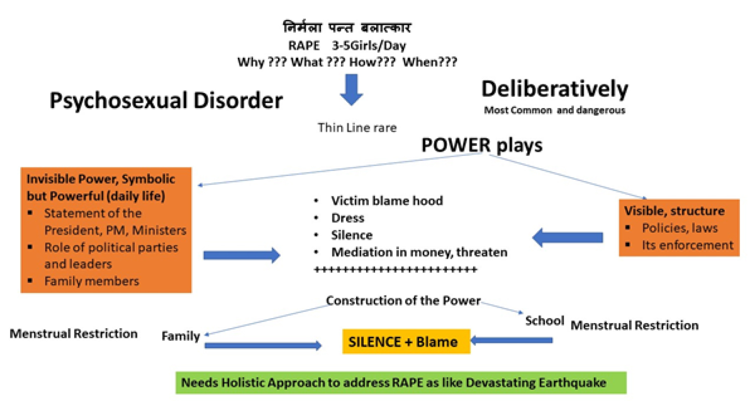
AWHC-19-134 - Thomas Wenzel_Austria_F1

Figure 1. Construction of Visible and Invisible Power and Relationship between Menstrual Restriction and Rape.

Summary of recommendations based on project results

1. Home

  • Reconstruct the language, behavior, division of labor, and other family dynamics inside the home for gender equality and human rights.
  • Abolish any bias division of household labor
  • Parents lead by example by sharing all household duty, abolish “kitchen/cleaning work as women’s work” and outside work as a mans
  • Create new division of chores within your household which breaks stereotypical division of work and propels gender inequality
  • Abolish any menstrual restriction practice at home
  • Men should advocate for elimination of any restriction practices at home and eeducate self and other family members about menstruation, especially male family members
  • Women should reject menstruation restriction practices at home
  • Educate self (if needed) and other family members about menstruation, focusing on younger members, teaching girls to self-advocate and menstruate with dignity and boys to advocate for girls dignity and respect menstruation
  • Change rules and regulations inside the home to reflect equal ideals
  • Apply same restrictions apply to both male and female members e.g. time to come home, how far one can go outside the home
  • Abolish imposing gender rules and norms regarding appearance on family members
  • Allow girls and boys to choose their appearance and apparel according to their choice not their gender
  • Girls can have short hair/not wear dresses/not have phulies (nose ring)

2. School

  • Revise school curriculum and activities to institutionalize gender equality and human rights e.g. holding teachers and school staff accountable as gender equality role models
  • Create enabling environment for dignified menstruation
  • Reinforce gender equality and human rights by organizing series of extracurricular activities including parents and students

3. Political Parties/Leader

  • Demonstrate as accountable state agents who propel gender equality and human rights as they committed in their political manifesto e.g. eeducate within the political party about gender equality, human rights, menstrual restriction, gender-based violence, and holding accountability at personal and party level.
  • Regardless of their political party/power, support victims and stand against any violence against women and men

4. NGO

  • NGOs demonstrate downward and upward accountability and role model for gender equality and human rights e.g. create zero tolerance for discrimination against gender
  • Demonstrate as a champion for dignified menstruation
  • Act as impartial agent for justice
  • Create strong network with local government to connect communities with government for open access to resources

5. MEDIA

  • Play as critical agent to transform gender norms, roles, and stereotypes e.g. take primary imitative to educate and empower public about gender equality and human rights, especially the impact of menstrual restriction on gender-based violence and dignity

6. STATE

  • Bharatpur metro municipality guarantee the safety, security of each individual e.g. transparency of roles, mechanisms, and activities for justice committee
  • Ensure access to justice committee including vice mayor
  • Create direct mechanism to access resources and services especially for victims
  • Establish hotline for victims of violence to access services
  • Education appears to be the most important of these factors

References

  1. http://citeseerx.ist.psu.edu/viewdoc/ download?doi=10.1.1.866.5866&rep=rep1&type=pdf
  2. https://www.usaid.gov/sites/default/files/documents/1865/Men_VAW_report_Feb2015_Final.pdf
  3. https://nepal.unfpa.org/sites/default/files/pub-pdf/ICRW.pdf
  4. The United Nations (1988) Convention on the Elimination of All Forms of Discrimination against Women. Treaty Series 1249: 13.
  5. Ranabhat C, Kim CB, Choi EH, Aryal A, Park MB, et al. (2015) Chhaupadi Culture and Reproductive Health of Women in Nepal. Asia Pac J Public Health 27: 785–795.
  6. Dahal K (2008) Nepalese woman dies after banishment to shed during menstruation BMJ 337: 2520.
  7. Atreya A, Nepal S (2019) Menstrual exile – a cultural punishment for Nepalese women. Med Leg J 87: 12–13.
  8. Cardoso LF, Clark CJ, Rivers K, Ferguson G, Shrestha B, et al. (2018) Menstrual restriction prevalence and association with intimate partner violence among Nepali women. BMJ Sex Reprod Health 2018.
  9. Hennegan J, Shannon AK, Rubli J, Schwab KJ, Melendez-Torres GJ (2019) Women’s and girls’ experiences of menstruation in low- and middle-income countries: A systematic review and qualitative metasynthesis. PLoS Med 16: 1002803.
  10. Sah AK, Shrestha N, Joshi P, Lakha R, Shrestha S, et al. (2018) Association of parental methylenetetrahydrofolate reductase (MTHFR) C677T gene polymorphism in couples with unexplained recurrent pregnancy loss. BMC Res Notes 11: 233.
  11. Sharma M, Gupta S (2003) Menstrual pattern and abnormalities in the high school girls of Dharan: a cross sectional study in two boarding schools. Nepal Med Coll J 5: 34–36.
  12. Thapa B, Powell J, Yi J, McGee J, Landis J, et al. (2017) Adolescent Health Risk and Behavior Survey: A School Based Survey in Central Nepal. Kathmandu Univ Med J (KUMJ) 15: 301–307.
Hepatic Arterial Communicating Arcades – Cases Series and Review of Literature

DOI: 10.31038/IMCI.2019216

Abstract

This case series describes the hepatic arterial communicating arcades and their importance in the endovascular management of hepatic artery pseudoaneurysm, paediatric post liver transplant lobar arterial occlusion and lobar arterial stenosis due to gall bladder carcinoma. We describe different types of arterial communicating arcades which have not been described earlier.

Keywords

Arterial communicating arcades, hepatic artery, pseudoaneurysm, liver transplantation, embolization, gall bladder carcinoma.

Introduction

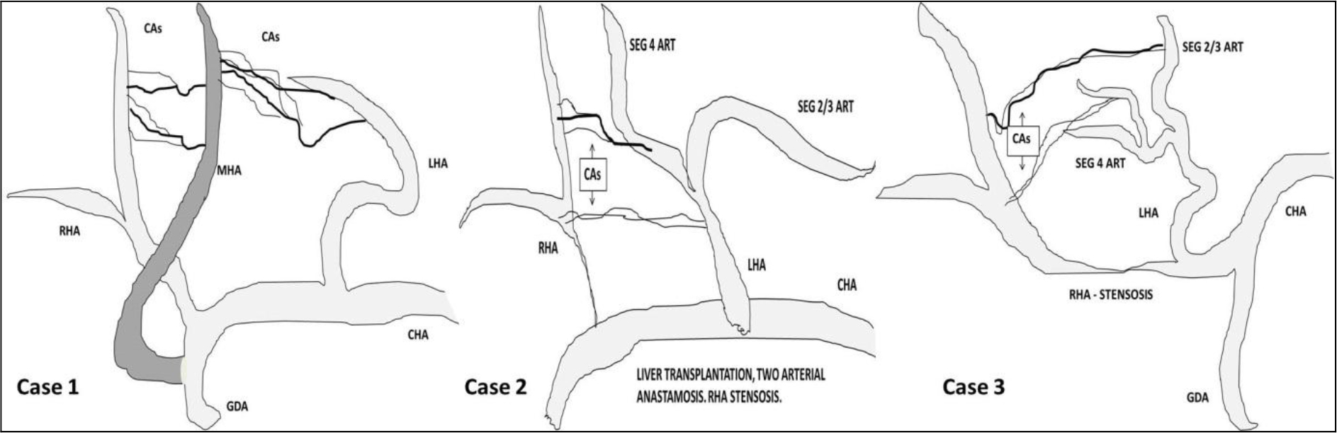
Interlobar arterial communication or Communicating Arcades (CA) in the hepatic hilum has been recognized as one of the most important collateral pathways to the liver. CAs plays an important role in the blood supply to the caudate lobe and also have a close relationship with the blood supply to the hilar biliary tract. Hepatic arterial communicating arcades develop in hilum when either right or left hepatic artery is occluded or significantly stenosed [1,2]. If Proper Hepatic Artery (PHA) is occluded, liver perfusion can occur through small collaterals in the hepatic ligaments, collaterals around the common bile duct, inferior phrenic artery, pancreatico-duodenal artery and intercalary ‘de novo’ collaterals [3,4]. We describe 3 cases showing the importance of hepatic arterial CA in endovascular treatment of mycotic Hepatic Artery Pseudoaneurysms (HAPAs), lobar arterial stenosis following paediatric deceased donor liver transplant and locally advanced carcinoma gallbladder infiltrating the Right Hepatic Artery (RHA).

Case Series

Case 1

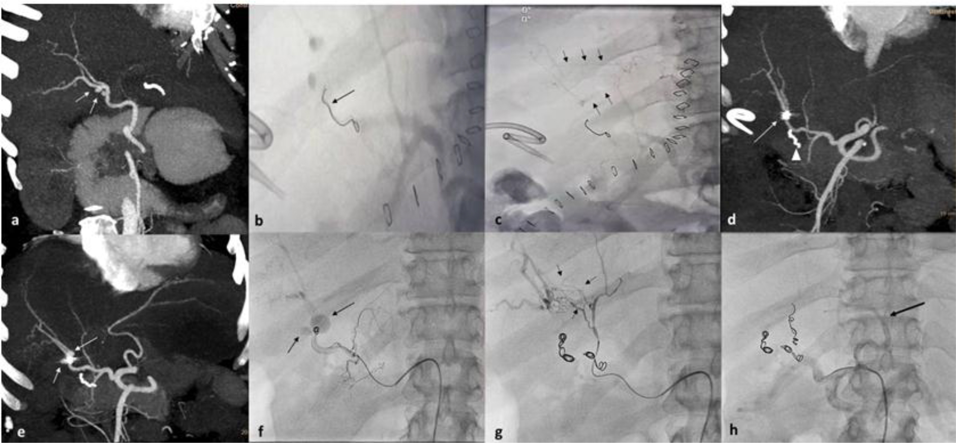
49 year old lady presented with cholelithiasis and choledo-cholithiasis. After multiple failed attempts of endoscopic management, the patient underwent a Roux-en-Y hepaticojejunostomy. On post-operative day 5, the patient had low grade fever and elevated white cell count (13,000/mm3). Ultrasound examination revealed a small collection anterior to the hepaticojejunostomy site. No biliary dilatation was seen. A contrast enhanced CT (CECT) scan was performed which revealed two extra hepatic pseudoaneurysms in the RHA proximal to its bifurcation (Figure 1a). Interventional radiologist was unavailable on that particular week. Drain was placed by diagnostic radiologist avoiding injury to pseudoaneurysms at least to drain the collection. Initially, bile stained fluid was drained, which was followed by drainage of frank blood through the drain tube and hemodynamic instability of the patient. Immediately patient was resuscitated and taken to angiosuite for embolization. In view of the emergency situation, the in house cardiologists performed the embolization. CHA angiogram confirmed the presence of two irregularly filling extra hepatic RHA pseudoaneurysms. A single coil was placed in the right hepatic artery proximal to the pseudoaneurysms. Post coiling, RHA went in to spasm and no reperfusion of pseudoaneurysm was observed (figure 1b). However, on proper hepatic artery angiogram collateral supply from Middle Hepatic Artery (MHA) to distal RHA branches was observed (Figure 1c). No reperfusion of pseudoaneurysms was observed post coiling, and the procedure was concluded. Post procedure CECT scan done after two days did not reveal reperfusion of pseudoaneurysms, however distal migration of coil in in RHA was noted (Figure 1d). Post embolization, liver enzymes and bilirubin were elevated (AST, ALT and ALP were 1352, 1454 and 584 respectively, bilirubin was 2.3 mg/dl) on day 1 and become near normal on day 7. Patient was discharged on 12thpost embolization day. On 17th post embolization day, the patient suddenly collapsed at home. She was found to be hypotensive and resuscitated at a local hospital before being shifted back to our hospital for management. Reperfusion of pseudoaneurysms and mild hemoperitoneum was observed on repeat CECT scan (Figure 1e). Angioembolization was performed by an interventional radiologist. Selective RHA angiogram was performed (Figure 1f) and pseudoaneurysms were delineated. During embolization, attempt was not made to cross the pseudoaneurysms in view of coil migration and rupture of aneurysms. RHA was embolized with multiple 6mm, 8mm 0.018” micro coils. Middle hepatic artery (MHA) was arising from proximal GDA. MHA angiogram showed faint perfusion of pseudoaneurysms through intrahepatic arterial arcades (Figure 1g).  Intrahepatic arterial arcades were embolized with gelfaom particles and main trunk of MHA was embolized with 5mm 0.018” coils (Figure 1h). Left hepatic artery (LHA) angiogram did not reveal perfusion of pseudoaneurysms, however few tiny CA were seen supplying the segment 4 of liver. Post embolization, the patient recovered well. No significant increase in liver enzymes or bilirubin was noted. No recurrence of pseudoaneurysm or other symptoms were observed on 12 months follow up.

IMCI 19 - 111_Kundaragi NG_F1

Figure 1. 49 year old female patient, post hepaticojejunostomy presented with prehepatic collection, fever and elevated total white cell count on day 5. Case describes CAs from both middle and left hepatic arteries.  (a) Arterial phase contrast enhanced CT scan, oblique coronal MIP image showing two small mycotic pseudoaneurysms in (white arrow) right hepatic artery. Small biliary collection was also noted. (b and c) Celiac angiography digital image post embolization of right hepatic artery (by cardiologist because of non-availability of Interventional radiologist) showing single coil (black arrow) in RHA with non opacification of pseudoaneurysms and intrahepatic arterial arcades communicating between right and accessory RHA (Black arrows). Pigtail catheter placed in view to drain the collection caused rupture of pseudoaneurysm. (d) Arterial phase contrast enhanced CT scan done post embolization day 2 revealed thrombosed pseudoaneurysm and distally migrated crumpled coil (arrow) near RHA bifurcation. Arrow head shows HepJ anastomotic bowel staple suture (e) Arterial phase contrast enhanced CT scan done post embolization on post op day 17, MIP image shows reperfusion of pseudoaneurysms (arrows) with increase in size of postero-medial pseudoaneurysm. (f) Digital right hepatic artery angiogram showing migrated coil and two pseudoaneurysms (arrow).  (g) Post RHA coiling digital angiogram of MHA showing multiple intrahepatic arterial arcades (small black arrows) feeding distal right hepatic artery. (h) Post MHA coiling, celiac axis digital angiogram showing patent left HA (black arrow) and GDA.

Case 2

A 9 year old male patient, a known case of Alagille syndrome had undergone Deceased Donor Liver Transplantation (DDLT). During transplant two arterial anastomosis were performed. Right and left hepatic arteries of donor were anastomosed to recipient’s PHA. Intraoperative colour and spectral doppler ultrasound didn’t reveal any significant abnormality in Peak Systolic Velocity (PSV), Resistive Index (RI) and spectral waveform of intrahepatic portions of right, segment 4 and left hepatic arteries. From 2nd post-operative day, ultrasound revealed multiple subcapsular ischemic areas in right lobe of liver and increasing post anastomotic (choledocho-choledochostomy) biliary dilatation. Colour and spectral doppler ultrasound revealed low RI (0.523 in RHA and 0.429 in segment 4 artery) and normal PSV in intraparenchymal portions of both right and left hepatic arteries (Figure2a, b & c). Marginal alterations in liver enzymes were observed with normal INR and serum lactate levels. In view of isolated right lobe subcapsular ischemic areas and biliary dilatation, CECT scan was performed. CECT scan showed, short segment complete stenosis of anastomotic and post anastomotic RHA and reperfusion of distal parenchymal arteries through CA from segment 4 artery. Segment 4 artery and LHA were enhancing normally. Multiple non enhancing subcapsular ischemic areas were seen in right lobe. No focal abnormality seen in left lobe of liver. Mild biliary dilatation was seen (Figure 2d, e and f). ERCP and plastic biliary stent was placed to assist biliary drainage. Routine interval ultrasound follow was done till discharge. The patient is doing well 10months after the transplant.

IMCI 19 - 111_Kundaragi NG_F2

Figure 2. 8 year old male patient, post liver transplant status with mild elevated liver enzymes. (a & b) Spectral Doppler imaging of right and segment 4 hepatic arteries showing elevated peak systolic velocity with low RI. (c) Gray scale and colour Doppler ultrasound image dual window showing biliary dilatation black arrow) with narrowing at anastamotic site. (d & e) Arterial phase contrast enhanced CT scan coronal MIP image showing post anastomotic unopacified proximal right hepatic artery (arrow heads). Coronal and axial MIP image showing intrahepatic arterial arcades (small white arrows) communicating between segment 4 artery and right hepatic artery. (f) Venous phase CECT axial images showing multiple non enhancing subcapsular hypodense areas in right lobe (open arrows) and predominant central biliary dilatation.

Case 3

A 64 year old male presented with progressive jaundice of 3 months duration. Total bilirubin was 13mg/dL. Transabdominal ultrasound revealed Gall Bladder (GB) fossa mass with infiltration of adjacent liver and few hypoechoic lesions seen in right lobe of liver. Hilar biliary confluence was involved with significant dilatation of intra hepatic biliary radicles. Through ERCP, plastic biliary stent was placed in to left side biliary ducts. Total bilirubin level came down to 3.5mg/dL in 3 days. The CECT scan revealed a large enhancing mass in GB fossa with infiltration of adjacent liver. Few peripherally enhancing focal lesions were seen in right lobe of liver (Figure 3a). Tumour was seen encasing the proximal RHA with its significant stenosis. Reperfusion of Intraparenchymal RHA branches were observed through arterial CAs from left hepatic artery both from segment 2/3 and segment 4 hepatic arteries (Figure 3b and c). Portal vein was not involved. Tumour was invading the biliary confluence with significant bilateral intra hepatic biliary dilatation. Plastic biliary stent was seen in left lobe biliary duct. Adenocarcinoma of gall bladder was confirmed on percutaneous biopsy of GB fossa mass. Biopsy of right lobe liver lesions revealed cholangitic abscess. The patient was informed about the prognosis and two self-expandable metallic stents were placed.

IMCI 19 - 111_Kundaragi NG_F3

Figure 3. 64 year old male patient, inoperable case of GB fossa mass, presented with progressive jaundice and abdominal pain since 3months. Case describes CAs from both the branches of left hepatic artery (segment 4 artery and segment 2/3 branches). (a) Venous phase CT scan, coronal image showing ill-defined enhancing mass in segment 5 and GB fossa. Rim enhancing lesion in segment VI. (b & c) Arterial phase contrast enhanced CT scan, coronal MIP images showing arterial arcades (small arrows) communicating between LHA (open white arrow) and RHA (Long arrow) and segment 4 artery (thick arrow) and RHA respectively. Significant stenosis of proximal RHA seen (curved arrow). (d & e) Spectral Doppler images showing normal velocity and low RI in intrahepatic branches of RHA and high velocity and normal RI in segment IV artery. (f) Spectral Doppler performed at stenotic proximal RHA showing increased peak systolic velocity (158cm/sec) with normal RI.

Discussion

Inter lobar hepatic arterial communications or Communicating Arcades (CAs) develop depending on the site of stenosis or occlusion of the hepatic artery. Interlobar collateral vessels usually develop in the hepatic hilum in patients with interruption either right or left hepatic artery. These vessels are not visualized on angiograms in patients with intact hepatic arterial supply [5].

The CAs is located extrahepatically in the hepatic hilum or cranial to the portal bifurcation close to the hilar bile duct. Caudate lobe is believed to derive  its blood supply not only from the segment I artery but also from the CA, because of which transarterial chemoembolization for caudate lobe hepatocellular carcinoma is not very effective [1].

Tohma et al classified CAs in to type 1a, 1b and 2 if arising from middle hepatic artery, segment 4 artery and left hepatic artery respectively. Authors have also classified CAs arising from right anterior hepatic artery, right hepatic artery or both as type 1, 2 and 3 respectively. In the present article we have presented all 3 cases with CAs arising from left side. In first case CA was arising from middle hepatic artery (type 1a). However in after embolizing segment 4 artery we were able to see tiny collaterals from left hepatic artery supplying the segment 4 area. So this type of collaterals from both middle and left hepatic artery is not mentioned in literature. In second case of post liver transplant CA was from segment 4 hepatic artery (type 1b). In third case CA was arising from both the branches of left hepatic artery (segment 4 artery and segment 2/3 branches). Our first and third cases described different types to Toham et al classification of CAs arising from left arteries (Diagram 1).

IMCI 19 - 111_Kundaragi NG_F4

Diagram 1. Pictorial demonstration of cases.

Hepatic artery is the second most common site for visceral artery pseudoaneurysm after splenic artery. Hepatic Artery Pseudoaneurysms (HAPAs) are usually iatrogenic and can present as life threatening complications of hepatic, biliary, and pancreatic interventions. Hepatic procedures accounts for 65% of these cases. Biliary and pancreatic procedures accounts for 30% and 5% respectively [6]. HAPAs can also be associated with intra-abdominal inflammation, infection, or trauma [6]. In our first case, surgery was uneventful and patient developed an infected biliary collection near hepatico-jejunostomy site on post operation day 5. All the authors felt right HAPAs were likely caused by infection (Klebsiella aerogenes bacteria) rather iatrogenic, as patient recovered well after endovascular reintervention.

Coil embolization or exclusion of the pseudoaneurysms by stent graft is most effective treatment option in an emergency setting, especially when associated with an infection. Both proximal and distal ends of the parent arteries of the aneurysm must be embolized simultaneously to block off the pseudoaneurysm in an extra hepatic HAPA, while proximal embolization alone can be performed for an intrahepatic arterial aneurysm; however, reestablishment of collateral circulation increases the risk of re-rupture of the aneurysms [7]. In our case extra parenchymal right HAPAs were seen, which were initially embolized by proximal solitary coil occlusion; however the coil migrated, pseudo-aneurysms were re-perfused and bled again. Later successful embolization was performed by an interventional radiologist with multiple coils in RHA, gel foam in CAs and coil in middle hepatic artery.

Early (<1 month) arterial complications are associated with graft loss and a high mortality rate after Orthotropic Liver Transplant (OLT).  Hepatic Artery Stenosis (HAS) and Hepatic Artery Thrombosis (HAT) are the most common hepatic arterial complications, with high rates of morbidity and mortality. Untreated significant HA anastomotic strictures can progress to HAT (65% at six months follow up). HAS is less frequently associated biliary complications compared to HAT. Biliary complications can be seen up to 67% in liver transplant recipients with HAS. Reduction of arterial flow during liver transplant is commonly associated with biliary tree complications due to ischemic processes. In some cases, HAS is likely to stimulate the development of arterial collaterals that protect the liver from ischemia at the time of HAT [8].

Doppler Ultrasound (DUS) is the gold standard investigation to assess hepatic artery patency in cases of HAT with sensitivity up to 92% or an increased Resistive Index (RI). Anatomical defects (stenosis or kinking) can be detected by CT angiogram or conventional angiography with a high sensitivity and specificity. Early HAS can be detected by DUS with a sensitivity of 100%, a specificity of 99.5%, a positive predictive value of 95% and a negative predictive value of 100%, and an overall accuracy of 99.5%. However MDCTA and standard angiography are the gold standard for HAS diagnosis [8].

Biliary tract ischemia contributes to biliary strictures and anastomotic leakage in liver transplants and can be seen up to 34% of patients. Division of the CA during graft donation may potentially lead to biliary ischemia and complications. Thus, during graft donation the right or left hepatic artery should not be dissected and separated from the bile duct distally to prevent biliary ischemia [1]. In our second case anastomotic stenosis of RHA has led to development of anastamotic biliary stricture (choledocho-choledochostomy) and visible biliary dilatation from 3rd day.

The third case is an uncommon presentation of gallbladder carcinoma involving the right hepatic artery with development of visible hepatic arterial arcades on CECT scan. Primary gallbladder carcinoma is the most common malignancy of the biliary tract. The most common route of dissemination is direct invasion of the liver. Hepatic invasion or hepatic metastasis has been reported in as many as 30%–80% of cases of gallbladder carcinoma. Gall bladder malignancies with vascular invasion (main portal vein or hepatic artery) are typically not amenable to surgery [9].

In conclusion, first attempt of intervention failed in first case due to hepatic inter arterial communication and incomplete embolization, which was later successfully embolized by an Interventional radiologist. Percutaneous drain must be placed only after treating pseudoaneurysms. In second case of post liver transplantation, arterial complication was precisely detected on CECT and not on Doppler ultrasound. Right lobe of graft survived because of hepatic inter arterial communications and no intervention was required, however patient developed few peripheral subcapsular ischemic changes in right lobe and low grade biliary dilatation which was managed by endoscopic plastic stenting. Our first and third case, show different types of arterial communicating arcades arising from middle hepatic artery (from GDA) and left hepatic artery. Thus knowledge of hepatic inter lobar arterial communication is important while treating and diagnosing arterial diseases of both native and transplant liver.

References

  1. Tohma T, Cho A, Okazumi S, Makino H, Shuto K, et al. (2005) Communicating arcade between the right and left hepatic arteries: evaluation with CT and angiography during temporary balloon occlusion of the right or left hepatic artery. Radiology 237: 361–365.
  2. Charnsangavej C, Chuang VP, Wallace S, Soo CS, Bowers T (1982) Angiographic classification of hepatic arterial collaterals. Radiology 144: 485–494.
  3. Redman HC, Reuter SR (1970) Arterial collaterals in the liver hilus. Radiology 94: 575–579.
  4. Mays ET, Wheeler CS (1974) Demonstration of collateral arterial flow after interruption of hepatic arteries in man. N Engl J Med 290: 993–996.
  5. Koehler RE, Korobkin M, Lewis F (1975) Arteriographic demonstration of collateral arterial supply to the liver after hepatic artery ligation. Radiology 117: 49–53.
  6. Harvey J, Dardik H, Impeduglia T, Woo D, DeBernardis F (2006) Endovascular management of hepatic artery pseudoaneurysm hemorrhage complicating pancreaticoduodenectomy. J Vasc Surg 43: 613–617.
  7. Ji WB, Wang WZ, Xu XJ, Mi YC, Fu X, et al (2017) Arterial embolization in treatment of hepatic artery pseudoaneurysm. Acta Medica Mediterranea 33: 449.
  8. Piardi T, Lhuaire M, Bruno O, et al (2016) Vascular complications following liver transplantation: A literature review of advances in 2015. World Journal of Hepatology 8: 36–57.
  9. Hussain HM, Little MD, Wei S (2013) AIRP best cases in radiologic-pathologic correlation: gallbladder carcinoma with direct invasion of the liver. Radiographics 33:103-108.

Supporting Older Nurses in the Workforce: Intersectional Considerations

DOI: 10.31038/AWHC.2019235

Introduction

In most industrialized countries, the nursing workforce, an overwhelmingly numerically female dominant profession, is aging [1]. Analysis of data suggests that this is due to interrelated social and economic factors pushing nurses towards later retirement. Nurses are choosing to remain in the workforce longer, delaying retirement for reasons that include economic and financial necessity, continued desire to care for others and meet professional goals [2], and positive life-span development that is attributable to increased life expectancy and longevity [1–2]. Apart from intrinsic motivators, there is also a growing drive to increase the labour market participation of older individuals in response to shifting population structures secondary to population aging [2, 3–5]. In occupations such as nursing where skills shortages are already being experienced, population aging threatens to deepen this existing problem [6].

Older nurses represent a pool of untapped human capital. Through their accumulated experience and resulting expertise, older working nurses present a number of benefits for economic growth and social welfare. However, if these benefits are to be derived from the continued participation of older nurses in the labour market, the intersecting challenges that they experience in the workplace must be recognized and addressed. The intersectionality of the physicality of nursing work, its embeddedness in gender-based power relations, the health care consequences of population aging, and the increased risk of disability and other outcomes associated with working when older must be taken into consideration in designing healthy, age-friendly work environments where a diversity of nurses can thrive.

Intersectional Issues of Concern

Older nurses working in the labour market are impacted by multiple and gendered axes of influence that must be understood. Equally importantly, they must be considered as part of employers’ and policy makers’ responses both to the struggles of older nurses in the workforce and to the broader challenges of population aging for health care. Key influences and issues of concern are explored briefly below.

Consequences of Aging on Working

While our bodies change with age (e.g., decreased strength, flexibility, and bone mass) [7] disability and illness are not an inevitable part of aging [8]. This notwithstanding, the probability of work-induced disability and injury increases as workers age [5, 9]. Data support that older workers are less apt to acquire injuries on the job than younger workers; however, aging-related physiological and cognitive changes make older workers susceptible to more severe and permanent injuries [5]. Older workers are more likely to experience soft tissue injuries affecting the back, neck, and feet [5, 9–10]. The probability of older nurses sustaining such injuries and acquiring disability is made even more likely because of the physically strenuous nature of nursing work [9] and the concurrent risk for violence [11]. Recent data reveals a significant positive correlation between age and incidence of nurses working with some form of physical or mental impairment [12]. This finding was consistent with other data indicating that the likelihood of individuals experiencing disability increases with age [12].

Given this context, it is surprising that disability among nurses is a topic rarely discussed within the literature and equally absent from policy and administrative perspectives [9, 13]. Available literature reveals that nurses with disabilities, particularly those who acquire a disability while working, receive very little support to persist within the profession [13–14]. Unsupported nurses with disabilities are left in a difficult situation with very little choice, often leaving direct practice or the profession altogether [13–14].

Gender-based Power Relations and Labour Market Discrimination

The influence of age on working does not happen in a vacuum and is not exclusive of other factors impacting individuals’ lives and their work. Gendered experiences of women in labour markets have a compounding effect on working women who are aging. With a few exceptions, women account for approximately 90–95% of the nursing workforce the world over. As a female dominated profession, deeply embedded within nursing are the gender-based power relations of society. As such, the nursing profession is reflective of the value placed on women and their standing in society [15]. In particular, the erroneous feminization of nursing and gender-based discrimination have resulted in occupational segregation and consequently, the forcing down of wages in the profession. Unfortunately, dominance of females within the profession is not effective against the gender-pay gap. Across practice settings, positions, and specialities, male nurses typically out earn female nurses [16–17]. This gender pay gap is also unaffected by education, a common means by which an individual can increase their earnings [17].

Although not all of the factors contributing to the gender pay gap within nursing are known, workplace discrimination and gender discrimination have been identified as factors limiting women from advancing in the workplace and gaining positions of authority [17]. Additionally, gender expectations of women within society have been identified as contributing factors. Like other working women, female nurses often must balance work and care for children, other dependent family members, or both, often at the expense of career progression and their health [18, 19]. While performing this juggling act can and does fall to men in some circumstances, the societal expectation that women should take care of their loved ones, even when they are working, persists. Many women have reported experiencing the weight of these often-unspoken expectations [18, 20].

Population Aging, Health and Health care

Trends in the health of populations and health care are yet another axis of influence on older nurses’ participation in the workforce. These trends include a diminished and slow-growing nursing supply resulting in inadequate staffing; [1, 21] increasing patient volumes and the requirement for more health services due to increased life expectancy and longevity; [1, 22, 23] and increasing patient acuity and the growing complexity of treatment modalities [1, 22]. The sum total of these pressures is that there are ever-rising professional demands and excessive workloads on nurses. Nurses are faced with increasing psychosocial, physical, and cognitive demands. In some cases, they must also deal with unsafe and unhealthy conditions such as workplace violence. Consequently, there is greater likelihood of fatigue, injury, and disability among today’s nurses, which have been linked to overall measures of the quality of nursing work life and patient safety [1, 20, 24]. Older nurses may be doubly impacted by these pressures associated with aging that are situated within the context of gendered labour market experiences.

Intersectional Considerations for Ways Forward

By and large, efforts to address the challenges faced by older working individuals have focused on the consequences of aging, often in isolation of other factors. Nursing-specific interventions that have been championed include flexible scheduling, shorter work shifts, comprehensive disability management programs with an emphasis on accommodation, and redesign of the work environment to meet ergonomic needs and reduce injuries [9, 25]. While there is no doubt that these initiatives are needed and would be of benefit to all nurses, they do not go far enough to address the multiple axes of influence and oppression structuring the experiences of not only older working nurses, but younger nurses as well.

An intersectional approach is needed whereby aging in the nursing workforce is understood in the context of the other interconnected factors that in some cases may have a far more significant impact on working nurses than the consequences of aging. An intersectional understanding of working, aging women exposes the multiple forces that impact upon the experiences of older nurses. These experiences should be understood in ways in which age, gender, issues of power and domination, and socioeconomics intersect with the work experience. Moreover, an intersectional approach to aging in the workplace provides the opportunity to reflect on and apply a social justice discourse in dealing with the challenges faced by nurses.

Ultimately, the problem of a skills shortage in nursing and the multitude of challenges accompanying an aging nursing workforce cannot be addressed through a sum-of-the-parts approach. An intersectional approach calls upon stakeholders to focus on the complexities of the situation and employ a broader frame to understand these problems. Using a well-informed frame in evaluating aging in the nursing workforce will enable changemakers to accurately recognize the issue of skills shortage and retention of nurses as being layered by structural inequalities along axes of age, gender, ableness, and social status. In this context, solutions should be pursued which are emancipatory and aim to empower the nursing workforce while also interrogating and seeking to remedy structural discrimination and injustices, such as the gender pay gap and visible and invisible barriers to career advancement and fulfillment. Estimates predict that upwards of 7 million skilled health professionals (the majority nurses) are currently needed globally [26]. In this context, failure to recognize the context and experience of work for aging nurses, compounded by the profound lack of incentives for younger nurses to remain in the profession [26] will undoubtedly negatively impact not only nurses, but likely the health of individuals, families, groups, and communities around the world who rely on nurses to provide compassionate, high quality care.

References

  1. Haddad LM, Toney-Butler TJ (2019) Nursing shortage [Internet]. Treasure Island, FL: StatPearls Publishing 2019.
  2. Auerbach DI, Buerhaus PI, Staiger DO (2014) Registered nurses are delaying retirement, a shift that has contributed to recent growth in the nurse work- force. Health Affairs 33: 1474–1480.
  3. Appannah A, Biggs S (2015) Age-friendly organisations: the role of organisational culture and the participation of older workers. Journal of Social Work Practice 29: 37–51.
  4. Promotion of labour force participation in older Canadians – promising initiatives [Internet]. Ottawa: Government of Canada 2018.
  5. McDonald T, Harder HG (2004) Older workers and disability management. IJDCR 3.
  6. Uthaman T, Chua TL, Ang SY (2016) Older nurses: a literature review on challenges, factors in early retirement and workforce retention. Proceedings of Singapore Healthcare 25: 50–55.
  7. Shannon H (2013) Health and safety issues in the aging workforce [Internet]. Toronto, ON: Institute for Work & Health 2013.
  8. Barlow FK, Walker N (2015) Disability and ageing. In Pachana NA, editor. Encyclopedia of Geropsychology. Gateway East, Singapore: Springer Singapore 2015.
  9. Matt SB, Fleming SE, Maheady DC (2015) Creating disability inclusive work environments for our aging nursing workforce. Journal of Nursing Administration 45: 325–330.
  10. Phillips JA, Miltner R (2015) Work hazards for an aging nursing workforce. J Nurs Manag 23: 803–812. [crossref]
  11. Cooper C, Swanson N (2002) Introduction. In Cooper C, Swanson N, editors. Workplace violence in the health sector. State of the art. Geneva: International Council of Nurses 2002.
  12. Wilson BL, Butler RJ, Butler MJ (2016) Employment and wage disparities for nurses with activity limitations. Journal of Nursing Scholarship 48: 608–615.
  13. Anyinam CK (2018) Work experiences of nurses with self-identified disabilities [dissertation]. Toronto (ON): York University 2018.
  14. Neal-Boylan L, Fennie K, Baldauf-Wagner S (2011) Nurses with sensory disabilities: their perceptions and characteristics. Rehabil Nurs 36: 25–31.
  15. Wrede S (2010) Nursing: globalization of a female-gendered profession. In Kuhlmann E, Annandale E, editors. The Palgrave handbook of gender and healthcare. London: Palgrave Macmillan 437–453.
  16. Muench U, Sindelar J, Busch SH, Buerhaus PI (2015) Salary differences between male and female registered nurses in the United States. JAMA 31: 1265–1267.
  17. Wilson BL1, Butler MJ2, Butler RJ3, Johnson WG4 (2018) Nursing Gender Pay Differentials in the New Millennium. J Nurs Scholarsh 50: 102–108. [crossref]
  18. Tannenbaum C, Voss P, El-Gabalawy H1, Joanette Y (2016) Gender, Work, and Aging. Can J Aging 35: 405–411. [crossref]
  19. Williams LA, Giddings LS, Bellamy G, Gott M (2017) ‘Because it’s the wife who has to look after the man’: a descriptive qualitative study of older women and the intersection of gender and the provision of family caregiving at the end of life. Palliative Medicine 31: 223–230.
  20. Scott LD, Hwang WT, Roger AE (2006) The impact of multiple care giving roles on fatigue, stress, work performance among hospital staff nurses. Journal of Nursing Administration 36: 86–95.
  21. Ariste R, Béjaoui A, Dauphin A (2019) Critical analysis of nurses’ labour market effectiveness in Canada: The hidden aspects of the shortage. The International Journal of Health Planning and Management 2019.
  22. Bloom DE, Chatterji S, Kowal P, Lloyd-Sherlock P, et al. (2015) Macroeconomic implications of population ageing and selected policy responses. The Lancet 385: 649–657.
  23. Health care in Canada 2011. A focus on seniors and ageing [Internet]. Ottawa, ON: Canadian Institute for Health Information 2012.
  24. Flin R, Winter J, Sarac C, Raduma M (2009) Human factors in patient safety: review of topics and tools. Report for methods and measures working group of WHO patient safety [Internet]. Geneva: World Health Organization 2009.
  25. Kwok C, Bates KA, Ng ES (2016) Managing and sustaining an ageing nursing workforce: identifying opportunities and best practices within collective agreements in Canada. Journal of Nursing Management 24: 500–511.
  26. A universal truth: no health without a work force [internet]. Geneva: Global Health Workforce Alliance and World Health Organization 2019.

Correlation between Magnetic Resonance Imaging and Arthroscopy in Meniscal Injuries

DOI: 10.31038/IJOT.2019241

Abstract

Introduction: Meniscal injuries have a very high incidence among professional and amateur athletes. It is estimated that the incidence amounts to 24 every 100 000 each year. The different forms of imaging diagnostics play an important role in the management of knee injuries, particularly in the event of uncertain clinical diagnosis, helping to avoid unnecessary and expensive surgeries. In order to diagnose meniscal pathologies, the most commonly used imaging test is Magnetic Resonance Imaging (MRI). Health Officials from all around the world are becoming increasingly involved in the definition of surgical treatment limits, making an effort to enhance their practice and the patients’ cost-effectiveness.

Our work aims to review evidence about the correspondence between imaging tests -particularly the MRI- and knee arthroscopy as the Gold Standard for the diagnosis of meniscal injuries.

Material and Methods: We performed a systematic search that included Medline (PubMed interphase) and Lilacs databases. The search totaled 607 articles. According to filters and inclusion/exclusion criteria, 23 papers were chosen for our bibliographic review.

Results: The selected papers were prospective studies. Our results are based on data retrieval specifically linked to sensitivity and specificity of the MRI with regards to arthroscopy in meniscal injuries.

Discussion: From the analysis of this information we may consider that there is no consistency in results and opinions in English-published bibliography of a prospective profile. Nevertheless, we must acknowledge that the prevailing results are those that prioritize the relevance of MRI in terms of sensitivity and specificity. We must currently accept that MRI is a very costly study for diagnosing meniscal injuries. There are some variations in its sensitivity and specificity, but they are minor and, therefore, do not invalidate these conclusions.

Introduction

Meniscal injuries have a very high incidence among professional and amateur athletes. This injury is one of the most frequent in sports medicine: 24 in every 100 000 athletes suffer one of this each year. They show a bimodal distribution; the first incidence peak is seen among young athletes and the second is seen in middle-aged patients with degenerative joint disease 1].

Macroscopically, the menisci of the knee are two intra-articular semicircular fibrocartilaginous structure, with a wedge shaped structure, placed between the tibia and the femur, in the medial and lateral compartments. They used to be considered as vestigial remains of muscular structures in the knee. Ever since the middle of the Twentieth Century we have thoroughly known their actual functions and their anatomic, therapeutic and prognostic relevance in knee pathology. The menisci have three main functions: load transmission, cushioning and secondary stabilization [2–4].

In general, the medial meniscus is the one with less movement and therefore it gets injured more frequently than the lateral meniscus [5].

In terms of clinical diagnosis, there are over twenty specific tests described for the assessment of meniscal injuries, with sensitivity and specificity levels that fall between 64 and 97% [6–10]. The sensitivity of these tests decreases when there are other associated injuries, particularly of the anterior cruciate ligament [11–13].

The different forms of imaging diagnostic play an important role in the management of knee injuries, and particularly in the event of uncertain clinical diagnosis; they help to avoid unnecessary surgeries [14].

Since MRI was first introduced in 1984 for clinical usage, its diagnostic role in knee injuries has had a substantial impact [15–17]. MRI is the most commonly used imaging study for the diagnosis of meniscal pathologies, even though there has been an increasing amount of studies that conclude that ultrasonography might be a valid diagnostic technique for meniscal injuries [18]. Notwithstanding, MRI possesses one advantage: it assesses both hard and strong parts of the knee together with the meniscal pathology

Some studies have shown that MRI is not better than physical examination for diagnosis of meniscal injuries [7,8,19]; other studies show that diagnostic failures range between 14 and 47% [20–22] and others showed the value of MRI as an effective and non-invasive diagnostic tool [11,23–30].

MRI and ultrasonography are the two most used screening methods for diagnosing meniscal tears and anterior cruciate ligament (ACL) tears. While there are numerous studies that show that MRI is a reliable and accurate diagnostic tool, it is very hard to establish its true sensitivity and specificity [29]. Ruwe et.al [31]. claim that MRI avoids unnecessary arthroscopies, while Bridgman et.al [32] State it doesn’t.

Currrently we need the highest levels of evidence in order to support the use of diagnostic tests, especially when these are an important part of the definition of therapeutic limits, such as knee arthroscopy, for anterior cruciate ligament and meniscus injuries. In the future, the technological and clinical advances shall, undoubtedly, change the way we use MRI [33].

Objective

This paper aims to review evidence about the correlation between MRI and knee arthroscopy as the Gold Standard for the diagnosis of meniscal injuries.

Material and Methods

In May 2018, we performed a systematic search that included Medline (PubMed interphase) and Lilacs databases. We used similar search methods in both databases, employing the term MESH for Medline-PubMed.

We combined the results using Boolean operators; the synthetic results for said search were ((“Menisci, Tibial” [Mesh]) AND “Magnetic Resonance Imaging”[Mesh]) AND “Arthroscopy”[Mesh].

In Lilacs we applied the same search method.

The filters we used were articles published between 2004 and 2018, articles written in English and articles about human beings.

Inclusion Criteria

The inclusion criteria were:

  • Human, adults, and published in English.
  • Prospective cohort studies
    • Evaluation of MRI for the diagnosis of meniscal injuries
    • Arthroscopy as a diagnostic reference (Gold Standard)
    • Results with sensitivity and specificity (Se.& Sp.)

Exclusion Criteria

Exclusion criteria were: retrospective articles, systematic reviews, children injuries, kinds of meniscal injuries.

Thereafter, we selected the title, made an overview (or full review in case of doubts) and used each work’s bibliography as an additional method.

Even though there are differences among the meniscal injury diagnosis criteria in MRIs, it is widely accepted that the presence of an intra-meniscal signal extending to an articular surface and/or a distortion of the regular shape represent a clinically significant injury [34].

Search Strategies

For the bibliographic selection we used, as a guide, the flow chart from the PRISMA (Preferred Reporting Items for Systematic Review and Meta-Analysis) protocols (Figure 1).

IJOT 19 - 121_Andrés Gelink_F1

Figure 1. Systematic Review Flowchart, PRISMA Protocol, 2009.

In Medline-PubMed we obtained 593 results as a total, using MESH terms and Boolean operators. After applying the aforementioned filters, the results were the following:

  • Since 2004 to 2018: 396
  • In humans: 390
  • In English: 360

In the Lilacs platform we found 14 studies, and applying the same filters we found 3 papers, one of which also turned up in the Medline-PubMed search.

From both searches, and excluding the repeated article, we obtained 362 articles that, added to 6 other papers found in the bibliography, gave us a total of 368 studies.

When we applied the aforementioned inclusion and exclusion criteria in both platforms with regards to title and overview, we selected 23 articles in total (this excluded 345 studies); so far the search was conducted by only one author.

Afterwards, these 23 studies were read in full and included in our bibliographic review by 2 authors.

When applicable, we extracted the following data from each work: Author, Year of Publishing, Hospital, Study Design, Amount of Patients, Patients’ Age, Study Period, 1 or both menisci studied, Se. & Sp. (Table 1).

Table 1.

 Author

Year

Hospital

Type of study

N

Age (years)

Period

1 or both menisci

Sensitivity and Specificity (%) – MI: internal menisci, ME: external menisci

1

Muresan et al

2017

 University of Medicine and Pharmacy of Tîrgu Mureş, Tîrgu Mureş, România

Prospective

45

29,4

May 2014 – July 2015

Both menisci

+ S y E: MI: 69,4/76,6 ME: 75,0/80,0

2

Chagas-Neto et al

2016

Division of Radiology, Internal Medicine Department,

Faculdade de Medicina de Ribeirão Preto da Universidade de São Paulo (FMRPUSP),

Ribeirão Preto, SP, Brazil

Prospective

38

33,5

Both menisci

+ S y E: MI: 83/71 ME: 54/92

3

Nilton Orlando Júnior et al

2015

Fundacão Hospital Adriano Jorge, Manaus, AM, Brazil

Prospective

72

33,54

June 2012 – December 2013

Both menisci

+ S y E: MI: 92,50/74,19 ME: 65/88,46

4

Khan et al

2015

Department of Orthopedics of the Holy Family Hospital,

 New Delhi, India

Prospective

26

13–50

March 2011- May 2012

Both menisci

+ S y E: MI: 100/50 ME: 50/86

5

James L. Cook et al

2014

Missouri Orthopaedic Institute, Department of Orthopaedic Surgery,

University of Missouri.

Prospective

71

37.2

Both menisci

+ S y E: 91, 7/66,7

6

H.N Chen et al

2014

The Second Affiliated Hospital of Soochow University, China.

Prospective

171

45.8

October 2009 – December 2011

Both menisci

+ S y E: MI: 95.60/96.25 ME: 96.47/95.25

7

Wei Chen et al

2014

Department of Radiology, Southwest Hospital, The Third

Military Medical University, Chongqing 400038, China

Prospective

94

40,5

December 2011 – October 2012

Both menisci

 + S y E: MI: 93,5/66,7 ME: 92,2/100

8

Bari et al

2014

Department of Radiodiagnosis, JNMC, DMIMS, Sawangi (Meghe) Wardha, Maharashtra, India

Prospective

71

June 2012 – July 2014

Both menisci

+ S y E: MI: 93,54/87,50 ME: 77,77/81,81

9

Timotijevic Sladjan et al

2014

Hospital – KBC

Prospective

107

29.7

****

External menisci

+ S y E: 68/87 (acute) S y E: 75/95 (cronic)

10

Navali et al

2013

The Orthopedic Ward at Tabriz Shohada Hospital,Tabriz, Iran

Prospective

120

29,13

October 2008 – October 2009

Both menisci

+ S y E: MI: 84,2/71.4 ME: 56,5/92,8

11

Roza Dzoleva-Tolevska et al

2013

University Orthopaedic Surgery Clinic, Ss. Cyril and Methodius University, Skopje, R. Macedonia

Prospective

70

Both menisci

+ S y E: MI: 79,5/38,1 ME: 40/92,7

12

Sharifah et al

2013

Department of Radiology, University Kebangsaan Malaysia,

Kuala Lumpur, Malaysia

Prospective

65

28

2009 – 2012

Both menisci

+ S y E: MI: 82/92 ME: 83/97

13

Pieter Van Dyck et al

2013

University Hospital and the University of Antwerp, Antwerp (Edegem), Belgium

Prospective

200

45

2010 – 2012

Both menisci

+ S y E: MI: 93/90 ME: 77/99 (1,5 T) MI: 96/88 ME: 82/98 (3 T)

14

Ersin Eercin et al

2011

Ankara Mevki Military Hospital,Orthopedics and Traumatology

Clinic,Istanbul, Turkey

Prospective

30

38

5 months

Both menisci

+ S y E: MI: 95/60 ME: 67/88

15

F. Rayan et al

2009

Kettering General hospital

Prospective

131

36 months

Both menisci

+ S y E: MI: 76/52 ME: 61/92

16

Gul-e-khanda et al

2008

Radiology Department, Aga Khan University Hospital, Karachi

Prospective

50

2006–2007

Both menisci

+ S y E: MI: 100/69.27 ME: 87.5/88.23

17

M.J. Sampson et al

2008

Departments of Radiology and Orthopaedics, Sports Surgery Clinic, Santry Demesne, Dublin.

Prospective

61

29,6

Both menisci

+ S y E: MI: 91/93 ME: 77/93

18

Naranje et al

2008

Departments of OrthopaedicsAll India Institute of Medical Sciences, New

Delhi, India

Prospective

50

27

Both menisci

+ S y E: MI: 96/89 ME: 84/90

19

Noha H. Behairy et al

2008

Cairo University, Cairo, Egypt

Prospective

70

22–59

Both menisci

+ S y E: MI: 47/95 ME: 100/75

20

F.K.W Schafer et al

2006

Department of Diagnostic Radiology, Christian-Albrechts-

Universitaet Kiel, Kiel, Germany

Prospective

31

40.5

18 months

Both menisci

+ S y E: MI: 88,6/98,3 ME: 90/95,9

21

Keith Winters et al

2005

Wellington Public Hospital

Prospective

67

37

1999 -2003

Both menisci

+ S y E: MI: 87/92 ME: 46/ 91

22

Sanchez Vaz et al

2005

Orthopaedics and Traumatology Department, Hospital Regional do Paraná,

State University of Londrina – Londrina/PA, Brazil.

Prospective

300

August 1998 – March 2002

Both menisci

+ S y E: MI: 97,5/92,9 ME: 91,9/93,6

23

Kocabey et al

2004

Division of Orthopedics (D.L.J.), Section of Sports

Medicine (Y.K., W.M.I., Ö.A.A.), University of Kentucky, Lexington,

Kentucky, U.S.A

Prospective

50

22

August 2001 – December 2001

Both menisci

+ S y E: MI: 80/79 ME: 85/97

Results

All 23 articles were diagnostic prospective studies with Arthroscopy as Gold Standard as diagnostic reference (Table 1).

Among these studies we found 22 that analyzed the correlation between imaging studies and arthroscopy (Se. & Sp.) in both menisci [35–56] and one paper in a single meniscus [18]. About the correlation between imagenology and arthroscopy in ACL we found 13 papers, [35–37,39,41–43,46–48,52,54,55] and 8 studies regarding the correlation between clinical examination and arthroscopy in meniscal injuries [18,35,43–46,52,53].

We found one work that compared the intensity of the MRI scanner’s field (1.5 T vs. 3T) for the assessment of meniscal and ligamentary disorders in the knee [47]. There were three studies that compared the diagnostic power of different sequences of the MRI scanner for meniscal injuries [38,51,55].

Finally, we found three papers that compared ultrasonography with MRI for the detection of meniscal injuries [18,50,56].

Regarding the description of the studies, we found the following: a) 14 [35,36,39,40,43–48,51–53,55] that showed a higher level of sensitivity than specificity in the medial meniscus and a higher level of specificity than sensitivity in the lateral meniscus; b) five [37,38,41,54,56] that showed a higher level of specificity than of sensitivity in the medial and lateral menisci; c) two [42,49] that showed a higher level of specificity than sensitivity in the medial meniscus and a higher level of sensitivity than specificity in the lateral meniscus (it is relevant to highlight that low-intensity scanners were used in said works); d) one [18] showing a higher level of specificity than sensitivity in the medial meniscus in acute and chronic injuries; and finally e) one study[50] that showed a higher level of sensitivity than of specificity globally in both menisci.

Discussion

The results of our review are shown within the context of other two previous reviews related to meniscal injuries [29,57]. Unlike the previous reviews, our investigation only included prospective studies with the aim of finding the most accurate results.

It is worth highlighting that, even though it is not the main focus of our work, the clinical examination is of utmost relevance with regards to the diagnosis of meniscal injuries. Several studies show that a correct and thorough clinical examination, preferably performed by an expert surgeon, is more sensitive and specific than MRI in order to diagnose medial meniscus injuries but has similar results in lateral meniscus issues [8,43–45,58].

Conceptually, it is customary to request an MRI in the event of diagnostic doubts [35,43,46,52] or when another therapeutic procedure is to be performed, e. g.: anterior cruciate ligament (ACL) surgery [42,45,59].

Another diagnostic element with good results, comparable with MRI, is ultrasonography [50,56]. Cook et al. state that in their series they found the same level of sensitivity but a better level of specificity than MRI for the diagnosis of meniscal injuries, thusly encouraging its usage, firstly due to its efficacy and secondly due to its low cost and quickness regarding the performance logistics [50].

Focusing on MRI, there are studies that substantially support its usage for diagnosing injuries within the context of a traumatic knee and, specifically, for meniscal injuries [60,61]. The vast majority of the studies analyzed in this review affirm that this test has great potential in comparison with other diagnostic tools (such as the clinical examination or the ultrasonography). They show that MRI has a better sensitivity level for the medial meniscus and a better specificity level for the lateral meniscus [35,36,39, 40, 43–48,51–53,55], and, also as previously stated, we compared similar results with other reviews contemporary to ours.(29, 57) There were studies in our review that did not show what most of the bibliography affirms [38,42,49]. Some, such as the one published by Behairy et al., showed opposite results: a higher sensitivity level for the lateral meniscus and a higher specificity level for the medial meniscus. Said authors say that this result might be the consequence of including anterior cruciate ligament injuries, which would decrease the sensitivity of the medial meniscus [11].

Other authors like Magee et. al., said that the MRI scanner’s power might be another element that modifies results [62]. The magnetic field’s intensity is a commonly reported study variable that might have some impact over the precision in meniscal injury cases. However, after analyzing different studies, we found no significant difference among different magnetic fields’ intensities, higher or lower (3.0 T and 1.5 T) [18,41,47,63]. Moreover, there are descriptions stating that 0.2 T scanners are equally effective and have lower costs than the conventional ones used nowadays [64,65]. Furthermore, given the current technological progress, it is necessary to develop new studies.

Some authors limited their revisions to the most current studies; they wanted to use those with modern technology and more experienced imagenology specialists [29]. This isn’t the case of other authors who disagree with the aforesaid, who claim that the previous studies have a higher quality and better methodology and that there might be a selection mistake [57]. In other words, all studies should be included regardless of its publishing year. This also avoids the bias that may happen when authors select a specific year for exclusion.

From the analysis of the collected information, and as a consequence of the preceding discussion, we may consider that there is no consistency of results and opinions in English-published bibliography of a prospective profile. Nevertheless, we must acknowledge that the prevailing results are those that prioritize the relevance of MRI in terms of sensitivity and specificity. We must currently accept that MRI is a very costly study for diagnosing meniscal injuries. There are some variations in its sensitivity and specificity, but they are minor and, therefore, do not invalidate these conclusions. There is no doubt that, in future years, technological progress shall provide more accurate devices that will allow us to reach safer diagnostic levels.

Likewise we shouldn’t forget that clinical examination, when combined with MRI, offers the most accurate non-invasive method to obtain the available information about meniscal pathological findings [53, 66].

A thorough search throughout medical literature, including PubMed and Lilacs databases, provided us with 23 studies that informed about the correlation between imaging screening and arthroscopy in both menisci and only one work about the lateral meniscus, all of them published between 2004 and 2018, and they constitute the strength of our study. The limitations are the reduced amount of cases in some studies and the inability of access to final conclusions due to the lack of uniformity in the results of the analyzed studies.

References

  1. Clayton RA, Court-Brown CM (2008) The epidemiology of musculoskeletal tendinous and ligamentous injuries. Injury 39: 1338–1344. [crossref]
  2. McCarty EC, Marx RG, DeHaven KE (2002) Meniscus repair: considerations in treatment and update of clinical results. Clin Orthop Relat Res122–134. [crossref]
  3. Getgood A, Robertson A (2010) (v) Meniscal tears, repairs and replacement – a current concepts review. Orthopaedics and Trauma. 24: 121–128.
  4. Stoller D (2016) Stoller’s Orthopaedics and Sports Medicine: The Knee: LWW 129–133.
  5. Majewski M, Susanne H, Klaus S (2006) Epidemiology of athletic knee injuries: A 10-year study. Knee 13: 184–188. [crossref]
  6. Gillies H, Seligson D (1979) Precision in the diagnosis of meniscal lesions: a comparison of clinical evaluation, arthrography, and arthroscopy. J Bone Joint Surg Am 61: 343–346.
  7. Miller GK (1996) A prospective study comparing the accuracy of the clinical diagnosis of meniscus tear with magnetic resonance imaging and its effect on clinical outcome. Arthroscopy 12: 406–413.
  8. Rose NE, Gold SM (1996) A comparison of accuracy between clinical examination and magnetic resonance imaging in the diagnosis of meniscal and anterior cruciate ligament tears. Arthroscopy 12: 398–405.
  9. Karachalios T, Hantes M, Zibis AH, Zachos V, Karantanas AH, et al. (2005) Diagnostic accuracy of a new clinical test (the Thessaly test) for early detection of meniscal tears. J Bone Joint Surg Am 87: 955–962. [crossref]
  10. Harrison BK, Abell BE, Gibson TW (2009) The Thessaly test for detection of meniscal tears: validation of a new physical examination technique for primary care medicine. Clin J Sport Med 19: 9–12.
  11. De Smet AA, Graf BK (1994) Meniscal tears missed on MR imaging: relationship to meniscal tear patterns and anterior cruciate ligament tears. AJR Am J Roentgenol 162: 905–911. [crossref]
  12. Shelbourne KD, Martini DJ, McCarroll JR, VanMeter CD (1995) Correlation of joint line tenderness and meniscal lesions in patients with acute anterior cruciate ligament tears. Am J Sports Med 23: 166–169.
  13. Jee WH, McCauley TR, Kim JM (2004) Magnetic resonance diagnosis of meniscal tears in patients with acute anterior cruciate ligament tears. J Comput Assist Tomogr 28: 402–406.
  14. Mather RC 3rd, Garrett WE, Cole BJ, Hussey K, Bolognesi MP, et al. (2015) Cost-effectiveness analysis of the diagnosis of meniscus tears. Am J Sports Med 43: 128–137. [crossref]
  15. Mackenzie R, Dixon AK, Keene GS, Hollingworth W, Lomas DJ, et al. (1996) Magnetic resonance imaging of the knee: assessment of effectiveness. Clin Radiol 51: 245–250. [crossref]
  16. Helms CA (2002) The meniscus: recent advances in MR imaging of the knee. AJR Am J Roentgenol 179: 1115–1122. [crossref]
  17. Major NM, Beard LN Jr, Helms CA (2003) Accuracy of MR imaging of the knee in adolescents. AJR Am J Roentgenol 180: 17–19. [crossref]
  18. Timotijevic S, Vukasinovic Z, Bascarevic Z (2014) Correlation of clinical examination, ultrasound sonography, and magnetic resonance imaging findings with arthroscopic findings in relation to acute and chronic lateral meniscus injuries. J Orthop Sci 19: 71–76.
  19. Brooks S, Morgan M (2002) Accuracy of clinical diagnosis in knee arthroscopy. Ann R Coll Surg Engl 84: 265–268. [crossref]
  20. Harper KW, Helms CA, Lambert HS 3rd, Higgins LD (2005) Radial meniscal tears: significance, incidence, and MR appearance. AJR Am J Roentgenol 185: 1429–1434.
  21. Kornaat PR, Bloem JL, Ceulemans RY, Riyazi N, Rosendaal FR, et al. (2006) Osteoarthritis of the knee: association between clinical features and MR imaging findings. Radiology 239: 811–817.
  22. Laoruengthana A, Jarusriwanna A (2012) Sensitivity and specificity of magnetic resonance imaging for knee injury and clinical application for the Naresuan University Hospital. J Med Assoc Thai 95: 151–157.
  23. Crues JV, 3rd, Mink J, Levy TL, Lotysch M, Stoller DW (1987) Meniscal tears of the knee: accuracy of MR imaging. Radiology 164: 445–448.
  24. Fischer SP, Fox JM, Del Pizzo W, Friedman MJ, Snyder SJ, et al. (1991) Accuracy of diagnoses from magnetic resonance imaging of the knee. A multi-center analysis of one thousand and fourteen patients. J Bone Joint Surg Am 73: 2–10. [crossref]
  25. Quinn SF, Brown TR, Szumowski J (1992) Menisci of the knee: radial MR imaging correlated with arthroscopy in 259 patients. Radiology 185: 577–580. [crossref]
  26. Justice WW, Quinn SF (1995) Error patterns in the MR imaging evaluation of menisci of the knee. Radiology 196: 617–621. [crossref]
  27. Feller JA, Webster KE (2001) Clinical value of magnetic resonance imaging of the knee. ANZ J Surg 71: 534–537. [crossref]
  28. McNally EG (2002) Magnetic resonance imaging of the knee. BMJ 325: 115–116. [crossref]
  29. Oei EH, Nikken JJ, Verstijnen AC, Ginai AZ, Myriam Hunink MG (2003) MR imaging of the menisci and cruciate ligaments: a systematic review. Radiology 226: 837–848. [crossref]
  30. Chang CY1, Wu HT, Huang TF, Ma HL, Hung SC (2004) Imaging evaluation of meniscal injury of the knee joint: a comparative MR imaging and arthroscopic study. Clin Imaging 28: 372–376. [crossref]
  31. Ruwe PA, Wright J, Randall RL, Lynch JK, Jokl P, et al. (1992) Can MR imaging effectively replace diagnostic arthroscopy? Radiology 183: 335–339. [crossref]
  32. Bridgman S, Richards PJ, Walley G, MacKenzie G, Clement D, et al. (2007) The effect of magnetic resonance imaging scans on knee arthroscopy: randomized controlled trial. Arthroscopy 23: 1167–1173 e1.
  33. Phelan N, Rowland P, Galvin R, O’Byrne JM (2016) A systematic review and meta-analysis of the diagnostic accuracy of MRI for suspected ACL and meniscal tears of the knee. Knee Surg Sports Traumatol Arthrosc. 24: 1525–1539.
  34. De Smet AA (2012) How I diagnose meniscal tears on knee MRI. AJR Am J Roentgenol 199: 481–499. [crossref]
  35. Kocabey Y, Tetik O, Isbell WM, Atay OA, Johnson DL (2004) The value of clinical examination versus magnetic resonance imaging in the diagnosis of meniscal tears and anterior cruciate ligament rupture. Arthroscopy 20: 696–700.
  36. Vaz CE, Camargo OP, Santana PJ, Valezi AC (2005) Accuracy of magnetic resonance in identifying traumatic intraarticular knee lesions. Clinics (Sao Paulo) 60: 445–450.
  37. Winters K, Tregonning R (2005) Reliability of magnetic resonance imaging of the traumatic knee as determined by arthroscopy. N Z Med J 118: U1301. [crossref]
  38. Schafer FK, Schafer PJ, Brossmann J, Frahm C, Hilgert RE, Heller M, et al. (2006) Value of fat-suppressed proton-density-weighted turbo spin-echo sequences in detecting meniscal lesions: comparison with arthroscopy. Acta Radiol 47: 385–390.
  39. Khanda GE, Akhtar W, Ahsan H, Ahmad N (2008) Assessment of menisci and ligamentous injuries of the knee on magnetic resonance imaging: correlation with arthroscopy. J Pak Med Assoc 58: 537–540. [crossref]
  40. Naranje S, Mittal R, Nag H, Sharma R (2008) Arthroscopic and magnetic resonance imaging evaluation of meniscus lesions in the chronic anterior cruciate ligament-deficient knee. Arthroscopy 24: 1045–1051.
  41. Sampson MJ, Jackson MP, Moran CJ, Shine S, Moran R, et al. (2008) Three Tesla MRI for the diagnosis of meniscal and anterior cruciate ligament pathology: a comparison to arthroscopic findings. Clin Radiol 63: 1106–1111.
  42. Behairy NH, Dorgham MA, Khaled SA (2009) Accuracy of routine magnetic resonance imaging in meniscal and ligamentous injuries of the knee: comparison with arthroscopy. Int Orthop 33: 961–967. [crossref]
  43. Rayan F, Bhonsle S, Shukla DD (2009) Clinical, MRI, and arthroscopic correlation in meniscal and anterior cruciate ligament injuries. Int Orthop 33: 129–132. [crossref]
  44. Ercin E, Kaya I, Sungur I, Demirbas E, Ugras AA, Cetinus EM (2012) History, clinical findings, magnetic resonance imaging, and arthroscopic correlation in meniscal lesions. Knee Surg Sports Traumatol Arthrosc 20: 851–856.
  45. Dzoleva-Tolevska R, Poposka A, Samardziski M, Georgieva D (2013) Comparative analysis of diagnostic methods in meniscal lesions. Pril (Makedon Akad Nauk Umet Odd Med Nauki) 34: 79–83.
  46. Navali AM, Bazavar M, Mohseni MA, Safari B, Tabrizi A (2013) Arthroscopic evaluation of the accuracy of clinical examination versus MRI in diagnosing meniscus tears and cruciate ligament ruptures. Arch Iran Med 16: 229–232.
  47. Van Dyck P, Vanhoenacker FM, Lambrecht V, Wouters K, Gielen JL, et al. (2013) Prospective comparison of 1.5 and 3.0-T MRI for evaluating the knee menisci and ACL. J Bone Joint Surg Am 95: 916–924. [crossref]
  48. Bari AA, Kashikar SV, Lakhkar BN, Ahsan MS (2014) Evaluation of MRI versus arthroscopy in anterior cruciate ligament and meniscal injuries. J Clin Diagn Res 8: RC14–18.
  49. Chen HN, Dong QR, Wang Y (2014) Accuracy of low-field MRI on meniscal tears. Genet Mol Res 13: 4267–4271. [crossref]
  50. Cook JL, Cook CR, Stannard JP, Vaughn G, Wilson N, et al. (2014) MRI versus ultrasonography to assess meniscal abnormalities in acute knees. J Knee Surg 27: 319–324.
  51. Chen W, Zhao J, Wen Y, Xie B, Zhou X, et al. (2015) Accuracy of 3-T MRI using susceptibility-weighted imaging to detect meniscal tears of the knee. Knee Surg Sports Traumatol Arthrosc 23: 198–204.
  52. Khan HA, Ahad H, Sharma P, Bajaj P, Hassan N, Kamal Y (2015) Correlation between magnetic resonance imaging and arthroscopic findings in the knee joint. Trauma Mon 20: 18635.
  53. Orlando Junior N, de Souza Leao MG, de Oliveira NH (2015) Diagnosis of knee injuries: comparison of the physical examination and magnetic resonance imaging with the findings from arthroscopy. Rev Bras Ortop 50: 712–9.
  54. Sharifah MI, Lee CL, Suraya A, Johan A, Syed AF, Tan SP (2015) Accuracy of MRI in the diagnosis of meniscal tears in patients with chronic ACL tears. Knee Surg Sports Traumatol Arthrosc 23: 826–30.
  55. Chagas-Neto FA, Nogueira-Barbosa MH, Lorenzato MM, Salim R, Kfuri-Junior M, Crema MD (2016) Diagnostic performance of 3D TSE MRI versus 2D TSE MRI of the knee at 1.5 T, with prompt arthroscopic correlation, in the detection of meniscal and cruciate ligament tears. Radiol Bras 49: 69–74.
  56. Muresan S, Muresan M, Voidazan S, Neagoe R (2017) The accuracy of musculoskeletal ultrasound examination for the exploration of meniscus injuries in athletes. J Sports Med Phys Fitness 57: 589–94.
  57. Crawford R, Walley G, Bridgman S, Maffulli N (2007) Magnetic resonance imaging versus arthroscopy in the diagnosis of knee pathology, concentrating on meniscal lesions and ACL tears: a systematic review. Br Med Bull 84: 5–23.
  58. Hardy JC, Evangelista GT, Grana WA, Hunter RE (2012) Accuracy of magnetic resonance imaging of the knee in the community setting. Sports Health 4: 222–31.
  59. Ali M, Jackson T, Rayan GM (2009) Closing wedge osteotomy of abnormal middle phalanx for clinodactyly. J Hand Surg Am 34: 914–918. [crossref]
  60. Cellár R, Sokol D, Lacko M, Å tolfa Å , Gharaibeh A, et al. (2012) Magnetic resonance imaging in the diagnosis of intra-articular lesions of the knee]. Acta Chir Orthop Traumatol Cech 79: 249–254. [crossref]
  61. Dutka J, Skowronek M, Skowronek P, Dutka L (2012) Arthroscopic verification of objectivity of the orthopaedic examination and magnetic resonance imaging in intra-articular knee injury. Retrospective study. Wideochir Inne Tech Maloinwazyjne 7: 13–8.
  62. Magee T1, Williams D (2006) 3.0-T MRI of meniscal tears. AJR Am J Roentgenol 187: 371–375. [crossref]
  63. Grossman JW, De Smet AA, Shinki K (2009) Comparison of the accuracy rates of 3-T and 1.5-T MRI of the knee in the diagnosis of meniscal tear. AJR Am J Roentgenol 193: 509–514. [crossref]
  64. Kersting-Sommerhoff B, Gerhardt P, Golder W, Hof N, Riel KA, Helmberger H, et al. (1995) MRI of the knee joint: first results of a comparison of 0,2-T specialized system and 1,5-T high field strength magnet]. Rofo162: 390–5.
  65. Böttcher P, Brühschwein A, Winkels P, Werner H, Ludewig E, et al. (2010) Value of low-field magnetic resonance imaging in diagnosing meniscal tears in the canine stifle: a prospective study evaluating sensitivity and specificity in naturally occurring cranial cruciate ligament deficiency with arthroscopy as the gold standard. Vet Surg 39: 296–305. [crossref]
  66. Glashow JL, Katz R, Schneider M, Scott WN (1989) Double-blind assessment of the value of magnetic resonance imaging in the diagnosis of anterior cruciate and meniscal lesions. J Bone Joint Surg Am 71: 113–9.

Impact of Drug-Based Treatment for Osteoporosis on Pain and Parameters of Physical Fitness – A Clinical Pilot Study

DOI: 10.31038/IJOT.2019235

Abstract

Background: The origin of osteoporosis is attributed to several factors and its prevalence is on the increase. It is one of the principal causes of fractures, morbidity, and chronic pain. Muscle and coordination exercises may help to improve physical performance in everyday life, alleviate pain, and prevent falls. On the other hand, the effect of targeted drug therapy on these parameters is not known yet.

Methods: Twenty-five patients with osteoporosis were observed prospectively for 18 months. During this time the patients received targeted drug therapy for the disease. The results of treatment (pre/post) in regard of parameters of physical fitness and pain were analysed.

Results: No changes were noted in respect of torso strength, mobility, and coordination (p>0.05). The patients’ body height was reduced to a significant extent (p<0.001), their hand grip strength on the right side was significantly reduced (p=0.006), and their pain levels were significantly reduced (p=0.001). Factors influencing the success of treatment were body weight, height, the administration of teriparatide, and sports.

Conclusions: While pain can be influenced by medication for the treatment of osteoporosis, no effect or even a decline was noted in parameters of physical fitness.

Keywords

Hand grip strength, mobility, osteoporosis, torso strength, pain treatment

Introduction

Osteoporosis is a systemic disease of bone, marked by reduced bone mass and a disrupted microarchitecture of bone. The result is a greater propensity for fractures and the frequent occurrence of fractures. The latter are associated with pain, limited mobility and quality of life, as well as greater morbidity and mortality [1]. Furthermore, the patient experiences a loss of conditional resources such as muscle strength, endurance, and coordination.

A number of therapy options in terms of drugs and physiotherapy are available for the treatment of this disease. Regular physical exercise alleviates pain, prevents falls, and improves mobility and quality of life [2–4]. Specific medications for osteoporosis, such as bisphosphonates, reduce fracture rates by adhering firmly to bone surfaces and inhibiting the enzyme known as farnesyl pyrophosphate synthase, which is needed in osteoclasts for the formation of the cytoskeleton [5,6]. Monoclonal antibodies also contribute significantly to reducing the risk of fractures [7]. The impact of long-term specific drug therapy on the development of motor and coordination skills in the course of the disease has been poorly investigated so far. Especially pain caused by osteoporosis is of paramount importance for many patients. Alleviating such pain by drug therapy would be subjectively interpreted as successful treatment and an improvement in quality of life for many patients; this was the subject of the current prospective study.

Material and Methods

The aim of the present study is to evaluate the impact of targeted drug therapy on parameters of physical fitness and pain experienced by patients with osteoporosis. Statements about the factors influencing the success of treatment will also be evaluated.

Study Design and Recruitment

We conducted a prospective single-centre clinical investigation of a treatment group. All probands were informed in detail about the methods, purposes, and risks of the study protocol. Furthermore, they were handed out a copy of their written informed consent. The probands were recruited during the outpatient consultation hours for osteoporosis at the Südstadt Klinikum in Rostock. The recruitment of patients takes 2 years. This made it difficult to predict the required number of patients with regard to the significance of the clinical examination.

Inclusion and Exclusion Criteria

Inclusion criteria for the clinical study were the presence of proven osteoporosis requiring treatment in patients with pathological bone densitometry values and the availability of X-rays of the thoracic and lumbar spine. At the start of the study, all patients underwent a physical investigation to determine their orthopaedic status, which included the Chair-Rising test (CR test), walking speed (WS), Tandem Stand (TS), Tandem Gait (TG), Hand Grip Strength (HGS), and a guideline-oriented laboratory screening.

Exclusion criteria were all forms of severe heart failure, uncontrolled hypertension, relevant neurological deficits, vestibulopathy, and the need for external care.

Clinical Tests

Short Physical Performance Battery (SPPB)

It consists of three tests: tandem stand, walking for 4 meters, and the CR test [8,9]. For each task the patient may achieve a maximum of 4 points. The scores of the three tests are then added. A person may achieve a minimum score of 0 and a maximum score of 12 points. We used the SPPB to measure the function of the lower extremities when performing tasks similar to the activities of daily living. Based on the total score, one can estimate how severely the patient is limited in his/her daily life. Patients with a final score of 0 to 3 points are strongly impaired, especially when walking a few hundred meters, ascending stairs, and in self-care [10,8]. Patients with a final score of 4 to 6 points are moderately impaired, and those with a score from 7 to 9 points are mildly impaired. A patient who achieves a score of 10 to 12 points is minimally impaired or not impaired in his/her daily life.

The patient’s balance and coordination were tested with the aid of the tandem stand. Three positions were retained for 10 seconds each: standing with closed feet, semi-tandem stand (the heel of one foot is at the mid-portion of the inside of the other foot), and tandem stand (one foot behind the other). The patient is free to decide which foot is placed in the front.

The walking test measures the time taken to walk 4 meters at normal speed. This task combines the patient’s strength and coordination in walking and is therefore a good parameter to assess physical performance capacity [11]. The threshold value for limited mobility is a walking speed of ≤0.8 meters per second [12].

The CR test measures five consecutive cycles of standing up from, and sitting on a chair without armrests, with the patient’s arms folded across his/her chest. In general the SPPB is a frequently used instrument that has proved its value not only for the identification and description of probands at the disabled end of the functional spectrum, but also for non-disabled elderly persons [9].

The results constitute a part of the SPPB on the one hand, and provide information about a normal or elevated risk of falls on the other: a score ≤10 s is normal, whereas a score >10 s signifies an elevated risk of falling [13].

Figure 1 (Figure 1) shows an example of the CR test (a), the TS (b), and the test of WS (c).

IJOT 19 - 120_Guido Schröder_F1

Figure 1. Exemplary tests of strength, coordination and mobility
CR test (a), TS (b), WS (c)

Hand Grip Strength (HGS)

In addition to the SPPB we measured HGS in kilograms (kg) with a traditional hand grip strength dynamometer. A score of <27 kg is considered to indicate limited physical capacity for men, while the cut-off value for women is 16 kg [12]. The HGS is an efficient and simple method to test overall strength in elderly persons, and has a high and independent predictive power in regard of functional limitations and disabilities [14,15].

Tandem Gait (TG)

TG: The patient is asked to walk 8 steps in a straight line. The outcome provides additional information about the risk of falling, to the extent that 8 walked steps are considered synonymous with a halved risk of falling in a comparison of age [13].

Pain

In the present clinical trial we used the Numerical Rating Scale (NRS). The latter is a unidimensional pain scale with 11 grades, with 0 indicating no pain and 10 the most severe imaginable pain. The probands selected the grade that described their perception of pain. The advantages of the NRS are the low error rate of its results and its high acceptance by test persons [16].

Non-Pharmacological Treatment for Osteoporosis

Calcium

Many postmenopausal women consume too little calcium. Supplementation is therefore useful, also to reduce fracture rates [17]. In the present investigation we tried to achieve a calcium supplementation level of 800 mg daily in order to achieve optimum absorption. After an initial laboratory investigation of retention parameters the patients were given nutritional counselling and advised to consume natural sources of calcium such as milk and cheese instead of food supplements (500 mg). The latter was only used in cases of marked deficiency, because the undesirable gastrointestinal effects of taking calcium could reduce the patients’ compliance. Furthermore, it has been found that higher doses of calcium may cause kidney stones or myocardial infarction [18].

Cholecalciferol

The probands were advised to take 20000 IU of vitamin D3 every week. The aim of the treatment was to achieve a serum 25-hydroxyvitamin-D level above 55 nmol/l.

Additionally all probands were given information about a balanced diet with a protein content of 1 g/kg daily [5].

Pharmacological Treatment for Osteoporosis

Bisphosphonates

A variety of bisphosphonates are currently approved for the treatment of postmenopausal osteoporosis. We used the following substances among others:

  • Alendronate 10 mg daily or 70 mg weekly taken orally; men with osteoporosis were given 10 mg daily.
  • Zolendronate 5 mg intravenously once every year in postmenopausal women and men with an elevated risk of fractures, including those with a recent fracture due to mild trauma, and for the treatment of osteoporosis in association with long-term systemic glucocorticoid therapy in both genders.
  • Ibandronate 150 mg a month taken orally, or 3 mg as an intravenous injection every 3 months in postmenopausal women with an elevated risk of fractures [19].

Denosumab

This is a monoclonal antibody against the receptor activator of the nuclear factor-kappa B ligand (RANKL), an important regulator of the development and activity of osteoclasts. It is also approved for the treatment of postmenopausal osteoporosis and for men with an elevated risk of fractures. In the present study denosumab was administered – in persons with the appropriate indication – as a subcutaneous injection at a dose of 60 mg every 6 months [19].

Teriparatide

Several cell pathways in the osteoblast are activated by teriparatide, which in turn leads to greater osteoblast recruitment [5]. In patients with an appropriate indication, teriparatide was administered at a dose of 20 to 40 mg daily.

Strontiumranelate

The distrontium saltknown as strontiumranelateconsists of two atoms of stable strontium and the organic portion, which is ranelic acid. It improves osteoblastic cell replication and enhances collagen synthesis. Simultaneously it reduces the differentiation of osteoclasts and the bone resorption activity of mature osteoclasts in vitro [20]. In patients with the appropriate indication it was given at a dose of 2 g daily.

Testosterone

Hypogonadism is the most frequent cause of osteoporosis in men. In this setting it may be useful to administer the male sexual hormone testosterone as a gel.

Statistics

The collected data were analysed using the statistical software packet SPSS, Version 23.0 (SPSS Inc., Chicago, USA). In a first step we performed a descriptive evaluation. The quantitative characteristics were described using means [MW], Standard Deviation (SD), minimum and maximum values, and the number of available observations; these were shown with the interval of means ± standard deviation. For the qualitative characteristics we mentioned absolute and percentage frequencies of the individual grades of severity.

Depending on the result of the Shapiro-Wilk tests on normal distribution, we used the dependent t-test to evaluate changes in the respective parameters between the various time points of measurement, and Wilcoxon’s rank sum test. To test qualitative characteristics and analyse categorical frequencies we used the Chi2 test. We then calculated the effect size from the quotient of the test value (z) and the square root of the number of probands (n). A resulting value below 0.3 was rated as a weak effect, a value between 0.3 and 0.5 as a moderately strong effect, and a value higher than 0.5 as a strong effect. The effect size of Cohen’s D was determined from the quotient of the mean value difference and the standard deviation (SD). A resulting value from 0.2 onward was rated weak, a value from 0.5 onward as moderate, and a value beyond 0.8 as a strong effect.

All p-values are the result of two-sided statistical tests; the level of significance was set to p≤0.05.

Results

Probands and Baseline Characteristics of the Study Population

Twenty-five patients with osteoporosis participated in the clinical trial. The patients’ age at the start of the investigation was between 48 and 78 years (65.0 ± 8.3). Twenty-three of 25 probands (92 %) concluded the clinical investigation. NRS values could not be determined for two patients because of the absence of appropriate documentation. Table 1 summarizes the baseline characteristics of the study population.

Table 1. Baseline characteristics of the study population (n = 25)

General medical history

M ± SD (Min-Max)

Gender m/f

3/22

Age (years)

65.0 ± 8.3 (48 – 78)

Height (cm)

167.2 ± 7.7 (154 – 181)

Weight (kg)

66.4 ± 11.6 (48 – 95)

BMI (kg/m2)

23.7 ± 3.4 (18.7 – 33.3)

Bone density (SD)

-2.7 ± 0.9

Fractures yes/no (%)

  • Central (%)
  • Peripheral (%)
  • Both (%)

76/24

60

4

12

School education 12 years/< 12 years (%)

24/76

Smokers / Non-smokers (%)

28/72

Drugs for osteoporosis

Bisphosphonates (%)

Monoclonal antibodies (%)

Recombinant human parathyroid hormone fragment (%)

Strontiumranelate (%)

Cholecalciferol (%)

Calcidiol (%)

Calcium 500mg (%)

68

16

8

4

72

4

4

Food rich in calcium prior to nutritional counselling

yes/no (%)

16/84

History of pain

Back pain yes/no (%)

88/12

Frequency of pain

  • Daily (%)
  • Occasionally (%)
  • Never (%)

28

60

12

Intensity of pain

  • NRS 0–10(n=23)

4.4 ± 2.9 (0 – 9)

Type of pain

  • Burning (%)
  • Stabbing (%)
  • Dull (%)
  • Different types (%)
  • No pain (%)

8

12

56

12

12

Location

  • Cervical spine (%)
  • Thoracic spine (%)
  • Lumbar spine (%)
  • Entire spine (%)
  • No pain (%)

0

4

56

28

12

Time of maximum pain

  • Morning (%)
  • Noon (%)
  • Evening (%)
  • Night (%)
  • Whole day (%)
  • No pain (%)

12

4

32

8

32

12

Regular use of analgesics yes/no (%)

28/72

Musculoskeletal diseases

  • Rheumatic disease (%)
  • Orthopae dicdisease (%)
  • None (%)

20

32

48

Work situation

  • Labourer (%)
  • Seeking employment/work (%)
  • Pensioner (%)

28

0

72

Activities that challenge the back muscles yes/no (%)

32/68

Activity profile

  • Sedentary (%)
  • Standing (%)
  • Mixed (%)

32

8

60

Sports yes/no (%)

76/24

Data presented as means ± SD and percentages

Height, Body Weight, BMI

The patients’ height at the time point T0 was on average 167.2 ± 7.7 cm. In the observation period we registered a mean value of 164.5 ± 8.1 cm, which amounted to a highly significant difference (p<0.001) between the two time points. The change in the patients’ BMI was also very significant (p=0.008). On the other hand, weight did not differ significantly between T0 und T1 (p>0.05).

CR test

With regard to the CR test, at the start of the investigation we noted a mean value of 9.7 ± 2.3 seconds. After the conclusion of treatment there was a statistical trend in terms of a deterioration of torso strength (10.7 ± 2.7 s, p=0.086).

Walking Speed

WS at the time point T0 was on average 0.9 ± 0.1 m/s. At T1 the mean value was 0.9 ± 0.2 m/s. The pairwise comparison of means revealed no significant difference (p=0.573).

Table 2 provides an overview of the above mentioned parameters.

Table 2. Results of parametric tests as percentage changes (n=25)

Parameter

Observation period vs. Baseline

p-value

Effect size, Cohens d

Height

-1.6 ± 1.8

<0.001

0.869***

Weight

0.9 ± 6.0

0.616

——-

BMI

4.1 ± 7.3

0.008

0.579**

Chair-rising test

12.3 ± 27.1

0.086

——-

Walking test

3.7 ± 21.2

0.573

——-

Data presented as means ± SD, t test against 0, *weak, **moderate, ***strong

Hand Grip Strength

HGS on the right side was reduced after drug therapy from 29.6 ± 9.9 kg to 27.2 ± 8.6 kg; the difference was highly significant (p=0.006). On the left side there was no significant difference (p>0.05). On the left side HGS was 28.0 ± 9.8 kg at the start of treatment and then reduced to 26.5 ± 8.4 kg.

Figure 2 shows the relative changes in the CR test, WS, and HGS.

IJOT 19 - 120_Guido Schröder_F2

Figure 2. Relative changes in selected parameters of strength and mobility

Tandem Gait

On tandem gait the patients were able to walk a minimum of 4 steps and a maximum of 8 steps at the start of treatment. At time point T1 we observed no significant difference in this regard (p>0.05).

SPPB

The SPPB values were between 8 and 11 points at the start of the investigation, and 10.2 ± 1.0 points on average. A comparison of values at the start of the investigation (T0) and after 18 months of drug treatment for osteoporosis (T1) revealed no significant difference (10.2 ± 1.0 points, p=0.776).

Pain

Sixty percent of the study participants said they had occasional pain and 28% had pain every day. The intensity of pain was moderate (NRS 4.4 ± 2.9) at the start of the investigation. Pain was primarily described as dulland was mainly experienced in the lumbar spine. Thirty-two percent of patients had persistent pain throughout the day or in the evening. Twenty-eight percent of the patients used painkillers of the WHO category I regularly while 8% used NSAID. Four percent of patients used phytopharmaceuticals or painkillers of the WHO category II, and 12% used a combination of treatments. After 18 months of drug treatment for osteoporosis the patients’ pain levels on the NRS were reduced on average to 2.6 ± 2.0 points; the result was highly significant (p=0.001), and the effect was strong (r=0.508).

Table 3 provides a summary of the results in regard of the above mentioned parameters.

Table 3. Results of the non-parametricsigned rank test (n=25)

Parameter

p-valueƟ

Effect size, r

Hand grip strength on the right side

0.006

0.391***

Hand grip strength on the left side

0.136

——-

Tandem gait

0.443

——-

SPPB

0.776

——-

NRS (n=23)

0.001

0.508***

ƟWilcoxon test, *weak, **moderate, ***strong

Success of Treatment

We rated the success of treatment (SOT) on a numerical rating scale from 0 to 10. An improvement by at least 2 points on the NRS was rated as successful treatment. Eight patients concluded the treatment successfully, whereas 15 patients experienced no success of treatment (NTS). We also determined the corresponding factors that influenced the success of treatment.

The two groups differed very significantly in terms of body weight at the start of the investigation [57.4 ± 6.8 kg (SOT) vs. 70.3 ± 11.2 kg (NTS), p=0.007], and still differed significantly after the intervention [59.6 ± 9.0 kg (SOT) vs. 69.3 ± 10.0 kg (NTS), p=0.033]. With regard to height, a statistically significant difference was noted between the two groups [161.9 ± 6.8 cm (SOT) vs. 169.0 ± 6.6 cm (NTS), p=0.023], which remained significant in the observation period [158.8 ±  4.4 cm (SOT) vs. 166.5 ± 7.9 cm (NTS), p=0.019]. With regard to BMI, at baseline the difference between the two groups revealed a trend towards significance (p=0.073), which was no longer present after the conclusion of treatment (p>0.05). With regard to the prescribed drugs, a difference was only noted between the SOT and NTS groups when they took teriparatide (p=0.043). As regards sports, a significant difference was noted between the SOT and NTS groups (p=0.021).

In contrast, for the CR test, walking speed, new fractures, and food rich in calcium, we registered no significant differences between the individual time points of measurement (p>0.05) (Table 4). The factors influencing the success of treatment are summarized in Table 4.

Table 4. Success of treatment

Parameter

Successful treatment*

(n=8)

No successful treatment

(n=15)

p-value

Age (years)

63.3 ± 8.5

65.1 ± 8.3

0.625π

Weight (kg)

Before the intervention

After the intervention

57.4 ± 6.8

59.6 ± 9.0

70.3 ± 11.2

69.3 ± 10.0

0.007π

0.033π

Height (cm)

Before the intervention

After the intervention

161.9 ± 6.8

158.8 ± 4.4

169.0 ± 6.6

166.5 ± 7.9

0.023π

0.019π

BMI (kg/m2)

Before the intervention

After the intervention

21.9 ± 2.0

23.5 ± 2.9

24.6 ± 3.8

25.0 ± 4.1

0.073π

0.355π

Chair rising test (s)

Before the intervention

After the intervention

8.9 ± 2.3

10.1 ± 1.8

10.2 ± 2.2

10.9 ± 3.1

0.105π

0.496π

Walking speed (m/s)

Before the intervention

After the intervention

0.9 ± 0.1

0.9 ± 0.1

0.9 ± 0.1

0.9 ± 0.2

0.413π

0.469π

Smokers yes/no

3/5

4/11

0.591c

Bisphosphonates yes/no

7/1

8/7

0.101c

Monoclonal antibodies yes/no

1/7

3/12

0.651c

Recombinant human parathyroid hormone fragment

2/6

0/15

0.043c

Strontiumranelate

1/7

0/15

0.161c

New fractures yes/no

3/5

3/12

0.363c

Sports yes/no

8/0

8/7

0.021c

*defined as a reduction on the NRS by 2 points. Data expressed as means ± SD, πindependent t test, cChi-square test

Discussion

The present investigation is the first to provide comprehensive data on the effects of 18 months of drug treatment for osteoporosis on parameters of physical fitness and pain. The level of pain could be reduced significantly in the entire group, whereas the parameters of physical fitness remained unchanged or even deteriorated. While the reasons for this change are manifold, we presume that the drug treatment could have influenced the patients’ perception of pain. In our investigation, especially teriparatide was found to exert favourable effects. Soen et al. [21] achieved similar results in their investigation of about 2000 patients who took 20 μg of teriparatide daily. The mode of action of this drug is not fully investigated yet and calls for further research. One potential mechanism of reducing back pain is reducing the severity as well as the number of new vertebral fractures [22]. New fractures played a subsidiary role in the present study, especially with regard to the success of treatment. The other drugs that we administered also had an effect on the patients’ perception of pain, regardless of the success of treatment. In the present study 68 % of the probands were given bisphosphonates, which enhance bone density by inhibiting osteoclast activity, and thus markedly suppress bone turnover when used for a long period of time [23]. However, these drugs may also cause an accumulation of micro injuries and thus impair the healing of stress fractures [24]. Despite an increasing quantity of bone, the quality of bone may deteriorate, which may favour bisphosphonate-related proximal femoral fractures among other conditions [25]. However, an animal experiment performed by Naito et al. [26] showed that treatment with alendronate may halt bone resorption and reduce levels of pain mediators. Compared to other bisphosphonates, the unique mechanism of action of minodronate on the inhibition of the P2X(2/3) receptor is advantageous, especially in reducing back pain among patients with osteoporosis [27]. However, in the present investigation we did not use minodronate. It should be noted that patients with osteoporosis benefit from the alleviation of pain in terms of an improvement of their quality of life.

Worthy of note was the significant change in hand grip strength on the right side. Simultaneously, pain levels were reduced in patients who did the sports program regularly. We attribute this effect to the specificity of the component of strength; in other words, only those patients who perform specific exercises for muscle strength are able to increase their muscle strength. Some participants performed the muscle strengthening exercises regularly while others swam or biked.

The results of the present study reveal that, in addition to drug treatment for osteoporosis, consistent muscle exercises may be useful. In a meta-analysis of the impact of various physical activities on osteoporosis, many studies revealed an increase in bone density under regular physical activity. Moderately intensive exercises performed twice to four times a week, in short intervals and at a high frequency, appeared to be especially effective [28]. In the present study only five patients performed regular sports at baseline by way of intensive muscle exercises. Based on these results, patients should be advised to perform regular exercise. Sling therapy is a suitable option; among our patients this exercise led to a significant reduction of pain, improvement of physical fitness, and a positive perception of their subjective health [4].

Independent of drug therapy, a lower body weight with a normal BMI does appear to exert a positive effect on pain levels. In the group that achieved successful treatment, the probands were on average about 12.5 kg lighter and their BMI was markedly lower than the corresponding values in patients who did not achieve successful treatment. Segar et al. [29] showed that a high BMI is associated with pain, especially in the lower extremity; the authors also noted back pain in these patients.

Conclusion

  • Teriparatide has – more than other drugs – a positive effect on the pain experienced by patients with osteoporosis.
  • Independent of the perception of pain, drugs for osteoporosis have no impact on muscle strength.
  • Physical activity or exercise in a sufficient dose does contribute to the reduction of pain in the long term among patients with osteoporosis.
  • A normal BMI may influence the perception of pain in patients with osteoporosis.
  • Supplementary muscle exercises aligned to the patient’s level of fitness appear meaningful in addition to drug therapy.

Limitations

The prospective design and the limited size of the group are limitations of the present study. More complex statistical procedures could not be used. Thus, no general conclusions can be drawn on the basis of the present data. The absence of the blinding of patients and investigators is a further limitation. This is a single pilot study, so a misinterpretation of the findings is possible. Our results may not be generalizable. In general the investigation period of 18 months is short. Bisphosphonates partly unfold their effect up to 3 years. This permits limited statements about the long-term effects of the treatment. In future investigations we intend to evaluate the data of a longer period of intervention and thus achieve a better level of evidence.

Declaration

Ethical Approval

We declare that this study with human subjects is in accordance with the Helsinki Declaration of 1975 as amended in 2000 and that it has been approved by the competent institutional ethics committee of the University of Rostock (Trial registration No. A 2018–0247).

Consent to participate

All  subjects  were  informed  comprehensively  about  the  methods,  purposes and risks of the study protocol and also received a written declaration of informed consent.

Consent for publication

BF agreed in writing to the publication of the illustration.

Availability of data and material

The vote of the Ethics Committee can be found at the following address (https: //www.ethik.med.uni-rostock.de/ A 2018–0247). The consent to the publication of the photographic material has been given to the journal.

Authors’ contribution

HCS led the investigation and is co-responsible for the clinical trial concept. He also participated in the recruitment of the test subjects. GS designed the data preparation concept. UV carried out the survey, measurement and documentation of the data. RB participated in the data preparation and correction of the typesetting. VB was responsible for the translation of the journal article. AH carried out the statistical data evaluation.

References

  1. Zanker J, Duque G (2018) Osteoporosis in Older Persons. Old and New Players. Journal of the American Geriatrics Society.
  2. Bergland A, Thorsen H, Karesen R (2011) Effect of exercise on mobility, balance, and health-related quality of life in osteoporotic women with a history of vertebral fracture. A randomized, controlled trial. Osteoporosis international 22: 1863–1871.
  3. Iwamoto J, Suzuki H, Tanaka K, et al. (2009) Preventative effect of exercise against falls in the elderly. A randomized controlled trial. Osteoporosis international 20: 1233–1240.
  4. Schröder G, Knauerhase A, Kundt G, Schober H-C. 2012. Effects of physical therapy on quality of life in osteoporosis patients – a randomized clinical trial. Health and quality of life outcomes 10: 101.
  5. Tella SH, Gallagher JC (2014) Prevention and treatment of postmenopausal osteoporosis. The Journal of steroid biochemistry and molecular biology 142: 155–170.
  6. Reid IR, Horne AM, Mihov B, et al. (2018) Fracture Prevention with Zoledronate in Older Women with Osteopenia. The New England journal of medicine 379: 2407–2416.
  7. Saag KG, Petersen J, Brandi ML, et al. (2017) Romosozumab or Alendronate for Fracture Prevention in Women with Osteoporosis. The New England journal of medicine 377: 1417–1427.
  8. Guralnik JM, Ferrucci L, Simonsick EM, et al. (1995) Lower-extremity function in persons over the age of 70 years as a predictor of subsequent disability. The New England journal of medicine 332: 556–561.
  9. Guralnik JM, Ferrucci L, Pieper CF, et al. (2000) Lower extremity function and subsequent disability. Consistency across studies, predictive models, and value of gait speed alone compared with the short physical performance battery. The journals of gerontology. Series A, Biological sciences and medical sciences 55: M221–31.
  10. Guralnik JM, Simonsick EM, Ferrucci L, et al. (1994) A short physical performance battery assessing lower extremity function. Association with self-reported disability and prediction of mortality and nursing home admission. Journal of gerontology 49: M85–94.
  11. Seefried L, Genest F (2017) Functional diagnostics of sarcopenia. Osteology 26: 13–17.
  12. Cruz-Jentoft AJ, Bahat G, Bauer J, et al. (2018) Sarcopenia. Revised European consensus on definition and diagnosis. Age and ageing.
  13. Runge M (2009) Fünf Esslinger. Ein Bewegungsprogramm für Muskel-Knochen- Fitness.
  14. Rantanen T (2003) Muscle strength, disability and mortality. Scandinavian journal of medicine & science in sports 13: 3–8.
  15. Rantanen T, Volpato S, Ferrucci L, et al. (2003) Handgrip strength and cause-specific and total mortality in older disabled women. Exploring the mechanism. Journal of the American Geriatrics Society 51: 636–641.
  16. Basler HD (2011) Akutschmerztherapie in Pädiatrie und Geriatrie – Schmerzmessung: Welche Schmerzskala bei welchen Patienten? Anästhesiol Intensivmed Notfallmed Schmerzther: 334–342.
  17. Prince RL, Devine A, Dhaliwal SS, Dick IM (2006) Effects of calcium supplementation on clinical fracture and bone structure. Results of a 5-year, double-blind, placebo-controlled trial in elderly women. Archives of internal medicine 166: 869–875.
  18. Bolland MJ, Avenell A, Baron JA, et al. (2010) Effect of calcium supplements on risk of myocardial infarction and cardiovascular events.Meta-analysis. BMJ (Clinical research ed.) 341: 3691.
  19. Compston J, Cooper A, Cooper C, et al. (2017) UK clinical guideline for the prevention and treatment of osteoporosis. Archives of osteoporosis 12: 43.
  20. Dimai HP (2005) Strontium ranelate. A novel concept for the treatment of osteoporosis. Wiener klinische Wochenschrift 117: 728–738.
  21. Soen S, Fujiwara S, Takayanagi R, et al. (2017). Real-world effectiveness of daily teriparatide in Japanese patients with osteoporosis at high risk for fracture. Final results from the 24-month Japan Fracture Observational Study (JFOS). Current medical research and opinion 33: 2049–2056.
  22. Genant HK, Halse J, Briney WG, et al. (2005) The effects of teriparatide on the incidence of back pain in postmenopausal women with osteoporosis. Current medical research and opinion 21: 1027–1034.
  23. Odvina CV, Zerwekh JE, Rao DS, et al. 2005. Severely suppressed bone turnover. A potential complication of alendronate therapy. The Journal of clinical endocrinology and metabolism 90: 1294–1301.
  24. Unnanuntana A, Saleh A, Mensah KA, et al. (2013) Atypical femoral fractures. What do we know about them?: AAOS Exhibit Selection. The Journal of bone and joint surgery 95: 1–13.
  25. Ma C-M, Cheung M-H, Wong W-B (2015) Surgical Difficulties and Complications in the Treatment of Bisphosphonate-related Proximal Femur Fractures. 非典型股骨近端骨折外科手術的困難和併發症. Journal of Orthopaedics, Trauma and Rehabilitation 19: 83–88.
  26. Naito Y, Wakabayashi H, Kato S, et al. 2017. Alendronate inhibits hyperalgesia and suppresses neuropeptide markers of pain in a mouse model of osteoporosis. Journal of orthopaedic science 22: 771–777.
  27. Ohishi T, Matsuyama Y (2018) Minodronate for the treatment of osteoporosis. Therapeutics and clinical risk management 14: 729–739.
  28. Moreira LDF, Oliveira MLd, Lirani-Galvao AP, et al. (2014) Physical exercise and osteoporosis. Effects of different types of exercises on bone and physical function of postmenopausal women. Arquivosbrasileiros de endocrinologia e metabologia 58: 514–522.
  29. Segar AH, Urban JPG, Fairbank JCT, Judge A. 2016. The Association between Body Mass Index (BMI) and Back or Leg Pain in Patients with Spinal Conditions. Results from the Genodisc Study. Spine 41: 1237–E1243.

Juvenile Tillaux Fractures: A Comparison of Direct and Indirect Fixation

DOI: 10.31038/IJOT.2019234

Abstract

Background: Two different screw configurations for the fixation of juvenile Tillaux fractures have been reported. One technique, direct fixation involves placement of one or two screws directly through the fragment, into the medial epiphysis or across the physis and into the metaphysis. The second technique involves placement of one or two screws through the medial epiphysis and into the fragment. The purpose of the study was to retrospectively study both techniques with regard to their effectiveness in maintaining an open anatomic reduction.

Methods: A review of all juvenile Tillaux fractures that had been operatively treated at our institution over a 5 year period was performed. A total of thirty-eight fractures were identified with twenty-three fractures (61%) treated utilizing a direct fixation technique and fifteen fractures (39%) treated utilizing an indirect fixation technique. All fractures were followed to union with a mean follow-up of twenty-one weeks.

Results: Thirty-four of the thirty-eight fractures were anatomically reduced on intraoperative radiographs with elimination of joint step-off on all radiographs and a residual one-millimeter fracture gap noted in four fractures. No fracture displaced between the time of internal fixation and healing. The mean healed fracture gap was 0mm and the mean healed joint step-off was 0mm. Four of the fifteen fractures stabilized with indirect fixation required hardware removal. One of the twenty-three fractures stabilized using direct fixation required hardware removal.

Conclusion: Either direct or indirect fixation of juvenile Tillaux fractures is effective in maintaining an anatomic open reduction of the joint surface.

Introduction

The juvenile Tillaux fracture was first recognized by Paul Jules Tillaux [1] but was not extensively described until the report of Kleiger and Mankin [2]. The fracture occurs primarily in older adolescents in whom closure of the distal tibial physis has begun [3–7]. Early descriptions of the fracture reported successful treatment with closed reduction and casting [2,8,9]. However, reports of early degenerative changes in patients with displaced intraarticular distal tibia fractures have led to recommendations for open reduction and internal fixation in displaced intraarticular distal tibia fractures [10]. Some authors have advocated closed reduction with percutaneous fixation [11] or arthroscopic reduction [12,13] but open reduction is the standard of care [14–22]. The literature describes two different fixation techniques. The first involves placement of one or two pins or screws through the epiphyseal fragment and into the distal tibial metaphysis [18,20,21,23–25] (figure 1). The second technique involves placement of one or two screws through the medial malleolus and into the Tillaux fragment [17] (figure 2). Both techniques have been used at our institution. This study was performed to evaluate the effectiveness of each technique at maintaining the anatomic reduction obtained at the time of open reduction.

Materials and Methods

After obtaining institutional review board approval, a review of all distal tibial Tillaux fractures treated operatively at our institution between over a five year period was performed. Patients successfully treated with cast immobilization and without surgical intervention were excluded. Charts were reviewed for demographic information, operative details and postoperative complications and follow-up. Radiographs were reviewed for confirmation of the fracture pattern, the screw configuration used and intraoperative measurement of the displacement after fixation. Follow-up radiographs were reviewed to assess healing and measure displacement in both a transverse direction (fracture gap) and a vertical direction (joint step off).

A total of forty-five patients with forty-five fractures were identified. Seven patients had radiographs that could not be located or inadequate follow-up for inclusion in the study. A total of thirty-eight patients with thirty-eight fractures were identified that were followed to union. A total of sixteen males and twenty-two females were included. The mean age of the patients was thirteen years and eight months (range eleven years and one month to sixteen years and two months). The right ankle was involved in twenty-two fractures (58%) and the left in sixteen (42%). A total of twenty-three fractures (61%) were stabilized using direct fixation while fifteen fractures (39%) underwent indirect fixation. All patients were followed to fracture union, which occurred in all fractures within six weeks after the injury. The mean follow up was twenty-one weeks (range six to 124 weeks)

Surgical Technique

After the patient had been placed in the supine position with a bump under the ipsilateral hip, the involved extremity was prepared and draped free. Under tourniquet, in all patients, an anterior longitudinal incision was made over the ankle and dissection was performed down to the level of the anterior tibial tendon. Dissecting either medial to the anterior tibial tendon or between the anterior tibial tendon and the extensor hallicus longus tendon an arthrotomy was then performed. The Tillaux fragment was then identified and distracted by inserting a freer elevator into the fracture site. Hematoma was then curetted from the fracture site and the ankle joint thoroughly irrigated. The articular surface was then reduced under direct vision using the elevator and held reduced using a large bone reduction clamp. One of two fixation configurations was then used after open reduction at the surgeon’s discretion. The first technique, designated direct fixation, involved the placement of one or two cannulated 4.0mm, partially threaded, interfragmentary screws (Smith-Nephew-Richards, Memphis, Tennessee, USA) angled proximally, medially and posteriorly through the Tillaux fragment and into either the distal tibial epiphysis or across the physis and into the metaphysis (Fig. 1a-f). The second technique, designated indirect fixation17, involved making a small (3mm) incision over the medial aspect of the epiphysis and placement of one or two cannulated interfragmentary screws through the medial epiphysis between the physis and the joint surface using image intensification (Fig. 2a-e). Closure of the incisions was carried out after confirming anatomic reduction both visually by inspecting the joint surface and by using image intensification. Regardless of the screw configuration utilized, the ankle was immobilized in a short leg cast or orthotic walker boot and the patient was requested to be non-weight bearing for six weeks. Hardware removal was not routinely performed after fracture healing but was performed if the patent experienced significant discomfort with activity over the area of screws.

IJOT-119_Gordon JE_F1a

Figure 1a. Anteroposterior radiograph of the left ankle in a 13+0 year old female after a twisting injury sustained after stepping into a hole showing a displaced Tillaux fracture.

IJOT-119_Gordon JE_F1b

Figure 1b. Lateral radiograph of the left ankle in the same patient.

IJOT-119_Gordon JE_F1c

Figure 1c. Coronal reconstruction of computed tomography of the distal tibia following closed reduction showing significant gapping of the Tillaux fragment.

IJOT-119_Gordon JE_F1d

Figure 1d. Coronal reconstruction of computed tomography of the distal tibia in the mid tibia following closed reduction showing mild displacement of the Tillaux fragment.

IJOT-119_Gordon JE_F1e

Figure 1e. Anteroposterior intraoperative image of the ankle in the same patient obtained using an image intensifier intraoperatively after open anatomic reduction and internal fixation using direct fixation with one screw

IJOT-119_Gordon JE_F1f

Figure 1f. Lateral image of the ankle in the same patient.

IJOT-119_Gordon JE_F2a

Figure 2a. Anteroposterior radiograph of the left ankle in a 12+6 year old female after an injury sustained while playing soccer. A displaced Tillaux fracture is present

IJOT-119_Gordon JE_F2b

Figure 2b. Lateral radiograph of the left ankle in the same patient.

OLYMPUS DIGITAL CAMERA

Figure 2c. Computed axial tomographic image of the left distal tibia showing displacement of the Tillaux fragment.

IJOT-119_Gordon-JE_F2d.jpg

Figure 2d. Intraoperative anteroposterior image of the ankle in the same patient obtained using an image intensifier intraoperatively after open anatomic reduction and internal fixation using indirect fixation with two screws.

IJOT-119_Gordon JE_F2e

Figure 2e. Intraoperative lateral image of the ankle in the same patient obtained using an image intensifier intraoperatively after open anatomic reduction and internal fixation using indirect fixation with two screws.

Results

Anatomic reduction was obtained in thirty-four of the thirty-eight fractures at the time of operative fixation. Joint step off was eliminated in all patients. Three of the twenty-three fractures treated by direct fixation (13%) and one of the fifteen fractures treated by indirect fixation (7%) were noted to have a residual one-millimeter fracture gap intraoperatively that was felt to be acceptable. Measurements at the time of bony union revealed that no fracture had displaced joint step off (mean 0 mm). At the time of union, no patient had a measurable fracture gap (mean 0 mm). One of the twenty-three patients who had direct fixation (4%) requested hardware removal. Four of the fifteen patients who had indirect fixation (29%) requested hardware removal. No intraoperative or postoperative complications occurred although transient tingling in the distribution of the superficial peroneal nerve was noted postoperatively in a few patients. This had resolved by the time of final follow-up in all patients. No patient developed other neurovascular complications, wound problems or infection. Hardware removal was uncomplicated in all patients who requested removal of the screws.

Discussion

Juvenile Tillaux fractures are uncommon and most reports consist of small series or case reports with the largest series in the literature representing 10 patients [26,27] Here we report the operative experience at our institution over an extended period of time with this uncommon fracture. Our routine during this time has been to perform antero-posterior, lateral, and mortise radiographs as the initial radiographic evaluation. Fractures that were displaced were splinted and operative treatment recommended. Fractures that were not clearly displaced were placed into long leg casts with the knee bent 45–90°. After cast placement, computed tomography with coronal and sagittal reconstruction was performed to evaluate the joint surface [19, 28–33]. Displacement with fracture gap or joint step-off of more than 1mm was considered to be significant and operative treatment recommended. Operative treatment in each instance involved open reduction with internal fixation using 4.0mm cannulated screws. Closed reduction of these fractures, if attempted was performed prior to definitive imaging and in our experience rarely achieved the anatomic reduction of the joint surface that we believe is essential to good long-term results.

The advantage of direct fixation is that it is technically slightly easier, not requiring joint space visualization and image intensification needed for indirect fixation. In addition, direct fixation does not require additional incisions to be made in the area of the medial malleolus. Crossing the physis typically involves little risk of late growth problems because these fractures typically occur after closure of the posterior and medial aspects of the distal tibial physis2, 7. Indirect fixation has the advantage of requiring a slightly smaller anterior incision because drilling and screw placement is not performed through this incision. Indirect fixation also has the advantage of simplified screw removal in the event of painful hardware. Hardware removal was more frequently requested in the patients who had been stabilized using indirect fixation, possibly due to prominence of the screws at the medial malleolus causing problems with shoe wear. Either technique seems to be effective in maintaining an anatomic reduction that has been achieved by open reduction.

In conclusion, both direct and indirect fixation of juvenile Tillaux fractures are effective in maintaining an anatomic reduction of the fracture when the limb is immobilized in a short leg, below knee cast or orthotic walker boot when the patient is kept non-weight bearing.

Acknowledgement

Study conducted at Washington University School of Medicine, St. Louis Shriners Hospital for Children, and St. Louis Children’s Hospital, St. Louis, Missouri, USA.

References

  1. Rang M (1983) Children’s Fractures. (2nd edn). Philadelphia and Toronto: J.B. Lippincott Company 1983.
  2. Kleiger B, Mankin HJ (1964) Fracture of the lateral portion of the distal tibial epiphysis. J Bone Joint Surg [Am] 46: 25–32.
  3. Protas JM, Kornblatt BA (1981) Fractures of the lateral margin of the distal tibia. The Tillaux fracture. Radiology 138: 55–7.
  4. Letts RM (1982) The hidden adolescent ankle fracture. J Pediatr Orthop 2: 161–4.
  5. Love SM, Ganey T, Ogden JA (1990) Postnatal epiphyseal development: the distal tibia and fibula. J Pediatr Orthop. 10: 298–305.
  6. Ogden JA, McCarthy SM (1983) Radiology of postnatal skeletal development. VIII. Distal tibia and fibula. Skeletal Radiol 10: 209–20.
  7. Spinella AJ, Turco VJ (1988) Avulsion fracture of the distal tibial epiphysis in skeletally immature athletes (juvenile Tillaux fracture). Orthop Rev 17: 1245–9.
  8. Kleiger B (1956) The mechanism of ankle injuries. J Bone Joint Surg [Am] 38: 59–70.
  9. Spiegel PG, Cooperman DR, Laros GS (1978) Epiphyseal fractures of the distal ends of the tibia and fibula. J Bone Joint Surg [Am] 60: 1046–50.
  10. Ertl JP, Barrack RL, Alexander AH, VanBuecken K (1988) Triplane fracture of the distal tibial epiphysis. J Bone Joint Surg [Am] 70: 967–76.
  11. Schlesinger I, Wedge JH (1993) Percutaneous reduction and fixation of displaced juvenile Tillaux fractures: a new surgical technique. J Pediatr Orthop 13: 389–91.
  12. Leetun DT, Ireland ML (2002) Arthroscopically assisted reduction and fixation of a juvenile Tillaux fracture. Arthroscopy 18: 427–9.
  13. Ogawa T, Shimizu S (2017) Arthroscopically assisted surgical fixation of a juvenile Tillaux fracture and implant removal: A case report. J Clin Orthop Trauma 8: 32–7.
  14. Dailiana ZH, Malizos KN, Zacharis K, Mavrodontidis AN, Shiamishis GA, et al (1999) Distal tibial epiphyseal fractures in adolescents. Am J Orthop 28: 309–12.
  15. de Sanctis N, Della Corte S, Pempinello C (2000) Distal tibial and fibular epiphyseal fractures in children: prognostic criteria and long-term results in 158 patients. J Pediatr Orthop B 9: 40–4.
  16. Koury SI, Stone CK, Harrell G, La Charite DD (1999) Recognition and management of Tillaux fractures in adolescents. Pediatr Emerg Care 15: 37–9.
  17. Lintecum N, Blasier RD (1996) Direct reduction with indirect fixation of distal tibial physeal fractures: a report of a technique. J Pediatr Orthop 16: 107–112.
  18. Kling TF Jr (1990) Operative treatment of ankle fractures in children. Orthop Clin North Am. 21: 381–92.
  19. von Laer L (1985) Classification, diagnosis, and treatment of transitional fractures of the distal part of the tibia. J Bone Joint Surg [Am]. 67: 687–98.
  20. Kling TF, Jr., Bright RW, Hensinger RN (1984) Distal tibial physeal fractures in children that may require open reduction. J Bone Joint Surg [Am] 66: 647–57.
  21. Dias LS, Giegerich CR (1983) Fractures of the distal tibial epiphysis in adolescence. J Bone Joint Surg [Am] 65: 438–444.
  22. Britton PD (1988) Adolescent-type Tillaux fracture of the ankle: two case reports. Arch Emerg Med 5: 180–3.
  23. Dingeman RD, Shaver GB (1978) Operative treatment of displaced Salter-Harris III distal tibial fractures. Clin Orthop 135: 101–103.
  24. Salter RB (1974) Injuries of the ankle in children. Orthop Clin North Am 1974;5: 147–152.
  25. Dias LS, Tachdjian MO (1978) Physeal injuries of the ankle in children. Clin Orthop 136: 230–3.
  26. Tiefenboeck TM, Binder H, Joestl J, et al. (2017) Displaced juvenile Tillaux fractures : Surgical treatment and outcome. Wien Klin Wochenschr 129: 169–75.
  27. Kaya A, Altay T, Ozturk H, Karapinar L (2007) Open reduction and internal fixation in displaced juvenile Tillaux fractures. Injury 38: 201–5.
  28. Yao J, Huurman WW (1986) Tomography in a juvenile Tillaux fracture. J Pediatr Orthop 6: 349–51.
  29. Felman AH (1989) Tillaux fractures of the tibia (in adolescents). Pediatr Radiol 20: 87–9.
  30. Horn BD, Crisci K, Krug M, Pizzutillo PD, MacEwen GD (2001) Radiologic evaluation of juvenile Tillaux fractures of the distal tibia. J Pediatr Orthop 21: 162–4.
  31. O’Connor DK, Mulligan ME (1998) Extra-articular triplane fracture of the distal tibia: a case report. Pediatr Radiol. 28: 332–3.
  32. Steinlauf SD, Stricker SJ, Hulen CA (1998) Juvenile Tillaux fracture simulating syndesmosis separation: a case report. Foot Ankle Int 19(5): 332–5.
  33. Karrholm J (1997) The triplane fracture: four years of follow-up of 21 cases and review of the literature. J Pediatr Orthop B 6: 91–102.

Extraction of Remaining Teeth and Same Day Loading of Neoss Proactive Dental Implants with a Full-arch Fixed Provisional Bridge. A Survival Analysis

DOI: 10.31038/JDMR.2019224

Abstract

Removal of remaining teeth and same day loading of an implant-supported full-arch bridge may be a sensible treatment modality for many patients presenting with a severely diseased partial dentition. The aim of the present study was to retrospectively analyse 30 consecutive patients (21 female/ 9 male, mean age 61.9 + 11.1 years) subjected to removal of all remaining teeth, placement of four to six implants and loading of a full-arch provisional bridge the same day.  A total of 156 dental implants (Proactive Straight, Neoss Ltd, Harrogate, UK) 3.5 to 5 mm in diameter and 9 to 13 mm in lengths were placed in 21 maxillae and 14 mandibles. Provisional acrylic bridges were fabricated in the in-house dental laboratory and fitted after a few hours from the surgical procedures using screw retention. The fabrication of the definitive prostheses was initiated between three to six months from the implant placement. A total of four implants failed in three patients during the initial healing period with provisional bridges in place, giving a cumulative survival rate of 97.3 % during a mean follow-up period of 3.5 + 1.0 years (range 2–5 years). Two failures occurred in the maxilla as a result of fracture of the provisional bridge and two in the mandible due to infection. These three patients had new implants placed and could maintain the repaired or a new provisional bridge during the additional healing period. A total of seven provisional acrylic bridges fractured. No implant failures were observed after placement of the permanent fixed bridges. Few minor other complications occurred during the follow-up. It is concluded that the evaluated treatment concept resulted in a high implant survival rate and few complications after a follow-up of 2 to 5 years.  Although not quantified, the positive effects on self-esteem and psychosocial wellbeing was obvious.

Keyword

Dental Implants, Full-Arch Bridge, Follow-Up Study, Extraction Sockets, Immediate Loading, Screw-Retained Prosthesis

Introduction

Today, most patients can be offered replacement of lost teeth with fixed implant-supported prostheses using swift and safe procedures irrespective of the conditions of the edentulous areas 1]. This is thanks to developments of the original strict osseointegration protocol [2], which was thoroughly evaluated in clinical studies and proven to be highly successful, initially in totally edentulous jaws [3] and later in partially dentate patients [4, 5] Long healing periods were originally advocated after tooth extraction as well as after implant placement in order to assure osseointegration of the implants prior to loading [6]. Further development and evaluation of improved implant surfaces, regenerative techniques and treatment protocols have resulted in the possibility to dramatically reduce treatment times without jeopardizing the outcomes [1]. For instance, implants can be placed immediately in extraction sockets [7, 8] and loaded the same day or a few days after installation [9, 10]. However, although immediate/early loading of dental implants has evident benefits for the patient, it is a resource demanding procedure, as it requires a well-planned collaboration between the clinical team and the dental technician. In fact, it is a logistic challenge to offer immediate/early loading to all implant patients in a busy dental practice. Since the majority of implant patients are missing one or a few teeth [11–13], fixed or removable provisional prostheses can be made and used during the implant healing period.

The present authors have identified one group of patients in whom immediate loading is justified and highly effective. These patients typically presents with a severely diseased partial dentition in one or both jaws. Apart from the functional aspects with impaired chewing comfort, the aesthetic appearance may have led to low self-esteem, depression and a decline in psychosocial wellbeing [14].  Moreover, the patients may not have seen a dentist for a long time due to severe dental fear.  It is our experience that these patients can be motivated to go through one surgical procedure including removal of remaining teeth and placement of implants followed by the manufacturing and loading of a provisional bridge the same day as reported by other authors [15–17]. Systematic reviews have concluded that immediate/early loading is a straightforward approach in the mandible [18], while treatment of the maxilla is less well documented [19–21], particularly when implants are placed in extraction sockets [21]. However, numerous studies have reported survival rates from 98% to 99% when placing implants in extraction and healed sites for immediate loading of maxillary full-arch constructions [22–25], while other studies have shown less good outcomes with increased failure rates in the maxilla [26, 27] and for implants in extraction sockets [28].

The aim of the present study was to retrospectively analyse implant survival and technical complications in 30 consecutive patients treated with same day loading of full-arch implant-supported temporary bridges in conjunction with tooth extractions.

Materials & Methods

Patients and data collection

This retrospective study includes consecutive patients treated with an immediately loaded fixed full-arch bridge on Neoss implants (Proactive Straight, Neoss Ltd, Harrogate, UK) in conjunction with extraction of remaining teeth in the maxilla and/or mandible at the Edinburgh Dental Specialist referral clinic, Edinburgh, Scotland and with at least two years of follow-up.

Patient data were collected from the charts and entered into spreadsheets.  Gender, age, diagnosis of the failing dentition, surgical date, number of teeth extracted, implant location, insertion torque, implant dimensions, abutment type and angulation, implant and restorative complications or failures were recorded. The study was made in accordance with the World Medical Association Declaration of Helsinki.

At the initial consultation, the patients signed a general consent form for data collection. A comprehensive medical history assessment was made. The clinical examination included an oral cancer screen, intraoral radiographs, comprehensive dental and periodontal examinations. Photographs were taken as well as impressions for diagnostic models (Figure 1). An initial treatment plan was outlined by the prosthodontist (PC) and presented and discussed with the patient. The presurgical patient evaluation with the implant surgeon (LS) consisted of a comprehensive oral examination and the use of orthopantomographic and/or cone beam computed tomographic (CBCT) scans. The only exclusion criteria applied was the insufficient bone availability for implant placement as evaluated initially on orthopantomographic assessment and confirmed by CBCT imaging.

JDMR-19-120- Lars Sennerby_-Sweden_F1

Figure 1. 48 year old female patient at initial consultation for treatment of both jaws. a. Orthopanthomogram, b. Extraoral appearance. c. Intraoral view. d. Left and e. right side. f. Occlusal view of upper and g. lower jaw.

Clinical procedures

The treatment consisted in the extraction of the remaining dentition in the dental arch and the immediate placement of four to six dental implants in the maxilla and four to five in the mandible (Figure 2). (Neoss Proactive Straight, Neoss Ltd, Harrogate, UK). The final number of implants to be placed in the particular case was decided by the surgeon during the surgical procedures, depending on the bone quantity and quality, on the implant distribution in the dental arch and on the initial implant stability. The implants were placed both in healed and extracted sites in order to achieve a good distribution of the implants within the jaw. The implants were typically placed between the maxillary sinuses and the mental foramina.

JDMR-19-120- Lars Sennerby_-Sweden_F2

Figure 2. Intraoral view after extractions of remaining teeth (see Figure 1a) and implant surgery, which was made at two different occasions with 6 weeks in between. a. Upper jaw with six implants and prosthetic abutments. b. Lower jaw with five implants and abutments.

Screw-retained transmucosal abutments (Access, Neoss Ltd, Harrogate, UK or Multi Unit Abutments, Nobel Biocare UK Ltd, Uxbridge, UK) were placed on the implants. Most of the abutments were straight, whereas angulated abutments were used to compensate for the intentionally tilted posterior implants (to avoid maxillary sinuses or mandibular nerve infringements) or to correct the angulation of forward positioned anterior implants. Resorbable sutures were used for soft tissue closure.

Directly after the placement of the transmucosal abutments, working impressions of the implant positions were taken with Impregum (3M Ltd, London, UK). The occlusal vertical dimension was registered in different ways. By using one remaining tooth to be extracted (or refitting the extracted tooth in the extraction socket) in case the original vertical dimension was acceptable. When there was the need of an increase in the vertical dimension, temporary cylinders sectioned at the wished length and fitted onto the transmucosal abutments were used to provide a stable bite registration. The provisional acrylic bridges were fabricated in the in-house dental laboratory and fitted after a few hours from the surgical procedures using screw retention (Figure 3). The distal extension of the temporary bridges was limited to the most distal implant position to prevent acrylic fractures and maintain the occlusal load to the minimum. The fit and occlusion were checked and adjusted as needed. Provisional fillings were placed in the access holes and the patient was discharged with instructions to follow a soft food diet for the time the temporary bridge was in use.

JDMR-19-120- Lars Sennerby_-Sweden_F3

Figure 3. a. Provisional acrylic bridge for the upper and b. lower jaw. c. Occlusal views of upper and d. lower bridge. e. Extraoral  and f. intraoral appearance with both provisional bridges fitted on the implants.

Post-operative protocol

Post-surgical prescriptions consisted of analgetic drugs in case of pain (brufen 400mg x4 or paracetamol 1gr x4) and a five-day course of antibiotics (amoxicillin 750 mg x2 or clindamycin 150 mg x2). Patients were seen 1–2 weeks following surgery to assess the healing process and the functionality of the temporary bridge (aesthetics, occlusion, vertical dimension) as well as to address any potential concerns regarding swelling and bruising. Patients were encouraged to contact the clinic in case any mobility of the bridge or portions of the bridge were experienced. In such a case, the bridge was carefully removed and the implant conditions assessed. In case of bridge fractures, the bridge was repaired in the dental laboratory and refitted after a few hours. In case of implant mobility, the implant was removed and later replaced with a new one.

Final bridges

The fabrication of the definitive prostheses was initiated between three to six months from the implant placement, depending on the amount of soft and hard tissue recession expected after surgery, on the jaw (maxilla or mandible), on the time availability from the patient’s and the prosthodontist’s sides.

At the removal of the temporary bridge, the stability of the implant/transmucosal abutment complex was verified and the conditions of the peri-implant soft tissues assessed. In the absence of mobility, pain, suppuration at palpation/pressure, the implants were considered ready to support a definitive restoration, which was fabricated during three to four appointments. This would include (i) final impressions, (ii) articulation of the working models in the laboratory using the provisional prostheses as guidance and (iii) insertion of the definitive bridge or (i) final impressions, (ii) bite registration, (iii) wax trial and (4) insertion of the definitive bridge. At the time of the final impressions, the proper fit of the impression copings onto the transmucosal abutments or onto the implants as well as the peri-implant bone level were checked with intraoral radiographs.

Three different types of definitive bridges could be provided:

  1. A titanium framework and acrylic resin denture base material with denture teeth around it,
  2. A monolithic zirconia framework with porcelain bonded on it
  3. A chrome-cobalt framework with porcelain bonded on it.

The bridges were screw-retained onto the transmucosal abutments or directly onto the implants after the removal of the transmucosal abutments (Figure 4, 5), depending on the amount of soft tissue recession that had occurred during healing and on the aesthetic demands. The fit of the bridge to the transmucosal abutments/implants was verified with intraoral radiographs that would also provide for baseline peri-implant bone levels.

JDMR-19-120- Lars Sennerby_-Sweden_F4

Figure 4. a. Soft tissue situation at the time of fitting the final bridges in the upper  and b. lower jaw.

JDMR-19-120- Lars Sennerby_-Sweden_F5

Figure 5. a-d. Showing the final bridge in the upper  and e-f. the lower jaw.

JDMR-19-120- Lars Sennerby_-Sweden_F6

Figure 6. a. Extraoral view with final bridges fitted. b Intraoral frontal view, c. Right and d. left side. e. Occlusal view of the upper and f. lower bridges. g. Oblique extraoral view of final bridges.

Follow-ups

A follow-up appointment was carried out after 3–4 weeks from the provision of the definitive bridge for a control tightening of the screws and the provision of permanent fillings onto the access holes. Patients were thereafter scheduled for recalls once a year the first two years, thereafter at the fifth, seventh, 10th anniversary and every 2–3 years thereafter. At these appointments, assessments of the integrity of the prostheses and of the soft and hard peri-implant tissues conditions by clinical and radiographic examinations were carried out.

Results

A total of 30 patients (21 female/ 9 male, mean age 61.9 + 11.1 years) were included in the study. Five patients had been treated in both jaws at two different occasions. On average, seven teeth (7.7 + 2.8) were extracted in each jaw and treated with 156 implants (Neoss Ltd, Harrogate, UK), where 93 had been placed in the maxilla (21 jaws) and 63 in the mandible (14 jaws) (Table 1). In five cases, previously placed implants were included in the bridge.

Table 1. Number and type of implants placed. Failed implants within brackets.

 Diameter

Length

3.5 mm

4.0 mm

4.5 mm

5.0 mm

Sa

9 mm

2

1

1

4

11 mm

3

14

1

18

13 mm

10

116 (4)

8

134

Sa

13

132

10

1

156

A total of four implants failed in three patients during the follow-up period giving a cumulative survival rate of 97.4% after a mean follow-up of 3.5 + 1.0 years (range 2–5 years) (Table 2). All implant failures occurred during the initial healing period with temporary bridges in place. Two failures occurred in the maxilla (2.2 %) in two patients as a result of fracture of the temporary bridge and two in the mandible (3.2 %) in one patient due to infection. These three patients had new implants placed and could maintain the repaired bridge (n=2) or got a newly made temporary bridge including the newly placed implants (n=1) during the additional healing period. No implant failures were observed after placement of the permanent fixed bridges.

Table 2. Implant survival. Life table analysis.

Interval

Implants

Failed

Not yet due

CSR

Insertion to final bridge

156

4

0

97.4 %

Final bridge to 1 year

152

0

0

97.4 %

1 to 2 years

152

0

31

97.4 %

2 to 3 years

121

0

31

97.4 %

3 to 4 years

90

0

68

97.4 %

4 to 5 years

22

Although not quantified in the present study, the peri-implant marginal bone levels were maintained throughout the observation period with the exception of one anterior mandibular fixture showing a bone loss of 2mm mesially and distally at the 1-y recall compared to the bone levels observed at the time of the fit of the permanent bridge. The fixture did not show any further bone loss at the subsequent recall appointments. From a prosthetic point of view, in seven patients the provisional acrylic prostheses fractured during the healing time. Two of these patients experienced two fractures and one patient three fractures of the same prosthesis. In two cases the fractures of the provisional restorations corresponded to the osseointegration failure of the implant supporting the fractured portion.

The lower jaws were all but one restored with titanium/acrylic prostheses. Fifteen upper jaws were restored with titanium/acrylic prostheses, five with metal/ceramic prostheses and one with a zirconia restoration.

During the follow-up period, four patients experienced the fracture of an acrylic tooth from the permanent restoration (one patient had three fractures of one acrylic tooth). In all cases, the prostheses were repaired in the laboratory within few hours and refitted the same day.

Discussion

In the present retrospective study, 30 patients received a total of 35 immediately loaded fixed implant-supported provisional bridges in conjunction with extraction of remaining teeth and were evaluated after 2 to 5 years. All provisional bridges could be maintained as planned during the initial period of 3 to 6 months in spite of four implant osseointegration failures, although the involved bridges had to be repaired and adjusted. No additional failures occurred after connection of the final bridges, giving an implant survival rate of 97.4 % with small differences between the maxilla and mandible after a mean follow-up of 3.5 years. The four failures in the present study were due to fracture of the provisional bridge and subsequent overload in two maxillary cases and infection in one mandible.

The same treatment modality has been evaluated by other authors in previous publications, which have shown varying clinical results and especially in the maxilla [23–28]. The reasons for the different outcomes may be attributed to differences in patient selection, inclusion criteria and the type/number of implants that were used. For instance, Balshi et al [23] placed a mean of 10 implants per patient and reported a survival rate of 99%, while studies with lower survival rates in general used fewer implants [29]. It is also known that implant surface topography has an impact on implant healing [30–31] and clinical outcome [32], which may explain differences between studies. For instance, Andersson et al [33] used a similar concept as in the present study in 55 patients but where the Neoss implants were loaded 1–3 days after extractions and implant placement. They reported a survival rate of 93.7 % after a mean follow-up of 2.9 years, which is lower than in the present study. Although utilising the same implant design as in our study, they used two different surfaces (Bimodal vs Proactive) and observed better results with the Proactive surface, 96.4 % vs 89.7 % for the Bimodal surface. They speculated that this could be explained by differences in surface topography as well as chemical properties of the two surfaces, as the Proactive surface is rougher and hydrophilic compared to the smoother and hydrophobic Bimodal surface. Experimental and clinical studies have shown a stronger bone tissue response to the Proactive surface, which showed more bone contacts and higher stability as measured with removal torque tests and resonance frequency analysis (RFA) measurements [34, 35]. In addition, clinical studies have also demonstrated higher stability [36] and better clinical outcomes [37] with Neoss Proactive than with Bimodal implants.  However, like in our study, all patients received and maintained a fixed bridge in spite of the implant failures in the Andersson et al study.

From a prosthetic point of view, the maintenance of the implant-retained prosthesis can be viewed as the primary objective of the treatment. In such a case, the survival rate of the restorations described in this study was 100%, with very few minor prosthetic complications that could be amended within few hours. These data are very similar to the data presented by Tealdo and co-workers who reported of a 100% fixed prostheses survival and minor fractures, easily adjusted, in a 6-year prospective study on immediate or delayed implant load on maxillary edentulous patients [38].

The immediate effect of the provision of an immediate fixed restoration on the patient’s life quality should not be underestimated. It is well documented that insertion of an implant-supported bridge in the edentulous patient results in marked psychological and social improvement when using the original and lengthy protocol [39]. Many of the patients in this investigation had a history of poor functioning removable prostheses, constant discomfort and often pain due to failing dentition, low self-esteem and limited social life. The one-day treatment approach had a dramatic effect as it clearly improved the subject life quality and self-esteem almost immediately.

It is concluded that extraction of remaining teeth and same day loading of a provisional full-arch bridge resulted in a high implant survival rate and few complications in both the mandible and maxilla after a follow-up of 2 to 5 years.  Although not quantified, the positive effects on self-esteem and psychosocial wellbeing was obvious.

References

  1. Buser D, Sennerby L, De Bruyn H (2017) Modern implant dentistry based on osseointegration: 50 years of progress, current trends and open questions. Periodontol 2000 73: 7–21. [crossref]
  2. Brånemark PI, Hansson BO, Adell R, Breine U, Lindström J, et al. (1977) Osseointegrated implants in the treatment of the edentulous jaw. Experience from a 10-year period. Scand J Plast Reconstr Surg 16: 1–132.
  3. Adell R, Lekholm U, Rockler B, Brånemark PI (1981) A 15-year study of osseointegrated implants in the treatment of the edentulous jaw. Int J Oral Surg 10: 387–416. [crossref]
  4. Lekholm U, Gunne J, Henry P, Higuchi K, Lindén U, et al. (1999) Survival of the Brånemark implant in partially edentulous jaws: a 10-year prospective multicenter study. Int J Oral Maxillofac Implants 14: 639–645. [crossref]
  5. Henry PJ, Laney WR, Jemt T, Harris D, Krogh PH, et al. (1996) Osseointegrated implants for single-tooth replacement: a prospective 5-year multicenter study. Int J Oral Maxillofac Implants 11: 450–455. [crossref]
  6. Adell R, Lekholm U, Brånemark PI (1987) Surgical procedures. In Brånemark, Per-Ingvar; Zarb, George A.; Albrektsson, Tomas (eds.) Tissue-Integrated Prostheses: Osseointegration in Clinical Dentistry, Chicago, Quintessence, pp: 211–232.
  7. Chrcanovic BR, Albrektsson T, Wennerberg A (2015) Dental implants inserted in fresh extraction sockets versus healed sites; a systematic review and meta-analysis. J Dent 43: 16–41.
  8. Chen H, Zhang G, Weigl P, Gu X (2018) Immediate placement of dental implants into infected versus noninfected sites in the esthetic zone: A systematic review and meta-analysis. J Prosthet Dent 120: 658–667. [crossref]
  9. De Bruyn H, Raes S, Ostman PO, Cosyn J (2014) Immediate loading in partially and completely edentulous jaws: a review of the literature with clinical guidelines. Periodontol 2000 66: 153–187. [crossref]
  10. Huynh-Ba G, Oates TW, Williams MAH (2018) Immediate loading vs. early/conventional loading of immediately placed implants in partially edentulous patients from the patients’ perspective: A systematic review. Clin Oral Implants Res 16: 255–269.
  11. Hugoson A, Koch G, Go¨thberg C, Helkimo AN, Lundin SA, et al. (2005) Oral health of individuals aged 3–80 years in Jo¨nko¨ping, Sweden during 30 years (1973–2003). Part II: Review of clini- cal and radiographic findings. Swed Dent J 29: 139–155.
  12. Mack F, Samietz SA, Mundt T, Proff P, Gedrange T, et al. (2006) Prevalence of single-tooth gaps in a population-based study and the potential for dental implants–data from the Study of Health in Pomerania (SHIP-0). J Craniomaxillofac Surg 34 Suppl 2: 82–85. [crossref]
  13. O¨sterberg T, Carlsson GE (2007) Dental state, prosthodontic treatment and chewing ability – a study of five cohorts of 70-year-old subjects. J Oral Rehabil 34: 553–559.
  14. Emami E, de Souza RF, Kabawat M, Feine JS (2013) The impact of edentulism on oral and general health. Int J Dent 2013: 498305. [crossref]
  15. Balshi TJ, Wolfinger GJ (1997) Immediate loading of Brånemark implants in edentulous mandibles: a preliminary report. Implant Dent 6: 83–88. [crossref]
  16. Wolfinger GJ, Balshi TJ, Rangert B (2003) Immediate functional loading of Brånemark system implants in edentulous mandibles: clinical report of the results of developmental and simplified protocols. Int J Oral Maxillofac Implants 18: 250–257. [crossref]
  17. Olsson M, Urde G, Andersen JB, Sennerby L (2003) Early loading of maxillary fixed cross-arch dental prostheses supported by six or eight oxidized titanium implants: results after 1 year of loading, case series. Clin Implant Dent Relat Res 5 Suppl 1: 81–87
  18. Papaspyridakos P, Mokti M, Chen CJ, Benic GI, Gallucci GO, et al. (2014) Implant and prosthodontic survival rates with implant fixed complete dental prostheses in the edentulous mandible after at least 5 years: a systematic review. Clin Implant Dent Relat Res 16: 705–717. [crossref]
  19. Attard NJ, Zarb GA (2005) Immediate and early implant loading protocols: a literature review of clinical studies. J Prosthet Dent 94: 242–258.
  20. Ostman PO (2008) Immediate/early loading of dental implants. Clinical documentation and presentation of a treatment concept. Periodontol 2000 47: 90–112. [crossref]
  21. Strub JR, Jurdzik BA, Tuna T (2012) Prognosis of immediately loaded implants and their restorations: a systematic literature review. J Oral Rehab 39: 704–717.
  22. Degidi M, Piattelli A, Felice P, Carinci F (2005) Immediate functional loading of edentulous maxilla: a 5-year retrospective study of 388 titanium implants. J Periodontol 76: 1016–1024.
  23. Balshi SF, Wolfinger GJ, Balshi TJ (2005) A prospective study of immediate functional loading following the Teeth-in-a-Day protocol: a case series of 55 consecutive edentulous maxillas. Clin Implant Dent Relat Res 7: 24–31.
  24. Agliardi EL, Pozzi A, Stappert CFJ, Benzi R, Romeo D, et al. (2014) Immediate fixed rehabilitation of the edentulous maxilla: a prospective clinical and radiological study after 3 years of loading. Clin Implant Dent Relat Res 16: 292–302.
  25. Gillot L, Canna B, Buti J, Noharet R (2012) A retrospective cohort study of 113 patients rehabilitated with immediately loaded maxillary cross-arch fixed dental prostheses in combination with immediate implant placement. Eur J Oral Implantol 5: 71–79.
  26. Grunder U (2001) Immediate functional loading of immediate implants in edentulous arches: two-year results. Int J Periodontics Restorative Dent 21: 545–551.
  27. Covani U, Orlando B, D’Ambrosio A, Sabattini VB, Barone A (2012) Immediate rehabilitation of completely edentulous jaws with fixed prostheses supported by implants placed into fresh extraction sockets and healed sites: a 4-year evaluation. Implant Dent 21: 272–279.
  28. Ji TJ, Kan JY, Rungcharassaeng K, Roe P, Lozada JL (2012) Immediate loading of maxillary and mandibular implant-supported fixed complete dentures: a 1- to 10-year retrospective study. J Oral Implantol 38: 469–477.
  29. De Bruyn H, Raes S, Ostman PO, Cosyn J (2014) Immediate loading in partially and completely edentulous jaws: a review of the literature with clinical guidelines. Periodontol 2000 66: 153–187. [crossref]
  30. Zechner W, Tangl S, Fürst G, Tepper G, Thams U, et al. (2003) Osseous healing characteristics of three different implant types. Clin Oral Implants Res 14: 150–157. [crossref]
  31. Burgos PM, Rasmusson L, Meirelles L, Sennerby L (2008) Early bone tissue responses to turned and oxidized implants in the rabbit tibia. Clin Implant Dent Relat Res 10: 181–190. [crossref]
  32. Jemt T, Olsson M, Franke Stenport V (2015) Incidence of First Implant Failure: A Retroprospective Study of 27 Years of Implant Operations at One Specialist Clinic. Clin Implant Dent Relat Res 17 Suppl 2: e501–510.
  33. Andersson P, Degasperi W, Verrocchi D, Sennerby L (2015) A Retrospective Study on Immediate Placement of Neoss Implants with Early Loading of Full-Arch Bridges. Clin Implant Dent Relat Res 17: 646–657. [crossref]
  34. Grossi-Oliveira GA, Antunes AA, Elias CN, Wennerberg A, Sennerby L, et al. (2015) Early Osseointegration Events on Neoss® ProActive and Bimodal Implants: A Comparison of Different Surfaces in an Animal Model. Clin Implant Dent Relat Res 17: 1060–1072.
  35. Sennerby L, Gottlow J, Gottlow J (2017) Integration of Neoss ProActive implants in comparison with other brands of dental implants. Letters Implant Dent 1: 7–20.
  36. Vanden Bogaerde L, Sennerby L (2016) A Randomized Case-Series Study Comparing the Stability of Implant with Two Different Surfaces Placed in Fresh Extraction Sockets and Immediately Loaded. Int J Dent 2016: 8424931. [crossref]
  37. Zumstein T, Sahlin H (2017) The evolution of the Neoss implant system: A retrospective follow-up of three patient cohorts treated with three types of Neoss implants. Letters Implant Dent 1: 35–37.
  38. Tealdo T, Menini M, Bevilacqua M, Pera F, Pesce P, et al. (2014) Immediate versus delayed loading of dental implants in edentulous patients’ maxillae: a 6-year prospective study. Int J Prosthodont 27: 207–214. [crossref]
  39. Blomberg S (1987) Psychological response. In Brånemark, Per-Ingvar; Zarb, George A.; Albrektsson, Tomas (eds) Tissue-Integrated Prostheses: Osseointegration in Clinical Dentistry, Chicago, Quintessence, pp: 16.

Asymmetrical Naproxen-Conjugated Dendrimer for Targeted- Drug Delivery to Human Prostatic Adenocarcinoma Cancer Cells

DOI: 10.31038/JPPR.2019235

Abstract

Naproxen was directly conjugated to NH2-terminated dendrimers by an amide bond and OH-terminated dendrimers by an ester bond. The drug-conjugated polyamidoamine dendrimers showed better cellular uptake than free naproxen. Free naproxen and conjugates in vitro cytotoxicity studies were performed in U251, PC3, K-562, HCT-15, MCF-7 and SKLU-1 cancer cells using different cytotoxicity assays. Naproxen-conjugates of first and second generation showed significant cytotoxic effects in human prostatic adenocarcinoma PC-3 and human mammary adenocarcinoma MCF-7. Moreover, the naproxen-conjugates improved cytotoxicity compared to free naproxen. The increased therapeutic efficacy was observed in specific naproxen conjugates of first generation using low doses, demonstrating that the conjugate was as potent as the antiproliferative agent cisplatin.

Keywords

Polyamidoamine-dendrimers, Naproxen-conjugates, Anti-cancer activity.

Introduction

Dendrimers are homogeneous structures from their nucleus to their terminal group, and also in their internal branching moieties. Dendrimers have unique properties, such as monomolecular weight and good biocompatibility. Dendrimers are spherical three-dimensional structures with exchanged surface functionalities [1–3]. Dendrimers have a great potential for applications in many fields, including drug/gene delivery, as catalysts, and in bioimaging. Among these, applications have been a key focus of research in nanotechnology and nanobiology [4,5]. Bioactive compounds could be joined with the dendrimers and can act as efficient nanocarriers of the bioactive compounds [6]. Naproxen (Nap, see Scheme 2) is a member of the class of Non-steroidal Anti-Inflammatory Drugs (NSAIDs). As one of the most commonly-used cyclooxygenase (COX) inhibitors, it has been used for the treatment of many inflammation-associated conditions, e.g. arthritis, gout, tendinitis, and bursitis [7,8]. Although some COX-2 inhibitors were demonstrated to be related to increases in the risk of cardiovascular events and gastrointestinal adverse effects [9–11], an emerging body of data suggests that Nap rarely increases the risk of adverse cardiovascular events compared to other NSAIDs. In the literature, the synthesis of dendrimers with naproxen as conjugates or complexes has been reported, improving the solubility and the permeability of the drug. It was also was found that the conjugates of naproxen showed anticancer activity [7–13]. Recently, we designed and synthesized several types of NSAIDs-modified dendrimers with good drug-delivery properties [12,13]. Herein we have further designed and developed naproxen and poly-amidoamine-modified dendrimers as possible targeted drug carriers towards human prostatic adenocarcinoma and human mammary adenocarcinoma cancer cells.

Materials and Methods

1H and 13C NMR spectra were recorded on a Varian Unity-300 MHz with Tetramethylsilane (TMS) as an internal reference.  Infrared (IR) spectra were measured on a Nicolet FT-SSX spectrophotometer. Elemental analysis was determined by Galbraith Laboratories, INC Knoxville. FAB+ mass spectra were taken on a JEOL JMS AX505 HA instrument. Electrospray mass spectra were taken on a Bruker Daltonic, Esquire 6000. MALDI-TOF mass spectra were taken on a Bruker Omni FLEX using 9-nitroanthracene (9NA) as a matrix. The UV-vis absorption spectra were obtained at room temperature with a Shimadzu 2401 PC spectrophotometer.

Characterization of the Conjugates

1H and 13C NMR spectra were recorded on a Varian Unity-300 MHz with tetramethylsilane (TMS) as an internal reference.  Infrared (IR) spectra were measured on a Nicolet FT-SSX spectrophotometer. Elemental analysis was determined by Galbraith Laboratories, INC Knoxville. FAB+ mass spectra were taken on a JEOL JMS AX505 HA instrument. Electrospray mass spectra were taken on a Bruker Daltonic, Esquire 6000.  MALDI-TOF mass spectra were taken on a Bruker Omni FLEX using 9-nitroanthracene (9NA) as a matrix. The UV-vis absorption spectra were obtained at room temperature with a Shimadzu 2401 PC spectrophotometer.

Anticancer Screening

U-251 (human glioblastoma), PC-3 (human prostatic adenocarcinoma), K-562 (human chronic myelogenous leukemia cells), HCT-15 (human colorectal adenocarcinoma), MCF-7 (human mammary adenocarcinoma), SKLU-1 (human lung adenocarcinoma) cell lines were supplied by the National Cancer Institute (USA). COS 7 Monkey African green kidney, SV40 transformed cells were supplied by National Cancer Institute (USA). Cytotoxicity assays were determined using the protein-binding dye sulforhodamine B (SRB) in microculture to measure cell growth, as described [12,13]. Conjugates of naproxen and polyamidoamine dendrimers were prepared in 2 % DMSO and added into the culture medium immediately before use. Control cells were treated with 2 % DMSO. For the assay with COS 7 Monkey African green kidney, SV40 transformed, the tested compounds were dissolved in fresh culture medium with 2% DMSO to afford different concentrations (1, 10, 50, 100 μmol/L).

Results and Discussion

Synthesis of Ethanolamine Polyaminoamide Dendritic Arms Dendrimers

The PAMAM dendrimers were synthesized by a divergent approach using ethanolamine as the core. This methodology involves typical stepwise and iterative two-step reaction sequences, consisting of the Michael addition of primary amines with methyl acrylate and the amidation of methyl ester groups with ethylendiamine to produce amine terminations (Scheme 1).

JPPR 19 - 119 - Martínez-García M_F1

Scheme 1. Synthesis of the ethanolamine PAMAM-dendrimers

JPPR 19 - 119 - Martínez-García M_F2

Scheme 2. Synthesis of the naproxen chloride 13.

The dendrons of first and second generation were characterized by 1H, 13C NMR, FTIR, UV-Vis spectroscopy and mass spectrometry.

Synthesis of Naproxen Chloride

After that, from the naproxen acid, the naproxen chloride with thionyl chloride in CH2Cl2 at reflux for 3h was obtained (Scheme 2) and characterized by 1H, 13C NMR and mass spectrometry.

Synthesis of Naproxen Dendrimers

The naproxen chloride was coupled to the dendrons with ethanolamine and amine terminal groups (Chart 1). For the NMR spectra of compounds 7 and 8, methanol deuterated was used as solvent. Their peaks were similar to those of compounds 2 and 4. The high-resolution mass spectra results of compounds 7 and 8 were 926.4 and 1806.8 m/z, respectively.

JPPR 19 - 119 - Martínez-García M_F3

Chart 1. Naproxen derivatives of first 7 and second 8 generation.

In the 1H NMR spectra, the following signals were observed: at δH 8.26 one broad signal due to the NH groups, three broad signals at δH 7.73–7.12 assigned to the Ar protons from naproxen, one singlet at δH 3.83 for the OCH3 groups, one broad signal at δH 3.75 for the CH protons due from the naproxen moiety, three broad signals at δH 3.38–2.24 due to the dendritic branches and finally at δH 1.39, one broad signal for the CH3 from the naproxen moiety.

Cytotoxicity of Naproxen conjugates

The cytotoxic activity of the synthesized PAMAM-ethanol derivatives 3, 4 and the conjugate compounds of naproxen 7 and 8 were chosen for evaluation of their biological activity against cancer cell lines. We screened in vitro against seven human cancer cell lines: U251 (human glioblastoma), PC-3 (human prostatic adenocarcinoma), K-562 (human chronic myelogenous leukemia cells), HCT-15 (human colorectal adenocarcinoma), MCF-7 (human mammary adenocarcinoma), SKLU-1 (human lung adenocarcinoma). As a control, we also tested against the COS-7 African green monkey kidney cell line. The free naproxen and cisplatin were used to compare the antiproliferative activity of the dendrimers 3, 4 and 7, 8. The dendrimers 3 and 4 with two and four NH2 terminal groups at 10 µM showed low anticancer activity against all the cells used. In the case of the conjugates 7 and 8, the concentration of the conjugate was diluted in 3 and 5 times to have the anticancer activity for one molecule of naproxen and compare it to the free naproxen. Table 1 shows the normalized percentage of inhibition of the growth that allows comparing the activity of the same amount of naproxen in its free state and when it is contained in the conjugates of compounds 7 and 8.

Table 1. Cytotoxic activity of the compounds 3, 4, 7 and 8 at 10 mM.

% of  inhibitión

Sample

U251

PC-3

K562

HCT-15

MCF-7

SKLU-1

COS7

3

NC

1.8±2.2

1.6±1.1

NC

1.8±1.2

NC

NC

4

NC

1.2±1.7

1.9±1.2

0.6±0.6

1.1±1.3

NC

NC

7

4.5±1.4

53.0±2.2

6±1.5

7.5±0.8

33.0±1.0

11.1±1.3

NC

8

5.1±1.1

59.4±1.1

7.1±1.4

8.1±1.3

38.5±1.9

15.3±0.8

NC

Naproxen

NC

NC

1.76

2.23

6.43

NC

NC

Cisplatin

87.49

42.65

79.15

32.42

32.42

81.35

42.39

NC = non cytotoxic.

Cisplatin was tested at the same concentration of 10 µM. The initially obtained cytotoxic screening data (Table 1) showed that at 10 µM, the conjugate 7 showed good inhibition activity 53.0±2.2 (%) against the human prostatic adenocarcinoma PC-3 cell line and 33.0±1.0 % against human mammary adenocarcinoma MCF-7 cell line, this activity was higher than the free naproxen. The conjugate 8 showed better activity from 15.3±0.8 to 39.4±1.1 against PC-3, MCF-7 SKLU-1 in comparison with the naproxen alone. For the dendrimer conjugate naproxen 7 the activity against PC-3 and MCF-7 was very close to that of cisplatin. The antiproliferative results obtained with the conjugates of naproxen 7 and 8 were compared to cisplatin, as a reference anticancer drug. The compounds 3, 4 and the conjugates 7 and 8 did not show any activity against the COS-7 African green monkey kidney cell line.

Conclusion

Naproxen was directly conjugated to NH2-terminated dendrimers by an amide bond and OH-terminated dendrimers by an ester bond. The drug-conjugated polyamidoamine dendrimers showed better cellular uptake than free naproxen. Free naproxen and conjugates’ in vitro cytotoxicity studies were performed in U251, PC3, K-562, HCT-15, MCF-7 and SKLU-1 cancer cells using different cytotoxicity assays. Naproxen-conjugates of first and second generation showed significant cytotoxic effects in human prostatic adenocarcinoma PC-3 and human mammary adenocarcinoma MCF-7. Moreover, the naproxen-conjugates improved cytotoxicity compared to the free naproxen. The increased therapeutic efficacy was observed in specific naproxen conjugates of first generation using low doses demonstrating that the conjugate was equally potent as the antiproliferative agent cisplatin.

Supplementary Material

Supplementary material is associated with this manuscript. It contains general experimental procedures, compound characterization data, and copies of 1H and 13C NMR spectra of representative compounds.

Acknowledgment

This work was supported by DGAPA IN101117 grants. EMK was supported by a postdoctoral fellowship from CONACyT-México. We would also like to thank Rios O.H., Velasco L., Huerta S.E., Patiño M.M.R., Peña Gonzalez M.A., Rios Ruiz L. and Garcia Rios E. for technical assistance.

References

  1. Sadekar S, Ghandehar H (2012) Transepithelial transport and toxicity of PAMAM dendrimers: Implications for oral drug delivery. Adv Drug Delivery Rev 64: 571–88.
  2. Mignani S, El Kazzouli S, Bousmina M, Majoral JP (2013) Expand classical drug administration ways by emerging routes using dendrimer drug delivery systems: A concise overview. Adv Drug Delivery Rev 65: 1316–30.
  3. Kesharwani P, Jain K, Kumar NJ (2014) Dendrimer as nanocarrier for drug delivery Progress in Polymer Science. 39: 268–307.
  4. Huang D, Wub D (2018) Biodegradable dendrimers for drug delivery. Materials Science & Engineering 90: 713–27.
  5. Sherje AP, Jadhav M, Dravyakar BR, Kadam D (2018) Dendrimers: A versatile nanocarrier for drug delivery and targeting. Inter J of Pharmaceutics 548: 707–20
  6. Kesharwani P, Gothwal A, Iyer AK, Jain K, Chourasia MK, Gupta U (2018) Dendrimer nanohybrid carrier systems: an expanding horizon for targeted drug and gene delivery. Drug Discovery Today 23:300–14.
  7. Zhao Y, Zeng Q, Wu F, Li J, Pan Z, et al (2016)  Novel naproxen-peptide-conjugated amphiphilic dendrimer self-assembly micelles for targeting drug delivery to osteosarcoma cells. RSC Adv 6: 60327–60335
  8. Gasparini G, Longo R, Sarmiento R, Morabito A (2003) Inhibitors of cyclo-oxygenase 2: a new class of anticancer agents? Lancet Oncol 4: 605–615.
  9. Najlah M, Freeman S, Attwood D, D’Emanuele A (2007) In vitro evaluation of dendrimer prodrugs for oral drug delivery. Inter J of Pharmaceutics 336:183–90.
  10. Najlah M, Freeman S, Khoder M, Attwood D, D’Emanuele A (2017) In Vitro Evaluation of Third Generation PAMAM Dendrimer Conjugates. Molecules 22:1661–1673.
  11. Ouyanga  L, Mac  L, Lia  Y, Pana  J, Guoa L (2010) Synthesis of water-soluble first generation Janus-Type dendrimers bearing Asp oligopeptides and naproxen. ARKIVOC: 256–66
  12. Padilla-Monroy S, Martínez-Klimova E, Ramírez-Ápan T, Nieto-Camacho A, Calderón-Pardo J, et al (2018) Porphyrin Conjugates of Ibuprofen and their Antiproliferative Activity Against Human Prostate and Breast Cancer Cells. Biointerface Research in Applied Chemistry 8: 3039–48.
  13. Pedro-Hernández LD, Martínez-Klimova E, Cortez-Maya S, Mendoza-Cardozo S, Ramírez-Ápan T, et al (2017) Synthesis, Characterization and Nanomedical Applications of Conjugates Between resorcinarene-Dendrimers and Ibuprofen. Nanomaterials 7: 163–73.