Author Archives: author

Efficacy of a Trauma Intervention for Women in a Security Housing Unit

DOI: 10.31038/AWHC.2020343

Abstract

The high rates of trauma exposure, post-traumatic stress disorder (PTSD), and related substance use disorders among incarcerated women suggests a significant need for trauma-informed treatment for women in correctional settings. Despite this need, there is a dearth of studied and effective interventions. This pilot study assessed the effectiveness of a brief trauma-informed intervention on a population of incarcerated women in the Security Housing Unit (SHU) of the California Institution for Women. Confinement in a SHU is considered the second highest level of security in a prison, second only to housing for condemned women. Healing Trauma [1] is a 6-session brief intervention that was designed for women who have experienced trauma associated with adverse childhood experiences. The intervention was delivered twice a week with two-hour long sessions in closed groups of 4-6 women. The sample consisted of 39 SHU participants who volunteered for the program and pilot study. Data were collected prior to the start of the first session and upon completion of the last session on these primary outcomes: depression, anxiety, PTSD, aggression, anger, resilience, empathy, and social connectedness. The results demonstrated strong support for the efficacy of this brief intervention for women housed in SHUs. Participants exhibited significant improvement across depression, anxiety, PTSD, aggression (all 5 sub-measures), anger (1 of 2 sub-measures) and social connectedness (P=0.05) from the brief intervention. Effect sizes were moderate to large in size, with the largest impact on physical aggression (Cohen’s d ranged from 0.389 to 0.819). The results provide preliminary evidence that a brief trauma-informed program can be successfully implemented and impactful with women incarcerated in a high level of secure custody. This builds upon previous work showing similar positive outcomes among men in SHUs and women in the general prison population.

Background

More  than  6,500  women  currently  reside  in  California’s  state prisons, with two-thirds serving lengthy or life sentences for violent crimes [2]. As the empirical and theoretical knowledge base on violent crime is heavily influenced by data on men, more studies focused on the needs of incarcerated women are needed. Accurate knowledge about women’s pathways into violent behavior is of fundamental importance to the design of appropriate gender-responsive and trauma-informed program service delivery for successful rehabilitation.

Adverse Childhood Experiences among Justice- Involved Women

Research among incarcerated women over the past two decades consistently shows extensive histories of trauma and abuse during childhood (e.g., physical abuse, sexual abuse, and household dysfunction). A recent study in California shows that among 768 incarcerated women, Adverse Childhood Experiences (ACEs) prior to the age of 16 were reported as follows: 64% of the women reported emotional abuse; 58% reported physical abuse; 57% reported sexual abuse; 70% had divorced parents; 40% witnessed domestic violence; 65% had alcohol/drug use in their home; 38% had mental illness in their home; and 39% had an incarcerated parent [3]. Another study directly compared the histories of adverse childhood experiences among 425 incarcerated men and 314 incarcerated women [4] and showed that women were significantly more likely to report emotional abuse and neglect (40% versus 20%, χ2 = 34.8, p < 0.001), physical abuse (29% versus 20%, χ2 = 7.58, p < 0.006), and sexual abuse (39% versus 9%, χ2 = 100.80, p < 0.001).

Histories of trauma and abuse are consistently reported in the literature as critical factors negatively impacting the lives of women [5- 7] showed that the impact of ACEs on health outcomes of 500 women on parole was strong and cumulative (finding that greater exposure to ACEs increased the likelihood of 12 of 18 health and mental health- related outcomes). In a large statewide study in California, Messina and colleagues found that 46% of incarcerated women (N = 4,386) reported witnessing continued violence in their home and being a victim of childhood physical abuse was significantly associated with being returned to prison [8]. ACEs reported among women offenders have been retrospectively linked to an increased likelihood of adolescent conduct disorder, teen pregnancy, chronic addiction, early criminal activity, prostitution, homelessness, and Intimate Partner Violence (IPV) in adolescent and adult relationships [7,9,10].

Violence and victimization during the life course are not isolated incidents  in  women’s  lives.  Often  victimization  becomes  a  pattern for women, repeated from relationship  to  relationship.  Women also continue to be victims of violence while in custody, potentially suffering sexual and physical abuse from interpersonal relationships they have formed in prison, from conflict with other residents, and from custody officers [11,12]. Thus, the intimate partner violence experienced in their adult lives continues in their custodial life.

Childhood Trauma and Female-Perpetrated Violence

Childhood trauma is also highly correlated with female- perpetrated violence [13-15] analyzed survey data from a random sample of incarcerated women (N = 574) and found significant indirect paths from childhood adversity, through victimization, to perpetration of violence against romantic partners (β = 0.20) and others (β = 0.19). A recent study of 1,113 incarcerated women in California  revealed  the  effect  of  multiple  factors  in  women’s  early life that contribute to their perpetration of violence [16]. Regression analyses indicate that the experience of physical abuse and arrest under the age of 18 significantly increased women’s risk of engaging in intimidation (r2 = 0.15; n = 430, p <0.05), minor physical abuse (r2 = 0.19; n = 775, p < 0.05), and even severe physical abuse (r2 = 0.18; n = 443, p <0.05) against others. These results demonstrate that women’s early (i.e., before age 18) exposure to trauma and/or the criminal justice system shared strong correlations with adult violence towards others. Although women comprise a small proportion of violent offenders in total [17] women offenders are consistently reported to have more complex histories of exposure to violence, trauma, and abuse than their male counterparts [4,18].

Security Housing Units ~ Implementing Segregation and Isolation

Violent and aggressive behavior in prison often results in disciplinary actions that can lead to isolation and segregation. A Security Housing Unit (SHU) can be used as a disciplinary tool for serious crimes that take place during incarceration. Thus, segregation becomes a secondary sentence imposed by the correctional facility that is unrelated to the conviction for which the person is incarcerated, often referred to as prison “jail” [12,19] The literature also indicates that for women segregation is used for more minor infractions such as disrespect, drug use, mental health crises (e.g., suicide attempts), and refusal to participate in programs (Still Worse than Second-Class, 2019). The United States Department of Justice defines segregation as the “removal from the general inmate population; placement in a locked room or cell (alone or with another); and an inability to leave … for the vast majority of the day ~22 hours a day.” (Report and Recommendation Concerning the Use of Restrictive Housing, 2016, p. 8.).

Research on segregation during imprisonment has  concluded that these circumstances can be correlated with adverse psychological affects) [20-26] specifically noted that many of the negative affective conditions experienced by residents in the SHU are analogous to those of trauma victims, such as poor impulse control, random outbursts of violence, anxiety, depression, insomnia, and suicide ideation. The American Psychological Association suggests that segregation and isolation exacerbate existing psychological vulnerabilities and can trigger trauma symptoms such as flashbacks, chronic hypervigilance, and a pervasive sense of hopelessness [27].

Other research on the use of segregation has focused on self- harming behavior. One study demonstrated that acts of self-harm, including those thatwere serious and potentially fatal, were significantly more likely to be engaged in by those who suffered from serious mental illness and by those in solitary confinement [28]. Moreover, women are more likely to engage in self-harming behaviors compared with men [29]. Many researchers contend that in-custody programs focused on trauma are needed to deter self-harming behaviors and   to provide the necessary tools to avoid conflict with staff and other women, which often results in a SHU sentence. However, residents in the SHU receive virtually no opportunity for program services and are completely isolated from the general population and visitation.

Relational-Cultural Theory of Development

Relational-Cultural theory describes women’s psychological development in the context of relationships and their connection to others [30]. Relational theory recognizes the different ways in which women and men develop psychologically and emphasizes the centrality of relationships in women’s lives [31]. Relational theory views women’s psychological development as growth with an emphasis on connection, not separation. Studies [32,33] have shown that women in prison struggle to maintain their prior relationships, and often seek to create a new relational context in their lives (e.g., creating prison families; and closeness and mutuality with other women).

Women who suffer from life-long trauma with a SHU sentence and a complete lack of social contact are likely to fall victim to the adverse effects associated with isolation [21,23-26,34]. Moreover, women with mental health challenges often require protective custody in higher numbers, commit rule violations more frequently, and are more likely to receive solitary confinement as a punishment [35,36]. The consequence is a disproportionately high percentage of women in the SHU with mental health problems, exacerbating the need for appropriate mental health services.

Trauma-Informed and Gender-Responsive Treatment Outcomes

Historically, services for incarcerated women have been based on the needs of men, despite women having very diverse and complex problems in comparison [37,38]. With  an increased understanding of the lifelong impact of trauma, clinicians recognize specific issues for women and their relation to criminal involvement and have been able to establish treatment guidelines for trauma and PTSD. Trauma- related difficulties are best treated in stages with the present-focused first stage focusing on safety, education, and skill building (Substance Abuse and Mental Health Services, 2014) [39].

When interventions target the unique needs of incarcerated women (i.e., being gender-responsive, trauma-informed, and relational) outcomes are improved. Messina, Calhoun, and Braithwaite [40] found significant reductions in PTSD symptomology among 62 incarcerated women who participated in a Peer Facilitated 20-session trauma-informed intervention grounded in Relational-Cultural Theory (i.e., Beyond Violence) [41]. In a randomized controlled trial [42] compared the 20 session Beyond Violence intervention with a 44-session treatment as usual group (i.e., Assaultive Offender Program), both delivered by trained clinicians. All women improved with regard to their PTSD symptoms and depression; however, there were significant between group differences whereby women in Beyond Violence had stronger declines in anxiety (F=5.32, p < 0.05) and on anger measures (using the STAXI State Anger Scale) (F=8.84, p < 0.05) than the comparison group.

A recent comprehensive meta‐analysis conducted [42] examined whether psychological treatments with men and women incarcerated for violent offenses are effective in preventing community recidivism and institutional misconduct. A total of 27 controlled studies containing 7,062 violent offenders were included in the meta-analysis. The authors state that overall, treatment program services significantly reduced violent and general/nonviolent recidivism. They concluded that the impact of multimodal treatment is most promising, as it was associated with the strongest treatment effects. Because women offenders report greater exposure to childhood trauma and abuse and have more extensive histories of mental health problems and substance use disorders, when compared to their male counter parts [43]  multimodal interventions that address the critical factors associated with IPV and aggressive behavior are suggested for reductions in and prevention of violence.

Landmark Changes in California Corrections

In  response to  California’s  prison  overcrowding,  Assembly  Bill 109: Public Safety Realignment Act, 2011, created a shift in prison populations,  more  so  in  women’s  prisons,  whereby  the  overall prison population is now comprised of more serious and violent offenders (California Public Safety Realignment Act, Legislative Bill AB 109) [44]. Those now sentenced to and remaining in prison are offenders who predominantly have a conviction for a serious crime (e.g., violence, use of weapon, rape, kidnapping, etc.) and are serving lengthy to life sentences.

Moreover, in 2013 and 2014 the California Institution for Women (CIW) came under great scrutiny with an organized hunger strike in the SHU to protest California’s use of solitary confinement, followed by an unprecedented increase in suicides, particularly among women in the SHU. This prompted a report by the California Office of the Inspector General on the policies, procedures, and conditions within CDCR’s  SHUs  (Inspector  General  Special  Report, 2013)  [45].  The report indicated that women serving sentences in a SHU are not privy to any of the programs or services that are made available to the larger prison population and further recommended that CDCR reduce lengthy SHU sentences and to reduce their female SHU population by one third.

As a result, CDCR Female Offender Program Services has made great strides toward providing SHU program services to reduce the re-occurrence of violence and increase safety. Most recently, the CDCR has begun to better understand the critical role of ACEs surrounding anger, aggression, and conflict. The department understands that it is imperative to develop and provide trauma-informed programs to address violent and aggressive behaviors while creating a safe environment for both staff and participant. In 2019, the CDCR was ordered by California’s Governor Newsom to provide manualized, gender-responsive, and trauma-informed substance use service programs  consistently  across all 35 California prisons.1 This pilot study sought to assess the overall effectiveness of a brief trauma-informed intervention, based in Relational-Cultural Theory (i.e., Healing Trauma: A Brief Intervention for Women) [1] to reduce trauma-related difficulties and aggressive behaviors as measured by depression, anxiety, PTSD symptoms, aggression, and anger among women housed in the SHU at CIW. This area of research can ultimately result in the delivery of evidence-based interventions, which may create a greater understanding of the resulting trauma from histories of violence and abuse for women and reduce the reoccurrence of such trauma.

Methods

Human Subjects approvals were obtained from the state of California Committee for Protection of Human Subjects, the California Department of Corrections and Rehabilitation’s Research Oversite Committee, and the University of California, Los Angeles Institutional Review Board prior to any contact with participants. The study began in July of 2017 and data collection ended in June 2019.

Hypothesis

Based on the results of studies that tested the impact of longer (20+ session) gender-responsive and trauma-informed interventions for women [39,45-48] it was hypothesized that a shorter, gender- responsive and trauma-informed intervention would exhibit statistically significant improvement on the measures tested for women in SHUs, whose sentences may not allow for longer programming.

Program Description

Healing Trauma (HT) is a brief, trauma-informed intervention designed for women who have experienced trauma and violence associated with ACEs. HT is based in Relational-Cultural Theory and is designed for delivery in settings in which a short-term intervention is needed. It  comprises six, 2-hour sessions in closed groups of up to 4-8 women. The materials are gender responsive and reflect an understanding of the impact of trauma on women. The intervention focuses on three core elements: (1) an understanding of  what trauma is, (2) its process, and (3) its impact on both the inner self (thoughts, feelings, beliefs, values) and the outer self (behavior and relationships). The program content specifically addresses childhood trauma, family/relationship dysfunction, and victimization. It also challenges antisocial norms to reduce the violence and aggression that has been normalized in many women’s lives. The HT curriculum includes a variety of therapeutic approaches: Cognitive Behavioral Therapy (CBT), expressive arts, mindfulness, and guided imagery.

AWHC-3-3-322_clip_image001

1Helping Men Recover and Helping Women Recover became the core content in the substance use programs in all 35 prisons in the State of California [46].

HT consists of the following 2-hour sessions: Session 1: Welcome and Introduction to the Subject of Trauma; Session 2: Power and Abuse; Session 3: The Process of Trauma and Self-Care; Session 4: The ACE Questionnaire and Anger; Session 5: Healthy Relationships; Session 6: Love, Endings, and Certificates. There is a Facilitator Guide and Participant Workbook for each program. Antisocial patterns are addressed by building self-management skills through CBT sessions on the connection between thinking, feeling, and behavior (anger and violence). Risk factors for dysfunctional relationships are addressed and both abusive and supportive relationship characteristics are explored throughout the content.

Staff Facilitators & Session Logistics

All staff responsible for managing and/or facilitating the HT program in the SHU attended a 2-day in-depth training on the HT curriculum  that  was  facilitated  by  the  program’s  author,  Stephanie Covington, Ph.D. The HT program was facilitated by two trained Program Coordinators (the facilitator was not a CDCR staff member). The women were released from their cell confinement, given the participant workbooks, and were able to participate in the group with the use of secured desks within the SHU. Women were required by the institution to be shackled to the desks at all times while they were free from their cell.

Eligible Participants

All women housed in the SHU at CIW who had enough time remaining in the SHU term to fully complete the 6-week curriculum were eligible to participate in the HT program and corresponding study. All women housed in the SHU were asked by the trained facilitator if they wanted to participate in the HT program and evaluation. Women who wished to participate in the program and the evaluation, and who had enough time left on their sentence   to complete the program, met with a research staff member for the consent process and were administered a pre-program questionnaire. They were then scheduled to participate in the next available set of HT sessions. Within one to two weeks of completing the intervention, each participant was administered a post-program survey by research staff. Changes over the course of the intervention on measures of interest were then computed.

A total of 45 women housed in the SHU participated in the HT program over the course of one year and 39 of those women completed the post-program survey (with an 87% follow-up rate). Reasons for attrition included premature release from the SHU to the general population (2), missing too many sessions or dropping out of the program (2), scheduling conflicts in obtaining the survey (2). The analysis is conducted on the 39 women who completed at least 5 of the 6 sessions as well as the pre- and post-program surveys.

Data Analysis

Analysis strategies included descriptive and inferential statistics based on background characteristics of participants. Descriptive statistics included percentages, means, and measures of variance. Frequency tables were used to examine cell sizes for categorical variables and non-normality for continuous variables. Where categorical variables had small cell sizes, categories were collapsed to create cells of sufficient size. Paired-sample t-tests were conducted to assess changes in the main outcomes across time, allowing for the examination of change over time per individual as well as the findings for the group as a whole. Thus, we do not need to control for other variables (e.g., age or race, etc.) because each person is their own control case and demographic variables will not vary over time.

Measures

To assess the effectiveness of the HT program, data were collected during the pre- and post-assessments on fourteen measures. Standardized instruments included detailed questions about demographics, childhood and adult trauma, mental health, substance use, and criminal justice involvement. The feasibility of these measures and procedures were previously found to be effective and valid [45].

Depression (Patient Health Questionnaire – Depression Subscale)

The Patient Health Questionnaire Depression Subscale is a 9-item subscale that measures current depressive symptomology. Participants report on  the  symptoms  they  have  experienced  in the preceding two-week period. Responses are based on a 4-point Likert-type scale ranging from 0 (Not at all) to 3 (Nearly every day) and are summed into an overall symptom severity scale score that falls between 0 and 27.

Anxiety (Patient Health Questionnaire – Anxiety Subscale)

The Patient Health Questionnaire Anxiety Subscale is a 6-item subscale that measures anxiety symptoms felt over the past four weeks. Responses are based on a 4-point Likert-type scale ranging from 0 (Not at all) to 3 (Nearly every day) and are summed into an overall symptom severity scale score that falls between 0 and 18.

PTSD (Short Screening Scale for DSM-IV PTSD (Modified Version))

The modified version of the Short Screening Scale for DSM-IV Posttraumatic Stress Disorder is used to assess current symptoms of PTSD. Respondents who responded affirmatively to the question “In your life, have you ever had any experience that you considered frightening, horrible, or upsetting?” were then asked to complete a 7-item Short Screening Scale, concerning symptom frequency in the prior four-week period. Item responses were based on a Likert-type scale, ranging from 0 (Not at all) to 3 (Nearly every day), and scale scores ranged from 0 to 21.

Aggression (Buss-Warren Aggression Questionnaire (AQ))

Buss-Warren Aggression Questionnaire (AQ), formally the Buss Perry Aggression Questionnaire, is a 34-item instrument used to assess anger and aggression (Buss & Warren, 2000). The respondent rates the description on a Likert-type scale, ranging from 1 (Not at all like me) to 5 (Completely like me). The Buss-Warren includes five sub-scales: Physical Aggression (8 questions 8-40 range), Verbal Aggression (5 questions, 5 – 25 range), Anger (7 questions, 7 – 35 range), Hostility (8 questions, 8 – 40 range), and Indirect Aggression (6 questions, 6 – 30 range).

Anger (Rev Instrumental and Expressive Representation Scales)

The Revised Instrumental and Expressive Representation Scales have 16 items with 2 sub-scales (instrumental and expressive) assessing anger expression. Instrumental anger is a more outward expression of anger that is often used to control others. In contrast, expressive anger is characterized by holding in or suppressing anger until there is an “explosion” of emotion. In the first subscales, respondents answered the degree of agreement about 8 items measuring instrumental anger, including “I believe that physical force is needed to get through to some people” and “If I hit someone and hurt them, they were asking for it.” The second subscales assessed expressive anger using 8 items such as “During a physical fight I feel out of control” and “After a physical fight I feel drained and guilty.” Participants responded on a scale from 1 (Strongly Disagree) to 5 (Strongly Agree). Some items were reverse scored so that higher scores indicate stronger anger expression. The eight items from each subscale are summed with a range of 8-40 for each sub-scale.

Resilience (The Brief Resilient Coping Scale)

The BRCS is a brief 4-item, unidimensional measure designed to capture to what extent an individual copes with stress in a resilient fashion. Participants responded on a scale from 1 (Does not describe me at all) to 5 (Describes me very well) and total summed scores range from 4 to 20. Higher scores indicate increased resilience.

Empathy (Interpersonal Reactivity Index)

The Interpersonal Reactivity Index is a measure of dispositional empathy.  The instrument contains four 7-item subscales, each tapping   a separate facet of empathy, of which two were scored. The Perspective Taking (PT) scale measures the reported tendency to spontaneously adopt the psychological point of view of others in everyday life (e.g., “I  sometimes  try  to  understand  my  friends  better  by  imagining how things look from their perspective”). The  Empathic  Concern (EC) scale assesses the tendency to experience feelings of sympathy and  compassion  for  unfortunate  others  (e.g.,  “I  often  have  tender, concerned feelings for people less fortunate than me”). Participants responded on a scale from 1 (Does not describe me at all) to 4 (Describes me very well) with some items reverse scored. Scores were summed with a range of 7-28 for each sub-scale.

Social Connectedness (Social Connectedness Scale-Revised)

The Social Connectedness Scale-Revised assesses experiences of closeness in interpersonal contexts, as well as difficulties establishing and maintaining a sense of closeness as evidenced by a mean item score equal to or greater than 3.5 (Lee & Lee, 2001; Lee & Robbins, 1995). The scale consists of 20 items that are scored on a scale of   1 (Strongly Disagree) to 6 (Strongly Agree) and some items were reverse scored. The score is represented as a mean item score with range 1-6.

Demographics

Prior to receiving the HT programming, each woman self-reported characteristics such as their ethnicity, marital status, age, education level, arrest history, drug and alcohol use history, and childhood and adulthood experiences with trauma (Tables 1-4). Of the 39 women who participated in the HT program, just under 80% are people of color, most have never been married, and over half did not graduate from high school. Most also have a significant history of involvement with the justice system. Among the women surveyed, the average age was 34.4, and the total years incarcerated was 12.6; on average women have been incarcerated about a third of their lives. They have also had experiences with the criminal justice system from a young age, with the average age of first arrest just over 16, and lifetime arrests averaging around 17; nearly half of the women previously served time in a juvenile justice facility. While incarcerated, many of these women have spent significant time in segregation, spending over two and a half years there on average prior to the current SHU term. Over half of the women in the sample meet criteria for alcohol use disorder and substance use disorder. Of substances used, alcohol (82%) and amphetamines (59%) were the most common, with over half of the women using in the 12 months prior to incarceration.

Table 1: Basic demographics (n = 39).

Description

%

Mean

SD

Race/Ethnicity

Latina/Hispanic

38.5%

White

20.5%

Black

20.5%

Multi-racial and other

20.5%

Marital Status

Never married

60.5%

Married or living together

26.3%

Divorced/separated/widowed

13.2%

Current Age

34.4

(9.57)

Education prior to incarceration

No high school degree

51.3%

High school degree/GED

17.9%

Some higher education

30.8%

Table 2: Criminal justice histories (n = 39).

Description

%

Mean

SD

Age of first arrest

16.3

(4.95)

Lifetime arrests

16.6

(23.02)

Total years incarcerated

12.6

(9.09)

Number of times previously incarcerated in SHU (n = 26)

4.8

(3.85)

Lifetime years previously incarcerated in SHU(n = 26)

2.7

(2.12)

Charged with new crime during incarceration

64.1%

Table 3: Substance use histories (n = 39).

Description

%

Used alcohol or drugs during the 12 months prior to current incarceration?

89.7%

Frequency of alcohol use prior to arrest

2-3 times per week/nearly every day

41.7%

Every day

19.4%

Frequency of drug use prior to arrest

2-3 times per week/nearly every day

25.0%

Every day

41.7%

Substances used during the 12 months prior to current incarceration?

Alcohol

82.1%

Amphetamine/meth

59.0%

Marijuana

48.7%

Heroin/opiates

30.8%

Cocaine

28.2%

Prescription Drugs

23.1%

Designer Drugs

12.8%

Hallucinogens

12.8%

Table 4: Childhood and adult abuse & mental health histories (n = 39).

Description

%

Mean

SD

Adverse Childhood Experiences

Verbal abuse

79.5%

Physical abuse

56.4%

Sexual abuse

61.5%

Emotional neglect

56.4%

Physical neglect

25.6%

Parents separated or divorced

74.4%

Mother treated violently

31.6%

An alcohol and/or drug abuser in the household

76.3%

Someone mentally ill or suicidal in household

46.2%

An incarcerated household member

53.8%

Total ACEs

5.59

2.55

Adult Experiences of Victimization

Minor physical abuse (pushing, slapping, restraining)

87.2%

Severe physical abuse (burning, choking, stabbing)

89.7%

Forced sex

28.2%

Intimidation

87.2%

Ever diagnosed with a mental illness

92.1%

Receiving medication for anxiety

71.4%

Currently classified as CCCMS

97.1%

Prior to incarceration many women have had complex histories of trauma and household dysfunction. The women surveyed had experienced over 5 ACEs, on average, with experiences of verbal abuse (80%), alcohol or drug abuse in the household (76%), parental separation / divorce (74%), sexual abuse (62%), physical abuse (56%), emotional neglect (56%), and an incarcerated household member (54%) impacting over half of the women. For most of the women, abuse in their adult lives was also common with over 85% experiencing minor physical abuse, severe physical abuse, and intimidation. Twenty- eight percent of the women reported experiencing forced sex as an adult. Additionally, those who experienced forced sex as a minor were over 50% more likely to experience forced sex as an adult. Over 90% of women have been diagnosed with a mental illness at some point in their lives, 71% reported receiving medication for anxiety, and almost all (97%) are currently classified as “CCCMS” – meaning they are in need of correctional case management supervision for mental health issues / behavior. This is not surprising given the research on the over-representation of women with mental health issues in SHUs and the level of childhood and adult trauma many of these women experienced.

Results

Table 5 summarizes the results of 39 women who participated in the HT program and completed both pre- and post-survey evaluations. Participants showed statistically significant improvement on 10 (72%) of the 14 measures analyzed. Specifically, there were statistically significant reductions in depression, anxiety, PTSD, all five aggression measures (physical aggression, verbal aggression, uncontrolled anger, hostility, and indirect aggression) and instrumental anger. Furthermore, there was a significant increase in social connectedness. Significant effect sizes were moderate to large, with the largest impact on physical aggression (Cohen’s d range of 0.389 to 0.819). There were not significant findings for expressive anger (the suppression of anger), resilience (self-regulation), perspective taking (understanding another person perspective), or empathetic concern (identifying with another person’s emotion). Overall, these results along with the participant feedback (Sigler, Messina, & Calhoun, in press) demonstrate that this program continues to have an impact on the lives of women housed in SHUs.

Table 5: Results for Healing Trauma participants at CIW-SHU (n = 39).

Description

Pre-Program Mean (SD)

Post-Program Mean (SD)

Change

Significance

Cohen’s d

Depression

9.3 (4.83)

5.8 (4.57)

-3.5 (5.74)

p < 0.001

0.616

Anxiety

6.8 (3.43)

3.7 (3.57)

-3.1 (4.21)

p < 0.001

0.741

PTSD (n = 33)

6.3 (4.58)

4.1 (4.03)

-2.2 (4.51)

p = 0.007

0.504

Aggression

Physical Aggression

23.3 (7.57)

18.7 (7.35)

-4.5 (5.54)

p < 0.001

0.819

Verbal Aggression

14.1 (4.20)

12.1 (4.13)

-2.0 (4.29)

p = 0.006

0.465

Uncontrolled Anger

19.1 (6.01)

17.1 (5.91)

-2.0 (4.88)

p = 0.013

0.415

Hostility

21.2 (6.49)

18.3 (6.14)

-2.8 (6.62)

p = 0.011

0.430

Indirect Aggression

15.5 (4.98)

13.6 (4.79)

-1.8 (4.80)

p = 0.02

0.389

Anger

Instrumental Anger

24.1 (6.92)

18.9 (7.48)

-5.2 (8.22)

p < 0.001

0.633

Expressive Anger

26 (5.91)

24.9 (5.52)

-1.0 (6.63)

p = 0.346

0.155

Resilience

12.8 (3.54)

13.5 (3.46)

0.7 (3.79)

p = 0.28

0.175

Empathy

Perspective Taking

18.5 (7.68)

18.4 (7.26)

-0.1 (8.90)

p = 0.928

0.015

Empathic Concern

16.3 (6.11)

17.8 (7.09)

1.5 (8.98)

p = 0.312

0.168

Social Connectedness

3.7 (0.70)

4.0 (0.80)

0.3 (0.70)

p = 0.017

0.399

Discussion

The pilot study results have demonstrated the feasibility of delivering a successful brief intervention in the most punitive correctional environment – a SHU. As previously outlined, women housed in the SHU are often those with the most serious behavioral problems and multiple chronic disorders. Women who have been sentenced for a violent crime during their incarceration often receive a SHU sentence; however, it has been shown that women have also been moved to the SHU for minor infractions or self-harming behaviors, resulting in segregation from others. A large body of literature from [20] has concluded that segregation and isolation can exacerbate existing psychological problems and can potentially re-traumatize a trauma survivor. The CDCR holds the responsibility of the provision of effective rehabilitative program services for the women under their care, including those with the most complex needs who are housed in the most punitive setting.

There is great difficulty in creating a therapeutic environment within a high security and segregated environment. The commitment from CDCR helped mitigate challenges associated with the implementation of this pilot study. Considering the physical restraints required for this level of custody, the women were able to participate in this program in small groups, while being secured to study desks that were bolted to the ground. CDCR supplied cubicle walls around the small circle of desks to create a level of confidentiality. The participants were also permitted to have the program materials and other necessary supplies for program activities. Without the cooperation from CDCR, programming in the SHU would not have been possible.

This study has demonstrated the preliminary effectiveness of HT for women housed in SHUs. The results showed significantly positive outcomes, with moderate to high effect sizes, for women incarcerated for violent or serious crimes on reductions in depression, anxiety, PTSD, aggression, and instrumental anger, and increases in social connectedness. It is important to reiterate that HT is a 6-session brief intervention (2-hour sessions), indicating that an appropriate brief intervention can be significantly impactful for justice-involved women to reduce trauma-related difficulties and increase well-being. Trauma- informed brief interventions can also be beneficial in institutional housing areas requiring short lengths of stay, such as reception centers and/or brief SHU sentences.

The study has also contributed to the understanding of the complex issues surrounding histories of trauma, abuse, violence, and justice-involved women. The results substantiate the existing literature outlining consistent factors associated with women housed in SHU facilities (e.g., a high prevalence of childhood trauma, violence, victimization, and adult mental health problems). In fact, the sample of women in this study reported extensive histories of ACEs, household dysfunction, criminal justice involvement beginning at an early age, frequent drug and alcohol use prior to incarceration, continued violence and victimization as adults, multiple SHU sentences and ongoing mental health challenges. As these are factors consistently found among women in SHU facilities, it becomes clear that insufficient attention is being paid to the detrimental issue surrounding isolation as punishment. There is potential for effective trauma-based program services to be implemented in the general prison population, thus reducing the reliance on isolation and punitive measures to create a safe custody environment (further indicated by the large reduction   in aggression). The findings from this study provide a foundation of procedures and services to provide appropriate services for women housed in a SHU, with the longer-term goal of eradicating the need for a SHU facility.

Limitations

The conclusions should be interpreted with caution as there are limitations. The study used a single group pretest–posttest design and did not include a comparison group of SHU women who did  not participate in HT. Therefore, it is difficult to judge whether improvements in posttest measures were indeed solely a product of participation in the curriculum. This study also relied on findings from a small sample size of incarcerated women in a California SHU. A small sample size increases the likelihood of a Type II error potentially skewing the results and decreasing the power of the study. However, even with low power, the analyses still revealed significant change over time in the hypothesized direction (71% significant positive change among measured outcomes). A larger sample size would provide more power to detect the potential impact of measures that did reach statistical significance, but were in the hypothesized direction (i.e., instrumental anger, resilience, perspective taking, and empathetic concern). It is possible that resilience to stress and interpersonal reactivity are challenging outcomes to validly measure in a punitive environment. The SHU operates on isolation and endorses solitary reflection with little emphasis on empathy.

The focus on a small sample of women in secure confinement in California also raises concerns about generalizability to other state SHU populations or lower level offenders; however,  the findings  are consistent with other trauma-informed and gender-responsive program studies with women in the general population serving long- term or life sentences from multiple prisons in Michigan [39,44,45,49].

Additionally, the current study relied on self-administered survey data. We did not have access to objective measures (i.e., records-based data) to determine previous mental health diagnoses or to substantiate self-reported histories of crime and addiction. The questions on the ACE survey were also limited, as the results regarding histories of sexual and physical abuse were dichotomous (yes or no) questions, which did not inquire about the perpetrator(s) of the abuse, the age at which it occurred, or the duration of the abuse. Thus, responses to the questions reflected each respondent’s interpretation of the questions, including those regarding physical and sexual assault.

Strengths

There are notable strengths to this pilot project. The program curricula itself, HT, is a manualized intervention providing both a detailed facilitator guide and a participant workbook. The use of a manualized curricula creates the ability to monitor fidelity and to provide reliability of program delivery. There were two HT facilitators throughout the course of the pilot project, and both had been trained by the program author, enhancing reliability of facilitation. The HT program also uses a variety of therapeutic approaches to address the impact of trauma: CBT, expressive arts, mindfulness, and guided imagery. It is also gender responsive in that it reflects an understanding of the realities of women’s lives and is guided by the Relational-Cultural Theory of women’s psychological development. Finally, the HT content was created to specifically address justice-involved women’s needs, such as, learning styles, motivation, abilities, and strengths.

This pilot project has also been replicated with men serving sentences in two California SHUs implementing Exploring Trauma: A Brief Intervention for Men (Covington & Rodriguez, 2016) [50]. The findings from the men’s SHU pilot project are consistent with the findings from the current study and further demonstrate the feasibility of implementing a brief trauma-informed intervention in a SHU setting [51]. The HT program continues to operate in the CIW SHU and has been expanded to the general population of women. Based on the positive results of the evaluation, the HT program is also being implemented at the Central California Women’s Facility in the reception center, in the Administrative Housing Unit, and for the condemned women. The CDCR further participated in the expansion of the male version of the trauma-informed brief intervention in the general population at five other male facilities in California (randomized controlled trials underway).

The pilot study findings provide a knowledge base to create larger more rigorous studies, which can ultimately identify appropriate policy and program opportunities for women in the highest levels of security. As larger and more rigorous studies are undertaken, there should be a focus on the longer-term impact of the brief intervention; whether the changes that occur over the course of participation persist or become extinct over some period of time. Future research should also focus on the potential that trauma-informed interventions can have on reducing recidivism after release from prison (Figure 1).

AWHC-3-3-322-g001

Figure 1. Graduation photos of the women in the SHU that participated in the study.

Acknowledgement

This pilot project would not have been possible without the strong support and engagement of the California Department of Corrections and Rehabilitation (CDCR) Lead Administrators such   as former FOPS Directors, Jay Virbel and Amy Miller, and the institutional staff at the California Institution for Women (CIW) including former Warden Molly Hill, the current Warden Richard Montes, Lieutenant Joseph Spinney, and the former Community Resource Manager, Ronnie Shoupe. We are especially grateful to Dr. Stephanie Covington, the author of the trauma-informed program  for women, Healing Trauma: A Brief Intervention for Women. Dr. Covington’s  dedication  to  enhancing  the  wellbeing  of  incarcerated women around the world is demonstrated in her collective publication and gender-responsive curriculum development. We would also like to acknowledge retired Captain Rochelle Leonard for her unwavering dedication and facilitation of the Healing Trauma program inside the facility. We would also like to thank the Supervising Psychiatric Social Worker, Karen Vertti  and the research assistant, Claire Samuelson for their voluntary assistance with the delivery of program in the SHU. We are grateful for CDCR’s and CIW’s continued support of the program, graduations, and ongoing navigation of the program in a difficult environment. Finally, we are indebted to the women who so kindly volunteered their time, insights, ideas and reflections from participating in Healing Trauma to this project.

Funding

This pilot project was funded by the California Department of Correctionsand Rehabilitation, Departmentof Rehabilitative Programs (DRP), via the Innovative Long-Term Offender Programming Grants – Round III, Contract #5606920 with Envisioning Justice Solutions, Inc.

Dr. Stephanie Covington, Co-director of the Center for Gender and Justice, was provided a subcontract with Envisioning Justice Solutions, Inc. to implement the program training and curriculum.

References

  1. Covington SS, Russo R (2011, rev 2016) Healing Trauma: A Brief Intervention for Women CD-Rom. Center City, MN: Hazelden.
  2. Bronson J, Carson EA (2019) Prisoners in 2017. Washington, DC: U.S. Department of Justice, Office of Justice Programs, Bureau of Justice Statistics.
  3. Messina N, Calhoun S (2018) Healing Trauma SHU Final Report. The California Department of Corrections and Rehabilitation.
  4. Messina N, Grella C, Burdon W, Prendergast M (2007) Childhood adverse events and current traumatic distress: A comparison of men and women prisoners. Criminal Justice and Behavior 34: 1385-1401. [crossref]
  5. Block CR, Blokland AAJ, Van der Werff C, Van Os R., Nieuwbeerta, P (2010) Long- term patterns of offending in women. Feminist Criminology 5: 73-107.
  6. Cauffman EE (2008) Understanding the female offender. Futureof Children 18: 119-142. [crossref]
  7. Messina N, Grella C (2006) Childhood trauma and women’s health outcomes in a California prison population. American Journal of Public Health 96: 1842-1848. [crossref]
  8. Messina N, Burdon W,  Hagopian G, Prendergast M (2006) Predictors of prison  TC treatment outcomes: A comparison of men and women participants. American Journal of Drug and Alcohol Abuse 32: 7-28. [crossref]
  9. Grella C, Stein J, Greenwell L (2005) Associations among childhood trauma, adolescent problem behaviors, and adverse adult outcomes in substance-abusing women offenders. Psychology of Addictive Behaviors 19: 43-53. [croosref]
  10. Reisig MD, Holtfreter K, Morash M (2006) Assessing recidivism risk across female pathways to crime, justice quarterly 23: 384-405.
  11. Owen BA (1998) “In the Mix”: Struggle and Survival in a Women’s Prison. United States: State University of New York Press. [11]
  12. Owen B, Wells J, Pollack J (2017) “In Search of Safety”: Confronting inequality in women’s imprisonment. University of California Press: Oakland, California. [12]
  13. Babcock JC, Miller SA, Siard C (2003) Toward a typology of abusive women: differences between partner-only and generally violent women in the use of violence. Psychology of Women Quarterly 27: 153-61.
  14. Kruttschnitt  C,  Gartner  R,  Ferraro  K  (2002)  Women’s  involvement  in  serious interpersonal violence. Aggression and Violent Behavior 7: 529-565.
  15. Kubiak SP, Fedock G, Kim WJ, Bybee D (2017) Examining perpetration of physical violence by women: The influence of childhood adversity, victimization, mental illness, substance abuse, and anger. Violence and Victims 32: 22-45. [crossref]
  16. Saxena P, Messina N. Under review. Perpetration of abuse and early criminal justice involvement among incarcerated women.
  17. West HC, Sabol WJ, Greenman, SJ (2010) Prisoners in 2009 (NCJ 231675). Bureau of Justice Statistics Bulletin. Washington DC: Bureau of Justice Statistics.
  18. Battle CL, Zlotnick C, Najavits LM, Gutierrez M, Winsor C (2003) Posttraumatic stress disorder and substance use disorder among incarcerated women.
  19. Browne A, Cambier A, Agha S (2011) Prisons within prisons: The use of segregation in the United States. Federal Sentencing Reporter 24: 46-4.
  20. Haney C, Lynch M (1997) Regulating prisons of the future: The psychological consequences of solitary and supermax confinement. New York University. Review of Law and Social Change 23: 477-570.
  21. Smith PS (2006) The effects of solitary confinement on prison inmates: A brief history and review of the literature. Crime and Justice 34: 441-528.
  22. Grassian, S (2006) Psychiatric effects of solitary confinement. Washington Universit.Journal of Law and Policy 22: 325-383.
  23. Arrigo BA, Bullock JL (2008) The psychological effects of solitary confinement on prisoners in supermax units: Reviewing what we know and recommending  what should change. International Journal of Offender Therapy and Comparative Criminology 52: 622-640.
  24. Winters  A  (2018)  Alone  in  isolation:  A  clinician’s  guide  to  women  in  solitary confinement. Criminal Behaviour and Mental Health 28: 217–222.
  25. Haney C (2008) A culture of harm: Taming the dynamics of cruelty in supermax prisons. Criminal Justice and Behavior 35: 956-984.
  26. American Psychological Association (2016) Statement on the Solitary Confinement of Juvenile Offenders. Washington, DC: American Psychological Association, Public Information Government Relations Office.
  27. Kaba F, Lewis A, Glowa-Kollisch S, Hadler J, Lee D, et al. (2014) Solitary confinement and risk of selfharm among jail inmates. American Journal of Public Health 104: 442-447. [crossref]
  28. Jordan J, Kaplan A, Miller JB, Stiver I, Surrey J (1991) Women’s growth in connection: Writings from the Stone Center: New York: Guilford Press.
  29. Pickard H (2015) Self-Harm as Violence: When Victim and Perpetrator Are One. In: Widdows H, Marway H, editors. Women and Violence: The Agency of Victims and Perpetrators. London: Palgrave Macmillan; Chapter 4.
  30. Miller JB (1976) Toward a new psychology of women. Boston: Beacon Press.
  31. Garcia-Coll C, Duff K (1995) Reframing the needs of women in prison: A relational and diversity perspective. Final report. Women in prison pilot project. Wellesley, MA: The Stone Center.
  32. Garcia-Coll C, Surrey J, Buccio-Notaro P, Molla B (1998) Incarcerated mothers: Crimes and punishments. In C. Garcia-Coll, J.L. Surrey, & K. Weingaten (Eds.), Mothering against the odds: Diverse voices of contemporary mothers (pp. 255-274). New York: The Guilford Press.
  33. Haney  C  (2003)  Mental  health  issues  in  long-term  solitary  and  “supermax” confinement. Crime & Delinquency 49: 124-156.
  34. Houser K, Belenko S (2015) Disciplinary responses to misconduct among female prison inmates with mental illness, substance use disorders, and co-occurring disorders. Psychiatric Rehabilitation Journal 38: 24-34. [crossref]
  35. Wright  E, Salisbury E, Van  Voorhis  P (2007) Predicting the prison misconducts  of women offenders. The importance of gender-responsive needs. Journal of Contemporary Criminal Justice 23: 310-340.
  36. Drapalski AL, Leith J, Dixon L (2009) Involving families in the care of persons with schizophrenia and other serious mental illnesses: history, evidence, and recommendations. Clinical Schizophrenia & Related Psychoses 3: 39-49.
  37. Teplin LA, Abram KM, McClelland GM (1996) Prevalence of psychiatric disorders among incarcerated women. Archivesof General Psychiatry 53: 505-512. [crossref]
  38. SAMHSA  (2014)  SAMHSA’s  Concept  of  Trauma  and  Guidance  for  a  Trauma- Informed Approach SAMHSA’s Trauma and Justice Strategic Initiative.
  39. Messina N, Calhoun S, Braithwaite J (2014) Trauma-informed treatment decreases posttraumatic stress disorder among women offenders. Journal of Trauma & Dissociation 15: 6-23. [crossref]
  40. Covington SS (2014) Beyond Violence: A prevention program for criminal justice- involved women. Hoboken, NJ: John Wiley & Sons, Inc.
  41. Fazel S, Bains P, Doll H (2006) Substance abuse and dependence in prisoners: A systematic review. Addiction 101: 181-191. [crossref]
  42. Papalia N, Spivak B, Daffern M, Ogloff J (2019) A meta‐analytic review of the efficacy of psychological treatments for violent offenders in correctional and forensic mental health settings. Clinical Psychology,Science and Practice 26: 2- 28.
  43. Mukamal D, Abarbanel S, Quan L (2014) Reallocation of Responsibility: Changes to the Correctional System in California Post-Realignment.
  44. Office of the Inspector General (2013) Special Review: Female Inmates Serving Security Housing Unit Terms in the California Department of Corrections and Rehabilitation.
  45. Kubiak SP, Fedock G, Tillander E, Kim WJ, Bybee D (2014a) Testing a violence- prevention intervention for incarcerated women using a randomized control trial. Research on Social Work Practice 1-15.
  46. Kubiak SP, Fedock G, Tillander E, Kim WJ, Bybee D (2014b) Assessing the feasibility and fidelity of an intervention for women with violent offenses. Evaluation and Program Planning 42: 1-10.
  47. Saxena, P., Messina, M. & Grella, C. (2014) Who Benefits from Gender Responsive Treatment?Accounting for Abuse History on Longitudinal Outcomes for Women in Prison. Crim Justice Behav 41: 417-432. [crossref]
  48. Covington S (2019) Helping women recover: A program for treating addiction (3rd. ed.). Hoboken, NJ: Wiley.
  49. Messina N, Braithwaite J, Calhoun S, Kubiak S (2016) Examination of a violence prevention program for serious female offenders. Violence and Gender 3: 143-149.
  50. Covington SS, Rodriguez R (2016) Exploring Trauma: A Brief Intervention for Men. Center City, MN: Hazelden Publishing.
  51. Messina N, Burdon W (2018) Moving Beyond Violence Final Report, Corcoran State Prison and Pelican Bay SHU. The California Department of Corrections and Rehabilitation.
Population Mobility and Adaptive Human Behaviour as Disease Control Mechanisms

DOI: 10.31038/PSYJ.2020224


The control of emergent infectious diseases has historically been a complex problem posing challenges on many different fronts. The absence of a vaccine or treatment forces us to rely on non-pharmaceutical interventions i.e., social distancing and mobility restrictions. Most countries’ first response to the ongoing Covid-19 pandemic has been to implement travel restrictions and social distancing policies. However, previous outbreaks have shown that the efficiency of these control measures varies.

The problem of deciding when, where and how to implement such control measures has multiple dimensions. Their effectiveness may also be evaluated by different criteria, depending on the intended goal. The spatial-temporal scales at which these interventions are implemented, the socio-economic and age distribution of the affected population, and the connectivity of the population with neighboring regions are amongst the factors to consider when assessing their effectiveness.

Recently, extreme mobility restrictions (cordon sanitaires) have been imposed in two very different regions, with contrasting results. On one hand, the cordon sanitaire imposed during the 2014 West African Ebola outbreak, may have increased the rate of transmission within the cordoned region [1,2]. On the other hand, the cordon sanitaire implemented along with the social distancing measures in Wuhan, seems to have reduced the number of Covid-19 cases and delayed its spread within the city and China [3,4]. The effectiveness of these interventions was driven by the population-specific characteristics in which the control measures were imposed.

Mathematical models analyzing disease dynamics in regions comprising populations exhibiting dramatic disparities may help to disentangle the role of local characteristics on the efficiency of control measures. Specifically, the effectiveness of mobility restrictions within and between heterogeneous but locally connected communities has been measured in terms of the final number of infected individuals [5]. It has been shown that it is possible to find conditions under which mobility across community’s results in a reduced number of infected individuals compared to the isolation of communities through cordon sanitaria’s [6].

Classic mathematical models assume a single health authority controlling interactions between sub-populations. In this approach, the overall mixing structure is modified at the scale at which the health authority operates. However, an emerging class of models addresses the effect of decentralized interpersonal contact decisions on disease dynamics [7-9]. These models focus on the economics of contact decisions at the individual scale to analyze the feedback between the cost of disease, the risk of infection during an epidemic, and disease dynamics.

The key step in framing the lessons learned from these modeling approaches (operating at different scales) for public health policy model was captured, in broad sense, by Simon Levin in his address as the 2004 recipient of the Heineken award: “Understanding the links between individual behavior and societal consequences, and characterizing the networks of interaction and influence, create the potential to change the reward structures so that the social costs of individual actions are brought down to the level of individual payoffs”.

References

  1. Espinoza B, Moreno V, Bichara D, Castillo-Chavez C (2016) Assessing the efficiency of movement restriction as a control strategy of ebola. Mathematical and Statistical Modeling for Emerging and Re-emerging Infectious Diseases 123-145.
  2. Towers S, Patterson-Lomba, O, Castillo-Chavez C (2014) Temporal variations in  the effective reproduction number of the 2014 West Africa Ebola outbreak. PLoS Currents 18: 6. [crossref]
  3. Tian H, Liu Y, Li Y, Wu CH, Chen B, et al. (2020) An investigation of transmission control measures during the first 50 days of the COVID-19 epidemic in China. Science 368: 638-642.
  4. Lau H, Khosrawipour V, Kocbach P, Mikolajczyk A, Schubert J, et al. (2020) The positive impact of lockdown in Wuhan on containing the COVID-19 outbreak in China. Journal of Travel Medicine 27: 37. [crossref]
  5. Espinoza B, Moreno V, Bichara D, Castillo-Chavez C (2016) Assessing the efficiency of movement restriction as a control strategy of ebola. InMathematical and Statistical Modeling for Emerging and Re-emerging Infectious Diseases 123-145.
  6. Espinoza B, Castillo-Chavez C, Perrings C (2020) Mobility restrictions for the control of epidemics: When do they work?. PLoS One 15: 0235731. [crossref]
  7. Castillo-Chavez C, Bichara D, Morin BR (2016) Perspectives on the role of mobility, behavior, and time scales in the spread of diseases. Proceedings of the National Academy of Sciences 113: 14582-14588. [crossref]
  8. Perrings C, Castillo-Chavez C, Chowell G, Daszak P, Fenichel EP, et al. (2014) Merging economics and epidemiology to improve the prediction and management of infectious disease. EcoHealth 11: 464-475. [crossref]
  9. Fenichel EP, Castillo-Chavez C, Ceddia MG, Chowell, G, Parra PAG, et al. (2011) Adaptive human behavior in epidemiological models. Proceedings of the National Academy of Sciences 108: 6306-6311.

Survey on Entomopathogens from the Arasbaran Biosphere Reserve (Iran) with a Modified Bait Insect Technique

DOI: 10.31038/GEMS.2020221

Abstract

A survey on entomopathogens was carried out in Arasbaran Biosphere Reserve soils during June 2018 using Galleria mellonella L. (Lepidoptera, Pyralidae) larvae as bait insect with a modified bait insect technique. Three entomopathogen’s categories were recorded in 34 out of 36 soil samples (94.4%) collected from different natural habitat; the entomopathogens were identified as nematodes (23.5%), fungi (61%) and bacteria (15.5%) using molecular and morphological techniques.

Introduction

The Arasbaran Biosphere Reserve is situated in the north of Iran and belongs to the Caucasus Iranian Highlands. The area  covers high alpine meadows and semi-arid steppes with rangelands, forests, rivers and springs. In these biotopes a survey has been carried out during June 2018, on soil-inhabiting entomopathogens. Soil samples were collected with the aim to evaluate the occurrence especially of entomopathogenic nematodes, fungi and bacteria, as important  bioindicator  organisms  of  soil’s  natural  environments. Entomopathogenic nematodes (EPNs) of the genera Steinernema Travassos (Rhabditida, Steinernematidae) and Heterorhabditis Poinar (Rhabditida, Heterorhabditidae) are obligate and lethal parasites of insects [1]. Their non-feeding Infective Juveniles (IJs), usually soil dwelling, hold in their foregut symbiotic bacteria which play an important role in killing susceptible insects. The IJs enter through the insect’s mouth, spiracles, anus or through the integument in the case of Heterorhabditis, invade the haemocoel through the mid-gut wall and release bacteria which establish suitable conditions for nematode reproduction by proving nutrients and inhibiting the growth of other microorganisms [2]. The associated bacteria multiply rapidly causing septicemia and death of the host after 24-48 hours, during which time the nematodes feed on the bacteria and reproduce in the cadaver. Entomopathogenic fungi, mainly Hyphomycetes and Ascomycetes, were regularly found infecting insects in soil. The Hyphomycetes, Metarhizium anisopliae (Metch.) Sorokin, and Beauveria bassiana (Bals-Criv.) Vuill. are probably the more known species. These organisms usually attach the external body of insects by conidia. Under the right conditions of temperature and high humidity, these spores germinate, grow as hyphae and colonize the insect’s body. After some days the insect is usually killed (especially by fungal toxins), and new spores are formed in or on the insect, sporulation, ready to be spread in the environment. Entomopathogenic bacteria are the most commercially successful microbial insecticides. They enter the host through ingestion and produce toxins and other pathogenic factors that disrupt the midgut epithelium to allow access to the nutrient- rich haemocoel, where they proliferate causing septicemia and death of the host. The most successful microbial pesticide to date is Bacillus thuringiensis Berliner (Bacillales, Bacillaceae) (Bt), a Gram- positive soil-dwelling bacterium, which produce crystal proteins during sporulation having insecticidal action. Few is known about the occurrence and importance of these entomopathogens in soils in Arasbaran Region; same data are available for EPNs [3,4] and for EPB [5,6] while, no data are available as regards to the soil inhabiting entomopathogenic fungi. The present survey has been conducted with the aim to contribute to the entomopathogen’s biodiversity knowledge, with particular regard to entomophathogenic nematodes, fungi and bacteria, in Arasbaran Reserve soils.

Material and Methods

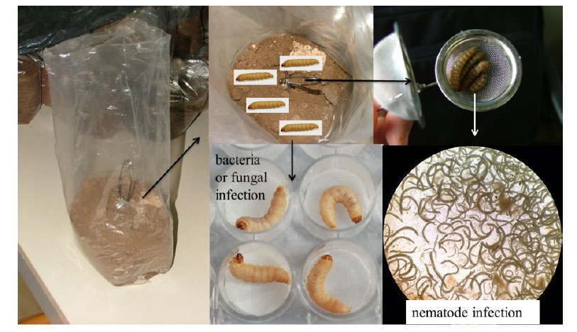
A total of 36 soil samples were collected over a period of 4 days during the second half of June 2018 in different biotopes of Arasbaran Biosphere, such as uncultivated soils, woodlands, river borders and grasslands. A hand shovel was used to collect approximately 2 Kg soil comprised each sample by pooling 3-4 sub samples taken at depths of 15-20 cm from an area of about 20 m2 [7]. The soil was transported in sterile polythene bags to the laboratory and prior baiting, water was added to give a content of 8-10% moisture and the samples were then stored at room temperature (~26°C). Final instar larvae of Galleria mellonella L. (Lepidoptera, Pyralidae) were used as bait insects to trap entomopathogens: for each soil sample a total of 8 Galleria larvae were released, 4 inside a long-handled tea infuser placed in the middle of the sample to attract the mobile entomopathogens (as the nematodes), and 4 released free on the top of each sample, to search for the static entomopathogens (as fungi conidia and bacteria spores) (Figure 1). The samples were kept at a room temperature and observations were done after 1 week to monitor the infected and dead larvae. The symptoms of cadavers after infection were recorded and used for diagnosis of EPNs, EPF or EPB induced infection. For the isolation of entomopathogenic nematodes dead larvae from each sample were placed in modified White traps [8] and kept at room temperature (~26°C). For the nematodes IJs were harvested and stored in distilled water at 8°C. These nematodes were used to infect fresh G. mellonella larvae and the progeny were used for identification and the establishment of cultures. Measurements were carried out on fresh specimens; the morphometric identification was based on infective juveniles and male morphology [1]. A molecular analysis followed for the EPN strains. For the isolation of entomopathogenic fungi and bacteria, infected wax moth larvae from each sample were surface sterilized by keeping them for 3 min. in 1% sodium hypochlorite and rinsing them in distilled water. After this, the larvae were incubated at 25°C in Petri dishes with moistened filter paper till the presence of pathogens could be assessed. For fungi, when sporulating structures appeared on the cadaver, attempts to isolate the fungus were made by transferring spores to potato dextrose agar in Petri dishes. For bacteria, infected hemolymph was cultured on specific media to isolate the bacteria colonies. For both, EPF and EPB, the inoculated Petri dishes were then checked every day and the tubes with pure cultures were sub-cultured in specific agar medium. Cultures were then stored at 8°C. For all the EPF and EPB entomopathogens the identification was made with a morphological analysis. For each sampling location, soil texture, time and type of vegetation were recorded (Table 1).

GEMS-2-1-404-g001

Figure 1. Soil sample with 8 Galleria larvae released, 4 inside a long-handled tea infuser and 4 released free on the top of each sample.

Table 1: Survey in Arasbaran Region (June 2018) – Sampling examination. For each sample habitat, soil texture and infection were recorded. Positive samples 34 of 36: EPN 8; EPF 26 (B: Beauveria, L: Lecanicillium, A: Aspergillus, F: Fusarium); EPB 5; Negative samples 2.

Sample

Habitat

soil texture

infection

Infected by

EPN

EPF

EPB

1

River border

Silty loam

+

*

*L

2

Corilus wood

Silty loam

+

*

3

Corilus wood

Silty loam

+

*L

4

Oak wood

Silty loam

+

*B

5

Wild meadow

Silty loam

+

*

*L

6

River border

Sandy loam

+

*B

7

River border

Sandy loam

+

*B

8

Oak wood

Silty loam

+

*

9

River border

Silty loam

+

*L*A

10

Wild meadow

Loamy clay

11

Wild meadow

Loamy clay

+

*B

12

Oak wood

loamy clay

+

*B

13

Wild meadow

Silty loam

+

*

14

Wild meadow

loamy caly

+

*

15

River border

Silty loam

+

*

16

Wild meadow

Silty loam

+

*B

17

Wild meadow

loamy caly

+

*B

18

Wild meadow

loamy clay

+

*B

19

Apple orchard

Silty loam

+

*L*A*F

20

River border

loamy clay

+

*

21

River border

loamy caly

+

*

22

River border

loamy sand

+

*L

23

Wild meadow

Silty loam

+

*

24

Wild meadow

loamy clay

+

*B

25

Oak wood

Silty loam

+

*B

26

Oak wood

Silty loam

+

*B

27

Oak wood

Silty loam

+

*B

28

Wild meadow

Silty loam

+

*

29

Wild meadow

Silty loam

+

*

30

Wild meadow

loamy clay

+

*B

31

Wild meadow

loamy caly

+

*B

32

Corilus wood

Silty loam

+

*

33

Wild meadow

Silty loam

+

*B

34

Oak wood

Silty loam

35

Urumy lake

Salty soil

+

*B

36

Urumy lake border

Salty beach

+

*B

Molecular Analysis

DNA extraction, PCR,  cloning  and  sequencing  were  performed  at IPSP laboratory, (Istituto per la Protezione Sostenibile delle Piante, Bari Italy), following the protocols described [9]. Individual nematodes were hand-picked, placed in 10 μl of lysis buffer (10 mM Tris-HCl, pH 8.8, 50mM KCl, 15 mM MgCl2, 0.1% Triton X100, 0.01% gelatin with 90 mg/ml proteinase K) on a glass slide and then cut into small pieces by using a sterilized syringe needle under a  dissecting  microscope. Each sample was incubated at 60°C for 1 hr and then at 95°C for  10 min. The crude DNA isolated from each individual nematode was directly amplified. The amplification of the ITS region was performed using the 18S forward primer (5’-TGATTACGTCCCTGCCTTT-3’) and the 26S reverse primer (5’-TTTCACTCGCCGTTACTAAGG-3’) [10],  D2-D3  expansion  segments  of  28S  rDNA  were  amplified using D2A (5’-ACAAGTACCGTGAGGGAAAGTTG-3’) and D3B (5’-TCGGAAGGAACCAGCTACTA-3’) (Nunn 1992), 18S rDNA using 18SnF (5’-TGGATAACTGTGGTAATTCTAGAGC-3’) and 18SnR (5’-TTACGACTTTTGCCCGGTTC-3’) primers [11]. PCR cycling conditions used for amplification were: an initial denaturation at 94°C for 5 min, followed by 35 cycles of denaturation at 94°C for 50s, annealing at 55°C for 50s and extension at 72°C for 1 min and a final step at 72°C for 7 min [9]. Following DNA amplification, 10 μl of PCR product was used for electrophoresis in 1X TBE buffer [12,13] in 1% agarose gel. A 100 bp ladder (Fermentas, St. Leon-Rot, Germany) was used as size marker. PCR products from individual nematodes were purified using the protocol listed by manufacturer (NucleoSpin Gel and PCR Clean-up, Machery Nagel, Germany ). Purified ITS, D2-D3 and 18S rRNA fragments were cloned in pGEM-T Easy Vector Systems (Promega, France) and sequenced at Eurofins genomics (Germany). ITS-RFLP analyses were performed  on 10 μl of PCR products from individual nematodes using five units of the following restriction enzymes: Dde I, Rsa I, Alu I, Hinf I (Promega, France). The restricted fragments were separated on a 2.5% agarose gel by electrophoresis. The gels were stained with gel red and visualized on a UV transilluminator and photographed with a digital system.

Results

Entomopathogens were recovered from 34 of 36 soil samples collected (94.4%): EPN strains were isolated in 8 sites and identify as Heterorhabditis bacteriophora Poinar. Strains of EPF were recovered from 26 soil samples (Table 1), Lecanicillium W. Gams & Zare came out from 6 soil samples (N. 1, 3, 5, 9, 19 and 22), Aspergillus Micheli from 2 soil samples (N. 9 and 19), Fusarium Link from N. 19 only and Beauveria Vuill. from 16 soil samples; strains of Bacillus thuringiensis (EPB) were isolated from 5 soil samples. In 4 samples more than one pathogen strain was recovered: from N.1 and N.5 H. bacteriophora and Lecanicilium sp., from N.9 Lecanicillium sp. and Aspergillus sp., and from N.19 three fungal strains Lecanicillium sp., Aspergillus sp. and Fusarium sp., with the first two isolated from the same larva. All the pathogen strains were recovered from different habitats and no correlation between the pathogen recovery and the characteristics of the sampling sites was observed, confirming the ubiquity of these entomopathogens.

Discussion and Conclusions

The presence and occurrence of entomopathogens is a key factor on the biological soil quality and these results represent a small contribution to the knowledge of the Arasbaran biodiversity also considering the methodology used for the pathogens isolations. The recovering of almost 95% of positive samples to infection was due to the combined used of baiting larvae for static and mobile entomopathogens; considering the ubiquity of these organisms, this modified baiting technique has maximized the possibility of insulation of entomopathogens from the soil.

Acknowledgment

The  research  was  carried  on  in  the  framework  of  “Della  Valle research  program”  between  the  Universities  of  Bari  “Aldo  Moro” (Italy) and Tabriz (Iran).

References

  1. Poinar GO Jr (1990) Taxonomy and biology of Steinernematidae and Heterorhabditidae. Entomopathogenic Nematodes in Biological Control. In: Gaugler R, Kaya HK (eds.) Boca Raton Fl, USA. CRC Press, pp: 23-61.
  2. Poinar GO Jr (1979) Nematodes for Biological Control of Insects. CRC Press, Boca Raton, Fl: 277.
  3. Nikdel M, Niknam G, Griffin C, Kary NE (2010) Diversity of entomopathogenic nematodes (Nematoda: Steinernematidae, Heterorhabditidae) from Arasbaran forests and rangelands in north-west Iran. Nematology 12: 767-773.
  4. Nikdel M, Niknam G, Ye W (2011) Steinernema arasbaranense sp.n. (Nematoda: Steinernematidae, a new entomopathogenic nematode from Arasbaran Forest, Iran. Nematol Medit 39: 17-28.
  5. Seifinejad A, Salehi Jouzani GR, Hosseinzadeh A, Abdmishani C (2008) Characterization of Lepidoptera-active cry and vip genes in Iranian Bacillus thuringiensis strain collection. Biological Control 44: 216-226.
  6. Salekjalali M, Barzegari A, Jafari B (2012) Isolation, Pcr Detection and Diversity of Native Bacillus thuringiensis Strains Collection Isolated from Diverse Arasbaran Natural Ecosystems. WorldApplied Sciences Journal 18: 1133-1138.
  7. Tarasco E, Clausi M, Rappazzo G, Panzavolta T, Curto G, et al. (2015) Biodiversity of entomopathogenic nematodes in Italy. Journal of Helminthology 89: 359-366. [crossref]
  8. White GF (1927) A method for obtaining infective nematode larvae from culture. Science 66: 302-303.
  9. De Luca F, Fanelli E, Di Vito M, Reyes A, De Giorgi C (2004) Comparison of the sequences of the D3 expansion of the 26S ribosomal genes reveals different degrees of heterogeneity in different populations and species of Pratylenchus from the Mediterranean region. European Journal of Plant Pathology 110: 949-957.
  10. Vrain TC, Wakarchuck DA, Levesque AC, Hamilton RI (1992) Intraspecific rDNA restriction fragment length polymorphism in the Xiphinema americanum group. Fundamental and Applied Nematology 15: 563-573.
  11. Kanzaki N, Futai K (2002) A PCR primer set for determination of phylogenetic relationships of Bursaphelenchus species within xylophilus group. Nematology 4: 35-41.
  12. Sambrook J, Fritschi EF, Maniatis T (1989) Molecular cloning: a laboratory manual (2ndedn), Cold Spring Harbor Lab Press, New York.
  13. Glare  TR,  Jurat-Fuentes  JL,  O’Callaghan  M  (2017)  Chapter  4:  Basic  and  Applied Research: Entomopathogenic Bacteria. Microbial control of Insect and mite pests. Academic Press, pp: 47-6.

A Cross-Cultural Adaptation of the Oxford Shoulder Score: The Arabic Version

DOI: 10.31038/IJOT.2020321

Abstract

Purpose: The Oxford Shoulder Score (OSS) is a widely used Patient-related Outcome tool used to assess patients with shoulder pain.The aim of this study was the translation and cultural adaptation of the Arabic version of the Oxford Shoulder Score as well as proving the reliability and validity of the score.

Methods: One hundred and fifty patients participated in this survey. The internal consistency tests were performed using Cronbach’s alpha. Test-retest reliability (intra-correlation coefficient), convergent construct validity, convergent validity, floor and ceiling effects and responsivenesswerealso calculated.

Results: Cronbach’s alpha coefficient was 0.95. The Intra-Class Correlation Coefficient (ICC) was 0.95. Regarding examining the validity of the Arabic version of OSS, there was a significant correlation between the Arabic OSS andthe previously published Arabic version of theDisabilities of the Arm, Shoulder and Hand Score (DASH Score) (p < 0.001).

Conclusion: Overall, Arabic version of OSS proved to be a good diagnostic tool for patients with shoulder pain.

Keywords

Oxford shoulder score;Shoulder pain;Reliability; Validation; Arabic

Abbreviations

OSS: Oxford Shoulder Score

ICC: Intra-Class Correlation Coefficient

DASH: Disabilities of the Arm, Shoulder and Hand Score

Introduction

Shoulder pain is a common problem, and a significant one at that. Its prevalence is estimated to be around 7% of the population, increasing to over 20% in people above 70 years of age [1-6]. Shoulder pain originates from peri-articular soft tissue disorders, mostly the rotator cuff [5,6]. Shoulder pain contributes to most visits to GPs, and even hospital specialists [7-10]. Surgery may be required by some patients, resulting in socio-economic implications due to morbidity and loss of productive hours [11].

It is very important that surgeons document the outcome from their practice for audit purposes; but then the major problem has been the lack of appropriate assessment records [12]. Many scoring methods have been developed over time to evaluate the outcome of orthopedic management of shoulder conditions. However, they are derived from radiological and clinical data and depend on the surgeon’s judgement [13,14]. It is becoming more apparent that most clinical assessments of major aspects of outcome are usually not reproducible and are inaccurate [15,16]. Also, there may be differences in the priorities and concerns of both the surgeon and his or her patient [17]. There is a need for methods which will elicit the perception of the patient’s outcome [18]. This has raised interests in patient-based assessments.

Studies have shown that patient reports can be valid, reliable, and sensitive to clinical change [19]. Evidence from research suggests that questionnaires issued to patients could be useful for clinical assessment in disorders of the shoulder [12]. In the event that a questionnaire is used, it should be practical, short, reliable, valid, and sensitive to clinical alterations and changes.The Oxford Shoulder Score (OSS)was first developed by Dawson et al. in 1996 [12]. The questionnaire is reported by patients, which includes 12 descriptors of pain and disability for shoulder ailments. The score of each descriptor ranges from 1 to 5, and the total score is calculated by adding the score of all 12 rated items from 12 (No pain) to 60 (highest level of Pain/Disability).

It encompasses general health questions, and questions specific to shoulder function and shoulder pain. The questions in the OSS are set in an easy context. Questions set are simple, and easily comprehended by patients; and so, patients do not need an explanatory instruction for independent usage. Although the Oxford shoulder score was initially used for prospective studies, a report by Wilson et al., suggested that it could as well be useful for retrospective application, for instance in acute trauma of the shoulder where it is not possible to take a prospective measurement [20]. A recent study of the range of Oxford Shoulder Score in the UK’s asymptomatic population showed that one of the major benefits of the study is the ability to gauge disease severity with respect to the index community’s normal population [21]. Another merit is the more accurate prediction of post-operative improvements.

Patient reported outcome scores have proven important in the medical field, providing informative and comparative subjective evidence that is quantified to help aid the medical practitioners when evaluating a patient’s health [7,22,23]. The difficulty of using such scores is due to the language restrictions of these scores as not all of them are provided for our regional language:Arabic. It would be beneficial to have the scores translated into Arabic to be used for patients who speak Arabic and do not extensively comprehend English. This will in turn enhance the ability for our physicians to better understand the population needs [23]. The Oxford Shoulder Score is very helpful in assessing patients with shoulder pain post-operatively [20,21,24].

The process of translating such scores and proving their effectiveness is lengthy yet extremely beneficial. They must follow specific guidelines ensuring linguistic integrity without the loss of the context [23].

The aim of this study was the translation and cultural adaptation of the Arabic version of the Oxford Shoulder Score as well as proving the reliability and validity of the score.

Methods and Materials

Patient Selection

One hundred and fifty patients participated in the survey.All were consecutive patients referred to the shoulder clinic between January to July 2019. The Protocol was approved by The Kuwaiti Ministry of Health Ethical Committee (2019/1068).We received the patient’s consent before filling the questionnaire.

Translation

Dawson et al. developed the original Oxford Shoulder Score in English, and had it translated based on the recommendations and guidelines in the literature [25-27]. Two bilingual orthopedic surgeons and one independent professional translator were involved in translation of the Arabic version. The authors held a coordination meeting which resulted in the production of a common version of the Arabic translation for the OES. Independent Arabic linguistics expert approved final draft. The questionnaire was distributed to a pilot of 11 patients to test the ease of language and understanding of the questions. No complaints were reported.

The Oxford Shoulder Score (OSS)

The OSS was first developed by Dawson et al. in 1996 [6]. The questionnaire was reported by patients and included 12 descriptors of pain and disability for shoulder ailments. The score of each descriptor ranges from 1 to 5, and the total score is calculated by adding the score of all 12 rated items from 12 (No pain) to 60 (highest level of Pain/Disability).

Disabilities of the Arm Shoulder and Hand (DASH)

The DASH Score consists of 30 items, which are reported by patients and designed to measure physical function and symptoms in patients with musculoskeletal disorders of the upper limbs [6]. The purpose of DASH Score is to describe the disability experienced by people with upper limb disorders and to monitor changes in symptoms and function over time following treatments [28]. It proved to be a reliable tool for physicians to investigate the joints in the upper extremity. The score of each item ranges from 1 to 5, and the total score is calculated by adding the score of all 30 rated items from 30 (No disability) to 150 (highest level of Disability).

To our current knowledge, the Arabic DASH (Alotaibi, 2010)score is the only relevant and available Arabic score. Therefore, it was used for testing the construct validity of the Arabic OSS.

Statistical Analysis

Internal Consistency

We evaluated the internal consistency by calculating the Cronbach’s alpha. The literature states that α > 0.70 is acceptable, while 0.95 should be the maximum to avoid redundancy [29]. Content validity was measured by examining the shape of data distribution, as well as floor and ceiling effects. Floor effect is the percentage of patients who scored the lowest possible score (score of 30), and ceiling effect is the percentage of those with the highest score (score of 150). If more than 30% of the respondents had a floor or ceiling effect, the effects would be considered to be relevant.

Reliability

In order to estimate the reliability of the questionnaire,Cronbach’s alpha was calculated. Since every patient completed the survey on two different occasions. The patients were asked to take the questionnaire a second time after 15 days from the initial test. To test the interobserver reliability, theCronbach’s alpha was calculated for each of the three test situations. Also, ICC (interclass correlation coefficient) was used to assess test-retest reliability.

Construct Validity

Spearman’s correlation coefficient between the OSS and DASH Score was calculated to test convergent validity of the OSS. Since the DASH Score has already been validated in Arabic speaking countries, higher correlation coefficient would prove convergent validity of the OSS.

Results

There have been 150 patients who have completed the OSS and DASH Outcome Score questionnaire and agreed to have their data analysed for research purposes. Average age of the participants is 46.2 years, with standard deviation of 14.6 years, which means that the majority of the sample is between 30 and 60 years of age. The youngest participant was 15, and the oldest was 74 years of age. It took an average of 4 minutes to complete the Arabic version of the OSS. For the purpose of a standardized score among OSS and DASH, all scores are rescaled to fall within the range between 30 and 150.

For all the items, in all the test periods, floor effect was 5% or less. On the other hand, no ceiling effect was recorded for any of items. Shapiro-Wilk test was used to check if the data in Arabic OSS significantly deviates from the normal distribution, which was not the case (0.801, p = 0.075).A p-value greater than 0.05 indicates that the data is normally distributed.

Reliability

Internal consistency was very high with overall Cronbach’s alpha value of 0.95. All items seemed to have strong correlations with the total score (r > 0.80). For the purpose of the test-retest reliability, the participants completed the OSS twice.The time between the test and re-test was 2 weeks and no significant difference was observed between the first and second assessment (p = 0.110). The value of ICC was ranged from 0.8 to 0.93 (Table 1). According to the previously published paper by Koo et al. values less than 0.5, between 0.5 and 0.75, between 0.75 and 0.9, and greater than 0.90 are indicative of poor, moderate, good, and excellent reliability, respectively (Koo et al. 2016). Thus, this indicates an excellent reliability.

Table 1: Internal consistency of Arabic OSS.

Item of OSS Score

Mean Score

SD

Item-Total Score Correlation*

Cronbach’sAlpha

1

2.21

0.91

0.85

0.96

2

2.51

0.84

0.82

0.95

3

3.01

0.76

0.90

0.93

4

3.06

0.77

0.85

0.97

5

2.68

0.90

0.84

0.97

6

2.71

1.12

0.86

0.94

7

2.45

0.65

0.90

0.95

8

2.01

1.20

0.80

0.96

9

2.09

0.87

0.93

0.92

10

2.74

0.88

0.89

0.93

11

2.21

0.84

0.88

0.95

12

2.38

1.12

0.82

0.96

*All ICC values are significant at 0.001 level.

Construct Validity

The Arabic OSS proved to be significantly correlated with the DASH Score, as was hypothesized in this study. The value of correlation between the OSS and the DASH Score was r = 0.92, which indicates a very high correlation and agreement between the two questionnaires.

Discussion

Dawson et al. presented the OSS as a disease-specific questionnaire designed for the patient for the evaluation of degenerative and inflammatory disorders of the shoulder way back in 1996 [12]. Since then, it has become a tool of choice in clinical assessments involving both operative and conservative treatments. The score has been validated in many cultures and languages, such as German, French, Dutch, Korean, and Italian [30-35]. The major finding of this study was that the Arabic version of OSS proved to be a reliable tool for assessment of patients with shoulder pain.

In this paper, we have outlined the step-by-step process of cross-cultural adaptation of the Oxford Shoulder Score in the Arabic language, and also given proof of its validity and reliability in patients suffering from shoulder disability and pain.

Shoulder pain accounts for 7-25% of the general population [36]. A 2005 study by Al-Awadhi et al. investigating the incidence of musculoskeletal pain in Kuwaiti adults found out that over 6.6% of the adult population were vulnerable [37]. Therefore, joint replacement, arthroscopic sport medicine shoulder surgery, and modern shoulder surgery has attracted the attention of orthopedic surgeons within the last two decades. Also, the current trend towards international meta-analysis and multicenter studies has resulted in an increased need for more standard clinical measures [38]. In the aspect of shoulder medicine and surgery, clinical instruments have been reported to be very responsive, comprehensive, reliable, and reproducible [39-41].

Reports suggest that various static measurements examine the reliability and agreement of the questionnaires. In our study, we had a Cronbach’s alpha coefficient of 0.95, with the Interclass Coefficients (ICC) ranging from 0.8 to 0.93. These values are similar to those of Slovene, German, Persian, Brazilian, and Thai versions (ICC = 0.84-0.95) [41-43]. This indicates an excellent value for the Arabic Oxford shoulder score to show reliability, thus confirming a high internal consistency for the Arabic OSS. As for the construct validity of the English OSS compared with the English DASH score, the Spearman’s correlation was 0.79 [16]. However, with the same comparison applied to the same scores in Arabic, a slightly higher correlation result of 0.92 was yielded from the analysis.

Conclusion

In conclusion, the translation of the OSS to Arabic was successful and confirmed to have good validity, reliability, and responsiveness. It can be used for assessment of the functioning/recovery status of Arabic patients with symptomatic shoulder malfunctions in treatments or clinical studies, assisting clinicians, or researchers to collect necessary data.

Acknowledgement

We are grateful to the authors of the original OSS. Their guidance through every stage of this research is deeply appreciated.

Conflict of Interest

None.

References

  1. Alotaibi NM (2010) Cross-cultural adaptation process and pilot testing of the Arabic version of the Disability of the Arm, Shoulder and Hand (DASH-Arabic). Hand Therapy 15: 80-86.
  2. Chakravarty KK, Webley M (1990) Disorders of the shoulder: An often unrecognised cause of disability in elderly people. BMJ 300: 848-849. [crossref]
  3. Chard MD, Hazleman R, King RH, Reiss BB (1991) Shoulder disorders in the elderly: a community survey. Arthritis Rheum 34: 766-769. [crossref]
  4. Badley EM, Tennant A (1992) Changing profile of joint disorders with age: Findings from a postal survey of the population of Calderdale, West Yorkshire, United Kingdom. Ann Rheum Dis 51: 366-371. [crossref]
  5. Van Schaardenburg D, Van den Brande KJ, Ligthart GJ, Breedveld FC, Hazes JMW (1994) Musculoskeletal disorders and disability in persons aged 85 and over: a community survey. AnnRheum Dis 53: 807-811. [crossref]
  6. Dawson J, Fitzpatrick R, Carr A (1996) Questionnaire on the perceptions of patients about shoulder surgery. J Bone Joint Surg Br 78: 593-600. [crossref]
  7. McCormick A, Fleming D, Charlton J (1995) Morbidity statistics from general practice. 4th National Study 1991-1992. London: HMSO; Office of Population Censuses and Surveys, Series MB5 No 3.
  8. Croft P (1998) Measuring up to shoulder pain. AnnRheum Dis 57: 650-66. [crossref]
  9. Bamji AN, Dieppe PA, Haslock DI, Shipley ME (1990) What do rheumatologists do? A pilot audit study. Br J Rheumatol 29: 295-298.
  10. Croft P, Pope D, Zonca M, O’Neill T, Silman A (1994) Measurement of shoulder related disability: results of a validation study. AnnRheum Dis 53: 525-528. [crossref]
  11. Ekberg K, Bjorkqvist B, Malm P, Bjerre-Kiely B, Axelson O (1994) Controlled two- year follow up of rehabilitation for disorders in the neck and shoulders. Occup Environ Med 51: 833-838. [crossref]
  12. Dawson J, Hill G, Fitzpatrick R, Carr A (2001) The benefits of using patient-based methods of assessment. Medium-term results of an observational study of shoulder surgery. J Bone Joint Surg Br 83: 877-882. [crossref]
  13. Koo, TK, Li MY (2016) A Guideline of selecting and reporting intraclass correlation coefficients for reliability research. Journal of Chiropractic Medicine 15: 155-163. [crossref]
  14. MacDonald DA (1993) The shoulder and elbow. In: Fairbank JC, Carr A, editors. Outcome Measures in Orthopaedics. Oxford: Butterworth Heinemann. pp. 144-173.
  15. Drake BG, Callaham CM, Dittus RS, Wright JG (1994) Global rating systems used in assessing knee arthroplasty outcomes. J Arthroplasty 9: 409-417.
  16. Conboy VB, Morris RW, Kiss J, Carr AJ (1996) An evaluation of the Constant-Murley shoulder assessment. J Bone Joint Surg Br 78: 229-232. [crossref]
  17. Wright JG, Rudicel S, Feinstein AR (1994) Ask patients what they want: Evaluation of individual complaints before total hip replacement. J Bone Joint Surg Br 76: 229-234. [crossref]
  18. Amadio PC (1993) Editorial. Outcomes measurement: More questions; same answers. J Bone Joint Surg Am 75: 1583-1584.
  19. Fitzpatrick R, Fletcher A, Gore S, Jones D, Spiegelhalter D, Cox D (1992) Quality of life measures in health care. I: Applications and issues in assessment. BMJ 305: 1074-1077. [crossref]
  20. Roy JS, MacDermid JC, Woodhouse LJ (2009) Measuring shoulder function: a systematic review of four questionnaires. Arthritis Rheum 61: 623-632. [crossref]
  21. Linsell L, Dawson J, Zondervan K, Rose P, Randall T, et al., (2006) Prevalence and incidence of adults consulting for shoulder conditions in UK primary care; patterns of diagnosis and referral. Rheumatology (Oxford) 45: 215-221. [crossref]
  22. Masnoon N, Shakib S, Kalisch-Ellett L, Caughey, GE (2018) Tools for Assessment of the Appropriateness of Prescribing and Association with Patient-Related Outcomes: A Systematic Review. Drugs & Aging 35: 43-60. [crossref]
  23. Byrne BM (2016) Adaptation of assessment scales in cross-national research: Issues, guidelines, and caveats. International Perspectives in Psychology: Research, Practice, Consultation 5: 51-65.
  24. Uhtoff HK, Sarkar K (1990) An algorithm for shoulder pain caused by soft tissue disorders. Clin Orthop 254: 121-127. [crossref]
  25. Guillemin F, Bombardier C, Beaton D (1993) Cross-cultural adaptation of health- related quality of life measures: Literature review and proposed guidelines. J Clin Epidemiol 46: 1417-1432. [crossref]
  26. Acquadro C, Jambon B, Ellis D, Marquis P (1996) Language and translation issues. In: Spilker B, editor. Quality of Life and Pharmacoeconomics in Clinical Trials. 575-585.
  27. Anderson RT, MacFarlane M, Naughton MJ, Shumaker SA (1996) Conceptual issues and considerations in cross-cultural validation of generic health-related quality of life instruments. 2nd (edn.). Philadelphia: Lippincott.
  28. Kennedy CA, Beaton DE, Smith P, VanEerd D, Tang K, et al., (2013) Measurement properties of the Quick DASH (disabilities of the arm, shoulder and hand) outcome measure and cross-cultural adaptations of the Quick DASH: a systematic review. Qual Life Res 22: 2509-2547. [crossref]
  29. Fayers PM, Machin D (2013) Quality of life: the assessment, analysis and interpretation of patient-reported outcomes. Manhattan: John Wiley & Sons.
  30. Huber W, Hofstaetter JG, Hanslik-Schnabel B, Posch M, Wurnig C (2004) The German version of the Oxford Shoulder Score–cross-cultural adaptation and validation. ArchOrthop Trauma Surg 124: 531-536. [crossref]
  31. Tuğay U, Tuğay N, Gelecek N, Özkan M (2011) Oxford Shoulder Score:cross-cultural adaptation and validation of the Turkish version. ArchOrthop Trauma Surg 131: 687- 694. [crossref]
  32. Murena L, Vulcano E, D’Angelo F, Monti M, Cherubino P (2010) Italian cross- cultural adaptation and validation of the Oxford Shoulder Score. J Shoulder Elbow Surg 19: 335-341. [crossref]
  33. Frich LH, Noergaard PM, Brorson S (2011) Validation of the Danish version of Oxford Shoulder Score. Dan Med Bull 58: 4335. [crossref]
  34. Jamnik H, Spevak MK (2008) Shoulder Pain Disability Index: Validation of Slovene version. Int J Rehabil Res 31: 337-341. [crossref]
  35. Ebrahimzadeh MH, Birjandinejad A, Golhasani F, Moradi A, Vahedi E, et al., (2015) Cross-cultural adaptation, validation, and reliability testing of the Shoulder Pain and Disability Index in the Persian population with shoulder problems. Int J Rehabil Res 38: 84-87. [crossref]
  36. Bjelle A (1989) Epidemiology of shoulder problems. Baillieres Clin Rheumatol 3: 437- 451.
  37. Al-Awadhi AM, Olusi SO, Al-Saeid K, Moussa M, Shehab D, et al., (2005) Incidence of musculoskeletal pain in adult Kuwaitis using the validated Arabic version of the WHO-ILAR COPCORD Core Questionnaire. AnnSaudi Med 25: 459-462. [crossref]
  38. Ekeberg OM, Bautz-Holter E, Tveitå EK, Keller A, Juel NG, Brox JI (2008) Agreement, reliability and validity in 3 shoulder questionnaires in patients with rotator cuff disease. BMC Musculoskelet Disord 9: 68. [crossref]
  39. Wilson J, Baker P, Rangan A (2009) Is retrospective application of the Oxford Shoulder Score valid? J Shoulder Elbow Surg 18: 577-580. [crossref]
  40. Younis F, Sultan J, Dix S, Hughes PJ (2011) The range of the Oxford Shoulder Score in the asymptomatic population: a marker for post-operative improvement. AnnR Coll Surg Engl 93: 629-633. [crossref]
  41. Martins J, Napoles BV, Hoffman CB, Oliveira A (2010) The Brazilian version of Shoulder Pain and Disability Index: translation, cultural adaptation and reliability. Rev Bras Fisioter 14: 527-536. [crossref]
  42. Phongamwong C, Choosakde A (2015) Reliability and validity of the Thai version of the Shoulder Pain and Disability Index (Thai SPADI). Health Qual Life Outcomes 13: 136. [croosref]
  43. Roach KE, Budiman-Mak E, Songsiridej N (1991) Development of a shoulder pain and disability index. Arthritis Care Res 4:143-149. [crossref]

Autism is Physiological, Nonautism is Pathological Biologically Speaking

DOI: 10.31038/PSYJ.2020223

Abstract

A brief functional and historical analysis is offered of the question, what is the physiological difference between human beings and other animals? All animals are autistic, even those equipped with a much higher intelligence than the human one is, like whales, for example. So the non-autism of most human beings is what needs explanation.

Nonautism is the consequence of a misunderstanding – a creative misunderstanding, that is. It consists in the suddenly conceived suspicion that the other was trying to make you happy – an absurd idea.While it is true that brood-caring animals get rewarded by signs of being satisfied emitted by the young, as a confirmation of their own brood-rearing efforts being momentarily successful, this reward is purely one-directional. There is no reason conceivable why the offspring ought to be rewarded by the adults being successful in anything (like feeding them). It is always only the effect that counts in biology. Survival is the currency, nothing else.

But in humans, the young are being rewarded by the displayed joy of adults? Exactly this is the case. Humans are a pathological species in this respect. Actually, such a functional “wiring” can be proved to be pathological in evolution – that is, to be subject to predictable fast evolutionary elimination, should it have arisen once.How, then, could this pathological type of wiring actually arise in evolution, as it did with the human species? The answer is: By accident. It is a very specific accident at stake here that can be pinpointed.

It is the phenomenological overlap between the innate (wired-in) expressions of bonding on the one hand and the expression of well-being and friskiness on the other.In many animals, this expression of friskiness and happiness and enthusiasm, if shown by the young, has a rewarding effect on the adults, for it signals that their attempts at successful brood-rearing are on the right track. It is sweet to watch this rewardability channel in action. Many predatorial animals are socially wired in this way among each other.

But this is not our question here. How come that the young are rewarded by the playful friskiness of the adults in some species? The “playing mode” has this very structure in many social animals. Play occurs when more “serious” needs are satisfied and the partners can invest energy in silly, that is not survival relevant activities, as a means to tighten their social bonds. Play makes peace and friendship. But our context is non-autism. How could it arise in one species so far? What arose here is very easy to see: Mutual rewardability by the expression of playfulness on the other side. By the playful mode.Homo ludens. All social animals are wired like this in play.

But there is a risk involved here: The suspicion of benevolence encountered arising on one side. For play is highly rewarding. Can there for this reason arise a “serious” component?. This is what has occurred in the human species. It happens in play as in the other high-performance social animals, but not only in play. “Homo ludens” (humans are players) even when not playing? The positive feedback that arises in play can indeed be maintained so as to enter all other social activities. The smile-laughter is the play mode. Wolves can enter the same mutually inspired frenzy. They are very much like human beings even though the expressions are not so much shown on the face as with the tail.

So what is the functional difference? That wolves are not mirror competent is the answer. So the “suspicion of benevolence encountered” cannot arise in the wolf. But it can and does – in the human wolf. The “werwolf” sagas show how irritated humans are by the close emotional similarity of the wolf.There are other highly social animals like dolphins and whales and also some octopuses and giant creatures from the depth. And don’t forget the elephants. Are we humans not wired socially in much the same manner, even if being somewhat less intelligent? There is one difference functionally speaking, however.

What is it? The smile of happiness, the expression of happiness, is at the same time the smile of bonding. It is the same expression.But is this not the same thing with the wolf? Yes, it is. And presumably so with the African wild dog, Lycaonpictus, as well?And maybe with some sepiae yet to be investigated.

Now I have left you in the dark long enough about what we are headed for: The positive feedback of the smile acting symmetrically. Not only when bonding, but also when merely playfully happy. Or just happy. The consequence is the sudden invention of the suspicion of one’s encountering benevolence.It is absolutely nonsensical, right? There is nothing but hard-wired couplings here. There is no room for such humanistic notions like “benevolence.” But actually, this transgression is exactly what is happening and taking place here between us.

It could happen also between orcas. Or between an orca and a human.Or between Kanzi and his wonderful trainer. Not because but in spite of the silly machine inserted in between them. Big revolutionary progress sometimes takes unnecessary roundabout ways. We will be able to ask Kanzi. He will understand the question and tell us.Come on: It can’t possibly be that simple! But it is. We walked together through a nice little wood of well-defined observational biological notions above and do now suddenly see ourselves in a mirror. Yes, the question posed at the outset – the onset of the suspicion of encountering benevolence – is really being raised here.

Will the reader tolerate having been pulled into this nightmare of a daydream: Of a functional understanding of what benevolence and love is? On a level infinitely more powerful than the other social domain of sex is, for example?The reader may feel that we will have to come back to this fascinating topic together again? Take care.

Low Level Diode Laser Therapy versus Surgical Abrasion in the Treatment of Gingival Racial Pigmentation – Clinical Study

DOI: 10.31038/JDMR.2020324

Abstract

Background: Racial gingival hyper pigmentation is a common esthetic concern, which is aggravated in patients with excessive gingival display, laser ablation is recognized as the most effective, and reliable easy handling technique.

The aim of this study was to evaluate the effect of the Low Level Diode Laser (LLDL) therapy versus surgical abrasion in the treatment of racial gingival pigmentation clinically by assessment of Melanin Pigmentation Index (MPI), Wond Healing Index (WHI), and Visual Analog Scale (VAS).

Subjects and Methods: The present study is randomized, clinical split mouth design, twenty patients of both genders with age ranged from 20 to40 years, all have racial gingival pigmentation score 2 were included in this study, all patients were non pregnant or lactating, free from systemic diseases, or any disease that cause oral pigmentation), all patients were nonsmokers, and had esthetic concern and good oral hygiene, forty sites in twenty patients were divided randomly into two groups (A and B), group A were treated by surgical abrasion at first was performed by large size round carbide bur started from the mucogingival junction toward the free gingival margin, including papillae, the wound covered by non eugenol periodontal dressing, group B: were treated by LLDL without dressing post treatment, the treated site was irradiated for a single time, LLDL was delivered by a 600-μm diameter fiber, 808 nm ± 10 nm wavelength with output power 3 Watt.

Results: There was improvement in MPI in both groups through follow up periods with superiority of LLDL group.

Conclusion: Low level diode laser can be considered as a reliable mean for elimination of melanotic lesion of gingiva and is well tolerated by patients.

Keywords

Racial gingival hyper pigmentation, Low level diode laser, Surgical abrasion

Introduction

Healthy oral mucosa is pink in color, but it can vary from light to dark pink depending on the thickness of epithelium, the amount of melanin and the number of erythrocytes present in the connective tissue vessels and their proximity to the surface. Gingival pigmentation is defined as color change of the gingiva from variable shades of pink to dark brown or black due to genetic factors which is the most important endogenous factor responsible for increased production of melanin by melanocytes, or pathological factors. Some other exogenous factors such as ultra violet radiation (UV), smoking and medications may also increase melanin synthesis [1]. Genetics Racial gingival pigmentation is a common esthetic concern, which is aggravated in patients with excessive gingival display and more common noticed in adults, the attached gingiva is the most common site of involvement in the anterior region of the maxilla and mandible followed by the buccal mucosa, lip corners, lip mucosa, dorsal and ventral surface of the tongue and rarely the floor of the mouth [2] Kauzman [3] were broadly classified the gingival pigmentation into: physiologic and pathologic, all patients except albinos have some degree of physiologic melanin distribution throughout epidermis, which occurs in all races of man without significant difference in oral pigmentation between males and females, the intensity and distribution of racial pigmentation of the oral mucosa is variable, not only between races, but also between different individuals of the same race [4]. Dummettet [5] suggested, that the degree of physiologic pigmentation is partially related to mechanical, chemical and physical stimulation. In darker skinned people oral pigmentation increase, but there is no difference in the number of melanocytes between fair skinned and dark skinned individuals, as it is due to greater melanocyte activity rather than greater number of melanocytes [6,7] and it clinically manifests as multifocal or diffuse melanin pigmentation, which is common in African, Asian and Mediterranean populations, Melanin, a brown pigment, is the most common natural pigment contributing to endogenous pigmentation of gingiva, it is produced by the melanocytes, which are mainly present in the basal and suprabasal layers of the gingival epithelium [8]. Several therapeutic techniques have been proposed and employed for gingival depigmentation, Roshni and Nandakumar [9] classified different gingival depigmentation methods into: methods aimed to mask the pigmented gingival as free gingival graft and acellular dermal matrix allograft, and methods aimed to remove the pigmented gingiva; the chemical methods these methods are no longer in use because of the destructive nature and difficulty in controlling the depth of their penetration and the surgical methods as, scalpel surgical technique, electrosurgery and radiosurgery. cryosurgery, bur abrasion; the first documented case using this technique was reported by Ginwalla [10] it is a relatively simple and versatile technique and requires minimum time and effort, and Lasers therapy which have been introduced to dentistry for more than three decades also been used for treatment of gingival pigmentation, [11] the word LASER is an acronym for light amplification by stimulated emission of radiation. The principle of the laser was the first known in 1917 when physicist Einstein [12] who described the theory of stimulated emission, there are two types of lasers: hard lasers such as carbon dioxide and neodymium-doped yttrium aluminium garnet which offer both hard tissue and soft tissue applications and cold or soft lasers based on the semiconductor diode devices, which are used predominantly for low-level diode laser therapy (LLDT) [11]. Lasers have been proposed as a useful method for the removal of the gingival melanotic pigmentations with many advantages include haemostatic capacity, no need for periodontal dressing, and fewer postoperative complications such as pain, oedema, or infections and a good visualization when compared with conventional surgical methods [13]. Laser light has four types of interactions with the target tissue which depend on the laser wavelength, emission mode, and optical properties of the tissues components such as pigments, mineral content, water content; absorption, transmission of laser energy, reflection, and scattering of the laser light these four phenomena occur together in some degree relative to each other [14]. The absorption of the laser energy by the intended tissue is the first and most desired interaction, different laser wavelengths have different absorption coefficients with the dental tissue components [14]. In general, the longer wavelengths, such as erbium laser has a greater affinity with water and hydroxyapatite, the shorter wavelengths ranging from 500 to 1000 nm are readily absorbed by the pigmented tissue and blood elements. For e.g., the pigment results of hemoglobin has greater affinity for argon laser while melanin absorbs diode and Nd:YAG laser and they have less effect on heamoglobin [15] depending on the wavelength used, some lasers able to penetrate the tissue deeper than others, in contrast other laser has a limited penetration and has effect only on the surface tissue, for example, the (Nd:YAG)  which is indicated for bone and hard  tissue applications, penetrates 2-5 mm into tissue [16,17] CO2 laser has a limited penetration up to 0.03 to 0.1 mm in the tissue, thus indicated for soft tissue applications, this wavelength provides enough depth to seal the damaged blood, lymphatic vessels and nerve endings resulting in good hemostasis and minimal post-operative morbidity [18] transmission of the laser energy directly through the tissue with no effect on the target tissue, the inverse of absorption, this effect is highly dependent on the wavelength of laser light, reflection, which causes laser light to redirects itself off the surface, having no effect on the target tissue, this reflected light could be dangerous when redirected to an unintended target such as eyes, a caries detecting laser device uses the reflected light to measure the degree of sound tooth structure [14,18]. Scattering of the laser light accompanied with correspondence decrease of laser energy and possibly producing no useful biologic effect, this property can cause unwanted damage as there is heat transfer to the tissue adjacent to the surgical site, however a beam deflected in different directions facilitates the curing of the composite resin or when treating an aphthous ulcer [14,18]. There are two laser delivery systems, the first is a flexible hollow waveguide or tube that has an interior mirror finish, the laser energy is reflected along this tube and exits through a hand piece at the surgical end with the beam striking the tissue in a noncontact fashion [17,19]. The second delivery system is a glass fiber optic cable, this cable can be more pliant than the waveguide, has a corresponding decrease in weight and resistance to movement, and is usually smaller in diameter, this fiber system can be used in contact or noncontact mode [19]. There are three different laser emission modes are described: [19] the first is continuous wave, where the beam is emitted at only one power level for as long as the operator depresses the foot switch, eg: diode (used in contact mode with water cooling system) and CO2 (used in no contact mode), the second is termed gated-pulse mode, meaning that there are periodic alternations of the laser energy, much like a blinking light, this mode is achieved by the opening and closing of a mechanical shutter in front of the beam path of a continuous wave emission, e.g. diode laser used in contact mode, the third mode is the free-running pulsed mode, sometimes referred to as “true pulsed”, this emission is unique in that large peak energies of laser light are emitted for a short time span, usually in microseconds, followed by a relatively long time in which the laser is off, the timing of this emission is computer controlled, not mechanically controlled as in a gated pulse device, this mode can be used for thin tissue, as: Neodymium: Yttrium, Aluminum, Garnet (Nd:YAG), Cr: YAG and Er:Cr:YSGG, if the laser is in a pulsed mode, the targeted tissue has time to cool before the next pulse of laser energy is emitted. But in continuous wave mode, the operator must cease the laser emission manually so that thermal relaxation of the tissue may occur, [20] diode lasers known as an injection laser, is a semiconductor device that produces coherent radiation (in which the waves are all the same frequency and phase) in the visible or infrared (IR) spectrum when current passes through it, [21-23] the active medium of the diode laser is a solid state semiconductor made of Aluminum, Gallium, Arsenide, and occasionally Indium, which produces laser wavelengths, ranging from approximately 810 nm to 980 nm, all diode wavelengths are absorbed primarily by tissue pigment (melanin) and hemoglobin, conversely, they are poorly absorbed by the hydroxyapatite and water present in the enamel, these lasers can also stimulate fibroblastic proliferation at low energy levels [23]. The chief advantage of the diode lasers is one of a smaller size, portable instrument, all diode laser beams are carried to the target tissue through quartz glass fibers, this is the simplest and the most effective means of conducting laser energy from its origin in the device to the oral cavity, this fibers are relatively thin between 200 and 600 microns in diameter also, there are disposable fiber-optic tips which can be discarded after use [23]. The diode laser has become the most commonly utilized laser in dentistry it has assumed a prominent role in managing soft tissues, diode lasers can be used in continuous wave  mode,  used  for  most  surgical  procedures  such  as ablating, incising and excising all types of intraoral soft tissue surgery and pulsed mode where more control over laser output is desirable in removal of coronal pulp adjunct to root canal procedures, tooth whitening, coagulation of extraction sites, disinfecting the periodontal pocket and sulcular debridement therefore it may be useful as an adjunctive means for scaling and root planing due to its bactericidal and detoxification effects, diode laser is highly absorbed by melanin pigment, so it is one of surgical treatment modalities of gingival pigmentation [23]. Moreover, diode lasers can be used with powers well below the surgical threshold for photobiomodulation or biostimulation properties which give the benefit for acceleration of healing, increasing circulation, reducing edema and minimizing pain, increased collagen synthesis, fibroblast proliferation, increased osteogenesis, and anti-inflammatory effect to treat maladies such as recurrent herpes and aphthous stomatitis [22] Materials and methods.

Subjects and Methods, the present study is randomized, clinical split mouth design, twenty patients of both genders with age ranged from 20 to40 years old, all have racial gingival pigmentation score 2 were included in this study and selected from the  Outpatient Clinic Department of Oral Medicine and Periodontology, Faculty of Dentistry, Tanta University, all patients were non Pregnant or lactating, free from systemic diseases, or any disease that cause oral pigmentation (e.g. Addison disease), all patients were nonsmokers patients with esthetic concern and with good oral hygiene, the purpose of the study was explained to the patients and informed consents were obtained, forty sites in twenty patients were divided randomly into two groups (A and B), and each site on right or left side of maxilla or mandible were considered from the mesial aspect of central incisor to the distal aspect of first premolar, group A were treated by surgical abrasion (Figure 1 and 2), at first local anesthesia was given (infiltration technique, 1.8 ml mepivacaine 2%) and the depigmentation was performed by large size round carbide bur started from the mucogingival junction toward the free gingival margin, including papillae, bleeding was stopped by applying pressure by a gauze piece on the denuded epithelium, care was taken to include  the epithelium at the tip of interdental papilla and the mucogingival junction on the other end without disturbing the marginal gingiva. The wound covered by non eugenol periodontal dressing. Group B: were treated by low level diode laser (LLDL) (Figure 3), without dressing post treatment, patient and operating staff wore special diode-laser protective eye glasses, highly reflective instruments or instruments with mirrored surfaces were avoided as there could be reflection of the laser beam, the treated site was irradiated for a single time, the soft tissue diode surgical laser unit delivered by a 600-μm diameter fiber (Figure 4), 808 nm ± 10 nm wavelength with output power 3 Watt, was used for depigmentation, during the  procedure  laser  ablated the gingival epithelial surfaces little by little to reach the pigments without causing any bleeding for clear visualization, the fiber tip was continuously moved across the site to avoid heat accumulation at any site, the wounds appeared fresh with no bleeding and were no need to apply periodontal dressing. Figures 5 and 6 Patients were instructed to continue good oral hygiene and to avoid eating hot or spicy food for the first 3 days, not to traumatize the area during the healing period which is 4-7 days after treatment and were instructed to use chlorhexidine mouth wash twice daily preferably after meals for one week, no pain medications were generally prescribed, but if needed a mild analgesic anti-inflammatory (sodium diclofenac 50 mg) twice a day was given, however no antibiotics were prescribed. Post-operative assessment: clinical parameters were evaluated using wound healing index (WHI) based on the following scoring system: [24] score A: complete epithelization, score B: incomplete  epithelization,  score C: ulcer formation, score D: tissue defect or necrosis; melanin pigmentation index (MPI) (Figure 7), based on the following scoring system: [25] score 0: no pigmentation, score 1: solitary units (s) of pigmentation in papillary gingiva without extension between neighboring solitary units, score 2: formation of continuous ribbon extending from neighboring solitary units, and visual analog scale (VAS) [26] is a sheet of paper with horizontal line on it has a left end marked as no symptoms and a right end marked as worst imaginable symptoms, as well as a mark indicating the midpoint, the scores were calculated as follows0 = no pain, 0.1-3.0 cm = slight pain, 3.1-6.0 cm = moderate pain, 6.1-10.0 cm = severe pain (Figure 8), clinical assessment were taken for both groups as shown in Table 1.

JDMR-3-2-307-g001

Figure 1. Rotary gingival abrasion by large rose head bur (Group A).

JDMR-3-2-307-g002

Figure 2. Application of periodontal pack after surgical abrasion.

JDMR-3-2-307-g003

Figure 3. Showing the diode laser divece laser (elexxion claros nanocompactclass IV dental, 600 µm optic fiber, Radolfzell,Germany).

JDMR-3-2-307-g004

Figure 4. 600 µm optic fiber.

JDMR-3-2-307-g005

Figure 5. Applicalion of LLDL (group B).

JDMR-3-2-307-g006

Figure 6. Depithlzation and carbonization of gingiva.

JDMR-3-2-307-g007

Figure 7. Scores of MPI.

Table 1: Comparison of MPI along the follow up periods in group A.

Melanin pigmentation index

At the baseline

2 Weeks

3 Months


χ2


MCp

No.

%

No.

%

No.

%

Score 0:

0

0.0

17

85.0

2

10.0


61.494*


<0.001*

Score1:

0

0.0

3

15.0

10

50.0

Score 2:

20

100.0

0

0.0

8

40.0

Sig. bet. periods

MCp1<0.001*, MCp2<0.001*, MCp3<0.001*


χ2: Chi square test for comparing along the follow up periods.
MCp: p value for Monte Carlo for Chi square test for comparing between the three periods.
p1: p value for comparing between baseline and 2 Weeks.
p2: p value for comparing between baseline and 3 Months.
p3: p value for comparing between 2 Weeks and 3 Month.
* Statistically significant at p ≤ 0.05.

Results

No  patient  had  marked  complications  or  infection  during   the healing of the gingiva, regarding melanin pigmentation index assessment in group A, there was a significant reduction in the gingival pigmentation from the baseline (before treatment) (100% +ve score 2) to 2 weeks post treatment and (15%+ve score 1) and the rest of cases were free from pigmentation, but at 3 months post treatment there was increase in the recurrence rate of pigmentation as (40%+ ve score 2 & 50% +ve score 1 and 10% free of pigment) (Table 2, Figure 9), however in group B at 2 weeks post laser treatment the melanin gingival pigmentation appear in 5% of cases (score1) only and the rest of cases remain free (score 0), and at 3 months post treatment 65% of cases remain free of repigmentation (score 0) as (30% score1, 5% of cases score 2 and the rest of cases remain score 0). In Table 3 and Figure 10 by comparing the results of MPI through the both groups there was statistically significant improvement in MPI in group B than group A showed in Table 2 and Figure 11.

Table 2: Comparison of MPI along the two studied groups at the follow up periods.


Melanin pigmentation index

Group A

Group B


χ2


MCp

No.

%

No.

%

At the baseline


Score 0

0

0.0

0

0.0


Score 1

0

0.0

0

0.0

Score 2

20

100.0

20

100.0

2 Weeks


Score 0

17

85.0

19

95.0


1.111


0.605

Score 1

3

15.0

1

5.0

Score 2

0

0.0

0

0.0

3 Months


Score 0

2

10.0

13

65.0


14.728*


0.001*

Score 1

10

50.0

6

30.0

Score 2

8

40.0

1

5.0

χ2: Chi square test for comparing between group A and group B.
MCp: p value for Monte Carlo for Chi square test for comparing between group A and group B.
*Statistically significant at p ≤ 0.05.

JDMR-3-2-307-g008

Figure 8. Recording the pain score by VAS.

JDMR-3-2-307-g009

Figure 9. Comparison of MPI along the follow up periods in group A.

Table 3a: Study assessment and the times of assessment.

Clinical assessment

Before treatment

Immediately post treatment

After 2 weeks

After 1 month

After 3 month

Clinical photographs

+

+

+

+

+

Melanin pigmentation score MPI

+

+

+

Wound healing score WHI

+

+

+

Pain score VAS

+

+

+

Table 3b: Comparison of MPI along the follow up periods in group B.

Melanin pigmentation index

At the baseline

2 Weeks

3 Months

c2

MCp

No.

%

No.

%

No.

%

Score 0

0

0.0

19

95.0

13

65.0


64.446*


<0.001*

Score 1

0

0.0

1

5.0

6

30.0

Score 2

20

100.0

0

0.0

1

5.0

Sig. bet. periods

MCp <0.001*, MCp <0.001*, MCp =0.044* 1                                                  2                                              3


χ2: Chi square test for comparing along the follow up periods.
MCp: p value for Monte Carlo for Chi square test for comparing between the three periods.

JDMR-3-2-307-g010

Figure 10. Comparison of MPI along the follow up periods in group B.

JDMR-3-2-307-g011

Figure 11. Comparison of MPI along the two studied groups at the follow up periods.

Table 4: Comparison of WHI along the two studied groups in the follow up periods.


Wound healing

Group A

Group B

No.

%

No.

%

At baseline


Score A

0

0.0

0

0.0

Score B

0

0.0

0

0.0

Score C

20

100.0

20

100.0

Score D

0

0.0

0

0.0

1 Month


Score A

20

100.0

20

100.0

Score B

0

0.0

0

0.0

Score C

0

0.0

0

0.0

Score D

0

0.0

0

0.0

3 Months


Score A

20

100.0

20

100.0

Score B

0

0.0

0

0.0

Score C

0

0.0

0

0.0

Score D

0

0.0

0

0.0

Table 5: Comparison the mean value of VAS scores along the two studied groups at the follow up periods.

Evaluation of pain

Group A

Group B

Z

p

At the baseline


Min.-Max.

0.50-6.0

0.50-2.50


3.572*


<0.001*

Mean ± SD.

3.33 ± 1.56

1.57 ± 0.57

Median

3.50

1.50

After 2 week


Min. -Max.

0.0-1.0

0.0-0.0


2.762*


0.006*

Mean ± SD.

0.30 ± 0.38

0.0 ± 0.0

Median

0.0

0.0

After 3 months


Min. -Max.

0.0-0.0

0.0-0.0


0.0


1.000

Mean ± SD.

0.0 ± 0.0

0.0 ± 0.0

Median

0.0

0.0

Regarding Pain score (VAS)

Inter group comparison of VAS at both groups (A and B): The results showed a statistically significant reduction in the mean value of VAS in group B as compared to group A at the baseline (P=0.001) and at two weeks post treatment (P=0.006). However there was statistically non-significant difference in the mean value of pain score between both groups at three month post treatment (P=0.317), with superiority of diode laser (Tables 4, 5 and Figures 12-14).

JDMR-3-2-307-g012

Figure 12. Comparison of WHI along the two studied groups in the follow up periods.

JDMR-3-2-307-g013

Figure 13. Comparison of the mean value of VAS scores along the two studied groups at the follow up periods.

JDMR-3-2-307-g014

Figure 14. Comparison of the mean value of VAS scores along the two studied groups at the follow up periods.

Discussion

Gingival abrasion technique was selected in present study as it is relatively simple, safe, non-aggressive method and easy to perform, it causes less discomfort, economical as no sophisticated equipment required and is esthetically acceptable to the patients [27,28] moreover in case of recurrence, the procedure can be done repeatedly in the same areas without clinical limitations or permanent damage, [16] the diode laser is a solid state semiconductor laser that typically uses a combination of gallium (Ga), arsenide (Ar), and other elements such as aluminum (Al) and indium (In) to change electrical energy into light energy; of wavelength range from 810 nm to 900 nm, which is poorly absorbed in water, but has an affinity for hemoglobin, melanin and other pigment, resulting in much more selective destruction and far less damage to normal gingival tissue than does with CO2 laser (100) thermal effects of diode laser are attributed to ‘hot tip’ effect caused by accumulation of heat at the end of fiber, these effects result in the production of a thick coagulation layer on the treated surface, [29] Soliman [30] and Sathyanarayanan and Hari [31] reported that diode laser is a useful safe mean, and is the preferred laser for treatment of the pigmented areas of the gums when no other short Lasers are available, the present study included patients who were medically free from any systemic disease associated with pathological hyper pigmentation or improper delayed wound healing as uncontrolled diabetes, and autoimmune diseases, and non-treated periodontal disease to avoid the interference with wound healing process as these diseases affect the inflammatory mediators production, [32] also pregnant women were excluded as the hormonal changes during pregnancy and lactation causes increase of melanin production which can affect the recurrence time of pigmentation and there is no a lot of information provide to be sure that the use of laser during pregnancy and lactation is safe, [33] smokers patients were excluded, to avoid the masking of the results by smoker melanosis as smoker’s melanosis is the most common lesions in smokers’ mouth and 30% of Caucasian heavy smokers have oral melanin pigmentation [34] the MPI was assessed at the baseline (before treatment), 2 weeks and 3 months post treatment, and the results showed a significant improvement in MPI scores in both groups along the follow up periods with superiority of diode Laser as In group A, the recurrence of gingival pigmentation started at two weeks post treatment in 15% of cases and increased gradually till three months as appeared in 90% of cases but In group B, the recurrence of pigmentation started at two weeks post treatment in 5% of cases and increased gradually till three months post treatment as appeared in 35% of cases; the recurrence  of melanin pigments which was observed at the end of three months in both groups (A and B), could be a result of the ongoing process  of repigmentation, but this repigmentation was not as pre-treatment level in intensity of color, may be due to the lesser production of pigments, and so the intensity of pigmentation may increase with time and may reach to pre-treatment level as it depends on the racial background of the patient, the present results was consistent with  the findings of Bergamaschi [35] who demonstrated that permanent results cannot be offered when gingival depigmentation procedures are performed for cosmetic reasons, repigmentation also may due to the fact that the melanocytes have a reproductive self-maintaining system of cells, and they when locally depleted, they repopulate and keratinocyte-derived growth factors “Fibroblast Growth Factor-β” act as a mitogen, these cells lack desmosomes and possess long dendritic processes that extend between keratinocytes, melanin is synthesized in the melanocytes in small structures called melanosomes, these melanosomes are injected into the keratinocytes by the dendritic processes, all individuals, whether lightly or darkly pigmented, have the same number of melanocytes in any given region of the mucosa, but it has been observed that cells which contain melanin are present in connective tissue in the case of individuals who have a very high melanin pigment score, these cells are actually macrophages that have engulfed the melanin pigment, [36] reappearance of pigmentation indicates that the melanocytes may have repopulated the treated sites from adjacent tissues, and these may be explained by Soliman [30] who discussed the use of soft-tissue diode laser in the treatment of oral racial pigmentation and found that some underlying melanocytes were not sufficiently affected by the heat to be destroyed which causes repopulation of melanocytes at treated areas causing repigmentation of gingiva (Figures 15-18).

JDMR-3-2-307-g015

Figure 15. (a) Preopertive clinical photograph show patient has score2 of MPI at the baseline. (b) The site immediatelyposttreatment. (c) MPI (score 0) at two weeks post treatment. (d) Complete epthilization and keratinzation of gingivaat one month. (e) Core 2 ofMPI at three months post treatment.

JDMR-3-2-307-g016

Figure 16. (a) Preopertive clinical photograph show patient has score 2 of MPI at the baseline. (b) The site immediatelyposttreatment. (c) MPI (score 0) at two weeks post treatment. (d) Complete epthilization and keratinzation of gingivaat one month post treatment. (e) MPI (score 2) at three months post treatment.

JDMR-3-2-307-g017

Figure 17. (a) Preopertive clinical photograph show patient has score 2 of MPI at the baseline. (b) the site immediatelypost application of LLDL. (c) MPI (score 0) at two weeks post treatment. (d) complete epthilization and keratinzation of gingivaat one month post treatment. (e) MPI (score 1) at three months post treatment.

JDMR-3-2-307-g018

Figure 18. (a) Preopertive clinical photograph show patient has score 2 of MPI at the baseline. (b) The site immediatelypostapplication of LLDL. (c) MPI (score 0) at two weeks post treatment. (d) Complete epthilization and keratinzation of gingivaat one month post treatment. (e) MPI (score 1) at threemonths post treatment.

Furthermore, Azzeh [37] who stated that because of the presence of rete pegs in oral epithelium, the ablation of the epithelium at the same level may not remove all melanocytes which repopulate the treated area and cause repigmentation [38,39]. The clinical findings of the present study were in agreement with results of the study carried by Sathanarayanan and Lyer Mahajan [31,39] also, Abdullah and Al- shmaah study [40]. However, the results were not in agreement with the clinical results of the study conducted by Bakutra [41] they showed that at twelve months post treatment in all sites; repigmentation was observed with different grades of Hedin index, and there pigmentation in surgical stripping is significantly lesser compared to laser ablation, as lesser numbers of melanocytes were found on immune histological examination at twelve months postoperatively in sites treated by scalpel, the difference in the results may be due to different in the race of patients or difference in the degree and intensity of gingival pigment before treatment. Regarding WHI assessment the healing of the depigmented gingiva was uneventful irrespective of the techniques used, however the abrasion treated sites healed faster compared to LLDL treated sites, but this did not reach a statistically significant level and also it was necessary to cover the exposed lamina propria with a periodontal pack for seven days, although the healing of diode laser wounds were slower than that of surgical abrasion wounds, a sterile inflammatory reaction occurs after laser use, [42] evaluation of both procedures on thirty days post treatment revealed restoration of normal features of the gingiva without any scar formation, which supported by the results of Moritz [27] study as they reported a bactericidal effect of LLDL moreover it has the unique property of being able to remove a thin layer of epithelium [9,43]. The LLDL wounds were initially covered by a carbonized layer then after 24 hour were covered by a thick white fibrinous layer which is a normal characteristic of a laser wound during the first several days of healing due to the relatively thick coagulation layer on the treated surface produced by the “hot tip” of the diode laser fiber optic [44]. Gingival recession and loss of papilla were not observed. At one month post treatment the gingiva showed a normal appearance with pink color irrespective of the techniques used. The delayed healing of LLDL wounds may be due to the fact that the usual mechanism of LLDL which lead to ablation or decomposition of biological cellular materials is thermal, the thermal ablation means that the energy delivered by the laser interacts with irradiated material by an absorption process, yielding rapid raise in the intercellular temperature and pressure lead to cellular rupture, necrosis and delayed healing [45,46] also it leads to release of vapors and cellular debris termed the laser plume, which creates locally sterile condition, resulting in a reduction of bacteremia [47]. The results of the present study were in agreement with the clinical results of the study conducted by [41,47-50]. The post-operative experience of pain is a complex phenomenon, influenced by psychological, environmental  and physical factors, VAS score a reliable and practical method to assess pain in clinical settings, sensitive to treatment effect and the its data derived can be analyzed using parametric statistical techniques [26]. In this study, there was a statistically significant improvement in VAS scores in both treatment groups, as in group A all patients suffer from pain ranged from mild to moderate, immediately post treatment, however pain gradually decreased till disappear completely after two weeks in all patients except two cases still complained from mild pain, only with external stimuli, like food chewing and this may be due to individual variation in pain threshold, but in group  B only slight or no pain was recorded,  however, the pain had reduced considerably  at two weeks post treatment. Laser depigmentation procedure as compared to abrasion method had significantly lesser postoperative pain immediately after surgery (P<0.05), the increased pain perception associated with the abrasion method might be attributed to the fact  that it is a more intrusive surgical procedure involving blood loss and  a wide open surgical wound, which cause discomfort postoperatively since it heals by secondary intention, and slight pain after using LLDL may be attributed by formation of protein coagulum on the wound surface serving as a biological barrier, also laser irradiation seals the sensory nerve endings and inhibits the transfer of pain signals and has analgesic effects [51] caused by a disruption of Na+-K+ pump in the cell membrane, resulting in a loss of impulse conduction or simply due to an ablation of the nerve endings because of protein coagulum formation [51]. Also, in the present study, a low power of LLDL was used(1.02 W)as higher power (>2.5 W) would manifest as discomfort and pain during the post-operative follow up periods, moreover could delay the healing time, therefore, as a rule, a low-power setting (≤2.5 W) was used during the procedure [43]. The results were in agreement with the results of the study conducted [41,52].

Conclusion

Both, LLDL and surgical abrasion were safe for treatment of oral racial pigmentation, and they showed significant clinical improvement in Melanin Pigmentation Index (MPI) with superiority of low level diode lasers until 3 months follow up without any post-surgical problem. LLDL documented as there was minimal postoperative pain and discomfort, ease of the procedure and more acceptable for patients as compared to surgical abrasion.

Recommendation

Multiple sessions of LLDL is recommended in some patients, removal of gingival melanin pigmentation should be performed cautiously and the adjacent teeth should be protected, since inappropriate application may cause gingival recession, damage to underlying periosteum and bone, delayed wound healing, as well    as loss of enamel. Further studies are  required  for  standardized laser parameters (as wave length and frequency of used laser) in the management of the different scores of gingival pigmentation.

References

  1. Brenner M, Hearing VJ (2008) Modifying skin pigmentation – approaches through intrinsic biochemistry and exogenous agents. Drug DiscovToday Dis Mech 5: 189-199. [crossref]
  2. Dummett CO, Barens G. (1971) Oromucosal pigmentation: An updatedliterary review. J Periodontol 42: 726-736. [crossref]
  3. Kauzman A, Pavone M, Blanas N, Bradley G (2004) Pigmented lesions of the oral cavity: Review, differential diagnosis, and case presentations. J Can Dent Assoc 70: 682-683. [crossref]
  4. Ozbayrak S, Dumlu A, Ercalik-Yalcinkaya S (2000) Treatment of melanin-pigmented gingiva and oral mucosa by CO2 laser. Oral Surg Oral Med Oral Pathol Oral 90: 14-15. [crossref]
  5. Dummett CO, Sakamura J, Barrens G (1981) Attitudes towards normal pigmentation of the oral mucosa. Quintessence Int 10: 115-22. [crossref]
  6. Esen E, Haytac MC, Oz IA, Erdoğan O, Karsli ED (2004) Gingival melanin pigmentation and its treatment with the CO2 laser. Oral Surg Oral Med Oral Pathol Oral RadiolEndod 98: 522-527.
  7. Gorsky M, Buchner A, Fundoianu-Dayan D, Aviv I (1984) Physiologic pigmentation of the gingiva in Israeli Jews of different ethnic origin. Oral Surg Oral Med Oral Pathol 58: 506-509. [crossref]
  8. Ciçek Y, Ertas U (2003) The normal and pathological pigmentation of oral mucous membrane: A review. J Contemp Dent Pract 4: 76-86. [crossref]
  9. Roshni T, Nandakumar K (2005) Anterior esthetic gingival depigmentation and crown lengthening: report of a case. J ContempDent Pract 3: 139-147. [crossref]
  10. Ginwalla TM, Gomes BC, Varma BR (1966) Surgical removal of gingival pigmentation. J Indian Dent Assoc 38: 147-150.
  11. Dederich DN, Bushick RD (2004) ADA council on scientific affairs, division of science; Journal of the American Dental Association. Lasers in dentistry: separating science from hype. J Am Dent Assoc 135: 204-212.
  12. Einstein A (1917) Strahlungs- emission and absorption of radiation in quantum theory. Verhandlungen der Deutschen Physiklischen Gesellschaft.Concept and theory of stimulated light emission. 18: 318-323.
  13. Lee KM, Lee DY,  Shin SI, Kwon  YH, Chung JH, Herr Y (2011) A comparison    of different gingival depigmentation techniques: ablation by erbium:yttrium- aluminum-garnet laser and abrasion by rotary instruments. J Periodontal Implant Sci 41: 201-207. [crossref]
  14. Kumar M, Grishmi M, Girish N, Harish B (2017) State of the art laser technology in dentistry. Heal Sci 3: 3-6.
  15. (1996) Lasers in periodontics. J Periodontol 67: 826-830.
  16. Coluzzi DJ (2004) Fundamentals of dental lasers: science and instruments. Dent Clin North Am 48: 751-70. [crossref]
  17. Rossmann JA, Cobb CM (2000) Lasers in periodontal therapy. Periodontal 9: 150- 164.
  18. Sulieman M (2005) An overview of the use of lasers in general dentist practice: Laser physics and tissue interaction. Dent Updat 32: 228-236. [crossref]
  19. Aoki A, Sasaki KM, Watanabe H, Ishikawa I (2004) Lasers in nonsurgical periodontal therapy. Periodontol 36:59-97.
  20. Clayman L, Kuo P (1997) Laser in Maxillofacial Surgery and Dentistry. New York: Thieme 1-9.
  21. Stirban A, Gawlowski T, Roden M (2013) Vascular effects of advanced glycation endproducts: Clinical effects and molecular mechanisms. Mol Metab 3: 94-108. [crossref]
  22. White JM, Goodis HE (1993) Thermal laser effects on intraoral soft tissue, teeth and bone in vitro In: Proceedings of the third international congress on lasers in dentistry international society for lasers in dentistry; 1993: University of Utah Printing Services 189-90.
  23. John JG (2015) Diode Lasers: A Primer. AGD 1-10.
  24. Tal H, Oegiesser D, Tal M (2003) Gingival depigmentation by erbium: YAG laser: Clinical observations and patient responses. J Periodontol 74: 1660-1667. [crossref]
  25. Takashi H, Tanaka K, Ojima M, Yuuki k (2005) Association of melanin pig mentation in the gingiva of children with parents who smoke pediatrics 116: 86-90. [crossref]
  26. Wewers ME and Lowe NK (1990) A critical review of visual analog scales in the measurement of critical phenomena. Research in Nursing and Health 13:227-236. [crossref]
  27. Moritz A, Gutknecht N, Doertbudak O, Goharkhay K, Schoop U et al. (1997) Bacterial reduction in periodontal pockets through irradiation with a diode laser: A pilot study. J Clin Laser Med Surg 15: 33-37. [crossref]
  28. Farnoosh AA (1995) Treatment of gingival pigmentation and discoloration for esthetic purposes. IntJPeriodontics Restorative Dent 10: 312-319. [crossref]
  29. Kravitz ND, Kusnoto B (2008) Soft-tissue lasers in orthodontics: an overview. AmJ Orthod Dentofacial Orthop 133: 110-114. [crossref]
  30. Soliman MM, Al Thomali Y, Al Shammrani A, El Gazaerly H (2014) The use of soft tissue diode laser in the treatment of oral hyper pigmentation. Int J Health Sci 8: 133-1340.
  31. Sathyanarayanan C, lyerH (2014) A comparative study between conventional method and diode laser in treatment of gingival hyperpigmentation. International Journal of laser dentistry 4: 8-19. [crossref]
  32. Agrawal A, Gang TB, Rusiñol AE (2014) Recognition functions of pentameric C-reactive protein in cardiovascular disease. Mediators Inflamm 2014: 319215. [crossref]
  33. Mcleod SD, Ranson M, Mason RS (1994) Effects of estrogens on human melanocytes in vitro. J Steroid Biochem Mol Biol 49: 9-14.
  34. Axell T, Hedin CA (1982) Epidemiologic study of excessive oral melanin pigmentation with special reference to the influence of tobacco habits. Scand J Dent Res 90: 434-442. [crossref]
  35. Bergamaschi O, Kon S, Doine AI, Ruben MP (1993) Melanin repigmentation after gingivectomy: A 5-year clinical and transmission electron microscopic study in humans. IntJ Periodontics Restorative Dent 13: 85-92. [crossref]
  36. Ashri N, GaziM (1990) More unusual pigmentations of the gingiva. Oral Sur Oral Med Oral Pathol 70: 445-449. [crossref]
  37. Azzeh MM (2007) Treatment of gingival hyperpigmentation by erbium- doped:yttrium, aluminum, and garnet laser for esthetic purposes. J Periodontol 78: 177-184.42. AshriN, GaziM (1990) Moreunusual pigmentations of the gingiva Oral Sur Oral Med Oral Pathol 70:445-449. [crossref]
  38. Dummett CO (1946) Physiologic pigmentation of the oral and cutaneous tissues in the Negro. J Dent Res 25: 421-432. [crossref]
  39. Mahajan G, Kaur Ht, Jain S, Kaur N, Kaur NS, et al. (2017) Compare the gingival melanin repigmentation after diode laser application and surgical removal done by scraping with Kirkland knife. IndianSociety of Periodontology.
  40. Abdullah A B and Al-shmaah AZ (2014) The Use of ErCrYSGG versus diode laser in gingival melanin de-pigmentation, Clinical study. IJERSTE 3: 2319-7463.
  41. Bakutra G, Shankarapillai R, Mathur L, and Manohar B (2017) comparative evaluation of diode laser ablation and surgical stripping technique for gingival depigmentation: A clinical and immunohistochemical study. International Journal of Health Sciences11: 51-58.
  42. Kaarthikeyan G, Jayakumar ND, Padmalatha O, Varghese S, Kapoor R (2012) Pain assessment using a visual analog scale in patients undergoing gingival depigmentation by scalpel and 970 nm diode laser surgery. J Laser Dent. 20: 20-23.
  43. Pick R, Colvard M (1993) Current status of laser in soft tissue dental surgery. J Periodontol 64: 589-602. [crossref]
  44. Ojha A, Srivastava1 V (2015) Gingival depigmentation with diode laser, electrosurgery and scalpel: a comparative report of 2 cases TMU. J Dent 2: 1.
  45. Coluzzi DJ (2004) Fundamentals of dental lasers: Science and instruments. Dent Clin North Am 48: 751-770. [crossref]
  46. Simşek Kaya G, YapiciYavuz G, Sümbüllü MA, Dayi E (2012) A comparison of diode laser and Er: YAG lasers in the treatment of gingival melanin pigmentation. Oral Surg Oral Med Oral Pathol Oral Radiol 113: 293-299. [crossref]
  47. Rossmann JA, Gottlieb S, Koudelka BM, McQuade MJ (1987) Effects of CO2 laser irradiation on gingiva. J Periodontol 58: 423-425. [crossref]
  48. (2016) Comparative evaluation of two surgical techniques using conventional scalpel method and diode laser for treatment outcome of depigmentation: 6 months follow- up study. J Dent Lasers 10: 2-9.
  49. Grover HS, Dadlani H, Bhardwaj A, Yadav A, Lal S (2014) Evaluation of patient response and recurrence of pigmentation following gingival depigmentation using laser and scalpel technique: A clinical study. J Indian Soc Periodontol 18: 586-592. [crossref]
  50. Lagdive S, Doshi Y, Marawar P (2009) Management of gingival Hyper pigmentation using surgical blade and diode laser therapy: a comparative study. J Oral Laser Applications9: 41-47. [crossref]
  51. Schroeder HE (1969) Melanin containing organelles in cells of the human gingival I. Epithelial melanocytes. J Periodont Res. 4: 1-18. [crossref]
  52. Suragimath G, Lohana MH, Varma S (2016) A split mouth randomized clinical comparative study to evaluate the efficacy of gingival depigmentation procedure using conventional scalpel technique or diode laser. J Lasers Med Sci 7: 227-232. [crossref]

Disease, Duration and Death

DOI: 10.31038/IMROJ.2020533

Abstract

Life has always been threaten by diseases, calamities, catastrophes leading to death caused by various known or unknown, animate or inanimate objects in human’s relatively medium life span. Ever since the documentation of the human history, it is well known that man loved their body and prefer to live in accordance with their wishes. When rationale judgment became prominent after the experiences and observations of life and death events, they started searching remedies such as medicine. This is how medicine evolved since our early civilization. With the development of reason, logic, observation, experimentation and practical application, we learned tremendous ways of saving body, brain and behavior. However, as time passes human environment changes unpredictably leading to change in human behavior and attitude towards objects/materials and living beings. It is not only a matter of physical, biological or cosmic change but also behavior of everything that brought unprecedented events such as unexpected war, epidemic, catastrophes etc. leading to death [1,2]. Measurement of several physical parameters of human and universal bodies has become routine but various functions/characters in relation to time has yet to measure fully. This is the point we fall short to save humans promptly resulting high number of unexpected loss of life such as in COVID-19 pandemic. Among 1554960 covid-19 infected population in more than 209 countries, territories and two conveyances 5.9% died, and among the deaths more than 80% occurring in just 10 countries (USA, Spain, Italy, Germany, France, China, Iran, UK, Belgium, Netherlands) of the world in the last three months duration [2].

Disease is an abnormal architecture/anatomy, function, condition of the body and mind in a specific duration. Many times and circumstances death occurs due to unprecedented cause, behavior or ignorance. Therefore, it is essential to know the unknown environment and diverse nature and behavior of human beings to diagnose epidemicity of the disease. Despite vast scientific discoveries and new achievement, there is a big hole in the measurement of core human behavior and intelligence. Human body, intelligence and behavior plays a great role in the defense mechanism as well as association in the causation, development, cessation of disease in specific duration in specific place/s. So far we are devoid of the precise knowledge on the creation of covid-19 however many scientists have been trying to explore the mystery of the occurrences, nature and impact on the human population of the globe [3].

The duration or natural course of illness or diseases is important in the management of cases, carrier as well as prevention of complications and death [4]. Alert researchers identify the key factors of the disease when there is sudden rise of cases of similar features in a short period. Ignorance about the nature of pathogen and ignorance of the general population about the disease leads to higher number of deaths in a very short duration. Lack of alertness in changing behavior and environment of the disease in the population further complicates its management and increases the number of deaths. The challenge of the new disease, ignorance on the part of environment and human behavior help to expand disease dimensions in terms of time, place and person.

Opportunities such as chance, experience, observation and experimentation lead to discovery and development of medicine and care system that can make our life easier, comfortable and lengthier. This is the beauty of medical discipline, research and practice in human population. A dynamic patience where a body and brain searches a remedy continuously in response to disease is probably the best stimulus to initiate new knowledge, skills, practice to cure patient and prevent death. Lack of precise knowledge of duration and the nature of the disease is biggest obstacles in managing covid-19 at present and many more diseases that are possible in the future. Following the spread of disease and management of the patient (source) meticulously in global environment, recording the evidences and continuous sharing among the fellow researchers and responsible individuals are the most important aspects of pandemic control.

Alertness, continuous searches, dynamic patience can help humans to increase its capacity to deal with covid-19 pandemic. Change in seasonality in different geographical regions may affect duration of the diseases and distribution of death in humans. This demands thinking globally and acting globally.

Keywords

Covid-19, Death, Disease, Duration, Pandemic

References

  1. Riedel S (2004) Biological warfare and bioterrorism: a historical review. BUMCProceedings17: 400-406. [crossref]
  2. Covid-19 Coronavirus Pandemic, Worldometer. Accessed on April 09, 2020, 16:30 GMT.
  3. Zhou P, Yang X, Wang X, Hu B, Zhang L, et al. (2020) A pneumonia outbreak associated with a new coronavirus of probable bat origin. Nature579: 270-273. [crossref]
  4. Rothan HA,ByrareddySN (2020) The epidemiology and pathogenesis of coronavirus disease (COVID-19) outbreak. Journal of Autoimmunity109: 102433. [crossref]

Differences between 5-Minute and 15-Minute Measurement Time Intervals of the CGM Sensor Glucose Device Using GH-Method: Math-Physical Medicine (No. 281)

DOI: 10.31038/IMROJ.2020532

Introduction

This paper describes the research results by comparing the glucose data from a Continuous Glucose Monitor (CGM) sensor device collecting glucose at 5-minute (5-min) and 15-minute (15-min) intervals during a period of 125 days, from 2/19/2020 to 6/23/2020, using the GH-Method: math-physical medicine approach. The purposes of this study are to compare the measurement differences and to uncover any possible useful information due to the different time intervals of the glucose collection.

Methods

Since 1/1/2012, the author measured his glucose values using the finger-piercing method: once for FPG and three times for PPG each day. On 5/5/2018, he applied a CGM sensor device (brand name: Libre) on his upper arm and checked his glucose measurements every 15 minutes, a total of ~80 times each day. After the first bite of his meal, he measured his Postprandial Plasma Glucose (PPG) level every 15 minutes for a total of 3-hours or 180 minutes. He maintained the same measurement pattern during all of his waking hours. However, during his sleeping hours (00:00-07:00), he measured his Fasting Plasma Glucose (FPG) in one-hour intervals.

With his academic background in mathematics, physics, computer science, and engineering including his working experience in the semiconductor high-tech industry, he was intrigued with the existence of “high frequency glucose component” which is defined as those lower glucose values (i.e. lower amplitude) but occurring frequently (i.e.. higher frequency). In addition, he was interested in identifying those energies associated with higher frequency glucose components such as the various diabetes complications that would contribute to the damage of human organs and to what degree of impact. For example, there are 13 data-points for the 15-minute PPG waveforms, while there are 37 data-points for the 5-minute PPG waveforms. These 24 additional data points would provide more information about the higher frequency PPG components.

Starting from 2/19/2020, he utilized a hardware device based on Bluetooth technology and embedded with customized application software to automatically transmit all of his CGM collected glucose data from the Libre sensor directly into his customized research program known as the eclaireMD system, but in a shorter time period for each data transfer. On the same day, he made a decision to transmit his glucose data at 5-minute time intervals continuously throughout the day; therefore, he is able to collect ~240 glucose data within 24 hours.

He chose the past 4-months from 2/19/2020 to 6/19/2020, as his investigation period for analyzing the glucose situation. The comparison study included the average glucose, high glucose, low glucose, waveforms (i.e. curves), correlation coefficients (similarity of curve patterns), and ADA-defined TAR/TIR/TBR analyses. This is his secondresearch report on the 5-minute glucose data. His first paper focused on the most rudimentary comparisons [1].

References 2 through 4 explained some example research using his developed GH-Method: math-physical medicine approach [2,3].

Results

The top diagram of Figure 1 shows that, for 125 days from 2/19/2020 – 6/23/2020, he has an average of 259 glucose measurements per day using 5-minute intervals and an average of 85 measurements per day using 15-minute intervals. Due to the signal stability of using Bluetooth technology, for the 5-min, it actually has 259 data instead of the 240 data per day.

IMROJ-5-3-516-g001

Figure 1. Daily glucose, 30-days & 90-days moving average glucose of both 15-minutes and 5-minutes.

The middle diagram of Figure 1 illustrates the 30-days moving average of the same dataset as the “daily” glucose curve. Therefore, after ignoring the curves during the first 30 days, we focus on the remaining three months and can detect the trend of glucose movement easier than “daily” glucose data chart. There are two facts that can be observed from this middle diagram. First, the gap between 5-min and 15-min is wider in the second month, while the gap becomes smaller during the third and fourth month. This means that the 5-min results are converging with the 15-min results.Secondly, both curves of 5-min and 15-min are much higher than the finger glucose (blue line). This indicates that the Libre sensor provides a higher glucose reading than the finger glucose. From the listed data below, the CGM sensor daily average glucoses are about 8% to 10% higher than the finger glucose.

5-min sensor: 118 mg/dL (108%)

15-min sensor: 120 mg/dL (110%)

Finger glucose: 109 mg/dL (100%).

The bottom diagram of Figure 1 is the 90-days moving average glucose. Unfortunately, his present dataset only covers 4 months due to late start of collecting his 5-min data; however, the data trend of the last month, from 5/19-6/23/2020, can still provide a meaningful trend indication. As time goes by, additional data will continue to be collected, his 5-min glucose’s 90-days moving trend will be seen more clearly.

Figure 2 shows the synthesized views of his daily glucose, PPG, and FPG.Here, “synthesized” is defined as the average data of 125 days.For example, the PPG curve is calculated based on his 125×3=375 meals. Listed below is a summary of his primary glucose data (mg/dL) in the format of “average glucose/extreme glucose”. Extreme means either maximum or minimum, where the maximum for both daily glucose and PPG due to his concerns of hyperglycemic situation, and the minimum for FPG due to his concerns of insulin shock. The percentage number in prentice is the correlation coefficients between the curves of 15-min and 5-min.

Daily (24 hours):15-min vs. 5-min

117/143vs. 119/144(99%)

PPG (3 hours):15-min vs. 5-min

126/135vs. 125/134(98%)

FPG (7 hours):15-min vs. 5-min

102/95 vs. 105/99 (89%).

Those primary glucose values between 15-min and 5-min are close to each other in the glucose categories. It is evident that the author’s diabetes conditions are under well control for these 4 months. However, by looking at Figure 2 and three correlation coefficients %, we can see that daily glucose and PPG have higher similarity of curve patterns (high correlation coefficients of 98% and 99%) between 15-min and 5-min, but FPG curves have a higher degree of mismatch in patterns (lower correlation coefficient of 89%). This signifies that his FPG values during sleeping hours have a bigger difference between 15-min and 5-min.

IMROJ-5-3-516-g002

Figure 2. Synthesized daily glucose, PPG, and FPG of both 15-minutes and 5-minutes.

Figure 3 are the results using candlestick model [4,5]. The top diagram is the 15-min candlestick chart and the bottom diagram is the 5-min candlestick chart. Candlestick chart, also known as the K-Line chart, includes five primary values of glucoses during a particular time period; “day” is used in this study. These five primary glucose data are:

Start: beginning of the day.

Close: end of the day.

Minimum: lowest glucose.

Maximum: highest glucose.

Average: average for the day.

Listed below are five primary glucose values of both 15-min and 5-min.

15-min: 108/116/86/170/120.

5-min: 111/116/84/173/118.

IMROJ-5-3-516-g003

Figure 3. Candlestick charts of both 15-minutes and 5-minutes.

By ignoring the first two glucoses, start and close, let us focus on the last three glucoses: minimum, maximum, and average. The 5-min method has a lower minimum and a higher maximum than the 15-min method. This is due to the 5-min method capturing more glucose data; therefore, it is easier to catch the lowest and highest glucoses during the day. The difference of 2mg/dL between 15-min’s average 120 mg/dL and 5-min’s average 118 mg/dL is only a negligible 1.7%.

Again, it is also obvious from these candlestick charts that the author’s diabetes conditions are under well control for these 4 months.

Conclusion

In summary, the glucose differences between 5-min and 15-min based on simple arithmetic and statistical calculations are not significant enough to draw any conclusion or make any suggestion on which are the “suitable” or better measurement time intervals. However, the author will continue his research to pursue this investigation of energy associated with higher-frequency glucose components in order to determine the glucose energy’s impact or damage on human organs (i.e. diabetes complications).

The author has read many medical papers about diabetes. The majority of them are related to the medication effects on glucose symptoms control, not so much on investigating and understanding “glucose” itself. This situation is similar to taming and training a horse without a good understanding of the temperament and behaviors of the animal. Medication is like giving the horse a tranquilizer to calm it down. Without a deep understanding of glucose behaviors, how can we truly control the root cause of diabetes disease by only managing the symptoms of hyperglycemia?

References

  1. Hsu, Gerald C. eclaireMD Foundation, USA (2020) Analyzing CGM sensor glucoses at 5-minute intervals using GH-Method: math-physical medicine (No. 278).
  2. Hsu, Gerald C. eclaireMD Foundation, USA(2020) Predicting Finger PPG by using Sensor PPG waveform and data via regression analysis with three different methods using GH-Method: math-physical medicine (No. 249).
  3. Hsu, Gerald C. eclaireMD Foundation, USA (2019) Applying segmentation pattern analysis to investigate postprandial plasma glucose characteristics and behaviors of the carbs/sugar intake amounts in different eating places using GH Method: math-physical medicine (No. 150).
  4. Hsu, Gerald C. eclaireMD Foundation, USA (2019) A case study of the impact on glucose, particularly postprandial plasma glucose based on the 14-day sensor device reliability using GH-Method: math-physical medicine (No. 124).
  5. Hsu, Gerald C. eclaireMD Foundation, USA. Comparison study of PPG characteristics from candlestick model using GH-Method: Math-Physical Medicine (No. 261).

Management of Cancer Patients Undergoing Radiation Therapy during the Novel Coronavirus Disease 2019 (COVID-19) Pandemic: A Review of the Literature

DOI: 10.31038/CST.2020531

Abstract

Cancer patients are more vulnerable to acquiring COVID 19 infection and may also experience higher morbidity and mortality. In the context of COVID-19, cancer patients may be affected through delayed diagnosis and have significant impact on management in resource strained settings. Cancer treatment typically involves a possible combination of surgical resection, chemotherapy and radiation therapy (RT). RT delivery requires often daily attendance to a cancer center, is complex and poses potentially additional risks for infection as well as treatment related complications. Optimization of infection control measures and RT treatment schedules is paramount to minimize the impact of the pandemic on patients and optimize outcomes. This review aims to summarize the existing limited literature surrounding RT administration and optimization in the context of COVID-19.

Introduction

Since the recognition of the COVID-19 pandemic, evidence has become available that a cancer diagnosis is considered one of the comorbid conditions that increase the risk of COVID 19 infection [1- 3]. Meanwhile COVID-19 infection in cancer patients is associated with higher morbidity and mortality [4]. Data is still preliminary, but it is likely that both the increased risk of acquiring COVID-19 and more severe consequences thereof in cancer patients are multifactorial in nature likely involving complex relationships between the type of cancer site and extent or location of the disease as well as more nuanced patient and treatment related factors. While a number of publications have delved into the presentation and management of COVID-19, and its relationship to comorbid conditions including malignancy, and yet others into the implications of systemic management (chemotherapy, targeted agents), fewer publications specifically discuss the implications for patients who undergo radiation therapy (RT) and the operational considerations of radiation therapy (RT) departments. RT is administered mostly on a daily basis in cancer centers or facilities with complex logistics, unique organizational demands, high possibility of interaction between vulnerable patients and risk of exposure to multiple patients and staff. The lack of evidence surrounding best practices has left radiation oncology providers and patients with many questions still unanswered.

These questions include the level of vulnerability of cancer patients, who for example may undergo radiation but may not be undergoing systemic management and may therefore not necessarily be immune- compromised, as well as the implication for daily attendance to a RT facility and the associated infection risk and how to best mitigate it. Other questions involve the ability to and safety of delaying or altering RT treatment schedules to minimize risk of infection. Important additional considerations abound in particular settings such a RT emergencies and treatment of the COVID-19 positive patient as well as the administration of radioactive isotopes such as radioactive iodine in thyroid patients who then need to self-isolate at home or as inpatients for several days. Since the COVID-19 pandemic has raised unprecedented challenges and questions, with ongoing lack of robust data, periodic review of the available evidence is important to help guide providers and patients in order to enable evidence-based care hence prompting this scoping review.

Materials and Methods

To carry out the scoping review, publications that specifically addressed the impact and management of cancer patients undergoing radiation therapy in the context of the COVID 19 pandemic were identified in PubMed using the following mesh terms: ((“risk”[MeSH Terms] OR “risk”[All Fields]) OR “risk of”[All Fields]) AND (((((((“covid 19”[All Fields] OR “covid 2019”[All Fields]) OR “severe acute respiratory syndrome coronavirus 2”[Supplementary Concept]) OR “severe acute respiratory syndrome coronavirus 2”[All Fields]) OR “2019 ncov”[All Fields]) OR “sars cov 2”[All Fields]) OR “2019ncov”[All Fields]) OR ((“wuhan”[All Fields] AND (“coronavirus”[MeSH Terms] OR “coronavirus”[All Fields])) AND (2019/12/1:2019/12/31[Date – Publication] OR 2020/1/1:2020/12/31[Date – Publication]))) AND ((((“patient s”[All Fields] OR “patients”[MeSH Terms]) OR “patients”[All Fields]) OR “patient”[All Fields]) OR “patients s”[All Fields]) AND (((((((((“cancer s”[All Fields] OR “cancerated”[All Fields]) OR “canceration”[All Fields]) OR “cancerization”[All Fields]) OR “cancerized”[All Fields]) OR “cancerous”[All Fields]) OR “neoplasms”[MeSH Terms]) OR “neoplasms”[All Fields]) OR “cancer”[All Fields]) OR “cancers”[All Fields]) AND ((((((((((((“radiate”[All Fields] OR “radiated”[All Fields]) OR “radiates”[All Fields]) OR “radiating”[All Fields]) OR “radiation”[MeSH Terms]) OR “radiation”[All Fields]) OR “electromagnetic radiation”[MeSH Terms]) OR (“electromagnetic”[All Fields] AND “radiation”[All Fields])) OR “electromagnetic radiation”[All Fields]) OR “radiations”[All Fields]) OR “radiation s”[All Fields]) OR “radiator”[All Fields]) OR “radiators”[All Fields]). 87 abstracts were identified.

Results

As of July 10, 2020: 1706 papers were published on the topic of COVID 19 and oncology in 2020 to date of which 457 (27%) of these discussed cancer as a risk factor for the development of the COVID 19 infection. The MeSH term was generated to capture papers specific to the context of the cancer patient undergoing radiation therapy during the COVID 19 pandemic. 87 papers were identified using the above MeSH term. Of these 5 (6%) were literature reviews or broad guideline recommendation papers, 12 (14%) dealt related to oncology but were not RT specific (eg. systemic management of hematological malignancies), 63 (72%) were oncology and RT specific and 7 (8%) were not directly relevant to either oncology or radiation but were relevant to COVID 19 more broadly. All of the abstracts were published between March and July, with 75% of the abstracts between the end of April and beginning of July 2020.

Types of Publications Identified

Four types of publications were broadly identified: 1) Literature reviews and operational/planning guidelines which did not contain patient data but rather represent institutional or expert opinion (Table 1); 2) consensus guidelines generated by governing bodies and site- specific groups for specific cancer histologies (Table 2); 3) retrospective single or multiple institution papers (Table 3) and 4) more context focused papers eg. management of the elderly with cancer during COVID-19 and palliative management (Table 4).

CST-5-3-513-g001

CST-5-3-513-g002

CST-5-3-513-g003

CST-5-3-513-g004

Literature reviews involving COVID-19 and oncology identified with ** were made available by Al-Shamsi et al., Shankar et al. and Anderson et al. (Table 1 ** denotes encompassing reviews), * smaller important reviews) [1-3]. These broader reviews (**) were not necessarily RT specific but contained some information regarding RT. Discussion of RT departmental planning, logistics and operational considerations were available in other reviews (Table 1). Site specific consensus guideline papers with recommendations (organ or histology specific) were also identified specifically for head and neck, lung, genitourinary and hematological malignancies and to a lesser extent in other sites (Table 2). Some papers dealt more generally with single center experiences which also provided some guidelines while others reported on testing for COVID 19 in cancer patients undergoing or about to undergo radiation therapy (Table 1). Finally, there were also papers that were more specifically targeting the elderly patient with cancer, palliative management and other smaller topics (Table 4).

Logistical and Operational Focused Publications

Logistics and Operational Considerations – Limit the Risk of Infection

From a practical day to day perspective the ability to effectively manage new logistical and operational challenges in order to mitigate risk of infection to patients and staff with need of frequent adaptation, poses one of the greatest challenges to RT in the context of COVID-19 [5-7]. In the absence of both data and homogenous higher-level guidelines, cancer therapy centers and individual radiation therapy departments have created their own guidelines and this is reflected in the publications uncovered in this review. Important insights originate in virus epicenters like Italy [8], New York in the US [9,10]. According to a survey by Opperman et al., who conducted an online survey among medical physicists in Germany, Austria and Switzerland from March 23rd to 26th 2020, 72.4% of the respondents stated that their processes were affected due to COVID-19, with longer processing times (54.2%), patient no-shows (42.5%) and staff reduction (36.7%). 75.8% expected further unavailability of their personnel in the upcoming weeks [11].

COVID-19 Testing in Cancer Patients Undergoing RT – Address Testing Practices

Another challenge is that of the selection, timing and actions taken with respect to patient and staff testing for COVID-19. As noted with respect to other aspects of the management of the pandemic, guidelines are lacking and complex standard of care practices had to be rapidly adapted to include patient (and staff) testing when appropriate [12]. This is particularly complex when patients may need to undergo brachytherapy or radioactive iodine and carry significant risk on infecting other staff if they themselves are infected and/or pose significant logistical challenges eg. Isolation as in or outpatient in the case of radioactive iodine administration. We now understand that testing negative for COVID carries a not in-insignificant possibility that the patient may have a false negative test and that the timing of the test is also impacting on its usefulness and accuracy. Testing itself is not readily available in many jurisdictions and may take some time to obtain results. Patients have to be pre-screened and directed to undergo testing, which may be difficult or impossible. However, data does support the notion that a positive result may render cancer patients more vulnerable to COVID-19 [4,13]. Bajaj et al. report on the salivary detection of SARS-CoV-2 (COVID-19) and implications for oral health-care providers summarizing guidelines for oral care specialists [14]. This literature review notes that salivary specimens have a higher than 90% concordance rate with nasopharyngeal specimens which while enabling PCR testing of salivary samples also reveals that saliva poses additional risk to health care providers. Since there is currently no data available to assess the risk of transmission of COVID-19 in dental practices, and since cancer patients in particular with head and neck primaries require dental interventions, this is an area where further data is required to optimize management and outcomes. Madriaga et al. reviewed the literature for COVID-19 testing in cancer patients and describe the approach to COVID-19 testing adopted in a large cancer center in Toronto [15].

Indications for/Modification of RT – Modify RT When Possible to Mitigate Risk

Parashar et al. from a tertiary cancer center in New York provides a general set of robust guidelines for curative RT in the context of the pandemic echoing other site-specific papers and setting some “ground rules” for this approach while summarizing alternative dose and fractionation options for each tumor site that may be employed in the context of COVID-19 to minimize risk to patients and staff [16]. They discuss the scenarios of RT as an alternative to surgery when immediate surgery is not possible, RT as a ‘bridge’ to surgery and radiation options as an alternative to chemotherapy given the risk of hospitalization with high-dose chemotherapy. It should be noted that while enrollment of patients on clinical trials is considered in this paper and would be preferable when fractionation schemes with lesser evidence are employed or patients are documented COVID-19 positive, in practical terms this is often curtailed as clinical trials are on hold in many centers due to the pandemic. Vordermark also provides a review of organ-specific cancer management [17]. In this publication the author searched for multidisciplinary and expert recommendations to guide potential shift in RT indications and found limited data as of April 2020 when it was published however provides a good summary of the available data at that point. Chen et al. and Franco et al. provide broader frameworks for prioritization of patients [6,18]. Franco et al. also provides guidelines for the management of patients with COVID-19 [18].

Brachytherapy

Williams et al. provide a thorough review of the impact of delaying or prolonging brachytherapy treatment courses in multiple disease sites including gynecological sites, prostate and breast [19]. While the timing and duration of brachytherapy is highly sensitive in sites like cervix and vaginal cancer, breast and in particular prostate may allow for some postponement of brachytherapy. Additional alternative fractionation schemes are also discussed for each cancer site. Aghili et al. review brachytherapy guidelines in the context of COVID-19 highlighting the need to consider the best regimens as opposed to discontinuation or postponement of brachytherapy [20].

Oncology Site Specific Publications and Consensus Guidelines

Head and Neck Cancers

Head and neck cancer patients are vulnerable to COVID-19 because in addition to the cancer diagnosis they may also share other risk factors (smoking, nutritional depletion, swallowing and/ or breathing dysfunction) [21,22]. In addition, many head and neck primaries are rapidly progressive causing significant clinical deterioration and often requiring hospital admission for nutritional support and refeeding syndrome. Delay in diagnosis is particularly detrimental [23] and in the case of nasopharyngeal cancer additional chemotherapy could be employed to counteract the delay in diagnosis [23]. Head and neck cancers also require significant PPE due to the diagnoses requiring examination under anesthesia or direct laryngoscopy with biopsy [24]. Thomson et al. provide the ASTRO-ESTRO consensus to risk adapted head and neck cancer RT [25]. Two pandemic scenarios: early (risk mitigation) and late (severely reduced radiation therapy resources), were evaluated and a panel of experts developed treatment recommendations for 5 HNC cases. They evaluated potential symptomatic benefit with the risk of active COVID-19 infection balancing potential for cure and risk of progression as well as patient fitness to recommended rational patient triage. With respect to interventions in the upper aerodigestive tract region (eg. rhinoscopy or flexible laryngoscopy in the outpatient setting and tracheostomy or rigid endoscopy under anesthesia), it is recommended that all health care personnel wear personal protective equipment such as N95, gown, cap, eye protection, and gloves [26]. Additional guidelines are provided by Werner et al. and Mehanna et al. [24,27].

Breast Cancer

Breast cancer patients make up a large proportion of the patients on treatment and follow-up in most cancer treatment centers and therefore a robust approach to risk stratification is crucial to ensure adequate access to care and diminish the risk of adverse outcomes while minimizing risk of COVID-19 infection. Curigliano et al. creates a framework on how to approach these using scenarios often encountered in clinical practice [28]. The use of primary systemic therapy is also discussed as an alternative to upfront surgery in the context of COVID-19. RT is prioritized according to the risk categorization of the cancer and whether the patient is already on treatment. Palliative treatments and acute spinal cord compression are considered urgent, followed by high risk patients while postoperative RT for low risk patients and post treatment visits are of lower priority. Additional recommendations include the omission of boost RT and accelerated partial breast RT for low risk patients. It is recognized that patients receiving chemotherapy regimens with intermediate/ high risk of immunosuppression, such as anthracyclines, 3-weekly docetaxel or 3 weekly platinum are at intermediate or high risk of immunosuppression. Vuagnat et al. set up a prospective registry for 15600 patients actively treated for early or metastatic breast cancer in the last 4 months [29]. They found that the COVID-19 mortality rate depended more on the comorbidities prior to RT that the treatment itself. Other recommendations are also provided by Chan et al. and Braunstein et al. but patient outcome data is still lacking [30,31].

Lung Cancer

Lung cancer patients pose a uniquely challenging scenario in the context of COVID-19 in part because they are already vulnerable to lung infections and in the context of RT because they are at risk for radiation pneumonitis which can be challenging to distinguish clinically and radiographically [32] but is also treated with high dose steroids which may worsen COVID-19 related lung injury. Practice recommendations for lung cancer radiotherapy during the COVID-19 pandemic are provided in an ESTRO-ASTRO consensus statement by Guckenberger et al. Singh et al. and Dingemans et al. also make recommendations on standardizing the care of lung cancer patients during COVID-19 while recommending that general standard principles of practice be followed [33-35]. Wu et al. provide guidelines for thoracic radiation therapy specifically while Kumar et al. discusses alternative management options [36,37].

Genito-Urinary

Delaying treatment in genito-urinary cancers is potentially of significant detriment [38]. In penile cancer, surgery should proceed when possible due to the aggressive nature of the disease, with RT as an organ preserving approach [39]. Zaorsky et al. provide RT recommendations for prostate cancer [40]. However, since prostate cancer is a more indolent malignancy in early stages, treatment can be avoided or delayed for very low, low, and favorable intermediate- risk disease For unfavorable intermediate-risk, high-risk, clinical node positive, recurrence post-surgery, oligometastatic, and low- volume M1 disease neoadjuvant hormone therapy for 4-6 months was recommended [40]. Ultrahypofractionation may be preferred for localized, oligometastatic, and low volume M1, and moderate hypofractionation may be preferred for post-prostatectomy and clinical node positive disease. Postoperatively, salvage RT is preferred to adjuvant radiation [40]. Short fractionation RT for early prostate cancer is at the forefront of the field and is discussed in the context of COVID-19 by Barra et al. [41].

Central Nervous System Cancers

In the context of central nervous system cancers, the unique aspects include the often older age of the patients and co-existing neurological symptoms requiring ongoing steroid use to decrease increased intracranial pressure. In addition to these, the prognosis is often guarded, and advanced care planning will be more important than ever to address upon diagnosis and initiation of management. Guidelines are provided by Mohile et al. [42]. These include mitigating risk through social distancing and discussions of goals of care (considering that ventilator use may unfortunately be denied to some of these patients considering that high grade glioma such as glioblastoma will be considered a terminal diagnosis particularly in the elderly). Nonetheless maximal safe resection is recommended both to decrease intracranial pressure but also to improve longevity and diminish steroid use. In lower grade glioma prognosis may be far superior and thus some interventions may be deferred. Tabrizi et al. extracted patient level data from 1321 elderly glioblastoma patients to provide a quantitative framework for modelling COVID-19 risk using published randomised trials in the elderly with glioblastoma [43]. They support hypofractionated RT and increased utilization of temozolomide alone in patients with MGMT methylation when the risk of COVID-19 infection is high. With respect to WHO grade III and IV gliomas Bernhardt et al. combined the opinion of 6 international experts in a consensus-based practice recommendation including neuro-oncologists, neurosurgeons, radiation -oncologists and a medical physicist [44]. Overall agreement was had that treatment cannot be significantly delayed and initiating therapy should not be outweighed by COVID-19.

Hematological  Cancers

Patients with hematological cancers are vulnerable to COVID-19 as they may suffer for cytopenias and may be immunosuppressed. Yahalom et al. offer guidelines to potentially omit RT in order to decrease the risk of exposure as a result of daily attendance to the cancer center [45]. They recommend possible omission of RT in: palliative settings where alternatives can be offered, for completely excised localized low-grade lymphomas and localized nodular lymphocyte- predominant Hodgkin lymphoma and for consolidation RT for diffuse large B-cell lymphoma/aggressive non-Hodgkin lymphoma (NHL) in patients who have completed a full chemotherapy course and achieved a complete remission [45]. Some lymphomas can safely delay RT (eg. asymptomatic localized low-grade lymphomas, localized nodular lymphocyte-predominant Hodgkin lymphoma, patients who develop COVID-19 infection prior to commencing RT), while others can benefit from shortened RT courses (eg. 20 Gy in 10 fractions or 30.6 Gy in 17 fractions). A consensus statement is available from Di Ciaccio et al. and Kirova et al. [46,47].

Other Cancer Subtypes

Guidelines have been published for pancreatic cancer [48], representing the UK consensus position. Endoscopy is recommended to continue for malignant biliary obstruction, however as it is an aerosol-generating procedure it is recommended that all elective and non-essential endoscopic procedures not be performed. Chemotherapy is suggested when surgery is not possible in the context of COVID-19 as are hypofractionated RT approaches (eg. 5-15 fraction regimens). Skin cancer management and triage was discussed by Baumann et al. and Tagliaferri et al. [49,50]. For patients with Merkel cell carcinoma, the authors recommend prioritizing treatment, unless favorable T1 disease. For patients with melanoma, the authors recommend delaying the treatment of patients with T0 to T1 disease for 3 months if there is no macroscopic residual disease at the time of biopsy. Treatment of tumors ≥T2 can be delayed for 3 months if the biopsy margins are negative. For squamous cell carcinoma, early disease can have treatment delayed for 2 to 3 months unless there is rapid growth, symptomatic lesions, or the patient is immunocompromised. The treatment of tumors ≥T2b should be prioritized, but a 1-month to 2-month delay is considered acceptable. For squamous cell carcinoma in situ and basal cell carcinoma, treatment can be deferred for 3 months unless symptomatic [49]. With respect to gynecological cancers Martinelli et al. carried out a survey showing that responders prioritized treatment of early stage high-risk uterine cancers (45%), newly diagnosed epithelial ovarian cancer (41%), and locally advanced cervical cancer (41%) [51]. 77% of respondents reported no changes in surgical treatment for early stage cervical cancer in COVID-19- negative patients, but treatment was postponed by 54% if the patient tested COVID-19-positive. Responders also considered neoadjuvant chemotherapy for advanced ovarian cancers and hypofractionation of RT for locally advanced cervical cancers. A similar survey was carried out by Nakayama et al. Rossi et al. report on their early experience in the management of sarcoma with priority given to bone and soft tissue sarcomas, metastases and aggressive benign tumors at risk of impending or pathologic fracture. For these and other sites further publications are as yet lacking [52,53].

Single Center Experiences

Single center experiences provide an “on the ground” perspective of the transformative experience COVID-19 has had on radiation oncology practice and health care resources and can provide an avenue for practical guidelines. Several papers exemplify this in particular in areas that were/are epicenters for the disease. Tey et al. provides a workflow for the COVID-19 positive patient on RT [54]. Chen et al. reports from a multicenter New York area institution with experience- based guidelines for disease sites that require concurrent chemo- irradiation and thus were more likely to result in patient presentation to the emergency room or in hospital admission [6]. The priority framework in this publication implemented as of April 13, 2020 is extremely practical in that it addresses efficiency from a systemic standpoint to optimize care for all patients within a RT department. Three priority levels are described; the first for cases where delay may result in loss of life, progression of disease or permanent loss of neurologic or other function (oncologic emergencies, advanced head and neck, gastrointestinal, gynecologic and lung cancers); priority 2 for cases that may be delayed for up to 4 weeks (early stage head and neck, lung and lymphoma, benign central nervous system cases) and priority 3 for cases that may be delayed for 30 days or more (early prostate, breast or prostate already on androgen deprivation therapy). This publication also presents patient data and addresses approaches to toxicity management in the context of COVID-19. Press et al. also described a single institution experience from a proton center in Manhattan quantifying the impact of treatment delays and interruptions [9]. Chhabra et al. also provide recommendations for prioritization of proton patient in the New York Proton Center [55]. Additional radiation oncology center experiences are published by Tan et al. (Singapore), Montesi et al. (Italy), Wu et al. (Wuhan), Handoko et al. (Indonesia), Mishra et al. (New York) [8,56-59].

Specific Considerations

The Elderly: Desideri et al. provides a very good summary of the data surrounding COVID-19 and the elderly [60]. Freedman et al. report specifically on managing older adults with breast cancer noting appropriately that considerations for management are highly relevant “within the new normal” considering that 30% of breast cancer patients are 70 years old or older [61]. They provide options for the most commonly encountered scenarios within the framework of existing evidence. Interestingly they also recommend deferral of routine follow-up and routine breast imaging and anticipate that this postponement will prompt discussion of the limited utility of these measures beyond the pandemic, as will no doubt be the case for other low value interventions. Asokan et al. provide a review of the impact of COVID-19 on the cardio-oncology population [62]. This is a population that also includes elderly patients with pre- existing cardiac comorbidities and possibly additional cardiotoxic insults such as chemotherapy and/or radiation or systemic treatment such as androgen deprivation therapy. Data surrounding the risk of COVID-19 infection and outcomes in this population is currently lacking.

Palliative RT: Yerramilli et al. provide a review of palliative RT for oncologic emergencies with emphasis on balancing risk and benefit [63]. Palliative treatments make up a large proportion of the workload of the RT department and the patients who require palliative RT often require it within days if not hours. This patient population is particularly vulnerable to the impact of the pandemic in a resource strained environment. Yerramilli et al. provides a framework for the triaging a patient with an oncologic emergency which is not dissimilar from frameworks already employed in resources strained environments [63]. Patients who are symptomatic and/or have an oncologic emergency and a more prolonged life expectancy are recommended to receive short course palliative RT, delay of RT or best supportive in the case of limited life expectancy is otherwise recommended. Thureau et al. in their GEMO (the European Study Group of Bone Metastases) position paper, astutely note that the indications and treatment modalities for palliative bone metastases must be re-discussed in the context of COVID-19 [64-67]. Palliative treatments often require the most clinical judgement and can benefit the most from available evidence surrounding reduction in the number of treatments and the complexity thereof in a resource strained setting. Their recommendations to carry out simulation planning and treatment at the same time as the consult, carry out tele- consults when appropriate, and use existing criteria to assess bone instability to allow for optimization of decision making enabling the least invasive technique. They also provide guidelines for retreatment of bone metastasis, spinal cord compression and SBRT (Stereotactic Body Radiation Therapy) for which they consider the level of evidence too low to be considered in the current situation.

Conclusions

Although multiple attempts at guideline and consensus generation have been made with respect to RT administration in the context of the COVID-19 pandemic in several tumor sites, evidence for the effectiveness or adequacy of these is lacking and some cancer sites have as yet very little or no guidelines. This is equally so the case with respect to the utilization of altered fractionation schemes being potentially proposed to diminish patient visits to cancer centers which should likely be approached with some caution and emphasis on following standard of care practice whenever possible. Several frameworks have been published for the optimization of logistics and operational planning that may be employed in radiation therapy centers and departments. Over time it is likely that more data will become available with respect to patient management and outcome, however as of July 2020, very few small retrospective data sets are available with respect to the outcomes of COVID-19 positive cancer patients undergoing RT. It is as yet unclear to what extent adverse outcomes in cancer patients may be related to preexisting comorbidities, the cancer diagnosis and its implications or the treatment of the cancer itself. Additional areas where evidence is lacking include:

1) The impact on of COVID-19 on older patients with cancer

2) The impact of treatment delay in patients currently considered intermediate or low risk for tumor progression

3) The impact of altered fractionation schedules

4) The long and short-term psychological impact of COVID-19 and altered cancer management on patients and staff.

Declarations

Ethics approval and consent to participate: This study is a literature review and does not report on data collected from humans and is exempt from ethics approval.

Consent for publication: Not applicable.

Availability of data and material: The data supporting the conclusions of this article are included within the article as references.

Competing Interests: The author declares that they have no competing interests.

Authors’ contributions: AVK conceived the idea for the review, reviewed the literature, created the accompanying material, and wrote the manuscript.

References

  1. Al-Shamsi HO, Alhazzani W, Alhuraiji A, Coomes EA, Chemaly RF, et al. (2020) A Practical Approach to the Management of Cancer PatientsDuring the Novel Coronavirus Disease 2019 (COVID-19) Pandemic: An InternationalCollaborative Group. Oncologist 25:e936-e945. [crossref]
  2. Shankar A, Saini D, Roy S, MosaviJarrahi A, Chakraborty A, et al. (2020) Cancer Care Delivery Challenges Amidst Coronavirus Disease- 19 (COVID-19) Outbreak: Specific Precautions for Cancer Patients and CancerCare Providers to Prevent Spread. Asian Pac J Cancer Prev 21:569-573. [crossref]
  3. Anderson N, Thompson K, Andrews J, Chesson B, Cray A, et al. (2020) Planning for a pandemic: Mitigating risk toradiation therapy service delivery in the COVID-19 era. J Med Radiat Sci. [crossref]
  4. Zhang H, Wang L, Chen Y, Wu Q, Chen G, et al. (2020) Outcomes of novel coronavirus disease 2019 (COVID-19)infection in 107 patients with cancer from Wuhan, China. Cancer. [crossref]
  5. Mukherji A, Gupta T, Agarwal JP (2020) Time, distance, shielding and ALARA; drawing similarities between measures for radiation protection and Coronavirusdisease pandemic response. Indian J Cancer 57:221-223. [crossref]
  6. Chen W, Su XY, Wang VJ, Wang EC, Xu R, et al. (2020) Novel Coronavirus International Public Health Emergency: Guidance on Radiation Oncology Facility Operation. AdvRadiat Oncol. [crossref]
  7. Krengli M, Ferrara E, Mastroleo F, Brambilla M, Ricardi U (2020) Running a Radiation Oncology Department at the time of coronavirus: an Italian experience. AdvRadiat Oncol. [crossref]
  8. Montesi G, Di Biase S, Chierchini S, Pavanato G, Virdis GE, et al. (2020) Radiotherapy during COVID-19 pandemic. How to create a No fly zone: a Northern Italy experience. Radiol Med 125:600-603. [crossref]
  9. Press RH, Hasan S, Chhabra AM, Choi JI, Simone CB (2020) Quantifying the Impact of COVID-19 on Cancer Patients: A Technical Report of Patient Experience During the COVID-19 Pandemic at a High-volume Radiation Oncology Proton Center in New York City. Cureus 12:e7873. [crossref]
  10. Chen WC, Teckie S, Somerstein G, Adair N, Potters L (2020) Guidelines to Reduce Hospitalization Rates for Patients Receiving Curative-Intent Radiation Therapy During the COVID-19 Pandemic: Report from a Multicenter New York Area Institution. AdvRadiat Oncol. [crossref]
  11. Reuter-Oppermann M, Müller-Polyzou R, Wirtz H, Georgiadis A (2020) Influence of the pandemic dissemination of COVID-19 on radiotherapy practice: A flash survey in Germany, Austria and Switzerland. PLoS One 15:e0233330. [crossref]
  12. Perni S, Milligan MG, Saraf A, Vivenzio T, Marques A, et al. (2020) Treating the SARS-CoV-2-positive patient with cancer: A proposal for a pragmatic and transparent ethical process. Cancer. [crossref]
  13. Ning MS, McAleer MF, Jeter MD, Minsky BD, Ghafar RA, et al. (2020) Mitigating the impact of COVID-19 on oncology: Clinical and operational lessons from a prospective radiation oncology cohort tested for COVID-19. Radiother Oncol148:252-257. [crossref]
  14. Bajaj N, Granwehr BP, Hanna EY, Chambers MS (2020) Salivary detection of SARS-CoV-2 (COVID-19) and implications for oral health-care providers. Head Neck 42:1543-1547. [crossref]
  15. Madariaga A, McMullen M, Sheikh S, Kumar R, Liu F, et al. (2020) COVID-19 testing in cancer patients: Does one size fit all?.Clin Cancer Res.
  16. Parashar B, Chen WC, Herman JM, Potters L (2020) Disease Site-Specific Guidelines for Curative Radiation Treatment During ‘Limited Surgery’ and ‘Hospital Avoidance’: A Radiation Oncology Perspective From the Epicenter of COVID-19 Pandemic. Cureus 12:e8190. [crossref]
  17. Vordermark D (2020) Shift in indications for radiotherapy during the COVID-19 pandemic? A review of organ-specific cancer management recommendations from multidisciplinary and surgical expert groups. Version 2. Radiat Oncol 15:140.
  18. Franco P, Kochbati L, Siano M, De Bari B (2020) Suggestions for Radiation Oncologists during the COVID-19 Pandemic. Biomed Res Int2020:4892382. [crossref]
  19. Williams VM, Kahn JM, Harkenrider MM, Chino J, Chen J, et al. (2020) COVID-19 impact on timing of brachytherapy treatment and strategies for risk mitigation. Brachytherapy 19:401-411. [crossref]
  20. Aghili M, Jafari F, VandRajabpoor M (2020) Brachytherapy during the COVID-19- Lessons from Iran. Brachytherapy 19:412-414. [crossref]
  21. Silverman DA, Lin C, Tamaki A, Puram SV, Carrau RL, et al. (2020) Respiratory and pulmonary complications in head and neck cancer patients: Evidence-based review for the COVID-19 era. Head Neck 42:1218-1226. [crossref]
  22. Civantos AM, Carey RM, Lichtenstein GR, Lukens JN, Cohen RB, et al. (2020) Care of immunocompromised patients with head and neck cancer during the COVID-19 pandemic: Two challenging and informative clinical cases. Head Neck 42:1131-1136. [crossref]
  23. Yang Y, Shen C, Hu C (2020) Effect of COVID-19 Epidemic on Delay of Diagnosis and Treatment Path for Patients with Nasopharyngeal Carcinoma. Cancer Manag Res12:3859-3864. [crossref]
  24. Werner MT, Carey RM, Albergotti WG, Lukens JN, Brody RM (2020) Impact of the COVID-19 Pandemic on the Management of Head and Neck Malignancies. Otolaryngol Head Neck Surg 162:816-817. [crossref]
  25. Thomson DJ, Palma D, Guckenberger M, Balermpas P, Beitler JJ, et al. (2020) Practice Recommendations for Risk-Adapted Head and Neck Cancer Radiation Therapy During the COVID-19 Pandemic: An ASTRO-ESTRO Consensus Statement. Int J Radiat Oncol BiolPhys 107:618-627. [crossref]
  26. Kowalski LP, Sanabria A, Ridge JA, Ng WT, de Bree R, et al. (2020) COVID-19 pandemic: Effects and evidence-based recommendations for otolaryngology and head and neck surgery practice. Head Neck 42:1259-1267. [crossref]
  27. Mehanna H, Hardman JC, Shenson JA, Abou-Foul AK, Topf MC, et al. (2020) Recommendations for head and neck surgical oncology practice in a setting of acute severe resource constraint during the COVID-19 pandemic: an international consensus. Lancet Oncol 21:e350-e359. [crossref]
  28. Curigliano G, Cardoso MJ, Poortmans P, Gentilini O, Pravettoni G, et al. (2020) Recommendations for triage, prioritization and treatment of breast cancer patients during the COVID-19 pandemic. Breast52:8-16. [crossref]
  29. Vuagnat P, Frelaut M, Ramtohul T, Basse C, Diakite S, et al. (2020) COVID-19 in breast cancer patients: a cohort at the Institut Curie hospitals in the Paris area. Breast Cancer Res 22:55.
  30. Chan JJ, Sim Y, Ow SGW, Lim JSJ, Kusumawidjaja G(2020) COVID-19: impact on and recommendations for breast cancer care: the Singapore experience. EndocrRelat Cancer.
  31. Braunstein LZ, Gillespie EF, Hong L, Xu A, Bakhoum SF (2020) Breast radiotherapy under COVID-19 pandemic resource constraints — approaches to defer or shorten treatment from a Comprehensive Cancer Center in the United States. AdvRadiat Oncol. [crossref]
  32. Shaverdian N, Shepherd A, Rimner A, Wu AJ, Simone CB 2nd, et al. (2020) Need for Caution in the Diagnosis of Radiation Pneumonitis in the COVID-19 Pandemic. AdvRadiat Oncol. [crossref]
  33. Guckenberger M, Belka C, Bezjak A, Bradley J, Daly ME, et al. (2020) Practice Recommendations for Lung Cancer Radiotherapy During the COVID-19 Pandemic: An ESTRO-ASTRO Consensus Statement. Int J Radiat Oncol BiolPhys 107:631-640. [crossref]
  34. Singh AP, Berman AT, Marmarelis ME, Haas AR, Feigenberg SJ, et al. (2020) Management of Lung Cancer During the COVID-19 Pandemic. JCO Oncol Pract.
  35. Dingemans AC, Soo RA, Jazieh AR, Rice SJ, Kim YT, et al. (2020) Treatment Guidance for Patients With Lung Cancer During the Coronavirus 2019 Pandemic. J Thorac Oncol 15:1119-1136. [crossref]
  36. Wu AJ, Rimner A, Shepherd AF, Gelblum DY, Shaverdian N, et al. (2020) Thoracic radiation therapy during COVID-19: provisional guidelines from a comprehensive cancer center within a pandemic epicenter. AdvRadiat Oncol. [crossref]
  37. Kumar S, Chmura S, Robinson C, Lin SH, Gadgeel SM, et al. (2020) Alternative Multidisciplinary Management Options for Locally Advanced NSCLC During the Coronavirus Disease 2019 Global Pandemic. J Thorac Oncol 15:1137-1146. [crossref]
  38. Wallis CJD, Novara G, Marandino L, Bex A, Kamat AM, et al. (2020) Risks from Deferring Treatment for Genitourinary Cancers: A Collaborative Review to Aid Triage and Management During the COVID-19 Pandemic. EurUrol 78:29-42. [crossref]
  39. Casco NC, Carmona MJ, Soto ÁJ (2020) Therapeutic and Surgical Indications for Patients with Penile Cancer in the COVID-19 era. IntBraz J Urol 46:86-92. [crossref]
  40. Zaorsky NG, Yu JB, McBride SM, Dess RT, Jackson WC, et al. (2020) Prostate Cancer Radiotherapy Recommendations in Response to COVID-19. AdvRadiat Oncol. [crossref]
  41. Barra S, Guarnieri A, di Monale E Bastia MB, Marcenaro M, Tornari E, et al. (2020) Short fractionation radiotherapy for early prostate cancer in the time of COVID-19: long-term excellent outcomes from a multicenter Italian trial suggest a larger adoption in clinical practice. Radiol Med 1-5. [crossref]
  42. Mohile NA, Blakeley JO, Gatson NTN, Hottinger AF, Lassman AB, et al. (2020) Urgent Considerations for the Neuro-oncologic Treatment of Patients with Gliomas During the COVID-19 Pandemic. Neuro Oncol. 22:912-917. [crossref]
  43. Tabrizi S, Trippa L, Cagney D, Tanguturi S, Ventz S (2020) A Quantitative Framework for Modeling COVID-19 Risk During Adjuvant Therapy Using Published Randomized Trials of Glioblastoma in the Elderly. Neuro Oncol. [crossref]
  44. Bernhardt D, Wick W, Weiss SE, Sahgal A, Lo SS, et al. (2020) Neuro-oncology Management During the COVID-19 Pandemic With a Focus on WHOGrade III and IV Gliomas. Neuro Oncol. [crossref]
  45. Yahalom J, Dabaja BS, Ricardi U, Ng A, Mikhaeel NG, et al. (2020) ILROG emergency guidelines for radiation therapy of hematological malignancies during the COVID-19 pandemic. Blood 135:1829-1832. [crossref]
  46. Di Ciaccio P, McCaughan G, Trotman J, Ho PJ, Cheah CY, et al. (2020) Australian and New Zealand consensus statement on the management of lymphoma, chronic lymphocytic leukaemia and myeloma during the COVID-19 pandemic. Intern Med J 50:667-679. [crossref]
  47. Kirova Y (2020) Guide pratique pour la radiothérapie des hémopathiesmalignesdans la situation d’épidémie de COVID-19 :recommandations de l’InternationalLymphomaRadiationOncologyGroup [Practical guidelines for the radiotherapy for patients presented with haematological malignancies in the epidemic COVID-19 situation: International Lymphoma Radiation Oncology Group recommendations]. Cancer Radiother 24: 194-195. [crossref]
  48. Jones CM, Radhakrishna G, Aitken K, Bridgewater J, Corrie P, et al. (2020) Considerations for the treatment of pancreatic cancer during the COVID-19 pandemic: the UK consensus position. Br J Cancer. 8:1-5. [crossref]
  49. Baumann BC, MacArthur KM, Brewer JD, Mendenhall WM, Barker CA, et al. (2020) Management of primary skin cancer during a pandemic: Multidisciplinary recommendations. Cancer.
  50. Tagliaferri L, Di Stefani A, Schinzari G, Fionda B, Rossi E, et al. (2020) Gemelli Skin-Cancer Multidisciplianry Tumour Board (S-MDTB). Skin cancer triage and managementduring COVID-19 pandemic. J EurAcadDermatolVenereol 34:1136-1139. [crossref]
  51. Martinelli F, Garbi A (2020) Change in practice in gynecologic oncology during the COVID-19 pandemic: a social media survey. Int J Gynecol Cancerijgc-2020-001585. [crossref]
  52. Nakayama J, El-Nashar SA, Waggoner S, Traughber B, Kesterson J (2020) Adjusting to the new reality: Evaluation of early practice pattern adaptations to the COVID-19 pandemic. Gynecol Oncol. [crossref]
  53. Rossi B, Zoccali C, Baldi J, Scotto di Uccio A, et al. (2020) Reorganization Tips from a Sarcoma Unit at Time of the COVID-19 Pandemic in Italy: Early Experience from a Regional Referral Oncologic Center. J Clin Med 9:E1868. [crossref]
  54. Tey J, Ho S, Choo BA, Ho F, Yap SP, et al. (2020) Navigating the challenges of the COVID-19 outbreak: Perspectives from the radiation oncology service in Singapore. Radiother Oncol148:189-193. [crossref]
  55. Chhabra AM, Choi JI, Hasan S, Press RH, Simone CB 2nd (2020) Prioritization of Proton Patients in the COVID-19 Pandemic: Recommendations from The New York Proton Center. Int J Part Ther 6:38-44. [crossref]
  56. Tan BF, Tuan JKL, Yap SP, Ho SZ, Wang MLC (2020) Managing the COVID-19 Pandemic as a National Radiation Oncology Centre in Singapore. Clin Oncol(R CollRadiol) 32:e155-e159. [crossref]
  57. Wu Y, Wang J, Luo C, Hu S, Lin X (2020) A Comparison of Burnout Frequency Among Oncology Physicians and Nurses Working on the Frontline and Usual Wards During the COVID-19 Epidemic in Wuhan, China. J Pain Symptom Manage 60:e60-e65. [crossref]
  58. Handoko, Permata TBM, Giselvania A, Nuryadi E, Octavianus S, et al. (2020) Ensuring safety and sustainability of radiotherapy services during the COVID-19 pandemic in resources constrain country: An Indonesian experience. Radiother Oncol150:57-60. [crossref]
  59. Mishra KK, Afshar A, Thariat J, Shih HA, Scholey JE (2020) Practice considerations for proton beam radiotherapy of uveal melanoma during the COVID-19 pandemic: PTCOG Ocular experience. AdvRadiat Oncol. [crossref]
  60. Desideri I, Pilleron S, Battisti NML, Gomes F, de Glas N, et al. (2020) Caring for older patients with cancer during the COVID-19 pandemic: A Young International Society of Geriatric Oncology (SIOG) global perspective. J Geriatr Oncol. [crossref]
  61. Freedman RA, Sedrak MS, Bellon JR, Block CC, Lin NU, et al. (2020) Weathering the Storm: Managing Older Adults with Breast Cancer Amid COVID-19 and Beyond. J Natl Cancer Inst. [crossref]
  62. Asokan I, Rabadia SV, Yang EH (2020) The COVID-19 Pandemic and its Impact on the Cardio-Oncology Population. Curr Oncol Rep 22:60. [crossref]
  63. Yerramilli D, Xu AJ, Gillespie EF, Shepherd AF, Beal K, et al. (2020) Palliative Radiotherapy for Oncologic Emergencies in the setting of COVID-19: Approaches to Balancing Risks and Benefits. AdvRadiat Oncol. [crossref]
  64. Thureau S, Faivre JC, Assaker R, Biver E, Confavreux CB, et al. (2020) Adapting palliative radiation therapy for bone metastases during the Covid-19 pandemic: GEMO position paper. J Bone Oncol100291. [crossref]
  65. Venkatesulu BP, Chandrasekar VT, Girdhar P, Advani P, Sharma A, et al. (2020) A systematic review and meta-analysis of cancer patients affected by a novel coronavirus. medRxiv.[crossref]
  66. Pezzulla D, Macchia G, Taccari F, Sticca G, Deodato F (2020) Radiotherapy in Southern Italy at the time of COVID-19: options for radiation oncology units. Int J Gynecol Cancer 30:917-919. [crossref]
  67. Wu F, Song Y, Zeng HY, Ye F, Chen B, et al. (2020) [Discussion on diagnosis and treatment of hepatobiliary malignancies during the outbreak of COVID-19]. ZhonghuaZhong Liu ZaZhi 42:187-191. [crossref]

Aspirin Use for Enhanced Primary Cardiovascular Prevention during the Coronavirus-19 Pandemic

DOI: 10.31038/JCCP.2020324


The 2019 American Heart Association/American College of Cardiology guidelines for the primary prevention of atherosclerotic cardiovascular disease virtually preclude aspirin use for adults ages 40-70 unless at long-term high risk (>10% threshold by 10-year risk calculators) [1]. The cardiovascular complications of coronavirus-19 (COVID-19) infection may require us to reconsider this, however, to take short-term high risk into account. Likened to a cytokine tsunami,elevated levels of interleukin-6 and C-reactive protein predict cardiac and respiratory failure, indicatingthat inflammation mediates excess morbidity and mortality [2-4].While dipyridamole has been associated with clinical improvement which was not observed with angiotensin-converting enzyme inhibitors and angiotensin receptor blockers [5,6], the effect of aspirin on clinical outcomeshas yet to be reported. Based on evidence that inhibition of inflammation prevents cardiovascular events andthat low-dose aspirinconclusively reducedfirst heart attacks in middle-aged men in the randomized controlled Physicians Health Study [7,8], this latter approach has been recommended to protect athletes from theincreased risk of event-related cardiac arrest and sudden death triggered by inflammation due to exertional rhabdomyolysis [9-11]. Aspirin’s anti-inflammatory and anti-thrombotic effects may mitigate pandemic-related increased short-term risk, perhaps bluntingthe surge in coronary heart disease deaths which have occurredunder such conditions [12]. C-reactive protein levels can reliably stratify risk for low-dose aspirin as have coronary artery calcium scores for statin therapy [13,14] (Table 1).

JCCP-3-2-313-g001

Prophylactic low-dose aspirin usefor susceptible individuals presents a window of opportunity toreduce the cardiovascularcomplications of COVID-19 infection ahead of the second wave anticipated by the United States Center for Disease Control [15]. Based on inflammation as the root cause of atherothrombosis, a predominance of current clinical evidencesupports this interventionwithout a randomized controlled clinical trial asnecessary for novel interventions such as the high-dose interleukin-1 receptor antagonist tocilizumab [16]. Revised guidelines for primary prevention to accommodate short-term high risk may facilitate this goal as accomplished by subspecialty societies for treating acute myocardial infarction [17]. Preventing fatal strokes in young persons might be anunintended collateral benefit [18].

Keywords

Aspirin, Coronary heart disease, COVID-19 pandemic, Primarycardiovascular prevention

References

  1. Arnett DK, Blumenthal RS, Albert MA, Andrew B Buroker, Zachary D Goldberger, et al. (2019) 2019 ACC/AHA guidelines on the primary prevention of cardiovascular disease. J Am CollCardiol4:e177-e232. [crossref]
  2. Long B, Brady WJ, Koyfman A, Gottlieb M (2020) Cardiovascular complications in COVID–19.Am J Emerg Med38: 1504-1507. [crossref]
  3. Clerkin KJ, Fried JA, Raikhelker J, Gabriel Sayer, Jan M Griffin, et al. (2020) Coronavirus disease 2019 (COVID-19) and cardiovascular disease. Circulation141:1648-1655. [crossref]
  4. Heriod T, Jurinovic V, Arnreich C, Brian J. Lipworth, Johannes C. Hellmuth, et al. (2020) Elevated levels of interleukin-6 and C-reactive protein predict the need for mechanical ventilation inCOVID-19. J Allergy Clin Immunol. 146: 128-136. [crossref]
  5. Liu X, Li Z, Liu S, Sun J, Chen Z, et al. (2020) Potential therapeutic effects of dipyridamole in the severely ill patients with COVID-19. ActaPharmaceuticaSinica B [crossref]
  6. Guo T, Fan Y, Chen M, Xiaoyan Wu, Lin Zhang, et al. (2020) Cardiovascular implications of fatal outcomes of patients with coronavirus disease 2019 (COVID-19).JAMA Cardiol 5:1-8. [crossref]
  7. Ridker PM, Libby P, MacFadyen JG, Tom Thuren, Christie Ballantyne, et al. (2018) Modulation of the interleukin-6 signalingpathway and incidence rates of atherosclerotic events and all-cause mortality: analysis from the Canakinumab Anti-Inflammatory Thrombosis Outcomes Study (CANTOS).EurHeart J 39: 3499-3507. [crossref]
  8. Steering Committee of the Physicians’ Health Study Research Group (1989) Final report on the aspirin component of the ongoing Physicians’ Health Study. N Engl J Med 321: 129-135. [crossref]
  9. Siegel AJ (2015) Pre-race aspirin to protect susceptible runners from cardiac arrest during marathon: is opportunity knocking? Open Heart2:e000102. [crossref]
  10. Siegel AJ, Noakes TD (2017) Can pre-race aspirin prevent sudden cardiac death during marathons? Br J Sports Med 51:1579-1581. [crossref]
  11. Siegel AJ, Noakes TD (2019) Aspirin to prevent sudden cardiac death in athletes with high coronary artery calcium scores. Am J Med132:138-141. [crossref]
  12. Madjid M, Miller CC, Zarubaev VV, Ivan G Marinich, Oleg I Kiselev, et al. (2007) Influenza epidemics and acute respiratory disease activity are associated with a surge in autopsy-confirmed coronary heart disease deaths: results from 8 years of autopsies in 34,892 subjects.Eur Heart J 28: 1205-1210. [crossref]
  13. Greenland P, Blaha MJ, Budoff MJ, Erbel R, Watson KE (2018) Coronary calcium scores and cardiovascular risk.J Am CollCardiol72: 434-447. [crossref]
  14. Siegel AJ (2020) Aspirin to Reduce Risk for Sudden Cardiac Death in Athletes with Elevated C-Reactive Protein Levels: Preventing exertional cardiac arrest in high-risk athletes.Am J Med133(9). doi.org/10.1016/j.amjmed.2020.04.004.
  15. Sun LH (2020) CD director warns second wave of coronavirus is likely to be even more devastating.Washington Post.
  16. Xu X, Han M, Li T, Wei Sun, Dongsheng Wang, et al. (2020) Effective treatment of severe COVID–19 patients with tocilizumab. ProcNatlAcadSci 117:10970-10975. [crossref]
  17. SCAI, ACC and ACEP release consensus on management of AMI patients amid COVID-19–pandemic (2020) SCAI, ACC and ACEP release consensus on management of AMI patients amid COVID-19 pandemic.
  18. Oxley TJ, Mocco J, Majidi S, Christopher P Kellner, HazemShoirah, et al. (2020) Large vessel strokes as a presenting feature of Covid-19 in the young. N Engl J Med. 382:e60. [crossref]