Abstract
A survey on entomopathogens was carried out in Arasbaran Biosphere Reserve soils during June 2018 using Galleria mellonella L. (Lepidoptera, Pyralidae) larvae as bait insect with a modified bait insect technique. Three entomopathogen’s categories were recorded in 34 out of 36 soil samples (94.4%) collected from different natural habitat; the entomopathogens were identified as nematodes (23.5%), fungi (61%) and bacteria (15.5%) using molecular and morphological techniques.
Introduction
The Arasbaran Biosphere Reserve is situated in the north of Iran and belongs to the Caucasus Iranian Highlands. The area covers high alpine meadows and semi-arid steppes with rangelands, forests, rivers and springs. In these biotopes a survey has been carried out during June 2018, on soil-inhabiting entomopathogens. Soil samples were collected with the aim to evaluate the occurrence especially of entomopathogenic nematodes, fungi and bacteria, as important bioindicator organisms of soil’s natural environments. Entomopathogenic nematodes (EPNs) of the genera Steinernema Travassos (Rhabditida, Steinernematidae) and Heterorhabditis Poinar (Rhabditida, Heterorhabditidae) are obligate and lethal parasites of insects [1]. Their non-feeding Infective Juveniles (IJs), usually soil dwelling, hold in their foregut symbiotic bacteria which play an important role in killing susceptible insects. The IJs enter through the insect’s mouth, spiracles, anus or through the integument in the case of Heterorhabditis, invade the haemocoel through the mid-gut wall and release bacteria which establish suitable conditions for nematode reproduction by proving nutrients and inhibiting the growth of other microorganisms [2]. The associated bacteria multiply rapidly causing septicemia and death of the host after 24-48 hours, during which time the nematodes feed on the bacteria and reproduce in the cadaver. Entomopathogenic fungi, mainly Hyphomycetes and Ascomycetes, were regularly found infecting insects in soil. The Hyphomycetes, Metarhizium anisopliae (Metch.) Sorokin, and Beauveria bassiana (Bals-Criv.) Vuill. are probably the more known species. These organisms usually attach the external body of insects by conidia. Under the right conditions of temperature and high humidity, these spores germinate, grow as hyphae and colonize the insect’s body. After some days the insect is usually killed (especially by fungal toxins), and new spores are formed in or on the insect, sporulation, ready to be spread in the environment. Entomopathogenic bacteria are the most commercially successful microbial insecticides. They enter the host through ingestion and produce toxins and other pathogenic factors that disrupt the midgut epithelium to allow access to the nutrient- rich haemocoel, where they proliferate causing septicemia and death of the host. The most successful microbial pesticide to date is Bacillus thuringiensis Berliner (Bacillales, Bacillaceae) (Bt), a Gram- positive soil-dwelling bacterium, which produce crystal proteins during sporulation having insecticidal action. Few is known about the occurrence and importance of these entomopathogens in soils in Arasbaran Region; same data are available for EPNs [3,4] and for EPB [5,6] while, no data are available as regards to the soil inhabiting entomopathogenic fungi. The present survey has been conducted with the aim to contribute to the entomopathogen’s biodiversity knowledge, with particular regard to entomophathogenic nematodes, fungi and bacteria, in Arasbaran Reserve soils.
Material and Methods
A total of 36 soil samples were collected over a period of 4 days during the second half of June 2018 in different biotopes of Arasbaran Biosphere, such as uncultivated soils, woodlands, river borders and grasslands. A hand shovel was used to collect approximately 2 Kg soil comprised each sample by pooling 3-4 sub samples taken at depths of 15-20 cm from an area of about 20 m2 [7]. The soil was transported in sterile polythene bags to the laboratory and prior baiting, water was added to give a content of 8-10% moisture and the samples were then stored at room temperature (~26°C). Final instar larvae of Galleria mellonella L. (Lepidoptera, Pyralidae) were used as bait insects to trap entomopathogens: for each soil sample a total of 8 Galleria larvae were released, 4 inside a long-handled tea infuser placed in the middle of the sample to attract the mobile entomopathogens (as the nematodes), and 4 released free on the top of each sample, to search for the static entomopathogens (as fungi conidia and bacteria spores) (Figure 1). The samples were kept at a room temperature and observations were done after 1 week to monitor the infected and dead larvae. The symptoms of cadavers after infection were recorded and used for diagnosis of EPNs, EPF or EPB induced infection. For the isolation of entomopathogenic nematodes dead larvae from each sample were placed in modified White traps [8] and kept at room temperature (~26°C). For the nematodes IJs were harvested and stored in distilled water at 8°C. These nematodes were used to infect fresh G. mellonella larvae and the progeny were used for identification and the establishment of cultures. Measurements were carried out on fresh specimens; the morphometric identification was based on infective juveniles and male morphology [1]. A molecular analysis followed for the EPN strains. For the isolation of entomopathogenic fungi and bacteria, infected wax moth larvae from each sample were surface sterilized by keeping them for 3 min. in 1% sodium hypochlorite and rinsing them in distilled water. After this, the larvae were incubated at 25°C in Petri dishes with moistened filter paper till the presence of pathogens could be assessed. For fungi, when sporulating structures appeared on the cadaver, attempts to isolate the fungus were made by transferring spores to potato dextrose agar in Petri dishes. For bacteria, infected hemolymph was cultured on specific media to isolate the bacteria colonies. For both, EPF and EPB, the inoculated Petri dishes were then checked every day and the tubes with pure cultures were sub-cultured in specific agar medium. Cultures were then stored at 8°C. For all the EPF and EPB entomopathogens the identification was made with a morphological analysis. For each sampling location, soil texture, time and type of vegetation were recorded (Table 1).
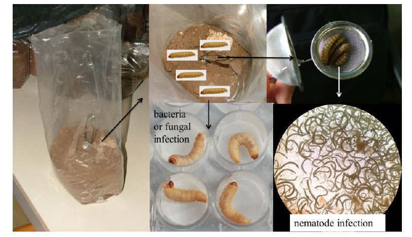
Figure 1. Soil sample with 8 Galleria larvae released, 4 inside a long-handled tea infuser and 4 released free on the top of each sample.
|
Table 1: Survey in Arasbaran Region (June 2018) – Sampling examination. For each sample habitat, soil texture and infection were recorded. Positive samples 34 of 36: EPN 8; EPF 26 (B: Beauveria, L: Lecanicillium, A: Aspergillus, F: Fusarium); EPB 5; Negative samples 2. |
||||||
|
Sample |
Habitat |
soil texture |
infection |
Infected by |
||
|
EPN |
EPF |
EPB |
||||
|
1 |
River border |
Silty loam |
+ |
* |
*L |
|
|
2 |
Corilus wood |
Silty loam |
+ |
* |
|
|
|
3 |
Corilus wood |
Silty loam |
+ |
|
*L |
|
|
4 |
Oak wood |
Silty loam |
+ |
|
*B |
|
|
5 |
Wild meadow |
Silty loam |
+ |
* |
*L |
|
|
6 |
River border |
Sandy loam |
+ |
|
*B |
|
|
7 |
River border |
Sandy loam |
+ |
|
*B |
|
|
8 |
Oak wood |
Silty loam |
+ |
* |
|
|
|
9 |
River border |
Silty loam |
+ |
|
*L*A |
|
|
10 |
Wild meadow |
Loamy clay |
– |
|
|
|
|
11 |
Wild meadow |
Loamy clay |
+ |
|
*B |
|
|
12 |
Oak wood |
loamy clay |
+ |
|
*B |
|
|
13 |
Wild meadow |
Silty loam |
+ |
* |
|
|
|
14 |
Wild meadow |
loamy caly |
+ |
|
|
* |
|
15 |
River border |
Silty loam |
+ |
|
|
* |
|
16 |
Wild meadow |
Silty loam |
+ |
|
*B |
|
|
17 |
Wild meadow |
loamy caly |
+ |
|
*B |
|
|
18 |
Wild meadow |
loamy clay |
+ |
|
*B |
|
|
19 |
Apple orchard |
Silty loam |
+ |
|
*L*A*F |
|
|
20 |
River border |
loamy clay |
+ |
* |
|
|
|
21 |
River border |
loamy caly |
+ |
* |
|
|
|
22 |
River border |
loamy sand |
+ |
|
*L |
|
|
23 |
Wild meadow |
Silty loam |
+ |
|
|
* |
|
24 |
Wild meadow |
loamy clay |
+ |
|
*B |
|
|
25 |
Oak wood |
Silty loam |
+ |
|
*B |
|
|
26 |
Oak wood |
Silty loam |
+ |
|
*B |
|
|
27 |
Oak wood |
Silty loam |
+ |
|
*B |
|
|
28 |
Wild meadow |
Silty loam |
+ |
* |
|
|
|
29 |
Wild meadow |
Silty loam |
+ |
|
|
* |
|
30 |
Wild meadow |
loamy clay |
+ |
|
*B |
|
|
31 |
Wild meadow |
loamy caly |
+ |
|
*B |
|
|
32 |
Corilus wood |
Silty loam |
+ |
|
|
* |
|
33 |
Wild meadow |
Silty loam |
+ |
|
*B |
|
|
34 |
Oak wood |
Silty loam |
– |
|
|
|
|
35 |
Urumy lake |
Salty soil |
+ |
|
*B |
|
|
36 |
Urumy lake border |
Salty beach |
+ |
|
*B |
|
Molecular Analysis
DNA extraction, PCR, cloning and sequencing were performed at IPSP laboratory, (Istituto per la Protezione Sostenibile delle Piante, Bari Italy), following the protocols described [9]. Individual nematodes were hand-picked, placed in 10 μl of lysis buffer (10 mM Tris-HCl, pH 8.8, 50mM KCl, 15 mM MgCl2, 0.1% Triton X100, 0.01% gelatin with 90 mg/ml proteinase K) on a glass slide and then cut into small pieces by using a sterilized syringe needle under a dissecting microscope. Each sample was incubated at 60°C for 1 hr and then at 95°C for 10 min. The crude DNA isolated from each individual nematode was directly amplified. The amplification of the ITS region was performed using the 18S forward primer (5’-TGATTACGTCCCTGCCTTT-3’) and the 26S reverse primer (5’-TTTCACTCGCCGTTACTAAGG-3’) [10], D2-D3 expansion segments of 28S rDNA were amplified using D2A (5’-ACAAGTACCGTGAGGGAAAGTTG-3’) and D3B (5’-TCGGAAGGAACCAGCTACTA-3’) (Nunn 1992), 18S rDNA using 18SnF (5’-TGGATAACTGTGGTAATTCTAGAGC-3’) and 18SnR (5’-TTACGACTTTTGCCCGGTTC-3’) primers [11]. PCR cycling conditions used for amplification were: an initial denaturation at 94°C for 5 min, followed by 35 cycles of denaturation at 94°C for 50s, annealing at 55°C for 50s and extension at 72°C for 1 min and a final step at 72°C for 7 min [9]. Following DNA amplification, 10 μl of PCR product was used for electrophoresis in 1X TBE buffer [12,13] in 1% agarose gel. A 100 bp ladder (Fermentas, St. Leon-Rot, Germany) was used as size marker. PCR products from individual nematodes were purified using the protocol listed by manufacturer (NucleoSpin Gel and PCR Clean-up, Machery Nagel, Germany ). Purified ITS, D2-D3 and 18S rRNA fragments were cloned in pGEM-T Easy Vector Systems (Promega, France) and sequenced at Eurofins genomics (Germany). ITS-RFLP analyses were performed on 10 μl of PCR products from individual nematodes using five units of the following restriction enzymes: Dde I, Rsa I, Alu I, Hinf I (Promega, France). The restricted fragments were separated on a 2.5% agarose gel by electrophoresis. The gels were stained with gel red and visualized on a UV transilluminator and photographed with a digital system.
Results
Entomopathogens were recovered from 34 of 36 soil samples collected (94.4%): EPN strains were isolated in 8 sites and identify as Heterorhabditis bacteriophora Poinar. Strains of EPF were recovered from 26 soil samples (Table 1), Lecanicillium W. Gams & Zare came out from 6 soil samples (N. 1, 3, 5, 9, 19 and 22), Aspergillus Micheli from 2 soil samples (N. 9 and 19), Fusarium Link from N. 19 only and Beauveria Vuill. from 16 soil samples; strains of Bacillus thuringiensis (EPB) were isolated from 5 soil samples. In 4 samples more than one pathogen strain was recovered: from N.1 and N.5 H. bacteriophora and Lecanicilium sp., from N.9 Lecanicillium sp. and Aspergillus sp., and from N.19 three fungal strains Lecanicillium sp., Aspergillus sp. and Fusarium sp., with the first two isolated from the same larva. All the pathogen strains were recovered from different habitats and no correlation between the pathogen recovery and the characteristics of the sampling sites was observed, confirming the ubiquity of these entomopathogens.
Discussion and Conclusions
The presence and occurrence of entomopathogens is a key factor on the biological soil quality and these results represent a small contribution to the knowledge of the Arasbaran biodiversity also considering the methodology used for the pathogens isolations. The recovering of almost 95% of positive samples to infection was due to the combined used of baiting larvae for static and mobile entomopathogens; considering the ubiquity of these organisms, this modified baiting technique has maximized the possibility of insulation of entomopathogens from the soil.
Acknowledgment
The research was carried on in the framework of “Della Valle research program” between the Universities of Bari “Aldo Moro” (Italy) and Tabriz (Iran).
References
- Poinar GO Jr (1990) Taxonomy and biology of Steinernematidae and Heterorhabditidae. Entomopathogenic Nematodes in Biological Control. In: Gaugler R, Kaya HK (eds.) Boca Raton Fl, USA. CRC Press, pp: 23-61.
- Poinar GO Jr (1979) Nematodes for Biological Control of Insects. CRC Press, Boca Raton, Fl: 277.
- Nikdel M, Niknam G, Griffin C, Kary NE (2010) Diversity of entomopathogenic nematodes (Nematoda: Steinernematidae, Heterorhabditidae) from Arasbaran forests and rangelands in north-west Iran. Nematology 12: 767-773.
- Nikdel M, Niknam G, Ye W (2011) Steinernema arasbaranense sp.n. (Nematoda: Steinernematidae, a new entomopathogenic nematode from Arasbaran Forest, Iran. Nematol Medit 39: 17-28.
- Seifinejad A, Salehi Jouzani GR, Hosseinzadeh A, Abdmishani C (2008) Characterization of Lepidoptera-active cry and vip genes in Iranian Bacillus thuringiensis strain collection. Biological Control 44: 216-226.
- Salekjalali M, Barzegari A, Jafari B (2012) Isolation, Pcr Detection and Diversity of Native Bacillus thuringiensis Strains Collection Isolated from Diverse Arasbaran Natural Ecosystems. WorldApplied Sciences Journal 18: 1133-1138.
- Tarasco E, Clausi M, Rappazzo G, Panzavolta T, Curto G, et al. (2015) Biodiversity of entomopathogenic nematodes in Italy. Journal of Helminthology 89: 359-366. [crossref]
- White GF (1927) A method for obtaining infective nematode larvae from culture. Science 66: 302-303.
- De Luca F, Fanelli E, Di Vito M, Reyes A, De Giorgi C (2004) Comparison of the sequences of the D3 expansion of the 26S ribosomal genes reveals different degrees of heterogeneity in different populations and species of Pratylenchus from the Mediterranean region. European Journal of Plant Pathology 110: 949-957.
- Vrain TC, Wakarchuck DA, Levesque AC, Hamilton RI (1992) Intraspecific rDNA restriction fragment length polymorphism in the Xiphinema americanum group. Fundamental and Applied Nematology 15: 563-573.
- Kanzaki N, Futai K (2002) A PCR primer set for determination of phylogenetic relationships of Bursaphelenchus species within xylophilus group. Nematology 4: 35-41.
- Sambrook J, Fritschi EF, Maniatis T (1989) Molecular cloning: a laboratory manual (2ndedn), Cold Spring Harbor Lab Press, New York.
- Glare TR, Jurat-Fuentes JL, O’Callaghan M (2017) Chapter 4: Basic and Applied Research: Entomopathogenic Bacteria. Microbial control of Insect and mite pests. Academic Press, pp: 47-6.