Monthly Archives: May 2021

Awareness of Ultrasonography, Preconception during Pregnancy and Use of Sonography by Tribal Women- Rural Community Based Study

DOI: 10.31038/IGOJ.2021415

Abstract

Background: Ultrasonography (USG) has become part of everyday care of pregnant women in most of the countries of the globe. However like any other technology, it has potential to raise social, ethical, economic dilemmas about benefits, challenges for health providers, beneficiaries of the services. Awareness, utilization of USG by rural tribal women who live in extreme poverty with access problems is not well known.

Objective: Community based study was carried out to know awareness of USG amongst rural, tribal, preconception, pregnant women and use of USG during pregnancy.

Material Methods: Study was conducted in tribal communities of 100 villages where community based mother child care services were initiated after having developed a health facility in one of 100 villages. Total 2400 preconception, 1040 pregnant women of 15-45 years, were interviewed in villages for knowing their awareness about USG, whether pregnant women had USG during pregnancy.

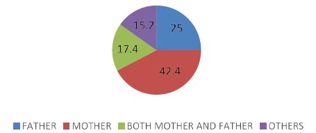
Results: Of 2400 preconception women, 626 (26.08%) were not aware of Sonography. Of those who knew, 694 (39.1%) said Sonography helped in confirmation of pregnancy, 1080 (60.88%) said it helped in knowing fetal age and position. Of 1040 pregnant women also 271 (26.1%) were not aware of USG. Those who knew, sources of information, were Accredited Social Health Activists (ASHAs) in 208 (27%), nurse midwives in 170 (22.1%), family members in 311 (40.4%), doctors in 80 (10.4%). Only 258 (33.5%) of 769 women who knew about USG had got USG done. Of them 82 (31.8%) were told that something was wrong without any details.

Conclusion: Study revealed that many rural tribal women did not even know about USG. Community health workers, ASHAs did create awareness of USG in some. Only 25% pregnant women had USG done but without knowing any details of findings.

Keywords

Preconception, Pregnancy, Awareness, Ultrasonography Finding

Background

Ultrasonography is now a integrated part of pregnancy care in most of the countries around the world. Diagnostic ultrasound during pregnancy may be employed for variety of reasons to see image of the baby, placenta and amonite fluid even for the woman and her family to see in addition to sonologist. Actually some clinicians are replacing clinical examination of pregnant women by USG, may be for confirmation of pregnancy, duration of pregnancy, number of foetuses, fetal growth and development, abnormalities of fetus, placenta and liquor by direct visualization, amniocentesis and/or cordocentesis. It can be used for foetal therapy too and even foetal foeticide and also for prediction of maternal disorders which affect mother as well as the baby [1]. However if abnormalities are detected during pregnancy it might lead to stress for the woman and the family, sometimes problems may be detected in women who do not have any risk factors, creating a lot of stress which has sequlae. Unfortunately there is likelihood of false alarm too specially when USG is performed by a person who lacks desired skill and knowledge or lack of time or desired attitude too. Assumptions are made that routine USG will prove beneficial by enabling earlier detection and improved management of pregnancy complications [2]. Routine screening may be done in early or late pregnancy, or both. Use of USG early in pregnancy is increasing, but there is limited information about linkage decision-making and impact on expectant women/couples. It is essential to know because globally there has been increasing medicalization of pregnancy [3]. However the awareness and utilization of USG by rural tribal women especially those with extreme poverty are not well known.

Material and Methods

After approval of ethics committee, which works on the principle of Helsinki Declaration, the study was conducted in tribal communities of 100 villages of rural, hilly and forestry, Melghat of Amravati, Maharashtra, India. In these villages community based mother and child care services were initiated after having created a health facility in one of the villages. Information was collected visiting every 5th house randomly, minimum 20 preconception women from each village total 2400 and 1040 pregnant women of 15-45 years. Interviews were conducted taking consent using a pretested tool in the language understood by women. Some questions needed yes or no answers and others short open answers.

Results

Of total 2400 preconception study subjects, 27% did not know anything and 1774 (73.9%) women were aware about sonography. Overall 694 (39.1%) of those who were aware said sonography helped in knowing about pregnancy, 1080 (60.88%) of those who knew about USG said it helped to know the fetal age and position. Overall 336 (14%) of 2400 women were of 15-19 years age, 271 (80.65%) of them were aware of sonography during pregnancy and 245 (90.41%) of them said USG helped in knowing fetal age with position and only 26 (9.59%) said it can confirm pregnancy. Of 74 women of 40-45 years age, 59 (79.3) were aware of USG similar to young women and 50 (84.75%) of them said it helped in knowing fetal age and position and only 9 (15.25%) said for confirmation of pregnancy. Of 953 (39.70%) of 2400 women were illiterate, 726 (76.18%) of them were aware of sonography and 539 (74.24%) said it helped to know fetal age and position and 187 (25.76%) said it confirmed pregnancy. Of 60 (65.93%) of 91 women with higher secondary education, 54 (90%) said USG helped in knowing fetal age and position and only 6 (10%) said confirmation pregnancy. Of 275 housewives 211 (76.72%) were aware of USG, 182 (86.26%) women said sonography helped in estimation of fetal age and position and 29 (13.74%) said for confirmation of pregnancy. Of 958 labourer 681 (71.86%), knew about sonography and 444 (65.2%) of those who knew about USG, said it helped to know the fetal age and position and 237 (34.8%) for confirmation pregnancy. Of 2400 preconception women, 662 (27.58%) belonged to upper lower economic class, [economic status was divided in five], 553 (83.53%) of them were aware of sonography, 340 (61.48%) said it helped in knowing fetal age and position and 213 (38.52%) confirmation of pregnancy. Seventy-four (50.34%) of 147 women who belonged to upper economic class, knew about sonography, significantly less (P Value 0.0127) and 39 (52.7%) said it helped in confirmation of pregnancy and 35 (47.3%) said to know about fetal age and position. Overall 85 (81%) of 105 who had no child were aware of sonography, 75 (88.24%) said it helped in estimation of fetal age, position and only 10 (11.8%) said confirmation pregnancy. Overall 421 (82.7%) of 509 who had five or more births were aware of sonography, similar to those with no child, 250 (59.4%) said it helped in confirmation of pregnancy and 171 (40.62%) estimation of fetal age and position. Total 626 (26.08%) 2400 preconception women did not know that there was something like sonography (Tables 1-3).

Table 1: Awareness of Ultrasonography in Preconception Women.

Variables

Total

Awareness

Age

No % Yes

%

15 to 19

336

65 19.35 271

80.65

20 to 24

828

181 21.86 647

78.14

25 to 29

736

243 33.02 493

66.98

30 to 34

333

75 22.52 258

77.48

35 to 39

93

47 50.54 46

49.46

TOTAL

74

15 20.27 59

79.73

Education

2400

626 26.08 1774

73.92

Illiterate
Primary

953

227 23.8 726

76.18

Secondary

850

282 33.2 568

66.82

Higher secondary

506

86 17.0 420

83

Graduate

91

31 34.1 60

65.93

Post graduate

0

0 0.0 0

0

Total

2400

626 26.08 1774

73.92

Economic status
Upper

275

64 23.27 211

76.73

Upper middle

958

277 28.91 681

71.09

Upper lower

468

121 25.85 347

74.15

Lower middle

699

154 22.03 545

77.97

Lower

2400

626 26.08 1774

73.92

Total
Profession

147

73 49.66 74

50.34

Housewife

183

59 32.24 124

67.76

Own farm labour

544

170 31.25 374

68.75

Labourer

662

109 16.47 553

83.53

Other work

864

215 24.88 649

75.12

Total

2400

626 26.08 1774

73.92

Parity
P. 0

105

20 19.05 85

81

P. 1

411

131 31.87 280

68.1

P. 2

672

218 32.44 454

67.6

P. 3

453

123 27.15 330

72.8

P. 4

250

46 18.4 204

81.6

P. 5 Above

509

88 17.29 421

82.7

Total

2400

626 26.08 1774

73.9

Table 2: Awareness of Ultrasonography Pregnant Women and Source of Information.

Variables

Total Awareness

Source of information

Age

NO

% YES % ASHA % ANM % Doctor % Family Member

%

15 to 19

323

107 33.1 216 66.9 129 59.7 49 22.7 21 9.7 17

7.9

20 to 24

536

130 24.3 406 75.7 166 40.9 152 37.4 66 16.3 22

5.4

25 to 29

109

22 20.2 87 79.8 19 21.8 41 47.1 3 3.4 24

27.6

30 to 34

68

12 17.6 56 82.4 21 37.5 19 33.9 4 7.1 12

21.4

35 to 39

4

0 0.0 4 100.0 1 25.0 0 0.0 0 0.0 3 75.0

TOTAL

1040 271 26.1 769 73.9 336 43.7 261 33.9 94 12.2 78

10.1

Education
Illiterate

56

19 33.9 37 66.1 21 56.8 13 35.1 3 8.1 0 0.0

Primary

321 42 13.1 279 86.9 134 48.0 97 34.8 33 11.8 15

5.4

Secondary

358

58 16.2 300 83.8 102 34.0 186 62.0 6 2.0 6 2.0

Higher secondary

196 58 29.6 138 70.4 41 29.7 29 21.0 13 9.4 55

39.9

Graduate

66

54 81.8 12 18.2 2 16.7 3 25.0 2 16.7 5 41.7
Post graduate

43

40 93.0 3 6.97 1 33.3 0 0.0 0 0.0 2 66.7

Total

1040 271 26.1 769 73.9 301 39.1 328 42.7 57 7.4 83

10.8

Economic status
Upper

43

42 97.7 1 2.3 1 100.0 0 0.0 0 0.0 0 0.0

Upper middle

51 49 96.1 2 3.9 2 100.0 0 0.0 0 0.0 0

0.0

Upper lower

142

42 29.6 100 70.4 56 56.0 20 20.0 11 11.0 13 13.0

Lower middle

186 61 32.8 125 67.2 67 53.6 30 24.0 12 9.6 16

12.8

Lower

618

77 12.5 541 87.5 294 54.3 166 30.7 52 9.6 29 5.4

Total

1040 271 26.1 769 73.9 420 54.6 216 28.1 75 9.8 58

7.5

Profession
Housewife

943

227 24.1 716 75.9 322 45.0 109 15.2 103 14.4 182 25.4

Own farm labour

53 24 45.3 29 54.7 19 65.5 6 20.7 1 3.4 3

10.3

Labourer

40

19 47.5 21 52.5 17 81.0 2 9.5 0 0.0 2 9.5

Other work

4 1 25.0 3 75.0 2 66.7 1 33.3 0 0.0 0

0.0

Total

1040

271 26.1 769 73.9 360 46.8 118 15.3 104 13.5 187

24.3

Parity
P.1

117

6 5.1 111 94.9 11 9.9 52 46.8 21 18.9 27

24.3

P.2

103

4 3.9 99 96.1 66 66.7 21 21.2 8 8.1 4

4.0

P.3

155

6 3.9 149 96.1 41 27.5 64 43.0 11 7.4 33 22.1

P.4

204 15 7.4 189 92.6 49 25.9 21 11.1 29 15.3 90

47.6

P.5 Above

461

240 52.1 221 47.9 41 18.6 12 5.4 11 5.0 157 71.0
Total

1040

271 26.1 769 73.9 208 27.0 170 22.1 80 10.4 311

40.4

ASHA: Accredited Social Health Activist.
ANM: Auxiliary nurse midwife.

Table 3: Ultrasonography during Pregnancy by Rural Tribal Women.

Variables

Total Ultrasound done

If YES Abnormality Informed

Age

NO

% YES % Yes % No

%

15 to 19

323

220 68.1 103 31.9 25 24.3 78 75.7

20 to 24

536 413 77.1 123 22.9 42 34.1 81

65.9

25 to 29

109

91 83.5 18 16.5 7 38.9 11 61.1

30 to 34

68 57 83.8 11 16.2 6 54.5 5

45.5

35 to 39

4

1 25.0 3 75.0 2 66.7 1 33.3

Total

1040 782 75.2 258 24.8 82 31.8 176

68.2

Education
Illiterate

56

40 71.4 16 28.6 4 25.0 12 75.0

Primary

321 282 87.9 39 12.1 19 48.7 20

51.3

Secondary

358

307 85.8 51 14.2 15 29.4 36 70.6

Higher secondary

196 136 69.4 60 30.6 16 26.7 44

73.3

Graduate

66

11 16.7 55 83.3 14 25.5 41 74.5

Post graduate

43 6 14.0 37 86.0 14 37.8 23

62.2

Total

1040

782 75.2 258 24.8 82 31.8 176

68.2

Economic status
Upper

43

1 2.3 42 97.7 11 26.2 31 73.8

Upper middle

51 10 19.6 41 80.4 6 14.6 35

85.4

Upper lower

142

82 57.7 60 42.3 12 20.0 48 80.0

Lower middle

186 130 69.9 56 30.1 27 48.2 29

51.8

Lower

618

559 90.5 59 9.5 26 44.1 33 55.9

Total

1040 782 75.2 258 24.8 82 31.8 176

68.2

Profession
Housewife

943

718 76.1 225 23.9 66 29.3 159 70.7

Own farm labour

53 34 64.2 19 35.8 16 84.2 3

15.8

Labourer

40

29 72.5 11 27.5 0 0.0 11 100

Other work

4 1 25.0 3 75.0 0 0.0 3

100

Total

1040

782 75.2 258 24.8 82 31.8 176

68.2

Parity
P.1

117

88 75.2 29 24.8 7 24.1 22 75.9

P.2

103 62 60.2 41 39.8 11 26.8 30

73.2

P.3

155

87 56.1 68 43.9 24 35.3 44 64.7

P.4

204 135 66.2 69 33.8 22 31.9 47

68.1

P.5 Above

461

410 88.9 51 11.1 18 35.3 33 64.7

Total

1040 782 75.2 258 24.8 82 31.8 176

68.2

Total 769 (73.9%) of 1040 rural tribal pregnant women, knew about USG in pregnant women but 271 (26.1%) did not know. The sources of information were Accredited Social Health activists (ASHA) 208 (27%), nurse midwives 170 (22.1%), family members 311 (40.4%) and in 80 (10.4%) doctors. Of 1040 study subjects, 406 (75.7%) of 536 of 20-24 years were aware of USG, Sources of information were ASHAs in 166 (40.9%), Nurse Midwives in 152 (34.4%), 66 (16.3%) Doctors and 22 (5.4%) family members. As age increased more women were found to be knowing about USG, 216 (66.9%) of 323 of 15 to 19 year, 60 (83.33%) of 72 of 30-39 years old. (P Value 0.3776) It seemed to be related to increased parity too. Out of 1040 pregnant women, 43 (4.13%) were postgraduate studied still only 3 (6.97%) were aware, one (33.3%) was toldby ASHA and 2 (66.7%) by family members. Overall 56 (5.38%) illiterate women, 34 (66.1%) were aware of USG, by ASHAs 21 (56.8%), Nurse midwives 13 (35.1%) and Doctors 3 (8.1%). Only 2 (3.9%) of 51who belonged to middle economic class were aware of USG, ASHAs, being the source of information in both. Of 1040 pregnant women, 618 belonged to lower economic class and 541 (87.5%) of them were aware of USG, ASHAs were the source in 294 (54.3%), nurse midwives in 166 (30.7%), doctors in 52 (9.6%) and family members in 29 (5.4%). Among 1040 pregnant women, 943 were housewives and of them 716 (75.9%) were aware of USG. ASHAs were the source in 322 (45.0%) and family members in 182 (25.4%). Overall 21 (52.5%) of 40 labourers were aware of USG and 17 (81%) were told by ASHAs.

As the parity increased number of women with awareness increased, 27 (24.3%) of 111 primigravida and 157 (71%) of 221 fifth gravida said they were told by family members. Only 258 (24.8%) of 1040 pregnant women themselves had USG and 782 (75.2%) did not. 258 (33.5%) of 769 women who knew about USG had USG done. Of them 82 (31.8%) were told of possibilities of some abnormalities but they did not know any details. There seemed to be no communication in most of the cases in whom USGs was done, probably because USG were done in camps at Primary Health Centers or Sub District Hospital with crowds around. Of 1040 study subjects, 536 (51.53%) were of 20-24 year, 123 (22.9%) got USG done, 42 (34.1%) said some abnormalities were told but did not know any details. 14 (19.44%) of 72 of 30-39 year had USG, of which 8 (57.14%) were told of abnormalities without details. Of 66 graduates, 55 (83.3%) had USG and 29 (52.7%) were told of some abnormalities. Only 16 (28.6%) of 56 illiterate had USG and 4 (25%) were told of some abnormalities. Of 43 (4.13%) who belonged to middle economic class, 41 (80.4%) had USG and 9 (22%) of them were told of some abnormalities. Only 59 (9.5%) of 618 women who belonged to lower economic class had USG. Twenty (33.9%) said some abnormalities were told without any details. Of 943 (90.67%) of 1040 pregnant housewives, 225 (23.9%) had USG and 66 (29.3%) were told about abnormalities, but they did not know any details. Only 11 (27.5%) of 40 labourers had USG and no one said they were told of any abnormalities. Total 117 (11.25%) were primipara, only 29 (24.8%) of them had USG and 7 (24.1%) said some abnormalities were told without any details. Overall 188 (29.74%) of 632 women who had 3 or more births in 64 (34.04%) were told of some abnormalities with no details.

Discussion

In the present day clinical practice the discussion is on evidence-based guidelines disseminated to physicians, obstetrician, nurses and sonologists for antenatal ultrasound scans with advocacy of guidance about the appropriate use of ultrasound scans to be shared with women in order to discourage unreasonable expectations, demands and apprehensions. On one side USG is done many times during pregnancy by urban women for various reasons, many rural women do not even know about USG, do not even think of diagnostic antenatal checkup. Bashour et al. reported that private doctors, who looked after 80% of pregnant women, offered ultrasound primarily to attract women to their clinics and increase their income [4]. Kozuki et al. reported that the utilization of obstetric USG in rural women of Nepal was very limited. Researchers reported that more research was necessary to assess the potential of health impact of obstetric USG in low-resource settings, while addressing limitations such as cost and misuse [5]. Cherniak et al. reported that women could be motivated to attend antenatal clinics when offered the incentive of seeing their baby through USG [6]. Huang et al. reported high use of antenatal ultrasound in rural Eastern China, influenced by socio-demographic and clinical factors [7]. Torloni et al. reported that USG in pregnancy was not associated with adverse maternal effects, impaired physical or neurological development or increased risk to children [8]. However with over diagnosis some stress is always likely. With under diagnosis there are many problems and it is essential that there is awareness and understanding of use and misuse.

Whitworth et al. also reported that early USG helped in the detection of multiple pregnancies with improved gestational dating which resulted in fewer inductions for post maturity [2]. This can only happen if women know and can use the technology. It does not seem to be happening for rural women. Abramowicz et al. reported that USG carried some risks of misdiagnosis on the one hand and possible undesired effects on the other [9]. The general belief existed that diagnostic USG did not pose any risk, neither to the pregnant women nor to the fetus. But risk-benefit analysis may also be important, as well as education of the end users to assure safety. Fact remains false diagnosis might give mental stress even dilemmas when not knowing anything as happened in the present study. USG were done during camps at PHCs and Sub-District Hospital (SDH) and women did not know details of abnormalities. Phutke et al. reported it is essential to re-examine and update the use of diagnostic, USG widely available even the most peripheral health facilities [10]. Studies showed that pregnant women generally value routine ultrasounds in the first two trimesters because they get reassurance and chances to see their unborn baby.

Although growing, evidence on the impact, access, utility, effectiveness, and cost-benefit of obstetric ultrasound in resource-constrained settings is still somewhat limited, questions around the purpose and the intended benefits as well as potential challenges across various domains must be carefully reviewed prior to implementation and scale-up of obstetric USG in Low-and Middle-Income Countries (LMICs). Whitworth et al. reported that some (but not all) benefits described in the literature have been validated by evidence-based analysis [2]. Unlike other modes for prenatal screening and diagnosis, USG offers parents direct access to images of the fetus. This makes obstetric ultrasound popular and attractive among expectant mothers so they want to use it often [11,12]. Women see prenatal USG as means for reassurance about the health and well-being of their fetuses. However, sometimes USG may yield unexpected findings which may have adverse effects on the mental health of mother and may provoke emotional crisis [13,14]. Significant psychological harm from antenatal ultrasound as well as positive psychological effects have been reported [15]. Counselling is needed to further enhance the USG experience and to reduce anxiety and dispel any misconceptions and irrational expectations regarding the antenatal USG. In the present study quite a few preconception as well as pregnant women did not know anything about USG. Those pregnant women who knew also did not get USG done during pregnancy due to various reasons. Those who had USG did not know details of abnormalities as any discussion or communication took place.

Of 2400 preconception study subjects, 1774 (73.9%) women were aware about sonography, 694 (39.1%) said sonography helped knowing about pregnancy, 1080 (60.88%) of those who knew said it helped in knowing fetal age and position. Overall 1774 of 2400 preconception women, 626 (26.08%) were not aware of USG, of 953 (39.70%) illiterate, 726 (76.18%) were aware of sonography, 539 (74.24%) said sonography helped to know fetal age and position and 187 (25.76%) said for confirmation pregnancy. So it was word of mouth which more often made women aware.

Of 1040 pregnant women, 271 (26.1%) were not even aware about USG during pregnancy, women with more than one birth too did not know. Most women got information from ASHAs 208 (27.0%), 170 (22.1%) NM of Sub center. Of 1040 study subjects, 258 of 769 (33.5%) of those who knew had USG, 82 (10.7%) of them were told about some abnormalities, but without any details. Rest did not know anything about what was found. Communication and counseling are essential.

Halle et al. reported that USG in the first half of pregnancy were in high use in Iceland and apparently became part of a broader pregnancy culture, encompassing both high- and low-risk pregnancies [16]. Whether this is a favourable development or to some extent represents unwarranted medicalization needs further discussion. More balanced information might be provided prior to early screening for foetal anomalies. In rural community women start care by mid pregnancy.

Yadav et al. reported that in their study 72.41% pregnant women felt USG was done for knowing fetal anomalies and 27.93% for sex detection, majority (93.1%) had USG more (43.45 %) in second trimester mainly on advice of doctors (91.03%) [17]. Nearly half of them (50.69%) considered it as expensive procedure and 50.69%% of them opined it should be done twice in pregnancy. Almost 94.83% considered USG as safe and beneficial. Awareness regarding the uses of USG during pregnancy and attitude towards USG was neither negative possible. Westerneng et al. reported that pregnant women seemed to appreciate a third trimester routine ultrasound, but it did not seem to reduce anxiety or improve bonding with their baby [18]. Women’s appreciation of a third trimester routine ultrasound might arise from getting used to routine ultrasounds throughout pregnancy. Results of such findings should be taken into consideration when balancing the gains, which are as yet not clear, of introducing a third trimester routine ultrasound against unwanted side effects and costs.

Ikeako et al. did a study and reported that the number of respondents who had USG in their previous pregnancies was 58.7% [19]. Although many reasons were given for personal USG requests, 19.7% women who had obstetric scan in their previous pregnancies thought it was a normal booking test done for every pregnant woman. When compared with other booking investigations, 60.1%, mainly civil servants said that USG in pregnancy was costly, 24.4% felt it was cheap, 9.1% said it was very costly and remaining 2.4% thought it was not affordable. Apart from visualizing the images of their babies, 17.8% of the cases wanted to know the gender and 15.4% said it was for knowing of fetal position.

Total 52.9% were of the opinion that women could decide when to request for sonography. Majority of Nigerian women requested ultrasound for looking at fetus and gender determination. Gururaj et al. reported that care providers and government officials perceived ultrasound diagnosis as critical to deciding whether to refer women who might need high-risk support from higher-level centres that are often geographically remote [20]. Findings suggested a strong need to re-evaluate the evidence base for routine obstetric ultrasound in rural LMIC settings and include more stakeholders in participatory, co-design approaches to innovation. Firtha et al. opined that ultrasound would increase Antenatal Care (ANC) attendance [21]. Kim et al. opined that as cost of obstetric ultrasound became more affordable in LMICs, it is essential to assess the benefits, trade-offs and potential drawbacks of large-scale implementation [22]. Additionally, there was a need to more clearly identify the capabilities and the limitations of ultrasound, particularly in the context of limited training of providers, to ensure that the purpose, for which an ultrasound was intended, was actually feasible. Researchers also reported that there was evidence that ultrasound was not associated with reducing maternal, perinatal or neonatal mortality, also reported various studies revealed both positive and negative perceptions and experiences related to ultrasound and lastly, illegal use of ultrasound for determining fetal sex raised a concern. Saleh et al. reported that most of the participants were aware of ultrasound scan and also believed that the procedure was safe, and the main purpose was for fetal wellbeing and viability [23].

Ugwu et al. did a study and reported that 73% women got their information from antenatal centres. Over 20% were interested in the lies and presentation of their foetus [24].

Conclusion

The role of prenatal sonography in obstetric care should be real with preconception awareness in antenatal centres, and initiating mother/sonographers interaction is necessary.

References

  1. Ville YG, Bault JP (2016) Prenatal Diagnoses of Fetal Malformations by Ultrasound. Genetic Disorders and the Fetus 5: 121-126.
  2. Whitworth M, Bricker L, Mullan C (2015) Ultrasound for fetal assessment in early pregnancy. Cochrane database of systematic reviews. [crossref]
  3. Miller S, Abalos E, Chamillard M, Ciapponi A, Colaci D, et al. (2016) Beyond too little, too late and too much, too soon: a pathway towards evidence-based, respectful maternity care worldwide. The Lancet 388: 2176-2192.
  4. Bashour H, Hafez R, Abdulsalam A (2005) Syrian women’s perceptions and experiences of ultrasound screening in pregnancy: implications for antenatal policy. Reproductive Health Matters 13: 147-154. [crossref]
  5. Kozuki N, Katz J, Khatry SK, Tielsch JM, LeClerq SC, et al. (2016) Community survey on awareness and use of obstetric ultrasonography in rural Sarlahi District, Nepal. International Journal of Gynecology & Obstetrics 134: 126-130. [crossref]
  6. Cherniak W, Anguyo G, Meaney C, Kong LY, Malhame I, et al. (2017) Effectiveness of advertising availability of prenatal ultrasound on uptake of antenatal care in rural Uganda: A cluster randomized trial. Plos One 12. [crossref]
  7. Huang K, Tao F, Raven J, Liu L, Wu X, et al. (2012) Utilization of antenatal ultrasound scan and implications for caesarean section: a cross-sectional study in rural Eastern China. BMC Health Services Research 12: 93.
  8. Torloni MR, Vedmedovska N, Merialdi M, Betràn AP, Allen T, et al. (2008) OC196: Safety of ultrasonography in pregnancy: WHO systematic review of the literature and meta‐analysis. Ultrasound in Obstetrics and Gynecology 32: 307-.
  9. Abramowicz S, Susarla HK, Kim S, Kaban LB (2013) Physical findings associated with active temporomandibular joint inflammation in children with juvenile idiopathic arthritis. Journal of Oral and Maxillofacial Surgery 71: 1683-1687. [crossref]
  10. Phutke GA, Laux T, Jain P, Jain YO (2018) Ultrasound in rural India: A failure of the best intentions. Indian J Med Ethics 18: 1-7. [crossref]
  11. Bricker L, Gacia J, Henderson J, Mugford M, Neilson J, et al. (2000) Ultrasound screening in pregnancy: a systematic review of the clinical effectiveness, costeffectiveness and women’s views. Health Technol Assess 4: 1-193. [crossref]
  12. Georgsson Ohman S, Waldenstrom U (2008) Second-trimester routine ultrasound screening: expectations and experiences in a nationwide Swedish sample. Ultrasound Obstet Gyneco 32: 15-22.
  13. Gammeltoft T, Thi H, Nguyen T (2007) The Commodification of Obstetric Ultrasound Scanning in Hanoi, Viet Nam. Reprod Health Matters 15: 163-171. [crossref]
  14. Sommerseth E, Sundby J (2010) Women’s experiences when ultrasound examinations give unexpected findings in the second trimester. Women and Birth 23: 111-116. [crossref]
  15. Harris G, Connor L, Bisits A, Higginbotham N (2008) “Seeing the Baby”: Pleasures and Dilemmas of Ultrasound Technologies for Primiparous Australian Women. Med Anthropol Q 18: 23-47. [crossref]
  16. Halle KF, Fjose M, Kristjansdottir H, Bjornsdottir A, Getz L, et al. (2018) Use of pregnancy ultrasound before the 19th week scan: an analytical study based on the Icelandic Childbirth and Health Cohort. BMC Pregnancy and Childbirth 1; 18: 512.
  17. Yadav JU, Yadav DJ (2017) Ultrasonography awareness among pregnant women attending medical college hospital in Kolhapur District of Maharashtra, India. International Journal of Research in Medical Sciences 5: 2612.
  18. Westerneng M, Diepeveen M, Witteveen AB, Westerman MJ, Van Der Horst HE, et al. (2016) Experiences of pregnant women with a third trimester routine ultrasound-a qualitative study. BMC Pregnancy and Childbirth 19: 1-0.
  19. Ikeako LC, Ezegwui HU, Onwudiwe E, Enwereji JO (2014) Attitude of expectant mothers on the use of ultrasound in pregnancy in a tertiary institution in South East of Nigeria. Annals of Medical and Health Sciences Research 4: 949-953. [crossref]
  20. Gururaj A (2017) Exploring the role of a semi-automated ultrasound technology in rural Indian antenatal care (Doctoral dissertation, University of Oxford).
  21. Firtha ER, Mlay P, Walker R, Sill PR (2011) Pregnant women’s beliefs, expectations and experiences of antenatal ultrasound in Northern Tanzania. African Journal of Reproductive Health 15: 91-107. [crossref]
  22. Kim ET, Singh K, Moran A, Armbruster D, Kozuki N (2018) Obstetric ultrasound use in low and middle income countries: a narrative review. Reproductive Health 15: 129.
  23. Saleh AA, Idris G, Dare A, Yahuza MA, Suwaid MA, et al. (2017) Awareness and perception of pregnant women about obstetrics ultrasound at Aminu Kano Teaching Hospital. Sahel Medical Journal 20: 38.
  24. Ugwu AC, Udoh BE, Eze JC, Eze PC (2011) Awareness of information, expectations and experiences among women for obstetric sonography in a south east Nigeria population. East African Journal of Public Health 8: 142-144. [crossref]
fig 1

DQRF™ (Dynamic Quadripolar Radiofrequency) and UPR™ (Ultra-Pulsed Radioporation) 12-Month Synergy in Postmenopausal Vulvovaginal Atrophy

DOI: 10.31038/IGOJ.2021414

Abstract

Introduction: The low-energy Dynamic Quadripolar Radiofrequency or DQRF™ vaginal technology overcomes several problems of manageability and safety experienced with other energy-based vulvovaginal energy-providing technologies by postmenopausal women with symptoms of vulvovaginal atrophy. The proprietary Ultra-Pulsed Radioporation or UPR™ technology has already shown to accrue the benefits of the new radiofrequency technology by facilitating penetration of active principles into the deep layers of vulvar skin and vaginal mucosa and enhancing hydration and trophism. Evaluating the impact on vulvovaginal atrophy symptoms, with vaginal dryness expected to benefit the most from the DQRF/UPR™ synergy, is the goal of this interim study.

Methods: Prospective real-life cohort study on 106 ambulatory women (mean age, 56.8 ± 8.61 years old) with vaginal atrophy and dryness. VVA treatment: four to five 25-min sessions every 14-16 days (coupling gel with hyaluronic acid); one more DQRF/UPR™ maintenance session after six months and a final visit (only assessment, no treatment) after 12 months. Operative temperatures in target tissues during the procedure: 42°C (range 40-43°C). Assessments (baseline and at the end of the treatment cycle): vaginal dryness (primary efficacy parameter, 10-cm impromptu Visual Analogue Scale); dyspareunia, burning and itching (4-score impromptu Likert-like scale) and photographic documentation at baseline at the end of the DQRF/UPR™ treatment cycle.

Results: Vaginal dryness rapidly improved vs baseline (T0), with a highly significant reduction (−83.1%) at the end of the treatment cycle (T1) that steadily persisted after 6 and 12 months (T2 and T3; −79.2% and −64.9%, respectively). All other symptoms similarly improved vs baseline over the year of follow-up: dyspareunia −81.5% (T1) and −70.4% (T3); burning −87.0% (T1) and −65.2% (T3); itching −89.5% (T1) and −68.4% (T3). All treatments were well tolerated, with no troubling pain or other side effects during or after the procedures.

Conclusion: The study confirms, over a one-year follow-up, the previously demonstrated benefits of the Dynamic Quadripolar Radiofrequency (DQRF™) in synergy with Ultra-Pulsed Radioporation (UPR™) as an innovative treatment option of vulvovaginal atrophy/genitourinary syndrome of the menopause symptoms. The novel UPR™ technology facilitates the deep penetration of active topical principles favourably acting on postmenopausal atrophic tissues. The DQRF/UPR™ concept aims to enhance the effects of the DQRF™ technology exploiting the synergy between the double biological effects-by the energy-based DQRF™ and the UPR™ active principle. Long-term studies will confi.

Keywords

Electroporation, Genitourinary syndrome of menopause, Dynamic quadripolar radiofrequency, DQRF™, Ultra-Pulsed Radioporation, UPR™, Vulvovaginal atrophy

Abbreviations

DQRF™: Dynamic Quadripolar Radiofrequency

GSM: Genitourinary Syndrome of Menopause

HA: Hyaluronic Acid

MDa: x106 Dalton

MHz: Megahertz or x106 Hertz

RSS™: Radiofrequency Safety System

SEM: Standard Error of the Mean

UPR™: Ultra-Pulsed Radioporation

VAS: Visual Analogue Scale

VDR™: Vaginal Dynamic Radiofrequency

VVA: Vulvovaginal Atrophy

W: Watt

Introduction

Energy-based vulvovaginal treatment technologies often show manageability and safety difficulties in postmenopausal women with life-disrupting Vulvovaginal Atrophy (VVA) and Genitourinary Syndrome of Menopause (GSM). The low-energy DQRF™ (Dynamic Quadripolar Radiofrequency) technology candidates to overcome such problems in VVA women [1,2]. The four algorithmically controlled radiofrequency electrodes, continuously cycling between receiver and transmitter states (VDR™ or Vaginal Dynamic Radiofrequency™ technology), generate repelling electric fields within the closed electrode system and concentrate their low-energy thermal effects with high topographical precision in precise subepithelial areas without the need for grounding pads.

In addition, the treated area – usually a 4-inch area centred on the hymenal ring -need no systemic analgesia or local anaesthesia thanks to the integrated RSS™ (Radiofrequency Safety System) proprietary technology that steadily tracks the tip movements and local tissue temperature [1,2]. The Novavision Group S.p.A. (Misinto, Monza-Brianza, Italy) holds worldwide rights for the patented DQRF™, VDR™, and RSS™ technologies.

Since 2018, integrating the DQRF™ concept with the proprietary UPR™ (Ultra-Pulsed Radioporation) radiofrequency electroporation technology has been a second technological jump forward. UPR™ modulates the DQRF™ radiofrequency effects and facilitates the transfer of biologically active principles through aqueous channels in vulvar skin and vaginal mucosa cell membranes [3]. The DQRF™ effects on vulvovaginal hypotrophy thus synergise with those of the UPR™-mobilised active principles-for instance, highly hydrating and pro-trophic Hyaluronic Acid (HA) [4]. Counteracting the postmenopausal loss of elasticity and volume with topical HA formulations and HA dermal fillers in women with labia majora hypotrophy has long been a common cosmetic gynaecology practice [5,6]. A double-blind pilot study compared VVA/GSM symptoms and women’s satisfaction with their sexual and couple lives in two random samples of postmenopausal women randomised to either DQRF™ and DQRF/ UPR™ vulvovaginal treatment [3]. The low-molecular-weight HA was interspersed in the coupling gel in the DQRF/ UPR™ treatment group; the study established the DQRF/ UPR™ superior benefits [3].

The interim 12-month DQRF/ UPR™ outcomes on VVA symptoms herein illustrated aim to confirm the previous favourable outcomes over a more extended follow-up period and a more ample postmenopausal women cohort. Vaginal dryness, expected to benefit the most from the UPR™ synergy with DQRF™, was the primary efficacy parameter. The interim DQRF/ UPR™ cohort study herein described is currently being expanded to identify the characteristics of VVA women who will most likely benefit from the advanced DQRF/UPR™ technology.

Methods

Real-life Study Design, Cohort Selection Criteria and Interim Cohort Demographics

All VVA/GSM women enrolled in the prospective DQRF/UPR™ cohort attended specialist departments for postmenopausal disorders in the authors’ private health facilities. Candidate participants in the 45 to 66 years old age range with moderate to severe VVA symptoms (vaginal dryness, itching, burning and dyspareunia) and negative recent Papanicolau and mammography tests should not have reported menstruations for at least 12 months. In addition, they should not have participated in other clinical studies for the last six months. After giving informed consent to the anonymous collection of their data and photographic evidence before the first treatment session, the 106 women underwent their planned DQRF/UPR™ treatment cycle between January 2020 and March 2021. All women had objective evidence of moderate to severe mucosal atrophy with thinning or loss of vaginal rugae and mucosal pallor; an explicit wish for a still-active sexual life was also a must.

Hormonal replacement therapy, pelvic organ prolapses beyond the hymenal ring, vulvodynia or chronic vulvar pain, vulvar dermatitis or dystrophy, viral lesions, including a high risk for human Papillomavirus infections, vaginal infections in the last two months, a Sjögren syndrome diagnosis, and inadequate thickness of the recto-vaginal septum at the pelvic examination-all were exclusion criteria from the study.

All study materials, including informed consent forms and study protocol and case report forms, were peer-reviewed for ethical problems, and the authors always safeguarded the full respect of the ethical standards laid down in the Declaration of Helsinki as revised in Brazil 2013. Participant women also agreed to the publication of the study outcomes.

Operative Procedures

The DQRF™-based EVA™ device (Novavision Group S.p.A., Misinto, Monza-Brianza, Italy) and the proprietary UPR™ technology were previously described [1-3]. The protocol foresaw 4 to 5 treatment sessions spaced 14-16 days as a treatment cycle. First, power was applied for 15 minutes to the vaginal mucosa with hyaluronic acid (1.5 to 2.0 MDa, 0, 2% concentration) mixed with the coupling gel, starting behind the hymenal ring with circular back-and-forth continuous movements and always keeping contact between the tip probe and the mucosa. Then followed another 10 minutes of DQRF™ application for vulvar treatment: in both steps with the power of the EVA™ device set at 20% to 27% of the 55-W maximum emitting power. Previous preparation was limited to an alcohol-free cleanse; all procedures were performed with the woman on the examining table in the dorsal lithotomy position.

With particular attention to pain and discomfort, safety was investigated in all women at each study visit and by telephone over the following days. The treatment protocol foresaw a further DQRF/UPR™ maintenance session after six months and a final visit (only assessment, no treatment) after 12 months. Figure 1 illustrates the sterilisable vaginal and vulvar DQRF™ tips with their medically certified AISI 316 stainless steel dynamic quadripolar electrodes. The electrodes continuously cycle between receiver and transmitter states; the generated active electric fields in subepithelial tissues minimise the delivered energy (only 11 to 15 W), tissue Ohm’s resistances and untoward thermal side effects with the help of the RSS™ safety technology.

inline matter fig

fig 1

Figure 1: On the left: the EVA™ vaginal tip with the four emitters/receivers DQRF™ electrodes distributed longitudinally to adapt to the vaginal anatomy. On the right: the EVA™ vulvar tip with the four DQRF™ electrodes distributed on the terminal tip plane to adapt ergonomically to the vulvar areas.

Assessments

Vaginal dryness was assessed before the first treatment session (baseline, T0) and at the end of the treatment cycle (T1), together with baseline and end-of-treatment photographs, with the help of a 10-cm impromptu Visual Analogue Scale (VAS). Assessments of itching, burning, and dyspareunia made use of impromptu 4-score Likert-like scales (0=none; 1=mild; 2=moderate; 3= severe), with semi-quantitative score assessments repeated at the two follow-up visits: after 6 and 12 months (T2 and T3, respectively). In addition, all participant women received a Pap-test and a transvaginal echography as further safety control at all visits up to T3.

Statistics

Descriptive data were tabulated as means ± standard errors of the mean. The non-continuous nature of the VAS (vaginal dryness) and Likert-like (other VVA symptoms) semi-quantitative scores and the lack of assumptions about the normal distribution of baseline and final data justified a conservative approach. The general linear model for repeated measures or Kruskal-Wallis test for independent samples (nonparametric one-way ANOVA test) was applied to scores, after correction of means for age and Body Mass Index, to assess for any DQRF/UPR™ effect, with two-sided 95% confidence levels and p <0.05 as a cut-off for significance. Using the nonparametric one-way ANOVA test was justified because the score variances were homogeneous at the Levene’s test. After detecting a significant DQRF/UPR™ effect, pairwise post-hoc Sidak multiple comparisons identified the exact time points of score trend divergence vs baseline during the T1 to T3 period.

Results

Table 1 illustrates the cohort demographics before the first treatment session. The clinical severity of the mail efficacy parameter, vaginal dryness, was alarming for most of the cohort VVA women (56.8 ± 8.61 years old), as highlighted by the high baseline VAS score and the low dispersion of baseline VAS scores (mean ± SEM, 7.7 ± 0.49). All other VVA symptoms were also quite troubling, as shown by the concentration of baseline scores for dyspareunia, but also burning and itching, in the “Moderate” (score 2) and “Severe” (score 3) groups. The baseline scores for dyspareunia and vaginal burning and itching were 2.7 ± 0.56, 2.3 ± 0.475 and 1.9 ± 0.88, respectively, in an impromptu 0-3 Likert-like scale. All women reported dyspareunia at baseline, and almost all intimate burning and itching.

Table 1: DQRF/UPR™ cohort demographics and baseline symptom profile. SEM, standard error of the mean.

Baseline Prospective Cohort Data

Postmenopausal women (N)

106

Age (years, mean ± SEM)

56.8 ± 8.61

Vaginal dryness (VAS scale ± SEM)

7.7 ± 1.35

Other VVA symptoms

Absent

Mild Moderate

Severe

Dyspareunia (cohort per cent)

0

5 22

74

Burning (cohort per cent)

1

14 39

46

Itching (cohort per cent)

4

32 34

30

Sixty-three women underwent four DQRF/UPR™ sessions, 43 women five treatment sessions. All participant women completed their DQRF/UPR™ treatment program as planned without missing visits and described their experience as always comfortable. All treatments were well tolerated, with no troubling pain, burns or blisters or other fastidious side effects or complications during or after the procedures. All women also resumed their everyday activities and sexual life immediately after the end of their treatment program.

The primary efficacy parameter, vaginal dryness, markedly improved at the end of the DQRF/UPR™ treatment cycle with the relevant VAS score already diverging from the null hypothesis of no-effect trend (mean ± SEM, 1.3 ± 1.35, −83.1% and p <0.001 vs baseline), with 41 women reporting total subjective relief with a zero score for dryness. Furthermore, the benefits for vaginal dryness steadily persisted six months after the end of the treatment cycle (median VAS score 1.0; mean 1.6 ± 1.41, −79.2% and p <0.001 vs baseline) and only slightly deteriorated after 12 months of no DQRF/UPR™ sessions (median 3.0; mean 2.7 ± 1.44, −64.9% and p <0.001 vs baseline) (Figure 2). However, after 12 months, five women still reported no vaginal dryness and 15 only a mild dryness (Figures 3 and 4).

fig 2

•• p <0.001 vs baseline.

Figure 2: Vaginal dryness VAS scores at T0 (baseline), T1 (after the end of the DQRF/UPR™ treatment cycle), T2 (maintenance and assessment visit after six months), T3 (assessment visit after 12 months); means ± standard errors of the mean.

fig 3

Figure 3: Per cent distribution of the symptom severity scores at baseline (T0), end of the DQRF/UPR™ treatment cycle (T1), and follow-up visits after 6 and 12 months (T2 and T3, respectively).

fig 4

Figure 4: Vestibular atrophy at the end of the DQRF/UPR™ cycle, with the evidence of new vestibular rugae as morphological markers of the treatment benefits.

The benefits at T1 were similar for other VVA symptoms (Table 2): dyspareunia −81.5%, burning −87.0%, itching −89.5% vs baseline, all of them already diverging at T1 with high significance from the null hypothesis of no-effect trend.

Table 2: Dyspareunia, burning and itching scores (0-3 Likert-like scales) after the 4-5 DQRF/UPR™ treatment sessions. ••p<0.001 vs. baseline; means ± standard errors of the mean.

VVA Symptom

End of the DQRF/UPR™ treatment cycle (T1)

Dyspareunia

0.5 ± 0.52•• (median 0)

56 women reporting no dyspareunia
Burning

0.3 ± 0.52•• (median 0)

70 women reporting no vulvovaginal burning
Itching

0.2 ± 0.41•• (median 0)

84 women reporting no vulvovaginal itching

The dyspareunia, burning and itching scores also remained steady at the maintenance visit after six months and only marginally deteriorated after 12 months (Table 3). However, the vaginal dryness VAS score was still 64.9% lower than baseline after one year, whilst the dyspareunia and vaginal burning and itching subjective scores were 70.4%, 65.2% and 68.4% lower.

Table 3: VVA severity scores (0-3 Likert-like scales) at the maintenance/assessment visit six months after the end of the DQRF/UPT™ treatment cycle (T2) and the final assessment follow-up after 12 months. ••p<0.001 vs. baseline; means ± standard errors of the mean.

VVA symptom

T2

T3

Dyspareunia

0.5 ± 0.62•• (median 0)

0.8 ± 0.73•• (median 1)

Burning

0.5 ± 0.62•• (median 0)

0.8 ± 0.70•• (median 1)

Itching

0.4 ± 0.57•• (median 0)

0.6 ± 0.68•• (median 0)

Discussion

The DQRF™-induced variable electrical currents continuously re-orient dipole moments like water molecules in target vulvovaginal tissues. Other biomolecules, facing variable electric impedance and mechanical attrition due to the water viscosity, dissipate their Brownian kinetic energy into heat [7].

Over the years, more and more evidence has highlighted how thermal energy conveyed to vulvovaginal tissue may help reverse the natural ageing processes by stimulating the proliferation of glycogen-enriched epithelium new vessels and collagen formation in the lamina propria and by improving natural lubrication and urination control [7]. The 40°C to 43°C temperature range is critical to activate neocollagenesis by tissue fibroblasts [8].

The burden of bleeding, pain and burning problems may be severe for laser devices [9]. The digitally controlled DQRF™ technology helps to reduce the related discomfort, while the synergy with the UPR™ technology helps the in-depth penetration of hydrating and pro-trophic agents in treated vulvovaginal areas [3].

The study confirms the short-term outcomes of the first DQRF/UPR™ double-blind study over a longer one-year follow-up. The double-blind study already established the UPR™ contribution acting in synergy with the DQRF™ technology [3].

Although the baseline vaginal dryness and overall cohort VVA symptom profile appeared quite severe, most cohort women reported T1 reductions of baseline symptom scores between -81.5% and -89.5%-quite impressive after the relatively short DQRF/UPR™ treatment cycle, at most no more than about 80 days. Indeed, some caution is justified: together with the uncontrolled design, assessing symptom relief from VVA symptoms only through impromptu, non-validated VAS and Likert-like subjective scales is a weak point that deserves consideration before hasty conclusions.

Besides physical discomfort, VVA symptoms may severely affect the postmenopausal woman’s self-perception [10-12]. In clinical situations where even minor clinical improvements may translate into significant perceived relief, benefits may appear magnified due to the placebo effect. Psychological and self-rated measures, mainly if assessed via subjective semi-quantitative scores, are primarily liable to placebo effects-as in this study.

However, the study intended only to confirm the benefits of the DQRF/UPR™ VVA treatment option, which the double-blind trial demonstrated [3], over a one-year follow-up, and its value is unaffected. On the contrary, the study provides new clinically significant information-the subjective VVA benefits persist for one year after a relatively short, four-to-five session treatment cycle, with VVA symptom severity scores still -64.9% and -70.4% vs baseline after twelve months. Interestingly, dyspareunia showed the most remarkable long-term improvement, indirectly highlighting the importance of a gratifying sexual life for the cohort’s postmenopausal women. The open-label nature of the study cannot contribute to defining the contributing role of in-depth radioporation of the lenitive and possibly pro-trophic glucose-hyaluronic acid gel. However, the previous double-blind investigation already demonstrated the DQRF™ and UPR™ synergy [3].

Of course, further long-term studies will confirm these preliminary encouraging results.

Conclusion

The study confirms, over a one-year follow-up, the benefits, previously demonstrated in a double-blind trial, of the Dynamic Quadripolar Radiofrequency (DQRF™) in synergy with Ultra-Pulsed Radioporation (UPR™) of hydrating and pro-trophic hyaluronic acid as an innovative treatment option of the vulvovaginal atrophy, and in the general genitourinary syndrome of the menopause symptoms.

Acknowledgement

The authors declare they have no financial or any other conflict of interest related to the study or the issues discussed in the paper.

References

  1. Vicariotto F, De Seta F, Faoro V, Raichi M (2017) Dynamic quadripolar radiofrequency treatment of vaginal laxity/menopausal vulvo-vaginal atrophy: 12-month efficacy and safety. Minerva Ginecol 69: 342-349. [crossref]
  2. Vicariotto F, Raichi M (2016) Technological evolution in the radiofrequency treatment of vaginal laxity and menopausal vulvo-vaginal atrophy and other genitourinary symptoms: first experiences with a novel dynamic quadripolar device. Minerva Ginecol 68: 225-236. [crossref]
  3. Tranchini R, Raichi M (2018) Ultra-Pulsed Radioporation further enhances the efficacy of Dynamic Quadripolar Radiofrequency in women with post-menopausal vulvo-vaginal atrophy. Clin Obstet Gynecol Reprod Med 4: 1-5.
  4. Landau M, Fagien S (2015) Science of hyaluronic acid beyond filling: fibroblasts and their response to the extracellular matrix. Plast Reconstr Surg 136: 188S-195S. [crossref]
  5. Fasola E, Gazzola R (2016) Labia majora augmentation with hyaluronic acid filler: technique and results. Aesthet Surg J 36: 1155-1163. [crossref]
  6. Tadir Y, Gaspar A, Lev-Sagie A, Alexiades M, Alinsod R, et al. (2017) Light and energy-based therapeutics for genitourinary syndrome of menopause: Consensus and controversies. Lasers Surg Med 49: 137-159. [crossref]
  7. Dunbar SW, Goldberg DJ (2015) Radiofrequency in cosmetic dermatology: an update. J Drugs Dermatol 14: 1229-1238. [crossref]
  8. Gaspar A, Addamo G, Brandi H (2011) Vaginal fractional CO2 laser: a minimally invasive option for vaginal rejuvenation. Am J Cosmetic Surg 28: 156-162.
  9. Beasley KL, Weiss RA (2014) Radiofrequency in cosmetic dermatology. Dermatol Clin 32: 79-90. [crossref]
  10. Alexiades M, Berube D (2015) Randomized, blinded, 3-arm clinical trial assessing optimal temperature and duration for treatment with minimally invasive fractional radiofrequency. Dermatol Surg 41: 623-632. [crossref]
  11. Nappi RE, Martini E, Cucinella L, Martella S, Tiranini L, et al. (2019) Addressing vulvovaginal atrophy (VVA)/Genitourinary Syndrome of Menopause (GSM) for healthy aging in women. Front Endocrinol (Lausanne) 10: 561. [crossref]
  12. Oken BS (2008) Placebo effects: clinical aspects and neurobiology. Brain 131: 2812-2823. [crossref]
fig 3

Caddisflies as an Underwater Architectures and Indicator for Water Quality and Classification of Water Habits

DOI: 10.31038/GEMS.2021321


Trichoptera (Caddisflies) insects has long, silky hairs that cover most of the body and wings. This order of insect is included: 21 families, 145 Genera and 1200 species. This order is closely related to Lepidoptera. The Immature stage is aquatic and respiration at the larval stage by respiratory gills. Adults are active and winged insects and they have broad diversity of habitats. Larvae are worm-like, soft bodies, head contains a hard covering, color vary from yellow or brown, but usually green, larvae are known for their construction of hollow cases that they either carry with them or attach to rock, cases are built from sand, twigs, small stones, crushed shells, rolled leaves, and bark pieces, cases used for protection and pupation, length up to 1 inch. Larvae are Eruciform (caterpillar-like) body, abdomen usually enclosed in a case made of stones, leaves, twigs, or other natural materials.Head capsule well-developed with chewing mouthparts . Thread-like abdominal gills usually present in case-makers . They have one pair of hooked prolegs often present at tip of abdomen (Figure 1). larvae feed on algae,small bits of plant material . Some species build nets where they catch drifting food, fed upon by several species of fish. They are sensitive to water pollution and are used as important indicators in studies of water quality. The larval habitats are; lotic, lentic, erosional,warm rivers, headwater stream, cool streams, rock face streams, seeps, large rivers, small spring, marshland, small rapid stream, pond, pool, lake, temporary streams, depositional habitats, moss. The main habits of this insects are; clingers, burrowers, sprawlers, collectors, filterer, gatherer, scraper, predator (engulfers), shredder, herbivores, piercers, climbers, chewer (detritivore), scavenger, swimmer.

fig 1

Figure 1: Larvae of Caddisfly

Caddisfly larvae best known for the construction of: nets, retreats and cases. Different types of larval cases are: smooth mineral, case flat, mineral tube with long plant pieces, tapered, flat tube of bark, sand construction case, case with stone, very slender case, case with wood fragments, snail shell shaped, flask shaped case, a tube of fine mineral, cylinder case, curved cylinder shape with wood pieces, tapered shape, irregular strands of vegetation, Christmas tree shape, curved cylinder of leaf pieces, square in cross-section, cylindrical of sand, rough mineral (Figure 2).

fig 2 a

fig 2b

fig 2c

fig 2d

fig 2e

fig 2f

Figure 2: Different types of cases of caddisflies larvae

Larvae change to pupae (Figure 3).

fig 3

Figure 3: Pupae of Caddisflies

Adults are moth-like, brownish and usually nocturnal, wings thickly covered with hairs (Figure 4).

fig 4

Figure 4: Adult of Caddisfly

Water Quality Indicators are: dissolved oxygen (do), phosphorus, coliform bacteria, turbidity, pH and macro-invertebrates. The presence, condition, and numbers of the types of Insects, can provide accurate information about the health of a specific river, stream, lake, wetland, or estuary. Cadissflies are being used as biological indicators for assessing water quality and good tools of physico-chemical properties of the water for classification. The most diverse group of freshwater benthic macroinvertebrates is the aquatic insects. Around 70% of known species of major groups of aquatic macro invertebrates were identified in North America. Around 4000 species of aquatic insects and water mites have been reported from Canada. Thus, as a highly diverse group, benthic macroinvertebrates are excellent candidates for studies of changes in biodiversity.

Psychological Responses of Patients Receiving a Diagnosis of Adenocarcinoma

DOI: 10.31038/CST.2021622


Mr. X is a 62-year-old man who just had a needle biopsy of the pancreas showing adenocarcinoma. The nurse and his doctor want to tell the patient about this result?

Question One

How can you deliver this news to Mr. X (Refer to 2 articles, references?)

Answer

In some health organizations, the patients kept ignored in relation to the true nature of their disease, the risks involved, and their prognosis. While in some cases a doctor and a nurse may still withhold information concerning certain details that might threaten the recovery of a patient who is unstable, nervous, or seriously depressed. Nowadays, the sharing of information is governed by the principle of autonomy that is, patients themselves take decisions on everything concerning their disease and must confirm their knowledge and acceptance of any tests or treatments that carry risks by signing an informed consent form Guerra-Tapia [1]. Therefore, before starting to discuss the case of Mr. X and how to inform him with his new diagnosis, more emphasis should be done to understand the definition of what we call it as “bad news”. Buckman (1984) [2], was the first person to define the bad news as “any information likely to alter drastically a patient’s view of his or her future” [3]. Although those information are as it was mentioned in the definition will altered Mr. X expectations for his future, he should be informed about his condition. Delivering unfavorable information is important for many reasons. Giving him the truthful and correct information about his condition can help him make informed choices about his treatment and take responsibility for his care, rise his understanding and awareness of his condition and support him to make appropriate plans for his future. Additionally, this practice will prevent him from undertaking heavy treatment and facilitate end-of-life care planning [4]. Mr. X should know everything he needs in order to participate in diagnostic and therapeutic decisions concerning his own disease. As a long term outcome, Mr. X will perceive the healthcare team as honest; he will experience an increase in his satisfaction, compliance, and coping mechanisms. Moreover, and in general, after a person’s death, memories of care at the end of life will remain with those who grieve the loss and can affect their perception of the facility, healthcare professionals, or memories about their role in easing suffering or death. For these reasons, bad news must be delivered sympathetically and empathetically [4].

The Barriers to Break the Bad News

On the other hand, and in order to decrease the barriers of breaking the bad news, we as a health team members should establish Mr. X’s trust and rapport, reduce his anxiety and uncertainty, educate him, provide him with the sufficient support, and help him to establish a treatment plan. All of those points will be enhanced and supported by the usage of proper communication skills which can be the most important tool we have to support Mr. X and his family on their cancer journey. Yet, oncology nurses may find communicating bad news difficult for several reasons. One of those reasons is that the nurses may fear that sharing critical medical information can cause harm, such as hopelessness, depression, or a sense of disappointment to the patient. Other reason is that, delivering bad news can be painful because of nurses’ lack of practice or skill; embarrassment with emotional reactions, such as denial or anger that can be focused toward care providers; or fear of removing hope. Further reasons are related to the staff overload, not finding the right time to talk to the patient and the increase in hospital unit demanding. Even during the pressured time, the nurses should be aware of their tone of voice, facial expressions, hand gestures, posture of being unhurried, and attention to space and touch. In addition to all of those reasons, lack of special training in breaking the bad news and how to deal with cancer patients is another crucial issue. Extra barriers encountered by both physicians and nurses include concerns about lack of privacy, language and cultural barriers and inconsistencies between relatives’ and patients’ wishes as to whether information is withheld or shared [5].

The Role of the Nurse in Breaking the Bad News

Communicating the bad news for the patient is not conserved only by the physician; in contrast, it is a multidisciplinary activity which necessitates the active participation of a wide range of healthcare professionals working as a team. The nurse actually became actively involved in the process of breaking bad news by assuming the role of educator, supporter, and advocate for patients. They were also involved in the bad news process as they helped patients understand the implications of their illnesses. The supportive activities that nurses have been found to engage in around bad news include assessing needs for information, identifying and clarifying misunderstandings, initiating discussion, obtaining and explaining complex medical information and helping patients and relatives cope with their emotional reactions [6]. Once news has been given, nurses often are asked additional questions about bad news after the physician leaves the room and may be needed to help restore equilibrium with patients or family members. Warnock et al. [4] did a study about “breaking bad news in in-patient clinical settings: role of the nurse”. They concluded that guidance for breaking bad news should encompass the whole process of doing this and acknowledge the challenges nurses face in the inpatient clinical area. Developments in education and support are required that reflect the challenges that nurses encounter in the inpatient care setting. From this study we can conclude that, nurses should have a vital role in breaking the bad news but after appropriate and high quality training sessions on the steps or strategies for delivering the bad news. Moreover, nurses can deliver hope and serve as advocates for patients and families by sharing patients’ information needs or concerns with the healthcare team and physician, providing support for the physician who may feel guilt or unwillingness to approach unfavorable issues, meeting with patients and families afterward to comfort them and clarify issues, and presenting information in the context of patients’ hopes and expectations [5].

The Steps for Delivering the Bad News to Mr. X

Delivering the bad news can be perceived as a process, which includes the communications that take place before, during and after the moment that bad news is delivered. Therefore, the preparation of Mr. X and /or his family for receiving the bad news, clarification and explanation for the information they have been given during all the disease stages become part of the process. Radziewicz [5], describe the skills for breaking the bad news by using the SPIKES—Six Step Protocol which was developed in 2000 by Baile [7] and colleagues. This model includes six steps: Setting, Perception, Invitation, Knowledge, Emotion, and Summary. But according to Guerra (2013) [1], they used what they called it as five practical steps that actually have the same concepts or way of approach to the SPIKES but without labeling the steps. They also add four pre-conditions criteria which should be met before breaking the bad news. Thus, and in the situation of Mr. X who was newly diagnosed with adenocarcinoma, we as a physician and a nurse (health team members) start to break the bad news in such a way: First we have to meet the four pre-conditions. Me and the physician should set first together and evaluate the seriousness of Mr. X disease and its prognosis, so we will understand the case from all the angles. Also, we should remember that each patient is a unique individual, and Mr. X is having a different personality, work situation, and Religious beliefs, economic status than the other patients. Then, we should know which type o family Mr. X is belonging to? If he likes to involve his family in the discussion of the bad news? And whom he wants to include in the meetings? The last consideration that, we as a group should foster the trust relationship between us and Mr. X. choosing the right members in our team who dealt previously with Mr. X and know him well will facilitate the delivery of his bad news. As a first practical step in delivering the bad news is to analyze the context. In this step, we should prepare ourselves by considering the questions of when, how, with whom, and how long to take with Mr. X. The context or the environment where the interview will be held in should be chosen precisely (e.g., free from distractions and interruptions, has a comfortable chairs, closed enough to maintain Mr. X privacy, and free enough for a reasonable length of time). The second step is to consider the starting point. This will include questions like: what does Mr. X know? What does he want to know? We should provide the answers and must accept possible silences, evasive answers, or a refusal to be informed, but always offer another possibility. Sharing the information is the third step in breaking the bad news. For this, we must do the following three points: firstly, pay attention to nonverbal communication (gestures, postures, looks). We should look to Mr. X directly in his eyes, but we must be careful not to stare, as that might frighten him. Our facial expression should express seriousness, but not severity. Gestures should be kind, but not overly cheerful. We should speak in a neutral tone, with a firm voice that is neither authoritarian nor tremulous. Our manner should express confidence and be unhurried. We also need to be prepared for the possibility when Mr. X taking his anger out on us, following the impulse to “kill the messenger”. The best response will be a serene and understanding one. Secondly, pay attention to verbal communication (the words). The most important elements here are our opening words, in which we formulate our main statements, questions, and answers. We must present the facts as objectively and concretely as possible. Difficult expressions and technical terminologies should be avoided, unless we are going to clarify their meaning immediately. During the interview, comprehension should be checked regularly: “Do you understand what I mean?”. Lastly, listen attentively to Mr. X feelings even if he interrupts our explanations. Listen with an attitude that is not judgmental or moralizing. Listening in this way, even to the silences, is known as “active listening” and is a very effective tool in human communication Guerra [1]. The fourth step in bad news breaking is to empathize, don’t sympathize. We as a breaking the bad news team must try to imagine how the other person might feel without letting it affect us personally. It is good to be well practiced in giving support and to learn to empathize with our patients’ emotions without identifying with their anguish. Repeated failure in adapting to individual situations can lead to physician and nurse burnout. The fifth and last step is that propose care and follow-up for Mr. X. We cannot change bad news, but we can offer positive advice and constant emotional support both to Mr. X and to his family. There is always something that can be done (e.g., “We can start eliminating the affected areas”). We should try to manage uncertainty without setting specific time limits. Additionally, we can reinforce the role of the family as a resource of support and provide information about the social support available from support groups, patients’ associations Guerra [1].

Question Two

Mr. X is crying uncontrollably. The nurse helps the client to calm down and then asks what made him cry. Mr. X says (I’m feeling very guilty). When the nurse ask why, Mr. X suddenly stops crying and replies angrily (why do you think I‘ve been keeping quiet? I know you will tell everybody here about what a failure I am. Please leave me alone. I‘m not saying anything. How will the nurse earn Mr. X trust to help build a therapeutic relationship? Refer to 2 articles.

Answer

Trust is described as a belief that our good will be taken care of, or as an attitude bound to time and space in which one relies with confidence on someone or something, and as a willingness to engage oneself in a relationship with an acceptance that vulnerability may arise [8]. Also it defines as “rely on “or “confidence in truth [9]. Although concept of trust is used in everyday nursing language, but it’s still vague, complex and not fully understood among nurses [10] conducted a systematic review to identify empirical studies on trust within the nurse–patient relationship and to analyze and synthesize the results, a total of thirty four studies were included, twenty-two studies used a qualitative design, and twelve studies used quantitative research methods. The context of most quantitative studies was nurse caring behaviors, whereas most qualitative studies focused on trust in the nurse–patient relationship, the result revealed that building trust was characterized as a process that includes various stages during which trust could be established, damaged and repaired, the first characteristic, trust is a dynamic process which means that it is ongoing process from feeling comfortable to building a rapport, that cannot be hastened. The trust-building process between nurse and patients involved trying to understand each other, individualizing and sharing of self, and for patients with chronic illness, the process developed from general naïve trust into specific reconstructed trust. The second characteristic trust as a relational phenomenon, which means trust was regarded as the foundation of any therapeutic relationship, and an essential element of nurse–patient relationships, It is considered inherent in the relationship between a nurse and children and between a nurse and parents, establishing a trusting relationship with patients was identified as an important facet of the nurse’s role and as a basis for continued care and treatment. Also trust is not something that nurses possess or are given; instead, it is something that they earn and have to work hard to achieve. It requires a two-way relationship between the person who makes themselves trustworthy and the person who puts their trust in them. Third characteristic trust as a fragile and ambiguous phenomenon, trust and distrust are often viewed as two ends of a continuum, and exposed as “fragile “ phenomena that can easily “ tip over” towards their opposites. Also the findings of included studies indicated that various factors may facilitate or impede the development of a trusting relationship, some of which were related to personal and professional characteristics of nurses or vulnerability of patients. Factors that facilitate the trust such as accepting patient’s culture and life style, and decisions without prejudgment, providing good advice, convey respect, show warmth and caring, use active listening, give sufficient time to answer patient’s questions, maintain confidentiality, show congruence between verbal and nonverbal behaviors, use appropriate eye contact, give complete information, provide consistency, be honest and open. Factors that hinder trust such as is lack of the necessary knowledge and skill to undertake nursing procedures, using medical terminology or jargon which the patient does not fully understand creates a language barrier that hinders effective communication and the building of a trusting relationship. Additionally, failure to anticipate or understand the information needs of patients, depersonalizing the patient by referring to him or her by medical diagnosis or bed number, neglecting responsibilities and remaining distant undermined patients’ trust of nurses, also work-related factors and emotionally challenging nursing procedures such as busy workload, inadequate time, lack of parental understanding. Similarly Hillen [11-16] designed a systematic review to describe the current knowledge of the conceptualization, assessment, correlates, and consequences of cancer patients’ trust in their physician, a total of eleven studies were included, and the result revealed that trust in physicians was strong overall, patients’ trust appeared to be enhanced by the physician’s perceived technical competence, honesty, and patient-centered behavior. A trusting relationship between patient and physician resulted in facilitated communication and medical decision making, a decrease of patient fear, and better treatment adherence. So after discussing these articles we can answer this question as: the nurse can earn Mr. X trust by firstly, telling him the truth and use honest disclosure related his condition and treatment option with full honesty and clarity, upon clarifying Mr. X diagnosis and prognosis and the treatment option, secondly, speaking with good purposes and calm approaches with honest, clear and respectful ways of communication that will contribute to building our relationship with Mr. X. thirdly, telling him about his diagnosis and prognosis and that all the issues related to his condition will be confidential; by stating that “Mr. X keep in mind that all information related to your health condition will be confidential”, fourthly, providing alternatives regarding his condition and his diagnosis with adenocarcinoma, then allow for decision making, in this stage we empower the patient by providing alternatives and allow him to choose, acknowledge Mr. X skills and abilities and demonstrates respect for his decisions and choice in the previous stage of communication. Fifthly, manage expectation and make it realistic and explicit, also to clarify the limits and boundaries of what can be done to treat his cancer, establishing therapeutics and professional boundaries, clear boundaries allow for safe and a therapeutic connection between the nurses and Mr. X. sixthly, using therapeutic ways of communication such as showing empathy, giving information, and exploring to answer and to build therapeutic relationship with Mr. X. Finally, building trust is an ongoing and progressive process during all phases of working with Mr. X.

Question Three

The nurse can provide Mr. X with the names and details of other clients in the unit who have been through similar experiences to make him realize that he is not the only one who have such diagnosis?

A: True

B: False

Select true or false and rationalize your answer?

Answer

False; the nurse at Mr. X situation breaches the principle of confidentiality when she shares information for other patient without the patient approval. The nurse should protect health information; including: Names, Geographic data, All elements of dates, Telephone numbers, and any information that can be used to identify the individual. Also Mr. X may feel insecure about his information and privacy, when the information of other patient shared to him.

Question Four

Connection with its corresponding:

Answer in the table below (Table 1):

Table 1: Answers to Question 4.

Num Element Description
1 Trust Involve talking the risk of sharing oneself with  another
2 Professionalism Involve applying a specific background of  knowledge and skills
3 Genuineness Allows the nurse to relax and resist trying to impress others
4 Empathy Having insight into the meaning of the other persons thoughts, feeling and behaviors
5 Unconditional positive regard Respecting and being attentive regardless of the other persons behavior
6 Caring Meeting a client needs and providing comfort measure when required

References

  1. Guerra Tapia A, González Guerrab E (2013) Communicating Bad News During an Office Visit: Transmisión de malas noticias en la consulta. Actas Dermosifiliogr 104: 1-3.
  2. Buckman R (1984) Breaking bad news: why is it still so difficult? BMJ 288: 1597-1599. [crossref]
  3. Martins RG, Carvalho IP (2013) Breaking bad news: Patients’ preferences and health locus of control. Patient Education and Counseling 92: 67-73. [crossref]
  4. Warnock C, Tod A, Foster J, Soreny C (2010) Breaking bad news in inpatient clinical settings: role of the nurse. Journal of Advanced Nursing, 66: 1543–1555. [crossref]
  5. Radziewicz R, Baile FW (2001) Communication Skills: Breaking Bad News inthe Clinical Setting. Leadership and Professional Development 28: 951-953. [crossref]
  6. McSteen K, Peden-McAlpine C (2006) The role of the nurse as advocate in ethically difficult care situations with dying patients. Journal of Hospice and Palliative Nursing 8: 259–269.
  7. Back AL, Arnold RM, Baile WF, Tulsky J A, Fryer‐Edwards K (2005) Approaching Difficult Communication Tasks in Oncology1. CA: A Cancer Journal for Clinicians 55: 164-177. [crossref]
  8. Dinç L, Gastmans C (2012) Trust and trustworthiness in nursing: an argument‐based literature review. Nursing inquiry 19: 223-237. [crossref]
  9. Bell L, Duffy A (2009) A concept analysis of nurse–patient trust. British journal of Nursing 18: 46-51. [crossref]
  10. Dinç L, Gastmans C (2013) Trust in nurse-patient relationships: a literature review. Nursing ethics 20: 501-516. [crossref]
  11. Hillen MA, de Haes HC, Smets EM (2010) Cancer patients’ trust in their physician—a review. Psycho-Oncology 20: 227-241. [crossref]
  12. Dean E (2011) It’s not all bad news: how the care campaign challenges are being met. Nursing Standard 26: 12-13.
  13. Kaplan M (2010) SPIKES: a framework for breaking bad news to patients with cancer. Clinical Journal of Oncology Nursing 14: 514-516. [crossref]
  14. Konstantis A, Exiara, T. (2015) Breaking bad news in cancer patients. Indian Journal of Palliative Care 21: 35-38. [crossref]
  15. McGowan C (2012. Patients’ confidentiality. Critical Care Nurse 32: 61-64.
  16. Salem A, Salem AF (2013) Breaking Bad News: Current Prospective and Practical Guideline for Muslim Countries. Journal of Cancer Education 28: 790-794. [crossref]
fig 2

Assessment of Knowledge, Attitude and Practice Towards Female Genital cutting (FGC) among Women of Reproductive Age Group in Jigjiga City, Somali Region; Ethiopia: Community Based Cross-Sectional Study

DOI: 10.31038/IGOJ.2021413

Abstract

Background: Female Genital Cutting (FGC) is dangerous and humiliating traditional practice that violates the right of girls and women and it is serous public health problems as it affects the health of poor Ethiopian girls and woman. Moreover, it is proved that all forms of FGC entail an immediate and long-term life treating damage to the physical, mental and social well-being of girls and women.

Objective: The objective of the study was to assess knowledge, attitude & practice towards FGC among women of reproductive age group in Jigjiga town.

Methodology: A community-based cross-sectional study design was used from January to March 2016. Structure questionnaires were used to collect data from-311study participants. Descriptive statistics was used to calculate frequency/percentage, mean and medium & the results were presented using tables, graphs and charts.

Results: A total of 311 women were included in the study. One hundred and thirty (43%) of the respondents were within the age of 15-30years. Majority 264 (84.9%)] were from Somali ethnic group and 272 (87.5%) were Muslims. 140(45%) were house wife and about 201 (64.6%) were found to be illiterate. All of the respondents have heard about FGC and 32.5% of them got information regarding FGC from Media. A total of 226 study participants practiced one or more type of the different forms of FGM/C on their daughters making the prevalence of FGC 72.7%. Of these, 158 (70.1%) were circumcised during the age < 8years. Most of the respondents 280(90.1%) knew that FGC has negative health effect. Common reasons for the practice of FGC they mentioned was to preserve virginity (49.8%) Most of those respondents (75.7%) reported that FGM/C was performed at their own home. The decision to have FGM/C was made by respondents’ mothers (42.5%), followed by father (25%]).

Conclusion: There is high practice of FGC among the community (72.7% prevalence) though they know the health impact of the FGC. As majority agrees to stop the practice efforts towards FGC prevision should focus on supporting the community on the factors that favour the practice. In addition, interventions focusing on behavioral change towards harmful traditional practices including FGC should be strengthened.

Keywords

KAP, FGC, Jigjiga, Somali region, Ethiopia

Introduction

Female Genital Cutting (FGC), sometimes called female circumcision or female genital mutilation, means piercing, cutting removing, or sewing closed all part of a girl’s or women’s external genitals for no medical reason. The operation, which lasts around 15-20 minutes, is carried out by traditional birth attendants and other untrained personnel living in the community with unsterile settings. According to its severity WHO/UNICEF/UNFPA, jointly adopted in April 1997 four type of FGC. Among the four types, Type II is commonest while the most extreme type is III [1].

There have been no comprehensive global surreys of prevalence of FGC. However, WHO estimated that 140 million of girls and women have under gone the operation and three million girls are at risk each year in 28 African countries including Ethiopia with estimated prevalence of 90% [2].

In Ethiopian FGC is practiced by Muslims and Christians as well with 90% prevalence. Study undertaken by National Committee on Traditional Practice in Ethiopia (NCTPE) about national base line survey to determine the prevalence of this practice 1997/1998 revealed regional statistics of prevalence; Afar 94.5%, Addis Ababa 70.2%, Somali 69.7%, Benishangul 52.9%, Tigray 48% and Southern region 46.3%. Other two studies conducted at Serbo and Seko Woredas, Jimma zone, Western Ethiopia revealed a prevalence of 96% & 78% respectively [3,4]. Similar study conducted on FGC in Somali on 1998 by Mohamed Omer, reported elimination of this practice would result in more than 40% and 50% reduction of Neonatal mortality and female child mortality, respectively [5].

Study done by Egyptian Care Society, showed that 39% of study women perpetuate FGC due to custom. Eighty percent believed that practice should continue. 15%-20% refused to give opinion on FGC. Sixty percent believed FGC was religious practice [6].

A study done by United Nations Population Fund (UNFPA) indicated that reproduction and sexual health are affected over the entire life course of FGC- despite the seriousness of the issues, there are major gaps in knowledge about extent of the problem and the nature of successful intervention. On a report of qualitative research on KAP related to FGC in Serra Leone, all 300 interviewees (adult and young men) were strongly; opposed to FGC because of it is determinant impact on Female (women’s) health and the drain of family financial resources [1]. Another study done in Sudan mentioned that 45 people of women interviewed believed that FGC is a good practice because of it is promote cleanliness, and keep virginity [7].

According to study in entitled female genital mutilation a new challenge for health service; most Children or women are circumcised by local women and traditional midwives. Often the practice is part of cultural rituals that make the transition to womanhood and preparation for marriage. There are four forms of FGM, most of community in Somali region practice at least one of it [8-10].

All types of female genital mutilation involve removal or damage to the normal functioning of the external female genitalia and can give rise to a range of well documented physical complications. They are irreversible and their effects last a lifetime. Studies on health effects of FGC shown this practice has negative consequences for delivery, first sexual intercourse, and during menstruation. Studies on the psychological effects of FGC are scarce and need to be given due emphasis, given that FGC is one of the reported risk factors for posttraumatic stress disorder in women [11-16].

It is important to bear in mind that FGC is a serious public health problem, as it affects the health of poor girls and women, particularly places where the most aggressive type (infibulations) is highly practiced. Despite recognition of this sensitive issue and realization of its extent that it should be addressed, if the health, social and economic development of girls are to be met for there is still major gap in knowledge about the extent and nature of the problem and the kinds of intervention that can be successful in eliminating reducing the FGC practice.

It is thus important to assess knowledge, attitude and practice of FGC to ensure existing health priorities of the women’s reproductive age group, in order to tackle the problems with community (teachers, student/Family) participation and other partnership or stakeholders.

Therefore, the objective of this study was to assess the knowledge, attitude, and practice towards FGC among women of reproductive age group in Jigjiga town Somali regional state, Eastern Ethiopia in 2016.

Methods and Materials

Study Area and Study Period

The study was conducted in Jigjiga town, Capital of Somali region from January to May 2016. Jigjiga is located in Eastern part of Ethiopia 635km from the capital city, Addis Ababa. The town’s climate is sub tropic climate and receives 300-500mm annual rain fall a year, the mean annual temperature of the town is 24-26 degree Celsius (Figures 1 and 2).

fig 1

Figure 1: Source of information on Knowledge on Female Genital Cutting (FGC) of the respondents in Jigjiga town, Somali regional state, Ethiopia, April 2016.

fig 2

Figure 2: Decision maker for Practice towards FGC of the respondents in Jigjiga town, Somali regional state, Ethiopia, April 2016.

Study Design

A community based cross-sectional study was conducted to assess the knowledge, attitude and practice to Female Genital Cutting (FGC) of females of reproductive age group in Jigjiga town of Somali regional state, Eastern Ethiopia.

Source Population

The source populations of the study were all women of reproductive age group of Jigjiga town, Somali regional state, Eastern Ethiopia.

Study Population

The study population was randomly selected women of reproductive age group in the selected kebeles who fulfilled inclusion criteria.

Sample Size

Sample size determination was done using the sample size formula using single population proportion formula with P=proportion of women’s reproductive age group, 74% (0.74) in 2005 EDHS and adding 5% (15) to it the final sample size was 311.

Data Collection Procedure

Face to face interview method was applied by using structured questionnaires to collect the data on knowledge, attitude and practice and other socioeconomic and demographic variables from the women of reproductive age group.

Data Quality Control and Analysis

Pre-test was done on to 5% women of reproductive age who did not participate in actual study. All collected questionnaires were checked for completeness and correctness on daily basis. The data was analyzed using descriptive statistics using mean, frequency/percentages and was presented by tables, graphs and charts,

Ethical Consideration

Ethical approval was obtained from Jigjiga University College of medicine and Health Science College and letter of permission was obtained from Jigjiga counsel to undergo the study. In addition participants were informed about the voluntary nature of participation and that they can exit the interview any time they decided. All data of the respondents were kept confidential.

Result

Three hundred eleven (311) women in the reproductive age group of Jigjiga town were interviewed. Majority of the respondents were Somalis (84.9%) & Muslims (90.67%) in religion. Nearly half (45%) of the study participants were house wives. Regarding their educational status, most of them were not able to read and write (67.85%). More than half of the respondents (62.4%) were single (Tables 1-4).

Table 1: Socio demographic and economic characteristics of the respondents in Jigjiga town, Somali regional state, Ethiopia, April, 2016 (N=311).

Feature

Variables Number

%

Age of women in years

15-29

 154  49.5%

30-49

 157

 50.5%

Total

 311

 100%

Ethnicity

Somali

 264  84.9%

Oromo

 18  5.8%

Amhara

 14

 4.5%

Others

 15

 4.8%

Total

 311

 100%

Religion

Muslim

 282  90.67

Orthodox

 11

 3.5%

Catholic

7 2.3%

Others

 11

 3.5 %

Total

 311

 100%

 Occupational status

Government worker

 70  22.5%

Private worker

 85

 27.6%

House wife

140 45%

Others

 16

 5.14%

Total

 311

 100%

Educational Status

Not able to read and write

 211  67.85%

Elementary school (1-8)

50

 16.1%

High school (9-12)

 36  11.6%

College & above

 14

4.5 %

Total

 311

 100%

Marital Status

Single

 194 62.4%

Married

70

22.5%

Divorced

 39  12.5%

Separated

 8

 2.6%

Total

 311

 100%

Table 2: knowledge about the health effects of FGC on health & Common reasons why the community practice FGM Jigjiga city, Ethiopian Somali in April 2016.

Effects of FGC

Frequency

Percentage

 Ill-effect

179

57.56%

Bleeding during delivery

39

12.54%

Urinary infections

54

17.4%

No effect

31

9.97%

Others

8

2.57%

Total

311

100%

Reason for FGC practice
Religious approval

 47

 15.1%

Preserve virginity

 155

 49.8%

Avoid sexual problems

 70

 22.5%

To keep hygiene and aesthetics

 23

 7.4%

Others

 16

 5.14%

Total

 311

 100%

Table 3: Distribution of woman’s attitude towards the practice of FGC Jigjiga city, Ethiopian Somali in April 2016.

 Response towards FGC

Variables Frequency % Remark
NO

%

 FGC should be stopped Strongly agree

 162

 52.1%

Agree

 78

 25..1%

Neutral

 16

 5.14%

Disagree

 39

 12.5%

Strongly disagree

 16

 5.14

Total

311

100%

Female should actively participate in FGC eradication Strongly agree

 147

 47%

Agree

78

 25%

Neutral

 78

 25%

Disagree

8

 3%

Strongly disagree

0

0%

Total

311

100%

Table 4: FGC practice in Jigjig city, Somali region, Ethiopia, April 2016.

S/N

performed FGC practice Number

Percent (%)

1 Yes

 226

 72.7%

2 No

 85

 27.3%

  Total

 311

 100%

Age at which circumcised
  1-8

 158

 70.1%

  8-16

 56

 24.76%

  >16

 12

 5.14%

  Total

 226

 100%

Place of FGC occurrence
  Home

 171

75.7%

  Do not know

 29

 12.8%

  Others (TBA, traditional healer etc home)

 26

 11.5%

  Total

 226

 100%

Knowledge on Female Genital Cutting (FGC)

All of the respondents have heard about FGC and 32.5% of them got information regarding FGC from Media followed by professionals (31.5%).

Most of the respondents 280(90.1%) knew that FGC has negative health effect. As to why FGC conducted in the community, Common reasons for the practice of FGC (nearly half of the respondents (49.8%)) mentioned to preserve virginity. In addition they reported religious approval (15.1%) as the next reason of the practice.

Attitude towards Female Genital Cutting (FGC)

Majority of the respondents 240(77.2%) agree that FGC should be stopped More than half 201(64.9%) of the respondents also believe FGC results poor sexual pleasure. In addition, majority 225(72%) of the respondents agree that female should actively participate on FGC eradication.

Practice towards FGC

A total of 226 study participants reported that one or more type of the different forms of FGM/C practice making the prevalence of FGC 72.7%. Of these, 158 (70.1%) were circumcised during the age < 8years. Most of those respondents (75.7%) reported that FGM/C was performed at their own home. The decision to have FGM/C was made by respondents’ mothers (42.5%), followed by father (25%]).

Discussion

Female genital cutting/mutilation (FGC/M) is a procedure that involves physically altering a woman’s/girl’s genitals for no health benefits. This is a practice that is deeply rooted in culture, religion, and social tradition primarily in some African and Middle East countries. It is performed by a midwife, barber, traditional healer with no surgical training, or a physician. The practice of FGC/M has been gaining increased attention as women from those countries have been migrating to the United States and Western Europe [9]. This community based cross-sectional study has attempted to assess the knowledge, attitude & practice towards FGC among women of reproductive age in Jigjiga town.

In this study, all of the respondents have heard about FGC and 32.5% of them got information regarding FGC from Media followed by professionals (31.5%). Most of the respondents 280 (90.1%) knew that FGC has negative health effect. a study done in Somalia revealed that, about 66.9% of women had good knowledge on the effects of FGC. In that Somalia study, respondents mentioned, infection 60%, bleeding 20%, and 68% difficult of labor to be the main ill effect of FGC [10-15]. This is consistency with our study finding, in contrast to our current findings, a study done in northwest Ethiopia shows that only 46.2% of women had good knowledge about the ill health effect of FGC and 53.8% of the mothers had poor knowledge about the ill health effect of FGC. This discrepancy might be due difference in study setting (facility Vs community) [9].

As to why FGC conducted in the community, Common reasons for the practice of FGC (nearly half of the respondents (49.8%)) mentioned to preserve virginity our study. In addition, they reported religious approval (15.1%) as the next reason of the practice similarly; more than half of Egyptian women believed that FGM would prevent adultery and that it is proof of a girl’s virginity and perceived that it improves marriage prospects for unmarried girls in Nigeria. This shows that traditional and religious reasons for practicing FGM are also widely accepted by females in the societies in different regions [10].

In this study, majority of the respondents 240 (77.2%) agree that FGC should be stopped More than half 201 (64.9%) of the respondents also believe FGC results poor sexual pleasure. In addition, majority 225 (72%) of the respondents agree that female should actively participate on FGC eradication. Similarly, in study conducted at Harari & Somali regions, the finding of the study reveals that 86% of study participants condemn the practice of FGC [12]. But in Similar study conducted in eastern Ethiopia showed, 47.9% of women have positive attitude, while 52.1% of women have favorable attitude against FGC practice [10]. This discrepancy might be due to combination efforts from different stake holders against FGC in the region & community awareness difference.

A total of 226 study participants reported that one or more type of the different forms of FGM/C practiced in their area making the prevalence of FGC 72.7%. As they reported 158 (70.1%) were circumcised during the age < 8years. Most of those respondents (75.7%) reported that FGM/C was performed at their own home. The decision to have FGM/C was made by respondents’ mothers (42.5%), followed by father (25%]). In Study conducted at Hadiya zone of southern Ethiopia, about 60% of the circumcisions were performed by traditional circumcisers while health professionals had performed 30% of them [14]. Similar study in Jigjiga on 2014 depicted that the prevalence of FGC among the respondents was found to be 82.6%. The dominant form of FGC in this study was type I FGC, 265 (49.3%). Four hundred and seven (62.7%) study participants had positive attitude toward FGC discontinuation. Religion, residence, respondents’ educational level, maternal education, attitude, and belief in religious requirement were the most significant predictors of FGC. The possible reasons for FGC practice were to keep virginity, improve social acceptance, have better marriage prospects, religious approval, and have hygiene [15].

On the other hand, in some countries, medical personnel, including doctors, nurses, and certified midwives perform FGM under anesthesia in health care facilities, even though it is forbidden and subject to prosecution in the west. The highest rate of use of medical personnel to perform FGM can be found in Egypt (61%), Kenya (34%), and Sudan (36%), with rates of 9% and 13%, respectively. These findings were inconsistency with our present study findings [12,16].

Limitation

Bias related to social desirability; since the study is self-reporting there is more likelihood of the participants to give culturally acceptable answer. There may be also information or recall bias as mothers were asked to recall events occurred long time.

Conclusion & Recommendation

There is high practice of FGC among the community (72.7% prevalence) though they know the health impact of the FGC. As majority agrees to stop the practice efforts towards FGC prevision should focus on supporting the community on the factors that favor the practice. In addition, interventions focusing on behavioral change towards harmful traditional practices including FGC should be strengthened.

Acknowledgment

I would like to pass my gratitude for Jigjiga town community specially the study groups who keenly supported by accepting the consent for the data collection. I would also like to pass my thanks to data collectors for their continuous effort during the data collection.

References

  1. World Health Organization (1997) “Female genital mutilation: a joint WHO/UNICEF/UNFPA statement,” WHO, Geneva.
  2. WHO (1998) “Female genital mutilation overview,” Geneva, Switzerland press.
  3. National committee on traditional practices in Ethiopia (NCTPE) (1997) “FGM,” Ethiopia, Addis Abeba.
  4. Abate A, Kifle M (2002) “Prevalence of female genital mutilation and attitude of mothers towards it” 12.
  5. Mahamed OA (1999) “Female circumcision and child mortality in urban Somali region”.
  6. “Egyptian Fertility care society, population council, Asia and East operation research and technical assistance final report,” Egypt, Cairo, November, 1996.
  7. Herieka E, Dhar J (2003) “Female genital mutilation in the Sudan: survey of the attitude of the Khartoum University students towards this practice” Sexually Transmitted Infect 79: 220-230. [crossref]
  8. UNICEF, “Female genital mutilation/cutting among Iraqi Kurdistan,” 2013.
  9. CM Little (2015) “Caring for Women Who Have Experienced Female Genital Cutting” MCN Am J Matern Child Nurs 40: 291-297. [crossref]
  10. M. e. Nurilign A, (2015) “Knowledge, Attitude and Practice of Women Towards FGM in Lejet Kebele, Dembecha Woreda, Amhara Regional state” Journal of Gynecology and Obstetrics 3: 21-25.
  11. Tag-Eldin MA, Gadallah MA, Al-Tayeb MN, Abdel-Aty M, Mansour E, et al. (2008) “Prevalence of female genital cutting among Egyptian girls” Bull World Health Organ 86: 269-274. [crossref]
  12. Abathun AD, Gele AA, Sundby J (2017) “Attitude Towards the Practice of Female Genital Cutting Among School Boys and Girls in Somali and Harari Regions, Eastern Ethiopia” Obstet Gynecol Int. [crossref]
  13. Muktar A (2013) “Knowledge, Attitude and Practice of FGM among women in Jigjiga town, Eastern Ethiopia; Cross sectional study” Gaziantep med J 19: 164-168.
  14. Tamire M, Molla M (2013) “Prevalence and Belief in the Continuation of Female Genital Cutting Among High School Girls: A Cross – Sectional Study in Hadiya Zone, Southern Ethiopia” BMC Public Health 5: 1120. [crossref]
  15. Gebremariam K, Assefa D, Weldegebreal F (2016) “Prevalence and associated factors of female genital cutting among young adult females in Jigjiga district, eastern Ethiopia: a cross-sectional mixed study”, International Journal of Women’s Health 8: 357-365. [crossref]
  16. Banks E, Meirik O, Farley T, Akande O, Bathija H, et al. (2006) “Female genital mutilation and obstetric outcome: WHO collaborative prospective study in six African countries” Lancet 367: 1835-1841. [crossref]
  17. Nurilign A, Getechew M, et’al. (2015) Knowledge, Attitude and Practice of Women
    towards Female Genital Mutilation in Lejet Kebele, Dembecha Woreda, Amhara
    Regional State. Journal of Gynecology and Obstetrics 3: 21-25.
fig 3

Biennial (2007/8) Crisis of Sarotherodon galilaeus Fishery in Lake Kinneret (Israel): A Synopsis

DOI: 10.31038/AFS.2021325

Background

There are 19 native fish species in Lake Kinneret [1,2] of which 6 are endemic, 2 are extinct, 5 are intentional and non-intentional introductions, and 10 under commercial exploitation and of the commercial species, Sarotherodon galilaeus (Arabic: Musht Abiad; Hebrew: Amnoon HaGalil; Common: Galilee St. Peter Fish) [1-6] and the stocked mugilids [2,3,7] have the highest market value. The silver carp (introduced), bleaks (native), the common carp (non-intentionally invasion), Barbus spp, and 3 other tilapia species have lower market values (Tables 1 and 2) [8].

Table 1: Scientific names and features (endemism, commercial fishery) of the Kinneret native fish species (Ben-Tuvia 1978; Gophen 1992;2018; 2019).

Scientific Name Features
Mirogrex terrae sanctae Commercial, endemic
Acanthobrama lissneri Commercial, endemic
Carasobarbus canis Commercial
Luciobarbus longiceps Commercial
Capoeta damascina Commercial
Garra jordanica
Hemmigramocapoeta nana
Pseudophoxinus kervilei
Coptodon zillii Commercial
Oreochromis aureus Commercial
Sarotherodon galilaeus Commercial
Tristramella simonis simonis Commercial, endemic
Tristramella sacra Endemic, no recent recording
Haplochromis flaviijosephi Endemic
Clarias gariepinus Commercial
Aphanius mento
Oxynoemacheilus jordanicus
Salaria fluviatilis

Table 2: Scientific names and features (commercial fishery, introduction, invasion) of the Kinneret non-native fish species (Ben-Tuvia 1978).

Scientific Name Features
Mugil cephalus Commercial, introduced
Mugil capito Commercial, introduced
Hypophthalmichthys molitrix Commercial, Introduced
Cyprinus carpio Commercial, invasion
Anguilla Anguilla Commercial, invasion
Salmo gairdneri irideus Commercial, invasion
Gambusia affinis Invasion

Sarotherodon galilaeus (SG) is a common native fish species in Lake Kinneret. The fish is a nest-builder and bi-parental mouth-breeder with tropical-Ethiopian origin. The adult stages are planktivorous filter-feeders specified on utilization of the phyrrhophyte Peridinium gatunenze [7,9]. The juveniles and fingerlings with a total length (TL) below 5 cm are visual particulate attack-feeders which selectively prey on zooplankton. Displays are carried out in the shallows (0.5-3-m depth) and nests are constructed on sand-silt-clay bottom in the shallows uncovered by vegetation. The nests are flat depressions (20-40-cm diameter) which are easily devastated by wave action immediately after spawning. The number of eggs in one clutch varies between 1000 and 3000, and fertilization takes place externally. Immediately after fertilization, collected by both parents’ mouths. Each couple may repeat 3-4 reproductive cycles in one season. Reproductive activity is initiated not before daily temperature in the shallows is above 21°C. Moreover, the reproductive process is very sensitive and the activity of swimmers or boat shading might cause an interruption. Newborn juveniles are sheltered 3-4 weeks inside the parents’ mouths and later on in shoals between submerged vegetation. Due to its flavored taste, SG is in high market demand. In the North-Eastern region of Lake Kinneret, there are open shallow lagoons which are highly favored by SG during the spawning season due to their ecological suitability for reproductive activity. Local fishermen are aware of that and used to intensify fishing pressure there from spring to early summer seasons. Therefore, a legislation imposing a two-month fishing ban in that area every spawning was passed. Moreover, due to climate change, severe river discharge was documented, accompanied by water scarcity and lake water level decline, which led to the disappearance of the lagoons. Nevertheless, it was documented that SG successfully used spawning grounds in other parts of the lake shallows. As part of SG population maintenance, a long-term program of fingerling production for stocking (2-5 × 10⁶ fingerlings, each of weight 5 g, per year) was implemented.

Landings (1959-2017)

A summary of landings (ton/year) of total fishery, SG, barbels (BAL), bleaks, and Oreochromis aureus (OA) is given in Table 3.

Table 3: Annual landings (ton/year) of total fishery, Sarotherodon galilaeus, bleaks, and barbels (and their percentage of the total) during two periods 1959–2002 and 2003–2016 (Sarid and Shapiro 1959-2017).

  1959-2002 2003-2016
Total Fishery 1969 670
Bleaks 970 (57%) 234 (35%)
S. galilaeus 243 (14%) 144 (21%)
O. aureus 129 (8%) 9 (1%)
Barbels 98 (6%) 40 (6%)

through 2010

Two principle parameters strongly influence total catch: fishing effort and the crisis of SG population. Moreover, fishing effort is the result of marketing potential. Marketing of bleaks was abolished in the late 1990s. Because barbel fishery is not affected by marketing and probably was not damaged by ecological factors, it is suggested that its landing decline is due to decrease in fishing effort. The market capabilities of SG are unlimited, therefore, its fishery decline is mostly related to ecological conditions. The fishery decline of O. aureus is probably indicative of the elimination of its stocking.

In the period 1987-2015, routine bi-monthly night acoustic surveys were carried out along 14 trans-sections directed perpendicularly to the shoreline towards the open water area, in order to document fish targets within a depth range of between 2 m from surface to bottom. Annual averages are presented in Figure 1, which shows a prominent elevation of population size since the 2000s. Because the majority of the recorded target signals are due to bleaks, the increase in their population during the period 2000-2016 was prominent. Results given in Figure 2 indicate a quasi-cycled population size of Sarotherodon galilaeus in Lake Kinneret [6,10]: a 10-12 periodical years of landing onset and offset cycling. Nevertheless, the exceptional decline in the early 2000s was followed by a consequent increase in landing of Sarotherodon galilaeus, which is clearly shown in Figure 2. Moreover, a clear indication of the positive relations between bleak and SG landings is presented in Figure 3. The more the bleak biomass removed by fishery, the higher the catch of S. galilaeus. Consequently, fish population size [11] has an inverse relation with the landing (Figure 4) of the most common fish, namely bleaks. Intensification of bleak landing is accompanied by a decrease in their population size.

fig 1

Figure 1: Temporal (1987-2015) changes of Fish Number (millions/lake)) As measured by acoustic surveys in Lake Kinneret, Left-Linear regression with confidence interval Right-LOWESS Smoother, band width-0.8.

fig 2

Figure 2: Temporal (1970-2017) changes of annual landings of Sarotherodon galilaeus: Left Upper: Line scatter; Right Upper: Trend of Changes by LOWESS (0.8); Left Lower: Lowess Smoother (Bandwidth 0.8);

fig 3

Figure 3: Annual Landing (tons) of Bleaks in relation to Sarotherodon galilaeus:Bleaks landing Vs S. galilaeus landings (1959-2016). Left – Linear Regression with Confidence interval (95%). Right – LOWESS Smoother, band-width – 0.8.

fig 4

Figure 4: Linear Regression between Bleak annual landings (ton) and fish population size (acoustic documentation in millions) in Lake Kinneret (1987-2015).

Simultaneous implementation of natural and anthropogenic parameters created optimal ecological combination for the initiation of the S. galilaeus crisis. This crisis was demonstrated by the following: 1: stocking reduction [9]; 2: fish size (TL) reduction caused by the use of illegal fishing nets with smaller mesh sizes (unpublished data); 3: dominant replacement of Peridinium by Cyanobacteria [4]; 4: significant reduction in bleak fishing due to market depletion: 5: outbreak of a mysterious viral disease that affects mainly tilapia (NODA virus blind eye disease) [12]; 6: intensified cormorant predation [13,14]; 7: natural cycled fluctuations in tilapia stock [6,10].

The number of great cormorants (Phalcocorax carbo) wintering (from the end of October through March) in the Lake Kinneret region is approximated as 6000 (5000-7000). The predation rate of cormorants indicates a daily range of 300-1000 (most commonly 500) grams per bird. The cormorants are winter migratory birds, arriving towards the end of October and leaving in March (they stay for 100-120 days). It has been documented that their diet comprises mostly of bleaks in October and November and of sub-commercial-sized S. galilaeus from January to March (100 days). Six thousand cormorants prey daily on 500 g of fish per bird during the 100 days from January to March, removing 300 tons of sub-commercial-sized S. galilaeus from the lake. It has to be considered that a fish the same size as one preyed on in January might have grown by 30-70 % when fished in summer (legal size >200 g/fish). Individually preyed tilapia of weight 50-70 g might grow up to a commercial size within 5-6 months. Consequently, the commercial value of such losses was approximated as US$1.5-3.0 million. Such a damage to fishermen income and to the ecological system can be reduced by aggressive deportation of the cormorants from Lake Kinneret and their night station site. The contribution of tilapia to the ecosystem, which is aimed at water quality protection, is the selective consumption of gradually reappearing Peridinium biomass [5,7].

Conclusive Remarks

The sharp decline in S. galilaeus landing in the 2000s is an extremity of the natural appearance of quasi-cyclic fluctuations in the S. galilaeus population size. High landing in the years 1970-2000 was correlated to heavy blooms of Peridinium which was dominated later by Cyanobacteria. Peridinium is a favored food by S. galilaeus, therefore the predation pressure of S. galilaeus on zooplankton was enhanced. The landing record confirms an increase in S. galilaeus accompanied by enhancement of bleak removal. The predictability of fishing ban indicates competition between proliferated bleaks and suppressed S. galilaeus. Such conditions are mostly predicted during drought seasons with nitrogen deficiency and Cyanobacteria takeover. Among parameters of climate change, temperature elevation is also included. Higher temperature enhances metabolic activity and increases food consumption. Therefore, fishing ban accompanied by bleak competition strength justifies objection of this recommendation. Lake Kinneret is not a completely open natural ecosystem. The hydrological management of Lake Kinneret is partly anthropogenic, i.e. human-controlled. The anthropogenic involvement is mostly emphasized during drought conditions. As part of climate change, droughts are predicted to be enhanced. Human management is accompanied by fishery regulations. Nevertheless, fishery regulations are based on the natural trait and regulation of fishing efforts and introductions should not exceeding the natural structure. This kind of management policy has continued for many years. Of course, fishery rates fluctuate but within acceptable ranges or extremes. The crisis of S. galilaeus fishery presented in this paper has never been recorded before. The recommendation suggested here is the following: the long history of successful management policy implementations must go on. A change in long-term management methods is risky due to the natural reclamation capabilities of the ecosystem, accompanied by the validity of previous policies of which the principles are: introduction of a limited number of fingerlings of S. galilaeus, mugilids and silver carp; commercial and sport fishing maintenance under fishery regulations (technologies, net mesh-size); fishing ban enforcement in the breeding grounds (North-East part of the lake) during the spawning season of S. galilaeus; and tight inspection of the fish market (fish size and health). The rational behind fishery management is dual: lake water quality protection and sustenance of fishers’ income. Total fishing ban for three years, which was wrongly suggested as part of reclamation, has the potential to damage both water quality and, obviously, fishers’ source of income. The national constrained commitments do not leave a free space for ecological trials, and previous management designs are recommended. Conclusively, the cancelation of the fishing ban was justified. Forwarded recovery of S. galilaeus landings recoinciled the recommendation to reject the fishing-ban suggestion.

Acknowledgement

Warm thanks to J. Shapiro, M. Lev, Z. Snovsky, O. Sonin, Y. Fdida. A. Eldar and G. Rubinstein for their technical assistance and providing with Kinneret fishery information.

References

  1. Ben-Tuvia A (1978) Chapter: Fishes, in: Lake Kinneret, Monographiae Biologicae 32 (C. Serruya ed.) Dr. Junk bv Publishers The Hague-Boston-London 1978, 407-430.
  2. Gophen M (1992) Book: The Kinneret Book, Part a: The Lake, Ministry of Defence -Israel. 160. (in Hebrew).
  3. Gophen M (2018) Part C, in: Ecoloical Research in the Lake Kinneret and Hula Valley (Israel) Ecosystems, Scientific Research Publishing, Inc. USA, 187-281.
  4. Gophen M (2019) The Replacement of Peridinium by Cyanobacteria in Lake Kinneret (Israel): A Commentary Review. Open Journal Modern Hydrlogy 9: 161-177.
  5. Gophen M (2019) Book: Different Kinneret, Glilitooks Publisher, 160. (in Hebrew).
  6. Pisanti S (2005) Quasi-cyclic fluctuations in St. Peter`s Fish landings in Lake Kinneret-continues. Fisheries and Fish Breeding in Israel 1: 777-781.
  7. Serruya CM, Gophen, U Pollingher (1980) Lake Kinneret: Carbon Flow Patterns and Ecosystem Management.  Hydrobiol 88: 265-302.
  8. Sarid S, J Shapiro (1959-2017) Fishery Department Agricultur Ministry-Lake Kinneret Branch 1970 – 2013. Fisheries in Israel: Chapters: Lake Kinneret.
  9. Gophen M, Sonin O, M Lev, G Snovsky (2015) Regulated Fishery is Beneficial for The Sustainability of Fish Population in Lake Kinneret (Israel). Open Journal of Ecology 5: 513-527.
  10. Pisanti SM, Ben-Yami, H Talpaz (1987) Quasi-Fluctuation in St. Peter`s Fish in Lake Kinneret and theie manage,ment implications,. A: The cyclic and the effect of effort. Fisheries and Fish Breeding in Israel 20: 26-38.
  11. Walline P, KLL-Kinneret Limnlogical Laboratory-IOLR 1987-2005. Annual reports.
  12. Eyngor MR, Zamostiano JE, Krmbou TA, Berkovitz H, Brcovier S Tinman et al. (2014) Identification of Novel RNA Virus Lethal to Tilapia. Journal of Clinical Microbiology 52: 4137-4146.
  13. Gophen M (2017) Tilapia Stock Suppression by the Great Cormorant (Falacrocorax carbo) in Lake Kinneret, Israel. Open Journal of Modern Hydrology 7: 153-164.
  14. KLL- LKDB (Lake Kinneret Data Base), Kinneret Limnological Laboratory, IOLR Co, Ltd.1970 – 2013 Annual Reports.
fig 2

The Role of Menopausal Hormone Therapy and Serms in the Long-Term Treatment of Osteoporosis

DOI: 10.31038/IGOJ.2021412

Introduction

Osteoporosis remains an under prioritized and undertreated disease, despite the emergence of very effective and safe treatment modalities over the last 30 years. Prior to this development Hormone Replacement Therapy (HRT), be it as estrogen only (ET) or combined with a progestogen (HT) in hysterectomized or women with intact uterus, respectively, were the main treatment options. Commonly used doses are 0,5-2 mg/day (oral 17b estradiol), 0,3-1,25 mg/day (Conjugated estrogens) and 25-100 ug/24 h (transdermal administration).

After the initial publications of the Women’s Health Initiative (WHI) trial 2002 [1], HRT has fallen out of favor as a treatment modality for osteoporosis. It is not recommended for this purpose in most countries, and the US Preventive Task Force (USPTF) advices against its use for long term treatment. Selective Estrogen Receptor Modulators (SERMs) were subsequently developed in an attempt to develop drugs, which reduced CV and breast cancer risk, while still reducing risk of osteoporotic fracture. The majority of SERMS under development, however, never made it to market. Arzoxifene, droloxifene, idoxifene, lasofoxifene, levormeloxifene are names associated with very expensive development programs, never resulting in a marketable compound due to either lack of efficacy or more often safety signals. Thus, only Raloxifene and Basedoxifene are available for osteoporosis treatment, and the latter is mainly used in combination with Premarin as a treatment for menopausal symptoms [2]. The use of SERMS in osteoporosis treatment has been limited by the absence of efficacy against non-vertebral fractures. Only one SERM (lasofoxifene) demonstrated significant reduction of non-vertebral fractures, but was not approved for clinical use.

In this short review. I will try to summarize the pertinent information pertaining to the use of HRT, or Menopausal Hormone Therapy (MHT) as it is more appropriately called now, and SERMS in the treatment of osteoporosis. In certain countries tibolone (1,25-2,5 mg/day) is an option. It is a compound, which binds to all 3 sex-hormone receptors. In younger postmenopausal women it preserves bone mass and increases sexual wellbeing and libido. Its use in older women is not recommended, however, after a 2-fold increase in stroke risk was seen, and caused discontinuation of the large scale LIFT study (4538 women aged 60-85) in 2006 [3]. I will therefore not consider Tibolone as an option for long term treatment of osteoporosis in the following.

Effects of Hormone Replacement Therapy (HRT, MHT) on Bone

Hormone replacement therapy (menopausal hormone therapy denotes the use of estrogen either alone or in combination with a progestogen to alleviate menopausal symptoms. Estrogen monotherapy can only be given to women, who have undergone hysterectomy. Estrogen has to be combined with a progestogen in non-hysterectomized women, in order to avoid excessive endometrial proliferation, which may cause menstrual bleedings and increased risk of endometrial cancer.

Mechanism of Action

Estrogen has specific effects on bone remodeling promoted mainly via binding to the Estrogen Receptor a (ERa) demonstrated in both osteoblasts, osteocytes and osteoclasts [4,5]. Binding of 17-b-estradiol to ERa in osteoblasts and osteocytes results in reduced levels of the central regulator of osteoclast differentiation Rank Ligand (RANKL) and increased levels of the endogenous inhibitor of RANKL, Osteoprotegerin (OPG). Other estrogen effects are reduced levels of osteoclast stimulating proinflammatory cytokines resulting in lower osteoclast numbers and reduced remodeling activity [6]. Estrogen exerts positive effects on osteocytes and osteoblasts by preserving mechano-sensing osteocytes in bone and increasing osteoblast life span and activity [6]. At the tissue level these effects result in lowering of bone turnover by 50-70% and preservation of the balance between resorption and bone formation [7]. Khastgir et al., corroborated these results and demonstrated a significant increase in trabecular bone structural units (wall thickness) pointing towards a possible osteoblast stimulatory, osteoanabolic, effect [8]. This is probably why estrogen supplementation after menopause reverses bone loss to a gain in bone mass over time. Such effects on tissue level bone balance have not been demonstrated for nitrogen containing bisphosphonates; the most widely used anti-osteoporotic agents.

SERMS bind to both estrogen receptors (ERaand ERb), and also exerts differential effects at estrogen receptor response elements in the nucleus [9,10]. The effects at the tissue level of are less than estrogen in terms of suppression of bone turnover and no data are available on bone balance [11]. This is probably the main reason for the limited antifracture efficacy of most SERMS showing significant reduction of vertebral fractures only in clinical trials, lasofoxifene being the exception with non-vertebral antifracture efficacy. Looking at combined results obtained with HRT, SERMS and bisphosphonates, it seems that bone turnover reduction has to exceed 50% in order to achieve reduction of non-vertebral fractures [11-13].

Effects of MHT on Bone

The positive effects of MHT on bone mineral density in postmenopausal women, who otherwise exhibit accelerated bone loss has been documented in numerous studies since the early studies of Lindsay et al. [14]. In early postmenopausal women Increases hover around 2% at the spine and 1% at the hip after 2 years [14-16]. Studies employing longer treatment periods report 6-9% increases at the spine and 4-6% at the hip [17]. A single study analyzing BMD changes after 16 years of treatment with estrogen implants reported 20-25% increases in BMD [18]. The data also clearly show dose dependency [19], a feature which should be considered when contemplating dose reductions in older women as proposed in some guidelines. The WHI study provided the first evidence of significant antifracture efficacy in a randomized controlled study, with 34% reduction of hip and vertebral fractures and 23% reduction of other fractures [1]. These effects are mediated mainly via a reduction of bone turnover, equal to what is seen for other antiresorptive drugs like bisphosphonates and denosumab, but the effects on bone balance may also play role. Based on bone markers the average reduction of bone turnover hovers around 50-60% [16], which is a reduction with demonstrable effects on non-vertebral fractures as shown in WHI. It is generally less than shown for bisphosphonates and denosumab, which achieve 70-80% and > 90% reduction of bone turnover, respectively [16,20,21].

Long Term Effects of MHT on Bone

As mentioned above the few long-term studies available suggest pronounced improvements in BMD at the order of 10-25% with treatment periods of 10-16 years. This is in keeping with my personal experience with BMD assessments in older women 70 years or older, who virtually all show values in the upper range of normal in spine and hip. Middelton and Steele [17] analyzed BMD prospectively in a reasonably large cohort of women on placebo, treated with HRT for 2 years. and HRT for 9 years. They found that just 2 years of treatment prevented the bone loss seen in the placebo group. Women treated for 9 years exhibited a continuous increase in BMD at both spine and hip ending up at levels 8 and 2, 6% over baseline. Bagger et al. [22] analyzed fractures in women 5, 10 and 15 years after short term MHT (2-3 years) and found a persistent 40-60% reduction of all osteoporotic fractures 15 years later. Similar analyses of the WHI cohort also demonstrated preserved antifracture efficacy in women given CEE only 5 years after discontinuation. No such reduction was seen for the CEE+MPA group [23]. This continuous increase is of interest, because it points towards a continued improvement over time and is in keeping with the demonstration of improved bone formation and reduced bone resorption demonstrated in the histological studies outlined above (Figure 1).

fig 1

Figure 1: Reconstructed bone remodeling sequences in early postmenopausal women treated with MHT or placebo for 2 years. Bone resorption is shown on the left and the bone formation sequence on the right. One remodeling cycle lasts around 200 days. Note the development of a negative balance between resorption and formation in untreated women and the preservation of bone balance in women on MHT. Also note the reduction of the activation frequency (Ac.F), which is a histomorphometric measure of bone turnover. From Eriksen et al. J Bone Miner Res, 14(7), 1217-1221, with permission.

Clinical Effects of SERMS on Bone

SERMS (raloxifene, arzoxifene, lasofoxifene, basedoxifene) achieve less reduction of bone turnover hovering around 15-50% and in line with this lesser effect on bone turnover, BMD increases are also lower than those reported for HRT around 1-2,9% after 2-3 years [24-27]. All 4 SERMS reduce vertebral fractures by 30% (raloxifene 60 mg), 41% (arzoxifene 20 mg), 42% (lasofoxifene 0,5 mg) and 42% (basedoxifene 20 mg). The effects on nonvertebral fractures were only significant for lasofoxifene 0,5 mg (RRR 24%; p=0,02), but not significant for any of the other 3 SERMS and lasofoxifene 0,25 mg: basedoxifene reported significant reduction (50%) of nonvertebral fractures was demonstrable in a post hoc high-risk group (p=0,02) (Figure 2).

fig 2

Figure 2: BMD over time in women treated with MHT for 2 years (short term HRT) and 9 years (long term HRT), compared to untreated women (No HRT). From Middleton ET & Steel SA Climacteric 2007; 10:257–263 with permission.

Safety

MHT

The initial conclusions from the WHI study emphasized an unfavorable risk/benefit ratio, mainly focusing on an increased risk of breast cancer, stroke and thromboembolism. The reduced risk of colon cancer, diabetes and reduced cancer and all-cause mortality also emerging from WHI [1], were rarely mentioned.

Later studies showed that the increased risk of breast cancer was mainly attributable to the progestogen component administered with estrogen to women with an intact uterus, as hysterectomized women given estrogen only did not display increased risk [28,29]. Moreover, the statistical analysis of the breast cancer risk in WHI has been called into question [28]. Finally, the discrepancy between WHI data and e.g. the DOPS data with respect to breast cancer risk may be partly due to estrogens used (13 different estrogen compounds in CEE used in WHI vs. 17b-estradiol used in DOPS) as well as a much different age profile.

The increased CV risk also depends on the woman´s age at initiation of therapy, with women below the age of 60 when starting MHT actually exhibiting protection against CV events, the so called “timing hypothesis” [29-31]. This notion is supported by a recent Cochrane analysis. It found that those who started hormone therapy less than 10 years after menopause exhibited 30% lower mortality and 48% reduction of coronary heart disease. The risk of venous thromboembolism was still increased by a factor 1,7 in estrogen treated subjects, but no detectable increase in stroke risk was demonstrable. Also worth noting, was the finding, that MHT initiation more than 10 years after menopause had little effect on risk of death or coronary heart disease, while risk of stroke and DVT were still increased by a factor 1,2 and 2,0 respectively. The findings of this Cochrane analysis are also of interest in light of the results obtained in the Danish DOPS study. In this study 1000 women, all below the age of 60, were randomized to either placebo of oral MHT. After 10 years, the study was stopped due to the findings of the WHI study, but women staying on HRT and placebo were still followed via health registers. After 10 years women on HRT showed a 52% lower risk of CV disease and CV death, and this risk reduction was preserved over 18 years in women staying on MHT. Breast cancer risk in hormone users was not increased in this study. Risk of thromboembolism was not increased significantly. The reanalysis of the mortality date emerging from WHI by Manson et al. [29] revealed that the Hazard ratio for all-cause mortality was 0.61 (95% CI, 0.43-0.87) during the intervention phase in WHI and 0.87 (95% CI, 0.76-1.00) during the cumulative 18-year follow-up after the trial.

Transdermal administration of estradiol has not been associated with increased risk of thromboembolism [32,33]. Micronized progesterone seems to reduce the risk of DVT, and has not been associated with increased breast cancer risk [33]. Medroxyprogesterone Acetate (MPA) has been associated with increased risk of both breast cancer and venous thrombosis.

Serms

Except for lasofoxifene, all SERMS have been associated with increased risk (RR 1,4-2,7) of venous thromboembolism [9,34-36]. This has to be weighed against significant reductions in risk of breast cancer (RR 0,19-0,44) demonstrated for raloxifene, arzoxifene and lasofoxifene [9,34-36]. Raloxifene increased risk of fatal stroke by 44% [37], but this has not been with other SERMS. Lasofoxifene has the ideal profile of a SERM. At a dose of 0.5 mg per day it reduced risks of nonvertebral and vertebral fractures, ER-positive breast cancer, coronary heart disease, but further development was stopped due to increased mortality in subjects treated with the lower dose of 0,25 mg [34].

Conclusions

The WHI data still form the basis for most safety considerations pertaining to MHT, and if you are a evidence based medicine purist, this will remain the case, until another well conducted similar sized randomized trial is published. However, with the price tag linked to such trials, this will probably not happen in the near future. We are therefore left with the sub-analyses emerging from WHI and other randomized trials like the Danish DOPS study, showing a much different safety profile. The interpretation depends on the relative weight one places on the different adverse effects, but to use WHI as the only basis for guidelines can certainly be discussed as emphasized by Langer et al. [28]. To me the long-term CV protection is crucial and outweighs other potential safety issues. Moreover, the thromboembolic risk seen with oral hormone administration seems to be absent with transdermal administration. The learning’s from the trials available can therefore in my view be summarized as follows:

  • MHT should be started in early menopause and not after the age of 60.
  • MHT should preferably be given by the transdermal route, as it reduces risk of venous thromboembolism.
  • Micronized progesterone is preferable to progestogen, but if the latter is used systemic exposure should be minimized (e.g. administration via intrauterine device).
  • If started before the age of 60, MHT reduces CV risk by 50%, and it can be continued beyond the age of 60 with continued CV protection.
  • MHT is also associated with reduced risk of diabetes and colon cancer.
  • The data on breast cancer risk associated with MHT are equivocal, and seem to depend on estrogen and progestogen used.
  • MHT before initiated before the age of 60 is associated with reduced all-cause mortality.
  • It should not be forgotten, that MHT will also improve general quality of life for most women by reducing hot flushes, improving sleep and sex life by improving libido and vaginal dryness.
  • MHT remains contraindicated in women with active or previous breast cancer. Also, women with hormone sensitive migraine also may encounter worsening of headaches.

In my view MHT therefore constitutes an effective and safe anti-osteoporotic medication for younger women with low bone mass at menopause with additional positive effects on health:

  • MHT reduces risks of both vertebral and non-vertebral fractures.
  • MHT is associated with a long-term continuous increase in bone mass, resulting in values in the upper normal range of normal after 10-15 years in most long-term users.
  • Even short-term use of MHT prevents accelerated bone loss in early menopause, and improves bone status in the long run, despite postmenopausal bone loss rates ensuing after discontinuation.
  • MHT has not been associated with very rare side effects like osteonecrosis of the jaw and atypical femoral fractures seen in long term users of bisphosphonates and denosumab.
  • In women with prevalent fractures, I would still prefer bisphosphonates possibly in combination with MHT, and in more severe cases osteoanabolic therapies like teriparatide or romosozumab.

The role of SERMS in the treatment of osteoporosis is in my view limited, due to the absence efficacy against non-vertebral fractures, which constitutes 75% of all clinical fracrtures. They remain, however, an option for women at high risk of breast cancer.

References

  1. Rossouw JE, Anderson GL, Prentice RL, LaCroix AZ, Kooperberg C, et al. (2002) Risks and benefits of estrogen plus progestin in healthy postmenopausal women: principal results From the Women’s Health Initiative randomized controlled trial. JAMA 288: 321-333. [crossref]
  2. Mirkin S, Komm B, Pickar JH (2014) Conjugated estrogen/bazedoxifene tablets for the treatment of moderate-to-severe vasomotor symptoms associated with menopause. Womens Health (Lond) 10: 135-146. [crossref]
  3. Cummings SR, Ettinger B, Delmas PD, Kenemans P, Stathopoulos V, et al. (2008) The effects of tibolone in older postmenopausal women. N Engl J Med 359: 697-708. [crossref]
  4. Eriksen EF, Colvard DS, Berg NJ, Graham ML, Mann KG, et al. (1988) Evidence of estrogen receptors in normal human osteoblast-like cells. Science 241: 84-86. [crossref]
  5. Oursler MJ, Landers JP, Riggs BL, Spelsberg TC (1993) Oestrogen effects on osteoblasts and osteoclasts. Ann Med 25: 361-371. [crossref]
  6. Khosla S, Oursler MJ, Monroe DG (2012) Estrogen and the skeleton. Trends Endocrinol Metab 23: 576-581. [crossref]
  7. Eriksen EF, Langdahl B, Vesterby A, Rungby J, Kassem M (1999) Hormone replacement therapy prevents osteoclastic hyperactivity: A histomorphometric study in early postmenopausal women. J Bone Miner Res 14: 1217-1221. [crossref]
  8. Khastgir G, Studd J, Holland N, Alaghband-Zadeh J, Fox S, et al. (2001) Anabolic effect of estrogen replacement on bone in postmenopausal women with osteoporosis: histomorphometric evidence in a longitudinal study. J Clin Endocrinol Metab 86: 289-295. [crossref]
  9. Ettinger B, Black DM, Mitlak BH, Knickerbocker RK, Nickelsen T, et al. (1999) Reduction of vertebral fracture risk in postmenopausal women with osteoporosis treated with raloxifene: results from a 3-year randomized clinical trial. Multiple Outcomes of Raloxifene Evaluation (MORE) Investigators. JAMA 282: 637-645. [crossref]
  10. Paech K, Webb P, Kuiper GG, Nilsson S, Gustafsson J, et al. (1997) Differential ligand activation of estrogen receptors ERalpha and ERbeta at AP1 sites. Science 277: 1508-1510. [crossref]
  11. Weinstein RS, Parfitt AM, Marcus R, Greenwald M, Crans G, et al. (2003) Effects of raloxifene, hormone replacement therapy, and placebo on bone turnover in postmenopausal women. Osteoporos Int 14: 814-822. [crossref]
  12. Bauer DC, Black DM, Garnero P, Hochberg M, Ott S, et al. (2004) Change in bone turnover and hip, non-spine, and vertebral fracture in alendronate-treated women: the fracture intervention trial. J Bone Miner Res 19: 1250-1258. [crossref]
  13. Rogers A, Glover SJ, Eastell R (2009) A randomised, double-blinded, placebo-controlled, trial to determine the individual response in bone turnover markers to lasofoxifene therapy. Bone 45: 1044-1052. [crossref]
  14. Lindsay R, Hart DM, Aitken JM, MacDonald EB, Anderson JB, et al. (1976) Long-term prevention of postmenopausal osteoporosis by oestrogen. Evidence for an increased bone mass after delayed onset of oestrogen treatment. Lancet 1: 1038-1041. [crossref]
  15. Christiansen C, Christensen MS, McNair P, Hagen C, Stocklund KE, et al. (1980) Prevention of early postmenopausal bone loss: controlled 2-year study in 315 normal females. Eur J Clin Invest 10: 273-279. [crossref]
  16. Evio S, Tiitinen A, Laitinen K, Ylikorkala O, Valimaki MJ (2004) Effects of alendronate and hormone replacement therapy, alone and in combination, on bone mass and markers of bone turnover in elderly women with osteoporosis. J Clin Endocrinol Metab 89: 626-631. [crossref]
  17. Middleton ET, Steel SA (2007) The effects of short-term hormone replacement therapy on long-term bone mineral density. Climacteric 10: 257-263. [crossref]
  18. Naessen T, Persson I, Thor L, Mallmin H, Ljunghall S, et al. (1993) Maintained bone density at advanced ages after long term treatment with low dose oestradiol implants. Br J Obstet Gynaecol 100: 454-459. [crossref]
  19. Heikkinen J, Vaheri R, Kainulainen P, Timonen U (2000) Long-term continuous combined hormone replacement therapy in the prevention of postmenopausal bone loss: a comparison of high- and low-dose estrogen-progestin regimens. Osteoporos Int 11: 929-937. [crossref]
  20. Black DM, Delmas PD, Eastell R, Reid IR, Boonen S, et al. (2007) Once-yearly zoledronic acid for treatment of postmenopausal osteoporosis. N Engl J Med 356: 1809-1822. [crossref]
  21. Cummings SR, San MJ, McClung MR, Siris ES, Eastell R, et al. (2009) Denosumab for prevention of fractures in postmenopausal women with osteoporosis. N Engl J Med 361: 756-765. [crossref]
  22. Bagger YZ, Tanko LB, Alexandersen P, Hansen HB, Mollgaard A, et al. (2004) Two to three years of hormone replacement treatment in healthy women have long-term preventive effects on bone mass and osteoporotic fractures: the PERF study. Bone 34: 728-735. [crossref]
  23. Watts NB, Cauley JA, Jackson RD, LaCroix AZ, Lewis CE, et al. (2017) No Increase in Fractures After Stopping Hormone Therapy: Results From the Women’s Health Initiative. J Clin Endocrinol Metab 102: 302-308. [crossref]
  24. Delmas PD, Bjarnason NH, Mitlak BH, Ravoux AC, Shah AS, et al. (1997) Effects of raloxifene on bone mineral density, serum cholesterol concentrations, and uterine endometrium in postmenopausal women. N Engl J Med 337: 1641-1647. [crossref]
  25. Johnston CC, Bjarnason NH, Cohen FJ, Shah A, Lindsay R, et al. (2000) Long-term effects of raloxifene on bone mineral density, bone turnover, and serum lipid levels in early postmenopausal women: three-year data from 2 double-blind, randomized, placebo-controlled trials. Arch Intern Med 160: 3444-3450. [crossref]
  26. Lufkin EG, Whitaker MD, Nickelsen T, Argueta R, Caplan RH, et al. (1998) Treatment of established postmenopausal osteoporosis with raloxifene: a randomized trial. J Bone Miner Res 13: 1747-1754. [crossref]
  27. Silverman SL, Christiansen C, Genant HK, Vukicevic S, Zanchetta JR, et al. (2008) Efficacy of bazedoxifene in reducing new vertebral fracture risk in postmenopausal women with osteoporosis: results from a 3-year, randomized, placebo-, and active-controlled clinical trial. J Bone Miner Res 23: 1923-1934. [crossref]
  28. Langer RD, Simon JA, Pines A, Lobo RA, Hodis HN, et al. (2017) Menopausal hormone therapy for primary prevention: why the USPSTF is wrong. Climacteric 20: 402-413. [crossref]
  29. Manson JE, Aragaki AK, Rossouw JE, Anderson GL, Prentice RL, et al. (2017) Menopausal Hormone Therapy and Long-term All-Cause and Cause-Specific Mortality: The Women’s Health Initiative Randomized Trials. JAMA 318: 927-938. [crossref]
  30. Manson JE, Allison MA, Rossouw JE, Carr JJ, Langer RD, et al. (2007) Estrogen therapy and coronary-artery calcification. N Engl J Med 356: 2591-2602. [crossref]
  31. Schierbeck LL, Rejnmark L, Tofteng CL, Stilgren L, Eiken P, et al. (2012) Effect of hormone replacement therapy on cardiovascular events in recently postmenopausal women: randomised trial. BMJ 345: e6409.
  32. Olie V, Canonico M, Scarabin PY (2011) Postmenopausal hormone therapy and venous thromboembolism. Thromb Res 127: S26-29. [crossref]
  33. Tremollieres F, Brincat M, Erel CT, Gambacciani M, Lambrinoudaki I, et al. (2011) EMAS position statement: Managing menopausal women with a personal or family history of VTE. Maturitas 69: 195-198. [crossref]
  34. Cummings SR, Ensrud K, Delmas PD, LaCroix AZ, Vukicevic S, et al. (2010) Lasofoxifene in postmenopausal women with osteoporosis. N Engl J Med 362: 686-696. [crossref]
  35. Cummings SR, McClung M, Reginster JY, Cox D, Mitlak B, et al. (2010) Arzoxifene for prevention of fractures and invasive breast cancer in postmenopausal women. J.Bone Miner.
  36. Siris ES, Harris ST, Eastell R, Zanchetta JR, Goemaere S, et al. (2005) Skeletal effects of raloxifene after 8 years: results from the continuing outcomes relevant to Evista (CORE) study. J Bone Miner Res 20: 1514-1524. [crossref]
  37. Barrett-Connor E, Mosca L, Collins P, Geiger MJ, Grady D, et al. (2006) Raloxifene Use for The Heart Trial, I. Effects of raloxifene on cardiovascular events and breast cancer in postmenopausal women. N Engl J Med 355: 125-137. [crossref]

Clomiphene Citrate is a Safe and Effective Alternative to Testosterone Replacement in Male Hypogonadism with Type 2 Diabetes Mellitus

DOI: 10.31038/EDMJ.2021512

Abstract

Aims: The study was planned to evaluate the effect of Clomiphene Citrate (CC) treatment as compared to testosterone replacement for late onset hypogoandism in  with type 2 Diabetes Mellitu.

Methods: The study included 72 male patients with late onset hypogonadism (assessed by ADAM questionnaire, serum total testosterone and LH) and T2DM out of 250 patients screened. The subjects with serum testosterone in the range of 200-300 ng/dl and with serum Luteinizing hormone (LH) level ≤9.4 IU/ml were treated with Clomiphene Citrate 25 mg/day (Group 1 N= 40). Patients with serum testosterone levels less than 200 ng/dl and serum LH < 9.4 IU/ml received testosterone every month for 3 months (Group 2 N=32). The post treatment hormone estimation along with ADAM questionnaire value was evaluated 3 month after commencing treatment.

Results: ADAM symptom scores were worse in group 2 (N=32) than group 1 (N= 40 ). There was a comparable increase in mean testosterone levels in both groups at 3 months (550.16 ± 85.05 vs 509.72 ± 39.18 ng/dl; p = 0.03). Mean ADAM scores also decreased significantly in both the groups.

Conclusion: Treatment with clomiphene citrate in male patients with T2DM and hypogonadism showed improvement in both clinical and biochemical measures. The study suggested that clomiphene citrate might be considered as a safe and effective alternative treatment strategy for late onset hypogonadism in male patients with type 2 DM.

Keywords

Hypogonadism, Clomiphene Citrate, Testosterone replacement, Type 2 diabetes mellitus

Introduction

Prevalence of type 2 diabetes mellitus (T2DM) is increasing worldwide to reach epidemic proportions [1]. Insulin resistance (IR) of variable degree is a feature of T2DM. It is recognized that low testosterone level in men is associated with reduced insulin sensitivity and T2DM. [2]. Epidemiological studies have reported that 30% – 50% of men with T2DM have testosterone deficiency, and up to 75% of them have sexual symptoms, particularly erectile dysfunction (ED)[3]. Dhindsa et al. demonstrated that 33% of men with type 2 diabetes had significantly lower levels of testosterone[4].

Questionnaires have been developed to assess hypogonadism. Androgen deficiency in the aging male (ADAM) questionnaire is one of them with a reasonable sensitivity (88%) and specificity (66%) in the presence of low testosterone levels.[5] In EMAS ( European Male Aging Study), subjects are classified into primary hypogonadism (LH > 9.4 u/L, T < 10.5 nmol/L), secondary hypogonadism (LH ≤ 9.4 u/L, T < 10.5 nmol/L) or compensated (primary) hypogonadism (LH > 9.4 u/L, T ≥ 10.5 nmol/L).[6]

Until very recently, treatment options mainly consisted of testosterone replacement using a variety of modalities but exogenous testosterone has unacceptable side effects. In many small studies clomiphene citrate (CC) has shown promising efficacy. As a selective estrogen receptor modulator (SERM), it acts on the Hypothalamic – Pituitary – Gonadal (HPG) axis and increases gonadotrophin levels. In turn this also stimulates testosterone production and corrects androgen deficiency but its role improve hypogonadal symptoms in males with T2DM are lacking.[7] The present study was planned to evaluate the effect of treatment with clomiphene citrate as compared to testosterone replacement  for hypogonadism in patients with T2DM, based on both symptoms and biochemical measures.

Subjects

This study was conducted in the Department of Endocrinology and Metabolism, Sir Sunderlal Hospital, Institute of Medical Sciences, BHU, Varanasi. Study subjects were enrolled from April 2016 to June 2017. Subjects with T2DM with biochemical (Testosterone < 300 ng/dl with low LH <9.4 IU/ml ) and symptomatic hypogonadism, aged 40–70 years with no serious concurrent medical conditions were enrolled. Patients were included in the study after obtaining their informed consent. The exclusion criteria were subjects on drug treatment which might cause HPG axis suppression, subjects with history of tumor, exposure to radiation, history of head trauma, spinal cord injuries, and history of pelvic trauma or with chronic disease such as human immunodeficiency virus, end-stage renal disease, liver cirrhosis, and psychiatric disease. A detailed clinical evaluation was done and recorded.

A total of 250 patients underwent screening for symptoms of hypogonadism by ADAM questionnaire. This is a 10 question validated questionnaire focusing on key clinical feature of hypogonadism (Appendix).

Material & Methods

All cases who had positive response based on decrease in loss of libido, strength of erections or any three nonspecific questions that included fatigability, mood changes and loss of height were subjected to biochemical investigations like serum Testosterone (total) , LH , Prolactin, Thyroid function test, Hemogram, fasting Lipid profile, HbA1c, Serum Creatinine and serum prostate specific antigen(PSA).

Patients with serum LH level in the low or normal range (≤9.4 IU/ml) and Testosterone < 300 ng/dl (Lower than Normal reference range 300-1000 ng/dl of our laboratory) were informed of their candidacy for treatment. The subjects were divided into two groups, Group I with serum Testosterone < 200ng/dl were given Testosterone intramuscular injection monthly for 3 months and Group II with S. testosterone between 200- 300 ng/dl were given Clomiphene citrate (CC) 25mg daily PO for 3 months. All patients received counseling on diet and exercise consistent with American Diabetes Association recommendations throughout the study. Each of the participants was given verbal information and were asked to report if they experienced any side effect related to the use of treatment. The post treatment hormone estimation along with ADAM questionnaire value was conducted 3 months after commencing treatment. The study was approved by the local Ethics committee.

Special Investigations – Total testosterone (twice) and LH was measured by chemiluminescence immunoassay (Beckman Coulter, USA). In patient with diabetes, blood sample was collected always between 8 a.m. and 10 am in the fasting state. Serum was obtained by centrifugation and stored at –20°C for assay in a batch.

Statistics -Data were collected, revised, coded, and entered in the statistical package for social science (IBM SPSS) version 16. The qualitative data were presented as numbers and percentages while the quantitative data were presented as means, standard deviations. The comparison between two groups with qualitative data was done by using Chi-square test while the comparison between two groups with quantitative data and parametric distribution were done by using independent t-test. Spearman correlation coefficients were used to assess the significance between two quantitative parameters in the same group. The confidence interval was set to 95% and the margin of error accepted was set to 5%. P-value was considered significant at the level of <0.05.

Results

The baselisne characteristics of the study population are shown in Table 1.  Both the groups were comparable. All subjects were between 40 to 70 years of age and mean age was 49.52 ± 6.64 years (SD). Duration of diabetes was more in group 2 in comparison to group 1 (7.76 ± 2.97 vs 6.36 ± 3.28, p = 0.12). Both the groups were comparable at baseline for PSA, LH and HbA1c (p value >0.05) while the mean testosterone level for group 1 was 279.68 ± 20.23 (range 257.45-299.91 ng/dl) and for group 2 was 158.69 ± 39.04 (range 62.87-199.7), p value <0.001. Mean ADAM score was significantly higher in group 2 in comparison to group 1 (8.36 ± 0.64 vs 7.52 ± 1.36). {Table 2}.

Table 1: Baseline Characteristics of Study Population

Characteristics

Group 1 (CC)

N =40

Group 2 (TT)

N=32

p-value

Age

47.92 ± 6.64

51.12 ± 6.37

0.088

Height (cm)

165.38 ± 5.65

165.34 ± 5.36

0.980

Weight (kg)

71.52 ± 10.90

71.68 ± 7.98

0.952

BMI (kg/m2)

26.16 ± 3.14

26.25 ± 2.35

0.905

Waist circumference (cm)

94.06 ± 8.09

92.96 ± 6.07

0.589

Hip circumference (cm)

95.41 ± 4.92

93.77 ± 3.81

0.192

Waist Hip Ratio

0.97 ± 0.05

0.99 ± 0.05

0.632

Diabetes duration (years)

6.36 ± 2.97

7.76 ± 3.28

0.120

Hemoglobin (gm%)

13.72 ± 0.82

13.08 ± 0.80

0.009

Prolactin (ng/ml)

7.22 ± 3.22

7.16 ± 2.40

0.939

TSH (mU/l)

2.66 ± 1.32

2.68 ± 1.05

0.979

Hematocrit (%)

43.22 ± 1.85

42.26 ± 1.39

0.042

S.Creatinine (mg/dl)

0.98 ± 0.19

1.00 ± 0.13

0.750

PSA

0.95± 0.58

0.79 ± 0.30

0.233

HbA1c

8.91 ± 1.29

9.31 ± 1.68

0.354

ADAM

7.52± 1.36

8.36± 0.64

0.007

Testosterone

279.68 ± 20.23

158.69 ± 39.04

0.000

LH

4.49 ± 1.17

4.53 ± 0.88

0.912

After treatment for 3 months with Clomiphene Citrate and testosterone in group 1 and 2 respectively there was significant reduction in ADAM score in comparison to baseline. (7.52 ± 1.36 at baseline to 2.68 ± 0.90 after 3 months in group 1, p<0.001;8.36± 0.64 at baseline to 3.24± 0.83 after 3 months in group 2,p<0.001). Similarly there was a significant improvement in mean testosterone levels in both the groups after 3 months of treatment (279.68 ± 20.23 ng/dl to 550.16 ± 85.05 in group 1 , p<0.001 ; 158.68 ± 39.04 to 509.72 ± 39.18 in group 2 , p< 0.001). {Table 2}

Table 2: Analysis of group 1 (Clomiphene Citrate) group II (testosterone treated) patients before and after treatment:


Group I  (CC)
N=40

Group II (TT)
N=32

parameters

Pre
Mean ± SD

Post
Mean ± SD
p-value Pre
Mean ± SD
Post
Mean ± SD

p-value

Hematocrit

43.22 ±1.85

43.99 ± 1.95 0.016 42.26 ± 1.39 43.26 ± 0.93

<0.001

PSA

0.95 ± 0.58

0.99 ± 0.52 0.153 0.79 ± 0.29 0.856± 0.28

0.044

Creatinine

0.98 ± 0.19

0.99 ± 0.16 0.799 1.00 ± 0.13 0.99 ± 0.13

0.829

HbA1c

8.92 ± 1.29

8.14 ± 0.98 <0.001 9.31 ± 1.67 8.83 ± 1.23

0.001

LH

4.49±1.17

7.49 ± 1.60 <0.001 4.52 ± 0.87 4.03 ± 0.74

<0.001

ADAM score

7.52± 1.36

2.68 ± 0.90  <0.001 8.36± 0.64 3.24± 0.83

<0.001

Total testosterone

279.68 ± 20.23

550.16 ± 85.05 <0.001 158.68 ± 39.04 509.72 ± 39.18

<0.001

Discussion

The type 2 DM has been recognized as a risk factor for male hypogonadism by most of the international endocrinology and andrology societies in their recommendations. [8] The hypogonadism in diabetic male has been defined on the basis of serum total testosterone in most of the studies but clinical symptoms of hypogonadism have been rarely considered in combination with testosterone deficiency. [9] In the present study, we used combination of both clinical (ADAM) and biochemical androgen deficiency to define hypogonadism.

Epidemiological[8]  studies have reported testosterone deficiency in 30%– 50% of men with T2DM and up to 75% of them having sexual dysfunction. In our study, it was found that 118 (47.2%) men had symptoms of androgen deficiency, while 72 (28.8%) men had both symptoms and biochemical testosterone deficiency.

Our study showed that treatment of hypogonadism with Clomiphene citrate in comparison to testosterone therapy had similar degree of improvement in both clinical (ADAM) and biochemical (total testosterone) parameters. Clomiphene Citrate has been evaluated in hypogonadal patients [10] but this is the first study to compare Clomiphene with testosterone in male T2DM patients with hypogonadism.

Therapy in both the groups was shown to improve glycemic control. This observation is in line with previous studies. [11] Not surprisingly, treatment of hypogonadism has a significant positive impact on the health related quality of life in affected men. None of our study subjects in both the groups had reported any major side effect which required change/discontinuation of treatment.

The strength of our study was  a prospective design assessing both the clinical response based on ADAM questionnaire as well as the biochemical response based on serum total testosterone levels. The weaknesses of our study included reasonable but small number of patients,  short term follow up, lack of estimation of free testosterone and SHBG levels and limitations associated with ADAM questionnaire.

Conclusions

Treatment of secondary hypogonadism with Clomiphene Citrate in male patients with T2DM showed improvement in both clinical and biochemical measures of a similar degree and a tolerability profile that did not differ from that of testosterone therapy. It might be considered as an effective and safe alternative treatment strategy in secondary hypogonadal diabetic patients. Further long-term studies in a large cohort of patients with T2DM and hypogonadism are needed to evaluate  the impact on metabolic parameters.


APPENDIX 1: ADAM QUESTIONNAIRE:

  1. Do you have a decrease in libido (sex drive)?
  2. Do you have a lack of energy?
  3. Do you have a decrease in strength and/or endurance?
  4. Have you lost height?
  5. Have you noticed a decreased “enjoyment of life”?
  6. Are you sad and/or grumpy?
  7. Are your erections less strong?
  8. Have you noted a recent deterioration in your ability to play sports?
  9. Are you falling asleep after dinner?
  10. Has there been a recent deterioration in your work performance?

This questionnaire is suggestive of the presence of HG when the patient answers ‘yes’ to items 1 or 7 or when 3 or more questions are answered affirmatively.

References

  1. Gupta R, Misra A. (2007) Review: Type 2 diabetes in India: regional disparities. The British Journal of Diabetes &amp; Vascular Disease 7(1):12–6.
  2. Reaven GM. Banting lecture 1988. Role of insulin resistance in human disease. Diabetes 37(12):1595–607.[cross-ref]
  3. Mulligan T, Frick MF, Zuraw QC, Stemhagen A, Mcwhirter C. et al.( 2008) Prevalence of hypogonadism in males aged at least 45 years: the HIM study. International Journal of Clinical Practice 60(7):762–9.[cross-ref]
  4. Dhindsa S, Prabhakar S, Sethi M, Bandyopadhyay A, Chaudhuri A, Dandona P. et al.(2004) Frequent Occurrence of Hypogonadotropic Hypogonadism in Type 2 Diabetes. The Journal of Clinical Endocrinology & Metabolism 89(11):5462-5468. [cross-ref]
  5. Morley JE, Perry III HM, Kevorkian RT, Patrick P. et al.(2006) Comparison of screening questionnaires for the diagnosis of hypogonadism. Maturitas 53:424–9. [cross-ref]
  6. Tajar A, Forti G, O’Neill T, Lee D, Silman A, Finn J et al.(2010) Characteristics of Secondary, Primary, and Compensated Hypogonadism in Aging Men: Evidence from the European Male Ageing Study. The Journal of Clinical Endocrinology & Metabolism 95(4):1810-1818.[cross-ref]
  7. Moskovic D, Katz D, Akhavan A, Park K, Mulhall J. et al.(2012) Clomiphene citrate is safe and effective for long-term management of hypogonadism. BJU International 110(10):1524-1528.[cross-ref]
  8. Corona G, Monami M, Rastrelli G, Aversa A, Sforza A, Lenzi A et al.(2010) Type 2 diabetes mellitus and testosterone: a meta-analysis study. International Journal of Andrology 34(6pt1):528-540.[cross-ref]
  9. Kapoor D, Aldred H, Clark S, Channer KS, Jones TH. et al.(2007) Clinical and biochemical assessment of hypogonadism in men with type 2 diabetes: correlations with bioavailable testosterone and visceral adiposity. Diabetes Care 30:911–917.[cross-ref]
  10. Guay A, Jacobson J, Perez J, Hodge M, Velasquez E. et al.(2003) Clomiphene increases free testosterone levels in men with both secondary hypogonadism and erectile dysfunction: who does and does not benefit?. International Journal of Impotence Research 15(3):156-165.[cross-ref]
  11. Katz D, Nabulsi O, Tal R, Mulhall J. et al.(2011) Outcomes of clomiphene citrate treatment in young hypogonadal men. BJU International 110(4):573-578.
fig 4

Identification of Phosphoglucomutase as an Enteropathogen Growth Stimulating Factor

DOI: 10.31038/NRFSJ.2021413

Abstract

A highly selective Salmonella and Shiga Toxin-producing E. coli (STEC) enrichment medium broth (SSS; commercially known as PDX-STEC), in comparative studies of low level E. coli O157:H7 inoculated ground beef and spinach, showed a 50- to 100- fold increase in STEC recoveries of the pathogen from ground beef compared to spinach enrichments. These observations suggested that either a soluble component of spinach inhibited the growth of the E. coli O157:H7 or a soluble component of ground beef stimulated the growth of the pathogen. The growth stimulating effect was linked to a soluble component of ground beef by comparing the growth of STEC and Salmonella in SSS conditioned with ground beef by passive extraction to their growth in control SSS containing traditional powdered beef extract media supplement. Then attempts were made to isolate and identify the responsible compound(s). A 20 to 60% ammonium sulfate fraction of ground beef extract maintained the growth stimulation of STEC and Salmonella. Further purification using affinity chromatography and preparative polyacrylamide gel electrophoresis identified three specific protein bands (52 kD, 35 kD and 20 kD) associated with the growth stimulating activity. Mass spectral analysis of the trypsin-digested peptides of these proteins provided a putative identification of the proteins as the glycolytic protein, phosphoglucomutase (E.C.5.4.2.2). Finally, commercially prepared rabbit muscle Phosphoglucomutase (PGM) was shown to have the same growth stimulating activity thereby confirming the identity of the active protein. The possible mechanisms of growth stimulation by PGM may be through increasing bacterial fitness and environmental adaptability. Inclusion of PGM in food safety test protocols can enhance detection and isolation of contaminating STEC and Salmonella.

Highlights/Significance

  • A putative growth stimulating factor was linked to 20 kD, 35 kD and 52 kD proteins.
  • Mass spectral analysis provided a provisional identification of the protein as Phosphoglucomutase (PGM) which was then confirmed using a commercially available PGM.
  • This protein can be used as an enrichment media supplement to improve the detection and recovery of STEC and Salmonella in foods.

Keywords

Food safety, Shiga toxin-producing E. coli, Salmonella, Selective enrichment

Introduction

Food borne illness linked to Shiga Toxin-producing Escherichia coli (STEc) and Salmonella enterica is on-going problem in the United States. United States Department of Agriculture (USDA) Food Safety and Inspection Service (FSIS) recalls involving these pathogens were reported in 2019 and 2020 in various food stuffs [1-3]. The US Food and Drug Administration (FDA) initiated recalls during the same period involved cantaloupe [4], cinnamon apple chips [5], peaches [6] and flour [7]. The frequency and breadth of food stuffs contaminated with STEC and Salmonella demonstrate that there are challenges in the available testing methodologies to properly assess the food safety systems producing these food products. The challenges in food testing methodologies may permit contaminated food to enter the national food supply.

E coli is usually a harmless bacterium living in the gastrointestinal tract of humans and other mammals. There are different serotypes of E. coli that have acquired bacterial Shiga toxin genes, originally arising in Shigella. The most prominent STEC associated with severe human disease in the US is E. coli O157:H7. This serotype is associated with cattle, its natural reservoir and can contaminate beef products during harvest and processing. Six other serogroups of STEC (O26, O45, O103, O111, O121 and O145) are responsible for about three-quarters of non-O157 STEC illness in the US [8]. These multiple serotypes and serogroups of pathogenic E. coli can be indistinguishable from the harmless E. coli of the gastrointestinal tract and thus pose a challenge for detection and isolation. Likewise, the CDC has identified Salmonella serotypes Enteritids, Newport, Typhimurium, and Javiana as the most common serotypes causing reportable Salmonellosis [9]. Although these are the most frequently reported Salmonella serotypes overall, their attribution to various food stuffs varies [10], with serotypes Typhimurium and Newport most commonly attributed to beef, while Enteritidis is attributed to chicken and eggs, and less common serotypes like Heidelberg attributed to turkey products. Even uncommon Salmonella serotypes such as Tennessee have been observed to emerge as significant outbreak strains, as occurred in 2006 associated with peanut butter [9].

The technical challenges of distinguishing these gram-negative pathogens from harmless gastrointestinal coliforms has been rendered substantially less difficult with the advent of PCR screening methods that target specific portions of the E. coli O157:H7 genome or common virulence factors such as the Shiga toxin gene (stx) present in STEC [11]. Similar molecular methods targeting virulence markers such as the invasion gene (invA) of Salmonella allow detection of most Salmonella serotypes [12]. Regardless of the detection method used, food samples contaminated by STEC and Salmonella must be enriched in a broth medium that increases the concentration of the pathogen to a detectable level, which is approximately 4 to 5 log10 CFU/mL for most methods. Reaching this effective level can be challenging due to the outgrowth of other naturally present contaminating flora [13]. Numerous methods are used to enhance target pathogen growth such as incubation at restrictive temperatures (eg: 42°C) or inclusion of various antimicrobial compounds [14-16].

In a previous publication we detailed the development of a highly selective enrichment medium for detection and isolation of STEC and Salmonella from ground beef [14]. The media was shown to substantially reduce the complexity of the methods described in the USDA FSIS Microbiological Laboratory Guidebook (MLG) [15]. This was achieved by utilization of selective antimicrobials and inclusion of an efflux pump inhibitor that reduced the growth of background microbiological flora in the food matrix. While the medium was selective, during further validation experiments it was observed to be only effective in beef products rather than all food matrices tested. It was hypothesized that a component inherent in meat was enhancing the growth of STEC and Salmonella. In this report we describe the studies that isolated and characterized the molecular nature of the growth stimulating factor present in ground beef.

Materials and Methods

Approach

The testing of our hypothesis was addressed through the following four experiments:

Experiment 1

Media conditioned with ground beef extract was compared to media containing traditional powdered beef extract supplement as control to verify STEC and Salmonella growth stimulation was directly related to the presence of fresh ground beef.

Experiment 2

Ammonium sulfate precipitation and fractionation, and molecular weight fractionation of ground beef extracts were performed to determine if a specific fraction contained the growth stimulating activity.

Experiment 3

Identification of compound(s) in the active ammonium sulphate fraction was performed by affinity chromatography and preparative SDS-polyacrylamide gel electrophoresis followed by mass spectral analysis of suspect tryptic peptides.

Experiment 4

Commercial sourced biomolecules were obtained and tested for growth stimulating activity to confirm the identity of the active compound(s) Figure 1.

FIG 1

Figure 1: Flow chart of experimental approach.

Media and Media Ingredients

Tryptic Soy Broth (TSB), MI (MUG: methylumbelliferyl-beta-D-galactopyranoside; IBDG: Inoxyl-beta-D-glucuronide) Agar, Brain Heart Infusion (BHI), Buffered Peptone Water, were obtained from Becton Dickinson (Franklin Lakes, NJ). Tryptic Soy Agar (TSA), D-Raffinose, D-Arabinose, Bromocresol Purple, Peptone from casein, D-Xylose were obtained from Sigma-Aldrich (St. Louis, MO). D-Sorbitol was obtained from Fisher Scientific (Hampton, NH). Trehalose was obtained from GoldBio (St. Louis, MO). Bile salt was obtained from Honeywell Fluka (Charlotte, NC). CHROMagarTM STEC and CHROMagarTM SALMONELLA PLUS and were obtained from CHROMagar (Paris, France). The SSS medium, commercially known as PDX-STEC, was prepared according to instructions from the U.S. Patent: 9518283 [17], with the addition of 0.025% (m/v) bromocresol purple. The modified SSS medium or m-SSS medium was prepared by removing sulfanilamide and myricetin from the formulation. Modified tryptic soy broth (mTSB) was prepared by adding 0.15% (w/v) bile salts and 0.0008% (w/v) sodium novobiocin obtained from Sigma Aldrich, Milwaukee, WI. Modified buffered peptone water (mBPWp) was prepared according to the Bacteriological Analytical Manual of the US Food and Drug Administration [18]. Cibacron Blue 3GA was purchased from Polysciences (Warrington, PA).

Bacterial Strains

STEC and Salmonella strains were obtained from the Penn State University E. coli Reference Center in University Park, Pennsylvania, the Center for Disease Control and Prevention in Atlanta, Georgia, the U.S. Meat Animal Research Center (USDA Agricultural Research Services) in Clay Center, Nebraska, the American Type Culture Collection (ATCC) in Manassas, Virginia, and the University of Minnesota Veterinary Diagnostic Laboratory in Saint Paul, Minnesota. Bacterial cultures were maintained as glycerol stock at -20°C and revived in TSB incubated at 37°C overnight before use.

E. coli Growth Stimulating Activity Assays

In Experiments 1 and 2 the growth effects of medium with and without putative stimulating factors were assayed by inoculating 3.0 mL of modified SSS medium or mBPWp with a STEC or Salmonella strain at ~1 CFU/mL. After 7 h of incubation at 37°C, 0.1 mL aliquots of the samples were spread plated on CHROMAGAR™ STEC or CHROMAGAR SALMONELLA PLUS then incubated for 18 hours at 37°C. Bacterial populations were enumerated the following day by colony counts. In Experiments 3 and 4 growth stimulating assays used 1.0 mL portions of SSS medium prepared with purified components or commercial proteins, inoculated with 5-6 CFU/mL of E. coli O157:H7, that were then incubated at 37°C for six hours, after which 0.1 mL was plated onto Chromagar STEC medium. The plates were incubated at 37°C overnight and enumerated the following day.

Time-course experiments were conducted to monitor the growth stimulating activity on STEC and Salmonella in media with and without putative stimulating factors where 100-μL aliquots were withdrawn at 3, 5, and 7 hours and plated onto CHROMAGAR™ STEC or CHROMAGAR SALMONELLA PLUS, incubated and scored as described above.

Assessing Growth Stimulating Factor in Alternate Medium

Wheat kernels (25 g) were placed in stomacher bags then inoculated with ~3 CFU of E. coli O157:H7 and held at room temperature for 20 minutes. Two hundred milliliters of mBPWp, or mBPWp supplemented with 10% (v/v) of the F-1 ammonium sulfate cut (described below) was added to the stomacher bags. The samples were enriched at 42°C for seven hours, and 0.1-mL aliquots were taken at 2-hour intervals and spread onto CHROMAGAR™ STEC plates then incubated overnight at 37°C. Mauve colonies were enumerated.

Conditioning Media

The hypothesized stimulating factor was extracted into SSS medium by incubation of ground beef in ~3:1 v/m ratio where 165 g ground beef (80:20 lean:fat) was suspended in 500 mL SSS medium and stirred for thirty minutes at 10°C. The medium was decanted through a screen in a stomacher bag and filtered through a Celite pad to provide the conditioned medium. The conditioned medium was sterilized by filtration through a 0.22 mm filter.

Extraction Procedure

Ground beef (80:20 lean:fat) was obtained from a local grocery store. Ground beef extracted was prepared by suspending 4:1 v/m in 0.02 M Tris-Cl, pH 7.9, 0.032 M MgCl2, 0.027% w/v Niaproof-4. Extracts were clarified by filtration through course screen in stomacher bags followed by filtration through Celite 545 (Sigma-Aldrich, St. Louis, MO).

Ammonium Sulfate Precipitation and Fractionation

Initial ammonium sulfate fractionation was carried out at 100% saturation to determine if the active component was salt perceptible. To 100 mL extract of the ground beef (see above), 72.9 g of solid ammonium sulfate was added with stirring at 10°C. After 30 minutes, the sample was centrifuged in a MyFuge Mini Centrifuge™, Benchmark Scientific, Edison, NJ, at 6,000 RPM to pellet the precipitate. The supernatant was collected, and the pellet was re-dissolved in 3 mL of 0.01 M Tris-Cl pH 7.8. The resuspended pellet was dialyzed against same buffer to desalt. This initial total fraction was termed “F-1”.

Ammonium sulfate fractionation was carried out by addition of solid ammonium sulfate to obtain 20% and 60% saturation. The protein precipitates obtained at all ammonium sulfate saturation levels were collected by filtration through glass fiber filters and redissolved in a minimum volume of 0.02 M Tris-Cl, pH 7.9. The ammonium sulfate fraction obtained from 20% to 60% saturation was designated “AS-20/60”.

Ammonium sulfate precipitate F-1 and fraction AS-20/60 were prepared in SSS medium to a final concentration of 10% v/v for use in Experiment 2 growth studies with STEC and Salmonella cultures (see above).

Additional Purification Procedures

The F-1 precipitate was further purified using molecular weight cut off filters as follows: The F-1 preparation was ultrafiltered using an Amicon stirred ultrafiltration cell with a 50 and 100 -kD nominal Molecular Weight Cut-off (MWCO) membranes purchased from Sterlitech (Kent, WA). The retentate and filtrate fractions were prepared at 10 % v/v in SSS medium to assayed in Experiment 2 for E. coli growth stimulating activity (above).

The AS-20/60 fraction was further purified on a Cibacron Blue Sephadex column prepared according procedures described by Turner [19], followed by polyacrylamide gel purification according to the procedure of Laemmli [20]. The AS-20/60 fraction was dialyzed into starting buffer, 0.01 M MES, pH 6.1, 0.04 M KCl, 0.001 M dithiothreitol. Then it was loaded onto the column and washed with 5 volumes of starting buffer. The active portion was eluted from the column by washing the column with 3 volumes of starting buffer containing 0.5 M NaCl. The AS-20/60 Sephadex column elutate was loaded into Mini-Protean TGX precast gels (Bio-Rad Laboratories, Hercules CA) for PAGE. Aliquots (10 to 12-uL) having 10 to 100 micrograms protein were applied to the wells and processed according to manufacturer’s instructions. Preparative gels were removed from the gel forms and fixed for 15 minutes in 1 M sodium acetate before negative staining using the zinc-imidazole procedure of Simpson [21]. The visualized bands were excised and minced with a razor blade. Minced bands were suspended overnight in 0.7 mL of 0.02 M Tris-Cl, 0.002 M dithiothreitol pH 7.9 buffer at 4°C. The supernatants obtained were prepared in mBPWp utilizing 10% v/v per assay of the material obtained from the excised protein bands for use in Experiment 3.

Tryptic Digest and Mass Spectrometry Identification of Putative Active Components

The active fractions identified from the polyacrylamide gel purification above were excised from polyacrylamide gel slab suspended in 2.0 mL 0.02 M Tris-Cl pH 7.8. The samples were submitted to the Center for Proteomics Mass Spectroscopy facility at the University of Minnesota. The samples were digested with Trypsin and subjected to mass spectroscopic analysis according to procedures previously published [22].

Putative Growth Factor Preparation

Rabbit muscle Phosphoglucomutase (PGM) was purchased from Sigma-Aldrich (Milwaukee, WI). Keratin was purchased from Fitzgerald Industries International (Acton, MA). PGM was diluted to 1 mg/mL in 0.02 M Tris-Cl pH 7.0, 0.001 M dithiothreitol (Sigma Aldrich, Milwaukee, WI). The PGM was prepared in mBPWp at 50 mg/mL and 100 mg/mL to test growth stimulating activity. Keratin was dissolved in 0.02 M Tris-Cl pH 7.0, 0.001 M dithiothreitol at 1 mg/mL and used at 50- and 100 µg/mL in mBPWp for growth stimulating assays.

Statistical Analysis

All cell count assays were performed in triplicate except where specifically mentioned above. Colony Forming Units (CFU) per mL were log transformed for analysis. Mean log10 CFU/mL and standard deviations were calculated using the AVERAGE and SDIFF functions in Microsoft Excel. Unpaired t-tests to identify significantly different means were performed using GraphPad Prizm quick calcs (www.graphpad.com/quickcalcs/ttest) with significant difference set at 0.05.

Results

Experiment 1

The initial experiment used conditioned SSS medium containing ground beef extract and compared that to SSS containing traditional powdered beef extract supplement (as control) to verify STEC and Salmonella growth stimulation was directly related to the ground beef extract. The increases in 7 h populations of roughly 50-fold for E. coli O157:H7 and ~300-fold for STEC-O45 suggested a growth stimulating compound was provided by extracts from fresh ground beef, but not any compounds present in powdered beef extract (Table 1).

Table 1: Effect of Supplementing Media on STECa Growthb.

E. coli Strain

Medium Supplementationd
None Beef Extract Powere

Ground Beef Conditionedf

O157:H7

<LODg 2.1 ± 0.03 3.7 ± 0.024h
STEC-O45 <LOD 0.4 ± 0.12

2.5 ± 0.018h

aSTEC are Shiga toxin-producing E. coli.
bValues represent mean Log10 CFU/mL ± standard deviation attained by a 1-3 CFU/mL of each strain following 7 h incubation at 42°C.
dThe highly selective SSS medium was used with supplements shown.
eBeef Extract Powder was supplemented at 5% (w/v) into SSS broth.
fConditioning of media was accomplished by incubation of ground beef in SSS media (3:1 v/m ratio) stirred 30 m at 10°C, then clarified by screening and filtering before sterilized using a 0.22 mm filter.
gValue below the level of detection (LOD) of 0.0 Log10 CFU/mL.
hThe difference between the two supplements is significantly different (P< 0.05).

Experiment 2

We commenced to partially purify the putative growth stimulating factor from crude ground beef extract by ammonium sulfate precipitation. The total precipitate, referred to as F-1, was used to supplement SSS media and compared to control SSS for growth stimulating activity after 7 h of incubation. STEC (O157, O111, and O45) at ~1 CFU/mL were demonstrated to reach concentrations of ~3 logs CFU greater over controls that lacked the growth stimulating factor supplied by the F-1 preparation (Table 2). The results further showed that using the more concentrated F-1 preparation provided a factor of ~100-fold (2 log) more colonies than the simply conditioned media in Experiment 1 (Table 1). Thus, demonstrating the growth stimulating factor was enriched in the F-1 preparation, and its activity was concentration dependent when examined under similar incubation conditions.

Table 2: Effect of Ammonium Sulfate Precipitate Fraction 1 (F-1) on STECa growthb.

E. coli Strain

Medium Supplementationd
None

F-1 Precipitatee

O157:H7

2.2 ± 0.07 >4.0f h
STEC-O111 2.1 ± 0.05

>4.0f h

STEC-O45

<LODg

3.7 ± 0.03h

aSTEC are Shiga toxin-producing E. coli.
bValues represent mean Log10 ± standard deviation CFU/mL attained by a 1-3 CFU/mL of each strain following 7h incubation at 42°C.
dThe highly selective SSS medium was used with supplements shown.
eThe F-1 Precipitate was a saturated ammonium sulphate precipitation from a ground beef suspension, that was used at X% (w/v) in SSS broth.
fThe upper limit of resolution in the colony counting assay was limited to 4.0 Log10 CFU/mL with these sample results being too numerous to count.
gValue below the level of detection (LOD) of 0.0 Log10 CFU/mL.
hThe difference between the two supplements is significantly different (P< 0.05).

The activity of the F-1 precipitate was examined over time on STEC and Salmonella. Growth Time points were taken every two hours and compared to control cultures. Growth was observed to be accelerated for three different STEC serotypes in the presence of SSS medium supplemented with 10% v/v F-1 preparation (Figure 1). STEC-O157:H7, -O111, and -O121 populations entered log phase growth in the presence of 10% v/v F-1 at a time point at where matched controls were still in lag phase growth (Figure 2). In analogous experiments using different Salmonella serotypes (Newport, Heidelberg, and Tennessee) similar activity of 10% v/v F-1 in SSS media was observed (Figure 3).

fig 2

Figure 2: Growth curves for STEC O157:H7 (A), STEC-O111 (B), and STEC-O121 (C) in SSS media (Cntrl; blue – diamond) and in SSS media containing 10% v/v F-1 preparation from ammonium sulfate precipitation of ground beef extract (F-1-STEC; orange – square) measured at 3, 5 and 7 h post inoculation.

fig 3

Figure 3: Growth curves for Salmonella newport (A), Salmonella heidelberg (B), and Salmonella tennessee (C) in modified SSS medium (Cntrl; blue) and in modified SSS medium containing 10% v/v F-1 fraction from ammonium sulfate precipitation of ground beef extract (F-1-STEC; orange) measured at 3, 5 and 7 h post inoculation.

Having determined that the F-1 precipitate influenced STEC and Salmonella growth, the ammonium sulphate precipitation was refined by fractionation to identify where the activity was most concentrated. The precipitate formed by the 20 to 60% ammonium sulphate fraction (AS-20/60) was found to possess >90% of the stimulating activity (Table 3). Then to further characterize the growth stimulating factor, its apparent molecular weight was estimated by ultrafiltration of the AS-20/60 fraction with 50- and 100 -kD nominal Molecular Weight Cut-off (MWCO) membranes. When the filtrate and retentate of each were tested for E. coli O157:H7 growth stimulation, the 50-kD retentate and larger molecular weight fractions were found to be most active (Table 4). The molecular weight of the growth stimulating factor was considered to be >50,000 and <100,000 molecular weight for Experiment 3.

Table 3: Growth Stimulation of E. coli O157:H7a by Ammonium Sulfate Fractionsb.

Ammonium Sulphate Fractionc

Controld

0-20% 20-60%

60-90%

<LODe 0.8 ± 0.10 2.4 ± 0.05f

<LOD

aValues represent mean Log10 ± standard deviation CFU/mL attained by a 1-3 CFU/mL of E. coli O157:H7 following 7h incubation at 42°C.
bThe highly selective SSS medium was used and supplemented with the ammonium sulphate fractions shown
cEach fraction was used at X% (w/v) in the SSS media broth.
dControl was non-supplemented SSS media.
eValue below the level of detection (LOD) of 0.0 Log10 CFU/mL.
fThe difference between the values for this ammonium sulphate fraction compared to the control is significantly different (P<0.05).

Table 4: Growth Stimulation of E. coli O157:H7a by ultrafiltration fractions of AS-20/60b.


50 kD MWCOc

100 kD MWCO

Controld

Filtratee Retentatef Filtrate Retentate
<LODg <LOD 4.2 ± 0.06h 4.2 ± 0.04h

4.3 ± 0.02h

aValues represent mean Log10 ± standard deviation CFU/mL attained by a 1-3 CFU/mL of E. coli O157:H7 following 7h incubation at 42°C.
bThe highly selective SSS medium was used and supplemented with the ultrafiltrate fractions shown. Each fraction was used at X% (v/v) in the SSS media broth.
cMWCO = Molecular Weight Cut Off.
dControl was non-supplemented SSS media.
eFiltrate was the fraction that passed through the MWCO membrane, and is presumed to contain proteins lower than the MWCO.
fRetentate was the fraction that did not pass through the MWCO, and is presumed to contain proteins greater than the MWCO.
gValue below the level of detection (LOD) of 0.0 Log10 CFU/mL.
hThe difference between the value for this filtrate or retentate compared to the control is significantly different (P<0.05).

Experiment 3

Candidate compounds responsible for growth stimulation were identified and characterized. To obtain higher purity preparation than ammonium sulfate fractionation we subjected the AS-20/60 fraction to purification on CibaCron Blue Sephadex that efficiently bound the growth stimulating substance. The growth stimulating activity was eluted from the affinity matrix and was further resolved by SDS PAGE. This resulted in several SDS PAGE bands predominantly in the 30- to 60- kD range with the most intensely staining bands at approximately 35-, 42-, and 52- kD. Similar suspect molecular weight proteins were observed on negatively stained non-denaturing PAGE. PAGE bands of approximately 20-, 35-, 42- and 52- kD were excised and tested for E. coli O157:H7 growth stimulating activity in mBPWp (Table 5). PAGE bands of approximately 20, 35, and 52-kD increased E. coli O157:H7 growth by 0.5 to 0.8 Log10, while the prominent 42 kD band had no activity. The bands corresponding to the 35-, 42-, and 52-kD proteins were submitted for mass spectroscopic analysis of their tryptic digests.

Table 5: Growth Stimulation of E. coli O157:H7a by PAGE Gel Band Preparationsb.


PAGE Gel Band Preparationc

Controld

20 kD 35 kD 42 kD 52 kD
2.7 ± 0.01 3.2 ± 0.01e 3.1 ± 0.01e 2.6 ± 0.04e

3.3 ± 0.04e

aValues represent mean Log10 ± standard deviation CFU/mL attained by a 1-3 CFU/mL of E. coli O157:H7 following 7h incubation at 42°C.
bProminent non-denaturing PAGE gel bands were excised, minced and extracted to determine which possessed growth stimulating activity.
cEach preparation was used at 10% (v/v) in mBPWp broth.
dControl was non-supplemented mBPWp.
eThe difference between the value for this band preparation compared to the control is significantly different (P< 0.05).

The resulting mass spectroscopy data (Tables S1, S2, and S3) showed a large percentage of the spectrum in the 35-kD protein band corresponded to keratin type proteins, KRT 2, KRT10, KRT 14 and KRT 9; whereas the mass spectrum data from the excised 52-kD band was mostly devoid of any keratin proteins. The one protein appearing in the MS analysis of both the 35-kD and 52-kD protein bands was phosphoglucomutase. The spectrum of the 42-kD protein, the inactive band, was primarily creatine phosphokinase.

Experiment 4

The identity of the active compound stimulating the growth of STEC and Salmonella was confirmed with commercially sourced biomolecules. The candidate proteins keratin and phosphoglucomutase were obtained and tested at two concentrations for growth stimulating activity to confirm the identity of the active compound (Table 6). This demonstrated that the putative growth stimulating factor comprises phosphoglucomutase as suggested by the mass spectroscopy results of the excised active PAGE bands. The commercially sourced PGM demonstrated concentration dependent stimulation of E. coli O157:H7 growth as characterized in the crude F-1 and AS-20/60 fractions. Further, while the protein keratin was found to be abundant in the mass spectroscopy analysis, it clearly had no growth stimulating activity and demonstrated a mild inhibitory activity. See Figure 1 depicting the experimental approach overview.

Table 6: Growth Stimulation of E. coli O157:H7a by candidate proteins obtained from commercial sourcesb.


PGM (ug/mL)

KRT (ug/mL)

Controlc

50 100 50 100
1.5 ± 0.10 2.1 ± 0.11e 2.4 ± 0.03e <LODd

1.0 ± 0.12e

aValues represent mean Log10 ± standard deviation CFU/mL attained by a 1-3 CFU/mL of E. coli O157:H7 following 7h incubation at 42°C.
bCommercially available rabbit muscle phosphoglucomutase (PGM) and keratin (KRT) were supplemented at two concentrations into SSS media broth.
cControl was non-supplemented SSS media broth.
dValue below the level of detection (LOD) of 0.0 Log10 CFU/mL.
eThe difference between the value for this this protein at this concentration compared to the control is significantly different (P< 0.05).

We anticipated that the use of PGM in an enrichment medium would decrease the time to detection for STEC and Salmonella, especially for samples that are not beef or meat. To examine this, an enrichment of wheat inoculated with E. coli O157:H7 was carried out in mBPWp and mBPWp supplemented with the PGM containing F-1 fraction. The growth curves from this demonstration showed that the PGM present in the F-1 fraction stimulated the growth of the E. coli O157:H7 over the control and would lead to more rapid detection (Figure 4).

fig 4

Figure 4: Effect of Phoshoglucomutase (PGM) containing F-1 fraction on the growth of E. coli O157:H7 wheat samples. Medium (modified Buffered Peptone Water with pyruvate; mBPWp) supplemented with PGM (10% F-1; orange) compared to control using mBPWp (blue) was measured over 7 h of incubation at 42°C.

Discussion

We entered into these experiments because we observed that inoculated spinach enrichments using the selective medium, SSS, at seven hours enrichment no detectable STEC colonies were found on plating medium while in comparable meat enrichments there were easily detectable numbers of STEC colonies. This suggested that there was either an active component provided by beef, or that there was an inhibitory compound supplied by the spinach. It could also be argued that since SSS media uses a number of components that inhibit background microflora growth, the beef was releasing a substance that negated the selectivity of the SSS media. However, since SSS media has been well characterized and defined for use in beef, and some control STEC and Salmonella strains grow slowly as a pure culture in SSS media [14], we decided to test the hypothesis that there was a component inherent in meat that was enhancing the growth of STEC and Salmonella.

We proceeded to isolate and characterize the growth stimulating activity extractable from beef tissues. Conditioned medium was shown to have 50-fold greater growth of E. coli O157:H7 than cultures of non-supplemented medium; and the same conditioned medium exhibited over 300-fold greater growth than the non-supplemented medium. The active component was shown to be precipitable in saturated ammonium sulfate solution permitting partial purification of the protein. Greater than 90% of the growth stimulating activity could be obtained by taking the 20%-60% ammonium sulfate saturation interval. As with the STEC growth stimulation effect, we demonstrated that the protein stimulates the growth of several Salmonella serotypes.

One of the earliest media used to cultivate bacteria was one that contained an infusion of meat [23]. Beef or meat extract has been a commonly used nutrient source in microbiology ever since. Current Beef Heart Infusion (BHI) or beef extract powders are intended to replace the classical aqueous infusions of meat in culture media. Typical preparations of beef extract are a mixture of peptides, amino acids, nucleotides, organic acids, minerals and some vitamins. Manufacture of BHI and beef extract powders employ techniques that can hydrolyze or denature the activity of PGM when it is present. We suspect that this is why these media supplements lack the activity we identified in our experiments.

Fratamico et al. [24] demonstrated that ground beef extracts activated genes associated with E. coli survival, particularly those associated with acid shock exposure. Although their findings did not hint at the apparent growth stimulating effects of a ground beef extractable protein. Harhay et al. [16] found that ground beef enrichments supported more rapid growth of Salmonella than parallel control enrichments in mTSB. The authors were focused on the impact of this observation on the prediction model accuracy rather than theorize on the reasons for the reduction in doubling time for both the slow and fast-growing Salmonella strains in media containing ground beef versus only mTSB.

After obtaining a more highly purified preparation from affinity chromatography we noted that the predominant bands, 35 kD, 42 kD and 52 kD SDS PAGE bands were within the 50 kD-100 kD molecular weight range from the ultrafiltration experiments, assuming that the smaller proteins might exist as dimers. To verify that these proteins were growth stimulatory we isolated them from PAGE gels under minimally denaturing conditions. The assay for E. coli growth stimulating activity identified three PAGE bands, two of which were submitted for mass spectroscopy along with one inactive band to determine their identities. Common proteins were identified by the MS analysis in the active bands that were absent from the inactive band. Keratin was initially considered to be the candidate protein; however, it was abundant and appeared in both the active and inactive band MS analyses. The 42 kD band was rich in creatine phosphokinase which has been reported to be growth inhibitory towards E. coli [25]. The single protein found in the MS spectrum of both active bands (the 35 kD and 52 kD PAGE bands) analyzed was Phosphoglucomutase (PGM). Purchased commercial rabbit muscle PGM was shown to exhibit appreciable E. coli growth stimulating activity while commercial keratin was devoid of growth stimulating activity.

The enzyme, PGM (E.C. 5.4.2.2), plays a central role in intermediary metabolism of glucose by inter-converting glucose1-phosphate with glucose-6-phosphate allowing the latter to enter the glycolytic pathway to generate cellular energy [26]. PGM mutants in E. coli are defective in their ability to utilize galactose as a carbon source since it cannot be converted to glucose 6-phosphate from glucose-1-phosphate, which ultimately generates energy via the glycolytic pathway [27]. Patterson et al. reported that PGM deletion mutants in Salmonella serotype Typhimurium were defective in O-antigen synthesis; were more susceptible to antimicrobial peptides and were less able to survive in infected mice than the wild type strain [28]. The authors concluded that PGM played a critical role in imparting fitness and adaptability to Salmonella Typhimurium. These findings appear to comport with our observations that PGM stabilizes the growth of STEC and Salmonella in a highly selective medium.

STEC and Salmonella commandeer the catecholamines in the gastrointestinal tract to stimulate their growth through induction of an autoinducer molecule [29,30]. An example of bacterial symbionts assimilating host enzymes to stimulate their growth is novel. Since the structure and function of PGM is evolutionarily conserved [31], it is possible that bacterial assimilation may be more readily achieved. The ability to commandeer host enzymes for survival and growth gives bacterial symbionts remarkable environmental adaptability. A recent in-silico study [32,33] examined numerous host pathogen protein interactions and implicated several bacterial enzymes and proteins in the pathogenesis process, however nothing quite like the assimilation of particular host proteins to facilitate pathogen adaptation and survival in the host environment.

Conclusion

In conclusion, we recognized and identified PGM as a growth stimulating substance in ground beef extracts. While much of this study was conducted utilizing bovine tissues, PGM is present at varying levels in all eukaryotic organisms. Its greater activity in meat samples compared to spinach may be due to the cell wall of plants prohibiting its release into the enrichment medium. Although these results need further development current data indicate that PGM can serve as a supplement in numerous enrichment media to improve pathogen detection.

Acknowledgment

The authors wish to acknowledge Paradigm Diagnostics for funding this project. We also wish to acknowledge the helpful input of the scientists at the University of Minnesota Center for Proteomics. We also thank Dr Tommy Wheeler for critical review of this manuscript and Jody Gallagher for administrative support. USDA is an equal opportunity provider and employer. Names are necessary to report factually on available data; however, the USDA neither guarantees nor warrants the standard of the product, and the use of the name by USDA implies no approval of the product to the exclusion of others that may also be suitable.

References

  1. Mckay B. Aurora Packing Company, Inc. Recalls Beef Products Due to Possible coli O157:H7 Contamination.
  2. Pfaeffle V. FSIS Issues Public Health Alert for Ready-to-Eat Meat and Poultry Products Containing FDA-Regulated Onions that have been Recalled due to Possible Salmonella Newport Contamination.
  3. Okonta C. FSIS Issues Public Health Alert for Raw Beef Ravioli Products Due to Possible coli O157:H7 Contamination.
  4. Hirschmugl J. Meijer Recalls Whole Cantaloupes and Select Cut Cantaloupe Trays Due to Potential Health Risk.
  5. Seneca Recalls Cinnamon Apple Chips Because of Possible Health Risk.
  6. Prima® Wawona Recalls Bulk/Loose and Bagged Peaches Due to Possible Salmonella Risk.
  7. King Arthur Flour Updates Three Lot Codes of Voluntarily Recalled Unbleached All-Purpose Flour (5 lb).
  8. Centers for Disease Control and Prevention (CDC) (2007) Multistate outbreak of Salmonella serotype Tennessee infections associated with peanut butter–United States, 2006-2007. MMWR Morb Mortal Wkly Rep 56: 521-524.
  9. Bosilevac JM, Kalchayanand N, Schmidt JW, Shackelford SD, Wheeler TL, et al. (2010) Inoculation of beef with low concentrations of Escherichia coli O157:H7 and examination of factors that interfere with its detection by culture isolation and rapid methods. J Food Prot 73: 2180-2188. [crossref]
  10. Brooks JT, Sowers EG, Wells JG, Greene KD, Griffin PM, et al. (2005) Non-O157 Shiga toxin-producing Escherichia coli infections in the United States, 1983-2002. The Journal of Infectious Diseases 192: 1422-1429. [crossref]
  11. Ceuppens S, Li D, Uyttendaele M, Renault P, Ross P, et al. (2014) Molecular Methods in Food Safety Microbiology: Interpretation and Implications of Nucleic Acid Detection. Comprehensive Reviews in Food Science and Food Safety 13: 551-577.
  12. Sunar NM, EI Stentiford, DI Stewart, LA Fletcher (2010) Molecular techniques to characterize the invA genes of Salmonella spp. for pathogen inactivation study in composting.
  13. Eggers J, JM Feirtag, AD Olstein, JM Bosilevac (2018) A Novel Selective Medium for Simultaneous Enrichment of Shiga Toxin-Producing Escherichia coli and Salmonella in Ground Beef. J Food Prot 81: 1252-1257. [crossref]
  14. S. Department of Agriculture, Food Safety Inspection Service. 2014. Microbiology laboratory guidebook. Method no. 5B.05.
  15. Harhay DM, Weinroth MD, Bono JL, Harhay GP, Bosilevac JM (2020) Rapid estimation of Salmonella enterica contamination level in ground beef -Application of the time-to-positivity method using a combination of molecular detection and direct plating. Food Microb 93: 103615-103623.
  16. Olstein AD (2016) Selective enrichment media and uses there of US Patent 9,518,283.
  17. FM Huntoon (1918) “Hormone” Medium: A Simple Medium Employable as a Substitute for Serum Medium. J Infect Dis 23: 169-172.
  18. Turner AJ (1979) A Simple and Colorful Procedure to Demonstrate the Principles of Affinity Chromatography. Ed 7: 60-62.
  19. Leammli UK (1970) Cleavage of structural proteins during the assembly of the head of bacteriophage T4. Nature 227: 680-685. [crossref]
  20. Simpson RJ (2007) Zinc/Imidazole Procedure for Visualization of Proteins in Gels by Negative Staining. Cold Spring Harbor Protocols [crossref]
  21. Thu YM, Van Riper SK, Higgins L, Zhang T, Becker JR, et al. (2016) Slx5/Slx8 Promotes Replication Stress Tolerance by Facilitating Mitotic Progression. Cell Rep 15: 1254-1265. [crossref]
  22. Snyder TR, Boktor SW, M’ikanatha NM (2019) Salmonellosis Outbreaks by Food Vehicle, Serotype, Season, and Geographical Location, United States, 1998 to 2015. Journal of Food Protection 82: 1191-1199. [crossref]
  23. Fratamico PM, Wang S, Yan X, Zhang W, Li Y (2011) Differential gene expression of coli O157:H7 in ground beef extract compared to tryptic soy broth. J Food Sci 76: M79-87. [crossref]
  24. An F, Fan N, Zhang S (2009) Creatine kinase is a bacteriostatic factor with a lectin-like activity. Immun 46: 2666-2670. [crossref]
  25. 540, Chap. 9 in Biochemistry, The Chemical Reaction of Living Cells. David Metzler ed. Academic Press. 1977.
  26. Adhya S, Schwartz M (1971) Phosphoglucomutase mutants of Escherichia coli K-12. J Bacteriology 108: 621-626. [crossref]
  27. Patterson GK, Cone DB, Peters SE, Maskell DJ (2009) The enzyme Phosphoglucomutase (PGM) is required by Salmonella enterica serovar Typhimurium for O-antigen production, resistance to antimicrobial peptides and in vivo fitness. Microbiology 155: 3403-3410. [crossref]
  28. Lyte M, Frank CD, Green BT (1996) Production of an autoinducer of growth by norepinephrine cultured Escherichia coli O157:H7. FEMS Microbiology Letters 139: 155-159. [crossref]
  29. Pullinger GD, Carnell SC, Sharaff FF, van Dieman PM, Dziva F, et al. (2010) Norepinephrine Augments Salmonella enterica-Induced Enteritis in a Manner Associated with Increased Net Replication but Independent of the Putative Adrenergic Sensor Kinases QseC and QseE. Infection and Immunity 78: 372-380. [crossref]
  30. Mehra-Chaudhary R, Mick J, Tanner JJ, Henzle MT, Beamer LJ (2011) Crystal structure of a bacterial phosphoglucomutase, an enzyme involved in the virulence of multiple human pathogens. Proteins 79: 1215-1229. [crossref]
  31. Bose T, Venkatesh KV, Mande, SS (2019) Investigating host-bacterial interactions among enteric pathogens. BMC Genomics 20: 1022. [crossref]
  32. S. Food and Drug Administration. Peter Feng and Karen Jinneman BAM: Diarrheagenic Escherichia coli. 2017.
  33. Turro NJ, Lei X, Ananthapadmanabhan KP, Aronson M (1995) Spectroscopic probe analysis of protein–surfactant interactions: The BSA/SDS system. Langmuir 11: 2525-2533.

Supplementary Files

Table S1: Scaffold file of mass spectrum of 52-kD excised protein band.

# Identified Proteins (61) Alternate ID Molecular Weight Percentage Total Spectrum_
1 Serum albumin OS=Bos taurus OX=9913 GN=ALB PE=4 SV=1 ALB 69 kDa 4.90%
2 Methanethiol oxidase OS=Bos taurus OX=9913 GN=SELENBP1 PE=1 SV=1 SELENBP1 53 kDa 2.00%
3 Glucose-6-phosphate isomerase OS=Bos taurus OX=9913 GN=GPI PE=1 SV=1 GPI 64 kDa 0.99%
4 Retinal dehydrogenase 1 OS=Bos taurus OX=9913 GN=ALDH1A1 PE=1 SV=3 ALDH1A1 55 kDa 0.82%
5 Aldehyde dehydrogenase, mitochondrial OS=Bos taurus OX=9913 GN=ALDH2 PE=1 SV=2 ALDH2 57 kDa 0.66%
6 Trypsin OS=Sus scrofa OX=9823 PE=1 SV=1 24 kDa 0.58%
7 Thioredoxin reductase 1, cytoplasmic OS=Bos taurus OX=9913 GN=TXNRD1 PE=2 SV=3 TXNRD1 55 kDa 0.39%
8 Keratin, type II cytoskeletal 1 OS=Homo sapiens OX=9606 GN=KRT1 PE=1 SV=6 KRT1 66 kDa 0.37%
9 PGM5 protein OS=Bos taurus OX=9913 GN=PGM5 PE=2 SV=1 PGM5 62 kDa 0.33%
10 Glutathione S-transferase P OS=Bos taurus OX=9913 GN=GSTP1 PE=1 SV=2 GSTP1 24 kDa 0.41%
11 Alpha-1-antiproteinase OS=Bos taurus OX=9913 GN=SERPINA1 PE=1 SV=1 SERPINA1 46 kDa 0.37%
12 Cytosol aminopeptidase OS=Bos taurus OX=9913 GN=LAP3 PE=1 SV=3 LAP3 56 kDa 0.33%
13 Cluster of Keratin, type I cytoskeletal 10 OS=Homo sapiens OX=9606 GN=KRT10 PE=1 SV=6 (P13645) KRT10 59 kDa 0.35%
13.1  Keratin, type I cytoskeletal 10 OS=Homo sapiens OX=9606 GN=KRT10 PE=1 SV=6 KRT10 59 kDa 0.27%
13.2  Keratin, type I cytoskeletal 14 OS=Bos taurus OX=9913 GN=KRT14 PE=1 SV=1 KRT14 50 kDa 0.12%
13.3  IF rod domain-containing protein OS=Bos taurus OX=9913 GN=KRT13 PE=3 SV=1 KRT13 47 kDa 0.10%
13.4  Keratin, type I cytoskeletal 19 OS=Bos taurus OX=9913 GN=KRT19 PE=2 SV=1 KRT19 44 kDa 0
14 Hemopexin OS=Bos taurus OX=9913 GN=HPX PE=2 SV=1 HPX 52 kDa 0.35%
15 Keratin, type I cytoskeletal 9 OS=Homo sapiens OX=9606 GN=KRT9 PE=1 SV=3 KRT9 62 kDa 0.27%
16 Uncharacterized protein OS=Bos taurus OX=9913 PE=1 SV=1 35 kDa 0.27%
17 Alanine aminotransferase 1 OS=Bos taurus OX=9913 GN=GPT PE=4 SV=1 GPT 88 kDa 0.25%
18 Dihydrolipoyl dehydrogenase OS=Bos taurus OX=9913 GN=DLD PE=1 SV=2 DLD 54 kDa 0.25%
19 Keratin, type II cytoskeletal 2 epidermal OS=Homo sapiens OX=9606 GN=KRT2 PE=1 SV=2 KRT2 65 kDa 0.23%
20 Creatine kinase B-type OS=Bos taurus OX=9913 GN=CKB PE=1 SV=1 CKB 43 kDa 0.25%
21 Glutathione reductase OS=Bos taurus OX=9913 GN=GSR PE=3 SV=3 GSR 56 kDa 0.23%
22 Rab GDP dissociation inhibitor alpha OS=Bos taurus OX=9913 GN=GDI1 PE=1 SV=1 GDI1 51 kDa 0.21%
23 Serotransferrin OS=Bos taurus OX=9913 GN=TF PE=1 SV=2 TF 78 kDa 0.23%
24 WD repeat-containing protein 1 OS=Bos taurus OX=9913 GN=WDR1 PE=4 SV=2 WDR1 66 kDa 0.18%
25 Phosphoglucomutase-1 OS=Bos taurus OX=9913 GN=PGM1 PE=3 SV=1 PGM1 62 kDa 0.16%
26 Lymphocyte cytosolic protein 1 OS=Bos taurus OX=9913 GN=LCP1 PE=1 SV=1 LCP1 70 kDa 0.19%
27 Peptidase D OS=Bos taurus OX=9913 GN=PEPD PE=3 SV=1 PEPD 55 kDa 0.18%
28 Phosphoglucomutase 2 OS=Bos taurus OX=9913 GN=PGM2 PE=1 SV=1 PGM2 67 kDa 0.16%
29 Thioredoxin reductase 2, mitochondrial OS=Bos taurus OX=9913 GN=TXNRD2 PE=1 SV=2 TXNRD2 55 kDa 0.18%
30 Serpin A3-3 OS=Bos taurus OX=9913 GN=SERPINA3-3 PE=1 SV=1 SERPINA3-3 46 kDa 0.12%
31 Hydroxyacyl-CoA dehydrogenase OS=Bos taurus OX=9913 GN=HADH PE=4 SV=1 HADH 34 kDa 0.14%
32 Carboxypeptidase B2 OS=Bos taurus OX=9913 GN=CPB2 PE=4 SV=1 CPB2 44 kDa 0.08%
33 Glucosylceramidase beta 3 OS=Bos taurus OX=9913 GN=GBA3 PE=3 SV=3 GBA3 54 kDa 0.08%
34 Cluster of SERPIN domain-containing protein OS=Bos taurus OX=9913 GN=LOC112445741 PE=3 SV=1 (A0A3Q1MGZ6) LOC112445741 45 kDa 0.08%
34.1  SERPIN domain-containing protein OS=Bos taurus OX=9913 GN=LOC112445741 PE=3 SV=1 LOC112445741 45 kDa 0.06%
34.2  Serpin A3-7 OS=Bos taurus OX=9913 GN=SERPINA3-7 PE=1 SV=1 SERPINA3-7 47 kDa 0.06%
35 Alpha-enolase OS=Bos taurus OX=9913 GN=ENO1 PE=3 SV=1 ENO1 45 kDa 0.08%
36 Inter-alpha-trypsin inhibitor heavy chain H4 OS=Bos taurus OX=9913 GN=ITIH4 PE=4 SV=1 ITIH4 101 kDa 0.08%
37 Aldo-keto reductase family 1 member A1 OS=Bos taurus OX=9913 GN=AKR1A1 PE=2 SV=1 AKR1A1 37 kDa 0.08%
38 Bifunctional purine biosynthesis protein ATIC OS=Bos taurus OX=9913 GN=ATIC PE=2 SV=1 ATIC 64 kDa 0.06%
39 Uncharacterized protein OS=Bos taurus OX=9913 PE=1 SV=1 40 kDa 0.10%
40 Aldo-keto reductase family 1 member B1 OS=Bos taurus OX=9913 GN=AKR1B1 PE=1 SV=2 AKR1B1 36 kDa 0.06%
41 Ceruloplasmin OS=Bos taurus OX=9913 GN=CP PE=1 SV=1 CP 116 kDa 0.06%
42 Superoxide dismutase [Cu-Zn] OS=Bos taurus OX=9913 GN=SOD3 PE=1 SV=1 SOD3 27 kDa 0.04%
43 Alpha-1B-glycoprotein OS=Bos taurus OX=9913 GN=A1BG PE=1 SV=1 A1BG 62 kDa 0.04%
44 Transthyretin OS=Bos taurus OX=9913 GN=TTR PE=3 SV=1 TTR 20 kDa 0.04%
45 Cluster of SERPIN domain-containing protein OS=Bos taurus OX=9913 GN=LOC511695 PE=3 SV=1 (A0A3Q1LY36) LOC511695 45 kDa 0.04%
45.1  SERPIN domain-containing protein OS=Bos taurus OX=9913 GN=LOC511695 PE=3 SV=1 LOC511695 45 kDa 0.04%
45.2  SERPIN domain-containing protein OS=Bos taurus OX=9913 GN=LOC511106 PE=3 SV=3 LOC511106 44 kDa 0.02%
46 Alpha-aminoadipic semialdehyde dehydrogenase OS=Bos taurus OX=9913 GN=ALDH7A1 PE=3 SV=1 ALDH7A1 59 kDa 0.04%
47 Cluster of Glutathione S-transferase OS=Bos taurus OX=9913 GN=GSTM3 PE=1 SV=1 (A0A3Q1LSN6) GSTM3 28 kDa 0.04%
47.1  Glutathione S-transferase OS=Bos taurus OX=9913 GN=GSTM3 PE=1 SV=1 GSTM3 28 kDa 0.04%
47.2  Glutathione S-transferase OS=Bos taurus OX=9913 GN=GSTM2 PE=3 SV=1 GSTM2 26 kDa 0
48 Transgelin OS=Bos taurus OX=9913 GN=TAGLN PE=1 SV=4 TAGLN 23 kDa 0.04%
49 SERPIN domain-containing protein OS=Bos taurus OX=9913 GN=LOC112445470 PE=3 SV=1 LOC112445470 28 kDa 0.04%
50 EMAP like 2 OS=Bos taurus OX=9913 GN=EML2 PE=4 SV=1 EML2 98 kDa 0.04%
51 Fascin OS=Bos taurus OX=9913 GN=FSCN1 PE=1 SV=1 FSCN1 55 kDa 0.04%
52 KRT5 protein OS=Bos taurus OX=9913 GN=KRT5 PE=2 SV=1 KRT5 63 kDa 0.06%
53 Fructose-bisphosphate aldolase OS=Bos taurus OX=9913 GN=ALDOA PE=1 SV=1 ALDOA 39 kDa 0.04%
54 Keratin, type II cytoskeletal 79 OS=Bos taurus OX=9913 GN=KRT79 PE=3 SV=1 KRT79 57 kDa 0.04%
55 IF rod domain-containing protein OS=Bos taurus OX=9913 GN=KRT6A PE=3 SV=1 KRT6A 61 kDa 0.04%

Table S2: Scaffold file of mass spectrum of 35-kD excised protein band.

# Identified Proteins (55) Alternate ID Molecular Weight Percentage of

Total Spectrum

1 Cluster of Keratin, type II cytoskeletal 2 epidermal OS=Homo sapiens OX=9606 GN=KRT2 PE=1 SV=2 (P35908) KRT2 65 kDa 2.90%
1.1  Keratin, type II cytoskeletal 2 epidermal OS=Homo sapiens OX=9606 GN=KRT2 PE=1 SV=2 KRT2 65 kDa 1.70%
1.2  KRT5 protein OS=Bos taurus OX=9913 GN=KRT5 PE=2 SV=1 KRT5 63 kDa 0.67%
1.3  Keratin 3 OS=Bos taurus OX=9913 GN=KRT3 PE=1 SV=1 KRT3 64 kDa 0.64%
1.4  IF rod domain-containing protein OS=Bos taurus OX=9913 GN=KRT6A PE=3 SV=1 KRT6A 61 kDa 0.47%
1.5  KRT4 protein OS=Bos taurus OX=9913 GN=KRT4 PE=2 SV=1 KRT4 58 kDa 0.27%
1.6  Keratin, type II cytoskeletal 75 OS=Bos taurus OX=9913 GN=KRT75 PE=2 SV=1 KRT75 59 kDa 0.27%
1.7  Keratin 77 OS=Bos taurus OX=9913 GN=KRT77 PE=1 SV=1 KRT77 63 kDa 0.24%
1.8  Keratin, type II cytoskeletal 79 OS=Bos taurus OX=9913 GN=KRT79 PE=3 SV=1 KRT79 57 kDa 0.17%
2 Cluster of Keratin, type I cytoskeletal 10 OS=Homo sapiens OX=9606 GN=KRT10 PE=1 SV=6 (P13645) KRT10 59 kDa 2.60%
2.1  Keratin, type I cytoskeletal 10 OS=Homo sapiens OX=9606 GN=KRT10 PE=1 SV=6 KRT10 59 kDa 2.60%
2.2  Keratin, type I cytoskeletal 15 OS=Ovis aries OX=9940 GN=KRT15 PE=2 SV=1 KRT15 49 kDa 0.44%
3 Keratin, type II cytoskeletal 1 OS=Homo sapiens OX=9606 GN=KRT1 PE=1 SV=6 KRT1 66 kDa 2.30%
4 Carbonic anhydrase 3 OS=Bos taurus OX=9913 GN=CA3 PE=2 SV=3 CA3 29 kDa 1.40%
5 Cluster of Keratin, type I cytoskeletal 14 OS=Bos taurus OX=9913 GN=KRT14 PE=1 SV=3 (F1MC11) KRT14 56 kDa 1.20%
5.1  Keratin, type I cytoskeletal 14 OS=Bos taurus OX=9913 GN=KRT14 PE=1 SV=3 KRT14 56 kDa 1.10%
5.2  Keratin, type I cytoskeletal 17 OS=Bos taurus OX=9913 GN=KRT17 PE=3 SV=1 KRT17 49 kDa 0.71%
5.3  Keratin 42 OS=Bos taurus OX=9913 GN=KRT42 PE=1 SV=1 KRT42 50 kDa 0.54%
6 Keratin, type I cytoskeletal 9 OS=Homo sapiens OX=9606 GN=KRT9 PE=1 SV=3 KRT9 62 kDa 0.98%
7 Phosphoglucomutase-1 OS=Bos taurus OX=9913 GN=PGM1 PE=3 SV=1 PGM1 62 kDa 0.91%
8 Trypsin OS=Sus scrofa OX=9823 PE=1 SV=1 24 kDa 0.81%
9 Fructose-bisphosphate aldolase OS=Bos taurus OX=9913 GN=ALDOA PE=1 SV=1 ALDOA 39 kDa 0.54%
10 Desmoplakin OS=Bos taurus OX=9913 GN=DSP PE=1 SV=2 DSP 332 kDa 0.57%
11 Pyridoxal phosphate homeostasis protein OS=Bos taurus OX=9913 GN=PLPBP PE=2 SV=1 PLPBP 30 kDa 0.44%
12 L-lactate dehydrogenase A chain OS=Bos taurus OX=9913 GN=LDHA PE=2 SV=2 LDHA 37 kDa 0.44%
13 Glutathione S-transferase A4 OS=Bos taurus OX=9913 GN=GSTA4 PE=2 SV=1 GSTA4 26 kDa 0.34%
14 Flavin reductase (NADPH) OS=Bos taurus OX=9913 GN=BLVRB PE=1 SV=1 BLVRB 21 kDa 0.30%
15 Junction plakoglobin OS=Bos taurus OX=9913 GN=JUP PE=1 SV=1 JUP 81 kDa 0.34%
16 NAD(P)H dehydrogenase, quinone 1 OS=Bos taurus OX=9913 GN=NQO1 PE=2 SV=1 NQO1 31 kDa 0.24%
17 Desmoglein-1 OS=Bos taurus OX=9913 GN=DSG1 PE=4 SV=2 DSG1 112 kDa 0.24%
18 Hemoglobin subunit beta OS=Bos taurus OX=9913 GN=HBB PE=1 SV=1 HBB 16 kDa 0.13%
19 GTP:AMP phosphotransferase AK3, mitochondrial OS=Bos taurus OX=9913 GN=AK3 PE=1 SV=3 AK3 26 kDa 0.17%
20 Carboxymethylenebutenolidase homolog OS=Bos taurus OX=9913 GN=CMBL PE=4 SV=1 CMBL 28 kDa 0.17%
21 Carbonic anhydrase OS=Bos taurus OX=9913 GN=CA2 PE=1 SV=3 CA2 29 kDa 0.13%
22 Uncharacterized protein OS=Bos taurus OX=9913 GN=GSTM2 PE=4 SV=2 GSTM2 26 kDa 0.20%
23 Pyruvate kinase OS=Bos taurus OX=9913 GN=PKM PE=1 SV=1 PKM 58 kDa 0.13%
24 Keratin 24 OS=Bos taurus OX=9913 GN=KRT24 PE=3 SV=3 KRT24 55 kDa 0.17%
25 Annexin A2 OS=Bos taurus OX=9913 GN=ANXA2 PE=1 SV=2 ANXA2 39 kDa 0.10%
26 Peroxiredoxin-1 OS=Bos taurus OX=9913 GN=PRDX1 PE=2 SV=1 PRDX1 22 kDa 0.10%
27 Hydroxyacylglutathione hydrolase, mitochondrial OS=Bos taurus OX=9913 GN=HAGH PE=1 SV=1 HAGH 61 kDa 0.10%
28 Triosephosphate isomerase OS=Bos taurus OX=9913 GN=TPI1 PE=3 SV=1 TPI1 31 kDa 0.10%
29 Glutathione S-transferase OS=Bos taurus OX=9913 GN=GSTM3 PE=1 SV=1 GSTM3 28 kDa 0.13%
30 Glycerol-3-phosphate dehydrogenase [NAD(+)] OS=Bos taurus OX=9913 GN=GPD1 PE=3 SV=1 GPD1 49 kDa 0.10%
31 Uncharacterized protein OS=Bos taurus OX=9913 GN=LOC100847119 PE=1 SV=2 LOC100847119 25 kDa 0.07%
32 Serum albumin OS=Bos taurus OX=9913 GN=ALB PE=4 SV=1 ALB 69 kDa 0.20%
33 Glyceraldehyde-3-phosphate dehydrogenase OS=Bos taurus OX=9913 GN=GAPDH PE=1 SV=1 GAPDH 41 kDa 0.10%
34 Plakophilin-1 OS=Bos taurus OX=9913 GN=PKP1 PE=4 SV=2 PKP1 80 kDa 0.07%
35 GLOBIN domain-containing protein OS=Bos taurus OX=9913 GN=HBA1 PE=3 SV=1 HBA1 14 kDa 0.07%
36 Peroxiredoxin-2 OS=Bos taurus OX=9913 GN=PRDX2 PE=2 SV=1 PRDX2 22 kDa 0.07%
37 Four and a half LIM domains 1 OS=Bos taurus OX=9913 GN=FHL1 PE=1 SV=2 FHL1 36 kDa 0.07%
38 LIM domain binding 3 OS=Bos taurus OX=9913 GN=LDB3 PE=4 SV=1 LDB3 66 kDa 0.13%
39 GTP-binding protein SAR1b OS=Bos taurus OX=9913 GN=SAR1B PE=2 SV=1 SAR1B 22 kDa 0.07%
40 Creatine kinase M-type OS=Bos taurus OX=9913 GN=CKM PE=1 SV=2 CKM 43 kDa 0.07%
41 Hydroxyacyl-CoA dehydrogenase OS=Bos taurus OX=9913 GN=HADH PE=4 SV=1 HADH 34 kDa 0.07%
42 Actin, cytoplasmic 1 OS=Bos taurus OX=9913 GN=ACTB PE=1 SV=1 ACTB 42 kDa 0.10%
43 Aconitate hydratase, mitochondrial OS=Bos taurus OX=9913 GN=ACO2 PE=1 SV=1 ACO2 85 kDa 0.07%
44 Filamin A interacting protein 1 like OS=Bos taurus OX=9913 GN=FILIP1L PE=4 SV=3 FILIP1L 135 kDa 0.07%
45 Keratin, type II cytoskeletal 80 OS=Bos taurus OX=9913 GN=KRT80 PE=3 SV=1 KRT80 49 kDa 0.07%

Table S3 (previous 8): Scaffold file of mass spectrum of 42-kD excised protein band.

# Identified Proteins (31) Alternate ID Molecular Weight Percentage of Total Spectrum
1 Creatine kinase B-type OS=Bos taurus OX=9913 GN=CKB PE=1 SV=1 CKB 43 kDa 4.20%
2 Serum albumin OS=Bos taurus OX=9913 GN=ALB PE=4 SV=1 ALB 69 kDa 0.45%
3 Cluster of Keratin, type II cytoskeletal 1 OS=Homo sapiens OX=9606 GN=KRT1 PE=1 SV=6 (P04264) KRT1 66 kDa 0.56%
3.1     Keratin, type II cytoskeletal 1 OS=Homo sapiens OX=9606 GN=KRT1 PE=1 SV=6 KRT1 66 kDa 0.49%
3.2     Keratin 1 OS=Bos taurus OX=9913 GN=KRT1 PE=1 SV=2 KRT1 63 kDa 0.15%
4 Cluster of Keratin, type I cytoskeletal 10 OS=Homo sapiens OX=9606 GN=KRT10 PE=1 SV=6 (P13645) KRT10 59 kDa 0.37%
4.1     Keratin, type I cytoskeletal 10 OS=Homo sapiens OX=9606 GN=KRT10 PE=1 SV=6 KRT10 59 kDa 0.37%
4.2     Keratin, type I cytoskeletal 14 OS=Bos taurus OX=9913 GN=KRT14 PE=1 SV=1 KRT14 50 kDa 0.15%
4.3     Keratin, type I cytoskeletal 25 OS=Bos taurus OX=9913 GN=KRT25 PE=2 SV=1 KRT25 49 kDa 0.08%
5 Keratin, type I cytoskeletal 9 OS=Homo sapiens OX=9606 GN=KRT9 PE=1 SV=3 KRT9 62 kDa 0.37%
6 Acetyl-CoA acetyltransferase, mitochondrial OS=Bos taurus OX=9913 GN=ACAT1 PE=2 SV=1 ACAT1 45 kDa 0.37%
7 Aspartate aminotransferase, cytoplasmic OS=Bos taurus OX=9913 GN=GOT1 PE=1 SV=3 GOT1 46 kDa 0.30%
8 Fumarylacetoacetase OS=Bos taurus OX=9913 GN=FAH PE=3 SV=1 FAH 45 kDa 0.22%
9.1     Keratin, type II cytoskeletal 2 epidermal OS=Homo sapiens OX=9606 GN=KRT2 PE=1 SV=2 KRT2 65 kDa 0.22%
9.2     IF rod domain-containing protein OS=Bos taurus OX=9913 GN=KRT6A PE=3 SV=1 KRT6A 61 kDa 0.08%
9.3     Keratin 3 OS=Bos taurus OX=9913 GN=KRT3 PE=1 SV=1 KRT3 64 kDa 0.04%
10 Fructose-bisphosphate aldolase OS=Bos taurus OX=9913 GN=ALDOA PE=1 SV=1 ALDOA 39 kDa 0.15%
11 Trypsin OS=Sus scrofa OX=9823 PE=1 SV=1 24 kDa 0.19%
12 Vinculin OS=Bos taurus OX=9913 GN=VCL PE=1 SV=1 VCL 111 kDa 0.11%
13 Cluster of SERPIN domain-containing protein OS=Bos taurus OX=9913 PE=3 SV=2 (F1MMS7) 45 kDa 0.19%
13.1     SERPIN domain-containing protein OS=Bos taurus OX=9913 PE=3 SV=2 45 kDa 0.15%
13.2     SERPIN domain-containing protein OS=Bos taurus OX=9913 GN=LOC511106 PE=3 SV=3 LOC511106 44 kDa 0.15%
14 Creatine kinase M-type OS=Bos taurus OX=9913 GN=CKM PE=1 SV=2 CKM 43 kDa 0.15%
15 Cluster of Aldo-keto reductase family 1 member B1 OS=Bos taurus OX=9913 GN=AKR1B1 PE=1 SV=2 (P16116) AKR1B1 36 kDa 0.11%
15.1     Aldo-keto reductase family 1 member B1 OS=Bos taurus OX=9913 GN=AKR1B1 PE=1 SV=2 AKR1B1 36 kDa 0.11%
15.2     Aldo_ket_red domain-containing protein OS=Bos taurus OX=9913 PE=4 SV=1 24 kDa 0.08%
16 Acyl-CoA dehydrogenase long chain OS=Bos taurus OX=9913 GN=ACADL PE=2 SV=1 ACADL 48 kDa 0.15%
17 Aldo-keto reductase family 1 member A1 OS=Bos taurus OX=9913 GN=AKR1A1 PE=2 SV=1 AKR1A1 37 kDa 0.08%
18 3-ketoacyl-CoA thiolase, mitochondrial OS=Bos taurus OX=9913 GN=ACAA2 PE=1 SV=1 ACAA2 42 kDa 0.08%
19 Creatine kinase U-type, mitochondrial OS=Bos taurus OX=9913 GN=CKMT1 PE=2 SV=1 CKMT1 47 kDa 0.08%
20 Citrate synthase, mitochondrial OS=Bos taurus OX=9913 GN=CS PE=1 SV=1 CS 52 kDa 0.11%
21 Cathepsin D OS=Bos taurus OX=9913 GN=CTSD PE=3 SV=1 CTSD 42 kDa 0.08%
22 Ribonuclease/angiogenin inhibitor 1 OS=Bos taurus OX=9913 GN=RNH1 PE=1 SV=1 RNH1 49 kDa 0.08%
fig 4

Nutrients: B-Vitamin Content Methionine, Micronutrients and Oestrogen of Osun River: A River that Runs Southwestern Nigeria into the Atlantic Gulf of Guinea

DOI: 10.31038/NRFSJ.2021411

Abstract

The wide use of water from the conserved Osun-Osogbo Grove for domestic, traditional, and medical uses by indigenes necessitated the assessment of the biochemical quality of water. This study assesses the presence of water-soluble vitamin, phosphate, nitrate, amino acid, hormone, and trace metal. Water samples were taken from two different sites before, during, and post Raining sessions (April 2017-September 2019). The samples were analyzed using High-Performance Liquid Chromatography (HPLC), Gas Chromatography-Mass Spectroscopy (GC-MS), and Atomic Absorption Spectrometer (AAS). Trace metal analysis revealed an average of 0.009-0.079 mg/Kg Zinc from site one and lower in site two. The mean value of manganese at both sites was virtually the same at 0.018-0.313 mg/kg, aluminum content was 0.045-0.179 mg/Kg at site one, 0.050-0.192 mg/kg at site two, cobalt was 0.024 mg/kg at site one, 0.026 mg/kg at the site two while nickel was 0.006 mg/kg and 0.004 mg/kg for site one and two respectively. HPLC analysis shows mean Methionine content at both sites is higher than the FDA standard value of 56.6 µg/mL; site one had 74.41 µg/mL while site two had 57.11 µg/mL. The mean values of two water-soluble vitamins; Thiamine (B1) was 3.758 mg/Kg and 2.355 mg/Kg while Pyridoxine (B6) was 0.108 mg/Kg and 0.072 mg/Kg at site one and two. GCMS analysis of steroidal content revealed values below LOEL, testosterone (4.8 ng/L), and estrogen (2.4 ng/L) were still elevated while ethinylestradiol and estriol were ≥1.5 ng/L. Generally, in both sites, varying quantities of different micronutrients were detected. This study identified for the very first time the presence of water soluble vitamin, phosphate, nitrate, amino acid, hormone, and trace metal dissolved in the conserved grove water that has served as major source of water for the community from historical days especially to devotees and indigenes.

Keywords

Water profiling, Osun River, Micronutrient, Trace metal

Introduction

Generally, all water bodies be it groundwater, surface water or any other forms, have other chemical components dissolved in it. Water contains small amounts of gases, minerals and organic matter of natural origin [1]. Since water acquires its constituents from contact with rocks, soil and the environment, it is natural therefore to detect other constituents in drinking water that are occurring naturally. Drinking water supplies may contain some of these essential minerals naturally or through deliberate or incidental addition. Prominent amidst these constituents are micronutrients, which are required by organisms throughout life in minute quantities to orchestrate a range of physiological functions. These may include; vitamins, amino acids, minerals as well as metals of enzymatic importance contributing significantly to the sustenance of lives. Micronutrients are vital for the proper functioning of all the body systems, enabling the body to produce enzymes, hormones, and other substances essential for proper growth and development. Although required in minute quantities, absence or decrease in quantities below body requirements may have consequences ranging from mild to severe [2]. The Osun river-water is one of the peculiar water bodies in Southern western Nigeria. The river has a lot of myth around it, prominent of which is the therapeutic potentials of the water which has raised concern in the scientific circle and thus leading to several research documentation on the heavy metal constituent and the postulation that the water is not safe for drinking and general usage as it may constitute health consequences [3-5]. As against previous reports which concentrated on Heavy metals, our group explore the beneficial content of the widely used Osun River water, this was necessitated by the fact that despite scientific reports, indigenes and devotee kept using the water, all background checks showed there were no proclaimed scientific hazard, thus we evaluated the water from beneficial point of view with believe that our findings might support the traditional and domestic use of this water. Therefore, this study postulate, that the therapeutic constituents of the Osun river-water supersede the toxic constituents. To verify this, we evaluate the physico-chemical properties, metabolic metals, vitamins (thiamin B1, riboflavin B2, pyridoxine B6, biotin B7 and cobalamin B12), methionine and oestrogen contents in the Osun River water.

Method

Sampling Area

This was conducted within the Osun-Osogbo Sacred grove, which is located along the bank of Osun River in Osogbo capital city of Osun State, South Western Nigeria. It is located on latitude of 7°45’05.9″N and longitude of 4°33’03.9″E, 250 km north of Lagos, land size of 75 hectares and about 350 m above sea level as indicated in Figure 1. The Groves houses hundred shrines, sculptures and it is the world heritage site [6,7].

fig 1

Figure 1: Graphical location of sampling location along the Osun River Path.
Source: Map Data@2020 (maps.google.com).

Collection of Water Samples

Water samples were collected from two locations namely; in the conserved region (Site X) of the Grove, with limited human activities (7°45’03.9″N and longitude of 4°33’03.9″E) and outside the Grove, where there are unlimited activities, Site Y (7°45’12.2″N 4°33’05.4″E) between April 2017-September 2019 at 7 am. Sample collection was subdivided to three, about 1000 mL each of water samples were collected in containers previously soaked in 10% HCL, washed with phosphate-free detergent, dried and pre-calibrated polythene screw capped plastic bottles. The remaining two portions were collected in clean High-Density Polyethylene (HDPE) dark bottles for vitamins analysis, amino acid assay as well as hormone content. All collected samples were immediately transported to the Molecular Biology and Genetic Diversity Research Laboratory, Biochemistry Unit, Department of Chemical Sciences, Fountain University Osogbo. The samples were then maintained at 4°C until required for analysis.

Water Analysis

Physico-chemical Analysis

Macroscopic Examination of Water Samples

This was performed using the protocol described by Sharif et al., [8]. It involves virtual and sensory evaluation of water samples in terms of color; odour and the presence of foreign matters were observed.

pH Determination

pH values of water samples were determined as described by Raphael and Emmanuel (2019) [9]. Prior to analysis, acidic and alkaline buffer solutions of pH 4 and 7 were used for calibration of the pH meter to optimize procedure. pH values of water samples were determined and pH values of less than 7 were deemed acidic, pH=7; neutral and greater than 7, alkaline.

Metal Analysis

Water samples were filtered through a 0.22 µm polypropylene Calyx capsule filter and collected in Low-Density Polyethylene (LDPE) bottles. Samples were further acidified to pH < 2 using ultrapure grade Hydrochloric Acid (HCl), and stored at 20°C for at least one month before extraction [10]. Afterwards, samples were analyzed using Atomic Absorption Spectrophotometer (AAS) as described by Smith [11].

Vitamins and Methionine Analysis

This was performed using the liquid chromatographic method as described by Cortés-Herrera et al., [12]. Water samples for vitamin and methionine analysis were filtered through 0.22 µm polypropylene Calyx capsule filters and collected in High-Density Polyethylene (HDPE) dark bottles and stored frozen until analysis. Dissolved B-vitamins and methionine were extracted and pre-concentrated in solid-phase extraction onto a C18 resin before analysis.

Nutrients Analysis

Phosphate and nitrate analysis were performed according to the protocol described by Environmental Protection Agency [13].

Phosphate Analysis

Standard solutions were prepared by accurately measuring 10 mL of the stock solution into a 250 mL volumetric flask and made up to volume with distilled H2O. Varying volumes of the standard were then measured (5 mL, 10 mL, 15 mL, 20 mL and 25 mL) into separate labeled 100 mL volumetric flasks. The test water sample was diluted by a factor 10, before 25 mL of diluted sample was been transferred to a 100 mL volumetric flask, then made to mark using dilute distilled water. All solutions were kept for 30 minutes to allow colour development before reading absorbance at 880 nm. Concentrations of the test samples were calculated from the standard curve.

Nitrate Analysis

Standard solutions were prepared by measuring 2 mL of the stock solution and made up to a 100 mL with distilled water. Varying volumes of the standard were measured into as separate beaker then interfering organic and metallic substances were removed by treating with 20 mL mercury (II) chloride solution. Two different volumes of each test sample were also subjected to similar treatment. The pH of all samples was adjusted to 11 with 50% sodium hydroxide (NaOH) and then filtered to remove insoluble pellet. The initial flow through was discarded before allowing complete filtration. Then 2 mL of each filtrate was transferred into a beaker, and 1 mL of 1% sodium salicylate solution was added, mixed well, and left to evaporate to dryness. It was later dried in the oven for 20 minutes at 105°C.

Oven incubated samples were allowed to cool to room temperature, and then dissolved with 2 mL concentrated tetraoxosulphate (VI) acid (H2SO4), 15 mL distilled water was added after the solution had cooled to room temperature followed by addition of 15 mL of the sodium hydroxide-potassium sodium tartrate. The mixture was allowed to stand at room temperature for one hour and absorbance read at 420 nm.

Oestrogen Analysis

River-water samples were prepared and analyzed as described by Xiao et al 2001 using 8 ng/L estradiol II as internal control in each calibrated sample. The samples were then subjected to 131 GCMS using the spitless technique, using 0.75 min period on an HP-5MS capillary column (15 m 132 x 0.25 mm I.D., 0.25 mm film thickness) and 5% diphenyl – 95% dimethyl siloxane liquid phase. The oven temperature was maintained at 65°C for 1 min and then programmed to 220°C at 40°C per min, then to 255°C at 5°C per min and finally to 330°C at 20°C per min and maintained at 330°C. The injector and transfer lines were 330°C. Methane (99.99%) was used as the reagent gas in the negative ion mode with source pressure of 160 Pa.

Data Analysis

All data were presented in tables, figures and charts were used to express the different concentrations of micronutrients, vitamins and heavy metals.

Results

Physico-chemical Analysis

Table 1 shows the macroscopic (colour, odour and foreign matter) characteristics and pH values for water samples collected from both sites. Water samples from both sites had similar characteristics with Site X having a slightly high alkaline pH value.

Table 1: Macroscopic characteristics and pH of water samples from both sites.

Samples

Colour Smell pH

Foreign matters

Site 1

Light Brown

None 10.1

Slight debris

Site 2

Light Brown

None 8.2

Same as above

Metal Analysis

Plate I show a screenshot of the result of metal analysis for Al, Zn, Cd, Cu, Ni, Co, Pb, Mn and Cr for samples collected from site 1 and site 2. The average zinc content in Site 1 was 0.079 mg/Kg, while that of site 2 was below detection. The manganese (Mn) content was practically the same for both sites, while the Nickel (Ni), Cobalt (Co) and Aluminum (Al) levels were almost the same throughout the study period in both study sites.

plate 1

plate 2

Plate 1: A screenshot of trace metal analysis for both sites.

Furthermore, phosphate and nitrate analysis performed on the water samples yielded relatively lower concentrations. Average phosphate content of site 1 was observed to be 0.027 mg/Kg while Nitrate content was 0.082 mg/Kg.

Methionine and Vitamins Analysis

Figure 2 shows the Methionine content at site 1 was 74.41 µg/mL while site 2 was 57.11 µg/mL. The mean values of two water-soluble vitamins; Thiamine (vitamin B1) content of site 1 was 3.758 mg/Kg and 2.355 mg/Kg at site 2 and B6 (Pyridoxine) was 0.108 mg/Kg in site 1 and 0.072 mg/Kg at site 2 as indicated Figures 3.

fig 2

Figure 2: HPLC spectra of Methionine content of Site 1 (a) and 2 (b).

fig 3

Figure 3: HPLC spectra of Thiamine content of Site 1 (a) and 2 (b).

Oestrogen Content

Over the stretch of the study period, the hormone values declined during the raining session by half from their maximum values for testosterone (4.8 ng/L), estrone (8.8 ng/L), ethinylestradiol (6.1 ng/L), and estrogen (4.9 ng/L) in site 1 estrogen (4.8 ng/L) and ethinylestradiol (2.4 ng/L) while estrogen was about ≥1.5 ng/L in site 2 as indicated in Figures 4 and 5.

fig 4

Figure 4: Chromatographic spectra of Estrone (A) and Ethynylestradiol (B).

fig 5

Figure 5: Mass spectra derivative Estrone (A) and Ethynylestradiol (B).

Also, the hormone values declined by half from their maximum mean values for testosterone (3.3 ng/L), estriol (8.8 ng/L), ethinylestradiol (6.1 ng/L), and estrogen (4.9 ng/L). From 67 to 100 km mark, testosterone (4.8 ng/L) and estrogen (2.4 ng/L) were still elevated while ethinylestradiol and estriol were ≥1.5 ng/L.

Discussion

Assessment of the biochemical quality of Osun river water becomes highly necessary due to the high traditional, domestic, human activities and the discharge of industrial wastes into the water body. The exposure of humans, animals and plants to such contaminated water may lead water 14 borne diseases which in severe cases cause damage to the body resulting to high level mortality [14]. Owing to the high local mythology ascribed to the Osun River, this research was carried out to give a background scientific knowledge on the constituents of the river, which are likely to aid understanding the role of some of these constituents in the acclaimed properties of the river water.

The results from this study revealed that the Osun river water is slightly brownish and highly alkaline pH. This is in concordance to the findings of Shomar [15], who reported alkaline pH for zamzam water and disagrees with the reports of Yusuf et al., [5] which reported a weak alkaline pH in Saba River. The slightly brownish colouration might be attributed to the dissolved organic materials, environmental pressure due to human activities from settlements along the river, flood inflow from rainfall and rituals performed during festival that attract thousands of people (NCMM, 2005) [6], other anthropogenic factors which affect the properties of the water [16] and inorganic contaminants, such as metals, are also common causes of color. In general, the point of consumer complaint is variable, ranging from 5 to 30 color units, although most people find color objectionable in excess of 10 color units. Other contaminants that may be related to change in watercolor include aluminum, copper, foaming agents, iron, manganese and 214 total dissolved solids (Scherer, 2019).

The alkaline pH=10.1 of site 1 is higher when compared with Zamzam water with pH 8 [15] and Mediterranean Sea water pH 8 [17]. Alkaline water are rich in minerals and attributed with health benefits such as ability to balance body pH, antioxidant, detoxification properties and generally optimized body immunity [18]. This could be attributed to the local use of the water from Osun River for therapeutic purposes.

The presence of vitamins in drinking water has been of particular interest due to the role vitamins play in metabolism, especially the vitamin B complex family known to play significant role as cofactor in enzyme catalyzed reaction such as dehydrogenase complexes [19]. Prominent among these vitamins are thiamine used in the synthesis of the cofactor Thiamine 224 pyprophosphostate, pyridoxine and its role in the glycogen synthesis pathway as well as amino acid metabolism. In this study, the vitamins and methionine concentrations along the Osun river follow different trend, for instance, site one was observed to be richer in methionine (74.410 g/Kg), thiamine (3.75823 g/Kg) and pyridoxine (0.108020 g/Kg; 0.622776 g/Kg) when compared with site two where methionine (54.11 g/Kg), thiamine (2.35473 g/Kg), pyridoxine (0.0715691 g/Kg) values were detected respectively. Conversely, an increase of vitamin B1 and B6 is observed in site one, when compared with site two, however, the values were lower than those reported for Moulouya river by Tovar-Sanchez et al., [20], other vitamins such as B12 were not detected in the water samples. Opposite responses in the various B-vitamins is not rare since their availability in water is governed by the specificity of the predominant phytoplankton species for those vitamins [21]. In this study, different values of vitamins (i.e., B1 and B6) were observed in the main worship area where the phytoplankton assemblages changed from dominance of diatoms to dinoflagellates mainly due to the fact that devotees tend to continuously drop sacrifices at this portion of the river. These might also give basis for the consistence slight brown colouration of the Osun water, going by the ability of dinoflagellates to generate “red tides”. In their report, Radi, et al., [22] established the relationship between dinoflagellate cyst assemblages and hydrographic conditions, productivity and nutrient concentrations; they suggested that dinoflagellate cyst assemblages can be used to reconstruct primary productivity, temperature and salinity. Sa~nudo-Wilhelmy et al., [21] emphasized the regulatory role of Vitamins in metabolic activities of marine plankton. Because of their high bacterial activities, freshwater sources (such as rivers and groundwater) are considered important sources of vitamin B1 and B6 [22-24].

The National Agricultural Library reported the role of trace metals such as: zinc, copper, manganese, etc. in the influence on reproduction and development. In a similar report by Rasheed et al., [25] reported NO3– and PO43– play an important role in biochemical processes. Looking at the trace metal zinc, the value 0.079 g/Kg was obtained for site one, and -0.015 g/Kg for site two. Zinc, an essential metal which is needed for hormone regulation, immune builder and fertility in women was detected in the river sample at 7 mg in each liter of water taken from the river, compared with standard FDA value of (3-5) mg/L. Aluminum content was observed to be 0.179 g/Kg, site one and 0.192 g/Kg for site two, compared with standard FDA value of 0.05-0.2 mg/L; this implies that for every liter of Osun water taken, 0.2 mg of aluminum is contained in it. The concentration of Cr in surface water represents the industrial activity [26]. Surface water contains chromium in the range of 0.004 to 0.007 mg/L [27]. Chromium, cadmium, copper and lead levels in the Osun River water were below detection 16 indicating. However, Manganese (Mn) which is an essential component of biochemical reactions that affects bone, cartilage, brain and energy supply but toxic in higher concentration was detected. In this present study, the concentration of Mn was 0.313 g/Kg for both sites and do not exceed the permissible limit for drinking water set by various organizations. The concentration obtained was comparable with the European Commission, World Health Organization (WHO) and United States Environmental Protection Agency (USEPA) prescribed guideline (Table 2 in [28]). Arsenic was 0.842 g/Kg for site one, and 0.569 g/Kg for site two, compared with 7.29 g/L reported by Fahad et al. (2016) for Zamzam. Although arsenic may cause low birth weight and spontaneous abortion, long266 term chronic health effects, such as skin disease, skin cancer, it was and is still applied for pharmaceutical and medical purposes in curing asthma and hematological illnesses. In their report, Stein and Tallman described the use of Arsenic Trioxide (ATO) as a new era in chemotherapeutic of Acute Promyelocytic Leukemia (APL) [29]. A growing body of literature demonstrates the feasibility and efficacy of ATO, usually given with ATRA, in the treatment of patients with newly diagnosed APL. However, he mentioned reports of potential unintended toxicities, which included impaired fertility in both men and women. Second edition textbook of Biopharmaceutical Biochemistry and Biotechnology also describe biologic agent as any other trivalent organic arsenic compound applicable to the prevention, cure or treatment of disease or conditions of human beings [30].

Copper, cadmium, and lead had relatively no value (-0.006 g/Kg) when tested for in the Osun water; knowing that lead is harmful to the body, it was satisfactory to know the lead content of the Osun water was below detectable level at the period. After obtaining the values 74.410 µg/mL for the first site, and 57.110 µg/mL for the second site, and knowing that methionine is an essential amino acid required for initiation of protein synthesis. It was satisfactory to know the methionine content is high when compared with standard FDA value 56.6 µg/mL. This might imply that an individual taking Osun water takes in over 55 µg of water dissolved methionine per every mL of the water. Vitamin B1 (Thiamine) content gotten in site one was 3.758 µg/mL and site two was 2.355 µg/mL compared with standard of 1.5 mg/l. Hence, it shows that if one takes a mL of Osun water, the thiamine content obtained from it is over 3 µg compared with the RDA value of 1.1 mg. Vitamin B6 (Pyridoxine) value obtained was 0.108 µg/mL for site one, while 0.072 µg/mL was observed for site two and this shows that for every mL of the Osun water taken in, 0.1 µg of 17 pyridoxine is contained in it. PO43– value observed from the Osun water did not exceed the stipulated standard of 0.02 g/Kg, as the value obtained was 0.027 g/Kg. The NO3– value obtained was 0.082 g/Kg. In summary, it was observed that higher nutrients levels was obtained from the first site, which is within the grove and the believed center of most of the spiritual activities of the devotees, and this is due to the natural conservation present over the river.

Occurrence of metals such as Cu, Zn and Fe in water is also of importance considering the role of metals as cofactors of enzymatic activities and protein structure. In natural surface waters, the concentration of zinc is usually below 0.010 mg/L, while in groundwater 0.010-0.040 mg/L [31,32]. Essential amino acids such as methionine found in some water bodies have be attributed to environment or climatic conditions of the water. Micronutrients indirectly serve as the catalyst to release the energy from the macronutrients.

Due to the high bacterial activities, freshwater sources (such as rivers and groundwater) are considered important sources of vitamin B1 and B6 and Baren-cohen et al., reported that hormones in readily measured quantities can be transported along a considerable distance from the source of pollution [33]. Several literatures have shown that steroid hormones produced by humans and animals constantly excreted into the environment found their ways into underground water and rivers [34-36]. This work concentrated on naturally occurring hormones such as estrone (E1) and estradiol-17b (E2) which were reported to exert physiological effect at concentrations above LOEL (Lowest observable effect level). E2 is abiotically converted to E1 thus, they are generally considered as oestrogen. The LOEL for E2 and E1 were report as 14 and 3.3 ng/L, respectively [37-50] while ethinylestradiol is 1 ng/L [33]. the mean values of steroid detected in the Osun River water over the study period shows the hormone content were lower doing pre-raining season but the content were both above the LOEL. Ethinyl estradiol binds to the estrogen receptor complex and enters the nucleus, activating DNA transcription of genes involved in estrogenic cellular responses. This agent also inhibits 5-alpha reductase in epididymal tissue, which lowers testosterone levels and may delay progression of prostatic cancer. In addition to its antineoplastic effects, ethinyl estradiol protects against osteoporosis. In animal models, short-term therapy with this agent has been shown to provide long-term protection against breast cancer, mimicking the antitumor effects of pregnancy.

In conclusion, this study established the presence of micronutrient, trace metals, water soluble vitamin, methionine and hormone content of the Osun River water that maybe associated with metabolic and physiological processes. Thus, this study report for the very first time the presence of water-soluble vitamin, phosphate, nitrate, amino acid, hormone, and trace metal dissolved in the conserved grove water that has served as major source of water for the community from historical days especially to devotees and indigenes.

References

  1. Sadgir P, Vamanrao A (2003) Water in Vedic literature. In: Abstract proceedings of the 3rd international Water history association conference, Alexandria.
  2. Hannah Ritchie (2017) “Micronutrient Deficiency”. Published online at OurWorldInData.org.
  3. Olajire AA, Imeokparia FE (2001) Water Quality Assessment of Osun River. Environmental Monitoring and Assessment 69: 17-28. [crossref]
  4. Azeez L, Salau AK, Adewuyi SO, Osineye SO, Tijani KO, et al. (2015) Safety Evaluation of Osun River Water Containing Heavy Metals and Volatile Organic Compounds (VOCs) in Rats. Niger J Physiol Sci 30: 103-109. [crossref]
  5. Yusuf OA, Adewole HA, Olaleye VF (2017) Assessment of the Water Quality of Saba River. Not Sci Biol 9: 188-195.
  6. National Commission for Museums and Monuments (NCMM) (2005) Nomination to the World Heritage List of Osun-Osogbo Sacred Grove, Osogbo, Osun State, Nigeria.
  7. Osegale G, Omisore E, Gbadegesin JT (2014) Exploratory survey on the maintenance of Osun-Osogbo sacred grove, Nigeria. African Journal of Hospitality, Tourism and Leisure 3: 1-22.
  8. Sharif MK, Butt SM, Sharif HR, Nasir M (2017) Sensory Evaluation and Consumer Acceptability 17.
  9. Raphael O, Emmanuel AO (2019) Quality Assessment of Some Selected Herbal 355 Medicinal Products Consumed in Wukari, Taraba State. Acta Scientific Microbiology 2: 28-36.
  10. Radulescu C, Stihi C, Ionita I, Dulama ID, Chilian A, et al (2014) Determination of heavy metal levels in water and therapeutic mud by atomic 353 absorption spectrometry. Romanian Journal of Physics 59: 1057-1066.
  11. Smith R (1983) A laboratory manual for the determination of metals in water and wastewater by atomic absorption spectrophotometry.
  12. Cortés-Herrera C, Artavia G, Leiva A, Granados-Chinchilla F (2019) Liquid chromatography analysis of common nutritional components, in feed and food. Foods 8. [crossref]
  13. Environmental Protection Agency (2006) Chapter 10: Nutrients-Nitrogen and Phosphorus. In Volunteer Estuary Monitoring Manual.
  14. Haseena M, Malik MF, Javed A, Arshad S, Asif N (2017) Water pollution and human health. Environ Risk Assess Remediation 1: 16-19.
  15. Shomar B (2019) Zamzam water: Concentration of trace elements and other characteristics Chemosphere Zamzam water: Concentration of trace elements and other characteristics. Chemosphere 86: 600-605.
  16. Khatri N, Tyagi S (2015) Influences of natural and anthropogenic factors on surface and groundwater quality in rural and urban areas. Frontiers in Life Science 8: 23-39.
  17. Flecha S, Pérez FF, García-Lafuente J, Sammartino S, Ríos AF, et al. (2015) Trends of pH decrease in the Mediterranean Sea through high frequency observational data: indication of ocean acidification in the basin. Scientific Reports 5.
  18. Mousa HA (2017) Health Effects of Alkaline Diet and Water, Reduction of Digestive-tract Bacterial Load, and Earthing. Alternative Therapies 22: 23-44. [crossref]
  19. Parra M, Stahl S, Hellmann H (2018) Vitamin B6 and Its Role in Cell Metabolism and Physiology. Cell 7: 84-108. [crossref]
  20. Antonio Tovar-Sanchez, Gotzon Basterretxea, Mostapha Ben Omar, Antoni Jordi, David Sanchez Quiles, et al. (2016) Nutrients, trace metals and B-vitamin composition of the Moulouya River: A major North African river discharging into the Mediterranean Sea. Estuarine, Coastal and Shelf Science 176: 47e57.
  21. Sanudo-Wilhelmy SA, Cutter LS, Durazo R, Smail EA, Gomez-Consarnau L, et al. (2012) Multiple B-vitamin depletion in large areas of the coastal ocean. Proceedings of the National Academy of Sciences 109: 14041-14045.
  22. Radi Taoufik, Pospelova Vera, Anne, de Vernal and Barrie, James (2007) Dinoflagellate cysts as indicators of water quality and productivity in British Columbia estuarine environments. Marine Micropaleontology 62: 269-297.
  23. Barada LP, Cutter L, Montoya JP, Webb EA, et al. (2013) The distribution of thiamin and pyridoxine in the western tropical North Atlantic Amazon River plume. Frontiers in Microbiology 4: 25. [crossref]
  24. Gobler CJ, Norman C, Panzeca C, Taylor GT, Sañudo-Wilhelmy SA (2007) Effect of B vitamins (B1, B12) and inorganic nutrients on algal bloom dynamics in a coastal ecosystem. Aquat Microb Ecol 49: 181-194.
  25. Okbamichael M, Sañudo-Wilhelmy SA (2004) A new method for the determination of Vitamin B12 in seawater. Analytica Chimica Acta 517: 33-38.
  26. Rasheed M, Badran MI, Huettel M (2003) Influence of sediment permeability and mineral composition on organic matter degradation in three sediments from the Gulf of Aqaba, Red Sea. Estuarine, Coastal and Shelf Science 57: 369-429.
  27. Shiller, Boyle (1987) Variability of dissolved trace metals in the Mississippi River. Geochimica ei Cosmochimica Acta 51: 3273-3217.
  28. Batayneh AM (2012) Toxic (aluminum, beryllium, boron, chromium and zinc) in groundwater: health risk assessment. Int J Environ Sci Technol 9: 153-162. [crossref]
  29. Guidelines for drinking water quality [Internet] 3rd ed. Geneva, Switzerland: World Health Organization 2004.
  30. Stein EM, Tallman MS (2012) Does arsenic trioxide impact fertility?. Leukemia & Lymphoma 53: 2099-2100. [crossref]
  31. Nriagu J (1980) Zinc in the environment, part I, ecological cycling. Wiley, New York.
  32. Walsh G (2003) Biopharmaceutical Biochemistry and Biotechnology. Biochemistry and Molecular Biology Education, 2004 Second Edition 1998 John Wiley & Sons: Abano, E. E., & Godbless Dadzie, R. (2014). Simultaneous detection of water-soluble vitamins using the High Performance Liquid Chromatography (HPLC) – a review. Croatian Journal of Food Science and Technology 6: 116-123.
  33. Elinder C (1986) Handbook on the toxicology of metals, 2nd edn. Elsevier Science Publishing, Amsterdam.
  34. Barel-Cohen K, Shore LS, Shemesh M, Wenzel A, Mueller J, et al. (2006) Monitoring of natural and synthetic hormones in a polluted river. Journal of Environmental Management 78: 16-23. [crossref]
  35. Lintelmann L, Katayama A, Kurihara N, Shore L, Wenzel A (2003) Endocrine disruptors in the environment (IUPAC Technical Report). Pure Appl Chem 75: 631-681.
  36. Shore LS, Shemesh M (2003) Naturally produced steroid hormones and their release into the environment. Pure Appl Chem 75: 1859-1871.
  37. Drewes JE, Shore LS (2001) Concerns about pharmaceuticals in water reuse, groundwater recharge, and animal waste. In: Daughton CG, Jones-Lepp T. (Eds.), Pharmaceuticals and personal care products in the environment: scientific and regulatory issues Symposium Series 791. American Chemical Society, Washington, DC.
  38. Olsen P, Bach K, Barlebo HC, Ingerslev F, Hansen M, et al. (2007) Leaching of Estrogenic Hormones from Manure-Treated Structured Soils. Environmental Science & Technology 41: 3911-3917. [crossref]
  39. Cortés-Herrera C, Artavia G, Leiva A, Granados-Chinchilla F (2019) Liquid chromatography analysis of common nutritional components, in feed and food. Foods 8.
  40. Olajire AA, Imeokparia FE (2001) Water quality assessment of osunriver: studies on inorganic nutrients. Environmental Monitoring and Assessment 69: 17-28. [crossref]
  41. Cornely K (2004) Biopharmaceuticals: Biochemistry and Biotechnology, 2nd edition: Walsh, Gary, John Wiley & Sons. Biochemistry and Molecular Biology Education 32: 137-138.
  42. Council Directive 98/83/EC of November 1998 on the quality of water intended for human consumption.
  43. Kasting JF, Catling D (2003) Evolution of a habitable planet. Rev. Astron. Astrophys 41: 429-463.
  44. Murrell, Nathaniel Samuel (2009) Afro-Caribbean Religions: An Introduction to Their Historical, Cultural, and Sacred Traditions. Temple University Press.
  45. Olivares M, Araya M, Uauy R (2000) Copper homeostasis in infant nutrition: deficit and excess. J Pediat Gastroenterol Nutr 31: 102-111.
  46. Reza R, Singh G (2010) Assessment of heavy metal contamination and its indexing approach for river water. Int J Environ Sci Technol 7: 785-792.
  47. Tovar-Sánchez A, Basterretxea G, Ben Omar M, Jordi A, SánchezQuiles D, et al. (2016) Nutrients, trace metals and B-vitamin composition of the Moulouya River: a major North African river discharging into the Mediterranean Sea. Estuar Coast Shelf Sci.
  48. National primary drinking water regulations [Internet] Washington, D.C.: United States Environmental Protection Agency 2009 6.
  49. National recommended water quality criteria [Internet] Washington, D.C.: United States Environmental Protection Agency; 2006: 25.
  50. Raphael O, Emmanuel AO (2019) Quality Assessment of Some Selected Herbal Medicinal Products Consumed in Wukari, Taraba State. Acta Scientific Microbiology 2: 28-36.