Introduction
Aging- and Inflammation-associated Neurovascular Disorders
Aging, inflammation and immune dysregulation are likely to play role(s) in high prevalence rate(s) of common neurodegenerative diseases such as Alzheimer’s dementia and Parkinson’s disease experienced by older persons [1]. Yet the underlying mechanisms for these associations are incompletely understood. The brain was previously thought to be an immune-privileged site. It is now known, however, that immune tolerance is only partial. Under certain pathophysiologic conditions the brain can harbor resident immune cells. For example, following traumatic brain injury macrophages contribute to neuroinflammation which is an important driver of later occurrence(s) of neurodegeneration.
Here we review prior evidence linking neurodegenerative disorders and subsets of multi-reactive IgG autoantibodies (AAB). A comparison of the chemical structure of two antigenic targets of neurovascular disorders AAB, namely heparan sulfate proteoglycan (HSPG) and the druggable G-protein coupled serotonin 2A receptor, reveals a possible shared anionic motif perhaps accounting for overlap in AAB specificities. Since both antigens are expressed on vascular cells and neurons, their targeting by plasma AAB from ‘neurovascular disorders’ patients appear to validate our underlying hypothesis that vascular injury and inflammation promote humoral immunity to receptors expressed in vasculature and the central nervous system.
Since both antigens are expressed (in part) on endothelial cells, the corresponding targeting IgG AAB are by definition anti-endothelial cell autoantibodies (AECA) which were previously reported to be heterogeneous and linked with autoimmune diseases. None of the AECA-linked disorders discussed here, i.e. adult type 2 diabetes mellitus, recurrent traumatic brain injury or neurodegenerative diseases, however, is a systemic autoimmune condition. Rather they each share a common underlying characteristic of persistent inflammation which may be sufficient to drive AAB production leading to associated pathophysiology.
Novel Autoantibody Risk Factor Associated with Age-related Neurodegeneration
IgG autoantibody formation is an important consequence of persistent inflammation. IgG autoantibodies are known to be long-lasting and can confer life-long protective immunity following repeated antigenic challenges (e.g. vaccines). In the case of immunity to self-antigens however, the pathophysiologic consequence may be severe and equally long-lasting. For example, in traumatic brain-injured adult patients, circulating agonist IgG autoantibodies (AAb) targeted the serotonin 2A receptor (5HT2AR) which has normal roles in mood regulation, spatial learning and perception [2]. Plasma TBI IgG AAb binding to a synthetic peptide identical to the 5HT2AR second extracellular loop was significantly associated with increased level of systemic inflammation (Figure 1A) [3]. Systemic inflammation was also directly proportionally-associated with raised plasma 5HT2AR-targeting AAB in older adult type 2 diabetes mellitus (T2DM) (Figure 1B) [4]and in acute severe Covid-19 infection Figure 1C) [5] consistent with a general role for inflammation in the appearance and level of plasma 5HT2AR-targeting AAB.
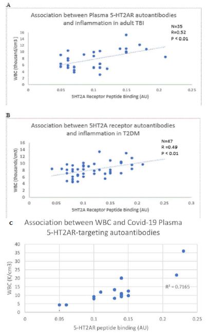
Figure 1: Significant positive correlation(s) between peripheral white blood cell count (WBC) and plasma IgG binding to serotonin 2 A receptor second extracellular loop peptide in A) adult TBI B) older adult type 2 diabetes mellitus C) Covid-19 infection. Reproduced from [4,5,6].
In two different adult TBI populations, increased baseline 5HT2AR-activating IgG was a significant predictor of the prospective (2 year- or 1 year-) rate, respectively, of accelerated cognitive decline [6,7] experienced by older or middle-aged, repeated TBI exposed patients. The AAB caused apoptosis in both endothelial cells and mouse neuroblastoma N2A cells by a mechanism involving Gq11/ phospholipase C/inositol triphosphate/Ca2+ and RhoA/Rho kinase signaling pathways’ activation [8]. Independent of TBI exposure, subsets of older adult obese type 2 DM patients harbored AAB which were significantly associated with the occurrence of Parkinson’s disease or dementia. For example, a 60 nanomolar concentration of IgG AAB from Parkinson’s disease (n=10) or dementia (n=2) patients caused significantly greater mean N2a neuroblastoma cell loss after 24 hours incubation compared to identical concentration of autoantibodies in the protein-A eluate of plasma in age-matched diabetic patients without neurodegenerative disorder (Figure 2A; control EL) [9]. Neuroblastoma cell loss induced by (60 nM) concentration of diabetic PD (n=5) or dementia (n=1) auto antibodies was completely prevented by co incubation with 200 nanomolar concentration of M100907, a highly selective 5-HT2A receptor antagonist (Figure 2B) evidence of involvement of the 5HT2A receptor [9] in Ig induced neurotoxicity.
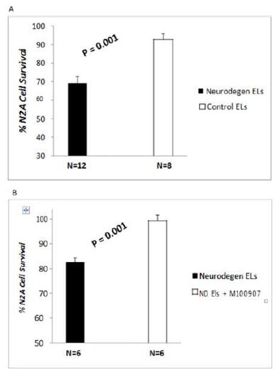
Figure 2: Diabetes Parkinson’s disease (n=10) or dementia (n=2) auto antibodies (60 nM) caused significant N2a neuroblastoma cell loss after 24 hrs incubation compared to identical concentration of autoantibodies in the protein-A eluate of plasma in diabetic patients without neurodegenerative disorder (control EL). B) Neuroblastoma cell loss induced by (60 nM) concentration of diabetic PD (n=5) or dementia (n=1) auto antibodies was completely prevented by co incubation with 200 nM concentration of M100907, a highly selective 5- HT2A receptor antagonist. N2A cells were incubated for 24 hours at 37 degrees. Cell number was determined as described in Methods. Results are mean ± SEM. Reproduced from [9].
An additional important role for injury in the causation of neurovascular AAB was evidenced by the finding that repeated TBI exposures not only caused a significant elevation in the level and titer of IgG AAB, (vs single TBI) (Figure 3) [3], but also an antigenic shift to involve another catecholaminergic receptor, alpha 1 adrenergic, whose receptor activation region is structurally closely-related to the 5HT2AR [7]. These findings are consistent with antigen-driven, adaptive humoral immune processes to a receptor (5HT2A), which is known treatment target in refractory depression and Parkinson’s disease – two of the most common later neurodegenerative complications following TBI.
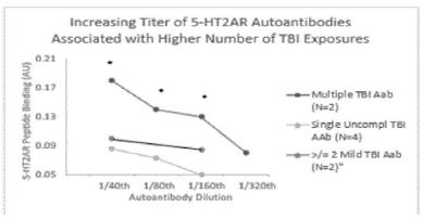
Figure 3: Multiple TBI exposure is associated with increased autoantibody titer and 5-HT2AR peptide binding potency compared to single uncomplicated TBI. * P< 0.01; Aab-autoantibody. IgG AAb from patients with multiple TBI, single uncomplicated TBI or two or more mild TBI was tested (at the indicated dilutions) for binding to the QN..8 second extracellular loop synthetic peptide in enzyme linked immunoassay. Background binding was 0.05 absorbance units (AU). Reproduced from [3].
Mechanism of ‘Self vs Non-self’ Immune Recognition
The recognition of ‘self vs non-self’ is critical to an organism’s ability to avoid systemic autoimmunity. The immune system normally differentiates self vs non-self-antigens through a developmental process of clonal deletion of B cell receptors (BCR) that exhibit high affinity binding to self-antigens. Recent studies in anti-DNA antibodies, however, demonstrate that a subset of self-reactive BCR clones can survive into adulthood through light chain-editing which reduces high affinity binding of heavy chain CDR3 to self-antigens [10]. These studies in lupus anti-DNA autoantibodies revealed that cationic arginine amino acid residues in the complementarity-determining-region CDR3 of the heavy chain mediate high affinity binding to anionic surface on certain autoantigens, e.g repeating phosphate groups in double-stranded dsDNA [10].
Mutation in the variable portion of light chains, specifically by introducing aspartic acid residues which ‘complement’ CDR3 heavy arginine residues (in anti-DNA antibodies) reduces the affinity of the heavy chain BCR for self-antigens [10]. A subset of ‘partially-edited’ BCR (in which the electrostatic interaction between heavy chain arginine and light chains aspartic acid) is not fully complemented, not only survive the process of clonal deletion but persist in adulthood as ‘self-reactive’ clones which bind glycosaminoglycans [10].
A Subset of Anti-DNA Antibodies are Anti-heparan Sulfate Antibodies
In older adult type 2 diabetes mellitus, we identified increased plasma anti-endothelial cell autoantibodies (AECA) in association with subsets of retinopathy, nephropathy, painful neuropathy [11] and later diabetic depression [12]. A shared target of the AECA was heparan sulfate proteoglycan (HSPG) [1]. HSPG is a known antigen in systemic autoimmune disorders such as systemic lupus erythematosus (SLE) [13] and it is elaborated from capillary basement membranes by the enzyme heparanase in poorly-controlled T2DM or under increased hemodynamic stress [14]. A subset of anti-DNA antibodies in lupus nephritis was reported to be anti-HSPG antibodies [15] indicative that some anti-HSPG antibodies are self-reactive, i.e. arise from self-reactive B cell clones.
Increased Expression of HSPG on Vascular Cells and Neurons
HSPG is highly expressed on both vascular surfaces and in neurons consistent with finding increased HSPG targeting-AAb in neurovascular disorders characterized by microvascular and/or neurologic dysfunction [4]. HSPG also functions as an obligatory co-factor for endothelial cell and neural survival-promoting growth factors such as fibroblast growth factor (FGF2) [16]. Fibroblast growth factor 2 is also important for the proliferation of hippocampal neural stem cells underlying therapeutic responses to anti-depressant treatment [17]. Through binding to the FGF2 co-receptor, anti-HSPG AAb can reduce FGF2 bioavailability potentially interfering with both neuronal and endothelial survival. Heparan sulfate proteoglycan is also a co-receptor for two key molecular regulators of amyloid beta production and clearance in the brain, namely beta (β)-secretase and lipoprotein receptor protein-1 (LRP-1). Since HSPG normally inhibits beta-secretase [18] and stimulates LRP-1 [19], anti-HSPG AAB would be predicted to increase Aβ production and reduce its brain clearance, actions which taken together might contribute to Alzheimer’s dementia progression.
Self-reactive BCR are ‘Multi-reactive’
An important property of self-reactive B cell clones is their inherent multi-reactivity, i.e. they can target more than one kind of self-antigen [10]. Based on the importance of aspartic acid (D) residues in edited light chains for mitigating high affinity heavy chain CDR3 antigen binding, we constructed structural models of heparin sulfate (Figure 4) for comparison to model of a recently identified autoantigen, the serotonin 2A receptor (Figure 5). The NMR structure of heparin sulfate shows a sulfur-sulfur distance of 6.6 Ångstroms (Figure 4). Adjacent aspartic acid (D) residues at positions 231and 232 in the epitope region of serotonin 2A receptor have carbonyl carbon-carbon distance of 6.6 Ångstroms (Figure 5). This makes it possible (if not plausible) that similar electrostatic interaction(s) may occur between heavy chain CDR3 arginine and the anionic region in either heparin sulfate or the two adjacent D residues in the receptor activating region of the serotonin 2A receptor.
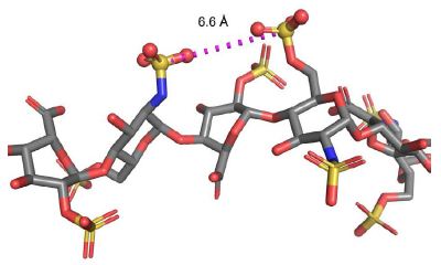
Figure 4: Heparin sulfate NMR structure showing sulfur-sulfur distance of 6.6 Ångstroms is shown. Throughout the structure, adjacent sulfur-sulfur distances range from 3.5 Å to 6.6 Å. PDB: 1HPN N.m.r. and molecular-modelling studies of the solution conformation of heparin. Mulloy B, Forster MJ, Jones C, Davies DB, Biochem J (1993) 293 ( Pt 3) p.849-58.
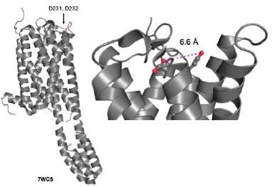
Figure 5: Crystal structure of the human serotonin 2A receptor showing adjacent aspartic acid residues at positions 231and 232. Carbonyl carbon-carbon distance of 6.6 Ångstroms is shown. PDB: 7WC5, From, Structure-based discovery of nonhallucinogenic psychedelic analogs. Cao, D., Yu, J., Wang, H., Luo, Z., Liu, X., He, L., Qi, J., Fan, L., Tang, L., Chen, Z., Li, J., Cheng, J., Wang, S. (2022) Science 375: 403-411.
In a study of older adult TBI (N=35; mean age 65 years old), we tested the IgG fraction of plasma for binding to either a second extracellular loop serotonin 2A receptor peptide or highly-purified HSPG derived from rat pheochromocytoma (PC12) cells. In thirty-one of thirty-five adult TBI patients tested, autoantibody (1/40th dilution = 30 µg/mL) binding to the 5-HT2AR second extracellular loop region peptide was significantly correlated (P< 0.01; R =0.46) with binding to purified PC12-derived HSPG (Figure 6) [3]. These results suggest that TBI autoantibodies may target (in part) strongly anionic sites present on both the 5-HT2AR and neuronal HSPG as a result of injury-induced release of vascular and neural antigens.
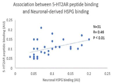
Figure 6: Correlation between 5-HT2AR peptide and neuronal HSPG binding in the protein-A eluates from thirty-one of thirty-five older adult TBI patients. A one-fortieth dilution of the protein A eluate was incubated either with 5HT2AR second extracellular loop synthetic peptide or with HSPG purified from rat pheochromocytoma cell culture. Background binding was 0.05 absorbance units (AU) in both assays.
Reproduced from [3].
Agonist Anti-serotonin 2A Receptor AAb in Neurovascular Disorders
The functional effects of AECA in diabetic subsets having retinopathy, nephropathy or painful neuropathy were pleiotropic: the IgG caused endothelial cell apoptosis [20], potent neurite retraction in mouse neuroblastoma cells, and evoked large sustained global increases in intracellular Ca2+ in EC [20] and other cell types [20]. This led us to consider whether (in addition to HSPG) another antigenic target of the IgG may be a Gq11 subclass of G-protein coupled receptor (GPCR) positively coupled to phospholipase C/inositol triphosphate receptor/Ca2+ release signaling pathway. Long-lasting depolarization can be another manifestation of phospholipase C/inositol triphosphate receptor pathway activation in excitable cells. Prior to our discovery of 5HT2AR as a target of neurovascular AAB, we used the fluorescent voltage-sensitive membrane dye, diBac4, in a high-throughput N2A mouse neuroblastoma membrane depolarization assay to screen a library of plasma IgG autoantibodies from patients having a wide range of neurologic, vascular or neuropsychiatric disorders. We found unexpectedly highest level of IgG-induced depolarization in plasma from patients with painful neuropathy, major depressive disorder, or schizophrenia [21] (Figure 7). The 5HT2AR is a well-known treatment target in refractory depression and schizophrenia. This observation led us to test and later demonstrate that the highly selective 5HT2AR antagonist, M100907, dose-dependently completed prevented neurotoxicity in IgG AAB from neurodegenerative diseases including ten patients with Parkinson’s disease (PD) and two having dementia (Figure 2).
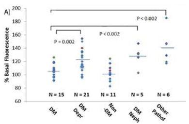
Figure 7: Significantly increased membrane depolarization (basal fluorescence) in IgG fraction of plasma from patients having diabetic depression, nephropathy, or other pathologies (schizophrenia, or painful neuropathy) compared to age-matched diabetes without any of these disorders. Diabetic depression (DM Depr), diabetic nephropathy (DM Neph) or other pathologies (Other Path) autoantibodies cause significantly increased mean depolarization in neuroblastoma (N2A) cells compared to control diabetic (DM) autoantibodies.
Reproduced from [21]
The second extracellular loop of the 5HT2A receptor is important in receptor activation. We configured an enzyme linked immunoassay using a synthetic peptide identical to the 2nd extracellular loop (as capture antigen) to screen plasma IgG from adult type 2 diabetes or adult TBI patients for significantly increased binding compared to control groups without a neurovascular disorder. Mean binding to the 5HT2AR second extracellular loop peptide was significantly increased in either TBI or T2DM patients having a neurodegenerative or neurovascular disorder [3,4] compared to control patients with uncomplicated TBI or T2DM (Figure 8). Of interest Parkinson’s disease had the highest prevalence of 5HT2AR-targeting AAB. Seventeen of twenty or 85% of Parkinson’s disease patients whose IgG was tested demonstrated significantly increased binding to the second extracellular loop serotonin 2A receptor antigen.
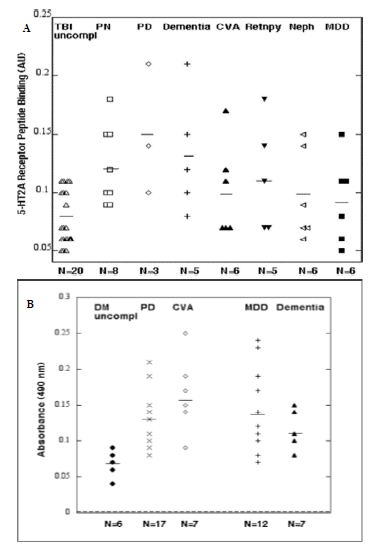
Figure 8: Enzyme linked immunosorbent assay using the 5HT2AR, second extracellular loop region synthetic peptide Q..N-18 as the solid-phase antigen. Results are arbitrary absorbance units (OD) in a one-fortieth dilution of the protein-A eluate fraction of plasma or serum from A) TBI patients with or without a neurovascular co-morbidity or B) uncomplicated type 2 diabetes (N=6), Parkinsons disease (PD) (N=17), cerebrovascular accident (CVA) (N=7), major depressive disorder (MDD) (N=12) or dementia (N=7). Reproduced from [3.4].
To further test for a specific association between PD and 5HT2AR-targeting AAB, we conducted an RNA seq study in which mouse neuroblastoma cells were briefly exposed to IgG from T2DM, and/or TBI (each subgroup was enriched in PD patients) vs normal patients without PD or targeting IgG AAb. We reported IGF from TBI, DM, and TBI + DM patient subgroups (compared to normals) caused significantly increased gene expression in cell death genes [22]. There was involvement of the mitochondrial dysfunction pathway and Parkinson’s disease pathways in TBI and DM autoantibody-induced gene expression changes in mouse neuroblastoma cells [22]. These data are consistent with prior studies implicating humoral immunity [23], and calcium-dependent proteolysis [24] in the pathogenesis of sporadic PD.
Summary
A summary diagram illustrating the humoral immune markers and mechanisms involved in type 2 diabetes mellitus-and TBI-associated neurodegeneration is shown in Figure 9.
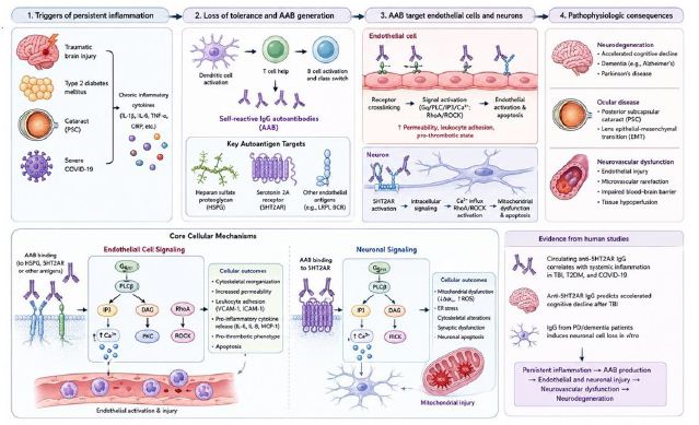
Figure 9: Summary diagram of the diseases, mechanisms, intracellular signaling pathways and tissue effects of circulating self-reactive autoantibodies. Assisted by ChatGPT 5.5.
Competing Interest
The authors declare no competing interests that would affect the objectivity of the presented results.
References
- Erkkinen MG, Kim MO, Geschwind MD (2018) Clinical Neurology and Epidemiology of the Major Neurodegenerative Cold Spring Harb Perspect Biol. [crossref]
- Xu T, Pandey SC (2000) Cellular localization of serotonin (2A) (5HT (2A)) receptors in the rat Brain Res Bull. [crossref]
- Zimering MB, Pulikeyil AT, Myers CE, Pang KC (2020) Serotonin 2A Receptor Autoantibodies Increase in Adult Traumatic Brain Injury In Association with J Endocrinol Diabetes. [crossref]
- Zimering MB (2019) Autoantibodies in Type-2 Diabetes having Neurovascular Complications Bind to the Second Extracellular Loop of the 5-Hydroxytryptamine 2A Endocrinol Diabetes Metab J. [crossref]
- Zimering MB (2021) Severe COVID-19 Pneumonia is Associated with Increased Plasma Immunoglobulin G Agonist Autoantibodies Targeting the 5-Hydroxytryptamine 2A Endocrinol Diabetes Metab J. [crossref]
- Zimering MB, Grinberg M, Myers CE, Bahn G (2022) Plasma Serotonin 2A Receptor Autoantibodies Predict Rapid, Substantial Decline in Neurocognitive Performance in Older Adult Veterans with Endocrinol Diabetes Metab J. [crossref]
- Zimering MB (2023) Repeated Traumatic Brain Injury is Associated with Neurotoxic Plasma Autoantibodies Directed against the Serotonin 2A and Alpha 1 Adrenergic Endocrinol Diabetes Metab J. [crossref]
- Zimering MB (2017) Diabetes Autoantibodies Mediate Neural- and Endothelial Cell-Inhibitory Effects Via 5-Hydroxytryptamine-2 Receptor Coupled to Phospholipase C/Inositol Triphosphate/Ca2+ Pathway. J Endocrinol Diabetes. [crossref]
- Zimering MB. Circulating Neurotoxic 5-HT2A Receptor Agonist Autoantibodies in Adult Type 2 Diabetes with Parkinson’s J Endocrinol Diab 5(2): 1-11. DOI: 10.15226/2374-6890/
- Khan SN, Witsch EJ, Goodman NG, Panigrahi AK, Chen C, Jiang Y, Cline AM, Erikson J, Weigert M, Prak ET, Radic M (2008) Editing and escape from editing in anti-DNA B Proc Natl Acad Sci U S A. [crossref]
- Zimering MB, Alder J, Pan Z, Donnelly RJ (2011) Anti-endothelial and anti-neuronal effects from auto-antibodies in subsets of adult diabetes having a cluster of microvascular Diabetes Res Clin Pract. [crossref]
- Zimering MB, Behnke JA, Thakker-Varia S, Alder J (2015) Autoantibodies in human diabetic depression inhibit adult neural progenitor cells in vitro and induce depressive-like behavior in J Endocrinol Diab. [crossref]
- Kim HJ, Hong YH, Kim YJ, Kim HS, Park JW, Do JY, Kim KJ, Bae SW, Kim CW, Lee CK (2017) Anti-heparan sulfate antibody and functional loss of glomerular heparan sulfate proteoglycans in lupus nephritis. Lupus. [crossref]
- Vlodavsky I, Iozzo RV, Sanderson RD (2013) Heparanase: multiple functions in inflammation, diabetes and atherosclerosis. Matrix Biol. [crossref]
- Termaat RM, Brinkman K, van Gompel F, van den Heuvel LP, Veerkamp JH, Smeenk RJ, Berden JH (1990) Cross-reactivity of monoclonal anti-DNA antibodies with heparan sulfate is mediated via bound DNA/histone J Autoimmun. [crossref]
- Shi J, Fan C, Zhuang Y, Sun J, Hou X, Chen B, Xiao Z, Chen Y, Zhan Z, Zhao Y, Dai J (2019) Heparan sulfate proteoglycan promotes fibroblast growth factor-2 function for ischemic heart repair. Biomater Sci. [crossref]
- Woodbury ME, Ikezu T (2014) Fibroblast growth factor-2 signaling in neurogenesis and neurodegeneration. J Neuroimmune Pharmacol. [crossref]
- Ozsan McMillan I, Li JP, Wang L (2023) Heparan sulfate proteoglycan in Alzheimer’s disease: aberrant expression and functions in molecular pathways related to amyloid-β Am J Physiol Cell Physiol. [crossref]
- Kanekiyo T, Zhang J, Liu Q, Liu CC, Zhang L, Bu G (2011) Heparan sulphate proteoglycan and the low-density lipoprotein receptor-related protein 1 constitute major pathways for neuronal amyloid-beta J Neurosci. [crossref]
- Zimering MB, Pan Z (2009) Auto-antibodies in type 2 diabetes induce stress fiber formation and apoptosis in endothelial cells. J Clin Endo Metab. [crossref]
- Zimering MB, Mirkovic N, Pandya M, Zimering JH, Behnke JA, et (2016) Toxic Immunoglobulin Light Chain Autoantibodies are Associated with a Cluster of Severe Complications in Older Adult Type 2 Diabetes. J Endocrinol Diab. [crossref]
- Zimering MB, Delic V, Citron BA (2021) Gene Expression Changes in a Model Neuron Cell Line Exposed to Autoantibodies from Patients with Traumatic Brain Injury and/or Type 2 Mol Neurobiol. [crossref]
- Orr CF, Rowe DB, Mizuno Y, Mori H, Halliday GM (2005) A possible role for humoral immunity in the pathogenesis of Parkinson’s Brain. [crossref]
- Dufty BM, Warner LR, Hou ST, Jiang SX, Gomez-Isla T, Leenhouts KM, et al. (2007) Calpain-Cleavage of α-Synuclein: Connecting Proteolytic Processing to Disease-Linked Aggregation. Am J Pathol. [crossref]