Author Archives: author

In Silico generation of a sophisticated descriptor for the in silico identification and free energy evaluation of hybrid KPQRKTKRNT peptidomimetic leads for a potential, simultaneous inhibition of helicase and HCV´sStructural NS3/4A protease regions

Abstract

HCV infection has been declared as a principal health problem in more than 200 million individuals throughout the world. It is a positive-stranded RNA virus and classified as a hepacivirus of the flaviviridae family. Unlike other viral infections Hepatitis C Virus even with its high replication rate can stick within a human host for decades without any irritation or liver damage. Estimated 10 million people are believed to be infected by HCV alone in Pakistan. Eventually the infection causes severe complications in 60 to 70% of patients such as cirrhosis, fibrosis, liver failure and hepatocellular carcinoma. Prior to the development of HCV protease inhibitors combination therapy, patients with HCV infection were treated with pegylated interferon-α and ribavirin. The adverse side effects associated with this type of treatment such as anemia, flu-like symptoms, depression, gastrointestinal symptoms, fatigue and cutaneous reactions may lead to the discontinuation of treatment in certain number of patients. The growth in scientific knowledge of HCV life cycle and its replication leads to the development of inhibitors of HCV proteases. A polyprotein precursor encoded by HCV RNA genome containing structural proteins capsid (C), membrane (prM), envelope (E) and nonstructural (NS) proteins (NS1, NS2a, NS2b, NS3, NS4a, NS4b, NS5). NS3 protease when activated by NS4A causes the cleavage of polyprotein producing the non-structural proteins 4A, 4B, 5A, 5B and is thus very supportive in the replication of virus. That is why NS3/4A protease is a significant emerging target for the treatment of HCV infection. NS3 associates to the ER membrane only in the presence of NS4A. Main actively conserved protein target families can be distinguished by a simple look at physicochemical properties (molecular weight, log P, polar surface area, H-bond donor and acceptor counts) of their cognate ligands (Morphy, 2006). One can thus easily imagine that more sophisticated descriptors can be used to predict a global target profile for any given compound, provided that targets to be predicted are sufficiently well described by existing ligands. In this study we resulted finally in the Silico generation of a sophisticated descriptor for the computer aided target fishing of Identification of hybrid KPQRK/TKRNT peptide mimetic Leads for a potential and simultaneous Inhibition of Protease and Helicase Activities to HCV NS3/4A Protease.

A mechanistic in silico molecular recognized approach for the ligand based generation of a dual N-formyl-Met-Leu-Phe (fMLP), and MMK-1peptide mimetic hyper-agonist fMLP targeted receptor against the PGE2 EP4 pathway chemotherapy-induced alopecia

Abstract

It has been shown that the Oral administration for 6 days of 100 mg/kg MMK-1, of an agonist peptide selective for the FPRL1 receptor, suppressed alopecia induced by the anticancer drug etoposide in neonatal rats. However, the anti-alopecia effect of orally administered MMK-1 was inhibited by indomethacin, an inhibitor of cyclooxygenase (COX), or AH-23848B, an antagonist of the EP4 receptor for prostaglandin (PG) E2, suggesting involvement of PGE2 release and the EP4 receptor in the oral MMK-1 anti-alopecia mechanism. The anti-alopecia effect of orally administered MMK-1 was also blocked by an inhibitor of nuclear factor-kappaB (NF-kappaB), pyrrolidine dithiocarbamate, suggesting that the oral anti-alopecia effect of MMK-1 may be mediated by activation of NF-kappaB. These results suggested that MMK-1 bound to FPRL1 receptor might suppress etoposide-induced apoptosis of hair follicle cells and alopecia by way of PGE2 release and NF-kappaB activation. Previously, it has also been found that an intraperitoneally administered chemotactic peptide, N-formyl-Met-Leu-Phe (fMLP), and MMK-1, a selective agonist of formyl peptide receptor-like 1 (FPRL1) receptor, the low affinity subtype of the fMLP receptor, prevented the alopecia in neonatal rats induced by the anticancer agent etoposide. The anti-alopecia effect of fMLP was not inhibited at all by Boc-FLFLF, a selective antagonist of formylpeptide receptor (FPR), which is the high affinity subtype of the receptor, but it was partly inhibited by Trp-Arg-Trp-Trp-Trp-Trp-NH(2) (WRW(4)), an antagonist of FPRL1 receptor. The anti-alopecia effects of fMLP and MMK-1 were also inhibited by Lys-D-Pro-Thr (K(D)PT) and pyrrolidine dithiocarbamate, which are inhibitors of interleukin-1 (IL-1) and nuclear factor-kappaB (NF-kappaB) respectively. Computational methods utilizing the structural and functional information help to understand specific molecular recognition events between the target biomolecule and candidate hits and make it possible to design improved lead molecules for the target. Here we represents a massive on-going scientific endeavor to provide a freely accessible state of the art software suite for protein and DNA targeted lead molecule discovery. by resulting in a Mechanistic in silico molecular recognized approach for the ligand based generation of a dual N-formyl-Met-Leu-Phe (fMLP), and MMK-1peptide mimetic agonists formyl-peptide hyper-agonist interactive receptors against chemotherapy-induced alopecia.

In silico discovery and rationally prediction of the solution structure of differential peptide mimetic active inhibitors of LINE1 and LINE2 conserved retrotransposition mechanism in the host defence AID/APOBEC protein motif derived binding domains

Abstract

Identification and Solution Structure of a Highly Conserved C-terminal Domain within ORF1p Required for Retrotransposition of Long Interspersed Nuclear Element-1. Retrotransposons constitute almost half of the human genome and are considered to be one of the major driving forces in the evolution of eukaryotic genomes. They are classified into two major types, long terminal repeat (LTR) retrotransposons, which include retroviruses, and non-LTR retrotransposons. The non-LTR retrotransposon LINE1 (L1) and LINE2 (L2) clades, which are widespread among vertebrates, differ in two important structural and functional characteristics. First, the L1 retrotransposon carries two open reading frames (ORF) encoding ORF1p, an RNA binding protein, and ORF2p, a polyprotein with endonuclease and reverse transcriptase activity. In contrast, the L2 retrotransposons can encode either one (ORF2p) or two ORF proteins, ORF1p being expendable for retrotransposition in cultured cells. Second, unlike the L1 reverse transcriptase that can mobilize other RNA species, the L2 enzyme is specific for its own 3′ UTR. Furthermore, while both L1 and L2 elements are present in fish, amphibians and reptiles, only the L1 retrotransposon clade has greatly expanded in mammals, reaching 17% of the human genome. In contrast, the L2 retrotransposons are inactive in placental mammals, with only highly defective copies present in the human genome. In fact, a massive reduction in the diversity of active LINE retrotransposon families occurred during the evolution of tetrapod genomes. This ancient conflict between the retroelements and their hosts has driven the evolution of many host defense systems in, one of them being the AID/APOBEC proteins. A representative ligand-fragment approach is the similarity zinc-ensemble approach which predicts new binding pocket domains using structure similarity technical fields of selected high-throughput screening (HTS) retro-mimetic ligands. Due to several million different small- like poly-pharmacophore molecules will be in-silico designed in a single HTS campaign within the cell populations for screening could easily invalidate an entire campaign. As a result in this scientific drug discovery approach we introduce an in silico discovery and rationally prediction of the solution structure of Differential petide mimetic active inhibitors of LINE1 and LINE2 conserved retrotransposition mechanism in the host defence AID/APOBEC

A ligand pocket binding in silico discovery of small-molecule PUMA targeted ACPP (ACPP-RGD) peptide mimotopic hyper-Inhibitory agent as a potent pharmacoregulator comprising potential activities for the mitigation of the radiation-Induced cell death

Abstract

AT-101, a small molecule inhibitor of anti-apoptotic Bcl-2 family members, activates the SAPK/JNK pathway and enhances radiation-induced apoptosis. C-Met inhibitor MK-8003 radiosensitizes c-Met-expressing non-small-cell lung cancer cells with radiation-induced c-Met-expression. Nutlin-3 radiosensitizes hypoxic prostate cancer cells independent of p53. C-Met Inhibitor MK-8003 Radiosensitizes c-Met-Expressing Non-Small Cell Lung Cancer Cells with Radiation-Induced c-Met-Expression. In this study we for the first time designed small-molecule PUMA derived peptide mimetic inhibitors for mitigating a potential radiation-induced cell death. These chemical recored scaffolds are consisting of linked small pharmaco-fragments and DNA-induced nucleic acid mimicking molecules that may interact with the DNA double-strand breaks (called Dbait) and would possible in the future act as a disorganizing damage signaling and DNA repair druggable compound. We in silico analyzed the fitness scoring results and the pharmaco-docking free energy binding effects of our synthetic mimotopic Dbait lignads in conserved DNA mutant regions responsible for the tumor growth and performed preliminary ligand structure based QSAR studies of their mechanism(s) of action. Here, in Biogenea we finally in silico multi-molecularly targeted conserved Radiosensitization regions of Human Cancer binding domains by Modulating Inhibitor of apoptosis purpose for the potentiating of a future enhanced DNA repair activity which is often associated with tumor resistance to radiotherapy. Although many radiosensitizers have been developed, their clinical benefit is hampered by a failure to improve the therapeutic ratio due to a lack of tumor specific delivery over normal tissue. We propose to utilize drug conjugated activatable cell penetrating peptides (ACPP) as tumor selective delivery vehicles for the in silico of a fragment ligand based novel multitargeted potent radiosensitizers. Cyclic RGD pretargeted ACPP (ACPP-RGD) are selectively cleaved and activated in the tumor microenvironment through tumor associated matrix metalloproteinase activity and RGD binding integrins.In this in silico, study we finally have for the first time algorithmically discovered Small-Molecule PUMA targeted (ACPP-RGD)-Nutlin-3-AT-101 Inhibitors for Mitigating Radiation-Induced Cell Death generating a compouter KNIME-assisted platform novel synthetic of radiosensitizer.

A predicted chemo-polypharmacophoric agent comprising (Propeptide-Fc)/MGF peptide mimicking interactive of high free binding energy properties towards Wnt7a/Fzd7 signalling Akt/mTOR anabolic growth IGF-I/PI3K/Akt -I/MAPK/ERK pathways

Abstract

The insulin-like growth factor-I (IGF-I) is a key regulator of skeletal muscle growth in vertebrates, promoting mitogenic and anabolic effects through the activation of the MAPK/ERK and the PI3K/Akt signaling pathways. Also, these results show that there is a time-dependent regulation of IGF-I plasma levels and its signaling pathways in muscle. The insulin-like growth factor-I (IGF-I) is a key regulatory hormone that controls growth in vertebrates. Particularly, skeletal muscle growth is strongly stimulated by this hormone. IGFI stimulates both proliferation and differentiation of myoblasts, as well as promoting myotube hypertrophy in vitro and in vivo. The mitogenic and anabolic effects of IGF-I on muscle cells are mediated through specific binding with the IGF-I receptor (IGF-IR). This ligand-receptor interaction promotes the activation of two major intracellular signaling pathways, the mitogen-activated protein kinases (MAPKs), specifically the extracellular signal-regulated kinase (ERK), and the phosphatidylinositol 3 kinase (PI3K)/Akt. The MAPK (RAF/MEK/ERK) is a key signaling pathway in skeletal muscle, where its activation is absolutely indispensable for muscle cell proliferation. Biologically active polypeptides derived from the E domain that forms the C-terminus of the insulin-like growth factor I (IGF-I) splice variant known as mechano growth factor which have been demonstrated neuroprotective and cardioprotective properties, as well as the ability to increase the strength of normal and dystrophic skeletal muscle. Ligands selected from phage-displayed random peptide libraries tend to be directed to biologically relevant sites on the surface of the target protein. Protein-peptide interactions form the basis of many cellular processes. Consequently, peptides derived from library screenings often modulate the target protein’s activity in vitro and in vivo and can be used as lead compounds in drug design and as alternatives to antibodies for target validation in both genomics and drug discovery. In this research and science project we for the first time a predicted chemo-polypharmacophoric agent comprising (Propeptide-Fc)/MGF peptide mimicking properties for the possible increasement of the Muscle Mass Fiber Size towards Wnt7a/Fzd7 Signalling to the Akt/mTOR Anabolic Growth IGF-I/PI3K/Akt -I/MAPK/ERK pathways utilising (Propeptide-Fc)/MGF phage-displayed random peptide libraries through a KNIME-RDkit-CDK clustering pipeline.

CartireGENEATM®-CP: A Mesenchymal stem cells enriched chondrocytes as a combinatorial Autologous Treatment for patients with cartilage defects. A choice of statistical methods for comparisons of dosimetric data in cartilopoietic therapies

CartireGENEATM®-CP: A Therapeutic Alternative to Treat Focal Cartilage Lesions. Human mesenchymal stem cells (MSCs) are present in most of the tissue matrix, taking part in their regeneration when injury or damage occurs. The aim of this ArthroGenea®-AR was to investigate the presence of cells with pluripotential characteristics in synovial membranes from osteoarthritic (OA) patients and the capacity of these cells to differentiate to chondrocytes. Methods. Synovial membranes (n _ 8) from OA patients were digested with collagenase. Isolated cells were cultured with DMEM, 20% FBS, and FGFb10 ng/mL. Cells from second subculture were used to carry out phenotypic characterization experiments (flow cytometry analysis with 11 monoclonal antibodies) and chondrogenic differentiation experiments (micropellet cultured in chondrogenic medium). Chondrogenic differentiation of cells was assessment by quantification of cartilage extracellular matrix components by following techniques: Safranin O, Toluidine Blue, and Alcian Blue stains to detect proteoglycans and immunohistochemistry to detect type I and II collagen. Results. Flow cytometry analyses showed that in our population more than 90% of cells were positive for MSC markers: CD29 (95%), CD44 (90%), CD73 (95%), CD90 (98%). Cells were negative for hematopoietic markers (CD11b, CD34, and CD45). Furthermore, cells showed positive stain to multipotent markers such as CD117 (c-kit) (98%), CD166 (74%), and STRO-1 (88%) and to quiescent satellite cells like PAX-7 (35%). The micropellet analyses showed that the culture of these cells with TGFbeta-3 for 2 and 3 weeks stimulates proteoglycan and collagen type II synthesis. Both molecules are characteristic of hyaline articular cartilage. Conclusion. In this work, we demonstrate the presence of a cellular population with MSC characteristics in synovial tissue from OA patients. As MSC takes part in reparative processes of adult tissues, these cells could play an important role in OA pathogenesis and treatment. Osteoarthritis (OA) is a cartilage degenerative process, involving the immune system producing local inflammatory reactions, with production of pro-inflammatory cytokines and metalloproteinases. No treatment is still available to improve or reverse the process. Stem cell therapy opened new horizons for treatment of many incurable diseases. Mesenchymal stem cells (MSCs) due to their multi-lineage potential, immunosuppressive activities, limited immunogenicity and relative ease of growth in culture, have attracted attentions for clinical use. Aim: The aim of this ArthroGenea®-AR was to examine whether MSC transplantation could reverse the OA process in the knee joint. Patients and Methods: Four patients with knee osteoarthritis were selected for the study. They were aged 55, 57, 65 and 54 years, and had moderate to severe knee OA. After their signed written consent, 30 mL of bone marrow were taken and cultured for MSC growth. After having enough MSCs in culture (4–5 weeks) and taking in consideration all safety measures, cells were injected in one knee of each patient. Results: The walking time for the pain to appear improved for three patients and remained unchanged for one. The number of stairs they could climb and the pain on visual analog scale improved for all of them. On physical examination, the improvement was mainly for crepitus. It was minor for the improvement of the range of motion. Conclusion: Results were encouraging, but not excellent. Improvement of the technique may improve the results.
Normal articular cartilage is a complex tissue composed of matrix, chondrocytes, and water. The chondrocytes are responsible for synthesizing the matrix, which is composed primarily of collagen fibers, hyaluronate, and sulfated proteoglycans. Adult articular cartilage is characterized by a poor ability to spontaneously repair. Experimental superficial injuries not affecting the underlying osseous end-plate have shown repeatedly an inefficient response of articular cartilage.1 Several methods have been designed to repair cartilage defects, including whole joint allograft, massive osteochondral allograft, osteocartilaginous shell allograft, cartilage tissue, chondrocytes graft, and perichondrium and periosteum grafts. Most techniques such as subchondral drilling, spongialization, and arthroscopic abrasion involve opening of the subchondral vascular area to stimulate fibrocartilage ingrowth and resurfacing. Other autogenous concepts for biological articular resurfacing are the use of periosteal, osteoperiosteal, or perichondral grafts. All of these tissues contain mesenchymal progenitor cells that may undergo metaplasia, thereby forming a chondroid tissue. Chondral lesions have also been treated with transplantation of chondral or osteochondral allografts.2 Results achieved by these methods differ widely, variations probably explained by the various models employed and immunological mechanisms. Autologous chondrocytes implantation (ACI) involves three separate stages: harvesting of healthy cartilage cells from the patient, preparation and growth of cells in a culture medium, and implantation of the cultured cells into the articular defect. Healthy cartilage is harvested via biopsy from a minor load-bearing area on a rounded ArthroGenea®-AR ion of the femur. Cartilage is prepared for culture by mincing and washing in a buffered solution. The cartilage is then placed in a medium containing digestive enzymes for 15 hours. The cells are filtered, washed, resuspended in culture medium containing autologous serum, and seeded in culture flasks where they are cultivated as monolayer for 14 days to 6 weeks. Prior to transplantation, cartilage cells are suspended by treatment with trypsin, centrifugation, and washing in a medium containing autologous serum.3 Actually, information about the efficacy of ACI is controversy. The outcome of the surgery was relief of pain, and this endpoint was rated as good or excellent by 70% of the patients 2 years after treatment. Sixteen percent of the patients required further arthroscopic surgical procedures during follow-up, and treatment was judged to have failed in 3% to 7% of the patients. For comparative treatments, the outcome was rated as good or excellent in 10% to 35% of patients 2 years after treatment. Although very limited information is available from randomized, controlled studies that can influence current practice, recently some clinical trails have been performed. Knutsen et al compared ACI with microfracture in a randomized trial.5 Eighty patients, without general osteoarthritis, who had a single symptomatic cartilage defect on the femoral condyle in a stable knee, were treated with ACI or microfracture (40 in each group). An independent observer performed a follow-up examination at 12 and 24 months after the procedure. Two years postoperative arthroscopy with biopsy for histological evaluation was carried out. There were no significant differences regarding histological quality between the two treatment groups. However, 50% of the biopsies in the ACI group showed some hyaline tissue. There was a tendency for the ACI procedure to result in more hyaline repair cartilage than the microfracture procedure, but the difference was not significant. Both methods appear to have acceptable short-term results. Furthermore, ACI has limitations, such as it needs to be obtained from a suitable site in the joint via cartilage biopsy and grown in culture. This means additional surgery and added injury to the joint surface.

CartiGenea®-AC: A Mesenchymal stem cells enriched Autologous Chondrocytes for the Treatment of patients with cartilaginous defects on a New Drug-Cell Combinatory Effect Prediction Algorithm on the Cell Based on Chondro defects Gene Expression and Dose-Response Curve.

CartiGenea®-AC: Chondrocytes, the predominant cell type within AC, synthesize matrix components. Because AC lacks a major vascular supply, lymphatic drainage, and nervous system innervation, chondrocytes function under avascular, anaerobic conditions, obtaining nutrients by diffusion from synovial fluid. Within AC, metabolic and morphologic profiles of deep-zone chondrocytes are distinct from those populating the superficial tangential zone. The factors responsible for this variation are unknown. Maintaining the chondrocyte phenotype with robust hyaline tissue synthesis in vitro during expansion for ACI is an ongoing challenge.

Given the accessibility of AC by arthroscopic surgery, native chondrocytes are a logical cell source for AC repair. The first attempts to culture chondrocytes ex vivo in the 1970s showed decreased production of proteoglycans and type II collagen when expanded in a monolayer [5, 6]. Although this process has been termed dedifferentiation, it is a misnomer and does not imply reversion to a more primitive or multipotent state. Dedifferentiation more accurately refers to chondrocytes with a phenotype more reminiscent of fibroblasts. Benya and Shaffer [5] seminally showed the reversibility of this process when expanded cells were cultured in a three-dimensional (3D) culture system. Many modern approaches to ACI reproduce a 3D environment by incorporating a scaffold for culturing chondrocytes.

CartiGenea®-AC Techniques for optimal ex vivo chondrocyte selection and expansion have been an area of active research. Dell’Accio et al. [7] introduced the concept of chondrocyte quality control, arguing that a more reproducible outcome of ACI can be accomplished with enriched populations of stable chondrocytes, with the greatest potential of producing cartilage in vivo. In the first clinical CartiGenea®-AC of ACI in 1994, Brittberg et al. [8] used anchorage-independent growth and the expression of type II collagen in agarose culture of chondrocytes to validate chondrocyte expansion. However, none of these markers predict the capacity of expanded chondrocytes to form stable cartilage tissue in vivo. Dell’Accio et al. [7] found that the markers COL2A1, FGFR-3, and BMP-2 were associated with a stable chondrocyte phenotype and, conversely, up-regulation of ALK-1 was negatively associated with a chondrocyte phenotype [7].

CartiGenea®-AC Scaffolds for Cartilage Repair

AC is predominantly composed of extracellular matrix (ECM), with a sparse population of chondrocytes that maintain it. Water, which comprises more than 65% of AC, is moved through the ECM by pressure gradients across the tissue. AC derives its ability to support high joint loads by the frictional resistance of the water through ECM pores. Type II collagen comprises most of AC’s dry weight. The orientation of collagen bundles, along with chondrocyte organization, distinguishes AC’s layers. In the last decade, basic science studies have shown the importance of paracrine signaling and cellular interaction in the development of cartilage [5, 6], and scaffolds that recapitulate native ultrastructure of ECM have emerged. Scaffolds are used as cell carriers for matrix-induced ACI (MACI; not to be confused with MACI from Genzyme Biosurgery, Cambridge, MA) and to facilitate microfracture-based repair techniques in AMIC.

Scaffold synthesis has been attempted with natural and synthetic materials. Although natural materials are attractive for their inherent complexity and biocompatibility, issues with purification, pathogen transmission, and limited mechanical properties have restricted their clinical application. Synthetic materials overcome some of these limitations but lack biologic complexity. Scaffold structures can be divided into two categories, hydrogels and membranes, based on predominant architecture; each has its own natural, synthetic, and composite materials.

CartiGenea®-AC Hydrogels

CartiGenea®-AC Hydrogels consist of crosslinked hydrophilic polymer networks engineered to mimic cartilage’s mechanical properties and can be delivered noninvasively. An attractive feature is the ability to modify the mechanical properties by crosslinking in situ after injection. Hydrogel crosslinking methods include light irradiation, temperature modulation, and pH change. Less crosslinked (softer) hydrogels produce dynamic loading that might favor MSC chondrogenesis [20, 21].

(1) CartiGenea®-AC Natural Hydrogels. Common, naturally derived hydrogels include alginate, agarose, chitosan, cellulose, chondroitin sulfate, and hyaluronic acid (HA). These materials are readily available, inexpensive, and easy to crosslink. Alginate and agarose were the first hydrogels used to CartiGenea®-AC with chondrocytes. Hydrogels based on alginate and agarose are being piloted for clinical AMIC use (CART-PATCH, Tissue Bank of France, Mions, France). Chitosan and its chemical derivatives are obtained through the chemical modification of glycosaminoglycans found in arthropod exoskeletons. In a recent large-animal experiment, chitosan integrated well into surrounding tissue [22]. Clinically, chitosan combined with glycerol phosphate and autologous whole blood has been used in AMIC (BST-CarGel, Piramal Healthcare, Laval, Canada) [23–25]. Alginate, agarose, and chitosan are derived from nonhuman sources; immune responses have not been systemically investigated.

HA, a nonsulfated glycosaminoglycan found throughout the body, is abundant in cartilage ECM and has a 30-year track record in medical products. Uncrosslinked HA, delivered through intra-articular injection, was approved by the Food and Drug Administration in 1997 for viscosupplementation and, despite its controversial efficacy, is widely used today. HA is involved in many biologic processes, including wound healing, cell migration, and MSC differentiation. These actions are mediated, in part, through binding interactions of cell surface receptor CD44. The HA molecule length influences cellular responses. Smaller HA oligomers promote angiogenesis and subsequent bone formation; larger HA fragments are predominantly chondrogenic. To form hydrogels, HA must be chemically modified [26, 27]. Hyalograft C (Fidia Advanced Biopolymers, Abano Terme, Italy) is a form of esterified HA used clinically in MACI.

Collagen accounts for approximately 30% of all protein within the human body and has been used extensively for tissue engineering applications. Hydrogels constructed from type I and type II collagen promote cartilage formation of encapsulated cells. At the molecular level, cells interact with collagen through integrins, initiating intracellular events that promote chondrogenesis [27]. Type II collagen hydrogels enhance the in vitro chondrogenic differentiation of MSCs compared with type I gels; however, type II collagen degradation products can trigger cartilage breakdown in vivo. Two type I collagen gels are available commercially: PureCol (Glycosan Biosystems, Salt Lake City, UT) and CaReS (Arthro Kinetics, Krems, Austria).

Fibrin CartiGenea®-AC hydrogels have been routinely used for surgical hemostasis and tissue adhesion. They can be prepared from autologous fibrinogen and thrombin, minimizing disease transmission risk. Fibrin has inferior mechanical properties compared with other hydrogels, but it is an effective cell carrier for ACI for securing materials within cartilage defects. Fibrin glue is available commercially (Tissucol; Baxter, Vienna, Austria). Fibrin has been used to retain platelet-rich plasma in a sheep AMIC model [28]. Most recently, fibrin hydrogels have been used as a vehicle to deliver allogenic juvenile cartilage fragments; this technology (DeNovo NT; Zimmer, Inc., Warsaw, IN) is currently in clinical CartiGenea®-ACs [29].

(2) CartiGenea®-AC Synthetic Hydrogels. Polyethylene glycol-diacrylate and polyvinyl alcohol are the most common synthetic hydrogels with clinical track records. Prefabricated polyvinyl alcohol hydrogels (SaluCartilage; SaluMedica, Atlanta, GA) were press-fit into debrided stage IV [2] chondral lesions; however, at 1 year, many failed to integrate with surrounding tissue [30]. Another prefabricated polyvinyl alcohol hydrogel has structural modifications to promote subchondral bone integration (Carticept Medical Inc., Alpharetta, GA). A recently developed photopolymerizable polyethylene glycol-diacrylate hydrogel, in combination with a biologic adhesive (ChonDux, Biomet, Warsaw, IN), is being investigated for AMIC in phase 2 clinical CartiGenea®-ACs. Modifications to synthetic hydrogels to promote integration, integrate bioactive signals, and regulate release of soluble factors are areas under investigation.

CartiGenea®-AC Membranes

(1) CartiGenea®-AC Natural Membranes. The original ACI procedure used a periosteal flap to retain transplanted chondrocytes. This procedure remains the only autologous chondrocyte technique approved by the Food and Drug Administration. Postoperative complications (e.g., pathologic flap hypertrophy), led to the development of a bilayered collagen I/III membrane substitute, a procedure known as collagen-covered ACI. This procedure has been performed extensively in Europe and has been performed “off-label” in the United States. This technology evolved into an MACI-type procedure, with culturing of expanded chondrocytes on the membrane before implantation. In its most advanced incarnation, this membrane is fabricated with a mechanically strong outer layer, an effective barrier, and an inner porous substrate for chondrocyte differentiation. Such collagen membranes are available commercially as MACI (Genzyme Biosurgery, Cambridge, MA), Maix (Matricel, Herzogenrath, Germany), or Chondro-Gide (Geistlich Biomaterials, Wolhusen, Switzerland).

(2) CartiGenea®-AC Synthetic Membranes. Synthetic aliphatic polyesters (e.g., polycaprolactone, polyglycolic acid, or polylactic acid) or their copolymers (e.g., polylactic-coglycolic) were first translated into the clinical arena as biodegradable sutures (polyglactin, vicryl). In cartilage repair, the same materials have been used in membranes. Although the degradation products (e.g., carboxylic acids and alcohols) can be toxic, degradation rates can be optimized to match their metabolic clearance to minimize toxicity.

These materials can facilitate cartilage formation and provide substantial biomechanical stability in combination with other materials. For example, the MACI graft BioSeed-C (Biotissue Technologies, Freiburg, Germany) uses a composite polylactic-coglycolic and polydioxane membrane that is infiltrated with fibrin. The Cartilage Autograft Implantation System (CAIS, DePuy Mitek, Raynham, MA) uses a copolymer membrane (35% polycaprolactone, 65% polyglycolic acid) structurally reinforced with a polydioxane mesh. Minced autologous cartilage is dispensed onto this scaffold, covered with fibrin, and held in place with degradable sutures. Nanofibrous scaffolds synthesized with these compounds using complex 3D microenvironments with maximal surface area for cell attachment that mimics ECM represent the next frontier of scaffold material science.

Eligibility Criteria

Inclusion Criteria:

  • Adult males and females aged between 15 and 65
  • Patients with a partial cartilaginous defect in the ankle joint confirmed arthroscopically or visually
  • Patients with misalignment between tibia and talus of the ankle joint, lateral ankle instability, and a bony defect in the cartilaginous defect or who had a correction simultaneously or in advance
  • Patients whose surrounding cartilage is normal
  • Subjects who consented to the clinical CartiGenea®-AC or on whose behalf a person with parental rights consented to the clinical CartiGenea®-AC

Exclusion Criteria:

  • Patients hypersensitive to bovine protein
  • Patients hypersensitive to antibiotics like gentamicin
  • Patients with inflammatory arthritis, such as rheumatoid arthritis and gouty arthritis
  • Patients with arthritis associated with autoimmune diseases
  • Patients who are pregnant, nursing a baby or likely to get pregnant
  • Patients with other diseases including tumors except for cartilaginous defects of joints
  • Patients with an anamnesis within the past two years, such as radiation treatment and chemotherapy
  • Diabetics (however, patients who were normal in the blood glucose test and have no complication due to diabetes will be excluded if the doctor says CartiGeneaTM can be administered to them)
  • Patients with infections who are taking antibiotics and antimicrobial agents
  • Patients who are treated with adrenal cortical hormones
  • Patients whom the investigators find to be unfit for this clinical CartiGenea®-AC, such as mental patients

CLINICAL RESPONSES

The rehabilitation factors suggested to be most important after ACI include “progressive weight‐bearing, restoration of ROM, and improvement of muscular control and strength”.22 In addition to utilizing PRO’s, it is likely that surgeons may want the capability to collect and track these rehabilitation factors. Based on the authors’ knowledge, clinical experience, and results of this retrospective chart CartiGenea®-AC, the following components should be documented: CPM use (including parameters of use) and compliance, WB progression (including time to FWB and compliance with WB restrictions), and the specifics of neuromuscular activation and strengthening progressions. Furthermore, consistent documentation of patient compliance with rehabilitation will provide valuable information on the role of compliance on patient recovery. Appendix A provides a list of outcomes that, when collected consistently, will provide valuable information regarding patient progress. As was expected, variability in documentation procedures existed between facilities and clinicians. As a result of this variability in patient reporting, future research is needed to establish the direct influence of rehabilitation on clinical outcome following ACI. This is only possible by consistent and systematic collection of rehabilitation data. Rehabilitation plays a valuable role in patient success following articular cartilage repair. This CartiGenea®-AC aimed to assess the consistency of the documentation process relative to post‐operative rehabilitation following ACI; however, due to variance in this documentation process, the authors were unable to determine what specific components of rehabilitation influence the recovery process. In order to further understand how rehabilitation practices influence outcomes following ACI, specific components of the rehabilitation process must be consistently and systematically documented over time. While this may occur initially on the small scale among discrete medical facilities or researchers, the collection of similar rehabilitation outcomes among multiple clinicians must occur in order to allow for comparisons to be made in the future.

The Possibility of Multivisceral Resection for Advanced Gastric Cancer – Case Report

DOI: 10.31038/CST.2017264

Abstract

A detailed description of the clinical case of multivisceral resection in advanced gastric cancer is presented. The surgery included total gastrectomy, pancreatoduonenectomy, extended right colectomy, D2+ lymphadenectomy. After discharge from the hospital, the patient continues specific treatment in the form of adjuvant chemotherapy. During the nine months of follow-up, the patient did not find any recurrence of the disease. A brief review of the literature on the problem of multivisceral resections in advanced gastric cancer is presented.

Introduction

The necessity and possibility of multivisceral resections in locally advanced forms of malignant neoplasms in abdominal oncosurgery has been the subject of permanent discussion for several decades. At the same time, not only and not so much technical nuances of performing large-scale and super-large-scale surgical interventions as well as real patient tolerance of these operations and their expediency from the point of view of long-term results are discussed. At the same time, if multivisceral resections for malignant neoplasms of the large intestine and pelvic organs are currently considered as a standard version of the surgical manual, the extension of operations with locally advanced gastric tumors still raises a number of questions. This, in turn, is associated both with the characteristics of malignant neoplasms of the stomach (rapid local progression of the tumor process and its generalization, questionable efficacy of neoadjuvant and adjuvant therapy), and with topographic and anatomical features of the location of the stomach itself, determining the possibility of tumor invasion of the liver, pancreas, duodenum, colon, major vessels of the upper floor of the abdominal cavity. These circumstances, which in themselves dictate the necessity of performing multivisceral resections (of course with regional lymphadenectomy > D2) with very often occurring locally advanced gastric cancer, are certainly very important for deciding whether to perform this particular patient surgery with the radicality R0. On the other hand, factors that limit the possibility of carrying out operations in volumes equal to or exceeding gastrectomy with pancreatoduodenectomy are obvious: adequate anesthesia and resuscitation allowance for high risk of intra- and postoperative complications, technology of bloodless surgery and replacement of acute blood loss. At the same time, the integral indicator of the patient’s overall status in combination with these factors objectively determines an extremely high degree of operational and anesthetic risk and in the overwhelming majority of cases is today a reasoned basis for refusing a radical operation. Similarly, a very limited amount of observations of long-term results after multivisceral resections, which are an integral part of gastrectomy, do not allow most authors to make unequivocal judgments about the appropriateness of these interventions in terms of increasing the three- and five-year survival of patients.

Xin-Bao Wang et al. [1] report 17 cases of total gastrectomy with PDR performed in a group of 53 patients with synchronous tumor lesions of the stomach and pancreatoduodenal complex over a 9-year period. 3- and 5-year survival after multivisceral resection was 77 and 34%, respectively, in 59% of cases; local progression of the process was noted. The authors indicate that the predictors of satisfactory long-term results are the high degree of differentiation of adenocarcinoma and the radicality of the performed operation R0.

P. Roberts et al. [2] after analyzing Medline, EMBASE and Cochrane database, report a total of 27 total gastrectomies performed in combination with pancreatoduodenectomy for the period from 1985 to 2009. In the overwhelming majority of cases, surgical interventions were performed in patients with the T-test T3 and T4. In this case, the pre-operative or intraoperative IV stage of the oncology process was not the reason for refusing multivisceral resection. The indication for pancreatoduodenectomy (PDE) in stomach cancer was the spread of the tumor distal to the pylorus, the invasion of the tumor into the head of the pancreas, metastatic damage of the lymph nodes in the head of the pancreas. Neoadjuvant chemotherapy and radiation therapy was conducted in a limited number of observations, was not systematic and its results could not be subjected to statistical analysis. The authors indicate that the combination of gastrectomy with PDR significantly increases the number of postoperative complications (23.1 – 73.9%) compared to gastrectomy in a monovariant (3.2 – 31.9%). However, the authors do not report any fatal outcome after multivisceral resection. Analyzing the long-term results of surgical interventions, the authors point out that although there are no significant differences in 5-year survival in some reports, there is still a trend in the increase in 3- and 5-year survival in patients after multivisceral resections compared to only gastrectomy due to an apparently smaller number of R1 resection in patients of the first group.

O.I. Kit, et al. [3] provide data on 22 total gastrectomies combined with PDE for the period from 1983 to 2010. Multivisceral resections were accompanied by the development of complications of the early postoperative period in 73.3% of patients and 2 deaths. Subsequently, 5 patients required deferred reconstructive surgery. The authors indicate that PDE in combination with gastrectomy performed for common stomach cancer, increase the radicalism of the operation, which is reflected in an increase in 3-year survival in these patients. The authors emphasize that at the present stage of development of the PDD technique its combination with gastrectomy in the hands of an experienced oncologist, with adequate anesthesia and postoperative rehabilitation, is an acceptable method of treating patients with locally advanced gastric cancer without metastases to regional lymph nodes.

In the monograph “Combined operations for stomach cancer”, published in 2005 under the editorship of prof. V.F. Kasatkin [4], the reasons for really single observations of a combination of gastrectomy with PDE are indicated: significant operational trauma, duration of surgery and anesthesia, removal of an extensive drug with the formation of a “tissue volume deficit” in the abdominal cavity, lack of a large epiploon as plastic material and a “biological” tampon, two dangerous in terms of development of insolvency of the anastomosis (pancreatic and esophageal). These circumstances are a natural cause of the complicated course of the postoperative period in almost 80% of patients. The authors provide information on two cases of expansion of the multivisceral resection volume to a combination of gastrectomy and PDE with right-sided hemicolectomy. The same authors give data on the operation of A.F. Chernousov (2000) combined gastrectomy, PDE and resection of the transverse colon. At the same time, the authors of the monograph argue reasonably enough about the feasibility of performing such operations: “Aware of the high risk of death, they relied on the idea of improving the patient’s condition and the illusory hope of recovery. The support was the fact that most of the operated ones were brought to their full desperation by their ailment, and realizing that the only real chance for prolonging life is surgery, literally demanding surgical intervention “[5-7].

Performing surgical interventions for stomach cancer in our Hospital, we very often face the need to expand the volume of surgery to a combination of subtotal resection or gastrectomy with distal or subtotal distal resection of the pancreas, resection of the colon, atypical or anatomical resection of the liver. However, with the need to perform multispecial resection in the amount of gastrectomy, PDE and extended right colectomy, we first encountered. Considering the aforementioned small number of observations of operations of this volume available in the literature, we considered it possible to describe this clinical case.

Clinical case

Patient N., 39 years old female, mother of three children, applied for consulting assistance to the Department of Abdominal Surgery of the Clinical Hospital 1 (Volynskaya) of the Hospital 1 Office of the President of the Russian Federation on 07.12.2016 with complaints about the inability to eat solid food, almost constant feeling of overflow in epigastria, regurgitation and periodic vomiting of food eaten. These symptoms were first noted by the patient in July 2016. In case of an out-patient examination in September 2016, during the esophagogastroduodenoscopy, “deformation and cicatricial-ulcerative stenosis of the pylorobulbar zone” was detected, a biopsy was taken. The histological examination of the biopsy specimen turned out to be uninformative. Due to increasing phenomena of gastric emptying, the patient again applied for an outpatient care to a gastroenterologist in November 2016. The patient received an outpatient X-ray of the stomach with contrast, at which the subcompensated stenosis of the pylorobulbar zone was diagnosed without specifying its etiology. From the end of November 2016, the patient noted the appearance of weakness, stool retention for 2 to 4 days, periodically the appearance of melena. The patient indicated the fact of the total mass loss for 4 months per 10 kg. In the history of the patient, there are three caesarean sections, the latter with a ligation of the fallopian tubes.

Upon examination, the patient’s condition was regarded as moderate. Patient was of normostenic physique, BMI 22.9. Clinically significant violations of the overall status, including peripheral lymphadenopathy, were not identified. Attention was drawn to the presence of palpable, limitedly displaced, painless volume formation in the epigastric region. In the rectal finger examination, traces of feces of black color were detected.

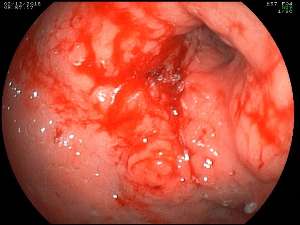
In case of esophagogastroduodenoscopy (Figure 1) circular tumor infiltration of the antral part was detected with proximal propagation along the small curvature 2 cm above the angle of the stomach, distally on the bulb of the duodenum and postbulbar region, with ulceration in the antrum 2 × 2.5 cm, in the bottom of which the plaque of hydrochloric acid hematin; multifocus biopsy was performed. Histological examination of the endoscopic biopsy specimen: a low-grade adenocarcinoma with the presence of signet-ring cells.

Figure 1. Endoscopic picture of a circular infiltrative tumor of the antral and stomach body with ulceration.

Figure 1. Endoscopic picture of a circular infiltrative tumor of the antral and stomach body with ulceration.

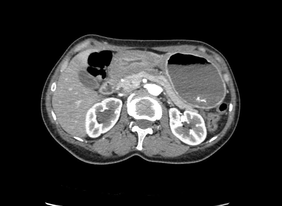
The patient was hospitalized in the Department of Abdominal Surgery of the Clinical Hospital 1 (Volynskaya) of the Hospital 1 Office of the President of the Russian Federation on 08.12.2016. At the time of hospitalization, laboratory indicators were within the physiological norm (including Hb 128 g/l, WBC 7.6, total protein 66 g/l). When multispiral computed tomography (MSCT) was performed with bolus contrasting, it was revealed (Figure 2): the upper part of the stomach was stretched with contents, in which a hyper-sensitive suspension was detected (probably, barium residues); the walls of the antrum are thickened to 15 mm, around the antrum there are quantitatively enlarged lymph nodes 9-12 mm in size, the pyloric department merges with the bulb of the duodenum without clear boundaries, the lumen in it is not traced; thickness of the walls of bulb and postbulbar part of the duodenum up to 7mm, the rest of the intestinal wall is not thickened; the walls of the stomach and the duodenum adjacent to the head of the pancreas without convincing signs of tumor’s invasion; the spleen is not enlarged, homogeneous; pancreas with clear contours, not enlarged, homogeneous structure; pancreatic duct is not enlarged; sites of pathological accumulation of contrast substance parenchyma of the gland is not revealed; liver is not enlarged, homogeneous structure; intra- and extrahepatic bile ducts are not dilated, gallbladder is in usual size, thin-walled, in the lumen – homogeneous liquid contents; there were signs of stenosis in low third of the right ureter with pronounced uretero-pylo-calicoectasia on the right; there is no fluid in the abdominal cavity. At the MSCT of the thorax no pathological changes were detected. At laboratory inspection of oncomarkers: CA 72-4 31,2 IU, CA 19-9 151,3 IU

CST2017-234-Russia_F2

Figure 2. MSCT: antrum tumor extends to the bulb of the duodenum.

The clinical diagnosis was formulated as follows: C16.8, cancer of the antrum and body of the stomach, infiltrative-ulcerative form, with spread to the duodenum (type III Borrmann) cT4N1Mx. Taking into account the verified histological tumor lesion of the stomach complicated by decompensated stenosis and recurrent bleeding, in the absence of signs of systemic dissemination of the tumor process and signs of tumor unresectability, a decision was made to conduct an operative intervention in the volume of distal subtotal resection of the stomach with pancreatoduodenectomy according to vital indications.

Within 4 days, the patient underwent preoperative preparation, including parenteral administration of 3-Chamber bags mixtures and sipping with official nutritional mixtures with a total calorie content up to 3,000 kcal/day. Immunonutrition support was provided for enteral and parenteral administration of the Omega-3 fatty acids. In connection with the stricture of the right ureter, an internal ureteral stent was placed on the right.

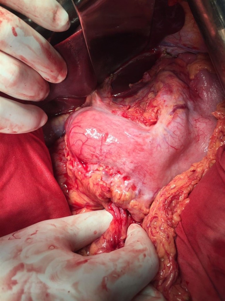
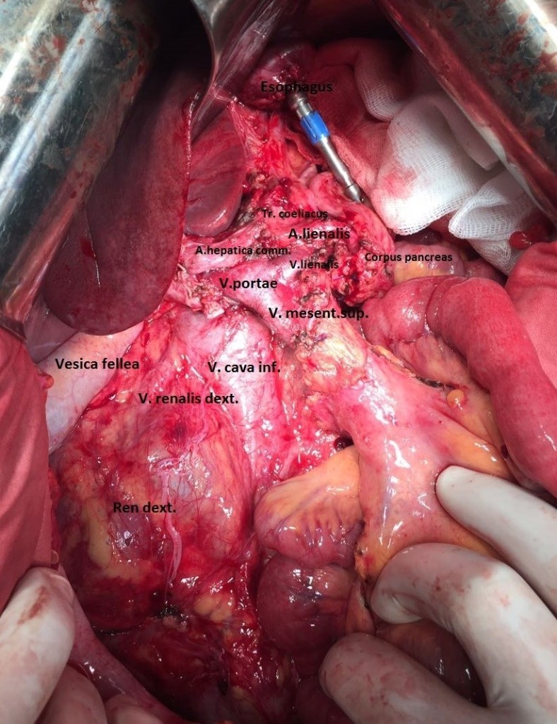
13.12.2016 the patient was operated under endotracheal anesthesia in combination with epidural anesthesia. At the stage of intraoperative revision, a circular tumor of the antral section, which propagates proximally to the body of the stomach and the subcardia by a small curvature (Figure 3), distally to the bulb and the postbulbar part of duodenum, is revealed invading the serous of the anterior and posterior walls of the stomach. It was found invasion of the tumor along the back wall of the antrum into the head of the pancreas, the mesocolon to the right and left of the middle colonic vessels, into the wall of the transverse colon in the hepatic flexure (Figure 4). In addition, infiltration of the right paracolic space, presence of characteristic foci of dissemination on the visceral peritoneum of ileo-cecal angle and in the projection of the distal part of the inferior mesenteric vein were determined. A macroscopic increase in the lymph nodes of groups 3, 4d, 5, 6, 7, 15 (according to JCGC) was found.

CST2017-234-Russia_F3

Figure 3. Appearance of the stomach during intraoperative exploration.

CST2017-234-Russia_F4

Figure 4. Local prevalence of the tumor process.

Taking into account the data of the operational finding, the volume of the operation was extended to gastrectomy, pancreatoduodenectomy, extended right colectomy with regional lymphadenectomy D2. During the resection stage, the esophagus was crossed 3 cm above the cardia, the jejunum – 10 cm distal to Treitz ligamentum; the pancreas was crossed along the border of the neck and body to the left of the superior mesenteric vein ; the ileum was crossed 20 cm proximal to the ileocolic angle, the colon – 8 cm distal to the splenic flexure; the c holedoch was crossed 0.5 cm distal to the confluence of the cystic duct; ilio-colon and middle colonic artery are ligated at the point of departure from the superior mesenteric artery, veins of the same name – at the point of confluence in the superior mesenteric vein. The inferior mesenteric vein after the test compression was crossed 4 cm from the point of confluence into the splenic vein. Regional lymphadenectomy with removal of lymph nodes of groups 1-12, 13, 14v, 15, 16b1, 17, 18 (according to JGCA classification) was performed (Figure 5). The resected tissues were removed by a single block (Figure 9). Dissection of tissues during the resection stage was carried out using ultrasound dissection and bipolar coagulation. Urgent histological examination showed no tumor growth at the margins of resection.

CST2017-234-Russia_F5

Figure 5. Resection stage of the surgery is completed.

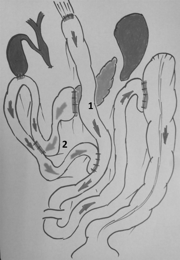
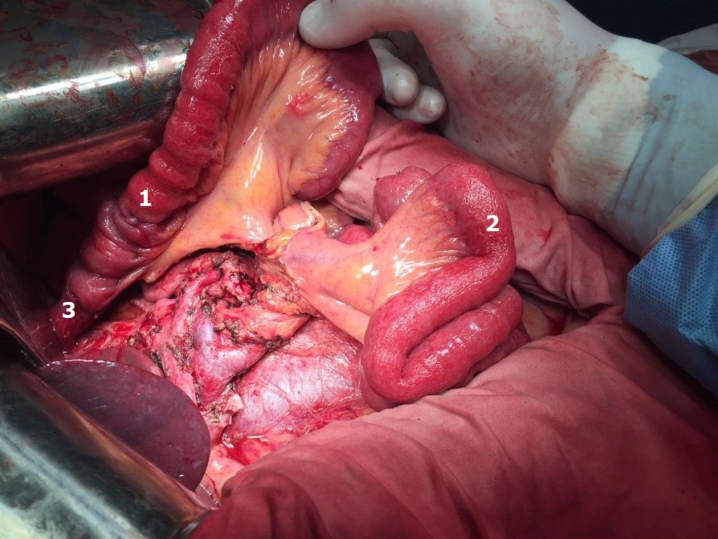
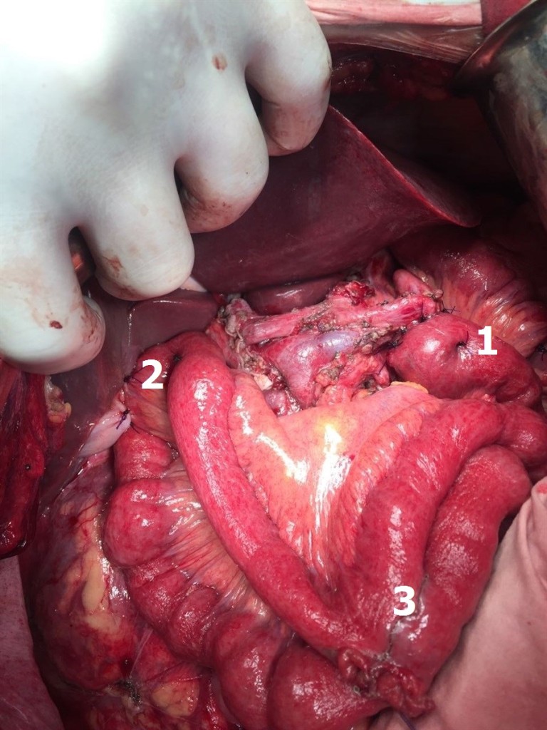
The reconstructive stage of the surgery was performed with the formation of alimentary and biliopancreatic (70 cm) loops of small bowel according to Roux mode (Figure 6). On the alimentary loop, an “end-to-end” esophagogo-enteroanastomosis was formed with the circular stapler (28 mm), ileo-descendoanastomosis “side by side” was formed with the stapler device (60mm) (Figure 7). The alimentary and biliopancreatic loops were connected by “side-to-side” Rouxe-en-Y-anastomosis with the stapler device (60mm), distal to esophago-enteroanastomosis by 45 cm. On the biliopancreatic loop, the “end-to-end” pancreato-enteroanastomosis was formed by single sutures Maxon 3/0 according to J.L. Cameron mode (invagination of the edge of the pancreas into the lumen of the small intestine) . This method of formation of pancreato-enteroanastomosis anastomosis was chosen because of the small diameter of the main pancreatic duct (up to 1.5 mm), the absence of fibrosis of the parenchyma and the unexpressed pancreatic capsule. At 20 cm distal to this anastomosis, cholecysto-enteroanastomosis was formed with a single continuous Biosyn 3/0 suture. In addition, on the biliopancreatic loop, the interintestinal (Braun’s) anastomosis was formed “side by side” with the stapler device (60mm) (Figure 8). A transnasal nourishing probe was conducted into the alimentary loop beyond the esophago-enteroanastomosis and Roux-en-Y-anastomosis. Surgery was completed by draining the subhepatic, left subdiaphragmatic space, the pelvic cavity and the pancreato-enteroanastomosis zone. The duration of the surgery was 7 hours and 30 minutes. The total volume of intraoperative blood loss, taking into account the remote tissues – up to 1 liter. Transfusion of one unit of RBC and three units of plasma was performed intraoperatively.

CST2017-234-Russia_F6

Figure 6. The general plan of the reconstructive stage of the surgery: 1 – alimentary loop of the small bowel, 2 – biliopancreatic loop of the small bowel.

CST2017-234-Russia_F7

Figure 7. The beginning of the reconstructive stage of the surgery: 1 – alimentary loop of the small bowel, 2 – biliopancreatic loop of the small bowel before the formation of anastomoses, 3 – esophagoeteroanastomosis.

CST2017-234-Russia_F8

Figure 8. Figure 8. Biliopancreatic loop of small bowel: 1 – invaginated pancreatoenteroanastomosis according to J.L. Cameron, 2 – cholecystoenteroanastomosis, 3 – interintestinal anastomosis according to Braun.

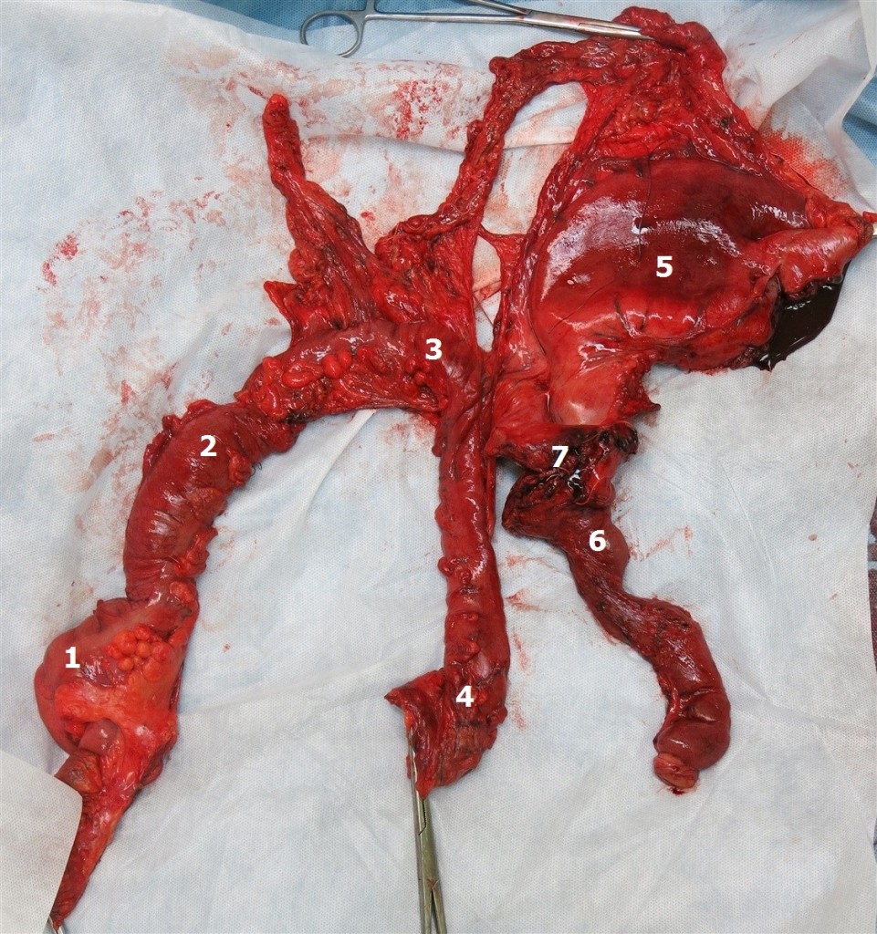
Thus, the patient underwent surgical treatment in the following volume: total gastrectomy, pancreatoduodenectomy, expanded right colectomy, D2+ lymphadenectomy (Figure 9).

CST2017-234-Russia_F9

Figure 9. Surgical specimen: 1 – caecum, 2 – ascending colon, 3 – transverse colon, 4 – the left splenic flexure, 5 – stomach, 6 – duodenum, 7 – head and neck of the pancreas.

In the first 48 hours of the postoperative period the patient was observed in the ICU. Self-breathing was restored in one hour after the end of the surgery. Six hours after the end of the surgery infusion of semi-elemental mixtures into the nutritive probe, parenteral nutritional support with a “three in one” mixture was started. After 24 hours from the end of the surgery the patient was switched to a probe feed with a standard enteral mixture. The feeding probe was removed 36 hours after the end of the operation. Later the patient received standard postoperative therapy according “fast track” protocol: prolonged epidural anesthesia and NSAID without opioids, combined parenteral-enteral support with the siping of high-calorie mixtures, immunonutrition support with the Omega-3 fatty acids, antibacterial prophylaxy, octreotide infusion 0.025 mg / h, thromboprophylaxis. From the second day after surgery began the physical activation of the patient (sitting in bed, lifting to the feet). Auscultatory peristalsis began to be determined 18 hours after the end of the surgery, stool – on the 3rd day of the postoperative period.

From the third day of the postoperative period, rehabilitation was performed under the conditions of the abdominal surgery department. The diet was extended by oral intake of fermented milk products, broths. During 3-6 days there was a multiple liquid stool, periodic spastic pains in the abdomen, and phenomena of flatulence. However, already by the 7 days of the postoperative period, these phenomena were leveled, the stool returned to normal, once a day. Separated by drainage, starting from 1 day postoperative period, was serous, amounting to a total of 100 ml per day and limited to 6 days postoperative period to 50 ml. Drainages from the small pelvis and the left sub-diaphragmatic space were removed on the 4th day of the postoperative period, drainage from the subhepatic space – on day 6. On the 7th day of the postoperative period, a clear colorless discharge from the drainage set in the region of pancreato-enteroanastomosis in the amount of up to 50-100 ml per day (amylase 9100 IU) was noted. With control ultrasound of the abdominal cavity on the 1st, 3rd, 6th, 9th and 13th day of the postoperative period there were no accumulations of fluid in the abdominal cavity and in the retroperitoneal space. The patient completely switched to taking the standart diet on the 9th day after surgery.

In laboratory indicators moderate anemia (Hb up to 95 g/l), leukocytosis (WBC up to 13.4), hyperamilazemia up to 340 IU, hypoproteinemia (total protein up to 47 g/l) for 1-4 days of the postoperative period were noted. Episodes of hyperthermia for the entire period of postoperative observation were not noted. On the 12th day of the postoperative period, Hb 103 g / l, WBC – 7.7, a-amylase – 76 IU, total protein – 66 g / l. The surgical wound healed by primary tension.

In consideration of absence of infectious complications, the relative normalization of the function of the digestive tract, but the remaining rate of pancreatic juice in the 50-70 ml / day, it was decided to discharge the patient from the hospital for outpatient monitoring with functioning drainage on the 12-day postoperative period. When follow-up examined on the 14th day after discharge, it was found that there was no liquid by drainage. The drainage was removed.

Histological study of the surgical specimen (stomach with omentum major and minor, duodenum, proximal part of small bowel, head and neck of pancreas, distal part of ileum, caecum, ascending colon with paracolic fat, transverse colon with mesocolon, left splenic flexure) : the circular tumor infiltration in the antrum and the body of the stomach is determined, spreading along the small curvature to the subcardia, passing to the duodenum, the ulceration of the tumor in the antrum (type III Borrmann), with signs of bleeding; in the wall of the stomach – the growth of a tumor having the structure of a low-grade adenocarcinoma with the presence of signet-ring cells; the tumor grows into the submucosal and muscular layers, into the subserous and serous layer, grows into a small omentum, the transverse colon and mesocolon, grows along the fibrous layers in the thickness of the fatty tissue; a large number of tumor cells is determined in perineural spaces, lymphatic vessels and veins; elements of the tumor are found in the tissue of the head of the pancreas, the muscular and submucosal layer of the caecum and the appendix, the wall of the duodenum, mesocolon, paracolic fiber. Tumor metastases were detected in 7 of the 38 lymph nodes examined. In the margins of resection (stomach, pancreas, jejunum, ileum, colon, mesocolon) tumor growth was not detected (PM 0, DM 0), radical surgery – R0. Pathomorphologic diagnosis: C16.8, advanced stomach cancer (type III Borrmann) with spread to the duodenum, pancreas head, transverse colon, mesocolon, right paracolon, metastasis of the visceral peritoneum of the caecum pT4b ly3 v3 N3 M1 P1 CYX H0. Immunohistochemical examination with antibodies to Her2 / neu (Pathway 4B5 Roche-Ventana): Her2 / neu – 0.

Starting with the fifth week of the postoperative period, the patient was subjected to adjuvant chemotherapy according to the EOX scheme (oxorubicin, oxaliplatin, capecitabine). A total of 6 courses of chemotherapy with stimulation filgrastim. In August 2017, a control PET CT was performed. A slight diffuse increased accumulation of difluoroglucose in the parietal peritoneum of the epigastric region is determined. At the control laboratory examination: indicators of clinical and biochemical blood tests within reference values, CA 72-4 5, 6 IU, CA 19-9 32,3 IU. Patient was assigned continued reception of capecitabine in mono regimen.

Currently in September 2017, the patient leads a normal lifestyle, has a regular diet without dyspepsia, regains her usual body weight and works by her profession.

Conclusion

Concluding the description of the above clinical case, we consider it necessary to give a number of comments. Undoubtedly, the surgical treatment of the patient is non-standard and rarely performed operations. This operation should be attributed to the category of desperation operations dictated by a perfectly understandable motivation to save or at least prolong the patient’s life in the presence of not only formal indications in the form of decompensated stenosis and recurrent bleeding from the tumor, but also motivation caused by a variety of social factors. During the surgery, having determined the prevalence of the tumor, we were perfectly aware of the illusory nature of our hopes for performing absolutely radical surgery. And at the same time, the decision to limit the surgery only by bypass formation, depriving the patient of any chance of life for more than 2-3 months was collectively found unacceptable. Moreover, taking into account the high risk of intra- and postoperative complications, we nevertheless counted on a favorable outcome of the surgery, taking into account a number of apparently positive factors, namely: young age and satisfactory physical condition of the patient, absence of signs of subclinical organ failure and significant violations of homeostasis. When performing surgery for a tumor of this prevalence, we certainly hoped for the possibility of modern adjuvant chemotherapy options. The early postoperative period and nine months of patient monitoring showed that our hopes were not unfounded.

We have presented only a single clinical observation of multivisceral resection in locally advanced stomach cancer, but we believe that it can be useful both for continuing the discussion about the necessity and possibility of operations of this size, and for making a decision that is extremely difficult from a medical and human perspective, in a concrete clinical situation.

References

  1. Xin-Bao Wang, Li-Tao Yang, Ze-Wei Zhang (2008) Pancreaticoduodenectomy for advanced gastric cancer with pancreaticoduodenal region involvement. World J Gastroenterol 14: 3425–3429.
  2. Roberts P, Seevaratnam R, Cardoso R, Law C, Helyer L, et al. (2012) Systematic review of pancreaticoduodenectomy for locally advanced gastric cancer. Gastric Cancer 15 Suppl 1: S108–115. [crossref]
  3. Kit OI, Kasatkin VF, Maksimov AY, Trifanov VS (2005) Gastrectomy in combination with pancreatoduodenal resection for gastric cancer. Oncosurgery Vol.5: 1.
  4. Combined operations for stomach cancer. Ed. V.F. Kasatkin
  5. Otsuji E, Yamaguchi T, et al. Total gastrectomy with simultaneous pancreaticosplenectomy or splenectomy in patients with advanced gastic carcinoma. Br J Cancer 79: 1789–1793.
  6. Zhang M, Zhang H, Ma Y, Zhu G, Xue Y. Prognosis and surgical treatment of gastric cancer invading adjacent organs. ANZ J Surg 80: 510–4.
  7. Ozer I, Bostanci EB, Orug T, Ozogul YB, Ulas M, Ercan M, et al. (2017) Surgical outcomes and survival after multiorgan resection for locally advanced gastric cancer. Am J Surg 198: 25–30.

Infection Trend, Distribution, and Factors Associated with Hepatitis B Virus Infection in Delaware, 2005-2015

DOI: 10.31038/IMROJ.2017232

Abstract

Background: Hepatitis B virus (HBV) infection is a global health problem. Immigrants to the United States have a high prevalence of HBV infection. Understanding the HBV infection trends and its distribution can improve prevention and control strategies. This study was to determine the infection trends, distribution, and factors associated with HBV infection in Delaware.

Methods: We performed a retrospective study on persons suspected of having HBV infection reported to Delaware Division of Public Health’s Surveillance System during January 1, 2005-December 31, 2015. The charts of 4, 981 persons were reviewed and included in the analysis.

Results: Of these 4, 981 persons, 2, 119 (42.5%) had HBV infection. During 2005-2015, acute and chronic HBV infection declined 80.9% and 60%, respectively for an overall reduction of 62.2%.

Males had a higher yearly infection rate. Rates declined 63.5% among males and 60.1% among females. There was an increase of 13.4% in the HBV infection in females during 2010-2015. HBV infection declined in all racial groups. Asians had a higher yearly infection rate and it increased 40.0% during 2010-2015. HBV infection declined in all age groups. However, an increase of 12.2% was seen among those 15-39 years during 2010-2015. Sixty-six percent of infected patients were in five cities: Wilmington, Newark, New Castle, Dover, and Bear.

In a multivariable logistic model, significant predictors for HBV infection included being male [adjusted odds ratio (aOR): 1.6, 95% CI: 1.4-1.8], age 15-39 years and 40-59 years (aOR: 3.7, 95% CI: 2.3-5.9 and 2.4, 95%CI: 1.5-3.8). Asian, black, and other race had a greater risk compared with white, with aOR of 5.8 (95% CI: 4.8-7.0), 1.7 (95% CI: 1.4-1.9), and 1.4 (95% CI: 1.1-1.9), respectively.

Conclusions: HBV infection is significant in Delaware and concentrated mainly in a few cities. Despite an overall decline, increases were seen among females, in the 15-39 age group, and in the Asian population during 2010-2015. Further studies should be conducted to identify factors contributing to these increases

Keywords

Hepatitis B virus (HBV), hepatitis B virus infection, incidence, prevalence, epidemiology, surveillance

Introduction

Hepatitis B virus (HBV) infection remains a major global health problem with an estimated 257 million chronic HBV-infected persons worldwide in 2017 [1]. In the United States, despite a comprehensive vaccination program to eliminate HBV transmission since 1991 [2], the estimated prevalence of current active HBV infection during 2011-2014 was 0.4% among U.S. adults age 18 years and over [3], with an estimate of 850, 000-2.2 million HBV-infected persons [4-6]. HBV infection is a vaccine preventable disease that is transmitted by percutaneous or mucosal exposure to infectious blood or body fluids. It is among the top 10 causes of infectious disease-related mortality in the world, with over 887, 000 deaths annually [1]. Delaware is a small state with a population of 945, 934 people in 2015 and home to 76, 768 immigrants in 2013 [7]. Immigrants to the United States have a high prevalence of viral hepatitis B surface antigen (HBsAg); it was 4.9% during 2004-2008 [8] and around 71.3% of chronic HBV infections were among persons born outside the United States [9]. Since individuals with chronic HBV infection are often unaware of their infection status, they are a major source of ongoing HBV transmission [10]. An understanding of HBV epidemiology is important for targeted public health efforts. This study aimed to determine HBV infection trends, identify its distribution and factors associated with HBV infection in Delaware during the period 2005-2015.

Methods

Data and patient population

HBV data reported by hospitals, clinics, and laboratories to the Delaware Division of Public Health (DPH) through the Delaware Electronic Reporting and Surveillance System (DERSS) were obtained for the years 2005-2015 (11-year period). Data reported to DERSS include information on laboratory testing results of suspected HBV infection persons. In addition, information collected by epidemiologists during the disease investigation process was reviewed, including data on the persons’ demographics, diagnosis, hospitalization, and vaccination status.

Study design

A retrospective study on persons suspected of having HBV infection was conducted. All reported persons to DERSS and information gathered during the disease investigation were included for review and analysis. The rate of HBV infection was the principal study outcome. HBV infection was defined based upon the Center for Disease Control and Prevention’s (CDC) clinical case definitions and laboratory criteria [11]. For acute HBV infection: a case was confirmed if met the clinical case definition, was laboratory confirmed, and was not known to have chronic hepatitis B. Clinical description includes an acute illness with a discrete onset of any sign or symptom consistent with acute viral hepatitis, and either a) jaundice, or b) elevated serum alanine aminotransferase (ALT) levels >100 IU/L. Laboratory criteria include hepatitis B surface antigen (HBsAg) positive, and Immunoglobulin M (IgM) antibody to hepatitis B core antigen (IgM anti-HBc) positive (if done). For chronic HBV infection: clinically, no symptoms are required. Persons with chronic HBV may have no evidence of liver disease or may have a spectrum of disease ranging from chronic hepatitis to cirrhosis or liver cancer. Laboratory criteria include IgM anti-HBc negative and a positive result on one of the following tests: HBsAg, hepatitis B e antigen (HBeAg), or nucleic acid test for hepatitis B virus DNA, or HBsAg positive or nucleic acid test for HBV DNA positive or HBeAg positive two times at least 6 months apart. A case was classified as a probable case if a person has a single HBsAg positive or HBV DNA positive or HBeAg positive and does not meet the case definition for acute hepatitis B, and a confirmed case if a person who meets either of the above laboratory criteria for diagnosis [11].

Statistical analysis

Descriptive statistics such as frequencies, means, medians, inter-quartile range, and cross-tabulation were used for patient characteristics. Between-group differences were evaluated using the chi-square test or Fisher’s exact test for categorical data or a Mann-Whitney test for continuous data. The yearly cumulative incidence of acute HBV infection and the yearly prevalence rate of chronic HBV infection per 100, 000 population were determined for the 2005-2015 period. Calculation of the yearly cumulative incidence was based on the number of newly-diagnosed patients and the number of people at risk for HBV infection within each year. The yearly prevalence rate of chronic HBV infection was estimated based upon the yearly number of chronic HBV-infected cases divided by the number of people in the population in the same year. In addition, the yearly rate of HBV infection per 100, 000 population was calculated by population characteristics (sex, age, and race). The yearly infection rate was estimated based on the yearly number of HBV-infected cases and the Delaware population in the same year stratified by sex, age group, and race. To identify distribution of HBV infection, patient characteristics were described and established by geographical location. Risk factors associated with HBV infection were analyzed by logistic regression models. Hosmer and Lemeshow stepwise strategies were applied for model building: potential independent variables with P-value <0.25 were included in the initial full model. Data analyses were performed using the Stata software program (version 13; STATA Corp., College Station, TX). P-values less than 0.05 (two tailed) were considered statistically significant.

Results

A total of 4, 981 people suspected of having HBV infection were identified and included in the analysis. Baseline and demographic characteristics, by HBV infection status, are presented in Table 1. HBV infection was identified in 2, 119 patients (42.5%, 232 acute and 1, 887 chronic HBV-infected patients), including 1, 988 (39.9%) and 131 (2.6%) cases of confirmed and probable HBV infection, respectively. Of this study population, a significantly larger number of reported persons were males compared with females [55.0% versus (vs.) 44.8%, P<0.001]. The overall study population’s mean age was 45.3 years [inter-quartile range (IQR): 34-56]. A majority (79.2%) were 15-59 years old; and white, black, and Asian races were observed in 38.5%, 34.1%, and 16.0%, respectively. Only 10.6% of the population had received one or more doses of HBV vaccination. Compared with the non-HBV infection group, the HBV-infected patients were younger [mean age: 42.7 years (IQR: 32-52) vs. 47.2 years (IQR: 36-58)] and had a significant larger number of patients in the 15-39 age group (43.1% vs. 27.3%, P<0.001). In addition, the HBV-infected patients had significantly fewer whites (26.4% vs. 47.4%), more persons of Asian origin (26.8% vs. 7.9%, P<0.001), and fewer patients who had received one or more doses of HBV vaccination, compared with the non-HBV infection group (6.1% vs. 14.0%, P < 0.001).

Table 1. Population characteristics

Characteristics HBV Infection(N = 2,119) Non-HBVInfection(N = 2,862) Total(N = 4,981) P-value
Gender; N (%)
Male 1246 (58.8) 1495 (52.2) 2741 (55.0)  <0.001
Female 870 (41.1) 1363 (47.6) 2233 (44.8)
Missing/Unknown  3 (0.1)  4 (0.2)  7 (0.2)
Age, N (%)   mean:45.3 years, IQR: 34-56 years)
 <15 27 (1.3) 94 (3.3) 121 (2.4) <0.001
15-39 914 (43.1) 782 (27.3) 1696 (34.1)
40-59 907 (42.8) 1341 (46.8) 2248 (45.1)
≥60 271 (12.8) 646 (22.6) 917 (18.4)
Race/Ethnicity, N (%)
White 560 (26.4) 1358 (47.4) 1918 (38.5) <0.001
Black 703 (33.2) 997 (34.8) 1700 (34.1)
Asian 568 (26.8) 227 (7.9) 795 (16.0)
Others* 82 (3.8) 128 (4.5) 210 (4.2)
Unknown 14 (0.7) 68 (2.4) 82 (1.7)
Missing 192 (9.1) 84 (3.0) 276 (5.5)
Received ≥01 dose of hepatitis B virus vaccination
Yes 130 (6.1) 399 (14.0) 529 (10.6) <0.001
No 1987 (93.8) 2463 (86.0) 4450 (89.3)
Unknown/Missing 2 (0.1) 0  2 (0.1)

* American Indian/Alaska Native, Pacific Islander, Hispanic, Multiracial

Hepatitis B virus infection trend

Between 2005 and 2015, 2, 119 patients (232 acute, 1, 887 chronic) infected with HBV were identified. Figure 1 shows the incidence of acute HBV infection and the prevalence rate of chronic HBV infection per 100, 000 population from 2005 through 2015. The incidence of acute HBV per 100, 000 population declined 80.9%, from 4.2 (34 cases in 2005) to 0.8 (8 cases in 2015). Similarly, chronic HBV infection per 100, 000 population declined 60% from 36.0 (295 cases in 2005) to 14.4 (136 cases in 2015), making the overall reduction (acute and chronic) of 62.2% from 40.2 (329 cases) to 15.2 (144 cases) per 100, 000 population. During a period of 2010-2012, there was a moderate spike of 28% in the prevalence of chronic HBV infection, from 13.4 (in 2010) to 17.1 cases (in 2012) per 100, 000 population; and then a slight increase of approximately 7%, from 13.5 (in 2013) to 14.4 cases (in 2015) per 100, 000 population.
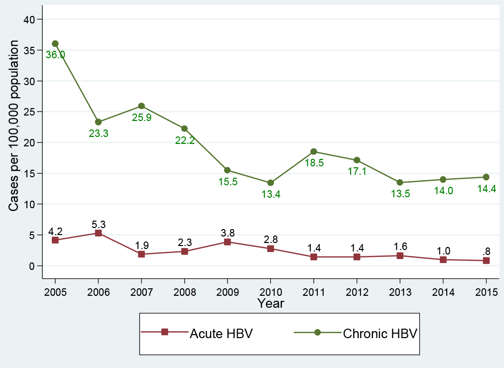
IMROJ2017-210-NhiemVietLuongUSA_f1

Figure 1. Hepatitis B virus infection trend, Delaware, 2005-2015

Hepatitis B virus infection by gender

Of the 2, 119 patients infected with HBV, males accounted for 58.8% (1, 246 cases) compared with 41.1% (870 cases) among females. In the acute HBV-infected group, 66.0% (153 cases) were in males compared with 33.6% (78 cases) in females. Similarly, in the chronic HBV-infected group, 57.9% (1, 093 cases) were in males compared with 42.0% (792 cases) in females, Table 1. Figure 2 presents the HBV infection trend by gender per 100, 000 population during the period 2005-2015: Generally, males had a higher yearly HBV infection rate in comparison with females. Between 2005 and 2015, the HBV infection rate among males declined 63.5%, from 49.1 (195 cases) to 17.9 (82 cases) per 100, 000 population; and the HBV infection rate among females declined 60.1%, from 31.8 (134 cases) to 12.7 (62 cases) per 100, 000 population. Interestingly, in the period 2005-2010, the HBV infection declined 64.8% among females, which was higher than the 56.2% decline for males. However, in the period 2010-2015, while we observed a decline of 16.7% in males (from 21.5 to 17.9 cases per 100, 000 population), the HBV infection rate increased 13.4% in females (from 11.2 to 12.7 cases per 100, 000 population).
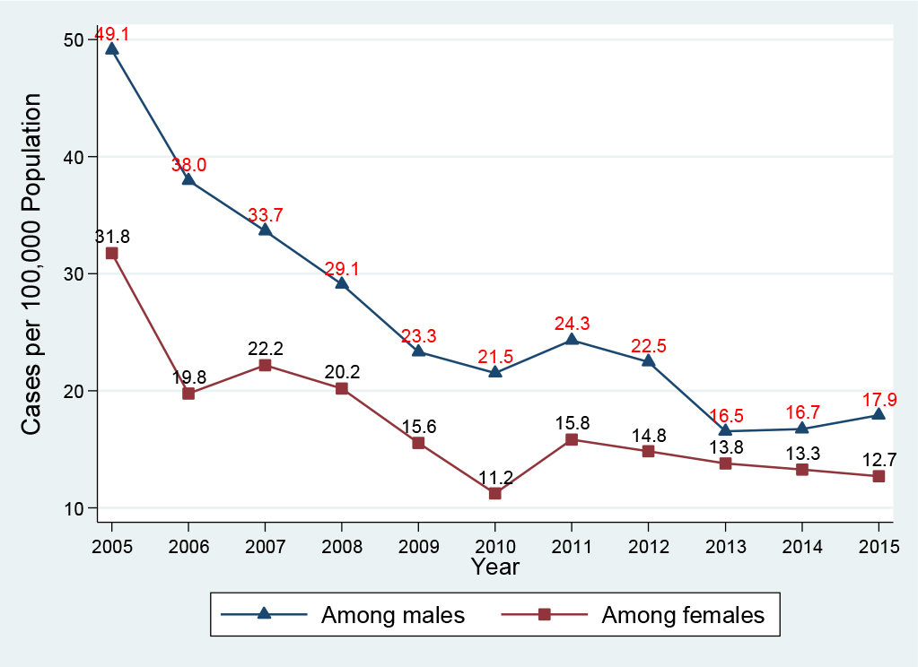
IMROJ2017-210-NhiemVietLuongUSA_f2

Figure 2. Hepatitis B virus infection by gender, Delaware, 2005-2015

Hepatitis B virus infection by age group

Of those infected with HBV, 85.9% (1, 821/2, 119 cases) were in the age groups of 15-39 and 40-59 years old, Table 2. Figure 3 presents the HBV infection trend per 100, 000 population by age group: In general, all age groups had a huge reduction between 2005 and 2015. The highest reduction (100%) was seen in the age group <15 years, from 2.5 (4 cases in 2005) to 0.6 (1 case in 2014) and 0.0 case (0 case in 2015) per 100, 000 population. The smallest reduction (63.6%) was observed in the age group of 15-39 years, from 51.7 (141 cases in 2005) to 23.8 (73 cases in 2015) per 100, 000 population. Approximately 88.9% reduction was seen in the age group of ≥60 years, from 19.7 (29 cases in 2005) to 7.2 (16 cases in 2015) per 100, 000 population; and 65.9% reduction was seen in the age group of 40-59 years, from 65.4 (155 cases in 2005) to 22.3 (55 cases in 2015) per 100, 000 population. Interestingly, in the period of 2010-2015, there was an increase of 12.2% in the HBV infection rate in the age group of 15-39 years, from 20.9 (62 cases in 2010) to 23.8 (73 cases in 2015) per 100, 000 population.
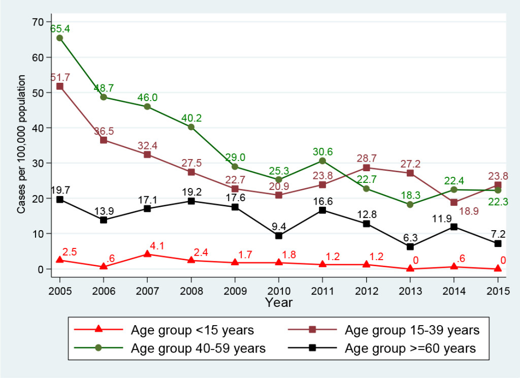
IMROJ2017-210-NhiemVietLuongUSA_f3

Figure 3. Hepatitis B virus infection by age group, Delaware, 2005-2015

Table 2. Characteristics of patients infected with Hepatitis B virus, Delaware, Period 2005-2015

Characteristics Acute HBVInfection(N=232) Chronic HBVInfection (N=1,887) Total (N=2,119)
Gender; N (%)
Male 153 (66.0) 1, 093 (57.9) 1, 246 (58.8)
Female 78 (33.6) 792 (42.0) 870 (41.1)
Missing/Unknown 1 (0.4) 2 (0.1) 3 (0.1)
Age, N (%) mean: 42.7 years, IQR: 32-52 years old)
<15 0 27 (1.4) 27 (1.3)
15-39 103 (44.4) 811 (43.0) 914 (43.1)
40-59 110 (47.4) 797 (42.2) 907 (42.8)
≥60 19 (8.2) 252 (13.4) 271 (12.8)
Race/Ethnicity, N (%)
White 101 (43.5) 459 (24.3) 560 (26.4)
Black 94 (40.5) 609 (32.3) 703 (33.2)
Asian 16 (6.9) 552 (29.3) 568 (26.8)
Others* 3 (1.3) 79 (4.2) 82 (3.9)
Unknown/Missing 18 (7.8) 188 (9.9) 206 (9.7)
County (N, %) and City** (zip code)
New Castle Wilmington
(19801-19810)
84 (36.2) 507 (26.9) 591 (27.9)
Smyrna
(19977)
3 (1.2) 25 (1.3) 28 (1.3)
Newark
(19702, 19711, 19713)
19 (8.1) 360 (19.0) 379 (17.9)
New Castle
(19720)
28 (12.0) 131 (6.9) 159 (7.5)
Middletown
(19709)
2 (0.8) 46 (2.4) 48 (2.2)
Hockessin
(19707)
1 (0.4) 51 (2.7) 52 (2.4)
Claymont
(19703)
6 (2.5) 58 (3.1) 63 (3.0)
Bear
(19701)
10 (4.3) 104 (5.5) 114 (5.3)
Kent Dover
(19901, 19904)
13 (5.6) 139 (7.3) 152 (7.1)
Smyrna
(19977)
4 (1.7) 51 (2.7) 55 (2.6)
Sussex Georgetown
(19947)
6 (2.5) 38 (2.0) 44 (2.0)
Lewes
(19958)
6 (2.5) 33 (1.7) 39 (1.8)
Millsboro
(19966)
3 (1.2) 27 (1.4) 30 (1.4)
Rehoboth Beach
(19971)
6 (2.6) 28 (1.4) 34 (1.6)
Seaford
(19973)
4 (1.7) 44 (2.3) 48 (2.2)

*: American Indian/Alaska Native, Pacific Islander, Hispanic, Multiracial

**: Only cities with a number of cases ≥25

Hepatitis B virus infection by race

Of the entire study population (4, 981 persons), white and black population accounted for a larger number of reported persons in comparison with Asian population (38.5% and 34.1% versus 16.0%, Table 1). However, in the group of HBV-infected patients (2, 119 HBV-infected persons, Table 2): the largest infected number was seen in black (33.2%), then Asian (26.8%), and white (26.4%). Particularly, in the acute HBV-infected patients, the largest number of cases was identified in white (43.5%), then black (40.5%, Asian (6.9%), and others (1.3%). In the chronic HBV-infected patients, the largest number was identified in black (32.3%), then Asian (29.3%), white (24.3%), and others (4.2%). Figure 4 presents the HBV infection trend per 100, 000 population by racial/ethnic group from 2005 to 2015: Generally, the decline was seen in all racial/ethnic groups. Asian population had a higher yearly infection rate per 100, 000 population in comparison with other populations: Compared with white, it was 25.1-fold and 31.5-fold higher in 2005 and 2015, respectively; and it was 5.9-fold and 6.4-fold higher in comparison with black in 2005 and 2015, respectively. In addition, Asian population had the lowest decline at 54.7%, from 348.6 (78 cases in 2005) to 157.8 (57 cases in 2015) per 100, 000 population; other race had the highest decline of 89.9%, from 38.8 (12 cases in 2005) to 3.9 (2 cases in 2015) per 100, 000 population; blacks had the second lowest decline of 57.9%, from 58.3 (95 cases in 2005) to 24.5 (50 cases in 2015) per 100, 000 population; and white obtained a decline of 64.0%, from 13.9 (84 cases in 2005) to 5.0 (33 cases in 2015) per 100, 000 population. Interestingly, regardless of a decline in all racial/ethnic groups, Asian group had an increase of 40.0% in the HBV infection rate, from 94.6 (26 cases in 2010) to 157.8 (57 cases in 2015) per 100, 000 population, during a period of 2010-2015.
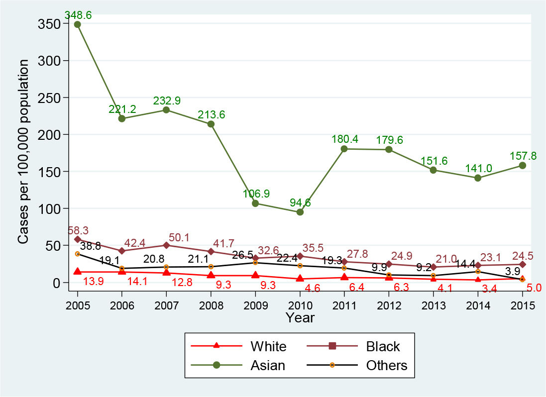
IMROJ2017-210-NhiemVietLuongUSA_f4

Figure 4. Hepatitis B virus infection by race, Delaware, 2005-2015

Geographical distribution of HBV infection

Delaware state consists of three counties (New Castle, Kent, and Sussex counties), with a total of 56 cities. Table 2 presents characteristics of HBV-infected patients and their geographic distribution. Of the 2, 119 HBV-infected people, 66% (1, 395 cases) were identified in five cities: Wilmington (27.9%, 591 cases), Newark (17.9%, 379 cases), New Castle (7.5%, 159 cases), Dover (7.1%, 152 cases), and Bear (5.3%, 114 cases). Figure 5 presents the trend of HBV infection for these top five cities for the period 2005-2015 versus the remaining 51 other cities combined. The top-ranking city for the number of HBV-infected patients in 2005 was Wilmington, which also achieved the largest reduction of 69.9%, from 93 cases in 2005 to 28 cases in 2015. Newark ranked second in 2005 and during the period 2005-2010, its HBV cases declined 71.4%, from 70 cases in 2005 to 20 cases in 2010; however, between 2010 and 2015, the case count increased 45%, from 20 cases in 2010 to 29 cases in 2015. The City of New Castle ranked third for HBV cases in 2005 and its case count fell 64%, from 25 cases in 2005 to 9 cases in 2015. The City of Dover’s HBV cases declined 26.3% between 2005 (19 cases) and 2006 (14 cases), and then it fluctuated up and down, maintaining around 13-15 cases per year. All other cities combined (51 cities) obtained an overall decline of 53.5%, from 112 cases in 2005 to 46 cases in 2010 and to 52 cases in 2015.
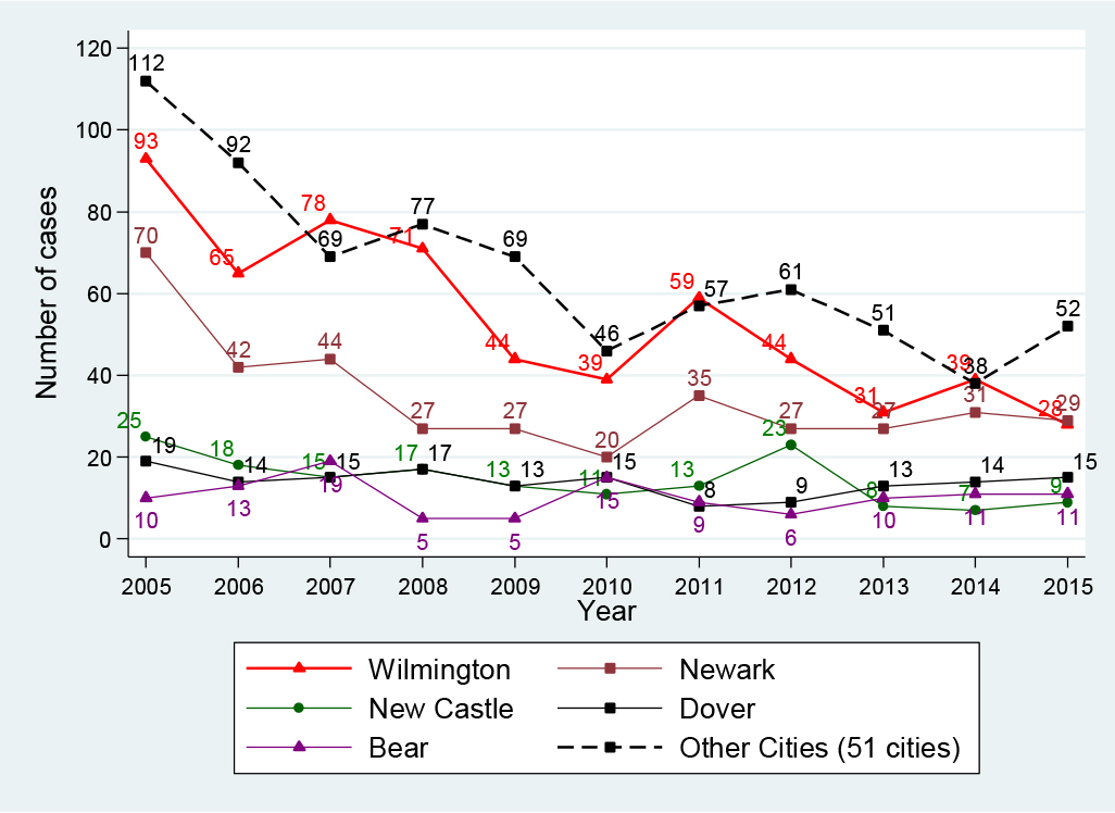
IMROJ2017-210-NhiemVietLuongUSA_f5

Figure 5. HBV infection in top five and other cities, Delaware, 2005-2015

Factors associated with HBV infection

Potential risk factors associated with HBV infection were examined in univariate and multivariate logistic regression models. These include gender, age, and race. Table 3 shows the selected demographic predictors in the univariate and multivariable logistic regression analyses. Results from the multivariable analysis indicate that males had a greater risk for HBV infection than females [adjusted odds ratio (aOR): 1.6, 95% CI: 1.4-1.8); those 15-39 years and 40-59 years had a greater risk of HBV infection (aOR: 3.7, 95% CI: 2.3-5.9 and 2.4, 95% CI: 1.5-3.8, respectively) than those in the age group less than 15 years. Interestingly, compared with whites, Asians had a 5.8-fold (aOR: 5.8, 95% CI: 4.8-7.0) greater risk of HBV infection; black and other racial groups also had a greater risk, its aOR was 1.7 (95% CI: 1.4-1.9) and 1.4 (95% CI: 1.1-1.9) for black and other racial groups compared with white, respectively.

Table 3. Factors associated with Hepatitis B virus infection

Predictor Univariate
Odds ratio (95% CI)
Multivariate
Odds ratio (95% CI)
Gender
Female 1 1
Male 1.3 (1.2-1.5) 1.6 (1.4-1.8)
Age, years
<15 1 1
15-39 4.1 (2.6-6.3) 3.7 (2.3-5.9)
40-59 2.4 (1.5-3.6) 2.4 (1.5-3.8)
≥ 60 1.5 (0.9-2.3) 1.5 (0.9-2.4)
Race/Ethnicity
White 1 1
Black 1.7 (1.4-1.9) 1.7 (1.4-1.9)
Asian 6.1 (5.1-7.3) 5.8 (4.8-7.0)
Others 1.5 (1.1-2.1) 1.4 (1.1-1.9)

Discussion

Understanding HBV infection trends and the epidemiologic characteristics of those infected with HBV are key to inform improvements in prevention and control strategies. While there are reliable data about the relationship between HBV vaccination and HBV infection, there are no published data on infection trends and epidemiologic characteristics of persons infected with HBV in Delaware. Over the past 11 years, our data suggest that HBV infection remains a significant public health issue in Delaware. During the period 2005-2015, although Delaware achieved a 62.2% overall reduction in HBV infection, its yearly infection rate exceeded the national rate and rates in many other states, including Maryland, California, New Jersey, New York, and Pennsylvania [9, 12]. The Centers for Disease Control and Prevention reported the yearly national rate of acute HBV infection per 100, 000 population at 1.9 cases for 2005, 1.1 cases for 2010, and around 0.9 cases for the period of 2011-2014 [9]. Delaware’s yearly infection rate for acute HBV infection per 100, 000 population was much higher at 4.2 cases in 2005, 2.8 cases in 2010, 1.4-1.6 cases for the period 2011-2013, and 1.0 case for 2014. In regards to chronic HBV infection, although Delaware achieved a large decline of 62.7%, from 36.0 (in 2005) to 13.4 cases (in 2010), it experienced spikes to 18.5 cases in 2011, 17.1 cases in 2012, then remained at 13.5-14.4 cases per 100, 000 population for the period 2013-2015. With the infection rate of 14.0 cases per 100, 000 population in 2014, Delaware’s infection rate was higher in comparison with the 2014 rates as reported by CDC: Massachusetts, 3.3 cases; Michigan, 4.9 cases; New York, 5.3 cases; the City of Philadelphia, 6.0 cases; and Washington, 1.3 cases, all per 100, 000 population [9].

New HBV infections in the United States are increasingly concentrated among certain populations such as injection drug users, prison inmates, and persons with sexual risk behaviors such as multiple sex partners, sex partners of HBV-infected persons, and men who have sex with men [13]. The spikes in rates of HBV infection we observed may probably be related to a rising trend of heroin use in Delaware [14]: During 2010-2014, we observed a spike in HBV infection that coincided with a spike in the number of people seeking heroin treatment. For example, in 2011, 1, 263 people in Delaware sought heroin treatment; that number accelerated to 1, 845 people in 2012, 2, 750 in 2013, and 3, 182 in 2014 [15].

Hepatitis B vaccination is the most effective measure to prevent HBV infection. In Delaware, the hepatitis B vaccination requirement for children going to public school began in the 1999-2000 school year, and by the 2005-2006 school year, all children from kindergarten to grade 12 must have the hepatitis B vaccine series. Our data showed that almost 90% of the study subjects (4, 981 persons) had no HBV vaccination, and among those infected with HBV (2, 119 persons), almost 94% had no HBV vaccination. Ongoing HBV transmission occurs primarily among unvaccinated persons with high risk behaviors for HBV transmission [16]. Our finding suggests that there is still a large proportion of Delawareans who may not have received the hepatitis B vaccine series.

We found the Asian population not only have a higher yearly infection rate in comparison to all other populations, but they also had the lowest decline in HBV infection: Compared with whites, Asians had a 5.8 fold increased risk for HBV infection; and interestingly, the Asian population had a 40% increase in HBV infection rate during a period of 2010-2015. Our findings are consistent with findings from the CDC and other studies from New York City, San Francisco, and Minnesota that Asians were at higher risk for HBV infection and the majority of chronic HBV infections in the United States were among Asians [9, 16-18].

France et al. reported that more than 93% of chronic HBV cases from January 1, 1999 to December 31, 2008 in New York City were among persons born outside the United States [19]. Recent studies also found that persons born outside of the United States, especially immigrants, had a high prevalence of chronic HBV infection and since they were often unaware of their infection status, were sources of infection [8-10]. Higher rates of HBV infection in Delaware and a recent increase in HBV infection among its Asian population may be attributed to a large number of immigrants. In 2013, Delaware was home to 76, 768 immigrants (8.3% of Delaware’s population); Asians accounted for 33, 639 persons (3.6% of the 2013 Delaware population); and around 34, 625 immigrants were naturalized U.S. citizens in Delaware in 2013. Unauthorized immigrants comprised roughly 20, 000 people (2.4% of the Delaware population) in 2012 [7], a group that may have limited access to health care. A large burden of HBV infection among certain populations suggest a need for the hepatitis B program targeting these populations to identify the infected and link them to care.

Chronic HBV was more common among males than females [20, 21]. We found males had a higher yearly rate of HBV infection, they had a 1.6 fold increased risk for HBV infection compared to females; our finding was consistent with CDC reports and other studies [5, 9, 12]. Interestingly, during the period 2010-2015, we observed an increase of 13.4% in HBV infection among females. The reasons for this increase are unknown, elucidating it would provide important insight into potential trends or behaviors that may affect Delaware’s HBV prevention efforts, such as whether Delaware females have experienced an increase using heroin or practicing risky sexual behaviors. In the United States, most infections occur among adolescents and adults due to sexual and injecting drug use exposures [16]. Adolescents and young adults are the most vulnerable subjects to risky sexual behaviors and injecting drug use. We found the young age group of 15-39 years had the least overall reduction in HBV infection compared with other age groups, and infection increased 12.2% in this age group during 2010-2015. Our finding suggests that more prevention efforts are needed to target this young age group e.g. education on HBV prevention and risky behaviors, screening for HBV, and HBV vaccination.

The geographical distribution of HBV infections provides an important hint in terms of where the HBV prevention efforts should be targeted. Delaware consists of 56 cities, however 66% of HBV-infected persons identified were in five cities: Wilmington, Newark, New Castle, Dover, and Bear. We observed different levels of reduction in these cities. Our finding suggests that there may be benefit to targeting HBV prevention activities in those five cities, especially in Newark, where HBV infection increased 45% in 2010-2015; and Wilmington, where around 60% of the state’s population lives, to reduce Delaware’s HBV infection rate.

Our study has some limitations. First, our study design was a retrospective with information obtained through chart review, we may have missed asymptomatic patients who might not be detected or documented by treating physicians; hence, have underestimated the infection rate. Nonetheless, because HBV infection is a reportable condition in Delaware, it is likely that the database captured the majority of identified HBV-infected cases. Second, our data were from the state surveillance data for hepatitis B virus infection, the study subjects were more likely to have HBV infection. Finally, DERSS is a state passive surveillance system. Although epidemiologists had tried to gather all necessary information on a case during the investigation process, it was obvious that lots of information (e.g. risky health behaviors, immigration status, comorbidities) was not captured in the system, thus, not allowing us the obtain data that definitely identify subsets of local population with higher risk for infection.

Conflict of interest: All authors have no conflict of interest to declare. This work was presented at the 2017 Council of State and Territorial Epidemiologists Annual Conference.

References

  1. World Health Organization. Hepatitis B: Fact Sheet, updated July 2017. July 2016. Accessed September 20, 2017. http: //www.who.int/mediacentre/factsheets/fs204/en/.
  2. Centers for Disease Control and Prevention (1991) Hepatitis B virus: a comprehensive strategy for eliminating transmission in the United States through universal childhood vaccination. Recommendations of the Immunization Practices Advisory Committee (ACIP). MMWR Recomm Rep 1991: 1–25.
  3. Kruszon-Moran D, Paulose-Ram R, Denniston M, McQuillan G (2015) Viral Hepatitis among Non-Hispanic Asian Adults in the United States, 2011-2014. NCHS Data Brief 2015: 1–8. [Crossref]
  4. Roberts H, Kruszon-Moran D, Ly KN, Hughes E, Iqbal K, et al. (2016) Prevalence of chronic hepatitis B virus (HBV) infection in U.S. households: National Health and Nutrition Examination Survey (NHANES), 1988-2012. Hepatology 63: 388–397.
  5. Wasley A, Kruszon-Moran D, Kuhnert W, Simard EP, Finelli L, et al. (2010) The prevalence of hepatitis B virus infection in the United States in the era of vaccination. J Infect Dis 202: 192–201.
  6. Kowdley KV, Wang CC, Welch S, Roberts H, Brosgart CL (2012) Prevalence of chronic hepatitis B among foreign-born persons living in the United States by country of origin. Hepatology 56: 422–433.
  7. American Immigration Council. New Americans in Delaware: The Political and Economic Power of Immigrants, Latinos, and Asians in the First State. 2015. Accessed January 4, 2017. https: //www.americanimmigrationcouncil.org/research/new-americans-delaware.
  8. Mitchell T, Armstrong GL, Hu DJ, Wasley A, Painter JA (2011) The increasing burden of imported chronic hepatitis B–United States, 1974–2008. PLoS One 6(12): e27717.
  9. Centers for Disease Control and Prevention (2016) Viral Hepatitis Surveillance-United States, 2014. Accessed 12/23, 2016. https: //www.cdc.gov/hepatitis/statistics/2014surveillance/index.htm.
  10. Weinbaum CM, Williams I, Mast EE, Wang SA, Finelli L, et al. (2008) Recommendations for identification and public health management of persons with chronic hepatitis B virus infection. MMWR Recomm Rep 19: 1–20. [Crossref]
  11. Centers for Disease Control and Prevention (CDC). National Notifiable Diseases Surveillance System (NNDSS). Case Definition. 2016; Accessed 12/17, 2016.https: //wwwn.cdc.gov/nndss/conditions/.
  12. Centers for Disease Control and Prevention. Viral Hepatitis Surveillance-United States, 2009. Accessed Jan 6, 2017. https://www.cdc.gov/hepatitis/statistics/2009surveillance/index.htm.
  13. Shepard CW, Simard EP, Finelli L, Fiore AE, Bell BP (2006) Hepatitis B Virus Infection: Epidemiology and Vaccination. Epidemiol Rev 28: 112–125.
  14. DEA Philadelphia Field Division. The Drug Situation in Delaware. 2016.
  15. Taylor A (2016) Delaware’s Heroin Crisis-Special report. Accessed March 13, 2017. http://www.delawareonline.com/story/news/local/heroindelaware/2014/06/14/delaware-heroin-problems/10468289/.
  16. Mast EE, Weinbaum CM, Fiore AE, Alter MJ, Bell BP, et al. (2006) A comprehensive immunization strategy to eliminate transmission of hepatitis B virus infection in the United States: recommendations of the Advisory Committee on Immunization Practices (ACIP) Part II: immunization of adults. MMWR Recomm Rep 8: 1–33.
  17. Centers for Disease Control and Prevention (CDC) (2012) Surveillance for chronic hepatitis B virus infection – New York City, June 2008-November 2009. MMWR Morb Mortal Wkly Rep 61: 6–9. [Crossref]
  18. Centers for Disease Control and Prevention (CDC) (2007) Characteristics of persons with chronic hepatitis B–San Francisco, California, 2006. MMWR Morb Mortal Wkly Rep 11: 446–448.
  19. France AM, Bornschlegel K, Lazaroff J, Kennedy J, Balter S (2012 ) Estimating the prevalence of chronic hepatitis B virus infection–New York City, 2008. J Urban Health 89: 373–383.
  20. Kim WR (2009) Epidemiology of hepatitis B in the United States. Hepatology 49: S28–34. [Crossref]
  21. Blumberg BS (2006) The curiosities of hepatitis B virus: prevention, sex ratio, and demography. Proc Am Thorac Soc 3: 14–20. [Crossref]