Author Archives: author

Ensure Quality Assurance for Companies and Institutions

DOI: 10.31038/JPPR.2019215


Journal for outside or inside quality assurance personnel are trained and chartered to partner with companies and/or institutions instill quality assurance, maintain process and product requirement compliance thru in-house audits and evaluations and to provide oversight.

Vision: Quality is inclusive for creating a community working together and establishes an inspired future for business management, employees and customers.

Mission: Drive the growth of our people and our business through personal and professional development focused on disciplined execution and quality. Processes and Evaluation Audit Steps are:

  • Quality Planning
  • Perform Audits & Evaluations
  • Record and Report Audit & Evaluation results to Senior Management, organization and employees.

At the start of each review period, auditors prepare for audit and evaluation planning by identifying contracts and those processes that will be evaluated during that specific review period.  The identified contracts and processes evaluated during the review period require the right criteria derived from company and/or institution documentation (or associated plans and procedures). Using criteria derived from the documentation plus plans and procedures provides the performance of the audits and evaluations planned for each month.  The purpose of the audits and evaluations ensure that activities and/or tasks are completed as planned and are compliant with approved company and/or institution contracts, plans and procedures. Performing audits and evaluations includes:

  • Review of contracts, plans and procedures to determine and select appropriate evaluation criteria.
  • In performing the evaluations, auditors make an assessment as to whether the implemented processes are compliant or noncompliant.
  • The auditor identifies an issue or opportunity for improvement, as a result of the audit and evaluation.
  • Auditors are not limited to performing only the process audits and evaluations that have been planned for a given month, but can provide improvements outside the audit including discussions and suggestions for companies.
  • Auditors to perform company process audits is to verify, analyzed, communicate, and track technical, financial/costs, schedules, contractual, customer, suppliers and external and internal risks to ensure long-term success.
  • Interviews with employees and Senior Management to ensure quality assurance is implemented for compliance and promoting a professional environment.

Record and Report Process Audit and Evaluation Results:

Companies and/or institutions maintain historical records (electronic or paper) such that they accurately reflect the activities and status they represent. Manage configuration and control of audit and evaluation records as required by company requirements are retained records for compliance and use for future improvements. There are other and effective methods for audits and evaluations, but the number one method is to ensure “Quality Assurance is First” and the other methods come in second!

Effective Processes for Quality Assurance

DOI: 10.31038/JPPR.2019214


Quality Assurance will provide an understanding and importance for support in providing perspective and understanding from day to day activities and to provide effective and advocate a culture that supports commitment to customer integrity.

Index Terms

Delivery, Evaluations, Commitment, Consulting, Planning, Quality, Requirements, Audits, Evaluations, Verification and Validation.

Introduction

The primary purpose is to increase the implementation of Effective Processes for Quality Assurance for Companies and Institutions to increase communication, knowledge, and the visibility into the company operations. This journal will provide informative, interesting, and convey the methods for Quality Assurance to be more effective in current and future companies and institutions and could benefit as well by adopting these effective processes.

Driving innovation will help in controlling costs for companies, institutions, military programs, and successful businesses. Delivering complex products must have high quality to reduce customer problems and defects. Integration of Quality Assurance processes provides compliant work product management and gap analysis. The purpose of Quality Assurance is to provide a common operating framework in which best practices, improvements, and cost avoidance activities can be shared, and Quality Assurance responsibilities assigned results from converging on quality shared best practices are improved process execution and reduction of operational costs.

Quality Assurance personnel must support Business Companies by encouraging a cooperative, pro-active approach and ensure compliance through evaluations and management participation. All results are reported to management. Make sure that happens.

Quality Management

To have quality management in place it is simply, having documented paperwork, online instructions, execution with knowledgeable employees, monitoring or measuring and making continual improvements. The following improvements are to Plan and document to deliver results and Do implementation by a skilled work force. Always Check and Act to take actions and continually improve performance.

In order to have quality management implemented, the companies and institutions must be focused, process based, and improvement oriented. Say what you do, do what you say, prove it, and improve it. A quality management system can be used for internal application certifications and contractual purposes and the focus on the effectiveness of the quality management system in meeting customer requirements and expectations.

Do what you say: (Compliance): Follow all procedures and instructions that affect your work. You must say what you do (Documentation): Use current plans, procedures, and work instructions. Prove It: (Records): Demonstrate your work in accordance with compliant processes/procedures and provide objective evidence. Improve It: (Business Management/Continual Improvement and implement change based on information provided by Business Management.

Managers do not control change but need to manage change.”

– Dr. Boyd L. Summers

Policy

A policy is the key element in business process and there are organizational, planning and control documentation and/or procedures to support key elements. The significant activities are defined in this book. To conduct a successful business, we should understand the scope of the work to be accomplished. A policy provides a mission statement of direction and guidance for companies and institutions. Policies are the highest level of authority and are consistent with the visions that should be used to be successful.

A very effective policy to review over and over is a policy for Quality Assurance. The policy states that we are the difference such as:

  • I am personally responsible and accountable for the quality of my work.
  • I acquire/use the necessary tools and skills needed to meet quality requirements.
  • I know my objectives and needed process improvement goals.

Quality Engineering

Quality Engineering is associated with analysis, requirements understanding, and the importance of employer and/or consultant capabilities. Interfaces are defined externally and internal to ensure Quality Assurance is compatible supporting business activities and military programs. The Quality Engineering process methods are included in tasks or assignments to integrate all disciplines to meet all requirements and expectations. In years of working Defense and Space related to military and aerospace program technical Quality Assurance needs are very important.

Quality Engineering methods are used for application setting the ladder for rigorous business techniques to solve complex problems both technical and functional.

Driving innovation will help in controlling costs for companies, institutions, military programs, and successful businesses. Delivering complex products must have high quality to reduce customer problems and defects. Integration of Quality Assurance processes provides compliant work product management and gap analysis.

The purpose of Quality Assurance is to provide a common operating framework in which best practices, improvements, and cost avoidance activities can be shared, and Quality Assurance responsibilities assigned results from converging on quality shared best practices are improved process execution and reduction of operational costs.

Quality Assurance personnel must support Business Companies by encouraging a cooperative, pro-active approach and ensure compliance through evaluations and management participation. All results are reported to management. Make sure that happens.

Lean and Agile

Coming from a software and Quality Assurance technology background, I have supported many software companies, military and aerospace programs that are Lean and Agile and have a competitive advantage. By implementing these two principles, practices, development deliveries of products to the customers will show Quality Assurance has been applied and with fewer defects.

The definition of Lean is a new concept in the software world. Lean principals establish clear priorities by getting rid of bad multitasking, focus, and not finishing the task assigned to an individual within a business companies, military and aerospace programs. Lean principals will eliminate the release of software being late and require an early delivery. One must prepare, start, finish, and use checklists to prevent software defects and risk. Teams will face issues and resolve them on timely basis and drive daily software execution and quality products.

Applying the Agile management model per Figure 1 implements software development, supports many initiatives, and provides a Business Company and Institutions a strong management approach to emphasize short-term planning, risk mitigation, and adaptability to changes as well as close collaboration with the customers.

Proactive Approach to Quality Assurance

The elements of basic standards require identification to plans and procedures for production and service which can affect Quality Assurance processes. There are elements that should always be addressed. These elements are:

  • All plans and documents show how work is done
  • Effective tools for handling work used in a working environment
  • Compliance to monitor and control work products
  • Approval of Quality Assurance processes

What Quality Assurance auditors should always assess the operations and where all the work is done. The auditors need to talk to personnel and ensure they have the training and experience and the knowledge for process control for all data documentation. The auditors will interview personnel and ask about the workmanship activities, specifications and tie them to records. Education and training of personnel are the required standards and mush always be correct. The standards offer ways to address specific processes by continuous monitoring of all processes.

All Quality Assurance auditors need to demonstrate the capability to deliver effective and efficient data. There should always be an ongoing program for training to stay current and show improvement to satisfy customer needs.

Quality Assurance consultants must support by encouraging companies, institutions, military and aerospace programs, and successful businesses to be cooperative and a pro-active approach to quality and ensure process compliance through evaluations and management participation.

Compliance verification is performed using quality evaluations, assessments, reviews, or appraisals. Quality Assurance consultant’s witnesses/monitors activities in accordance with the project-level reviews and meetings.

Quality Management System

The Quality Management System (QMS) is a requirement to have processes documented and execute with knowledgeable people and teams. At times metrics are reviewed and monitored to ensure processes are showing improvement. I will have a chapter that defines and talks about metrics and a very good understanding of the importance of metrics and how they come into play with Quality Assurance.

All customer focus should be QMS and provide the framework that is followed to say; what you do, do what you say, prove it, and show improvement. The Standards for QMS is AS9100 AS9100C, AS9100D, SAE AS9110, and ISO 900 and is the model for:

  • Quality Assurance
  • Design and Development
  • Production and Delivery Results
  • Business Compliances
  • Customer Contracts

Biography

Boyd L. Summers has completed his Bachelor of Science (BS), Business Administration at Weber State University, USA. Areas of emphasis: Information Systems, Production and Operations Management, Quantitative Analysis and Methods, Human Resources, Economics, Business Management and Statistical Analysis and Computer Science.

He is a Software Technology and Quality Consultant for BL Summers Consulting LLC located in Florence, Arizona. With 30 years of experience in Software Engineering and a leader of multiple software development teams, Boyd continues to solve complex technical challenges to ensure that system and software engineering problems are addressed, resolved and include: System Design, Software Requirements, Software Design, Software Test and Evaluation, Configuration Management, Quality Assurance, Process and Product evaluations. Applies Processes in Agile, Lean and Six-Sigma including a Software Technology Speaker at conferences and member of the American Society Quality (ASQ).

Author of the three software and quality technology books titled; “Software Engineering Reviews and Audits.” and “Effective Methods for Software and Systems Integration. and Effective Process for Quality Assurance and Provides Software and Quality Articles to Journals and magazines.

Conversations and Action; Combining World Cafés with Experience-Based-Co-Design to Support Women to Breastfeed

DOI: 10.31038/AWHC.2019213

Abstract

The World Health Organisation recommends exclusive breastfeeding for the first six months of an infant’s life. Low breastfeeding initiation and duration rates remain of concern internationally including Ireland. One strategy to address these rates is to involve women and their families in designing healthcare services that are more responsive to their needs. Research approaches which emphasise consumer participation are therefore needed. We discuss combining two participatory approaches; World Cafés and Experience-Based-Co-Design. These approaches facilitate consumer and healthcare provider participation in designing and researching healthcare services. We conclude that World Cafés are useful when combined with Experience-Based-Co-Design to identify the important issues for women, families and healthcare providers to design responsive services to support women to breastfeed.

Keywords

Breastfeeding, Experience-Based-Co-Design, Health Service Improvement, Participatory Research Designs, Public-Patient Involvement, World Café

Introduction

Participatory research approaches are needed to engage women, families, and healthcare providers to design healthcare services to better support women to breastfeed in the weeks and months after delivery. The World Health Organisation [WHO] [1] continues to recommend exclusive breastfeeding for the first six months of the infant’s life. However, sub-optimal initiation and duration rates of breastfeeding remain of concern in industrialised nations. [2] Report that in 2010 in the United States (US), 76.5 % of U.S. mothers initiated breastfeeding, but by six months this had fallen to 49 %. The reasons why women may not begin to breastfeed or may discontinue breastfeeding before six months are complex and multi-factorial [3]. The woman’s decision may be influenced by peers, family, community, and the wider culture which may or may not support them to breastfeed. Healthcare services can enable women to breastfeed but may also function as a barrier. Government policies and corporate pressures from the dairy industry for example to promote bottle feeding may also influence the woman’s decision [4, 5].

All these factors are evident in Ireland. By international comparisons, Irish breastfeeding rates are reported as one of the lowest for breastfeeding initiation (56.9%) and duration (falling to 38% by month three). This is in comparison to initiation rates of 90% in Australia, 81% in the United Kingdom, and 79% in the US [6]. Furthermore, in the Mid-West Region of Ireland, breastfeeding rates are below the Irish national average. In 2016, the Mid-West figure for exclusive breastfeeding at the first visit by the community nurses was 49.6% where the national target was 56%. Promotion, support, and protection of breastfeeding are therefore identified as a priority area for children’s health in Ireland [6]. As nurses and midwives in this Region, we wanted to understand why our rates were so low. We also wanted to find ways to engage women, their families and healthcare providers to review and to change if necessary aspects of service provision in this Region. To achieve this, we combined a World Café approach [7] with Experience-Based-Co-Design (EBCD) [8–10]. World Cafés facilitate meaningful and co-operative dialogue around questions that count, leading to collective thinking, identification of innovative solutions, and collective action [11]. In EBCD, experiences of the service are collected from relevant stakeholders and used as a platform to co-design often small but meaningful changes in practice. We used the World Café to identify the issues that were important to women, their families, nurses, midwives and other stakeholders, which could then be taken forward as an EBCD project.

Experience-Based-Co-Design

The idea of consumers of healthcare services contributing to service design in healthcare has been around since the 1970s [12]. Experience-Based-Co-Design (EBCD) is a participatory approach to service improvement where consumers, stakeholders, and relevant others share experiences of the service. Drawing on these experiences, usable solutions to improve the service are identified. Together, the stakeholders design a new version of the service [8–10]. EBCD as an approach has been used in six countries with over 60 different projects instigated [13]. [12], in a rapid evidence synthesis identified 11 papers reporting studies from five countries in a variety of practice settings. These included out-patient facilities, emergency departments, mental health services and intensive care settings.

The EBCD process is divided into eight stages [14]. The first, is observing clinical areas to gain an understanding of what is happening on a daily basis. The second and third are to interview staff, patients, and families to explore the issues of concern to them. These interviews are then edited into a 25 to 30-minute film. The fourth, fifth, and sixth stages are feedback sessions to all stakeholders. The film can be used to trigger the discussion with staff and then patients. Areas of the service that could be improved are identified and agreed. Stage seven, is running small co-design groups to work on the identified improvement with stage eight being a celebration event.

Experience-Based-Co-Design is considered a useful approach for encouraging collaborative working between consumers of healthcare services, family, and staff in complex healthcare environments [8–10]. E.B.C.D. offers an inclusive way to design better services through an explicit focus on consumer experiences. Using EBCD, the project team aims for better engagement with those who typically may not be invited to contribute to quality improvement work [15–17]. E.B.C.D. represents a radical reconceptualization of the role of consumers of healthcare services with a structured process to involve them throughout all stages of research and quality improvement cycles [12]. There is some evidence that the processes used such as involving staff, patients, and generating ideas for service improvement are beneficial. There is however, little evidence of robust evaluations of cost effectiveness, sustainability, and possible impact on patient outcomes [12].

EBCD begins then with observing clinical practice in order to gain an understanding of what is happening on a daily basis. This works well in a single ward or clinical unit in which the areas that need to be improved might be clear. However, in a complex practice issue such as promoting breastfeeding there are many matters to consider. The inter-disciplinary project team, including the nurses and midwives working in community and hospital settings in the Region, were aware of how complex the decision to breastfeed is for women. Before the project began, we needed to identify the issues of most importance to all stakeholders including women to decide what the EBCD project should focus on. Guidance on how to achieve this in complex practice issues was not always evident in the EBCD literature. A rapid appraisal of the issues was needed. This was achieved through holding a World Café event which invited regional and national stakeholders including women. Rather than the project team deciding on what the focus of the project should be, the focus would be decided by the stakeholders. It was decided to use a World Café because the participatory ethos of the World Café approach complements the participatory ethos of EBCD.

The World Café Event

The project team identified participants who might be interested in attending this free, event. These included local women and their families (fathers and other family members), educationalists, healthcare professionals (midwives, public health nurses, general practitioners, obstetricians, and neonatologists), voluntary support groups, and policy makers. Invitations were sent by the project team through healthcare and university networks and local support groups for women. There was no expectation that participants must attend, rather that they would be very welcome if they wished to. 43 invitations were sent with 30 in total participating. The event was guided by the seven design principles of a World Café [11].

The first principle is to clarify the context. The context in this project was to explore the low rates of breastfeeding initiation and duration in the Mid-West Region of Ireland and what might be possible ways to support women to breastfeed if they wish. The second principle is to create a hospitable environment. A spacious and private restaurant area on a university campus with good parking and space for childcare was used. Attention was paid to providing comfortable surroundings with regular rest and refreshment periods. The area was set up in a Café style, with round tables, a ‘menu’ of the activities for the morning, and flip chart paper acting as a ‘tablecloth’ to record the conversations. Each table had four to six people hosted by a facilitator who had experience and training in hosting Café events. There is some debate as to whether a facilitator is necessary or desirable when using World Cafés [18]. From a participatory perspective, participants can self-facilitate without the need for external control or direction. However, the topic of infant feeding can be a sensitive one. As there was a mix of breastfeeding and non-breastfeeding women attending, we wanted to ensure that all participants felt safe to discuss their views. It was agreed therefore that experienced facilitators should be table hosts. Participants also came and went as they pleased throughout the event and babies and children were welcome.

The third principle is to explore questions that matter and these consisted of two:

  1. Why does the Mid-West region have the lowest recorded national breastfeeding initiation and duration rates?
  2. What can be done to increase breastfeeding rates in the Mid-West region?

A Café host oversaw the overall running of the event, introduced the topic guide and aimed with the table facilitators to encourage all contributions, the third principle. There were four rounds of conversations. Rounds one and two were to discuss the two questions posed above. Round three, was to identify the priority issues participants thought were most important to them. These issues were collated by the project team and the whole group then voted for their top three priority issues. In round four, participants were back into small groups to discuss actions that could be taken locally to address the issues.

The fifth and sixth principles are cross pollination and connecting diverse perspectives and listening together for patterns and deeper insights. To achieve these principles, each round lasted for approximately 30 minutes with notes and drawings made by participants on the paper tablecloths. The table host also kept a detailed written record of the conversations. Participants could change tables after each round, with the table host briefing them on the previous discussions.

The seventh and final principle is to harvest and share collective discoveries. Harvesting is collecting all the notes that are made during the Café. After round three, all the notes were summarised and collated by a university research team guided by the principles of thematic analysis [19]. This resulted in a list of priority issues. As a whole group, the participants anonymously voted for their top three priority issues. After collation of all votes, three final priorities were identified. To promote further sharing, all the material was summarised and compiled after the Café into a report and sent to all participants as a record of the event.

This harvesting process provided some answers to the two questions posed in round one. The reasons why breastfeeding rates were low in this Region reflected the complexities described by [3]. Broadly, these were a perceived lack of professional and family support for women, and that breastfeeding was not seen as the norm in this Region. To improve breastfeeding rates, the groups suggested, (a) that women and their families need to be better supported, (b) education about breastfeeding to the wider community including schoolchildren was also required and (c) some specific areas identified for improvement in the local hospital and community health service provision. From these three, the main priority identified by the participants was the need for intensive support of women in the first 48 hours after discharge from hospital. What intensive support might actually mean in practice in this region needed further clarification before it could be taken forward to the EBCD project. A further workshop with the same stakeholders was then convened to explore what intensive support entailed and how it might be offered.

Discussion

Engagement of consumers of healthcare services as partners in identifying health research and service improvement priorities is claimed to lead to optimisation in the design and delivery of a more patient-centred health service [18–20]. World Cafés can be used to generate the questions and issues important to all participants and lay the foundations for participatory action research strategies [21]. Using a World Café with EBCD was a useful strategy for consumers of healthcare, staff, and other stakeholders to identify their research and service priorities. It provided a forum to facilitate collaborative engagement with heterogeneous groups regarding health service and research prioritisation [18]. Exploring questions of importance together, also appeared to facilitate an examination of their own views surrounding breastfeeding and compare these to other stakeholders [22]. The World Café event allowed the participants to explore a complex issue such as breastfeeding and identify their priority areas for this region. The participants at the World Café clearly indicated that support for women and their families was their priority. Indeed, the ideas generated from the World Café to support women to breastfeed were comparable to a recent Cochrane systematic review findings in supporting women to breastfeed [3]. As the need for support was clearly identified, the project team could then make that their single focus in the EBCD phase.

Conclusion

Meaningful engagement and involvement of women, their families, healthcare providers and policy makers can be effective to develop services and identify research priorities. Using a World Café approach prior to an Experience-Based-Co-Design project, allowed a variety of stakeholders to meet, actively share their experiences and perspectives, and identified priority action points for practice and research. The World Café format has potential to be very useful when linked to Experience-Based-Co-Design to engage stakeholders in identifying their priority areas for health research and service improvement.

Acknowledgement

The authors acknowledge funding from the Nursing and Midwifery Planning and Development Unit HSE West/Mid-West, Ireland and would like to thank all who participated in the World Café event and members of the project group.

References

  1. World Health Organisation (2018) Exclusive breastfeeding for optimal growth, development and health of infants. E-Library of Evidence for Nutrition Actions eLENA. http://www.who.int/elena/titles/exclusive_breastfeeding/en/
  2. Dagher RK, McGovern PM, Schold JD, Randall XJ (2016) Determinants of breastfeeding initiation and cessation among employed mothers: a prospective cohort study. BMC Pregnancy and Childbirth 16: 194.
  3. McFadden A, Gavine A, Renfrew MJ, Wade A, Buchanan P, et al. (2017) Support for healthy breastfeeding mothers with healthy term babies. Cochrane Database of Systematic Reviews 2: CD001141. [crossref]
  4. Tarrant RC, Younger KM, Sheridan Pereira M, White MJ, Kearney JM (2009) The prevalence and determinants of breast-feeding initiation and duration in a sample of women in Ireland. Public Health Nutrition 13: 760–770. [crossref]
  5. McGorrian C, Shortt E, Doyle O, Kilroe J, Kelleher CC (2010) An assessment of the barriers to breastfeeding and the service needs of families and communities in Ireland with low breastfeeding rates. UCD: Dublin.
  6. Health Service Executive (HSE) (2016) Breastfeeding in a Healthy Ireland. Health Service Breastfeeding Action Plan 2016–2021. Dublin: HSE.
  7. Brown J, Isaacs D, the World Café Community (2005) The World Café: Shaping our Future through Conversations that Matter. San Francisco, CA: Berrett-Koehler.
  8. Bate P, Robert G (2006) Experience-based design: from redesigning the system around the patient to co-designing services with the patient. Quality & Safety in Health Care 15: 307–310. [crossref]
  9. Bate S, Robert G (2007a) Bringing user experience to healthcare improvement: the concepts, methods and practices of experience-based design. Oxford: Radcliffe Publishing.
  10. Bate, SP, Robert G (2007b) Towards more user-centric organisational development: lessons from a case study of experience-based design. Journal of Applied Behavioural Science 43: 41–66.
  11. Clarke D, Jones F, Harris R, Robert G3, Collaborative Rehabilitation Environments in Acute Stroke (CREATE) team (2017) What outcomes are associated with developing and implementing co-produced interventions in acute healthcare settings? A rapid evidence synthesis. BMJ Open 7: e014650. [crossref]
  12. Schieffer A, Isaacs D, Gyllenpalm B (2004) The World Café: Part One and Part two. Transformation 18: 1–7 & 18: 1–9.
  13. Robert G, Cornwell J, Locock L, Purushotham A, Sturmey G, et al. (2015) Patients and staff as codesigners of healthcare services. BMJ 350: g7714. [crossref]
  14. Point of Care Foundation (2014) Experience-Based-Co-Design Toolkit. https://www.pointofcarefoundation.org.uk/resource/experience-based-co-design-ebcd-toolkit/
  15. Blackwell RW, Lowton K, Robert G, Grudzen C, Grocott P (2017) Using Experience-based Co-design with older patients, their families and staff to improve palliative care experiences in the Emergency Department: A reflective critique on the process and outcomes. Int J Nurs Stud 68: 83–94. [crossref]
  16. Kenyon SL, Johns N, Dugal S, Hewston R, Gale N (2016) Improving the care pathway for women who request Caesarean section: an Experience-Based- Co-Design study. BMC Pregnancy and Childbirth 16: 348. [crossref]
  17. Donetto S, Tsianakas V, Robert G (2014) Using Experience-Based Co-design to improve the quality of healthcare: mapping where we are now and establishing future directions. London: King’s College London.
  18. McFarlane A, Galvin R, O’Sullivan M, McInerney C, Meagher E, Burke D, et al. (2017) Participatory methods for research prioritization in primary care: an analysis of the World Café approach in Ireland and the USA. Family Practice 34: 278–284. [crossref]
  19. Braun V, Clarke V (2013) Successful Qualitative Research a Practical Guide for Beginners. London: Sage.
  20. Morrow E, Boaz A, Brearley S, Ross F (2012) Handbook of Service User Involvement in Nursing & Healthcare Research. Oxford: Wiley-Blackwell.
  21. Shippee ND, Domecq Garces JP, Prutsky Lopez GJ, Wang Z, Elraiyah TA, Nabhan M, et al. (2015) Patient and service user engagement in research: a systematic review and synthesized framework. Health Expectations 18: 1151–1166. [crossref]
  22. Terry J, Raithby M, Cutter J, Murphy F (2015) A menu for learning: a World Café approach for user involvement and inter-professional learning on mental health. Social Work Education 34: 437–458.

Observations on and Challenges to Research for the Future Treatments for Envenomation

DOI: 10.31038/JPPR.2019213


In this limited space I intend to make a few observations and opinions, and raise some questions, in order to stimulate thinking about future treatments for envenomation, mainly but not exclusively for the benefit of newer investigators and investigators new to this field. There is no way to be definitive completely in this short opinion piece. It seems fair to disclaim that while not all questions have simple answers, the process of consideration, including debate, stimulated in part by controversial statements and open questions, can lead to improved understanding, and hopefully better clinical outcomes.

Envenomation is a significant health challenge worldwide. Aside from medically serious hypersensitivities to venom peptides/proteins from envenomation that would not otherwise be medically serious, e.g. by insects, the main concerns are snake and spider envenomation. The effects range from significant tissue loss to death. Traditional treatments have been the use of antisera in severe cases, and supportive care. The importance of treating envenomation and conducting research toward better treatment has periodically been specifically addressed by the World Health Organization.

Some time ago it was held in this country that you might acquire public research funding for a variety of projects, but not for a better understanding of the pathology of envenomation. The entrenched approach included reliance on antisera, and led to the Wyeth product for snake envenomation, helpful but with limitations for which it was difficult to impress many scientists that these existed and a better product was needed. Now it is no longer deemed safe and efficacious for human use, supplanted by a newer and more satisfactory product from abroad. Concerns limiting commercial development for many years, arguably with some validity, considered profitability of the development of such products. The recent involvement of at least one company in Mexico to develop products is another encouraging step, as has recent investment by the NIH, after a checkered history in this area. For neurotoxic snake venoms, one opinion expressed was that if the patient received timely supportive care in hospital, including respiratory support, they generally recover with minimal effects, without the use of anti-venom. Those are a lot of qualifications. One might ask what does real data say, both overall and for locales, and how uniformly is supportive care available.

We may also ask “Given the toll on populations in Africa and Asia, is the expenditure on research toward therapies for their envenomation at an appropriate level?”, and if this is an economically disadvantaged area, “Is there a responsibility by the more economically advantaged countries to engage in meaningful contributions?” The less common the envenomation, and less profitable the market, the lower the likelihood of product development. “What is the value to humankind of the development of an effective, accessible treatment, that if found will be useful in the future in perpetuity?”

As has been often mentioned including in print, traditional treatments have had limitations. In the context of snake envenomation, hospitalization is not universal, antisera if locally available are expensive and perishable, and effectiveness varies. It is not always clear which species of animal was responsible, and the composition of venoms can vary substantially within a species, making the targeting of therapies a challenge.

Is there a single path of knowledge development, a single research approach to develop the new therapy, acceptable to the general body of researchers? I have had a reviewer indicate as much. But historically no; and for the future there seem developing options and pitfalls. A substantial volume of work has been to deconstruct venoms, with detailed in vitro enzymology, pharmacology and lately proteomics, studies of each toxin present, with much less of the venom’s pathology in vivo. How comparable are studies done in vitro with reactions in simple buffers, occasionally with simple cell systems, compared to the clinically relevant in vivo complicated microenvironments of plasma and parenchymal tissue? For tissue destructive venoms, if a therapy to arrest tissue damage is found, can additional insights from wound management be applied to control excessive acute inflammation and hasten recovery, perhaps with tissue regeneration?

Lessons from and methods in drug development, from high throughput screening to lead optimization seem directly applicable. But to what extent are researchers trained in the study of purified enzymes in model reactions ready to consider the inhibition of the mixture of toxins, often enzymes in venoms, as is the actual clinical challenge, broadening their experimental systems, approaches and expectations, both in their own work and as grant or manuscript reviewers? Will established investigators adapt to new methodologies?

Is there a potential for enzyme inhibitors as a major component of anti-venom therapy, an idea that appears in print from the nineteen eighties? This idea has had a surge of interest recently, I hope in at least some small part due my own modest contributions, but recently by many others as well. Under what circumstances would the enzyme inhibitor approach have the best chances of success? Is the best target pathology due to venom with a single enzyme as the main virulence factor? Is the composition of venom across related species, within a species at different locales and developmental age of animal, sufficiently consistent a target for us to construct a simple yet effective cocktail of inhibitors, which if used in a single or brief dosing, will be effective but minimally toxic to the patient? Will it be possible to devise better, rapid, accurate diagnostic tests for the clinical lab to identify which venom is harming the patient, and point to specific therapies? The challenges are substantial, but equally so are the opportunities.

JAK Inhibitors: New Treatments for RA and beyond

DOI: 10.31038/JPPR.2019212


Recent years have brought great progress in our understanding of the pathogenesis of inflammatory and immunologic diseases, thereby uncovering novel therapeutic targets. One of these newly identified targets is the Janus Kinase (JAK) / Signal Transducer and Activator of Transcription (STAT) pathway.

Janus kinases are a family of intra-cellular tyrosine kinases that are activated after stimulation of several cell surface receptors by their specific growth factors, growth hormones, chemokines and cytokines. After activation, they phosphorylate STAT transcription factors, resulting in the transportation of these STATs to the nucleus and affecting expression of specific genes. These transduced cytokine-mediated signals via the JAK-STAT pathway are pivotal for the downstream signaling of inflammatory responses and their desired, as well as pathologic affects. As such, JAK kinases are a critical conduit for translating information from a cell’s extracellular environment to its nucleus, resulting in gene expression profiles corresponding to these extracellular cues.

There are four known types of JAKs: JAK1, JAK2, JAK3, and TYK2, which are predominantly, but not exclusively, expressed in hematopoietic cells. As such, JAKs can contribute substantially to the immunologic processes involved in inflammatory diseases, and with autoimmune pathologies in particular. There are currently three FDA approved oral JAK inhibitors in clinical use: Tofacitinib (Xeljanz), Ruxolitinib (Jakafi) and, most recently, Baricitinib (Olumiant). There are also a significant number of additional JAK inhibitors, with varying JAK selectivity profiles, currently undergoing clinical trials for a number of indications.

Tofacitinib (a JAK1/3 inhibitor) and Baricitinib (a JAK1/2 inhibitor) are approved to treat moderate-to-severe rheumatoid arthritis, with Tofacitinib also approved for ulcerative colitis, and active psoriatic arthritis. Ruxolitinib (a potent JAK 1/2 inhibitor) is approved for the treatment of myelofibrosis and polycythemia vera in cases where specific mutations lead to constitutive activation of JAK 2, contributing to dysregulated JAK signaling in the JAK/STAT pathway and growth factor hypersensitivity/independence.

Rheumatoid Arthritis (RA) is a well characterized autoimmune disease that affects a large patient population. Additional treatment options to methotrexate and TNF blocking injectable biologics are desirable for patients that don’t respond well to these therapies, so it made sense for first-in-class JAK inhibitors that can attenuate a dysregulated immune response to initially target RA. Other prominent autoimmune indications, in addition to ulcerative colitis and inflammatory bowel diseases such as Crohn’s, are currently being studies in clinical trials. A significant number of these targeted indications are dermatologic in nature, such as psoriasis (especially plaque psoriasis), atopic dermatitis, and vitiligo [1].

However, beyond these above stated indications for JAK inhibitors, there are other potential therapeutic utilities that have emerged. For example, Alopecia Areata (AA) (spot baldness), was identified as a possible indication for treatment with JAK inhibitors when a patient being treated with Tofacitinib for plaque psoriasis also saw improvements in his alopecia, which did not occur when on corticosteroids [2]. Thus, both oral and topical treatments for AA are currently being studied in clinical trials [3].

Furthermore, it has recently been demonstrated that chronic itch is dependent on neuronal JAK1 signaling in a conditional JAK 1 KO mouse model of itch, as well as efficacy in a mouse model of itch with a small molecule JAK inhibitor [4]. Approximately 15% of the general population suffers from chronic itch, which has been shown to be equivalent in terms of impact on quality of life as chronic pain. In contrast to pain, there are currently no FDA-approved treatments for chronic itch. Patients with recalcitrant itch that failed other immunosuppressive therapies showed marked improvement when treated off-label with the JAK inhibitor Tofacitinib [4]. In this case, signaling mechanisms attributed mainly to the immune system may represent novel therapeutic targets within the nervous system as well.

Graft-versus-Host Disease (GvHD) is a major and sometimes life-threatening complication of bone marrow transplantation in the treatment of blood cancers and in whole organ transplants. There are over 20,000 allogenic Hematopoietic Stem Cell Transplantations (allo-HSCT) performed annually, and approximately 30–60% get GvHD, which can result in death or significant decrease in quality of life, carrying a 50% mortality rate. Additionally, more than 30,000 solid organ transplants are performed in the US alone, of which 25–40% experience episodes of organ rejection. Current therapies to treat GvHD include intravenously administered glucocorticoids, which are often not effective and can have serious side effects such as chronic and life threatening infections. These complications limit wider application of allo-HSCT as a therapeutic approach to patients with high risk hematologic malignancies. Thus, new, safer and more effective therapies to treat GvHD are needed. Recent advances have shown that JAK inhibitors can, in animal models and small clinical trials, reduce graft-versus-host disease while maintaining their anti-cancerous effects against leukemia [5,6,7]. Optimization of such inhibitors as a therapeutic option for GvHD and whole organ transplant would provide clinicians with a much needed alternative to current standard of care.

Other potential uses for JAK inhibitors include Multiple Myeloma (MM) (in combination with other chemotherapeutic regimens), and Peutz-Jeghers syndrome.

In the multiple myeloma case, it is the tumor bone marrow stromal cell microenvironment that stimulates a JAK-STAT proliferative program in myeloma cells [8]. In another case, it was shown that a JAK-STAT pathway stimulated in an IL-6 environment down regulated CD38 expression on multiple myeloma cells, thus making patients on the anti-CD38 antibody daratumumab become resistant to this therapy. In vitro experiments with MM cells from these relapsed patients demonstrated that significant recovery of CD38 expression on these cells could be achieved following treatment with the JAK inhibitor Ruxolitinib, co-cultured with supernatant from bone marrow stromal cells [9].

Peutz-Jeghers Syndrome (PJS) is an autosomal dominant genetic disorder characterized by the development of benign hamartomatous polyps in the gastrointestinal tract. Germline mutations in the gene encoding tumor suppressor kinase LKB1 lead to gastrointestinal tumorigenesis in PJS patients and in mouse models. Loss of Lkb1 in stromal cells was associated with induction of an inflammatory program and activation of the JAK/STAT3 pathway in tumor epithelia concomitant with proliferation. PJS patients display hallmarks of chronic inflammation, marked by inflammatory immune-cell infiltration, the stated STAT3 activation, and increased expression of inflammatory factors associated with cancer progression. Targeting either T cells, IL-6, or STAT3 signaling reduced polyp growth in Stk11+/− animals [10]. Importantly, treatment of LKB1-defcient mice with the JAK1/2 inhibitor Ruxolitinib dramatically decreased polyposis [11]. These data indicate that the cytokine mediated induction of JAK/STAT3 is critical in gastrointestinal tumorigenesis following Lkb1 mutations and suggest that targeting this pathway has therapeutic potential in Peutz-Jeghers syndrome.

Lastly, it was recently reported that a JAK 1 inhibitor delivered locally to the lungs via inhalation suppressed ovalbumin-induced lung inflammation in both murine and guinea pig asthma models and improved allergen-induced airway hyper responsiveness in mice. In a mouse model driven by human allergens, this inhibitor had a more potent suppressive effect on neutrophil-driven inflammation compared to systemic corticosteroid administration. The inhibitor reduced lung pathology, without affecting systemic Jak1 activity in these rodents [12]. Thus local inhibition of Jak1 in the lung has the potential to suppress lung inflammation without significant exposure to Jak inhibition systemically, a strategy that might be effective for the treatment of asthma if this pre-clinical data translates to humans.

These examples highlight how seemingly disparate diseases with different patho-mechanisms may be affected positively by a single agent, in this case a JAK inhibitor. This illuminates the interplay between advances in basic science and clinical therapeutics and provides a compelling narrative of the ways in which an increasingly complex understanding of medicine and ingenuity in new treatment designs can benefit patients.

Chronic inflammation has been suspected to play a contributing role to disease progression in cancer, cardiovascular disease, neurodegeneration, and organ fibrosis, to name a few. The obvious beneficial effects of the immune system in neutralizing invading pathogens, wound healing, etc. are essential to overall well-being. But when optimum homeostatic control mechanisms go awry, and dysfunctional and pathogenic inflammatory signaling mechanisms stay locked in a perpetual “on” position, such chronic, unregulated signaling leads to non-homeostatic and disease enabling gene expression profiles. A number of key cytokines that are drivers of inflammation signal through the JAK-STAT pathways. If JAK inhibitors could be dosed and utilized in such a manner as to attenuate this dysregulated signaling and reset conditions back to a more reasonable homeostatic state, then perhaps JAK inhibitors can become a more versatile therapeutic tool in the treatment of multiple diseases driven in part by chronic inflammation.

Much of the positioning of such drugs will eventually also depends on the safety profile of JAK inhibitors. Increased susceptibility to opportunistic infections, sometimes fatal, is an obvious drawback to suppressing the immune system, and this has been observed with current JAK inhibitors. Other side effects will present themselves with increased usage over time, some being related to off-target effects specific to an inhibitor’s particular chemical structure. Clearly, the safety of long-term use will need to be assessed in follow-up clinical studies and safety registries. It is possible that strategic dosing regimens, where drug holidays are employed, can reduce pathologic inflammatory conditions to a satisfactory degree without significantly impairing immune surveillance abilities. Topical or other localized delivery options would further reduce systemic exposure and limit unwanted side effects. Also, JAK inhibitors with different chemical structures may have similar JAK inhibition profiles, but may interact variably in a heterogeneous patient population in regards to efficacy and, as implied above, side effect profiles. Thus, with the promise that JAK inhibitors can play a therapeutic role in the treatment of a wide range of diseases with an inflammatory and autoimmune pathology, development of multiple and chemically diverse JAK inhibitors would be desirable.

References

  1. Shreberk-Hassidim R, Ramot Y, Zlotogorski A (2017) Janus kinase inhibitors in dermatology: A systematic review. J Am Acad Dermatol 76: 745–753. [Crossref]
  2. Craiglow BG, King BA (2014) Killing two birds with one stone: oral tofacitinib reverses alopecia universalis in a patient with plaque psoriasis. Journal of Investigative Dermatology 134: 2988–2990.
  3. Laita Bokhari, Rodney Sinclair (2018) Treatment of alopecia universalis with topical Janus kinase inhibitors – a double blind, placebo, and active controlled pilot study. International Journal of Dermatology 57: 1464–1470.
  4. Oetjen LK, Mack MR, Feng J, Whelan TM, Niu H, et al (2017) Sensory Neurons Co-opt Classical Immune Signaling Pathways to Mediate Chronic Itch. Cell 171: 217–228. [Crossref]
  5. Jaebok Choi , Matthew L. Cooper, Bader Alahmari, Julie Ritchey, Lynne Collins, et al (2014) Pharmacologic Blockade of JAK1/JAK2 Reduces GvHD and Preserves the Graft-Versus-Leukemia Effect. PLOS ONE 9: 109799.
  6. Choi J, Cooper ML, Staser K,  Ashami K, Vij KR (2018) Baricitinib-induced blockade of interferon gamma receptor and interleukin-6 receptor for the prevention and treatment of graft-versus-host disease. Leukemia 32: 2483–2494. [Crossref]
  7. Madan Jagasia, Robert Zeiser, Michael Arbushites, Patricia Delaite, Brian Gadbaw, et al (2018)Ruxolitinib for the treatment of patients with steroid-refractory GVHD: an introduction to the REACH trials. Immunotherapy. 10: 391–402.
  8. Christine Lam, Ian D. Ferguson, Margarette C. Mariano, Yu-Hsiu T. Lin, Megan Murnane, et al (2018) Repurposing Tofacitinib As An Anti-Myeloma Therapeutic To Reverse Growth-Promoting Effects Of The Bone Marrow Microenvironment. Haematologica 103: 1218–1228.
  9. Daisuke Ogiya, Jiye Liu, Hiroto Ohguchi, Yu-Tzu Tai, Teru Hideshima,et al (2018) 4440 JAK-STAT3 Pathway Regulates CD38 Expression on Multiple Myeloma Cells. ASH Annual Meeting, San Diego Convention Center, San Diego, California, USA.
  10. M. C. Poffenberger, A. Metcalfe-Roach, E. Aguilar, J. Chen, B. E. Hsu (2018) LKB1 deficiency in T cells promotes the development of gastrointestinal polyposis. Science 361: 406–411.
  11. Saara Ollila,  Eva Domènech-Moreno,  Kaisa Laajanen,  Iris P.L. Wong, Sushil Tripathi, et al (2018) Stromal Lkb1 deficiency leads to gastrointestinal tumorigenesis involving the IL-11–JAK/STAT3 pathway. J Clin Invest 128: 402–414.
  12. Dengler HS, Wu X, Peng I, Rinderknecht CH, Kwon Y, et al (2018) Lung-restricted inhibition of Janus kinase 1 is effective in rodent models of asthma. “Science translational medicine.” Sci Transl Med  10: 468. [Crossref]

Inappropriate Patient Sexual Behavior in Nursing Education

DOI: 10.31038/AWHC.2019212


Nurses and other health care workers are often exposed to inappropriate patient sexual behavior (IPSB). Johnson and colleagues define IPSB as any “verbal or physical act of an explicit, or perceived sexual nature which is unacceptable within the social context in which it is carried out.” [1] IPSB encompasses a spectrum of behaviors including: gesturing, giving romantic gifts, making suggestive remarks, propositioning, exposing genitalia, unnecessary touching, with some of the more extreme cases resulting in sexual assault and rape. [2]. It is imperative to note that when a patient’s behavior creates a hostile or intimidating work environment for any health care worker, as in its extreme manifestations, IPSB falls under the legal classification of sexual harassment, a form of sex discrimination that violates Title VII of the Civil Rights Act of 1964 [3]. The definition of IPSB and how it relates to sexual harassment is not clear in existing literature. Not only are these terms unclear in and of themselves, but they are frequently used interchangeably. When IPSB is recognized as sexual harassment, it can imply patients as adversaries creating a difficult care environment. Sexual harassment by patients is a significant problem for general healthcare professionals; however, nurses consistently report sexual harassment by their patients more than any other healthcare sector [2]. This is particularly poignant given that nursing is predominantly female and that we are in the midst of several international campaigns increasing the awareness of sexual assault and harassment. Additionally, the Joint Commission has issued a Sentinel Event Alert on physical and verbal violence against healthcare workers, including sexual harassment, which calls to enforce workplace policies that keep nurses and other healthcare workers safe [4].

The emotional repercussions of sexual harassment include but are not limited to frustration, embarrassment, fear, anxiety, shame, depression, diminished self-esteem, and isolation by the victim [5]. The confusion and self-blame that often accompanies these emotions can then lead to psychological distress. For healthcare providers, inappropriate sexual behavior has been shown to impact ability to function, which can ultimately result in patient avoidance or neglect [6]. This is especially concerning in nursing given the level of care that we provide. While experienced nurses may have learned over time how to cope with toxic work environments, nursing students may lack the skills to navigate through such patient situations while simultaneously learning how to be a nurse. High levels of stress can affect learning, performance, and retention in nursing programs [7].

Creating psychologically safe environments can increase the effectiveness of teaching strategies [8] so it is crucial to consider aspects of psychological safety when designing any intervention related to nursing education. Thus, any interventions must take psychological safety into account (e.g. role-playing or simulation experiences). If nursing students are unable to feel engaged or if they feel unable to share ideas or concerns without fear of negative consequences, psychological safety will be compromised and learning will not be optimal. Nursing needs a theoretical framework from which to identify clear definitions and modifiable risk factors so that we can further empower our profession without causing undo harm to our patients that we serve, protect our nursing students with appropriate tools and aim to prevent IPSB in the future.

References

  1. Johnson C, Knight C, Alderman N (2006) Challenges associated with the definition and assessment of inappropriate sexual behaviour amongst individuals with an acquired neurological impairment. Brain Injury 20: 687–693. [crossref]
  2. Cambier Z (2013) Preparing new clinicians to identify, understand, and address inappropriate sexual behavior in the clinical environment. Journal of Physical Therapy Education 27: 7–14.
  3. The U.S. Equal Employment Opportunity Commission. Sexual harassment. https://www.eeoc.gov/laws/types/sexual_harassment.cfm. Accessed 1/29/2019.
  4. The Joint Commission (2018) Physical and verbal violence against health care workers. Sentinel Event Alert Pg No: 1–9. [crossref]
  5. De Mayo RA (1997) Patient sexual behaviors and sexual harassment: a national survey of physical therapists. Physical Therapy 77: 739–744. [crossref]
  6. O’Sullivan V, Weerakoon P (1999) Inappropriate sexual behaviours of patient towards practicing physiotherapists: a study using qualitative methods. Physiotherapy Research International 4: 28–42. [crossref]
  7. Vermeesch A, Barber H, Howard L, Payne K, Sackash C (2016) Road Less Traveled: Stresses and Coping Strategies of Nursing Students. Nurse Educ 41: 117. [crossref]
  8. Henricksen J, Altenburg C, Reeder R (2017) Operationalizing healthcare simulation psychological safety. Society for Simulation in Healthcare 12: 289–297. [crossref]

Complete Occlusion of Aortic Arch Branch Vessels Diagnosed by Intraoperative Point-of-Care Ultrasonography: A Case Report

DOI: 10.31038/JCRM.2019211

Abstract

The use of point-of-care ultrasonography (POCUS) is becoming increasingly widespread and clinically important. Here, we report a case in which the use of POCUS in the operating room identified a previously undiagnosed bilateral subclavian artery stenosis. A 70-year-old woman with a diagnosis of meningioma was scheduled to undergo a craniotomy. After the induction of general anesthesia, an abnormal radial artery waveform was observed bilaterally. A blood flow deficiency in both upper extremities was suspected, and POCUS was immediately performed. Using POCUS, the anesthesiologist identified stenosis and hypoplasia of the bilateral subclavian arteries, complete occlusion of the bilateral common carotid arteries, and remarkably dilated bilateral vertebral arteries. By identifying these vascular abnormalities with POCUS, the anesthesiologist was able to adjust the patient’s perioperative circulatory management and lead to a positive postoperative outcome.

Background

Point-of-care ultrasonography (POCUS) is becoming progressively more widespread and clinically valuable [1]. The use of POCUS by anesthesiologists and critical care physicians may help improve outcomes in surgical and critically ill patients [2,3]. During the perioperative period, POCUS is a very efficient and beneficial tool that can be used to diagnose previously unrecognized clinical conditions. However, to the best of our knowledge, there are very few reports in which previously undiagnosed vascular abnormalities of aortic arch branch vessels were identified by POCUS performed by anesthesiologists in the operating room. Here, we report a case in which we suspected bilateral subclavian artery stenosis based on abnormal bilateral radial artery pressure waveforms after induction of anesthesia, immediately performed POCUS, and identified a previously undiagnosed bilateral subclavian artery stenosis.

Case presentation

A 70-year-old woman diagnosed with a meningioma was scheduled to undergo a craniotomy. In addition to the meningioma, preoperative head magnetic resonance imaging (MRI) revealed that bilateral common carotid arteries were abnormally narrow and bilateral vertebral arteries were abnormally dilated; the blood vessels below the neck were not evaluated. She was alert and oriented with no neurological abnormalities, and the results of the Allen’s test were negative.

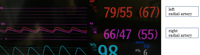
Her noninvasive blood pressure, measured from the left upper arm at admission, was 139/111 mm Hg. After induction of general anesthesia, a 20-gauge catheter was inserted in her left radial artery, and the arterial blood pressure was 79/54 mm Hg. The waveform ascended slowly, and the pulse pressure was unusually narrow. Phenylephrine (0.1 mg) was given intravenously 3 times, and her systolic pressure increased by 20 mm Hg after each dose; however, the abnormal wave form did not improve. The arterial line circuit was intact and exhibited no bending, kinking, or air bubbles. We conducted a flash test, which was normal. Because we initially suspected stenosis of the left radial artery, we inserted a second arterial catheter in her right radial artery; however, the pressure tracing revealed a similarly abnormal waveform (Fig. 1). The patient’s bilateral ulnar pulses were palpated; however, her radial pulses were stronger and more easily felt. We suspected that the patient may have had a perfusion deficiency in the bilateral upper extremities; consequently, POCUS was immediately performed.

JCRM 2019-101 - Masafumi Idei Japan_Fig. 1

Figure 1. Image shows similarly abnormal waveform of arterial blood pressure from bilateral radial arteries.

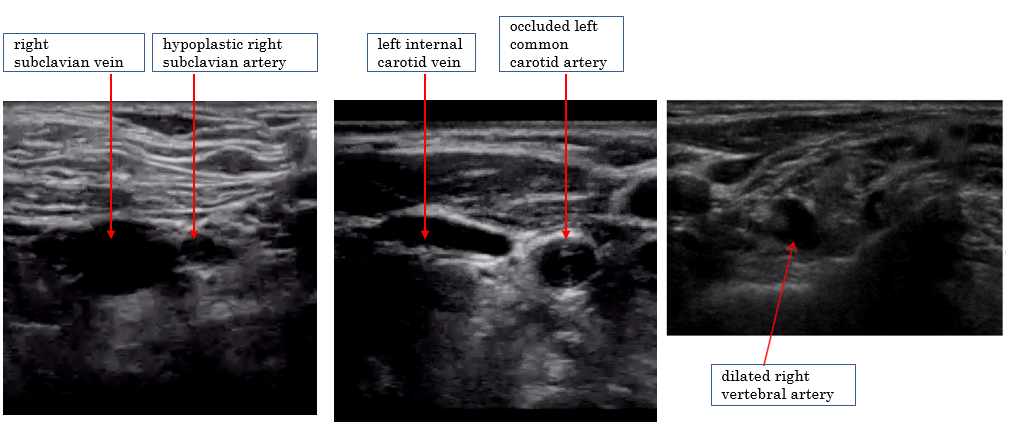
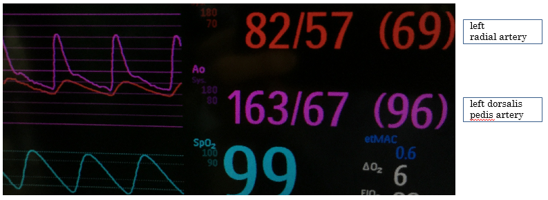
Using POCUS, we identified hypoplasia and stenosis of bilateral subclavian arteries and complete obstruction of bilateral common carotid arteries (Fig. 2). At around 3 mm in diameter, the bilateral subclavian arteries were around a quarter of the size of the accompanying subclavian veins. The diagnosis of bilateral common carotid artery obstruction was easily confirmed by color doppler imaging. Furthermore, bilateral vertebral arteries were significantly dilated with a diameter of approximately 7 mm. Because both subclavian arteries were observed to be abnormally narrow via imaging, we believed that the blood pressure measurement in the bilateral upper extremities was not reliable. Therefore, we inserted a third arterial catheter in the patient’s left dorsalis pedis artery. When the arterial blood pressure measured from her left radial artery was 81/56 (mean 66) mm Hg, it was 66/47 (mean 55) mm Hg from her right radial artery and 160/69 (mean 96) mm Hg from her left dorsalis pedis artery (Fig. 3). Based on the POCUS findings, we determined that the blood pressure measured from her dorsalis pedis artery was most accurate for perioperative circulatory management. The patient emerged from anesthesia immediately after the operation was concluded, and no neurological deficits were observed.

JCRM 2019-101 - Masafumi Idei Japan_Fig. 2

Figure 2. Ultrasonography of the neck. Hypoplasia and stenosis of bilateral subclavian arteries, complete obstruction of bilateral common carotid arteries, and dilated bilateral vertebral arteries were observed.

JCRM 2019-101 - Masafumi Idei Japan_Fig. 3

Figure 3. Image shows arterial blood pressure measured from the left radial artery and left dorsalis pedis artery.

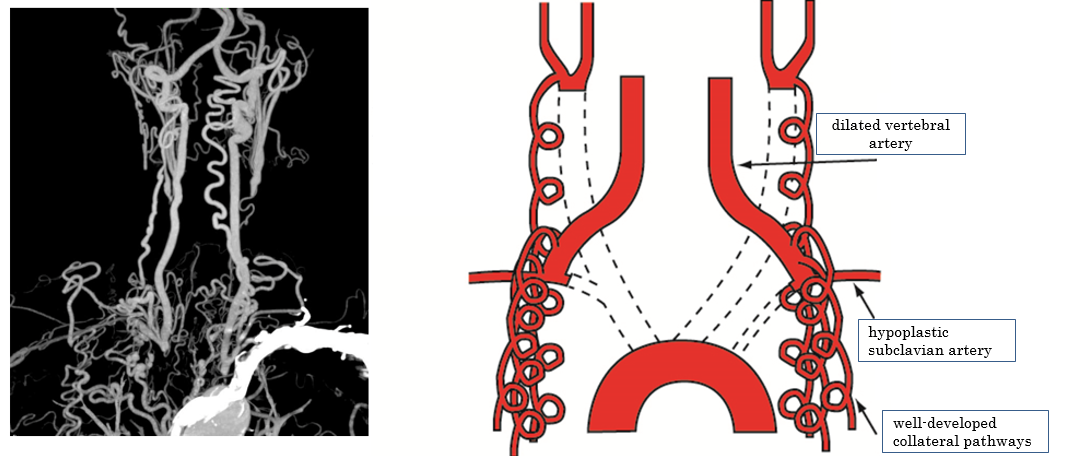
Due to the POCUS findings, we conducted a detailed examination by enhanced computed tomography (CT) postoperatively. All 3 branch vessels of her aortic arch (brachiocephalic trunk, left common carotid artery, and left subclavian artery) were completely occluded at their regions of origin, and well-developed collateral pathways supplied blood to her arms and brain (Fig. 4). A history of aortic arch syndrome (Takayasu’s arteritis) was suspected.

JCRM 2019-101 - Masafumi Idei Japan_Fig. 4

Figure 4. Computed tomography (CT) and schema. All three branch vessels of the aortic arch were totally occluded and well-developed collateral pathways supplied blood flow to her arms and brain.

Discussion

POCUS is being utilized more often in clinical settings [1–3]. In this case, bilateral subclavian artery stenosis was suspected based on abnormal pressure waveforms of bilateral radial arteries. POCUS was immediately performed, which revealed a previously undiagnosed vascular abnormality that could affect the patient’s brain and upper extremities. Postoperative enhanced CT confirmed the findings identified by POCUS.

In POCUS findings, we could easily identify vascular abnormalities. The vessel walls of bilateral common carotid arteries were thickened circumferentially, and color doppler imaging showed complete occlusion. The bilateral subclavian arteries were only about 3 mm in diameter, and were clearly smaller than accompanying subclavian veins. Bilateral vertebral arteries, which are normally about 3 mm in diameter [4,5], were significantly dilated to approximately 7 mm in diameter.

In order to accurately interpret the POCUS findings, sufficient experience is required [1]. The common carotid and subclavian arteries are routinely identified by anesthesiologists during ultrasound-guided central venous catheter insertion, and the vertebral artery is frequently identified during ultrasound-guided brachial plexus blocks. Moreover, anesthesiologists often evaluate stenosis of the common carotid artery as part of perioperative POCUS. Because we routinely use ultrasonography and are accustomed to POCUS, we immediately noticed the abnormality of the arteries, which contributed to our ability to make this diagnosis.

Based on the POCUS findings, we relied on the lower extremity blood pressure for perioperative circulatory management. Because POCUS and preoperative head MRI results showed obstruction of the bilateral common carotid arteries and dilation of the bilateral vertebral arteries, the patient’s cerebral blood flow was thought to depend on the vertebral arteries. We judged that only the upper extremities were hypotensive, and that the blood pressure measured from the lower extremities more accurately reflected the perfusion pressure of the patient’s major organs. We investigated the regional oxygen saturation (rSO2) as an index of cerebral blood flow. However, it would have been difficult to monitor the patient’s rSO2 in this case due to the location of the surgical field.

This case highlights how circulatory management that blindly gives priority to radial artery pressure could lead to end-organ hypertension, which could increase bleeding from the surgical field. In this case, the radial artery blood pressure was significantly lower than the blood pressure of the lower extremities (about 30–40 mm Hg less in mean arterial pressure). We thought that tight blood pressure control was warranted, because excessive hypotension could have caused ischemia of the upper extremities, and hypertension could have induced surgical bleeding. Thus, we sought to maintain a mean arterial pressure in the lower extremities of approximately 80 mm Hg (130 mm Hg in systolic blood pressure), because at this pressure, the mean arterial pressure of the right radial artery stayed around 50 mm Hg. Although setting an appropriate target blood pressure was extremely difficult in the limited environment and information during surgery, we deemed this target blood pressure as appropriate because the patient had no definite history of hypertension and it would enable us to avoid excessive hypertension, which is important during craniotomies.

The patient was similarly managed in the intensive care unit postoperatively, and she experienced no bleeding, neurological problems, or other complications including major organ failure. A CT scan performed 6 days after the operation showed complete occlusion of her brachiocephalic trunk, left common carotid artery, and left subclavian artery at their regions of origin from the aortic arch. Numerous collateral pathways from arteries of the superior mediastinum and chest wall, which branched from the aorta, supplied blood to the aortic arch branch vessels. Collateral pathways also supplied blood to bilateral vertebral arteries.

Although there is no conclusive evidence that the patient’s lower extremity blood pressure reflected her cerebral blood pressure and that our target arterial pressure was exactly appropriate, our careful circulatory management that avoided excessive hypertension and hypotension during the perioperative period, might have contributed to the patient’s good postoperative neurological outcome. Because preoperative head MRI data revealed abnormalities in the bilateral common carotid arteries and bilateral vertebral arteries, further detailed evaluation of other blood vessels below the neck by enhanced CT should have been considered before operation and may be important for similar situations in the future.

After consulting with a cardiothoracic surgeon, the patient subsequently confirmed that she had a weak pulse during a medical exam when she was in her twenties. Since this issue had been identified in the patient’s youth and because she had few risk factors of arteriosclerosis such as hypertension, diabetes mellitus, and smoking, a history of aortic arch syndrome (Takayasu’s arteritis) was suspected. There were, however, few findings and symptoms suggesting active arteritis such as an elevation of inflammatory markers, and the patient denied further evaluation and treatment; therefore, it is unknown whether the complete occlusion of the aortic arch branch vessels was caused by aortic arch syndrome.

Some reports have described anesthetic management of patients diagnosed with stenosis and/or obstruction of the aortic arch branch vessels that occurs with aortic arch syndrome (Takayasu’s arteritis) [6,7]. These reports highlight the usefulness of ultrasonography to determine the best site for blood pressure monitoring [6] and the efficacy of rSO2 monitoring as an index of cerebral blood flow [7]. However, to the best of our knowledge, there are few reports in which a previously undiagnosed stenosis and/or obstruction of the aortic arch branch vessels was identified by POCUS during a perioperative period. Because POCUS has the potential to significantly improve perioperative care, anesthesiologists should strive to acquire this skill. We expect future clinical studies and cases that involve POCUS during surgery will further demonstrate how its use can improve perioperative outcomes.

Acknowledgements: We would like to thank our patient for providing to publish this case report. We would also like to thank Editage (www.editage.jp) for English language editing.

Funding: The authors declare that they have no funding.

Authors’ contributions: MI and RY wrote the draft of the manuscript. TY made the figures. IW reviewed the manuscript. The final version of the manuscript was approved by all authors.

Ethics approval and consent to participate: Not applicable.

Consent for publication: Written informed consent was obtained from the patients for publication of this case report and accompanying images.

Competing interests: The authors declare that they have no competing interests.

Author details: Department of Anesthesia, Yokohama Minami Kyosai Hospital, 1-21-1 Mutsuurahigashi, Kanazawa-ku, Yokohama 2360037, Japan

Abbreviations

POCUS: Point-of-care ultrasound

MRI: magnetic resonance imaging

ICU: intensive care unit

CT: Computed tomography

rSO2: regional oxygen saturation

References

  1. Moore CL, Copel JA. (2011) Point-of-Care Ultrasonography. N Engl J Med. 364: 749–757. [Crossref]
  2. Johnson DW, Oren-Grinberg A. (2011) Perioperative point-of-care ultrasonography: the past and the future are in anesthesiologists’ hands. Anesthesiology. 115(3): 460–2. [Crossref]
  3. Holm JH, Frederiksen CA, Juhl-Olsen P, Sloth E. (2012) Perioperative Use of Focus Assessed Transthoracic Echocardiography (FATE). Anesth Analg. 115(5): 1029–32. [Crossref]
  4. Park JH, Kim JM, Roh JK. (2007) Hypoplastic vertebral artery: frequency and associations with ischaemic stroke territory. J Neurol Neurosurg Psychiatry. 78(9): 954–8. [Crossref]
  5. Ogeng’o J, Olabu B, Sinkeet R, Ogeng’o NM, Elbusaid H. (2014) Vertebral Artery Hypoplasia in a Black Kenyan Population. Int Sch Res Notices. 2014: 934510.
  6. Narasimha PK, Chaudhuri S, Joseph TT. (2013) Utility of intra-operative ultrasound in choosing the appropriate site for blood pressure monitoring in Takayasu’s arteritis. Indian J Anaesth. 57(1): 66–8. [Crossref]
  7. Xiao W, Wang T, Fu W, Wang F, Zhao L. (2016) Regional cerebral oxygen saturation guided cerebral protection in a parturient with Takayasu’s arteritis undergoing cesarean section: a case report. J Clin Anesth. 33: 168–72. [Crossref]

Validation of an Automated Extraction Procedure for Amino Acids and Acylcarnitines for Use with Tandem Mass Spectrometry for Newborn Screening

DOI: 10.31038/EDMJ.2019314

Abstract

A certified reagent kit for newborn screening was transferred on a fully automated dried blood spot platform. The dried blood spot cards are directly eluted and the extract is online guided to tandem mass spectrometry instrument, where the amino acid and acyl carnitine panel is detected. The method takes 2 minutes per sample and requires no human interaction for up to 500 samples. The method is fully standardized through the automation and the usage of only certified consumables and reference material. The manual reagent kit was first modified to fit the automated platform, secondly validated and third, successfully transferred into a routine newborn screening laboratory.

Keywords

Dried blood spot, Newborn, Screening, Amino acid, Carnitine, Automation

Introduction

Newborn screening (NBS) is a public health program provided by most of the countries around the world aimed at screening newborns for a list of serious genetic and metabolic disorders. Early diagnosis of these conditions can help prevent their further development, which if untreated often results in brain damage, organ damage, and even death [1–4]. A routine neonatal screening procedure requires that a health professional takes a few drops of blood from the baby’s heel, applies them onto special filter paper and sends such prepared samples to a laboratory for a number of analytical tests [5].

The amino acids and acyl carnitines are detected in modern methods with tandem mass spectrometry (MS/MS). MS/MS is a fundamentally different technology than systems previously used by most newborn screening laboratories, such as bacterial inhibition assays. It is a versatile and modular system that can be easily adapted to the preferred testing approach by the user. This has led to numerous variations of newborn screening by MS/MS, and it became a challenge to compare results between laboratories. There is a recognized need to develop consensus solutions to provide more consistency between MS/MS screening programs [6–9].

This validation addresses a certified screening method using fully automated analysis equipment from sample recognition towards extraction and analysis. The method was based on the commercial MassChrom® Reagent Kit from Chromsystems to allow full standardization of the complete process. The method development was performed at the Shimadzu laboratory (Reinach, Switzerland) and the application has been transferred to a routine NBS laboratory in Switzerland (Childrens Hospital Zurich). After the method development, a validation was performed, followed by a transfer into a routine laboratory focusing on inter-, intra-day variations, correlation and robustness. To standardize the sample preparation, handling and extraction, a fully automated DBS system from CAMAG (DBS- MS 500) was integrated into the workflow. The main goal of this study was to validate this change in analytical procedure.

Application of automated DBS card handling systems, which are connected to mass spectrometry analyzers, offers a modern and fast approach where a circular area of the DBS is directly eluted from the filter paper card without any punching [9–12].

Materials and Methods

Chemicals

The following MassChrom Reagents (Chromsystems Munich, Germany) were used; Mobile Phase (No. 57001), Internal Standard (No. 57004), Extraction Buffer (No. 57008) and Mass Check Controls propionylcarnitine, decanoylcarnitine, lauroylcarnitine, myristoylcarnitine, palmitoylcarnitine and stearoylcarnitine, were purchased from Sigma-Aldrich (St. Louis, USA). MassChrom rinsing solution (No. 55007) was used for the rinsing process and also purchased from Chromsystems (Munich, Germany). Dried blood spot cards (903, TFN, MN818 and 2992) were provided by CAMAG (Muttenz, Switzerland). Fresh whole blood was obtained from the local blood donation center (Basel, Switzerland). The blood was previously tested for infectious diseases.

Analytical Methods

A DBS-MS 500 unit (CAMAG, Muttenz, Switzerland) was attached as front end to a modularHPLC system from Shimadzu (Kyoto, Japan), containing a system controller (CBM-20A), a Nexera X2pump and a degasser (DGU-20ASR). The loop outlet of the DBS-MS 500 system was connected with a1.8 m PEEK tubing (yellow 1/16» OD x .007» ID) using a KrudKatcher Ultra (KrudKatcher Ultra,Phenomenex, Torrance, CA, USA) inline filter at the mass spectrometer inlet. Analysis was performedin positive multiple reaction monitoring (MRM) mode on an electrospray ionization tandem massspectrometry system 8060 in Reinach and 8050 in Zurich (Shimadzu, Kyoto, Japan). The extract wasdirectly injected into the mass spectrometer without an analytical column.

The elution was performed isocratic with the mobile phase from the kit using a flow gradient starting at 0.2 mL/min to 0.6 mL/min in 0.3 min to 1.9 min, 1.91 min back to 0.2 mL/min, 2.0 min controller stop. The following m/z transitions were programmed for the mass spectrometry detection (Table 1);

Table 1. m/z transitions

Name

Precursor  m/z

Product  m/z

Dwell  (msec)

Q1 Pre Bias (V)

CE

Q3 Pre Bias (V)

Alanine

90.2

44.2

10

-10

-12

-19

Alanine-2H 4

94.2

48.2

10

-10

-12

-19

Arginine

175.2

70.2

10

-10

-24

-16

Arginine-2H 7

182.2

77.2

10

-10

-24

-16

Aspartic acid

134.2

134.2

10

-15

-12

-13

Aspartic acid-2H 3

137.2

75

10

-16

-16

-27

Citrulline

176.1

113.1

10

-10

-16

-25

Citrulline-2H 2

178.1

115.1

10

-10

-16

-25

Glutamic acid

148.15

84.1

10

-17

-17

-14

Glutamic acid-2H 5

153.1

88.2

10

-17

-18

-19

Glycine

76

30

10

-11

-12

-30

Glycine-13C3/ 15N1

79

32

10

-10

-16

-28

Leucine

132

86.2

10

-16

-12

-19

Leucine-2H 3

135

89.2

10

-16

-12

-19

Methionine

150.1

104.1

10

-18

-14

-22

Methionine-2H 3

153.1

107

10

-18

-13

-18

Ornithine

133.2

133.2

10

-16

-12

-27

Omithine-2H 6

139.2

76

10

-15

-19

-15

Phenylalanine

166.2

120.2

10

-18

-14

-28

Phenylalanine-2H 5

171.2

125.2

10

-18

-14

-28

Proline

116.2

70.1

10

-14

-18

-23

Proline-2H 7

123.2

77.1

10

-13

-18

-16

Tyrosine

182.1

123.1

10

-10

-18

-24

Tyrosine-2H 4

186.1

127.1

10

-10

-18

-24

Valine

118.2

72.1

10

-14

-13

-17

Valine-2H 8

126.2

80.2

10

-14

-13

-17

Carnitine

162

85

10

-21

-23

-20

Carnitine-2H 9

171

85.1

10

-10

-23

-17

Acetylcarnitine

204

84.9

10

-20

-25

-20

Acetylcarnitine-2H 3

207

85.1

10

-11

-19

-18

Propionylcarnitine

217.9

85

10

-20

-25

-20

Propionylcarnitine-2H 3

221

85.1

10

-12

-23

-18

Butyrylcarnitine

231.9

85

10

-20

-25

-20

Butyrylcarnitine-2H 3

235

85.1

10

-13

-23

-17

Valerylcarnitine

246

85.1

10

-20

-14

-21

Valerylcarnitine-2H 9

255

85.1

10

-14

-24

-17

C5DC-carnitine

276.2

85

10

-46

C5DC-carnitine-2H 6

282.6

85

10

-15

-26

-19

Hexanoylcarnitine

260

85

10

-20

-25

-20

Hexanoylcarnitine-2H 3

263

85.1

10

-13

-22

-14

Octanoylcarnitine

288

85

10

-20

-30

-20

Octanoylcarnitine-2H 3

291

85.1

10

-15

-23

-14

Decanoylcarnitine

316

85.1

10

-10

-24

-17

Decanoylcarnitine-2H 3

319

85.1

10

-10

-24

-17

Lauroylcarnitine

344

85

10

-20

-28

-20

Lauroylcarnitine-2H 3

347

85.1

10

-11

-25

-19

Myristoylcarnitine

372.2

85

10

-19

-30

-20

Myristoylcarnitine-2H 3

375

85.1

10

-11

-27

-17

Palmitovlcarnitine

399.9

85.1

10

-12

-28

-18

Palmitoylcarnitine-2H 3

402.9

85.1

10

-12

-28

-18

Stearoylcarnitine

427.9

85.1

10

-13

-29

-17

Stearoylcarnitine-2H 3

430.9

85.1

10

-13

-29

-17

ESI mode, Nebulizing gas; 1.5L/min, Heating gas Flow; 10 L/min, Interface Temperature; 300 °C,   DL Temperature 300°C, Heat Block Temperature 400°C, Drying gas Flow; 10 L/min

The data analysis uses a linear curve type using the internal standard for the area calculation. The two Chromsystems MassCheck levels are used according to the specified concentrations. All peaks are  as default integrated from 0.05 to 1.5 min with a width of 25 sec.

DBS-MS 500 Instrumentation and Settings

The MassChrom internal standard 57004 was dissolved in 25 mL extraction buffer 57008 according to the Chromsystems procedure and connected to elution bottle 1, this solution was used for extracting the DBS. The MassChrom rinsing solution was connected to rinsing bottle 1 (R1). The extraction head was cleaned in an ultra sound bath at 40 °C for 10 min prior to a large set of analyses. The extraction solvent was primed for 5 cycles and the rinsing solvents were flushed for 1 min (this process is an automated system prime method). The DBS cards were photographed with the built-in camera of the DBS-MS 500 before and after each extraction to check for the presence of a blood spot and to adjust the extraction head to the center of each spot. The Chronos for CAMAG software automatically recognized inadequate dried blood spots based on their roundness, diameter, and area. Inadequate DBS were excluded from analysis. The samples were extracted with a 4 mm diameter clamp and a volume of 60 μL and a 200 μL/min flow rate into a 20 μl loop (the 40 μl upfront volume is directed to the waste). The extraction solvent passes the sealed area on the DBS card horizontally from the bottom back to the bottom into a sample loop, which is online guided to the mass spectrometer after the elution step. The area next to the 4 mm extraction ring is not affected by the solvent and could be reused if needed. To complete the automated DBS extraction cycle, the system was rinsed for 20 s with R1 [11].

Standard and DBS Sample Preparation

DBS calibration samples for method development and implementation were prepared manually in the laboratory (described as calibrators). Later on for method validation, external quality control material from Chromsystems (Masscheck) was integrated into the analysis workflow and used for reference (described as controls).

Five calibration points were prepared for the validation process. Here, 10 mg of each analyte was dissolved in 10 ml of water for the amino acids and in 10 ml methanol for the acyl carnitines. For each level, a mix was prepared according to reference values provided by the MassCheck Controls from Chromsystems. EDTA stabilized blood was pooled and centrifuged at 1300 rcf for 5 minutes. The plasma and buffy coat layer was removed and replaced with saline (0.9% NaCl) [13]. After gently mixing, this washing procedure was repeated twice. After removing the wash solution, a spiked saline with amino acids and acyl carnitines in five different levels (A-E, Supplementary data) was added and gently mixed [14].

Aliquots of the five calibrator blood levels were spotted in 15, 30 and 50 μl (standard 30 μl) droplets by an Eppendorf pipette onto TFN, 226, 903, 818 and 2992 filter paper cards from different vendors and dried in a horizontal position for a minimum of three hours. After drying, the calibrators were placed in a plastic bag with desiccant and stored at -20 °C. Calibrators B and E were used to derive data for intra- and interday imprecision. Those levels were used to compare to the high and low control levels from the MassChrom kit which will be used for the routine afterwards.

To measure the external MassCheck control card, a DBS frame in the standardized format of 84.67 mm × 53.2 mm (w × h) was fastened to the previously cut out reference DBS spots (Figure 1). By using the X-offset of the DBS-MS 500, several extractions per reference spot can be performed.

EDMJ 2019-102 - Gaugler Stefan Switzerland_F1

Figure 1. Manually prepared QC card.

All DBS were prepared from the same blood source, prepared with the same procedure to neutralize potential hematocrit effects [11], [15], [16] , which were not investigated in this study. All samples were prepared at one site and shipped to the other laboratory under controlled environment, to also eliminate the inaccuracy of different sample preparation.

The five calibration points were used for method validation and transfer, afterwards the routine measurement will be referred to the two Chromsystems MassCheck control levels high and low.

Validation Procedure

The inter- and intra-assay precision was obtained by measuring calibrator levels B and E at 3 consecutive days six fold. Precision was evaluated within a single validation run (intra-day) as well as between three runs recorded on different days (inter-day). The precision was calculated as the percentage relative standard deviation (CV, %) within an analytical run (intra-day precision, n = 6) and over all three runs (inter-day precision, n = 18). Depending on the analyte, CVs for within-run precision can range from 15% to 25% and for inter-run precision can range from 20% to 35% for newborn screening MS/MS assays [14]. The accuracy was assessed from the overall mean of each MassCheck control concentration divided by its nominal value (bias, %). Since all signals are related to official and certified reference material from Chromsystems [17], no other accuracy assays were conducted.

The extraction recovery of the DBS-MS 500 autosampler was investigated for DBS samples at level D. The five compounds methionine, proline, valine, carnitine and lauroyl-carnitine were chosen for this experiment. Each spot was extracted five times in triplicates. Between two extractions, a drying time of approximately 15 min was programmed. Using the built-in camera of the DBS-MS 500, the extraction head automatically locked onto the same area in the center of the blood spot. The recovery was finally estimated as the percentage ratio of the analyte peak area of the first extraction to the sum of the peak areas of all subsequently conducted extractions [11, 14].

Correlation and robustness was performed on each three days in two different laboratories. The correlation is determined using the 5 calibrator levels on 903 type filter paper, measured four fold. Each day, the run included a double measurement of each MassCheck level high and low as reference.

The carryover was assessed to study the possible effects from specimens with a high concentration of an analyte on the result of the subsequent specimen or specimens. Although there are several potential sources for carryover within the MS/MS system, the most common source is the autosampler injection port and the tubing leading to the MS/MS electrospray unit. To determine the carry over in the automated DBS-MS 500 system, an extraction with a high concentration at level E is performed, followed by three extractions of blank DBS cards. For no carryover, the analyte concentrations of the blank sample should be below the LOD concentration previously determined. This should be repeated a minimum of five times [14].

Although no interferences are known or have been documented [17], the blank filter papers used for specimen collection were checked for possible interferences of common m/z for any of the analytes measured, especially since new filter paper sources and new lot numbers are used. Here, we investigated the most commonly used filter paper types from different vendors used for manufacturing the DBS cards. Paper types are TFN and 226 from Ahlstrom, 903 from EBF, MN 818 from Macherey Nagel and 2992 from Hahnemühle. Calibrator level B, D and F were measured as triplicates on each paper type to detect precision and any significant off-set.

The robustness is determined during the interference and repeatability study by varying the following parameters; technician, card type, eluent preparation (using solvent and buffer from different lots), elapsed times (with or without breaks between the measurements) and laboratory environment (location, temperature and humidity). All cards used for the interference study were spotted with level a calibrator.

Matrix effects, an extended interference study and stability tests were not performed, since this data is given by the MassChrom kit and its process remain unchanged [17].

Documentation

The documentation process is given by the DBS-MS 500 instrument which takes a picture of the DBS card before and after each extraction to assure sample traceability. All pressures on the DBS card sampler as well as on the LC-MS/MS unit are monitored and documented.

The camera documentation system checks for preset values, where quality control parameters can be integrated. If the quality criteria are not met, the system automatically checks the next spot and continues the preset program. In addition, the camera detects already extracted spots and blocks those to prevent reanalysis. The system checks the xy shift of the circle on the card and the blood spot to center the extraction spot to the middle of each DBS (to avoid inhomogeneous distribution effects). Further, the inbuilt pressure sensors are monitoring the extraction and rinsing pressure (in this study, the extraction pressure was 0.5 bar and the rinsing pressure 45 bar). The maximum extraction pressure is 1.2 bar, depending on the age of the blood sample, and the system can be rinsed with pressure up to 100 bar to prevent carry over [9].

Results

MS/MS Method

The method from the MassChrom kit was transferred to the automated DBS-MS 500 platform allowing full automation of this process. The method was successfully installed at a routine NBS laboratory. Five calibrator levels of blood spotted on DBS cards were established for the method validation and installation. For routine, the two levels high and low from the MassCheck controls were used. Since the extract is guided online to the tandem mass spectrometry system, it only takes 2 minutes per sample. All processes are overlapping, where the DBS-MS 500 runs a wash program and extracts the consecutive sample while the MS/MS is detecting and reporting the target analytes of the previous sample.

Method validation

First, validation including additionally carry-over, interference, volume effects and recovery was performed after setting up the method on the automated platform. Evaluation of accuracy was not part of this method validation, since control material from the MassChrom kit was used as reference in all experiments. The intra- and inter-day variations as well as the coefficients of determination (R2) of the calibration standards are summarized in Table 2. All factors were within the general acceptance criteria for NBS screening methods [17]. Intra-day variations are all below 15% with the exception of aspartic acid and glutamic acid which are slightly above but still acceptable since the criteria sets a value of < 25%. Also, glutamic acid gave a relative high variation for the inter-day variation (still within the criteria), compared to the other values which are all below 25%. Therefore the optimization of the MRM transition of glutamic acid was carefully monitored for the method installation at the routine site.

Table 2. Selected recovery comparison to MassCheck controls

MassCheck

Methionine

Proline

Valine

Carnitine

Laurovl-carnitine

Extraction 1

0.6159

1.421

1.597

15.499

56.966

Extraction 2

0.1284

0.460

0.527

2.416

9.854

Extraction 3

0.0557

0.154

0.184

0.653

4.461

Extraction 4

0.0276

0.063

0.083

0.293

3.981

Extraction 5

0.0154

0.031

0.035

0.152

3.368

[%]

[%]

[%]

[%]

[%]

MassCheck recovery

73.1

66.7

65.8

81.5

72.4

Sample recovery

79.9

70.3

61.5

80.0

76.4

DBS spots using 15, 30 and 50 μl blood were prepared at calibrator level A, C and E and compared as triplicates. The deviations from 50 μl to 30 μl spots were between 90.8 – 102.2 % and the deviations from 30 μl to 15 μl were between 78.2 – 88.8 %. There is a trend of smaller signals towards the 15 μl spots, however there is no obvious trend between 30 μl and 50 μl spots.

Carry-over from the high concentration to a blank DBS card passed the ICH guideline criteria and all investigated filter paper types were feasible for being used with this method. The best results were accomplished with the TFN filter paper (2.6% variation), paper types 226, 903, 229 were below 5% variation and the 818 paper moved with 6.0% on the last rank (supplementary data). The standard deviation of the complete panel from the triplicate measurement was taken into account.

The recovery of the compounds from the DBS card lies within 40 to 80%. This recovery remains constant for each analyte with the chosen extraction parameters. Since the results are always considered relative to the used MassChrom quality control card values with the same consistent recovery, this has no effect to the screening method, (see Table 2 and supplementary data).

Also the extraction behavior remains constant. This was investigated by comparison of freshly spotted blood and blood spots with 4 days of age stored at -20 °C (see supplementary data).

DBS spots using 15, 30 and 50 μl blood were prepared at calibrator level A, C and E and compared as triplicates. The deviations from 50 μl to 30 μl spots were between 90.8 – 102.2 % and the deviations from 30 μl to 15 μl were between 78.2 – 88.8 %. There is a trend of smaller signals towards the 15 μl spots, however there is no obvious trend between 30 μl and 50 μl spots.

Method Transfer into a Routine Environment

The previously developed and validated method was transferred into a routine environment. A short validation focusing on correlation and robustness was performed, where all five calibration levels were measured four-fold on 903 filter paper on three consecutive days. Each day was referred to high and low Mass Check control measured before the sample run.

Table 3. Intra-day, inter-day precision and R2 of calibrators level B (L1) and E (L2) from the spiked DBS samples (*Xle refers leucine/ Isoleucine)

Intraday day 1

Correlation

Intraday day 2

Correlation

Intraday day 3

Correlation

Inter day

LI [%]

L2 [%]

R2

LI [%]

L2 [%]

R2

LI [%]

L2 [%]

R2

LI [%]

L2 [%]

Alanine

6.8

7.6

0.994

3.7

7.1

0.981

8.6

5.4

0.998

24.5

13.0

Arginine

6.1

4.1

0.998

8.1

4.6

0.998

7.8

1.9

0.997

8.5

5.7

Aspartic acid

15.7

6.8

0.964

13.1

6.8

0.987

5.3

10.9

0.925

13.9

10.4

Citrulline

4.8

5.9

0.999

6.5

5.3

0.998

6.5

4.3

0.999

7.9

8.4

Glutamic acid

15.5

7.2

0.998

1.4

4.3

0.997

3.5

3.4

0.995

32.4

11.7

Glycine

9.2

9.9

0.998

9.2

9.9

0.998

7.7

12.3

0.987

8.7

18.9

Leucine (Xle*)

6.5

4.2

0.999

2.8

5.0

0.999

5.0

3.5

0.901

12.3

9.5

Methionine

10.3

4.8

0.999

9.6

6.1

0.985

11.9

4.8

0.999

24.9

5.8

Ornithine

6.9

4.0

0.999

6.5

5.7

0.998

11.1

4.6

0.996

13.3

14.8

Phenylalanine

5.7

3.9

0.999

4.3

4.9

0.999

8.6

8.3

0.998

11.7

6.0

Proline

4.0

4.1

0.999

4.6

5.0

0.999

11.0

8.8

0.999

9.4

6.9

Tyrosine

7.8

6.8

0.999

14.5

5.4

0.991

14.1

5.3

0.997

16.2

11.1

Valine

6.0

4.3

0.999

4.7

4.2

0.999

4.8

2.9

0.999

6.5

6.3

Carnitine

6.9

8.9

0.999

1.7

9.4

0.999

6.1

8.1

0.999

6.6

15.5

Acetvlcarnitine

9.3

2.8

0.978

9.7

6.3

0.986

8.0

6.2

0.983

9.4

7.4

Propionylcarnitine

2.3

2.9

0.998

1.5

3.3

0.999

3.8

3.6

0.996

12.9

3.3

Butyrylcarnitine

8.8

2.9

0.999

6.3

4.0

0.998

10.2

8.1

0.999

11.1

7.8

Valerylcarnitine

6.3

3.5

0.999

6.3

4.4

0.999

7.6

4.3

0.996

8.0

6.2

Hexanoylcarnitine

9.2

3.1

0.999

5.0

5.6

0.998

6.5

7.7

0.998

15.0

7.0

Octanoylcarnitine

5.7

3.5

0.999

6.6

3.6

0.999

5.4

7.4

0.997

10.0

6.7

Decanoylcarnitine

5.0

2.4

0.978

3.7

12.5

0.998

7.0

4.5

0.997

15.4

9.4

Laurovlcarnitine

14.0

4.2

0.999

11.5

4.2

0.999

7.8

3.5

0.999

12.8

4.2

Myristovlcarnitine

3.2

5.0

0.997

3.4

3.3

0.998

5.9

6.6

0.999

10.8

5.6

Palmitoylcarnitine

1.6

2.7

0.996

1.3

1.8

0.997

1.3

2.5

0.996

14.8

10.6

Stearoylcarnitine

2.3

2.8

0.995

1.8

2.4

0.999

2.1

3.2

0.995

18.1

14.1

At the Children’s hospital in Zurich, the Shimadzu MS/MS 8050 was coupled to a DBS-MS 500 unit as used for the method development. The criteria were met for intra- and inter-day variation following the validation procedure (supplementary data).

Discussion

The described method is fully automated and uses exclusively certified consumables and reference material. Through this degree of standardization, the application can be directly transferred in-between newborn screening laboratories and therefore following the trend towards the development of standardized programs. The method represents a high throughput application with only 2 minutes per sample. All processes are well documented by the reporting system of the DBS- MS 500, where a picture is taken of each spot before and after the extraction and all pressures for extraction, rinsing and LC pump are monitored. The DBS card picture is analyzed with image recognition software, providing results for the spot diameter, area and roundness, which can be used as quality control criteria. Also, the system detects a spot which has been previously extracted, to avoid multiple extractions of the same area. In a standard setting, the extraction is performed automatically from the center of each spot to overcome inhomogeneous distribution effects within the DBS sample [18].

We encountered a relative high endogenous concentration of the target analytes in the available donor blood. To bring this into the range of interest, the blood was washed with saline following a protocol. This procedure allowed gently spiking the washed red blood cells with the target analytes in the according concentrations. The five calibration points were used for method development, validation and installation at the routine site. After implementing the method, all results were referred to externally prepared and certified reference cards from the MassChrom kit.

The protocol of the MassChrom kit describes an extraction of a 3.2 mm disc in 100 μl extraction buffer (including the internal standard). Here, a 4 mm area was sealed on the DBS card and extracted with 60 μl into a 20 μl loop, whereas the first 40 μl are guided into waste. This was experimentally optimized using different loop volumes, extraction volumes and flow rates. This outcome could be due to an initially high portion of certain analytes, such as salts and phospholipids, causing matrix effects and ion suppression in the ESI source. However, the basic principle of this result needs to be further investigated. The method uses 60 μl of extraction buffer per sample instead of 100 μl from the protocol, which reduces the amount of solvent and analytical standard. In addition, consumables for sample preparation according to the protocol such as Eppendorf vials and pipette tips are no longer required.

Leucine, isoleucine, hydroxy l proline and allo-isoleucine are detected as sum (Xle) and are not separated within this method. An abnormal result for this parameter will have the automatic consequence of a second tier method using an analytical column prior the mass spectrometer to properly distinguish between all isobars. This is needed for the detection of maple syrup urine disease (MSUD).

A volume of 30 μl blood was chosen for the preparation of the calibrator spots. The 50 μl standard volume from literature resulted in relatively large spots on the standard filter paper card for automation with a spot-to-spot distance of 13.7 mm. The 50 μl spots were overlapping if all four positions on the DBS card were spotted. A comparison of 30 and 50 μl droplets showed no difference in the results.

Conclusion

A validated reagent kit for the extraction and analysis of dried blood spots in the field of newborn screening was transferred and validated on an automated DBS card extraction platform. First, a validation was performed and the method was then successfully transferred into a routine environment. Each process step is well documented and all analysis steps follow Good Laboratory Practice (GLP) [19]. The method can be easily modified or extended and transferred to other routine laboratories.

Author Contributions

Stefan Gaugler and Vincente Luis Cebolla Burillo participated in the concept and design of the study. Jana Rykl contributed to the optimization of the MS methods. Stefan Gaugler and Jana Rykl did the method development work, Stefan Gaugler did the practical work at the Children’s Hospital Zurich. Stefan Gaugler was responsible for drafting the manuscript. Vincente Luis Cebolla Burillo was responsible for revising the manuscript. All authors approved the manuscript as submitted.

Conflict of Interest

Jana Rykl is an employee of Shimadzu Schweiz GmbH (Reinach, Switzerland) and Stefan Gaugler is an employee of CAMAG (Muttenz, Switzerland). None of the other authors report any conflict of interest regarding this study. All instrumentations are property of the respective companies and laboratories.

References

  1. G. Nys, M. G. M. Kok, A. C. Servais, and M. Fillet, “Beyond dried blood spot: Current microsampling techniques in the context of biomedical applications,” TrAC – Trends Anal. Chem., vol. 97, pp. 326–332, 2017.
  2. J. L. Merritt, L. L. Brody, G. Pino, and P. Rinaldo, “Newborn screening for proximal urea cycle disorders: Current evidence supporting recommendations for newborn screening,” Mol. Genet. Metab., no. February, pp. 0–1, 2018.
  3. H. Lemonde, “Newborn screening for inborn errors of metabolism,” Paediatr. Child Health (Oxford)., vol. 25, no. 3, pp. 103–107, 2014.
  4. U. Groselj, M. Zerjav, A. Smon, N. Angelkova, D. Anton, I. Baric, M. Djordjevic, L. Grimci, M. Ivanova, A. Kadam, V. Mulliqi, H. Maksic, O. Marginean, O. Margineanu, O. Milijanovic, F. Moldovanu, M. Muresan, S. Murko, M. Nanu, B. Repic, M. Samardzic, V. Sarnavka, A. Savov, M. Stojiljkovic, B. Suzic, R. Tincheva, H. Tahirovic, A. Toromanovic, N. Usurelu, and T. Battelino, “Newborn screening in southeastern Europe,” Mol. Genet. Metab., vol. 113, no. 1–2, pp. 42–45, 2014.
  5. A. W. El-Hattab, M. Almannai, and V. R. Sutton, “Newborn Screening: History, Current Status, and Future Directions,” Pediatr. Clin. North Am., vol. 65, no. 2, pp. 389–405, 2018.
  6. A. Starkweather, B. Coleman, V. B. De Mendoza, M. Fu, J. Taylor, W. Henderson, C. Kenner, D. Walker, L. Amankwaa, and C. Anderson, “American Academy of Nursing on Policy Policy brief : Improve coverage of newborn genetic screening to include the Recommended Uniform Screening Panel and newborn screening registry,” Nurs. Outlook, vol. 65, no. 4, pp. 480–484, 2017.
  7. M. Resano, M. A. Belarra, E. García-Ruiz, M. Aramendía, and L. Rello, “Dried matrix spots and clinical elemental analysis. Current status, difficulties, and opportunities,” TrAC – Trends Anal. Chem., vol. 99, pp. 75–87, 2018.
  8. R. Fingerhut, M. L. Silva Polanco, G. D. J. Silva Arevalo, and M. A. Swiderska, “First experience with a fully automated extraction system for simultaneous on-line direct tandem mass spectrometric analysis of amino acids and (acyl-)carnitines in a newborn screening setting,” Rapid Commun. Mass Spectrom., vol. 28, no. 8, pp. 965–973, 2014.
  9. S. Gaugler, J. Rykl, I. Wegner, T. von Däniken, R. Fingerhut, and G. Schlotterbeck, “Extended and Fully Automated Newborn Screening Method for Mass Spectrometry Detection,” Int. J. Neonatal Screen., vol. 4, no. 1, p. 2, 2017.
  10. “CAMAG DBS,” 2018. [Online]. Available: http://www.camag.com/en/dbs/what_is_dried_blood_spot_sampling.cfm. [Accessed: 29-May-2018].
  11. M. H. U. Duthaler, B. Berger, S. Erb, M. Battegay, E. Letang, S. Gaugler, S. Krähenbühl, “Automated high throughput analysis of antiretroviral drugs in dried blood spots.,” J Mass Spectrom., vol. 52, no. 8, pp. 534–542, 2017.
  12. S. Gaugler, J. Rykl, M. Grill, and V. L. Cebolla, “Fully automated drug screening of dried blood spots using online LC-MS / MS analysis,” J. Appl. Bioanal., vol. 4, no. 1, pp. 7–15, 2018.
  13. D. H. Chace, B. W. Adam, S. J. Smith, J. R. Alexander, S. L. Hillman, and W. H. Hannon, “Validation of accuracy-based amino acid reference materials in dried-blood spots by tandem mass spectrometry for newborn screening assays.,” Clin. Chem., vol. 45, no. 8 Pt 1, pp. 1269–77, 1999.
  14. G. L. Hoffman, “CLSI guideline NBS04-A,” 2010.
  15. P. Abu-Rabie, P. Denniff, N. Spooner, B. Z. Chowdhry, and F. S. Pullen, “Investigation of different approaches to incorporating internal standard in DBS quantitative bioanalytical workflows and their effect on nullifying hematocrit-based assay bias,” Anal. Chem., vol. 87, no. 9, pp. 4996–5003, 2015.
  16. N. Kostić, Y. Dotsikas, N. Jović, G. Stevanović, A. Malenović, and M. Medenica, “Quantitation of pregabalin in dried blood spots and dried plasma spots by validated LC-MS/MS methods,” J. Pharm. Biomed. Anal., vol. 109, pp. 79–84, 2015.
  17. Chromsystems, “MassChrom Amino acids and Acylcarnitines from Dried Blood (non derivatised), 57000, EN 06/2016 R7,” 2016.
  18. G. Nys, M. Kok, A.-C. Servais, and M. Fillet, “Beyond dried blood spot: current microsampling techniques in the context of biomedical applications,” Trends Anal. Chem., vol. In press, 2017.
  19. European Medicines Agency, “Guideline on bioanalytical method validation,” 2015.

Supplementary data

Filter paper comparison                                                                     

226

Average

3.7 %

TFN

Average

2.7 %

903

Average

3.6 %

2992

Average

2.9 %

903NBS

Average

2.6 %

818

Average

6.0 %

Calibration sample preparation

Spike A

Spike B

Spike C

Spike D

Spike E

[ul]

[ul]

[ul]

[ul]

[ul]

AA, CO/I Spike

25

50

100

167

250

AC Spike

5

10

25

50

100

H20

225

200

150

83

0

MeOH

95

90

75

50

0

NaCl 0.9%

2150

2150

2150

2150

2150

Red blood cells

2500

2500

2500

2500

2500

Total volume

5000

5000

5000

5000

5000


Carry over (Area blank/Area E * 100%)

Recovery

Alanine

2.1

55.6

%

Arginine

0.1

63.3

%

Aspartic acid

1.8

74.4

%

Citrulline

0.1

66.5

%

Glutamic acid

0.1

44.0

%

Glycine

1.8

68.4

%

Leucine (Xle)

0.1

65.6

%

Methionine

0.1

76.9

%

Ornithine

0.1

64.5

%

Phenylalanine

0.3

68.3

%

Proline

0.3

70.3

%

Tyrosine

0.3

55.1

%

Valine

0.3

61.5

%

Carnitine

0.1

80.0

%

Acetylcarnitine

0.1

78.7

%

Propionylcarnitine

0.1

50.1

%

Butyrvlcarnitine

0.1

81.0

%

Valerylcarnitine

0.1

81.7

%

Hexanoylcarnitine

0.3

75.6

%

Octanoylcarnitine

0.6

77.4

%

Decanoylcarnitine

0.8

72.3

%

Lauroylcarnitine

0.9

76.4

%

Mvristoylcarnitine

0.1

70.8

%

Palmitoylcarnitine

0.2

40.9

%

Stearoylcarnitine

0.2

43.4

%

Intra-day, inter-day precision and R2 of calibrators level B (LI) and E (L2) from the spiked DBS samples in the routine laboratory

Intraday day 1

Correlation

Intraday day 2

Correlation

Intraday day 3

Correlation

Interday

LI [%]

L2 [%]

R2

LI [%]

L2 [%]

R2

LI [%]

L2 [%]

R2

LI [%]

L2 [%]

Alanine

13.3

4.8

0.980

6.9

6.6

0.994

3.8

10.0

0.994

21.9

9.1

Arginine

6.5

4.0

0.999

5.0

2.0

0.998

4.7

3.2

0.999

6.9

4.5

Aspartic add

7.9

7.1

0.996

13.9

9.5

0.996

11.4

5.5

0.962

19.7

13.3

Citrulline

6.0

6.8

1.000

6.7

4.0

0.999

7.4

5.0

0.998

6.9

5.5

Glutamic add

1.0

5.4

0.994

9.8

2.9

0.993

10.8

7.0

0.999

19.6

14.2

Glycine

14.7

14.7

0.994

10.8

14.5

0.980

10.3

14.1

0.991

15.0

23.6

Leucine (Xle)

7.5

3.1

0.999

2.4

2.5

0.998

4.7

2.4

0.999

11.6

9.7

Methionine

13.2

5.0

0.997

14.3

7.3

0.997

8.4

9.5

0.999

21.3

13.7

Ornithine

5.9

2.9

0.997

15.2

9.3

0.994

9.5

6.7

0.994

12.2

13.6

Phenylalanine

9.9

7.6

0.999

4.9

3.4

0.999

6.1

3.6

1.000

7.9

14.8

Proline

9.9

5.2

0.999

4.3

2.8

0.998

5.8

5.7

0.999

7.4

6.2

Tyrosine

11.1

17.0

0.945

14.3

3.0

0.999

13.3

5.7

0.998

21.6

20.7

Valine

10.2

8.6

0.997

12.4

2.1

0.998

3.9

3.0

0.997

11.7

9.1

Carnitine

3.6

4.4

0.997

5.2

8.8

0.998

7.9

11.6

0.997

7.2

27.5

Acetylcarnitine

8.1

3.9

0.989

6.5

5.7

0.995

13.2

6.1

0.993

9.7

12.7

Propionylcarnitine

1.3

2.4

0.998

3.1

3.0

1.000

3.7

3.9

0.999

11.7

3.6

Butyrylcarnitine

6.0

5.4

0.997

8.7

4.5

0.999

11.8

5.1

0.999

9.2

10.4

Valerylcarnitine

7.3

5.5

0.999

6.7

3.8

1.000

9.6

5.3

0.999

8.5

6.5

Hexanoylcarnitine

9.7

7.1

0.999

9.6

4.8

0.999

11.2

6.2

0.999

16.3

6.9

Octanoylcarnitine

8.1

4.6

0.999

5.8

5.0

0.999

5.3

3.6

0.999

7.4

4.9

Decanoylcarnitine

15.3

6.1

0.999

5.1

3.4

0.999

6.0

2.3

0.999

10.1

4.2

Lauroylcarnitine

4.7

4.5

0.999

3.7

2.1

0.999

6.3

4.3

0.999

7.5

4.6

Myristoylcarnitine

6.9

3.8

0.998

6.2

3.2

0.998

2.6

5.9

0.997

6.1

5.1

Palmitovlcarnitine

3.9

4.7

0.995

8.2

11.4

0.896

6.7

5.5

0.992

8.8

7.8

Stearoylcarnitine

4.8

2.3

0.985

17.4

14.4

0.973

19.0

15.3

0.954

34.9

18.5

The Assessment of Quality of Life Using GOHAI among Edentulous Patients

DOI: 10.31038/JDMR.2019211

Abstract

Introduction: Edentulism may compromise the quality of life (QoL) of a patient. Geriatric patients who are satisfied with their complete dentures are usually satisfied with their daily life. This research was conducted to assess quality of life using Geriatric Oral Health Assessment Index (GOHAI) focusing on functional limitation, oro-facial pain, psychological impact and behavioural impact among edentulous patient attending USIM Polyclinic.

Materials and Methods: Thirty-two geriatric edentulous patient to be treated by third year students in polyclinic USIM were recruited. Patient completed the Malay version of GOHAI questionnaire.

Result: The data showed there were 62.5% male and 37.5% female. The mean age was 63.88 ± 5.621 year. 87.5% of the samples are wearing complete denture and 56.3% of them have been wearing denture for the past five years. The lowest mean score was 1.66 ( ± 1.6) for trouble in biting and chewing. The highest mean for GOHAI was in medication used to relieve pain with the mean score of 4.56 ( ± 0.8).

Conclusion: The QoL of geriatric patient was mostly affected by the oral function. However the least affected was seen in pain and discomfort. Despite wearing complete denture, denture replacement is essential to improve QoL.

Keyword

Edentulous and Quality of life, GOHAI, Geriatric Dentistry

Introduction

According to the Gerodontology society, the life expectancy of the elderly who need complete denture pros-thesis has increased [1]. Edentulism is considered as an outcome of poor oral healthcare and may compromise the quality of life [2]. The physiological changes in edentulous arch with continuous resorption of the residual ridges leads to the need of complete denture replacements. The use of complete denture increases the quality of life of geriatric patient in term of oral function, reduce oro-facial pain, improve oro-facial appearance, and psychosocial impact [3]. Oral health is often assessed separately from patient’s general health. Sometimes it perceived as a distinct entity from any chronic conditions of the elderly patient. Edentulism may compromise the quality of life (QoL) because patients will have difficulty eating proper food and this might lead to malnourish among the elderly. Edentulous patients need dentures to restore their oral function. Satisfaction of the denture influences psychological condition which is part of quality of life (QoL). Geriatric patient who are well satisfied with their daily lives, usually also satisfied with their complete denture [4].

There is a measure called Geriatric/General Oral Health Assessment Index (GOHAI) is an instrument consists of 12-item questionnaires and it is considered a gold standard used in measuring the oral health impact of the geriatric patients. It was developed by Atchinson and Dolan [4] and has been validated over a few languages including Malay language  [5] which is useful to measure oral-health related quality of life in Malaysia. From its name, GOHAI Index is usually used for the assessment of the quality of life in geriatric patient and not suitable to assess the overall oral health status because there was a weak correlation with the clinical measure of disease. Therefore, GOHAI cannot be used to conclude a diagnosis of dental disease because it will never assess the oral examination clinically and radiographically. However, indication for a referral or further thorough oral examination need can be obtained through GOHAI assessment as it will provide necessary information in symptoms as well as the limitation in psychosocial and functional. Therefore the aim of this paper was to assess the quality of life of the elderly population among the geriatric patients that received complete dentures from the undergraduate dental students of the Faculty of Dentistry, Islamic Science University of Malaysia (USIM) Polyclinic, Kuala Lumpur.

Methods

An approval was attained before starting the study by Board of Research Ethic of Islamic Science University of Malaysia (USIM). A total of 32 community dwelling participants were recruited among complete denture patients who will receive complete dentures from the undergraduate dental students of USIM. They must satisfy all the following inclusion criterias; Malaysians aged 60 years and above, complete denture wearers, subject with con-trolled or no underlying systemic diseases, and fluent in Malay language. The consents were obtained from par-ticipants after being informed about voluntary participation in this research. Then, self-administered questionnaire was distributed to collect information of participant’s demographic and prosthetic experience.

The participants were interviewed by a single interviewer and were asked to estimate the frequency of problems in Malay Version of GOHAI questionnaire using a six point Likert scale rating (always [5], very often [4], often [3], sometimes [2], seldom [1] or never [0]). The 12-item questionnaire was classified into four major domains which were the functional limitation, pain or discomfort, psychological and behavioural impact. The GOHAI score is determined by summing the final score of each of the 12 items ranges from 0 to 60. The score for GOHAI item number 3, 5 and 7 were maintained while the rest were reversed in order to attain a positive oral health GOHAI score. The higher GOHAI score denotes better oral health status perceived by the participants themselves. If there were 3 or more missing items in the data, the data will be dropped out. Alternately, if no more than two missing items in a data, the score of the missing will be substituted with the mean. The mean GOHAI scores in relation with demographic variables were analyzed using Independent t-test and ANOVA. 0.05 was set as the level of significance. The data were analyzed using IBM SPSS Statistic version 19.

Results

The patients’ age ranges from 60–76 years with mean age of 63.88 ± 5.6 (as shown in (Table 1) below. Almost one-third of them were female and more than half received education up to secondary education level (11 years of formal education in school). Majority of them have been wearing a complete denture for more than 5 years (56.3%). Most of the patients request for new dentures because they want to improve the chewing ability (50.0%) and aesthetic reasons (40.6%) over the other health and speech reasons. (Table 2)

Table 1. Distribution of patients according to sex.

Gender

Number

Mean

SD

Male

20

64.40

5.707

Female

12

63.00

5.608

Total

32

63.88

5.621

Table 2. Characteristics of subjects.

Participant’s characteristic

(N=32)

N

 %

Mean GOHAI score

SD

p-value

GOHAI Score

Range

40.1

7.4

(27–60)

Sex a

Male

Female

20

12

62.5

37.5

39.2

41.8

6.0

9.5

.346

Age b

<55 years

55–59 years

60–69 years

70–79 years

2

5

21

4

6.3

15.7

65.5

12.5

42.50

39.40

39.48

43.25

3.5

9.2

7.6

6.7

.789

Education Level b

No schooling

Primary school

Secondary school

University

2

6

19

5

6.3

18.8

59.4

15.7

42.0

43.0

40.4

34.8

4.2

11.7

6.4

4.0

.315

History of Denture Wearing b

< 1 year

1–3 year

3–5 year

>5 year

Never

4

3

3

18

4

12.5

9.4

9.4

56.3

12.5

39.3

44.3

41.0

40.1

37.3

10.2

14.0

3.6

7.0

4.2

.820

Reason for Wearing Denture b

Chewing

Aesthetic

Health

Speech

16

13

2

1

50.0

40.6

6.3

3.1

39.1

40.2

47.5

40.0

6.0

9.1

6.4

0.0

.539

The mean and median of the summary GOHAI scores was 40.1
( ± 7.4, range 27–60) and 39.0, respectively. Besides that, the skewness of the GOHAI was 0.75. There was only one out of 32 subjects scored the maximum in this measure. About one-third of the participants (31.3%) scored 41 and above. The mean of GOHAI scores in regard to each variable varies from 37.3 (± 4.2) to 47.5 (± 6.4). The lowest and highest mean GOHAI scores can be found in university level participants and reason of wearing denture for health purpose respectively (Table 2). Based on the data exhibited in Table 2, there is no significant association between gender, age, level of education, history of wearing denture and reason of wearing denture with the oral health related quality of life (OHRQoL). The used of medication to relieve pain in pain and discomfort domain was recorded as the highest mean for GOHAI item with 4.56 (SD 0.8) followed by 4.31 (SD 1.1) for sensitive to hot, cold or sweet foods and 4.31 (SD 1.3) for limit contact with people. The lowest mean was 1.66 (SD 1.6) in trouble biting and chewing. Meanwhile, Table 3 shows the percent of participants responding ‘sometimes,’ ‘often,’ ‘very often,’ or ‘always’ to each of the GOHAI items. The percentage of subjects responding positively to each item ranged from 16% to 80%, with ten out of 12 items being reported by 20% or more and six of 12 reported by 33.3% or more (Table 3).

Table 3. Percentage of subjects responding ‘sometimes,’ ‘often,’ ‘very of-ten,’ or ‘always’ to each GOHAI and mean each of the items.

GOHAI ITEMS

Percentage of participants responding positive response (%)*

Mean (SD)

Functional limitation

2. Trouble biting and chewing

3. Able to swallow comfortably

4. Problem to speak clearly

64

80

24

1.66 (1.6)

3.19 (1.5)

3.53 (1.6)

Pain and discomfort

5. Able to eat without discomfort

8. Used medication to relieve pain

12. Sensitive to hot, cold or sweet foods

52

16

20

2.38 (1.5)

4.56 (0.8)

4.31 (1.1)

Psychological impacts

7. Pleased with the look of teeth

9. Worried about teeth, gums or dentures

10. Self-conscious of teeth, gums or dentures

11. Uncomfortable eating in front of others

56

48

16

20

2.25 (1.4)

3.50 (1.5)

4.19 (1.4)

3.69 (1.5)

Behavioural impact

1. Limit the kinds of food

6. Limit contact with people

64

20

2.41 (1.8)

4.31 (1.3)

Discussion

TThis research was intended to evaluate the OHRQoL of the edentulous patients. There are many tools available to study the OHQoL, but the two most common measures used are GOHAI and OHIP-14. According to Ikebe et al (2012), GOHAI is more sensitive than OHIP-14, hence the choice of measures in this study. However, there was no measurements has proven that one measures is superior than the others [7]. Unfortunately, the relationship between gender, age, level of education, history of wearing denture and reason of wearing denture with the oral health related quality of life (OHRQoL) could not be established due to small sample size. For the improvement, larger sample size is needed in order to look for the significant association between those variables with the OHRQoL.

In this present study, the most prevalent impact found in the functional limitation and behavioural domains with 64% of the participants had responded positive response in both negatively worded items (trouble biting and chewing as well as limit the kinds of food). On the other hand, pain and discomfort domain (used of medication to relieve pain) found as the least prevalent impact in OHRQoL. The research conducted by Adam (2006) using OHIP-EDENT exhibited functional limitation as the most prevalent impact and psychological discomfort and physical pain came after as second and third most prevalent impact [8]. However, physical pain is the most prevalent impact according to Heydecke et al (2004) as the research used OHIP-DENT to measure OHRQoL of the samples [9].

In this study, the lowest mean for GOHAI item falls in functional limitation (trouble in biting and chewing) as it contributes a significance burden on individual as well as community. The complete denture patient experience more mastication problem compared to their dentate counterpart [8]. Meanwhile, an item in pain and discomfort domain which is “used of medication to relieve the pain” has highest mean. In other words, either the participant did not get bothered by pain while wearing denture or they were able to tolerate the pain. There was no standard classification on GOHAI score which indicates good or poor QoL. However, there was another classification used but it cannot be compare with this result because it use modified rating score. Hence, as the summary mean of GOHAI score is lower than 41, it means that almost all participants (68.7%) have relatively poor QoL and need treatment [5, 8].

Lastly, the main reason to seek complete denture treatment in this group of patients was to enhance chewing capability.

Conclusion

The QoL of geriatric patient was mostly affected by oral function and the least affected was seen in pain and discomfort. Thus, despite wearing complete denture, denture replacement is essential to improve Quality of life in geriatric patient.

Acknowledgement

This study was Funded by Short Term Grant Research Universiti Sains Islam Malaysia PPP/USG-0115/FPG/30/12715. We express our particular appreciation for the assistance of Azlan Jaafar, statistics advisor for this project.

References

  1. Mojon P (2003) The world without teeth: demographic trends. Fastest clincal dentistry insight engine Pg No: 18–21.
  2. Laurina L, Soboleva U (2006) Construction faults associated with complete denture wearers’ complains. Stomatologija, Baltic Dental and Maxillofac J 8: 61–64. [crossref]
  3. John MT, Feuerstahler L, Waller N, Baba K, Larsson P, et al. (2014) Confirmatory factor analysis of the Oral Health Impact Profile. J Oral Rehabil 41: 644–652. [crossref]
  4. Atchison KA, Dolan TA (1990) Development of the Geriatric Oral Health Assessment Index. J Dent Educ 54: 680–687. [crossref]
  5. Othman WN, et al. (2012) Validation of the Geriatric Oral Health Assessment Index (GOHAI) in the Malay lan-guage. Association between self-assessment of complete dentures and oral health-related quality of life. J Oral Rehabilitation 39: 847–857.
  6. Ikebe K, Hazeyama T, Enoki K, Murai S, Okada T, Kagawa R, et al. (2012) Comparison of GOHAI and OHIP-14 measures in relation to objective values of oral function in elderly Japanese. Community dentistry and oral epidemiology 40: 406–414. [crossref]
  7. Locker D, Matear D, Stephens M, Lawrence H, Payne B (2001) Comparison of the GOHAI and OHIP-14 as measures of the oral health-related quality of life of the elderly. Community Dent Oral Epidemi-ol 29: 373–381. [crossref]
  8. Adam RZ (2006) Do complete dentures improve the quality of life of patients? (Doctoral dissertation, University of the Western Cape).
  9. Heydecke G, Tedesco LA, Kowalski C, Inglehartet MR (2004) Complete dentures and oral health-related quality of life – do coping styles matter? Community Dent Oral Epidemiol 32: 297–306.

Vulnerability of Parkinson Patients to Iatrogenic Adverse Events: Emphasis on the Perioperative Period through a Case Report

DOI: 10.31038/JNNC.2018122

Abstract

Deep brain stimulator surgery represents an interesting option for medically resistant Parkinson’s disease patients, indisposed by “wearing-off” and motor fluctuations. Considering the neurodegenerative nature of PD, after approximately five years of natural disease evolution, one half to two-thirds of patients develops this delayed complication. The occurrence of dyskinesia and motor fluctuations interferes with activities of the daily living, consequently impacting the quality of life. A high frequency chronic stimulation targeting the subthalamic nucleus, located ventral to the thalamus and involved in the basal ganglia system, generates the same outcome than an ablation of this structure. Despite the positive impact of DBS surgery on PD symptomatology, physicians should be mindful of the vulnerability of this particular population and promote thorough monitoring, especially during the perioperative phase. A thoughtful approach considering the pathophysiological mechanisms underlying PD is required in order to select an individual’s pharmacopoeia. We report the case of a 61 year-old male, known for seventeen year of evolving PD, who developed acutely a state of generalized symmetrical rigidity, with a predilection for axial and bulbar musculature. After a meticulous review of this patient’s pharmacologic profile, sufentanil was deemed the responsible agent.

Keywords

Sufentanil, Parkinson Disease; Deep Brain Stimulator, Wearing-Off, Motor Fluctuations, Dopamine

Introduction

After several years of Parkinson’s Disease (PD) evolution, most patients develop a wearing-off effect or motor fluctuations induced by chronic exposure to exogenous levodopa. Despite the introduction of dopaminergic agonists, Catechol-O-Methyl Transferase Inhibitors (COMTI) or amantadine, the efficacy of the Deep Brain Stimulator (DBS) on this delayed complication is undeniable. On the other hand, PD patients are prone to serious adverse events justifying cautious choices in the selection of an anesthetic regimen, especially in the perioperative period.

Case Description

A 61 year-old male, known for seventeen year of evolving PD, underwent bilateral deep brain stimulator implantation. The surgery comports three systematic steps. The first stage consisted of a right temporal lead implantation. This procedure is repeated on the contralateral side on the following day. Finally, the last stage consists in the stimulator internalization.

The preoperative neurologic examination revealed a severe resting tremor implicating bilateral upper and lower limbs. The speech was significantly compromised by the facial rigidity and bradykinesia. Supplemental pertinent findings included: chin tremor, diffuse and global appendicular cogwheel rigidity, postural instability and a typical parkinsonian gait. While our patient was “en route” to the operation room (OR), he was administered his regular dopaminergic regimen. An uneventful general anesthesia involved the following agents: sufentanil, propofol, rocuronium, and lidocaine. No intra-operative anti-dopaminergic medication was judged necessary. Upon arrival to ICU (Intensive Care Unit), our gentleman developed an acute severe extra-pyramidal reaction immediately following extubation. The main concern at this point was the limitation of thoracic expansion, at imminent risk to evolve towards respiratory failure. The administration of benzodiazepines was insufficient to reach stable respiratory parameters, justifying an emergent reintubation. The acute iatrogenic reaction induced dysphagia, preventing the administration of oral medication such as levodopa-carbidopa, mandating the installation of a nasogastric tube. A direct correlation was observed between the improved chest wall compliance and the expected washout period of opioids.

Discussion

PD, described initially as the “Shaky Palsy” by James Parkinson in the 19th century, affects 0.3% of the overall population [1]. The endpoint of PD consists of progressive vanishing of the dopaminergic neurons within the substantia nigra pars compacta. This process induces an imbalance between dopaminergic inhibition and cholinergic excitation of the striatal output. Dopaminergic depletion subsequently induces excessive thalamic inhibition. The phenotypic repercussion of dopamine depletion is classical “parkinsonism”, characterized by: akinesia/bradykinesia, rigidity, tremor and postural instability.

Electrophysiological mapping mandatorily requires the patient’s cooperation. Medication preventing hyperkinetic symptoms is contraindicated in the immediate postsurgical state. The priority is to depict those involuntary movements, to assure a proper positioning of the leads and confirm the success of the procedure. Leads implantation is performed in two distinct steps. Firstly, the stereotaxic frame and leads are positioned. Local analgesia such as a scalp bloc is preferred to general anaesthesia, to maintain the patient’s cooperation. General anesthesia is performed during the second stage, consisting of the internalization of the leads and implantation of the programmable impulse generator.

Every anaesthetic agent comports pros and cons, an individualized “case-by-case” approach is preferable. Opioids should be administered cautiously in general, and even more in parkinsonian individuals considering PD’s pathophysiological basis. The striatum, representing a major source of input to the basal ganglia, contains a high concentration of opioid receptors, modulating reward, addiction and motor control [2]. Opioids neuropeptides, such as enkephalin and dynorphin. contribute to neuromodulation. A divergent form of plasticity leads to the remodeling of the nigrostriatal network, as a consequence of dopamine depletion and its treatment: exogenous L-dopa. The outcome is an upgrade of opioid transmission involving the basal ganglia, possibly preventing the development of motor dyskinesias [3]. In parallel, fentanyl and sufentanil are powerful opioids reported to induce a rare complication: “The Wooden Chest Syndrome” [4]. Several hypotheses were emitted to explain the opioid-induced rigidity resulting from narcotic exposure. PD patients are particularly vulnerable, considering their unique intrinsic features, mentioning alteration in the central mu1-opioid receptors, cerulospinal noradrenergic and glutamatergic pathways, as well as spinal motoneurons [5] (Table 1). This complication can le lethal, as increased muscular tone incapacitates the closure of the vocal cords, glottis and jaw, potentially accompanied by thoraco-abdominal rigidity impairing ventilation.

Table 1. Hypothetical Pathways and Mechanisms Predisposing Parkinson’s Disease Patients to Develop “Wooden Chest Syndrome.

Structure/Pathway

Mechanism

Striatal opioid binding sites

Interaction with dopaminergic D2-receptors.

Neurotransmitters imbalance

Reduced dopamine level results in imbalance with the cholinergic neurotransmitter system; both systems impact the control of muscle tone, leads to rigidity.

Striatal dopaminergic neurons

Dopamine depletion results in nigro-striatal dysfunction

Inhibition of tyrosine hydroxylase in the nigro-striatal system

Induced opioids, tyrosine hydroxylase is an essential enzyme acting on dopamine sysnthesis.  Overall impact is increased muscle tone/rigidity.

Decline in pallidal GABA level

Due to interconnection of the dopaminergic-gabaminergic systems, decline in the pallidal GABA concentration.  Overall impact is activation of cholinergic neurons projecting to thalamus.

Dysregulation of GABA concentration in the putamen

Impact of opioids: reduced gabaminergic transmission, improved by benzodiazepines.

Central mu1 opioid receptors

Increase in efferent motor signal, leading to increased muscle contraction and rigidity.

Locus ceruleus descending signal

Cerulospinal noradrenergic and glutamatergic impact of spinal neurons.

Pons descending signal

Activation of spinal motoneurones.

Conclusion

In conclusion, DBS surgery could be a life changing procedure for medically resistant PD candidates selected thoroughly based on their symptomatology, and realistic expectations. Anaesthetic drugs should be prescribed judiciously to prevent deleterious iatrogenic events. The importance of this case relies on three main factors: 1) the increasing popularity of DBS surgery, 2) the high prevalence of PD in our society, and 3) the high likelihood for any individual to necessitate general anesthesia in the context of a surgical procedure in a lifetime. Those elements highlight the necessity to develop optimal knowledge regarding perioperative pharmacotherapy in PD context.

Highlights

  1. The endpoint of Parkinson’s disease is a progressive vanishing of the dopaminergic neurons within the “substantia nigra pars compacta”. The clinical picture resulting from this ongoing depletion is named “parkinsonism”, characterized by: akinesia/bradykinesia, rigidity, tremor and postural instability.
  2. Despite the introduction of dopaminergic agonists, catechol-O-methyl transferase inhibitors or amantadine, the efficacy of subthalamic deep brain stimulator implantation is undeniable to improve the course of dyskinesia and motor fluctuations, typically presenting after several years of disease evolution.
  3. High frequency chronic stimulation targeting the subthalamic nucleus generates the same outcome than an ablation of this structure, explaining the effectiveness of deep brain stimulator implantation in medically resistant Parkinson’s disease patients.
  4. Deep brain stimulator surgery represents an interesting option for medically resistant Parkinson Disease patients, indisposed by “wearing-off” and motor fluctuations, considering the neurodegenerative nature of Parkinson’s disease, resulting in incurable, indolent and progressive symptoms.
  5. It is essential to have in mind the pathophysiological mechanisms underlying Parkinson’s disease in order to select optimally given patient’s pharmacopoeia.
  6. Parkinson’s disease patients are particularly vulnerable during the perioperative phase. Several risk factors are linked to the modifications of the pharmacologic profile during that period. Local or generalized anaesthesia represent a supplemental risk of developing an iatrogenic adverse events or drug interaction.

Author Contribution

Catherine Maurice: Conceptualization, Drafting, Redaction, Supervision, Editing, Revision and Final Approval.

Kiran Grant: Redaction, Editing, Revision and Final Approval.

Austin M. Pereira: Redaction, Editing, Revision and Final Approval.

Yasser B. Abulhasan: Conceptualization, Drafting, Redaction, Supervision, Editing, Revision and Final Approval.

Abbreviations

DBS: Deep Brain Stimulator

PD: Parkinson’s Disease

COMT: Catechol-O-Methyltransferase

CNS: Central Nervous System

References

  1. Parkinson (1817) An essay on the Shaking Palsy. J Neuropsychiatry and Clinical Neurosciences 14: 223–236
  2. A.H. Erga, I. Dalen, A. Ushakova, J. Chung, C. Tzoulis, et al (2018) Dopaminergic and Opioid Pathways Associated with Impulse Control Disorders in Parkinson’s Disease. Front Neurol 9:109
  3. MA Cenci (2007) Dopamine Dysregulation of Movement Control in 1-DOPA-Induced Dyskinesia. Trends Neurosci 30: 236–243. [Crossref]
  4. C.P. Phua, A. Wee, A. Lim, J. Abisheganaden, A. Verma (2017)  Fentanyl-Induced Chest Wall Rigidity Syndrome in a Routine Bronchoscopy.  Respir Med Case Rep 20: 205–207.
  5. Dimitriou, I. Zogogiannis, D. Liotiri, F. Wambi, N. Tawfeeq, et al (2014) Impossible Mask Ventilation After an Unusually Dose Fentanyl-Induced Muscle Rigidity in a Patient with Essential Tremor: A Case Report and Review of the Literature. Middle East J Anaesthesiol 22: 619–622.