Author Archives: author

Biomechanical Etiology of the So-called Idiopathic Scoliosis: Classification and Dates in History of Research. Principles of Causal Prophylaxis, Indications to New Therapy

DOI: 10.31038/IJOT.2019233

Abstract

The biomechanical etiology of the so-called idiopathic scoliosis [Adolescent Idiopathic Scoliosis (AIS)] has been the subject of the author’s research since 1984. The problem was presented on many orthopedics congresses, symposia and also in “competent groups” who are specialized in the problem of “idiopathic scoliosis” in SOSORT and IRSSD meetings. I have given lectures on this matter in SOSORT Meetings in Athens and Wiesbaden (Germany), in IRSSD Meetings in Athens, Genth (Belgium), Liverpool, Poznan (Poland) and in SICOT Congresses in Havana (Cuba), Istanbul (Turkey), Kołobrzeg (Poland), and Marrakesh (Morocco) [1–28].

Keywords

Scoliosis, Etiology, Symptoms, New classification, Therapy.

Material

In the years 1984 – 2018, more than 2500 patients with scoliosis have been observed and treated (80%). In 20 % there were older patients coming because of spinal pain. Older people with pain problems were in the second or third group of the new Lublin classification of scoliosis (see next chapter – classification).

Classification

“The model of hip movements” (T. Karski – described in 2006) explains the new classification of scoliosis. When movements of hips are equal, scoliosis never develops. There is no biomechanical pathological influence acting on the spine. The growth of spine is proper.

When movements of hips are asymmetrical – there is an input to develop scoliosis in the three groups and four types.

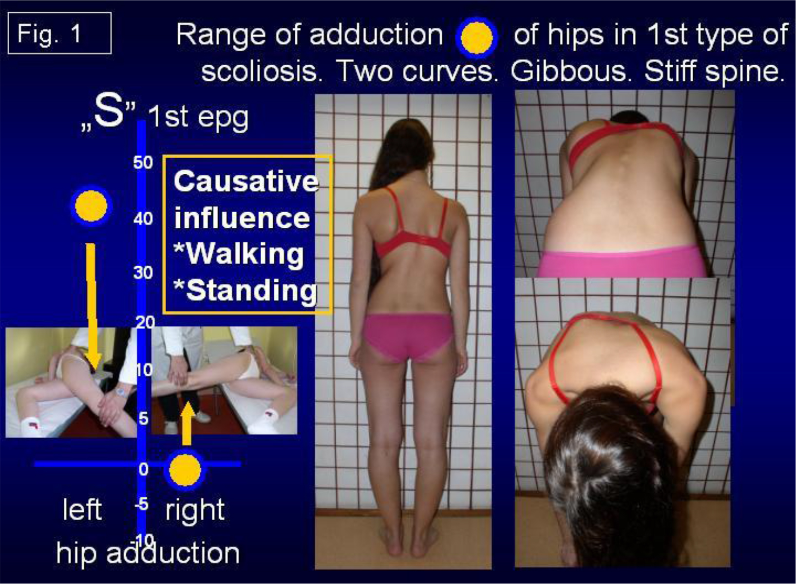
Scoliosis “S” 1st etiopathological group (epg) [Fig. 1]- double curve. Stiff spine (3D). Rib hump on the right side of the thorax. Connection with gait and standing „at ease’ on the right leg.

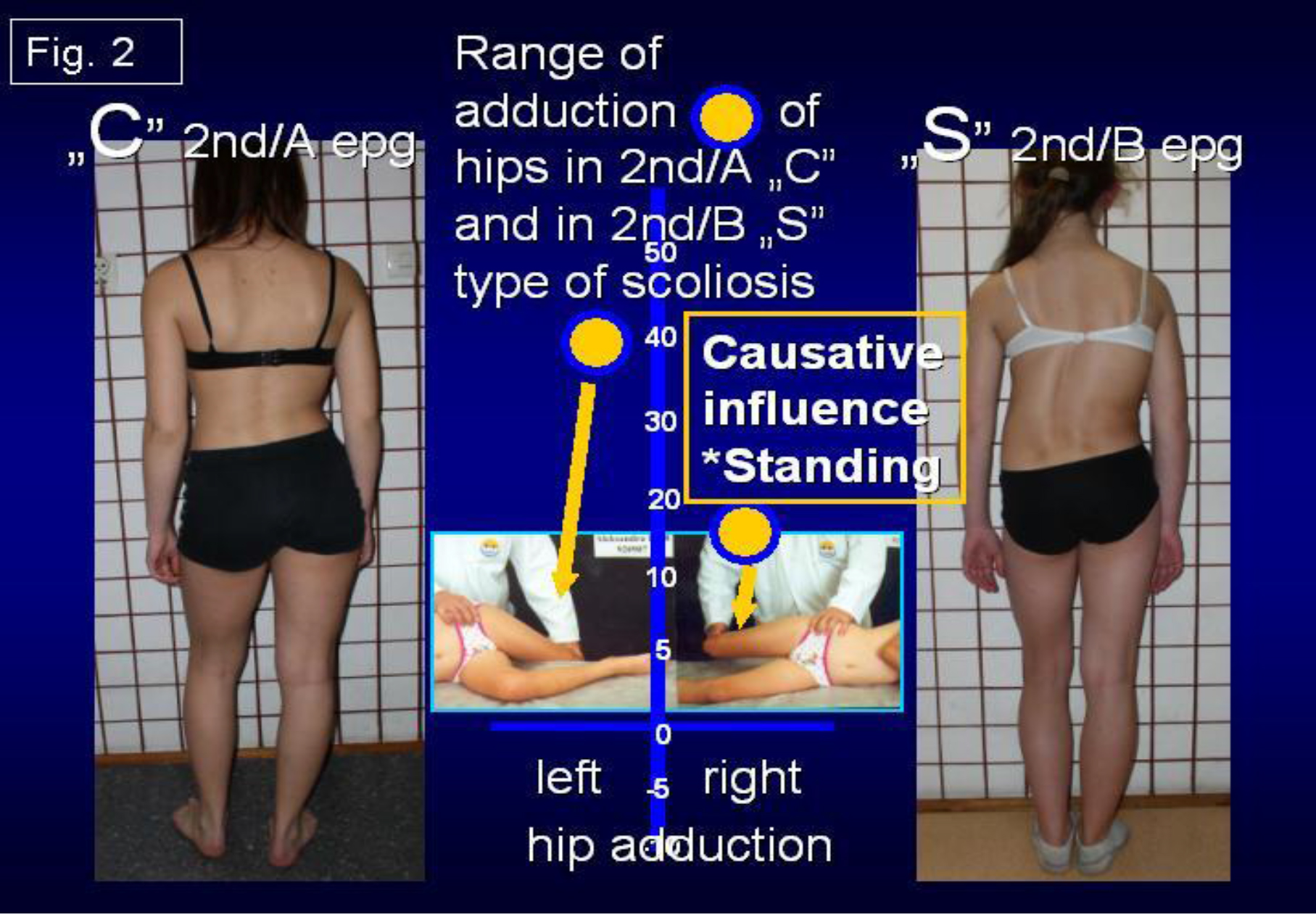
    1. Scoliosis “C” 2nd/A epg [Fig. 2] – one curve – lumbar left convex. Spine flexible (1D or 2D). Connection with standing „at ease’ on the right leg only.
    2. Scoliosis “S” 2nd/B epg [Fig. 2] – two curves, (2D or 3D). Connection with standing ‘at ease’ on right leg and additionally with laxity of joints or / and harmful previous exercises.

      In these second 2a and 2b types of scoliosis – the spine is flexible.

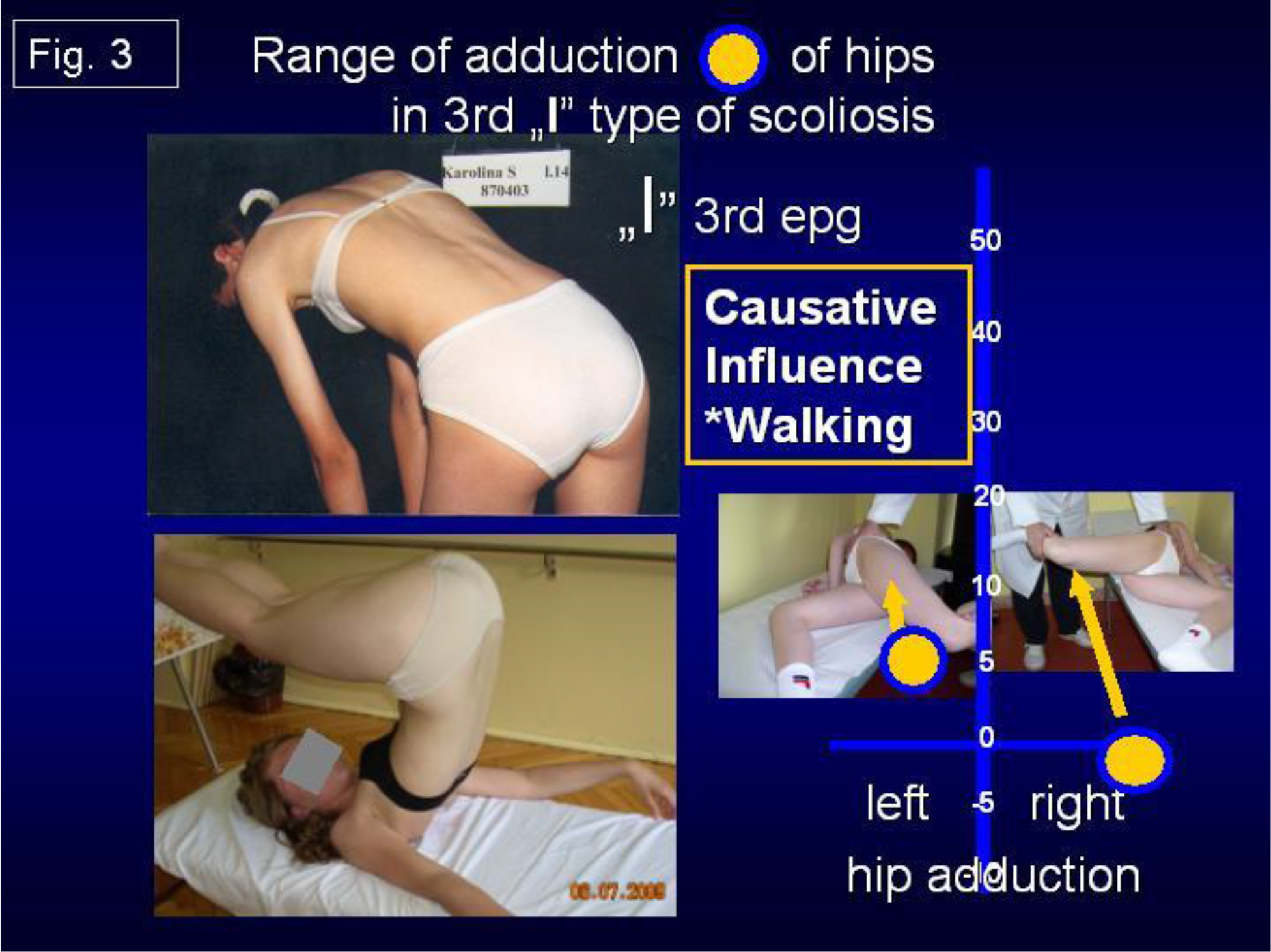
  1. Scoliosis “I” 3rd epg [Fig. 3]. Deformity has the form of a stiff spine, (2D or 3D). No curves or small ones. The cause is gait only. Such “spine deformity” was till 2004 never included / classified as “scoliosis”.

The Questions and the Authors Answers to the Problem of Scoliosis [1–28].

The etiology of idiopathic scoliosis was unsolved through many centuries and many questions are discussed in the world till now.

In research of etiology of the so-called idiopathic scoliosis everybody who will answer the question: what is “the etiology?” – must answer the all below presented questions about scoliosis or even more. Answering only some questions is not enough and is not the answer for cardinal question “what is the etiology”.

The Questions are following:

  1. The etiology – of course – the subject of whole discussion?
  2. Why girls have more frequent scoliosis?
  3. Why do we mostly observe the lumbar left convex curve?
  4. Why do we mostly observe the thoracic right convex curve?
  5. When there is one curve scoliosis and when there are two curves scoliosis?
  6. Why the rib hump (gibbous costalis) is on the right side?
  7. When does scoliosis start to develop – at what age?
  8. What kind of classification is proper? What is the patient’s type of scoliosis?
  9. Why there is a rapid progression of scoliosis in the period of accelerated growth during the childhood?
  10. Which type of scoliosis progresses?

    IJOT - 118_Karski T_F1

    Figure 1. Range of adduction of hips in 1st type of scoliosis. Two curves. Gibbous. Stiff spine. Causative influence: walking & standing.

    IJOT - 118_Karski T_F2

    Figure 2. Range of adduction of hips in 2nd/A „C” and in 2nd/B „S” type of scoliosis. Causative influence: standing.

    IJOT - 118_Karski T_F3

    Figure 3. Range of adduction of hips in 3rd „I” type of scoliosis. Causative influence: walking.

  11. Which type of scoliosis does not progress?
  12. Why blind children do not have scoliosis?
  13. Is there any influence of CNS in development of scoliosis? Direct influences? Indirect influences?
  14. What kind of therapy – conservative or operative should be used in treatment?
  15. Are extension and strengthening exercises correct?
  16. What kinds of rehabilitation exercises should be applied?
  17. Is corset (orthopedic devices) treatment proper – yes? no?
  18. Is causative prophylaxis possible?

My Answers and Comments:

For all these questions the only answer is “biomechanical etiology of so-called scoliosis”. This idea of etiology is presented in my lectures and articles in years 1995 – 2019 – also in Web Site www.ortopedia.karski.lublin.pl

I have given lectures during the IRSSD Meetings – in 2002 in Athens, in 2006 in Ghent, in 2008 in Liverpool, in 2012 in Poznań, in two SOSORT Congresses and on many others Congresses – e.g. during SICOT in Cairo, in Havana, in Istanbul, next in Germany, in Hungary, in Czech Republic, in Slovak Republic, in Hong Kong, in China / Beijing, in Finland e.g. .

The only answer to all the questions informs about the etiology of scoliosis! If somebody answers only partially to the question about etiology – the etiology presented by him is not correct.

Here, I present my answer to all the questions on the basis of “biomechanical influences”:

  1. The Etiology is biomechanical. Connected with asymmetry of anatomy of body of children (already in newborns and babies) and next connected with the asymmetry of movements in many joints – but – especially important – asymmetry in range of movement of hips. All these asymmetries are connected with “The Seven Contractures Syndrome” (“Siebener [Kontrakturen] Syndrom” – according to Prof. Hans Mau) [see Literature] or The Syndrome of Contractures and Deformities [SofCD, 2006] – T. Karski).

    Because of these “asymmetries” – it is *asymmetry of loading during gait, *asymmetry of time of standing on left / right leg – more on the right (!), *asymmetry in development and growth of spine, *in result scoliosis in three ethiopathological groups & four types.

  2. Why girls have more frequent scoliosis? Answer SofCD – appears mostly in girls. Girls are more sensible for forces during pregnancy (especially if the fetus has not enough space in uterus) and next such children / girls more frequently suffer from “Syndrome of Contractures and Deformities”.
  3. Why lumbar left convex curve? Answer: The SofCD is mostly “left sided” (90 % – 95 % of pregnancies – Prof. Jan Oleszczuk / Lublin and all gynecologists). Permanent standing on the right leg – is due “contracture” more stable (!) but makes lumbar left convex scoliosis / curve. A child told me – I stand on the right leg – because it is more comfortable, more stable, more easy, it doesn’t tire me, I like to stand on the right leg e.g.
  4. Why thoracic scoliosis / curve is right convex? Answer: The SofCD is mostly “left sided”. 90 % – 95 % of pregnancies are “left sided pregnancies” – Literature: Prof. Jan Oleszczuk / Lublin and all gynecologists on the world. Permanent standing on the right leg (this leg due “contracture” is more stable) makes lumbar left convex scoliosis / curve and secondary right convex thoracic curve in 2nd / B ethiopathological group (epg). Some cases in “S” 2nd / B epg scoliosis are “kifoscoliosis / kiphoscoliosis”. Important remark: in I-epg group both curves – lumbar left convex and thoracic right convex develop at the same time – look at the following points.
  5. Why there can be one curve or two curves in scoliosis? The answer is – one curve scoliosis “C” lumbar left convex deformity is in 2nd / A epg group connected with standing ‘at ease’ on the right leg. The scoliosis in form of “S” is in 1st epg group – spine is stiff and also formed in “S” form in 2nd / B epg group – spine is flexible. These both types of scoliosis are double curve deformities.
  6. Why the rib hump (gibbous costalis) is on the right side? The answer: The SofCD is mostly “left sided”. 90 % – 95% of pregnancies are “left sided pregnancies” – Literature: Prof. Jan Oleszczuk / Lublin and all gynecologists on the world. In “S” 1st epg group permanent standing on the right leg – this leg due to a “contracture” is more stable, it triggers the lumbar left convex scoliosis and secondary right convex scoliosis. Due to walking and permanent rotation distortion in inter-vertebral joints a rib hump develops on the right side. Important remark: such rotation distortion appears because of compensatory movement while the adduction and internal rotation movement in right the hip in 1st epg group of scoliosis is maximally limited. Rotation deformity and stiffness develop primarily. This rotation deformity develops in three stages: a/ flat back, b/ disappearance of processi spinosi, c/ in some cases lordotic deformity in thoracic part of spine. Some cases in “S” 1st epg scoliosis are “lordoscoliosis”.
  7. When scoliosis starts to develop – in which year of the life of the child ? Every type of scoliosis starts to develop when the child starts “standing” and “walking” – at the age of two.

    The “S” scoliosis in 1st epg can be observed very early – at the age of 4 – 6. The “C” 2nd/A epg scoliosis and “S” 2/B epg can be spotted at the age of 10 – 12. First it is only physiological deviation movement of the spine and – later “a fixed deformation” in the form of scoliosis.

  8. What kind of classification is proper? There are three groups and four types of scoliosis. All groups in this “new classification” are connected with “the model of hips movements” (T. Karski, 2006):

    1st epg –“S” scoliosis with stiffness of spine – some types of scoliosis in 1st epg are “lordotic scoliosis” – connection with gait and with permanent standing ‘at ease’ on right leg, 2nd/A “C” scoliosis – connection with permanent standing ‘ at ease’ on right leg, 2nd/B “S” scoliosis – connection with permanent standing ‘at ease’ on right leg – plus laxity of joints and wrong exercises (!). Some types of scoliosis in 2nd / B epg are “kypho-scoliosis”) 3rd epg “I” scoliosis – small curves or none, small gibbous or none – only stiffness of spine. The 3rd epg scoliosis is only connected with gait / walking.

    Here I repeat – every type of scoliosis starts developing when the child starts standing and walking.

  9. Why is there a rapid progression of scoliosis in the period of accelerated growth of the child? Answer: bones grow – even 10 cm – 15 cm per year in some children. Contracted soft tissue in the region of the right hip does not grow and its influence is bigger(!). Especially in children whose legs growth faster than the trunk.
  10. Which type of scoliosis progresses? The progression is in the “S” 1st epg scoliosis
  11. Which type of scoliosis does not progress? The 2nd/A epg, 2nd/B epg and 3rd epg type scoliosis are without progression. In scoliosis 3rd epg – patients notice the problem in the adult age – because of pain. Young people have problems doing sports – but almost nobody believes that the problem is connected with the stiffness of the spine in special type of scoliosis.
  12. Why blind children do not have scoliosis? The gait of blind children protects from scoliosis – they walk without lifting legs, instead every step is very careful. The walking model is totally different than in healthy children.
  13. Is there any influence of CNS in the development of scoliosis? Direct influences? Indirect influences? Yes – there are only indirect influences in children with Minimal Brain Dysfunction (MBD) or with Attention Deficit Hyperactivity Disorder (ADHA) – [according author – MBD and ADHD are equal]:
    1. extension contracture of the trunk in small children – because of spastic (semi spastic) contracture of trunk extensors,
    2. anterior tilt of pelvis – because of spastic (semi spastic) contracture of m. rectus both sides,
    3. “laxity” of joints – because of changed properties of collagen. In some cases laxity is the cause of progression of scoliosis. According to Prof. Harald Thom – 10 % of children with CP or MBD are with laxity (with Prof. Thom personal contacts in Heidelberg 1972 – 1973 during authors DAAD scholarship time and next in Rummelsberg / Nürnberg).
  14. What kind of therapy – conservative or operative should be applied in treatment? Answer: only conservative – in my material from years 1995 – 2009, only 13 % children need surgery and there were children previously treated by wrong, incorrect exercises / methods. In years 2010 – 2019 the number of children needing surgery in my material is maximally low – 3 % or less.
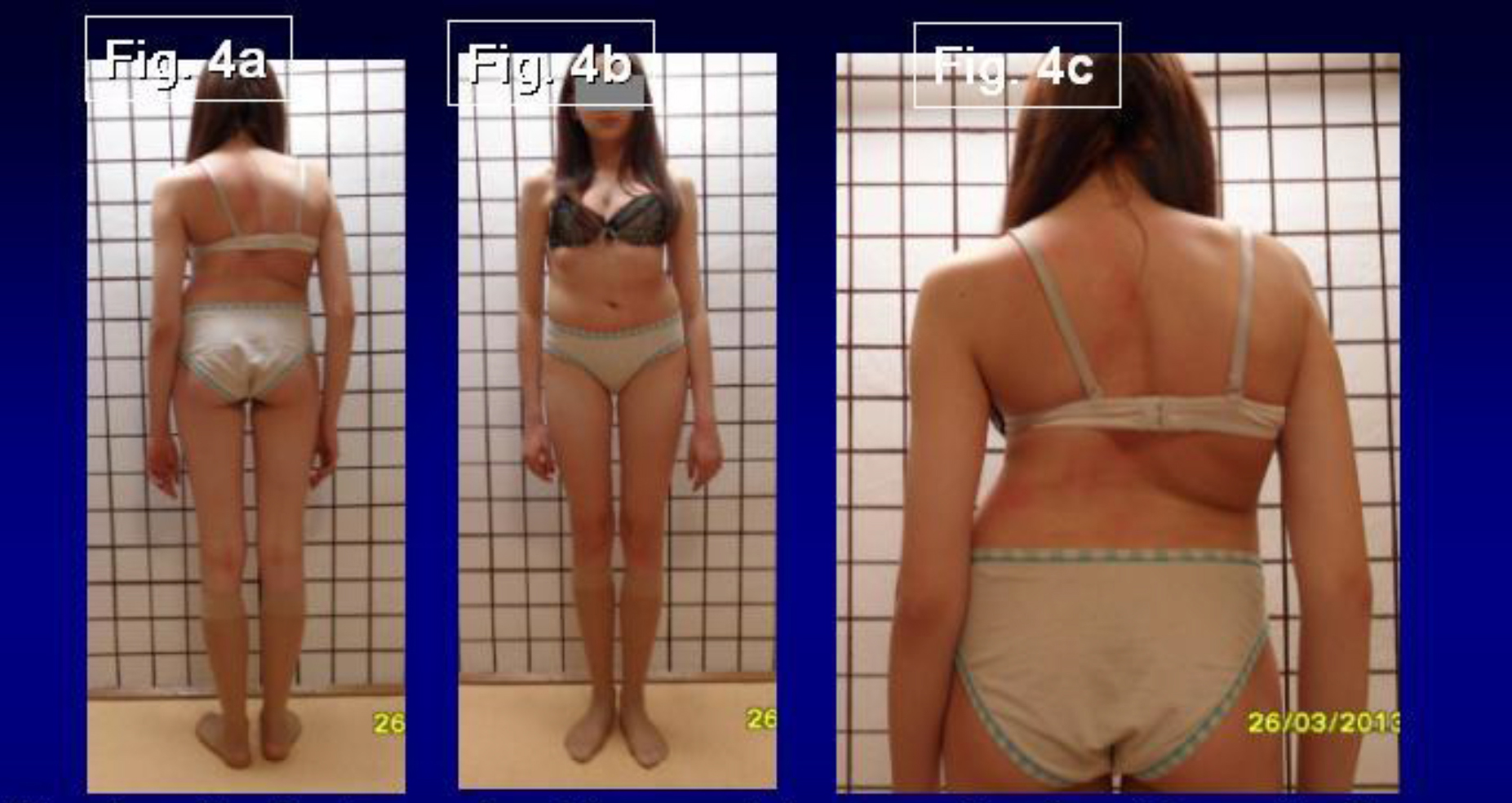
  15. Are extension exercises correct? [Fig. 4a, 4b, 4c] No – such exercises are wrong – they cause “iatrogenic deformity” – bigger curves, bigger rib hump, stiffer spine. Wrong exercises can change the direction of curves. They are the cause of extra – additional curves. They can be the cause of “three curves scoliosis”. Every doctor in every country should stop such wrong exercises.
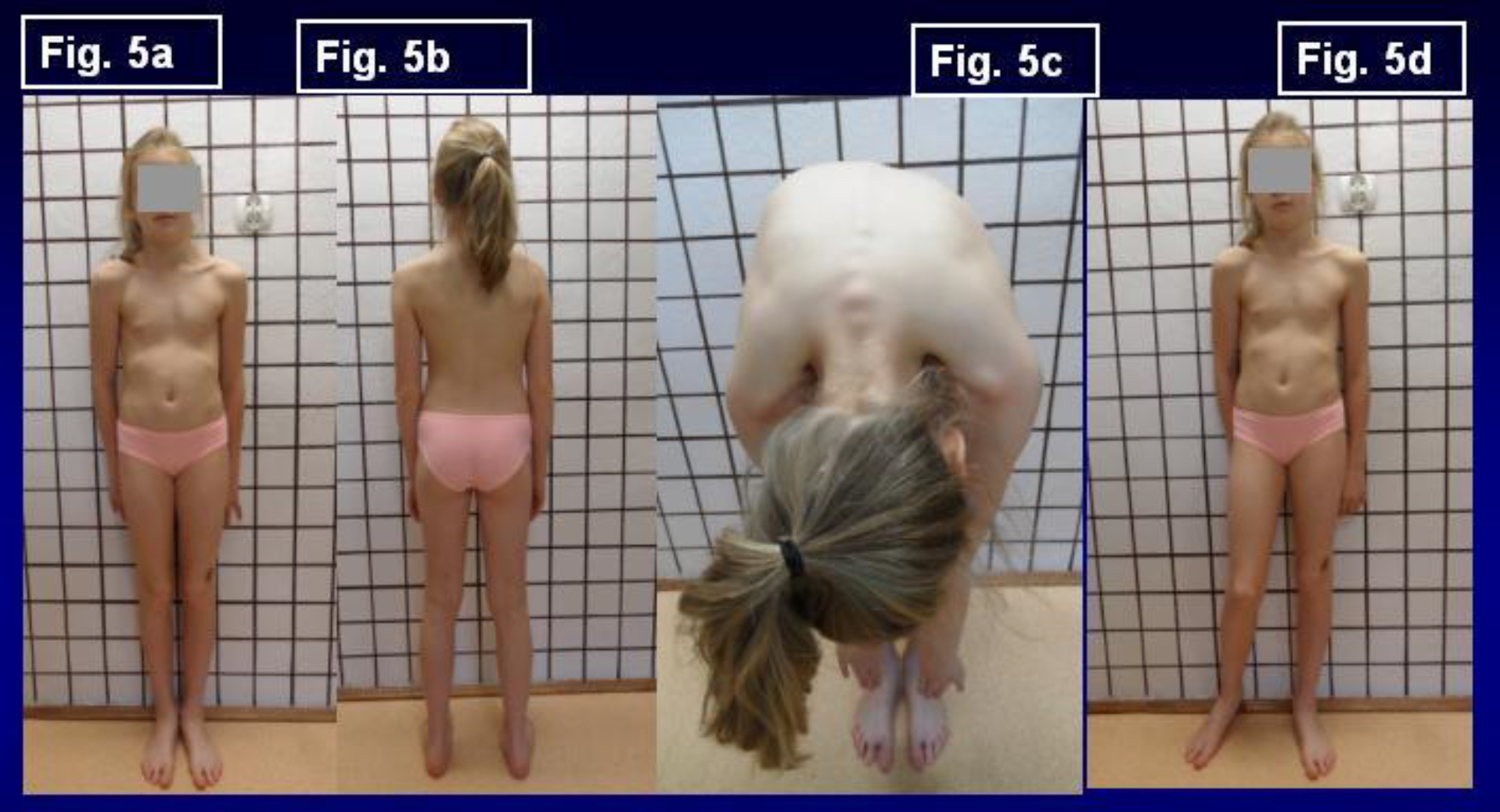
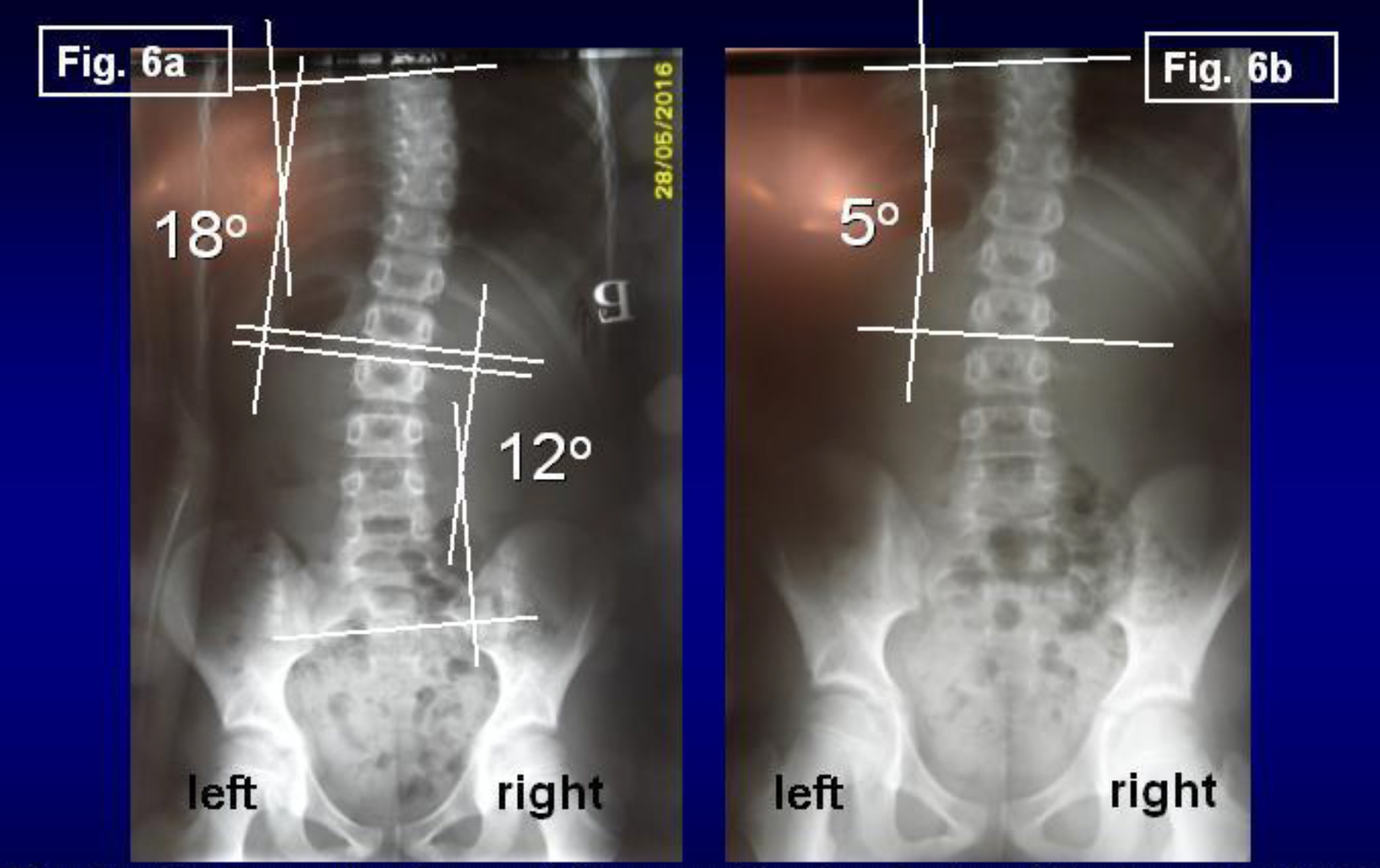
  16. What kinds of rehabilitation exercises should be applied? [Fig. 5a, 5b, 5c, 6a, 6b] Only – stretching exercises – giving symmetry of movements and next symmetry of growth and development of pelvis and spine. In the first place – we should receive / restore the “full symmetry of all movement of hips”, next symmetry of “function of trunk muscles”. For spine there are very important “bending / flexions exercises” even in very small children (age 3 – 4 years) such passive exercises are possible. When the child has full, symmetrical movements of both hips – it is no more asymmetry of loading of body left / right side during walking and no more “permanent standing ‘at ease’ on right leg” – but “symmetrical time of standing left / right leg”. In such situation scoliosis never develops.

    IJOT - 118_Karski T_F4 - Copy

    Figure 4. 4a, 4b, 4c. Example of incorrect therapy. Karolina 16 y. old. Four years incorrect therapy by improper / wrong exercises (2012 – 2016) in Kxyz and in Zxyz (Poland). As a result – iatrogenic big deformity. Consultation in Lublin on 1st June 2016. „S” scoliosis in 1st epg (etiopathological group). Two curves. Gibbous on the right side of thorax. Stiffness. Length of corpus 53 cm, of legs 92 cm. Start with new therapy. In such cases achieving good results is impossible. All children need causal prophylaxis.

    IJOT - 118_Karski T_F5 - Copy

    Figure 5. 5a, 5b, 5c, 5d. Example of correct therapy. Child Natalia in age 10 years. No 060712. Double curve scoliosis in 2nd/B type of deformity. Additionally rickets. Figure 5c show in Adams test slight rotation deformity. Axis of spine after therapy normal. Treatment in Lublin three years. Important – standing ‘at ease’ on the left leg (Fig 5d). See next figures with X-ray.

    IJOT - 118_Karski T_F6 - Copy

    Figure 6. 6a, 6b. Example of correct therapy. Karolina P. Age 10y. Born 12.07.2006. No 060712. Fig. 6a – before treatment in Lublin. Fig. 6b – after 3y. of new treatment. In therapy: 1/ standing ‘at ease’ on the left leg, 2/ every day flexion exercises forwards, left and right direction, 3/ sleeping in embryo position, 4/ karate.

  17. Corset (orthopedic devices) treatment – yes ? no ? I use the corset in 20 % of children in “S” 1st epg scoliosis and in 5% – 10% of children in “S” 2nd / B epg scoliosis.
  18. Is causative prophylaxis possible? Yes – and should be introduced in all countries. First, doctors should inform parents / patients that all previous methods of treatment – for example extensions, “muscles strengthening exercises” were incorrect and harmful. They only cause bigger curves and make the spine stiffer. Because of this, orthopedic surgeons in the past used to speak about “Natural History of Scoliosis” to explain to parents and treated children bad results of therapy.

Exercises leading to symmetry of movements and symmetry of function of hips and spine are important in prophylaxis and therapy. All exercises removing flexion contractures of both hips, removing abduction contracture or only “too small range of adduction” of the right hip, removing external rotation contracture of right hip and extension contracture of whole spine belong to these methods. We should remember that the rigidity of spine is very often the first step on the way to scoliosis in “S” 1st epg group of scoliosis.

Flexion exercises for spine should be introduced already in small children at the age of 3 – 5. Also is very important to inform parents or patients about position of standing – all children endangered with scoliosis should stand ‘at ease’ only on the left leg. Standing ‘at ease’ on the right leg is permanent and after some years (important – cumulative time of standing) plays a very important role in etiology of scoliosis. Standing on the left leg is not permanent and does not leading to scoliosis.

Conclusion

  1. In all years of observations (T. Karski, 1984 – 2018), the biomechanical etiology of the so-called idiopathic scoliosis was confirmed.
  2. Development of scoliosis and the types of spine deformity are connected with pathological “model of hips movements” (T. Karski, 2006) and function – “standing ‘at ease’ on the right leg” and “walking”.
  3. Restricted range of movements in the right hip is connected with the “Syndrome of Contractures and Deformities” according Prof. Hans Mau.
  4. Every type of scoliosis starts to develop at the age of 2–3.
  5. There are three groups and four types of scoliosis:

    S” scoliosis 1st epg, 3D. Causative influence: standing and gait,

    1. C” scoliosis 2nd / A epg, 1D. Causative influence: standing.
    2. S” scoliosis 2nd / B epg, 1D or 2D. Causative influence: standing, plus, – laxity of joints and/or incorrect exercises in previous therapy.
    3. I” scoliosis 3rd epg, 2D or 3D. Clinically only stiffness of the spine. Causative influence: gait. The clinical symptom of this deformity is – sport problems at a young age and “permanent pain” in adults.

The proper therapy of scoliosis – are only stretching exercises help to obtain full movements of the right hip, the proper position of the pelvis and full movement of the spine.

The causal prophylaxis of scoliosis is possible and should be introduced in every country.

The rules in prophylaxis – are – standing ‘at ease’ on the left leg, sitting relax, sleeping in embryo position, active participation in sport – especially in karate, taekwondo, aikido and other similarly.

Acknowledgement: I would like to express my many thanks to Honorata Menet for correction of the article.

References

  1. Burwell G, Dangerfield PH, Lowe T, Margulies J. Spine (2000) Etiology of Adolescent Idiopathic Scoliosis: Current Trends and Relevance to New Treatment Approaches 14: 324.
  2. Green NE, Griffin PP (1982) Hip dysplasia associated with abduction contracture of the contralateral hip. J Bone Joint Surg Am 64: 1273–1281. [crossref]
  3. Hensinger RN (1979) Congenital dislocation of the hip. Clin Symp 31: 1–31. [crossref]
  4. Howorth B (1977) The etiology of the congenital dislocation of the hip, Clin Orthop 29: 164–179.
  5. Karski T (2002) Etiology of the so-called “idiopathic scoliosis”. Biomechanical explanation of spine deformity. Two 272 groups of development of scoliosis. New rehabilitation treatment. Possibility of prophylactics, Studies in 273 Technology and Informatics Research into Spinal Deformities 91: 37–46.
  6. Karski T, Kalakucki J, Karski J (2006) “Syndrome of contractures” (according to Mau) with the abduction contracture of the right hip as causative factor for development of the so-called idiopathic scoliosis. Stud Health Technol Inform 123: 34–39. [crossref]
  7. Karski T (2010) Explanation of biomechanical etiology of the so-called idiopathic scoliosis (1995 – 2007). New 276 clinical and radiological classification” in”Pohybove Ustroji” [Locomotor System] vol. 17, 2010, No.1 + 2, pages: 277 26 – 42 (Czech Republic – 2010) 278.
  8. Karski T (2011) Biomechanical Etiology of The So-Called Idiopathic Scoliosis (1995 – 2007) – Connection with 279 “Syndrome of Contractures” – Fundamental Information for Paediatricians in Program of Early Prophylactics / 280 Journal of US-China Medical Science 8: 281
  9. Karski Tomasz (2010) Factores biomechanicos en la etiologia de las escoliosis dinominadas idiopaticas. Nueva 282 clasificacion. Nuevos test clinicos y nueavo tratamento conservador y profilaxis”, Cuestiones de Fisioterapia, Mayo- 283 Agosto 2010, volumen 39, Numero 2, paginas 85 – 152 [Cuest. Fisioter. 2010, 39 (2); 144 – 152] / Spain [2010] 284
  10. Karski Tomasz (2010) Biomechanical Etiology of the So-called Idiopathic Scoliosis (1995–2007). New Classification: 285 Three Groups, Four Sub-types. Connection with „Syndrome of Contractures”. Pan Arab J Orth Trauma 14: 287.
  11. Karski Tomasz (2013) Biomechanical Etiology of the So-called Idiopathic Scoliosis (1995 – 2007). Three Groups and 288 Four Types in the New Classification, Journal of Novel Physiotherapies, OMICS Publishing Group 289 6: 290.
  12. Karski Jacek, Tomasz Karski (2013) So-Called Idiopathic Scoliosis. Diagnosis. Tests Examples of Children Incorrect 291 Treated. New Therapy by Stretching Exercises and Results, Journal of Novel Physiotherapies, OMICS Publishing 292 Group, USA, 2013, 3–2, 9 pages 293.
  13. Karski Tomasz (2014) Biomechanical Aetiology of the So-Called Idiopathic Scoliosis. New Classification (1995 – 2007) in Connection with “Model of Hips Movements”. Pages 12. Global Journal of Medical Research H: Orthopedic and Musculoskeletal System 14.
  14. Karski Tomasz (2014) Biomechanical Etiology of the So-called Idiopathic Scoliosis (1995 – 2007) – Connection with„ Syndrome of Contractures” – Fundamental Information for Pediatricians in Program of Early Prophylactics. Surgical Science 5: 33–38.
  15. Karski Tomasz, Karski Jacek (2015) Syndrome of Contractures and Deformities” according to Prof. Hans Mau as Primary Cause of Hip, Neck, Shank and Spine Deformities in Babies, Youth and Adults American Research Journal of Medicine and Surgery 1.
  16. Karski Tomasz, Jacek Karski (2015) Biomechanical etiology of the so-called Idiopathic Scoliosis (1995 – 2007). Causative role of „gait” and „permanent standing ‘at ease’ pn the right leg”. New classification. Principles of new therapy and causal prophylaxis. Canadian Open Medical Science & Medicine Journal, Vol. 1: 1–16.
  17. Tomasz Karski, Jacek Karski (2016) Bóle krzyza – problem neurologiczno – ortopedyczny. Objawy. Przyczyny. Leczenie. Back pain – neurology-orthopedic problems. Clinic, causes, therapy and prophylaxis. Postepy Neurologii Praktycznej, Wydawnictwo Czelej. 4/2016, Str. 9 – 16.
  18. Karski Jacek, Karski Tomasz (2016) Imperfect hips” As a Problem at an Older Age. Early and Late Prophylactic Management before Arthrosis. Jacobs Journal of Physiotherapy and Exercises 015: 7.
  19. Karski Tomasz (2018) Biomechanical Aetiology of the So-called Adolescent Idiopathic Scoliosis (AIS). Lublin Classification (1995–2007). Causative Influences Connected with “Gait” and “Standing ‘at ease’ on the Right Leg”, Journal of Orthopaedics and Bone Research (USA), Scholarena, pages 10.
  20. Mau H (1979) Zur Ätiopathogenese von Skoliose, Hüftdysplasie und Schiefhals im Säuglinsalter. Zeitschrift f. 294 Orthop. 5, 601–5. 295.
  21. Mau H (1982) Die Atiopatogenese der Skoliose, Bücherei des Orthopäden, Band 33, Enke Verlag Stuttgart, 1 – 296 110 297.
  22. Mau Hans – personal information and letter.
  23. Normelly H (1985) Asymmetric rib growth as an aetiological factor in idiopathic scoliosis in adolescent girls, 298 Stockholm 1–103. 299.
  24. Stokes IAF (1999) Studies in Technology and Informatics, Research into Spinal Deformities 59: 1–385.
  25. Sevastik J, Diab K (1997) Studies in Technology and Informatics, Research into Spinal Deformities 37: 1–509. 301.
  26. Sevastik John – personal information.
  27. Thom Harald – personal information.
  28. www.ortopedia.karski.lublin.pl

The Novel ReNu Region of TAF12 Regulates Gcn5 Nucleosomal Acetylation

DOI: 10.31038/JMG.2019214

Abstract

The post-translational acetylation of the histone components of chromatin mediates numerous DNA-templated events, including transcriptional activation, DNA repair, and genomic replication. The conserved SAGA (Spt-Ada-Gcn5 Acetyltranferase) and SLIK (SAGA-Like) Histone Acetyltransferase (HAT) complexes are required for transcriptional activation of a subset of yeast genes and contain multiple subunits including the histone fold-containing TBP- Associated Factors (TAFs): 6, 9, 10, and 12. These TAFs are also components of the TFIID complex and are consequently involved in most RNA polymerase II-transcription in yeast. Here we identify a novel conserved region of TAF12, termed ReNu, outside of its histone fold, which is required for SAGA and SLIK-directed nucleosomal acetylation. We demonstrate that this region is not required for chromatin association, but show that this region plays an important role for histone H3 acetylation at specific SAGA and SLIK-regulated promoters. Our data suggests that the ReNu region of TAF12 regulates Gcn5 acetylation of specific substrates within the SAGA super-family of HAT complexes.

Introduction

The packaging of the eukaryotic genome into chromatin is generally believed to be refractive to DNA-templated processes such as transcriptional activation, DNA repair, and replication. However, chromatin-modifying complexes regulate these processes through histone modifications such as acetylation, methylation, ubiquitination, and phosphorylation [1]. Histone acetylation correlates with transcriptional capacity and is catalyzed by histone acetyltransferase (HAT) activities such as the yeast Spt-Ada-Gcn5-Acetyltransferase (SAGA) coactivator complex [2]. Both yeast and human SAGA complexes contain the HAT subunit Gcn5 (or the related PCAF protein), the Tra1, Ada, Spt, and a subset of TAF proteins [3–8]. The histone fold containing TAFs 6, 9, 10, and 12 are components of the SAGA, SAGA-like (SLIK/SALSA), and TFIID complexes in yeast, a feature conserved through evolution [9]. TFIID and SAGA are broadly required for transcriptional activation [10,11], however specific targeting of the TAF12 subunit of these complexes has recently been found to lead to a selective transcriptional response that interferes with acute myeloid leukemia [12]. Prior work had shown that TAF12 can bind to the activation regions of the Gal4, GCN4, VP16 and HNF4A transcription factors and that these interactions make differential contributions to transcriptional activation [13–18].

The observation that TAF12 is required for SAGA HAT activity on nucleosomal substrates is complicated by the fact that a taf12 temperature sensitive mutation in yeast compromises SAGA structural integrity and size with the observed loss of at least the SAGA Spt3 subunit [4]. Here we examine the direct role of TAF12 in SAGA and SLIK-mediated HAT activity on specific substrates by assaying complexes bearing various mutations in TAF12. Furthermore, we investigate the in vivo contributions of an evolutionarily conserved TAF12 region on nucleosome acetylation at specific SAGA and SLIK regulated promoters.

Materials and Methods

Yeast Strains and Point Mutation Generation

SAGA and SLIK were prepared from yeast strain YSB493 (MATa, ura3-52, leu2::PET56, trp1-1, his3-200, ade2, taf12-259::LEU2 {pRS314-TAF12}, YSB556(MATa, ura3-52, leu2::PET56, trp1-1, his3-200, ade2, taf12- 259::LEU2 {pZM279-taf12 322-539}), YSB557 (MATa, ura3-52, leu2::PET56, trp1-1, his3- 200, ade2, taf12-259::LEU2 {pZM280-taf12 374-539}) [19]  and YJR21-9 (taf12-9) [20].

TAF12 ReNu region point mutations were generated using the Quick Change Protocol (Stratagene) where pRS314-TAF12 served as a TAF12 template. Point mutation incorporated TAF12 plasmids were shuffled into yeast strain YSB452 (MATa, ura3-52, leu2::PET56, trp1-1, his3-200, ade2, taf12-259::LEU2 {pJA73-TAF12-URA3}) to generate strains YMT1 (MATa, ura3-52, leu2::PET56, trp1-1, his3-200, ade2, taf12-259::LEU2 {pRS314-taf12 N336A}), YMT2 (MATa, ura3-52, leu2::PET56, trp1-1, his3-200, ade2, taf12-259::LEU2 {pRS314-taf12 L344A}) and YMT3 (MATa, ura3-52, leu2::PET56, trp1-1, his3-200, ade2, taf12-259::LEU2 {pRS314-taf12 S342A}).

Purification of Mutant SAGA and SLIK Complexes

SAGA and SLIK complexes were partially purified by a scheme adapted from our earlier work [4]. In brief, 4 liters of yeast were grown to mid-log phase at 30ºC. Yeast whole cell extracts were prepared by glass bead disruption [4]. Extracts were bound batch wise with 15ml of Ni2+-nitrilotriacetic acid (NTA) agarose (Qiagen). The resin was washed in a column with 20mM imidazole followed by elution with 300mM imidazole. The Ni2+-NTA agarose eluate was directly loaded onto a MonoQ HR 5/5 column (GE Healthcare). Bound proteins were eluted with a 25ml linear gradient from 100 to 500 mM NaCl. Peak SAGA fractions were pooled and concentrated down to 0.7ml with a Centriprep-30 concentrator (Millipore). Samples were then loaded on a Superose 6 H/R 10/30 column (GE Healthcare) equilibrated with 350mM NaCl. Peak fractions were used in subsequent assays.

Western Blotting and HAT Assays

Proteins from column fractionations were separated by 12% SDS-PAGE, transferred to nitrocellulose, and processed for immunoblotting using the antisera described. HAT assays were performed in the presence of 2 mg HeLa nucleosomes or core histones, 0.25 µCi of [3H]acetyl coenzyme A, and equivalent amounts of purified SAGA complexes, as described [4].

Chromatin Immunoprecipitation Assays

ChIP assays were performed largely as described [21].  Yeast strains YSB493 and YSB557 were grown in Synthetic Complete (SC) medium containing 2% dextrose to an OD 600 of 1.0. The yeast were reseeded in SC dextrose-medium, SC medium containing 2% galactose, or SC medium containing 2% KAc and then grown for an additional 4 hr. Antiserum specific for histone H3 acetylated at lysine 9 (Millipore) was used for ChIP. Input and precipitated DNA was PCR amplified for regions upstream of the CIT2, GAL10, RPL9A, and RPS5, as described in Pray-Grant MG et al [7]. Real-time PCR experiments were performed with three biological replicates and conducted with a SYBR Green/Taq polymerase reaction mix (Bio-Rad) in a MyIQ real-time PCR detection system (Bio-Rad). Data were collected and analyzed with thermal cycler software (Bio-Rad).

Results and Discussion

The Novel TAF12 ReNu Region Regulates SAGA and SLIK Nucleosomal Acetylation

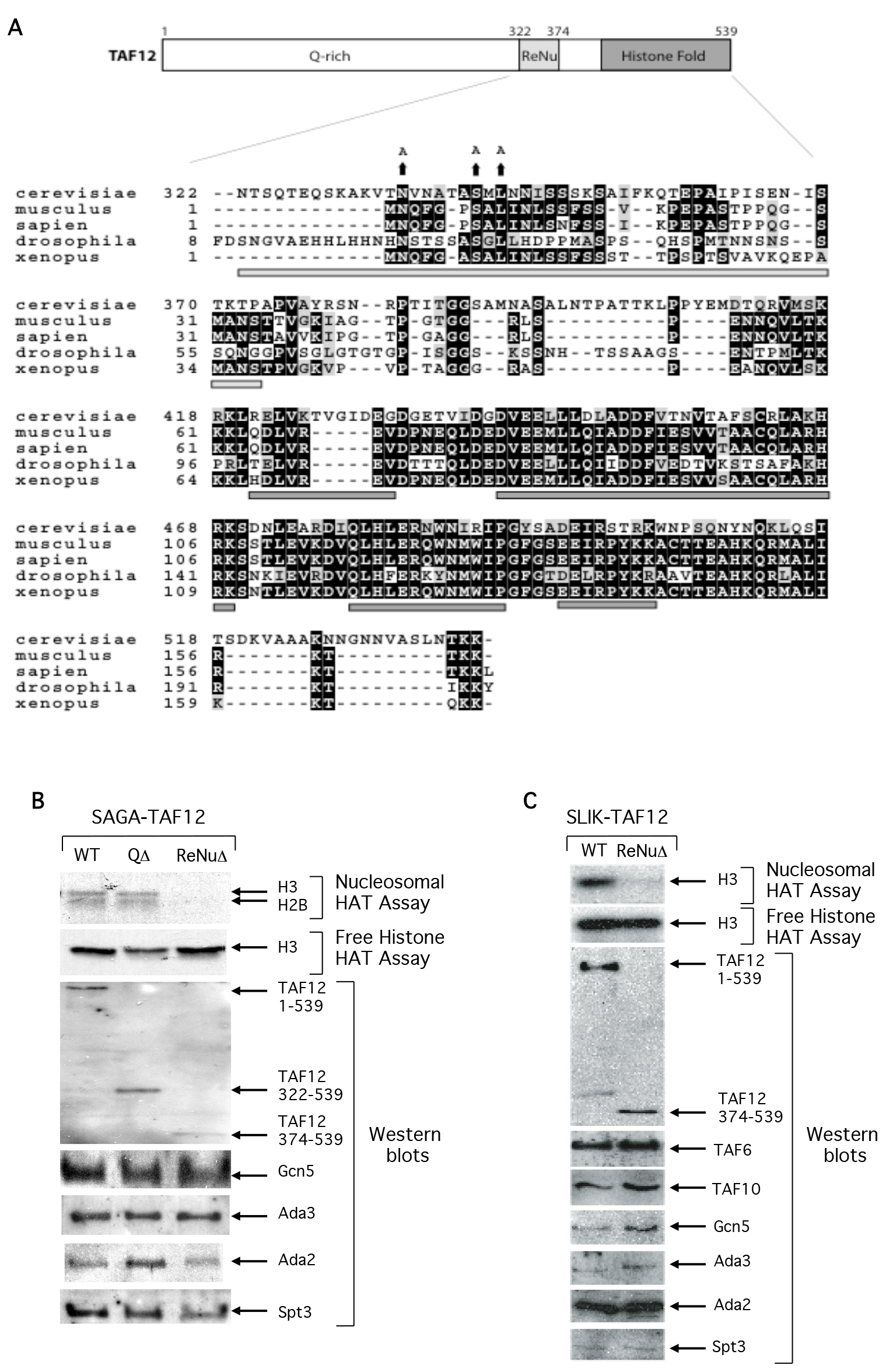
Primary sequence analysis of yeast TAF12 reveals that the protein consists of three regions: an amino-terminal glutamine rich domain, followed by a novel-conserved region upstream of the TAF12 carboxy-terminal histone fold domain (Figure 1a). We examined the structural and functional roles of these domains by assaying SAGA complexes containing full length TAF12 (WT), a TAF12 mutant protein deleted of amino acids 1-322 spanning the non-conserved glutamine rich domain (QΔ,), and a TAF12 mutant deleted of both the glutamine rich domain and an additional 52 amino acids (QΔ + ReNuΔ), hereafter named ReNuΔ. Western analysis using antisera against multiple SAGA subunits reveal that the structural integrity of the partially purified SAGA complex was intact in both of the TAF12 truncation protein strains, and interestingly, Spt3 was identified as a component of SAGA in all complexes (Figure1b). This could be because the two histone folds of Spt3 may be free to interact with the remaining histone fold of TAF12 in either complex [22]. Ada2, Ada3, and Gcn5 also associate with the TAF12- truncated SAGA complexes (Figure 1b) and are important for SAGA HAT function [23–25]. We assayed WT, QΔ, and ReNuΔ SAGA for HAT activity on specific histone substrates. We found that deletion of the glutamine rich domain of TAF12 did not interfere with SAGA HAT activity on free histones or nucleosomes (Figure 1b). However, deletion of both the TAF12 glutamine rich domain and the additional 52 C-terminal amino acids abolished nucleosomal acetylation, while there was no effect on SAGA HAT activity on free histone substrates (Figure1b). This result suggests that within the SAGA complex, this novel TAF12 region regulates Gcn5 substrate specificity, and we have consequently named it the ReNu (Required for Nucleosomal acetylation) region. Consistent with this, invariant amino acids within the ReNu region contribute to SAGA nucleosomal acetylation (Figure 4b).

Torok_JC3

Figure 1. The TAF12 ReNu domain is required for SAGA-dependent nucleosome acetylation.

(A) Primary sequences of the Saccharomyces cerevisiae, Mus musculus, Homo sapien, Drosophila melanogaster, and Xenopus laevis TAF12 homologs were aligned in ClustalW and graphically represented with the BoxShade program. Conserved residues are shown in bold and similar residues are shaded. The regions encompassing the minimal ReNu region and four histone-fold α-helices, but excluding the yeast non-conserved glutamine rich region, are represented by bars underneath the alignment in light and dark grey respectively. The changed sequence of invariant amino acids in the TAF12 yeast mutants is shown above the corresponding wild-type residues. (B) HAT assays and Western blot analysis of partially purified SAGA complexes. SAGA was prepared from the yeast strains YSB493, expressing full length TAF12 (WT), YSB556, yeast deleted of the TAF12 glutamine rich N-terminus (QΔ), and YSB557, yeast deleted of the TAF12 glutamine rich and ReNu regions (ReNuΔ). HAT assays were performed with free histone or nucleosome substrates and Western analysis was performed with the indicated antisera. (C). HAT assays and Western blot analysis of partially purified SLIK complexes. SLIK was prepared from the yeast strains YSB493 and YSB557. HAT assays were performed with free histone or nucleosome substrates and Western analysis was performed with the indicated antisera.

The related SLIK complex also contains both Gcn5 and TAF12 [7,26]. Therefore, we examined the contribution of the ReNu region in SLIK-mediated nucleosomal acetylation. We found that the structural integrity of partially purified SLIK was intact in both WT and ReNuΔ complexes and that Spt3 associates with ReNuΔ SLIK (Figure 1c). Similar to ReNuΔ SAGA, ReNuΔ SLIK was found to acetylate free histones at a level comparable to WT levels, but was unable to acetylate nucleosomes (Figure 1c).

These data establish a nucleosome acetylation regulatory role for the TAF12 ReNu region within the SAGA and SLIK HAT complexes and help explain the acquired ability of Gcn5 to acetylate nucleosomal histones when present in native complexes [7,8,27]. This may be achieved by directly interacting with the core catalytic components of the complex, Gcn5, Ada2 or Ada3 [23,24] or through histone tail presentation in the context of the nucleosome. Notably, Ada3 is a key component in regulating Gcn5’s nucleosomal acetyltransferase activity, but is still insufficient to drive native SAGA dependent nucleosomal acetylation in the absence of functional TAF12.

Yeast Strains Lacking the TAF12 ReNu Region Exhibit a Modest gcn5 Phenotype

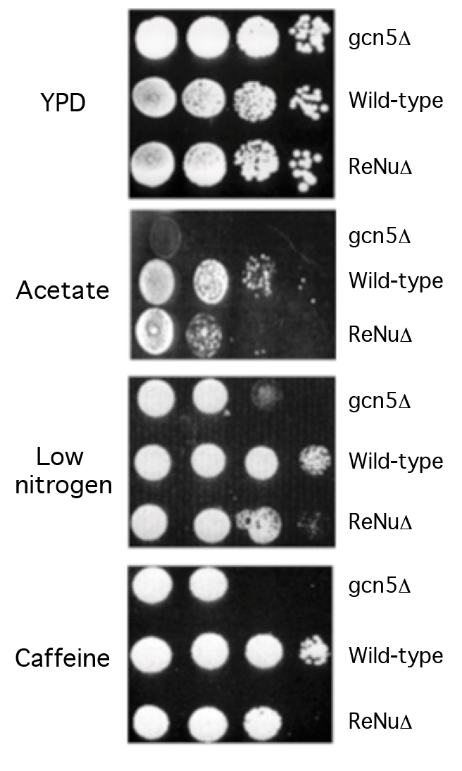
We examined the role of the ReNu region in genetic assays, hypothesizing that yeast lacking the TAF12 ReNu region may display a gcn5 phenotype, due to the loss of SAGA nucleosomal acetylation. We assayed wild-type and ReNu deleted yeast strains for growth defects on media containing low nitrogen, potassium acetate as a carbon source, and caffeine [7,8]. Serial dilutions of a ReNu wild-type strain, a ReNu-deleted yeast strain and a gcn5 strain were compared. We found that in these media, yeast bearing a deletion of the ReNu region exhibit a modest growth defect, not as severe as that of a gcn5 strain (Figure 2). All strains showed equivalent growth on the rich media YPD (Figure 2). Presumably, the observed intermediate gcn5 phenotype may be because TAF12 is not present in all yeast Gcn5 containing complexes [28]. Therefore, the observed modest phenotype might be a result of compensation of SAGA/SLIK acetyltransferase activity by other Gcn5 containing complexes, such as ADA and HAT-A2.

Torok_JC3

Figure 2.The TAF12 ReNu domain mediates Gcn5 function in vivo.

Wild-type, gcn5Δ and TAF12 ReNuΔ yeast strains were assayed for their ability to grow on Yeast extract-Peptone-Dextrose (YPD) media, synthetic complete media with 2% Potassium Acetate as a carbon source (Acetate), synthetic complete media supplemented with 12mM caffeine, and synthetic complete media lacking ammonium sulphate (low nitrogen). Shown are agar plates with ten-fold serial dilutions of each yeast strain, from left to right.

The TAF12 ReNu Region is not Required for SAGA Substrate Associations

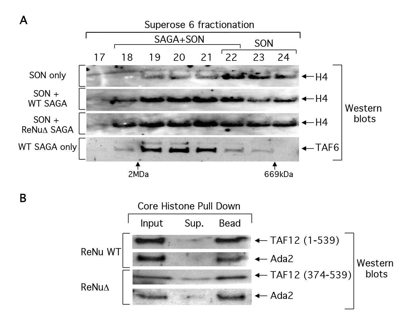
The observed loss of nucleosomal acetylation in ReNuΔ SAGA and SLIK could be due to a loss of chromatin association. We examined this possibility by binding ReNu WT or ReNuΔ SAGA complexes to SONs (short, 3–6, oligonucleosomes) and then fractionating these reactions by size exclusion chromatography. Fractions were processed for western analysis and immunoblotted with histone H4 antisera in order to detect the presence of SONs in any given fraction. We hypothesized that unbound SONs should fractionate at later fractions than those SONs bound to SAGA because the SAGA-SON interactions would shift SONs on a size exclusion column to an earlier fraction, corresponding to a higher molecular mass. As predicted, unbound SONs fractionated with an apparent mass of 0.6–1.0 MDa (fraction 22–24). Both ReNu WT and ReNuΔ SAGA shifted SONs towards the 2MDa column exclusion volume, fraction 18/19, which correlates to a mass at or above that predicted for SAGA, 1.8 MDa (Figure 3A). This data suggests that the ReNuΔ SAGA complexes retain the ability to associate with nucleosomes and furthermore, this association is salt stable in 350mM NaCl buffer.

Torok_JC3

Figure 3. ReNu deleted SAGA interacts with nucleosomes and core histones.

(A) Partially purified wild-type (WT) and ReNuΔ SAGA complexes were incubated with short oligonucleosomes (SON) and then fractionated over a Superose 6 size exclusion column. Fractions were processed for western blotting and the presence of SON were detected with histone H4 antiserum, and the elution of SAGA was monitored with TAF6 antiserum. Elution of the dextran blue (2MDa) and thyroglobulin (669kDa) molecular mass markers from the same column is indicated. (B) WT SAGA and ReNuΔ SAGA were bound to immobilized core histones. Inputs, supernatants (sup), and bound fractions (bead) from the reactions were analyzed by western blotting with the indicated antisera to SAGA components

We further assayed whether or not immobilized core histones could pulldown SAGA in standard binding assays. We bound WT SAGA and ReNuΔ SAGA to core histone agarose resin and analyzed the bound fractions with antisera against SAGA subunits. Similar to our SAGA- nucleosome interaction data, we observed that ReNuΔ SAGA can interact efficiently with core histones (Figure 3b) or double stranded DNA (data not shown). Taken together, our data demonstrate that ReNuΔ SAGA retains nucleosomal and core histone substrate interactions and that the loss of nucleosomal acetylation by ReNuΔ SAGA is likely not due to a loss in substrate association.

Invariant Amino Acids within the ReNu Region Contribute to SAGA Nucleosomal Acetylation

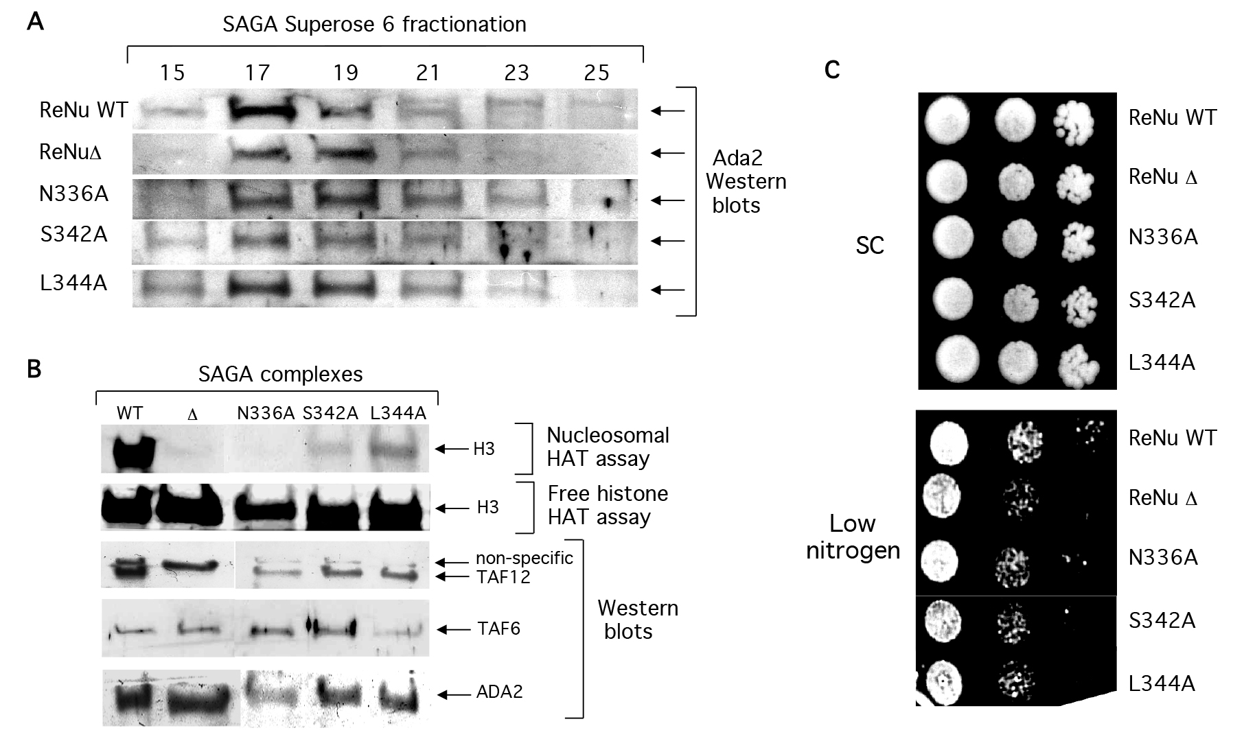
SAGA complexes were purified from yeast bearing point mutations in invariant amino acids: N336A, S342A, and L344A (Figures 4a and 4b), as described [7,27]. Western analysis indicates that similar to ReNuΔ SAGA, point mutant SAGA complexes retained their structural integrity and predicted native size of a 1.8 Mda (Figure 4a). Also similar to ReNuΔ SAGA, the three point mutants had little effect on SAGA’s free histone acetylation (Figure 4b). However, each significantly reduced nucleosomal acetylation by SAGA (Figure 4b) albeit not as completely as ReNuΔ. Additionally, ReNu point mutant yeast were serially diluted on low nitrogen and found to give an intermediate phenotype between our ReNu WT and ReNuΔ yeast (Figure 4c). Together, these findings indicate that each of the three invariant residues within the ReNu region play an important role in SAGA-mediated nucleosomal acetylation.

Torok_JC3

Figure 4. ReNu region point mutations compromise SAGA catalytic function, but not structural integrity.

(A) Western blot analysis of partially purified WT ReNu, ReNuΔ, or ReNu point mutant SAGA complexes. SAGA was prepared from the yeast strains YSB493, YSB557, YMT1 (taf12 N336A), YMT2 (taf12 L344A), and YMT3 (taf12 S342A). Yeast extracts were sequentially purified by nickel-NTA, anion exchange, and size exclusion chromatography. SAGA fractionated in superose 6 fractions 17–19 as determined by Ada2 immunoblotting, in an approximately 1.8 MDa complex. (B) SAGA complexes were similalrly partially purified from the yeast strains and peak fractions were incubated in HAT assays with nucleosome and free histone substrates. Western blotting from the same fractions was performed with the indicated antisera. (C) ReNu region point mutant yeast N336A, S342A, and L3444A were serially diluted on low nitrogen and found to give an intermediate phenotype relative to ReNu WT and ReNuΔ yeast.

The TAF12 ReNu Region is Essential for Promoter Acetylation

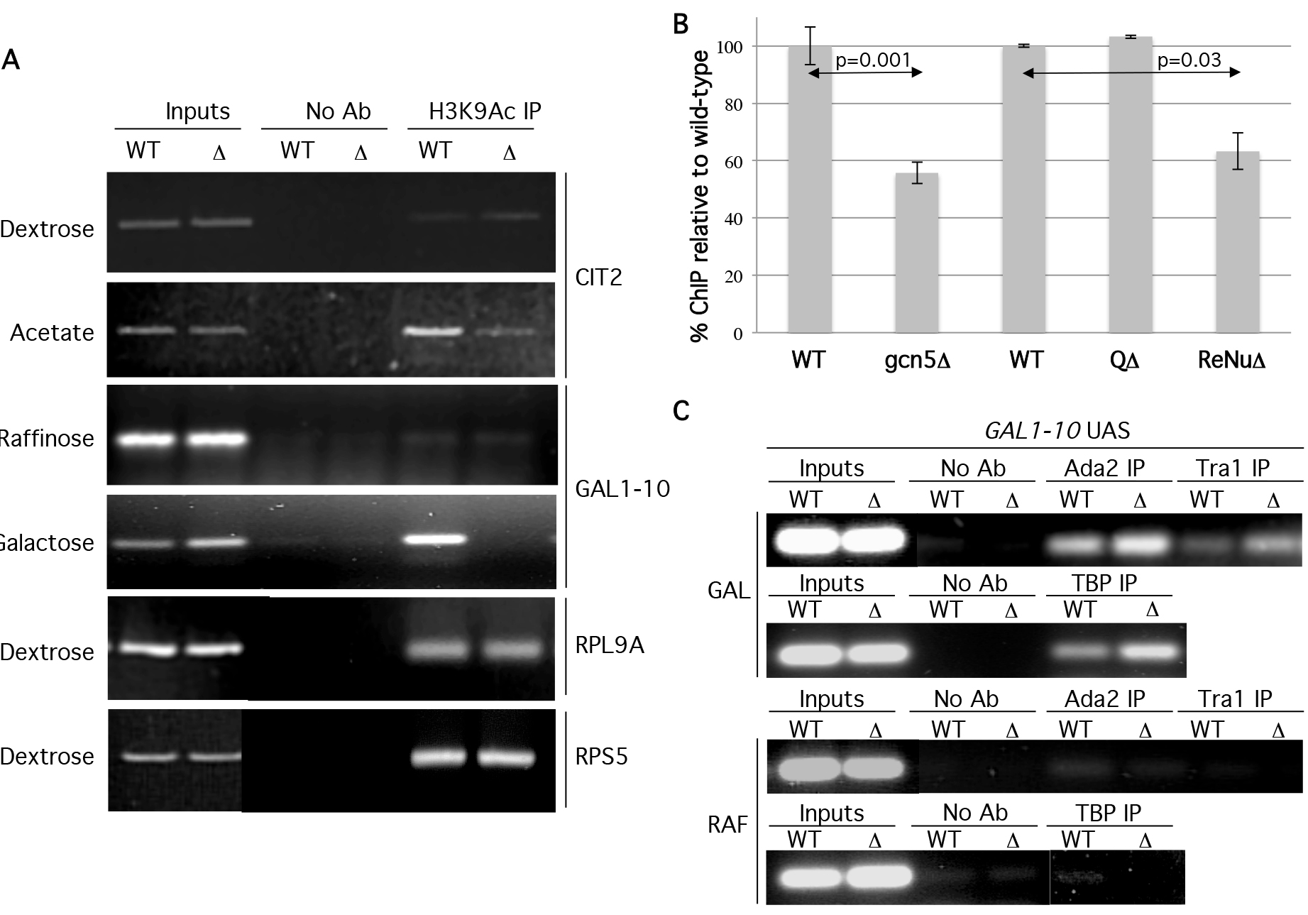
To further investigate the in vivo role of the TAF12 ReNu region in nucleosomal acetylation and to validate our in vitro observations, we examined specific promoter acetylation in wild type ReNu and ReNu region deleted yeast strains. Therefore, we conducted Chromatin Immunoprecipitation (ChIP) assays to assess the acetylation status of SAGA and SLIK-regulated promoters GAL1 and CIT2 [7,29–32]. Under inducing conditions, the GAL1-10 UAS is occupied by the SAGA and SLIK complexes [7,32–35], SAGA/SLIK is required for GAL1 and GAL10 mRNA production [36,37], and CIT2 expression is regulated by SLIK [38]. ChIP assays were performed using antisera against H3K9Ac (acetylated lysine 9 of histone H3) because this modification is implicated as being dependent on Gcn5 [39,40]. Upon activation, WT ReNu yeast show increased H3 K9Ac signal at both the GAL1-10 and CIT2 UAS. However, we found a striking absence of H3K9Ac signal at GAL1-10 and CIT2 UAS in ReNuΔ yeast (Figure 5a). These findings are in agreement with our in vitro data showing a loss of nucleosomal acetylation in ReNuΔ SAGA and SLIK. Interestingly, H3K9Ac ChIPs for the RPS5 and RPL9A promoters, both described as TFIID-regulated genes [36], showed no loss of acetylation in the ReNuΔ yeast (Figure 5a). These results suggest that the TAF12 ReNu region is required for H3K9Ac at the SAGA and SLIK- regulated genes GAL1-10 and CIT2, but not at the TFIID-regulated genes RPS5 and RPL9A. To quantify the changes in acetylation observed in TAF12 mutant strains we performed real-time PCR for the GAL-10 UAS under inducing conditions. We performed ChIP in gcn5Δ, or TAF12 mutant strains for H3K9Ac relative to wild-type strains. These experiments revealed a reduction in acetylation to approximately 55% in gcn5Δ (p=0.003) and 63% in ReNuΔ strains (p=0.01), while deletion of the Q-rich region caused no reduction in acetylation (Figure 5b).

Torok_JC3

Figure 5. The ReNu region is required for H3K9Ac at SAGA and SLIK regulated UAS.

(A) ChIP assays were performed from wild type (WT) or ReNu-deleted (Δ) yeast under noninducing dextrose (Dex) or raffinose (Raff) and inducing potassium acetate (KAc) and galactose (Gal) conditions, respectively, with antisera against H3K9Ac. Precipitated DNA was amplified by PCR using primers for the gene loci indicated. A control IP was performed without antibody (no Ab). (B) Down regulation of H3K9 acetylation at the GAL-10 UAS in gcn5Δ and ReNuΔ strains under inducing (Gal) conditions was quantified using Real-time PCR. Error bars represent standard errors and p-values were derived from two-tailed Student t tests. (C). ChIP assays were performed under noninducing (RAF) or inducing (GAL) conditions with antisera against Tra1, Ada2, and TBP. Precipitated DNA was amplified by PCR using primers for the GAL-10 UAS.

The loss of H3K9Ac at the GAL1-10 and CIT2 UAS in ReNuΔ yeast could be caused by an inability of SAGA and SLIK to interact with the promoter-chromatin environment. SAGA and SLIK components are recruited to the GAL1-10 UAS during activation [7,32–35].  We tested this possibility by using antisera against SAGA and SLIK components Ada2 and Tra1 in ChIP assays at the GAL1-10 UAS. As expected, we found Ada2 and Tra1 were recruited to the GAL-10 UAS in ReNu WT yeast under inducing conidtions. When we examined ReNuΔ yeast, we not only found that we could ChIP ReNuΔ SAGA/SLIK to the GAL1-10 UAS, but that there was a modest enrichment of Ada2 and Tra1 to the GAL1-10 locus (Figure 5c). These data, taken together with our findings that ReNuΔ SAGA interacts with nucleosomes, (Figure 3a) suggest that the observed loss of H3K9Ac at GAL1 is not caused by an inability of SAGA or SLIK to interact with the GAL1 UAS in vivo. Furthermore, our studies agree with findings [34] which demonstrate that SAGA is recruited to the GAL1 UAS in the absence of the Gcn5, which is equivalent to the loss of SAGA-mediated nucleosomal acetylation found in ReNuΔ complexes.

SAGA possesses multiple activities besides its nucleosomal acetylation function such as TATA binding protein (TBP) recruitment via Spt3 and Spt8 [32,41–43], acidic activator association [32,33,44–47], deubiquitination of histone H2B [35,48], and methyl H3K4 binding [26,49]. We examined a SAGA function that is independent of its nucleosome HAT activity in order to determine whether or not ReNuΔ SAGA and SLIK were compromised in other complex functions. We examined SAGA and SLIK’s ability to recruit TBP to the GAL1 UAS [30,36,50,51] by ChIP using an antiserum against TBP in WT and ReNuΔ yeast. Our data reveal that both WT and ReNuΔ SAGA/SLIK recruit TBP to the GAL1 locus, and interestingly, TBP is modestly enriched at this locus in the ReNuΔ strain (Figure 5b). This could be explained through the enhanced GAL1 UAS residency of the SAGA and SLIK complexes also found in the ReNuΔ yeast. Furthermore, a similar result shows that a taf12 ts strain shows a slight enrichment of TBP at GAL1 and that TBP can still be recruited to the GAL1 UAS in gcn5Δ yeast, which lack Gcn5-mediated nucleosomal acetylation [36].

ReNuΔ yeast are capable of growing in galactose because the GAL1 gene is still transcribed in the absence of Gcn5 [42,50]. In addition, the loss of nucleosomal acetylation at the GAL1 and CIT2 UAS is likely not caused by compromised HAT complex recruitment via acidic activators because studies have shown that the acidic activator GAL4 can bind the GAL1 UAS in the absence of the SAGA subunits Spt3, Spt20, or Gcn5 [42]. This suggests that the acetylation function or structural integrity of SAGA is not required for GAL4 association with the GAL1 promoter. Also, Rtg3, the acidic activator required for CIT2 expression has been shown to interact with the CIT2 UAS independently of SAGA or SLIK [52] and Rtg3 protein levels are comparable between WT, gcn5Δ, or ada2Δ yeast strains [53].

The SAGA/SLIK component Spt3 recruits TBP [32,42,43] and TAF12 is required for Spt3 association with the SAGA complex [4]. Here we show that Spt3 is retained in ReNuΔ SAGA, but there is a loss of nucleosomal acetylation, indicating that the effects we observe are relevant to TAF12 rather than Spt3 function. The increased occupancy of mutant SAGA at target promoters seen in our ChIP assays may indicate that substrate acetylation is required for dissociation of the SAGA complex. Furthermore, TBP is also recruited at GAL1-10 at higher levels in ReNuΔ yeast. One possible explanation for this is that Mot1 may not be able to remove TBP from the GAL1-10 UAS in ReNuΔ yeast. It has been reported that Mot1 occupancy at the GAL1 promoter is greatly reduced in a gcn5 yeast strain [54].

Conclusion

Our results describe a novel regulatory function for a TAF protein, which is mediated by a conserved disordered region outside of the histone fold domain. The in vitro nucleosomal acetylation defects observed with ReNu-lacking SAGA complexes agree with the in vivo loss of acetylation at SAGA and SLIK-regulated promoters. Complexes bearing wild-type or truncated TAF12 are capable of interacting with DNA, histones and nucleosomes in in vitro binding studies, indicating that the ReNu region is not necessary for substrate interaction.

Together these data suggest that TAF12 mediates a regulatory mechanism for modulating Gcn5 HAT activity in SAGA and SLIK. This may be achieved by directly interacting with the core catalytic components of the complex, Gcn5, Ada2 or Ada3 [23,24] or through histone tail presentation in the context of the nucleosome. These findings may also implicate a putative mechanism by which HAT complexes are activated, which would likely be conserved in all other members of the SAGA HAT superfamily: SLIK, PCAF, STAGA, and TFTC. It is noteworthy that some of the conserved amino acids in the ReNu region have been described as sites for post-translational modification, including serines 342, 348 and 352 in S.cerevisiae and threonine 283 in S.pombe [55–58], which may serve to regulate ReNu function and correspondingly Gcn5 activity.

Data Availability

Data not included within this manuscript are available from the corresponding author upon request.

Footnotes

Abbreviations used are: SAGA, Spt-Ada-Gcn5-acetyltransferase; TAF, RNA polymerase II Specific TATA-binding protein associated factors; ReNu, TAF12 region required for nucleosomal acetylation; SLIK, SAGA-like histone acetyltransferase; HAT, histone acetyltranse complex; GCN5, general control non-derepressible; PCAF, p300/CBP-associated factor; ADA, alteration/deficiency in activation; SPT, suppressors of Ty transcription; TBP, TATA-binding protein; TFTC, TBP-free TAF containing; STAGA, Spt3-TAF31-Gcn5L acetyltransferase; SC, synthetic complete media; YPD, yeast extract-peptone-dextrose media, ChIP, chromatin immunoprecipitation assay; PCR, polymerase chain reaction; NTA, Ni2+-nitrilotriacetic acid

Acknowledgement

We thank Drs Joe Reese, Stephen Buratowski, Shelley Berger, and Jerry Workman for yeast strains and antisera, and Drs C. David Allis, David Auble, and the members of the Grant lab for technical advice and stimulating discussion. P.A.G. is supported by NIH grant R01 GM111911. This work was previously supported by NIH grant R01 DK58646 to P.A.G.

References

  1. Suganuma T, Workman JL (2011) Signals and combinatorial functions of histone modifications. Annu Rev Biochem 80: 473–499. [crossref]
  2. Grant PA, Duggan L, Cote J, Roberts SM, Brownell JE, et al. (1997) Yeast Gcn5 functions in two multisubunit complexes to acetylate nucleosomal histones: characterization of an Ada complex and the SAGA (Spt/Ada) complex. Genes Dev 11: 1640–1650.
  3. Ogryzko VV, Kotani T, Zhang X, Schiltz RL, Howard T, et al. (1998) Histone-like TAFs within the PCAF histone acetylase complex. Cell 94: 35–44. [crossref]
  4. Grant PA, Schieltz D, Pray-Grant MG, Steger DJ, Reese JC, Yates JR., Workman JL (1998) A subset of TAF(II)s are integral components of the SAGA complex required for nucleosome acetylation and transcriptional stimulation. Cell 94: 45.
  5. Brand M, Yamamoto K, Staub A, Tora L (1999) Identification of TATA-binding protein- free TAFII-containing complex subunits suggests a role in nucleosome acetylation and signal transduction. J Biol Chem 274: 18285–18289.
  6. Martinez E, Kundu TK, Fu J, Roeder RG (1998) A human SPT3-TAFII31-GCN5-L acetylase complex distinct from transcription factor IID. J Biol Chem 273: 23781–23785. [crossref]
  7. Pray-Grant MG, Schieltz D, McMahon SJ, Wood JM, Kennedy EL, et al (2002) The novel SLIK histone acetyltransferase complex functions in the yeast retrograde response pathway. Mol Cell Biol 22: 8774–8786.
  8. Sterner DE, Belotserkovskaya R, Berger SL (2002) SALSA, a variant of yeast SAGA, contains truncated Spt7, which correlates with activated transcription. Proc Natl Acad Sci 99: 11622–11627.
  9. Green MR (2000) TBP-associated factors (TAFIIs): multiple, selective transcriptional mediators in common complexes. Trends Biochem Sci 25: 59–63.
  10. Baptista T, Grunberg S, Minoungou N, Koster MJ E, Timmers HTM, et al (2017) SAGA Is a General Cofactor for RNA Polymerase II Transcription. Mol Cell 2017.
  11. Warfield L, Ramachandran S, Baptista T, Devys D, Tora L, et al. (2017) Transcription of Nearly All Yeast RNA Polymerase II-Transcribed Genes Is Dependent on Transcription Factor TFIID. Mol Cell 68: 118–129. [crossref]
  12. Xu Y, Milazzo JP, Somerville TDD, Tarumoto Y, Huang YH, et al (2018) TFIID-SAGA Perturbation that Targets MYB and Suppresses Acute Myeloid Leukemia. Cancer cell 33: 13–28 e18
  13. Alpern D, Langer D, Ballester B, Le Gras S, Romier C, Mengus G, Davidson I (2014) TAF4, a subunit of transcription factor II D, directs promoter occupancy of nuclear receptor HNF4A during post-natal hepatocyte differentiation. eLife 3:03613.
  14. Fishburn J, Mohibullah N, Hahn S (2005) Function of a eukaryotic transcription activator during the transcription cycle. Mol Cell 18, 369–378
  15. Hall DB, Struhl K (2002) The VP16 activation domain interacts with multiple transcriptional components as determined by protein-protein cross-linking in vivo. J Biol Chem 277: 46043–46050.
  16. Herbig E, Warfield L, Fish L, Fishburn J, Knutson BA, et al. (2010) Mechanism of Mediator recruitment by tandem Gcn4 activation domains and three Gal11 activator-binding domains. Mol Cell Biol 30: 2376–2390. [crossref]
  17. Klein J, Nolden M, Sanders SL, Kirchner J, Weil PA, Melcher K (2003) Use of a genetically introduced cross-linker to identify interaction sites of acidic activators within native transcription factor IID and SAGA. J Biol Chem 278: 6779–6786.
  18. Reeves WM, Hahn, S (2005) Targets of the Gal4 transcription activator in functional transcription complexes. Mol Cell Biol 25: 9092–9102.
  19. Moqtaderi Z, Yale JD, Struhl K, Buratowski S (1996) Yeast homologues of higher eukaryotic TFIID subunits. Proc Natl Acad Sci U S A 93: 14654–14658. [crossref]
  20.  Reese JC, Green MR (2001) Genetic analysis of TAF68/61 reveals links to cell cycle regulators. Yeast 18: 1197–1205. [crossref]
  21. Kuo MH, Allis CD (1999) In vivo cross-linking and immunoprecipitation for studying dynamic Protein:DNA associations in a chromatin environment. Methods 19: 425–433.
  22. Birck C, Poch O, Romier C, Ruff M, Mengus G, et al (1998) Human TAF(II)28 and TAF(II)18 interact through a histone fold encoded by atypical evolutionary conserved motifs also found in the SPT3 family. Cell 94: 239–249.
  23. Boyer LA, Langer MR, Crowley KA, Tan S, Denu JM, Peterson CL (2002) Essential role for the SANT domain in the functioning of multiple chromatin remodeling enzymes. Mol Cell 10: 935–942.
  24. Balasubramanian R, Pray-Grant MG, Selleck W, Grant PA, Tan S (2002) Role of the Ada2 and Ada3 transcriptional coactivators in histone acetylation. J Biol Chem 277: 7989–7995. [crossref]
  25. Sterner DE, Grant PA, Roberts SM, Duggan LJ, Belotserkovskaya R, et al. (1999) Functional organization of the yeast SAGA complex: distinct components involved in structural integrity, nucleosome acetylation, and TATA-binding protein interaction. Mol Cell Biol 19: 86–98. [crossref]
  26. Pray-Grant MG, Daniel JA, Schieltz D, Yates JR., Grant PA  (2005) Chd1 chromodomain links histone H3 methylation with SAGA- and SLIK-dependent acetylation. Nature 433: 434
  27. Grant PA, Duggan L, Cote J, Roberts SM, Brownell JE, et al (1997) Yeast Gcn5 functions in two multisubunit complexes to acetylate nucleosomal histones: characterization of an Ada complex and the SAGA (Spt/Ada) complex. Genes Dev 11: 1640–1650.
  28. Grant PA, Eberharter A, John S, Cook RG, Turner BM, Workman JL (1999) Expanded lysine acetylation specificity of Gcn5 in native complexes. J Biol Chem 274: 5895–5900.
  29. Wu PY, Winston F (2002) Analysis of Spt7 function in the Saccharomyces cerevisiae SAGA coactivator complex. Mol Cell Biol 22: 5367–5379.
  30. Deckert J, Struhl K (2001) Histone acetylation at promoters is differentially affected by specific activators and repressors. Mol Cell Biol 21: 2726–2735.
  31. Bhaumik SR, Green MR (2001) SAGA is an essential in vivo target of the yeast acidic activator Gal4p. Genes Dev 288: 1935–1945. [crossref]
  32.  Larschan E, Winston F (2001) The S. cerevisiae SAGA complex functions in vivo as a coactivator for transcriptional activation by Gal4. Genes Dev 15: 1946–1956. [crossref]
  33. Bhaumik SR, Green MR (2001) SAGA is an essential in vivo target of the yeast acidic activator Gal4p. Genes Dev 15: 1935–1945. [crossref]
  34. Bryant GO, Ptashne M (2003) Independent recruitment in vivo by Gal4 of two complexes required for transcription. Mol Cell 11: 1301–1309. [crossref]
  35. Daniel JA, Torok MS, Sun ZW, Schieltz D, Allis CD, Yates JR, Grant PA (2004) Deubiquitination of histone H2B by a yeast acetyltransferase complex regulates transcription. J Biol Chem 279: 1867–1871.
  36. Bhaumik SR, Green MR (2002) Differential requirement of SAGA components for recruitment of TATA-box-binding protein to promoters in vivo. Mol Cell Biol 22: 7365- 7371.
  37. Daniel JA, Torok MS, Sun ZW, Schieltz D, Allis CD, Yates JR., Grant PA (2004) Deubiquitination of histone H2B by a yeast acetyltransferase complex regulates transcription. J.Biol.Chem. 279: 1867.
  38. Pray-Grant MG, Schieltz D, McMahon SJ, Wood JM, Kennedy EL, Cook RG, Workman JL, Yates JR, Grant, P. A. (2002) The novel SLIK histone acetyltransferase complex functions in the yeast retrograde response pathway. Mol.Cell.Biol. 22: 8774.
  39. Zhang W, Bone JR, Edmondson DG, Turner BM, Roth SY (1998) Essential and redundant functions of histone acetylation revealed by mutation of target lysines and loss of the Gcn5p acetyltransferase. Embo J 17: 3155–3167.
  40. Grant PA, Eberharter A, John S, Cook RG, Turner BM, Workman JL (1999) Expanded lysine acetylation specificity of Gcn5 in native complexes. J Biol Chem 274: 5895–5900.
  41. Lee TI, Causton HC, Holstege FC, Shen WC, Hannett N, et al (2000) Redundant roles for the TFIID and SAGA complexes in global transcription. Nature 405: 701.
  42. Dudley AM, Rougeulle C, Winston F (1999) The Spt components of SAGA facilitate TBP binding to a promoter at a post-activator-binding step in vivo. Genes Dev 13: 2940–2945. [crossref]
  43. Eisenmann DM, Arndt KM, Ricupero SL, Rooney JW, Winston F (1992) SPT3 interacts with TFIID to allow normal transcription in Saccharomyces cerevisiae. Genes Dev 6: 1319–1331.
  44. Brown CE, Howe L, Sousa K, Alley SC, Carrozza MJ, et al. (2001) Recruitment of HAT complexes by direct activator interactions with the ATM-related Tra1 subunit. Science 292: 2333–2337. [crossref]
  45. Kuo MH, vom Baur E, Struhl K, Allis CD (2000) Gcn4 activator targets Gcn5 histone acetyltransferase to specific promoters independently of transcription. Mol Cell 6: 1309- 1320.
  46. Allard S, Utley RT, Savard J, Clarke A, Grant P, et al (1999) NuA4, an essential transcription adaptor/histone H4 acetyltransferase complex containing Esa1p and the ATM-related cofactor Tra1p. Embo J 18: 5108–5119.
  47. Natarajan K, Jackson BM, Zhou H, Winston F, Hinnebusch AG (1999) Transcriptional activation by Gcn4p involves independent interactions with the SWI/SNF complex and the SRB/mediator. Mol Cell 4: 657–664.
  48. Henry KW, Wyce A, Lo WS, Duggan LJ, Emre NC, et al. (2003) Transcriptional activation via sequential histone H2B ubiquitylation and deubiquitylation, mediated by SAGA-associated Ubp8. Genes Dev 17: 2648–2663. [crossref]
  49. Bian C, Xu C, Ruan J, Lee KK, Burke TL, et al. (2011) Sgf29 binds histone H3K4me2/3 and is required for SAGA complex recruitment and histone H3 acetylation. EMBO J 30: 2829–2842. [crossref]
  50. Topalidou I, Papamichos-Chronakis M, Thireos G (2003) Post-TATA binding protein recruitment clearance of Gcn5-dependent histone acetylation within promoter nucleosomes. Mol Cell Biol 23: 7809–7817.
  51. Li XY, Bhaumik SR, Green MR (2000) Distinct classes of yeast promoters revealed by differential TAF recruitment. Science 288: 1242–1244.
  52. Jia Y, Rothermel B, Thornton J, Butow RA (1997) A basic helix-loop-helix-leucine zipper transcription complex in yeast functions in a signaling pathway from mitochondria to the nucleus. Mol Cell Biol 17: 1110–1117.
  53. Massari ME, Grant PA, Pray-Grant MG, Berger SL, Workman JL, et al. (1999) A conserved motif present in a class of helix-loop-helix proteins activates transcription by direct recruitment of the SAGA complex. Mol Cell 4: 63–73. [crossref]
  54. Topalidou I, Papamichos-Chronakis M, Thireos G, Tzamarias D (2004) Spt3 and Mot1 cooperate in nucleosome remodeling independently of TBP recruitment. Embo J 23: 1943–1948.
  55. Albuquerque CP, Smolka MB, Payne SH, Bafna V, Eng J, Zhou H (2008) A multidimensional chromatography technology for in-depth phosphoproteome analysis. Mol Cell Proteomics 7: 1389–1396.
  56. Holt LJ, Tuch BB, Villen J, Johnson AD, Gygi SP, Morgan DO (2009) Global analysis of Cdk1 substrate phosphorylation sites provides insights into evolution. Science 325: 1682–1686.
  57. Laboucarié T, Detilleux D1, Rodriguez-Mias RA2, Faux C1, Romeo Y, et al. (2017) TORC1 and TORC2 converge to regulate the SAGA co-activator in response to nutrient availability. EMBO Rep 18: 2197–2218. [crossref]
  58. Swaney DL, Beltrao P, Starita L, Guo A, Rush J, et al (2013) Global analysis of phosphorylation and ubiquitylation cross-talk in protein degradation. Nature methods 10: 676–682.

Morphological Observations on the Shell of Fresh Water Turtles and Tortoises Found in Sri Lanka

DOI: 10.31038/IJVB.2019313

Abstract

Sri Lankan freshwater turtles; Melanochelys trijuga parkeri, Melanochelys trijuga thermalis, Lisseymys puntata punctata and land tortoise; Geochelone elegans face a significant threat of population reduction due to various environmental and man-caused factors. Management and conservation of these threatened species have been hindered by the scarcity of necessary scientific information. Thus, our scientific observations on these species are documented to provide morphological information for future use in research, management and conservation aspects.  A total of 56 healthy adult tortoises, Melanochelys trijuga parkeri (n = 05), Melanochelys trijuga thermalis (n = 25), Lissemys punctata punctata (n = 12) and Geochelone elegans (n = 14) were selected for the study. They were raised in tortoise holding facility which was set-up with an environment that resembled to their natural habitat.  Shell morphometrics were observed and measured using a flexible tape ruler. Weight was recorded for using a top loading balance. Morphological observations of Melanochelys trijuga thermalis and Melanochelys trijuga parkeri were common scutation in its carapace and plastron. Carapacial scutation was with single nuchal, five vertibrals, four pairs of costals and twelve pairs of marginals. Geochelone elegans had the same pattern but in carapace no nuchal was observed, but there was a supracaudal. Plastron of Melanochelys trijuga thermalis, Melanochelys trijuga parkeri and Geochelone elegans had the same pattern in gular, humeral, pectoral, abdominal, femoral and anal were paired. Lissemys punctata punctata had soft fleshy carapace and plastron.  In conclusion, it was evident low availability of Melanochelys trijuga parkeri when compare to the other testudines and increased body weight was associated with increased carapace length, carapace width, carapace height, plastron length, and plastron width of these testudines.

Keywords

fresh water Turtles, tortoise, Shell, morphology, body weight

Introduction

Sri Lanka is an island country, in the Indian Ocean southwest of the Bay of Bengal south Asia with many agro-ecological subregions in wet, intermediate and dry climatic zones. The rich biodiversity in aquatic and terrestrial habitats is based on the heterogeneity of climatic, topographic and soil conditions in Sri Lanka [1]. Natural ecosystems and habitats composed of Forests and grasslands, freshwater and marine wetlands, rivers, streams, mangroves, and coral reefs in Sri Lanka. Further, Conservation International (CI) has identified Sri Lanka together with the Western Ghats of India as one of the 34 global biodiversities “hotspots” with a high concentration of endemic species [2]. Amphibians and reptiles play a vital role in maintaining and regulating ecosystem functions; nutrient cycling, bioturbation, pollination, seed disposal, energy flow as predator or prey [3]. Interestingly, marine turtles, land tortoises and freshwater terrapins representing the all three major clades of chelonians (Class: Reptilia, Order: Testudines) can be found in Sri Lanka [4]. Natural ecosystems and habitats provide habitats for Testudines’ family includes five (05) marine turtle species and four (04) freshwater turtle species including; Black Turtle (Melanochelys trijuga thermalis), Parker’s Black Turtle (Melanochelys trijuga parkeri), Flap-Shell Turtle (Lissemys puntata punctata) and Red-eared slider (Trachemys scripta), a well-known introduced alien invasive species (Das & De Silva, 2005). These terrapins can be found in a variety of aquatic and semiaquatic habitats including wetlands, running water, stagnant water throughout the country and they feed on a wide range of food including aquatic and semi-aquatic plants, fruits, many invertebrates and animal feces [5]. These terrapin populations are suffering from severe threats due to the exploitation of natural habitat, human consumption, pet trade, depletion of prey species and ingestion of synthetic materials [6]. According to the 1999 list of threatened species of Sri Lanka published by IUCN, Melanochelys trijuga thermalis, Lissemys punctata punctata and Geochelone elegans species  are considered to be nationally threatened and  Melanochelys trijuga parkeri categorized as an endemic species which is listed under IUCN 2002 global red list.

A global action plan for the conservation of freshwater turtles and tortoises was implemented with different initiations ex: captive breeding and management programs, promote research activities, trade monitoring, sustainable harvest programs throughout the world [7]. Unfortunately, Conservation of Sri Lankan freshwater turtles faced many challenges due to lack of existing herpetological literature. In spite of that, there are many chelonian research and conservation efforts are carried out with towards marine turtles in Sri Lanka [8].

Given this situation, the management and conservation of freshwater turtles and tortoises in Sri Lanka must be backed by active research programs since very few studies have directed their attention related to these subjects [4]. Moreover, it is essential to generate to basic information related to population dynamics, especially concerning structure (age, sex, size, and weight of individuals), population density, and use of habitat and related to their biological aspects, etc.

The main objective of this article is to contribute scientific information related to shell characteristics of Sri Lankan freshwater turtles and tortoise in captive conditions.

Methodology

Ethical statement

The research work was conducted according the guidance stipulated by Animal Ethics Committee of Wayamba University of Sri Lanka (Application No: 201509AI04, approval was granted on 22 September 2015).  Though all these animal species were kept under captivity, they were provided with a suitable natural environment to minimize the stress that may cause due to the captive nature.

Study population

A total of 56 male and female tortoises from four different types, Geochelone elegans (n = 14), Melanochelys trijuga parkeri (n = 05), Melanochelys trijuga thermalis (n = 25) and Lissemys punctata punctate (n = 12) were recruited as subjects. They were kept at the Tortoise holding facility, Department of Livestock and Avian Sciences, Wayamba University of Sri Lanka.

Morphological observations

Records were made on pigmentation differences on the carapace and plastron, differences in between male and females and morphological variations within the populations. Weight of an individual was measured using a top loading balance.

Morphometrics of Shell

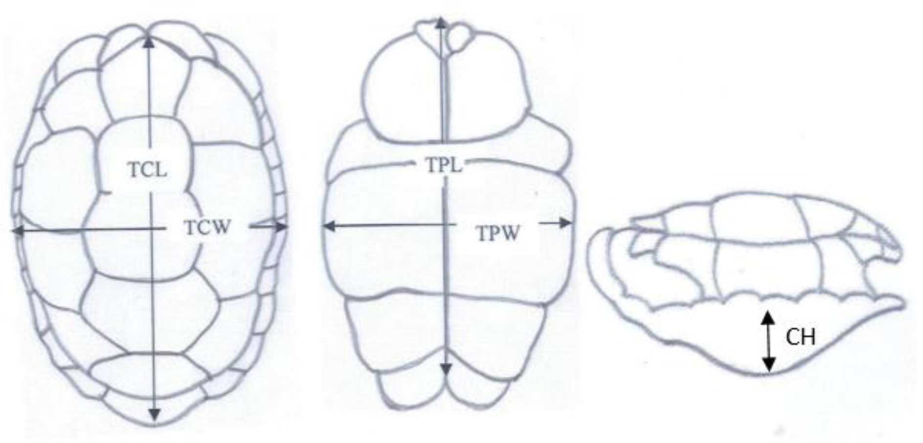
Total Carapace Length (TCL), Total Carapace Width (TCW), Total Plastron Length (TPL), Total Plastron Width (TPW) and Height were obtained using vernier caliper (resolution 1.00mm and 0.1mm, respectively) and a flexible tape ruler (resolution 1.00mm) for straight and curved measurements, respectively.

Results and Discussion

According to the morphological observations of Melanochelys trijuga parkeri and Melanochelys trijuga thermalis have common scutation on their carapace and plastron.

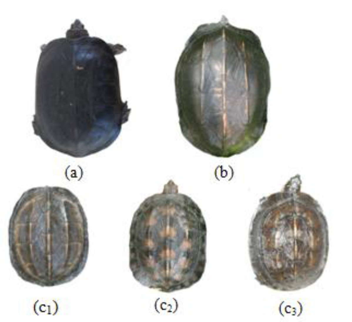
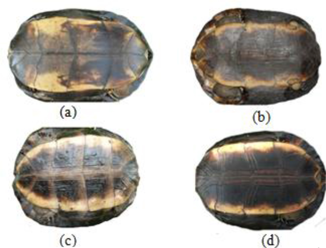
The shell of Melanochelys trijuga parkeri and Melanochelys trijuga thermalis consist of an upper carapace and lower plastron connected laterally by bony bridges. The carapace scutes are in black or dark coppery red with or without a yellow margin to the plastron. The plastron is dark brown or black and usually possesses a diffuse, dirty yellow lateral margin.

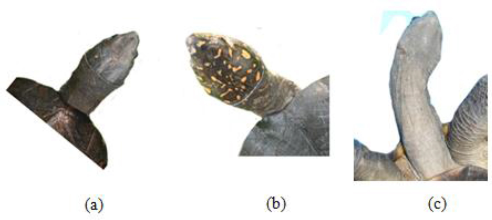
Melanochelys trijuga thermalis has yellow to orange color spots on the dorsal surface of head skin, Melanochelys trijuga parkeri doesn’t have such color pattern and which is uniformly olive brown color. The neck is moderate and possesses three or four lateral longitudinal folds.

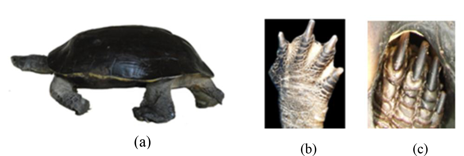
Limbs are strongly scaled with movable digits which are webbed and with grey or white skin near to carapace.

This terrapins are subject to considerable individual variation in colour on their carapace and plastron. It is very important to study pigmentation changes on the carapace and plastron throughout the growth period because we have observed more colour variations in young ones when compared to the adults.

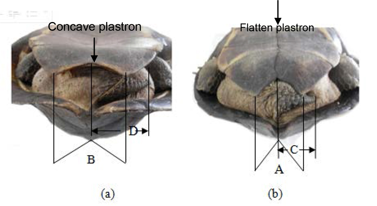
The male possesses a concave plastron with more reflex angled (B>A) in comparison to the female. The female’s tail is much shorter in length than males (C<D). Male has V shaped anal scutes whereas in females it is U shaped. Plastron is concave in males and female plastron is flat.

It is interesting to note that of all Melanochelys trijuga parkeri were males. We were unable to find females from Melanochelys trijuga parkeri during our study period.

Morphologically, Melanochelys trijuga thermalis are differentiated from Melanochelys trijuga parkeri by using yellow to orange color spots on their head and neck area.

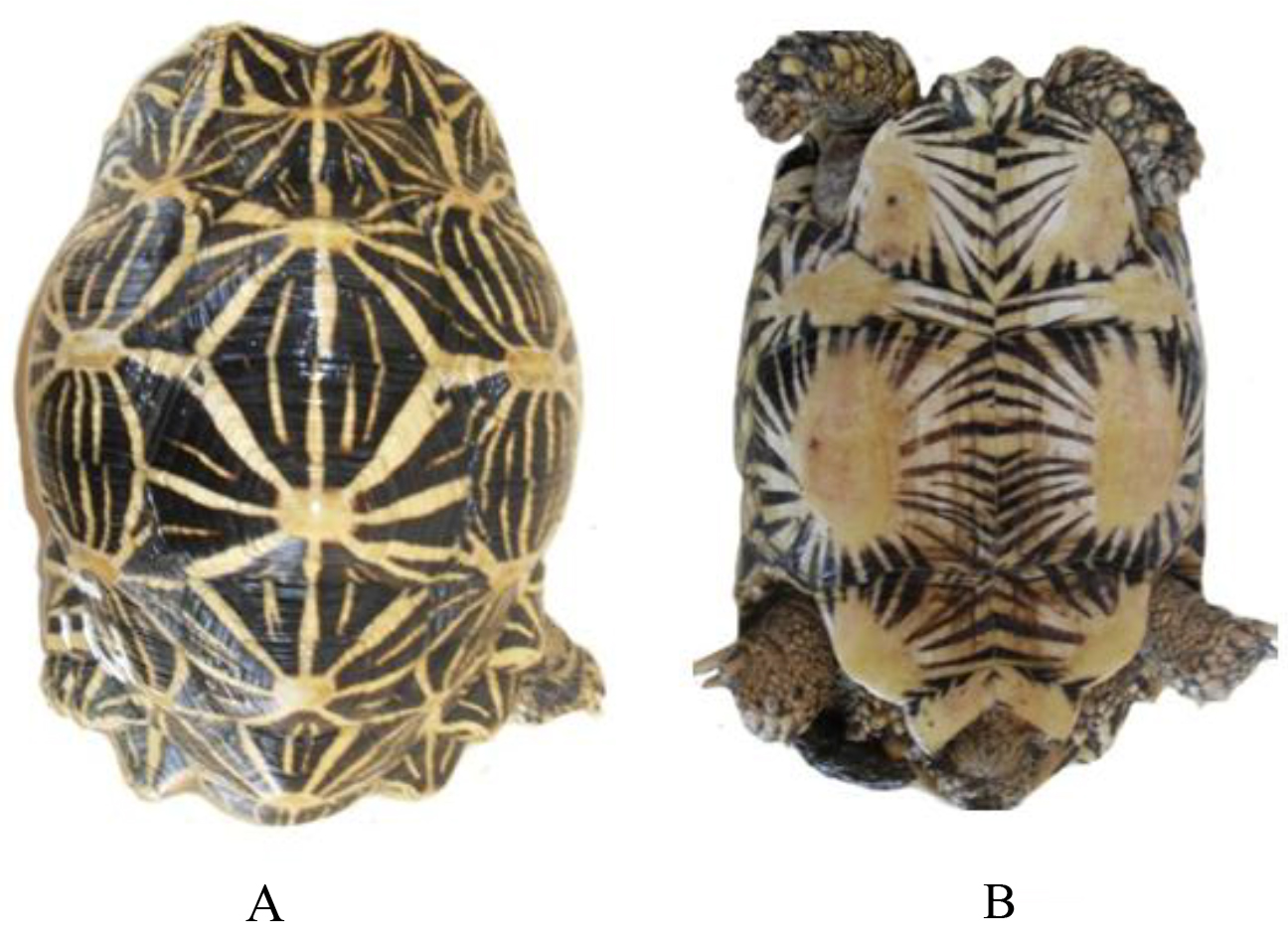
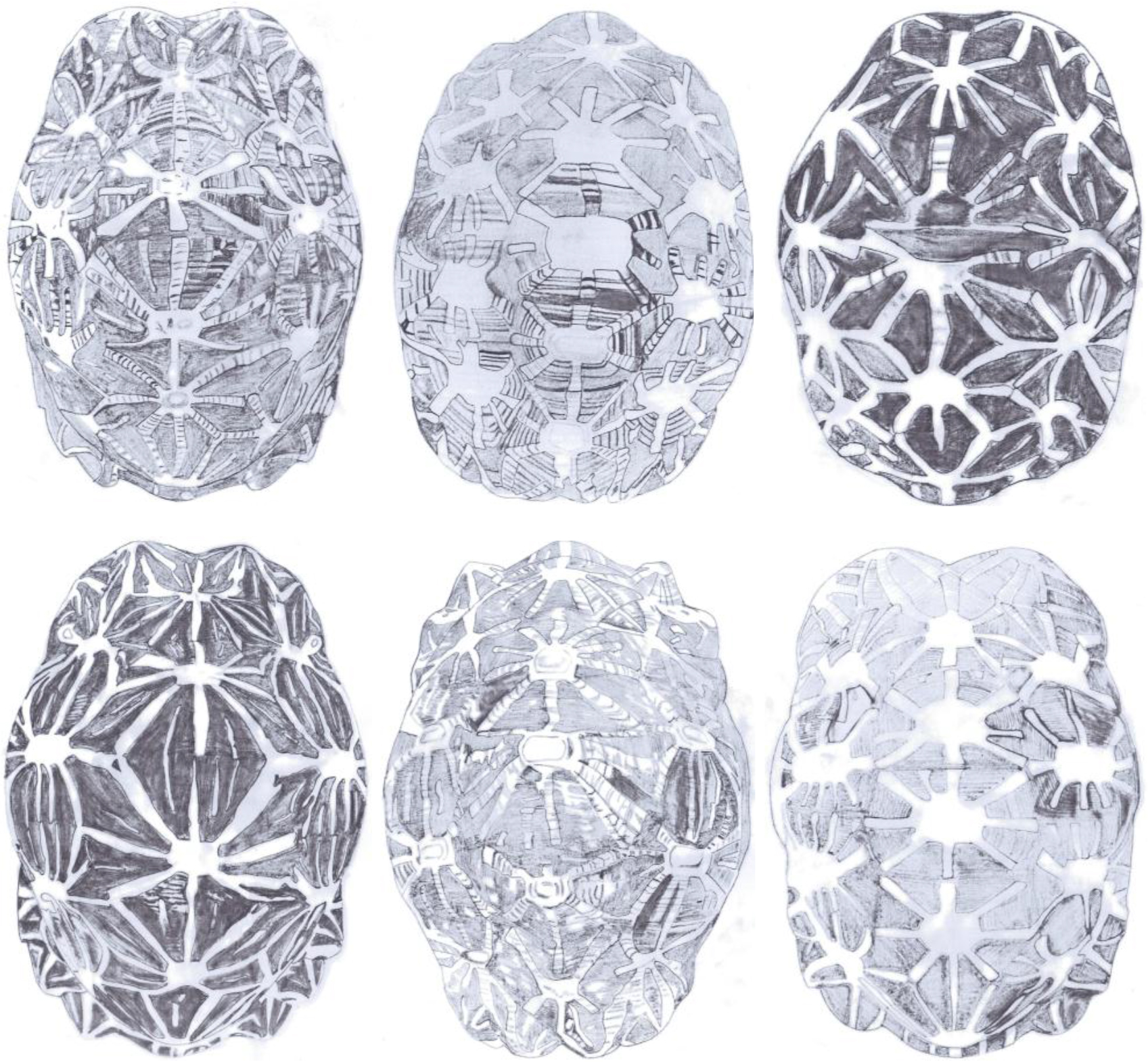
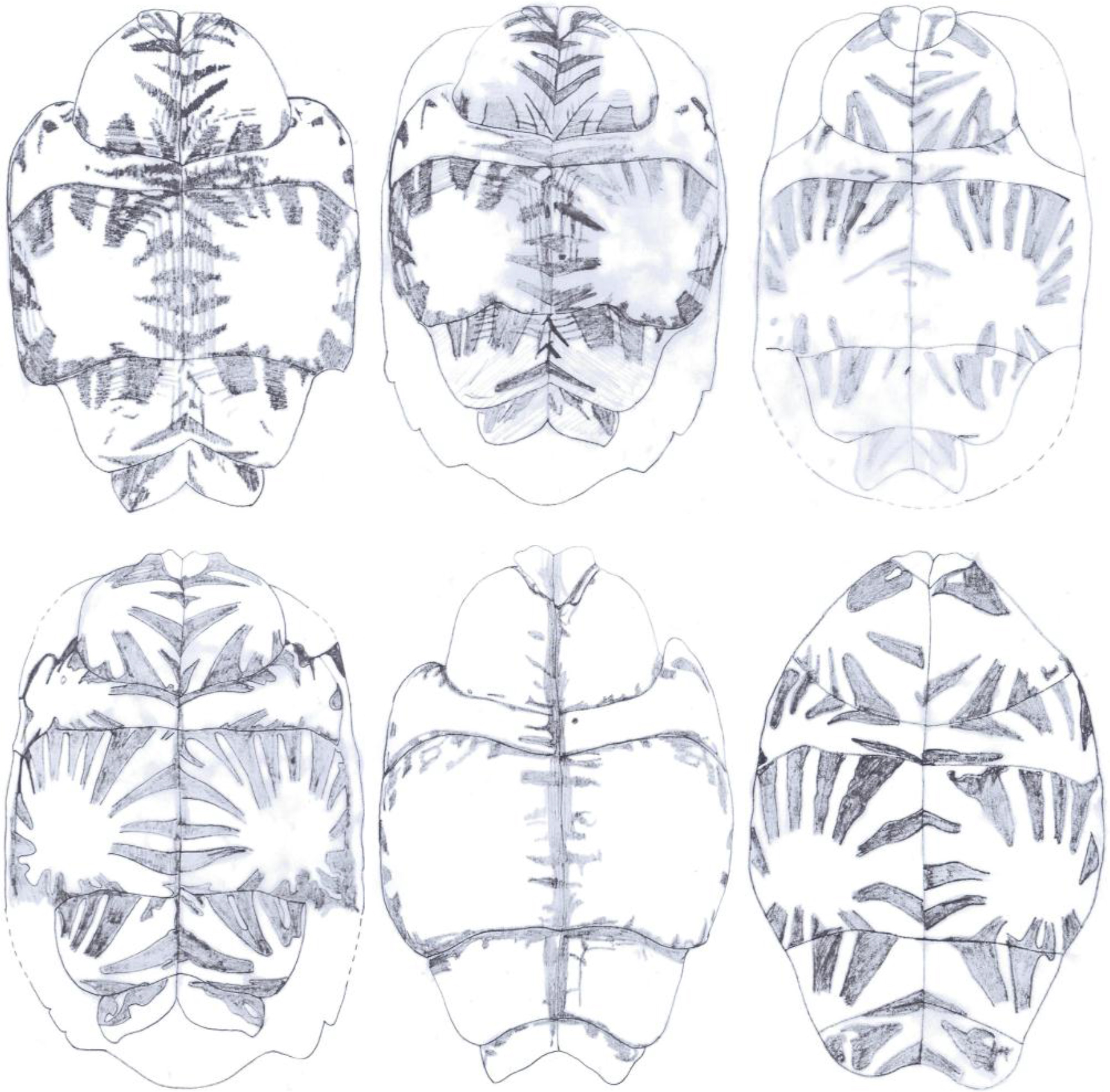
Geochelone elegans is easily recognized by the black and yellow stellate pattern upon its carapace and using pointed uneven scales. It is more strongly domed with pointed uneven vertibrals. Plastron is somewhat shorter than the carapace.

The head is moderate in size and covered with a few enlarged, and numerous small scales. Fore and hind limbs consist of claws and they are having pointed flat scales directed towards claws.

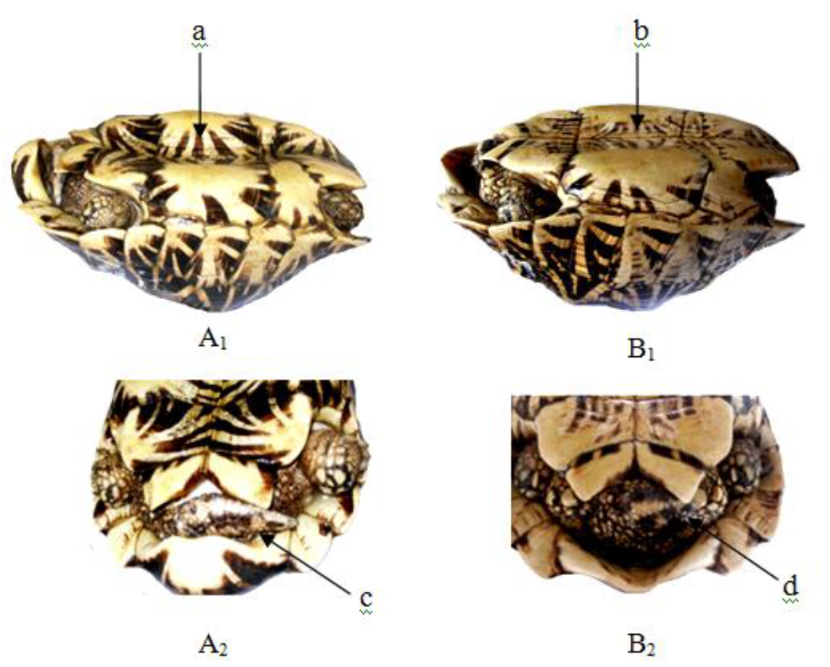
In adult male the tail is more elongated than that of the female, the supracaudal scute ends well below the level of the other marginal and is wider than the last vertebral and curves strongly inwards. The plastron is concave. In the adult female the supracaudal scute ends at the same level as the other marginals and is narrower than the last vertebral and slants straight downwards (Figures 1–14). Male animals were smaller than females.

IJVB 2019-105 - Gamika Srilanka_F1

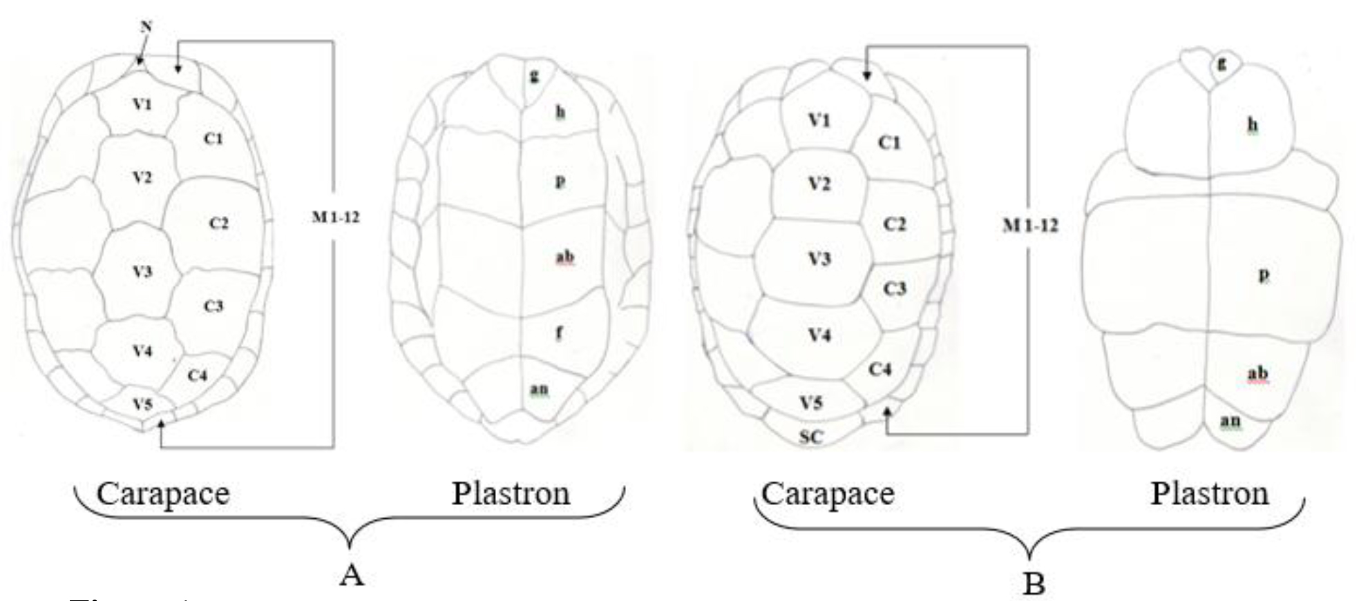
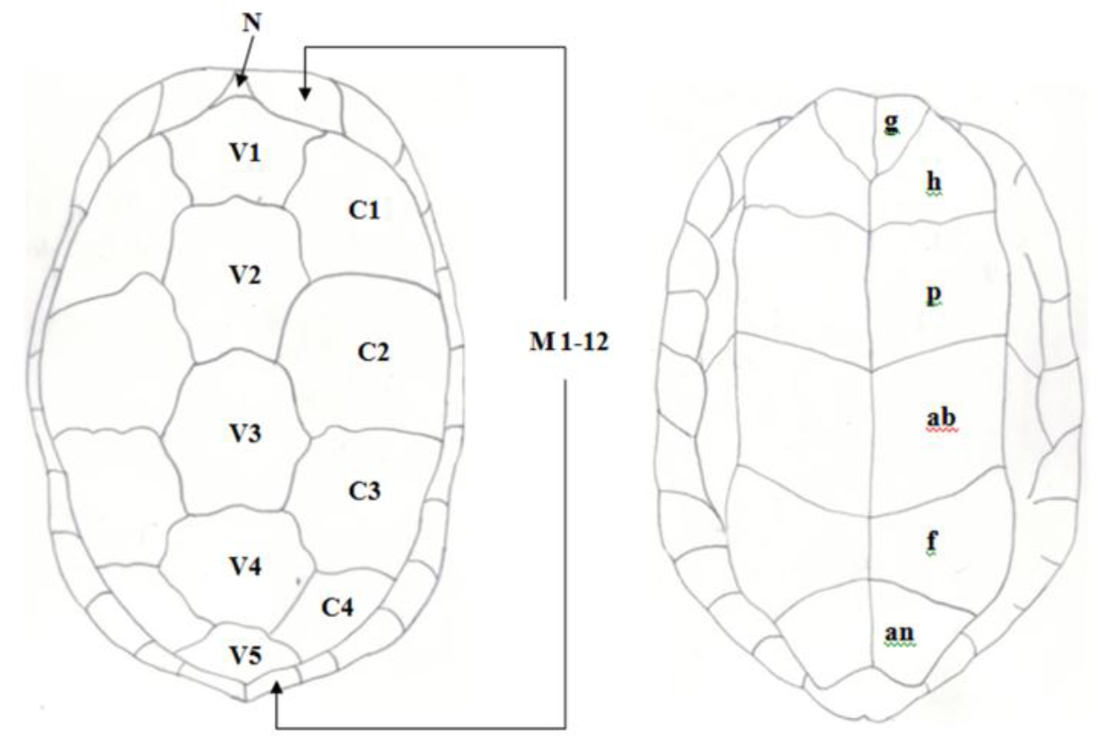
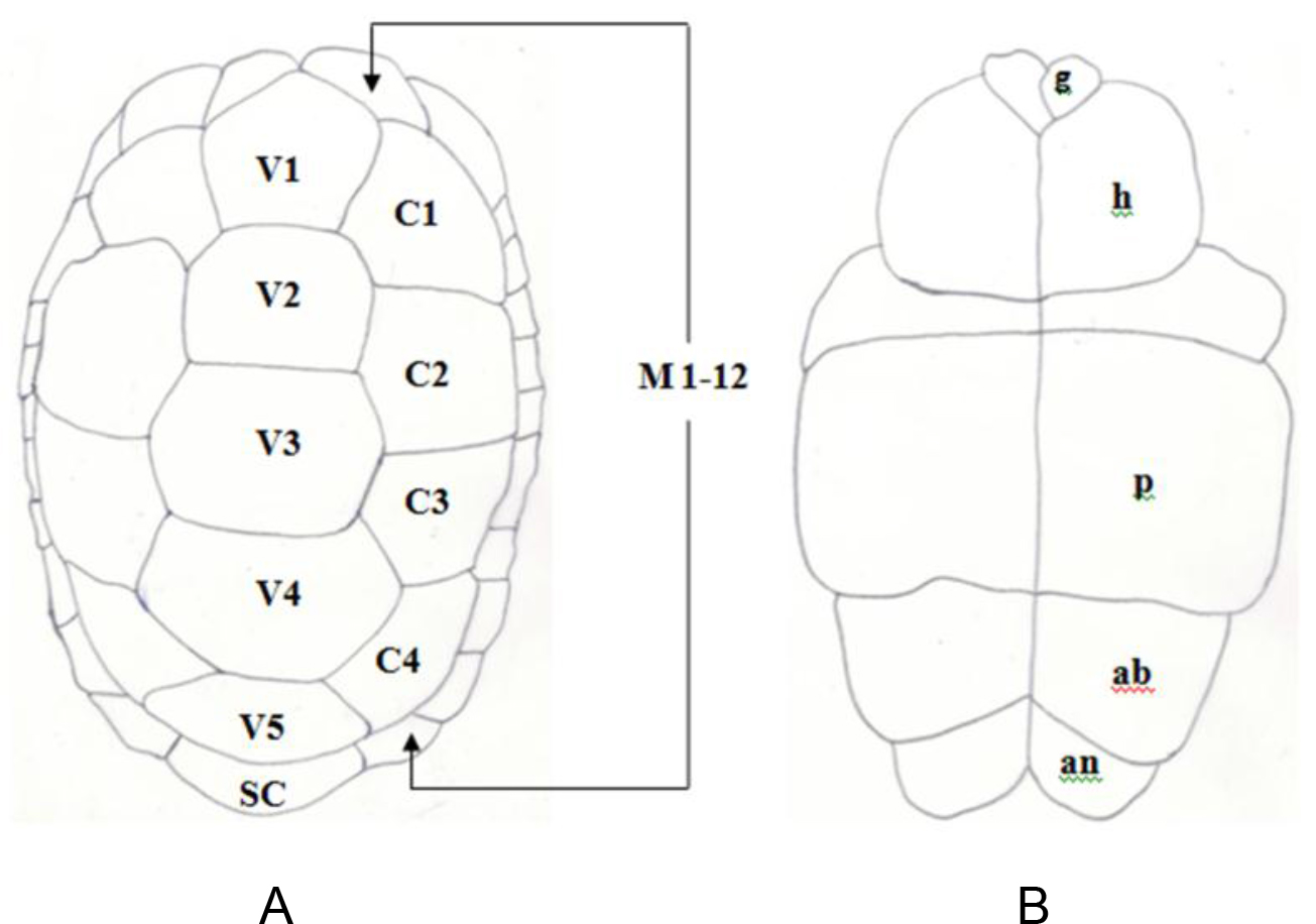
Figure 1. Pattern of scutes on carapace and plastron of Melanochelys trijuga parkeri and Melanochelys trijuga thermalis: Carapace,  (N) Nucal (1), (V) Vertibrals (1–5), (C) Costals (1–4, both sides), (M) Marginal (1–12, both sides): Plastron, (g) gular, (h) humeral, (p) pectoral, (ab) abdominal, (f) femoral and (an) anal (all paired)  and B. Geochelone elegans: Carapace, (V) Vertibrals (1–5), (C) Costals (1–4, both sides), (M) Marginal (1–12, both sides) and (SC) Supracaudal : Plastron  (g) gular, (h) humeral, (p) pectoral, (ab) abdominal, (f) femoral and (an) anal (all paired)

IJVB 2019-105 - Gamika Srilanka_F2

Figure 2. Shell Measurements

IJVB 2019-105 - Gamika Srilanka_F3

Figure 3. Pattern of scutes on carapace and plastron of Melanochelys trijuga parkeri and Melanochelys trijuga thermalis : Carapace, (N) Nucal (1), (V) Vertibrals (1–5), (C) Costals (1–4, both sides), (M) Marginal (1–12, both sides): Plastron, (g) gular, (h) humeral, (p) pectoral, (ab) abdominal, (f) femoral and (an) anal (all paired)

IJVB 2019-105 - Gamika Srilanka_F4

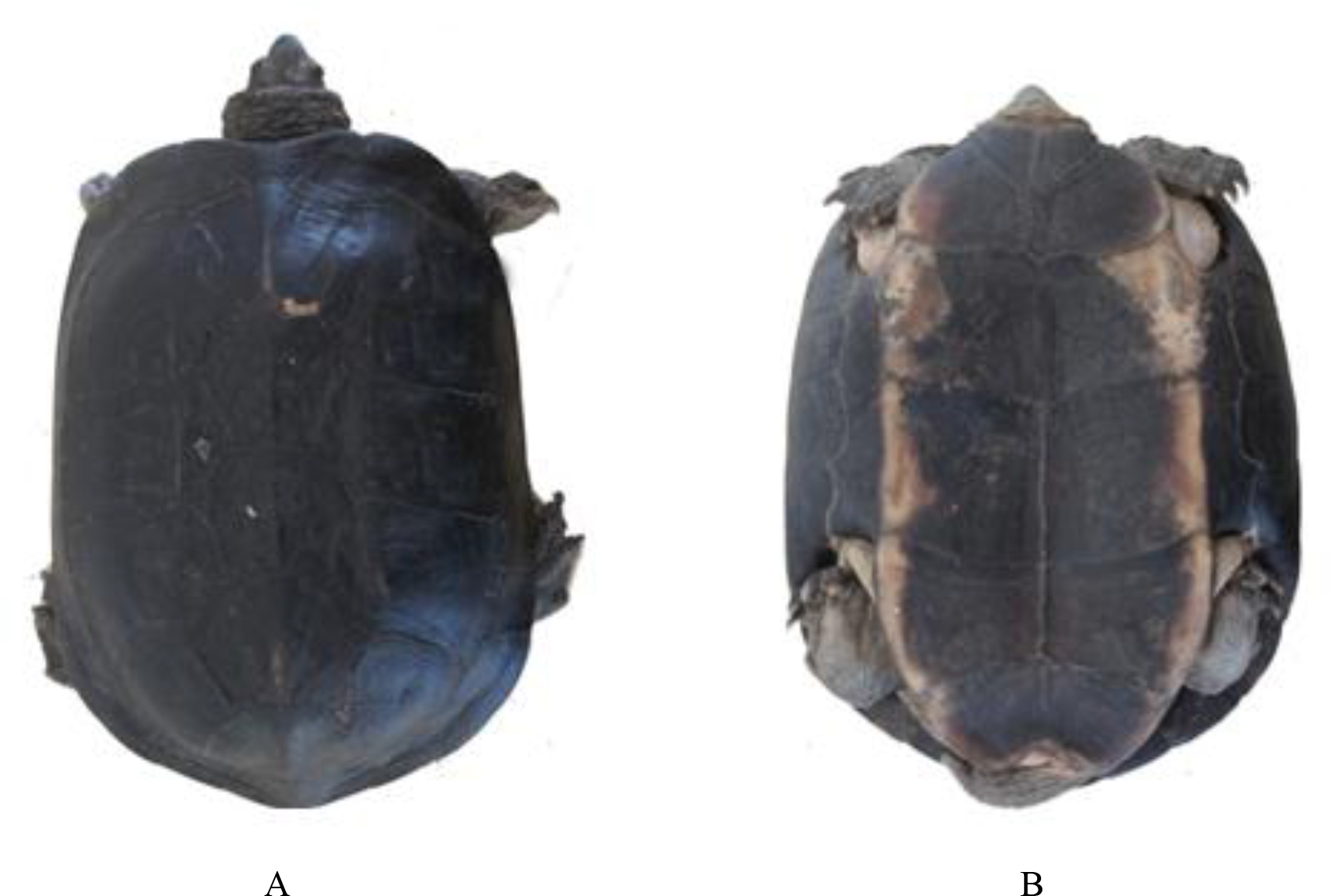
Figure 4. The Dorsal viewand B), Ventral viewof a Melanochelys trijuga parkeri, adult ♂ × 1/8

IJVB 2019-105 - Gamika Srilanka_F5

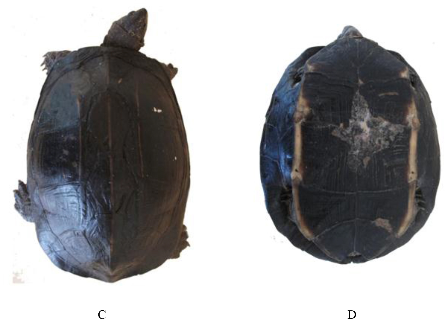
Figure 5. The Dorsal viewand D), Ventral viewof a Melanochelys trijuga thermalis, adult ♂ × 1/8

IJVB 2019-105 - Gamika Srilanka_F6

Figure 6. Pigmentation of the carapace of Melanochelys trijuga parkeri: a) Carapace with uniform black, b) Black with three light yellow ridges in carapace, Melanochelys trijuga thermalis: c 1–3) Carapace with dirty brownish color and with different designs

IJVB 2019-105 - Gamika Srilanka_F7

Figure 7. Pigmentation of the plastron of Melanochelys trijuga parkeri and Melanochelys trijuga thermalis: (a) and (c) is with yellow colored with a diffuse dusky central area, (b) Dark brown plastron with an orange border, (d) Plastron with yellow border.

IJVB 2019-105 - Gamika Srilanka_F8

Figure 8. Appearance of upper neck region of the (a) Melanochelys trijuga parkeri and (b) Melanochelys trijuga thermalis
(c) Ventral view of long neck of testudine

IJVB 2019-105 - Gamika Srilanka_F9

Figure 9. (a) Walking posture of a testudine, Appearance of limbs of the (b) Melanochelys trijuga parkeri  and (c) Melanochelys trijuga thermalis

IJVB 2019-105 - Gamika Srilanka_F10

Figure 10. Caudal view of Melanochelys trijuga thermalis X 1/8, (a) Adult male (b) Adult female

IJVB 2019-105 - Gamika Srilanka_F11

Figure 11. A) Pattern of scutes on carapace and plastron of Geochelone elegans: Carapace, (V) Vertibrals (1–5), (C) Costals (1–4, both sides), (M) Marginal (1–12, both sides) and (SC) Supracaudal : B) Plastron (g) gular, (h) humeral, (p) pectoral, (ab) abdominal, (f) femoral and (an) anal (all paired)

IJVB 2019-105 - Gamika Srilanka_F12

Figure 12. A) The dorsal view and B), the ventral view of aGeochelone elegans, adult ♂ × 1/8

IJVB 2019-105 - Gamika Srilanka_F13

Figure 13. Carapace designs observed in Geochelone elegans, adult ♂ × 1/8

IJVB 2019-105 - Gamika Srilanka_F14

Figure 14. Plastron designs observed in Geochelone elegans, adult ♂ × 1/8

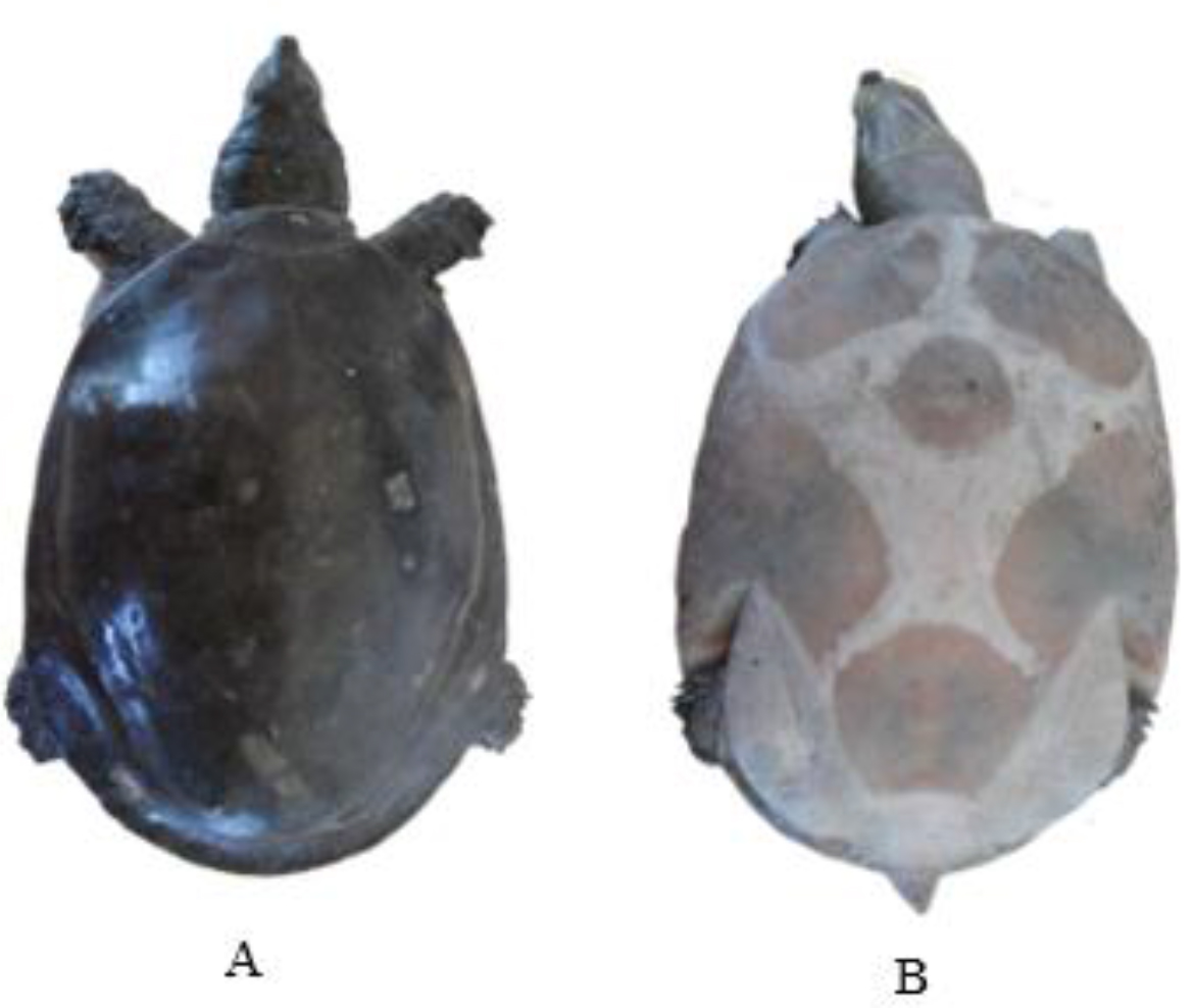
Lissemys punctata punctata has soft fleshy carapace and plastron. This species can completely retracte its limbs when the plastral flaps are pulled tightly against the bony rim of the carapace. This ability can act as additional protection for the hind limbs.

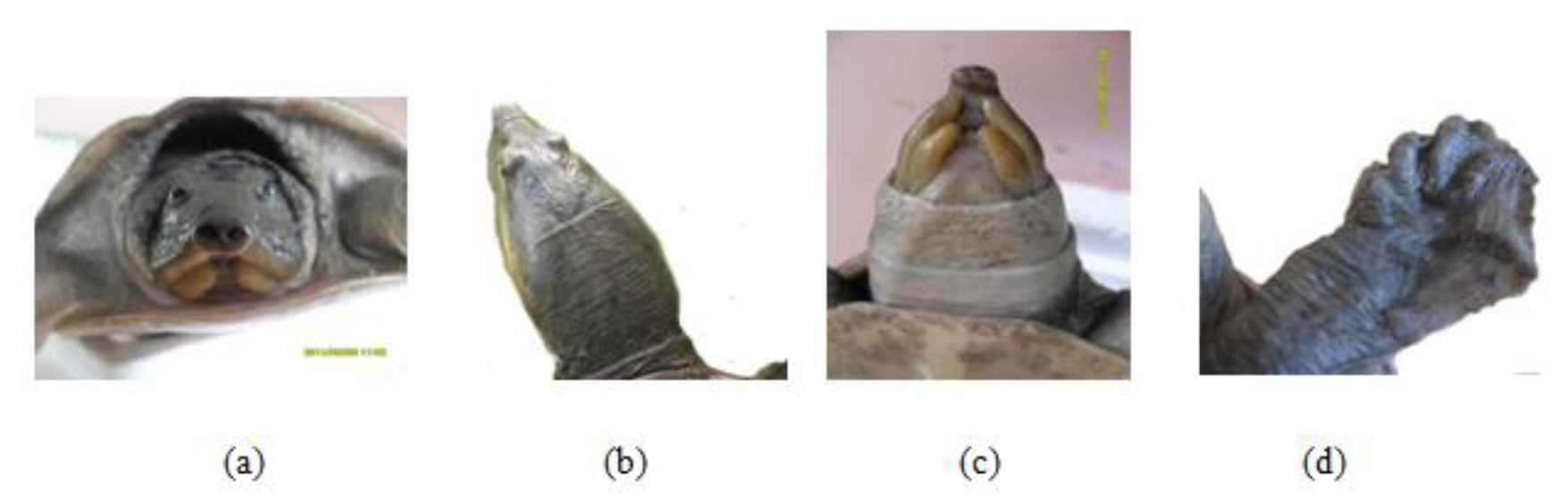
This soft terrapin is easily distinguished from other local testudines by its scuteless smooth skinned carapace, tubate nostrils, fleshy lips (c) and three clawed limbs with a few vestigial scales (d). The secondary sexual characters were poorly observed with our experimental group of Lissemys punctata punctate.

This soft terrapin is easily distinguished from other local testudines by its scuteless smooth skinned carapace, tubate nostrils, fleshy lips (c) and three clawed limbs with a few vestigial scales (d). The secondary sexual characters were poorly observed with our experimental group of Lissemys punctata punctate.

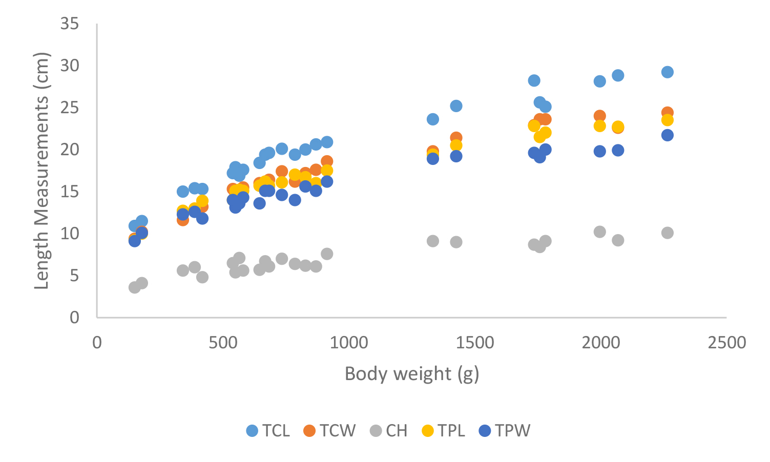
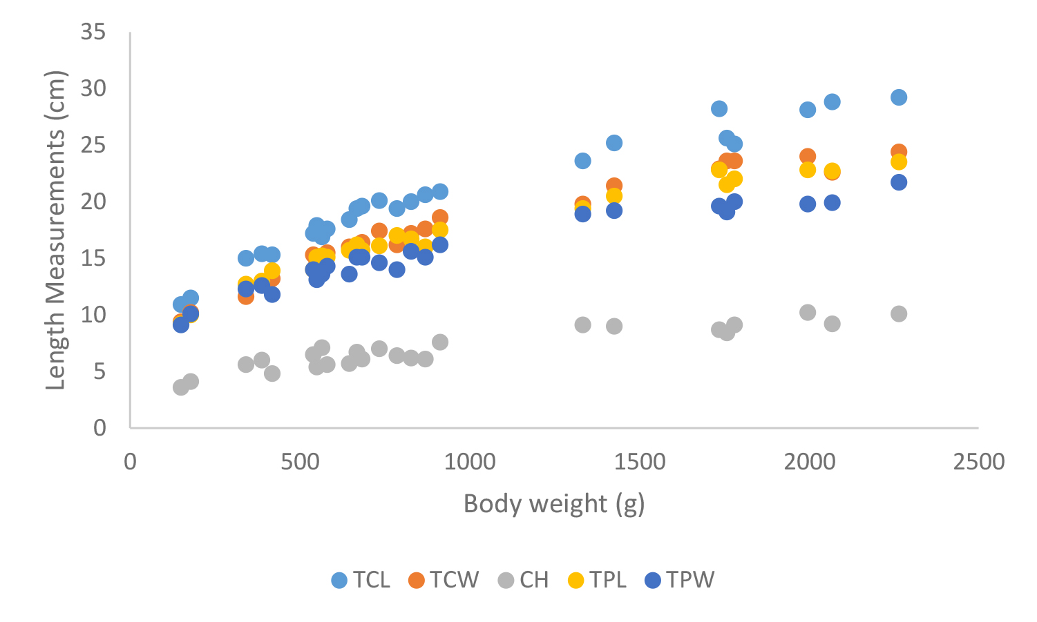
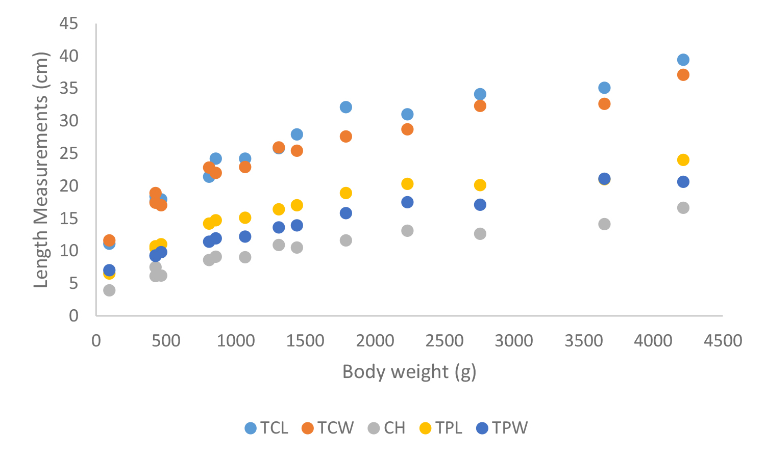
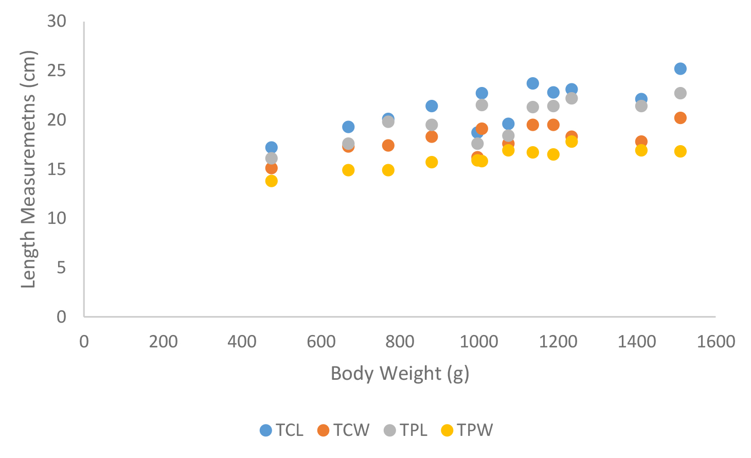
According to shell morphometrics observations increased body weight of was associated with increased carapace length, carapace width, carapace height, plastron length and plastron width of Melanochelys trijuga parkeri, Melanochelys trijuga thermalis, Lissemys punctata punctata and Geochelone elegans. [Figures 15–22]

IJVB 2019-105 - Gamika Srilanka_F15

Figure 15. Lateral and caudal views of Geochelone elegans X 1/8

(A1–2) Adult male – (a) concave plastron and (c) elongated tail,
(B1–2) Adult female – (b) flat plastron and (d) short tail

IJVB 2019-105 - Gamika Srilanka_F16

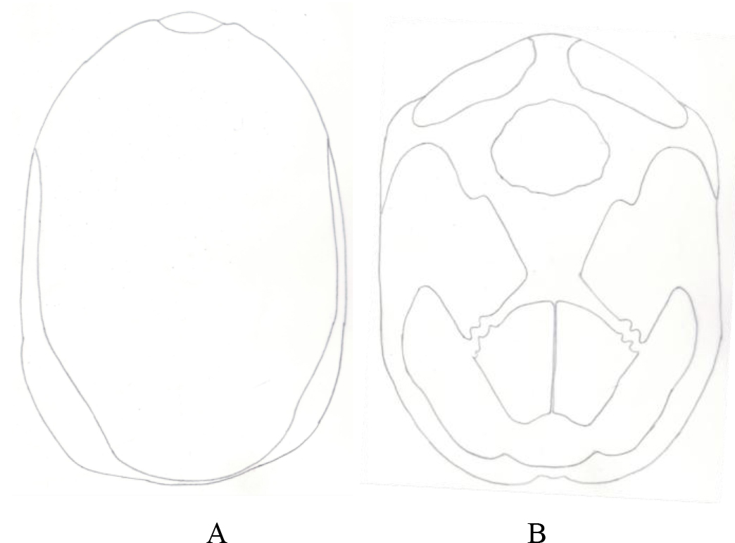
Figure 16. A) Plastron and B) carapace of  Lissemys puntata punctata

IJVB 2019-105 - Gamika Srilanka_F17

Figure 17. A) The dorsal view and B), the ventral view of aLissemys punctata punctata, adult × ¼

IJVB 2019-105 - Gamika Srilanka_F18

Figure 18. (a) Rostral view of head (b) Dorsal view of head, (c) fleshy lips and (d) limb of Lissemys punctata punctata

IJVB 2019-105 - Gamika Srilanka_F19

Figure 19. Carapace and plastron measurements of Melanochelys trijuga parkeri
Total carapace length (TCL), Total carapace width (TCW), Carapace height (CH),Total plastron length (TPL) and Total plastron width (TPW)

IJVB 2019-105 - Gamika Srilanka_F20

Figure 20. Carapace and plastron measurements of Melanochelys trijuga themalis
Total carapace length (TCL), Total carapace width (TCW), Carapace height (CH), Total plastron length (TPL) and Total plastron width (TPW)

IJVB 2019-105 - Gamika Srilanka_F21

Figure 21. Carapace measurements of Geochelone elegans; Total carapace length (TCL), Total carapace width (TCW), Carapace height (CH), Total plastron length (TPL) and Total plastron width (TPW)

IJVB 2019-105 - Gamika Srilanka_F22

Figure 22. Carapace and plastron measurements of Lissemys punctata punctata, Total carapace length (TCL), Total carapace width (TCW), Total plastron length (TPL) and Total plastron width (TPW)

Conclusion

This article provides basic morphological information of Sri Lankan fresh water turtles and tortoises which will helpful in future research, management and conservation aspects.

Acknowledgement

The authors wish to appreciate the assistance given by the I.W.M.D.N. Sadaruwan and K. A. C. kumara, for the support given in the data collection.

Conflicts of interests

The authors would like to declare that there is no conflict of interest related to publication of this paper.

Authors’ Contribution

This work was carried out in collaboration between all authors. GAP designed the study and involved in field research trials.  HNND was conducted the field research trial, data collection, managed the literature searches, interpreted the data and drafted the manuscript. LJPAPJ took part in preparing and critical checking of this manuscript. GAP supported the research facilities and funding. All authors read and approved the final manuscript.

References

  1. Kottawa-Arachchi JD, Wijeratne MA (2017) Climate Change Impacts on Biodiversity And Ecosystems In Sri Lanka: A Review. Nature Conservation Research 2: 2–22.
  2. Marambe B, Silva P, Ranwala S, Gunawardena J, Weerakoon D, Wijesundara S, Kurukulasuriya M (2011) Invasive alien fauna in Sri Lanka: National list, impacts and regulatory framework.
  3. Das I, De Silva A (2005) A photographic guide to snakes and other reptiles of Sri Lanka: New Holland.
  4. Karunarathna S, Amarasinghe AT, Henkanaththegedara S, Surasinghe T, Madawala M, et al. (2017) Distribution, habitat associations and conservation implications of Sri Lankan freshwater terrapins outside the protected area network. Aquatic Conservation: Marine and Freshwater Ecosystems 27: 1301–1312.
  5. Deraniyagala P (1939) The tetrapod reptiles of Ceylon.–Vol. I. Testudinates and Crocodilians. Colombo Museum Natural History Series. Colombo, Sri Lanka: Museum of Natural History.
  6. DeSilva A (1996) Proposed Action Plan: Conservation, restoration and management of the Testudines and their habitats in Sri Lanka: Department of Wild Life Conservation & Global Environmental Facility Programme.
  7. Fund TC (2002) A global action plan for conservation of tortoises and freshwater turtles. Strategy and funding prospectus.
  8. Hewavisenthi S (1993) Turtle hatcheries in Sri Lanka: Boon or bane. Marine Turtle Newsletter. 60: 19–21.

At Doctor Level

DOI: 10.31038/CST.2019431

Letter to The Editor

Patients, even who are enrolled in clinical trials, need good doctors. Good medical practice (GMP) refers to what is expected of all doctors practicing medicine, while good clinical practice (GCP) provides international quality standards for clinical trials involving human subjects [1]. It’s noteworthy to point out that GMP and GCP rules are not always superimposable, neither meet the same objectives.

There is a growing acceptance that clinical trials should acknowledge the unique characteristics of each patient and seek to individualize patient care [2]. Those purposes may also be extended to the evaluation of individual doctor performance, in order to assess quality of physician behavior and its impact on patient outcome [3,4]. However, analyzing doctor performance is challenging, and no single, valid, reliable, and practical measure of performance exists [5]. A prototype of a patient-reported grading scale for individual doctor performance is suggested in Table 1.

Table 1. Doctor performance status

0

Performs medical examination at each visit, empathic, good communication skills.

1

Performs medical examination at almost every visit, partially empathic, average communication skills.

2

Performs medical examination occasionally, poor-empathic, substandard communication skills. Attending more than 50% of scheduled appointments.

3

Performs medical examination occasionally, poor-empathic, substandard communication skills. Attending less than 50% of scheduled appointments.

4

Never performs medical examination. Attending scheduled appointments occasionally.

5

Always absent.

Intra-study variability of doctor performance should be addressed and no longer underestimated, to avoid unexpected regressions to mediocrity of doctor-patient interactions during clinical trial conduct.

Compliance with ethical standards

The author did not receive any funding for this work.

Conflict of Interest: Dr. A. Musolino has received research grants from: Roche; EISAI; Transgenomics. He has received speaker honorarium from: Roche; EISAI; Macrogenics; Pfizer; Lilly; Novartis. Dr. A. Musolino does not own any company’s stock.

Ethical approval: This article does not contain any studies with human participants or animals performed by   the author.

References

  1. Integrated addendum to ICH E6(R1): Guideline for good clinical practice: https://www.ich.org/fileadmin/Public_Web_Site/ICH_Products/Guidelines/Efficacy/E6/E6_R2__Step_4_2016_1109.pdf.
  2. Kent DM, Steyerberg E, van Klaveren D et al (2018) Personalized evidence based medicine: predictive approaches to heterogeneous treatment effects. Br Med 363: 4245.
  3. Ann M, Bode AM, Dong Z (2018) Recent advances in precision oncology research. NPJ Precis Oncol 2: 11.
  4. Franks P, Jerant AF, Fiscella K et al (2006) Studying physician effects on patient  outcomes: physician interactional style and performance on quality of care indicators. Soc Sci Med 62: 422–32.
  5. Leep Hunderfund AN, Park YS, Hafferty FW et al (2017) A Multifaceted Organizational Physician Assessment Program: Validity Evidence and Implications for the use of Performance Data. Mayo Clin Proc Innov Qual Outcome 1:130–140.

Diagnosis of CLABSI from Blood and Needleless Connector Cultures in Patients with Acute Leukemia: a Prospective Cohort Study

DOI: 10.31038/CST.2019425

Abstract

Background: Global guidelines defining the procedure for collection of blood cultures (BC) are still unclear (especially number of samples and the procedure itself).

Purpose: The aim of this study is to evaluate the management of the collection of BC and needleless connector (NC) cultures to study the impact on the diagnosis of CLABSI.

Methods: A single-center prospective cohort study was performed, including all consecutive cases of first event febrile neutropenia that occurred in Acute Leukemia patients using a central venous catheter (Hickman®), since April 15th 2018 to October 15th 2018.

Results: Ninety-six hospital admissions along with 1235 hospital days and 1172 CVC-days were analyzed. A total of 38 cultures (BC and CVC-line) including 76 NCs were studied. The gram negative bacteria was the most representative microorganism reported in the CVC-line, p<0.05. No gram negative bacteria was reported in the NC. All BCs were negative in the presence of NC colonization events. No positivity risk between CVC-lines was found [RR 1.292, 95% CI, 0.639 to 1.960].

Conclusion: Our study suggests that: 1) NCs should be removed prior to drawing blood for culture testing; 2) Paired BCs should be obtained in every catheter lumen and peripheral blood; 3) A 72-hour period substitution for NCs is useful to control the microbiological interconnection risk between the CVC-line and NCs.

Keywords

CLABSI; CRBSI; Acute Leukemia; Needleless Connector; Blood Cultures

Introduction

Infections are one of the most important causes of morbidity and mortality in immunosuppressed patients, namely haematology oncology patients. Patients with Acute Leukemia (AL) have a higher risk of neutropenia due to Chemotherapy Treatments (CT) and to malignancy itself. Multiple chemotherapy cycles and high transfusion rates are known predisposing risk factors that, associated with antibiotic resistant bacteria, increase the incidence and prevalence of Bloodstream Infections (BSI) [1].

Aiming for a safe and efficient chemotherapy regimen, the use of central venous catheters (CVC) increased in the 1980s; however, this resulted in reports of higher numbers of nosocomial infections. Along with CVCs, the use of needleless systems to access the catheter is a major recommendation, with the split septum valve preferred over mechanical valves due to the associated infection risk [2]. In addition, we now know that many nosocomial pathogens can persist on inanimate surfaces for weeks or even months [ex: E.Coli (1.5h to 16 months); Klebsiella pneumoniae (2h to >30 months); Pseudomonas aeruginosa (6h to 16 months); or Staphylococcus aureus (7 days to 7 months)], being the Staphylococcus spp considered the most representative [3]. If colonization progresses and clinical infection occurs, it is recommended that blood cultures should be obtained and empirical antibiotics started immediately.4 If the infection source is identified as the CVC, it is defined a Catheter-Related Bloodstream Infection (CRBSI) [2].

In neutropenic patients, the natural host defense against local flora is reduced, enhancing a direct invasion across the colonic mucosa, predisposing patients to BSI. Infection due to Mucosal Barrier Injury (MBI) microorganisms (ex: viridians group, Streptococci, Enterococci spp., E. coli, Klebsiella and Enterobacter spp) is common [4]. Taking into account the potential sources for bacterial contamination [5], the hematogenous seeding from distant sites increases the infection risk in this special population where central-line associated bloodstream infection (CLABSI) is frequently reported [2].

Global guidelines defining the procedure for collection of blood cultures are still unclear. Blood collection of CVC-line and peripheral blood are consensual, but the number of samples and the procedure itself are still undefined [4–8].

The aim of this study is to evaluate the management of the collection of blood cultures (BC) and needleless connector (NC) cultures to study the impact on the diagnosis of CLABSI.

Material and Methods

Selection and Description of Participants

A single-center prospective cohort study was performed, including all consecutive cases of first event febrile neutropenia that occurred in AL patients using a central venous catheter (Hickman® type, double lumen, 7 French) for more than 72h, undergoing Chemotherapy Treatment (CT) or aplasia support, since April 15th 2018 to October 15th 2018.

Patients older than 18 years old, with newly diagnosed or relapsed acute leukemia, admitted for CT or iatrogenic aplasia support were included.

Data concerning patients’ background was prospective collected. Baseline demographic data was collected on the CVC placement day and the assessment was encompassed in every hospital admission.

Central-line devices management and culture procedure

Only the first BC episode during the hospital admission was analyzed. No concurrent antibiotic was used before BC episode. For every first BC episode, samples were collected first from a peripheral vein (PV), followed by the CVC line no more than five minutes apart (to reduce DTP results bias) [1,7]. The NCs were removed before collecting BC samples [1,6–7]. The BC procedure is performed by one nurse alone. BC samples were collected (with a minimum of 5 ml of blood, when possible) into BACTED PLUS Aerobic/F® vials [9] and the NCs were inserted in a sterile container for sample collection. Positive cultures were automatically analyzed by COS Biomérieux; incubation (24 hours, 37 ºC) was performed. Mass spectrophotometry (Microflex – Bruker) was used for microorganism identification. The NCs were withdrawn and cultured in a liquid medium (Brain-heart-infusion Becton-Dickinson).

A NC external film surface swab rubbed was also performed. The NC microorganism lumen growth was reported when positive recoveries observed between 1 and < 15 colony-forming units (CFU). When BCs were collected, an insertion site swab was rubbed on the surface of 1–2 cm around the catheter insertion site.

Central-line Infections and neutropenia definitions

CLABSI and CRBSI rates were calculated, considering BCs yielding an organism (positive culture in PV and at least one CVC-line) per 1000 CVC-days. In the presence of a positive DTP, CRBSI was considered [2]. Also, it was considered localized catheter colonization if microorganism growth ≥ 15 CFU in the absence of positive blood results.[2] Neutropenia was considered as the absolute neutrophil count (ANC) less than 500/µL or ANC less than 1000/µL and predicted decline to 500/µL or less over the next 48 hours [4].

Technical Department Information

A 20-bed unit, distributed along eight double rooms and four single rooms, all equipped with positive pressure ventilation and HEPA filters. Only patients with a diagnosis of hematological malignancy are admitted to the department.

The management of CVCs follows the CDC (2011) guidelines [2]. Double lumen Hickman® type catheters (Vygon SA) are usually inserted (7 French, lumen 1 = 0.6 mm and lumen 2 = 1.0 mm). No antibiotic prophylaxis is given.

Specific CVC management in our unit includes the use of 2% chlorhexidine in 70% isopropyl alcohol solution for NC disinfection (considering 15” disinfection of NC hubs) [10] (neutral split septum needleless connector Bionecteur®), either in the stand-alone presentation or in a double lumen configuration (Octopus®) [11]. The CVC-flushing interval time in the CVC-lines was performed every 72-hours. Sodium heparin 20 IU/ml (Fibrilin®) is used for CVC-lock. Push-pause and positive pressure techniques always were performed using syringes 10 mL volume or higher. [1]

Data Analysis

Data analysis was conducted using IBM SPSS Statistics for Windows (SPSS Inc., Version 24.0). A continuous variable was reported by median and range. Categorical variables were reported as frequency and percentages. Normality tests reported a sample without normal distribution; therefore, hypothesis tests were analyzed by non-parametric tests. Relative Risk was performed by confidence intervals of 95%. A p value of ≤ 0.05 was determined to be statistically significant.

Ethics

The study was approved by the Ethics Committee (CES IPO: 104/018) of the Portuguese Institute of Oncology (Porto) in April 12th, 2018. Informed consent was waived for the included patients.

Results

A total of 21 patients diagnosed with AL were included: median age of 49 years [range, 75 to 22]; 15 (71.4%) female patients. Ninety-six hospital admissions [median 4, range 11 to 1] along with 1235 hospital days [median 10, range 44 to 3] and 1172 CVC-days [median 10, range 44 to 3] were analyzed.

A total of 38 cultures (BC, CVC-line and NC) were analyzed (Table I). Neutropenia was reported in 27/38 events (71.1%). BSI was only observed in neutropenic patients [BSI 2 (28.6%); CLABSI 5 (71.4%) (4.26/1000 CVC-days)]. No CRBSI was reported. One fungus in the bloodstream was discovered and documented.

Table 1. Microbiological Results

Peripheral Vein

1.0 mm CVC-line

0.6 mm CVC-line

NC Lumen

NC Surface

1

E. coli

2

P. aeruginosa

P. aeruginosa

3

S. epidermidis

4

S. epidermidis + Corynebacterium

5

E. coli

E. coli

E. coli

6

S. epidermidis

S. epidermidis

7

S. hominis

8

S. epidermidis

S. epidermidis

9

E. coli

10

E. coli

E. coli

E. coli

S. epidermidis

S. epidermidis

11

S. haemolyticus

12

Missing

K. pneumoniae

K. pneumoniae

13

E. coli

14

E.coli

E. coli

E. coli

Missing

Missing

15

S. epidermidis*

16

Strep. dysgalactiae

Strep. dysgalactiae

Strep. dysgalactiae

Strep. dysgalactiae*

+ S. epidermidis*

+ Bacillus cereus*

17

S. epidermidis*

18

S. hominis*

19

S. hominis*

20

S. epidermidis

21

Geotrichum capitatum

 n=38 (17 negative); * Microbiological Growth

The microbiological recovery (Table II) reported E.Coli as the most representative microorganism in peripheral blood and in the catheter lumen; S. epidermidis was the most representative microorganism in the NCs. The gram negative bacteria was the most representative microorganism reported in the CVC-line, p<0.05. MBI microorganisms were observed in 60% of CLABSIs. No positive insertion site swipe was observed.

Table 2. Microorganisms Recovered

Microorganisms

Total n (%)

Gram –

E. coli

6 (31.5)*

K. pneumoniae

1 (5.3)*

P. aeruginosa

1 (5.3)*

Gram +

Strep. dysgalactiae

1 (5.3)*

S. epidermidis

6 (31.5)**

S. hominis

1 (5.3)**

S. haemolyticus

1 (5.3)**

Corynebacterium

1 (5.3)**

Fungi

Geotrichum capitatum

1 (5.3)*

Total

19 (100)

*PV/CVC ; **NC

Needleless Connector Assessment

Overall, 76 NCs were studied. Thirteen positive samples were obtained [8 (≥ 15 CFU) and 5 (< 15 CFU)].

In two cases, the CVC-line and the NC were positive at the same time; one case showed different microorganisms in the devices [gram negative bacteria E. Coli (CVC-line) and gram positive bacteria S. Epidermidis (NC)]; the other case showed the same microorganism in both samples [gram positive bacteria Strep dysgalactiae] from the NC and the CVC-line [1/13 (7.6%)]. No gram negative bacteria was reported in the NC.

S. Epidermidis was the only microorganism observed in the external NC surface. No different microbiological species between the lumen and surface of the NC were identified, being both NC lumen and surface positive at the same time in 3 (37.5%) cases. No colonization risk by the same microorganisms between the lumen and surface of the NC was observed [RR 2.000, 95% CI, 0.899 to 4.452].

Positive catheter and/or peripheral BCs when the NC was negative were reported in 7/59 (11.8%) cases. All BCs were negative in the presence of NC colonization events.

CVC-lines Positivity Reports

Considering CVC-lines positivity, the 1.0 mm CVC lumen was the most representative, being reported in 8 (61.5%) cases, always associated with CLABSI identification.

The 0.6 mm CVC-line was never positive alone, being always associated with the 1.0 mm CVC-line positivity. However, the 1.0 mm CVC-line was identified in more 37.5% reports (3 cases) than the 0.6 mm CVC-line. No positivity risk between 1.0 mm and 0.6 mm CVC-lines was found [RR 1.292, 95% CI, 0.639 to 1.960]. No positive risk was found for the association with the CVC-line condition (open/closed) [RR 1.339, 95% CI, 0.582 to 3.083].

Discussion

Several studies have been published regarding the assessment of bloodstream and CVC infections. However, the management of the BC collection procedure associated with central line devices remains unclear. Y Siegman-Igra and colleagues (1996) [12] published in the actual reference guidelines of the National Comprehensive Cancer Network in the Prevention and Treatment of Cancer-Related Infections (“Diagnosis of Vascular Catheter-related bloodstream infection: a meta-analysis”). The authors reported that the use of semiquantitative catheter segment culture is considered the less expensive test for the microbiology laboratory to perform, reporting less accuracy than the quantitative methods (pooled sensitivity and specificity above 90%). Considering long-term tunneled catheters, the hospital costs associated with the semiquantitative method is high, with the paired quantitative method considered the most accurate of BC methods, reporting a labor-intensive process involved. Gaur Aditya and colleagues (2003) [13] published “The difference in time to detection as a simple method to differentiate CRBSI or non-CRBSI in pediatric immunocompromised patients”, concluding that paired CVC and PV BCs, using DTP, is a useful test to distinguish CRBSI from non-CRBSI in that specific population.

Cultures Procedure and number of sets

The NCCN recommendations present three options to obtain BCs in neutropenic patients: 1. one set obtained peripherally and one from a CVC; 2. both sets can be obtained peripherally; 3. both sets can be obtained through the CVC [4]. This suggests that the first and third options are prone to false positives (reporting BSI when the correct diagnosis is CLABSI) or false negatives (when the correct diagnosis is colonization). In the case of CVCs with more than one line, colonization could be present and not identified when only one CVC-line sample is recovered, reducing the probability of finding a positive report by at least 50%. Our study does not allow us to suggest a specific CVC-line to analyze because no risk was found between the CVC-line positivity (1.0 mm versus 0.6 mm). Martinez and colleagues (2017) [7], in their thesis publication “Central-line associated bloodstream infection rates and blood cultures collection assessment in Acute Leukemia patients: retrospective cohort study”, reported the analyses of 105 BCs in neutropenic patients, suggesting that a false CLABSI negative could be identified in one-third of CLABSIs reported in their research. Besides, Martinez and colleagues reported the 1.0 mm CVC-line positivity in 12 (92.3%) cases when CLABSI was identified (PV and 0.6 mm CVC-line positive reported in 1 (4.7%) observations), suggesting that if BCs were collected from one of the negative CVC-lines and the positive PV was identified, BSI could be diagnosed in place of CLABSI.[7] Similar reports were found by Planes and colleagues in 2016 [14] suggesting that one-third of CRBSI diagnoses could be missed if BCs were not collected from all CVC lumens. Our study reported positivity in PV and concomitant CVC-lines (1.0 mm and 0.6 mm CVC-lines) in five cases, not being reported CLABSI or colonization events only including PV and/or 0.6 mm CVC-line positivity. However, considering the first option in the NCCN recommendation, if only one set is collected from the CVC-line, the real source of the infection (CRBSI) cannot be studied, being CLABSI reported in place of CRBSI, affecting the clinical and educational decision. On the other hand, Herrera-Guerra and colleagues (2015) [8] suggested a pooled multiple-lumen BC collection; however, our study reported colonization events of one CVC-line alone. In consequence, the BC method suggested could be considered a diagnosis bias regarding the posterior clinical decision taken (e.g., antimicrobial-lock).

Needleless Connectors and CLABSI prediction

The study suggests Gram positive bacteria as the most representative microorganism recovered from the NCs, being identified only once in the CVC-line. Guembe and colleagues (2016) [15] in their publication “Assessment of central venous catheter colonization using surveillance culture of withdrawn connectors and insertion site skin reported”, suggested that the NCs could be used as an alternative diagnostic method to hub cultures in intensive care units. It was also referred that their results could not be immediately extrapolated to populations other than ICU patients and the results should be used with prudence to maintain, remove or change a CVC-line undergoing CRBSI suspicion. However, immunosuppression in intensive care unit patients in oncology hospitals is plausible and could affect the microbiological results associated with MBI microorganisms. In consequence, it could be possible to recover MBI microorganisms in the CVC-line and other species in the NC lumen, as shown in our study. Our population, the AL patients (special neutropenic and CLABSI risk population) [1] reported high rates of MBI microorganisms without associated NC cultures (especially with high gram positive bacteria reports). In consequence, NCs should be removed prior to drawing blood for culture testing; in order to reduce the incidence of false positives such the Infusion Nurses Society recommends [2]. Indeed, in our study the NC (that is removed in all BC events) shows no interference with the clinical decision taken, regardless of CVC-line results, in most cases empirical.

NC management and substitution

The Guidelines for the Prevention of Intravascular Catheter-Related Infections published by the CDC in 2011 [2] recommended the replacement of NCs and the administration sets no more frequently than 72–96 hours (category II). Mauro Pittiruti and colleagues published in 2016 [16] the “Evidence-based criteria for the choice and the clinical used of the most appropriate lock solutions for central venous catheters (excluding dialysis catheters): a GAVeCeLT consensus”, suggesting the use of neutral or positive pressure displacements, being the risk of occlusion related to inappropriate policies of flushing and/or CVC-line or NC use. However, the infection risk associated with positive pressure devices is documented; being the split septum neutral device recommended, especially in high infection risk populations [1]. The specific procedure performed by our department: 1) the 72-hour period substitution for NCs and administration sets; 2) NC and administration sets substitution in every BC collection (supported by the theoretical rationale, based on the attempt to remove the possible device infection source at the BC collection moment), could suggest that 72-hour interval time for NC substitution is useful to control the microbiological interconnection risk between the CVC-line and NCs [7]. However, future clinical trials are required.

Potential directions of the study and limitations

The study reports 3 potential limitations: First, this is a single population study, and our suggestions may not applicable to low infection risk populations; however, the oncology-hematology clinical research in this field is scarce and more studies are strongly recommended [17]. Second, this study includes a small number of patients but this is a population that have multiple hospital admissions by patient, being the study event (CLABSI)  frequently reported [1]. Third, the process of clinical cultures “in vivo” from blood and NC collection could be influence by the patient symptomatology. Our study reported two missing cultures: 1) the venipuncture procedure missed in a patient undergoing chills; 2). A sample of NC was not processed by human lapse (without microbiological study) associated with the complex CVC-management in a patient undergoing septicemial shock.

On the other side, the homogeneity of the sample could be considered an advantage, being reduced the bias observed in the central elements related to effectiveness of CVC clinical research (product, practice and patient), frequently reported in multicenter studies [18]. Several studies does not report CVC-management dynamics based on the use of chlorohexidine 2% NC hub disinfection 15”, CVC-flushing frequency report, positive pressure and push-pause techniques [19]; being the quality of the research limited to the product. The authors can conclude that the future clinical research in this field should relate to the CVC and NC management associated practice.

Conclusion

Our study suggests that: 1) NCs should be removed prior to drawing blood for culture testing; 2) Paired BCs should be obtained in every catheter lumen and peripheral blood; 3) A 72-hour period substitution for NCs is useful to control the microbiological interconnection risk between the CVC-line and NCs.

Acknowledgement

Gillian Ray-Barruel, RN, PhD, MACN

Senior Research Fellow | QEII Jubilee Hospital | School of Nursing and Midwifery | Alliance for Vascular Access Teaching and Research (AVATAR) | Menzies Health Institute Queensland | Griffith University

Editor-in-Chief | Vascular Access: The official journal of the Australian Vascular Access Society

Honorary Research Fellow | Royal Brisbane and Women›s Hospital | Nursing and Midwifery Research Centre

Visiting Scholar | Princess Alexandra Hospital | Nursing Practice Development Unit

Grant Support: This study was supported by the Portuguese Institute of Oncology of Porto (Clinical Research Center: CI-IPOP 67/2017)

References

  1. Martinez JM, Santos AE, Godinho A, Azevedo A, Felix A, Chacim S, et al. (2018) Acute Leukemia Patients: A CLABSI Risk Special Population. Ann Hematol Oncol 5: 1192.
  2. O’grady NP, Alexander M, Burns LA, Dellinger EP, Garland J, et al. (. 2011) Guidelines for the prevention of intravascular catheter-related infections. Clinical infectious diseases 52: 162–193.
  3. Kramer A, Schwebke I, Kampf G (2006) How long do nosocomial pathogens persist on inanimate surfaces? A systematic review. BMC infectious diseases 6: 130.
  4. Baden LR, Swaminathan S, Angarone M, Blouin G, Camins BC, et al. (2016) Prevention and treatment of cancer-related infections, Version 2.2016, NCCN Clinical Practice Guidelines in Oncology. Journal of the National Comprehensive Cancer Network 14: 882–913.
  5. Rupp ME, Karnatak R (2018) Intravascular Catheter–Related Bloodstream Infections. Infectious Disease Clinics 32: 765–787.
  6. Garcia RA, Spitzer ED, Beaudry J, Beck C, Diblasi R, et al. (2015) Multidisciplinary team review of best practices for collection and handling of blood cultures to determine effective interventions for increasing the yield of true-positive bacteremias, reducing contamination, and eliminating false-positive central line–associated bloodstream infections. American journal of infection control 43: 1222–1237.
  7. Martinez JMF (2017) Central-line associated bloodstream infection rates and blood cultures collection assessment in Acute Leukemia patients: retrospective cohort study.
  8. Herrera-Guerra AS, Garza-González E, Martínez-Resendez MF, Llaca-Díaz JM, Camacho-Ortiz A (2015) Individual versus pooled multiple-lumen blood cultures for the diagnosis of intravascular catheter-related infections. American journal of infection control 43: 715–718.
  9. Zhang L, Rickard CM (2017) Non-culture based diagnostics for intravascular catheter related bloodstream infections. Expert review of molecular diagnostics 17: 181–188.
  10. Moureau NL, Flynn J2 (2015) Disinfection of Needleless Connector Hubs: Clinical Evidence Systematic Review. Nurs Res Pract 2015: 796762. [crossref]
  11. Martinez JM, Neves F, Sousa J, Santiago D, Rodrigues D, et al. (2018) Improving a better nurse practice associated with the manipulation of cvc and needleless connectors. ON 37: 6–12.
  12. Siegman-Igra Y, Anglim AM, Shapiro DE, Adal KA, Strain BA, et al. (1997) Diagnosis of vascular catheter-related bloodstream infection: a meta-analysis. Journal of Clinical Microbiology 35: 928–936.
  13. Gaur AH, Flynn PM, Giannini MA, Shenep JL, Hayden RT (2003) Difference in Time to Detection: A Simple Method to Differentiate Catheter-Related from Non-Catheter-Related Bloodstream Infection in Immuno compromised Pediatric Patients. Clinical infectious diseases 37: 469–475.
  14. Planes AM, Calleja R, Bernet A, Campins-Martí M, Almirante B, et al. (2016) Evaluation of the usefulness of a quantitative blood culture in the diagnosis of catheter-related bloodstream infection: Comparative analysis of two periods (2002 and 2012). Enfermedades infecciosas y microbiologia clinica 34: 484–489.
  15. Pérez-Granda MJ, Guembe M, Cruces R, Barrio JM, Bouza E (2015) Assessment of central venous catheter colonization using surveillance culture of withdrawn connectors and insertion site skin. Critical Care. 20: 32.
  16. Pittiruti M, Bertoglio S, Scoppettuolo G, Biffi R, Lamperti M, et al. (2016) Evidence-based criteria for the choice and the clinical use of the most appropriate lock solutions for central venous catheters (excluding dialysis catheters): a GAVeCeLT consensus. The journal of vascular access 17: 453–464.
  17. Zakhour R, Chaftari AM, Raad II (2016) Catheter-related infections in patients with haematological malignancies: novel preventive and therapeutic strategies. Lancet Infect Dis 16: 241–250. [crossref]
  18. Chernecky C, Zadinsky J, Macklin D, Maeve MK (2013) The Healthcare and Technology Synergy (HATS) framework for comparative effectiveness research as part of evidence-based practice in vascular access. Journal of the Association for Vascular Access 18: 169–174.
  19. Ramada D, Coelho F, Guilherme D, Marques P (2018) Permeabilidade do cateter venoso central totalmente implantado revisão de literatura. ON 37: 20–29.

The Short-Term Results of Pyrocarbon Lunate Implants in Patients with Advanced Kienböck’s Disease

DOI: 10.31038/IJOT.2019232

Abstract

Purpose: The purpose of this study was to evaluate the short-term results of pyrocarbon lunate implants (PLI) (Ascension Orthopaedics, Austin, Texas).

Methods:  Patients with advanced Kienböck’s disease who received PLI were prospectively followed for one year. The implant outcomes were assessed by pre- and postoperative questionnaires and physical examination.

Results: six patients (six implants) with Kienböck’s disease grade IV were included in this study. All implants remained in situ at one-year follow-up. Pain was satisfactorily reduced in five patients. Grip strength improved slightly in three patients and worsened in two. The DASH scores improved in four patients with mean 26 points. Five patients returned to their previous jobs and the two patients whom did sport could resume it. Three patients had to be mobilized under general anaesthesia because of a severely stiffened wrist.

Conclusions: The short-term results of the PLI are suboptimal, probably largely due to severe stiffening of the wrist. Nonetheless, long-term results are necessary to adequately assess the longevity and functionality of this implant and to assess which postoperative treatment would provide the optimal clinical result. Type of study/level of evidence: Therapeutic, level IV.

Keywords: Lunate Implant, Pyrocarbon, Results

Introduction

In 1910, Kienböck [1] was the first to describe the radiological signs of isolated lunatomalacia, currently known as Kienböck’s disease. Despite many years of clinical experience and research, the cause of this disorder still remains unclear. Morphological variations such as the negative ulnar variance, the particular pattern of vascularity and repetitive trauma may be predisposing factors, suggesting a multifactorial aetiology [2]. Kienböck’s disease is divided into 4 stages and can be diagnosed by conventional radiography [3] and Magnetic Resonance Imaging (MRI) [4].

Kienböck’s disease can be surgically treated. Patients with negative ulnar variance, without (radio) carpal osteoarthritis and Kienböck’s disease stage I-IIIa can be treated by a radial osteotomy [5]. Patients with neutral ulnar variance, no (radio) carpal osteoarthritis and Kienböcks disease stage I-IIIa can be treated by revascularisation [6–8]. Patients with Kienböck’s disease stage IIIb-IV can be treated by proximal row carpectomy, partial or total wrist arthrodesis or arthroplasty: resulting in limited mobility and function of the wrist. The lunate implant was developed to maintain wrist function in patients with severe Kienböck’s disease.

There are only few reports in the literature that report on the clinical outcomes of pyrocarbon lunate implants [9]. We present a case series with short-term outcomes of patients with this type of pyrocarbon lunate implant.

Patients and Methods

Study Design

All patients with advanced Kienböck’s disease who received this pyrocarbon lunate implant (Pyrocarbon Lunate prosthesis, Ascension Orthopedics, Inc, 8700 Cameron Road, Suite 100, Austin, TX 78754 USA) were identified at the Amphia Hospital in Breda, The Netherlands. The inclusion criteria were patients with wrist pain as a result of Kienböck’s disease grade IIIb or IV. The exclusion criteria were patients who performed heavy labour, patients with osteoarthritis of the radio- or midcarpal joint other than the lunate fossa and lunate-capitate joint and patients who received a prior surgical treatment for their wrist complaints. Informed consent was obtained before implant insertion. Relevant data were extracted from the medical records: demographics, medical history, profession, affected wrist and result of the Magnetic Resonance Imaging (MRI), This study’s level of evidence is IV based on the absence of a control group and was approved by the Medical Ethical Committee at the Amphia Hospital in Breda, The Netherlands.

Surgical Technique

Surgery was performed under general anaesthesia and tourniquet control. A longitudinal incision on the dorsum of the wrist was made. The sensory branches of the radial and ulnar nerves were preserved. After identification of the third compartment of the extensor retinaculum, the compartment was opened to identify the extensor pollicis longus tendon, which was held to the radial side. The fourth extensor compartment was elevated from the radius and capsule and held ulnarly. The dorsal capsule was then incised, creating a distally based capsule flap. The degenerative lunate was identified and removed. Two K-wires fixated the triquetrum and scaphoid to the capitate. A 3.5 mm hole was drilled from the ulnar side of the scaphoid towards the volar aspect of the scaphoid. A 2 mm hole was drilled from the radial side of the triquetrum towards the dorsal side of the triquetrum. On the volar aspect of the wrist, two small incisions were made to reach the flexor carpi radialis tendon. The radial third of the Flexor Carpi Radialis (FCR) tendon was harvested leaving the distal attachment inserted. The FCR tendon graft was tunnelled through the scaphoid. Two Mitek-anchors were inserted, one in the scaphoid and one in the triquetrum. The tendon was guided through the volar hole of the prosthesis and through the hole in the triquetrum towards the ulnar side. Two wires of the anchors (one of each anchor) were guided through the volar hole of the prosthesis too. These wires were attached to each other. The position of the prosthesis was controlled by fluoroscopy. The other wires were put through the dorsal hole of the prosthesis and attached to each other while closing the ‘gaps’ radialy and ulnarly of the lunate. A third Mitek-anchor was placed into the scaphoid. Two gutters were created on the dorsum of the triquetrum en scaphoid. The remaining FCR tendon graft on the dorsal side of the triquetrum was positioned into the gutters towards the scaphoid and attached to the scaphoid using the wires of the third Mitek-anchor. The position of the prosthesis was checked again with fluoroscopy to assure an anatomical position. The capsule and skin were closed by sutures. A forearm cast was worn for six weeks. The K-wires were removed after six weeks and active mobilization of the wrist was started. For another six weeks a removable forearm splint was worn during stressful moments.

Clinical Evaluation

The PLI outcome was assessed by questionnaires and physical examination. Grip strength (measure by the Jamar hand-dynamometer), range of motion and the Disabilities of the Arm, Shoulder and Hand (DASH) questionnaire [10] were measured preoperatively and at one-year follow-up. Furthermore, return to work and sport, complications and patient satisfaction on pain reduction were evaluated.

Radiological Evaluation

X-rays were performed postoperatively and at one-year follow-up to evaluate the pyrocarbon lunate implant position, intercarpal distance and progression of disease.

Results

Six patients (six implants, four females and two males) with Kienböck’s disease grade IV were included in this study (Table 1). The mean age was 35 years (range 23 to 47 years). The dominant hand was involved in one patient. Preoperatively, all patients experienced wrist pain at rest that worsened after activities.

Clinical Evaluation

All six implants remained in situ at one-year follow-up. Five patients were satisfied with the pain relief. Grip strength improved slightly in three patients and worsened in two patients. Range of motion improved in two of the six patients. The DASH scores improved in four patients with mean 26 points and worsened in two patients with mean nine points. Five patients returned to their previous jobs and the two patients whom did sport could resume it.

Complications and Revisions

A K-wire was infected in one patient. The K-wire was removed and the patient was successfully treated with oral antibiotics. The wrists of three patients were severely stiffened and were remobilized under general anaesthesia. None of the implants needed revision.

Radiological Evaluation

Analysis of radiographs showed that the scapho-lunate distance increased after removal of the K-wires in all six six patients. However, at one-year follow-up, this distance did not increase and there was no progression of disease (Figure 1). Furthermore, five wrists developed dorsal intercalated segment instability and one developed volar intercalated segment instability.

IJOT 19 - 111 -Beumer A_F1

Figure 1. The anterolateral view of the pyrocarbon lunate implant at one-year follow-up.

Discussion

The aim of this study was to determine the short-term results of the pyrocarbon lunate implant. Lunate implant arthroplasty has been used as treatment for advanced Kienböck’s disease for more than 60 years. In contrast to the silicon lunate implant, with a high incidence of silicone cysts (78% after 27 years follow-up) [11], the titanium lunate implant has promising long-term results [12]. Although not mentioned by these authors, titanium might give rise to tissue reactions. Compared to titanium, pyrocarbon is more similar to cortical bone and transfers the load more effectively, potentially limiting bone resorption. Although Pyrocarbon implants might break, they are biologically inert and biocompatible resulting in a low tendency to wear and tissue reactions. Furthermore, pyrocarbon is less susceptible for wear when compared to titanium [13].

Table 1. Characteristics, clinical outcome and complications of patients with the pyrocarbon lunate implant.

Case no.

Age

Kienböck’s disease stage

Grip strength (kg)

Flexion/extension

DASH

Complications

Patient satisfied with pain reduction

Return to work

Return to sport

Follow-up (mo)

Preop

Postop*

Preop

Postop*

Preop

Postop*

1

42

IV

37

42

60/50

60/80

56

14

None

Yes

Yes

Yes

12

2

47

IV

42

14

45/30

70/25

38

50

K-wire infection

Yes

No

Yes

14

3

28

IV

21

13

45/45

10/25

68

37

MUA

Yes

Yes

na

12

4

23

IV

12

18

60/50

20/20

30

35

MUA

No

Yes

No preop sport

14

5

23

IV

9

19

70/70

35/35

40

28

MUA

Yes

Yes

No preop sport

20

6

45

IV

na

na

45/45

30/45

51

32

None

Yes

Yes

No preop sport

15

Mean

35

IV

24

21

54/48

38/38

47

33

66%

83%

83%

100%

15

Abbreviations: preop, preoperatively; postop, postoperatively; na, non-available; K-wire, Kirschner wire; MUA, mobilisation under anesthesia.
*1 year postoperatively

This study has many limitations. First, and perhaps most importantly, only six patients were included in this study. Statistical analyses were not performed due to this small sample size. Secondly, there is no control group. Thirdly, the postoperative results aren’t complete.

The PLI outcome was assessed by implant survival, physical examination and questionnaires. Implant survival was 100% at one-year follow-up. The main indication for treatment was wrist pain, which was satisfactorily reduced in five patients. However, grip strength improved only slightly in three patients and moreover range of motion decreased in four patients at one-year follow-up. Despite these limitations, DASH scores reduced in four patients and five patients of the six patients could return to their previous jobs. These short-term results are suboptimal, especially considering that three out of six patients needed to be remobilized under general anaesthesia, probably largely due to severe stiffening of the wrist. The decreased range of motion could be the result of our postoperative treatment and might be avoided by a different postoperative treatment that would allow a shorter immobilization period and earlier range of motion exercises [9,12]. Although a higher risk of implant dislocation (one of 17 patients) [9] and malposition (2 of 11 patients) [12] could occur. Nonetheless, long-term results are necessary to adequately assess the longevity and functionality of this implant and to assess which postoperative treatment would provide the optimal clinical results. Until these results are published, we have abandoned this implant in our Hospital.

Declaration

Ethics approval and consent to participate: This study was approved by the Medical Ethical Committee at the Amphia Hospital in Breda, The Netherlands.

Availability of data and material: The datasets used and/or analysed during the current study are available from the corresponding author on reasonable request.

References

  1. Kienbock R (1910) Uber traumatische Malazie des Monatbeins und ihre Folgezustande: Entartungsformen und Kompressionsfrakturen. Fortschrit Rontgenstrallen 16: 77.
  2. Green DP, Hotchkiss RN, Pederson WC, Wolfe SW (2005) Green’s Operative Hand Surgery, 5th edition. Philadelphia: Churchill Livingstone 2005:744–745.
  3. Schmitt R, Krimmer H (2007) Osteonecrosis of the hand skeleton. In: Schmitt R, Ulrich L (Eds.), Diagnostic imaging of the hand. (1stedn), Stuttgart, New York, Georg Thieme Verlag Pg No: 351–64.
  4. Luo J and Diao E (2006) Kienböck’s disease. An approach to treatment. Hand Clin 22:465–73.
  5. Lichtman DM, Lesley NE, Simmons SP (2010) The classification and treatment of Kienböck’s disease: the state of the art and a look at the future. J Hand Surg [Eu] 35: 549–54.
  6. Frangen TM, Konieczny MR, Gaggl AJ, Struewer J, Müller EJ, et al (2012) Semilunar bone necrosis (Kienböck’s disease) – first clinical results after free microvascularised bone graft from the distal femur. Z Orthop Unfall. 
  7. Arora RLutz MZimmermann RStruve P, Pechlaner S, et al (2010) Free vascularised iliac bone graft for Kienböck’s disease stage III. Handchir Mikrochir Plast Chir 42: 198–203.
  8. Mathoulin C, Haerle M, Vandeputte G (2005) Vascularized bone graft in carpal bone reconstruction. Ann Chir Plast Esthet 50: 43–48.
  9. Visser NJ, de Wijn RS, Moojen TM, Feitz R (2017) Lunate implant arthroplasty: analysis of physical function and patient satisfaction. Eur J Plast Surg 40: 229–34.
  10. Kennedy CA, Beaton DE, Solway S, McConnell S, Bombardier C (2011) Disabilities of the Arm, Shoulder and Hand (DASH). The DASH and QuickDASH Outcome Measure User’s Manual. Third Edition. Toronto, Ontario: Institute for Work & Health 2011.
  11. Viljakka T, Tallroth K, Vastamaki M (2014) Long-term outcome (22–36 years) of silicone lunate arthroplasty for Kienbock’s disease. J Hand Surg Eur Vol 39: 405–415.
  12. Viljakka T, Tallroth K, Vastamaki M (2018) Long-Term Clinical Outcome After Titanium Lunate Arthroplasty for Kienbock Disease. J Hand Surg Am 43:  945.
  13. Cook SD, Beckenbaugh RD, Redondo J, Popich LS, Klawitter JJ et al (1999) Long-term follow-up of pyrolytic carbon metacarpophalangeal implants. J Bone Joint Surg Am 81: 635–648.

The Effect of a Postpartum Smoking Relapse Prevention Education Program on Perinatal Nurses’ Counseling Behavior

DOI: 10.31038/AWHC.2019234

Introduction

In the United States, smoking is the largest preventable risk factor for pregnancy-related mortality and morbidity [1, 2]. While evidence-based, pregnancy specific, smoking cessation interventions increase the rate of quitting, half of those who quit will resume smoking within a few weeks of delivery and 90% will be smoking within 12 months [3, 4]. The unique pregnancy specific factors motivating women to abstain from cigarettes while pregnant are time limited and diminish after giving birth [5]. Assisting women to remain tobacco free after childbirth is a high priority in healthcare [6]. Quitting long term improves life expectancy, reduces health risks in future pregnancies, and protects children from second-hand smoke (SHS) exposure.

The U.S. Public Health Service (USPHS) clinical practice guidelines recommend that health care providers assess patients’ tobacco use at each clinical encounter using a five-step strategy referred to as the 5A’s: ask about tobacco use, advise smokers to quit, assess interest in quitting, assist with treatment, and arrange follow-up [7]. This method has proven effective in increasing cessation rates and is a standard component of prenatal care [8]. However, continuity during the postpartum hospital period is limited.

There is little research on perinatal nurses providing relapse prevention interventions for postpartum women during the hospital stay. Nurses’ role in the postpartum period is to ensure new mothers have the education they need to care for themselves and their babies. By helping them remain tobacco free, nurses can reduce women’s health risks associated with smoking and provide lifelong benefits for newborns, allowing them to grow up in tobacco free environments [1, 8]. The aim of this study, therefore, was to explore the effectiveness of a smoking cessation and relapse prevention education program on perinatal nurses’ knowledge, attitude, self-efficacy and behavior regarding tobacco use counseling.

Methods

Design

This study used a one group pretest-post-test design exploring the effectiveness of the education program “Helping Patients Stop Smoking During Pregnancy and Beyond.” Nurses who care for women in the postpartum period attended the program.

Sample

The study was conducted at four hospitals in New York and Pennsylvania. The obstetrical (OB) department of each hospital in the study had more than 1200 deliveries a year, had a neonatal intensive care unit (NICU), and employed over 100 nurses. The final sample consisted of 162 nurses.

Intervention

The intervention was developed to promote nurses’ awareness and utilization of evidence-based treatments. The theoretical perspective underlying this research draws on Ajzen’s Theory of Planned Behavior and Bandura’s Social Cognitive Theory. These theories supported the study’s assumption that, for nurses to learn and practice new behaviors, they need to have: knowledge of effective counseling behavior, an attitude or belief that the counseling will have positive consequences and self-efficacy in their ability to provide the counseling.

 “Helping Patients Stop Smoking During Pregnancy and Beyond” was the education program developed specifically for this study [9]. It was based on: 1) a review of the literature; 2) the Tobacco Use Clinical Guidelines [7], results of focus group research with pregnant smokers and their health care providers [10], and 4) interventions used in the Forever Free for Baby and Me booklet series [11, 12]. The significance of the problem was highlighted by a review of health effects that tobacco use during pregnancy has on the entire life cycle. Prevalence rates of smoking in pregnancy, postpartum relapse rates and the unique circumstances in the postpartum period that make relapse likely were reviewed. Counseling interventions presented were based on the tobacco cessation clinical practice guidelines [7]. The 5As were outlined with information on quit-line referral as an option for the 5th A: arranging follow up. Basic mental and behavioral coping mechanisms from the Forever Free booklet series were also outlined [12].

Measures

The questionnaires were based on two previously tested surveys, the Helping Smokers Quit (HSQ) survey and the Smoking Cessation Counseling (SCC) survey with minor changes made to reflect use with postpartum women [13–15]. The pre-test consisted of 35 questions; the first 17 were related to demographics and nurses’ characteristics. The remaining 18 questions were divided into construct subscales: knowledge, attitude, self-efficacy, and behavior. The questions were answered on an 11-point Likert scale ranging from 0 (not at all) to 10 (most possible). The post-test consisted of the subscales of knowledge, attitude, and self-efficacy. The one-month follow-up test included all 4 subscales, since it was postulated that by this point nurses would have had a chance to change their counseling behavior. Cronbach’s alpha values on the adapted surveys were robust: five item knowledge scale (.88 – .91), four item attitude scale (.73-.81), four item self-efficacy scale (.89 – .95), and five item behavior scale (.87-.91).

Procedure

The study protocol was approved by the institutional review board of each hospital and the authors’ University. Recruitment of nurses was done through flyers and an announcement letter. Verbal and written consents were obtained from all participants. The principal investigator offered the education program several times at each institution. Completion of demographic information and the pre-test questionnaire took approximately 10 minutes. The program lasted 45 minutes, and completion of the post-test took 5 minutes. Follow-up questionnaires were mailed to participants 1 month after completing the education program.

Data Analysis

Data were analyzed using descriptive statistics to characterize respondents. One-way repeated ANOVAs were used to evaluate differences in scores on attitude, self-efficacy, and knowledge. Paired sample t tests were used to evaluate differences in behavior and quit-line referrals. Analysis of data were performed using SPSS for Windows 20 (IBM Corp. Armonk, NY).

Results

Sample Characteristics

One hundred and sixty-two participants attended the education program and completed pre and post-tests. Seventy-one percent returned one-month follow-up tests. Demographic and professional characteristics of participants are listed in (Table 1).

Table 1: Demographic and Professional Characteristics of Participants.

Variable

Category

Total

Percentage

Level of nursing education

Associate

39

24.1

Diploma

47

29

Bachelors

55

34

Masters

15

9.3

Doctorate

0

0

Years of experience

0–5

38

23.8

6–10

25

15.6

11–15

10

6.3

16–20

11

6.9

20+

76

47.5

Nursing position

Staff nurse

140

86.4

Nurse manager

6

3.7

Nurse practitioner

7

4.3

Educator

9

5.5

Unit

Obstetrics

88

54.3

Neonatal

74

45.7

Tobacco cessation training

Yes

37

22.8

No

125

77.2

Tobacco cessation training in past 24 months

Yes

15

9.3

No

147

90.7

Ever smoked

Yes

43

26.5

No

119

73.5

Current smoke

Yes

6

3.7

No

156

96.3

Knowledge, attitude and self-efficacy changes

There was a significant effect on knowledge, F (2, 111) = 76.75, p < .001, and on self-efficacy, F (2, 111) = 75.38, p < .001. Pairwise post-hoc comparisons indicated a significant increase in knowledge and self-efficacy from pretest to the one-month follow-up test (p< .001). A significant effect was also noted for attitude, F (2, 111) = 30.17, p < .001, but the increase in mean attitude score of 4.5 points from pre- to post-test was not maintained at the one-month follow-up. Listed in (Table 2).

Table 2. Mean Scores of Construct Subscales at Each Time Point.

Pre-test

Post-test

Significance Pre to Post

Follow-up

Significance Pre to F/U

M

(SD)

M

(SD)

M

(SD)

Knowledge

14.54

(9.9)

26.55

(8.80)

p < .001

25.48

(8.82)

p < .001

Attitude

36.27

(7.68)

40.74

(7.20)

p < .001

37.18

(7.43)

p = .20

Self-efficacy

18.40

(9.28)

18.40

(9.28)

p < .001

25.27

(8.38)

p < .001

Behavior

25.30

(13.35)

30.99

(13.34)

p < .001

Quit-line Referral

1.76

(2.67)

4.00

(3.61)

p < .001

Counseling Behavior

One-month follow-up counseling behavior test score (M = 30.99, SD = 13.34) was significantly greater than the pre-test score (M = 25.30, SD = 13.35), t (113) = -4.96, p < .001, and the specific behavior of referring to the quit-line also showed a significant increase from pre-test (M = 1.76, SD = 2.67) to one-month follow-up (M = 4.0, SD = 3.61), t (113) = -6.91, p < .001. However, initial scores were low and remained low and are listed in Table 2.

Associations among participant characteristics and scores

There were no significant correlations among age, education, years of nursing experience, place of residence (rural vs. urban) and pretest scores for attitude, knowledge, self-efficacy or behavior at baseline (pre-test). Although there were only six nurses who reported they currently smoked, they had significantly higher pre-test knowledge scores than those who did not smoke. Nurses who worked on obstetric units (labor and delivery, postpartum and nursery) had significantly higher pre-test scores on all constructs than nurses who worked in

NICU as well as significantly higher change in scores than NICU nurses in: knowledge, F (1, 111) = 8.821, p = .004, self-efficacy, F (1, 111) = 8.250, p = .005, and behavior scores, F (1, 111) = 10.925, p = .001.

Discussion

Results of this study indicated a significant improvement in all constructs immediately after the education program, and a significant improvement in knowledge, self-efficacy and behavior scores, but not in attitude at one month follow up.

Knowledge

According to the Theory of Planned Behavior, knowledge precedes action and clinical education programs are a first step in knowledge translation, the complex process of applying knowledge to practice [16]. The significant improvement in knowledge scores from pre-test to one-month follow-up test indicates that the education intervention was effective in improving perinatal nurses’ perceived knowledge toward smoking cessation and relapse prevention counseling. These results are consistent with other studies done with other types of health care providers in which smoking cessation counseling education improved perceived knowledge.

The significant association found between unit where nurse worked and knowledge could be related to a lack of educating NICU nurses about the USPHS’s “Treating Tobacco Use and Dependence: Clinical Practice Guideline”. A large percentage of participants (77%) reported that they had never had any tobacco cessation training, and before participating in this study were unaware that tobacco use assessment and intervention was an expected part of NICU nursing care.

Attitude and self-efficacy

According to Puffer and Rashidian [17], who explored the utility of the Theory of Planned Behavior in explaining the variance in community nurses use of clinical guidelines, if a person feels that a behavior will produce a desired effect, they will have a positive attitude about performing the behavior. Attitude towards a behavior is closely related to the value a person places on the behavior [18]. Pre-test attitude scores were high, indicating participants started out with positive attitudes towards smoking cessation counseling. This finding is important in that the non-significant increase in attitude score from pre-test to follow-up may be related to the fact that nurses already had positive attitudes toward smoking cessation counseling. This confirms results of a survey of 387 staff nurses from four hospitals, in which most nurses had positive attitudes regarding their role in providing smoking cessation interventions [18]. Likewise, the significant improvement in self-efficacy scores among these perinatal nurses is consistent with other studies involving nurses employed in other specialties [19].

Behavior

The goal of improving perinatal nurses’ knowledge, attitude and self-efficacy toward smoking cessation and relapse prevention counseling is to increase the behavior of counseling postpartum women. Like other health care provider groups, perinatal nurses who attended the brief smoking cessation education program demonstrated significant increases in counseling behavior [20]. Use of the interventions outlined in the HSS “Treating Tobacco Use and Dependence, Smoking Cessation Clinical Practice Guidelineneeds to be a standard of care for all postpartum women [7]. However, our results show that few nurses adhere to the 5th A, especially referral to the Quit Line.

Strengths and Limitations

The strength of this study is that it focused on the importance of the postpartum period, a critical time for nurses to take advantage of “teachable moments” to help prevent smoking relapse. The education intervention, although brief, was evidenced based and proved to be easily delivered and well received by nursing administration and staff.

A major limitation of the study was the use of self-reported data with nurses possibly misreporting their level of counseling behavior. Health care providers may over-report the amount of counseling they engage in representing hoped-for rather than actual behavior. There is no objective evidence that the intervention resulted in an actual increase in smoking cessation counseling. Verification of the nurses’ self-report with patient interviews or chart audits would have increased the validity and accuracy of self-report, but this was not feasible due to logistical and budgetary constraints.

Implications for future research

This study’s findings suggest that the program “Helping Patient’s Stop Smoking in Pregnancy and Beyond” improved perinatal nurses’ knowledge, self-efficacy and behavior. Additional research is needed to evaluate long term effectiveness of the educational program by assessing change in number of documented quit-line referrals. Results also indicate a need for education developed specifically for NICU nurses. Finally, long-term patient outcome studies are needed to evaluate the effectiveness of nurse counseling and utilization of quit-lines in the immediate postpartum period.

Conclusion

Perinatal nurses are in the perfect position to provide postpartum women with effective strategies to help them remain tobacco free. The findings in this study are preliminary, but a first step in developing an effective continuing care approach to help women maintain smoking cessation. Reducing postpartum relapse rates not only ensures improvement of women’s and their children’s health, but also changes the culture of tobacco use being passed on to the next generation.

Funding

This research was supported by the Nurse Practitioner Healthcare Foundation Scholarship and Award Program through an educational grant from Astellas.

Acknowledgements

One of the authors, Ann Feeney, was a participant in the National League for Nursing Scholarly Writing Program, sponsored by the NLN Chamberlain College of Nursing Center for the Advancement of the Science of Nursing Education.

References

  1. Lumley J, Chamberlain C, Dowswell T, Oliver S, Oakley L, et al. (2009) Interventions for promoting smoking cessation during pregnancy. Cochrane Database Syst Rev 8: CD001055. [crossref]
  2. Warren GW, Alberg AJ, Kraft AS, Cummings KM (2014) The 2014 Surgeon General’s report: “The health consequences of smoking-50 years of progress”: A paradigm shift in cancer care. Cancer 120: 1914–1916. [crossref]
  3. Meernik C, Goldstein AO (2015) A critical review of smoking, cessation, relapse and emerging research in pregnancy and post-partum. Br Med Bull 114: 1–12. [crossref]
  4. Rockhill KM, Tong VT, Farr SL, Robbins CL, D’Angelo DV, et al. (2016) Postpartum Smoking Relapse After Quitting During Pregnancy: Pregnancy Risk Assessment Monitoring System, 2000–2011. J Women’s Health 25: 480–488. [crossref]
  5. Orton S, Coleman T, Lewis S, Cooper S, et al. (2016) “I Was a Full Time Proper Smoker”: A Qualitative Exploration of Smoking in the Home after Childbirth among Women Who Relapse Postpartum. PLoS One 11: e0157525. [crossref]
  6. Jha P, Ramasundarahettige C, Landsman V (2013) 21st-Century hazards of smoking and benefits of cessation in the United States. J Vasc Surg 57: 1448. [crossref]
  7. Fiore M (2008) Treating Tobacco Use and Dependence: 2008 Update, Clinical Practice Guideline. Diane Publishing. http://www.ncbi.nlm.nih.gov/books/NBK63952
  8. Jordan T, Dake J, Price J (2006) Best practices for smoking cessation in pregnancy: Do Obstetrician/Gynecologists use them in practice? J Womens Health 15: 400–441. [crossref]
  9. Feeney A, Britton G (2016) Counseling women on smoking relapse prevention during postpartum. MCN Am J Matern Child Nurs 41: 287–292. [crossref]
  10. Britton GR, Collier R, McKitrick S, Sprague, LM, Rhodes-Keefe J, et al. (2017) The Experiences of Pregnant Smokers and Their Providers: Study findings suggest ways to better address tobacco use. Am J Nurs 117: 24–45. [crossref]
  11. Brandon T, Simmons V, Meade, Quin G, Lopez Khoury EN, et al. (2012) Self-Help Booklets for Preventing Postpartum Smoking Relapse: A Randomized Trial. Am J Public Health 102: 2109–2115. [crossref]
  12. Brandon T (2018) Smoke Free Women Tools: Forever Free Baby and Me Booklets. 2004; https://www.moffitt.org/research-science/research-teams/tobacco-research-and-intervention-program-trip/trip-research/forever-free-self-help, Retrieved November 3, 2018.
  13. Sarna L, Brown J, Lillington L, Rose M, Wewers ME, et al. (2000) Tobacco interventions by oncology nurses in clinical practice: report from a national survey. Cancer 89: 881–889. [crossref]
  14. Sheffer C, Barone C, Anders M (2009) Training health care providers in the treatment of tobacco use and dependence: pre- and post-training results. J Eval Clin Pract 15: 607–613. [crossref]
  15. Sheffer C, Barone C, Anders M (2011) Training nurses in the treatment of tobacco use and dependence: pre- and post-training results. J Adv Nurs. 67: 176–183. [crossref]
  16. Herie M, Connolly H, Voci S, Dragonetti R, Selby P (2012) Changing practitioner behavior and building capacity in tobacco cessation treatment: The TEACH project. Patient Educ Couns 86: 49–56. [crossref]
  17. Puffer S, Rashidian A (2004) Practice nurses’ intentions to use clinical guidelines. J Adv Nurs 47: 500–509. [crossref]
  18. McCarty M, Hennrikus D, Lando H, Vessey J (2001) Nurses’ attitudes concerning the delivery of brief cessation advice to hospitalized smokers. Prev Med 33: 674–681. [crossref]
  19. Preechawong S, Vanthesathogkit K, Suwanratsamee S (2011) Effects of Tobacco Cessation Counseling Training on Thai Professional Nurses’ Self-efficacy and Cessation Counseling Practices. Pac Rim Int J Nurs Res 15: 3–13.
  20. Tremblay M, O’Loughlin J, Comtois D (2012) Respiratory therapists’ smoking cessation counseling practices: A comparison between 2005 and 2010. Respir Care 58: 1299–1306. [crossref]

Delayed Cord Clamping: Attitudes, Knowledge and Intention of Delivery Room Staff Before and After Training

DOI: 10.31038/AWHC.2019233

Abstract

Background: Delayed Cord Clamping (DCC) has a known beneficial effect on the newborn, resulting primarily in an increase in iron levels of the newborn up to the age of six months. Despite the acknowledged advantages of delayed cord clamping, many surveys show that Early Cord Clamping (ECC) is in fact the technique most commonly used.

Goals: Identification of attitudes, knowledge and the behavior of midwives and gynecologists as regards cord clamping before and after a seminar on the subject.

Methods: The subjects were 62 midwives and 17 gynecologists who responded anonymously to a 24 item questionnaire examining their attitudes, knowledge and practice as regards cord clamping. The answers were on a scale of 1 to 4, where 4 represented “strongly agree” and 1 represented “do not agree”.

The questionnaires were distributed to the subjects at two points in time: a month before a programmed staff seminar on the subject, and a month after the aforementioned seminar. The results were analyzed by theoretical statistical methods, and relations were examined with Spearman’s coefficient.

Results: There was a significant difference (p = 0.03) between the perceived advisable time of delayed cord clamping before and after the seminar (3.14 minutes before and 4.52 minutes after). In clinical practice the average time of cord clamping was 2.7 minutes before the seminar and 4.1 minutes after (p = 0.02). A significantly statistical positive relationship (p = 0.000???) was found between the recommended time for cord clamping and the practice in the field.

Discussion: The seminar was effective in changing the attitudes of both midwives and doctors. This was especially true in clinical practice. A protocol for work in the delivery room was written on the basis of this research.

Scientific Background

Cord clamping is the most common intervention performed in the course of delivery. Until this occurs, placental blood flows from the placenta to the newborn in what is termed a “placental transfusion” [1]. This placental transfusion can increase the fetal blood volume [2]. In the course of the last decade, the question as to when to perform cord clamping has become a subject of great interest with disagreements on the optimal time to perform the clamping [1,3,4].

Already at the start of the nineteenth century a British physician by the name of Erasmus Darwin wrote:

Another thing very injurious to the child is the tying and cutting of the navel string too soon which should always be left not only until the child has repeatedly breathed, but till all pulsations in the cord cease. As otherwise the child is much weaker than it ought to be, a portion of the blood being left in the placenta, which ought to have been in the child.”

 At the start of the 1950s “Early” Cord Clamping (ECC) was defined as clamping within the first minute after birth. “Delayed” or “late” cord clamping was defined as clamping five minutes after delivery [5]. In 2013 the American Health Organization defined DCC as clamping performed one minute or more after delivery, or after the cessation of a pulse. Since 2014 the World Health Organization (WHO) has adopted this definition [6, 3] on the basis of the fact that this is the minimal amount of time necessary to improve maternal and fetal health [3].

This definition is based on the research of Ferrai et al, who found that 60% of the placental blood flows to the newborn in the first 60 seconds after birth. Most of the placental blood is transferred in the first 2 minutes after birth, and the flow of blood from the placenta to the newborn ceases within 3–5 minutes after the delivery. Thus the “placental transfusion” is completed in this time period. In the case of a term newborn, the placental transfusion adds from a quarter to one third of the child’s potential blood volume. The benefits, both immediate and long term, for the newborn have been reported in numerous studies [2]. The most obvious of these in term newborns is the increase in iron levels, which continues until the age of 6 months. The infusion of placental blood increase in the number of red blood cells [8], which in turn causes an increase in hemoglobin, hematocrit [9, 10] and ferritin [11]. This increase adds 50 mg/kg of iron to the baby, which is preserved for the first half year of life [12]. This prevents the development of anemia until it is possible to start feeding the child with iron rich foods [9, 10, 13].

The WHO’s recommendation on the optimal timing of cord clamping extends also0 to premature newborns. There are researchers who maintain that the benefits of DCC are especially true for premature babies [3] since DCC reduces the risk of intracranial bleeding by approximately 50% [14,15, 5]. Furthermore, the increase in blood volume is proportionately more since a premature baby’s blood volume is less to begin with, thus reducing even more the need to transfuse blood as treatment for anemia [14–16]. Furthermore, in the 25th to 31st week of pregnancy cord blood is rich in hematopoietic, or stem cells, and therefore ECC will lead to a low level of stem cells, thus increasing the chance of infection. As opposed to this, DCC will have a protective effect against infection in low birth rate newborns [17].

In research by Mercer et al which included prematures at 32 weeks of pregnancy, the relationship between DCC and the development of sepsis was examined. The results show that DCC reduces the risk of sepsis. They hypothesized that even a small amount of blood supplied in DCC provides enough stem cells to assist the immune system [17].

There are disadvantages to DCC such as the fact that the increase in hemoglobin caused by the intervention increases the risk of development of physiological jaundice in the newborn. This jaundice occurs when the general bilirubin levels in the blood of the newborn are higher than 5 mg per dl due to the breakdown of the red blood cells. In other words, the more red blood cells there are, the higher the risk of developing jaundice [3]. A review of the literature which examined 1,762 newborns supports this. Their results show that in babies with DCC there is a significant rise in the number requiring light therapy as a result of jaundice [11]. However, in another review that examined 1,912 newborns in 15 clinical studies, no difference was found in the incidence of light therapy between the groups with DCC or ECC. Furthermore, in the DCC group there was significantly lower incidence of anemia along with an increase of the hematocrit, ferritin and iron [9]. Anderson et al reached similar conclusions in 400 term infants divided into 2 groups: the first with ECC (less than 10 seconds) and the second with DCC (more than 180 seconds). The results showed no relationship between time of cord clamping and the development of jaundice in the children.

A further disadvantage attributed to DCC is polycythemia, defined in newborns as a hematocrit above 65% [18]. The increase in the blood volume which is a result of DCC is likely to cause an increase in the hematocrit and the viscosity of the blood. This viscosity is likely to cause a decrease in the oxygen transportation and thus lead to respiratory distress in the newborn [11]. This hypothesis found no support in the research. In a Cochrane review from 2013 which included 5 papers with 2,000 subjects, the effect of DCC on the mother was examined, in particular in regard to bleeding after delivery. The results showed no significant differences in the incidence of postpartum hemorrhage (more than 1,000 ml) or regular postpartum bleeding (less than 500 ml) between groups with DCC or with ECC. Furthermore, there were no significant differences between the two groups in the incidence of manual lysis of the placenta, the need for blood transfusion or the length of the 3rd stage of delivery [19].

In spite of the benefits of DCC for the newborn, it appears from the examination of research that in fact ECC is practiced more frequently [20].

For example, in a review that examined the attitudes of 148 obstetrical staff members on the optimal time for cord clamping, the majority actually practiced cord clamping 20–40 seconds after delivery, and a minority performed cord clamping more than 2 minutes after delivery [21]. In another study 101 observations of midwives, obstetricians and family physicians showed that the obstetricians performed the highest rate of ECCs (1).

A survey of 43 doctors showed that they did not perform DCCs on premature newborns and reported that they were either not aware of the evidence on the advantages of DCC or that they thought that DCC would interfere with the care that the neonatologists needed to supply [22].

Aims

The purpose of this study was to examine the knowledge and attitudes of midwives as far as regards DCC before and after a seminar to improve their knowledge on the subject.

Methods

The population of the study included 79 midwives and obstetricians working in the delivery rooms of a large medical center in Israel. 78.5% 62 were midwives, and the rest (21.5%, [23]) were obstetricians. The average age was 43.6 years (range 31–67 years). The average professional experience was 11.7 years (range 3 months-43 years). Only 17.7% (14) had taken a course in natural delivery.

Research Tools

Due to a lack of existing questionnaires on the subject, we built one on the basis of the existing literature. This comprised 24 questions addressing knowledge of cord clamping (for example, “Immediate cord clamping is likely to cause anemia”), attitudes (for example, “I do not practice DCC because I don’t believe it has many advantages”) and practice (for example, “I practice DCC only if the woman requests it”). The answers were on a scale of 1–4, 1 being “do not agree” and 4 being “agree.” A further part of the questionnaire examined the recommended time of cord clamping compared to the actual time practiced. In this part the subjects were asked to estimate the time of cord clamping of their low risk deliveries in the past week. They were also asked what they believed to be the best time for cord clamping in these infants. The questionnaire was given validity by midwife experts in the field and a researcher knowledgeable and experienced in the building of questionnaires.

The questionnaire was presented to the subjects at 2 points in time: one month before the seminar on the subject and one month after. The seminar consisted of lectures together with slide shows and presentation of articles on the subject.

Process

After receiving permission from the Medical Center’s Helsinki Committee, the questionnaires were provided to the subjects. Anonymity was promised, and the process was repeated one month after the seminar.

Statistical Analysis

The results from the questionnaire were recorded on SPSS. They were examined by theoretical analysis and statistics which included comparison of the groups with a

Results

In a comparison of the desired time for cord clamping, we found that before the seminar the subjects thought that 3.14 minutes was the optimal time and after the seminar 4.52 minutes. This is a significant difference (p = 0.03). In fact, the average time at which they performed cord clamping before the seminar was 2.7 minutes and after the seminar 4.1 minutes (p = 0.02).

Before the seminar we found a positive significant relationship (P = 0.000) of average strength/power (r = 0.61) between the recommended time of cord clamping and the time of performance of cord clamping. It is interesting to note that after the seminar the relationship between the desired time of cord cutting and the actual time of cord cutting became even stronger (p = 0.000 and r = 0.85). Before the seminar about half of the participants reported that they did not have enough knowledge on the subject. As a result of the seminar there was significant increase in their knowledge of the subject: For example, before the seminar 42% answered incorrectly as to the position of a newborn in relation to the mother at the time of cord cutting. After the seminar 92% answered correctly (p = 0.003). Before the seminar only one half of the participants knew that DCC improves the immune system and after the seminar most of the participants were aware of this (p = 0.02, see graph1).

AWHC-19-132 - Michal Rassin_ Israel_F1

Graph 1

No differences were found in knowledge before and after the seminar as far as regards the length of the 3rd stage of labor and the development of neonatal jaundice. Levels of knowledge of this subject were high before and after the seminar. Before the seminar more than one third stated they did not perform DCC since they did not ascribe advantage to it, and after the seminar no participant answered thus (see graph 2).

AWHC-19-132 - Michal Rassin_ Israel_F2

Graph 2

AWHC-19-132 - Michal Rassin_ Israel_F3

Graph 3

In terms of knowledge, attitudes and time of cord clamping, no significant differences were found between the two groups of midwives and the physicians at the two points in time. The satisfaction of participants from the seminar on a scale of 1–10 was high (x = 9 S.D. = 1.09).

Discussion

The picture received from our research was that despite the large body of research presenting the advantages of DCC to the newborn [7], still many of the participants in our research did not ascribe importance of DCC before participation in the seminar. They also were not aware of the fact that DCC is responsible for increase in the iron of a newborn up to the age of 6 months [8, 12]. These results reinforce the results of many surveys that report on the fact that ECC is the most common practice [22]. However, there was no difference in the attitudes and knowledge as far as regards the possibility of the development of neonatal jaundice [3, 9, 11]. This finding is in accordance with the professional literature which presents conflicting results as to the timing of the cord clamping and the development of jaundice in the newborn [24, 25]. Participation in the seminar caused a significant change in understanding of the fact that DCC would cause respiratory distress of the newborn.

To summarize, the seminar was very effective in changing the attitude of midwives and obstetricians with regard to DCC. Furthermore, these new attitudes were then expressed in their clinical practice. One of the expressions of this was in the delivery rooms protocol for management of the 3rd stage of delivery, which was changed to reflect the new approach to DCC.

Recommendation

In light of the fact that research and our own experience shows that the common practice in deliveries is ECC, we recommend that ongoing education in a forum such as this seminar be considered for professionals in the field.

References

  1. Hutton E.K.,Stoll K., Taha N (2013) An Observational Study of Umbilical Cord Clamping Practices of Maternity Care Providers in A Tertiary Care Center. BIRTH 40: 39–451. [crossref]
  2. Farrar D, Airey R, Law G , et al (2011) Measuring placental transfusion for term birth : Weighing babies with cord intact. Br J Obstet. Gynecol 118:70–75
  3. World Health Organization (2014) Guideline: Delayed umbilical cord clamping: for improved maternal and infant health and nutrition outcomes.
  4. Jessica L. Bechard (2015) MSN RN : Delayed Umbilical Cord Clamping: Is It Necessary to Wait? International Journal Of Childbirth Education 30: 14 -16.
  5. Dunn PM. Dr Erasmus Darwin (1731–1802) of Litchfield and placental respiration. Arch Dis Child Fetal Neonatal Ed 88: 346–8.
  6. The American College of Obstetricians and Gynecologists (2012) Timing of Umbilical Cord Clamping After the Birth. Committee Opinion. 2012 Number 543.december 2012.
  7. Pan American Health Organization and World Health Organization Regional Office for the Americas. Beyond survival: integrated delivery care practices for long-term maternal and infant nutrition, health and development, 2nd ed. Washington, DC: Pan American Health Organization 2013.
  8. Strauss RG, Mock DM, Johnson K J, Cress GA, Burmeister L F, Zimmerman M, et al(2008) A randomized clinical trial comparing immediate versus delayed clamping of the umbilical cord in preterm infants: Short-term clinical and laboratory endpoints Transfusion, 658–665.48.
  9. Hutton E K, Hassan ES (2007) Late vs early clamping of the umbilical cord in full term neonates. Systematic review and meta- analysis of controlled trials. JAMA 297: 1241 -1252.
  10. Raju T, Singhal N (2012) Optimal timing for clamping the umbilical cord after birth. Clinics In Perinatology 39: 889 -900.
  11. McDonald SJ, Middleton P (2008) Effect of timing of umbilical cord clamping of term infants on maternal and neonatal outcomes. Cochrane Database of Systematic Reviews 2: CD004074.
  12. Eichen Eichenbaum-Pikser G, Zasloff, JS (2009) Delayed clamping of the umbilical cord: A review with implications for practice. Journal of Midwifery Women’s Health 54: 321–326.
  13. Van Rheenen P, Brabin BJ (2004) Late umbilical cord-clamping as an intervention for reducing iron deficiency anemia in term infants in developing and industrialized countries: a systematic review. Annals of Topical Paediatrics 24: 3–16
  14. Rabe H, Reynolds G, Diaz-Rossello J (2008) A systematic review and meta-analysis of a brief delay in clamping the umbilical cord of preterm infants. Neonatology 93: 138–44.
  15. Rabe H, Diaz-Rossello JL, Duley L, Dowswell T (2012) Effect of timing of umbilical cord 15. Clamping and other strategies to influence placental transfusion at preterm birth on maternal and infant outcomes. Cochrane Database of Systematic Reviews 8: CD003248.
  16. Kugelman A., Borenstein-Levin L .,Kessel A., Riskin A., Toubi E., Bader D (2009) Immunologic and infectious consequences of immediate versus delayed umbilical cord clamping in premature infants :A prospective, randomized, controlled study. J. Perinat. Med 37: 281–287
  17. Mercer JS , Vohr BR, McGrath MM, Padbury JF, Wallach M ,Oh M. (2006) Delayed cord clamping in very preterm infants reduces the incidence of intraventricular hemorrhage and late-onset sepsis :A randomized, controlled trial. Pediatrics 117: 1235–42.
  18. Sankar M, Agarwal R, Deorari A, Paul, V. (2010). Management of polycythemia in neonates. Retrieved from http://www.newbornwhocc.org pdf/Polycythemia_2010_200810.pdf
  19. McDonald SJ, Middleton P, Dowswell T, Morris PS (2013) Effect of timing of umbilical cord clamping of term infants on maternal and neonatal outcomes. Cochrane Database Syst Rev 7: CD004074.
  20. Winter C, Macfarlane A, Deneux-Tharaux C, Zhang W-H, Alexander S, Brocklehurst P et al. (2007) Variations in policies for management of the third stage of labor and the immediate management of postpartum hemorrhage in Europe. BJOG 114: 845–54.
  21. Sivaraman T & Arulkumaran S (2011) Delayed umbilical cord clamping : potential for change in obstetric practice. BJOG 118: 767.
  22. Ononeze ABO, Hutchon DJR (2009) Attitude of obstetricians towards delayed cord clamping: A questionnaire based study. J Obstet Gynecol 29: 223–224
  23. Haneline LS, Marshall KP,ClappDW (1996) The highest concentration of primitive hematopoietic progenitor cell in cord blood is found in extremely premature infants. Pediatric Res 39: 820–25.
  24. Jelin AC , Kupermann M, Erickson K, Clyman R, Schulkin J (2014) Obstetrician’s attitudes and beliefs regarding umbilical cord clamping. The Journal of Maternal-Fetal & Neonatal Medicine 27 : 1457–1461.
  25. Andersson O,Hellstrom- Westas L, Andersson D, Domellof M (2011) Effect of delayed versus early umbilical cord clamping on neonatal outcomes and iron status at 4 month : A randomized controlled trial. BMJ 343 : 7157

Blood Flow and Arterial Infusion by Implanted Port in In- 111 Octreotide Therapy

DOI: 10.31038/IMCI.2019215

Abstract

In-111-Octreotide infusion, via intrahepatic catheterization is well established technique in our Institution in hepatocellular carcinoma and neuroendocrine tumors treatment. In order to facilitate repetitive infusions of our patients, a method of implanted ports use, gave a simpler therapeutic way but also improved therapy results. Our aim is to show that radiopharmaceutical fluid flow through implanted port is rich; the absorbed dose in the tumor increased for best therapy results.

Surgically implanted ports have been used in repetitive intra-arterial In-111 radiolabeled Octreotide infusions for 22 patients with hepatocellular carcinoma and similarly 18 patients with neuroendocrine tumors in a continuous base. A percutaneous implantation procedure facilitates safe and less invasive radiopharmaceutical infusions for the treatment. We have focused on the interventional techniques for percutaneous implantation of a vascular access device, consisting of an implantable port, to perform In-111 Octreotide infusions. Hepatic arterial infusion radiotherapy employs a hepatic artery catheter as a conduit to achieve a high concentration of radiolabeled agent to liver tumors. It is performed using less-invasive percutaneous image guided procedures. Various techniques were used to ensure high concentration of radiopharmaceutical in liver tumors, as there are many anatomical hepatic arterial variations and complicated blood flow patterns. These techniques are composed of arterial redistribution by embolization, percutaneous catheter placement, evaluation and management of flow patterns that reflect In-111 Octreotide distribution.

Using fluid flow theory, we describe blood flow alterations that could be performed to obtain selective radiopharmaceutical distribution to the target area and avoid side effects caused by the accumulation of the radiolabeled agent into non tumor areas. By steady, laminar and disturbed flow equations, the rich distribution of our agent in the scintigraphy imaging of the tumor, by the implanted ports technique, can be explained.

The factors affecting hepatic arterial flow in tumor feeding artery were analyzed. The patency rate of the hepatic artery was significantly higher in patients with catheter placement using fixed port method than those undergo fully interventional catheterization. A ratio of 5: 1 to 3: 1 flow increase was calculated through poiseuille flow and Reynolds number for circular pipe.

We consider that in continuous therapy, it is important to use the simplest fixed port method for percutaneous catheter placement instead of interventional catheterization, in order to increase absorbed dose into tumor for best response of radionuclide therapy.

Keywords

Blood flow, Laminar flow, Poiseuille law, Implanted Port, Arterial infusion, In-111-Octreotide Therapy

Blood Flow in Arteries

Blood flow in arteries principally is an unsteady phenomenon. Normal arterial flow is laminar but secondary flows are generated at curves and branches. Arteries may change with the varying hemodynamic conditions. Unusual hemodynamic conditions could create an abnormal biological response. Velocity profile skewing creates pockets, in which the direction of the wall shear stress oscillates.

The study of arterial blood flow will lead to the prediction of individual hemodynamic flows in any patient, the development of diagnostic tools to quantify disease and the design of devices that mimic or alter blood flow.

The blood flow in human arterial system can be considered as a fluid dynamics problem. Simulation of blood flow in the arterial network system will provide a better understanding of the physiology of human body. Simulation studies of blood flow in the diseased condition can diagnose the health problem easily. There are two distinctly different types of blood fluid flow. The first type is known as laminar, the second as turbulent flow.

In laminar flow the fluid particles move along smooth paths in layers with every layer (lamina) sliding smoothly over its neighbor. Laminar flow becomes unstable at high velocities and breaks down into turbulent flow. In turbulent flow the particles follow very irregular and erratic paths, their velocity vectors varying continually both in magnitude and direction [1, 2].

Steady Flow

Steady flow is that one where all conditions, such as velocity and pressure, at any point in a stream remain constant with respect to time. The definition is usually expanded to include flow in which the conditions fluctuate equally on both sides of a constant average value. Arterial flow is pulsatile and each flow may be analyzed in terms of a steady flow together with a number with superimposed sinusoidal components.

Steady flow of Newtonian fluids in rigid vessels is well understood and can serve as a starting point in the study of blood flow in arteries. Blood flow is also relevant as pulsatile flow and can be considered to be the sum of a steady component and a number of oscillatory components which interact neither with each other nor with the steady component [2].

IMCI - 110_Georgosopoulou ML_F1

Figure 1. An element of fluid in steady flow at velocity υ takes a time t to traverse a distance s.

Laminar Flow in a Vessel

Flow is laminar when the velocity gradient is smooth and continuous.

Cross section of the vessel shows the laminae moving at different speeds; closest to the edge of the vessel, fluid moves slowly, though near the center moves quickly (2).

IMCI - 110_Georgosopoulou ML_F2

Figure 2. Schema of the laminar flow in a cylindrical vessel. The laminae slide over each other, binded by friction from within the fluid.

Poiseuille Flow

Steady laminar flow established in long cylindrical pipes is sometimes referred to as Poiseuille flow. This is a basic type of flow and blood moves in a series of concentric shells such that the velocity profile across the vessel is parabolic. Blood in the center of the vessel is moving most rapidly, though blood in contact with the vessel wall does not move. The velocity of any lamina at radius r from the center of the vessel may be expressed by

υ(r) = υmax (1-r2/R2) [1]

 Where υmax is the velocity of the center stream and R the radius of the vessel.

IMCI - 110_Georgosopoulou ML_F3

Figure 3. Poiseuille law, viscosity of fluid.

Steady flow discharge Q is sustained in a long rigid tube. The pressure over a length l is P2-P1. Poiseuille described how the steady flow of a fluid is influenced by its viscosity, showing that for a pressure difference P2–P1 along a length l of the vessel, the pressure and the flow discharge Q are related to, in the following way

P2–P1 = (8 l v/πr4) × Q [2]

Where v is the viscosity and r the radius of the vessel lumen.

Consequently, the fluid resistance increases with the fourth power of decreasing radius of the vessel lumen. So, halving the radius means that a 16-fold increase in pressure is required to maintain the same flow.

According to Poiseuille law the variation of velocity across the lumen of a long cylindrical pipe in steady viscous flow, is smooth and has the form of a paraboloid of rotation. (1–4)

Turbulent Flow

Laminar flow becomes unstable at high velocities and breaks down to turbulent flow. The point at which the transition between the two flow regimes occurs cannot be predicted exactly, but it is largely determined by the Reynolds number, Re, which for a circular pipe is defined as:

Re = υ.D/ν [3]

Where, υ is the average velocity of flow across the pipe, D the diameter of the pipe and v the kinematic viscosity.

Reynolds number (Re) is a dimensionless number that characterizes different flow regimes.

Laminar flow occurs at low Re, as a smooth, constant fluid motion, though turbulent flow because of flow instabilities occurs at high Re.

Turbulent flow may never be described as steady as there are continual fluctuations in both velocity and pressure at every point. 2.25 to 4.45 folds flow increase was calculated through poiseuille flow and Reynolds number, for cylindrical pipe. (4).

Blood Flow Energy

Fluid with sufficient kinetic energy is capable of flowing up a pressure gradient. The property of a fluid that determines the direction and speed of flow is its total fluid energy. Fluid energy can take several forms as the following. In real conditions these energy forms may be combined.

a. Pressure energy (P)

Pressure in a fluid may be thought of as the ability to do work, e.g. overcome viscous resistance; so pressure is a form of potential energy.

b. Kinetic energy (K)

Each volume of moving fluid has energy by virtue of its mass and motion, determined as ½ ρ.υ2, where ρ is the density and υ the velocity of flow.

c. Gravitational potential energy (G)

Fluid at a higher level than an arbitrary point is also capable of doing work; its weight imparts a potential energy equal to ρ.g.h, where, ρ the density, g the acceleration of gravity and h is the height. This gravitational energy may be transformed into kinetic energy by, e.g., allowing the fluid to flow down.

d. Viscous energy (V)

Flow against a viscous resistance described above may also be seen as a form of energy loss, in which kinetic or potential energy of the fluid is transferred to heat. With laminar flow the magnitude of this loss depends on vessel geometry.

In a single streamline of flow, the principle of conservation of energy holds that the sum of these individual energies, the total fluid Energy E, must remain constant at each point along the path of flow.

It is a difference in blood fluid energy that determines the direction and character of flow.

Mathematically, blood flow is described by Darcy’s law

F = ΔP/R [4]

Where F = blood flow, ΔP = pressure gradient, R = resistance

It is also described approximately by the following Hagen-Poiseuille equation

R = (v.L/r4(8/π) [5]

Where ν = blood fluid viscosity, L = length of tube, r = radius of tube

The viscosity of normal blood is about three times as great as the viscosity of water.

It is important to note that resistance to flow changes dramatically, with respect to the radius of the tube as already is referred for fluid flow in Poiseuille law [2–4].

In large arteries blood can be approximated as an homogeneous Newtonian fluid because the vessel size is much greater than the size of particles; particle interactions have a negligible effect on the flow. In smaller vessels, blood behavior is expressed as non-Newtonian [5].

Materials and Method

The treatment of hepatocellular carcinoma via the hepatic artery is based on the existence of arterial hyper vascularization of the tumor. For tumors bigger than 2 cm in diameter, more than 80% of their blood supply is drawn from the hepatic artery. Normal liver parenchyma draws more than 80% of blood from the portal vein. By delivery of radioactive agents into the hepatic artery, which represents almost exclusively the arterial supply to liver tumors, highly selective tumor uptake can be achieved [6].

IMCI - 110_Georgosopoulou ML_F4

Figure 4. Human vessels sizes (aorta, artery, arteriole)

The theoretical rationale for hepatic artery infusion is based on the observation that hepatocellular carcinoma and neuroendocrine tumors are hyper vascular tumors and derive the majority of their blood supply from the hepatic artery.

In general the radiopharmaceutical is delivered at the lobar artery level to be distributed to numerous tumors in that lobe.

IMCI - 110_Georgosopoulou ML_F5

Figure 5. (Left) an histologic sample of normal (A) and tumor liver cells (B).Cell dimensions and distances between cell surface and nuclei show that DNA lies within the micrometer range of In-111 emissions. (Right) Hepatic artery supplied the tumor is the liver entrance for infusion.

Individually determined patient-specific activities are used. In the case of main hepatic artery injection, radiation is distributed to both lobes of the liver. If the lesions are limited to one lobe, the catheter can be selectively inserted either into the left or right lobar artery supplying the affected lobe, thus sparing the contralateral.

In selected cases, hyper selective, single segment treatments can be considered.

Gentle infusion, by a steady low pressure, should be used to strictly avoid backflow (fig 6).

IMCI - 110_Georgosopoulou ML_F6

Figure 6. In order the flow be maintained against viscosity, a force F is applied to the fluid.

Gentle infusion (no excessive pressure) should be used to strictly avoid backflow.

Indium -111- Octreotide

Indium-111 (111In) is a radioisotope that was introduced for hepatocellular carcinoma and neuroendocrine tumor cancer cells diagnosis. It is also successfully used for neuroendocrine tumor radio-immunotherapy.

a) Production and Physical Characteristics of In-111

In-111 is produced by cyclotron from Cd-112 collision with protons of energy 2.8 MeV according to the nuclear reaction Cd-112 (p, 2n) In-111. Radioactive In-111 decays by physical half-life time of 2.83 days. The type, energy and emission ratio for In-111 decay, are displayed in Table 1.

Table 1. Indium-111(In111) Decay Chart

In-111 Decay Chart

Type of decay

Energy (keV)

Emission ratio

(Bq·s) – 1

Photons

150.8

3·10–5

Photons

171.3

0.906

Photons

245.4

0.941

Electrons IT*

145 – 170

0.1

Electrons IT*

218 – 245

0.06

Auger electrons

19 – 25

0.16

Auger electrons

2.6 – 3.6

1.02

Auger electrons

0.5

1.91

IT, Internal Transform

Purity of the final product of In-111 is affected by the undesired isotopes In-110m, In-110 and In-114m that are not possible to spare from In-111 due to the similar chemical characteristics of these isotopes with In-111.

Isotopes In-110m and In-110, do not affect dosimetry of radioisotopes labeled with In-111, as these undesired isotopes have minor presence and short half-life time (4.9h and 1.1h, respectively). On the contrary, In-114m that is produced from Cd-114 according to a (p, n) nuclear reaction, has 49.51 days half-life time and decays by internal transition (96.9%) and electron capture (3.2%) with emission of photons at (192, 558 and 725) keV. In-114m affects dosimetry due to its long half-life time [7].

 The Flow Discharge Q depends on the volume V of the fluid that passes in time interval t

That is Q = V/t [6a]

Flow Discharge is also determined by the multiplication of the cross section of the object with mean velocity

Q = S × υ[6b]

Then mean velocity depends on flow discharge and is inversely proportional to the cross section of the tube

υ = Q / S [6c]

Angiographic catheter cross section SA is smaller than Port catheter cross section SP, (SP > SA) consequently the Port flow QP is greater than Angiographic flow QA.

QP/QA = SP/SA > 1 [6d]

Assuming that the velocity in angiographic tube is obtained equal to the velocity in port tube vA = vP, we consider (by equation 6d) that Port Flow QP > Angiographic Flow QA.

b) In-111 uptake and biokinetic properties

The radiopharmaceutical In-111-DTPA-D-Phe-1 octreotide is a peptide composed of 8 amino acids and is an analogue of the active part of the peptide hormone somatostatin In-111-octreotide. It is used for radio immunotherapy for neuroendocrine tumors of the gastro hepatic system. Octreotide is a somatostatin analogue used for labeling In-111. Somatostatin is a peptide of the gastro enteric system that inhibits the production of the grow hormone.

There is an over expression of the somatostatin receptors at the surface of the neuroendocrine tumor cancer cells. In-111-octreotide is bounded to the somatostatin inhibitors and transferred into the cancer cell. Auger electrons that are emitted from In-111 can damage the DNA of the cancer cell.

In palliative treatment use, the radiopharmaceutical entrance into the tumor cell and its destructive effect to DNA by emission of Auger and internal conversion electrons has been exploited earlier [8, 9].

The Peptide Receptor Radionuclide Therapy (PRRT), a somatostatin-analogue-based targeted therapy, is a field in the palliative treatment of Neuro Endocrine Tumors (NET) with very encouraging data. The treatment modality utilizes radioactive substances that are conjugated with various somatostatin analogues such as octreotide.

Intravenously administrated, these drugs bind to the somatostatin receptor localized on the tumor cells. The ligand–receptor complex is internalized and the attached radiation has the potential to destroy the tumor cells.

The side effects are few and mostly mild, in particularly when treatment protocols consider renal protective agents. Initially used In-111-DTPA-[D-Phe1]-octreotide achieved positive results, whereas stable disease was seen in 42% to 81% of patients and partial remission rates up to 8%. However, because of the limited tissue penetration range of In-111, In-111 coupled peptides are rather ineffective for PRRT and their utilization should be restricted for hepatocellular carcinoma therapeutic purposes.

Auger electrons with particle ranges (.02–10) μm and Internal Conversion electrons with ranges (200–550) μm that In-111 emits during its transformation procedure are extremely short. In large arteries blood can be approximated as homogeneous Newtonian fluid because the vessel size is much greater than the size of particles; particles’ interactions have a negligible effect on the flow [5, 6, 8, 13].

Implanted Port /Arterial Infusion

Surgically implanted ports have been used in repetitive intra-arterial In-111 radiolabeled Octreotide infusions for 22 patients with hepatocellular carcinoma and similarly 18 patients with neuroendocrine tumors in a continuous base. A percutaneous implantation procedure facilitates safe and less invasive radiopharmaceutical infusions for the treatment. We have focused on the interventional techniques for percutaneous implantation of a vascular access device consisting of an implantable port, to perform In-111-Octreotide infusions.

Hepatic arterial infusion radionuclide therapy employs a hepatic artery catheter as a duct to achieve a high concentration of radiolabeled agent to liver tumors. Various techniques were used to ensure high concentration of radiopharmaceutical in liver tumors as there are many anatomical hepatic arterial variations and complicated blood flow patterns. They are composed of arterial redistribution by embolization, percutaneous catheter placement and evaluation and management of flow patterns reflecting In-111 octreotide distribution.

Using fluid flow theory we describe details of the alteration of blood flow by first pass embolization that can be performed to obtain selective radiopharmaceutical distribution to the target area and to avoid side effects caused by the accumulation of the radiolabeled agent into non tumor areas.

Different hypothetical flow mechanisms lead to different patterns of strain relaxation with time. Representative tissue properties show blood fluid drainage into the local microvasculature to be the dominant flow-related stress/strain relaxation mechanism; due to drainage into the microvasculature is on the order of 5–10 sec. This is the relaxation time of strain in solid tumors under realistic applied pressure magnitudes. The magnitude of the strain relaxation can be as high as approximately 0.4% strain, well within the range of strains measurable by elastography.

A rich distribution of the In-111 radiopharmaceutical that is recorded in the scintigraphy imaging of the tumor, by the implanted ports technique, is explained by steady, laminar and disturbed flow equations [10, 11].

a) Tip-Fixation method

In order to facilitate repetitive infusions of our patients, a method of implanted ports, gave a simpler therapeutic way but also improved therapy results.

Our aim is to show that radiopharmaceutical fluid flow through implanted port is rich and the absorbed dose in the tumor increased for best therapy results.

Implanted ports have been used in repetitive intra-arterial In-111 Octreotide infusions.

The catheter should be placed directly into the artery supplying the tumor. Since the position of the catheter tip cannot be changed after placement, hemodynamic alterations must be taken into consideration.

Patients, with histologically confirmed hepatocellular carcinoma or neuroendocrine tumors lesions located in liver and with normal kidney function, were infused with mean activity of 4500 MBq In-111 – octreotide.

The technique is composed of arterial redistribution, percutaneous catheter placement applying “tip-fixation method, ” evaluation & management of flow patterns that reflect radiopharmaceutical distribution.

The infusion was performed through a port attached to the hepatic artery. The hepatic artery port makes the therapy more comfortable for the patient as in this way the hepatic artery angiography catheterization, at each therapeutic session, is avoided.

Port chamber is made of titanium & silicone membrane (diameter12mm). It is 28 mm long and 13.5 mm wide. Inner diameter of port catheter is 1.2 mm and outer diameter is 2.5mm [12].

IMCI - 110_Georgosopoulou ML_F7

Figure 7. Percutaneously implantable catheter-port system.

The “tip-fixation method uses the gastroduodenal artery. Hepatic arterial infusion radiotherapy employs a hepatic artery catheter as a duct to achieve a high concentration of radiolabeled agent to liver tumors.

For the “tip-fixation method, a distal branch of the hepatic artery could also be used to fix the catheter tip. Sometimes, in cases of lower tumor load, a more selective delivery at the segmental artery level is possible.

This is proved to be the best method to give better uptake of the radiopharmaceutical by the tumor. The infusion of the radiopharmaceutical was performed directly to patient liver through the port that was attached to the hepatic artery.

IMCI - 110_Georgosopoulou ML_F8

Figure 8. The “tip-fixation method” using the gastroduodenal artery (A) or a distant hepatic artery (B)

To fix the tip of the indwelling catheter, the gastroduodenal artery (GDA) is most commonly used. When using the GDA, an indwelling side-holed catheter is inserted into the GDA. However, if necessary, it is possible to use other arteries.

Scintigraphy

Patient specific dose estimation for the tumor and healthy tissue is very important for radiopharmaceutical therapeutic applications as this is the most accurate way to assign an administered dose to the best therapeutic result. It is also assistance to the physician in order to avoid side effects of the therapy and compare the treatment results to other related therapies as external radiotherapy [13].

Scintigraphy is performed immediately after radiopharmaceutical infusion and at 24h and at 48h post-infusion. Individually determined patient-specific activities are used. Quantitative whole-body scintigraphy imaging one week post therapy is recommended to confirm the distribution of the radiopharmaceutical. This is proved to be the best method to give better uptake of the radiopharmaceutical by the tumor.

Gamma camera is calibrated in order to estimate source organ activity considering count rate, patient’s body diameter and source organ size [14].

Average activity (6.3+-2.3) GBq per session was administered at monthly intervals. Infusion repetition did not exceed the 12th fold.

Results

The patient individualized dosimetry calculations were based on anterior and posterior scintigraphy images that were acquired immediately after radiopharmaceutical infusion, through hepatic arterial port and at 24 and 48 h post-infusion.

The results showed that the tumor absorbed dose ranged from 2.5mGy/MBq to 18.4mGy/MBq depending on the lesion size [13]. The average absorbed dose per session to a tumor for a spherical mass of 10 gr was estimated to be 10.8 mGy/MBq, depending on the histotype of the tumor.

The organ average radiation dose estimation after In-111-DTPA-Phe1-octreotide trans-hepatic infusion was found as follows:

(a) Liver Tumor

10.80

mGy/MBq

(b) Liver organ

0.14

mGy/MBq

(c) Kidneys

0.41

mGy/MBq

(d) Spleen

1.40

mGy/MBq

(e) Pancreas

0.13

mGy/MBq

(f) Bone marrow

0.003

mGy/MBq

IMCI - 110_Georgosopoulou ML_F9

Figure 9. Above First pass infusion for selective In-111 Octreotide distribution to the target area. below (A) 1h post infusion via arterial port, (B) 1h post angiography thin catheter infusion.

IMCI - 110_Georgosopoulou ML_F10

Figure 10. (Above) Intensity histogram of In111 Octreotide distribution by port infusion (catheter diameter 1.2mm) and (Below) Intensity histogram of In111-Octreotide distribution by angiography (catheter diameter 0.8mm)

IMCI - 110_Georgosopoulou ML_F11

Figure 11. Examples of cumulative activity to tumor

IMCI - 110_Georgosopoulou ML_F12

Figure 12. Examples of cumulative activity to critical organs.

Discussion

The factors affecting hepatic arterial flow in tumor feeding artery were analyzed. The patency rate of the hepatic artery was significantly higher in patients with catheter placement using fixed port method than those undergo fully interventional catheterization. A ratio of 5: 1 to 3: 1 flow increase was calculated through poiseuille flow and Reynolds number for circular pipe.

The infusion of the radiopharmaceutical was performed directly to patient liver through a port that was attached to the hepatic artery. Percutaneously implantable catheter-port system placement is safe and technically feasible for use in the hepatic artery.

This is proved to be the best method to give better uptake of the radiopharmaceutical by the tumor. Patient specific dose estimation for the tumor and healthy tissue is very important for radiopharmaceutical therapeutic applications as this is the most accurate way to assign an administered dose to the best therapeutic result [13–15]. It is also assistance to the physician in order to avoid side effects of the therapy and compare the treatment results to other related therapies as external radiotherapy.

In the case of main hepatic artery injection, radiation is distributed to both lobes of the liver. If the lesions are limited to one lobe, the catheter can be selectively inserted either into the left or right lobar artery supplying the affected lobe, thus sparing the contralateral. In selected cases, hyper selective (i.e. single segment) treatments can be considered.

Patient specific dosimetry calculations help the physician to optimize the planning of the treatment, avoid side effects to healthy tissue and assign administered dose to treatment results.

Conclusion

We consider that in continuous therapy, it is important to use the simplest fixed port method for percutaneous catheter placement instead of interventional catheterization, in order to increase absorbed dose into tumor for best response to radionuclide therapy. A percutaneous implantation procedure facilitates safe and less invasive radiopharmaceutical infusions for multiple treatments. Implanted ports have been used in repetitive intra-arterial In-111 Octreotide infusions. The patency rate of the hepatic artery was significantly higher in patients with catheter placement using fixed port method than those undergo fully interventional catheterization. Percutaneously implantable catheter-port system placement is safe and technically feasible for use in the hepatic artery.

Advantages of the use of intra-arterial radionuclide agents are the ability to deliver high doses of radiation to small target volumes, the relatively low toxicity profile, the possibility to treat the whole liver including microscopic disease and the feasibility of combination with other therapy modalities. Disadvantages are mainly due to radioprotection problems.

Percutaneous image-guided catheter placement for hepatic arterial infusion radionuclide therapy is technically safe and provides a viable alternative to traditional methods of hepatic artery access. However, a thorough knowledge of hepatic arterial anatomy, potential of complicated blood flow patterns and management of the infusion devices will ensure a high likelihood of tumor-only delivery of the radionuclide agent.

In summary, we modified the technique of radiologic placement of a catheter and port system for hepatic arterial infusion internal radiotherapy by fixing the catheter tip to the vessel and infusing radionuclide agents from the side hole of the catheter. We conclude that this technique is a safe and not unduly time-consuming method that obviates surgical laparotomy and represents a significant therapeutic advance in the medical oncology field for patients with liver malignancies.

Our intention is to study further, the behavior of the radionuclide fluid as is inserting in arteries and arterioles.

References

  1. Evans, DH, McDicken, WN, Skidmore, R., and Woodcock, JP, 1989, Doppler ultrasound: Physics, instrumentation, and clinical applications, John Wiley & Sons
  2. Taylor, Kenneth JW, Burns Peter N, Wells Peter NT (1987) Clinical Applications of Doppler Ultrasound, Raven Press 1988
  3. Ku David N (1997) Blood flow in arteries, Annual Review of Fluid Mechanics 29: 399–434
  4. Sankar DS, Hemalatha K (2007) A non-Newtonian fluid flow model for blood flow through a catheterized artery—Steady flow, Applied Mathematical Modelling 31: 1847–1864.
  5. Blessy Th, Sumam KS (2016) Blood Flow in Human Arterial System-A Review, Procedia Technology 24: 339–346.
  6. Giammarile F, Bodei L, Chiesa C, Flux G, et al (2011) Therapy, Oncology and Dosimetry Committees, EANM procedure guideline for the treatment of liver cancer and liver metastases with intra-arterial radioactive compounds, EJNMMI 38: 1393–406.
  7. Lyra ME Andreou M. Georgantzoglou A et al, (2013) Radionuclides Used in Nuclear Medicine Therapy – From Production to Dosimetry, Current Medical Imaging Reviews. Bentham Science Publishers 9: 51–75.
  8. Limouris GS, Chatziioannou A, Kontogeorgakos D. et al., 2008, Selective hepatic arterial infusion of In-111-DTPA-Phe1-octreotide in neuroendocrine liver metastases ejnmmi 35: 1827–1837.
  9. Virgolini I, Szilvasi I, Kurtaran et al. (1998) Indium-111-DOTA-Lanreotide: biodistribution safety and radiation absorbed dose in tumor patients. JNM 39: 1928–1936.
  10. Arai Y, Takeuchi Y, Inaba Y, Yamaura H, et al. (2007) Percutaneous catheter placement for hepatic arterial infusion chemotherapy, Techniques in vascular and interventional radiology 10: 30–37.
  11. Tanaka T, Arai Y, Inaba Y, Matsueda K, et al (2003) Radiologic placement of side-hole catheter with tip fixation for hepatic arterial infusion chemotherapy. Journal of vascular and interventional radiology 14: 63–68.
  12. Lyra ME, Limouris GS (2009) In-111 radiolabelled Octreotide therapy -Arterial infusion strategy using implanted ports versus Artery Catheterization- EJNMMI 36: 260–80.
  13. Vamvakas I, Lagopati N, Andreou M, et al, (2009) Patient specific computer automated dosimetry calculations during therapy with 111In Octreotide. European J of Radiography 1: 180–183.
  14. EANM Guideline (2010) Ellinor Busemann Sokole, Anna Płachcínska, Alan Britten with contribution from the EANM Working Group Maria Lyra Georgosopoulou, Wendy Tindale, Rigobert Klett, Routine quality control recommendations for nuclear medicine instrumentation, EJNMMI 37: 662–671.
  15. Maecke H R, Reubi Jean-Claude (2011) Somatostatin Receptors as Targets for Nuclear Medicine Imaging and Radionuclide Treatment JNM 39: 1928–1936.

Cellular Adaptation to Ischemia; Ischemic Conditioning to Confer Neuroprotective Benefits in Mild Cognitive Impairment and Alzheimer’s disease

DOI: 10.31038/ASMHS.2019325

Abstract

Small, controlled doses of ischemia induced in a healthy limb has been demonstrated to strengthen the body’s tolerance to larger more toxic doses. Ischemic conditioning utilising protocols of remote ischemic conditioning (RIC) and physiological ischemic training (PIT) trigger mechanisms of cellular adaptation to ischemia. These protocols may represent practical and translatable therapies for neurological diseases, such as mild cognitive impairment and Alzheimer’s disease, that have an ischemic or inflammatory basis. Whilst the current literature supports the neuroprotective and anti-hypertensive effects of RIC and PIT, to date there has been no investigation into the effects of PIT utilising isometric exercise training (IET) on cognitive performance outcomes in an elderly neuropathological cohort. However, it seems feasible that the anti-hypertensive effects elicited through IET might be a stimulus for improvements in systemic and neurovascular circulation and as a result, enhanced cognitive performance.

Keywords

Alzheimer’s Disease, Blood Pressure, Hypertension, Ischemic Conditioning, Isometric Exercise Training, Mild Cognitive Impairment, Reactive Hyperaemia, Remote Ischemic Conditioning, Vascular Risk Factors

Ischemia is thought to play a pivotal role in the aetiology and progression of Alzheimer’s disease 1–3] with reduced cerebral blood flow (CBF) the manifestation of this [4]. Ischemia arises as a consequence of restricted blood flow and compromises the efficient circulation of oxygen, nutrients, other important blood borne factors, and the removal of toxic metabolic waste. Cellular dysfunction arising from ischemia will often result in cell death. Ischemia may be partial as in the case of hypoperfusion which occurs in conditions such as mild cognitive impairment (MCI) and AD or total as in the case of heart attack and stroke. Ischemia is associated with serious physiological implications. However, through a process of ischemic conditioning, small controlled doses of ischemia delivered to a healthy limb is able to strengthen the body’s tolerance to larger more toxic doses [5]. This type of treatment is currently administered to patients to improve the outcomes of heart attack [6] and ischemic stroke [7, 8]. Two methods of inducing remote limb ischemia are remote ischemic conditioning (RIC) and physiological ischemic training (PIT). RIC involves inflating a blood pressure cuff to above systolic blood pressure (SBP) to induce total ischemia via full occlusion of the brachial artery, followed by reperfusion after cuff deflation [9]. PIT involves subjecting skeletal muscle to intense contraction via the application of isometric contraction using handgrip dynamometer or tourniquet to induce partial occlusion of the brachial artery followed by reperfusion [10].

Ischemic Conditioning

The practice of ischemic conditioning to elicit cellular adaptation in the myocardium has been utilised for approximately 30 years [6]. Research investigating the neuroprotective potential of RIC began approximately10 years later with initial findings reporting RIC as an effective method of limiting neural tissue damage after stroke [11]. Since then there has only been a small amount of research in this area, with exciting results, Mouse models of vascular cognitive impairment have shown increased CBF, reduction in inflammation, reduced amyloid-beta deposition and improved cognition [12]. Human trials have shown increased CBF [9], the enhancement of neuroplasticity [13], and motor and cognitive learning enhancements in healthy adults [14].

As with RIC, PIT has been shown to stimulate collateral formation in the myocardium [15], upregulate vascular endothelial growth factor (VEGF) production, and promote angiogenesis [16]. More recent research reported that the application of PIT using isometric exercise training (IET), at 50% Maximal Voluntary Contraction (MVC), to patients with acute cerebral infarction promoted brain collateral formation via the increased expression of VEGF and recruitment of endothelial progenitor cells [17]. The same researchers also reported a positive correlation between increased CBF and improved motor function. More recently, improvement in cognitive performance and reduction in systemic arterial stiffness was demonstrated in a cohort of non-elderly, non-neuropathological adults after 8 weeks of IET at 30% MVC [18]. Traditionally, IET using handgrip dynamometer has been successfully demonstrated as an anti-hypertensive therapy [19]. IET can elicit blood pressure (BP) reductions greater than those achieved through aerobic exercise and equivalent to those achieved through monotherapy with beta-blocker [20]. Furthermore, there is a considerable amount of research that links hypertension to conditions of cognitive decline such MCI and AD [21].

The Role of Hypertension

Hypertension, a hallmark of aging, has been linked with cerebrovascular pathology, hypoperfusion [22], and cognitive decline [23]. Imaging studies have demonstrated an association between brain atrophy and untreated hypertension [24] and positive correlations have been demonstrated among SBP, diastolic blood pressure, and burden of neural AD pathology [21, 25]. Hypertension is associated with structural and functional changes in cerebrovascular pathways. These deleterious alterations are potentially reversible [26]. Subsequently, improving CBF may also lead to improvements in cognitive performance. The treatment and management of hypertension has been observed to slow cognitive decline in individuals with AD [27] and to reduce the risk of progression from MCI to AD Li et al. [17].

Change Mechanisms of Ischemic Training

Whilst the signalling mechanisms involved in ischemic conditioning are not fully understood, evidence obtained through animal models and clinical trials suggest that signalling initiation occurs via autacoids such as adenosine, bradykinin, and calcitonin gene-related peptide. To varying extents each of these autacoids are involved in neuromodulation, inflammatory mediation, and vasodilation [28]. Signal transmission to the brain is believed to occur through an interaction between humoral and neural pathways such as peripheral and autonomic nervous systems, blood borne factors (IL-10, and nitrite), immune and anti-inflammatory factors (IL-6, IL-10, IL-1ra), and endogenous carbon monoxide [9]. Abnormalities in the metabolism of endogenous carbon monoxide have been linked to a variety of diseases including neurodegenerations, hypertension, heart failure, and inflammation [29]. Mitochondria play a prominent role in signal transduction with most pathways that are triggered by ischemic conditioning converging on the mitochondria [5].. Mitochondrion are the energy packages of the cell and are responsible for regulating cell metabolism. Nitrite protects mitochondria from oxidative stress and is upregulated after the application of RIC, as is CBF [9]. Inadequate CBF is a contributor to oxidative stress in brain cells and has significant implications in neurological diseases such as AD where the mitochondria are ravaged by oxidative stress [30, 4 ].

Considerations for Future Investigation

Dementia is Australia’s second leading cause of death in adults over 65 years [31] and is the greatest cause of disability among the elderly. Both RIC and PIT/IET represent practical and translatable therapies for neurological diseases that have an ischemic or inflammatory basis. Whilst the current literature supports the neuroprotective and anti-hypertensive effects of PIT/IET, to date there has been no investigation into the effects of this protocol on cognitive performance outcomes in an elderly neuropathological cohort. However, it seems feasible that the anti-hypertensive effects elicited through IET might be a stimulus for improvements in systemic and neurovascular circulation and as a result, enhanced cognitive performance.

The potential for limb ischemia to trigger neuroprotective physiological responses to support and repair the brain has been demonstrated via RIC and PIT/IET protocols and introduces exciting therapeutic potential for individuals with MCI and AD. The application of RIC and PIT/IET may create an ischemic event that is adequate to confer neuroprotective benefits and anti-hypertensive effects in elderly adults with cognitive impairment or AD. Further investigations within this domain have the potential to yield life-changing results for many individuals.

References

  1. de la Torre JC (2002) Alzheimer disease as a vascular disorder nosological evidence. Stroke 33: 1152–1162. [crossref]
  2. de la Torre, J C (2010) Vascular risk factor detection and control may prevent Alzheimer’s disease. Ageing Res Rev 9: 218–225. [crossref]
  3. de la Torre JC, Mussivand T (1993) Can disturbed brain microcirculation cause Alzheimer’s disease? Neurol Res 15: 146. [crossref]
  4. Zlokovic BV (2011) Neurovascular pathways to neurodegeneration in Alzheimer’s disease and other disorders. Nat Rev Neurosci 12: 723–738. [[crossref]
  5. Hess DC, Blauenfeldt RA, Andersen G, Hougaard KD, Hoda MN, et al. (2015) Remote ischaemic conditioning mdash] a new paradigm of self-protection in the brain. Nat Rev Neurol 11: 698–710. [crossref]
  6. Murry CE, Jennings RB, Reimer KA (1986) Preconditioning with ischemia: a delay of lethal cell injury in ischemic myocardium. Circulation 74: 1124–1136. [crossref]
  7. Hougaard KD, Hjort N, Zeidler D, Sørensen L, Nørgaard A,et al. (2014) Remote Ischemic Perconditioning as an Adjunct Therapy to Thrombolysis in Patients With Acute Ischemic Stroke A Randomized Trial. Stroke 45: 159–167. [crossref]
  8. Meng R, Ding Y, Asmaro K, Brogan D, Meng L, et al. (2015) Ischemic conditioning is safe and effective for octo-and nonagenarians in stroke prevention and treatment. Neurotherapeutics 12: 667–677. [crossref]
  9. Hess DC, Hoda MN, Khan MB (2016) Humoral mediators of remote ischemic conditioning: important role of eNOS/NO/nitrite. Acta Neurochir Suppl 121: 45–48. [crossref]
  10. Ni J, Lu H, Lu X, Jiang M, Peng Q, et al. (2015) The evolving concept of physiological ischemia training vs ischemia preconditioning. J Biomed Res 29: 445–450. [crossref]
  11. Kitagawa K, Matsumoto M, Tagaya M, Hata R, Ueda H, et al. (1990) ‘Ischemic tolerance’phenomenon found in the brain. Brain Res 528: 21–24. [crossref]
  12. Bink DI, Ritz K, Aronica E, van der Weerd L, Daemen MJ (2013) Mouse models to study the effect of cardiovascular risk factors on brain structure and cognition. J Cereb Blood Flow Metab 33: 1666–1684. [crossref]
  13. Cherry-Allen KM, Gidday JM, Lee JM, Hershey T, Lang CE (2015) Remote limb ischemic conditioning enhances motor learning in healthy humans. J Neurophysiol 113: 3708–3719. [crossref]
  14. Cherry-Allen KM, Gidday JM2, Lee JM, Hershey T, Lang CE (2017) Remote limb ischemic conditioning at two cuff inflation pressures yields learning enhancements in healthy adults. J Mot Behav 49: 337–348. [crossref]
  15. Lin S, Lu X, Chen S, Ye F, Zhang J, et al. (2012) Human coronary collateral recruitment is facilitated by isometric exercise during acute coronary occlusion. J Rehabil Med 44: 691–695. [crossref]
  16. Zhao Y, Li J, Lin A, Xiao M, Xiao B, et al. (2011) Improving angiogenesis and muscle performance in the ischemic limb model by physiological ischemic training in rabbits. Am J Phys Med Rehabil 90: 1020–1029. [crossref]
  17. Wan C, Li J, Yi L (2011) Enhancement of homing capability of endothelial progenitor cells to ischaemic myocardium through physiological ischaemia training. J Rehabil Med 43: 550–555. [crossref]
  18. Dempster KS, McGowan CL, Wade TJ, O’Leary DD (2018) Effects of Isometric Handgrip Exercise Training on Systemic Arterial Stiffness, Cardiovagal Baroreflex Sensitivity, and Cognition in Treated Adults with Hypertension: A Pilot Study. Critical Reviews™ in Physical and Rehabilitation Medicine 30.
  19. Carlson DJ, Dieberg G, Hess NC, Millar PJ, Smart NA (2014) Isometric exercise training for blood pressure management: a systematic review and meta-analysis. Mayo Clin Proc 89: 327–334. [crossref]
  20. Wong GW, Wright JM (2014) Blood pressure lowering efficacy of nonselective beta-blockers for primary hypertension. Cochrane Database Syst Rev. 28: CD007452 [crossref]
  21. Hess NC, Smart NA (2017) Isometric exercise training for managing vascular risk factors in mild cognitive impairment and Alzheimer’s disease. Front Aging Neurosci 9: 48. [crossref]
  22. Brown,W, Thore C (2011) Review: cerebral microvascular pathology in ageing and neurodegeneration Neuropathol Appl Neurobiol 37: 56–74. [crossref]
  23. Reitz C, Tang MX, Manly J, Mayeux R, Luchsinger JA (2007) Hypertension and the risk of mild cognitive impairment. Arch Neurol 64: 1734–1740 [crossref]
  24. Launer LJ, Masaki K, Petrovitch H, Foley D, Havlik RJ (1995) The association between midlife blood pressure levels and late-life cognitive function. JAMA 274: 1846–1851. [crossref]
  25. Korf ES, White LR, Scheltens P, Launer LJ (2004) Midlife Blood Pressure and the Risk of Hippocampal Atrophy The Honolulu Asia Aging Study. Hypertension 44: 29–34. [crossref]
  26. Lipsitz LA, Gagnon M, Vyas M, Iloputaife I, Kiely DK, et al. (2005) Antihypertensive therapy increases cerebral blood flow and carotid distensibility in hypertensive elderly subjects. Hypertension 45: 216–221. [crossref]
  27. Deschaintre Y, Richard F, Leys D, Pasquier F (2009) Treatment of vascular risk factors is associated with slower decline in Alzheimer disease. Neurology 73: 674–680. [crossref]
  28. Steensrud T, Li J, Dai X, Manlhiot C, Kharbanda RK, Tropak M, et al. (2010) Pretreatment with the nitric oxide donor SNAP or nerve transection blocks humoral preconditioning by remote limb ischemia or intra-arterial adenosine. Am J Physiol Heart Circ Physiol 299: H1598-H1603. [crossref]
  29. Wu L, Wang R (2005) Carbon Monoxide: Endogenous Production, Physiological Functions, and Pharmacological Applications. Pharmacol Rev 57: 585–630. [crossref]
  30. Iadecola C, Park L, Capone C (2009) Threats to the mind aging, amyloid, and hypertension Stroke 40: 40–44. [crossref]
  31. Australian Bureau of Statistics (2017) 33030 – Causes of death, Australia, 2016: Australian Bureau of Statistics Canberra, Australia.