Author Archives: author

Narrative Communication Messaging in Raising Public Awareness of Type 2 Diabetes Risk – A Multi-Perspective Cartography

DOI: 10.31038/EDMJ.2020441

Abstract

Objectives: Type 2 diabetes is the seventh leading cause of death and disability worldwide. Estimates suggest that by 2035, 642 million people worldwide will suffer from diabetes. Higher awareness of risks of type 2 diabetes is a prerequisite to prevent or delay the onset of diabetes but, little attention has been paid to identifying effective communication messages to raise public awareness of risks of this disease and this is the focus of this study.

Methods: A conjoint based procedure facilitated an experimental design testing the power of narrative persuasion messages as driving awareness of risks of type 2 diabetes based on stability of utilities. The sample comprised 50 Americans recruited by Luc.id, Inc.

Results: Similarity in response patterns to messages uncovered three mindsets, each responsive to different messages. We identified effective messages for each mindset and developed a prediction tool assigning a person/group in the population to a sample mindset.

Discussion: Members of Mindset1 respond to empowering messages depicting members as having control, as able to modify their behaviors. Members of Mindset2 respond to messages presenting doctors as enhancing the health literacy of members and informing them of ways to prevent diabetes. Members of Mindset3 respond to messages presenting patients as a resource for learning, information, and support. Messages used in campaigns emerged as ineffective across mindsets

Conclusion: The prediction tool assigning people to mindsets may enable professionals to detect the psychological mind-set of an individual and drive health behavior changes using effective messaging.

Keywords

Awareness, Communication, Diabetes, Narrative-Messaging, Public, Mindsets

Type 2 diabetes is a major cause of increasing mortality incurring vast expenditures. Direct costs of type 2 diabetes account for $1.31 trillion and additional indirect costs account for 35% of the total burden [1]. Type 2 diabetes is one of five leading causes of premature death in high-income countries [2]. In 2017, around 415 million adults suffered from Type 2 diabetes [2]. Estimates suggest that over the next decade, 642 million adults will suffer from type 2 diabetes [2]. Moreover, type 2 diabetes, thus far diagnosed among adults, is now expanding to adolescents and children, making it THE epidemic of the 21st century [3]. So far, all interventions to maintain Glycemic control achieved sub-optimal outcomes [3-7]. Lack of Glycemic control leads to progress of type 2 diabetes resulting in a range of health complications, morbidity and disability [2].

Tremendous efforts to halt the expansion of type 2 diabetes involved: behavior-modification programs, pharmacological interventions and educational interventions. Whereas policy makers viewed these efforts as promising means to affect modifiable determinants of type 2 diabetes (i.e., obesity, sedentary lifestyles, smoking, stress), research evidence remains inconsistent [6-8]. Moreover, most interventions failed as program-compliance challenged patients and due to the global shortage in clinicians which limited program accessibility [9-14]. Research on pharmacological interventions suggests that lifestyle modifications, are, in fact, more efficacious [15]. No findings of other specific pharmacological interventions were published in papers. Last, research on educational interventions demonstrated contradictory results exposing provision barriers which inhibited intervention deployment [16,17]. In sum, to date, hypotheses of evaluation studies for interventions for control, prevention, and delay of the onset of type 2 diabetes, have not been corroborated. The prevalence of diabetes and the per capita expenditures of diabetes (direct and indirect costs) are rising and while they may be grounds for diabetes prevention, the awareness level among prediabetes and the rate of preventing interventions are low and of great concern [18].

Two recent studies that tested awareness among high risk British adults and among Americans, reported that half of participants demonstrated gaps in awareness of risks, symptoms and related behaviors [19,20]. Researchers recently acknowledged that individual-level prevention approaches are inadequate and thus unable to reverse epidemic trends, costly, and not scalable for targeting the healthy population [21]. Since lack of public information is a major barrier to risk awareness, researchers suggest the importance of increasing vigilance in reducing risks of type 2 diabetes by addressing gaps in public awareness [2,3,15,19,22,23].

Numerous institutions, including the World Health Organization, have recommended mass communication messaging to improve public awareness [22]. Studies in medicine, public health, and health communication, however, targeted patients rather than the healthy population [20-22]. Creating public awareness of the healthy population was thus far seen as less important than improving practices of patients [22]. The few studies that tested communication messaging for public awareness regarding general health issues, featured linkages among awareness behavior and health outcomes [23,24]. While research on communication messaging in general health topics is growing, the topic of communication messaging to the healthy population in type 2 diabetes, is unexplored [21,23-25].

One study presented a successful communication messaging campaign on determinants of type 2 diabetes but its reach was limited as it only engaged youth [21]. Another study on communication messaging in type 2 diabetes revealed that awareness of risks increased in the one Indian city where it was conducted [26]. Whereas studies on communication messaging in type 2 diabetes are scarce, findings of these two studies suggest that public campaigns may effectively raise awareness among many different segments of the healthy population and may reverse current concerning paths of behavioral trends in modifiable drivers of type 2 diabetes [21]. Since all groups in the population suffer from the burden of type 2 diabetes, this epidemic should be addressed, as other healthy issues, by effective communication messaging. An alternative as yet unexplored way to respond to previous calls to reduce risks of type 2 diabetes is to test and optimize messaging among healthy people. A systematic examination of effective campaigns indicates that in health disparities narrative messages have a persuasive impact on attitude change and behavioral modifications [27,28]. Also, facets that draw on the narrative persuasion theory, have been reported to bring individuals in the population to accept greater responsibility for their health [28]. In this study we draw on the theory of narrative persuasion in the daily life experiences of healthy people touching on risk awareness of Type 2 diabetes.

The Theoretical Framework and Hypotheses Development

A narrative comprises cohesive and coherent statements that may make up a story with an identifiable beginning, middle, and end and provide information about a daily phenomenon [29]. Narratives entail an integration of attention, imagery, and feelings [30]. Narrative communication messages either convey a point to another party or receive information from another party [31]. Narratives are a scientific way of relying on empirical and experimental methods to discover, describe, or elucidate some domain of interest through which we develop our understanding of the world and the distinctive ways of constructing reality [29,32]. One’s engagement with a narrative may result in one’s endorsement of modified attitudes and behaviors [33]. Engagement is affected by the attentional focus and one’s identification with the narrative [34]. Prior studies that tested the effect of narrative persuasion, however, did not focus on health [35,36]. Narrative messages may be effective in the health-behavior context, as they are the basic mode of human interaction that we use in our day-to-day lives to influence others [32].

Narrative messages in health communication entail anecdotes, testimonials, and stories [33]. In health communication several moderators were found to shape the narrative’s extent of persuasion: the health behavior, the delivery channel, the research design and the sample characteristics [35]. Previous studies tested the effect of narratives on health-related attitudes and intentions which were strongly associated with health behaviors [37-42].

Previous studies relied on behavioral modeling of exemplars with whom the audience identified [43,44]. Identification with an exemplar was key in the persuasion effect the narrative yielded [38,40,45-47]. Narratives of an exemplar delivered via audio and video which aimed at prevention, had a greater persuasion effect than did print-based narratives aiming at cessation [35]. Crafting engaging narratives, however, requires lengthy stories which are hard to apply in public health communication [43]. Some brief print-based narrative messages regarding risks of type 2 diabetes may have a stronger effect than will others.

Hypothesis 1

Different narrative communication messaging will have a different effect on perceived risks of Type 2 diabetes. Since the narrative persuasion theory is interpretive, it explores the impact of each message asking: for whom; under what circumstances; how; and when does each message achieve optimum effects [48,49]. The extent to which narrative messages regarding risks of type 2 diabetes will influence individuals may, therefore, differ among them. Narrative messages that are employed specifically for type 2 diabetes, could be more effective than general health related messaging because they touch on well-known determinants of type 2 diabetes. We hypothesize that the ability of narrative messages to raise awareness of type 2 diabetes risks, may depend, in part, on the extent to which people “identify” with the different narrative messages [31]. Narrative messaging, therefore, may carry a different appeal to homogeneous groups of people who are defined by similarity in their pattern of responses to narrative persuasion messages (i.e. mindset-segments) regarding risks in type 2 diabetes.

Hypothesis 2

Different narrative communication messages regarding risk of type 2 diabetes will carry a different appeal for different people by mindset segments. Narrative messages may also impact beliefs of healthy individuals about who is responsible for addressing and engaging in health behaviors, thereby, shaping their perceptions about their risks of type 2 diabetes [50].

Hypothesis 3

Different narrative communication messages will have a greater impact on perceived responsibility for health behaviors to avoid risks of type 2 diabetes.

The Current Study

Studies that tested the effect of specific print-based narrative messaging on identification and on attitudes towards the messaging by mindset-segmentation regarding urgent health problems are scant [35]. This study is in response to previous calls to explore under what condition a print-based narrative messaging may influence attitudes [35]. This study attempts to explore the effect of various elements of narrative messaging on the identification with the message and on risk awareness. While previous studies mostly included lab experiments or field experiments, this study employs an experimental research design [35]. This study is an important step in beginning to close the literature gap by testing specific brief narrative messaging for awareness of type 2 diabetes risk among healthy people. The aim of this study was to fill the above theoretical, methodological and practical gaps in the state of the art. Theoretically, to best of our knowledge, studies that tested specific narrative messaging for healthy populations regarding risks of type 2 diabetes are scant [29]. Also, the application of the narrative persuasion theory in the context of diabetes prevention while measuring one’s identification with elements of the message is an underutilized and underexplored research area [43]. Methodologically, we test unexplored communication messages among healthy individuals through a multi-disciplinary view, examining narrative messaging from different perspectives. In terms of practice, this research project assesses the impact of a variety of narrative messages as drivers of perceived risk of type 2 diabetes and proses to apply the knowledge by a classification tool developed based on the impact of the narrative communication messages.

Research Design and Methods

Conjoint measurement refers to a class of research procedures in which the respondent is provided with a set of systematically varied combinations of features (questions and answers) and rates the combination, providing an estimate for the part-worth utility of each answer [51]. Since our objective is to develop a model of messaging for each respondent, the question of sample size devolves into a question of the number of respondents needed before the average model across respondents becomes stable [52]. Whereas sociologists study behaviors of large groups of people and deal with the percent of people who achieve a given score, experimental psychologists deal with individual behavior focusing on the magnitude of a response and looking at means, and the stability of the mean as a predictor of the performance of the dependent variable. Thus, in Mind-Genomics, since results are based upon the average rating assigned to a narrative statement, the size of the sample is not a question of the stability of the average rating, but rather the stability of the model averaged across the different respondents. Data on utilities from several conjoint measurement samples confirm previous observations on base size studies and indicates that much of the information can be obtained with lower bases than the typical base size and the same conclusions can be made with base sizes around 50 [53]. Therefore, the sample comprised 50 respondents, healthy American adults with 25 females and 25 males, ages 31-44 (n=12); 45-55 (n=18) and 56+ (n=18). Respondents were selected by Luc.id, Inc., a panel provider of on-line samples. Respondents represent a cross-section of the typical respondent.

We structured messages that tell a story and attend to coherent statements with a clear beginning, middle, and end [54,55]. We designed the narrative messages and shaped the rating question to influence cognitive attention, thereby, mediating between the content and the intention. Since narrative communication messaging that relates to only one perspective, may carry bias, reducing the complexity in our world and inhibiting effective awareness for different target audiences, this study combines among narrative persuasion messages from several perspectives: the psychological perspective (individual’s behavior), the sociological perspective (perceived contextual factors), the economics perspective (social structural elements and costs) and the health management perspective (health services). We structure messages by an experimental design guided by Mind-Genomics®, a new conjoint based scientific approach, best described a ‘cartography of the mind.’ Mind-Genomics® examines responses of people to different stimuli in daily life [56]. Mind-Genomics maps an experience by identifying its different facets, determining to what facets the person attends, and how important each facet is for each person. Mind-Genomics® reveals how people react to the specifics of the messaging, looking at the nuances, whereas accounting for the richness of the experience. Mind-Genomics® segments different groups of people by their different viewpoints, so-called mindsets.

Outcome and Independent Variables

The dependent variable is perceived risks of type 2 diabetes, measured by the extent of importance each respondent attributes to each driver of risk on a 1-9 rating scale. Four categories of narrative statements: determinants of type 2 diabetes, healthcare needs, expectations and support and responsibility. Each category comprised four narrative messages about type 2 diabetes, each from different disciplines. Each respondent evaluated a unique set of 24 combinations of narrative messages that are each independent of all other messages by experimental design, with each category comprising a minimum of two statements, or a maximum of four statements. By virtue of the Mind-Genomics® experimental design, the 16 messages are statistically independent of each other. The structure of the 24 combinations remained the same, ensuring statistical independence of the predictor variables for subsequent regression. Effective messages regarding risks of type 2 diabetes need not only proper framing, but also avoiding the activation of negative attitudes and resistance to the message itself [57]. The specific combinations changed, however, due to a permutation scheme allowing the experiment to cover many more of the possible combinations of messages using today’s standard experimental designs [57,58].

With 50 respondents, the researcher covers 1200 messages (50×24), rather than repeating the same 24 messages 50 times. Table 1 presents categories and messages per category. Respondents rated the importance of each message in shaping their perceived risk of type 2 diabetes on a 1-9 rating scale. Response biases were overcome by presenting the respondent with combinations of messages assembled by an experimental design, which mixes and matches different types of ideas to test combinations of messages by categories that drive the perceived risk of type 2 diabetes [20,31]. To test the instrument, reliability was established by the split-half method. The entire data set was divided into two equal groups, with each respondent contributing data equal to both groups. Each group is used to estimate the coefficient of the messages. Three sets of coefficients were created: from the total panel, and from each half-set. The two half sets of data were highly correlated with data for the total panel (0.90 for group 1; 0.87 for group 2).

Table 1: The Four Categories and the Four Narrative Messages in Each Category.

Question A: type 2 diabetes determinants
A1 By living longer there is a greater chance of suffering from type 2 Diabetes
A2 Type 2 diabetes is dangerous without treatment
A3 Diet and exercise are key to type 2 Diabetes prevention
A4 Type 2 Diabetes is the most profound disease of this century
Question B: Healthcare Needs
B1 It’s OK to self-manage type 2 diabetes
B2 People with type 2 diabetes use a lot of health services
B3 Frequent doctor promote medication-adherence
B4 Type 2 diabetes requires a lot of medications
Question C: Expectations
C1 It’s a doctor’s role to educate patients about type 2 diabetes
C2 The internet is all you need to learn about type 2 diabetes
C3 A doctor should refer people to reliable educational materials about type 2 diabetes
C4 People should know all the possible treatments of type 2 diabetes
Question D: Support
D1 Family support is important to manage diabetes
D2 Learning how others cope with challenges is beneficial
D3 Participation in workshops helps prevent type 2 diabetes
D4 Belonging to a community helps maintain health behaviors that prevent type 2 diabetes

Statistical Analysis

The experimental design ensures that an individual-level regression model can be run on the data. The original 9-point rating scale, anchored at both ends, was transformed to a binary scale (i.e., Ratings of 1-6 were considered ‘not important,’ and transformed to 0. Ratings of 7-9 were transformed to 100), to denote that these were important. The data were then subject to Ordinary Least-Squares regression (OLS). The regression equation was run for total panel and for each key subgroup (total, gender, age), incorporating all relevant data into one regression model for the group. Whereas the regression model suggests that a standard error around 4.0 or so characterizes the different coefficients, as a rule of thumb in conjoint coefficients of 8 or higher tend to be statistically significant and to co-vary with measurable external behavior which might serve as a validation.

In conjoint analysis, regression coefficients reveal the impact (degree of agreement) of communications of messages. The pattern of strong performing (positive) coefficients across different subgroups, suggest the nature of what is important for the respondents who are assigned to a mindset which is created by clustering the coefficients across all of the messages. There is no need for Beta values for the coefficient because in the modeling the messages are represented as either 0 (absent from the combination) or 1 (present in the combination) [57].

Results

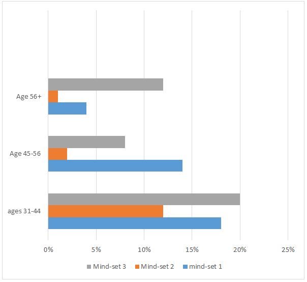
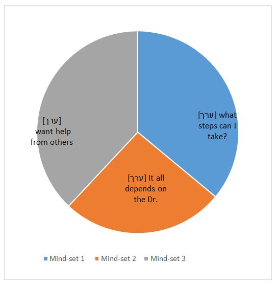
The rated importance of the information presented by the narrative message on type 2 diabetes varied across groups. Respondents in the youngest group rated information as unimportant. Members of the other age groups rated the information as moderately important and very important. Males had a higher additive constant indicating that they will require less specific information to reach risk awareness than will females. The differences in response patterns of different groups showed that there are three distinct mind-sets. Table 2 presents the coefficients for the total panel and for the three mind-set segments reflecting different patterns of responses to the importance of each narrative statement contributing to the perceived risk as emerged from Mathematical K-clustering analysis. Figure 1 presents sample distribution by mind-set segments and Figure 2 presents sample distribution into mindset-segments by age group.

Table 2: Coefficients for total panel and the three emergent mindsets based upon the patterns of coefficients of narrative messages.

Total Mindset 1 Mindset 2 Mindset 3
Base 50 18 13 19
Additive constant 59 62 43 67
Mind-Set 1 – The patient is in control, and must take responsibility (psychology)
A3 Diet and exercise are key to type 2 Diabetes prevention 8 17 4 3
A2 TYPE 2 DIABETES is dangerous without treatment 7 14 8 -1
Mind-Set 2 – The doctor is very important (sociology)
C3 A doctor should refer patients to educational materials about type 2 Diabetes -3 -13 20 -11
C1 It’s a doctor’s role to educate patients about type 2 Diabetes -4 -13 16 -9
C4 A patient should know all the possible treatments of Type 2 Diabetes 0 -3 12 -7
Mind-Set 3 – Help from others is important (Social structure & Services)
D3 Participation in workshops for patients helps manage Type 2 Diabetes 2 -12 -2 19
D1 Family support is important to manage type 2 Diabetes 5 0 0 16
D4 Belonging to a community of patients helps support others with type 2 Diabetes 1 -10 -3 15
B3 Frequent doctor visits help adherence to type 2 Diabetes treatment 4 4 -8 11
D2 Learning how others cope with type 2 Diabetes is beneficial 3 -5 4 8
Elements which are not key to any mind-set (Cost & Health services)
B2 People with type 2 Diabetes use a lot of health services -3 -4 -13 2
B4 Type 2 Diabetes requires a lot of medications -5 5 -22 -4
A4 Type 2 Diabetes is the most profound disease of this century -3 4 0 -10
B1 It’s OK to self-manage the type 2 Diabetes -17 -14 -27 -13
A1 By living longer there is a greater chance of suffering from Type 2 Diabetes -9 1 -5 -20
C2 The internet is all you need to learn about diabetes -26 -24 -4 -42

fig 1

Figure 1: Risk Awareness of Type 2 Diabetes in Healthy Individuals.

fig 2

Figure 2: Distribution into Mindsets by Age.

Applying Narrative Communication Messaging for Raising Awareness to Type 2 Diabetes

To identify which communication messaging should be used in practice, we applied the personal viewpoint identifier (PVI) which is created a new for each study and in this case revealed the mind-set of an individual through a simple, 30-second interaction. The respondent completed a short, 6-question evaluation, with the pattern of the responses linked to membership in one of the three mind-sets. The six most discriminating narrative messages were chosen to create the PVI which then created a binary scale on which new participants may rapidly indicate their answers. Based on these answers, the Mind-Genomics system instantly presents the sample mind-set membership for the individual in the population, who can then be given the most effective messaging for the mind-set to which the individual appears to belong. The PVI is available at: http://162.243.165.37:3838/TT37

Discussion

This study tested narrative persuasion messages as drivers of public awareness of risks of type 2 diabetes. This study makes theoretically, methodologically and practical contribution. Theoretically, this study extended the narrative persuasion theory testing narrative communication messaging in the health context, focusing on risk awareness in type 2 diabetes among healthy people. Methodologically, this study explored narrative communication messaging through a multi-disciplinary view, from different perspectives applying an innovative conjoint-analysis procedure. Practically, findings direct professionals on a local level and on a national health policy level to use narrative communication messaging that impact the risk perception of healthy people, by mind-set segments, regarding type 2 diabetes. Findings show that all study hypotheses were corroborated and also suggest that just as narrative messaging was effective in public campaigns on general health issues, it can be effective for public campaigns targeting awareness of risks in type 2 diabetes for the healthy population [21].

As for hypothesis 1, stating that different narrative communication messaging will have a different effect on perceived risks of type 2 diabetes, findings suggest that a number of narrative messages, that may be used on local level by professionals appear to be ineffective, and may create antagonism and anxiety, rather than raise public awareness of risks of type 2 diabetes and navigate individuals towards the adoption of healthy behaviors. Such messages are: People with type 2 diabetes use a lot of health services; Type 2 diabetes requires lots of medications; Type 2 diabetes is the most profound disease of this century and; living longer raises the risk of suffering from Type 2 diabetes. This finding may be attributed to the underlying communication orientation in these narrative messages. Ineffective messages fall under the category of content-oriented messaging which focuses on ‘what the public should know about type 2 diabetes’. Whereas content-oriented messaging is popular, it does not promote behavioral change, particularly compared to change-process oriented communication which was found to be a ‘make or break’ factor in behavioral modifications, including in type 2 diabetes [56,57].

As for hypothesis 2 stating that different narrative communication messages regarding risk of type 2 diabetes will carry a different appeal for different people by mindset segments, data show that response patterns to narrative messages differed among groups of people, differentiating them by mindset segments. People who are members of mind-set1 will react to messages depicting individuals as having control and able to take responsibility for their health stressing the psychology view (e.g., Diet and exercise are key to type 2 diabetes prevention; Type 2 diabetes is dangerous without treatment). This finding supports previous findings on the role of communication in promoting perceived control in behavioral modifications [56,57]. Outreach to people belonging to mindset l should entail information on: steps in the behavioral change process; what may assist them throughout the change process; what internal and external resources they may use; and how they may overcome barriers [58].

Messaging is to be inspirational and hopeful enhancing self-efficacy [57,58]. People who belong to Mindset2, the smallest mindset segment, will positively react to narrative messages depicting the doctor as a resource stressing the sociological view (e.g., A doctor should refer patients to educational materials about type 2 diabetes; It’s the doctor’s role to educate patients about type 2 diabetes; A patient should know all the possible treatments of type 2 diabetes). This finding supports previous studies on the sociological role of trust in physicians and the public expectation from physician to inspire them in adopting healthy behaviors [57,59].

People who are members of Mindset 3, will positively react to narrative messages depicting help from others stressing the economics and health management view (e.g., Participation in workshops helps prevent type 2 diabetes; Belonging to a community helps maintain healthy behaviors to prevent type 2 diabetes; learning how others cope with challenges is beneficial), focusing on community and available supporting health services. These findings support previous studies that claimed that communication messaging in national public campaigns may improve awareness of type 2 diabetes risks in healthy populations and among different segments of the population [23,29]. Study findings echo previous findings on the strong impact that narrative messaging may have on attitude change and behavioral modifications [29,31]. As for hypothesis 3, stating that different narrative communication messages will have a greater impact on perceived responsibility of individuals to modify their health behaviors and avoid risks of type 2 diabetes, the membership of people to mindset-segments entailed differences in their perceived responsibility. Perceived responsibility for reducing risks of type 2 diabetes ranged from self- responsibility (segment 1) to doctor’s responsibility (segment 2) and to community as having a role in helping its healthy members prevent risks of type 2 diabetes. Findings support previous studies stating that narrative messaging may play an important role in health communication campaigns to influence attitudes, intentions, and health behaviors [35].

Practice Implications

The knowledge derived from this research enables policy makers to accord the most effective narrative communication messaging to each person and group in the population, by the mind-set segmentation, in the sample. The segmentation by mindset suggests that a public communication campaign with the same messages for all, will be futile. When designing campaigns to raise public awareness of risks of type 2 diabetes, content-oriented messages should be omitted. Figures 1 and 2 indicate that segments of mindsets1 and 3 are about the same size but as age increases, on one hand the perceived importance of this information increases but perceived individual’s responsibility to modify risk behaviors decreases. People in the oldest group (56+) who are under the highest risk of type 2 diabetes, attribute the highest authority to doctors compared to people in other age groups and expect support of doctors, the community and health services (i.e. workshops) in behavior modification to prevent type 2 diabetes. These data suggest that in campaigns targeting people who are older than 45 years old, messaging is to be based on the change-process orientation rather than content-oriented. Last, three campaigns are to be designed, each targeting people in a different mindset segments and each focusing on a different perspective. One campaign entailing messages of perceived control over health (psychology), another campaign entailing messages of advice from trusted professionals (sociology) and the third campaign offering health-services (i.e., workshops and mentorship of supporting individuals) (health management) for maintaining healthy behaviors.

Future Studies

Future studies may continue to test unique features of brief printed-based narrative messaging and their effect on health attitudes and intentions. Future studies may also broaden our knowledge as to the effect of culturally tailored messaging on risk awareness in urgent health problems.

Conclusion

To sum, this study closed a knowledge gap in the state of the art examining multi-perspective narrative persuasive messaging as means to raise awareness of type 2 diabetes in healthy populations. This study presents a new approach to raise public awareness of risks of type 2 diabetes through narrative messaging by mindset segmentation. To enhance effectiveness in raising awareness of type 2 diabetes risks and to wisely allocate budgets, health professionals, policy makers and public campaign designers, are urgently called upon to use the VPI, ask the right few questions to identify the belonging of each individual in the healthy population to a mindset segment in the sample and use the appropriate narrative communication messaging per segment in raising awareness. The theoretical, methodological and practical knowledge derived from this study will enable policy makers and professionals to accord the most effective narrative communication messaging to each person and group in the population, by the mindset segmentation, in a cost-effective manner.

References

    1. Bommer C, Heesemann E, Sagalova V, Manne-Goehler J, Atun R, et al. (2017) The global economic burden of diabetes in adults aged 20-79 years: a cost-of-illness study. The Lancet Diabetes & Endocrinology 5: 423-430.
    2. Gregg EW (2017) The changing tides of the type 2 diabetes epidemic-smooth sailing or troubled waters ahead? Kelly West Award Lecture 2016. Diabetes Care 40: 1289-1297. [crossref]
    3. Rubin RR, Peyrot M, Siminerio LM (2006) Health care and patient-reported outcomes: results of the cross-national Diabetes Attitudes, Wishes and Needs (DAWN) study. Diabetes Care 29: 1249-1255. [crossref]
    4. Tiv M, Viel JF, Mauny F, Eschwege E, Weill A, et al. (2012) Medication adherence in type 2 diabetes: the ENTRED study 2007, a French population-based study. PLoS One 7: e32412.
    5. Carratalá‐Munuera FMC, Gil‐Guillen VF, Orozco‐Beltran D, Navarro‐Pérez J, Caballero‐Martínez F, et al. (2013) Barriers associated with poor control in S panish diabetic patients. A consensus study. International Journal of Clinical Practice 67: 888-894.
    6. Shadi F, Mohammad AS, Amir HZ, Amini M (2011) Effect of pharmacist-led patient education on glycemic control of type 2 diabetics: a randomized controlled trial. Journal of Research in Medical Sciences 16: 43-49. [crossref]
    7. Walker EA, Shmukler C, Ullman R, Blanco E, Scollan-Koliopoulus M, et al. (2011) Results of a successful telephonic intervention to improve diabetes control in urban adults: a randomized trial. Diabetes Care 34: 2-7. [crossref]
    8. American Diabetes Association (2013) Standards of medical care in diabetes. Diabetes Care 36: S11-S66.
    9. Atlas, Diabetes (2015) International diabetes federation. IDF diabetes atlas. Brussels: International Diabetes Federation.
    10. Guangwei L, Zhang P, Wang J, Gregg EW, Yang W, et al. (2008) The long-term effect of lifestyle interventions to prevent diabetes in the China Da Qing Diabetes Prevention Study: a 20-year follow-up study. The Lancet 371: 1783-1789.
    11. Diabetes Prevention Program Research Group (2009) 10-year follow-up of diabetes incidence and weight loss in the Diabetes Prevention Program Outcomes Study. The Lancet 374: 1677-1686.
    12. Diabetes Prevention Program (DPP) Research Group (2002) The Diabetes Prevention Program (DPP): description of lifestyle intervention. Diabetes Care 25: 2165-2171.
    13. Sepah S, Cameron LJ, Peters AL (2015) Long-term outcomes of a Web-based diabetes prevention program: 2-year results of a single-arm longitudinal study. Journal of Medical Internet Research 17: e92. [crossref]
    14. Subramanian K, Inuka M, Chellapilla V (2017) Overcoming the challenges in implementing type 2 diabetes mellitus prevention programs can decrease the burden on healthcare costs in the United States. Journal of Diabetes Research. [crossref]
    15. Diabetes Prevention Program Research Group (2013) The 10-year cost-effectiveness of lifestyle intervention or metformin for diabetes prevention: an intent-to-treat analysis of the DPP/DPPOS. Diabetes Care 36: 4172-4175. [crossref]
    16. Butcher MK, Karl K, Vanderwood TO, Hall D, Helgerson GSD, et al. (2011) Capacity of diabetes education programs to provide both diabetes self-management education and to implement diabetes prevention services. Journal of Public Health Management and Practice 17: 242-247. [crossref]
    17. Bloomfield HE, Greer N, Kane R, Wilt JT (2017) Effects on health outcomes of a Mediterranean diet with no restriction on fat intake. Annals of Internal Medicine 166: 378-379.
    18. Riddle MC, Herman WH (2018) The cost of diabetes care-an elephant in the room. Diabetes Care 41: 929-932. [crossref]
    19. Reem K, Slater N, Sahi A, Mepani D, Lalji K, et al. (2019) Type 2 Diabetes: how informed are the general public? A cross-sectional study investigating disease awareness and barriers to communicating knowledge in high-risk populations in London. BMC Public Health 19: 138. [crossref]
    20. Okosun IS, Davis-Smith M, Seale JP (2012) Awareness of diabetes risks is associated with healthy lifestyle behavior in diabetes free American adults: evidence from a nationally representative sample. Primary Care Diabetes 6: 87-94. [crossref]
    21. Rogers EA, Fine S, Handley MA, Davis H, Kass J, et al. (2014) Development and early implementation of the bigger picture, a youth-targeted public health literacy campaign to prevent type 2 diabetes. Journal of Health Communication 19: 144-160. [crossref]
    22. Niederdeppe JQ, Bu L, Porismita B, Kindig DA, Robert SA (2008) Message design strategies to raise public awareness of social determinants of health and population health disparities. The Milbank Quarterly 86: 481-513. [crossref]
    23. Randolph KA, Whitaker P, Arellano A (2012) The unique effects of environmental strategies in health promotion campaigns: a review. Evaluation and Program Planning 35: 344-353.
    24. Robert SA, Booske BC (2011) US opinions on health determinants and social policy as health policy. American Journal of Public Health 101: 1655-1663. [crossref]
    25. Clarke CE, Niederdeppe J, Lundell HC (2012) Narratives and images used by public communication campaigns addressing social determinants of health and health disparities. International Journal of Environmental Research and Public Health 9: 4254-4277. [crossref]
    26. Siegel K, Venkat Narayan (2008) The Unite for Diabetes campaign: overcoming constraints to find a global policy solution. Globalization and Health 4: 1-8. [crossref]
    27. Somannavar S, Lanthorn H, Deepa MR, Pradeepa MR, Mohan V (2008) Increased awareness about diabetes and its complications in a whole city: Effectiveness of the “prevention, awareness, counselling and evaluation [PACE] Diabetes Project [PACE-6]. Journal of the Association of Physicians of India 56: 495-502. [crossref]
    28. Lundell HC, Niederdeppe J, Clarke CE (2013) Exploring interpretation of complexity and typicality in narratives and statistical images about the social determinants of health. Health Communication 28: 486-498. [crossref]
    29. Hinyard LJ, Kreuter MW (2007) Using narrative communication as a tool for health behavior change: a conceptual, theoretical, and empirical overview. Health Education and Behavior 34: 777-792. [crossref]
    30. Moyer-Gusé E (2008) Toward a theory of entertainment persuasion: Explaining the persuasive effects of entertainment-education messages. Communication Theory 18: 407-425.
    31. Schank R, Berman T (2002) The pervasive role of stories in knowledge and action.
    32. Gabay G (2019) Patient self-worth and communication barriers to trust of Israeli patients in acute-care physicians at public general hospitals. Qualitative Health Research 29: 1954-1966. [crossref]
    33. Bilandzic H, Busselle RW (2011) Enjoyment of films as a function of narrative experience, perceived realism and transportability. Communications 36: 29-50.
    34. Busselle R, Bilandzic H (2009) Measuring narrative engagement. Media Psychology 12: 321-347.
    35. Shen F, Sheer VC, Li R (2015) Impact of narratives on persuasion in health communication: A meta-analysis. Journal of Advertising 44: 105-113.
    36. Van Laer T, De Ruyter K, Visconti LM, Wetzels M (2014) The extended transportation-imagery model: A meta-analysis of the antecedents and consequences of consumers’ narrative transportation. Journal of Consumer Research 40: 797-817.
    37. Morgan SE, Movius L, Cody MJ (2009) The power of narratives: The effect of entertainment television organ donation storylines on the attitudes, knowledge, and behaviors of donors and non-donors. Journal of Communication 59: 135-151.
    38. Moyer-Gusé E, Chung AH, Jain P (2011) Identification with characters and discussion of taboo topics after exposure to an entertainment narrative about sexual health. Journal of Communication 61: 387-406.
    39. Moyer-Gusé E, Nabi RL (2010) Explaining the effects of narrative in an entertainment television program: Overcoming resistance to persuasion. Human Communication Research 36: 26-52.
    40. Murphy ST, Frank LB, Chatterjee JS, Baezconde-Garbanati L (2013) Narrative versus non-narrative: The role of identification, transportation, and emotion in reducing health disparities. Journal of Communication 63: 116-137. [crossref]
    41. Van Leeuwen L, Renes RJ, Leeuwis C (2013) Televised entertainment-education to prevent adolescent alcohol use: Perceived realism, enjoyment, and impact. Health Education & Behavior 40: 193-205. [crossref]
    42. Fishbein M, Ajzen I (2010) Prediction and change of behavior: The reasoned action approach. Taylor and Francis Publishing, New York.
    43. Kim HS, Bigman CA, Leader AE, Lerman C, Cappella JN (2012) Narrative health communication and behavior change: The influence of exemplars in the news on intention to quit smoking. Journal of Communication 62: 473-492. [crossref]
    44. Tal-Or N, Cohen J (2010) Understanding audience involvement: Conceptualizing and manipulating identification and transportation. Poetics 38: 402-418.
    45. Graaf DA, Hoeken H, Sanders J, Beentjes JW (2012) Identification as a mechanism of narrative persuasion. Communication Research 39: 802-823.
    46. Slater MD, Rouner D (2012) Entertainment-education and elaboration likelihood: Understanding the processing of narrative persuasion. Communication Theory 12: 173-191.
    47. Banerjee SC, Greene K (2012) Role of transportation in the persuasion process: Cognitive and affective responses to antidrug narratives. Journal of Health Communication 17: 564-581. [crossref]
    48. Gollust SE, Lantz PM, Ubel PA (2009) The polarizing effect of news media messages about the social determinants of health. American Journal of Public Health 99: 2160-2167. [crossref]
    49. Schank RC, Berman T (2006) Living stories: Designing story-based educational experiences. Narrative Inquiry 16: 220-228.
    50. Gabay G (2020) From the crisis in acute care to post-discharge resilience-The communication experience of Geriatric patients: A qualitative study. Scandinavian Journal of Caring Sciences.
    51. Green PJ, Douglas C, Goldberg SA (1981) General approach to product design optimization via conjoint analysis. Journal of Marketing 45: 17-37.
    52. Gofman A, Moskowitz HR (2010) Isomorphic permuted experimental designs and their application in conjoint analysis. Journal of Sensory Studies 25: 127-145.
    53. Moskowitz HR (1997) Base size in product testing: A psychophysical viewpoint and analysis. Food Quality and Preference 8: 247-255.
    54. Gabay G, Gere A, Stanley J, Habsburg-Lothringen C, Moskowitz HR (2019) Health threats awareness–responses to warning messages about Cancer and smartphone usage. Cancer Studies Therapeutics Journal 1-10.
    55. Orme B (2010) Getting started with conjoint analysis: strategies for product design and pricing research second edition. Madison: Research Publishers LLC.
    56. Ratanawongsa N, Karter AJ, Parker M, Heisler LM, Moffet HH, et al. (2013) Communication and medication refill adherence: the diabetes study of northern California. JAMA Internal Medicine 173: 210-218. [crossref]
    57. Gabay G (2015) Perceived control over health, communication and patient–physician trust. Patient Education and Counseling 98: 1550-1557. [crossref]
    58. Gabay G (2016) Exploring perceived control and self-rated health in re-admissions among younger adults: A retrospective study. Patient Education and Counseling 99: 800-806. [crossref]
    59. Gabay G, Moskowitz HR (2012) The algebra of health concerns: implications of consumer perception of health loss, illness and the breakdown of the health system on anxiety. International Journal of Consumer Studies 36: 635-646.

To What Extent do Fracture Clinic Patients use Smart Mobile Technology and What are Their Specific Educational and Rehabilitee Needs that can be Addressed

DOI: 10.31038/IJOT.2020324

Abstract

Introduction: The fracture burden of the UK utilises a vast proportion of National Health Service (NHS) resources. Subsequent complications in healing result in poor patient outcomes (indirect costs) and increased demand on healthcare services (direct costs). Inadequate education regarding risk factors for poor outcomes provides a target for intervention. The increasing proportion of smartphone users makes smartphone applications (apps) a viable platform from which to distribute educational resources and conduct research.

Methods: A questionnaire was distributed randomly to 100 patients attending fracture clinic at the Queen Elizabeth Hospital in Birmingham over a twelve-week period. The mean age was 46 years (range 19 to 78), 52% female and a third were aged over 60 years. Primary questions determined the proportion of smartphone users, specifically those willing to utilise apps as an educational resource. Secondary information collected included patients’ concerns, smoking status, interest in smoking cessation and awareness of the risk factors affecting fracture healing.

Results: Almost 72% of responders used a smartphone, 71% would use an app for education and 74% would allow their data to be utilised for research. Some 60% of smokers would engage with cessation therapy through an app. The two greatest concerns identified were healing time (46%) and the long-term consequences of a fracture (46%). NSAID use was reported in 30%, however only 20% identified these as risk factor.

Conclusion: he majority of fracture clinic patients use smartphones and are were willing to utilise apps for both healthcare education and research. This could provide a cost-effective solution to an existing void in patient awareness. Developing an out-patient data collection tool offers new opportunities to epidemiological researchers.

Keywords

Fracture, Smartphone, Non-union, Education; Virtual clinic

Introduction

The incidence rate of fractures in the United Kingdom is approximated at 3.6 per 100 people per year [1]. According to recent statistics from Public Health England, the number of people aged 65 and over will increase by 40% over the next 17 years. In any year, 35% of these individuals will have a fall, and 5% of these falls will result in a fracture [2]. Fractures however, are not unique to the elderly; evidence suggests that almost one third of children will experience at least one fracture before the age of 17 [3]. This rising burden presents an immense challenge to existing NHS services striving to deliver urgent, definitive and complete follow-up care [4].

The impact on healthcare services is further increased by complications of fracture treatment, which require extended periods of follow-up and in some cases, readmission (direct costs). These complications affect up to 10% of patients within the two years of the initial injury [5]. Patients affected by complications have reported a reduction in their quality of life and a failure to return to their premorbid activity levels (indirect costs) [5,6]. Increased risk of delayed union and non-union has been associated with multiple risk factors. Whilst some, such as age, ethnicity and gender are non-modifiable, others, including prolonged NSAID use, smoking and poorly regulated diabetes management are potentially modifiable. These modifiable factors provide therapeutic targets for intervention [7-9].

Increasingly busy fracture clinics impede clinician’s ability to deliver information on fracture rehabilitation and meaningful dialogue on modifiable risk factors. This setting hinders the clinician’s ability to address patient concerns, such as answering individualised questions on healing times and functional impairment. As fracture clinics evolve, we see reduced face to face encounters with patients, the introduction of physician assistants and virtual clinics. Improved self-management following a fracture has the potential to improve patient outcomes and reduce the strain on healthcare services. Whilst there is extensive evidence for the relationship between modifiable risk factors and fracture healing, there is limited evidence surrounding public awareness of these. Without first establishing and improving public awareness, it follows that limited progress will be made in reducing the number of poor outcomes following acute fractures.

Over recent years, the number of individuals with access to a smartphones has risen exponentially [10]. A recent systematic review investigated the use of healthcare related smartphone applications by both medical professionals and patients. Findings suggested that these applications had a vital role to play in patient education, disease management and the remote monitoring of patients [11]. Considering the incidence of fractures within the UK population and the ability to therapeutically target modifiable risk factors, it is therefore surprising that to date, no smartphone application exists to educate the public on these risk factors and to give advice about fracture management. As a result, we hypothesise that current patient education regarding risk factors may be improved via education delivered through a smartphone application.

This study therefore aims to quantify the possession of smartphones in patients attending fracture clinic, and secondarily, report awareness of risk factors for fracture healing and attitudes towards the use of smartphone applications in acute fracture management.

Method

Over a twelve-week period, from June to August 2017, 144 patients attending fracture clinic at the Queen Elizabeth Hospital in Birmingham were asked to complete a questionnaire regarding their acute fracture in the waiting room. Of those approached, 100 voluntarily returned the completed forms. All patients seen in the fracture clinic for a first or follow-up appointment following both surgical and conservative management were included. The host hospital is a major trauma centre treating skeletally mature patients and comprises a limb reconstruction service. Patients not admitted via the emergency department are seen within 24 to 48 hours of their injury in fracture clinic. Skeletally immature patients were excluded as they are assessed at the regional Children’s’ Hospital.

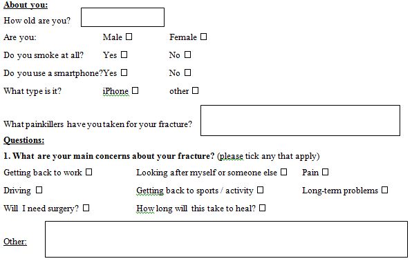
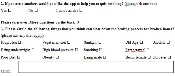
The questionnaire was designed with an ethos of simplicity to encourage compliance and precise questions to reduce data collection bias [12]. Prior to distribution, the questionnaire was peer reviewed by senior orthopaedic clinicians and amendments made. A pilot study was also conducted on a smaller number of patients, to assess feasibility of circulation and the willingness of patients to participate. An adverse event pathway developed in case of any incidents. The final questionnaire comprised of closed and open questions to provide quantitative and qualitative information. The questionnaire included questions relating to patients’ concerns about their fracture, smoking status, interest in smoking cessation and understating of the risk factors affecting bone healing. It also recorded their level of smartphone use and willingness to engage with smartphone application in relation to their healthcare (Appendix I).

Data was collated using Microsoft Excel (Office 2016) and statistical analysis was performed using IBM SPSS Statistics 23. The trusts ethical review board deemed the study to be a service evaluation. No funding was received for this study.

Results

A total of 100 fracture clinic patients competed the questionnaire over a six-week period. The mean age of participants was 46 years (SD 16.7, range 19 to 78 years). Of the patients who completed the questionnaire 29% were over 60 years of age and of the responders 52% were female.

Smartphone Use

Of the patients who completed the questionnaire, 72% used a smartphone, of which 52% owned Apple iPhones© with IOS Software. Non-smartphone users were predominantly female (n=26, 92%) and older (mean age 62, range 45 to 76). With regards to the role of smartphone application in patient education, 71% of participants would use an application to educate themselves and support their healing journey, 15 would not and 14 patients did not respond to the question. Similarly, 74% of participants would be happy for their information to contribute towards research.

Patient Concerns

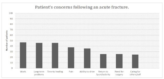
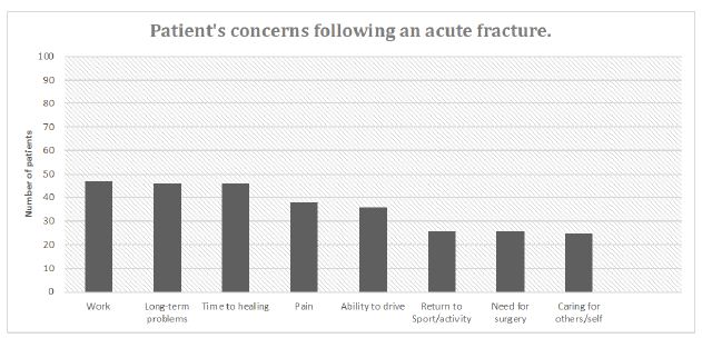
The two greatest concerns affecting patients were the time taken for the fracture to heal (46%) and the long-term consequences of an acute fracture (46%). Other concerns included functional aspects of the injury such as returning to work, driving and sport. Several patients also commented in free text box worries about the possibility of a recurrent fracture. Figure 1 below details patient concerns following an acute fracture.

fig 1

Figure 1: Patient concerns following an acute fracture.

Smoking

Of the 25 (25%) patients who admitted to smoking at the time of being interviewed 60% stated that they would engage with smoking cessation therapy via a smartphone application. Five respondents would not use smartphone applications to stop smoking and the remainder did not answer (n=5).

Analgesia

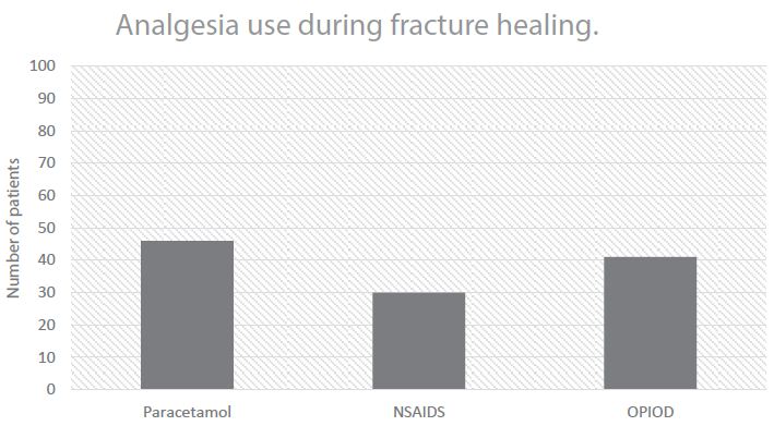
Of the patients who completed the questionnaire, 30% admitted to regular use of NSAIDs such as ibuprofen for analgesia. The most commonly used analgesic was paracetamol. Figure 2 shows the analgesia use following acute fracture.

fig 2

Figure 2: Patient analgesia use following acute fracture.

Perceived Risk Factors to Fracture Healing

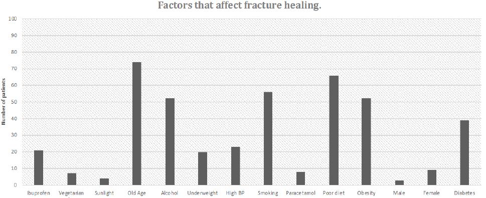
There was a wide variety of responses regarding the risk factors associated with poor outcomes following a fracture. There was an increased tendency for the patients to highlight lifestyle factors such as alcohol consumption, smoking, poor diet and obesity, all of which are modifiable. The most commonly recognised risk factor was age (74%). Other factors such as ibuprofen use (20%) and diabetes (39%), were less frequently identified, despite their recognised detrimental effects on fracture healing amongst medical professionals. Interestingly, a select number of participants attributed the use of paracetamol, vegetarian diet and hypertension as risk factors, despite the lack of evidence to substantiate these. Figure 3 below represents the commonly identified risk factors to fracture healing.

fig 3

Figure 3: Perceived risk factors to fracture healing.

Discussion

This study highlights the wide demographics of patients attending fracture clinic who could benefit from personalised management via smartphone applications, with over three quarters of those questioned having access to such devices. This provides a valuable platform from which to target a large population and opens the door to health interventions delivered via mobile application and web-based tools. Smartphone use was lower in older patients, particularly females, but we anticipate this to increase over the next decade. Correspondingly, 72% of participants were happy for their anonymised data to be used in research, which could facilitate the development of large, multi-centre databases via mobile applications to be used in epidemiological research. Investment by technology industries to create clinician and patient faced applications has increased. There are already devices to help patients regulate blood glucose levels, aid smoking caseation (QuitMedGuide, University of Texas, America) and help treat dementia. Primary care is being delivered remotely via face-to-face mobile platforms (GPatHand, Babylon, London) and clinicians are increasing using smartphones to transfer images, communicate and monitor patient care.

Despite follow-up in fracture clinic, where patient education should be incorporated into the consultation, it is concerning that patient’s still lack knowledge of less publicised risk factors such as the prolonged use of NSAIDs and optimising management of chronic diseases. The disparity of the results reinforces our hypothesis that patients lack understanding and need further education to encourage favourable behaviour and promote healing. We have not investigated the causality of poor information delivery but suggest it is sequelae of limited contact time with patients in fracture clinic as clinic sizes increase and resources decrease.

The proportion of smokers within the sample size is representative of the national average of 19%. The latter percentage decreased dramatically from an all-time high of 46% in 1974 [13]. Despite this relative decrease, smoking remains a considerable factor in non-union [9,14,15]. As of 2012, Abroms et al. identified 252 smoking cessation smartphone applications for iPhone and android devices [16]. They suggested that adherence with the applications was poor overall, despite a high download rate. Similar experiences have been reported when modifying other additive behaviours, such as alcoholism, where significant decreases in risky drinking behaviour in alcoholic patients were observed following the use of an application compared to controls [17]. We hypothesise that an acute fracture may provide an opportune moment for successful lifestyle modifications.

Interestingly, patient recognition of risk factors affecting fracture healing demonstrated a trend to identify modifiable lifestyle risk factors such as smoking, obesity and alcohol consumption. These risk factors are in general well publicised by prominent public health campaigns. Established In 2012, the ‘Stoptober’ campaign has been very successful in raising awareness of the association between smoking and lung cancer [18]. Smoking was identified as a risk factor for poor fracture healing by 53% of the population questioned, however evidence suggests that smoking is more commonly identified by the population as a risk factor for cancer [19]. These findings highlight the power of patient education and the ability to target vast numbers of people with the aim of improving their self-care. On the other hand, the discrepancy between the public awareness of the effects of smoking on cancer compared to fracture healing suggests that further information must be provided to fracture patients in order for them to make adequate lifestyle modifications.

Introduced in 2009, the NHS campaign ‘Change4Life’ was targeted specifically at dietary modification to reduce childhood obesity and subsequently promote a healthier nation. A year after its launch, over 400,000 families were participating with an awareness rate of 87% amongst parents with children in the target age range [20]. Since its development, the campaign has both expanded to incorporate adult health and modernised to utilise a smartphone application providing information on food groups, exercise and recipes. The success of these public health campaigns highlights the scalability of mobile devices and reinforces the importance of patient education as the key to reducing the burden of modifiable risk factors and their consequences on NHS services.

In addition to patient identified risk factors, primary patient concerns included the time taken for the fracture to heal, as well as long term consequences of a fracture. As these figures will vary according between individuals, providing tailored information to patients about appointments, long term follow up and recovery milestones would be of benefit. Utilising the smartphone platform to engage patients would enable them to record their past and upcoming appointments whilst gaining access to information regarding cast care, return to driving and physiotherapy exercises designed to promote rapid return of function. Access to all the necessary resources in one application would act as both a source of information and a method of alleviating patient concerns.

Limitations

Although a small study, the data collected from this population is consistent with national statistics. However, it is more challenging to compare subjective measures such as level of education and understanding. The sample tested did not include children with skeletally immature fractures. The rate of non-union in children is low and many do not have behaviours that require modification [21]. Considering the lower reading age of children and diminished understanding, they were excluded from the study and the application was tailored towards adults.

Those who failed to return a completed questionnaire were not included in the study, this may constitute an element of selection bias. Conversely, it may be that patients who were more technologically aware were more inclined to complete questionnaire. It would not be able to eliminate this source of bias this unless completion was mandatory. Factors such as reporting and recall bias are also valid considerations. Although the proportion of smokers identified was in keeping with the expected level, some patients may not have reported their smoking habits truthfully.

Conclusion

Overall, this preliminary research has demonstrated the widespread use of smartphones within the patient population attending fracture and the willingness of patients to engage with healthcare information using this platform. It has also confirmed that the majority of patients would accept having their anonymised data utilised for research purposes, which could facilitate the development of large, multi-centre databases via mobile applications.

A large proportion of patients were unaware of behaviours that impaired fracture healing and focused their concerns on finite endpoints, time to healing, ability to drive and long-term morbidity. Reinforcing the need for a more targeted approach to patient management following a fracture, to address these specific agendas. This study provides evidence to support the development of software that will improve overall treatment satisfaction, educate patients and modify behaviour that may in term improve fracture care outcomes. For example, mobile devices can play a vital role in appointment planning, information on cast care, basic physiotherapy exercises and fracture healing.

References

    1. Donaldson LJ, Reckless IP, Scholes S, Mindell JS, Shelton NJ (2208) The epidemiology of fractures in England. Journal of Epidemiology and Community Health 62: 174. [crossref]
    2. England PH. Falls and fracture consensus satement: Supporting commissioning for prevention. 2017.
    3. Cooper C, Dennison EM, Leufkens HG, Bishop N, van Staa TP (2004) Epidemiology of childhood fractures in Britain: a study using the general practice research database. Journal of Bone and Mineral Research 19: 1976-1981. [crossref]
    4. (NHFD) NHFD (2016) National Hip Fracture Database (NHFD).
    5. Ekegren CL, Gabbe BJ, Edwards ER, Steiger Rd, Page R (2016) 791 Incidence, costs and outcomes of non-union, delayed union and mal-union following long bone fracture. Injury Prevention.
    6. Court-Brown CM, McQueen MM (2008) Nonunions of the proximal humerus: their prevalence and functional outcome. The Journal of Trauma 64: 1517-1521. [crossref]
    7. Hernandez RK, Do TP, Critchlow CW, Dent RE, Jick SS (2012) Patient-related risk factors for fracture-healing complications in the United Kingdom general practice research database. Acta Orthopaedica 83: 653-660. [crossref]
    8. Jiao H, Xiao E, Graves DT (2015) Diabetes and its effect on bone and fracture healing. Current Osteoporosis Reports 13: 327-335. [crossref]
    9. Patel RA, Wilson RF, Patel PA, Palmer RM (2013) The effect of smoking on bone healing: A systematic review. Bone & Joint Research 2: 102-111. [crossref]
    10. The UK is now a smartphone society The communications market report 2015: Ofcom; 2015
    11. Mosa ASM, Yoo I, Sheets L (2012) A Systematic review of healthcare applications for smartphones. BMC Medical Informatics and Decision Making 12: 67. [crossref]
    12. Edwards P (2010) Questionnaires in clinical trials: Guidelines for optimal design and administration. Trials 11: 2. [crossref]
    13. (HSCIC) HaSCIC (2016) Statistics on Smoking, Englang.
    14. Sloan A, Hussain I, Maqsood M, Eremin O, El-Sheemy M (2010) The effects of smoking on fracture healing. The surgeon: Journal of the Royal Colleges of Surgeons of Edinburgh and Ireland 8: 111-116. [crossref]
    15. Castillo RC, Bosse MJ, MacKenzie EJ, Patterson BM (2005) Impact of smoking on fracture healing and risk of complications in limb-threatening open tibia fractures. Journal of Orthopaedic Trauma 19: 151-157. [crossref]
    16. Abroms LC, Westmaas JL, Bontemps-Jones J, Ramani R, Mellerson J (2013) A content analysis of popular smartphone apps for smoking cessation. American Journal of Preventive Medicine [crossref]
    17. Gustafson DH, McTavish FM, Chih MY, Atwood AK, Johnson RA, et al. (2014) A smartphone application to support recovery from alcoholism: A randomized clinical trial. JAMA Psychiatry 71: 566-572. [crossref]
    18. Stoptober Public Health England 2012.
    19. Sanderson SC, Waller J, Jarvis MJ, Humphries SE, Wardle J (2009) Awareness of lifestyle risk factors for cancer and heart disease among adults in the UK. Patient Education and Counselling 74: 221-227. [crossref]
    20. (BHFNC) TBHFNCfPAaH. Change4Life one year on. 2010.
    21. Mills LA, Simpson AH (2013) The risk of non-union per fracture in children. Journal of Children’s Orthopaedics 7: 317-322. [crossref]

Appendix I – Unify Questionnaire

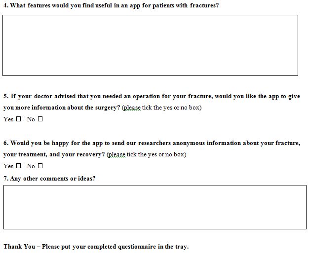
The University of Birmingham and Queen Elizabeth Hospital are designing a smartphone app for people with fractures. This app will be called MyFracture and will be tailored to each specific patient and their injury. Please could you help us by answering a few questions? Thank you.

Appendix fig 1

Appendix fig 2

Appendix fig 3

Neuroinvasive Action of SARS-CoV-2 in Coronavirus Disease (COVID-19): A Review

DOI: 10.31038/JNNC.2020324

Abstract

Background: SARS-CoV-2 can affect different organ systems, causing a range of symptoms that include fever, cough, myalgia, fatigue, headache, diarrhea and dyspnoea; in more severe cases acute respiratory distress, acute cardiac injury and secondary infection can develop and result in death. Skin lesions, ophthalmic changes and anosmia may also occur, which may be related to the effects of the virus on the central nervous system. Because it is an emerging virus, it is not yet known precisely how the virus can affect the human brain. The aim of the present study is therefore to investigate the causal relationship between SARS-CoV-2 infection and neurological manifestations, through scientific evidence.

Methods: A systematic search was carried out in the databases SciELO, Web of Science, Science Direct, PubMed, Embase and Scopus. Initially, 912 articles were identified; at the end of the selection process and after applying the inclusion and exclusion criteria, 18 articles were selected for this review.

Results: Of the selected articles, two did not associate the neurological symptoms with the new coronavirus; despite this, both did not exclude the possibility that COVID-19 actually has neuroinvasive potential. The remaining articles attributed the neurological changes evaluated to COVID-19.

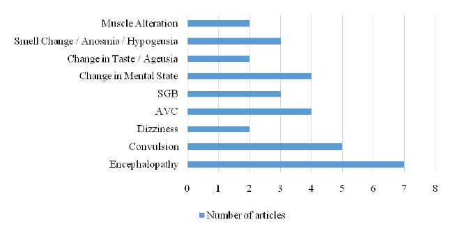
Discussion: It was reported that COVID-19 manifests itself neurologically in different ways, including stroke, Guillan-Barré syndrome, encephalopathy, convulsion, dizziness and altered consciousness.

Conclusion: Neurological symptoms that compromise the central nervous system should not be ruled out in the diagnosis of the new coronavirus. It is important to investigate neurological changes, with or without the presence of respiratory changes, as these changes may appear as the initial symptoms of COVID-19.

Keywords

COVID-19, Neuroinvasive action, Neurological changes, SARS-CoV-2

Introduction

In recent decades, a number of coronaviruses with different characteristics have appeared. Due to their capacity for mutation, recombination and infection, there is a possibility that new coronavirus variations will continue to appear [1]. Recently, a new coronavirus called SARS-CoV-2, which causes the disease COVID-19, appeared. It started in Wuhan, China, in December 2019, but quickly reached the level of a pandemic disease [2,3]. The initial reports of the infection characterized the condition as pneumonia of unknown origin; however, it has been observed that COVID-19 can affect different organ systems, causing a range of symptoms that include fever, cough, myalgia, fatigue, headache, diarrhea and dyspnoea. In more severe cases, acute respiratory distress, acute cardiac injury and secondary infection can develop which may result in death [4]. Some studies describe other clinical pictures that seem to be associated with the new disease, with symptoms such as skin lesions, ophthalmic changes and anosmia – manifestations that may be associated with the Central Nervous System (CNS) [5-7]. Since SARS-CoV-2 is an emerging virus, it is not yet known to what extent the virus can affect the human brain; therefore, the present study aims to investigate the causal relationship between SARS-CoV-2 infection and neurological manifestations, through scientific evidence.

Methods

This article is a systematic review based on the following guiding question: “Is there a causal relationship between COVID-19 and neurological manifestations?”

Search Strategy for the Identification of Studies

A search of the literature was undertaken in six databases: ScIELO, Web of Science, Science Direct, PubMed, Embase and Scopus; the descriptors (“COVID-19” OR “CORONAVIRUS” OR “SarS-CoV-2” OR “Coronavirus infections”) AND (“Neurological” OR “Nerve” OR “Brain” OR “Convulsion” OR “Nervous system”) were used. Two reviewers independently participated in each review phase (screening, eligibility and inclusion) and a third independent reviewer was invited to resolve any conflicts in the selection process.

Inclusion and Exclusion Criteria

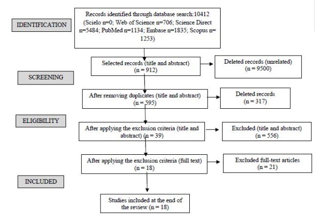
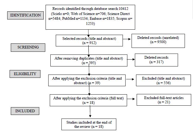
As an inclusion criterion, articles were selected that were available in full and free of charge in the databases, written in English, Portuguese or Spanish, and published in the period between 2019 and May 2020. The exclusion criterion were: systematic reviews, meta-analyses, letters to the editor, brief communications, perspective, editorials, studies addressing psychological problems, and articles that, after reading the title and abstract, could not identified as being relevant to the proposed theme. The first search of the databases identified 10,412 articles. After applying the inclusion criteria, 912 articles were selected, of which 317 were excluded because they were duplicate texts, leaving 595 articles to be analyzed from reading the title and abstract; after this reading, 556 articles that did not fit the study criteria were eliminated and the remaining 39 articles were selected for reading in full. After reading the full texts, 21 were excluded because they did not meet the criteria previously established, leaving 18 articles to be included in the review. Figure 1 shows the systematic search strategy used to select articles for this review.

Capture

Figure 1: Flowchart of search and selection of articles.

Results

The data were organized according to the title, authorship, country, correlation between the new coronavirus and neurological changes and which neurological changes were mentioned in the articles. Table 1 shows the general characteristics of the articles identified by our systematic review.

Table 1: Details of articles included in the review.

Title Author

Year

(Country)

Correlation Neurological manifestations
SARS-CoV-2 can induce brain and spine demyelinating lesions (Zanin et al., 2020) 2020

(Italy)

Yes ·         Anosmia

·         Ageusia

·         Convulsion

Neurological Complications of Coronavirus Disease (COVID-19): Encephalopathy, MRI Brain and Cerebrospinal Fluid Findings: Case 2 (Espinosa et al., 2020) 2020

(USA)

No ·         Encephalopathy

·         Altered mental state

Basal Ganglia Involvement and Altered Mental Status: A Unique Neurological Manifestation of Coronavirus Disease 2019 (Haddadi, Ghasemian and Shafizad, 2020) 2020

(Iran)

Yes ·         Encephalopathy


2019 novel coronavirus pneumonia with onset of dizziness: a case report (Kong et al., 2020) 2020

(China)

Yes ·         Dizziness
Neurological Complications of Coronavirus Disease (COVID-19): Encephalopathy (Filatov et al., 2020) 2020

(USA)

No ·         Encephalopathy

·         Severe change in                      mental status

COVID-19 and anosmia in Tehran, Iran (Gilani, Roditi and Naraghi, 2020) 2020

(Iran)

Yes ·         Hyposmia

·         Anosmia

·         Ageusia

Lessons of the month 1: A case of rhombencephalitis as a rare complication of acute COVID-19 infection (Wong et al., 2020) 2020

(England)

Yes ·         Encephalopathy

·         Rhomboencephalitis

A first case of meningitis/encephalitis associated with SARS-Coronavirus-2 (Moriguchiet al., 2020) 2020

(Yamanashi)

Yes ·         Encephalitis

·         Meningitis

·         Convulsion

·         Epileptic crisis

COVID-19 Presenting with Seizures (Sohal and Mansur, 2020) 2020

(USA)

Yes ·         Convulsion

·         Altered mental state

Early Guillain-Barré syndrome in coronavirus disease 2019 (COVID-19): a case report from an Italian COVID-hospital (Ottaviani et al., 2020) 2020

(Italy)

Yes ·         Guillain Barré Syndrome

·         Flaccid paralysis

·         Unilateral facial                        neuropathy

COVID-19 presenting as stroke (Avula et al., 2020) 2020

(USA)

Yes ·         Stroke
Facial diplegia, a possible atypical variant of Guillain-Barré Syndrome as a rare neurological complication of SARS-CoV-2 (Juliao Caamaño and Alonso Beato, 2020) 2020

(Spain)

Yes ·         Guillain Barré Syndrome

·         Facial nerve palsy

Guillain-Barré syndrome related to COVID-19 infection (Alberti et al., 2020) 2020

(Italy)

Yes ·         Guillain Barré Syndrome

·         Paresthesia

·         Severe flaccid                          tetraparesis

Two patients with acute meningo-encephalitis concomitant to SARS-CoV-2 infection (Bernard-Valnet et al., 2020) 2020

(Switzerland)

Yes ·         Non-convulsive                        epileptic focal status

·         Viral                                        meningoencephalitis

·         Tonic-clonic convulsion

·         Aseptic encephalitis

Plasmapheresis treatment in COVID-19-related autoimmune meningoencephalitis: Case series (Dogan et al., 2020) 2020

(Turkey)

Yes ·         Autoimmune                            meningoencephalitis
A Case of Coronavirus Disease 2019 With Concomitant Acute Cerebral Infarction and Deep Vein Thrombosis (Zhou, B. et al., 2020) 2020

(China)

Yes ·         Acute Cerebral Infarction
Coexistence of COVID-19 and acute ischemic stroke report of four cases (TUNÇ et al., 2020) 2020

(Turkey)

Yes ·         Acute ischemic stroke


Neurologic Manifestations of Hospitalized Patients with Coronavirus Disease 2019 in Wuhan, China


(Mao, Jin, et al., 2020)


2020

(China)


Yes

·         Ischemic stroke

·         Acute                                      cerebrovascular                        disease

·         Convulsion

·         Change in sense of taste

·         Change insenseofsmell

·         Dizziness

·         Change in the level of              consciousness

·         Muscle alteration

Cerebrovascular Accident (CVA)

It is believed that COVID-19, in addition to invading the respiratory system, also causes negative effects on the nervous systems of people who are affected by this disease. Four of the studies identified in the review presented evidence of a relationship between COVID-19 infection and the development of cerebrovascular accident (CVA). A study carried out in Turkey discussed the clinical status of four patients aged between 45 and 77 years diagnosed with COVID-19 and who had acute ischemic CVA. Three of the four patients had high levels of D-dimer, and two of them had high levels of C-reactive protein (CRP); these increases may have played a considerable role in the formation of ischemia. From these findings, the authors suggested that there may be a relationship between COVID-19 and the development of ischemic cerebrovascular diseases, independently of the critical disease process [8].

Similar evidence was found in a study in the USA, in which four patients, still in the early stages of COVID-19 infection, were also diagnosed simultaneously with acute ischemic CVA. The four patients were over 70 years old and had altered mental status, weakness on either side of the body, as well as difficulty in finding words. The authors suggested that the CVA may have occurred due to hypercoagulability, which is a pathophysiology directly related to the COVID-19 infection [9]. Another study reported a case in which a 75-year-old patient, who was being treated for COVID-19, suddenly had neurological symptoms, with changes in muscle strength. Computed tomography findings of the head showed that the patient had suffered cerebral infarction and had high levels of D-dimer, suggesting hypercoagulability [10]. Corroborating the findings from the aforementioned studies, a larger study conducted with 214 patients in China reported that there were strong indications that SARS-CoV-2 can infect the nervous system and skeletal muscle, in addition to the respiratory system. Clinical data showed that just over 35% of the study patients had neurological manifestations due to the infection. In people with severe infection, neurological involvement is greater, which includes ischemic or hemorrhagic CVA, acute cerebrovascular diseases, impaired consciousness and skeletal muscle damage [6].

Guillain-Barré Syndrome

Studies show that the development of Guillan Barré Syndrome (GBS) is associated with COVID-19. A study [11] reported on a 71-year-old patient who had an acute and severe peripheral nervous system disorder, manifested by the subacute onset of paresthesia in the extremities of the limbs, followed by distal weakness that quickly evolved into severe flaccid tetraparesis. In general, these clinical findings were interpreted as a severe form of acute polyradiculoneuritis with prominent demyelinating characteristics, thus diagnosing GBS associated with COVID-19 [11]. Another case occurred in Spain, where a 61-year-old patient infected with COVID-19 was diagnosed with paralysis of the right peripheral facial nerve with an eye reflex that did not respond to the stimulus, but without other neurological symptoms. Due to facial diplegia, the authors believe that this neurological disorder is a rare variant of GBS directly related to COVID-19 infection [12]. Similarly, in Italy, a 66-year-old patient diagnosed with COVID-19 infection presented a clinical picture consistent with GBS, which started with progressive difficulty in walking and acute fatigue, initial distal weakness in the upper limbs, diffuse areflexia, but without any clear sensory deficits. The patient progressively developed proximal weakness in all limbs, dysesthesias and unilateral facial paralysis, with increasing flaccid weakness of the limbs [13].

Encephalopathy

There were two cases of encephalopathy and COVID-19 in the same center, in very close periods. In the first case, a 72-year-old man hospitalized due to COVID-19 infection, did not show neurological improvement after sedation, did not respond to verbal commands, and did not show any reaction to noxious painful stimuli. For diagnostic clarification, an electroencephalogram (EEG) was performed after the patient had been sedated for 72 hours and showed only bilateral deceleration consistent with encephalopathy; however, when a puncture was performed with analysis of cerebrospinal fluid, no evidence was found to suggest meningitis or encephalitis. Thus, the authors concluded that the virus does not appear to cross the blood-brain barrier, even if patients with COVID-19 develop encephalopathy. In this case, the authors believe that the cause of encephalopathy was multifactorial, but stressed that the virus may have contributed to the encephalopathy [14]. In the second case at the same center [15] reported on a 74-year-old patient who presented encephalopathy with severe mental status changes, without verbal communication and unable to follow any command, but without motor changes. The study reported that patients with COVID-19 may have encephalopathy; however, the authors did not attribute the neurological complications to COVID-19, and did not consider encephalopathy to be a symptom or complication of COVID-19 in this case.

In a similar study in Iran, the authors reported that a 54-year-old patient diagnosed with COVID-19 presented disorders related to encephalopathy, manifesting severe changes in mental status, without verbal communication and unable to follow any order; however, he did not present any motor alteration, being able to move all of his extremities. Therefore, the authors emphasized the importance of considering neurological manifestations as a consequence of COVID-19, and stressed that it is necessary to understand the pathways of viral neuroinvasion in order to improve the fight against the new coronavirus [16]. Unlike the previously mentioned cases, in a study from the United Kingdom the authors attributed neurological changes to COVID-19 infection, in a case report in which a 40-year-old man developed acute brain stem dysfunction after being infected with COVID-19. Magnetic resonance imaging of the brain and cervical spine showed inflammation of the brain stem and upper cervical cord, leading to a diagnosis of inflammatory rhombencephalitis/myelitis, with the authors concluding that rhombencephalitis is a complication of COVID-19 infection [17].

A case of encephalitis as a result of COVID-19 was described by a study [18] in which a 24-year-old patient, hospitalized with a diagnosis of COVID-19, underwent an MRI scan that demonstrated abnormal findings in the temporal lobe, including in the hippocampus, suggesting encephalitis. The authors emphasized the importance of paying attention to the symptoms of encephalitis or cerebropathy, as these, in addition to respiratory symptoms, may be the first indication of SARS-CoV-2. A study [19] reported the case of two patients who developed autoimmune meningoencephalitis a few days after a diagnosis of mild COVID-19 infection. The patients developed severe neuropsychological symptoms suddenly and after specific exams, the hypothesis was that meningoencephalitis was derived from COVID-19; thus, the authors made the temporal association between acute SARS-CoV-2 infection and aseptic encephalitis with focal neurological symptoms and signs [19]. Finally, the study [20] reported on a series of cases in which the involvement of COVID-19 in the CNS was diagnosed in 6 of 29 intubated patients. A neurological investigation was carried out by means of specific tests, as some patients were unable to regain consciousness or developed agitated delirium during the weaning period, and found evidence that was compatible with autoimmune meningoencephalitis. The authors hypothesized that the neurological changes in the CNS were associated with COVID-19 [20].

Convulsions

In a case report, a 64-year-old patient with a positive result for COVID-19 infection presented with tonic-clonic seizure after being hospitalized with acute psychotic symptoms; he also had disorientation, strong attention deficit and psychotic symptoms. A routine electroencephalogram revealed non-convulsive epileptic focal status, which contributed to the association between acute COVID-19 infection and the neurological symptom [19]. In another study, a 54-year-old patient was admitted to hospital after being found unconscious at home; after a brief neurological examination the patient did not present any focal sensorimotor deficits. However, she presented disturbances of consciousness and convulsions, these symptoms being consistent with neurological involvement resulting from infection by SARS-CoV-2. The authors pointed out that sudden neurological impairment with convulsions in COVID-19 patients may occur due to CNS involvement and demyelinating lesions [21]. In agreement with the previous cases, another study reported a case of convulsion associated with COVID-19, in which a 24-year-old patient was transported to hospital following a convulsion and unconsciousness. In the ambulance, the patient had a transient generalized convulsion that lasted for about a minute. After arriving at hospital, the patient needed mechanical ventilation due to multiple epileptic convulsions. The differential diagnosis was post-convulsive encephalopathy [18].

A study in China reported the case of a patient who had a seizure characterized by the sudden onset of limb spasms, foaming at the mouth and loss of consciousness, which lasted for three minutes. The authors concluded that SARS-CoV-2 may infect the nervous system and cause several neurological changes [6]. Similarly, a case was presented in which a 72-year-old patient, with no previous history of convulsion, developed several convulsive episodes after being transferred to the intensive treatment unit due to complications derived from COVID-19. The causal relationship between COVID-19 and neurological manifestations, such as the appearance of convulsion, was highlighted [22].

Dizziness

A study carried out in China highlighted the presence of dizziness as an initial symptom of COVID-19 infection. A 53-year-old patient complained of sudden dizziness for three days, with no apparent cause. He did not show any other neurological symptoms, and had a normal MRI result. However, he presented the characteristic symptoms of the infection and tested positive for COVID-19. From these findings, the authors concluded that COVID-19 can manifest itself in the nervous system, and suggest that greater attention should be paid to complaints of dizziness by patients, as it may be an early symptom of COVID-19 [23]. In a series of retrospective and observational cases that included 214 hospitalized patients with a confirmed diagnosis in the laboratory of infection with the new coronavirus, in patients who manifested changes in the central nervous system, dizziness was the most common symptom, being reported by 36 of the patients. The authors concluded that the more severe the patient’s clinical condition, the worse the neurological manifestation associated with COVID-19 infection can be [6].

Other Neurological Manifestations

Studies cite other neurological changes, but not all discussed in detail how the changes occurred and how they were associated with COVID-19. Table 2 describes other manifestations mentioned in the studies selected by this review.

Table 2: Other manifestations described in the articles.

Neurological Alteration

References

Change in Taste/Ageusia (GILANI, RODITI AND NARAGHI, 2020; MAO et al., 2020a)
Smell Change/Anosmia/Hypogeusia (GILANI, RODITI AND NARAGHI, 2020; MAO et al., 2020a; ZANIN et al., 2020)
Change in mental status (ESPINOSA et al., 2020; FILATOV et al., 2020; Mao et al., 2020a; SOHAL E MANSUR, 2020)
Muscle alteration (ALBERTI et al., 2020; MAO et al., 2020a)

Most of the selected articles were case reports and some of them discussed more than one neurological disorder, not being restricted to just one symptom, as can be seen in Figure 2.

fig 2

Figure 2: Symptoms cited in articles.

Discussion

COVID-19 is a relatively new disease, and it is not yet known how it behaves neurologically. Of the articles in this review, only two of the studies did not associate the neurological symptoms described with the new coronavirus, although neither excluded the possibility that COVID-19 has neuroinvasive potential. The number of studies that aim to explore the virus’s action in the brain is growing, and there are several that have already shown neurological changes due to SARS-CoV-2 infection [24-28]. Current evidence, therefore, suggests that there is a risk of developing neurological diseases as a result of COVID-19, but it is not yet known what the possible long-term neurological sequelae might be [29]. In respect of this emerging evidence suggesting that SARS-CoV-2 is associated with neurological changes in patients with COVID-19, particularly in those with severe clinical manifestations, there are three feasible scenarios: the first is that the impact of SARS-CoV-2 on the CNS could lead to neurological changes directly; the second is that it could aggravate pre-existing neurological conditions; and the third is that it could increase susceptibility to the infection, or aggravate the damage it causes [30,31]. It is believed that, together with the host’s immunological mechanisms, SARS-CoV-2 can cause infections to result in neurological diseases [28].

A recent study highlighted the possibility that SARS-CoV-2 reaches the central nervous system through the olfactory bulb and infects the olfactory nerve; from there, it would spread to various parts of the brain via trans-synaptic transmission and infect the PreBotzinger complex (PBC) in the brain stem, the brain’s respiratory center that controls the lungs, shutting off breathing and potentially causing death [30]. Olfactory and gustatory disorders have been found as prevalent symptoms among infected patients, and studies have stressed the importance of recognizing anosmia and ageusia as relevant symptoms in the diagnosis of COVID-19 [32,33]. Hyposmia and hypogeusia, together with dizziness, headaches and stroke have all been widely reported in patients with COVID-19, and could all be associated with neurological changes that affect the central nervous system [31]. Among the various neurological changes described in the articles in this review, the most prevalent was encephalopathy and similar changes, also with the presence of seizures, data that corroborates other studies which discuss the presence of encephalopathy associated with the new coronavirus [34,35]. The presence of necrotizing encephalitis associated with COVID-19 was also seen, but without evidence of viral isolation from cerebrospinal fluid. This neurological change can lead to brain dysfunction caused by the virus, which results in seizures and mental disorientation after infection [36]. The presence of seizures in the clinical picture calls attention to the possible involvement of SARS-CoV-2 in the neurological system. They can occur as a result of infection, an acute systemic disease, a primary neurological disease, or the adverse effect of medication on critically ill patients, and can present a variety of symptoms, ranging from seizure activity, subtle spasms and even lethargy [36,37]. In a number of studies identified in the review, Guillain-Barré syndrome was considered by the authors to be a possible important neurological complication of COVID-19 infection. It is imperative that medical teams pay attention to the presence of this syndrome, as it can lead to admission to the intensive care unit (ICU) and needs to be differentiated from a possible weakness acquired in the ICU after treatment [38-40].

Studies carried out recently have observed that patients with COVID-19 have an increased risk of cerebrovascular disease the incidence is similar to other critical illnesses. This has been widely reported as causing cytokine storm syndromes that may be one of the factors that leads to stroke [37,41]. Ischemic stroke associated with COVID-19 infection can occur in the context of a highly prothrombotic systemic state, and it is necessary to pay extra attention to the signs, as there is a strong association between SARS-CoV-2 and the development of systemic thromboembolisms due to a hypercoagulable state. This possibility, regardless of the patient’s age, makes it necessary to increase the awareness of the health team about severe forms of ischemia and systemic stroke in patients with signs of COVID infection, in order to provide all patients with the best possible care [42-44]. In view of the severity of the neurological changes that can occur in infected patients, early assessments of neurological symptoms are necessary. The assessment should investigate headaches, disturbances in consciousness, paresthesia, ageusia, paralysis, among other factors. Timely cerebrospinal fluid analysis, and awareness and management of infection-related neurological complications are critical to improving the prognosis of critically ill patients [28].

Conclusion

The new coronavirus manifests itself neurologically in different ways, including headache, paraesthesia, encephalopathy, altered consciousness, Guillain-Barré syndrome, dizziness, seizure, stroke, among other conditions. These different neurological symptoms that affect patients and compromise the central nervous system should not be ruled out in the differential diagnosis of the new coronavirus. The health professional needs to be aware of these symptoms and request an evaluation from a neurologist to confirm the clinical suspicions; close attention should be paid to possible neurological conditions in patients who are suspected of having COVID-19 infection. The respiratory changes caused by COVID-19 infection are well documented, but it is important that attention is also focused on neurological changes, as they can cause serious damage to patients affected by the infection. This is particularly the case with patients who have comorbidities or are part of the risk group with previous diseases, as they are more susceptible to developing complications.

It is not yet known with certainty whether the neurological damage is temporary or permanent after the patient has recovered from COVID-19. Studies that aim to verify the presence of neurological damage in patients previously affected by the new coronavirus are necessary, so that the extent of any damage can be scientifically assessed. As the disease only emerged relatively recently, there is still a lack of knowledge in the scientific community regarding the subjects addressed here. Finally, the importance of investigating neurological changes is emphasized, with or without the presence of respiratory changes, as these changes may appear as the initial symptoms of COVID-19, thus being able to help in a faster and more accurate diagnosis, in order to provide better treatment for the patient.

Acknowledgments

The authors dedicate this article to all health professionals who are facing COVID-19. We are eternally grateful to them, and hope that our article can contribute to reducing the number of deaths.

Author contributions: All authors have read and agreed to the published version of the manuscript.

Funding: This research was not funded. The authors themselves financed it.

Conflicts of interest: The authors have declared that there is no conflict of interest.

References

    1. Fehr AR, Perlman S (2015) Coronaviruses: An Overview of Their Replication and Pathogenesis. Coronaviruses: Methods and Protocols 1282: 1-282. [crossref]
    2. Zhu N, Zhang D, Wang W, Li X, Yang B, et al. (2020) A Novel Coronavirus from Patients with Pneumonia in China, 2019. New England Journal of Medicine 382: 727-733. [crossref]
    3. World Health Organization (2020) Coronavirus disease (COVID-19) pandemic. Geneva: WHO. URL: htps://www.who.int/emergencies/diseases/novel-coronavirus-2019.
    4. HUANG C, WANG Y, LI X, REN L, ZHAO J, et al. (2020) Clinical features of patients infected with 2019 novel coronavirus in Wuhan, China. The Lancet 395: 497-506. [crossref]
    5. GALVÁN CC, CATALÀ A, CARRETERO HG, RODRÍGUEZ JP, FERNÁNDEZ ND, et al. (2020) Classification of the cutaneous manifestations of COVID-19: a rapid prospective nationwide consensus study in Spain with 375 cases. British Journal of Dermatology 183: 71-77. [crossref]
    6. MAO L, JIN H, WANG M, HU Y, CHEN S, et al. (2020) Neurologic Manifestations of Hospitalized Patients With Coronavirus Disease 2019 in Wuhan, China. Jama Neurology 77: 683-690. [crossref]
    7. ZHOU B, SHE J, WANG Y, MA X (2020) A Case of Coronavirus Disease 2019 with Concomitant Acute Cerebral Infarction and Deep Vein Thrombosis. Frontiers in Neurology 11: 2-5. [crossref]
    8. TUNÇ A, ÜNLÜBAŞ Y, ALEMDAR M, AKYÜZ E (2020) Coexistence of COVID-19 and acute ischemic stroke report of four cases. Journal of Clinical Neuroscience 77: 227-229. [crossref]
    9. AVULA A, NALLEBALLE K, NARULA N, SAPOZHNIKOV S, DANDU V, et al. (2020) COVID-19 presenting as stroke. Brain, Behavior, and Immunity 87: 115-119. [crossref]
    10. ZHOU Y, LI W, WANG D, MAO L, JIN H, et al. (2020) Clinical time course of COVID-19, its neurological manifestation and some thoughts on its management. Stroke and Vascular Neurology 5: 177-179. [crossref]
    11. ALBERTI P, BERETTA S, PIATTI M, KARANTZOULIS A, PIATTI ML, et al. (2020) Guillain-Barré syndrome related to COVID-19 infection. Neurology: Neuroimmunology and NeuroInflammation 7: 1-4. [crossref]
    12. CAAMAÑO DSJ, BEATO RA (2020) Facial diplegia, a possible atypical variant of Guillain-Barré Syndrome as a rare neurological complication of SARS-CoV-2. Journal of Clinical Neuroscience 77: 230-232. [crossref]
    13. OTTAVIANI D, BOSO F, TRANQUILLINI E, GAPENI I, PEDROTTI G, et al. (2020) Early Guillain-Barré syndrome in coronavirus disease 2019 (COVID-19): a case report from an Italian COVID-hospital. Neurological Sciences 41: 1351-1354.
    14. ESPINOSA PS, RIZVI Z, SHARMA P, HINDI F, FILATOV A (2020) Neurological Complications of Coronavirus Disease (COVID-19): Encephalopathy, MRI Brain and Cerebrospinal Fluid Findings: Case 2. Cureus [crossref]
    15. FILATOV A, SHARMA P, HINDI F, ESPINOSA PS (2020) Neurological Complications of Coronavirus Disease (COVID-19): Encephalopathy. Cureus [crossref]
    16. HADDADI K, GHASEMIAN R, SHAFIZAD M (2020) Basal Ganglia Involvement and Altered Mental Status: A Unique Neurological Manifestation of Coronavirus Disease 2019. Cureus [crossref]
    17. WONG PF, CRAIK S, NEWMAN P, MAKAN A, SRINIVASAN K, et al. (2020) Lessons of the month 1: a case of rhombencephalitis as a rare complication of acute covid-19 infection. Clinical Medicine 20: 293-294. [crossref]
    18. MORIGUCHI T, HARII N, GOTO J, HARADA D, SUGAWARA H, et al. (2020) A first case of meningitis/encephalitis associated with SARS-Coronavirus-2. International Journal of Infectious Diseases 94: 55-58. [crossref]
    19. BERNARD-VALNET R, PIZZAROTTI B, ANICHINI A, DEMARS Y, RUSSO E, et al. (2020) Two patients with acute meningo-encephalitis concomitant to SARS-CoV-2 infection. European Journal of Neurology, 2020. doi:10.1111/ene.14298
    20. DOGAN L, KAYA D, SARIKAYA T, ZENGIN R, DINCER A, et al. (2020) Plasmapheresis treatment in COVID-19-related autoimmune meningoencephalitis: Case series. Brain, Behavior, and Immunity 155-158. [crossref]
    21. ZANIN L, SARACENO G, PANCIANI PP, RENISI G, SIGNORINI L, et al. (2020) SARS-CoV-2 can induce brain and spine demyelinating lesions. Acta Neurochirurgica 162: 1491-1494. [crossref]
    22. SOHAL S, MANSUR M (2020) COVID-19 Presenting with Seizures. ID Cases 20: e00782. [crossref]
    23. KONG Z, WANG J, LI T, ZHANG Z, JIAN J (2020) 2019 Novel Coronavirus Pneumonia With Onset of Dizziness: a Case Report. Annals of Translational Medicine 8: 506-506. [crossref]
    24. GILANI S, RODITI R, NARAGHI M (2020) COVID-19 and anosmia in Tehran, Iran. Medical Hypotheses 141: 109757. [crossref]
    25. AZIM D, NASIM S, KUMAR S, HUSSAIN A, PATEL S (2020) Neurological Consequences of 2019-nCoV Infection : A Comprehensive Literature Review. Cureus
    26. SANTOS MF, DEVALLE S, ARAN V, CAPRA D, ROQUE NR, et al. (2020)Neuromechanisms of SARS-CoV-2: A Review. Frontiers in Neuroanatomy 14: 1-12. [crossref]
    27. MUNHOZ RP, PEDROSO JL, NASCIMENTO FA, ALMEIDA SM DE, BARSOTTINI OGP, et al. (2020) Neurological complications in patients with SARS-CoV-2 infection: a systematic review. Arquivos de neuro-psiquiatria 78: 290-300. [crossref]
    28. WU Y, XU X, CHEN Z, DUAN J, HASHIMOTO K, et al. (2020) Nervous system involvement after infection with COVID-19 and other coronaviruses. Brain, Behavior and Immunity 87: 18-22. [crossref]
    29. HENEKA MT, GOLENBOCK D, LATZ E, MORGAN D, BROWN R (2020) Immediate and long-term consequences of COVID-19 infections for the development of neurological disease. Alzheimer’s research & therapy 12: 69.
    30. FELICE FG, TOVAR-MOLL F, MOLL J, MUNOZ DP, FERREIRA ST (2020) Severe Acute Respiratory Syndrome Coronavirus 2 (SARS-CoV-2) and the Central Nervous System. Trends in Neurosciences 43: 355-357. [crossref]
    31. MAO L, WANG M, CHEN S, HE Q, CHANG J, et al. (2020) Neurological Manifestations of Hospitalized Patients with COVID-19 in Wuhan, China: a retrospective case series study. Med Rxiv 77:683-690. [crossref]
    32. GIACOMELLI A, PEZZATI L, CONTI F, BERNACCHIA D, SIANO M, et al. (2020) Self-reported olfactory and taste disorders in SARS-CoV-2 patients: a cross-sectional study. Clinical infectious diseases: an official publication of the Infectious Diseases Society of America 2-3. [crossref]
    33. LECHIEN JR, ESTOMBA CMC, SIATI DR, HOROI M, BON SDL, et al. (2020) Olfactory and gustatory dysfunctions as a clinical presentation of mild-to-moderate forms of the coronavirus disease (COVID-19): a multicenter European study. European Archives of Oto-Rhino-Laryngology 277: 2251-2261. [crossref]
    34. ASADI-POOYA AA (2020) Seizures associated with coronavirus infections. Seizure: European Journal of Epilepsy 79: 49-52. [crossref]
    35. YE M, REN Y, LV T (2020) Encephalitis as a clinical manifestation of COVID-19. Brain, Behavior, and Immunity 88: 945-946. [crossref]
    36. POYIADJI N, SHAHIN G, NOUJAIM D, STONE M, PATEL S, et al. (2020) COVID-19–associated Acute Hemorrhagic Necrotizing Encephalopathy: imaging features. Radiology 296: 119-120. [crossref]
    37. LI YC, BAI WZ, HASHIKAWA T (2020) Theneuroinvasive potential of SARS-CoV2 may play a role in the respiratory failure of COVID-19 patients. Journal of Medical Virology 92: 552-555. [crossref]
    38. CAMDESSANCHE JP, MOREL J, POZZETTO B, PAUL S, THOLANCE Y, et al. (2020) COVID-19 may induce Guillain–Barré syndrome. Revue Neurologique 176: 516-518. [crossref]
    39. SEDAGHAT Z, KARIMI N (2020) GuillainBarresyndrome associated with COVID-19 infection: A case report. Journal of Clinical Neuroscience 76: 233-235. [crossref]
    40. ZHAO H, SHEN D, ZHOU H, LIU J, CHEN S (2020) Guillain-Barré syndrome associated with SARS-CoV-2 infection: causality or coincidence? The Lancet Neurology 19: 383-384. [crossref]
    41. MEHTA P, MCAULEY DF, BROWN M, SANCHEZ E, TATTERSALL RS, et al. (2020) COVID-19: consider cytokine storm syndromes and immunosuppression. The Lancet 395: 1033-1034. [crossref]
    42. BEYROUTI R, ADAMS ME, BENJAMIN L, COHEN H, FARMER SF, et al. (2020) Characteristics of ischaemic stroke associated with COVID-19. Journal of Neurology, Neurosurgery and Psychiatry 8-11. [crossref]
    43. GONZÁLEZ-PINTO T, LUNA-RODRÍGUEZ A, MORENO-ESTÉBANEZ A, AGIRRE-BEITIA G, RODRÍGUEZ-ANTIGÜEDAD A, et al. (2020) Emergency Room Neurology in times of COVID-19: Malignant Ischemic Stroke and SARS-COV2. Infection. European Journal of Neurology 1: 1-2. [crossref]
    44. SAIEGH FA, GHOSH R, LEIBOLD A, AVERY MB, SCHMIDT RF, et al. (2020) Status of SARS-CoV-2 in cerebrospinal fluid of patients with COVID-19 and stroke. Journal of Neurology, Neurosurgery & Psychiatry 91: 846-848. [crossref]

Effects of Intermittent Exercise during Initial Rainbow Trout Oncorhynchus mykiss Rearing in Tanks Containing Vertically-Suspended Environmental Enrichment

DOI: 10.31038/AFS.2020222

Abstract

This study evaluated the effect of an exercise routine on the hatchery rearing performance of juvenile rainbow trout (Oncorhynchus mykiss) reared in circular tanks containing vertically-suspended environmental enrichment. This experiment occurred in two sequential trials, with fish remaining in the same treatment group in each trial. The first trial began upon initial feeding and lasted for 47 days with velocities in the exercised tanks alternated bi-weekly between 5 cm s-1 and 8 cm s-1. The second trial began the day after the first trial ended and lasted for 98 days, with velocities in the exercised tanks alternating between 5 cm s-1 and 11 cm s-1. Velocities in the unexercised tanks stayed constant at 5 cm s-1 throughout the study. No significant differences in final tank weight, gain, percent gain, individual weight, individual length, feed conversion ratio, specific growth rate, or condition factor were found between the two treatments in either trial. These results indicate that an intermittent exercise regime does not improve the hatchery rearing performance of juvenile rainbow trout grown in circular tanks with vertically-suspended enrichment.

Keywords

Exercise, Structure, Enrichment, Rainbow trout, Oncorhynchus mykiss

Introduction

Modifications to fish rearing units to encourage natural behaviors or mimic natural habitats have been studied with many different fish species [1-4]. Different forms of environmental enrichment have been shown to increase growth and post-stocking survival of fish raised in hatcheries [5]. Environmental enrichment research has particularly focused on salmonids during hatchery rearing [6-10].

Occupational enrichment is a category of environmental enrichment including exercise [5]. Fish are typically forced to exercise by increasing water velocities [9-11], which has generally been associated with increased fish growth. However, when fish are over-exercised, fatigue and reduced rearing performance can occur [12,13]. Most exercise studies start with relatively larger and older salmonids [2,3,9,14-16]. Beginning an exercise routine at initial feeding has not occurred.

Physical structure has been added to fish rearing tanks as a form of environmental enrichment [17-24]. Vertically-suspended structures were developed to add enrichment but still maintain circular tank hydraulic self-cleaning. Kientz and Barnes [25] and Kientz et al. [26] reported an increase in rainbow trout (Oncorhynchus mykiss) growth using suspended aluminum rods and suspended strings of plastic spheres. Positive results using a variety of vertically-suspended structures with a variety of salmonid species have been reported [27-33].

Voorhees et al. [16] evaluated the combination of vertically-suspended structure and an exercise routine during juvenile rainbow trout rearing and found no significant interaction between structure and exercise. Given the lack of paucity of information on exercise in conjunction with vertically-suspended structure, and exercise in general with small salmonids, the objective of this study was to evaluate the effects of an exercise routine, beginning at initial feeding, on the rearing performance of rainbow trout reared in circular tanks containing vertically-suspended environmental enrichment.

Methods

This experiment was conducted at McNenny State Fish Hatchery, rural Spearfish, South Dakota, USA, using degassed and aerated well-water (constant temperature 11°C; total hardness as CaCO3, 360 mg L-1; alkalinity as CaCO3, 210 mg L-1; pH, 7.6; total dissolved solids, 390 mg L-1) using 2,000-L circular tanks (1.8 m diameter x 0.6 m deep; 0.4 m water depth). This experiment used Arlee strain rainbow trout in two sequential trials.

All tanks contained vertically-suspended environmental enrichment, which consisted of an array of four suspended aluminum angles (2.5-cm wide on each angle side x 57.15-cm long) [27] suspended from a corrugated plastic cover [34]. The angles were placed so the peak of the angle faced into the water current.

Fish were fed every 15 minutes during daylight hours using automatic feeders. Feeding rates were determined by the hatchery constant method [35], with an expected feed conversion ratio of 1.1. Total feed fed was 116.7 kg per tank throughout the entire experiment.

The experimental design for each trial was similar, with treatments being either unexercised (control) or exercise. Velocities in the control tanks were maintained at 5 cm s-1, the minimum velocity required for hydraulic self-cleaning. Exercise occurred by changing the angle of the incoming water (spray bar) to increase in-tank water velocities; incoming water flows were not changed. The exercise regime was alternating every 84 hours (3.5 days) between velocities of 5 cm s-1 and higher velocities of 8 to 11 cm s-1 depending on the trial. The exercise regime started after one week of acclimation to the lower velocity of 5 cm s-1. Velocities were measured using a flowmeter (JDC Electronics Flowatch Flowmeter, JDC, Yverdon-les-Bains, Switzerland), with all readings taken directly across from the incoming water spray bar approximately 20 cm deep (halfway from water surface).

The first trial began on January 14, 2020 and lasted until March 2, 2020, for a total of 47 days. Each of eight tanks received 1.2 kg (approximately 6,600 fish) of trout (individual mean ± SE, weight = 0.2 ± 0.0 g, total length 26.8 ± 0.4 mm, n = 30). Feeding rates were projected at 0.08 cm day-1, a rate at or slightly above satiation. Fish were fed #1 granules (Fry, Skretting USA, Tooele, Utah, USA) until February 4, when feed was switched to #2 granules (Fry, Skretting USA, Tooele, Utah, USA). Four tanks received the control velocity and four tanks received the exercise regime (n = 4). The higher velocity used in the exercise regime was 8 cm s-1. At the end of the trial, total tank weight was obtained by weighing all fish in each tank to the nearest 0.1 kg. In addition, ten individual fish from each tank were weighed to the nearest 0.1 g and total length measured to the nearest 1.0 mm.

The second trial used the fish from the first trial and began on March 3, 2020, immediately after cessation of the first trial. The second trial lasted for 98 days until June 9, 2020, with the trout from the first trial subjected to the same treatment (control or exercised). Fish in the control treatment for the first trial were pooled (combined into one tank) and split into seven tanks, with each tank receiving 9.1 kg (approximately 3,600 fish). Initial mean ± SE weights and total lengths were 2.5 ± 0.1 g and 61 ± 1 mm (n = 40) respectively. The exercised fish from the first trial were also pooled and split into seven tanks, with each tank receiving 9.3 kg (mean ± SE, weight = 2.7 ± 0.1 g, total length 61 ± 1 mm, n = 40). Feeding rates were based on a projected growth of 0.075 cm day-1. Fish were fed #2 granules (Fry, Skretting USA, Tooele, Utah, USA) until March 26, when the fish received 1.5 mm extruded floating pellets (Protec Trout, Skretting USA, Tooele, Utah, USA). The higher velocity used in the exercise regime was 11 cm s-1. At the end of the trial, total tank and individual fish data was recorded as in the first trial.

The following formulas were used:

formula final

Data were analyzed using SPSS (24.0) statistical program (IBM Corporation, Armonk, New York, USA). T-tests were used with significance was predetermined at p < 0.05. This experiment was carried out within the American Fisheries Society “Guidelines for the Use of Fishes in Research” [36] and within the guidelines of the Aquatics Section Research Ethics Committee of the South Dakota Department of Game, Fish and Parks, USA.

Results

In both trials, no significant differences were found in final tank weight, gain, percent gain, feed conversion ratio, or percent mortality (Table 1). In the first trial, mean feed conversion ratios were relatively low at 0.75 in the control and 0.77 in those tanks of fish that were exercised. Feed conversion ratios were higher in the second trial and nearly identical between the groups at 1.12 in the control and 1.11 in the exercised treatment. Overall feed conversion for both trials combined was identical between the control and exercised groups. Mortality was relatively low and did not exceed 1.2% in either trial.

Table 1: Mean (± SE) final tank weight, gain, percent gain, feed conversion ratio (FCRa), and percent mortality of rainbow trout reared with or without exercise.

Unexercised Exercised
Trial 1 n 4 4
Initial weight (kg) 1.2 1.2
Final weight (kg) 16.0 ± 0.8 16.3 ± 0.3
Gain (kg) 14.7 ± 0.8 15.0 ± 0.3
Gain (%) 1,207 ± 64 1,232 ± 22
FCR 0.75 ± 0.04 0.77 ± 0.01
Mortality (%) 1.2 ± 0.3 1.0 ± 0.1
Trial 2 n 7 7
Initial weight (kg) 9.1 9.3
Final weight (kg) 118.3 ± 2.6 116.7 ± 1.9
Gain (kg) 109.2 ± 2.6 107.6 ± 1.9
Gain (%) 1,200 ± 28 1,183 ± 21
FCR 1.12 ± 0.03 1.11 ± 0.02
Mortality (%) 0.8 ± 0.1 0.6 ± 0.1
Overall FCR 0.94 0.94

a FCR = Food fed/gain.

Individual total length, weight, specific growth rate, and condition factor were also not significantly different between the two groups in either trial (Table 2). Specific growth rate decreased from 5.59 and 5.73 in the control and exercised groups in the first trial respectively, to 2.81 and 2.70 in the second trial.

Table 2: Individual fish mean (±SE) total length, weight, specific growth rate (SGRa), and condition factor (Kb) of rainbow trout reared with or without exercise.

Unexercised Exercised
Trial 1 Length (mm) 60 ± 2 61 ± 1
Weight (g) 2.5 ± 0.2 2.7 ± 0.2
SGR 5.59 ± 0.19 5.73 ± 0.16
K 1.14 ± 0.05 1.16 ± 0.01
Trial 2 Length (mm) 146 ± 2 143 ± 2
Weight (g) 38.5 ± 1.6 37.2 ± 2.1
SGR 2.81 ± 0.04 2.70 ± 0.05
K 1.24 ± 0.01 1.26 ± 0.02

aSGR = 100 * (ln (end weight) – ln (start weight))/(number of days).
bK = 105 * [(fish weight)/(fish length)3].

Discussion

The results of this study indicate that exercise beginning with initial feeding does not improve the growth of rainbow trout during hatchery rearing. This study is unique because it used small rainbow trout, which were only 27 mm long at the start of the experiment. Most research using exercise in juvenile salmonids begins when the fish are larger. For example, Parker and Barnes [37] found positive effects of exercise in a trial using 72 mm long rainbow trout. Voorhees et al. [16] conducted an exercise study with 69 mm rainbow trout and Reiser et al. [11] used rainbow trout that were 409 mm long. Exercise studies using other salmonid species have also used larger fish, such as the 130 mm Arctic charr (Salvelinus alpinus) used by Christiansen and Jobling [12].

The lack of difference in growth between the control and exercise regime tanks in this study support the observations of Voorhees et al. [16] that a combination of both physical and occupational enrichment was not necessary to improve the hatchery rearing performance of rainbow trout. Voorhees et al. [16] reported that either exercise or vertically-suspended structures increased trout growth, but that no further improvements were realized when both forms of environmental enrichment were combined. The lack of any improvement in growth observed using exercise in this study was likely due to the presence of vertically-suspended structures in both the exercise and non-exercise tanks.

The water velocities used for exercise in this experiment may have affected the results. At the start of the first trial, the exercise velocities were 5 cm s-1, which based on fish size was approximately 3.1 body lengths per second. By the end of the second trial, relative velocities in the exercised fish had decreased to approximately 0.9 body lengths per second. The preferred relative velocity for optimal exercise in salmonids has been reported to be between 1.5 and 2.0 body lengths per second [6,37,38]. The relatively high velocities used for exercise at the start of the trial may have been particularly deleterious. Exercising Chinook salmon (Oncorhynchus tshawytscha) at 3.0 body lengths per second resulted in poorer feed conversion ratios compared to those exercised at 1.5 body lengths per second [37].

The feed conversion ratios obtained in this study are consistent with the observations of Huysman et al. [29] and Voorhees et al. [16]. They are also similar to studies using similarly sized rainbow trout in environmental enrichment studies [15,25,27]. The decreased feed conversion ratio in the first trial compared to the second trial was likely due to the smaller size of the fish [39].

It is possible that the high rearing densities encountered at the end of the second trial may have influenced trout growth [40-42]. It is possible, as suggested by Huysman et al. [29], that the trout in the exercised tanks initially grew more rapidly, with growth slowing as they achieved relatively high densities faster than the unexercised tanks. In other words, the fish in the control tanks may have grown more slowly throughout the course of the second trial and only achieved the high final densities because of the long duration of the experiment. Interestingly, the final tank densities observed in this study were higher than those reported by Huysman et al. [29].

The specific growth rates of this study are consistent with those of similarly-sized rainbow trout reared using vertically-suspended enrichment [15,16,29]. However, Gregory and Wood [7] reported a much lower specific growth rate for juvenile rainbow trout of similar size who were exercised intermittently than was observed in this experiment. The difference may be because of the inclusion of vertically-suspended enrichment, which has been shown to improve specific growth rate [25,27]. It may also be due to differences in fish size, water temperature, water chemistry, diet, or the genetic strain used, though the results are similar to other non-enriched salmonid exercise studies [7,15,43].

In conclusion, the results of this study indicate no significant improvement in the rearing performance of rainbow trout when exercised in the presence of vertically-suspended environmental enrichment. Further research should examine the interaction of exercise routines and vertically-suspended enrichment, including evaluating exercise routines designed to minimize the risk of exercise fatigue.

Acknowledgements

We thank Lynn Slama, Joshua Caasi, and Michael Robidoux for their assistance with this study.

References

    1. Li XM, Yu LJ, Wang C, Zeng LQ, Cao ZD, et al. (2013) The effect of aerobic exercise training on growth performance, digestive enzyme activities and postprandial metabolic response in juvenile qingbo (Spinibarbus sinensis). Comparative Biochemistry and Physiology Part A: Molecular & Integrative Physiology 166: 8-16. [crossref]
    2. Liu G, Wu Y, Qin X, Shi X, Wang X (2018) The effect of aerobic exercise training on growth performance, innate immune response and disease resistance in juvenile Schizothorax prenanti. Aquaculture 486: 18-25.
    3. Martin-Perez M, Fernandez-Borras J, Ibarz A, Millan-Cubillo A, Felip O, et al. (2012) New insights into fish swimming: a proteomic and isotopic approach in gilthead sea bream. Journal of Proteome Research 11: 3533-3547.
    4. Shrivastava J, Rašković B, Blust R, De Boeck G (2018) Exercise improves growth, alters physiological performance and gene expression in common carp (Cyprinus carpio). Comparative Biochemistry and Physiology Part A: Molecular & Integrative Physiology 226: 38-48. [crossref]
    5. Gerber B, Stamer A, Stadtlander T (2015) Environmental enrichment and its effects on welfare in fish. Review. Research Institute of Organic Agriculture (FiBL) CH-Frick.
    6. Christiansen JS, Ringø E, Jobling M (1989) Effects of sustained exercise on growth and body composition of first-feeding fry of Arctic charr, Salvelinus alpinus (L.). Aquaculture 79: 329-335.
    7. Gregory TR, Wood CM (1998) Individual variation and interrelationships between swimming performance, growth rate, and feeding in juvenile rainbow trout (Oncorhynchus mykiss). Canadian Journal of Fisheries and Aquatic Sciences 55: 1583-1590.
    8. Houlihan DF, Laurent P (1987) Effects of exercise training on the performance, growth, and protein turnover of rainbow trout (Salmo gairdneri). Canadian Journal of Fisheries and Aquatic Sciences 44: 1614-1621.
    9. Lagasse JP, Leith DA, Romey DB, Dahrens OF (1980) Stamina and survival of Coho salmon reared in rectangular circulating ponds and conventional raceways. The Progressive Fish-Culturist 42: 153-156.
    10. Voorhees JM, Barnes ME, Chipps SR, Brown ML (2018) Rearing performance of juvenile brown trout (Salmo trutta) subjected to exercise and dietary bioprocessed soybean meal. Open Journal of Animal Sciences 8: 303-328.
    11. Reiser S, Sähn N, Pohlmann DM, Willenberg M, Focken U (2019) Rearing juvenile brown Salmo trutta (L.), and rainbow trout Oncorhynchus mykiss (Walbaum), in earthen ponds with and without an induced current. Journal of Applied Aquaculture 31: 1-21.
    12. Christiansen JS, Jobling M (1990) The behaviour and the relationship between food intake and growth of juvenile Arctic charr, Salvelinus alpinus L., subjected to sustained exercise. Canadian Journal of Zoology 68: 2185-2191.
    13. Davison W, Goldspink G (1977) The effect of prolonged exercise on the lateral musculature of the brown trout (Salmo trutta). Journal of Experimental Biology 70: 1-12.
    14. Brown EJ, Bruce M, Pether S, Herbert NA (2011) Do swimming fish always grow fast? Investigating the magnitude and physiological basis of exercise-induced growth in juvenile New Zealand yellowtail kingfish, Seriola lalandi. Fish Physiology and Biochemistry 37: 327-336. [crossref]
    15. Voorhees JM, Barnes ME, Chipps SR, Brown ML (2019) Effects of Exercise and Bioprocessed Soybean Meal Diets during Rainbow Trout Rearing. The Open Biology Journal 7: 1-13.
    16. Voorhees JM, Huysman N, Krebs E, Barnes ME (2020) Use of Exercise and Structure during Rainbow Trout Rearing. Open Journal of Applied Sciences 10: 258-269.
    17. Bosakowski T, Wagner EJ (1995) Experimental use of cobble substrates in concrete raceways for improving fin condition of cutthroat (Oncorhynchus clarki) and rainbow trout (O. mykiss). Aquaculture 130: 159-165.
    18. Berejikian BA, Tezak EP, Flagg TA, LaRae AL, Kummerow E, et al. (2000) Social dominance, growth, and habitat use of age-0 steelhead (Oncorhynchus mykiss) grown in enriched and conventional hatchery rearing environments. Canadian Journal of Fisheries and Aquatic Sciences 57: 628-636.
    19. Berejikian BA (2005) Rearing in enriched hatchery tanks improves dorsal fin quality of juvenile steelhead. North American Journal of Aquaculture 67: 289-293.
    20. Näslund J, Rosengren M, Del Villar D, Gansel L, Norrgard JR, et al. (2013) Hatchery tank enrichment affects cortisol levels and shelter-seeking in Atlantic salmon (Salmo salar). Canadian Journal of Fisheries and Aquatic Sciences 70: 585-590.
    21. Salvanes AGV, Moberg O, Ebberson LOE, Nilsen TO, Jensen KH et al. (2013) Environmental enrichment promotes neural plasticity and cognitive ability in fish. Proceedings of the Royal Society B 280.
    22. Roberts LJ, Taylor J, Gough PJ, Forman DW, Leaniz CG (2014) Silver spoon in the rough: can environmental enrichment improve survival of hatchery Atlantic salmon Salmo salar in the wild? Journal of Fish Biology 85: 1972-1991.
    23. Bergendahl IA, Salvanes AGV, Braithwaite VA (2016) Determining the effects of duration and recency of exposure to environmental enrichment. Applied Animal Behaviour Science 176: 163-169.
    24. Cogliati KM, Herron CL, Noakes DLG, Schreck CB (2019) Reduced stress response in juvenile Chinook salmon reared with structure. Aquaculture 504: 96-101.
    25. Kientz JL, Barnes ME (2016) Structural complexity improves the rearing performance of rainbow trout in circular tanks. North American Journal of Aquaculture 78: 203-207.
    26. Kientz JL, Crank KM, Barnes ME (2018) Enrichment of circular tanks with vertically suspended strings of colored balls improves rainbow trout rearing performance. North American Journal of Aquaculture 80: 162-167.
    27. Krebs E, Huysman N, Voorhees JM, Barnes ME (2018) Suspended arrays improve rainbow trout growth during hatchery rearing in circular tanks. International Journal of Aquaculture and Fishery Sciences 4: 27-30.
    28. Crank KM, Kientz JL, Barnes ME (2019) An evaluation of vertically suspended environmental enrichment structures during rainbow trout rearing. North American Journal of Aquaculture 81: 94-100.
    29. Huysman N, Krebs E, Voorhees JM, Barnes ME (2019) Use of large vertically-suspended rod array in circular tanks during juvenile rainbow trout rearing. International Journal of Marine Biology and Research 4: 1-5.
    30. Huysman N, Krebs E, Voorhees JM, Barnes ME (2019) Use of two vertically-suspended environmental enrichment arrays during rainbow trout rearing in circular tanks. International Journal of Innovative Studies in Aquatic Biology and Fisheries 5: 25-30.
    31. Rosburg AJ, Fletcher BL, Barnes ME, Treft CE, Bursell BR (2019) Vertically-suspended environmental enrichment structures improve the growth of juvenile landlocked fall Chinook salmon. International Journal of Innovative Studies in Aquatic Biology and Fisheries 5: 17-24.
    32. White SC, Krebs E, Huysman N, Voorhees JM, Barnes ME (2019) Use of suspended plastic conduit arrays during brown trout and rainbow trout rearing in circulars. North American Journal of Aquaculture 81: 101-106.
    33. Jones MD, Krebs E, Huysman N, Voorhees JM, Barnes ME (2019) Rearing performance of Atlantic salmon grown in circular tanks with vertically-suspended environmental enrichment. Open Journal of Animal Science 9: 249-257.
    34. Walker LM, Parker TM, Barnes ME (2016) Full and partial overhead tank cover improves rainbow trout rearing performance. North American Journal of Aquaculture 78: 20-24.
    35. Buterbaugh GL, Willoughby H (1967) A feeding guide for brook, brown, and rainbow trout. The Progressive Fish-Culturist 29: 210-215.
    36. American Fisheries Society (AFS) (2014) Guidelines for the Use of Fishes in Research. Bethesda, MD: American Fisheries Society.
    37. Parker TM, Barnes ME (2014) Rearing velocity impacts on landlocked fall Chinook salmon (Oncorhynchus tshawytscha) growth, condition, and survival. Open Journal of Animal Sciences 4: 244-252.
    38. Parker TM, Barnes ME (2015) Effects of different water velocities on the hatchery rearing performance and recovery from transportation of rainbow trout fed two different rations. Transactions of the American Fisheries Society 144: 882-890.
    39. Handeland SO, Imsland AK, Stefansson SO (2008) The effect of temperature and fish size on growth, feed intake, food conversion efficiency and stomach evacuation rate of Atlantic salmon post-smolts. Aquaculture 283: 36-42.
    40. Holm JC, Refstie T, Bø S (1990) The effect of fish density and feeding regimes on individual growth rate and mortality in rainbow trout (Oncorhynchus mykiss). Aquaculture 89: 225-232.
    41. Procarione LS, Barry TP, Malison JA (1999) Effects of high rearing densities and loading rates on the growth and stress responses of juvenile rainbow trout. North American Jounral of Aquaculture 61: 91-96.
    42. Ellis T, North B, Scott AP, Bromage NR, Porter M, et al. (2002) The relationships between stocking density and welfare in farmed rainbow trout. Journal of Fish Biology 61: 493-531.
    43. Jørgensen EH, Jobling M (1993) The effects of exercise on growth, food utilisation and osmoregulatory capacity of juvenile Atlantic salmon, Salmo salar. Aquaculture 116: 233-246.

Inverse Association between Serotonin 2A Receptor Antagonist Medication Use and Mortality in Severe COVID-19 Infection

DOI: 10.31038/EDMJ.2020443

Abstract

Advanced age and medical co-morbidity are strong predictors of mortality in COVID-19 infection. Yet few studies (to date) have specifically addressed risk factors associated with COVID-19 mortality in a high-risk subgroup of older US adults having one or more chronic diseases. Our hypothesis is that medications having ‘off-target’ anti-inflammatory effects may play a role in modulating the immune response in COVID-19 infection. We analyzed baseline risk factors associated with respiratory failure or death in 55 older adult US military veterans hospitalized for COVID-19 infection during (March-June 2020) the peak of the pandemic in New Jersey. Fifty-three percent (29/55) of patients experienced respiratory failure and thirty-one percent (17/55) died. In adjusted logistic regression analysis, baseline neutrophil to lymphocyte ratio (NLR) (P=0.0035) and body mass index (P=0.03) were significant predictors of the risk for respiratory failure. Age (P=0.05) and non-use (vs. use) of psychotropic medications having serotonin 2A receptor antagonist properties (odds ratio 5.06; 95% confidence intervals 1.18-21.7; P= 0.029) was each a significant predictor of an increased risk of death. There was a significant interaction effect of age and non-use (vs.. use) of psychotropic serotonin 2A receptor antagonist medications on the odds ratio (OR) for death (P=0.011). In selected, ventilator-dependent COVID-19 pneumonia patients treated with psychotropic serotonin 2A receptor antagonist medications to control agitation and ICU delirium, there was an apparent positive association between medication use and significant rise in the absolute lymphocyte count and decrease in the neutrophil: lymphocyte ratio. Taken together, these data are the first to suggest that certain psychotropic medications used in the treatment of chronic psychiatric illness and/or for acute delirium are inversely associated with mortality in severe COVID-19 infection by unknown mechanism which may involve (in part) immunomodulatory effects.

Introduction

According to the Centers for Disease Control [1], medical co-morbidity was associated with a substantially increased risk of admissions to the ICU due to severe COVID-19 infection. Hypertension, diabetes and advanced age were among the risk factors associated with increased mortality due to severe COVID-19 infection in a recent large study from metropolitan New York [2]. Older adult U.S. military veterans carry a substantial burden of medical co-morbidities including type 2 diabetes mellitus and hypertension [3]. Yet few studies to date have examined risk factors predictive of severe COVID-19 infection in patients having one or more chronic illnesses. The aim of the present study was to evaluate risk factors associated with poor outcome in COVID-19 infection requiring hospitalization in older adult US military veterans. The SARS-Cov-2 virus mediates hyper-inflammation and dysregulated immunity leading to ‘cytokine storm’ [4]. Inflammation predisposes to hypercoagulability and platelet-derived serotonin (5-HT) promotes neutrophil infiltration at sites of injury, each process is recognized as a ‘bad actor’ in severe COVID-19 infection [5]. The 5-hydroxytryptamine 2A receptor is expressed on platelets, innate and adaptive immune cells [6,7] and it was reported to ‘drive’ chronic inflammation in animal models of autoimmune diseases [8,9]. A secondary aim of the present study was to test whether psychotropic medications belonging (in part) to the class of 5-hydroxytryptamine 2A receptor antagonists (in current use as anti-depressant or atypical antipsychotic medications) may modify the risk of death in severe COVID-19 infection.

Patients and Methods

The retrospective study was reviewed and approved by the local Veterans Affairs New Jersey Healthcare System Institutional Review Board. Patients were consecutively selected from among those inpatients admitted to an acute medical ward or intensive care unit (ICU) at the Veterans Affairs New Jersey Healthcare System (VANJHCS), East Orange campus between late March 2020 and early June 2020, i.e. during the peak of the pandemic in New Jersey. Nearly all patients tested COVID-positive (n=53) by polymerase chain reaction of an oropharyngeal or nasopharyngeal swab. Two patients had a clinical picture consistent with COVID-19 pneumonia, but a negative COVID-19 PCR test and were included in the analysis. Ninety-six percent of patients were men. Forty-one of 55 patients (75%) had respiratory symptoms on admission. Thirty-four percent of patients were 74 years or older, nearly all patients had at least one co-morbidity, including forty percent of patients who had a baseline history of cardiovascular disease (Table 1).

Table 1: Baseline clinical characteristics in the study patients.

Risk Factor

Mean (SD)

Age (years)

70.6 (11.5)

BMI (kg/m2)

29.0 (7.5)

Race AA/NHW/H (%)

47/35/11

Co-morbidities

(%)

Hypertension (%)

71

Diabetes (%)

58

Cardiac disease (%).

40

COPD/Asthma (%)

20

ESRD (%)

17

Psychiatric illness (%)

20

Medications/Treatments

(%)

Psychotropic having Serotonin 2A receptor Antagonist activity (%)

45

ACEi/ARB (%)

44

Glucocorticoids (%)

33

Insulin (%)

25

Remdesivir (%)

5

Convalescent plasma (%)

2

N=55 patients; BMI: Body Mass Index; ACEi: Angiotensin Converting Enzyme Inhibitor; ARB: Angiotensin Receptor Blocker Medication; AA: African-American, NHW: Non-Hispanic White, H: Hispanic; COPD: Chronic Obstructive Pulmonary Disease; ESRD: End Stage Renal Disease.

Endpoint(s)

Twenty-nine of fifty-five patients (52.7%) experienced respiratory failure and there were 17 deaths which generally occurred within 2-3 months of the acute COVID-19 hospitalization. Unless a patient was readmitted to the same institution (VANJHCS), or had subsequent entries in the VANJHCS medical record, he or she was presumed to have survived acute episode of COVID-19 infection following his or her successful discharge home or to a lower-intensity, rehabilitative-type care facility.

Respiratory failure is defined as requiring intubation and mechanical ventilation or high-flow, concentrated oxygen, e.g. 50% oxygen via nasal cannula at 15 liters/min or > 50% oxygen via a non-rebreather mask.

Diabetic microvascular complications – retinopathy – macular edema, or proliferative retinopathy, nephropathy – >300 mg/g creatinine albuminuria; painful neuropathy – evidenced by treatment with gabapentin and clinical diagnosis determined by trained neurology staff. Neutrophil/Lymphocyte ratio (NLR) – is the average on two consecutive days of the absolute neutrophil count/absolute lymphocyte count which occurred at the nadir of the absolute lymphocyte count.

Clinical and other laboratory data was extracted from retrospective chart reviews. Body weight index (BMI), glycosylated hemoglobin value was based on prior results closest to the date of inpatient admission. Baseline insulin use or the use of angiotensin converting enzyme inhibitor or angiotensin receptor blocker medications was determined from baseline outpatient medication lists.

Patients Treated with Antipsychotic or Antidepressant Medications

Treatment with an atypical, second-generation antipsychotic medication (SGA) or an antidepressant medication that has antagonist activity at the 5-hydroxytryptamine (serotonin)2A receptor was evaluated as a possible risk factor for the outcome of death vs. survival after acute COVID-19 infection. Both atypical antipsychotics and certain anti-depressants (tricyclic antidepressants, trazadone, mirtazapine) share high-affinity antagonism on the 5-hydroxytryptamine 2A receptor. Total twenty patients were treated with a SAG antipsychotic medication (risperidone, olanzapine, quetiapine, aripiprazole) and five were treated with one of the 5-HT2AR antagonist antidepressant medications (trazadone, mirtazapine) either for an underlying mental health disorder (schizophrenia, schizoaffective disorder, major depressive disorder) or to manage acute agitation/delirium.

Statistics

Logistic regression analysis was used to model possible baseline risks factors associated with the occurrence of respiratory failure or death. Age and body mass index were included as covariates in the model when testing for an effect of other risk variables. In univariate regression analysis, other risk variables [NLR, diabetes mellitus, baseline use of various medication classes, baseline cardiac disease] were tested for association with the endpoint, and were included in the model if it reached 0.05 of significance level. Two way-interactions were checked among significant risk variables. Logistic regression was conducted using SAS 9.4 (SAS Institute Inc, Cary, NC).

Results

The baseline clinical characteristics in the study patients are shown in Table 1. The mean age was 70.6 ± 11.5 years old. More than half of the patients had diabetes mellitus (nearly all type 2 DM) and one in four patients were treated with insulin. Mean glycosylated hemoglobin was 7.7 ± 2.0% among diabetic patients and one-third had at least one microvascular complication (not shown in Table 1).

Lymphopenia is a characteristic laboratory feature in severe COVID-19 disease. The neutrophil to lymphocyte ratio (NLR) was reported to be a prognostic marker in COVID-19 infection: a threshold NLR value >5.0 vs. ≤5.0 demonstrated high sensitivity and specificity for differentiating between severe and mild COVID-19 infection [10]. In univariate regression analysis, the neutrophil: lymphocyte ratio (Odds ratio: 1.232; 95% confidence intervals 1.079-1.406; P=0.002) was a significant predictor of the risk for respiratory failure (Table 2A).

In multivariate logistic regression that adjusted for age and body mass index, baseline NLR (OR 1.2554; 95% CI: 1.0776-1.4624; P=0.0035) and BMI (OR 1.1429; 95% CI: 1.0131-1.12893; P=0.030) was each a significant predictor of the risk of respiratory failure in COVID-19 infection (Table 2B).

Table 2: Univariate (A) and multivariate (B) logistic regression of risk factors associated with Covid-19 respiratory failure.

Variable Odds Ratio 95% CI P-value
NLR 1.232 1.079-1.406 0.002
Age (years) 1.039 0.355-3.038 0.944
BMI (kg/m2) 1.081 0.991-1.179 0.231
Diabetes (yes/no) 1.031 0.983-1.081 0.212
Insulin (yes/no) 1.890 0.540-6.617 0.319
ACEi/ARB (yes/no) 1.108 0.381-3.224 0.851

N=55; NLR: Neutrophil to Lymphocyte Ratio; CI: Confidence Interval.

Variable

Odds Ratio

95% CI

P-value

NLR

1.255

1.078-1.462

0.0035

BMI (kg/m2)

1.143

1.013-1.129

0.030

Age (years)

1.052

0.993-1.134

0.079

N=55; NLR: Neutrophil to Lymphocyte Ratio; CI: Confidence Interval.

We next evaluated risk predictors of COVID-19 mortality. In univariate logistic regression analysis, the use of an antipsychotic or antidepressant medication having 5-HT2A receptor antagonist properties (OR 0.198; 95% CI:0.038-0.634; P=0.0094) was a significant predictor of a decreased risk of death (Table 3). Age (OR 1.076; 95% CI 1.015-1.141; P=0.0139) was a significant predictor of an increased risk of death (Table 3). Baseline cardiac disease (OR 3.095; 95% CI 0.948-10.109; P=0.0613) was a nearly significantly predictive of an increased risk of death (Table 3).

Table 3: Univariate logistic regression analysis of risk factors associated with Covid-19 death.

Variable

Odds Ratio

95% CI

P-value

Serotonin 2A R

Blocker med* (yes/no).


0.156


0.038-0.634


0.0094

Age (years)

1.076

1.015-1.141

0.014

Cardiac disease (yes/no)

3.095

0.948-10.109

0.061

NLR

1.031

0.992-1.072

0.125

African-Am vs. other race or

ethnicity


0.700


0.220-2.225


0.546

Diabetes (yes/no)

0.734

0.232-2.350

0.599

Glucocorticoids (yes/no)

2.489

0.753-8.223

0.135

Acei/ARB (yes/no)

0.606

0.186-1.975

0.406

Insulin (yes/no)

0.289

0.057-1.469

0.101

BMI (kg/m2)

0.994

0.921-1.072

0.876

BMI: Body Mass Index; dz: Disease; *Second generation atypical antipsychotics, tricyclic antidepressants, trazadone or mirtazapine; CI: Confidence Interval, N=55 patients.

In multi-variate logistic regression analysis, non-use of antipsychotic or antidepressant medications having 5-HT2AR antagonist properties (OR 5.058; 95% CI: 1.180-21.689; P=0.029) and age (OR 1.062; 95% CI: 1.000-1.128; P=0.050) was each a significant predictor of an increased risk of death in COVID-19 infection (Table 4A).

There was a significant interaction effect of age and 5-HT2A receptor-blocking classes of medication on the risk of death in COVID-19 infection (P=0.0112) (not shown in Table 4). Use of 5-HT2A receptor blocking medications was associated with low mortality rate (0/18) in younger patients (≤72 years old) and the overall mortality rate (3/8) increased among patients aged 73-95 years old treated with a 5-HT2A receptor blocking class of medication (not shown in Table 4).

Table 4: Multivariate logistic regression analysis of risk factors associated with Covid-19 death.

Variable

Odds Ratio

95% CI

P-value

Serotonin 2A R

Blocker med* (no/yes)


5.058


1.180-21.689

0.029

Age (years)

1.062

1.000-1.128

0.050

N=55 patients; *Second generation atypical antipsychotics, tricyclic antidepressants, trazadone or mirtazapine; CI: Confidence Interval.

In representative patients having hypoxemic respiratory failure, treatment with psychotropic medications having 5-HT2A receptor blocking activity (to control agitation and/or delirium on the ventilator in the ICU) was associated with an abrupt increase in the absolute lymphocyte count and a decrease in the NLR.

Case 1

A 59-year old female with systemic lupus erythematosus developed hypoxemic respiratory failure. She manifested a high level of systemic inflammation, an initial C-reactive protein level > 320 mg/L, normal (1-10) and 2 weeks later it was still 189 mg/L. She was treated for 3 weeks with a nightly dose of an oral SGA to manage agitation/delirium on the ventilator in the ICU. Her absolute lymphocyte count increased from 0.9 to 3.7 K/cm2 in association with SGA treatment (solid line, Figure 1). She was discharged to a rehabilitation center in stable condition.

fig 1.

Figure 1: A 59-year old woman with systemic lupus erythematosus who developed hypoxemic respiratory failure. Solid line indicates nightly treatment with an oral second-generation anti-psychotic medication. The absolute lymphocyte count increased from 0.9 to 3.7 K/cmm in association with SGA treatment. The patient was discharged to a rehabilitation center in stable condition.

Case 2

A 55-year old man with T2DM experienced hypoxemic respiratory failure. The initiation of nightly atypical antipsychotic medication was associated with an abrupt rise in absolute lymphocyte count (solid bar, Figure 2A). The absolute lymphocyte count was significantly higher (Figure 2B) and the NLR was significantly lower (Figure 2C) during 7 days on SGA treatment compared to corresponding mean level 4 days before initiation of SGA treatment.

fig 2A

fig 2B, 2C

Figure 2: A 55-year old man with type 2 diabetes mellitus (T2DM) who developed hypoxemic respiratory failure. A) The initiation of treatment with nightly atypical, second-generation antipsychotic medication was associated with an abrupt rise in absolute lymphocyte count (solid bar). B) The absolute lymphocyte count was significantly higher and C) the NLR was significantly lower during 7 days on SGA treatment (Medication) compared to corresponding mean level on 4 consecutive days before initiation of SGA treatment (Medication).

Case 3

A 76-year old man with T2DM and polyneuropathy developed hypoxemic respiratory failure and was intubated. During the initial 15 days he was treated with a combination of D2 receptor blockers (Haldol) and benzodiazepines to control agitation/delirium in the ICU on the ventilator (dashed line, Figure 3A). Three days after successful extubation (arrow, Figure 3A) the drug regimen was changed and he received an 18-day course of an oral SGA medication nightly to control agitation/delirium (solid line, Figure 3A). He was later discharged (in stable condition) from the hospital. The mean absolute lymphocyte count was significantly higher during a 16-day period on daily treatment with an SGA compared to the mean level during a comparable, preceding 16-day period on intermittent D2 receptor antagonist and/or benzodiazepine medications (Figure 3B).

The mean neutrophil/lymphocyte ratio (NLR) was significantly lower (6.6 vs. 18.0; P < 0.001) during the period on treatment with an SGA (atypical antipsychotic medication) compared to the corresponding preceding period on D2R antagonist and/or benzodiazepine medications to control agitation and delirium (Figure 3C).

fig 3A

figure 3B, 3C

Figure 3: A 76-year old man with T2DM who developed hypoxemic respiratory failure and was intubated. A) During initial 15 days he was treated with a combination of D2 receptor blockers (Haldol) and benzodiazepines to control agitation/delirium in the ICU on the ventilator (dashed line). Following extubation (arrow) the drug regimen was changed to a nightly dose (for 18 consecutive days) of an oral SGA medication to control agitation/delirium (solid line). He was later discharged (in stable condition) from the hospital. B) The mean absolute lymphocyte count was significantly higher during a 16-day period on daily treatment with an SGA compared to the mean level during a comparable, preceding 16-day period on intermittent D2 receptor antagonist and/or benzodiazepine medications. C) The mean neutrophil/lymphocyte ratio (NLR) was significantly lower (6.6 vs. 18.0; P < 0.001) during the period on treatment with an SGA (atypical antipsychotic medication) compared to the corresponding preceding period on D2R antagonist and/or benzodiazepine medications to control agitation and delirium.

Discussion

Biomarkers that predict an increased risk of severe COVID-19 infection can help guide therapy in selected patients. The present finding that the baseline neutrophil to lymphocyte ratio was a significant predictor of the risk for respiratory failure in older adult hospitalized patients suffering with COVID-19 pneumonia is consistent with other reports of the prognostic value of the NLR [10]. Yet, to our knowledge, this is the first report that treatment with certain psychotropic medications having 5-HT2A receptor blocking properties was associated with substantially lower mortality in severe COVID-19 infection. The apparent association between the use of these classes of medications (to control delirium and agitation in the ICU) and higher absolute lymphocyte count and lower NLR suggests a possible immunomodulatory role for 5-HT2AR antagonists. In a mouse model of autoimmune hepatitis, treatment with the 5-HT2AR blocking anti-depressant medication mirtazapine substantially lowered hepatic inflammation [8]. Other evidence derived from in vitro and animal studies [6,7,9] suggests that 5-hydroxytryptamine 2A receptor antagonism may mediate an anti-inflammatory effect in part by interfering with the elaboration of pro-inflammatory cytokines from innate immune cells, e.g. macrophages. Despite unexplained lymphopenia, T-cell activation is another characteristic immunologic feature in severe COVID-19 infection [4]. The 5-hydroxytryptamine 2A receptor is widely expressed on peripheral immune cells (B cells, T cells) and vascular cells and among the diverse reported effects of 5-HT and/or the 5-HT2A receptor are: T-cell activation [11] and increases in pro-inflammatory cytokines IL-6, interferon-gamma, and interleukin-2 [7,8].

Second-generation, atypical antipsychotic medications, tricyclic antidepressants and other antidepressant medications useful in treatment-resistant depression (trazadone, mirtazapine) all share high affinity for the 5-HT2A receptor. Several of the SGA medications also target the D2 dopamine receptor, the H1 histamine receptor, the alpha-1 adrenergic receptor, and the muscarinic acetylcholine receptor-making it difficult to ascribe a putative beneficial effect solely to actions at the 5-HT2A receptor. Yet (in our study) even administration of low doses of the antidepressant trazadone (relatively selective for 5-HT2AR antagonist activity) appeared to significantly upregulate lymphocyte count and downregulate the NLR within a time period (7-8 hours) consistent with the half-life of the medication.

Many of the patients in the present study having co-morbid psychiatric illness were treated (before and during COVID-19 infection) with an SGA or anti-depressant medication having serotonin 2A receptor antagonist properties. These patients tended to experience a less severe form of illness in which the baseline NLR was generally < 10. Mean baseline NLR did not differ significantly between patients who were treated or not treated with psychotropic medication having serotonin 2A receptor antagonist properties. In our multi-variate logistic regression model that included adjustment for baseline NLR, 5-HT2AR antagonist medication use (vs.. non-use) was still a significant predictor of a lower risk of mortality. Taken together, there did not appear to be a selection bias toward use of 5-HT2AR antagonist medications in patients with less severe form of COVID-19 infection.

The present retrospective study was not designed to test for a possible causal relationship between the use of 5-HT2AR antagonist-like medications and mortality in severe COVID-19 infection. There are caveats important in the interpretation of the present findings: first, patients having co-morbid schizophrenia or major depression were generally younger compared to the overall patient cohort and second, certain genetic and/or environmental factors (including unknown immune factors) previously associated with an increased risk of psychiatric disorders may have independently affected mortality risk in COVID-19 pneumonia.

The study has several limitations. It was small and the results may only apply to the experience of older adult men having a number of different co-morbidities (diabetes, advanced age, hypertension, cardiovascular disease) previously associated with a higher risk of death in COVID-19 infection. More study in a larger group of patients (including women and patients having fewer co-morbidities) is needed to determine the generalizability of the findings.

In summary, advanced age and the non-use of certain antipsychotic and anti-depressant medications having shared antagonist activity on the 5-HT2A receptor was each a significant predictor of an increased risk of death in a small cohort of hospitalized, older adults who experienced COVID-19 infection requiring hospitalization. A future randomized trial may help determine whether an apparent association between the use of these classes of ‘anti-inflammatory’ medications and improved immunological parameters may translate into lower COVID-19 mortality in a subset of severely-affected patients.

Acknowledgements

We thank the dedicated and caring nurses, physicians, hospital workers and administrators at the Veterans Affairs New Jersey Healthcare System for their tireless and compassionate efforts on the part of the veterans and other patients affected by the COVID-19 pandemic. The views expressed here are solely those of the authors and do not represent the official position of the U.S. Department of Veterans Affairs or the US Government.

References

      1. CDC, National Diabetes Fact Sheet (2011) Centers for Disease Control and Prevention [crossref].
      2. Richardson S, Hirsch JS, Narasimhan M, et al. (2020) Presenting Characteristics, Comorbidities, and Outcomes Among 5700 Patients Hospitalized With COVID-19 in the New York City Area. JAMA. [crossref]
      3. Selim A, Fincke G, Ren X, et al. (2004) Comorbidity Assessments Based on Patient Report: Results From the Veterans Health Study, Journal of Ambulatory Care Management 27(3): 281-295. [crossref]
      4. Buszko, M, Park, J, Verthelyi, D. et al. (2020) The dynamic changes in cytokine responses in COVID-19: a snapshot of the current state of knowledge. Nat Immunol.
      5. Duerschmied D, Suidan GL, Demers M, et al. (2013) Platelet serotonin promotes the recruitment of neutrophils to sites of acute inflammation in mice. Blood 121(6): 1008-15. [crossref]
      6. Herr N, Bode C, Duerschmied D, et al. (2017) The effects of Serotonin in immune Cells. Front Cardiovas Med [crossref]
      7. Ding WM, Wang L, Han D, Gao J Pujun (2020) Serotonin: A Potent Immune Cell Modulator in Autoimmune Diseases. Frontiers in Immunology. [crossref]
      8. Almishri W, Shaheen AA, Sharkey KA, Swain MG, et al. (2019) The antidepressant mirtazapine inhibits hepatic innate immune networks to attenuate immune-mediated liver injury in mice Front Immunol 10: 803. [crossref]
      9. Xiao J, Shao L, Shen J, Jiang W, Feng Y, Zheng P, Liu F, et al. (2016) Effects of ketanserin on experimental colitis in mice and macrophage function Int J Mol Med 37(3): 659-668. [crossref]
      10. Liu J, Li S, Liu J, et al. (2020) Longitudinal characteristics of lymphocyte responses and cytokine profiles in the peripheral blood of SARS-CoV-2 infected patients EBioMedicine 55: 102763. [crossref]
      11. Inoue M, Okazaki T, Kitazono T, Mizushima M, Omata M, Ozaki S. et al. (2011) Regulation of antigen-specific CTL and Th1 cell activation through 5-Hydroxytryptamine 2A receptor. Int Immunopharmacol 11(1): 67-73. [crossref]

Health Care Provider’s Knowledge on Snakes and Snakebites – A Study in the Three Tongu Districts of the Volta Region, Ghana

DOI: 10.31038/IMROJ.2020534

Abstract

Objectives: According to the World Health Organization, out of the 5 million snakebites that occur annually, 2.7 million results in envenomation out of which between 81,000 and 139,000 leads to death. Since snakebite is more prevalent in developing countries, it is imperative that their healthcare professionals should be knowledgeable on snakes and snakebites to enable them provide the optimal management of snakebite. This study therefore assessed health professionals’ preparedness by way estimating their knowledge on snakes and snakebites three Tongu districts in Ghana.

Method: Using a de novo semi-structured questionnaire, data was collected from 186 health workers using a google form whose link was sent via WhatsApp platforms on their Android phones. Data was analyzed using Statistical Package for the Social Sciences (SPSS) Version 23. Results were presented in the form of tables and association between variables determined using the appropriate tools at a confidence interval of 95%.

Results: The study showed the respondents’ overall mean knowledge score on snakes was lower than their knowledge on snakebites [(8.31 ± 2.95/18 (46.2%)] vs. [13.78 ± 4.0/22 (62.6%)]. Respondents’ sex and a previous training on snakebite were significantly associated with their knowledge on snakes. Their knowledge on snakebites, was significant associated with type of health facility, professional grouping and training experience (p<0.05).

Conclusions: The knowledge of health workers in the Tongu districts in Ghana on snakes and snakebite was inadequate. Since previous in-service training was associated with respondents’ knowledge on snakes and snakebite, educational intervention is imperative, especially the nursing professionals who are frontline health workers.

Keywords

Knowledge, Healthcare providers, Snakes, Snakebites, Tongu districts

Introduction

According to the World Health Organization, (2010) when venomous snakes bite, it may not introduce venom into the tissue referred to as dry bite or non-venomous bite [1]. Dry bites may or may not be associated with local inflammation but do not present with systemic manifestations. Following a dry bite, the victim may present with presence of fang marks, local swelling, pain, redness and bleeding from the bite sites as well as reduced function of the affected body part resulting in anxiety [1]. The systemic effects produced after injection of large amount of venoms into the victim may cause haemotoxicity, neurotoxicity, nephrotoxicity, and cardiotoxicity.

In West Africa, most of the snakebites occur in the savanna regions with the saw-scaled or carpet viper (Echis ocellatus) being the snake most implicated in causing morbidity and mortality [2]. Other venomous snakes in the West African region include the spitting cobras (Naja nigricollis and N. katiensis), the puff adders (Bitis arietans) and (Dendroaspis spp) the manbas [1].

Although, the exact number of snakebites globally is not known, the WHO (2019a) estimated that about 5.4 million snake bites occur each year, resulting in 1.8 to 2.7 million cases of envenoming [3]. There are between 81 410 and 137 880 deaths and around three times as many amputations and other permanent disabilities each year. Most snake envenoming and fatalities occur in South Asia, Southeast Asia, and sub-Sahara Africa, with India reporting the most snakebite deaths of any country [4]. In sub-Saharan Africa, about a million people are estimated to be bitten by snake each year, with estimated 7000-20 000 deaths occurring with West Africa bearing an annual snakebite deaths of 3,557 to 5,450 [5]. Europe, Australia, and North America statistically have the lowest incidence of envenoming [5]. Many people who survive bites nevertheless suffer from permanent tissue damage caused by venom, leading to disability [6]. Despite the number of deaths caused by snakebite annually, it was until June, 2017 that the World Health Organization formally listed snakebite envenoming as a highest priority neglected tropical disease [7]. Worldwide, snakebites occur most frequently in the summer or dry seasons when snakes are active and humans are undertaking outdoors related activities such as agriculture [8].

Injuries, disabilities and deaths from snakebites are something that happens daily in most parts of the world particularly in the poorest communities. Knowledge in relation to the management of snakebite patients is therefore very important. If clinicians are unfamiliar with the different species of snakes and unable to distinguish between venomous and non-venomous snakes, as well as the characteristics of snakebites, it can be difficult to know how to respond appropriately in terms of management in the event of a bite. Therefore, it is important for healthcare providers especially those in rural environments to be well equipped in terms of knowledge on snakes and snakebites which is required for effective management of victims who get bitten by snakes hence the need for this study in three rurally situated Tongu districts of the Volta region of Ghana. Again, this study is important since a search on the internet found no publication on the level of knowledge of health care professionals on snakes and snake bites in Ghana although some studies were done in some African countries.

Methodology

Study Design

A descriptive cross-sectional design was used to conduct this study between May to July 2019 among health care providers comprising of medical doctors, pharmacists, pharmacy technicians, physician/medical assistants, nurses of various categories and midwives working in selected Community-Based Health Planning and Service (CHPS) compound, health centres and hospitals.

Study Location

The study was undertaken in selected health facilities across the three neighbouring districts in the Volta region of Ghana namely South Tongu, North Tongu and Central Tongu. The selected health facilities include the Sogakope district and Comboni hospitals, Kpotame, Dabala, and Adutor health centres, Sasekope and Agbakope CHPS compounds from the South Tongu district; the Battor Catholic hospital and Volo health centre in the North Tongu district; the Adidome hospital, Mafi-Kumase and Mafi Dove health centres in the Central Tongu district. The Tongu districts are located in the South eastern part of the Ghana and are mainly inhabited by people of the Ewe tribe speaking the Tongu dialect. The total population of these districts in the 2010 population census was 237, 138 with agriculture as the main occupation of the people [9].

Sample Size

The sample size for this study was calculated using the Cochran formula, formula 1

Where t = value for selected alpha level of 0.025 in each tail = 1.96, d = acceptable margin of error for proportion being estimated = 0.05, p is the estimated proportion of an attribute that is present in the population, which is considered as 0.5 and q is 1-p. The knowledge of health care providers about snakes, snakebites and the management of snakebites was estimated at 50%.

formula 2

Since 384 exceeds the 5% (537 x 0.05 = 26.9) of the eligible study population of 537 having excluded the 20 who took part in the piloting, there was the need to use the Cochran correction formula to get adjusted sample size formula 5

formula 3

Assuming a response rate of 90%, the actual sample size = formula 4 = 244

The total response received was 186 giving a response rate of 76.2% (186/244*100).

Sampling Technique

A total of 186 respondents across the three Tongu districts participated in the study. The selection of respondents for this study was done through the use of both census and convenience sampling techniques. Effort was made to take a census sample of all the pharmacists (5), physician/medical assistants (26), medical doctors (17), and pharmacy technicians (6) because of their small numbers in the selected health facilities. However, for the nurses and midwives who were about 483, convenience sampling technique was applied to select the respondents.

Collection Instrument and Technique

Data for the study was collected in the period between May and June, 2019 through the use of a self-administered semi-structured questionnaire designed using google form. Section one of the questionnaire consists of eight questions on sociodemographic characteristics of the respondents; section two, eighteen questions assessing the knowledge on snakes and section three, sixteen questions assessing the knowledge on snakebites. The questionnaire development was guided by the WHO (2010) publication on Guidelines for the prevention and clinical management of snakebites in Africa. The questionnaire was administered through the WhatsApp accounts of the respondents using the link https://forms.gle/iV5NtKzdjbg5LTSc9 which they submitted online after the completion of the form.

Data Analysis

Microsoft Excel spreadsheet was generated from the google form. The data was processed and cleaned after which analysis was done using Statistical Package for the Social Sciences (SPSS) Version 23. Descriptive data were presented as frequencies, percentages and means in tables. Association between variables was determined using a confidence interval of 95%. Significance was assumed when p<0.05.

Data Measurement

The level of knowledge of respondents on snakes and snakebite were assessed by scoring the answers provided by the respondents which are compared with literature sources. A score of 1 mark was awarded for any correctly answered question with options to choose from. Besides the wrong answer, an ‘I don’t know’ option also attracts no mark. For open-ended questions that requires the respondent to provide a specified number of answers, each correct answer scores 1 mark, hence the maximum total score for a question requiring three answers is 3. The total scores for the knowledge on snakes and snakebites of the respondents were 18 and 22 respectively.

Ethical Consideration

Permission was sought from the District Health Directorates, Medical Superintendents and the Administrators of the South Tongu, North Tongu and Central Tongu hospitals before the data was collected. Study tool used for the study was approved by the Ethic Committee of the School of Medicine and Health Sciences of the University for Development Studies. Consent was obtained once the respondents agree to take part in the study, completed the form and submitted it. The preamble on the questionnaire explained the purpose of the research and stating clearly that submitting the form after completion is indicative of giving consent. They were also assured of confidentiality of all the information they were to provide.

Results

Socio-Demographic Characteristics of Respondents

Table 1 shows the socio-demographic characteristics of respondents in this study. Majority, 95 (51.1%) were males, and were within the age group 30-39 years, 98 (52.7%). Most respondents, 87 (46.8%) were from the South Tongu district, followed by the North Tongu, 54 (29.0%) with Central Tongu having the least number of respondents, 45 (24.2%). For the number of years of practice, majority, 112 (60.2%) had served for less than five years with the least number, 3 (1.6%) working for more than ten years. Majority of respondents, 146 (78.5%) work at various hospitals, while those from health centres and CHPS zones were 25 (13.4%) and 15 (8.1%) respectively. Registered General Nurses, 80 (43.3%) formed the largest number of health professional group with the pharmacists, 4 (2.2%) being the least.

Table 1: Socio-demographic characteristics of respondents.

Variable Subgroup Frequency Percentage (%)
Sex Male 95 51.1
Female 91 48.9
Age (years) 20-29 81 43.5
30-39 98 52.7
>39 7 3.7
District South Tongu 87 46.8
Central Tongu 45 24.2
North Tongu 54 29.0
Number of years of practice (years) <5 112 60.2
5-10 71 38.2
>10 3 1.6
Level of health facility CHPS compounds 15 8.1
Health Centre 25 13.4
Hospital 146 78.5
Profession category Registered General Nurse 80 43.0
Enrolled/Community Nurse 37 26.3
Midwife 15 8.1
Medical Doctor 14 7.5
Pharmacy Technician 5 2.7
Pharmacist 4 2.2
Physician/Medical assistant 19 10.2

Knowledge of Respondents about Snakes

Table 2 shows knowledge of respondents on snakes. The top five best answered questions on the knowledge of respondents about snakes were; Snakes being reptiles (99.0%), Cobra being venomous (92.0%), not all snakes are venomous (85%), identification of Cobra (82.0%), Cobra being the snake that spit into the eyes of the perceived enemies and Puff adder being venomous (67.0%). Five questions about snakes which were most poorly scored were; Identification of Savanna egg eater (2.0%), identification of Boomslang (3.0%), identification of Africa beauty snake (4.0%), Savanna egg eater being nonvenomous (5.0%) and Africa beauty snake being partially venomous (10.0%). The overall average knowledge score of respondents about snakes was 8.31 ± 2.950/18 (46.2%).

Table 2: Knowledge of respondents on snakes.

Question Responses Mean knowledge score Percentage knowledge score
Correctness Frequency Percentage
What type of animals are snakes? (Reptiles) Incorrect 2 1.1 0.99 ± 0.103 99.0
Correct 184 98.9
All snakes are carnivorous, i.e. feed on other animals. (Yes) Incorrect 78 41.9 0.58 ± 0.495 58.1
Correct 108 58.1
All snakes are venomous, i.e. inject “toxins” or venom into a person after a bite? (No) Incorrect 28 15.1 0.85 ± 0.359 85.0
Correct 158 84.9
All snakes have fangs in front of their mouth. (No) Incorrect 86 46.2 0.54 ± 0.500 54.0
Correct 100 53.8
Snakes pick sounds using their ears? (No) Incorrect 133 71.5 0.28 ± 0.453 29.0
Correct 53 28.5
Name of snake that spits venom towards the eyes’ enemies? (Cobra) Incorrect 62 33.3 0.67 ± 0.473 67.0
Correct 124 66.7
Identify the snake A

{Boomslang/green tree snake)

Incorrect 181 97.3 0.03 ± 0.162 3.0
Correct 5 2.7
Is snake A venomous, partially venomous (V) or nonvenomous (NV)? (NV) Incorrect 94 50.5 0.49 ± 0.501 50.0
Correct 92 49.5
Identify snake B (Cobra)


Incorrect 34 18.3 0.82 ± 0.388 82.0
Correct 152 81.7
Is snake B venomous, partially venomous or nonvenomous? (V) Incorrect 15 8.1 0.92 ± 0.273 92.0
Correct 171 91.9
 Identify snake C (Python)


Incorrect 76 40.9 0.59 ± 0.493 59.0
Correct 110 59.1
Is snake C venomous, partially venomous or nonvenomous? (NV) Incorrect 114 61.3 0.39 ± 0.488 39.0
Correct 72 38.7
Identify snake D (Savanna egg eater) Incorrect 183 98.4 0.02 ± 0.126 2.0
Correct 3 1.6
Is snake D venomous, partially venomous or nonvenomous? (NV) Incorrect 176 94.6 0.05 ± 0.226 5.0
Correct 10 5.4
Identify snake E (Puff adder) Incorrect 133 71.5 0.28 ± 0.453 29.0
Correct 53 28.5
Is snake E venomous, partially venomous or nonvenomous? Incorrect 62 33.3 0.67 ± 0.473 67.0
Correct 124 66.7
Identify snake F (Africa beauty snake) Incorrect 178 95.7 0.04 ± 0.203 4.0
Correct 8 4.3
Is snake F venomous, partially venomous (PV) or nonvenomous? (PV) Incorrect 167 89.8 0.10 ± 0.304 10.0
Correct 19 10.2
Overall mean score 8.31 ± 2.950/18 46.2%

Correct answers are in parenthesis ( ) at the end of the question.

Association between Socio-Demographic Characteristics and Knowledge on Snakes

Table 3 shows association between socio-demographic characteristics and knowledge on snakes. For knowledge of respondents about snakes, males significantly scored better than females (9.04 vs. 7.55; p < 0.001). Respondents working at the lowest part of the health system, the CHPS compound obtained the best scores (9.27), followed by those in the health centres (9.24) while respondents in hospitals scores the least of 8.05 but the differences were not significant. Respondents from the Central Tongu district obtained the best mean score (9.04) followed by North Tongu (8.67) and South Tongu recorded the lowest (7.17) but the differences were not significant. There was no significant association between area of profession and knowledge on snakes but pharmacists had the highest mean score (10.25) and the Registered General Nurses (RGNs) had the lowest mean score (7.80). The prescribers and pharmacy groups had a better knowledge mean scores of 9.67 and 9.27 respectively while the nurses and the midwives group scored 8.01 but there were no significant differences. Respondents who had training on snakebite management significantly scored better than those who did not received training (9.93 vs. 7.60; p < 0.0001). Respondents with more than 10 years of practice scored better with mean score of 12.50, followed by respondents with < 5years (8.48) and 5-10 years (7.93) in that order but there were no significant differences.

Table 3: Association between socio-demographic characteristics and knowledge on snakes.

Variable Sub group Mean score ± standard deviation (SD) p-value
Sex Male 9.04 ± 2.982 < 0.001*
Female 7.55 ± 2.730
Level of health facility CHPS compound 9.27 ± 2.963 0.75
Health centre 9.24 ± 3.551
Hospital 8.05 ± 2.803
District of health facility South Tongu 7.71 ± 2.753 0.27
Central Tongu 9.04 ± 3.398
North Tongu 8.67 ± 2.706
Area of profession RGN 7.80 ± 2.528 0.35
Pharmacist 10.25 ± 4.924
Medical officer 9.64 ± 3.455
Physician assistant 9.00 ± 3.448
CHN/ENa 7.82 ± 2.855
Pharmacy technician 9.20 ± 3.114
Registered midwife 9.73 ± 2.890
Professional groups Nursing and midwifery group 8.01 ± 2.726 0.30
Prescriber group 9.67 ± 3.755
Pharmacy group 9.27 ± 3.412
Training No training 7.60 ± 2.600 <0.001*
Received training 9.93 ± 3.076
Number of years of practice <5 years 8.48 ± 2.825 0.060
5-10 years 7.93 ± 3.073
>10 years 12.50 ± 2.121

aCHN/EN – Community Health Nurse/Enrolled Nurse, * Statistically significant.

Knowledge of Respondents on Snakebite

The top five best answered questions on the knowledge of respondents about snakebite were; the local signs and symptoms of snakebite (93.0%), ways of preventing snakebite (84.0%), handling of a death snake not being safe enough (78.0%), signs and symptoms of snakebite being determined by the type of snake responsible for the bite (78.0%), the rainy season being the season with most snakebite incidence in Ghana (74.3%), The five most poorly scored questions on knowledge about snakebite were; percentage of snakebite (out of hundred percent) that may come from venomous snakes (1.1%), walking on a log of wood being the best thing to do to prevent snakebite when you come across a log of wood on your path in the forest (11.2%), number of times a venom will be injected into a victim out of hundred bites (13.4%), sleeping under mosquito nets preventing snakebites (32.1%) and the day being the most common time of snakebite (37.0%). The overall average knowledge score of the respondents on snakebite was 13.78 ± 4.000/22 (63%). Table 4 shows knowledge of respondents on snakebite.

Table 4: Knowledge of respondents on snakebite.

Question

Responses

Mean knowledge score ± SD Percentage knowledge score
Sub-group/

Correctness

Frequency Percentage
State 3 ways a person can prevent snake bitesa 0/3 3 1.6 2.53 ± 0.758 84.0
1/3 21 11.2
2/3 36 19.3
3/3 126 67.4
Handling a dead snake’s head is safe enough? (No) Incorrect 41 21.9 0.78 ± 0.416 78.0
Correct 145 77.5
Fang marks can always be seen or found on the victim after every snake bite? (No) Incorrect 57 30.5 0.69 ± 0.462 69.0
Correct 129 69.0
Can a person report at the hospital with symptoms of snake bite toxin injection without actually being bitten by a snake after he or she might have been pricked by an object he or she suspected to be a snake? (Yes) Incorrect 70 37.4 0.62 ± 0.486 62.0
Correct 116 62.0
Sleeping under mosquito nets can prevent snakebites. (Yes) Incorrect 126 67.4 0.32 ± 0.469 32.1
Correct 60 32.1
Do you think every time a venomous (“poisonous”) snake bites, it always injects venom (poison) into the victim? (No) Incorrect 93 49.7 0.50 ± 0.501 50.0
Correct 93 49.7
Which of the following is best used to determine if a person bitten by a snake had venom actually being injected into him or her by the snake?b Incorrect 64 34.2 0.66 ± 0.476 65.2
Correct 122 65.2
Signs and symptoms of snake bites are determined by the type of snake responsible for the bite. (Yes) Incorrect 41 21.9 0.78 ± 0.416 78.0
Correct 145 77.5
State 3 local symptoms and signs you will see on the part of the human body bitten by a snake.c 0/3 4 2.1 2.78 ± 0.612 93.0
1/3 7 3.7
2/3 14 7.5
3/3 161 86.1
State 3 general or systemic signs and symptoms that may be exhibited by a venomous snake bite victim.d 0/3 20 10.7 2.12 ± 1.030 71.0
1/3 28 15.0
2/3 47 25.1
3/3 91 48.7
Out of ONE HUNDRED (100) snake bites, what percent may come from venomous snakes? (30) Incorrect 184 98.4 0.01 ± 0.103 1.1
Correct 2 1.1
Out of ONE HUNDRED (100) bites by venomous snakes, how many times do you think venom will be injected into the victim? (50) Incorrect 161 86.1 0.13 ± 0.342 13.4
Correct 25 13.4
The signs and symptoms of snake bite depends on the amount of venom injected by the snake. (Yes) Incorrect 73 39.0 0.61 ± 0.490 60.4
Correct 113 60.4
What time of the day do you think snake bites are most common? (During the day) Incorrect 117 62.6 0.37 ± 0.484 37.0
Correct 69 36.9
Which season in Ghana is snake bites most common? (Rainy) Incorrect 47 25.1 0.75 ± 0.436 74.3
Correct 139 74.3
When you are walking in the forest or farm and you come across a log of wood across your path, what would be the best thing to do prevent being bitten possibly by a snake? e Incorrect 165 88.2 0.11 ± 0.317 11.2
Correct 21 11.2
Overall mean score 13.78 ± 4.000/22 (63%)

aKeep grass short or the ground clear around your house; Clear underneath low bushes to close to the house; Avoid keeping livestock in the house; Store food in rat- proof containers; Do not have tree branches touching your house; Use a light and proper shoe when walking at night; Clear heaps of rubbish from near your house. bSigns and symptoms. cPain, swelling, fang marks, blisters formation, swollen lymph nodes draining the site, local bruising and bleeding, redness of the site. dBleeding and clotting disorders, dizziness, blurred vision, and syncope which may occur as a result of hypotension after the bite, Transient paraesthesiae of the tongue and lips, heaviness of the eyelid, nausea and vomiting, bilateral ptosis, respiratory and generalized flaccid paralysis. eStep/walk on it. Correct answers are in parenthesis ( ) at the end of the question.

Association between Socio-Demographic Characteristics and Knowledge on Snakebite

Table 5 shows association between socio-demographic characteristics and knowledge on snakebite. Male respondents had a better means knowledge score on snakebites than their female counterparts (14.49 vs. 13.03) but the difference was not significant. Respondents from the CHPS compound significantly scored better than respondents from the health centres and hospitals (16.6 > 14.84 > 13.31; p < 0.03). Respondents from Central Tongu obtained the highest mean score, (14.36) on knowledge about snakebite, followed by North Tongu (14.28) and South Tongu scored the lowest (13.17) but the differences were not significant. Pharmacists and medical doctors significantly scored better than Physicians assistants (PAs), Pharmacist technicians, Community health nurses/Enrolled nurses (CHN/ENs) and Registered general nurses (RGNs) on knowledge on snakebite (p 15.09 > 13.35; p < 0.020). Respondents who had training on snakebite management significantly scored better than those who had not received training on snakebite management (15.60 vs. 12.98; p < 0.0001). There was no significant association between number of years of practice and knowledge on snakebite management. Respondents with more than 10 years of practice obtained the best mean score of 15.50, followed by < 5 years (14.13) and lastly 5-10 years (13.18) but the differences were not significant.

Table 5: Association between socio-demographic characteristics and knowledge on snakebite.

Characteristic Sub-group Mean Score + SD P-value
Sex Male 14.49 ± 3.670 0.12
Female 13.03 ± 4.210
Type of health facility CHPS compound 16.60 ± 4.290 0.03*
Health centre 14.84 ± 5.088
Hospital 13.31 ± 3.621
District of health facility South Tongu 13.17 ± 3.593 0.152
Central Tongu 14.36 ± 4.107
North Tongu 14.28 ± 4.444
Area of profession RGN 12.80 ± 3.107 0.023*
Pharmacist 16.50 ± 3.416
Medical officer 16.50 ± 2.739
Physician assistant 14.05 ± 3.908
CHN/EN 13.96 ± 4.528
Pharmacy technician 15.40 ± 6.693
Registered midwife 14.27 ± 5.325
Professional group Nursing and midwifery group 13.35 ± 3.921 0.020*
Prescribers 15.89 ± 5.207
Pharmacy group 15.09 ± 3.625
Training No training 12.98 ± 3.686 <0.001*
Received training 15.60 ± 4.118
Number of years of practice <5 years 14.13 ± 4.186 0.240
5-10 years 13.18 ± 3.386
>10 years 15.50 ± 2.121

*Statistically significant.

Discussion

The findings from the study showed that, majority, 95 (51.1%) of the respondents were males to similar studies in Nigeria and Cameroun but different from the study in Laos [10-12]. This could be because in Ghana, health care professional groups (medical doctors, pharmacists and pharmacy technicians, physician assistants) in exception of the nursing and midwifery profession are dominated by males although presently there are more males in the nursing profession than it used to be some years ago. Most of the respondents were within the age groups 30-39 years (52.7%)) and 20-29 years (43.3%). This age groups fall within the group considered to be the working age group (15 years and above) in Ghana by the Ghana Statistical Service (GSS) [9]. It is therefore not surprising that majority of health care professionals in the three districts are of a youthful age group. The result of the study also showed that the South Tongu district recorded the highest number of respondents, 87 (46.8%) in the study. This was because the South Tongu district has two main hospitals (the District hospital and Comboni hospital) and more health centres and CHPS compounds combined than the North and South Tongu districts. The results on the number of years of practice revealed that 112 (60.2%) of the respondents form the majority with less than five years of practice. This result corresponded with the findings of Michael et al. (2018) where 66.3% of the respondents were with < 10 years working experience. This could be due to the reason that most professionals who had served for more than five years had gone to further their studies as health professionals in Ghana are granted study leave after working for a period of 3 to 5 years. The study also revealed that majority, 146 (78.5%) worked at the hospitals while to 25 (13.4%) and 15 (8.1%) worked in the health centres and CHPS compound respectively. This is because the hospitals have larger number of health care professionals than the lower level health facilities. The nursing group (RGN: 43% and EN/CHN: 26.3%) formed the most common group of health workers in the study because of their dominance in terms of numbers in every health facility in Ghana. This result is in contrast with a similar study conducted in Lao People Democratic Republic (Lao PDR) and Cameroun where physicians formed the majority of the respondents [11,12]. The difference clearly could be the difference in the settings of the different studies and target groups involved in these studies.

The overall average knowledge score rated by the respondents on knowledge on snakes in the study was 46.2%. This score shows that the health care providers performed below average in the assessment of their knowledge about snakes. Health professionals were least knowledgeable about identity of various species of snakes. Similar study done in India [13] revealed that 65% of the respondents had poor information about snake identification. Similar studies in Lao, Nigeria and Cameroun also found health professional exhibiting poor knowledge on identification of snakes [10-12]. This means the health training schools in many countries do not have enough materials on snakes and snakebites and even after graduation, not many health authorities organize in-service training on snakes and snake bite management. This is of a great concern because, to be able to determine whether envenoming could occur or had occurred after a snakebite, the health care professionals need to identify the type of snake involved in the bite if brought along or upon description by victim or relatives. Knowing that the offending snake species is venomous, partially venomous or nonvenomous will guide the health care professional on how best to manage the condition and whether it will be necessary to administer anti-snake venom which is usually difficult to get in many countries. Despite the low overall level of knowledge, male health workers possess a significantly better knowledge on snakes than females. The difference in knowledge score between the sexes on snakes could possibly be because women generally fear snakes and would do everything to avoid issues concerning snakes [14]. The reason why health care providers working in the lowest part of the health system (CHPS zones and health centres) had better score on knowledge on snakes than those working in the higher level (hospitals) could be because since they work in more rural communities where snakes are more common, they will invariably be more familiar with snake species than those from the facilities located in the bigger towns where encounters with snakes are less common. The results of this study also showed that the prescribers (medical officers and physician assistants) and the pharmacy group (pharmacists and pharmacy technicians) had better knowledge on snakes than the nurses and midwives just as reported in the Cameroonian study [11]. These differences in knowledge between these groups of health care professionals could be because the prescribers and the pharmacy professionals may have had more training on snakebite management than the nurses and the midwives. Again, the prescribers also play important role in the management of snakebites while the pharmacy personnel supply the medications but the involvement of a nurse in the snakebite issues depended on his or her area of work. The study showed a high level of knowledge among the health care professionals on the local signs and symptoms (93.0%) and preventive measures (84.0) of snakebites. These results corroborated the findings in a similar study in Northern Nigeria where respondents scored 62.3% on clinical features of snakebite and 97.1% on preventive measures [10]. The reason that could account for the high level of knowledge among the health care professional on the local sign and symptoms of snakebite could be that most of them have severally seen victims of snakebite reporting to the health facility presenting with these signs and symptoms. The study also revealed that majority (74.3%) of the respondents knew that snake bites occur more often during the rainy season in Ghana. The high knowledge on the season with the most prevalence of snakebite may be because during the rainy season, the number of snake bite cases reporting to the health facilities increases as compared to the dry season. The overall average knowledge score of 63.0% on the knowledge about snakebite is an indication of some deficit in knowledge of health care professionals in the Tongu districts about snakebites. The overall knowledge score on snakebite is a little higher (63.0% vs. 52.9%) than what was recorded in the study conducted among physicians in Northern Nigeria [10]. A study in Cameroon also recorded poor knowledge on snake bites among health professionals [11]. This study therefore reveals a yawning gap between what our health care professionals should know and what they know about snakes and snake bites which will compromise their management of victims of snake bites. If health workers in rural environments where more snake bites will be reported seem to possess such low level of knowledge about consequences and management of effects of human snake conflict, then it can be extrapolated to mean than health practitioners in urban areas will be more deficient in snake bite management. This study however had some limitations worth noting. This study was conducted in only three out of about two hundred and sixty districts of Ghana so may not represent the situation across the country. Again, since convenience sampling was used in the selection of the nursing professionals, there may be some biases in their selection which can affect the generalization of the results of this study. Despite these limitations, the outcomes of this study being the first of its kind in Ghana, should cause health policy-makers to provide more in-service training on snakes and snake bites to all health workers so as to bridge the gap of knowledge deficit. Again, health training institutions should include snake bites issues in their academic curricula so that their trainees will be adequately equipped to help reduce morbidity and mortality associated with snake bites after graduation.

Conclusion

This study had shown some inadequacies in knowledge regarding snakes and snakebites among health care professional in the three Tongu districts of the Volta region of Ghana. Since in-service training was associated with respondents’ knowledge on snakes and snakebite, there is a clear need for improvement in knowledge about snakes and snakebites among health workers in the three Tongu districts and across the Ghana.

Acknowledgement

We wish to acknowledge the support of heads of health facilities where the data was collected for granting the permission for the study to be conducted in their health institutions. We also acknowledge the support given the team of researchers by health workers in the North, Central and South Tongu districts of the Volta region. The authors had no conflict of interest in this research since the study was funded by the researchers themselves.

References

    1. WHO Regional Office for Africa. Guidelines for the prevention and clinical management of snakebite in Africa: Mauritius. 2010. Retrieved April 15 2019 from
      http://afrolib.afro.who.int/documents/2010/En/Snakebite_guidelines.pdf.
    2. Habib AG. Public health aspects of snakebite care in West Africa: perspectives from Nigeria. Journal of Venomous Animals and Toxins including Tropical Diseases. 2013;19:1-4.
      https://doi.org/10.1371/journal.pntd.000671
    3. Prevalence of snakebite envenoming. 2019a. Retrieved June 5, 2019 from
      https://www.who.int/snakebites/epidemiology/en/
    4. Kasturiratne A, Wickremasinghe AR, de Silva N, Gunawardena NK, Pathmeswaran A, Premaratna R, Savioli L, Lalloo DG, de Silva HJ. The global burden of snakebite: a literature analysis and modelling based on regional estimates of envenoming and deaths. PLoS Med. 2008 Nov 4;5(11):e218.
    5. Snakebite envenoming. 2019b. Retrieved May 21, 2019 from
      https://www.who.int/snakebites/treatment/Diagnostic_tests_and_tools/en/
    6. Williams D, Gutiérrez JM, Harrison R, Warrell DA, White J, Winkel KD, Gopalakrishnakone P. The Global Snake Bite Initiative: an antidote for snake bite. The lancet. 2010 Jan 2;375(9708):89-91.
    7. Chippaux JP. Snakebite envenomation turns again into a neglected tropical disease! Journal of Venomous Animals and Toxins including Tropical Diseases. 2017 Dec;23(1):38.e218.
    8. Wingert WA, Chan L. Rattlesnake bites in southern California and rationale for recommended treatment. Western Journal of Medicine. 1988 Jan;148(1):37.
    9. Ghana Statistical Service.  Population and Housing Census Summary Results of Final. 2012. Retrieved July 17, 2019 from
      http://statsghana.gov.gh/gssmain/storage/img/marqueeupdater/Census2010_Summary_report_of_final_results.pdf
    10. Michael GC, Grema BA, Aliyu I, Alhaji MA, Lawal TO, Ibrahim H, Fikin AG, Gyaran FS, Kane KN, Thacher TD, Badamasi AK. Knowledge of venomous snakes, snakebite first aid, treatment, and prevention among clinicians in northern Nigeria: a cross-sectional multicentre study. Transactions of The Royal Society of Tropical Medicine and Hygiene. 2018 Feb 1;112(2):47-56.
    11. Taieb F, Dub T, Madec Y, Tondeur L, Chippaux JP, Lebreton M, Medang R, Foute FN, Tchoffo D, Potet J, Alcoba G. Knowledge, attitude and practices of snakebite management amongst health workers in Cameroon: Need for continuous training and capacity building. PLoS neglected tropical diseases. 2018 Oct 25;12(10):e0006716.
    12. Inthanomchanh V, Reyer JA, Blessmen J, Phrasisombath K, Yamamoto E, Hamajima N. Assessment of knowledge about snakebite management amongst healthcare providers in the provincial and two district hospitals in Savannakhet Province, Lao PDR. Nagoya journal of medical science. 2017 Aug;79(3):299.
    13. Pandve HT, Makan A, Kulkarni TA. Assessment of awareness regarding snakebites and its related issues among rural communities. SF J Pub Health. 2017;1(1).
    14. Rakison DH. Does women’s greater fear of snakes and spiders originate in infancy?. Evolution and Human Behavior. 2009 Nov 1;30(6):438-444.

How does Transvaginal Sonography Influence Surgical Strategies in Deep Endometriosis – The Role of the Classification System ENZIAN

DOI: 10.31038/IGOJ.2020322


In 1860, a Viennese pathologist named Carl Rokitansky first described histopathological features of adenomyosis by examination of histological sections of the uterus and the rectovaginal space (RVS) [1]. First descriptions of radical surgical interventions reach back until 1903 when the German gynecologist H. Füth published a noteworthy case of a rectal shaving procedures including a radical hysterectomy procedure performed for a patient with extensive rectovaginal DE on the Lofot Islands, reporting on a “depressed vaginal vault and ulcerated surface the size of a half-a-crown” as well as a “….fixed mass the size of a fist stuck above the cervix just posterior to the uterus” [2].

Interestingly, the main surgical principles of radical resection of rectovaginal DE are similar to those described by the pioneer surgeons of the early 20th century. However, significant advances in minimally invasive surgical techniques have lead to the more widespread use of surgical treatment since then and surgical morbidity and mortality are – obviously – by far lower then a century ago. Today the treatment of endometriosis remains the same as it was when first described: the resection of all identified endometriotic lesions and preservation of reproductive function in patients wishing to conceive. However, besides minimal invasive approaches, the use of antibiotics and the armory of high-tech medicine it is first and foremost surgical skill and knowledge about the extent of the disease which will decide on optimal or suboptimal outcomes of surgical interventions. The lack of information about the extent of the disease before and during the procedure often leads to incomplete resections and pseudo-recurrences. So how can knowledge on the extent of endometriosis, especially DE be increased before embarking on surgery?

The most cost effective, easy-at-hand and highly accurate non-invasive imaging method is Transvaginal Sonography (TVS), which is nowadays regarded as the first line diagnostic tool for patients with suspected endometriosis. Within this, 2 issues should be discussed: the accuracy of TVS for detection of DE and the need and applicability of a easy to use classification system that serves surgeon and sonographers to guide surgical therapies and plan interdisciplinary procedures.

Several meta-analysis have demonstrated that TVS accurately detects DE affecting the rectosigmoid, urinary bladder and uterosacral ligaments (USL`s) in experienced hands with – for example – overall, likelihood ratio pooled sensitivity, specificity, positive likelihood ratio (LR+) and negative likelihood ratio (LR-) for detecting DIE in the rectosigmoid up to 91% (95%CI, 85-94%), 97% (95%CI, 95-98%), 33.0 (95%CI, 18.6-58.6) and 0.10 (95%CI, 0.06-0.16), respectively [3-5]. In addition, surgical risk factors such as low and deep rectal endometriosis, consequently low anastomotic height and therefore the elevated risk of anastomotic leakage (AL) following bowel resection may be correctly predicted by TVS [6].

Secondly, sonographers and surgeons need to speak the same language when describing DE with the aim to adequately stage DE preoperatively and facilitate optimal interdisciplinary surgery. Some attempts have been made to correlate the most widely used rASRM score with the results of TVS showing high accuracy for prediction of rASRM stages I-IV using TVS [7].

However, the rASRM score primarily describes the extent of intra-abdominal, tubo-ovarian adhesions and is of limited value when describing the extent and localization of severe DE. Extraperitoneal localization of the foci and/or severe adhesions may also obscure anatomical spaces and DE, which will only be correctly staged when surgery is expanded and hidden compartments and DE are exposed. As a result, another staging system named the ENZIAN classification has gained increasing acceptance by surgeons and diagnosticians since it is based on anatomical compartments and DE [8,9]. Recommended by several guidelines [10,11] there is now also evidence that – in contrast to the rASRM score – DE described by the ENZIAN score does indeed significantly correlate with type and severity of symptoms in women with DE [12]. This knowledge opens up future new insights into the disease, which were not possible with the application of the rASRM classification. Furthermore, there is increasing evidence that the ENZIAN classification is also applicable to MRI and TVS underlining its use in surgical staging and diagnostic workup with surgical planning [13-17].

So how does TVS influence surgical therapy? The answer is simple – by detailed knowledge on the true extent and localization of the disease. Endometriosis surgery used to be determined primarily by decisions made during surgery. By using non-invasive tools such as TVS can now provide the surgeon with a detailed image of the localization and extent of DE, which is essential and makes complete and safe endometriosis surgery much easier.

Routinely performed and widespread use of non-invasive imaging with TVS can, as a consequence and in the ideal scenario, lead to triage of women with suspected endometriosis to a surgical “low-risk” and “high risk” group. In the authors opinion, women with DE exhibiting colorectal, vaginal or ureteral involvement should – similar to oncological patients – be dealt as “high risk” patients and be treated by skilled and well-trained high-volume gynecological surgeons in tertiary referral centers with colorectal and urological surgeons in a team setting. This may increase optimal surgical outcomes and minimize severe complications [18]. TVS is the optimal tool to guide this referral strategy. The additional use of a generally applicable classification score such as the ENZIAN system has the potential to enable clinicians dealing with endometriosis to communicate with one common language, independent on the diagnostic technique or surgical treatment method. Bona diagnosis, bona curatio.

References

    1. Rokitansky K (1860) Ueber Uterusdruesen-Neubildung. Zeitschrift der kaiserl königl Gesellschaft der Aerzte zu Wien 37: 578-581.
    2. Hudelist G, Keckstein J, Wright JT (2009) The migrating adenomyoma: past views on the etiology of adenomyosis and endometriosis. Fertil Steril 92: 1536-1543. [crossref]
    3. Hudelist G, English J, Thomas AE, Tinelli A, Singer CF, et al. (2011) Diagnostic accuracy of transvaginal ultrasound for non-invasive diagnosis of bowel endometriosis: systematic review and meta-analysis. Ultrasound Obstet Gynecol 37: 257-263. [crossref]
    4. Guerriero S, Ajossa S, Orozco R, Perniciano M, Jurado M, et al. (2016) Accuracy of transvaginal ultrasound for diagnosis of deep endometriosis in the rectosigmoid: systematic review and meta-analysis. Ultrasound Obstet Gynecol 47: 281-289. [crossref]
    5. Guerriero S, Ajossa S, Minguez JA, Jurado M, Mais V, et al. (2015) Accuracy of transvaginal ultrasound for diagnosis of deep endometriosis in uterosacral ligaments, rectovaginal septum, vagina and bladder: systematic review and meta-analysis. Ultrasound Obstet Gynecol 46: 534-545. [crossref]
    6. Aas-Eng MK, Dauser B, Lieng M, Condous G, Hudelist G (2020) Transvaginal sonography accurately predicts lesion to anal verge distance in women with deep endometriosis of the rectosigmoid. Ultrasound Obstet Gynecol.
    7. Leonardi M, Espada M, Choi S, Chou D, Chang T, et al. (2020) Transvaginal Ultrasound Can Accurately Predict the American Society of Reproductive Medicine Stage of Endometriosis Assigned at Laparoscopy. J Minim Invasive Gynecol 4650: 30117-30125. [crossref]
    8. Tuttlies F, Keckstein J, Ulrich U, Possover M, Schweppe KW, et al. (2005) [ENZIAN-score, a classification of deep infiltrating endometriosis]. Zentralbl Gynakol 127: 275-281. [crossref]
    9. Keckstein J, Ulrich U, Possover M, Schweppe KW (2003) ENZIAN-Klassifikation der tief infiltrierenden Endometriose. Zentralbl Gynäkol 125: 291.
    10. Ulrich U, Buchweitz O, Greb R, Keckstein J, von Leffern I, et al. (2013) Interdisciplinary S2k Guidelines for the Diagnosis and Treatment of Endometriosis. Geburtshilfe Frauenheilkd 73: 890-898.
    11. Working group of Esge E, Wes, Keckstein J, Becker CM, Canis M, et al. (2020) Recommendations for the surgical treatment of endometriosis. Part 2: deep endometriosis. Human Reproduction Open 2020: 002. [crossref]
    12. Montanari E, Dauser B, Keckstein J, Kirchner E, Nemeth Z, et al. (2019) Association between disease extent and pain symptoms in patients with deep infiltrating endometriosis. Reprod Biomed Online 39: 845-851. [crossref]
    13. Burla L, Scheiner D, Samartzis EP, Seidel S, Eberhard M, et al. (2019) The ENZIAN score as a preoperative MRI-based classification instrument for deep infiltrating endometriosis. Arch Gynecol Obstet 300: 109-116. [crossref]
    14. Di Paola V, Manfredi R, Castelli F, Negrelli R, Mehrabi S, et al. (2015) Detection and localization of deep endometriosis by means of MRI and correlation with the ENZIAN score. Eur J Radiol 84: 568-574. [crossref]
    15. Haas D, Chvatal R, Habelsberger A, Schimetta W, Wayand W, et al. (2013) Preoperative planning of surgery for deeply infiltrating endometriosis using the ENZIAN classification. Eur J Obstet Gynecol Reprod Biol 166: 99-103. [crossref]
    16. Hudelist G, Montanari E, Dauser B, Nemeth Z, Keckstein J (2020) Comparison between sonography-based and surgical extent of deep endometriosis (DE) using the Enzian classification, Abstract, Meeting of the Stiftung Endometrioseforschung, Weissensee.
    17. Thomassin-Naggara I, Lamrabet S, Crestani A, Bekhouche A, Wahab CA, et al. (2020) Magnetic resonance imaging classification of deep pelvic endometriosis: description and impact on surgical management. Hum Reprod 35: 1589-1600.
    18. Bendifallah S, Roman H, Rubod C, Leguevaque P, Watrelot A, et al. (2018) Impact of hospital and surgeon case volume on morbidity in colorectal endometriosis management: a plea to define criteria for expert centers. Surg Endosc 32: 2003-2011. [crossref]

Unusual Sella Mass: Pituitary Abscess (PA)

DOI: 10.31038/EDMJ.2020442

Abstract

Pituitary abscess (PA) caused by an infectious process is a rare cause of Sellar mass. The clinical features and radiological appearance of PA as an intra- or supra-sellar mass are similar to many other pituitary lesions, and so they are often misdiagnosed as pituitary tumor.

70% of cases occur in a previously healthy pituitary gland. These are classified as primary pituitary abscesses, persumbly secondary to either hematogenous spread or as an extension from an adjacent infective focus such as meningitis, sphenoid sinusitis, Cavernous sinus thrombophlebitis or contaminated cerebrospinal fluid (CSF) fistula.

The rest are secondary abscesses, and arise from pre-existing lesions, such as an adenoma, apoplexy in a tumor, a craniopharyngioma, or a complicated Rathke’s cleft cyst and lymphoma. The risk factors are for PA are immunosuppression, previous irradiation or surgical procedures to the pituitary gland [1].

In almost 50% of cases, the pathogenic microorganism causing the infection is not isolated. A history of recent meningitis sinusitis or head surgery can be the source [2].

Correct diagnosis before surgery is difficult and is usually confirmed intra- or post-operatively. The early surgical intervention allows appropriate antibiotic therapy and hormone replacement resulting in reduced mortality and morbidity. A long term follow-up is recommended because of the high risk of recurrence and of postoperative hormone deficiencies.

Keywords

Pituitary abscess, Papilledema, Panhypopituitarism, Rathke’s cleft cyst, Propionibacterium acnes

Introduction

A pituitary abscess (PA) represents 0.2%-0.6% of all pituitary lesions and can be life threatening. It can have a prolonged disease course. The first case was reported by Heslop in 1848, and so far, <300 cases have been reported worldwide [3]. It is an infectious process that presents as a mass in the Sella. Clinical features and the radiological appearance of the PA as an intra or suprasellar mass are similar to many other pituitary lesions, so it is often misdiagnosed as a cystic pituitary tumor, craniopharyngioma, and Rathke’s cyst. It can be life-threatening if not appropriately diagnosed or treated, and the outcome is difficult to predict. Fortunately, the majority of the cases have a chronic course. The disease has a higher prevalence in females between the age of 12 to 76 years. The average period it takes to diagnose from the onset of symptoms is around 8 years.

PA can occur as a primary disease or can be secondary to infections caused by either hematogenous spread or as an extension from an adjacent infected tissue such as meningitis, sphenoid sinusitis, Cavernous sinus thrombophlebitis or contaminated cerebrospinal fluid fistula 70% of cases occur in a previously healthy pituitary gland. These are classified as primary pituitary abscesses, and the rest are secondary abscesses that arise from pre-existing lesions, such as an adenoma, apoplexy in a tumor, a craniopharyngioma or a complicated Rathke’s cleft cyst and lymphoma [4].

In almost 50% of cases, the pathogenic microorganism causing the infection cannot be isolated. A history of recent meningitis sinusitis or head surgery can be the source [2].

Correct diagnosis before surgery is difficult and is usually confirmed intra- or post-operatively. The early surgical intervention allows appropriate antibiotic therapy and hormone replacement resulting in reduced mortality and morbidity. A long term follow-up is recommended because of the high risk of recurrence and postoperative hormone deficiencies.

We present 2 cases of pituitary abscess in young women. One presented with bilateral papilledema and the other with panhypopituitarism. Both had a sellar mass on an MRI scan, and the diagnosis was made intra-operatively. Microbiological culture in both cases was positive for Propionibacterium acnes (P.acnes). P.acnes is a gram-positive organism, a part of the normal skin microbe. This organism is most commonly isolated from wounds following craniotomies after Staphylococcus aureus and streptococcus epidermidis. Low-grade infections can manifest between 3-36 months.

Case 1

A 14-year old South Asian girl presented with a one-month history of worsening frontal headaches that occurred daily, associated with vomiting, nausea, lethargy, photophobia, and sleep disturbance. Aside from well-controlled asthma, she has been previously healthy. There was no recent travel history or infectious contacts. On examination, she appeared alert and active. She had bilateral papilledema, suggesting raised intracranial pressure (ICP). She was apyrexial and systemically well. Her cerebral magnetic resonance imaging (MRI) scan revealed a soft tissue mass in the pituitary fossa extending up towards the optic chiasm, with mild edema in the optic nerve and tracts. The scan also showed an enlarged pituitary gland and thickened stalk. The findings suggest an inflammatory process like hypophysitis, particularly Langerhans cell histiocytosis (LCH) because of her age. There were no other features of LCH. She had a normal liver US and skeletal survey. She had no symptoms of Diabetes insipidus. Her pituitary hormones were normal, including the stimulated cortisol. Her Prolactin was elevated. Her serum sodium and osmolality were normal. Her ESR was slightly raised, but autoantibodies, serum tumor markers, ACE, and the Quantiferon tuberculosis test were negative. Her IgG4 subclass was normal (Table 1). The formal ophthalmology review did not show evidence of bilateral papilledema. Her symptoms improved with oral analgesics, and steroid treatment was not initiated.

Table 1: Results at initial presentation.

                    Short Synacthen test

Time T=0 T-30 T=60
Cortisol (nmol/L) 186 452 594

                   Baseline tests

Test Result Normal range
IGF-1(nmol/L) 47.9 18.3 to 63.5
TSH (mU/L) 1.62 0.51-4.3
T4 (pmol/L) 13.3 10.8-19
LH (IU/L) 4.3 Follicular phase 2-13

Mid cycle 14-6

Luteal phase 1-11

FSH (IU/L) 1.8 Follicular phase 4-13

Mid cycle 5-22

Luteal phase 2-8

Postmenopause>25

ACTH (ng/L) <3 0-50
Prolactin (mU/L) 806 102-496
Serum Na mmol/L 144 133-146
Serum Osmolality mOsmo/Kg 293 282-300
Random Urine Osmolality mOsmo/Kg 475 100-1400
LDH (u/L) 188 120 to 300
HCG (IU/L) <1 0-1
alpha Fetoprotein (kU/L) 1 0-10
C-Reactive protein (mg/L) 1.5 0-5
ESR (mm/h) 28 1-12
Complement C3 (g/l) 1.1 0.75-1.65
Complement C4 (g/l) 0.29 0.14-0.54
Antinuclear antibodies Negative
Angiotensin convert enzyme (U/L) 42 16-85
IgG4 (g/L) 0.04 0-1.3


A repeat MRI scan 3 months later discussed in a multidisciplinary meeting was reported to suggest Rathke’s cleft cyst abscess/ Pituitary abscess (Figures 1 and 2). She underwent a trans-sphenoidal endoscopic pituitary biopsy for diagnosis. The appearances suggested a Rathke’s left cyst and a pituitary abscess. Immunostaining for ACTH, FSH, LH, growth hormone, TSH and Prolactin, chromogranin, synaptophysin, and collagen IV was consistent with anterior pituitary tissue. Microbiological culture on prolonged incubation was positive for Propionibacterium with no acid-fast bacilli growth. TB culture was also negative. She received a 6-week course of antibiotics, including 2 weeks of intravenous ceftriaxone and oral metronidazole followed by 4 weeks of oral co-amoxiclav. Her headaches and vomiting deteriorated after biopsy with a peak CRP of 218 mg/L, which resolved following medical treatment. Imaging with MRI and baseline pituitary function blood tests has since been repeated following the 6 weeks to assess the management’s effectiveness, which showed normal results. The patient reported the resolution of headaches and able to resume full-time schooling.

fig 1 414

Figure 1: MRI at presentation.

fig 2 414

Figure 2: MRI 3 months after transphenoidal surgery.

Case 2

29 years old Caucasian fine arts student presented to the emergency department with fever, headaches, profuse sweating, tiredness, and blurring of vision. Her symptoms, particularly headaches, had worsened over the last 12 months. She had noticed polydipsia and polyuria. She also had amenorrhoea for twelve months. She was treated at her local hospital twice in the preceding 3 years with symptoms of headaches, fever, weight loss, and vomiting. She had a lumbar puncture 3 times to rule out a possibility of central nervous system infection. On both occasions, she was discharged home after empirical treatment with antibiotics for suspected meningitis. There was no other past medical history. There was no recent travel history or infectious contacts. She was not on any regular medications.

The initial pituitary MRI and contrast-enhanced MRI scan revealed the absence of the posterior pituitary bright spot and a thickened pituitary stalk with a deviation of infundibulum to the right. There was a homogenous hyperintense area within the pituitary gland with no discernable pituitary tissue. This area was hypointense on T2 (Figures 3-5). The differential diagnosis was apoplexy, hypophysitis or a proteinaceous cystic lesion replacing or compressing the pituitary gland. The optic nerves and the chiasm appeared normal. Her investigations confirmed her to have hypopituitarism with Diabetes insipidus (Table 2). Her lumbar puncture showed no CSF abnormality. Her tumor markers and Quantiferon for tuberculosis were negative. The case was discussed in multidisciplinary meeting (MDT) and with empirical diagnosis of hypophysitis, she was started on prednisolone with the replacement of deficient hormones, including Desmopressin. She showed no improvement in her clinical symptoms. A 3 month interval scan showed an increase in the size of the pituitary gland with further thickening of the stalk and optic chiasm displaced superiorly. After the second discussion in MDT, she had a pituitary biopsy. During surgery, soft yellow-white pus-like material was drained after dural incision. The microscopy showed necrotic material with a little amount of compressed anterior pituitary gland, chronic inflammation, and no evidence of adenoma or granuloma or giant cells was found. No acid-fast bacilli or organisms were seen on gram staining, and the culture for TB was negative. There was scanty growth of Propionibacterium acneformis. Her interval scan 3 months later showed complete resolution of the non-enhancing T1 hypertense pituitary tissue with a further decrease in the size of the pituitary gland. She remains on full hormones replacement. She had an insulin tolerance test that confirmed her growth hormone deficiency, and she is now on growth hormone replacement. She remains on hydrocortisone, Thyroxine, female hormone replacement, and Desmopressin.

fig 3 414

Figure 3: MRI at presentation.

fig 4 414

Figure 4: MRI 3 months later.

fig 5 414

Figure 5: MRI post-surgery.

Table 2: Results at initial presentation.

                   Short Synacthen test

Time T=0 T-30
Cortisol (nmol/L) 148 169

                   Baseline tests

Test Result Normal range
IGF-1(nmol/L) 12.7 11.9-40.7
TSH (mU/L) 1.35 0.27-4.20
T4 (pmol/L) 5.3 10.8-25.5
LH (IU/L) 3.1 Follicular phase 2-13

Mid cycle 14-96

Luteal phase 1-11

FSH (IU/L) 5.1 Follicular phase 4-13

Mid cycle 5-22

Luteal phase 2-8

Postmenopause>25

Oestradiol (pmol/L) <92 92-1462
Prolactin (mU/L) 577 102-496
Serum Na mmol/L 142 133-146
Serum Osmolality mOsmo/Kg 301 275-295
Random Urine Osmolality mOsmo/Kg 154 100-1400
CSF-b HCG (IU/L) <2 <2
CSF-alpha fetoprotein (µg/L) <1 <1
C-reactive protein (mg/L) 1.4 0-5
ESR (mm/h) 3 1-12
Antinuclear antibodies Negative
IgG4 (g/L) <0.01 0-1.3

Discussion

A pituitary abscess is an infectious process characterized by the accumulation of purulent material in the sella turcica. It is rare, and can be a life-threatening condition unless promptly diagnosed and treated. We report 2 cases of secondary pituitary abscess in young women. The first case was due to abscess in the Rahtke’s cleft cyst (RCC), and the second was Pituitary gland abscess with a history of otitis media and repeated lumbar punctures for presumed meningitis.

The clinical presentation of PA is nonspecific, such as headaches, pituitary hypofunction, and visual disturbances, whereas the infection can be discreet and inconstant [5,6]. Symptoms can be acute, subacute, or chronic, explaining the late diagnosis; in some cases. Visual disturbance, including hemianopia, can be present in 50% of cases. Headache without a particular pattern is a regular feature (70-90%) and can be debilitating. Anterior pituitary hypofunction due to destruction and necrosis of the gland is the commonest presentation resulting in fatigue and amenorrhoea (54-85%). In one series, 28 out of the 33 patients had anterior pituitary hypofunction. Pituitary hormone deficiencies persist in the majority of patients following treatment Up to 70% of patients with PA can have central Diabetes insipidus. In contrast, fever with signs of meningeal irritation is reported in 25% of cases [5].

MRI is the imaging of choice for the pituitary lesions. PA can present as a suprasellar mass (65%) or as an intrasellar mass (35%). A typical PA appears as a single cystic or partially cystic mass that is hypointense on T1-weighted image and hyperintense in T2-weighted image. It can show a rim of enhancement after contrast gadolinium. The posterior pituitary bright spot is mostly absent in majority of the cases (Wang et al.). The lesion’s signal depends on protein, water, lipid content, and whether there is hemorrhage. Imaging can also show the invasion of an adjacent anatomical structure, peripheral meningeal enhancement, thickening of the pituitary stalk, and paranasal sinus enhancement [6].

Diffusion-weighted magnetic resonance imaging (DWI) is widely used to differentiate cerebral abscess from other necrotic masses. Brain abscesses typically show high intensity on DWI with decreased apparent diffusion coefficient (ADC) value in their central region. The high intensity on DWI is useful but not specific to PA because pituitary apoplexy can also exhibit high intensity on DWI [7]. The accuracy of DWI in PA remains controversial. In the Wang et al. case series, PA was misdiagnosed in one-third of the case [6]. The radiological differential diagnosis includes, Rathke’s cleft cyst, cystic pituitary adenoma, arachnoid, and dermoid cysts, metastases, glioblastoma multiforme, chronic hematoma, and multiple sclerosis [8]. Rathke’s cleft cyst mainly can mimic a pituitary abscess [9]. RCC is the second most common incidentaloma after adenomas and accounts for 20% of incidental pituitary lesions at autopsy. The incidence of RCCs in children was reported to be much lower than in adults. However, the prevalence is now believed to be much higher, especially among those with the endocrine-related disorder [10]. Gunes et al. reported the radiological appearance of RCC on MRI in 13.5% of the children who underwent MRI for the investigation of endocrine-related disorders. Patients with RCC are usually asymptomatic, but symptomatic RCC is more common in females in both adult and pediatric populations [11]. RCC can cause significant morbidity such as headache, visual disturbances, chemical meningitis, endocrine dysfunction (hypothyroidism, menstrual abnormalities, diabetes insipidus, adrenal dysfunction, and very rarely apoplexy). Short stature, growth deceleration, delayed puberty are also reported in children and adolescents.

The diagnosis of PA in most cases can only be confirmed after surgical exploration, due to overlapping of clinical signs, symptoms, imaging, and laboratory findings with other sellar lesions. Signs of inflammation are present in less than a third of the patients. The PA should be included in the differential diagnosis of patients with headaches or signs of pituitary dysfunction and patients with pituitary mass who develop signs of meningeal inflammation.

The main treatment for PA in patients with mass effect is Transsphenoidal excision (TSS) with decompression of sella and antibiotic therapy. This can result in the resolution of visual abnormalities. Treatment is effective for typical symptoms such as fever, headache, and visual changes. Patients with shorter duration of symptoms and those with primary abscess have better improvement in their pituitary dysfunction. Majority of the patients remain with pituitary dysfunction even after the treatment.

Antibiotic therapy should to started promptly even in the patients who are waiting for microbiology and histological confirmation for about 4–6 weeks [1,12]. Empirical treatment with ceftriaxone is indicated until the results are available. Hormone replacement is commenced depending on the hormone deficits including stress dose glucocorticoid therapy. Hypocortisolemia should be recognized among patients presenting with sellar masses, as early diagnosis and treatment improve survival and endocrinological outcome. Patients who suffer from the pituitary abscess may eventually have a good quality of life if they are diagnosed and treated early. A craniotomy is reserved for larger lesions with the suprasellar extension or where transsphenoidal surgery is ineffective [13]. In a series published with 66 patients, 81.8% of patients recovered completely, 12.1% of patients had at least one operation for recurrence, and only one patient had died [14].

There are widespread pathogenic microorganisms in abscesses. These include Gram-positive bacteria, Gram-negative bacteria, anaerobes, and fungi [8,11]. Streptococcus and Staphylococcus are the most predominant Grampositive bacteria, whereas Escherichia coli, Mycobacterium, and Neisseria have also been reported [3,10,11]. Aspergillus fumigatus is mostly isolated in cases of secondary PA. Immunosuppressed patients mostly have Candida and Histoplasma. Cultures are positive only in 50% of cases; therefore, broad-spectrum antibiotics are given as empirical treatment. The pathogen identification is important for the therapeutic management [15].

Both patients had culture-positive for Propionibacterium acnes (P. acnes). This organism is seated deeply in the pilosebaceous glands, mainly in the scalp and face. It is a slow-growing, pleomorphic, non-spore-forming gram-positive anaerobic bacillus that is a universal component of the normal skin microbiota. It is usually considered a contaminant of blood cultures but occasionally can cause serious infections, including postoperative central nervous system (CNS) infections. P. acnes are the most commonly isolated organism after Staphylococcus aureus and Staphylococcus epidermidis following craniotomies. In the presence of heavy infiltrates, the Gram stain is not reliable. Gram stain is only positive in about 10.5% of clinically significant infections with moderate growth. P. acnes behave in a less aggressive manner than other postsurgical organisms and only accounts for a small fraction of CNS infections [16]. P. acnes abscesses typically follow craniotomy, shunts, access to reservoirs, trauma, and foreign bodies. Granulomatous responses have been documented in the CNS following P. acnes infections.

P. acnes grow slowly in the laboratory. This can cause in a delay in diagnosis, missed diagnosis, or delay in treatment if specimens are not cultured for an extended period. Cultures may not grow for as long as 14 days, so samples should be held beyond the usual 5 to 7 days. Gram stain may not be a reliable technique for the rapid diagnosis of P. acnes infections. When there is evidence of an abundant inflammatory response in the Gram-stained smear, a more careful evaluation of cultures must be performed. Polymerase chain reaction for the 16S rRNA or mass spectrometry can be a useful tool for rapid identification and typing of P. acnes following recovery in culture. Propionibacterium is susceptible to antibiotics used for the treatment of anaerobic infections, including penicillin, erythromycin, lincomycin, and clindamycin, but not metronidazole, which is notably ineffective against P. acnes [17].

Patients with PA should be followed up with serial MRI of the pituitary, hormonal profile and visual fields at 3, 6, and 12 months after surgery. The recurrence rate is variable and depends on the nature of the abscess (primary or secondary. The majority of relapses are associated with either an immunological defect or previous pituitary surgery [12,18].

Conclusion

We presented 2 cases of unusual sellar mass from an abscess in an adolescent and a young adult due to P. acnes, both responded well to treatment.

The pituitary abscess should be included as the differential diagnosis of patients with a sellar or a suprasellar mass, headaches, pituitary dysfunction, and meningeal inflammation.

The diagnosis is difficult before surgery because of overlapping clinical signs, radiological and laboratory findings with other sellar lesions.

Broad-spectrum antibiotics should be started empirically even before the culture results are available.

Culture is positive only in 50% of cases, and in case of unusual bacteria like P. acnes, an extended culture is required for the confirmation of the diagnosis.

Pituitary dysfunction should be recognized and appropriately treated particularly glucocorticoid replacement.

Transsphenoidal surgery is the treatment of choice and this is followed by pronged 4-6 weeks of broad-spectrum antibiotic therapy.

Early and efficient surgical and medical management results in lower mortality and higher recovery of pituitary hormone function.

Patients should be followed up with MRI imaging, assessment of the hormone replacement if required, and visual field assessment because of a chance of recurrence.

References

    1. Lin Y, Lin F, Liang Q, Li Y, Wnag Z, et al. (2017) Pituitary abscess: report of two cases and review of the literature. Neuropsychiatr Dis Treat 13: 1521-1526. [crossref]
    2. Furnica RM, Lelotte J, Duprez T, Maiter D, Alexopoulou O, et al. (2018) Recurrent pituitary abscess: case report and review of the literature. Endocrinol Diabetes Metab Case Rep 17-0162. [crossref]
    3. Kummaraganti S, Bachuwar R, Hundia V, et al. (2013) Pituitary abscess: A rare cause of pituitary mass lesion. Endocrine Abstracts 31: 1. [crossref]
    4. Al Salamn JM, Al Agha RAMB, Helmy M, et al. (2017) Pituitary abscess. BMJ Case Rep 2016-217912. [crossref]
    5. Nordjoe YE, Igombe SRA, Laamrani FZ, Jroundi L, et al. (2019) Pituitary abscess: two case reports. J Med Case Rep 13: 342.
    6. Wang Z, Gao L, Zhou X, Guo X, Wang Q, Lian W, Wang R, Xing B, et al. (2018) Magnetic resonance imaging characteristics of pituitary abscess: a review of 51 cases. World Neurosurg 114: e900-e902. [crossref]
    7. Xu XX, Li B, Yang HF, Du Y, Li Y, Wang WX, et al. (2014) Can diffusion-weighted imaging be used to differentiate brain abscess from other ring-enhancing brain lesions? A meta-analysis. Clin Radiol 69: 909-915. [crossref]
    8. Corsello SM, Paragliola1 RM, et al. (2017) Differential diagnosis of pituitary masses at magnetic resonance Imaging. Endocrine 58: 1-2.
    9. Coulter IC, Mahmood S, Scoones D, Bradey N, Kane PJ, et al. (2014) Abscess formation within a Rathke’s cleft cyst. J Surg Case Rep 11: 105. [crossref]
    10. Vasilev V, Rostomyan1 L, Daly AF, Potorac J, Zacharieva S, et al. (2016) Bonneville JF and Becker A. Pituitary ‘incidentaloma’: neuroradiological assessment and differential diagnosis. European Journal of Endocrinology 175: R171-R18. [crossref]
    11. Güneş A, Güneş SO (2020) The neuroimaging features of Rathke’s cleft cysts in children with endocrine-related diseases. Diagn Interv Radiol 1: 61-67. [crossref]
    12. Vates GE, Berger MS, Wilson CB, et al. (2001) Diagnosis and management of pituitary abscess: a review of twenty-four cases. J Neurosurg 95: 233-241. [crossref]
    13. Karagiannis AKA, Dimitropoulou F, Papatheodorou A, Lyra S, Seretis A, Vryonidou A, et al. (2016) Pituitary abscess: a case report and review of the literature. Endocrinol Diabetes Metab Case Rep [crossref]
    14. Ling X, Zhu T, Luo Z, Zhang Y, Chen Y, Zhao P, Si Y (2017) A review of pituitary abscess: our experience with surgical resection and nursing care. Transl Cancer Res 6(4): 852-859.
    15. Achermann Y, Goldstein EJC, Coenye T, Shirtliff ME, et al. (2014) Propionibacterium acnes: from Commensal to Opportunistic Biofilm-Associated Implant Pathogen. Clin Microbiol Rev 27: 419-440. [crossref]
    16. Chung S, Kim JS, Seo SW, Ra EK S, Joo SI, Kim SY, Park SS, Kim EC, et al. (2011) A Case of Brain Abscess Caused by Propionibacterium acnes 13 Months after Neurosurgery and Confirmed by 16S rRNA Gene Sequencing. Korean J Lab Med 31(2): 122-126. [crossref]
    17. Yacoub AT, Khwaja S, Daniel L, et al. (2015) Propionibacterium acnes Causing Central Nervous System Infections: A Case Report and Review of Literature. Infectious Diseases in Clinical Practice 23: 60-65. [crossref]
    18. Batool SM, Mubarak F, Enam SA, et al. (2019) Diffusion-weighted magnetic resonance imaging may be useful in differentiating fungal abscess from malignant intracranial lesion: Case report. Surg Neurol Int 10: 13. [crossref]

Safety and Efficacy of a Spirulina-Based Dietary Supplement, in Patients with Long-Term Tendinopathy: An Observational Preliminary Study

DOI: 10.31038/IJOT.2020323

Abstract

Background: In vitro data demonstrated that TOL-19-001, a spirulina-based dietary supplement, improves tendon healing in IL-1β and ciprofloxacin induced tendinopathies

Aim: To obtain, from real life conditions, preliminary data on efficiency and safety of TOL19-001, before designing a high quality double-blind, controlled clinical trial.

Patients and methods: Cross-sectional survey including 300 consecutive subjects treated at least 3 months with TOL-19-001 for a tendinopathy. Patients were asked to fulfill a 37-item questionnaire including demographic data, previous and current sports activity, location and duration of the tendinopathy, previous and current treatments for tendinopathy, patient assessment of efficacy, safety, and satisfaction. Patients were classified according to the tendinopathy duration (<and> 6 months).

Results: 219 patients (73%) fulfilled the questionnaire (mean age 61, range 21-88). The most frequently involved tendons were rotator cuff (44%), lateral epicondyle (19%) and Achilles (16%) tendons. Before taking TOL19-001 most patients have been treated with NSAIDs and/or analgesics (56%), rehabilitation/physiotherapy (58%), corticosteroid injection (40%), and shock waves (21%). Patients with tendinopathy 6 months (65%) were not significantly different regarding demographics, sports activity and tendinopathy location. 88% of patients reported improvement in pain and function after TOL19-001 treatment. Time for achieving improvement was 8 weeks in 28%. There was no difference in patient’s satisfaction according to the involved tendon and the tendinopathy duration. 78% of patients who had to stop professional or sports activity were able to return to sport or work. The percentage of patients who returned to sport was lower in long-lasting tendinopathies (77% vs. 93%; p=0.004). There was no safety concern.

Conclusion: Due to its original mechanism of action, TOL19-001 might be helpful in patients with long-lasting tendinopathy, in whom the conventional treatment has failed. Randomized controlled trials are mandatory to confirm or refute these preliminary data.

Introduction

Tendinopathies account for a substantial percentage of overuse injuries in sports and are very common causes of consultation with General Practitioners (GPs), rheumatologists and sports medicine doctors [1,2]. The medical management of tendinopathy includes non-pharmacological approach (i.e., rest, rehabilitation with eccentric exercises and stretching, low laser therapy, extracorporeal shock waves, cryotherapy…) and pharmacological treatments [3-5]. The latter are mainly symptomatic and include analgesics, Non-Steroidal Anti-Inflammatory Drugs (NSAIDs), corticosteroid, prolotherapy, and hyaluronic acid [6] or Platelet-Rich Plasma (PRP) injections. NSAIDs, in the absence of an inflammatory process, do not change the course of chronic tendinopathy [5] and should be used with caution, because of a possible negative effect on the regenerative processes of tissue [7]. Corticosteroid injections are very commonly used but have a short-term effect on pain and their effectiveness at long term has not been evidenced [4]. However, despite the adequate use of the available therapies, tendinopathies can result in chronic disability and ultimately in the most severe cases, in tendon rupture most often requiring surgery.

Histologic abnormalities of tendinopathy include tenocyte apoptosis, disorganization of the collagen matrix, and acute or chronic inflammation mediated through an up regulation of pro-inflammatory cytokines, enhancing Matrix Metalloproteinases (MMPs) expression, and Prostaglandin E2 (PGE2) production [5,8,9]. Despite an appropriate management, after injury the tendon structure and mechanical properties can no more reach those of healthy tendons, especially because of higher concentrations of type III collagen, thinning of fibrils and hypercellularity [8]. All these histologic abnormalities result in tendon weakening that predispose to injury recurrences. However, most of the tendinopathies have a spontaneously favorable prognosis, and cure in a few weeks to a few months, even in absence of treatment, provided that relative rest is maintained.

TOL19-001 is a food supplement composed of spirulina, glucosamine sulfate and several antioxidants (ginseng, selenium, silicon, iron, vitamin E and zinc). Due to its mechanisms of action TOL19-001 might improve tendon healing by inhibiting the abnormal production of type III collagen. In an in vitro study Baugé et al. [8] showed that, in IL-1β stimulated human tenocytes, co-treatment with TOL19-001 reduced the effects of IL-1β by inhibiting by about 40% the induction of matrix metalloproteases MMP1, MMP2 and MMP3. Interestingly, TOL19-001 reduced dramatically (by 80%) type III collagen expression, suggesting a potential effect on tendon healing.

Before considering the design of a high quality clinical controlled trial, aimed to demonstrate the clinical effectiveness of TOL19-001 in patients with chronic or remitting tendinopathies, it seemed to us of mandatory to obtain data from real life conditions, on both efficacy and safety of TOL19-001. Hence, we conducted the present study by collecting data on safety and efficacy, in subjects suffering from a tendinopathy who have been treated with TOL19-001 in daily practice conditions. We particularly studied those whose tendinopathy had been evolving for more than 12 months. Indeed, if one can imagine that most of the patients, treated for a recent tendinopathy would have cured anyway in a few weeks or months, it is much unlikely that those suffering from a tendinopathy for more than 1 year, despite adequate care, can heal within a few weeks.

Patients and Methods

Design: Observational Cross-sectional Survey

Patients

300 consecutive patients who have been treated within the previous year with TOL19-001 (marketed under the brand name Cicatendon®, Labrha SAS, Lyon, France) at a dosing regimen of 2 capsules by day for at least 3 months, for a symptomatic tendinopathy, were asked to fulfill a 37-item standardized questionnaire. Patients were fully informed of the trial objectives and methodology and were required to give their oral informed consent on the scientific and anonymous usage of the data collected in the questionnaire. The survey was achieved in compliance with the reference methodology of the French “Conseil National Informatique et Libertés” (CNIL MR-001).

Patients were registered under a single number, in increasing order of inclusion (neither initials nor birth dates were registered). According to the French regulatory, no IRB number was necessary due to the non-drug status of the studied food-supplement. The questionnaire was administered by a single research nurse, blinded to the patient’s identity. Demographic data (gender, age, professional activity), previous and current sports activity, frequency of sports practice, location of the tendinopathy, history of recurring tendinopathy, time since diagnosis, analgesics and NSAIDs consumption, previous and current pharmacological and non-pharmacological treatments for tendinopathy, patient self-evaluation of efficacy, satisfaction with the treatment and tolerability were all collected on a 4-point Likert scale (4 pt-LS). Patients were classified according to the disease duration (<and> 6 months).

Statistics

A descriptive analysis was performed on the collected data. Qualitative variables were described using frequencies and percentages. Quantitative variables were described using mean, standard deviation and characteristics of their distribution (minimum, maximum and median). Univariate analysis was performed using chi-2 test or Fischer’s exact test, or Mann-Whitney test as appropriate. All statistical tests were carried out two tailed at the 5% level of significance. The statistical analysis was carried out using XLstats© software version 2019.3.2 (Addinsoft).

Results

Patients’ Characteristics

Two hundred nineteen patients (73%) accepted to fulfill the questionnaire: 59% were women, and 41% men. The mean age was 61, ranging from 21 to 88. Most patients were retired (56%) and practiced a regular sporting activity (61%), mostly twice a week. In 66% the present tendinopathy was the first one involving this tendon, but 51% had history of previous tendinopathy at other(s) tendon(s). The most frequently involved tendons were rotator cuff (44%), lateral epicondyle (19%) and Achilles (16%) tendons. Before taking TOL19-001 most patients have been treated with NSAIDs and/or analgesics (56%), rehabilitation and physiotherapy (58%) Forty percent received at least one corticosteroid injection, and 21% were treated with shock wave therapy. Prescribers were mostly rheumatologists (45%) and sports medicine practitioners (14%). The tendinopathy was attributed by patients to a transient overuse (26%), sports practice (24%), work (21%) and injury (13%). In 16% no etiology was mentioned. At time of TOL19-001 prescription, the tendinopathy has been evolving for less than 3 months in 20% of patients, from 3 to 6 months in 15%, from 6 to 12 months in 11% and for more than 1 year in 54%.

The 76 patients with recent tendinopathy (<6 months) and the 143 with long-lasting tendinopathy (>6 months) were not significantly different regarding gender, age, body mass index, sports activity, tendinopathy location (all p>0.05). Patients with long-lasting tendinopathy were more often treated by rheumatologists, whereas those with recent tendinopathy were more likely treated by sport medicine practitioners (p=0.04). Patients with long-lasting tendinopathy attributed it more frequently to their professional activity (27%) but the difference with patients with recent tendinopathy (12%) did not reach the statistical significance (p=0.07). Unsurprisingly the 2 subgroups differed in the treatments used before resorting to TOL19-001 (p=0.001): In 38% of patients with recent tendinopathy, TOL19-001 was the first-line treatment versus only 17% in patients with long-lasting tendinopathies. Long-lasting tendinopathies were more frequently treated with corticosteroid injection (44% versus 30%). The full patients’ characteristics are given in Table 1.

Table 1: Patients characteristics.

Items Units All <6 Months >6 Months P Value
Number of patients N 219 76 143
Gender: M/F % 41/59 43/57 39/61 0.66
Age (SD) range year 61(13) 21-88 61 (13) 28-88 61(13) 21-86 0.99
BMI (SD) range kg/m2 25(4) 18-35 24(4) 18-35 25(4) 18-38 0.70
Activity

  • Active/Retired or inactive
 

%


44/56


51/49


42/58


0.26

Regular sport practice: Yes/No % 61/39 67/33 58/42 0.24
Weekly sport practice

  • 1 time
  • 2 times
  • 3 times or more


%


29

42

30


32

47

21


25

39

35


0.08

First tendinopathy: Yes/No % 66/34 73/27 61/39 0.07
History of other tendonitis Yes/No % 49/51 49/51 49/51 1
Involved tendon

  • Rotator cuff tendinopathy
  • Epicondylitis
  • Calcaneus tendinopathy
  • Other


%


44

19

16

21


42

22

18

18


45

16

15

24


0.64

Previous treatment for current tendinopathy

  • None
  • Rehabilitation
  • Shock waves
  • NSAIDS/analgesics
  • Corticosteroid injection
  • PRP injection
  • Hyaluronic acid injection
  • Acupuncture


%


25

58

21

56

40

4

5

16


38

55

19

53

30

2

0

6


17

58

22

57

44

4

7

19


0.001

Prescriber of TOL19-001

    • Rheumatologist
    • Sport medicine
    • Orthopedic surgeon
    • General practitioner
    • Other


%


45

14

4

4

33


36

22

1

4

37


50

10

5

4

31


0.04

Probable cause of tendinopathy

    • Sport
    • Work
    • Injury
    • Transient overuse
    • No detectable cause


%


24

21

13

26

16


29

12

11

29

19


21

27

14

24

14


0.07


Efficacy Outcomes

Eighty-eight percent of patients reported pain and function improvement after TOL19-001 treatment. Time needed to achieve improvement was <4 weeks in 36% of patients, 4 to 8 weeks in 37% and more than 8 weeks in 28%. After TOL19-001 treatment, 78% of patients who had to stop their professional of sport activity because of the tendinopathy, were able to return to sport or go back to work. Overall, 83% of patients expressed satisfaction with the treatment (24% very satisfied, 59% satisfied, 10% little satisfied, 7% not satisfied). There was no difference in the patient’s satisfaction according to the involved tendon (χ2, P=0.54; Fisher’s test, P=0.48) and gender (P=0.24). Satisfaction was positively correlated with age (Figure 1).

fig 1

Figure 1: Self-rated patient’s satisfaction according to age.

Differences between Recent and Long-Lasting Tendinopathies

Even more interestingly, there was no significant difference in improvement (P=0.89), time before onset of improvement (P=0.12) and patient satisfaction (P=0.15) (Table 2), according to the tendinopathy duration. However, the percentage of patients who returned to sport or professional activities was lower in long-lasting tendinopathies (P=0.004) and the treatment duration was significantly longer (P=0.001) in the latter than in those with recent ones. All details are given in Table 3.

Table 2: Improvement, patient’s satisfaction and return to sport and professional activities, according to the tendinopathy duration (6 months).

Items Units All <6 Months >6 Months P Value
Number of patients N 219 76 143
Improvement Yes/No % 88 93 86 0.89
Percentage of improvement

  • <25%
  • [25-50[%
  • [50-75[%
  • [75-100[%
  • 100%
 

12.5

22.8

37.2

21.9

5.6


9

21

35

23

9


16

25

37

21

2


0.15

Time before improvement

    • <2 weeks
    • [2-4[ weeks
    • [4-8[ weeks
    • > 8 weeks


%


8

28

37

28


9

25

45

21


7

29

31

33


0.12

TOL19-001 treatment duration

    • <3 months
    • [3-6[ months
    • >6 months


%


11

36

53


16

42

42


9

33

58


0.001

Satisfaction

    • Very satisfied
    • Satisfied
    • Little satisfied
    • Not satisfied


%


24

59

10

7


22

68

7

4


26

54

12

8


0.19

Return to sport or work

    • Without limitation
    • With some limitations
    • No recovery


%


49.5

33

17.5


59

34

7


44.4

32.3

23.3


0.004


Table 3: Percentage of satisfied and unsatisfied patients according to the tendinopathy duration before TOL19-001 prescription (χ2 test: P=0.23; Fisher test: P=0.25).

Time Before TOL19-001 (month=M)

<1 M [1 M-3 M[ [3 M-6 M[ [6 M-12 M[

≥12 M


Level of satisfaction

(% of patients)

1-Very satisfied

54.5 12.5 19.3 20.8 26.8

2-Satisfied

45.5 81.2 61.3 62.5

52.7

1+2

100 93.7 80.6 83.3

79.5

3-Little satisfied

0 6.3 9.7 12.5

11.6

4-Not satisfied

0 0 9.7 4.2

8.9

3+4 0 6.3 19.4 16.7

19.5


Safety

Neither severe nor serious Adverse Events (AEs) were reported by the patients. Safety was rated as very good or good by 97% of subjects. Only 7 patients reported minor gastrointestinal discomfort, possibly related to treatment. Three patients (1.4%) discontinued TOL19-001 for AEs.

Discussion

First of all it must be stressed that the present study has not been designed to demonstrate the efficacy of TOL19-001 in tendinopathies, but for giving information that could be useful to design, at best, a controlled trial. It should also be emphasized that, in the absence of a control group, we do not assert that TOL19-001 is an effective treatment for relieving pain and “accelerate” healing of tendinopathies. Although the magnitude of the clinical benefit may be overestimated due to a possible placebo effect, our results suggest that TOL19-001 may be helpful in many patients with long-lasting tendinopathies, in whom conventional therapies have failed. Indeed, it is unlikely that a chronic tendinopathy, lasting for more than 6, and even more than 12 months (i.e., 54% of our cohort), despite a well-conducted treatment (including rehabilitation and corticosteroid injection), spontaneously cures in less than two months, as we observed in several of our patients. On the contrary, as mentioned above, it is probable that most patients suffering from a tendinopathy for less than 3 to 6 months would have healed within 2 or 3 months anyway, even without treatment. It is the reason why we particularly focused attention on patients with tendinopathy evolving for many months (>12 months). Most of them had been forced to stop any sport or professional activity because of the tendinopathy. Among those patients, a large majority could return to sport or work after only a few weeks, most of the time without any limitation. More than 8 out of 10 patients considered TOL19-001 as very effective or effective and were satisfied with the treatment, whatever the tendon involved or the duration of the condition. Yet most of them had already received multiple treatments, without getting healing.

Our survey suffers from several limitations. The present data are patient self-reported and have been obtained retrospectively. Only 73% of TOL19-001 treated patients answered the questionnaire. So it cannot be presumed what the missing 27% would have answered. The retrospective nature of the survey made impossible to measure the decrease of pain over time. Furthermore, we were not able to obtain data about the anatomical severity of the condition and did not have any imaging data. Lastly, satisfaction with a treatment, is a subjective and patient-dependent data, involving many and varied parameters.

It has been shown that the mechanism of action of TOL19-001 is mainly mediated through its antioxidant properties [9]. TOL19-001 is mainly constituted of spirulina (Arthrospira maxima), an undifferentiated multicellular filamentous cyanobacterium, also called blue-green alga. Spirulina is known, since a long time, for being a potential source of vitamins and essential amino acids. Indeed, Spirulina contains a wide spectrum of nutrients including B-complex vitamins, minerals, essential amino-acids, beta-carotene, vitamin E. Spirulina is also noteworthy for its high contents (7%) in fatty acids (α-linolenic acid, linoleic acid, stearidonic acid, docosahexaenoic acid, and arachidonic acid) and proteins, making it desirable as a food supplement [10]. Spirulina has hypolipidemic, hypoglycemic, and antihypertensive properties. It is claimed to have also anti-cancer and immune-suppressing properties [11,12]. Spirulina also exerts a strong Radical Oxygen Species (ROS) scavenging activity and can reduce oxidative damage by decreasing free oxygen radical accumulation, through activation of several antioxidant enzyme systems (i.e., Superoxide Dismutase (SOD), glutathione peroxidase, catalase) [13]. The anti-inflammatory activity of Spirulina is mainly due to phycocyanin as demonstrated in several in vitro and in vivo animal models of arthritis [13].

Spirulina is also an important source of essential amino acids. A strong relationship between leucine and collagen synthesis has been demonstrated in several animal models [5]. A positive effect on collagen synthesis was demonstrated in malnourished mice teated with a leucine-enriched diet. Leucine increases hydroxyproline levels which plays a role in collageb fiber stability [14]. Glycine also stimulates the synthesis of both hydroxyproline and glycosaminoglycans, resulting in higher collagen fiber strength [15]. These properties could also support recovery from tissue damage [5].

TOL19-001® is mainly composed of Spirulina maxima (about 60%), but also contains significant amounts of glucosamine sulfate (~20%), and ginseng (~15%). Consequently, it is impossible to assert which of these components are responsible for TOL19-001 effect on the tendon. A systematic review [16] including preclinical and clinical data from 46 articles suggested that several nutraceuticals (i.e. curcumin, vitamin C, bromelain, glucosamine, chondroitin…) demonstrated beneficial effects on normal and pathological tendons through a possible role on collagen synthesis, inflammation, mechanical properties, oxidative stress, and analgesia. We previously published that TOL19-001® reduces the expression of MMPs, PGE2 and type III collagen in IL-1β stimulated cells. By reducing MMPs, TOL19-001 might reduce the deterioration and favor regenerative processes and tendon healing [8]. The downregulation of PGE2, likely plays a role in relieving pain due to tendon inflammation.

Glucosamine may also contribute to the beneficial effect of TOL-19-001 on tendon. Animal studies [17-19] have shown positive effects of glucosamine and chondroitin sulfate on collagen synthesis, tendon ultrastructural organization and biomechanical properties, probably as a consequence of the lower tissue inflammation and greater collagen synthesis [19]. Lastly, another reason for TOL19-001 effectiveness in long-lasting tendinopathies may be its ability in reducing type III collagen production, since type III collagen-rich scar tissue impairs tendon function and makes the tendon susceptible to re-injury [8]. This particular property might be beneficial effect on tendon healing, especially in long-lasting or recurrent tendinopathies.

Conclusion

This pilot cross-sectional study, carried-out in real life conditions gave interesting information on tendinopathies clinical evolution in patients taking TOL19-001. It suggested that TOL19-001 might benefit patients with long-lasting tendinopathy, in whom the conventional treatment failed. Further randomized controlled trials, focused on patients with long-lasting tendinopathies and using tendon imaging methods, are mandatory for confirming these preliminary results, and to assess the in vivo mechanisms of action of TOL19-001 on tendon tissue.

Authors Contribution

TC performed data analysis, contributed to the drafting the article, gave final approval of the version to be published, and agreed to be accountable for all aspects of the work.

PL wrote the article, gave final approval of the version to be published, and agreed to be accountable for all aspects of the work.

EN contributed to the drafting the article, gave final approval of the version to be published, and agreed to be accountable for all aspects of the work.

HB contributed to the drafting the article, gave final approval of the version to be published, and agreed to be accountable for all aspects of the work.

Funding

No funding.

None of the authors have received fees related to the present study.

LABRHA SAS paid only for the article publication.

Ethics Declarations

Conflict of Interest

HB declares having received fees from LABRHA SAS as a board member.

TC declares having received fees from LABRHA SAS as a scientific and medical consultant and board member.

EN and PL declare that they have no conflict of interests related to the present study.

Informed Consent

Due to the retrospective nature of the study, the patient consent was not required. We make sure to keep patient data confidential and compliance with the Declaration of Helsinki.

Availability of Data and Materials

All data generated or analyzed during this study are available at Laboratoire de rhumatologie appliqué; 19 place Tolozan, Lyon, France.

References

    1. Andarawis-Puri N, Flatow EL, Soslowsky LJ (2015) Tendon Basic Science: Development, repair, regeneration, and healing. J Orthop Res 33: 780. [crossref]
    2. McCormick A, Charlton J, Fleming D (1995) Assessing health needs in primary care. Morbidity study from general practice provides another source of information. BMJ 310: 1534. [crossref]
    3. Andres BM, Murrell GAC (2008) Treatment of tendinopathy: What works, what does not, and what is on the horizon. Clin Orthop Relat Res 466: 1539-1554. [crossref]
    4. Childress MA, Beutler A (2013) Management of chronic tendon injuries. Am Fam Physician 87: 486-490. [crossref]
    5. Loiacono C, Palermi S, Massa B, Belviso I, Romano V, et al. (2019) Tendinopathy: Pathophysiology, therapeutic options, and role of nutraceutics. A narrative literature review. Medicina (Kaunas) 55: 447. [crossref]
    6. Abate M, Schiavone C, Salini V (2014) The use of hyaluronic acid after tendon surgery and in tendinopathies. Biomed Res Int 2014: 783632. [crossref]
    7. Zhang K, Zhang S, Li Q, Yang J, Dong W, et al. (2014) Effects of celecoxib on proliferation and tenocytic differentiation of tendon-derived stem cells. Biochem Biophys R Commun 450: 762-766.
    8. Baugé C, Leclercq S, Conrozier T, Boumediene K (2015) TOL19-001 reduces inflammation and MMP expression in monolayer cultures of tendon cells. BMC Complement Altern Med 15: 217. [crossref]
    9. Riley G (2008) Tendinopathy-from basic science to treatment. Nat Clin Pract Rheumatol 4: 82-89. [crossref]
    10. Nicoletti M (2016) Microalgae nutraceuticals. Foods 5. [crossref]
    11. Khan Z1, Bhadouria P, Bisen PS (2005) Nutritional and therapeutic potential of spirulina. Curr Pharm Biotechnol 6: 373-379. [crossref]
    12. Finamore A, Palmery M, Bensehaila S, Peluso I (2017) Antioxidant, immunomodulating, and microbial-modulating activities of the sustainable and ecofriendly spirulina. Oxid Med Cell Longev  2017: 3247528. [crossref]
    13. Romay C, Armesto J, Remirez D, González R, Ledon N, et al. (1998) Antioxidant and anti-inflammatory properties of C-phycocyanin from blue-green algae. Inflamm Res Off J Eur Histamine Res Soc Al 47: 36-44. [crossref]
    14. Barbosa AWC, Benevides GP, Alferes LMT, Salomão EM, Gomes-Marcondes MCC, et al. (2012) A leucine-rich diet and exercise affect the biomechanical characteristics of the digital flexor tendon in rats after nutritional recovery. Amino Acids 4: 329-336. [crossref]
    15. Vieira CP, De Oliveira LP, Guerra FDR, De Almeida MDS, Marcondes MCCG (2015) Glycine improves biochemical and biomechanical properties following inflammation of the achilles tendon. Anat Rec 298: 538-545. [crossref]
    16. Fusini F, Bisicchia S, Bottegoni C, Gigante A, Zanchini F, et al. (2016) Nutraceutical supplement in the management of tendinopathies: A systematic review. Muscles Ligaments Tendons J  6: 48-57. [crossref]
    17. Taşkesen A, Ataoğlu B, Özer M, Demirkale İ, Turanli S (2015) Glucosamine-chondroitin sulphate accelerates tendon-to-bone healing in rabbits. Jt Dis Relat Surg 26: 77-83. [crossref]
    18. Lippiello L (2007) Collagen synthesis in tenocytes, ligament cells and chondrocytes exposed to a combination of glucosamine hcl and chondroitin sulfate. Evid Based Complement Alternat Med 4: 219-224. [crossref]
    19. Ozer H, Taşkesen A, Kul O, Selek HY, Turanlı S, et al. (2011) Effect of glucosamine chondroitine sulphate on repaired tenotomized rat achilles tendons. Eklem Hastalik. Cerrahisi 22: 100-106. [crossref]

Utilization of a Combined Adductor Canal and IPACK (Interspace between the Popliteal Artery and the Capsule of the Posterior Knee) Block for Pediatric Anterior Cruciate Ligament Repair

DOI: 10.31038/IJOT.2020322


The incidence of Anterior Cruciate Ligament (ACL) reconstruction amongst pediatric patients has risen over the last decade [1]. Injuries to the ACL can result in ligament rupture or, less commonly, traumatic avulsion fracture at the ACL insertion site on the proximal tibia. Postoperative pain control after ACL reconstruction or repair usually involves opioids. Adult literature demonstrated daily opioid consumption of 35-45 mg morphine equivalents in the first 2 weeks after ACL reconstruction [2]. Regional anesthesia has proven to reduce nociceptive pain and opioid requirements following ACL surgery, thereby facilitating earlier hospital discharge and improved patient satisfaction [3-5].

Widely used for knee surgery, the Adductor Canal Block (ACB) provides motor-sparing analgesia to the anteromedial knee and distal medial leg [2,4,6]. However, its lack of posterior knee and anterolateral coverage makes it non-ideal as a sole agent. The ultrasound-guided deposition of local anesthetic to the Interspace between the Popliteal Artery and The Posterior Knee Capsule (IPACK) in combination with blockade of the anterior knee with an ACB has gained significant interest for such surgery [7,8]. Herein we describe the utilization of the IPACK combined with an ACB for arthroscopic ACL repair in a pediatric patient.

A 10-year old, 41kg, healthy male presented following a motor vehicle injury. Imaging revealed left ACL avulsion from its tibial attachment, anterior-lateral meniscus tear and avulsion of the popliteofibular ligament. He underwent arthroscopic assisted repair of the proximal tibial avulsion fracture. Proceeding general anesthesia, the patient was positioned supine with the left lower extremity externally rotated and flexed 40 at the knee. A high-frequency linear ultrasound probe (15MHz) was placed transverse to the medial thigh, and rotated medially to visualize the adductor canal for ACB. A 22-gauge 50 mm echogenic needle was advanced in-plane from the lateral to medial direction, deep to the sartorius and juxtaposed to the vastus medialis. Following negative aspiration, 6ml of bupivacaine 0.25% with preservative-free clonidine 20mcg was deposited in the adductor canal. A curvilinear, low-frequency transducer (2-5MHz) was used for the IPACK to maximize visualization. With the patient in the same position, the probe was placed in the popliteal fossa identifying the popliteal artery and femoral condyles. The probe was then advanced cephalad until the shaft of the femur was visualized. A 22-gauge 80 mm echogenic needle was advanced in-plane, deep to the popliteal artery and above the joint capsule. Following negative aspiration, 10 ml of bupivacaine 0.25% with preservative-free clonidine 20mcg was deposited (Figure 1). Both ACB and IPACK single shot nerve blocks were performed in 6 minutes.

fig 1

Figure 1: IPACK blockade with arrow highlighting needle in-situ. Femoral Artery (FA), Popliteal Nerve (PN), Local Anesthetic (LA).

After the patient was prepped and draped, the anatomic landmarks of the anteromedial and anterolateral portals were identified. Additional short-acting local anesthetic (lidocaine 1% with epinephrine 1:200k) was administered by the surgeon at the anterolateral and anteromedial port sites. The arthroscope was introduced through the anterolateral portal. Systematic arthroscopic evaluation revealed a displaced tibial spine fracture with the ACL attached to a fragment and hinging of the anterior aspect of the fracture with intermeniscal ligament interposition. The intermeniscal ligament was extracted and the fracture bed was debrided. The sutures were secured to the ACL base and then secured to the donor site of the fracture with push lock anchors. There was successful fixation after anchor placement. The patient was maintained on 1 MAC of sevoflurane and received a total of 1.5 mg/kg of ketamine given in divided doses; no intraoperative opioids were required. The total operative time was 89 minutes. The procedure was uneventful and there were no apparent complications. The estimated blood loss was minimal.

In the immediate post-operative period the child’s numeric pain score was 4/10. Pain was isolated to the anterior lateral port site, for which 0.02 mg/kg (1mg) of morphine was administered. Physical exam revealed no sensation to light touch on the anteromedial or posterior knee with preserved plantar-dorsiflexion at the ankle. On 24-hour outpatient follow-up, parents reported good pain control without motor weakness. He received one dose of an oral acetaminophen-opioid preparation approximately16 hours from discharge. There was no evidence of block complication.

Arthroscopic tibial spine fracture repair usually involves medial and lateral parapatellar portals. The tibial spine is fixed via sutures or screws. The ACL emerges from the medial and anterior tibial plateau coursing superiorly to its posterior insertion site along the posterior lateral femoral condyle. As such, surgical manipulation can impose both anterior and posterior capsule knee pain. The ACB reproducibly offers similar anterior knee analgesia when compared to a femoral nerve block while avoiding quadriceps weakness, which allows faster ambulatory recovery and discharge [6,7]. IPACK targets the articular branches originating from the tibial and obturator nerves that travel through a tissue space between the popliteal artery and the femur. It provides posterior knee capsule analgesia by blockade of the obturator, tibial and common peroneal sensory innervation while sparing the motor effect observed with a sciatic nerve block, thereby maintaining the sensorimotor function of the leg and foot.

As evident in a cadaveric study, local anesthetic delivered via IPACK spreads throughout the popliteal fossa without proximal sciatic involvement [8], albeit extension to the common peroneal and tibial nerves was plausible. Combining the ACB and IPACK, opioid-sparing effect can be achieved for ACL repair or reconstruction while preserving motor function. This concept harmonized with the adult literature where reduction of morphine equivalent consumption and faster ambulatory discharge were noted with the addition of an IPACK block for ACL repair [9]. Our experience demonstrated motor-sparing effect and adequate analgesia with the IPACK and ACB combination for a pediatric patient undergoing ACL repair. While the adult literature and our observation suggests promising results, there are no studies that have investigated the safety and efficacy of IPACK block among pediatric patients undergoing knee surgery. Future investigation into the role of the IPACK block for ACL repair in pediatric patients is required.

References

    1. Ellis HB, VandenBerg C, Beck J, Pennock A, Pennock A, Cruz AI, et al. (2019) Trends in pediatric anterior cruciate ligament reconstruction: A review of surgeon fellowship, geography, and meniscus surgery in the ABOS part 2 database. Orthopc J Sports Med. [crossref]
    2. Barnett S, Murray MM, Liu S (2020) Resolution of pain and predictors of postoperative opioid use after bridge-enhanced anterior cruciate ligament repair and anterior crucial ligament reconstruction. Arthrosc Sports Med Rehabil 2: 219-228.
    3. Iskandar H, Benard A, Ruel-Raymond J, Cochard G, Manaud B, et al. (2003) Femoral block provides superior analgesia compared with intra-articular ropivacaine after anterior cruciate ligament reconstruction. Reg Anesth Pain Med 28: 29-32. [crossref]
    4. Abdallah FW, Whelan DB, Chan VW, Prasad GA, Endersby RV, et al. (2016) Adductor canal block provides noninferior analgesia and superior quadriceps strength compared with femoral nerve block in anterior cruciate ligament reconstruction. Anesthesiology 124: 1053-1064. [crossref]
    5. Koh IJ, Chang CB, Seo ES, Kim SJ, Seong SC, et al. (2012) Pain management by periarticular multimodal drug injection after anterior cruciate ligament reconstruction: A randomized, controlled study. Arthroscopy 28: 649-657. [crossref]
    6. Ludwigson JL, Tillmans SD, Galgon RE, Chambers TA, et al. (2015) A comparison of single shot adductor canal block versus femoral nerve catheter for total knee arthroplasty. J Arthroplasty 30: 68-71. [crossref]
    7. Kim D, Beathe J, Lin Y, Goytizolo E, Oxendine J, et al. (2019) The addition of ACB and IPACK to PAI enhances postoperative pain control in TKA: A randomized controlled trial. Anesth Analga 129: 526-535.
    8. Niesen AD, Harris DJ, Johnson CS, Stoike DE, et al. (2019) Interspace between Popliteal Artery and Posterior Capsule of the Knee (IPACK) injectate spread: A cadaver study. J Ultrasound Med 38: 741-745. [crossref]
    9. Huang A, Singh PA, Woon KL (2020) Interspace between the popliteal artery and the capsule of the knee (IPACK) block for anterior cruciate ligament reconstruction surgery: A two case series. Open Journal of Anesthesiology 10: 134-143.