Abstract
A 33-year-old woman presented to the emergency department after generalized seizure activity and was found to have profound hypotonic hyponatremia (serum sodium 107 mmol/L; serum osmolality 219 mOsm/kg). After initial stabilization, focused history-taking revealed approximately 10 years of secondary amenorrhea beginning after her second childbirth, together with chronic fatigue, weakness and reduced exercise tolerance. Morning cortisol was markedly low at 0.70 µg/dL; free T4 was low with a non-elevated TSH, and LH and FSH were low, indicating deficiencies across multiple anterior pituitary axes. MRI showed a partially empty sella without an acute intracranial lesion. The patient received controlled 3% hypertonic saline and intravenous hydrocortisone, followed by hydrocortisone and levothyroxine replacement. Neurological status improved, serum sodium normalized during hospitalization, and no further seizures were reported. The case highlights the emergency value of integrating electrolyte abnormalities with menstrual and postpartum history and early assessment of cortisol and pituitary function.
Keywords
Hyponatremia, Seizure, Hypopituitarism, Secondary adrenal insufficiency, Central hypothyroidism, Amenorrhea, Empty sella, Emergency medicine
Learning Points
- Severe symptomatic hypotonic hyponatremia requires immediate, controlled treatment while the underlying cause is investigated in parallel.
- Secondary adrenal insufficiency is an important reversible cause of severe hypotonic hyponatremia and may present with hypotension and nonspecific constitutional symptoms.
- A low free T4 with a low, normal or mildly elevated TSH can indicate central hypothyroidism when interpreted in the setting of pituitary disease.
- Long-standing secondary amenorrhea may provide an important historical clue to chronic gonadotropin deficiency and broader pituitary dysfunction.
- A partially empty sella is a radiological finding rather than, by itself, proof of hypopituitarism or its cause; imaging must be correlated with clinical and biochemical findings.
- When central adrenal insufficiency is suspected with central hypothyroidism, glucocorticoid replacement should be addressed before or together with thyroid hormone replacement.
Background
Severe hyponatremia is a time-critical emergency because marked reductions in plasma sodium can cause cerebral dysfunction, seizures and coma. In patients with neurological manifestations, stabilization and etiological evaluation should proceed simultaneously. Endocrine causes, particularly glucocorticoid deficiency related to hypothalamic or pituitary disease, are important reversible contributors and may be missed when chronic symptoms are nonspecific [1-3].
Pituitary dysfunction can evolve over years, with fatigue, weakness, menstrual disturbance and reduced exercise tolerance preceding recognition of the disorder. Empty or partially empty sella may be incidental, but clinically relevant pituitary hormone deficiencies occur in a subset of patients and require biochemical assessment [4,5]. This case is reported because an acute seizure-associated electrolyte emergency led to recognition of previously unintegrated chronic endocrine symptoms and abnormalities involving several anterior pituitary axes.
Case Presentation
Emergency Presentation
A 33-year-old woman presented following generalized seizure activity characterized by loss of consciousness, upward eye deviation, teeth clenching and stiffening of all four limbs, followed by postictal drowsiness. The available clinical documentation described recurrent vomiting and several seizure episodes over approximately two days. One radiology referral described an episode lasting approximately 30 minutes, whereas the clinical record described the principal generalized seizure as approximately three minutes; therefore, the event is not classified as status epilepticus in this report.
There was no documented history of head trauma, fever or central nervous system infection. The available record did not document headache, focal neurological deficit, sensory symptoms or other acute intracranial symptoms. She had a history of hypothyroidism and was taking levothyroxine 25 µg daily.
Initial Emergency Assessment
On arrival, she was postictal but arousable. Blood pressure was 92/60 mmHg, heart rate 96 beats/min, respiratory rate 18/min, temperature 36.7°C and oxygen saturation 98% on room air. No focal neurological deficit was documented.
Laboratory Evaluation
Initial testing demonstrated serum sodium of 107 mmol/L and serum osmolality of 219 mOsm/kg, confirming profound hypotonic hyponatremia. Serum potassium was 3.33 mmol/L and blood glucose was 86 mg/dL. Urine osmolality was 209 mOsm/kg and urine sodium was 67.6 mmol/L. Non-contrast CT of the brain showed no acute intracranial pathology (Table 1).
Table 1: Initial emergency laboratory investigations.
|
Parameter |
Result |
| Serum sodium |
107 mmol/L |
| Serum potassium |
3.33 mmol/L |
| Serum osmolality |
219 mOsm/kg |
| Blood glucose |
86 mg/dL |
| Urine osmolality |
209 mOsm/kg |
| Urine sodium |
67.6 mmol/L |
Endocrine Clues and Diagnostic Work-up
After immediate stabilization, a more detailed history changed the diagnostic direction. Menstrual cycles had previously been regular but stopped approximately 10 years earlier, after her second full-term vaginal delivery at 23 years of age. The prolonged amenorrhea had not previously been evaluated. She also reported longstanding generalized weakness, chronic fatigue, reduced exercise tolerance and intermittent dizziness.
Endocrine investigations demonstrated a morning cortisol concentration of 0.70 µg/dL, low free T4, TSH 1.37 mIU/L, and low LH and FSH. A morning cortisol below 3 µg/dL is strongly suggestive of central adrenal insufficiency in the appropriate clinical setting, and a low free T4 with a low, normal or mildly elevated TSH supports central hypothyroidism when pituitary disease is present [3]. The combination of marked cortisol deficiency, low free T4 with a non-elevated TSH, and low gonadotropins therefore supported multiple anterior pituitary-axis deficiencies. Growth hormone/IGF-1 and prolactin were not available in the supplied record; consequently, the term “panhypopituitarism” is not used as a definitive diagnosis (Table 2).
Table 2: Endocrine investigations demonstrating abnormalities in multiple anterior pituitary axes.
|
Endocrine parameter |
Result |
| Morning cortisol |
0.70 µg/dL |
| TSH |
1.37 mIU/L |
| Free T4 |
Low |
| LH |
Low |
| FSH |
Low |
Imaging
Brain MRI performed on 8 April 2026 showed no obvious focal intracranial lesion, acute infarction or hemorrhage. The report described bilateral prominent preoptic cerebrospinal-fluid sleeves with a partially empty sella and recommended ophthalmic examination to assess for papilledema. That recommendation is reproduced from the radiology report; no diagnosis of papilledema or raised intracranial pressure is made because an ophthalmic examination result was not available in the supplied record (Figures 1-3).
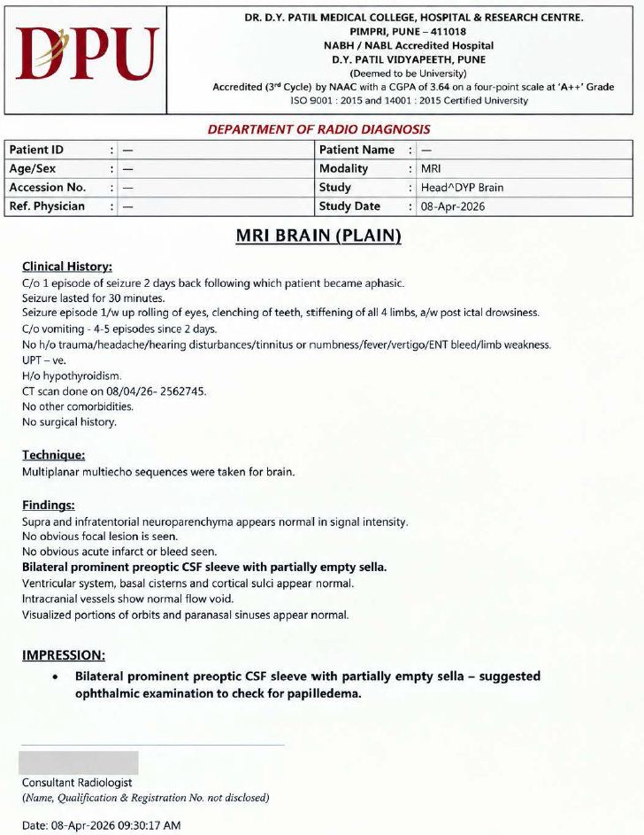
Figure 1: De-identified MRI brain report dated 8 April 2026. The report describes a partially empty sella with prominent bilateral preoptic CSF sleeves and recommends ophthalmic assessment for papilledema. Patient and radiologist identifiers are obscured.
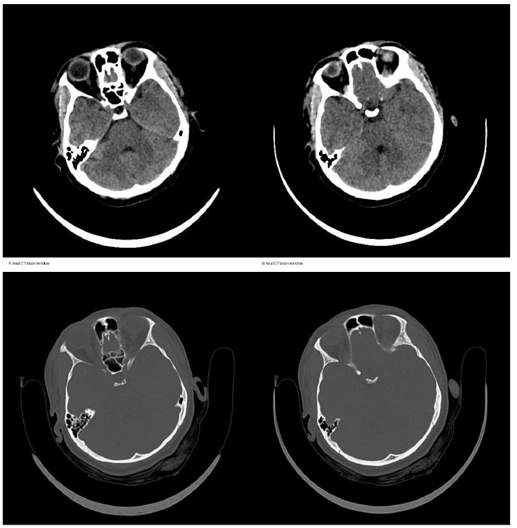
Figure 2: Representative non-contrast CT brain images from the presenting evaluation. No obvious acute intracranial hemorrhage, large territorial infarction or major mass effect is evident on the supplied images.
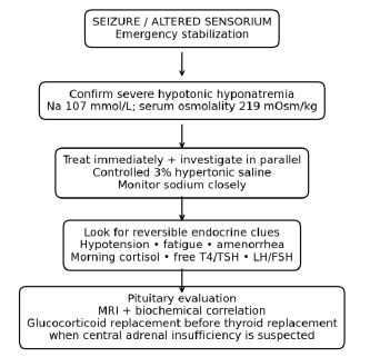
Figure 3: Emergency diagnostic pathway emphasizing simultaneous treatment of severe symptomatic hyponatremia and evaluation for endocrine causes.
Emergency Management
The immediate priority was treatment of acute symptomatic severe hyponatremia. Controlled administration of 3% hypertonic saline was undertaken with close monitoring of serum sodium and attention to avoiding overly rapid correction, consistent with guideline-based emergency management of severe symptomatic hyponatremia [1,2]. Given the hypotension and subsequently documented severe cortisol deficiency, intravenous hydrocortisone 100 mg was administered. After stabilization, hydrocortisone and levothyroxine replacement were continued. In suspected central adrenal insufficiency, glucocorticoid treatment should precede or accompany thyroid hormone replacement because thyroid hormone can increase cortisol requirements and may precipitate adrenal decompensation [3].
Clinical Outcome and Follow-up
The patient’s neurological status improved after correction of severe hyponatremia and corticosteroid therapy. Serum sodium gradually returned toward the normal range during hospitalization, and no further seizures were reported. She was discharged in stable condition with endocrine follow-up and hormone replacement therapy.
Discussion
This case illustrates an important emergency–endocrine overlap: the seizure was the acute event that brought the patient to medical attention, while the biochemical pattern pointed toward chronic pituitary dysfunction. At a serum sodium concentration of 107 mmol/L, severe hypotonic hyponatremia can produce major neurological manifestations. Current guidance emphasizes prompt hypertonic saline therapy for severe symptoms, together with close biochemical and clinical monitoring [1,2].
The markedly low morning cortisol is a central diagnostic finding. Glucocorticoid deficiency can promote non-osmotic antidiuretic hormone activity and reduce renal free-water excretion, thereby causing or aggravating hypotonic hyponatremia [1,3]. In this patient, the low cortisol occurred alongside hypotension and years of constitutional symptoms, strengthening the clinical relevance of the result.
The thyroid profile also required a pituitary-oriented interpretation. A TSH value within the laboratory reference interval does not exclude central hypothyroidism when free T4 is low. Endocrine Society guidance recommends interpreting free T4 and TSH together in patients with suspected pituitary disease [3]. Here, low free T4 was accompanied by cortisol and gonadotropin abnormalities, making a central process clinically plausible.
The decade-long secondary amenorrhea was the most useful historical clue to chronic gonadotropin-axis dysfunction. Its onset after childbirth raises the possibility of postpartum pituitary injury, including Sheehan syndrome. However, the supplied record does not document postpartum hemorrhage, severe postpartum hypotension, shock or failure of lactation. Because severe postpartum hemorrhage is a defining historical feature of classic Sheehan syndrome, a specific postpartum etiology cannot be established from the available evidence [6].
The partially empty sella provides structural support for a pituitary disorder but is not, by itself, diagnostic of hypopituitarism or its cause. Reviews emphasize that empty sella can be an incidental radiological finding, while a subset of patients has clinically significant endocrine dysfunction [4,5]. In this patient, the imaging finding becomes more relevant because it coincides with biochemical abnormalities in several anterior pituitary axes and a long history of endocrine symptoms.
From an emergency-medicine perspective, the key lesson is not to stop the diagnostic process once a seizure has been recognized. Severe hyponatremia requires immediate treatment, but its cause must be pursued in parallel. A brief review of menstrual history, postpartum events, chronic fatigue, orthostatic symptoms and previous endocrine treatment can substantially redirect the differential diagnosis. In this case, focused history-taking connected a decade of amenorrhea with the acute electrolyte emergency and prompted targeted pituitary evaluation.
Conclusion
A neurological emergency may be the first presentation of a chronic endocrine disorder. In this patient, profound hypotonic hyponatremia with seizure led to identification of marked cortisol deficiency, central thyroid-axis dysfunction and gonadotropin deficiency, with a partially empty sella on MRI. For emergency and endocrine clinicians, severe unexplained hyponatremia accompanied by hypotension, chronic fatigue or longstanding amenorrhea should prompt early assessment of the hypothalamic–pituitary–adrenal axis and other pituitary hormones. Careful history-taking remains a high-yield diagnostic intervention.
Declarations
Consent for Publication: Taken.
Ethics Approval: Not applicable for this case report, as stated in the supplied manuscript record.
Competing Interests: The authors declare that they have no competing interests.
Funding: No specific funding was received for this case report.
Author Contributions: Dr. Minakshee Patil: clinical care, data collection and manuscript preparation; Dr. Jagdish Patil: clinical interpretation; Dr. Sanyogita Naik: manuscript preparation; Dr. Sarbari Swaika: supervision and final approval.
Data Availability: Only de-identified information is presented in the manuscript. The data supporting this case are contained within the clinical record and are not reproduced in identifiable form.
AI-assisted Writing: Generative AI was used as a language and formatting aid during manuscript preparation. The authors remain responsible for verifying all clinical facts, interpretations, references and final wording before submission.
References
- Sterns RH (2015) Disorders of plasma sodium—causes, consequences, and correction. N Engl J Med. [crossref]
- Spasovski G, Vanholder R, Allolio B, et al. (2014) Clinical practice guideline on diagnosis and treatment of hyponatraemia. Eur J Endocrinol. [crossref]
- Fleseriu M, Hashim IA, Karavitaki N, et al. (2016) Hormonal replacement in hypopituitarism in adults: an Endocrine Society clinical practice guideline. J Clin Endocrinol Metab. [crossref]
- Lundholm MD, Yogi-Morren D (2024) A comprehensive review of empty sella and empty sella syndrome. Endocr Pract. [crossref]
- Padovano Sorrentino F, Chiloiro S, Giampietro A, et al. (2024) Empty sella syndrome: an update. Pituitary. [crossref]
- Diri H, Karaca Z, Tanriverdi F, Unluhizarci K, Kelestimur F (2016) Sheehan’s syndrome: new insights into an old disease. Endocrine. [crossref]