Introduction
Dementia occurrences increase significantly in older adult type 2 diabetes mellitus (T2DM) [1]., Epidemiologic studies indicate diabetes confers an ~ 1.5-fold increased risk of non-vascular dementia and a ~ 2-fold risk for vascular dementia occurrence compared to rates in age-matched non-diabetic populations [1] Because of the current world-wide epidemic of type 2 diabetes mellitus, diabetic dementia occurrences are expected to increase substantially in the coming decades. There is a need for better understanding of the underlying disease mechanisms including potentially modifiable risk factors to reduce the future medical, social and economic burdens of diabetic dementia.
Recent evidence (from autopsy studies) suggests that the underlying causes of ‘diabetic dementia’ likely differ from those in Alzheimer’s dementia (AD) [2] For example, the characteristic neuropathologic lesions found in AD brain (amyloid plaque, neurofibrillary tangles) were not evident at increased rate in brain autopsy specimens from diabetic dementia vs non-diabetic population [2]. Cerebrovascular burden was increased in diabetic dementia brain autopsy specimens including lacunar infarct, but not microinfarct or large vessel infarct [2]. Arteriolosclerosis of deep penetrating vessels was also a significant finding reported in diabetic dementia brain autopsy specimens [2].
Genome-wide association studies in dementia related to Alzheimer’s dementia have identified gene pathways involving immune function, extracellular matrix remodeling, lipoprotein metabolism and intracellular catabolism [3] as comprising several of the many (>100) genetic risk determinants. Several forms of dementia (including Alzheimer’s dementia) have a high genetic hereditability, but dementia is a complex trait and only a fraction of the overall hereditable variability could be accounted in GWAS studies [3]. Glaucoma is another late-onset neurodegenerative disease in which gene pathways involving extracellular matrix remodeling and vascular development contribute in part to overall genetic risk [4]. Yet only 9-17% of overall hereditability in late-onset glaucoma was accounted for in GWAS studies [4]. In addition, the inclusion in GWAS studies of glaucoma of multi-ethnic populations showed that different genes were significant risk determinants in different ethnic/racial populations [5].
Regarding the unknown mechanisms underlying diabetic dementia, the apoE4 gene isoform is associated with characteristic AD-like neuropathology and confers a large population attributable risk of AD [6]. Insulin resistance has been proposed to play a role in diabetic dementia, but the precise mechanisms are not well understood [7]. Taken together, novel ‘non-AD’ disease mechanisms are likely to be multifactorial and may overlap (in part) both with vascular dementia and Alzheimer’s dementia. The aim of the current review article is to summarize our prior findings from separate reports (largely from type 2 diabetic patients) having a late-onset neurodegenerative disorder, i.e. primary open angle glaucoma, or diabetic dementia, that support roles for dysregulated immunity, chronic inflammation and endothelial dysfunction in the mechanism of non-AD diabetic dementia.
One of the mediators of endothelial dysfunction and neurotoxicity in late-onset neurodegenerative diseases (occurring in subsets of diabetes) was a novel kind of anti-heparan sulfate proteoglycan autoantibody which not only interfered with the neurotrophic (and endothelial cell survival-promoting) effects of growth factors such as fibroblast growth factor 2 (FGF2) [8], but also appeared to bind to a G-protein coupled receptor, serotonin 2A receptor, causing long-lasting activation of PLC gamma/IP3R/Ca2+ intracellular signaling in neurons and endothelial cells [9]. Our studies support intracellular calcium dysregulation as another highly relevant mechanism in diabetes-associated neurodegenerative disease pathophysiology. G-protein receptor coupled signaling is pleiotropic and in electrically-excitable cells such as neurons [10], anti-HSPG autoantibodies evoked sustained depolarization as well as actin cytoskeleton rearrangement via RhoA/ROCK activation altering morphology in neurites (causing neurite retraction) and in glial cells (causing process withdrawal) [8].
In both type 1 and type 2 diabetes mellitus, loss of heparan sulfate proteoglycan (HSPG) is an early event underlying diffuse vascular injury, an idea formulated by the Steno Group and referred to as the ‘Steno hypothesis’ [11]. We later advanced the concept that chronic inflammation associated with obese type 2 diabetes mellitus or in systemic autoimmunity (type 1 DM) drives humoral immune response(s) to HSPG promoting formation of (endothelial and neuro-toxic) anti-HSPG IgG autoantibodies which we reported to be increased in association with a diverse array of diabetic microvascular complications including: retinopathy [12], nephropathy [13,14], and painful neuropathy [8]. In our prior study of anti-HSPG autoantibodies in painful diabetic neuropathy, we observed that patients suffering with painful neuropathy were significantly more likely to have co-morbid late-onset primary open angle glaucoma (POAG) [15], the latter also increases in type 2 DM. Both POAG and painful neuropathy involve damage to small unmyelinated axons, retinal ganglion cells in the case of POAG. Tezel et al. [16] reported increased circulating autoantibodies to optic nerve head heparan sulfate proteoglycans (HSPG) in subsets of normal tension or POAG. In our studies of diabetic microvascular complications, anti-HSPG AAB caused sustained activation of Rho A/Rho kinase signaling pathway in endothelial cells and neuronal cells [8,13]. Rho kinase activation affects endothelial-like trabecular meshwork cells which play a role [17] in mediating intraocular pressure changes which are an important determinant of glaucoma risk. Taken together, these findings, led us to undertake a small study in forty-four older adult type 2 diabetic patients (with and without POAG) to test for increased level of circulating anti-HSPG AAB [15].
Endothelial cell inhibitory activity in the protein A-eluate fraction was significantly increased in plasma from glaucoma or suspects (vs no glaucoma) (Table 1). There was a significant association between co-morbid painful neuropathy and glaucoma or glaucoma suspect (vs no glaucoma) (Table 1). Glaucoma patients were significantly more likely to have reported a family history of dementia in a first-degree relative compared to no glaucoma patients (Table 1). The latter observation led us to consider whether a component of the autoantibody encoded by a heavy chain, or light chain gene might contribute to a part of the unexplained hereditability in occurrences of late-onset POAG.
Table 1: Association between diabetic glaucoma or suspect and co-morbid microvascular complications or family history of dementia.
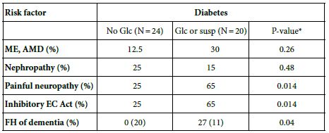
Results are proportion; (), N number of patients tested; Glc, glaucoma, susp, suspect, *Fischer’s exact test, comparison of diabetics with or without glaucoma; ME, macular edema, AMD, age-related macular degeneration, FH, family history in a 1° relative of Alzheimer’s dementia; significant inhibitory endothelial cell (EC) activity, defined as ≤90% of basal cell number after 48 hours incubation at 37 deg C in the presence of test fractions, i.e. a ~30 ug/mL concentration of the protein-A eluate. Reproduced from [15].
We first reported an unusually potent EC-inhibitory fragment having peak apparent MW 12.5kD [8] (Figure 1D) in the protein-A eluate from two prostate cancer patients’ plasma. The activity appeared following long-term storage of protein-A eluates (at 0-4 deg C). We used a combination of heparin Sepharose, and hydroxyapatite affinity chromatography to obtain N-terminal amino acid microsequencing data showing that peak active EC-inhibitory activity had significant 87% amino acid homology to a human kappa V variable light chain [8].
Peak inhibitory activity in the protein-A eluates from both diabetic and prostate cancer patients (Figure 1A-C) had apparent MW corresponding to light chain 23kD or half-light chains 12 kD [8]. Peak prostate cancer IgG EC toxicity was substantially enhanced after heating at 56 deg x 30 minutes (Figure 1E) [8] consistent with aggregation (in light chain fragment).
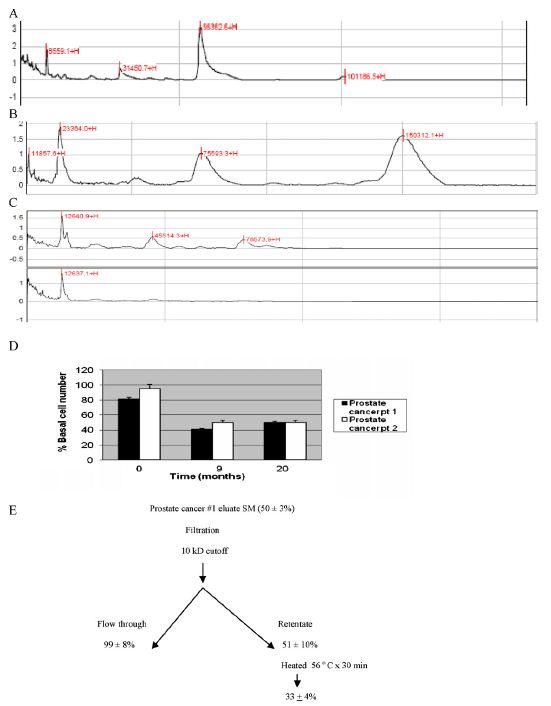
Figure 1: Mass spectrometry of diabetic (A and B) or prostate cancer (C) endothelial cell inhibitory autoantibodies; (D) increase in potency of inhibitory prostate cancer autoantibodies after long-term storage of protein-A eluates at 0-4 deg C and (E) unusual stability of the long-lasting antibodies. (A–C) Samples were analyzed by mass spectrometry as previously reported [8]. (D and E) Endothelial cell activity (mean, SD) is expressed as % of basal cell number in quadruplicate assays as previously reported [8].
Since prostate cancer cells elaborate proteases such as furin [18] which activate other proteases [19,20] that can degrade extracellular matrix we next tested whether exposure of protein A-eluates to recombinant furin could reproduce the effects of long-term eluate storage. Furin is a proprotein convertase which cleaves proteins at specific multi-basic amino acid residues [21] including those present in the variable-constant switch region in a limited subclass of kappa light chains. In a small sample of five diabetic dementia patients (mean age 71 ± 5.8 years; mean diabetes duration 22.4 ± 12.6 years) long-term storage of diabetic dementia protein-A eluates (at 0-4 deg C) unmasked potent EC inhibitory activity (Table 2) [15], but not in the protein-A eluates from an age-matched subset of diabetes having macular edema and/or nephropathy without dementia (Table 2). Brief exposure of the (freshly-isolated) protein-A eluate of a diabetic dementia, POAG patient sample to recombinant furin caused similar unmasking of latent potent EC inhibitory activity (Table 2) [15] perhaps consistent with cleavage at a furin-susceptible multi-basic peptide region in the mechanism of generation of highly potent EC inhibitory activity.
Table 2: Effect of long-term storage of protein A eluates or furin digestion o EC activity.
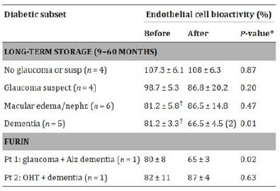
Reproduced from [15]. Furin digestion of protein-A eluate fraction was carried out as previously described [15]. Endothelial cell (EC) survival (mean, SD) was determined in quadruplicate after 48 hours incubation at 37 deg C with test fractions and is compared to survival (in EC) without added test fractions (100%)
Light chain amyloidosis is a systemic disease caused by overproduction of a monoclonal light chain or fragment by a plasma cell clone. Peripheral nerve, heart and kidney are among the most common sites of deposition of monoclonal light chains in AL disease. We observed that a patient manifesting co-occurrence of atrial fibrillation, nephropathy and a family history of dementia harbored an IgG in the protein-A eluate fraction susceptible to furin digestion resulting in a highly EC toxic fragment (Table 3). In three age-matched type 2 DM patients with atrial fibrillation alone, exposure of the protein-A eluates to furin did not generate a toxic EC fragment (Table 3). These data suggest that in diabetic patients with a family history of dementia exposure of the protein-A eluate of plasma to recombinant furin might be a simple method to identify patients at high risk for harboring a latent highly EC toxic light chain fragment capable of contributing to the later development of diabetic dementia.
Table 3: Effect of furin treatment in diabetic depression protein A eluates on endothelial cell inhibitory activity.
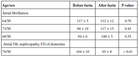
Endothelial cell activity (%)- endothelial cell number was determined after 48 hours incubation (at 37 deg C) with a 30 ug/mL concentration of the protein-A eluate fraction of plasma: Results (mean, SD) is expressed as a percent of basal cell number in bovine pulmonary artery endothelial cells not exposed to test fractions.
*Comparing (mean, SD) EC activity in quadruplicate samples before and after exposure to recombinant furin as previously reported [15].
It is known that kappa light chains comprise functional antibodies [22] and that certain kappa light chain genes cluster in populations that have been exposed to similar kinds of evolutionary selection pressure. For example, subclasses of kappa II gene-encoded light chains are involved in functional antibodies providing protective immunity against the life-threatening childhood infection, Hemophilus influenza meningitis [23]. Yet because these light chains harbor a furin-targetable multi-basic amino acid sequence in the variable-joining switch region, they could contribute to late-onset neurodegenerative disease (which we hypothesize) may be due in part to unusually potent toxicity in a light chain fragment.
There have been several recent reports that plasma furin was increased significantly in adult type 2 DM and was associated with an increased risk of dysmetabolism and cardiovascular morbidity and mortality risk [24,25]. Among its many potential protein targets, furin is known to inactivate endothelial and hepatic lipoprotein lipase [26] which are responsible for normal metabolism of very low-density (triglyceride-rich) lipoproteins (vLDL). Diabetic dyslipidemia is characterized by increased plasma concentration of triglyceride TG-rich lipoproteins and it is an important risk factor accounting for substantial excess CVD morbidity in adult type 2 DM. Hepatic production of TG-rich very low-density lipoproteins (VLDL) increases as a result of either insulin deficiency and/or insulin resistance, and vLDL metabolism is affected by Apo C I a recently reported genetic risk determinant in Alzheimer’s related dementia [3]. It is of interest that the metabolism of TG-rich lipoprotein was reported to increase blood brain barrier BBB permeability and contribute to neuroinflammation which are hallmarks of Alzheimer’s related dementia [27]. Taken together, diabetes associated increases in plasma furin might have a dual role in generating toxic LC fragments which can circulate in VLDL particles and contribute to enhanced endothelial cell toxicity and neurotoxicity underlying dementia.
We had previously shown that highly neurotoxic IgG fragments from three different diabetic dyslipidemia patients with dementia (mean plasma cholesterol 181 ± 51 mg/dL: and mean plasma triglyceride concentration 392 ± 164 mg/dL) displayed increased affinity for a Liposorber (dextran sulfate) column useful in lowering plasma concentrations of LDL and vLDL apolipoprotein B-100-containing particles [28]. In one of the three patients tested (Patient 5, Table 4), in whom the peak toxicity was purified to near homogeneity, N terminal amino acid sequencing revealed significant homology to a restricted subclass of kappa II light chain [28]. Peak EC toxicity in the highly-purified DM k light chain (Patient 5) (induced 70% EC apoptosis after 48hrs incubation at 37 deg C) which was nearly as potent as toxicity in an amyloidogenic monoclonal lambda light chain secreted by lymphoblasts obtained from a patient who had died from amyloid light chain (ALL) disease [28]. Patient 5 suffered from a constellation of systemic and neurovascular complications including multiple small and medium-sized cerebrovascular infarctions and died from complications related to ESRD about ten years after study entry and blood drawing for autoantibody determination [28].
Table 4: Clinical Characteristics in Five Diabetic Dementia Patients Harboring Toxic LC.
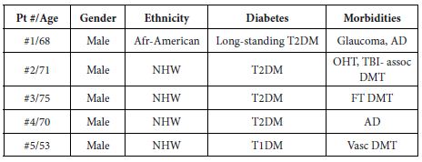
NHW-non: Hispanic white; AD: Alzheimer’s dementia; OHT: Ocular hypertension, TBI: Traumatic brain injury; FT: Frontotemporal; Vasc: Vascular; DMT: Dementia
There is no data to indicate whether or not toxic kappa LC may form insoluble, amyloid precipitates in target organs as occurs in AL disease. Heating may have induced aggregation in kappa LC half chains to form toxic soluble oligomers. Whether toxic oligomers can form under physiological conditions is not known. Light chains are comprised of a high percentage of hydrophobic amino acid residues which normally form tightly-packed beta sheets. Since vLDL particles are amphipathic, it is possible that light chains or fragments coalesce around the lipid-rich microenvironment of the particle which favors hydrophobic residue-rich peptides. Evidence of a possible permissive role for the vLDL particle in enhancing LC toxicity comes from our study of neurotoxicity in protein A eluates from patients with Parkinsons disease and/or dementia [29]. Compared to neurotoxicity in the protein A eluates from three patients with Parkinsons disease Figure 2A), when the Liposorber-retained fractions from two dyslipidemic diabetic dementia patients plasmas was purified on protein-A, the resulting IgG displayed significantly higher, unusually potent neurotoxicity (Figure 2B) [29]. The purified IgG present in diabetic dementia and dyslipidemic plasma was active in the low-ten nanomolar range of concentrations (Figure 2B) (approaching potency reported for oligomeric Abeta). The purified diabetic dementia IgG-induced neurite retraction was significantly, but not completely prevented by co-incubation with a 200 nanomolar concentration of the selective 5HT2AR antagonist M100907 (Figure 3B). A similar concentration of M100907 completely prevented PD Ig-induced neurite retraction (Figure 3A). These data suggest that hypertriglyceridemic plasma enhances the diabetic dementia autoantibody toxicity through unknown mechanisms. It is possible that heavy chain-bearing IgG components in diabetic dementia target the 5HT2AR receptor and are blockable by selective 5HT2AR antagonist, but that more potent light chain components interact with a different receptor.
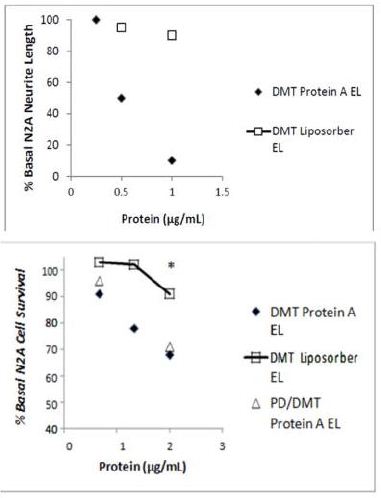
Figure 2: The highly purified protein-A eluate fraction of diabetic dementia plasma (solid diamond) caused significantly greater dose-dependent suppression of N2A mouse neuroblastoma neurite outgrowth compared to the Liposorber eluate fraction (open square) from the same patient (A). Dose-dependent N2A mouse neuroblastoma cell toxicity in the protein-A eluates from type 2 diabetic dementia (solid diamond) and PD/DMT (open triangle) significantly exceeded toxicity in the corresponding Liposorber DMT eluate fraction (open square) (B). **P < 0.001;* P < 0.05 comparing neurite length-shortening or N2 mouse neuroblastoma cell survival induced by Liposorber eluate vs protein-A eluate fractions. Reproduced from [29].
PD/DMT: Parkinson’s disease with dementia.
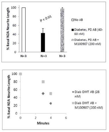
Figure 3: Mean N2A neurite length suppression induced by three different diabetic PD autoantibodies was nearly completely prevented by co-incubation with 200 nanomolar M100907, a highly selective 5-HT2AR receptor antagonist (A). Potent, time-dependent suppression of N2A neurite outgrowth by diabetic dementia autoantibodies was only partially prevented by co-incubation with M100907 (B). Reproduced from [29].
The implications of these findings for the prevention of diabetic dementia are several-fold. First, while the inheritance of genes encoding a furin-cleavable light chain is not easily modifiable, the prevention of diabetic dyslipidemia is in part modifiable by measures such as adherence to a low-fat diet, exercise, maintenance of ideal body weight, medications that correct insulin resistance and insulin deficiency, and avoidance of alcohol and other substances that inhibit lipoprotein lipase. The regulation of extracellular furin in diabetes and how it might cleave a subset of LC is not completely understood. Furin contains a heparin-binding domain [30] and since EC and neurotoxic diabetes AAB targeted HSPG [8], anti-HSPG AAB and furin may colocalize in spaces rich in HSPG. For example, HSPG acts as a co-receptor for lipoprotein lipase and for lipoprotein-related protein LRP which is involved in the catabolism of vLDL particles [31]. Several genes involved in protein catabolism were reported to be risk factors for dementia [3]. We cannot rule out the possibility that toxic LC fragments may be generated in part during the process of vLDL intracellular catabolism, e.g. in an acidic cellular compartments which might favor spontaneous dissolution of LC harboring multi-basic amino acid residues.
In summary, the presented (published) data support our novel hypothesis that a subset of diabetic dementia may result (in part) from inheritance of specific genes encoding subclasses of kappa light chain which harbor a furin-cleavage site in the variable-constant switch region which can serve as a substrate for increased plasma furin reported in diabetes. Cleavage generates light chain autoantibody fragments highly toxic in both endothelial and neuronal cells. The LC fragments might aggregate to form soluble oligomers having enhanced cell neurotoxicity in the low-ten nanomolar range of toxic concentrations as reported for amyloid beta oligomers. More study is needed to substantiate this theory and to test whether furin cleavability in the protein-A eluates obtained from diabetic patients having other microvascular complications and a family history of dementia might be a significant predictor of subsequent occurrence of dementia compared to patients without similar family history of dementia.
The author declares no competing interests that would affect the objectivity of the presented results.
References
- Bello-Chavolla OY, Antonio-Villa NE, Vargas-Vázquez A, Ávila-Funes JA, Aguilar-Salinas CA (2019) Pathophysiological Mechanisms Linking Type 2 Diabetes and Dementia: Review of Evidence from Clinical, Translational and Epidemiological Research. Curr Diabetes Rev. [crossref]
- Biessels GJ, Despa F (2018) Cognitive decline and dementia in diabetes mellitus: mechanisms and clinical implications. Nat Rev Endocrinol. [crossref]
- Loi KJ, Radtke N, Kiriluk C, et al. (2025) Shared genes and pathways in dementia: Insights from genome-wide association studies. Alzheimer’s Dement. [crossref]
- Hamel AR, Yan W, Rouhana JM, et al. (2024) Integrating genetic regulation and single-cell expression with GWAS prioritizes causal genes and cell types for glaucoma. Nat Commun. [crossref]
- Tavakoli K, Huang BB, Mirmira T, et al. (2026) Multi-ancestry genome-wide association study in all of Us for primary open-angle glaucoma. Sci Rep. [crossref]
- Kaji S, Berghoff SA, Spieth L, et al. (2024) Apolipoprotein E aggregation in microglia initiates Alzheimer’s disease pathology by seeding β-amyloidosis. Immunity. [crossref]
- Gonçalves RA, Wijesekara N, Fraser PE, De Felice FG (2019) The Link Between Tau and Insulin Signaling: Implications for Alzheimer’s Disease and Other Tauopathies. Front Cell Neurosci. [crossref]
- Zimering MB, Alder J, Pan Z, Donnelly RJ (2011) Anti-endothelial and anti-neuronal effects from auto-antibodies in subsets of adult diabetes having a cluster of microvascular complications. Diabetes Res Clin Pract. [crossref]
- Zimering MB (2017) Diabetes Autoantibodies Mediate Neural- and Endothelial Cell- Inhibitory Effects Via 5-Hydroxytryptamine- 2 Receptor Coupled to Phospholipase C/Inositol Triphosphate/Ca2+ Pathway. J Endocrinol Diab. [crossref]
- Zimering MB, Mirkovic N, Pandya M, Zimering JH, Behnke JA, et al. (2016) Toxic Immunoglobulin Light Chain Autoantibodies are Associated with a Cluster of Severe Complications in Older Adult Type 2 Diabetes. J Endocrinol Diab. [crossref]
- Deckert T, Feldt-Rasmussen B, Borch-Johnsen K, Jensen T, Kofoed-Enevoldsen A (1989) Albuminuria reflects widespread vascular damage. The Steno hypothesis. Diabetologia. [crossref]
- Zimering MB, Anderson RJ, Moritz TE, Ge L and VADT Investigators (2009) Endothelial cell inhibitory auto-antibodies are associated with laser photocoagulation in adults from the Veterans Affairs Diabetes Trial. Metabolism. [crossref]
- Zimering MB, Pan Z (2019) Auto-antibodies in type 2 diabetes induce stress fiber formation and apoptosis in endothelial cells. J Clin Endo Metab. [crossref]
- Zimering MB, Zhang, JH, Guarino P, Emanuele N, Mc Cullogh P, Fried L and VA NEPHRON D Investigators (2014) Endothelial cell autoantibodies in predicting declining renal function, end-stage renal disease or death in adult type 2 diabetic nephropathy. Endocrinol. [crossref]
- Zimering MB, Moritz TE and Donnelly RJ (2013) Anti-neurotrophic effects from autoantibodies in adult diabetes having primary open angle glaucoma or dementia. Endocrinol. [crossref]
- Tezel G, Edward DP, Wax MB (1999) Serum autoantibodies to optic nerve head glycosaminoglycans in patients with glaucoma. Arch Ophthalmol. [crossref]
- Pattabiraman PP, Rinkoski T, Poeschla E, Proia A, Challa P, Rao PV (2015) RhoA GTPase-induced ocular hypertension in a rodent model is associated with increased fibrogenic activity in the trabecular meshwork. Am J Pathol. [crossref]
- Uchida K, Chaudhary LR, Sugimura Y, Adkisson HD, Hruska KA (2003) Proprotein convertases regulate activity of prostate epithelial cell differentiation markers and are modulated in human prostate cancer cells. J Cell Biochem. [crossref]
- Remacle A, Rozanov D, Fugere M, et al. (2006) Furin regulates the intracellular activation and the uptake rate of cell surface-associated MT1-MMP. Oncogene. [crossref]
- Nguyen HL, Zucker S, Zarrabi K, Kadam P, Schmidt C, Cao J (2011) Oxidative stress and prostate cancer progression are elicited by membrane-type 1 matrix metalloproteinase. Mol Cancer Res. [crossref]
- Remacle A. G., Shiryaev S. A., Oh E. S., Cieplak P., Srinivasan A., Wei G., et al. (2008) Substrate cleavage analysis of furin and related proprotein convertases. A comparative study. J Biol Chem. [crossref]
- Jaffe DB, Shahi P, Adams BA, et al. (2022) Functional antibodies exhibit light chain coherence. Nature. [crossref]
- Granoff DM, Pandey JP, Boies E. et al. (1984) Response to immunization with Haemophilus influenzae type b polysaccharide-pertussis vaccine and risk of Haemophilus meningitis in children with the Km(1) immunoglobulin allotype. Clin. Invest. [crossref]
- Fathy SA, Abdel Hamid FF, Zabut BM, et al. (2015) Diagnostic utitily of BNP, corin and furin as biomarkers for cardiovascular complications in type 2 diabetes mellitus patients. Biomarkers. [crossref]
- Fernandez C, Rysä J, Almgren P, Nilsson J, Engström G, Orho-Melander M, Ruskoaho H, Melander O (2018) Plasma levels of the proprotein convertase furin and incidence of diabetes and mortality. J Intern Med. [crossref]
- Seidah NG, Prat A (2007) The proprotein convertases are potential targets in the treatment of dyslipidemia. J Mol Med (Berl). [crossref]
- Lee LL, Aung HH, Wilson DW, Anderson SE, Rutledge JC, Rutkowsky JM (2017) Triglyceride-rich lipoprotein lipolysis products increase blood-brain barrier transfer coefficient and induce astrocyte lipid droplets and cell stress. Am J Physiol Cell Physiol. [crossref]
- Zimering MB (2010) Recurrent macular edema and stroke syndrome in Type 1 diabetes with potent endothelial cell inhibitory autoantibodies. Endocr Pract. [crossref]
- Zimering MB (2018) Circulating neurotoxic 5-HT2A receptor agonist autoantibodies in adult type 2 diabetes with Parkinson’s disease. J Endocrinol Diab. [crossref]
- Zeng J, Meng Y, Chen SY, Zhao G, Wang L, Zhang EX, Qiu H (2022) Structural characteristics of Heparan sulfate required for the binding with the virus processing Enzyme Furin. Glycoconj J. [crossref]
- Wilsie LC, Gonzales AM, Orlando RA (2006) Syndecan-1 mediates internalization of apoE-VLDL through a low density lipoprotein receptor-related protein (LRP)-independent, non-clathrin-mediated pathway. Lipids Health Dis. [crossref]