DOI: 10.31038/GEMS.2025761
Abstract
African Ancestry communities in Barbados have established, through time and space, deep heritage ties to places along nearby littorals; that is, cultural zones along the shore referred to here and in previous publications as The Sea’s Edge. An analysis of these areas is based on ethnographic field interviews, participant observation, and surveys conducted during eleven field events over a 20-year time frame on the island of Barbados. The cultural attachments to the sea’s edge, spanning the terrestrial and marine, began during the period of enslavement and continue to be maintained and celebrated today as a viable part of cultural identity and heritage pride. This paper explores how these places became functionally integrated into a landscape and central to the culture of these coastal peoples. Individual places and natural resources continue to provide important ecological and cultural services, including hundreds of plants, dozens of shallow marine resources, and areas of historical and spiritual significance. This analysis documents the ongoing value of natural/cultural coastal heritage landscapes, an area known as the littoral zone adjacent to the Bath Plantation lands. It is an area utilized and celebrated, protected, and informally managed by the surrounding communities and is an example of the way places like this fit the littoral landscape model, recommending that local involvement is appropriate in cooperative management with the national government. This example is applicable to other similar coastal communities throughout the world and provides a framework to understand local connection to the littoral zone and both maritime and terrestrial natural and cultural resources.
Keywords
Heritage cultural landscapes, International Union for the Conservation of Nature Heritage Guidelines, Barbados West Indies, Enslaved African people, culture of marine littoral edges
Introduction
The Sea at its best is a place where I can walk in the water up to my knees and look down and see conch, fish and other foods for life. It should all be there for you – Lester Flowers, a Professor of Biology at the College of Bahamas [1].
Heritage landscapes encompass landscapes of scenic, cultural, and scientific value [2]. The International Union for Conservation of Nature (IUCN) [2,3] argues for the importance of world heritage landscapes in conservation to encourage holistic approaches to conservation. This analysis is based on research conducted in communities surrounding the Bath Plantation Lands (known as Bath), Barbados, and supports the integration of heritage landscapes by providing information about the social and cultural importance of the littoral zone. This paper provides national planners and international developers with the information needed to meaningfully involve local Barbadian communities in the decisions that impact the future of their homelands and culture. It also provides insight into the need for collaborative management practices that can be utilized in similar settings in coastal areas throughout the World. Barbadian law stipulates that the littoral is for the public (National Conservation Commission Act, Cap. 393), and belongs to all the people of the country; however, it does not recognize the rights associated with local African Ancestry (Afro-Caribbean) heritage cultural landscape that has formed over hundreds of years. As a result, current coastal development and use of natural resources has and potentially will threaten the community- based natural and human resources located at the sea’s edge [1,4-6]. Using the heritage littoral landscape model, a conceptual or analytical framework used to understand and manage coastal (littoral) zones that have both natural and cultural heritage significance, this paper explores those culturally connected to the sea’s edge and the rationale for their involvement in its management and interpretation. These data and analysis were the foundation for sustainability impact assessments [7] and Community-Based Human Rights Impact Assessments [8]. This analysis also documents the complexity of conducting ethnographic studies of heritage in littoral landscapes, which span both the marine and terrestrial locales and resources.
Background
African Ancestry History at the Sea’s Edge
The coastal lands of the Caribbean are special heritage places for the ancestors of the formerly enslaved peoples who were forcibly transplanted from ecosystems they understood in Africa to ones with new natural and social ecologies (Figure 1). While beyond the scope of this analysis, many of the people would arrive at Caribbean plantations that were established near coastal areas. The history of industrial agriculture of Caribbean plantations focused on monocrop production (in most areas this was sugarcane cultivation), processing, and shipment from local ports to markets elsewhere in Europe and the New World. There are hundreds of former coastal enslavement plantations throughout the Caribbean. Barbados alone had hundreds of industrial agricultural plantations (Figure 2). The island is on a flat coral limestone plateau, different from the topography of steep volcanic islands like St. Lucia and large mountainous locations such as Jamaica, Cuba and the Dominican Republic. Volcanic islands share the topographical characteristic of little to no underwater shelves, and they rise steeply to mountains that support hundreds of inches of rainfall per year. Given that the localized weather in the Caribbean is directly impacted by land elevation, flat islands experienced consistent levels of annual rainfall, scant surface water, and dependable weather conditions that made Barbados suitable for viable large-scale commercial sugarcane agriculture for approximately 400 years.
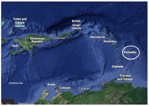
Figure 1: Barbados in the Lesser Antilles (Google Earth).
The ecological and cultural shifts from the old traditional societies of Africa to the new Caribbean societies posed a human adaptation challenge that was experienced by hundreds of thousands of African people. Subsequent successful ecological co-adaptations resulted in these people defining their own heritage places and events of cultural importance. Taken together these new heritage places became coherent and functionally integrated cultural landscapes. Given the location of many plantations near the sea to facilitate commercial shipment of products elsewhere, the enslaved and later freed African Ancestry peoples made a way of life that involved both land and sea. Their home often was centered on a heritage littoral landscape located at the sea’s edge [1]. The heritage littoral landscape which is the focus of this research is operationally defined as being composed of near shore marine and terrestrial places and environments. Each is defined in space, time, and function by the people who have socially constructed the landscape through use and experience. Together the land and sea habitats and their associated human interactions and adaptations constitute a heritage littoral landscape. The analysis argues that such integrated landscapes define the culturally appropriate ways (that is, frames of analysis) to understand the heritage of the coastal people and understand and apply the research findings to terrestrial and marine management as well as cultural interpretation [1,6,9-13].
The adaptations of people with African Ancestry varied by their cultural backgrounds and unique ecological contexts of where they landed and ultimately resided. As enslaved people they lived where and how others demanded; however, within these constraints, they expressed their agency and increasingly created their own cultural spaces and meanings. It is argued here that the littorals of their new coastal lands would become a primary place for the expression of their agency [1]. African Ancestry people would establish their places in areas that were largely not useful to the export-oriented colonial industrial plantations and in ecosystems that remained largely influenced by natural variables. Being close to plantations, these areas were most accessible to the people during the long period of their enslavement. The littoral, in other words, became for many people of African Ancestry “our place” at the sea’s edge and thus a foundation for heritage places and landscapes. Caribbean research documents how for many African Ancestry people, the sea’s edge has historically represented freedom and survival [14-18]. The sea’s edge has always provided marine and terrestrial resources for food, fertilizer and medicine, in addition to diverse items for trading and crafts (shells and corals). Also, the sea served as a mode of transportation. The water itself can be seen as something that simultaneously separates and connects people with other places. In these special locations, knowledge of the terrestrial resources has grown apace with knowledge of the marine resources and sea, thus allowing people to survive and thrive in environments with little external support. The “fruits” of the sea were combined with those of the land, such as in the building of boat ribs from native trees and the mixing of seaweed with land plants to make medicines. Because much of the traditional natural vegetation was removed for the planting of export crops like sugar and cotton, soon natural vegetation occurred only at the sea’s edge where sea spray salted the land thus precluding cash cropping.
Knowledge and appreciation of the sea appears to have been traditionally expressed through a respect that is grounded in experience, not only through collection but use of the resources. Fishermen have learned to “read the language” of the sea and the coastal marine environment in overt as well as subtle ways. Collective and individual experience has led to an understanding of the ways that marine life interacts and sustains itself and nearby land plants and animals. For example, fishermen have learned to gauge the wind and the tides, the seasonality of fish populations, and how to follow dolphins to find certain schools of fish. Likewise, farmers have learned that some marine life can be used as a medicine for skin rashes, that the hundreds of coastal plants have a special ecology that can be managed, and that seaweed can be dried for food and fertilizer. Today, people keep traditional knowledge of the sea’s edge alive through their occupations, social relations, and the cultural transmission of knowledge through instruction and participation with the local youth. Residents have built the sea into their lives through stories, songs, lessons and games. Many discuss the ways in which knowledge of the sea was kept alive in old fishermen’s songs, songs that were sung while undergoing trials at sea and later again over rum and ale in ports where knowledge of these events were shared by turning such occasions into social affairs. Through sailing stories, shipwreck stories, and even mythical stories about sea animals helping boaters in distress, the centrality of the sea is once more realized. Nationally today, in Barbados, attachments to the sea’s edge are evidenced in the expressive arts. People today do not live as they did in the past but choose to value the protection and interpretation of heritage places, natural resources, and storied places because these represent how they thrived during the best and harshest of times.
Cultural Landscapes and the Edge Effect
According to the International Union for Conservation of Nature [3]:
Cultural landscapes are seen as ‘cultural heritage’ in the framework of the World Heritage Convention, yet they have considerable overlap with one of the heartland issues of nature conservation, the global coverage of protected areas. Recent research reveals the overlaps and synergies between World Heritage cultural landscapes and IUCN’s global category system for recognizing protected areas, and some of the possible implications.
In the western world, the term ‘landscape’ has been used in many vernacular and academic ways. It has been used in painting, writing, and poetry since the late 19th and early 20th centuries, and it is a term used by natural and social science scholars to frame many shared analytical perspectives. This concept contrasts with the alternative notion of nature and landscape as a living integrated landscape in the Far East. The early German geographer Otto Schluter in 1908 defined two forms of landscapes: the Urlandschaft (original landscape) or landscape that existed before major human induced changes and the Kulturlandschaft (cultural landscape) a landscape created by human culture. Schlüter argued that the major task of geography was to trace the changes in these two landscapes. Carl Sauer wrote that cultural landscapes are made up of human forms and actions superimposed on physical landscapes [19]. He was probably the most influential in promoting and developing the idea of culture as a force in shaping the visible features of the Earth’s surface in delimited areas. Within his definition, the physical environment retains a central significance, as the medium with and through which human cultures act [20]. He maintained that objects which exist together in the landscape exist in interrelation. His classic definition of a ‘cultural landscape’ reads as follows: “The cultural landscape is fashioned from a natural landscape by a cultural group. Culture is the agent, the natural area is the medium, the cultural landscape is the result.”
Since Schlüter’s first formal use of the term, and Sauer’s effective promotion of the idea, the concept of cultural landscapes has been variously used, applied, debated, developed and refined within academia. By 1992, the World Heritage Committee elected to convene a meeting of landscape specialists to advise and assist redrafting the Committee’s Operational Guidelines to include ‘cultural landscapes’ as an option for heritage listing properties that were neither purely natural nor purely cultural in form (i.e., mixed heritage) [21]. This analysis is focused on defining and explaining African Ancestry places and landscapes in the Caribbean and thus adds to expanding definitions used in western academic circles of natural and cultural landscapes by detailing how these locations can also span marine and the terrestrial resources, freshwater and saltwater, and sea and the sand. The heritage objects, natural resources, and heritage places that were identified in our studies are more broadly framed as components of heritage littoral landscapes. When considered as a functionally integrated cultural phenomenon they can more clearly be preserved, managed, and interpreted in more holistic ways. Cultural landscapes in the littoral are special because they define a boundary between habitats. Ecologists have termed this the edge effect because where two different habitats meet, they form an edge of nature and thus increase biological diversity and complexity. McCay has discussed the diverse and productive nature of edges, proposing that the “edge effect” may be used as a metaphor for the bringing together of people, ideas and institutions [12]. In a data-based summary of the concept, Turner, Davidson-Hunt, and O’Flaherty [22] argue that, Like ecological edges, cultural knowledge systems can integrate, producing a richness of knowledge and practices that enhance the resilience of local societies. Cultural edges, rather than being border zones between discrete social entities, are zones of social interaction, cross-fertilization, and synergy wherein people not only exchange material goods but also learn from one another.
The edge effect defines the location and cause of new food webs and trophic levels. Expanding the ecology concept of edge to include human dimensions results in considerations of edges as stimulating cultural diversity, centering social activities, and serving as a foundation of heritage [23]. The littoral landscape is operationally defined, for this analysis, as a social, natural, and biological zone adjacent to the edge of the sea [1,6]. When stable communities use such areas for generations, they often become heritage cultural landscapes that are locally perceived as belonging to the people who have used and protected them for hundreds of years. The heritage littoral landscape extends onto the land through food webs that depend on both salt and freshwater habitats. It extends inland along small rivers and up estuaries into mangrove wetlands. It can be defined as far as amphibious animals crawl inland. The littoral contains shallow coral reefs, seagrass beds, exposed beach rock, sand dunes, and mud flats. Socially and culturally the littoral extends on land and in the sea through related activities, historic places, and the cultural patterns of contemporary communities.
Methods
A key aspect of this research is its focus on heritage cultural landscapes, particularly those developed during slavery and subsequently shaped over time by African Ancestry communities to be a presence in contemporary time. These landscapes are often inherently private, and local community ownership or attachment to these areas is typically unrecognized by national authorities. As such, community members are frequently excluded from discussions surrounding the management and future of these landscapes. This study underscores how long-term participant observation and iterative ethnographic fieldwork can foster mutual trust and rapport, ultimately enhancing the depth and richness of ethnographic data on heritage landscapes. This study examines the Bath littoral zone through both ethnographic and participatory research methods, incorporating a range of data sources to capture the social, cultural, and environmental dimensions of the region. While the littoral landscape is often considered as a distinct physical and cultural space, this research demonstrates its interconnectedness with other marine and terrestrial environments, including deeper oceanic areas and adjacent hill zones. The analysis is primarily based on a combination of formal surveys, ethnographic interviews, and participant observation with residents from all over the island, with the majority of the interactions focused specifically on the Bath Plantation area [24]. Ethnographic methods were employed in a comprehensive, triangulated approach to data collection and analysis [25]. This strategy combines multiple techniques, including structured interviews, semi-structured key informant interviews, informal interviews, group discussions, participant observation, and transect walks. Previous literature was also reviewed to contextualize findings and enhance the depth of analysis. The interviews spanned a diverse demographic, including individuals from 8 to 94 years of age, with men and women from various educational, geographic (rural and urban) and economic backgrounds. The research also involved extensive participant observation conducted over 12 field visits between 1996 and 2024, with site visits ranging from 7 days to 5 months in duration. These visits enabled immersion in daily life and direct engagement with local communities, especially those who utilized the Bath Littoral zone’s marine and terrestrial resources [1,24].
This research spanned over two decades, with an initial focus on traditional microeconomic systems of exchange and its role within the larger societal context of interaction within various types (familial, communal and occupational) of social networks. Many of these surveys and interviews during this time frame focused in rural areas examining the role of these saving and credit associations within the larger framework of household food security and financial well being, this lending insight into the interplay between formal and informal economics as well as natural resource use. These associations are called Rotating Savings and Credit Associations (ROSCAs), known locally as “meeting turn,” and throughout the Caribbean as “sousou,” “asu,” “box hand,” and “partner” [26]. This component of the research was conducted over five field sessions and included 500 ethnographic interviews, 120 of which were conducted in 1999 with individuals from the Bath Plantation area on the rural east coast of Barbados. The other 380 interviews were conducted between 1996 and 2024 with people throughout the island, including but not limited to individuals from the Bath Plantation area. Many of these interviews include discussions with fisheries officers, local NGOs, fishermen of all ages and from all sectors (commercial, recreational, for-hire, and subsistence), as well as restaurant owners, chefs and fish cleaners and vendors from the Fish Markets at Conset Bay, Oistins and Bridgetown. These interviews primarily explored informal economic systems in the context of fishing, coastal harvesting, and resource management. In addition, interviews also focused on the changing physical environment and development of eco-tourism related to the littoral and creation of the old railway walking trail (from Bath through Martins Bay to Bathsheba).
Participant observation formed the backbone of data collection, spanning 20 plus years of fieldwork and interaction with the residents and resources of the Bath littoral zone. During the field site visits, the lead author engaged in daily activities with the community, participating in fishing trips and documenting the local use of marine and terrestrial resources. This observational approach also allowed for the gathering of traditional ecological knowledge, particularly related to food webs, trophic levels, and lunar, tidal and seasonal changes in the littoral environment [1]. Local residents also shared their concerns about the protection and preservation of key cultural places within the Bath Littoral zone. During extended stays in Bath Beach, the lead author developed close relationships with local fishermen, engaging in participant observation and free-diving fishing expeditions. These experiences provided a detailed understanding of fishing techniques and species targeted in both inshore and offshore environments. When weather permitted the researcher spent five to six days per week freediving with local fishers, building rapport and learning about traditional ecological knowledge, especially regarding the seasonal availability of key marine species. These insights were supplemented by continued visits to the area, including 12 additional trips between 2000 and 2024, where the researcher continued to engage in fishing activities and document changes in the local environment, particularly the rising abundance of sargassum.
This study shifts the environmental focus from discrete geographical locations to socially constructed landscapes, emphasizing the need to understand human adaptations to natural systems and the persistence of sustainable heritage practices. This methodological shift recognizes the importance of adjusting traditional ethnographic techniques—both in terms of detail and temporal scope—when applied to landscape-scale research. This means examining multiple locations and resources associated with an area, not a specific building or landscape. It moves beyond a single time frame as well examines the relationship over generations. It specifically asks how these places and these resources played and play a part in the identity of a local population as well as served as a place for cultural transmission of cultural and environmental knowledge, especially in relation to food security and the creation of social stability through the strengthening of social networks. This study draws heavily on the insights of Fred Watson, a local fisherman born in Bath Beach 97 years ago, whose deep knowledge of the region’s fishing practices contributed greatly to the research. Known as “the Best Fisherman in all of Barbados,” Watson played a pivotal role in both the community and the broader fishing industry, serving as a mentor to younger generations of fishers and providing expert advice to government fishery managers. His personal experiences, documented in an oral history conducted over several years, serve as a foundational resource for understanding the cultural and ecological significance of the Bath Littoral zone.
The ethnographic approach used in this study is consistent with and builds upon previous research on rural Barbadian communities [26-29] and similar studies from nearby islands [30,31]. These studies document the co-adaptations of African-descended peoples and the landscapes where they live, highlighting the importance of cultural landscapes as dynamic, socially constructed spaces. Additionally, this research draws on the work of Cumberbatch and Hinds [32] on Flying Fish as an aspect of Barbadian bio-cultural heritage, further illustrating the interconnectedness of people, place, and resource management. In addition to the ethnographic data, we utilize photographic analysis of early 20th-century images to strengthen the argument for recognizing the Bath Littoral zone as a heritage site. These photographs provide visual evidence of the cultural connection between local Bath residents and the littoral landscape, further substantiating its significance as a heritage area and its continuing importance from the past to the present. Various findings from the Barbados study have been reported and published elsewhere and these will not be repeated here [1,33]. Instead, new heritage sea and land interactions are discussed in this analysis, thus expanding what is known of these littoral landscapes. Through this multi-method approach, the study contributes to a deeper understanding of the Bath Littoral zone as a dynamic and culturally significant heritage landscape.
Study Site
Littoral landscapes are difficult to study because of their bathymetric characteristics. The sea portions are largely under water often with some protruding rock formations, the littoral landscapes included in this study are shallow thus permitting fishing, lobster gathering, shellfish collecting, and the picking of seaweeds by hand. During enslavement the movements of enslaved people were restricted to prevent escape and or revolt, so they were not permitted to either make or use boats or rafts. Sea resource use was thus limited to shallow and calm waters. The Barbados coastal segment studied here is called for purposes of this analysis the Bath Littoral Landscape. It is located on the eastern edge of this 14-mile wide by 21-mile-long island (Figure 2). The landscape is 2.21 miles along the windward north shore extending from Congor Bay to past the Bath Beach House to Conset Bay (Figure 2). This landscape includes, (1) a beach used for all aspects of life including social gathering, tourism, sea baths (both recreational and healing activities), plant gathering (for subsistence and medicinal purposes), (2) a spring with spiritual dimensions as well as healing properties, (3) the Quamins river, (4) the old railway and historic train station, (5) the Bath plantation, a location that has existed from enslavement in the 1700s to modern times with many of the relatives of former unfree laborers, and (6) an underwater marine habitat used for cultural transmission of knowledge regarding resource use as well as gathering marine and terrestrial resources for both food and medicinal purposes, an activity that dates back to the earliest of the areas inhabitants (Amer-Indians) and people brought through forced migration (slaves and indentured laborers).
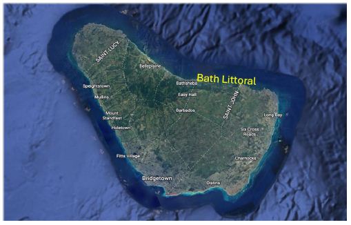
Figure 2: Island of Barbados, West Indies. The narrow coastal limestone shelf and coral reef are abutted by deep ocean (Google Earth images).
Topographically the Bath Heritage Landscape has hills to the south and a relatively quiet beach area, with a wide shallow sea shelf extending to a deep drop off to the north. The limestone shelf drop has a vertical ledge with a 40-foot drop, that is marked above by large limestone rocks that rise above the ocean even at high tide. The shelf can be walked at low tide with only light swimming needed over deep holes. Bath is on the windward north side of Barbados, so the Bath littoral experiences the persistent Northeast Trade Winds, which make for active ocean waves beyond the shallows and strong winds on the uplands (see Windy Ridge in Figure 3).
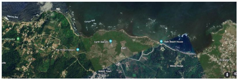
Figure 3: Bath Heritage Landscape (Google Earth).
The Bath Heritage Landscape by definition includes both land and water dimensions. Each has contributed to the development of local coastal African Ancestry communities, especially Bath. Key to the formation of this as a littoral landscape, rather than just a coastal landscape, is the special contribution of the local sea to the lives of local African Ancestry people. In this case, key was the presence of a shallow, flat, shelf that extends to an underwater drop off. The presence of an enslaved population and their space restrictions and limited access to natural resources made both land and the sea components special for the production of this landscape.
Historic and Contemporary Connections to the Bath Heritage Landscape
One of the most useful images for this analysis is the historic Bath littoral zone (Figure 5) curated by Jim Webster [34] who, building on the oral history of Richard Goddard, used it to illustrate an article regarding the earliest photographs of the people and coast of Bath [34]. The photos used in the article were taken by professional photographer Manuel Auguste Nunes Siza in 1896-1897. They are located at the “Anglo-Luzo Photographic Gallery” in Beckwith Place, Bridgetown [35]. The photos illustrate some cultural dimensions of the littoral, and for this analysis these largely remain important unvoiced images of the past. Some of the Siza photos are presented here along with additional ethnographic information acquired by Stoffie during his research and supported and built upon by a local elder Fred Watson (and others) who was born in Bath Beach in the early 20th century.
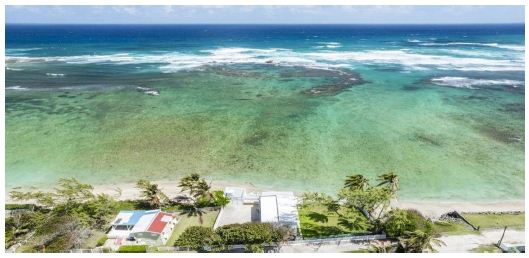
Figure 5: Part of the Bath Littoral Zone: From Beach to Great Rocks [34].
The Bath Plantation
The Bath plantation (Figure 4) was owned in 1791 by Edward Day. During this early time it had 212 enslaved people and 400 workers. The number of enslaved workers remained about the same until near the end of slavery in 1832 when there were 245 enslaved people. In 1913 there were a total of 627 workers on the plantation [36]. Given the location of the Bath plantation, near what is called the Scotland District, it is assumed that the workers were a combination of African Ancestry slaves mixed with individuals of Scots-Irish Ancestry. Scots- Irish workers were among the first unfree laborers in Barbados. Bath workers lived on and around Bath plantation lands during enslavement and continue in today’s districts such as Welch Town, Pothouse and Sergeant Street. At the end of slavery, about 1837, most plantations forced formerly enslaved workers to move their homes to small villages located along nearby roads. The tenantries were on public lands along these roads. This action removed the former enslaved people from both the plantation and the notion that the plantation was obligated to them for food, medicine, and housing as it had been during slavery. After slavery, Barbados government laws were passed to prevent African Ancestry people from owning small plots of land. Soon after their forced removal, the house pads in their former living areas on the plantation were plowed by the plantation owners, thus breaking long term relations (in some cases hundreds of years) with these home locations.
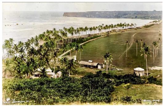
Figure 4: The Bath Plantation [34].
The Tenantry Freehold Purchase Act (1980-53-2022) was passed as a modification of the Barbados Constitution in an effort to legalize the land ownership of people who were forced to move from their homes at the end of slavery. After forced removal to a tenantry only the littoral continued to be a place considered to be their own.
Marine and Terrestrial Resources
Traditionally, Amer-Indians (beginning in 350AD) occupied and regularly used the marine and terrestrial resources of Barbados. In fact they continued to be a part of Barbadian life for some decades after the island became a part of the British Empire in 1627 [37]. Richard Ligon [38] who lived in Barbados from 1647 to 1650 provided a map of the Indian area located inland near Bath. He observed that: Indian women were primarily, if not solely, employed in household tasks. On the other hand, the men…were used for footmen and the killing of fish, which they are good at. With their own bows and arrows they will go out and in a day’s time kill as much fish as will serve a family of a dozen persons two or three days, if you can keep the fish so long.
This is an intriguing insight into the pristine condition of the littoral at that time. It is not clear how long Indian men were employed as fishers with their six-foot bows and long arrows, but it is unlikely that people of African Ancestry were permitted to make and use such weapons. The Native canoes used to travel to other islands were not available to enslaved people.
Crabs were fished at night using torches to provide both light for movement in the shallow waters and to illuminate the crabs and lobsters. Torches combined with nets were used to catch flying fish and McKinnen in 1802 recorded that local people were very successful at catching flying fish using this method. At night they spread their nets before a light and would disturb the water at a small distance. The fish would rise and eagerly fly toward the light and become intercepted by the nets. This pattern of using torches and nets to catch flying fish was observed again by another visitor in 1818 [33,37]. Use of torches to collect resources in the littoral is in keeping with the need for enslaved people to be out of sight at night when they are away from the plantation. The Siza photos (1896-1897) reveal much about life for average people and the environment of the littoral. Several scholars have written about the Siza photo of nine fishermen along Bath Beach as it visually represents issues of ethnicity, class, food security, social networks, and littoral use (Figure 6). Fred Watson was born and raised in the Bath Littoral Zone and his family members are among the nine fishermen. The following discussion weaves Mr. Watson’s life into the discussion of a cultural heritage area designation.
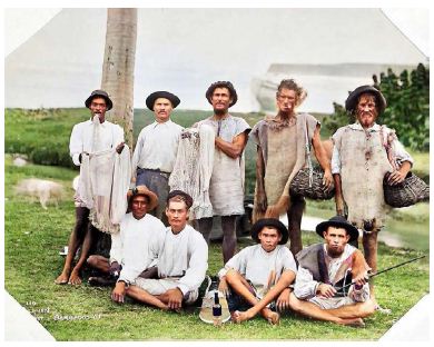
Figure 6: Nine Bath Fishermen in 1896 (Siza Photo 110). From left to right in back is George Watson, Aubrey King, then Joe Watson (holding fish net), Gerald Goddard (smoking clay pipe) and Thomas Henry Goddard, Joseph Josiah Goddardʼs brother. Sitting in the front row is Simeon Goddard, Ben Watson, Althard Watson and Robert Haynes [34].
These fishers, like many people in Bath, were of both African Ancestry and Scots-Irish descent. The visible handmade fishing equipment (wooden poles and woven cotton nets) were used on the shallow bank and the bags for holding fish were made from local vines. Current fishing methods in the Bath littoral zone are in ways similar to those used by the men pictured here. In fact, some of them have descendants who still today collect plants and fish in the same manner as in 1896. Key informant Fred Watson was born at Bath Beach not 100 yards from where this photo was taken. It was here he kept his little boat that he would row out to the drop offs to fish for his family. Like the fishermen in the photo, he also walked the littoral collecting both marine and terrestrial resources (throwing cast nets for fray and sprats much like the fishermen in the photo) as a means of subsistence, food and health security, and small-scale commercial production. From the collection of plants for “bush tea” and consumption of items such as sea grapes, to marine resources such as fish and crabs (both land and sea) this place was and has always been an important part of creating stability and security for the people of the area. Both younger and older men are present in this photograph which represents a common feature of the area, that its environmental features make it accessible and suitable for both shore-based and inshore fishing for people of all ages. The area is protected from strong currents and high wave activity because of the Great Rocks and structure of the littoral. It is still an area where fathers and uncles bring sons and nephews to learn how to handline and freedive to collect fish and other marine resources. It is also an area where youth and elders can collect octopus and whelks and walk their way out more than 200 yards at certain tide and moon phases. While these strategies continue to be a part of their efforts to provide food security as well as means of recreational activity, for many of the youth from the area, this is their environmental classroom and the elders their teachers. In addition, the coastal marine environment is productive which makes it a viable location for resource gathering both at night and day and many still use it as an area to collect food and medicinal resources. The marine area is exceptional for night diving for lobsters and sea eggs (Tripneustesventricosus) and not only locals but people from all over the island come in different seasons and moon phases to target specific species.
Bath Beach and its littoral zone was and is an important social, cultural and economic relevant component in the economics of both African Ancestry people and the descendants of the first Irish and Scottish indentured workers. There is little doubt that the resources did and still do today provide for people in the area and now reach out beyond community boundaries to those from other parts of the island. Those who moved away from their homes in Bath to foreign countries like England and Canada still sit and tell stories with pride and joy about their youth growing up in and around the Bath Plantation Area and about how it never is far from their thoughts or their hearts. These emotions are due in part to social relationships that were developed as youth, but also are a component of their personal, national, and ethnic identity. In a conversation with a gentleman who was headed back to England after his month-long holiday in Bath he stated with tears streaming down his face, “I will leave here tomorrow to return to England, but ya know I never leave here…my home.” The littoral is culturally important today because it retains many of its traditional uses, but it also has heritage value for the people who have lived here for hundreds of years. People continue to come to experience all it has to offer. From a fishing standpoint the coral and hard bottom shelf of the littoral provides a productive environment. It is common, based on this research, to observe that during both day and night bait fish (Clupeidae) and other species of jacks (Caranx hippos; Caranxignobilis; Caranxcrysos), parrotfish (Scaridae), grunts (Haemulidae) and lobsters (Panulirusargus) move in and out of the area on the tides, using segments of the inshore areas as a means of aggregation and protection from larger species. This accumulation of species, be they fish, black and white sea urchins (Diademaantillarumand Tripneustesventricosus; the white urchins are called sea eggs), and octopus (Octopus briareus; locally known as seacats) all entice larger predator species to come in during certain tides, moon phases and especially at night.
Stoffie’s community residence and subsequent fishing experiences in the area meant he was able to fish at all times of the day/night using a variety of targeting strategies (hook and line from shore and boat, both bottom fishing and trolling). He also did free diving in deeper water using hooks and spears. He observed a wide variety of desired species that could be targeted utilizing these multiple methods. Some extraordinary catches he observed included an 11-foot-long Tiger Shark (Carcharhinidae), Large Barracuda (Sphyraenidae), Giant Trevally (Caranxignobilis) and a 70-pound Cubera Snapper (Lutjanuscyanopterus). These are just a few of the larger predator species that enter this area to feed at night. In addition, at night there are lobsters found while walking across the flats or in tidal pools and it is common to find large parrotfish sleeping in the seagrass. The productivity of the area is special and has always been a key component of community life. Fishermen today confirm that the area from Conger Bay east to Conset Bay is a culturally special heritage area to them because of the way the marine portion of the littoral extends out to the Great Rocks. At this point there is the beginning of a drop off into the deep water of the open ocean (Figure 5). The 40-foot shear drop off shelf edge creates an opportunity for artisanal fishers to target certain species otherwise not accessible with simple net and pole. Because of this shelf, fishers have over the last 400 years developed a special relationship and traditional ecological knowledge of this portion of the marine littoral, which holds special heritage meanings as a place for the free use by the people who have been largely without property and a safe place where they can always turn for food, medicine, recreation, education, and solace.
Tourism
Tourism has always been an important feature of the area. Be it local tourism, people from the area spending time on the beach with family and friends fishing, playing cricket in the sand, having a picnic or a nightly sea bath; island tourism, the busing (or as it was in the past when the Barbados railway was in use and brought people from Bridgetown to and through Bath ending just north in Bathsheba) of individuals from all over the island to “lime” the day away in the soft Bath sand; or International tourism, people from all over the world who come and rent houses or stay in luxury resorts across the island and are transported either by personal vehicle or bused in as a part of a regular visit to the area. Resorts like Almond Beach Resort would on every Tuesday and Thursday bring busloads of tourists to spend a day picnicking and swimming in the quiet and safe water of the area. While the resort supplied drinks and food, many of the local youth would sell various items to the tourists, be it small bags of sea grapes, Bajan fruits like mangos, Bajan Apples, and Aki. Of course, a favorite among the often-sunburned tourists is the Aloe grown on the plantation lands above the littoral that is cut open and made into jelly to be used to cool their burning skin. It is also common to see that same aloe consumed by locals, eaten raw or crushed into a liquid aloe shake said to be good for digestion and general stomach health. Figure 7 shows a photo circa 1910 of the Train Station in Bath, which is still present today and marks the beginning of the East Coast Walking Trail between Bath and Bathsheba. You can see the large rum barrels in the background. These and other items were moved between the “country” and “town” on these railways, in addition to local and foreign tourists.
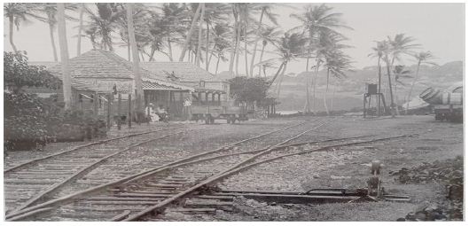
Figure 7: The Old Bath Beach Train Station [34].
Today the train station is maintained but not operational. It is now a part of a nature trail that travels north as the train once did but is now used for hiking and recently developed for bicycling utilized by locals and tourists alike. On the trail, guides share stories of the area in addition to talking about the use of the local natural resources and their uses in the past and present (Figure 8).
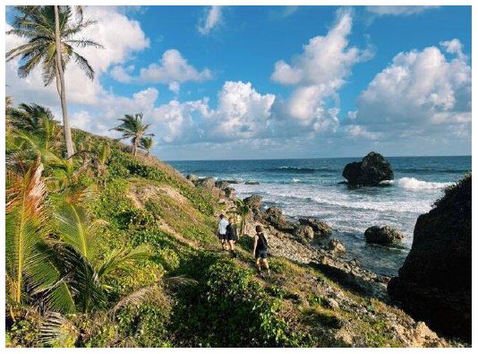
Figure 8: The former Barbados East Coast Railroad has since been made into a very popular walking trail. In the photo, tourists are following the Old Railroad walking trail [34].
The trail begins in Bath Beach at the train stop (a house that used to be the Bath Train Station) and follows a coastal route through Martin’s Bay to Bathsheba (Figure 8). There are guided tours for tourists and many local walking clubs select this path for their group hikes/outings. It crosses many important historical sites in addition to all of the littoral resources that created food security and social stability in the communities for centuries.
The River and the Spring
Drewel [39] states that, “Diverse African cultures have stressed the value and power of water not only as a source of sustenance but as the focus of spiritual expression”. The following is an examination of the Bath Plantation Spring system which includes both Pothouse and Bath Beach Springs (Figure 9) (connected above and below ground) and the historic and contemporary spiritual connection and role in the lives of African Ancestry people of the Bath Plantation.
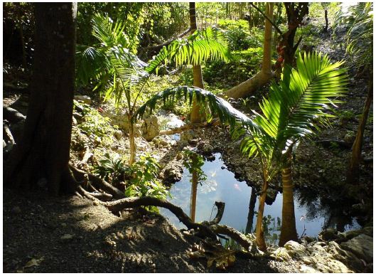
Figure 9: Spring that Connects the Land and Sea in Bath, St. John Barbados.
The Quamins River, named by past Amer-Indian inhabitants, is likely to be the above and below ground connection of a Spring that runs from Pothouse to Bath Beach, pooling up in some locations with the creation of natural and man-made waterfalls, and then in places disappearing under the ground only to reappear further downhill towards the beach below. This “river” or spring has likely long had a significant role in the spiritual and everyday prerequisite of life for people of native American and African Ancestry backgrounds. The following examines both the spiritual and utility of the Spring and its water.
In Drewel’s examination of a water spirit known throughout Africa and the African diaspora (especially in the Caribbean) as Mami Wata, not only did the water have a spiritual meaning but so did the deities associated with it [39]. Drewel states that:
Sacred waters bathe the histories of African peoples, sometimes as tears of deep sorrow, sometimes as drops of soothing and cooling liquid sustaining life and hope. Water connects—world with otherworld, life with afterlife—for many African and African Atlantic peoples – Yoruba, Kong o, Fon, and others. And among Africans dispersed across vast oceans, those waters are emblematic of the ultimate journey back home to all those distant yet living ancestors.
Water divinities of Africa and the African Atlantic are regularly invoked to maintain, refresh, and strengthen the spirit needed to endure the hardships and challenges of lives scattered and shattered by the avarice, arrogance, and brutality of those who would enslave others for their own benefit.
It is a fairly simple line to draw from people of African Ancestry in and around the Bath Plantation to a spiritual connection with the Spring. The Spring today, as in the past, offers a cleansing and a “cooling down” calming component to participation (much in the way going to confession or church may provide for Christians and Catholics). Perhaps it was in this meditative state of tranquility that enslaved people were able to transcend themselves back home to loved, distant yet living, ancestors as suggested by Drewel [39]. From a historical context it is evident based on other examples throughout the diaspora to see why under strict persecution of the English/Anglican Church that this type of spirituality was hidden from those outside of the group, much in the way people of African Ancestry maintained and hid cultural and religious institutions and beliefs (African Gods/ Spirits and practices) even in the face of Christianity and Catholicism in the New World (e.g Santeria). These kinds of verbal interactions are not readily visible and accessible in daily discourse. In fact, like other aspects of daily life hidden from vertical society, Wilson [31] makes this point in his discussion of vertical versus horizontal society in the Caribbean and Stoffie [24] demonstrates this with his findings on Rotating Savings and Credit in the Caribbean/Barbados. The Spring is a part of the diaspora during slavery and maintains an important spiritual component in contemporary life.
There is historic and contemporary use as means of bathing or washing away physical and psychological ailments affecting individuals in everyday life. This is something that in the past may have been guarded but with certain individuals has been shared, as it was with Stoffie in 1997. After a day’s spearfishing and due to a number of cuts and abrasions from diving on and around the reef, it was suggested by local fishermen that they go for a “fresh.” Skin burning and agitated, Stoffie and the others walked to Pothouse Spring (part of the Bath Plantation and Bath Beach Spring) and went to bathe in the cooling and healing waters (Figure 10). After about 20 minutes of standing under the makeshift waterfall the cuts and abrasions had been cleaned, the burnt skin cooled, the body temperature reduced, and overall, well being restored. In this moment the mind and body had reached the place where the Spring had completely altered the physical and psychological in a short period of time.
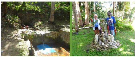
Figure 10: Pothouse Spring: A place for gathering, healing and spirituality.
Currently there are examples of others being brought to the Spring as means of healing. People have heard of the Pothouse Spring, and it is not uncommon for them to come from other parts of the island as well as from the local communities to take part in a “fresh.” In fact, there is an account of a Bajan man from Bridgetown who, with the guidance of one of the local Bath residents, was taken to the Spring because of its healing properties. The two went together to the Spring and the local Bath resident mixed the Spring water with a collection of local plants. This was formulated into a mixture/tea with the “nutrients” of the Spring water and plants creating a medicinal tonic used to rejuvenate, remove the toxins and stimulate the body. In addition to the spiritual component of the Spring, it is likely that from a practical standpoint the access to water for survival, cooking, cleaning and washing reduced health risks among the people of the Bath Plantation area. The Bath Plantations Spring was historically one of the most important resources as a means of hydration and access to clean water for consumption and cooking, in addition to being a source to draw upon to carry home for irrigation of house gardens. Handler [40] suggests that during slavery, vulnerability to waterborne diseases was a significant problem. This was in large part a hierarchical class/power issue where the plantation had access to the best water and slaves were forced to drink from natural and man-made ponds/ watering holes, often infected with various forms of bacteria due to human and animal waste. In addition, these open water holes were the breeding ground for mosquitoes and the transmission of yellow, dengue and other types of severe fevers.
Hauser [41] suggests that there are three dimensions of water; (1) salt versus sweet, (2) clear versus pond, and (3) cool versus hot. To have access to a source that hits the mark in all three categories is something revered and protected. The Springs of the Bath Plantation area hit these three marks in that they are sweet (cold and fresh) and not brackish, they are clear and not clouded or muddied with pollution of “sickness”, and they are cool and not hot, providing an enjoyable and refreshing source of hydration. During pre and post slavery it is perceived that this would make those who had the knowledge and access to the Spring in a more advantageous position of mental and physical health and well-being compared to those on other plantations who are forced to struggle with hydration and health issues because of a lack of this type of water. This water is what would be classified by Sloave in 1707-1725 as “sweet.” In this account he states that Spring water is preferred to all others.
Discussion
The Bath Heritage Littoral Landscape was and is a place for people of all ages, economic backgrounds, ethnicities and religions. It is an important location for health reasons, such as sick children that come to sit in the tranquil waters for a sea bath, clearing their head and chest congestion. In addition, it is a place where older men and women collect specific plants to make their Bush Tea to fight fevers, in addition retaining a level of independence and food security through collection of marine resources for household consumption. It is a place of cultural transmission of ecological knowledge where elders instruct children on different types of fishing strategies and species to target, in addition to being able to share general cultural values of appropriate use and conservation of marine and terrestrial resources. It is a place where young men can fish as means of creating food security within their social networks in addition to offsetting the cost of food for family households; even being able to engage in the informal economy with their small-scale commercial production. It is a place of social gathering for big events as well as nightly conversations. It is a place that has and continues to be a special heritage environment where a person can gather what is needed for life and share time with family and community, something that has been part of the social fabric since slavery. It is a place of importance. This analysis is responsive to contemporary research that argues for the incorporation of traditional heritage communities in the future management of the ecological zones; areas they have used and conserved for hundreds of years [42]. The analysis builds on ethnographic and ethnological comparative studies some of which have been conducted by the author in Barbados, the Dominican Republic, and the U.S. Virgin Islands [24,43-46].
Other ethnographic studies suggest that the findings can be extrapolated elsewhere in the Caribbean [33,47]. When generalizing about heritage littoral landscapes, however, it is important to take into consideration that the coastal littorals vary ecologically, as do the people and their communities. A key human dimension of any coastal study is that the people involved have resided in a stable coastal community for long periods. African Ancestry people, for example, typically have heritage landscapes associated with their community which involve hundreds of years. A heritage littoral landscape emerges from generations of co-adaptations between the people and the sea. These co-adaptations are incorporated into the peoples’ cultural systems of belief and practice. Many coastal heritage landscapes have been radically modified in structure, function, and the human services they provide (Lopez-Angarita et al. 2016; Jennings et al. 2012). Changes have occurred due in part to the following (1) mangrove removal for industry, (2) coastal aquaculture like shrimp farming found throughout the Americas and Asia, (3) coastal farming like the U.S. Carolinian paddy rice farms, (4) tourism development especially all-inclusive resorts which own, modify, and reserve the beaches for tourists, and (5) recreational boating harbors which involve mangrove removal, dredging, and construction. The professional journal Coastal Zone Management contains numerous systematic studies of coastal littoral damage and contemporary efforts to restore it and sets the criteria time and space frame for restoration, including when it was sustainably used and managed by African Ancestry communities. Guidelines from the United Nations IUCN [48] argue for the restoration of these places including protection of heritage community relationships with the environment. A general principle of management and restoration provided was recommended by a team of scholars based on their research [48]. They maintain that ecological restoration is facilitated by documenting and recognizing in policy the resident people, their societies, and their traditional role in conserving natural systems [49]. Increasingly involving traditional people in coastal management has proved a viable conservation strategy [43,44,50]. While working together local communities and management agencies can both find common conservation goals [51], but heritage littoral conservation requires a holistic identification of the marine and terrestrial environments. Both land and sea are functionally and culturally interrelated, so to persist they must be conserved as a holistic littoral landscape.
Author Contributions
Conceptualization, B.S.; methodology, B.S.; formal analysis, B.S.; investigation, B.S.; resources, B.S.; data curation, B.S.; writing— original draft preparation, B.S. and A.D.S.; writing—review and editing, B.S. and A.D.S.; supervision, B.S.; project administration, B.S.; funding acquisition, B.S. All authors have read and agreed to the published version of the manuscript.
Funding
This research was funded by WennerGren (1996), in addition to small travel grants from the University of South Florida Graduate School and School for Latin American and Caribbean Studies (1999). The research conducted after 2000 was all self-funded.
Data Availability Statement
Not applicable.
Acknowledgment
We would like to acknowledge the contributions of those that assisted in the creation of this article. Dr. Amanda Stoltz was instrumental in her efforts to guide this process and ignite new energy towards the creation and completion of this effort. Dr. Kathleen Van Vlack and Dr. Richard Stoffie were both instrumental by lending their expertise on fisheries in the Caribbean, Caribbean History and issues related to cultural heritage in places throughout the world. In Barbados we would like to thank the people of the Bath Plantation Area, who have always been more than friends and willing to share their lives and knowledge with us about a wide variety of topics. While Fred Watson will never read this, I wish somehow to let him know how instrumental he was in all of this effort, from my dissertation to the authorship of this article. It was Fred who taught me about the Bath Littoral and all its uses. I would not be who I am today without having been fortunate enough to have known him and shared time together. Finally, to Dr. Trevor Purcell, who passed away in 2007. You are the reason I returned to Barbados in 1996 and re-awoke in me my Caribbean connection.
Conflicts of Interest
The authors declare no conflict of interest. The funders had no role in the design of the study; in the collection, analyses, or interpretation of data; in the writing of the manuscript, or in the decision to publish the results.
References
- Stoffie B, Stoffie R (2007) At the Sea’s Edge: Elders and Children in the Littorals of Barbados and the Human Ecology 35: 547-558.
- Zube EH, Pitt DG (1981) Cross-Cultural Perceptions of Scenic and Heritage Landscape Planning 8: 69-87.
- Cultural landscapes and protected areas: Unfolding the linkages and synergies. International Union for the Conservation of Nature. Cultural landscapes and protected areas: Unfolding the linkages and synergies. International Union for the Conservation of Nature.
- Bomhauer-Beins L, de Guttry C, Ratter BMW (2019) When Culture Materializes: Societal Dynamics in Resilience of Social-Ecological Systems in the Case of Conch Management on Abaco, The Bahamas. Sustainability 11.
- Jennings DE, Di Battista JD, Stump KL, Hussey NE, Franks BR, et al. (2012) Assessment of the Aquatic Biodiversity of a Threatened Coastal Lagoon at Bimini, Journal of Coastal Conservation 16: 405-428.
- McCay B (v) The Littoral and the Liminal: Challenges to the Management of the Coastal and Marine MAST 7: 7-25.
- Bond A, Morrison-Saunders A, Howitt R (2013) Sustainability Assessment: Pluralism, Practice and Routledge London.
- Getting It Right: Community-Based Human Rights Impact Assessment Tool. 2022.
- Antrop M (2005) Why Landscapes of the Past Are Important for the Landscape and Urban Planning 70: 21-34.
- Contested Landscapes: Movement, Exile and (2001) Bender B, Winer M (Eds). Routledge London.
- Fairclough G, Harrison R, Jameson J, Schofield J (2007) The Heritage Routledge London.
- McCay Presidential Address Part II: Edges, Fields, and Regions. The Common Property Resource Digest 54: 6-8.
- Westerdahl C (1992) The Maritime Cultural Landscape. International Journal of Nautical Archaeology, 21: 5-14.
- Bilby K (2005) True-Born Maroons; University Press of Florida: Gainesville.
- Geggus DP, Fiering N (2009) The World of the Haitian Revolution; Indiana University Press: Bloomington IN.
- Price R (1996) Maroon Societies: Rebel Slave Communities in the America; Johns Hopkins University: Baltimore MD.
- Thompson A (2006) Flight to Freedom: African Runaways And Maroons in the West Indies University Press: Bridgetown, Barbados.
- Walker C (2020) Jamaica Ladies: Female Slaveholders and the Creation of Britain’s Atlantic Empire ; University of North Carolina Press: Chapel Hill.
- Denevan W, Mathewson K (2009) Carl Sauer On Culture and Landscape. Louisiana University Press: Baton Rouse.
- Sauer C (1925) The Morphology of Landscape. University of California Publications in Geography 22; University of California: Berkley, CA, USA. Pg: 19-53.
- Fowler PJ (2003) World Heritage Cultural Landscapes; UNESCO World Heritage Centre: Paris, Pg: 1992-2000.
- Turner N, Davidson-Hunt I, O’Flaherty M (2003) Living on the Edge: Ecological and Cultural Edges as Sources of Diversity for Social-Ecological Resilience. Human Ecology 31: 439-461.
- Clift Greatecology, Living on the Edge. Greatecology, Living on the Edge.
- Stoffie Brent (2001) We Don’t Put All of Our Eggs in One Basket: An Examination of Meeting Turn, A Rotating Savings and Credit Association in PhD Dissertation, University of South Florida, Tampa FL.
- Bernard HR (2005) Research Methods in Anthropology: Qualitative and Quantitative Approaches (4th Ed); Altamira Press: Walnut Creek, CA, USA.
- Gmelch G, Gmelch S (2005) The Parish Behind God’s Back: The Changing Culture of Rural Barbados; Waveland Press: Long Grove, IL.
- Beckles H (1989) Natural Rebels: A Social History of Enslaved Women in Barbados ; Rutgers University Press: New Brunswick NJ.
- Beckles H. A History of Barbados from Amerindian Settlements to Nation-State. England ; Cambridge University Press: London.
- Reilly M, Norris A (2019) Collaborative Archaeology in ‘Redleg’ In Pre- Colonial and Post-Contact Archaeology in Barbados. Waal M, Finneran N, Reilly M, Armstrong D, Farmer K (Eds) Sidestone Press: Leiden, Netherlands. Pg: 337-352.
- Olwig KF (1993) Global Culture, Island Identity: Community and Change in the Afro-Caribbean Community of Routledge: London.
- Wilson P (1973) Crab Antics: A Caribbean Study of the Conflict Between Reputation and Respectability ; Waveland Press: Long Grove, IL.
- Cumberbatch J, Hinds C (2013) Barbadian Bio-Cultural Heritage: An Analysis of the Flying International Journal of Intangible Heritage 8: 118-134.
- Stoffie B, Stoffie R, Van Vlack K (2020) Sustainable Use of the Littoral by Traditional People of Barbados and Sustainability 12: 1-26.
- Burton Bajan Things.
- Siza MAN (2021) 1896-1897. Photos Archived in the Anglo-Luzo Photographic Gallery.
- Centre for the Study of the Legacies of British The Bath, Barbados, St. John.
- Handler JS (1970) Aspects of Amerindian Ethnography in 17th Century Barbados. Caribbean Studies 9: 50-72.
- Ligon R (1657) A True and Exact History of the Island of Barbados, 5th Smith DC (Ed) Wilfrid Laurier University: London.
- Drewal HJ, Mami Wata (2008) Arts for Water Spirits in Africa and Its African Arts 41: 60-83.
- Handler JS (1969) English Rustics in Black Skin: A Study of Modern Family Forms in a Pre-Industrialized Society. Sidney M. Greenfield. American Anthropologist 71: 335-337.
- Hauser MW (2017) A Political Ecology of Water and Enslavement: Water Ways in Eighteenth-Century Caribbean Plantations. Current Anthropology 58: 227-256.
- Brosius J, Tsing A, Zerner C (2005) Introduction: Raising Questions about Communities and Conservation. In Communities and Conservation: Histories and Politics of Community Based Natural Resource Management. Brosius J, Tsing A, Zerner C (Eds) Altamira Press: New Pg: 1-34.
- Stoffie B (1993) Indigenous Marine Conservation: A Case Study from the Dominican East Carolina University, Greenville, NC.
- Stoffie R, Halmo D, Wagner T, Luczkovich J (1994) Reefs from Space: Satellite Imagery, Marine Ecology, and Ethnography in the Dominican Republic. Human Ecology 22: 355-378.
- Stoffie B, Waters S, Abbott-Jamieson S, Kelley S, Grasso D, et (2009) Can an Island Be a Fishing Community: An Examination of St. Croix and Its Fisherie; U.S. Department of Commerce, National Oceanic and Atmospheric Administration National Marine Fisheries Service Southeast: Miami.
- Stoffie B, Grace C, Snodgrass D (2011) The Socio-Economic Importance of Fishing in Thomas, USVI: An Examination of Fishing Community Designation; Technical Memorandum NMFS-SEFSC-623; NOAA: Miami.
- Purcell T, Onjoro E (2003) Indigenous Knowledge, Power and Parity Models of Knowledge Integration. In Participating in Development. Bicker A, Pottier J, Sillitoe P (Eds). Routledge London. Pg: 27.
- World Commission on Protected Areas 2021-2025. World Commission on Protected Areas 2021-2025.
- Löfqvist S, Kleinschroth F, Bey A, de Bremond A, DeFries R, et (2023) How Social Considerations Improve the Equity and Effectiveness of Ecosystem Restoration. BioScience 73: 134-148.
- Doehring K, Cole C, Young RG, Longnecker N (2023) Collective Storytelling as a River Restoration Tool: The Role of Catchment Communities in Inspiring Environmental Change. Frontiers in Communication 7.
- Agrawal A, Gibson C (2001) The Role of Communities in Natural Resource Conservation. In Communities and the Environment: Ethnicity, Gender, and the State in Community-based Conservation; Agrawal A, Gibson C (Eds) Rutgers University Press: New Brunswick, NJ.