Author Archives: author

Treating uterine synechiae with human amniotic membrane graft

DOI: 10.31038/IGOJ.2020311

Abstract

Objective: To determine if placement of a human amniotic membrane graft at the time of hysteroscopic intrauterine adhesiolysis will promote regeneration of the endometrium in women with secondary amenorrhea and infertility. This case-series evaluated the use of human amniotic membrane graft in women with secondary amenorrhea and who had a desire for future fertility.  Results: Five women received the amniotic membrane graft between 2015–2019. Four of the five women had secondary amenorrhea and one had scanty periods prior to the surgery (post suction and evacuation procedure due to spontaneous miscarriage), and all of the women achieved menstruation post-operatively. Two of them achieved pregnancy.

Conclusion: This procedure may provide an alternative treatment option for patients with uterine synechiae who desire future menses and pregnancy.

Introduction

Women usually present with secondary amenorrhea, infertility, or recurrent pregnancy loss in cases of uterine cavity synechiae [1]. Intrauterine adhesions are most commonly caused by a dilation and curettage (D&C) procedure for medical termination of pregnancy or spontaneous abortion or latent genital tuberculosis, as in case of our patients. The risk of intrauterine adhesions after one or two D&C’s is about 14–16%; severity of adhesions also increases with repeated D&C [2]. Operative hysteroscopy with lysis of intrauterine adhesions is a viable treatment option. Hysteroscopic adhesiolysis can be performed using scissors, cautery or laser vapourization [3–5]. However, with all forms of adhesion resection, there is a high rate of recurrence of intrauterine adhesions ranging between 3.1% and 23.5%. [1] Due to the high rate of intrauterine adhesion formation, many procedures have been attempted to reduce it. An unique technique has been performed by few authors in which was they had used a fresh amnion graft over an inflated balloon or a Foley catheter for 2 weeks at the time of adhesiolysis in women with moderate and severe intrauterine adhesions and there was  significant improvement in uterine length and intrauterine adhesions [6].  Our study is a case-series evaluating the use of human amniotic membrane graft to reduce adhesion formation, promote mesenchymal stem cell growth, evaluate for a thickened endometrial stripe and subsequent pregnancy in patients with secondary infertility.

Materials and Methods

Five infertile women of age 28–32years, who presented to Calcutta Fertility Mission between 2015–2019  with secondary amenorrhea , diagnosed by transvaginal ultrasonography  9 thin endometrium with glistening structure) and hysterosalpingogram , were informed that the placement of a human amniotic membrane graft  will be done for regeneration of the endometrium. Latent genital tuberculosis was ruled out by DNA-PCR of pre- menstrual endometrial aspirate. After informed consent was obtained,  five women underwent operative hysteroscopy, lysis of intrauterine synechiae  and curettage. A 3.5×3.5cm amniotic membrane graft was wrapped around a curette (Size 8) and inserted into the uterine cavity in three women. In another two cases the graft was put in the cavity using sinus forceps. After insertion, the graft was adjusted and excised at the level of the cervix. Routine antibiotics were prescribed for all patients and Estradiol Valerate (2mg) was given for the next three months. All five women had regular menstrual cycles after 3 months, ET (endometrial thickness) was assessed on 10th day and 12th day of the menstrual cycle and were found to be satisfactory. ( ET on 10th day – 6 to 7.5mm; on 12th day – 8 to 9mm)

Results

Five women received the human amniotic membrane graft between 2015–2019. All of them  had amenorrhea prior to the surgical procedure (ranging from 3–8 months of amenorrhea), and all had achieved menstruation post-operatively. There is no universal grading system for the severity of intrauterine adhesions, the severity was graded using the Valle and Sciarra’s 1988 classification. Three of five women had moderate synechiae and two of them had severe form of synechiae. Mild disease indicates flimsy adhesions composed of basal endometrium producing partial or complete uterine cavity occlusion. Moderate disease involves fibromuscular adhesions that are characteristically thick, covered by endometrium that may bleed with  partial or total occlusion of the uterine cavity.  Severe disease is composed of connective tissue with no endometrial lining and  partially or totally occlude the endometrial cavity. There were no post-operative complications.

Two of the five women achieved pregnancy. The first patient had conceived with ovulation induction drugs within 6 months of regular menstrual cycles and had a spontaneous miscarriage at 8 weeks period of gestation. The second patient had a spontaneous conception after 9 months of the procedure and ended up with a subsequent premature labour at 34 weeks period of gestation.

Discussion

Human amniotic membrane grafting is a novel treatment modality to reduce inflammation and adhesion formation after hysteroscopic adhesiolysis. It has been postulated that the basalis layer of the endometrium harbours endometrial stem or progenitor cells, which are responsible for its regenerative capacity [7]. Inflammation may prevent the endometrium from regenerating due to the deposition of fibrotic tissue as fibrosis is triggered. [8]. But according to Nagori CB et al., autologous bone marrow stem cells placed in the uterus in severe Asherman’s Syndrome, enabled regeneration of the endometrium sufficient to support a donor IVF embryo [9]. The intrauterine device (IUD) and an intrauterine Foley balloon have been studied and used to prevent adhesions after hysteroscopic procedures and Foley balloon has been found to yield better results. [10]. The use of amniotic membrane graft in cases of vaginoplasty has yielded remarkable results as  the vagina is well formed without any evidence of stenosis [11]. Pregnancy outcomes in patients who received the graft needs to be explored further however, there are many confounding factors that could impact the pregnancy rate. In the present study we had two pregnancies and one live-birth following intrauterine grafting. Estrogen supplementation after lysis of intrauterine adhesions may be advantageous to increase the endometrial proliferation and prevent fibrosis [12]. Amniotic membrane use in ophthalmology has been proved to be an alternative for corneal and conjunctival reconstruction in many clinically challenging cases. The translational potential of an efficient and inexpensive method to prepare de‐epithelialized HAM as a basement membrane scaffold for cell‐based tissue‐engineered treatments of ocular surface disorders has also been documented [13].

Conclusion

It can be concluded that based on this case-series there may be a utility for using human amniotic membrane intrauterine graft in a selective group of women to improve endometrial thickness while the functional aspects of the endometrium as demonstrated by successful pregnancy outcome. At present this work is being pursued at our institute to assess its proper utility on a larger scale.

Authorship

Dr. Siddhartha Chatterjee: Conception and design, analysis and interpretation of data; revising manuscript critically for important intellectual content

Dr. Bishista Bagchi: Drafting the article; acquisition of data, revising manuscript

Dr. Arpan Chatterjee: Drafting of article, data collection

Final approval of the version to be published given by all authors.

Agreement to be accountable for all aspects of the work in ensuring that questions related to the accuracy or integrity of any part of the work are appropriately investigated and resolved.

Acknowledgments

Miss Orphi Bhattacharya for formatting the manuscript

References

  1. Yu D, Wong YM, Cheong Y, Xia E, Li TC (2008) Asherman syndrome—one century later. Fertil Steril 89: 759–779. [crossref]
  2. Friedler S, Margalioth EJ, Kafka I, Yaffe H (1993) Incidence of post-abortion intra-uterine adhesions evaluated by hysteroscopy–a prospective study. Hum Reprod 8: 442–444. [crossref]
  3. Fedele L, Vercellini P, Viezzoli T, Ricciardiello O, Zamberletti D (1986) Intrauterine adhesions: Current diagnostic and therapeutic trends. Acta EurFertil 17: 31–37. [crossref]
  4. Chen FP, Soong YK, Hui YL (1997) Successful treatment of severe uterine synechiae with transcervical resectoscopy combined with laminaria tent. Hum Reprod 12: 943–947. [crossref]
  5. Newton JR, MacKenzie WE, Emens MJ, Jordan JA (1989) Division of uterine adhesions (asherman’s syndrome) with the nd-YAG laser. Br J Obstet Gynaecol 96: 102–104. [crossref]
  6. Amer MI, Abd-El-Maeboud KH (2006) Amnion graft following hysteroscopic lysis of intrauterine adhesions. J Obstet Gynaecol Res 32: 559–566. [crossref]
  7. Rockey DC, Bell PD, Hill JA (2015) Fibrosis–A common pathway to organ injury and failure. N Engl J Med 373: 95–96 [crossref]
  8. Gargett CE, Healy DL (2011) Generating receptive endometrium in asherman’s syndrome. J Hum Reprod Sci 4: 49–52. [crossref]
  9. Nagori CB, Panchal SY, Patel H (2011) Endometrial regeneration using autologous adult stem cells followed by conception by in vitro fertilization in a patient of severe asherman’s syndrome. J Hum Reprod Sci 4: 43–48. [crossref]
  10. Orhue AA, Aziken ME, Igbefoh JO (2003) A comparison of two adjunctive treatments for intrauterine adhesions following lysis. Int J Gynaecol Obstet 82: 49–56. [crossref]
  11. Chakravarty BN (1977) Congenital absence of the vagina and uterus – simultaneous vaginoplasty and hysteroplasty. Journal of obstetrics and gynaecology of India 627–632.
  12. Farhi J, Bar-Hava I, Homburg R, Dicker D, Ben-Rafael Z (1993) Induced regeneration of endometrium following curettage for abortion: A comparative study Hum Reprod 8: 1143–1144. [crossref]
  13. Bandeira F, Yam GH, Fuest M, Ong HS, Liu YC (2019) Urea-De-Epithelialized Human Amniotic Membrane for Ocular Surface Reconstruction. Stem Cells Transl Med 8: 620–626. [crossref]

Climate Change and Fisheries: The Case Study of Corsica, an Ideal Reference Station in the Mediterranean Sea

DOI: 10.31038/AFS.2020212

Case Study

Climate change is one of the main factors influencing the structure and functioning of marine ecosystems, mainly because of the rise in water temperature [1]. Its effects are expected to increase in the near future, especially in the Mediterranean region which has been defined as a primary « hot-spot » regarding future climate change projections [2]. From 1950 to 2009, the average temperature of Mediterranean surface waters increased more (+ 0.50°C) than those of oceanic waters (e.g. + 0.42°C for the Atlantic Ocean and + 0.30°C for the Pacific Ocean) [3]. In addition, based on the different GHG (Greenhouse Gas) emission scenarios proposed by the IPCC (Intergovernmental Panel on Climate Change), models predict an increase in Mediterranean surface temperatures ranging from + 1.73°C to + 2.97°C Ocean warming for the period 2070–2090 compared to the period 1961–1990 [4]. Ocean warming can exert a direct influence on marine organisms, for example, leading to mass mortality events of benthic invertebrates [5] or affecting many life history traits of fish [6]. Such influence has also lead to the upheaval in the geography of the climate in the Mediterranean basin impacting ultimately on the distribution range of species. Indeed, the Mediterranean Sea is not exempted from « tropicalization » phenomena via the arrival of exotic species, neither from « meridionalization » via the extension of the distribution area of species with affinity to warm-waters [7]. Exotic species can be invasive and thus, can seriously disrupt the existing ecosystems [8]. On the island of Corsica, this is the case of the bluespotted cornetfish (Fistularia commersonii), a Atlantic lessepsian the Atlantic crab Percnon gibbesi fish species observed in the Lavezzi islands since 2008, the Atlantic crab Percnon gibbesi crab spotted in the Bonifacio Strait for the first time in 2010 or the famous algae Caulerpa cylindracea [9]. Although the effects of these changes on a global scale are currently under discussion, on a local scale, they bring about significant changes in terms of structure and functioning of marine ecosystems [1]. Furthermore, climate change is considered as responsible for a decline of more than 4% in global marine fisheries production from 1930 to 2010, with up to 35% in particularly productive regions, although some species appear to be favour [10]. While it is complex to fully discriminate the impacts of fishing from the effects of climate change [11] fishing can strongly amplify negative population fluctuations [12].

Artisanal fisheries or small-scale coastal fisheries are particularly important in the Mediterranean Sea and especially in Corsica. It is the fourth largest island in the Mediterranean Sea located relatively central within the western basin. The island has a surface area of 8722 km2 and a coastline of 1047 km, representing more than half of the French Mediterranean coast. In 2015, 91% of the 195 active boats of the Corsican fishing fleet corresponded to artisanal fisheries. This region is among the least intensively fished areas in the entire northern Mediterranean Sea; yet, it has received little attention from fishery biologists [13]. Fishermen operate with small boats near the coast (0-200 m depth) and adapt their practices to resource availability: a wide variety of gear is used, targeting a diversity of species that change in space and time [14]. Used fishing gears (trammel nets, fish mesh or longlines) provide a very diversified production estimated at around 1,100 tonnes per year, all species combined [15].

In this context, Corsica appears as an ideal area in the Mediterranean to follow the modification of the population structures. Moreover, within the framework of various scientific projects, the STARESO marine station has been monitoring the Calvi Bay for several decades, collecting a large panel of biological and physicochemical parameters. The study of small-scale fisheries coupled with the analysis of such long-term environmental data could help to better understand current fish population dynamics and to better discriminate anthropogenic pressures from climate change-induced impacts at local and global scales. Given the current situation, it seems essential to closely monitor both fisheries landings and environmental aspects in order to assess the vulnerability of the exploitation of resources and their potential risks of overexploitation and extinction in this context of climate change.

Acknowledgment

This research was funded by the “Agence de l’eau Rhône Méditerranée Corse” and the “Collectivité de Corse” (CdC), as part of the STARE CAPMED project research.

References

  1. Adloff  F, Somot S, Sevault F, Jordà G, Aznar R, et al. (2015) Mediterranean sea response to climate change in an ensemble of twenty first century scenarios. Climate Dynamics 45: 2775–2802.
  2. Bianchi CN, Caroli F, Guidetti P, Morri C (2018) Seawater warming at the northern reach for southern species: Gulf of Genoa, NW Mediterranean. Journal of the Marine Biological Association of the United Kingdom 98: 1–12.
  3. Bodilis P, Arceo H, Francour P (2011) Further evidence of the establishment of Fistularia commersonii (Osteichthyes: Fistulariidae) in the north-western Mediterranean Sea. Marine Biodiversity Records 4.
  4. Drinkwater KF, Beaugrand G, Kaeriyama M, Kim S, Ottersen G, et al. (2010) On the processes linking climate to ecosystem changes. Journal of Marine Systems 79: 374–388.
  5. Essington TE, Moriarty PE, Froehlich HE, Hodgson EE, Koehn LE, et al. (2015) Fishing amplifies forage fish population collapses. Proceedings of the National Academy of Sciences 112: 6648–6652. [Crossref]
  6. Garrabou J, Coma R, Bensoussan N, Bally, M, Chevaldonné P, et al. (2009) Mass mortality in Northwestern Mediterranean rocky benthic communities: effects of the 2003 heat wave. Global Change Biology 15, 1090–1103.
  7. Giorgi F (2006) Climate change hot-spots. Geophys Res Lett 33, L08707.
  8. Hollowed AB, Barange M, Beamish RJ, Brander K, Cochrane K, et al.  (2013) Projected impacts of climate change on marine fish and fisheries. ICES Journal of Marine Science 70: 1023–1037.
  9. Le Manach F, Dura D, Pere A, Riutort JJ, Lejeune P, et al. (2011) Preliminary estimate of total marine fisheries catches in Corsica. Fisheries Centre Research Reports.
  10. Mannino AM, Balistreri P, Deidun A (2017) The marine biodiversity of the Mediterranean Sea in a changing climate: the impact of biological invasions. Mediterranean Identities-Environment, Society, Culture.
  11. Marengo M, Culioli JM, Santoni MC, Marchand B, Durieux, EDH (2015) Comparative analysis of artisanal and recreational fisheries for Dentex dentex in a Marine Protected Area. Fisheries Management and Ecology 22: 249–260.
  12. Petitgas P, Poulard JC, Biseau A (2003) Comparing commercial and research survey catch per unit of effort: megrim in the Celtic Sea. ICES Journal of Marine Science: Journal du Conseil 60: 66–76.
  13. Plagányi É (2019) Climate change impacts on fisheries. Science 363: 930–931.
  14. Relini G, Bertrand J, Zamboni A (1999) Synthesis of the knowledge on bottom fishery resources in central Mediterranean (Italy and Corsica). Biologia Marina Mediterranea 6: 314–322.
  15. Stocker TF, Qin D, Plattner GK, Tignor MMB, Allen SK (2013) Working Group I Contribution to the Fifth Assessment Report of the Intergovernmental Panel on Climate Change 14.

Identification of a Novel GLA Variation (p.Val269Leu) in a Pediatric Patient with Classic Fabry disease: A Case Report

DOI: 10.31038/JMG.2020314

Abstract

Fabry Disease (FD) is an X-linked disease caused by variants at GLA gene, producing deficient activity of α-galactosidase. An enzyme, leading to progressive lysosomal accumulation of globotriaosylceramide (GL3) and globotriaosylsphingosine (Lyso-Gb3) in cells. More than 900 GLA variants have been associated with FD, some of them with uncertain pathogenicity. Plasma LysoGb3 is a useful diagnostic tool and considered a potential indication for early Enzyme Replacement Therapy (ERT). However, in pediatric population, the genotype and biomarkers measurement are often not enough to define phenotype and to decide the timely initiation of ERT. We report a novel missense hemizygous mutation, c.805G>C (p.Val269Leu) which caused Fabry nephropathy in a 2 year old male with normal biomarkers.

Introduction

Fabry Disease (FD) is an inherited, genetic condition caused by defects in the GLA gene that are carried by the X-chromosome and thus can be transmitted by the mother and father. Defects in the GLA gene cause deficient or absent lysosomal α-Gal An activity and consequently accumulation of glycosphingolipids mainly GL-3. The progressive GL-3 accumulation in tissues results in cellular and organ damage across the body [1].

More than 900 GLA variants have been reported to date, certain GLA mutations are associated with classic Fabry disease and others with later-onset phenotype [2]. However there is often not enough evidence across individuals for full phenotypic characterization, especially in pediatric population [3].

Measurement of biomarkers like Lyso-Gb3 in plasma has become appreciated as a useful diagnostic tool and considered useful as potential indication for early Enzyme Replacement Therapy (ERT) [4]. However in pediatric population the genotype and biomarkers measurement are often not enough to define phenotype and to decide the timely initiation of ERT. [5].

Clinical Case

We report a new GLA variation in 2-year-old male who was born in Mexico City. His 28-year-old uncle with chronic renal insufficiency was diagnosed with Fabry disease, his 30-year-old mother was an asymptomatic carrier. (Figure 1)

JMG-2020-305_Cerón-Rodríguez M_F1

Figure 1. Genographics

On clinical assessment the patient complained of acroparesthesia in the hands and the feet and experienced substantial irritability during the episodes of pain. The physical examination was unremarkable, normal weight and height for age and normal developmental milestones. The enzymatic activity assay in dry blood spot revealed reduced α-galactosidase. An enzymatic activity (0.39nmol/ml/hr; normal range 1.04–5.9 nmol/ml/hr).

We genetically analyzed all coding parts of the GLA gene via PCR and direct sequencing, Molecular analysis revealed that the patient was hemizygous for a missense mutation of exon 6 of the GLA gene c.805G>C (p.Val269Leu). The mutation was previously not described and the p.Val269Leu mutation to our knowledge was detected only in our patient and his family (mother and uncle).

A thorough multisystemic assessment was performed to search for other clinical features of Fabry disease, but no gastrointestinal, cutaneous, ocular, audiologic, cardiologic, neurologic, respiratory or renal involvement was found, only with no significant proteinuria. (Table 1)

Table 1. Clinical Features.

Clinical features of patient with V269L mutation

Age

2 years

Gender

Male

α-galactosidase A (normal 1.04–5.9 nmol/ml/hr)

0.39

Lyso Gb3 (<5 ng/ml)

BQL

Plasma GL-3 (<7 µg/mL)

3.8

Urine GL-3 (<7 µg/mL)

BQL

Symptoms

Hypohidrosis

No

Angiokeratoma

No

Neuropathic pain

Yes

Gastrointestinal

No

Dyspnea

No

Cardiac

EKG

Normal

IVSD(mm)

4

LV mass (g/m2)

64

Renal

eGFR(Schwartz ml/min)

115

Proteinuria (g/24h)

No-significant

Serum creatinine (mg/dl)

0.36

Neurological

MRI brain

Normal

Neuropathy

Temperature analgesia

BQL * Bellow quantification levels

The patient’s Lyso-Gb3 plasma level was normal (below quantification levels) measured by tandem mass spectrometer. Moreover, solid-phase chromatography demonstrated that the patient’s Plasma GL-3 level was normal and liquid chromatography of urinary GL-3 level was within normal range. We continued with outpatient follow-up visits for a year and because of the aggressive phenotype of the other members of the family and the presence of acroparesthesia, we decided to perform a kidney biopsy that showed significant GL3 accumulation in several types of kidney cells; GL3 inclusions were present in glomerular endothelial cells, tubular epithelial cells and mesangial cell inclusions were also found. (Figure 2–3)

JMG-2020-305_Cerón-Rodríguez M_F2

Figure 2. Distal tubules showing abundant inclusions with cytoplasmic expansion (PAS 40X).

JMG-2020-305_Cerón-Rodríguez M_F3

Figure 3. Glomerulus with epithelial cells showing cytoplasmic expansion that becomes vacuolated (PAS 40X).

Electron microscopy showed numerous electron-dense multi-lamellar inclusions in the epithelial cytoplasm with podocyte effacement, typical of Fabry disease. (Figure 4)

JMG-2020-305_Cerón-Rodríguez M_F4

Figure 4. Electronic microscopy showing podocytes with myelin figures and zebra bodies in cytoplasm (4,500x).

Enzyme replacement therapy with agalsidase beta was initiated at 1 mg/kg every other week; during the course of treatment the patient presented a diminished frequency of acroparesthesia.

Discussion

To our knowledge this is the first report of pathogenicity of the p.Val269Leu variant. Three other pathogenic missense mutations have been reported, affecting the same amino acid: p.Val269Met [6], p.Val269Ala [7] and p.Val269Gly, (8) one variant classified as with unknown significance, though without any clinical condition provided (p.Val269Glu)[9], all pointing out a potentially putative role of this amino acid in the protein.

The p.Val269Met mutation was first found in a male patient with classic FD phenotype, together with the p. Asp264Tyr mutation [6]. In vitro analyses demonstrated that while the p.Val269Met mutation led to an approximately 10 per cent of expressed wild-type enzymatic activity, together with the p.D264Y variant, it led to a non-detectable activity in plasma and leukocytes[6]. The p.Val269Ala and p.Val269Gly variants were also detected in patients with classical FD [7–8], and the deleterious effect of the mutations have been confirmed by in vivo measurements of biomarker levels.

Since their first reports, an international reference laboratory confirmed the p.Val269Met variant 65 patients in total (heterozygous/homozygous/hemizygous: 44/0/21, resp.), while p.Val269Ala was confirmed in 4 patients (3/0/1) and p.Val269Gly in 5 patients (2/0/3).

In this case the GLA variant p.Val269Leu demonstrated to be pathogenic in a very young patient with organ tissue damage (kidney and skin biopsy) despite normal biomarkers (lysoGb3, urine GL3 and plasma GL3) highlighting the importance of histologic assessment in children to timely decide initiation of ERT.

Conclusion

In conclusion, we identified a novel missense mutation in GLA, p.Val269Leu, in a 2-year-old male with classic Fabry disease diagnosed with kidney and skin biopsy despite normal biomarkers. The histologic findings provided organ damage information that helped us decide the initiation of ERT. The identification of this novel mutation will enable clinicians and genetic counselors to have better understanding of Fabry disease.

The issue of timely initiation of ERT in childhood is an increasingly important debate, although lysoGb3 is a useful marker, histologic assessment including kidney and skin biopsy may provide more definitive information about organ injury related to FD and better inform treatment decisions, especially when the especially when the GLA variants are novel or variants of uncertain significance.

References

  1. Ortiz A, Germain DP, Desnick RJ, Politei J, Mauer M et al. (2018) Fabry disease revisited: Management and treatment recommendations for adult patients. Molecular genetics and metabolism 123: 416–427. [Crossref]
  2. http://www.hgmd,cf,ac,uk/ac/gene.php?gene=GLA
  3. Oliveira, JP, Ferreira, SI (2019) Multiple phenotypic domains of Fabry disease and their relevance for establishing genotype-phenotype correlations. The application of clinical genetics 12: 35–50. [Crossref]
  4. Duro G, Zizzo C, Cammarata G, Burlina A, Burlina A (2018). Mutations in the GLA Gene and LysoGb3: Is It Really Anderson-Fabry Disease? International journal of molecular sciences.
  5. Politei J, Alberton V, Amoreo O, Antongiovanni N, Arán MN et al. (2018). Clinical parameters, LysoGb3, podocyturia, and kidney biopsy in children with Fabry disease: is a correlation possible? Pediatric Nephrology 33: 2095–2101. [Crossref]
  6. Shabbeer J, Yasuda M, Benson SD, Desnick RJ (2005) Fabry disease: Identification of 50 novel α-galactosidase A mutations causing the classic phenotype and three-dimensional structural analysis of 29 missense mutations. Human Genomics 2: 297–309. [Crossref]
  7. Davies JP, Winchester BG, Malcolm S (1993) Mutation analysis in patients with the typical form of Anderson-Fabry disease. Human molecular genetics 2: 1051–1053. [Crossref]
  8. Lukas J,  Scalia S, Eichler S, Pockrandt AM, Dehn N et al. (2016). Functional and Clinical Consequences of Novel α-Galactosidase A Mutations in Fabry Disease. Human mutation 37: 43–51. [Crossref]
  9. https://www.egl-eurofins.com/emvclass/emvclass.php?approved_symbol=GLA

The Dementia-Specific Self-Concept

DOI: 10.31038/ASMHS.2020411

Abstract

Objectives: The aim of this study is to expose similarities and differences in the self-concept as well as in the illness perception of persons living alone with dementia based on their biographical background.

Methods: Twelve biographical narrative interviews with persons living alone with dementia were conducted and analyzed using the methodology of Grounded Theory (Glaser and Strauss).

Results: A model of the dementia-specific self-concept was developed which explains how persons living alone with dementia experience and cope with their disease situation. In the narratives the perception of the disease situation is primarily characterized by the persons’ relation to the self-concept rather than by dementia-specific symptoms. The extent to which a person experiences their disease as a critical life event depends on their premorbid internal and external loci of control. Depending on the use of various coping strategies the self-concept is either maintained or modified.

Discussion: The open and qualitative research approach with persons with dementia enables a first explanation towards illness perception that acknowledges the persons’ active coping abilities in the disease situation. Thereby, it largely surpasses deficit-oriented views. In order to fully exploit the potential of participatory and patient-oriented research further development of research designs specific to dementia is needed.

Keywords

Dementia, Alzheimer’s disease, self-concept, coping with disease, living alone

Introduction

A positive self-concept is central to preserving mental and physical health with changing life situations, and it is key to successful and healthy aging [1,2]. Baumeister [3] gives the following definition:

“The term self-concept refers to the totality of inferences that a person has made about himself or herself. These refer centrally to one’s personality traits and schemas, but they may also involve an understanding of one’s social roles and relationships” [3, p.698]. As such, the self-concept remains a relatively stable construct of beliefs a person has made about themselves across the lifetime [4]. In advanced age subjective health status [5], subjective age [6], and the experience of social integration [7] are among the most important aspects of the self-concept. However, being confronted with critical life events such as the loss of close relatives and social roles, or disease can threaten the stability as well as the preservation of positive beliefs about one’s self [8–10]. The way a person relates to such life events does not exclusively depend on environmental and personal resources [2], but also on the locus of control [11] as a dimension of the self-concept. According to Lazarus and Folkmann [12], relating to and appraising a critical life event leads to individual coping strategies: the problem-focused strategy (e.g. consulting a doctor), the emotion-focused strategy (e.g. distraction, denial), and appraisal-focused strategy (e.g. the strain is perceived as challenge). Similarly, using positive coping strategies (such as searching information, or emotional support during the disease) correlates with maintaining a positive self-concept [13].

In the present study dementia will be regarded as a critical life event, for it is among the most common and the most momentous diseases in advanced age [14]. According to data of the World Alzheimer Report [15] the prevalence of dementia is 1.6 million individuals in Germany (1.9% of the entire population). In industrialized countries such as Germany as many as 37% of persons with dementia live alone [16]. Reasons for this are an increasing amount of single-person households in old age and changes in family structures. Living alone may coincide with further potentially critical life events such as the loss of social contacts and roles [17,18], and it can have a negative impact on the self-concept.

To the best of our knowledge there is insufficient evidence for the impact (potentially) unfavorable factors, objective symptoms of dementia, and living alone may have on areas of the self-concept [19]. The aim of this study is to identify similarities and differences in the illness perception as well as in the self-concept of persons living alone with dementia from a sociological perspective. From a medical point of view, persons with dementia have limited abilities to be aware of and to appraise their disease (anosognosia) due to neuro-cognitive dysfunctions [20]. In this deficit- and symptom-oriented view underlying structures, relationships, mechanisms and factors may remain unnoticed. In contrast, sociological approaches to illness perception and the self-concept can uncover these aspects. Such a comprehensive understanding of experience and coping of the disease situation is a prerequisite for specific and patient-oriented care.

The following questions lead our research:

  • Which relevance does dementia have for the individuals‘ biography?
  • How do persons with dementia experience their potentially critical life event in the context of living alone?
  • Which kind of knowledge about their self and about their biography do persons with dementia have, and how do they make recourse to this knowledge over the course of their disease?

By adopting the technique of the biographical narrative interview we pursue a research with instead of about persons with dementia. This approach answers to the call to include persons with dementia into the research process made by Alzheimer Europe in their position paper [21]. Given that illness perception [22] and the self-concept [23,24] are predominantly analyzed quantitatively, a qualitative analysis seems worthwhile. Indeed, in quantitative studies persons with dementia are directly confronted with their disease. This can lead to a refusal to participate in the study [19], or to a painful experience of the cognitive impairment during the survey [25]. In addition, if the participants do not fully understand the standardized questions, the results may be distorted [19]. On the contrary, the context of free narration offers two advantages: It allows a sensitive approach to the participants, on the one hand. On the other, it enables to capture the complexity of the participants’ subjectively construed world in a dementia-specific fashion. For example, persons with dementia often rely on non-verbal communication [26], and a qualitative analysis can include para- and non-verbal features such as posture, facial expression, and gestures in the analysis.

Method of Data Collection

The Biographical Narrative Interview

This study focuses on the biographies of the participants in order to develop an understanding for the self-concept as well as mechanisms and factors that influence the perception of the disease situation. The technique of the biographical narrative interview, following Fritz Schütze [27], was used to gather biographical material. The narrative interview requires the participants’ ability to narrate. Persons with dementia master this ability for a long time after the onset of the disease [26]. In contrast to other scientific methods to data collection, this technique acknowledges the abilities of persons with dementia, so that they are able to express themselves openly about topics that are important to them. In addition, interviewers can include their impressions of the interviewees as contextual information in the analysis.

Interview Design

Our interview design closely follows Schütze’s [27] narrative interviewing technique, which is structured into three phases. According to this technique the interview starts with an initial opening question in order to encourage the interviewees’ exploration of their main narration: “Could you tell me about your life? Starting from your birth, going on to the little child that you once were and then to everything that you have experienced after that until now. You have all the time you need; anything that is important to you is interesting to me” (closely following Hermanns 1995).1 [28].

After the initial opening question, the interviewees autonomously lay out their biography (“Stegreiferzählung”). During this phase the interviewers adopt the role of the active listener, and they consciously avoid influencing the contents of the narration. During the second phase of the conversation (the period of questioning) the interviewer poses questions on topics or events in the narrative that have remained vague, or seemed implausible. In the concluding phase the interviewers specifically ask questions on topics not mentioned in the main narration, but which are relevant with regard to the research questions [27]. The method was tested in June 2017 with two persons living alone with dementia, and it was found to be viable. It should be noted that one of the interviews deviated from the ideal structure in that questions that were meant for the concluding phase were preponed. This was necessary due to the participant’s concentration and word finding difficulties during the narrative exploration. In this case, Kruse and Schmieder’s [29] postulation that the priority of qualitative research is being open towards the research object and process rather than adhering to rigid methodological principles was followed. We adopted their integrative approach for all subsequent interviews.

Sampling

Inclusion and Exclusion Criteria

The study comprised female and male home care clients aged 65 and over living alone with medically diagnosed dementia. A further prerequisite for participating in the study was the clients’ ability to informed consent. The type of diagnosed dementia refers to both Alzheimer’s disease as well as other primary or secondary forms of dementia. In order to focus the narration on the meaning of dementia, and to control for the physical strain the interview situation may cause, persons with further chronical diseases in the acute phase were excluded from the study.

The Sampling Process

The sampling process comprised several steps: First, a quantitative survey was used to collect data on previously determined characteristics of persons living alone with dementia. A descriptive analysis of this data followed (for further information refer to Illiger et al. 2018 [16]). The data comprised sociodemographic characteristics (age and sex), the availability of social resources, and the “Pflegegrad”. The latter is the term for a ranked degree of independence that regulates the provision of benefits given by the statutory care insurance in Germany. The ranking into a “Pflegegrad” follows a standardized system in which an expert assesses a person’s degree of independence in several areas of everyday life (mobility, cognitive and communicative abilities, the individual’s organization of their everyday life, social contacts, and their ability to care for themself). The lower the person’s independence, the higher is the ranked degree (1–5). As home care in Germany is largely carried out by relatives, the availability of social resources was additionally taken into account. A client has high social resources, if at least one person from their social network assumed the care and/or organization of the client multiple times per week in addition to the professional care service. A client has medium social resources, if a person from their social network assumed such responsibilities once a week, and a client has no social resources, if such support was given less than once a week. Similar categorizations can be found in other studies on social support [30, 31] and social isolation [18]. Based on the frequency distribution of these characteristics the desired sample size would be composed of twelve participants in order to cover all variables of the criteria and to reach a gender ratio of 1 (male) to 3 (female) (table 1). The cases were included in the study in stages during data collection and data analysis according to the principles of theoretical sampling [32]. On the one hand, institutions that a person attends in their lifetime (kindergarten, school) can socialize biographies. On the other, standardization is part of a comprehensive individualization process shaped by childhood experience, forming a family, or the professional background [33]. Thus a minimal and maximal contrast between the cases of the final sample could be attained: During the iterative research process, for instance, we found that dementia was not a main subject in the first three narrations. So, the search was extended to include participants who attended dementia support groups, specifically, assuming they would be more willing to speak about their disease. For maximal contrast an interview was conducted with a person who received formal care only after a neighbor had informed psychiatric services about their precarious living situation and their lack of insight for having a disease.

Table 1. Desired sample and final sample.

male

female

desired

final

desired

final

Age2

young-old persons (65–79 years)

1

1

3

3

middle-old persons (80–84 years)

1

1

3

3

old-old persons (85 years and over)

1

0

3

4

“Pflegegrad”

“Pflegegrad” 2

1–2

1

4–5

5

“Pflegegrad” 3 and 4

1–2

1

4–5

5

Social resources

none

1

1

3

3

medium

1

1

3

3

high

1

0

3

3

N

3

2

9

10

Participant Recruitment and Field Access

Field access was gained via physio therapists, legal advisors, home care services, advice centers and support groups in a major German city (for more information on the choice of the research region: Illiger et al. 2018 [16]). Additionally, the study was advertised in a newspaper, in a radio interview, as well as during a dementia event inviting persons with dementia or intermediaries to contact the researchers. They were given an information sheet to spread to persons with dementia living alone. If the latter expressed interest in the study, they were contacted over the telephone or in person.

Final Sample and Biographical Material

Between October 2017 and November 2018 43 potential participants for the study were reached. 21 out of these did not fulfil the inclusion or exclusion criteria while ten others were not available for interview for various reasons (disease, death, transfer to a health care center, hospitalization, faded interest in the study). The final sample comprises ten female and two male participants (Table 1). Ten of the one-time interviews were conducted in the participants’ homes and two were conducted in a day care facility. The length of the interviews varied between 19 and 106 minutes. The average length of an interview was 43 minutes (arithmetic mean: 49 minutes). Age2

Method of Data Analysis

The interviews were recorded digitally and were fully transcribed following Selting’s [34] transcription rules. These rules entail an accurate reproduction of the spoken material, intonation (stress) by using capital letters for syllables, as well as recording the duration of pauses (in seconds in parentheses). This way, speech deviations, the prioritization of certain topics, and pausing caused by dementia, as well as para- and non-verbal expressions could be included in the analysis. For better readability the original German sentences transcribed according to these rules will be given in the footnotes exclusively. Three interviews were conducted in the presence of a further person (legal advisor and family caregivers). Their contributions are documented in the transcripts but were not included in the analysis. The data was anonymized and pseudonymized, and it was analyzed using Grounded Theory Methodology [35] and the evaluation program MAXQDA. This procedure is closely linked to the everyday understanding of persons with dementia and is suited for discovering entirely new relationships and research phenomena. The analysis started after the first interview, and, as the sampling strategy suggests, it was essential to the preparation for the ensuing interviews. The process of analysis started with generating preliminary in-vivo codes for each line that closely reflected the data. This step was conducted in regular qualitative research meetings with other social scientists in order to ensure an inter-subject comprehensibility of the data [36]. Subsequently, axial coding was used in order to establish first relationships between categories based on the many codes yielded during the open coding phase. Our analysis of the material was guided by precise questions including conditions that may prompt a person to reflect on their self-concept, persons who played important roles in their biography, as well as consequences for the self-concept. Additionally, persons without knowledge of the subject matter from other disciplines (mathematicians, engineers, physicists) attended the meetings. This was done to keep an open mind despite the social scientists’ familiarity with pertinent theory, and to allow for differentiated point of views on the data.

An important step in axial coding is dimensionalizing different characteristics, i.e. to compare the multiple variables of a feature. Illustration 1 exemplarily shows how characteristics are dimension-alized [38]. A core category found during the analyses was the person’s relation to the self-concept. Dementia represented one of the multiple attributes of this category. Based on the codes the participants’ statements were graphically organized along the continuum “strongly communicated” and “not communicated”. During this phase of the research process the following theoretical assumption was formulated: The greater influence a person with dementia believes to have had on passed life events (internal locus of control), the higher is their perceived control over the disease.

ASMHS 2020-401_Kristin Illiger_F1

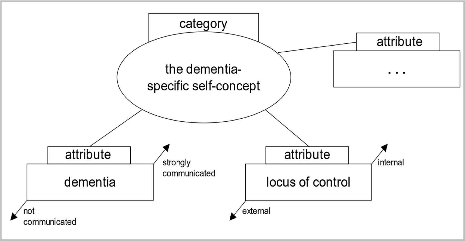
Illustration 1: Example for dimensionalizing specific attributes (own depiction using Böhm, Legewie, and Muhr’s (2008) theoretical schema [38]).

Using Strauss and Corbin’s [39] coding paradigm a foundation of relevant core categories and their relationship to one another was generated. According to Grounded Theory this foundation of core categories is composed of a central “phenomenon” (A). The data was then analyzed for “causal conditions” (B) and “contextual conditions” (C) as well as for “action strategies” (D) for the phenomenon and the resulting consequences (E). As a last step present systems of relations were re-coded and reviewed in accordance with selective coding, thus closing the theoretical coding process [40].

Results

The Model of the Dementia-Specific Self-Concept

Using the coding paradigm [39] resulted in the development of the dementia-specific self-concept model (ill. 2). The essential structure of the model will be outlined in this section while the following chapters are dedicated to the model’s several components (A-E). Dementia represents the causal condition (B) for a person’s relation to the self-concept (A). How a person reflects on the self-concept depends on a variety of contextual conditions (C). The analyses revealed that premorbid characteristics of behavior and personality traits are central categories in this context. These influenced the extent to which a person perceived dementia to take control. Taking into account contextual conditions (C) the individuals developed two different action strategies (D) in their relation to their self-concept (A): Either they maintained their self-concept (a), or they modified it (b). The adopted strategy (D) entails various consequences (E) for the self-concept of persons with dementia: Either the old self-concept is maintained (c), or a new self-concept is developed (d).

The Person’s Relation to the Self-Concept (A) in the Context of Dementia (B) – “Who am I?”

We were able to observe the person’s relation to their self-concept, i.e. the phenomenon under investigation (A) in their behavior and in their interactions. Dementia (B) causes an individual to reflect on their self-concept, i.e. the phenomenon (A) in the first place. A person with dementia is confronted with two conflicts due to their disease: The first conflict is between being self-determined and being dependent on bodily functions (e.g. limitations in everyday life caused by the disease). The second conflict is between being self-determined and being dependent on other persons (heteronomy) (e.g. relatives, health care services etc.) due to entering need for care. These conflicts trigger a discrepancy between the “old” self-concept previous to the onset of the disease and result in reflecting on the self-concept.

ASMHS 2020-401_Kristin Illiger_F2

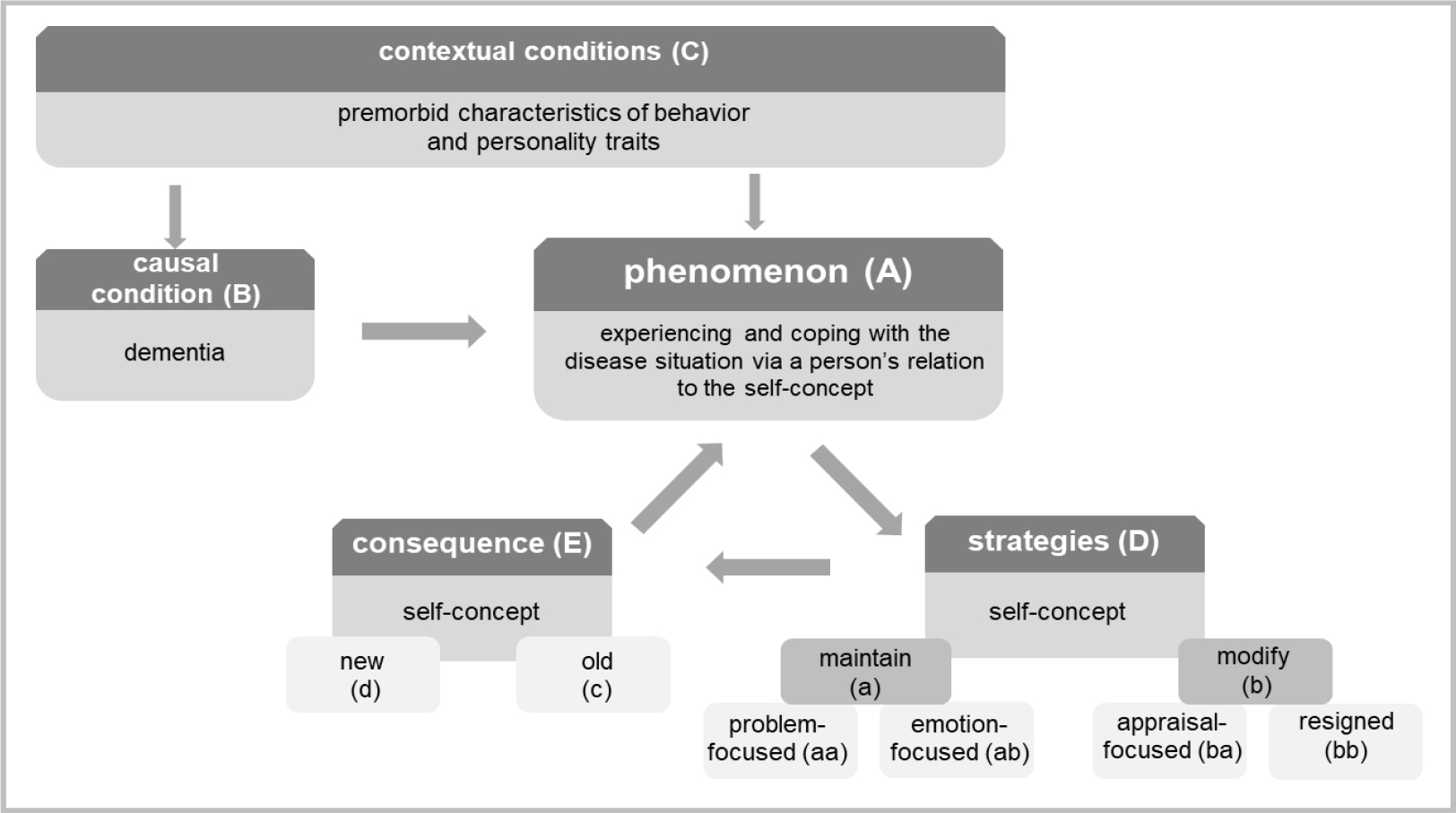
Illustration 2: The model of the dementia-specific self-concept based on Strauss and Corbin’s [39] coding paradigm.

Contextual conditions (C) – “Who was I?”

The biographical narrations revealed contextual conditions (C) such as premorbid characteristics of behavior and personality traits for the person’s relation to the self-concept (A). In the following, examples for this finding within the contexts of professional background and forming a family will be presented. The data reveals which roles the participants identified with before the onset of their disease (old self-concept) and whether they attributed biographical events to their own actions or rather to fate and influences by other people. The majority of women related abandoning their career after meeting their future husbands and/or due to giving birth to their first child, as becomes apparent in the following: “to what’s it called, what’s it called, to women’s technical college, which I didn’t finish, though, because at that point I knew I would marry [my husband]“ (P6:236).3 After abandoning the career path, they dedicated themselves to their role as wife and mother: “no, I couldn’t do that [continue working]. It was me who had to raise the children. and such. and prepare food and such. and clean the apartment.” (P7:274).4 Men, in contrast, saw themselves in the role of the provider: “I worked, I didn’t only provide for my family, but I provided for them very well” (P12:18).5 Often a change in the men’s career also involved a change in place: “when my husband completed his studies and got a job in X-city, I just followed him faithfully.” (P6:121).6 The number of children was also determined by the men in certain cases: “now there are two boys. He didn’t allow more, no.” (P7:173).7 Words such as “faithfully” and “allow” emphasize the experienced social dependence on the partner. However, the women’s narrations neither show any normative evaluation of their role in the relationship, nor the wish or the attempt at reaching more self-determination.

Maxims and sayings equally revealed specific characteristics of behavior or personality traits. Some considered their life to be a consequence of their own behavior: “what I did: making the best out of everything” (P9:212)8, or “I couldn’t and wouldn’t let another decide that I couldn’t stand.” (B12:24).9 Others characterized their life as fortunate or providential: “I am very satisfied with my fate, very satisfied I am.” (P11:72)10, “well, we were very fortunate.” (P9:559)11, or “must have been an angel” (P7:80).12 Analyzing context conditions (C) and action strategies (D) in the course of the disease shows how autobiographical knowledge helps persons with dementia to refer to patterns of thought and behavior that were characteristic for them in the past and how these have served them. The more influence a person with dementia believes to have had on past life events (C), the stronger their perceived control over the disease.

Action Strategies (D) – “Who do I want to be?”

Considering contextual conditions (C) in reference to a person’s relation to the self-concept (A) two action strategies (D) emerge: One consists in proactively maintaining the old self-concept (a), which results either in a problem-focused behavior (aa) (e.g. by taking helpful measures, information seeking, visits to the doctor), or in an emotion-focused behavior (ab) (redefining the disease situation, or denial of it). The second consists in modifying (b) the old self-concept, resulting in either an appraisal-focused strategy by accepting the loss of certain aspects of the self-concept (ba), or passively by expressing resignation or frustration (bb). In the following, the two strategies (a, b) with their various components (problem-focused/ emotion-focused and appraisal-focused/ resigned) when coping with dementia will be presented. The strategies were gained inductively by analyzing the interview data. The analysis of the strategies is based on the transactional stress model by Lazarus and Folkmann (1984), and the terms problem-focused, emotion-focused and appraisal-focused are borrowed from it. According to Schütze narrative continuity in the biographical narrative interview is constituted of several “Zugzwänge”, or requirements: the requirement to close (Gestaltschließung), the requirement to depict (Darstellung) as well as the requirement to provide detail (Detaillierung). All participants deviated from these immanent requirements of narration as they displayed symptoms of dementia such as word finding difficulties or autobiographical “blanks”. Some participants discussed and reflected the symptoms of their disease whenever they abandoned the narrative structure: “yes, I’m a little, well, but I am obviously extremely forgetful. Half an hour later I don’t remember what I have eaten.” (P5:549).13  The original chronological narration was briefly abandoned in order to report on daily experiences with speech losses or with malfunctions of the executive functions. In one instance the participant revealed the signs of dementia they perceived to the interviewer when abandoning the narration: “I have also talked about this with the doctor during the last evaluation and (…) I say “there are only old people who come to see you and er that’s when the forgetting starts.” he laughed. “yes“, he says, “but you still notice it, don’t you?” I say “thank the lord, yes” (P9:1375).14 Here one can observe a striving for maintaining the self-concept by adopting a problem-focused strategy (aa): The participant is active and self-reliant in reflecting on the disease by consulting a doctor and talking to them about initial symptoms.

In contrast, other participants rarely commented on word finding difficulties, or on obviously forgetting biographical events during the conversation. The symptoms were relativized or distracted from as the following dialogue between interviewer (I) and participant (P) shows (P4:97–102)15:

I: “And how old are you now?”

P: “Oh, I have to think about that. Born in 28. 38 48 58 68.78. What year are we in?”

I: “2017.”

P: “excuse me?”

I: “2017.”

P: “Oh. She still has. I still have a few years left. You are welcome to help yourself to cookies.” (P4: 97–102)

The responses show a conscious avoidance strategy in the uncomfortable situation caused by the interview question. This is an example of an emotion-focused strategy (ab). Most participants identified forgetfulness as being part of the natural aging process: “what have I done, oh dear, that’s how it is when you get old, you forget half of the things.” (P10:88).16 In addition they often compared themselves to other persons their age from their social network: “well, what [the neighbor] has at 80 years old, Alzheimer’s or a part of it. Some things just don’t work anymore. I mean for me it’s the same.” (P9:1375).17 Only one person explicitly named their diagnosed dementia as such. Some participants rather described it less specifically as problems with the head: “you know, my head has gotten old. (laughs)” (P10:114).18 Apparently the disease is redefined as a “problem” and is isolated from the overall experience of health on a psychological level: “my health is good. But my head.” (P11:128).19 The participants speak fundamentally differently about dementia than they do about other diseases they have. The latter are described either as concrete disease patterns (such as cardiovascular problems, limitations of the locomotor system and deterioration of vision), or as limitations of the freedom of action and of the organization of everyday life: “I kept my theater subscription for many years, but, as somebody always had to pick me up and take care of me, because I cannot walk well anymore, you know, I let it go.” (P5:44).20 Interests, skills and abilities equally constitute the self-concept. Accordingly, in the aforementioned example the interest in theater cannot be pursued due to limitations of the mobility, and conflicts with maintaining the old self-concept. In none of the examined cases is dementia perceived as cause for abandoning an interest. A reaction to the process of forgetting is to pursue present interests such as reading and cross-word solving, for instance, even more rigorously. In order to respond to potential memory “blanks”, three of the participants wrote down important moments in their life as a reminder (strategy aa). Confronting the disease in a problem-focused way (aa) can involve the attendance of memory trainings: “once a week I attend a memory training to make sure it stays chirpy up there.” (P6:196).21 For other participants, maintaining the self-concept (strategy a) rather meant to adopt “intrapsychic processes”. This means that they looked for ways to reduce strains connected to the experienced symptoms of the disease (strategy ab). One of these ways is to interpret certain symptoms in a self-protective way. For example, forgotten information is dismissed as unimportant events, or events that happened too long ago to remember: “it has been so long. I cannot remember that.” (P7:226).22 This also led to ignoring certain symptoms and to direct the attention to basic bodily needs: “Other than that, I don’t have any complaints. Food tastes good, I sleep well” (P4:351).23 They would respond to forgetting by relativizing external norms: “I…cannot know everything. I can’t know everything from A to Z. I will be over ninety years old soon. Or already am.” (P11:140).24 Some said they actively decided not to remember negative events. In contrast to persons who adopted problem-focused strategies (aa), persons using emotion-focused strategies (ab) would cite rare visits to the doctor as proof for their good health status and leave their disease to chance: “Yes, all in all I am thankful to life that I am still healthy.” (P7:186).25 They remembered past behavior and characteristics that helped them find solutions to current problems: “you always have to take care of keeping at least a little contact to others. That has never been a problem for me. So… (laughs) if just one of them was still alive, of the relatives, they would certainly confirm. They know me, after all” (P9:493).26 This is an example for seeking emotional support via positive experiences of social interaction (strategy ab).

Instead of using strategy a, others accepted the partial loss of the self-concept: “my [daughter] always says: “Mum, you go through the door and your thought is gone” (laughs) and sometimes it really makes me laugh. That’s how it is.” (P9:1375).27 This is an example of an appraisal-focused strategy (ba) marked by a humorous and resource-oriented way of coping with the disease. For other participants, on the contrary, the modification of the old self-concept (strategy b) means to meet the disease with resignation (strategy bb): “You cannot do anything against it, that’s just exhaustion at this age.” (P10:64).28

A person’s relation to the self-concept (A) is reflected in the decision of all participants to keep on living alone in their homes. The reasons for this decision varied according to the strategy used. Those who adopted a problem-focused behavior towards the disease situation (aa) consciously perceived the symptoms, but also saw sufficient room for action to continue living alone. Moving to a care center, for instance, would be equal to losing control over the disease situation for them. Persons who adopted an emotion-focused strategy (ab) perceived the symptoms less consciously. For them, living alone is a matter of course and is not characterized by fears and worries. They neither considered an alternative form of living, nor moving to a care center as an option: “That would mean I must have something. That I have an illness or something. If I cannot live here by myself or I’m not allowed to.” (P7:343).29 Participants adopting an appraisal-focused strategy (ba) perceived living alone with dementia as a challenge: “I have to get used to getting by myself. I am convinced, I can make it.” (P12:66).30 A positive view on the disease situation resulted in reacting to challenges during the process of dementia and to appraise risks and need for help related to living alone realistically (e.g. by using formal help services). The more influence the person believed to have had on past life events (C), the stronger they did so with regard to the disease situation by adopting a problem-focused or an appraisal-focused strategy (aa, ba). If the person thought to have had little influence on past life events (C), they developed less autonomous solutions (ab, bb). The latter withdraw from social life and resign, compared to persons who adopt emotion-focused strategies (ab). As mentioned in the beginning, the self-concept is also formed by other people’s reactions to the self. Reflected evaluations (e.g. perceived social exclusion) are internalized and result in according behavior (e.g. withdrawal due to shame): “Alone, that is the problem of an old person. Nobody wants to be together or gets together sometimes.” (P12:44).31

Consequences (E): Maintaining or modifying the self-concept – “This is me!”

The consequence (E) of the adopted action strategy (D) is that persons with dementia enter into a (new) relationship with the self. The consequence of reflecting on the self-concept (A) can be to either maintain the old self-concept (c), or to develop a new one (d). The data shows that this is especially reflected in the participants’ depiction of several experienced and lived social roles and of their subjective age. All participants had in fact experienced a loss of their social role when they retired, were widowed or when their children moved out. Those who wanted to maintain their old self-concept during the course of dementia (strategy a) upheld former social roles in the present and in the future (consequence c). Biographical knowledge concerning their profession, for instance, was used by persons who had a distorted perception of their symptoms, or who denied their symptoms in such a way as to maintain their identity and their self-concept: “I am a professional nurse, you know. That’s why I have many elderly people up here at our place that I have to care for from time to time.” (P4:354).32 Persons who adopted a problem-focused strategy, in contrast, mentioned gardening and running the household as activities. This designs a changed understanding of their social roles rather than a loss of their professional roles. Persons who gradually withdrew from social roles and professional obligations, and who were willing to abandon roles related to their old self-concept (strategy b) developed a new one (consequence d): „I am saying I used to be an early bird but now I am so lazy now I get up only at half past eight. I have time.” (P8:1045).33 Subjective age likewise affects maintaining the old self-concept or developing a new one. For persons who adopted strategies for maintaining their self-concept (a) age and getting older felt less threatening (consequence c): “I can’t sit still and mope yet, I am too fit for that still. Ok? I am very simply too fit for that still.” (P12:76).34 This was also true for individuals who did not remember their age, or who denied their age: “I still cannot believe that I am this old. Oh god, oh god” (P9:380).35 While these persons mainly maintained their self-concept, other persons developed a new one (consequence d) by abandoning parts of their old self-concept (strategy b). Benefits of old age were embedded in a new, positive self-concept: “When you’re old, you’re authorized to such judgement.” (P9:1432).36 Others felt they were at the mercy of the aging process. This feeling resulted in a new, negative self-concept: “Of course I have not anticipated getting this old, you know. But that’s generally the case nowadays that people get older, right. There are many reasons for that” (P5:352).37

Conclusion and Discussion of Results

Based on the data we developed a theoretical model of the dementia-specific self-concept. The narratives show that perceiving and experiencing the disease situation are primarily characterized by the person’s relation to the self-concept rather than by dementia-specific symptoms (such as memory dysfunctions, and dysfunctions of spatial and temporal orientation, or linguistic impairments). The medical definition of dementia (the objective symptoms) thus stands in opposition to the individuals’ subjective perception of the disease situation. The subjective perception leads to reflecting on the self-concept using either one of two action strategies resulting in either maintaining or modifying the old self-concept.

Contextual Conditions

Several contextual conditions determine how a person reflects on the self-concept during the process of dementia. The analysis of the data involved a conceptualization based on the construct of internal versus external locus of control by Rotter [11] which helped identify (premorbid) personality traits. Specifically, we looked at expectations participants had towards certain events that followed their own actions. Differences between internal and external loci of control based on the example of the participants’ professional biography were found: Some participants experienced their profession in an internal manner (residing in their behavior, e.g. professional choices and actions). External locus of control manifested itself in the form of depending on others, and especially, in the case of women, on the partner (social-external locus of control). Conditions related to a specific generation influence certain personality traits such as locus of control [41]. The data reveals a traditional understanding of typical gender roles of the 60s: the woman as housekeeper and mother, the man as moneymaker and provider. The results concerning the contextual conditions (locus of control) show opportunities and living conditions specific to this generation, which can be retraced to old age as well as to the disease [42]. Since experiences of childhood, youth and young adulthood permanently influence later loci of control [43], these areas were taken as a basis for the interpretation of dementia as the critical life event. In particular, positive connections between (premorbid) internal locus of control and the problem-focused strategy (aa) as well as the appraisal-focused strategy (ba) were found: Persons with internal locus of control took specific measures in order to confront the disease and challenges tied to it. Persons with external locus of control rather viewed luck, bad luck, or fate as reasons for their health-status. Their coping with dementia resulted in a “feeling of helplessness” which manifested itself via emotion-focused strategies (ab) or resignation (bb). The transfer from past locus of control to present ones should be viewed critically as it might be a variable personality trait [44]. With advanced age a strong internal locus of control becomes significantly less probable [45]. In an American study persons with dementia report on events of losses of control (in terms of health) that are due to the unpredictability of the course of the disease [46]. Additionally, it is not entirely clear whether we can speak of premorbid personality traits and behavior. These may well be traits that are rather constructed or distorted in the autobiographical memory, since we retrospectively asked about the biographical locus of control during the interview.

Action Strategies

The field of health care approaches illness perception in dementia from a symptomatic or deficit-oriented point of view. While this view merely regards neuro-cognitive dysfunctions that lead to the persons’ impaired ability to reach insight [10], we choose a different perspective. Our model includes contextual factors (C) and action strategies (D) that go beyond this deficit-oriented view and that explain the experience and behavior of persons with dementia. We assume that the person’s current relation to the self-concept (A) is linked to passed (premorbid) strategies of behavior and personality traits (C). The strategies closely follow the theoretical elements of the transactional stress and coping model by Lazarus and Folkmann [12]. In their model, the first step is to determine the meaning the disease situation has for a person (primary appraisal). In this study the diagnosis is mentioned only in one case. For the majority of the participants the appraisal of the disease situation is embedded in the narrations on the relation to the self-concept. Problem-focused coping strategies (aa) involve an increasing number of visits to the doctor, attendance to memory trainings, or seeking information as reactions to a threatening loss of the old self-concept (e.g. loss of cognitive abilities). In contrast, emotion-focused coping strategies (ab) involve an unwillingness to accept having a disease by trivializing or denying the symptoms, for example, as reactions to a threatening loss of the self-concept. Often the persons interpreted their symptoms in a self-protective manner. Other studies have equally found that elderly persons often explain memory blanks by referring to the unimportance of the forgotten information [47]. Emotion-focused coping strategies also involve comparisons: “I do not feel ill, although I am not as industrious and as fit as I used to be.” (B4:309). According to Social Comparison Theory [48] individuals permanently compare themselves to their social environment. This entails that the self-concept is always related to the social environment. These comparisons occur especially in insecure situations. In such situations the participants primarily compared themselves to people their age from their social environment – according to Festinger the persons they compare themselves to are persons who share central traits of their own self-concept as the following quote shows: “It’s enough that I can still walk around. When I see that the others, they are so much younger, ten years younger and are being driven around back and forth every day.” (B9:246). In this example the person appraises dementia in relation to other persons’ diseases (impaired mobility). In a longitudinal study on the development of the self-concept of persons after a leg amputation, Klein [49] observed that the persons moved from physical activities to intellectual exchange and socio-communicative activities. This shift in perceiving their existence was entirely reversed for some persons with dementia: They, in contrast, emphasized their high bodily activities (walks, household etc.) and avoided direct communication: “And I am so happy every time I get mail, you can always read that again, but a phone call, that’s out too” (B9:1375). Rather than being a coping strategy, focusing on bodily rather than psychological conditions, might be specific to a generation (cohort effect) that lacks sensibility for psychological diseases [50].

Consequences

The adopted strategies result in maintaining the old self-concept (c) or in developing a new self-concept (d). When maintaining the self-concept (consequence c from strategy a) a positive self-concept results from a successful experience of personal skills, aging and living social roles. Disregarding or denying symptoms using an emotion-focused strategy (ab) enabled the person to maintain the old self-concept. Modifying the self-concept (consequence d from strategy b) resulted in a positive new self-concept when the loss of certain skills was viewed as a challenge, old age was accepted, and the person voluntarily withdrew from former social roles. Pinquart [47] defines the self-concept of the elderly as a sensitive indicator for the specific life situation and the ability to deal with the latter. Consequently, the phenomenon of reflecting on the self-concept is observable in all elderly people. As in this study, in other studies with elderly people without dementia, the participants believed to be much younger than they actually [5, 6]. The example of subjective age shows that the self-concept can deviate from objective reality [49]. In contrast to other elderly people, persons with dementia are only rarely capable of realistically assessing their current life situation. Often persons with dementia located themselves in a time prior to the onset of dementia, and they did not know their current age. They had a distorted perception of social roles, i.e. they believed to occupy roles that they objectively did not occupy any longer (as the example of the pensioner with dementia who perceived of themself as practicing nurse illustrates).

Implications

According to the World Health Organization the development and the maintenance of functional abilities (such as meet their basic needs, to learn, grow and make decisions; to be mobile) is a prerequisite for healthy aging: “Functional ability comprises the health-related attributes that enable people to be and to do what they have reason to value” [2, p. 28]. Departing from this definition our model of the dementia-specific self-concept is process-based rather than phase-based. This is because reflecting on the self-concept (“Who am I?”) is tied to the course of the disease (early, middle and late dementia) (B), on the one hand, and to contextual conditions (C) that are subject to change, on the other. The components interact with one another. This leads to new action strategies (D) and consequences (E): For instance, the progression of the disease entails increasing memory dysfunctions (B). Contextual conditions (C) change because making recourse to autobiographical material becomes more difficult. The relation between the requirements caused by the new symptoms and the perception of personal knowledge resources (such as using premorbid strategies of behavior) equally changes. Strategies used for the person’s relation to the self-concept can differ from strategies used previously in the process of dementia, and can lead to new answers to the question “Who do I want to be?”.

The question of which strategy is more helpful (adaptive) for a person’s relation to the self-concept and which is less helpful (maladaptive), cannot be answered precisely [51]. The advantage of a problem-focused strategy (aa) lies in an individual actively confronting their disease. In this case, a person with dementia may participate in making decisions concerning treatment options with their doctors. This strategy is only helpful to a limited extent as dementia is irreversible, and as the disease can only be temporarily stopped from progressing, and complaints (such as depression) can only be alleviated. Knowing about the uncontrollability of the disease can be a heavy burden for the individual. In contrast, the advantage of an emotion-focused strategy (ab) lies in seeking ways that help avoiding high emotional strains. This can lead to a higher quality of life. A lack of willingness to accept the disease (distraction, denial of risks, overestimating one’s personal abilities), however, can equally lead to serious dangers in everyday life, and can delay or prevent treatments with or without medication [52]. When adopting an appraisal-focused strategy (ba), individuals accept the disease and are able to evaluate risks realistically. At the same time a premature willingness to accept the disease can lead to insufficient consideration of potential treatments. If neither of these strategies is adopted, coping with the disease can come to mean resignation and heighten the risk of social isolation (strategy bb). The dementia-specific self-concept offers a theoretical approach to the analysis of the individual experience and coping with the disease situation of persons with dementia. A long-term goal of this research is to deduce implications for health care practices, in order to meet the beliefs, expectations and needs of persons with dementia in an individual, suitable, and sensitive way during the course of the disease.

Discussion of Methods

Biographical narrations offer an access to the participants that is especially suitable in the context of dementia. Memory dysfunctions caused by dementia affect semantic content in long-term memory (including common knowledge and word meanings, for instance), but do not affect episodical and autobiographical memory [53]. Childhood experiences, family/partnerships, as well as professional backgrounds are areas of life that were remembered by all participants and that could be used for the analyses. Another advantage was that the interviews were conducted in the homes of the interviewees, and objects such as pictures, photographs or religious symbols could be used as narrative stimuli. Accordingly, interviews not conducted in the participants’ homes were significantly shorter. Findings from social gerontology and from the care of the elderly show that reflecting on one’s biography is health-promoting and has positive effects on memory [33]. The need to turn to early memories and remember personal life events is a central result of the LUCAS study [54, 55] At the same time negative experiences, such as experiences of war, may re-emerge during biographical narrations. However, the method is open and sensitive to dementia in that it allows the participants to determine relevant topics and to avoid sensitive subjects if desired. Consequently, contents that were not remembered by the participants, or that that they did not wish to discuss remained unrecorded. This also means that the participants may have consciously avoided depicting their disease for fear of shame or stigmatization. Therefore, it cannot be excluded that the data contains reporting bias. Three interviews were conducted in the presence of family caregivers and advisors who often interrupted the participants’ narrative and related biographical events from their perspective. Here differences between self-perception and the perception by others became apparent. The comments by the third party may have influenced both the researchers’ analyses and the participants’ narration to a certain extent. Dementia was neither mentioned by the researchers when delivering information on the study, nor during the interviews, as this might have influenced the biographical narration, and it might have caused emotional strains for the participants. At the same time this procedure raises the ethical question whether the tabooing of dementia is supported by avoiding a direct confrontation with the disease. Presuppositions regarding the field of research that are issued from literature research, and from individual, as well as socio-cultural experiences were recorded in a research diary and reassessed deductively on the basis of the empirical data. We decided against a member check [36] with which to retrace the results to the interviewees and verify the results for two reasons: On the one hand, we could not establish to which extent a confrontation with the topics dementia and need for care might have a negative impact on the participants. On the other hand, the interview data represent but a snapshot of remembering, experiencing, and acting. Due to the long-time span between data collection and data analysis, but also due to the progression of the disease, the results could not be replicated with the same participant. Instead, we agreed on the credibility and the meaning of the data in numerous research meetings with other social researchers and non-professionals. Despite methodological limitations and challenges inherent in dementia research, explanations regarding illness perception and perception of the self that would have remained unnoticed without an open and qualitative research approach were found. In order to fully exploit the potential of research with persons with dementia, further development of research designs that enable a higher degree of participation for people with dementia along the entire course of the disease is needed.

Conflict of Interest : The authors of the study  (ethic number: 3533–2017) have no conflict of interest to report.

Acknowledgement

A special thanks to our interviewees for the participation in our study and their willingness to tell about their life story. We are furthermore grateful to all the persons and institutions, wo helped us with recruiting and getting field access: therapists and physiotherapists, legal guardians, outpatient nursing services, information centers, self-help groups, volunteers, media and close relatives.

References

  1. Baltes Margret M, Baltes Paul B (1990) Successful aging. In: Baltes Margret M, Baltes Paul B (eds.). Perspectives from the behavioral sciences. European Network on Longitudinal Studies on Individual Development. Cambridge: Cambridge University Press.
  2. World Health Organization (2015) World report on ageing and health. Geneva: WHO.
  3. Baumeister Roy F (1998) the self. In: Gilbert Daniel Todd, Fiske Susan T, Gardner Lindzey (eds.). The handbook of social psychology. (4th edn). Boston, New York: Distributed exclusively by Oxford University Press, p. 680–740.
  4. Kernis Michael Howard M, Goldman, Brian Middleton (2003) Stability and variability in self-concept and self-esteem. In: Mark R. Leary und June Price Tagney (eds.). Handbook of self and identity. New York: The Guilford Press, p. 106–127.
  5. Essex Marilyn J, Klein Marjorie H (1989) The Importance of the Self-Concept and Coping Responses in Explaining Physical Health Status and Depression among Older Women. In: Journal of Aging and Health 1: 327–348.
  6. Weiss David, Lang Frieder (2012) “They” are old but “I” feel younger: age-group dissociation as a self-protective strategy in old age. In: Psychology and aging 27: 153–163.
  7. Douglas Heather, Georgiou Andrew, Westbrook Johanna (2017) Social participation as an indicator of successful aging: an overview of concepts and their associations with health. In: Australian health review: a publication of the Australian Hospital Association 41: 455–462.
  8. Filipp Sigrun-Heide, Klauer Thomas (1991) Subjective well-being in the face of critical life events: The case of successful copers. In: Fritz Strack, Michael Argyle and Norbert Schwarz (eds.). Subjective well-being. Oxford: U.K.: Pergamon, p. 213–234.
  9. Strack Fritz, Argyle Michael, Schwarz Norbert (eds.) (1991) Subjective well-being. Oxford: U.K.: Pergamon.
  10. Iqbal Khizra, Amin Rizwana (2018) Negative Life Events and Mental Health Among Old Age People: Moderating Role of Social Support. In: Pakistan Journal of Psychological Research 33: 401–412.
  11. Rotter Julian B. (1990): Internal versus external control of reinforcement: A case history of a variable. In: American Psychologist 45: 489–493.
  12. Lazarus Richard S,  Folkman Susan (1984) Stress, appraisal, and coping. New York: Springer.
  13. Busch Albert, Spranz-Fogasy Thomas (2015) Handbuch Sprache in der Medizin. [Handbook language in medicine.] Berlin, Boston: De Gruyter Mouton (Handbücher Sprachwissen, 11).
  14. Mejia-Arango Silvia, Gutierrez Luis Miguel  (2011) Prevalence and incidence rates of dementia and cognitive impairment no dementia in the Mexican population: data from the Mexican Health and Aging Study. In: Journal of aging and health 23: 1050–1074.
  15. Prince Martin James (2015) World Alzheimer Report 2015: the global impact of dementia: an analysis of prevalence, incidence, cost and trends: Alzheimer’s Disease International.
  16. Illiger Kristin, Walter Ulla, Koppelin Frauke (2018) Alleinlebende mit Demenz – Eine Datenanalyse der ambulanten Pflege in einer kreisfreien Großstadt. [Persons living alone with dementia – a data analysis of outpatient care in an independent city] In: Pflegewissenschaft 20: 437– 444.
  17. Bleck Christian, van Rießen Anne, Knoop Reinhold  (2018) Alter und Pflege im Sozialraum. Theoretische Erwartungen und empirische Bewertungen. [Age and care in the social environment. Theoreticalexpectations and empiricalvaluations.] Wiesbaden: Springer Fachmedien Wiesbaden GmbH.
  18. Coyle Caitlin E, Dugan Elizabeth (2012) Social isolation, loneliness and health among older adults. In: Journal of aging and health 24: 1346–1363.
  19. Illiger Kristin, Walter Ulla, Koppelin Frauke (2017) Demenz im Fokus der Gesundheitsforschung: Eine vergleichende Analyse aktueller Altersstudien. [Dementia in the focus of health research. A comparative analysis of current ageing studies.] In: Bundesgesundheitsblatt, Gesundheitsforschung, Gesundheitsschutz 60: 563–571.
  20. Leicht Hanna, Berwig Martin, Gertz Hermann-Josef  (2010): Anosognosia in Alzheimer’s disease: the role of impairment levels in assessment of insight across domains. In: Journal of the International Neuropsychological Society 16: 463–473.
  21. Gove Dianne, Diaz-Ponce Ana, Georges Jean, Moniz-Cook Esme, Mountain Gail, et al. (2018) Alzheimer Europe’s position on involving people with dementia in research through PPI (patient and public involvement). In: Aging & mental health 22: 723–729.
  22. Orfei Maria Donata, Assogna Francesca, Pellicano Clelia, Pontieru Francesco Ernesto, Caltagirone Carlo, Pierantozzi Mariangela et al. (2018) Anosognosia for cognitive and behavioral symptoms in Parkinson’s disease with mild dementia and mild cognitive impairment: Frequency and neuropsychological/neuropsychiatric correlates. In: Parkinsonism & related disorders 54: 62–67.
  23. Malek Hédi Ben, Philippi Nathalie, Botzung Anne, Cretin Benjamin, Berna Fabrice, Manning Liliann, Blanc Frédéric (2019) Memories defining the self in Alzheimer’s disease. In: Memory 27: 698–704.
  24. Byrne Barbara M (1996) Measuring self-concept across the life span: Issues and instrumentation. Washington: American Psychological Association.
  25. Riemann Gerhard (1987) Das Fremdwerden der eigenen Biographie. Narrative Interviews mit psychiatrischen Patienten. [The Alienation ofone’s own biography. Narrative interviews with psychiatric patients.] München: Fink.
  26. Savundranayagam Marie Y, Ryan Ellen B, Anas Ann P, Orange JB (2007) Communication and Dementia. In: Clinical Gerontologist 31: 47–63.
  27. Schütze Fritz (1983) Biographieforschung und narratives Interview. [Biography research and the narrative interview.] In: Neue Praxis 13: 283–293.
  28. Hermanns Harry (1995) Narratives Interview. [Narrative interview] In: Flick Uwe, Kardoff Ernst von,  Keupp Heiner, Rosenstiel Lutz von, and Wolff Stephan (eds.). Handbuch qualitative Sozialforschung. [Handbook qualitative Social Research.] (2nd edn). Munich: Psychologie Verlags Union, p. 182–185..
  29. Kruse Jan, Schmieder Christian (2014) Qualitative Interviewforschung. Ein integrativer Ansatz. [Qualitative interview research. an integrative approach.] Weinheim: Beltz Juventa (Grundlagentexte Methoden).
  30. Phillips Susan P, Auais Mohammad, Belanger Emmanuelle, Alvarado Beatriz, Zunzunegui Maria-Vitoria (2016) Life-course social and economic circumstances, gender, and resilience in older adults: The longitudinal International Mobility in Aging Study (IMIAS). In: SSM – population health 2: 708–717.
  31. Ashida Sato, Heaney Catherine A (2008) Differential associations of social support and social connectedness with structural features of social networks and the health status of older adults. In: Journal of aging and health 20: 872–893.
  32. Glaser Barney G, Strauss Anselm L (2017) Theoretical sampling. In Sociological methods: Routledge 105–114.
  33. Panke-Kochinke Birgit (2014) Menschen mit Demenz in Selbsthilfegruppen. Krankheitsbewältigung im Vergleich zu Menschen mit Multipler Sklerose. Weinheim: Beltz Verlagsgruppe.
  34. Selting Margret, Auer Peter, Barth-WeingartenDagmar, Bergmann Jörg, Birkner Karin, et al. (2009) Gesprächsanalytisches Transkriptionssystem 2. [Language-analysis-basedtranscriptionsystem 2.] In: Gesprächsforschung – Online-Zeitschrift zur verbalen Interaktion 10: 353–402.
  35. Glaser Barney G, Strauss Anselm L (1999) The discovery of grounded theory. Strategies for qualitative research. 4. Paperback printing. New Brunswick: Aldine.
  36. Steinke Ines (2010) Quality criteria in qualitative research. In: Flick Uwe, Kardorff Ernst von, Steinke Ines (eds.). A companion to qualitative research. repr. London: Sage.
  37. Böhm Karin, Tesch-Römer Clemens, Ziese Thomas (2009) Gesundheit und Krankheit im Alter. [Health and illness in old age.] Berlin: Robert Koch-Inst (Beiträge zur Gesundheitsberichterstattung des Bundes).
  38. Böhm Andreas, Legewie Heiner, Muhr Thomas (2008) Kursus Textinterpretation: Grounded Theory. [Text interpretation course: grounded theory]. Available online via https://www.ssoar.info/ssoar/bitstream/document/2662/1/ssoar-2008-bohm_et_al-kursus_textinterpretation_grounded_theory.pdf
  39. Strauss Anselm L, Corbin Juliet (1990) Basics of qualitative research. Newbury Park: Sage.
  40. Strübing Jörg (2018) Grounded Theory und Theoretical Sampling. [Grounded Theory and theoretical sampling]. In: Baur Nina, Blasius Jörg (eds.). Handbuch Methoden der empirischen Sozialforschung [Handbook methods in empirical social research]. Wiesbaden: Springer Fachmedien Wiesbaden 51: 525–544.
  41. Hülür Gizem (2017) Cohort differences in personality. In: Personality Development Across the Lifespan, 519–536.
  42. Pasero Ursula, Backes Gertrud, Schroeter Klaus R (2007) Altern in Gesellschaft. Ageing – diversity – inclusion. [Aging in Company of Others.] Wiesbaden: VS Verl. für Sozialwiss.
  43. Steptoe A, Poole L (2016) Control and Stress. In: Fink George (eds.). Stress: Concepts, Cognition, Emotion, and Behavior. Amsterdam: Elsevier, p. 73–80.
  44. Pudrovska Tetyana (2015) Gender and health control beliefs among middle-aged and older adults. In: Journal of aging and health 27: 284–303.
  45. Schnee Melanie, Grikscheit Florian (2013) Gesundheitliche Kontrollüberzeugungen von Patienten in Disease Management Programmen. [Health Locus of Control of Patients in Disease Management Programmes] In: Gesundheitswesen (Bundesverband der Ärzte des Öffentlichen Gesundheitsdienstes (Germany) 75: 356–359.
  46. Portacolone Elena, Rubinstein Robert L,. Covinsky Kenneth E, Halpern Jodi, Johnson Julene K (2019): The Precarity of Older Adults Living Alone With Cognitive Impairment. In: The Gerontologist 59: 271–280.
  47. Pinquart Martin (1998) Das Selbstkonzept im Seniorenalter. [The self-concept in old age]. Weinheim: Beltz.
  48. Festinger Leon (1954) A Theory of Social Comparison Processes. In: Human Relations 7: 117–140.
  49. Klein Thomas (2003) Selbstkonzept und Coping-Prozesse bei Patienten nach einer Amputation. Eine Längsschnittstudie zur Entwicklung des Selbstkonzepts und zum Prozess der Bewältigung bei Patienten nach einer Beinamputation. [Patient self-concept and coping processes after an amputation. A longitudinal study on the development of the self-concept and the process of how patients manage a leg amputation.] Dissertation. Dortmund.
  50. Friederich Hans-Christoph, Hartmann Mechthild, Bergmann Günther, Herzog Wolfgang (2002) Psychische Komorbidität bei internistischen Krankenhauspatienten-Prävalenz und Einfluss auf die Liegedauer. [Psychiatric comorbidity in medical inpatients. Prevalence and effecton the length of stay.] In: Psychother Psychosom Med Psychol 52: 323–328.
  51. Moritz Steffen, Jahns Anna Katharina, Schröder Johanna, Berger Thomas, Lincoln Tania M, Klein Jan Philipp, Göritz Anja S (2016) More adaptive versus less maladaptive coping: What is more predictive of symptom severity? Development of a new scale to investigate coping profiles across different psychopathological syndromes. In: Journal of affective disorders 191: 300–307.
  52. Heßmann Philipp, Dreier Maren, Brandes Iris, Dodel Richard, Baum Erika, et al. (2018) Unterschiede in der Selbst- und Fremdbeurteilung gesundheitsbezogener Lebensqualität bei Patienten mit leichter kognitiver Beeinträchtigung und Demenz vom Alzheimer-Typ. [Differences between self- and proxy-assessment of health-related quality of life in patients with mild cognitive impairment and alzheimer’s disease.] In: Psychiatrische Praxis 45: 78–86.
  53. Bartsch Thorsten, Falkai Peter (2013) Gedächtnisstörungen. Diagnostik und Rehabilitation. [Memory Disorders. Diagnostic and Rehabilitation.] Berlin: Springer.
  54. Dapp Ulrike, Fertmann Regina, Anders Jenny, Schmidt Silke, Pröfener Franz, et al. (2011) Die Longitudinal-Urban-Cohort-Ageing-Studie (LUCAS). [The Longitudinal Urban Cohort Ageing Study (LUCAS).]  In: Zeitschrift für Gerontologie und Geriatrie 44: 55–72.
  55. Dapp Ulrike, Anders Jennifer, Renteln-Kruse Wolfgang von, Golgert Stefan, et al. (2012): The Longitudinal Urban Cohort Ageing Study (LUCAS): study protocol and participation in the first decade. In: BMC geriatrics 12: 35.

1 German original: “Könnten Sie mir von Ihrer Lebensgeschichte erzählen? Am besten beginnen Sie mit der Geburt, mit dem kleinen Kind, das Sie einmal waren, und erzählen dann all das, was sich so nach und nach in Ihrem Leben zugetragen hat, bis zur Gegenwart. Sie können sich dabei ruhig Zeit lassen, auch für Einzelheiten, denn für mich ist alles interessant, was Ihnen wichtig ist.”

2 The terms are taken from the classification of old age [37] The age categories correspond to the frequency distribution of people living alone with dementia according to Illiger et al. 2018 [16].

3 „sag mal grad, wie hieß die grad, zur frauenfachschule, wo ich dann aber nicht weiter gemacht habe, weil ich inzwischen wusste, dass ich [meinen Mann] heiraten würde“ (B6:236)

4 „nein, das [weiter arbeiten] konnt ich nicht. ICH musste ja auf die kinder aufpassen. und so. und essen machen und so. und die wOHnung sAUber halten.“ (B7:274)

5 „ich hab geArbeitet, ich hab meine familie nicht gut, sondern sEHr gut dUrchgebracht“ (B12:18)

6 „als mein mann dann fertig war, mit dem studium und die stelle dann in X-Stadt kriegte, bin ich dann hinterhergedackelt.“ (B6:121)

7 „da sind es nun zwei jungs. er hat da nicht mehr erlaubt, ne.“ (B7:173)

8 „WAs ich gemacht hab: aus allem immer das beste“ (B9:212)

9 „ich kONnte und wOLlte das nicht dass Irgendeiner bestIMmt (2.0) DAS konnte ich nicht hAben.“ (B12:24)

10 „ich bin SEHr zufrieden mit meinem schicksal, bin ich sehr zufrieden.“ (B11:72)

11 „naja, wir haben ja auch glÜCK gehabt.“ (B9:559)

12 „wohl nen engel“ (B7:80)

13 „ja, ich bin n bisschen, gut, aber ich bin natürlich total vergesslich. ich weiß ne halbe stunde später nicht mehr was ich gegessen habe.“ (B5:549)

14 „ich hab auch mitm ARzt darüber gesprochen als ich bei der letzten auswertung war und (…) ich sage „zu ihnen KOMMen ja nur alte leute und und äh da fängt jetzt das verGESSen an“. musste er lachen. „ja“, sagt er, „aber sie mERKens noch, wa?” ich sag „na gott sei dank ja” (B9:1375)

15 I: „und wie alt sind sie jetzt?“

B: „oh, jetzt muss ich nachdenken. jahrgang 28. 38 48 58 68.78. was haben wir jetzt?“

I: „2017.“

B: „wie bitte?“

I: „2017.“

B: „oh. da muss se noch. ja, dann muss ich noch ein paar jährchen. sie dürfen sich gerne auch

kekse nehmen.“ (B4:97–102) In the German version, the letter B for “Betroffener” is used.

16 „was hab ich n denn gemacht o gott, so is das wenn man ALt wird, vergisst man die hälfte.“ (B10:88)

17 „naja was hat [der nachbar] nu mit 80 jahre, ALZheimer oder n TEil davon (4.0) es läuft eben alles nicht mehr so. (2.0) ich meine bei MIR is es auch so.“ (B9:1375)

18 „jaja, mein kopf ist schon alt. (lacht)“ (B 10:114:)

19 „gesUNDeitlich fühl ich mich gut. aber mit dem kOPF.“ (B11:128)

20 „hatte noch lange jahre n theaterabo, aber weil mich da auch immer jemand abgeholt hat und sich um mich kümmern muss, weil ich ja nicht mehr richtig gehen kann, ne, hab ich das auch aufgegeben.“(B5:44)

21 „ich hab einmal die woche ein gedächtnistraining, damit das da oben auch munter bleibt.“ (B6:196)

22 „das ist solange her. da kann ich mich gar nicht mehr erinnern.“ (B7:226)

23 „ansonsten habe ich gar keine beschwerden. essen schmeckt mir, schlafen kann ich gut.“ (B4:351)

24 „ich….kann ja nicht ALles wissen. kann ja nicht alles wiSSen von hiEr bis dA. ich bin über NEUNzig bald. oder bIN schon.“ (B11:140)

25 „ja, eigentlich bin ich dem leben dankbar, dass ich noch gesund bin.“ (B7:186)

26 „man muss IMMEr dafür sorgen, dass man so n bisschen kontakt hat. und das ist mir noch NIE schwergefallen. also…(lacht) ja wenn davon noch einer LEBen würde, von den verwandten, die könnten das nur bestätigen. die kennen mich ja auch.“ (B9:493)

27 „meine [tochter] sagt immer „mutti du gehst durch n TÜRrahmen und denn is der gedanke weg“ (lacht) und manchmal muss ich WIRKlich lachen. SO ist das eben.“ (B9:1375)

28 „man kann ja nichts gegen machen, das ist ja verschlEIß, in dem alter.“ (B10:64)

29 „da muss ich ja irgendwie was haben. muss ich ja dann krank sein oder wie. dass ich hier alleine nicht leben kann oder nicht mehr leben darf.“ (B7:343)

30 „ich mUss mich daran gewÖhnen allEine zurEcht zu kommen. ich bin davon überzEUgt, ich schAffe es.“ (B12:66)

31 „allEINe, das ist eben das problEm. (3.0) eines älteren menschen. (2.0) keiner will mitnander oder tUt miteinander mal.“ (B12:44)

32 „ich bin ja krankenschwester von beruf. Insofern habe ich viele ältere leute hier bei uns oben, wo ich mich mal ab und zu sehen lassen muss.“ (B4:354).

33 „ich sage ja, ich war n frühaufsteher (2.0) aber JETZt bin ich so bequEem (1.0) jetzt steh ich erst um halb ACHt auf. Ich hab Zeit. (2.0)“ (B8:1045)

34 „HINsetzen und TRÜbsal blasen kAnn ich noch nicht, dAfür bin ich eben noch zu fIt. Ja? (2.0) dAfür bin ich eben GANZ Einfach noch zu fIT.“ (B12:76)

35 „Ich kann immer gar nicht glauben, dass ich schon so alt bin. Ach gott, ach gott.“ (B9:380)

36 „Wenn man so ALT ist, kann man sich das Urteil ja erlauben.“ (B9:1432)

37 „ich hab natürlich nicht geahnt, dass man so alt wird, ne. Aber es ist ja generell so, dass die leute heute älter werden, ne. Daran ist vieles schuld“ (B5:352)

Cultivation of Chinemys Reevesii (Chinese Three Keeled Turtle) in Greenhouse System

DOI: 10.31038/AFS.2020211

Abstract

Chinemys reevesii (Chinese three keeled turtle) is developed in china’s turtle farms for production in pet trade market and chinese medicine. The aim of this study was to calculate growth rate and survival rate of juvenile C. reevesii under the greenhouse based overwinter breeding technology to survive and not to get weight loss. 2000 stocking density were cultured in each twenty tank and then randomly collected 10 turtles from each tank during January to may 2019 to calculate growth rate. The results showed that the temperature was maintained within the range of 30 ±2°C and ambient air was controlled by 32±2°C. After five months cultivation, final weight reached average 278.44 ±57.61 g with 125.13±99.73 final weight gain by high survival rate of 89.8± 2.73% and low feed conversion rate 1.8±0.2. Moreover, resulting dissolved oxygen 3.8 to 9 mg/l, pH between 7 to 8, ammonia and nitrite at 0.005 to 0.02 mg/l showed suitability of this species cultivation. As a result, this study will be hopeful artificial husbandry to increase healthy growth rate during overwintering turtle in greenhouse system.

Keywords

chinese three keeled turtle; greenhouse; growth rate; survival rate, FCR, temperature, water quality

Introduction

For many centuries, turtles have been used as food, pets and in traditional medicine in different regions of the world [1]. Freshwater turtles are being developed for traditional medicine, meat, eggs and pet trade. Because of this, turtle farmings are boomed in aquaculture industries in the past two decades [2]. As farm bred turtles took advantage of good numbers in the last 20 years, the market share has raised significantly. Asia especially china is stood at the top of world turtle consumption as food and medicine. Despite having used in turtle products for thousands of years, turtles command a high price and this is the reason to develop farms in china [2]. To organize large scale commercial turtle farms some companies have invested larged sums of money. Although early efforts focused almost exclusively on the Chinese soft shell Pelodiscus sinensis [1], the farm output of this species has now satiated demand. Thus, farms have started to rear more valuable hard shelled turtles.

Chinemys reevesii, Reeve turtle (or Chinese three keeled turtle) (family Geoemydide) is widely distributed in eastern asia, including Central and Eastern contenital china, Taiwan, Kimmen island, Southern and central japan and korea [3] This species is one of the most commercially important turtles for aquaculture and is widely cultured in China [4, 5]. Shell from C.reevesii are the most valuable in the high volume Traditional Chinese Medicine market in Taiwan [1]. In china, especially Anhui provience, the estimate production rate is flucturated from 2012 to 2018. More than 80% of this species occur in china [6]. This species has been classified as endangered in the IUCN Red List of Threatened species [1, 6] and Chinese Red List of Threatened species [7] owing to overhunting and habitat destruction in the wild.

As turtle are reptiles, hard shell turtles cannot control their internal body temperature so that they are greatly affected by temperature. The best way for warming themselves is to move to be warmer place during the winter. In central china such as Anhui Province, hard shell turtle usually hatches during June to September and juveniles are too weak to grow through winter time. Most hard shell turtle farmers always use greenhouse system to hibernate for them during winter and for early start up. In the low range of temperature, they can get weight loss. In the greenhouse, air and water temperature are needed to control constantly between 30 and 32°C (Li et al, 2009). There are many kind of greenhouse designs in markets so that farm owners can choice as they like. Turtles can adapt to natural or artificial environments well, but in greenhouse, turtles, when reared in high density and poor environment are prone to disease. To minimize such occurrences, culture techniques and several culture models, have been developed and introduced as standard processes for turtle culture. Hence, the main objective of this paper was threefolds (1) to utilize greenhouse based breeding program (2) to use effective microbe in monitoring water quality under greenhouse (3) to develope economic model of hard shell turtle by describing weight increment for current and future turtle farming. This study will be able to get advantages to reduce farmers hesitate using greenhouse.

Materials and Methods

Experimental Culture Site, Greenhouse Design and Construction: The green house was located in Luan city in western Anhui Province. Its administrative area spans 31 3002.15 N Latitude and 116 36 06.35 E Longitude. Luan has a monsoon influenced, humid subtropical climate. Winter are cold and damp, the January 24 hours average temperature is 2.6 C (36.7F). Summer is typically hot and humid, with a July average of 27.8°C (82.0°F). Because of long time low temperature conditions, it was not suitable for the growth of juveniles three keeled turtle and could hardly survive. In this study, the cultivation period in greenhouse was 150 days from January 2019 to may 2019.

AFS_2020-Yelin Jiang_F1

Figure 1: Plan view of greenhouse.

AFS_2020-Yelin Jiang_F2

Figure 2: Construction of Greenhouse in Luan city in western Anhui Province, China.

AFS_2020-Yelin Jiang_F3

Figure 3: Juvenile Chinemys reevesii.

The greenhouse design of chinemys reevesii were considered for the suitability of its behavior and physiological parameters and effects on the external environment and construction cost. Its design for juvenile chinemys reevesii were (1) to gain healthy turtles with good weight gain, (2) to control for a suitable and healthy culture environment, such as optimal air and water temperature and good water quality, (3) use of effective microbes to reach high productivity.

AFS_2020-Yelin Jiang_F4

Figure 4: Juvenile turtle pellet.

The cardinal whole factory was 1130m2 and made rooms for two greenhouses, two hatching rooms, one storage rooms and the other facilities such as oxygen supply equipment, four oxygen generators, pipeline oxygen supply and drainage facilities. Each greenhouse has twently 14.17 m2 culture tanks. All tanks were made of concrete brick. For each culture tank, there were included shelter, underwater feeding platform, water inlet and outlet drainage system, temperature control system. As turtle are timid animal and hide behind shelters without feeding, they need shelters to take a rest, activity, hidden from noise and sudden circumstance. Those shelters, 1.2m squared polyethylene knotless small mesh (0.5cm square) were parallel with a distance about 80cm to each other at the depth of 30cm water. For feeding, feeding platform were used in each culture tank. Each asbestos platforms (100 x 120 cm) was places under water with 40cm depth. There were foam insulation cotton on the roof. Total project budget for one greenhouse were about 130,000 RMB (approximately 18571 USD). Construction began in March 2016 and was completed by May 2016.

Aeration system, water supply and discharge system: In the greenhouse, a three blade roots blower (2.2kw) was used for aeration system. When the compressed air was turned on, an upwelling flow with microbubbles was generated. Nine air stones for each culture tank were aerated constantly.

Two 4 mL x 0.62mH x 1.3m W heater water tanks was filled from the ground water with water pump. These tanks ran to culture tanks with water outlet pipe line system. Two 4cm diameter water supply inlets were set through the tank wall to adjust water level through valves. Chinese three keeled turtle are not good swimmer so that the water depth should be kept relatively shallow [8]. The reason to keep shallow water was that it was easy to swim and reduce energy and also save production cost. Moreover, according to the turtle respiration system, they have to come water surface to breath air for a while. Therefore, water level was controlled at 0.8m.

For discharge system, the tank bottom was built in the form of wok shaped sloping toward the central drain. The bottom type drainage caliber of greenhouse culture tank was 40cm width. As uneaten feed and fees settled down at the bottom as the organic debis, they can easily siphoned to the seawage ponds for filtration system. Each culture tank had their own discharge system because their own discharge system can prevent the cross contamination and infection. Water was drained one tenth of the amount of water in each culture tank once a week. The underground 5 cm diameter pipe was connected to L standing pipe outside the culture tank.

Temperature control system: The framework pipes was made up of stainless steel to support the arc shaped ceiling. The framework was so strong that this can not only withstand snow falling in winter but also light insulation. There was sharp slope on both sides of the roof not to stay snow and to fall off. The highest point of the celling was 1.8m. Welling and ceiling were built with thermal insulation layers.

As with temperate species, Chinese three keeled turtles are absolutely affetcted by temperature. The only recourse they have for warming themselves is to remove warmer place for winter period. Temperature gradients should be provided for the water, ambient air and basking area [9, 10]. To provide a warm environment in turtle greenhouse, air need to heat first and secondly water is warmed through air to water heat transduction and aeration. Firstly water heating system may cause evaporation and inside greenhouse foggy and poor visibility. Moreover, moist air can be a favour to grow pathogenic microbes. Water should be maintained within the range of 30 to 32°C, the ambient air between 32–34°C. A basking area where the turtle can leave the water completely, with a good basking light to help the turtle thermo regulate, is an absolute must. A good heat emitting light should always be provided over the basking area. For basking area there were four 32 Watt in each culture tank.

Stocking: The culture tanks had size 420 cm x 600cm x 68cm and water level was 0.8m depth as the water depth should never be less than about 1.5 times the length of the turtle shell. Before, transferring the turtle into the culture tank, they are needed to disinfect with calcium oxide solution at 50–100 mgl-1 for 3–5 days. Stocking density in the culture tank was 2000 turtles m-2. The average weight of the turtles were 123.68g.

Useage of effective microbe: Effective microbe, Lactobacillus acidophilus and Candida prion producing strain were used. The effectiveness of this product can proliferate beneficial intestinal flora, control the harmful bacteria to the intestine, stomach, and pancreas of the hard shell turtle, so as to achieve the purpose of producing bacteria by bacteria reduce the occurrence of disease [11, 12]. The beneficial compound bacteria contained in this product can attach to animal feces, decompose the excreted feces twice reduce the pollution of feces to water body from the source, control the function of sticking, heat,  eutrophication of water body and harmful algae breeding. Aquatic feed is a high protein and nutrient substances so that long term stacking will breed and contaminate will breed and contaminate with a set of bacteria [13]. This product has the function of keeping fresh preventing feed mildew and removing the clamor for mildewed feed.

AFS_2020-Yelin Jiang_F5

Figure 5: Expression of weight increment of juvenile chinemys reevesii. Bar indicate the standard error of the mean (n═30). Different Latin letters on top of the columns denote statistically significant differences ( P <0.05).

AFS_2020-Yelin Jiang_F5a

Figure 5: Expression of length increment of juvenile chinemys reevesii. Bar indicate the standard error of the mean (n ═ 30). Different Latin letters on top of the columns denote statistically significant differences ( P<0.05).

Water management: Although the uneaten feed less and feces waste from culture tanks were routinely drained out to the settling tank, the dissolved organics from the waste can still remain and accumulate in the culture tank [12]. It will be able to become favourable for microbe propagation. Because of this, the water quality will be declined. This situation was especially evidenced in intensive type of aquaculture [14]. It was reported that bacteria can get rid of pollutants by digesting them [15]. EM sprayed to culture water body at the concentration of 5gm-3culture water once a month in the first 3 months and 10 gm-3 of culture water at fortnightly. Water was collected monthly around 10:00 Am for each pond and water temperature, PH, dissolved oxygen, ammonia and nitrite were analyzed by standard methods using spectrophotometer.

Feeding: Reeve turtle are omnivores and there is a wide variety of commercial turtle food available on the market and most have been formulated to provide optimum nutrition for aquatic turtles at all stage of growth. Juvenile hard shell turtles were fed juvenile feed from Fujian Zheng Yuan Feed Co, Ltd. Feeding time was at 7:00 Am and at 18:00 Pm per days for winter. In this experiment, starting weight for turtles was average 123.68g. Breeding density was 2000 turtles per pond. The proximate chemical composition of turtle feed was moisture ‹10%, crude protein ≥42%, crude fat ≥5%, crude fiber ≥5.0%, crude ash ≤16.0% ,lysine ≥2%, Calcium ≤4.5%, total phosphous ≥1.2% and water ≤ 11.0%. We added vitamin C for extra immunity protection. The maximum amount of feed per day have been 40kg.

Moreover, Effective Microbes (EM) was fed when it first came out of shell. According to the manufacturer‘s specifications, Lactobacillus acidophilus, the main microbe in its EM, could secret acidolin, acidophilin and aetocidon and enhanced antagonistic function against entire pathogens in the intestine and to improve the immunity. EM needs 1000g for the whole greenhouse. It was fed every half month.

Total ratio of feed rate was about 3% body weight at the first month and then altered monthly because this ration was determined monthly observations and experiment. If the uneaten feed was left, it was cleaned through the central drain.

Result

Survival and Growth rate: The juvenile initial weight was mean 123.68 ± g and final at 278.44± 57.61 g. The final survival rate got 89.8 ± 2.73% after five months period. Furthermore, no bite mark, no bleeding, no pathogen had been checked. It was shown that this stocking density, 2000 turtle/tank was suitable for this period. Temperature is the major environmental factor for survival and growth weight.

Feed and feeding: In aquaculture, one of an important parameter was Feed Conversion Ratio (FCR) because upto about 60% of the total production cost was represented by feed cost. The lower the feed conversion ratio, the higher the economic return. Feed conversion ratio in this trail was 1.8 ± 0.2 SD (Table 1). The suitable nutrition and crude protein contant 42% became the good diet. In this trail, turtle was already finished within one hour after observation. Moreover, the usage of solid underwater feeding platform gave the favorite feeding site of juvenile turtle.

Table 1: Survival Rate and Growth Rate of juvenile C.reevesii.

Final weight

Final survival number

Survival Rate

Weight Gain

FCR

Mean

278.44

1796

89.8

125.13

1.8

Standard deviation

57.61

82.26

2.73

99.73

0.2

Water quality: Every tank was tested water monitoring system. Changes of the monthly average temperature, ammonia, nitrite and dissolved oxygen in culture tanks from January 2019 to may 2019 are shown in Figures (6, 7& 8). All water quality parameter was met well water quality standard for fisheries (people republic of china national standard GB 11607–89). Water temperature was maintained within the range of 30±2°C which was the most favourable temperature for juvenile turtle‘s intake and growth. Geothermal heat pump system effectively controlled the narrow variation of temperature balance.

The monthly ammonia and nitrite were in the range of 0.005 to 0.02mg/l. In this experiment, good water quality could maintain by two factors. Firstly, routinely discharge of organic debris by central drainage greatly reduced the loading in the culture tank. Second, the use of EM may facilitate the assimilation of ammonia into microbe and reduced ammonia [12]. The dissolved oxygen level was maintained 3.8 to 9 mg/l and pH between 7 to 8.

AFS_2020-Yelin Jiang_F6

Figure 6: Average monthly pH of greenhouse culture pond from january to may.

AFS_2020-Yelin Jiang_F7

Figure 7: Average monthly Ammonia of greenhouse culture pond from january to may.

AFS_2020-Yelin Jiang_F8

Figure 8: Average monthly Nitrite of greenhouse culture pond from january to may.

Ecological analysis: The cost of one greenhouse was 130,000RMB including slope shape greenhouse frame and plastic cover, twenty culture tanks and basic equipments. Electric fee was about 3369.63 RMB per month for one greenhouse. Labor cost was on the basis of 1500 RMB per one month and the feed cost was 13320 RMB per month. Benefit of production rate was 7500 RMB per ton.

Discussion

The cultivation of juvenile Chinese three keeled turtle in greenhouse with suitable environment was important in green aquaculture system because this species was endangered species according to IUCN red list and also stood highly in medicine and pet trade market. Moreover, turtle cultivation in greenhouse were caused high mortality and not suitable for environment. In this paper, by using green house can show that there was no serious disease and pathogen event when checking any turtle health. Furthermore, the green house was designed to provide suitable and stable living environment [16]. The water temperature was steady under the control of ground source heat pump system. Stocking density influences survival, growth, health and feeding. Thus determination of stocking density for cultured animals is essential for optimizing production, profitability and sustainability. Hard shell turtle can fight and bite each other for food and space when getting high stocking density [17–29]. Generally, high stocking density can get high production rate but it will not always be true for economic benefit. In this study, the whole period survival rate and final stocking density number were 89.8±2.2% and 1796±84.23% respectively (Table 1). After monthly checking these species, no disease, no bite each other and no destructive each other were shown that this stocking density and culture tanksˈ capacity gave safety production. The reason for high increasing nitrite and ammonia concentration was due to high stocking density with dead turtle, feed residue and feces. But this study demonstrated that water quality of ammonia and nitrite was safe range because of good stocking density, usage of EM microbe and wok shaped tanked bottom design during this five month period. As shown in table 1, FCR 1.8± 0.2 SD and protein content 42% was also favourable because this study used underwater feeding platform so that it can easily calculate feed conversion ratio after checking and calculating.. The ammonia and nitrite were in the safe range because the usage of EM. It was shown that water quality environment was suitable for the growth of juvenile turtle. The final weight, survival rate and FCR were also favourable after checking and calculating during these five months period.

Conclusion

This study could report that the greenhouse cultivation was more suitable than outer pond in winter period. Because of shelter, it can reduce the fighting and mortality so that it can get high survival rate. Moreover, underwater feeding platform system gave good feed and feeding facilities. Furthermore, the wok shaped tank bottom design was effective in great drainage system and maintained water to be cleaned. Usage of effective microbes could provide not only water quality but also feed rate. High survival rate 898±2.73%, good feed conversion ratio 1.8±0.2%, good water quality was concluded that it was excellent business by cultivation of juvenile turtle in greenhouse during over winter period. Moreover, the future of turtle farming is not only important for the economy of the region but also has potential to serve as the foundation for conservation and recovery of wild turtle population.

Acknowledgements

We wish to thank to Talented Young Science Program for one year period to do research work in china and Professor Jiang Ye Lin, fisheries institute, Anhui Academy of Agricultural Science, Hefei, Anhui Province. Special thanks go to Zhang Tai Xiang, the owners of the hard shell Turtle Farm who were welcoming to our research ideas and provided an overview of the past and current turtle market situation.

Reference

  1. Chen T-H, Chang H-C, Kue K-Y 2009. Unregulated trade in turtle shells for Chinese traditional medicine in East and Southeast Asia: the case of Taiwan. Chelonian Conservation and Biology 8: 11–18.
  2. Gong SP, Chow AT, Fong JJ, Shi HT (2009) The chelonian trade in the largest pet market in china: scale, scope and impact on turtle conservation. Oryx 43: 213–216.
  3. Lovich JE, Ernst CH. In press. Maurem, Gotte, SW (1985) Geographic variation in the Asiatic turtle Chinemys reevesii (Gray) and the status of Geoclemys grangeri Schmidt. Journal of Herpetology 19: 238–245.
  4. Aoki R (1974) On the imported individuals of Chinemys reevesii. Herpetile Notes 12: 122–124.
  5. DuW-G, SHEN J-W (2007) Growth and sexual size dimorphism in the Chinese three keeled pond turtle ( Chinemys reevesii ). Acta Herpetologica Sinica 11: 42–47.
  6. Hilton-Taylor C (2000) IUCN Red List of Threatened Species. Gland and Cambridage: IUCN.
  7. Li Y, Li D (1998) The dynamics of trade in live wildlife across the Guangxi border between China and Vietnam during 1993 -1996 and its control strategies. Biodiversity and Conservation 7: 1–20.
  8. Jiang YL (2016) Shelter for use in soft shelled turtle culture and its production method. Chinese Patent 201,610,485,845.3 Sep 2016 (in Chinese).
  9. Du W-G, ZHENG R-Q, SHU L (2006) The influence of incubation temperature on morphology, locomotor performance, and cold tolerance of hatchling Chinese three keeled pond turtles, Chinemys reevesii, Chelonian Conservation and Biology 5: 294–299.
  10. Zhang YP, Du WG, Shen JW, Shu L (2009) Low optimal temperature for food conversion and growth in the big headed turtle, Platysternon megacephalum. Aquaculture 295: 106–109.
  11. Lei G,Yaohong Z (2010) Effect of Bacillus subtilis on growth performance , digestive enzyme activities and blood biochemical indices of Pelodiscus sinensis. Chinese J. Anim. Nutr 1: 42.
  12. Lananan F, Abdul Hamid SH, Sakinah Din WN, Ali N, Khatoon H, Jusoh A, et al. (2014) Symbiotic bioremediation of aquaculture wastewater in reducing ammonia and phouphorus utilizing Effective Microorganism (EM -1) and microalgae ( Chlorella sp.) Int Biodeter Biodegr 95: 127–134.
  13. Hirai T (2003) Reeve’s pond turtle, Chinemys reevesii, eaten by exotic bullfrog, Rana catesbeiana. Bulletin of Kansai Organization for Nature Conservation 25: 3–5.
  14. Sharrer MJ, Summerfelt ST (2007) Ozonation followed by ultraviolet irradiation provides effective bacteria inactivation in a freshwater recirculating system. Aquac Eng 37: 180–191.
  15. Rashed EM, Massoud M (2015) The effect of Effective Microorganisms (EM) on EBPR in modified contact stabilization system. Housing and Building National Research Center 11: 384–392.
  16. Ji SC, Chen DG, Xiang SD, Lai NY (2003) Research application of terrapin greenhouse heating system based on ground source heat pump technology, Journal of Anhui Agri SCI 41: 13423–13425 (in Chinese).
  17. Shi H, Fan Z, Yin F, Yuan Z (2004) New data on the trade and capative breeding of turtles in Guangxi Province, South China. Asiatic Herpetological Research 10: 126–128.
  18. Chow M-C (1955) Notes on the remains of Chinemys from Hsiatsaohwan, Shihhung, northern Anhui. Acta Palaeontologica Sinica 3: 69–72.
  19. Ewert MA, Jackson DR, Nelson CE (1994) Patterns of temperature -dependent sex determination in turtles. J Exp Zool 270: 3–15.
  20. Fukada H, Ishihhara S (1974) Overwintering of hatchling of the turtle Chinemys reevesii, Japanese Journal of Herpetology 5: 45–48.
  21. Galgon F, Fritz U (2002) Capative bred hybrid between Chinemys reevesii (Gay, 1931) and Cuora amboinensis kamaroma Rummler & Fritz, 1991. Herpetozoa 15: 137–148.
  22. Haramura T, Yamange M, Mori A (2010) Radiotelemetric study of movement patterns of lotic freshwater turtles during breeding and hibernation seasons, Journal of Freshwater Ecology 25:251–259.
  23. Iverson JB (1992) A Revised Checklist with Distribution Maps of the Turtles of the World. Richmond, Indiana: Privately printed 363.
  24. Jiang YL (2012) Feeding platform in soft shell turtle culture pond. Chinese patent 201, 220215, 224.0 Dec 2012 (in Chinese).
  25. Klemens MW (2000) Turtle Conservation. Washington, DC: Smithsonian Institution Press.
  26. LEE JC-I, TSAI L-C, LIAO S-P, linacre A, Hsieh H-M (2009) Species identification using the cytochrome b gene of commercial turtle shells. Forensic Science International: Genetics 3:67–73.
  27. Mc Arthur S, Wilkinson R, Meyer J (2008) Editors. Medicine and Surgery of Tortoises and Turtles. Oxford, UK, Wiley -Blackwell 600.
  28. Stevens CE, Hume ID (1998) Contributions of microbes in vertebrate gastrointestinal tract to production and conservation of nutrients. Physiol. Rev 78: 393–427.
  29. Zhou X, Niu C, Sun R, Li Q (2002) The effect of vitamin C on the non-specific immune response of juvenile soft shelled turtle (Trionyx sinensis ). Biochem. Phys. A 131: 917–922. [Crossref]

Current and Emerging Technologies for Diabetes Care

DOI: 10.31038/EDMJ.2020414

Abstract

In recent years, several technological innovations have or should in the near future become part of the daily lives of diabetic patients as non-invasive glucose sensors, connected insulin pens, intelligent insulin pumps, artificial pancreas, telemedicine, and artificial intelligence. A review of the literature dedicated to these technologies supports the efficacy of these latter in diabetic patients. Mainly, these technologies have shown a beneficial effect on diabetes management with an improvement of: blood glucose control, with a significant reduction in HbA1c; patient ownership of the disease; patient adherence to therapeutic and hygiene–dietary measures; the management of co-morbidities (hypertension, weight, dyslipidemia); and at least, good patient receptivity and accountability. Especially, the emergence of these technologies in the daily lives of diabetic patients has led to an improvement of the quality of life for patients. To date, the magnitude of its effects remains debatable, especially with the variation in patients’ characteristics, samples selection and approach for treatment of control groups.

Keywords

diabetes mellitus, glucose sensors, connected insulin pens, intelligent insulin pumps, artificial pancreas, telemedicine, artificial intelligence, big data

Introduction

Worldwide the number of patients with diabetes mellitus is increasing. In industrialized countries, there are estimations that diabetes is one of the leading causes of death. Today, patient with diabetes spend time each day carefully tracking blood glucose levels, food intake and physical exercise to calculate when and how much insulin should be injected into their bodies. Living with diabetes requires constant vigilance and a strong sense of self-determination and efficacy.

In this context, diabetes, as many chronic diseases, benefits from both the contributions of molecular biology and innovative therapies (e.g., new insulins, immunotherapy, stem cell therapy, intestinal microbiote transplantation), and from major advances in technologies (e.g., sensors, infusion systems, connected objects) and in artificial intelligence (e.g., Big Data analysis) [1]. Combined with the Information and Communication Technologies (ICT) and the social and educational sciences, these technological advances will revolutionize the care of diabetic patients in the future [2].

This short narrative review focuses on new and current technologies, used in the field of diabetes mellitus.

Current Management of the Diabetic Patient

To date, the management of the diabetic patient is based on a balance of his diabetes (documented by the level of hemoglobin A1c [HbA1c]) with regard to his clinical phenotype, with personalized blood glucose targets [1, 3]. Intensive glucose control has been shown to delay or prevent the development of micro- and macrovascular complications related to diabetes [1]. In this context, optimal management of the diabetic patient is based on: patient ownership of the disease, therapeutic education, compliance with hygiene-dietary measures, therapeutic compliance and physical activity [3, 4].

The last two decades have seen major advances in technology, which has manifested in more accurate glucose monitoring systems and insulin delivery devices (‘insulin pump’). Increased understanding of the pathophysiological deficits underlying type 2 diabetes has led to the development of targeted therapeutic approaches such as on the small intestine (glucagon-like peptide-1 receptor analogues and dipeptidyl-peptidase IV inhibitors) and kidneys (sodium-glucose cotransporter-2 inhibitors).

For type 1 diabetic patient, intensive insulin therapy is the reference treatment (“gold standard”) [1, 3]. In this setting, large multicentre randomized trials have confirmed the effectiveness of intensive glycemic control on microvascular outcomes, but macrovascular outcomes and cardiovascular safety remain controversial with several glucose-lowering agents. Improvements in insulin formulations over the decades, including rapid-acting and long-acting insulin analogues that more closely mimic physiological insulin secretion, have increased the flexibility and efficacy of type 1 diabetes management.

Based on studies that have demonstrated the benefits on HbA1c, the frequency of acute hypoglycemic and hyperglycemic episodes, the external rapid analogue infusion pump associated with the Flash Glucose MonitoringTM system (Abbott Laboratory) (Figure 1) is currently the reference management for patients undergoing intensive insulin therapy [4].

EDMJ 402-Emmanuel Andrès_f1

Figure 1: Flash Glucose MonitoringTM system from Abbott Laboratory (adapted from https://www.google.com/search?q=Flash+Glucose+Monitoring+system+(Abbott+Laboratory)&client=firefox-b-ab&source=lnms&tbm=isch&sa=X&ved=0ahUKEwimtoSBsMLfAhUEIVAKHUhUD1cQ_AUIECgD&biw=1920&bih=954).

For the type 2 diabetic patient, it is imperative, in addition to the balance of diabetes (e.g., using metformin, GLP1-agonist drugs or DPP-IV inhibitors recently launched on the market) and the prevention of its complications, to properly manage comorbidities as, overweight, dyslipidemia, arterial hypertension, smoking ant sedentary lifestyle [3].

Non-Invasive Sensors for Glucose Self-Monitoring

For the diabetic patient, self-monitoring with a capillary blood glucose meter has long been the only way to understand his or her blood glucose control [5]. This self-monitoring gives a more or less truncated reflection of glycemic control (subject to interpretation) and above all allows the patient to adapt his insulin administration. In this setting, Holter glycaemia, followed by real-time continuous glucose measurement in the 2000s, revolutionized our vision of glycemic control [5].

In recent years, non-invasive connected sensors measuring interstitial glucose continuously have become more accurate, gradually freeing themselves from calibration constraints (e.g., Freestyle LibreTM, Abbott Laboratory), or from drug interference (e.g., paracetamol), operating for longer and longer (15 days to 6 months), and becoming more discreet by placing themselves under the skin (EversenseTM, Senseonics/Roche Diabetes Care) [5, 6]. In the near future, Novo Nordisk’s connected insulin pens will integrate with the Abbott Freestyle LibreTM system, allowing Freestyle LibreTM users to see data about their insulin alongside their glucose readings.

The improvement in their accuracy (meaning Mean Absolute Relative Difference [MARD], from 16–20% to 10–14%) allows direct adaptation of insulin without concomitant control of capillary blood glucose levels [6]. Clinical studies have validated this method, which replaces the classic capillary self-monitoring of blood glucose in the management of patients treated with intensive insulin therapy.

Controlled clinical studies have shown the efficacy of these devices on the improvement of HbA1c, associated with a decrease in the time spent in hypoglycemia, in type 1 diabetes under external pump, but also under multi-injection (DexCom STSTM System, Dexcom, Inc.) [6]. In addition, their efficacy has also been confirmed in type 2 diabetes, in pregnant women and in children [7]. The connection of the sensors and the possible sharing of data (Dexcom G5TM Mobile, Dexcom, Inc.), allow a joint analysis of these data by the patient, the parents of a child, the doctor or the nurse, thus avoiding, thanks to rapid adaptation of the treatment, deterioration in glycemic control. Interstitial glucose data, glycemic variability, time spent in the target defined for a patient, complete the old “hard” criteria of HbA1c and frequency of hypoglycemia.

Thus new guidelines, which will be refined based on clinical studies, may propose in the near future a new definition of glycemic control assessment: “time spent in the target of 0.70–1.80 g/L greater than 60% and time spent in hypoglycemia of less than 10%” [6]. These criteria perfectly complete HbA1c, a reflection of the glycation of the body’s proteins, whose place remains to be redefined. In some industrialized countries (e.g., in France), the reimbursement by health insurance companies of these devices (e.g., FreeStyleTM Libre, Abbott laboratory), and the soon-to-be-announced reimbursement of sensors coupled to external pumps for highly unstable type 1 diabetic patients, opens the way to another modality of the concept of glycemic control assessment [5].

Connected Insuline Pens

Novo Nordisk recently announced its plans for a connected (“smart”) insulin pen, which will automatically record how much insulin was injected. For those on multiple daily injections, this means no logs, no forgetting doses or accidental insulin stacking, and access to the same computer-generated reports that help recognize patterns and optimize therapy as pump users. The new connected pens (NovoPen 6TM and the NovoPen Echo PlusTM) are reusable, already approved in Europe (CE marked), and include a tiny screen that displays the last dose. They have piloted with great success in approximately 700 Swedish users with diabetes. A 2019 US launch may be possible, depend on how things go with the FDA. Novo Nordisk’s connected insulin pens will integrate with the Abbott Freestyle LibreTM system, allowing Freestyle LibreTM users to see data about their insulin alongside their glucose readings (http://www.diabetesincontrol.com/new-smart-pens-hoped-to-change-the-way-we-treat-diabetes/).

Lilly has also joined the race to offer tech-enabled, smarter methods of insulin delivery to people with diabetes. Lilly plans to launch two systems: an Automated Insulin Delivery (AID) with Lilly’s own custom disk-shaped pump, CGM, and a hybrid closed loop control algorithm; and a smart insulin pens with a dosing decision (“titration”) support app (https://diatribe.org/lilly-developing-smart-pens-and-automated-insulin-delivery-pump). Lilly has been developing both products for two years, and the first trials are expected to begin next month. Dexcom’s CGM will be used in both, per an agreement announced in tandem with this news.

During a time of fast-paced innovation and competition in the world of diabetes, all three major insulin companies – Lilly, Novo Nordisk, and Sanofi – are investing in digital health and connected delivery devices, though this represents the largest commitment yet. Lilly will bring all the components together for smarter insulin delivery (both pump and injection), submit them to the FDA, and commercialize both systems. Bigfoot Biomedical is currently the only other company pursuing both injection- and pump-based automated delivery of insulin.

Intelligente Insulin Pumps

For type 1 and numerous type 2 diabetic patients (e.g., type 2 diabetic patients with cardiovascular complications), insulin therapy is the necessary treatment. In this setting, fast or slow insulin analogues are usually administered subcutaneously, with one or more injections per day (e.g., multiple injections in intensive therapy) [3, 4].

Recent years, progress has been made with the development of ultra-fast analogues (aspart FiaspTM, Novo Nordisk Laboratory, recently launched on the French market), which allow the maximum peak action to be advanced and reduce the duration of action, and therefore the quantity of insulin “on board”, by about 10 minutes [4]. They will limit the latency between flow rate changes and insulin levels in the blood, improving system performance. Nevertheless, the limitations of subcutaneous administration remain related to the still too long insulin kinetics, the reproducibility of imperfect absorption, and the absence of a first hepatic passage that is physiological.

In this context, studies have been carried out with the intraperitoneal route of administration. Compared to the subcutaneous route, this latter improves the HbA1c and is associated with a decrease in the frequency of severe hypoglycemia [8]. The outer surface of the peritoneum appears to be a promising site, and some bio-artificial pancreases already use this route (e.g., BAirTM, Beta-O2 Technologies and MailPanTM [for MAcrocroencapsulation of PANcreatic ILôts], Defymed Company), with kinetic and metabolic results comparable to those of the intraperitoneal route (https://www.defymed.com/mailpan/). An access port device at this site allows for optimized insulin delivery either by an external pump or by injections. On this model, the device ExOlinTM (Defymed Company) is under development (https://www.defymed.com/exolin/).

The connection of the EnliteTM sensor to the MiniMed VeoTM and 640GTM pumps (Medtronic Company) allows the automatic stopping of insulin infusion when a low interstitial glucose concentration is detected or predicted, dramatically reducing the occurrence of severe hypoglycemia (Figure 2) [4]. The recent reimbursement by the health insurance of this system in certain poorly balanced type 1 diabetic patients, subject to severe hypoglycemia under insulin therapy by pump and adapted self-monitoring, allows for management within the framework of the care of this precursor of the “artificial pancreas”.

EDMJ 402-Emmanuel Andrès_f2

Figure 2. EnliteTM sensor – MiniMed VeoTM – 640GTM pumps from Medtronic Company (adapted from https://www.google.com/search?client=firefox-b-ab&biw=1920&bih=954&tbm=isch&sa=1&ei=SQgmXLq4JsnkkgWAq53wCw&q=MiniMed+Veo+and+640G+&oq=MiniMed+Veo+and+640G+&gs_l=img.3…50784.53946..54601…0.0..0.72.188.3……0….1..gws-wiz-img.UjQ_2YH35qw#imgrc=h50L-In2D0oL2M).

In this context, several “bolus calculators” have been developed, especially for the insulin pumps, offering a bolus dose by coupling the current blood glucose level and a predetermined insulin/glucose ratio [2].

Nowadays, these systems have been replaced by new intelligent systems based on algorithms (Artificial Intelligence [AI]) [2, 7]. These latter make it possible to propose a real adaptation of prandial and basal doses by integrating several parameters (glycaemia, insulin sensitivity, etc.) specific to the patient phenotype (personalized medicine). Self-learning, they are specifically adapted to the patient’s history of glycemic variations. They have shown their effectiveness on HbA1c, without increasing hypoglycemia, especially when coupled with nursing “coaching” (DiabeoTM, Sanofi Laboratory) [9]. This system is currently approved within the framework of telemedicine [9]. Coupled with an external 670GTM pump (Medtronic Company), other algorithms already allow automatic adaptation of basal rates, with the patient managing only bolus doses [7].

Artificial Pancreas for Glycemic Management

The rise of all these technologies that we have just seen has led to the recent appearance of the “artificial pancreas”, the “diabetic patient’s dream” [10]. Since the demonstration, in 2015, of its efficacy in ambulatory care, the results of 24 studies on 585 patients, compiled in a recent meta-analysis, have confirmed a significant improvement in the time spent in the target, the reduction of HbA1c and mean blood glucose, without an increase in hypoglycemia [10, 11].

To date, the artificial pancreas is based on a closed-loop insulin delivery system, integrating AI. Most of these devices are mono-hormonal (insulin) and semi-automatic, with the patient manually reporting food intake and physical activity. Many of these devices are expected to be quickly brought to market (e.g., DiabeloopTM from Medtech Company) [12]. The limitations of single-hormonal subcutaneous devices are related to sensor latency, kinetics of interstitial glucose changes, and reproducibility of peripheral administration of subcutaneous insulin.

In this setting, the bi-hormonal approach (insulin-glucagon), poses technical problems, as the stability of glucagon and the necessity of double reserves, but seems interesting to avoid hypoglycemia, especially during physical exercise [10, 11]. The addition of amylin or glucagon-like peptide-1 (GLP1) receptor analogue improves post-meal blood glucose levels by decreasing glucagon secretion; future years should make it possible to clarify the place of these molecules in the artificial pancreas.

Another approach would be to operate other sites that combine sensors and insulin delivery. A study combining a subcutaneous sensor and intraperitoneal insulin infusion showed better regulation of post-meal periods [13]. Intraperitoneal insulin, which is more physiological, could improve problems related to meals and physical activity. Projects to miniaturize the implantable system and reduce its cost are all assets for make it an attractive alternative.

Improving the skills and the capacities of algorithms, by using the databases set up (big data analysis), optimizing their self-learning capacity, their patient-specific adaptation capacity, and supplementing their information with multiple sensors collecting parameters other than blood glucose levels, could allow early detection of food intake, physical activity, stress, and adaptation of the system to specific situations (children, pregnancies, highly unstable diabetes) [14]. The connection of the system to a telemedicine and coaching platform is an evolution that is already underway in the system DiabeloopTM.

Telehealth for Diabetic Patients

A 2009 study conducted by Julie Polisena and her team at the Canadian Agency for Drugs and Technologies in Health found storing or sharing self-monitored blood glucose using home telehealth tools such as PDAs or fax machines, supported with physician feedback, showed improved glycemic levels and reduced hospitalizations (https://www.diabetesselfmanagement.com/diabetes-resources/tools-tech/smart-technology-diabetes-self-care/).

In this setting, In contrast, a systematic review of the use of cell phones in health promotion strategies found that of the ten studies that looked at cell phones and HbA1c, nine reported significant improvements in the blood glucose control (https://pdfs.semanticscholar.org/09b7/e93e051978a4385503d77108cf46a8306802.pdf?_ga=2.92810655.306525227.1579695813-556288028.1579695813).

In addition to improved diabetes-related health outcomes, knowledge, self-efficacy and better adherence to protocol scores were increased in subjects who practiced self-management behaviors.

Technology now has evolved beyond telehealth. Smart technology exists as wearables, implants, and mobile applications to track glucose levels, share data, access relevant information, communicate with both health-care providers and others with diabetes, and, ultimately, guide you in making better decisions.

There is an abundance of Smart Apps available today (https://www.ceceliahealth.com/blog/2018/1/22/the-use-of-smart-technology-for-diabetes-management), with a variety of features such as monitoring food intake, carbohydrate intake, tracking physical activity, scanning the barcode of a food product and retrieving its nutritional information as well as offering suggestions for healthier options, healthy recipes, getting signed to create a community database where patients can share their stories etc. Some examples include apps such as DiabetikTM, FooducateTM, FigweeTM and MyFitnessPalTM, etc. To our knowledge, more than 350,000 applications are currently available for the general public, without medical CE marking.

In addition, several blood glucose monitors can be connected to an app that can be downloaded on a device and track blood glucose numbers as well as any adjustments that need to be made with medications. Some companies such as Glooko have developed Apps that can sync data collected from patient’s glucometers and fitness watches to downloadable software that can enable physicians with real-time tracking of patient data.

Telemedecine for Diabetic Patients

Since the early 1990s to the end of the 2010’s, numerous telemedicine projects and studies have been developed in the field of diabetes, especially developed for patient monitoring [2]. Practically all of them have investigated telemonitoring in specific diabetic patients (children and young people, elderly patients, patients with intensified therapy, patients under insulin pump therapy and patients with complicated or complex diabetes).

The results of these telemedicine projects (including type 1 and type 2 diabetic patients, involving the upload and direct transmission of blood-glucose data by diabetic patients to providers via cellular telephone, telephone land line, or a Web-based program) differed from study to study, with fairly inconclusive results as to their potential clinical benefits in terms of balancing diabetes (Table 1) [15]. This is also the case in terms of: the management of associated-metabolic problems and comorbidities; re-hospitalization; and decreased morbidity or mortality, particularly regarding the statistical significance of the results.

Over the last ten years, “new” generation telemedicine projects and studies have been developed in the setting of diabetes management [2]. These projects and studies have for main objectives to evaluate the use of technology to implement medical and cost-effective health care management on a large scale for diabetes management. Compared to the first projects, most of these new generation projects incorporate: self-administered medical questionnaires or forms on: symptoms, signs of diabetes decompensation; tools for medical education, particularly disease self-appropriation, food hygiene, and physical activity; tools for patient motivation; tools for therapeutic and hygiene observance; tool to remote comorbidities (e.g., arterial hypertension, obesity, dyslipidemia); tools for interaction between the patient and healthcare professionals like telephone support centers, tablets, and Web-sites [2].

Table 1. Results of the telemonitoring studies conducted in the field of diabetes during the period from 2010 to 2015 [2, 15].

Name of the Study

Results

The Utah Remote Monitoring Project (n=109)

Principal criteria:

  • Mean HbA1c had decreased from 9.73% at baseline to 7.81% at the end of the program (p <0.0001).
  • Systolic blood pressure (BP) had decreased from 130.7 mmHg at baseline to 122.9 mmHg at the end (p=0.0001).

Secondary criteria:

  • Low-density lipoprotein content had decreased from 103.9 mg/dL at baseline to 93.7 mg/dL at the end (p=0.0263)
  • Knowledge of diabetes and arterial hypertension have increased significantly (p <0.001 for both).
  • Patient engagement and medication adherence also have improved, but not significantly
  • Per questionnaires at study end, patients felt the telemonitoring program had been useful.

Randomized Trial on Home Telemonitoring for the Management of Metabolic and Cardiovascular Risk in Patients with type 2 Diabetes (n=302)

Principal criteria:

  • Mean HbA1c difference of 0.33±0.1 (p=0.001) have been observed between the telemonitoring compared and the control group. The proportion of patients reaching the target of HbA1c (HbA1c <7.0%) had been higher in the telemonitoring group than in the control group after 6 months: 33.0% vs. 18.7% (p=0.009) and 12 months: 28.1% vs. 18.5% (p=0.07).
  • No difference had been registered for body weight, BP, and lipid profile

Secondary criteria:

  • For quality of life (evaluated with the 36-item Short Form health survey), significant differences in favor of the telemonitoring group, as for physical functioning (p=0.01) and mental health (p=0.005).
  • On an economic level, a lower number of specialist visits was reported in the telemedicine group: incidence rate ratio of 0.72 (95% confidence interval, 0.51–1.01; p=0.06).

Study assessed the utility and cost-effectiveness of an automated Diabetes Remote Monitoring and Management System (DMRS) (n=98)

Principal criteria:

  • No significant difference for mean HbA1c between the DRMS and control groups at 3 months: 7.60% vs. 8.10% and at 6 months: 8.10% vs. 7.90% (p=ns)

Secondary criteria:

  • Changes from baseline to 6 months have been not statistically significant for self-reported medication adherence
  • Changes of diabetes-specific quality of life have been not significant registered, except for the Daily Quality of Life-Social/Vocational Concerns subscale score (p=0.04)

Telescot Diabetes Pragmatic Multicenter Randomized Controlled Trial (n=321)

Principal criteria:

  • The Mean (SD) HbA1c at follow-up was 7.92% in the intervention group vs. 8.36% in the usual care group]. For primary analysis, adjusted mean HbA1c was 0.51% lower (95% CI 0.22% to 0.81%, (principal criterion) (p=0·0007)

Secondary criteria:

  • Adjusted mean ambulatory systolic BP has been 3.06 mmHg lower (95% CI 0.56–5.56 mmHg, p=0.017) and mean ambulatory diastolic BP has been 2.17 mmHg lower (95% CI 0.62–3.72, p=0.006) among people in the intervention group when compared with usual care after adjustment
  • No significant differences were identified between groups in terms of: weight, treatment pattern, adherence to medication or quality of life
  • The number of telephone calls was greater between nurses and patients in the intervention compared with control group: rate ratio of 7.50 (95% CI 4.45–12.65, p <0.0001) but no other significant differences between groups in use of health services were identified between groups

The analysis of these different projects and studies shows that remote monitoring (telemonitoring) showed: improvements in control of blood glucose level, significant reduction in HbA1c; better appropriation of the disease by patients; greater adherence to therapeutic and hygiene-dietary measures; positive impact on comorbidities (arterial hypertension, weight, dyslipidemia); better patient’s quality of life; and at least, good receptiveness by patients and patient empowerment [2]. Moreover, a cost-effectiveness analysis found a potential of medical economy.

However to date, the magnitude of its effects remains debatable, especially with the variation in patients’ characteristics (e.g., background, ability for self-management, medical condition), samples selection and approach for treatment of control groups.

Over the last 5 years, new-generation telemedicine projects and studies have emerged in the setting of type 1 and type 2 diabetes [2, 9, 16, 17]. They support transmission and remote interpretation of patients’ data for follow-up and preventive interventions. These new generation telemedicine projects are often known as “telemedicine 2.0” projects, given that they all utilize new Information and Communication Technologies (ICT) and the Web (tools for the “e-Health 2.0”) [18]. These projects rely on the standard connected tools for monitoring diabetes, such as glucose meters, BP, heart rate monitors, weighing scales, and pulse oximeters, which relay the collected information via Bluetooth, 3G or 4G [2, 19]. They include continuous glycemic monitoring solution and often a video-call.

Artificial Intelligence for Diabetes Management

In recent years, several informatics solutions or tools have been developed and used to optimize the management of chronic disease, such as: Artificial Neural Networks (ANN) algorithms, data mining software, ontology [2, 20]. These solutions or tools are called artificial intelligence (AI) and the support of “telemedicine 3.0”.

For this later, three clinical datasets are of particular interest: 1) patients’ phenotype; 2) patients’ electronic medical records containing physicians’ notes, laboratory test results, as well as other information on diseases, treatments, and epidemiology that may be of interest for association studies and predictive modeling on prognosis and drug responses; and 3) literature knowledge including rules on diabetes management [20].

In the setting of diabetes, two of the aforementioned telemedicine projects use AI in order to be able: firstly, to adjust the blood glucose level to the patient’s activity (software DiabeoTM, Sanofi Laboratory) [9]; and secondly, to predict patient risks of diabetes decompensation (https://www.predimed-technology.fr/solutions/plateforme-intelligente-my-predi/). In this later situation, the cloud-based software aggregates, cleans, and analyzes patient data to allow for identifying patterns that may indicate potential risks and provide predictive insights on healthcare outcomes, as the software MyPrediTM (Predimed Technology Company).

In the TELESAGE study, type 1 diabetic patients were randomized to usual quarterly follow-up (G1), home use of a smartphone recommending insulin doses (DiabeoTM software) with quarterly visits (G2), or use of the smartphone with short teleconsultations every 2 weeks but no visit until point end (G3) [9,17]. At six-month, the mean HbA1c level: 8.41±1.04% in G3 vs. 8.63±1.07% in G2 vs. 9.10±1.16% in G1 (p=0.0019 for G1-G3 comparison). The DiabeoTM system gave a 0.91% (0.60–1.21) improvement in HbA1c over controls and a 0.67% (0.35–0.99) reduction when used without teleconsultation. There was no difference in the frequency of hypoglycemic episodes or in medical time spent for hospital or telephone consultations. However, patients in G1 and G2 spent nearly 5 h more than G3 patients attending hospital visits.

The DIABETe telemonitoring project, has been developed and designed to optimize home monitoring of diabetic patients by detecting, via a telemonitoring 2.0 platform, situations with a risk of decompensation of diabetes and its complications (e.g., myocardial infarction or chronic heart failure), the latter ultimately leading to hospitalization (https://www.predimed-technology.fr/solutions/plateforme-intelligente-my-predi/). The AI of the DIABETe platform (MyPrediTM, tool of telemedicine 3.0) automatically generates indicators of “health status” deterioration, i.e., “warning alerts” for any chronic disease worsening, particularly diabetes, its macrovascular complications and cardiovascular comorbidities (e.g., arterial hypertension, chronic heart failure). For the patient, these situations may lead to hospitalization if not treated appropriately.

To our knowledge, this is one of the first projects that use AI in addition to ICT (telemedicine 3.0). The platform comprises connected nonintrusive medical sensors, a touchscreen tablet connected by Wi-Fi, and a router or 3G/4G, rendering it possible to interact with the patient and provide education on treatment, diet, and lifestyle (Figure 3) (https://www.predimed-technology.fr/solutions/plateforme-intelligente-my-predi/).

EDMJ 402-Emmanuel Andrès_f3

Figure 3. Telemedicine project: DIABETe.

A: DIABETe is based on a smart system comprising an inference engine and a medical ontology for personalized synchronous or asynchronous analysis of data specific to each patient and, if necessary, the sending of an artificial intelligence-generated alert (MyPrediTM, main tool of telemedicine 3.0). B: The platform comprises connected nonintrusive medical sensors, a touchscreen tablet connected by Wi-Fi, and a router or 3G/4G, rendering it possible to interact with the patient and provide education on treatment, diet, and lifestyle. C: The system involves a server that hosts the patient’s data and a secure internet portal to which the patient and hospital- and non-hospital-based healthcare professionals can connect.

The telemonitoring platform used in DIABETe was first experimented in a monocentric study conducted in the Strasbourg University Hospital, carried out as part of the E-Care project, primarily focused on the problem of chronic heart failure [21]. Between February 2014 and April 2015, 175 patients (mean age of 72 years) were included into the E-care project, 30% of these patients suffered from type 2 diabetes. During this period, the telemonitoring platform was used on a daily basis by patients and healthcare professionals, according to a defined protocol of use specific to each patient. During the study, 1,500 measurements were taken, generating 700 alerts in 68 patients. 107 subjects (61.1%) had no alerts upon follow-up. Analysis of the warning alerts in the 68 other patients showed that MyPrediTM detected any worsening of the “patient’s health”, with a sensitivity, specificity, as well as positive and negative predictive values of: 100%, 30%, 89% and 100%, respectively. In this experimentation, both the healthcare professionals and patients, even the frailest, used the E-care system without difficulty until the end of the study.

In this setting of IA, all new connected sensors collect data on a daily basis, which are stored and analyzed by Big Data algorithms such as Machine Learning, which will make it possible to predict risk situations, investigate their causes and highlight new alternatives for care procedures [20]. The aim is to draw the caregiver’s attention to the right patient at the right time and thus avoid an emergency consultation or even hospitalization.

The current upgraded version of E-care AI had led to the development of MyPrediTM, the main tool (AI) of our current telemedicine 3.0 project, called DIABETe. To date, MyPrediTM is able to follow several pathologies in the same patient, in particular diabetes, heart failure, hypertension, geriatric risks, etc (https://www.predimed-technology.fr/solutions/plateforme-intelligente-my-predi/).

Conclusions

This short pragmatic narrative review supports the potential interest of numerous technologies as non-invasive glucose sensors, connected insulin pens, intelligent insulin pumps, artificial pancreas, telemedicine, AI, in the field of diabetes mellitus. Mainly, these technologies have shown a beneficial effect on diabetes management with an improvement of: blood glucose control, with a significant reduction in HbA1c; patient ownership of the disease; patient adherence to therapeutic and hygiene-dietary measures; the management of co-morbidities (hypertension, weight, dyslipidemia); and at least, good patient receptivity and accountability. Especially, the emergence of these technologies in the daily lives of diabetic patients has led to an improvement of the quality of life for patients. To date, the magnitude of its effects remains debatable, especially with the variation in patients’ characteristics, samples selection and approach for treatment of control groups.

Innovative technologies based on AI (machine learning, Big Data) are going to build the future of diabetology (“diabelology 3.0”); fully automated artificial pancreas, telemedicine interventions preventing severe glucose degradations and helping with diabetes burden in a day-to-day basis. Moreover, these technologies will also be a major source to understand mechanisms of disease degradation and psychology and behavior of patients who have to cope with this. This will lead to a new optimized way of patient and disease management. Diabetologists will have to adapt to this new world.

References

  1. Bartlett ST, Markmann JF, Johnson P, Korsgren O, Hering BJ, et al. (2016) Report from IPITA-TTS Opinion Leaders Meeting on the future of B-cell replacement. Transplantation 100: S1–44. [Crossref]
  2. Andrès E, Hajjam M, Talha S, et al. (2018) Télémédecine dans le domaine de l’insuffisance cardiaque. État des lieux et focus sur le projet de télémédecine 2.0 E-care. Perspectives dans le domaine de la diabétologie. Médecine des Maladies Métaboliques 12: 224–231.
  3. Nathan DM (2015) Diabetes: Advances in Diagnosis and Treatment. JAMA 314: 1052–62. [Crossref]
  4. Benhamou PY, Lablanche S (2018) Diabète de type 1: perspectives technologiques. MCED 92: 11–6.
  5. Rodbard D (2017) Continuous Glucose Monitoring: a review of recent studies demonstrating improved glycemic outcomes. Diabetes Technol Ther 19: S25–37. [Crossref]
  6. Borot S, Benhamou PY, Atlan C, et al. (2018) Société francophone du diabète (SFD), Société française d’endocrinologie (SFE), Évaluation dans le diabète des implants actifs Group (EVADIAC). Practical implementation, education and interpretation guidelines for continuous glucose monitoring: A French position statement. Diabetes Metab 44: 61–72.
  7. Biester T, Kordonouri O, Holder M, Remus K, Kieninger-Baum D , et al. (2017) “Let the algorithm do the work”: reduction of hypo- glycemia using sensor-augmented pump therapy with predictive insulin suspension (SmartGuard) in pediatric type 1 diabetes patients. Diabetes Technol Ther 19: 173–182. [Crossref]
  8. Barrientos R, Baltrusch S, Sigrist S, Legeay G, Belcourt A, et al. (2009) Kinetics of insulin secretion from MIN6 pseudoislets after encapsulation in a prototype device of a bioartificial pancreas. Horm Metab Res 41: 5–9. [Crossref]
  9. Jeandidier N, Chaillous L, Franc S, Benhamou PY, Schaepelynck P, et al. (2018) DIABEO App Software and Telemedicine Versus Usual Follow-Up in the Treatment of Diabetic Patients: Protocol for the TELESAGE Randomized Controlled Trial. JMIR Res Protoc 7: e66. [Crossref]
  10. Castle JR, DeVries JH, Kovatchev B (2017) Future of automated insulin delivery systems. Diabetes Technol Ther 19: S67–72. [Crossref]
  11. Bertachi A, Ramkissoon CM, Bondia J, Vehí J (2018) Automated blood glucose control in type 1 diabetes: A review of progress and challenges. Endocrinol Diabetes Nutr 65: 172–181. [Crossref]
  12. Christiansen SC, Fougner AL, Stavdahl Ø, Kölle K, Ellingsen R, et al. (2017) A review of the current challenges associated with the development of an artificial pancreas by a double subcutaneous approach. Diabetes Ther 8: 489–506. [Crossref]
  13. Renard E, Place J, Cantwell M, Chevassus H, Palerm CC, et al. (2010) Closed- loop insulin delivery using a subcutaneous glucose sensor and intraperitoneal insulin delivery: feasibility study testing a new model for the artificial pancreas. Diabetes Care 33: 121–127. [Crossref]
  14. Aleppo G, Webb K (2018) Continuous Glucose Monitoring Integration in Clinical Practice: A Stepped Guide to Data Review and Interpretation. J Diabetes Sci Technol 1932296818813581. [Crossref]
  15. Andrès E, Talha S, Jeandidier N, Meyer L, Hajjam M, et al. (2018) Telemedicine in Chronic Diseases: the Time of Maturity with Telemedicine 2.0 in the Setting of Chronic Heart Failure and Diabetes Mellitus! Curr Res Diabetes Obes J 6: 1–4.
  16. Lindberg I, Torbjørnsen A, Söderberg S, Ribu L (2017) Telemonitoring and Health Counseling for Self-Management Support of Patients With type 2 Diabetes: A Randomized Controlled Trial. JMIR Diabetes 2: e10. [Crossref]
  17. Charpentier G, Benhamou PY, Dardari D, Clergeot A, Franc S, et al. (2011) TeleDiab Study Group. The Diabeo software enabling individualized insulin dose adjustments combined with telemedicine support improves HbA1c in poorly controlled type 1 diabetic patients: a 6-month, randomized, open-label, parallel-group, multicenter trial (TeleDiab 1 Study). Diabetes Care 34: 533–539. [Crossref]
  18. Van De Belt TH, Engelen L, Berben S, Schoonhoven L (2010) Definition of Health 2.0 and Medicine 2.0: A Systematic Review. J Med Internet Res 12: e18. [Crossref]
  19. Puricel SG, Ruiz J (2014) Le diabète et l’ère de la télémédecine. Rev Med Suisse 10: 1246–1248.
  20. Rjeily CB, Badr G, El Hassani AH, Andrès E (2018) Medical Data Mining for Heart Diseases and the Future of Sequential Mining. Machine Learning Paradigms: Advances in Data Analytics 149: 71–99.
  21. Andrès E, Talha S, Hajjam M, Hajjam J, Ervé S, et al. (2018) Experimentation of 2.0 telemedicine in elderly patients with chronic heart failure: A study prospective in 175 patients. Eur J Intern Med 51: e11-e12. [Crossref]

Dating and Marriage: What ordinary people think important suggested by a Mind Genomics Cartography

DOI: 10.31038/PSYJ.2020215

Abstract

Respondents in two studies evaluated systematically varied combinations of appearance descriptors about another person to be matched with them in a dating site (Study 1), or behavior-description about another person with whom they are currently in a relationship (Study 2.)Study 1 on physical appearance showed features which drove positive responses. Study 2 on relationships did not show features which drove positive responses. Neither study showed dramatically opposite mind-sets of the type usually seen when people evaluate products, services, or situations.  These results suggest that respondents have an easier job judging external stimuli, rather than stimuli appropriate to their own personality, supporting the proverbs which talking about the blindness people have towards their own behavior.

Introduction

Love, whether romantic or married, licit or illicit, experienced by the young or by the old, fascinates and has fascinated for millennia. One need only poetry to get a sense of the fascination with love.  The world is changing, however, and dramatically so [1] suggest that “the solidity and security once provided by life-long partnerships has been ‘liquefied’ by rampant individualisation and technological change… internet dating is symptomatic of social and technological change that transforms modern courtship into a type of commodified game [2] bring the topic even more up to date by investigating the increasingly popular mobile dating apps, technologies that up to then had received little academic investigation, despite their growing popularity.

The two studies presented here are not meant to be definitive, or even deeply exploratory. Rather, they present a way to understand as aspect of human feelings, love and perhaps marriage, from the point of view of the inside of the mind.  A word of explanation is appropriate here. A great deal of the scientific literature on love and its ramifications is given over to the statistics of love, to the frequencies of behaviors, and to the depth of feelings as measured by scale. From these metrics, one gets a sense of the nature of this universal phenomenon of love. The ordinary person, and indeed the therapist dealing with issues of love, does not understand love from the point of view of numbers and statistics, but rather from the world view of experience, the internal experience, what is going on in the mind of the people who are being studied or helped.  It is the idiographic, the personal, which makes the world of love so interesting. Each person has a story, and when generally well-told, the story is interesting.

The philosophical and methodological contribution of Mind Genomics

A glance at the scientific literature in virtually any field today shows the prevalence of the hypothetico-deductive method (henceforth abbreviated as HDM). The world view of the HDM is that knowledge in science proceeds by creating hypotheses, whichcan then be confirmed or falsified.  Confirmation of a hypothesis does not mean that the hypothesis accurately and correctly describes how nature is working. In contrast, disconfirmation, falsifying, suffices to disprove the linkage between hypothesized cause and effect.

The HDM leads to a certain approach in the literature, in which new studies are grounded in the results of previous studies. That is, the previous studies, having been published, are assumed to present scientific evidence which ‘call for confirmation or falsification.’  As a result, science can be seen in the literature to process in a stepwise, cautious, quite conservative manner, with the previous studies leading to the current studies. In common research parlance, many studies are done to ‘plug holes in the literature.’ That statement is not disparaging, but simply the language that is used.

In contrast to the approach of conventional science, which can be called ‘nomothetic,’ to recognize the desire for nomos, law, is the idiographic, the study of the individual, and in many ways the study of the richness of the experience of an individual.  Whereas the nomotheticmight be filled with statistics and analyses, the idiographic is often more interesting reading, dealing with the specificexperience.  There is no issue involving nomos, laws, other than observations of repeating pattern. Rather, there is the focus on the individual, on the richness of experience.

Design set up and analysis

The pair of studies reported in this paper comprise the authors’ attempt to combine the idiographic and the nomothetic, using a method that is best conceived of as an experiment. The respondent is presented with systematically varied combinations of messages, assigns a rating, and from that simple set of activities the underlying criteria used by the respondent to make the decision are uncovered. We apply the method to relationships, at the level of the physical attractor, and at the level of behavior in a marriage.

The Mind Genomics studies are set up in a straightforward manner which allows the researcher to understand the ‘mind of the respondent’ in terms of what messages drive positive versus negative judgments. The origin of Mind Genomics is traceable to the Socratic tradition of question and answer, from which truth emerges. The origin is also traceable to experimental psychology, and the perception of patterns in noise. Previous papers in a variety of journals present the approach, which is herewith summarized by the series of steps [3, 4].  The approach, based a combination of statistics [5], consumer research [6], and intuitive, ‘System 1 Thinking’ [7], provides an efficient way to understand the mind, rapidly (hours), inexpensively, with scientific rigor, and with actionable results.

  1. Select the topic, ask four questions which ‘tell a story’ and provide four ‘answers’ to each question.  It is the answers, short phrases, that are mixed to become the test stimuli. The questions are used to elicit the answers. The questions themselves never appear.
  2. Combine the answers into vignettes, combinations, without any connectives. The vignette comprises two, three, or four answers, at most one answer from a question. The vignettes are created according to an experimental design, a set of recipes, in this case 24 vignettes or pre-set combinations. Each answer appears five times, and absent 19 times in the set of 24 vignettes. Each respondent evaluates a totally unique set of 24 vignettes, different from the vignettes evaluated by any other respondent [8] Many of the vignettes will be incomplete by design, a fact which does not bother the respondent, although occasionally is the source of discomfort to the researcher or the researcher’s ‘client.’ The ingoing belief causing such discomfort is that the respondent ‘cannot possibly’ rate the incomplete vignette, a discomfort proved wrong again and again by the successful experiments with such incomplete vignettes.
  3. The rating question is 5, 7, or 9-point category or Likert scale. The scale is anchored at both ends, and in some cases may be anchored at every point.  Prior to the analysis the scale is bifurcated to create two scales, a negative and a positive. When the focus is on the positive aspect (e.g., interested, or successful marriage) the scale will be truncated so that the top 2 scale points on the 5=point scale will be assigned the value 100, and the remaining 3 lower scale points will be assigned the value 0. In contrast, when the focus is on the negative aspect (not interested, marriage will be a failure), the lowest two scale points will be assigned the value 100, and the higher three scale points will be assigned the value 0.
  4. The method of OLS, ordinary least-squares regression, relates the presence/absence of the 16 elements to the ratings. The experimental design allows the OLS regression to be used to create models for each respondent, in preparation for clustering and segmentation.
  5. The pattern of 16 coefficients for the individual respondents in a study is used to create two and three cluster groups. A cluster or mind-set is defined as a group of respondents who show a similar pattern of their coefficients for the 16 elements, suggesting a similar way of thinking about the topic. There is no reason to assume that there is any a priori relation between WHO a respondent is as determined by the classification questionnaireand how the respondent THINKS as determined by the clustering.
  6. Prior to the actual experiment, the respondent profile himself or herself using a short questionnaire, to determine gender, age, and then their membership in a third group, that group being the one of interest to the researcher. For these early stage studies, the focus of subgroups will be gender differences, and then mind-sets.

Study 1 – Dating site – before the relationship

Study 1 was occasioned by the inquiry of two female teenagers in Montenegro about how one would use Mind Genomics to understand what people are looking for in relationships.  Beyond the actual experiment in Mind Genomics is the nature of the test stimuli, and what that suggests about how the researcher ‘thinks.’ Science typically focuses on the answers to the questions, attempting to understand the field by the way answers fit together to create a larger picture. Mind Genomics moves one step forward with cognitively meaningful test elements.  Not only are the patterns of responses to the test elements relevant, but the pattern of what test elements (answers to questions) becomes important.  For this study The elements are all simple physical descriptions of the person.  Table 1 shows the 16 answers, sorted in terms of the degree of interest each generates when part of a vignette.

Table 1. Dating-site models relating the presence/absence of elements to the binary transformed rating of interested


Interested
 (4–5 transformed to 100)

Total

Male

Female

MS1

MS2


Additive constant
(basic interested in the absence of elements)

51

55

43

48

60

C1

Nice teeth

7

12

1

11

0

D3

Soft voice

7

10

5

12

-3

A3

Green eyes

6

-4

19

13

-12

A2

Big eyes

5

5

7

12

-10

C4

Kind smile

4

2

7

8

-4

C3

Wide smile

-2

10

-14

2

-13

B1

Long legs

-3

-7

2

-8

5

A1

Sparkling eyes

-4

-3

-2

7

-29

D1

Deep voice

-4

-10

5

-4

0

A4

Deep eyes

-4

-10

6

0

-17

B3

Average height

-5

-7

-3

-5

-7

C2

Big large mouth

-7

-7

-6

-11

0

D2

High pitched voice

-8

-5

-12

-10

-5

B2

Tall as basketball player

-10

-12

-9

-19

4

D4

Loud voice

-11

-9

-11

-16

1

B4

Petite height

-12

-14

-10

-19

1

The respondents were US residents, members of an on-line panel (Luc.id), and accustomed to participating in on-line surveys. The respondents are compensated for their participation. The age range specified was 18–35. The panel comprised 16 males and 14 females. No additional information beyond gender, age, and relationship status was collected, ensuring that there was no information which could be used to identify a respondent.  Table 1 shows the summary results for Total panel, gender, and then two emergent mind-sets

  1. Additive constant = estimated percent of responses 4–5 in the absence of elements. The additive constant is a purely theoretic, estimated parameter. It is a good baseline, however. The additive constant for the total panel, 51, suggests that in the absence of elements, about half of the responses should be ‘interested’.  The males show a slightly higher additive constant, 55, whereas the females show a slightly lower constant, 43.    The two mind-sets also show different additive constant (48 for MS1 vs 60 for MS2)
  2. The shaded cells show coefficients of 8 or higher, corresponding to strong performers, with a statistically significant coefficient (P, 0.05). The total panel shows no strong performing elements, but the gender subgroups and MS1 do show strong performing elements

    Males (n=16, 12 in MS1, 4 in MS2) – respond to nice teeth, soft voice, and a wide smile

    Females (n=14, 8 in MS1, 6 in MS2) – respond to green eyes

    Mind-Set1 (MS1, n=20, 12 males 8 females) – wants green eyes, big eyes, soft voice, nice teeth and a kind smile

    Mind-Set 2 (MS2, n=10, 4 males, 6 females) – shows a higher additive constant, but nothing specific.

When we change our focus from what interests the respondent to what turns off the respondent, we find a totally different pattern.  Recall that the scale was bidirectional. By coding ratings of 1–2 as 100, it becomes straightforward to discover hat is a ‘turn-off’ to the respondent.

Table 2 shows a much richer pattern of turn-offs, compared to Table 1 which showed the pattern for ‘turn ons.’ The additive constant is lower, around 30, meaning that in the absence of elements, we expect 30% of the ratings to be negative (1 or 2 on the five-point scale.)

Table 2. Models relating the presence/absence of elements to the binary transformed rating of disinterested


Not interested
 (1–2 transformed to 100)

Tot

Male

Female

MS1

MS2


Additive constant
(basic not interested in the absence of elements)

31

30

34

30

27

D4

Loud voice

13

21

2

17

3

B4

Petite height

13

12

14

15

9

D2

High pitched voice

12

15

8

15

8

B2

Tall as basketball player

10

14

8

14

8

D1

Deep voice

9

19

-4

11

1

B3

Average height

6

7

6

6

8

A1

Sparkling eyes

3

-4

9

-2

16

A4

Deep eyes

2

3

-2

1

8

B1

Long legs

0

1

0

1

1

A3

Green eyes

-1

-2

-2

-5

8

A2

Big eyes

-5

-6

-4

-8

4

C2

Big large mouth

-5

-6

-4

-4

-5

D3

Soft voice

-6

-2

-10

-10

4

C3

Wide smile

-8

-19

4

-12

3

C4

Kind smile

-10

-8

-13

-15

0

C1

Nice teeth

-13

-21

-4

-16

-6

Stronger patterns emerge from the ‘turnoffs’

The total panel feel that the extremes are turn-offs, especially voice. Some of these may be gender related.

Males are more critical than are females, and Mind-Set 1 is more critical than Mind-Set 2.

The Mind-Sets show different patterns of turn-offs, with a few common turn-offs (petite height, high pitched voice, tall as a basketball player

Petite height, thigh pitched voice, and tall as a basketball player appear to be universal turn-offs.

The final analysis for the dating study looks at the Consideration Time, defined at the number of seconds elapsing between the appearance of the vignetteon the screen and the response.  Consideration times lasting longer than 9 seconds were brought to 9 seconds, assuming the respondent was multi-tasking.  The modeling created an equation without an additive constant, based upon the premise that in the absence of elements there is no response.

We assume that the consideration time reflects the amount of time need to read the element and process it as part of the decision. The consideration time is not good or bad, but simply reflects the amount of processing going on.

A number of the longer consideration times tend to be those associated with negative interest, as if it were taking the respondent extra time. Examples are ‘Tall as a basketball player’ and ‘petite height.’

On average, males and females show the same average Consideration Time. In contrast, Mind-Set 1 (MS1) shows a long Consideration Time (average =1.0 second), where as Mind-Set 2 (MS2) shows a  short Consideration Time (average = 0.6 seconds)

Study 2 – Marriage in trouble

The second study deals with marriage in trouble, and the likelihood that it will last.   Marriage is clearly changing. As [1] suggested, the solidity and security once provided by life-long partnerships has been ‘liquefied’ by rampant individualisation and technological change… internet dating is symptomatic of social and technological change that transforms modern courtship into a type of commodified game.”  The phenomenon known as the gray divorce (after age 50) has exploded in frequency, as adults later in life decide to end their marriage, and live as singles [9, 10] One out of every four divorces in the United States is a gray divorce, presumably because the partners want to experience personal growth. Nonetheless, the commitment model is still quite strong in most marriages, a commitment to help each other, based upon binding, romantic love. It is only when the personal strains between the members of the couple become so great as to be unbearable that the gray divorce occurs [11].

Divorce is not the only problem. So is the marriage itself, which world-wide appears to be occurring at a later age. For example, in Hong Kong, a set of interviews by [12] suggest that rather than divorce the marriage is postponed, to a great degree because of the difficulties associated with transitions from school to work.

To fully plumb the topic of marriage, divorce, relationships would require a far large undertaking than one or two experiments. Nonetheless, the Mind Genomics approach might well shed some light on some of the mind-sets involved, and the different criteria used to judge the potential failure versus success of a marriage. As the data will show, the cause of success versus failure in a relationship elude simple patterns, a ‘first’ for Mind Genomics, which generally finds clear patterns and a small number of meaningful, interpretable mind-sets.

The second study in this pair concerned the evaluation by respondents as to the future of a marriage by four variables, the four questions (reasons for marriage, spouse intimacy style, spouse communication style, spouse financial style.) Discussions with those in the counseling field suggested these four categories, which were then fitted into the Mind Genomics format.

The same set-up approach was used, comprising the topic, the four questions, and the four answers to each question. The Mind Genomics system, created in the form of an input template, enables the research to create the study quickly, once the questions and answers have been created.  The   rating question was: What is the future of this relationship in the next 6 months:  1 = Split-up … 5 = Improving

Table 3. Models relating the presence/absence of elements to the Consideration Time (seconds used to process the information and make a judgment)


Consideration Time

Total

Male

Female

MS1

MS2


Average Consideration Time across all16 elements

0.8

0.9

0.8

1.0

0.6

B3

Average height

1.2

1.4

1.0

1.4

0.6

C2

Big large mouth

1.1

1.1

1.2

1.3

0.8

B2

Tall as basketball player

1.0

0.8

1.3

1.4

0.3

B4

Petite height

1.0

1.1

1.0

1.3

0.6

D3

Soft voice

1.0

1.1

0.9

1.0

1.1

C3

Wide smile

0.9

1.0

0.6

0.9

0.6

D2

High pitched voice

0.9

0.9

1.0

0.9

0.9

D4

Loud voice

0.9

1.1

0.7

0.9

0.9

A4

Deep eyes

0.8

0.6

1.0

1.1

0.3

C1

Nice teeth

0.8

1.0

0.6

0.7

0.9

D1

Deep voice

0.8

1.0

0.7

1.0

0.4

C4

Kind smile

0.7

1.1

0.4

1.0

0.2

B1

Long legs

0.6

0.6

0.6

0.7

0.3

A1

Sparkling eyes

0.5

0.4

0.7

0.5

0.7

A3

Green eyes

0.5

0.7

0.2

0.6

0.4

A2

Big eyes

0.3

0.3

0.2

0.5

-0.2

The set-up of this first study on relationships, the dating site, revealed the way teens think about the other sex.  The test stimuli created by the researchers suggest that they perceive the other gender in terms of physical properties. The longer Consideration Times occur for those descriptions of physical attractiveness which are ambiguous, and not the typical response of what ‘attractive’ is.

The respondents comprised 34 panelists, once again supplied by Luc.id, all 35 and older.

Males (n=13, 6 in MS1, 7 in, MS2)

Females (n=21, 11 in MS1, 10 in MS2) – More optimistic than males

Mind-Set 1 (MS1 n = 17, 6 males, 11 females)- More optimistic than MS2. For Mind-Set 1, the more optimistic, some answers, especially spouse communication, fail the ‘meaning test.’  This departure is worthy of deeper investigation because both positive and negative ‘spouse communication styles’ are performing strongly, and positively.

Mind-Set 2 (MS2 n = 17, 7 males, 10 females)

The data allowed for the same analysis as Study 1, specifically what drives positive feelings (marriage improving, ratings of 4–5 transformed to 100, ratings 1–3 transformed to 0), what drives negative feeling (split up, ratings 1–2 transformed to 100, ratings 3–5 transformed to 0), and the Consideration Time.

Despite the various strong performing elements, and thus a variety of elements which drive improvement, there is no clear pattern for any group. We can conclude that there is no clear understanding of what will improve the marriage, knowing the situation. (Table 4- Table 6)

Table 4. Models relating the presence/absence of elements to the binary transformed rating of marriage improves in six months


Marriage improves in six months
 (Ratings 4–5 transformed to 100)

Tot

Male

Fem

MS1

MS2


Additive constant

45

46

44

37

48

B2

Spouse Intimacy Style: Erotic

7

4

9

15

3

B4

Spouse Intimacy Style: Have given up on it

7

3

10

14

4

C1

Spouse Communication Style: I know where I am with him

6

5

7

15

0

B1

Spouse Intimacy Style: Affectionate

6

1

8

11

2

C3

Spouse Communication Style: I am frustrated

4

1

7

14

-3

D2

Spouse Financial Style: We disagree about finances

2

-2

6

1

6

A2

Married Because: Financial reasons

2

5

0

-7

9

A4

Married Because: Want children

2

9

-2

-4

7

B3

Spouse Intimacy Style: Desire to bond

1

0

3

4

0

D3

Spouse Financial Style: We tolerate each other’s financial habits

1

0

3

5

1

A3

Married Because: Sexual Attraction

0

6

-4

-3

4

D1

Spouse Financial Style: We agree on finances

0

-3

1

5

-1

A1

Married Because: I felt lonely

-3

1

-4

-4

0

C4

Spouse Communication Style: It’s hopeless

-3

-7

0

8

-12

C2

Spouse Communication Style: I cannot predict when we will communicate

-4

-3

-4

11

-16

D4

Spouse Financial Style: I am worried about his financial style

-8

-13

-5

-6

-6

When we look at the negative side, we see that the belief in a split is about 37% to 40% as a baseline, just a little lower than the baseline for success.   Furthermore, there are only threestrong elements which drive a breakup, two dealing with finances, one dealing with bonding style.

Table 5. Models relating the presence/absence of elements to the binary transformed rating of split up in six months


Split up in 6 months
(Ratings 1–2-5 transformed to 100)

Tot

Male

Fem

MS1

MS2


Additive constant

36

37

37

40

32

C2

Spouse Communication Style: I cannot predict when we will communicate

2

1

2

-1

5

A1

Married Because: I felt lonely

2

2

0

0

3

D4

Spouse Financial Style: I am worried about his financial style

1

12

-5

2

1

D3

Spouse Financial Style: We tolerate each other’s financial habits

0

5

-2

0

0

A3

Married Because: Sexual Attraction

0

0

-1

4

-4

B4

Spouse Intimacy Style: Have given up on it

0

-1

-1

-3

4

D2

Spouse Financial Style: We disagree about finances

0

9

-5

3

-2

B3

Spouse Intimacy Style: Desire to bond

-1

4

-4

-10

9

A4

Married Because: Want children

-2

-7

0

1

-5

A2

Married Because: Financial reasons

-3

-2

-4

2

-7

C4

Spouse Communication Style: It’s hopeless

-4

-8

-2

-7

-1

C1

Spouse Communication Style: I know where I am with him

-4

-6

-3

-10

2

B2

Spouse Intimacy Style: Erotic

-4

1

-7

-9

0

C3

Spouse Communication Style: I am frustrated

-6

-3

-8

-11

-2

D1

Spouse Financial Style: We agree on finances

-6

2

-9

-3

-8

B1

Spouse Intimacy Style: Affectionate

-6

-3

-8

-11

0

Our final analysis deal with Consideration Time, this time for the future of a marriage (Table 6).  The consideration times are fairly long, especially intimacy styles.  The shortest Consideration Time is spousal financial habits.  The Consideration Time suggests a basic fascination with intimacy, and a basic lack of fascination with financial habits.  Fascination does not mean positive or negative, but simply ‘engagement time, i.e., time spent considering the element when making a decision.

Table 6. Models relating the presence/absence of elements to the Consideration Time


Consideration Time – Marriage

Total

Male

Fem

MS1

MS2


Average across 16 elements

1.1

1.0

1.1

1.3

0.8

B2

Spouse Intimacy Style: Erotic

1.5

1.0

1.8

1.7

1.3

B3

Spouse Intimacy Style: Desire to bond

1.5

1.1

1.7

1.5

1.3

B1

Spouse Intimacy Style: Affectionate

1.4

1.4

1.4

1.3

1.5

C4

Spouse Communication Style: It’s hopeless

1.2

1.2

1.2

1.6

0.8

A2

Married Because: Financial reasons

1.1

1.2

1.1

1.1

1.0

C3

Spouse Communication Style: I am frustrated

1.1

1.1

1.1

1.2

0.9

A1

Married Because: I felt lonely

1.0

1.1

1.1

1.3

0.7

A3

Married Because: Sexual Attraction

1.0

0.9

1.1

1.7

0.4

B4

Spouse Intimacy Style: Have given up on it

1.0

1.2

0.9

0.8

1.2

C1

Spouse Communication Style: I know where I am with him

1.0

0.9

1.0

1.1

0.7

C2

Spouse Communication Style: I cannot predict when we will communicate

1.0

1.1

0.9

1.0

1.1

A4

Married Because: Want children

0.9

1.3

0.7

0.8

0.9

D1

Spouse Financial Style: We agree on finances

0.9

0.8

0.8

1.5

0.4

D2

Spouse Financial Style: We disagree about finances

0.9

0.8

0.9

1.6

0.2

D4

Spouse Financial Style: I am worried about his financial style

0.9

0.4

1.1

1.8

0.2

D3

Spouse Financial Style: We tolerate each other’s financial habits

0.6

0.3

0.7

0.7

0.5

These data suggest that the complexities of marriage are quite different from the complexities of a relationship, and that it may be simply impossible to predict the likelihood of a breakup. The patterns are not clear at all in the way they are clear using Mind Genomics for so many other topic areas

Overall discussion

The topic of love, from early attraction to its death in the end of a marital (other) relationship is a topic fascinating everyone, from writers and poets to sociologists, psychologists, marketers, and so forth. The millions of articles each year on attraction, relationship, love, and the heartbreaks which ensure are silent witness to the preoccupation of people with love.

The preliminary data in this pair of experiments suggests that it is easier to deal with the physical aspects which drive attraction. When the topic turns to relationships, especially marital relationship and their future under stressful conditions, Experiment 2 suggests a totally different story, one in which there is a difficulty inherent in uncovering meaningful, interpretable patterns. Whereas most Mind Genomics studies reveal easy-to-label mind-sets, Experiment 2 in our work reported here suggests that the mind-sets are not clear. There are probably many more mind-sets, so perhaps we are lacking the requisite base size. Yet, for the same base size of 30–35 respondents, other studies reveal quite different, and easy to label mind-sets.

Acknowledgment

Attila Gere thanks the support of the Premium Postdoctoral Researcher Program of the Hungarian Academy of Sciences.

These data suggest that the complexities of marriage are quite different from the complexities of a relationship, and that it may be simply impossible to predict the likelihood of a breakup. The patterns are not clear at all in the way they are clear using Mind Genomics for so many other topic areas

References

  1. Hobbs Mitchell, Stephen Owen, Livia Gerber (2017) Liquid love? Dating apps, sex, relationships and the digital transformation of intimacy.” Journal of Sociology 53: 271–284.
  2. Albury K, Burgess J, Light B, Race K, Wilken R (2017) Data cultures of mobile dating and hook-up apps: Emerging issues for critical social science research. Big Data & Society 4.
  3. Moskowitz HR, Gofman A, Beckley J, Ashman H (2006) Founding a new science: Mind genomics. Journal of sensory studies 21: 266–307.
  4. Moskowitz HR, Gofman A (2007) Selling blue elephants: How to make great products that people want before they even know they want them. Pearson Education.
  5. Luce RD, Tukey JW (1940) Simultaneous conjoint measurement: A new type of fundamental measurement. Journal of mathematical psychology 1: 1–27.
  6. Green PE, Srinivasan V (1990) Conjoint analysis in marketing: new developments with implications for research and practice. The journal of marketing 54: 3–19.
  7. Kahneman D (2011) Thinking, fast and slow. New York: Farrar, Straus and Giroux.
  8. Gofman A, Moskowitz H (2010) Isomorphic permuted experimental designs and their application in conjoint analysis. Journal of Sensory Studies 25: 127–145.
  9. Brown SL, Wright MR (2017) Marriage, cohabitation, and divorce in later life. Innovation in Aging 1.
  10. Grover S, Helliwell JF (2019) How’s life at home? New evidence on marriage and the set point for happiness. Journal of Happiness Studies 20: 373–390.
  11. Crowley JE (2019) Gray divorce: Explaining midlife marital splits. Journal of women & aging 31: 49–72. [Crossref]
  12. Ng T (2019) Is marriage in decline: A qualitative study of perceptions of young people on marriage in Hong Kong, China. Asian People Journal 2: 126–137.

The Analysis of Structure of c-Myc Gene Transcript

DOI: 10.31038/JMG.2019215

Summary

The modular folding patterns are different from 5’UTR, coding region and 3’ UTR. The 5’ UTR folds into more stable form per unit chain length than coding region and 3’ UTR. These differences appear to be universal for many mature mRNAs. The 3’ UTR folding is least stable where the miRNA’s target preferentially. The local screening of the splice region between exon 1 and exon 2 reveals a stable hairpin structure which may have regulatory role in splicing. The mutation sequence in BL22 lymphoma introduced poly C sequences and regional folding produces in favor of spliceosome formation than germ line sequences. The changes may become a drug targetable site, because the mutation into –CCCCC- located near the stable hairpin and it can be cancer specific target to inhibit regulatory hairpin at the splice site.

Introduction

The human Myc gene is located at the chromosome 8q24 and expressed throughout the cell cycle and mitogen stimulation (increase Myc protein) or terminal differentiation (decrease Myc protein) leads to different levels of Myc gene expression and the protein levels. The c-myc gene has four promoter sites designated as P0, P1, P2 and P3 (Figure 1). The P3 is located near 3’ end of intron 1. They are independently regulated transcription initiation sites. In HL60 cells, the expression of c–myc RNA from these promoters are 5% (P0), 10.25% (P1) and 75–90% (P2). The P3 is responsible for ~5% of total c-myc mRNA. The sizes of mature mRNAs from these promoters are 2.5–3.1 kb. 2,2 kb, 2.4 kb and 2.3 kb for the P0, P1, P2 and P3 respectively [1]. The protein of 114 amino acids called mrtl (myc-related transcription/localization regulatory factor) is produced from transcript nucleotide 1,731–2,075 (Accession X00364) between P0 and P1. This protein is transmembrane protein distributed at the nuclear membrane and contiguous to endoplasmic/nucleoplasmic reticulum. It binds to translation initiation factor eIF2α, the 40S ribosomal protein RACK1 and the c-myc mRNA [2]. The c-myc gene contains pause /block/termination sites at +30 nucleotides (proximal to TSS2) and at +371/+421 nucleotides (end of exon 1 and beginning of intron 1) from P2 promoter [3] [4] [5]. It also contains two poly(A) sites at the end of the gene. In the experiments with human c-myc gene construct and HeLa cell extracts, it was found that the majority of transcription is from P2 and RNA polymerase II and III can initiate transcriptions. However, the pol III transcription terminates at the end of exon 1 where elongation block sequence motifs are present for pause or termination. The pol II transcription is also paused at the exon1 and intron 1 junction but reads through depending on the context of translocation, mutation, mitogen treatment or retinoic acid treatment [5]. In case of disruption in intron 1 during translocation (B-cell lymphoma Manca), the cryptic promoter within intron 1 or close to translocated c-myc gene under the influence of the immunoglobulin heavy chain enhancer, enables transcription of exon 2 and exon 3 from the translocated c-myc genes. In HL60 promyelocytic leukemia cell line, the exon 1 transcription over exon 2 transcription is ~3 fold in excess while when cells treated with retinoic acid, the ratio increases up to 15 folds. The retinoic acid treatment of HL60 cells induces differentiation into granulocytes and overall c-myc gene expressions reduce more than 10 fold in a coordinated manner from P0, P1 and P2 promoters at 21 hours and at 6 days when differentiation is complete, the expression from P0 and P1 are not detectable and only P2 expressions are detectable. The runoff transcription of c-myc gene in HL60 nuclear extract also reveal the bidirectional transcription upstream of exon 1 utilizing P0 and P1 promoters [6]. The DMSO (dimethylsulfoxide) treatment, a potent inducer of granulocyte differentiation in HL60 cells, also causes rapid decrease of cytoplasmic c-myc RNA by elongation block within untranslated c-myc leader sequence [4].

Figure 1. The c-Myc Gene Map

JMG_2019-Tae Suk Ro-Choi_F1

The c-myc gene contains three exons in ~5.2 kb transcriptional unit. There are two transcription initiation sites in exon 1 which are 160 bp apart [7]. The start site 1 expresses ~555 nt exon 1. The TSS 3 in intron 1 is also identified. Two translation initiation sites are present in mature c-myc mRNA , one at the end of exon 1 (CUG initiation codon) and the other one at the exon 2 (AUG initiation codon) producing 67 kD and 64 kD proteins with difference in the N-terminal region [8]. The DNA replication origin and transcription enhancer motif are present at ~2kb upstream of the exon 1 in c-myc gene. The 7 nucleotide base pair motif sequence in this region is TCTCTTA and binds c-myc protein/c-myc protein complex. The motif is essential for the DNA replication and also transcriptional activation mediated by c-myc (H-P; HindIII-PstI site) region in Raji cells [9]. The translocation of c-myc genes often silences the untranslocated c-myc gene and activates translocated c-myc genes. However, some of the lymphomas such as Raji Burkitt’s lymphoma cells have both normal and translocated c-myc genes in transcriptionally active state but translocated c-myc genes are much more active. The translocation to chromosome 14 which is often found in Burkitt’s Lymphoma, and activates the c-myc gene transcription. The translocations are to CH region or JH region on chromosome 14. Minority translocations are occurring by t(8;2) and t(8;22) translocations. The common c-myc breakpoint is around exon 1. The tumors with breakpoint 5’ to exon 1 express normal c-myc mRNA of 2.25 kb and 2.4 kb with different 5’ ends from two different TSS (transcription start sites) [7]. However, tumors with breakpoint within exon1 or intron 1 express altered c-myc mRNA of 2.1–2.7 kb initiated from intron 1. The translation initiation site is located at nucleotide 16 of exon 2 and both types of translocations produce same c-myc proteins. The exon 2 encodes 252 amino acids and exon 3 encodes 187 amino acids. The intron1 is 1,616 bp long in human [10].The c-myc gene translocations in Burkitt’s lymphoma often lead to shortening or loss of the exon1 and multiple somatic mutations in exon 1. One noted example is BL22 cell line where the breakpoint is at up stream of 5’ end of c-myc gene, but there are mutations near the 3’ end of exon 1 close to donor site of splicing [7]. In the COLO 320 cell line, there are both normal and exon 1 truncated c-myc genes exist and both are expressed. It was found that the exon 1 truncated c-myc mRNA has much longer half-life than the normal c-myc mRNA which has half-life ~30 min [11]. Therapeutically, to inhibit the c-myc gene expression, the c-myc gene DNA, RNA transcrips and effector c-myc protein can be targeted. The c-myc protein is labeled as undruggable protein due to its structural instability and sought for the interruption of protein-protein (Myc-Max etc) interactions for the inhibition of c-myc activity. At the level of DNA, targeting to promoter 1 (P1) [12] and promoter 2 (P2) [13] for the transcriptional inhibition were made by TFO (Triplex Forming Oligodeoxynucleotides) or in combination of TFO with daunomycin for the triplex stabilization in HeLa cells. Antisense oligonucleotides therapy for the c-myc gene activity is focused on the protein expression targeting to mature c-myc mRNA.

These are all for the normal c-myc gene over expression disregarding mutation sites or changes in RNA sequences and structures. It was of interest to see how the RNA transcript of c-myc gene may change by mutations and translocations which can be targeted. The c-myc gene sequence is retrieved from GenBank: X00364.2 and analyzed potential secondary structure formations by RNA Fold program in Albany New York. The changes in BL22 Burkitt’s lymphoma due to mutations in exon 1 splice region was also analyzed. Overall the secondary structure in the mutated region changes to the less stable structure which may favor the spliceosome formation. However, utilizing two other RNA fold program such as Rochester program and Q-fold program in addition to New York program, the local sequence of 40 to 100 nucleotides at the exon 1 and intron 1 region was examined. The stable hair pin structure at the site with unknown function was noticed which may be targeted by antisense or antisense in combination with small molecular antibiotics.

Materials and Methods

The c-Myc gene sequence is retrieved from the GenBank:X00364.2. The sequence from 2327 to 7740 is utilized for the analyses. The exon 1 is from 2327–2881, intron 1 is from 2882–4505, exon 2 is from 4506–5277, intron 2 is from 5278–6653 and exon 3 is from 6654–7560 [one poly(A) site] or 6654–7740 [two poly(A) sites]. The 5’ UTR is from 2327–2881 + 4506–4520 (555 nt of exon 1 plus 15 nucleotides of exon 2), coding region is from 4521–5277 + 6654–7213 (757 nt of exon 2 plus 560 nt of exon 3) and 3’ UTR is from 7214–7560 [one poly(A) site included] or 7214–7740 [two poly(A) sites included] (Figure 1). The exon 1 mutation sequences of BL22 Burkitt’s Lymphoma is from the reference [7] (Figure 2). The UNA Fold Web Server is utilized for the RNA folding.

Figure 2. Changes in Sequence by Mutations in Translocated BL22 c-myc Gene from germ line c-myc gene at last 50 nucleotides of Exon 1

JMG_2019-Tae Suk Ro-Choi_F2

Results

Differences in Base Pairing Potentials in 5’ UTR, Coding and 3’UTR Regions

The base pairing potential of the sequence in the 5’ UTR is highest among coding region and 3’ UTR, and the 3’ UTR has the least base pair potentials (Figure 3 & Table I) in c-Myc gene transcript. These differences are also found in FMR1 gene transcript by different folding methods [14].

Figure 3. Modular RNA Secondary Structures of c-Myc mRNA

JMG_2019-Tae Suk Ro-Choi_F3

The RNA foldings are made using “The UNAFold Web Server” at the RNA Institute, Albany New York. The lowest dG Structures are Illustrated.

Table I. The free energy (dG) per Np (For the comparison, dG values are divided by number of nucleotides in the structure)

Region

dG

Length (Nt)

dG/Np

5’ UTR

-252.80

  570

-0.4435

Coding Region

-478.80

1,317

-0.3635

3’ UTR 1pA site
2pA sites

-71.70
-114.60

   347
527

-0.2066
-0.2175

Base Pairing Potentials in Exons and Introns

With regards to differences in base pairing potential between exons and introns, it was found that there are no significant differences but the base pairing potentials decrease towards 3’ end of the transcript.
(Figure 4 & Table II).

Figure 4. Modular Secondary Structures of pre-mRNA of c-myc gene Transcript

JMG_2019-Tae Suk Ro-Choi_F4

The Modular Secondary Structures are formed by using The UNAFold Web Server at the RNA Institute, Albany New York. The lowest dG Structures are Illustrated.

Table II. Base Pairing Potentials of Exons and Introns

Region

dG

Length (Nt)

dG/Np

Exon 1

-244.40

555

-0.4404

Intron1

-739.20

1,624

-0.4552

Exon 2

-289.20

772

-0.3746

Intron 2

-446.80

1,376

-0.3247

Exon 3

-255.80

907

-0.2820

Changes in Base Pairing by Mutations

The mutation sequences available to be found is mutations in the exon 1 of BL22 Burkitt’s lymphoma. Although the c-myc gene translocation break point is ~1000 bp upstream of the mutation area, the mutations are clustered at the 3’ end of exon 1 where the pause /block motifs are present which attenuates the pause/block function making the mutation more favorable for the read-through at the junction (Figure 5). The corresponding segment of untranslocated allele in BL22 was identical to prototypic wild-type sequence [7]. The mutation is less than 10% (37 nt out of 550 nt) and over all folding pattern is not much different from control sequences (Figure 4). Interesting folding pattern reveals the long double stranded helix by base pairing potential in exon 1 in both control and mutated exon 1 in comparison with other parts of the sequence. This may suggest that the region in exon 1 can be targeted by RNase III in favor of other parts which may be in concordance of rapid turnover of full length c-myc mRNA in comparison with truncated c-myc mRNA. The study of local region of mutated exon one reveals the different folding pattern which can be visualized that made more unstable structures at the splice site of mutated c-myc gene transcript in favor of easier spliceosome formation (Figure 5). The mutation also altered the U1 snRNA 5’ binding region (Figure 6) which is less favorable than the control in spliceosome formation. The mutation created long C-track close to splice site which may be targeted for the enhancement of the elongation block and inhibition of splicing. (Figure 2)

Figure 5. Changes in Secondary Structures at the Splice site by Somatic Mutations

JMG_2019-Tae Suk Ro-Choi_F5

The secondary structures are made by UNAFold Web Server at The RNA Institue, Albany New York using sequences 20 nucleotides from 3’ end of exon 1 and 20 nucleotides from 5’ end of intron 1. The splicing motif –GU-s are marked by JMG_2019-Tae Suk Ro-Choi_F5a

Figure 6. The Changes in Complementary Sequence to U1 snRNA 5’ Fragment

JMG_2019-Tae Suk Ro-Choi_F6

Discussion

The c-Myc protein is a site specific DNA (12 nt palindrome GACCACGTGGTC) binding transcription factor [15] and elevated in >15% of all cancer cells by gene amplification (double minutes), translocation, somatic mutations, activation of upstream of c-Myc gene and retroviral insertion. The c-myc affects all levels of gene expression at the transcription, splicing and translation. The myc protein is a transcription factor which in combination with other proteins enhances transcriptional activity of genes participating on cell division, growth and cell survival. The c-myc protein contains multiple transcription activation domains (TADs) in the amino terminal domain and recruits transcription cofactors and chromatin regulators. The c-Myc protein has been found to bind TRRAP (transformation/transcription domain-associated protein) which is scaffold protein and also binds E1A/E2F. The antisense to TRRAP abrogates cell transformation by c-Myc protein. The TRRAP is recruited by c-Myc to specific chromosomal site [16]. The TRRAP has revealed that it recruits histone acetyltransferase hGCN5 to c-Myc bound site for gene activation [17]. The c-Myc at the transcription, the myc protein function at the RNA polymerse II release stage at the promoter proximal pause site. The c-myc binds to E-box in promoters of many active genes and stimulates recruitment of elongation factor p-TEFb, [18] [19] [20]. Using the CAD gene, (Carbamoyl-phosphate synthetase 2, Aspartate Transcarbamylase and Dihydroorotase, trifunctional enzyme coding gene) and immune precipitation method, it was found that the CAD gene has RNA polymerase II bound form in quiescent NIH 3T3 and in differentiated U937 cells without promoter activity. The serum stimulation of the cell growth for the activation of the genes leads to phosphorylation of pol II CTD and its c-myc association which is detectable by antibody to hyperphosphorylted form of pol II CTD [19]. The c-myc also has effects on the splicing [21] [22] and translation [2] [23]. The c-Myc protein is a transcription factor which increases splicing factors such as SRSF1, Sam68 and introduces alternative splicing on cell cycle control proteins in favor of cell proliferation [21] [22]. In addition, splicing factor BUD3 is required for the c-myc driven cell proliferation. The knock down of BUD3 in c-myc hyperactive state reveals the increased intronic retention and cell apoptosis [24]. The inhibition of c-myc expression/function can be made by targeting transcription, splicing and translation. At the level of transcription, the inhibition of upstream regulatory element, inhibition of promoters have been demonstrated. The translational inhibition by antisense oligonucleotides at the AUG initiation region revealed significant inhibitory effects. However these agents are targeting normal gene over expression without specificity to cancer specific sequences which may arise by mutations or translocations.

In addition, the c-myc gene has regulatory elements at the end of exon 1 and intron 1 region where the ratio of exon1/exon2 changes in different genomic condition leading to differentiation or transformation of the cells. In many cases of Burkitt’s lymphoma, the mutation at the end of exon 1 increases the read through pause/block site and decreases the exon1/exon2 ratio, leading to in favor of cell proliferation. The mutations in BL22 lymphoma is consistent with this finding, where local duplication, T deletions and T/G to C mutations eliminate pause site and increases the read through of the pause site. At the transcript level, the changes in secondary structure make it more favorable for the spliceosome formation (Figure 5). This in turn can make accessibility of target site by antisense more favorable. Screening the local region (exon1/intron1), the hairpin structure was found which can be formed in both normal and mutated but in normal flanking sequence make the hairpin structure formation unfavorable (Figure 7). It may be feasible to target this mutated –CCCCC- sequence in combination of hairpin targeting small molecule for more effective therapeutic strategy. There are extensive study on inhibition of promoter function as well as inhibition of translation by antisense oligonucleotides. Examples are illustrated as below.

Figure 7. Alternative hairpin structures at the splice site between the exon 1 and intron 1

JMG_2019-Tae Suk Ro-Choi_F7

dG values are calculated with values in the table in the book by von Heijine, G. The number in parenthesis is from UNAFold Web Server which also makes same structure as Q-fold program.

A. The TFO (Triplex Forming Oligonucleotide) to P1 (Promoter 1)

The Target site -153 to-117 from +1 of P1

5’ –TCTCCTCCCCACCTTCCCCACCCTCCCCACCCTCCCC- 3’

3’ –AGAGGAGGGGTGGAAGGGGTGGGAGGGGTGGGAGGGG- 5’

Target sequence is colored in red.

PU1 TFO for the Target
5’ –TGGGGAGGGTGGGGAGGGTGGGGAAGG- 3’

Underlines are mismatch for the preferred antiparallel orientation for triplex formation.

Scrambled control ODN
5’ –CCTTCCCCACCCTCCCCACCCTCCCCA- 3’

B. The TFO to P2 site (triple helix forming oligonucleotides at P2)

JMG_2019-Tae Suk Ro-Choi_F8

The short TFO against region 1 anti-parallel oligo; 5’ –TGGGTGGGTGG- 3’ and the TFO against region 2 parallel oligo 5’ –CCCTTTTTCTT- 3’ have been found to form stable triple helix and inhibits transcription factor binding. The longer triple forming oligo for full length also was effective in triplex forming and inhibition of protein binding [25]. Moreover, conjugation of daunomycin to TFO (dauno-GT11; dauno-TGGGTGGGTGG-3’) increased stability of its triple helix by intercalating target site DNA. The dauno-GT11 inhibited transcription of myc gene in vitro in prostate and breast cancer cell lines [26]. The combined TFO covering entire P2 region (3’ –TGGGTGGGTGGTTTGTTTTTGGG- 5’) in comparision with control scrambled TFOc (3’ –GGTGTGTGGTGTGGGTGGG- 5’) has been shown to inhibit COLO 320DM human colon cancer xenografts (containing amplified c-MYC)in mice [27].

C. Antisense to Translation Initiation Site;

Antisense phosphorodiamidate morpholino oligomer [28]

AVI-4126;                        5’ –ACGTTGAGGGGCATCGTCGC- 3’

Target site                        3’ –UGCAACUCCCCGUAGCAGCG- 5’

AVI-144 Scrambled;         5’ –ACTGTGAGGGCGATCGCTGC- 3’

antisense oligodeoxynucleotide [29]

as ODN                            5’ –TAACGTTGAGGGGCAT- 3’

Target site                         3’ –AUUGCAACUCCCCGUA– 5’

scrambled                         5’ –TAAGCATACGGGGTGT- 3’

The initiation codon AUG is highlighted in blue and scrambled nucleotides are colored in red.

these antisense oligonucleotides have shown significant inhibition of cMyc synthesis in comparision with scrambled oliginucleotides

D. Potential target for the druggability.

The mutation sequence –CCCCC- in conjunction with stable stem-loop structure (Fig. 7) may serve as potential target for the lymphoma treatment.

It is an approach to find druggable site using computer screening the primary and secondary structure of RNA transcript at the mutation sites

References

  1. Marcu KB, Bossone SA, Patel AJ  (1992)  myc FUNCTION AND REGULATION. Annu Rev Biochem 61: 809–860.
  2. Choi H, Jackson NL, Shaw DR, Emanuel PD, Liu L, et al. (2008) mrtl-a translation/localization regulatory protein encoded within the human c-myc locus and distributed throughout the endoplasmic and nucleoplasmic reticular network. J Cell Biochem 105: 1092–1108.
  3. Krumm A, Meulia T, Brunvand M, Groudine M (1992) The block to transcriptional elongation within the human c-myc gene is determined in the promoter-proximal region. Genes Dev 6: 2201–2213.
  4. Eick D, Bornkamm GW (1986) Transcriptional arrest within the first exon is a fast control mechanism in c-myc gene expression. Nucl Acids Res 14:  8331–8345.
  5. Chung J, Sussman DJ, Zeller R, Leder P (1987) The c-myc gene encodes superimposed RNA polymerase II and III promoters. Cell 51: 1001–1008.
  6. Bentley  DL, Groudine M  (1986)  A block to elongation is largely responsible for decreased transcription of c-myc in differentiated HL60 cells. Nature  321: 702–706.
  7. Taub  R, Moulding C, Battey J, Murphy W, Vaaicek T  et al. (1984) Activation and Somatic Mutation of the Translocated c-myc Gene in Burkitt Lymphoma Cells.  Cell  36: 339–348.
  8. Hann SR, King MW, Bentley DL, Anderson CW, Eisenman RN (1988) A Non-AUG Translational Initiation in c-myc Exon1 Generates an N-terminally Distinct Protein Whose Synthesis Is Disrupted in Burkitt’s Lymphomas. Cell  52: 185–195.
  9. Ariga H, Imamura Y, Iguchi Ariga SMM  (1989)  DNA replication origin and transcriptional enhancer in c-myc gene share the c-myc protein binding sequences. EMBO J  8: 4273–4279.
  10. Bernard O, Cory S, Gerondakis S, Webb E,  Adams JM (1983)  Sequence of the murine and human cellular myc oncogenes and two modes of myc transcription resulting from chromosome translocation in B lymphoid tumours. EMBO J 2: 2375–2383.
  11. Rabbitts PH, Forster A, Stinson MA, Rabbitts TH (1985) Truncation of exon 1 from the c-myc gene results in prolonged c-myc mRNA stability. EMBO J 4: 3727–3733.
  12. Postel EH, Flint SJ, Kessler DJ, Hogan ME (1991) Evidence that a triplex-forming oligodeoxyribonucleotide binds to the c-myc promoter in HeLa cells, thereby reducing c-myc mRNA levels. Proc Natl Acad Sci USA  88:  8227–8231.
  13. Kim HG,  Miller DM (1995) Inhibition of in vitro Transcription by a Triplex-Forming Oligonucleotide Targeted to Human c-myc P2 promoter. Biochemistry  34:  8165–8171.
  14. Ro-Choi TS, Choi YC (2007) A Modeling Study of Co-transcriptional Metabolism of hnRNP Using FMR1 Gene. Mol Cells  23: 228–238.
  15. Halazonetis TD, Kandil AN (1991) Determination of the c-MYC DNA-binding site. Proc Natl Acad Sci USA 88:  6162–6166.
  16. Mc Mahon SB, Buskirk HAV, Dugan KA, Copeland TD,  Cole MD  (1998) The Novel ATM-Related Protein TRRAP Is an Essential Cofactor for the c-Myc and E2F Oncoproteins. Cell  94: 363–374.
  17. Mc Mahon SB, Wood MA, Cole MD (2000) The Essential TRRAP Recruits the Histone Acetyltransferase hGGCN5 to c-Myc. Mol Cell Biol 20: 556–562.
  18. Rahl PB, Young RA (2014) MYC and Transcription Elongation. Cold Spring Harb Perspect Med  4 .
  19. Eberhardy SR, Farnham PJ  (2001) c-Myc Mediates Activation of the cad Promoter via a Post-RNA Polymerase II Recruitment Mechanism.  J Biol Chem  276 :  48562–48571.
  20. Eberhardy SR, Farnham PJ (2002) Myc Recruits P-TEFb to Mediate the Final Step in the Transcriptional Activation of the cad Promoter. J Biol Chem 277: 40156–40162.
  21. Das S, Anczuków O, Akerman M Krainer AR (2012) Oncogenic Splicing Factor SRSF1 Is a Critical Transcriptional Target of MYC. Cell Reports 1: 110–117.
  22. Caggiano C, Pieraccioli M, Panzeri V, Sette C, Bielli P (2019) c-MYC empowers transcription and productive splicing of the oncogenic splicing factor Sam68 in cancer. Nucl Acids Res 47: 6160–6171.
  23. Cargnello M, Topisirovic I (2019) c-Myc steers translation in lymphoma. J Experimantal Medicine 216: 1471–1473.
  24. Hsu TYSimon LMNeill NJ, Marcotte R, Sayad A et al. (2015) The spliceosome is a therapeutic vulnerability in MYC-driven cancer. Nature 525: 384–388.
  25. McGuffie EM, Catapano CV (2002) Design of a novel triple helix-forming oligodeoxynucleotide directed to the major promoter of the c-myc gene. Nucl Acids Res 30: 2701–2709.
  26. Carbone G, McGuffie E, Napoli S, Flanagan CE, Dembech C, et al. (2004) DNA binding and antigene activity of a daunomycin-conjugated triplex-forming oligonucleotide targeting the P2 promoter of the human c-myc gene. Nucl Acids Res 32: 2396–2410.
  27. Boulware SB, Christensen LA, Thames H, Coghlan L, Vasquez KM et al. (2014) Triplex-forming oligonucleotides targeting c-MYC potentiate the anti-tumor activity of gemcitabine in a mouse model of human cancer. Mol Carcinog 53: 744–752.
  28. Iversen P, Arora V, Acker AJ, Mason DH, Devi GR (2003) Efficacy of Antisense Morhpolino Oligomer Targeted to c-myc in Prostate Cancer Xenograft Murine Model and a Phase I Safety in Humans. Clin Cancer Res 9: 2510–2519.
  29. Pastorino F, Brignole C, Marimpietri D, Pagnan G, Morando A et al. (2003) Targeted Liposomal c-myc Antisense Oligodeoxynucleotides Induce Apoptosis and Inhibit Tumor Growth and Metastasis in Human Melanoma Models. Clin Cancer Res 9: 4595–4605.
  30. von Heijine G (1987) Nucleotide sequences: what you can do with your sequence once you have it. Sequence analysis in molecular biology (1stedn), Academic Press 19–80.

Analysis of Copy Number Variations and Knockdown in Zebrafish Pronephros Identifies Novel Candidate CAKUT Genes

DOI: 10.31038/JMG.2020313

Abstract

Congenital anomalies of the kidney and urinary tract (CAKUT) are serious birth defects that occur in ~1:1000 pregnancies. Mutations in ~40 different genes are likely to account for these disorders. However, because mutations in unique genes affect a small number of patients with variable penetrance and expressivity, identification of causative genes has been challenging. We identified six novel candidate CAKUT genes in regions of genomic imbalance and showed pronephric phenotypes when gene expression was reduced in zebrafish.

Introduction

Congenital anomalies of the kidney and urinary tract (CAKUT) are the most common cause of chronic kidney disease in children. They account for ~48-59% of childhood chronic kidney disease (CKD) and 34-43% of childhood end stage kidney failure requiring dialysis and transplantation [1]. CKD in infants and children is associated with serious sequelae, including reduced life expectancy, cardiovascular disease, impaired growth and neurocognitive delay. Genetic variants contribute significantly to the pathogenesis of CAKUT [2]. Syndromic forms of CAKUT, often with extra-renal manifestations are typically monogenic disorders with high penetrance that are readily diagnosable. More challenging is identifying the genetic basis for the more common sporadic forms of CAKUT because of the high degree of locus and allelic heterogeneity, reduced penetrance and variable severity. To date, ~40 genes have been implicated in sporadic, non-syndromic CAKUT. However, this only accounts for~25% of CAKUT cases, indicating that many more genes are expected to contribute to this developmental disorder [3, 4]. Moreover, in many of these reports there are no functional data to support the pathogenicity of the candidate gene variants. It has recently been appreciated that 10-17% of CAKUT cases are attributed to copy number variations (CNVs) [5, 6]. Genes contained in these regions of chromosomal imbalance represent novel genetic causes of CAKUT. Chromosomal microarray analyses were used to identify genomic imbalances (deletions or duplications) in two cohorts of children with CAKUT [7]. Here we report results of functional analysis of 12 novel candidate CAKUT genes within regions affected by these structural variants.

Results and Discussion

We analyzed a dataset that identified CNVs in a cohort of 457 CAKUT patients, but were extremely rare or absent in several control cohorts totaling 11,787 individuals. CNVs can identify dosage-sensitive genes that are linked to phenotypes. We used the following criteria to prioritize genes to test in functional assays: expression in the mouse urogenital tract in public databases (GUDMAP, Geo) or our own studies in the mouse embryonic kidney; functional data implicating the gene in kidney formation in a model organism; a biological pathway with a strong link to kidney development. We also considered whether mutations in the gene were associated with a human congenital anomaly syndrome, with or without known urogenital tract anomalies.

We tested whether knockdown of genes disrupted by these rare CNVs in the CAKUT cohort affected formation of the pronephros in zebrafish. We queried the Zebrafish Model Organism Database (ZFIN) to identify orthologs of candidate human CAKUT genes contained within regions of genomic imbalance. We used morpholinos to test if gene knockdown affected pronephric development in transgenic fish that expressed GFP in the glomerulus [Tg(wt1b:egfp)li2] and the pronephric duct [Tg(cdh17:egfp)pt305]. Single cell embryos were injected with morpholino oligonucleotide at 0.0125-0.25 mM and were visualized by epifluorescent microscopy at 48 hours post-fertilization.

Out of twelve genes tested, knockdown of six genes showed a pronephric phenotype (table 1). PCDH15 encodes for a member of the Protocadherin protein family. The gene is mutated in Usher syndrome type 1D/F, which is associated with sensorineural hearing loss and retinitis pigmentosa (OMIM #601067). Studies of the Usher syndrome protein network has revealed important molecular links to ciliopathies, many of which are associated with nephronophthisis, a common cause of childhood chronic kidney disease [8]. HACE1 is a HECT-domain and ankyrin repeat-containing E3 ubiquitin ligase. Homozygous loss of function mutations lead to spastic paraplegia and neuro developmental delay (OMIM #616756). The gene is highly expressed in fetal kidney and its loss of expression may play a role in the pathogenesis of sporadic Wilms tumor [9, 10]. Slc8a1a encodes for a sodium calcium exchanger. Knockdown of the gene in renal epithelial cells destabilized E-cadherin and disrupted canonical Wnt signaling, thereby affecting the mesenchymal to epithelial transition, an essential step in formation of the kidney [11]. Lrp1b encodes for the low-density lipoprotein receptor related-protein 1b. Variants of this this gene are associated with insulin resistance and childhood BMI [12-14]. It has been suggested that maternal hyperglycemia in gestational diabetes results alters DNA methylation at this locus and thereby contributes to fetal metabolic reprogramming [15]. Therefore, LRPB1 may be a candidate gene involved in gene-environment interactions in conditions such as diabetes, which are associated with a higher risk of birth defects. In addition, deletion of LRP1B has been observed in adult Wilms tumor [16].

Two of the genes were studied in more detail because they are both inhibitors of receptor tyrosine kinase signaling, a pathway that is critical for kidney development in mice and humans [17]. Knockdown of Spred1 and Sprouty2 (Spry2) produced similar, dosage-sensitive phenotypes with two independent morpholinos. The observed phenotype included glomerular cysts and lack of extension of the pronephric duct leading to the absence of a patent opening at the cloaca (Figures 1, 2). Defective growth and branching of the nephric duct and ureteric bud are characteristic of mutations in the c-Ret receptor tyrosine kinase, which is essential for normal development of the mammalian kidney and lower urinary tract [17]. Spry2 plays a critical role in regulating c-Ret in developing kidney [18, 19]. Spred1 encoding for the Sprouty1-related gene product is a negative regulator of Ras-Mitogen Activated Protein Kinase (MAPK) activity. Point mutations in c-RET that disrupt MAPK signaling lead to congenital anomalies affecting the kidney and lower urinary tract [20]. Mutations in SPRED1 causes Neurofibromatosis type I (Legius syndrome) which is associated with childhood renal cancer (OMIM#611431).

Table 1:

Morpholino

Injection [ ]

fish tg line

n injected

n affected

96 phenotype

phenotype description

Spry2 #1

0.25mM

Cdh17

61

30

49.2

CD, TR

Spry2 #1

0.125mM

Cdh17 and Wt1b

180

26

14.4

CD, GC

Spry2 #2

0.25mM

Cdh17 and Wt1b

182

30

16.5

CD, GC, GM

Spred1 #1

0.125mM

Cdh17 and Wt1b

44

11

25.0

CD, GC, GM

Spred1 #2

0.125mM

Cdh17 and Wt1b

12

2

16.7

CD

Spred1 #2

0.0625mM

Wt1b

47

7

14.9

GC, SD

Spred1 #2

0.0125mM

Cdh17 and Wt1b

33

17

51.5

GC, GM

Pcdh15

0.25mM

Cdh17 and Wt1b

182

39

21.4

GC, TR, OT, SD

Hace1

0.25mM

Cdh17 and Wt1b

79

9

11.4

GC, GM

Lrp1b

0.25mM

Cdh17 and Wt1b

23

17

73.9

CD, GC, SD

Lrp1b

0.125mM

Cdh17 and Wt1b

3A

13

38.2

CD, SD

SIc8a1a

0.25mM

Cdh17 and Wt1b

44

2

4.5

GC

CD (tubules don’t exit fish at cloacal duct), GC (glomerular cyst), GM(glomerular malformation), SD(severe developmental defect), TR(truncated tubule), OT(obstructed/enlarged tubule)

JMG 2020-302_Michael Rauchman_F1

Figure 1.A. Low power images of pronephric phenotypes. Bi-transgenic fish expressing GFP under the control of the Wt1 promoter in the glomerulus and in the pronephric duct under control of the Cdh17 promoter. Uninjected fish displayed normal formation of the glomerulus and pronephric duct. B. Morpholino knockdown of Spred1 (Spred1 MO) resulted in glomerular cyst seen in Wt1 transgenic mice. C. Morpholino knockdown of Sprouty2 (Spry2 MO) resulted in a shortened pronephric duct in Cdh17 transgenic fish.

JMG 2020-302_Michael Rauchman_F2

Figure 2. A. High power confocal images of pronephric phenotypes. Normal glomerulus in a control (uninjected) embryo that expressed GFP from the Wt1 promoter. B. Spred1 morpholino knockdown caused glomerular cyst formation (asterisk). C. Pronephric duct shown exiting at the cloaca (arrow). D. Blunted pronephric duct that fails to exit at the cloaca due to morpholino knockdown of Spry2 (arrow). Note the dilatation at the distal end of the pronephric duct that occurred because the duct is not patent.

In conclusion, we have identified six novel candidate CAKUT genes by combining CNV data and functional analysis in zebrafish. Validation of these genes as causative of human CAKUT awaits discovery of additional affected individuals with mutations in these genes using whole exome sequencing.

Acknowledgement

We would like to thank Ms Denise Smith for technical support. . We also would like to thank Drs. Tomoko Obara (Univ. Oklahoma), Christoph Englert (Fritz Lipmann Inst.) and Neil Hukriede (Univ. Pittsburgh) for sharing their wt1b and cdh17 transgenic fish lines. This work was supported by grants to M.R. from the March of Dimes (#6-FY-13–127) and NIDDK (DK098563), and President’s Research Award from Saint Louis University to M.R. and M.V.

References

  1. Ingelfinger JR, Kalantar Zadeh K, Schaefer F, World Kidney Day Steering Committee (2016) Averting the legacy of kidney disease:focus on childhood. Future Sci. OA.; 2(2)FSO112.
  2. Vivante A, Hildebrandt F (2016) Exome Sequencing Frequently Reveals the Cause of Early-Onset Chronic Kidney Disease. Nat Re Nephrol 12: 133–146. (Crossref)
  3. Vivante A, Hwang D-Y, Kohl S, Chen J, Shril S, et al. (2017) Exome Sequencing Discerns Syndromes in Patients from Consanguineous Families with Congenital Anomalies of the Kidneys and Urinary Tract. J Am Soc Nephro 28: 69–75. (Crossref)
  4. Nicolaou N, Pulit SL, Nijman IJ, Monroe GR, Feitz WF, et al. (2016) Prioritization and burden analysis of rare variants in 208 candidate genes suggest they do not play a major role in CAKUT. Kid Int 89: 476–486. (Crossref)
  5. Caruana G, Wong MN, Walker A, Yves Heloury Y, Webb N, et al. (2015) Copy-number variation associated with congenital anomalies of the kidney and urinary tract. Pediatr Nephol 30: 487–495. (Crossref)
  6. Verbitsky M, Sanna Cherchi S, Fasel DA, Levy B, Kiryluk K, et al. (2015) Genomic imbalances in pediatric patients with chronic kidney disease. J Clin Invest 125: 2171–2178. (Crossref)
  7. Sanna-Cherchi S, Kiryluk K, Burgess KE, Bodria MSampson MG, et al. (2012) Copy-number disorders are a common cause of congenital kidney malformations. Am J Hum Genet 91: 987–997. (Crossref)
  8. Sorusch N,Wunderlich K, Bauss K, Nagel-Wolfrum K, Wolfrum U (2014) Usher syndrome protein network functions in the retina and their relation to other retinal ciliopathies. Adv Exp Med Biol 801: 527–533. (Crossref)
  9. Anglesio MS, Evdokimova V, Melnyk N, Zhang L, Fernandez CV, et al. (2004) Differential expression of a novel ankyrin containing E3 ubiquitin-protein ligase, Hace1, in sporadic Wilms’ tumor versus normal kidney. Hum Mol Genet 13: 2061–2074. (Crossref)
  10. Jia W, Deng Z, Zhu J, Fu W, Zhu S, et al. (2017) Association between HACE1 Gene Polymorphisms and Wilms’ Tumor Risk in a Chinese Population. Cancer Invest 35: 633–638. (Crossref)
  11. Balasubramaniam SL, Gopalakrishnapillai A, Petrelli NJ, Barwe SP (2017) Knockdown of sodium- calcium exchanger 1 induces epithelial-to-mesenchymal transition in kidney epithelial cells. J Biol Chem 292:11388–11399. (Crossref)
  12. Burgdorf KS, Gjesing AP, Grarup N, Justesen JMSandholt CH et al. (2012) Association studies of novel obesity-relatedgene variants with quantitative metabolic phenotypes in a population-based sample of 6,039 Danish individuals. Diabetologia 55: 105–113. (Crossref)
  13. Cornelis MC, Rimm EB, Curhan GC, Kraft PHunter DJ, et al. (2014) Obesity susceptibility loci and uncontrolled eating, emotional eating and cognitive restraint behaviors in men and women. Obesity (Silver Spring) 22: E135–E141. (Crossref)
  14. Namjou B, Keddache M, Marsolo K, Wagner MLingren T et al. (2013) EMR-linked GWAS study: Investigation of variation landscape of loci for body mass index in children. Front Genet 4. (Crossref)
  15. Houde AA ,Ruchat SM, Allard C, Baillargeon JP, St-Pierre J, et al. (2015) LRP1B, BRD2 and CACNA1D: new candidate genes in fetal metabolic programming of newborns exposed to maternal hyperglycemia. Epigenomics 7:1111–1122. (Crossref)
  16. Karlsson J, Holmquist Mengelbier L, Elfving P, Gisselsson Nord D (2011) High-resolution genomic profiling of an adult Wilms’ tumor: evidence for a pathogenesis distinct from corresponding pediatric tumors. Virchows Arch 459: 547–553. (Crossref)
  17. Costantini F (2010) GDNF/Ret signaling and renal branching morphogenesis: From mesenchymal signals to epithelial cell behaviors. Organogenesis  6: 252–262. (Crossref)
  18. Miyamoto R, Jijiwa M, Asai M, Kawai K, Ishida-Takagishi M, et al. (2011) Takahashi M. Loss of Sprouty2 partially rescues renal hypoplasia and stomach hypoganglionosis but not intestinal aganglionosis in Ret Y1062F mutant mice. Dev Biol 349: 160–168. (Crossref)
  19. Chi L, Zhang S, Lin Y, Prunskaite-Hyyryläinen R, Vuolteenaho R, et al. (2004) Sprouty proteins regulate ureteric branching by coordinating reciprocal epithelial Wnt11, mesenchymal Gdnf and stromal Fgf7 signalling during kidney development. Development 131: 3345–3356.(Crossref)
  20. Chatterjee R, Ramos E, Hoffman M, VanWinkle J, Martin DR, et al. (2012) Traditional and targeted exome sequencing reveals common, rare and novel functional deleterious variants in RET-signaling complex in a cohort of living US patients with urinary tract malformations. Hum Genet 131: 1725–1738. (Crossref)

Avoid Using Sample with More Than 70% Blasts for HLA Typing to Reduce Erroneous Homozygous HLA Typing Results by Microbead Assay

DOI: 10.31038/JMG.2020312

Abstract

Background: Loss of heterozygosity (LOH) at the human leukocyte antigen (HLA) region could lead to erroneous homozygous HLA typing results.

Materials and methods: We investigated HLA typing on peripheral blood samples derived from a patient with acute myeloid leukemia (AML) at diagnosis and remission by Luminex microbead assay. LOH was analyzed by short tandem repeat (STR) analysis at markers of long arm and short arm of chromosome 6 and single-nucleotide polymorphism (SNP) array analysis. For DNA mixing test to define the detection threshold for heterozygous HLA genotypes, we selected 6 samples of homozygous HLA typing and 6 samples of heterozygous HLA typing.

Results: At diagnosis (blasts 77% in peripheral blood), HLA typing revealed A*11, B*15:02/88(B75), C*08:01/08, DRB1*08, DQB1*06. Short tandem repeat (STR) analysis of peripheral blood revealed segmental uniparental disomy (UPD) of chromosome 6 (LOH of marker at 6p22, but no LOH at other markers of short arm and long arm of chromosome 6). Analysis of LOH by single-nucleotide polymorphism (SNP) array analysis demonstrated no copy number variations, but LOH on chromosome 6 [34.6Mb] and on chromosome 13 [92.8Mb]. At remission, HLA typing was A*11, A*24, B*15:02/112(B75), B*40:01/22N (B60), C*07:02/32N, C*08:01/08, DRB1*08, DRB1*14, DQB1*05, DQB1*06. DNA mixing experiments revealed that the minimum threshold for detecting HLA heterozygosity using Luminex technology is < 70% homozygous sample.

Conclusion: We describe an erroneous homozygoug HLA typing due to LOH resulting from segmental UPD. We should avoid sampling of blood with more than 70% blasts for HLA typing to reduce erroneous homozygous HLA typing results.

Keywords

human leukocyte antigen typing; loss of heterozygosity; uniparental disomy; microbead assay

Introduction

The human leukocyte antigen (HLA) gene complex resides on chromosome 6p21. HLA typing is used for HLA matching of donor–recipient pairs in hematopoietic stem cell transplantation (HSCT), searching HLA matched platelet components, identification of humoral responses to donor antigens in solid organ transplantation, and personalized risk assessment of HLA-associated autoimmune diseases and adverse drug reactions. Multiple mechanisms are responsible for HLA phenotypes alterations: alteration in steps in the biosynthetic pathway of HLA membrane expression, mutations in or loss of beta 2-microglobulin gene or HLA genes, and loss of heterozygosity (LOH) at the HLA region [1–3]. LOH at the HLA region could lead to loss of one HLA haplotype and erroneous homozygous HLA typing results [4]. LOH is defined as the loss of one parent’s contribution to the cell and can be caused by deletion, gene conversion, mitotic recombination, or loss of chromosome or uniparental disomy (UPD).

Copy-neutral LOH, also referred as to UPD [5], is thus called because no net change in the copy number occurs in the affected individual. UPD cannot be detected by conventional cytogenetics. UPD results when both copies of a chromosome pair originate from one parent. This might result in homozygosity [6]. In UPD, a person receives two copies of a chromosome (complex UPD), or part of a chromosome (segmental UPD), from one parent and no copies from the other parent due to errors in meiosis I or meiosis II [7].

Microsatellite analysis of STR and SNP array technology allows the identification and mapping of LOH in AML patients with normal karyotype. LOH can be identified in cancers by noting the presence of heterozygosity at a genetic locus in an organism’s germline DNA, and the absence of heterozygosity at that locus in the cancer cells. This is often done using polymorphic markers, such as analysis of short tandem repeats (STRs) and single-nucleotide polymorphism (SNP) array analysis. With genome-wide SNP-based array analysis providing both copy number and allele-specific information, it is possible to search the genome for subtle copy number alterations and regions with loss of heterozygosity [8].

Higher density SNP array can be used effectively to detect small regions of chromosomal changes and provide more information regarding the boundaries of loss regions. The Affymetrix 750K SNP array, an array of 750,000 markers for copy number analysis which consist of 550,000 unique non-polymorphic probes and approximately 200,000 SNPs, provides high-density SNP coverage for LOH detection, with greater than 99 percent accuracy.

Materials and methods

Samples and Institutional review board clearances

Venous blood samples were collected from a patient with acute myeloid leukemia (AML) at diagnosis and at complete remission, and healthy blood donors who signed informed consent. Three mL of blood was collected in sterile tubes containing EDTA. Buccal swab cells were obtained from the AML patient at complete remission. Karyotype analysis of the bone marrow of the AML patient did not show any chromosomal abnormality. The Institutional Review Board of Taipei Veterans General Hospital approved this study.

DNA extraction and analysis of PCR- STR

Genomic DNA was extracted from blood samples or buccal cell swabs with Puregene DNA isolation kit (Gentra System, Minneapolis, MN, USA) according to the method recommended by the manufacturer. In this study, six representative STR markers (D6S289, D6S276, D6S257, D6S434, D6S292, D6S281) covering the 6p/6q arms of chromosome 6 including the HLA region were selected for LOH study.

The polymerase chain reaction (PCR) amplification has been performed according to the method previous described by Ramal et al. [9]  : 1.20 ml of DNA; 1.00 ml of Primer Mix (5 mM each primer); 1.50 ml of 10 × PCR reaction buffer including MgCl2 15 mM (Boehringer Mannheim, Mannheim, Germany); 1.50 ml of dNTPs mix (250 mM each dNTP); 0.12 ml of Taq DNA polymerase (5 U/ml) (Boehringer Mannheim) and distilled, deionized water to 15 ml of final volume.

The polymerase chain reaction (PCR) schedule was adjusted to GeneAmp® 9700 cycler (Applied Biosystems, Singapore) as follows: The initial denaturation at 95°C for 2 minutes (hot start); ten PCR cycles including 94ºC for 0.5 minutes, 55ºC for 0.5 minutes, 72ºC for 0.5 minutes; twenty PCR cycles including 89ºC for 0.5 minutes, 55ºC for 0.5 minutes, 72ºC for 0.5 minutes; and a final extension at 72°C for 10 min. Short tandem repeat (STR) data were analyzed using ABI PRISM® 310 Genetic Analyzer (Applied Biosystems Inc., Foster City, CA, USA).

LOH analysis by STR markers of chromosome 6

STR markers (D6S289, D6S276, D6S257) of short arm and STR markers (D6S434, D6S292, D6S281) of long arm were analyzed. Positions of STR markers are summarized in Table 1.

Table 1. Results of loss of heterozygosity detected by markers of short tandem repeats at chromosome 6

STR marker

Chromosome location

LOH calculation
(<0.75=LOH)

Results

D6S289

6p22.3

0.91

NO LOH

D6S276

6p22

0.67

LOH

D6S257

6p12.1

Non-informative

Homozygous

D6S434

6q16.3

1.02

NO LOH

D6S292

6q23.3

0.91

NO LOH

D6S281

6q27

0.93

NO LOH

In capillary electrophoresis, the ratio of the amount of PCR product for the two alleles is derived from the relative peak heights. It was assigned LOH when more than 25% of signal reduction of one allele was observed in the analyzed samples as compared to the control sample [9].

Ratio of allele height (RH) = peak height of the smaller allele/ peak height of the larger allele

Reduction of signal = 1 – (RH of analyzed sample/RH of control sample)

LOH analysis by SNP array analysis

We used SNP array analysis to evaluate LOH in a specific chromosomal region. Detection of LOH requires SNPs to be heterozygous (i.e., informative). SNP analysis was performed using Affymetrix CytoScan® Assay (CytoScan® 750K Array, Affymetrix Inc. Taiwan) was performed following the manufacturer’s protocols [10]. After performing a SNP array hybridization experiment, each slide is scanned, and the array probe signal intensities and SNP calls are subsequently analyzed. The signal intensities are analyzed to determine copy number estimate with the updated version 2.0 of the CNAG (Copy Number Analyzer for Affymetrix GeneChip mapping) software package [11]. Copy number variants were analyzed by array comparative genomic hybridization.

Luminex technology for HLA typing by PCR-SSO

The LABType SSO (One Lambda, Inc., Canoga Park, CA, USA) assays for HLA typing was performed according to the method previous described by Trajanoski et al. [12]. Target DNA is polymerase chain reaction (PCR) amplified using group-specific primers and then biotinylated which allows it to be detected using R-Phycoerythrin-conjugated Streptavidin. The PCR product is then denatured and allowed to hybridise to complementary DNA probes conjugated to fluorescently code microsperes. The Luminex Flow Analyser was used to detect the fluorescent intensity on each microsphere. The assignment of HLA alleles is based on the reaction pattern of the various beads compared to patterns with known HLA alleles.

DNA mixing experiments

Six sample (sample A) of homozygous HLA typing at a locus (HLA-A*02; B*38; C*07) and six sample (sample B) of heterozygous HLA typing at the same locus (A*02, A*11;B*15, B*38; C*07,C*08) were selected. The concentrations of these samples were adjusted to 20 ng/microliter. Sample A and sample B were serial mixed as the following ratio: 1:9, 2:8, 3:7 and 4:6, respectively. HLA typing was performed for these mixture samples individually. The assay threshold is the minimum allowable concentration of the sample B at which the heterozygous HLA typing can be determined.

Results

HLA typing results at diagnosis of AML and remission

At diagnosis of AML (blasts 77% in peripheral blood), HLA typing by PCR-SSOP revealed A*11, B*15:02(B75), C*08:01/08, DRB1*08, DQB1*06. At remission, HLA typing results were A*11, A*24, B*15:02 (B75), B*40:01/22N (B60), C*07:02/32N, C*08:01/08, DRB1*08, DRB1*14, DQB1*05,DQB1*06. HLA typing on cells obtained by a buccal swab at remission revealed the same HLA typing results.

Assignment of LOH by STR

Results of loss of heterozygosity detected by markers of short tandem repeats at chromosome 6 were shown in Table 1. A clear LOH is observed in alleles of marker D6S276 which located at 6p22.3–21.3 near HLA gene complex resides within chromosome 6p21. But no LOH was observed in other alleles of markers: 6S289 (6p22.3), D6S434 (6q16.3),D6S292(6q23.3), D6S281(6q27). So segmental UPD of chromosome 6 is suggested.

Assignment of LOH by SNP Array

Results of genome-wide SNP analysis were shown in Figurer 1. LOH were observed on chromosome 6 [34.6Mb] and on chromosome 13 [92.8Mb], but no copy number variation in all chromosomes was demonstrated (Fig. 1).

JMG 2020-301_Jeong-Shi Lin_F1

Figure 1. Results of single nucleotide polymorphism array analysis

X-axis represents number of chromosomes. (A) No copy number variation was observed in all chromosomes. (B) Loss of heterozygosity (LOH) was observed at short arm of chromosome 6 [34.6Mb] and chromosome 13 [92.8Mb]. CNV = copy number variation.

Detection threshold of HLA typing by Luminex

Using LABType SSO assays, the detection rate of heterozygous HLA typing for the mixture of a sample of homozygous HLA typing and a sample of heterozygous HLA typing at the same locus were summarized in Table 2. The threshold of LABType SSO assays for detecting heterozygosity was more than 30% of the samples with heterozygous HLA typing.

Table 2. Detection rate of heterozygous HLA genotype by DNA mixing experiments (n=6)

Heterozygous

HLA type

Ratio of sample B to sample A

1:9

2:8

3:7

4:6

HLA-A*11

0

83.3

100

100

HLA-B*15

16.7

83.3

100

100

HLA-C*08

0

50

100

100

Sample A: homozygous HLA typing of HLA-A*02; B*38; C*07;

Sample B: heterozygous HLA typing of HLA-A*02, A*11; B*15, B*38; C*07, C*08.

Discussion

We report false homozygous HLA genotyping in a patient with AML at diagnosis when the blast cell was 77% in blood sample. Microsatellite markers have shown segmental UPD. Copy number variant analysis by array comparative genomic hybridization did not reveal copy number variations in chromosome 6 and thus confirmed that the HLA homozygosity was due to partial UPD (Fig. 1). We repeated HLA genotyping at complete remission, and correct heterozygous HLA typing was obtained. In this case LOH was present at diagnosis, suggesting that clones with acquired UPD could occur spontaneously in the developing leukemia.

Systematic application of whole genome scanning technologies with SNP arrays has demonstrated that LOH without changes in copy number frequently occur in many types of cancer [13]. Bullinger et al. performed high-resolution SNP analyses in 157 adult cases of CN-AML, and regions of acquired UPDs were identified in 12% of cases and in the most frequently affected chromosomes, 6p, 11p and 13q [14]. Genome-wide analysis of SNPs in AMLs has revealed that 18.8 % of AML patients exhibited large regions of homozygosity due to partial UPD, and the homozygosity was found to be restricted to the leukemic clone [15]. The role of UPD may be underestimated. Raghavan et al. found an increased frequency of UPD (41%) at relapsed AML [16]. After transplantation of haploidentical hematopoietic stem cells the CNN-LOH in 6p provides a common mechanism of leukemic relapse after HLA haploidentical stem cell transplantations, in which leukemic cells can escape the immunologic surveillance of the engrafted donor T cells through the loss of the mismatched HLA haplotype [17, 18].

By DNA mixing test, we found a low proportion (< 30%) of homozygous cells cannot be detected with Luminex technology. LOH can involve the entire HLA region, but in some cases it may be partial with the involvement of only one or a few HLA loc. Dubois et al. studied 6 AML patients with HLA mistyping, and found that complete HLA-A, B, C homozygosity happened in patients with more than 80% blast cells except in one case of acute myeloid leukemia in which LOH was observed only at locus A with 27% of blast cells but pronounced monocytosis [4]. It is unusual to observe LOH in patients without blast cells. Partial remission may be associated with peripheral blood leucocytes affected by chromosomal abnormalities without morphological malignancy [19].

Conclusions

In conclusion, we reported erroneous HLA typing resulted from segmental UPD of chromosome 6 in an AML patient at diagnosis with blasts 77% in peripheral blood sample. Blood sample with more than 70% blast cells should not be used for HLA typing to reduce erroneous HLA typing results by Luminex microbead assay.

Funding

This study was supported by Taipei Veterans General Hospital and Taiwan Clinical Oncology Research Foundation.

Authorship contributions

J.S. Lin and L.H. Lee wrote the manuscript. J.S. Lin, L.H. Lee, H.M. Liu, and T.J. Chiou designed the study. Y.J. Chen coordinated the study. L.H. Lee and H.M. Liu enrolled the subjects and performed the experiment. J.S. Lin and L.H. Lee analyzed the data and performed the statistics. All authors reviewed and approved the final version of the manuscript.

References

  1. Garrido F, Ruiz-Cabello F, Cabrera T, Perez-Villar JJ, Lopez-Botet M et al. (1997) Implications for immunosurveillance of altered HLA class I phenotypes in human tumours. Immunol Today 18: 89–95. [crossref]
  2. Koopman LA, Mulder A, Corver WE, Anholts JD, Giphart MJ, et al. (1998) HLA class I phenotype and genotype alterations in cervical carcinomas and derivative cell lines. Tissue Antigens 51: 623–636. [crossref]
  3. Browning M, Dunnion D (1997) HLA and cancer: implications for cancer immunotherapy and vaccination. Eur J Immunogenet 24: 293–312. [crossref]
  4. Dubois V, Sloan-Bena F, Cesbron A, Hepkema BG, Gagne K, Gimelli S (2012) Pretransplant HLA mistyping in diagnostic samples of acute myeloid leukemia patients due to acquired uniparental disomy. Leukemia 26: 2079–2085. [crossref]
  5. Thomas LaFramboise T (2009) Single nucleotide polymorphism arrays: a decade of biological, computational and technological advances. Nucleic Acids Res 37: 4181–4193. [crossref]
  6. Tuna M, Knuutila S, Mills GB (2009) Uniparental disomy in cancer. Trends Mol Med 15: 120–128. [crossref]
  7. Kotzot D (2001) Complex and segmental uniparental disomy (UPD): review and lessons from rare chromosomal complements. J Med Genet 38: 497–507. [crossref]
  8. Komura D, Shen F, Ishikawa S, Fitch KR, Chen W et al. (2006) Genome-wide detection of human copy number variations using high-density DNA oligonucleotide arrays. Genome Res 16: 157515–84. [crossref]
  9. Ramal LM, Feenstra M, van der Zwan AW, Collado A, Lopez-Nevot MA et al. (2000) Criteria to define HLA haplotype loss in human solid tumors. Tissue Antigens 55: 443–448 [crossref]
  10. McMullan DJ, Bonin M, Hehir-Kwa JY, de Vries BB, Dufke A et al. (2009) Molecular karyotyping of patients with unexplained mental retardation by SNP arrays: a multicenter study. Hum Mutat 30: 1082–1092. [crossref]
  11. Nannya Y, Sanada M, Nakazaki K, Hosoya N, Wang L (2005) A robust algorithm for copy number detection using high-density oligonucleotide single nucleotide polymorphism genotyping arrays. Cancer Res 65: 6071–6079. [crossref]
  12. Trajanoski D, Fidler SJ (2012) HLA typing using bead-based methods. Methods Mol Biol 882: 47–65. [crossref]
  13. Makishima H, Maciejewski JP (2011) Pathogenesis and consequences of uniparental disomy in cancer. Clin Cancer Res 17: 3913–23. [crossref]
  14. Bullinger L, Kronke J, Schon C, Radtke I, Urlbauer K et al. (2010) Identification of acquired copy number alterations and uniparental disomies in cytogenetically normal acute myeloid leukemia using high-resolution single-nucleotide polymorphism analysis. Leukemia 24: 438–449. [crossref]
  15. Raghavan M, Lillington DM, Skoulakis S, Debernardi S, Chaplin T (2005) Genome-wide single nucleotide polymorphism analysis reveals frequent partial uniparental disomy due to somatic recombination in acute myeloid leukemias. Cancer Res 65: 375–378. [crossref]
  16. Raghavan M, Smith LL, Lillington DM, Chaplin T, Kakkas I (2008) Segmental uniparental disomy is a commonly acquired genetic event in relapsed acute myeloid leukemia. Blood 112: 814–821. [crossref]
  17. Vago L, Perna SK, Zanussi M, Mazzi B, Barlassina C (2009) Loss of mismatched HLA in leukemia after stem-cell transplantation. N Engl J Med 361: 478–488. [crossref]
  18. Villalobos IB, Takahashi Y, Akatsuka Y, Muramatsu H, Nishio N et al. (2010) Relapse of leukemia with loss of mismatched HLA resulting from uniparental disomy after haploidentical hematopoietic stem cell transplantation. Blood 115: 3158–3161. [crossref]
  19. Bontadini A, Iannelli S, Fruet F, Capelli S, Masetti R, Dubois V, et al. (2015) Erroneous HLA typing as a result of acquired uniparental disomy in a patient with acute lymphoblastic leukaemia in peripheral blood complete remission. Blood Transfus 13:678–681. [crossref]