Author Archives: author

Fibrosarcoma of the Lower Eyelid in a Dog

DOI: 10.31038/IJVB.2017114

Abstract

A ten-year-old neutered male golden retriever weighing 39.2 kg was presented with a lump and considerable discharge around the right eye. Clinical examination revealed a firm, smooth, touch-sensitive mass on the right lower eyelid. Laboratory examination, including clinical haematology and chemistry revealed no significant abnormalities. After the mass was removed through surgery, gross findings showed that the mass was pink, cauliflower-like, and hairless. Histopathological studies revealed a characteristic pattern of well-demarcated and nonencapsulated lesions. The neoplastic cells in the mass were markedly spindle-shaped and infiltrated basophilic intermediate cells in interwoven or herringbone patterns. On the basis of these findings, the dog was diagnosed with fibrosarcoma. On the basis of the American Joint Committee on Cancer staging system, the fibrosarcoma was classified as histologic grade I-II (T1aN0M0). It should be frequently monitored for local recurrence, and recurrence of the tumour should be properly treated.

Keywords

fibrosarcoma, lower eyelid, dog

Introduction

Soft tissue sarcomas (STSs) such as fibrosarcoma, liposarcoma, lymphoma, hemangiosarcoma, and peripheral nerve sheath tumours are occasionally seen in dogs and cats [1-6]. They are mesenchymal neoplasms derived from soft connective tissues, which can occur in at any anatomical site of the body, most commonly involving cutaneous and subcutaneous tissues [5]. STSs have been reported to comprise 15% of all canine skin tumours [6-19], and most of them are solitary tumours in middle-aged to older dogs. No specific breed or sex has a predilection for STSs [12].

Fibrosarcomas of the skin, subcutaneous tissue, or oral cavity are general malignant [2]. Histologically, they can be well circumscribed but unencapsulated. They can be comprised of mature fibrocyte, which produce abundant collagen. Well‐differentiated spindle‐shaped tumour cells are arranged in interwoven or herringbone patterns. Cytoplasm is scant, and nuclei are fairly uniform, with inconspicuous, elongated to oval-shaped nucleoli. Malignant characteristics include packed spindle-shaped fibroblasts exhibiting active mitotic figures and marked cellular pleomorphism with increasing cellular density [16]. These characteristics must be distinguished from peripheral nerve sheath tumours (PNSTs) and leiomyosarcomas. PNSTs are composed of interwoven or herringbone Schwann cells, which pronounce collagenous stroma [5, 10]. Leiomyosarcoma is also composed of spindle cells, but it contains abundant deeply eosinophilic cytoplasm and exhibits some vacuole [17].

Case history

A ten-year-old neutered male golden retriever weighing 39.2 kg was presented. Three weeks previously, the owner had noticed a mass on the dog’s right lower eyelid, which produced considerable discharge in the eye. The mass rapidly doubled in size. The dog was in good general condition. Physical examination, panting respiratory rate, and other items revealed no serious abnormalities. The results of laboratory examination, including clinical haematology (Table 1) and chemistry (Table 2), faecal floatation, and thoracic radiography revealed no serious clinical abnormalities.

Table 1. Complete blood count

Test a Unit Result Reference Interval b
RBC 106/µL 6.77 5.65-8.87
HCT % 44.3 37.3-61.7
HGB g/dL 15.1 13.1-20.5
MCV fL 65.4 61.6-73.5
MCH Pg 22.3 21.2-25.9
MCHC g /dL 34.1 32.0-37.9
WBC 103/µL 27.19 ↑ 5.05-16.75
 Neu. (%)103/µL 16.96 ↑ 2.95-11.64
 Eosino. (%)103/µL 1.37 ↑ 0.06-1.23
 Baso. (%)103/µL 0.08 0.00-0.10
 Lymph. (%)103/µL 7.06 ↑ 1.05-5.10
 Mono. (%)103/µL 1.72 ↑ 0.16-1.12
PLT 103/µL 202 148-484

a: Full name of test subjects: RBC, red blood cell; HCT, haematocrit; HGB, haemoglobin; MCV, mean corpuscular volume; MCH, mean corpuscular haemoglobin; MCHC, mean corpuscular haemoglobin concentration; WBC, white blood cell; Neu., neutrophil; Lymph., lymphocyte; Mono., monocyte; Eosino., eosinophil; Baso., basophil; PLT, platelet

b: Reference range from: IDEXX ProCyte Dx® Hematology Analyzer (IDEXX Laboratories, Inc., Westbrook, ME USA).

Table 2. Serum biochemical examination

Test a Unit Result Reference Interval
GLU mg/dL 115 70-143 b
BUN mg/dL 12 7-27 b
CREA mg/dL 0.8 0.5-1.8 b
PHOS mg/dL 3.6 2.5-6.8 b
CA mg/dL 10.5 7.9-12.0 b
TP g/dL 8.9 ↑ 5.2-8.2 b
ALB g/dL 3.0 2.2-3.9 b
GLOB g/dL 5.9 ↑ 2.5-4.5 b
ALT U/L 51 10-125 b
ALKP U/L 71 23-212 b
GGT U/L 0 0-11 b
TBIL mg/dL 0.7 0.0-0.9 b
CHOL mg/dL 151 110-320 b
Na+ mmol/L 157 144-160 c
K+ mmol/L 4.5 3.5-5.8 c
Cl– mmol/L 117 109-122 c

a: Full name of test subjects: GLU, glucose; BUN, blood urea nitrogen; CREA, creatinine; PHOS, phosphate; CA, Calcium ion; TP, total protein; ALB, albumin; GLOB, globumin; ALT, alanine aminotransferase; ALKP, alkaline phosphatase; GGT, Gamma-glutamyl transferase; TBIL, total bilirubin; CHOL, cholesterol; Na+, Sodium ion; K+, Potassium ion; Cl–, Chloride ion.

b: Reference range from SPOTCHEMTM SP-4430 dry biochemical analyzer (Arkray, Kyoto, Japan).

c: Reference range from SPOTCHEMTM EL SE-1520 electrolyte analyzer (Arkray, Kyoto, Japan) .

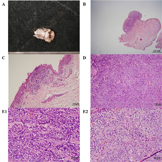
On the advice of veterinarians, the owner agreed to remove the mass through surgery. The mass was surgically removed through a full-thickness V-excision. A figure-of-eight suture was applied to the edge of the eyelid to separate the knot from the edge of the eyelid. A simple interrupted suture was used to close the remaining wound away from the edge of the eyelid. The mass was pink, cauliflower-like, and hairless.

For histopathological examinations, the mass was fixed in 10% neutral buffered formalin for 24 hours. After tissue dehydration, paraffin embedding, and slicing to make sections, tissue sections were stained with haematoxylin and eosin (Figure 1).

Discussion

This particular fibrosarcomas was well-demarcated and nonencapsulated. The visible tumour cells have scant cytoplasm and oval to spindle-shaped nuclei with inconspicuous nucleoli. Cells are spindle shaped and arranged in interwoven patterns [4]. In other field, numerous mitotic figures are observed, and cells have marked cellular and nuclear pleomorphisms [13]. Bright pink collagenous stroma, which are produced by fibroblasts, can be observed between the neoplastic cells.
IJVB2017-104-Geng-RueiChangTaiwan_f1

Figure 1.

Within the field of veterinary medicine, the treatment of STSs includes surgery, chemotherapy, radiation therapy (RT), and combination therapies [9]. The main treatment for sarcomas is surgical resection [18]. For surgical resection, the recommended excision width for STSs is 30 mm lateral to the tumour and one fascial plane [6, 12]. For marginal surgical resection, the excision is made just outside the tumour or at the pseudocapsule [18]. In the current case, the resection was made with a margin of only 1 cm from the tumour. In this situation, marginal resection should be combined with other therapies. RT is used with marginal resection to obtain local control equivalent to radical resection alone [14]. RT should begin 10 to 14 days postoperatively to minimize the risk of the dehiscence or infection of the surgical wound [15]. The efficacy of chemotherapy for controlling soft tissue sarcoma among dogs remains unknown, and it is only recommended for tumours with high histologic grades [3, 9, 14].

The predictors of outcome related to STSs include size, location, grade, histologic type, previous treatment, and surgical margins. Tumour size and location may be prognostic only through their effect on the completeness of the margins [6]. Histological grade is regarded as a more reliable predictor of outcome [1]. Criteria regarding histological grade are detailed by Lipta & Forrest [12]; the American Joint Committee on Cancer staging system was modified from the human STSs staging system. The current case was classified as histologic grade I-II (T1aN0M0; Suppl. 1). Management of low-grade (grade I, grade II) STSs with marginal resection includes frequent monitoring for local recurrence and the appropriate treatment for tumour recurrence [12].

The metastatic rate for soft tissue sarcoma is less than 20%. However, for high-grade tumours, the metastatic rate is estimated as 50% [7]. During the late course of the disease local metastasis often occurs, with a median time of approximately 384 days [8]. Overall, the mean survival time (MST) for dogs with STSs was reported as 1416 days for those treated with surgery alone [11]. By contrast, the MST for fibrosarcomas and hemangiopericytomas was 1851 days, and dogs treated with postoperative RT had an MST of 2270 days [9].

Supplementary material

Suppl. Table 1. Modified Staging System for Canine Soft Tissue Sarcomas (Lipta & Forrest 2013).

Parameters    
Primary Tumor (T) T1 Tumor ≤5 cm in diameter at greatest dimension
T1a Superficial tumor  
T1b Deep tumor  
T2 Tumor >5 cm in diameter at greatest dimension
T2a Superficial tumor  
T2b Deep tumor  
Regional Lymph Nodes (N)
N1
N0 No regional lymph node metastasis
Regional lymph node metastasis  
Distant Metastasis (M)
M1
M0 No distant metastasis
Distant metastasis  
Stage Grouping Tumor (T) Node (N) Metastasis (M) Grade
I Any T N0 M0 I-II
II T1a-T1b, T2a N0 M0 III
III T2b N0 M0 III
IV Any T N1 Any M I-III
Any T Any N M1 I-III

Acknowledgements: The authors thank Jing-Rong Hu of the Department of Veterinary Medicine, National Chiayi University for conducting the histopathological examination.

Conflict of interest: None of the authors of this article has a financial or personal relationship with other people or organisations that could inappropriately influence or bias the content of the paper.

References

  1. Baker-Gabb M, Hunt GB, France MP (2003) Soft tissue sarcomas and mast cell tumours in dogs; clinical behaviour and response to surgery. Aust Vet J 81: 732–738. [crossref]
  2. Bedrossian CW, Verani R, Unger KM, Salman J (1979) Pulmonary malignant fibrous histiocytoma. Light and electron microscopic studies of one case. Chest 75: 186–189.
  3. Casper ES, Gaynor JJ, Harrison LB, Panicek DM, Hajdu SI, Brennan MF (1994) Preoperative and postoperative adjuvant combination chemotherapy for adults with high grade soft tissue sarcoma. Cancer 73: 1644–165.
  4. Crago AM, Brennan MF2 (2015) Principles in Management of Soft Tissue Sarcoma. Adv Surg 49: 107–122. [crossref]
  5. Dennis MM, McSporran KD, Bacon NJ, Schulman FY, Foster RA, et al. (2011) Prognostic factors for cutaneous and subcutaneous soft tissue sarcomas in dogs. Vet Pathol 48: 73–84. [crossref] 
  6. Dernell WS, Withrow SJ, Kuntz CA, Powers BE (1998) Principles of treatment for soft tissue sarcoma. Clin Tech Small Anim Pract 13: 59–64. [crossref] 
  7. Ehrhart N (2005) Soft-tissue sarcomas in dogs: a review. J Am Anim Hosp Assoc 41: 241–246. [crossref]
  8. Fong Y, Coit DG, Woodruff JM, Brennan MF (1993) Lymph node metastasis from soft tissue sarcoma in adults. Analysis of data from a prospective database of 1772 sarcoma patients. Ann Surg 217: 72–77.
  9. Forrest LJ, Chun R, Adams WM, Cooley AJ, Vail DM (2000) Postoperative radiotherapy for canine soft tissue sarcoma. J Vet Intern Med 14: 578–582. [crossref] 
  10. Krohel GB, Rosenberg PN, Wright JE, Smith RS (1985) Localized orbital neurofibromas. Am J Ophthalmol 100: 458–464. [crossref] 
  11. Kuntz CA, Dernell WS, Powers BE, Devitt C, Straw RC, Withrow SJ (1997) Prognostic factors for surgical treatment of soft-tissue sarcomas in dogs: 75 cases (1986–1996). J Am Vet Med Assoc 211: 1147–115.
  12. Lipta JM, Forrest LJ (2012) Soft tissue sarcomas. In: Withrow, SJ and Vail, DM (Eds.), Small animal clinical oncology, 5th ed. Elsevier Medicine Press, Philadelphia, 356–380.
  13. Madarame H, Sato K, Ogihara K, Ishibashi T, Fujii Y, et al. (2004) Primary cardiac fibrosarcoma in a dog. J Vet Med Sci 66: 979–982. [crossref] 
  14. McKnight JA, Mauldin GN, McEntee MC, Meleo KA, Patnaik AK (2000) Radiation treatment for incompletely resected soft-tissue sarcomas in dogs. J Am Vet Med Assoc 217: 205–210. [crossref]
  15. McLeod DA, Thrall DE (1989) The combination of surgery and radiation in the treatment of cancer. A review. Vet Surg 18: 1–6. [crossref] 
  16. Nakajima T, Watanabe S, Sato Y, Kameya T, Hirota T, Shimosato Y (1982) An immunoperoxidase study of S-100 protein distribution in normal and neoplastic tissues. Am J Surg Pathol 6: 715–727.
  17. Roncaroli F, Eusebi V (1996) Rhabdomyoblastic differentiation in a leiomyosarcoma of the retroperitoneum. Hum Pathol 27: 310–313. [crossref] 
  18. Stefanello D, Morello E, Roccabianca P, Iussich S, Nassuato C, et al. (2008) Marginal excision of low-grade spindle cell sarcoma of canine extremities: 35 dogs (1996–2006). Vet Surg 37: 461–465.
  19. Theilen GH, Madwell BR (1979) Tumours of the skin and subcutaneous tissues. In: Veterinary cancer medicine, 2nd ed. Lea and Febiger, Philadelphia, 123–19.
The Expression of Autophagy-Related Genes Atg9a and Atg9b in Normally Developing and Arresting Porcine Conceptuses on Gestational Days 20 and 50

DOI: 10.31038/IJVB.2017113

Abstract

Livestock productivity can be severely compromised by low fertility due to conceptus loss. Prenatal mortality is of particular concern in swine reproduction. Conceptus loss in pigs occurs mainly in early pregnancy (a primary loss of ~30%) but also during the mid-gestation period (a second loss of ~20%). Autophagy is a degradation system that controls the clearance and reuse of intracellular constituents as well as balancing the sources of energy in times of development and stress. There is compelling evidence to suggest that autophagy plays a role in embryogenesis and pregnancy-allied complications in mammalian species. Hence, the objective of the present experiment was to determine the expression of autophagy-related genes Atg9a and Atg9b in the endometrial and trophoblast tissues of healthy and arresting porcine conceptuses on gestational days 20 (gd20) and 50 (gd50). Relative mRNA expression was assessed by real-time polymerase chain reaction, and protein levels were quantified by Western blot and densitometric analyses. Atg9a mRNA expression was greater (P < 0.05) in the trophoblast from arresting (AT) compared with healthy embryos (HT) on gd20, while Atg9b mRNA expression was greater (P < 0.05) in AT than in HT on gd50. No protein expression was detected with the Western blots of both Atg9 proteins in trophoblast tissue samples on either of the two gestational days except for low levels of Atg9a protein expression in AT samples on gd20; there were no significant differences in Atg9 protein content between tissues and gestational days in the sows of the present study. On the basis of variations in Atg9 mRNA levels in the trophoblast, in can be proposed that the autophagy system is mainly involved in the autoregulation of porcine embryonic/fetal development. Further studies are needed to elucidate the specific roles of intrauterine Atg9 genes and their products throughout porcine pregnancy.

Keywords

Pig; Pregnancy; Conceptus; Endometrium; Trophoblast; Autophagy

Introduction

Prenatal mortality is an important adverse factor that influences reproductive efficiency of livestock [1]. Two waves of spontaneous conceptus loss may occur in porcine pregnancy: a primary embryo loss of ~30% is usually observed around gestational day 20 and a second loss of ~20% of remaining fetuses during mid-gestation (~day 50; [2]). “Crowding” in the uterus due to limited space for developing embryos and fetuses (“uterine capacity”; [2]) or asynchronous development of the uterus and the embryo [1] may both result in the arrest and demise of porcine conceptuses.

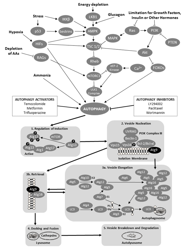
Autophagy is a degradation mechanism that controls the clearance or recycling of intracellular constituents as well as balancing the sources of energy during development and stress ([3] Figure 1). The autophagy system is also responsible for removing damaged organelles, clearing non-functional proteins, eliminating pathogens, and recycling misfolded proteins [3]. There is increasing evidence that autophagy plays an important role in mammalian differentiation and development [4]. Autophagy begins with the formation and elongation of phagophores. The phagophores engulf organelles and sequester them in a double-membrane autophagosome [3]. The autophagosome then fuses with the lysosome and matures into autolysosome. The content of the autolysosome is degraded by proteases and the products of degradation are exported into the cytoplasm in order to be reused for various metabolic processes including the biosynthesis of ATP, nucleic acids, carbohydrates, amino acids and lipids. The autophagy processes provide the cell with sufficient nutrients until conditions improve and the cell can survive on its own.

The role of autophagy in pregnancy was studied at the fetal-maternal interface in normal and aberrant human pregnancies [5]. Increased intensity of autophagy processes was observed in the specimens obtained after spontaneous miscarriages; this increase was due mainly to an elevated concentration of autophagy vacuoles. These results can be interpreted to suggest that the conditions inside the uterus of the spontaneous miscarriage enhanced the activity of the autophagy system. Genetic screening has identified a number of autophagy-related genes (Atg genes; [3]). The Atg9 genes and proteins are responsible for the formation of the double-membrane autophagosome; in situations of starvation, the Atg9 vesicles fuse with the outer membrane of the autophagosome [6]. Two isoforms of the Atg9 proteins, Atg9a and Atg9b, are typically required for autophagosome formation [6, 7]. A previous study using the fetal Atg9a knockout mouse model has shown considerable growth restriction and increased mortality of Atg9a-/- fetal mice, suggesting that the non-functional autophagy system can impede or prematurely terminate fetal development [8]. In addition, the intrauterine growth retardation of the Atg9a-/- fetal mice was associated with the presence of maternal hypertension. The aforementioned observations in pregnant women and small laboratory rodents prompted us to examine the pattern of changes in Atg9 gene expression in healthy and arresting porcine conceptuses collected on gestational days 20 and 50.

IJVB2017-103-Pawel-Canada_f1

Figure 1. A schematic of the cellular macroautophagy processes (based on a flowchart by the Selleck Co. (Houston, TX, USA) shown here: http://www.selleckchem.com/pharmacological_autophagy.html). Abbreviations used (in alphabetical order and in bold font face for easier identification within the legend): AAs – amino acids; Akt – protein kinase B; AMPK – AMP-activated protein kinase; Atg − autopha-gy-related gene proteins; Bcl – B-cell lymphoma proteins; HIFs – hypoxia-inducible factors; eEF-2 kinase − eukaryotic elongation factor 2 kinase; FOXO − forkhead box protein O; iKK-β − inhibitor of nuclear factor kappa-B kinase subunit beta; LC3 − microtubule-associated protein 1A/1B-light chain 3; LKB1 − liver kinase B1 a.k.a. serine/threonine kinase 11 (STK11) or renal carcinoma antigen NY-REN-19; LY294002 − a morpholine-containing chemical compound that is a potent inhibitor of numerous proteins, and a strong inhibitor of phosphoinositide 3-kinases; MAPK – mitogen-activated protein kinase; mTOR − mechanistic target of rapamycin, formerly mammalian target of rapamycin; mTORC1 − mammalian target of rapamycin complex 1 or mechanistic target of rapamycin complex 1; p53 − tumor protein p53, a.k.a. cellular tumor antigen p53, phosphoprotein p53, tumor suppressor p53, antigen NY-CO-13, or transformation-related protein 53; PI3K − phosphoinositide 3-kinase; PTEN − phosphatase and tensin homolog; RAGs − recombination-activating genes; RAS − small GTPase proteins; RHEB − Ras homolog enriched in brain (GTP-binding protein); ULK1 Complex − serine/threonine-protein kinase complex; UVRAG – UV radiation resistance-associated gene protein; and VPS − vacuolar protein sorting-associated protein. Atg 9 proteins have been denoted by black oval shapes.

Methods and Materials

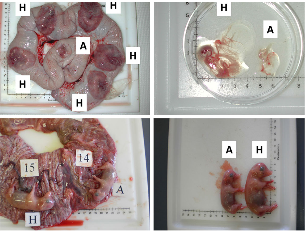
Second parity Yorkshire sows housed in the Arkell Swine Research Station near Guelph, ON, Canada, were used in this study. All sows were artificially inseminated within 7 days post-weaning (at first estrous detection and 24 h later) using pooled Duroc semen (Ontario Swine Improvement; Innerkip, ON, Canada). Tissue samples were collected from eight pregnant animals on gestational day 20 (gd20) and from eight pregnant sows on gestational day 50 (gd50). The University of Guelph Animal Care Committee had approved animal handling and euthanasia protocols (Animal Utilization Protocol no. 10R061). All experimental procedures were in compliance with the guidelines of the Canadian Council on Animal Care and Use of Experimental Animals. The reproductive tracts were collected immediately after slaughter and within 30 min transported on ice to the laboratory. To remove the embryos from the attachment sites, the uterine horns were opened longitudinally along the anti-mesometrial side. Conceptuses from each sow were categorized as either healthy or arresting based on embryonic/fetal size, weight and visual assessment of vascularization of the attachment sites as previously described [9-11], Figure 2; the conceptuses were removed from the study if they possessed debatable health status or were classified as reabsorbing. Endometrial tissue on the maternal side was collected separately from the trophoblast. All samples were rinsed with PBS and then frozen immediately and stored at −80°C until RNA isolation. The RNeasy mini kits (Qiagen; Mississauga, ON, Canada) were used to perform total RNA extraction from all collected tissues. Total RNA concentrations were determined using the A260/A280 (the ratio of absorbance at 260 and 280 nm) and the Gene Quant pro RNA/DNA calculator (Biochrom Ltd.; Cambridge, UK). Extracted RNA samples were frozen immediately at −80°C. Subsequently, the First-Strand cDNA Synthesis Kit (GE Healthcare Bio-Science Inc.; Baie d’Urfe, QC, Canada) was used for cDNA synthesis. In short, 20 μl of diluted RNA at concentrations ranging from 224 to 890 ng/μl were heated for 10 min at 65°C in the GeneAMP polymerase chain reaction (PCR) System 2700 (Applied Biosystems; Foster, CA, USA). Once the heating was finished, 11 μl of the bulk first-strand cDNA reaction mix, 1 μl of poly (dT) primer and 1 μl of DTT solution was added; the reagents were then incubated at 37°C for 1 h. Using the Gene Quant Pro RNA/DNA calculator, the cDNA concentration was measured. The cDNA products were then stored at −20°C for quantitative real-time PCR.

Primers targeting the genes of interest (porcine Atg9a and Atg9b) were designed using the Primer 3 software from the electronic nucleotide database, GenBank. Aliquots of cDNA were pooled and used as a template to test the primers and to optimize their efficiency. Table 1 lists the primers for β-actin (ACTB), Atg9a and Atg9b. The LightCycler 480 SYBR Green I Master (Roche Diagnostics; Mannheim, Germany) was used to optimize primer efficiency in real-time PCR system (ViiATM 7 Real Time PCR System, Applied Biosystems by Life Technologies; Foster, CA, USA). Genes quantified in the samples were run in duplicates using the MicroAmp Fast 96-well reaction plate (Biosystems by Life Technologies; Foster, CA, USA). Data were analyzed using the ViiATM 7 software for the ViiA 7 Real-Time PCR System (Biosystems by Life Technologies; Foster, CA, USA). All results from the PCR reactions were expressed as a ratio of Atg9a or Atg9b mRNA relative to β-actin (ACTB) mRNA.

For Western blot analyses of Atg9a and Atg9b proteins, the tissues were thawed on ice and divided into 30-mg samples placed in Eppendorf tubes. A protease inhibitor and 200 μl of phosphate buffered saline (PBS) were added to each sample followed by homogenization on ice for 1 min. Homogenates were centrifuged at 4°C for 15 min and the supernatants were separated. The Bradford method was used to determine protein concentrations. The samples were then diluted to a final concentration of 2 μg/μl and stored at −80°C. Protein samples (30 μg) were denatured at 100°C for 5 min using the Applied Biosystems GeneAmp PCR System 2700. Samples were loaded in the wells using the 4-20% Mini-PROTEAN TGX Pre-cast gels (12 well comb/20 μl/well; Bio-Rad Laboratories; Mississauga, ON, Canada). Protein fractions were separated by electrophoresis for 1 h at 140 V. The proteins were transferred onto nitrocellulose membrane by assembling a “gel sandwich” that was immersed in the transfer buffer and exposed to 90 V for 90 min. The membrane was blocked with 2.5 g of skimmed powder milk and 50 ml of Tris-buffered saline with Tween 20 (TBS-T) at room temperature for 1 h and then incubated overnight at 4°C with the primary antibody: Atg9a Goat Polyclonal IgG (sc-70141, Santa Cruz Biotechnologies Inc.; Dallas, TX, USA) or Atg9b Goat Polyclonal IgG (sc-163710, Santa Cruz Biotechnology) diluted 1: 200 in skimmed milk with TBS-T (40 μl of primary antibody with 8 ml of skimmed milk blocking solution). After washing with TBS-T, the membrane was incubated with the secondary antibody: HRP-conjugated rabbit anti-goat IgG (ref-61120; Invitrogen-Thermo Scientific; Frederick, MD, USA) diluted 1: 10,000 in skimmed milk with TBS-T (0.8 μl of secondary antibody with 8 ml of blocking solution) for 1 h at room temperature. Subsequently, the membrane was washed again and imaged using the electroluminescence kit and ChemiDocTM MP Imaging System (Bio-Rad Laboratories; Mississauga, ON, Canada). The membrane was then stripped using the Restore Western Blot Stripping Buffer (Thermo Scientific; Rockford, IL, USA) for re-probing for β-actin; the same protocol as for Atg9a and Atg9b proteins was used except that a primary antibody pre-conjugated with horseradish peroxidase (HPA) was anti-β actin antibody (mAbcam 8226; Abcam PLC; Cambridge, UK). The membranes were imaged and analyzed using the Image Lab analytical software (Version 5.1 Bio-Rad Laboratories Inc.; Hercules, CA, USA) to obtain relative expression values (ratios to β-actin) from each corresponding blot.

IJVB2017-103-Pawel-Canada_f2

Figure 2. Images illustrating the early (gestational day (gd) 20) and mid-gestational (gestational day (gd) 50) conceptus status in porcine pregnancy. Two upper panels depict healthy (H) and arresting (A) conceptuses on gd20, classified on the basis of disparity in size and vascularization of fetal membranes (Kridli et al., 2016), and two lower panels show healthy (H) and arresting (A) conceptuses from a uterine horn, which were classified on the basis of weight and length on gd50 (Kridli et al., 2016).

Table 1. Specific primers used for Atg9a and Atg9b and β-actin (ACTB) quantification by rtPCR.

Gene Primers (5`to 3`) Product size (bp)
Atg9a Forward: ATCCTCGCTCACATCCACTAC
Reverse: GTGAAATTGCGAAGAAGTCTA
206
Atg9b Forward: GCATCTGCCGAGATCAGTC
Reverse: TCCTTCTGGGTGTCCGTAGT
266
ACTB Forward: ACGTGGACATCAGGAAGGAC
Reverse: ACATCTGCTGGAAGGTGGAC
210

Statistical analyses were conducted using the SigmaPlot® software (Systat Software Inc., Richmond, CA, USA). To attain normality before statistical testing, all of the data were transformed logarithmically. For real-time PCR and Western blot, the results for endometrial and trophoblast tissue samples across both gestational days were compared by two-way analysis of variance (ANOVA). Comparisons were also made between the tissues obtained from healthy and arresting conceptuses. A P-value < 0.05 was considered significant. All results are given as mean ± standard error of the mean (SEM).

Results and Discussion

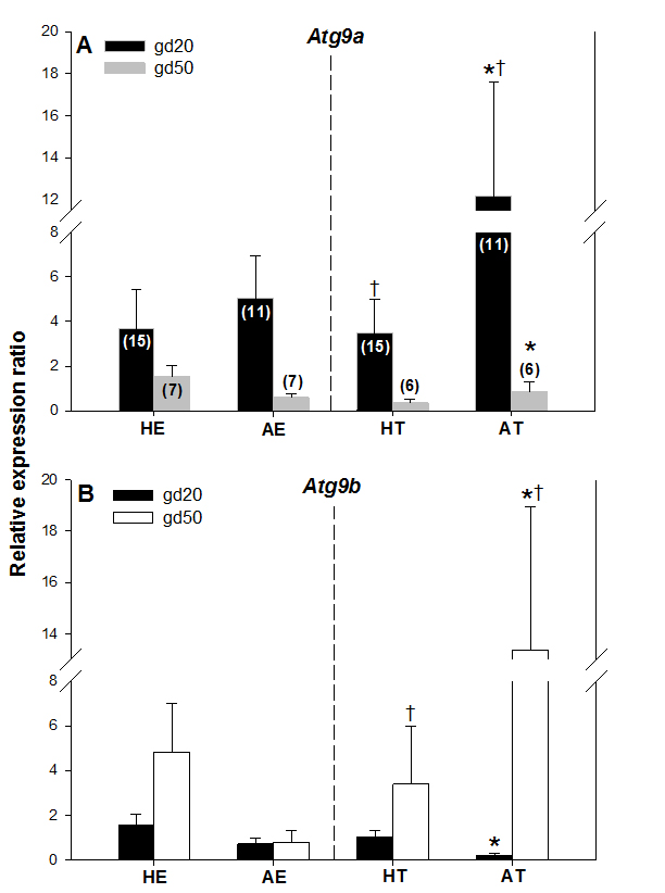
Atg9a mRNA expression was significantly greater in arresting than in healthy trophoblast tissue on gestation day 20 (gd20), and it was greater (P < 0.05) in AT on gd20 compared with AT on gestation day 50 (Figure. 3A). There were no other differences in Atg9a mRNA expression between healthy and arresting trophoblast tissue nor between the two gestation days studied. Atg9b mRNA expression was greater (P < 0.05) in AT than HT on gd50 (Figure 3B). As with Atg9a gene, no other differences in Atg9b expression levels were recorded in this study. These results suggest that both Atg9 genes may be involved in maintaining healthy pregnancy in swine. The elevated Atg9 gene expression levels in arresting tissue samples, as compared to the healthy trophoblast tissue, also suggest that these genes are mainly activated by unfavorable uterine conditions during pregnancy in sows; this is in agreement with observations obtained after miscarriages in women [5]. The specific reason for the differences in Atg9a mRNA and Atg9b mRNA expression between gd20 and gd50 are difficult to explain, but since alterations in only one of those genes can lead to reproductive disturbances [8], they can still be implicated in embryonic and fetal arrest in pregnant pigs. One of the reasons for this disparity could be the fact that Atg9a gene is ubiquitously expressed whereas Atg9b gene is only expressed in the placenta and the pituitary gland [12]. As of yet, no experimental studies have been conducted on the role of Atg9b gene in mammalian pregnancy.

IJVB2017-103-Pawel-Canada_f3

Figure 3. Comparisons of Atg9a mRNA (A) and Atg9b mRNA (B) expression levels in healthy and arresting porcine conceptuses and attachment sites on gestational days 20 (gd20) and 50 (gd50). Healthy endometrium (HE), arresting endometrium (AE), healthy trophoblast (HT), arresting trophoblast (AT). Numbers of samples used for rtPCR are given in parentheses (upper panel). Values denoted by the same symbols (*) differ significantly.

IJVB2017-103-Pawel-Canada_f4

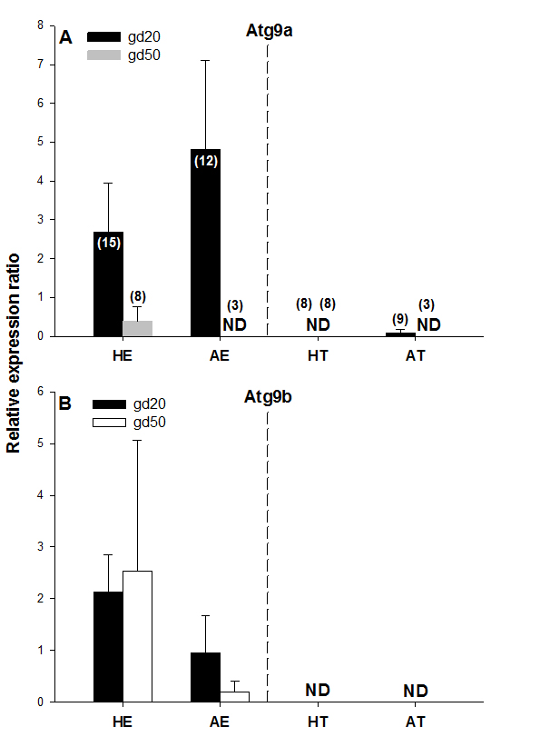
Figure 4. Atg9a and Atg9b protein expression on gestational days 20 (gd20) and 50 (gd50) in healthy and arresting conceptuses and their attachment sites. Protein expression levels were calculated as the ratios to β-actin from each corresponding blot; the bands detected in the 87-kDa range were quantified. Numbers of samples used for Western blot are given in parentheses (upper panel). Healthy endometrium (HE), arresting endometrium (AE), healthy trophoblast (HT), arresting trophoblast (AT), and non-detectable (ND).

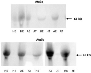
No protein expression was detected with the Western blot of both Atg9 proteins in trophoblast tissue samples on either of the two gestational days studied except for low levels of Atg9a protein in AT samples on gd20 (Figure 4A and 4B, Figure 5). In addition, no Atg9a protein was detected in AE obtained on gd50 (Figure. 4A). On either gestational day, no variation in Atg9 protein levels between healthy or arresting conceptus attachment sites was detected. This is in contrast with previous observations in humans [13]. Autophagy was predominantly localized to the syncytiotrophoblast layer and autophagosomes were more abundant in the fetal growth restricted (FGR) placentae [13]. Moreover, the autophagy regulators including LC3B, Beclin-1, Atg5, Atg9 and Atg16L1 were all detected in villous trophoblast [14]. Our results, however, appear to be in partial agreement with a previous rodent study, wherein the absence of Atg9a gene expression resulted in fetal developmental abnormalities and demise. A lack of protein detection could be due to a number of factors including mRNA stability, protein degradation or protein relocation. Protein production depends on the abundance and stability of mRNA; rapid decays of mRNA can prevent the accumulation of transcripts and protein biosynthesis [15]. Alternatively, intrauterine proteins may have been degraded in response to various hormonal stimuli [16]. In the present Western blotting experiments, the real weight of proteins was somewhat less than the theoretical weight (positive control), which can be indicative of ongoing post-translational modifications [17]. Protein relocation could also have influenced protein detection in the present study [18, 19]. Further research is needed to provide information about synthetic/secretory pathways and specific roles of the Atg9 proteins in porcine pregnancy. It is attractive to speculate that inadequate expression of Atg proteins was associated with the altered development of conceptus in the sows of the present study. It would now be interesting to test the effect of various autophagy inhibitors (e.g., LY294002, Paclitaxel or Wortmannin) and activators (e.g., Temozolomide, Metformin or Trifluoperazine; Figure 1), administered just prior to the periods of expected embryonic or fetal arrest, on the piglet productivity.

IJVB2017-103-Pawel-Canada_f5

Figure 5. Examples of Western blot images containing Atg9a (upper panel) and Atg9b (lower panel) protein bands from porcine tissue specimens collected on gestational day 20. The intensity of bands for both Atg9a and Atg9b proteins were subsequently quantified relative to the expression of the beta actin bands. Healthy endometrium (HE), arresting endometrium (AE), healthy trophoblast (HT), arresting trophoblast (AT), and (+) manufacturer’s positive control.

In conclusion, there may be possible involvement of embryonic and fetal autophagy-related genes in the placentation and development of porcine conceptuses. Due mainly to the discrepancies in the results from rtPCR and Western blotting, more research is needed on the transcriptional regulation of Atg9 genes as well as the sites and mode of action of their products during porcine pregnancy.

Competing Interests: The authors declare that they have no competing interests.

Acknowledgements: Thanks are extended to: staff members at the Arkell Swine Research Station (OMAFRA) in Guelph, ON, Canada, for care and management of experimental animals; the Meat Wing of the Department of Animal and Poultry Science, U of G, for help with euthanasia; Dr. Jocelyn Wessels for technical assistance with specimen collection and the preparation of samples; Dr. Tami Martino (Department of Biomedical Sciences, U of G) for access to the rtPCR and Western blot facilities, and the Jordan University of Science and Technology (sabbatical funding for RTK). The present results were presented, in the preliminary form, during the 4th World Congress of Reproductive Biology (Okinawa, Japan; 27-29 September 2017).

Funding Information: Primary funding in the form of an operating research grant to CT was provided by the Ontario Pork. Additional funding was provided by the Department of Biomedical Sciences, U of G (PMB).

References

  1. Wilson ME (2002) Role of placental function in mediating conceptus growth and survival. J Anim Sci 80(E. Suppl.2) E195–E201.
  2. Pope WF (1994) Embryonic Mortality in Swine. In: Embryonic Mortality in Domestic Species. CRC Press 53–78.
  3. Glick D, Barth S, Macleod KF (2010) Autophagy: cellular and molecular mechanisms. J Pathol 221: 3–12. [crossref]
  4. Mizushima N, Levine B (2010) Autophagy in mammalian development and differentiation. Nat Cell Biol 12: 823–830. [crossref]
  5. Avagliano L, Terraneo L, Virgili E, Martinelli C, Doi P2, et al. (2015) Autophagy in Normal and Abnormal Early Human Pregnancies. Reprod Sci 22: 838–844. [crossref]
  6. Yamamoto H, Kakuta S, Watanabe TM, Kitamura A, et al. (2012) Atg9 vesicles are an important membrane source during early steps of autophagosome formation. J Cell Biol 198–219: 33.
  7. Bestebroer J, V’kovski P, Mauthe M, Reggiori F (2013) Hidden behind autophagy: the unconventional roles of ATG proteins. Traffic 14: 1029–41.
  8. Kojima T, Yamada T, Akaishi R, Furuta I, Saitoh T, et al. (2015) Role of the Atg9a gene in intrauterine growth and survival of fetal mice. Reprod Biol 15: 131–138. [crossref] 
  9. Croy BA, Wessels J, Linton N, Tayade C (2009) Comparison of immune cell recruitment and function in endometrium during development of epitheliochorial (pig) and hemochorial (mouse and human) placentas. Placenta 30(Suppl A) 2009:  S26–S31.
  10. Bidarimath M, Khalaj K, Kridli RT, Wessels J, et al. (2017) Altered expression of chemokines and their receptors at porcine maternal-fetal interface during early and mid-gestational fetal loss. Cell Tissue Res.
  11. Kridli RT, Khalaj K, Bidarimath M, Tayade C (2015) Placentation, maternal-fetal interface, and conceptus loss in swine. Theriogenology 85,135–44.
  12. Xie Y, Kang R, Sun X, Zhong M, et al. (2015) Posttranslational modification of autophagy-related proteins in macroautophagy. Autophagy 28–45.
  13. Bagola K, Menhert M, Jarosch E, Sommer T. Protein dislocation from the ER. Biochim Biophys Acta 2011: 1808: 25–36.
  14. Curtis S, Jones CJP, Garrod A, Hulme CH, et al. (2013) Identification of autophagic vacuoles and regulators of autophagy in villous trophoblast from normal term pregnancies and in fetal growth restriction. J Matern Fetal Neo M 26,339–46.
  15. Yamada T, Carson A, Caniggia I, Umebayashi K, et al. (2005) Endothelial nitric-oxide synthase antisense (NOS3AS) gene encodes an Autophagy-related Protein (APG9-like2) highly expressed in trophoblast. J Biol Chem 18283–90.
  16. Wu X, Brewer G (2012) The regulation of mRNA stability in mammalian cells: 2.0. Gene 500: 10–21. [crossref]
  17. Cooper GM (2000) Protein Degradation. The Cell: A Molecular Approach, 2nd Edition. Sunderland: Sinauer Associates.
  18. Mahmood T, Yang PC (2012) Western blot: technique, theory, and trouble shooting. N Am J Med Sci 4: 429–434. [crossref] 
  19. Bagola K, Mehnert M, Jarosch E, Sommer T (2011) Protein dislocation from the ER. Biochim Biophys Acta 1808: 925–936. [crossref]

Asymmetric bagging and feature selection for activities prediction of drug molecules of an in silico developed unique fragment poly-pharmacologic modulator of CXCR4 tumor-derived heat-shock GGHFGPFDY peptide mimotopic complex-96 (HSPPC-96) by highthroughput identifying hits in the hydrophobic autotaxin/lysophospholipase D pocket

Abstract

Background

Activities of drug molecules can be predicted by QSAR (quantitative structure activity relationship) models, which overcomes the disadvantages of high cost and long cycle by employing the traditional experimental method. With the fact that the number of drug molecules with positive activity is rather fewer than that of negatives, it is important to predict molecular activities considering such an unbalanced situation. Glycoprotein-96, a non-polymorphic heat-shock protein, associates with intracellular peptides. Autologous tumor-derived heat shock protein-peptide complex 96 (HSPPC-96) can elicit potent tumor-specific T cell responses and protective immunity in animal models. Chemokines were described originally in the context of providing migrational cues for leukocytes. They are now known to have broader activities, including those that favor tumor growth. Treatment with autologous tumor-derived HSPPC-96 was feasible and safe at all doses tested. Observed immunological effects and antitumor activity were modest, precluding selection of a biologically active dose. Coevolution between proteins is crucial for understanding protein-protein interaction. Simultaneous changes allow a protein complex to maintain its overall structural-functional integrity. In this Research Scientific Project, we combined statistical coupling analysis (SCA) and molecular dynamics simulations on thecomplex-96 (HSPPC-96) protein complex to evaluate coevolution between conserved binding protein domain regions. We reconstructed an inter-protein residue coevolution network, consisting of 37 residues and 37complex-96 (HSPPC-96) binding domains conserved peptide derived residues and its fitness scoring reverse ligand docking interactions. It shows that most of the coevolved residue pairs are spatially proximal. When the mutations happened, the stable local structures were broken up and thus the protein interaction was decreased or inhibited, with a following increased risk of melanoma. The identification of inter-protein coevolved residues in thecomplex-96 (HSPPC-96) complex can be helpful for designing protein drug target and in silico discovery of engineering novel nanomolecule experiments.

Results

Here, asymmetric bagging and feature selection are introduced into the problem and asymmetric bagging of support vector machines (asBagging) is proposed on predicting drug activities to treat the unbalanced problem. At the same time, the features extracted from the structures of drug molecules affect prediction accuracy of QSAR models. Therefore, a novel algorithm named PRIFEAB is proposed, which applies an embedded feature selection method to remove redundant and irrelevant features for asBagging. Numerical experimental results on a data set of molecular activities show that asBagging improve the AUC and sensitivity values of molecular activities and PRIFEAB with feature selection further helps to improve the prediction ability.

Conclusion

Asymmetric bagging can help to improve prediction accuracy of activities of drug molecules, which can be furthermore improved by performing feature selection to select relevant features from the drug molecules data sets. In this scientific study we have in silico discovered a Unique Small Molecule Modulator of CXCR4 tumor-derived heat-shock protein peptide complex-96 (HSPPC-96) by identifying Hits of a High-Throughput Screen Identify the Hydrophobic Pocket of Autotaxin/Lysophospholipase D as an Inhibitory Surface Molecular dynamic simulation and statistical coupling analysis via a Asymmetric bagging and feature selection for activities prediction of drug molecules of an in silico developed unique fragment poly-pharmacologic modulator of CXCR4 tumor-derived heat-shock GGHFGPFDY peptide mimotopic complex-96 (HSPPC-96) by highthroughput identifying hits in the hydrophobic autotaxin/lysophospholipase D pocket.

Keywords

In silico development; unique fragment; poly-pharmacologic modulator; CXCR4 tumor-derived; heat-shock; GGHFGPFDY peptide; mimotopic complex-96; (HSPPC-96); highthroughput identifying hits; hydrophobic autotaxin/lysophospholipase D pocket.

CHARMM additive and polarizable force fields for biophysics and computer-aided drug design as an in silico chemoproteomic prediction-scan for the generation of a tyrosinase aa95-104FMGFNCGNCK antigenic pattern LFA-3/IgG fusion polypeptide IleAlaArgArgPheLeuOH (Kinetensin) mimetic pharmacophore on conserved Vitiligo post-trancripts domains.

Abstract

Background

Molecular Mechanics (MM) is the method of choice for computational studies of biomolecular systems owing to its modest computational cost, which makes it possible to routinely perform molecular dynamics (MD) simulations on chemical systems of biophysical and biomedical relevance. Vitiligo is a skin disorder characterized by selective melanocyte destruction and concomitant appearance of depigmented macules that over time enlarge, coalesce, and form patches. It has been suggested that vitiligo is, at least in part, caused by autoimmune responses mediated by cytotoxic T cells against melanocytes, causing depigmentation Immune responses contribute to the pathogenesis of vitiligo and target melanoma sometimes associated with vitiligo-like depigmentation in some melanoma patients. It has been perviously reported that the tyrosinase autoantigen was immunorecognized with the same molecular pattern by sera from vitiligo and melanoma patients. Five autoantigen peptides was found to compose the immunodominant antityrosinase response: aa95-104FMGFNCGNCK; aa175-182 LFVWMHYY; aa176-190FVWMHYYVSMDALLG; aa222-236IQKLTGDENFTIPYW, and aa233-247IPYWDWRDAEKCDIC. Synergistic therapies for the treatment of vitiligo are provided. The major therapies for the treatment of vitiligo a pigmentary disorder characterized by patchy depigmentation of skin are Psoralens plus UV-A, steroids, basic fibroblast growth factor (bFGF) peptide location or surgical procedures. Psoralens plus UV-A is effective in about 50% of cases, steroids are limitedly effective only in fast spreading cases of vitiligo and often reoccurs on stoppage of treatment. Surgical treatment is the last resort for vitiligo therapy, when all other therapies failed. It is limitedly effective. Basic fibroblast growth factor peptide(s) location was developed as a new mode therapy for the treatment of vitiligo. Therefore, SEQ ID NO: 01 VPHIPPN, SEQ ID NO: 02 MPPTQVS, SEQ ID NO: 03 QMHPWPP, SEQ ID NO: 1 1 LPLTPLP, SEQ ID NO: 12 QLNVNHQARADQ, SEQ ID NO: 13 TSASTRPELHYP, SEQ ID NO: 14 TFLPHQMHPWPP peptides, modified peptides and antibody or antibody fragments inhibiting the activity of MIA and can be used for treating vitiligo by inducing re-pigmentation. Fragment-based lead discovery is a method used for finding lead compounds as part of the drug discovery process.

Scope of Review

As one of the main factors limiting the accuracy of MD results is the empirical force field used, the present paper offers a review of recent developments in the CHARMM additive force field, one of the most popular bimolecular force fields. Additionally, we present a detailed discussion of the CHARMM Drude polarizable force field, anticipating a growth in the importance and utilization of polarizable force fields in the near future. Throughout the discussion emphasis is placed on the force fields’ parametrization philosophy and methodology. In this science project we perfomed an in silico ChemoProteomic Prediction-Scan for the generation of Antigenic PatternLFA-3/IgG fusion polypeptide aa95-104FMGFNCGNCK; aa175-182 LFVWMHYY; aa176-190FVWMHYYVSMDALLG; aa222-236IQKLTGDENFTIPYW, and aa233-247IPYWDWRDAEKCDICmimetic pharmacophore on conserved Vitiligo post-trancripts domains.

Major Conclusions

Recent improvements in the CHARMM additive force field are mostly related to newly found weaknesses in the previous generation of additive force fields. Beyond the additive approximation is the newly available CHARMM Drude polarizable force field, which allows for MD simulations of up to 1 microsecond on CHARMM additive and polarizable force fields for biophysics and computer-aided drug design analysis as an in silico chemoproteomic prediction-scan for the generation of a tyrosinase aa95-104FMGFNCGNCK antigenic pattern LFA-3/IgG fusion polypeptide IleAlaArgArgPheLeuOH (Kinetensin) mimetic pharmacophore on conserved Vitiligo post-trancripts domains.

General Significance

Addressing the limitations ensures the reliability of the new CHARMM36 additive force field for the types of calculations that are presently coming into routine computational reach while the availability of the Drude polarizable force fields offers a model that is an inherently more accurate model of the underlying physical forces driving macromolecular structures and dynamics.

Keywords

CHARMM additives; polarizable force fields; biophysics; computer-aided drug design; in silico; chemoproteomic; prediction-scan; tyrosinase; aa95-104FMGFNCGNCK antigenic pattern; LFA-3/IgG fusion polypeptide; IleAlaArgArgPheLeuOH (Kinetensin); mimetic; pharmacophore; conserved Vitiligo post-trancripts domains; CHARMM additive and polarizable force fields for biophysics and computer-aided drug design; molecular dynamics, empirical force field, potential energy function, molecular mechanics, computer-aided drug design, biophysics.

An Emerging Computational Methods for the shannon entropy descriptor (SHED) Rational Discovery of Allosteric Drugs for the in silico prediction of an annotated suitable lead chemo-recored compound as a potent computer predicted inhibitor comprising potential hyper-mimicking activities to 5 conserved anti-plasmodium peptides.

Abstract

Drug discovery programs launched by the Medicines for Malaria Venture and other product-development partnerships have culminated in the development of promising new antimalarial compounds such as the synthetic peroxide OZ439 (Charman et al., 2011) and the spiroindolone NITD 609 (Rottmann et al., 2010), which are currently undergoing clinical trials. In spite of these recent successes, it is pivotal to maintain early phase drug discovery to prevent the antimalarial drug development pipeline from draining. Due to the propensity of the parasite to become drug-resistant (Muller and Hyde, 2010; Sa et al., 2011), the need for new antimalarial chemotypes will persist until the human-pathogenic Plasmodium spp. are eventually eradicated. Rational post-genomic drug discovery is based on the screening of large chemical libraries – either virtually or in high-throughput format – against a given target enzyme of the parasite. Experimental tools to validate candidate drug targets are limited for the malaria parasites. Gene silencing by RNAi does not seem to be feasible (Baum et al., 2009). Gene replacement with selectable markers is (Triglia et al., 1998), but it is inherently problematic to call a gene essential from failing to knock it out. However, none of the reverse genetic methods is practicable at the genome-wide scale. On the other hand Mestres et al. (Cases et al., 2005; Mestres et al., 2006) have annotated a library of molecules targeting NHRs. Using a hierarchical classification for 200.000 ligands and 5 receptors, chemogenomic links bridging ligand to target space can be easily recovered to distinguish selective from promiscuous scaffolds. Using Shannon Entropy descriptors (SHED) based on the distribution of atom-centred feature pairs, any compound collection can be screened to identify hits presenting SHED distances to a reference NHR ligand beyond a defined threshold and therefore likely to share the same NHR profile. Here, we successfully applied a machine-learning algorithm using Bayesian statistics (Xia et al., 2004) to predict target profiles from extended connectivity conserved motif like binding site active pharmacophore fingerprints of selected compounds from the biologically annotated free and non commercial databases (Nidhi et al., 2006) in resulting finally to a potent computer predicted inhibitor comprising potential hyper-mimicking activities to 5 conserved anti-plasmodium peptides.

Keywords

Emerging Computational Methods; Rational Discovery; Allosteric Drugs; shannon entropy descriptor (SHED); in silico prediction; annotated suitable; lead chemo-recored compound; potent computer; predicted inhibitor; comprising potential hyper-mimicking activities; conserved anti-plasmodium peptides.

An integrated computational Mechanisms of Protein Allostery Simulations with Molecular Dynamics Revealing Atomic-Level approach on an in silico LWPQ designed multi-core super-agonist motif-like regulatory peptide for the activation of human stem cell transcripts

Abstract

Molecular dynamics (MD) simulations have become a powerful and popular method for the study of protein allostery, the widespread phenomenon in which a stimulus at one site on a protein influences the properties of another site on the protein. By capturing the motions of a protein’s constituent atoms, simulations can enable the discovery of allosteric binding sites and the determination of the mechanistic basis for allostery. These results can provide a foundation for applications including rational drug design and protein engineering. Here, we provide an introduction to the investigation of protein allostery using molecular dynamics simulation. We emphasize the importance of designing simulations that include appropriate perturbations to the molecular system, such as the addition or removal of ligands or the application of mechanical force. We also demonstrate how the bidirectional nature of allostery—the fact that the two sites involved influence one another in a symmetrical manner—can facilitate such investigations. Through a series of case studies, we illustrate how these concepts have been used to reveal the structural basis for allostery in several proteins and protein complexes of biological and pharmaceutical interest OF integrated computational Mechanisms of Protein Allostery Simulations with Molecular Dynamics Revealing Atomic-Level approach on an in silico LWPQ designed multi-core super-agonist motif-like regulatory peptide for the activation of human stem cell transcripts.

Keywords

Revealing Atomic-Level Mechanisms; Protein Allostery; Molecular Dynamics; Simulations; in silico; LWPQ designed; multi-core super-agonist; motif-like; regulatory peptide; human stem cell transcripts; integrated computational approach.

Variational solvent-solute interface Quantum dynamics in continuum for proton transport in Silico generation of a sophisticated descriptor for the in silico identification and free energy evaluation of hybrid KPQRKTKRNT peptidomimetic leads as a potential inhibitor against helicase and HCV´sStructural NS3/4A protease regions

Abstract

Proton transport plays an important role in biological energy transduction and sensory systems. Therefore it has attracted much attention in biological science and biomedical engineering in the past few decades. The present work proposes a multiscale/multiphysics model for the understanding of the molecular mechanism of proton transport in transmembrane proteins involving continuum, atomic and quantum descriptions, assisted with the evolution, formation and visualization of membrane channel surfaces. We describe proton dynamics quantum mechanically via a new density functional theory based on the Boltzmann statistics, while implicitly model numerous solvent molecules as a dielectric continuum to reduce the number of degrees of freedom. The density of all other ions in the solvent is assumed to obey the Boltzmann distribution in a dynamic manner. The impact of protein molecular structure and its charge polarization on the proton transport is considered explicitly at the atomic scale. A variational solute-solvent interface is designed to separate the explicit molecule and implicit solvent regions. We formulate a total free energy functional to put proton kinetic and potential energies, the free energy of all other ions, the polar and nonpolar energies of the whole system on an equal footing. The variational principle is employed to derive coupled governing equations for the proton transport system. Generalized Laplace-Beltrami equation, generalized Poisson-Boltzmann equation and generalized Kohn-Sham equation are obtained from the present variational framework. The variational solvent-solute interface is generated and visualized to facilitate the multiscale discrete/continuum/quantum descriptions. Theoretical formulations for the proton density and conductance are constructed based on fundamental laws of physics. A number of mathematical algorithms, including the Dirichlet to Neumann mapping (DNM), matched interface and boundary (MIB) method, Gummel iteration, and Krylov space techniques are utilized to implement the proposed model in a computationally efficient manner. The Gramicidin A (GA) channel is used to validate the performance of the proposed proton transport model and demonstrate the efficiency of the proposed mathematical algorithms. The proton channel conductances are studied over a number of applied voltages and reference concentrations. HCV infection has been declared as a principal health problem in more than 200 million individuals throughout the world. It is a positive-stranded RNA virus and classified as a hepacivirus of the flaviviridae family. Unlike other viral infections Hepatitis C Virus even with its high replication rate can stick within a human host for decades without any irritation or liver damage. Estimated 10 million people are believed to be infected by HCV alone in Pakistan. Eventually the infection causes severe complications in 60 to 70% of patients such as cirrhosis, fibrosis, liver failure and hepatocellular carcinoma. Prior to the development of HCV protease inhibitors combination therapy, patients with HCV infection were treated with pegylated interferon-α and ribavirin. The adverse side effects associated with this type of treatment such as anemia, flu-like symptoms, depression, gastrointestinal symptoms, fatigue and cutaneous reactions may lead to the discontinuation of treatment in certain number of patients. The growth in scientific knowledge of HCV life cycle and its replication leads to the development of inhibitors of HCV proteases. A polyprotein precursor encoded by HCV RNA genome containing structural proteins capsid (C), membrane (prM), envelope (E) and nonstructural (NS) proteins (NS1, NS2a, NS2b, NS3, NS4a, NS4b, NS5). NS3 protease when activated by NS4A causes the cleavage of polyprotein producing the non-structural proteins 4A, 4B, 5A, 5B and is thus very supportive in the replication of virus. That is why NS3/4A protease is a significant emerging target for the treatment of HCV infection. NS3 associates to the ER membrane only in the presence of NS4A. Main actively conserved protein target families can be distinguished by a simple look at physicochemical properties (molecular weight, log P, polar surface area, H-bond donor and acceptor counts) of their cognate ligands (Morphy, 2006). One can thus easily imagine that more sophisticated descriptors can be used to predict a global target profile for any given compound, provided that targets to be predicted are sufficiently well described by existing ligands. In this study, Variational solvent-solute interface Quantum dynamics in continuum for proton transport are generated of a sophisticated descriptor for the in silico identification and free energy evaluation of hybrid KPQRKTKRNT peptidomimetic leads as a potential inhibitor against helicase and HCV´sStructural NS3/4A protease regions.

Keywords

Quantum dynamics; continuum for proton transport II: Variational solvent-solute interface; in Silico generation; sophisticated descriptor; in silico identification; free energy evaluation; hybrid KPQRKTKRNT peptidomimetic leads; simultaneous inhibition; helicase and HCV´sStructural; NS3/4A protease regions.

Computer-aided CHARMM additive drug design and Orthodox Quantum Mechanics polarizable force fields for biophysics Success and Incoherence of an in silico KIF20A-derived Peptide agonistic mimicking sited designed poly-chemo-scaffold as an innovative drug-like molecule with potential clinical hyper-inhibitor properties in Gemcitabine treated Patients With Advanced Pancreatic Cancer

Abstract

As one of the main factors limiting the accuracy of MD results is the empirical force field used, the present paper offers a review of recent developments in the CHARMM additive force field, one of the most popular bimolecular force fields. Additionally, we present a detailed discussion of the CHARMM Drude polarizable force field, anticipating a growth in the importance and utilization of polarizable force fields in the near future. Throughout the discussion emphasis is placed on the force fields’ parametrization philosophy and methodology.CHARMM additive and polarizable force fields for biophysics and computer-aided drug design An in silico KIF20A-derived Peptide agonistic mimicking sited and computer-aided designed poly-chemo-scaffold as an innovative drug-like molecule comprising potential clinical hyper-inhibitor properties in Patients With Advanced Pancreatic Cancer when combined with Gemcitabine. Success and Incoherence of Orthodox Quantum Mechanics. Orthodox quantum mechanics is a highly successful theory despite its serious conceptual flaws. It renounces realism, implies a kind of action-at-a-distance and is incompatible with determinism. Orthodox quantum mechanics states that Schrödinger’s equation (a deterministic law) governs spontaneous processes while measurement processes are ruled by probability laws. It is well established that time dependent perturbation theory must be used for solving problems involving time. In order to account for spontaneous processes, this last theory makes use of laws valid only when measurements are performed. This incoherence seems absent from the literature. KIF20A (RAB6KIFL) belongs to the kinesin superfamilyof motor proteins, which play critical roles in the traffickingof molecules and organelles during the growth of pancreatic cancer.Immunotherapy using a previously identified epitope peptide forKIF20A is expected to improve clinical outcomes. A phase I clinicaltrial combining KIF20A-derived peptide with gemcitabine (GEM) was therefore conducted among patients with advancedpancreatic cancer who had received prior therapy such as chemotherapyand/or radiotherapy. Despite, huge importance of the field, no dedicated AVP resource is available. In the present Research Scientific Project , we have collected 1245 peptides with antiviral activity targeting important human viruses like influenza, HIV, HCV and SARS, etc. After removing redundant peptides, 1056 peptides were divided into 951 training and 105 validation data sets. We have exploited various peptides sequence features, i.e. motifs and alignment followed by amino acid composition and physicochemical properties during 5-fold cross validation using Computer-aided CHARMM additive drug design and Orthodox Quantum Mechanics polarizable force fields for biophysics Success and Incoherence of an in silico KIF20A-derived Peptide agonistic mimicking sited designed poly-chemo-scaffold as an innovative drug-like molecule with potential clinical hyper-inhibitor properties in Gemcitabine treated Patients With Advanced Pancreatic Cancer.

Keywords

CHARMM additive; polarizable force fields; biophysics; computer-aided drug design; in silico KIF20A-derived Peptide; agonistic mimicking; computer-aided designed; poly-chemo-scaffold; innovative drug-like molecule; clinical hyper-inhibitor properties; Patients With Advanced Pancreatic Cancer; Success and Incoherence; Orthodox Quantum Mechanics.

Designing the Drug Discovery Sniper: Improving Targeted Human Cytolytic Fusion Proteins for Anti-Cancer Therapy via Molecular Simulation of an in silico KIF20A-derived Peptide agonistic mimicking sited designed poly-chemo-scaffold as an innovative drug-like molecule with potential clinical hyper-inhibitor properties in Gemcitabine treated Patients With Advanced Pancreatic Cancer

Abstract

Targeted human cytolytic fusion proteins (hCFPs) are humanized immunotoxins for selective treatment of different diseases including cancer. They are composed of a ligand specifically binding to target cells genetically linked to a human apoptosis-inducing enzyme. hCFPs target cancer cells via an antibody or derivative (scFv) specifically binding to e.g., tumor associated antigens (TAAs). After internalization and translocation of the enzyme from endocytosed endosomes, the human enzymes introduced into the cytosol are efficiently inducing apoptosis. Under in vivo conditions such enzymes are subject to tight regulation by native inhibitors in order to prevent inappropriate induction of cell death in healthy cells. Tumor cells are known to up-regulate these inhibitors as a survival mechanism resulting in escape of malignant cells from elimination by immune effector cells. Cytosolic inhibitors of Granzyme B and Angiogenin (Serpin P9 and RNH1, respectively), reduce the efficacy of hCFPs with these enzymes as effector domains, requiring detrimentally high doses in order to saturate inhibitor binding and rescue cytolytic activity. Variants of Granzyme B and Angiogenin might feature reduced affinity for their respective inhibitors, while retaining or even enhancing their catalytic activity. A powerful tool to design hCFPs mutants with improved potency is given by in silico methods. These include molecular dynamics (MD) simulations and enhanced sampling methods (ESM). MD and ESM allow predicting the enzyme-protein inhibitor binding stability and the associated conformational changes, provided that structural information is available. Such “high-resolution” detailed description enables the elucidation of interaction domains and the identification of sites where particular point mutations may modify those interactions. This review discusses recent advances in the use of MD and ESM for hCFP development from the viewpoints of scientists involved in both fields by designing the Sniper: Improving Targeted Human Cytolytic Fusion Proteins for Anti-Cancer Therapy via Molecular Simulation of an in silico KIF20A-derived Peptide agonistic mimicking sited designed poly-chemo-scaffold as an innovative drug-like molecule with potential clinical hyper-inhibitor properties in Gemcitabine treated Patients With Advanced Pancreatic Cancer.

Keywords

immunotherapy, targeted human cytolytic fusion proteins, molecular dynamics, high performance computing, Angiogenin, Granzyme B; Designing the Sniper: Targeted Human Cytolytic Fusion Proteins; Anti-Cancer Therapy; Molecular Simulation; in silico; KIF20A-derived; Peptide agonistic; mimicking sited; poly-chemo-scaffold; innovative drug-like; clinical hyper-inhibitor properties; Gemcitabine; Patients With Advanced Pancreatic Cancer.

Variational multiscale Success and Incoherence of Orthodox Quantum Mechanics models for charge transport CHARMM additive and polarizable force fields for biophysics and computer-aided drug design as an in silico KIF20A-derived Peptide agonistic mimicking sited and computer-aided designed poly-chemo-scaffold innovative drug-like molecule with potential clinical hyper-inhibitor properties in Patients With Advanced Pancreatic Cancer

Abstract

This work presents a few variational multiscale models for charge transport in complex physical, chemical and biological systems and engineering devices, such as fuel cells, solar cells, battery cells, nanofluidics, transistors and ion channels. An essential ingredient of the present models, introduced in an earlier paper (Bulletin of Mathematical Biology, 72, 1562-1622, 2010), is the use of differential geometry theory of surfaces as a natural means to geometrically separate the macroscopic domain from the microscopic domain, meanwhile, dynamically couple discrete and continuum descriptions. Our main strategy is to construct the total energy functional of a charge transport system to encompass the polar and nonpolar free energies of solvation, and chemical potential related energy. By using the Euler-Lagrange variation, coupled Laplace-Beltrami and Poisson-Nernst-Planck (LB-PNP) equations are derived. The solution of the LB-PNP equations leads to the minimization of the total free energy, and explicit profiles of electrostatic potential and densities of charge species. To further reduce the computational complexity, the Boltzmann distribution obtained from the Poisson-Boltzmann (PB) equation is utilized to represent the densities of certain charge species so as to avoid the computationally expensive solution of some Nernst-Planck (NP) equations. Consequently, the coupled Laplace-Beltrami and Poisson-Boltzmann-Nernst-Planck (LB-PBNP) equations are proposed for charge transport in heterogeneous systems. A major emphasis of the present formulation is the consistency between equilibrium LB-PB theory and non-equilibrium LB-PNP theory at equilibrium. Another major emphasis is the capability of the reduced LB-PBNP model to fully recover the prediction of the LB-PNP model at non-equilibrium settings. To account for the fluid impact on the charge transport, we derive coupled Laplace-Beltrami, Poisson-Nernst-Planck and Navier-Stokes equations from the variational principle for chemo-electro-fluid systems. A number of computational algorithms is developed to implement the proposed new variational multiscale models in an efficient manner. A set of ten protein molecules and a realistic ion channel, Gramicidin A, are employed to confirm the consistency and verify the capability. Extensive numerical experiment is designed to validate the proposed variational multiscale models. A good quantitative agreement between our model prediction and the experimental measurement of current-voltage curves is observed for the Gramicidin A channel transport. This paper also provides a brief review of the Variational multiscale Success and Incoherence of Orthodox Quantum Mechanics models for charge transport CHARMM additive and polarizable force fields for biophysics and computer-aided drug design as an in silico KIF20A-derived Peptide agonistic mimicking sited and computer-aided designed poly-chemo-scaffold innovative drug-like molecule with potential clinical hyper-inhibitor properties in Patients With Advanced Pancreatic Cancer.

Keywords

Variational multiscale Success; Incoherence of Orthodox Quantum Mechanics; models for charge transport; CHARMM additive; polarizable force fields; biophysics and computer-aided drug design; in silico; KIF20A-derived; Peptide agonistic; mimicking sited; computer-aided designed; poly-chemo-scaffold; innovative drug-like molecule; clinical hyper-inhibitor properties; Patients With Advanced Pancreatic Cancer; Variational multiscale models, Ion channels, Fuel cells, Nanofluidics, Electronic devices, Laplace-Beltrami equation, Poisson-Boltzmann equation, Nernst-Planck equation, Navier-Stokes equation.