Author Archives: author

Restraint Use in the Management of the Elderly with Dementia in Hospital

DOI: 10.31038/IMROJ.2016124

Abstract

There is widespread use of physical restraints among the elderly with dementia in residential setting and acute hospitals. Physical restraints are means to limit a person’s freedom of movement. The commonest indications for restraining an elderly are to manage agitated and aggressive elderly at risk of harming themselves or others, reduce falls risk and avoid dislodgement of medical devices. Physical restraints have not been proven to benefit the patients and have been reported to be associated with injuries, falls and deaths. The ethical dilemma associated with restraint use is often conflicting. There are active moves to reduce/ remove physical restraints use in institutions among the elderly with dementia and challenging behaviours. The use of restraints should be considered a last resort when there is imminent danger and where other means of management have failed and patients being restrained should be reviewed regularly to have the restraint removed at the earliest opportunity.

Key words

physical restraints, behavioral and psychological symptoms, dementia, elderly with dementia

Introduction

With the rapid aging population in the world, there is an increasing trend of people living with dementia. Reports have estimated that there will be about 131.5 million people with dementia by 2050. Dementia has a huge economic impact, costing US $818 billion in total worldwide, and it will become a trillion dollar disease by 2018. In many parts of the world, there is a growing awareness of dementia, but a diagnosis of dementia can bring with it stigma and social isolation. Today, it is estimated that 94% of people living with dementia residing in low and middle income countries are cared for at home. These are regions where health and care systems often provide limited or no support to people living with dementia or to their families. [1]

In an acute hospital setting, people with dementia have more than three times hospital stays per year compared to the elderly without dementia. Utilisation of healthcare resources for chronic medical conditions, such as stroke, cancers, diabetes, coronary heart disease is increased among the elderly with dementia. [2]

The elderly patients with dementia in an acute hospital are at high risk of being restrained, especially if they require assistance with their activities of daily living (ADLs) and the most frequently cited reasons for restraint use are for the protection of the patients themselves, and to prevent interference with medical therapies. Physical restraint usage is between 33-68% among the elderly in hospitals. [3]

The elderly with dementia and behavioural symptoms

Dementia is a group of prolonged, debilitating neuropsychiatric disorders which affect the patients and their family for years after diagnosis. The presence of behavioural and psychological symptoms of dementia occurs among 98% of individuals with dementia at some point during their disease progression. Behavioral and Psychological Symptoms of Dementia (BPSD) has been associated with more rapid decline in cognition, greater impairment of ADLs, caregiver burden leading to caregiver burnout, diminished quality of life for caregivers and patients and early institutionalisation. [4-7] The caregiver for a person with dementia has been described as living a 36 hour day by Mace and Rabins, resulting in physical, emotional and mental fatigue. [8]

The spectrum of behavioural abnormalities in BPSD can be divided into behavioural or psychological abnormalities, as shown in (Table 1). Currently, there is no recommended single treatment for BPSD. Clinicians use a combination of drugs such as Cholinesterase inhibitors, anti-depressants, anti-convulsants with mood stabilising properties, antipsychotics, benzodiazepines and N-Methyl-D-aspartate receptor antagonist with varying degree of success. The elderly are more susceptible to side effects of these medications, including anticholinergic side effects with agitation and sedation, extrapyramidal side effects and orthostatic hypotension contributing to fall risk. [9] The non-pharmacological treatment of BPSD with an aim to reduce medication side effects have been studied with music therapy, art therapy, aromatherapy, touch therapy, orientation therapy, physical exercises and tailored activities have been tried with variable success rate due to heterogeneity of the study designs and further research is required. [10]

Table 1. Spectrum of BPSD

Behavioural Psychological
Agitation-e.g. restless, pacing, disrobing inappropriately. Mood disorders- anxiety, depression
Aggression-hitting, biting, scratching, pushing, throwing objects, destroying property, tearing items. Changes in personality
Verbal aggression- cursing, swearing, shouting, screaming Psychosis- delusions, hallucination
Wandering Pathological crying
Repetition Apathy
Sexual disinhibition Irritability
Urination/ defecation-at inappropriate time and place Mood lability
Hoarding Elation

Behavioural symptoms in dementia suggest the presence of underlying unmet needs and must therefore be looked at as means of communication as cognitive abilities decline. The unmet needs include physical causes like medical illness and pain or psycho social and emotional needs. Agitation signifies progression of dementia. At the middle stages of dementia where verbal communication is diminishing, verbally agitated behaviours such as repetitions, cursing or screaming, are common. In severe stages of dementia, physically agitated behaviours predominate because they have lost the abilities to communicate verbally. [11] Agitated behaviours may be a reflection of others’ behaviour where the elderly with dementia does not comprehend or does not want. Agitation is associated with poor outcome for health and general wellbeing. Agitated behaviour places an elderly at risk of harm on themselves, caregivers and often leads to early institutionalisation. Nurses often see an agitated elderly as a challenge and feel helpless to intervene. [12]

Causes of BPSD- Theoretical models

Cohen-Mansfield applied theoretical models to analyse the causes of BPSD. The problematic behaviours in dementia may arise from various causes such as 1.) unmet needs, 2.) behavioural/ learning models and 3.) environmental factors.

Unmet needs among the person with dementia are frequently not obvious to the caregivers. Among some of these include inadequately treated pain, toileting needs, thirst, hunger, sensory deprivation, boredom and loneliness. Use of restraints causes restriction in independence, social isolation and may worsen behaviour. Assisting the person with dementia with proper eye wear and hearing aids, regular toilet rounds, assistance for physical exercise, meaningful activities and meals, providing sensory stimulation like pet therapy, music therapy, social interactions may reduce agitation.

The ABC model of behaviours consists of antecedent events which are the stimuli leading to the behaviour and consequences of the behaviours. The consequences reinforce certain behaviours in response to the antecedents. Many problem behaviours are learned through reinforcements by staff who paid attention when problem behaviours are displayed. To modify the behaviours requires new learning experiences which change the antecedent and behaviour.

The environmental theory suggests that persons with dementia are more vulnerable to environmental stimuli and they have a lower threshold at which their behaviour changes in response. The persons with dementia lose their coping abilities progressively and find the environmental changes increasingly more stressful. The threshold for stress also lowers progressively. When environmental stimuli exceed the stress threshold, they are more likely to show anxiety and inappropriate behaviours. [13]

Cohen-Mansfield suggested that the different models may interact and complement each other. For example, an environmental stimulus (unfamiliar surrounding) may cause an unmet need to surface (getting lost looking for toilets, bedroom) which may account for different behaviours (resulting in pacing, agitation, incontinence, etc) among different people. The different models provide the basis for intervention and the effectiveness of interventions indicates the usefulness of these models.

Indications for Physical restraints

The commonest reasons for restraints use are prevention of falls, protection of medical devices, means to control behaviour like aggression and wandering and to stabilise patient’s position. The traditional management for an agitated elderly is to restrain them either physically or chemically or ignored. [12] A physical restraint is any physical or mechanical method attached or adjacent to the body which restricts one’s freedom or movement or normal access to one’s body. [14] There are various types of physical restraints available, among the commoner ones used in the hospitals and nursing homes include body vest, pelvic vest, limb ties, mittens, lap belts, bed rails and tray tables. The most commonly used restraints are bed rails and belts. The predictors for restraints usage are, poor mobility, cognitive impairment, high physical dependency, organisational characteristics and high fall risk. [15]

Fall prevention and restraints- is there a role?

The elderly with dementia are more likely to be put on physical restraints because of poor memory for recent events, behavioural symptoms, delirium, language dysfunction with impaired abilities to communicate needs. Falls risk increases in dementia due to unsteady gait, poor safety awareness and poor judgement. [16] In a hospital setting, inpatient falls are considered a risk management issue and carries with it guilt, self-blame and possible litigations. There are measures in place in hospitals to reduce falls such as early fall risk assessment with policies for fall precautions and provision of a safe environment. Among some of the data published on nurses’ attitude towards restraint usage among the elderly, most nurses feel negatively towards restraining the elderly. However, they do believe there is a need for restraints mainly to reduce falls. This causes moral conflicts. Generally, when in doubt, most nurses were in favour of restraints. [17]

Staffs frequently have a sense of false security when they put an elderly on restraints to protect them from falls. Physical restraints have not been shown to reduce falls. In fact, restraints like body vests have been associated with higher fall risk and fractures. Tinetti showed that usage of physical restraints resulted in three fold increase likelihood of serious fall-related injures compared to the unrestrained elderly, after adjusting for other factors. [18, 19] Patients being restrained often struggle to get out and in doing so, they often become more agitated with reports of patients getting trapped between mattress and bed rail, some of the patients become more restless and attempt to climb over bed rails resulting in falls from greater height. The struggling also causes fatigue and prolonged restraints imposed immobility causes significant muscular atrophy which is accelerated compared to the younger patients, leading to falls, functional decline and needing longer periods of rehabilitation to restore. Muscle strength reduces by up to 5% a day. Repeated episodes of atrophy and recovery may lead to permanent loss of skeletal muscle mass and strength with disability. [20, 21]

Once a physical restraint has been deemed unnecessary, removal of physical restraints has not been shown to increase falls among nursing home residents. In fact, restraint removal has positive effects on the welfare and independence of the elderly, with changes in behaviour and reductions in the number of antipsychotic prescriptions. [22, 23]

Harm associated with physical restraints

Despite the widespread use of physical restraints in Nursing homes and hospitals, the safety and efficacy have not been well studied. Currently, there is no evidence that restraint prevent falls or secondary injuries. The types of restraint related injuries reported include direct injuries where the physical restraint causes direct physical damage to skin, with skin tear and haematomas being the commonest. Other reported direct injuries include nerve injuries, asphyxiation and sudden death. Sudden deaths occurred among elderly patients with cardiac conditions who struggled to be free of physical restraints. Vests have been associated with asphyxiation leading to death, the mechanisms included patients hanging by vest over bed rails, with vest caught against the neck. Retrospective reviews of death certificates identified deaths associated with restraints use among people in beds or chairs. Bed rails have been associated with getting heads trapped between mattress and bed rails. [24]

Indirect injuries associated with restraint use include increased mortality, falls, longer hospital length of stay, physical deconditioning, contractures, nutritional impairment, pressure ulcers, bowel and urinary incontinence. Patients put on restraints for more than 4 days were at higher risks of developing pressure sores and nosocomial infections. [24]

Apart from physical injuries, restraining the elderly with no or moderate cognitive impairment in a residential setting has been associated with greater decline in cognition. The elderly with severe cognitive impairment seemed to be unaffected. [25] Physical restraints have also been reported to be associated with more unsociable behaviours, depression, fear and regression. [26]

The ethics of using physical restraints

There will be occasions where the patient may be of danger to himself or others around them, and there is a need of using physical restraints to limit harm by restricting patient’s movement. However, sometimes physical restraints are misused in circumstances for staff’s convenience or punishing patient for their bad behaviour.

Autonomy

Respect for autonomy is the belief in individual freedom. The individual has a right to make his/her own decisions and intentionally act upon them, without being coerced or manipulated. The individual also has a right to liberty or self-determination, without controlling influence or interference from others. In people with dementia, there is progressive loss in the decision making capacity. For making a decision on medical treatment, we as healthcare providers need to ensure that the medical information provided is clear and understood. There is capacity to make a decision without coercion or deception. In the cases where the patient has limited decision making capacity, principles of beneficence and non-maleficence outweighs autonomy. [27]

In the situation of treating elderly with behavioural issues and dementia, there is often coercion or deception involved in the behavioural management, like hiding medications in food, putting up seat belts or bed rails when the elderly with dementia are not complying with treatment and yet failed to understand the risk to their own safety if they fail to comply with instructions. The paternalistic view that healthcare professionals are specialists who know best and the patients under their care, in their sick roles are expected to comply. Compliance itself suggests a requirement to yield in the context of cure. The patients are expected to believe their caregivers have the best knowledge, determine the best outcome and act in the patient’s best interest, and often, the interventions are beyond question. Nursing staffs are often faced with the dilemma of weighing the patient’s autonomy and their safety especially when there is a shortage of staff to provide better supervision. In nursing and medical practice, when the expectation is for patient to comply, there may be coercion or deception involved and autonomy is often compromised. [28]

Beneficence and nonmaleficence

In health care, healthcare workers are to act for the patients’ benefits, maximising utility and taking into account risks and cost incurred in doing a procedure or action. Among patients who may have difficulties making decisions, autonomy may be constrained by beneficence. Beneficence is a continuum from preventing or removing harm to doing good or promoting a person’s welfare.

Nonmaleficence in medical ethics means do no harm, remove harm and facilitate good. In the case of physical restraint use among the elderly, there is evidence to show that restraints cause more harm than benefits. [24-26] Since there is no evidence for effectiveness, it is questionable to classify restraint as therapeutic. It is therefore important to ask if restraint use violate the principle of nonmaleficence.

The principle of beneficence to an agitated elderly is rarely absolute where safety is concerned. It is unclear whether restraint actually confers safety to the patients. Instances where immediate safety of patient/ staff is threatened, beneficence is in conflict with autonomy. Other than in those instances where safety is really a legitimate concern, we need to consider the principles of beneficence and nonmaleficence carefully. [29]

The ethical struggle- clinicians left with “dirty hands”

In situations where the patient is at risk of imminent danger to himself or staff, restraining them may be the unavoidable and right thing to do. This may give rise to conflict in professional practice. The argument goes that patient may benefit from restraint and this justifies the risk of harm. This paradox constitutes the philosophical dilemma of dirty hands which essentially means to commit a moral wrong in order to do what is right. Healthcare workers often have to use coercive methods to treat patients with dementia, or getting them to comply with treatment, leaving them with a complex moral stain in order to do “right” for the patients. [30]

Is it possible to have a restraint free environment?

The Federal Drug Authority put forth warnings of safety and alert on vests, limb restraints and bedrails in 1992 and 1995. The legal standard has changed their stand from liability from failure to restrain to one that presumes appropriate care relying on interventions other than restraints.

In the United States, moves to care for the elderly without using restraints include, better tolerance of behavioural symptoms, complete ban of restraints in homes, nurses’ low acceptance for restraint use, improving staff knowledge about restraint hazards, minimising falls risk, understanding and responding to behaviours. Education and leadership of a gerontological trained nurse was helpful in reducing not only restraint use, there was no increase in staff number, psychoactive drug prescription or serious falls-related injuries. [31]

Individualised care plans with psychosocial interventions like anticipation of needs, physiological needs like pain management, planned activities and environmental interventions such as low beds, contoured chairs, based on the individual patient’s needs were also effective in reducing restraint use. The individual’s needs to assist with activities of daily living with walking aids, sensory aids are helpful to determine the changes in functional abilities from baseline. Premorbid toileting habits were adhered to minimise agitation from discomfort. Medical interventions like oxygen tubes or feeding tubes are minimised or disguised to reduce discomfort and distract the patient. Behavioural patterns and psychosocial needs were explored with the family to provide an idea of changes from baseline and to reduces the stresses of environmental changes in causing agitation and restlessness. [32] Environmental modifications like contoured chairs, low beds are safer and more comfortable for the elderly. Bedside alarms and commodes are recommended to reduce injuries and reduce restraint use. [33]

Summary

Physical restraints should be eliminated for the care of elderly with dementia. The risks of harm for physical restraints far outweigh the benefits. Careful, individualised assessment and individualised care plans addresses needs which are often unmet among the elderly with dementia. Education and guidance from a specialist trained nurse has been shown to be successful. A restraint free care environment is possible only if there is support from the organisation.

References

  • World Alzheimer Report 2015. The analysis of prevelance, incidence, cost and trends. Published By Alzheimer’s Disease International (ADI), London. August 2015.
  • Alzheimer’s Association (2015) Alzheimer’s disease facts and figures. Alzheimers Dement 11: 332-384. [crossref]
  • Gallinagh R, et al. (2002) the use of physical restraints as a safety measure in the care of the older people in four rehabilitation wards: findings from an exploratory study. Int J Nurs Stud 39: 147-156.
  • Lyketsos CG, Carrillo MC, Ryan JM, Khachaturian AS, Trzepacz P, et al. (2011) Neuropsychiatric symptoms in Alzheimer’s disease.Alzheimers Dement 7: 532-539. [crossref]
  • Stern Y, Mayeux R, Sano M, Hauser WA, Bush T (1987) Predictors of disease course in patients with probable Alzheimer’s disease. Neurology 37: 1649-1653. [crossref]
  • González-Salvador T, Lyketsos CG, Baker A, Hovanec L, Roques C, et al. (2000) Quality of life in dementia patients in long-term care. Int J Geriatr Psychiatry 15: 181-189. [crossref]
  • Hersch EC, Falzgraf S (2007) Management of the behavioral and psychological symptoms of dementia.Clin Interv Aging 2: 611-621. [crossref]
  • Mace NL, Rabins PV (2000) The 36-hour Day: A family guide to Caring for People Who have Alzheimer Disease, Related dementias and Memory Loss.
  • Zaudig M (2000) A risk-benefit assessment of risperidone for the treatment of behavioural and psychological symptoms in dementia.Drug Saf  23: 183-195. [crossref]
  • Martini de Oliveira A, et al. (2015) Non pharmacological interventions to reduce behavioural and psychological symptoms of dementia: A systematic review. BioMed Research International.
  • Cohen-Mansfield J (2001) Nonpharmacologic interventions for inappropriate behaviors in dementia: a review, summary, and critique.Am J Geriatr Psychiatry 9: 361-381. [crossref]
  • Dewing J (2010) Responding agitation in people with dementia. Nurs Older People 22: 18-25. [crossref]
  • Hall GR (1994) Caring for people with Alzheimer’s disease using the conceptual model of progressively lowered stress threshold in the clinical setting. Nurs Clin North Am 29: 129-141. [crossref]
  • Healthcare financing Administration, 42 CFR Section 483.1 and 42 CFR.13 €; 2000.
  • Hamers JP, Huizing AR (2005) Why do we use physical restraints in the elderly? Z Gerontol Geriatr 38: 19-25. [crossref]
  • Cotter VT (2005) Restraint free care in older adults with dementia. Keio J Med 54: 80-84. [crossref]
  • Mohler R, Meyer G (2014) Attitudes of nurses towards the use of physical restraints in geriatric care: a systematic review of qualitative and quantitative studies.Int J Nurs Stud 51: 274-288. [crossref]
  • Huabin Luo, et al. (2011) Physical restraint use and falls in nursing homes: A comparison between residents with and without dementia. Americal Journal of Alzheimer’s Disease & Other Dementias 26(1): 44-50.
  • Tinetti ME, Liu WL, Ginter SF (1992) Mechanical restraint use and fall-related injuries among residents of skilled nursing facilities.Ann Intern Med 116: 369-374. [crossref]
  • Creditor MC (1993) Hazards of hospitalization of the elderly. Ann Intern Med 118: 219-223. [crossref]
  • Kasper CE (2003) Sketetal muscle atrophy. In: Carrieri-Kohlman V et al. Pathophysiological phenomena in nursing: human responses to illness. Louis: Saunders 389-412.
  • Capezuti E, Strumpf NE, Evans LK, Grisso JA, Maislin G (1998) The relationship between physical restraint removal and falls and injuries among nursing home residents. J Gerontol A Biol Sci Med Sci 53: M47-52. [crossref]
  • Tortosa MÁ, Granell R2, Fuenmayor A2, Martínez M3 (2016) Effects of a physical restraint removal program on older people with dementia in residential care. Rev Esp Geriatr Gerontol 51: 5-10. [crossref]
  • Evans D, Wood J, Lambert L (2003) Patient injury and physical restraint devices: a systematic review. J Adv Nurs 41: 274-282. [crossref]
  • Burton LC, German PS, Rovner BW, Brant LJ (1992) Physical restraint use and cognitive decline among nursing home residents. J Am Geriatr Soc 40: 811-816. [crossref]
  • Castle NG (2006) Mental health outcomes and physical restraint use in nursing homes.Adm Policy Ment Health 33: 696-704. [crossref]
  • Woods B, Pratt R (2005) Awareness in dementia: ethical and legal issues in relation to people with dementia.Aging Ment Health 9: 423-429. [crossref]
  • Lejman E, Westerbotn M, Pöder U, Wadensten B (2013) The ethics of coercive treatment of people with dementia. Nurs Ethics 20: 248-262. [crossref]
  • Mohr WK (2010) Restraints and the code of ethics: An uneasy fit. Arch Psychiatr Nurs 24: 3-14. [crossref]
  • De Wijze S (2005) Tragic remorse- The anguish of Dirty Hands. Ethical Theory and Moral Practice.
  • Patterson JE, Strumpf NE, Evans LK (1995) Nursing consultation to reduce restraints in a nursing home. Clin Nurse Spec 9: 231-235. [crossref]
  • Strumpf, et al. (1998) Restraint free care: Individualised approaches for frail elders. NY: Springer.
  • Hospital Bed Safety Workgroup (2003) Clinical guidance for the assessment and implementation of bed rails in hospitals, long term care facilities, and home care settings. Crit Care Nurs Q 26: 244-262. [crossref]
Obesity and Kidney Disease: Hidden Consequences of the Epidemic

DOI: 10.31038/EDMJ.2017121

Abstract

Obesity has become a worldwide epidemic, and its prevalence has been projected to grow by 40% in the next decade. This increasing prevalence has implications for the risk of diabetes, cardiovascular disease and also for Chronic Kidney Disease. A high body mass index is one of the strongest risk factors for new-onset Chronic Kidney Disease. In individuals affected by obesity, a compensatory hyperfiltration occurs to meet the heightened metabolic demands of the increased body weight. The increase in intraglomerular pressure can damage the kidneys and raise the risk of developing Chronic Kidney Disease in the long-term. The incidence of obesity-related glomerulopathy has increased ten-fold in recent years. Obesity has also been shown to be a risk factor for nephrolithiasis, and for a number of malignancies including kidney cancer. This year the World Kidney Day promotes education on the harmful consequences of obesity and its association with kidney disease, advocating healthy lifestyle and health policy measures that makes preventive behaviors an affordable option.

Keywords

obesity, chronic kidney disease, nephrolithiasis, kidney cancer, prevention

Abbreviations and Acronyms

Normal weight: BMI 18.5 to 24.9 kg/m2
overweight: BMI 25.0 to 29.9 kg/m2
class I obesity: BMI 30.0 to 34.9 kg/m2
class II obesity: BMI 35.0 to 39.9 kg/m2
class III obesity: BMI ≥40 kg/m2
BMI: body mass index
CKD: chronic kidney disease
DM: diabetes mellitus
eGFR: estimated glomerular filtration rate
ESRD: end stage renal disease
HR: hazard ratio
OR: odds ratio
UACR: urine albumin-creatinine ratio

Introduction

In 2014, over 600 million adults worldwide, 18 years and older, were obese. Obesity is a potent risk factor for the development of kidney disease. It increases the risk of developing major risk factors for Chronic Kidney Disease (CKD), like diabetes and hypertension, and it has a direct impact on the development of CKD and end-stage renal disease (ESRD). In individuals affected by obesity, a (likely) compensatory mechanism of hyperfiltration occurs to meet the heightened metabolic demands of the increased body weight. The increase in intraglomerular pressure can damage the kidney structure and raise the risk of developing CKD in the long-term.

The good news is that obesity, as well as the related CKD, are largely preventable. Education and awareness of the risks of obesity and a healthy lifestyle, including proper nutrition and exercise, can dramatically help in preventing obesity and kidney disease. This article reviews the association of obesity with kidney disease on the occasion of the 2017 World Kidney Day.

Epidemiology of obesity in adults and children

Over the last 3 decades, the prevalence of overweight and obese adults (BMI ≥25 kg/m2) worldwide has increased substantially [1]. In the US, the age-adjusted prevalence of obesity in 2013-2014 was 35% among men and 40.4% among women [2]. The problem of obesity also affects children. In the US in 2011-2014, the prevalence of obesity was 17% and extreme obesity 5.8% among youth 2-19 years of age. The rise in obesity prevalence is also a worldwide concern [3,4] as it is projected to grow by 40% across the globe in the next decade. Low- and middle-income countries are now showing evidence of transitioning from normal weight to overweight and obesity as parts of Europe and the United States did decades ago [5]. This increasing prevalence of obesity has implications for cardiovascular disease (CVD) and also for CKD. A high body mass index (BMI) is one of the strongest risk factors for new-onset CKD [6,7].

Definitions of obesity are most often based on BMI (i.e. weight [kilograms] divided by the square of his or her height [meters]). A BMI between 18.5 and 25 kg/m2 is considered by the World Health Organization (WHO) to be normal weight, a BMI between 25 and 30 kg/m2 as overweight, and a BMI of >30 kg/m2 as obese. Although BMI is easy to calculate, it is a poor estimate of fat mass distribution, as muscular individuals or those with more subcutaneous fat may have a BMI as high as individuals with larger intraabdominal (visceral) fat. The latter type of high BMI is associated with substantially higher risk of metabolic and cardiovascular disease. Alternative parameters to more accurately capture visceral fat include waist circumference (WC) and a waist hip ratio (WHR) of >102 cm and 0.9, respectively, for men and >88 cm and >0.8, respectively, for women. WHR has been shown to be superior to BMI for the correct classification of obesity in CKD.

Association of obesity with CKD and other renal complications

Numerous population based studies have shown an association between measures of obesity and both the development and the progression of CKD (Table 1). Higher BMI is associated with the presence [8] and development [9-11] of proteinuria in individuals without kidney disease. Furthermore, in numerous large population-based studies, higher BMI appears associated with the presence [8,12] and development of low estimated GFR, [9,10,13] with more rapid loss of estimated GFR over time,[14] and with the incidence of ESRD [15-18] Elevated BMI levels, class II obesity and above, have been associated with more rapid progression of CKD in patients with pre-existing CKD [19]. A few studies examining the association of abdominal obesity using WHR or WC with CKD, describe an association between higher girth and albuminuria, [20] decreased GFR [8] or incident ESRD [21] independent of BMI level.

Table 1. Studies examining the association of obesity with various measures of CKD

Study Patients Exposure Outcomes Results Comments
Prevention of Renal and Vascular End-Stage Disease (PREVEND) Study8 7,676 Dutch individuals without diabetes Elevated BMI (overweight and obese*), and central fat distribution (waist-hip ratio) -Presence of urine albumin 30-300 mg/24h

-Elevated and diminished GFR


-Obese + central fat: higher risk of albuminuria

-Obese +/- central fat: higher risk of elevated GFR

-Central fat +/- obesity associated with diminished filtration

Cross sectional analysis
Multinational study of hypertensive outpatients20 20,828 patients from 26 countries BMI and waist circumference Prevalence of albuminuria by dip stick Higher waist circumference associated with albuminuria independent of BMI Cross sectional analysis
Framingham Multi-Detector Computed Tomography (MDCT) cohort22 3,099 individuals Visceral adipose tissue (VAT) and subcutaneous adipose tissue (SAT) Prevalence of UACR >25 mg/g in women and >17 mg/g in men VAT associated with albuminuria in men, but not in women Cross sectional analysis
CARDIA (Coronary Artery Risk Development in Young Adults) study11 2,354 community-dwelling individuals with normal kidney function aged 28-40 years -Obesity (BMI >30 kg/m2)

-Diet and lifestyle-related factors

Incident microalbuminuria Obesity (OR 1.9) and unhealthy diet (OR 2.0) associated with incident albuminuria Low number of events
Hypertension Detection and Follow-Up Program10 5,897 hypertensive adults Overweight and obese BMI* vs. normal BMI Incident CKD (1+ or greater proteinuria on urinalysis and/or an eGFR <60 mL/min/1.73 m2) Both overweight (OR 1.21) and obesity (OR 1.40) associated with incident CKD Results unchanged after excluding diabetics
Framingham Offspring Study9 2,676 individuals free of CKD stage 3 High vs. normal BMI* -Incident CKD stage 3

-Incident proteinuria

-Higher BMI not associated with CKD3 after adjustments

-Higher BMI associated with increased odds of incident proteinuria

Predominantly white, limited geography
Physicians’ Health Study13 11,104 initially healthy men in US -BMI quintiles

-Increase in BMI over time (vs. stable BMI)

Incident eGFR <60 mL/min/1.73 m2 -Higher baseline BMI and increase in BMI over time both associated with higher risk of incident CKD Exclusively men
Nation-wide US Veterans Administration cohort14 3,376,187 US veterans with baseline eGFR ≥60 mL/min/1.73 m2 BMI categories from <20 to >50 kg/m2 Rapid decline in kidney function (negative eGFR slope of >5 mL/min/1.73 m2) BMI >30 kg/m2 associated with rapid loss of kidney function Associations more accentuated in older individuals
Nation-wide population-based study from Sweden12 926 Swedes with moderate/advanced CKD compared to 998 controls BMI ≥25 vs. <25 kg/m2


CKD vs. no CKD Higher BMI associated with 3x higher risk of CKD -Risk strongest in diabetics, but also significantly higher in non-diabetics

-Cross sectional analysis

Nation-wide population based study in Israel17 1,194,704 adolescent males and females examined for military service Elevated BMI (overweight and obesity) vs. normal BMI* Incident ESRD Overweight (HR 3.0) and obesity (HR 6.89) associated with higher risk of ESRD Associations strongest for diabetic ESRD, but also significantly higher for non-diabetic ESRD
The Nord-Trøndelag Health Study (HUNT-1)15 74,986 Norwegian adults BMI categories* Incidence of ESRD or renal death BMI >30 kg/m2 associated with worse outcomes Associations not present in individuals with BL <120/80 mmHg
Community-based screening in Okinawa, Japan16 100,753 individuals >20 years old BMI quartiles Incidence of ESRD Higher BMI associated with increased risk of ESRD in men, but not in women Average BMI lower in Japan compared to Western countries
Nation-wide US Veterans Administration cohort19 453,946 US veterans with baseline eGFR<60 ml/min per 1.73 m2 BMI categories from <20 to >50 kg/m2 -Incidence of ESRD

-Doubling of serum creatinine

-Slopes of eGFR

Moderate and severe obesity associated with worse renal outcomes Associations present but weaker in patients with more advanced CKD
Kaiser Permanente Northern California18 320,252 adults with and without baseline CKD Overweight, class I, II and extreme obesity; vs. normal BMI* Incidence of ESRD Linearly higher risk of ESRD with higher BMI categories Associations remained present after adjustment for DM, hypertension and baseline CKD
REGARDS (Reasons for Geographic and Racial Differences in Stroke) Study21 30,239 individuals Elevated waist circumference or BMI Incidence of ESRD BMI above normal not associated with ESRD after adjustment for waist circumference

-Higher waist circumference associated with ESRD

Association of waist circumference with ESRD became on-significant after adjustment for comorbidities and baseline eGFR and proteinuria


Higher visceral adipose tissue measured by computed tomography has been associated with a higher prevalence of albuminuria in men [22] The observation of a BMI-independent association between abdominal obesity and poorer renal outcomes is also described in relationship with mortality in patients with ESRD [23] and kidney transplant, [24] and suggests a direct role of visceral adiposity. In general, the associations between obesity and poorer renal outcomes persist even after adjustments for possible mediators of obesity’s cardiovascular and metabolic effects, such as high blood pressure and diabetes mellitus, suggesting that obesity may affect kidney function through mechanisms in part unrelated to these complications (vide infra).

The deleterious effect of obesity on the kidneys extends to other complications such as nephrolithiasis and kidney malignancies. Higher BMI is associated with an increased prevalence [25] and incidence [26,27] of nephrolithiasis. Furthermore, weight gain over time, and higher baseline WC were also associated with higher incidence of nephrolithiasis [27] Obesity is associated with various types of malignancies, particularly cancers of the kidneys. In a population-based study of 5.24 million individuals from the UK, a 5 kg/m2 higher BMI was associated with a 25% higher risk of kidney cancers, with 10% of all kidney cancers attributable to excess weight [28] Another large analysis examining the global burden of obesity on malignancies estimated that 17% and 26% of all kidney cancers in men and women, respectively, were attributable to excess weight [29]. The association between obesity and kidney cancers was consistent in both men and women, and across populations from different parts of the world in a meta-analysis that included data from 221 studies (of which 17 examined kidney cancers) [30]. Among the cancers examined in this meta-analysis, kidney cancers had the third highest risk associated with obesity (relative risk per 5 kg/m2 higher BMI: 1.24, 95%CI 1.20-1.28, p<0.0001) [30].

Mechanisms of action underlying the renal effects of obesity

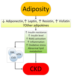
Obesity results in complex metabolic abnormalities which have wide-ranging effects on diseases affecting the kidneys. The exact mechanisms whereby obesity may worsen or cause CKD remain unclear. The fact that most obese individuals never develop CKD, and the distinction of up to as many as 25% of obese individuals as “metabolically healthy” suggests that increased weight alone is not sufficient to induce kidney damage [31] Some of the deleterious renal consequences of obesity may be mediated by downstream comorbid conditions such as diabetes mellitus or hypertension, but there are also effects of adiposity which could impact the kidneys directly, induced by the endocrine activity of the adipose tissue via production of (among others) adiponectin, [32] leptin [33] and resistin [34] (Figure 1). These include the development of inflammation, [35] oxidative stress, [36] abnormal lipid metabolism, [37] activation of the renin-angiotensin-aldosterone system,[38] and increased production of insulin and insulin resistance [39,40].

Figure 1. Putative mechanisms of action whereby obesity causes chronic kidney disease

Figure 1. Putative mechanisms of action whereby obesity causes chronic kidney disease

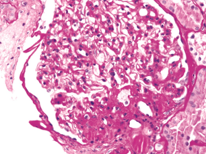
These various effects result in specific pathologic changes in the kidneys [41] which could underlie the higher risk of CKD seen in observational studies. These include ectopic lipid accumulation [42] and increased deposition of renal sinus fat, [43,44] the development of glomerular hypertension and increased glomerular permeability caused by hyperfiltration-related glomerular filtration barrier injury, [45] and ultimately the development of glomerulomegaly, [46] and focal or segmental glomerulosclerosis [41] (Figure 2). The incidence of the so-called obesity-related glomerulopathy (ORG) has increased ten-fold between 1986 and 2000. [41] Importantly, ORG often presents along with pathophysiologic processes related to other conditions or advanced age, conspiring to result in more accentuated kidney damage in patients with high blood pressure [47] or in the elderly. [14-39].

Figure 2. Obesity-related perihilar focal segmental glomerulosclerosis on a background of glomerulomegaly. Periodic Acid-Schiff stain, original magnification 400x

Figure 2. Obesity-related perihilar focal segmental glomerulosclerosis on a background of glomerulomegaly. Periodic Acid-Schiff stain, original magnification 400x

Obesity is associated with a number of risk factors contributing to the higher incidence and prevalence of nephrolithiasis. Higher body weight is associated with lower urine pH [48] and increased urinary oxalate,[49] uric acid, sodium and phosphate excretion [50] Diets richer in protein and sodium may lead to a more acidic urine and decrease in urinary citrate, also contributing to kidney stone risk. The insulin resistance characteristic of obesity may also predispose to nephrolithiasis [51] through its impact on tubular Na-H exchanger [52] and ammoniagenesis, [53] and the promotion of an acidic milieu [54]. Complicating the picture is the fact that some weight loss therapies result in a worsening, rather than an improvement in the risk for kidney stone formation; e.g. gastric surgery can lead to a substantial increase in enteral oxalate absorption and enhanced risk of nephrolithiasis [55].

The mechanisms behind the increased risk of kidney cancers observed in obese individuals are less well characterized. Insulin resistance, and the consequent chronic hyperinsulinemia and increased production of insulin-like growth factor 1 and numerous complex secondary humoral effects may exert stimulating effects on the growth of various types of tumor cells [56] More recently, the endocrine functions of adipose tissue, [57] its effects on immunity, [58] and the generation of an inflammatory milieu with complex effects on cancers 59, 60] have emerged as additional explanations.

Obesity in patients with advanced kidney disease: The need for a nuanced approach

Considering the above evidence about the overwhelmingly deleterious effects of obesity on various disease processes, it is seemingly counterintuitive that obesity has been consistently associated with lower mortality rates in patients with advanced CKD [19,61] and ESRD [62,63] Similar “paradoxical” associations have also been described in other populations, such as in patients with congestive heart failure [64], chronic obstructive pulmonary disease [65], rheumatoid arthritis, [66] and even in old individuals [67]. It is possible that the seemingly protective effect of a high BMI is the result of the imperfection of BMI as a measure of obesity, as it does not differentiate the effects of adiposity from those of higher non-adipose tissue. Indeed, studies that separated the effects of a higher waist circumference from those of higher BMI showed a reversal of the inverse association with mortality [23,24]. Higher muscle mass has also been shown to explain at least some of the positive effects attributed to elevated BMI [63, 68]. However, there is also evidence to suggest that higher adiposity, especially subcutaneous (non-visceral) fat, may also be associated with better outcomes in ESRD patients [62] Such benefits may indeed be present in patients who have very low short term life expectancy, such as most ESRD patients [69]. Indeed, some studies that examined the association of BMI with time-dependent survival in ESRD have shown a marked contrast between protective short term effects vs. deleterious longer term effects of higher BMI [70]. There are several putative short term benefits that higher body mass could portend, especially to sicker individuals. These include a benefit from the better nutritional status typically seen in obese individuals, and which provides better protein and energy reserves in the face of acute illness, and a higher muscle mass with enhanced antioxidant capacity [63] and lower circulating actin and higher plasma gelsolin levels, [71] which are associated with better outcomes. Other hypothetically beneficial characteristics of obesity include a more stable hemodynamic status with mitigation of stress responses and heightened sympathetic and renin-angiotensin activity [72] increased production of adiponectines [73] and soluble tumor necrosis factor alfa receptors[ 74] by adipose tissue neutralizing the adverse effects of tumor necrosis factor alfa; enhanced binding of circulating endotoxins [75] by the characteristically higher cholesterol levels seen in obesity; and sequestration of uremic toxins by adipose tissue [76].

Potential interventions for management of obesity

Obesity engenders kidney injury via direct mechanisms through deranged synthesis of various adipose tissue cytokines with nephrotoxic potential, as well as indirectly by triggering diabetes and hypertension, i.e. two conditions that rank among the strongest risk factors for CKD. Perhaps due to the survival advantage of obesity in CKD, the prevalence of end stage kidney disease is on the rise both in the USA [77] and in Europe [78]. Strategies for controlling the obesity related CKD epidemic at population level and for countering the evolution of CKD toward kidney failure in obese patients represent the most tantalizing task that today’s health planners, health managers and nephrologists face.

Countering CKD at population level

Calls for public health interventions in the community to prevent and treat CKD at an early stage have been made by major renal associations, including the International Society of Nephrology (ISN), International Federation of the Kidney Foundation (IFKF), the European renal association (ERA-EDTA) and various national societies. In the USA, Healthy People 2020, a program that sets 10-year health targets for health promotion and prevention goals, focuses both on CKD and obesity. Surveys to detect obese patients, particularly those with a high risk of CKD (e.g. hypertensive and/or diabetic obese people) and those receiving suboptimal care to inform these patients of the potential risk for CKD they are exposed to, is the first step towards developing public health interventions. Acquiring evidence that current interventions to reduce CKD risk in the obese are efficacious and deployable, is an urgent priority to set goals and means for risk modification. Appropriate documentation of existing knowledge distilling the risk and the benefits of primary and secondary prevention interventions in obese people, and new trials in this population to fill knowledge gaps (see below) are needed. Finally, surveillance programs that monitor progress on the detection of at-risk individuals and the effectiveness of prevention programs being deployed [79] constitute the third, fundamental element for establishing efficacious CKD prevention plans at population level.

A successful surveillance system for CKD has already been implemented in some places such as the United Kingdom (UK [80]. A campaign to disseminate and apply K-DOQI CKD guidelines in primary care within the UK National Health Service was launched. This progressively increased the adoption of K-DOQI guidelines and, also thanks to specific incentives for UK general physicians to detect CKD, led to an impressive improvement in the detection and care of CKD, i.e. better control of hypertension and increased use of angiotensin-converting enzyme and angiotensin receptor blockers [80]. This system may serve as a platform to improve the prevention of obesity-related CKD. Campaigns aiming at reducing the obesity burden are now at center stage worldwide and are strongly recommended by the WHO and it is expected that these campaigns will reduce the incidence of obesity-related complications, including CKD. However obesity-related goals in obese CKD patients remain vaguely formulated, largely because of the paucity of high-level evidence intervention studies to modify obesity in CKD patients [81].

Prevention of CKD progression in obese people with CKD

Observational studies in metabolically healthy obese subjects show that the obese phenotype unassociated with metabolic abnormalities per se predicts a higher risk for incident CKD [82] suggesting that obesity per se may engender renal dysfunction and kidney damage even without diabetes or hypertension (vide supra). In overweight or obese diabetic patients, a lifestyle intervention including caloric restriction and increased physical activity compared with a standard follow up based on education and support to sustain diabetes treatment reduced the risk for incident CKD by 30%, although it did not affect the incidence of cardiovascular events [83]. Such a protective effect was partly due to reductions in body weight, HbA1c, and systolic BP. No safety concerns regarding kidney-related adverse events were seen [83]. In a recent meta-analysis collating experimental studies in obese CKD patients, interventions aimed at reducing body weight showed coherent reductions in blood pressure, glomerular hyper-filtration and proteinuria [81]. A thorough post-hoc analysis of the REIN study showed that the nephron-protective effect of ACE inhibition in proteinuric CKD patients was maximal in obese CKD patients, but minimal in CKD patients with normal or low BMI [84]. Of note, bariatric surgical intervention have been suggested for selected CKD and ESRD patients including dialysis patients who are waitlisted for kidney transplantation [85-87].

Globally, these experimental findings provide a proof of concept for the usefulness of weight reduction and ACE inhibition interventions in the treatment of CKD in the obese. Studies showing a survival benefit of increased BMI in CKD patients, however, remain to be explained [88]. These findings limit our ability to make strong recommendations about the usefulness and the safety of weight reduction among individuals with more advanced stages of CKD. Lifestyle recommendations to reduce body weight in obese people at risk for CKD and in those with early CKD appear justified, particularly recommendations for the control of diabetes and hypertension. As the independent effect of obesity control on the incidence and progression of CKD is difficult to disentangle from the effects of hypertension and type 2 diabetes, recommendation of weight loss in the minority of metabolically healthy, non-hypertensive obese patients remains unwarranted. These considerations suggest that a therapeutic approach to overweight and obesity in patients with advanced CKD or other significant comorbid conditions has to be pursued carefully, with proper considerations of the expected benefits and potential complications of weight loss over the life span of the individual patient.

Conclusions

The worldwide epidemic of obesity affects the Earth’s population in many ways. Diseases of the kidneys, including CKD, nephrolithiasis and kidney cancers are among the more insidious effects of obesity, but which nonetheless have wide ranging deleterious consequences, ultimately leading to significant excess morbidity and mortality and excess costs to individuals and the entire society. Population-wide interventions to control obesity could have beneficial effects in preventing the development, or delaying the progression of CKD. It is incumbent upon the entire healthcare community to devise long-ranging strategies towards improving the understanding of the links between obesity and kidney diseases, and to determine optimal strategies to stem the tide. The 2017 World Kidney Day is an important opportunity to increase education and awareness to that end.

References

  • GBD 2013 Risk Factors Collaborators, Forouzanfar MH, Alexander L, Anderson HR, Bachman VF, et al. (2015) Global, regional, and national comparative risk assessment of 79 behavioural, environmental and occupational, and metabolic risks or clusters of risks in 188 countries, 1990-2013: a systematic analysis for the Global Burden of Disease Study 2013. Lancet 386: 2287-2323. [crossref]
  • Flegal KM, Kruszon-Moran D, Carroll MD, Fryar CD, Ogden CL1 (2016) Trends in Obesity Among Adults in the United States, 2005 to 2014. JAMA 315: 2284-2291. [crossref]
  • Cattaneo A, Monasta L, Stamatakis E, Lioret S, Castetbon K, et al. (2010) Overweight and obesity in infants and pre-school children in the European Union: a review of existing data. Obes Rev 11: 389-398. [crossref]
  • Olaya B1,2, Moneta MV3,4, Pez O5, et al. (2015) Country-level and individual correlates of overweight and obesity among primary school children: a cross-sectional study in seven European countries. BMC Public Health 15: 475. [crossref]
  • Subramanian SV, Perkins JM, Özaltin E, Davey Smith G (2011) Weight of nations: a socioeconomic analysis of women in low- to middle-income countries. Am J Clin Nutr 93: 413-421. [crossref]
  • Tsujimoto T, Sairenchi T, Iso H, Irie F, Yamagishi K, et al. (2014) The dose-response relationship between body mass index and the risk of incident stage >/=3 chronic kidney disease in a general japanese population: the Ibaraki prefectural health study (IPHS). J Epidemiol 24: 444-45.
  • Elsayed EF, Sarnak MJ, Tighiouart H, Griffith JL, Kurth T, et al. (2008) Waist-to-hip ratio, body mass index, and subsequent kidney disease and death. Am J Kidney Dis 52: 29-38. [crossref]
  • Pinto-Sietsma SJ, Navis G, Janssen WM, de Zeeuw D, Gans RO, et al. (2003) A central body fat distribution is related to renal function impairment, even in lean subjects. Am J Kidney Dis 41: 733-741. [crossref]
  • Foster MC, Hwang SJ, Larson MG, Lichtman JH, Parikh NI, et al. (2008) Overweight, obesity, and the development of stage 3 CKD: the Framingham Heart Study. Am J Kidney Dis 52: 39-48. [crossref]
  • Kramer H, Luke A, Bidani A, Cao G, Cooper R, et al. (2005) Obesity and prevalent and incident CKD: the Hypertension Detection and Follow-Up Program. Am J Kidney Dis 46: 587-594. [crossref]
  • Chang A, Van HL, Jacobs DR, Liu K, Muntner P, et al. (2013) Lifestyle-related factors, obesity, and incident microalbuminuria: the CARDIA (Coronary Artery Risk Development in Young Adults) study. Am J Kidney Dis 62:267-275.
  • Ejerblad E, Fored CM, Lindblad P, Fryzek J, McLaughlin JK, et al. (2006) Obesity and risk for chronic renal failure. J Am Soc Nephrol 17: 1695-1702. [crossref]
  • Gelber RP, Kurth T, Kausz AT, Manson JE, Buring JE, et al. (2005) Association between body mass index and CKD in apparently healthy men. Am J Kidney Dis 46: 871-880. [crossref]
  • Lu JL, Molnar MZ, Naseer A2, Mikkelsen MK, Kalantar-Zadeh K3, et al. (2015) Association of age and BMI with kidney function and mortality: a cohort study. Lancet Diabetes Endocrinol 3: 704-714. [crossref]
  • Munkhaugen J, Lydersen S, Widerøe TE, Hallan S (2009) Prehypertension, obesity, and risk of kidney disease: 20-year follow-up of the HUNT I study in Norway. Am J Kidney Dis 54: 638-646. [crossref]
  • Iseki K, Ikemiya Y, Kinjo K, Inoue T, Iseki C, et al. (2004) Body mass index and the risk of development of end-stage renal disease in a screened cohort. Kidney Int 65: 1870-1876. [crossref]
  • Vivante A, Golan E, Tzur D, Leiba A, Tirosh A, et al. (2012) Body mass index in 1.2 million adolescents and risk for end-stage renal disease. Arch Intern Med 172: 1644-1650. [crossref]
  • Hsu CY, McCulloch CE, Iribarren C, Darbinian J, Go AS (2006) Body mass index and risk for end-stage renal disease. Ann Intern Med 144: 21-28. [crossref]
  • Lu JL, Kalantar-Zadeh K2, Ma JZ3, Quarles LD, Kovesdy CP4 (2014) Association of body mass index with outcomes in patients with CKD. J Am Soc Nephrol 25: 2088-2096. [crossref]
  • Thoenes M, Reil JC, Khan BV, Bramlage P, Volpe M, et al. (2009) Abdominal obesity is associated with microalbuminuria and an elevated cardiovascular risk profile in patients with hypertension. Vasc Health Risk Manag 5: 577-585. [crossref]
  • Kramer H, Gutiérrez OM2, Judd SE3, Muntner P4, Warnock DG5, et al. (2016) Waist Circumference, Body Mass Index, and ESRD in the REGARDS (Reasons for Geographic and Racial Differences in Stroke) Study. Am J Kidney Dis 67: 62-69. [crossref]
  • Foster MC, Hwang SJ, Massaro JM, Hoffmann U, DeBoer IH, et al. (2011) Association of subcutaneous and visceral adiposity with albuminuria: the Framingham Heart Study. Obesity (Silver Spring) 19:1284-1289.
  • Postorino M, Marino C, Tripepi G, Zoccali C; CREDIT (Calabria Registry of Dialysis and Transplantation) Working Group (2009) Abdominal obesity and all-cause and cardiovascular mortality in end-stage renal disease. J Am Coll Cardiol 53: 1265-1272. [crossref]
  • Kovesdy CP, Czira ME, Rudas A, Ujszaszi A, Rosivall L, et al. (2010) Body mass index, waist circumference and mortality in kidney transplant recipients. Am J Transplant 10: 2644-2651. [crossref]
  • Scales CD Jr, Smith AC, Hanley JM, Saigal CS; Urologic Diseases in America Project (2012) Prevalence of kidney stones in the United States. Eur Urol 62: 160-165. [crossref]
  • Curhan GC, Willett WC, Rimm EB, Speizer FE, Stampfer MJ (1998) Body size and risk of kidney stones. J Am Soc Nephrol 9: 1645-1652. [crossref]
  • Taylor EN, Stampfer MJ, Curhan GC (2005) Obesity, weight gain, and the risk of kidney stones. JAMA 293: 455-462. [crossref]
  • Bhaskaran K, Douglas I2, Forbes H2, dos-Santos-Silva I2, Leon DA2, et al. (2014) Body-mass index and risk of 22 specific cancers: a population-based cohort study of 5·24 million UK adults. Lancet 384: 755-765. [crossref]
  • Arnold M, Pandeya N2, Byrnes G3, Renehan AG4, Stevens GA5, et al. (2015) Global burden of cancer attributable to high body-mass index in 2012: a population-based study. Lancet Oncol 16: 36-46. [crossref]
  • Renehan AG, Tyson M, Egger M, Heller RF, Zwahlen M (2008) Body-mass index and incidence of cancer: a systematic review and meta-analysis of prospective observational studies. Lancet 371:569-578.
  • Blüher M (2010) The distinction of metabolically ‘healthy’ from ‘unhealthy’ obese individuals. Curr Opin Lipidol 21: 38-43. [crossref]
  • Sharma K (2009) The link between obesity and albuminuria: adiponectin and podocyte dysfunction. Kidney Int 76: 145-148. [crossref]
  • Wolf G, Ziyadeh FN (2006) Leptin and renal fibrosis. Contrib Nephrol 151: 175-183. [crossref]
  • Ellington AA, Malik AR, Klee GG, Turner ST, Rule AD, et al. (2007) Association of plasma resistin with glomerular filtration rate and albuminuria in hypertensive adults. Hypertension 50:708-714.
  • Bastard JP, Maachi M, Lagathu C, Kim MJ, Caron M, et al. (2006) Recent advances in the relationship between obesity, inflammation, and insulin resistance. Eur Cytokine Netw 17:4-12, 2006
  • Furukawa S, Fujita T, Shimabukuro M, Iwaki M, Yamada Y, et al. (2004) Increased oxidative stress in obesity and its impact on metabolic syndrome. J Clin Invest 114: 1752-1761. [crossref]
  • Ruan XZ, Varghese Z, Moorhead JF (2009) An update on the lipid nephrotoxicity hypothesis. Nat Rev Nephrol 5: 713-721. [crossref]
  • Ruster C, Wolf G (2013) The role of the renin-angiotensin-aldosterone system in obesity-related renal diseases. 33:44-53, 2013
  • Oterdoom LH, de Vries AP, Gansevoort RT, de Jong PE, Gans RO, et al. (2007) Fasting insulin modifies the relation between age and renal function. Nephrol Dial Transplant 22: 1587-1592. [crossref]
  • Reaven GM (1988) Banting lecture 1988. Role of insulin resistance in human disease. Diabetes 37: 1595-1607. [crossref]
  • Kambham N, Markowitz GS, Valeri AM, Lin J, D’Agati VD (2001) Obesity-related glomerulopathy: an emerging epidemic. Kidney Int 59: 1498-1509. [crossref]
  • de Vries AP, Ruggenenti P, Ruan XZ, Praga M, Cruzado JM, et al. (2014) Fatty kidney: emerging role of ectopic lipid in obesity-related renal disease. Lancet Diabetes Endocrinol 2:417-426.
  • Foster MC, Hwang SJ, Porter SA, Massaro JM, Hoffmann U, et al. (2011) Fatty kidney, hypertension, and chronic kidney disease: the Framingham Heart Study. Hypertension 58:784-790.
  • Henegar JR, Bigler SA, Henegar LK, Tyagi SC, Hall JE (2001) Functional and structural changes in the kidney in the early stages of obesity. J Am Soc Nephrol 12: 1211-1217. [crossref]
  • Knight SF, Quigley JE, Yuan J, Roy SS, Elmarakby A, et al. (2008) Endothelial dysfunction and the development of renal injury in spontaneously hypertensive rats fed a high-fat diet. Hypertension 51: 352-359. [crossref]
  • Tsuboi N, Utsunomiya Y, Kanzaki G, Koike K, Ikegami M, et al. (2012) Low glomerular density with glomerulomegaly in obesity-related glomerulopathy. Clin J Am Soc Nephrol 7: 735-741. [crossref]
  • Ribstein J, du Cailar G, Mimran A (1995) Combined renal effects of overweight and hypertension. Hypertension 26: 610-615. [crossref]
  • Maalouf NM, Sakhaee K, Parks JH, Coe FL, Adams-Huet B, et al. (2004) Association of urinary pH with body weight in nephrolithiasis. Kidney Int 65: 1422-1425. [crossref]
  • Lemann J, Jr., Pleuss JA, Worcester EM, Hornick L, Schrab D, et al. (1996) Urinary oxalate excretion increases with body size and decreases with increasing dietary calcium intake among healthy adults. Kidney Int 49:200-208.
  • Siener R, Glatz S, Nicolay C, Hesse A (2004) The role of overweight and obesity in calcium oxalate stone formation. Obes Res 12: 106-113. [crossref]
  • Taylor EN, Stampfer MJ, Curhan GC (2005) Diabetes mellitus and the risk of nephrolithiasis. Kidney Int 68: 1230-1235. [crossref]
  • Klisic J, Hu MC, Nief V, Reyes L, Fuster D, Moe OW, et al. (2002) Insulin activates Na(+)/H(+) exchanger 3: biphasic response and glucocorticoid dependence. Am J Physiol Renal Physiol 283: F532-F539.
  • Chobanian MC, Hammerman MR (1987) Insulin stimulates ammoniagenesis in canine renal proximal tubular segments. Am J Physiol 253: F1171-F1177.
  • Daudon M, Lacour B, Jungers P (2006) Influence of body size on urinary stone composition in men and women. Urol Res 34: 193-199. [crossref]
  • Sinha MK, Collazo-Clavell ML, Rule A, Milliner DS, Nelson W, et al. (2007) Hyperoxaluric nephrolithiasis is a complication of Roux-en-Y gastric bypass surgery. Kidney Int 72:100-107.
  • Calle EE, Kaaks R (2004) Overweight, obesity and cancer: epidemiological evidence and proposed mechanisms. Nat Rev Cancer 4: 579-591. [crossref]
  • Dalamaga M, Diakopoulos KN, Mantzoros CS (2012) The role of adiponectin in cancer: a review of current evidence. Endocr Rev 33: 547-594. [crossref]
  • Lamas O, Marti A, Martínez JA (2002) Obesity and immunocompetence. Eur J Clin Nutr 56 Suppl 3: S42-45. [crossref]
  • Lim C, Savan R (2014) The role of the IL-22/IL-22R1 axis in cancer. Cytokine Growth Factor Rev 25: 257-271. [crossref]
  • Grivennikov SI, Greten FR, Karin M (2010) Immunity, inflammation, and cancer. Cell 140: 883-899. [crossref]
  • Kovesdy CP, Anderson JE, Kalantar-Zadeh K (2007) Paradoxical association between body mass index and mortality in men with CKD not yet on dialysis. Am J Kidney Dis 49: 581-591. [crossref]
  • Kalantar-Zadeh K, Kuwae N, Wu DY, Shantouf RS, Fouque D, et al. (2006) Associations of body fat and its changes over time with quality of life and prospective mortality in hemodialysis patients. Am J Clin Nutr 83: 202-210. [crossref]
  • Beddhu S, Pappas LM, Ramkumar N, Samore M (2003) Effects of body size and body composition on survival in hemodialysis patients. J Am Soc Nephrol 14: 2366-2372. [crossref]
  • Curtis JP, Selter JG, Wang Y, Rathore SS, Jovin IS, et al. (2005) The obesity paradox: body mass index and outcomes in patients with heart failure. Arch Intern Med 165: 55-61. [crossref]
  •  Wilson DO, Rogers RM, Wright EC, Anthonisen NR (1989) Body weight in chronic obstructive pulmonary disease. The National Institutes of Health Intermittent Positive-Pressure Breathing Trial. Am Rev Respir Dis 139:1435-1438.
  • Escalante A, Haas RW, del Rincón I (2005) Paradoxical effect of body mass index on survival in rheumatoid arthritis: role of comorbidity and systemic inflammation. Arch Intern Med 165: 1624-1629. [crossref]
  • Kalantar-Zadeh K, Kilpatrick RD, Kuwae N, Wu DY (2005) Reverse epidemiology: a spurious hypothesis or a hardcore reality? Blood Purif 23: 57-63. [crossref]
  • Noori N, Kopple JD, Kovesdy CP, Feroze U, Sim JJ, et al. (2008) Mid-arm muscle circumference and quality of life and survival in maintenance hemodialysis patients. Clin J Am Soc Nephrol 5:2258-2268.
  • Dekker FW, de Mutsert R, van Dijk PC, Zoccali C, Jager KJ (2008) Survival analysis: time-dependent effects and time-varying risk factors. Kidney Int 74: 994-997. [crossref]
  • Snyder JJ, Foley RN, Gilbertson DT, Vonesh EF, Collins AJ (2003) Body size and outcomes on peritoneal dialysis in the United States. Kidney Int 64: 1838-1844. [crossref]
  • Lee PS, Sampath K, Karumanchi SA, Tamez H, Bhan I, et al. (2009) Plasma gelsolin and circulating actin correlate with hemodialysis mortality. J Am Soc Nephrol 20: 1140-1148. [crossref]
  • Horwich TB, Fonarow GC, Hamilton MA, MacLellan WR, Woo MA, et al. (2001) The relationship between obesity and mortality in patients with heart failure. J Am Coll Cardiol 38: 789-795. [crossref]
  • Stenvinkel P, Marchlewska A, Pecoits-Filho R, Heimburger O, Zhang Z, et al. (2004) Adiponectin in renal disease: relationship to phenotype and genetic variation in the gene encoding adiponectin. Kidney Int 65:274-28.
  • Mohamed-Ali V, Goodrick S, Bulmer K, Holly JM, Yudkin JS, Coppack SW (1999) Production of soluble tumor necrosis factor receptors by human subcutaneous adipose tissue in vivo. Am J Physiol 277: E971-E975.
  • Rauchhaus M, Coats AJ, Anker SD (2000) The endotoxin-lipoprotein hypothesis. Lancet 356: 930-933. [crossref]
  • Jandacek RJ, Anderson N, Liu M, Zheng S, Yang Q, et al. (2005) Effects of yo-yo diet, caloric restriction, and olestra on tissue distribution of hexachlorobenzene. Am J Physiol Gastrointest Liver Physiol 288: G292-299. [crossref]
  • Kramer HJ, Saranathan A, Luke A, Durazo-Arvizu RA, Guichan C, et al. (2006) Increasing body mass index and obesity in the incident ESRD population. J Am Soc Nephrol 17: 1453-1459. [crossref]
  • Postorino M, Mancini E, D’Arrigo G, Marino C, Vilasi A, et al. (2016) Body mass index trend in haemodialysis patients: the shift of nutritional disorders in two Italian regions. Nephrol Dial Transplant.
  • Chimeddamba O, Peeters A, Walls HL, et al. (2015) Noncommunicable Disease Prevention and Control in Mongolia: A Policy Analysis. BMC Public Health 15: 660. [crossref]
  • O’Donoghue DJ, Stevens PE (2012) A decade after the KDOQI CKD/guidelines: a perspective from the United Kingdom. Am J Kidney Dis 60: 740-742. [crossref]
  • Bolignano D, Zoccali C (2013) Effects of weight loss on renal function in obese CKD patients: a systematic review. Nephrol Dial Transplant 28 Suppl 4: iv82-iv98.
  • Chang Y, Ryu S, Choi Y, Zhang Y, Cho J, et al. (2016) Metabolically Healthy Obesity and Development of Chronic Kidney Disease: A Cohort Study. Ann Intern Med 164:305-312.
  • Wing RR, Bolin P, Brancati FL, Bray GA, Clark JM, et al. (2013) Cardiovascular effects of intensive lifestyle intervention in type 2 diabetes. N Engl J Med 369:145-154.
  • Mallamaci F, Ruggenenti P, Perna A, Leonardis D, Tripepi R, et al. (2011) ACE inhibition is renoprotective among obese patients with proteinuria. J Am Soc Nephrol 22: 1122-1128. [crossref]
  • Friedman AN, Wolfe B2 (2016) Is Bariatric Surgery an Effective Treatment for Type II Diabetic Kidney Disease? Clin J Am Soc Nephrol 11: 528-535. [crossref]
  • Chang AR, Chen Y2, Still C3, Wood GC3, Kirchner HL4, et al. (2016) Bariatric surgery is associated with improvement in kidney outcomes. Kidney Int 90: 164-171. [crossref]
  • Jamal MH, Corcelles R, Daigle CR, Rogula T, Kroh M, et al. (2015) Safety and effectiveness of bariatric surgery in dialysis patients and kidney transplantation candidates. Surg Obes Relat Dis 11:419-423.
  • Ahmadi SF, Zahmatkesh G, Ahmadi E, Streja E, Rhee CM. et al. (2015) Association of Body Mass Index with Clinical Outcomes in Non-Dialysis-Dependent Chronic Kidney Disease: A Systematic Review and Meta-Analysis. Cardiorenal Med 6:37-49.

Epidemiological, Diagnostic, Therapeutic and Prognosis Aspects of Soft Tissues Sarcomas at Dakar Cancer Institute

DOI: 10.31038/CST.2016125

Abstract

Purpose: The purpose of this work was to specify the epidemiological profile of soft tissue sarcomas at the Dakar Cancer Institute and to evaluate their diagnostic and therapeutic management.

Patients and Methods: This was a prospective study involving 40 patients with soft tissue sarcomas treated at the Dakar Cancer Institute during a 3-year period from August 1, 2009 to July 30, 2012.

Results: Soft tissue sarcoma represents 0.4% of cancer cases at the Dakar Cancer Institute. The average age was 41.2 years. The sex ratio was 0.6. There was a family history of cancer in 5 patients. History of type 1 neurofibromatosis was found in 2 patients and irradiation for pelvic cancer in 1 patient. The average time for consultation was 17.2 months. The most frequent localizations were the thigh (17.5%) and the shoulder (12.5%). The mean lesion size was 14.3 cm. Imaging showed metastasis in 9 patients (24.4%) and extension to neighboring organs in 17 patients (42.5%). The most common histological type was Rhabdomyosarcoma (20%) followed by Dermatofibrosarcoma (17.5%). High grade sarcoma was found in 5 patients (12.5%). Excision was the most common type of surgery (30%). Chemotherapy was performed in 13 patients (32.5%) including 8 cases of palliative chemotherapies. Radiotherapy was performed in 5 patients (12.5%). The goal was neo adjuvant in 2 patients, adjuvant in 1 patient and palliative in 2 patients. After a 52-month follow-up period, 1 patient presented a local recurrence and 18 patients died.

Keywords

sarcoma; surgery; chemotherapy; radiotherapy; recurrence.

Introduction

Soft tissue sarcomas represent all malignant tumors with mesenchymal differentiation in all extra-bone tissues with the exception of lymph nodes and glial tissue [1]. It is a rare cancer. The prognosis depends on the initial stage, the histological type, the grade, the location and the initial treatment [2]. The objective of this work was to specify the epidemiological profile of soft tissue sarcomas at the Dakar Cancer Institute and to evaluate their diagnostic and therapeutic management.

Patients and methods

We carried out a descriptive prospective study involving 40 patients with soft-tissue sarcomas treated at the Dakar Cancer Institute for a period of 3 years from August 1, 2009 to July 30, 2012.

Results

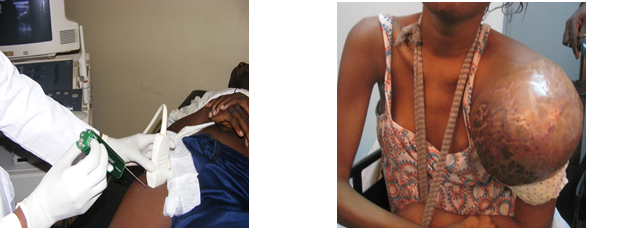
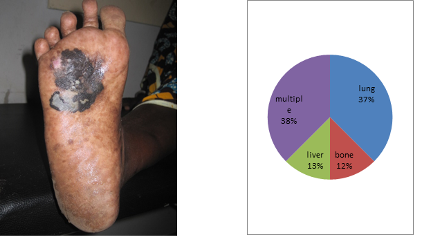
Soft tissue sarcoma represents 4 patients per 1000 cases at the Dakar Cancer Institute. The mean age was 41.2 years with extremes of 9 and 81 years. There was a female predominance with a sex ratio of 0.6. There was a family history of cancer in 5 patients. History of type 1 neurofibromatosis was found in 2 patients and irradiation for pelvic cancer in 1 patient. Mean follow-up was 17.2 months after onset of symptomatology. The most frequent localizations were the thigh (n = 7) [Figure 1] and the shoulder (n = 5) [Figure 2]. The size of the lesions varied from 5 to 35 cm with an average of 14.3 cm. The majority of patients (n = 37) had a single lesion. The initial lesion was bifocal in 2 patients and 1 patient had 4 lesions. The imaging assessment used CT for 20 patients, MRI for 11 and ultrasound in 6 cases. It showed the presence of metastasis in 9 patients (24.4%) at the time of diagnosis and an extension to neighboring organs in 17 patients. Surgical biopsy was the most common modality (26 cases, 67%) followed by ultrasound guided biopsy (6 cases, 15%). The most common histological type was Rhabdomyosarcoma (20%) followed by Dermatofibrosarcoma (17.5%) [Table 1]. A high-grade sarcoma was found in 19 patients (47.5%). Surgery was performed in 24 patients (60%). Wide resection was the most common type of surgery (n = 12). Chemotherapy was performed in 13 patients including 8 palliative chemotherapies. The Adriamycin-Cisplatin chemotherapy protocol was the most widely used (n = 11). Radiotherapy was performed in 5 patients. The goal was neo adjuvant in 2 patients, adjuvant in 1 patient and palliative in 2 patients. After a 52-month follow-up period, 2 patients had local recurrence and 18 patients died [Table 2].

Figure 1. deep limb localization Figure 2. shoulder localization of a rhabdomyosarcoma

Fig1.deep limb localization                    Fig2.shoulder localization of a rhabdomyosarcoma

Table 1. Different histologic types of STS

Histologic  type Frequencies Percentage (%)
Angiosarcoma 1 2,5
Chondrosarcomea 1 2,5
Dermatofibrosarcoma protuberans 7 17,5
Fibrosarcoma 5 12,5
Atypical Fibroxantoma 1 2,5
Atypical Hémangioendothelioma 1 2,5
Endemic  Kaposi Sarcoma 1 2,5
Leimyosarcoma 1 2,5
Liposarcoma 2 5,0
Neurosarcoma 3 7,5
Rhabdomyosarcoma 8 20
Fusiform Cells Sarcoma 3 7,5
Phyllod Sarcoma 1 2,5
Pleomorph Sarcoma 2 5
Synovialosarcoma 3 7,5
Total 40 100

Table 2. Results of follow up

Evolution Frequencies Percentage(%)
Décès 18 45
Récidive 2 5
Patients en cours de traitement 7 17,5
Patients traités  sans récidive 13 32,5
Total 40 100

Discussion

Soft tissue sarcoma (STS) accounts for 0.5-1% of malignant tumors in adults. Their incidence is estimated at 30 cases per million inhabitants [1]. The frequency of soft tissue sarcomas increases in adults with age, and half of the patients are older than 50 years [2]. There is a slight male predominance especially after 60 years [3]. Several genetic factors including Gardener syndrome, Li Fraumeni syndrome and type 1 neurofibromatosis predispose to the onset of STS [4]. The sarcomas of the limbs represent approximately 60% of the sites, 45% of which are in the lower limbs [5, 6]. They are characterized by their large sizes [7,8]. It is a very lymphophilic tumor in the higher grades, in the associated forms in particular carcinosarcomas and in certain histological types such as synovialosarcomas and rhabdomyosarcomas [9]. One patient in four is metastatic at the time of diagnosis and secondary lesions are predominantly located at lymph nodes and lungs [10, 11]. MRI is the main method of imaging in STS. In association with PET-SCAN, its role is increased in the diagnosis of recurrences [12, 13]. The scanner is only used in the local assessment if MRI is contraindicated. Ultrasound is of limited utility except in the superficial small masses. The delay of consultation, very long in case of limited resources are bad prognoses factors [2]. The most frequent histological types are liposarcomas, leiomyosarcomas and malignant histiocytofibromas [14].

The treatment need a multidisciplinary approach and is better considered in a reference center [2]. Surgery regardless of location is the basic treatment. It is more functional and more conservative thanks to multidisciplinary teams and the development of new techniques like the isolated perfusion of the limb. The objective is to minimize functional sequelae and amputation rates [15, 16]. Chemotherapy occupies an important place in neo adjuvant situation in the locally advanced sarcomas and the high grades [2]. The question of adjuvant chemotherapy is generally left to the appreciation of multidisciplinary consultations [17]. Radiotherapy, in particular radio chemotherapy, has produced encouraging results in the neo-adjuvant period for locally advanced tumors [18]. The surgical excision followed by complementary radiotherapy is the standard loco regional treatment of limbs and operable and localized STS [19]. Abstinence from adjuvant radiotherapy is possible for superficial or strictly intramuscular tumors for which surgery showed clear margins [20].

The best survival rate, obtained in the reference centers, is of the order of 60 to 70% at 5 years in the early stages and of 2 months in the advanced stages [2, 11].

Conclusion

Soft tissue sarcoma is a rare cancer. It occurs sporadically or on specific genetic situations. Histological forms and grade determine susceptibility to treatment and prognosis. High grade and long delay are the cause of poor prognosis in limited resources situations. The main goal is first conservative and functional treatment. It needs multidisciplinary approach associating mainly surgery and radiotherapy. High grade sarcomas and metastatic stages have very poor prognosis.

Conflict of interest: None declared.

Source of funding: None.

Consent: Written informed consent was obtained from the patients.

Acknowledgments: None.

Author Contribution: Ka conceived this presentation while Diouf and Dem participated in quality control of this manuscript. All authors read and approved the final manuscript.

References

  • Fletcher DM, Unnik K and Mertens F (2002) World Health Organization classification of tumors. Pathology and genetics of tumors of soft tissue and bone. Lyon: IARC Press.
  • Honoré C, Méeus P, Stoeckle E, Bonvalot S (2015) Soft tissue sarcoma in France in 2015: Epidemiology, classification and organization of clinical care. J Visc Surg 152: 223-230. [crossref]
  • Jørgensen PH, Lausten GS, Pedersen AB (2016) The Danish Sarcoma Database. Clin Epidemiol 8: 685-690. [crossref]
  • Ducimetière F, Lurkin A, Ranchère-Vince D, Decouvelaere AV, Isaac S, et al. (2010) [Incidence rate, epidemiology of sarcoma and molecular biology. Preliminary results from EMS study in the Rhône-Alpes region].Bull Cancer 97: 629-641. [crossref]
  • Chang AE, Rosenberg SA (1988) Clinical evaluation and treatment of soft tissue tumors. In: Enzinger FM, Weiss SW. Soft tissue tumors (2nd ed) St Louis, Toronto, London: CV Mosby Co 19–42.
  • Coindre JM, Terrier P, Bui NB, Bonichon F, Collin F, Le Doussal V (1996) Prognostic factors in adult patients with locally controlled soft tissue sarcoma. A study on 546 patients from the French Federation of Cancer Centers Sarcoma Group. J Clin Oncol 14:869–77.
  • Mazeron JJ, Suit HD (1987) Lymph nodes as sites of metastases from sarcomas of soft tissue. Cancer 60: 1800-1808. [crossref]
  • Pisters PW, Harrison LB, Leung DH, Woodruf JM, Casper ES (1996) Long-term results of a prospective randomized trial of adjuvant chemotherapy therapy in soft tissue sarcoma. J Clin Oncol 14:859–68.
  • Davis CH, Yammine H, Khaitan PG, Chan EY, Kim MP (2016) A multidisciplinary approach to giant soft tissue sarcoma of the chest wall: A case report. Int J Surg Case Rep 28:211-213.
  • Christie-Large M, James SL, Tiessen L, Davies AM, Grimer RJ (2008) Imaging strategy for detecting lung metastases at presentation in patients with soft tissue sarcomas. Euro Journ Cancer 44: 1841-45.
  • Marudanayagam R, Sandhu B, Perera MT, Bramhall SR, Mayer D, et al. (2011) Liver resection for metastatic soft tissue sarcoma: an analysis of prognostic factors.Eur J Surg Oncol 37: 87-92. [crossref]
  • Choi YY, Lee IS, Kim SJ, Kim JI, Choi KU, et al. (2013) Analyses of short-term follow-up MRI and PET-CT for evaluation of residual tumour after inadequate primary resection of malignant soft-tissue tumours.Clin Radiol 68: 117-124. [crossref]
  • Shapeero LG (1990) MRI of the foot and ankle. In: Gooding CR editor, Diagnostic radiology. San Francisco: University of California Press 454–66.
  • Ferté C, Pascal LB, Penel N (2009) [Prognostic and predictive factors of soft tissue sarcoma: a daily use of translational research].Bull Cancer 96: 451-460. [crossref]
  • Stoeckle E, Italiano A, Stock N, Kind M, Kantor G, et al. (2008) [Surgical margins in soft tissue sarcoma]. Bull Cancer 95: 1199-1204. [crossref]
  • Aisen AM, Martel W, Braunstein EM, McMillin KI, Phillips WA, et al. (1986) MRI and CT evaluation of primary bone and soft-tissue tumors. AJR Am J Roentgenol 146: 749-756. [crossref]
  • Fayette J, Blay JY, Ray-Coquard I (2006) [Soft tissues sarcomas: good medical practices for an optimal management]. Cancer Radiother 10: 3-6. [crossref]
  • Casali P, Jost L, Sleijfer S, Verweij J, Blay JY (2008) Soft tissue sarcomas: ESMO clinical recommendations for diagnosis, treatment and follow-up. Ann Oncol 19:89-93.
  • Cheng EY, Dusenbery KE, Winters MR, Thompson RC (1996) Soft tissue sarcomas: preoperative versus postoperative radiotherapy. J Surg Oncol 61: 90-99. [crossref]
  • Suit HD, Mankin HJ, Wood WC, Proppe KH (1985) Preoperative, intraoperative, and postoperative radiation in the treatment of primary soft tissue sarcoma. Cancer 55:2659-67.

Epidemiological, Diagnostic, Therapeutic and Prognostic Aspects of Melanoma of Black Skin in an African Cancer Institute

DOI: 10.31038/CST.2016124

Abstract

Objectives: To describe the epidemiological, clinic, histologic, therapeutic and prognosis aspects of cutaneous melanoma in African black at the Dakar cancer Institute.

Materials and methods: This was a 6 years retrospective study that included all cases of cutaneous melanoma at the Institute of Dakar cancer. The clinical, epidemiological, clinical and histological parameters as well as the treatment and prognosis were analyzed.

Results: There were 21 black skin patients with malignant melanoma. The sex ratio is 0.75. The average age of our patients was 60.8 years and the average time of consulting, 32.8 months. The plantar melanoma accounted for 76% of cases (n = 16). The mean tumor size was 7.1 cm. The presence of inguinal lymphadenopathy was noted in 12 patients or 57% of cases. Pathology showed an acral lentiginous melanoma in 18 patients or 85.7%, and a nodular melanoma in 2 patients or 9.5%. The Breslow thickness was more than 5 in half of the patients. Staging showed at thoraco abdominal CT, secondary locations in 9 patients (43%). Surgical excision was performed in 8 patients while inguinal lymph node dissection was performed in 12 patients. We performed chemotherapy in 5 patients (23.8%). Palliative haemostatic radiotherapy was successfully performed in 1 patient. The mean time follow-up was 16.1 months with extremes of 1 and 66 months. We recorded 10 deaths (47.6%) during the period of study.

Keywords

melanoma; black skin; stage; surgery; prognosis.

Introduction

Malignant melanoma is the most aggressive skin cancer with a high metastatic potential. There are 200 000 new cases each year and it is the leading cause of death from skin cancer [1]. It is a rare in black people about which the skin is better protected by melanin. Surgery is the best treatment at early stage. The advent of targeted therapies and immunotherapy has improved survival in advanced cases [2]. Because of the low incidence in Africa melanoma of the black skin is understudied. The objective of this study was to report the epidemiology, diagnostic, therapeutic characteristics and outcomes of malignant melanoma of black skin at the Dakar Cancer Institute.

Materials and Methods

This was a retrospective study over a period of 6 years from January 2008 to December 2013. We evaluated the epidemiologic, clinical, histological, therapeutic and oncologic results.

Results

It was about 21 patients with malignant melanoma skin cancer of 136 cases or 15.4% of cases. They found 12 women to 9 men for a ratio of 0.75. The average age of our patients was 60.8 years, ranging from 29 years to 85 years. The average time of consultation was 32.8 months with extremes of 4 to 108 months. All patients were black subjects. No family history of melanoma was found. Plantar melanoma accounted for 76% of cases (n = 16) [Figure 1]. The trunk was the second site of localization [Table 1]. The average tumor size was 7.1 cm with extremes of 2.5 and 15 cm. Inguinal lymphadenopathy was noted in 12 patients (57%). Pathotology showed an acral lentiginous melanoma in 18 patients (85.7%) and a nodular melanoma in 2 patients (9.5%). The Breslow thickness was more than 5 in half of the patients. Staging showed at thoraco abdominal CT secondary locations in 43% of patients (n = 9), 3 in lungs (37.5%), 1 in liver (12.5%), 1 in bones (12.5%) and 3 cases of multiple locations (37.5%) [Figure 2]. Surgery was performed in 14 patients or 66.7%. The excision was carried out in 8 patients with macroscopic margins of 3 cm. Amputation or dislocation was performed in 6 patients. The surgical margins were healthy in 8 patients 57% of cases. The inguinal lymph node dissection was performed in 12 patients. The average number of lymph nodes removed was 7.16 nodes with a range of 2 to 10 nodes. Node-metastasis was founds in 5 patients (41.7%). Chemotherapy was given to 5 patients (23.8%). Palliative haemostatic radiotherapy was successfully performed in 1 patient with melanoma of the chest wall, at a dose of 8 Gy in one session. Among the 8 patients who had secondary locations, the coup average time was 12 months. The mean follow-up was 16.1 months with extremes of 1 and 66 months. We recorded 10 deaths or 47.6% during the study period.

Table 1. Distribution by tumor site

  Nombre Percentage (%)
Foot plant                                                   16 76
Trunk                                         3 14,4
Right Hallux                                           1 4,8
3rd toe                                            1 4,8
TOTAL 21 100
Figure 1. Plantar melanoma Figure 2. Different locations of metastases

Figure 1. Plantar melanoma                              Figure 2. Different locations of metastases

Discussion

Malignant melanoma is the most common skin cancer in light skin populations in areas where there is strong sunlight [3]. The reasons are genetic and environmental. The redhead phenotype depends on the MC1R gene and some areas like Australia are most exposed [4]. In Africa, it is less common than squamous cell carcinomas [5]. The occurrence of non-melanoma skin cancer depends on the immune status and many co-factors such as UV and HPV exposure [6]. It is an ubiquitous cancer for people aged 50 to 60. Family history plays an important role in the occurrence of melanoma [7]. Feet localizations are more frequent on black skin. It is essentially sporadic. Surveillance of plantar zone is difficult. That’s probably why plantar melanoma stages are advanced and are characterized by important tumor sizes, ulceration and budding. At resection, it has a clue high Breslow. The most common histological type is acral lentiginous melanoma [1,8]. It is the most common form in non-caucasian, hispanic and oriental populations [5]. The cause of its preferential localization at foot plant is not yet clear. Extension of this melanoma uses lymphatic, blood and step by step ways. The extension can be done to the bones, joints and muscles of the feet; typically we can found skip metastasis along the lower limb or regional, inguinal and popliteal lymphadenopathy and even retro and under peritoneum lymph nodes. Metastases are frequently seen at diagnosis. They are most commonly lung, brain and bones [9]. The later stages characterized the diagnosis in Africa. Most of the plantar location, by diagnostic errors and traditional treatments before medical care and the limited resources of patients are seen lately. The treatment is local, loco regional and systemic and depends on the stage of the disease. The surgical resection is more effective when the Breslow thickness is low. Amputation is frequent in our conditions. The node involvement varies. The prognosis depends more on local excision in early stage of the treatment of lymph node and distant metastases [10]. In early stages, the sentinel node biopsy reduces intra operative surgical risk, post-operative complications and distance lymphedema of the lower limb [9,11]. Lymph node dissection in advanced stages decreases inguinal recurrence but does not change the prognosis [12]. Iliac dissection using under peritoneal route without a formal indication in non-metastatic patients, seems to improve the prognosis [13]. Advanced stages raise the problem of therapeutic choice between palliative surgery sometimes mutilating such as limb amputations in patients with poor prognosis and systemic treatment alone or no treatment. Chemotherapy slightly modified melanoma prognosis. Radiotherapy is considered rather palliative. It is preconized in adjuvant situation for large lesions and after inguinal lymphadenectomy [14]. The melanoma-related mortality in advanced stages is very high. The low socioeconomic level in Africa is therefore indirectly a poor prognosis factor. Immunotherapy with the advent of anti PD1 and anti CTLA 4 increases significantly survival of melanoma [2,15].

Conclusion

Melanoma remains the most aggressive skin cancer with high metastatic potential. Its frequency in developed countries is a major public health problem. Its specific locations and socioeconomic level in black people in Africa makes the mortality very high. Surgery is more palliative than curative. Choosing a mutilating radical treatment is frequently an option. The advent of targeted therapies and immunotherapy revolutionizes its treatment and improves survival rates. Their availability is a priority in Africa.

Conflict of interest: None declared.

Source of funding: None.

Consent: Written informed consent was obtained from the patients.

Acknowledgments: None.

Author Contribution: Ka conceived this presentation while Diouf and Dem participated in quality control of this manuscript. All authors read and approved the final manuscript.

References

  • Erdei E, Torres SM (2010) A new understanding in the epidemiology of melanoma. Expert Rev Anticancer Ther 10: 1811-1823. [crossref]
  • Philips GK, Atkins M2 (2015) Therapeutic uses of anti-PD-1 and anti-PD-L1 antibodies. Int Immunol 27: 39-46. [crossref]
  • Grange F1 (2005) [Epidemiology of cutaneous melanoma: descriptive data in France and Europe]. Ann Dermatol Venereol 132: 975-982. [crossref]
  • Giles GG, Armstrong BK, Burton RC, Staples MP, Thursfield VJ (1996) Has mortality from melanoma stopped rising in Australia? Analysis of trends between 1931 and 1994. BMJ 312: 1121-1125. [crossref]
  • Pitche P, Tchamdja S, Napo koura G, Bakonde P et al. (1997)Les cancers cutanés en consultation dermatologique à Lomé(Togo). Méd Afr Noir 44:15-17.
  • Reusser NM, Downing C2, Guidry J3, Tyring SK4 (2015) HPV Carcinomas in Immunocompromised Patients. J Clin Med 4: 260-281. [crossref]
  • Payette MJ, Katz M 3rd, Grant-Kels JM (2009) Melanoma prognostic factors found in the dermatopathology report. Clin Dermatol 27: 53-74. [crossref]
  • Phan A, Touzet S, Dalle S, Ronger S, Balme B, Thomas L (2007) Acral lentiginous melanoma: histopathological pronostic feature of 121 cases. Br J Dermatol 157:311-318.
  • Homolak D, Šitum M, Cupic H (2015) Clinico-pathological features of patients with melanoma and positive sentinel lymph node biopsy: a single institution experience. Acta Dermatovenerol Croat 23:122-9.
  • Arcangeli F, Catrani S, Greco I, Pierleoni M, Landi G (2010) Prevention and early diagnosis of melanoma: a regional experience in Italy. Jour Eur Acad Dermatol Venereo 2:207-210.
  • Duvernay A, Henault B, Danino MA, Trost O, Dalac S, et al. (2012) [Complications associated with sentinel lymph node biopsy for cutaneous melanoma. A retrospective study of 127 patients]. Ann Chir Plast Esthet 57: 151-157. [crossref]
  • Mozzillo N, Caracò C, Marone U, Di Monta G, Crispo A, et al. (2013) Superficial and deep lymph node dissection for stage III cutaneous melanoma: clinical outcome and prognostic factors. World J Surg Oncol 11: 36. [crossref]
  • Giudice G, Robusto F, Nacchiero E (2016) The surgical treatment of a melanoma patient with macroscopic metastasis in peri and retrocaval lymph nodes and with a positive sentinel lymph node in the groin. Ann Ital Chir 4: 87.
  • Burmeister BH, Henderson MA, Ainslie J, Fisher R, et al. (2012) Adjuvant radiotherapy versus observation alone for patients at risk of lymph-node field relapse after therapeutic lymphadenectomy for melanoma: a randomised trial. Lancet Oncol 13:589-97.
  • Blank CU, Enk A (2015) Therapeutic use of anti-CTLA-4 antibodies. Int Immunol 27: 3-10. [crossref]

Re-igniting the PE debate

DOI: 10.31038/IMROJ.2016123

Case Report

Abstract: We describe the case of patient with a sub-massive pulmonary embolus and the evidence based management.

Learning points: The decision to thrombolyse or not to thrombolyse a sub-massive PE is difficult and several clinical factors need to be taken into consideration.

Key words: sub-massive, pulmonary embolus, thrombolysis

Joseph Mackenzie (JM): A 78 year old female patient presented 4 days ago with 3 day history of sudden onset breathlessness and dizziness. She was known to have chronic unspecified gromerulonephritis and had a WHO performance score of 1. Her regular medications included anti-hypertensives and quinine for night cramps. She was a never smoker with no dust exposure.

Initially, her pulse rate was 76 beats per minute, her oxygen saturations 95 per cent on air and blood pressure 105 over 76 mm of mercury. Her inflammatory markers were normal but her urea and creatinine had risen to 27.9 mmol per litre and 238 ummol per litre. Her chest radiograph and electrocardiogram was normal.

She was admitted under the care of the elderly team, was rehydrated and her nephrotoxics stopped. On the second day of admission, she had a syncopal episode with a spontaneous return to circulation: An arterial blood gas done on air showed a pCO2 of 3.2 KPascals, a pO2 of 8.9 KPascals, a base excess of -10mmol/L and oxygen saturations of 94 per cent.

The next day, a D-dimer was checked- that was more than 20.00 mg/L (normal being <0.50). Her hypotension was fluctuating. An echocardiogram was done- it showed a flattened septum throughout the cardiac cycle suggesting right ventricular (RV) overload, mildly impaired systolic function, moderate tricuspid regurgitation with a pulmonary arterial systolic pressure (PASP) estimated at 70mm mercury added to the right atrial pressure and a tricuspid annular plane systolic excursion of 1cm.

A computed tomography pulmonary angiogram was then performed which showed massive pulmonary embolism(PE) bilaterally in upper, middle and lower lobe pulmonary arteries, bilateral main pulmonary arteries (Fig 1) with significant right ventricular strain noted with reflux of contrast into IVC and hepatic veins.

She was started on full dose low molecular weight heparin and warfarin. Her current International Normalised Ratio (INR) is 1.8

Figure 1. CT scan showing massive bilateral pulmonary emboli and RV strain

Figure 1. CT scan showing massive bilateral pulmonary emboli and RV strain

Avinash Aujayeb (AA): Her initial Wells score was 31(only scored yes for PE being likely diagnosis), which puts her in a moderate risk group and combined with a high D dimer initially, she certainly warranted further investigation.

Her alveolar–arterial oxygen tension difference (PA–aO2) was 7 kPa.

The PA–aO2 is a measure of the difference between the alveolar concentration (A) of oxygen and the arterial (a) concentration of oxygen. In room air (inspiratory oxygen fraction of 0.21) at sea level (atmospheric pressure of 760 mmHg) assuming 100% humidity in the alveoli, a simplified version of the equation is 21−((PaCO2/0.8)−PaO2), 0.8 being the respiratory quotient. Whilst her peripheral saturations might appear normal, her high tension difference suggests a ventilation-perfusion mismatch and in a never smoker with a normal chest radiograph, a vascular event needs excluding.

The echocardiographic findings are worrisome. The main cause of death in acute PE is RV failure due to pressure overload. The abrupt increase in pulmonary vascular resistance results in RV dilation, which alters the contractile properties of the RV myocardium via the Frank-Starling mechanism. The increase in the RV afterload also causes the tricuspid valve to fail and the PASP is a marker of that. TAPSE is a simple echocardiographic measure of RV ejection fraction and any value below 2 cm is considered normal. Echocardiographic examination is not recommended as part of the diagnostic work-up in haemodynamically stable, normotensive patients with suspected (not high-risk) PE2, but in a suspected high-risk PE, a normal echocardiogram can exclude it.

Joseph Mackenzie (JM): The patient has had no further syncopal episodes but has been more breathless today, and is now requiring 2litres via nasal cannulae to maintain oxygen saturations at 95%. Her respiratory rate has increased to 26 per minute and her blood pressure chart is as in Figure 2. A troponin T is 114 nanograms/L (normal range 0-14) and her lactate is 3.3 millimol/L (normal 0.5-2.2)- that was previously 1.9. This is now almost 7 days after she developed the initial symptoms. I note her INR of 1.8 but wonder if she would benefit from thrombolysis.

Figure 2. Observation chart showing fluctuations in blood pressure

Figure 2. Observation chart showing fluctuations in blood pressure

Avinash Aujayeb (AA): Patients with PE and shock or hypotension are at high risk of death, particularly in the first few hours after admission. The clinical classification an acute PE is based on estimated early mortality risk (in-hospital or 30-day)2 and shock or hypotension in PE is defined as a systolic blood pressure less than 90mm of mercury or a systolic sustained drop of more than 40mm of mercury over more than 15 minutes, in the absence of arrythmia, hypovolemia and sepsis. Hence, by strict definition, at the moment, she has a submassive PE (confirmed PE in a normotensive patient with evidence of RV dilatation and/or RV dysfunction and/or pulmonary hypertension) but I note the fluctuation hypotension.

There have been excellent debates of the pros and cons of thrombolysis in submassive PE recently.

The con arguments3 are that large registries suggest 90 day mortality in thrombolysed patients to be around 3% and in the heparin only group to be around 2%, that RV dilatation is a dynamic process and some studies have shown that 93% of such patients have normalised their RV at 6 months and that there is no evidence proving that early haemodynamic improvements has survival benefits, prevents recurrence and development of chronic thromboembolic pulmonary hypertension. The PETHIO trials2 also suggested statistically significant differences in bleeding complications (2% incidence of haemorrhagic stroke after thrombolytic treatment and 6% risk of major non-intracranial bleeding events).

However, I think that her initial presenting symptoms are important and I agree with thrombolysis. There is a relative contraindication that her INR is 1.8 but I think her risk of death is high.

I have sought a second opinion on this from a cardiologist who agrees with thrombolysis.

She has markers of significant myocardial necrosis and RV dysfunction. Her initial presentation with syncope and hypotension is an adverse prognostic sign and even though her outward haemodynamics have normalised now, her rising lactate suggests otherwise. Thrombolysis can quickly restore pulmonary perfusion and resolve pulmonary resistance to improve RV function4. The greatest benefit occurs when the agent is administered within 48 hours of the primary event, but benefit has been proven for patients up to 14 days down the line2.

The ESC guidelines2 would classify her to be in the intermediate high risk group, where thrombolysis should not be routinely considered, unless there is haemodymanic decompensation. A rising lactate is a proven maker of this5.

Joseph Mackenzie (JM): The patient provided written consent to thrombolysis.

10mg of alteplase was given over 2 minutes followed by 90mg over 2 hours. Continuous haemodynamic and neurological monitoring was performed with no anomalies detected. There was a small brisk episode of epistaxis which was self limiting and subcutaneous bruising appeared. Within 4 hours of administration, oxygen levels increased to 100 per cent on 2L nasal cannulae and remained at 96% on air afterwards. The patient’s breathlessness disappeared and full dose low molecular weight heparin was restarted as well as warfarin loading.

After a brief period of rehabilitation, she has now been discharged and will have follow up in the thrombosis clinic with a repeat echocardiogram in 3 months.

References

  • http://www.mdcalc.com/wells-criteria-for-pulmonary-embolism-pe/ (Accessed 3.3.16)
  • http://www.escardio.org/Guidelines-&-Education/Clinical-Practice-Guidelines/Acute-Pulmonary-Embolism-Diagnosis-and-Management-of (Accessed 3.3.16)
  • Simpson AJ (2014) Thrombolysis for acute submassive pulmonary embolism: CON viewpoint. Thorax 69: 105-107. [crossref]
  • Howard LS (2014) Thrombolytic therapy for submassive pulmonary embolus? PRO viewpoint Thorax 69:103-5.
  • Fuller, Brian M, Phillip Dellinger R (2012) “Lactate as a Hemodynamic Marker in the Critically Ill.” Current opinion in critical care 18.3 267–272.

Evidence-Based Medicine (EBM) and Clinical Practice

DOI: 10.31038/IMROJ.2016122

Editorial

The interest around EBM was born from the belief that it might reduce the concerns raised in recent years about health care. Such concerns involve the quality of medical practice, the unwarranted variation in the use of medical procedures, and the risk of decreasing quality of care of physicians as they progress in their practice, as outlined in the following paragraphs.

There is evidence that the quality of medical practice is not consistent with the ongoing development of the medical knowledge [1,2]. Diagnostic and therapeutic practices of proven effectiveness are often underused, whereas other practices are overused in contrast with trustworthy clinical practice guidelines, and their improper use can result in.

A pointer of such inconsistencies is the well-demonstrated existence of considerable variation of care in the clinical practice, not explained by patients’ characteristics or preferences, and instead related to local clinical routine, physicians’ specialties, training and opinions, and other factors [3,4].

Finally, there is evidence that doctors frequently perform their practice as a series of automatic interventions according to the standard formula [if…then…], a practice resulting in lower professional skills and in providing lower quality of care as they progress in their medical career [5,6].

Can EBM contribute to overcome these concerns?

“Within 5 years of the first proposal [in 1992], evidence-based medicine (EBM) has received enthusiastic endorsement from editors of prominent medical journals, achieved the publicational outlet of its own new journal, and acquired the sanctity often accorded to motherhood, home, and the flag” [7]. Though ironic, this statement by Feinstein and Horwitz provides an exact account of the fervent acceptance of EBM in the medical literature. According to the precepts of EBM, clinicians should identify and adopt methodologically sound published evidence when deciding on the treatments or diagnostic procedures for their patients. However, EBM has been conceived according to two different approaches: EBM as a new paradigm of clinical practice, or EBM as a component of the physician’s expertise in the care of an individual patient.

EBM as the new paradigm of clinical practice. According to the Evidence-Based Medicine Working Group (chaired by Gordon Guyatt): “A new paradigm for medical practice is emerging. Evidence-based medicine de-emphasizes intuition, unsystematic clinical experience, and pathophysiologic rationale as sufficient grounds for clinical decision making and stresses the examination of evidence from clinical research” [8]. In Kuhnian terms, EBM should replace the “no longer tenable paradigm of traditional medical practice,” as re-affirmed and expanded in the three editions of the Users’ Guides to the Medical Literature published up to date [9-11]. This concept of EBM disregards the clinical expertise of physicians in caring individual patients, acquired through a lifelong habit of learning and reflection at the workplace. Population-derived research evidence has its role but cannot overlook the physician’s approach to the care to the individual patient.

EBM as a component of the expertise of clinicians and of the preferences and values of patients can contribute to approach the current concerns on the quality of medical practice. This EBM model was introduced by David Sackett “Evidence-based medicine involves the conscientious, explicit and judicious use of current best evidence in making decisions about the care of individual patients. The practice of evidence-based medicine consists of integrating individual clinical expertise with the best available external evidence from systematic research. By individual clinical expertise we mean the proficiency and judgement that individual clinicians acquire through clinical experience and clinical practice.” [13].

However, EBM is not of help to approach two cardinal components of the clinical expertise, ie diagnosis and patient-doctor relationship.

EBM and diagnosis

The diagnosis and the diagnostic process are weak points of EBM. In the publications by the Sackett’s group the chapter on diagnosis is fully dedicated to diagnostic tests. The Users’ Guides to the Medical Literature (3rd edition) report the standard distinction between “pattern recognition” and “probabilistic diagnostic reasoning”, the latter representing an inadequate and partial definition of the analytic diagnostic process [14]. Neither the series of publications by the Sackett’s group nor the Users’ Guides contain any information on the cognitive aspects of the diagnostic process (e.g. generating hypotheses, comparing the information provided by patients with memorized illness scripts, or the important issue of diagnostic errors). Eventually, the Fowler’s statement that, “evidence-based medicine only follows when a correct diagnosis has been made” appears to be appropriate [15].

EBM and the patient-doctor relationship

The physicians’ attitude towards establishing a sound relationship with the patients represents a key element of good practice [16, 17]. As written by Osler: “Medicine is more than the sum of our knowledge about diseases. Medicine concerns the experiences, feelings and interpretations of human beings in often extraordinary moments of fear, anxiety and doubt.” The seeds of this concept should be conveyed to students in the medical school, and then developed in their professional career. Instead, there is evidence that the natural empathy and patient-centered approach of the medical students tends to decline as they progress in their clinical curriculum [18], and that patients frequently complain about inappropriate behavior of physicians, stressing disrespect, misinformation and perceived unavailability [19]. Although this aspect clearly would require special attention, there is no element in the EBM-related educational initiatives to foster a positive and compassionate relationship of physicians with their patients.

Outside EBM: deliberate practice

Another citation from Osler is relevant here: “To study the phenomena of disease without books is to sail an uncharted sea, while to study books without patients is not go to sea at all.” Beyond and before the search and use of population-derived evidence from the literature and in contrast with a practice performed routinely by means of automatic interventions, the performance of “deliberate practice” [20], is a key for a sound approach to the medical profession. “Deliberate practice” i.e. a practice associated with reflection and continuous learning at the workplace is a key factor of the medical profession as shown by the relationship between large volume of medical practice and improved outcome in many clinical areas (e.g. myocardial infarction, heart failure, pneumonia, and surgery [21, 22]). The EBM movement should not bring about the unintentional effect of distracting young trainees from deliberate practice and continuous learning in the workplace.

Moving towards a tentative conclusion: EBM can be conceived as the search, evaluation and use of literature evidence to support the approach to clinical problems. EBM, i.e. the search and use of published evidence, is only a component and not a new paradigm of physicians’ professional skills and clinical expertise.

References

  • McGlynn EA, Asch SM, Adams J, Keesey J, Hicks J, et al. (2003) The quality of health care delivered to adults in the United States. N Engl J Med 348: 2635-2645. [crossref]
  • Mangione-Smith R, DeCristofaro AH, Setodji CM, Keesey J, Klein DJ, et al. (2007) The quality of ambulatory care delivered to children in the United States. N Engl J Med 357: 1515-1523. [crossref]
  • Chassin MR, Brook RH, Park RE, Keesey J, Fink A, et al. (1986) Variations in the use of medical and surgical services by the Medicare population. N Engl J Med 314: 285-290. [crossref]
  • Wennberg JE (2005) Variation in the use of Medicare Services among regions and selected academic medical centres: is more better? Duncan W. Clark Lecture, New York Academy of Medicine.
  • Choudhry NK, Fletcher RH, Soumerai SB (2005) Systematic review: the relationship between clinical experience and quality of health care. Ann Intern Med 142: 260-273. [crossref]
  • Southern WN, Bellin EY, Arnsten JH (2011) Longer lengths of stay and higher risk of mortality among inpatients of physicians with more years in practice. Am J Med 124: 868-874. [crossref]
  • Feinstein AR, Horwitz RI (1997) Problems in the “evidence” of “evidence-based medicine”. Am J Med 103: 529-535. [crossref]
  • Evidence-Based Medicine Working Group (1992) Evidence-based medicine. A new approach to teaching the practice of medicine. JAMA 268: 2420-2425. [crossref]
  • Guyatt G, Rennie D (2002) Users’ guides to the medical literature. A manual for evidence-based clinical practice. JAMA & Archives Journals.
  • Guyatt G, Rennie D, Meade MO, Cook DJ (2008) Users’ guides to the medical literature. A manual for evidence-based clinical practice. Second Edition. JAMA & Archives Journals, the McGraw Hill Companies.
  • Guyatt G, Rennie D, Meade MO, Cook DJ (2015) Users’ guides to the medical literature. A manual for evidence-based clinical practice. McGraw Hill Education.
  • Kahneman D (2012) Pensieri lenti e veloci. A. Mondadori Editore,
  • Sackett DL, Rosenberg WMC, Gray JAM, Haynes RB, Richardson WS (1996) Evidence-based Medicine. What it is and what it isn’t. It’s about integrating individual clinical expertise and the best external evidence. BMJ 312:71-72
  • Pagliaro L, Bobbio M, Colli A (1997) La Diagnosi in Medicina. Raffaello Cortina Editore. Milano
  • Fowler PBS (1997) Evidence-based diagnosis. J Eval Clin Pract 3:153-59
  • Ridd M, Shaw A, Lewis G, Salisbury C (2009) The patient-doctor relationship: a synthesis of the qualitative literature on patients’ perspectives. Br J Gen Pract 59: e116-133. [crossref]
  • Truog RD, 200th Anniversary article. Patients and doctors – the evolution of a relationship. N Engl J Med 2012; 366: 581-85
  • Hojat M, Mangione S, Nasca TJ, Rattner S, Erdmann JB, et al. (2004) An empirical study of decline in empathy in medical school. Med Educ 38: 934-941. [crossref]
  • Wofford MM, Wofford JL, Bothra J, Kendrick SB, Smith A, et al. (2004) Patient complaints about physician behaviors: a qualitative study. Acad Med 79: 134-138. [crossref]
  • Ericsson KA (2008) Deliberate practice and acquisition of expert performance: a general overview. Acad Emerg Med 15: 988-994. [crossref]
  • Birkmeyer JD, Siewers AE, Finlayson EV, Stukel TA, Lucas FL, et al. (2002) Hospital volume and surgical mortality in the United States. N Engl J Med 346: 1128-1137. [crossref]
  • Davoli M, Amato L, Minozzi S, Bargagli AM, Vecchi S, et al. (2005) [Volume and health outcomes: an overview of systematic reviews]. Epidemiol Prev 29: 3-63. [crossref]

Cystatin C is not Useful to Predict Approaching Acute Kidney Injury in Unstable Critical Care Patients

DOI: 10.31038/IMROJ.2016121

Editorial

The Concept of Acute Kidney Injury (AKI) has changed very much lately. Some decades ago, it was considered a benign condition and needed only supportive treatment. But it is proven now that it may have devastating consequences. Study reported that, in the community; the patients who recovered from AKI have increased risk of death (HR:1.5) also they have increased risk to become a chronic renal failure patients (HR:1.91) in the United States of America [1]. Therefore, in the community, in the hospitals or in the Intensive Care Units patient with risk must be protected from developing of AKI. To do this we should have better biomarkers than conventional ones which are considered serum creatinine and several other urinary markers. The most important reasons of these unwanted outcomes should be delayed diagnosis of AKI. Better biomarkers should alert us beforehand should be practical and applicable in any conditions.

More than 30 different definitions were used for the definition of AKI hitherto which both caused difficulties to interpret and compare the studies. These definitions were developed based on the serum creatinine level which was considered late marker of AKI because it was not start to increase unless kidney functions decline 50% or more. It was suggested that re-evaluation of the definition of AKI was mandatory. For the consensus of the definition and improvement of the quality of studies on AKI, Acute Dialysis Quality Initiative (ADQI) group was developed. They recommended the term of AKI instead of ARF, and indicated that spectrum of AKI is broader and covers different degrees of severity of the disease. In 2002, for a uniform definition of AKI, they described three categories for severity (Risk of ARF, Injury of the kidney, and Failure of kidney function) and two classes for kidney outcome (Loss of kidney function and ESRD), which is called shortly RIFLE criteria [2].

Later, they excluded outcome categories and made some corrections and developed AKIN criteria[3] . Finally, in 2013 guideline of AKI definition was improved and took the final version; accepted by the nephrologists’ in almost all around the world. But in any case, these definitions were based on the serum creatinine level so, they were good for established AKI, but not as early as to prevent and not useful to warn the upcoming AKI threat.

Many researches had being going on during the last decade to discover new biomarkers for AKI, since the conventional biomarkers were not sensitive enough to diagnose AKI beforehand. NGAL(Neutrophyl Gelatinase Associated Lipocaline) and CysC (Cystatin C) were the most studied ones among the others. Many investigators have proposed that CysC may be more sensitive to early AKI development and small changes in the GFR than conventional markers, such as creatinine.[4] On the contrary, a large multicenter study has revealed that CysC is less sensitive than creatinine for the early diagnosis of AKI [5]. We intend to investigate comparing these two biomarkers recently in Intensive Care Unit patients in point of the time of AKI developed.

The sNGAL, uNGAL and sCysC levels were determined at 48 hours of admission and surprisingly we found that sNGAL , uNGAL(AUC-ROC: 0.77, p = 0.005; 0.78, p = 0.002) but not CysC (0.54, p = 0.657)were useful for predicting of the development of AKI following 3-7 days in the ICU[6].

CysC was not found as efficient as serum and urine NGAL to show AKI risk in ICU in this Study. So, we thought that it was wise to detect urine and/or serum NGAL at the 48 hours in ICU admission to estimate AKI risk, even though this biomarker might be affected by so many factors in ICU.

References

  • Bucaloiu ID, Kirchner HL, Norfolk ER, Hartle JE 2nd, Perkins RM (2012) Increased risk of death and de novo chronic kidney disease following reversible acute kidney injury. Kidney Int 81: 477-485. [crossref]
  • Bellomo R, et al. (2004) Acute renal failure – definition, outcome measures, animal models, fluid therapy and information technology needs: the Second International Consensus Conference of the Acute Dialysis Quality Initiative (ADQI) Group. Crit Care 4: R204-12.
  • Joannidis M, Metnitz B, Bauer P, Schusterschitz N, Moreno R, et al. (2009) Acute kidney injury in critically ill patients classified by AKIN versus RIFLE using the SAPS 3 database. Intensive Care Med 35: 1692-1702. [crossref]
  • Herget-Rosenthal S, Marggraf G, Hüsing J, Göring F, Pietruck F, et al. (2004) Early detection of acute renal failure by serum cystatin C. Kidney Int 66: 1115-1122. [crossref]
  • Spahillari A, Parikh CR, Sint K, Koyner JL, Patel UD, et al. (2012) Serum cystatin C- versus creatinine-based definitions of acute kidney injury following cardiac surgery: a prospective cohort study. Am J Kidney Dis 60: 922-929. [crossref]
  • Kamis F, et al. (2016) Neutrophil gelatinase-associated lipocalin levels during the first 48 hours of intensive care may indicate upcoming acute kidney injury. J Crit Care 34: p. 89-94.

Rare Cause of Pacemaker Lead Interference: Tricuspid Valve Myxoma

DOI: 10.31038/CST.2016123

Abstract

Introduction: Primary tumors of the heart are a rare finding, and the most common benign tumors are cardiac myxomas, accounting for up to 80% of cases. They are of endocardial origin therefore the prevalence of myxomas in the atrial septum is highest. Among myxomas left atrial ones are the most common (75%), followed by right atrial ones (18%).

Case Presentation: We present a case of a cardiac myxoma of the tricuspid valve with a very unusual clinical presentation of ventricular pacemaker leads interference. A 71-year-old male with history of chronic atrial fibrillation, acute coronary artery syndrome, heart failure with ICD-CRT(Implantable cardioverter Defibrillator-Cardiac Resynchronization Therapy) implantation and mitral regurgitation. A transthoracic echocardiogram showed a right atrial filamentous mass originating from the posterior leaflet of the tricuspid valve and causing inappropriate sensing of the ventricular pacemaker leads. He underwent resection of the mass which on pathology was revealed to be a cardiac myxoma.

Conclusions: Cardiac masses are relatively rare findings and the clinical presentation is characterized by nonspecific signs and symptoms, such as embolism, dyspnea, pulmonary edema, fever, fatigue and weight loss, which lead to a wide differential diagnosis. It is crucial for clinicians to consider cardiac myxomas among the possible diagnoses. This case report described an unusual clinical presentation of cardiac myxomas.

Key words

Cardiac myxoma, tricuspid valve myxoma, pacemaker leads

Introduction

Primary tumors of the heart are a rare finding, with an incidence of 0.0017 to 0.33% at autopsy [1]. The most common benign tumors are cardiac myxomas, accounting for up to 80% of cases 2. Cardiac myxomas are defined as neoplasms composed of stellate to plump cytologically bland mesenchymal cells set in a myxoid stroma [2]. They are of endocardial origin, and project from the endocardium into the cardiac chambers; cells giving rise to such tumors are thought to be multipotent mesenchymal cells that persist as embryonal residues [3]. As a result, the prevalence of myxomas in the atrial septum is highest.

We are reporting a case of cardiac myxoma of the right side of the heart with an unusual initial presentation of pacemaker leads interference.

Case Report

A 71-year-old male with past medical history of chronic atrial fibrillation, acute coronary artery syndrome treated with multiple stents on the circumflex coronary artery, heart failure with ICD-CRT implantation and mitral regurgitation treated with the percutaneous MitraClip system, was presented to our department due to repeated episodes of noise sensed from the right ventricular electrode recognized inappropriately as ventricular fibrillation (no shock delivered due to the short-lasting episode).

The goals of the initial evaluation were to ascertain whether the documented episode could be reproduced and whether the ICD-CRT leads were correctly positioned within the right ventricle. A trial was done and upon deep breathing a long episode of noise interference was detected and inappropriately sensed as an R wave, with consequent pacing deficit and asystole, in a pacemaker-dependent patient.

Both chest x-ray (CXR) and transthoracic echocardiography (TTE) were later performed to assess the pacemaker catheters and cardiac function.

TTE showed good results of the previous MitraClip implant, with mild mitral regurgitation; dilated left atrium and left ventricle (DTD 60mm, EF 50%); presence of a filiform mass attached to the atrial aspect of the tricuspid valve posterior leaflet of 12mm in dimensions, consistent with a hypothesis of fibroelastoma; moderate tricuspid valve regurgitation (++) and presence of pacemaker catheter within the right ventricle.

Laboratory findings were as follows: WBC 10.5 x10⁹/L, Hb 13.8 g/dL, Hct 41.2%, Platelets 170 x109/L. Electrolyte panel showed: Na+ 139.2 mmol/L, K+ 4.60 mmol/L, Mg2+ 0.92 mmol/L, Creatinine 1.23 mg/dL, indirect bilirubin 0.62 mg/dL, LDH 210 U/L, and glucose 97 mg/dL.

A diagnosis of tricuspid valve fibroelastoma was made and the patient underwent surgery with resection of the right atrial mass (reported intraoperative dimensions of 15mm), reconstruction of the posterior leaflet of the tricuspid valve, tricuspid valve annuloplasty due to annular dilation resulting in tricuspid insufficiency, and closure of an atrial septal defect (ostium secundum type).

Postoperative EKG showed normal pacemaker-dependent rhythm, with good function and positioning of the ICD-CRT catheters (on chest x-ray and TTE). The patient recovered well and was discharged on post-operative day 4. Pre-discharge transthoracic echocardiography showed good result of mass excision, no residual tricuspid valve regurgitation or stenosis, and a left ventricular ejection fraction of 50%.

Unexpectedly, tissue biopsy of the mass came back positive for cardiac myxoma.

Discussion

Cardiac myxomas are rare benign tumors accounting for 45% of primary cardiac tumors in adults, and among myxomas left atrial ones are the most common (75%), followed by right atrial (18%), left and right ventricular masses (2.5-4%) [4].

Two types of macroscopic appearance are observed: polypoid and papillary types [5]. The former is the most common, usually compact, round or oval with a smooth or gently lobulated surface. The less common papillary myxomas have a surface with multiple fine villous extensions; these tend to be gelatinous and fragile and are at increased risk of breaking off [5]. The rate of growth of myxomas is unknown, however they are thought to grow rather quickly [6].

Myxomas can be detected in any age group but are particularly frequent between the third and sixth decade, and mainly in females [7]. Most commonly occurring sporadically, familial cardiac myxomas have been reported as part of a the Carney complex syndrome; a disorder of young (mean age 24 years) men (66%), often multicentric and associated with other rare conditions, such as skin myxomas, skin pigmented lesions and endocrine tumors [8].

Clinical Characteristics

Clinical features of myxomas are determined by their location, size and mobility. They may be completely asymptomatic, especially in the case of small masses (20% of cases). However, the most common triad of presentation is embolism, intracardiac obstruction and constitutional symptoms, such as fatigue, fever, exanthematous rash, myalgia, weight loss and laboratory abnormalities. Embolism occurs in approximately 30 to 40 % of patients, and being the majority located in the left atrium, systemic embolism to cerebral arteries is particularly frequent [9].

Myxomas give rise to signs of obstructed filling of the left and right ventricles with subsequent dyspnea, pulmonary edema and heart failure. Furthermore, the rocking back and forth of the mass on the atrioventricular valves may be responsible for damage to the leaflets or to the subvalvular apparatus, resulting in chordal rupture and valve insufficiency [5].

Imaging techniques

Among the various diagnostic tests available, echocardiography, computed tomography (CT) scan and magnetic resonance imaging (MRI) are of primary importance for the detection of cardiac masses.

Echocardiography is the gold standard, readily available and non-invasive tool. Both transthoracic and transesophageal echocardiography are used to determine location, size and shape; however, the latter is particularly useful in detecting the site of insertion and morphologic features, such as cysts and calcification [10]. However, the technique is operator-dependent and lacks the ability of discriminating specific tissue qualities.

Cardiac tumors can be identified by CT and MRI; both have emerged as alternative techniques, non-invasive, operator independent and capable of providing sectional and orthogonal views. MRI is capable of demonstrating tissue characteristics therefore can demonstrate masses of various etiologies [11]. As a consequence, MRI is better suited for suggesting etiology, delineating the extent, the relationship to adjacent structures and presence of any hemodynamic effects, despite the possible limitations related to cardiac and respiratory motion artifacts [12]. CT scan is another important, non-invasive tool with a higher density resolution to distinguish soft tissue mass and measure both fatty content and calcifications. This allows not only measurement of the morphologic character of cardiac myxomas (solid, liquid, hemorrhagic, fatty) but also the tumor pedicle diameter and its modification over time [13].

Differential diagnosis

Whenever an intracardiac mass is detected, differential diagnosis takes into account benign and malignant primary heart tumors, metastasis, thrombi and vegetations. Secondary or metastatic tumors, via lymphatic or hematogenous spread, are 20-40 times more frequent than primary cardiac tumors. Both are accompanied by constitutional symptoms, like fever, anemia, weight loss, leukocytosis and elevated erythrocyte sedimentation rate (ESR) values [14].

The formation of thrombi, instead, mainly occurs in patients with regional or global wall-motion abnormalities, like dilated cardiomyopathy, myocardial infarction and atrial fibrillation. Left atrial thrombi are generally attached to the posterior left atrial wall, while ventricular thrombi are rare in patients with normal left ventricular function [15]. Vegetations are another important aspect to be evaluated and ruled out.

Treatment

Treatment of choice is surgical excision performed promptly to avoid embolic complications. The root of the pedicle should be excised and in the case of atrial septal defect it should be corrected via direct suture or pericardial patch closure. Reported short and long-term prognosis is generally very good, with an operative mortality as low as 0-3% [16]. Postoperative atrial arrhythmias or atrioventricular conduction abnormalities have been described in the literature [17]. Moreover, patients may have increased risk of developing recurrence of myxomas or other cardiac masses, as high as 5%, posing indication to thorough follow-up [18].

Conclusion

Cardiac masses are relatively rare findings and the clinical presentation is characterized by nonspecific signs and symptoms which lead to a wide differential diagnosis. However, it is critical for clinicians to consider cardiac myxomas, the most common benign cardiac mass, as part of the possible diagnoses. This is a rare case of ventricular pacemaker lead interference by a tricuspid valve myxoma.

Disclosures

Authors have no conflicts of interest or financial ties to disclose.

References

  • Wold LE, Lie JT (1980) Cardiac myxomas: a clinicopathologic profile. Am J Pathol 101: 219-240. [crossref]
  • Travis WD, Brambilla E, Müller-hermelink HK, Harris CC (2004) World Health Organization Classification of Tumours WHO Classification Tumours of the Lung , Pleura , Thymus and Heart Pathology & Genetics Tumours of the Lung , Pleura , Thymus and Heart.
  • Vito A Di, Mignogna C, Donato G, A DV, Mignogna C, Donato G, Vito A Di (2015) The mysterious pathways of cardiac myxomas?: a review of histogenesis , pathogenesis and pathology. Histopathology. 66:321–332.
  • MacGowan SW1, Sidhu P, Aherne T, Luke D, Wood AE, et al. (1993) Atrial myxoma: national incidence, diagnosis and surgical management. Ir J Med Sci 162: 223-226. [crossref]
  • Reynen K1 (1995) Cardiac myxomas. N Engl J Med 333: 1610-1617. [crossref]
  • Malekzadeh S1, Roberts WC (1989) Growth rate of left atrial myxoma. Am J Cardiol 64: 1075-1076. [crossref]
  • Wang JG, Li YJ, Liu H, Li NN, Zhao J, et al. (2012) Clinicopathologic analysis of cardiac myxomas: Seven years’ experience with 61 patients. J Thorac Dis 4: 272-283. [crossref]
  • Carney JA (1985) Differences between nonfamilial and familial cardiac myxoma. Am J Surg Pathol 9: 53-55. [crossref]
  • Blondeau P1 (1990) Primary cardiac tumors–French studies of 533 cases. Thorac Cardiovasc Surg 38 Suppl 2: 192-195. [crossref]
  • Engberding R, Daniel W, Erbel R, Kasper W, Lestuzzi C, et al. (1993) Diagnosis of heart tumours by transesophageal echocardiography: a multicentre study in 154 patients. Eur Heart J 14:1223–8.
  • Kaminaga T, Takeshita T, Kimura I (2003) Role of magnetic resonance imaging for evaluation of tumors in the cardiac region. Eur Radiol 4:L1–10.
  • Gulati G, Sharma S, Kothari SS, Juneja R, Saxena A, et al. (2004) Comparison of echo and MRI in the imaging evaluation of intracardiac masses. Cardiovasc Intervent Radiol 27: 459-469. [crossref]
  • Quan H, Liang P, Tan Y. The Value of Multi-slice CT Imaging in Cardiac Myxomas in Comparison with Follow-Up Screening in Thoracoscopic Surgery. Cell Biochem Biophys. 2015;73:565–569.
  • Chan O, Igwe M, Breburda CS, Amar S (2016) Burkitt Lymphoma Presenting as an Intracardiac Mass: Case Report and Review of Literature. Am J Case Rep 17: 553-558. [crossref]
  • Lee WC, Huang MP, Fu M (2015) Multiple intracardiac masses: myxoma, thrombus or metastasis: a case report. J Med Case Rep 9: 179. [crossref]
  • Rahmanian PB, Castillo JG, Sanz J, Adams DH, Filsoufi F (2010) Cardiac myxoma?: preoperative diagnosis using a multimodal imaging approach and surgical outcome in a large contemporary series. Interact Cardiovasc Thorac Surg 6:479–483.
  • Pinede L, Duhaut P, Loire R (2011) Clinical presentation of left atrial cardiac myxoma. A series of 112 consecutive cases. Medicine (Baltimore) 80:159–72.
  • Vohra HA, Vohra H, Patel RL (2002) Cardiac myxoma with three recurrences. J R Soc Med 95: 252-253. [crossref]

Tyrosine Kinase Inhibitors in Advanced Adenocarcinoma of Lung Cancer: Are able to fight the disease or not?

DOI: 10.31038/CST.2016122

Abstract

Cancer of the lungs is among the leading causes of cancer in the world. It has two forms; small cell lung cancer (SCLC), and non-small-cell lung cancer (NSCLC). NSCLC constitutes about 85% of cases of lung cancer. Epidermal growth factor receptor (EGFR) and its mutations are found to have an important role in this cancer. Therefore, EGFR tyrosine kinase inhibitors (TKIs) can work effectively against NSCLC. Gefitinib, which is a first generation TKI, and Afatinib, which is a second-generation TKI, are effective as a first-line therapy for advanced NSCLC. Erlotinib is effective as a second-line therapy for advanced NSCLC. However, further studies are required in cases of combination of TKIs with chemotherapeutic agents as some studies show negative outcomes while others show better outcomes. Patients of advanced NSCLC can also develop resistance to TKIs, and in that case, some other therapeutic strategies such as radiotherapy can help. This paper deals with several aspects of NSCLC, EGFR mutations, TKIs, and their resistance. It also gives future guidelines in the use of TKIs against NSCLC.

Key words

Lung cancer, NSCLC, Target therapy, EGFR, TKI

Introduction

Lung cancer is among the leading causes of cancer in both genders in the U.S. The median five-year survival rate for the cancer is about 5% in the world. There are two main categories of the lung cancer based on their histological characteristics; one is Small Cell Lung Cancer (SCLC) and the other is Non-Small Cell Lung Cancer (NSCLC) [1].

SCLC constitutes about 15% of the cases of lung cancer and NSCLC constitutes about 85% of the cases of lung cancer. Most of the patients of NSCLC have unresectable and advanced disease (in the stage of IIIB or stage IV). Median survival of the patients of NSCLC is below 6 months, if it is not properly treated. The preliminary therapeutic strategy usually involves the use of platinum agents along with taxane.

Another highly accepted therapeutic strategy in the treatment of the patients of advanced NSCLC is to target the epidermal growth factor receptor (EGFR) [2].

Recently, the NSCLC classified as squamous cell carcinoma and non-squamous which include adenocarcinoma and large cell type [Figure 1].

Figure 1. Story of lung cancer diagnosis

Figure 1. Story of lung cancer diagnosis

Epidermal Growth Factor Receptor (EGFR)

Epidermal growth factor was initially studied by Stanley Cohen and collaborators [3], who got Nobel Prize in 1986 for this discovery, and in 1988, Mendelsohn and collaborators obtained the receptors showing that EGFR can be a promising anticancer target [Table 1].

In May 2004, researchers found that the somatic mutations in the kinase domain of EGFR are positively related to the potent response of EGFR Tyrosine Kinase Inhibitors (TKIs) against advanced NSCLC [2].

EGFR, also known as ErbB1, and it belongs to receptors commonly referred to as receptor tyrosine kinases (RTKs) of the family of ErbB. Among the other members of the family of these receptors are ErbB2 (also known as HER2), ErbB3 (also known as HER3), and ErbB4 (also known as HER4) [4].

All of these receptors share a structural architecture consisting of a transmembrane domain, an extracellular ligand-binding domain, as well as an intracellular domain having tyrosine kinase activity to transducer the signals. The attachment of the ligand to EGFR starts a series of intracellular signaling that finally results in the appearance of cellular effects as cell proliferation as well as survival [2].

Table 1. EGFR TKI in the first line treatment of NSCLC compared with chemotherapy: phase III trials

Study No. patients TKI Control arm Median PFS P value
IPASS 1217
216 mutant EGFR
176 non mutant
Gefitinib Carbo/paclitaxel 9.8 vs 6.4 significant
WJTOG-3405 177 all Mutant Gefitinib Cisplatin/Docetaxel 9.2 vs 6.3 significant
NEJ-02 230 all mutant Gefitinib Carbo/paclitaxel 10.8 vs 5.4 significant
First signal 313
42 Mutant EGFR
Gefitinib Cisplatin/Gem 8 vs 6.4 significant
OPTIMAL 165 all mutant Erlotinib Carbo/Gem 13.1 vs 4.6 significant
EURTAC 173 all mutant Erlotinib Platinum based +
(Gem or Doct.)
9.7 vs 5.2 significant
LUX-lung 3 345 all mutant Afatinib Cisplatin/pemetrexed 11.1 vs 6.9 significant
LUX-lung 6 364 all mutant Afatinib Cisplatin/Gem 11 vs 5.6 significant

IPASS: Iressa Pan-Asia Study
NEJ: North East Japan
FIRST-SIGNAL: First-line Single Agent Iressa Versus Gemcitabine and Cisplatin Trial in Never-Smokers with Adenocarcinoma of the Lung;
OPTIMAL: Randomised Phase III Study Comparing First-line Erlotinib versus Carboplatin Plus Gemcitabine in Chinese Advanced Non-Small-Cell Lung Cancer Patients with EGFR Activating Mutations;
EURTAC: Erlotinib versus Standard Chemotherapy as First-line Treatment for European Patients with Advanced EGFR Mutation-Positive Non-Small-Cell Lung Cancer;
LUX-Lung 3: Phase III Study of Afatinib or Cisplatin Plus Pemetrexed in Patients With Metastatic Lung Adenocarcinoma With EGFR Mutations;
LUX-Lung 6: a Randomized, Open Label, Phase III Study of Afatinib Versus Gemcitabine/Cisplatin as First-line Treatment for Asian Patients With EGFR Mutation-Positive Advanced Adenocarcinoma of the Lung;

EGFR Mutations

EGFR mutations were first considered as important cancer causing factors when gefitinib, which is among the first TKIs developed to work on the EGFR intracellular tyrosine kinase domain, showed significant decrease in the size of tumor in some patients having EGFR mutations. Mutations in the EGFR tyrosine kinase are found in nearly 15% of NSCLC adenocarcinomas in the U.S., and it is most commonly found in women and non-smokers. However, incidences of the disease in East Asian populations range from 22% to 62% [2].

In NSCLC, two most commonly encountered EGFR mutations include L858R mutation in exon 21 as well as the exon 19 deletions. Both of these mutations are drug sensitizing and represent over 85% of EGFR mutations. Research shows that purified intracellular domain of EGFR L858R and the representative deletion mutant show a huge difference in sensitivity to EGFR TKIs as compared to wild-type receptor [2, 5].

It has been found that exons 18-21 results in the coding of a part of the EGFR tyrosine kinase domain and T to G mutation in exon 21 is considered as the most frequently found alteration resulting in the replacement of arginine with leucine at the position of 858 (L858R). It has also been found that in the exon 19 deletion (del.), there is a removal of four amino acids [2, 6].

EGFR mutations with L858R and del 19 can activate EGFR signalling pathway in the mutant EGFR-positive cancer causing cells. Some of the mutations also result in higher level of sensitivity to TKIs as compared to the cases having wild-type EGFR. On the other hand, resistance mutations can also be found either in the start of the mutations or after sustained exposure to TKIs. Some of the most important examples of EGFR mutations resulting in resistance are PTEN, KRAS, and BRAF mutations [7] that are commonly involved in developing resistance to EGFR TKIs in cases of NSCLC.

Other common resistance mutations are T790M in the EGFR gene, which can be primary or acquired, and also epithelial-mesenchymal transition (EMT) and MET amplification, which are usually acquired.

Some other EGFR mutations of unidentified clinical significance can also occur in the advanced NSCLC. However, they are small in number as compared to the well-known EGFR mutations, which are of clinical importance. These mutations involve the substitution of amino acid in G719, E709, L861, and S768. Their connection to the efficacy of EGFR TKIs needs further studies.

The mutation divided into favorable and un-favorable in which the mutation in L861 and G719 are rare but it can result in favorable efficacy of EGFR TKIs, whereas other mutations can result in poor responses to EGFR TKIs.

Use of EGFR Tkis to Treat NSCLC

Gefitinib, which is a first generation EGFR TKI, got accelerated approval from the U.S. Food and Drug Administration (FDA), in 2003, for the treatment of advanced NSCLC as a second-line treatment. Studies showed the efficacy of the drug in the form of response rate (RR) of over 9% in Caucasian participants and over 25% in Japanese participants. In the year 2004, erlotinib got approval for the treatment of the cancer.

It was founded that the erlotinib monotherapy resulted in 2-month survival advantage in comparison to best supportive care in cancer patients having chemotherapy-refractory NSCLC in the advanced stages. Erlotinib monotherapy gave a RR of about 9% while placebo gave RR below 1% [2, 6].

Nodaway TKIs used both as a first-line therapy in advanced stages of NSCLC as well as second-line therapy and also as third-line therapy for EGFR mutation-positive cancer [Table 1].

Use of EGFR Tkis as a First-Line Therapy For NSCLC

Gefitinib has been found effective as a first-line treatment in patients having EGFR-mutated NSCLC in advanced stages [8] [Figure 2].

Figure 2. Site of action of 1st Generation TKI

Figure 2. Site of action of 1st Generation TKI

In a study on patients having active EGFR mutations, the tumor samples of the patients were checked retrospectively for EGFR mutations as these mutations functioned as an important biomarker to know about the working of EGFR TKIs. Researchers found that tumor RRs were about 71% with gefitinib in patients having EGFR activating mutations as compared to about 41% in the chemotherapy group. Researchers found significant effect by considering the prolongation of life, i.e. 9.4 months in gefitinib treatment group as compared to 6.4 months in the other group. During the study, most of the patients, who were previously getting first-line chemotherapy, were moved to the gefitinib treatment, as the drug showed significant benefits [8] [Table 2].

Table 2. EGFR TKI in treatment NSCLC combined with chemotherapy as 1st line: phase III

Study No. of patients TKI+ chemo Type of chemotherapy Primary end point outcome
INTACT1 1093 Unselected (EGFR) Gefitinib Cisplatin/Gem. OS Negative
9.9 vs 10.9
months
INTACT 2 1037 Unselected (EGFR) Gefitinib Carboplatin/paclitaxel OS Negative
9.8 vs 9.9
months
TRIBUTE 1079 Unselected (EGFR) Erlotinib Carboplatin/paclitaxel OS Negative
Positive in
nonsmoker
TALENT 1172 Unselected (EGFR) Erlotinib Cisplatin/Gem. OS Negative
10.8 vs 11

INTACT: The Iressa NSCLC Trial Assessing Combination Treatment
TRIBUTE: Tarceva responses in conjunction with paclitaxel and carboplatin
TALENT: Tarceva Lung Cancer Investigation

Subsequent multiple trials, in which patients having EGFR mutations were considered, also showed the efficacy of EGFR TKIs as compared to standard doublet chemotherapy that was platinum-based. Randomized studies show substantially higher RRs as well as prolonged progression free survival (PFS), further showing the effectiveness of EGFR TKIs as a first-line therapy for patients having advanced stages of NSCLC with EGFR mutations [2].

In 2013, FDA approved afatinib, a second-generation EGFR TKI. It is an irreversible TKI and is helpful as a first-line therapeutic option in patients of advanced metastatic NSCLC with EGFR mutations [6].

This drug binds with ATP attachment sites on the tyrosine kinases resulting in long lasting inhibitory effect on HER2 receptor. First-line afatinib has been found effective in improving the overall survival (OS) of patients having advanced stages of NSCLC with EGFR exon 19 deletion. Moreover, this improvement in the OS of patients was independent of race of patients. Studies consisting of a worldwide population showed that a median OS was over 30 months with the use of afatinib that is more than the median OS with chemotherapy. Although researchers found no considerable difference between the afatinib group and chemotherapy group in OS in patients having L858R mutations, but still afitinib can be a better treatment option for patients of L858R mutations [2] [Figure 3].

Figure 3. Site of action of 2nd Generation TKI

Figure 3. Site of action of 2nd Generation TKI

Use of EGFR Tkis as a Second-Line Therapy for NSCLC

Studies on erlotinib also show the effectiveness of the drug against wild-type EGFR NSCLC. In a study, researchers compared the effectiveness of docetaxel with erlotinib as a second-line treatment in patients having progressive wild-type EGFR NSCLC, who were initially treated with a platinum-based substances as a first-line therapeutic regimen. Researchers found that the median OS was about 8.2 months in patients using docetaxel while the median OS was about 5.4 months in patients using erlotinib. Moreover, PFS was substantially better in patients using docetaxel (i.e. 2.9 months) as compared to patients using erlotinib (i.e. 2.4 months). This showed that in spite of the efficacy of erlotinib, chemotherapy shows more effectiveness in the treatment of patients having advanced stages of wild-type EGFR NSCLC [2] [Table 3].

Table 3. EGFR TKI in NSCLC as 2nd or 3rd line (monotherapy): phase III

Study No. of patients TKI+ chemo Type of chemotherapy Primary end point outcome
ISEL 1129 Non selected Gefitinib Supportive care OS Negative trial
BR.21 731 Non selected Erlotinib Supportive care OS Positive
6.7 vs 4.7
months
INTEREST 1466 Non selected Gefitinib Docetaxel OS (non-inferior) Positive
7.6 vs 8 months
DELTA 301
50 EGFR
M+
Erlotinib Docetaxel PFS Negative
TITAN 424 unselected Erlotinib Docetaxel or
pemetrexed
OS Negative
5.3 vs 5.5
TAILOR 222
EGFR
wild
Erlotinib Docetaxel OS Negative
5.4 vs 8.2

ISEL trail: Iressa Survival Evaluation in Lung Cancer
INTEREST: Iressa NSCLC Trial Evaluating Response and Survival Versus Taxotere
Delta: The Docetaxel and Erlotinib Lung Cancer Trial
TaILOr: Tarceva Italian Lung Optimization tRial

Combination of EGFR Tkis with Chemotherapy in the Treatment of Advanced NSCLC

Combination of EGFR TKIs with chemotherapy show poor outcomes in the treatment of advanced NSCLC. Several randomized studies show that the platinum-based regimen along with EGFR TKI has no or reduced benefits as compared to chemotherapy alone, thereby requiring further studies [9].

Studies have also been done on finding the negative effects of EGFR TKIs on chemotherapy, and researchers are of opinion that EGFR TKIs protect G1 phase of the cell cycle from the action of chemotherapy, thereby affecting the overall action of the combination therapy. It has also been found that concurrent administration of erlotinib with M phase-specific taxane results in decreased levels and a prolonged shorter apoptosis duration. In another study, it has been found that patients having wild-type EGFR tumors may show elevated rates of progressive disease as well as inferior survival on receiving combination of erlotinib with chemotherapy as compared to chemotherapy alone. The similar outcomes were reported for patients having activating EGFR mutations. On the other hand, some studies on Asian population have shown better median PFS in case of combining chemotherapy with erlotinib as compared to chemotherapy alone [2].

Resistance to Tkis

Researchers have found that tumor having exon 20 insertions show insensitivity to EGFR TKIs. However, this problem has been found in about 4% of the cases of NSCLC. Approximately 20% of the cases of NSCLC show primary resistance caused by alteration in the KRAS signaling protein, which is commonly found in former as well as current smokers. Some other mutations that can result in primary resistance to TKIs include MEK, PTEN, and ALK-fusion [10].

Resistances can also develop in patients having advanced EGFR mutation–positive NSCLC getting gefitinib or erlotinib as a treatment strategy. Disease progression can appear after nearly one year of therapy with any of these drugs. Most commonly found acquired resistance is due to mutation in T790M in which alteration occurs in exon 20 resulting in the replacement of methionine with threonine at the position 790. The 790M residue could disturb the attachment capacity of the TKIs with the ATP binding site. The EGFR exon 20 T790M mutations may result in up to 65% of cases of acquired resistance to TKIs [2, 10] [Figure 4].

Figure 4. LUX-Lung 7 study

Figure 4. LUX-Lung 7 study

Amplification of MET is also found to be an important mechanism behind the development of acquired resistance. Studies show that MET amplification may occur in about 10% of cases. Some other types of acquired resistance, which are in need of further studies, are caused by transformations to SCLC, 3CA mutation, epithelial-mesenchymal transition (EMT), and stimulation of insulin-like growth-factor receptor pathway [10].

Management of the condition with acquired resistance to Tkis

The resistant is either primary, secondary (usually involves exon 20 (T790M), or tertiary (C797S mutation). Other mechanisms including: (1) amplification of c-met 5-20%, (2) amplification of her-2 in 12%, (3) mutation in BRAF 1 %,( 4) transformation to small cell lung ca in 3-14%.Resistance occurs usually with median of 9 – 13 months and commonly due to T790M mutation.

On November 13, 2015, the U. S. Food and Drug Administration granted accelerated approval to osimertinib once daily tablets, for the treatment of patients with metastatic epidermal growth factor receptor (EGFR) T790M mutation-positive non-small cell lung cancer (NSCLC), as detected by an FDA-approved test, who have progressed on or after EGFR tyrosine kinase inhibitor (TKI) therapy [Table 4].

Table 4. Approval of different EGFR TKI

Table 4. Approval of different EGFR TKI

The approval was based on two multicenter, single-arm, open-label clinical trials in patients with metastatic EGFR T790M mutation-positive NSCLC who had progressed on prior systemic therapy, including an EGFR TKI (Study 1 and 2). All patients were required to have EGFR T790M mutation-positive NSCLC as detected by the cobas® EGFR mutation test and received osimertinib 80 mg once daily. The major efficacy outcome measure was objective response rate (ORR) according to RECIST v1.1 as evaluated by a Blinded Independent Central Review (BICR). Duration of response (DOR) was an additional outcome measure.

It has been found that doublet chemotherapy, which is platinum-based, can be a standard choice for treatment of patients of advanced stages of NSCLC, who are EGFR TKI-resistant. On the other hand, when there is metastasis of the cancer to the brain, treatment of choice is radiotherapy [2].

Concluding Remarks and Future Directions

In the last few years, EGFR TKIs have been found to be among the most helpful treatment options for advanced NSCLC. Those substances were studied after the discovery of EGFR mutation, which is an important determinant of NSCLC. Moreover, the study of mutations helps the scientists to work on personalized medicine [Figures 5,6,7].

Figure 5. work up for advance NSCLC (adenocarcinoma)

Figure 5. work up for advance NSCLC (adenocarcinoma)

Figure 6. Treatment algorithm of metastatic NSCLC (adenocarcinoma) with sensitizing EGFR mutation

Figure 6. Treatment algorithm of metastatic NSCLC (adenocarcinoma) with sensitizing EGFR mutation

Figure 7. Treatment of metastatic NSCLC progressed on EGFR TKI

Figure 7. Treatment of metastatic NSCLC progressed on EGFR TKI

Finally, we can conclude that; in the era of molecular study and personalized therapy, TKIs nowadays are able to fight with the disease more effectively. However, further studies are required in reducing the resistance of the cancer against TKIs.

Furthermore, optimization of the combination therapy is required, so that patients would be either fully cured or live longer because of the better treatment.

References

  • Holland JC, Breitbart W S, Jacobsen PB, Loscalzo MJ, Butow PN, et al. (2015) Psycho-Oncology: Oxford University Press.
  • Ogunleye F, Ibrahim M, Stender M, Kalemkerian G, Jaiyesimi I (2015) Epidermal Growth Factor Receptor Tyrosine Kinase Inhibitors in Advanced Non-Small Cell Lung Cancer. The American Journal of Hematology/Oncology 11: 16-25.
  • Taylor JM, Cohen S, Mitchell WM (1970) Epidermal growth factor: high and low molecular weight forms.Proc Natl Acad Sci U S A 67: 164-171. [crossref]
  • Lappano R, De Marco P, De Francesco EM, Chimento A, Pezzi V, et al. (2013) Cross-talk between GPER and growth factor signaling. J Steroid Biochem Mol Biol 137: 50-56. [crossref]
  • Lee VH, Tin VP, Choy TS, Lam KO, Choi CW, et al. (2013) Association of exon 19 and 21 EGFR mutation patterns with treatment outcome after first-line tyrosine kinase inhibitor in metastatic non-small-cell lung cancer.J Thorac Oncol 8: 1148-1155. [crossref]
  • Reungwetwattana T, Dy GK2 (2013) Targeted therapies in development for non-small cell lung cancer.J Carcinog 12: 22. [crossref]
  • Carneiro JG, Couto PG2, Bastos-Rodrigues L2, Bicalho MA3, Vidigal PV4, et al. (2014) Spectrum of somatic EGFR, KRAS, BRAF, PTEN mutations and TTF-1 expression in Brazilian lung cancer patients. Genet Res (Camb) 96: e002. [crossref]
  • Maemondo M, Minegishi Y, Inoue A, Kobayashi K, Harada M, et al. (2012) First-line gefitinib in patients aged 75 or older with advanced non-small cell lung cancer harboring epidermal growth factor receptor mutations: NEJ 003 study. J Thorac Oncol 7: 1417-1422. [crossref]
  • Cufer T, Ovcaricek T, O’Brien M E (2013) Systemic therapy of advanced non-small cell lung cancer: major-developments of the last 5-years. European journal of cancer 49: 1216-1225.
  • Gainor JF, Shaw AT (2013) Emerging paradigms in the development of resistance to tyrosine kinase inhibitors in lung cancer. J Clin Oncol 31: 3987-3996. [crossref]

Programming Of Transcription and HPA Responses to Stress

DOI: 10.31038/CST.2016121

Abstract

The signaling pathways link neuronal activity to transcription, revealing both the transcription factors that mediate this process and the neuronal activity-regulated genes. The neuronal activity regulates a complex program of gene expression involved in many aspects of neuronal development. Human genetic studies have revealed that the disruption of the activity-regulated gene expression program in humans gives rise to neurological disorders. Social states can affect health in further life. It is a completely revolutionary idea. Stress changes methylation and influence the whole life.

Introduction

The central aim is to formulate results based on studies in the fields of neurobiology and genetics to understand more human behavior at the level of neuropsychology. We have now a detailed molecular mechanism by which is possible to understand why social states can affect health in further life. It is a completely revolutionary idea.

The cellular and molecular mechanisms underlie to the experience-driven changes in neural connectivity. Sensory experience results in neurotransmitter release at synapses within a neural circuit and leads to membrane depolarization, calcium influx into individual neurons. Which triggers a wide variety of cellular changes with these neurons capable of altering synaptic connectivity of the circuit. Changes such as the activation of calcium-sensitive signaling cascades lead to posttranslational modifications of proteins, at the regulation of mRNA translation [1]. It’s resulting in the production of new proteins locally at the sites of calcium entry and play critical roles in altering synaptic function in a synapse-specific manner.

Materials and Methods

We have used the studies cited in the references to make a review from the latest results at the field of neurobiology, genetics, and neuropsychology to analyze what are the mechanisms regulating human behavior at neural and psychological level under conditions of stress. We try to formulate how sensory information influences response behavior by semi-analytical, information theoretical, statistical and neuropsychological methods.

To understand more human behavior in the psychological conditions of stress we must start from the underlying principles of neurobiology and genetics. It can be done by the method of relating neurobiological models to behavioral models of signaling pathways.

Calcium influx can alter cellular function by activating new gene transcription

Calcium influx into the postsynaptic neuron can alter cellular function by activating new gene transcription. Calcium influx activates a number of signaling pathways converging on transcription factors within the nucleus, which in turn control the expression of a large number of neuronal activity regulated genes. Signaling pathways mediate activity-dependent transcription in experience-dependent neural development and plasticity. This neuronal activity regulates by the signal transduction pathways the activity-dependent gene expression program. On the other side, neuronal activity-regulated genes showing how this activity-regulated program controls neuronal development [1, 2].

The c-fos mRNA is induced by synaptic activity resulting from sensory experience due the Fos protein with Jun family members comprised the AP-1 transcriptional complex, which is critical for the organism’s adaptive responses to experience. A brain-specific deletion of the c-fos gene displays deficits in synaptic plasticity and defects in learning and memory. Loss of Fos-dependent transription gives raise to additional behavioral deficits. [3]

The activity-regulated transcriptional program uncovered a mechanism by which calcium-dependent gene induction alters the function of specific synapses. The translation of select mRNAs can occur at individual synapses through the actions of microRNAs (miRNAs) which inhibit the translation of mRNAs having nucleotide sequences closely matching the miRNAs. The level of miR-134 is increased by neuronal activity. The miRNA could be a component of the local mRNA translation machinery allowing proteins to be translated in a synapse-specific manner. This transcriptional program is critical in coordinating both dendritic and synaptic remodeling.

The transcriptions of c-fos and other immediate early genes

The transcriptions of c-fos and other immediate early genes (IEGs) increases in many cells of the body in response to extracellular factors inducing proliferation or differentiation of the cells. IEGs mediate cellular responses to changes in the cell’s environment. Recent studies have identified a subset of genes that is activated specifically in response to excitatory synaptic transmission that triggers calcium influx into the postsynaptic neuron. One gene is specifically induced by neuronal activity in neurons: bdnf encodes a neurotrophin important in neural development. The level of the bdnf mRNA increases in neurons in response to physiological stimuli, such as fear conditioning and seizure induction. The induction of the bdnf mRNA is due to an increase in transcription of the bdnf gene [4].

Transcripts of these promoters splice from their first exon to a common downstream exon, which contains the entire open reading frame encoding the BDNF protein. This diversity could explain how BDNF can control such a large number of distinct processes during nervous system development. Neuronal activity sharply increases the rate of transcription initiation with most transcripts ending within the central intron. These coordinate transcriptional events rapidly convert a constitutive gene to an IEG and regulate the expression of functionally different Homer 1 proteins. The short forms modulate the properties of the long forma and are critically involved in activity-dependent alterations of synaptic structure and function. [1- 3]

The switch from constitutive to activity-dependent expression

The switch from constitutive to activity-dependent expression entails intronic to exonic sequence conversion, transcript termination within the central intron of the Homer 1 gene. Homer proteins play key roles in signal transduction in the brain [1-3]. Hypothalamic-pituitary-adrenal (HPA) responses to stress suggesting a causal relation among epigenetic state, glucocorticoid receptor (GR) expression and the maternal effect on stress responses in the later offspring. There are increasing number of the results confirming that an epigenomic state of a gene may be established through forms of an environmental and programming and this is potentially reversible. [4]

Variations in maternal behavior are connected with development of individual differences in behavioral and HPA responses to stress in the offspring. They serve as a mechanism for the nongenomic transmission of individual differences in stress reactivity across generations. Recent findings suggest that the mechanisms of these maternal effects, or other forms of environmental programming, remain sustained over the lifespan. [5]

Maternal behavior in the rat permanently alters the development of HPA responses to stress through tissue-specific effects on gene expression. The magnitude of the HPA response to stress is a function of hypothalamic corticotropin-releasing factor (CRF) release, thus activating the pituitary-adrenal system. There are also some modulatory influences, like glucocorticoid negative feedback, which inhibits CRF synthesis and release, dampening HPA responses to stress. [4]

Epigenetic programming

The changes in Avp expression were restricted to the parvocellular subpopulation of neurons in the hypothalamic paraventricular nucleus (PVN) in those neurons that drive the HPA axis. Research data verify the critical role of arginine vasopressin (AVP) in driving the disturbed endocrine phenotype in stressed mice. This hypothesis was supported by the observation that the methyl CpG-binding protein 2 (MeCP2) phosphorilation was prominently increased in parvocellular AVP-expressing neurons in the PVN. Phosphorilation of MeCP2 at S438 is critical for MeCP2 function as a reader and interpreter of the DNA methylation signal at the Avp enhancer. MeCP2 serves as an epigenetic integration platform on which synergistic cross-talk between histone deacyclation, K3K9 methylation and DNA methylation act to confer gene silencing. [1,2,4]

Research data suggests that stress tilts the balance toward persistent hypomethylation and Avp overexpression by inducing reductions in MeCP2 binding. Phosphorilation of MeCP2 appears to be a carrier of experience-driven changes in gene expression, as a important mediator of the persistent effects of stress. By DNA methylation, there are evidence for postmitotic epigenetic modifications in neuronal functions. Modifications can facilitate or disfavor physiological and behavioral adaptations [2]. Epigenetic marks and their initiators, mediators and readers (MeCP2) bring new evidences for understanding the molecular basis of stress-related disorders of the brain.

Glucocorticoid programming

Genetic background might predispose to early-life events as maternal care, which can change the genetic profile through epigenetic signaling pathways. The programming effect of maternal behavior is associated with a single gene: the glucocorticoid (GR) gene. The offspring of caring mothers had higher hippocampal GR expression, owing to demethylation of a cysteine residue at the 5’NGF1A binding region in the exon 1, promoter [1]. Corticosteroids operate in both stress-system modes through mineralcorticoid (MR) and GR receptors co-expressed in the neurons of limbic structures. MR acts in the appraisal process and the onset of the stress response. GR is only activated by large amounts of corticosteroid, terminates the reactions to competition (the stopping rule). GR also promotes memory storage in preparing for future events [4, 6].

Behavioral programming

In vivo studies suggest that the effect of maternal behavior on GR gene expression is accompanied by an increased hippocampal expression of nerve growth factor-inducible protein A (NGFI-A). The non-coding exon 1 region of the hippocampal GR includes a promoter region, exon , containing a binding site for NGFI-A. Splice variants of the GR mRNA containing the exon sequence are found predominantly in the brain.

Use of promoter is enhanced as a function of maternal care, what explain the increased GR expression in the neonate. Maternal care alters DNA methylation of the GR exon promoter, and these changes are stably maintained into adulthood, associated with differences in GR expression and HPA responses to stress. Variations in maternal care directly alter the methylation status of the exon promoter of the GR gene. DNA methylation pattern can be established also through a behavioral programming without germ line transmission [4]. Postnatal de novo methylation of the Hoxa5 and Hoxb5 genes in development was documented also in another study [7].

Thus, maternal programming of the exon GR promoter involves DNA methylation, histone H3-K9 acetylation and alterations in NGFI-A binding [4, 6]. The afferent input from limbic networks converts purely psychological stress reactions to the HPA axis. Above interplay of limbic inputs from the hippocampus, amygdala and prefrontal cortex with HPA axis activity may lead to a vulnerable phenotype for mental illness [4, 5].

Environmental programming

We have now evidence that maternal behavior produces stable alterations of DNA methylation and chromatine structure, providing a mechanism for the long-term effects of maternal care on gene expression in the offspring. Such a gene-environment interactions during development result in the sustained environmental programming of gene expression and function of defensive responses through increased HPA activity over the lifespan [4, 5]. Natural selection shaped offspring to respond to subtle variations in parental behavior as forecast of the environmental conditions. They serve as a major source of epigenetic variations in gene expression and mediating such maternal effects. Effects on chromatine structure serve as an intermediate process imprinting dynamic environmental experience on the fixed genome with stable variations in phenotype [1, 2,5].

Environment-assisted invariance

The state of composite object (consisting of the system S and the environment E) can be ignorant of the state of S alone. Environment-assisted invariance, or envariance based on symetry allows observer to use perfect knowledge of SE as a proof of his ignorance of S: when a US acting on S alone, can be undone by a transformation acting solely on E, and the joint state of SE is unchanged. This state is said „envariant“ with respect to US. Envariant properties not belong S alone. Entanglement between S and E enables envariant and implies ignorance about S. Envariance is associated with phases of the Schmidt decomposition of the state representing SE. It anticipates the consequences of environment – induced superselection („einselection“) of the preferred set of pointer states, they remain unperturbed to immersion of the system in the environment. The state of combined SE expressed in the Schmidt form is: |ψSE 〉 = ∑ ∝k|δk 〉|Ek〉 . Schmidt states are in an intimate relationship with the pointer states and have been regarded as „instantaneous pointer states“ [8]. Quantum Darwinism brings new focus on the environment as a communication channel. This explains the emergence of objectivity. Even hazy environment will communicate a very clear image [9].

Adaptational programming

Limbic pathways activated by psychological stressors of competition are parts of the afferent pathways activating the CRH neurons in the PVN. The interface between incoming sensory information and the appraisal is converted by limbic brain structures (the hippocampus, amygdala and prefrontal cortex-PFC).

Not only homeostatic disturbance, but purely psychological code can determine the stress response to competition. Its determinants include the ability predict upcoming events and getting control over the situation. The adaptive competition stress-related processes take place in limbic brain regions. An inappropriate response to the winner-take-all instabilities (WTAIs) produces a vulnerable phenotype leaving genetically predisposed individuals at an increased risk of stress-related brain disorders. [5, 10].

Multiple peaks of activity appear simultaneously within a single frontal or parietal region, they compete against each other through inhibitory antagonism. This can be seen in biased competition mechanism of visual attention. During colour-cue period preferring the given colour pushes group of cells towards stronger activity than others and causes the competition in dorsal premotor cortex (PMd) to become unbalanced, because one peak increases its activity, while the other is suppressed. Since neural activities are noisy, competition between distinct peaks of activity cannot follow a simple winner-take-all rule, or random fluctuations will determine the winner each time. If activity of a given choice becomes sufficiently strong, than it should be allowed to suppress its opponent and conclusively win the competition. But the cost of reinstating homeostasis also might become too high, causing through WTAIs an allostatic load with increased risk of mental illness [5,11-19].

Dynamics of the winner-take-all instability

To derive an equation for the dynamics of the winner-take-all instability, we express the dynamical variables as x = xSS + x∝Y(T) + … where Y represents the slow dynamics along the critical eigenvector and T is a slow time scale. The reflection symmetry of the system implies the dynamics of Y should be invariant under the transformation Y → – Y and this switches the identity of x1 and x2. The increase in input I is common to both x1 and x2 leads to the developing decision in the winner-take-all system and is thus the bifurcation parameter. The linear growth rate of the spontaneous state must be proportional to the difference between the presynaptic input and the value of the input at the bifurcation with an unknown prefactor, i.e. μ(I – I∝). The difference in inputs I1 – I2 breaks the reflection symmetry thereby introducing a constant term which, to first approximation, must be proportional to that difference with an unknowvn prefactor, i.e. η(I1 – I2). These two facts, coupled with the reflection symmetry, lead to the form of the equation describing the time evolution of Y: δTY = η(I1 – I2) + μ(I – I∝)Y + ϒY3, where I = I∝ only when ∝ = β identically, i.e. at point of instability, and δT is a time derivative with respect to the slow time T. For I1 – I2 the equation is invariant under Y → – Y as it should be, Y3 is the lowest order nonlinearity which obeys reflection symmetry. For more complex systems, which exhibit winner-take-all behavior, above euation captures the qualitative dynamics of the system near the bifurcation in general [20].

Concluding Remarks

During adaptation sensory experience driven changes in neural connectivity, transcription, and HPA axis responses to stress are complex and multifactorial: they cannot be attributed to mutations in single gene, or to a single external event, but rather, result from the concerted actions of many subtle genetic polymorphisms and external events, the effects of which might accumulate over time. Once traumatic life events, in combination with genetic disposition, have engrained long-lasting changes in MR and GR signaling, a vulnerable phenotype emerges [5].

DNA methylation is behind the changes associated with stress. It is based on differences in the gene encoding AVP, a hormone associated with mood and cognitive behavior. After stress, there was lover level of methylation in the regulatory region of the Avp gene in the brain. This hypomethylation was specific to a subset of neurons in the hypothalamic paraventricular nucleus – a brain area involved in regulating hormones linked to stress [1, 2].

The decreases in methylation in stressed subjects result from the inactivation of a protein MeCP2, involed in the start of the DNA methylation. It is a detailed molecular mechanism by which is possible to understand why social states as sensory experience can affect health in further life. It is a completely revolutionary idea [2]. Stress changes methylation and influence the whole life. Depression may be facilitated by a failure in competition to contain the biological stress response to challenge of unemployment at the time of the trauma, resulting in a cascade of alterations leading to recollections of the WTAIs, avoidance of the reminders to event and symptoms of hyperarousal [5]. From psychological and biological data we may hypothesize that the pathological mechanism of stress-related brain disorders depend on distress connected with inhibitory antagonism produced by winner-take-all instabilities. Mechanism is triggered by interactive behavior of an appraisal of unit P probabilities trade-off with environment. Stressors can kill with information itself through probabilities. Probabilities are the killer by information [9].

Sensory information itself, as first communication of diagnosis, may act as psychic stressor, psychological weapon (of mass destruction) due stress-related brain disorders [11-18]

It is well documented in recent large population-based study about men newly diagnosed with prostate cancer, they were at higher risk of cardiovascular events and suicide. The excess risks were highest during the first week after diagnosis, suggesting that stress of diagnosis itself plays a critical role. The emotional stress as an information itself caused a cardiovascular morbidity increase immediately after communication of the diagnosis [18].

Emotionally stressful competition events may lead to altered function of the heart, a stress-related left ventricular dysfunction [15]. Increased risk of myocardial infarction was documented following the Athen earthquake in 1983 [14]. Emotional stress brought on by viewing a World cup soccer match was reported to raise the risk for cardiovascular morbidity and mortality [17]. Being informed about diagnose of prostate cancer may also serve as a stressor of substantial weight. About 20% of the prostate cancer patients were reported as having no one to confide in Fall K, et al. [18]. On the basis of above results bring a hypothesis of the weights function in a framework of feedback paradigm as the psychological code. Possible mechanism may be the emotional shock caused by the information of diagnosis, anxiety, together with emotional isolation.

References

  • Bottai D, Guzowski JF, Schwarz MK, Kang SH, Xiao B, et al. (2002) Synaptic activity-induced conversion of intronic to exonic sequence in Homer 1 immediate early gene expression. J Neurosci 22: 167-175. [crossref]
  • Cisek P (2007) Cortical mechanisms of action selection: the affordance competition hypothesis. Philos Trans R Soc Lond B Biol Sci 362: 1585-1599. [crossref]
  • Fall K, Fang F, Mucci LA, Ye W, Andrén O, et al. (2009) Immediate risk for cardiovascular events and suicide following a prostate cancer diagnosis: prospective cohort study. PLoS Med 6: e1000197. [crossref]
  • Flavel SW and Greenberg ME (2009) Ann Rev Neurosci 2008:31:583-590
  • Hershko AY, Kafri T, Fainsod A, Razin A (2003) Methylation of HoxA5 and HoxB5 and its relevance to expression during mouse development. Gene 302: 65-72. [crossref]
  • Leor J, Poole WK, Kloner RA (1996) Sudden cardiac death triggered by an earthquake. N Engl J Med 334: 413-419. [crossref]
  • Li J, Hansen D, Mortensen PB, Olsen J (2002) Myocardial infarction in parents who lost a child: a nationwide prospective cohort study in Denmark. Circulation 106: 1634-1639. [crossref]
  • Li J, Laursen TM, Precht DH, Olsen J, Mortensen PB (2005) Hospitalization for mental illness among parents after the death of a child. N Engl J Med 352: 1190-1196. [crossref]
  • Katsouyanni K, Kogevinas M, Trichopoulos D (1986) Earthquake-related stress and cardiac mortality. Int J Epidemiol 15: 326-330. [crossref]
  • Meisel SR, Kutz I, Dayan KI, Pauzner H, Chetboun I, et al. (1991) Effect of Iraqi missile war on incidence of acute myocardial infarction and sudden death in Israeli civilians. Lancet 338: 660-661. [crossref]
  • Murgatroyd C, Patchev AV, Wu Y, Micale V, Bockmühl Y, et al. (2009) Dynamic DNA methylation programs persistent adverse effects of early-life stress. Nat Neurosci 12: 1559-1566. [crossref]
  • de Kloet ER, Joëls M, Holsboer F (2005) Stress and the brain: from adaptation to disease. Nat Rev Neurosci 6: 463-475. [crossref]
  • Roxin A, Ledberg A (2008) Neurobiological models of two-choice decision making can be reduced to a one-dimensional nonlinear diffusion equation. PLoS Comput Biol 4: e1000046. [crossref]
  • Schairer C, Brown LM, Chen BE, Howard R, Lynch CF, et al. (2006) Suicide after breast cancer: an international population-based study of 723,810 women. J Nat Cancer Inst 98: 1416-1419. [crossref]
  • Seckl JR, Meaney MJ (2004) Glucocorticoid programming. Ann N Y Acad Sci 1032: 63-84. [crossref]
  • Wilbert-Lampen U, Leistner D, Greven S, Pohl T, Sper S, et al. (2008) Cardiovascular events during World Cup soccer. N Engl J Med 358: 475-483. [crossref]
  • Wittstein IS, Thiemann DR, Lima JA, Baughman KL, Schulman SP, et al. (2005) Neurohumoral features of myocardial stunning due to sudden emotional stress. N Eng J Med 352: 539-548. [crossref]
  • Weaver IC, Cervoni N, Champagne FA, D’Alessio AC, Sharma S, et al. (2004) Epigenetic programming by maternal behavior. Nat Neurosci 7: 847-854. [crossref]
  • Zurek WH (2002) arXiv: 0211.037 v1 [quant-ph]
  • Zwolak M, Quan HT, Zurek WH (2009) arXiv: 0904.0418v2 [quant-ph]