Author Archives: author

Long Term Results of High Dose Rate Brachytherapy and External Beam Radiotherapy for Local and Locally Advanced Prostate Cancer

DOI: 10.31038/CST.2017213

Abstract

Purpose: Several studies provided evidence for the efficacy of dose-escalation on biochemical control (BC) of prostate cancer and High-dose-rate brachytherapy (HDR) is one method for it.

Materials and Methods: Patients with histological diagnosis Gleason scored (GS), clinical stage T1 to T3a, no evidence of metastatic disease, prostate volume

Results: From 1997 to 2005 there were 273 patients treated with this treatment combination at AC Camargo Cancer Center, Sao Paulo, Brazil. The median age and FU time were 64.7 and 10.3 years, respectively. Two hundred thirteen (78.0%) patients had FU longer than 5 years. Actuarial 10-year overall survival (OS), Clinical Specific Survival (CSS) and BC were 89.8%, 63.6% and 71.8%, respectively. On univariate analysis GS<7, clinical stage<T2b, low risk group (LR), absence adjuvant androgen deprivation (ADT), age>65, PSAi<10, localized EBRT and 3D-HDR plan were associated with improved CSS and BC, excluding PSAi, age for the last one. Multivariate Cox regression analysis confirmed LR, GS<7, PSAi<10, absence of ADT, age

Conclusion: The present data represents a unique uni-institutional study at long FU for the given technique. A comparison with the current literature confirms the excellent results achieved with this treatment modality. HDR has also the advantage of treatment time reduction and increasing in the capability of work load of the linear accelerators, especially in developing countries, where waiting lists and lack of radiation oncology facilities are a reality.

Keywords

prostate cancer, radiotherapy, high-dose rate brachythrerapy, biochemical control, PSA

Abbreviations and Acronyms

ADT: Adjuvant Androgen Deprivation
AJCC: American Joint Committee on Cancer
BC: Biochemical Control
CSS: Clinical Specific Survival
EBRT: External Beam Radiotherapy
FU: Fallow Up
GS: Gleason Score
HDR: High-Dose-Rate Brachytherapy
LR: Low Risk Group
NAAD: Neoadjuvant Hormonal Therapy
OS: Overall Survival
PSAi: Initial Prostate-Specific Antigen
TRUS: Trans Rectal Ultrasound

Introduction

More than 62% of Prostate Cancers (PCa) are diagnosed in men over 65 years. It has become a public health and socioeconomic problem with increasing incidence, in special due to a rapidly aging population worldwide [1]. In Brazil it was expected the diagnosis of 61,200 new cases of PCa in 2016, and the crude mortality for 2013 was around 14,000 deaths [2].

Management options for localized and locally advanced PCa are controversial and include active surveillance, radical prostatectomy, external beam radiotherapy (EBRT) and brachytherapy with low or high dose rate sources. [3]

Several studies provided evidence for the efficacy of dose-escalation on biochemical control (BC) of PCa. Mature results from randomized trials show a direct relation between increasing the radiation dose given to the prostate and/or seminal vesicles and BC [4-7].

High-dose-rate after loading brachytherapy (HDR) is one method that can deliver a high localized radiation dose to the tumor with excellent BC when combined to EBRT [8]. One prospective randomized trial with up to 10 years follow up has proved that HDR plus EBRT is more efficient than EBRT alone in terms of BC with less acute rectal toxicity and improved quality of life [9].

The aim of this retrospective study is to evaluate the mature results of patients with local and locally advanced PCa treated with combination of HDR and EBRT.

Materials and Methods

Patients with confirmed histological diagnosis Gleason scored (GS) of PCa, AJCC clinical stage T1 to T3a, with no evidence of metastatic disease and initial PSA <60mg/ml, prostate volume

This single-centre institutional protocol of treatment was performed in compliance with the Declaration of Helsinki and approved by the local research Ethics Committee. Written informed consent was mandatory.

External Beam Radiotherapy

The EBRT target volume was defined using diagnostics CT images on conventional two dimensional or 3D planning. The targets were the prostate gland and the proximal seminal vesicles with a 1 to 1.5 cm margin except to the posterior region, which margins were reduced to 0.5 to 1.0cm. The EBRT dose ranged from 45 to 54 Gy prescribed to the intersection point. Further details of the radiotherapy schedules have been published previously [10].

High Dose Rate Brachytherapy

HDR was done under spinal anesthesia. Using TRUS with a perineal template affixed to perineum the exact needles positions were determined intraoperatively. In a first moment treatments were planned based on semi-orthogonal X-rays – two dimensional planning (2D) – and after that we moved to three dimensional (3D) planning, based on CT images. The prostate gland, the rectum, and the urethral trajectory and length were countered and identified in both situations. Implant dosimetry geometric optimization was initially utilized, followed later by use of inverse planning. Treatment parameters and dose constraints changed minimally throughout the years. The patients considered low risk had 16 Gy given in 4 fractions BID, one single implant. Intermediate and high risk patients had 20 Gy given in the same treatment schedule. The dose-volume histogram constraints were as follows: the TRUS or CT-based prostate´s volume receiving 100% of the dose (V100) should be >95%, the uniformity index should be more than 50%, and the V150 less than 30%. The urethra maximum punctual and the maximal dose to 1cc of anterior rectal wall should not exceed 135% and 75% of prescribed doses, respectively.

Definition of end points and statistical analysis

BC was measured using PSA tests and assessed according to the Phoenix definitions [11]. Clinical Specific Survival (CSS) was calculated from the start of treatment to the lost of BC, diagnose of metastatic disease or death from PCa. The BC was evaluated from the date of start the treatment until date of first biochemical failure. The follow up (FU) program also included clinical investigation, digital rectal examination and image studies.

The statistical program SPSS (statistical package for the social sciences) Inc., released 2008, Statistics for Windows, version 20.0 (SPSS Inc., Chicago, IL) was used for all statistical analysis. The analysis of OS, CSS and BC was made using the Kaplan–Meier method. The log-rank test was used to test the significance when comparing different subgroups. Univariate and multivariate Cox regression analysis were also performed. The alpha level considered for statistically significant differences was 0.05.

Results

Between March, 1997 and March, 2005 there were 305 patients treated with combination of HDR and EBRT at the Department of Radiation Oncology, AC Camargo Cancer Center, Sao Paulo, Brazil. Thirty two patients were lost of FU and the data of 273 patients was available for analysis. Sixty four (27.1%) patients had pelvic EBRT and the remaining 209 (76.5%) localized EBRT. Clinical and treatments characteristics are depicted in Tables 1 and 2.

Table 1. Patients Characteristics

Median Range Variable n %
Age (years) 64.7 42-82
Prostate Vol (cc) 36.3 19-72 <35 121 44.3
>35 152 55.7
PSAi (ng/ml) 10.3 1-52 <10 173 16.8
10-20 54 19.8
>20 46 63.4
Gleason Score <7 190 69.6
=7 58 21.2
>7 25 21.2
Yes 47 17.2
No 226 82.8
Clinical Stage <T2b 192 70.3
T2b-c 47 17.2
>T2c 34 12.5
Risk Group Low 133 48.7
Interm 76 27.8
High 64 23.4
ADT NAAD Yes 93 34.1
No 180 65.9
ADJ 91 33.3
Salvage 37 13.6
WO 145 53.2
EBRT Pelvic 64 23.4
Localized 209 76.6
Comorbidities No 146 53.5
SAH 42 15.4
Diabetes 19 7.0
Other 39 14.3
TOTAL 273 100.0

Legend: ADJ (adjuvant hormonal therapy), ADT (Androgen deprivation therapy), BF (Bichemical failure, EBRT (External beam radiotherapy), NAAD (neoadjuvant hormonal therapy), SAH (Systemic arterial hypertension), Salvage (Salvage hormonal therapy)

Table 2. Treatment  Characteristics

Median Range Variable n %
Dose EBRT 50 40-54 < 50 149 54.6
>50 124 45.4
HDR 16 133 48.7
18.3 16-20 20 140 51.3
HDR plan 2D 167 61.2
3D 106 38.8
Interval 18.5 9-61 <18 173 63.4
>18 100 36.6

Legend: EBRT (External beam radiotherapy), HDR (High-dose-rate brachytherapy), HDR plan 2D/3D (two or three dimensional planning)

The median age and FU time were 64.7 (range, 42-82) and 10.3 (range, 1-15) years, respectively. Two hundred thirteen (78.0%) patients had FU longer than 5 years, and of these 153 (56.1%) longer than 10 years.

Androgen deprivation therapy

Androgen deprivation therapy (ADT) in a short course neo-adjuvant ADT, was prescribed for less than 6 months. Neoadjuvant hormonal therapy (NAAD) was administered to 34.1% of the patients. Adjuvant hormonal therapy (ADJ) was observed, mostly, for intermediate and high risk patients (33.4%), generally for no more than 6 months for intermediate risk and up to 3-years in high risk patients. Salvage ADT was observed in 37 (90.2%) of 41 patients dead due PCa. The profile of hormonal therapy is shown in Tables 3 and 4.

Table 3. Neoadjuvant Hormonal therapy according to Risk Group – Risk NAAD Cross tabulation

NAAD
 Risk Group WO % YES  % Total %
Low 74 27.1 8 2.9 82 30.0
Interm 65 23.8 35 12.8 100 36.6
High 41 15.0 50 18.3 91 33.3
             Total 180 65.9 93 34.1 273 100

Legend: ADJ (adjuvant hormonal therapy), Interm (intermediate), NAAD (neoadjuvant hormonal therapy), WO (without hormonal therapy)

Table 4. Adjuvant and Salvage Hormonal therapy according to Risk Group

WO   % ADJ   % Salv    % Total    %
Low 72 26.4 7 2.6 3 1.1 82 30.0
Interm 57 20.9 34 12.5 9 3.3 100 36.6
High 16 5.9 50 18.3 25 9.2 91 33.3
Total 145 53.2 91 33.4 37 13.6 273 100

Legend: ADJ (adjuvant hormonal therapy), Interm (intermediate), NAAD (neoadjuvant hormonal therapy), Salv (Salvage hormonal therapy), WO (without hormonal therapy)

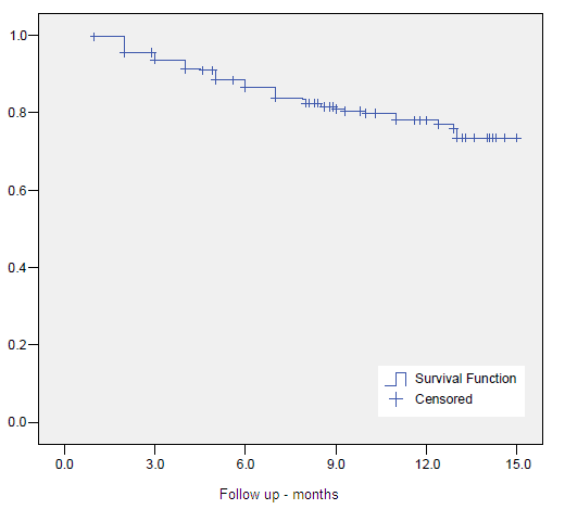
The crude 10-year overall survival (OS) rate at was 52.7%. Actuarial 5- and 10-year OS, CSS and BC were 80.1%, 89.8%, 83.7%, 63.6%, 85.5% and 71.8%, respectively. (Figures 1-3)
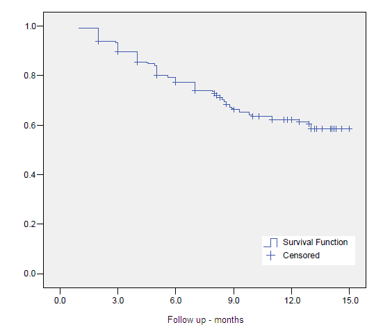
Figure 1- Overall Survival

    Figure 1. Overall Survival

Figure 2 - Clinical Specific Survival

Figure 2. Clinical Specific Survival
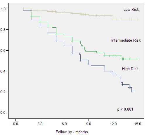
Figure 3 - Clinical Specific Survival by Risk Group

Figure 3. Clinical Specific Survival by Risk Group

Univariate and multivariate analysis

On univariate analysis GS <7, clinical stage <T2b, low risk group, absence adjuvant ADT, older age (>65-years), PSAi Univariate analysis failed to identify neoadjuvant androgen deprivation therapy (NAAD) as a predictor for BC in all group risks (p=ns). When we pooled the intermediate and high risk group into a unique denominated unfavorable risk group, NAAD also failed to predict improved CSS and BC.

Multivariate Cox regression analysis confirmed low risk group (HR 0.03, 95% CI 0.006-0.116, p<0.001) and intermediate risk (HR 0.09, 95% CI 0.042-0.216, p<0.001) compared to high risk, presence of ADT (HR 0.39, 95% CI 0.179-0.868, p=0.021) as favorable predictors for CSS. GS >7 (HR 3.24, 95% CI 1.279-8.220, p<0.001), PSA >10 (HR 6.19, 95% CI 2.015-19.041, p=0.001), age >65 years (HR 2.87, 95% CI 1.264-6.504, p=0.012) and EBRT dose >50 Gy (HR 14.50, 95% CI 1.874-112.164, p=0.010) were confirmed as adverse predictors for CSS. Tables 6-8, Figures 4-9.

Low risk group compared to intermediate, (HR 0.70, 95% CI 0.023-0.213, p<0.001) and high risk (HR 0.99, 95% CI 0.052-0.190, p<0.001) groups, was a favorable predictive factor for BC. GS >7 (HR 3.09, 95% CI 1.473-6.473, p=0.003) and PSAi >10 (HR 6.18, 95% CI 2.331-16.393, p<0.001) were negative predictive factor for BC.

Low risk group was confirmed as the only predictive factor for OS when compared to intermediate (HR 0.28, 95% CI 0.133-0.608, p=0.001) and high (HR 0.38, 95% CI 0.223-0.665, p=0.001) risk groups.
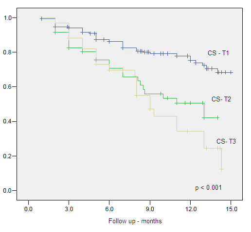
Figure 4. Clinical Specific Survival by Clinical Stage
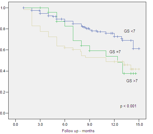
Figure 4. Clinical Specific Survival by Clinical Stage

Figure 5. Clinical Specific Survival by Gleason Score
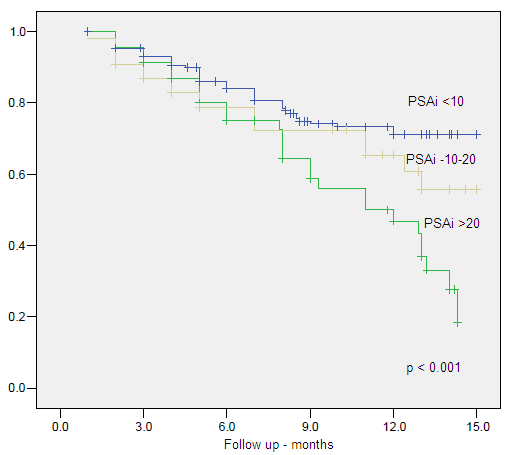
Figure 5. Clinical Specific Survival by Gleason Score

Figure 6. Clinical Specific Survival by Initial PSA
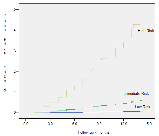
Figure 6. Clinical Specific Survival by Initial PSA

Figure 7. Hazard Plots - Clinical Specific Survival by Risk Group
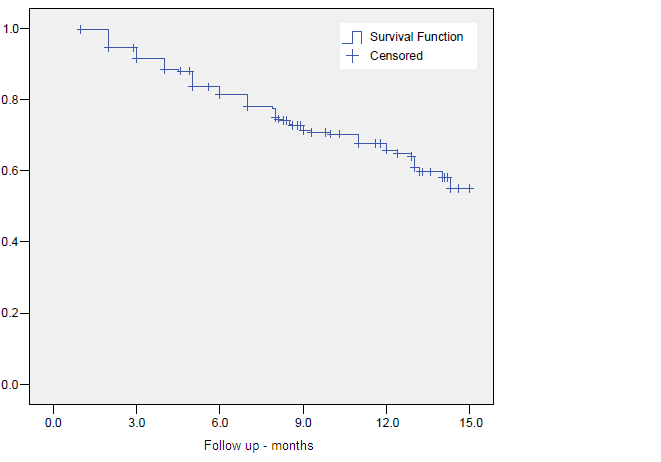
Figure 7. Hazard Plots – Clinical Specific Survival by Risk Group

Figure 8. Biochemical Control
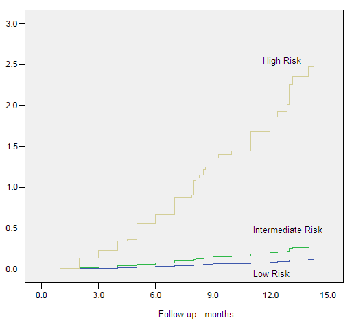
Figure 8. Biochemical Control

Figure 9. Hazard Plots - Biochemical Control by Risk Group

Figure 9. Hazard Plots – Biochemical Control by Risk Group

Table 6. Cox regression for CSS

B SE Wald df Sig. Exp(B) 95.0% CI for Exp(B)
Lower Upper
LR 44.294 2 .000
IR -3.641 .761 22.910 1 .000 .026 .006 .116
HR -2.351 .418 31.579 1 .000 .095 .042 .216
EBRT

Pelvic x Local

-.110 .312 .124 1 .724 .896 .486 1.651
2D x 3D -1.861 1.134 2.693 1 .101 .156 .017 1.436
ADT -.931 .403 5.336 1 .021 .394 .179 .868
PSAi <10 10.858 2 .004
PSAi (>10<20) 1.925 .661 8.481 1 .004 6.856 1.877 25.048
PSAi (>20) 1.824 .573 10.133 1 .001 6.195 2.015 19.041
GS <7 7.328 2 .026
GS = 7 .335 .709 .223 1 .636 1.398 .349 5.608
GS >7 1.176 .475 6.145 1 .013 3.243 1.279 8.220
CS <T2b 3.350 2 .187
CS T2b/T2c -.662 .551 1.443 1 .230 .516 .175 1.519
CS >T2c .464 .818 .321 1 .571 1.590 .320 7.895
Age <65y 1.053 .418 6.352 1 .012 2.867 1.264 6.504
EBRT <50Gy 2.674 1.044 6.562 1 .010 14.498 1.874 112.164
HDR dose <20Gy 1.763 1.023 2.973 1 .085 5.830 .786 43.261
Interval EBRT to HDR -.187 .303 .381 1 .537 .829 .458 1.502

Legend: 2D (two dimensional plan), 3D (tridimensional plan), ADT (Androgen deprivation therapy), BC (biochemical control), Comorb (Comorbidites), CS (Clinical stage), CSS (Clinical Specific Survival), EBRT (External beam radiotherapy), GS (Gleason score), IR (Intermediate risk group), HDR (High-dose-rate brachytherapy), HR (High risk group), LR (Low risk group), NAAD (neoadjuvant hormonal therapy), OS (overall survival), P vol. (Prostate volume cc)

Table 7. Cox regression for BC

B SE Wald df Sig. Exp(B) 95.0% CI for Exp(B)
Lower Upper
LR 62.053 2 .000
IR -2.660 .568 21.948 1 .000 .070 .023 .213
HR -2.309 .330 48.920 1 .000 .099 .052 .190
2D x 3D -.789 .656 1.448 1 .229 .454 .126 1.642
ADT -.455 .307 2.192 1 .139 .634 .347 1.159
PSAi <10 13.634 2 .001
PSAi (>10<20) 1.822 .498 13.403 1 .000 6.182 2.331 16.393
PSAi >20 1.346 .462 8.494 1 .004 3.843 1.554 9.502
GS <7 8.944 2 .011
GS =7 .957 .465 4.240 1 .039 2.603 1.047 6.469
GS >7 1.127 .378 8.915 1 .003 3.088 1.473 6.473
CS <T2b 2.825 2 .244
CS T2b/T2c -.647 .410 2.493 1 .114 .523 .234 1.169
CS >T2c -.306 .580 .278 1 .598 .737 .236 2.294
EBRT 50Gy 1.411 .550 6.590 1 .010 4.101 1.396 12.044
HDR dose <20Gy -.470 .680 .477 1 .490 .625 .165 2.370
Interval EBRT to HDR -.064 .230 .078 1 .780 .938 .597 1.473

Legend: 2D (two dimensional plan), 3D (tridimensional plan), ADT (Androgen deprivation therapy), BC (biochemical control, Comorb (Comorbidites), CS (Clinical stage), CSS (Clinical Specific Survival), EBRT (External beam radiotherapy), GS (Gleason score), IR (Intermediate risk group), HDR (High-dose-rate brachytherapy), HR (High risk group), LR (Low risk group), NAAD (neoadjuvant hormonal therapy), OS (overall survival), P vol. (Prostate volume)

Table 8. Cox regression for OS

B SE Wald df Sig. Exp(B) 95.0% CI for Exp(B)
  Lower Upper
ADJ .149 0.3 0.3 1 .580 1.161 .684 1.970
Age <65y .223 0.2 1.0 1 .322 1.250 .804 1.942
Comorbidity -.310 0.2 2.1 1 .149 .734 .482 1.117
EBRT

Pelvic x Local

-.089 0.2 0.1 1 .722 .915 .560 1.494
LR 14.3 2 .001
IR -1.259 0.4 10.5 1 .001 .284 .133 .608
HR -.954 0.3 11.7 1 .001 .385 .223 .665

Legend: ADJ (adjuvant hormonal therapy, EBRT (External beam radiotherapy), IR (Intermediate risk group), HDR, HR (High risk group), LR (Low risk group), OS (overall survival)

Discussion

Conventional EBRT to treat PCa is securely limited to doses of 64–70 Gy in 1.8–2.0 Gy fractions. These levels of doses are determined by the risk of long-term toxic effects to the bladder and rectum. The clinical and biochemical relapse rates associated with these dose levels are around 33% within 5 years. Peeters et al 12 published the results of a randomized trial comparing total doses of 68 Gy and 78 Gy using EBRT alone. The 5-year OS in the higher dose arm of that study was 83% using ASTRO definition and the BC was significantly better in the 78-Gy arm compared to the 68-Gy arm, with an adjusted hazard ratio of 0.74 (p=0.02). Other published reports using dose-escalated photon beam EBRT alone also point in the same direction with respect to their long-term BC results [13-14].

HDR can escalate the dose given to the prostate by the combination with EBRT, and further more, in locally advanced disease has also the possibility of including the seminal vesicles when they needed to be encompassed. HDR has also a potential biological advantage through the delivery of high doses per fraction [10]. It is important to note that the comparisons between series published are difficult due differences in the techniques and planning for both, EBRT and HDR.

The combination of values of PSAi, GS and CS to identify a more or less aggressive disease has being extensively discussed in the literature, and was confirmed by this study. The most frequent challenge is to identify, for example the indication of prostate versus pelvic EBRT, varying risk categories, absence or use of NAAD, ADT and their length. Despite this, the combination of HDR and EBRT provides an optimal modulation of dose delivery. Results of this combination, in terms of BC and with more than 5 years of FU, range from 57% to 100% according to the risk group for biochemical failure (Table 9).

Table 9. Results of biochemical control by risk groups in series of HDR plus EBRT with more than 5-year follow up

Reference n Median FU  (months) Biochemical control by risk groups (%)

Low       Intermediate       High

Dose in Gy

(HDR(n.fx)/EBRT)

13 344 61 84 74 19.5(3) / 46
14 121 63 91 10(1)/50
15 313 68 100 88 79 23(2)/46
16 229 61 95 90 57 21(3)/50.4
17 64 105 84 80 18(3)/45
18 90 95 80 16.5(3)/45
19 264 75 97 18(3)/45
20 100 62 84 82 10(1)/60
21 64 61 100 91 21(3)/50
22 196 66 86 18(3)/46
23 131 63 87 71 30(4)/45

The search for factors predicting BC and CSS is important on defining what patients should be treated more aggressively. We, as other authors [26-28] observed that PSAi <10 ng/ml was confirmed as a favorable predictive factor related to BC and CSS.

Age<65 years was found to be an adverse prognostic factor in our analysis for CSS, with a marginally statically significance impact on OS, not confirmed on multivariate analysis. Smolska-Ciszewska et al, conversely to our results, noted that younger age at time of treatment impacted only on OS, not explaining if there was any association between treatment and side effects or worsening of associated comorbidities. As in our analysis, they found that low risk group and association of HDR to EBRT correlated with improved BC [29].

As in our results, Kamrava et al found that T stage, GS, and use of ADT were significantly associated with CSS on univariate analysis, but on multivariate analysis only GS and use of ADT were significantly associated CSS [30]

It is expected that the grouping of the patients in risk groups for biochemical failure based on PSAi, GS and CS, to aggregate patients with adverse features in the intermediate and high risk, leading to worse CSS and BC for the two last one, what was confirmed in our analysis. This was also observed by Morris et al [31].

In our series, as in others, patients who received adjuvant ADT had significantly higher risk features suggesting patient selection bias for CSS in this group of patients, instead of a negative interaction between HDR and EBRT [31-32].

Mature data in the literature evaluated the 10-year outcomes of intermediate- and high-risk patients noting a clear dose response by increasing the dose escalation through HDR doses [27]. More recently the use of Intensity modulated radiation therapy combined to HDR has being investigated. Chen et al. published the results of 148 patients treated with HDR – 22 Gy in 4 fractions followed IMRT up to 50.4Gy. All patients with Gleason score of 8 or higher had ADT for 1 year. They noted a 4-year actuarial CSS of 96.8% and of 100%, 100% and 94% for low, intermediate and high risk, respectively [32].

The results of the first randomized prospective trial, which has addressed dose escalation using an HDR and EBRT is a trial with a relatively slow accrual rate. There are some critics that must be addressed as the changes in EBRT technique during the time of the study, and, by current standards, the control arm is a relatively low-dose treatment. Despite that, the study reported the results of 218 patients treated between 1997 and 2005. There were 108 patients assigned to EBRT alone and 110 patients treated by EBRT followed by HDR. They noted that CSS was significantly higher in patients treated with combined modality (p = 0.04). In multivariate analysis the category treatment modality and ADT were significant covariates for BC, but with no differences in OS, as observed in our study. After a median follow-up time of 10.5 year follow-up, an 18% increase in CSS was obtained relative to EBRT alone, reflecting a 31% reduction in the risk of recurrence (p = 0.01) and no evidence of an increase in long-term severe morbidity [9].

Surgically induced gland deformation is inevitable during brachytherapy procedures. We observed that the use more of advanced methods of images (3D plan based on CT, MR or TRUS) images to identify the target, organs at risk and needles is important and have already been reported to impact on BC and CSS [33]. In our analysis Multivariate Cox regression analysis confirmed EBRT dose >50 Gy as predictor for CSS and BC, but with no impact on OS. This may be explained by the relative low dose per fraction schedule used in both groups. Of importance is to note that the dose given by HDR was relative constant thorough the risk groups, leading to higher biological effective dose to intermediate and high risk patients, and even though, these patients had a worse outcome, showing that there is space for further studies of dose escalation or treatment combination. After 2005 with the introduction of real time TRUS image acquisition and planning we have changed our protocol and moved forward for a more intense dose escalation, increasing dose and reducing the number of fractions.

Other information of this study is that presence of NAAD had no impact on DSS, BC or OS in any risk group, showing that this treatment strategy should be reserved for downsizing the prostate prior to treatment, in special for low risk patients.

In conclusion, this report demonstrates that HDR combined with EBRT is an important and effective method in achieving dose escalation in the radical radiotherapy of PCa. This combination has also the advantage of treatment time reduction and in increasing in the capability of work load of the linear accelerators, especially in developing countries, where waiting lists and lack of radiation oncology facilities are a reality. The present data represents a unique uni-institutional study at long FU for the given technique, and a comparison with the current literature confirms the excellent results achieved with this treatment modality. The satisfactory BC, CSS and OS are probably result of improved LC achieved with dose escalation, showing that HDR is an optimal alternative method of local dose escalation when combined to EBRT.

References

  • Quinn MJ, d’Onofrio A, Møller B, Black R, Martinez-Garcia C, et al. (2003) Cancer mortality trends in the EU and acceding countries up to 2015. Ann Oncol 14: 1148-1152. [crossref]
  • http://www.inca.gov.br/estimativa/2014/sintese-de-resultados- comentarios.asp
  • Novara G, Ficarra V, Mocellin S, et al. (2012) Systematic review and meta-analysis of studies reporting oncologic outcome after robot-assisted radical prostatectomy. Eur Urol 62: 382.
  • Al-Mamgani A, van Putten WL, Heemsbergen WD, van Leenders GJ, Slot A, et al. (2008) Update of Dutch multicenter dose-escalation trial of radiotherapy for localized prostate cancer. Int J Radiat Oncol Biol Phys 72: 980-988. [crossref]
  • Dearnaley DP, Sydes MR, Graham JD, et al. (2007) Escalated-dose versus standard-dose conformal radiotherapy in prostate cancer: first results from the MRC RT01 randomized controlled trial. Lancet Oncol 8: 475.
  • Kuban DA, Tucker SL, Dong L, Starkschall G, Huang EH, et al. (2008) Long-term results of the M. D. Anderson randomized dose-escalation trial for prostate cancer. Int J Radiat Oncol Biol Phys 70: 67-74. [crossref]
  • Zietman AL, DeSilvio ML, Slater JD, et al. (2005) Comparison of conventional-dose vs high-dose conformal radiation therapy in clinically localized adenocarcinoma of the prostate: a randomized controlled trial. JAMA 294: 1233.
  • Pellizzon AC, Salvajoli JV, Maia MA, et al. (2004) Late urinary morbidity with high dose prostate brachytherapy as a boost to conventional external beam radiation therapy for local and locally advanced prostate cancer. J Urol 171: 1105.
  • Hoskin PJ, Rojas AM, Bownes PJ, et al. (2012) Randomized trial of external beam radiotherapy alone or combined with high-dose-rate brachytherapy boost for localized prostate cancer. Radiother Oncol 103: 217.
  • Pellizzon AC, Nadalin W, Salvajoli JV, Fogaroli RC, et al. (2003) Results of high dose rate afterloading brachytherapy boost to conventional external beam radiation therapy for initial and locally advanced prostate cancer. Radiother Oncol 66: 167.
  • Roach M 3rd, Hanks G, Thames H Jr, et al. (2006) Defining biochemical failure following radiotherapy with or without hormonal therapy in men with clinically localized prostate cancer: recommendations of the RTOG-ASTRO Phoenix Consensus Conference. Int J Radiat Oncol Biol Phys 65: 965.
  • Peeters ST, Heemsbergen WD, Koper PC, et al. (2006) Dose-response in radiotherapy for localized prostate cancer: results of the Dutch multicenter randomized phase III trial comparing 68 Gy of radiotherapy with 78 Gy. J Clin Oncol
  • Pollack A, Zagars GK, Starkschall G, Antolak JA, Lee JJ, et al. (2002) Prostate cancer radiation dose response: results of the M. D. Anderson phase III randomized trial. Int J Radiat Oncol Biol Phys 53: 1097-1105. [crossref]
  • Dearnaley DP, Sydes MR, Graham JD, et al. (2007) Escalated dose versus standard-dose conformal radiotherapy in prostate cancer: first results from the MRC RT01 randomized controlled trial. Lancet Oncol 8: 475.
  • Khor R, Duchesne G, Tai KH, et al. (2013) Direct 2-arm comparison shows benefit of high-dose-rate brachytherapy boost vs external beam radiation therapy alone for prostate cancer. Int J Radiat Oncol Biol Phys 85: 679.
  • Cury FL, Duclos M, Aprikian A, et al. (2012) Single-fraction high-dose-rate brachytherapy and hypofractionated external beam radiation therapy in the treatment of intermediate-risk prostate cancer – long term results. Int J Radiat Oncol Biol Phys 82: 1417.
  • Prada PJ, González H, Fernández J, Jiménez I, Iglesias A, et al. (2012) Biochemical outcome after high-dose-rate intensity modulated brachytherapy with external beam radiotherapy: 12 years of experience. BJU Int 109: 1787-1793. [crossref]
  • Kotecha R, Yamada Y, Pei X, Kollmeier MA, Cox B, et al. (2013) Clinical outcomes of high-dose-rate brachytherapy and external beam radiotherapy in the management of clinically localized prostate cancer. Brachytherapy 12: 44-49. [crossref]
  • Kaprealian T, Weinberg V, Speight JL, et al. (2012) High-dose-rate brachytherapy boost for prostate cancer: comparison of two different fractionation schemes. Int J Radiat Oncol Biol Phys 82: 222.
  • Savdie R, Symons J, Spernat D, Yuen C, Pe Benito RA, et al. (2012) High-dose rate brachytherapy compared with open radical prostatectomy for the treatment of high-risk prostate cancer: 10 year biochemical freedom from relapse. BJU Int 110 Suppl 4: 71-76. [crossref]
  • Aluwini S, van Rooij PH, Kirkels WJ, Jansen PP, Praag JO, et al. (2012) High-dose-rate brachytherapy and external-beam radiotherapy for hormone-naïve low- and intermediate-risk prostate cancer: a 7-year experience. Int J Radiat Oncol Biol Phys 83: 1480-1485. [crossref]
  • Agoston P, Major T, Fröhlich G, et al. (2011) Moderate dose escalation with single-fraction high-dose-rate brachytherapy boost for clinically localized intermediate- and high-risk prostate cancer: 5-year outcome of the first 100 consecutively treated patients. Brachytherapy 10: 376.
  • Ghadjar P, Keller T, Rentsch CA, Isaak B, Behrensmeier F, et al. (2009) Toxicity and early treatment outcomes in low- and intermediate-risk prostate cancer managed by high-dose-rate brachytherapy as a monotherapy. Brachytherapy 8: 45-51. [crossref]
  • Zwahlen DR, Andrianopoulos N, Matheson B, Duchesne GM, Millar JL (2010) High-dose-rate brachytherapy in combination with conformal external beam radiotherapy in the treatment of prostate cancer. Brachytherapy 9: 27-35. [crossref]
  • Viani GA, Pellizzon AC, Guimarães FS, Jacinto AA, dos Santos Novaes PE, et al. (2009) High dose rate and external beam radiotherapy in locally advanced prostate cancer. Am J Clin Oncol 32: 187-190. [crossref]
  • Aoki M, Miki K, Kido M, et al. (2014) Analysis of prognostic factors in localized high-risk prostate cancer patients treated with HDR brachytherapy, hypofractionated 3D-CRT and neoadjuvant/adjuvant androgen deprivation therapy (trimodality therapy). J Radiat Res 55: 527.
  • Martinez AA, Gonzalez J, Ye H, et al. (2011) Dose escalation improves cancer-related events at 10 years for intermediate- and high-risk prostate cancer patients treated with hypofractionated high-dose-rate boost and external beam radiotherapy. Int J Radiat Oncol Biol Phys 79: 363.
  • Shigehara K, Mizokami A, Komatsu K, Koshida K, Namiki M (2006) Four year clinical statistics of iridium-192 high dose rate brachytherapy. Int J Urol 13: 116-121. [crossref]
  • Smolska-Ciszewska B, Miszczyk L, Bialas B, et al. (2015) The effectiveness and side effects of conformal external beam radiotherapy combined with high-dose-rate brachytherapy boost compared to conformal external beam radiotherapy alone in patients with prostate cancer. Radiat Oncol 10: 366.
  • Kamrava M, Rwigema JC, Chung M, Banerjee R, Wang J, et al. (2013) Predictors of distant metastasis after combined HDR brachytherapy and external beam radiation for prostate cancer. J Contemp Brachytherapy 5: 127-133. [crossref]
  • Morris LM, Izard MA, Wan WY (2015) Does prostate-specific antigen nadir predict longer-term outcome es of prostate cancer after neoadjuvant and adjuvant androgen deprivation therapy in conjunction with brachytherapy? Brachytherapy 14: 322.outcome
  • Chen WC, Tokita KM, Ravera J, Fu P, Jiang Y, et al. (2013) Four-year outcomes of hypofractionated high-dose-rate prostate brachytherapy and external beam radiotherapy. Brachytherapy 12: 422-427. [crossref]
  • Pellizzon AC, Salvajoli J, Novaes P, et al. (2008) The relationship between the biochemical control outcomes and the quality of planning of high-dose rate brachytherapy as a boost to external beam radiotherapy for locally and locally advanced prostate cancer using the RTOG-ASTRO Phoenix definition. Int J Med Sci 5: 113.
Is Intravenous Iron Treatment a Risk for Infection?

DOI: 10.31038/IMROJ.2017214

Abstract

Background: Iron is a pro-oxidant and an essential nutrient for different bacteria. Experimental animal studies have demonstrated exacerbating sepsis episodes. Intravenous iron treatment has been implicated, at least theoretically in increasing infection episodes. However, these studies did not obtain any conclusion in this respect. Therefore, the aim of our study was to evaluate whether there is a risk between intravenous iron treatment and infection episodes following iron treatment.

Methods: In our study, 186 patients who received intravenous iron treatment were included. The biochemical parameters included “pre and post iron treatment” (e.g., ferritin, serum iron, transferring saturation, and hemoglobin) and the later emergence of infections. Moreover, we evaluated all possible risk factors that could interfere with the subsequence appearance of infection.

Results: Infectious complications were reported in 24 patients (12.8%) after the iron treatments began, and no infection was reported in 87.15% (n=162) of the patients. The most common infections were urinary (33%) and respiratory infections (25%). The most common infectious agents were enterobacteriae (e.g., E coli, E faecalis, and E faecium). Notably, we did not observe any intracellular pathogens. In our statistical survey, we did not find a relationship between infection onset and intravenous iron treatment. To analyze the baseline disease, comorbidities, such as diabetes and other treatment (e.g., corticosteroids or anti-TNF) were evaluated, and, no association was found.

Conclusion: In our study, intravenous iron was an effective treatment to correct anemia, and it did not appear to correlate with the development of secondary infections.

Key words

iron therapy; intravenous iron; iron infection

Abbreviations

IV – intravenous; HD – hemodialysis infection

Introduction

Iron is an important nutrient for many bacteria, and in laboratory animals, it was shown to exacerbate sepsis. [1] It has been postulated that increased plasma iron is responsible for the association of hemochromatosis with infections by Vibrio vulnificus, Yersinia enterocolitica, and Escherichia coli. [2] Accordingly, there are host defense mechanisms that tend to restrict the available iron from invading pathogens. [3] For example, the transferrin receptor is the major pathway for delivery of iron to peripheral tissues through endocytosis of its ligand, transferrin, which binds to iron and circulates in the plasma. Down regulation of the transferrin receptor limits the cell’s ability to acquire transferrin-bound iron and concomitantly reduces the endosomal pool of iron that can be accessed by intracellular pathogens, such as L. pneumophila, Mycobacterium tuberculosis, and Mycobacterium avium, and thus restricts their growth. [4, 5] Iron repletion with oral iron has been associated with adverse outcomes for certain infections. In a study of 137 iron-deficient Somali nomads, who were treated with placebo or oral iron (71 subjects), Murray et al. noted seven infection episodes in the placebo group and 36 in the oral iron group (including reactivation of preexisting malaria, brucellosis, and tuberculosis). [6] A large iron supplementation trial of young children on Pemba Island, was prematurely terminated as a result of an excess of serious adverse events (including deaths) in those receiving iron. [7] Ferric iron (Fe)–carbohydrate complexes are widely used for treating Fe deficiency in patients who are unable to meet their Fe requirements with oral supplements. [8] Intravenous (IV) Fe is generally well tolerated and effective in correcting Fe-deficient states. However, complexing Fe to carbohydrate polymers does not block its potent pro-oxidant effects; therefore, systemic free radical generation and, possibly, tissue damage may result. [9] On the other hand, excessive iron administration may lead to oversaturation of transferrin and the release of free, catalytically active iron into circulation (NTBI) [10] which could be readily utilized by bacteria, leading to their multiplication and the possibility of an overwhelming infection. [11, 12] The aim of the present work was to determine the short-term infection rate associated with IV iron treatment and the possible risk factors that can predict these infections in a contemporary patient cohort.

Material and methods

Patient enrollment A retrospective, longitudinal study was carried out at Marqués de Valdecilla University Hospital (Santander, Spain). The hospital records of patients who underwent ambulatory treatment with IV iron at the hospital day center between January 2011 and December 2012 were reviewed.

Patient parameter evaluation The medical records of these patients were searched after institutional review board approval was obtained for the following data fields: a) demographic features; b) principal diagnosis; c) comorbid factors, such as diabetes mellitus, arterial hypertension, and dislipemia; d) concomitant treatment as potential infection causes, such as corticosteroids, immunosuppresive therapy, and anti-TNF treatment; e) previous infection, and; f) laboratory data prior to and after IV iron treatment, such as hemoglobin concentration, mean cell hemoglobin (MCH), mean cell hemoglobin concentration (MCHC), red cell volume distribution width (RDW), serum iron, transferrin saturation, and serum ferritin.

Intravenous iron treatment One thusand mg (50 mg/ml) of ferric carboxymaltose was administered in an ambulatory regimen to the subjects at the hospital day center. The doses of this treatment were adapted subsequently according to the Ganzoni equation and their efficacy.

Case definitions We used a retrospective cohort design with a 1-month baseline period. After IV iron treatment was started, all of the patients were followed for 3 months. We looked for any infectious diseases, and we differentiated the subjects by bacterial, viral and fungal infections, as well as by gastrointestinal, genitourinary, respiratory and other diseases. If a single blood culture was positive for coagulase-negative staphylococci, Corynebacteria, or Bacillus sp., the culture was considered contaminated or representative of a transient bacteremia but not of a bacteremic episode. All other situations in patients in whom at least one blood culture was positive were defined as bacteremic episodes.

Statistical Analysis The results were analyzed with the SPSS 15.0 computer software package (Statistical Package for Social Sciences, Inc., Chicago. IL). Continuous variables were summarized as the means or as the medians and ranges. Categorical variables were compared with the chi-square and Fisher´s exact tests. Correlations between the data sets were examined using the Pearson (r) or Spearman rank (rs) correlation coefficients. Any differences were considered significant for all statistical tests at p values of less than 0.05.

Results

A total of 186 adult patients (70 men and 116 women; mean age, 63 yrs; range, 18 to 80 yrs) were enrolled. The principal diagnosis of the patients included in the study were cardiac diseases (n=41, 22%), neoplasm diseases (n=32, 17.2%), renal diseases (n=31, 16.7%), malnutrition due to bariatric surgery (n=19, 10.2%), hepatic diseases (n=15, 8.1%), and lung diseases (n=15, 8.1%) (Table 1). A comorbid condition known to increase the infection risk was found in 88 patients (47.3%), which included diabetes mellitus (n = 53), current malignancy (n = 32) (under treatment and/or not cured), being a transplant recipient (n= 3), and having both a malignancy and diabetes mellitus (n= 5). Twelve patients were treated with corticosteroids and six were treated with anti-TNF therapy. The pretreatment median hemoglobin level was 9.5 gr/dL (5.6- 12.6 gr/ dL) and after the treatment it was 11.8 gr/dL (7.4-14.1 gr/dL) (Table 2). Additionally, we found a statistically significant difference between the baseline biochemical iron metabolism parameters pre-and post IV iron treatment (Table 2). Thus, we corroborated that IV iron was an effective therapy in our patients.

Table 1. The principal diagnosis of the patients included in the study

Disease n (%)
Cardiac diseases 41 (22%)
Neoplasms diseases 32 (17.2%)
Kidney disease 31 (16.7%)
Bariatric surgery 19 (10.2%)
Liver diseases 15 (8.1%)
Lung diseases 15 (8.1%)
Others 33 (17.7%)

Table 2. The baseline laboratory iron metabolism parameters pre-and post-intravenous iron treatment

  Pre-treatment Post-treatment P
Hemoglobin (gr/dL) 9.5 (5.6-14.5) 11.8 (7.4- 14.10) <0.01
MCH (pg/RBC) 26.4 (8.6-37.5) 29.9 (21.8-37.4) <0.01
MCHC (gr/dL) 32.4 (28.6-35.3) 33.1 (25.5-35.3) <0.01
RDW (%) 18.9 (12.2-37.4) 21.4 (9-43.6) <0,01
Serum iron (mcg/dL) 25 (10-96) 69 (16-177) <0.541
Transferrin saturation (Fe/TIBC)% 7 (2-37) 23,5 (5-62) <0.01
Serum ferritin 54 (2-753) 408 (10-1492) <0.01

MCH – mean cell hemoglobin;
MCHC – mean cell hemoglobin concentration;
RDW – red cell volume distribution width.

No patients had more than one bacteremic episode within the month prior to treatment. Twenty-four patients (12.9%) suffered from one infection episode after the IV iron treatment, and 162 patients (87.1%) did not have any infection episodes. The majority of these infections were from bacterial microorganisms (75%, n=18) followed by unknown etiologies (16.7% n=4) and viral episodes (8.3%, n=2). There were no fungal infections. The urinary tract was involved in 33.3% of the cases (n=8); 25% of the cases involved therespiratory system (n=6), which was similar to the unknown focus case percentage; and 16.7% of the cases involved the gastrointestinal system (n=4). The infectious agent was unknown in 63.5% of the cases (n=15). The pathogens responsible for these episodes were E coli (16.7%, n=4) followed by E faecalis, E faecium, Lysteria and Proteus mirabilis, which all occurred in 4.2% of the cases (n=1). No intracellular microorganisms or non-capsulated bacteria were found in our series. Additionally, no serious diseases and no mortality cases were reported three months after the treatments began.

The main characteristics of the patients with and without infection episodes are shown in Table 3. More patients with an infection episode had a comorbidity (diabetes or malignancy) and ongoing immunosuppressive therapy than the patients without infections. However, this was not statistically significant. Furthermore, when we analyzed the relationship between infections with other comorbidities, such as diabetes, cancer, transplantation or treatments such as steroids or anti-TNF we did not find any association.

Table 3. Comorbidity incidences in the infection or no infection groups

  Infection No Infection p
Diabetes mellitus 8 (33.3%) 45 (28.3%) NS
Arterial hypertension 14 (58.3%) 65 (40.1%) NS
Dislipemia 5 (20.8%) 23 (14.2%) NS
Cardiac diseases 7 (29.2%) 34 (21%) NS
Renal diseases 7 (29.2%) 24 (14.8%) NS
Lung diseases 1 (4.2%) 14 (8.6%) NS
Liver diseases 1 (4.2%) 14 (8.5%) NS
Bariatric surgery 3 (12.5%) 16 (9.9%) NS
Neoplasm 7 (29.2%) 25 (15.4%) NS
Organ transplantation 0 (0%) 3 (1.9%) NS
Corticosteoids 3 (12.5%) 9 (5.6%) NS
Biologic therapy 1 (4.2%) 5 (3.1%) NS

NS – Non-Significant

Discussion

Almost all life forms require iron because of its involvement in basic cellular processes. Free iron was shown to potentiate bacterial growth in vitro. [13] In fact, during infections, pathogens use various means to acquire iron from their hosts, whereas hosts attempt to withhold it from pathogens. [14, 15] Iron therefore represents a point of conflict between the host and the pathogen, and an altered iron balance associated with poor outcomes in several infectious diseases, including malaria, [7] tuberculosis, [16] and HIV-1 infection. [17] Furthermore, certain bacterial species, including E. Coli, Klebsiella spp., and Salmonella spp., use unbound iron in the blood to enhance their growth. These organisms release siderophores, which are iron chelators, into the blood. Once these siderophores are released, bacteria can compete with proteins, such as transferrin for unbound iron in the serum. [18, 19] Staphylococcus aureus and Haemophilus influenzae do not possess siderophore; however, they do have transferrin receptors, which allow these bacteria to use iron for growth. [18] Vibrio vulnificus cannot grow if there is no iron available, and their virulence depends on the ability to obtain iron combined with transferrin. Hepcidin, which is produced during innate immune responses to infections, reduces the iron availability in the serum by inhibiting ferroportin function in macrophages and enterocytes and potentially limits extracellular pathogen growth therein. [20] Intravenous iron may also impair immune function and increase infection susceptibility. [21] Gupta et al. [22] found that exposure of mononuclear cells to IV iron agents induced significant intracellular oxidative stress and shortened CD4+ T lymphocyte survival. High doses of IV iron agents impaired the phagocytic activity and microbial killing capability of polymorphonuclear leukocytes [23-25]. Furthermore, in a recent in vitro study, iron sucrose led to impaired phagocytic function and increased polymorphonuclear leukocyte apoptosis. [26] However, the current evidence in clinical practice cannot determine whether iron supplementation increases the risk of infection or worsens outcomes due to infection. [27, 28] There is increasing data to suggest that infective and adverse-event risks may be related to the intensity and frequency of IV iron dosing. [29-31] Brookhart et al. [32] studied iron dosing patterns in a retrospective cohort of 117.050 prevalent hemodialysis (HD) patients and found that administration of large boluses of IV iron for repleting iron deficiency was associated with increased infection-related hospitalization or death compared with smaller doses of IV iron maintenance therapy. The risk of infection-related hospitalization was increased further in patients who experienced infections within the past month. Similarly, the DOPPS study showed a trend towards an increase infection.related mortality in prevalent HD patients treated with > 300 mg of IV iron. [33] A recent meta-analysis of randomized controlled trials evaluating IV iron use (often administered as frequent boluses) in patients with varying infective risk profiles found IV iron to be associated with a 30% greater risk of infection compared with oral or no iron therapy. [34] Conversely, a prospective observational study of 985 patients failed to demonstrate a relationship between infection and serum ferritin or IV iron dosing. [35] In our study, large IV iron boluses were an effective treatment for anemia that was caused by different etiologies, because hemoglobin increased at 2 or 3 points at the end of the treatment and we found any relationship between IV iron administration and infection, which was similar to the previously published data. Furthermore, in our study, transferrin saturation increased to 23.5%, thereby leaving little to no unbound iron available for bacterial utilization, as shown in previous in vitro studies. [10] For example, in the K. pneumonia case, bacterial suppression occurred throughout its incubation until the transferrin saturation exceeded 60%, at which time bacterial growth occurred. [19] Another in vitro study showed that the inhibition of Staphylococcus epidermidis growth was lost once the transferrin saturation exceeded 80%. [18] Subsequent studies had ambiguous results that left the relationship somewhat unclear. [36, 37] In a retrospective observational cohort study of 23.000 adult patients on HD hospitalized for bacterial infection, Ishida et al. found no association between iron treatment and readmission for infection. [38] Feldman’s group, in a re-analysis of their HD cohort using multivariable analysis, showed no statistically significant association between any level of iron administration and mortality. [39] Moreover, a French multicenter prospective study regarding bacteremia risk factors in HD patients (EPIBACDIAL) did not find any correlation between parenteral iron or ferritin and bacteremia. [40] Furthermore, in the Anker study, quality of life and functional status improved in heart failure (a relatively high-risk group) without an increase in infections [41].

Our study had limitations due to its retrospective nature. However, of the patients who were analyzed, we found a 12.8% infections after the beginning of iron therapy. Notably, an important difference in our study compared with the previously published studies was that we did not find any association between IV iron treatment and intracellular pathogens.

In conclusion, based on our study, iron treatment is safe and it does not associate with the development of new infections. However, the infection relationship is not clear and many studies are treating to investigate the different mechanisms regarding this issue. It is important to verify this result with other studies to improve the care and future treatment of patients because iron treatment is frequently used worldside. This knowledge can improve anemia management and avoid side effects. Thus, large multi-centered randomized controlled trials designed to evaluate both the short- and long-term safety of different IV iron dosing regimens are still required to determine the optimal iron therapy.

Conflict of interests

No conflict of interest exists and all authors had full access to all the data used in the study and take full responsibility for the decision to submit the manuscript for publication.

References

  • Guerinot ML (1994) Microbial iron transport. Annu Rev Microbiol 48: 743-772. [crossref]
  • Khan FA, Fisher MA, Khakoo RA (2007) Association of hemochromatosis with infectious diseases: expanding spectrum. Int J Infect Dis 11: 482-487. [crossref]
  • Wander K, Shell-Duncan B, McDade TW (2009) Evaluation of iron deficiency as a nutritional adaptation to infectious disease: an evolutionary medicine perspective. Am J Hum Biol 21:172-179.
  • Byrd TF, Horwitz MA (1989) Interferon gamma-activated human monocytes downregulate transferrin receptors and inhibit the intracellular multiplication of Legionella pneumophila by limiting the availability of iron. J Clin Invest 83:1457-1465.
  • Clemens DL, Horwitz MA (1996) The Mycobacterium tuberculosis phagosome interacts with early endosomes and is accessible to exogenously administered transferrin. J Exp Med 184:1349-1355.
  • Murray MJ, Murray AB, Murray MB, Murray CJ (1978) The adverse effect of iron repletion on the course of certain infections. Br Med J 2: 1113-1115. [crossref]
  • Sazawal S, Black RE, Ramsan M, Chwaya HM, Stoltzfus RJ, Dutta A, et al. (2006) Effects of routine prophylactic supplementation with iron and folic acid on admission to hospital and mortality in preschool children in a high malaria transmission setting: community-based, randomised, placebo-controlled trial. Lancet 367:133-1343.
  • Clinical practice guidelines and clinical practice recommendations for anemia in chronic kidney Disease in adults (2006) Am J Kid Dis 47 (5 suppl3): S16-S85.
  • Zager RA (2006) Parenteral iron compounds: potent oxidants but mainstays of anemia management in chronic renal disease. Clin J Am Soc Nephrol 1 Suppl 1: S24-31. [crossref]
  • Parkkinen J, von Bonsdorff L, Peltonen S, Grönhagen-Riska C, Rosenlöf K. (2000) Catalytically active iron and bacterial growth in serum of haemodialysis patients after i.v. iron-saccharate administration. Nephrol Dial Transplant 15: 1827-1834.
  • Slotki I (2005) Intravenous iron supplementation in the anaemia of renal and cardiac failure–a double-edged sword? Nephrol Dial Transplant 20 Suppl 7: vii16-23. [crossref]
  • Bullen JJ, Rogers HJ, Spalding PB, Ward CG (2005) Iron and infection: the heart of the matter. FEMS Immunol Med Microbiol 43: 325-330. [crossref]
  • Fishbane S (1999) Review of issues relating to iron and infection. Am J Kidney Dis 34: S47-52. [crossref]
  • Weinberg ED (1984) Iron withholding: a defense against infection and neoplasia. Physiol Rev 64: 65-102. [crossref]
  • Armitage AE, Eddowes LA, Gileadi U, Cole S, Spottiswoode N, et al. (2011) Hepcidin regulation by innate immune and infectious stimuli. Blood 118: 4129-4139. [crossref]
  • Boelaert JR, Vandecasteele SJ, Appelberg R, Gordeuk VR (2007) The effect of the host’s iron status on tuberculosis. J Infect Dis 195: 1745-1753. [crossref]
  • McDermid JM, Jaye A, Schim van der Loeff MF, Todd J, Bates C, Austin S, et al. (2007) Elevated iron status strongly predicts mortality in West African adults with HIV infection. J Acquir Immune Defic Syndr 46:498-507.
  • Brewster UC, Perazella MA (2004) Intravenous iron and the risk of infection in end-stage renal disease patients. Semin Dial 17: 57-60. [crossref]
  • Cieri E (1999) Does iron cause bacterial infections in patients with end stage renal disease? ANNA J 26: 591-596. [crossref]
  • Rivera S, Nemeth E, Gabayan V, Lopez MA, Farshidi D, Ganz T (2005) Synthetic hepcidin causes rapid dose-dependent hypoferremia and is concentratedin ferroportin-containing organs. Blood 106:2196-2199.
  • Ishida JH, Johansen KL (2014) Iron and infection in hemodialysis patients. Semin Dial 27: 26-36. [crossref]
  • Gupta A, Zhuo J, Zha J, Reddy S, Olp J, Pai A (2010) Effect of different intravenous iron preparations on lymphocyte intracellular reactive oxygen species generation and subpopulation survival. BMC Nephrol 11:16.
  • Brewster UC (2006) Intravenous iron therapy in end-stage renal disease. Semin Dial 19: 285-290. [crossref]
  • Deicher R, Ziai F, Cohen G, Müllner M, Hörl WH (2003) High dose parenteral iron sucrose depresses neutrophil intracellular killing capacity. Kidney Int 64:728-736.
  • van Asbeck BS, Marx JJ, Struyvenberg A, Verhoef J (1984) Functional defects in phagocytic cells from patients with iron overload. J Infect 8: 232-240. [crossref]
  • Ichii H, Masuda Y, Hassanzadeh T, Saffarian M, Gollapudi S, Vaziri ND (2012) Iron sucrose impairs phagocytic function and promotes apoptosis in polymorphonuclear leukocytes. Am J Nephrol 36: 50-57.
  • Kliger AS, Foley RN, Goldfarb DS, Goldstein SL, Johansen K, et al. (2013) KDOQI US commentary on the 2012 KDIGO Clinical Practice Guideline for Anemia in CKD. Am J Kidney Dis 62: 849-859. [crossref]
  • Shah A, Roy NB2, McKechnie S3, Doree C4, Fisher SA4, et al. (2016) Iron supplementation to treat anaemia in adult critical care patients: a systematic review and meta-analysis. Crit Care 20: 306. [crossref]
  • Feldman HI, Santanna J, Guo W, Furst H, Franklin E, et al. (2002) Iron administration and clinical outcomes in hemodialysis patients. J Am Soc Nephrol 13: 734-744. [crossref]
  • Freburger JK, Ellis AR, Kshirsagar AV, Wang L, Brookhart MA (2014) Comparative short-term safety of bolus versus maintenance iron dosing in hemodialysis patients: a replication study. BMC Nephrol 15:154.
  • Agarwal R, Kusek JW, Pappas MK (2015) A randomized trial of intravenous and oral iron in chronic kidney disease. Kidney Int 88: 905-914. [crossref]
  • Brookhart MA, Freburger JK, Ellis AR, Wang L, Winkelmayer WC, et al. (2013) Infection risk with bolus versus maintenance iron supplementation in hemodialysis patients. J Am Soc Nephrol 24: 1151-1158. [crossref]
  • Bailie GR, Larkina M, Goodkin DA, Li Y, Pisoni RL, Bieber B, et al. (2015) Data from the Dialysis Outcomes and Practice Patterns Study validate an association between high intravenous iron doses and mortality. Kidney Int 87: 162-168.
  • Litton E, Xiao J, Ho KM (2013) Safety and efficacy of intravenous iron therapy in reducing requirement for allogeneic blood transfusion: systematic review and meta-analysis of randomized clinical trials. BMJ 347: f4822.
  • Hoen B, Paul-Dauphin A, Hestin D, Kessler M (2002) Intravenous iron administration does not significantly increase the risk of bacteriemia in chronic hemodialysis patients. Clin Nephrol 57:457-461.
  • Bansal A, Sandhu G, Gupta I, Kalahalli S, Nayak R, Zouain E, et al. (2014) Effect of aggressively driven intravenous iron therapy on infectious complications in end-stage renal disease patients on maintenance hemodialysis. Am J Ther 21:250-253.
  • Miskulin DC, Tangri N, Bandeen-Roche K, Zhou J, McDermott A, et al. (2014) Intravenous iron exposure and mortality in patients on hemodialysis. Clin J Am Soc Nephrol 9: 1930-1939. [crossref]
  • Ishida JH, Marafino BJ, McCulloch CE, Dalrymple LS, Dudley RA, et al. (2015) Receipt of Intravenous Iron and Clinical Outcomes among Hemodialysis Patients Hospitalized for Infection. Clin J Am Soc Nephrol 10: 1799-1805. [crossref]
  • Feldman HI, Joffe M, Robinson B, Knauss J, Cizman B, et al. (2004) Administration of parenteral iron and mortality among hemodialysis patients. J Am Soc Nephrol 15: 1623-1632. [crossref]
  • Hoen B, Paul-Dauphin A, Hestin D, Kessler M (1998) EPIBACDIAL: a multicenter prospective study of risk factors for bacteremia in chronic hemodialysis patients. J Am Soc Nephrol 9: 869-876. [crossref]
  • Anker SD, Comin Colet J, Filippatos G, Willenheimer R, Dickstein K, et al. (2009) Ferric carboxymaltose in patients with heart failure and iron deficiency. N Engl J Med 361: 2436-2448. [crossref]

Platinum Plus Gemcitabine as the Most Effective Regimen in the First Line of Chemotherapy in Advanced Squamous Cell Lung Cancer

DOI: 10.31038/CST.2017212

Abstract

Purpose: Platinum-based doublet chemotherapy had been the standard first-line treatment for advanced NSCLC, regardless of histologic subtypes. We report overall survival (OS) and time to treatment failure (TTF) in patients with squamous cell lung cancer (SCC) receiving doublet of platinum.

Patients and Methods: Patients (N = 82) with advanced NSCLC received doublet of platinum. Sixty five (79,2%) patients were treated with a combination of platinum plus gemcitabine and 17 (20,7%) received microtubules inhibitor (3 patients were treated with vinorelbine, 3 patients with docetaxel and 11 patients with paclitaxel).

Results: Median TTF was 2,53 months CI95% [2,21 – 2,84] and median OS was 8,246 months CI95% [5,8 –2,6]. Regarding doublet of chemotherapy, in patients in which gemcitabine was used there was an improvement in TTF of 1,2 months (p= 0,107; log rank) and 4,75 months in OS (p= 0,018; log rank).

Conclusion: Gemcitabine plus platinum must be the chemotherapy of ghoice in advanced SCC. Randomized clinical trials with gemcitabine in advanced SCC are needed.

Keywords

squamous cell lung cancer, chemotherapy, gemcitabine

Introduction

Primary lung cancer is the most common malignancy and the first death related causes from cancer in the worldwide. Nowadays, it is the most important cause of cancer mortality in men and women. Lung cancer is still increasing both in incidence and mortality worldwide. In Spain, more than 21,000 men were diagnosed of lung cancer in 2012, while over 17,000 died. Lung cancer is the leading cause of dead among Spanish men. Figures in women were near 5,000 and more than 3,500 respectively. Women got into the habit of smoking some decades later than men in Spain [1]. Non-small-cell lung cancers (NSCLC) account for 85%–90% of lung cancers. NCSLC includes several histologic subtypes such as adenocarcinoma, squamous cell carcinoma (SCC), and large cell carcinoma. Platinum-based doublet chemotherapy had been the standard first-line treatment for advanced NSCLC, regardless of histologic subtypes [2].

New agents has been developed recently (anti folate, anti VEGF, antiEGFR, etc) [3]. These agents play a crucial role in first-line systemic therapy for nonsquamous histology while they do not have activity in SCC.

On the other hand, several regimens of platinum-based doublet chemotherapy are currently the standard first-line therapy for advanced lung SCC, including platinum combined with gemcitabine, docetaxel, paclitaxel, or vinorelbine and these scheludes have similar effectiveness [4].

There are not any phase III studies focused on determining what is the most active platinum-based chemotherapy regimen to treat advanced lung SCC. This study examined the comparative effectiveness of various platinum-based regimens as first-line therapy for advanced lung SCC.

Material and Methods

Patients

This is a retrospective study. The study population included patients with newly diagnosed lung SCC from 2012 to 2014 in the University General Hospital of Ciudad Real (Spain). The following inclusion criteria were used to identify eligible patients: (1) pathologically proven initial diagnosis of lung SCC as the single primary cancer; (2) age ≥ 18 years; and (3) advanced disease stage at diagnosis, which was defined as stage IIIB or stage IV disease according to the American Joint Committee on Cancer, 7th edition.5 Patients who underwent surgery during the first course of treatment and those who underwent radiotherapy with curative intent (which was defined as a cumulative dose > 50 Gy) were excluded.

All patients received chemotherapy for advanced lung SCC. Main regimens considered in our study including cisplatin (P), carboplatin (CP), gemcitabine (G), docetaxel (D), paclitaxel (T), or vinorelbine (V). Platinum agent was considered and patients were classified into patients who received cisplatin or carboplatin.

Objective

The main objective was overall survival (OS). OS was determined according to the date of diagnosis of advanced lung SCC to the date of death.

Statistical analysis

Baseline demographic and clinical variables were summarized with descriptive statistics. Regard chemotherapy, G were recoded as antimetabolite and D, T, V as microtubules inhihitor (MI).

The OS was estimated using the Kaplan-Meier method, and the differences between the study groups were compared using the log-rank test. The Cox proportional hazard model was used to estimate the univariate or adjusted hazard ratios and associated 95% confidence intervals for detecting differences in the effects of treatments on overall mortality. The sex, age, brain metastases and type of platinum were adjusted in the Cox proportional hazard model. Subgroup analyses defined according to sex, age (< 70 or ≥ 70 years) and platinum were performed as sensitivity analyses to determine whether the differences in effects on mortality of platinum + antimetabolite compared with those of P + MI. Two-sided P values of ≤ .05 were considered statistically significant. All analyses were performed by SPSS for windows v. 18.

Results

Baseline characteristics

Eighty two patients were included in this study. Among them, 79 (96.3%) were men, 41 (50%) were aged ≥ 70 years, and 2 (2,7%) had brain metastases (Table 1). Sixty five (79,2%) patients were treated with a combination of platinum plus gemcitabine and 17 (20,7%) received microtubules inhibitor (3 patients were treated with vinorelbine, 3 patients with docetaxel and 11 patients with paclitaxel). Patients aged < 70 years were more likely to receive chemotherapy with microtubules inhibitors than older patients (70,6% vs. 44,6%, P = 0.057). Up to December the 31th, 2014, 66 patients (80,5%) were with progression disease, 68 (82,2%) had died and 45 patients (54,9%) were controlled by the Palliative Care Unit. The median follow-up time was 6 months (table 1).

Table 1. Baseline characteristics of patients with advanced lung squamous cell carcinoma in our series


Baseline Characteristics

Gemcitabine


Microtubule inhibitor


All p
N (%) 65 (79,2%) 17 (20,7%) 82 (100%)
Age (median, range) 71 (44-88) 64 (40-81) 70 (40-84) 0,118
Years (n, %)

<70 años

>70 años


29 (44,6)

36 (55,4)


12 (70,6)

5 (29,4)


41 (50,0)

41 (50,0)


0,057

Gender (n, %)

Male

Female


63 (96,9)

2 (3,1)


16 (94,1)

1 (5,9)


79 (96,3)

3 (3,7)


0,583

Brain Metastases (n, %)

No

Yes


63 (96,9)

2 (3,1)


17 (100,0)

0 (0,0)


80 (97,6)

2 (2,4)


0,464

Platinium (n,%)

Cisplatin

Carboplatin


21 (32,3)

44 (67,7)


3 (17,6)

14 (82,4)


24 (29,3)

58 (70,7)

0,237
Progression first line (n, %)

No

Yes


11 (16,9)

54 (83,1)


5 (29,4)

12 (70,6)


16 (19,5)

66 (80,5)

0,247
Second line  (n, %)

Taxanes

Gemcitabine

TKI

27 (41,5)

21 (77,7)

0 (0,0)

6 (22,2)

  4 (23,5)

2 (50,0)

1 (25,0)

1 (25,0)

31 (37,8)

23 (74,19)

1 (3,3)

7 (22,5)

0,173
Control by Palliative Care Unit (n, %)

No

Yes


29 (44,6)

36 (55,4)


8 (47,1)

9 (52,9)


37 (45,1)

45 (54,9)

0,857
Estatus

Alive

Exitus


12 (18,5)

53 (81,5)


2 (11,8)

12 (88,2)


14 (17,1)

68 (82,9)

0,514

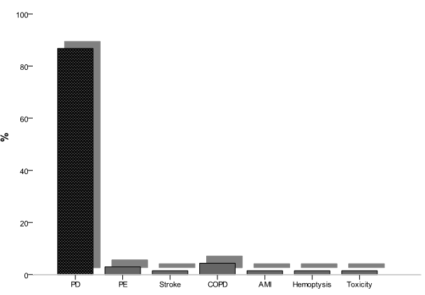
In patients, the main causes of death were progression disease in 59 patients (86,7%), pulmonary embolism in 2 patients (2,9%), stroke in 1 patient (1,47%), chronic obstructive pulmonary disease in 3 patients (4,4%), myocardial infarction in 1 patient (1,47%), hemoptysis in 1 patient (1,47%) and chemotherapy related toxicity in 1 patient (1,47%) (Figure 1).

Figure 1. main causes of death of patients

Figure 1. main causes of death of patients

Time to treatment failure

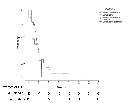
Median TTF was 2,53 months CI95% [2,21 – 2,84] (Figure 2). Considering age, sex, brain metastases, platinum compound and doublet of chemotherapy, we did not find differences (Table 2). Regarding doublet of chemotherapy, in patients in which gemcitabine was used there was an improvement in TTF of 1.2 months (p= 0.107, log rank) (Figure 3).

Figure 2. Median time to treatment failure in all patients

Fig 2.Median time to treatment failure in all patients

Figure 3. Time to treatment failure regarding doublet of chemotherapy.

Fig3.Time to treatment failure regarding doublet of chemotherapy.

Table 2. Medians and hazard ratios of time to treatment failure regarding age, sex, brain metastases, platinum compound and doublet of chemotherapy

  Median months, IC95%


P, log rank Hazard ratio, IC95% P, cox
Age

<70 años

>70 años


2,9 [2,07-3,9]

2,3 [2,1-2,5]


0,264


0,757 [0,46-1,24]


0,269

Sex

Male

Female


2,5 [2,2-2,8]

1,9 [0,0-5,04]


0,714


0,806 [0,25-2,6]


0,718

Brain Metastases (n, %)

No

Yes


2,5 [2,1-2,8]

2,3 [n.r.]


0,771


0,747 [0,1-5,5]


0,774

Platinum (n,%)

Cisplatin

Carboplatin


2,3 [2,01-2,7]

2,6 [1,9- 3,2]


0,895


1,037 [0,6-1,7]


0,896

Doublet of CT (n, %)

Microtubules inhibitor

Gemcitabine


1,7 [0,0-4,3]

2,5 [2,2- 2,8]


0,107


1,677 [0,8-3,1]


0,115

Overall survival

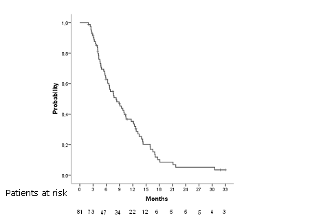
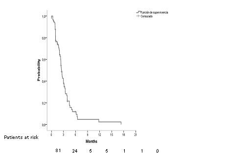
Median OS was 8,246 months CI95% [5,8 –2,6] in all patients (Figure 4). Overall survival rate al 6, 12 and 18 months were 62,8%, 35,1% and 3,8% respectively. Considering age, sex, brain metastases, platinum compound and doublet of chemotherapy there were statistically significant differences regard sex and doublet of CT (Table 3).

Figure 4. Overall survival in all patients

Fig 4. Overall survival in all patients

Table 3. Medians and hazard ratios of time to treatment failure regarding age, sex, brain metastases, platinium compound and doublet of chemotherapy

  Median months, IC95%


P, log rank Hazard ratio, IC95% P, cox
Age

<70 años

>70 años


7,5 [4,6-10,5]

8,9 [6,4-11,3]


0,619


0,884 [0,54-1,43]


0,619

Sex

Male

Female


8,3 [6,2-10,5]

3,2 [2,4-4,1]


0,006


0,217 [0,06-0,719]


0,012

Brain Metastases (n, %)

No

Yes


8,2 [5,8-10,6]

4,3 [n.r.]


0,596


1,466 [0,35-6,1]


0,599

Platinium (n,%)

Cisplatin

Carboplatin


10,48 [3,9-17,02]

7,65 [4,9- 10,33]


0,260


0,764 [0,42-1,2]


0,263

Doublet of CT (n, %)

Microtubules inhibitor

Gemcitabine


5,7 [3,8-7,6]

9,45 [6,7- 12,1]


0,018


1,989 [1,1-3,5]


0,021

In multivariate analyses adjusted for sex, age (< 70 or ≥ 70 years) and platinium the first line chemotherapy regimen based in antimetabolites was a predictor of OS (p=0.018).

Second line

Only 31 (37,8%) patients received second line therapy of treatment. Most common agents were docetaxel (17 patients, 54,8%), paclitaxel (6 patients, 19,9%), erlotinib (6 patients, 19,4%), gefitinib (1 patient, 3,2%) and gemcitabine (1 patient, 3,2%).

Median OS posprogression (OSpp) was 6,637 months CI95% [5,4 –7,8]. With respect to type of treatment, patients treated with docetaxel had a median of 9,01 months CI95% [5,1 –12,9], patients treated with paclitaxel 6,6 months CI95% [0,5 –12,7] and patients treated with erlotinib 5,8 months CI95% [4,6 –7,1]. Patient treated with gefitinib and gemcitabina were alive while 5,4 months and 3,2 months respectively. There were statistically significant differences in median OSpp regarding type of treatment (p=0,026 log rank). Hazard ratio for use of taxanes was 0,695, CI95% [0,26 –1,83], p=0,4.

Discussion

Squamous cell NSCLC is a particularly aggressive type of lung cancer, and few treatments are effective [6]. The NCCN and ESMO frontline recommendations include chemotherapy doublets, which are considered the cornerstone of initial therapy for squamous NSCLC [7,8]. To our knowledge, no prospective clinical trial has specifically compared cytotoxic chemotherapies for advanced lung SCC so there is is uncertainty about the best option of treatment.

Respect gender, women is represented in 3,7% in our study. In most published studies, little is reported about women diagnosed of advanced squamous NCSLC treated with CT. Overall survival among women in our study is poor (3,2 months versus 8,3). Recently, it has been described that females patients with squamous NCSLC had a significantly higher rate of human papillomavirus (HPV) infection compared to males with SCC [9] and HPV infection appears to be involved in cancer progression in SCC by promoting the expression of p53. On the other hand, patterns of mutation in SCC are unknown being KRAS, FGFR1 and PIK3CA most frequently reported and women seem to have less PIK3CA mutation than men. Moreover, there may exist unknown endocrine mechanisms this particularly lack of response and bad prognosis in women. To our knowledge, there are no differences in smoking patters that could explain this fact [10].

Elderly and young patients, defined by >70 years or <70 years, have similar proportion in our series. Although doublets with microtubule inhibitors are less used in old patients overall survival is similar in both groups. The definition of elderly patients varies widely across trials and and an uniform definition is lacking. Particularly in the actual world setting, older patients are often untreated even even when free of comorbid illnesses. Most studies in advanced NSCLC define elderly patients as those older than 70 years for the treatment of advanced NSCLC [11] and the benefit of platinum-based doublet regimens in this population seems to be greater than single-agent chemotherapy. So avoiding the use of doublets in elderly  is not justified in absence of comorbid status. Studies in elderly patients show similar response rates than in younger ones even with aggressive regimens of chemotherapy.

Palliative Care Unit (PCU) has played a preeminent role in the management of patients. More than half of our patients were remitted to PCU (45 patients, 54,9%) This fact allowed better control of symptoms, especially at home, and ultimately improved patients’ quality of life [12]. Early integration of palliative care into the treatment strategy should be mandatory because it may improve quality of life, particularly end-of-life care and improve overall survival. Our institution is working to make success this target.

Median time to treatment failure was 2,53 months and overall survival 8,24 months. The use of cisplatin or carboplatin has no impact on TTF or on OS, although use of cisplatin has a tendency to improve overall survival in 3 months over the use of carboplatin. Our data are similar to the reported about Veterans Health Administration data [12]. The use of gemcitabine as platinum doublet has demonstrated increased survival in our patients over microtubule inhibitors (9,45 months versus 4,7 months). A phase III trial of cisplatin and pemetrexed compared with cisplatin and gemcitabine revealed a statistically significant improvement in OS with cisplatin and gemcitabine in patients with squamous cell histology (10.8 vs 9.4 months) [13,14]. A retrospective study in patients with advanced lung SCC, revealed the use of various regimens did not have a significant effect on survival outcomes [15].

Finally, 31 patients (37,8%) received second line of treatment being docetaxel the most common agent used in this setting followed by paclitaxel and erlotinib. Although the number of patients is small, there is an improvement in survival in the group treated with taxanes respect to the group treated with tyrosine kinase inhibitor TKI). In TITAN phase III study, [16] patients were randomized to receive TKI or CT (taxanes or pemetrexed) and both treatments were similar in efficacy with different toxicity profiles.

We conclude that gemcitabine must be the CT of choice in advanced SCC. On the other hand, female patients had worse prognosis, patients aged >70 years old must receive a double of platinum, cisplatin is similar to carboplatin and cisplatin must be used when there is no contraindication for its use. Aditional agents and strategies must be developed in this setting to improve quality of life and survival.

References

  • Ferlay J, Steliarova-Foucher E, Lortet-Tieulent J, Rosso S, Coebergh JW, et al. (2013) Cancer incidence and mortality patterns in Europe: estimates for 40 countries in 2012. Eur J Cancer 49: 1374-1403. [crossref]
  • Le Chevalier T, Scagliotti G, Natale R, Danson S, Rosell R, et al. (2005) Efficacy of gemcitabine plus platinum chemotherapy compared with other platinum containing regimens in advanced non-small-cell lung cancer: a meta-analysis of survival outcomes. Lung Cancer 47: 69-80.
  • Reck M, Popat S, Reinmuth N, De Ruysscher D, Kerr KM, et al. (2014) Metastatic non-small-cell lung cancer (NSCLC): ESMO Clinical Practice Guidelines for diagnosis, treatment and follow-up. Ann Oncol 25 Suppl 3: iii27-39. [crossref]
  • Schiller JH, Harrington D, Belani CP, et al. (2002) Comparison of four chemotherapy regimens for advanced non small-cell lung cancer. N Engl J Med 346: 92-8.
  • Edge SB, Compton CC (2010) The American Joint Committee on Cancer: the 7th edition of the AJCC cancer staging manual and the future of TNM. Ann Surg Oncol 17: 1471-1474. [crossref]
  • McKeage MJ, Jameson MB, et al. (2010) Comparative outcomes of squamous and non-squamous non-small cell lung cancer (NSCLC) patients in phase II studies of ASA404 (DMXAA): retrospective analysis of pooled data. J Thorac Dis 2:199-204.
  • Ettinger DS, Wood DE, Akerley W, Bazhenova LA, Borghaei H, et al. (2014) Non-small cell lung cancer, version 1.2015. J Natl Compr Canc Netw 12: 1738-1761. [crossref]
  • Reck M, Popat S, Reinmuth N, De Ruysscher D, Kerr KM, Peters S (2014) ESMO Guidelines Working Group. Metastatic non-small-cell lung cancer (NSCLC): ESMO Clinical Practice Guidelines for diagnosis, treatment and follow-up. Ann Oncol 25 Suppl 3: iii27-39.
  • Fan X, Yu K, Wu J, Shao J, Zhu L, et al. (2015) Correlation between squamous cell carcinoma of the lung and human papillomavirus infection and the relationship to expression of p53 and p16. Tumour Biol 36: 3043-3049. [crossref]
  • Qiong Z, Na WY, Bo W2, Zhu Z, Ling P, et al. (2015) Alterations of a spectrum of driver genes in female Chinese patients with advanced or metastatic squamous cell carcinoma of the lung. Lung Cancer 87: 117-121. [crossref]
  • Owonikoko TK, Ramalingam SS2 (2015) Minimize toxicity or preserve efficacy? A delicate trade-off in the management of older patients with lung cancer. J Clin Oncol 33: 534-536. [crossref]
  • Wagner B, Meffert C2, Becker G2 (2015) [Availability and integration of palliative medicine at certified lung cancer centers]. Pneumologie 69: 218-224. [crossref]
  • Santana-Davila R, Szabo A, Arce-Lara C, Williams CD, et al. (2014) Cisplatin versus carboplatin-based regimens for the treatment of patients with metastatic lung cancer. An analysis of Veterans Health Administration data. J Thorac Oncol 9: 702-9.
  • Scagliotti GV, Parikh P, von Pawel J, et al. (2008) Phase III study comparing cisplatin plus gemcitabine with cisplatin plus pemetrexed in chemotherapy-naive patients with advanced-stage non-small-cell lung cancer. J Clin Oncol 26:3543-3551.
  • Liao BC, Shao YY, Chen HM, Shau WY, Lin ZZ, et al. (2015) Comparative effectiveness of first-line platinum-based chemotherapy regimens for advanced lung squamous cell carcinoma. Clin Lung Cancer 16: 137-143. [crossref]
  • Ciuleanu T, Stelmakh L, Cicenas S (2012) Efficacy and safety of erlotinib versus chemotherapy in second-line treatment of patients with advanced, non-small-cell lung cancer with poor prognosis (TITAN): a randomised multicentre, open-label, phase 3 study. Lancet Oncol 13:300-8.

Humanized Monoclonal Antibodies in Pulmonology: An Integrated Review

DOI: 10.31038/IMROJ.2017213

Abstract

Asthma is an important chronic disease affecting a lot of people worldwide. Treatment options for asthma like biological agents are being developed more frequently nowadays. Despite a lot of treatment options, some patients still remain symptomatic. As more and more practitioners choose treatment with biologic agents as a convenient way of therapy, biologic agents and other valuable methods must be discovered in order to cope with a growing number of treatment agents. This manuscript emphasizes on new generation monoclonal human (ized) antibodies in asthmatics and off-label use. The first developed biologic agent is the anti- immunoglobulin E monoclonal antibody called omalizumab. Currently it is an approved treatment option for asthma.

Introduction

Asthma is an important chronic disease affecting a lot of people worldwide [1]. Treatment options for asthma like biological agents are being developed more frequently nowadays. Despite a lot of treatment options, some patients still remain symptomatic. As more and more practitioners choose treatment with biologic agents as a convenient way of therapy, biologic agents and other valuable methods must be discovered in order to cope with a growing number of treatment agents. This special issue emphasizes on new generation monoclonal human (ized) antibodies in asthmatics [2-4].

The pathophysiological mechanisms underlying asthma, which is a heterogeneous disease, are characterized by interactive responses among various cell types and the hematopoietic cells of the adaptive and innate immune systems. Frequently is conventional therapy like inhaled steroids and beta-agonists sufficient for asthma symptoms. However, a little minority of the asthmatics is not controlled with conventional therapy. Therefore, are new treatment options essential for severe asthmatic patients [5].

Anti-IL-5 Molecules

Interleukins derived from T-helper-2 (Th2) cells and innate lymphoid cells play an important role in the pathogenesis of asthma. Monoclonal antibodies targeting these cytokines as treatment for severe asthma are expected to be beneficial [6]. Eosinophilic inflammation is an important event in the pathogenesis of asthma. IL-5 is a key cytokine that arranges eosinophil production, survival, maturation and recruitment of eosinophils to the inflammation [7]. Mepolizumab, reslizumab, and benralizumab are new developed monoclonal antibodies that target the cytokine IL-5. Mepolizumab and reslizumab have been approved by the US Food and Drug Administration (FDA) for the treatment of patients with severe asthma with an eosinophilic phenotype [8,9]. Mepolizumab and reslizumab binds directly to IL-5 ligand. These molecules effectively decreased circulating and sputum eosinophil counts, but they failed to improve airway mucosal eosinophilia, acute exacerbation rates, lung function and symptom scores in several studies. These disappointing results may be affected from inappropriate selection of the patients. In order to overcome the probable mechanistic limitations of early anti-IL-5 agents, an anti-IL5R monoclonal antibody was developed and called as benralizumab. Benralizumab, previously known as MEDI-563, is a humanized recombinant IgG1-k isotype monoclonal antibody. It was constructed from the mouse anti-human IL-5Rα mAbs generated by mice immunized with recombinant human IL- 5Rα [10]. Clinical studies revealed that anti-interleukin 5 monoclonal therapies for asthma could be safe for slightly improving FEV1 (or FEV1% of predicted value), quality of life, and reducing exacerbations risk and blood and sputum eosinophils. However these drugs have no significant effect on PEF, and SABA rescue use. These may be a result of patient selection. Further trials are required to clarify the optimal antibody for different patients [7].

Anti-IL-4/IL-13 Molecules

Another investigated cytokine important in the inflammatory pathways in the pathogenesis of asthma is anti-IL-4. IL-4 is a pleiotropic cytokine secreted mainly by activated T cells. Mast cells, basophils, and eosinophils can also secret IL-4 [10,11]. IL-4 is important in inducing IgE isotype switching, T cell polarization into Th2 cells, and generation of IL-4, IL-5, and IL-13 by Th2 cells. IL-4Rα is expressed on CD4+ and CD8+ T cells, B cells, macrophages, lung epithelial cells, airway goblet cells, and smooth muscle cells [11]. There is a functional homogeniety between IL-4 and IL-13. IL-4 can activate a heterodimeric receptor complex consisting of the IL-4 receptor α- subunit (IL-4Rα) and a γC subunit. IL-13 can activate the IL-4Rα and the IL-13 receptor α1-subunit (IL-13Rα1) [5]. Both IL-4 and IL-13 can bind to heterodimeric combination of the α-subunit of the IL-13 receptor and the α-subunit of the IL-4 receptor. And this leads to signaling of both IL-4 and IL-13. Therefore, will blocking IL-4R α with an antibody of this receptor chain expected to block the effects of both IL-4 and IL-13 [6].

Pascolizumab and VAK694 are anti-IL-4 neutralizing monoclonal antibodies. Also, IL-4 receptor antagonist drugs like dupilumab, pitrakinra and AMG-317 have been discovered. Even a recombinant IL-4Rα that captures soluble IL-4 and prevents their binding to IL-4 receptors, has been developed. It is called altrakincept. However, further research on this drug was discontinued by its manufacturer, since the phase 3 clinical trial failed to confirm its earlier promising results. Since there is a high redundancy of IL-4 and IL-13 signaling, blocking of both IL-4 and IL-13 has been expected to be more efficient [11]. Dupilumab is a drug that inhibits signaling from IL- 4 and IL-13 concomitantly. It is a molecule that binds to the alpha subunit of the IL-4 receptor. Phase II trials for dupilumab showed that asthma exacerbations were decreased in patients using this drug [12,13]. Dupilumab also improved lung function. It reduced the inhaled corticosteroid dose in the patient group. There was also an associated reduction in fractional exhaled nitric oxide with reduced serum concentrations of Th2-associated inflammatory markers such as CCL17 (TARC), CCL26 (eotaxin-3), and IgE [6]. These results are promising and further clinical trials will us show us the long-term efficacy of dupilumab [11].

Another similar drug that targets IL-4 is pitrakinra. It is a recombinant human IL-4 variant that competitively inhibits IL-4Ra to interfere with the actions of both IL-4 and IL-13. Studies on this competitive antagonist called pitrakinra revealed that it leads to significant reduction in asthma exacerbations and improves asthma symptoms in patients with eosinophilia [14]. Pitrakinra also attenuated the late-phase asthmatic response to allergen challenge in patients with mild atopic asthma [6]. The other drug called AMG317 was evaluated in another phase II trial in approximately 300 patients with moderate to severe asthma. Weekly injections over 12 weeks were well tolerated but did not have significant effects on the Asthma Control Questionnaire score (ACQ score; the primary outcome) [14].

Interleukin 13 shares 30% homology with interleukin 4. IL-13 is secreted by Th2 cells, ILC2s, mast cells, basophils, and eosinophils. IL-13 has the potential to increase goblet-cell differentiation, and activation of fibroblasts. IL-13 production can induce an increase in bronchial hyperresponsiveness, and switching of B-cell antibody production towards IgE [6]. IL-13 is similar to IL-4 and uses the same signaling pathways. The high-affinity receptor of IL-13 is a heterodimer of IL-4Rα/IL- 13Rα1. IL-13Rα1 is present on eosinophils, B cells, monocytes, macrophages, smooth muscle cells, lung epithelial cells, airway goblet cells, and endothelial cells. Biologicals that target IL-13 are anti-IL-13 mAbs: anrukinzumab, dectrekumab, GSK679586, IMA-026, lebrikizumab, RPC-4046, and tralokinumab [11].

Patients with severe asthma often have elevated levels in sputum despite therapy with high dose inhaled or oral corticosteroids. Lebrikizumab is one of the many humanized monoclonal antibodies that have been developed to specifically bind to IL-13 and inhibit its function [14]. In a randomized, double-blind, placebo- controlled study were 219 asthma patients observed and evaluated whether the drug lebrikizumab could alter the course of asthma. Lebrikizumab treatment was associated with improved lung function. Patients with high pretreatment levels of serum periostin had greater improvement in lung function with lebrikizumab. This therapy needs futher evaluation before being utilized in clinics [15]. Another monoclonal antibody called tralokinumab, an investigational human IL-13-neutralising immunoglobulin G4 monoclonal antibody, has been evaluated in adults with moderate to severe uncontrolled asthma despite controller therapies. Patients were randomly assigned to receive tralokinumab or placebo subcutaneously every 2 weeks for 13 weeks. Although it had an acceptable safety and tolerability, it did not reduce asthma exacerbations [16].

Antithymic stromal lymphopoietin

Thymic stromal lymphopoietin (TSLP) is an epithelial cell derived cytokine that may trigger allergic inflammation and, thus, play a role in allergic asthma [17]. It is an epithelial-derived cytokine and makes its effect through its receptor, TSLP-R, which is a heterodimeric receptor that consists of the IL-7 receptor alpha chain (IL-7Rα) and the TSLP receptor alpha chain 1 (TSLPRα). In hematopoietic cells, TSLP-R is mainly expressed in DCs, monocytes, B cells, T cells, NK cells, invariant natural killer T (iNKT) cells, eosinophils, basophils, and mast cells [11].

A human anti TSLP monoclonal immunoglobulin G2 lambda (AMG 157) that binds human TSLP and prevents receptor interaction was assessed in a trial. Randomly assigned 31 patients with mild allergic asthma received AMG 157 (700 mg) or placebo intravenously, once a month for three doses. The primary outcome, the maximum percentage decrease in the FEV during the late asthmatic response was 45.9 percent less in the AMG 157 group than the placebo group on day 84. AMG 157 reduced allergen induced bronchoconstriction and airway inflammation. No serious adverse effects were reported. Further studies on this drug are planned to clarify its use in clinical practice [17].

Anti-IL-9 Monoclonal Antibody

IL-9 is a Th2 cytokine and a T cell and mast cell growth factor. Anti-IL-9 antibody-treatment has been shown to protect from allergen-induced airway remodeling, with a concomitant reduction in mature mast cell numbers and activation. It can also decrease expression of the profibrotic mediators transforming growth factor (TGF)-b1, vascular endothelial growth factor (EGF), and fibroblast growth factor-2 (FGF-2) in the lung. The function of IL-9 in allergy has been investigated for its pleiotropic activities on cell types associated with allergic diseases including Th2 lymphocytes, mast cells, B cells, eosinophils, and airway epithelial cells. An anti-IL-9 monoclonal antibody (MEDI-528) has been studied in a clinical trial on 327 asthmatic subjects. Patients were randomized to receive placebo or one of three doses of MEDI-528 (dosage 30, 100, or 300 mg s.c. twice weekly for 4 weeks) in addition to their usual asthma medications. The addition of MEDI-528 to existing asthma controller medications did not improve ACQ-6 scores, asthma exacerbation rates, or FEV1 values. Further clinical trials are needed to explore this drug for altering the course of asthma. Thus, the potential clinical benefit of targeting IL-9 or its receptor in the treatment of asthma remains to be shown in further studies [5].

Anti-IL-2 antibody

Allergen exposure can stimulate IL-2 and its receptor expression (IL -2R) a chain (sCD25) in airways of patients with severe asthma. Daclizumab is a humanized monoclonal antibody that binds specifically to a subunit (CD25) of the high-affinity IL-2R, and inhibits IL-2 binding and its biological activity. Daclizumab can inhibit various T cell functions, including T cell proliferation and cytokine production. It has been investigated in a randomized controlled study. The drug has the potential to improve pulmonary function and asthma control in patients moderate to severe chronic asthma [18]. The risk of immunosuppression in clinical practice needs to be clarified.

Anti-GATA3-spesific DNAzyme

Approximately half of the asthmatic patients exhibit a Th2 type in response to allergen exposure. This Th2 endotype is characterized by a predominant activation of Th2 cells that produce cytokines such as interleukins 4, 5, and 13. The expression and production of all these Th2 cytokines have been shown to be controlled by the zinc finger transcription factor GATA3, which is essential for Th2-cell differentiation and activation. It is considered to be the master transcription factor of the Th2 pathway of immune activation. Therefore, could be interventions to disrupt this immune network, a synthetic DNA molecule (DNAzyme), that binds to GATA3 messenger RNA and cleaves it, a solution. This synthetic molecule called SB010 could significantly attenuate both late and early asthmatic responses after allergen provocation in patients with allergic asthma. Biomarker analysis after this drug showed an attenuation of Th2-regulated inflammatory responses [19].

Anti-IL-17 antibody

Although half of the asthma patients exhibit a Th2 type endotype, some remaining patients exhibit a Th17 driven endotype. This subpopulation is characterized with a Th17 driven inflammation. Th17 cells can contribute to airway hyperresponsiveness by recruiting both eosinophils and neutrophils. Therefore, has been IL-17 receptor blocking suggested to beneficial in asthma treatment [20]. Biologicals targeting IL-17 include an anti-IL-17A mAb: secukinumab and an anti-IL- 17 receptor mAb: brodalumab. Although the inhibition of IL-17 receptor A had no effect on subjects with asthma as a whole, a subgroup analysis showed an effect with uncertain significance. Further studies are needed to determine the role of secukinumab in asthma [11]. Brodalumab (AMG 827) is a human, anti–IL-17RA immunoglobulin G2 (IgG2) monoclonal antibody that binds with high affinity to human IL-17RA, blocking the biologic activity of IL- 17A, -17F, -17A/F heterodimer, and -17E (IL-25). Brodalumab can block IL-25 activity and IL-17A and IL-17F. In a randomized controlled study were 302 patients taking this drug evaluated and at the end of the study there was no evidence for an effect of brodalumab in these patients. Further studies may clarify the potential of this drug [20].

Anti TNF antibodies

In addition, human(ized) monoclonal antibodies (HMA) evaluated for the treatment of severe persistent asthma (SPA), but not approved after Phase II trial are as follows; Infliximab (Recombinant human–murine chimeric anti-TNFα monoclonal antibody Infliximab), etanercept (Soluble TNFα receptor fusion protein), and golimumab (Fully human TNFα-blocking antibody) [5]. The expression of TNF alfa is increased in asthma in association with airway neutrophilia. Berry and colleagues have demonstrated that the TNF-α axis is upregulated in patients with refractory asthma, as evidenced by the increased expression of membrane-bound TNF-α, TNF receptor 1, and TNF-α– converting enzyme by peripheral-blood monocytes [21]. Treatment with golimumab did not demonstrate a favorable risk-benefit profile in patients with severe persistent asthma [22]. A study with etanercept showed a small decrease in asthma exacerbations was observed in a randomized placebo controlled study [23]. In a case-series report was it told that in severe, uncontrolled, steroid-dependent asthma infliximab could reduce exacerbations and hospitalizations [24]. In some severe refractory asthma endotypes anti-TNFα therapy may have a role. However, it should be kept in mind that these agents have some safety concerns and should use carefully only in some severe refractory asthma endotypes [5].

ANTI-IgE

The first developed biologic agent is the anti-immunoglobulin E monoclonal antibody called omalizumab. Currently it is an approved treatment option for asthma [5].

The other human(ized) promising monoclonal antibody drug developed, but not approved yet is ligeluzimab. Ligeluzimab binds with very high affinity to the Cε3 domain of IgE. Ligeluzumab may provide longer supression of IgE. Trials with this biologic agent are ongoing [5].

Anti-ige: off-label use non-atopic asthma

The off-label use of omalizumab in patients with uncontrolled non-atopic asthma has resulted in a decrease in exacerbation rates and improvement in asthma symptom scores. In a study conducted in 2013, omalizumab was administered to 266 patients with severe allergic asthma and 29 patients with non-atopic severe asthma for two years, and the study found a decline in the exacerbation rate, increase in the quality of life, and significantly improved disease control in both groups [25-27]. In two studies which used omalizumab in a group of patients with non-atopic severe asthma, the authors observed downregulation of FcRI expression in the basophils and increased FEV1.

Nasal polyposis, allergic rhinitis, and allergic bronchopulmonary aspergillosis

Allergic bronchopulmonary aspergillosis affects 7 to 9% of patients with cystic fibrosis (CF) and 1 to 2% of patients with asthma, posing a diagnostic challenge [28,29]. Cases series related to ABPA were first published in 2007. In a series of eight cases with cystic fibrosis and ABPA published by Tanou et al. [30] in 2014, the authors reported increased FEV1, improved respiratory symptoms, and reduced steroid consumption. In a series of six cases with CF diagnosed with ABPA published by Lehman et al. [31], the authors reported improved symptoms in patients receiving omalizumab, whereas the efficacy of the treatment was less pronounced in patients diagnosed with ABPA and long disease duration and in patients who developed progressive lung problems [31]. In a series of 14 patients with severe asthma and ABPA in 2015, Aydın et al. [32] showed that 11 patients achieved complete and three patients achieved partial response. The authors also reported an overall improvement in the pulmonary functions and respiratory symptoms with a statistically significant reduction in the use of oral corticosteroids (OCS) and number of disease episodes. Also, the patients with a total immunoglobulin E (IgE) level of <1,000 IU showed a better response to omalizumab, compared to those with a total IgE level of >1,000 IU. However, this finding was found to have a low-evidence level, considering the lack of large-scale, prospective case series and randomized and placebo- controlled studies.

Efficacy and safety of omalizumab were first evaluated in a randomized, double-blind, placebo-controlled study of 221 patients with seasonal allergic rhinitis in 2002 [33]. This study reported a significant symptomatic relief up to 48% in the combination treatment group (specific immunotherapy [SIT]+omalizumab), compared to SIT group alone. A randomized study in Japan reported a significant improvement in daily nasal and eye symptoms in patients with seasonal allergic rhinitis receiving omalizumab [34].

In 2007, a randomized placebo-controlled study of eight patients was the first to report reduced rates of postoperative polyp recurrence in patients with atopic asthma and nasal polyps (NP) [35]. In a study of 19 patients with severe asthma and NP in 2011, Vennera et al. [36] reported symptom reduction and disease stabilization with the use of omalizumab treatment. In addition, Tajiri et al. [37] evaluated omalizumab in patients with severe asthma and NP, and reported significant improvements in nasal symptoms, asthma control, and sinus tomography results. However, not all studies were able to show the beneficial effects of the treatment. In a randomized, double-blind, placebo-controlled study of patients with chronic rhinosinusitis receiving omalizumab, Pinto et al. [38] showed improvement in the Sino-Nasal Outcome Test (SNOT-20) scores at three, five, and six months, although there was no significant difference in the scores compared to the control group. The aforementioned study did not observe any changes in the quality of life, symptom scores, cellular inflammation, nasal passage, and olfactory test parameters.

Atopic Dermatitis And Food Allergy

In a series of three patients published in 2005, the authors reported no response after four months of treatment [39], while Lane et al. [40] published a series of three patients in the same study period and reported successful treatment outcomes of severe AD using omalizumab [40]. In addition, a pilot study of 21 patients published in 2009 found a statistically significant clinical improvement in all patients [41]. Another series of three patients published in 2011 reported significant improvement in the Eczema Area and Severity Index (EASI) and itching severity score in patients with severe AD unresponsive to conventional treatment and those with elevated IgE levels [42]. A study of 11 patients published in the same year reported reduced SCORing Atopic Dermatitis (SCORAD) scores, reduced symptoms, and significant improvement in the quality of life (3). The efficacy of omalizumab was also evaluated in 20 adults with severe AD in a prospective, 28-week, open-label study conducted by Hotze et al. [43] in 2014. The authors reported no response to treatment in seven patients harboring filaggrin gene mutation (FLG), while there was a significant improvement in the remaining eight patients. The authors also concluded that the patients with FLG gene mutation were prone to achieve lower response to omalizumab.

Furthermore, in food-related immunotherapy (IT) studies, omalizumab initiated before or received simultaneously with the treatment facilitated the development of tolerance. In a series of 11 patients with cow milk allergy scheduled for IT, 10 patients tolerated daily intake of 8 g cow milk after the initiation of omalizumab, nine weeks before IT, and combination with IT treatment, thereafter [44]. Another use of this treatment is to facilitate a rapid and safe transition to the maintenance phase in patients with food allergy receiving oral IT [45]. Another use of the treatment in food allergy is eosinophillic esophagitis developing in association with multiple food allergies. Also, in a study administered omalizumab to patients with eosinophillic esophagitis, the authors found reduced allergic symptoms and improved quality of life, although there was no change in endoscopic and histologic characteristics of the disease [46].

Food Allergy and Anaphylaxis

Anaphylaxis can occur as a result of exposure to various allergens such as food, drug, and venom; however, no triggering factor can be shown in some cases, of which the latter is known as idiopathic anaphylaxis. The patients with elevated baseline tryptase levels or those diagnosed with mastocytosis are expected to have higher rates of anaphylaxis with a more aggressive course of disease. In particular, patients with venom allergy on IT may experience some difficulties in switching to maintenance dose. Severe anaphylactic episodes can be observed in patients diagnosed with mastocytosis on IT due to venom allergy. In addition, IT combined with omalizumab has enabled a safe transition to the maintenance phase in this group of patients [47-49]. Addition of omalizumab to rush and ultra-rush venom IT protocols has increased the success of IT and enabled a safer transition to the maintenance phase. Another use of omalizumab is to prevent recurrent anaphylactic episodes in patients who are unable to be controlled with conventional therapies [50- 55]. There are case reports on the role of this treatment in preventing idiopathic anaphylactic episodes [56-58].

Several studies have reported asthma symptom control, improved quality of life, and development of tolerance to aspirin in two patients with the use of omalizumab in patients with aspirin-induced airway disease, nasal polyps, and severe asthma [59, 60]. Two patients with recurrent insulin allergy, despite the use of desensitization protocols, and one patient of carboplatin allergy during carboplatin therapy due to ovarian cancer successfully continued their treatment with the addition of omalizumab to the treatment [61-63].

In conclusion, although omalizumab has been approved for the treatment of severe allergic asthma and chronic idiopathic urticaria, it offers an off-label use as a final resort in many allergic diseases. Recent studies have shown that omalizumab is effective in treating bullous pemphigoid, Stevens-Johnson syndrome (SJS)/toxic
epidermal necrolysis (TEN, Type-III/IV reaction, such as graft versus host disease), Netherton syndrome, asthma and chronic obstructive pulmonary disease overlap syndrome (ACOS). Its effects on soluble inflammatory markers, such as sCD200, sTRAIL, hematopoietic cells, Th1/2 cytokines (CXCL8; IL-1β; IL-4; IL-5; IL17A), total antioxidant capacity, hydrogen peroxide, malondialdehyde and total nitric oxide concentrations were demonstrated in several studies [64-70].

Monoclonal antibodies are a candidate for use in several indications with the contribution of large-scale studies to the literature in the near future.

Declaration of Interest

All authors declare that they have no conflict of interest.

Acknowledgements

Prof.Dr. Tse Wen Chang, Prof.Dr.Saadet Gumuslu, Prof.Dr. Fatih Uz, Prof.Dr Arzu Mirici.

References

  • Beasley R, Semprini A, Mitchell EA (2015) Risk factors for asthma: is prevention possible? Lancet 386: 1075-1085. [crossref]
  • Yalcin AD, Celik B, Gumuslu S (2014) D-dimer levels decreased in severe allergic asthma and chronic urticaria patients with the omalizumab treatment. Expert Opin Biol Ther 14:283-6.
  • Yalcin AD (2015) Advances in anti-IgE therapy. Biomed Res Int 2015: 317465. [crossref]
  • Yalcin AD, Cilli A, Bisgin A, et al. (2013) Omalizumab is effective in treating severe asthma in patients with severe cardiovascular complications and its effects on sCD200, d-dimer, CXCL8, 25-hydroxyvitamin D and IL-1ß levels. Expert Opin Biol Ther 13:1335-41.
  • Mitchell PD, El-Gammal AI, O’Byrne PM (2016) Emerging monoclonal antibodies as targeted innovative therapeutic approaches to asthma. Clin Pharmacol Ther 99:38-48.
  • Chung KF (2015) Targeting the interleukin pathway in the treatment of asthma. Lancet 386: 1086-1096. [crossref]
  • Wang FP, Liu T, Lan Z, Li SY, Mao H (2016) Efficacy and Safety of Anti-Interleukin-5 Therapy in Patients with Asthma: A Systematic Review and Meta-Analysis. PLoS One 11: e0166833. [crossref]
  • Ortega HG, Liu MC, Pavord ID, Brusselle GG, FitzGerald JM, et al. (2014) Mepolizumab treatment in patients with severe eosinophilic asthma. N Engl J Med 371: 1198-1207. [crossref]
  • Castro M, Zangrilli J, Wechsler ME et al. (2015) Reslizumab for inadequately controlled asthma with elevated blood eosinophil counts: results from two multicentre, parallel, double-blind, randomised, placebo-controlled, phase 3 trials. Lancet Respir Med 3:355-66.
  • Khorasanizadeh M, Eskian M, Assa’ad AH, Camargo CA Jr, Rezaei N,  et al. (2016) Efficacy and Safety of Benralizumab, a Monoclonal Antibody against IL-5Rα, in Uncontrolled Eosinophilic Asthma. Int Rev Immunol 35: 294-311. [crossref]
  • Tan HT, Sugita K, et al. (2016) Novel Biologicals for the Treatment of Allergic Diseases and Asthma. Curr Allergy Asthma Rep 16: 70. [crossref]
  • Wenzel S, Ford L, Pearlman D, Spector S, Sher L, et al. (2013) Dupilumab in persistent asthma with elevated eosinophil levels. N Engl J Med 368: 2455-2466. [crossref]
  • Wenzel S, Castro M, Corren J, et al. (2016) Dupilumab efficacy and safety in adults with uncontrolled persistent asthma despite use of medium-to-high- dose inhaled corticosteroids plus a long-acting ß2 agonist: a randomised double-blind placebo-controlled pivotal phase 2b dose-ranging trial. Lancet 388:31-44.
  • Hambly N, Nair P (2014) Monoclonal antibodies for the treatment of refractory asthma. Curr Opin Pulm Med 20: 87-94. [crossref]
  • Corren J, Lemanske RF, Hanania NA, Korenblat PE, Parsey MV, et al. (2011) Lebrikizumab treatment in adults with asthma. N Engl J Med 365: 1088-1098. [crossref]
  • Piper E, Brightling C, Niven R, Oh C, Faggioni R, et al. (2013) A phase II placebo-controlled study of tralokinumab in moderate-to-severe asthma. Eur Respir J 41: 330-338. [crossref]
  • Gauvreau GM, O’Byrne PM, Boulet LP, et al. (2014) Effects of an anti-TSLP antibody on allergen-induced asthmatic responses. N Engl J Med 370:2102-10.
  • Busse WW, Israel E, Nelson HS, et al. (2008) Daclizumab improves asthma control in patients with moderate to severe persistent asthma: a randomized, controlled trial. Am J Respir Crit Care Med 178:1002-8.
  • Krug N, Hohlfeld JM, Kirsten AM, et al. (2015) Allergen-induced asthmatic responses modified by a GATA3-specific DNAzyme. N Engl J Med 372:1987-95.
  • Busse WW, Holgate S, Kerwin E, et al. (2013) Randomized, double-blind, placebo-controlled study of brodalumab, a human anti-IL-17 receptor monoclonal antibody, in moderate to severe asthma. Am J Respir Crit Care Med 188:1294-302.
  • Berry MA, Hargadon B, Shelley M, Parker D, Shaw DE, et al. (2006) Evidence of a role of tumor necrosis factor alpha in refractory asthma. N Engl J Med 354: 697-708. [crossref]
  • Wenzel SE, Barnes PJ, Bleecker ER, et al. (2009) A randomized, double-blind, placebo-controlled study of tumor necrosis factor-alpha blockade in severe persistent asthma. Am J Respir Crit Care Med 179:549-58.
  • Morjaria JB, Chauhan AJ, Babu KS, et al. (2008) The role of a soluble TNFalpha receptor fusion protein (etanercept) in corticosteroid refractory asthma: a double blind, randomised, placebo controlled trial. Thorax 63:584-91.
  • Taillé C, Poulet C, Marchand-Adam S, Borie R, Dombret MC, et al. (2013) Monoclonal Anti-TNF-α Antibodies for Severe Steroid-Dependent Asthma: A Case Series. Open Respir Med J 7: 21-25. [crossref]
  • Domingo C, Pomares X, Angril N, Rudi N, Amengual MJ, et al. (2013) Effectiveness of omalizumab in non-allergic severe asthma. J Biol Regul Homeost Agents 27: 45-53. [crossref]
  • Garcia G, Magnan A, Chiron R, et al. (2013) A proof-of-concept, randomized, controlled trial of omalizumab in patients with severe, difficult-to-control, nonatopic asthma. Chest 144:411-9.
  • de Llano LP, Vennera Mdel C, Álvarez FJ, Medina JF, Borderías L, et al. (2013) Effects of omalizumab in non-atopic asthma: results from a Spanish multicenter registry. J Asthma 50: 296-301. [crossref]
  • Laufer P, Fink JN, Bruns WT, Unger GF, Kalbfleisch JH, et al. (1984) Allergic bronchopulmonary aspergillosis in cystic fibrosis. J Allergy Clin Immunol 73: 44-48. [crossref]
  • Greenberger PA, Patterson R (1988) Allergic bronchopulmonary aspergillosis and the evaluation of the patient with asthma. J Allergy Clin Immunol 81: 646-650. [crossref]
  • Tanou K, Zintzaras E, Kaditis AG (2014) Omalizumab therapy for allergic bronchopulmonary aspergillosis in children with cystic fibrosis: a synthesis of published evidence. Pediatr Pulmonol 49:503-7
  • Lehmann S, Pfannenstiel C, Friedrichs F, Kröger K, Wagner N, et al. (2014) Omalizumab: a new treatment option for allergic bronchopulmonary aspergillosis in patients with cystic fibrosis. Ther Adv Respir Dis 8: 141-149. [crossref]
  • Aydin Ö, Sözener ZÇ, Soyyigit S et al. (2015) Omalizumab in the treatment of allergic bronchopulmonary aspergillosis: One center’s experience with 14 cases. Allergy Asthma Proc 36:493-500.
  • Kuehr J, Brauburger J, Zielen S, et al. (2002) Efficacy of combination treatment with anti-IgE plus specific immunotherapy in polysensitized children and adolescents with seasonal allergic rhinitis. J Allergy Clin Immunol 109:274-80.
  • Okubo K, Nagakura T (2008) Anti-IgE antibody therapy for Japanese cedar pollinosis: omalizumab update. Allergol Int 57: 205-209. [crossref]
  • Penn R, Mikula S (2007) The role of anti-IgE immunoglobulin therapy in nasal polyposis: a pilot study. Am J Rhinol 21: 428-432. [crossref]
  • Vennera Mdel C, Picado C, Mullol J, Alobid I, Bernal-Sprekelsen M (2011) Efficacy of omalizumab in the treatment of nasal polyps. Thorax 66: 824-825. [crossref]
  • Tajiri T, Matsumoto H, Hiraumi H, Ikeda H, Morita K, et al. (2013) Efficacy of omalizumab in eosinophilic chronic rhinosinusitis patients with asthma. Ann Allergy Asthma Immunol 110: 387-388. [crossref]
  • Pinto JM, Mehta N, DiTineo M, et al. (2010) A randomized, double-blind, placebo- controlled trial of anti-IgE for chronic rhinosinusitis. Rhinology 48:318-24.
  • Krathen RA, Hsu S (2005) Failure of omalizumab for treatment of severe adult atopic dermatitis. J Am Acad Dermatol 53: 338-340. [crossref]
  • Lane JE, Cheyney JM, Lane TN, Kent DE, Cohen DJ (2006) Treatment of recalcitrant atopic dermatitis with omalizumab. J Am Acad Dermatol 54: 68-72. [crossref]
  • Sheinkopf LE, Rafi AW, Do LT, Katz RM, Klaustermeyer WB (2008) Efficacy of omalizumab in the treatment of atopic dermatitis: a pilot study. Allergy Asthma Proc 29: 530-537. [crossref]
  • Thaiwat S, Sangasapaviliya A (2011) Omalizumab treatment in severe adult atopic dermatitis. Asian Pac J Allergy Immunol 29: 357-360. [crossref]
  • Hotze M, Baurecht H, Rodríguez E, et al. (2013) Increased efficacy of omalizumab in atopic dermatitis patients with wild-type filaggrin status and higher serum levels of phosphatidylcholines. Allergy 69:132-5.
  • Nadeau KC, Schneider LC, Hoyte L, Borras I, Umetsu DT (2011) Rapid oral desensitization in combination with omalizumab therapy in patients with cow’s milk allergy. J Allergy Clin Immunol 127: 1622-1624. [crossref]
  • Bégin P, Dominguez T, Wilson SP, Bacal L, Mehrotra A, et al. (2014) Phase 1 results of safety and tolerability in a rush oral immunotherapy protocol to multiple foods using Omalizumab. Allergy Asthma Clin Immunol 10: 7. [crossref]
  • Rocha R, Vitor AB, Trindade E, Lima R, Tavares M, et al. (2011) Omalizumab in the treatment of eosinophilic esophagitis and food allergy. Eur J Pediatr 170: 1471-1474. [crossref]
  • Kontou-Fili K. High omalizumab dose controls recurrent reactions to venom immunotherapy in indolent systemic mastocytosis. Allergy 63:376-8.
  • Sokol KC, Ghazi A, Kelly BC, Grant JA (2014) Omalizumab as a desensitizing agent and treatment in mastocytosis: a review of the literature and case report. J Allergy Clin Immunol Pract 2: 266-270. [crossref]
  • da Silva EN, Randall KL (2013) Omalizumab mitigates anaphylaxis during ultrarush honey bee venom immunotherapy in monoclonal mast cell activation syndrome. J Allergy Clin Immunol Pract 1:687- 8.
  • Douglass JA, Carroll K, Voskamp A, Bourke P, Wei A, et al. (2010) Omalizumab is effective in treating systemic mastocytosis in a nonatopic patient. Allergy 65: 926-927. [crossref]
  • Jagdis A, Vadas P (2014) Omalizumab effectively prevents recurrent refractory anaphylaxis in a patient with monoclonal mast cell activation syndrome. Ann Allergy Asthma Immunol 113:115-6.
  • Paraskevopoulos G, Sifnaios E, Christodoulopoulos K, Mantopoulou F, Papakonstantis M, et al. (2013) Successful treatment of mastocytic anaphylactic episodes with reduction of skin mast cells after anti-IgE therapy. Eur Ann Allergy Clin Immunol 45: 52-55. [crossref]
  • Matito A, Blázquez-Goñi C, Morgado JM, Alvarez-Twose I, Mollejo M, et al. (2013) Short-term omalizumab treatment in an adolescent with cutaneous mastocytosis. Ann Allergy Asthma Immunol 111: 425-426. [crossref]
  • Bell MC, Jackson DJ (2012) Prevention of anaphylaxis related to mast cell activation syndrome with omalizumab. Ann Allergy Asthma Immunol 108: 383-384. [crossref]
  • Kibsgaard L, Skjold T, Deleuran M, et al. (2014) Omalizumab induced remission of idiopathic anaphylaxis in a patient suffering from indolent systemic mastocytosis. Acta Derm Venereol 94:363-4.
  • Lee J (2014) Successful prevention of recurrent anaphylactic events with anti-immunoglobulin E therapy. Asia Pac Allergy 4: 126-128. [crossref]
  • Demirtürk M, Gelincik A, ColakoÄŸlu B, Dal M, Büyüköztürk S (2012) Promising option in the prevention of idiopathic anaphylaxis: omalizumab. J Dermatol 39: 552-554. [crossref]
  • Jones JD, Marney SR Jr, Fahrenholz JM (2008) Idiopathic anaphylaxis successfully treated with omalizumab. Ann Allergy Asthma Immunol 101:550-1.
  • Bobolea I, Barranco P, Fiandor A, et al. (2010) Omalizumab: a potential new therapeutic approach for aspirin-exacerbated respiratory disease. J Investig Allergol Clin Immunol 20:448-9.
  • Matheu V, Franco A, Perez E, Hernández M, Barrios Y (2007) Omalizumab for drug allergy. J Allergy Clin Immunol 120: 1471-1472. [crossref]
  • Cavelti-Weder C, Muggli B, Keller C, et al. Successful use of omalizumab in an inadequately controlled type 2 diabetic patient with severe insulin allergy. Diabetes Care 35:e41.
  • Yong PF, Malik R, Arif S, Peakman M, Amiel S, et al. (2009) Rituximab and omalizumab in severe, refractory insulin allergy. N Engl J Med 360: 1045-1047. [crossref]
  • Ojaimi S, Harnett PR, Fulcher DA (2013) Successful carboplatin desensitization by using omalizumab and paradoxical diminution of total IgE levels. J Allergy Clin Immunol Pract 2:105-6.
  • Uzun R, Yalcin AD, Celik B, et al. (2016) Levofloxacin Induced Toxic Epidermal Necrolysis: Successful Therapy with Omalizumab (Anti-IgE) and Pulse Prednisolone. Am J Case Rep 17:666-71.
  • Yalcin AD (2015) A case of netherton syndrome: successful treatment with omalizumab and pulse prednisolone and its effects on cytokines and immunoglobulin levels. Immunopharmacol Immunotoxicol 38:162- 6.
  • Yalcin AD, Celik B, Yalcin AN (2016) Omalizumab (anti-IgE) therapy in the asthma-COPD overlap syndrome (ACOS) and its effects on circulating cytokine levels. Immunopharmacol Immunotoxicol 38:253-6.
  • Yalcin AD, Genc GE, Celik B, Gumuslu S (2014) Anti-IgE monoclonal antibody (omalizumab) is effective in treating bullous pemphigoid and its effects on soluble CD200. Clin Lab 60: 523-524. [crossref]
  • Yalcin AD, Bisgin A, Gorczynski RM (2012) IL-8, IL10, TGF-ß and GCSF levels were increased in severe persistent allergic asthma patients with the anti-IgE treatment. Mediators inflamation
  • Yalcin AD, Bisgin A (2012) The Relation of sTRAIL Levels and Quality of Life In Omalizumab Using Severe Persistent Allergic Asthma Patients. (WISC 2012: WAO Outstanding Abstract Award) Med Sci Monit 18: LE9-10.
  • Yalcin AD, Ucar S, Gumuslu S, Strauss L (2015) Effects of Omalizumab on Eosinophil Cationic Peptid, 25-Hydroxyvitamin-D, IL-1ß, and sCD200 in a cases of Samter’s syndrome: 36 Months follow-up. Immunopharmacology And Immunotoxicology.

The Usefulness of the Rate Pressure Product (RPP) for Cardiac Rehabilitation Exercise Prescription

DOI: 10.31038/IMROJ.2017212

Introduction

The autonomic nervous system (ANS) is an arm of the nervous system surrounded by the peripheral nervous system (PN) and the central nervous system (CNS). It is responsible for the regulation of involuntary bodily functions such as the beating of the heart to the way in which food is digested [1-4]. This system further separates into two division: the sympathetic nerves (SNs) and the parasympathetic nerves (PNs) with each carrying efferent (motor) signals to the heart and afferent responses to the brain [2,3]. In maintaining the body’s homeostasis, each nerve fibre triggers internal or external stimulus. Stimulations coming from the SNs releases epinephrine and norepinephrine prepare the body for stressful or emergency situations or what is best known as the fight or flight state [2]. SNs activities increase heart rate, cardiac output, contractility, conduction velocity and blood pressure during physical stimuli such as exercise [2]. SNs also makes the palm of the hand sweat, the pupils to dilate, and causes the hair on the body to stand on end [2].

In contrast, the PNs which originate from the brain stem and the sacral portion of the spinal cord releases acetylcholine to conserve energy during normal relaxed situations [2]. The efferent outflow termed the vagus nerve operates the parasympathetic to transmit nerves fibres to the lung, heart and other organs [2, 3,]. These nerves work to lower blood pressure (BP), to slow the heart rate (HR) down and to control digestive functions [2].

The SNs and PNs work in opposite direction of each other and as such the SNs enhance automaticity, while the PNs inhabit it [5]. A good demonstration of SNs and PNs operating in opposed action would be if the heart receives a neural stimulus from the parasympathetic branch; it would slow the heart down whereas sympathetic activities would speed up the heart. There is a wide consent suggesting that any changes between the systems play a role in pathological dysfunctions of the ANS [3-5]. For example, a cascade of adverse cardiac events takes place if parasympathetic vagal tone decreases [3, 5-6]. Hypertension-related diseases, coronary heart disease, heart failure and myocardial ischemia are various heart conditions caused by chronic SNs activation [5-8]. The shifts to a more sympathetic overdrive are a catastrophe for ANS impairment. As such any treatment whether by drug action or with exercise training that tilts the autonomic balance toward greater parasympathetic dominance and less sympathetic activity significantly improve prognosis [3].

Moreover, cardiovascular autonomic functions or dysfunctions are clinically evaluated by measuring resting heart rate (RHR), heart rate (HR), BP or heart rate recovery (HRR) [6]. Whether done directly or indirectly these autonomic parametres are good indicators in determining how the heart is working during conditions like exercise or stress [6,7,5,9]. In clinical practices, more specifically in cardiac rehabilitation (CR), several autonomic parametres are used in assessing patients ANS function and their physical capacity. Unfortunately, because of the difficulties and the lack of experience to perform some tests, and the time it takes to do the test, some methods are not applicable in CR setting [4].

In looking at different nerve simulations, clinical research studies found that ANS parametres were risk markers for cardiovascular diseases [5,7,9]. A decline in heart rate variability (HRV), for example, was associated with many cardiac conditions including sudden death [9]. Indeed clinical procedures like HRV in monitoring autonomic processes are necessary with patients. They are practical to check if the ANS is operating normally or to see if a disease or disorders are attacking the system. In this paper, we briefly look at the RPP and how the usefulness of this autonomic test is to CR. The RPP has been quoted in the literature, but it is now accepted as a reliable tool for making clinical decisions for exercise prescription.

Cardiac Rehabilitation and Exercise Testing (ET)

Cardiac rehabilitation is an outpatient health programme delivered by a multidisciplinary team of health professionals (i.e. physicians, nurses, exercise physiologist, dieticians) following cardiac incidents [10-13]. The plan typically provides a multifaceted offering of health services such as low to moderate exercise training, health education, risk factor modifications, counselling and social services [10-12]. The objective of CR is to enhance secondary prevention by lessening cardiac symptoms thereby reducing cardiac mortalities and morbidities for patients with cardiovascular disease [13]. Evidence demonstrate the efficacy of CR interventions where these schemes have improved patient’s quality of life (i.e. reduce depression, better risk profile, enhanced functional status) [12-14]. In one study a CR exercise-based programme was safe to improve cardiopulmonary function with patients who had preserved left ventricular ejection fraction (LVEF) and reduced LVEF [15]. In another research, the authors suggested following coronary artery bypass surgery exercise has the potential to better the long-term prognosis and lower the need for hospital care in cardiac patients [16]. CR is indicative in supporting cardiac autonomic functions to improve the long-term health and well-being of cardiac patients and their families.

Exercise prescription in CR is a determinant on patient’s ET results. Before the start of a CR programme, it is standard practice for all patients to undergo clinical assessments which include ET [11]. With a goal to boost patient’s clinical outcome, ET is done by evaluating left ventricular function (LVF) using an echocardiography or with a maximal exercise test limited by symptoms [11,17-20]. Before the beginning and ending of the programme, ET is the most critical testing component in CR. It provides plenty of information about patients’ functional capacity, their hemodynamic adaptation to maximal and submaximal levels of exercise HR and BP, their residual myocardial ischemia, and their cardiac arrhythmias which can be either induced or worsens with activity [20]. CR exercise testing also let us knows the amount needed to calculate patients training heart rate (THR) for the aerobic exercise [20].

The cardiopulmonary graded test or CPX is the gold standard and approved method used for CR exercise testing. Testing is conducted by treadmill walking, ergometre cycling, stepping, or performing a 12 minute timed walking test [11, 18-20]. During the CPX, patient’s peak oxygen uptake (VO2peak), their anaerobic threshold, their VE/VCO2 and O2 are observed as well as other parametres such as their maximal workload, and their resting and exercised BP and HR [20]. VO2peak is the most frequently analysed CPX parametre as it determines patients’ functional capacity, and it is the strongest prognostic for cardiovascular disease [20]. VO2peak provides information on exercise intensity with a percentage of 50% to 70% the most acceptable [18, 20]. Under the supervision of a healthcare professional patient’s workload is monitored at various exercise stage [18, 20]. They are asked about the perception of exercise intensity using the well-known Borg Rating of Perceived Exertion Scale (RPE) [11, 20]. Furthermore, it is advisable the patient completes each stages of exercise [11]. However, with their discretion, the physician or cardiologist could terminate the test at a particular heart rate or at the request of the patient [11].

Following the completion of ET patients HR, BP and their total VO2peak are recorded and analysed [20]. As a component of functional capacity, the VO2peak decides exercise prescription and is cited as an independent predictor of all-cause mortality in patients with cardiac conditions [11, 20]. After they have been discharged from CR, studies show patient’s functional capacity gets better [20]. The VO2peak test appears to be a valuable clinical assessment in the planning of patients’ management. If the test is not available to measure patients’ fitness capacity, the one metabolic equivalent (MET) formula is applied [11, 20]. The one METs is a very simple procedure to express the energy cost of physical activities as multiples of resting metabolic rate [11]. It is a measurement of the exertion intensity of physical activity, and it is defined by the amount of oxygen consumed while sitting quietly at rest and is equal to 3.5 ml O2 per kg body weight x min (i.e. 3.5 ml O2/kg/min) [11, 20]. For example, a physical activity requiring an 8-MET resting metabolic rate represents a VO2 of 28 ml • kg-1 • min-1. In calculating the absolute oxygen requirement of the activity with 8 MET the individual’s body weight is multiplied by the VO2 (kg-1 • min-1) (i.e. VO2 (kg-1 • min-1 =28 kg-1 • min-1 x 70 kg = 1.960 ml • min-1). A noted feature of the metabolic equivalent is that men and women do not produce the same values (i.e. METs= 14.7 – 0.11 x age for males and respectively 14.7 – 0.13 x age for females.) [20]. This gender difference in computation accounts for women’s having lower level of work capacity [11].

As mentioned earlier, patients’ exertion level in CR is estimated from the RPE scale. The scale ranges from 6 to 20, but the American Association of Cardiovascular and Pulmonary Rehabilitation (AACPR) suggest a RPE of 11 to 15 as a safe zone for patients [11]. An important characteristic of the RPE is that it works linearly with HR and with exercise intensity [11,21]. As such you can estimate the HR value of various levels of work intensity by adding a zero to each point on the PRE [21]. For instance, RPE of 6 becomes 60 and represents HR at rest, and 20 becomes 200, which may represent patients maximal HR [21]. Subsequently, you can use the RPP to know patients’ maximal HR and training workload. Case in point, if a patient develops some discomfort in the chest (i.e. angina pectoris) at a given level of exercise intensity, for safety the CR health professional should advise the patient to workout at a lower intensity. In this way, it would help to keep his training HR below the threshold where he may experience physical symptoms. The RPP is complementary to the RPE while having the ability to support safe CR exercise prescription for cardiac patients who might experience mild chest pain while they are exercising.

Rate Pressure Product (RPP)

To determine the energy requirement and establish the amount of stress put on the heart during exercise, cardiac specialist or exercise physiologists use the RPP. It is an observation of myocardial oxygen consumption (MVO2) [22-26] representing the internal myocardial workload when the heart beats while the external myocardial work is a reflection of different stages of exercise. [27]. Expressed as the product of systolic blood pressure (SBP) and resting heart rate, you can calculate the RPP by multiplying the SBP by the RHR and dividing by 100 (i.e. RPP = SBP x HR/100) [21-27]. PNs and SNs mediate both HR and SBP with SBP only affected by SN [25]. What’s more, depending on the individual physical or health condition RPP score may vary. Fornitano and de Godoy suggested RPP above 30,000 mmHg bpm are good values to predict the absence of obstructive coronary artery disease in patients with positive ET [27].

Heart Rate and Blood Pressure on Exercise Training

The heart needs sufficient amount of oxygen to work properly, and if there is not enough supply, it will cause the heart to weaken (e.g. heart failure) [22]. In this case, the RRP is important in providing information on patients’ myocardial oxygen consumption [20]. Blood pressure and HR is a determinant of physical fitness since they both increase during exercise, but not at the same pace [20, 24, 25]. In subjects with BP between 110 to 120 systolic and 60-80 diastolic whereas resting heart rate (RHR) is 65 to 70 beats per minute (bpm) is considered normal [26]. Under these conditions, the heart does not need to work as hard because the oxygen demand is less [26]. Conversely, in patients with BP over 140/90 mm Hg and an RHR of 85 bpm or higher the heart works harder as it requires more oxygen [26].

Typically, an increase in HR during exercise is a sign that more blood and oxygen is travelling to the working muscles, while elevated BP indicates more blood gets pumped to the heart [24, 26]. As noted, increased BP and HR do not occur at the same time. Thus, a rise in HR triggers blood vessels to widen which in turn helps to keep BP under control [24]. This situation is why healthy people can recover much faster from exercise as compared to someone with a medical condition [20, 22]. The quick recovery is also a sign that there is more parasympathetic vagal tone and less sympathetic activity, which also accounts for the reduction in HR [22].

Extensive clinical and rehabilitation studies on the impact of RPP noted its efficacy as a reliable index to assess patients’ with cardiovascular conditions or related complications on myocardial oxygen consumption during their exercise. Coelho and colleague identified positive changes in MVO2 values following training in patients with ischemic heart disease [28]. Keyhani and co-authors investigated the effects of an eight-week CR aerobic exercise programme on BP, HR, and RPP in patients with congestive heart failure (CHF) found their cardiac functional capacity improved as well as their autonomic function [29]. Still, Adams et al. compared peak RPP values with various modes of aerobic exercise after CR training discovered treadmill walking to associate with a higher score while resistance training produced a much lower number [21].

Looking at the RPP and autonomic responses of Tai Chi practitioners and non-practitioners at rest and using two different stressors: hand gripping and standing Figueroa and colleagues saw improved autonomic function (i.e. parasympathetic tone) with the Tai Chi group [27]. The Tai Chi practitioners’ sympathetic outflow and RPP were also significantly lower at rest suggesting they were better efficient in myocardial oxygen use during resting and pathological stress [27]. The positive outcomes are a testament that the RPP is a reliable tool and an acceptable approach in observing patient’s cardiac autonomic exercise responses that favour greater parasympathetic tone.
The magnitude and the time BP and HR changes after the cessation of exercise are not without discrepancies [28]. When compared to pre-exercise during the first hour of recovery, Somers et al. [30] found lower BP levels, whilst Pescatello et al. [30] saw a significant fall that was up to 12 hours following exercise. Equally, post-exercise HR was reported to enhance, cause no change or decrease [30]. These observations give us a hint that different exercise intensity, duration, and mode significantly influence BP and HR responses following training. Forjaz et al. [31] study recognised that exercise training at a lower intensity, does not only generate a small increase in RPP during exercise, but it also decreases post-exercise rate at rest. By doing so, this reduces myocardial oxygen consumption and lower cardiovascular risks after exercise. In regards to exercise at moderate or high intensity, RPP appears to be greater during training but it decreases below baseline following the recovery period [31]. As cited RPP varies with exercise and there is evident it has clinical implications in providing exercise prescription with those experiencing medical conditions [30].

Conclusion

In addition to standardised CR exercise tests, the RPP supplements with other ET. This autonomic measurement is efficient as gives clues and evaluates patient’s physical or cardiac functional capacity, exercise tolerance and oxygen demand during CR exercise testing and training. Importantly, its utilisation offers support to CR health professionals in selecting the right exercise intensity or training method for those patients whom may show cardiac risk.

References

  • Shields RW Jr (1993) Functional anatomy of the autonomic nervous system. J Clin Neurophysiol 10: 2-13. [crossref]
  • Freeman JV, Dewey FE, Hadley DM, Meyers J, Froelicher VF (2006) Autonomic nervous system interaction with the cardiovascular system during exercise. Pro ardiovasc Dis 48: 342-362.
  • Curtis BM, O’Keefe JH Jr (2002) Autonomic tone as a cardiovascular risk factor: the dangers of chronic fight or flight. Mayo Clin Proc 77: 45-54. [crossref]
  • Zygmunt A, Stanczyk J (2010) Methods of evaluation of autonomic nervous system function. Arch Med Sci 6: 11-18. [crossref]
  • Sztajzel J (2004) Heart rate variability: a noninvasive electrocardiographic method to measure the autonomic nervous system. Swiss Med Wkly 134: 514-522.
  • Martin B, Papelier Y, Laursen PB, Ahmaidi S (2007) Noninvasive assessment of cardiac parasympathetic function: postexercise heart rate recovery or heart rate variability? Am Jf Physiol Heart Circ Physiol 293: H8-H10.
  • Ruediger H, Seibt R, Scheuch K, Krause M, Alam S (2004) Sympathetic and parasympathetic activation in heart rate variability in male hypertensive patients under mental stress. J Hum Hypertens 18: 307-315.
  • Palatini P, Julius S (2009) The role of cardiac autonomic function in hypertension and cardiovascular disease. Curr Hypertens Rep 11: 199-205. [crossref]
  • Del Pozo JM, Gevirtz RN, Scher B, Guarneri E (2004) Biofeedback treatment increases heart rate variability in patients with known coronary artery disease. Am Heart J 147: 545.
  • National Health and Medical Research Council: Strengthening cardiac rehabilitation and secondary prevention for Aboriginal and Torres Strait Islander Peoples: a guide for health professionals. Canberra, Australia: Australian Government; 2005.
  • Goble AJ, Worcester M (1999) Best practice guidelines for cardiac rehabilitation and secondary prevention. Victoria, Australia: Heart Research Centre – on behalf of Department of Human Services Victoria
  • Cardiac rehabilitation: a model of care for South Australia. Australia: South Australia Department of Health, Statewide Service Strategy division.
  • Cardiac rehabilitation: evidence-based best practice guidelines. New Zealand: Heart Foundation; 2002.
  • Williams MA, Ades PA, Hamm LF, Keteyian SJ, LaFontaine TP, et al. (2006) Clinical evidence for a health benefit from cardiac rehabilitation: an update. Am Heart J 152: 835-841. [crossref]
  • Chul K, Choi HE, Lim YJ (2016) The effect of cardiac rehabilitation exercise training on cardiopulmonary function in ischemic cardiomyopathy with reduced left ventricular ejection fraction. Ann Rehabil Med 40: 647-656.
  • Hedbäck B, Perk J, Hörnblad M, Ohlsson U (2001) Cardiac rehabilitation after coronary artery bypass surgery: 10-year results on mortality, morbidity and readmissions to hospital. J Cardiovasc Risk 8: 153-158.
  • Zoneraich S (1983) Exercise testing and cardiac rehabilitation in patients with coronary artery disease. Bull N Y Acad Med 59: 635-659. [crossref]
  • Pina IL, Balady GJ, Hanson P, Labovitz AJ, Madonna DW, Myers J (1995) Guidelines for Clinical Exercise Testing Laboratories A Statement for Healthcare Professionals From the Committee on Exercise and Cardiac Rehabilitation, American Heart Association. Circulation 91:912-21.
  • Fletcher GF, Balady GJ, Amsterdam EA, Chaitman B, Eckel R, et al. (2001) Exercise standards for testing and training a statement for healthcare professionals from the American Heart Association. Circulation 104: 1694-1740.
  • Niebauer J (2011) Cardiac rehabilitation manual.
  • Adams J, Hubbard M, McCullough-Shock T, Simms K, et al. (2010) Myocardial work during endurance training and resistance training: a daily comparison, from workout session 1 through completion of cardiac rehabilitation. In Baylor University Medical Center.
  • Rishu S, Gupta V, Walia L, Mittal N (2013) Rate pressure product predicts cardiovascular risk in type 2 diabetics with cardiac autonomic neuropathy. Nat J Physiol, Pharma and Pharmacology 3: 43-47
  • Miyai N, Arita M, Miyashita K, Morioka I, Shiraishi T, Nishio I (2002) Blood pressure response to heart rate during exercise test and risk of future hypertension. Hypertension 39:761-6.
  • Gobel FL, Norstrom LA, Nelson RR, Jorgensen CR, Wang Y (1978) The rate-pressure product as an index of myocardial oxygen consumption during exercise in patients with angina pectoris. Circulation 3: 549-556.
  • Figueroa MA, Demeersman RE, Manning J (2012) The autonomic and rate pressure product responses of tai chi practitioners. N Am J Med Sci 4: 270-275. [crossref]
  • Sembulingam P, Sembulingam K, Ilango S, Sridevi G (2015) Rate pressure product as a determinant of physical fitness in normal young adults. IOSR-JDMS 1: 8-12.
  • Fornitano LD, Godoy MF (2006) Increased rate-pressure product as predictor for the absence of significant obstructive coronary artery disease in patients with positive exercise test. Arquivos brasileiros de cardiologia 86:138-44. 28
  • Coelho EM, Monteiro F, Da Conceição JM, Cruz J, Cunha D, et al. (1982) The rate pressure product: fact of fallacy? Angiology 33: 685-689. [crossref]
  • Keyhani D, Kargarfard M, Sarrafzadegan N, Sadeghi M (2013) Autonomic function change following a supervised exercise program in patients with congestive heart failure. ARYA atherosclerosis 9:150.
  • Al-Shamma YMH, Mhous AJ, Mousawi AM (2008) Heart Rate and Blood Pressure Changes After Exercise In Normal Adolescent In Relation To Body Mass Index. Kufa J Med 11: 437-447
  • Forjaz CL, Matsudaira Y, Rodrigues FB, Nunes N, Negrão CE (1998) Post-exercise changes in blood pressure, heart rate and rate pressure product at different exercise intensities in normotensive humans. Braz J Med Biol Res 31: 1247-1255. [crossref]

Revisiting ‘what causes cancer?’

DOI: 10.31038/CST.2017211

Letter to the Editor

A thoughtful editorial previously published in Cancer Studies and Therapeutics pondered the question of “What is the Main Cause of Cancer?’ [1]. Certainly there are no simple answers.

Perhaps a study by Poutahidis et al (2015) [2] provides some clues to this ‘What Causes Cancer?’ enigma. Their studies in animal models revealed multigenerational cancer phenomena that were transplantable using fecal microbiota alone. These findings raise the possibility that disrupted microbiota, arising from societal practices such as refined diets or antibiotics during earlier generations, may have carcinogenic consequences in subsequent generations. The authors postulated that detrimental microbial effects in utero and during infancy lead to a dysregulated host immune system featuring premature thymic involution, possibly via epigenetic mechanisms. Under these immune-suppressed conditions, future infant mucosal surfaces become more permeable to environmental threats including sepsis [3]. Extrapolating across generations, microbiota may function as part of a quorum sensing mechanism ultimately influencing host immune and hormonal homeostasis, thus altering cancer susceptibility of progeny animals [4]. In those studies, grandchildren of mice consuming ‘fast food’ diets were at high risk to develop cancer at a young age, even without other predisposing genetic or environmental risks.

These intriguing data are supported by other findings suggesting that bacteria should be on our ‘What Causes Cancer?’ radar screen [5-7]. Firstly, direct evidence exists in humans with Helicobacter pylori infection and inflammation-associated gastric cancer [8]. Likewise, in the lower bowel, infection with a related microbe H. hepaticus leads to inflammation-associated colon and mammary tumors in mice [9, 10]. Further, certain pathogenic Escherichia coli organisms are shown to cause DNA damage in gut epithelia [6], and even to invade the bloodstream and extra-intestinal tissues. Indeed, E. coli has been implicated in mastitis and breast cancer in women [11], whereas Lactobacillus sp apparently inhibits mammary cancer development [12]. This raises the possibility that certain microbiota serve as invisible mutagens or guardians that help to explain the enigma.

And there’s more. Many studies have now shown that cancer-fighting capacity of our immune system can be mobilized or inhibited by our gut bacteria [10, 13-15]. Animal model systems mimicking complex cancer processes in human subjects reveal that microbes indirectly modulate tissue injury repair capacity and risk for tumor development and progression [3, 7, 9, 13-17]. For example, Poutahidis et al (2013) found that microbe therapy in mice led to proficient wound repair occurring twice-as-fast as in untreated controls [16]. Another study by Varian et al (2016) showed that microbe monotherapy was sufficient to increase thymus gland size, inhibit intestinal polyp formation, and increase lifespan in mouse models [18]. The proposed immune mechanisms involved microbial up-regulation of transcription factor Forkhead box protein N1 (FoxN1), the protein that is entirely lacking in athymic nude mice rendering them without T lymphocytes and as a result highly permissive to cancer growth [18, 19].

This leads us back to the original question of ‘What causes Cancer?’. The original author posited that for a theory of to be widely accepted, the answer should explain the striking differences in cancer risk by age and among tissues [1]. We should at least consider the possibility that our modernized lifestyle practices using antibiotics, Caesarian births, and refined diets have depleted valuable diversity and beneficial organisms in our microbiome with carcinogenic consequences to future generations. After all, oral supplementation with a model organism Lactobacillus reuteri, once believed to be widespread in humans but now dwindling to <4% of people worldwide [4], was sufficient to rescue multigenerational health impairments in infant mice [2, 20].

Further research is needed to better understand the roles of microbiota among the many possibilities for “What is the Main Cause of Cancer?’. However, based on existing data, opportunities abound for engineering diets and microbe cocktails to reinforce host balance and extinguish cancer for generations to come.

References

  • López-Lázaro M (2016) What is the Main Cause of Cancer? Can Stu The J 1:1-2.
  • Poutahidis T, Varian BJ, Levkovich T, Lakritz JR, Mirabal S, et al. (2015) Dietary microbes modulate transgenerational cancer risk. Cancer Res 75: 1197-1204. [crossref]
  • Erdman SE, Poutahidis T (2015) Gut bacteria and cancer. Biochim Biophys Acta 1856: 86-90. [crossref]
  • Erdman SE, Poutahidis T (2016) Microbes and Oxytocin: Benefits for Host Physiology and Behavior. Int Rev Neurobiol 131: 91-126. [crossref]
  • Rao VP, Poutahidis T, Fox JG and Erdman SE (2007) Breast cancer: should gastrointestinal bacteria be on our radar screen? Cancer Res 67:847-850.
  • Arthur JC, Perez-Chanona E, Muhlbauer M, Tomkovich S, et al. (2012) Intestinal inflammation targets cancer-inducing activity of the microbiota. Science 338:120-123.
  • Erdman SE (2016) Gut microbiota: Microbes offer engineering strategies to combat cancer. Nat Rev Gastroenterol Hepatol 13:125-126.
  • Wroblewski LE, Peek RM, Jr. and Wilson KT (2010) Helicobacter pylori and gastric cancer: factors that modulate disease risk. Clinical microbiology reviews 23:713-739.
  • Erdman SE, Poutahidis T, Tomczak M, Rogers AB, Cormier K, et al. (2003) CD4+ CD25+ regulatory T lymphocytes inhibit microbially induced colon cancer in Rag2-deficient mice. Am J Pathol 162: 691-702. [crossref]
  • Rao VP, Poutahidis T, Ge Z, Nambiar PR, et al. (2006) Innate immune inflammatory response against enteric bacteria Helicobacter hepaticus induces mammary adenocarcinoma in mice. Cancer Res 66:7395-7400.
  • Urbaniak C, Gloor GB, Brackstone M, Scott L, Tangney M, et al. (2016) The Microbiota of Breast Tissue and Its Association with Breast Cancer. Appl Environ Microbiol 82: 5039-5048. [crossref]
  • Lakritz JR, Poutahidis T, Levkovich T, Varian BJ, et al. (2014) Beneficial bacteria stimulate host immune cells to counteract dietary and genetic predisposition to mammary cancer in mice. Int J Cancer 135:529-540.
  • Erdman SE, Rao VP, Olipitz W, Taylor CL, Jackson EA, et al. (2010) Unifying roles for regulatory T cells and inflammation in cancer. Int J Cancer 126: 1651-1665. [crossref]
  • Vétizou M, Pitt JM,  Daillère R,  Lepage P2, Waldschmitt N3, et al. (2015) Anticancer immunotherapy by CTLA-4 blockade relies on the gut microbiota. Science 350: 1079-1084. [crossref]
  • Sivan A, Corrales L, Hubert N, Williams JB, et al. (2015) Commensal Bifidobacterium promotes antitumor immunity and facilitates anti-PD-L1 efficacy. Science 350:1084-1089.
  • Poutahidis T, Kearney SM, Levkovich T, Qi P, Varian BJ, et al. (2013) Microbial symbionts accelerate wound healing via the neuropeptide hormone oxytocin. PLoS One 8: e78898. [crossref]
  • Dvorak HF (2015) Tumors: wounds that do not heal-redux. Cancer Immunol Res 3: 1-11. [crossref]
  • Varian BJ, Goureshetti S, Poutahidis T, Lakritz JR,  et al. (2016) Beneficial bacteria inhibit cachexia. Oncotarget 7: 11803-11816. [crossref]
  • Erdman SE (2016) Microbes and healthful longevity. Aging (Albany NY) 8: 839-840. [crossref]
  • Buffington SA, Di Prisco GV, Auchtung TA, Ajami NJ, et al. (2016) Microbial Reconstitution Reverses Maternal Diet-Induced Social and Synaptic Deficits in Offspring. Cell 165:1762-1775.

Basic Food Safety Practise of Open-Air-Market Vending In The Eastern Highlands Of Papua New Guinea

DOI: 10.31038/IMROJ.2017211

Abstract

A cross-sectional survey was conducted to appraise basic food safety practises by street or open-air market vendors about the township of Goroka in late 2014. Study subjects were randomly selected over a period of two months. Of the one hundred and twenty food vendors observed in the survey, most had unconventional methods of handling food or allowed consumers to pick up food with bare hands if they choose. Use of gloves was hardly observed. Only thirty-two percent handled cooked food with thongs or fork which was apparent at barbeque sites. Twenty-one percent of the subjects in the survey did not provide proper food wrappers. Water use was limited to fresh fruit sale but obviously lacking in all others. Practically all cooked-food vendors did not use covers on food containers consequently increasing the risk of environmental or fly contamination. This survey clearly demonstrates that street food vendors either possess limited knowledge of food safety and hygiene, or just improvise to earn a living. The activity is widespread and unregulated. However, such practise can easily set the stage for an outbreak of food-borne infection. Vulnerable consumers could be at particular risk and the practise could also help maintain and spread existing infections.

Key Words

Street food safety, sanitation, street food, vending and Papua New Guinea

Background

Easy access to clean water and proper sanitation relates to better health as much they are essential preconditions for promoting basic food safety [1-4]. Maintenance of hand hygiene through washing and use of gloves can lessen the level of contamination thereby ensuring food safety [4-7]. Contamination can occur during handling, processing or storage but can be minimised if appropriate safety measures are observed and those involved have a clear knowledge of basic food safety practises [8-10]. Some studies have found that the level of food contamination is associated with hygiene awareness and the attitudes of people involved in the business [11-13]. Contamination risk is nevertheless heightened if there is a notable gap in basic food safety knowledge and practise. [1, 3]. In Papua New Guinea, most diarrhoeal infections originate from contaminated food or water subsequent to poor sanitation or inaccessibility to clean water [14]. Cooked food sale in the open environment in the highlands is common. It is an informal sector practised by locals including the unemployed. Food such as cooked vegetables, meat, rice or floor is often prepared at homes and brought to the vending site. The food is usually packed in large bowls, or cooking pots and transported to vending site as opposed to static food warmers used in restaurants or other established outlets where a certain level of food safety is assured. Ready-to-eat food sale in open- air-market or street could be possibly compromised if there is a lax in food safety during preparation or storage for the duration of vending [15, 16]. On the other hand, vendors may not be obliged to adhere to basic food safety measures as the activity is not regulated [17, 18]. This survey aims to identify key gaps in basic food safety practise with respect to handling, protection from environmental contamination and easy access to clean water.

Materials and Methods

A cross-sectional survey was conducted in late 2014. It involved observing and recording the sale of ready-to-eat food, particularly cooked food by open-air-market or street vendor about the township of Goroka in the eastern highlands of Papua New Guinea. Sample size determination was not necessary as target subjects were limited and variable at the different sites visited. Data was obtained by observing key questions in a brief questionnaire without having to making verbal contact or interviewing subjects involved in the cooked-food sale. The anonymous approach by this this survey guaranteed confidential use of data. Questions were rather basic with the assumption that most of the vendors would possess basic concepts of hygiene and food safety despite having no formal training on food safety. Different sites were visited including the main market in town where such activities are competitive and frequent. Each questionnaire was completed through up-close examination of food containers, covers used on containers, serving utensils such as thongs, food wrapping and use of gloves. Observations were done on different occasions but it is possible some subjects may have appeared more than once in the survey as cooked food sale is common in the area. Necessary details of the vending events were captured in the questionnaire and analysed using Ms Excel.

Figure 1. Observed food safety measures

Figure 1. Observed food safety measures

Results

Following the survey, majority of the street and open-air-market vendors failed to maintain basic food safety. Categories of food observed in the survey were basically local, cooked food (Table 1). The hundred and twenty (n=120) vendors observed apparently demonstrated a low level of hygiene or food safety practise. Glove use (0%) was not observed through the entire duration of the survey although most vendors applied irregular measures of food handling (32%). It is probable that such food were prepared in similar manner and transported. Food wrapping (21%) appeared to be improvised in some instances where old newspapers were used. On the other hand, local consumers had a tendency to pick up food with bare hand or simply choose to eat despite the obvious. Hygiene about the vending sites was questionable as running water or basic sanitation infrastructure was non-existent with periodic waste management. It is a widely known concept that hand hygiene is maintained through washing particularly during food preparation. However, water use was only observed in peeled, fresh-fruit sale (31%). Virtually all food containers or boxes had covers stripped off (16%) purposely to allow visibility of food to prospective consumers. This practise exposed food to environmental or fly contamination. In contrast to barbeques, most other cooked food sale did not maintain heat so evidently food went cold after a while. The practise appeared to be regular throughout the duration of the survey with similar method of food preparation. All measures undertaken by vendors to ensure food safety were documented (Figure 1).

Table 1. Types of ready-to-eat food sold at streets or open-air-markets

Types of ready-to-eat food
Barbeques (sausages, pork, lamb etc,.)
Deep fried floor balls
Fried potatoes
Peeled fresh fruits
Plain bread balls/sandwiches
Boiled vegetables
Fried eggs

Discussion

Sale and consumption of ready-to-eat food in streets or open-air-markets in the highlands of Papua New Guinea is common amongst locals simply because they are within a consumer’s means and convenient than restaurants or established fast-food outlets [16]. However, open-markets are makeshift and mobile, therefore lack the necessary structure to protect and maintain food safety [17]. Poor regulation of the practise and overall insanitary conditions of the environment and vending sites raises serious issues of both health and food safety [18]. The apparent lack of water and sanitation infrastructure at designated markets should be also be a concern for overall public health safety [13, 18]. It was evident that individuals involved in this trade were semi-educated and possessed limited knowledge of basic food safety or would have had partial exposure to such information including personal hygiene [15]. The activity cannot be banned as it is an income-generating for many locals and unemployed town residents. The consistent demand for these easily accessible and cheap foods will certainly encourage and sustain the trade. This survey emphasizes the need to develop street-food safety by initiating awareness and education on basic food safety and raises the need to formulate appropriate codes of practise in accordance with the Hazard Analysis Critical Control Point (HACCP) guidelines [19, 20]. Moreover, public health authorities could consider developing policies aimed at training and registering or licensing street-food vending.

Conclusion

This survey clearly demonstrates that there is a poor level of food safety observance by vendors and consumer alike. In a region where diarrhoeal diseases are endemic and sanitation is deplorable, such vending practise has the potential to trigger enteric disease outbreaks [14, 21]. However, these vending practises are likely to continue unhindered unless relevant authorities step up. Contamination of food is likely and can easily set the stage for an outbreak of food-borne infection or associated diarrhoeal illness particularly amongst the vulnerable populations [13]. In addition, the practise could also help maintain and spread existing infections. Further study is required to determine the extent of microbial contamination on food samples and hands of food handlers in the open-air-market vending business. A major limitation to this study is the obvious lack of a structured interview to establish individual knowledge of food safety and use of raw materials for food preparation.

References

  • Fewtrell L, et al. (2005) Water, sanitation, and hygiene interventions to reduce diarrhoea in less developed countries: a systematic review and meta-analysis. The Lancet Infectious Diseases 5:42-52.
  • Annette Prüss, D.K., Lorna Fewtrell, and Jamie Bartram, Estimating the Burden of Disease from Water, Sanitation, and Hygiene at a Global Level. Environmental Health Perspectives 110: 6.
  • Bartram J, Cairncross S (2010) Hygiene, sanitation, and water: forgotten foundations of health. PLoS Med 7: e1000367. [crossref]
  • Curtis V, Schmidt W, Luby S, Florez R, Touré O, et al. (2011) Hygiene: new hopes, new horizons. Lancet Infect Dis 11: 312-321. [crossref]
  • Ayçiçek H, et al. (2004) Assessment of the bacterial contamination on hands of hospital food handlers. Food Control 15: 4253-259.
  • Malhotra R, et al. (2006) Study of hand hygiene and enteroparasite infestation among food handlers working in a medical college of North India. Indian Journal of Pathology & Microbiology 49: 296-301.
  • Green LRR, Vincent Mason, Ryan Bushnell, Lisa Reimann, David W, Mack James C (2007) Motsinger, Michelle D.; Stigger, Tammi; Selman, Carol A., Factors Related to Food Worker Hand Hygiene Practices. Journal of Food Protection
  • Lues JFR and I Van Tonder (2007) The occurrence of indicator bacteria on hands and aprons of food handlers in the delicatessen sections of a retail group. Food Control 18:326-332.
  • Brown KL and S Wray (2014) Control of airborne contamination in food processing, in Hygiene in Food Processing (Second edition), HLM Lelieveld, Holah JT, and Napper D, Editors, Woodhead Publishing. 174-202.
  • Skovgaard N (2007) New trends in emerging pathogens. Int J Food Microbiol 120: 217-224. [crossref]
  • Angelillo IF, Viggiani NM, Rizzo L, Bianco A (2000) Food handlers and foodborne diseases: knowledge, attitudes, and reported behavior in Italy. J Food Prot 63: 381-385. [crossref]
  • Bas M, Azmi Safak Ersun and Kivanç G (2006) The evaluation of food hygiene knowledge, attitudes, and practices of food handlers’ in food businesses in Turkey. Food Control 17:
  • Isara AR, Isah EC, Lofor PV, Ojide CK (2010) Food contamination in fast food restaurants in Benin City, Edo State, Nigeria: Implications for food hygiene and safety. Public Health 124: 467-471. [crossref]
  • Horwood P, Greenhill A (2012) Cholera in Papua New Guinea and the importance of safe water sources and sanitation. Western Pac Surveill Response J 3: 3-5. [crossref]
  • FAO (2014) Food for the Cities: Food processing and street foods.
  • FAO (2014) Food for the Cities: Street Foods, 2014.
  • Chander Pal Thakur, Chahat Narula RM, Swati Mahapatra and Kalita TJ (2013) Food safety and hygiene practices among street food vendors in Delhi, India. International Journal of Current Research
  • Regulators F.W.G.F.o.F.S (2002) The experience of improving the safety of street food via international technical assistance.
  • Ekanem EO (1998) The street food trade in Africa: safety and socio-environmental issues. Food Control 9: 211-215.
  • Unnevehr LJ and Jensen H (1999) The economic implications of using HACCP as a food safety regulatory standard. Food Policy 24: 625-635.
  • Bukenya, Nwokolo GB, Compound Hygiene N (1991) Presence of Standpipe and the Risk of Childhood Diarrhoea in an Urban Settlement of Papua New Guinea. International Journal of Epidemiology 20: 534-539.

Introduction to the Special Issue on Type 2 Diabetes Mellitus (T2DM) in Pediatric Patients

Introduction

Type 2 Diabetes Mellitus (T2DM) in children and adolescents is a recent chronic disease facing the medical community in many countries [1-5]. Recent data from the United States (US) has demonstrated an incidence of 8.1 per 100 000 person years in the 10- to 14-year age group and an incidence of 11.8 per 100 000 person years in the 15- to 19-year group. In this survey, the highest rates were found in descending order from American Indian, African American, Asian/Pacific Islander and Hispanic youth, and the lowest incidence occurred in non-Hispanic white youth [1, 3] A recent Canadian survey has demonstrated a similar incidence of T2DM in youth <18 years of age of 1.54 per 100 000 children per year [2-3]. In this survey, 44% of children with new onset T2DM were of Aboriginal origin, 25% Caucasian, 10.1% Asian, 10.1% African/Caribbean and the remaining of other or mixed ethnic origin (2-3). About 45% of new cases of diabetes mellitus (DM) in youth were estimated to be T2DM. The SEARCH for Diabetes in Youth study (SEARCH) has shown that in the US alone in 2010, over 20 000 individuals below 20 years of age had T2DM. Moreover, the survey predicted that this number may increase up to 30 000 by 2020 and up to 84 000 by 2050 [4].

Considering that T2DM in pediatric patients is a relatively new clinical entity, Healthcare professionals (HCP) have had little chance to generate clinical experience in the management of hyperglycaemia and risk factors in pediatric patients with T2DM. Furthermore, very few clinical trials exist to guide clinical practice in T2DM pediatric patients. Therefore, current management guidelines rely largely on data from adult studies and expert consensus [5-6]. However, information regarding adults with T2DM may not always be applicable to youth with the disease.

Also because T2DM in pediatric patients is strongly associated with pediatric obesity current management guidelines rely also on those from pediatric obesity [6-9]. Unfortunately, the literature on the management and treatment for both T2DM and its complications in the pediatric population remains limited. The lack of information about pediatric T2DM may influence the care delivered by HCP. It is hoped that with a well-organised approach in the treatment and prevention that we would be able to stop the onset and progression of this complicated disease. This special issue aims to highlight in a single document what is known about pediatric T2DM and to try to feed most gaps as possible in our understanding of this metabolic disorder.

The review does not address in details all the complications associated with T2DM in this population. Minimal inferences are also made on the metabolic syndrome; a disorder closely associated with T2DM. All the articles mentioned address mostly T2DM. This special issue does not address research about T1DM, except when comparisons with T2DM are required. Similarly, it does not address research about T2DM in adults, but sometimes it is necessary to better document the topics discussed.

In the second article, I will first give you the definition of pediatric T2DM. Then, I will discuss the risk factors and consequences associated with T2DM in pediatric patients. Subsequently, in the third article, I will describe the approaches to prevent T2DM in this age group that are highly comparable to those used to prevent pediatric obesity already discussed in some of my previous publications and books (8-11). The “6As” model for counseling and motivational interviewing methods in primary care clinical practice validated in obese pediatric patients are two effective methods that can certainly be useful to manage T2DM in pediatric patients and has not be discussed yet. Therefore, these methods will be described in details in the fourth article of this special issue on the management of T2DM in pediatric patients. There are only 2 pharmacologic molecules that can be used to treat T2DM in pediatric patients. These 2 molecules will be discussed in the fourth article as well as few other potential molecules that are still not authorized in children for the treatment of T2DM. Therefore, in the subsequent article (article number 5), it seems reasonable to discuss briefly the barriers and potential solutions surrounding the clinical research with pediatric patients suffering from T2DM. The article number 6 is probably the most practical; it is composed of a case report using questions and answers in order to consolidate the information discuss in the previous articles of this special issue. Similarly, pediatric T2DM is difficult to treat, and around 50% of patients treated with Metformin will become less responsive to this drug and this may be due to clinical characteristics of the patients as well as the molecular characteristics of the drug itself used to treat T2DM.That is why I consider useful to introduce in the article number 7 a relatively new concept in this area; this concept is the pharmacogenomics or pharmacogenetics of T2DM with the ultimate goal of having a personalized treatment for those patients i.e., being able to provide a treatment that will be more efficient, more secure and more adapted to a specific patient with T2DM. The fructose metabolism is completely different than the sucrose metabolism and it is associated with a higher risk of obesity and cardiovascular disorders. That is why I decided in the article # 8 to make the point on the issue of fructose to ensure that all the readers are on the same footing regarding this issue. In the article number 9, I discussed the issue of hypoglycemia unawareness. Although this disorder is more frequent in older patients, there is a possibility to get this disorder even in adolescents especially in those that are not highly concerned by their symptoms of diabetes or decide to ignore them, which is frequent in the follow-up of T2DM adolescent patients. Therefore, it seems highly appropriate to already discuss this concept in the context of this document. Finally, I put some energy at finding what should be the best definition of metabolically healthy but obese (MHO) patients as we have observed that many definitions of this concept exist in adults. In this final short review (article # 10), after considering that having diabetes at a young age and for a longer period of time is associated with a very high risk of cardiovascular disorder later in life, I found that the definition of MHO in pediatric patients should be as restrictive as adult patients in order to reduce obesity-associated complication.

For this special issue, I first did a literature search, which searches primarily from January 2006 to December 2016. This research in children and adolescents focuses on the following themes: Pediatric T2DM, primary care, diet, physical activity, sedentary behavior, behavior modification, prevention, T2DM management, fructose, hypoglycemia unawareness, pharmacogenomic and pharmacogenetic. I selected the most recent articles to better reflect current knowledge. Selected documents come from Scopus, Medline and the database of systematic consultation such as Cochrane Reviews.

References

  • Nadeau K and Dabelea D (2008) Epidemiology of type 2 diabetes in children and adolescents Endocr Res 33:35-58.
  • Amed S, Dean HJ and Panagiotopoulos C (2010) Type 2 diabetes, medication-induced diabetes, and monogenic diabetes in Canadian children: a prospective national surveillance study Diabetes Care 33: 786-791.
  • Public Health Agency of Canada (2011) Chapter 5 – Diabetes in children and youth Diabetes in Canada: Facts and figures from a public health perspective.
  • Hamman RF, Bell RA, Diabelea D et al. (2014) For the SEARCH for Diabetes in Youth Study Group. The SEARCH for Diabetes in Youth Study: Rationale, Findings and Future Directions. Diabetes Care. 37:3336-3344.
  • Panagiotopoulos C, Riddell MC and Sellers AC (2013) Canadian Diabetes Association Clinical Practice Guidelines Expert Committee. Type 2 Diabetes in Children and Adolescents.
  • Plourde G, Prud’homme Denis (2012) Managing Obesity in Adults in Primary Care. CMAJ 184:1039-1044.
  • Plourde G (2008) Recommendations on prevention and management of paediatric obesity. Can Fam Physicians 52:322-328,.
  • Plourde G (2014) Paediatric obesity: A guide on diagnosis, prevention and management.
  • Plourde G (2014) Les Jeunes et l’Obésité: Diagnostics et Interventions. Les Presses de l’Université Laval, Editors.
  • Plourde G (2013) Six As Model of Counselling in Obesity. Can Fam Physician 59:353-354.
  • Plourde G, Prud’homme D (2012) The authors respond. CMAJ 184:1603-1604.

Complications Associated with Type 2 Diabetes Mellitus in Pediatric Patients

Introduction

T2DM in pediatric patients is a serious public health problem that requires the attention of stakeholders at different levels. It is associated with immediate health and metabolic problems and it constitutes an important risk factor for early morbidity and mortality. In this second article, I want to define pediatric T2DM and do a small but representative inventory of the causes that could explain its development and discuss briefly its short and long term consequences. In addition, I aim to make uniform these important concepts to ensure that all stakeholders are on an equal step. Obviously, in this special issue, there will be often links made to pediatric obesity as it is the main cause of pediatric T2DM.

Definition

Diabetes mellitus (DM) consist of a heterogeneous group of disorders characterized by intolerance to glucose to eventually develop in hyperglycemia. In clinical practice, Type 1 Diabetes Mellitus (T1DM) contains about 96% of all affected children, and is characterized by an absolute insulin deficiency due to autoimmune destruction of insulin producing β-cells of the pancreas. Unfortunately, affected children will die unless insulin therapy is instituted [1-2].

In contrast, T2DM occurs when insulin secretion is insufficient to meet the increased insulin needs caused by tissue insulin resistance [1-2]. T2DM is frequently associated with obesity, dyslipidaemia, hypertension (HTN), albuminuria, ovarian hyperandrogenism, non-alcoholic fatty liver disease (NAFLD), and obstructive sleep apnoea [1- 2]. T2DM is also associated with component of systemic inflammation as estimated by elevated C-reactive protein, inflammatory cytokines and white blood cell counts [1-2].

As explained earlier, the natural history of pediatric T2DM starts with fasting hyperinsulinaemia, exacerbated by obesity [1-3]. This is followed by postprandial hyperglycaemia, when the pancreatic β-cells are unable to maintain high enough circulating insulin levels to respond to a glucose load as demonstrated by an impaired glucose tolerance (IGT) on an oral glucose tolerance test (OGTT) [3-4]. Due to the combination of both lipid and glucose toxicity on β-cells, increasing tissue insulin resistance and hepatic glucose output, fasting hyperglycaemia follows [1-4] and then T2DM develops.

Genetics of type 2 diabetes mellitus

Several genome wide association studies (GWAS) have been helpful to help highlight the genetic basis of T2DM and several single nucleotide polymorphisms (SNPs) have been discovered to be associated with T2DM. The majority of these SNPs are in non-coding regions or nearby a gene and few are missense mutations (such as the rs1801282 in the PPAR-γ characterized by a C-to-G substitution encoding a proline to alanine substitution at codon 12) [5].

These GWAS studies have demonstrated that the majority of gene variants associated with T2DM are in genes expressed in the β-cells [5]. While the majority of these studies have been conducted in large cohorts of adults, information about these associations in children and adolescent is limited but there is no reason to believe that these observations could be different from those in adults. Dabble et al. genotyped the rs12255372 and rs7903146 variants in or near the TCF7L2 gene in a multiethnic cohort with 1239 (240 cases and 999 controls) children and adolescents enrolled in the SEARCH study; they observed that in African Americans the rs7903146 variant was associated with almost two folds increased odds of T2DM occurrence [6].

Barker et al. genotyped 16 SNPs, found to be associated with diabetes by GWAS studies, in a population of over 6000 children and adolescent, and investigated whether they may be additionally associated with fasting glucose levels [7]. Baker et al. observed that 9 loci were associated with the fasting plasma glucose levels. In particular, they confirmed 5 previously discovered SNPs and discovered 4 more loci associated with fasting plasma glucose. The strongest associations were with the G6PC2 rs560887, MTNR1B rs10830963, and GCK rs4607517, and the effect size of the confirmed loci was similar to that observed in adults. The latter observation confirms that the effect of certain gene variants is constant over time and may not be influenced by changes in insulin secretion and sensitivity occurring with age [7]. Again, the discovery of this SNPs and Loci may represent a great advent for the treatment of T2DM in a sense that children having specific gene may have different glucose level target, different medication and/ or different dosage of the same medication and less risk of developing adverse drug reactions. This interesting topic will be further discussed in the article number 7 of this special issue.

Many studies have demonstrated that the co-occurrence of more risk alleles does not improve the ability to predict the development of T2DM when compared to the clinical risk factors, such as BMI or family history of diabetes and other risk factors [5]. More recently, a significant association between the co-occurrence of risk alleles and T2DM has been observed by the investigators of the CARDIA study [8]. They followed young adults into middle adulthood and observed that the co-occurrence of 38 gene variants predicted the incidence of T2DM over 24 years follow up. In addition, it has been shown that the genetic predisposition to T2DM may be stronger in the pediatric population [8]. In fact, Vassy JL et al. have demonstrated that the co-occurrence of five common variants in or near the genes modulating insulin secretion are associated with a higher risk of developing pre-diabetes and T2DM in children and adolescents. Vassy JL, et al. asked whether the co-occurrence of risk alleles in or near the 5 genes discovered by GWAS studies (TCF7L2 rs7903146, IGF2BP2 rs4402960, CDKAL1 rs7754840, the HHEX rs1111875, and HNF1A rs1169288) might be associated with a higher risk of IGT or T2DM in obese children and adolescents [8]. With a higher number of risk alleles, there is a higher chance of progression from NGT to IGT or T2DM. For those who were IGT at baseline, a higher number of risk alleles were associated with lower odds to revert back to NGT [8].

Despite the strength of these associations, the portion of heritability explained by the identified loci is estimated to be less than 10%. Although the sample size of GWAS studies continues to increase revealing new associations, each newly associated variant has an incrementally smaller effect size to the cumulative variation of the phenotype. GWAS may be reaching the limits of its ability to reveal genetic variations underlying complex traits associated with T2DM [5].

Risk Factors

Risk factors for the development of T2DM in children include the following [9-22]; 1). history of T2DM in a first- or second-degree relative; 2). being a member of a high-risk population (e.g. people of Aboriginal, Hispanic, South Asian, Asian or African descent); 3). obesity; 4). IGT; 5). PCOS; 6). exposure to diabetes in utero; 7). acanthosis nigricans; 8). HTN and dyslipidemia; 9). NAFLD; 10). atypical antipsychotic medications and 11). neuropsychiatric disorders. In the following paragraphs I will only discussed some of these risks factors. However, while you perform the medical history of a new pediatric patient with T2DM all of the above should be considered.

Family factors

An important risk factor in the increase risk of developing T2DM in youth is the genetic influence [23] as discussed above. A strong family history of diabetes is present in 45% to 80% of children with T2DM. Having parents with T2DM is a risk factor for the development of T2DM among pre-pubertal youth. Children born to mothers with T2DM are particularly at increased risk for T2DM when compared to children and adolescents whose fathers had T2DM. The risk is higher for boys than for girls for a ratio of 2 for 1. More females than males are diagnosed with T2DM during puberty; however, among adults, more males than females are diagnosed with T2DM according to the data from the Public Health Agency of Canada [23].

Insulin resistance

Insulin resistance associated with T2DM means an impaired response to the physiological actions of insulin on glucose, lipid and protein metabolism and on endothelial function [1-3]. The main tissues affected by insulin resistance are the liver, muscle and fat. In the liver this impaired insulin-related action leads to an increased hepatic glucose output which exacerbates hyperglycaemia. In muscle, insulin resistance leads to reduced transport of glucose into muscle combined with lipid deposition in muscle cells which results in impaired exercise ability and reduced the threshold for fatigue in response to physical activity. In fat tissue, there is impaired insulin mediated reduction of hormone-dependent lipase, with breakdown of lipids to free fatty acids and glycerol, contributing to dyslipidaemia [1-3].

Insulin resistance is a key factor in the development of T2DM in both adults and pediatric patients [1-3]. While insulin resistance is most commonly associated with obesity, it is not all obese children that have insulin resistance and conversely insulin resistance is seen also in non-obese children. As briefly discussed above, several genes linked to beta cell function and insulin sensitivity have been demonstrated to be associated with T2DM in different population. For Instance, the T2DM protective variant Pro12Ala in PPAR-ϒ is associated with higher insulin sensitivity in Caucasian children [5] which suggest that this variant can be considered as a possible molecule for the treatment of T2DM in pediatric patients (See article # 7).

Overweight and Obesity

Obesity is the major contributor to the rising prevalence of T2DM in children [1-3]. Globally, overweight and obesity are extremely common in T2DM, affecting about 90% of children and adolescent and 92% have two or more cardiovascular (CVD) risk factors at the time of diagnosis of T2DM [24]. For instance, HTN affects approximately 23% of children and adolescent with T2DM and lipid abnormalities about 33% of them. Clearly, youth who are overweight or obese have a higher risk for early T2DM development. Weight loss and/or weight maintenance is effective in preventing T2DM in at-risk children and adolescent. As expected, youth with T2DM tend to be less active, less physically fit, and more sedentary when compared with aged matched non-diabetic children and adolescent which emphasis our role in putting in place public health measures to keep our youth active. Risk prediction models estimated that by 2035, up to 100,000 excess cases of CVD could be attributable to increased obesity in children and adolescent. This is likely to get worst by the earlier age of onset of T2DM [24]. In Canada, currently almost 1 in 7 children and youth are obese. Rates vary based on sociodemographic factors such as age, sex, socioeconomic status and place of residence. But the good news is that overall; the rates of excess weight have been relatively stable over the past decade [25].

T2DM is associated with a twofold excess risk for a wide range of CVD, including coronary heart disease, stroke, and vascular deaths, after adjusting for age, sex, smoking status, BMI, and systolic blood pressure [24]. The causes for this increased risk for CVD complications and mortality in T2DM compared with T1DM are not well understood. Certainly, obesity and a greater degree of insulin resistance in obese youth with T2DM compared with obese peers with normoglycemia and compared with youth with T1DM may be the underlying factors for this higher CVD risk, with an added effect related to chronic exposure to hyperglycemia. According to Bacha F and Gidding SS, hyperglycemia and insulin resistance are associated with increased oxidative stress and increased advanced glycation end-products, which have been implicated in microvascular and macrovascular complications [24].

Puberty

Puberty is a period of dynamic physiologic change, including activation of the reproductive axis and subsequent secretion of sex steroids hormones, acceleration in growth, and accumulation of both lean and fat mass. There is also a well-known physiologic decrease in insulin sensitivity during puberty. The presence of relative insulin resistance in puberty was first described by Amiel et al. in 1986 in a study designed to explore reasons for deterioration of glycemic control in pubertal children with T1DM (26). The authors found that pubertal children, both with and without diabetes, had lower insulin sensitivity than prepubertal children and adults [26]. This transient pubertal reduction in insulin sensitivity has been confirmed in multiple cross-sectional and longitudinal studies [26-27].

Puberty is also a period of change in other cardiometabolic risk factors, such as lipids, blood pressure, and adipokines. This has significant implications for obese children and adolescents. In fact, there is evidence that puberty is one of the greatest risk factors for transition from metabolically healthy to unhealthy obesity [28]. Furthermore, the incidence of youth-onset T2DM is tightly linked with puberty as mentioned before. For these reasons, it is critical to understand the normal physiology of metabolic changes during puberty and the additional impact of obesity on these changes. In healthy youth, this decline in insulin sensitivity discussed above is accompanied by compensatory insulin secretion that recovers after puberty is completed. In contrary, there is evidence that obese youth do not recover baseline insulin sensitivity at the end of puberty [27].

Antipsychotic Medications

Children and adolescent receiving treatment with antipsychotic medication are particularly susceptible to weight gain, T2DM and its associated metabolic disorders [29]. The risk of T2DM is 2 to 3 fold that of the general population, it starts early in the course of treatment, and reflects the effects of weight gain in conjunction with the direct effects of antipsychotics on the hypothalamus, the pancreatic β-cells and the insulin-sensitive peripheral tissues. Regular monitoring with early intervention through lifestyle intervention is essential with the use of this medication, Switching for antipsychotics with less deleterious metabolic effects and adjunctive treatment with metformin are modalities available to mitigate weight gain and improve cardiometabolic health in these patients [29].

Comorbidities

Short-term complications in pediatric patients with T2DM include diabetic ketoacidosis (DKA) and hyperglycemic hyperosmolar state (HHS). Around 10% of Canadian children and adolescent with T2DM present DKA at the time of diagnosis of T2DM (23, 30-31). Up to 37 % of mortality rates have been reported in youth presenting with combined DKA and HHS at the onset of T2DM. Evidence suggests that early-onset of T2DM is often associated with severe and early-onset of microvascular complications, including retinopathy, neuropathy and nephropathy [23, 30-31]. Micro- or macroalbuminuria has been observed frequently in children and adolescent at the time of diagnosis of T2DM [23, 30-31]. For the purpose of this article, it is impossible to discuss all the co-morbidities associated with T2DM in details. Therefore, my discussion will be limited to the most common one.

Cardiovascular complications

One reason for the possible future of CVD is that the high density lipoprotein (HDL) size shifts to smaller particles in children and adolescent with T2DM [24, 31]. A major cause to explain this shift is the insulin resistance. The changes that occur in T2DM patients are influenced by the changes observed in obesity. For example, carotid intima-media thickness (CIMT) has been noted to be thicker and stiffer among obese adolescents with T2DM than among non-obese adolescents without T2DM [32]. Regardless of the causative factor, these vascular changes can predispose obese adolescents with T2DM to stroke and myocardial infarction later in life [32].

Unfortunately, many of the lifestyle behaviors associated with these risk factors in adults, such as physical inactivity (sedentary lifestyle), poor eating habits, smoking and others, origin in childhood and adolescence, and the risk factors for both CVD and T2DM that can be tracked from childhood into adulthood increase the likelihood of adverse health outcomes in adulthood [33]. Therefore, it is evident that early screening for these risk factors in children and adolescents and early interventions to address these unhealthy lifestyle behaviors will help prevent the development of these diseases in later years [33]. Given the unfortunate rise in both of these diseases in pediatric populations, it is increasingly important to begin prevention efforts in childhood or even prenatally (this will be further discussed in the following article of the current issue).

Vascular Health in Children and Adolescent with Obesity and T2DM

Results from autopsy studies of individuals dying from non-cardiac causes demonstrated a strong association between obesity and the extent and severity of early coronary atherosclerosis in adolescents and young men [32]. In large population-based cohort studies, a strong linear association was found between BMI in childhood and adolescent and risk for coronary artery disease (CAD) in adulthood [24, 32]. These studies clearly indicated a clear relationship between obesity in the childhood years and subsequent CVD in adulthood [24, 32-33]. This has led to an effort to better understand the pathophysiology of vascular injury in children and adolescent using surrogate measures of subclinical vascular disease to help in risk prediction. These methods include the peripheral endothelial function measures (PEFM), the brachial artery reactivity measurement (BARM), the carotid intima-media thickness (CIMT), aortic pulse wave velocity (a-PWV), and peripheral arterial tonometry (PAT) measurements among others. PWV is a marker of arterial stiffness and is associated with CVD and predictive CV mortality in adults. CIMT is a marker of atherosclerosis and is predictive of CV morbidity and mortality in adults.

Using these methods, it was found that the BARM was adversely affected by overweight/obesity and hyperinsulinemia in children suggesting that these patients has less brachial artery distensibility compared to normal subjects [24]. Children and adolescent with T2DM and with obesity were found to have elevated a-PWV compared with normal weight controls suggesting a higher risk for arterial stiffness in this population. Youth with T2DM had higher CIMT compared with lean and obese normoglycemic controls indicating a higher risk for atherosclerosis. Finally, an association was found between CIMT and glycemia, as reflected by the HbA1c, whereas vascular stiffness measure by the a-PWV was mainly related to insulin resistance and inflammation.

These findings suggest that different aspects of the vasculature are differentially affected by different metabolic disturbances associated with childhood obesity and T2DM. These findings also demonstrate that the presence of early coronary artery calcification in these obese youth. These calcifications were mainly related to total body fat and abdominal adiposity measures independent of the traditional CVD risk factors of blood pressure and dyslipidemia [24]. Globally, these studies of surrogate markers of vascular health indicate premature aging of the vascular system in children with obesity and T2DM and a higher propensity for early-onset CVD events in young adulthood.

Retinopathy and nephropathy

In addition to macrovascular changes, T2DM can also present with major microvascular induced complications such as retinopathy, microalbuminuria, neuropathy and nephropathy [34-35]. According to the authors, one of the reasons for the increase in diabetic microvascular complications among adolescents with T2DM is due to the increased in blood hypercoagulability secondary to an elevation in D-dimer and in the total serum cholesterol levels [34-35].

However, even though retinal abnormalities i.e., retinal venular dilation occur very early in the course of T2DM, the clinical picture may remain occult during childhood and adolescence. For example, most diabetic retinopathy during childhood and adolescence remains only as background retinopathy. Therefore, glycemic control during childhood and adolescence is essential in order to delay or to prevent the development of diabetic retinopathy later in life [34-35].

Non-Alcoholic Fatty Liver Disease (NAFLD)

The deposition of fat in the liver is commonly associated with T2DM, with approximately 25% of children having NAFLD at the time of diagnosis of T2DM [5]. NAFLD is determined by an elevated serum liver enzyme levels because of infiltration and accumulation of large triglyceride droplets within the hepatocytes [5]. Adolescents with T2DM have nearly three times as much hepatic triglyceride as adolescents of a comparable weight but without T2DM (36-37). As a consequence of this elevated tryglyceride levels, NAFLD occurs and it is the most common cause of childhood liver disease and is common in pediatric patients with T2DM, dyslipidemia, and abdominal obesity. Approximately 3% to 10% of children in the general population and 40% to 70% of obese children have NAFLD [5].

Recent studies in obese children and adolescents have demonstrated the effect of hepatic steatosis on insulin sensitivity. In a multiethnic group of 118 obese adolescents, it was observed that independent of obesity, that the severity of fatty liver disease was associated with the presence of pre-diabetes i.e., IGT with and without IFG [36]. In parallel to the severity of hepatic steatosis, there was a significant decrease in insulin sensitivity and impairment in beta-cell function in these obese adolescents. Moreover, it was observed with the increasing severity of fatty liver disease, that there was a significant rise in the prevalence of the metabolic syndrome, suggesting that hepatic steatosis may be a strong predictive factor of metabolic syndrome in obese children and adolescents [36].

In recent studies, the role of hepatic fat content in modulating insulin sensitivity was shown [37-39). The authors studied two groups of adolescents, one group with hepatic steatosis and the other group without this disorder. The two groups had similar visceral fat and intramyocellular lipid (IMCL) contents [37]. The obese subjects with hepatic steatosis showed an increased in muscular and hepatic insulin resistance; although not statistically significant, and a trend towards increased adipose tissue insulin resistance was also noted [37]. In a recent longitudinal study it was shown that the baseline hepatic fat content correlates with the 2-hour glucose, insulin sensitivity, and the insulin secretion at follow-up. These data indicate that the deleterious effect of intra-hepatic fat accumulation influences the insulin sensitivity at a multi-organ level, playing a bigger role than the other ectopic compartments [38].

Hepatic steatosis is only the first step of a more complex disease known as NAFLD, which has become the most common cause of liver disease in obese pediatrics patients [38-39]. NAFLD is defined by the presence of macrovescicular steatosis in more than 5% of the hepatocytes in the absence of drug consumption, alcohol abuse and other determinants that may result in fatty liver (38-39). NAFLD encompasses a range of disease severity, from simple steatosis to non-alcoholic steatohepatitis (NASH) and cirrhosis [39].

Therefore, the screening for NAFLD should be recommended to overweight and obese children (40) and also in children and adolescents with T2DM. Although liver histology is the gold standard for diagnosing NAFLD, performing biopsies in regular clinical practice to determine disease prevalence is not always possible. Children with NAFLD typically have elevated liver enzyme values [aspartate aminotransferase (AST), and alanine aminotransferase (ALT)] in absence of other causes of steatosis. Therefore, elevated serum levels of liver enzymes, even though they often misrepresent the entity of intrahepatic damage, are used as a non-invasive test to screen for pediatric NAFLD along with liver ultrasound (US), that can detect the disease when steatosis involves >30% of hepatocytes. Although it does not represent the imaging gold standard, performing liver US has several advantages as a screening tool including it’s: 1) relative low cost; 2) large diffusion in medical community, and 3) feasibility in the pediatric population (41). However, a diagnosis based upon elevated liver enzymes is not necessarily sufficient to diagnose NAFLD. If ALT levels are elevated three times the upper limit of normal for more than six months, an abdominal examination using liver US should be performed to rule out the possibility of viral hepatitis. Liver biopsy is required for accurate diagnosis and staging of the NAFLD [40].

Computed tomography (CT) scan is not recommended in pediatric setting to screen for NAFLD because of the unjustified radiation exposure involved in the process (41). Magnetic resonance spectroscopy (MRS) and magnetic resonance imaging (MRI) have been demonstrated to be the best methods to assess and quantify the amount of lipids present in the liver, but these techniques are too expensive to be used in clinical practice [38].

NAFLD, as well as reduced insulin sensitivity, may be reversible by application of even a short-term diet and exercise program that induces weight loss [3]. If left untreated, however, NAFLD is progressive and may ultimately lead to cirrhosis later in either childhood or adulthood. Other complications associated with NAFLD include a possible progression to hepatocarcinoma, liver-related death in adulthood, and the development of CVD.

Dyslipidemia

Pediatric patients with T2DM have an increased prevalence of dyslipidemia, with ~ 45% of children reported to have dyslipidemia at the time of diagnosis. There is recommended to, screen for dyslipidemia at diagnosis of T2DM and every 1 to 3 years as clinically indicated thereafter [30].

In pediatric patients, dyslipidemia is significantly worse among those with T2DM when compared to those who are obese but without T2DM. Even with tight glycemic control, the dyslipidemia may persist [31-32]. Nevertheless, data on dyslipidemia in pediatric patients with T2DM remain limited. The problem is complicated by the differences among ethnic groups. For example, Canadian Aboriginal children with T2DM were less likely to present with dyslipidemia than White children with T2DM [23, 30]. The control of elevated triglycerides is important in preventing the development of CVD as dyslipidemia including elevated levels of triglycerides are also risk factors for the development of CVD and atherosclerosis in patients with T2DM [23, 30, 43-44].

These risk factors are highly prevalent in children and adolescents with T2DM early in the presentation of the disease. Moreover, youth with T2DM appear to be at higher risk for these complications when compared with children and adolescents with T1DM. In the SEARCH for Diabetes in Youth study, youth with T2DM exhibited a more atherogenic lipid profile compared with youth with T1DM, with higher fasting total cholesterol, higher LDL-C, and triglycerides and lower HDL-C, for a similar degree of HbA1c elevation [45].

The first step in the treatment of dyslipidemia should be weight loss through diet and exercise, as both of which are known to have a significant impact on cholesterol levels (discussed in article number 4). If the use of drug is not needed or if other options are available they should first be used. If the cholesterol levels continue to increase with age and if other signs of CVD are discovered perhaps statins would be something that needs to be considered. However, statin is not approved to be used in children and it’s not worth risking the side effects associated with statins when long term effects are unknown. Statins should only be use if the benefits outweigh the risks and there are no alternatives [30].

In children with familial dyslipidemia and a positive family history of early CVD events, a statin should be started if the LDL-C level remains >4.1 mmol/L after a 3- to 6 months of unsuccessful life style interventions (LSI). The goals of the therapy are to maintain LDL-C below 2.6 mmol/L, triglycerides below 1.7 mmol/L, and HDL-C above 0.9 mmol/L. Statins are the first line of therapy in these patients. However, long term effects have not yet been determined and they are known for have mild side effect of headache, GI distress, and myalgia [42-43].

Hypertension (HTN)

HTN (BP ≥ 95th percentile for age, sex, and height and confirmed on two visits is present in 20–30% at initial diagnosis of T2DM. Blood Pressure (BP) should be checked at diagnosis and with every clinical visit afterwards [30]. When HTN is associated with proteinuria, it can progress to end-stage renal disease (ESRD) and requires aggressive treatment [46]. HTN may be responsible for 35–75% of micro- and macrovascular problems in T2DM. HTN is uncommon in the general pediatric population. However, HTN is more common among children with T2DM than children with T1DM. Among children with T2DM, rates for HTN range from 12% to 36% [46].

As for dyslipidemia, the development of HTN also varies according to ethnicity and the family history of HTN. Minimal weight loss and LSI is most of the time sufficient to correct HTN in obese pediatric patients with T2DM. However, if the HTN is not corrected after 3–6 months of LSI, treatment using angiotensin converting enzyme inhibitors (ACEI) or angiotensin receptor blockers (ARB) might be considered (Further detail will be provided in the article number 4).

Pancreatic complications

β-cells function in overweight and obese adolescents is impaired relative to the reduction in insulin sensitivity in pediatric patients with T2DM [46]. This is due to the fact that β-cell function is rapidly declining, even without significant changes occurring concurrently, with peripheral or hepatic insulin sensitivity. At the time of diagnosis of T2DM, adolescents already present with β-cell dysfunction that is comparable to that observed in their adult counterparts. In response to β-cell dysfunction, it is recommended to use the HbA1c as a screening tool to investigate the progression and even the reversal of T2DM risk in such adolescents [47].

Polycystic Ovary Syndrome (PCOS)

PCOS is seen in obese and T2DM women, including adolescents [48-50]. The diagnosis criteria are not fully defined in teens because several features of PCOS are only seen during the course of normal puberty. However, emphasis on using more rigorous criteria for teen PCOS diagnosis has gained more support, including the recently revised Rotterdam criteria; these involve having oligo- or anovulation or primary amenorrhea at 16 years of age, clinical and biochemical hyperandrogenism, and ovarian volume of ≥ 10 cm3 on ultrasound (need 3 of 3) [51]. Patients with PCOS require an OGTT as there is a higher rate of dysglycemia associated with the diagnosis of PCOS. Some of the features of PCOS result from excess insulin actions including increasing ovarian testosterone production and reducing hepatic sex hormone-binding globulin production [51].

The treatment of PCOS involves LSI; in addition, combined oral contraceptive pills, antiandrogens (e.g., spironolactone), and insulin sensitizers including metformin all play a role in different patients [47]. For more information on the management of PCOS in pediatric obese patients, please consult the book of Dr. Plourde on the management of pediatric obesity, especially on chapter 7 of this book and in the learning module for the BMJ [49-50].

Proteinuria

Microalbuminuria (≥2.5 mg/mmol) or macroalbuminuria is far more common in T2DM when compared to T1DM. Microalbuminuria was present in 22.2% of T2DM versus 9.2% in T1DM patients [34-35, 46, 52]. It was observed in Canada that 14.2% of T2DM subjects had proteinuria (30). The rate of progression of microalbuminuria is also faster in T2DM. Screening for proteinuria should start at diagnosis and annually afterwards. It should be confirmed on 2-3 samples [30].

Screening can be done initially with a random or early morning albumin: creatinine ratio (ACR), and, if the result is abnormal, this should be confirmed with another early morning ACR 4 weeks later. If the two results are abnormal, a timed overnight urine collection for ACR should be done. The diagnosis is made by repeated samples over 3-4 months, and, if persistent over 6–12 months, then a referral to nephrology specialist for further evaluation is warranted. A patient with T2DM should be tested several times a year for protein in the urine [30, 52].

This is a sign that there is diabetes related kidney damage as the kidney is allowing protein to escape the body without being absorbed. An extremely high amount of protein may be a sign of kidney disease [34, 52]. Kidney malfunctions and diabetes are related as kidneys are one of the organs that respond to the body’s glucose intolerance [34, 52].

Renal injury

Chronic kidney disease (CKD) and end-stage renal disease (ESRD) can begin in childhood, particularly in children who are obese and have T2DM (35). In fact, diabetic kidney disease (DKD) remains a leading cause of morbidity and mortality in people with T2DM. The 2011 US Renal Data System reported that DKD accounted for 44.5 % of all cases of ESRD. In 2009, overall Medicare expenditure for people with CKD and diabetes accounted for $18 billion [53]. The prevalence of DKD has remained relatively stable over the last 20 years, despite increasing prevalence of T2DM, likely related to improved glycemic control, blood pressure, and weight control, since evidence-based therapies directly targeting DKD are rather rare. However, children and adolescents with T2DM are at higher risk for developing primary renal disease (e.g., IgA nephropathy, membrano-proliferative glomerulonephritis) and a four-fold increased risk for developing renal failure. As such, children and adolescent diagnosed with T2DM should be screened with regards to glomerular filtration rate (GFR), blood pressure, and urinary albumin excretion rate [30, 52, 54-55]. The detection of microalbuminuria is the earliest possible marker for renal disease; it is also an independent predictor for future CVD morbidity and mortality [35, 52, 54-55]. However, renal disease cannot be reliably determined only by clinical and laboratory findings. Renal biopsy is needed to provide accurate diagnosis of renal disease. T2DM and HTN are the 2 leading causes of ESRD. The risks for developing diabetic nephropathy are further increased by the presence of the co-existing risk factors of hyperlipidemia and/or obesity [34]. The risk factors for DKD in T2DM include female sex, obesity, triglycerides, hyperglycemia, CVD, insulin resistance, and elevated uric acid (39). Children and adolescent with T2DM have increased risk for earlier onset and accelerated progression of albuminuria when compared with both their T1DM counterparts and adults with T2DM of similar duration [54-55]. Furthermore, children and adolescent with T2DM have an extended lifetime exposure to these risk factors [34].

As discussed earlier, β-cell failure may have a negative impact on nephropathy progression [26-27, 34]. In addition, worsening of glycemic control among teens and young adults with T2DM is responsible for both earlier and increased cumulative microvascular complications [54-55]. Longitudinal data from the T2DM in Adolescents and Youth (TODAY) study predict that children and adolescents diagnosed with T2DM may have a much more aggressive course of disease with an increased risk for early HTN and nephropathy when compared with adolescents with T1DM. [56] A higher prevalence of hyperlipidemia, NAFLD, and inflammatory markers further contributes to the concern for cumulative lifetime nephropathy risk in children and adolescents with T2DM [57-61].

Sustained motivation of youth with T2DM to adhere to LSI is often difficult. Also, the compliance with medical therapy and LSI recommendations is often hampered by a multitude of contributing psychosocial, medical, and physiologic factors [49-50]. Effectively addressing the underlying factors that contribute to deteriorating glycemic control, HTN, and obesity in adolescent is critical to reducing renovascular disease risk in T2DM pediatric patients [5]. This very important issue will be discussed in depth in the article number 4. In the previous book and learning module from Dr Plourde, the use of motivational interviewing clearly explain how to proceed to help manage the issue of non-compliance and lack of motivation in pediatric patients having chronic diseases such as obesity [49-50].

Conclusion

To date, a huge number of complications have been identified regarding T2DM in children and adolescents including cardiovascular (coronary heart disease, macrovascular and microvascular changes, HTN), metabolic (dyslipidemia), hepatic (NAFLD), pancreatic (â– cell dysfunction), pulmonary (altered peak oxygen intake, sleep disorders), and renal (CKD, ESRD). Considering, the high number of complications associated with T2DM, it is, therefore, essential that major effort be put in place at the prevention level to ensure that this number will not further increase. Efforts should also be put in place to rapidly diagnose and treat these patients. Which further increase the need of rending available to the HCP, and to other stakeholders relevant information on the prevention and on the management of T2DM in pediatric patients?

References

  • Barnett T (2013) Type 2 diabetes mellitus: incidence, management and prognosis. Pediatrics and Child Health 23 :163-167
  • Ramkumar S, Tandon N (2013) Type 2 diabetes mellitus in children and youth. Indian J Pediatr 80 Suppl 1: S87-94. [crossref]
  • Flint A and Arslanian S (2011) Treatment of Type 2 Diabetes in youth. Diabetes Care 34: S177-S183
  • Plourde G (2013) Reversal of Type 2 Diabetes in an Obese Man: Role of Diets and Physical Activity. Clinical Nursing Studies 1:38-42.
  • Van Name M, Santoro N (2013) Type 2 diabetes mellitus in pediatrics: a new challenge. World J Pediatr 9: 293-299. [crossref]
  • Dabelea D, Dolan LM, D’Agostino R Jr, Hernandez AM, McAteer JB, et al. (2011) Association testing of TCF7L2 polymorphisms with type 2 diabetes in multi-ethnic youth. Diabetologia 54: 535-539. [crossref]
  • Barker A, Sharp SJ, Timpson NJ, Bouatia-Naji N, Warrington NM, et al. (2011) Association of genetic Loci with glucose levels in childhood and adolescence: a meta-analysis of over 6,000 children. Diabetes 60: 1805-1812. [crossref]
  • Vassy JL, Durant NH, Kabagambe EK, Carnethon MR, Rasmussen-Torvik LJ, et al. (2012) A genotype risk score predicts type 2 diabetes from young adulthood: the CARDIA study. Diabetologia 55: 2604-2612. [crossref]
  • Nadeau K, Dabelea D (2008) Epidemiology of type 2 diabetes in children and adolescents. Endocr Res 33: 35-58. [crossref]
  • Amed S, Dean HJ, Panagiotopoulos C (2010) Type 2 diabetes, medication-induced diabetes, and monogenic diabetes in Canadian children: a prospective national surveillance study Diabetes Care 33: 786-791.
  • Pinhas-Hamiel O, Zeitler P (2007) Acute and chronic complications of type 2 diabetes mellitus in children and adolescents. Lancet 369: 1823-1831. [crossref]
  • Pinhas-Hamiel O, Dolan LM and Daniels SR (1996) Increased incidence of non-insulin-dependent diabetes mellitus among adolescents. J Pediatr 128: 608-615.
  • Weiss R, Taksali SE, Tamborlane WV, Burgert TS, Savoye M, et al. (2005) Predictors of changes in glucose tolerance status in obese youth. Diabetes Care 28: 902-909. [crossref]
  • Palmert MR, Gordon CM, Kartashov AI, Legro RS, Emans SJ, et al. (2002) Screening for abnormal glucose tolerance in adolescents with polycystic ovary syndrome. J Clin Endocrinol Metab 87: 1017-1023. [crossref]
  • Dabelea D RL, Hanson RS (2000) Lindsay Intrauterine exposure to diabetes conveys risks for type 2 diabetes and obesity: a study of discordant sibships Diabetes 49: 2208 2211
  • Young TK, Martens PJ, Taback SP, Sellers EA, Dean HJ, et al. (2002) Type 2 diabetes mellitus in children: prenatal and early infancy risk factors among native canadians. Arch Pediatr Adolesc Med 156: 651-655. [crossref]
  • Mendelson M, Cloutier J, Spence L, Sellers E, Taback S, et al. (2011) Obesity and type 2 diabetes mellitus in a birth cohort of First Nation children born to mothers with pediatric-onset type 2 diabetes. Pediatr Diabetes 12: 219-228. [crossref]
  • Stoddart ML, Blevins KS and Lee Cherokee ET (2002) Diabetes Study. Association of acanthosis nigricans with hyperinsulinemia compared with other selected risk factors for type 2 diabetes in Cherokee Indians: the Cherokee Diabetes Study. Diabetes Care 25: 1009-1014.
  • Weiss R, Dziura J, Burgert TS, Tamborlane WV, Taksali SE, et al. (2004) Obesity and the metabolic syndrome in children and adolescents. N Engl J Med 350: 2362-2374. [crossref]
  • Perseghin G, Bonfanti R, Magni S, Lattuada G, De Cobelli F, et al. (2006) Insulin resistance and whole body energy homeostasis in obese adolescents with fatty liver disease. Am J Physiol Endocrinol Metab 291: E697-703. [crossref]
  • Panagiotopoulos C, Ronsley R and Davidson J (2009) Increased prevalence of obesity and glucose intolerance in youth treated with second-generation antipsychotic medications Can J Psychiatry 54: 743-749.
  • Levitt Katz LE, Swami S, Abraham M, Murphy KM, Jawad AF, et al. (2005) Neuropsychiatric disorders at the presentation of type 2 diabetes mellitus in children. Pediatr Diabetes 6: 84-89. [crossref]
  • Public Health Agency of Canada (2011) Chapter 5 – Diabetes in children and youth Diabetes in Canada: Facts and figures from a public health perspective.
  • Bacha F, Gidding SS (2016) Cardiac Abnormalities in Youth with Obesity and Type 2 Diabetes. Curr Diab Rep 16: 62. [crossref]
  • Rao DP, E. Kropac E, Do, Roberts KC et al. (2016) Childhood overweight and obesity trends in Canada. Public Agency of Canada. Health Promotion and Chronic Disease Prevention in Canada Research, Policy and Practice 36:194-196.
  • Amiel SA, Sherwin RS, Simonson DC, Lauritano AA, Tamborlane WV (1986) Impaired insulin action in puberty. A contributing factor to poor glycemic control in adolescents with diabetes. N Engl J Med 315: 215-219. [crossref]
  • Kelsey MM, Zeitler PS2 (2016) Insulin Resistance of Puberty. Curr Diab Rep 16: 64. [crossref]
  • Plourde G and Karelis A (2014) Current Issues in the Identification and Treatment of Metabolically Healthy but Obese Individuals. Nutrition, Metabolism and Cardiovascular Disease 24:455-459.
  • Pramyothin P, Khaodhiar L (2015) Type 2 diabetes in children and adolescents on atypical antipsychotics. Curr Diab Rep 15: 53. [crossref]
  • Panagiotopoulos C, Riddell MC and Sellers AC (2013) Canadian Diabetes Association Clinical Practice Guidelines Expert Committee. Type 2 Diabetes in Children and Adolescents.
  • Jordan DN and Jordan JL (2012) Pediatric type 2 diabetes mellitus complications: a systematic review of the literature. J of Diab Res & Clin Met 1-6.
  • Urbina EM, Gao Z, Khoury PR, Martin LJ, Dolan LM (2012) Insulin resistance and arterial stiffness in healthy adolescents and young adults. Diabetologia 55: 625-631. [crossref]
  • Plourde G (2002) Impact of obesity on glucose and lipid profiles in adolescents at different age groups in relation to adulthood. BMC Fam Pract 3: 18. [crossref]
  • Solis-Herrera C, Triplitt CL, Lynch JL (2014) Nephropathy in youth and young adults with type 2 diabetes. Curr Diab Rep 14: 456. [crossref]
  • Wong J, Constantino M, Yue DK (2015) Morbidity and mortality in young-onset type 2 diabetes in comparison to type 1 diabetes: where are we now? Curr Diab Rep 15: 566. [crossref]
  • Cali AM, De Oliveira AM, Kim H, Chen S, Reyes-Mugica M, et al. (2009) Glucose dysregulation and hepatic steatosis in obese adolescents: is there a link? Hepatology 49: 1896-1903. [crossref]
  • D’Adamo E, Cali AM, Weiss R, Santoro N, Pierpont B, et al. (2010) Central role of fatty liver in the pathogenesis of insulin resistance in obese adolescents. Diabetes Care 33: 1817-1822. [crossref]
  • Kim G, Giannini C Pierpont B, et al. (2013) Longitudinal effects of MRI-measured hepatic steatosis on biomarkers of glucose homeostasis and hepatic apoptosis in obese youth. Diabetes Care 36:130-136.
  • Mencin AA, Lavine JE (2011) Advances in pediatric nonalcoholic fatty liver disease. Pediatr Clin North Am 58: 1375-1392, x. [crossref]
  • Chalasani N, Younossi Z, Lavine JE et al. (2012) The diagnosis and management of non-alcoholic fatty liver disease: Practice guideline by the American Gastroenterological Association, American Association for the Study of Liver Disease and American College of Gastroenterology. Gastroenterology 142: 1592-1609.
  • Schwimmer JB, Deutsch R, Kather T, et al. (2006) Prevalence of fatty liver in children and adolescents. Pediatrics 108:1388-1393.
  • Onge ES, Miller SA, Motycka C, DeBerry A (2015) A review of the treatment of type 2 diabetes in children. J Pediatr Pharmacol Ther 20: 4-16. [crossref]
  • Reinehr T (2013) Type 2 diabetes mellitus in children and adolescents. World J Diabetes 4: 270-281. [crossref]
  • Samaan MC (2013) Management of Pediatric and Adolescent Type 2 Diabetes. Int J of Pediatrics 1-9.
  • Hamman RF, Bell RA, Dabelea D, D’Agostino RB Jr, Dolan L, et al. (2014) The SEARCH for Diabetes in Youth study: rationale, findings, and future directions. Diabetes Care 37: 3336-3344. [crossref]
  • Thomas MC, Cooper ME and Zimmet P (2016) Changing epidemiology of type 2 diabetes mellitus and associated chronic kidney disease. Nature Reviews Nephrology 12: 73-79.
  • Bacha, F, Gungor N, Lee S and Arslanian SA (2012) Progressive deterioration of â–cell function in obese youth with type 2 diabetes. Diabetes.
  • Sjaarda L A, Michaliszyn S F, Lee S, Tfayli H, Bacha F, Farchoukh L, et al. (2012) HbA1c Diagnostic Categories and β-cell Function Relative to Insulin Sensitivity in Overweight/Obese Adolescents. Diabetes Care 35:2559-63.
  • Plourde G (2014) Les Jeunes et l’Obésité: Diagnostics et Interventions. Les Presses de l’Université Laval, Editors.
  • Plourde G (2014) Paediatric obesity: A guide on diagnosis, prevention and management.
  • Legro RS, Arslanian SA, Ehrmann DA, Hoeger KM, Murad MH, et al. (2013) Diagnosis and treatment of polycystic ovary syndrome: an Endocrine Society clinical practice guideline. J Clin Endocrinol Metab 98: 4565-4592. [crossref]
  • Bjornstad P, Cherney DZ, Maahs DM, et al. (2016) Diabetic Kidney Disease in Adolescents With Type 2 Diabetes: New Insights and Potential Therapies. Curr Diab Rep 16: 11. [crossref]
  • Collins AJ, Foley RN, Herzog C, Chavers B, Gilbertson D, et al. (2011) US Renal Data System 2010 Annual Data Report. Am J Kidney Dis 57: A8, e1-526. [crossref]
  • Eppens MC, Craig ME, Cusumano J, Hing S, Chan AK, et al. (2006) Prevalence of diabetes complications in adolescents with type 2 compared with type 1 diabetes. Diabetes Care 29: 1300-1306. [crossref]
  • TODAY Study Group (2013) Rapid rise in hypertension and nephropathy in youth with type 2 diabetes: the TODAY clinical trial. Diabetes Care 36: 1735-1741. [crossref]
  • Amin R, Widmer B, Prevost AT, Schwarze P, Cooper J, et al. (2008) Risk of microalbuminuria and progression to macroalbuminuria in a cohort with childhood onset type 1 diabetes: prospective observational study. BMJ 336: 697-701. [crossref]
  • TODAY Study Group, Zeitler P, Hirst K, Pyle L, Linder B, et al. (2012) A clinical trial to maintain glycemic control in youth with type 2 diabetes. N Engl J Med 366: 2247-2256. [crossref]
  • TODAY Study Group (2013) Lipid and inflammatory cardiovascular risks worsens over 3 years in youth with Type 2 diabetes : the TODAY clinical trial. Diabetes Care 36:1758-1764.
  • Maric-Bilkan C (2013) Obesity and diabetic kidney disease. Med Clin North Am 97: 59-74. [crossref]
  • Tsioufis C, Dimitriadis K, Aztoniadis D et al. (2004) Inter-relationships of microalbimunuria with the other surrogate of the atherosclerotic cardiovascular disease in hypertensive subjects.. Am J Hypertens 17:470-474.
  • Wadwa RP, Urbina EM, Anderson AM, Hamman RF, Dolan LM, et al. (2010) Measures of arterial stiffness in youth with type 1 and type 2 diabetes: the SEARCH for diabetes in youth study. Diabetes Care 33: 881-886. [crossref]

Prevention of T2DM in Pediatric Population

Introduction

The large numbers of children and adolescent with obesity suggests that we have the potential for greater numbers of youth developing T2DM in the near future. Prevention of T2DM in the pediatric population requires prevention of obesity, particularly in at risk groups such as children and adolescent from ethnic minorities, children and adolescent with a family history of T2DM and others as discussed in the previous article. Prevention of T2DM involves reversing inadequate eating and sedentary habits in homes, schools and communities that lead to excess calorie intake and decreased energy expenditure. Chapter 2 of the book from Dr Plourde as well as his learning module provide very useful information on the prevention of pediatric obesity that can also be applied to pediatric patients with T2DM [1-2].

As explained in these documents, most lifestyle interventions (LSI) to prevent pediatric T2DM at the individual or family level should target changes in dietary and physical activity habits [1-3]. In a recent study the participants were able to reverse obesity-related markers of inflammation after 3 months of participation in LSI despite negligible changes in body weight (4). There were significant decreases in body fat mass and insulin resistance confirming that LSI approaches are very useful tools to prevent T2DM in pediatric patients [1-4].

A multicomponent family-based randomised controlled trial (RCT) with severely obese children has shown improvements in cardiometabolic factors that persisted into follow-up even though differences in weight between the intervention group and the usual care participants did not persist [5]. In this RCT, LSI has focused more exclusively on manipulating the macronutrient composition of the diet without the inclusion of an exercise component [6]. Again this study confirm that LSI is very helpful at preventing T2DM and supports the concepts discuss in the book and articles by Plourde that involving the family in the treatment of obese children and adolescent further increases the chance for success [1-3]. In a study conducted in obese adolescents, it was found that a low glycemic index diet through home provision of water and diet beverages to displace consumption of sugar-sweetened beverages (SSBs) was superior to a more traditional low-fat diet for weight loss and improving insulin resistance [7] which goes in the sense of the recommendation of no added sugar and no SSBs to promote weight loss in overweight or obese youth [1-3]. There is no doubt that this recommendation is also applicable to T2DM pediatric patients since the majority of them are overweight or obese.

The PREMA study (Prediction of Metabolic Syndrome in Adolescence) has identified other risk factors that could be associated with a higher risk of developing T2DM in youth that are important to consider in the medical history of a pediatric patient presenting with T2DM. These risk factors include low birth weight, small head circumference, and parental history of overweight and obesity [8]. Therefore, prenatal interventions with prospective parents may be useful to reduce future risk of T2DM in children and adolescent. Unfortunately, it may be difficult to create changes at the individual level since our current environment or societal influence is so unfavorable. Even with targeted early prevention programs, overcoming these larger societal issues is difficult [9].

As stated earlier, before the development of T2DM in youth, at-risk children and adolescent progress through a period of IGT due to insulin resistance accompanied by β-cells dysfunction [10-11]. Establishing adequate lifestyle routines and decreasing sedentary behaviours time before puberty may be especially important to help prevent T2DM, given that T2DM in youth generally develops during the adolescent years and especially at the time of mid-puberty as explained earlier.

Furthermore, physical inactivity has an additive impact upon the patient who may already show signs of insulin resistance before puberty [12-14]. In the other hand, increased isometric muscle strength and cardiorespiratory fitness in children and adolescent with insulin resistance and β-cells dysfunction has been associated with reductions in fasting insulin level, HOMA-IR and HOMA-B in young adulthood [15]. Considering that the prevention and even the reversal of inadequate glycemic control is possible with LSI [16-17] it is essential that our public health efforts should be oriented on the prevention of T2DM throughout LSI. To be successful in this prevention effort, we need the collaboration of the entire community. In the following sections of this article, I am presenting the role of various groups in the prevention of T2DM and its associated complications. Since the prevention of T2DM in this population is tightly linked to the prevention of pediatric obesity in the following paragraphs I also include pediatric obesity in the discussion.

Parents

While it is apparent that the involvement and support of parents in behavior change programs is critical for the success in the prevention and treatment of children and adolescent at risk for T2DM and CVD, the long-term impact of family-based LSI efforts toward prevention of risk factors for T2DM and eventually CVD deserves our attention [18]. Some experts in the obesity field would argue that efforts to prevent the devastating health effects of obesity should begin in early childhood and it is the same with T2DM in pediatric patients. As for the treatment of pediatric obesity, children are not the only individuals targeted by LSI since parental weight loss has been shown to predict weight loss in overweight children when parents have been encouraged to lose weight along with their children. In fact, it was found that for every 1 BMI unit reductions in parents, their children experienced a 0.255 reduction in BMI units [19]. Therefore, encouraging weight loss in parents who are overweight should be included in any family- or home-based obesity prevention program for children. Since pediatric obesity is highly linked to pediatric T2DM, this approach is suitable for the latter.

Improving the health of prospective parents may be an important focus in T2DM prevention efforts. Family-based interventions are important, as the analysis of the community-based pediatric obesity prevention program, “Be Active Eat Well,” (http://www.healthinfonet.ecu.edu.au/key-resources/programs-projects?pid=50) suggests [20]. This analysis demonstrates that the home environment has more influence on zBMI than the school environment, but other studies have shown that community-based programs to prevent obesity in children benefit from the inclusion of dietary and physical activity components that are implemented within the schools as well [21-22].

Peer and Social Support

As adolescents with T2DM often report feeling isolated from peers, marginalized and that support for behaviour change is essential in changing lifestyle, we can argue that peer-led approaches may be necessary for behaviour change in children and especially adolescent with T2DM. It was found that young children receiving curriculum that supported healthy living behaviours from older peers experienced significantly greater reductions in measures of adiposity and noted improvements in healthy living knowledge. Children and adolescent living with and at risk for T2DM often require programmes more relevant to their immediate needs and often their immediate needs is to be accepted by the groups, not being isolated from peers and other primary needs even more immediate than losing weight [1-3].

School-/Community-Based Interventions

A multicomponent lifestyle intervention delivered to inner city was also tested in a minority of children and adolescent at risk for T2DM within the school [23]. In this study the authors investigated the addition of coping skills training (CST) and health coaching to see if these would improve outcomes by addressing participants’ barriers to incorporating lifestyle changes. Schools were randomized to either the CST intervention (four schools) or the general education (GE) intervention (two schools). All seventh graders in the schools received the same nutrition and activity educational component (eight classes), but the CST schools received an additional five classes in CST and the youth identified as at-risk for T2DM received 9 months of telephone health coaching. Interestingly, the participants from the schools randomized to the CST intervention evidenced improvements in some key markers of metabolic risk such as decreased BMI of T2DM patients at the end of the intervention. Recent reviews suggest that school-based obesity prevention interventions can be effective in reducing BMI among children [24-25], particularly for those programs with more comprehensive content, involving parental support, and duration longer than 1 year. It was concluded that there is strong evidence that school-based studies of physical activity, that include a home component, improve obesity outcomes [26] and even though not studied in children and adolescent with T2DM indirectly we can conclude that these approach would have a similar impact. Two of the three studies reviewed by Wang et al [27] focused on reducing sedentary activity, which may have contributed to the positive results. In addition, combined interventions of diet and physical activity interventions in schools that included home and community involvement should be more effective [1-3].

Public Health Initiatives and Interventions

Savoye and colleagues [16] evaluated the effects of the Bright Bodies (BB) Healthy Lifestyle Program (http://www.brightbodies.org/program.html) on 2-h OGTT results in comparison with adolescents receiving standard of care. The intervention group attended exercise and nutrition/behavior modification classes over the course of 6 months. The BB program significantly decreased 2-h glucose in children at high risk for T2DM after 6 months. In addition, the intervention group lowered BMI z scores by maintaining weight close to baseline values, while the control group continued to gain weight. The BB group also had greater improvements in systolic blood pressure, fasting triglycerides, reduced total body fat, improvements in insulin sensitivity, and statistically and clinically significant improvements in glucose tolerance.

Several public health initiatives have been created at the national and international levels in an effort to reduce children’s CVD risk factors [28]. The World Heart Federation has created a program for children and adolescent called the Youth for Health (Y4H) campaign (http://hriday-shan.org/?page_id=439) in which children and adolescent are encouraged to mentor and educate their peers on the importance of preventing CVD risk factors in their lives. The American Heart Association, the Clinton Foundation and the Alliance for a Healthier Generation work across several sociocultural levels, families, schools, corporations, and HCPs, to prevent childhood overweight and obesity which indirectly would have an impact on the prevention of T2DM in pediatric patients (https://www.healthiergeneration.org/). The First Lady’s signature program, “Let’s Move!,” seeks to improve children’s health and decrease CVD risk factors by increasing children’s physical activity, improving the nutritional quality of their school lunches, and increasing families’ access to healthy food and activity (http://www.letsmove.gov/) which would also have an impact on the prevention of T2DM in pediatric patients.

The Creating Opportunities for Personal Empowerment (COPE) (https://www.cope2thriveonline.com/) intervention provides promising evidence that the inclusion of educational materials that promote self-efficacy, problem solving, stress management, coping and communication can positively influence both mental and biological health outcomes in overweight adolescents. Compared to standard intervention that promoted simple healthy living messages, the COPE-enhanced programme led to significant short and long-term reductions in adiposity, improvement in social skills and lower substance use in overweight adolescents. Among children and adolescents, the promotion of structured physical activity, particularly within schools, is an effective approach for reducing depression. The effects were particularly robust among adolescents older than 13 years and those that are overweight or obese. Although no data are published for children and adolescent with T2DM, there is no doubt that such program is also beneficial for them.

The Centers for Disease Control and Prevention’s Steps program (also known as the Steps to a Healthier US program) (http://www.cdc.gov/nccdphp/dch/programs/healthycommunitiesprogram/evaluation-innovation/pdf/stepsinaction.pdf) is another initiative targeting the prevention of chronic diseases such as T2DM and CVD in children and adolescent [29]. The biomedical results from a state-level study, the Carolina Abecedarian Project (ABC), have recently been analyzed. This early intervention initiative targeted disadvantaged children and adolescent between ages 0 and 5 years resulted in significantly lower prevalence of risk factors for CVD and metabolic diseases when the participants were assessed in their mid- 30s [29]. This is an example of initiative that can be used by other states or other countries with the chances of resulting on positive preventive impact on their at risk population.

The ABC project has demonstrated the persistence of early intervention benefits into adulthood, and more such longitudinal studies are needed to determine whether lifestyle-induced changes targeting cardiometabolic risk factors in childhood persist over the long term [29]. Although LSI aimed at reducing the risk of T2DM and CVD have traditionally focused on dietary and physical activity behaviors, there is strong evidence identifying other modifiable risk behaviors that should be included as targets in LSI to prevent non communicable diseases such as T2DM and CVD.

Smoking, sleep, and mental health such as depression are a few examples of the concerns that warrant attention in the design of future risk reduction efforts. While smoking has long been associated with CVD risk, it has been implicated as a risk factor for T2DM as well [30- 31]. Smoking initiated at an early age (age 16) has been found to be associated with increased risk for T2DM in men [32]. Therefore, CVD and T2DM prevention efforts with children and adolescent would benefit from including smoking cessation treatment components in their LSI efforts [33-34]. There is evidence in adults that there is a relationship between sleep duration and T2DM risk since both long and short sleep durations have been found to be associated with increased risk for T2DM [35-37]. Additional research into the role of sleep disturbance and risk for T2DM and CVD in children and adolescent is warranted since Matthews et al. [38] found a relationship between short sleep duration and insulin resistance in youth but not for long sleep duration.

Social Networks and Social Media

Social media is largely present in the lives of adolescents and significantly influence their behaviour. The American Heart Association recently released a statement regarding the efficacy of social networks in the prevention and management of childhood obesity [39] and indirectly T2DM. There is significant evidence that behaviours related to weight are associated with individuals within social networks, in some cases in a dose-response manner [40-41]. As overweight and obese children and adolescent are more likely to be socially isolated [42], the use of social media may be an attractive approach to support behaviour modification, particularly using a peer-based approach [40-44].

Systematic reviews of web-based approaches to behaviour modification in children and adolescent revealed mixed results [45]. In most cases, internet-based approaches lead to changes in lifestyle behaviours and in some cases reductions in adiposity. The effects of the interventions are often modest; however, these approaches are often used in clinical practice with very good succes. Future studies aimed at behaviour modification for lifestyle management in youth with T2DM may want to consider these approaches.

For the Health Care Providers and Policy Makers

The ADA (in 2000) and ISPAD (in 2011) have formulated recommendations for screening asymptomatic children with T2DM predominantly based on BMI and family history. Screening should be initiated from 10 y of age or at onset of puberty; if puberty occurs earlier, and repeated every 2 y. Screening is done by measuring HbA1C, FPG, or performing OGTT. The OGTT is a more sensitive test than FPG, because OGTT detects patients with diabetes early in the development of their disease when the FPG may not be elevated. It is recommended to use fasting glucose and HbA1C for screening routinely and to use OGTT when results are discrepant, with intermediate (FPG 5.5-7.0 mmol/L or HbA1C 5.7–6.4%) values or clinical suspicion for T2DM is strong. In children with blood glucose in pre-diabetic range, repeat testing should be done annually and LSI initiated to induce weight loss.

HCPs can use the following simple slogan to promote a healthy lifestyle among youth and their families: “5, 3, 2, 1, 0”, designating five [5] portions and more of fruits and vegetables per day, three [3] structured meals per day (including breakfast), two (2) hours or less of television or video games per day an (1) hour or more of moderate to vigorous physical activity daily and no (0) sugary drink or added sugar. It is a great message of prevention to leave to pediatric patients and their parents. His promotion to the general public, through the media, is also recommended [1-3]. Policy makers and Governments should work at different levels in order to create an environment facilitating the acquisition of better eating and physical activity habits among young people and promote their implementation at the school, family and community levels, i.e. by changing the environment so that the choices presented to children are favourable to a healthy lifestyle. They should opt for a strategy named ‘create default options’ by which a pre-selected choice is created with the purpose of producing the desired behaviour change. However, the patient and his/her parents remain free to choose a different option to the proposed one but it becomes more difficult to obtain. In the field of the treatment of obesity or T2DM, «create default options» means changing the food environment and physical activity of the population, so that the default options are not favourable to obesity or T2DM, but favourable to a healthy lifestyle [1-3].

Conclusion

There is some evidence from the National Health and Nutrition Examination Survey (NHANES) that indicates that childhood obesity rates in the US may have stabilized in the past several years, with some decreases in preschool-age groups, although the results should be interpreted with caution. In Canada, currently almost 1 in 7 children and youth are obese. But overall; the rates of excess weight have been relatively stable over the past decade [47]. Consequently, there may be small, but hopeful changes in the overall prevalence of childhood obesity as a result of current obesity intervention and prevention efforts. Since T2DM is highly linked to obesity in the pediatric population, it may not be too speculative to hypothesis that we will also observe a decrease in the prevalence of T2DM in this population in a near future.

Intensive public health efforts are needed and should involve a variety of different stakeholders to target changes at personal, environmental, and socioeconomic levels. Such efforts need to be sustainable, economically feasible, and culturally acceptable so the policies can be effectively implemented across multiple domains. Prevention of T2DM may be classified as primary and include the prevention of overweight or obesity. Prevention of T2DM can also be classified as secondary and include the prevention of weight regain following weight loss, or limiting weight gain in obese people who have not been successful at losing weight. In order to prevent obesity in children and the possibility of developing obesity-associated T2DM, it has been suggested that LSI should focus on those children at high risk for obesity: children with BMIs in the 85th–95th percentiles, who have a family history of obesity in one or both parents or those coming from specific minorities.

Prevention of childhood overweight and obesity may be an even more appropriate target for preventing T2DM, particularly since obesity is very challenging to treat once it is established. For these reasons, organizations such as the National Institute for Health and Care Excellence guidelines (https://www.nice.org.uk/guidance/ng28) recommend a focus on all people achieving and maintaining a healthy weight in order to have the most substantial impact on the prevalence and financial costs of T2DM. The National Heart, Lung, and Blood Institute’s (NHLBI) Expert Panel’s Guidelines for Cardiovascular Health and Risk Reduction in Children and Adolescents also notes the importance of maintaining a healthy weight in childhood to prevent the development of CVD in adulthood (48). LSI have primarily focused on changing dietary and physical activity behaviors, but interventions designed to prevent T2DM may improve prevention outcomes by targeting additional health behaviors such as sleep habits, stress management or mental health treatment, and smoking [48].

Our prevention efforts have lagged way behind in embracing technology to help effective targeting of this population. Much of our research investigating LSI for young people and their families have relied upon traditional education and behavior change methodology such as paper and pencil self-monitoring of eating and exercise behaviors, hard or soft-bound educational materials and handouts, in-person coaching, and teaching in clinics or other community settings. However, the children and adolescent today are familiar with and more comfortable using web based applications, even available on their cell-phone, to learn new information and to track weight and behavior changes. More research is needed to determine in what ways these web-based applications and computing devices as well as social media can be used to impact health behaviors to reduce the risk of T2DM in youth. Therefore, it is hoped that early screening and intervention to address the unhealthy lifestyle behaviors may help prevent the development of T2DM in later years.

References

  • Plourde G (2014) Paediatric obesity: A guide on diagnosis, prevention and management.
  • Plourde G (2014) Les Jeunes et l’Obésité: Diagnostics et Interventions. Les Presses de l’Université Laval, Editors.
  • Plourde G (2013) Six As model of counseling in obesity. Can Fam Physician 59: 353. [crossref]
  • Balagopal P, George D, Patton N et al. (2005) Lifestyle-only intervention attenuates the inflammatory state associated with obesity: a randomized controlled study in adolescents. J Pediatr 146:342-348.
  • Kalarchian MA, Levine MD, Azslanian SA et al. (2009) Family-based treatment of severe pediatric obesity: randomized controlled trial. Pediatrics 124:1060-1068.
  • Garnett SP, Gow M, Ho M, Baur LA, Noakes M, et al. (2013) Optimal macronutrient content of the diet for adolescents with prediabetes; RESIST a randomised control trial. J Clin Endocrinol Metab 98: 2116-2125. [crossref]
  • Ebbing CB, Feldman HA, Osganian SA et al. (2006) Effects of decreasing sugar-sweetened beverage consumption on body weight in adolescents: a randomized controlled pilot study. Pediatrics 117:173-680.
  • Efstathiou SP Skeva II, Zorbela E, et al. (2012) Metabolic syndrome in adolescent can it be predicted from natal and parental profile? The prediction of metabolic syndrome in adolescent (PREMA) study. Circulation 125:902-910.
  • Beyerlein A, Kusian D, Ziegler AG, et al. (2014) Classification tree analysis reveal limted potential for early targeted prevention against childhood overweight. Obesity 22:512-517.
  • Giannini C, Weiss R, Cali A, et al. (2012) Evidence for early defects in insulin sensitivity and secretion before the onset of glucose dysregulation in obese youths: a longitudinal study. Diabetes 61:606-614.
  • Weiss R (2007) Impaired glucose tolerance and risk factors for progression to type 2 diabetes in youth. Pediatr Diabetes 8 Suppl 9: 70-75. [crossref]
  • Pivovarov JA, Taplin CE, Riddell MC (2015) Current perspectives on physical activity and exercise for youth with diabetes. Pediatr Diabetes 16: 242-255. [crossref]
  • Van Buren DJ, Tibbs TL (2014) Lifestyle interventions to reduce diabetes and cardiovascular disease risk among children. Curr Diab Rep 14: 557. [crossref]
  • McGavock J, Dart A, Wicklow B (2015) Lifestyle therapy for the treatment of youth with type 2 diabetes. Curr Diab Rep 15: 568. [crossref]
  • Grontved A,Ried-Larsen A, Ekelund U, et al. (2013) Independent and combined association of muscle strength and cardiorespiratory fitness in youth with insulin resistance and beta cell function. The European Youth Heart Study. Diabetes Care 36:2575-2581.
  • Savoye M, Caprio S, Dziura J, et al. (2014) Reversal of abnormalities in glucose metabolism in obese youth: results of an intensive lifestyle randomized controlled trial. Diabetes Care 37:317–324.
  • Plourde G (2013) Reversal of Type 2 Diabetes in an Obese Man: Role of Diets and Physical Activity. Clinical Nursing Studies 1:38-42.
  • Boutelle KN, Cafri G, Crow SJ (2012) Parent predictors of child weight change in family based behavioral obesity treatment. Obesity 20:1539–1543.
  • Wrotniak BH, Epstein LH, Paluch RA, Roemmich JN (2004) Parent weight change as a predictor of child weight change in family-based behavioral obesity treatment. Arch Pediatr Adolesc Med 158: 342-347. [crossref]
  • Johnson BA, Kremer PJ, Swinburn BA, de Silva-Sanigorski AM. (2012) Multilevel analysis of Be Active Eat Well intervention: environmental and behavioral influences on reductions in child obesity risk. Int J Obes 36:901–907.
  • Flynn MA, McNeil DA, Maloff B, Mutasingwa D, Wu M, et al. (2006) Reducing obesity and related chronic disease risk in children and youth: a synthesis of evidence with ‘best practice’ recommendations. Obes Rev 7 Suppl 1: 7-66. [crossref]
  • Bleich SN, Segal J, Wu Y, Wilson R, Wang Y (2013) Systematic review of community-based childhood obesity prevention studies. Pediatrics 132: e201-210. [crossref]
  • Grey M, Jaser SS, Holl MG, Jefferson V, Dziura J, et al. (2009) A multifaceted school-based intervention to reduce risk for type 2 diabetes in at-risk youth. Prev Med 49: 122-128. [crossref]
  • Patrick K, Norman GJ, Davila EP, Calfas KJ, Raab F, et al. (2013) Outcomes of a 12-month technology-based intervention to promote weight loss in adolescents at risk for type 2 diabetes. J Diabetes Sci Technol 7: 759-770. [crossref]
  • Vivian EM, Colbert LH2, Remington PL3 (2013) Lessons Learned from a Community Based Lifestyle Intervention for Youth at Risk for Type 2 Diabetes. J Obes Weight Loss Ther . [crossref]
  • Paul JH, Piehl MD, Logarde WH (2011) ENERGIZE! A community-based lifestyle intervention targeting at-risk, overweight children. N C Med J 72: 381. [crossref]
  • Wang Y, Wu Y, Wilson RF, et al. (2013) Childhood obesity prevention programs: comparative effectiveness review and meta-analysis. Comparative Effectiveness Review No. 115.
  • King A, Fuster V (2010) Children are key to CVD prevention. Nat Rev Cardiol 7: 297. [crossref]
  • Nichols P, Ussery-Hall A, Griffin-Blake S, Easton A (2012) The evolution of the steps program, 2003–2010: transforming the federal public health practice of chronic disease prevention. Prev Chronic Dis 9: E50.
  • Campbell F, Conti G, Heckman JJ, et al. (2014) Early childhood investments significantly boost adult health. Science 343:1478–1485.
  • Wannamethee SG, Shaper AG, Perry IJ; British Regional Heart Study (2001) Smoking as a modifiable risk factor for type 2 diabetes in middle-aged men. Diabetes Care 24: 1590-1595. [crossref]
  • Willi C, Bodenmann P, Ghali WA, Faris PD, Cornuz J (2007) Active smoking and the risk of type 2 diabetes: a systematic review and meta-analysis. JAMA 298: 2654-2664. [crossref]
  • Kim SJ, Jee SH, Nam JM, Cho WH, Kim JH, et al. (2014) Do early onset and pack-years of smoking increase risk of type II diabetes? BMC Public Health 14: 178. [crossref]
  • Tobacco M, Butterbrodt M (2007) The need to prevent nicotine addiction and diabetes in our youth: the role of school health programs. School Nurse News Sept:13–14.
  • Holliday EG, Magee CA, Kritharides L, Banks E, Attia J (2013) Short sleep duration is associated with risk of future diabetes but not cardiovascular disease: a prospective study and meta-analysis. PLoS One 8:E82305.
  • Reutrakul S, Van Cauter E (2014) Interactions between sleep, circadian function, and glucose metabolism: implications for risk and severity of diabetes. Ann NY Acad Sci 1311:151–173.
  • Tuomilehto H, Peltonen M, Partinen M, et al. (2009) Sleep duration, lifestyle intervention, and incidence of type 2 diabetes in impaired glucose tolerance: the Finnish prevention study. Diabetes Care 32:1965–1971.
  • Matthews KA, Dahl RE, Owens JF, Lee L, Hall M (2012) Sleep duration and insulin resistance in healthy black and white adolescents. Sleep 35: 1353-1358. [crossref]
  • Li JS, Barnett TA, Goodman E, et al. (2013) Approaches to the prevention and management of childhood obesity: the role of social networks and the use of social media and related electronic technologies: a scientific statement from the American Heart Association. Circulation127:260–267.
  • Moreno MA, Whitehill JM (2012) New media, old risks: toward an understanding of the relationships between online and offline health behavior. Arch Pediatr Adolesc Med 166:868–869.
  • O’Keeffe GS, Clarke-Pearson K; Council on Communications and Media (2011) The impact of social media on children, adolescents, and families. Pediatrics 127: 800-804. [crossref]
  • Strauss RS, Pollack HA (2003) Social marginalization of overweight children. Arch Pediatr Adolesc Med 157: 746-752. [crossref]
  • Christakis NA, Fowler JH (2007) The spread of obesity in a large social network over 32 years. N Engl J Med 357: 370-379. [crossref]
  • Fletcher A, Bonell C, Sorhaindo A (2011) You are what your friends eat: systematic review of social network analyses of young people’s eating behaviours and bodyweight. J Epidemiol Community Health 65:548–555.
  • An JY, Hayman LL, Park YS, et al. (2009) Web-based weight management programs for children and adolescents: a systematic review of randomized controlled trial studies. ANS Adv Nurs Sci 32:222–240.
  • Amed S, Dean HJ, Panagiotopoulos C (2010) Type 2 diabetes, medication-induced diabetes, and monogenic diabetes in Canadian children: a prospective national surveillance study Diabetes Care 33: 786-791.
  • Rao DP, E. Kropac E, Do, Roberts KC et al. (2016) Childhood overweight and obesity trends in Canada. Public Agency of Canada. Health Promotion and Chronic Disease Prevention in Canada Research, Policy and Practice 36:194-196.
  • National Heart, Lung, and Blood Institute Expert panel on integrated guidelines for cardiovascular health and risk reduction in children and adolescents: summary report. Pediatrics 128(Suppl 5):S213