Author Archives: author

Corona Virus Transmission and Controls Challenge Mystery for Dentists (Systematic Review)

DOI: 10.31038/JDMR.2020322

Abstract

Objective: Authors aim here to high light important distinctive information with the guidelines which should be known and practiced in these challenged situations. These done by give brief overview on the epidemiology, symptoms and routes of transmission of this unique infection. As well, specific recommendations for dental practice are suggested for patient screening, infection control strategies and patient management protocol. Finally we high lighting the protection mode followed in my governorate to avoid the dissemination of the crisis.

Material and Method: Citation of more than 75 articles discuss corona virus dilemma which attacks widely different countries, we arranging and assess the information in brief method. Dentists with regardless to different specialty can be source of outbreak of such serious disease.

Conclusion: Protection of public in an intensive crisis such as Corona virus disaster are responsibilities of all with no exception starting from individuals themselves and ending with the Prime Minister. Its logical from the evidence based research that dealing with the crisis is challenge to all, moreover is imperative to improve current real strategies for anticipation and protection.

Highlight

COVID attacks are life threatening for patients similar to fire in wildfire.

Dentists Role in COVID Virus crisis outbreak is highly substantial.

Dentists are liable to handle their role in prevention and protection.

Dental care providers need to be aware for tackling any impending crisis challenge.

Keywords

COVID virus attack, Transmission, Precaution, Person protection, Dentists role

Introduction

In 10th March 2020, the whole world faces a globally dangerous random attack by specific, enveloped, nonsegmented, single-strand RNA viruses termed as Coronavirus [1,2]. The Challenge attack story started, in December 2019 when the World Health Organization)WHO( in China Wuhan state received information regarding some cases were detected of pneumonia of unknown aetiology [3] and extracting a novel coronavirus from lower respiratory tract sample of many cases. This novel beta coronavirus presently named 2019 novel coronavirus (2019-nCoV) [4].

Since February 26, COVID-19 is found in many countries (34), a total of 2,700 deaths an 80,239 laboratory-confirmed patients [5].

Evidence-based articles showed that family settings and hospitals were the main causes of the virus individual transmission [6-10]. This constitutes an emergency of public health in the global context as the WHO declared [11].

Cross infection in dentistry has particular attention for many infectious diseases as hepatitis or AIDS. Now a day in such exceptional situations where COVID virus gripped the countries leaving sadness with extraordinary mortality rate [12,13]. Cross infection considered high between dental practitioners and patients with different causes [14].

Great efforts are taken place to comprise the disease spread by different wide range institutions but despite these efforts, the outbreak is rising quickly because of the social gathering patterns of this infection. Covid19 has been considered as a zoonotic infection, similar to diverse infections of coronavirus, is thought to be originated in pangolins and bats, then, it was transmitted to human beings [15].

When the virus “Coronavirus (SARS-CoV-2)” is transmitted to persons’ body is plentifully presented secretions of salivary and nasopharyngeal of patients. It is believed that the spread is respiratory contact/droplet in nature [15].

North Carolina department of health and human declares that "Dental providers have an important role in the overall effort to contain COVID-19, and those who work in local health departments and health clinics are essential to ensuring that care is available to high-risk members of our community" [16].

Dentists with regardless of a different speciality can be a source of an outbreak of such serious disease. For that, we aim here to high light important distinctive information with the guidelines which should be known and practised in these challenging situations. These were done by giving a short sketch on the routes of transmission, symptoms and epidemiology, of this exclusive infection. As well, some suggestions were given regard to patient management protocol, patient screening, dental practices and infection control strategies. Finally, in this article we high lighting the protection mode followed in my governorate to avoid the dissemination of the crisis.

Materials and Method

Assessment of 75 article discuss COVID Virus through network services. Authors analyzed articles in three axes; these are general information with guidelines that should known by dentist, role of dentists in preventing transmission, proved protections protocol published in articles. Excluding about 30 articles not specified on COVID Virus. Screening approved web page, publish documents, published systemic reviews, in press articles.

Ethical Approval

Compliance with ethical standards as its review, no ethical approval need.

Epidemiologic Characteristics

Types

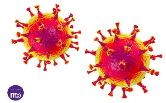
Coronaviruses were called so because of their surface’s spikes which looks like a crown (Figure 1). Its Firstly was determined in the middle of sixties -1960s. Four common human coronaviruses which can attack humans which are: HKU1 (beta coronavirus), OC43 (beta coronavirus), 229E (alpha coronavirus), and NL63 (alpha coronavirus). Other human types which occur newly are SARS-CoV-2 (COVID-19, or the novel coronavirus 2019 ) SARS-CoV (SARS or the beta coronavirus that causes the severe acute respiratory syndrome, or) and the final disseminated dangerous type MERS-CoV (MERS or the beta coronavirus that causes Middle East Respiratory Syndrome, or) [17].
JDMR-3-2-305-g001

Figure 1. Crown-like spikes of Corona Virus (COVID 19). Cited from Charles Shepherd [70].

Transmission Mode

Genetic and epidemiological research confirmed that COVID dissemination is transmitted from human-to-human spread after the single animal-to-human transmission [18,19] Interpersonal contact transmission has been proved to be the main source for outbreak specifically with respiratory droplets [20]. Consequently, sneezing or coughing of the infected patient is capable distribute virus aerial ending potentially which transmit the virus to the close people and infect them with. Articles advocate that approximately 6 feet radius needed for protection [15].

Inanimate objects also can be considered as another important transmission route specifically if it’s nearby to infected person respiratory droplets through sneeze or cough. Latest reports think that the virus can withstand for nine days on a hard surface like metal or plastic [15].

Studies approve that Virus has a great ability to connect to human angiotensin-converting enzyme 2 (ACE-2) positive cells that concentrated abundantly in salivary gland [21]. For that saliva also can play a role in transmission in the dental office setting [22].

Naso-Oropharyngeal droplets ordinarily associated with saliva. Distance dissemination for larger droplets could influence nearby subjects, and, vice versa, smaller droplets can infect the long-distance subjects [8,23].

Various researches warned that asymptomatic persons can also be a source of the outbreak. This has been approved in tow studies done in China, Italy and Japan were viruses disseminated as like wildfire 51%, 44% and 0.06% of the laboratory-confirmed cases [24-26].

International mission reports of the World Health Organization stated in that majority of the cases (75%) of newly infected patients will be developed to be the clinical disease, based on Chinese data [27].

Published case reports focus on pre-symptomatic transmission proportion estimated is around 48% and 62% [28] which expected to be based on a shorter serial interval of COVID-19 [4.0 to 4.6 days] rather than the mean incubation period (five days) recorded in symptomatic cases as secondary transmission detected [29].

Rothe C. stated in his article that till know its unproved that recovered patients can be a source of transmission of disease [8].

Age, Gender and Existing Medical Condition Risk

Generally speaking, all age group are susceptible for infection this estimated by the current observations in hospitals all over the worlds with priorities to medically compromised people above 60 years of age, males frequently considered at high risk and death [30-34].

In a study done by Wei-Jie Guan [35] describe that "the median age was 47.0 years (IQR, 35.0 to 58.0), and 41.9% were females. 0.9% of the patients were aged below 15 years. For clinical manifestation fever (87.9%) and cough (67.7%) were the most common symptoms, whereas diarrhoea (3.7%) and vomiting (5.0%) were rare. 25.2% of patients had at least one underlying disorder (i.e., hypertension, chronic obstructive pulmonary disease)".

Tobacco-users/smokers and those with hypertension are expected to be with high risk too, this can be attributed to the fact of ACE 2 expression in the tissues of the lungs maximize with age, hypertensive treatment, and users of tobacco,-. Thus, it’s critical to state that tobacco-users are possibly a vulnerable group for COVID-19 [36-39].

Clinical observation also indicates that children experience mild clinical manifestations despite that they are likely to be infected as adults, [24,40]. World Health Organization highlighted that cases that are less than 19 years old constitute 2.4% of the overall infected patients in China [as of 20 February 2020]. A few portions of this group have a critical disease (0.2%) or developed severe (2.5%) [34]. On the whole, medically compromised older ages were associated with poorer prognosis [41-44].

Health care workers are also vulnerable to be infected as a study done by L. Menganalyzed 138 hospitalized infected cases who have the virus in Wuhan, in the early epidemic stage 40 (29%) are health care workers [10,41,43].

Incubation Period

This period of infection is able to last for 14 d although at start of attack of disease the incubation period expected to be 5 to 6 d on average. To date the fourteen days consider the regularly adopted period for quarantine and medical observation of (possible) exposed cases [45]. Moreover incubation period of patients as well asymptomatic patients are evidenced to be carriers in some articles to be another sources for outbreak [8,18].

Clinical Manifestations

According to recent studies [35,44] most of the of COVID-19 patients with considered as moderately mild cases while sever cases among all patients was 15% to 25%. Fever, fatigue, dry cough, breath shortness , and irregular symptoms, like headache,vomiting diarrhea sore throat, confusion, and muscle pain [35,46] are descried in literature. Bilateral pneumonia [35,43] 1/4 and 1/3 of Wuhan hospitalized cases developed serious complications, like shock, arrhythmia, acute respiratory distress syndrome need urgent intensive care unit [32,43,46].

Diagnosis

Epidemiologic information basically travelling history is one of the important factor in diagnosis here (for example a travel history or residence in the affected area two weeks before the symptoms start), As any disease condition clinical manifestation, Radiographical findings in specific CT imaging, and blood, saliva laboratory sample tests These diagnostic rules are determined based on the standards of Chinese National Health Commission or World health Organization to of either the WHO [47,48].

Salivary diagnosis platforms need to emphasis as some strains of the virus were discovered in saliva even 29 days after the case reported [49,50]. Samples of saliva can be gathered in cases who present with oropharyngeal secretions as a symptom [51,52]. Table 1 explains list of questioner used for scheduled patients for endoscope procedure; these questioners prepared to evaluate or screen the COVID virus in these scheduled patients. We think it’s liable to be applied for dental patients too as initial screening purposes.

Table 1: COVID Screening Questioners cited from Amber Ather [15].

Name:
Date:

Date of Birth:

COVID Screening Questioners

Yes

No

In the past 14 days, have you or any household member travel to area with known cases of COVID V. infection


In the past 14 days, have you or any household member had contact with known cases of COVID V. infection


have you or any household member had history of exposuretoCOVID biologic materials


In the past 14 days, have you had history of fever


In the past 14 days, have you had any symptoms as cough, diarrhea, difficulties in breathing,nausea, body ach, extra….


Urgent Dental Need Question (Do you have Un controlled dental or oral pain, infection, swelling, or bleeding or trauma in your mouth)


Treatment

To date, there no evidence was found from random control trials to suggest special anti-CoV treatments; only supportive administration of COVID-19 is established [41]. Presently, the COVID-19 approach is sourcing control of infection; control and prevention transmission risks; isolation and early diagnosis for infected patients [43] these are major roles for outbreak control.

Wei-jie Guan [35] stated that intravenous antibiotics, mechanical ventilation and oxygen therapy; and overall were initiated in 35.8%, 6.1%, 57.5%, and 38.0%, of infected cases , correspondingly. These therapies have been taken in noticeably high percentages of hard cases (all P<0.05).

Dentist Role

Dental Clinic as Possible Transmission Route of Infection

Robinson Sabino [23] in his published study consider dental procedures are high risk procedure as airborne inhalation of aerosols and particles practiced during dentist steps on COVID-19 infected cases make dentists exposed closely and directly to the virus. Consequently, it is vital for dentists to take protectiontechniques to stop the infection of COVID-19. In this article we discuss the preventive policies should be well known for dentist by focusing on placement of patient, hygiene of hands, personal protective equipment (PPE), and caution in performing procedures of aerosol-generating.

We explain earlier that one of important route of transmission is airborne particles in noso-oral-pharyngeal area. To, K. et al. [53] reported that viral culture method used to detect presence of live viruses in infected persons and virus can live up to 29 days in saliva 43 this related to specific characters of COVID virus which has th ability to utilize ACE2 effectively as a receiver to invade cells, that possibly be similar to type of transmission “human-to-human” [54]. ACE2 abundantly present along the respiratory tract, beside that salivary gland duct cell morphologically also compatible and approved to be a class early targets for 2019-nCoV as well as previously discovered SARSCoV infection [55].

From this point of view dental professionals and their patients couldn’t protect themselvesfrom pathogenic microorganisms both bacteria and viruses which manifest within the respiratory tract and oral cavity. In these exceptional circumstances, professionals dentist care hold the infection risk of 2019-nCoV because of the specificity of the profession dealing, including communication type with patient which is “face-to-face” instruments handling, blood, saliva, , exposure.

Through inhalation of microorganisms of airborne that last in the air for long time [56] oral fluids, blood, or direct dealing with patient materials [57]. Coughing and talking with an infected persons and persuade a short distance without a mask can induce transmission of droplets and aerosols containing microorganisms dental settings [58,59].

In addition contaminated instruments and/or environmental surfaces indirect contact [60] These are the specific method of dissemination involved in an infected individual in dental clinics and hospitals especially during the outbreak of 2019-nCoV.

Airborne Spread

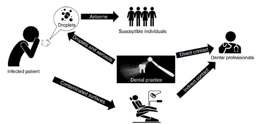
Many literatures discuss the hard acute respiratory syndrome caused by coronavirus airborne spread of SARS-Cov. Dental researches explain that several steps that dentist practice can make droplets and aerosols are polluted with virus [58,60]. Moreover Airborne spread of 2019-nCoV are the biggest distress in hospitals and dental clinics (Figure2). Beside patients breathing and cough of patients whether infected or not. Dental hand piece and turbines devices with, it’s widely use in dental clinics and hospitals rotate with high speed with running water, when its used in patient’s oral cavity generating, a big amount of droplets and aerosol mixed with saliv or blood of patients As the size of these aerosols and droplets particle is small, they can stay for long period before settle on environmental surfaces or entering the respiratory tract This, explain spread of the 2019-nCoV in dental clinics and hospitals.
JDMR-3-2-305-g002

Figure 2. Transmission Routes in Dental Clinics and Hospitals. Cited from Xian Peng [71].

Contact Spread

Cleveland, J. L. et al. in his article advocate that contact spread also one of transmission ways of virus in dental clinics as commonly dental professional are contact directly or indirectly with patient materials, saliva, blood, , and contaminated environmental surfaces or dental instruments [58].

Contaminated Surfaces Spread

Articles estimated that contaminated surfaces spread of different species of Coronavirus is able to last on surfaces like plastic metal or glass, for 14 days [56,61]. Thus, polluted surfaces which are contacted frequently in healthcare places are a potential.

Patient Management and Prevention of Nosocomial Infection

Coronavirus outbreak causes extraordinary public health challenge for all individual in various names. Responsibilities of outbreak and protection of people from such disasters infection are rests on each person starting with individuals, society, states, health and non-governorates institutions even military actors. All precautions and management of the Wuhan crisis are on previous outbreak experience of SARS-CoV-2 and SARS-CoV and its related disease (COVID-19).

We briefly highlight the significant evidence needed to be known by dentist as they have essential role in transmission. Dental professionals should follow specific procedures for management pf their patient’s in COVID-19 epidemic period.

One of the most important recommendations is to delay minor procedures for at minimum, three weeks exclusively, dentist provide emergencies American Dental Association recommended in March 16, 2020, list of advices for dental procedures [15].

Initial Screening

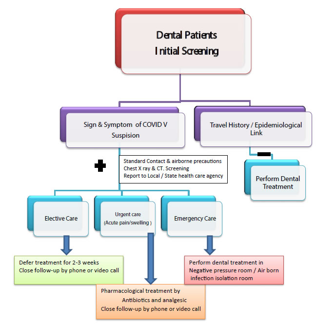
We explain previously that early diagnosis COVID-19 infection is crucial for both prevent dissemination transmission and early management and isolation of suspected individuals. Focus on travelling to infected area and symptoms of flue presence (any febrile respiratory illness). Tracking of reporting cases can be done through many public sources (Figure 3).
JDMR-3-2-305-g003

Figure 3. Dental Patient Screening of COVID V and Dental Management cited from Amber Ather [15].

Depending on answer; dental professional can manage. Positive response to questions is able to advance early worry and prefer to defer minor dentist care for 14-21 days minimum (incubation period).

Risk Level Classification and Emergency Evaluation of Dental Patients

In the dental practice, complete medical history is mandatory with the screening questioner, detection of Potential COVID Virus Risk level in patients (Table 2) and evaluation of specific emergency correctly too. It’s vital to measure the body temperature of the patient using infrared thermal sensors cameras or a remote forehead thermometer [62]. Elective dental care deferred also as in role one if patient temperature is [>100.4°F = 38°C] and/or respiratory disease symptom observed.

Table 2: Potential COVID Virus Risk Classification Cited from Alessandro Repici [72].

Risk

Clinical Picture

Low

No

No Symptom (cough, fever…)

No contact with positive infected persons

Non stay in high risk area during last 14 days

Intermediate Risk

Presence of Symptom with

No Medical History for contact with positive persons

Non stay in high risk area during last 14 days

No Symptom/but

Contact with positive infected persons

Stay in high risk area during last 14 days

High Risk*

At least 1 Symptom + 1 of the following

Contact with positive infected persons

Stay in high risk area during last 14 days

*In an emergency setting, all procedures must be considered high risk if adequate history can’t be assessed.

Isolation of Patients in the Clinic or Hospitals: Suspected individual should be separated in well-ventilated waiting place 6 feet minimum from the normal patients looking for dental care [63]. Patient are advised to use medical masks and use respiratory hygiene, like covering the nose and mouth by tissue when sneezing and coughing, and then throw the tissue [63]. These are the guidelines recommended by the Centers for Disease Control and Prevention (CDC) Moreover we have to educate patient for self -quarantine them and consult their doctors to check the possibility of being infected with COVID-19.

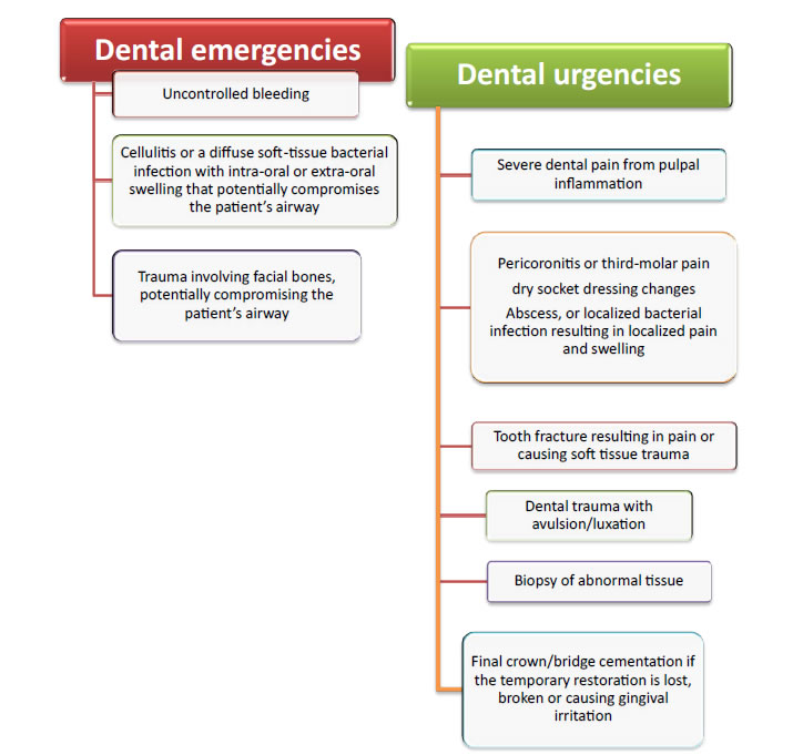
Pharmacologic Management: In presence of true emergency dental care like tooth pain and or swelling in infected or suspected individuals, its preferable to manage with analgesics or/and antibiotics as alternative which can give good relief in addition it offers dentist time to make a prcedures to give teeth care with suitable measures in clinic to hinder the transmission (Figure 4).
JDMR-3-2-305-g004

Figure 4. American Dental Association: Interim Guidance for Management of Emergency and Urgent Dental Care. Cited from Ciro Bocchetti [73].

Special Dental Recommendation in Treatment

Dental treatment decision can depend on case assessment with emergency questioner. Mostly active patients not present to dental units seeking treatment only in case of trauma presence or in sever space infection which might need surgical intervention.

Under these critical situations which is fill of danger that can be faced by dental professional in specialized dental health care centers or hospitals, dentist should be aware and alert for some definite and precise recommendation which are estimated and published in various articles since attack and outbreak of COVID virus. These are:

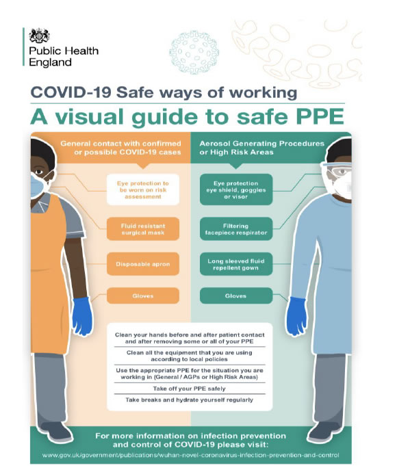
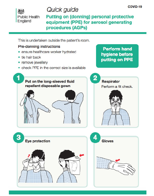
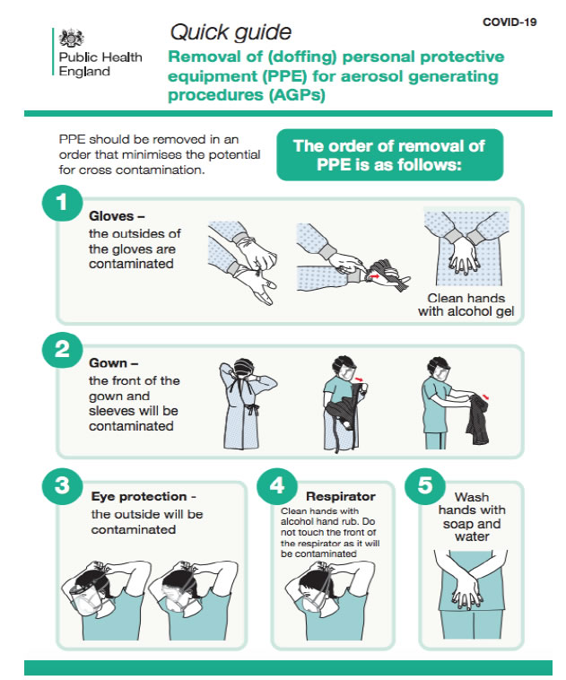
• Personal protective equipment [PPE] and hand hygiene practices used appropriately to prevent direct contact, and airborne (Figures 5-7).
JDMR-3-2-305-g005

Figure 5. A visual guide to safe PPE Cited from Public Health England Gateway [74].
JDMR-3-2-305-g006

Figure 6. Covid-19 Protection (PPE) – Putting-on-for-Aerosol-Procedure Cited from Guidance Public Health England [75].
JDMR-3-2-305-g007

Figure 7. Covid-19 Protection (PPE) – Doffing-for-Aerosol-Procedures Cited from Guidance Public Health England [75].

• Patient advice to use mouth rinse before intervention. Various published articles estimate that previous studies have shown that SARS and MERS were highly susceptible to povidone mouth rinse [64]. Therefore, pre procedural mouth rinse with 0.2% povidone-iodine might reduce the load of corona viruses in saliva [62,65].

• Disposable equipment use mandatory such as mouth mirror, syringes and blood pressure cuff to prevent cross-contamination.

• Intraoral imaging should be avoided as it can initiate gag reflex. Instead extraoral imaging such as panoramic radiograph or CBCT. In special situations when intraoral imaging is required, sensors should be double barrier to prevent perforation and cross-contamination [66].

• Rubber dam use highly indicated as it’s minimizing splatter generation and it covers the nose.

• High risk of contaminated droplet spread through use of high speed handpieces, triple syringe and ultrasonic instruments for these reason dentists should reduce the use of high-speed handpieces and three-way syringes.

• Routine dental practice setting is not suitable for suspected or confirmed patients [63].

• Human coronavirus can survive on inanimate surfaces up to 9 days at room temperature, with a greater preference for humid conditions [56]. Therefore, clinic staff should make sure to disinfect inanimate surfaces using chemicals recently approved for COVID-19 and maintain a dry environment to curb the spread of SARS-CoV-2 [67].

• Universal precautions are cleared by the American Dental Association [ADA] all over the worlds which focus on those dentists nationwide deferred any elective treatment up to 2-3 weeks. Emergency treatments take place with high precaution under the idea that patients supposed to be asymptomatically infected with COVID virus [68,69].

• Dentist as health care providers must keep themselves up to date about this evolving disease and provide adequate training to their staff to promote many levels of screening and preventive measures

State Responsibilities in COVID Outbreak

Politically, related to the crisis all efforts from the Prime Minister as well as other ministers such as (Ministry of Education, Ministry of Defense) also Civil Society Institutions, Medical and Dental Associations extra, gathered to support ministry of Health and World Health Organization in Iraq. To avoid transmission and dissemination, certain recommendation followed nationally and applied in my country too through the decision of Iraqi Health Minister. These recommendations briefly are:

Anticipation and Control Measures

• Forming the Epidemiological Crisis Control Committee responsible for Evidence-based control strategy decision making and adjustments is the key epidemic indicator. Responsibilities of committee to declare and aware institutions about effect of infection prevention and control [IPC] measures in various health care settings.

• Responsibilities also to pursuit entry and exit screening for individuals travelling and be aware of its effect public health control measures and their socio-economic impact. Movement restriction, Impose of curfew for emergency only, School and workplace closures all these actions were taken to avoid outbreak. Moreover educate individuals about Social distancing importance wearing mask in general public, mandatory quarantine for proved infected persons and voluntary quarantine with active surveillance.

• Different stages of outbreak should be assessed, risk, capacities and time screening is crucial in program recommendations, assess response of infected individuals all this aims to reach balance between reposes and social development.

• Initiate timely scientific evidence based, efficient and flexible joint multi-sectorial mechanism, which is driven by strong government leadership.

Practical Recommendations which Depend on Published Articles

• Keep in touch with the newly observed surveillance all over the country with early investigation priorities.

• Sharing information on patient management, disease progression and severity and favorable management outcomes.

• Estimation of Clinical care and infection control according to national evidence based guidelines as example isolation of suspected patients in special pre equipped prepared well centers.

• Instruct all health care workers with specific to physicians to be alert for detection of cases can be infected with COVID-19 as well as ensure maintenance of usual and essential services during the outbreak.

• Encourage pathogenesis studies can be accomplished using biopsy/post-mortem specimens of COVID-19 patients.

Nineveh Local Dealing Measures for COVID Virus Outbreak Crisis

Governorate represented by the Governor’ rule, Nineveh Health Directorate with specification for my institution (Al-Salam Teaching Hospital), Crisis Cell Committee, Army and Policeleaderships joined forces and efforts to take all local protection measures to avoid the spread of the disease all over the governorate. Follow guidelines on evidence based policy with continued recommendation and observation of the Ministry of Health and World Health Organization taking into account the outbreak scenarios in other countries whether near or far with all expectations to what happen in the next step.

The most vital recommendations in general speaking are:

• Prevent traveling; close all local borders avoiding entry and exit for any persons.

• Prepare health care providers in different specialties and in all grades, learn them to be aware for the disease as a whole. Deal with persons attained as suspicious till prove otherwise that mean take all precautions of safe distance, mask, gown, avoid direct contact with saliva, blood and other body fluids.

• Educate individuals as much as possible how to avoid transmission, safety contact, observe abnormal clinical symptoms, self – quarantine, urgent seeking contact if suspicion present.

• Stop institutional works, schools and universities.

• Impose a curfew with exception for emergencies and health care workers.

• Prepare special quarantine hospitals for suspected and confirmed cases. Highly equipped isolated rooms arranged to manage the patients in all circumstances. Al-Salam Teaching Hospital and Al-Shifaa General Hospital are chosen to be arranged for receive suspected and confirmed cases.

• Prevent dentist from practice work in their clinics and advice dental health centers to deal with only critical cases under precautions with as possible defer any treatment can establish infection.

Conclusion

In conclusion, protection of public in an intensive crisis such as Corona virus disaster are responsibilities of all with no exception starting from individuals themselves and ending with the Prime Minister. Great duties are established on healthcare workers and maintaining high standards of care and infection control are priorities for all. Its logical from the evidence based research that dealing with the crisis is challenge to all, moreover is imperative to improve current real strategies for anticipation and protection.

Dentist as eminent segment of the society are liable to handle their role in prevention and protection against outbreak of the crisis. We focus on saliva essential role in the human-to-human transmission and diagnosis of COVID-19 infection positivity of cases. Dental care providers need to be aware and prepared for tackling any impending infectious disease challenge which can be life threatening to susceptible patients.

All what described previously, is a brief review can be used for starting education argument and continue to be updated by others.

Review Limitations

Need more detailed descriptions in each part of the study with referral to clinical cases studies.

Acknowledgement

To all persons lift up the legend of public protection as priorities.

All my Institution Members as they pay great efforts to help COVID 19 patients.

To all health care workers are; involving dentists.

To the sole of all Health Care Workers attacked by the COVID 19 and lost their lives.

References

  1. Weiss SR, Leibowitz JL (2011) Corona virus pathogenesis. Adv Virus Res 81: 85-164.
  2. World Health Organization (2020) Coronavirus disease [COVID-2019] situation reportd50. Accessed March 10.
  3. World Health Organization (2020) Pneumonia of unknown caused China. Accessed February 14.
  4. Lu R, Zhao X, Li J, Juan Li, Peihua Niu, et al. (2020) Genomic characterization and epidemiology of 2019 novel coronavirus: implications of virus origins and receptor binding. Lancet 395: 565-574. [crossref]
  5. World Health Organization. 2020. Coronavirus disease 2019 [COVID-19]: situation report-36 [accessed 2020 Feb 26].
  6. Chan JF, Yuan S, Kok KH, Wang To KK, Hin Chu, et al. (2020) A familial cluster of pneumonia associated with the 2019 novel coronavirus indicating person-to-person transmission: a study of a family cluster. Lancet.
  7. Phan LT, Nguyen TV, Luong QC, Nguyen TV, Nguyen HT, et al. (2020) Importation and human-to-human transmission of a novel coronavirus in Vietnam. N Engl J Med 382: 872-874. [crossref]
  8. Rothe C, Schunk M, Sothmann P, Camilla Rothe, Mirjam Schunk, et al. (2020) Transmission of 2019-nCoV infection from an asymptomatic contact in Germany. N Engl J Med 382: 970-971. [crossref]
  9. Wu JT, Leung K, Leung GM (2020) Now casting and forecasting the potential domestic and international spread of the 2019-nCoV outbreak originating in Wuhan, China: A modeling study. Lancet 395: 689-697. [crossref]
  10. Li Q, Guan X, Wu P, Wang X, Lei Z, et al. (2020) Early transmission dynamics in Wuhan, China, of novel coronavirus-infected pneumonia. N Engl J Med 382: 1199- 1207. [crossref]
  11. WHO main website. https://www.who.int [accessed February 5th, 2020] [crossref]
  12. Worldometer Coronavirus Population COVID-19 Coronavirus/Death Rate Updates: Coronavirus [COVID-19] Mortality Rate.
  13. Weier Wang, Jianming Tang  Fangqiang  Wei  (2020)  Updated  understanding  of the outbreak of 2019 novel coronavirus [2019nCoV] in Wuhan, China. Journal of Medical Virology 92: 441-447. [crossref]
  14. Meng L, Hua F, Bian F (2020) Coronavirus Disease 2019 [COVID-19]: Emerging and Future Challenges for Dental and Oral Medicine. Journal of Dental Research 99: 481-487. [crossref]
  15. Amber A, Biraj P, Nikita BR, Anibal D, Kenneth MH (2020) Coronavirus Disease 19 [COVID-19]: Implications for Clinical Dental Care Journal of Endodontics 46.
  16. North Carolina Department of Health and Human Services (2020) Interim Coronavirus Disease 2019 [COVID-19] Guidance for Dental Providers.
  17. National Center for Immunization and Respiratory Diseases [NCIRD], Division of Viral Diseases: Human Coronavirus Types.
  18. Chan JF, Yuan S, Kok KH, To KK, Chu H, et al. (2020) A familial cluster of pneumonia associated with the 2019 novel coronavirus indicating person-to-person transmission: a study of a family cluster. Lancet 395: 514-523.
  19. Del Rio C, Malani PN (2020) Novel Coronavirus-Important Information for Clinicians. JAMA. [crossref]
  20. Centers for Disease Control and Prevention (2020) Disease burden of influenza; [accessed 2020 Feb 25].
  21. Brij K (2017) The composition, function and role of saliva in maintaining oral health: A review Article. International Journal of Contemporary Dental and Medical Reviews.
  22. Hamid RF,  Seied OK, Dana Z, Kim SG, Behzad C (2020) Being a front-line  dentist during the Covid19 pandemic: a literature review. Maxillofacial Plastic and Reconstructive Surgery 42: 12. [crossref]
  23. Robinson SS, Ana C, Gomes J, Walter LS (2020) Coronavirus COVID-19 impacts to dentistry and potential salivary diagnosis. Clinical Oral Investigations 24: 1619-1621. [crossref]
  24. Istituto SS, Sorveglianza I (2020) COVID-19 in Italia (February 27, 2020).
  25. Labour, Welfare, KATO (2020) Coronavirus disease 2019 [COVID-19] situation within and outside the country. Press Conference, Minstry of Health.
  26. Hoehl S, Rabenau H, Berger A, Kortenbusch M, Cinatl J, et al. (2020) Evidence of SARS-CoV-2 Infection in Returning Travelers from Wuhan, China. N Engl J Med 382: 1278-1280. [crossref]
  27. World Health Organization [WHO] (2020) Report of the WHO-China Joint Mission on Coronavirus Disease 2019 [COVID-19].
  28. Ganyani T, Kremer C, Chen D, Torneri A, Faes C, et al. (2020) Estimating the generation interval for COVID-19 based on symptom onset data. medRxiv.
  29. Nishiura H, Linton NM, Akhmetzhanov AR (2020) Serial interval of novel coronavirus [COVID-19] infections. International Journal of Infectious Diseases. 93: 284-286. [crossref]
  30. Liang W, Guan W, Chen R, Wang W, Li J, et al. (2020) Cancer patients in SARS-CoV-2 infection: a nationwide analysis in China. Lancet Oncology 21: 335-337. [crossref]
  31. Chen N, Zhou M, Dong X, Qu J, Gong F, et al. (2020) Epidemiological and clinical characteristics of 99 cases of 2019 novel coronavirus pneumonia in Wuhan, China: a descriptive study. Lancet. 395: 507-13. [crossref]
  32. Huang C, Wang Y, Li X, Ren L, Zhao J, et al. (2020) Clinical features of patients infected with 2019 novel coronavirus in Wuhan, China. Lancet 395: 497-506.
  33. Fei TY, Ronghui Du, Guohui F, Ying L, Zhibo L, et al. (2020) Clinical course and risk factors for mortality of adult inpatients with COVID-19 in Wuhan, China: a retrospective cohort study. Lancet 395: 1054-1062. [crossref]
  34. World Health Organization (WHO). Report of the WHO-China Joint Mission on Coronavirus Disease 2019 (COVID-19) 2020.
  35. Wei-jie G, Zheng-yi Ni , Yu Hu , Wen-hua L , Chun-quan O, et al. (2020) Clinical characteristics of 2019 novel coronavirus infection in China. Original article. New England Journal of Medicine 382: 18.
  36. Cai G (2020) Bulk and single-cell transcriptomics identify tobacco-use disparity in lung gene expression of ACE2, the receptor of 2019-nCov. medRxiv.
  37. Duclos GE, Teixeira VH, Autissier P, Yaron BG , Marjan AR, et al. (2019) Characterizing smoking-induced transcriptional heterogeneity in the human bronchial epithelium at single-cell resolution. Sci Adv 5: 3413. [crossref]
  38. Chen YS, Qian K (2020) Asians Do Not Exhibit Elevated Expression or Unique Genetic Polymorphisms for ACE2, the Cell-Entry Receptor of SARS-CoV-2. Preprints.
  39. Mengyuan LL, Yue Z, Wang X (2020) An Investigation of the Expression of 2019 Novel Coronavirus Cell Receptor Gene ACE2 in a Wide Variety of Human Tissues [Preprint]. Research Square.
  40. Bi Q, Wu Y, Mei S, Ye C, Zou X, et al. (2020) Epidemiology and Transmission of COVID-19 in Shenzhen China: Analysis of 391 cases and 1,286 of their close contacts. medRxiv.
  41. World Health Organization – China Report of the WHO-China Joint Mission on Coronavirus Disease [COVID-19]. 16-24 February 2020.
  42. Kui L, Fang YY, Deng Y, Liu W, Wang MF, et al. (2020) Clinical characteristics of novel coronavirus cases in tertiary hospitals in Hubei province. Chin Med J [Engl]. [crossref]
  43. Wang D, Hu B, Hu C, Zhu F, Liu X, et al. (2020) Clinical characteristics of 138 hospitalized patients with 2019 novel coronavirus–infected pneumonia in Wuhan, China. JAMA.
  44. Yang Y, Lu Q, Liu M, Wang Y, Zhang A, et al. (2020) Epidemiological and clinical features of the 2019 novel coronavirus outbreak in China. medRxiv.
  45. Backer JA, Klinkenberg D, Wallinga J (2020) Incubation period of 2019 novel coronavirus [2019-nCoV] infections among travellers from Wuhan, China, 20–28 January. Euro Surveill 25: 2000062. [crossref]
  46. Zhu H, Wang L, Fang C, Peng S, Zhang L, et al. (2020) Clinical analysis of 10 neonates born to mothers with 2019-nCoV pneumonia. Transl Pediatr.2020. 9: 51-60. [crossref]
  47. World Health Organization (2020) Clinical management of severe acute respiratory infection when novel coronavirus [2019-nCoV] infection is suspected: interim guidance [accessed 2020 Feb 17].
  48. National Health Commission of China. (2020) An update of novel coronavirus pneumonia outbreak as of 24:00 on February 25 [accessed 2020 Feb 26].
  49. Barzon L, Pacenti M, Berto A, Sinigaglia A, Franchin E, et al. (2016) Isolation of infectious Zika virus from saliva and prolonged viral RNA shedding in a traveller returning from the Dominican Republic to Italy, January 2016. Euro Surveill 21: 30159. [crossref]
  50. Zuanazzi D, Arts EJ, Jorge PK, Mulyar Y, Gibson R, et al. (2017) Postnatal identification of zika virus peptides from saliva. J Dent Res 96: 1078-1084. [crossref]
  51. Gorbalenya AE, Baker SC, Baric RS, Groot RJ, Drosten C, et al. (2020) Severe acute respiratory syndrome-related coronavirus: The species and its viruses—a statement of the Coronavirus Study Group.
  52. Fehr AR, Perlman S (2015) Coronaviruses: an overview of their replication and pathogenesis. Methods Mol. Biol. 1282: 1-23.
  53. Tsang TY, Yip CY, Chan KH, Wu TC, Chan MC, et al. (2020) Consistent detection of 2019 novel coronavirus in saliva. Clin Infect Diseases. [crossref]
  54. Zhou P, Yang XL, Wang XG, Shi ZL, Hu B, et al. (2020) A pneumonia outbreak associated with a new coronavirus of probable bat origin. Nature 579: 270-273. [crossref]
  55. Liu L, Wei Q, Alvarez X, Wang H, Zhu Z, et al. (2011) Epithelial cells lining salivary gland ducts are early target cells of severe acute respiratory syndrome coronavirus infection in the upper respiratory tracts of rhesus macaques. J Virol 85: 4025-4030. [crossref]
  56. Kampf G, Todt D, Pfaender S, Steinmann E (2020) Persistence of coronaviruses on inanimate surfaces and its inactivation with biocidal agents. J Hosp Infect 104: 246-251. [crossref]
  57. Chen, J (2020) Pathogenicity and transmissibility of 2019-nCoV—a quick overview and comparison with other emerging viruses. Microb Infect. Volume 22: 69-71.
  58. Cleveland JL, Gray SK, Harte JA, Robison VA, Moorman AC, et al. (2016) Transmission of blood-borne pathogens in US dental health care settings: 2016 update. J Am Dent Assoc 147: 729-738. [crossref]
  59. Harrel SK, Molinari J (2004) Aerosols and splatter in dentistry: a brief review of the literature and infection control implications. J Amnn Dent Assoc 135:429-437. [crossref]
  60. Wei J, Li Y (2016) Airborne spread of infectious agents in the indoor environment. Am J Infect Control 44: 102-108. [crossref]
  61. Otter JA, Donskey C, Yezli S, Douthwaite S, Goldenberg SD, et al. (2016) Transmission of SARS and MERS coronaviruses and influenza virus in healthcare settings: the possible role of dry surface contamination. J Hosp Infect. 92: 235-50. [crossref]
  62. Hoffmann M, Kleine-Weber H, Schroeder S, Krüger N, Herrler T, et al. (2020) SARS- CoV-2 cell entry depends on ACE2 and TMPRSS2 and is blocked by a clinically proven protease inhibitor. Cell 181: 271-280. [crossref]
  63. Wang Y, Wang Y, Chen Y, Qin Q (2020) Unique epidemiological and clinical features of the emerging 2019 novel coronavirus pneumonia (COVID-19) implicate special control measures. J Med Virol. 92: 568-576. [crossref]
  64. Centers for Disease Control and Prevention (2020) Infection control: severe acute respiratory syndrome coronavirus 2 [SARS-CoV-2].
  65. Eggers M, Koburger JT, Eickmann M, Zorn J (2018) In vitro bactericidal and virucidal efficacy of povidone-iodine gargle/mouthwash against respiratory and oral tract pathogens. Infect Dis Ther 7: 249-259. [crossref]
  66. Kariwa H, Fujii N, Takashima I (2004) Inactivation of SARS coronavirus by means of povidone-iodine, physical conditions, and chemical reagents. Jpn J Vet Res 52: 105-112.
  67. Hokett SD, Honey JR, Ruiz F, Baisden MK, Hoen MM, et al. (2000) Assessing the effectiveness of direct digital radiography barrier sheaths and finger cots. J Am Dent Assoc 131: 463-467. [crossref]
  68. Lan L, Xu D, Ye G, Chen X, Shaokang W, et al. (2020) Positive RT-PCR test results in patients recovered from COVID19. JAMA 323: 1502-1503. [crossref]
  69. Susan HD, Anty L, Christine (2020) Coronavirus Transmission in the Dental Setting. Decision in Dentistry Journal 6: 27-33.
  70. Charles Shepherd ME (2020) Association Guidance Coronavirus (covid-19).
  71. Peng X, Xu X, Li Y, Cheng L, Zhou X, et al. (2020) Transmission routes of 2019-nCoV and controls in dental practice. Int J Oral Sci 12: 9.
  72. Alessandro Repici, Roberta Maselli, Matteo Colombo, Roberto Gabbiadini, Marco Spadaccini, et al. (2020) Coronavirus (COVID-19) outbreak: what the department of endoscopy should know. GASTROINTESTINAL ENDOSCOPY 92: 192-197. [crossref]
  73. Ciro Bocchetti Roberto Sorrentino Fabio Cozzolino. (2020) COVID-19: Dentistry and the new Coronavirus, a compilation from PubMed resources. Pathology and Diagnostic.
  74. © Crown copyright 2020. Public Health England Gateway. Number 2019296. Version 1 April 2 2020
  75. Guidance Public Health England COVID-19 personal protective equipment (PPE) Updated 12 April 2020.
Comparison of Fetal Main Pulmonary Artery Doppler Wave Forms between Normally Grown and Growth Restricted Fetuses: A Case-Control Study

DOI: 10.31038/AWHC.2020341

Abstract

Objective: In this study we aimed to compare the fetal main pulmonary artery (MPA) Doppler wave forms between normally grown and growth restricted fetuses.

Method: In April 2019-March 2020, a single operator, who was unaware of the subsequent analysis, performed prospectively the MPA Doppler measurements including MPA-PI (pulsatility index) and MPA-PSV (peak systolic velocity). The data were then analyzed by SPSS version 21.

Results: A total of 120 women with singleton pregnancies – 60 normally grown fetuses as controls and 60 growth restricted fetuses as cases-participated in the study. We categorized the fetuses into five gestational age periods: 26-29, 30-32, 33-34, 35-37, and 38-40 weeks.

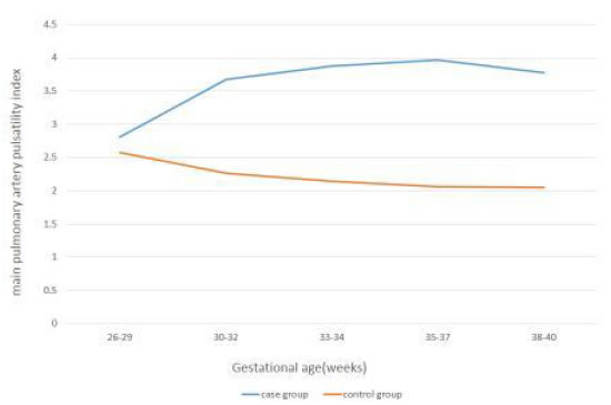
In all five time intervals the mean of MPA-PI was significantly higher in cases than controls (P<0.001). The mean of MPA-PI in cases was increasing with gestational age, while in the control group it was decreasing. Also, in all time intervals, the mean of MPA-PSV was significantly higher in the control group (P<0.001). The mean of MPA-PSV in cases was decreasing by advancing the gestational age, while in the control group it was increasing. In the case group, per one-week increase in gestational age, MPA-PI increased as 16%(p<0.001), while MPA-PSV significantly decreased (p<0.001).

Conclusion: According to the results, there are noticeable changes in the right ventricular outflow tract of the fetal heart in the growth restricted fetuses. In comparison with normal fetuses, the fetal MPA-PI increases and the MPA-PSV decreases significantly in growth restricted fetuses.

Keywords

Doppler ultrasound, MPA-PI, MPA-PSV, Growth restriction

Introduction

Fetal growth restriction (FGR) is associated with several changes at the level of the fetal heart involving preload, after load, ventricular compliance, and myocardial contractility. An increase in after load is seen at the right ventricle owing to increased placental impedance [1]. A decrease in after load is noted at the level of the left ventricle owing to decreased cerebral impedance associated with the brain sparing reflex [1]. Evidence of reduced myocardial contractility in the presence of severe FGR has also been reported. Ventricular ejection force, an index of ventricular systolic function that is independent of preload and afterload, is decreased at the level of the right and the left ventricles in FGR [2].

Longitudinal data on the hemodynamic sequence of the natural history of FGR show that the umbilical artery and middle cerebral artery are the first variables to become abnormal [3]. These arterial Doppler abnormalities are followed by abnormalities in the right cardiac diastolic indices, followed by the right cardiac systolic indices, and finally, by both left diastolic and systolic cardiac indices [3]. Several of the Doppler changes seen in association with FGR in the peripheral circulation are directly related to the adaptation of the fetal heart [4].

Normal pregnancy is characterized by a low fetal and placental vascular resistance. In the presence of intrauterine growth restriction (IUGR), there is an elevated pulsatility index in the fetal descending aorta and umbilical artery and reflecting increased umbilical, placental and utero-placental vascular resistance [5]. At the cardiac level, Doppler flow velocity waveforms have been previously recorded in the fetal heart outflow tracts in normally developed pregnancies [5-10] and those with fetal growth restriction [1,3,4,11-13].

The current management of FGR involves the use of Doppler at the peripheral arterial circulation (middle cerebral and umbilical arteries), at central venous vessels (Ductus venosus and inferiorvena cava), and with cardiotocography [4]. In the fetus, the Doppler evaluation of the main pulmonary artery has been shown to be useful in the analysis of the impedance of such vascular systems and changes in those variables have been demonstrated to correlate with gestational age, fetal lung maturity and neonatal outcomes [8,14-20]. Adding central arterial Doppler (aortic and pulmonary artery waveforms) may improve management of the fetus with FGR, but studies are lacking on the prospective clinical evaluation of the ventricular outflow tracts Doppler in FGR fetuses.

In this study we choose to compare the Doppler waveform variables at the fetal right ventricle outflow tract, at main pulmonary artery (MPA) in normally grown and growth restricted fetuses. The objective of our study was two-fold:

1. To establish the distribution of peak systolic velocities (PSV) and pulsatility indices (PI) of the fetal cardiac right outflow tract (main pulmonary artery) in normally grown fetuses of singleton pregnancies.

2. To determine the degree of abnormality of MPA-PSV and MPA-PI in growth restricted fetuses compromised with abnormal umbilical artery pulsatility indices (UMA-PI) and to compare these Doppler waveform variables between the two groups.

Material and Methods

Study Design

This prospective multicenter case-control study was conducted from April 2019 toMarch 2020 at three referral academic centers affiliated to Tehran university of medical science (Imam Khomeini complex, Dr Shariati hospital, Yas hospital). The study was approved by Tehran university ethics committee (ethics code: IR.TUMS.MEDICINE.1398.072) and all women gave their informed consents to participate  [2].

Setting

60 healthy women with singleton pregnancies and normally grown fetuses(abdominal circumference between 10th and 90th percentile, and estimated fetal weight between 10th and 90th percentile) at 27 to 29 weeks gestation entered the study as the control group and compared with 60 pregnant women with intrauterine growth restricted fetuses included the study as the case group.

Growth restricted fetuses were defined by the following criteria:

1. Gestational age was defined by either the first trimester crown-rump length (CRL) ultrasound or by the last menstrual period confirmed by ultrasound examination before 20 weeks gestation.

2. Abdominal circumference (AC) less than 10th percentile, or estimated fetal weight (EFW) less than 10th percentile prior to 32 weeks gestation.

3. Early placental insufficiency as defined by umbilical artery pulsatility index (UMA-PI) elevation equal or more than 95th percentile at the first time of examination.

4. To be eligible for longitudinal analysis, it was necessary for patients to have had at least three Doppler examinations prior to delivery.

5. Maternal age, gestational age, and maternal history of medical disorders was individually matched between two groups.

Eligibility Criteria

Mothers who consumed corticosteroids or any medication that could affectthe fetal heart or lungs, such as antihypertensive drugs or corticosteroids were excluded.Fetuses with chromosomal abnormalities, structural anomalies, advanced fetal vascular disease (brain sparing, absent or reversed flow in umbilical artery, elevated ductus venosus pulsatility index, absent or reversed a-wave in ductus venosus), fetuses with any major structural heart defect or arrhythmias and non-singleton pregnancies were excluded.

Study Procedure

After recruitment, fetal biometry was assessed every two weeks and Dopplerstudies were obtained by a single operator. We categorized the fetuses into five gestational age periods; 26-29, 30-32, 33-34, 35-37, and 38-40 weeks. The study was performed using a Philips affinity 70w ultrasound machine equipped with a C9-2 convex transducer. All fetuses were in a quiet state without fetal breathing movements.The fetal MPA was visualized in the short-axis view. The pulsed Doppler sample gate was placed at the middle of the fetal MPA, between the pulmonary valves and the pulmonary artery bifurcation, and away from arterial walls. After enlarging the image, the sample gate was adjusted to 2-3mm due to pulmonary artery diameter and the angle of insonation was maintained at less than 15 degrees. Doppler gain and scale were adjusted, and the high-pass filter was set at 100 Hz to record diastolic blood flow. Velocity range of blood flow was 100 cm/s and the sweep speed was 200 mm/s. Doppler variables including MPA pulsatility index (MPA-PI) and MPA peak systolic velocity (MPA-PSV), were measured at least three times during each study, using manual-trace and measurements were averaged. The last MPA Doppler examination was done within 48 hours before delivery [3].

Statistical Analysis

Statistical analysis was performed using SPSS version 21.0 (SPSS, Inc, Chicago, IL). The data were expressed as number (%) and mean (SD). Clinical and demographic characteristics between the cases and the controls were compared through Chi-square test for categorical variables and student t test for continuous variables. Linear regression analysis was used to investigate the relationship between the individual fetal MPA Doppler waveform variables and gestational age. Moreover, logistic regression was used to investigate the effect of fetal MPA Doppler waveform on RDS in neonates. P<0.05 was considered statistically significant.

Results

A comparison of the maternal demographic and clinical data between case and control group is shown in Table 1 and 2; Figures 1 and 2.

Table 1: Comparison of Baseline Demographic and Clinical Characteristics between Case and Control Groups.

Variable

Case group

Control group

P. value

N (% or SD)

N (% or SD)

Cesarean delivery

54 (90)

37 (61.67)

<0.001

Un-usual Apgar

42 (70)

10 (16.67)

<0.001

NICU admission

49 (81.67)

14 (23.33)

<0.001

RDS

49 (81.67)

14 (23.33)

<0.001

ASA consumption

26 (43.33)

15 (25.00)

0.034

Allergy

9 (15.00)

8 (13.33)

0.79

Smoking

5 (8.33)

5 (8.33)

1

Infertility

9 (15.00)

5 (8.33)

0.26

Parity 0

29 (48.33)

31 (51.67)

0.28

1

22 (38.33)

26 (43.33)

2

8 (13.33)

3 (5.00)

0.94

Age (year)

32.1 (5.87)

32.02 (5.71)

0.94

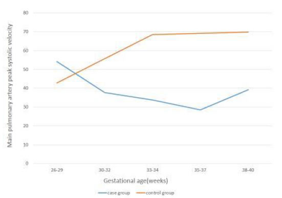
The percentage of cesarean delivery(90% vs. 61.67%) was significantly higher in case group. There wasn’t significant difference between two groups in regard of allergy, smoking, infertility and parity.MPA Doppler variables at each 2-week gestational age intervals in both case and control groups are presented in Table 2 and Figures 1 and 2.

Table 2: Changes in MPA-PSV and MPA-PI with Gestational Age in Case and Control Groups.

Gestational Age

MPA-PSV in Case

MPA-PSV in Control

P.Value

MPA-PI in

MPA-PI in

P.Value

 (weeks)

Group

Group


Case Group

Control Group


26-29

54.26 ± 11.05

42.81 ± 19.08

<0.001

2.81 ± 0.33

2.58 ± 0.32

<0.001

30-32

37.70 ± 11.52

55.76 ± 19.15

<0.001

3.67 ± 0.36

2.26 ± 0.26

<0.001

33-34

33.81 ± 9.99

68.53 ± 21.45

<0.001

3.87 ± 0.28

2.14 ± 0.29

<0.001

35-37

28.49 ± 7.18

69.25 ± 12.98

<0.001

3.97 ± 0.23

2.06 ± 0.20

<0.001

38-40

39.23 ± 10.89

69.85 ± 18.51

0.007

3.77 ± 0.38

2.05 ± 0.16

<0.001

Overall

41.59 ± 14.31

56.20 ± 21.60

<0.001

3.49 ± 0.63

2.31 ± 0.34

<0.001

In all five time intervals the mean of MPA-PI was significantly higher in cases than controls(P&lt;0.001). The mean of MPA-PI in cases was increasing with gestational age, while in thecontrol group it was decreasing. Also, in all time intervals, the mean of MPA-PSV was significantly higher in the control group(P&lt;0.001). The mean of MPA-PSV in cases was decreasing by advancing the gestational age, while in the control group it was increasing.

AWHC-3-3-321-g001

Figure 1. Changes in MPA-PI with Gestational Age in Case and Control groups.

AWHC-3-3-321-g002

Figure 2. Changes in MPA-PSV with Gestational Age in Case and Control groups.

In the case group, as shown in Table 3, per one-week increase in gestational age, MPA-PI increased as 16%(β=0.16, p&lt;0.001), while MPA-PSV significantly decreased, significantly (β=-3.08, p&lt;0.001).

Table 3: Regression Equations for MPA-PI and MPA-PSV with Gestational Age.

Doppler parameters

Regression Equations

R2

F

P.Value

MPA-PI

Y=-1.35+0.16x

0.31

80.83

<0.001

MPA-PSV

Y=136.4-3.08x

0.23

53.09

<0.001

Discussion

This study aimed to determine the main pulmonary artery Doppler waveforms changes in normally grown and growth restricted fetuses during gestation, and to compare the MPA Doppler indices between two groups. In the present study, the MPA flow velocity waveforms, were successfully recorded in all fetuses. In normally developed fetuses (the control group), with increasing gestational age, the MPA-peak systolic velocity increased, and the MPA-pulsatility index decreased significantly. These findings are compatible with prior publications by Chaouiet al. [10], Guan et al. [8], and Herrenet al. [6].

Fetal oxygen exchange occurs almost exclusively in the placenta and not in the lungs. For this reason, the pulmonary circulation in the fetus is maintained in a state of high resistance, high pressure, and low flow. Doppler ultrasonography provides a simple, noninvasive, and reproducible technique of directly evaluating the status of the fetal pulmonary circulation [8]. As pregnancy progresses, the vascular beds of the fetal lungs expand, leading to a progressive decrease in pulmonary arterial vascular resistance and a slow increase in pulmonary blood flow. These changes might explain the elevation in pulmonary artery peak systolic velocity and the reduction in the pulsatiliy index as well [8].

Herrenet al. determined reference ranges for Doppler parameters (PI and PSV) of fetal pulmonary artery segments (proximal, middle and distal) in 150 healthy singleton pregnancies from 19 to 39 weeks of gestation [6]. The mean PI and PSV values (mean ± SD) in the middle segment of MPA were 2.52 ± 0.96 and 24.39 ± 11.86, respectively. We measured the MPA-PI and MPA-PSV in the middle segment of main pulmonary artery.

Guan et al. defined reference ranges for fetal main pulmonary artery (MPA) Doppler waveform parameters in 288 healthy fetuses throughout gestation [8]. They used MPA Doppler indices to predict the subsequent development of neonatal respiratory distress syndrome (RDS) in a prospective cohort of 52 pregnant women with impending preterm birth before 37 weeks gestation. The authors concluded that the acceleration time (AT), acceleration time/ ejection time ratio (AT/ET), peak systolic velocity (PSV), and mean velocity (MV) were significantly lower in the RDS group than in the non-RDS group. All these variables showed no significant differences between the two groups. They mentioned that AT and AT/ET ratios of the fetal MPA Dopplerwaveform less than or equal to the 5th percentile for gestational age may provide a reliable noninvasive test to identify fetuses at risk of developing neonatal RDS.

Our study has focused on a special group of growth restricted fetuses (estimated fetal weight below 10th centile) that were diagnosed at 26-27 weeks gestation and pulsatility index above 95th centile showed in their umbilical artery Doppler study. So far, there hasn’t been any similar prior case-control studies to be acknowledged. In our study, there was significant difference in the MPA Doppler variables between the two groups. In all time intervals, the mean MPA-PI was significantly higher in cases than controls (p<0.001). Also, from 30 to 38 weeks gestation, the mean MPA-PSV was significantly lower in the case group (p<0.05). The mean MPA-PI in growth restricted fetuses (the case group), increased by the increase of gestational age, while it reduced in the control group. The mean MPA-PSV in the case group, decreased by the increase of gestational age, while it increased in the control group.

We followed all cases, evaluating the MPA Doppler waveforms every 2 weeks of gestation and during 48 hours before delivery, and the longitudinal assessment of MPA Doppler measurements within the same gestational ages was done in the control group. We noticed that the significant differences in the MPA Doppler indices existed between normally grown and IUGR fetuses with the umbilical artery Doppler pulsatility index>95th centile. An interesting finding in the case group was the concurrent occurrence of centralization and Doppler impairment in the main pulmonary artery. Centralization, so called Brain sparing effect, is defined as middle cerebral artery pulsatility index (MCA-PI) less than 5th centile and cerebro-placental ratio < 1. In %73.3 (n: 44/60) of IUGR cases, we had detections of simultaneous MCA-PI< 5th centile and MPA-PI> 95th centile.

Groenenberg and colleagues evaluated the fetal cardiac outflow tracts (ascending aorta and pulmonary artery) peak systolic velocities in 46 normal pregnancies and 21 IUGR cases. In normal fetuses, the mean PSV in the pulmonary artery increased from 39.0 cm/s at 19 weeks gestation to 63.7 cm/s at 33 weeks. In IUGR fetuses the PSV in the pulmonary artery was decreased (<5th percentile) in 95% of cases, and no relationship was established between PSV in pulmonary artery and the presence or absence of end-diastolic flow velocities in the umbilical artery [4].

Verburg et al. mentioned in their prospective cohort study that decreased fetal growth is associated with adaptive fetal cardiovascular changes [13]. Cardiac remodeling and cardiac output changes are consistent with a gradual increase in afterload and compromised in arterial compliance in conditions of diminished fetal growth. Furthermore, fetal hemodynamic patterns have already begun to change in the presence of reduced fetal growth while the fetus is still within the normal estimated fetal weight range.Evidence of reduced myocardial contractility in the presence of severe IUGR has also been reported by Rizzo et al. [2] and Makikallioet al. [1].

We concluded that there were noticeable changes in the right ventricular outflow tract of the fetal heart in the growth restricted fetuses. In the case group, per one week increase in gestational age, the MPA-PI increased as 16% (p<0.001), while the MPA-PSV decreases significantly (p<0.001).

Moety and colleagues measured MPA-PSV, PI and AT/ET in 643 healthy fetuses, aged 34 to 38+6 weeks, without postnatal respiratory distress syndrome [14]. The strongest correlation was found with AT/ET (cutoff value: 0.305). They provided 76.4% sensitivity and 91.6% specificity for prediction [7] of postnatal RDS.The MPA-PI was significantly higher, whereas MPA-PSV was significantly lower in fetuses that developed RDS.

Our study emphasizes the role of placental insufficiency in the course of fetal growth restriction. Placenta, although is not placed directly on the course of pulmonary artery flow, it can indirectly affect the PA flow on its way to the descending aorta [21,22]. Pulmonary artery flow faces three different downstream resistances; the high-resistance-non-aerated fetal lungs, the lower resistance-aortic vascular bed and placental vascular bed with the lowest resistance in normal pregnancy [9]. The MPA-PSV reflects right ventricular systolic function and in our study it reduced in growth restricted fetuses. The MPA-PI influences by systolic and diastolic pulmonary artery pressure and central venous pressure. In the presence of IUGR, there is an elevated pulsatility index in the fetal descending aorta and umbilical artery, reflecting increased placental vascular resistance [5].

Our study showed that not only the umbilical and middle cerebral arteries were affected by the elevated placental impedance in FGR fetuses, but also the main pulmonary artery was influenced by placental vascular insufficiency. We compared fetal pulmonary artery PI and PSV between normally grown and growth restricted fetuses almost at the same gestational ages. It permits better comprehension of fetal circulation and cardiac hemodynamics in diminished fetal growth. We chose to assess two Doppler variables (PI and PSV) and the other variables (AT, ET) didn’t evaluate in our study. Subsequent large prospective studies are needed to better investigate the occurrence of adverse Doppler measurements in this part of the fetal circulation.

Aknowledgement

The authors would like to express their deepest gratitude to the perinatology department of Tehran University and the head professors for their continuous support.

Conflict of Interest

None.

References

  1. Makikallio K, Vuolteenaho O, Jouppila P,JuhaRäsänen (2002)Ultra sonographic and biochemical markers of human fetal cardiac dysfunction in placental insufficiency. Circulation 105: 2058-2063. [crossref]
  2. Rizzo G, Capponi A, Rinaldo D,Arduini D, Romanini C(1995) Ventricular ejection force in growth restricted fetuses. Ultrasound ObstetGynecol 5: 247-255. [crossref]
  3. Figueras F, Puerto B, Martinex JM,VicençCararach, et al. (2003) Cardiac function monitoring of fetuses with growth restriction. Eur J ObstetGynecolReprodBiol 110: 159-163. [crossref]
  4. Groenenberg IAL, Stijnen T, Wladimiroff JW (1990) Flow velocity waveforms in the fetal cardiac outflow tract as a measure of fetal well-being in intrauterine growth retardation. Pediatr Res 27: 379-382. [crossref]
  5. Huhta JC, Moise KJ, Fisher DJ, Sharif DS, Wasserstrum N, et al. (1987) Detection and quantitation of constriction of the fetal ductus arteriosus by Doppler echocardiography. Circulation 75: 406-412. [crossref]
  6. Herren H, Junior EA, Wellington P, Ricardo CC, Alessandra CM et al. (2016) Reference ranges of Doppler parameters of fetal pulmonary artery segments between 19 and 39 weeks gestation. J MaternFetal Neonatal Med 29: 85-90. [crossref]
  7. Sosa Olavarria A, Zurita Peralta J, Schenone CV, Scherone MH, Prieto F (2019) Doppler evaluation of the fetal pulmonary artery pressure. J Perinatol Med47: 218-221. [crossref]
  8. Guan Y, Li S, Luo G, Wang C, Norwitz ER, et al. (2015) The role of Doppler waveforms in the fetal main pulmonary artery in the prediction of neonatal respiratory distress syndrome. J clin Ultrasound43: 375-383. [crossref]
  9. Malekan rad E, AghaeiME, Majnoon M, Nikoofar M, Kocharian A, et al. (2018) Comparison of reference values and Z scores of pulse-Doppler waveforms of fetal pulmonary artery and aorta. Iran J Pediatr28.
  10. Chaoui R, Taddei F, Rizzo G,C Bast, F Lenz, et al.(1998)Doppler echocardiography of the main stems of the pulmonary arteries in the normal human fetus. Ultrasound ObstetGynecol11: 173-179. [crossref]
  11. Baschat AA, Hecher K (2004)Fetal growth restriction due to placental disease. SeminPerinatol 28: 67-80. [crossref]
  12. Boito S, Struijk PC, Ursem NT, Stijnen T, Wladimiroff JW (2002) Umbilical venous volume flow in the normally developing and growth restricted human fetus. Ultrasound ObstetGynecol 19: 344-349. [crossref]
  13. Verburg BO, Jaddoe V, Wladimiroff JW, Hoffman A,Jacqueline CM, et al. (2008) Fetal hemodynamic adaptive changes related to intrauterine growth the generation R study. Circulation 117: 649-659. [crossref]
  14. Moety GA, Gaafar HM, El Rifai NM (2015) Can fetal pulmonary artery Doppler indices predict neonatal respiratory distress syndrome? J Perinatol35: 1015-1019.
  15. Buke B, Destegul E, Akkaya H, Simsek D, Kazandi M (2019) Prediction of neonatal respiratory distress syndrome via pulmonary artery Doppler examination. J MaternFetalNeonat Med 32: 1640-1645. [crossref]
  16. Kim SM, Park JS, Norwitz ER, Hwang EJ,et al. (2013) Acceleration time-to-ejection time ratio in fetal pulmonary artery predicts the development of neonatal respiratory distress syndrome: a prospective cohort study. Am J Perinatol30: 805-812. [crossref]
  17. Azpurua H, Norwitz ER, Campbell KH, Funai EF, Pettker CM, et al. (2010) acceleration/ejection time ratio in the fetal pulmonary artery predicts fetal lung maturity. Am J Obstet Gynecol203. [crossref]
  18. Schenone MH, Samson JE, Jenkins L, Suhag A, Mari G (2014) Predicting fetal lung maturity using the fetal pulmonary artery Doppler wave acceleration/ejection time ratio. FetalDiagnTher 36: 208-214. [crossref]
  19. ZareMehrjardi M (2019)Fetal pulmonary artery Doppler examination as a non-invasive test for assessing prenatal lung maturity. Iran Cong Radiol35: 89-89.
  20. Aboulghar M, KhalifaY, Hamed ST, Tomerak R, Kamal E (2019) Prenatal prediction of lung maturity using pulmonary artery Doppler and 3D ultrasound. Ultrasound ObstetGynecol 54: 107-108.
  21. Poston L (1997)The control of blood flow to the placenta. ExpPhysiol82: 377-387. [crossref]
  22. Boura ALA, Leitch IM, Read MA, Walters WAW (1998)The control of fetal vascular resistance in the human placenta. Placenta19: 299-313.

ACBD3, Its Cellular Interactors, and Its Role in Breast Cancer

DOI: 10.31038/CST.2020523

Abstract

ACBD3 breast cancer research to date reveals that overexpression at mRNA and protein level is near universal in breast tumour tissue and that high ACBD3 expression is associated with worse patient prognosis. ACBD3 has been shown to have an important role in specifying cell fate and maintaining stem cell pools in neurological development and deletion of ACBD3 in human cell lines prevents cell division. Combined with observations that β-catenin expression and activity is increased when ACBD3 is overexpressed it has been hypothesised that ACBD3 promotes breast cancer by increasing Wnt signalling. This may only be one aspect of ACBD3’s effects as its expression and localisation regulatessteroidogenesis, calcium mediated redox stress and inflammation, glucose import and PI(4)P production which are all intrinsically linked to breast cancer dynamics. Given the wide scope for a role of ACBD3 in breast cancer, we explore its interactors and the implications of preventing these interactions.

Keywords

ACBD3, Breast cancer, Chromosome 1, Golgi, NUMB, PI4Kβ, Phosphatidylinositol, Protein kinase A, Steroidogenesis, Wnt signalling, 1q

Introduction

Although breast cancer incidence has increased in recent years, largely due to improved diagnostic techniques, greater awareness and the introduction of national screening programmes, mortality rates are declining as result of earlier detection and improved treatment regimes. Despite this, treating advanced disease remains difficult and there is a need to identify new therapeutic targets. Proteins encoded by the q-arm of chromosome 1 are of particular interest as regions of 1q are frequently amplified and overexpressed in breast cancer leading to the hypothesis that 1q is important in disease development and progression [1-3]. The frequency at which regions of arm 1q were amplified was investigated and the 1q42.12 locus was found to be amplified in both breast cancer cell lines and primary tumours with a number of genes in or near this region also being over expressed [4]. Of emerging interest is ACBD3, a Golgi protein with multiple functions, only recently linked to breast cancer [5].

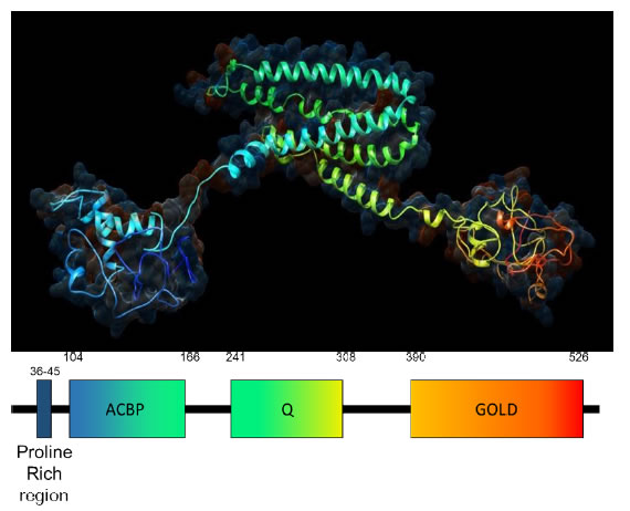
ACBD3 was discovered as an interactor of GOLGB1 and named GCP60, and independently discovered as aninteractor of the mitochondrial translocator protein TSPO and protein kinase A and named PAP7 [6,7]. Having found that each of these names were describing one aspect of a diverse adapter protein it was renamed by the HUGO gene nomenclature committee in 2004 as Acetyl CoA Binding Domain containing protein 3, or ACBD3, reflecting its functional groups and protein family rather than any particular role, of which there are many (www.genenames.org). In addition to the acyl CoA binding domain at its N-terminus, ACBD3 contains a Golgi dynamics (GOLD) and a glutamine rich Q domain as well as a proline rich region (Figure 1) [8]. The GOLD domain is found in Golgi and lipid trafficking proteins and makes up the C-terminus of ACBD3 (aa384-526). It is a β-strandrich domain and is responsible for ACBD3 localization to the Golgi via direct interaction with GOLGB1 [6]. ACBD3 is a largely unstructured or loosely structured protein, as many linkers are, and of all the recognisable domains only the GOLD domain structure has been solved by X-ray crystallography with the rest of ACBD3 being modelled by NMR and predictive modelling software (Figure 1). The Q domain is a glutamine rich region (aa241-308) which forms a long loop made of alpha helices [8]. The N-terminal ACB domain binds Acyl CoA and Palmitoyl CoA in other ACBD family proteins but the function of this domain in ACBD3 is unclear. To the N-terminus of the ACB domain is a proline rich region (aa21-60), which is indicative of protein-protein interaction sites and may complement the ACB domain whichis often found paired with protein-protein interaction domains such as the Pleckstrin homology domain (PH) and the Src homology domain in other proteins. ACBD3 makes essential interactions with an unusually high number of protein partners in cellular processes as diverse as Golgi structure, steroid synthesis and glucose import; other functions not reviewed here include iron transport and a causal role in Huntington’s disease progression [7,9-12]. ACBD3 is essential for neural development and human cell lines do not divide when ACBD3 is excised by CRISPR-CAS9 [13].

CST-5-2-510-g001

Figure 1. Predicted 3D structure of human ACBD3.The structure is modelled by Phyre2 software using the primary amino acid sequence which agrees strongly with crystal structures of individual ACBD3 domains and related proteins [86]. From the N-terminus in blue to the C-terminus in red ACBD3 clearly contains 3 domains: the ACBP domain, the Q domain and the Golgi dynamics (GOLD domain) respectively connected by flexible linkers with an N terminal proline rich region. The hydrophilic surface of acbd3 has been superimposed on ACBD3 showing the electrostatic charge of the protein model with red depicting negative charge and blue depicting positive charge.

ACBD3 in Breast Cancer

The q arm of Chromosome 1 contains many genes important in cancer progression or tumour suppression: NRAS, JUN, MYCL, ESRRG, ARF1 and RAB25are amongst the best known. There are however many more 1q genes that are amplified in breast cancer with deletions strikingly rare despite common deletions in the p arm. Some of these genes (PI4Kβ, PIP5K1A and HIST2H2BE) have more recently been recognised as oncogenic with ACBD3 being the latest 1q gene observed to affect breast cancer [14]. ACBD3 mRNA is reported to be upregulated in breast tumour tissue matched against adjacent normal tissue in all subtypes [5]. Protein levels of ACBD3 were upregulated in 8 breast cancer cell lines (MDA-MB453, MDA-MB-415, BT549, MDA-MB-231, ZR-75-30, SKBR3, T47D and MCF7) compared to 2 normal breast epithelial cell samples (NBEC1 and NBEC2). In a cohort of Chinese breast cancer patients ACBD3 protein expression increased as cancer stage became more advanced. Kaplan-Meier survival curves were plotted and it was found that high levels of ACBD3 mRNA in breast tumour tissue predicted lower rates of patient survival and that this made a large difference in stage III and IV cancers with 60% probability of survival at 120 months when ACBD3 expression is low but less than 30% probability of survival when ACBD3 expression is high.

The ACBD3 containing 1q42.12 locus is seen to be amplified in an additional 6 breast cell lines (BRCAMZ01, BT20, HCC2218, MDAMB436, SUM149, ZR751) and 6 out of 25 primary breast tumours in a breast cancer 1q amplification study [4]. Loss of region 1q42.12 was seen in only 1 cell line (UACC812) where the terminal ~38 megabases of arm 1q were deleted and loss of 1q42.12 was not observed in any primary tumour samples. RNA expression levels revealed that 1q42.12 is located in the middle of a region of gain coined G7, the largest region of gain (in bases) on chromosome 1 in breast cancers.Overexpression of ACBD3 in cell cultures caused increased side populations of stemlike cancer cells and inhibition of ACBD3 by siRNAsignificantly reduced these populations [5]. GSEA analysis found that CTNNB1- and TCF4-activated gene signatures both positively correlated with ACBD3 expression [5]. CTNNB1 encodes the β-catenin protein which, in response to Wnt signalling, accumulates in the cytoplasm and then translocates to the nucleus where it propagates the Wnt signal.ACBD3 overexpression led to an increase of β-catenin in the cytoplasm and nucleus compared to when ACBD3 expression was low (65% versus 20% nuclear and cytoplasmic localisation) [5]. TCF4 is a transcription factor for genes that code proteins in the Wnt signalling pathway. When TCF4 was knocked down, the self-renewal ability of ACBD3-expressing cells was abolished suggesting that ACBD3 may promote cancer stem cell propagation via the Wnt/ β-catenin signalling pathway in breast cancer.All of this provides strong evidence that ACBD3 overexpression affects breast cancer but the ACBD3 protein has many binding partners, in disparate cellular pathways and cells appear to have few redundancies for the essential roles of ACBD3.

ACBD3 and Steroidogenesis

Although often considered a resident Golgi protein due its structural role and interactions with other structural components, ACBD3 can also be found at other membranes including the cytosolic cell membrane and at the outer mitochondrial membrane (OMM). ACBD3 interacts withtranslocator protein TSPO (previously the peripheral-type benzodiazepine receptor) on the cytosolic OMM and stimulates cholesterol transport from the OMM to the IMM (inner mitochondrial membrane) (Figure 2) [7,15]. P450scc (CYP11A1) makes direct contact with the IMM and converts cholesterol to pregnenolone, the precursor to mammalian steroids, by side chain cleavage [16,17]. TSPO is anchored to the voltage dependent anion channel VDAC1 and makes up approximately 2% of OMM proteins. TSPO tethers cytosolic ACBD3 at the OMM and ACBD3 subsequently recruits protein kinase A (PKA) via the PKARIα subunit. This brings PKA into proximity with one of its substrates, the steroidogenic acute regulatory (StAR) protein which is phosphorylated on residues S57 and S195 by PKA [18]. StAR then facilitates cholesterol import from the OMM to the IMM, the rate limiting step in steroidogenesis. ACBD3 overexpression increases chorionic gonadotropin-induced steroid production; increased steroid production has obvious implications for cancer progression by enabling self-sufficiency in growth signals, a hallmark of cancer [19,20].

CST-5-2-510-g002

Figure 2. ACBD3 in redox stress and steroidogenesis. ACBD3 is a Golgi resident protein but also has functions elsewhere in the cell. At the Outer Mitochondrial membrane (OMM) ACBD3 is essential for mediating interactions between PKA holoenzyme (via direct tethering with the PKAR1α subunit shown) and two of its substrates: StAR and VDAC1.
a) The phosphorylation state of StAR determines whether cholesterol can cross the IMM and be converted to pregnenolone, the basic building block of all steroid hormones in mammals. Cholesterol import is the rate limiting step in steroidogenesis and ACBD3 is indispensable for this process [18].
b) VDAC1 is a Ca2+ ion import channel at the OMM and phosphorylation by PKA closes this channel to prevent calcium import. Mitochondrial import of Ca2+ forms part of the calcium homeostasis mechanism in the cell, closing the VDAC1 ion channel causes cytosolic Ca2+ concentration to rise in the cell which leads to redox stress and local inflammation. Again, ACBD3 is essential for localising PKA to the mitochondria where it can then phosphorylate the VDAC1 substrate [27].

PKAR1α is a tumour suppressor gene and is important in primary pigmented nodular adrenocortical disease (PPNAD) nodule formation and tumorigenesis in mice and humans. Mutation of PKAR1α leads to hypercortisolism that drives tumorigenesis, and high ACBD3 expression in steroidogenic tissues (of which the adrenal cortex is one) may contribute to the overexpression/over activity of the mutant PKAR1α [21]. PPNADs are characterised by a resistance to apoptosis which in itself contributes to cancer occurrence and is another hallmark of cancer [20]. The first publication to suggest any link between ACBD3 and cancer demonstrated that ACBD3 follows the same expression profile as PKAR1α in PPNAD tissue and speculated that, in tumorigenesis, this could lead to deregulation of steroid synthesis [21]. More recent studies have shown that PKA activation may instead have a suppressive effect on cancer [22,23], whilst others show PKARIα is upregulated in cancer cell lines [24,25].

ACBD3 in Redox Stress

In a separate process, glutamate induces expression of TSPOand increased TSPO recruits ACBD3 and PKA to the mitochondria. Glutamate is a signalling molecule that is known to cause acute neurotoxicity [26-28]. PKA phosphorylates the calcium channel protein VDAC1, preventing Ca2+ import into the mitochondria (Figure 2). This causes Ca2+ accumulation in the cytosol which signals redox stress via the calcium sensing CamKII and its effector NADPH oxidase (NOX5) leading to inflammation by increased reactive oxygen species (ROS). Glutamate mediated redox stress is particularly important in neuro-inflammationwhere TSPO is not expressed in healthy brain tissue but can accumulate in age related degenerative disease or after traumatic stress, leading to increased ACBD3 mitochondrial recruitment and subsequent VDAC1 phosphorylation by PKA which may contribute to neurodegeneration [29]. VDAC1 is important in Ca2+ homeostasis, especially mitochondrial Ca2+ homeostasis which controls the metabolism of mitochondria and therefore energy availability in the cell [30]. Dysregulation of cellular energetics is a hallmark of cancer and an inflammatory environment can be tumour promoting when chronic and over time [20].

ACBD3 and Insulin Mediated Glucose Import

GLUT4 (glucose transporter type 4) allows the facilitated diffusion of glucose from the surroundings into cells,andis sequestered into storage vesicles (GSVs) that are tethered to Golgi membranes by TUG (Tether containing UBX domain for GLUT4), Golgin-160 and ACBD3 when insulin is absent (Figure 3) [31]. ACBD3-bound TUG can be acetylated on lysine 549 which has a higher binding affinity with Golgin-160 than with ACBD3 [10]. In response to insulin receptor activation the cytoplasmic effector of insulin PIST (PDZ interacting specifically with TC10) binds Golgin-160 and catalyses the cleavage of acetylated TUG. This releases GSVs allowing them to fuse with the plasma membrane where GLUT4 forms a channel for glucose import [32]. GLUT4 is continuously cycled away from plasma membranes back into GSVs to increase the on/off response of insulin sensitive cells when insulin is not present. GLUT4 exocytosis is regulated by tankyrase 1 as are several other ACBD3 related processes including Golgin45 expression and the promotion of β-catenin transcription in the Wnt signalling pathway [33,34].

CST-5-2-510-g003

Figure 3. The effect of insulin on TUG, the interaction between TUG and ACBD3, and the recycling of GLUT4 storage vesicles to regulate glucose import [10,31].
a) 1. Extracellular insulin binds the transmembrane insulin receptor (IR) causing receptor activation. 2. The active IR tyrosine phosphorylates CBL inside the cell. 3. Phosphorylated CBL recruits the CRK-C3G complex to the membrane lipid raft sub domain facilitating interaction of C3G and TC10α. 4. C3G activates TC10α which subsequently activates its effector: PIST. 5. PIST relocates to the Golgi causing the release of GSVs with embedded GLUT4 transporter which fuse with the cell membrane, 6. allowing glucose to enter the cell. GLUT4 is continuously cycled away from the membrane in GSVs creating a fast on and off switch for insulin dependent glucose import.
b) 1. ACBD3 interacts with TUG and is dependent on the acetylation state of TUG. 2. Acetylation of TUG on lysine 549 causes TUG to preferentially bind Golgin-160 over ACBD3. 3. PIST, activated by the insulin receptor signalling cascade, also binds Golgin-160 and catalyses the cleavage of acetylated TUG causing GSVs to be released into the cytoplasm 4. to fuse with the cell membrane. 5. GLUT4 is continuously cycled away from the cell membrane embedded in GSVs and is sequestered back to the Golgi where they bind TUG.

The hormone 17β-oestradiolhas a central role in breast cancer progression, ithas been found to upregulateGLUT4 expression and translocation to the membranein breast cancer cell lines and was associated with increased glucose uptake [35-37]. GLUT4 is being investigated as a target for breast cancer therapy as part of an informed approach to target the Warburg effect. Downregulation of GLUT4 by siRNAimpairs viability of MDA-MB-231 and MCF7 breast cancer cell lines and increases mitochondrial oxidation of pyruvate [38]. The EGFR/HER2-targeted drug lapatinib has been shown to downregulate GLUT4 in ER-/HER2+ HMEC cell lines, and GLUT4 downregulation by siRNA in these cell lines led to the formation of normal acini structures in 3D culture [39].The insulin receptor (IR) is upregulated in breast cancer and is a potential target for breast cancer therapy as it has been demonstrated that knock down of IR by shRNA and inhibition by peptide drugs inhibits breast cancer cell growth [40-43].

ACBD3, PI4Kβand Phosphatidylinositols

Phosphatidylinositol 4 Kinase III beta (PI4Kβ) is a lipid kinase that converts phosphatidylinositol (PI) into phosphatidylinositol 4-phosphate (PI(4)P) [44]. PI4Kβ is localised to the Golgi by extension of an amphipathic helix at the N-terminus of PI4Kβ (aa44-64) through the Q domain alpha helices loop of ACBD3 (aa241-308) [8]. The small GTPase Rab11 binds PI4Kβ to support this interaction whilst ACBD3 also interacts with GOLGB1 on the Golgi surface bringing PI4Kβ in close and constant contact withits PI substrate embedded in the lipid bilayer. ACBD3 does not affect the enzymatic activity of PI4Kβ directly by this interaction but, by tethering it to the Golgi membrane, PI4Kβ is proximal to the PI substrate and does not rely on diffusion through the cytoplasm for the phosphorylation of substrate. PI4Kβ is heavily implicated in breast cancers with 20% of primary tumours showing over expression of PI4Kβ at the protein level [45,46]. PI4Kβ is a chromosome 1q gene (at 1q21.3) and is reported to have increased gene copy number in 62% of 939 patient breast tumour samples [14]. Evidence of PI4Kβ upregulation at the protein level in breast ductal carcinoma samples from the human protein atlas was also found by Waugh. Independent of its lipid kinase function, PI4KIIIβ also mediates indirect phosphorylation and activation of AKT (Protein kinase B), an important kinase in breast cancer signalling [46,47]. AKT dysregulation drives many breast cancers by promoting cell cycle progression and suppressing apoptosis, it is commonly overexpressed or constitutively active [47].

Both PI and PI(4)P are cellular signalling molecules and docking sites on the membrane for other proteins including ARF1 (ADP-ribosylation Factor 1), which is essential for the formation of COPI vesicles and Golgi function including localisation of Golgin-160 to the Golgi; ARF1 is encoded by a gene adjacent to ACBD3 on chromosome 1 (1q42.13) [48-50]. PI4Kβ is also positioned on chromosome 1q, where amplification is common in breast cancers and its substrates localise ARF1 to membranes. ACBD3 is hijacked by some picornavirusviral proteins to form replication organelles and recruits PI4Kβ to these sites to enrich them for PI(4)P [51,52]. This is another example of how the role of ACBD3 is contextual and dependent on its cellular location, cell cycle position and binding partners. PI4Kβ has been found to be a target in malaria and drugs to inhibit PI4Kβ have already been developed [53]. There have so far been no publications on PI4Kβ drug inhibition in cancer.

ACBD3 Cell Signalling in Neurogenesis

Mammalian NUMB, an endocytic adapter protein, is involved in cytosolic signalling and is segregated asymmetrically into one daughter cell during the mitosis of neural progenitor cells and inhibits NOTCH [54]. This asymmetric distribution of NUMB results in 1 identical pluripotent daughter cell (high NUMB protein level) to maintain the population of neuronal precursors and 1 differentiated neuron cell (low NUMB protein level). This balances the need to create neurons through NOTCH signalling and maintain the pool of precursor cells in embryonic neurogenesis by NOTCH inhibition (Figure 4) [55,56]. ACBD3 was identified as a NUMB binding partner after observations that ACBD3 cytosolic release during mitosis was paired with NUMB mediated cell fate [57]. The ACBD3 interacting region on NUMB is essential for NUMB activity and interaction with ACBD3 increases NUMB activity [57]. The C-terminus of ACBD3 binds with the N-terminus of NUMB, and NOTCH also binds the N-terminus of NUMB [58]. Cytosolic ACBD3 expression leads to inhibition of NOTCH, suggesting that NOTCH inhibition by NUMB is conserved from drosophila to mammals indicating that ACBD3 and NUMB are both required to specify cell fate in neural progenitors. ACBD3 is bound to Golgi/mitochondrial membranes through most of the cell cycle and can only bind NUMB during mitosis when the breakdown of the Golgi releases ACBD3 into the cytosol (Figure 4). Constitutively cytosolic mutant ACBD3 inhibits neurogenesis in mouse embryos resulting in fewer neurons. This indicates that permanently cytosolic ACBD3 is preventing differentiation in otherwise neuronal fated cells and it achieves this by binding NUMB outside of mitosis [57].

CST-5-2-510-g004

Figure 4. The differential regulation of NOTCH signalling by ACBD3 and NUMB in neurogenesis. Cytosolic NUMB (yellow shading) acts synergistically with cytosolic ACBD3 (blue shading) to inhibit NOTCH signalling (represented by a green nucleus) and specifies progenitor cell fates during mitosis [57]. In one daughter cell, cytosolic ACBD3 binds to NUMB increasing the ability of NUMB to inhibit NOTCH and causing that daughter cell to remain a progenitor like its parent. The other daughter cell does not contain NUMB and so NOTCH signalling cannot be prevented despite the presence of cytosolic ACBD3 protein and so the daughter cell begins to differentiate into a neuron. Paradoxically NUMB quickly accumulates in the differentiating cell but because the Golgi has reformed at this point, ACBD3 is membrane bound and cannot influence NUMB-NOTCH protein interactions, instead NUMB promotes neuron differentiation and survival through other pathways. This leads to the creation of one daughter neuron and one progenitor cell to balance neurodevelopment and stem cell pools.
a) NUMB accumulates asymmetrically in one half of a progenitor cell before mitosis. ACBD3 is bound to the Golgi apparatus and other organelles (not shown) and does not interact with NUMB.
b) During mitosis, the Golgi apparatus fragments into vesicles and ACBD3 is released into the cytosol where it can interact with asymmetrically distributed NUMB.
c) One of the daughter cells will contain NUMB and cytosolic ACBD3. ACBD3 increases the ability of NUMB to bind and inhibit NOTCH. Without NOTCH signalling the daughter cell remains a progenitor cell to maintain the pool of neuronal precursors.
d) The second daughter cell will contain cytosolic ACBD3 but no NUMB protein meaning that NOTCH is not inhibited and enters the nucleus signalling to the cell to differentiate.
e) NUMB protein is produced in the differentiating cell in G1 but at this time the Golgi has reformed and ACBD3 is no longer free in the cytoplasm and cannot interact with NUMB and so cannot inhibit NOTCH signalling.

Krüppel-like factor 9 (KLF9) is a tumour suppressor and is significantly down regulated in invasive breast cancers, endometrial carcinoma, glioblastoma and colorectal cancer, and its expression can inhibit growth of tumour xenografts from glioblastomaneurospheres [59,60].  ACBD3 and NOTCH1 expression are suppressed by KLF9 in endometrial carcinoma cells and both proteins promote breast cancer progression, specifically in cancer stem cell maintenance [5,61,62]. KLF9 supresses glioblastomaderivedneurosphere formation by 60% in controls but only by 33% when NOTCH1 expression is constitutively active strongly suggesting that KLF9 must suppress other proteins relevant to glioblastoma cancer progression and this could include ACBD3 [59]. NOTCH1, NOTCH3 and JAG1expression isassociated with poor survival in breast cancer patients with high NOTCH1 expression conferring a 66% chance of mortality and 74% chance of relapse at 10 years [63]. NOTCH receptorshave been found to have activating mutations in triple negative breast cancers and result in the upregulation of NOTCH controlled genes [64]. NOTCH overexpression was able to transform the MCF10A breast cell line and reduce its sensitivity to apoptotic drugs such as staurosporine, melphalan, or mitoxantrone, and overexpression of NUMB reverted the transformation [61].High levels of NOTCH in breast tumours are significantly associated with nuclear phospho-Erk 1 and 2 conferring an association between NOTCH and Ras-MAPK expression [65]. Inhibition of NOTCH and NOTCH-related proteinshave therefore become a target for therapy [66]. ACBD3 overexpression prevents NOTCH signalling in neurogenesis but this is reliant on NUMB expression and only during mitosis when the Golgi is fragmented. The NOTCH suppressorsNUMB and its paralogue NUMB-L are predictably down regulated in breast cancers and their overexpression reduces epithelial to mesenchymal transition in triple negative breast cancer cell lines [67-69]. NUMB-deficient breast cancer cells have an increased ability to form cancer stem cell pools and NUMBdownregulation causes inactivation of p53 [70,71].

Discussionand Future Perspectives

Throughout this review an argument is presented that ACBD3 may do more than promote Wnt signalling in the context of breast cancer. Dysregulation of cellular energetics, sustaining proliferative signalling, replicative immortality andtumour-promoting inflammation are all hallmarks of cancer and overexpression of ACBD3 could conceivably support any or all of these [10,18,21,27,57]. Other factors including the position of ACBD3 on chromosome 1 in close proximity to other oncogenes, and the number of its binding partners and pathways already being targeted for cancer therapies leave ACBD3 nothing short of overlooked.ARF1 and RAB4 are located close to ACBD3, both at 1q42.13, withinOrsetti’s(4) region of gain G6 and were both found to be significantly overexpressed at the mRNA level in breast cancer. RAB4 in conjunction with RAB5 promotes and drives metastasis by facilitating the formation of invadosomes containing membrane type 1 matrix metalloprotease (MT1-MMP) and β3 integrin which together degrade the extracellular matrix, a process vital for cancer invasion and metastasis [72]. RAB4 is overexpressed in breast cancers and unsurprisingly associated with increased cell motility, it is one of many RAS related proteins that has clinical significance in cancer [73]. ARF1 is the most amplified gene of the ADP-ribosylation factor family in breast cancers and its amplification is associated with increased gene transcription and worse prognosis for patients [74]. ARF1 inhibition prevents metastasis of tumour xenografts in immuno-deficient mice and is replicable in zebrafish models of breast cancer metastasis. ACBD3 proximity to ARF1, RAB4 and other 1q oncogenes may confer a huge selective advantage to breast cancer cells with amplifications of these lociproviding these cells with both survival and invasive advantages.

Tankyrase 1 regulates GLUT4 exocytosis and β-catenintranscription,and ACBD3 interacts with proteins in both pathways [75-78]. Tankyrase 1 controls the expression of Golgin45 which is a direct binding partner of ACBD3 [79]. Tankyrase also targets Axin for degradation leading to increased Wnt signalling, known to be aberrant in breast cancers andis reported to be effected by ACBD3 [5,76,80]. Tankyrase 1 and 2 are currently being targeted as cancer therapeutics because of their interactions inmany carcinogenic pathways [77,81-83]. PI4Kβ expression in breast cancer correlates with poor patient outcomes and its locus (1q21.3) is a biomarker for breast cancer [46,84]. It is most associatedas an ACBD3 binding partner and the ACBD3 interaction has a solved X-ray crystal structure [8]. PI4Kβ mutants that do not bind ACBD3 have been engineered and drugs that inhibit PI4Kβ are available which aids its study [53,85]. ACBD3deletion is embryonic lethal and may be invaluable for normal cell division [12,57]. As it does not have an enzymatic function an inhibitor for ACBD3 may not be viable and non-targeteddownregulation may not be desirable, instead targeting one or more of its protein-protein interactions or partners may be the route to new treatments in breast cancer.

References

  1. Fridlyand J, Snijders AM, Ylstra B, Li H, Olshen A, et al. (2006) Breast tumor copy number aberration phenotypes and genomic instability. BMC Cancer 6: 96. [Crossref]
  2. Tirkkonen M, Tanner M, Karhu R, Kallioniemi A, Isola J, et al. (1998) Molecular cytogenetics of primary breast cancer by CGH. Genes Chromosomes Cancer 21: 177-184. [Crossref]
  3. Soloviev M, Esteves MP, Amiri F, Crompton MR, Rider CC (2013) Elevated transcription of the gene QSOX1 encoding quiescin Q6 sulfhydryl oxidase 1 in breast cancer. PloSOne8: e57327. [Crossref]
  4. Orsetti B, Nugoli M, Cervera N, Lasorsa L, Chuchana P, et al. (2006) Genetic profiling of chromosome 1 in breast cancer: mapping of regions of gains and losses and identification of candidate genes on 1q. Br J Cancer 95: 1439-1447. [Crossref]
  5. Huang Y, Yang L, Pei Y, Wang J, Wu H, et al. (2018) Overexpressed ACBD3 has prognostic value in human breast cancer and promotes the self-renewal potential of breast cancer cells by activating the Wnt/beta-catenin signaling pathway. Exp Cell Res 363:39-47. [Crossref]
  6. Sohda M, Misumi Y, Yamamoto A, Yano A, Nakamura N, et al. (2001) Identification and Characterization of a Novel Golgi Protein, GCP60, That Interacts with the Integral Membrane Protein Giantin. J BiolChem 276:45298-45306. [Crossref]
  7. Li H, Degenhardt B, Tobin D, Yao Z, Tasken K, et al. (2001) Identification, Localization, and Function in Steroidogenesis of PAP7: A Peripheral-Type Benzodiazepine Receptor- and PKA (RIalpha)-Associated Protein. MolEndocrinol 15:2211-2228. [Crossref]
  8. Klima M, Tóth DJ, Hexnerova R, Baumlova A, Chalupska D, et al. (2016) Structural insights and in vitro reconstitution of membrane targeting and activation of human PI4KB by the ACBD3 protein. Scientific Reports6:23641. [Crossref]
  9. Xihua Y, Mengjing B, Romain C, Siyang L, Jia M, et al. (2017) ACBD3 functions as a scaffold to organize the Golgi stacking proteins and a Rab33b-GAP. FEBS Lett 591:2793-802. [Crossref]
  10. Belman JP, Bian RR, Habtemichael EN, Li DT, Jurczak MJ, et al. (2015) Acetylation of TUG Protein Promotes the Accumulation of GLUT4 Glucose Transporters in an Insulin-responsive Intracellular Compartment. J BiolChem 290:4447-4463. [Crossref]
  11. Sbodio JI, Paul BD, Machamer CE, Snyder SH (2013) Golgi protein ACBD3 mediates neurotoxicity associated with Huntington’s disease. Cell reports 4:890-897. [Crossref]
  12. Okazaki Y, Ma Y, Yeh M, Yin H, Li Z, et al. (2012) DMT1 (IRE) expression in intestinal and erythroid cells is regulated by peripheral benzodiazepine receptor-associated protein 7. Am J PhysiolGastrointest Liver Physiol 302:G1180-1190. [Crossref]
  13. Lyoo H, van der Schaar, Hilde M, Dorobantu CM, Rabouw HH, et al. (2019) ACBD3 Is an Essential Pan-enterovirus Host Factor That Mediates the Interaction between Viral 3A Protein and Cellular Protein PI4KB. mBio 10:e02742-18. [Crossref]
  14. Waugh MG (2014) Amplification of Chromosome 1q Genes Encoding the Phosphoinositide Signalling Enzymes PI4KB. AKT3, PIP5K1A and PI3KC2B in Breast Cancer. J Cancer 5:790-796. [Crossref]
  15. Krueger KE, Papadopoulos V (1990) Peripheral-type benzodiazepine receptors mediate translocation of cholesterol from outer to inner mitochondrial membranes in adrenocortical cells. J BiolChem 265:15015-15022. [Crossref]
  16. Strushkevich N, MacKenzie F, Cherkesova T, Grabovec I, Usanov S, et al. (2011) Structural basis for pregnenolone biosynthesis by the mitochondrial monooxygenase system. ProcNatlAcadSci U S A 108:10139-10143. [Crossref]
  17. Elustondo P, Martin LA, Karten B (2017) Mitochondrial cholesterol import. BiochimBiophysActaMol Cell Biol Lipids 1862:90-101. [Crossref]
  18. Arakane F, King SR, Du Y, Kallen CB, Walsh LP, et al. (1997) Phosphorylation of Steroidogenic Acute Regulatory Protein (StAR) Modulates Its Steroidogenic Activity. J BiolChem 272:32656-32662. [Crossref]
  19. Hanahan D, Weingberg R (2000) The hallmarks of cancer. Cell 100:57-70. [Crossref]
  20. Hanahan D, Weinberg RA (2011) Hallmarks of Cancer: The Next Generation. Cell 144:646-674. [Crossref]
  21. Liu J, Matyakhina L, Han Z, Sandrini F, Bei T, et al. (2003) Molecular cloning, chromosomal localization of human peripheral-type benzodiazepine receptor and PKA regulatory subunit type 1A (PRKAR1A)-associated protein PAP7, and studies in PRKAR1A mutant cells and tissues. FASEB J 17:1189-1191. [Crossref]
  22. Pattabiraman DR, BierieB, Kober KI, Thiru P, Krall JA, et al. (2016) Activation of PKA leads to mesenchymal-to-epithelial transition and loss of tumor-initiating ability. Science 351:aad3680. [Crossref]
  23. Persaud L, Mighty J, Zhong X, Francis A, Mendez M, et al. (2018) IL-24 Promotes Apoptosis through cAMP-Dependent PKA Pathways in Human Breast Cancer Cells. Int J MolSci 19:3561. [Crossref]
  24. Mantovani G, Bondioni S, Lania AG, Rodolfo M, Peverelli E, et al. (2008) High expression of PKA regulatory subunit 1A protein is related to proliferation of human melanoma cells. Oncogene 27:1834-1843. [Crossref]
  25. McDaid HM, Cairns MT, Atkinson RJ, McAleer S, Harkin DP, et al. (1999) Increased expression of the RIalpha subunit of the cAMP-dependent protein kinase A is associated with advanced stage ovarian cancer. Br J Cancer 79:933-939. [Crossref]
  26. Loilome W, Juntana S, Namwat N, Bhudhisawasdi V, Puapairoj A, et al. (2011) PRKAR1A is overexpressed and represents a possible therapeutic target in human cholangiocarcinoma. Int J Cancer 129:34-44. [Crossref]
  27. Gatliff J, East DA, Singh A, Alvarez MS, Frison M, et al. (2017) A role for TSPO in mitochondrial Ca2+ homeostasis and redox stress signaling. Cell Death Dis8:e2896. [Crossref]
  28. Atlante A, Calissano P, Bobba A, Giannattasio S, Marra E, (2001) Glutamate neurotoxicity, oxidative stress and mitochondria. FEBS Lett 497:1-5. [Crossref]
  29. Kumar A, Muzik O, Shandal V, Chugani D, Chakraborty P, et al. (2012) Evaluation of age-related changes in translocator protein (TSPO) in human brain using (11)C-[R]-PK11195 PET. J Neuroinflammation 9:232. [Crossref]
  30. Shoshan-Barmatz V, Krelin Y, Shteinfer-Kuzmine A (2018) VDAC1 functions in Ca2+ homeostasis and cell life and death in health and disease. Cell Calcium69:81-100. [Crossref]
  31. Bogan JS, Rubin BR, Yu C, Löffler MG, Orme CM, et al. (2012) Endoproteolytic Cleavage of TUG Protein Regulates GLUT4 Glucose Transporter Translocation. J BiolChem 287:23932-23947. [Crossref]
  32. Mohan S, Sheena A, Poulose N, Anilkumar G (2010) Molecular Dynamics Simulation Studies of GLUT4: Substrate-Free and Substrate-Induced Dynamics and ATP-Mediated Glucose Transport Inhibition. PLoS One 5:e14217. [Crossref]
  33. Guo H, Zhang C, Liu Q, Li Q, Lian G, et al. (2012) The Axin/TNKS complex interacts with KIF3A and is required for insulin-stimulated GLUT4 translocation. Cell Res22:1246-1257. [Crossref]
  34. Huang SA, Mishina YM, Liu S, Cheung A, Stegmeier F, et al. (2009) Tankyrase inhibition stabilizes axin and antagonizes Wnt signalling. Nature 461:614-620. [Crossref]
  35. Russo J, Russo IH (2006) The role of estrogen in the initiation of breast cancer. J Steroid BiochemMolBiol 102:89-96. [Crossref]
  36. Miller WR, O’Neill J (1987) The importance of local synthesis of estrogen within the breast. Steroids 50:537-548. [Crossref]
  37. Neeman M, Degani H (1989) Metabolic Studies of Estrogen- and Tamoxifen-treated Human Breast Cancer Cells by Nuclear Magnetic Resonance Spectroscopy. Cancer Res 49:589. [Crossref]
  38. Garrido P, Osorio FG, Morán J, Cabello E, Alonso A, et al. (2015) Loss of GLUT4 induces metabolic reprogramming and impairs viability of breast cancer cells. J Cell Physiol 230:191-198. [Crossref]
  39. Acharya S, Xu J, Wang X, Jain S, Wang H, et al. (2016) Downregulation of GLUT4 contributes to effective intervention of estrogen receptor-negative/HER2-overexpressing early stage breast disease progression by lapatinib. Am J Cancer Res 6:981-995. [Crossref]
  40. Chan JY, LaPara K, Yee D (2016) Disruption of insulin receptor function inhibits proliferation in endocrine-resistant breast cancer cells. Oncogene 35:4235-4243. [Crossref]
  41. Rostoker R, Abelson S, Bitton-Worms K, Genkin I, Ben-Shmuel S, et al. (2015) Highly specific role of the insulin receptor in breast cancer progression. EndocrRelat Cancer 22:145-157. [Crossref]
  42. Papa V, Pezzino V, Costantino A, Belfiore A, Giuffrida D, et al. (1990) Elevated insulin receptor content in human breast cancer. J Clin Invest 86:1503-1510. [Crossref]
  43. Chan JY, Hackel BJ, Yee D (2017) Targeting Insulin Receptor in Breast Cancer Using Small Engineered Protein Scaffolds. Mol Cancer Ther 16:1324. [Crossref]
  44. Meyers R, Cantley LC (1997) Cloning and characterization of a wortmannin-sensitive human phosphatidylinositol 4-kinase. J BiolChem 272:4384-4390. [Crossref]
  45. Tan J, Brill JA (2014) Cinderella story: PI4P goes from precursor to key signaling molecule. Crit Rev BiochemMolBiol 49:33-58. [Crossref]
  46. Morrow AA, Alipour MA, Bridges D, Yao Z, Saltiel AR, et al. (2014) The Lipid Kinase PI4KIIIβ Is Highly Expressed in Breast Tumors and Activates Akt in Cooperation with Rab11a. Mol Cancer Res 12:1492. [Crossref]
  47. Paplomata E, O’Regan R (2014) The PI3K/AKT/mTOR pathway in breast cancer: targets, trials and biomarkers. TherAdv Med Oncol 6:154-166. [Crossref]
  48. Yadav S, Puthenveedu M, Linstedt A (2012) Golgin160 Recruits the Dynein Motor to Position the Golgi Apparatus. Dev Cell 23:153-165. [Crossref]
  49. Lee MCS, Miller EA, Goldberg J, Orci L, Schekman R (2004) Bi-directional Protein Transport Between the ER and Golgi. Annu Rev Cell DevBiol 20:87-123. [Crossref]
  50. Liu Y, Kahn RA, Prestegard JH (2014) Interaction of Fapp1 with Arf1 and PI4P at a membrane surface: an example of coincidence detection. Structure 22:421-430. [Crossref]
  51. Sasaki J, Ishikawa K, Arita M, Taniguchi K (2012) ACBD3-mediated recruitment of PI4KB to picornavirus RNA replication sites. EMBO J 31:754. [Crossref]
  52. Xiao X, Lei X, Zhang Z, Ma Y, Qi J, et al. (2017) Enterovirus 3A facilitates viral replication by promoting PI4KB-ACBD3 interaction. J Virole00791-17. [Crossref]
  53. McNamara CW, Lee MC, Lim CS, Lim SH, Roland J, et al. (2013) Targeting Plasmodium PI(4)K to eliminate malaria. Nature 504:248-253. [Crossref]
  54. Artavanis-Tsakonas S, Rand MD, Lake RJ (1999) Notch Signaling: Cell Fate Control and Signal Integration in Development. Science 284:770. [Crossref]
  55. Uemura T, Shepherd S, Ackerman L, Jan LY, Jan YN (1989) numb, a gene required in determination of cell fate during sensory organ formation in Drosophila embryos. Cell 58:349-360. [Crossref]
  56. Verdi JM, Schmandt R, Bashirullah A, Jacob S, Salvino R, et al. (1996) Mammalian NUMB is an evolutionarily conserved signaling adapter protein that specifies cell fate. Current Biology 6:1134-1145. [Crossref]
  57. Zhou Y, Atkins JB, Rompani SB, Bancescu DL, Petersen PH, et al. (2007) The Mammalian Golgi Regulates Numb Signaling in Asymmetric Cell Division by Releasing ACBD3 during Mitosis. Cell 129:163-178. [Crossref]
  58. Guo M, Jan LY, Jan YN (1996) Control of Daughter Cell Fates during Asymmetric Division: Interaction of Numb and Notch. Neuron 17:27-41. [Crossref]
  59. Ying M, Tilghman J, Wei Y, Guerrero-Cazares H, Quinones-Hinojosa A, et al. (2014) Kruppel-like Factor-9 (KLF9) Inhibits GlioblastomaStemness through Global Transcription Repression and Integrin α6 Inhibition. J BiolChem 289:32742-32756. [Crossref]
  60. Limame R, de Beeck KO, Van Laere S, Croes L, De Wilde A, et al. (2014) Expression profiling of migrated and invaded breast cancer cells predicts early metastatic relapse and reveals Krüppel-like factor 9 as a potential suppressor of invasive growth in breast cancer. Oncoscience 1:69-81. [Crossref]
  61. Stylianou S, Clarke RB, Brennan K (2006) Aberrant Activation of Notch Signaling in Human Breast Cancer. Cancer Res 66:1517-1525. [Crossref]
  62. Simmen FA, Su Y, Xiao R, Zeng Z, Simmen RCM (2008) The Krüppel-like factor 9 (KLF9) network in HEC-1-A endometrial carcinoma cells suggests the carcinogenic potential of dys-regulated KLF9 expression. ReprodBiolEndocrinol6:41. [Crossref]
  63. Reedijk M, Odorcic S, Chang L, Zhang H, Miller N, et al. (2005) High-level coexpression of JAG1 and NOTCH1 is observed in human breast cancer and is associated with poor overall survival. Cancer Res 65:8530-8537. [Crossref]
  64. Wang K, Zhang Q, Li D, Ching K, Zhang C, et al. (2015) PEST domain mutations in Notch receptors comprise an oncogenic driver segment in triple-negative breast cancer sensitive to a γ- secretase inhibitor. Clin Cancer Res 21:1487-1496. [Crossref]
  65. Mittal S, Subramanyam D, Dey D, Kumar RV, Rangarajan A (2009) Cooperation of Notch and Ras/MAPK signaling pathways in human breast carcinogenesis. Mol Cancer8:128,4598-8-128. [Crossref]
  66. Kontomanolis EN, Kalagasidou S, Pouliliou S, Anthoulaki X, Georgiou N, et al. (2018) The Notch Pathway in Breast Cancer Progression. ScientificWorldJournal2018:2415489. [Crossref]
  67. Zhang J, Shao X, Sun H, Liu K, Ding Z, et al. (2016) NUMB negatively regulates the epithelial-mesenchymal transition of triple-negative breast cancer by antagonizing Notch signaling. Oncotarget 7:61036-61053. [Crossref]
  68. García -Heredia JM, VerdugoSivianes EM, Lucena-Cacace A, Molina-Pinelo S, Carnero A (2016) Numb-like (NumbL) downregulation increases tumorigenicity, cancer stem cell-like properties and resistance to chemotherapy. Oncotarget 7:63611-63628. [Crossref]
  69. Kuchenbaecker KB, Hopper JL, Barnes DR, Phillips K, Mooij TM, et al. (2017) Risks of Breast, Ovarian, and Contralateral Breast Cancer for BRCA1 and BRCA2 Mutation Carriers. JAMA 317:2402-2416. [Crossref]
  70. Tosoni D, Pambianco S, EkalleSoppo B, Zecchini S, Bertalot G, et al. (2017) Pre-clinical validation of a selective anti-cancer stem cell therapy for Numb-deficient human breast cancers. EMBO Mol Med 9:655-671. [Crossref]
  71. Tosoni D, Zecchini S, Coazzoli M, Colaluca I, Mazzarol G, et al. (2015) The Numb/p53 circuitry couples replicative self-renewal and tumor suppression in mammary epithelial cells. J Cell Biol 211:845-862. [Crossref]
  72. Frittoli E, Palamidessi A, Marighetti P, Confalonieri S, Bianchi F, et al. (2014) A RAB5/RAB4 recycling circuitry induces a proteolytic invasive program and promotes tumor dissemination. J Cell Biol 206:307-328. [Crossref]
  73. Tzeng H, Wang Y (2016) Rab-mediated vesicle trafficking in cancer. J Biomed Sci23:70. [Crossref]
  74. Xie X, Tang S, Cai Y, Pi W, Deng L, et al. (2016) Suppression of breast cancer metastasis through the inactivation of ADP-ribosylation factor 1. Oncotarget 7:58111-58120. [Crossref]
  75. Su Z, Deshpande V, James DE, Stöckli J (2018) Tankyrase modulates insulin sensitivity in skeletal muscle cells by regulating the stability of GLUT4 vesicle proteins. J BiolChem 293:8578-8587. [Crossref]
  76. Zhang Y, Liu S, Mickanin C, Feng Y, Charlat O, et al. (2011) RNF146 is a poly(ADP-ribose)-directed E3 ligase that regulates axin degradation and Wnt signalling. Nat Cell Biol13:623. [Crossref]
  77. Kim MK (2018) Novel insight into the function of tankyrase. Oncol Lett 16:6895-6902. [Crossref]
  78. Kang DH, Lee DJ, Lee S, Lee S, Jun Y, et al. (2017) Interaction of tankyrase and peroxiredoxin II is indispensable for the survival of colorectal cancer cells. Nature Communications 8:40. [Crossref]
  79. Zhao J, Li B, Huang X, Morelli X, Shi N (2017) Structural Basis for the Interaction between Golgi Reassembly-stacking Protein GRASP55 and Golgin45. The J BiolChem 292:2956-2965. [Crossref]
  80. Howe LR, Brown AM (2004) Wntsignaling and breast cancer. Cancer Biol&Ther 3:36-41. [Crossref]
  81. Lu H, Lei Z, Lu Z, Lu Q, Lu C, et al. (2013) Silencing tankyrase and telomerase promotes A549 human lung adenocarcinoma cell apoptosis and inhibits proliferation. Oncol Rep 30:1745-1752. [Crossref]
  82. Seimiya H, Muramatsu Y, Ohishi T, Tsuruo T (2005) Tankyrase 1 as a target for telomere-directed molecular cancer therapeutics. Cancer Cell 7:25-37. [Crossref]
  83. Haikarainen T, Krauss S, Lehtio L (2014) Tankyrases: structure, function and therapeutic implications in cancer. Curr Pharm Des 20:6472-6488. [Crossref]
  84. Goh JY, Feng M, Wang W, Oguz G, Yatim SMJM, et al. (2017) Chromosome 1q21.3 amplification is a trackable biomarker and actionable target for breast cancer recurrence. Nat Med23:1319. [Crossref]
  85. Greninger AL, Knudsen GM, Betegon M, Burlingame AL, DeRisi JL (2013) ACBD3 interaction with TBC1 domain 22 protein is differentially affected by enteroviral and kobuviral 3A protein binding. mBio 4:e00098-13. [Crossref]
  86. Kelley LA, Mezulis S, Yates CM, Wass MN, Sternberg MJE (2015) The Phyre2 web portal for protein modeling, prediction and analysis. Nature Protocols10:845. [Crossref]

Titania-Platinum Nanobiocatalyst as Treatment for Central Nervous System Tumors: A Case Report on a Pediatric Ependymoma

DOI: 10.31038/JNNC.2020312

Abstract

Central nervous system tumors affect the pediatric population in 3.3 to 4.5 cases or every 100,000 children per year, representing the main cause of cancer in children (26%), only second to leukemia (30%). Traditional treatments include cytotoxic chemotherapy, nonetheless, platinum-derived compounds widely used as chemotherapeutics, such as cisplatin and carboplatin, are limited by their cumulative nephrotoxicity and neurotoxicity. In this report  we synthesized a novel nanobiocatalyst based in platinum stabilized in functionalized titania which had previously exhibited antineoplastic properties with high selectivity. The nanoparticles were tested in an SH-DY5Y cells model and in a pediatric patient with ependymoma. The in vitro test allowed us to study the mechanism of action of the nanobiocatalyst, with the observation of cellular uptake by endocytosis. The nanobiocatalyst triggers catalysis reactions in the C-C and C-N bonds of the DNA present in the mitochondria and the nucleus. After the application of the nanobiocatalyst in the pediatric patient total tumor elimination was observed, as well as general clinical improvement with no signs or symptoms of tumor recurrence and no side effects. The results of this work demonstrate the importance of continuing research in catalytic nanomedicine.

Keywords

Catalytic nanomedicine, CNS tumor, Ependymoma, Nanobiocatalyst, Platinum-Titania

Introduction

Central nervous system (CNS) tumors constitute a heterogeneous group of neoplasms that includes from well-differentiated and relative nonmalignant lesions, such as meningiomas, to highly invasive, undifferentiated lesions, such as multiform glioblastoma [1]. In the pediatric population, CNS tumors occur in 3.3 to 4.5 cases for every 100,000 children per year [2,3], with astrocytic tumors being the most common lesion [4]. These tumors represent the second main cause of cancer in children (26%), only second to leukemia (30%) [5,6]. It is currently the leading cause of death from neoplasms in children [5]. Between 1970 and 2012, the rate of mortality was reduced by 76% for leukemias, while for tumors of the nervous system the percent- age of mortality showed a reduction of 31% [5,7]. About 3 out of 4 children with brain tumors (all types combined) survive at least 5 years after being diagnosed; this number drops to 1 out of 4 in the case of glioblastoma [8]. In Mexico, children in the age group 0-9 years represent the most affected, with a predominance of male patients presenting astrocytoma and medulloblastoma [9]. At the Hospital Infantil de México “Federico Gómez” (HIMFG) the most common cases of CNS tumors are astrocytomas (32%), medulloblastomas (19%), craniopharyngiomas (11%) and ependymomas (10.24%), with a significant incidence increase between 1970 and 2004 [10].

Traditional treatments for pediatric brain tumors include surgery, radiation, proton beam therapies, radiosurgery, targeted drug therapy, and cytotoxic chemotherapy, the latter reserved for children who have had treatment failure with surgery and radiation therapy [11]. Platinum-derived compounds, such as cisplatin and carboplatin, are widely used as chemotherapeutics in CNS tumors [12,13]. However, high dose-platinum therapies are limited by their cumulative nephrotoxicity and neurotoxicity [14]. In 2006, T. López, et al. reported the characterization of a novel nanobiocatalyst formed by functionalized titania network with 1% of platinum (NPt), this nanodevice showed an important antineoplastic activity, demonstrating, by means of experimental models, that it can cause cell death mediated by apoptosis without causing any kind of damage to healthy cells, both locally and systematically [15,16]. The HIMFG has approved the application of this therapy as a compassionate treatment for resilient patients in accordance with the Declaration of Helsinki, ClinicalTrials.gov Identifier: NCT03250520. In this article we present a physicochemical and in vivo evaluation of the NPt’s properties, as well as a case report of a pediatric ependymoma tumor treated with the nanobiocatalyst. A possible mechanism of the cytotoxic action at the cellular level is also offered.

Methods and Materials

NPt synthesis

Pt-TiO2 (NPt) nanoparticles were obtained through the modified sol-gel method process previously reported [17] patented under WO 2019/017723 A2, using Titanium (IV) butoxide (Sigma-Aldrich, 97%) as the precursor, acetylacetone (Sigma-Aldrich, 99%) as the solvent and without hydrolysis catalyst, and distilled water. An appropriate amount of Platinum (II) acetylacetonate (Sigma-Aldrich, 97%) was dissolved in acetylacetonate in order to obtain a 1% mol of Platinum. Functionalization agents were also added in order to develop an analogy with cells. The sample was dried at 70°C and crushed for further analysis.

Electronic microscopy

Scanning electron microscopy (SEM) was performed in a Hitachi-4800 Field Emission Scanning Microscope operated at 3 kV to investigate porous morphology and nanostructure. The cryogenic conditions were maintained with a constant liquid nitrogen flow through the SEM analysis chamber. The aqueous phase from the top surface of the bubble shape frozen sample was removed by maintaining the temperature gradient (10°C) for 5 min between the sample and the anticontamination plate. The sample was then transferred under the protection of a high vacuum into the cryo-FESEM microscope chamber and imaged at an accelerating voltage of 2 kV and at a working distance of 5 to 6 mm. The high magnification Transmission Electron Microscopy (TEM) images were obtained using TEM; JEOL 2010, operated at 120 kV voltage equipped with energy dispersive spectroscopic (EDS) microanalysis system (OXFORD). The images were obtained using a CCD Mega Vision (III) camera.

X-ray photoemission spectroscopy

The spectra were collected in a VSW Scientific Instrument HA100 with 285 lenses, the excitation source was Al Kα (E=1486.6 eV), the instrument is equipped with spherical sector analyzer; the analyzer pass energy was 22 eV and resolution of 0.6 eV. Calibration and reference spectra were taken the same day. A Shirley function was applied for background correction [18].

In vitroantitumor activity in cells

SH-DY5Y cells were cultured using DMEM containing 10% FBS, 10000 U penicillin/10 mg/mL streptomycin at 37°C in 5% CO2 atmosphere. The cells were washed with PBS sterile and added 800 mL of trypsin (0.05%, EDTA) for each box. NPt was administered and a in situ transmission electron microscopy was carried out to observe the cells. The material was fixed in 5% glutaraldehyde in cacodylate buffer (0.067 M, pH 6.2) with 0.15% ruthenium red for 24 hours at 20°C. The material was then washed five times in the buffer, postfixed in 2% osmium tetroxide in buffer, washed five more times in the buffer, and dehydrated through a series of acetone washes. The specimen was embedded in low viscosity embedding resin and strained with uranyl
acetate and lead citrate, reinforced with evaporated carbon.

In vivoantitumor activity

The nanoparticles had already been tested in C6 glioma cells in a Wistar rat model, showing tumor elimination and no side effects [17].

Case Report

The present case is a 7 year and 3 months old right-handed male patient, with no significant family or an allergic background. Starts at the age of 2 years and 3 months with progressive headache and vomiting, after a neuroimaging study, neurologists diagnosed hydrocephalus as a consequence of the presence of a tumor in the posterior fossa (Figure 1a), surgical resection was performed removing 90% of the lesion and a ventriculoperitoneal shunt was placed. The histopathological analysis indicates a Classic Ependymoma (WHO grade II). He remained hospitalized for three months, presenting ventriculitis as a complication and therefore, requiring multiple antibiotic treatment schemes. On discharge presented neurological cognitive and motor sequelae, and focal epileptic seizures treated with 30 mg/kg/day of levetiracetam every 12 hours. After receiving 45 Gy of radiotherapy on an outpatient basis and neurological rehabilitation, partial recovery of motor and cognitive skills is observed.

JNNC-3-1-302-g001

Figure 1. T1 weighted MRI of encephalon with gadolinium; (a) first lesion detected at 2 years and 3 months; (b) first tumor recurrence at 6 years and 2 months; (c) 18-Fluoro-deoxyglucose positron emission tomography (FDG-PET) confirming tumor recurrence at 6 years and 5 months.

When reaching the age of 6 years and 2 months, the mother of the child detected the onset of progressive deviation to the right of labial commissure and difficulty in swallowing of solids associated with alteration in gait. A neurological examination noted right peripheral facial paralysis, deviation of the soft palate to the left, alteration in solids swallowing, left hemibody with dysmetria and dysdiadochokinesia, and posterior opulsion of the march to the left. Tumor recurrence was diagnosed with a lesion in the stem and left cerebellum (Figure 1b). A resection surgery was performed again removing 85% of the tumor, leading to important neurological sequelae, requiring mechanical ventilation and tracheostomy and gastrostomy, remaining hospitalized for 2 months.

Three months after the last surgical procedure, Magnetic Resonance Imaging (MRI) showed tumoral growth that was later confirmed by 18-Fluoro-deoxyglucose positron emission tomography (FDG-PET) (Figure 2c). The patient required a new surgical procedure, resecting 80% of the tumoral mass. At this point, the patient presented important neurological sequelae. Histopathological diagnoses indicate a Recurrent Classical Ependymoma (WHO grade II). A 20 Gy of radiotherapy was applied as palliative treatment. Clinically he was alert, active, have adequate interaction with the environment, obeyed simple orders and presented alterations in working memory and attention with moderate intellectual disability, the persistence of left peripheral facial paralysis, alteration in swallowing and left hemispheric cerebellar syndrome with tracheostomy without oxygen and was fed by gastrostomy (he had a Lansky scale of 70). Four months later, the patient presented again tumor recurrence in the brain observed in MRI (Figure 2a). After a joint informative session with the parents, it was decided to use NPt as compassionate treatment, in accordance with the national and international ethical guidelines and the previous agreement to the informed consent from the parents.

JNNC-3-1-302-g002

Figure 2. (a, b, c, e) Magnetic resonance imaging in different axial sections of the brain in T1 without gadolinium are observed in the different columns. In column (d) 18F-fluoro-ethyl-L tyrosine positron emission tomography. From left to right (a) 48 hours prior to placement and post placement (b) 24 hours, (c) 1 month, (d) 3 months and (e) 4 months.

Results and Discussion

Electronic microscopy studies. Electronic microscopy studies were carried out to investigate the morphologic porous nanostructure of the NPt-Ca nanoparticles. SEM images (Figure 3a and 3b) show conglomerate formation with individual diameters of 0.2 to 1.0 μm. The TEM study (Figure 3c and 3d) confirmed particle size to be less
than 10 nm in diameter. As expected, TiO2 exhibited a crystalline structure. A profound analysis throughout the nanobiocatalyst did
not allow us to identify any point related to platinum nanoparticles. The absence of Pt nanoparticles is caused by the high dispersion and the low concentration of the metal. Nonetheless, when the sample was frozen the small nanoparticles previously observed (1-2 nm) agglomerated into 50 nm-particles, as the platinum in the non- reduced sample was monodisperse. An EDX analysis carried out during the SEM study showed the composition of NPt material (Figure 4). As can be observed, the material is mainly composed
of Ti and O (due to the TiO2 matrix), Pt and C. The large amount detected of carbon is due to the presence of acetylacetonate ligands that are forming mono or bidentate ligands with the oxygen atoms of octahedral titania. One the other hand, the different positions observed for Pt and Ti peaks indicate that titania supports platinum with different oxidation states.

JNNC-3-1-302-g003

Figure 3. (a, b) SEM and (c, d) TEM images of the NPt nanoparticles. SEM images with a cote of 1 μm (a) and 500 nm (b) show agglomerates with diameters from 0.2 to 1.0 μm. TEM images with a cote of 20 nm (c) and 10 nm (d) confirm particle size to be less than 10 nm in diameter.

JNNC-3-1-302-g004

Figure 4. EDS spectrum of the NPt nanoparticles. The appearance of Ti, O, and Pt peaks indicates the presence of the platinum supported in titania. Characteristic peaks of titanium and platinum distributed in different positions of the spectrum indicates platinum stabilized in titania with different oxidation states.

XPS spectra of the NPt nanoparticles are shown in the reference [18]. Due to the main interest in the titania matrix and the supported platinum, special attention was given to these peaks. The Ti 2p spectrum displays a spin-orbit split of 5.79 eV between the Ti 2p3/2 (459.67 eV) and the Ti 2p1/2 (465.46 eV) peaks related with Ti(IV) species [18]. The O 1s signal was centered in 532.2 eV with a slight shift from the reported for conventional TiO2 commonly around 530 eV [19]. This signal is formed by two bands located at 531.02 and 532.51 eV, indicating two different oxygen species. The first was identified with the Ti-O bond in titania [17] and the last one with oxygen in the carbonyl group of acetylacetonate ligand [20]. In the platinum XPS spectra we show the 4f7/2 and 4f5/2 signals at 74.13 and 77.26 eV, correspondingly. The spin-orbit split observed indicates a probable combination of Pt(II) and Pt(IV) species [17].

In vitro study

The obtained nanoparticles were tested in vitro directly with cancer cells (SH-DY5Y cells). A suspension of nanoparticles was incubated directly with the cells and a TEM study was carried out to observe the behavior of the nanoparticles in situ (Figure 5). The internalization mechanism of the nanoparticles is endocytosis (Figure 5a-5c), specifically pinocytosis, a biological process used by the cell to uptake liquids and macromolecules from the extracellular matrix through vesicles [21].

JNNC-3-1-302-g005

Figure 5. TEM micrographs of SH-DY5Y cells treated with NPt. Nanoparticles surrounding the malignant cell (a) enter the membrane trough vesicle formation (b) and are endocytosed into the cytosol (c), distributed on the luminal face of the vesicle (d). Vesicles travel through the cytosol (e,f) and disintegrate liberating the nanoparticles (g,h,i) which attack mitochondria and the nucleus (j,k,l). Yellow lines indicate the position of the nanoparticles outside the cell. M-mitochondrion. VNP-vesicle with nanoparticles

Our main hypothesis is based on the fact that the functionalization agents in the NPt resemble those found in cellular biology [22-25]. The endocytosis process is believed to be carried out due to a specific ligand-receptor interaction between the functionalization elements in the surface of the nanoparticles (possibly phosphate) and an unidentified receptor exclusively found in the cancer cells. This theory is supported by the specificity observed in previous works [26,27], in which the nanoparticles entered in the malignant cells while healthy cells were only surrounded without exhibiting endocytosis. The identification and characterization of such receptor remains in process. Another hypothesis considers the enhanced permeability and retention (EPR) effect observed in the tumor environment, in which liposomes, nanoparticles, and macro-molecular drugs tend to accumulate in tumor tissue much more than they do in normal tissues [28-31]. This phenomenon could induce endocytosis by accumulation. In both theories, nanoparticles exhibit high selectively, entering only cancer cells by means of vesicle uptake.

Nanoparticles surrounding the malignant cell (Figure 5a) start entering the membrane trough vesicle formation (Figure 5b) and are endocytosed into the cytosol (Figure 5c). The vesicle formed exhibits the NPt distributed on its luminal face (Figure 5d), which supports the hypothesis of a ligand-receptor binding. In the cytosol, vesicles travel through the intracellular matrix (Figure 5e and 5f) and disintegrate liberating the nanoparticles in the intracellular matrix (Figure 5g-5i), where they pursue two objectives: the mitochondrial crest and the nuclear membrane (Figure 5j-5l). This behavior can be explained in terms of the acidity of the deoxyribonucleic acid (DNA) present in both the nucleus and the mitochondria, which makes these organelles highly reactive to the nanoparticles. The main targets of the NPt are the phosphodiester bonds and the nitrogen bases, which present available electron pairs that allow them to be adsorbed by the nanobiocatalyst’s surface [15]. The nanoparticles then carry out a catalytic reaction breaking the C-C and C-N bonds in the molecules involved, hence modifying the structure of the DNA. These transformations initiate a signaling pathway that ends up in the death of the cell (apoptosis) [32]. In Figure 5k and 5l a mitochondrion is observed to be filled with nanoparticles, carrying out the catalytic reactions mentioned above. The process is synthesized in Figure 6.

JNNC-3-1-302-g006

Figure 6. Mechanism of endocytosis and catalysis followed by the nanobiocatalyst (NPt) in the malignant cell. A possible ligand-receptor interaction in the plasmatic membrane triggers endocytosis and the NPt enter de cell in vesicles, travel through the intracellular matrix and arrive to the mitochondria and the nucleus, where they carry out catalyst reactions that modify the structure of the DNA causing apoptosis.

In vivo analysis. Nanoparticles were applied in the pediatric patient with ependymoma following the correspondent ethic statutes of the HIMFG, which has approved the application of this therapy as a compassionate treatment for resilient patients in accordance with the Declaration of Helsinki, ClinicalTrials.gov Identifier: NCT03250520. Ependymomas show histological similarities with the lining cells surrounding the ventricles, with the posterior fossa being the most common location (60-70%) [33,34]. Its historical classification has been controversial since the differences between classical and anaplastic ependymoma does not represent any benefit from a clinical point of view, nor in the decision-making process [35-37]. The most important prognostic factor to consider a cure is the percentage of surgical resection, 41 ± 7% of the patients with partial resection can live without the disease for 3 years in, compared to 73 ± 4% of those having a total resection [38,39]. There are no randomized studies that demonstrate the benefits of radiotherapy, considering that its response is only partial, observing recurrence in 50% of all the cases [39,40].

Our patient presented a recurrent classical ependymoma that did not respond to surgical treatment. There are isolated reports that indicate that after the tumor recurrence, it is possible to improve patient’s survival with a new radiotherapy cycle, however, in this case no benefit was observed [39,41]. This tumor is resistant to chemotherapy and no adjuvant therapy has brought any benefit to the patient [40,42]. Recurrence in most cases is local and with a high mortality, with a 5 years survival from 0 to 25% [34,41]. A new resection surgery was performed with the removal of 90% of the tumor. After resection, 3 g of NPt were placed in the surgical bed before closure. Patient without postoperative complications remains under hospital supervision for 10 days without observing local or systemic adverse effects, clinical and radiological outpatient follow-up on a weekly basis for one month. Follow-up by external consultation continues without complications (Figure 2a). 18F-fluoro-ethyl-L- tyrosine positron emission tomography (18F-FET) was performed 3 months after the placement of NPt in which radiopharmaceutical uptake was not observed (Figure 2b).

After NPt administration in glioblastoma experimental models, tumor reduction has been confirmed [17,43,44]. This compound is internalized into cancer cells by endocytosis and produces cell death through apoptotic mechanisms mediated by caspase-3 [45]. So far no adverse effects associated with the use of this nanomaterial have been documented; the cellular mechanism by which it is selectively detected by cancer cells without causing injury to the healthy cells is still under investigation, as the present case demonstrates. The FET-PET study has shown 94% of sensitivity and 88% specificity for CNS tumors detection, and it is considered superior to FDG- PET for cancer recurrences detection [46,47]. It is essential that the nanoparticle moves directly to the tumor and forms a halo that can be observed by magnetic resonance image as a white circle with a lot of light (Figure 2d). In order to know the role of the nanoparticles in front of the tumor, magnetic resonances were taken every week. It was observed that the nanoparticles were found inside the tumor between the malignant cells at the same time that they closed the ring until the cancerous part was completely saved. We turned to the cat that was marked with that is completely selective to brain tumors what was found was the total disappearance of the tumor. There were scattered nanoparticles on the right side. Floating in the cerebrospinal fluid, these disappear from the organism little by little via the kidneys.

After 6 months of monthly follow-up post-NPt administration no signs or symptoms of tumor recurrence are observed. MRI does not show signs of adverse effects caused by nanobiocatalyst or tumor growth; hematological, pancreatic, renal, and hepatic levels are normal, without alteration. The absence of the radiopharmaceutical and the clinical improvement observed in the patient in terms of facial paralysis and cerebellar syndrome make us consider it a successful case of cancer elimination, as well as a proof of the antineoplastic efficiency of the platinum stabilized in functionalized titania. A new FET-PET analysis is pending in order to confirm cancer absence. Further studies are yet to be carried out to identify the exact mechanism of action of the nanoparticles, specifically regarding the selective endocytosis process.

Conclusion

In this article we synthesized and characterized a nanostructure compound based on titania and platinum that exhibit antineoplastic properties. The nanoparticles were studied in a cell model to observe their behavior in the intracellular matrix. This analysis showed that the NPt entered the cell through endocytosis and, afterward, attack both the mitochondria and the nucleus, where the nanoparticles catalyze the breaking of the C-C and C-N bonding present in the DNA chain. Our hypothesis is that the functionalization agents present in the nanobiocatalyst develop a ligand-receptor interaction with the receptors in the plasmatic membrane surface of only malignant cells, hence activating the endocytosis. Further studies must be carried out to identify such receptors. This case of tumor elimination through nanobiocatalyst administration demonstrates the importance of continuing research studies in catalytic nanomedicine. We continue with the clinical, radiological, and laboratory follow-up of our patient. Remaining vigilant of recurrence data and the presence of undocumented side effects.

Author Contributions

All authors have given approval to the final version of the manuscript.

Funding Information

The Ministry of Health of Mexico City and the Autonomous Metropolitan University supported this research under the registry number 101/100/014/13.

Acknowledgment

We would like to thank CINVESTAV-Zacatenco, the Western Institute of Technology and Higher Education (ITESO), the Autonomous Metropolitan University – Xochimilco and the CONACYT for their support in this project.

Abbreviations

NPt: Platinum Stabilized in Functionalized Titania; CNS: Central Nervous System; HIMFG: Hospital Infantil de México “Federico Gómez”; SEM: Scanning Electron Microscopy; TEM: Transmission Electron Microscopy; EDX: Energy-Dispersive X-ray Spectroscopy; XPS: X-ray Photoemission Spectroscopy; MRI: Magnetic Resonance Imaging; 18F-FET: 18F-Fluoro-Ethyl-L-Tyrosine Positron Emission Tomography

References

  1. Alegría-Loyola MA, Galnares-Olalde JA, Mercado M (2017) Tumores del Sistema Nervioso Central. Rev Med Inst Mex Seguro Soc 55: 330-340. [Crossref]
  2. Wells EM, Packer RJ (2015) Pediatric Brain Tumors. Contin. Lifelong Learn Neurol 21: 373-396. [Crossref]
  3. Grob ST, Levy JMM (2018) Improving Diagnostic and Therapeutic Outcomes in Pediatric Brain Tumors. MolDiagnTher 22: 25-29. [Crossref]
  4. Frühwald MC, Rutkowski S (2011) Tumors of the Central Nervous System in Children and Adolescents. DtschArzteblInt 108: 390-397. [Crossref]
  5. Siegel RL, Miller KD, Jemal A (2016) Cancer Statistics. CA Cancer J Clin 66: 7-30. [Crossref]
  6. PoussaintTY, Panigraphy A, Huisman TAGM (2015) Pediatric Brain Tumors.PediatrRadiol 45: 443-453.
  7. Back M, Rodríguez M, Jayamanne D, Khasraw M, Lee A, et al. (2016) Understanding the Revised Fourth Edition of the World Health Organization Classification of Tumors of the Central Nervous System (2016) for Clinical Decision-Making: A Guide for Oncologists Managing Patients with Glioma. ClinOncol 30: 556-562. [Crossref]
  8. Ostrom QT, Gittleman H, Liao P, Vecchione-Koval T, YingliWolinsky, et al. (2017) CBTRUS Statistical Report: Primary Brain and Other Central Nervous System Tumors Diagnosed in the United States in 2010-2014. Neuro Oncol 19: v1-v88. [Crossref]
  9. De la Torre-Mondragón L, Ridaura-Sanz C, Reyes-Mujica M, Rueda-Franco F (1993) Central Nervous System Tumors in Mexican Children. Child’s NervSyst 9: 260-265. [Crossref]
  10. Chico-Ponce-de-León F, Castro-Sierra E, Perezpeña-Diazconti M (2006) Tumores Intracraneanos Del Niño. Bol Med Hosp Infant Mex 63: 367-381.
  11. Burzynski SR (2006) Treatments for Astrocytic Tumors in Children. Paediatric Drugs 8: 167-178. [Crossref]
  12. Dilruba S, Kalayda GV (2016) Platinum-Based Drugs: Past, Present and Future.Cancer ChemotherPharmacol 77: 1103-1124. [Crossref]
  13. Riddell IA, Lippard SJ (2018) Cisplatin and Oxaliplatin: Our Current Understanding of Their Actions. Met Ions Life Sci 18: 1-42. [Crossref]
  14. O’Dwyer PJ, Stevenson JP, Johnson SW (1999) Clinical Status of Cisplatin, Carboplatin and Other Platinum-Based Antitumor Drugs. In: Cisplatin: Chemistry and Biochemistry of a Leading Anticancer Drug; Lippert B (eds.). Zurich: Wiley- VCH 29-70.
  15. López-Goerne TM (2013) Nanomedicina Catalítica: Ciencia y Cáncer (1st ed.). Mexico City: Arkhé Ediciones 149-156.
  16. Caruso G, Merlo L, Tot E, Pignataro C (2017) Nanotechnology and the New Frontiers of Drug Delivery in Cerebral Gliomas. In: Nano and Microscale Drug Delivery Systems; Grumezescu AM, (eds.). Amsterdam: Elsevier 95-107.
  17. López T, Álvarez M, González RD, Uddin MJ, Bustos J, et al. (2011) Synthesis, Characterization and Invitro Cytotoxicity of Pt-TiO2 Nanoparticles. Adsorption 17: 573-581. [Crossref]
  18. López T, Cuevas JL, Ilharco L, Ramírez P (2018) XPS characterization and E. Coli DNA degradation Using Functionalized Cu/TiO2 Nanobiocatalysts. MolCatal 449: 62-71.
  19. Erdem B, Hunsicker RA, Simmons GW, Sudol ED, Dimonie VL, et al. (2001) XPS and FTIR Surface Characterization of TiO2 Particles Used in Polymer Encapsulation. Langmuir 17: 2664-2669.
  20. Beamson G, Briggs D (1992) High Resolution XPS or Organic Polymers. Chichester: The Scienta ESCA 300 Database.
  21. Rieger R, Michaelis A, Green MM (1991) Glossary of Genetics. Classical and Molecular (5th ed.). Berlin: Springer-Verlag.
  22. Chou LYT, Ming K, Chan WCW (2011) Strategies for the Intracellular Delivery of Nanoparticles. ChemSoc Rev 40: 233-245. [Crossref]
  23. You CC, Miranda OR, Gider B, Ghosh PS, Ik-Bum Kim, et al. (2007) Detection and Identification of Proteins Using Nanoparticle–Fluorescent Polymer ‘Chemical Nose’ Sensors. NatNanotechnol 2: 318-323.[Crossref]
  24. Jiang Y, Zhao H, Lin Y, Zhu N, Ma Y, et al. (2010) Colorimetric Detection of Glucose in Rat Brain Using Gold Nanoparticles. Angew ChemInt Ed Engl 49: 4800-4804. [Crossref]
  25. Saha K, Bajaj A, Duncan B, Rotello VM (2011) Beauty Is Skin Deep: A Surface Monolayer Perspective on Nanoparticle Interactions with Cells and Bio- Macromolecules. Small 7: 1903-1918. [Crossref]
  26. López T, Larraza P, Gómez E (2017) Platinum and Copper Supported in Functionalized Titania Nanoparticles for the Treatment of Cervical and Prostate Cancer. J NanomaterMolNanotechnol 6: 4.
  27. López T, Ortiz-Islas E, Guevara P, Gómez E (2013) Catalytic Nanomedicine Technology: Copper Complexes Loaded on Titania Nanomaterias as Cytotoxic Agents of Cancer Cell. IntJ Nanomed 8: 581-592. [Crossref]
  28. Matsumura Y, Maeda H (1986) A New Concept for Macromolecular Therapeutics in Cancer Chemotherapy: Mechanism of Tumoritropic Accumulation of Proteins and the Antitumor Agent Smancs. Cancer Res 46: 6387-6392. [Crossref]
  29. Duncan R, Sat YN (1998) Tumor Targeting by Enhanced Permeability and Retention (EPR) Effect. AnnOncol 9: 39.
  30. Vasey PA, Kaye SB, Morrison R, Twelves C, Wilson P, et al. (1999) Phase I Clinical and Pharmacokinetic Study of PK1 [N-(2-hydroxypropyl) methacrylamide Copolymer Doxorubicin]: First Member of a New Class of Chemotherapeutic Agents-Drug- Polymer Conjugates. Cancer Research Campaign Phase I/II Committee. Clin Cancer Res 5: 83-94. [Crossref]
  31. Park J, Choi Y, Chang H, Um W, Ryu J, et al. (2019) Alliance with EPR Effect: Combined Strategies to Improve the EPR Effect in the Tumor Microenvironment. Theranostics 9: 8073-8090. [Crossref]
  32. Norbury CJ, Zhivotovsky B (2004) DNA Damage-Induced Apoptosis. Oncogene 23: 2797-2808. [Crossref]
  33. Baig MA, Klein JP, Mechtler LL (2016) Imaging of Brain Tumors. Contin. Lifelong Learn Neurol 22: 1529-1552. [Crossref]
  34. Zacharoulis S, Moreno L (2009) Ependymoma: An Update. J Child Neurol 24: 1431- 1438. [Crossref]
  35. Pajtler KW, Mack SC, Ramaswamy V, Smith CA, Hendrik Witt, et al. (2017) The Current Consensus on the Clinical Management of Intracranial Ependymoma and Its Distinct Molecular Variants. Acta Neuropathol 133: 5-12. [Crossref]
  36. Gupta K, Salunke P (2017) Understanding Ependymoma Oncogenesis: An Update on Recent Molecular Advances and Current Perspectives. MolNeurobiol 54: 15-21. [Crossref]
  37. Andreioulo F, Ferreira C, Puget S, Grill J (2013) Current and Evolving Knowledge of Prognostic Factors for Pediatric Ependymomas. FutureOncol 9: 183-191. [Crossref]
  38. Gajjar A, Packer RJ, Foreman NK, Cohen K, Daphne Haas-Kogan, et al. (2013) Children’s Oncology Group’s 2013 Blueprint for Research: Central Nervous System Tumors. Pediatr Blood Cancer 60: 1022-1026. [Crossref]
  39. Khatua S, Ramaswamy V, Bouffet E (2017) Current Therapy and the Evolving Molecular Landscape of Paediatric Ependymoma. Eur J Cancer 70: 34-41. [Crossref]
  40. Kline C, Forester C, Banerjee A (2017) Ependymoma. In Pediatric CNS Tumors; Gupta N, Banerjee A, Haas-Kogan DA (eds.). Cham: Springer International Publishing 69-92.
  41. Conter C, Carrie C, Bernier V, Geoffray A, Anne Pagnier, et al. (2009) Intracranial Ependymomas in Children: Society of Pediatric Oncology Experience with Postoperative Hyperfractionated Local Radiotherapy. Int J Radiat Oncol Biol Phys 74: 1536-1542. [Crossref]
  42. Bouffet E, Capra M, Bartels U (2009) Salvage Chemotherapy for Metastatic and Recurrent Ependymoma of Childhood. Child’sNerv Syst 25: 1293-1301. [Crossref]
  43. López T, Recillas S, Guevara P, Sotelo J, M Alvarez, et al. (2008) Pt/TiO2 Brain Biocompatible Nanoparticles: GBM Treatment Using the C6 Model in Wistar Rats. Acta Biomater 4: 2037-2044. [Crossref]
  44. Álvarez-Lemus M, López-Goerne T (2012) Nanotecnología y Cáncer: Aplicación al Tratamiento de TumoresCerebrales. ArchNeurociencias 17: 102-109.
  45. Álvarez-Lemus MA, Monroy H, López T, De la Cruz-Hernández EN, Rosendo López‐ González (2016) Effect of Surface Modification on the Bioactivity of Sol-Gel TiO2-Based Nanomaterials. J Chem Technol Biotechnol 91: 2148-2155.
  46. Langen KJ, Galldiks N, Hattingen E, Shah NJ (2017) Advances in Neuro-Oncology Imaging. NatRev Neurol 13: 279-289. [Crossref]
  47. Dunet V, Pomoni, A, Hottinger A, Nicod-Lalonde M, John O Prior (2016) Performance of 18F-FET Versus 18F-FDG-PET for the Diagnosis and Grading of Brain Tumors: Systematic Review and Meta-Analysis. Neuro Oncol 18: 426-434. [Crossref]

Thoughts of an Immunobiologist about Covid-19

DOI: 10.31038/IDT.2020115

Abstract

Attention is drawn to the existence of two very different facets of the immune processes operating consequent upon infection; Innate, a primitive mechanism which is quick acting and which plays a major part in inflammatory processes, and Adaptive, a mechanism that is slower to deliver elements specifically adapted from its recognition of the foreign invader. The cytokine storms that can be a harmful outcome of the response to infection derive initially from components of the innate immune system which, in addition to responding to foreignness, are activated by dead and/or dying cells of the infected host. It is suggested that although attack on the invading virus, by, say vaccination, seems the logical way to reduce the consequences of infection, it could be that exploration of the immunopathological effects of invasion could also help to specify means to reduce their impact. In particular it is suggested that prebiotics, orally ingested materials that can have beneficial effects on the gut microbiota, may be able, beneficially, to modify potentially harmful patterns of inflammation. In addition attention is drawn to the possibly exacerbating role of CRP an acutephase protein for which antagonists have been devised which could also help to reduce immunopathology.

The (London, UK) Times on05/04/2020 contained, on pp10-11, a double page spread by Tom Whipple, their Science Editor since 2011, on ‘How Britain can beat Covid-19’. The useful table offered considers four approaches, Anti-viral drugs, Contact tracing, Herd Immunity and Vaccine(sic). Whilst not in any way wishing to dispute the utility of the facets of the approach presented, as an Immunobiologist for sixty years or so, I wonder whether we are missing a trick or two. It is widely recognized that all living things have interface mechanisms which regulate the interactions between themselves and the multiply complex aspects of the environment which impinge on their well- being. As human beings our thinking about these mechanisms is going to be tainted by what could be termed anthropocentrism and this can mislead us about the extent to which our thinking is germane to the totality of living organisms; i.e. is our thinking based on what pertains to Nature as whole? Perhaps most importantly we should try to make sure that our approaches to disease take cognizance of how living organisms in the wild state take care of themselves and how this is different from our own approach which is based on the advantages and constraints deriving largely from contemporary medical practice which can bear little relationship to recognition of the time taken for the evolution of the mechanisms which it deals with.

As humans we recognize five basic senses, sight, hearing, smell, taste and touch. Each of these has its own associated technology relating to the mechanisms involved and the various ways that they can go wrong and the defects corrected, as far as possible, within the existing framework of medical knowledge. All the senses involve complex patterns of neuronal stimulation and interpretation enabling us, often with training, to respond, as best we can, to such adverse environmental factors as, for example, excessive light, excessive noise, noxious gases, foul tastes and dangerously hot heat. We know either instinctively, or by learning, using aspects of our five senses, to avoid potential harm and what can either be pleasant or useful. Deployment of the five senses is similar in most vertebrates. In addition to the five basic senses, concerned directly with responses to environmental changes, there are internal more arcane senses which are also interface regulatory devices, operating either to protect us from danger or beneficially to enhance our life experiences. Some of these inner senses relate to such interactions as are involved with our responses to Covid-19 which we cannot see, smell, hear, taste or sense by touch. The inner senses are complex and include what are usually called the immune responses. The immune responses are widely believed, in a variety of ways, to be protective and to involve a non-cognitive learning element, referred to by many professional immunologists as immunologic memory. As far as protection is concerned this seems sensible in what can be seen as a hostile world that, following the thinking of Charles Darwin, Tennyson wrote of as ‘Nature, red in tooth and claw’. The whole concept of ‘immune’ implies ‘not affected by’. Wikipedia gives the meaning of ‘immune’ as ‘resistant to a particular infection or toxin owing to the presence of specific antibodies or sensitized white blood cells’. This definition could be thought largely to ignore one of the basic defense mechanisms of the body the innate immune response.

In fact the human body, and that of most other multi cellular animals, has a complex array of protective devices including the skin, which is a major albeit passive barrier to entry of potentially harmful agencies, the secretions of mucous membranes which can have antiseptic properties, the stomach acid which very effectively reduces the microbial content of our food and the gut microbiota (of which more later) that acts as a complex balanced ecosystem which is capable of resisting invasion by what could be called niche occupation, i.e. there is no room at the Inn. Underlying these various barriers to invasion which can be regarded as part of the immune apparatus are two complex reactive systems, the innate and the adaptive immune responses. The former involves a whole series of mechanisms the activation of which is particularly important in considering how humans react to invasion by Covid-19 and how we should use our knowledge of the mechanisms concerned to restrict as far possible the dangers such an invasion can lead to. The adaptive response is a slower process leading classically to the production of antibodies which are capable of binding specifically to the antigens which led to their production. The antibodies are seen as a specifically adaptive response to the antigenic stimulus and particularly important in relation to dealing with resistance to further invasions by the same organism.

Innate immunity is possessed by all triploblasts but only vertebrates have the capacity to enact adaptive immune responses. A recent article [1] gives a summary of the differences between innate and adaptive immunity. In outline, innate immunity operates immediately an appropriate stimulus is located. The cells operating the system, particularly macrophages, have significant capacity not only to recognize foreign entities but also, and very importantly in the present context of the background to viral infection, damaged and/or dead cells of the responding organism. The target elements will usually be taken into the phagocytes where, in time, and with exercise of digestive enzymes they will be destroyed. Simplified portions of the phagocytically ingested material can be re-presented on the surfaces of the ‘eating’ cells for the attention of other reactive cells within the lymphoid system often initiating activity of the adaptive immune system. The phagocytes also, when they have been ‘fed’, emit a wide variety of secretions called cytokines. These agencies, of which their numbers are legion, can have powerful physiological effects which are important in considering what can be adverse consequences of invasion by Covid-19.

The assumption that all immune responses are primarily defensive has been questioned [2-4]. The so-called Adaptive element, it is contended, far from being a device that always rids us of potentially dangerous pathogens by developing processes of rejection, has as a prime purpose active interaction with invading organisms, a virus, say, by reducing the viral burden to what can be a stable and potentially permanent accommodation which may well, in the long term, as far as the species concerned, be beneficial to invader and invaded alike. The benefits to the invaded are that resistance to further infection by the same organism is secured in addition to the acquisition of more potentially useful genes and their products. The benefit to the invader is that its survival is achieved which, had the host died, might not have been so likely. As the main author of the papers concerned wrote, ‘the adaptive immune response, in many if not all instances can be, as its name suggests, thought of in natural circumstances as a ‘Welcome mat rather than a Rejection slip’. Sadly during the process of accommodation, without medical intervention and often with it, some infected individuals may die. Later, the instance of myxomatosis infection of rabbits in Australia will be used to exemplify an extreme case of such mortality and how in some natural circumstances accommodation of host and invader can be achieved despite the existence of what is usually thought of as a mechanism always hostile to invaders.

The innate immune responses are not thought to be specific in that second contact with a specific stimulus elicits a better response than the first contact. The innate responses, in contrast to the adaptive immune response, are not recognized to have the specific memory that offers the possibility of making specific neutralizing antibody responses sometimes for many years. The innate immune response in evolutionary terms is one of the basic protective mechanisms with the added advantage that it also operates when for whatever reason dead or damaged cells can be mopped up, a kind of internal debridement process. The adaptive immune response characteristically operates by a system of antigen reactive T- and B-lymphocytes which are to be found throughout the body. As shown by the classical studies of the late Sir James Gowans, these cells have the capacity to escape from the main, high pressure, vascular system and recirculate via a low pressure lymphoid network that eventually feeds back into the main blood stream. The intricacies of the two systems involved in lymphocyte existence and behaviour were recounted in two texts [5,6] which could be read with advantage by many contemporary immunologists.

Activation of both adaptive and innate immunity, particularly the latter, can have consequences for the general physiological status of the body in which the reaction takes place. These consequences often result in what is called inflammation a complex process that in extreme cases can lead to death. Two examples should suffice to indicate what can happen and why. Non-indigenous Zebu cattle in East Africa can suffer from East Coast Fever which is caused by Theileriaparva, a protozoan organism. Theileria has as its vector a species of cattle tick. When bitten by infected ticks T. parva can be transferred to cattle in tick saliva. In the infected host the protozoans can bind to and enter many types of nucleated (white) cells in the host blood. In the infected cells the parasite divides and can be liberated from the host cells into the blood stream where further infection of host cells occurs. Infected host cells which are activated and changed morphologically produce and liberate cytokines which are capable, inter alia, of drastically increasing capillary permeability. Within 14 or so days the infected animals may die in a most distressing way due in large part to loss of body fluids. This example of a lethal cytokine storm due to an infection is dramatic and it can affect almost all the organs of the body of the infected animal and, as will be argued, is comparable in some ways to what happens when influenza- like ‘new’ viruses such as Covid-19 kill humans. It should be noted, in the context of East Coast Fever, that some species of indigenous ungulates carry the parasite, ostensibly asymptomatically, in contrast to the non-indigenous cattle for which infection can be 100% lethal. In wild animals the host organism has somehow come to terms with the parasite and it is likely that these terms are determined by the manner of maintained activation of the total immunological apparatus.

As has been seen in the massive publicity associated with Covid-19, deaths from the influenza pandemic in1918 have also been attributed to cytokine storms. In the latest issue of Private Eye their medical correspondent, under the pseudonym MD, writes, in a version I hope he will forgive me for abbreviating:-‘Severe Covid-19 isn’t an ordinary viral pneumonia. It’s more of a dirty bomb, causing havoc in the immune and vascular systems. The overwhelming immune reactions can lead to a cytokine storm – death can be very quick. The havoc caused by the cytokine storm is due to the response of the body of the infected host rather than the virus itself which is not capable of cytokine production. Most individuals infected with the virus have minor symptoms perhaps also deriving from activation of elements of the immune systems but not with such drastic effects. The virus was probably the same with or without the cytokine storm and yet in two individuals that differ hugely in their response to infection we really cannot be sure what the different responses are due to. Clearly, we know some of the risk factors including age, weakened capacity to make immune responses, diabetes and a variety of other debilitating illnesses but what goes wrong to precipitate in some individuals such harmful responses as are occurring albeit more rarely than in younger and previously healthy individuals. Should we always be trying to kill the virus or should we be trying to intervene in the immunopathological processes which are a property of the host responding to the virus? This is not a new idea in that the elements of the inflammatory process have in recent years been the subjects of many investigations and it has been discovered that there are very many cytokines with a bewildering array of properties that can impinge in ways that are only just being discovered on the physiological well-being of individuals. The complexity of the system and the reductionism practiced by most research immunologists has perhaps helped to bring about the lack of incisive interventions in relation to cytokine storms. The Scientific Editor of the Times makes no mention of attempts to calm consequences of the immune response to the virus which can have, albeit relatively rarely, such disastrous consequences. Reduction of immunopathology is not yet part of the main stream thinking of many immunologists who are resolutely sticking to the adaptive elements of the immune systems with its specific antibodies, its specifically cytotoxic cells (usually displayed in vitro) and its apparent long term memory.

A long time ago Ian Clark, an Australian veterinarian, ‘undertook research in innate immunity and disease pathogenesis at the MRC Clinical Research Centre at Harrow. It was during this period he began to develop the then novel view that infectious disease is caused not directly by the invading pathogen, but by the host’s innate immunity to it. The host-derived mediators of this response, such as TNF (tumor necrosis factor) and IL-1, when produced excessively, were argued to generate disease through disrupting the normal homeostatic physiological processes. (from Clark’s web site) On returning to Australia Clark continued to develop this idea that production of cytokines, particularly TNF, is central to pathogenesis of non-infectious as well as infectious diseases’. Over several decades he and his colleagues carried out experiments the results of which show clearly that his early views in relation to the harmful role that could be played by innate immunity in infectious disease could be supported by strong evidence. Certain kinds of activation of the immune apparatus especially those which lead to production of TNF, one of the initiating elements in what may turn out to be a veritable cascade of cytokines that can lead to what is referred to as immunopathological damage. Such damage which is not due proximally to activity by the agents of infection but to massive stimulation of the elements particularly of the innate immune system. It could be thought of as an unintended side-effect. Clark showed in his work that TNF can play a vital role in triggering a harmful cytokine storm and that, if the capacity to produce TNF was variously suppressed, infected animals which would otherwise succumb to disease survived. He also showed that, if normal animals were injected with TNF, it was possible to mimic the manifestation of some of the symptoms infection in which it was supposed that immunopathological processes were active. Clark’s work is seminal in beginning to understand how to reduce immunopathological damage.

Immunopathology attracts relatively little attention on the presently relevant web sites but the argument will be made here that its better recognition in conjunction with re-evaluation of the primary role of the adaptive immune response could add to our armamentarium of ways effectively to treat such dangerous epidemics as those caused by Covid-19. It is crucial to our understanding of how to treat Covid-19 infections to note that they are only rarely lethal but that, in serious cases when death seems likely, only symptomatic palliation presently seems possible. Why do some people die but the majority of infected individuals can suffer a relatively mild set of symptoms rather more serious than a common cold but not apparently much more dangerous?

The American Public Health Association has published for many years a manual entitled ‘Control of Communicable Diseases’. This useful book, summarizes, for Public Health practitioners, what is known about all the infections that affect human beings and what can be done about them. It is a remarkable document from which much information can be gleaned about the generalities of infectious disease in Homo sapiens. The following points are germane to the present arguments.

1. There are millions of species of microorganisms, Viral, Archaean and Bacterial of which only two hundred are regularly harmful to humans; the vast numbers of remaining organisms are ostensibly of as little significance to us as we are to them. The idea that the micro-organismal hordes are lurking to attack us is not supported by the facts of what is to be found in the big wide world of living organisms.

2. Very many of the organisms recognized as infectious are known to exist asymptomatically in human hosts. For example in relation to de novo contact with Mycobacterium tuberculosis it is authoritatively stated [7] ‘Approximately 90-95 % of those initially infected enter a latent phase from which there is a lifelong risk of reactivation’. This in relation to what is commonly accepted to be causing globally the biggest annual human lethality from an infection. What do we know about the ‘immunological status of the 90-95% of infected humans who normally develop no symptoms? Relatively little, perhaps on grounds that if it is not apparently broken we do not need to know how to repair it. What do we know is the immune status of those infected with Covid-19 virus but with minor symptoms or none at all? Is contagion possible from infected individuals with few or no symptoms? Does the virus live on after the signs of acute infection have disappeared?

3. Some pathogenic viruses can successfully be ‘vaccinated’ against using living but variously attenuated, viruses. The way that Henderson, working with WHO, led the campaign to eradicate small pox as a disease by prophylactic vaccination using live cow pox virus exemplifies what can be done. It should however be noted that the principle involved had been discovered by Jenner two hundred years previously and that Henderson, working in the WHO, with a massive global campaign found:-‘Vaccination brought some control, but the key strategy was “surveillance-containment”. These techniques entailed rapid reporting of cases from all health units and prompt vaccination of household members and close contacts of confirmed cases’.

It took ten years successfully to eradicate small-pox by creating the circumstances in which the virus could no longer at the time find a susceptible host. Whether a few decades later the lack of susceptible hosts would still pertain is an interesting question which, let us hope, will never be answered. In the present context the necessity of surveillance containment should be noted as a likely requirement for a fully successful vaccination campaign against Covid-19.The contemporary injunction from the head of WHO to test, test, and test is compatible with this notion.

In relation to Covid-19 it seems that

1. It is common that infectious organisms, called infectious because of the damage they can cause, often do not cause much if any damage. Those infected with Covid-19 who remain without symptoms or with minor illnesses, that is a large majority, exemplify the genre. However it should be noted that for a highly contagious virus, which can kill 1% of those who catch it, there are serious public health problems in dealing quickly with 1% of a very large number of infected individuals in a relatively short time. As the antagonists of Covid-19 point out this could be enough to overwhelm Health Services if they were not adequately prepared.

2. Vaccination is a possible means to avoid the damage caused by Covid-19 but it could take a longish time to enact a successful campaign This is not to say that the strenuous efforts to produce a vaccine for Covid-19 should not continue but there should be a note of caution in the time it might take in addition to the requirement for global surveillance and containment.

3. Some anti-viral vaccination campaigns, successful over long periods of time (yellow fever virus for example), have used live viruses. Immunization against influenza symptom causing viruses have not so far involved live viruses but fragments with what are thought or known to be the important elements of viral protein reaction to which can lead to protective antibody production. The issue of whether immunological memory supposedly in due to the adaptive immune system, is active here is important but it is possible that the enormous genetic potential of the gut microbiota, with at least 100 times as many genes as on the human genome, could provide a huge array of antigens from which there could be a trickle of the specific antigenic material cross reactive with non-living viral antigens used to resistance to infection. Such material could help to maintain a low level of active immunity once an initial vaccination with a non-living agent has occurred. Is it possible that the human microbiome could be changed a little to ensure the relevant specific trickle could be created without persistence of a living element in the vaccine? Alternatively if a living virus is used as the active agent in a new vaccine are we trying to arrive at accommodation or outright rejection? If the latter, as shown for a number of infections there is, after a short time, no residual resistance to re-infection. Immunological memory is perhaps a variable feast.

The almost total lethality of rabbits in Australia deliberately exposed to Myxomatosis virus in the 1950s, indicates what can happen. Some twenty five years later it could be supposed that the remaining quite numerous rabbits were resistant to, immune, to the virus and this is certainly a possibility but it emerged that the virus had anyway, in the intervening period, now got reduced pathogenicity; whereas previously it had killed the overwhelming proportion of infected rabbits it now killed only a quarter of newly infected animals.

Contemporary humans, encouraged by advertisements from the manufacturers of antiseptics, are often totally xenophobic in relation to microbiological organisms but this view may change. It is recognized that humans, along with other multicellular animals that have an alimentary canal, have associated microbiomes of micro-organisms in and on their bodies. As far as humans are concerned the number of cells of micro-organisms carried by the human body is of the same order as the number of human cells (tens of trillions). Whether we like it or not we are, like most if not all multi cellular organisms, extraordinarily complex collections of many species of organism. There can be little doubt that our association with the microbiome represents symbiosis; living together for mutual benefit. Two recent texts [8,9], written for the general public, detail this state of affairs. The more daunting thing is how to reconcile these clear facts with the, seemingly xenophobic, concept of immunity. It should also be noted, in the present context, that it is likely that the microbiome has a major role to play in the maintenance of normal health. ISAPP, the International Scientific Association for Pro- and Pre-biotics has put an authoritative document online [10]. giving the details of this state of affairs.

It is not presently known how the microbiome affects body physiology but it seems likely that it does so in part by affecting the brain impinging on the hypothalamic pituitary adrenal axis, HPA, which in turn can have an impact on the endocrine functions in the body. A recent book entitled The Psychobiotic Revolution, Mood, food and the New Science of the Gut-Brain Connection [11] gives a flavour of how this line of thought is being developed. How the gut flora affects the brain is not known with certainty. It has been suggested, and there is supportive evidence for this idea, that cytokines deriving from the innate immune apparatus, perhaps from the Kupffer cells of the liver stimulated by the continuous flow of material from the gut microbiota into the liver via the hepatic portal vein, are important. The stimulated liver sends the cytokine messages via the vagus nerves to the brain [12]. In recent work kindly undertaken by Prof Gareth Leng who, though not deeming the results adequate for general publication, has no objection to it being written about, established that, in rats within a short time of oral ingestion of a prebiotic, there were clear indications of neuronal activation [13] in the paraventricular nucleus, a component of the hypothalamus. Prebiotics, cannot by definition be absorbed from the gut into the body proper but, again by definition, they have a beneficial effect on the gut microbiota. This useful finding could indicate part of the route along which the brain can be affected by gut activity.

Significant reduction of inflammation, induced in the colon of rats by trinitrobenzenesulfonic acid, was obtained by oral feeding with a prebiotic incorporated in the chow fed to the animals [14]. In addition, in the treated animals four clinical biochemical markers of inflammation, TNF, Il-1, IL-6 and myeloperoxidase activity ( this latter a marker of neutrophil activity) were significantly reduced. This demonstration shows that prebiotics can have a significant prophylactic influence on inflammation. The experimental system adopted, pioneered by Morris et al [15], is widely used to explore ways of treating colonic inflammation. In the recent study discovered changes in the gut flora, concomitant with the use of the prebiotic, are an interesting correlate. It was also shown that the prebiotic use was, in terms of reduction of inflammation, better than sulfasalazine or infliximab, both standard treatments in man for reduction of colonic inflammation. We are only just beginning to come to know enough about inflammation to look towards having available more evidence-based control by methods other than administration of so-called anti-inflammatory agents such as steroids. These can certainly be effective but they can have undesirable side effects and anyway are not given on the basis of a firm understanding of inflammatory processes. It is significant that there are cytokines which are known to be anti-inflammatory and others pro- inflammatory. For example, the Kupffer cells in the liver and macrophages in general, the significant elements in the array of cells involved in creating innate immune reactions [16], are sometimes liberators of pro-inflammatory cytokines and sometimes liberators of anti-inflammatory cytokines. Whether these two different cells are ontogenetically distinct or whether according to the prevailing circumstances will produce one or other of the kinds of cytokine seems not to be known. It can be queried why do we need both forms of activity?

Reference to the process of wound healing could give a simple indication of the separate positive roles of pro- and anti- inflammatory influences. Consider a small wound inflicted by, say, a knife on the back of a human hand. There is an elaborate mechanism for stopping the blood flow. Once this has been achieved some of the cells liberated from the blood stream become active in tidying up the wound in a manner that could be termed pro-inflammatory. Visually for a short time the wound could look inflamed but all being well within a few days the healing process starts to occur and it likely during this time that anti-inflammatory influences are active to help promote cell movement and the cell division of the local epithelia required to bring about a full repair. The two elements of in flammationin this way are complementary in leading to what superficially seems to be an uneventful wound healing. The balance between pro- and anti- inflammatory influences will normally work sequentially in such a way as to help to maintain body integrity but if the balance is disturbed potentially harmful pathological changes can be an outcome. For the moment we are not sure how generally to maintain the requisite order of change from pro-to anti- inflammatory processes or to redress imbalance but it seems possible the microbiome could be influential in this respect.

The acute phase response is a highly conserved, cytokine mediated, non-specific response to almost all forms of tissue injury, infection and inflammation. A key component is the sensitive, rapid and dramatic increase in the circulating concentration of the so-called acute phase proteins. The classical human acute phase protein is C-reactive protein (CRP), the plasma concentration of which closely corresponds with the presence, extent and activity of most diseases and their response to treatment [17]. Indeed, the universal, routine measurement of CRP concentration is one of the most widely used clinical chemistry assays.CRP values are also importantly prognostic in many conditions, including Covid-19, consistent with CRP not merely reflecting disease activity but also acting as a pathogenic factor, as first demonstrated by Pepys [18]. CRP binds specifically to dead and damaged cells in vivo and then activates the powerfully pro-inflammatory complement system, thereby exacerbating pre-existing tissue damage. The original small molecule CRP inhibitor designed by Pepys [19] reduces the morbidity and mortality of severe influenza A virus infection in a mouse model [20] (4) and his subsequent more potent inhibitor is now in full scale development for clinical testing in Covid-19 and other indications.

Contemporary medical practice has enabled increase in the average expectation of human life in what we call the Western World from about 40 years of age in the middle of the nineteenth century to its present 80 or so level. We have also learned how to repair or replace at least some defective body organs and thus prolonging the lives of those who in years gone by would have died much earlier. Human expectation now is that when life can be preserved it should be. In so-called wild populations of animals such artificial preservation cannot occur and life span is determined by whatever living circumstances constitute the environment. Prolonging human life span has advantages and is now an expectation but perhaps it should be recognized more widely that there are demographic down sides in terms of building up more elderly humans the medical requirements of whom are more onerous and expensive than those who created the remarkable NHS anticipated.

An old immunobiologist, suggests

1. We should recognize more clearly that Homo sapiens, as is true for most other living organisms, is not just a single species but an extremely complex collection of organisms with most of which for most of the time we live in harmony. We have within us what is known as a balanced ecosystem of microorganisms that we are largely unaware of. Improving our recognition of what is a clear fact could help in time to maintain human health. We should learn how better to look after our inner microbial garden. Learning more accurately to manage our microbiomes could help to restore the balance between pro- and anti- inflammatory influences disturbance of which leads to the often painful diseases with immunopathological component from which humans and their domestic, farm and companion, animals can suffer.

2. Infectious disease can be seen as, naturally, an evocation of a slow evolutionary process to accommodate more organisms some of which, in time, will be moved from the status of parasites or commensalsto slaves contributing freely to our welfare. Such a vision is not comfortable for those dying as consequence of contact with an apparently new organism encountered for the first time. By way of consolation, it should be better known that nearly all the nucleated cells in the human body contain mitochondria. These self-replicating organelles, containing their own DNA, many millions of years ago were free living bacteria. Aggregation of such organisms, followed by genetic simplification involving loss of genes made redundant during division of labour, left the current mitochondria which essentially manage energy supply in human cells. In plants the chloroplasts which are capable of collecting the energy from sunlight and using it to manufacture energy containing molecules the break- down of which we rely on in big scheme of things to make things happen are, like mitochondria, of archaean/bacterial origin. We are totally dependent on the activities of genetic material derived from other species of organism.

3. Prebiotics could play a role at least prophylactically and possibly therapeutically in helping to reduce the morbidity of Covid-19 infections by beneficially affecting the gut microbiota which in turn is capable of influencing the pattern of immunopathological changes that can lead to a dangerous cytokine storm.

4. Antagonists of CRP could be used to help to inhibit the cytokine storm which is associated with the lethality of Covid-19 infection.

5. The Science Editor of the Times did a good job helping us to follow the dictates of our political masters who in turn claim; somewhat uneasily it has to be written, to be led by scientists. The scientists concerned not surprisingly adopt the establishment views of what is important but it can seem to other scientists that the views being used to determine action in the Covid-19 pandemic are lacking in imagination particularly in not trying to diminish the immunopathological response of a few of those infected and being able to predict which ones are most likely to suffer.

Acknowledgements

I am grateful to my colleagues Dr Attila Erdos and Dr Deirdre McIntosh for their careful reading of and comments on the manuscript I would also like to thank Dr Chris Owens a good friend for his intelligent interest and helpful suggestions. To Prof Bruce Reid, an erstwhile colleague and good friend, I am most grateful for the time he took over the manuscript and his incisive and useful comments. The support of my wife, Agneta, for her unswerving backing at all times was invaluable. Professor Sir Mark Pepys kindly drew my attention to the significant role that CRP could be playing in Covid-19 infections and his creation of a likely solution to one of the problems thereby created.

References

  1. Sagar Aryal (2018) Difference between Innate and Adaptive Immunity. Microbiology Info.com.
  2. Davies AJS, Hall JG, Targett GAT, Murray M (1980) The biological significance of the immune response with special reference to parasites and cancer. J.Parasitol 66: 705-721. [Crossref]
  3. Davies AJS (2008) Immunological Tolerance and the Autoimmune Response. Autoimmunity Reviews 7: 538-544.
  4. Davies AJS (2012) Immigration control in the vertebrate body with special reference to chimerism. Chimerism 3: 1-8. [Crossref]
  5. Principles of Bacteriology and Immunity (1964) Topley and Wilson, Fifth Ed, Edward Arnold.
  6. Lymphatics, Lymph and the Lymphomyeloid Complex (1970) Academic Press.
  7. APHA Control of Communicable Diseases Manual 17th Edition, 2000: 521.
  8. The Diet Myth, The Real Science behind what we Eat. Tim Spector (2016) Wiedenfeld and Nicholson.
  9. I contain Multitudes (2016) The microbes within us and a grander viewof life. Ed Yong, Vintage.
  10. (2017)Nature Reviews Gastroenterology & Hepatology. 14: 491-502.
  11. The Psychobiotic Revolution, Mood, Food, and the new Science of the Gut Brain Connection (2017) SC Anderson, JF Cryan, Ted Dinan (eds.). National Geographic, Washington.
  12. Natasha Corbitt, Shoko Kimura,y KumikoIsse, Susan Specht, Lisa Chedwick, et al. (2013) Gut Bacteria Drive Kupffer Cell Expansion via MAMP-Mediated ICAM-1 Induction on Sinusoidal Endothelium and Influence Preservation-Reperfusion Injury after Orthotopic Liver Transplantation, The American Journal of Pathology 182:180-191. [Crossref]
  13. Prof Gareth Leng, Personal Communication.
  14. AVIDIN Kft, Szeged. Hungary,Contract Research ,Polyrem Study, 2019.
  15. Morris GP, Beck PL, Herridge MS, Depew WT, Szewczuk MR, et al. (1989) Hapten-induced model of chronic inflammation and ulceration in the rat colon. Gastroenterology 96: 795-803. [Crossref]
  16. https://www.cusabio.com/c-20938.html , M1 and M2 Macrophages
  17. Pepys MB, Hirschfield GM (2003) C-reactive protein: a critical update. J Clin Invest111:1805-1812. [Crossref]
  18. Griselli M, Herbert J, Hutchinson WL, Taylor KM, Sohail M, et al. (1999) C-reactive protein and complement are important mediators of tissue damage in acute myocardial infarction. J Exp Med 190:1733-1739. [Crossref]
  19. Pepys MB, Hirschfield GM, Tennent GA, Gallimore JR, Kahan MC, et al. (2006) Targeting C-reactive protein for the treatment of cardiovascular disease. Nature 440:1217-1221. [Crossref]
  20. Gao R, Wang L, Bai T, Zhang Y, Bo H, et al. (2017) C-reactive protein mediating immunopathological lesions: a potential treatment option for severe influenza A diseases. EBioMedicine 22:133-42. [Crossref]

Discharge Dilemma: COVID 19 Positive Patients from Hospital

DOI: 10.31038/JCRM.2020333

Abstract

COVID 19 pandemic started as cluster of unexplained Pneumonia in Wuhan, China. More than 5 million cases have been reported so far. The disease process is variable, poorly understood and is evolving. It is highly infectious and main mode of transmission is person to person. Therefore, stringent public health measures have been in place such as social distancing, personal and hand hygiene, lockdown strategies etc to minimise the transmission. In hospital medicine, safe discharge and arranging a follow up of COVID positive patient poses a challenge and currently there are no clear guidelines available due to uncertainty of infectivity in patients (both immune competent and immune compromised). Safe discharging is very essential to restrict further cluster and outbreak of COVID19 in community.

Why is Safe Discharging Important? Infectivity and Transmission

WHO mission to China report mentioned that SARS-CoV-2 virus can be detected 1-2 days prior to onset of symptoms in nasal-oropharyngeal samples, can persist for 7-12 days in moderate cases and up to 2 weeks in severe cases [1]. Viral RNA is also detectable in faeces 4-5 weeks after symptom onset in approximately 30% of cases; however its clinical significance is not known [1]. In Singapore, prolonged viral shedding from upper airway aspirates was reported and in some cases up to at least 24 days after the onset of symptoms [2].Transmission of SARS-CoV-2 from asymptomatic individuals (or individuals within the incubation period) has also been well documented [3].

Zouet all reported that viral load is similar in both symptomatic and asymptomatic groups. Patients with no or modest symptoms had detectable viral RNA for at least 5 days indicating risk of transmission from asymptomatic patients [4].

The biologic basis for this is supported by a study of a SARS-CoV-2 outbreak in a long-term care facility, in which infectious virus was cultured from reverse transcription polymerase chain reaction (RT-PCR)-positive upper respiratory tract specimens in pre-symptomatic and asymptomatic patients as early as six days prior to the development of typical symptoms [5]. There is no data or study to determine the longest documented transmission from an asymptomatic person. Viral RNA can persist over long periods of time in bodily fluids. This does not necessarily mean that the person is still infectious. Isolation of viruses in virus culture is needed to show infectivity. Based on the data obtained it is uncertain to determine when exactly the patient will be non-infective and if precautions are not placed can lead to further outbreaks in community, which can lead to further burden on health care facilities.

Clinical Problem

Suspected and positive COVID-19 patients attending hospitals are Isolated as per clear pathways and all necessary precautions are taken with appropriate PPE. Some patients have mild respiratory compromise with normal radiographs, some have bilateral infiltrates and some are intubated and ventilated in ICU/HDU. Some patients were admitted for other medical conditions in hospital and were screened for concerns (exposure to COVID patients or clinical concerns) and were positive.

The varied presentation, spectrum and uncertainty about the illness pose a clinical challenge to arrange a safe discharge and follow up. Some of the challenges faced by our COVID teamat time of discharge of COVID patient when medically fit, stable or do not need any intervention in hospital are:

• When do you discharge COVID positive patients?

• Where do you discharge the patients? e.gin clinical situations where an elderly patient living on their own or with little support who lost mobility due to recent bilateral pneumonia/significant illness or patient who are clinically very well but have a family member at home who is immunocompromised?

• When do the patients become clear of infection?

• Is the onset of symptom history from patient reliable or the reference point should be when they were positive?

• Do COVID positive patients need any follow up?

• When do you re-image them if they had infiltrates?

• Do they need any formal respiratory follow up and is there a need of lung function testing?

• If the patients develop any new symptoms after discharge and are presumed to be non-infective as per current guidelines and re-present to the hospital, should they be isolated and retested because that can potentially expose other admitted patients?

• If the repeat swab or re-presentation to hospital is negative, is one negative swab enough to admit them in a general ward?

• What about immunocompromised, and patients with persistently positive swabs? Is their infectivity similar to the immunocompetent patients?

Current Clinical Guidelines for De-isolation of COVID-19 Cases

COVID-19 patients discharge planning is done by taking into account the existing capacity of healthcare, laboratory and diagnostic resources and the epidemiological situation at the time of discharge in that particular area. Some of the current clinical guidelines for de-isolation are:

1) Ministero della salute, Consiglio Superiore di Sanità, Italy (28 February 2020) -A COVID-19 patient can be considered cured after the resolution of symptoms and 2 negative tests for SARS-CoV-2 at 24-hour intervals. For patients who clinically recover earlier than 7 days after onset, an interval of 7 days between the first and the final test is advised.

2) CDC USA (Interim guidance) – Negative rT-PCR results from at least 2 consecutive sets of nasopharyngeal and throat swabs collected ≥ 24 hours apart from a patient with COVID-19 (a total of four negative specimens) and resolution of fever, without use of antipyretic medication, improvement in illness signs and symptoms.

3) CHINA CDC– Patients meeting the following criteria can be discharged: Afebrile for >3 days, Improved respiratory symptoms, pulmonary imaging shows obvious absorption of inflammation, and nucleic acid tests negative for respiratory tract pathogen twice consecutively (sampling interval ≥ 24 hours).

After discharge, patients are recommended to continue 14 days of isolation management and health monitoring, wear a mask, live in a single room with good ventilation, reduce close contact with family members, eat separately, keep hands clean and avoid outdoor activities. It is recommended that discharged patients should have follow-up visits after 2 and 4 weeks.

4) European Centre of Disease Prevention and Control Guidelines:

• Clinical criteria (e.g. no fever for > 3 days, improved respiratory symptoms, pulmonary imaging showing obvious absorption of inflammation, no hospital care needed for other pathology, clinician assessment)

• Laboratory evidence of SARS-CoV-2 clearance in respiratory samples; 2 to 4 negative RT-PCR tests for respiratory tract samples (nasopharynx and throat swabs with sampling interval ≥ 24 hours). Testing at a minimum of 7 days after the first positive RT-PCR test is recommended for patients that clinically improve earlier.

• Serology: appearance of specific IgG when an appropriate serological test is available.

Recommendations

Our suggestion is to classify patients who are deemed suitable for discharge from hospital, into mild, moderate and severe category based on certain clinical and radiological features. Our suggestion is to discharge patients to home or convalescent facility depending on patient’s physical, functional and home situation.

Mild Cases

Patients with no radiographic abnormalities and patient who did not require supplemental oxygen or had exercise induced desaturation to be classified as mild cases prior to discharge.

Moderate Cases

Patients with infiltrates or abnormalities on imaging requiring supplemental oxygen, who do not have significant co-morbid condition and good functional baseline, who did not require assisted ventilation can be classed as Moderate Cases prior to discharge.

Severe Cases

Patients, who had severe illness requiring NIV/High Flow Nasal Cannula/Mechanical Ventilation or had significant co-morbid conditions, or have had decline in functional capacity due to severe illness, would be classified as Severe Cases of COVID infection.

A discharge for mild cases may be considered to home if patient can isolate himself at home (e.g. single room with good ventilation, face-mask wear, reduced close contact with family members, separate meals, good hand sanitation, no outdoor activities) with follow up phone calls by specially designated health care provider due to the risk of worsening of the clinical symptoms, keeping in view the delayed onset of cytokine storm.

Moderate cases may be discharged home if they can self isolate and they are provided with Pulse oximeters and thermometers for home monitoring for further 7 days. They should be linked in with specially designated clinical nurse specialist for twice daily monitoring of symptoms and recording parameters. If patients are technology savvy and able to update symptoms and parameters on App either on Smartphone or computer, an App can be designed for maintain data of such patients and monitored centrally.

In severe cases that have experienced functional decline in terms of mobility, cognition and activities of daily living should be discharged to step-down facility with rehabilitation and multidisciplinary facility (physiotherapy, occupational therapy, pulmonary rehabilitation and geriatrician input). If the patient is not able to self-isolate due to reasons such as living in accommodation with multiple people, hostel or with immunocompromised and elderly population discharge to step down/convalescent facility speciallydesignated for similar cases should be considered to minimise cluster of infections.

COVID positive patients who had infiltrates/pneumonia or opacification on chest radiograph should have a follow up imaging to look for resolution. The timing of repeat imaging is uncertain due to the phenomenon of viral shedding and unclear infectivity of the disease. Our suggestion would be to repeat radiography 8 weeks after the onset of symptoms as viral shedding has not been observed after 6 weeks.

Current evidence suggesting viral shedding in bodily fluids for 6 weeks makes de-isolation decision difficult. At present, de-isolation guidance are unclear with a lot of institutional variability. The timeframe for de-isolation can only be provided after robust clinical trials exploring the infectivity of viral shedding in the bodily fluids to avoid clustering and re-infection. Antibody testing seems to be of benefit in cases that are immunocompromised or were COVID positive for prolonged duration. Patients who were immunocompromised or remained COVID positive on re-swabbing should be isolated on the side of precaution if they re-present to the hospital with a different medical problem.

A COVID team consisting of member of representative of medical team, infection control, microbiologist, occupational therapist and public health should have a daily meeting to determine appropriate discharge to reduce burden on health care an prevent community outbreaks.

References

  1. World Health Organization (2020) Report of the WHO-China Joint Mission on Coronavirus Disease 2019 (COVID-19). Geneva: WHO.
  2. Young BE, Ong SWX, Kalimuddin S, Low JG, Tan SY, et al. (2020) Epidemiologic features and clinical course of patients infected with SARS-CoV-2 in Singapore. JAMA.
  3. Rothe C, Schunk M, Sothmann P, Bretzel G, Froeschl G, et al.(2020) Transmission of 2019-nCoV Infection from an Asymptomatic Contact in Germany.N Engl J Med 382: 970.
  4. Zou L, Ruan F, Huang M, Liang L, Huang H, et al. (2020) SARS-CoV-2 Viral load in upper respiratory specimens of infected patients. New England Journal of Medicine 382: 1177-1179. [crossref]
  5. Arons MM, Hatfield KM, Reddy SC, Kimball A, James A, et al.(2020) Presymptomatic SARS-CoV-2 Infections and Transmission in a Skilled Nursing Facility:N Engl J Med.

A Promising Alternative to Temporal Artery Biopsy for GCA

DOI: 10.31038/JCRM.2020332

Case Report

A 57 year old gentleman was referred by GP to our Acute Medical Assessment Unit with right sided headache. He described 3 days history of right fronto-parietal headache with ‘’heaviness’’ and associated blurring of vision and dizziness. The headache was paroxysmal ‘coming in waves’’ and did not respond to first line analgesics. He is an ex-smoker, living on his own with no past medical history of note.

On examination, he had right sided temporal tenderness without any other findings. CNS exam was unremarkable. There was no evidence of proximal muscle weakness.

His laboratory investigations including inflammatory markers were within normal limits. A working diagnosis of Temporal Arteritis was made and he was commenced on High dose oral Steroids with bone protection and PPI. Temporal artery biopsy was performed which did not show any evidence of vasculitis. Ultrasound of Temporal Artery was organised which showed hyperechoic thickening of Right temporal artery, highly suggestive of Temporal Arteritis, thus confirming the clinical suspicion (Figure 1). He was discharged on tapering dose of steroids, PPI and bone protection and was reviewed periodically in clinic. He is clinically asymptomatic on treatment.

JCRM-3-3-316-g001

Figure 1. (a) Loss of temporal artery visibility on compression of artery. (b) Temporal artery wall maintained despite compression (‘compression sign’).

Discussion

Giant Cell Arteritis is the most common systemic vasculitis and the vascular involvement can be widespread [1]. It is common in older white males after age of 50 years with peak incidence after the age of 70 [2].

The presentation can be acute or subacute with symptoms of low grade fever, headache, jaw claudication, visual problem and constitutional symptoms. Temporal arteritis can lead to transient or permanent visual loss. There is an association between temporal arteritis and Polymyalgia Rheumatica [3].

Temporal Arteritis poses a diagnostic challenge and traditionally the diagnostic criteria used, incorporates clinical presentation, inflammatory markers and temporal artery biopsy. Temporal Artery biopsy is used to acquire the evidence of vasculitis, however it can be normal due to the presence of skip lesions [4]. The segmental nature of arteritis leads to high false negative results of biopsy and hence the sensitivity of biopsy has never been calculated. The negative biopsy results can be as high as 30-40% [5].

Colour Doppler Ultrasonography has emerged as an alternative to Temporal Artery biopsy. The two signs explained in the literature are the presence of ‘’halo’’ sign (representing mural oedema) and the presence of ‘compression sign’ (representing persistent halo after compressing artery with ultrasound probe) [6]. Albert et al. (2007) in a retrospective analysis postulated that ultrasonography has superseded temporal artery biopsy and should be reserved for situations where the imaging is inconsistent with the clinical picture [7].

High dose glucocorticoids remain the mainstay of therapy and should be commenced as soon as the diagnosis is suspected [8]. Steroid sparing agents such as Tocilizumab and Methotrexate can be used in patients who develop complications and intolerance to steroids [9]. Bone and gastric protection along with antiplatelets and Rheumatology follow up is advisable.

Conclusion

Temporal Arteritis is a debilitating form of vasculitis if left untreated. It remains a diagnostic challenge especially due to the nature of vascular involvement. Temporal biopsy can be falsely negative; colour doppler ultrasonography is emerging as a new diagnostic tool in suitable patients. The ultrasonography is non-invasive and is readily available.

References

  1. Gonzalez-Gay MA, Vazquez-Rodriguez TR, Lopez-Diaz MJ, et al. (2009) Epidemiology of giant cell arteritis and polymyalgia rheumatica. Arthritis Rheum61:1454. [crossref]
  2. Crowson CS, Matteson EL, Myasoedova E, Michet CJ, Ernste FC, et al. (2009) The lifetime risk of adult-onset rheumatoid arthritis and other inflammatory autoimmune rheumatic diseases. Arthritis Rheum63:633. [crossref]
  3. Jones JG (1991) Clinical features of giant cell arteritis.Baillieres Clin Rheumatol 5:413. [crossref]
  4. Duhaut P, Pinède L, Bornet H, Demolombe-Ragué S, Dumontet C, et al. (1999) Biopsy proven and biopsy negative temporal arteritis: differences in clinical spectrum at the onset of the disease. Groupe de Recherchesurl’Artérite à Cellules Géantes. Ann Rheum Dis 58:335. [crossref]
  5. Ashton-Key MR, Gallagher PJ (1992) False-negative temporal artery biopsy. Am J Surg Pathol16:634. [crossref]
  6. Schmidt WA, Kraft HE, Vorpahl K,Völker L, Gromnica-Ihle EJ(1997) Color duplex ultrasonography in the diagnosis of temporal arteritis. N Engl J Med337:1336. [crossref]
  7. Alberts MS, Mosen DM (2007) Diagnosing temporal arteritis: duplex vs. biopsy. QJM100:785. [crossref]
  8. Proven A, Gabriel SE, Orces C, et al. (2003) Glucocorticoid therapy in giant cell arteritis: duration and adverse outcomes. Arthritis Rheum49:  CXS703.
  9. Stone JH, Tuckwell K, Dimonaco S(2017) Trial of tocilizumab in giant-cell arteritis. N Engl J Med377:317. [crossref]

Controlled Clinical Trial on Effect of ‘Carica Papaya’ Leaf Extract on Patients with Dengue Fever

DOI: 10.31038/JCRM.2020331

Abstract

Background: Dengue is a mosquito-borne infection, which in recent decades has become a major international public health concern. Dengue is found in tropical and sub-tropical regions around the world, predominantly in urban and semi-urban areas with high morbidity and mortality rates. Up to now there is no safe and effective vaccine or therapeutic agent available for Dengue fever. Carica papaya leaf extract is the only therapeutic agent used for its platelet rising properties. In this study we are looking at other therapeutic benefits.

Aim: To compare the clinical, haematological and biochemical profiles of dengue patients treated with and without oral Carica papaya leaf extracts.

Design & Setting: Open label randomized controlled clinical trial in a Teaching Hospital in Sri Lanka.

Method: 161 Dengue suspected patients. Treatment group was given to drink leaf extract daily until discharged. Control group was not given the extract. Both groups were managed according to national dengue guidelines.

Results: 161 patients were randomized (77 treatment group and 84 control group). After exclusions, 43 patients who consumed leaf extract and 76 controls were taken for analysis.

Mean duration of fever in hospital was 1.13 days less in treatment group 1.67 ± 1.36 vs. 2.8 ± 1.79 (p <0.001). Mean duration of the illness was 0.5 days less in treatment group 6.51 ± 1.05 vs. 6.96 ± 1.32 (p <0.05).

Mean duration of hospital stay was 0.78 day less in treatment group 3.69 ± 1.08 vs. 4.47 ± 1.40 (p <0.001).

Leading to dengue haemorrhagic fever was significantly reduced (p value < 0.05) in treatment group (Proportion Normality test, p value 0.000).

Conclusion: Papaya leaf extracts has significantly reduced the duration of fever, illness, hospital stay and the incidence of conversion to dengue hemorrhagic fever.

Keywords

Carica papaya, Dengue, Dengue hemorrhagic fever, Pleural effusion, Duration of illness, Platelet count

Introduction

Background

Dengue is a mosquito-borne infection, which in recent decades has become a major international public health concern. Approximately half of the world now lives under the threat of Dengue, which is responsible for approximately 390 million infections and 20,000 deaths globally each year. Decades of attempt to eradicate dengue have not being successful at any part of the world. Up to now there is no safe and effective vaccine or therapeutic agent available [1]. Carica papaya leaf extract (CPLE) is the only therapeutic agent that has been used for its platelet rising properties [2,3].

Carica papaya (Family Caricaceae) originated in Central America. The papaya is an extraordinarily useful plant. In the tropics of the world, ripe papaya fruit is consumed as a breakfast fruit. Its juice is a popular beverage. The leaves and young stems are steamed and served as a vegetable. In some Asian countries, young leaves of papaya are steamed and eaten like spinach. Yet in other countries papaya leaves are brewed as tea and taken as a preventative measure against Malaria [4,5]. Monkeys and peacocks consumes papaya leaf at will when they enter a papaya plantation. All this points to the fact that it’s not toxic for human consumption.

Salutary Effects of CPLE in Dengue Fever (DF) patients was published in 2008 edition of the Journal of Sri Lanka Family Physicians [6]. This is most likely to be the first ever scientific biomedical study conducted to discover the salutary effects of CPLE in patients suffering from DF, reported in the world literature.

Following this study many research have been done including Randomized Controlled clinical Trials (RCT) and toxicology studies [7-24]. All have shown beneficial effects with no major adverse effects. In this study we have also looked at other therapeutic benefits, which have not been looked at before like its effects on the duration of fever, illness and most importantly prevention of conversion of DF to Dengue haemorrhagic fever (DHF).

The first contact in DF is Primary Care physicians. If an effective therapeutic agent could be given at this first consultation, it can lead to reduction in morbidity and mortality. CPLE is a potential therapeutic agent, which need further research [3,8].

Objectives

The main objective of this study is to compare the clinical (Duration of Hospital stay, Duration of fever, Plural Effusion/ percentage of conversion to DHF), Haematological (White cell  count (WBC), Packed cell volume (PCV) Platelet count (PLT) and Biochemical parameters (Alanine transaminase (SGPT), Aspartate transaminase (SGOT) ) of dengue patients treated with and without oral CPLE.

Methods

Trial Design

Open labelled Randomized Controlled clinical Trial.

Patients fulfilling the inclusion criteria were randomly allocated to the treatment and control groups.

Participants

Patients admitted to University medical unit full filling the following inclusion criteria war eligible for the study.

Inclusion criteria were acute onset of fever, 18 to 60 years of age, with a platelet count<150,000 per cu.mm and white cell count<5000 per cu.mm or with a positive dengue NS1 antigen test who wear in the febrile phase of illness and not developed DHF.

All subjects in both groups were confirmed as dengue by IgM positive after 7 days of fever. Subjects who were IgM negative after 7 days were excluded.

Exclusion criteria were patients who are in critical phase with DHF, history of allergy to papaya, pregnant mothers, patients who have already taken papaya juice extracts before admitting to hospital, patients with previously diagnosed abnormal clotting profile as per past history and medical records, patients suffering from any other illnesses which will influence the course of dengue fever, patients suffering from any other illnesses or on drugs which will influence the bone marrow function or haemopoitic system, dengue patients who have been treated with blood products.

Eligible subjects were randomly allocated to treatment and control groups Subjects were recruited until the required numbers of participants were achieved.

Study Setting

Study was conducted at University medical unit at Colombo South Teaching Hospital, Kalubowila, Sri Lanka. It is a tertiary care hospital in an urban setting where patients can present themselves to OPD on their own or referred by a family doctor or by other medical institutions.

Written Informed consent was obtained from all the patients included in the study. Patients who gave consent to take part in the study were randomized equally to treatment group and control group.

Interventions

The Treatment group was given oral CPLE, 20ml 12 hourly daily till the patient was fit to be discharged. Both groups was managed according to national guidelines in dengue [25,26].

CPLE was prepared by obtaining mature papaya leafs from previously identified and tested red lady variety papaya plantation. 100g of leaves was cut into small pieces and grinded in a grinding machine till you get a uniform pulp. This pulp was put to a juice extract to obtain the pure extract. To 50 ml of this extract, 50ml of cool boiled water was added and mixed well.

Initial preparation and subsequent preparations were assessed and standardized using high performance liquid chromatography. 20 ml of this preparation was packed in sterile glass bottles and stored in refrigerator at 4°C before giving it to the patient.

Primary Outcome

Immediately after admission and daily till discharge, a 10 ml sample of blood was withdrawn by venepuncture in both groups for the determination of WBC, PLT, PCV, SGPT and SGOT.

Both groups were examined daily by the ward registrar and the senior house officer and fever, haematological and biochemical parameters were entered in the data collecting forms. In ward ultrasound scans were done  to  detect  pleural  effusion  by  the  ward registrar who had been trained in this procedure before the commencement of the study. Pleural effusion was taken as an indicator for plasma leakage and DHF [25].

Patients in both groups were discharged according to the same criteria as given in the national guidelines of dengue fever management [26]. The discharge criteria were absence of fever with improvement of all other clinical and biochemical parameters including the absence of leakage and rise in the PLT. The dissection to discharge was made by chief consultant in charge of the unit who did not know whether the patient was in the treatment group or the control group.

Sample Size

Platelet counts were considered for sample size calculation [27].

Approximate standard deviation = (Highest typical value-Lowest typical value)/4

= (400,000-150,00028)/4

= 62,500

= Minimum difference to be detected for a significant difference/ standard deviation = 50,000/62,500

= 0.8

N = 41 for each group with 95% power

Randomization

The patients who gave consent to take part in the study was randomly allocated to treatment group and control group by the medical registrars attached to the ward by  a  concealed  method. This randomization was done by asking the patient to pick up a tag concealed in an envelope (labelled as treatment or control) from a box, which was not transparent.

Statistical Methods

Collected data were analyzed using the Shapiro-Wilk test to assess the normality of the data and it showed that p values of all the variables are less than 0.05 and the data are not normal. Mann Whitney U test (Non parametric test) had been used to assess the significance.

Trial Registration

Sri Lanka Clinical Trial Registry. SLCTR/2013/005.

Results

Hundred and sixty one (n=161) patients entered the study and randomized into two groups (77 patients in the treatment group and 84 patients in the control group). Thirty-four patients were excluded from the treatment group (Sample rejected at the onset n=13, Bitter induce vomiting=02, Data Incomplete n=01 Dengue IgM negative n=18). Eight patients were excluded from the control group (IgM negative N=8) (Figure 1).

JCRM-3-3-318-g001

Figure 1. Flow Chart of Study design.

After exclusion, treatment group was 43 patients and the control 76 patients. 43 patients who consumed CPLE and 76 controls were taken for analysis.

The number of Controls (n=76) were higher than the Treatment group (n=43) was mainly due to sample rejection at the onset of the study and Dengue IgM negative 18 in the Treatment group and only 8 in the Control group.

The sample rejection in the treatment group at the onset was due to some opinions given to them by their relatives due to the fear of mixing herbal drugs with western medicine. This decision was taken by these subjects after signing the consent form by going through the information sheet but clearly before starting to consume CPLE.

The data collection was conducted from June 2014 to February 2015. Recruitment was stopped after achieving the required simple size in both groups.

Age and Sex Distribution

Both groups had almost equal age and sex distribution with a male predominance (Table 1).

Table 1: Mean age & Sex distribution among treatment and control groups.

Mean Age

Standard deviation

Sex

Male

Treatment

30.58

10.28

36 (83.7%)

Control

27.36

8.24

61 (80.3%)

Female

Treatment

29.43

7.34

7(16.3%)

Control

26.60

7.93

15 (19.7%)

Total

Treatment n=43

30.40

9.80

43 (100%)

Control n=76

27.21

8.13

76 (100%)

Condition of the Patients at the Time of Entry

All the patients were in the febrile phase at the time of entry into the study. Means of number of days of fever on admission, systolic blood pressure, diastolic blood pressure, WBC, PLT and PCV counts were compared between treatment and control groups. Both groups had similar baseline values at entry to the study with no significant variation between both groups (P>0.05) (Table 2).

Table 2: Comparison of Clinical & Haematological parameters between Treatment and Control groups at the time of entry.

Variable

Treatment n=43

Control n=76

P value

Mean

Standard deviation

Mean

Standard deviation

Mean of No. of days of fever on admission

4.558

0.958

4.224

1.30

0.157

Mean systolic blood pressure(mmHg)

107.419

13.477

105.947

17.114

0.900

Mean diastolic blood pressure(mmHg)

68.884

10.395

67.368

10.433

0.568

Mean white blood cell count(109/liter)

3.950

1.592

3.847

1.659

0.434

Mean platelet count(109/liter)

105.093

41.698

107.368

45.570

0.836

Mean packed cell volume(%)

44.068

4.799

42.809

4.444

0.241

The reason for these parameters to be same in both groups was due to the fact that both treatment and control groups were entered into the study early in the illness before onset of complications.

Clinical Parameters

Clinical parameters (total days of illness, duration of fever in the hospital, duration of hospital stay and incidence of pleural effusion) were considered in the analysis and Mann Whitney U test (Non parametric test) has been used to assess the significance (Table 3).

Table 3: Comparison of Clinical & Haematological parameters between Treatment and Control groups during the study.

Variable

Treatment n=43

Control n=76

 P value

Mean

Standard deviation

Mean

Standard deviation

Mean of total days of illness/Onset of fever to discharge

6.512

1.05

6.961

1.32

0.029

Mean duration of fever in the hospital

1.674

1.36

2.803

1.79

0.001

Mean duration of hospital stay

3.698

1.08

4.474

1.40

0.001

Mean SGPT on 6th day of illness (%)

147.08

160.45

135.05

147.24

0.702

Mean SGOT on 6th day of illness (109/liter)

195.90

209.99

187.74

148.35

0.245

Mean white blood cells on 6th day of illness (109/liter)

4.59

1.98

3.95

1.86

0.077

Mean platelet count on 6th day of illness (109/liter)

67.47

33.39

60.96

33.74

0.299

Mean packed cell volume on 6th day of illness (%)

41.64

4.7

42.50

4.45

0.141

Total duration of illness was taken as from onset of fever to discharge .The mean duration of illness is significant between treatment and control group (p <0.05). Mean total days of the illness in treatment group 6.51 ± 0.05 and in the control group 6.96 ± 0.32. It shows that mean total duration of the illness is reduced by 7.3% (0.5 days) in the treatment group (Table 3).

Mean duration of fever in the hospital was 1.67 ± 1.36 and 2.80
± 1.79 in the treatment group and control group respectively and mean duration of hospital stay was 3.69 ± 1.80 and 4.47 ± 1.40 in the treatment and control groups respectively and are highly significant between treatment and control group (p  <0.001).  It  also  shows  that duration of hospital stay is reduced by 21.3% (0.78days) and duration of fever is reduced by 57.1% (1.13 days) in the treatment group (Table 3).

Clinically Evident Complications/Dengue Haemorrhagic Fever

Both groups were screened for pleural effusions throughout the study by ultrasound scanning. Presence of pleural effusion indicates that the patient is in dengue haemorrhagic fever [26].

Only two (02) patients led to pleural effusion in treatment group and twelve (12) patients led to pleural effusion in control group (Proportion Normality test, p value 0.000). Hence development of pleural effusion/leading to dengue haemorrhagic fever is significantly lowers (p value < 0.05) in treatment group when compared to control group (Table 4).

Table 4: Evidence of pleural effusion among treatment and control groups.

Treatment

Control

Pleural effusion

2 (4.7%)

12 (15.8%)

No pleural effusion

41(95.3%)

64 (84.2%)

Total

43 (100%)

76 (100%)

All these patients developed pleural effusion after allocating to the study and all the 161 patients were free of pleural effusion at the entry to the study.

Haematological Parameters

Mean values of WBC, PLT  and PCV were more favorable in  the treatment group when compared to control group as shown graphically (Figures 2-4).

JCRM-3-3-318-g002

Figure 2. Comparison of mean white blood cells between treatment and control groups.

JCRM-3-3-318-g003

Figure 3. Comparison of mean platelets between treatment and control groups.

JCRM-3-3-318-g004

Figure 4. Comparison of mean packed cell volume/Hematocrit between treatment and control groups.

Highest difference was seen on the 6th day of illness that is after 2 to 3 days of CPLE treatment. These differences in improvement were not statistically significant (Table 3).

Biochemical Parameters

There was no significant difference in the Mean SGOT and SGPT levels (U/L) on the 6th day of illness between treatment and control groups. 195.90 vs. 187.74 and 147.08 vs. 135.05 in the treatment & control groups respectively (Table 3). The mean values are high in both groups.

Discussion

Summary

This study have shown that CPLE is effective in improving clinical parameters of Dengue (Fever, Duration of illness, Hospital stay) and most importantly reduction in the incident of DHF in the Treatment Group. These clinical improvements were statistically significant (Table 3). Haematological Parameters (WBC, PLT, PCV) showed improvement in parallel to the clinical improvement though these differences were not statistically significant but they are clinically significant (Figures 2-4).

Haematological improvements were seen in the treatment group from the 5th day of the illness CPLE had taken about 24 to 48 hours to exert its action on homological parameters.

We did not observe solitary increase in the PLT without the improvement of other clinical parameters (Figures 2-4).

Strength & Limitation

Most other studies  have  looked  only  at  PLT  rising  property of CPLE. This is the only study which have looked at other clinical parameters. Most clinicians were in the opinion that CPLE only had an effect on the PLT but this research have shown that it has other actions capable of improving clinical outcomes like fever duration of illnesses and reducing the incidence of complications like DHF.

The main limitation of the study was blinding and placebo control was not possible. Blinding was not possible; as we had to give freshly prepared pure CPLE to make sure that its constituents remain stable. It is not possible to make a placebo for CPLE and that is why we conducted the research as an open labelled study.

The study was also not large enough to show any differences in clinical complications like shock, haemorrhage etc. It would have been ideal if we could have looked at the effect of CPLE from day one of the illnesses at primary care level to complete clinical and biomedical recovery in hospital. Patients entered to this study on 2nd, 3rd and 4th days of the illness and was discharged early before full biochemical improvement, to be managed by general practitioners in their home environment. Therefore we could only observe the effect of CPLE on a limited time period of the illness.

Comparison with Existing Clinical Research

Hettige S. in 2008 studied 12 consecutive Dengue patients treated with CPLE and showed that there was rapid increase in WBC, PLT and all patients recovered without hospital admission [6]. Following this study many clinical trials including RCT had shown similar results [7-24].

Otsuki N. and others in 2010 studied anti-tumor activity and immune-modulatory effects of CPLE [11]. This in-vitro study had shown that CPLE can enhance the Th1 type immunity. Research have shown that shift of DF to DHF is the result of the shift in the immune response of Th1 type immunity to Th2 type immunity [12]. According to the above research CPLE prevents the immune shift by reducing the incidence of DHF.

Norahmed N.A. and others have shown  immuno-modulatory  role of CPLE in Dengue virus infected rats [17-20]. Our study has demonstrated this effect clinically by reducing the incidence of DHF in the treatment group.

Recommendation and Generalizability

Studies have repeatedly shown potential benefit of CPLE in DF. In many Asian countries it’s now being used as tablets and capsules for its PLT rising properties [21-24,28].

The most important property of therapeutic agent in dengue would be to prevent leakage of fluid and conversion  of  DF to DHF. This study has shown that CPLE may have above mentioned therapeutic properties in addition to the improvement of other clinical parameters.

Therefore we recommend a placebo controlled similar study with a larger sample size and observe the therapeutic effects from day one of the illness to full biochemical and haematological normalization (e.g. Till the PLT reach 250-300 x 109/liter).

Abbreviations

CPLE: Carica papaya leaf extract

DF: Dengue Fever

RCT: Randomized Controlled clinical Trials

DHF: Dengue haemorrhagic fever

WBC: White cell count

PCV: Packed cell volume

PLT: Platelet count

SGPT: Alanine transaminase

SGOT: Aspartate transaminase

Declarations

Ethics Approval

Ethical approval had been obtained from Ethical approval committee of Faculty of Medical sciences University of Sri Jayewardenepura and also registered in the  Sri  Lanka  Clinical  Trial Registry SLCTR/2013/005 and World Health Organization International Clinical Trials Registry Platform.

Special approval also had been taken from the Sub Committee on Clinical Trials of Ministry of Health, Sri Lanka as this study was conducted in a tertiary care hospital in Sri Lanka.

Consent to Participate & Publication

Written consent had been obtained from all participants before they were entered to the research.

Data Sharing Statement

All the data collected are analysed and presented in this document.

The datasets used & analysed during the current study are available from the corresponding author on reasonable request.

We are willing to share the data that we have collected and presented in this article for interested parties in Dryad repository if required.

Competing Interests

None declared by the authors.

Funding

Self-Funded.

References

  1. World Health Organization, Dengue and severe dengue. http://www.who.int/mediacentre/factsheets/fs117/en/, updated May 2015, Accessed 23 September 2019.
  2. NDTV Here’s How Papaya Leaf Juice Can Be Used to Treat Dengue and Malaria: As Suggested by Luke Coutinho. https://www.ndtv.com/health/denguepapaya-leaf-juice-heres-a-simpleway-to-boost-your-platelet-count-naturally2066099 updated July 2019, Accessed 23 September 2019.
  3. SaralaN, PaknikarSS (2014) Papaya Extract to Treat Dengue: A Novel Therapeutic Option.Ann Med Health Sci Res. 4(3): 320-324. [crossref]
  4. Carica papaya, http://en.wikipedia.org/wiki/Carica_papaya, Wikipedia free encyclopedia, updated: on 26 July 2012, Accessed 16 August 2019.
  5. WHFoods, papaya, http://www.whfoods.com/genpage.php?tname=foodspice&dbid=47, www.WHfoods.org, Accessed 16 August 2019.
  6. Hettige S (2008) Salutary Effects of Carica papaya in dengue fever patients, Sri Lanka Family Physician, 2008 Vol. 29,PP. 17-19. Accepted by WHO http://apps.who.int/trialsearch/Trial2.aspx?TrialID=SLCTR/2013/005
  7. Hettige S (2019) Dengue: an escalating problem. BMJ 2002, http://www.bmj.com/rapid-response/2011/11/03/repapaya-leavesspeedyrise-platelet-count-dengue, published 29 June 2002, Accessed 16 August 2019.
  8. Hettige S (2015) ‘Guidelines in using carica papaya leaf extract for Dengue fever patients’, BMJ Clinical Review (BMJ 2015;351:h4661), pp. [Online]. Available at: https://www.bmj.com/content/351/bmj.h4661/rr-4 (Accessed:22ng October 2019).
  9. Hettige S (2016)Carica papaya leaf extract in Dengue fever. IMPA Journal|December 2015|Volume 09|Number 01, IMPA Journal; pg. 11-14. [Internet]. 2016 [cited 24February2016]; Availablefrom:https://www.dropbox.com/s/b8zejo8r7nx3l6e/09%20Impa%20Journal%20 -%20volume%2009.pdf?dl=0 (Accessed: 22ng October 2019).
  10. Subenthiran, S, Choon TC, Cheong KC, et al. (2013) Carica papaya leaves Juice significantly accelerates the rate of increase in platelet count among patients with Dengue fever and Dengue haemorrhagic fever. Evidence based complementary and alternative Medicine. 2013 (ID616737), 7 [crossref]
  11. Otsuki, N, Dang NH, Kumagai E, et al. (2010) Aqueous extract of carica papaya leaves exhibits anti-tumor activity and immunomodulatory effects. Journal of Ethnopharmacology. 127, p760-767. [crossref]
  12. Chaturvedi U, Agarwal R, Elbishbishi E (2000) Cytokine cascade in dengue hemorrhagic fever: implications for pathogenesis. 1st ed. [ebook] Elsevier Science BV, FEMS Immunology & Medical Microbiology, pp.28.[crossref]
  13. Ranasinghe P, Ranasinghe P, Abesekara WPKM, et al. (2011) In-vitro erythrocyte membrane stabilization properties of carica papaya L. leaf extracts. 3rd international conference on medicinal plants and herbal medicines. Colombo, Sri Lanka.
  14. Imaga NOA, Gbenle GO, Okochi VI, et al. (2009) Anitisicking property of carica papaya leaf extract. African Journal of Biochemistry Research. 3 (4), p102-106.
  15. Senthilvel P, Lavanya P, Kumar KM, et al. (2013) Flavonoid from Carica papaya inhibits ns2bnS3 protease and prevents Dengue 2 viral assembly. Bioinformation. 9(18):889-895.[crossref]
  16. Pangtey GS, Prakash A, Munjal YP (2016) Role of Carica papaya leaf extract for Dengue associated Thrombocytopenia. Journal of the Association of Physicians of India. 2016, [Online] Available at: http://www.japi.org/june_2016/01_Editorial_role_of_carica.pdf Accessed 14 September 2019[crossref]
  17. Norahmad NA, Razak MRMA, Misnan NM, et al. (2019) The effect of freeze dried Carica papaya leaf juice on inflammatory cytokine production during dengue virus infection in AG129 mice. BMC Complementary and AlternativeMedicine. 19:44 [crossref]
  18. Sathasivam K, Ramanathan S, Mansor SM, et al. (2009) Thrombocyte counts in mice after the administration of papaya leaf suspension. WienKlinWochenschr-The middle European Journal of Medicine. 121 (3), 19-22.
  19. HettiarachchiK (2012) Guinea pig rats fed papaya leaf juice concentrate show intriguing results – Tests recording platelet count rise may give hope for dengue patients. Sunday Times.
  20. Halim SZ, Abdullah NR, Afzan A, et al. (2011) Study of acute toxicity of Carica papaya leaf extract in spraguedawley rats. Journal of Medicinal Plants Research. 5 (2), p1867-1872.
  21. Kasture PN, Nagabhushan KH, Kumar A (2016) A multi centric double blind placebo controlled randomized prospective study to evaluate the efficacy and safety of carica papaya leaf extract as empirical therapy for thrombocytopenia associated with Dengue fever. Journal of the Association of Physicians of India. [crossref]
  22. Gadhwal AK, Ankit BS, Chahar C, et al. (2016) Effect of Carica papaya leaf extract capsule on platelet count in patients of dengue fever with Thrombocytopenia. Journal of the Association of Physicians of India [Online] Available at: http://www.japi.org/june_2016/03_oa_effect_of_carica.pdf Accessed 14 September 2019
  23. Hettige S (2017) ‘The use of dried carica papaya leaf capsules in dengue fever patients in the recent dengue epidemic in June – August 2017’, Sri Lankan Family Physician, Vol. 33 No. 1(ISSN 1391-1961), pp. 21-26 [Online]. Available at: http://cgpsl.org/wpcontent/uploads/2018/01/SLFP-October-2017PDF1.pdf (Accessed: 15th October 2019).
  24. Srikrishna HA, et al. (2018) Clinical Evaluation for the Thrombopoietic Activity of Platenza Tablet in Cases of Dengue with Thrombocytopenia – RandomizedOpen Label Comparative Clinical Study. Ann Med Health Sci Res. 8: 29-38.
  25. Kalayanarooj S. Clinical Manifestations and Management of Dengue/DHF/DSS.Tropical Medicine and Health 2011, http://www.ncbi.nlm.nih.gov/pmc/articles/PMC3317599/, published 22 Dec 2011, Accessed 16 August 2019. [crossref]
  26. Ministry of Health – Sri Lanka. Guidelines on Management of Dengue Fever &Dengue Haemorrhagic Fever InAdults.http://www.epid.gov.lk/web/images/pdf/Publication/guidelines_for_the_ma nagement_of_df_and_dhf_in_adults.pdf Accessed 16 August 2019.
  27. Conroy R. Sample size a rough guide: sample sizes and powers for comparing two means where the variable is measured on a continuous scale that is (more or less) normally distributed: 13-15.
  28. Medline Plus, platelet count, http://www.nlm.nih.gov/medlineplus/ency/article/003647.htm, updated 02 February 2013, Accessed 16 August 2019.

Personalised Approaches in Treating Early-Stage Hodgkin Lymphoma

DOI: 10.31038/IMROJ.2020531

Editorial

Early-stage Classical Hodgkin Lymphoma (HL) is now one of the most curable malignancies and one of the success stories of modern haemato-oncology. Great progress has been made since X-rays were used as one of the first examples of non-surgical cancer treatment in 1901 by Pusey.More than half a century later, the development of wide field megavoltage irradiation techniques to multiple lymph node chains led by Peters in the 1950’s and Kaplan in the 1960’s were transformational and meant for the first time there were long-term survivors treated for HL [1,2]. Increasing use of systemic chemotherapy in the 1970’s improved outcomes yet further. However, this improvement in tumour control came at a cost and follow-up in these long-term survivors led to the concerning observations that more patients were dying in later life from the consequences of their treatment than from recurrence of their HL [3,4].

In this century, the approach towards management of early-stage HL has therefore increasingly focused on improving the quality of long-term survival by aiming to decrease treatment‐induced mortality and morbidity whilst at the same time maintaining long-term disease control. The aims of treatment are to balance the best chance of cure against the risks of late side effects that may lead to reduced long-term survival and impaired quality of life. Striking the right balance in this treatment goal has in turn led to the ambition to develop a personalised approach to treating HL. However there remain many unmet challenges in achieving the goal of personalised therapy for HL and some of these will be outlined in this brief commentary. These challenges include moving beyond the well-established pre-treatment clinical risk stratification approaches, integrating [18F] fluorodeoxyglucose ([18F]FDG) Positron Emission Tomography (PET) response-adjusted therapy, integrating individual patient characteristics (age, sex, site of disease) alongside emerging biological biomarkers in the decision making.

Risk-adapted Strategies: Pre-treatment Clinical Risk Stratification and Combined Modality Therapy

Pre-treatment staging and clinical risk stratification have been important tools for treatment decision making in early-stage HL over recent decades.Most international groups have used clinical risk factors, such as number of involved nodal sites, Erythrocyte Sedimentation Rate (ESR), and the presence of mediastinal bulk or B-symptoms, to differentiate early-stage HL into favourable and unfavourable risk groups with different treatment approaches. A series of large randomised trials conducted by the German Hodgkin Study Group (GHSG) and European Organisation for the Research and Treatment of Cancer (EORTC) have used clinical risk stratification to optimise CMT approaches for early-stage HL [5-7] .The GHSG HD10 trial showed that 2 cycles of ABVD and 20Gy Involved Field Radiation Therapy (IFRT) delivered excellent long-term tumour control, with freedom from treatment failure of 91.1% at 5 years [8]. This CMT approach became widely established as a standard treatment approach in early-stage favourable HL.For early-stage unfavourable HL, the results from the GSHG HD11 using 4 x ABVD and 30 Gy IFRT provided 83% long-term Progression-Free Survival (PFS),with room for further clinical improvements [9]. In an attempt to improve outcomes further, the GHSG HD14 trial investigated intensification of chemotherapy with 2 cycles ofBEACOPPescalated plus 2 x ABVD (2+2). This approached improved PFS in early-stage unfavourable HL, with an improved 5 year PFS difference of 6.2% over the “standard arm”. However, this “2 + 2” approach was associated with more acute haematological toxicity and no difference in long-term toxicity or overall survival, therefore this more intensified approach has not been widely adopted.

Whilst pre-treatment clinical risk stratification can be used to guide treatment selection within validated treatment protocols, there are a number of limitations. These include the fact that all adverse risk factors are given equal weight in scoring, albeit that mediastinal bulk appears to have influenced treatment decisions perhaps more than other risk factors [10].In view of these limitations, pre-clinical risk stratification is less frequently applied in many UK and US early-stage HL clinical trial protocols and patients with mediastinal bulk and B-symptoms are excluded from early stage protocols and treated as advanced-stage disease. Secondly, the prognostic value of pre-clinical risk stratification in the current era of PET- risk adapted therapy has become less clear. By way of example the in the UK NCRI PET adapted “RAPID” trial, the PET response after 3 cycles of ABVD was more important than preclinical risk factors with no difference in outcomes seen between patients who initially presented with favourable- and unfavourable-risk disease [11]. Importantly patients with mediastinal bulk and B-symptoms were excluded from this trial. Finally, risk stratification does not take account of other baseline variables that may have utility in early-stage HL, including Maximum Tumour Diameter (MTD) and metabolic tumour volume [12-14].

Emerging tissue and serum biomarkers may in future help to refine pre-treatment risk stratification and develop precision medicine strategies that reflect underlying disease biology [15]. Circulating biomarkers such as serum Thymus Activation-Related Cytokine (TARC) and circulating tumour DNA have prognostic value in HL but have not yet been evaluated within early-stage HL protocols [16,17]; further assessment and standardisation is required before wider clinical application.

Response-adapted Strategies: PET-directed Treatment

PET response assessment has emerged as one of the most powerful prognostic tools in HL over recent decades [18]. A number of large, randomised trials have informed the use of PET-adapted treatment approaches, including the UK NCRI RAPID trial, EORTC H10, US intergroup 50604 Phase II trial and GHSG HD16 trials [19-22]. The principal focus of these clinical trials has been to investigate the utility of PET as an imaging biomarker to deliver response-adjusted therapy with Chemotherapy Alone (CA) for those who achieve Complete Metabolic Response (CMR). In this way, the aim has been to potentially avoid RT and further reduce RT -related long-term toxicity. The first trial to report was the RAPID trial, which included 602 patients with stage I/IIA HL and no mediastinal bulk [22]. Patients received 3 cycles of ABVD followed by PET response assessment. Those achieving a CMR (defined as Deauville score 1-2) were randomly assigned to receive 30Gy IFRT or no further treatment. Approximately two-thirds of patients enrolled had a favourable risk profile according to GHSG or EORTC risk classification. PET-negative patients in the intent-to-treat and per-protocol cohorts had PFS differences of 3.8% (3-year PFS, 94.6% vs 90.8%) and 6.3% (3-year PFS, 97.1% vs 90.8%) favouring consolidative IFRT, respectively.

In a similar fashion, the EORTC/FondazioneItalianaLinfomi performed the HD10 trial in both Early stage Favourable (F) and unfavourable (U) patients. In the standard arm, all patients received either 3 (F) or 4 cycles (U) of ABVD followed by 30Gy ISRT. In the experimental arm, patients achieving CMR after 2 cycles ABVD received chemotherapy alone, with a total of 4 (F) or 6 (U) cycles of ABVD. In a pre-planned interim analysis of PET-negative patients, futility of the trial was declared by the independent data monitoring committee because of an increased number of HL-related events in the non-RT arms [23]. The 5-year PFS rates with and without RT were 99.0% and 87.1%, respectively for favourable-risk disease, and 92.1% and 89.6%, for unfavourable-risk disease. The GHSG HD16 trial compared 2 cycles of ABVD alone with 2 ABVD plus 20Gy IFRT in patients with favourable-risk early-stage HL that achieved CMR after chemotherapy [21]. Again, there was a PFS difference in favour of CMT, with 5-year PFS rates of 93.4% with CMT and 86.1% with ABVD.

Although non-inferiority of chemotherapy alone compared with CMT could not be demonstrated, it is important to note that overall survival rates were excellent, uniformly exceeding 95% for both CMT and chemotherapy alone at 5 years. In most studies, non-HL deaths were a greater risk and outnumbered HL-related deaths, irrespective of treatment approach [21,22]. Follow-up for these studies is still relatively short and it remains to be seen whether mortality from late RT-related toxicity has an impact on survival over the decades to come, therefore the optimum approach remains unclear.

To address whether intensified therapy can improve outcomes in early-stage HL, the H10 trial evaluated a response-adapted approach based on PET assessment after 2 cycles of ABVD.In the standard arm, PET-positive patients (Deauville score 3-5) continued with ABVD to receive a total of 4 (F) or 6 (U) cycles followed by 30Gy INRT, whilst in the experimental arm, PET-positive patients were switched to an intensified treatment with 2 x BEACOPP escalated plus 30 Gy INRT. Outcomes clearly favoured the experimental arm, with 5-year PFS rates of 90.6% compared with 77.4% for PET-positive patients in the standard arm and a trend towards improved overall survival (96.0% versus 89.3%, respectively, p=0.062) [19]. This approach has been adopted as standard of care in many centres, although the degree of PET-positivity is an important consideration. An analysis of PET-positive patients in the RAPID protocol has shown that patients with stage I/IIA non-bulky HL that achieve Deauville score 3 or 4 have excellent outcomes following 3 x ABVD and 30 Gy IFRT (5-year event-free survival rates 95.3% and 93.5%, respectively), thus may potentially be spared the additional toxicity of escalated BEACOPP, whilst those with Deauville score 5 have a much higher relapse risk [11].

Personalised Approaches to Treatment

PET-adapted trials provide important information regarding the risk of disease relapse with CMT and chemotherapy approaches, but consideration of the risks of early and late toxicity is equally as important in determining the optimum treatment approach. Radiation field, dose, sex, co-morbidities and age are all important considerations. A large study of HL survivors has shown secondary malignancy rates of up to 33% at 30 years, noting that many will have received more extended-field radiotherapy than is current practice. The standard incidence ratio of cancer was highest in those diagnosed aged 15-35 years, particularly with respect to breast cancer risk in females receiving mediastinal radiotherapy [24,25]. Similarly, in a study of teenage and young adult cancer survivors, the standardised mortality ratio for cardiac-related death was 3.8 for those diagnosed with HL, with the greatest relative and absolute increase in risk for those aged 15-19 years at diagnosis [26].

Given that the large RT fields and higher radiation doses (>40 Gy) of the past were largely implicated in the late side effects of the survivors, newer RT techniques, improvements in advanced RT delivery and substantial reductions in the RT field size and dose are now used. The International Lymphoma Radiation Oncology Group have led the current international standard of care, which is Involved Site Radiation Therapy (ISRT) [27]. ISRT represents a large normal tissue volume reduction from the previous wide RT treatment fields of the past [28,29], which is anticipated to reduce, but not eliminate, late toxicity.

These trials confirm that CMT provides the best disease control and help clinicians to quantify the risk of omitting RT when informing patients. Nevertheless, a PET-based approach using chemotherapy alone for those achieving CMR remains an entirely reasonable option for selected patients based on their individual characteristics or treatment goals. The decision about whether to omit RT after chemotherapy provides an insight into the challenges of modern effective cancer medicine, where life beyond cure is now a key consideration for most patients. Randomised trial results suggest that some patients can be safely treated with chemotherapy alone and these results inform the patient-specific discussion about the increased risk of relapse omitting RT. Examples where omitting RT may be the preferred option include a young woman with axillary or mediastinal disease where receiving RT to axillary and mediastinal lymph nodes may necessitate irradiating breast tissue and substantially increase the risk of breast cancer, or where cardiac structures may receive clinically significant RT doses in addition to the potential cardiac toxicity of anthracycline-based therapies. In these patients, the individual risk of relapse and the risk of RT-associated late effects including secondary cancer should be carefully considered. In many older or co-morbid patients, minimising acute chemotherapy toxicity may be more of a concern, and RT can be useful in reducing the risk of relapse and need for salvage therapies.

Of paramount importance indecision making in early stage HL is the patient’s informed choice as to whether a 3.6 to upto 12.1% reduction in the rate of relapse with the addition of RT is worth the potential risk of additional late toxicity, which often occurs decades after delivering the RT. Some patients will elect to receive RT because they do not want to live with the increased short term risk of relapse. Others will elect to minimise the long term risks of radiotherapy and accept an increased risk of local relapse, where late radiation toxicity, such as effects on the heart and the risk of a second cancer or breast cancer in younger women, are of greater concern [30]. Based on current evidence, CMT is preferred for patients at highest risk of relapse, or where the anticipated risk of late radiation toxicity is low or negligible. However, chemotherapy alone confers excellent overall survival rates and is a valid option for selected patients, particularly where the late toxicity risks of RT may be higher.

Can modelling help with this complex clinical risk decision making in evaluating the risks and benefits with these different treatment options and the important question of whether to consolidate with RT? Early attempts to do this have started [31], although this type of modelling is certainly not ready for routine clinical application at present. Whatever model is used can only be a reflection of the input data, for example, much of the long-term outcome data is derived from older studies using more extensive radiation fields [29]. Importantly, when complex clinical outcome scenarios are modelled, often using study-level rather than individual patient data, a multitude of underlying assumptions must be made.

In summary, considerable progress has been made in moving towards personalising treatment approaches in early-stage HL and in moving away from “one size fits all” with the same treatment approach for each clinical risk group. However, we currently have much more work to do before we are able implement this personalised approach for all patients.Currently discussing and agreeing the most appropriate treatment approach can be amongst the most difficult parts of decision making for the both haemato-oncologist, radiation oncologist and the patient.We are now making progress in moving from long established pre-treatment risk stratification dictating management approaches to integrate response-adapted approaches with PET alongside patient-specific characteristics of age, sex, site of disease and patient preferences. Only such a balanced, integrated and informed approach with long term detailed follow-up will lead to the personalised care required to further improve outcomes in early-stage HL.

References

  1. Kaplan HS (1996) Role of intensive radiotherapy in the management of Hodgkin’s disease. Cancer 19: 356-367.
  2. Peters MV, Middlemiss KC(1958) A study of Hodgkin’s disease treated by irradiation. Am J Roentgenol Radium. TherNucl Med 79: 114-121. [crossref]
  3. Aleman BM, van den Belt-Dusebout AW, Klokman WJ,Van’t Veer MB,Bartelink H, et al.(2003) Long-term cause-specific mortality of patients treated for Hodgkin’s disease. J Clin Oncol 21: 3431-3439. [crossref]
  4. Ng AK,Bernardo MP,Weller E,Backstrand KH,Silver B, et al.(2002) Long-term survival and competing causes of death in patients with early-stage Hodgkin’s disease treated at age 50 or younger. J Clin Oncol 20: 2101-2108. [crossref]
  5. Engert A, Franklin J,Eich HT, Brillant C, Sehlen S, et al. (2007) Two Cycles of Doxorubicin, Bleomycin, Vinblastine, and Dacarbazine Plus Extended-Field Radiotherapy Is Superior to Radiotherapy Alone in Early Favorable Hodgkin’s Lymphoma: Final Results of the GHSG HD7 Trial. Journal of Clinical Oncology 25: 3495-3502. [crossref]
  6. Fermé C, Eghbali H,Meerwaldt JH,Rieux C,Bosq J,et al. (2007) Chemotherapy plus involved-field radiation in early-stage Hodgkin’s disease. N Engl J Med 357: 1916-1927. [crossref]
  7. Noordijk EM, Carde P,Dupouy N,Hagenbeek A,Krol AD,et al.(2006) Combined-modality therapy for clinical stage I or II Hodgkin’s lymphoma: long-term results of the European Organisation for Research and Treatment of Cancer H7 randomized controlled trials.J Clin Oncol 24: 3128-3135. [crossref]
  8. Engert A, et al.(2010) Reduced treatment intensity in patients with early-stage Hodgkin’s lymphoma. N Engl J Med 363: 640-652.
  9. Eich HT, et al.(2010) Intensified chemotherapy and dose-reduced involved-field radiotherapy in patients with early unfavorable Hodgkin’s lymphoma: final analysis of the German Hodgkin Study Group HD11 trial. J Clin Oncol28: 4199-4206.
  10. Klimm B, Goergen H, Fuchs M, von Tresckow B,Böll B,et al.(2013) Impact of risk factors on outcomes in early-stage Hodgkin’s lymphoma: an analysis of international staging definitions. Ann Oncol 24: 3070-3076. [crossref]
  11. Barrington SF, Phillips EH, Counsell N,Hancock B,Pettengell R, et al.(2019) Positron Emission Tomography Score Has Greater Prognostic Significance Than Pretreatment Risk Stratification in Early-Stage Hodgkin Lymphoma in the UK RAPID Study.J Clin Oncol 37: 1732-1741. [crossref]
  12. Illidge TM, Phillips EH, Counsell N,Pettengell R,Johnson PWM,et al.(2020) Maximum tumor diameter is associated with event-free survival in PET-negative patients with stage I/IIA Hodgkin lymphoma.Blood Advances 4: 203-206. [crossref]
  13. Kumar A, Burger IA, Zhang Z,Drill EN,Migliacci JC,et al.(2016) Definition of bulky disease in early stage Hodgkin lymphoma in computed tomography era: prognostic significance of measurements in the coronal and transverse planes. Haematologica 101: 1237:1243. [crossref]
  14. Cottereau A-S, Versari A, Loft A,Casasnovas O, Bellei M,et al.(2018) Prognostic value of baseline metabolic tumor volume in early-stage Hodgkin lymphoma in the standard arm of the H10 trial. Blood131: 1456-1463. [crossref]
  15. Mottok A, Steidl C (2018) Biology of classical Hodgkin lymphoma: implications for prognosis and novel therapies. Blood 131: 1654-1665. [crossref]
  16. Hsi ED, Li H, Nixon AB, Schöder H, Bartlett NL, et al.(2019) Serum levels of TARC, MDC, IL-10, and soluble CD163 in Hodgkin lymphoma: a SWOG S0816 correlative study. Blood 133: 1762-1765. [crossref]
  17. Spina V, Bruscaggin A, Cuccaro A, Martini M, Di Trani M,et al.(2018) Circulating tumor DNA reveals genetics, clonal evolution, and residual disease in classical Hodgkin lymphoma. Blood 131: 2413-2425. [crossref]
  18. Gallamini A, Hutchings M, Rigacci L, Specht L, Merli F, et al.(2007) Early Interim 2-[18F]Fluoro-2-Deoxy-D-Glucose Positron Emission Tomography Is Prognostically Superior to International Prognostic Score in Advanced-Stage Hodgkin’s Lymphoma: A Report From a Joint Italian-Danish Study. Journal of Clinical Oncology 25: 3746-3752. [crossref]
  19. André MPE, Girinsky T, Federico M,Reman O,Fortpied C,et al.(2017) Early Positron Emission Tomography Response-Adapted Treatment in Stage I and II Hodgkin Lymphoma: Final Results of the Randomized EORTC/LYSA/FIL H10 Trial. J Clin Oncol 35: 1786-1794. [crossref]
  20. Straus DJ, Jung SH, Pitcher B,Kostakoglu L, Grecula JC,et al.(2018) CALGB 50604: risk-adapted treatment of nonbulky early-stage Hodgkin lymphoma based on interim PET. Blood 132: 1013-1021. [crossref]
  21. Fuchs M, Goergen H, Kobe C, Kuhnert G, Lohri A, et al.(2019) Positron Emission Tomography–Guided Treatment in Early-Stage Favorable Hodgkin Lymphoma: Final Results of the International, Randomized Phase III HD16 Trial by the German Hodgkin Study Group. Journal of Clinical Oncology 37: 2835-2845. [crossref]
  22. Radford J, Illidge T,Counsell N, Hancock B,Pettengell R,et al.(2015) Results of a Trial of PET-Directed Therapy for Early-Stage Hodgkin’s Lymphoma. New England Journal of Medicine 372: 1598-1607.
  23. Raemaekers JMM, André MP, Federico M, Girinsky T, Oumedaly R, et al.(2014) Omitting Radiotherapy in Early Positron Emission Tomography–Negative Stage I/II Hodgkin Lymphoma Is Associated With an Increased Risk of Early Relapse: Clinical Results of the Preplanned Interim Analysis of the Randomized EORTC/LYSA/FIL H10 Trial. Journal of Clinical Oncology 32: 1188-1194. [crossref]
  24. Schaapveld M, et al.(2015) Second Cancer Risk Up to 40 Years after Treatment for Hodgkin’s Lymphoma. New England Journal of Medicine 373: 2499-2511.
  25. Sud A, Thomsen H, Sundquist K, Houlston RS, Hemminki K(2017) Risk of Second Cancer in Hodgkin Lymphoma Survivors and Influence of Family History. Journal of Clinical Oncology 35: 1584-1590.
  26. Henson KE, Reulen RC, Winter DL, Bright CJ,Fidler MM,et al.(2016) Cardiac Mortality Among 200 000 Five-Year Survivors of Cancer Diagnosed at 15 to 39 Years of Age: The Teenage and Young Adult Cancer Survivor Study.Circulation 134: 1519-1531. [crossref]
  27. Illidge T, Specht L,Yahalom J, Aleman B,Berthelsen AK,et al.(2014) Modern Radiation Therapy for Nodal Non-Hodgkin Lymphoma—Target Definition and Dose Guidelines From the International Lymphoma Radiation Oncology Group. International Journal of Radiation Oncology, Biology, Physics 89: 49-58. [crossref]
  28. Engert A, et al.(2003) Involved-field radiotherapy is equally effective and less toxic compared with extended-field radiotherapy after four cycles of chemotherapy in patients with early-stage unfavorable Hodgkin’s lymphoma: results of the HD8 trial of the German Hodgkin’s Lymphoma Study Group.J Clin Oncol 21: 3601-3608.
  29. Meyer RM, et al.(2011) ABVD Alone versus Radiation-Based Therapy in Limited-Stage Hodgkin’s Lymphoma. New England Journal of Medicine 366: 399-408.
  30. Bröckelmann PJ, McMullen S, Wilson JB, Mueller K, Goring S,et al.(2019) Patient and physician preferences for first-line treatment of classical Hodgkin lymphoma in Germany, France and the United Kingdom. British Journal of Haematology 184: 202-214. [crossref]
  31. Parsons SK, Kelly MJ, Cohen JT,Castellino SM, Henderson TO,et al.(2018) Early-stage Hodgkin lymphoma in the modern era: simulation modelling to delineate long-term patient outcomes. British Journal of Haematology 182: 212-221. [crossref]

Lopinavir-ritonavir (LPV/r) for the Treatment of SARS-CoV-2 (COVID-19): A Systematic Review

DOI: 10.31038/JPPR.2020321

Abstract

Background: SARS-CoV-2 is the pathogenic agent of COVID-19, which has affected more than 200 countries; infected over 4 million people and declared a global pandemic. At the time of writing, no approved definitive therapeutic treatment for COVID-19 is available. Many studies are still on-going. Lopinavir-ritonavir (LPV/r), or its combination has been advocated as a potential treatment. This study reviews the evidence of LPV/r usage in the treatment of SARS-CoV-2 infection.

Methods: A systematic review protocol was written based on the PRISMA Statement Article for review selected from electronic databases (PubMed, Embase and Medline). Inclusion criteria were: full English articles published between 2019 and 2020, accessible and peer-reviewed. The search keywords were: Lopinavir, COVID, and SARS-CoV-2. Studies fulfilling the inclusion criteria were included, regardless of study designs. Data were extracted from published reports.

Findings: As of 9 May 2020, 243 manuscripts were identified. Thirteen studies were included with a total of 494 patients. These consisted of clinical trials (n=2), case reports (n=5), case series (n=3), and retrospective cohort studies (n=3). In the thirteen studies, the use of LPV/r shortened the PCR negative-conversion time for SARS-CoV-2, the earliest as being 5 days (Range: 5 to 28 days), and clinical improvement was expected as early as 2 days (Range: 2 to 28 days).

Interpretation: Our review shows that the use of LPV/rmay be an effective treatment for non-severe COVID-19 patients, while only limited benefits were observed in severe COVID-19 patients.

Introduction

Coronavirus disease in 2019(COVID-19) is caused by SARS-CoV-2 infection, first reported from Wuhan, China in early December 2019 [1]. It spreads quickly with a reproductive number R0 between 2.2 [2] and 5.7 [3]. It has been declared a pandemic with over 200 countries affected [4]. More than 4 million patients have been infected, resulting in over 250,000 deaths (6.93% case-fatality rate) as of May 10, 2020 [5]. However, there is no definite effective treatment and vaccine against COVID-19. SARS-CoV-2 is a positive-sense single-stranded RNA virus with a diameter of 60-140nm [6]. It is a beta-coronavirus which includes MERS-CoV and SARS-CoV. It is believed to be zoonotic in origin, with close genetic linkage to bat coronavirus. The incubation period is 1-14 days, transmitted through droplets and close contacts [7]. Lopinavir and ritonavir (LPV/r) are protease inhibitors for treating HIV infection. Lopinavir is used in fix-dosage combination with ritonavir to increase bioavailability. LPV/r has been used in the treatment of SARS-CoV and MERS-CoV infection, with effective outcomes [8]. Preliminary research supported the use of LPV/r in COVID-19. It has been recommended by the Chinese Centre for Disease Control and Prevention(CDC) since their third amendment of guidelines [7]. Previously, an inconclusive rapid review was published [9]. Since then more studies have been performed on efficacy of LPV/r for COVID-19 from other Asian [10-14] and European countries [15]. This prompts a more comprehensive review. In this review, we included a summary review of thirteen studies on the usage of LPV/r for treatment of COVID-19, exploring its clinical efficacy, adverse events, and usage in special populations. The strengths and limitations of these studies will be discussed.

Methods

Search Strategy and Selection Criteria

The target reports of this review were peer-reviewed English articles that are accessible on the three electronic databases (PubMed, Embase, Medline). We limited the search period between 2019 and 2020. All patients included in the studies had COVID-19 infection confirmed by PCR testing. A systematic review protocol was written on the basis of PRISMA 2009 guidelines. The search keywords were lopinavir, COVID-19 and SARS-CoV-2. All existing literature with therapeutic data on the use of LPV/r for COVID-19 were included. Database outputs were combined to address the key issues:

1. The documentation of LPV/r for COVID-19 patients in clinical practice, regardless of patient characteristics, countries of residence, clinical settings and outcome measures.

2. Clinical outcomes of the treatment (recovery, mortality) and side effects, especially for special populations.

Study Selection and Validity Assessment

All papers fitting the inclusion criteria were selected and analysed. The inclusion criteria were:

1. Peer-reviewed English article with therapeutic data.

2. Accessible on the databases. (PubMed, Embase and Medline)

3. Published between 2019 and May 9 2020.

The titles, abstracts and full articles were independently screened by the authors. Following the PRISMA guidelines in PRISMA flow diagram, the study profile is shown in Figure 1. Duplicate articles were removed, and reasons for exclusions are documented in the table in Appendix. Quality assessments were assessed by CASP appraisal on each study (if appropriate). Bias or quality issues were minimized by cross-checking of quality assessments by the authors.

JPPR-3-2-312-g001

Figure 1. Study profile (PRISMA Flow Diagram).

Data Extraction and Bias Assessment

Data extraction was performed by Zhipeng Yan and Ching-Lung Lai with specific focus on: study design, population demographics, therapeutic outcomes, adverse events and other key findings (if appropriate). The principal source of potential bias was the lack of standardization of outcome measures: viral loads were measured after different days of treatment with LPV/r, usually without continuous monitoring. This review used a time-interval approach, with 5-days units, to assess the time for patients to become negative for PCR test for SARS-CoV-2. Patient recovery was classified into six different groups: 1-5 days, 6-10 days, 11-15 days, 16-20 days, 21-25 days and 26-30 days. Assessment was based on the reported data; without authors for extra or missing information.

Data Analysis

The following were analysed to assess the efficacy of LPV/r in COVID-19 patients: the time to obtain a negative-conversion of PCR test for SARS-CoV-2 and the number of adverse events affecting different systems. All analyses were conducted using Microsoft Excel 2013.

Results

As of 9 May, 2020, 243 articles were identified initially. Using the PRISMA guidelines, the reasons for inclusion and exclusion are presented in a PRISMA flow diagram (Figure 1).

Thirteen studies were finally included (Table 1) :randomised clinical trial (n=2), case reports (n=5), retrospective cohort study (n=3), and case series (n=3). Ten out of the thirteen (76.9%) studies were conducted in China including Hong Kong, and one each from Italy, South Korea and Spain.A total of 494 patients were reported in the studies. The mean age was 52.8 years (excluding the seventh study in Table 1 because data on age were not complete). The outcome measures included time to clinical improvements, days to achieve negative-conversion in PCR detection for SARS-CoV-2 and mortality (Tables 1 and 2).

Table 1: Summary of the thirteen selected studies.

Name of the study

Cityandcountry

Sample size

Age(mean)

Gender

Type of study

Therapeutic treatment

Type/Number of patients & %

Outcomes(recovery/mortality)

Quality assessment(applicable/inapplicable)

1

Cao, B., Wang, Y., Wen, D., et al. A Trial of Lopinavir-Ritonavir in Adults Hospitalized with Severe Covid-19. N Engl J Med 2020. May 7. DOI:10.1056/NEJMoa2001282

Wuhan,China

199

58 Y

120 M
79 F

Randomized
controlled trial

LPV/r

LPV/r:
99 patients, (49.7%)

Standard Care:
100 patients, (50.3%)

-No benefit was observed with LPV/r treatment beyond standard care in severe COVID-19 patients.

-19 patients on intervention arm died.

-3 premature deaths in LPV/r group within 24 hours after randomization

-A focused issue addressed.

-Randomization performed with intention-to-treat analysis.

-Population were properly accounted their inclusion

-Not blinded.

-Baseline demographics was similar in both groups.

-Primary outcome clearly specified.

-Showed little benefits without statistical significance.

-Total 5 patients dropped out: 3 premature death, 2 failedprescriptions of LPV/r by physician.

2

Hung IFN LK, Tso EYK, et al. Triple combination of interferon beta-1b, lopinavir–ritonavir, and ribavirin in the treatment of patients admitted to hospital with COVID-19: an open-label, randomised, phase 2 trial. Lancet. 2020.May 8.
DOI:10.1016/S0140-6736(20)31042-4

Hong Kong,
China

127

52 Y

68 M
59 F

Randomised controlled trial

LPV/r +
IFN-ß* + ribavirin

LPV/r + IFN-ß + ribavirin:
86 patients,
(67.7%)

LPV/r control group:
41 patients
(32.2%)

-Triple therapy group had a significantly shorter median time for negative-conversion PCR test for SARS-CoV-2:
Triple therapy group
(7 days, [IQR 5-11]) vs. control group
(12 days [IQR 8-15]); hazard ratio 4.37
[95% CI 1.86-10.24], p=0.0010

-Triple therapy group achieved a significantly shorter median time to National Early Warning Score 2 (NEWS2) of 0:
Triple therapy group
(4 days, [IQR 3-8]) vs. control group
(8 days, [IQR 7-9]), p<0.0001.

-Triple therapy achieved a significantly shorter median time to Sequential Organ Failure Assessment (SOFA) score to reach zero:
Triple therapy group
(3.0 days, [IQR 1.0-8.0]) vs.
control group
(8.0 days, [IQR 6.5-9.0])

-Shorter median duration of stay in triple therapy group:
triple therapy group
(9 days; [IQR 7-13]) vs. control group (14.5 days, [IQR 9.3-16.0])

-No patient died.

-A focused issue addressed.

-Randomization performed.

-Population were properly accounted their inclusion.

-Not blinded.

-Baseline demographics was similar in both groups.

-Primary outcome clearly specified.

-Secondary outcome clearly specified.

-1 patient in control group dropped out due to biochemical hepatitis.

3

Righi, G., & Del Popolo, G. COVID-19 tsunami: the first case of a spinal cord injury patient in Italy. Spinal Cord Ser Cases 2020, 6: 22.

Firenze,
Italy

1

56 Y

1 M

Case report

LPV/r, and HCQ†

LPV/r and HCQ:
1 patient
(100%)

-Fever subsided 2 days after treated with LPV/r and HCQ.

-Low-dose oxygen therapy was not required 3 days after treated with LPV/r and HCQ.

-Use of combination treatment that masked the real therapeutic outcome of LPV/r.

-In contrast to the spinal cord injury-induced immune depression syndrome, the patient was discharged with complete healing within 2 weeks.

-Reported that absence of cough as the presenting symptoms in spinal cord injury.

-Only one case.

-Side effects of medication not reported.

4

Lim, J., Jeon, S., Shin, H. Y., et al. Case of the Index Patient Who Caused Tertiary Transmission of COVID-19 Infection in Korea: the Application of Lopinavir/Ritonavir for the Treatment of COVID-19 Infected Pneumonia Monitored by Quantitative RT-PCR. J Korean Med Sci 2020, 35: e79.

Goyang,
South Korea

1

54 Y

1 M

Case report

LPV/r

LPV/r:
1 patient
(100%)

-Fever subsided 5 days after treatment with LPV/r.

-Undetectable viral load since the second day after taking LPV/r.

-Difficult to determine whether recovery was due to natural cause or use of LPV/r, due to the late administration of drugs.

-Only one case.

5

Fernandez-Ruiz, M., Andres, A., Loinaz, C., et al. COVID-19 in solid organ transplant recipients: a single-center case series from Spain. Am J Transplant 2020. Apr 16.
DOI:10.1111/ajt.15929

Madrid,
Spain

18

71 Y

14 M
4 F

Single centre retrospective
case series

LPV/r

HCQ

IFN-ß

IVIg‡

Tocilizumab

No antivirals

LPV/r:
1/18 (5.56%)

LPV/r + HCQ:
6/18 (33.3%)

LPV/r + HCQ + IFN-ß:
2/18 (11.1%)

HCQ:
5/18 (27.8%)

HCQ + IVIg:
1/18 (5.56%)

HCQ + IFN-ß:
1/18 (5.56%)

no anti-viral:
2/18 (11.1%)

-A total of 5 deaths: 4 receiving LPV/r died, and 1 receiving standard care without anti-viral agent died.

-For patients on HCQ, 2 showed clinical improvements, 1 showed mild ARDS, 1 showed persistent respiratory failure and 1 died.

-For patients on LPV/r together with HCQ, 2 showed clinical improvement and discharged. The other 3 showed persistent respiratory failure, mild ARDS and death respectively.

-The patient on HCQ and IVIg was discharged.

-The patients on LPV/r, HCQ and IFN-ß were discharged home.

-Patients received no antivirals resulted in 1 death and 1 low-grade fever till the end of study.

-Small sample size.

-No numerical data provided to tell the efficacy of drugs by measurement of viral load change during the study.

-Single centre.

-Only 2 patients performed cytokine study.

-Side effect of drugs not reported.

6

Tang, B., Li, S., Xiong, Y., et al. Coronavirus Disease 2019 (COVID-19) Pneumonia in a Hemodialysis Patient. Kidney Med 2020. Mar 12. DOI::10.1016/j.xkme.2020.03.001

Zhongshan,
China

1

50 Y

1 M

Case report

LPV/r and moxifloxacin

LPV/r:
1 patient
(100%)

-Nucleic acid test of SARS-CoV-2 turned negative in throat swab after 8 days of treatment.

-No observable side effects of LPV/r.

-Only one case.

-Use of moxifloxacin might masked the real therapeutic effect of LPV/r.

7

Ye, X. T., Luo, Y.L.,
Xia, S. C., et al.
Clinical
efficacy of
lopinavir/ritonavir in the treatment of Coronavirus disease 2019. Eur Rev Med Pharmacol Sci 2020, 24, 3390-3396.

Rui’an,
China

47

9 under 30 Y

38 over 30 Y

22 M
25 F

Single centre retrospective cohort study

LPV/r

Adjuvant drugs§

LPV/r:
42/47 (89.4%)

Adjuvant drugs:
5/47 (10.6%)

-Fever subsided earlier in test group.
(test group 4.8±1.94 days
vs
control group 7.3±1.53 days, p=0.0364)

-Shorter SARS-nCoV-2 RNA negative conversion time in test group.
(test group 7.83.09 days
vs
control group
12.0±0.82 days, p=0.0219)

-No observed liver toxicity.

-Single centre study.

-Unbalanced treatment arm and control arm.

8

Wang, Z., Chen, X.,
Lu, Y., Chen, F., and Zhang, W.
Clinical characteristics
and therapeutic
procedure for four
cases with 2019 novel
coronavirus
pneumonia receiving
combined Chinese
and Western medicine
treatment. Biosci
Trends 2020, 14:64-68.

Shanghai,
China

4

44.3 Y

3 M
1 F

Single centre
retrospective
observational case series

LPV/r

Arbidol

SFJDC¶

All received LPV/r, arbidol or SFJDC; or a combination of them. Detailed distribution was not provided.

-2 patients discharged with confirmed negative PCR on 2 consecutive throat swab 2019-nCoV test.

-1 patient was negative on the first virus testing of 2019-nCoV.

-All patients showed chest radiography improvement after 5-15 days of taking
anti-viral agents.

-Side effects of medication were not observed.

-Failed to mention exact regime for each patient.

-Small sample size to assess the effect of combined Chinese and western medicine treatment for COVID-19.

9

Liu, F., Xu, A., Zhang, Y., et al. Patients of COVID-19 may benefit from sustained lopinavir-combined regimen and the increase of eosinophil may predict the outcome of COVID-19 progression. Int J Infect Dis 2020. Mar 12.DOI:10.1016/j.ijid.2020.03.013

Hangzhou
China

10

42 Y

4 M
6 F

Single centre retrospective
observational case series

LPV/r

IFN-ß:

LPV/r + IFN-ß:
9/10 (90%)

LPV/r:
1/10 (10%)

-The patient on LPV/r alone was discharged after 3 days of treatment.

-3 patients on LPV/r + IFN-ß developed serious complications, persistent SARS-CoV-2 RNA PCR test positive and were transferred to more specialised unit, all presented with low eosinophil counts.

-5 patients on “LPV/r + IFN-ß” developed severe O2 desaturation <93%.

-No reported acute myocardial injury nor acute kidney injury.

-Small sample size in a single centre.

-Failed to provide the treatment details and subsequent clinical progress of the 3 transferred patients.

10

Han, W., Quan, B., Guo, Y., et al.The course of clinical diagnosis and treatment of a case infected with coronavirus disease 2019. J Med Virol 2020, 92: 461-463.

Wuwei,
China

1

47 Y

1 M

Case report

LPV/r

Steroid

IFN-α2b||

Ambroxol

Moxifloxacin

LPV/r:
1 patient
(100%)

-PCR tests for
SARS-CoV-2 were persistently negative on day 6 and 7.

-Discharged on day 10 with no reported complications during treatment period.

Failed to address whether the clinical improvement was due to LPV/r or other drugs.

11

Zhu, Z., Lu, Z., Xu, T.,
et al.Arbidol
monotherapy is
superior to
lopinavir/ritonavir in
treating COVID-19. J
Infect 2020. Apr 10.
DOI:10.1016/j.jinf.2020.03.060

Changzhou
and Wuhu,
China

50

36.0 Y

26 M
24 F

Retrospective cohort study

LPV/r

Arbidol

LPV/r:
34/50 (68%)

Arbidol:
16/50 (32%)

-None developed severe pneumonia or ARDS.

-On day 7 after treatment, higher percentage of patients with undetectable viral load in Arbidol group (50%) vs. LPV/r group (23.5%).

-On day 14 after treatment, all patients were with undetectable viral load in Arbidol group (100%) vs. LPV/r group (55.9%).

-Patients with arbidol had a shorter duration of RNA positive period. (p<0.01)

Unbalanced treatment arm and control arm.

12

Deng, L., Li, C., Zeng,
Q., et al.Arbidolcombined with LPV/rversus LPV/r aloneagainst Corona VirusDisease 2019: Aretrospective cohortstudy. J Infect 2020.Mar 11. DOI:10.1016/j.jinf.200.03.002

Zhuhai
China

33

44.6 Y

17 M
16 F

Retrospective cohort study

LPV/r

LPV/r + arbidol

LPV/r:
17/33 (51.5%)

LPV/r + arbidol:
16/33 (48,.5%)

-On day 7, more patients were tested PCR negative for SARS-CoV-2 in respiratory sample in combination group [12/16 (75%) patients] than monotherapy group [6/17 (35%) patients].

-On day 14, more patients were tested PCR negative for SARS-CoV-2 in respiratory sample in combination group [15/16 (94%) patients] than monotherapy group [9/17 (53%) patients]

-Small sample size.

-Non-randomized study.

-Selection and unmeasured confounding bias.

13

Wang L, Xu X, Ruan J,
Lin S, Jiang J, Ye H.
Quadruple therapy for
asymptomatic COVID-
19 infection patients.
Expert Rev Anti Infect
Ther. 2020. May 3.
DOI:10.1080/14787210.2020.1758066

Fujian
China

2

54.5 Y

1 M
1 F

Case report

LPV/r + arbidol + Lianhuaqingwen +
IFN-α2b
(quadruple therapy)

Quadruple therapy:
2
(100%)

-The Male and female patient had a negative PCR-test for SARS-CoV-2 after 7 days and 17 days respectively.

-After receiving quadruple therapy, CT improvement was obtained after 10 days in male patient, and 8 days in female patient.

-Only two cases.

-No control group to show the relative efficacy of quadruple therapy.

Abbreviation:
*IFN-ß: interferon-beta
†HCQ: hydroxychloroquine
‡IVIg: Intravenous Immunoglobulins
§ adjuvant drugs: interferon, arbidol, asmeton, eucalyptol limonene and penene entericsoft capsules and moxifloxacin.
¶SFJDC: Shufeng Jiedu Capsule
|| IFN-α2b: Interferon-alpha-2b.

Table2: Change of viral load, time to clinical improvement and reported side effects in thirteen studies.

Author

PCR finding of change of viral load after LPV/r-based treatment

Time to clinical improvement after LPV/r-based treatment

Adverse events (percentage of patients)

Cao et al. (2020) [20]

Percentage of patients with undetectable level SARS-CoV-2 by PCR in both arm was similar:
(Treatment arm vs. control arm)
Day 5: 34.5% vs. 32.9%
Day 10: 50.0% vs. 48.6%
Day 14: 55.2% vs. 57.1%
Day 21: 58.6% vs. 58.6%
Day 28: 60.3% vs. 58.6%
p-value not reported.

1.No significant difference when assessed by improvement in National Early Warning Score 2 (NEWS2) in intention-to-treat analysis.

2.Slightly shorter median time to obtain clinical improvement in treatment arm. Treatment arm requires 15 days and standard care requires 16 days (hazard ratio, 1.39; 95% CI, 1.00 to 1.91). But this was without statistical significance.

Lymphopenia (8.0%)
Respiratory failure (6.0%)
Nausea (4.5%)
Leukopenia (3.0%)
Thrombocytopenia (3.0%)
Abdominal discomfort (4.0%)
Diarrhoea (2.0%)
Stomach ache (2.0%)
Neutropenia (2.0%)
Increased total bilirubin (1.5%)
Severe anaemia (1.5%)
Acute kidney injury (1.5%)
Increased creatinine (1.0%)
Anaemia (1.0%)
Rash (1.0%)
Decreased appetite (1.0%)
Shock (1.0%)
Acute gastritis (1.0%)
Haemorrhage of lower digestive tract (1.0%)
Hypoalbuminemia (0.5%)
Unconsciousness (0.5%)
Prolonged QT interval (0.5%)
Facial flushing (0.5%)
Sleep disorders (0.5%)
Secondary infection (0.5%)
Disseminated intravascular coagulation (0.5%)
Pneumothorax (0%)
Sepsis (0%)
Acute heart failure (0%)

Hung et al.
(2020) [27]

Earlier PCR test negative-conversion for SARS-CoV-2 in triple therapy group in all specimens (nasopharyngeal swab, posterior oropharyngeal swab saliva, throat swab and stool):

[Triple therapy group]
Median time is 8 days,
IQR: 6-12 days

[Control group]
Median time is 13 days,
IQR: 8-15 days

The reported p-value is 0.0010.

1.Earlier achievement of NEWS2 of 0 in triple therapy group (median is 4 days, IQR= 3 – 8 days) relative to control group (median is 8 days,
IQR: 7 – 9 days). The p-value is <0.0001.

2.Earlier achievement of sequential organ failure assessment (SOFA) score of 0 in triple therapy group (median is 3.0 days; IQR:1.0-8.0 days) relative to control group (median is 8.0, IQR: 6.5 to 9.0 days). The p-value is 0.041.

Nausea (33.9%)
Diarrhoea (40.9%)
Rise of ALT (14.2%)
Hyperbilirubinemia (5.51%)
Sinus bradycardia (3.15%)

Righi et al. (2020) [15]

PCR nasopharyngeal swab turned negative on day 6 after treatment with LPV/r associated with hydroxychloroquine.

Fever ceased 2 days after LPV/r associated with hydroxychloroquine therapy.

No data.

Lim et al. (2020) [10]

PCR turned negative after 8 days treatment with LPV/r.

Fever ceased 6 days after treatment.

Psychiatric symptoms such as depression, insomnia, and suicidal thoughts (100%)

Fernandez-Ruiz
et al. (2020) [26]

No numerical data.

6 patients were discharged between 8 to 23 days with adjustment of immunosuppressant dosage.

2 asymptomatic patients were on outpatient follow-up without any complication during treatment period.

No data.

Tang et al. (2020) [11]

PCR turned negative after 8 days treatment with LPV/r.

CT and laboratory test results showed improvements after 8 days of LPV/r.

Not observable.

Ye et al. (2020) [12]

PCR test turned negative in LPV/r group earlier.
(test group: 7.8 ± 3.09 days vs. control group: 12.0 ± 0.82 days, p=0.0219)

1.Earlier return to normal body temperature in test group.
(test group: 4.8 ± 1.94 days vs. control group: 7.3 ± 1.53 days, p=0.0364)

2.Lower abnormal proportion of White blood cells, lymphocytes,
C-reactive protein (CRP) and platelets in test group.

3.Lymphocytes, haemoglobin, granulocytes and CRP gradually decreased throughout the tests.

Liver biochemistry derangement.

First measurement of liver biochemistry after treatment:

[Treatment group]
Rise of ALT (9.5%)
Rise of AST (19%)

[Control group]
Rise of ALT (25%)
Rise of AST (25%)

Conclusion:Liver biochemistry derangement was not associated with side effects of medications.

Wang et al. (2020) [13]

4 patients were reported, their time taken to turn PCRnegative were 9,7,12 days and unreported in the 4th patient who was severely ill.

1.Time taken to obtain CT improvement was 9 days, 9 days, 6 days and 11 days.

2.2 patients obtained improvement of arterial blood gas (ABG) parameter after 5 days and 11 days of treatment. 1 patient was with normal ABG throughout and the days taken for the 4th patient to obtain ABG improvement was unreported.

No data.

Liu et al. (2020) [14]

In the 7 discharged patients, viral load decreased continuously during day 3 to 14.Negative conversion of PCR was demonstrated 3 days after treatment in 1 patient, and 7-14 days in the remaining 6 patients.
(Average=11.7 days,
range=7-18 days)

3 remaining patients were with respiratory complications and transferred to other hospital. Their PCR remained positive before they were transferred, despite they were receiving LPV/r.

1.In the 7 discharged patients, radiograph improved continuously between day 6 and day 8.
2.Fever subsided after 4 days of treatment in the 7 discharged patients.

Hypokalaemia (70%)
Digestive upsets (50%)
Acute myocardial injury (0%)
Acute kidney injury (0%)

Han et al. (2020) [57]

The patient obtained PCR test negative-conversion for SARS-CoV-2 on day 6 after treatment.

CT improvement shown since day 6 and discharged on day 7.

No data.

Zhu et al. (2020) [29]

On day 7, higher proportion of patients with undetectable viral load in arbidol group (50%) than LPV/r monotherapy group (23.5%).

On day 14, higher proportion of patients with undetectable viral load in arbidol group (100%) than LPV/r monotherapy group (55.9%).

Shorter duration of positive RNA test in arbidol group patients compared with LPV/r group (p<0.01).

Higher percentage of patients with fever subsided within 7 days in arbidol group (88.2%) vs. LPV/r monotherapy group (81.3%).

Rise of ALT (17.6%)
Leukopenia (8.8%)

Deng et al. (2020) [25]

On day 7, higher proportion of patients obtained PCR test negative-conversion for SARS-CoV-2 in “LPV/r + Arbidol” combination group (75%) than LPV/r monotherapy group (35%), p<0.05.

On day 14, higher proportion of patients obtained PCR test negative-conversion for SARS-CoV-2 in “LPV/r + Arbidol” combination group (94%) vs. LPV/r monotherapy group. (53%), p<0.05.

Fewer patients showed PCR positive for stool SARS-CoV-2 in combination group (1 patient) vs. monotherapy group (3 patients).

On day 7, more patients gained CT improvement in combination group (69%) than monotherapy group (29%), p<0.05.

Elevated bilirubin (68.7%)
Digestive upsets (43.7%)
Depression (0%)
Acute confusion (0%)

Wang
et al.
(2020) [58]

On day 7, the male patient obtained a negative conversion of PCR test for SARS-CoV-2.
On day 17, the female patient obtained a negative conversion of PCR test for SARS-CoV-2.

1.Patients were asymptomatic throughout the treatment period.

2.The male patient showed CT improvement 10 days after treatment.

3.The female patient showed CT improvement 8 days after treatment.

Reported no adverse events

Studies with LPV/r in treatment regimen were classified as LPV/r-based treatment (N=364), whether as monotherapy or in combination with other agent(s). Non-LPV/r based treatment (N=130) included standard care only (N=99), hydroxychloroquine (N=9), arbidol (N=21). Standard care comprised of supplemental oxygen, non-invasive and invasive ventilation, antibiotics, vasopressor support, renal replacement therapy and extracorporeal membrane oxygenation (ECMO). Among the thirteen studies, four studies had non LPV/r-based treatment (Cao et al, Fernandez et al, Ye et al. and Zhu et al). Cao et al. included standard care only; Fernandez et al. included hydroxychloroquine, Ye et al. and Zhu et al. included arbidol.

Figure 2 shows the number of patients with PCR negative-conversion for SARS-CoV-2 at different time intervals.

JPPR-3-2-312-g002

Figure 2. Number of days after LPV/r-based treatment and number of patients with negative conversion of PCR test for SARS-CoV-2 in eleven studies.
*The study by Fernandez et al. is excluded: authors did not provide data on negative conversion of PCR test for SARS-CoV-2.
†The study by Hung et al. is excluded: the data were in median and inter-quartile range (IQR).

Figure 3 shows the number of adverse events in LPV/r-based treatment group and non-LPV/r based treatment group in the thirteen studies.

JPPR-3-2-312-g003

Figure 3. Total number of adverse events in different systems A) LPV/r-based treatment group reported in thirteen studies (N=364). B) non-LPV/r based treatment group in thirteen studies (N=130).

The distribution of adverse events in each system in respective treatment groups are as shown in Figures 4 and 5 respectively.

JPPR-3-2-312-g004

Figure 4. Distribution of adverse events in different systems in LPV/r-based treatment group in thirteen studies. (N=364).

JPPR-3-2-312-g005

Figure 5. bution of adverse events in different systems in non-LPV/r based treatment group in thirteen studies (N=130).

Discussion

This is the first comprehensive review on the use of LPV/r in COVID-19 patients. The 13 eligible articles include two randomised clinical trials; others were case reports, retrospective cohort studies or case series. LPV/r and its combination with other medications have been reported, including arbidol, interferons, ribavirin, hydroxychloroquine/chloroquine, intravenous immunoglobulins, antibiotics, corticosteroid and Chinese medicine. Quality assessment was applied to the thirteen articles. However, evidence was insufficient to conduct a meta-analysis. Most articles are of low quality; the study designs were not consistent. The studies also showed discordance in conclusions. In spite of these limitations, this review provides updated information on the therapeutic effects of LPV/r in COVID-19 patients.LPV/r had been regarded as the key therapeutic for treatment of COVID-19 patients. Its effectiveness was first proven in in vitro experiment, and included in the third amendment of the Chinese CDC treatment guidelines for severe novel coronavirus infection. De wildeet al. (2014) reported that with mean EC50 of lopinavirranging from 6.6 to 17.1 μM, lopinavir showed effective anti-viral outcome against SARS-CoV, MERS-CoV and hCoV-229E in vitro [16]. However, this was not observed with ritonavir. Previously Chu et al. (2004) reported that a lower rate of development into acute respiratory distress or death was observed in SARS patients receiving LPV/r, when compared with the control group treated with ribavirin and corticosteroids [17]. Since SARS-CoV-2, SARS-CoV and MERS-CoV are all beta-coronaviruses, it has been hypothesized that LPV/r may be effective to combat SARS-CoV-2.Choy et al. (2020) reported that lopinavir (EC50 at 26.1 μM) reduces viral RNA copy of SARS-CoV-2 in vitro, but not with ritonavir alone [18]. This is consistent with the finding of De wildeet al. (2014) on the effect of LPV/r on SARS-CoV. However, ritonavir is used with lopinavir in 1:4 ratio because it increases lopinavir bioavailability in vivo, as seen in HIV patients [19]. In the thirteen selected studies (Figure 2), most supported the use of LPV/r as a viable anti-viral agent for SARS-CoV-2. Patients were responsive to treatment with negative-conversions of PCR testing for SARS-CoV-2 after 5 to 28 days of treatment, and clinical improvement was observable as early as 2 days as reported by Lim et al. [10]. For those responsive to treatment, the majority of them had a SARS-CoV-2 negative-conversion time between 6 days to 10 days.

However, there are discordances concerning the efficacy of LPV/r for COVID-19. Liu et al. reported three severe patients receiving LPV/r who showed no obvious clinical improvement; their clinical conditions worsened and were transferred to specialised care hospital [14]. In the randomised, controlled, open-label trial involving 199 severe COVID-19 patients with median National Early Warning Score 2 (NEWS2) of 5 reported by Cao et al. , the intention-to-treat analysis showed median time to clinical improvement was only shortened by 1 day compared to the control group with standard care [20]. (hazard ratio, 1.39, 95% CI, 1.00 to 1.91). There was a better outcome of 28-day mortality (treatment group:19.2% vs standard care group: 25%; difference: -5.8%; 95% CI, -17.3 to 5.7), shorter median days of stay in Intensive Care Units (treatment group:6 days vs standard care group:11 days; difference: -5 days; 95% CI, -9 to 0) and higher percentage of patients with clinical improvement at day 14 (treatment group: 45.5% vs standard care group:30.0%; difference:15.5 percentage points; 95% CI, 2.2. to 28.8) [20]. But none of these reached statistical significance. It was concluded that LPV/r added to standard supportive care was not associated with clinical improvement or mortality in severe COVID-19 patients. However, Hung et al. showed that LPV/r-based triple therapy is effective in a study of 127 non-severe COVID-19 patients with median NEWS2 of 2, by shortening the PCR negative-conversion time to within a week in more than half of the patients.Most of the other studies with promising effects of LPV/r recruited patients with non-severe SARS-CoV-2 infection.

Severe COVID patients are defined with the following characteristics by the Chinese CDC in their Seventh Amendment of COVID-19 guideline [21]:

1. Dyspnea and tachypnea ≥ 30 breaths per minute

2. Blood oxygen saturation ≤ 93% when not in exertion

3. PaO2/FiO2 ≤ 300 mmHg.

4. Signs of respiratory failure, shock, multi-organ failure or need of mechanical ventilation and ICU admission.

In view of the insufficient clinical data to date, additional larger scale double-blinded randomized controlled trials, with classification of patient’s clinical status into severe and non-severe type, should be carried out before LPV/r can be adopted in international guidelines. Currently, it is advisable for physicians to classify the severity of COVID-19 patients. It is likely that LPV/r may be effective in reducing viral load in non-severe COVID-19 patients, but its benefits remain questionable in severe patients. Severe COVID-19 is associated with immunopathological damages such as diffuse alveolar damage with hyaline membrane formation [22]. This may be caused by cytokine storms or inflammatory processes [23]. Since 19% of patients have the severe form[24], patients should be classified soon after admission. Once recognised, they should be treated to minimize the cytokine storm. Steroid was used as adjuvant therapy in some studies [12,14,25-27]. This is debatable because Auyeunget al. (2005) showed that use of steroid was associated with adverse outcomes in SARS [28]. Combining LPV/r with other drugs seems to be an effective modality of treatment. Zhu et al. reported that arbidol monotherapy achieved better reduction of viral load to an undetectable level than LPV/r monotherapy on day 7 (arbidol: 50% vs LPV/r:23.5%) and on day 14 (arbidol:100% vs 54.9%) [29]. Deng et al. reported that an even higher proportion of negative-conversion of PCR test for SARS-CoV-2 could be achieved with “arbidol and LPV/r” combination therapy (Day 7: 75%, Day 14: 94%)than LPV/r monotherapy (Day 7:35%, Day 14: 53%) [25]. Hung et al. showed that an earlier PCR negative-conversion was also obtained in LPV/r-based triple therapy (median=8 days, IQR=6-12 days) than control group (median=13 days, IQR=8-15 days), p-value=0.0010. Combinations with remdesivir and hydroxychloroquine should also be studied together with LPV/r to explore the more effective combinations, they being inhibitors of SARS-CoV-2 through mediation of viral polymerase and the proofreading exoribonuclease [30,31]. Clinical usage of LPV/r requires extra care in special populations. Fernandez et al. reported 18 post-organ transplant patients on immunosuppressants. Therapeutic regimens and dosages were adjusted when LPV/r was initiated [26]. Calcineurin and mammalian target of rapamycin (mTOR) inhibitors were stopped, and prednisolone was reduced by 50% in these patients. The serum trough concentrations of LPV/r were obtained after 48-72 hours, with close monitoring for adjustment of dosage. The dosage of mycophenolate mofetil/mycophenolic acid (MMF/MPA) was decreased in patients receiving LPV/r. Similarly in the studies by Fan et al. and Zhang et al. on renal transplant recipients, patients were given reduced dosage of immunosuppressants and methylprednisolone [32,33]. When patients developed severe graft rejection, consideration of alternative antiviral and continuation of corticosteroid at reduced dose has been suggested [34]. Another group for special consideration are cancer patients due to drug interaction with CYP3A4, a common pathway for chemotherapeutic agents. Liang et al. showedthat cancer patients with COVID-19 were associated with higher risks of severe events compared to patients without cancers [35]. This might be due to the leukopenia and lymphopenia commonly found among COVID-19 patients [36], leading to a higher risk of super-infections. In addition, the dosage of some chemotherapeutic agents may require readjustment [37] such as docetaxel [38] anderlotinib [39]. Therefore, in managing drug interactions between chemotherapeutic and antiviral agents, it is advised to consider the following [35]:

1. Intentional postponing of adjuvant chemotherapy or elective surgery for stable cancer;

2. Strong personal protection provisions for cancer patients and survivors;

3. More intensive surveillance or treatment when cancer patients are infected with SARS-CoV-2, especially in older patients and those with comorbidities.

Another patient group with immunocompromised state are patients on hemodialysis. Hemodialysis predisposes to chronic immunocompromised state due to disorders of B cell and T cell function [40,41]. T cells play a vital role for patients’ recovery from other beta-coronavirus infection[42-44]. But lymphopeniais commonly observed in hemodialysis patients [45]. No dose adjustment is deemednecessary in the treatment of hemodialysis patients with COVID-19, probably due to the liver clearance and the high protein-binding capacity of LPV/r [26].The effects of COVID-19 on pregnancy are noteworthy. Li et al. summarized the outcomes of 55 pregnant COVID-19 women and 46 neonates: vertical transmission to neonates was not observed[46,47].This was further confirmed by Chen et al. [47]: 3 out of 4 infants tested negative for SARS-CoV-2 (consent was not obtained for the forth infant).Use of LPV/r in pregnancy is safe, as documented by a study of population-based surveillance in HIV-positive pregnancies. It found no increase in the risk of foetal anomalies, preterm birth nor low-birth weight infants [48]. This was further confirmed in pregnant mothers with COVID-19. However, it is advisable to have close surveillance of both the mothers and the neonates. In case of maternal hypoxia due to SARS-CoV-2 infection, there would be an increase of endothelin-1 and hypoxia-inducible factor, impairing placental perfusion to the fetus [49]. Therefore, at least one ultrasound after maternal recovery is recommended to monitor the potential intra-uterine growth retardation, which was observed in approximately 10% of COVID-19 pregnancies. However, SARS-CoV-2 infection during pregnancy was not found to be associated with an increased risk of spontaneous abortion and preterm birth [50]. Chen et al. did a study involving 118 pregnancy women and found that SARS-CoV-2 infection during pregnancy did not increase the risk of severe disease among pregnancy women. The risk is only half of that in the general population [51]. Pregnant women are at higher risks of hypercoagulability than the general population. Physicians should monitor possible thromboembolic events in severe COVID-19 pregnancy, because the incidence of venous thromboembolism is more than 30% in severe COVID-19 patients [52]. The side effects of LPV/r need to be monitored. These include nausea, vomiting, gastrointestinal disturbances, pancreatitis, hepatotoxicity, QT interval prolongation, PR interval prolongation, and metabolic disturbances [51]. In the 13 studies (Figures 3 and 4), of the 364 COVID-19 patients receiving LPV-r based treatment, 145 patients (39.8%) had gastrointestinal adverse effects after LPV/r treatment. Hepatic injury was observed in 56 patients (15.4%). 12 patients (3.30%) had respiratory failure. Prolongation of QT interval was only observed in 1 patient. Haematological alteration was observed in 39 patients (10.7%). Eight patients (2.20%) showed leukopenia. Granulocytes colony stimulating factor (GCSF)was prescribed whichprevented complications in 3 patients [29]. Other side effects such as metabolic disturbances and PR interval prolongation have not been reported. Because of the possible side effects, patients should be closely monitored. In digestive tract adverse effects, hydration status and electrolytes should be monitored. With severe nausea and vomiting, use of anti-emetics or stopping medication may be possible choices. However, the use of 5-HT3 receptor antagonists and neurokinin-1 receptor antagonists shall be cautious due to their risk of QT prolongation and prolonged serum concentration [53]. Liver biochemistry derangement may be due to the following causes: LPV/r induced, SARS-CoV-2 related or immune-mediated inflammation such as cytokine storm. Liver biochemistry usually returns to normal without specific treatment in mild COVID-19 [54]. Ye et al. reported liver enzyme elevations in both LPV/r-based and non LPV/r-based treatment groups, and it was found to be unrelated to treatment. However, with extensive hepatic damage orpre-existing liver diseases, close monitoring of liver biochemistry should be considered [55]. In immune-mediated inflammation, the use of glucocorticoid is unclear.

Prolonged QT interval and ventricular arrhythmia are two serious adverse effects of LPV/r.The Canadian Heart Rhythm Society has published guidelines on minimizing the risk [56]:

1. Discontinue unnecessary medications that prolong QT interval.

2. Identify low-risk outpatients who do not need further testing (no history of prolonged QT, unexplained syncope or family history of premature sudden cardiac death, no medications
which may prolong the QT interval, and/or prior known
normal QTc.

3. Performing baseline testing in hospitalized and high-risk patients. If the QTc is markedly prolonged, drugs which further prolong QTc should be avoided. Expert consultation may permit administration with mitigating precautions.

Conclusion

With the evidence to date, the review shows that LPV/r may be effective for treating non-severe COVID-19 patients, while only limited benefits are observed in severe COVID-19 patients. Clinical classification and close monitoring of drug dosage and treatment progress are recommended for special populations. Further research on LPV/r, precluding in combination with other drugs, are required to confirm its use for COVID-19 patients.

Evidence before This Study

Published studies on electronic databases such as Pubmed, Embase and Medline on the use of LPV/r in the treatment of COVID-19 patients are conflicting. Previously only an inconclusive rapid review was done. No systematic review nor meta-analysis have been performed to date to evaluate the results. The real therapeutic effect of LPV/r is debatable. A systematic search on the three electronic databases was done on 9 May, 2020 and outputs were gathered for a systematic review of the therapeutic outcome, adverse events and clinical management of special populations. Search terms used included: Lopinavir, SARS-CoV-2, COVID-19. Full peer-reviewed articles that are written in English, published between 2019 and 2020, accessible on the three databases are included in this systematic review.Items without therapeutic data were eliminated.

Added Value of This Study

The review selected thirteen articles with primary therapeutic data to look at the therapeutic effect of LPV/r, number of adverse events, distribution of adverse events in different systems and the precautions in prescribing LPV/r in special populations with COVID-19. Most studies were of low evidence value, with potential experimental bias. Their outcome measures varied. It is likely that LPV/r is effective in treating non-severe COVID-19 patients, but only limited benefits are observed in severe COVID-19 patients. Clinical classification of patients according to the severity of COVID-19 infections should be carried out for a better treatment plan.

Implications of All the Available Evidence

LPV/r may be considered in non-severe COVID-19 patients. Further research of LPV/r, preferably in combination with other antiviral agents, in severe COVID-19 patients is required for more effective treatment.

Contributions

Literature search was done by ZY, KLS and CLL. Searches screening, and article review was done by ZY, KLS and CLL. Study designs were done by ZY,KLS and CLL. Data extraction and analysis was done by ZY and CLL. Data interpretation was done by ZY and CLL. Manuscript writing was done by ZY, KLS and CLL.

Declaration of interests

Prof. Ching-Lung Lai has given sponsored lectures on hepatitis C for Abbvie Inc.

Ethical approval

No ethical approval is required since the whole review is based on published data on readily-accessible databases.

Acknowledgement

None.

Funding

None.

References

  1. Huang C, Wang Y, Li X,Lili Ren, Jianping Zhao,et al. (2020) Clinical features of patients infected with 2019 novel coronavirus in Wuhan, China. Lancet 395: 497-506.
  2. Li Q, Guan X, Wu P,Xiaoye Wang, Lei Zhou, et al. (2020) Early Transmission Dynamics in Wuhan, China, of Novel Coronavirus-Infected Pneumonia. N Engl J Med 382: 1199-1207. [crossref]
  3. Sanche S, Lin YT, Xu C, Romero-Severson E, Hengartner N, et al. (2020) High Contagiousness and Rapid Spread of Severe Acute Respiratory Syndrome Coronavirus 2. Emerg Infect Dis26. [crossref]
  4. WHO. WHO announces COVID-19 outbreak a pandemic. 2020.
  5. WHO. Coronavirus disease (COVID-19) Situation Report– 111. 2020 May 11.
  6. Hassan SA, Sheikh FN, Jamal S, Ezeh JK, Akhtar A (2020) Coronavirus (COVID-19): A Review of Clinical Features, Diagnosis, and Treatment. Cureus12. [crossref]
  7. Chinese Centre for Disease Control and Prevention. Chinese CDC “novel coronavirus infection treatment guideline third amendment”. 2020.
  8. Yao TT, Qian JD, Zhu WY, Wang Y, Wang GQ (2020) A systematic review of lopinavir therapy for SARS coronavirus and MERS coronavirus-A possible reference for coronavirus disease-19 treatment option. J Med Virol92: 556-563. [crossref]
  9. Dorward J, Gbinigie K (2020) Lopinavir/ritonavir: A rapid review of effectiveness in COVID-19.
  10. Lim J, Jeon S, Shin HY, Jung KM, Seong YM, et al. (2020) Case of the Index Patient Who Caused Tertiary Transmission of COVID-19 Infection in Korea: the Application of Lopinavir/Ritonavir for the Treatment of COVID-19 Infected Pneumonia Monitored by Quantitative RT-PCR. J Korean Med Sci35. [crossref]
  11. Tang B, Li S, Xiong Y,Ming Tian, Jianbin Yu, et al. (2020) Coronavirus Disease 2019 (COVID-19) Pneumonia in a Hemodialysis Patient. Kidney Med. [crossref]
  12. Ye XT, Luo YL, Xia SC,Sun QF, Ding JG, et al. (2020) Clinical efficacy of lopinavir/ritonavir in the treatment of Coronavirus disease 2019. Eur Rev Med Pharmacol Sci24: 3390-3396.
  13. Wang Z, Chen X, Lu Y, Chen F, Zhang W (2020) Clinical characteristics and therapeutic procedure for four cases with 2019 novel coronavirus pneumonia receiving combined Chinese and Western medicine treatment. Biosci Trends14: 64-68. [crossref]
  14. Liu F, Xu A, Zhang Y,Weiling Xuan, Tingbo Yan, et al.(2020)Patients of COVID-19 may benefit from sustained lopinavir-combined regimen and the increase of eosinophil may predict the outcome of COVID-19 progression. Int J Infect Dis 95: 83-191.
  15. Righi G, Del Popolo G (2020) COVID-19 tsunami: the first case of a spinal cord injury patient in Italy. Spinal Cord Ser Cases6.
  16. De Wilde AH, Jochmans D, Posthuma CC, Zevenhoven-Dobbe JC, Stefan van Nieuwkoop,et al. (2014) Screening of an FDA-approved compound library identifies four small-molecule inhibitors of Middle East respiratory syndrome coronavirus replication in cell culture. Antimicrob Agents Chemother58: 4875-4884. [crossref]
  17. Chu CM, Cheng VC, Hung IF,Wong MML,Chan KH, et al. (2004) Role of lopinavir/ritonavir in the treatment of SARS: initial virological and clinical findings. Thorax59: 252-256. [crossref]
  18. Choy KT, Wong AY, Kaewpreedee P,Sin Fun Sia, Dongdong Chen, et al. (2020) Remdesivir, lopinavir, emetine, and homoharringtonine inhibit SARS-CoV-2 replication in vitro. Antiviral Res 178. [crossref]
  19. Kempf DJ, Marsh KC, Kumar G,A D Rodrigues,Denissen JF, et al. (1997) Pharmacokinetic enhancement of inhibitors of the human immunodeficiency virus protease by coadministration with ritonavir. Antimicrob Agents Chemother41: 654-660. [crossref]
  20. Cao B, Wang Y, Wen D,Wen Liu,Jingli Wang, et al. (2020) A Trial of Lopinavir-Ritonavir in Adults Hospitalized with Severe Covid-19. N Engl J Med382: 1787-1799.
  21. Chinese Centre for Disease Control and Prevention. Novel coronavirus infection treatment guideline seventh edition 2020.
  22. Xu Z, Shi L, Wang Y, Jiyuan Zhang, Lei Huang,et al. (2020) Pathological findings of COVID-19 associated with acute respiratory distress syndrome. Lancet Respir Med8: 420-422. [crossref]
  23. Mehta P, McAuley DF, Brown M,Emilie Sanchez, Rachel S Tattersall, et al. (2020) COVID-19: consider cytokine storm syndromes and immunosuppression. Lancet395: 1033-1034. [crossref]
  24. Wu Z, McGoogan JM (2020) Characteristics of and Important Lessons From the Coronavirus Disease 2019 (COVID-19) Outbreak in China: Summary of a Report of 72314 Cases From the Chinese Center for Disease Control and Prevention. JAMA.
  25. Deng L, Li C, Zeng Q,Xi Liu, Xinghua Li, et al. (2020)Arbidol combined with LPV/r versus LPV/r alone against Corona Virus Disease 2019: A retrospective cohort study. J Infect 81.
  26. Fernandez-Ruiz M, Andres A, Loinaz C, Juan F Delgado, Francisco López-Medrano,et al. (2020) COVID-19 in solid organ transplant recipients: a single-center case series from Spain. Am J Transplant. [crossref]
  27. Hung IFN LK, Cheung LK,Tso EYK, Raymond Liu, Tom Wai-Hin Chung, et al, (2020) Triple combination of interferon beta-1b, lopinavir–ritonavir, and ribavirin in the treatment of patients admitted to hospital with COVID-19: an open-label, randomised, phase 2 trial.Lancet.395:1695-1704. [crossref]
  28. Auyeung TW, Lee JS, Lai WK,Chun HC, Lee KH, et al. (2005) The use of corticosteroid as treatment in SARS was associated with adverse outcomes: a retrospective cohort study. J Infect51: 98-102. [crossref]
  29. Zhu Z, Lu Z, Xu T,Cong Chen, Gang Yang, et al. (2020)Arbidol monotherapy is superior to lopinavir/ritonavir in treating COVID-19. J Infect 81. [crossref]
  30. Wang M, Cao R, Zhang L,Xinglou Yang, Jia Liu, et al. (2020) Remdesivir and chloroquine effectively inhibit the recently emerged novel coronavirus (2019-nCoV) in vitro. Cell Res30: 269-271. [crossref]
  31. Agostini ML, Andres EL, Sims AC,Rachel LG, Timothy PS, et al. (2018) Coronavirus Susceptibility to the Antiviral Remdesivir (GS-5734) Is Mediated by the Viral Polymerase and the Proofreading Exoribonuclease. mBio.
  32. Zhu L, Xu X, Ma K,Yang, Junling,GuanHanxiong, et al. (2020) Successful recovery of COVID-19 pneumonia in a renal transplant recipient with long-term immunosuppression. Am J Transplant .
  33. Guillen E, Pineiro GJ, Revuelta I,Rodriguez D, Marta Bodro, et al. (2020)Case report of COVID-19 in a kidney transplant recipient: Does immunosuppression alter the clinical presentation? Am J Transplant.
  34. Bartiromo M, Borchi B, Botta A,Bagalà A, Gianmarco L, et al. (2020) Threatening drug-drug interaction in a kidney transplant patient with Coronavirus Disease 2019 (COVID-19). Transpl Infect Dis. [crossref]
  35. Liang W, Guan W, Chen R,Wang W, Jianfu L, et al. (2020) Cancer patients in SARS-CoV-2 infection: a nationwide analysis in China. Lancet Oncol21: 335-337. [crossref]
  36. Guan WJ, Ni ZY, Hu Y,en-hua Liang, Chun-quan Ou, et al. (2020) Clinical Characteristics of Coronavirus Disease 2019 in China. N Engl J Med382: 1708-1720. [crossref]
  37. Rudek MA, Moore PC, Mitsuyasu RT,Bruce J DezubeDavid Aboulafia, et al. (2014)A phase 1/pharmacokinetic study of sunitinib in combination with highly active antiretroviral therapy in human immunodeficiency virus-positive patients with cancer: AIDS Malignancy Consortium trial AMC 061. Cancer; 120: 1194-1202. [crossref]
  38. Oostendorp RL, Huitema A, Rosing H,Robert SJ, Heine RT et al. (2009) Coadministration of ritonavir strongly enhances the apparent oral bioavailability of docetaxel in patients with solid tumors. Clin Cancer Res; 15: 4228-4233.
  39. Pillai VC, Venkataramanan R, Parise RA,Susan MC, Roberto G, et al. (2013) Ritonavir and efavirenz significantly alter the metabolism of erlotinib–an observation in primary cultures of human hepatocytes that is relevant to HIV patients with cancer. Drug Metab Dispos41: 1843-1851. [crossref]
  40. Borges A, Borges M, Fernandes J,Henrique N, SameiroFM, et al. (2011)Apoptosis of peripheral CD4(+) T-lymphocytes in end-stage renal disease patients under hemodialysis and rhEPO therapies. Ren Fail33: 138-143. [crossref]
  41. Freitas GRR, da Luz Fernandes M, Agena F, Omar Jaluul, Sérgio CS et al. (2019)Aging and End Stage Renal Disease Cause A Decrease in Absolute Circulating Lymphocyte Counts with A Shift to A Memory Profile and Diverge in Treg Population. Aging Dis 10: 49-61. [crossref]
  42. Channappanavar R, Zhao J, Perlman S (2014) T cell-mediated immune response to respiratory coronaviruses. Immunol Res59: 118-128. [crossref]
  43. Janice Oh HL, Ken-En Gan S, Bertoletti A, Tan YJ, Yee-Joo Tan (2012) Understanding the T cell immune response in SARS coronavirus infection. Emerg Microbes Infect1. [crossref]
  44. Liu WJ, Zhao M, Liu K,KunXu, GaryWong, et al. (2017)T-cell immunity of SARS-CoV: Implications for vaccine development against MERS-CoV. Antiviral Res137: 82-92.
  45. Meisner M, Lohs T, Huettemann E, Schmidt J, Hueller M, et al. (2001)The plasma elimination rate and urinary secretion of procalcitonin in patients with normal and impaired renal function. Eur J Anaesthesiol18: 79-87. [crossref]
  46. Li Y, Zhao R, Zheng S,Xu Chen, Jinxi Wanget al. (2020) Lack of Vertical Transmission of Severe Acute Respiratory Syndrome Coronavirus 2, China. Emerg Infect Dis. [crossref]
  47. Chen Y, Peng H, Wang L,Yin Zhao, Lingkong Zeng et al. (2020) Infants Born to Mothers With a New Coronavirus (COVID-19). Front Pediatr8. [crossref]
  48. Tookey PA, Thorne C, van Wyk J, Norton M (2016) Maternal and foetal outcomes among 4118 women with HIV infection treated with lopinavir/ritonavir during pregnancy: analysis of population-based surveillance data from the national study of HIV in pregnancy and childhood in the United Kingdom and Ireland. BMC Infect Dis16. [crossref]
  49. Dashraath P, Wong JLJ, Lim MXK,Mahesh Choolani, Citra Mattar, et al. (2020) Coronavirus disease 2019 (COVID-19) pandemic and pregnancy. Am J Obstet Gynecol 222.
  50. Yan J, Guo J, Fan C,Yuanzhen Zhang, Liona C. Poon,et al. (2020) Coronavirus disease 2019 (COVID-19) in pregnant women: A report based on 116 cases. Am J Obstet Gynecol.
  51. Chen L, Li Q, Zheng D,Hai Jiang, Yuan Wei, et al. (2020) Clinical Characteristics of Pregnant Women with Covid-19 in Wuhan, China. N Engl J Med. [crossref]
  52. Klok FA, Kruip M, van der Meer NJM,Arbous MS, Gommers DAMPJ, et al. (2020) Incidence of thrombotic complications in critically ill ICU patients with COVID-19. Thromb Res191: 145-147.
  53. Zheng XW, Tao G, Zhang YW, Yang GN, Huang P (2020)[Drug interaction monitoring of lopinavir / ritonavir in COVID-19 patients with cancer]. Zhonghua Nei Ke Za Zhi59: 400-404. [crossref]
  54. Fan Z, Chen L, Li J,Xin Cheng, et al. (2020) Clinical Features of COVID-19-Related Liver Damage. Clin Gastroenterol Hepatolc18.
  55. Zhang C, Shi L, Wang FS (2020). Liver injury in COVID-19: management and challenges. Lancet Gastroenterol Hepatol5: 428-430. [crossref]
  56. Sapp JL, Alqarawi W, MacIntyre CJ,Rafik Tadros, Christian Steinberg, et al. (2020) Guidance On Minimizing Risk of Drug-Induced Ventricular Arrhythmia During Treatment of COVID-19: A Statement from the Canadian Heart Rhythm Society. Can J Cardiol36: 948-951. [crossref]
  57. Han W, Quan B, Guo Y,Jun Zhang, Yong Lu, et al.(2020) The course of clinical diagnosis and treatment of a case infected with coronavirus disease 2019. J Med Virol92: 461-463. [crossref]
  58. Wang L, Xu X, Ruan J, Lin S, Jiang J, et al. (2020)Quadruple therapy for asymptomatic COVID-19 infection patients. Expert Rev Anti Infect Ther 3:1-8. [crossref]

Appendix

Appendix table. List of excluded papers

No.#

Authors

Title

Covid-19
Yes/No

Reason for exclusion

1

Khot WY, Nadkar MY. The 2019 Novel Coronavirus Outbreak – A Global Threat. J Assoc Physicians India. 2020;68:67-71.

The 2019 Novel Coronavirus Outbreak – A Global Threat

Yes

No details on LPV/r therapeutics

2

Ahmad A, Rehman MU, Alkharfy KM. An alternative approach to minimize the risk of coronavirus (Covid-19) and similar infections. Eur Rev Med Pharmacol Sci. 2020;24:4030-4.

An alternative approach to minimize the risk of coronavirus (Covid-19) and similar infections

Yes

No details on LPV/r therapeutics

3

Khan Z, Karatas Y, Rahman H. Anti COVID-19 Drugs: Need for More Clinical Evidence and Global Action. Adv Ther. 2020. Apr 29.
DOI: 10.1007/s12325-020-01351-9

Anti COVID-19 Drugs: Need for More Clinical Evidence and Global Action

Yes

Review

4

Yousefifard M, Zali A, Mohamed Ali K, Madani Neishaboori A, Zarghi A, Hosseini M, et al. Antiviral therapy in management of COVID-19: a systematic review on current evidence. Arch Acad Emerg Med. 2020;8:e45.

Antiviral therapy in management of COVID-19: a systematic review on current evidence.

Yes

Review

5

Simsek Yavuz S, Unal S. Antiviral treatment of COVID-19. Turk J Med Sci. 2020;50:611-9.

Antiviral treatment of COVID-19

Yes

Review

6

Vanden Eynde JJ. COVID-19: A Brief Overview of the Discovery Clinical Trial. Pharmaceuticals (Basel, Switzerland) 2020 Apr 10.
DOI: 10.3390/ph13040065

A Brief Overview of the Discovery Clinical Trial

Yes

Review

7

Liu YJ, Yang YL, Xu Y. [What we learned from SARS may provide important insights into understanding and management of coronavirus disease 2019]. Zhonghua Jie He He Hu Xi Za Zhi 2020 Apr 12;43:339-344.

[What we learned from SARS may provide important insights into understanding and management of coronavirus disease 2019]

Yes

Review

8

Rubin EJ, Baden LR, Morrissey S. Audio Interview: New Research on Possible Treatments for Covid-19. N Engl J Med. 2020;382:e30.

Audio Interview: New Research on Possible Treatments for Covid-19

Yes

Review

9

Lim J, Jeon S, Shin HY, Kim MJ, Seong YM, Lee WJ, et al. The Author’s Response: Case of the Index Patient Who Caused Tertiary Transmission of Coronavirus Disease 2019 in Korea: the Application of Lopinavir/Ritonavir for the Treatment of COVID-19 Pneumonia Monitored by Quantitative RT-PCR. J Korean Med Sci. 2020;35:e89.

Author’s Response: Case of the Index Patient Who Caused Tertiary Transmission of Coronavirus Disease 2019 in Korea: the Application of Lopinavir/Ritonavir for the Treatment of COVID-19 Pneumonia Monitored by Quantitative RT-PCR

Yes

No details on LPV/r therapeutics

10

Mothay D, Ramesh KV. Binding site analysis of potential protease inhibitors of COVID-19 using AutoDock. Virusdisease. 2020 2:1-6.

Binding site analysis of potential protease inhibitors of COVID-19 using AutoDock

Yes

No details on LPV/r therapeutics

11

McKee DL, Sternberg A, Stange U, Laufer S, Naujokat C. Candidate drugs against SARS-CoV-2 and COVID-19. Pharmacol Res. 2020:104859.

Candidate drugs against SARS-CoV-2 and COVID-19

Yes

Review

12

Inciardi RM, Lupi L, Zaccone G, Italia L, Raffo M, Tomasoni D, et al. Cardiac Involvement in a Patient With Coronavirus Disease 2019 (COVID-19). JAMA Cardiol. 2020. Mar 27. DOI: 10.1001/jamacardio.2020.1096

Cardiac Involvement in a Patient With Coronavirus Disease 2019 (COVID-19)

Yes

No details on LPV/r therapeutics

13

Naksuk N, Lazar S, Peeraphatdit TB. Cardiac safety of off-label COVID-19 drug therapy: a review and proposed monitoring protocol. Eur Heart J Acute Cardiovasc Care. 2020:2048872620922784.

Cardiac safety of off-label COVID-19 drug therapy: a review and proposed monitoring protocol

Yes

No details on LPV/r therapeutics

14

Kakodkar P, Kaka N, Baig MN. A Comprehensive Literature Review on the Clinical Presentation, and Management of the Pandemic Coronavirus Disease 2019 (COVID-19). Cureus 2020 Apr 06;12:1.

Comprehensive Literature Review on the Clinical Presentation, and Management of the Pandemic Coronavirus Disease 2019 (COVID-19)

Yes

Review

15

Qiu H, Wu J, Hong L, Luo Y, Song Q, Chen D. Clinical and epidemiological features of 36 children with coronavirus disease 2019 (COVID-19) in Zhejiang, China: an observational cohort study. Lancet Infect Dis. 2020. Mar 25.
DOI: 10.1016/S1473-3099(20)30198-5

Clinical and epidemiological features of 36 children with coronavirus disease 2019 (COVID-19) in Zhejiang, China: an observational cohort study

Yes

No details on LPV/r therapeutics

16

Allameh S.F. All about COVID-19 in brief. New Microbes and New Infections. 2020;35:no pagination.

All about COVID-19 in brief

Yes

No details on LPV/r therapeutics

17

Du B, Qiu HB, Zhan X, Wang YS, Kang HYJ, Li XY, et al. [Pharmacotherapeutics for the new coronavirus pneumonia]. Zhonghua Jie He He Hu Xi Za Zhi 2020 Mar 12;43:173-176.

[Pharmacotherapeutics for the new coronavirus pneumonia]

Yes

Review

18

Liu K, Chen Y, Lin R, Han K. Clinical features of COVID-19 in elderly patients: A comparison with young and middle-aged patients. J Infect. 2020. Mar 27.
DOI: 10.1016/j.jinf.2020.03.005

Clinical features of COVID-19 in elderly patients: A comparison with young and middle-aged patients

Yes

No details on LPV/r therapeutics

19

Fan Z, Chen L, Li J, Cheng X, Yang J, Tian C, et al. Clinical Features of COVID-19-Related Liver Damage. Clin Gastroenterol Hepatol. 2020. Apr 10.
DOI: 10.1016/j.cgh.2020.04.002.

Clinical Features of COVID-19-Related Liver Damage

Yes

No details on LPV/r therapeutics

20

Rosa SGV, Santos WC. Clinical trials on drug repositioning for COVID-19 treatment. Rev Panam Salud Publica. 2020;44:e40.

Clinical trials on drug repositioning for COVID-19 treatment

Yes

Review

21

Martinez MA. Compounds with Therapeutic Potential against Novel Respiratory 2019 Coronavirus. Antimicrob Agents Chemother. 2020;Apr 21.
DOI: 10.1128/AAC.00399-20

Compounds with Therapeutic Potential against Novel Respiratory 2019 Coronavirus

Yes

Review

22

Lv DF, Ying QM, Weng YS, Shen CB, Chu JG, Kong JP, et al. Dynamic change process of target genes by RT-PCR testing of SARS-Cov-2 during the course of a Coronavirus Disease 2019 patient. Clin Chim Acta. 2020;506:172-5.

Dynamic change process of target genes by RT-PCR testing of SARS-Cov-2 during the course of a Coronavirus Disease 2019 patient

Yes

No details on LPV/r therapeutics

23

Muralidharan N, Sakthivel R, Velmurugan D, Gromiha MM. Computational studies of drug repurposing and synergism of lopinavir, oseltamivir and ritonavir binding with SARS-CoV-2 protease against COVID-19. J Biomol Struct Dyn. 2020; 16:1-6.

Computational studies of drug repurposing and synergism of lopinavir, oseltamivir and ritonavir binding with SARS-CoV-2 protease against COVID-19

Yes

No details on LPV/r therapeutics

24

Song J, Kang S, Choi SW, Seo KW, Lee S, So MW, et al. Coronavirus Disease 19 (COVID-19) complicated with pneumonia in a patient with rheumatoid arthritis receiving conventional disease-modifying antirheumatic drugs. Rheumatol Int. 2020;40:991-5.

Coronavirus Disease 19 (COVID-19) complicated with pneumonia in a patient with rheumatoid arthritis receiving conventional disease-modifying antirheumatic drugs

Yes

No details on LPV/r therapeutics

25

Wang M, Zhou Y, Zong Z, Liang Z, Cao Y, Tang H, et al. A precision medicine approach to managing 2019 novel coronavirus pneumonia. Precis Clin Med. 2020;3:14-21.

A precision medicine approach to managing 2019 novel coronavirus pneumonia

Yes

No details on LPV/r therapeutics

26

McCreary EK, Pogue JM. Coronavirus Disease 2019 Treatment: A Review of Early and Emerging Options. Open Forum Infect Dis. 2020;7:ofaa105.

Coronavirus Disease 2019 Treatment: A Review of Early and Emerging Options

Yes

Review

27

Yuan J, Zou R, Zeng L, Kou S, Lan J, Li X, et al. The correlation between viral clearance and biochemical outcomes of 94 COVID-19 infected discharged patients. Inflamm Res. 2020;69:599-606.

The correlation between viral clearance and biochemical outcomes of 94 COVID-19 infected discharged patients

Yes

No details on LPV/r therapeutics

28

Han W, Quan B, Guo Y, Zhang J, Lu Y, Feng G, et al. The course of clinical diagnosis and treatment of a case infected with coronavirus disease 2019. J Med Virol. 2020;92:461-3.

The course of clinical diagnosis and treatment of a case infected with coronavirus disease 2019

Yes

No details on LPV/r therapeutics

29

Guzik TJ, Mohiddin SA, Dimarco A, Patel V, Savvatis K, Marelli-Berg FM, et al. COVID-19 and the cardiovascular system: implications for risk assessment, diagnosis, and treatment options. Cardiovasc Res. 2020. Apr 30.
DOI:10.1093/cvr/cvaa106

COVID-19 and the cardiovascular system: implications for risk assessment, diagnosis, and treatment option

Yes

Review

30

Sankar J, Dhochak N, Kabra SK, Lodha R. COVID-19 in Children: Clinical Approach and Management. Indian J Pediatr. 2020. Apr 27.
DOI: 10.1007/s12098-020-03292-1

COVID-19 in Children: Clinical Approach and Management

Yes

Review

31

Ma J, Xia P, Zhou Y, Liu Z, Zhou X, Wang J, et al. Potential effect of blood purification therapy in reducing cytokine storm as a late complication of critically ill COVID-19. Clin Immunol. 2020;214:108408.

Potential effect of blood purification therapy in reducing cytokine storm as a late complication of critically ill COVID-19

Yes

No details on LPV/r therapeutics

32

Bleasel MD, Peterson GM. Emetine, Ipecac, Ipecac Alkaloids and Analogues as Potential Antiviral Agents for Coronaviruses. Pharmaceuticals (Basel). 2020; Mar 21.
DOI: 10.3390/ph13030051.

Emetine, Ipecac, Ipecac Alkaloids and Analogues as Potential Antiviral Agents for Coronaviruses

Yes

Review

33

Arshad Ali S, Baloch M, Ahmed N, Arshad Ali A, Iqbal A. The outbreak of Coronavirus Disease 2019 (COVID-19)-An emerging global health threat. J Infect Public Health. 2020;13:644-6.

The outbreak of Coronavirus Disease 2019 (COVID-19)-An emerging global health threat

Yes

No details on LPV/r therapeutics

34

Cai Q, Huang D, Yu H, Zhu Z, Xia Z, Su Y, et al. COVID-19: Abnormal liver function tests. J Hepatol. 2020. Apr 13.
DOI: 10.1016/j.jhep.2020.04.006

COVID-19: Abnormal liver function tests

Yes

No details on LPV/r therapeutics

35

Chan KW, Wong VT, Tang SCW. COVID-19: An Update on the Epidemiological, Clinical, Preventive and Therapeutic Evidence and Guidelines of Integrative Chinese-Western Medicine for the Management of 2019 Novel Coronavirus Disease. Am J Chin Med. 2020. Mar 13.
DOI: 10.1142/S0192415X20500378

Update on the Epidemiological, Clinical, Preventive and Therapeutic Evidence and Guidelines of Integrative Chinese-Western Medicine for the Management of 2019 Novel Coronavirus Disease

Yes

Review

36

Scavone C, Brusco S, Bertini M, Sportiello L, Rafaniello C, Zoccoli A, et al. Current pharmacological treatments for COVID-19: what’s next? Br J Pharmacol. 2020.Apr 24.
DOI:10.1111/bph.15072

Current pharmacological treatments for COVID-19: what’s next?

Yes

Review

37

Tursen U, Tursen B, Lotti T. Cutaneous Side-Effects of the Potential Covid-19 Drugs. Dermatol Ther. 2020. May 5.
DOI: 10.1111/dth.13476

Cutaneous Side-Effects of the Potential Covid-19 Drugs

Yes

Review

38

Testa S, Prandoni P, Paoletti O, Morandini R, Tala M, Dellanoce C, et al. Direct oral anticoagulant plasma levels’ striking increase in severe COVID-19 respiratory syndrome patients treated with antiviral agents: The Cremona experience. J Thromb Haemost. 2020. Apr 23.
DOI: 10.1111/jth.14871

Direct oral anticoagulant plasma levels’ striking increase in severe COVID-19 respiratory syndrome patients treated with antiviral

Yes

No details on LPV/r therapeutics

39

Wu F, Zhang W, Zhang L, Wang D, Wan Y. Discontinuation of antiviral drugs may be the reason for recovered COVID-19 patients testing positive again. Br J Hosp Med (Lond). 2020;81:1-2.

Discontinuation of antiviral drugs may be the reason for recovered COVID-19 patients testing positive again

Yes

No details on LPV/r therapeutics

40

Zheng XW, Tao G, Zhang YW, Yang GN, Huang P. [Drug interaction monitoring of lopinavir / ritonavir in COVID-19 patients with cancer]. Zhonghua Nei Ke Za Zhi. 2020;59:E004.

[Drug interaction monitoring of lopinavir / ritonavir in COVID-19 patients with cancer]

Yes

Review

41

Lu H. Drug treatment options for the 2019-new coronavirus (2019-nCoV). Biosci Trends. 2020;14:69-71.

Drug treatment options for the 2019-new coronavirus (2019-nCoV)

Yes

Review

42

Holzhauser L, Lourenco L, Sarswat N, Kim G, Chung B, Nguyen AB. Early Experience of COVID-19 in Two Heart Transplant Recipients: Case Reports and Review of Treatment Options. Am J Transplant. 2020. May 7.
DOI: 10.1111/ajt.15982.

Early Experience of COVID-19 in Two Heart Transplant Recipients: Case Reports and Review of Treatment Options

Yes

Review

43

Diurno F, Numis FG, Porta G, Cirillo F, Maddaluno S, Ragozzino A, et al. Eculizumab treatment in patients with COVID-19: preliminary results from real life ASL Napoli 2 Nord experience. Eur Rev Med Pharmacol Sci. 2020;24:4040-7.

Eculizumab treatment in patients with COVID-19: preliminary results from real life ASL Napoli 2 Nord experience

Yes

No details on LPV/r therapeutics

44

Zhong H, Wang Y, Zhang ZL, Liu YX, Le KJ, Cui M, et al. Efficacy and safety of current therapeutic options for COVID-19 – lessons to be learnt from SARS and MERS epidemic: A systematic review and meta-analysis. Pharmacol Res. 2020. Apr 30.
DOI:10.1016/j.phrs.2020.104872

Efficacy and safety of current therapeutic options for COVID-19 – lessons to be learnt from SARS and MERS epidemic: A systematic review and meta-analysis

Yes

Review

45

Zhu S, Guo X, Geary K, Zhang D. Emerging Therapeutic Strategies for COVID-19 patients. Discoveries (Craiova). 2020;8:e105.

Emerging Therapeutic Strategies for COVID-19 patients.

Yes

Review

46

Cai Q, Yang M, Liu D, Chen J, Shu D, Xia J, et al. Experimental Treatment with Favipiravir for COVID-19: An Open-Label Control Study. Engineering (Beijing). 2020. Mar 18.
DOI: 10.1016/j.eng.2020.03.007

Experimental Treatment with Favipiravir for COVID-19: An Open-Label Control Study

Yes

No details on LPV/r therapeutics

47

Wang J. Fast Identification of Possible Drug Treatment of Coronavirus Disease-19 (COVID-19) through Computational Drug Repurposing Study. J Chem Inf Model. 2020. May 4.
DOI: 10.1021/acs.jcim.0c00179

Fast Identification of Possible Drug Treatment of Coronavirus Disease-19 (COVID-19) through Computational Drug Repurposing Study

Yes

No details on LPV/r therapeutics

48

Du YX, Chen XP. Favipiravir: Pharmacokinetics and Concerns About Clinical Trials for 2019-nCoV Infection. Clin Pharmacol Ther. 2020. Apr 4.
DOI: 10.1002/cpt.1844.

Favipiravir: Pharmacokinetics and Concerns About Clinical Trials for 2019-nCoV Infection

Yes

Review

49

Mullard A. Flooded by the torrent: the COVID-19 drug pipeline. Lancet. 2020;395:1245-6.

Flooded by the torrent: the COVID-19 drug pipeline

Yes

Review

50

Sapp JL, Alqarawi W, MacIntyre CJ, Tadros R, Steinberg C, Roberts JD, et al. Guidance on Minimizing Risk of Drug-Induced Ventricular Arrhythmia During Treatment of COVID-19: A Statement from the Canadian Heart Rhythm Society. Can J Cardiol. 2020. Apr 8.
DOI: 10.1016/j.cjca.2020.04.003

Guidance on Minimizing Risk of Drug-Induced Ventricular Arrhythmia During Treatment of COVID-19: A Statement from the Canadian Heart Rhythm Society

Yes

Guidelines

51

Sun J, Deng X, Chen X, Huang J, Huang S, Li Y, et al. Incidence of Adverse Drug Reactions in COVID-19 patients in China: an active monitoring study by Hospital Pharmacovigilance System. Clin Pharmacol Ther. 2020. Apr 23.
DOI: 10.1002/cpt.1866

Incidence of Adverse Drug Reactions in COVID-19 patients in China: an active monitoring study by Hospital Pharmacovigilance System

Yes

Review

52

Paital B, Das K, Parida SK. Inter nation social lockdown versus medical care against COVID-19, a mild environmental insight with special reference to India. Sci Total Environ. 2020;728:138914.

Inter nation social lockdown versus medical care against COVID-19, a mild environmental insight with special reference to India

Yes

No details on LPV/r therapeutics

53

Kim JY. Letter to the Editor: Case of the Index Patient Who Caused Tertiary Transmission of Coronavirus Disease 2019 in Korea: the Application of Lopinavir/Ritonavir for the Treatment of COVID-19 Pneumonia Monitored by Quantitative RT-PCR. J Korean Med Sci. 2020;35:e88.

Letter to the Editor: Case of the Index Patient Who Caused Tertiary Transmission of Coronavirus Disease 2019 in Korea: the Application of Lopinavir/Ritonavir for the Treatment of COVID-19 Pneumonia Monitored by Quantitative RT-PCR

Yes

Review

54

Rubel AR, Chong PL, Abdullah MS, Asli R, Momin RN, Mani BI, et al. Letter to the Editor: Lipemic serum in patients with COVID-19 undergoing treatment. J Med Virol. 2020. Apr 28.
DOI: 10.1002/jmv.25942

Letter to the Editor: Lipemic serum in patients with COVID-19 undergoing treatment

Yes

No details on LPV/r therapeutics

55

Stower H. Lopinavir-ritonavir in severe COVID-19. Nat Med. 2020;26:465.

Lopinavir-ritonavir in severe COVID-19

Yes

No details on LPV/r therapeutics

56

Bhatnagar T, Murhekar MV, Soneja M, Gupta N, Giri S, Wig N, et al. Lopinavir/ritonavir combination therapy amongst symptomatic coronavirus disease 2019 patients in India: Protocol for restricted public health emergency use. Indian J Med Res. 2020;151:184-9.

Lopinavir/ritonavir combination therapy amongst symptomatic coronavirus disease 2019 patients in India: Protocol for restricted public health emergency use

Yes

No details on LPV/r therapeutics

57

Docea AO, Tsatsakis A, Albulescu D, Cristea O, Zlatian O, Vinceti M, et al. A new threat from an old enemy: Reemergence of coronavirus (Review). Int J Mol Med. 2020;45:1631-43.

A new threat from an old enemy: Reemergence of coronavirus (Review)

Yes

Review

58

Dong L, Hu S, Gao J. Discovering drugs to treat coronavirus disease 2019 (COVID-19). Drug Discov Ther. 2020;14:58-60.

Discovering drugs to treat coronavirus disease 2019 (COVID-19)

Yes

Review

59

Xu K, Cai H, Shen Y, Ni Q, Chen Y, Hu S, et al. [Management of corona virus disease-19 (COVID-19): the Zhejiang experience]. Zhejiang Da Xue Xue Bao Yi Xue Ban. 2020;49:0.

[Management of corona virus disease-19 (COVID-19): the Zhejiang experience]

Yes

Review

60

Lenkens M, de Wit H, Danser AH, Esselink AC, Horikx A, Ten Oever J, et al. [Medication and comedication in COVID-19 patients]. Ned Tijdschr Geneeskd. 2020;164.

[Medication and comedication in COVID-19 patients].

Yes

Review

61

Zhang P, Cai Z, Wu W, Peng L, Li Y, Chen C, et al. The novel coronavirus (COVID-19) pneumonia with negative detection of viral ribonucleic acid from nasopharyngeal swabs: a case report. BMC Infect Dis. 2020;20:317.

The novel coronavirus (COVID-19) pneumonia with negative detection of viral ribonucleic acid from nasopharyngeal swabs: a case report

Yes

No details on LPV/r therapeutics

62

Plusa T. [Options for controlling new Corona virus infection – 2019-nCoV]. Pol Merkur Lekarski. 2020;48:112-9.

[Options for controlling new Corona virus infection – 2019-nCoV]

Yes

Review

63

Pavone P, Ceccarelli M, Taibi R, La Rocca G, Nunnari G. Outbreak of COVID-19 infection in children: fear and serenity. Eur Rev Med Pharmacol Sci. 2020;24:4572-5.

Outbreak of COVID-19 infection in children: fear and serenity

Yes

Review

64

Yethindra V. Role of GS-5734 (Remdesivir) in inhibiting SARS-CoV and MERS-CoV: The expected role of GS-5734 (Remdesivir) in COVID-19 (2019-nCoV)-VYTR hypothesis. International Journal of Research in Pharmaceutical Sciences. 2020 Mar 6;11:1-6.

Role of GS-5734 (Remdesivir) in inhibiting SARS-CoV and MERS-CoV: The expected role of GS-5734 (Remdesivir) in COVID-19 (2019-nCoV)-VYTR hypothesis

Yes

No details on LPV/r therapeutics

65

Md Insiat Islam R. Current Drugs with Potential for Treatment of COVID-19: A Literature Review. J Pharm Pharm Sci. 2020;23:58-64.

Current Drugs with Potential for Treatment of COVID-19: A Literature Review

Yes

Review

66

Pant S, Singh M, Ravichandiran V, Murty USN, Srivastava HK. Peptide-like and small-molecule inhibitors against Covid-19. J Biomol Struct Dyn. 2020. May 6.
DOI: 10.1080/07391102.2020.1757510

Peptide-like and small-molecule inhibitors against Covid-19

Yes

No details on LPV/r therapeutics

67

Gupta R, Ghosh A, Singh AK, Misra A. Clinical considerations for patients with diabetes in times of COVID-19 epidemic. Diabetes Metab Syndr. 2020;14:211-2.

Clinical considerations for patients with diabetes in times of COVID-19 epidemic

Yes

No details on LPV/r therapeutics

68

Wei J, Xu H, Xiong J, Shen Q, Fan B, Ye C, et al. 2019 Novel Coronavirus (COVID-19) Pneumonia: Serial Computed Tomography Findings. Korean J Radiol. 2020;21:501-4.

2019 Novel Coronavirus (COVID-19) Pneumonia: Serial Computed Tomography Findings

Yes

No details on LPV/r therapeutics

69

Li H, Wang YM, Xu JY, Cao B. [Potential antiviral therapeutics for 2019 Novel Coronavirus]. Zhonghua Jie He He Hu Xi Za Zhi. 2020;43:170-2.

[Potential antiviral therapeutics for 2019 Novel Coronavirus]

Yes

Review

70

Gyebi GA, Ogunro OB, Adegunloye AP, Ogunyemi OM, Afolabi SO. Potential Inhibitors of Coronavirus 3-Chymotrypsin-Like Protease (3CL(pro)): An in silico screening of Alkaloids and Terpenoids from African medicinal plants. J Biomol Struct Dyn. 2020. May 5.
DOI:10.1080/07391102.2020.1764868.

Potential Inhibitors of Coronavirus 3-Chymotrypsin-Like Protease (3CL(pro)): An in silico screening of Alkaloids and Terpenoids from African medicinal plants

Yes

No details on LPV/r therapeutics

71

Lu CC, Chen MY, Chang YL. Potential therapeutic agents against COVID-19: What we know so far. J Chin Med Assoc. 2020. Apr 1.
DOI:10.1097/JCMA.0000000000000318.

Potential therapeutic agents against COVID-19: What we know so far

Yes

Review

72

Beck BR, Shin B, Choi Y, Park S, Kang K. Predicting commercially available antiviral drugs that may act on the novel coronavirus (SARS-CoV-2) through a drug-target interaction deep learning model. Comput Struct Biotechnol J. 2020;18:784-90.

Predicting commercially available antiviral drugs that may act on the novel coronavirus (SARS-CoV-2) through a drug-target interaction deep learning model

Yes

No details on LPV/r therapeutics

73

Gentile D, Patamia V, Scala A, Sciortino MT, Piperno A, Rescifina A. Putative Inhibitors of SARS-CoV-2 Main Protease from A Library of Marine Natural Products: A Virtual Screening and Molecular Modeling Study. Mar Drugs. 2020. Apr 23.
DOI: 10.3390/md18040225

Inhibitors of SARS-CoV-2 Main Protease from A Library of Marine Natural Products: A Virtual Screening and Molecular Modeling Study

Yes

No details on LPV/r therapeutics

74

Zhang Y, Xu J, Li H, Cao B. A Novel Coronavirus (COVID-19) Outbreak: A Call for Action. Chest. 2020;157:e99-e101.

A Novel Coronavirus (COVID-19) Outbreak: A Call for Action

Yes

Review

75

Kupferschmidt K, Cohen J. Race to find COVID-19 treatments accelerates. Science. 2020;367:1412-3.

Race to find COVID-19 treatments accelerates

Yes

Review

76

Choy KT, Wong AY, Kaewpreedee P, Sia SF, Chen D, Hui KPY, et al. Remdesivir, lopinavir, emetine, and homoharringtonine inhibit SARS-CoV-2 replication in vitro. Antiviral Res. 2020;178:104786.

Remdesivir, lopinavir, emetine, and homoharringtonine inhibit SARS-CoV-2 replication in vitro

Yes

No details on LPV/r therapeutics

77

Kumar S, Zhi K, Mukherji A, Gerth K. Repurposing Antiviral Protease Inhibitors Using Extracellular Vesicles for Potential Therapy of COVID-19. Viruses. 2020. Apr 26.
DOI: 10.3390/v12050486

Repurposing Antiviral Protease Inhibitors Using Extracellular Vesicles for Potential Therapy of COVID-19

Yes

Review

78

Misra DP, Agarwal V, Gasparyan AY, Zimba O. Rheumatologists’ perspective on coronavirus disease 19 (COVID-19) and potential therapeutic targets. Clin Rheumatol. 2020. Apr 10.
DOI: 10.1007/s10067-020-05073-9

Rheumatologists’ perspective on coronavirus disease 19 (COVID-19) and potential therapeutic targets

Yes

Review

79

Xu X, Ong YK, Wang Y. Role of adjunctive treatment strategies in COVID-19 and a review of international and national clinical guidelines. Mil Med Res. 2020;7:22.

Role of adjunctive treatment strategies in COVID-19 and a review of international and national clinical guidelines

Yes

Review

80

Costanzo M, De Giglio MAR, Roviello GN. SARS-CoV-2: Recent Reports on Antiviral Therapies Based on Lopinavir/Ritonavir, Darunavir/Umifenovir, Hydroxychloroquine, Remdesivir, Favipiravir and Other Drugs for the Treatment of the New Coronavirus. Curr Med Chem. 2020. Apr 16
DOI:10.2174/0929867327666200416131117

SARS-CoV-2: Recent Reports on Antiviral Therapies Based on Lopinavir/Ritonavir, Darunavir/Umifenovir, Hydroxychloroquine, Remdesivir, Favipiravir and Other Drugs for the Treatment of the New Coronavirus

Yes

Review

81

Meziyerh S, Zwart TC, van Etten RW, Janson JA, van Gelder T, Alwayn IPJ, et al. Severe COVID-19 in a renal transplant recipient: A focus on pharmacokinetics. Am J Transplant. 2020. Apr 26.
DOI: 10.1111/ajt.15943

Severe COVID-19 in a renal transplant recipient: A focus on pharmacokinetics

Yes

No details on LPV/r therapeutics

82

Nham E, Ko JH, Jeong BH, Huh K, Cho SY, Kang CI, et al. Severe Thrombocytopenia in a Patient with COVID-19. Infect Chemother. 2020.

Severe Thrombocytopenia in a Patient with COVID-19

Yes

No details on LPV/r therapeutics

83

Unknown Author. Some drugs for COVID-19. Med Lett Drugs Ther. 2020;62:49-50.

Some drugs for COVID-19

Yes

Review

84

Nakamura K, Hikone M, Shimizu H, Kuwahara Y, Tanabe M, Kobayashi M, et al. A sporadic COVID-19 pneumonia treated with extracorporeal membrane oxygenation in Tokyo, Japan: A case report. J Infect Chemother. 2020. Apr 18.
DOI: 10.1016/j.jiac.2020.03.018

A sporadic COVID-19 pneumonia treated with extracorporeal membrane oxygenation in Tokyo, Japan: A case report

Yes

No details on LPV/r therapeutics

85

Yao TT, Qian JD, Zhu WY, Wang Y, Wang GQ. A systematic review of lopinavir therapy for SARS coronavirus and MERS coronavirus-A possible reference for coronavirus disease-19 treatment option. J Med Virol. 2020. Feb 27.
DOI: 10.1002/jmv.25729.

A systematic review of lopinavir therapy for SARS coronavirus and MERS coronavirus-A possible reference for coronavirus disease-19 treatment option

Yes

Review

86

Ford N, Vitoria M, Rangaraj A, Norris SL, Calmy A, Doherty M. Systematic review of the efficacy and safety of antiretroviral drugs against SARS, MERS or COVID-19: initial assessment. J Int AIDS Soc. 2020;23:e25489.

Systematic review of the efficacy and safety of antiretroviral drugs against SARS, MERS or COVID-19: initial assessment

Yes

Review

87

Baron SA, Devaux C, Colson P, Raoult D, Rolain JM. Teicoplanin: an alternative drug for the treatment of COVID-19? Int J Antimicrob Agents. 2020;55:105944.

Teicoplanin: an alternative drug for the treatment of COVID-19?

Yes

Review

88

Bartiromo M, Borchi B, Botta A, Bagala A, Lugli G, Tilli M, et al. Threatening drug-drug interaction in a kidney transplant patient with Coronavirus Disease 2019 (COVID-19). Transpl Infect Dis. 2020. Apr 12.
DOI: 10.1111/tid.13286

Threatening drug-drug interaction in a kidney transplant patient with Coronavirus Disease 2019

Yes

No details on LPV/r therapeutics

89

Zhang H, Xie C, Huang Y. Treatment and Outcome of a Patient With Lung Cancer Infected With Severe Acute Respiratory Syndrome Coronavirus-2. J Thorac Oncol. 2020;15:e63-e4.

Treatment and Outcome of a Patient With Lung Cancer Infected With Severe Acute Respiratory Syndrome Coronavirus-2

Yes

No details on LPV/r therapeutics

90

Jean SS, Lee PI, Hsueh PR. Treatment options for COVID-19: The reality and challenges. J Microbiol Immunol Infect. 2020. Apr 4.
DOI: 10.1016/j.jmii.2020.03.034

Treatment options for COVID-19: The reality and challenges

Yes

Review

91

Stebbing J, Phelan A, Griffin I, Tucker C, Oechsle O, Smith D, et al. COVID-19: combining antiviral and anti-inflammatory treatments. Lancet Infect Dis. 2020;20:400-2.

COVID-19: combining antiviral and anti-inflammatory treatments

Yes

No details on LPV/r therapeutics

92

Carmona-Bayonas A, Jimenez-Fonseca P, Castanon E. A Trial of Lopinavir-Ritonavir in Covid-19. N Engl J Med. 2020. May 5
DOI:10.1056/NEJMc2008043

A Trial of Lopinavir-Ritonavir in Adults Hospitalized with Severe Covid-19

Yes

Letter to the editor

93

Corrao S, Natoli G, Cacopardo B. A Trial of Lopinavir-Ritonavir in Covid-19. N Engl J Med. 2020. May 5.
DOI:10.1056/NEJMc2008043

A Trial of Lopinavir-Ritonavir in Adults Hospitalized with Severe Covid-19

Yes

Letter to the editor

94

Dalerba P, Levin B, Thompson JL. A Trial of Lopinavir-Ritonavir in Covid-19. N Engl J Med. 2020. May 5.
DOI:10.1056/NEJMc2008043

A Trial of Lopinavir-Ritonavir in Adults Hospitalized with Severe Covid-19

Yes

Letter to the editor

95

Havlichek D, Jr. A Trial of Lopinavir-Ritonavir in Covid-19. N Engl J Med. 2020. May 5.
DOI:10.1056/NEJMc2008043

A Trial of Lopinavir-Ritonavir in Adults Hospitalized with Severe Covid-19

Yes

Letter to the editor

96

Kunz KM. A Trial of Lopinavir-Ritonavir in Covid-19. N Engl J Med. 2020. May 5.
DOI:10.1056/NEJMc2008043

A Trial of Lopinavir-Ritonavir in Adults Hospitalized with Severe Covid-19

Yes

Letter to the editor

97

Shah B, Modi P, Sagar SR. In silico studies on therapeutic agents for COVID-19: Drug repurposing approach. Life Sci. 2020;252:117652.

In silico studies on therapeutic agents for COVID-19: Drug repurposing approach

Yes

No details on LPV/r therapeutics

98

Kim Y, Kwon O, Paek JH, Park WY, Jin K, Hyun M, et al. Two distinct cases with COVID-19 in kidney transplant recipients. Am J Transplant. 2020. Apr 26.
DOI: 10.1111/ajt.15947.

Two distinct cases with COVID-19 in kidney transplant recipients

Yes

99

Qiu L, Jiao R, Zhang A, Chen X, Ning Q, Fang F, et al. A Typical Case of Critically Ill Infant of Coronavirus Disease 2019 With Persistent Reduction of T Lymphocytes. Pediatr Infect Dis J. 2020. May 1.
DOI: 10.1097/INF.0000000000002720

A Typical Case of Critically Ill Infant of Coronavirus Disease 2019 With Persistent Reduction of T Lymphocytes

Yes

No details on LPV/r therapeutics

100

Giudicessi JR, Noseworthy PA, Friedman PA, Ackerman MJ. Urgent Guidance for Navigating and Circumventing the QTc-Prolonging and Torsadogenic Potential of Possible Pharmacotherapies for Coronavirus Disease 19 (COVID-19). Mayo Clin Proc. 2020. Apr 7.
DOI: 10.1016/j.mayocp.2020.03.024.

Urgent Guidance for Navigating and Circumventing the QTc-Prolonging and Torsadogenic Potential of Possible Pharmacotherapies for Coronavirus Disease 19 (COVID-19)

Yes

No details on LPV/r therapeutics

101

Taniguchi H, Ogawa F, Honzawa H, Yamaguchi K, Niida S, Shinohara M, et al. Veno-venous extracorporeal membrane oxygenation for severe pneumonia: COVID-19 case in Japan. Acute Med Surg. 2020;7:e509.

Veno-venous extracorporeal membrane oxygenation for severe pneumonia: COVID-19 case in Japan

Yes

No details on LPV/r therapeutics

102

Larreal Y. Pandemic of the new coronavirus SARSCoV-2 in Venezuela. [Spanish]. Investigacion Clinica (Venezuela). 2020;61:pp 1-3.

Pandemic of the new coronavirus SARSCoV-2 in Venezuela.

Yes

No details on LPV/r therapeutics

103

Nutho B, Mahalapbutr P, Hengphasatporn K, Pattaranggoon NC, Simanon N, Shigeta Y, et al. Why Are Lopinavir and Ritonavir Effective against the Newly Emerged Coronavirus 2019? Atomistic Insights into the Inhibitory Mechanisms. Biochemistry. 2020. Apr 24.
DOI: 10.1021/acs.biochem.0c00160.

Why Are Lopinavir and Ritonavir Effective against the Newly Emerged Coronavirus 2019? Atomistic Insights into the Inhibitory Mechanisms

Yes

Review

104

Ning L, Liu L, Li W, Liu H, Wang J, Yao Z, et al. Novel coronavirus (SARS-CoV-2) infection in a renal transplant recipient: Case report. Am J Transplant. 2020. Apr 10.
DOI: 10.1111/ajt.15897

Novel coronavirus (SARS-CoV-2) infection in a renal transplant recipient: Case report

Yes

No details on LPV/r therapeutics

105

Decaro N, Martella V, Saif LJ, Buonavoglia C. COVID-19 from veterinary medicine and one health perspectives: What animal coronaviruses have taught us. Res Vet Sci. 2020;131:21-3.

COVID-19 from veterinary medicine and one health perspectives: What animal coronaviruses have taught us

Yes

No details on LPV/r therapeutics

106

Ortega JT, Serrano ML, Pujol FH, Rangel HR. Unrevealing sequence and structural features of novel coronavirus using in silico approaches: The main protease as molecular target. Excli j. 2020;19:400-9.

Unrevealing sequence and structural features of novel coronavirus using in silico approaches: The main protease as molecular target

Yes

No details on LPV/r therapeutics

107

Das S, Sarmah S, Lyndem S, Singha Roy A. An investigation into the identification of potential inhibitors of SARS-CoV-2 main protease using molecular docking study. J Biomol Struct Dyn. 2020. May 2.
DOI: 10.1080/07391102.2020.1763201.

An investigation into the identification of potential inhibitors of SARS-CoV-2 main protease using molecular docking study

Yes

No details on LPV/r therapeutics

108

Young BE, Ong SWX, Kalimuddin S, Low JG, Tan SY, Loh J, et al. Epidemiologic Features and Clinical Course of Patients Infected With SARS-CoV-2 in Singapore. Jama. 2020. Mar 3.
DOI: 10.1001/jama.2020.3204.

Epidemiologic Features and Clinical Course of Patients Infected With SARS-CoV-2 in Singapore

Yes

No details on LPV/r therapeutics

109

Alpern JD, Gertner E. Off-Label Therapies for COVID-19-Are We All In This Together? Clin Pharmacol Ther. 2020. Apr 20.
DOI: 10.1002/cpt.1862.

Off-Label Therapies for COVID-19-Are We All In This Together?

Yes

Review

110

Buonaguro FM, Puzanov I, Ascierto PA. Anti-IL6R role in treatment of COVID-19-related ARDS. J Transl Med. 2020;18:165.

Anti-IL6R role in treatment of COVID-19-related ARDS

Yes

No details on LPV/r therapeutics

111

Fu B, Xu X, Wei H. Why tocilizumab could be an effective treatment for severe COVID-19? J Transl Med. 2020;18:164.

Why tocilizumab could be an effective treatment for severe COVID-19?

Yes

No details on LPV/r therapeutics

112

Calligari P, Bobone S, Ricci G, Bocedi A. Molecular Investigation of SARS-CoV-2 Proteins and Their Interactions with Antiviral Drugs. Viruses. 2020; Apr 14.
DOI: 10.3390/v12040445

Molecular Investigation of SARS-CoV-2 Proteins and Their Interactions with Antiviral Drugs

Yes

No details on LPV/r therapeutics