Monthly Archives: November 2022

B Cell Immunology of COVID

Abstract

The objective of the present opinion paper was aimed to present an at glance review of B cell immunology in COVID-19. B cell system is important both in human health and disease. B cells in health interplayed a role in organization of lymphoid tissue and regulation of lympho-angiogenesis. The antigen presenting, antibody producing and cytokine producing subsets of B cells are crucial for the activities of human immune system both in health and disease. The memory B cell functions and  counts  are  acting  as  a  valid  probe  for  vaccine  efficacy,  vaccine  breakthrough  infections,  severe  infection  form and hypoxic severe infection form of COVID-19. B cell depletion serves as indicator for septic COVID-19 disease conditions.

Keywords

B cell, Breakthrough, COVID-19, Deletion, Hypoxia, Vaccine

Introduction

Bone marrow plays a niche for the leukocyte primordium and    a site where they differentiated. This primordium is the pluripotent stem cell that serves as the mono-chotomus origin of the immune cells. Stem cells are differentiated through the action cytokine into the tri-chotomus progenitor cell lines. The myeloid progenitor cell which forms the origin of granulocytes, the lympho-myeloid progenitor cell, the origin of mononuclear cell system and the lymphoid cell progenitor which constitute the origin of lymphocytes. Macrophages, T cells and B cells are forming the functional back bone of the immune system. B cell system displayed an array of immune functions in human health and disease. The present opinion paper concerned with the role played by B cell system in COVID-19 immunity [1].

B Cell Immunology

B cell can have two main subsets B1 and B2 [2]. Though, functionally they were subset as systemic and mucosal B cells. B cell may display; antibody production,  cytokine  production  [3]  and antigen presentation [4,5]. This story is ensemble under the umbrella of B cell system. B cell descends from the lymphoid cell progenitor of the pluri-potent haemo-poietic stem cell. In general lymphocytes were considered from the morphological point of view as more heterogeneous than mono and poly-nuclear leukocytes. The ratio of nucleus to cytoplasm is large. They are devoid from Golgi apparatus and from endoplasmic reticulum. Their mobility is rather slower than other leukocytes and has amoebic movement fashion.   B cells bear surface Ig that acts as a recognition molecules and IgFc fragment receptor. B cells are devoid from thymus antigens. They have transmembrane cluster of differentiation; CD19, CD20, CD22, CDR1, CDR2, CD5, CD35, BCR and B7 (Table 1) [6].

Table 1: Developmental B cell phases and their receptors
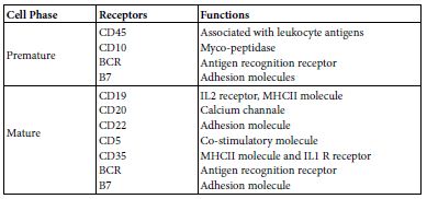
tab 5-1

Maturation of B cell from the B lymphocyte progenitor may establish in bone marrow or it may migrate as an immature B lymphocyte to spleen or to other peripheral lymph nodes and maturate there. The onto-genic maturation steps starts as; progenitor the primordium, early Pro B, Bro B, pre B to immature B cell then to mature B cells. Mature B lymphocytes when stimulated by an antigen it well grow, differentiate and expand the transform to an effector plasma cells producing either antibodies  and/or  cytokines  IL10  and IL12. As well memory B cells, Memory B cells are elected from those cells that undergoes heavy chain translocation and V fragment amplifying mutation which lead to the production of high affinity cells producing Ig G, Ig A, IgE, their BCR have high affinity for antigen binding [7-9].

B Cell Molecular Immunology

The molecular genetic system of B cells contains an array of gene sets that encodes for the various immune functions like those encoding genes for mitotic cell cycle, Ig gene superfamily, cytokine production genes, antigen presenting genes and the apoptosis encoding genes. The ontogeny of the different cell differentiation phases based on molecular genetic mechanisms like; gene re- arrangement, gene exclusion, alternative splicing and  apoptotic  gene expression. B cell on antigenic epitope stimulation, the epitope triggers naïve B cells to be activated for growth, proliferation, expansion and change to effector plasma cell and memory B cells.

Early in the Immune response time curve B cell produce IgM then class switched to other isotype. IL4, IL10, TGFB and IFN gamma promotes class switching [7]. When antigen primed B cell acts as   an antigen presenting cell, it well take up and process antigen then present it onto their surface with MHCII molecules to be presented to T helper cell. It appear that there was a shift from antibody production to APC function. Primed B cells that are transformed    to plasma cells are devoid from either IgM or IgD. Memory B cells once reactivated will perform class switching of Ig iso-types. BCR are composed of two identical Ig heavy and two identical light chains together with Ig alpha and Ig beta chains that transmit signals to   the cell interior when legends are bound to BCR. Each chain has amino and carboxy termini. Domains are found on the amino  termini of each chain. The epitope specificity of Ig molecule of BCR is determined before they are facing the antigen. The number of the possible BCR-epitope binding specificities high exceeds that number of genes forming human genome. This present a paradox. Such paradox can be solved through molecular re-arrangement, splicing and /or gene exclusion. The Kappa light chain encoding genes are mapped onto chromosome 2. While, those encoding lambda light chains are mapped onto chromosome 22. The gene cluster encoding is located onto chromosome 14. The potential antigen binding segment combinations are greater than 26 million. The heavy and light chain gene segments for both variable and constant regions are rearranged, transcribed into RNA and translated into single heavy or light chain polypeptide. The restriction of VLCL, and VHCH expression to a single member of each of the involved chromosome pairs is termed as allele exclusion. However, the collective combination of all of B cell means that both maternal and paternal of allele genes are expressed within any particular individual. There has been a guess that each B cell has the ability to produce a range of individual antigen specific receptor capable for binding as many as ten different epitopes. Significant part of this BCR epitope diversity are attributed to chromosomal DNA rearrangement. BCR represent a case of natural construct of a set of gene clusters for both of the heavy and light chains in which each set of gene clusters construct RNA transcript that express as a single polypeptide. Then these polypeptides undergo post-translational assembly. A single B cell synthesizes Ig of one single specificity at a time. This is because of its nature of combination of VL and VH regions at the effect of allelic exclusion. Un-stimulated B cell synthesizes and display monomeric IgM and IgD on their surfaces. Upon stimulation B cell may change their iso-type but not the epitope specificity of the Ig they produce. This process is known as iso-type switching. Iso-type switching influences the ultimate nature of humoral immune responses. Memory B cell producing IgM can undergo further DNA re-arrangement to change the Ig iso-type they produce [7-11].

Antigen Presenting B Cell

B lymphocytes including B1 may act as antigen presenting cell. When they do so B cells use specialized MHCII complex antigen presentation pathway to process BCR bound and internalized  protein antigens and present them to CD4 T cells. Such processes  are performed through several efficient molecular mechanisms in a stepwise manner as; (i) antigen capture and uptake, (ii) intersection of the internalized antigen-BCR complexes with MHCII complexes,(iii) generation and regulation of MHCII-peptide complexes, (iv) exo- cytotic transport for presentation of MHCII-peptide complexes at the surface of B cells and (v) activation of CD4 T cells. Collectively, these molecular mechanisms affect both of the fate of lymphocyte and shape the immune response [4,5,12].

Antibody Producing B Cells

A naïve B cell subset, on priming with SARS-COV-2 antigenic epitope(s) in continuum with in-vivo, ex-vivo and in-vitro settings will be activated through; cell growth, proliferation, expansion and transition into plasma cell producing antibodies specific for spike, receptor binding domain and other epitopes. In a study performed   in 2020 workers isolate 61 SARS-COV-2 neutralizing monoclonal antibodies from five hospitalized severe COVID-19 infection. Of which 19 were potent neutralizing to authentic SARS-COV-2 in-vitro [13,14].

Cytokine Producing B Cells

B cell performed an array of immune functions both in human and mammals independent on that of antibody formation. Like; tolerance, tumor rejection, immune response suppression autoimmunity, organization of lymphoid tissue and regulate lymphoangiogenesis. Broadly speaking it can be bio-typed into antibody producing and cytokine producing. Cytokine producing B cells are ramified into an effector and regulator B cells. The cytokine profiles of the effector are somewhat different from regulator B cells. The ontogenic origin of antibody producing B cells is different from that of cytokine producing B cells (Table 2) [3].

Table 2: Immune Functions of B cells

tab 5-2

Proposed COVID-19 Immunity

It seems to be that the nature of human immunity; during SARS- COV-2 infections, recovery and post-infection is somewhat different from other human viral infections. Collective scientific task has been made by STAT scientists [15] proposed four scenarios for immunity to SARS-COV-2 infections. These are; sterilizing, functional, waning and lost. Other idea tempted to put-down a collective scenario that mix up more than one of the mentioned scenarios. Sterilizing; The immune system of the host is armed to a foe and able to fend it off before infection take a hold. Functional; The immune system produce specific antibody responses but the antibodies wan on time post-infection but many cellular responses are not waning. Under this scenario, people whose immune system have been primed to recognize and fit the virus in continuum with either infection or vaccination could contract it again in the future but with either mild or symptomless. Waning; In this scenario people who have been infected or vaccinated would lose their protection overtime. But even if immunity wans, reinfection would be less severe and be a variation to the functional immunity. Lost; In this scenario people who have been infected would lose all of their immunity against the virus within some time frame. A re- infection after that point would be like that of first infection. Mixed Scenario; The overall picture of human immunity to SARS-COV-2 infection will be mixed. Some people will have sterilizing but most will fit about functional or waning immunity. Neither sterilizing nor lost immunity gains support by the scientific immunity [15].

Evidence Based COVID-19 Immunity

Collective local experience from the in-practice COVID-19 patient some of them recover with use of convalescent sera others not. Most patient produce specific antibodies but few fail to raise antibodies though they clinically infected [unpublished data, early in the waves of the pandemic]. The to date information in this context indicated that both humoral and cellular immunity can be lasting up to eight months post infection [14,16-19]. Effective effector B, memory B, effector T and memory T cells are involved in the protection against COVID illness. Breakthrough infection during post-vaccination periods could function exactly the same as a booster immunization. Though this may be a theoretical concept rather than practical one [20].

Memory B Cell Probe COVID-19 Vaccine Efficacy

In an experiment two groups of human subjects were assigned as; SARS-COV-2 naïve 33, and SARS-COV-2 recovered 11. Antibody and antigen specific B cells were mapped overtime. SARS-COV-2 naïve group were found to be requiring both vaccine doses for antibody response. Likewise memory B cells tracing have shown full length spike protein and spike receptor binding domain were detectable at second mRNA vaccine shot. While in SARS-COV-2 recovered group antibody and B cell responses were significantly boosted after first vaccine dose. Second dose neither increase antibody nor B cells. First vaccine dose strongly correlated with the pre-existing memory B cells in recovered group. COVID mRNA vaccine priming to individuals have shown distinct response based on prior SARS-COV-2 exposure. Memory B cells could be used as a probe for vaccine efficacy both in naïve and recovered COVID-19 individuals [21].

Memory B Cells Breakthrough Infection

In an experimental setting in which two group of patients. One vaccine breakthrough infection 55 patients and the second vaccinated close contacts who did not contract infection 88 subjects. Vaccine breakthrough infection was sharing lower memory B cell frequency but high antibody in plasma cell and those produced by memory B cells. Inflammatory cytokines, the IL1B and TNF were lowered in vaccine breakthrough infection than that infection of similar settings. Hence, lower memory B cells are correlated with vaccine breakthrough infections of COVID-19 [22].

Memory B Cells and Severity

In a retrospective study that was planned as 208 laboratory confirmed COVID-19 patients in which severity was checked on a scale graded from 0-10 severity score. Two parameters were adopted for evaluation of severity; the memory B cell count and the serum immunoglobulin levels. The age range for those patients  35-63  years with median of 50 years of which 88 were females (42%). The survivors were 191:208(91.82%) and diseased were 17:208(8.18%). The severity was ranged from6-8 with a median of 8 in deceased and 0-2 with median of 1 in survivors. Significant low levels of total B, naïve B, switched memory B, and serum immunoglobulin levels; IgA, IgG1, IgG2 in deceased than in survivors. Negative significant correlation between serum Igs and memory B cells was evident. The prognosis of COVID-19 disease is associated with B cell subsets and serum immunoglobulin levels [23].

B Cell and Hypoxia

In an experimental setting including COVID-19 patients with variable severity and hypoxia and genetically modified VHL deficient mice kept in hypoxia exposed mice. Blood was collected  from  those patients for detailed B cell phenotypes in peripheral blood lymphocytes using flow cytometery. Single cell transcription and whole blood sequence analysis were done to evaluate the impact of hypoxia on patients B cells and same was done onto the genetically modified hypoxia kept mice. There was an early and perminant defects in B cell subsets in moderate to severe COVID-19 patients including marginal zone-like, memory and transitional B cells. Similar findings were noted on genetically modified hypoxia kept mice. To this end hypoxia may contribute to pronounce and persistent defects of B cell pathology observed both in COVID hypoxic patients and hypoxia kept mice [24].

B Cell and Sepsis

In a clinical settings of an abscess forming super-infection viral COVID-19 pneumonia. Bone marrow and splenic B cell count have shown severe B cell loss in bone marrow or spleen in 64% of the of the patients. This was reflected by peripheral blood lymphopenia. B cell loss was associated with higher pulmonary SARS-COV-2 burden and only with marginal decrease in T cell counts. Study suggests the presence of sepsis related immunodeficiency in sever COVID-19 pneumonia with super-infection [25].

B Cell in COVID-19 with Seasonal Coronavirus

In this clinical setting tempts were made to evaluate the role of the pre-existing seasonal endemic coronavirus B cell on the development of sars cov-2 specific IgG responses. The tried parameters were; kinetics, breadth, magnitude and levels of cross-reactivity of IgG antibodies against SARS-COV-2  and  heterologous  corona  virus  at the clonal levels in patients with mild or severe COVID-19 and controls. Assessment were made onto antibody reactivity to nucleo- capsed and spike antigens and correlate this with IgG responses to SARS-COV-2 neutralization. Patients with COVID mounted a mostly type specific SARS-COV-2 responses. IgG clones directed against seasonal coronavirus were boosted in patients with severe covd-19. these boosted clones did not neutralize SARS-COV-2. Such findings indicate a boost of poorly protective COVID-19 specific antibodies in patients with COVID-19 that correlate with disease severity, reveals “Original antigenic sin” [26].

Relaxing B Cell Tolerance

Five groups of individuals were the test and controls. First COVID-19 convaluscent, second mild COVID-19, third severe COVID-19 fourth COVID-19 vaccinated and fifth control. Blood were collected from these groups. Mononuclear cells were separated from the test and control blood samples. Flow cytometery was performed on these preparations. Anergic B cells were found in high frequency in severe COVID-19 patients as compared to mild cases. They were in activated state displaying reduced inhibitory receptor expression and restored BCR signaling indicative of breach of anergy during viral infection, supported by increment shift in autoantibody levels. This together with phenotypic and functional alterations significantly correlated with hyper-inflammation in severe SARS-COV-2 infection. Hence, B –ND undergoes relaxation in their peripheral tolerance in severe COVID-19 [27].

Memory B Cell Pulmonary Subsets

Memory B cells are immune cells produced primarily in the lymph node and spleen. They persist for long time there in these regions and retain the memory of the infectious agents. If the subject body facing with same agent in future. These cells immediately mobilized and rapidly reactive immune system for effective protection. Recently workers have been documented the presence of memory B cells in lungs of laboratory animal models. Whereby, the laboratory animal model infected with the virus as influenza or COVID-19. Ten weeks later the virus eliminated from the body of infected animals. Memory B cells were tracked in lungs of the infected animals using flurescent markers followed by single cell transcriptomic analysis. Such techniques enable localizing memory B cells in lung and their gene expression profile cell by cell. Results have shown groups of memory B cells in the bronchial respiratory mucosa in two subpopulations of different gene expression profile and functions. The Bona fide and Bystandard subsets. The bona fide subset showed high affinity to virus and trigger their appearance immediately on entry and infection. While the bystandard is not directly recognize the virus but bind to the receptor of the immune complexes formed by the antibodies produced by bona fide cells. Both of the subsets exhibit synergistic function as a two tiers system [28].

Conclusions

B cell immunology of COVID-19 is in the forefront and current scientific mode. Memory B cell was found pathognomic with an array of human COVID-19 pathological changes. The changes cover both subnormal count of the B cell subsets and cell defects in different COVID-19 disease forms.

References

  1. Abbas AK, Lichtman AH, Pillai S (2015) Cellular and Molecular Immunology, 8th Elsevier Saunders, Phildelphia.
  2. Allman D, Pillai S (2008) Peripheral B cell Curr opin Immunol 20: 149-157. [crossref]
  3. Lund FE (2008) Cytokine producing B lymphocytes-key regulator of Curr Opin Immunol 20: 332-338. [crossref]
  4. Popi AF, longo-Maugeri IM, Marino M (2016) An overview of B1 cells as an antigen presenting cells. Front Immunol 7: 138. [crossref]
  5. Adler LN, Jiang W, Bhamidipati K, Millican M, Macaubas C, et al. (2017) Other functions: class II restricted antigen presentation by B cells. Immunol 8: 319. [crossref]
  6. Shnawa IMS (2014) Dijla-ALWadah Publishing House. Iraq-Jordan.
  7. Shnawa IMS (2013) Molecular Immunology. Lap-Lambert Academic Publication-
  8. Lu L (2013) Frontier in B cell Cellular and molecular Immunology 10: 95-96. [crossref]
  9. Alt FW, Horj T, Reth M (2015) Molecular Biology of B ElSevers Ltd.
  10. Scheid JF, Barnes CO, Eraslan B, Hudak A, Keeffe JR, et (2021) B cell genomics behind cross-neutralization of sars-cov-2 variant and sars-cov. Cell 164: 3205-3221. [crossref]
  11. Cai H, Hu J, Huang L, Gao C, Xu M, et (2022) The relationship between convergent IgA signatures and severity of covid-10 patients by next generation sequences of BCR repertior. Front Microbiol 12: 833054. [crossref]
  12. Frazi R, Aghbash PS, Eslami N, Azadi A, Shamek A, et (2022) The role of antigen presenting cells in pathogenesis of covid-19. Pathol Res Pract 233: 153848. [crossref]
  13. Ng K, Faulkner N, Cornish GH, Rosa A, Harvey R, et (2020) Pre-existing and de- novo humoral immunity is sars-cov-2 in humans. Science 370: 1339-1343. [crossref]
  14. Liu L, Wang P, Nair MS, Yu J, Rapp M, et al. (2020) Potent neutralizing antibodies directed to multiple epitopes on sarscov-2 Nature 584: 450-456. [crossref]
  15. Branswell H (2020) Four scenarios on how we might develop immunity to covid- 19-STAT.
  16. LeBert N, Tan AT, Kunasegaran I, Tham CYL, Hafezi M, et (2020) T cell immunity in cases of covid-19 and sars and uninfected controls. Nature 584: 457-462. [crossref]
  17. 17-Gerfoni A, Weiskopf D, Sydney I, Mateus J, Dan JM, et al. (2020) Target T cells responses to sars-cov-2 corona virus I humans with covid-19 disease and unexposed individuals. Cell 181: 1489-1501. [crossref]
  18. Gallais F, Velay A, Wending MJ, Partisani M, Sibilia J, et al. (2020) Intrafamilial exposure to sars-cov-2 induce cellular immune responses without seroconverstion, France. Emerg Infect Dis 27: 113-121. [crossref]
  19. Wise J (2020) Covid-19: T cells responses for at least six months after infection study shows. BMJ 371:4257.
  20. Smith J (2021) Is covid-19 exposure post-vaccination, a booster or a risk. Oper in Coronavirus.medium.com
  21. Goel RR, Apostolidis SA, Painter MM, Mathew D, Pattekar A, et (2021) Distinct antibody and memory B cell responses in sars-cov-2 naïve and recovered individuals after mRNA vaccination. Sci.Immunol 6: 6950. [crossref]
  22. Tay MZ, Rouers A, Fong SW, Goh YS, Chan YH, et (2022) Decreased memory B cell frequencies in covid-19 delta variant vaccine breakthrough infection. EMBO Mol Med 14: 152272. [crossref]
  23. Colkesen F, Kurt EK, Vatansev H, Korkmaz C, Colkesen F, et al. (2021) Memory B cell and serum immunoglobulins are associated with disease severity and mortality in patients with covid-19. Postgrad Med J 98: 765-771. [crossref]
  24. 24-Kotagiri P, Mescia F, Hanso AL, Turner L, Bergamaschi L, et 2022.The impact of hypoxia in covid-19.EbioMedicine 77: 103878. [crossref]
  25. Ihlow J, Michaelis E, Greuel S, Heynol V, Lehmann A, et al. (2021) B cell depletion and signs of sepsis-aquired immunodeficiency in bone marrow and spleen of covid-19 Int Infect Dis 103: 628-635. [crossref]
  26. Aguilar-bretones M, Westerhuis BM, Raadsen MP, Bruin ED, Cahndler FD, et al. (2021) Seasonal coronavirus specific B cells with limited Sars-cov-2 cross-reactivity dominate the IgG responses in severe covid-19. J Clin Invest 131: e150612. [crossref]
  27. Castleman MJ, Stumpf MM, Therrien NR, Smith MJ, Lesteberg KE, et (2022) Sars- cov-2 infection relaxes peripheral B cell tolerance. J Exp Med 219: e20212553. [crossref]
  28. Greggoire C, Spinelli L, Villazala-Merino S, Gill L, Holgado MP, et (2022) Viral infection engenders bona fide and bystander subsets of lung-resident memory B cells through a permissive mechanism. Immunity 55: 1216-1233. [crossref]

Regulatory Lymphcytes in COVID-19

Abstract

The ontogeny, the basic and molecular biology and the subsets of human lymphocytes were briefed. Functional, anergic, exhausted, and senescent lymphocyte subsets were detected in severe SARS-COV-2 human infection forms. The aim of the present opinion paper tempted to reveal the comparative biology of the regulatory lymphocyte subsets RLS both in health and disease. RLS, are regulatory natural killer cells NK reg., regulatory B cell, B reg. and regulatory T cell, T reg. to visualize their roles in severe SARS-COV-2 human infections. RLS of NK, B and T cells were found almost associated with dampening the immune mediated tissue injuries following COVID-19 illness. Though it was not clear whether these regulatory lymphocytes     did their dampening role in simultaneous, sequential, synergistic and/or antagonistic manner. Show case analysis was made for Nk reg and T reg in COVID-19 cases. This opinion paper suggest a clinical experimental setting in which a group of severely ill COVID-19 patients and normal controls will be blood collected and their lymphocyte separated then subjected to regulatory lymphocyte [NK reg, B reg. and T reg.], flow cytometry, single cell RNA sequencing and interactome studies in order to visualize the way they perform their immunologic roles and the possible interplay between them.

Keywords

Anergy, COVID-19, Exhausted, Lymphocyte, Regulatory, Senescent

Introduction

Regulatory Cells, regulatory immune cells, and regulatory lymphocytes are terms denoted to immunosuppressive lymphocytes RLS. RLS functioned both in the innate and adaptive immune responses and their functions mainly in down regulation of immune over-reaction and/or immune unwanted reactions that served as cellular bases for anergy, tolerance and suppression. Major regulatory subsets mapped both in human health and disease are: NK reg., B reg., and T reg. [1-15]. The present opinion tempts to reveal the actual roles played by regulatory lymphocytes in COVID-19 in a comparative attitude and show case analysis.

Ontogeny

The primordial mother cell, the heamopoietic stem cells of all   of immune cells was found in fetal liver in the pregnant woman.    As gestation proceeds it will migrate to fetal bone marrow and remained there in postnatal life and adulthood life. In bone marrow the haemopoietic stem cells differentiated  into  three  progenitor  cell types; the myeloid, the lymphoid and the lympho-myeloid. The lymphoid cell descend from the lymphoid progenitor cells through the action of haemopoietic cytokines. Lymphoid cells within the continuum of bone marrow microenvironments either stay therein and differentiated in to naïve B cells or trafficked and migrate to thymus. In thymic tissue niche naïve cells undergoes thymic negative and positive selection through thymic proteins and cytokines then mature to be T cells [15]. In two groups of infants, first healthy while the second was premature and stressed. Cord blood lymphocytes were separated from both groups and their phenotypes compared. The phenotyping process was done by fluorometric analysis. This analysis had shown that early third trimester cord blood lymphocytes were as 80-85% fetal T cells belongs to T4+ inducer population and 10% as T8+ suppressor/ cytokine subsets. As gestation proceeds, the T4 and T8 ratio shifts towards adults value together with increase in expression of mature antigen T12. The naïve B cells differentiated into antigens of B1, B2 and B 4 were not changed during gestation of normal healthy infants. Antenatal stress which threatens fetal survival leads to the appearance of phenotypically less mature B cells in circulation for both of the lineages than expected for the gestational age. Cells expressing very early B cell markers B2, B4 increase and exceed the numbers of the more mature B1+ cells in the cord blood most notably due to hypoxic stress during antenatal condition [1]. The fetal blood from fetuses in the second trimester of gestation were collected, lymphocyte separated and analyzed by monoclonal antibody two color immune-fluorescence technique as well as flow cytometry. Lymphocyte surface markers were evaluated. The study indicate that cells of B, T and NK lineages as well as precursor cells can be detected in fetal blood at 18 to 20 weeks of gestation. During this stage of development variable proportions of T & B cells express surface molecules such as CD 1, CD10, CD38, CD45RA indicative of precursor or naïve state; on the other hand the CD57 molecules is not detectable on membrane of NK and T cells and the RO isoform of CD45 leukocyte antigens is synthesized at low percentages of T cells. Such findings suggest that the observed phenotypic peculiarities of lymphoid cells might be due to the inductive easiness of tolerance that occur in early ontogenic stages of the immune system [2]. T, B and NK lymphocytes and their respective phenotypic subsets originate from bone marrow stem cells and their progenitor lineages. Lymphoid cell that migrate to the thymus receive signals through notch commit to the T cell lineage. The lineage development in human beings is critically dependent on IL7 for T cell, IL 4 for B cell and IL15 for NK cells. The specificity and diversity of lymphocytes are gained during the process of generation TCR in T cells and BCR in B cells. The T and B cell repertoire is determined by random variable V, diversity  D and joining J somatic gene segments that recombine with an imprecise addition of nucleotides at the segment connection. This recombination is performed by an enzyme complex known as VDJ recombinase that contains the recombination activation gene RAG. RAG proteins expressed onto B & T cells. Successful recombination is determined by the expression of a functional antigen receptor which allows survival and durability of development [3].

Molecular Biology of Lymphocytes

In an experimental setting for a human lymphocyte cell line that was irradiated with 12C ion beams at; 0, 0.1, 0.5 and 2.0 Gy. The transcriptional profiles were evaluated by human gene expression microarray method at 24 hr. post-irradiation. In accordance with microarray assays, there are 1113 genes were up regulated and 833 genes were down regulated in human lymphocyte irradiated with 0.1 Gy 12C ion beams compared to the control group. 1095 genes were up regulated and 1220 down regulated in cells irradiated with 0.5 Gy 12C ion beams and 1055 genes were up regulated and 1356 genes were down regulated with 2.0 Gy. A total sum of 504 genes were differentially expressed in all irradiated groups of which 88 were up regulated and 416 were down regulated. Most of these altered genes were related to; cell cycle, apoptosis, signal transduction, DNA transcription, repair and replication. Thus, the differentially expressed genes at 24 hr post- irradiation increased as irradiation dose increased, up regulated genes gradually decreased and down regulated genes increased. 12C ion beams irradiation could express a number of genes in dose dependent manner which might initiate failure of multiple biological functions of the cell [4].

Lymphocyte Functional Phenotypes and Subsets

Lymphocytes are white blood cells that forms parts of the systemic and mucosal immune system with uniform appearance and varied functions. Bone marrow derived cells, the B cells are involved in the innate immune function of antigen presenting cells, adaptive antibody and IL10 responses. T cells functions in; cell mediated immune response, TH1, TH2, Th17 cytokine production as well as delayed type IV hypersensitivity reactions. TH17 take part in the immune cross- road functions both for innate and adaptive responses. Regulatory T cells the T regs. are commited in down regulation of over-immune and/or unwanted immune reactions. Natural killer cell the NK interplay direct cell mediated cytotoxicity of virus and tumor cells. NK includes innate and adaptive phenotypes [LaRosa and Orange 2008, National human genome institute 2022 [3,5].

Regulatory Lymphocyte Subsets

The B, T and NK lymphocytes are expressing regulatory phenotypes as; B reg., T reg. and NK reg. All of which are involved in down regulation of immune over and/or  unwanted  reaction  noted through the cellular events of immune responses. Such down regulation processes almost mediated by various cytokines through; signal transduction, cell-cell communication and cross-talk [3,7,12].

Regulatory Natural Killer Cells

Cellular Immuno-Biology

A large lymphocyte with finely granular cytoplasm commit in antiviral and anti-cancerous cells. These cells are known as Natural Killer Cells NK cell that belongs to innate immunity with an adaptive immune potentials. From the structural point of view these cells are devoid from both TCR an BCR. But they have; cytotoxicity receptor family (NKp30, NKp44, NKp16, NKp80), type C lectin domain containing receptors (NKG2D), the CD2 superfamily receptor and IgG receptor (CD16). The legend of some of these receptors up regulated by stress and infection like vial haemagglutinins for NKp46, Cd46 for CD2 and IgG for CD16 [LaRosa and Orange [3]. NK cells have distinct subsets with desperate function, location and developmental origin. Peripheral blood NK cells can be of two functional subsets are known based on expression of CD56 and CD16. the first subset isCD56 dim CD 16+ form 90% of the total blood NK effective in killing target cells and secret low cytokines. While, the second is CD56 bright CD16- constitute 10% of the total blood NK but are enriched in secondary lymphoid tissues. The effector functions of the NK are cytotoxicity and cytokine production. These functions are separate in NK subpopulations. The bright and the dim subsets were differing in; (i) the expression of inhibitory and activating receptors (ii) adhesion molecules and (iii) chemockine receptors (Leunemann et al.) [6]. The subsets, Table 1 and immune regulatory subsets of NK cells are; Bright [Gross et al. [7] and CD73 + [8].

Table 1: Natural Killer Cell subsets*

tab 6-1

*Adapted from Lunemann et al. [6]

NK Immune Functions

NK have an array of immune functions as; Controlling T cell responses and maintaining homeostasis [Gross et al. [7], Orchestrate immune responses, linking innate and adaptive immune responses and regulating, T, B and DC [9].

Regulation by Natural Killer Cell, A Mechanistic View

NK cells performed their immune regulatory function using either of the following mechanisms; (i) direct contact cytotoxicity of myeloid cells, (ii) polarization of TH1, shaping and dampening through cell mediated cytotoxicity and (iii) antibody dependent blocked of Qa 1 NKG2A interaction resulted in potent NK dependent elimination of T cells [7,12].

Regulatory NK in Human Diseases

The immune regulatory actions of NK cells are operable in a number of human diseases; CD72 NK mediated pathology in tumor micro-environment [8], autoimmune and inflammatory diseases [6] autoimmune liver diseases [Jian and Wang [9] and multiple seclerosis [6,7].

NK in COVID-19

SARS-COV-2 clearance rate, antibody response and disease progression in COVID-19 correlate with NK pathophysiological status and NK dysfunction is linked to disease susceptibility. Thus NK may act as a key element in the switch events from effective to harmful immune responses in COVID-19 illness. NK depletion and dysfunction correlated with severity and anti-fibrotic activity [10]. There were significant reduction in number and function of NK attributed to their exhaustion [11].

Regulatory NK in COVID-19

A bidirectional of cell-cell cross-talk of NK with, neutrophil, DC, and monocyte/ macrophages have shown elimination of neutrophil and DC in a separate experimental settings [12]. High NK count, low T cell CD8+ and regulatory CD56 bright CD16- dim in severe COVID-19 patients [13] as shown in the show case analysis with a notable NK subsets of NK reg. in COVID-19 disease (Table 2).

Table 2: Show case Analysis of natural killer cells in COVID-19

tab 6-2

Regulatory B Lymphocyte

Cell Immunobiology

B lymphocytes are bone marrow derived lymphocytes. They developed in bone marrow and matured in the peripheral lymphoid tissues starting with pro B then Pre B, immature B and mature B. The immune potentials of B lymphocytes are multiple. As antibody producing, antigen presenting and cytokine producing as well as immune regulating [3,17].

The Immune Functions of B lymphocytes

Naive B lymphocytes on activation through stimulatory and co- stimulatory signals by either of; antigen, mitogen and/or cytokine they may perform one or more of the following immune functions; (i) antibody production, (ii) antigen presentation, (iii) cytokine production, (iv) immune regulatory and (v) toleragenesis [17,18].

Regulatory B lymphocytes [B reg.]

Regulatory B lymphocyte express immunosuppressive functions via diverse mechanisms. Of which B reg. modulate immune responses through secretion of cytokines, IL10, IL35, and TGFB.   Or by direct cell-cell contact dependent mechanisms. B reg. is important to human welfare both in health and disease. In disease, however, they are associated with abnormalities both in numbers  and functions. B reg. accounts for 0.5% of human peripheral blood lymphocytes. And expressed a variety of protective and pathologic. They take part in the tissue transplantation tolerance and involved in creating immune prevealiged sites for the uterine environment and neonatal life [17-19].

Regulatory B Lymphocytes Subsets

Human regulatory B lymphocyte is heterogeneous in term of number, origin and localities as shown in the following (Table 3).

Table 3: The Subsets of Human B reg.*

tab 6-3

*Adapted from Menon et al. [17]

Mechanisms of Action of B reg

The operable mechanisms by which naïve B lymphocytes becomes B reg. are; (i) change in CD markers and surface immunoglobulins, (i) stimulation by IL10, (iii) stimulation by IL35 and TGFB, (iv) direct cell-cell contact dependent which might be attributed to CD 80 CD86 PD-L 1 CD40L and CDd 1 d, (v) granezyme dependent cell killing, (vi) anergic or tolerizing mechnisms and TLR-BCR engagement mechanisms [17,19].

B reg. in Human health and Disease

B reg. take part in; pathogenesis, protection and or promotion of human disease as in; cancer of lung, infections, malaria, allergic air way inflammation and transplantation Diabetes and pregnancy [17,19].

B reg. in Viral Infections

In human hepatitis B disease immature transition CD19+ CD24hi CD38hi and CD19+Il10+ Breg. involved in regulation of the antigen specific CD8+ Tcells. While, incaseofacquiredimmunedeficiency HIV disease, activated immature transtionalCD19+CD24hiCD38hi Breg. reduces the frequency of HIV infected individuals through suppression of CTL function which cause viral persistence. In Deng fever disease, CD19+CD24hi CD38hi reduces the severity of the disease. Neonatal CD5hiCD10-CD 1chiCD45RACd23loCd24hiC38loIgDloIgMlo Breg. produce IL10 that dampen the beneficial cytokine production by TH1 cells and contribute to severe disease [18].

Breg in COVID-19 Disease

In severe SARS-COV-2 infections the precursors of human transitional CD19hi CD38hi B reg. were reduced [19]. In a clinical setting in which two men vaccinee were enrolled in immune cell flow cytometery, it was evident that Breg was increased post first shot but reduced post to the second COVID-19 vaccine dosage [20].

Regulatory T lymphocytes

Cell Immunobiology

The CD4+ T lymphocytes may be differentiated into lymphocytes that have the ability to suppress other immune cells including T cell responses. These differentiated cells are known as regulatory T cells. Their development from naïve lymphocytes can either be thymic or extra-thymic. The thymic developed are natural T regs and stably expressFoxp3 transcription factor and is eligible for its immune suppressive function. The natural T regs are IL2 dependent and Foxp3 transcription factor producer. The surface markers of natural T regs are CD3+CD4+Cd25+ Foxp3+. While, the extra thymic developed T regs are of inducible nature. The surface markers of the inducible type 1 T regs are CD3+CD4+ [3,21].

Human Tregs Subsets

Three main subsets of T regs are known till date. One of which  is natural and the other two were of inducible nature. The inducible subsets are either TGFB or IL10 induced subsets (Table 4).

Table 4: Human Regulatory Lymphocyte Subsets*

tab 6-4

*LaRosa and Orange [3]

T regs Immune Functions

T regs expresses an array of immune functions including; negative regulation of T cell responses, sustaining immune tolerance, reserve immune homeostasis, taking part in establishment of lymphocyte anergic state [21].

T regs in Human Diseases

In human host microenvironment T regs. may express either protective or pathologic functions depending on the nature of the surrounding micro-environmental factors. Loss of function mutation in the Foxp3 gene lead to; inflammatory autoimmune disease, immune dysregulation, poly-endocrino-pathy, enteropathy and or graft versus host reaction GVHR. Such mutation may aggraviate human virus infections [21].

Tregs in COVID-19

In early stage of SARS-COV-2 infection, activated Tregs. Populations potently suppress the recruitment of immune cells such as Th1 a CD8+CTLs leading to reducing of the immune responses. In mild infection form, T regs becomes increased in numbers and could attenuate the inflammatory responses and quench the cytokine storm, this event could promotes recovery of the patients. When the infection form proceeds towards severity, Treg depletion can enhance the activation of pro-inflammatory immune cells and production of pro-inflammatory cytokine that leads to hyper-cytokinemia and lung injury [21]. The exact role played by T reg. in COVID-19 seemed   to be controversial among different workers all over the world. In a study were 57 mild, severe and recovered patients, peripheral blood mononuclear cell were subjected to study Tregs CD25+ foxp3 has shown increase in proportion of these cell [22]. Other working group use to study 109 mild, severe and recovered patients T regs they were found Cd4+CD25+ CD127+ got increased in both proportion and numbers of Tregs [23]. No change in CD4+CD25+Foxp3+ Tregs have been found in a group of 17 moderate,27 severe and 8 control [24].

Show Case Analysis

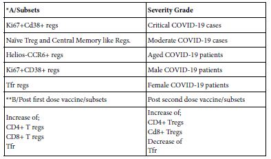
A show case analysis was made for two published works; one concerning SARS-COV-2 infected patients in Japan and the other done on two COVID-19 vaccinee men. The investigation in both    of the infected Table A and vaccinee Table B, tempted to analyze peripheral blood lymphocytes using flow cytometery and immune informatic approach of lymphocytes including T regs. Researchers reached results of different subsets of Tregs (Table 5) [20,25].

Table 5: Show case analysis of T reg subsets in COVID-19 infected and COVID-19 vaccinee

tab 6-5

*Sondergraad et al. [25];**Gupta et al. [20]

Conclusion

In comparative Biologic sense regulatory lymphocytes as ordered in accordance with their chronological order of discovery are; T reg., Breg. and NK reg. They are at most immunosuppressive lymphocytes. Each of which  may  include  typical  or  conventional  and  atypical  or unconventional phenotypes. The main conventional regulatory lymphocytes are; Three T regs, six B regs and two Nk regs. The current newly reported regulatory lymphocytes are; Eight T regs, one Bregs and one NK regs. Hence the total number of these regulatory subsets are; Eleven T regs, seven B regs and three NK regs. These regulatory lymphocytes are of suggested basic sharing in common immune features as: (i) Natural or induced, (ii) initiated by activation and action mechanisms, (iii) mobilized by chemkines and activated by  cytokines, (iv) own sets of surface inhibitory and/or stimulatory functions, (v) on function they may associated with age sex or severity of the disease, (vi) the consequences of their immune function in disease either promote protection or promote disease and vii- the mechanistic path can be of use as chemotherapeutic target. The regulatory lymphocytes shared essential roles in pathogenesis and immunology of COVID-19. Though therein presence of gaps in understanding of their exact interplay. The role of T regs in COVID-19 to date it is still in debate. Leem et al. [16] have done a landmark  study  of natural killer cell including NKregs in COVID-19 patients. Maucourant et al. [14] have been performing landmark investigation on NK immunotypes as related to severity, Bergantini et al. [13] did immunological signature of T cell and NK cells on hospitalized COVID-19 patients. They conclude that higher frequencies of NK and NKreg. Corresponded to lower frequencies of CD3+ and CD4+TCM in severe cases and B cells were not mapped. High frequencies of senescent and exhausted NK, and memory CD4+, CD8+ T cells associated with severe infection forms. B cells compartment were not mapped [Srivastava et al. [26]. Regulatory NK and T regs as well as B cells were investigated through mass flow cytometery with an emphasis on cell networking were tempted [25] performed profiling study to lymphocytes for NK, B and T cells including regulatory lymphocyte subsets have been reported in two vaccine men [Gupta et al. [20]. Hence full lymphocyte profiling using flow cytometery, single cell RNA sequencing and interactome studies including regulatory subsets as well as the deduced network in between lymphocyte and other immune cells to visualize the way they interacts between them and with other cells in mild, moderate, severe and deceased COVID-19 patients still need to uncovered. Thus it is being suggested.

References

  1. Wilson M, Rosen FS, Scholssman SF, Reinherz EL (1985) Ontogeny of human T and B lymphocytes during stressed and normal gestation; phenotypic analysis of umbilical cord lymphocytes from term and preterm infants. Clin Immunol Immunopathol 37: 1-12. [crossref]
  2. Luciveo G, Addario VD, Tannoia N, Dellosso A, Gambatesa V, et (1991) Ontogeny of human lymphocytes: Two color fluorescence analysis of circulatory lymphocyte subsets in fetuses in the second triemester. Fet Diag Ther 6: 101-106. [crossref]
  3. LaRosa DF, Orange TS (2008) I J Allergy Clin Immunol 121: S464-S369.
  4. Zhang R, Dang X, Zhang Z, Yuan Y, Ren Y, et (2019) Comparison of transcriptional profiles in human lymphocyte cells irradiated with 12C ion beams at )-2.0Gy. Cancer Manag Res 11: 2363-2369. [crossref]
  5. National Human Genome Institue (2022) Lymphocytes.
  6. Lunemann A, Lunemann JD, Munz C (2009) Regulatory NK cells. Function In inflammation and auto-immunity. Med 15: 352-358. [crossref]
  7. Gross CC, Schulto-Mecklenbeck A, Wiendi H, Marcenaro E, Rosbo NKD, et al. (2016) Regulatory functions of natural killer cells in multiple seclerosis. Front Immunol 7:606.
  8. Neo SY, Wang B, Record J, Ma R, Chen X, et al. (2020) CD73 immune check point defines regulatory NK cells within tumor J Clin Invest 130: 1185- 1198. [crossref]
  9. Jiao G, Wang B (2016) NK subtypes in regulation of auto-immune liver Gasterointerol Res Pract 2016: 6903496. [crossref]
  10. Gallardo-Zapate J, Maldonado-Bernal (2022) Natural killer cellexhaustion in sars- cov-2 infections. Innat Immunty 28: 189-198. [crossref]
  11. Deng X, Terunuma H, Nieda M (2022) Exploring the utility of NK cells in covid-19. BioMedicine 10: 1002. [crossref]
  12. Di Vito C, Calcaterra F, Coianiz N, Terzoli S, Voza A, et (2022) Natural killer cells in sars-cov-2 infection: pathogenesis and therapeutic implicatons. Frot Immunol 13: 888248. [crossref]
  13. Bergantini L, d’Alessandro M, Cameli P, Cavallaro D, Gangi S, et (2021) NK and T cell immunological signature in hospitalized patients with covid-19. Cells 10: 3182. [crossref]
  14. Maucourant C, Filipovic I, Ponzetta A, Aleman S, Cornillet M, et (2020) Natural killer cell immunity related to covid-19. Sci Immunol 5: eabd6832. [crossref]
  15. Borner K, Teichmann SA, Quardokus EM, Gee JC, Browne K, et al. (2021) Anatomical structures,cell types and biomarkers of human reference atlas. Nat Cell Biol 23: 1117-1128. [crossref]
  16. Leem G, Cheon S, Lee H, Choi SJ, Jeong S, et al. (2021) Abnormality in NK cell populations is prolonged in severe covid-19 J Allerg Clin Immunol 148: 996- 1006. [crossref]
  17. Menon V, Hussell T, Shuwa HA (2020) Regulatory B cells in respiratory health and diseases. Imunol Rev 299: 61-73. [crossref]
  18. Upasani V, Rodenhule-Zybert I, Cantaert T (2021) Review: Antibody independent functions of B cells during viral Plos Pathogens 17: e1009708. [crossref]
  19. Abebe EC, Dejenie TA, Ayele TM, Baye NG, Teshome AA, et (2021) The role of regulatory B cells in health and disease: A systematic review. J Inflam Res 14: 74-84. [crossref]
  20. Gupta S, Su H, Agarwal S (2020) Immune response to sars-cov-2 vaccine in two Int Arch Allergy Immunol 183: 350-359.
  21. Wang Y, Zheng J, Islam MS, Yang Y, Hu Y, et al. (2021) The role of CD4+Foxp3+ regulatory T cell in the immune-pathogenesis of covid-19:implication for treatment. Int J Biol Sci 17: 1507-1520. [crossref]
  22. Galvan-Pena S, Leem J, Chowdhary K, Michelson DA, Vijaykumar B, et al. (2020) Profound T reg pertuberations with covid-19 bioRxiv 416180.
  23. Chen X, Haung J, Chen J, Huang Y, Jiang X, et (2020) Characterisitics of immune cells and cytokines in patients with coronavirus didsease-19 in Guangzhou China. Human Immunol 81: 702-708. [crossref]
  24. Meckiff BJ, Ramire-Suasteyal C, Fajardo V, Chee SJ, Kusnadi A, et al. (2020) Imbalance of regulatory and cytokines of sars-cov-2 CD4+ T cells in covid-19. Cells 183: 1340-1353. [crossref]
  25. Sondergraade JN, Tulyeu J, Edahira R, Shira Y, Yamaguchi Y, et (2022) Regulatory T cells are central lubs for age, sex, and severity associated networks during covid-19. MedRxivan 22268711.
  26. Srivasta R, Dhanushkodi N, Prakash S, et al. (2022) High frequency phenotypically and functionally senescent and exhausted CD58+CD57+PDL+ NK cell sars-  cov-2 specific memory CD4+ and CD8+ T cells associated with severe disease in unvaccinated covid-19 patients.

MAIT Cells Functions in Homeostasis COVID-19 Infected and COVID-19 Vaccinee

Abstract

MAIT cells are innate like T cells served as; bacterial and fungal sensors through TCR dependent mechanism and viral sensors via cytokine TCR independent mechanism. These mechanisms are along with an overlapping tissue repair mechanisms. The objective of the present opinion paper was to deduce the immune functions and immune features of MAIT cells in; Homeostasis, SARS-COV-2 infection and SARS-COV-2 vaccination as appeared in the current 2020 up to 2022 publications. Single cell transcriptomics, mass transcriptomics, flow cytometery, unsupervised analysis and full immune      cell landscapes were the major approaches followed by lymphocyte immunologists. During homeostasis and activation, early antigen specific MAIT        cells activation with MRI legand5-OP-RU and non-specific TCR stimulation, it has been found an array of phenotypes as; homeostatic, effector, helper,  tissue infiltrating, regulatory and exhausted phenotypes. While, in prolonged stimulation, proliferative, cytotoxic, immune modulating and exhausted phenotypes were identified. In SARS-COV-2 human infection, MAIT cells may be reduced in circulation and enriched in the airways, dys-regulated, or activated then migrate to lungs in the pneumonic COVID-19 pathotype. In vaccinee, however, MAIT cells in mRNA COVID-19 vaccinee was found to be positively correlated with magnitude of humoral and cellular response to vaccine in normal healthy vaccinee but their cytotoxic function post activation        is negatively correlated with humoral and cellular immune response to vaccine. MAIT cells were found to be helpful mucosal cellular immune-adjuvant in influenza nasal vaccination strategy. Thus MAIT cell immune functions may be summed up as;  microbial  immune  sensors,  immune-pathogenic, immune modulating, mucosal cellular adjuvant to viral protein antigen and intrinsic systemic cellular adjuvant in COVID-19 vaccinee. The evolution mechanisms for MAIT functional phenotypes were suggested.

Keywords

Adjuvant, Activation, cell, cytotoxicity, mucosal, MAIT, TCR dependent, TCR independent, vaccine.

Introduction

MAIT cells are innate-like of recirculating T cells. They can function as bacterial and fungal sensors through TCR dependent mechanism and as viral sensors via cytokine activation mechanisms. These mechanisms are overlapping with their ability to repair tissues. MAIT cells are also found in association with autoimmune, immune mediated and cancer diseases. They are enriched in human; liver, lung, gut with an evident variable existence in peripheral blood of normal healthy individuals. In human viral infections they are of reduced frequencies in blood and enriched in local tissues. The situation      in human SARS-COV-2 infection so far concerning the MAIT distribution seems to be not far from that of other viral infections [1- 8]. The aim of the present opinion paper was to deduce the immune functions and immune features of MAIT cells in in; normal human homeostasis, SARS-COV-2 infected and SARS-COV-2 vaccinee.

Investigative Approaches

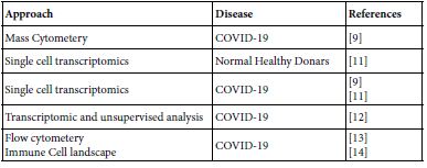
Google mapping the MAIT published contributions all over the world paved the author to the years 2020 up to 2022. The publication covers the areas of biology, molecular biology, pathogenesis in microbial, autoimmune, immune mediated and cancer besides their role in tissue repair. The aiming of the present opinion paper was focused on their role in homeostasis, COVID-19 and COVID-19 vaccinee. Table 1 lists the investigative approaches ensembled in current MAIT-COVID-19 research.

Table 1: Molecular and Immune investigation approaches in COVID-19, homeostasis and COVID-19 vaccinee

tab 7-1

MAIT Cell Immunobiology

Ontogeny

The human MAIT cells expressing V alpha 7-CD161hi T cells are generated during gestation and likely share a common prenatal development program. Within cord blood niche the total MAIT cells, V alpha 7+ CD161hi. T cells are forming the minority recognizing MR1; 5Op-RU display a TRAV/TRBV repertoire very similar to adult MAIT cells. During the few weeks of postnatal life only MR1:5-OP-RU reactive to V alpha 7.2+CD161hi T cells aquire memory phenotype Only these cells expand to form adult MAIT pool diluting out other V alpha.7-2+ CD161hi. And V alpha 7-2-CD161hi. Population in a process requiring at least6 years to reach adult level [1].

MAIT Cell and Molecular Biology

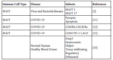
MAIT cell are forming one subset of T cells with an evident unique characteristics. They function both in innate and adaptive immune responses. MAIT can perform their biologic functions both through TCR dependent and TCR independent manners. Their basic TCR structure composed of TCR alpha chain V alpha33 associated with limited TCR Beta chain repertoire and restricted by the non – polymorphic MHC class related MRI molecule. MAIT cells are innate- like immune cells produce wide range of cytokines. Resting cells are devoid from garnzyme and porins and no-cytotoxic. On activation by either microbial riboflavin through TCR or through non-TCR way by cytokines, they own granzyme and porins and becomes cytotoxic. Such activated MAIT cells allowed to migrate to the inflamed areas and functions therein. So can act as first line defenders against microbial infections. They also contribute in autoimmune and immune mediated diseases. MAIT cells harbor mucosal surfaces of lung, gut and in liver. Variable MAIT cell counts were found in peripheral blood of normal human blood doners. The surface markers of MAIT cells are; C type lectin CD161, integrin and chemokine receptors. The fine structure of the MAIT cellular system composed of g ranzyme type B, TCR alpa and TCR beta but the beta is very limited [5,7,8]. MAIT cell TCR can fine-tune MRI recognition through antigen dependent manner, by which MAIT cell recognition is modulated [4]. MAIT cell are ramified into a number of subsets so far concerned in COVID-19 (Table 2). Comparative view to MAIT and conventional T cells are depicted in Table 3.

Table 2: MAIT cell subsets in COVID-19 and in normal healthy donors

tab 7-2

Table 3: Comparative View to MAIT cells And Conventional T cells*

tab 7-3

*Based on to Vorkas et al. [10]

To be a functional MAIT cell phenotype there may be a number of suggested sequential steps and several influencing factors in effect leading to the rise of functional MAIT cell phenotype as;

  • Primitive lymphoid cell progenitor in bone marrow migrates and homed into thymus.
  • Therein thymus homed lymphoid progenitor cells undergoes positive and negative selection processes through the action of thymic factors and cytokines and maturate to naïve MAIT cells.
  • Naïve MAIT matured cells undergoes further developmental events mediated by cytokines finalized by evolving of effector MAIT These effector cells leave the thymus.
  • On leaving thymus effector MAIT cells migrate through circulation to peripheral tissues and homed therein. By this, two forms of MAIT cells are merging circulatory and tissue resident forms.
  • Tissue micro-environmental stimuli are in action within the localized tissue niche.
  • Genetic, epigenetic and metabolic reprograming happened.
  • Evolving and acquisition of new surface and intracellular receptors or markers.
  • Cytokine induction or cell-cell cross-talk are in action
  • New functional MAIT phenotype emerged.

Immune Recognition by MAIT Cells

Two immune recognition mechanisms are known to date in MAIT cells. First is the invariant TCR dependent in which MAIT recognize microbial conserved microbial riboflavin derivatives antigens. The process terminated by MAIT cell activation. The second mechanism is TCR independent in which cell mediators the cytokines interacts with their surface receptors on MAIT cells, signal transduced in to the cell interior leading to cell activation. The activated MAIT cell in both mechanisms MAIT cells transformed from resting to activate cells form with acquisition of granzym B and porins. Activated MAIT cells performed immune and non-immune functions. The non- immune functions expressed as affected tissue repair, and the immune functions spans in innate and adaptive immune response arms. The activation by TCR dependent may renders MAIT cells cytotoxic killing virus infected cells. Activation by cytokine may induce MAIT cells to produce IL17A with pathological consequences in the affected tissue lungs [3].

MAIT Cell Immune Functions

MAIT cells of human and mice are programed in the thymus to seed and reside in barriers tissues. Therein local cues possibly modulate the MAIT transcription program so that MAIT cells isolated from different organs express distinct gene sets [15]. Hence MAIT cells seems to display specific properties according to the organ from which they recovered, suggesting that their function is related to the tissue they recovered from [16]. MAIT cells express both antimicrobial and wound healing molecules. So that they are well aquinted to contribute both of these overlapping phases of immune responses to infections according to lymphokine milieu and legend availability [2]. The assembly of granzym B and porins within the basic biology structure of MAIT cells renders them capable for direct infected cell cytotoxicity. MAIT cells hold the position of microbial sensor via microbial metabolite and first line defenders within the inflamed tissue niche against the tissue invasive microbial infections. In human pulmonary tissue microenvironment MAIT cells produce IL17A cytokine that implicated in viral infection including SARS- COV-2. They may contribute to immune protection against some virus human infection like influenza virus [7,8].

Homeostasis

In an experimental setting, peripheral blood was collected for   30 blood healthy donors from whom PBMC were separated by cell separation media Ficoll prep. Washed PBMC were subjected to an early and prolonged activation with 5-OP-RU or anti CD3/CD28 in presence of IL2 or Il2/TGFB and examined by single cell transcriptomics and flow cytometery. It was evident that CD4+MAIT cells are associated with expression of co-stimulatory receptors IL2 signaling and memory markers, whereas CD8+ MAIT cells are defined by granzym modulated cytotoxicity and type IFN signaling. CD4-CD8-expression on MAIT cells may define distinct functional subpopulation. The landscape   of MAIT cell clusters during homeostasis and early activation have shown that MAIT cell may respond earlier than conventional T cells. CD4+ and CD8+ MAIT cell subsets are transcriptionally distinct and can adopt similar program to innate lymphocyte and conventional    T cells. MAIT cells up-regulate Foxp3 prolonged activated MAIT cell expand homeostatic subpopulation and inducing; proliferative, cytotoxic, regulatory and exhausted phenotypes [10].

MAIT Cells and Human Diseases

MAIT cells decreased in blood stream of tuberculus patients but appeared to be accumulated in lungs suggesting that they are recruited to the infected lung tissue contributing to tissue defects. Migration  of blood MAIT cells to the infected tissue as activated and expanded clones in gut of typhoid patients. MAIT cells could be involved in autoimmune and immune mediated diseases. After activation MAIT cells could act doubly on targets of other immune cells in immune mediated and autoimmune diseases as reduced in blood, increase in the affected tissues and are in altered and dys-regulated forms [5].

MAIT Cells in Viral Infections

At resting state in human being MAIT cells are characterized by lack of granzyme B and low perforin expression. These two key proteins are required for cytotoxic activity. While once MAIT cells activated they can rapidly induce granzyme B and granzyme K. Pateint with human influenza A virus infection has shown significantly increased MAIT cell counts with an evident cytotoxic function as compared   to healthy controls. Influenza infection is capable of inducing MAIT cells to up-regulate antiviral IFN-g and cytolytic granzyme B in a TCR independent pathway, requiring IL18 and potentially other mediators from accessory cells include monocyte and macrophages clinical significant of these findings is a matter of debate [8].

Measles McV initially infects macrophages or conjunctival epithelial cells then migrate to regional lymph nodes where it infects lymphocytes and spread systematically through the lympho-reticular system and generation of viremia of secondary nature. McV utilize CD46-nectin 4 and CD150 to enter host cells. While nectin 4 is used as a receptor on epithelial cell, CD150 is used to infect immune cells leading to apoptosis of CD15 memory cells, resulting in immune amnesia. Human PBMC MAIT cells were highly expressing CD150. The rapid virus induced apoptosis in infected immune cells is likely share in induction of immunosuppression associated with measles virus disease [8,17,18].

MAIT in SARS-COV-2 Infections

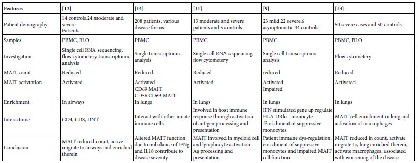
In 24 moderate and severe SARS-COV-2 cases, It was found  that total and CD4, CD8 and double negative MAIT cell counts in circulation is reduced but with marked activation state. Meanwhile MAIT cells were enriched in the airways, on recovery MAIT cell counts restored to normal [12]. MAIT cell frequencies in 208 COVID-19 patients were investigated for MAIT cell profiles. They were of reduced counts in peripheral blood with appearance of; active, cytokine producing and CD4CD8 MAIT cell phenotypes. The activated MAIT cells were migrating to lungs and enriched therein in the affected pulmonary tissues [14]. In other study, 13 moderate and severe COVID-19 patients PBMC were mapped for MAIT cell profiles, they were decreased in circulation and activated, their activation initiate chemotaxis, apoptosis and found involved in the virus immune responses and possibly engaged in immune tissue damage [11]. MAIT cell reduction and functionally impaired in peripheral blood of; 23 mild, 22 severe, 6 asymptomatic patients and 44 were controls. Together with appearance of IFN stimulated gene up-regulated HLA-DRlo-monocytes and enrichment of suppressive macrophages [9]. Kim et al. [12], have been studied 50 severe and 50 normal control subjects, they were showing that MAIT cell reduction in number   but activated in circulation and they were inversely correlated with disease severity and mortality. The activated MAIT cells migrate to lung wherein stimulate macrophage. MAIT cells contribute to the worsening of inflammation in severe pneumonic lungs (Table 4).

Table 4: Role of MAIT cells in SARS-COV-2 human pneumonia*

tab 7-4

*The infection of MAIT cells by SARS-COV-2 virus is rather unclear till date

MAIT Cells in SARS-COV-2 Vaccinee

At the days seven and 21 post mRNA COVID-19 vaccination of; 42, 42, and 24 normal, immune compromised patients. From whom peripheral blood were sampled. PBMC were separated and mapped for MAIT cells. The MAIT cell frequencies remained stable overall the normal Subject vaccinee, immune compromised vaccinee. There were a positive correlation between the size of MAIT cell compartment and the vaccine induced adaptive immune response to srs-cov-2 spike protein. The pre-vaccinee and post-vaccinee levels of MAIT cells correlated positively with magnitude of SARS-COV-2 spike specific antibodies and CD4 T cells in both vaccinee groups. In normal healthy donors the levels of MAIT cell activation is negatively correlated with the spike antigen specific immune responses. Hence, MAIT  cell compartment is involved in the early stages of primary adaptive immune to vaccine and may be they are important in the vaccine induced immunity [19].

The protective immune response to respiratory pathogens including influenza virus are initiated by mucosal immune system. The development of safe and effective mucosal vaccine has greatly been impaired due to lack of a properiat mucosal adjuvant. MAIT cells when co-administered with influenza A haeagglutinin protein induced protective immunity in mice and the resultant antibodies were of predominant IgA type. The heamagglutinin immune primed mice were found on influenza A live virus challenge immune protected. MAIT cell in the study act as a cellular adjuvant. Their adjuvanicity was mediated by CD40L dependent activation of DCs and subsequent priming of CD4+ follicular T cells. Hence MAIT cells acts as cellular mucosal adjuvant in a mucosal vaccine strategy [20]. The comparative role of MAIT cells in SARS-COV-2 infection and vaccination was depicted in Table 5.

Table 5: Comparative view to the role of MAIT cells in SARS-COV-2 infection and in vaccination*

tab 7-5

*Based on Boulus [19], Pankurest et al. [20], Shi et al. [11].

Conclusions

MAIT cells are functioning in; Homeostasis, infection, vaccination, tissue repair, autoimmune diseases, immune mediated diseases and cancer. In SARS-COV-2 infections, MAIT displayed number of immune functions as; Chemo-attractant, initiation of antigen processing, antigen presentation in antigen presenting cells as well as activation of lymphocytes. During the adaptive immune responses to SARS-COV-2 virus infection it acts early in the adaptive immune response as an intrinsic cellular adjuvant to the immune cells involved in the immune response. While in vaccination with influenza haemagglutinins it was found that MAIT cell activate DCs through DC40L and prime follicular CD4+ helper T cells by this it may acts as mucosal cellular vaccine adjuvants. MAIT cells may adopt; pyroptic, apoptotic, active tissue infiltrating and exhausted phenotypes. During SARS-COV-2 severe infection, MAIT cell count in circulation dropped down but resume active phenotype then migrates to air ways then to lungs therein they expand and worsen the patients state. On recovery, however, MAIT cells restore their normal distribution both in circulation and mucosal tissues. The evolution of MAIT functional phenotype was suggested.

References

  1. Ben Youssef G, Tourret M, Salon M, Ghazarian L, Houdouin V, et (2018) Ontogeny of human mucosal associated T cells and related T cell subsets. J Exp Med 215: 459-479. [crossref]
  2. Legoux F, Salou M, Lantz O (2020) MAIT cell development and functions: the microbial connections. Immunity 53: 710-723. [crossref]
  3. Lopez-Sagaseta J, Dulberger CL, Crook JE, Parks CD, Luoma AM, et (2013) The molecular basis of mucosal associated invariant T cell recognition of MRI proteins. PNAS E1771-E1778. [crossref]
  4. Eckle SBG, Birinskaw RW, Kostenks L, Corbett AJ, Hamish EGMC et al. (2014)  A molecular basis of underpinning the T cell receptor heterogeneity of mucosal associated invariant T J Exp Med 211: 1585-1600. [crossref]
  5. Toubal A, Nel I, Lotersztaji S, Lehuen A (2019) Mucosal associated invariant T cells and disease. Nat Rev Immunol 19: 663-657. [crossref]
  6. McCarthy C, O’Donnell D, Kelly NEW, Shea DO, Hogan AE, et (2021) Covid-19 severity and obesity;Are MAIT a factor. Lancet 5: 445-447. [crossref]
  7. Ussher JE, Willberg CB, Klenerman P (2018) MAIT cells and J Immunol Cell Biol 96: 630-641. [crossref]
  8. Long Y, Hinks TSC (2021) MAIT cells in respiratory viral infections in mouse and human. Crit Rev Immunol 41: 19-35. [crossref]
  9. Yang Q, Wen Y, Qi F, Gao X, Chen W, et al. (2022) Suppressive monocyte impair MAIT cell response via IL10 in patients with severe covid-19. J Immunol 207: 1848- 1856. [crossref]
  10. Vorkas CK, Krishna C, Li K, Aube J, Fitzgerald DW, et al. (2022) Single cell transcriptional profiling reveals signatures of helper, effector, and regulatory MAIT cells during homeostasis and J Immunol 208: 1042-1056. [crossref]
  11. Shi J, Zhou J, Zhang X, Hu W, Zhao JF, et (2021) Single cell transcriptomic profiling of MAIT cells in patients with covid-19. Front Immunol 12: 700152. [crossref]
  12. Parrot T, Gorin JB, Ponzetta A, Maleki KT, Kammann T, et al. (2020) MAIT cell activation dynamics associated with covid-19 disease severity. Sci Immunol 5: eab1670. [crossref]
  13. Kim TO, Park KJ, Cho YN, Jin HM, Jo YG, et (2022) Alterd distribution ,activation and increased IL17 production of mucosal associated invariant T cells in patient with acute respiratory distress syndrome. Thorax 77: 865-872. [crossref]
  14. Flament H, Rouland M, Beaudoin L (2021) Outcomes of sars-cov-2 infection is linkedto MAIT cell activation and Nat Immunol 22: 222-235.
  15. Saluo M, Nicol B, Garcia A, Baron D, Michel L, et al. (2016) Neuropathogenic phenotype and functional analysis of mucosal associated invariant T cells in multiple seclerosis. Clin Immunol 166-167: 1-11. [crossref]
  16. Matzinger P, Kamala T (2011) Tissue based class control; the other side of Nat rev Immunol 11: 221-230. [crossref]
  17. DeVenes RD, McQuaid S, Van Amerongen G, Yuksel S, Verburgh RJ, et al. (2012) Measles immunosuppression lessons from maqaque Plos Pathog 8: e1002885. [crossref]
  18. Rudak PT, Richardson CD, Haeryfar SMM (2021) Measles virus infects and program MAIT cells for J Infect Dis 223: 667-672. [crossref]
  19. Boulouis C, Kammann T, Cupio A, Parrot T, Gao Y, et (2022) MAIT compartment characteristic associated with immune response magnitude and sars-cov-2 vaccine. Mol Med 28: 54. [crossref]
  20. Pankhurst TE, Buick KH, Lange JL, Marshall AJ, Button KR, et al. (2022) MAIT cells activate dendritic cells to promote T follicular helper cells differentiation and humoral immunity. BioRxiv 486638.