Monthly Archives: June 2022

fig

Macrosomia: A Risk Factor of Childhood Obesity: A Case Report with Literature Review

DOI: 10.31038/PSC.2022211

Abstract

Childhood Obesity is a complex health issue caused by the abnormal accumulation or excessive fat in the body, threatening the life of the infant. We report the case of a 9 months old male infant who came in for a paediatric general consultation with complaints of left ocular reddishness and associated purulent discharge but was later on addressed to the endocrinologist consultation because was found to have a weight above normal on taking anthropometric parameters prior to consultation. After a complete medical observation, the diagnosis retained was communal obesity on the basis of a genetic predisposition (macrosomia at birth, a family history of obesity in both parents) and unhealthy life style habits (inappropriate nutrition). Through this case report with literature review, we wish to emphasize the facts that, although there are several aetiologies of obesity in children, the most common being communal obesity, include genetic predisposition which may serve as breeding ground to nutritional factors. Macrosomia appears to be a starting point, and so such babies should be followed up closely as they are at high risk of becoming obese later on in infancy. This with related complications. While the early diagnosis relies on routine assessment of anthropometric parameters, the effective management of communal obesity of childhood requires a multidisciplinary approach, with the parents being at the frontline.

Keywords

Macrosomia, Obesity, Anthropometric parameters, WHO-child growth curves

Introduction

Childhood Obesity is a challenging health issue caused by the abnormal accumulation or excessive fat in the body threatening the life of the infant through systemic involvement. The World Health Organization (WHO) defines obesity in children less than 5 years of age as a weight-for-height greater than 3 standard deviations above the WHO-Child Growth Standards median [1,2]. Many factors contribute to the onset of obesity in children such as non-modifiable factors including genetics and modifiable factors. Because anthropometric mensuration may suffice to pose the diagnosis, obesity is therefore a clinical diagnosis. It is worth mentioning that up to 39 million children under the age of 5 were overweight or obese in 2020 [1,2]. The fundamental cause of obesity is thought to be an energy imbalance between calories consumed and calories expended. Many factors might predispose to the onset of obesity in children. They can be classified as modifiable and non-modifiable risks factors with macrosomia and genetics being at the forefront, as it is often associated with a higher chance of obesity, premature death and disability in adulthood [3,4]. In addition to increased future risks, obese children experience breathing difficulties, hypertension, and early markers of cardiovascular disease, insulin resistance and psychological effects [3,4].

Case Report

We report the case of a 9 months old male infant who came in for a paediatric general consultation with complaints of left ocular reddishness and associated purulent discharge but was later on addressed to the endocrinologist consultation because was found to have a weight above normal on taking anthropometric parameters prior to consultation. The mother’s history revealed a notion of deliveries of big babies ˃ 3,5 kg. The pregnancy was uneventful without gestational diabetes, nor chronic diseases. The infant was born preterm at 36 weeks Gestational Age (GA) with a birth weight of 3600 g ˃ 95th percentile, with good extra uterine adaptation. Nutritional history permitted to note that the infant was exclusively breastfed up to 3 weeks of age, mixed feeding was then initiated till 6 months and thereafter, milk substitutes were stopped, while breast milk was continued thrice during the day and five times at night, in addition to meals that covered the day.

fig

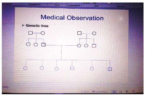
The Genetic Tree of Family Obesity

During the systemic enquiry we noted rhinorrhoea. On Physical exam, the infant was obese with weight at 15.8 Kg (˃ 3 zscore) and height at 80 cm (height˃ 3 zscore) weight-for-height index was ˃ 3-zscore as well. A flu-like syndrome was found with cough, rhinorrhoea and fever. There was systemic inflammatory response syndrome with fever at 38,5°c, tachycardia with 120 beats per minute and also reddishness of the left sclera associated with purulent discharge. No dysmorphic signs were found. The diagnosis posed was a left bacterial conjunctivitis associated with rhinitis in a child with communal obesity. The management consisted of nasal wash with serum saline as frequent as possible (at least 6 times daily), antipyretic with paracetamol syrup: 15 mg/kg/6 h, Fucidin gel 1%: 1 eye drop twice daily as ocular antibiotic. Most essentially, was diet modification by reducing breastfeeding frequency during the night from 5 to 1 time? The mother was advised to complete breastfeeding with mineral water in case the baby cried for more. The number of meals per day was maintained at 3, but was automatically to include fruits and vegetables.

Discussion

Childhood obesity is one of the most serious public health challenges in the 21st century. The genetic factor accounts for less than 5 percent of cases [3,4]. Foetal macrosomia is associated with increased risk of obesity in children under 3 years, with estimated risk of 3,74 folds higher than that of babies with normal birth weight [3,4]. Results from a study conducted by Sonia Sparano et al. in 2013 showed that macrosomia was an independent determinant of obesity after the adjustment of confounders [4,5]. This was the case of our patient, who was born premature at 36 weeks GA, but with birth weight of 3600 g˃95th percentile for gestational age, which corresponded to macrosomia. This is a condition which requires effective perinatal and deep neonatal assessments, given potential complications. More so, there was maternal obstetrical history of big babies, which was predisposing to macrosomia as well, and a contributing finding [6-19].

The diagnosis of obesity is solely made on a clinical basis and varies according to age of the infant. For infants aged ≤ 5 years the diagnosis relies upon the use of weigh-for-height growth curves from which an index ˃ 3 z-score is indicative of obesity. This was the case of our patient weighing 15.8 Kg for 80 cm height which corresponded to this classification. On the other hand, it is worth mentioning that the diagnosis of obesity in children aged ≥ 5 years makes use of the body mass index-for-age curves when ˃ 2 z-score. This indicates a necessity for routine anthropometric assessment in paediatric consultations [20-22].

The known risk factors of childhood obesity can be classified into two groups, namely: modifiable risk factors and non-modifiable risk factors. The modifiable risk factors include maternal overweight or obesity, maternal smoking, gestational weight gain, sleeping, sedentary lifestyle, lack of breastfeeding, infant and young child feeding [23-27]. Whereas the non-modifiable risk factors are genetic or familial, and high birth weight. Our patient had family history of obesity, as well as history of macrosomia which oriented etiological hypotheses towards genetic determinism.

In effect, there are several aetiologies attributed to childhood obesity. They could have a genetic origin, especially when there is family history as it was the case in the patient we presented. Nevertheless, obesity could as well be of endocrinal cause with hypothyroidism, growth hormone deficiency and hypercortisolemia, in which case there is usually characteristic stunting with rapid onset obesity [20-22]. From a semiological standpoint, childhood obesity may be communal, when there is no other possible nor identifiable cause than genetic. Meanwhile, it is classified as syndromic when associated with a spectrum of other anomalies such as mental retardation, dysmorphism and/or visceral malformations, hypogonadism, just to name a few. This is the case with classical paediatric syndromes such as Prader Wilis or Bardet Biedl syndromes in which dysmorphism is often associated with obesity.

Oedemato-ascitic syndromes such as in heart failure, nephrotic syndrome, malnutrition, and anaphylactic reaction are sometimes evoked as differential diagnoses in the discussion of childhood obesity. However, they lack consistency with regards to context, evolution, duration, and physical exam findings. As a matter of facts, the positive diagnosis of obesity is clinical as we earlier mentioned, and anthropometric parameters are pathognomonic [20-22].

The management of childhood obesity relies upon a multidisciplinary approach and spans on a long term. Specialists involved in this procedure include the paediatrician and/or endocrinologist who coordinate and evaluate the process at regular intervals. The nutritionist-dietician enables to regulate quality and quantity feeding, while the sports coach helps to lose weight through age-appropriate physical exercises. The role of a psychologist is important and may be indispensable especially in adolescents, for whom the development of individual character may transit through constant opposition to established rules and recommendations, doubled with rebellion [28]. Furthermore, in this population group, self-esteem is paramount, being grounded in mirror image, peer, and peers’ opinion. Surgical intervention with liposuction or partial gastrectomy is experimental in paediatrics, but may be envisaged in extreme situations, just as in adults. However, the initial management in our patient consisted solely of dietetic measures given the young age. This comprised modification of breastfeeding frequency, meals with vegetables, and fruits [23-27].

When childhood obesity is not adequately managed, complications are numerous and multisystemic, occuring as time goes on. The basis of these complications stem from four factors, including: disorders of excess lipid metabolism, atherosclerosis, physical impact of plethora and conditioning. Cardiovascular complications include dyslipidemia, hypertension, blood coagulation disorders, chronic inflammation and endothelial dysfunction which are all due to accumulating lipids [20-22]. Neuropsychological complications may occur with pseudotumor cerebri with intracranial hypertension syndrome mimicry, while poor self-esteem, depression and eating disorders are related with non-acceptation or non-coping with the body image. Pulmonary manifestations are also frequent with physical exercise intolerance, asthma, sleep apnoea, and are aggravated by lung compliance disorders. This is due to the work load of mobilising relatively massif surrounding tissues to the respiratory system. Increased lipogenesis may give rise to gastrointestinal and urinary complications with gallstones, steatohepatitis and glomerulosclerosis. Affected by the impact of weight, the musculoskeletal system can manifest with fractures and arthrosis. Whereas, the metabolic syndrome might induce endocrinopathies such as type-2 diabetes, precocious puberty, polycystic ovary syndrome in girls and hypogonadism in boys [20-22].

Conclusion

We reported a case of communal obesity with fortuitous discovery, in an infant predisposed through macrosomia and family obesity, to which a nutritional factor was grafted. The diagnosis being purely clinical through anthropometric mensuration, a long course but simplified management was initiated by the child specialist. This process essentially relied on parental observation of recommendations over nutrition and dietetics, based on the child’s age. Another important aspect of this paper was the emphasis on the need for routine assessment of anthropometric parameters during children consultations. This is important for the early diagnosis and management of growth or nutritional disorders in children, in order to prevent complications.

References

  1. Obesity: preventing and managing the global epidemic. Report of a WHO consultation. World Health Organ Tech rep Ser. 2000;894:i-xii,1-253. PMID: 11234459
  2. World Health Organization. Obesity and Overweight. Fact sheet 2014 311.
  3. Sahoo K, Sahoo B, Choudhury AK, Sofi NY, Kumar R, et al. (2015) Childhood obesity causes and consequences. J Family Med Prim Care 4: 187-192. [crossref]
  4. Pan XF, Tang L, Lee AH, Binns C, Yang Y, et al. (2019) Association between fetal macrosomia and risk of obesity in children under 3 years of age in western china: a cohort study. World J Pediatr 15: 153-160. [crossref]
  5. Sparano S, Ahrens W, De Henauw S, Marild S, Molnar D, et al. (2013) Being macrosomic at birth is an independent predictor of overweihgt in children: results from the IDEFICS study. Matern Chirld Health J 17: 1373-1381. [crossref]
  6. Chiabi A, Kago DA, Moyo GPK, Obadeyi B (2019) Relevance and Applicability of the Apgar Score in Current Clinical Practice. EC Paediatrics 8: 01-07.
  7. Moyo GPK, Tetsiguia JRM (2020) Discussing the “First Cry” as an Initial Assessment for Neonates. Am J of Pediatr 6: 129-132.
  8. Moyo GPK, Sobguemezing D, Adjifack HT (2020) Neonatal Emergencies in Full-term Infants: A Seasonal Description in a Pediatric Referral Hospital of Yaoundé, Cameroon. Am J Pediatr 6: 87-90.
  9. Moyo GPK, Um SSN, Awa HDM, et al. (2022) The pathophysiology of neonatal jaundice in urosepsis is complex with mixed bilirubin!!! J Pediatr Neonatal Care 12: 68-70.
  10. Moyo GPK, Nguedjam M, Miaffo L (2020) Necrotizing Enterocolitis Complicating Sepsis in a Late Preterm Cameroonian Infant. Am J Pediatr 6: 83-86.
  11. Moyo GPK, Sap Ngo Um S, Awa HDM, Mbang TA, Virginie B, Makowa LK et al. (2022) An Atypical Case of Congenital and Neonatal Grave’s Disease. Annal Cas Rep Rev 2.
  12. Ngwanou DH, Ngantchet E, Moyo GPK (2020) Prune-Belly syndrome, a rare case presentation in neonatology: about one case in Yaounde, Cameroon. Pan Afr Med J 36: 120. [crossref]
  13. Tague DAT, Evelyn Mah, Félicitee Nguefack, Moyo GPK, Tcheyanou LLK, et al. (2020) Beckwith-Wiedemann Syndrome: A Case Report at the Gynaeco-Obstetric and Pediatric Hospital in Yaounde, Cameroon. Am J Pediatr 6: 433-436.
  14. Moyo GPK, Mendomo RM, Batibonack C, Mbang AT (2020) Neonatal Determinants of Mothers’ Affective Involvement in Newly Delivered Cameroonian Women. Journal of Family Medicine and Health Care 6: 125-128.
  15. Moyo GPK (2020) Epidemio-clinical Profile of the Baby Blues in Cameroonian Women. Journal of Family Medicine and Health Care 6: 20-23.
  16. Moyo GPK, Djoda N (2020) Relationship between the Baby Blues and Postpartum Depression: A Study among Cameroonian Women. American Journal of Psychiatry and Neuroscience 8: 26-29.
  17. Moyo GPK (2020) Perinatality and Childbirth as a Factor of Decompensation of Mental Illness: The Case of Depressive States in Newly Delivered Cameroonian Women ABEB 4: 000592.
  18. Moyo GPK, Djoda N (2020) The Emotional Impact of Mode of Delivery in Cameroonian Mothers: Comparing Vaginal Delivery and Caesarean Section. American Journal of Psychiatry and Neuroscience 8: 22-25.
  19. Foumane P, Olen JPK, Fouedjio JH, GPK Moyo, Nsahlai C, Mboudou E (2016) Int J Reprod Contracept Obstet Gynecol 5: 4424-4427.
  20. Moyo GPK, Djomkam IFK (2020) Epidemio-clinical Profile of Stunting in School Children of an Urban Community in Cameroon. Am J Pediatr 6: 94-97.
  21. Moyo GPK, Djomkam IFK (2020) Factors Associated with Stunting in School Children of an Urban Community in Cameroon. Am J Pediatr 6: 121-124.
  22. Moyo GPK, Ngapout OD, Makowa LK, Mbang AT, Binda V, Albane EA et al. (2022) Exogenous Cushing’s Syndrome with Secondary Adrenal Insufficiency in an Asthmatic Infant: “Healing Evil with Evil”. Arch Pediatr 7: 203.
  23. Hermann ND. Moyo GPK (2020) Neonatal Determinants of Inadequate Breastfeeding: A Survey among a Group of Neonate Infants in Yaounde, Cameroon. Open Access Library Journal 7: e6541.
  24. Hermann ND, Moyo GPK, Ejake L, Félicitée N, Evelyn M, et al. (2020) Determinants of Breastfeeding Initiation Among Newly Delivered Women in Yaounde, Cameroon: a Cross-Sectional Survey. Health Sci Dis 21: 20-24.
  25. Moyo GPK, Dany Hermann ND (2020) Clinical Characteristics of a Group of Cameroonian Neonates with Delayed Breastfeeding Initiation. Am J Pediatr 6: 292-295.
  26. Moyo GPK, Hermann ND (2020) The Psycho-Sociocultural Considerations of Breastfeeding in a Group of Cameroonian Women with Inadequate Practices. J Psychiatry Psychiatric Disord 4: 130-138.
  27. Moyo GPK, Ngwanou DH, Sap SNU, Nguefack F, Mah EM (2020) The Pattern of Breastfeeding among a Group of Neonates in Yaoundé, Cameroon. International Journal of Progressive Sciences and Technologies 22: 61-66
  28. Moyo GPK (2020) Children and Adolescents’ Violence: The Pattern and Determinants Beyond Psychological Theories. Am J Pediatr 6: 138-145.
fig 5

Assistance to Patients with Terminal Cancer in the Last 30 Days Prior to Death: Differences in the Care of the Usual Health Care Units and a Palliative Care Unit

DOI: 10.31038/CST.2022731

Abstract

Objectives: To compare aspects related to the care of patients with terminal cancer during the 30 days prior to death between usual care units (UCUs) and a palliative care unit (PCU).

Methods: A retrospective cohort, for the last 30 days preceding the death of patients with terminal cancer, followed in UCUs and PCU. Demographic, clinical and nutritional (baseline) data were collected; also, performance of medical examinations and procedures, prescription of nutritional therapy (referring to 30, 7 and 3 days before death); and prescription of drugs and administration routes (relating to the last 3 days of life).

Results: We evaluated 239 patients, of which 131 (54.8%) have been assisted in UCUs and 108 (45.2%) in the PCU. Prescription of nutritional therapy, number of laboratory tests, imaging and procedures performed in the UCUs was higher than in the PCU. Regarding the four drugs considered essential for end-of-life care, we found that all were prescribed to patients in the PCU, while in the UCUs there was no prescription of haloperidol and scopolamine in any of the cases.

Conclusion: In the PCU, there was a better use of health resources, as clinical guidelines recommend limiting the use of disproportionate resources to the advancement of the disease in patients with limited life expectancy.

Keywords

Advanced cancer, Terminal cancer, Health care, Oncology, Usual care units, Palliative care unit

Introduction

The elaboration of the care plan for patients with terminal cancer must be based on a careful evaluation of clinical, bioethical and prognostic elements. The prognostic assessment can lead to the improvement of treatment strategies and support the planning of care and the efficient use of available resources, helping to minimize the risks of under treatment or excessive and futile treatments, especially in the phase close to death [1]. In the hospital setting, it is common for patients with terminal cancer to receive inadequate and ineffective care, with no provision for palliative care and pain relief. Even in a reality of scarce resources, there is an unnecessary use of invasive and high-tech methods, focused on trying to cure, which are unable to treat the most prevalent symptoms of the disease, prolonging suffering and pain [2].

Furthermore, the World Health Organization (WHO) [3] points out that palliative care with quality requires access to essential medicines (basic basket of medications) able to treat the most prevalent symptoms in terminal disease, rather than the use of measures and futile drugs. In 2013, a study published in the Journal of Palliative Medicine carried out through an international consensus of specialist physicians and practitioners in large Palliative Care centers described the relevance of four essential drugs (Morphine, Midazolam, Haloperidol and Scopolamine) for the relief of the most prevalent symptoms in patients with terminal cancer in the days before death. Therefore, physicians caring for patients with terminal cancer must be familiar with these medications to prescribe them and achieve their benefits [4].

In Brazil, the possibility of a patient with chronic illness in a terminal stage of disease, including oncological disease, not having access to basic medications to control symptoms and also remaining without access to the team and palliative care, is very large [5]. It is necessary to improve care for this group so that their real demands are met. In the end-of-life care (EOLC) phase, the patient may present different signs, symptoms and suffering that demand a reorganization of the therapeutic plan. Thus, this study proposes the comparison of aspects related to the care of patients with terminal cancer during the last 30 days prior to death between usual care units (UCUs) and a palliative care unit (PCU) of an oncological center of national reference.

Methods

This is a clinical, observational, retrospective cohort study, referring to the last 30 days of life of patients with terminal cancer, followed up in the different care units of an oncological center of national reference, located in Brazil. The study was approved by the Research Ethics Committee. The oncological center of national reference is composed UCU where treatments are carried out aimed at cytoreduction, whether by chemotherapy, surgery or radiotherapy. It also has the exclusive PCU, where patients from UCUs are referred to control symptoms and promote quality of life and death, at the end of the possibilities of treatment lines and failure to cure, disease progression during treatment or worsening of their clinical condition.

All patients who died of any reason in the period of interest defined in the research proposal (06/01/2019 to 07/31/2019) were identified through an electronic system and selected according to the criteria of inclusion, namely: ≥20 years of age; confirmed diagnosis of advanced-stage malignant tumor (locally advanced and/or with distant metastasis); having died between June and July 2019; having been enrolled at least 30 days before the date of death for follow-up at INCA; and having been admitted to INCA in at least one of the last three days of life. Patients with missing or inconsistent data on the date of death were considered losses.

Data Collection

The thirtieth day before death was considered the baseline and the day of death was the study deadline. The data were extracted from medical records and recorded in a specific form, as shown in Figure 1.

fig 1

Figure 1: Flowchart of data collection from patients with terminal cancer in the 30 days prior to death

Data Sociodemographic, Clinical, Nutritional and Performance Status (for the Baseline Study)

Age (<60 vs. ≥60 years old); gender (male vs. female); diagnosis [cancer of the gastrointestinal tract (GIT) vs. breast vs. head and neck vs. gynecological vs. lung vs. connective bone tissue vs. others]; disease progression (local vs. local + distance); Previous cancer treatment (yes vs. no)].

Information was collected on the Patient-Generated Subjective Global Assessment short form (PG-SGA SF) (©FD Ottery, 2005, 2006, 2015), available at pt-global.org. The tool is answered by the patient and allows for the assessment of: (1) change in body weight: the score can range from 0 to 5; (2) food intake: with a score from 0 to 4; (3) presence of symptoms of nutritional impact: scoring up to 24; and (4) functional capacity assessment: scoring from 0 to 3. At the end of the assessment, a numerical score is generated based on the sum of each of the items in the questionnaire. The higher the score, the worse the nutritional status. Patients with scores ≥9 were classified as being at nutritional risk [6,7].

The cachexia is defined by the modified Glasgow prognosis score (mGPS) at four different stages: not cachexia, malnutrition, pre-cachectic and refractory cachectic [8] (Table 1).

Table 1: Classification of cachexia using the modified Glasgow Prognostic Score

Biomarker

mGPS

Cachexia Stages

CRP (mg/L)

Albumin (g/dL)

0

<10 >3.5

Non cachectic

0

<10 <3.5

Malnourished

1

>10 >3.5

Pre-cachectic

2

>10 <3.5

Refratary cachectic

Note: mGPS= modified Glasgow Prognostic Score; CRP= C-reactive protein. Source: Douglas and McMillan (2014).

The performance status data obtained in the UCU refer to the Performance Status Eastern Cooperative Oncology Group (ECOG-PS) that ranges from 0 (normal activity) to 5 (death) [9]; while in the PCU was used the Karnofsky Performance Status (KPS) that ranging from 100 (normally active) to 0 (dead) [10]. These scales were converted and categorized as PS < 3 or KPS ≥ 40% (yes or no), as proposed by Ma et al. [11]

Laboratory Tests and Procedures (Referring to the Period of 30, 7 and 3 Days before Death)

Total number of laboratory tests performed; total number of full images of examinations and the most frequent types [e.g.: computed radiography (CR), computed tomography (CT), endoscopy, ultrasound and magnetic resonance image (MRI)]; and the total number of procedures performed and the most frequent types [e.g., chemotherapy (QT), radiotherapy, blood and platelet transfusion, biopsy, and gastrostomy].

Nutritional Therapy Prescription (Referring to the Period of 30, 7 and 3 Days before Death)

Prescription of oral (ONT), enteral (ENT) and parenteral (PNT) nutritional therapy.

Prescription and Administration Routes (Referring to the Period of 3 Days before Death)

Prescription of medications and routes of administration.

Statistical Analysis

Analyses were performed using Stata Data Analysis and Statistical Software (STATA) version 13.1 (Stata Corp., College Station, Texas, USA). To assess data, the Kolmogorov Smirnov test was applied. For continuous parametric data, averages, standard deviation, Student’s T test and ANOVA were used; for the categorical variables, number of observations and frequency were used, and the Chi-square test was used for proportions. The p-value <0.05 was considered statistically significant.

Results

The study included 239 patients who, in the majority, were >60 years old (63.2%), female (61.1%) and had the primary tumor site located in the breast (20.1%), followed by GIT (19.7%). The prevalence of nutritional risk was 70.3% and most patients were cachectic (35.4%) or refractory cachectic (46.9%). One hundred and thirty-one (54.8%) were assisted in the UCUs and 108 (45.2%) in the PCU (Table 2). In most patients, the reason for hospitalization was a decline in their general condition, with no statistically significant difference between the units (data not shown in tables).

Table 2: Sociodemographic, clinical and nutritional characterization of patients with terminal cancer according to health care units (N=239)

Variables

Total


N=239

UCU


N=131 (54.8%)

PCU


N=108 (45.2%)

p-valuea

Age (years)
 

<60


88 (36.8%)


54 (41.2%)


34 (31.5%)


0.120

>60

151 (63.2%) 77 (58.8%)

74 (68.5%)


Sex


Male


93 (38.9%)


52 (39.7%)


41 (38.0%)


0.785

Female

146 (61.1%) 79 (60.3%)

67 (62.0%)


Diagnostic

GITb

47 (19.7%) 21 (16.0%) 26 (24.3%) 0.062

Breast

48 (20.2%) 28 (21.4%)

20 (18.7%)

Head and neck

31 (13.0%) 13 (9.9%)

18 (16.8%)

Gynecologicalc

39 (16.4%) 27 (20.6%) 12 (11.2%)

Lung

25 (10.5%) 14 (10.7%)

11 (10.3%)

CBT

13 (5.5%) 5 (3.8%) 8 (7.5%)

Othersd

36 (14.7%) 23 (17.6%)

13 (11.2%)


Metastasis


Local


62 (26.0%)


39 (30.0%)


23 (21.3%)


0.311

Local + distant

177 (74.0%) 92 (70.0%)

85 (78.7%)


Previous cancer treatment

No (virgin)

41 (17.2%)

22 (16.9%) 19 (17.6%)

0.206

Note: UCU= Usual Care Units; PCU= Palliative Care Unit; N= number of observations; %= frequency; GIT= gastrointestinal tract; CBT= connective bone tissue; PG-SGA SF= Patient-Generated Subjective Global Assessment short form; PS= Performance Status; KPS= Karnofsky Performance Status.
ap-value refers to the chi-square test for proportions; bupper and lower GIT; ccutter, endometrium, ovary and vulva;
dcentral nervous system, kidney and urinary tract, male genitals, peritoneum, mediastinum, haematological and unknown primary; evariables with missing data.

The number of laboratory, imaging tests and procedure performed throughout the follow-up period was greater in patients assisted in the UCUs than in the PCU (Figure 2). The most frequently performed imaging tests were X-ray and CT, and the procedures were QT and blood/platelet transfusion. The frequencies of CT (UCU=4.6% vs. PCU=4.6%) and QT (UCU=0.9% vs. PCU=0) were only similar between units in the last three days of life. Patients followed in the PCU were less likely to prescribe ONT compared to those followed in the UCU during the entire evaluation period (p-value <0.001). The prescription of ENT was lower in the PCU only in the last 3 days before death (p-value <0.050) (Table 3).

fig 2(1)

fig 2(2)

Figure 2: Average number of prescriptions for laboratory tests (A), imaging tests (B) and procedures (C) in the last month of life by patients with terminal cancer according to health care units (N=239).
Note: N=number of observations; UCU=Usual Care Units; PCU= Palliative Care Unit; *p-value<0.050 and **p-value<0.001 of the Student t test.

Table 3: Types of imaging tests and most prescribed procedures in the last month of life for patients with advanced cancer according to health care units (N=239)

Period of follow up before death

Variables

30 days

7 days

3 days

Total UCU PCU Total UCU PCU Total UCU

PCU

Imaging exams

X-ray

135 (56.5%)

88 (67.2%) 47 (43.5%)** 78 (32.6%) 57 (53.5%) 21 (19.4%)** 50 (21.0%) 36 (27.5%)

14 (13.0%)*

CT

80 (33.5%)

54 (41.2%) 26 (24.0%)* 29 (12.1%) 22 (16.8%) 7 (6.5%)* 11 (4.6%) 6 (4.6%)

4 (4.6%)

Ecodoppler

15 (6.3%)

15 (11.4%) 0** 3 (1.3%) 3 (2.3%) 0 0 0

0

Endoscopy

10 (4.2%)

8 (6.1%) 2 (1.8%) 5 (2.1%) 5 (3.8%) 0 0 0

0

Ultrasonography

7 (2.9%)

7 (5.3%) 0 3 (1.3%) 3 (2.3%) 0 0 0

0

Outrosa

14 (5.8%)

12 (9.2%) 1 (0.9%)* 5 (2.1%) 2 (1.5%) 0 3 (1.3%) 3 (2.3%)

0

Procedure

Quimiotherapy

31 (13.0%)

29 (22.1%) 2 (1.8%)** 8 (3.3%) 7 (5.3%) 1 (0.9%)* 1 (0.4%) 1 (0.9%)

0

Transfusion

29 (12.1%)

29 (22.1%) 0** 19 (8.0%) 19 (14.5%) 0** 13 (5.4%) 13 (9.9%)

0*

Biopsy

16 (6.7%)

16 (12.2%) 0* 3 (1.3%) 3 (2.3%) 0 0 0

0

Radiotherapy

13 (6.4%)

11 (8.4%) 2 (1.8%)* 5 (2.1%) 5 (3.8%) 0 0 0

0

Note: UCU= Usual Care Units; PCU= Palliative Care Unit; CT=computed tomography; acystoscopy, colonoscopy, cholangio, resonance; bcatheter bi-implantation, lumbar puncture, thoracentesis, nephrostomy, arterial embolization, tracheostomy, gastrostomy, paracentesis, biliary and percutaneous drainage; *p-value<0.050 and **p-value<0.001 of the chi-square test.

Dipyrone remained as the drug with the highest proportion (average of 85%) of prescriptions in the last three days of life in the UCUs, followed by morphine (average of 70%). It should be noted that enoxaparin appeared in the sixth position (average of 38%) and insulin appeared in the ninth position (average of 21%) during the period (Figure 3). The three most prescribed medications in the last three days of life in the PCU were morphine (average of 92%), dipyrone (average of 88%), and midazolam (average of 71%) (Figure 4).

fig 3

Figure 3: Ranking of the ten most prescribed drugs in the Usual Care Units in the last three days of life of patients with terminal cancer (N=131).
Note: N=number of observations. *p-value<0.050 of the Chi-square test for proportions.

fig 4

Figure 4: Ranking of the ten most prescribed drugs in the Palliative Care Unit in the last three days of life of patients with terminal cancer (N=108).
Note: N=number of observations.*p-value<0.050 of the Chi-square test for proportions.

According to the analysis of the average frequency of prescription of the four essential drugs, in the last three days of life of patients with advanced cancer, according to the health care units, it was verified that in the UCU there was no prescription of haloperidol and scopolamine. Morphine and midazolam were prescribed in the UCUs, but in a much lower quantity than the PCU (p-value <0.050) (Figure 5).

fig 5

Figure 5: Analysis of the average frequency of prescription of the four essential drugs, according to Lindqvist et al. 2013, in the last three days of life of patients with terminal cancer according to the health care units (N=239).
Note: N=number of observations; UCU=Usual Care Units; PCU=Palliative Care Unit. *p-value<0.050 of the Chi-square test for proportions.

Discussion

The present study, about care provided to patients with terminal cancer in a national cancer treatment center, brings some main results. Patients followed-up in the last 30 days prior to death in the PCU underwent fewer laboratory, imaging and procedural tests, had fewer prescriptions for nutritional therapy and more prescriptions for essential drugs for end-of-life care, when compared to those in treatment in the UCUs. Therefore, as was to be expected, at PCU there was a limitation of the use of futile therapies and incapable of meeting the most relevant demands of terminally ill patients. This approach may be related to the fact that teams specialized in palliative care have greater technical knowledge about prognosis and a careful look at the management of symptoms, promotion of quality of life and death [12].

Even in follow-up at a national referral center for cancer treatment, most patients (54.8%) did not receive assistance from a team specialized in palliative care during the terminal process. World estimates by WHO5 indicate that more than 56.8 million people demand palliative care but only 12% of this need is met. Brazil has one of the worst offers of palliative care services, accessed by only about 0.3% of people who die annually in the country [13]. In addition to the incipient offer of this type of service, the referral of patients to exclusive palliative care is a difficult task that permeates different barriers, such as those related to health professionals, among which we can mention those related to oncologists. They find referring a patient with advanced cancer to exclusive palliative care a complex task, causing patients to be referred late or never be referred [14].

As expected, we found a high prevalence of nutritional impairment (nutritional risk: 70.3%; cachexia: 82.3%), regardless of the type of care unit. It is irrefutable that the impairment of nutritional status increases as cancer progresses [15]. Previous studies show that nutritional risk may be present in 71% to 100% [16,17] and cachexia in 13.8% to 53.9% of patients with advanced cancer [18].

The high prevalence of laboratory tests, imaging and procedures (chemotherapy and blood/platelet transfusion) performed in the UCUs reflect the therapeutic futility often present in care provided by professionals who are not specialized in palliative care for patients in the process of finitude. Receiving the last dose of chemotherapy within 14 days before death can be defined as an aggressive intervention [19]. Blood transfusion, in turn, involves the expenditure of a finite resource and requires careful evaluation for indication in patients with advanced cancer. However, scientific evidence has shown that patients with terminal cancer admitted to UCUs are likely to receive treatments with questionable benefits, such as chemotherapy and blood transfusion, towards the end of life, differently from those seen in PCUs [20,21].

The highest prevalence of ONT, ENT and PNT prescription occurred in UCUs. The decision to initiate and maintain Nutritional Therapy in patients with advanced cancer involves prognostic and bioethical issues, as an inadequate prescription can increase discomfort and suffering [22-24]. Kempf et al.20, in a study carried out in France, demonstrated that more than 15% of patients with advanced cancer received ENT and PNT in the last weeks of life, most of them (75.3%) in non-specialized hospitals. It is likely that palliative care providers are more conservative in their conduct related to Nutritional Therapy prescription in the last weeks of life, which may be related to the experience of patient-centered care [25,26].

The quantity and quality of medications used by patients with advanced cancer during their last days of life reflect the quality of care provided. In this context, we observed, for example, the presence of enoxaparin (advised in the institutional protocol for prevention of venous thromboembolism in prolonged hospitalization) and the absence of haloperidol (indicated in cases of hyperactive delirium) [27] among the 10 drugs. These data suggest the absence of medication reconciliation practice among non-palliative professionals, through the continuity of the prescription of futile medications [28,29].

Another relevant fact regarding the ranking of the 10 most prescribed drugs in UCUs is the absence of sedative drugs such as midazolam. We hypothesize that, this fact, linked to the prescription of morphine, and may indicate the use of opioids to sedate at the end of life, to the detriment of the use of appropriate sedatives, making it difficult to achieve safe sedation. Morphine is a strong opioid indicated for the treatment of pain and terminal dyspnea, which has a decreased level of consciousness as an adverse effect, characteristic of drug intoxication. Therefore, its use for the purpose of sedation is considered an inappropriate conduct [30]. In addition, the high prescription of omeprazole, ondansetron and bromopride found in UCUs may be related to the increase in symptoms of nausea and vomiting, common in intoxication conditions [31].

According to an international consensus of specialist physicians working in large Palliative Care centers, morphine, midazolam, haloperidol and scopolamine were considered the four essential drugs to control the symptoms prevalent in patients with terminal cancer, especially in the last 48 hours of life. Therefore, they must be available and prescribed in all care units for cancer patients. However, our results showed the absence of prescription of haloperidol and scopolamine and the reduced prescription of morphine and midazolam for patients followed in the UCUs during the last three days of life, when compared to those in the PCU.

Considering, therefore, the high prevalence of distressing symptoms at the end of life of cancer patients [32] and that appropriate drug interventions are essential to reduce suffering, we assume that terminal patients not assisted by a team specialized in palliative care are unlikely to receive adequate comfort for a good death. The development of institutional protocols for terminal patients, whether in the PCU or in the UCUs, could contribute to reversing this reality.

Despite all the evidence brought by this study, some methodological limitations need to be highlighted. Due to the retrospective design, it was not possible to assess the comfort and quality of life and death of the patients who made up the study group. Despite not having been the objective of the proposal, such an evaluation would enrich our findings. In addition, data collection from medical records can be a source of bias derived from potentially inadequate or insufficient records of information about the care provided to patients in the source document. It is necessary to develop further studies, with an appropriate design to assess other important variables such as symptom control based on the interventions performed.

Conclusion

The present study demonstrated that the use of health resources in the care of patients with terminal cancer differ between the assessed care units. The assistance provided at the PCU involved a better use of health resources, reflected in the limitation of the use of futile therapies in the context of limited life expectancy, as well as in the prescription of drugs potentially capable of contributing to reduce the burden of symptoms inherent in the terminal phase.

Funding

This research received no specific grant from any funding agency in the public, commercial, or not for profit sectors.

Declaration of Conflicts of Interest

The Authors declares that there is no conflict of interest

References

  1. Hui D, Mori M, Watanabe SM, et al. (2016) Referral criteria for outpatient specialty palliative cancer care: an international consensus. Lancet Oncol 17: 552-559. [crossref]
  2. Carvalho RCT, Parsons HA (2012) Academia Nacional de Cuidados Paliativos (ANCP). Manual de Cuidados Paliativos.
  3. World Health Organization (WHO) (2014) Global atlas of palliative care at the end of life.
  4. Lindqvist O, Lundquist G, Dickman A, et al. (2013) Four essential drugs needed for quality care of the dying: a Delphi-study based international expert consensus opinion. J Palliat Med 16: 38-43. [crossref]
  5. World Health Organization WHO (2020) WHO report on cancer: setting priorities, investing wisely and providing care for all.
  6. Vigano AL, Tomasso J, Kilgour RD, et al. (2014) The Abridged Patient-Generated Subjective Global Assessment Is a Useful Tool for Early Detection and Characterization of Cancer Cachexia. J Acad Nutr Diet 114: 1088-98. [crossref]
  7. Abbott J, Teleni L, McKavanagh D, et al. (2016) Patient-Generated Subjective Global Assessment Short Form (PG-SGA SF) is a valid screening tool in chemotherapy outpatients. Support Care Cancer 24: 3883-7. [crossref]
  8. Douglas E, McMillan DC (2014) Towards a simple objective framework for investigation and treatment of cancer cachexia: the Glasgow Prognostic Score. Cancer Treat Ver 40: 685-91. [crossref]
  9. Eastern Cooperative Oncology Group (ECOG-ACRIN) Cancer Research Group. ECOG Performance Status.
  10. Schag CC, Heinrich RL, Ganz PA (1984) Karnofsky performance status revisited: reliability, validity, and guidelines. J Clin Oncol 2: 187-193. [crossref]
  11. Ma C, Bandukwala S, Burman D, et al. (2010) Interconversion of three measures of performance status: An empirical analysis. Eur J Cancer 46: 3175-3183. [crossref]
  12. White N, Reid F, Vickerstaff V, et al. (2020) Specialist palliative medicine physicians and nurses accuracy at predicting imminent death (within 72 hours): a short report. BMJ Support Palliat Care 10: 209-212. [crossref]
  13. Academia Nacional De Cuidados Paliativos. Análise situacional e recomendações da ANCP para estruturação de programas de cuidados paliativos no Brasil. 2018. São Paulo: ANCP.
  14. Horlait M, Chambaere K, Pardon K, et al. (2016) What are the barriers faced by medical oncologists in initiating discussion of palliative care? A qualitative study in Flanders, Belgium. Support Care Cancer 24: 3873-81. [crossref]
  15. Fearon K, Strasser F, Anker SD, et al. (2011) Definition and classification of cancer cachexia: an international consensus. Lancet Oncol 12: 489-95. [crossref]
  16. Andrew IM, Waterfield K, Hildreth AJ, et al. (2009) Quantifying the impact of standardized assessment and symptom management tools on symptoms associated with cancer-induced anorexia cachexia syndrome. Palliat Med 23: 680-8. [crossref]
  17. Cunha MS, Wiegert EVM, Calixto-Lima L, et al. (2018) Relationship of nutritional status and inflammation with survival in patients with advanced cancer in palliative care. Nutrition 51: 98-103. [crossref]
  18. Wiegert EVM, Oliveira LC, Calixto-Lima L, et al. (2020) Cancer cachexia: Comparing diagnostic criteria in patients with incurable cancer. Nutrition 79-80.
  19. Cheung MC, Earle CC, Rangrej J, et al. (2015) Impact of aggressive management and palliative care on cancer costs in the final month of life. Cancer 121: 3307–15. [crossref]
  20. Kempf E, Tournigand C, Rochigneux PL, et al. (2017) Discrepancies in the use of chemotherapy and artificial nutrition near the end of life for hospitalised patients with metastatic gastric or oesophageal cancer. A countrywide, register-based study. Eur J Cancer 79: 31-40. [crossref]
  21. Wachtel TJ, Mor V (1985) The use of transfusion in terminal cancer patients. Hospice versus conventional care setting. Transfusion 25: 278-9. [crossref]
  22. Druml C, Ballmer PE, Druml W, et al. (2016) ESPEN guideline on ethical aspects of artificial nutrition and hydration. Clin Nutr 35: 545-56. [crossref]
  23. Bischoff SC, Austin P, Boeykens K, et al. (2020) ESPEN guideline on home enteral nutrition. Clin Nutr 39: 5-22. [crossref]
  24. Weimann A, Braga M, Carli F, et al. (2017) ESPEN guideline: Clinical nutrition in surgery. Clin Nutr 36: 623-50. [crossref]
  25. Masuda Y, Noguchi H, Kuzuya M, et al. (2006) Comparison of medical treatments for the dying in a hospice and a geriatric hospital in Japan. J Palliat Med 9: 152-60.
  26. Hickman SE, Tolle SW, Brummel-Smith K, et al. (2004) Use of the physician orders for life-sustaining treatment program in Oregon nursing facilities: beyond resuscitation status. J Am Geriatr Soc 52: 1424-9. [crossref]
  27. Friedlander MM, Brayman Y, Breitbart WS (2004) Delirium in palliative care. Oncology 18: 1541-53. [crossref]
  28. Saito AM, Landrum MB, Neville BA, et al. (2011) The effect on survival of continuing chemotherapy to near death. BMC Palliat Care 10: 1-11. [crossref]
  29. Marin H, Mayo P, Thai V, et al. (2020) The impact of palliative care consults on deprescribing in palliative cancer patients. Support Care Cancer 28: 4107–13.
  30. De Graeff A, Van Bommel JMP, Van Deijck RHPD (2010) Palliative care guidelines. Comprehensive cancer center the Netherlands (IKNL): Utrecht
  31. Pereira J, Bruera E (1997) Emerging neuropsychiatric toxicities of opioids. J Pharmaceut Care Pain Symptom Contr 5: 3-29
  32. Lichter I, Hunt E (1990) The last 48 hours of life. J Palliat Care 6: 7-15. [crossref]

Clinical Results of Kinetic Oscillation Stimulation (K.O.S.) in Non-Allergic Rhinitis

DOI: 10.31038/JCRM.2022532

Abstract

Introduction: Rhinitis is a condition associated with an inflammatory response. Non-Allergic Rhinitis (NAR) describes a syndrome of chronic symptoms of nasal congestion and rhinorrhea, unrelated to a specific allergen.

Objective: Our study is about a new method of rhinitis therapy with Kinetic Oscillation Stimulation (K.O.S.). The aim of the study is to evaluate the response after treatment with K.O.S. in the various groups of vasomotor rhinitis.

Methods: All the patients underwent K.O.S. treatment after a period of suspension from topical and systemic steroid therapy and any other kind of therapy for the rhinitis. The active treatment, K.O.S., consisted of vibrations created using an oscillation which stimulates and rebalances his autonomic nervous system.

Results: The data show an improvement in the overall quality of life in treated patients. The study of the nasal cells was found to be important to classify patients in the various forms of rhinitis, and to tailor the best treatment. Moreover, the stratification of SNOT-22 according to the various cell types has highlighted how the differences between the various cell types played an important role on determining good outcomes. In NARMA and NARNE patients the treatment results were not satisfactory and not statistical significative, while in the other forms they were optimal.

Conclusions: The K.O.S. treatment could be used as a successful and alternative treatment for vasomotor rhinitis. We believe that the study should continue to increase the number of cases available and better typing the various patients.

Keywords

Cytology, Rhinitis, Sympathetic Nervous system

Introduction

Rhinitis is a condition related to inflammatory responses as allergic rhinitis but can also occur in the absence of a specific cause such as in the “vasomotor” rhinitis. Allergic rhinitis is an Ig E mediated condition; it affects approximately 25% of the population of European countries and is characterized by nasal itching, sneezing, rhinorrhea, and congestion [1,2]. Non-Allergic Rhinitis (NAR) involves chronic sneezing, congested nose, drippy nose, unrelated to a specific allergen, in fact the term is used to describe rhinitis symptoms associated with nonallergic, non-infectious triggers It represents till 10 to 18 % of idiopathic rhinitis and affects children and adults [1,2]. A diagnosis of NAR is made after an allergic cause is ruled out and it may require allergy skin or blood tests and nasal citology. The diagnosis of the specific type of rhinitis can be tricky. The study of the nasal cells has been shown to be a useful and easy diagnostic tool in the study of rhinitis. We can detect and measure the cell population in the nasal mucosa at a certain instant to better discriminate different pathological conditions and to evaluate the effects of various stimuli [3]. NAR are divided into numerous different subtypes with vasomotor rhinitis being the most common type. When associated to an inflammatory cellular infiltration, NAR can be subclassified into Non allergic Rhinitis eosinophils (NARES), Non allergic rhinitis eosinophil mast cell (NARESMA), Non-Allergic Rhinitis Neutrophils (NARNE), Non allergic Rhinitis Mast cell (NARMA) due to different type of inflammatory cell [4]. In vasomotor rhinitis, especially for non-IgE mediated rhinitis, cytological diagnostics has become key. Based on the cell types present on the nasal mucosa, many of these specific forms have acquired a nosologically dignity. Therefore, based on the cytological pattern, it is possible to diagnose eosinophilic rhinitis (nonallergic rhinitis with eosinophils – NARES), neutrophils (NARNE), mast cells (NARMA), and eosinophil-mast cell forms (NARESMA) as reported in the ARIA classification. Among all cases of rhinitis, these forms have an incidence of 13%, and their appropriate diagnosis is important for prognostic and therapeutic purposes. These different types of cellular rhinitis can be diagnosed by a cytological exam of nasal cells. Examination that is carried out by taking cells at the level of the inferior turbinate. Material is affixed to a slide, fixed in the air, and colored with the May-Grunewald Geimsa method. The specimens are read under an optical microscope with a magnification of 100x. This method based on the cell type found at the sampling level allows to differentiate the various forms of rhinitis. The pathophysiology of nonallergic rhinitis is not a simple mechanism and must be discovered. An imbalance between parasympathetic and sympathetic inputs on the nasal mucosa can be the cause of the pathology. The etiology of vasomotor rhinitis is not well understood, it is probably associated with a dysregulation of sympathetic, parasympathetic, and nociceptive nerves innervating the nasal mucosa. This can increase vascular permeability and mucus secretion from the nasal glands. Mucous secretion is regulated by the parasympathetic nervous system, while the sympathetic nervous system controls vascular tone. To contribute to degranulate mast cell as well as the itching/sneezing reflexes the sensory neuropeptides and nociceptive type C fibers of the trigeminal nerve play an important role. The airflow is sensed by the nervous system. The nervous regulation by sympathetic and parasympathetic system is important to control all the function of the nose and the nasal cycle. An alteration of this control causes many functional alterations like a disfunction on the control of nasal flow, the temperature, the reflex, and so many cells can be recall in the alteration of this process: eosinophils, mast cell, neutrophil. These cells cause a cellular rhinitis in many patients [5]. The Kinetic Oscillation Stimulation (K.O.S.) treatment is based on kinetic oscillation (vibrations) and it works by stimulating the autonomic nervous system through its nerve endings in the mucosa of the nasal cavity. The underlying mechanism for this treatment effect is largely unknown, but an hypothesis is that it may be mediated through an alteration in autonomic balance (Juto & Hallin) [6]. The K.O.S. precise mechanism acting at the level of the mucosa is represented by the 50 Hz oscillation which regulates the nervous signal of the parasympathetic and sympathetic system operating at variable frequencies between 40 and 60 Hz. The alteration of these frequencies probably causes a dysfunction at the level of the nasal cycle.A particular catheter is inserted into one nasal cavity at time for 10 minutes to stimulate the nasal mucous membrane and the nervous system [6,7]. The idea behind the Kinetic Oscillation Stimulation (K.O.S.) treatment was that applying mechanical oscillations like naturally occurring turbulence would have a positive effect on the inflammatory condition on the mucosal surface layer [8,9].

Materials and Methods

A study was carried out and 90 patients evaluated in the centers of Varese, Pisa and Milan was enrolled. The average age of the patients is 39 years, 50 females and 32 males. All patients were evaluated before the procedure with an accurate medical history, a nasal endoscopy, a skin prick test, a nasal cytological examination, and compilation of SNOT-22.

It was used the SNOT-22 because it is the only validated tool that allows us to evaluate the quality of life of patients with chronic rhinosinusitis, which, considering the characteristics of vasomotor rhinitis, can be applied to this type of pathology. The patient’s symptoms began from a period of 10 years to 1 with an average of 3,27 years. All 90 patients underwent K.O.S. treatment, after a period of suspension of topical and systemically therapy for the rhinitis. The device was inserted into the nasal cavity, and it is inflated to 50mbar (0,05 atm). Active treatment, K.O.S., consisted of mechanical vibrations created using regular pressure oscillation (increased and decreased) at a frequency of 50 Hz. All patients underwent nasal cytological examination. This one allowed us to classify the various patients in the different forms of vasomotor rhinitis (Table 1). We submitted a questionnaire, in this case we use SNOT 22, to determinate the quality of life to all the patients, before, after one month and after three months, in order to assess whether the therapy was satisfactory. All the patients underwent many types of therapy before K.O.S. treatment and the therapy were stopped 7-10 days before the treatment. We excluded patients with an allergic pathogenesis, with important anatomical problems such as deviation of the nasal septum, patients with chronic polypoid rhinosinusitis, and patients with coagulation alterations and serious related diseases. All patients were asked to stop using topical nasal therapy of any kind (topical steroids, nasal decongestant) in the seven weeks prior to the procedure; even those suffering from medical rhinitis were asked to discontinue topical vasoconstrictor therapy.

The study was approved by the ethical committee with N. IAR2015112.

Table 1: Cytological Classification

NARES

NARESMA NARMA NARNE NANIPER

Meidcamentous rhinitis

Patients

31

10 4 5 16

16

All the patients are divided in the various form of Non allergic Rhinitis Eosinophils (NARES), Non allergic Rhinitis Eosinophil and mast cell (NARESMA), Non Allergic Rhinitis mast cell (NARMA), Non Allergic Rhinitis Neutrophils (NARNE), Non Allergic non infections perennial Rhinitis (NANIPER), and Medicaments Rhinitis.

Results

All patients completed the SNOT-22 questionnaire before treatment, after one month and after three months (Table 2). This cumulative data shows how there is an improvement in the overall quality of life in treated patients. The stratification of SNOT-22 (Table 3) according to the various cell types highlighted important differences between the various cell types. In NARMA and NARNE the result was not satisfactory, while in the other forms the results were optimal. A particular analysis must be done in the mast cell eosinophilic forms, NARESMA, where the result was only partial. Only 8 patients had no improvement on the nasal symptoms and exit from the follow up. The Table 4 analyzes the changing in the average of the symptoms, both dyspnea and rhinorrhea are decreased after the treatment, and the value is significative. The major results are on rhinorrhea. The cytological results show how the major result is the reduction of the value of eosinophils as reported in Table 5.

Table 2: Value of the SNOT 22 in all patients

Before-Treatment

After 1 month

After 3 months

Average

37,66

29,1

23,1

Snot 22: The value average of Snot 22 before the treatment and after one and three month shows how the improvement of quality of life of the patients.

Table 3: Snot 22 in all type of rhinitis before and post treatment

Rhinitis

Pre-treatment Post treatment (1 month) Post Treatment (3 month)

P value

NARES

32

21 18

<0.03

NARESMA

35

30 20

n.s.

NARMA

38

38 36

n.s.

NARNE

39

48 49

n.s.

NANIPER

41

18 15

<0.01

MEDICAMENTOUS

41

20 18

<0.01

Snot 22 by different type.
Data is statistical significative p<0.001.
Average of snot 22 before treatment and after one and three month from the end of the treatment.
The only data statistical significative are in the Naniper, medicaments and Nares.

Table 4: Value of dyspnea and rhinorrhea

Symptoms

pretreatment value 1 month value 3-months value

p

Dyspnea

4,15

0,9 0,4

p<0,001

Rhinorrhea

4,16

2,49 1,04

p<0,001

The first column is the value of the dyspnea pretreatment and after 3 month with a decrease of the grade, the second column is the rhinorrhea the decrease is more significative than dyspnea and statistical significative.
p < 0.001. The value of dyspnea and rhinorrhea is based on a VAS scale.

Table 5: Type of cells in nasal mucosa

Type of cells

Pre-treatment after 1 month after 3 months

p

Eosinophils

1,88

1,5 1

p<0.001

Mast cells

2

1,8 1,7

N.S.

Neutrophils

2,57

2 2

N.S.

The value of the eosinophils is the only type of cells decreased with a statistical significance.
p < 0.001. The value of the cells is based on the number of cells on the specimen.

Discussion

Rhinitis is a pathology involving the nose that represents an excessive reaction of normal defensive functions, and they are mediated by neural activity; some rhinitis symptoms are exclusively produced by nervous system. High responses to environmental or endogenous stimuli occur because of a highest neural activity due to a pathologic inflammatory nature. This phenomenon is known as neural hyperresponsiveness and probably due to a central role of the nervous system. The parasympathetic innervation of the nasal airways originates from the facial nucleus of the brain stem and the superior salivatory nucleus. Paraganglion fibers follow the greater superficial petrosal nerve and the vidian nerve to synapse in the sphenopalatine ganglion. The post ganglionic fibers are distributed through the branches of the posterior nasal nerve to the nasal mucosa [10]. The sympathetic input in the human nose originates from preganglionic fibers in the thoracolumbar region of the spinal cord and relays in the superior cervical ganglion. Sympathetic stimulation induces vasoconstriction and increases nasal airway patency. The sympathetic activity can induce airway secretion through stimulation of serous cells even through there is no evidence that the glands receive sympathetic innervation [6]. A central reflex is the sneezing which targets are various respiratory and laryngeal muscles. Vasodilation with consequent nasal vascular congestion and airflow limitation can also be generated through neural stimulation. The sensorineural stimulus would lead to decreased sympathetic outflow in combination with increased parasympathetic discharge [6]. Nasal congestion that alternates between nostrils can be explained by an exaggerated form of nasal cycle. The non-allergic rhinitis should be associated with abnormalities in the neural control of the nasal function. This condition can be due to nasal hyperresponsiveness to irritants and to changes in environmental condition. Another mechanism could be an overinterpretation by the central nervous system [11,12]. The K.O.S. method proved its effectiveness in the treatment of vasomotor rhinitis. In our study we staged patients by dividing them into groups based on the outcome of the cytological examination. In this way we were able to obtain more data based on the effectiveness of K.O.S. therapy. We were able to identify the groups that responded better to the treatment compared to those less responsive. In detail, the cellular rhinitis characterized by the presence of mast cells (NARMA), neutrophils (NARNE), did not respond to treatment, probably due to an inflammatory status of the mucosa. In fact, the data were not statistical significative; patients with this pathology are unlikely to have any improvements. The patients who received the best benefit were those with negative nasal cytology, with a rhinitis medicamentosa followed by those with a NARES and NARESMA. The patients with NARESMA had a poor result with the K.O.S. treatment probably due to the presence of mast cells. The cytological examination of the nasal mucus has shown the reduction in the number of eosinophils during follow up, especially in the eosinophilic forms such as NARES. This data is statistically significant; instead of neutrophils and mast cell which numbers were unvaried after the treatment. The cytological analyses can help us in the patient targeting to give more prospective of success. The mechanism has yet to be known. This treatment effect may be explained by the active stimulation of sensory nerves and, directly or indirectly, by the autonomic nervous system involved in the nasal cavity. Other mechanism of action could be a balanced change in each of parts of the autonomic nervous system, sympathetic and parasympathetic nerves or in the nerve signal transmission itself. Treatment is easy to do, well tolerate, and no side effects were observed during the procedure or in the immediate post-treatment.

Conclusion

The study of the cytology of the nasal mucosa cell in the diagnosis of vasomotor rhinitis was important to identify the different forms of rhinitis, and to submit them to the best possible treatment. The correct classification of the different patients into the groups made it possible to clarify the criteria for using K.O.S. Moreover, we consider nasal cytology as an indispensable procedure before treatment with K.O.S. We believe that the study should continue to increase the number of cases available for further typing the various patients but K.O.S. must be considered as an alternative treatment for the NAR rhinitis.

References

  1. Bousquet J, Fokkens W, Burney P, Durham SR, Bachert C, et al. (2008) Important research questions in allergy and related disease: non allergic rhinitis: a Ga2len paper. Allergy 63: 842-853. [crossref]
  2. Hellings PW, Klimek, Cingi C, Agache I, Akdis C, Bachert C, et al. (2017) Non allergic rhinitis: position paper of the European Academy of Allergy and Clinical Immunology. Allergy 72: 1657-1665. [crossref]
  3. Yan CH, Hwang PH (2018) Surgical Management of Nonallergic Rhinitis. Otolaryngol Clin North Am 51: 945-955. [crossref]
  4. Macchi A, Gelardi M (2017) Cellular rhinitis. The Rhinologist 4: 36.
  5. Bernstein JA (2013) Non allergic rhinitis: therapeutic options. Curr Opin Allergy Clin Immunol 13: 410-416. [crossref]
  6. Juto JE, Axelsson M (2014) Kinetic oscillation stimulation as treatment of non-allergic rhinitis: an RCT study. Acta Otolaryngol 134: 506-512. [crossref]
  7. Avdeeva KS, Reitsma S, Fokkens WJ (2012) The Effect of Kinetic Oscillation Stimulation on Symptoms of Non-allergic Rhinitis: A Per-protocol Analysis of a Randomized Controlled Trial Journal Otolaryngology Head & Neck Surgery 2012.
  8. Joe SA (2012) Non allergic rhinitis. Facial Plast Surg Clin North Am 20: 21-30. [crossref]
  9. Yıldız E, Axelsson M (2014) Non-Allergic Rhinitis, Inflammation. Acta Otolaryngology 134: 506-512.
  10. Ishman SL, Martin TJ, Hambrook DW, Smith T, Jaradeh S, et al. (2007) Autonomic nervous system evaluation in allergic rhinits. Otorlaryngology Head and Neck Surg 136: 51-56. [crossref]
  11. Sarin S, Undem B, Sanico A, Togias A (2006) The role of the nervous system in rhinitis. Molecular mechanism in allergy and clinical immunology 09: 13. [crossref]
  12. Tai CF, Baraniuk J (2002) Upper airways neurogenic mechanism. Curr opin Allergy Clin Immunol 2: 11-19. [crossref]
fig 3

Structure and Properties of Barium and Strontium Cobaltites Synthesized in a Solar Furnace

DOI: 10.31038/NAMS.2022511

Abstract

Perovskite cobaltites of strontium SrCoO3-δ and barium BaCoO3-δ have been studied. It is shown that the technological route, which includes melting a stoichiometric mixture of cobalt oxide with barium or strontium carbonates in a solar furnace, quenching the melt into water, grinding the casting and molding, followed by sintering at 1100°C, makes it possible to obtain a material based on hexagonal barium and strontium cobaltites with a developed fine microstructure and semiconductor properties, the nature of the electrical conductivity.

Keywords

Barium cobaltites, Strontium, Solar furnace, Melting, Hardening, Sintering, Ceramics

Introduction

It is known that perovskite cobaltites of strontium SrCoO3-δ and barium BaCoO3-δ exhibit a wide range of electronic and magnetic characteristics and are of great interest. A feature of such compounds is the possibility of influencing their transport properties by varying the concentration of anionic vacancies [1]. At the same time, synthesis at high pressures makes it possible to obtain an ideal oxygen stoichiometry (δ = 0). For example, SrCoO3 obtained at 6 GPa [2,3] is a simple cubic perovskite structure.

When SrCoO3-δ oxides are produced at ambient pressure in air, they exhibit the approximate stoichiometry of Sr2Co2O5 (or SrCoO2.5). The observed high-temperature brownmillerite-like structures, the so-called “high-temperature phases”, and the hexagonal structures, called “low-temperature phases” are stabilized due to order-disorder transitions of oxygen vacancies. The complete ordering of vacancies with the formation of the brownmillerite phase is established within a few seconds during quenching after high-temperature (usually 1000°C) solid-phase synthesis [4,5].

Recently, more and more attention has been paid to barium cobaltite oxide due to its semiconductor characteristics [6-9]. Materials based on BaCoO3-δ doped with some other elements have low resistivity at low temperatures and can be used as thermistors.

Technological Approaches

In this work, we studied perovskite structures based on barium and strontium cobaltites obtained by melt synthesis in a solar furnace of the corresponding mixture of barium and/or strontium carbonates with cobalt oxide: BCO3 + Co2O3; SrCO3+Co2O3. From the mixture after grinding (63 μm) and molding by semi-dry pressing (P = 1t), samples were made in the form of a cylinder pie20 mm, which were installed on a water-cooled melting unit located on the focal plane of the solar furnace.

A concentrated flux of solar radiation with a density of the order of Q = 150 W/cm2 was directed to the sample. Such a value of the flux density according to the law of Stefan Boltzmann tq , where σ = 5.67 × 10⁻⁸ W/m2K is the Stefan Boltzmann constant, corresponded to the temperature of the heated body 1900°C. At this temperature, the sample melted. Melt droplets fell into water and cooled at a rate of 103 deg/s. Such cooling conditions made it possible to fix the high-temperature structural states of the material.

Drops of the melt loaded into water cracked into small glassy particles of arbitrary shape. To study such a material, it was ground to a fineness of 60 μm, dried at 400°C, and samples were molded in the form of cylinders pie8 mm and 15 mm high for firing at a temperature of 1000°C followed by arbitrary cooling.

The obtained samples were subjected to X-ray phase analysis using a DRON-3M setup with a copper anode with K-α radiation in the Bragg-Brentano reflection geometry with CuKα radiation (λ = 1.5418˚A). The data were obtained between 20 ≤ 2θ ≤ 60°. The slit system was chosen to ensure that the X-ray beam was completely within the sample over the entire 2θ range.

The temperature coefficient of thermal expansion was measured on a cathetometer in the temperature range 25-950°C. The electrical resistance was measured by the four-contact method in the temperature range 25-1000°. The density of the samples was determined pycnometrically ρef = m/Vef , the value of which was 4.87 g/cm3 for BaCoO3 and 4.64 g/cm3 for SrCoO3

Experimental Results and Their Discussion

Figure 1 shows X-ray patterns of barium and strontium perovskite cobaltites. The analysis of X-ray patterns showed that for the case of BaCoO3 the diffraction pattern is described by a hexagonal lattice of space group P63/mmc with lattice parameters a = 5.652 A, c = 4.763 A. In the case of strontium cobaltite SrCoO3, a hexagonal structure is also observed with lattice parameters a = 9.511 A, c = 12.287 A.

fig 1

Figure 1: X-ray patterns of perovskite structures of barium cobaltites BaCoO3 and strontium SrCoO3 obtained from a melt in a solar furnace

Figure 2 shows SEM micrographs of barium and strontium cobaltites obtained by melt quenchin. SEM analysis of BaCoO3-δ micrographs shows that the grains have a fine and uniform microstructure. The average ceramic grain size is 3 µm. The relative density of the samples was 94%. The dense microstructure made it possible to obtain good reproducibility of the electrical characteristics of the ceramic.

fig 2

Figure 2: SEM micrographs of barium (a) and strontium (b) cobaltites obtained by melt quenching in a solar furnace

The temperature coefficient of thermal expansion of the samples in the temperature range 25-950°C was α = 11.7 × 10⁻⁶ K–1 for SrCoO3 and α = 14.1 × 10⁻⁶ K–1 for BaCoO3.

The temperature dependence of resistivity (ρ) and samples are shown in Figure 3. As can be seen from Figure 3, the resistivity decreases exponentially with increasing temperature. Resistivity depends on temperature and can be expressed by the Arrhenius equation

formula 1

where ρ and ρ0 are electrical resistivity at a certain temperature and room temperature, respectively. Ea is the activation energy of electrical conductivity.

The analysis of the obtained results made it possible to determine the activation energy equal to 0.01 eV. The obtained results indicate that BaCoO3 and CaCoO3 cobaltites, demonstrating high electrical conductivity and low thermal expansion coefficient, can be used as a promising thermoelectric material [10].

fig 3

Figure 3: Temperature dependences of the electrical resistance of barium and strontium cobaltites in the temperature range 300-1200 K

Сonclusion

Thus, the technological route, which includes melting a stoichiometric mixture of cobalt oxide with barium or strontium carbonates in a solar furnace, quenching the melt into water, grinding the casting and molding, followed by sintering at 1100°C, makes it possible to obtain a material based on hexagonal barium and strontium cobaltites with a developed fine microstructure and semiconductor properties. the nature of the electrical conductivity. The materials, exhibiting high values of electrical conductivity and low coefficient of thermal expansion, can be used as a promising thermoelectric material.

References

  1. Grenier JG, Ghodbane S, Demazeau G, Pouchard M, Hagenmuller P (1979) ChemInform Abstract: Synthesis, Structural, Magnetic, and Electrical Study of BaSrCo2O5, a Highly Disordered Cubic Perovskite. Mat Res Bull 14: 831.
  2. Wang XL, Sakurai H, Takayama ME (2005) Synthesis, structures, and magnetic properties of novel Roddlesden–Popper homologous series Srn+1ConO3n+1 (n=1,2,3,4, and ∞) J Appl Phys 97,10M519.
  3. Deng ZQ, Yang WS, Liu W, Chen CS (2006) Oxygen-Vacancy-Related Structural Phase Transition of Ba8Sr0.2Co0.8Fe0.2O3-delta. J Solid State Chem 179: 362.
  4. Watanabe H, Takeda T (1970) Proc. Int. Conf. on Ferrites. p. 598.
  5. Wei Z, Ran Ra, Wanqin J. (2009) In situ templating synthesis of conic Ba5Sr 0.5Co0.8Fe0.2O3-δ perovskite at elevated temperature. Bulletin of Materials Science.
  6. Yao JC, Wang JH, Zhao Q,Chang AM.(2013). Int J Appl Ceram Technol 10: E106.
  7. Zhenhua H, Huimin Z, Junhua W (2017) Fabrication and thermosensitive characteristics of BaCoO3−δ ceramics for low temperature negative temperature coefficient thermistor. Journal of Materials Science: Materials in Electronics. 28: 8.
  8. Yamaura K, Zandbergen HW, Abe K, Cava RJ (1999) Synthesis and Properties of the Structurally One-Dimensional Cobalt Oxide Ba1-xSrxCoO3-d. J Sol St Chem 146:96.
  9. Felser C, Yamaura K, Cava RJ (1999) The Electronic Band Structure of BaCoO3. J Sol St Chem. 146: 411.
  10. Koumoto K, Terasaki I, Murayama N (2002) Oxide Thermoelectrics. Research Signpost.
fig 1

Joint Infection following an Ankle Sprain – A Case Report

DOI: 10.31038/IJOT.2022424

Abstract

We herein report an unusual case of an infected ankle joint haematoma following a non-operatively managed closed traumatic ankle joint injury. This case report is about a 48-year-old man, who developed an open wound of his left ankle two weeks after an inversion trauma of the ankle. The patient was admitted for further examination and was diagnosed with septic arthritis. Treatment following international standards for septic arthritis was started. After thirteen weeks, the wound was healing sufficiently without further complications.

Keywords

Ankle sprain, Joint infection, Trauma

Introduction

Ankle sprains are one of the most common musculoskeletal injuries in the Western World [1]. An ankle sprain is an injury to the ligamentous structures supporting the ankle joint typically due to an inversion trauma of the ankle [2]. Most of the injuries involve the lateral ligament complex and most commonly the anterior talofibular ligament [3]. Ankle sprains often cause acute soft tissue swelling due to haemorrhage and oedema, which result in pain and recurrent injuries due to instability years after the initial injury [4,5]. Treatment is based on the MICE principles; mobilization, ice, compression and elevation.

Acute bacterial septic arthritis is a condition that needs early diagnosis and correct treatment to save the joint from irreversible degradation. The incidence in Western Europe is 4-10 per 100,000 per year. Of these, less than 10% involve the ankle joint [6]. Bacterial septic arthritis is often a one joint disease, presenting with a red, swollen and painful joint. Risk factors are diabetes mellitus, recent joint surgery, rheumatoid arthritis, previous intra-articular corticosteroid injection and skin infections. The most frequent causative organism is Staphylococcus Aureus followed by other Gram-positive bacteria.

Treatment involves debridement of purulent material from the joint and antibiotics. The antibiotic treatment should be based on the organisms involved examined by joint aspiration [7-9]. If not treated properly septic arthritis can be lethal.

This case presents a young healthy man, suffering a sprain to his ankle leading to an infected joint. We believe this is the first reported case of an infected ankle joint haematoma following a non-operatively managed closed traumatic ankle joint injury.

Case

Patient Description

A 48-year-old formerly healthy man presented in the emergency department (ED) two days after he sustained an inversion trauma of the left ankle. Pain was localized to the lateral malleolus. The ankle was swollen and discolored without excoriations or open wounds. X-ray showed no fracture and the patient was initially treated according to the MICE principles for a sprained ankle.

Two weeks later the patient presented in the ED, now with an open wound over the left lateral malleolus. The walk was with a limp but fully weight bearing. The patient described that after the trauma a scab with serous seepage developed superficial of left lateral malleolus. A few days before the second contact to the ED the crust had dissolved, and the wound was now open with serous seepage. The patient had not observed fever or any feeling of illness.

Physical Examination Results

The left ankle was found swollen, red and the pain was localized to the posterior part of the lateral malleolus. An open wound measuring 4×4 cm with a depth of 1 cm was seen over lateral malleolus (Figure 1). Serous seepage with blood mixed fluid was seen from the wound. The fluid smelled badly. There were normal neurovascular conditions distally from the wound. Rectal temperature was 36.9°C. Blood sample showed C-reactive protein < 4 and leukocytes 7.76 ^9 per liter (normal range 3.5-10.0 ^9 per liter). The patient was admitted for further examination and debridement surgery.

fig 1

Figure 1: The wound in the operating room before debridement surgery (day 0)

Results of Pathological Tests and Other Investigations

The patient underwent surgery and it was proven that the anterior talofibular ligament and calcaneofibular ligament were torn. There was rupture of the joint capsule. The patient was diagnosed with septic arthritis, and treatment following international standards for septic arthritis was started. A vacuum-assisted closure (VAC) system was applied and the patient was initially treated with 1,5-gram Cefuroxime intravenously three times daily.

The ankle capsule and hematoma tissue were sent for cultivation and antimicrobial resistance which showed Staphylococcus Aureus sensitive for Dicloxacillin.

Figure 2a shows photo from the second look operation two days after the primary. A smaller amount of fibrin was removed. The wound was with fresh bleeding, no undermining cavities and without signs of infection. Hereafter, the wound dressing was changed every other day. Intravenous Cefuroxime treatment continued for two weeks. Subsequently, the patient switched to oral treatment with Dicloxacillin for four weeks. The patient was discharged after three weeks and followed up by regular out-patient checkups.

After six weeks, the wound had almost healed. As seen on Figure 2c there was a cavity above the wound only of cosmetic significance. At last follow-up thirteen weeks after debridement, the wound was healing sufficiently without further complications.

fig 2

Figure 2: Photos of the thirteen weeks long wound healing period. a) Second look operation two days after debridement surgery b) Day 40 c) Day 61 d) Day 88

Discussion

This case report addresses a rare, but severe complication to an ankle sprain. To our knowledge this is the first reported case of an infected ankle joint haematoma following a non-operatively managed closed traumatic ankle joint injury. No inherent risk factors of septic arthritis were identified for the patient. Neither did the patient suffer from any apparent exposures that could cause septic arthritis.

Staphylococcus Aureus commonly resides on the skin of healthy individuals. Since there was no primary traumatic lesion to the skin of the ankle, one explanation to the etiology could be hematogenous or lymphogenous spread of the bacteria to the traumatic hematoma.

However, no bacterial focus was identified in this patient. Another feasible explanation could be a secondary rupture of the skin due to the traumatic oedema, thereby introducing skin bacteria to the underlying structures. It addresses the importance of treating the oedema following an ankle sprain.

However, the direction of causality between the wound and the infection is still an open question.

Intraarticular swelling is common in ankle sprains, but rupture of the joint capsule is not. Rupture of the capsule may have made the joint more vulnerable and susceptible to bacteria.

Up to 25% of patients with septic arthritis will experience impaired joint function afterwards [10]. Furthermore, pain and ankle instability may be sequelae of ankle sprain. It is therefore likely that the patient in this case will suffer from sequelae.

Conclusion

Septic arthritis is an extremely rare, but severe complication to an ankle sprain. The treatment existing of debridement and intravenously antibiotic is effective but cannot eliminate the risk of impaired joint function.

Notes on Patient Consent

Informed consent was obtained from the patient

References

  1. Thompson JY, Byrne C, Williams MA, Keene DJ et al. Prognostic factors for recovery following acute lateral ankle ligament sprain: a systematic review. BMC. [crossref]
  2. Blankenbaker D, Davis KW (2016) Ankle Sprain, in Diagnostic Imaging: Musculoskeletal Trauma. Elsevier 952-955.
  3. Doherty C, Delahunt E, Caulfield B, Hertel J (2014) The incidence and prevalence of ankle sprain injury: a systematic review and meta-analysis of prospective epidemiological studies. Sports Medicine 44: 123-140. [crossref]
  4. Buttaravoli P (2007) Ankle Sprain: (Twisted Ankle) in Minor Emergencies, pp: 396-403.
  5. Konradsen L, Bech L, Ehrenbjerg M, Nickelsen T (2002) Seven years follow-up after ankle inversion trauma. Scandinavian Journal of Medicine & Science in Sports 12: 129-135. [crossref]
  6. Holtom PD, Borges L, Zalavras CG (2008) Hematogenous septic ankle arthritis. Clinical Orthopaedics and Related Research 466 (6) : 1388-1391. [crossref]
  7. Mathews CJM, Weston VCF, Jones ADM, Field MF et al. (2010) Bacterial septic arthritis in adults. The Lancet 375: 846-855. [crossref]
  8. Wang J, Wang L (2021) Novel therapeutic interventions towards improved management of septic arthritis. BMC Musculoskeletal Disorders 22: 530. [crossref]
  9. Mathews CJ, Kingsley G, Field M, Jones A, et al. (2007) Management of septic arthritis: a systematic review. Annals of the Rheumatic Diseases 66: 440-445. [crossref]
  10. Weston V, Jones A, Bradbury N, Fawthrop F, et al. (1999) Clinical features and outcome of septic arthritis in a single UK Health District 1982-1991. Annals of the Rheumatic Diseases 58: 14-9. [crossref]
fig 3

Soft Tissue Management in a Lisfranc Fracture- Dislocation Case

DOI: 10.31038/IJOT.2022423

Abstract

Lisfranc fracture-dislocation injuries are commonly associated with axial loading on plantar flexed foot [1]. The trauma causing the injury can occasionally be either a low or a high energy trauma [2]. Lisfranc fracture-dislocation injuries are often treated with open reduction and internal fixation [3]. Status of the soft tissues and the skin should be assessed before any surgical intervention [4]. Here we present a case of Lisfranc fracture-dislocation initially treated with open reduction and internal fixation which consequently developed soft tissue problems and had to be operated several more times. Six months after the injury the patient had mild midfood pain, no activity limitations and no soft tissue problems.

Introduction

The tarsometatarsal join is often called the Lisfranc joint [5]. Lisfranc injury is a rare foot trauma and it’s diagnosis is often missed [1]. It accounts for 0.2% of all fractures [6]. It is more common in males than females [3]. Lisfranc injuries lead to functional problems and gait impairty. Lisfranc injuries are generally classified using the Hardcastle & Myerson classification system. Lisfranc injuries are considered intra-articular injuries which concern the tarsometatarsal joint [5]. In high-energy traumas, there is often associated fractures and the Lisfranc injury can be missed in the acute polytrauma setting [7]. Early diagnosis of a Lisfranc injury is important to achieve anatomic reduction which is the most important surgical parameter to avoid long term complications such as flatfoot deformity, loss of medial arch of foot, limited range of motion, arthrosis and chronic pain syndrome [8]. The aim of Lisfranc injury treatment is to provide rapid soft tissue healing, prevention of repositioning, and stabilization of foot structures at the same time [9].

Here, we present a case of lisfranc fracture-dislocation who had been struggling with soft tissue complications. The treatment plan process of the case was arranged in accordance with orthopedic practice.

Case Report

A 51-year-old male applied to our emergency service after a tractor trailer fell on his feet. About two hours after the trauma, the patient presented to the emergency room. No neurovascular deficit was detected after the first evaluation in the emergency department. His foot was sore and swollen with a ‘toe up’ sign. After the radiological examinations, 2-3-4 metatarsal basis, middle and lateral cuneiform fractures were identified. Short leg plaster splint was applied after the patient was evaluated after consultation from the emergency department to the orthopedic clinic. In the radiological examinations performed in the emergency room, it was observed that the fleck sign and the alignment of the 2nd metatars with the medial edge of the medial cuneiform were impaired. As a result, the patient was diagnosed with type B2 Lisfranc fracture-dislocation. The patient was hospitalized in the orthopedic service for operation preparation. In accordance with his orthopedic practice, after 48 hours of moderate elevation, cold application and skin circulation, it was decided that the soft tissue was suitable for surgery and the patient’s surgery was performed (Figure 1)

fig 1

Figure 1: Foot anteroposterior and lateral views

Surgical Procedure

After spinal anesthesia and pneumatic tourniquet application in the orthopedic operating room, the Lisfranc joint was approached with a dorsomedial incision of the foot after proper surgical preparation. After establishing a medial cuneiform relationship with the 2nd metatarsal, a headless cannula screw was sent to the medial cuneiform and 1st metatarsal joint. Afterwards, the medial cuneiform and the base of the second metatarsal were fixed with a headless cannulated screw while the reduction was maintained with the help of a clamp. After it was observed that lisfranc joint alignment was achieved with fluoroscopy controls, additional 3 percutaneous kischner wires provided support for the stability of the foot columns . After bleeding control and washing, the layers were closed in accordance with the anatomy. There was no opening in the skin after wound closure (Figure 2)

fig 2

Figure 2: Postoperative foot anteroposterior, lateral, oblique views

A few days after the operation, skin necrosis began to develop on the dorsal side of the patient’s foot. It was thought that the discharges in the dorsum of the foot were due to necrotic tissue rather than infection. The patient was followed up with daily antibiotic-pomade-dressing, elevation and ice compression for two weeks. Intravenous dextran was used to avoid distal circulation problems. The patient was followed up daily before discharge due to necrosis and wound follow-up in the dorsum of the foot. When the demercation line became evident in wound necrosis, surgery was planned for the patient on the 15th postoperative day. A meticulous and detailed debridement and vacuum assisted closure (VAC) application was performed together with the plastic surgeon. Wound culture samples were taken during the surgical procedure. The culture sample was examined and S. aureus was identified by the laboratory. The patient was given vancomycin treatment for two weeks in line with the recommendations of the infectious diseases clinic. During the next two weeks, the debridement and VAC application procedure was performed four more times. The K-wires were removed approximately 1 month after the initial surgical fixation. In this process, the patient was consulted to the plastic and reconstructive surgery clinic and the infectious diseases clinic, and as a result of a multidisciplinary approach, sural fasciocutaneous flap operation was decided (Figure 3).

fig 3

Figure 3: Wound necrosis and debride

The wide skin opening on the dorsum of the foot was closed with the planned sural fasciocutaneous flap with the support of the plastic surgery clinic. Flap viability was closely monitored in the first 48 hours after surgery. High sensitivity was shown in terms of daily circulation control, dressing applications and protection from infection (Figure 4). The patient was followed at the service for one more week. During this period, the distal part of the muscle flap developed skin necrosis. The decision was made again, together with the plastic and reconstructive surgery department. Thus, a skin graft was applied to the necrotic part of the dorsal side of the foot by the plastic and reconstructive surgery team. The patient was followed for one more week. He was dismissed as his neurovascular status was very good and his flap vitality was fine.

fig 4

Figure 4: Fasciocutaneous flap surgery stage

After flap surgery, the patient was followed up in the ward for one more week. Outpatient follow-up from the outpatient clinic continued for about 6 months. Daily dressing with rifampicin was recommended to the patient. Oral antibiotic therapy was discontinued after discharge. After the flap sutures were removed, rifampicin administration was also discontinued. At 6th week partial weight-bearing was started and full weight-bearing was achieved in the 12th week. At the 6th month follow-up appointment of the patient, no signs of infection or soft tissue problems were detected. The patient had 10 degrees of dorsiflexion and 40 degrees of plantar flexion in ankle range of motion. Visual analogue scale (VAS) score was 30, American Orthopedic Foot & Ankle Society (AOFAS) function score was 39, and compliance score was 8, reaching a total of 77. There was no obstacle in finger flexion and extension (Figure 5).

fig 5

Figure 5: At last control, skin fotography and anteroposteior/lateral radiographies

Discussion

Lisfranc fracture dislocation describes a range of injuries, from occult fractures/ligamental injuries to open crush injuries with extensive bone/soft tissue damage [10]. Early diagnosis of a Lisfranc injury is important to achieve anatomic reduction which is the most important surgical parameter to avoid long term complications [8]. Anatomical realignment, stabilization, and soft tissue coverage are key principles in the management of Lisfranc injuries [11]. There is no general consensus on the best fixation method. However, the current trend is to treat this injury with open anatomical reduction and internal fixation [12,13]. It is stated that in Lisfranc-fracture dislocation, soft tissue damage caused by inflammation and edema may affect the results more than the delay in surgical treatment, so early diagnosis is very important to avoid this [14]. Soft tissue management is fundamental for a Lisfranc injury treatment [15]. As the literature suggests, we made an early diagnosis of lisfranc fracture-dislocation injury in our case. Since there was no displaced joint dislocation, the surgical timing was decided according to soft tissue suitability. Open reduction and internal fixation were performed for the patient’s lisfranc injury in accordance with the guidelines.

In the treatment of Lisfranc fracture-dislocations, both the severity of soft tissue damage and non-anatomical reduction are unfavorable prognostic factors [16]. Soft tissue treatment is especially important in open Lisfranc fracture-dislocation. Compared to conventional methods, the vacuum assisted closure technique resulted in earlier wound closure, clean wound surface drainage, faster detumescence, accelerated tissue growth, and reduced clinical workload [17,18]. In the article by Wenqing Qu et al. [19] on open lisfranc injury, when the vacuum assisted closure was first replaced 5-7 days after surgery, wounds healed well in most cases. The soft tissue was quickly repaired by direct suture, skin graft, or skin flap transplantation, thanks to emergency measures such as washing with large volumes of normal saline. It has been mentioned that holding as much skin as possible, avoiding high tension sutures are essential elements for soft tissue closure in lisfranc injuries. An aggressive management by use of a ‘one-stage fix and flap protocol’ has been proven effective in the treatment for severe open fractures of the tibia (Gustilo IIIb or IIIc). This protocol consisted of immediate radical wound debridement, skeletal stabilisation and immediate soft-tissue cover [20]. In the case report of Ilknur et al. regarding another open lisfranc injury, a radical debridement of the foot followed by a Thiersch skin graft was performed one month after surgery due to superficial necrosis of the interposition skin flap. Four months after surgery, a patient with excellent wound healing and good functional outcome was seen. As it is understood from the studies, lisfranc injuries are a type of injury that is pregnant with soft tissue problems. Close follow-up of soft tissue and timing of surgery are the most essential points. It has been mentioned that even flap application can be performed in a single session in open lisfranc injuries. We believe that soft tissue healing aids such as not tight closure of the soft tissue, gradual closure and VAC application should be considered in lisfranc closed fractures. Otherwise, more serious soft tissue problems may be encountered.

There are very limited studies on soft tissue healing in lisfranc injury in the literature. Most of these studies also deal with open lisfranc injuries. In our case report, we encountered serious soft tissue problems, although we continued the treatment process of the patient, whom we diagnosed and treated with closed lisfranc fracture-dislocation, in accordance with the guidelines. Along with the Plastic and Reconstructive Surgery clinic, more difficult and costly surgical treatments were applied. Although the functional result is satisfactory, the lesson we will learn from our case is which methods can be preferred to provide soft tissue healing without complications.

Conclusion

Goal of the treatment of Lisfranc fractured location is to achieve a painless, functional plantigrade foot with a good appearance. The issue that we want to emphasize in our case is the timing of surgery in closed Lisfranc fracture-dislocations. As much as possible, minimally invasive approaches should be prioritized. Avoiding the use of tight sutures during surgery for the healing of soft tissue, and secondary healing can be considered with VAC application when necessary. It should be considered that closing the wounds with skin flaps, which can be closed with a partial skin graft if necessary, leads to possible consequences such as prolongation of the process, decrease in functional results and increase in cost.

References

  1. Desmond EA, Chou LB (2006). Current concepts review: Lisfranc injuries. Foot Ankle Int 27: 653-660. [crossref]
  2. Renninger CH, Cochran G, Tompane T, Bellamy J, Kuhn K (2017). Injury Characteristics of Low-Energy Lisfranc Injuries Compared With High-Energy Injuries. Foot Ankle Int 38: 964-969. [crossref]
  3. Moracia-Ochagavía I, Rodríguez-Merchán EC (2019). Lisfranc fracture-dislocations: current management. EFORT Open Rev. Jul 4: 430-444. [crossref]
  4. Peicha G, Labovitz J, Seibert FJ, Grechenig W, Weiglein A, et al. (2002). The anatomy of the joint as a risk factor for Lisfranc dislocation and fracture-dislocation. An anatomical and radiological case control study. J Bone Joint Surg Br 84: 981-985. [crossref]
  5. Myerson MS, Fisher RT, Burgess AR, Kenzora JE (1986) Fracture dislocations of the tarsometatarsal joints: end results correlated with pathology and treatment. Foot Ankle 6: 225-242. [crossref]
  6. Myerson MS, Cerrato R (2009). Current management of tarsometatarsal injuries in the athlete. Instr Course Lect 58: 583-594. [crossref]
  7. Feng P, Li YX, Li J, Ouyang XY, Deng W, et al. (2017). Staged Management of Missed Lisfranc Injuries: A Report of Short-term Results. Orthop Surg 9: 54-61. [crossref]
  8. Aronow MS (2006) Treatment of the missed Lisfranc injury. Foot Ankle Clin 11: 127-142. [crossref]
  9. Kamin K, Rammelt S, Kleber C, Marx C, Schaser KD (2020). Fixateur externe: temporäre Fixation und Weichteilmanagement am oberen Sprunggelenk [External fixator: temporary fixation and soft tissue management of the ankle]. Oper Orthop Traumatol 32: 421-432.
  10. Ahmed N, Kugan R (2015) Ilizarov frame delayed internal fixation of Lisfranc fracture dislocation with severe soft tissue injury: New technique. Trauma Case Rep 1: 88-94. [crossref]
  11. Panagiotis S, Craig SR, Fragiskos NX, Peter VG (2010) The role of reduction and internal fixation of Lisfranc fracture-dislocations: a systematic review of the literature. Orthop 34: 1083-1091. [crossref]
  12. Boffeli TJ, Pfannenstein RR, Thompson JC (2014). Combinedmedial column primary arthrodesis,middle column open reduction internal fixation, and lateral column pinning for treatment of Lisfranc fracture-dislocation injuries, Foot Ankle Surg 53: 657-663. [crossref]
  13. García-Renedo RJ, Carranza-Bencano A, Leal-Gómez R, Cámara-Arrigunaga F. (2016)Análisis de las complicaciones en pacientes con fractura-luxación de Lisfranc [Complication analysis in Lisfranc fracture-dislocation]. Acta Ortop Mex 30: 284-290. [crossref]
  14. Gu W, Shi Z (2017) Staged management of open Lisfranc injury: Experience from 14 patients. Medicine (Baltimore) 96: e6699. [crossref]
  15. Demirkale I, Tecimel O, Celik I, Kilicarslan K, Ocguder A et al. (2013) The effect of the Tscherne injury pattern on the outcome of operatively treated Lisfranc fracture dislocations. Foot Ankle Surg 19: 188-193. [crossref]
  16. Li W, Ji L, Tao W (2015) Effect of vacuum sealing drainage in osteofascial compartment syndrome. Int J Clin Exp Med 8: 16112-16116. [crossref]
  17. Ko YS, Jung SW. (2014).Vacuum-assisted close versus conventional treatment for postlaparotomy wound dehiscence. Ann Surg Treat Res 87: 260-264. [crossref]
  18. Qu W, Ni S, Wang Z, Zhao Y, Zhang S, (2016) Severe open Lisfranc injuries: one-stage operation through internal fixation associated with vacuum sealing drainage. J Orthop Surg Res 11: 134. [crossref]
  19. Gopal S, Majumder S, Batchelor AGB, Knight SL, De Boer P et al. (2000). Fix and flap: the radical orthopaedic and plastic treatment of severe open fractures of the tibia. J Bone Joint Surg Br 82-B: 959-966. [crossref]
  20. Sanli I, Hermus J, Poeze M (2012). Primary internal fixation and soft-tissue reconstruction in the treatment for an open Lisfranc fracture-dislocation. Musculoskelet Surg 96: 59-62. [crossref]
fig 3

The Six Keys for Optimal Quality Perception and Successful Orthodontic Service

DOI: 10.31038/JDMR.2022514

Abstract

In this paper, we elaborate and describe the steps of the orthodontic journey which are oriented to increase the patient’s satisfaction. Six keys, aimed to improve the quality perception, are also summarized and discussed.

Keywords

Efficiency, Quality perception, Practice management, Orthodontic journey, Satisfaction

Introduction

As medical specialty in the healthcare service, quality perception (P) is essential to obtain satisfaction (S) when the orthodontic journey meets patient’s expectation (E). The relationship among these three variables is showed by the following equation [1]:

S = (P – E) >= 0

The three main relative scenarios are: 1) P<E, then S < 0, dissatisfaction; 2) P=E, then S=0, satisfaction; 3) S>0, then P>E, Satisfaction Beyond the Expectation (SBE).

The consequent considerations are: 1) unrealistic or unmet expectations always lead to dissatisfaction because the quality perception will always be smaller than waits; 2) S=0 must be the minimum goal, that is when orthodontic problem is solved by effective orthodontic treatment; 3) patients who, other than solving malocclusion, experience efficient and people-oriented orthodontic journey, will always reach SBE. These patients most likely will be fans of the orthodontic team, referring other patients enthusiastically.

Therefore, a well-organized and structured practice is needed to provide a successful orthodontic service [2], which should be able to solve malocclusion effectively and, at the same time, to obtain high quality perception by efficiency. Efficiency in this paper will be considered the ability to reach the visualized objectives in the most predictable and comfortable ways and shorter treatment time.

Importantly, the premise for an optimal quality perception is based on creating and maintaining a relationship of mutual trust among all the people involved in the orthodontic journey [2].

The main stages and their related steps to reach both the minimum goal (S=0) and SBE goal (S>0) are described as follows in a chronological order.

First Visit and Treatment Plan

Minimum Goal

The first visit starts from the first contact that usually occurs by phone, however after having verified the web-reputation; for this reason, build a well-perceived on-line presence.

Perform an effective call by: (1) listening carefully the reasons leading to the consultation; (2) giving all the information regarding what will happen in the first appointment.

Communicate the value of the first visit by describing all the stages included in the meeting: the reception assistance, the mutual knowledge, the clinical visit, the digital impression, the photographs, the x-ray needed, the final feedback; then, communicate the fee.

Assist patients while approaching the first visit, by reminding the date, by giving instruction on how to reach the office;

At the date, perform the visit coherently with the information given at phone.

SBE Goal

Once the first visit is scheduled, send a video in which you thank for trust, explain the aim and what will happen in the first visit, coherently with the previously given information.

Also, share the link of your website form, through which patients enter their data so that they will be ready once arrived.

When the date is approaching, send the map link how to reach the office.

Welcome the people in the waiting room and be on time to start the first visit. If possible, receive patient/family in a consultation room, different from the operation room.

As part of the anamnestic questionnaire, ask what they desire from the orthodontic treatment and listen carefully the compliance and any functional or aesthetic concerns, to understand the expectation precisely.

While parents/relatives are waiting for in the consultation room, perform the visit in the chair room, collecting also all the images needed, pictures and x-ray.

Also, take impression by intraoral scanner because the digital impression was referred significantly more comfortable than conventional impression [3].

At the end of the visit journey, by using “why, how, what” process [2] explain the malocclusion which needs to be corrected and show the visualized treatment objectives (VTO). Show some treated cases to help the patients in visualizing the goals and the need of long-term retention.

Help patient to comprehend the treatment options by a synoptic table which reports the advantage and disadvantage of each other.

Give a branded package including images, diagnosis, treatment plan, informed consent and the proposal treatment fee, specifying carefully all the services included and the possible tailor-made terms of payment. The last point should be assessed by the back-office employ who should always be present at the consultation meeting, by listening and assisting the family/relatives, and also to know, then to fulfil their extra-clinical needs.

If a deeper case study is necessary, schedule another meeting, even online, for the case discussion. In the case of on-line appointment, send all the branded package by email after the case discussion.

At the end of the case presentation, always schedule an appointment to start treatment or to receive a feedback.

When the mutual acceptance is confirmed, congratulate with them for their contribution to the public health undergoing orthodontic treatment.

Treatment Protocols and Patient’s Experience

Minimum Goal

Meet patient’s expectation by solving malocclusion effectively, with no aesthetic decline, no residual CO-CR discrepancy, keeping periodontics healthy with long-term stability [4].

Improve the post-treatment outcomes by developing the orthodontic skills in order to treat patients at the best, attending post-graduate orthodontic programmes [5] aimed to improve the expertise.

SBE Goal

Be always available by listening, assisting, supporting patients and families, exploiting at the best the saved time.

Visualize the final tri-dimensional position of the upper central incisors as the crucial variable influencing the final aesthetic outcomes, because it establishes the smile arc display and tooth exposure [6]. In this view, perform an indirect bracket positioning guide [7] which may help in planning and obtaining an early smile arc protection [8] by an efficient and effective indirect bonding technique (Figure 1).

fig 1

Figure 1: Upper incisors flaring and crowding, reduced upper incisor display and left class-2 subdivision are shown in a 13-years old female patient (A). The sagittal over correction of upper left class-2 subdivision, with the consequent space recovering, was performed by using bilateral upper 3-to-6 segmental bars, lower essix, full time 8 oz 3/16 class-2 elastics on the left and full time 6 oz 3/16 class-2 elastics on the right, in the first 4 months (B). Upper and lower MBT-prescription straight-wire appliance, .014 NiTiHA arch-wires and early anterior class-2 elastics (2 Oz, 3/16; full time), were applied in one step (C). Notice 1-mm over correction of upper central bracket position, the improvement of both upper incisors display and smile arc after the levelling occurred in the next 5 months by sequential .016x.022 .019x.025 NiTiHA (D, E).

Start treatment at the right time to be efficient, taking into account of several variables: (1) teeth eruption in the late mixed dentition, especially upper canines and second molars; (2) the pubertal growth spurt; (3) the psychomotor maturity to undergo orthodontic treatment.  The synchrony of all previous variables usually allows starting treatment at the best time.

By using Indirect bonding technique, also focus on the levelling of the marginal ridges among the premolar and molars in order to reduce the need of bracket repositioning [7], causing unnecessary prolonged treatment time.

Perform one-step upper and lower indirect bonding because it allows to have significant chair-time saving and also to use early inter-arch mechanics [9,10]. In the bonding stage, also use strategic build-up (e.g. turbos) in order to have disarticulation of both arches and an early vertical control [11] (Figure 2). Both early inter-arch mechanics and vertical control may help in improving efficiency.

fig 2

Figure 2: Bilateral class-2 div-2, deep-bite and over erupted upper incisors are shown in a 13-years old male patient (A, B). The sagittal correction of bilateral class-2 div-2 were performed by using bilateral upper 3-to-6 segmental bars, lower essix, full time bilateral 8 oz 3/16 class-2 elastics in the first 5 months. Upper and lower MBT-prescription straight-wire appliance, .014 NiTiHA arch-wires, early posterior class-2 elastics (2 Oz; 3/16; full time) and upper incisor turbos were applied in one step (C). Notice 1-mm over correction of upper central bracket position and the posterior open bite created to allow vertical correction, which was obtained by posterior extrusion (D) and anterior intrusion (E), in the next 14 months.

Once the upper and lower bonding is performed, give a branded package with the instrument for brushing and cleaning, and also send a video where the instructions are reinforced.

In non-extraction cases with sagittal discrepancy, use a sagittal-fast strategy (SFS) in order to exploit the initial best patient’s compliance by a minimal invasive and comfortable strategy (Figure 3). The SFS also should allow to transform a sagittal malocclusion into a class I malocclusion in 3-6 months, which should be finished by further efficient aligning, levelling, space closure and settling stages. Even in extraction cases, apply simplified mechanics [4] and use mini-screws when they are indispensable to reinforce absolute anchorage and/or when the conventional mechanics are unable.

fig 3

Figure 3: Reduced upper incisor display, bilateral class 3, edge-to-edge incisor relationship, open bite tendency and upper and lower crowding are shown in an 11-years old female patient (A, B). Upper and lower MBT-prescription straight-wire appliance, .014 NiTiHA arch-wires and early class-3 elastics (2 Oz, 3/16; full time), were applied in one step after having maintained lower E space by lingual arch (C). Notice 1-mm over correction of upper central bracket position, performed in order to improve upper incisors display, smile arc and open bite tendency. In the next orthodontic stages, occurred by using sequential .016x.022, .019x.025 NiTiHA, .019x.025 SS, alignment, levelling, arch width coordination, space closure and settling completed the treatment (D, E).

When the debonding stage is approaching, schedule a meeting with the parents in order to show the advancement of the case and the pictures which were taken in progress. This is the time to let the family be aware of both the improvements and the reached objectives. It is also time to explain again the strategies for upper and lower retention.

At the debonding stage, take all the final records and hand the retention appliances at the same day together with all the written instruction to prevent relapse. At the same time, give a book with the orthodontic image history to show the reached results; then, plan the retention appointments.

Ask web recension, written feedback or a video testimony about the reached objectives and the experience lived during the entire orthodontic journey.

Discussion and Description of the Keys

Since the current evidence does not support the clinical use of aligners as a treatment modality that is equally effective to the gold standard of braces [12], in this paper the clinical and extra-clinical factors which contribute to reach SBE focused on orthodontic journey performed by using fixed appliance. However, most of the principles enounced in this paper are also applicable in orthodontic journey performed by clear aligner therapy, unless the reduced predictability of tooth movement affects the orthodontic outcomes and patient’s expectation. The following key factors include early correction of transversally discrepancy and/or reverse overjet whom cases are eventually affected [13,14].

The first key for optimal quality perception is described in the following sentence: “the first visit is everything”. Therefore, the families coming into the office for the first visit will search for all positive confirmation during the journey if the first impressions will be optimal, increasing the chances of mutual acceptance of the treatment plan; the reverse is also true. In addition, the use of “why, how, what” process [2] contribute in helping to visualize the objectives and in understanding the proposed journey and the devices chosen.

The second key which contributes in obtaining SBE is the chair-time saving. This variable impacts the quality perception by different mechanisms. The more is the time saving: (1) the more is the available time for listening patient’s feedbacks and for communicating with them; (2) the more is the perceived comfort due to the efficiency of each performance; (3) the less is the perception of treatment duration, due to the reduced time spent in the entire journey. Furthermore, time-saving affects the economic sustainability of the orthodontic practice because the chair-time expresses the fixed costs of the orthodontic treatment. Therefore, the more is the entire chair-time, the more is the fixed costs to supply the orthodontic service [2].

The third key influencing the quality perception is the assistance given to families by supplying tools and information during the entire orthodontic treatment, every time it is possible. The mechanisms which allow to improve the quality perception is related to receive unexpected useful service oriented to sincere interest in the patients well-being, increasing the P value more than their E value [1].

The fourth key is the treatment timing. This factor is fundamental to perform an efficient orthodontic treatment because it impacts on the duration of the entire journey. As mentioned above, the best timing to start treatment is when the synchrony among the eruption of upper canines and sevenths, the pubertal spurt and the psychomotor maturity is present. On the contrary, early treatment, when it is not indicated, always leads to prolonged treatment duration, reducing efficiency, increasing number of appointments with more costs.

The fifth key which helps in reaching SBE is the use of simplified and minimal invasive mechanics in relation of the complexity of the case. The use of minimal invasive devices, obviously improves the patient’s experience by two main mechanisms: (1) more comfort; (2) chair-time saving. Furthermore, the use of simplified mechanics impacts on management control, because both fixed and variable costs are reduced by chair-time saving and less number of devices applied, respectively.

The sixth key for optimal quality perception described in this paper is the use of early vertical and sagittal inter-arch mechanics with the priority to solve sagittal discrepancy fast and to fix the three-dimensional position of upper incisors at best, obtaining both the correction of malocclusion and optimal aesthetic perception contextually. The achievement of the mutual accepted visualized treatment objectives, together with an optimal upper incisor display, always lead to a satisfaction for the reached outcomes.

Therefore, the six keys for optimal quality perception and successful orthodontic service may be summarized as follows:

  1. The first visit is everything
  2. Save and spend time to inform, assist, support
  3. Explain and supply all digital and physical supporting tools
  4. Start treatment at the best timing
  5. Use the most simplified and minimal invasive mechanics
  6. Solve sagittal discrepancy fast, fix upper incisors at best

In conclusion, the more the patient’s satisfaction is researched, the more well-structured orthodontic service, expertise, trained human resources and systematized processes are needed. The six keys shared in this paper may contribute in increasing the quality perception and reaching SBE.

References

  1. Fornell C, Johnson MD, Anderson EW, Cha J E Bryant E (1996) The American Customer Satisfaction Index: nature, purpose and findings. J Mark 60: 7-18.
  2. Ciuffolo F (2021) The key factors for future orthodontic prosperity: A commentary paper. APOS Trends Orthod 11: 169-173.
  3. Yilmaz H, Aydin MN (2019) Digital versus conventional impression method in children: Comfort, preference and time. Int J Paediatr Dent 29:728-35. [crossref]
  4. Arnett GWA, McLaughlin RP (2003) Facial and dental planning for orthodontists and oral surgeon. 1st ed. Philadelphia: Mosby (Elsevier).
  5. Nur Yilmaz RB, Nalbantgil D, Ozdemir F (2016) The effect of awareness of American Board of Orthodontics Criteria on treatment outcomes in a postgraduate dental clinic. J Dent Educ 80: 1091-1097. [crossref]
  6. Sarver DM (2001) The importance of incisor positioning in the esthetic smile: the smile arc. Am J Orthod Dentofacial Orthop 120: 98-111. [crossref]
  7. Ciuffolo F, Tenisci N, Pollutri L (2012) Modified bonding technique for a standardized and effective indirect bonding procedure. Am J Orthod Dentofacial Orthop 141: 504-509. [crossref]
  8. Pitts TR. (2017) Bracket Positioning for Smile Arc Protection. J Clin Orthod 51: 142-156.
  9. Ciuffolo F. (2016) Contemporary contribution of orthodontics to the public health: A brief commentary paper. Dent Oral Craniofac Res 2: 1-2.
  10. Li Y, Mei L, Wei J, Yan X, Zhang X, Zheng W, Li Y (2019) Effectiveness, efficiency and adverse effects of using direct or indirect bonding technique in orthodontic patients: a systematic review and meta-analysis. BMC Oral Health 19: 137. [crossref]
  11. El-Bokle D, Abbas NH (2020) A novel method for the treatment of Class II malocclusion. Am J Orthod Dentofacial Orthop 158: 599-611.
  12. Papageorgiou SN, Koletsi D, Iliadi A, Peltomaki T, Eliades T (2020) Treatment outcome with orthodontic aligners and fixed appliances: a systematic review with meta-analyses. Eur J Orthod 42:331-43. [crossref]
  13. Mutinelli S, Manfredi M, Guiducci A, Denotti G, Cozzani M (2015) Anchorage onto deciduous teeth: effectiveness of early rapid maxillary expansion in increasing dental arch dimension and improving anterior crowding. Prog Orthod 16: 22. [crossref]
  14. Baccetti T, McGill JS, Franchi L, McNamara JA Jr, Tollaro I (1998) Skeletal effects of early treatment of Class III malocclusion with maxillary expansion and face-mask therapy. Am J Orthod Dentofacial Orthop 113: 333-343. [crossref]

A Novel Strategy for Communication to Drive Voluntary Compliance with Social Distancing in COVID-19 across Religious-Cultures in Mumbai India – The Case of ‘Cognitive Polyphasia’

DOI: 10.31038/JIPC.2022212

Abstract

Aims: Social distancing contains the coronavirus but compliance with social distancing is challenging. Previous studies called to enhance compliance by culturally adaptive messages. We fill the gap in the state of the art testing the power of specific messages as drivers of willingness to comply.

Methods: The sample comprised 277 residents of Mumbai India, who self-classified themselves into one of four religious-cultural groups. A conjoint-based experimental-design was applied with willingness to comply as the dependent variable and contributors to compliance as independent variables.

Results: Regression coefficients for the total panel suggested minor differences in the power of messages. Commonalities in response patterns yielded three distinct mindsets transcending cultures: people seeking to assure compliance; people focusing on the policy communicator; and people focusing on risks of coronavirus. Different messages drive willingness to comply among members of each mindset.

Conclusions: A web-based prediction tool enables to identify the mindset-belonging of individuals/groups and use mindset-tailored messaging to enhance compliance.

Keywords

COVID-19; India; Messaging; Mindset-segments; Religious-culture; Social distancing; Social representation theory; Voluntary compliance

Introduction

Under the complexity and uncertainty of the COVID-19 pandemic, social distancing was found to be effective in containing the Coronavirus [1,2]. Social distancing entails isolation of people with symptoms of COVID-19; quarantines for people with confirmed COVID-19; prohibition congregations, and maintaining physical distance. Social distancing is a central non-pharmaceutical intervention for breaking the chain of infection transmission [3-6]. But compliance with social distancing is poor among members from different cultures compared to the general population [7,8]. Health authorities aspire to optimize compliance with social distancing [9,10]. Optimal compliance with social distancing emerges from personal responsibility for the greater good [11].

In India the COVID-19 pandemic started on 30 January 2020. Within 8 months, India reported 78,761 new cases; 3,542,733 cumulative cases; and 63,498 cumulative deaths on 30 August 2020 [12,13]. Health authorities in India were early to adopt non-pharmaceutical interventions to contain the spread of the Coronavirus slowing the spread of the epidemic [14]. The government of India implemented sought to  understand the impact of social distancing interventions on the dynamics of the daily rates of COVID-19 infections, by estimating rates across 7 periods of the pandemic (Pre-lockdown, Lockdown Phases 1 to 4 and Unlock 1–2), and phased relaxations [1]. Interventions were estimated using Google mobility data, estimates at the national level and for 12 Indian states [1].

Data collection in this current study was from May 20 to July 28th, 2020, which was parallel to the third and fourth strict Lockdown from May 18th to May 31st and to the first and second unlock phases from 1 June to 31 July 2020 in which a conditional relaxation was allowed where the virus spread was contained. A study performed in April 2020 with 2164 participants from India through social networks and WhatsApp found that 61% of participants had heard details about COVID‑19 from the social media, 89% knew all ways of coronavirus transmission, 40% felt that COVID‑19 is a serious disease, and 78% agreed with the lockdown intervention, 85% believed that lockdowns help reduce the rate of infection, 89% reported following lockdown guidelines, and 87% reported maintaining social distancing [15]. Data, however, indicated that knowledge about the virus and positive attitudes towards social distancing did not enhance compliance with it [1]. In Mumbai as well, poor compliance with social distancing was evident resulting in a severe outbreak of COVID-19 [10,16,17].

Social distancing is challenging as it alters norms (e.g., personal space, transportation, gender relations within the family), particularly in heavily populated crowded living conditions as in Mumbai [3,18]. Health authorities acknowledge that communication is essential to voluntary compliance [3,8]. People may comply better with social distancing if messages are crafted to promote voluntary rather than mandatory compliance [2,19]. To protect the vulnerable population, in the absence of an effective treatment and a vaccine, social distancing will continue as the non-pharmaceutical intervention, especially in a populous crowded country as India [14,20].

Health authorities have a critical role in designing messages clearly and consistently to enhance willingness to comply (hereafter: WTC) with social distancing [7,21-23]. Culture was found to be central designing messages to shape behavior [24]. The social representation theory stresses that messages regarding social distancing need to be adapted to religious cultures so they reflect the shared reality of group members of each religious culture yielding higher WTC [24-26]. Health authorities were called upon to consider the unique characteristics, needs, and behaviors, of members of distinct religious cultures in designing messages to contain the spread of the virus. Since WTC is strongly related to compliance behavior, identifying messages that drive WTC with social distancing is essential to higher WTC across religious cultures [2,27,28]. Research on the effect of specific messaging on WTC with social distancing in the COVID-19 context is scant. This study responds to previous calls to discover messages that influence WTC particularly necessary for those whose compliance with preventive measures is lower [1,2,24,29-31]. This study seeks to start closing the gaps in state-of-the-art by applying novel strategy for communication to enhance WTC with social distancing.

This study tests the power of messages as drivers of WTC with social distancing From May 18th 2020 to July 31st, 2020 across religious cultures in Mumbai, India [32]. Perceived benefits of social distancing and its practices predict WTC with social distancing [23]. Likewise, trust in the agent communicating the social distancing policy enhances compliance [33]. Some messages may have greater power in driving WTC. Membership in a religious culture relates to shared history, myths, beliefs, language, values, which may not be a matter of personal choice but rather be shared by all members of that religious culture [25].

According to the social representation theory, one’s inner world encompasses both the collective and the personal, creating a shared reality among members of a religious-cultural group [25]. The shared religious-culture may transcend the individual so that one’s identity accords with perceptions, beliefs, and norms of the religious-cultural group, ignoring dimensions that are inconsistent with them [34]. The influence of messages on WTC with social distancing may depend, in part, on how people from different religious cultures identify with the different messages [35]. Individuals may differ from each other in many other ways but will share a common response to the messaging on social distancing.

Hypothesis 1: Groups of People Will Respond Similarly to Different Messages on Social Distancing, by Their Religious-Cultural Belonging, Revealing ‘Cultural-Mindsets’

In a pandemic, individuals may have low exposure to mass communication and to networks, they may lack information, or may have different individual experiences (e.g., being infected, quarantined, or hospitalized), all creating a different psychological impact [36]. It is therefore possible that messages regarding social distancing may center the individual, transcending cultural differences.

Hypothesis 2: Groups of People Will have Similar Response Patterns to Different Messages Regarding Social Distancing, Transcending Religious-Cultural Belonging

We explore the effectiveness of messages to drive WTC with social distancing across religious-cultural groups in Mumbai, India. The exploratory research questions are a). Do responses to messaging differ by religious-cultural group? b). What patterns of response are there to different messages?

Subjects and Methods

Ethics

This study is part of a multi-national research project on WTC with social distancing during the second wave of COVID-19 in Canada, the US, Hungary, Italy, Turkey, England, Australia, India, and Israel. This study protocol was approved by the Ryerson University Research Ethics Board (#2020-149). Participants were informed that participation is anonymous and confidential. Participants signed an informed consent regarding participation and publication.

Sample

Respondents were 277 residents of various neighborhoods of Mumbai. Respondents were recruited through social networks and were not paid for their participation. The sample size is acceptable for conjoint-based studies, particularly when aiming for stable coefficients [37]. Based on the concept of religiousness as a universal four-dimensional structure which was recently validated as encompassing the four dimensions of religiousness for cross-cultural and cross-religious research applications in India, participants self-classified themselves to one of the four groups: believing (orthodox), bonding (conservative), behaving (liberal), and not belonging (no religion) [16,38].

Procedure

We utilized an experimental design in which we allocated participants to different groups using repeated measures, where the same participants took part in each condition of each of the independent variables (within groups, or within-subjects design). In this experimental design, participants rated a series of different combinations of messages with the same rating question. This way, participants did not complete “parallel measures” but were repeatedly exposed to the same question in relation to different aspects of physical distancing. To control the results, we alternated the order by which participants performed in different conditions of an experiment. This experimental design enabled higher variation, randomization, analysis of co-variance and control than in typical observational studies [39]. Considering our complex reality, in which many stimuli may interact with one another, we utilized well known conjoint-based experimental design methodology known for testing the power of messages which has been used to test the power of messages in a great variety of topics [40,41]. With 277 participants and 16 messages in 24 vignettes presented to each participant, 4432 messages were tested with no limitation of degrees of freedom while bypassing typical biases of surveys [37]. A digital link for this online study was distributed through social networks and snowball sampling.

Instrument

The dependent variable is ‘WTC with social distancing, independent variables in conjoint analysis are four categories, each acknowledged as a driver of WTC with social distancing [23,33]. Each category contained four messages, strictly one from each category, all together sixteen different messages. Messages were created based on elements we identified in a thorough literature review on drivers of compliance with social distancing [41]. Participants were instructed to rate each vignette as a unity [37]. The rating question was: “To what extent does the following vignetter drive your WTC with social distancing?” The rating question appeared on each screen above the vignette. The rating scale ranged on a scale of 1 (Does not at all drive my WTC with social distancing) to 9 (Strongly drives my WTC with social distancing).

The order of the vignettes was dictated by a well-crafted mathematical method called an ‘experimental design’ which structures the 24 vignettes to ensure statistical independence of the predictor variables for subsequent regression at both the individual and group levels [26,29] The vignettes generated a compound message, pulling in different directions, forcing the respondents to evaluate the vignette using their intuition reducing typical biases of surveys [29]. Instrument reliability was tested by comparing data for the total sample with data for half of the sample (0.70; 0.76). Table 1 presents the study instrument.

Table 1: The Instrument with Messages according to the Four Independent Variables

Code Message
Category A: The perceived risk of the COVID-19
A1 The COVID-19 is a dangerous virus spreading wildly.
A2 Health experts suggest what to do, but government is reactive rather than proactive.
A3 The COVID-19 is not a dangerous virus, but the media dramatizes its strain.
A4 Experts suggest what to do, but the government is reactive rather than proactive
Category B: Preferences of social distancing practices
B1 To practice social distancing, everyone should work only from home on internet, e.g., Zoom/Skype
B2 To practice social distancing, everyone stays 2 meters apart.
B3 To practice social distancing, everyone is to be confined to within 100 meters from home.
B4 To practice social distancing, everyone should wear a mask everywhere.
Category C: Ways to ensure social distancing
C1 To assure social distancing, we need a military lockdown.
C2 To assure social distancing, food shopping should be limited to 3 people at a time and pharmacy shopping to 1 person at a time.
C3 To assure social distancing, only age 60+ are allowed to buy groceries during first 2 hours of store day.
C4 To assure social distancing, designated young volunteers should shop for elderly and disabled.
Category D: The agent communicating the social distancing policy
D1 Provincial/State Government should communicate the social distancing policy.
D2 Federal Government should communicate the social distance policy.
D3 Religious Clergy should communicate the social distancing policy.
D4 The media should communicate the social distancing policy.

Data Analysis

The experimental design enabled the deconstruction of responses to the messages by ordinary least-squares regression (OLS) [37]. We created 277 models for WTC using OLS, one for each respondent, each with an additive constant and 16 coefficients, one coefficient for each message. The additive constant is the intercept in a linear equation that may be interpreted as the predisposition of the group to agree to a set of messages in the absence of any specific message. High additive constants (60+) represent groups of people who are likely to agree with the messages. Low additive constants (<35) represent groups of people for whom specific messages drive agreement, not the general proclivity to agree.

We performed OLS to generate individual level equations for each respondent relating to the presence/absence of the sixteen messages [40]. The OLS model was written as follows: for 1, where for 2 is the predicted or expected value of WTC (here, the transformed, binarized ratings), x1 through for 3 are for 4 distinct independent or predictor variables. for 5 is the value of Y when all of the independent variables, (x1 through for 3), are equal to zero, and for 6 through for 7 are the estimated regression coefficients. The OLS coefficient is the conditional probability that the specific message adds to the perceived driving power of the message for WTC. A coefficient of six or higher is statistically significant, given the standard error of about 4 for the coefficient [40]. A higher coefficient means higher WTC. OLS was run for the total panel, for each religious-culture and for key subgroups (gender, age), incorporating all relevant data into one regression model for the sample. The response to the vignettes, uncovered by OLS, reveals the part-worth contribution of each message to WTC [40].

Since the self-ratings of respondents are not calibrated, following OLS the rating was transformed to a categorical variable (1-6=0; 7-9=1) enabling reduction of variability and crystallization of the strongest drivers of WTC. Next, we analyzed response patterns to each message, using k-means clustering algorithm with 1 Pearsons’s R distance measure. Fundamental groups, ‘mindsets’, emerged. ANOVA and Post Hoc tests indicated that differences among mindsets are significant and different specific messages drive l WTC for each group. The pattern of positive high coefficients across the mindsets guided the assignment of respondents to mindsets. Last, to translate the knowledge to policy implementation, we developed a prediction tool, the personal viewpoint identifier (PVI). The PVI enables health authorities to may assign a person in the population to a mindset based on the summary data, converting the six strong distinguishing messages to binary questions (agree or disagree). The six messages were chosen using a Monte-Carlo simulation. Each of the 64 possible patterns of responses to the set of six messages is best associated with one of the three mindsets. Based on answers to the six questions in the PVI, the individual or group is assigned to one of the three mindsets, and thus, the appropriate messages may be established for individuals or groups.

Results

Preliminary Analysis

Participants were 202 Liberals, 41 conservatives, 19 orthodox, and 15 with no religion belonging, ages 18 to 70. The sample comprised 130 females and 147 males. The response rate was 48% (Out of 573 people that started the online-study, 277 completed it). Table 2 presents the sample demographics.

Table 2: Sample Demographic Composition

Variable

Level

Size (n)

Affiliation


Liberal

202

Conservative

41

Orthodox

19

No religion

15

Gender Female

130

Male

147


Age

18-24

53

25-34

167

35-44

33

45-54

13

55-64

9

65+

2

Hypotheses Testing

To simplify the analysis, we present only messages with positive regression coefficients, driving WTC with social distancing. There were no significant differences in the driving power of messages for the total panel and subgroups. Significant differences emerged when respondents were clustered by the commonality in the patterns of their responses to the individual messages. Analysis of variance and post hoc tests indicate that the distinct mindsets that emerged from are significantly different, highlighting the different messages that impact WTC with social distancing for members of each mindset. The pattern of positive high coefficients across different mindsets guided the assignment of respondents to a mindset. Mindsets are “Pandemic Observers”, who pay close attention to the news; “Obedient Followers”, who expect to be told EXACTLY what to do; and “Sensitive Interpreters” who are attentive to what the government decides. The names of the mindsets were determined by the dominant messages in each. Table 3 presents the additive constant, coefficients, p values, and post hoc results of the mindset-segmentation.

Table 3: Mindset segments by ANOVA and Post Hoc Tests for Messages Driving WTC

Group

Total Segment 1 of 3 Segment 2 of 3

Segment 3 of 3

Base Size

277

92 81

104

Additive Constant

52

58 49

49

Code Category A: The perceived risk of the COVID-19 virus
A1 The COVID-19 is a dangerous virus spreading wildly.

0

-12a -6a

14b

A2 The COVID-19 is not a dangerous virus, but the media over dramatizes its strain.

-1

-12a -3b

10c

A3 Health experts suggest what to do but government is reactive rather than proactive.

-2

-17a -5b

13c

A4  The COVID-19 is not dangerous, but all news seems to be about it.

-2

-15a -1b

11c

Category B: Preference of social distancing practices
B1 To practice social distancing everyone should work only from home on internet, e.g., Zoom/Skype

-3

-11a 5b

-4a

B2 To practice social distancing, everyone stays 2 meters apart.

-3

-12a 6b

-3a

B3 To practice social distancing, everyone should be confined to within 100 meters from home.

-3

-10a 6b

-3a

B4 To practice social distancing, everyone should wear a mask everywhere.

-4

-16a 8c

-2b

Category C: Ways to ensure social distancing
C1 To assure social distancing, we need a military lockdown.

3

15c -11a

3b

C2 To assure social distancing food shopping is to be limited to 3 people at a time and …pharmacy shopping to 1 person at a time

2

12c -12a

3b

C3 To assure social distancing only age 60+ are allowed to buy groceries during first 2 hours of store day

1

8c -9a

3b

C4 To assure social distancing, designated young volunteers should shop for elderly and disabled.

2

15c -12a

2b

Category D: The agent communicating the social distancing policy
D1 Provincial/State Government should communicate the social distancing policy.

0

3b 10c

-9a

D2 Federal Government should communicate the social distance policy.

-2

2b 5b

-12a

D3 Religious Clergy should communicate the social distancing policy.

0

1b 11c

-10a

D4 The media should communicate the social distancing policy.

0

2b 9c

-10a

Analysis of variance (ANOVA) showed significance differences (p<0.05) between mind-sets for all elements. Letters indicate homogenous subsets determined by Tukey test.

Translating Knowledge to Practice

The three mindsets transcend religious-culture, age, and gender as seen in Table 4. To identify the belonging of individuals in the population to a mindset-segment a PVI is required. We generated 64 patterns, mapping each of the three mindset-segments. We identified six messages that best differentiate among the mindset-segments, based on a two-point scale. Figure 1 presents the web based PVI. The link of the PVI for Mumbai is: https://www.pvi360.com/TypingToolPage.aspx?projectid=223&userid=2018

Table 4: Cross Tabulation among Mindsets


Total

MS1 Strong Controller MS2 Religious Attentive

MS3 Pandemic Observer

Total

277

92 81

104

Male

147

54 43

50

Female

130

38 38

54

20-29

157

52 50

55

30-49

100

33 24

43

50- 50 Plus

20

7 7

6

Orthodox

19

5 7

7

Conservative

41

18 9

14

Liberal

202

65 60 77
No religion

15

4 5

6

fig 1

Figure 1: Personal Viewpoint Identifier for Assigning Individuals to Sample Mindsets

Conclusion

This study applied a novel mindset-tailored communication strategy which tested the power of specific messages as drivers of WTC with social distancing through the second wave of the COVID-19 pandemic, across religious-culture groups in Mumbai, India. Theoretically, this study extends the knowledge suggesting that in an extreme health crisis, commonality is based on one’s thinking rather than on one’s belonging to a religious-cultural group. Methodologically, this study used a conjoint-based experimental design, overcoming typical biases of surveys, and simultaneously testing numerous messages with no limit of degrees of freedom. Practically, this study presents a novel strategic approach of specific mindset-tailored messaging to enhance WTC with social distancing during future waves of COVID-19.

Hypothesis 1, stating that people from religious-cultural groups will respond similarly to messages on social distancing was not corroborated. Findings contradict the social representation theory and indicate that members of religious-cultural groups have differential sensitivities to messages [25]. Hypothesis 2, stating that messages transcend religious-cultural belonging, was corroborated. Responses to messages transcended demographics and cultural differences. Findings may be explained by the ‘cognitive polyphasia’ phenomenon [42]. Accepted social representations regularly shared by members of a cultural group, may be challenged in a health crisis creating ‘cognitive polyphasia’, the coexistence of several incongruent social representations at both the group and the individual level, despite their inconsistency with the traditional social representation of the religious-cultural group [34]. Even within one culture, there may be different sources of information about social distancing, generating a variety of ways that people process the information and only then connect it to the social context of the culture.

Members of the four religious-cultural groups may have obtained different information because of who they are as a group (i.e., lack of information, little exposure to mass communication and to networks), and because of their individual experiences in the situation, (i.e., being infected, quarantined, or hospitalized), illustrating ‘cognitive polyphasia’ [42]. Thus, ‘cognitive polyphasia’ may account for the three mindsets emerging across religious cultures rather than within religious cultures [42]. The emergence of three mindsets revealed the strong messages for each mindset. The proper messages, by mindset, may encourage WTC with social distancing in a pandemic [2,3]. ‘Strong controllers’, (33%), are driven to WTC through messages detailing ways to assure compliance with social distancing: “A military lockdown”; and “Designated young volunteers to shop for the elderly and disabled.” ‘Strong controllers’ prefer harsher measures to assure compliance with social distancing. ‘Religion Attentive’, (29%), are driven to WTC by the agent communicating the message. They prefer that religious leaders communicate the policy. ‘Pandemic Observers’, (38%), pay close attention to the news and are influenced by messages describing the dangers of infection that affect their attitudes and behaviors.

Findings may prompt health officials in Mumbai, India to use the novel strategy of mindset-tailored communication to effectively optimize WTC with social distancing across religious-culture groups, rather than use the same messages for everyone. Recognizing the existence of mindsets and identifying them within the population will allow health officials to communicate through mindset-tailored messaging using the PVI we developed. To assign individuals to a mindset, individuals may be led to a video or a ‘landing page’ on a website creating a base line of mindset-belonging for groups and individuals [2].

Study Limitations and Future Directions

The independent variables of this study are based on recent literature, omitting variables that may not yet be acknowledged as drivers of WTC with social distancing. Also, participants may have been exposed to messaging regarding social distancing before participating in the study, perhaps influencing the rating of the vignettes. Further, the study used a convenience sample, and was conducted in English, perhaps limiting the sample to English speakers in India. Future studies may test the effect of mindset-tailored messaging on WTC with social distancing and examine the effect of previous exposure to messages, the prime effect of messaging, and their effect on WTC.

References

  1. Singh BB, Lowerison M, Lewinson RT, Vallerand IA, Deardon R, et al. (2021) Public health interventions slowed but did not halt the spread of COVID-19 in India. Transbound Emerg Dis 68: 2171-2187. [crossref]
  2. Saikia B, Tamuli RP, Sharma D (2020) Community engagement in times of COVID-19: Lessons from neo-Vaishnavite practices. Indian J. Med. Res 151: 499-500. [crossref]
  3. Bhatia R (2020) Public engagement is key for containing COVID-19 pandemic. Indian J Med Res 151(2 & 3): 118-120. [crossref]
  4. Kant R, Zaman K, Shankar P, Yadav R (2020) A preliminary study on contact tracing & transmission chain in a cluster of 17 cases of severe acute respiratory syndrome coronavirus 2 infection in Basti, Uttar Pradesh, India. Indian J Med Res 152(1 & 2): 95-99. [crossref]
  5. Bassi A, Arfin S, John O, Jha V (2020) An overview of mobile applications (apps) to support the coronavirus disease 2019 response in India. Indian J Med Res 151: 468-473. [crossref]
  6. Varghese GM, John R (2020) COVID-19 in India: Moving from containment to mitigation. Indian J Med Res 151: 136-139. [crossref]
  7. Andersen M (2020) Early Evidence on Social Distancing in Response to COVID-19 in the United States. SSRN [Internet] 2020
  8. Mæland S, Bjørknes R, Lehmann S, Sandal GM, Hazell W, et al. (2022) How the Norwegian population was affected by non-pharmaceutical interventions during the first six weeks of the COVID-19 lockdown. Scand J Public Health 50: 94-101. [crossref]
  9. Lytras T, Tsiodras S (2020) Lockdowns and the COVID-19 pandemic: What is the endgame? Scand J Public Health 49: 37-40. [crossref]
  10. Agrawal M, Kanitkar M, Vidyasagar M (2021) Modelling the spread of SARS-CoV-2 pandemic – Impact of lockdowns & interventions. Indian J Med Res 153: 175-181. [crossref]
  11. Collatuzzo G, Boffetta P (2021) Memorial in honour of Andrea Farioli. J. Public Health 49: 123. [crossref]
  12. Coronavirus disease 2019 (COVID-19) (2020) Situation Reports [Internet].
  13. Coronavirus Disease (COVID-19) Dashboard [Internet]
  14. Patel P, Athotra A, Vaisakh TP, Dikid T, Jain SK (2020) Impact of nonpharmacological interventions on COVID-19 transmission dynamics in India. Indian J Public Health 64: S142-S146. [crossref]
  15. Dkhar S, Quansar R, Saleem S, Khan S (202) Knowledge, attitude, and practices related to COVID-19 pandemic among social media users in J&K, India. Indian J Public Health 64: 205-210. [crossref]
  16. Kumar S, Jain R, Saini R (2021) Confirmatory factor analysis and gender invariance of the Four Basic Dimensions of Religiousness Scale in India. Psycholog Relig Spiritual 13: 53-62.
  17. Wasdani KP, Prasad A (2020) The impossibility of social distancing among the urban poor: the case of an Indian slum in the times of COVID-19. Local Environ 25: 414-418.
  18. Mishra M, Majumdar P (2020) Social Distancing During COVID-19: Will it Change the Indian Society? J Health Manag 22: 224-235.
  19. Qazi A, Qazi J, Naseer K, Zeeshan M, Hardaker G, et al. (2022) Analyzing situational awareness through public opinion to predict adoption of social distancing amid pandemic COVID-19. J Med Virol 92: 849-855. [crossref]
  20. Lahiri A, Jha SS, Bhattacharya S, Ray S, Chakraborty A (2020) Effectiveness of preventive measures against COVID-19: A systematic review of In Silico modeling studies in indian context. Indian J Public Health 64: S156-S167. [crossref]
  21. Nihlén Fahlquist J (2021) The moral responsibility of governments and individuals in the context of the coronavirus pandemic. Scand J Public Health 49: 815-820. [crossref]
  22. Masters NB, Shih S-F, Bukoff A, Akel KB, Kobayashi LC, et al. (2020) Social distancing in response to the novel coronavirus (COVID-19) in the United States. PLoS One 15: 1-12.
  23. Fullerton MK, Rabb N, Mamidipaka S, Ungar L, Sloman SA (2021) Evidence against risk as a motivating driver of COVID-19 preventive behaviors in the United States. J Health Psychol
  24. Huynh TLD. Does culture matter social distancing under the COVID-19 pandemic? Saf Sci 2020;130:104872. [crossref]
  25. Wagner W, Hayes N (2005) Everyday Discourse and Common Sense: The Theory of Social Representations. London: Macmillan Education UK; 2005.
  26. Michie S, West R, Amlôt R, Rubin J (2020) Slowing down the covid-19 outbreak: changing behaviour by understanding it. BMJ Opin
  27. Ghader S, Zhao J, Lee M, Zhou W, Zhao G, Zhang L (2020) Observed mobility behavior data reveal social distancing inertia. PsyArXiv
  28. Simonov A, Sacher SK, Dubé J-PH, Biswas S (2020) The Persuasive Effect of Fox News: Non-Compliance with Social Distancing During the Covid-19 Pandemic.
  29. Bourassa KJ, Sbarra DA, Caspi A, Moffitt TE (2020) Social Distancing as a Health Behavior: County-Level Movement in the United States During the COVID-19 Pandemic Is Associated with Conventional Health Behaviors. Ann Behav Med 54: 548-556. [crossref]
  30. Yan Y, Malik AA, Bayham J, Fenichel EP, Couzens C, Omer SB (2021) Measuring voluntary and policy-induced social distancing behavior during the COVID-19 pandemic. Proc Natl Acad Sci USA 118: e2008814118.
  31. Beca-Martínez MT, Romay-Barja M, Falcón-Romero M, Rodríguez-Blázquez C, Benito-Llanes A, Et al. (2021) Compliance with the main preventive measures of COVID-19 in Spain: The role of knowledge, attitudes, practices, and risk perception. Transbound Emerg Dis 3: 10. [crossref]
  32. Salmon CT, Poorisat T, Kim SH (2019) Third-person effect in the context of public relations and corporate communication. Public Relat Rev 45: 101823.
  33. Briscese G, Lacetera N, Macis M, Tonin M (2020) Expectations, reference points, and compliance with COVID-19 social distancing measures.
  34. Ben-Asher S, Wolff R (2014) Privacy as a social mechanism for maintaining inconsistency between identities. Pap Soc Represent 23: 1-22.
  35. Clarke CE, Niederdeppe J, Lundell HC (2012) Narratives and images used by public communication campaigns addressing social determinants of health and health disparities. Int J Environ Res Public Health 9: 4254-4277. [crossref]
  36. Brooks SK, Webster RK, Smith LE, Woodland L, Wessely S, et al. (2020) The psychological impact of quarantine and how to reduce it: rapid review of the evidence. Lancet 395: 912-920. [crossref]
  37. Cattin P, Wittink DR (1982) Commercial Use of Conjoint Analysis: A Survey. J Mark 46: 44-53.
  38. Saroglou V (2011) Believing, bonding, behaving, and belonging: The Big Four religious dimensions and cultural variation. J Cross Cult Psychol 42: 1320-1340.
  39. Kirk RE. Experimental Design. In: Weiner IB, Schinka JA, Velicer WF, editors. Handbook of Psychology. Wiley; 2012 23-46.
  40. Gofman A, Moskowitz H (2010) Isomorphic Permuted Experimental Designs and Their Application in Conjoint Analysis. J Sens Stud 25: 127-145.
  41. Bellissimo N, Gabay G, Gere A, Kucab M, Moskowitz H (2020) Containing covid-19 by matching messages on social distancing to emergent mindsets—the case of North America. Int J Environ Res Public Health 17: 1-10.
  42. Provencher C (2011) Towards A Better Understanding of Cognitive Polyphasia. J Theory Soc Behav 41: 377-395.
fig 1

Effectiveness of Therapy with Hyaluronic Acid and a Mint Olfactory Substance in the Treatment of Olfactory Dysfunctions in Patients with Post-Viral Hyposmia-Anosmia

DOI: 10.31038/JCRM.2022531

Abstract

Introduction: Sense of smell represents an important system with a great impact on our life, since it allows recognizing the chemical signals from the environment and its direct involving in routine life. The loss of smell leads to a critical issue in patients’ life. The olfactory disfunction is a result set up by many etiologies; from posttraumatic to neurodegenerative disorder. Our study focused on patients afflicted by general post viral olfactory deficiency since they represent the largest number and the etiopathogenesis is related to olfactory mucosa degeneration. Idiopathic anosmia and anosmia related to rhinosinusitis were excluded given the unknown cause and the chronic phlogistic process they related to, respectively.

Objective: Human olfactory processing is determinate by the smell perception of the nasal olfactory epithelium, localized in the upper part of nasal cavities. This precious nervous structure can be damaged by multiples agents such as virus, chemical substances. During the years, olfactory training, with its daily exposures to a range of odorants, proved to be an important means to enhance the olfactory disorder. Patients with post viral olfactory disfunction can improve their impaired smell sense through olfactory training practice to restore its physiological function. The objective of our study is to evaluate the effectiveness of an olfactory training made of two substances, the Hyaluronic Acid (HA) and mint flavor in the treatment of post viral olfactory disfunction.

Methods: 150 patients with olfactory disfunction were enrolled in our study and divided in three groups of 50 each one. The patients underwent to a treatment based of hyaluronic acid, known for its reparation tissue quality, and a precise pure olfactory stimulus represented by the mint flavor to stimulate regeneration of olfactory neurons. Every group was submitted to a precise olfactory therapy (hyaluronic acid, mint flavor solo or the dual combination) for three months, twice a day.

Results: The data display the dual treatment of hyaluronic acid combined with mint improved the impaired olfactory perception by 50% of their optimal value. Hyaluronic acid associated with the pure mint olfactory essence found out to be more effective than the use of both materials alone and have a more valuable statistic data (p < 0.001).

Conclusions: Hyaluronic acid, with high molecular weight and hydrophilic nature, can form viscous water solutions and has uninflammatory properties, while mint oil is well-known to stimulate the olfactory and trigeminal nerve. Our study aimed to evaluate the effectiveness of administrating the two substances in the treatment of post viral olfactory pathologies. We concluded the two substances can be associated in the olfactory training with mostly higher results than the two used alone.

Keywords

Anosmia, Hyposmia, Olfactory training, Hyaluronic acid, Mint

Introduction

An unimpaired sense of smell allows us to perceive the chemical signs the environment is made of, by doing this, the sense contributes to determinate significantly the quality of our lives. One of its main characteristics is focus on the attention toward the hazards and positive items ordinary life is characterized. The smell is considered chemical warning sensor for safety issues, and is directly involved in the social relationships [1].

Nowadays it is well known smell disfunction is increasing with age, with a higher prevalence in male than female. The prevalence of the sense disfunction in the population has been reported between 9,5 and 15,3% [2,3].

As it regards the smell nervous processing, the whole system is based on a single cranial nerve that mediates data from the olfactory neuroepithelium to the brain. Olfactory perception starts at the level of the olfactory epithelium in the olfactory cleft, situated in the nasal cavities. The Olfactory Epithelium (OE) of vertebrates has the property to have a highly regenerative neuroepithelium which is maintained in natural conditions by a population of stem, progenitor and Globose Basal Cells (GBCs). Olfactory Receptor Neurons (ORN) are embedded within the respiratory epithelium and the axons through the cribriform plate. The key to olfactory information processing is based on the action of Olfactory Receptors (OR). All ORN converge in the same site within the bulb, called “glomerulus”. Then the fibers directly project to the pyriform and entorhinal cortices as well as to the amygdalae (“limbic system”), in memory and emotional processing [4]. Several causes, from posttraumatic injury to neurodegenerative disorders, can lead to an impaired smell function; each one with different mechanism, still not completely ruled out. Upper Respiratory Tract Infection (URTI) is one of the leading causes of post viral olfactory impairment. The precise mechanism and harm location are still unknown; nevertheless, a direct damage of the olfactory receptor cells is very likely. The affected patients usually report a spontaneous recovery which might occur within 2 years, but the improvement reported to be modest, and likely in younger patients [5]. No prognostic items predict the clinical outcome and up to now, no effective therapy exists. Despite that, a specific olfactory training, applied twice a day over a period of 3 months at least, emerged as a promising therapy in promoting the olfactory regeneration [5]. The patients with olfactory loss due to general post-infectious disease can have an increase of smell sense with olfactory training [6,7]. The pathophysiological mechanism for successful of smell training is due to involve increased regenerative capacity of neurons as a result of repeated odorant exposure and due to plasticity of the olfactory sensor. The exact mechanism is still unknown, but many studies had demonstrated the increase of the smell identification after the specific training with intense odors such as lemon, mint, and cloves. In fact, a pure olfactory stimulus is well known to increase the perception of the aromatic substance by the receptor in smell area and mint oil is renowned to stimulate the olfactory and trigeminal nerve [8]. Starting from this premises giving a pure olfactory stimulus for the smell training (mint odor) could improve olfactory functions, and its association with another regenerator tissue substance as Hyaluronic Acid (HA) could be able to speed up the recovery of the olfactory damage. The study aimed to evaluate the effectiveness of administrating a dual therapy with hyaluronic acid and a mint olfactory in the treatment of URTI post-viral olfactory disorders.

Methods

It is well known to stimulate regeneration of olfactory neurons; it is necessary to administrate a pure olfactory stimulus as a part of the olfactory training. For this reason, we decided to submit a treatment based on hyaluronic acid which is able to increase tissues reparation to a precise pure olfactory stimulus such as mint odorant. A total of 150 patients affected by post-viral hyposmia-anosmia were enrolled, and they were provided for a treatment based on a randomized in a single blind in the various defined groups. A group of 50 patients underwent a treatment with only hyaluronic acid, a group of 50 patients with hyaluronic acid plus mint and a group of 50 patients with only mint in solution. Patients were given a solution by intranasal nebulization made of ha 0.3% in association with mint 0.1% as pure olfactory essence. They were treated twice daily for a period of 3 months.

Before starting the therapy, every patient underwent an endonasal endoscopy in order to verify the absence of organic obstruction at the level of the olfactory fissures and spheno-ethmoidal recess bilaterally. All patients then underwent the Sniffin Sticks test kit from Dresden university which involves: standard identification test (the patient is exposed for a few seconds to 16 felt-tip pens with different smells and has to choose between 4 possibilities), discrimination test (the discrimination test requires to where three olfactory elements are submitted to him with eyes closed, two equal and one different, he must indicate the one different from the two, identification of the substance is not necessary) and threshold test (used to verify the minimum concentration at which the patient can perceive an olfactory substance). The sum of the scores from the three subtests resulted in the TDI-score (Threshold, Discrimination, and Identification) with a maximum of 48 points. The test was performed before the treatment and after one month of the end of the treatment. The primary end point is given by the number of patients who have recovered the olfactory function (Table 1). The secondary end point (Table 2) is given by:

1) The number of patients with the 50% improvement compared to the basal

2) The number of patients with the 25% improvement from baseline.

The statistical analysis used for the data was Fisher’s Exact Test significant for “adjusted” P-Values less than 0.05.

Our study was approved by ethical committee IARA1202015 – 1.2.2015

Table 1: Analysis of Primary Endpoint

Statistical Model Information

Statistical Test Fisher’s Exact Test
Tails for discrete tests Two-tailed
Strata weights None
P-value adjustment Permutation
Number of resamples 1000
Seed 764511

Statistical data used for analysis

Table 2: Analysis of 1st Secondary Endpoint

Statistical Model Information

Statistical Test Fisher’s Exact Test
Tails for discrete tests Two-tailed
Strata weights None
P-value adjustment Permutation
Number of resamples 1000
Seed 764511

Statistical data used for secondary endopoint analysis

Results

All groups are homogenous for gender; age and the time of begin of the pathology (Table 3). The medium age is equal in all three groups of patients. The onset of hyposmia is various from 9 to 11 months. The TDI score before the start of the treatments is from 19 to 22 score. All the patients underwent to the therapy and concluded the cycle of therapy, medical control and Sniffin Sticks test. According to the data analysis, HA in association with the pure mint olfactory essence is more effective to improve olfactory perception by 50% of their optimal value than the use of both treatments alone (Figure 1) and it demonstrated to have a more valuable statistic data (p<0.001).

Table 3: Patients data and result of sniffing stick test

Hyluronic alone

Hyluronic + mint

Mint alone

Medium age

54

56

56

m/f

25/25

27/23

24/26

TDI score before treatment

20

19

22

TDI score after treatment

22

31

23

Time iposmia begining (month)

10

11

9

P-value

P=0.2

P<0.001

P=0.198

The medium age of the three groups is uniform.

The age of the three groups is uniform.

The beginning of iposmia is similar in the three groups.

TDI: The better result is in the group of patients treated with hyaluronic acid plus mint, this is statistically significative.

fig 1

Figure 1: TDI Score.
The value of the TDI of the three groups after therapy. The better results is the group treated with hyluronic acid with mint that results statistical significative p<0.001.

Discussion

Post viral olfactory disorders following Upper Respiratory Tract Infection (URTI) are documented in many studies and typically associated with common cold or influenza. The exact pathogenesis and location of the epithelial damage caused by URTI remains still unclear, even if a damage of the olfactory receptor cells is very likely. The onset of the olfactory disease is typically sudden but many patients delay the medical consultation since they assume the smell deficit is just transient. The olfactory loss is indeed too often underestimated, both from medical and patient points of view. The smell sense is usually considered as a “forgotten sense” and its importance is realized only when it is missing. Furthermore, the diagnosis and treatment of patients come too late in order to be useful to restore proper olfaction’s functionality. For this reason, it is useful to investigate accurately the olfactory loss in every patient referring smell impairment after an URTI episode, so that the damage can be treated as soon as possible. Spontaneous recovery can occur in about one third of patients with postviral olfactory diseases and it is more frequent in younger patients than in the elderly [9]. The individual prognosis is challenging to make and no clinical factors are predictable of a good outcome. It is known the longer the disease has been lasting, the less likely is a recovery, although a timing of 2 years represents the highest chance of recovery [10]. Nowadays, no effective therapy still exists but olfactory training appears to be promising in increasing the regeneration of olfactory function [11]. The olfactory training can be helpful in the recovery of smell loss. The training consists in exposing patients with smell loss with selected odors twice a day for over a period of 12 weeks. The utility of olfactory training in a group of patients with olfactory loss due to post-infectious, posttraumatic or idiopathic etiologies was investigated by Konstantinidis et al. In this study forty of these patients underwent the olfactory training twice-daily with 4 odorants: rose, eucalyptol, lemon, and clove, and they compared the final result, tested with Sniffin Sticks test, with the patients who did not perform olfactory training [12]. They found out the training group significantly improved at 12 weeks, whereas the non-training group did not [12,13]. Even group Geißler et all. demonstrated improved psychophysical test scores following prolonged training (32 weeks) [14]. In a randomized, controlled multicenter study, Damm et al. demonstrate that olfactory training with high odor concentrations resulted in greater improvement than very low odor concentrations [15]. So far olfactory training has gained successful results and suggests it may be a helpful supplement for recovery in patients with smell loss.

As it is known, the damage of smell function is often caused by an injury of the sensory epithelium, the harm, at first, causes alterations in the mucosa and receptors, and often causes alterations of the nerve transmission along the course of the olfactory nerve. The repair of damage in the olfactory area is the first action to be encouraged, keeping in mind that the olfactory area has the intrinsic property of continuously regenerating itself over life’s course. This feature is linked to the presence of a significant amount of stem cells, which have the potential to transform themself into olfactory neurons. Hyaluronic Acid (HA) is an extracellular matrix component consisting of glycosaminoglycans with long polysaccharide chains with molecular weights from 1 kDa up to 8 MDa. HA is produced in the cytoplasmic membrane of mammalian cells by three Hyaluronic Acid Sintetasis HAS membrane enzymes. HAS-1 is responsible for the production of medium to high molecular weight HA (200-2000 kDa), HAS-2 for high molecular weight HA (2000 kDa) and HAS-3 for low molecular weight HA (< 300 kDa) [1,4,7]. HA is continuously extruded through the plasma membrane and it provides a hydrophilic viscous that facilitates cell motility, proliferation and differentiation [5,16]. Its metabolism is regulated by HAS and the plasma concentrations through hyaluronidases enzymes [3,17,18]. The hydrophobic groups and the degree of HA are important for the formation of amphipathic structures to create aggregates in water, generating physical hydrogels [19]. Hydrophobized or crosslink allow HA to have a higher resistance to biodegradation and viscous supplementation [4,20,21]. In literature, many studies had proven the effectiveness of the HA in remodeling the damage of nasal mucosa, facilitating the tissue hydration. In fact, the HA acts as a mucosal lubricant and it is able to influence the nasal bio-mechanical forces, hydric balance, cellular functions, growth factors activity and cytokines behavior. In this way the substance improves the capacity of the cell to carrier the essence to the olfactory area and keep remaining there for longer to stimulate the stem cell to be transform in olfactory receptor [22-24]. It is reported nebulized HA acts positively in determine a significant reduction in nasal exudate and inflammatory cells. It improves the microbiological status, nasal respiratory patency, mucociliary clearance and regulation of mucosal glands secretion in many ENT disorders [25]. HA shown to improve not just the sinonasal symptoms, such as nasal obstruction, but even the olfactory ability in CRSsNP patients [26]. All things considered, HA can be considered effective in modulation of the inflammatory response, being a useful tool for the improvement of reactivation of the normal tissue functions (remodeling) [22-27].

In order to obtain a more successful regeneration of the olfactory tissue, we considered necessary the association of the HA with another substance as a pure olfactory stimulus. For this reason, the dual treatment was created involving HA, capable of accelerating the damage heal at the olfactory level, in association with a precise pure olfactory odorant such as mint odorant. They were submitted to patients as nebulized nasal spray, twice a day. The proposal of our treatment was to stimulate the supposed damaged area of the smell function with an early prompt therapy. The administration of the dual substances improved the olfactory function in the patients group submitted with statistically significant results. For this reason, we can say the synergy action of both elements demonstrated to act in a more effective way than the administration of the solo treatment.

Conclusion

Our study found out the combined action of HA and mint has proven its efficiency in improving the smell functionality in patients with olfactory disfunction after viral damage. Furthermore, nowadays no specific and valid treatment still exists as an option for recovery in these kinds of patients, highlighting the importance of the dual association figured out. Nevertheless, during the years olfactory training emerged as an effective improvement modality with great results in olfactory disease. Therefore, more studies will be necessary to validate this protocol of therapy, such an association with olfactory training.

References

  1. Stevenson RJ (2010) An initial evaluation of the functions of human olfaction. Chem Senses 35: 3-20. [crossref]
  2. Murphy C, Schubert CR, Cruickshanks KJ, Klein BE, Klein R, et al. (2002) Prevalence of olfactory impairment in older adults. JAMA 288: 2307-2312. [crossref]
  3. Nordin S, Brämerson A, Bende M (2004) Prevalence of self-reported poor odor detection sensitivity: the Skövde population-based study. Acta Otolaryngol 124: 1171-1173. [crossref]
  4. Smith DV, Scott TR (2003) Gustatory neural coding. In: Doty RL, ed. Handbook of Olfaction and Gustation. New York, NY: Marcel Dekker 2003:731-758.
  5. Duncan HJ, Seiden AM (1995) Long-term follow-up of olfactory loss secondary to head trauma and upper respiratory tract infection. Arch Otolaryngol Head Neck Surg 121: 1183-1187. [crossref]
  6. Skovbjerg S, Johansen JD, Rasmussen A, Thorsen H, Elberling J (2009) General practitioners’experiences with provision of healthcare to patients with self-reported multiple chemical sensitivity. Scand J Prim Health Care 27: 148-152. [crossref]
  7. Knaapila A, Tuorila H, Kyvik KO, Wright MJ, Keskitalo K, et al. (2008) Self-ratings of olfactory function reflect odor annoyance rather than olfactory acuity. Laryngoscope 118: 2212-2217. [crossref]
  8. Moss M, Hewitt S, Moss L, Wesnes K (2008) Modulation of cognitive performance and mood by aromas of peppermint and ylang-ylang Int J Neurosci 118: 59-77. [crossref]
  9. Hummel T (2000) Perspectives in Olfactory Loss Following Viral Infections of the Upper Respiratory Tract. Arch Otolaryngol Head Neck Surg 126: 802-803. [crossref]
  10. Reden J, Mueller A, Mueller C, Konstantinidis I, Frasnelli J, Landis BN, Hummel T (2006) Recovery of olfactory function following closed head injury or infections of the upper respiratory tract. Arch Otolaryngol Head Neck Surg 132: 265-269. [crossref]
  11. Hummel T, Rissom K, Reden J, Hähner A, Weidenbecher M, et al. (2009) Effects of olfactory training in patients with olfactory loss. Laryngoscope 119: 496-499. [crossref]
  12. Konstantinidis I, et al. (2013) Use of olfactory training in post-traumatic and postinfectious olfactory dysfunction. Laringoscope 123: 2013. [crossref]
  13. Goodspeed RB, Gent JF, Catalanotto FA (1987) Chemosensory dysfunction. Clinical evaluation results from a taste and smell clinic. Postgrad Med 81: 251-257. [crossref]
  14. Geissler K, Reimann H, Gudziol H, Bitter T, Guntinas-Lichius O (2014) Olfactory training for patients with olfactory loss after upper respiratory tract infections. Eur Arch Oto- Rhino-Laryngology 271: 1557-1562. [crossref]
  15. Damm M, Temmel A, Welge-Lüssen A, Eckel H, et al. (2004) Olfactory dysfunctions. Epidemiology and therapy in Germany, Austria and Switzerland. [Article in German] HNO 52: 112-120. [crossref]
  16. Brämerson A, Johansson L, Ek L, Nordin S, Bende M (2004) Prevalence of olfactory dysfunction: the Skövde population-based study. Laryngoscope 114: 733-737. [crossref]
  17. Nordin S, Brämerson A (2008) Complaints of olfactory disorders: epidemiology, assessment and clinical implications. Curr Opin Allergy Clin Immunol 8: 10-15. [crossref]
  18. Shu CH, Hummel T, Lee PL, Chiu CH, Lin SH, et al. (2009) The proportion of self-rated olfactory dysfunction does not change across the life span. Am J Rhinol Allergy 23: 413-416. [crossref]
  19. Bremner EA, Mainland JD, Khan RM, Sobel N (2003) The prevalence of androstenone anosmia. Chem Senses 28: 423-432. [crossref]
  20. Tepper BJ (2008) Nutritional implications of genetic taste variation: the role of PROP sensitivity and other taste phenotypes. Annu Rev Nutr 28: 367-388. [crossref]
  21. Hoffman HJ, Cruickshanks KJ, Davis B (2009) Perspectives on population-based epidemiological studies of olfactory and taste impairment. Ann N Y Acad Sci 1170: 514-530. [crossref]
  22. Castelnuovo P, Tajana G, Terranova P, Digilio E, Bignami M, et al. (2016) From modeling to remodeling of upper airways: Centrality of hyaluronan (hyaluronic acid). Int J Immunopathol Pharmacol 29: 160-167. [crossref]
  23. Macchi A, Castelnuovo P, Terranova P, Digilio E (2013) Effects of sodium hyaluronate 9 mg in children with recurrent upper respiratory tract infections: Results from a randomized controlled study. International Journal of Immunology and Pharmacology 26: 127-135. [crossref]
  24. Macchi A, Gallo S, G.Montrasio, Periolo A, Simoncini D (2017) Anlysys of mucociliar clearance. A new diagnostic methods and therapeutical proposal. The Rhinologist 4: 33. [crossref]
  25. Pignataro L, Marchisio P, Ibba T, Torretta S (2018) Topically administered hyaluronic acid in the upper airway: A narrative review. Int J Immunopathol Pharmacol 32: 2058738418766739. [crossref]
  26. Savietto E, Marioni G, Maculan P, Pettorelli A, Scarpa B, et al. (2020) Effectiveness of micronized nasal irrigations with hyaluronic acid/isotonic saline solution in non-polipoid chronic rhinosinusitis: A prospective, randomized, double-blind, controlled study. Am J Otolaryngol 41: 102502. [crossref]
  27. Isnard N, Legeais JM, Renard G, Robert L (2001) Effect of hyaluronan on MMP expression and activation. Cell Biology International 25: 735-739. [crossref]

Can Active Aging Reduce the Elderly Abuse?

DOI: 10.31038/ASMHS.2022643

The Phenomenon of Aging

Life expectancy has increased with increasing quality of life and health services. The aging population has grown so rapidly that it is estimated that by 2050 years, 30% of the world’s population will be aging, and this is a serious crisis. With the increase of the elderly community in any country, there is a possibility of increasing chronic diseases such as diabetes, hypertension, Alzheimer’s, dementia, etc. And these diseases can affect a person’s health ,social status and Interaction.

Term of Active Aging?

The process of transforming opportunities into health, participation and security in the elderly in order to improve their quality of life. It seems that some elderly people with cognitive disorders, disabilities, Alzheimer’s, cannot actively participate in society and their presence in society may even pose a risk to their lives (accident, fall, theft, etc.)

From the above definitions it is understood that achieving active and healthy aging is difficult but possible. Chronic disease imposes a lot of costs on the elderly and the government. Chronic diseases in the elderly are among the main obstacles to achieving these goals [1].

Elderly Abuse?

Elderly due to physiological and anatomical changes due to increasing age, retirement and decreased social activities Death of relatives and friends, child avoidance due to marriage, work or Migration is more vulnerable and at risk. One of the risk factors for the elderly is elder abuse [2]. Elderly abuse is one of the indirect predictors of death, which is difficult to evaluate.  Elderly abuse: A general term that includes doing or not doing

Performing a single or repeated behavior that causes harassment or harm a person and acted upon by someone he or she trusts, such as family and children.

The Relationship between Elder Abuse and Active Aging

Elderly people with disabilities and chronic illnesses who need the help of others to provide care seem to be more prone to abuse. Active seniors are more self-reliant, independent in their personal affairs, and do not put pressure on family members and caregivers. The higher the degree of dependence of the elderly on the family and caregiver, the greater the risk of abuse. It seems that one of the medium-term strategies for controlling the elderly  abuse is to strengthen active aging, and we have proposed solutions. Which includes the following.

  1. Survey of knowledge, attitude and practice of the elders about aging.
  2. Survey of knowledge, attitude and practice of the elders about chronic diseases.
  3. Develop regular screening programs for the elderly to prevent chronic diseases.
  4. Identify sources of stress in the elderly as an important risk factor for many diseases.
  5. Regular evaluation of drug adherence in the elderly.
  6. Reduce medication administration if possible to prevent polypharmacy and drug side effects.
  7. Develop an appropriate diet plan according to the economic and physical condition of the elderly by health centers.
  8. Develop an appropriate exercise program with the physical ability of the elderly.
  9. Follow nursing education at home.
  10. Create elderly-friendly cities appropriate physical situation.
  11. Teaching the elderly how to properly use drugs and their side effects.
  12. Identify risk factors in the life of the elderly and reduce it.
  13. Periodic evaluation of the elderly for cognitive impairment by health centers.
  14. Training self-care ability in middle age and old age.
  15. Training of physiological and pathological processes in middle age to better prevent chronic diseases of old age.
  16. Familiarize family members with the aging process in order to better support the elderly.
  17. Creating a safe environment to prevent secondary complications of the disease (falls, sleep disorders, etc.)
  18. Familiarity of the elderly with various types of abuse.
  19. Existence of protection laws for the elderly
  20. Consider financial and social support for the elderly and caregivers.
  21. Familiarity of the elderly and families with various types of abuse.

References

  1. Mansouri F, Pourghane P, Mansour Ghanaei R (2019-2020) Investigating the factors affecting the promotion of self-concept in the elderly: A review article. Cjhaa 4: 18-27.
  2. Safarkhanlou H, Rezaei Ghahroodi Z (2017) The evolution of the elderly population in Iran and the world. Statistics Journal 5: 8-16.