Abstract
We herein report an unusual case of an infected ankle joint haematoma following a non-operatively managed closed traumatic ankle joint injury. This case report is about a 48-year-old man, who developed an open wound of his left ankle two weeks after an inversion trauma of the ankle. The patient was admitted for further examination and was diagnosed with septic arthritis. Treatment following international standards for septic arthritis was started. After thirteen weeks, the wound was healing sufficiently without further complications.
Keywords
Ankle sprain, Joint infection, Trauma
Introduction
Ankle sprains are one of the most common musculoskeletal injuries in the Western World [1]. An ankle sprain is an injury to the ligamentous structures supporting the ankle joint typically due to an inversion trauma of the ankle [2]. Most of the injuries involve the lateral ligament complex and most commonly the anterior talofibular ligament [3]. Ankle sprains often cause acute soft tissue swelling due to haemorrhage and oedema, which result in pain and recurrent injuries due to instability years after the initial injury [4,5]. Treatment is based on the MICE principles; mobilization, ice, compression and elevation.
Acute bacterial septic arthritis is a condition that needs early diagnosis and correct treatment to save the joint from irreversible degradation. The incidence in Western Europe is 4-10 per 100,000 per year. Of these, less than 10% involve the ankle joint [6]. Bacterial septic arthritis is often a one joint disease, presenting with a red, swollen and painful joint. Risk factors are diabetes mellitus, recent joint surgery, rheumatoid arthritis, previous intra-articular corticosteroid injection and skin infections. The most frequent causative organism is Staphylococcus Aureus followed by other Gram-positive bacteria.
Treatment involves debridement of purulent material from the joint and antibiotics. The antibiotic treatment should be based on the organisms involved examined by joint aspiration [7-9]. If not treated properly septic arthritis can be lethal.
This case presents a young healthy man, suffering a sprain to his ankle leading to an infected joint. We believe this is the first reported case of an infected ankle joint haematoma following a non-operatively managed closed traumatic ankle joint injury.
Case
Patient Description
A 48-year-old formerly healthy man presented in the emergency department (ED) two days after he sustained an inversion trauma of the left ankle. Pain was localized to the lateral malleolus. The ankle was swollen and discolored without excoriations or open wounds. X-ray showed no fracture and the patient was initially treated according to the MICE principles for a sprained ankle.
Two weeks later the patient presented in the ED, now with an open wound over the left lateral malleolus. The walk was with a limp but fully weight bearing. The patient described that after the trauma a scab with serous seepage developed superficial of left lateral malleolus. A few days before the second contact to the ED the crust had dissolved, and the wound was now open with serous seepage. The patient had not observed fever or any feeling of illness.
Physical Examination Results
The left ankle was found swollen, red and the pain was localized to the posterior part of the lateral malleolus. An open wound measuring 4×4 cm with a depth of 1 cm was seen over lateral malleolus (Figure 1). Serous seepage with blood mixed fluid was seen from the wound. The fluid smelled badly. There were normal neurovascular conditions distally from the wound. Rectal temperature was 36.9°C. Blood sample showed C-reactive protein < 4 and leukocytes 7.76 ^9 per liter (normal range 3.5-10.0 ^9 per liter). The patient was admitted for further examination and debridement surgery.
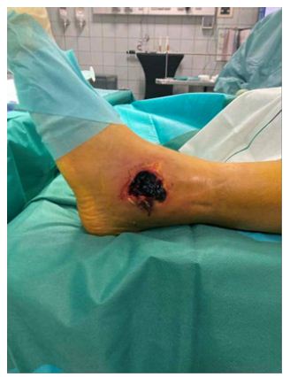
Figure 1: The wound in the operating room before debridement surgery (day 0)
Results of Pathological Tests and Other Investigations
The patient underwent surgery and it was proven that the anterior talofibular ligament and calcaneofibular ligament were torn. There was rupture of the joint capsule. The patient was diagnosed with septic arthritis, and treatment following international standards for septic arthritis was started. A vacuum-assisted closure (VAC) system was applied and the patient was initially treated with 1,5-gram Cefuroxime intravenously three times daily.
The ankle capsule and hematoma tissue were sent for cultivation and antimicrobial resistance which showed Staphylococcus Aureus sensitive for Dicloxacillin.
Figure 2a shows photo from the second look operation two days after the primary. A smaller amount of fibrin was removed. The wound was with fresh bleeding, no undermining cavities and without signs of infection. Hereafter, the wound dressing was changed every other day. Intravenous Cefuroxime treatment continued for two weeks. Subsequently, the patient switched to oral treatment with Dicloxacillin for four weeks. The patient was discharged after three weeks and followed up by regular out-patient checkups.
After six weeks, the wound had almost healed. As seen on Figure 2c there was a cavity above the wound only of cosmetic significance. At last follow-up thirteen weeks after debridement, the wound was healing sufficiently without further complications.
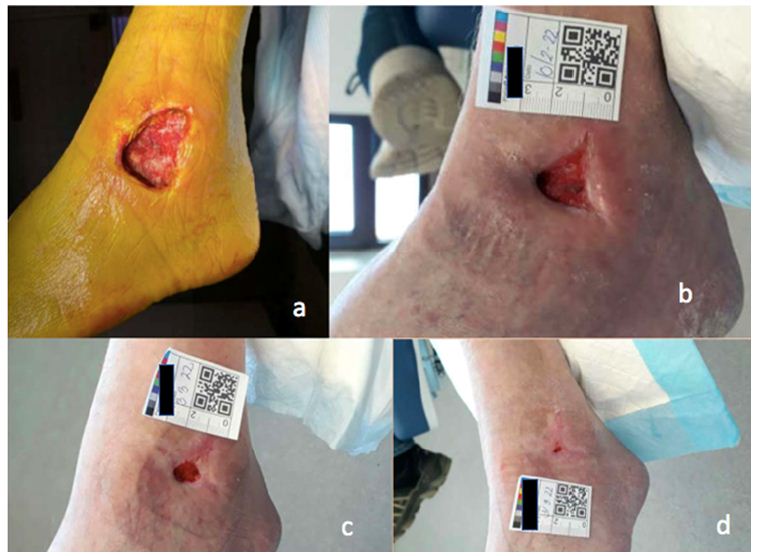
Figure 2: Photos of the thirteen weeks long wound healing period. a) Second look operation two days after debridement surgery b) Day 40 c) Day 61 d) Day 88
Discussion
This case report addresses a rare, but severe complication to an ankle sprain. To our knowledge this is the first reported case of an infected ankle joint haematoma following a non-operatively managed closed traumatic ankle joint injury. No inherent risk factors of septic arthritis were identified for the patient. Neither did the patient suffer from any apparent exposures that could cause septic arthritis.
Staphylococcus Aureus commonly resides on the skin of healthy individuals. Since there was no primary traumatic lesion to the skin of the ankle, one explanation to the etiology could be hematogenous or lymphogenous spread of the bacteria to the traumatic hematoma.
However, no bacterial focus was identified in this patient. Another feasible explanation could be a secondary rupture of the skin due to the traumatic oedema, thereby introducing skin bacteria to the underlying structures. It addresses the importance of treating the oedema following an ankle sprain.
However, the direction of causality between the wound and the infection is still an open question.
Intraarticular swelling is common in ankle sprains, but rupture of the joint capsule is not. Rupture of the capsule may have made the joint more vulnerable and susceptible to bacteria.
Up to 25% of patients with septic arthritis will experience impaired joint function afterwards [10]. Furthermore, pain and ankle instability may be sequelae of ankle sprain. It is therefore likely that the patient in this case will suffer from sequelae.
Conclusion
Septic arthritis is an extremely rare, but severe complication to an ankle sprain. The treatment existing of debridement and intravenously antibiotic is effective but cannot eliminate the risk of impaired joint function.
Notes on Patient Consent
Informed consent was obtained from the patient
References
- Thompson JY, Byrne C, Williams MA, Keene DJ et al. Prognostic factors for recovery following acute lateral ankle ligament sprain: a systematic review. BMC. [crossref]
- Blankenbaker D, Davis KW (2016) Ankle Sprain, in Diagnostic Imaging: Musculoskeletal Trauma. Elsevier 952-955.
- Doherty C, Delahunt E, Caulfield B, Hertel J (2014) The incidence and prevalence of ankle sprain injury: a systematic review and meta-analysis of prospective epidemiological studies. Sports Medicine 44: 123-140. [crossref]
- Buttaravoli P (2007) Ankle Sprain: (Twisted Ankle) in Minor Emergencies, pp: 396-403.
- Konradsen L, Bech L, Ehrenbjerg M, Nickelsen T (2002) Seven years follow-up after ankle inversion trauma. Scandinavian Journal of Medicine & Science in Sports 12: 129-135. [crossref]
- Holtom PD, Borges L, Zalavras CG (2008) Hematogenous septic ankle arthritis. Clinical Orthopaedics and Related Research 466 (6) : 1388-1391. [crossref]
- Mathews CJM, Weston VCF, Jones ADM, Field MF et al. (2010) Bacterial septic arthritis in adults. The Lancet 375: 846-855. [crossref]
- Wang J, Wang L (2021) Novel therapeutic interventions towards improved management of septic arthritis. BMC Musculoskeletal Disorders 22: 530. [crossref]
- Mathews CJ, Kingsley G, Field M, Jones A, et al. (2007) Management of septic arthritis: a systematic review. Annals of the Rheumatic Diseases 66: 440-445. [crossref]
- Weston V, Jones A, Bradbury N, Fawthrop F, et al. (1999) Clinical features and outcome of septic arthritis in a single UK Health District 1982-1991. Annals of the Rheumatic Diseases 58: 14-9. [crossref]