Author Archives: author

Therapeutic Effectiveness over Ground Walking with Body Weight Support Treadmill Training for the Treatment Subacute Stroke Gait Dysfunction

DOI: 10.31038/IMROJ.2021633

Abstract

Background: Stroke is an acute onset of neurological dysfunction due to abnormality in cerebral circulation with resultant signs and symptoms that correspond to the involvement of focal areas of the brain. Among all the neurological diseases of adult life, the cerebral vascular ones clearly rank first in frequency and importance.

Aim: To find out the effectiveness of over ground walking with treadmill gait training in right side subacute stroke subjects.

Settings and design: Physiotherapy Center, NIMHANS, Bangalore. Simple Random Sampling Technique used in this study.

Methods and material: 30 Subjects were selected on the basics of inclusion and exclusion criteria. All the subjects were divided equally into two groups; control Group and experimental Group. Before starting the training, pre-test scores are measured by using cadence and stride length. Control group received over ground walking and experimental group received over ground walking with treadmill gait training for 30 minutes, and both the groups received conventional therapy. At the end of five months, post-test scores of both groups were taken by used measure the cadence and stride length.

Results: Overground gait training with Treadmill gait training group post-test score (71.3, p<0.05) showed better improvements in mobility and gait speed, When compared to over ground gait training group (58.2.3, p<0.05).

Conclusion: The present study which proved that the use of body weight support treadmill training with partial body weight support combined with conventional physiotherapy to be more effective in improving gait ability in subacute subjects.

Keywords

Body weight support, Treadmill gait training, Gait speed, Mobility, Overground gait training

Introduction

Stroke refers to the sudden death of some brain cells due to a lack of oxygen when the blood flow to the brain is impaired by blockage or rupture of an artery to the brain [1].

Stroke may be manifested as Hemiplegia, which is the paralysis of muscles of one side of the body, contralateral to the side of the brain in which CVA occurred [2,3]. Clinically a variety of deficits are possible including the changes in the level of consciousness, impairments of sensory, motor, cognitive, perceptual and language functions. The locations of lesion, the extent of lesion, and the amount of collateral blood flow and early acute care management determine the severity of neurological deficits [4-6].

28% of stroke occurs in individuals under the age of 65 years. The incidence of Stroke is about 19% higher for males than females [7,8].

Stroke can be classified by etiological basis (Ischameic or Haemorrhagic), vascular basis (territory involvement), anatomical basis (cortical or brainstem or capsular or cerebellar or spinal), severity basis (minor or major) progression basis (completed or evolving) and onset basis (infantile or young or elderly stroke).

Stroke usually results in some degree of muscle weakness. It may lead to difficulty with producing force effectively within the context of a task and slowness to produce force is few of the commonest problems faced by the stroke subjects [9]. Moreover several studies have shown that muscle weakness is associated with reduced walking speed and endurance. And also muscle weakness has been suggested as a significant predictor of walking ability in stroke subjects [10].

Statement of Problem

Study on analyzing the effectiveness of Treadmill training with partial body weight support and physiotherapy in improving gait ability after stroke.

Need for the Study

Locomotion is one of the commonest problems after stroke in terms of asymmetry and reduction of speed etc. The most often stated goal for stroke subjects is to improve walking. For the improvement of walking, good strength of the lower extremity muscles is essential irrespective of the presence of spasticity because of growing evidence that muscle weakness rather abnormal reflex activity is a major limiting factor in physical function particularly for locomotor tasks following stroke. Through this study I would like to find out the effectiveness of body weight support treadmill training with partial body weight support and physiotherapy in improving gait ability of stroke subjects

Objectives

  1. To determine the effect if body weight support treadmill training with partial body weight support and physiotherapy in improving gait ability of stroke subjects in group A subjects.
  2. To determine the effect of body weight support treadmill training alone in improving gait ability of stroke subjects in group B.
  3. To determine the difference between the effectiveness of body weight support treadmill training with partial body weight support and physiotherapy in improving gait ability of stroke subjects.

Materials Used in This Study

  1. Treadmill
  2. Supporting Harness
  3. Set of pulleys
  4. Couch
  5. Pillows
  6. Towels
  7. Sand Bags
  8. Swiss Ball
  9. Parallel Bar

Methodology

Research Design

The design that is used for this study is experimental study design

Study Setting

Physiotherapy Center NIMHANS Bangalore

Study Sample

A total Number of 30 patient with stroke were selected by random sampling method with consideration of inclusion criteria and exclusion criteria and they were divided in to Group A and Group B.

Study Duration

All subjects participated in comprehensive 6 months Rehabilitation program.

A three week baseline study consisted of occupation therapy, and speech and neuro physiological therapy according to individual needs. During the subsequent 3 weeks of specific intervention gait training measured. Group A-Body weight support treadmill training with partial body weight support for 30 minutes 5 times a week. Group B – Body weight support treadmill training with partial body weight support for 30 minutes 5 times a week for 3 weeks.

Experimental Group (Group A): It consists of 15 subjects who underwent body weight support treadmill training and physiotherapy

Control Group (Group B): It consists of 15 subjects who underwent only partial body weight supported treadmill training.

Criteria for Selection

Inclusion Criteria: Subjects with all types of stroke.

  1. Age group between 29-40 years.
  2. Subjects of willingness of participate in the study.
  3. Subjects with both right and left hemiplegia
  4. Both genders.
  5. Able to understand at least simple instructions.
  6. No other orthopedic or neurological diseases impairing mobility.

Exclusion Criteria

  1. Subjects with other musculoskeletal disorder.
  2. Medically unstable.
  3. Non Co-operative subjects.

Parameter Used

Functional ambulation category (FAC).

Interventions

The purpose of the treatment and aim of the study were explained subjects who are selected for the treatment. All patients signed the consent form before undergoing treatment program. Subjects in group A was treated with body weight support treadmill training with partial body weight support and conventional physiotherapy. Subjects in group B was treated with exclusive body weight support treadmill training with partial body weight support. The treatment was given for both groups for periods of 6 months.

Procedure

Subjects were supported in a modified parachute harness suspended centrally by a set of pulleys connected to a flexible spring.

At the beginning of the therapy, two therapists provided manual help to correct gait deviations. One therapist sitting by the paretic side facilitated the swing of the paretic limb, determined that its initial ground contact was made with the heel, and prevented knee hyperextension during mid stance and encouraged symmetry of step length and stance symmetry.

The second therapist stood on the treadmill behind the patient and facilitated weight-shift onto the stance limb, hip extension and trunk erection. Mean treadmill speed was 0.21 (range 0.15-0.30 m/s was reached and kept constant until the end. The mean BWS was 27% (range 20-30) of body weight at the beginning. The support whilst 10 subjects needed a support of 5-15% BWS until the end. Net walking a support of treadmill was approximately 20 min per session with a brief rest in the middle.

Results

The pre and post-test values were assessed for gait ability in Group A. the standard deviation was 0.4. The ‘t’ values were calculated for gait ability by paired ‘t’ test was 28.5 and it was more than table value 2.15 for 5% level of significant at 14 degrees of freedom (Figures 1, 2, Tables 1 and 2).

fig 1

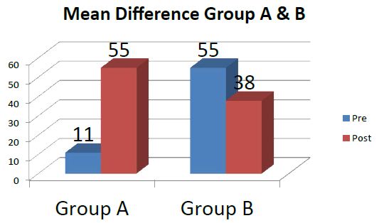
Figure 1: Mean and mean difference value for group A and group B.

fig 2

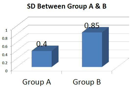
Figure 2: Standard Deviation value for Group A and Group B.

Table 1: Mean and mean difference value for group A and group B.

Study Group

Walking Ability

Mean

Mean Difference

Group A

Pre

Post

3

11

55

Group B

25

38

1

Table 2: Standard Deviation value for Group A and Group B.

Study Group

Standard Deviation

Walking Ability

Group A

0.4

Group B

0.85

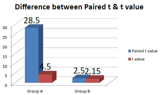
The pre and post-test values were assessed for gait ability in Group B. the standard deviation was 0.85. The ‘t’ vales were calculated for gait ability by paired ‘t’ test was 4.5 and it was more than table value 2.15 for 5% level of significance at 14 degrees of freedom (Figure 3 and Table 3).

fig 3

Figure 3: Paired ‘t’ test values for Group A and Group B.

Table 3: Paired ‘t’ test values for Group A and Group B.

Study Group

Calculated Paired ‘t’ Values

Table Value

Significance

Walking Ability

Group A

28.5

2.15

Significant
Group B

4.5

2.15

Significant

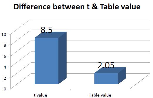
The calculated ‘t’ values by unpaired ‘t’ test was 8.5. The calculated’ values were more than the table value 2.05 for 5% level of significance of 28 degrees of freedom (Figure 4 and Table 4).

fig 4

Figure 4: Un paired ‘t’ test Values for walking ability in Group A and B.

Table 4: Un paired ‘t’ test Values for walking ability in Group A and Group B.

Study Group

Calculated Unpaired ‘t’ Values

Table Value

Significant

Walking Pattern

Comparison of Group A and Group B

8.5

2.05

Significant

The paired ‘t’ values have shown that body weight support treadmill training with partial body weight support combined with physiotherapy are more effective for the improving gait ability of subjects after stroke. The unpaired ‘t’ test values have shown that there is significant difference in showing improvement in gait ability of stroke subjects.

This study has proved the 3 week combination of body weight support treadmill training with BWS and physiotherapy effected a large improvement of a gait ability of non-ambulatory hemi paretic subjects than an exclusive 3- week treadmill therapy with BWS.

Discussion

This study has proved that The 3 week combination of body weight support treadmill training with conventional physiotherapy effected a large improvement of a gait ability of non-ambulatory hemi paretic subjects than an exclusive 3- week treadmill therapy with BWS.

Recently, Kwakkel et al. reported that greater intensity of leg rehabilitation improved gait ability and activities of daily living in acute stroke victims [11]. The key elements of their lower limb rehabilitation program were comparable with the physiotherapy within the present study.

Richards et al had shown that an additionally applied, task-specific program including body weight support treadmill training without body weight support resulted In a larger gait velocity in acute stroke victims 6 weeks after study onset as compared with a conventionally treated group who received less therapy [12,13].

The potential for motor recovery after stroke therefore seems to be limited, and subjects of group A probably reached this presumed level faster, i.e. the combined treatment of physiotherapy and body weight support treadmill training accelerated motor recovery [14,15]. Richards et al. also reported in the above mentioned study that differences in gait ability between the high and low-intensity group had waned at follow-up 6 months later, also because of a further improvement of a large extent in the low- intensity group.

Conclusion

From the results of this study 3 weeks of body weight support treadmill training with BWS pulls physiotherapy accelerated the restoration of gait ability in chronic hemi paretic subjects; correspondingly, a focused and intense treatment regime including locomotion training seems most promising in gait rehabilitation after stroke.

The result was analyzed using which proved that the use of body weight support treadmill training with partial body weight support combined with physiotherapy to be more effective in improving gait ability in hemi paretic subjects.

Limitation of the Study

  1. This Study has been conducted on small size sample only.
  2. The outcome of the study has been limited to improving gait ability only.

Recommendations

  1. Further study may be extended with large sample.
  2. Other aspect of motor impairment such as balance, strength may be considered.
  3. The patient ability to either improve or retain the regained functional capacity may be assessed at regular intervals over a period of time.
  4. The efficacy of this treatment may be found by altering the frequency and intensity.
  5. The extended efficacy of these exercises may also be found out by increasing the total in duration of the treatment.
  6. The body weight support treadmill training with partial body weight support may be applied to other neurological conditions such as Paraplegia.

References

  1. Asanuma H, Keller A (1991) Neurobiological basis of motor learning and memory. Concepts Neuro Sci 2: 1-30.
  2. Carr J, Shepherd R (1998) Neurological Rehabilitation. Butterworth & Heinemann, Oxford.
  3. Collen FM, Wade DT, Bradshaw CM (1990) Mobility after stroke: reliability of measures of impairment and disability.
  4. Dietz V, Colombo G Jensen L, Baumgartner L (1995) Locomotor capacity of spinal cord in paraplegic subjects. Ann Neurol 37: 574-582.
  5. Grilner S (1985) Neurologic basis of rhythmic motor acts in vertebrates. Science 228: 143-149.
  6. Hesse S, Berlet C, Schaffrin A, Malezic M, Mauritz KH (1994) Restoration of gait in non- ambulatory hemiparetic subjects by treadmill with partial body weight support. Arch Med Rehabil 75: 1087-1093.
  7. Hesse S, Bertelt C, Jahnke MT, et al. (1995) Body weight support treadmill training with partial body weight support as compared to physiotherapy in non-ambulatory heparetic subjects. Stroke 26: 976-981.
  8. Hesse S, Malezic M, Schaffrin A, Maurtiz KH (1995b) Restporation of gait by a combained body weight support treadmill training and multichannel electrical stimulation in non-ambulatory hemiparetic subjects. Scand J Rehabil Med 27: 199-205.
  9. Holden MK, Gill KM, Magliozzi MR (1986) Gait assessment for neurologically impaired subjects.Standards for outcome assessment. Phys Ther 66: 1530-1539.
  10. Jorgensen HS, Nakayama H, Raaschou HO, Olsen TS (1995) Recovery of walking function in stroke subjects:the Copenhagen stroke study. Arch Phys Med Rehabil 76: 27-32.
  11. Kwakkel G, Wagenaar RC, Twisk JWR, Lankhorst GL, Koetsier JC (1999) Intensity of leg and arm training after middle cerebral artery stroke:a randomized trail. Lancent 354: 191-196.
  12. Visintin M, Barbeau H, Korner-Bitensky N, Mayo NE (1998) Anew approach to retain gait in stroke subjects through body weight support and treadmill stimulation. Stroke 29: 1122-1128.
  13. Werning A, Muller S (1992) Laufband locomotion with body weight support in persons with severe spinal cord injuries. Paraplegia 30: 229-238.
  14. Lovely RG, Gregor RJ, Roy RR, Edgerton VR (1986) Effects of training on the recovery of full weight bearing stepping in the adult spainal cat. Exp Neurol 92: 421-435.
  15. Visintin M, Barbeau H (1989) The effects of body weight support on the locomotor pattern of spastic paretic subjects. Can J Neurol Sci 16: 315-325.

Case Report of Successful Lung Therapy in COVID-19

DOI: 10.31038/IMROJ.2021631


As in many countries COVID-19 infection levels are high, consecutivly many patients develop the severe form of the disease whilst the capacity of clinics is limited. SARS-CoV-2 is acting unfortunately on different levels, once infected there is no single drug able to prevent the ongoing disease. I would like to share my personal experience because it might help to prevent lung damage with simple and available remedies.

March 2020, aged 52 years, I got infected two times, the 11th and the 17th, by a double exposition to SARS-CoV-2 during several hours. The onset of a severe clinical form of the disease followed. No PCR-test was possible at that time.

The 19th of March the symptoms started. They disappeared at about the 29th, but the 30th March retrosternal pain began associated to a beginning pulmonary dysfunction, accompagnied by serious neurologic troubles. I suffocated while breathing normally and therefore a hypoxemia was more than possible. Hypoxia means reactive oxygen (ROS) and reactive oxygen nitrogen species (RNOS) occur. In the meantime their interference in COVID-19 has been proven.

In the Center of Oxygen, Research and Development, where I worked during my PhD studies on hypoxia, ROS and RNOS, my fellow researchers and colleagues examined the antioxidant properties of vitamin C, E, selenium, curcumin and resveratrol. Reseveratrol is found in highest levels in red wine like Pinot Noir. With the beginning of the pulmonary dysfunction I started the intake of several nutrients present at home as I was not sure medical doctors in a clinic would accept my hypothesis.

The pulmonary function decreased in a spectacular way during three days and stabilized then. A week later the improvement started.

The therapy was threefold: Increasing blood oxygen saturation, sustaining enzymes such as the glutathione peroxidase and intake of antioxidants.

In order to charge the blood physically with more oxygen the respiration pattern was voluntary increased several times a day. This means, first determination of the basal respiration frequency. Increasing this frequency with ten to fifteen more breath takes per minute during three minutes. Returning to the basal frequency.

Theophylline is known to improve the acute moutain sickness, another form of hypoxemia (known to lead to loss of smell and taste). Black tea was part of the diet.

On one hand the hypothesis worked out, on the other hand it did not.

It did not work out, because in the morning hours of the 25th April 2020 one of three heart beats missed and the blood pressure regulation got completely dysregulated.

The cardiac issues needed medical care.

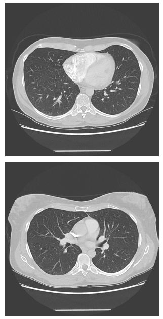
It did work out, because the lungs did not develop the COVID-specific lesions as observable in the two joined images resulting from a contrasted thoracic CT scan the 29th May 2020.

 
fig
 

The 25th of April the access for the cardiac problems to the emergency unit of a local hospital has been declined due to a blood oxygen level of 96%, and 5th of May the same at another local hospital (97% oxygen blood saturation). A supplementary blood analysis at the 5th of May did no show any particularities other than a slight lymphocytosis, slightly increased basophiles, a neutropenia and an insufficient vitamin D level. The level of SARS-CoV-2 antibodies was negative.

The remaining effects of the SARS-CoV-2 infection are the heart issues and a slight tinnitus of the left ear. SARS-CoV-2 specific T-cells are not yet examined. Another blood sample of the 10th December presents high levels of Gamma-globulins, indicating the presence of non-specific antibodies. EBV has been excluded.

Taken together, it could be interesting to add antioxidants to the therapy of COVID-19 in order to prevent lung damage, especially when no other medical care is possible.

Please find below a list of nutrients:

Supplementation of nutrients:

Vitamin B1:                          1,1 mg

Riboflavin:                            2,8 mg

Niacin:                                  16 mg

Pantothenic acid:                    6 mg

Vitamin B 6:                          1,4 mg

Biotin:                                  50 µg

Folic acid:                              200 µg

Vitamin B 12:                        12,5 µg

Vitamin C:                            1800 mg

Vitamin D:                            10 µg

Vitamin E:                             12 mg

Calcium:                                400 mg

Iron:                                     10 mg

Zinc:                                     5 mg

Selenium:                              55 µg

Iodine:                                 100 µg

Diet

Darjeeling/Earl Grey: 600 ml

Pinot Noir (Aigle Noir, Gérard Bertrand, Pays d‘Oc, 2019): 350 ml

Herbal infusion (1,75 g : 55% curcumin, 14% cinnamon, apple, 7% ginger, cardamom, 3% stevia leafs, fennel, nutmeg, cocoa shell 2%, black pepper, cloves)

Dark chocolate (85%): 25 to 50 g

In undetermined amounts: peanuts, olive oil, almonds, curcumin, fatty fish.

References

  1. Pritom Chowdhury, Anoop Kumar Barooah (2020) Tea Bioactive Modulate Innate Immunity: In Perception to COVID-19 Pandemic. Review; Front Immunol 11: 590716. [crossref]
  2. PT Goud, D Bai, HM Abu-Soud (2021) A Multiple-Hit Hypothesis Involving Reactive Oxygen Species and Myeloperoxidase Explains Clinical Deterioration and Fatality in COVID-19. Review. Int J Biol Sci 17: 62-72. [crossref]
  3. M Iddir, A Brito, G Dingeo, SS Fernandez Del Campo, H Samouda , et al. (2020) Strengthening the Immune System and Reducing Inflammation and Oxidative Stress through Diet and Nutrition: Considerations during the COVID-19 Crisis. Review. Nutrients 12: 1562. [crossref]
  4. L Loffredo, F Violi (2020) COVID-19 and cardiovascular injury: A role for oxidative stress and antioxidant treatment? Int J Cardiol 312: 136. [crossref]
  5. Montserrat M, E de Gregorio, C de Dios, V Roca-Agujetas, B Cucarull, et al. (2020) Mitochondrial Glutathione: Recent Insights and Role in Disease. Review. Antioxidants 9: 909. [crossref]
  6. M Mrityunjaya, V Pavithra, R Neelam, P Janhavi, PM Halami, et al. (2020) Immune-Boosting, Antioxidant and Anti-inflammatory Food Supplements Targeting Pathogenesis of COVID-19. Front Immunol 11: 570122. [crossref]
  7. BB Muhoberac (2020) What Can Cellular Redox, Iron, and Reactive Oxygen Species Suggest About the Mechanisms and Potential Therapy of COVID-19? Front Cell Infect Microbiol 10: 569709. [crossref]
  8. J Saleh, C Peyssonnaux, KK Singh, M Edeas (2020) Mitochondria and microbiota dysfunction in COVID-19 pathogenesis. Mitochondrion 54: 1-7. [crossref]
  9. F Silvagno, A Vernone, GP Pescarmona (2020) The Role of Glutathione in Protecting against the Severe Inflammatory Response Triggered by COVID-19. Antioxidants (Basel) 9: 624. [crossref]
  10. J Wu (2020) Tackle the free radicals damage in COVID-19. Nitric Oxide 102: 39-41. [crossref]

MINI OPCAB Mammary to LAD and Optimal Medical Treatment in High Risk Patients with Multivessel Coronary Disease Long Term Results

DOI: 10.31038/JCCP.2021415

Abstract

Old patients with multivessel coronary artery disease (CAD) are a challenging group to treat The MINI OPCAB technique is an operation were we connected the left internal mammary to LAD artery through an small incision in the lower part of the sternum. The objective of this prospective study was to show the results and survival during a follow-up in a group of high-risk patients with Multivessel disease treated with the MINI OPCAB operation Results The operative mortality was 0% in this group of patients. The incidence of perioperative infarction was 0%. The average time of the operation was 2 hours and 20 minutes MACE in this group of patients at 80 months was 0%. The survival rate (K-M) at 80 months was 82% Conclusion We strong believe the combination of a MINI OPCAB operation in high risk patients with multivessel coronary disease and optimal medical treatment an eventually and stent in a very big dominate artery is a valuable option for this type of patients more experience is needed to confirm this data.

Statistics: Data were analysed with the Statistical Package for Social Sciences (SPSS, Version 15.0).

Keywords

Coronary surgery and medical treatment, Coronary surgery plus medical treatment, MINI OPCAB in High risk patients, Treatment in high risk coronary patients, Treatment in multivessel coronary

Introduction

Old patients with multivessel coronary artery disease (CAD) are a challenging group to treat; these cases elicit discussion within heart teams regarding the actual benefit of undertaking major surgery on these patients and often lead to abandon the surgical option. Since these patients usually present with age-related comorbidities, preoperative risk stratification is mandatory and less invasive treatment options are favorable. Although conventional surgical revascularization can be carried out in old patients with acceptable short- and long-term a result, perioperative mortality is markedly elevated [1]. For high-riskpatients with multivessel CAD, not eligible to on-pump complete revascularization surgery or percutaneous procedures, incomplete revascularization with OPCAB LIMA-on-LAD offers benefits in survival when compared to OMT (Optimal medical treatment) alone [2]. MIDCAB is an effective approach for managing high-risk patients with symptomatic three-vessel coronary artery disease. Longer follow-up is needed to further clarify patient selection and the long-term outcome of this approach [3,4].

The MINI OPCAB techniqueis an operation were we connected the left internal mammary to LAD artery through an small incision in the lower part of the sternum [5] The long term results were previous described [6]. The objective of this prospective study was to show the results and survival during a follow-up in a group of high-risk patients with Multivessel disease treated with the MINI OPCAB operation and maximal medical treatment during the last 7 years in our Foundation.

Patients and Methods

During the last ten years 14 high risk patients with multivessel coronary disease prospective enroled received a MINI OPCAB operation Left mammary to the LAD bypass plus maximal medical treatment and strictly risk factor controls. The average age was 71, 07(st D 9,051 ci 95%), 21% were females: The preoperative Logistic Euroscore was 10, 68 (st D 5,407 CI 95%). The patients were strictly follow monthly in the Clinic of the Foundation by the Heart Team.

Results

The operative mortality was 0% in this group of patients. The incidence of perioperative infarction was 0%. The average time of the operation was 2 hours and 20 minutes. Ten (71%) of the patients were extubated in the operating room. The average time of Hospitalization was two days and eleven hours One patient at 30 days received a PTCA STENT in the Right Coronary artery; was a very big and dominant artery and the patient started again with angor after the procedure;another patient with a big dominant Circumflex was stenting inmediatly after the operation MACE in this group of patients at 80 months was 0%. We lost one patient at 85 years old due to a cerebrovascular accident at almost 5 years (62 months). The survival rate (K-M) at 80 months was 82%

Discussion

The primarily supposed benefit of off-pump surgery in elderly patients is still undetermined [7] in selected patients with multivessel disease (MVD), MIDCAB can be reasonable with concomitant percutaneous coronary intervention (PCI) as a hybrid procedure [8,9]. To date, the 2014 ESC/EACTS guidelines on myocardial revascularization judge hybrid revascularization as reasonable only in selected patients when PCI of the LAD is not an option and conventional CABG is associated with an increased surgical risk [10].

During a total of 6.3 (median, 4.9) years of follow-up, the primary composite outcome of all-cause mortality, myocardial infarction, stroke, or repeat revascularization occurred in 26% (141/550) and 34% (179/529) of patients in the CABG and PCI groups, respectively (hazard ratio (HR), 0.75; 95% confidence interval (CI), 0.60-0.94; P =.012). CABG was associated with fewer myocardial infarction (4% vs. 8% for PCI; HR, 0.48; 95% CI, 0.29-0.80; P =.037); and repeat revascularizations (8% vs. 17% for PCI; HR, 044; 95% CI, 0.31-0.64; P <.001), but had little association with all-cause mortality or stroke [11].

For high-risk patients with multivessel CAD, not eligible to on-pump complete revascularization surgery or percutaneous procedures, incomplete revascularization with OPCAB LIMA-on-LAD offers benefits in survival when compared to OMT alone.

Patients who underwent OPCAB survived more than those discharged in optimal medical treatment [2]. Considerably more data are available concerning the outcome of old patients undergoing CABG. Sen et al. compared the outcome of 240 octogenarians with matched younger patients in a retrospective two-centre analysis. They found a statistically significant higher 30-day mortality rate of 6.8% in the elderly patients. Age was identified as a risk factor for early death [12] Gunn et al. [13] reported a 30-day mortality rate of 8.8% in octogenarians after CABG in a retrospective analysis where perioperative strokes were significantly more frequent than in younger patients (5.5 vs. 1.6%). We strong believe the combination of a MINI OPCAB operation in high risk patients with multivessel coronary disease and optimal medical treatment an eventually and stent in a very big dominate artery is a valuable option for this type of patients more experience is needed to confirm this data.

References

  1. Grischa Hoffmann. Christine Friedrich, Moritz Barrabas, Rainer Petzina, Assad Haneya, et al. (2016) Short- and long-term follow-up after minimally invasive direct coronary artery bypass. Interact Cardio Vasc Thorac Surg 23: 377-382. [crossref]
  2. Prestipino F, Cristiano Spadaccio, Antonio Nenna, Fraser Wh Sutherland, Gwyn W Beattie, et al. (2016) Off-pump coronary artery bypass grafting versus optimal medical therapy alone: effectiveness of incomplete revascularization in high risk patients. J Geriatr Cardiol 13: 23-30. [crossref]
  3. Benetti Method for coronary artery bypass (1999) United States Patent Ñ 5,888.
  4. Izzat MB, Yim AP (1997) Minimally invasive LAD revascularization in high-risk patients with three-vessel coronary artery disease. Int J Cardiol 1: S101-4. [crossref]
  5. Federico j Benetti , Natalia Scialacomo ,Gustavo Mazzolino (2021) Mini Opcab Operation Surgical Thecnique. Surg Thecnol Int 38: sti38/1400. [crossref]
  6. Federico J Benetti (2010) MINI-off-pump coronary artery bypass graft: long-term results. Future Cardiology 6: 791-795. [crossref]
  7. Deppe AC, Oliver J Liakopoulos, Elmar W Kuhn, Ingo Slottosch, Maximilian Scherner, et al. (2015) Minimally invasive direct coronary bypass grafting versus percutaneous coronary intervention for single-vessel disease: a meta-analysis of 2885 patients. Eur J Cardiothoracic Surg 47: 397-406. [crossref]
  8. Holzhey, Stephan Jacobs, Michael Mochalski, Denis Merk, Thomas Walther, et al. (2008) Minimally invasive hybrid coronary artery revascularization. Ann Thorac Surg 86: 1856 -60. [crossref]
  9. Repossini A, Maurizio Tespili, Antonio Saino, Igor Kotelnikov, Annalisa Moggi, et al. (2013) Hybrid revascularization in multivessel coronary artery disease. Eur J Cardiothorac Surg 44: 288-93. [crossref]
  10. Kolh P, Fernando Alfonso, Jean-Philippe Collet, Jochen Cremer, Volkmar Falk, et al. (2014) 2014 ESC/EACTS Guidelines on myocardial revascularization: the Task Force on Myocardial Revascularization of the European Society of Cardiology (ESC) and the European Association for Cardio-Thoracic Surgery (EACTS). Eur J Cardiothorac Surg 46: 517-92. [crossref]
  11. Chang, Cheol Whan Lee, Jung-Min Ahn, Rafael Cavalcante, Yohei Sotomi, et al. (2016) Outcomes of Coronary Artery Bypass Graft Surgery Versus Drug-Eluting Stents in Older Adults. Ann Thorac Surg 103: 517-525. [crossref]
  12. Sen B, Bernd Niemann, Peter Roth, Raed Aser, Markus Schönburg, et al. (2012) Short- and long-term outcomes in octogenarians after coronary artery bypass surgery. Eur J Cardiothorac Surg 42: e102-7. [crossref]
  13. Gunn j, Kari Kuttila, Francesco Vasques, Raine Virtanen, Anne Lahti, et al. (2012) Comparison of results of coronary artery bypass grafting versus percutaneous coronary intervention in octogenarians. Am J Cardiol 110: 1125-1129. [crossref]


Diagnosis and Therapeutic Tactics of Landau-Kleffner Syndrome in Adults

DOI: 10.31038/ASMHS.2021523

Abstract

Purpose: The clinical signs of Landau-Kleffner syndrome (LCS) are electroencephalographic features and are discussed in five patients aged 25-33 years.

Methods: LCS diagnostic criteria: 1) acquired aphasia or oral hearing aphasia; 2) to have focal, tonic-clonic seizures. The patients were observed at the Mental Health Center of the Ministry of Health of the Republic of Azerbaijan. Of the five people examined, four were women.

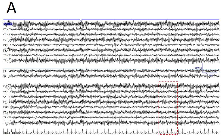
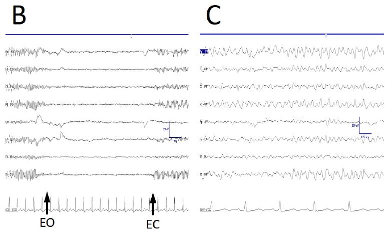
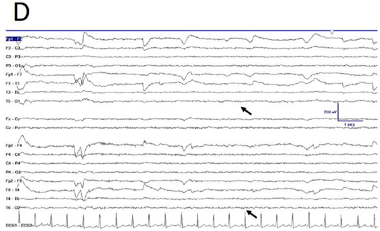
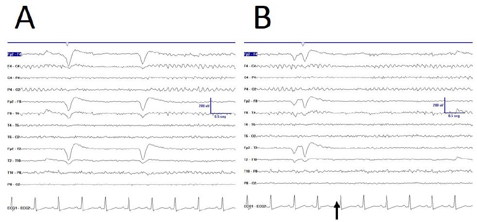
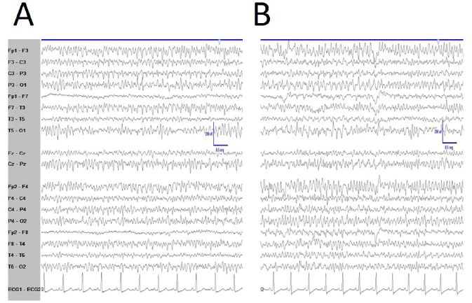
Results: The mean follow-up lasted 24 months (January 2019 to January 2020) and tonic-clonic seizures were observed in all five patients. Oral auditory agnosia was found to be moderately aphasic in all patients. Typical epileptiform EEG patterns in SLK are high-amplitude (200-400 μV) regional adhesions, sharp waves.

Conclusions: Landau-Kleffner syndrome is characterized by acquired oral hearing aphasia, seizures, and EEG changes in most patients. Patients were also prescribed depakine® chrono, lamotrigine and levetiracetam in combination with Cortexin®. After 3 months of treatment, speech and cognitive dysfunction were prevented. Patients were monitored regularly for 24 years. Observations showed no convulsions or speech or cognitive impairment.

Keywords

LCS adults, Diagnostics, Treatment, Landau – Kleerner syndrome, Adult epilepsy, Treatment

Introduction

Landau-Kleffner syndrome (LCS) is a rare age-related epileptic encephalopathy characterized by developmental regression in the tongue and electroencephalogram (EEG) abnormalities located mainly around the temporo-parietal areas. If present, seizures consist of absence seizures or tonic-clonic episodes and are more likely to occur during sleep. Behavioral disorders can form part of the clinical picture. The syndrome described in 1957 [1] is also referred to as acquired aphasia with epilepsy (ICD-10, F80.3) to indicate the main features of this disease. It is considered a form of continuous burst during slow wave sleep (CSWS), although the two syndromes have different clinical manifestations and diagnostic implications.

The exact etiology of Landau-Kleffner syndrome is unknown. Structural brain damage in patients with LCS is very rarely associated with pathophysiology. Moreover, genetic factors may be involved. For example, the disease may be correlated with GRIN2A (16p13.2) mutations. This gene encodes a protein called GluN2A (also known as NR2A), which is a subunit of the glutamate ion channel receptor N-methyl-D-aspartate (NMDA). It should be noted that NR2A can be identified at high concentrations in areas of the brain important for speech and language, while NMDA receptors are involved in a wide range of functions related to memory and learning. Again, GRIN2A changes correlate with a significant number of neurodevelopmental disorders, among which epilepsy may be a clinical manifestation [2,3].

It is difficult to express an estimate of the prevalence and incidence of the syndrome. Landau-Kleffner syndrome, indeed, is described as sporadic or limited case series. From the description of the syndrome, in 1957, no more than a few hundred cases have been reported in the literature. An epidemiologic study demonstrated that the incidence of children with LKS in Japan was about 1 in a million [4,5]. Furthermore, it emerges that males are more affected than females and that the reference age is between 3 and 8 years. However, documented cases also exist of younger children or adolescents with the syndrome.

We searched MEDLINE systems, – PreMEDLINE https://www.ncbi.nlm.nih.gov/pubmed/; https://www.ncbi.nlm.nih.gov/pmc/; The Cochrane Library; https://www.accessdata.fda.gov/scripts/cder/daf/; http://www.ema.europa.eu/ema/; https://scholar.google.com; https://www.rxlist.com/script/main/hp.asp; http://www.nejm.org; https://www.bmj.com in order to find at least some information about the incidence of Landau-Kleffner syndrome in adults. The main task of the search was to find information about the presence of Landau-Kleffner syndrome in adults. In none of these search engines did we find data on the primary onset of Landau-Kleffner syndrome in adults. Based on the above, the aim of this study was to study the clinical picture and develop tactics for the treatment of Landau-Kleffner syndrome in adults.

Materials and Methods

Consent

In accordance with the Helsinki Declaration of the World Medical Association “Recommendations for doctors engaged in bio-medical research involving people”, adopted by the 18th World Medical Assembly (Finland, 1964, revised in Japan in 1975, It-aly-1983, Hong Kong-1989, the South African Republic- 1996, Edinburgh-2000); The Constitution of the Republic of Azerbaijan, the Law “On Psychiatric Assistance” (adopted on 12.06.2001, with amendments and additions-11.11.2011. Parents or guardians have provided written informed consent to provide specific anonymized information obtained from their clinic visits for research use and have been reassured that their participation in the study is not related to ongoing clinical care. Consensus and data were obtained as patients were examined over a two-year period.

The decision of the Ethical Committee at the Azerbaijan Psychiatric Association on the article of NA. Aliev, ZN. Aliev “Clinical picture, diagnosis and therapeutic tactics of Landau-Kleffner syndrome in adults” submitted for publication in psychiatric journals: in connection with compliance with its legislative requirements and regulatory documents is to approve the article by NA. Aliyev, ZN. “Clinical picture, diagnosis and therapeutic tactics of Landau-Kleffner syndrome in adults” The patients were observed at the Mental Health Center of the Ministry of Health of the Republic of Azerbaijan from January 2018 to January 2020 for 24 months. Patients received depakin-chron 500 mg 2 times a day, lomotrigine 100 mg 3 times a day, levotisetam 1000 mg 2 times a day per os.

Additionally, the patients were assigned Cortexin® contains a complex of low-molecular water-soluble polypeptide fractions that penetrate through the BBB directly to nerve cells. The drug has a nootropic, neuroprotective, antioxidant and tissue-specific effect. The mechanism of action of the drug Cortexin® is due to the activation of peptides of neurons and neurotrophic factors of the brain; optimization of the balance of the metabolism of excitatory and inhibitory amino acids, dopamine, serotonin; GABAergic effects; a decrease in the level of paroxysmal convulsive activity of the brain, the ability to improve its bioelectrical activity; preventing the formation of free radicals (lipid peroxidation products). Active substance: polypeptides of cattle cerebral cortex. The drug is administered intramuscularly. Before injection, the contents of the vial are dissolved in 1 ml of a 0.5% solution of procaine (novocaine), water for injection or 0.9% sodium chloride solution and injected once daily: adults at a dose of 10 mg for 10 days; children from the neonatal period, with a body weight of up to 20 kg at a dose of 0.5 mg / kg, with a body weight of more than 20 kg – at a dose of 10 mg for 10 days. Before injection, the contents of the vial are dissolved in 1 ml of a 0.5% solution of procaine (novocaine), water for injection or 0.9% sodium chloride solution and injected once daily: adults at a dose of 10 mg for 10 days; If necessary, repeat the course in 3–6 months. Of the five people examined, four were women.

Results

Routine neurological examination of patients, as a rule, does not reveal any focal symptoms. Magnetic resonance imaging (MRI) with SLS, as a rule, does not reveal any pathology, however, it allows to exclude the symptomatic nature of the disease. The average follow-up lasted 24 months (January 2019 to January 2020) and tonic-clonic seizures were observed in all five patients. Oral auditory agnosia was found to be moderately aphasic in all patients. Typical epileptiform EEG patterns in SLK are high-amplitude (200-400 μV) regional adhesions, sharp waves. Landau-Kleffner syndrome is characterized by acquired auditory aphasia, seizures, and EEG changes in most patients. Cortexin® was also prescribed to patients with depakin-chrono, lamotrigine and levotirasem. After 3 months of treatment, speech and cognitive dysfunction were prevented. Patients were monitored regularly for 24 years. Observations showed no convulsions or speech or cognitive impairment.

Discussion

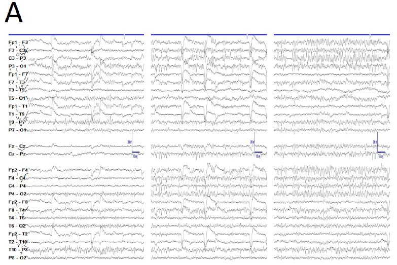
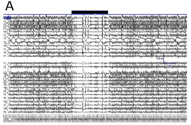
Our findings indicate that the most important characteristic of epileptiform activity in SLS is the tendency to diffuse spread. The diffuse propagation of peak-wave complexes in SLS is based on the phenomenon of secondary bilateral synchronization. At the same time, it is almost always possible to establish the temporal asynchrony of the complexes, as well as their amplitude predominance on the side dominant in speech. The highest amplitude is observed in the temporal leads, more often with an accent on the left; although cases of the location of the focus in the subdominant hemisphere of K.Yu. Mukhin [6] emphasize that sideliness of EEG disturbances does not always correspond to the dominant side, determined by the hand or eye, and epileptiform patterns can be observed in both the dominant and subdominant hemispheres [1].

This is the first preliminary descriptive study aimed at developing treatment strategies for clinical picture, diagnosis and therapeutic tactics of Landau-Kleffner syndrome in adults. It was found that Landau-Kleffner syndrome in adults after appropriate treatment, in all patients after appropriate treatment, in all patients, observations showed no convulsions or speech or cognitive impairment.

This study had several limitations; 1) a small number of patients; 2) a short time observation.

The sample size was relatively small. Further large-scale studies are needed to differentiate the use other anticonvulsants of Landau-Kleffner syndrome in adults.

Conclusions

Although clinical specimens are not representative of the wider population, this study nevertheless highlights the urgent need for further research of Landau-Kleffner syndrome in adults on the LCS diagnostic criteria- acquired aphasia or oral hearing aphasia, focal, tonic-clonic seizures. A. After appropriate treatment, in all patients, observations showed no convulsions or speech or cognitive impairment. The restoration of cognitive functions is very important to eliminate stigma and improve the quality of life of patients. It was found that in children the occurrence of aphasia not with epileptic seizures, but with epileptiform activity on the EEG, i.e., in fact, formulated the modern concept of epileptic encephalopathies. Our data indicate that in the Landau-Kleffner syndrome, the occurrence of aphasia is directly related to the frequency of epileptic seizures,

Author Disclosure

Authors declare that the manuscript is submitted on behalf of all authors. None of the material in this manuscript has been published previously in any form and none of the material is currently under consideration for publication elsewhere other than noted in the cover letter to the editor. Authors declare to have any financial and personal relationship with other people or organizations that could inappropriately influence this work. All authors contributed to and have approved the final manuscript.

Conflict of Interest Statement

The authors declare that the research was conducted in the absence of any commercial or financial relationships that could be construed as a potential conflict of interest.

References

  1. Landau WM, Kleffner FR (1957) Syndrome of acquired aphasia with convulsive disorder in children. Neurology 7: 523-530. [crossref]
  2. Lesca G, Møller RS, Rudolf G, Hirsch E, Hjalgrim H, et al. (2019) Update on the genetics of the epilepsy-aphasia spectrum and role of GRIN2A mutations. Epileptic Disord 21: 41-47. [crossref]
  3. Strehlow V, Heyne HO, Vlaskamp DRM, Marwick KFM, Rudolf G, et al. (2019) GRIN2A study group. GRIN2A-related disorders: genotype and functional consequence predict phenotype. Brain 142: 80-92. [crossref]
  4. Kaga M, Inagaki M, Ohta R (2014) Epidemiological study of Landau-Kleffner syndrome (LKS) in Japan. Brain 36: 284-286. [crossref]
  5. Caraballo RH, Cejas N, Chamorro N, Kaltenmeier MC, Fortini S, et al. (2014) Landau-Kleffner syndrome: a study of 29 patients. Seizure 23: 98-104. [crossref]
  6. Mukhin K Yu (2016) Landau–Keffner syndrome (acquired epileptic. aphasia) with a focus on electroencephalographic criteria. Russian Journal of Child Neurology 11: 3.

Abandonment of Family Planning among Women in Period of Genital Activity in the City of Mahajanga Madagascar

DOI: 10.31038/AWHC.2021442

Introduction

The World Health Organization or WHO defines family planning as the set of measures to promote births, act on the interval between births, prevent unwanted births and give each couple the means of determining the number of children he wants [1].

Poor access to family planning is associated with unintended pregnancies and poorer maternal and newborn outcomes, including abortion-related morbidity and mortality [2]. Unmet need for contraception refers to the percentage of sexually active and fertile women who either no longer want children or are delaying the next child, but are not using contraception. Meeting unmet need helps increase contraceptive use and reduce unintended pregnancies, which improves health outcomes and offers great social and economic benefits to women, their families, and society [3].

According to the WHO, in developing countries that wish to delay having a child or stop having children but do not use any contraception, the number of women is estimated at 225 million [4].

Family planning therefore appears as one of the solutions likely to allow socio-economic development to gain some ground in relation to population growth [5].

Madagascar faces many demographic and health challenges which have negative consequences on economic emancipation. The country has a young population, two-thirds of which are under the age of 25 [6]. The first step in the process of accelerating economic growth is based on declining fertility [7]. Note that the national fertility index was 4.9 in 2018 [8].

According to the 2012-2018 annual report, the modern contraceptive coverage rate has increased considerably, from 27.8% in 2012 to 34.6% in 2018. On the other hand, the unmet need for contraceptive methods has decreased by 28%. in 2012 to 24.9% in 2018 [9].

Increasing access to family planning and meeting unmet need for contraception are key goals for improving reproductive health. Madagascar is committed to the Global Family Planning 2020 partnership to improve access to family planning [6].

Thus, stopping contraceptives has significant repercussions, not only for family planning and maternal and child health, but also for population growth and the overall economic development of countries. Most women who stop using contraceptives do so at the start of their contraceptive use and without consulting a health care professional [10].

A study on family planning dropouts seemed necessary to us in Mahajanga, in order to identify the causes of women abandoning contraception.

Methodology

We carried out this study in the city of Mahajanga, among women in genital activity. This is a 3-month observational, descriptive and cross-sectional study, running from August 1 to October 31, 2019.

We included in this study all women aged 15 to 49 who had agreed to participate in the survey and had temporarily or permanently abandoned the contraceptive method.

The parameters evaluated were the variables related to the socio-demographic profiles of the women, to the data concerning the knowledge of these women in matters of family planning and the causes of discontinuation or abandonment.

Results and Discussion

Among these women who had abandoned modern contraceptive methods, 33.46% had used pills, 31.91% injectables and 27.63% condoms. In the other studies as well, users of pills and injectable contraceptives were the most frequent users of family planning, but in different proportions [11,12].

It was a temporary abandonment in 95.33% of cases, final in 4.67%. Several reasons were mentioned by the women, but the most important were the desire for a child, the opposition of the husband or partner, side effects, rumors and contraceptive failure. Thus, the desire for a child was the first motive pushing women to abandon modern contraception, found in 31.5% of cases.

This result was comparable to those of other studies which found that stopping the use of contraceptive methods was mainly due to a desire to have a child [13-15].

This would explain the relatively small number of children in our study, which witnessed not reaching the desired number of children. Partner opposition was the second reason for stopping the contraceptive method, found in 27.20% of cases, as reported in other studies [16,17].

The problem of family planning is a couple’s affair and the role of the partner must be preponderant before and after the choice of the contraceptive method. Yet, according to some authors, the majority of men believe that they have the absolute right, the power to decide on the use of modern contraception by their wives [16,17].

The problem of side effects was the third reason for giving up contraception, with 10.5% of cases. These side effects are multiple and depend on the contraceptive method used. Thus, each user must be informed at the time of the choice [18,19].

Negative rumors, mentioned by 10.1% of women, represent a phenomenon that compromises the development of modern contraceptive use. According to these rumors, modern contraceptives are the cause of serious disease such as cervical cancer or breast cancer, and are the cause of secondary infertility.

Thus, these rumors cause concern among users and may lead them to quit permanently, bringing back to the question of the sufficiency of information on family planning [20,21].

Among the women surveyed who temporarily gave up family planning, the majority resumed contraception using the same contraceptive method as before. Thus, the female family planning users in this study were almost faithful to the latest methods they used, despite the various reasons for quitting.

In addition, they were easy to convince about the use of family planning in the future, while the remaining 12%, those who encountered difficulties, said they did not intend to use it in the future.

Conclusion

The study on the reasons for abandoning the contraceptive method of women of childbearing age carried out in the city of Mahajanga has provided interesting information.

The results of this study showed that the majority of women surveyed were already aware of the existence of different contraceptive methods, and they were in favor of the use of modern contraception.

Among contraceptive methods, pills rank first, followed by injectable methods and condoms. The majority of these women are under 30, married or single, self-employed, with more than one living child, with at least secondary education. In almost 90% of cases, they gave up contraception temporarily for different reasons. For example, the desire for a child, lack of information, fear of opposition from a family member, and side effects were cited as reasons for giving up or not using contraceptive methods. Therefore, any effort to increase contraceptive prevalence should target these factors to optimize achievement of this goal.

Keywords

Abandonment, Family planning, Woman, Mahajanga

References

  1. Gentilini M (1988) Tropical Medicine. Paris: Flammarion.
  2. Ahmed S, Li Q,Liu L,Tsui AO (2012) Maternal Deaths Alerted by Contraceptive Use: An Analysis of 172 Countries. Lancet 380: 111-125. [crossref]
  3. Andrzej K (2018) Overcoming the Challenges of Family Planning in Africa: Towards Meeting Unmet Needs and Increasing Service Delivery; African Journal of Reproduction Health 22: 14-15. [crossref]
  4. Family planning. WHO 2016 May [Accessed 07/07/2019]. Available at the URL http://www.who.int/topics/family_planning/fr/
  5. Alkema L, Chou D,Hogan D, Zhang S,Moller AB, et al. (2015) Global, regional, and national levels and trends in maternal mortality between 1990 and 2015, with scenario-based projections to 2030: a systematic analysis by the UN Maternal Mortality Estimation Inter-Agency Group. Lancet 387 : 462-474. [crossref]
  6. Ministry of Public Health. National budgeted action plan for family planning in Madagascar 2016-2020: MSP.2016; 9-21.
  7. UNFPA Madagascar. National Conference on Family Planning: Demographic Dividend. September 14, 2016
  8. Multiple Indicator Cluster Surveys. Fertility and Family Planning: INSTAT .MICS.Madagascar.2018
  9. Summary of Main Indicators: FP2020 Annual Report 2018-2019. Madagascar. 2019
  10. Castle S and Askez I (2015) Stopping Contraceptives: Reasons, Challenges, Solution. Preliminary Report. Population Council 47.
  11. Summary of Key Indicators: FP2020 Annual Report 2018-2019. Ethiopia. 2019.
  12. Summary of Key Indicators: FP2020 Annual Report 2018-2019. Rwanda. 2019.
  13. Andriamialisoa (2007) Epidemiological profile of those lost to follow-up family planning at the CSB2 of Moramanga: Thesis in Med Tana 47.
  14. Mahamadou S (2019) Use of Family Planning services in the health area of the rural commune of Farako of the health district of Ségou from January 1 to December 31, 2018: Thesis in Med. BAMAKO. 45.
  15. Ravonisoa (2006) Family Planning: The reasons for the low use in Analavory, Manakara in 2006: Thesis in Med Tana. N ° 7530. 44.
  16. Mahougnon AT (2019) Choice and effectiveness of contraceptive methods: a reduction in unmet need for family planning. International Review of Marketing and Strategic Management 1: 23-42.
  17. Vouking MZ, Evina CD, Tadenfok CN (2014) Male involvement in family planning decision making in sub-Saharan Africa-what the evidence suggests. The Pan African Medical Journal 7: 19-33. [crossref]
  18. PSI Madagascar (2003) Study on the knowledge, attitudes and practices of young people aged 15 to 24 in Toamasina in terms of family planning. PSI / Mad. 2003: 6-9.
  19. Vololoniaina N (2007) Prevalence of side effects of modern contraceptives to CSB2 of Amparafaravola. Antananarivo: Medicine thesis n ° 5812: 27-30.
  20. Razafimiarantsoa TT (2004) Exposure to the risk of pregnancy. EDS 97-107.
  21. PSI Madagascar (2006) Family planning training. Antananarivo: Institute of Educational Technology and Management 14-15.

Case Report of Neuron-Binding IgGs in ALS Patient Serum in Argentina

DOI: 10.31038/JNNC.2021423

Abstract

Amyotrophic Lateral Sclerosis (ALS) is a degenerative disorder characterized by ongoing loss of motoneurons. The etiology of the sporadic ALS form it is thought to be related to immune-mediated motoneuron degeneration and death. The present study was designed to describe the effect of serum factors derived from sporadic ALS patients on mouse spinal cord preparations in vitro. Sera from patients with sporadic ALS were collected and used for immunofluorescence analysis to investigate their effects on neuronal survival and microgliosis. Our experiments demonstrated that 5 h application of serum factors (derived from three ALS patients) labeled with human IgG secondary antibody were localized to ventral spinal neurons identified with the NeuN marker. Moreover, a significant reduction in the number of ventral NeuN positive cells was observed with serum from a patient who suffered from upper motor neuron signs as criteria for ALS diagnosis (20% less compared to sham, spinal cord preparations without serum). No change in microglia number was found after exposure to ALS sera, although in two cases a significant decrease in microglial branch length was observed (28-32%). Microglia morphology showed increased number of end points and branches with serum from the patient with upper motor neuron symptoms. These observations were absent after control sera. Our data indicate that spinal cord cultures can be a useful model to further characterize the pathological processes of sporadic ALS and the immune mechanism as previously suggested in vivo. Future studies are needed to unveil the molecular mechanisms underlying this preferential targeting of neurons and microglia by ALS serum.

Keywords

Amyotrophic lateral sclerosis, Autoimmunity, Microglial activation, Neuron labeling

Non-standard Abbreviations

ALS, Amyotrophic Lateral Sclerosis; HC1 (2), serum form healthy control 1 (2); Iba-1, ionized calcium-binding adapter molecule; IgG, immunoglobulin; MNs, motor neurons; NeuN, neuronal-specific nuclear protein; P1 (2 and 3) serum from ALS sporadic patient 1 (2 and 3); PBS, phosphate-buffered saline.

Highlights

  • Immune mechanisms contribute to ALS pathology at spinal cord.
  • Unlike controls, ALS serum factors bound spinal ventral neurons.
  • Microglia morphological changes were induced by ALS serum factors.
  • In vitro spinal preparations can be a useful platform to study immunopathology of ALS.

Introduction

Amyotrophic Lateral Sclerosis (ALS) is a neurodegenerative disease that affects motoneurons with fatal outcome usually within 2-5 years [1]. Although the aetiology and pathogenesis of ALS are unknown increasing evidence supports immune-mediated mechanisms, although this remains controversial [2]. Our previous studies have demonstrated that motor nerve terminals are a target for autoimmune response, induced by immunoglobulin’s (IgGs) from sporadic ALS patients, resulting in neuromuscular dysfunction in vivo [3]. Indeed, IgGs obtained from patients with ALS reduce Ca2+ transients and glutamate receptor desensitization so that excitotoxic damage is facilitated in a culture brain neuron model [4]. Furthermore, we have shown that subcutaneous injection of ALS-IgG into the levator auris muscle were immunoreactive to spinal motoneurons, preferentially accumulate in the soma by binding to unknown specific cytoplasmic targets [5]. The question then arises about the role of ALS serum factors on spinal cord neurons and the inflammatory process that would be associated with the disease onset and development. In vitro spinal cord preparations have been exploited as a model to investigate early pathophysiological mechanisms of spinal cord damage and developmental changes in cellular profiles [6]. The aim of the present study was to investigate, using neonatal mouse spinal cord preparations in vitro, the effect of serum from sporadic ALS patients. Thus, sera from patients with sporadic ALS diagnosis in accordance with the El Escorial criteria [7] and from healthy volunteer subjects were tested on neuronal numbers and microglia morphology.

Materials and Methods

Preparation of Mouse Spinal Cord Cultures and ALS Serum Incubation

Thoracolumbar spinal cord preparations were isolated from neonatal C57BL/6 mice (1-3 day old) in accordance with standard procedures [8]. Briefly, spinal cords were dissected with Krebs’ solution of the following composition (in mM): NaCl, 113; KCl, 4.5; MgCl2.7H2O, 1; CaCl2, 2; NaH2PO4, 1; NaHCO3, 25; glucose, 11; gassed with 95% O2 5% CO2; pH 7.4 at room temperature. After dissection spinal slice cultures were incubated with sera from sporadic ALS patients (listed as P1, P2 and P3) in standard Krebs’ solution (1:1000) for 5 h at room temperature. Patients were diagnosed with sporadic ALS according to clinical criteria that included progressive paralysis with mixed upper and lower motor neuron signs (1 woman and 2 men) [7]. Healthy control serum was collected from two subjects (HC1 and HC2) with no evidence of neurological disease. The serum samples were obtained with prior consent from patients attending the FLENI clinic. Mouse spinal cord controls, without any serum treatment (sham), were processed in parallel to test preparations in each experiment. After incubation, all spinal cord tissues were immediately fixed in phosphate-buffered saline (PBS) containing 4% paraformaldehyde (24 h at 4°C) followed by 30% sucrose PBS for cryoprotection (24 h at 4°C) for immunostaining.

Slice Immunostaining and Cell Analysis

Full details of this procedure have previously been published [6]. In brief, spinal cord slices (30-µm thick) were collected sequentially on histology slides. Slices were blocked with fetal calf serum (3%), bovine serum albumin (3%) and Triton X-100 (0.3%) in PBS for 1 h at room temperature, followed by overnight incubation at 4°C in blocking solution containing the following antibodies: NeuN (neuronal-specific nuclear protein, 1:300, Millipore, Billerica, MA, USA) for neurons and Iba-1 (ionized calcium-binding adapter molecule 1, 1:500, Wako, Osaka, Japan) for microglia. Primary antibodies were visualized using the corresponding secondary fluorescent antibody (at 1:500 dilution; Invitrogen, Carlsbad, CA, USA). The serum factors from ALS patients were recognized by an anti-human IgG secondary antibody (at 1:500 dilution; Invitrogen). Since the staining was diffuse, data quantification was performed in terms of immunofluorescence intensity (expressed in arbitrary units, AU) obtained with a line scan of each image to verify the nuclear distribution of IgG according to NeuN soma size measured with ImageJ software (NIH, https://imagej.nih.gov/ij/index.html)http://imagej.nih.gov. For each slice culture, the number of NeuN positive cells was obtained by counting stacks of 10 images (40x magnification) with FV300 confocal microscope (Olympus Optical, Tokyo, Japan), and quantified using ImageJ software. NeuN positive cells were counted in two ROIs (namely, dorsal, and ventral) in an area= 90488, 5 µm². In view of the very large number of histological sections provided by each spinal cord the final numbers of counted cells were expressed as fold average respect to sham, obtained from an equivalent number of sections by experiment.

The morphology of Iba1-positive cells was analysed by the ‘skeleton’ method [9]. Thus, the signal from Iba1-positive processes was enhanced to optimize their detection followed by noise de-speckling to eliminate background fluorescence. The resulting images were converted to binary data and then ‘skeletonized’ by using ImageJ software. The Analyze Skeleton plug-in (http://imagejdocu.tudor.lu) was then applied to Iba-1 images to collect mean raw data on the number of branches, end points and process lengths.

Data Analysis

Data were expressed as means ± S.E.M; n = number of slices from 5 different independent experiments. Statistical analysis was carried out with SigmaStat (SigmaStat 3.1, Systat Software, Chicago, IL, USA). For multiple comparisons, the analysis of variance (ANOVA) test for parametric data followed by the Tukey-Kramer post hoc test was used. Nonparametric values were analyzed with the Kruskal-Wallis test. The accepted level of significance was always p < 0.05.

Results

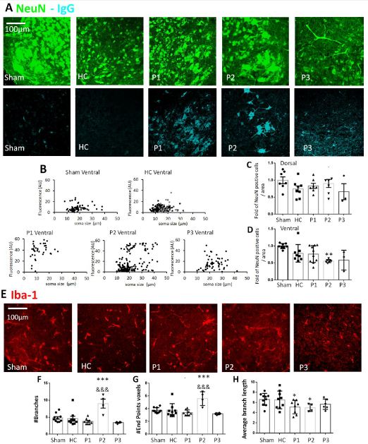
Clinical diagnosis of sporadic ALS was achieved by careful patient history collection, plus physical and neurological examination according to El Escorial criteria [7] and evidence of signs of lower or upper motor neuron degeneration. Hence, two patients were classified with lower motor neuron (P1 and P3) and one patient with upper motor neuron degeneration (P2). In vitro slice preparations of the neonatal spinal cord were treated with healthy control serum (HC), patient serum (P) or without serum (sham condition) for 5 h at room temperature. Figure 1A shows examples of immunohistochemistry analysis to identify neurons (NeuN, in green. Figure 1A top row), human immunoglobulins (IgG, light blue, Figure 1A lower row) and microglia (Iba-1, in red, Figure 1E).

Ventral Neuronal Labeling with ALS Serum Factors

Figure 1A and 1B shows that serum factors from all ALS patients labeled with the anti-IgG human secondary antibody were localized to NeuN positive neuronal cells. The quantification of mean immunofluorescence intensity for IgG signal was analyzed for different neuronal soma size. Figure 1B demonstrates higher mean fluorescence intensity (arbitrary units, AU) for P1, P2 and P3 in comparison to sham and HC1 and HC2 in the ventral spinal region. On the contrary a unique soma size IgG signal labeling was observed at dorsal region, data not shown. As exemplified in Figure 1C and 1D, the number of dorsal NeuN positive cells was consistent throughout samples. Conversely, a significant reduction in neuron number at the ventral spinal region was observed following serum from patient 2 (**p ≤ 0.01 vs. sham, n=3-10, Kruskal-Wallis one-way analysis of variance on ranks test).

Microglial Phenotypes Observed after ALS Serum Factors

The immunohistochemical staining for Iba-1 microglia marker was evaluated in ventral spinal regions after ALS serum incubation, as shown in Figure 1E. There was no significant change in the number of Iba1-positive cells. However, a morphological change in microglia was observed for P2 with significant increase in the number of microglial branches and endpoints (Figure 1F and 1G, ***p ≤ 0.001 vs. sham, &&& p ≤ 0.001 vs. HC, n=4-10, Mann-Whitney test). The vast majority microglia of P1 and P2 also showed a significant reduction in branch length (Figure 1H, *p ≤ 0.05 vs. sham, n=4-10, Mann-Whitney test).

fig 1

Figure 1: Neurons and microglia immunolabeling after incubation with serum factors derived from ALS patients in vitro spinal cord preparations. (A) Examples of neuron staining (NeuN, green) and serum factors that were identified by anti-human IgG labeling (light blue) in the ventral region of the spinal cord for sham, healthy serum (HC), serum from patients with sporadic ALS (P1, P2 and P3). (B) Plots showing IgG mean fluorescence intensity quantification (arbitrary units, AU) that colocalized with NeuN staining by measuring different neuronal soma size (µm). Results are expressed as raw data for in vitro spinal cord preparations for sham, HC (HC1 grey dots and HC2 black dots, respectively) and for P1, P2 and P3 at ventral region. (C-D) Histograms showing the fold of NeuN positive cells in dorsal and ventral area related to sham condition, or after 5h of incubation. No differences in the number of neurons were found in dorsal spinal cord region. (D) There was a significant decrease in the number of neurons following serum from P2 at the ventral region, n=3-10, **p<0.05 vs. sham. (E-H) Iba-1 morphological changes after ALS serum incubation. (E) Examples of microglia staining (Iba-1 in red) in mouse isolated spinal cord. Histograms showing the raw data for number of branches (F), end points voxels (G), and average branch length (H, in µm) for Iba-1 in ventral region of spinal cord cultures. In the ventral region there was a significant change in microglia morphology for P2, with a significant reduction in the average branch length of microglia also for P1 (G), n=4-10, *p<0.05 vs. sham, ***p<0.001 vs. sham, &&&p<0.001 vs. HC.

Discussion

Most studies describe the role ALS effects on lower MNs in the spinal cord and brain stem, and upper MNs in the motor cortex [10]. While ALS is a multifactorial disease with diverse aetiology, the main cause of its onset in the sporadic form is still unknown, although it is suspected that the activity of the immune system impacts its course and development [1]. Several studies show that, in this immune response, the IgG of the patients play a fundamental role as triggers of the disease [2]. The present report shows that serum factors derived from three patients labelled with human IgG secondary antibody were localized to ventral spinal neurons. Interestingly, the sera of all three ALS sporadic patients induced large soma size labeling of ventral neurons and microgliosis in in vitro spinal cord preparations.

Ventral Neurons were Labeled by ALS Serum Factors

Our previous studies demonstrated that ALS-IgG injected into the mouse levator auris muscle significantly immune react with nerve terminals and, by retrograde axonal transport, are actively accumulated in the MNs soma [5]. Here we have shown that most large soma size ventral spinal neurons were immunoreactive to serum factors derived from patients with the sporadic ALS form. Indeed, we could not observe a clear preference of ALS serum for a certain neuronal type. However, in this study it is expected that ALS IgG have been in contact with every single neuron while, in our previous study, ALS IgG reached the MNs via the motor axons. We have shown that the cellular composition varies between dorsal and ventral spinal regions, the latter being characterized by the presence of differential soma size interneurons as well as MNs [6]. Indeed, our pervious data have also demonstrated that ALS does not affect all neurons, but mainly certain types of MNs [5]. Our early studies have shown that the delayed neuroprotection by riluzole after kainate treatment, to mimic excitotoxicity as one major factor MN degeneration in ALS, was observed in the dorsal and central regions, but not in the ventral one [11]. The different neuronal vulnerability in the spinal cord [8,12], probably related to the neuron-selective ability by different protocols to induce neuroprotection, might highlight preference of ALS serum binding to large ventral neurons. Our results also demonstrate that binding of serum is not a trigger for neurodegeneration, at least within the short time of experimental serum application. A likely explanation, in addition to selective large cell binding, is that MNs have a delicate metabolic state and are perhaps more susceptible to any intracellular signalling process that can be initiated by the pathological ALS serum in vitro [13]. Further studies are needed to clarify the molecules responsible for this phenomenon, as well as the mechanism of IgG to differential cell labeling on sporadic ALS onset.

Microglia Morphological Changes Induced by ALS Serum Factors

To advance our understanding of ALS pathogenesis and the role of inflammatory processes, an immunofluorescence study with Iba-1, a microglial marker, was performed. Inflammation is mostly related to deterioration in neuronal function and involves a change of microglia number or morphological phenotypic changes. Our results demonstrated that the number of microglia in both dorsal and ventral areas did not vary after incubation with serum from patients or voluntary healthy controls. A simple explanation for this result is that neuroinflammation (if any) would not be initially characterized by microglia proliferation [14]. However, another important characteristic of neuroinflammation is the different states of microglia activation. When the inflammatory process occurs, microglia can pass from rest to different phenotypic active states [14]. In the present study serum factors derived from a patient with upper motoneuron degeneration induced significant microglia morphological change. Further studies will be needed to determine if this inflammatory change is a cause or consequence of neuronal degeneration.

Final Considerations and Conclusion

The present findings provide further evidence in favour of immunological mechanisms contributing to ALS pathology and disease progression in the spinal cord. In vitro spinal cord preparation is a useful model to explore the basic molecular mechanisms and cellular consequences of ALS.

Acknowledgments

We thank Maria Eugenia Martin for the assistance with the spinal preparations. We thank Dr. Andrea Nistri and Dr. Carly Mc Carthy for invaluable comments and critical reading of the manuscript. This study was supported by Universidad Austral, FLENI, CONICET and Grant 01/Q666 (20020130100666BA; Universidad de Buenos Aires Ciencia y Tecnología [UBACYT]) from University of Buenos Aires (to O.D.U.).

Declaration of Competing Interest

None declared.

References

  1. Beers DR, Appel SH (2019) Immune dysregulation in amyotrophic lateral sclerosis: mechanisms and emerging therapies. Lancet Neurol 18:211-220. [crossref]
  2. Pagani MR, Gonzalez LE, Uchitel OD (2011) Autoimmunity in amyotrophic lateral sclerosis: past and present. Neurol Res Int 2011: 497080. [crossref]
  3. Gonzalez LE, Kotler ML, Vattino LG, Conti E, Reisin RC, et al. (2011) Amyotrophic lateral sclerosis-immunoglobulins selectively interact with neuromuscular junctions expressing P/Q-type calcium channels. J Neurochem 119: 826-838. [crossref]
  4. Andjus PR, Khiroug L, Nistri A, Cherubini E (1996) ALS IgGs suppress [Ca2+]i rise through P/Q-type calcium channels in central neurones in culture. Neuroreport 7: 1914-1916. [crossref]
  5. Fratantoni SA, Dubrovsky AL, Uchitel OD (1996) Uptake of immunoglobulin G from amyotrophic lateral sclerosis patients by motor nerve terminals in mice. J Neurol Sci 137:97-102. [crossref]
  6. Cifra A, Mazzone GL, Nani F, Nistri A, Mladinic M (2012) Postnatal developmental profile of neurons and glia in motor nuclei of the brainstem and spinal cord, and its comparison with organotypic slice cultures. Dev Neurobiol 72:1140-1160. [crossref]
  7. Brooks BR (1994) El Escorial World Federation of Neurology criteria for the diagnosis of amyotrophic lateral sclerosis. Subcommittee on Motor Neuron Diseases/Amyotrophic Lateral Sclerosis of the World Federation of Neurology Research Group on Neuromuscular Diseases and the El Escorial “Clinical limits of amyotrophic lateral sclerosis” workshop contributors. J Neurol Sci 124: 96-107. [crossref]
  8. Mazzone GL, Margaryan G, Kuzhandaivel A, Nasrabady SE, Mladinic M, et al (2010) Kainate-induced delayed onset of excitotoxicity with functional loss unrelated to the extent of neuronal damage in the in vitro spinal cord. Neuroscience 168: 451-462. [crossref]
  9. Mazzone GL, Mladinic M, Nistri A (2013) Excitotoxic cell death induces delayed proliferation of endogenous neuroprogenitor cells in organotypic slice cultures of the rat spinal cord. Cell Death Dis 4: e902. https://doi.org/10.1038/cddis.2013.431
  10. Shaw PJ (1999) Motor neurone disease. BMJ 318:1118-1121. https://doi.org/10.1136/bmj.318.7191.1118
  11. Mazzone GL, Nistri A (2011) Delayed neuroprotection by riluzole against excitotoxic damage evoked by kainate on rat organotypic spinal cord cultures. Neuroscience 190: 318-327. [crossref]
  12. Taccola G, Margaryan G, Mladinic M, Nistri A (2008) Kainate and metabolic perturbation mimicking spinal injury differentially contribute to early damage of locomotor networks in the in vitro neonatal rat spinal cord. Neuroscience 155: 538-555. [crossref]
  13. Comoletti D, Muzio V, Capobianco A, Ravizza T, Mennini T (2001) Nitric oxide produced by non-motoneuron cells enhances rat embryonic motoneuron sensitivity to excitotoxins: comparison in mixed neuron/glia or purified cultures. J Neurol Sci 192: 61-69. [crossref]
  14. Graeber MB (2010) Changing face of microglia. Science 330: 783-788.

Why Public Health Interventions Need a Multidisciplinary Approach to Understand and Address Behaviours Effectively?

DOI: 10.31038/PEP.2021241

Abstract

Although we have made significant efforts in controlling disease with strong improvements in, for instance, people’s life expectancy, it is obvious that the 21st Century is full of health challenges such as unhealthy behaviors which are fueled by the way societies are organized so that there must be another focus on how we consider health and health issues in order to be more effective in addressing them. The objective of this reflection is to call upon the importance of a genuine understanding of health and ways of its improvement through interventions that address behaviors effectively while we are facing challenges that are different from the ones in the past centuries.

Keywords

Ecological approach of health; Transdisciplinary approach and effective behavior change interventions

Introduction

When considering health and health issues throughout centuries behind, it is obvious that improvements have been made with great achievements like the one on people’s life expectancy. After strong efforts to control disease in the past, the 21st Century is facing some different health challenges and unhealthy behaviors in a rapid changing world. The way societies are organized, dealing with development processes that are not getting all citizens onboard, the consequences of the climate change, the rapid demographic changes, globalization, and technological advances, among others, are impacting strongly health of populations [1]. If from the beginning, researchers could be skeptics with the World Health Organization (WHO) definition of health, nowadays there is great interest to consider that definition which leads to a comprehensive view of health so that by positioning people on a good level of the social ladder they will improve health and increase resilience [2].

The objective of this reflection is to call upon the importance of a genuine understanding of health and ways of its improvement through interventions that address behaviors effectively while we are facing challenges that are different from the ones in the past centuries.

Ecological Model of Health

According to WHO, “health is a state of complete physical, mental and social well-being and not merely the absence of disease or infirmity” [3]. This means that to be ‘healthy’, one should be well physically, mentally and socially. Health has been seen on different ways mainly with three major models: the biomedical model that views health as the absence of diseases or disorders; the behavioral model that views health as the product of making healthy lifestyle choices; and the socio-environmental model that views health as the product of social, economic and environmental determinants that provide incentives and barriers to the health of individuals and communities. This third model which is the more holistic way of viewing health is known also as biopsychosocial model or ecological model [4]. It emphasizes the linkages and relationships among multiple factors (determinants) affecting health. These determinants include biology, the social and physical environment, education, employment, and behavior. Dahlgren and Whitehead [5] drafted an example of the ecological model as shown in Figure 1 with one omitted factor, the general/local political context.

fig 1

Figure 1: Example of the ecological model of health according to Dahlgren & Whitehead [5].

Gebbie et al. [1] explained that while an ecological model addresses the interactions and linkages among determinants of health, there are related concepts to it. The first is an ecological view of health which is a perspective that involves knowledge of the ecological model of determinants of health and an attempt to understand a specific problem or situation in terms of that model. The second is an ecological approach to health which is the one that develops multiple strategies to impact determinants of health relevant to the desired health outcomes. It is then to say that addressing public health challenges requires an ecological approach with its implications among which the transdisciplinary approach and health professionals who are fully prepared to work with others to improve health.

Why should one, in dealing with health issues, considering not the ecological approach and its emphasis on the social determinants of health?

Social Determinants of Health and the Necessity for Transdisciplinary Interventions

The health challenges we are facing in this 21st Century which are, among others, the high level of consciousness of population’s health with lot of investments but without consistent results could be compared to the ones that led Canadian Government in the 1970s to the Lalonde Report [6] with the important notion of determinants of health.

Determinants of health include: broad social, economic, cultural, and environmental conditions; living and working conditions; social, family, and community networks; individual behavior; individual traits such as age, sex, race, and biological factors, and the biology of disease [1]. According to Blake [7], health can be seen like an iceberg with multiple underpinning determinants that shape it, such as: i) Values including culture, biology and genetic endowment, employment and working conditions, income and social status; ii) Assumptions that comprise personal health practices and coping skills, social environments, social support networks; iii) Beliefs that encompass gender, healthy child development, physical environment, education; and iv) Health services.

A part from individual traits and the biological factors, it is obvious that health is influenced by multiple factors that are the results of how societies are organized, the Social Determinants of Health (SDHs). SDHs are the conditions in which people are (conceived), born, grow, live, work and age including mechanism in place to address diseases [8]. By defining SDHs that way, it is obvious that they are under the responsibility of multiple sectors outside of the health system and are shaped by the distribution of money, power and resources at global, national and local levels [8]. This helps understand that health results are not the responsibility of the health system alone and even, other sectors are more responsible than the health system for the health outcomes [9]. These health results (or health status) which is different across the population and between specific populations groups are mostly the responsibility of the social determinants of health through their distribution. These differences in health status due to how a society is organized to assure a fair distribution of SDHs are avoidable and known as health inequities. By questioning these differences, Evans and colleagues [10] showed the importance of SDHs in maintaining people healthy. Improving health status is then a task for multiple sectors which must work together to achieve better health.

As the WHO Commission on Social Determinants of Health (CSDH) [11] put it in its framework (Figure 2), a patient is a “messenger” telling the practitioner about its living conditions, because illness is a “feedback” on a given individual’s social position. And social position is created by social, economic and political mechanisms, whereby populations are stratified according to income, education, occupation, gender, race/ethnicity and other factors. Socioeconomic positions in turn shape specific determinants of health status reflective of people’s place within social hierarchies. Based on their respective social status, individuals experience differences in exposure and vulnerability to health-compromising conditions and behaviors. In other words, diseases and behaviors are a deeply social process. Their distribution lays bare society’s structures of wealth and power, and the responses they elicit illuminate strongly held values [12]. According to Marmot [2], health and behavior follow a social gradient. A social gradient occurs when the frequency (e.g., of a health problem) increases steadily from the most advantaged to the least advantaged categories [2,13]. One could understand what the CSDH report was saying: “Why treat people and send them back to the conditions that made them sick?” [8] There is absolutely a necessity to change the social position for sick people in order to be treating them adequately. To do so, it must be through a transdisciplinary approach because health system cannot afford to do that alone. That’s why it is an unfinished work all the time people are treated and send back to the same conditions that made them sick.

fig 2

Figure 2: CSDH conceptual framework of social determinants of health inequities [11].

What is then the implication of the ecological approach in addressing health issues and unhealthy behaviors?

Implications of the Ecological Approach in Dealing with Health Issues and Unhealthy Behaviors

With this ecological health perspective, health system has a huge responsibility which covers two main domains that are i) first and most important, the coordination of all other sectors through what WHO called “make every sector a health sector” [8], which means that it has to assume and act on the fact that health is all sectors’ endeavor so that multidisciplinary approaches, when defining barriers and drivers and designing behavioral interventions, should be the corner stone. By so doing, SDHs are addressed, reducing social inequities, improving the living conditions of the populations and leading to healthy behaviors. ii) Second, disease management that has to be in the perspective of the ecological approach as shown in Table 1 according to Swannack and Appleby [14], considering necessarily the underpinning conditions that must be addressed in collaboration with other sectors.

Table 1: Disease management with the ecological model compared to the biomedical model.

Areas of disease management

Biomedical Model

Ecological Model

Considering factors Only takes account of biological factors Takes account of biological, psychological and social factors
Views on what causes illness All physical factors – pathogens, injury, physiological change Multiple factors – physical, social and psychological
Patient responsibility No responsibility on the patient, because all factors are out of the patient’s control There is patient responsibility, because lifestyle has an influence
Treatment style Bodily interventions only Whole person themes, mind and body
Responsibility for treatments Doctor only Doctor and patient combined
Role of psychology No relationship with physical illness Causal influence and consequence of physical illness

Source: Adapted from Swannack & Appleby [14].

As Gebbie and colleagues put it [1], health professionals must look beyond the biological risk factors that affect health and seek to also understand the impact on health of environmental, social, and behavioral factors. They must be aware of how these multiple factors interact in order to evaluate the effectiveness of their interventions. It is their responsibility to understand the theoretical underpinnings of the ecological model in order to develop research that further explicates the pathways and interrelationships of the multiple determinants of health. It is through this understanding that health professionals will be able to more effectively address these 21st century challenges we are facing, including globalization, scientific and medical technologies, and demographic transformations.

Frieden through his Health Impact Pyramid (Figure 3) [15] explained that interventions focusing on lower levels of the pyramid tend to be more effective because they reach broader segments of society and require less individual effort. The base of the pyramid is indicating interventions that have the greatest potential impact and represent efforts to address SDHs. The multisectoral aspect of the pyramid reinforces the maximum possible sustained public health benefit when implementing interventions at each of the levels. One of the levels of that pyramid is clear for what specifically is related to behavior change in stipulating that context must be changed to make individual’s default decisions healthy. Behavior is not only individual’s responsibility (Figure 4) [16-19].

fig 3

Figure 3: The Health Impact Pyramid [15].

fig 4

Figure 4: Complementary approaches to prevention (adapted from Puska 2001) [19].

Taket [20] in a paper entitled “Making partners-intersectoral action for health” clarified what the environment is all about, physical and socioeconomic and in link with the health gradient: poverty, poor housing, unemployment, inadequate food and nutrition, lack of education, environmental health hazards. It is then clear that the weapon we do have to succeed understanding and addressing effectively behaviors is the multisectoral approach in which the health sector has to play an important role of coordinating other sectors through what WHO calls “Action across sectors” [16].

Conclusion

Health cannot be viewed as a simple phenomenon; it is a complex one and then needs that multiple sectors gathered together to solve issues related to it because of that complexity. Working for healthy choices and effective behavior change interventions requires then a multidisciplinary approach that implies the whole-of-society approach with a leading role at local government level and communities equipped with health literacy.

References

  1. Gebbie K, Rosenstock L, Hernandez LM (2003) Who will keep the public healthy? Educating public health professionals for the 21st Committee on Educating Public Health Professionals for the 21st Century. Board on Health Promotion and Disease Prevention, The National Academies Press, Washington D.C., USA. [Crossref]
  2. Marmot M (2004) Status syndrome: How your place on the social gradient directly affects your health. Bloomsbury, London, UK.
  3. World Health Organization (WHO). Preamble to the Constitution of WHO as adopted by the International Health Conference, New York, 19 June – 22 July 1946; signed on 22 July 1946 by the representatives of 61 States (Official Records of WHO, no. 2, p. 100) and entered into force on 7 April 1948.
  4. Houéto D, Laverack G (2014) Promotion de la santé et autonomisation dans le contexte africain, Rossendale Books Editions, Raleigh, USA.
  5. Dahlgren G, Whitehead M (1991) Policies and strategies to promote social equity in health. Stockholm: Institute for the Futures Studies.
  6. Lalonde M (1974) A New Perspective on the Health of Canadians. Ottawa, ON: Ministry of Supply and Services.
  7. Blake (2013) Likely healthy: Public health aims to increase the likelihood of good health at the population level.
  8. World Health Organization (WHO) (2008) Closing the gap in a generation: Health equity through action on the social determinants of health. Final Report of the WHO Commission on Social Determinants of Health. Geneva, Switzerland.
  9. Kottke ET, Pronk PN (2009) Taking On the Social Determinants of Health: A Framework for Action.
  10. Evans GR, Morris L, Theodore R (1994) Why are some people healthy and others not? The determinants of health of populations. New York (USA): Aldine de Gruyter, Social institutions and social change series.
  11. Solar O, Irwin A (2010) A conceptual framework for action on the social determinants of health. Social Determinants of Health Discussion Paper 2 (Policy and Practice). World Health Organization, Geneva, Switzerland.
  12. Jones SD, Podolsky HS, Greene AJ (2012) The Burden of Disease and the Changing Task of Medicine. The New England Journal of Medicine 366: 2333-2338. [Crossref]
  13. Leclerc A, Kaminski M, Lang T (2008) Inégaux face à la santé : du constat à l’action. Institut National de la Santé et de la Recherche Médicale, Ed La Découverte.
  14. Swannack M, Appleby B. Models of Health. 2020. Available from: https://simplemed.co.uk/subjects/population-and-social-science/models-of-health [accessed on 10 December 2020].
  15. Frieden RT (2010) A Framework for Public Health Action: The Health Impact Pyramid. American Journal of Public Health 100: 590-595. [Crossref]
  16. World Health Organization (WHO). Ninth Global Conference on Health Promotion: “Health in the Sustainable Development Goals” Shanghai, China, 21 to 24 November 2016.
  17. World Health Organization (WHO). Shanghai Consensus on Healthy Cities 2016. Healthy Cities Mayors Forum. Ninth Global Conference on Health Promotion, Shanghai, China, 21 November 2016.
  18. World Health Organization (WHO). Jakarta Declaration on Leading Health Promotion into the 21st The Fourth International Conference on Health Promotion: New Players for a New Era – Leading Health Promotion into the 21st Century, meeting in Jakarta from 21 to 25 July 1997.
  19. Puska P‎ (2001) Health-related lifestyles are the key: round table discussion/Pekka Puska. Bulletin of the World Health Organization: the International Journal of Public Health 79: 985-998.
  20. Taket AR (1990) Making partners-intersectoral action for health. In: Proceedings and outcome of a joint working group on intersectoral action for health, Utrecht, 30 November-2 December 1988. The Hague, Ministry of Welfare, Health and Cultural Affairs.

Caribbean Faith-Based Organisations: Friend or Foe in the Fight against the Feminisation of HIV and AIDS in the Anglophone Caribbean

DOI: 10.31038/AWHC.2021441

Abstract

The feminisation of HIV and AIDS has become a worldwide phenomenon, and the Caribbean region has not been fortunate enough to be excluded. Caribbean females had 3-4 times higher infection rates than males up to a decade ago. Studies that focus on the contributing psychosocial factors to HIV risk in the Caribbean are limited. This narrative review showcases pivotal work which addresses the reciprocally connected responsibilities of patriarchy and religious practices and how they feed into the desolate reality of Anglophone Caribbean women. The relationship between these cultural issues in the Caribbean, using an anthropological lens, sets a platform for an investigation into HIV and AIDS. This paper seeks to encourage further research centred on the religious elements, which influence heterosexual relationships, and how these relationships are predisposed to potential HIV and AIDS risk. The ultimate goal of this study is to provide English- speaking Caribbean faith-based organisations, public health officials and policymakers a public stage to consider further policy implications for the staggering and disproportionate rates of HIV and AIDS between women and men.

Keywords

Anglophone Caribbean women, Faith-based organisations, Patriarchy, Feminisation of HIV and AIDS, Heterosexual relationships

Introduction

HIV and AIDS is an ongoing public health concern in the Caribbean community, with the second-highest HIV prevalence after Sub-Saharan Africa (Avert, 2020). Globally, there are almost equal infection rates among women and men. In Caribbean nation-states, like other countries, however, more women, as opposed to men, are particularly vulnerable to this epidemic: women account for approximately 53% of all reported HIV cases (UNAIDS, 2020). Currently, women, including transgender women, contract HIV disproportionally compared to the rates of men, and the gap continues to increase among certain groups (Avert, 2020). For example, in Trinidad and Tobago, HIV and AIDS rates are five times higher for girls than boys ages 15-19 (Pan American Health Organization (2011). These trends have initiated united cooperation between local and regional health and gender agencies to reverse the spread of this feminised disease within Caribbean territories.

Methodology

A comprehensive narrative review was conducted to identify research articles that explored the Feminization of the HIV and AIDS epidemic and the contributing role of Faith-Based Organisations in dealing with this infectious disease challenge in the Caribbean. The literature search also included current rates and trends of disease transmission according to gender within the Caribbean region. For this study, women were defined as cisgender and did not include women in the transgender community. This review included the analysis of peer-reviewed articles and official reports from international and regional public health agencies. Library databases were the primary tool used for the literature search. The World Health Organization, The Joint United Nations Programme on HIV/AIDS, The Pan American Health Organization and The United Nations Women Entity for Gender Equality and the Empowerment of Women websites were also used to access fact sheets and other pertinent data on HIV and AIDS.

Collection of the literature was done using the following search terms: “Faith-Based Organisations and HIV and AIDS,” “HIV and AIDS in the Caribbean,” “Women and HIV and AIDS,” and “Religion and HIV and AIDS.” All articles were evaluated for their relevance and applicability to the themes that were to be explored. A total of twenty-three (23) data sources were used for this literature review. Most of the articles selected were published within the last twenty years. An exception was made to include an article outside the specified period since the covered information is still relevant today. The findings were categorised into four different themes, which underscored the importance of addressing the Feminization of HIV and AIDS in the Caribbean and the role of religious organisations in ameliorating the situation.

Women and HIV Risk

Despite the continued recognition of women’s alarming HIV and AIDS case numbers, with their concomitant high-risk sexual practices, the perceived HIV risk can be best described as low to moderate. This low perception of women’s HIV risk can be attributed to multiple determinants, including generational knowledge, traditional practices, and misconceptions regarding safe sex behaviours (Charlery, 2005). Primarily, these cultural beliefs are responsible for driving unsafe sexual behaviours, which threaten many women’s physical and mental health. Because of this disconcerting truth, attending to HIV and AIDS within the Caribbean is vital by targeting female groups with the worst health outcomes. This public health matter will require a multi-sectoral approach to reduce the incidence of HIV among Caribbean women. The religious community plays a prominent role in Caribbean society and can be a chief ally in pursuing greater HIV awareness and well-being among the female fellowship.

Faith-based values and beliefs are at the heart of Caribbean culture, and they play an integral role in the ways people relate one to the other. Religious dogma also influences the attitudes and behaviours which govern these human interactions. Sometimes, these religious doctrines are viewed as a purposive extension and perpetuation of patriarchy: a means of advocating and maintaining social control. Soares (2005) elucidates, “This ideology of male dominance, which underlies society’s oppression of and discrimination against women, is often encouraged by social and cultural institutions such as the church [1]. “Socio-cultural perspectives as they relate to religious cosmology are therefore linked to gender inequity.

As part of the societal norms, gender disparities have become a driving force, integral to the feminisation of HIV and AIDS. To mitigate this harmful outcome, “Acknowledgement must be made of Caribbean gender politics and its obliteration of true female agency. The negative repercussions of cultural indoctrination on all women within a patriarchal system must also be given prominence” [2]. All persons are entitled to what Soares (2005) describes as “gender justice” [1]. [This is where] “the same rights, freedoms, opportunities, recognition, and respect for all women and men regardless of their position in society, race and colour identity, religious persuasion, ethnic origin, and sexual orientation” [are given]. Opportunities should be awarded to everyone in a utopian world, especially those traditionally dispossessed and disadvantaged by their life’s circumstances. Does religion, therefore, provide this space for espousing “gender justice,” or does it epitomise and propagate gender injustice?

The Role of Religion

Religions practised in the Caribbean such as Christianity, Hinduism, Islam, and Rastafarianism all have clearly defined positions on the gender issues raised in this discourse. Collectively, these religions add to the complexity of the problem involving HIV and AIDS and women. For this discussion, allusion will be principally made to Christianity as it is the most pervasive religion. The Holy Bible, in Isaiah 61:1, preaches that people should live their lives following the example of Jesus Christ, whose coming was “to proclaim liberty to the captives and to heal the broken-hearted.” These “captives” and “the broken-hearted” refer to the oppressed in the society, such as women, gay men, sex workers and the poor, all of whom are at high risk of HIV. Therefore, it seems logical that Christians should devote themselves to eradicating injustices meted out to these groups. Isaiah presents God’s message, “I love justice and hate oppression,” while another prophet warns that “injustice is unrighteous and sinful” [1]. On this premise, religious leaders should cry out against abhorrent acts of inequalities and discrimination against disenfranchised communities, and in this case, vulnerable and victimised women. The revered God is the epitome of love and would therefore want justice for all. The conflict lies in reconciling what the Holy Bible says and what the church preaches.

The machismo culture (masculinity) and marianismo culture (femininity) in the Caribbean aligns with Christian values and influences women’s exposure and vulnerability to HIV and AIDS. Marianismo (derived from the Virgin Mary) portrays the ideal woman as modest, pure, dependent, weak, acquiescent, vulnerable, and abstinent until marriage. In this sense, “femininity” implies that a woman must be innocent and self-sacrificing, placing the needs and desires of her husband before herself [3]. Gupta (2002) and PAHO (2002), as cited in Hem-Lee-Forsyth (2019), explained that marianismo requires “good” females to be Virgin Mary like, and to possess less information on matters related to sex, as more knowledge might be indicative of promiscuous activities or extramarital relations [3-5]. Christianity, which can act as a conduit of machismo, teaches that the man is the head of the home and the wife must submit to him. According to 1 Corinthians 11:3: “Christ is supreme over every man; the husband is supreme over his wife”.

The everyday living of “good” Christians often reflects this philosophy of male dominion over the subservient woman as part of the divine design. In cases of infidelity, domestic violence, and any abuse in the marriage, where men are the primary perpetrators, these acts are often described as “their [women’s] cross to bear;” spouses are encouraged to forgive and work through their differences, and mend the relationships [6]. Male privilege is yet again demonstrated as reigning supreme in the private and intimate lives of women. Gleeson, 2017, p. 12, questions whether “an all-male hierarchy [in the church] [even] capable of responding effectively to gendered violence?” if called on to provides an intervention [6]. Many couples, therefore, end up staying together “for richer or poorer, in sickness and in health, to love and to cherish, till death do [they] part,” although their relationships can potentially lead to the detriment of the women’s welfare. This case demonstrates that these types of social constructs, which put women in harm’s way, are seldom contested by religious organisations. It is heartening to see that Christians are becoming more progressive on the current surge of domestic abuse incidents and are encouraged to leave due to the lower stigma attached to divorce [6].

Despite the Caribbean’s religiosity, particularly among Christians who are proponents of monogamy, the Caribbean seems more tolerant of male promiscuity than female promiscuity. With that said, monogamy is the most apparent solution for HIV and AIDS prevention [7]. Some worshippers hold the religious belief that chastity until marriage guarantees disease prevention [8]. For Christian Catholics, engaging in sexual intercourse within the sanctity of marriage prohibits all contraceptive methods. Irrevocably, for those who heed this instruction, their HIV risk is increased, particularly if spouses are engaging in extramarital sexual relationships.

Similarly, male supremacy in the Caribbean Rastafarianism appears to be the order of the day; males are the physical and spiritual leaders of the household. Teachings in the Rastafarianism religion are derived from Christianity; many biblical references to wives submitting to their husbands are similar to Rastafarianism [9]. Some Muslims are strong supporters of the practice of polygamy. Only the first wife is recognised in Caribbean civil law, and the others are common-law wives. The lack of urgent attention paid to this polygamous practice by civil society and other interest groups contributes to the spread of HIV as there is no surety as to whether the husband and his wives will remain faithful. In the Caribbean, collectively, the tenets of these religions tend to preserve and even bolster gender bias; inadvertently, this actuality affects women’s health to a higher degree than their male counterparts.

Religion and Marginalised Groups

Religious groups have consensus on and have publicly lauded the prohibition of prostitution and homosexuality. Most English-speaking Caribbean countries have criminalised both; nonetheless, these “illegal acts” are still widespread with few or no convictions at all. However, it is common knowledge that several churchgoers overtly condemn and demonise any semblance of sex work or LGBTQIA+ orientation. In a one of its kind study done in Grenada on the views of the Grenadian faith-based community on HIV and AIDS, Gomez and Alexis-Thomas found that church members do not alienate these groups; instead, they offer advice and counselling in the hope that they change their “illegal,” “wrong” and “sinful” lifestyle [10].

In so far as HIV-risk taking behaviour is concerned, this is frowned upon by most religious communities. Inevitably, these organisations are faced with a predicament: on the one hand, there is a celebration of abstinence and fidelity, but on the other, there is a lack of compliance by devotees, thereby contributing to increased risk-taking practices in the general public. Although these religious sects have traditionally assumed crucial roles vis à vis education and social justice, discussions in this modern era on gender, sexual health and safe sex continue to be taboo- often deemed irreconcilable with most Christian beliefs. Nonetheless, the struggle for advocacy around these serious issues should be pursued. In a study, Cotton, Puchalski, Sherman et al., emphasised that religion could be a great source of solace for individuals living with HIV [11]. Within the study, most individuals belonged to a religious community; they credited their faith-based organisation to boost their self-esteem and optimism towards their future lives [11]. Although this study was conducted in the United States, lessons can be easily extrapolated in the Caribbean context, of which there is a dire need for redress.

It is undeniable that followers are impressed by religious influences on societal values and norms in everyday living. In Matthew 25:35-36, the Holy Bible stresses the importance of helping those individuals in need. To fulfil that obligation, religious organisations and their affiliates participate in service activities centred around serving underprivileged and dispossessed community members. However, as expounded previously, this practice does not transcend affairs related to gender and sexual health. In Gomez & Alexis-Thomas, the unfortunate actuality that came to the fore in that study is the Herculean task of obtaining buy-in from religious leaders to embark on health campaigns targeting sexual health matters [10].

The unswerving heterosexual thrust of marianismo and Christianity can be linked to the feminisation of HIV and AIDS in the Caribbean. At such an inopportune moment in history, the insistence on heteronormality, and the blatant denial and rejection of same-sex sexual orientation, provide a thriving environment for stigmatisation of lesbian and bisexual women. According to PAHO, this makes it difficult for women who have sex with women to access sexual health information and services, including appropriate barrier methods used for safer sex [3]. This resolute and myopic position on human sexuality within religion-based organisations presents an obvious quandary in striking a balance between the need to assist the public through sexual health education and doing it in such a way sans compromise of integrity regarding religious beliefs.

Perspectives from the Religious Communities

The religious community’s dilemma is being the champion for social justice and being the defenders of their faith. The Holy Bible says, “Do not be unequally yoked with unbelievers. For what partnership has righteousness with lawlessness; sin is lawlessness” (In 2 Corinthians 6:14).

For members of the Christian public, this passage suggests avoiding any association with unrighteous practices. While Christianity typically has a mission of service, education, and social justice, its followers are also required to uphold their faith without compromising the teachings of the Holy Bible.

For conservative Christians, in particular, participating in sexual health education presents a potential threat to their beliefs, teachings, and perspectives. According to a study conducted in Jamaica, there are faith-based initiatives to tackle the HIV and AIDS crisis in the country; however, these initiatives are not well-documented. Nevertheless, Christian establishments give support dedicated to sexual health education through their HIV and AIDS ministries. These services come in the form of psychosocial and family life counselling, health fairs, and seminars/workshops. While stigma and discrimination towards risky sexual behaviours are still prevalent in the Caribbean, these religious bodies respond to the loud cry to the cause by making provisions for disenfranchised groups within their communities [12].

In different country contexts, there is documented evidence that reinforces the fact that the church does sterling work in health promotion services. For example, in the United States, the Black church plays an essential role in offering health education programming to its membership. Austin and Harris examined the role of the Black congregation in transmitting health information, which included HIV and AIDS materials [13]. The study highlighted the benefits of bringing awareness to the community, including increased discussion among family members about trepidations regarding their health; and ensuring that communities are empowered to take personal responsibility for their holistic health care.

Historically, the Black church has been an indispensable source of general assistance to the African American community; in this regard, there is a greater likelihood that educational resources shared by church volunteer personnel will be more welcomed and trusted than similar resources presented by the external community healthcare workers. In addition, the church provides a safe and suitable site: multiple media can be employed to inform the flock about the different sexual health risks and the importance of screening for HIV and AIDS, and self-help groups can provide morale-boosting conversations around daily sexual health activities. Further, church settings and other community type centres are also less stigmatising than clinics, and the setting up of health centres can serve as testing or treatment centres [14]. Similarly, in the Caribbean, houses of religion can become the backbone of health and social support for members, providing well-needed resources on HIV and AIDS in their respective communities.

Acknowledging that faith-based organisations may be reluctant to engage in sexual health education, there may be other avenues to reduce the spread of HIV and AIDS among women. Several of the women in the Caribbean considered to be at high risk of HIV are sex workers. According to Sharpe & Pinto, some of these women are unemployed and are responsible for caring for their family members [15]. Religious organisations can indirectly assist these women through self-empowerment programs that educate and equip them to gain employment and subsequent financial security. These organisations can also implement social safety net programs to provide food security, housing, and financial aid for children and the elderly within vulnerable households. There are creative ways that faith-based organisations can alleviate the burden of the ongoing epidemic; this can be carried out in a manner that aligns with their beliefs and practices. Moreover, HIV and AIDS activists and other interest groups should not rule out the possibility of garnering attention from the religious community. On the contrary, these groups should foster meaningful relationships; engage in meaningful conversations with the religious communities; create healthy and happy societies.

In numerous capacities, Caribbean faith-based organisations have taken the mantle to assist communities with natural disasters, pandemics such as COVID-19, and other public health hurdles such as chronic and lifestyle diseases. Consequently, there are limited reasons to believe that the HIV and AIDS epidemic is any different: religious organisations do have a role in confronting the existence of HIV and AIDS. Organisations are constantly changing and evolving to meet the demands of society. Religious communities should boost efforts to help the most susceptible female populations in society. Undoubtedly, this will be a somewhat novel and challenging venture for most religious groups; however, with the proper guidance to execute community programs from public health institutions and non-profit organisations, this undertaking can indeed exist. With this combined stakeholder engagement, there will be positive outcomes when grabbling with the feminisation of HIV and AIDS.

Conclusion

Internationally, as The Joint United Nations Programme on HIV/AIDS [UNAIDS] and critical patrons strive to end the HIV and AIDS epidemic by 2030 aggressively, it is also of utmost importance to consider the feminisation of HIV and AIDS. In the Caribbean community, religious groups are significant partners and influencers; in this regard, their power of influence on the cultural practices of their people is second to none. Notably, Caribbean societies are matriarchal- women often lead households; furthermore, females play pivotal roles within the religious community. With prominent female leadership engagement within religious institutions, strategically, women are positioned to challenge gender norms and advocate for gender equality; this affirmative action will lead to an emphatic win: a reduction in the feminisation of HIV and AIDS.

According to the Kaiser Family Foundation Global Health Policy, approximately 50% of people living with HIV in the Caribbean have suppressed viral loads, below the global average of 59% [16-22]. This statistic indicates that Caribbean islands lag behind the rest of the world in guaranteeing equitable access to medication for blood viral load reduction in HIV patients. At this juncture, further scrutiny into discrepancies of viral load within the Caribbean compared to the rest of the world and viral load differences among genders is warranted.

This article mainly draws on resources from countries outside of the Caribbean region for comparisons and recommendations. There are limited regional analyses that focus on religion’s role in the fight against the feminisation of HIV and AIDS. Findings and recommendations on men who have sex with men and transgender women, both associated with the feminisation of HIV and AIDS in the Caribbean, have not been included in this manuscript. Considering both identities in the discussion on HIV and AIDS within religious organisations for future studies must be considered. Indisputably, apart from religious communities, there are many other communities providing opportunities for scholars to explore other pertinent and interconnected areas of HIV research to concentrate on the ongoing HIV epidemic. Anthropological factors, and their influence on HIV and AIDS, need to be a focal point of the exploration if there is any hope of reversing the feminisation of HIV and AIDS in Caribbean nations.

References

  1. Soares J (2005) Gender justice and the Christian mission. Journal of Religious Thought 57: 67-82.
  2. Hem-Lee-Forsyth S (2019) Carnival bacchanal culture and the silent HIV risk among female Caribbean professionals. International Journal of Public Health 11: 413-423.
  3. Pan American Health Organization. (2011). The UNGASS, Gender and Women’s Vulnerability to HIV/AIDS in Latin America and the Caribbean. PAHO.
  4. Gupta RG (2002) Vulnerability and resilience: Gender and HIV/AIDS in Latin America and the Caribbean.
  5. Hem-Lee-Forsyth S, Hunter C, McNab J, Ku C (2019) Culture and its impact on the feminisation of HIV and AIDS risk among Caribbean women. International Journal of Gender and Women’s Studies 7: 17-28.
  6. Gleeson H (2017) ‘Their cross to bear’: The Catholic women told to forgive domestic violence. ABC News Australia.
  7. Mount Holyoke, Roberts D, Reddock R, Douglas D, Reid S (2009) Sex, power and taboo: Gender and HIV in the Caribbean and beyond. Kingston: Ian Randle Publishers.
  8. Voelker R (2005) Women shoulder growing HIV/AIDS burden. JAMA 293: 281-282. [Crossref]
  9. Lake O (1994) The Many Voices of Rastafarian Women: Sexual Subordination in the midst of Liberation. New West Indian Guide 68: 235-257.
  10. Gomez A, Alexis-Thomas C (2010) HIV/AIDS: perceptions of the Grenadian faith-based community. West Indian Medical Journal 59: 356-361. [Crossref]
  11. Cotton S, Puchalski CM, Sherman SN, et al. (2006) Spirituality and religion in patients with HIV/AIDS. Journal of general internal medicine 21: S5-13. [Crossref]
  12. Muturi N (2008) Faith-Based Initiatives in Response to HIV/AIDS in Jamaica. IJOC 108-131.
  13. Austin S, Harris G (2011) Addressing Health Disparities: The Role of an African American Health Ministry Committee. Social Work in Public Health 26: 123-135. [Crossref]
  14. Pillay Y (2011) The Role of the Black Church, the Barbershop/Beauty Salon, and Digital Communication to Support African American Persons Living With HIV/AIDS. Journal of Creativity in Mental Health.
  15. Sharpe J, Pinto S (2006) The Sweetest Taboo: Studies of Caribbean Sexualities; A review essay. Journal of Women in Culture and Society 32: 247-274.
  16. King James Bible (2021) King James Bible Online. The Global HIV/AIDS Epidemic. Kaiser Family Foundation.
  17. Francis C, Lewis-Agard N (2020) Faith Based Efforts in the Caribbean to Combat Discrimination Based on Sexual Orientation and Gender Identity. Arcus Foundation.
  18. Lazarus L (2012) This is a Christian Nation: Gender and Sexuality in Processes of 9 Constitutional and Legal Reform in Jamaica. Social and Economic Studies 61 (3) Special issue on women’s health and rights in select Caribbean countries (September 2012).
  19. Pan American Health Organisation (2002) Gender and HIV/AIDS in Latin America and the Caribbean. [Fact Sheet].
  20. Reed CS (2014) Sexual & Reproductive Health of Dutch Afro-Caribbean Women: An Analysis of Culture, Reproductive Statistics and Black Female Sexuality.
  21. Susser I, Stein Z (2000) Public health matters: Culture, sexuality, and women’s agency in the prevention of HIV/AIDS in Southern Africa. American Journal of Public Health 90: 1042-1048.
  22. The United Nations Entity for Gender Equality and the Empowerment of Women. (2015). Facts and figures: HIV and AIDS. US Agency for International Development.

The Role of Interleukin-1β in the Pathogenesis and Treatment of Acute Pericarditis

DOI: 10.31038/JCCP.2021422

Abstract

Acute pericarditis is characterized by severe inflammation of the fibrous and serous pericardial membranes covering the heart. It is caused by multifactorial conditions, such as systemic diseases, autoimmune inflammatory diseases, malignant tumours, bacterial and viral infections, including SARS-CoV-2. In Europe, most cases of acute pericarditis are idiopathic (80-90%), and may follow viral infections. In sub-Saharan Africa, the leading cause of effusive and constrictive pericarditis is tuberculous pericarditis, secondary to HIV/AIDS in about 70% of patients. Approximately, 30% of cases of acute pericarditis are recurrent despite the standard of care, and about 25-30% present as pericardial effusion which may lead to cardiac haemodynamic compromise (cardiac tamponade). Stepwise treatment of consists of aspirin, or any other non-steroidal anti-inflammatory drugs, colchicine, and corticosteroids. Interleukin-1 is a master proinflammatory cytokine existing in two isoforms, IL-1α and IL-1β, and the latter is the most studied, and is implicated in several autoinflammatory diseases, autoimmune diseases, metabolic syndromes, cardiovascular disease, including acute pericarditis. Anakinra is a recombinant, nonglycosylated human interleukin-1 receptor antagonist that competes and inhibits the effects of IL-1α and IL-1β, thus reducing their systemic inflammatory effects. Treatment with anakinra has been shown to be effective in the control of recurrent acute pericarditis in patients who are resistant to colchicine and corticosteroid-dependent. Additionally, treatment with anakinra results in more patients tapering or discontinuing corticosteroids, with no further recurrences of acute pericarditis. Furthermore, treatment with anakinra has been shown to prevent or reverse constrictive pericarditis. Rilonacept effectively acts as an “IL-1 trap” by binding to circulating IL-1α and IL- 1β molecules, inhibiting the downstream activation of IL-1β inflammatory cascade (Table 1).

Table 1: Causes of acute pericarditis and recurrent pericarditis.

Idiopathic
Viral infections

Adenovirus, Coxsackie virus A and B,  Echovirus, Epstein-Barr virus, Influenza, Mumps, HIV, SARS-CoV-2

Bacterial infection

Mycobacterium tuberculosis, Streptococcus pneumoniae, Staphylococcus aureus, Haemophilus influenzae, Legionella, Listeria, Meningococcus, Gonococcus, Salmonella, Syphilis

Fungal infections

Aspergillosis, Blastomycosis, Coccidioidomycosis, Histoplasmosis, Candida

Parasitic infections

Echinococcus granulomatosis, Entamoeba histolitica

Protozoal infection

Toxoplasmosis gondii

Chest trauma
Irradiation
Cardiovascular disease

Chronic heart failure

Acute myocardial infarction

Post-myocardial infarction (Dressler syndrome)

Aortic dissection

Cardiac surgery, post-pericardiotomy syndrome

Cardiac procedures, catheterization, post-pacemaker insertion

Neoplastic diseases

Primary: mesothelioma, angiosarcoma

Metastatic: lung, breast, bone, lymphoma, leukaemia, melanoma

Collagen vascular diseases

Rheumatoid arthritis, Systemic lupus erythromatosus, Scleroderma, Sjögren syndrome, Ankylosing spondylitis, Wegener granulomatosis, Behçet’s syndrome, Dermatomyositis

Infiltrative diseases

Sarcoidosis, Amyloidosis

Metabolic diseases

Uraemia, Hypothyroidism (myxedema), Gout

Drugs

Hydralazine, Minoxidil, Methysergide, Penicillin, Doxorubicin, Phenytoin, Procainamide, Sodium cromoglycate

Autoinflammatory diseases

Familial Mediterranean fever, Cryopyrin-associated periodic syndrome

Chylopericardium

Treatment with rilonacept has been shown to significantly relieve pain and other symptoms of pericarditis, and to rapidly resolve recurrent pericarditis. Additionally, rilonacept led to tapering or discontinuation of corticosteroids. Interleukin-1β antagonists should be initiated early in the course of acute pericarditis in order to avert the dreaded complications of acute pericarditis, such as recurrent pericarditis, tamponade, and constrictive pericarditis.

Keywords

Acute pericarditis, Anakinra, Colchicine, Interleukin-1, Interleukin-1 inhibitors

Introduction

Acute pericarditis is characterized by severe inflammation of the fibrous and serous pericardial membranes covering the heart [1,2]. It is caused by multifactorial conditions, such as systemic diseases, autoimmune inflammatory diseases, connective vascular diseases, neoplastic tumours, bacterial, fungal, and viral infections [3-6]. However, in Europe and North America, most cases of pericarditis are idiopathic (80-90%) [6], and may follow viral infection. In sub-Saharan Africa, the leading cause of chronic pericarditis is opportunistic tuberculous pericarditis (70%) [7,8], secondary to HIV/AIDS in about 70% of patients [8-11]. Recently, acute and recurrent pericarditis has been reported to be associated with severe acute respiratory syndrome coronavirus 2 (SARS-CoV-2) [12-17].

Approximately, 20-30% of cases of acute pericarditis are recurrent despite the standard of care (SoC) [1,3,18-22], and about 25-30% present as pericardial effusion which may lead to cardiac tamponade [3,4,12,23-25]. Cardiac tamponade is a life-threatening condition resulting in compression of the heart, reduced cardiac filling, haemodynamic compromise, and heart failure [25,26]. Constrictive pericarditis is another ominous complication of acute pericarditis [27,28]. It occurs in about 9% of patients with acute pericarditis, and may require pericardiectomy [29], which is lumbered by 5%-10% perioperative mortality [30].

Treatment of pericarditis is challenging, recurrent pericarditis with effusion is frequent, despite treatment with the standard of care (SoC). The stepwise treatment acute pericarditis based on the 2015 European Society of Cardiology guidelines for the management of recurrent pericarditis [1], consists of aspirin, or any other non-steroidal anti-inflammatory drugs (NSAIDs), such as ibrufen, indomethacin, and naproxen; colchicine; and corticosteroids [6,18-22,31-33]. At the top of the ladder treatment, azathioprine, and intravenous immunoglobulins may be added if symptoms persist, or if patients develop complications, such as recurrent pericarditis, and effusive constrictive pericarditis. However, some of the patients become unresponsive to the SoC [34,35]. About 5% of the patients despite treatment with standard dosages of aspirin or NSAIDs, colchicine, and prednisone continue to complain of symptoms, or have recurrent pericarditis with effusion. This sub-group of patients is resistant to colchicine and azathioprine, and become corticosteroid-dependent [36-41]. They require innovative therapies, such as IL-1β inhibitors (anakinra, or rilonacept) which block the inflammatory cascade implicated in pathogenesis of acute pericarditis [42,43].

Interleukin-1 Family

The interleukin-I (IL-1) family is ranked at the top of the hierarchy of innate immune signaling, and is comprised of 11 soluble molecules and 10 receptors [44-46]. It is divided into three subgroups, depending on the IL-1 consensus sequence, and the signaling receptor chain. It include cytokines with agonistic activity, such as IL-1α, IL-1βb, IL-18, IL-33, IL-36a, IL-36b, and IL-36g; receptors antagonists, including IL-1Ra, IL-36Ra, and IL-38, and an anti-inflammatory cytokine IL-37 [44-47]. The IL-1 family signaling is via 10 receptors, coreceptors, decoy receptors, and inhibitory receptors with similar and different immunopathological effects [48,49]. Interleukin-1 receptors, and decoy receptors are potential targets for blockade, and have been exploited in the development of several biologics for the treatment of several diseases, including cardiovascular diseases, and cancer.

Interleukin-1 and its most related family members IL-18, and IL-33 play different roles in innate immunity and inflammation in response to microbial, and environmental insults. Interleukin-18 mediates mostly type 1 innate immunity, and inflammatory responses [50], whereas, IL-33 plays a central role in type 2 innate and adaptive immunity, and inflammation [51]. IL-33 is an ‘alarmin’ cytokines secreted by epithelial cells, in response to microbial infections, cell death, necrosis, mechanical stress, and trauma [52], and plays a central role in the pathogenesis of eosinophilic asthma [52-55], and chronic rhinosinusitis with nasal polyps [56-59]. IL-1β and IL-18 are the most studied family members [44,45], and IL-1β has emerged as the most promising therapeutic target for the treatment of several autoimmune, and inflammatory diseases, including cardiovascular diseases [60,61].

Interleukin-1β

Interleukin-1 is a master pro-inflammatory cytokine which exists in two isoforms, including IL-1α, and IL-β, with proinflammatory and pyogenic properties [62,63]. It is produced principally in monocytes and macrophages, but also in neutrophils.  IL-1β is a key up-regulator of inflammatory mediators, such as cyclo-oxygenase-2 (COX-2), and prostaglandins. Interleukin-1β, and the inflammatory mediators it induces for secretion are responsible for the inflammation, hyperaemia, hyperesthesia, and oedema characteristic of acute pericarditis [60,63-66]. IL-1β production and secretion is stimulated by NLRP3 inflammasome, pathogen-associated molecular patterns (PAMPs), damage-associated molecular patterns (DAMPs), and other inflammatory cytokines, such a TNFβ, IL-8, and in an autocrine fashion by IL-1β [67]. NLRP3 (NACHT LRR and PYD domains-containing protein 3) plays a very important role in the production of IL-1β and IL-18 from their precursor immature forms [68,69]. Interleukin-1β is produced as a 269-AA precursor protein, and is processed by caspase-1 activated in the inflammasomes into its mature active form [70-73].

Interleukin-1β signaling is via two surface receptors, IL-1R1, and IL-1 type 2 receptor (IL-1R2), a decoy receptor. IL-1 binds to IL-1R1, which requires formation of a heterodimer with IL-1 type 3 receptor (IL-1R3) before binding [74]. Subsequently, MyD88 (myeloid differentiation factor 88) binding triggers a proinflammatory signaling via a cascade of phosphorylation resulting in activation of NF-kB (nuclear factor-kB) [74,75]. NF-kB translocates into the nucleus, henceforth, promoting transcription and translation of several proinflammatory genes, especially for the precursors of IL-1β, and IL-18, as well as components of the NLRP3 inflammasome [75]. Interleukin 1β and its receptors, coreceptor, and decoy receptor are favourable therapeutic targets in cardiovascular diseases [76-78], including acute and recurrent pericarditis [64-67].

Anakinra

Anakinra (Kineret; Swedish Orphan Biovitrum, Stockholm, Sweden) is a recombinant, nonglycosylated human interleukin-1 receptor 1 antagonists that competes and inhibits the effects of IL-1α and IL-1β, thus reducing their systemic inflammatory effects. It is approved for the treatment of several diseases, including rheumatoid arthritis, cryopyrin-associated periodic syndrome (CAPS), a multisystematic IL-1β-mediated disease due to a gain of function in NLRP3, and neonatal-onset multisystem inflammatory disease (NOMID). It is given as 100 mg subcutaneouly once daily. Anakinra when administered early has been shown to be very effective in in the treatment of colchicine resistant, and corticosteroid-dependent recurrent pericarditis [79,80]. Treatment with anakinra has been shown to be effective in the control of symptoms due to acute pericarditis, and in preventing recurrent pericarditis, and pericardial effusion [79-81], and reversing constrictive pericarditis [82]. Additionally, treatment with anakinra results in more patients tapering or discontinuing corticosteroids, with no further recurrences of acute pericarditis. Furthermore, treatment with anakinra has been shown to prevent or reverse constrictive pericarditis. Adverse events related to treatment with anakinra are listed in Table 2.

Table 2: Anakinra adverse effects.

Injection-site reaction, redness and swelling
Arthralgia
Myalgia
Mild fever
Hives
Tiredness or weakness
Headache
Stomachache
Nausea, vomiting
Diarrhoea
Swelling of face, lips, tongue, and eyelids
Unusual bruising or bleeding
Infections, nasopharyngitis, sore throat
Neutropenia
Hypereosinophilia
Thrombocytopenia
Elevation of transaminases
Optic neuritis (rare)
Diverticulitis perforation (rare)

Rilonacept

Rilonacept  (Arcalyst; Regeneron Phamaceuticals, Tarrytown, NY) is a dimeric fusion protein that consists of ligand binding domains of the extracellular portions of the IL-1 receptor component (IL-R1), and the IL-1 receptor accessory protein that is linked to the Fc portion of human IgG1. Rilonacept effectively act as an “IL-1 trap” by binding to circulating IL-1α and IL- 1β molecules, effectively blocking the engagement of IL-1β to pro-inflammatory cell surface receptors, and inhibiting the downstream activation of IL-1β inflammatory cascade. It is approved for the treatment of CAPS, and is administered as a loading dose of 320 mg subcutaneously once, then followed by 160 mg every 2 weeks. Treatment with rilonacept has been shown to significantly relieve pain and other symptoms of pericarditis, and to rapidly resolve recurrent pericarditis [83]. Additionally, rilonacept led to tapering or discontinuation of corticosteroids [83]. Notably, rilonacept has been demonstrated to reverse constrictive pericarditis [83]. Adverse effects of rilonacept include injection-site reaction, and neutropnea with danger of susceptibility to infections. Rilonacept was approved by the US Food and Drug Administration (FDA) in March 2021 for the treatment of pericarditis, in patients aged 12 years and older.

Interleukin-1 Antagonists And Covid-19

Interleukin-1 blockade causes neutropnenia and susceptibility to infection. However, it seems that anakinra and rilonacept do not influence the epidemiology, and clinical outcome of SARS-CoV-2. Moreover, several studies have reported that anakinra is associated with reduced mortality and need for mechanical ventilation, and has a good safety profile in patients with SARS-CoV-2 [84-90].

Conclusion

Acute pericarditis is characterized by severe inflammation of the fibrous and serous pericardial membranes covering the heart. The stepwise treatment of acute pericarditis consists of aspirin or NSAIDs, colchicine, and prednisone. About 5% of the patients with acute pericarditis develop resistance to colchicine, and are corticosteroid-dependent. Interleukin-1β antagonists, such as anakinra and rilonacept should be initiated early in the course of acute pericarditis in order to avert the troublesome complications of acute pericarditis, such as recurrent pericarditis, cardiac tamponade, and constrictive pericarditis.

References

  1. Adler Y, Charron P, Imazio M, Badano L, Barón-Esquivias G, Bogaert J, et al. (2015) 2015 ESC guidelines for the diagnosis and management of pericardial diseases. Eur Heart J 36: 2921-2964. [crossref]
  2. Tombetti E, Mulè A, Tamanini S, Matteuci L, Negro E, et al. (2020) Novel pharmacotherapies for recurrent pericarditis; Current options in 2020. Curr Cardiol Rep 22: 59. [crossref]
  3. Schwier NC, Hale GH, Davies ML (2017) Treatment of adults with idiopathic recurrent pericarditis: novel use of immunotherapy. Pharmacotherapy 37: 305-318. [crossref]
  4. Brucato A, Imazio M, Cremer PC, Adler Y, Maisch B, et al. (2018) Recurrent pericarditis: still idiopathic? The pros and cons of a well-honoured term. Intern Emerg Med 13: 839-844. [crossref]
  5. Chang SA (2017) Tuberculous and infectious pericarditis. Cardiol Clin 35: 615-622. [crossref]
  6. Andreis A, Imazio M, Casula M, Avondo S, Brucato A (2021) Recurrent pericarditis: an update on diagnosis and treatment. Intern Emerg Med 1-8. [crossref]
  7. Imazio M, Gaita F, LeWinter M (2015) Evaluation and treatment of pericarditis: a systemic review. JAMA 314: 1498-1506. [crossref]
  8. Mayosi BM, Wiysonge CS, Ntsekhe M, Volmink JA, Gumedze P, et al. (2006) Clinical characteristics and initial management of patients with tuberculous pericarditis in the HIV era: the investigation and management of pericarditis in Africa (IMP Africa) registry. BMC Infect Dis 6:2. [crossref]
  9. Ntsekhe M, Mayosi BM (2009) Cardiac manifestation of HIV infection: An African perspective. Nat Clin Pract Cardiovasc Med 6: 120-127. [crossref]
  10. Abubaker U, Adeoye PO, Adebo OA, Adegboye VO, Kesieme EB, et al. (2011) Pattern of pericardial diseases in HIV-positive patients at University College Hospital, Ibadan, Nigeria. HIV Medicine 12: 2.
  11. Noubiap JJ, Agbor VN, Ndoadoumgue AL, Nkeck JN, Kamguia A, et al. (2018) Epidemiology of pericardial disease in Africa: a systemic scoping review. BMJ Heart 105.
  12. Blagojevic NR, Bosnjakovic D, Vukomanovic V, Arsenovic S, Lazic JS, et al. (2020) Acute pericarditis and severe acute respiratory syndrome coronavirus 2: Case report. Int J Infect Dis 101: 180-182. [crossref]
  13. Farina A, Uccello G, Spreafico M, Bassanelli G, Savonitto S (2020) SARS-CoV-2 dtected in the pericardial fluid of a patient with cardiac tamponade. Eur J Intern Med 76: 100-1001. [crossref]
  14. Fox K, Prokup JA, Butson K, et al. (2020) Acute effusive pericarditis: A late complication of COVID-19. Cureus 12: e9074. [crossref]
  15. Kumar R, Kumar J, Daly C, Edroos SA (2020) Acute pericarditis as a primary presentation of COVID-19. BMJ 13.
  16. Tung-Chen Y (2020) Acute pericarditis due to COVID-19 infection: An undiagnosed disease? Med Clin (Engl Ed) 155: 44-45. [crossref]
  17. Fajay R, Belkhayat C, Bouchlarhem A, El Aidouni G, Bkiyar H, et al. (2021) Acute pericarditis revealing COVID-19 infection. Case report. Annal Med Surg 62: 225-227. [crossref]
  18. Imazio M, Brucato A, Cemin R, Ferrua S, Maggiolini S, et al. (2013) A randomized trial of colchicine for acute pericarditis. N Engl J Med 369: 1522-1528. [crossref]
  19. Imazio M, Brucato A, Cemin R, Ferrua S, Belli R (2011) CORP (Colchicine for Recurent Pericarditis) Investigators. Colchicine for recurrent pericarditis (CORP): a randomized trial. Ann Intern Med 155: 409-414. [crossref]
  20. Imazio M (2014) Treatment of recurrent pericarditis. Revista Espanola de Cardiologia 67: 345-348.
  21. Tombetti E, Giani T, Brucato A, Cimaz R (2019) Recurrent pericarditis in children and adolescents. Front Pediatr 7: 419. [crossref]
  22. Chiabrando JG, Bonaventura A, Vecchie A, Wohlford GF, Mauro AG, et al. (2020) Management of acute and recurrent pericarditis: JACC state-of-the-art review. J Am Coll Cardiol 75: 76-92. [crossref]
  23. Sagristà-Sauleda J, Mercé AS, Soler-Soler J (2011) Diagnosis and management of pericardial effusion. World J Cardiol 3: 135-143. [crossref]
  24. Imazio M, Mayosi BM, Brucato A, Markel G, Trinchero R, et al. (2010) Triage and management of pericardial effusion. I Cardiovasc Med 11: 928-935. [crossref]
  25. Imazio M, Adler Y (2013) Management of pericardial effusion. Eur Heart J 34: 1186-1197.
  26. Ristic AD, Imazio M, Alder Y, Anastasakis A, Badano LP, et al. (2014) Triage strategy for urgent management of cardiac tamponade: a position statement of the European Society of Cardiology Working Group on Myocardial and Pericardial Diseases. Eur Heart J 35: 2279-2284. [crossref]
  27. Matshela MR (2017) Constrictive pericarditis: prevention and treatment. E-J Cardiol 15:24.
  28. Maisch B (2018) Management of pericarditis and pericardial effusion, constrictive and effusive-constrictive pericarditis. Herz 43: 663-678. [crossref]
  29. Yadav NJ, Siddique MS (2021) Constrictive pericarditis. StatPearls. [crossref]
  30. Khandaker MH, Sahaff HV, Greason KL, Anavekar NS, Espinosa RE, et al. (2012) Pericardiectomy vs medical management in patients with relapsing pericarditis. Mayo Clin Proc 87: 1062-1070. [crossref]
  31. Dauphin C, Merlin E, Chalard A, Trésorier R, Lusson J, et al. (2016) Recurrent pericarditis: current challenges and future prospects. Res Reports Clin Cardiol 7: 99-108.
  32. Imazio M, Bobbio M, Cecchi E, Demarie D, Pomari F, et al. (2005) Colchicine as first-choice therapy for recurrent pericarditis: results of the CORE (Colchicine for Recurrent pericarditis) trial. Arch Intern Med 165: 1987-1991. [crossref]
  33. Imazio M, Belli R, Brucato A, Cemin R, Ferrua S, et al. (2014) Efficacy and safety of colchicine for the treatment of multiple recurrences of pericarditis (CORP-2): a multicentre, double-blind, placebo-controlled, randomised trial. Lancet 383: 2232-2237. [crossref]
  34. Soler-Soler J, Sagristà-Sauleda J, Permanyer-Miralda G (2004) Relapsing pericarditis. Heart 90: 1364-1368. [crossref]
  35. Imazio M, Trinchero R, Shabetai R (2007) Pathogenesis, management, and prevention of recurrent pericarditis. J Cardiovasc Med (Hagerstown) 8: 404-410. [crossref]
  36. Spodick DH (2003) Acute pericarditis: current concepts and practice. JAMA 289: 1150-1153. [crossref]
  37. Little WC, Freeman GL (2006) Pericardial disease. Circulation 113: 1622-1632. [crossref]
  38. Imazio M, Brucato A, Trinchero R, Adler Y (2009) Diagnosis and management of pericardial disease. Nat Rev Cardiol 6: 743-751. [crossref]
  39. Imazio M, Spodick DH, Brucato A, Trinchero R, Adler Y (2010) Controversial issues in the management of pericardial disease. Circulation 121: 916-928. [crossref]
  40. Cremer PC, Kumar A, Kontzias A, Tan CD, Rodriguez ER, et al. (2016) Complicated pericarditis: understanding risk factors and pathophysiology to inform imaging and treatment. J Am Coll Cardiol 68: 2311-2328. [crossref]
  41. Lazaros G, Antonopoulos AS, Antonatou K, Skendros P, Ritis K, et al. (2020) Hydroxychloroquine for colchicine-resistant glucocorticoid-dependent idiopathic recurrent pericarditis: a pilot observational study. In J Cardiol 311: 77-82. [crossref]
  42. Lazaros G, Anthonatou K, Vassilopoulos D (2017) The therapeutic role of interleukin-1 inhibition in idiopathic recurrent pericarditis: current evidence and future challenges. Front Med (Lausanne) 4: 78. [crossref]
  43. Brucato A, Emmi G, Cantarini L, Di Lenarda A, Gattorno M, et al. (2018) Management of idiopathic recurrent pericarditis in adults and in children: a role for IL-1 receptor antagonism. Emerg Med 13: 475-489. [crossref]
  44. Dinarello CA (2009) Immunological and inflammatory functions of the interleukin-1 family. Annu Rev Immunol 27: 519-550. [crossref]
  45. Dinarello CA (2017) Overview of the IL-1 family in innate inflammation and acquired immunity. Immunol Rev 281: 8-27. [crossref]
  46. Dinarello CA (2019) The IL-1 family of cytokines and receptors in rheumatic diseases. Nat Rev Rheumatol 15: 612-632. [crossref]
  47. Martovani A, Dinarello CA, Molgora M, Garlanda C (2019) Interleukin-1 and related cytokines in the regulation of inflammation and immunity. Immunity Rev. [crossref]
  48. Dinerallo C, Arend W, Sims J, Smith D, Blumberg H, et al. (2010) IL-1 family nomenclature. Nat Immunol 11: 973. [crossref]
  49. Garlanda C, Dinarello CA, Mantovani A (2013) The interleukin-1 family: back to the future. Immunity 39: 1003-1018. [crossref]
  50. Kaplanski G (2018) Interleukin-18: Biological properties and roles in disease pathogenesis. Immunol Rev 281: 138-153. [crossref]
  51. Schmitz J, Owyang A, Oldham E, Song Y, Murphy E, et al. (2005) IL-33, an interleukin-1-like cytokine that signals via IL-1 receptor related protein ST2 and induces T helper type 2-associated cytokines. Immunity 23: 479-490. [crossref]
  52. Borish L, Steinke JW (2011) Interleukin-33 in asthma: How big of a role does it play? Curr Allergy Asthma Rep 11: 7-11. [crossref]
  53. Wang Y, Wang L, Hua S (2017) Interleukin-33 in children with asthma: A systemic review and meta-analysis. Allergol Immunopathol (Madr) 45: 387-392. [crossref]
  54. Ding W, Zou G-L, Zhang W, Lai X-N, Chen H-W, et al. (2018) Interleukin-33 its emerging role in allergic diseases. Molecules 23: 1665. [crossref]
  55. Syabbalo N (2021) The role of alarmin cytokines in the pathogenesis of severe uncontrolled asthma. Ann Clin Med Res 2: 1022.
  56. Schleimer RP (2017) Immunopathogenesis of chronic rhinosinusitis and nasal polposis. Annu Rev Pathol 12: 331-357.
  57. Kim DK, Jin HR, Eun KM, et al. (2017) The role of interleukin 33 in chronic rhinosinusitis. Thorax 72: 635-645. [crossref]
  58. Sehmi R (2017) Role of interleukin 33 in chronic rhinosinusitis. Thorax 72.
  59. Zhang L, Jiang L-L, Cao Z-W (2017) Interleukin-33 promotes the inflammatory reaction in chronic rhinosinusitis with nasal polyps by NF-kB signaling. Eur Rev Med Pharmacol Sci 21: 4501-4508. [crossref]
  60. Dinarello CA (2011) Interleukin-1 in the pathogenesis and treatment of inflammatory diseases. Blood 117: 3720-3732. [crossref]
  61. Dinarello CA (2009) Interleukin-1 beta and the autoinflammatory disease. N Engl J Med 360: 2467-2470. [crossref]
  62. Abbate R, Trankle CT, Buckley LF, Lipinski MJ, Appleton D, et al. (2020) Interleukin-1 blockade inhibits the acute inflammatory response in patients with ST-segment-elevation myocardial infarction. J Am Heart Assoc 9: e014941. [crossref]
  63. Dinarello CA, Anna S, van der Meer J (2012) Treating inflammation by blocking interleukin-1 in a broad spectrum of diseases. Nat Rev Drug Discov 11: 633-652. [crossref]
  64. Brucato A, Emmi G, Cantarini L, et al. (2018) Management of idiopathic recurrent pericarditis in adults and children: a role for IL-1 receptor antagonism. Int Emerg Med 13: 475-489. [crossref]
  65. Buckley LF, Viscusi MM, Van tassel BW, Abbate A (2018) Interleukin-1 blockade for the treatment of pericarditis. Eur Heart J Cardiovasc Pharmacother 4: 46-53. [crossref]
  66. Emmi G, Urban ML, Imazio M, et al. (2018) Use of interleukin-1 blockers in pericardial and cardiovascular disease. Curr Cardiol Rep 20: 61. [crossref]
  67. Picco P, Brisca G, Traverso F, Loy A, Gattorno M, et al. (2009) Successful treatment idiopathic recurrent pericarditis in children with interleukin-1 receptor antagonist (anakinra): an unrecognized autoinflammatory disease? Arthritis Rheum 60: 264-268. [crossref]
  68. Hoffman HM, Gregory SG, Mueller JL, Tresiettas M, Broide DH, et al. (2003) Fine structure mapping of CIAS1: identification of an ancenstral haplotype and a common FCAS mutation, L353P. Human Genet 112: 209-216. [crossref]
  69. Karasawa T, Takahashi M (2017) Role of NLRP3 inflammasomes in atherosclerosis. J Atherosclerosis Thrombosis 24: 443-451. [crossref]
  70. March CJ, Mosley B, Larsen A, Cerretti DP, Braedt G, et al. (1985) Cloning, sequence and processing of two distinct human interleukin-1 complementary DNAs. Nature 15: 641-647. [crossref]
  71. Martinon F, Burns K, Tschopp J (2003) The inflammasome: a molecular platform triggering activation of inflammatory caspases and processing of proIL-β. Mol Cell 10: 417-426. [crossref]
  72. Agostini L, Martinon F, Burns K, McDermott MF, Hawkins PN, et al. (2004) NALP3 forms an IL-1beta-processing inflammasome with increased activity in muckle-wells autoinflammatory disorder. Immunity 20: 319-325. [crossref]
  73. Amin J, Boche D, Rakic S (2017) What do we know about the inflammasome in human? Brain Pathol 27: 192-194. [crossref]
  74. Abbate A, Toldo S, Marchetti C, Kron J, Van Tassell BW, et al. (2020) Interleukin-1 and the inflammasome as a therapeutic target in cardiovascular disease. Circ Res 126: 9. [crossref]
  75. Brikos C, Wait R, Begum S, O’Neill LA, Saklatvala J (2007) Mass spectrometric analysis of the endogenous type 1 interleukin-1 (IL-1) receptor signaling complex formed after IL-1 binding identifies IL-1RAcP, MyD88, and IRAK-4 as the stable components. Mol Cell Proteomics 6: 1551-1559. [crossref]
  76. Toldo S, Abbate A (2018) The NLRP3 inflammasome in acute myocardial infarction. Nat Rev Cardiol 15: 203-214. [crossref]
  77. Toldo S, Mauro AG, Cutter Z, Abbate A (2018) Inflammasome, pyroptosis, and cytokines in myocardial ischaemia-reperfusion injury. Am J Physiol Circ Physiol 315: H1553-H1568. [crossref]
  78. Zhou W, Chen C, Chen Z, Liu L, Jiang J, et al. (2018) NLRP3: A novel mediator in cardiovascular disease. J Immunol Res 2018: 5702103. [crossref]
  79. Basker S, Klein AL, Zeft A (2016) The role of IL-1 receptor antagonist (anakinra) in idiopathic recurrent pericarditis: A narrative review. Cardiol Res Pract 7840724.  [crossref]
  80. Chiabrando JG, Bonaventura A, Vecchié A, et al. (2020) Management of acute and recurrent pericarditis. JACC state-of-art review. J Am Coll Cardiol 75: 76-92. [crossref]
  81. Brucato A, Emmi G, Cantarini L, Di Lenarda A (2018) Management of idiopathic recurrent pericarditis in adults and children: a role of IL-1 receptor antagonism. Int Emerg Med 13. [crossref]
  82. Lazaros G, Tousoulis D (2020) Interleukin-1 inhibition with anakinra: a valuable ally to reverse constrictive pericarditis? Heart 106: 1540-1542.
  83. Klein AL, Imazio M, Cremer P, Brucato A, Abbate A, et al. (2021) Phase 3 trial of interleukin-trap rilonacept in recurrent pericarditis. N Engl J Med 384: 31-41. [crossref]
  84. Pontali E, Volpi S, Antonucci G, Castellaneta M, Buzzi D, et al. (2020) Safety and efficacy of early high-dose IV anakinra in severe COVID-19 lung disease. J Allergy Clin Immunol 146: 213-215. [crossref]
  85. Aouba A, Baldolli A, Geffray L, Verdon R, Bergot E, et al. (2020) Targeting the inflammatory cascade with anakinra in moderate to severe COVID-19 pneumonia: case series. Ann Rheum Dis 2020. [crossref]
  86. Dimopoulos G, de Mast Q, Markou N, Theodorakopoulou M, Komnos A, et al. (2020) Favorable anakinra responses in severe Covid-19 patients with secondary hemophagocytic lymphohistiocytosis. Cell Host Microbe 2020. [crossref]
  87. Navarro-Millán I, Sattui S, Lakhanpal A, Zisa D, Siegel C, et al. (2020) Use of anakinra to prevent mechanical ventilation in severe COVID-19: a case series. Arthritis Rheumatol. [crossref]
  88. Huet T, Beaussier H, Voisin O, Jouveshomme S, Dauriat G, et al. (2020) Anakinra for severe forms of COVID-19: a cohort study. Lancet Rheumatol 2020.
  89. Kooistra EJ, Waalders NJB, Grondman I, Jassen NAF, de Nooijer AH, et al. (2020) Anakinra treatment in critically ill COVID-19 patients: a prospective cohort study. Critical Care 24: 688. [crossref]
  90. Pasin L, Cavalli G, Navalesi P, Sella N, Landoni G, et al. (2021) Anakinra for patients with COVID-19: a meta-analysis of non-randomized cohort studies. Eur J Intern Med 86: 34-40. [crossref]

Towards an Active Aging Society Based on Information and Communication Technology

DOI: 10.31038/ASMHS.2021521


With the development of medicine and technology, the life expectancy of people, nowadays, is getting longer than several decades ago. Aging society is progressively emerging. More and more nations in the world have steadily moved towards an aging society or even a super-aged society. Obviously, the arrival of the Tsunami of the Aged people could result in national vibrations at various levels that eventually would have a major impact upon the global economy, society and politics.

“Aging” is not mainly a physiological process, but also a social and cultural manner. Aging is traditionally regarded as a physiological development with biological function and physical system progressively failure. However, it is ordinarily accompanied by changes in mental and social conditions. In addition to the phenomenon of aging that occurs inside the individual, the stimulus of the external environment also affects the speed and level of aging. The World Health Organization (WHO) has proposed the concept of “active aging” in its 2002 Report of Active Ageing: A Policy Framework. This landmark document drew attention to:

the fact that population aging was the product of two converging trends: more and more people living to be old at the same time dramatic decreases were occurring in fertility rates; that population aging was to occur in both the developed and the developing worlds; and that if it was to be a positive experience for countries and individuals, “longer life must be accompanied by continuing opportunities for health, participation and security” [1].

Essentially, it is derived from the concept of successful aging and has increasingly developed as productive aging and healthy aging. The goal is to build a society more compatible with the increase in the elderly population and the advent of the aging society. According to the widely accepted WHO definition of active aging, it is the process of optimizing opportunities for health, participation, and safety in order to promote the quality of life of people in old age. The definition is echoing the WHO’s definition of health, a state of complete physical, mental, and social well-being. This has ultimately become the main reference framework for many international organizations and countries around the world to formulate health policies for the elderly.

Today, entering the information age, the overall life and behavior patterns of human beings have correspondingly been profoundly affected by cyber power. The invention of computers, the use of the internet, the popularization of tablets and smartphones, and internet usage and so on is evenly connected to the lives of the elderly. Surprisingly and beyond anyone’s imagination, elder people are affected by the internet as much as the young generations do. Based on empirical research findings that through the use of information technology could not only shorten the unapproachability between people [2] and decrease loneliness [3] but also moderate depression [4] and life stress [5]. Thus, it is important to have the elderly being supported through the internet and by the internet. Still, despite all the benefits, the elder people in any society are the sole group that exposed to the internet the least [6]. The main reason lies in the lack of information preparation, lack of manipulative ability, anxiety, and dehumanization of not accustomed to computers [7]. Further studies have found that most elderly people use the internet for telecommunication which plays an important role in their social functions [8]. Yet, some studies correspondingly have shown that there are still quite a few elderly people who refuse to use computers or those who give up halfway through their studies [9] due to frustration or anxiety.

Facing the era of the aging society, proper use of information technology and the internet can make up for the impact of interpersonal contacts decline and physical degradation. Technology has always been aimed at making people’s lives more convenient. Under the current wave of continuous innovation in information technology, especially, in the area of Artificial Intelligence, exploring the ways to meet the technological needs of the elderly, the elderly can use technology and information capabilities to support their own independent livings and social interactions and assist in managing and providing care. In turn, it promotes the social, mental and physical health of the elderly, improves their quality of life, and achieves the goal of successful, healthy and active aging.

Together, advances in Artificial Intelligence and information technology have changed the ways we live, and promise a bright future for aging human beings. Through a literature review, this article intends to integrate the goal of active aging and the advance of Artificial Intelligence and information technology in a state-of-the-art manner. It configures out a conceivable tactic for the elderly to live a happier and healthier life based on the infrastructure of ICT.

References

  1. World Health Organization (2002) Active Aging-A policy framework. http://whqlibdoc.who.int/hq/2002/WHO_NMH_NPH_02.8.pdf
  2. Bobillier Chaumon ME, Michel C, Tarpin Bernard F, Croisile B (2014) Can ICT improve the quality of life of elderly adults living in residential home care units? From actual impacts to hidden artefacts. Behaviour & Information Technology 33: 574-590.
  3. Cotten SR, Anderson WA, McCullough BM (2013) Impact of internet use on loneliness and contact with others among older adults: cross-sectional analysis. Journal of Medical Internet Research 15: 39. [crossref]
  4. Cotten SR, Ford G, Ford S, Hale TM (2012) Internet use and depression among older adults. Computers in Human Behavior 28: 496-499.
  5. Wright KB (2000) Social support satisfaction, on-line communication apprehension, and perceived life stress within computer-mediated support groups. Communication Research Reports 17: 139-147.
  6. Baker C (2013) A connection for all ages: Enabling the benefits of high-speed Internet access for older adults. AARP Public Policy Institute 79: 18.
  7. Cutler SJ, Hendricks J, Guyer A (2003) Age differences in home computer availability and use. The Journals of Gerontology Series B: Psychological Sciences and Social Sciences 58: 271-S280. [crossref]
  8. Hilt ML, Lipschultz JH (2004) Elderly Americans and the Internet: E-mail, TV news, information and entertainment websites. Educational Gerontology 30: 57-72.
  9. Namazi KH, McClintic M (2003) Computer use among elderly persons in long-term care facilities. Educational Gerontology 29: 535-550.