Monthly Archives: March 2021

Women and Venous Disease of the Lower Limbs

DOI: 10.31038/AWHC.2021421


The most common occasions for phlebological examination in women are edema of the lower limbs and varicose veins. These are closely related. One study investigating the cause of leg edema of unknown origin found lymphatic insufficiency, Chronic Venous Disease (CVD) or both in 70% of patients [1]. There is also a close relationship between the two diseases. Long lasting CVD causes tissue damage, lymphatic dysfunction and phlebolymphedema. On the other hand, progressive lymphedema causes leg imobility, failure of the calf-muscle venous pump and venostasis.

CVD is one of the most frequent disorders in European countries. The incidence of this condition increases with age and is higher among women than among men but the difference in prevalence between the sexes is small. The cross-sectional Edinburgh Vein Study found that telangiectases and reticular veins were each present in approximately 80 % of men and 85% of women [2]. Risk factors for CVD include female sex and pregnancy in addition to heredity, age, obesity, prolonged standing and greater height [3]. For women, therefore, we encounter an accumulation of risk factors even at a young age (heredity, pregnancy, obesity, prolonged standing).

Very often, CVD in women first appears or worsens with pregnancy. The role of obesity is controversial and the evidence on Body Mass Index (BMI) for CVD is inconclusive. Some studies have found different risks for women and for men, a positive association of increased BMI with varicose veins in women [4,5] or only in postmenopausal women [6] but not in men. Other authors suggest obesity as a risk factor in women, but it appears to be an aggravating circumstance rather then a primary cause [7]. According to some studies the frequency of both, visible and functional venous disease, increases with BMI [8]. We found in women a positive correlation between increased BMI and prevalence of venous reflux and between increased BMI and clinical severity/grade of CVD, according to the CEAP classification. Obese women had clinically more severe venous disease [9].

The high prevalence of varicose veins and the chronicity of leg signs and symptoms are the reason for frequent visits of patients in outpatient clinics. In women with an uncomplicated and asymptomatic clinical pattern it is mainly cosmetic concerns. But clinical features of CVD such as pain, swelling, aching, heaviness, cramps, itching, tingling, and restless legs may increase health concerns and many patients are anxious about „circulation problems“. In these cases, initial treatment should be conservative, starting with compression stockings and venoactive drugs. The optimal treatment for telangiectasias and reticular veins is sclerotherapy and for varices and recurrent varicose veins, surgery or endovascular treatment (radiofrequency occlusion and endovenous laser treatment), if the patient agrees and understands all the risks.

Advanced CVD is refereed to as Chronic Venous Insufficiency (CVI) and characterised by skin changes (pigmentation, venous eczema, lipodermatosclerosis, atrophie blanche, leg ulcer). Women seem to develop venous leg ulcers, the most severe manifestation of CVI, more often than men [10]. In obese older women (between 60 and 80 years of age) with CVD, venous ulcers can be a major health problem because of long duration (median nine months) and a considerable impact on health care resources [3]. This condition is usually painful and affects the quality of life. Since the venous ulcer is a chronic condition characterized by slow repair and the tendency to recur, the goal of therapy is not only healing, but also the prevention of recurrence. Compression therapy is able to treat and prevent the ulcer´s recurrence. Surgery does not reduce healing time, but it does reduce the incidence of relapses.

A very common condition associated with CVD is superficial vein thrombosis (SVT). In the Calisto trial, for example, almost 90% of patients with acute, symptomatic SVT suffered from varicose veins [11]. Our study revealed that the risk of SVT in primary CVD was associated with female gender and in both genders to the same extent, age-related [12].

Varicose veins are also referred to as independent and important risk factors for VTE. According to our observation, the risk of VTE associated with varicose veins very significantly rises in the elderly. A family history of VTE, smoking and estrogen hormone therapy in patients with CVD were not revealed as significant risk factors with one exception: smoking increased the risk in obese patients. In women with CVD, the combination of intrinsic and extrinsic factors (BMI, smoking, oral contraceptives/hormone replacement therapy) did not increase the risk of VTE [13].

References

  1. Suehiro K, Furutani A, Morikage N, Yamashita O,Yoshimura K, et al. (2010) Routine diagnostic venous ultrasound and las for leg edema of unknown cause. Ann Vasc Dis 3: 222-227. [crossref]
  2. Evans CJ, Fowkes FGR, Ruckley CV, Lee AJ (1999) Prevalence of varicose veins and chronic venous insufficiency in men and women in the general population: Edinburgh Vein Study. J Epidemiol Community Health 53: 149-153. [crossref]
  3. Bergan JJ, Schmid-Schönbein GW, Coleridge Smit, Nicolaides AN, Boisseau MR, et al. (2006) Chronic venous disease. N Engl J Med 355: 488-48.
  4. Brand FN, Dannenberg AL, Abbott RD, Kannel WB (1988) The epidemiology of varicose veins: the Framingham Study. Am J Prev Med 4: 96-101. [crossref]
  5. Rabe E, Pannier-Fischer F, Bromen K (2003) Bonner Venenstudie der Deutschen Gesellschaft für Phlebologie – epidemiologische Untersuchung zur Frage der Häufigkeit und Ausprägung von chronischen Venenkrankheiten in der städtischen und ländlichen Wohnbevölkerung. Phlebologie 32: 1-14.
  6. Iannuzzi A, Panico S, Ciardullo AV, Bellati C, Cioffi V, et al. (2002) Varicose veins of the lower limbs and venous capacitance in postmenopausal women: Relationship with obesity. J Vasc Surg 36: 965-968. [crossref]
  7. Robertson L, Evans CJ, Fowkers FG (2008) Epidemiology of chronic venous disease. Phlebology 23: 103-111. [crossref]
  8. Chiesa R, Marone EM, Limoni C, Volonté M, Petrini O (2007) Chronic venous disorders: correlation between visible signs, symptoms, and presence of functional disease. J Vasc Surg 46: 322-330. [crossref]
  9. Musil D, Kaletová M, Herman J (2011) Age, body mass index and severity of primary chronic venous disease. Biomed Pap Med Fac Univ Palacky Olomouc Czech Repub 155: 253-258. [crossref]
  10. De Araujo T, Valencia I, Federman DG, Kirsner RS (2003) Managing the patient with venous ulcers. Ann Int Med 138: 326-334. [crossref]
  11. Decousus H, Prandoni P, Mismetti P (2010) Calisto Study Group: Fondaparinuxfor the treatment of superficial-vein thrombosis in the legs. N Eng J Med 363: 1222-1232.
  12. Musil D, Kaletová M, Herman J (2016) Risk factors for superficial vein thrombosis in patients with primary chronic venous disease. Vasa 45: 63-66. [crossref]
  13. Musil D, Kaletová M, Herman J (2017) Venous thromboembolism – prevalence and risk factors in chronic venous disease patients. Phlebology 32: 135-140. [crossref]

Double-Blind Testing of the Lifewave X39 Patch to Determine GHK-Cu Production Levels

DOI: 10.31038/IMROJ.2021612

Abstract

Purpose: To determine if the LifeWave X39 non-transdermal photobiomodulation active patch would show improved production of GHK-Cu over controls in a double blind randomized controlled trial.

Materials: BD Vacutainer Safety Loc Blood Collection sets with Pre-attached holder sized 21GX0.75 or 23GX0.75 and lavender top tubes. Kendro Sorvall Biofuge Centrifuge 75005184+ and AB Sciex API4000 Qtrap. Analysis software included: Qtrap Analyst software 1.6.2 and R software version 3.5.1. Statistical analyses were conducted using R software (version 3.5.1; http://www.r-project.org/).

Method: Sixty people age 40-80 were computer randomized into two groups. One lavender top tube was drawn and then spun in Kendro Sorvall centrifuge for 10 minutes at 1300 rcf. The plasma was placed in cryo tubes and flash frozen to -22C then shipped in dry ice to laboratory for analysis. The filtrate was concentrated by speed-vac and reconstituted with de-ionized water to 50 ul and analyzed with AB Sciex API4000 Qtrap. Statistical assessments were evaluated using a nonparametric Wilcoxon signed rank test, p values are two-sided and p<0.05 was used to define statistical significance.

Results: A significant increase in GHK-Cu concentration in the blood of the active group was seen comparing changes from Day 2 to Day 7 between Group A vs. Group B in GHK-Cu Concentration (ng/ml) at p<0.035 and in Total GHK-Cu (ng) at p<0.03.

Conclusion: This study showed a significant increase in the GHK-Cu concentration present in the blood as a result of wearing the LifeWave X39 patch for 1 week in individuals age 40 to 80. This is seen from Day 2 to Day 7 between Active vs. Control in GHK-Cu Concentration (ng/ml) at p<0.035 and in Total GHK-Cu (ng) at p<0.03.

Keywords

GHK-Cu, Meridian, Non-transdermal, Photobiology, Phototherapy

Introduction

This study explores the impact of wearing the LifeWave X39 non-transdermal photobiomodulation patch over the period of one week on levels of glycyl-L-histidyl-L-lysine-copper(2+) (GHK-Cu) levels in the blood in a double-blind randomized controlled trial. This particular tripeptide was first isolated by Dr. Loren Pickart in 1973. GHK-Cu is important as the “copper tripeptide-1 belongs to a group of emergency response molecules which are released during injury and come to the body’s aid…” [1] It is naturally sent by the body to any type of injury to tissue. For example: the “copper tripeptide-1 has been suggested to have a potential therapeutic role in age-related neurodegeneration and cognitive decline. It improves axon survival and maintenance of nerves” [1]. It has been implicated in the resetting of 4000 genes [2]. Blood samples to determine GHK-Cu levels were taken at baseline, 24 hours and at 7 days of wearing the patch. A sample of convenience of 60 subjects made up of both men and women aged 40-81 were selected to participate in this study. Participants were randomized into Group A or Group B by computer.

Background

The LifeWave X39 patch uses phototherapy to stimulate a rebalancing of the body. Based on data from other studies, it was felt that a possible change in the copper tripeptide GHK-Cu might be a factor in the effects produced by the patch. As a follow on to prior studies it was determined that a double blind study was an appropriate method of testing this theory. The tripeptide has been demonstrated to improve tissue remodeling in previous research. “It increases keratinocyte proliferation and normal collagen synthesis, improves skin thickness, skin elasticity and firmness, improves wrinkles, photodamage and uneven pigmentation, improves skin clarity, and tightens protective barrier proteins” [3]. Research has identified that the peptide is used to signal the beginning of the natural repair process.

The Tripeptide

“Copper tripeptide-1(GHK-Cu) is a small protein composed of the three amino acids (protein building blocks) glycine, histidine, and lysine combined in a specific geometric configuration with the physiologically beneficial mineral (copper)” [4]. Later research established the strong affinity between the GHK peptide and copper, and the two forms (GHK and GHK-Cu) it exists in, as this was not covered in the initial experiment. It should also be mentioned that GHK has never been found to cause an issue in all of the research that has been done [1].

Non-transdermal Patch

All X39 patches are sealed to prevent the contact of any of the substances inside to the skin. The sealing of the patches allows for consistent light flow through the patch the entire time that the patch is worn. Patches are designed to reflect wavelengths of light in the infrared, near infrared, and visible light bands. Using the same adhesives as band-aids, this limits the level of irritation which might be developed through consistent daily use of the patch.

Phototherapy

Phototherapy has been used for over 100 years in various forms. There has been little evidence of negative side effects throughout that time period. This suggests that phototherapy is a relatively untapped option for healing, and one that has relatively few risks [5].

Purpose

To determine if the LifeWave X39 non-transdermal photobiomodulation active patch would show improved production of GHK-Cu over controls in a double blind randomized controlled trial.

Procedure

Once human research studies ethics board approval was received (NFFEH 01-16-20-01) recruitment was begun. Flyers advertising for interested research participants were posted at various local sites. Participants would call into the main study phone number and were assessed for inclusion and exclusion criterion. If appropriate they were scheduled for consenting. At the time of arrival at the study site, each participant was consented and then randomized into group A or B. Individual participants were then taken into the exam room and a blood sample was taken at baseline. Additional samples were taken at 24 hours and 7 days of patch placement.

For convenience, participants were asked to use what is a recognized meridian point, GV14 or CV6 [6], for the patch placement. BD Vacutainer Safety Loc Blood Collection sets were used with Pre-attached holder sized 21GX0.75 or 23GX0.75 and placed in lavender top tubes. Each blood sample was then placed in the Kendro Sorvall Biofuge centrifuge 75005184+ HERAEUS 7591 with a 4000 RPM rotor, spun for 10 minutes at 1300 rcf to separate the plasma, which was then placed in the cryo tubes, and then flash frozen using a medical freezer at -22C. Samples were then placed in 2″ thick polystyrene containers, wrapped in thermal box liners and placed in double walled boxes with dry ice for overnight shipping. Samples were sent to HT-Labs, a division of AxisPharm in San Diego, CA.

Analysis of Blood Samples

The blood samples were processed according to the original thesis of Dr. Pickard. The filtrate was concentrated by speed-vac and reconstituted with de-ionized water to 50 ul and analyzed with AB Sciex API4000 Qtrap. The data was analyzed with Analyst software 1.6.2. Values were placed in a spread sheet and then sent for statistical analysis. Both the blood analysis and statistical analysis was done at groups independent from the principle research laboratory.

Statistical Analysis

Absolute changes in GHK and GHK-Cu levels from baseline to the 24 hours and day 7 assessments were summarized in terms of means, standard deviations, medians and ranges. Changes from baseline to the 24 hours and day 7 assessments were evaluated using a nonparametric Wilcoxon signed rank test. All reported p values are two-sided and p<0.05 was used to define statistical significance. Statistical analyses were conducted using R software (version 3.5.1; http://www.r-project.org/). Once the statistical analysis was complete, the blind was broken.

Results

A sample of convenience of individuals consisted of 60 individuals randomized into two groups (A and B) with an age range of 41-80. Significant results of the LifeWave X39 patch testing are as follows:

Discussion

This was a randomized double blind trial which used a sample of convenience recruited from the general population of the greater Tucson, AZ area. Individuals were age 40-81. It should be noted that this trial was interrupted by the COVID SARS-2 pandemic in March of 2020 and resumed in Aug of 2020. At that time special procedures were put in place to be sure of the safety and health of all participants. This included separation of times for scheduled blood draws and special cleaning procedures between each participant: UV-C wanding of all hard surfaces, Clorox wipe of draw chair, changes in gloves and gowns for all study team members and the wearing of masks for both participants and study team members. Study team members were tested weekly to confirm no contagion. No study participant developed COVID SARS-2 through participation in this study process. This study confirmed that there was a significant change in the levels of GHK-Cu in 7 days in both concentration and total amount. This confirms data from earlier studies [7,8]. The repeated trials data supports promotion of positive benefits to the body through increased production of GHK-Cu by the wearing of the LifeWave X39 non-transdermal photobiomodulation patch.

Conclusion

This study explored the changes in amounts of GHK-Cu present in the blood as a result of wearing the LifeWave X39 patch 8-12 hours per day for 1 week. A significant increase in GHK-Cu concentration in the blood of the active group was seen in the p-value for comparing changes from Day 2 to Day 7 between Group A vs. Group B in GHK-Cu Concentration (ng/ml) at p<0.035 and in Total GHK-Cu (ng) at p<0.03 (Tables 1 and 2).

Table 1: A significant increase in GHK-CU concentration in the blood of the active group was seen comparing changes from Day 2 to Day 7 between Active vs. Control in GHK-Cu Concentration (ng/ml) at p< 0.035 and in Total GHK-Cu (ng) at p<0.03.

table 1

Table 2: Study X39 – Evaluation of Changes in Outcomes from Day 1 to Day 2, Day 1 to Day 7, and Day 2 to Day 7 within Groups and Comparisons of Changes between Groups.

table 2

1p-value for evaluating changes from Day 1 to Day 2, and Day 2 to Day 7 within Control Group.

2p-value for evaluating changes from Day 1 to Day 2, and Day 2 to Day 7 within Active Group.

3p-value for comparing changes from Day 1 to Day 2, and Day 2 to Day 7 between Control
Group vs. Active Group.

References

  1. DeHaven C (2014) Copper Tripeptide-1. Science of Skincare.
  2. Pickart L, Vasquez-Soltero J, Margolina A (2014) GHK and DNA: resetting the human genome to health. BioMed Research International 2014: 151479. [crossref]
  3. Pickart L, Vasquez-Soltero J, Margolina A (2015) GHK Peptide as a Natural Modulator of Multiple Cellular Pathways in Skin Regeneration. Hindawi Publishing Corporation BioMed Research International 2015: 648108.
  4. Geo Peptides Staff (2015) What are Copper Peptides? https://www.geopeptides.com/copperpep.html
  5. Kakimoto C (2017) What is phototherapy, and how does it work? https://www.dermatologistoncall.com/blog/what-is-phototherapy-and-how-does-it-work/
  6. Deadman P, Al-Khafaji M, Baker K (2001) A Manual of Acupuncture. Eastland Press.
  7. Connor M, Connor C, Gombosuran N, Eickhoff J, et al. (2020) LifeWave X39 Pilot Demonstrates Light Triggered Changes. International Journal of Healing and Careing ijhc.org
  8. Connor C, Connor M, Yue D, Chang C, Eickhoff J, et al. (2020) Changes in Tripeptides Produced By the LifeWave X39 Patch. International Journal of Healing and Careing www.ijhc.org
fig 1

Sleep Disturbance and COVID-19: An Epidemic Inside the Pandemic

DOI: 10.31038/IDT.2021211

Abstract

The COVID-19 pandemic is challenging the world affecting heavily people’s mental health. The psychological consequences of this pandemic include depressive symptoms, anxiety, worry, and loneliness due to home confinement during lockdowns. A good sleep quality is essential to face this stressing condition, nevertheless one of the first complaint reported by the population, soon after the onset of the pandemic in China, was sleep disturbance. A large amount of work has now evidenced that sleep disturbance related to the pandemic affects more than one third of the population of both sexes across all involved countries. Very young people and the elderly are particularly affected by this problem. Anxiety, depression and post-traumatic stress disorder are strictly associated with sleep disturbance. As risk of suicide has increased during the pandemic, insomnia has also been proven to be a cause of suicidal ideation. The strict confinement measures adopted by most countries to tackle the outbreak have also contribute to create a sense of disorientation among people and to change their sleep habits. Poor sleep quality reduces daytime functioning and mental resources. These effects can have dramatic consequences in the general public, but more specifically in health care workers, now facing the most stressful experience right in the center of this catastrophe.  In this article we review available evidence on the impact of COVID-19 on people’s quality of sleep and on the occurrence of sleep disturbance in different subsets of the population, analyzing also risk factors determining insomnia and the consequences of this dreadful condition.

Introduction

Outbreaks of infectious diseases, along with control measures to limit the spread of pathogens, profoundly affect people’s mental health and well-being [1]. The ongoing COVID-19 pandemic, is not only challenging worldwide health care systems and governments, but also the general public, concerned by the risk of being infected, distressed by the implementation of mobility restrictions and worried about future insecurity. These concerns collaborate to impair sleep and, due to the role of sleep-in emotional stabilization, this impairment can further weaken people’s mental health status [2]. The psychological consequences of this pandemic have been compared to those following natural disasters, as in both cases individuals are forced to change their everyday and work practices [3]. In both situations psychological stress can cause relevant sleep disturbance, however until the onset of SARS-CoV-2 outbreak in December2019, our experience on pandemics, at least in developed countries, was limited.

Sleep quality indicates a satisfying sleep, including aspects of sleep initiation, sleep maintenance, sleep quantity, and refreshment upon awakening. Sleep disturbance encompasses disorders of initiating and maintaining sleep, which have a great impact on public health affecting millions of people worldwide [4]. Insomnia, the most common sleep complaint, is defined as a difficulty in initiating and maintaining sleep or an overall worsened quality of sleep. A good sleep quality is important to maintain physical activity, to preserve energy levels and to sustain mental health. The lack of sleep, or sleep disruption, negatively affects the daily physical functioning and mental status, causing depression and anxiety, thus reducing the quality of life [5]. In addition, sleep disorders have been associated with a variety of diseases including diabetes, cardiovascular diseases and stroke [5]. A good quality of sleep, is particularly relevant when facing difficult and challenging situations such as the ongoing pandemic. Though, one of the main consequences of the psychological stress affecting the population after the pandemic abruptly started, were insomnia and sleep disruption. The occurrence of sleep disturbance further worsens the general state of anxiety and stress among the population so that a vicious circle, difficult to break, is generated. The consequences of this vicious circle can be devastating. A remarkable study has shown that anxiety about COVID-19 positively correlated with insomnia severity and suicidal ideation. Analysis revealed that the statistical association between pandemic fears and suicidal thinking was fully accounted for by insomnia severity [6]. To make the picture worse, some authors have also hypothesized a possible correlation between sleep loss and risk of SARS-CoV-2 infection, as sleep loss negatively impacts the immune responses by disrupting its circadian rhythmicity [7]. After one year from its onset, this pandemic, first believed to be transient, is still ongoing and a great body of evidence is now available on its effects on sleep quality in the general population and in specific categories, such as healthcare workers, who more than others, have to deal with the burden of the disease. In this article we review available evidence on the impact of COVID-19 on people’s quality of sleep and on the occurrence of sleep disturbance in different subsets of the population, analyzing also risk factors determining insomnia and the consequences of this dreadful condition (Table 1).

Table 1: Issues related to the impact of COVID-19 on sleep quality that have been addressed by the currently available literature.


References

Prevalence of sleep disorders and symptoms of insomnia in the general public

9,10,14,15,18,20,22

Prevalence of sleep disorders in subsets of population, mainly health care workers

9, 54, 65,66,67,68

Effects of home confinement on sleep habits and sleep disorders

25, 29, 30,31,32,33

Individual factors influencing the intensity and the type of the sleep disorders

9, 10,11,18, 20,22,37,40

General factors influencing the occurrence of sleep disorders (e.g. physical activity)

31, 43,44

Differences in sleep quality and habits across countries

9, 29, 38

Change in the occurrence of sleep disorders over time (through different stages of the pandemic)

46,47,48

Only main references are cited

Sleep Disturbance in the General Population

Sleep quality is a complex construct, making it difficult to evaluate empirically. Over the years, many different sleep assessment methods have appeared. Most of the available studies have been performed using subjective methods such as sleep questionnaires and sleep diaries and only in few cases contact or contactless devices. Sleep questionnaires are very rapid and inexpensive tests that can be administered via internet and for these reasons they are ideal to assess sleep in large population samples. Moreover, questionnaires summarize in a quantitative way the subject’s perception about his own quality of sleep. As they are mostly subjective, sleep questionnaires can be influenced by the same sources of bias and inaccuracy as any other such reports [8]. About 50% of all studies published on sleep quality during this pandemic use the Pittsburgh Sleep Quality Index (PSQI), followed by the Athens Insomnia Index and the Insomnia Severity Index [9]. Other research-developed methods have been used, but the validity of these studies is unclear.

Prevalence of Sleep Disturbance

First data on the occurrence of sleep disturbance came from China, where the epidemic started in Wuhan in December 2019 and where the first psychological and emotional impact of the pandemic was assessed. Here, during the initial phase of the outbreak, more than half of people reported signs of distress and moderate to severe anxiety [10-13]. In one study, 2030 subjects who were Wuhan residents, had been to Wuhan or had contact with anyone from Wuhan during the outbreak, were interviewed in the two months following the WHO’s announcement [10]. Results revealed that in an early phase more than 50% of participants were not fully satisfied with their sleep quality and one third reported various sleep problems including longer sleep onset latency, short sleep duration and sleep fragmentation[10]. The worst sleep difficulties were reported by 4% of subjects complaining symptoms of post-traumatic stress disorder (PTSD). Huang and Zhao also collected early information from a survey on 7236 volunteers and found that 35% of these participants reported symptoms of general anxiety, 20% of depression, and 18% of poor sleep quality [14]. The high prevalence of COVID-19-related sleep disorders and its correlation with psychological stress has been confirmed worldwide outside China. In Italy a devastating outbreak spread soon after the onset in China with an impressive number of deaths in March 2020. Here, Casagrande and colleagues showed that, among 2291 subjects from the general population, 57.1% complained poor sleep quality, and evidenced a significant relationship between sleep quality, generalized anxiety and PTSD symptoms [15]. The population included in this study was very young (mean age 18 years), but similar data were reported by Innocent and co-workers in another sample of adults (mean age 50 years) [16]. These authors, analyzing details of the sleep disturbance, reported an increase in bad dreams from 1 out of 10, before the pandemic, to 4 out of 10 after [16]. They also reported that the COVID-19 pandemic was associated with a 6% increase in the number of people who took sleeping medications 3 or more times a week [16]. A recent Italian study also revealed an increased frequency of nightmares in a sample of 5988 adults during the pandemic. Predictors of higher nightmare frequency were young age, female gender, sleep duration, intra-sleep wakefulness, anxiety and depression [17]. Data collected from a large representative sample in UK households (14 393 participants) indicate that the percentage of adults reporting mental health problems increased from approximately 23% in 2017–2019 to approximately 37% in late April 2020. Again, problems included anxiety, depression and sleep disturbance [18]. In 2020, high rates, up to73%, of self-reported sleep disturbance were shown also in Germany, France and Greece [19-21]. After one year of wide investigation of COVID-19-related sleep disturbance, a few meta-analyses have summarized results providing worrying conclusion [9,22]. One meta-analysis analyzed a total of forty-four papers pooling together 54,231 subjects from 13 countries, reporting a prevalence of sleep problems of approximately 35% [9]. As expected, patients with active COVID-19 appeared to have a higher prevalence rate. Subgroup analysis by countries revealed that Italy had the highest pooled prevalence rate of 55% (95% CI 53-56%), followed by France 50.8% (95% CI 49-52.6%). These data are not surprising as Italy faced the shock to represent the gate way of the epidemic in the western world, when most of the scientific leaders worldwide believed that the outbreak would be confined to China. It is interesting that another meta-analysis, summarizing psychological conditions of the general population during the pandemic, found that rates of anxiety and depression were 33.7% and 31.9%, respectively [23]. The overlapping prevalence rates between psychological stress indicators and sleep problems point to a bidirectional relationship between sleep and psychiatric morbidities [9] (Figure 1). The latest meta-analysis analyzing both psychological stress and insomnia pooling 137 articles found a prevalence of depression, anxiety and insomnia of 15.9%, 15.1% and 23% respectively [22]. Interestingly, this meta-analysis, published after one year from the pandemic onset, showed a very high rate of PTSD (21.9%) as compared to what reported earlier China. These authors conclude that the psychological stress and insomnia related to COVID-19 are equally high across affected countries and across gender [22]. While I am writing, a large survey on several thousand adults from different countries around the world is ongoing. The survey, conducted by the International COVID-19 Sleep Study aims to describe the nature of various sleep and circadian rhythms symptoms, as well as their psychological and medical correlates, that arise at various points during the COVID-19 pandemic [24].

Effect of Home-confinement on Sleep Pattern

Being forced to stay at home and in many cases working from home, thus reducing social contacts, may have a great impact on daily functioning such as in night sleep and produces alteration of circadian rhythms. Italy was the first European country to impose a total unprecedented lockdown in March 2020. During that month, while people were forced into home confinement, an on-line survey was conducted in 1310Italian residents [25]. It was observed that during home confinement, sleep timing significantly changed, with people going to bed about one ore later and waking up later, spending more time in bed. However, paradoxically, the sleep quality was lower according to the PSQI results [25]. Sleep difficulties, other than correlating with the level of anxiety, were associated with the feeling of elongation of time. Other peculiar data relative to the lockdown were reported, such as people experiencing confusion about what day of the week, day of the month and time of the day it was. After this early study, other studies have confirmed changes in sleep habits during the lockdown period. Some of these confirmed a longer sleep time, particularly among students [26-29]. Lee and co-workers analyzed the sleep pattern of 25217 subjects between 1st January and 29th April 2020 in the US and in16 European countries. They found that the sleeping pattern before and after the country-level lockdown largely differed, but generally the population delayed and slept longer than usual [29]. It is important to note that a longer time spent in bed simply reflects more time spent at home and does not indicate a good quality of sleep, that in fact worsens [25,30]. The stress-sleep link during lockdown periods has been analyzed by a taskforce of the European CBT-I Academy [31]. This taskforce has highlighted that individuals’ sleep habits during lockdown periods are challenged by several factors other than physiological distress. These include alteration of the circadian rhythm due to a reduced exposure to sunlight and reduced physical activity which is known to improve sleep quality [31] (Figure 1).

fig 1

Figure 1: Factors influencing sleep quality during the COVID-19 pandemic.

In working parents, sleep habit may change due to the need to combine work with home-schooling and home administration, that may affect the time spent to sleep. A different situation is that of people living alone, particularly those who have not chosen it. In these individuals home confinement may induce an exacerbation of loneliness and it has been previously established that the lack of social interaction, causing feeling of loneliness, may negatively affect sleep quality [32]. Conversely, it has been reported that during the pandemic those who scored higher on measures of social participation and sense of belonging reported a better quality of sleep [33]. Although common trends have been described, it is clear that effect of confinement measures on sleep quality is not uniform, depending on a variety of individual characteristics. For example, one study has been published showing, unexpectedly, that in one sample COVID-19 lockdown measures worsened sleep quality in pre-pandemic good sleepers, whereas a subset of people with pre-pandemic severe insomnia underwent a clinically meaningful alleviation of symptoms [34]. The concern about negative sleep-related effects of home confinement has prompt the European CBT-I Academy taskforce to provide specific recommendations for the general population, for women and children in family context, for healthcare staff and recommendations for the use of sleep medication [24].

Risk Factors for Sleep Disturbance

Some individuals are more likely to develop sleep problems than others. Those individuals sensitive to stress-related sleep disruption are more likely to develop chronic insomnia [31]. Examined risk factors for COVID-19-related sleep disturbance include age, sex, level of education, occupation and other individual factors [9,31]. It is likely however, that the common means by which sleep is affected is the level of anxiety generated by all pandemic-related concerns, including mainly family’s health, country’s economic stability, physical health and personal economic situation [35]. The effect of gender on sleep disturbance has been extensively investigated. Sleep in woman differs from that of man, mainly for hormonal factors changing during their life span, but also for social factors such as children caring [36,37]. Generally, women report more need to sleep and more insomnia. Sleep disruption is frequently reported by women particularly during pregnancy and the first years of life of children [31,36,37]. As confinement may change habits in the life of the children, this period can be particularly stressful for mothers or other caregivers [31]. Notwithstanding, pre-existing insomnia is a major risk for developing PTSD during the pandemic, particularly in women [31,37]. Many studies have reported that female sex is a major determinant of poor sleep quality [10,11,20]. However, when data were pooled, meta-analysis did not confirm the effect of gender on sleep quality [9,22]. Trakada and colleagues assessed sleep disturbance and risk factors during the lockdown periods in five European countries and in Brazil, in a sample of 1908 participants [38]. Overall the total sleep time was approximately 25 minutes longer than usual but 1in 3 individuals reported sleep disturbance. People with a higher educational level reported the best quality and longer duration of sleep.  Differences in sleep duration were observed among different countries, with the worst quality recorded in Brazil a country experiencing a dramatic outbreak followed by striking political problems [39]. Differences among countries depend on different ways of living and sleeping habits, but also on the socioeconomic status of the country as this can increase the public fear and concern [38]. As for the role of the educational level, differently from this study, other reports showed that people with higher education levels experienced an increased mental stress at the time of the pandemic [18,40]. In a sample of adults in the United States a higher education level was associated with greater concerns about the consequences of COVID-19 (e.g., becoming seriously ill) [40]. Perhaps, a higher education determines greater engagement and interest in health information, but also an increased ability to use information to rationalize risks. The effect of age on sleep disturbance has also been explored. It is now acquired that young adults, without a pre-existing diagnosis, are at great risk to develop sleep disturbance when compared to the general population [9,20,21,25,41]. Other factors have been taken into account as determinants of poor sleep quality. Never, before this pandemic, media have played such a determinant role in providing information and recommending preventive behaviors. However, despite its undisputable usefulness, media, by repeating stressing information, can alter the emotional status, producing anxiety. The predictable overuse of media during lockdowns has been associated with poor sleep quality and day time impairment [25,42]. The over use of digital media before going to bed increases sleep latency, bedtime and wake time [25]. Furthermore, people suffering from insomnia tend to use internet overnight, consequently creating a vicious circle. The lockdowns during the pandemic also caused a reduction in physical activity. As it is known that physical activity improves sleep quality, the reduced activity and the poor quality of sleep are both related to decrease wellbeing during the pandemic [43,44]. Finally, many less obvious factors, belonging to the individual’s personal experiences and life, have been considered as stressors determining sleep disturbance. For example, one study showed that, among American Indians, childhood trauma predicts greater declines in sleep quality associated with the onset of the COVID-19 pandemic [45].

Is Pandemic-related Sleep Disturbance a Transient Problem?

Indeed, the common sense suggests a form of adaptation to this pandemic, as to other negative events that may occur in life. In Germany, a web-based survey was conducted to assess the public mental health burden over a period of 50 days after the COVID-19 outbreak onset. A total of 16245 individuals provided information on sleep disturbances, COVID-19-fear, and level of anxiety [46]. The fear increased with the increasing infection rate but decreased within six weeks to the level before the shutdown, indicating habituation to the threatening situation. The level of anxiety and sleep disturbance proceeded simultaneously with high peaks of infection [46]. Interestingly, one group showed that in France the prevalence of sleep problems significantly decreased during the last weeks of the confinement, and this trend was confirmed one month after the end of the confinement, therefore suggesting that sleep disorders might be transient[47]. However, the possibility of recovering a good sleep pattern largely depends on the type and on the severity of the sleep disorder and is more common in people with mild problems [47]. One study analyzed the effect of sleep habits and social interactions on depression, insomnia and sleepiness, through the COVID-19 pandemic, in a group of patients visiting a specialized sleep clinic in Japan. The authors reported that self-isolation due to COVID-19 had relatively small one-year effects on depression, sleepiness and insomnia in a clinical population [48]. It is therefore likely that as the pandemic is going on, coping strategies have been activated to protect the mental status with favorable consequences on sleep quality.

Sleep Disturbance in Specific Subsets of the Population

Patients with SARS-CoV-2 Infection and COVID-19

Most of the studies on sleep disturbance have been performed in the general population and in health care workers, with fewer studies addressing the problem in patients with SARS-CoV-2 infection/COVID 19. Hartley and co-workers performed an on-line survey in 1777 patients in quarantine for SARS-CoV-2 infection [49]. Forty-seven percent of participants reported a decrease in sleep quality during the quarantine. Factors associated with a reduction in sleep quality by logistic regression were sleep reduction (OR 15.52), going to bed later (OR 1.72), getting up earlier (2.18), an increase in sleep-wake irregularity (OR 2.29), reduced exposure to daylight (OR 1.46) and increased screen use in the evening (OR 1.33) [49]. Liguori and co-workers interviewed a total of 103 patients hospitalized for COVID-19 in order to assess the presence of neurological symptoms. They found that sleep impairment was the most frequent symptom, followed by dysgeusia, headache, hyposmia, and depression [50]. They also reported that women were more commonly affected by daytime somnolence. In addition, sleep impairment was more frequent in patients with more than 7 days of hospitalization [50]. Insomnia was also reported in 12% of patients with COVID-19 during the recovery period after discharge from the hospital [51]. As for other categories, data on patients with COVID-19 were obtained using questionnaires. However, another Italian group assessed sleep using wrist actigraphy in a limited number of patients who had experienced severe respiratory symptoms and who had needed prolonged intensive care unit (ICU). In these patients they reported a lower sleep efficiency and immobility time and a higher fragmentation index compared to those patients who had experienced only mild respiratory symptoms and not requiring ICU stay [52]. A meta-analysis showed that patients with COVID-19 have a higher prevalence of sleep disturbance, 74.8%, compared to the general population and HCW [9]. These data are not surprising as symptoms such as cough, dyspnea, fever and pain may disturb sleep. In addition, drugs used to treat COVID-19, such as oral steroids, may cause insomnia. Furthermore, patients with SARS-CoV2 infection, particularly if hospitalized, show a high prevalence of anxiety and depression that may contribute to impair sleep [53].

Older People

The COVID-19 pandemic has greatly affected the life of the elderly and the association of age with a higher vulnerability to COVID-19 is a subject of major importance [54]. Older adults may experience loneliness due to social distancing and isolation. Moreover, as age is the main factor influencing mortality for Coivid-19, the elderly may be greatly concerned by the risk to be infected, particularly if affected by chronic diseases [54]. With aging the maintenance of circadian rhythms is more difficult, as melatonin level diminishes, and older age is often associated with a reduction in sleep efficiency and continuity [54,55]. Notwithstanding, as most of data on sleep problems during the pandemic have been collected by web survey, studied samples more commonly include young adults, being these the biggest users of the internet, and only few studies included the elderly. Indeed in the general population age is associated with a higher prevalence of sleep disorders during the pandemic [9]. Sleep problems in the elderly correlate with the level of loneliness [56]. This loneliness-sleep disturbance association is especially strong among those people with more COVID-19 related worries or among those with lower resilience [56]. Goodman-Casanova and co-workers in a group of old adults with a diagnosis of moderate dementia, confirmed that those living alone reported greater negative psychological effects and sleeping problems during home confinement [57]. Indeed, further studies are necessary to understand the impact of the pandemic on mental status and sleep quality of the elderly, so that protective strategies can be implemented.

Healthcare Workers

This epidemic is greatly challenging HCW who are particularly vulnerable to the effect of COVID-19 on mental status as well as on physical health. As the emergency started the burden of work has dramatically increased for all healthcare categories and particularly for those directly involved in the management of patients with COVID-19. In addition to common general concerns, the strict contact with patients increases the HCW’s fear of the to be infected or to infect familiars. Moreover, when the pandemic started most of the HCW were unfamiliar with the use of personal protection equipment and were not used to deal with infectious patients, so that their working condition was extremely stressing [58]. As a consequence of accumulated psychological pressure and intense fear of dying, suicides have been also reported among HCW [59]. In this context, the occurrence of sleep disturbance among HCW has been a predictable consequence and has been widely explored since the beginning of the emergency [33,58,60,61]. Ferini-Strambi and co-workers highlight that sleep problems among HCW were common before the pandemic onset, with up to 61% of nurses complaining poor sleep quality [62]. Poor sleep quality reduces day time functioning and mental resources, impairs decision making and reduces concentration [63]. Therefore, in HCW a poor sleep night, particularly if recurrent over time, causes irreversible errors that can threaten patient’s life. Particularly during this pandemic, errors can also threaten the worker’s health, as the risk of infection while working increases. The first large study on medical staff reported insomnia, accompanied by anxiety and depression in more than 30% of frontline workers [64]. Since this and other early observations in China, a large amount of work has been produced and analyzed by a number of meta-analysis [9,65-68]. An early meta-analysis, published in May 2020, included 13 studies with a total of 33062 participants [65]. Anxiety, depression and insomnia were reported with a prevalence rate of 23.2%, 22.8% and 38.9% respectively. The prevalence rate of anxiety and depression appeared to be higher in females. Nurses, that are the larger healthcare workforce and are at close contact with the patients, were more affected than doctors. Conversely, Salari and co-workers reported a pooled prevalence rate of sleep disturbance of 37.8% in nurses and 41.6% in physicians [67]. A more recent meta-analysis including a total of 93 studies on nurses found even higher pooled rates of anxiety and sleep disturbance, 37% and 43%, respectively [65]. Although some small differences among studies, it is clear and expected that the occurrence of sleep disturbance, associated with a high level of physiological stress, among HCW is a worrying issue that may have important social implications. These reports outline the urgency of interventions to improve the psychological wellbeing of health workers.

Conclusion

The COVID-19 pandemic is one of the greatest challenges faced by the world from the beginning of this century, heavily affecting governments, general public and health systems. The pandemic, as well as causing physical problems and deaths, has heavily weakened the population’s mental health, particularly in developed countries where people, since long time, were not used to such kind of catastrophe. The widespread of depression, anxiety, sorrow and worry for the future has been associated invariably with sleep disruption and insomnia. The occurrence of a sleep disorder can further contribute to worsen people’s psychological status in a vicious cycle, that is difficult to break and which can contribute to generate fearsome and irreversible events, including suicide. Sleep disturbance is widespread in all countries parallel with the pandemic and a bidirectional relationship exists between sleep and psychological disorders. In light of these considerations, it is clear that measures must be taken in order to help people to restore their quality of sleep.

Disclosures

The author has no disclosures.

References

  1. WHO (2020) Mental health and psychosocial considerations during COVID-19 outbreak.
  2. Tempesta D, Socci V, De Gennaro L, Ferrara M (2018) Sleep and emotional processing. Sleep Med Rev 40:183-95. [crossref]
  3. Sakurai M, Chughtai H (2020) Resilience against crises: COVID-19 and lessons from natural disasters. Special edition: Orchestration in Contemporary Software Development Ecosystems 29.
  4. Léger D, Bayon V (2010) Societal costs of sleep disturbances. Sleep Med Rev 14:379-89. [crossref]
  5. Grandner MA (2020) Sleep, Health. Sleep Med Clin 15:319-340.
  6. Killgore WDS, Cloonan SA, Taylor EC, Fernandez F, Grandner MA, et al. (2020) Suicidal ideation during the COVID-19 pandemic: The role of insomnia. Psychiatry Res 290:113134. [crossref]
  7. Silva ESME, Ono BHVS, Souza JC (2020) Sleep and immunity in times of COVID-19. Rev Assoc Med Bras 66:143-147. [crossref]
  8. Ibáñez V, Silva J, Cauli O (2018) A survey on sleep assessment methods. PeerJ 6: e4849. [crossref]
  9. Jahrami H, BaHammam HS, Bragazzi NL, Saif Z, Faris MA, et al. (2021) Sleep problems during the COVID-19 pandemic by population: a systematic review and meta-analysis. J Clin Sleep Med 17: 299-313. [crossref]
  10. Zhang F, Shang Z, Ma H, Jia Y, Sun L, et al. (2020) Epidemic area contact history and sleep quality associated with posttraumatic stress symptoms in the first phase of COVID-19. Sci Rep 10: 22463.
  11. Wang C, Pan R, Wan X, Tan Y, Xu L, et al. (2020) Immediate psychological responses and associated factors during the initial stage of the 2019 coronavirus disease (COVID-19) epidemic among the general population in China. Int J Environ Res Public Health 17:1729-132. [crossref]
  12. Zhao X, Lan M, Li H, Yang J (2020) Perceived stress and sleep quality among the non-diseased general public in China during the 2019 coronavirus disease: a moderated mediation model. Sleep Med 77: 339-345
  13. Lin LY, Wang J, Ou-Yang XY, Miao Q, Chen R, et al. (2020) The immediate impact of the 2019 novel coronavirus (COVID-19) outbreak on subjective sleep status. Sleep Med 77: 348-354 [crossref]
  14. Huang, Y and Zhao N (2020) Generalized anxiety disorder, depressive symptoms and sleep quality during COVID-19 epidemic in China: A webbased cross-sectional survey. PsychiatryRes 288: 1129-54. [crossref]
  15. Casagrande M, Favieri F, Tambelli R, Forte G (2020) The enemy who sealed the world: Efects quarantine due to the COVID-19 on sleep quality, anxiety, and psychological distress in the Italian population. SleepMed 75: 12-20. [crossref]
  16. Innocenti P, Puzella A, Mogavero MP, Bruni O, Ferri R, et al. (2020) Letter to editor: COVID-19 pandemic and sleep disorders: a web survey in Italy. Neurol Sci 41:2021-2022. [crossref]
  17. Scarpelli S, Alfonsi V, Mangiaruga A, Musetti A, Quattropani MC, et al. (2021) Pandemic nightmares: Effects on dream activity of the COVID-19 lockdown in Italy. J Sleep Res Pg No: e13300.
  18. Daly M, Sutin AR, Robinson E (2020) Longitudinal changes in mental health and the COVID-19 pandemic: evidence from the UK Household Longitudinal Study. Psychol Med 13: 1-10. [crossref]
  19. Jung S, Kneer J, Krueger T (2020) The German COVID-19 survey on mental health: primary results. MedRxiv.
  20. Beck F, Léger D, Fressard L, Peretti-Watel P, Verger P, et al. (2021) COVID-19 health crisis and lockdown associated with high level of sleep complaints and hypnotic uptake at the population level. J Sleep Res 30:e13119
  21. Kaparounaki CK, Patsali ME, Mousa DV, Papadopoulou EVK, Papadopoulou KKK, et al. (2020) University students’ mental health amidst the COVID-19 quarantine in Greece. Psychiatry Res290:113111. [crossref]
  22. Cénat JM, Blais-Rochette C, Kokou-Kpolou CK, Noorishad PG, Mukunzi JN, et al. (2020) Prevalence of symptoms of depression, anxiety, insomnia, posttraumatic stress disorder, and psychological distress among populations affected by the COVID-19 pandemic: A systematic review and meta-analysis. Psychiatry Res 295:113599. [crossref]
  23. Salari N, Hosseinian-Far A, Jalali R, Vaisi-Raygani A, Rasoulpoor S, et al. (2020) Prevalence of stress, anxiety, depression among the general population during the COVID-19 pandemic: a systematic review and meta-analysis. Global Health 16:57. [crossref]
  24. Partinen M, Bjorvatn B, Holzinger B, Chung F, Penzel T, et al. (2021) Sleep and circadian problems during the coronavirus disease 2019 (COVID‐19) pandemic: the International COVID‐19 Sleep Study (ICOSS). J Sleep Res 30:e13206.
  25. Cellini N, Canale N, Mioni G, Costa S (2020) ense of time and digital media use during COVID-19 lockdown in Italy. J Sleep Res Pg No: e13074. [crossref]
  26. Ong JL, Lau T, Massar SAA, Chong ZT, Ng BKL, et al. (2020) COVID-19 Related Mobility Reduction:heterogenouseffects on physical activity rhythms. Sleep 44:179. [crossref]
  27. Gao C, Scullin MK (2019) Sleep health early in the coronavirus disease 2019 (COVID-19) outbreak in the United States: integrating longitudinal, cross-sectional, and retrospective recall data. Sleep Med 73: 1-10. [crossref]
  28. Wright KP Jr, Linton SK, Withrow D, Casiraghi L, Lanza SM, et al. (2020) Sleep in university students prior to and during COVID-19 Stay-at-Home orders. CurrBiol 30:R797-R798 [crossref]
  29. Lee PH, Marek J, Nálevka P (2021) Sleep pattern in the US and 16 European countries during the COVID-19 outbreak using crowdsourced smartphone data. Eur J Public Health 31: 23-27.
  30. Blume C, Schmidt MH, Cajochen C (2020) Effects of the COVID-19 lockdown on human sleep and rest-activity rhythms. CurrBiol 30: R795-R797. [crossref]
  31. Altena E, Baglioni C, Espie CA, Ellis J, Gavriloff D, et al. Dealing with sleep problems during home confinement due to the COVID‐19 outbreak: Practical recommendations from a task force of the European CBT‐I Academy. J Sleep Res 29:e13052. [crossref]
  32. McHugh JE and Lawlor BA(2013) Perceived stress mediates the relationship between emotional loneliness and sleep quality over time in older adults. Br J Health Psychol 18: 546-555. [crossref]
  33. Xiao H, Zhang Y, Kong D, Li S, Yang N, et al. (2020) The effects of social support on sleep quality of medical staff treating patients with coronavirus disease 2019 (COVID-19) in January and February 2020 in China Med Sci Monit 26:e923549.
  34. Kocevska D, Blanken TF, Van Someren EJW, Rösler L (2020) Sleep quality during the COVID-19 pandemic: not one size fits all. Sleep Med 76:86-88. [crossref]
  35. Statista (2020) Main worries or concerns about the COVID-19 / coronavirus pandemic in the United States, United Kingdom and Germany.
  36. Riemann D, Baglioni C, Bassetti C, Bjorvatn B, Dolenc Groselj L, et al. (2017) European guideline for the diagnosis and treatment of insomnia. J Sleep Res 26: 675-700. [crossref]
  37. Mindell JA and Jacobson BJ (2000) Sleep disturbances during pregnancy. J Obstet Gynecol Neonatal Nurs 29: 590-597. [crossref]
  38. Trakada A, Nikolaidis PT, Andrade MDS, Puccinelli PJ, Economou NT, et al. (2020) Sleep During “Lockdown” in the COVID-19 Pandemic. Int J Environ Res Public Health 17: 9094. [crossref]
  39. The Lancet (2020) COVID-19 in Brazil: “So what?” Lancet395:1461.
  40. Sutin AR, Robinson E, Daly M, Gerend MA, Stephan Y, et al. (2020) Body mass index, weight discrimination, and psychological, behavioral, and interpersonal responses to the coronavirus pandemic. Obesity 28: 1590-1594.
  41. Hyun S, Hahm HC, Wong GTF, Zhang E, Liu CH (2020) Psychological correlates of poor sleep quality among U.S. young adults during the COVID-19 pandemic. Sleep Med 78:51-56. [crossref]
  42. Léger D, Beck F, Fressard L, Verger P, Peretti-Watel P, COCONEL Group (2020) Poor sleep associated with overuse of media during the COVID-19 lockdown. Sleep 43:125. [crossref]
  43. Chouchou F, Augustini M, Caderby T, Caron N, Turpin NA, et al. (2020) The importance of sleep and physical activity on well-being during COVID-19 lockdown: reunion island as a case study. Sleep Med 77: 297-301. [crossref]
  44. Alonso-Martinez AM, Ramirez-Velez R, Garcia-Alonso Y, Izquierdo M, García-Hermoso A (2021) Physical Activity, Sedentary Behavior, Sleep and Self-Regulation in Spanish Preschoolers during the COVID-19 Lockdown. Int J Environ Res Public Health 18:693. [crossref]
  45. John-Henderson NA (2020) Childhood trauma as a predictor of changes in sleep quality in American Indian adults during the COVID-19 pandemic. Sleep Health 6: 718-722 [crossref]
  46. Hetkamp M, Schweda A, Bäuerle A, Weismüller B, Kohler H, et al. (2020) Sleep disturbances, fear, and generalized anxiety during the COVID-19 shut down phase in Germany: relation to infection rates, deaths, and German stock index DAX. Sleep Med 75:350-353. [crossref]
  47. Beck F, Leger D, Cortaredona S, Verger P, Peretti-Watel P, COCONEL Group (2020) Would we recover better sleep at the end of COVID-19? A relative improvement observed at the population level with the end of the lockdown in France. Sleep Med 78:115-119. [crossref]
  48. Ubara A, Sumi Y, Ito K, Matsuda A, Matsuo M, et al. (2020) Self-Isolation Due to COVID-19 Is Linked to Small One-Year Changes in Depression, Sleepiness, and Insomnia: Results from a Clinic for Sleep Disorders in Shiga Prefecture, Japan. Int J Environ Res Public Health 17:8971-76. [crossref]
  49. Hartley S, Colas des Francs C, Aussert F, Martinot C, Dagneaux S, et al. (2020) The effects of quarantine for SARS-CoV-2 on sleep: an online survey. Encephale 3:S53-S59.
  50. Liguori C, Pierantozzi M, Spanetta M, Sarmati L, Cesta N, et al. (2020) Subjective neurological symptoms frequently occur in patients with SARS-CoV2 infection. Brain BehavImmun 88:11-16. [crossref]
  51. Liu HQ, Yuan B, An YW, Chen KJ, Hu Q, et al. (2021) Clinical characteristics and follow-up analysis of 324 discharged COVID-19 patients in Shenzhen during the recovery period. Int J Med Sci 18: 347-355.
  52. Vitale JA, Perazzo P, Silingardi M, Biffi M, Banfi G, et al. (2020) Is disruption of sleep quality a consequence of severe COVID-19 infection? A case-series examination. ChronobiolInt 37:1110-1114.
  53. Liguori C, Pierantozzi M, Spanetta M, Sarmati L, Cesta N, et al. (2021) Depressive and anxiety symptoms in patients with SARS-CoV2 infection.J Affect Disord 278:339-340. [crossref]
  54. Cardinali DP, Brown GM, Reiter RJ, Pandi-Perumal SR(2020) Elderly as a High-risk Group during COVID-19 Pandemic: Effect of Circadian Misalignment, Sleep Dysregulation and Melatonin Administration. Sleep Vigil 26: 1-7.
  55. Welz PS, Benitah SA (2020) Molecular connections between circadian clocks and aging. J Mol Biol 432: 3661-3679.
  56. Grossman ES, Hoffman YSG, Yuval Palgi Y, Shrira A (2021) COVID-19 related loneliness and sleep problems in older adults: Worries and resilience as potential moderators. PersIndivid Dif 168:110371.
  57. Goodman-Casanova JM, Dura-Perez E, Guzman-Parra J, Cuesta-Vargas A, Mayoral-Cleries F (2020) Telehealth home support during COVID-19 confinement for community-dwelling older adults with mild cognitive impairment or mild dementia: survey study. J Med Internet Res 22:e19434. [crossref]
  58. Sheng X, Liu F, Zhou J, Liao R (2020) Psychological status and sleep quality of nursing interns during the outbreak of COVID-19. Nan Fang Yi Ke Da Xue Xue Bao 40:346-350. [crossref]
  59. Montemurro N (2020) The emotional impact of COVID-19: From medical staff to common people. Brain BehavImmun 87: 23-24. [crossref]
  60. Qi J, Xu J, Li BZ, Huang JS, Yang Y, et al. (2020) The evaluation of sleep disturbances for Chinese frontline medical workers under the outbreak of COVID-19. Sleep Med. 72:1-4. [crossref]
  61. Tu ZH, He JW, Zhou N (2020) Sleep quality and mood symptoms in conscripted frontline nurse in Wuhan, China during COVID-19 outbreak: a cross-sectional study. Medicine (Baltimore) 99:e20769. [crossref]
  62. Ferini-Strambi L, Zucconi M, Casoni F, Salsone M (2020) COVID-19 and Sleep in Medical Staff: Reflections, Clinical Evidences, and Perspectives. Curr Treat Options Neurol 22:29. [crossref]
  63. Medic G, Wille M, Hemels ME (2017) Short- and long-term health consequences of sleep disruption. Nat Sci Sleep 9:151-161.
  64. Zhang C, Yang L, Liu S, Ma S, Wang Y, et al. (2020) Survey of insomnia and related social psychological factors among medical staff involved in the 2019 novel coronavirus disease outbreak. Front Psychiatry 11: 306.
  65. Pappa S ,Ntellac V, Giannakas T, Giannakoulis VG, Papoutsi E, et al. (2020) Prevalence of depression, anxiety, and insomnia among healthcare workers during the COVID-19 pandemic: A systematic review and meta-analysis. Brain, Behavior, and Immunity 88:901-907. [crossref]
  66. Al Maqbali M, Al Sinani M, Al-Lenjawi B (2021) Prevalence of stress, depression, anxiety and sleep disturbance among nurses during the COVID-19 pandemic: A systematic review and meta-analysis. J Psychosom Res 141: 110343. [crossref]
  67. Salari N, Khazaie H, Hosseinian-Far A, Ghasemi H, Mohammadi M, et al. (2020) The prevalence of sleep disturbances among physicians and nurses facing the COVID-19 patients:a systematic review and meta-analysis.Global Health.16:92.
  68. Zeng LN, Yang Y, Wang C, Li XH, Xiang YF, et al. (2019) Prevalence of poor sleep quality in nursing staff: a meta-analysis of observational studies. Behav Sleep Med 18: 746-759. [crossref]
FIG 1

Tissue Infiltration of Tumor-Associated Macrophages: Towards the Identification of Therapeutic Targets

DOI: 10.31038/IMROJ.2021611


Macrophages are present in all body tissues. They play a key role in the clearance of pathogens, participate in the immune and inflammatory responses, and partake in tissue repair and homeostasis. However, tissue infiltration of macrophages also exacerbates pathological processes, such as cancers [1-4]. Tumor-associated macrophages (TAMs) mostly originate from blood monocytes [5]. They are recruited to the tumor stroma at all stages of cancer progression [2,6] and can represent more than fifty percent of the tumor mass, thus representing by far the most abundant immune cell of the tumor stroma. Their number positively correlates with poor prognoses in most solid cancers as they are involved in several cancer-promoting events such as angiogenesis, lymphangiogenesis, immunosuppression, metastasis formation and resistance to therapies [7,8]. Therefore, the control of TAM infiltration is a current therapeutic strategy against cancers [9-11]. However, many questions regarding the mechanism of TAM tissue migration remain unresolved, which further hinders the development of novel therapeutic approaches.

FIG 1

Cell migration in tissues occurs in three dimensions (3D) that profoundly differs from 2D migration processes [12,13]. Two main mechanistically distinct migration modes have been described in 3D environments: amoeboid and mesenchymal [14]. The amoeboid movement is characterized by rounded, ellipsoid, or moderately elongated cells that form blebs or generate small actin-rich filopodia [15-17]. These cells do not require adhesion to the extracellular matrix (ECM), but rather use a propulsive and pushing migration mode [16,18,19]. This non-directional motility involves acto-myosin contractions and depends on the Rho–ROCK pathway. Cells migrating through the mesenchymal mode adopt an elongated and protrusive morphology [15,17,18]. The movement is directional, involves cell adhesion to the substratum, and requires proteases to degrade the ECM in order to create paths through dense environments. In macrophages, in contrast to the amoeboid movement, mesenchymal migration is not inhibited, but rather stimulated, by treatment with ROCK inhibitors [20,21]. Unlike lymphocytes, neutrophils and monocytes [3,22,23], macrophages share the capacity with only few cell types including tumor cells or immature dendritic cells (DCs) [17,24,25], to use both amoeboid and mesenchymal migration modes in 3D environments. In vitro studies revealed that macrophages tailor their migration mode to the architecture of the surrounding ECM [20,22,26-35]. In vivo in mouse tumors and ex vivo in human breast cancer explants, macrophages use the two migration modes depending on the tissue they infiltrate. TAMs use the protease-dependent mesenchymal migration mode in mouse fibrosarcoma in vivo or human breast cancers ex vivo [20]. In contrast, in non-tumorous tissues such as the tumor periphery or in inflamed ear derma, macrophages use the amoeboid motility in vivo [20]. A chronic treatment with a broad-spectrum inhibitor of matrix metalloproteinases (MMPs) blocked the mesenchymal migration of macrophages, which correlates with a decrease in both TAM infiltration and tumor growth in vivo [20]. These findings strongly suggest that inhibition of TAM motility could be a way to impede their pro-tumor action and urge to identify specific effectors of the TAM mesenchymal migration as new targets in anti-cancer therapy.

Among effectors of TAM mesenchymal migration, MMPs need to be considered. MMP inhibitors have already been used to hamper tumor cell invasiveness by impeding tumor stroma remodeling and cancer cell escape from the primary tumor as well as decreasing angiogenesis [36,37]. Anti-tumor action of MMP inhibitors can now be explained by their action on TAM motility. Batimastat as well as its orally bioavailable derivative Marimastat were the first MMP inhibitors to enter clinical trials more than a decade ago [36]. However, clinical trials in patients with pancreatic, brain, lung or renal cancers were disappointing, essentially because these drugs were only tested in patients with advanced diseases despite the fact that studies in animal models had shown a most effective effect in treating early-stage diseases [38]. In addition, the primarily tested broad-spectrum MMP inhibitors were non-specific and did not differentiate between pro-tumor and anti-tumor MMPs depending on the type of cancer [38]. Thus, the recent knowledge on MMP biology and their differential involvement in tumor progression [39] together with the development of new generation MMP inhibitors [40-42] and the involvement of MMP activity on TAM motility stress the need to reassess the use of such inhibitors in early cancer treatment in combination with other anti-cancer molecules.

Another future strategy to identify new potential therapeutic targets consists in identifying new specific effectors of TAM mesenchymal migration. Therefore, exhaustive approaches to reach a comprehensive understanding of this process will be necessary as recently described in a transcriptomic-based analysis [27]. This strategy leads to the identification of a large number of potential targets and the future challenge will be to validate or invalidate all the potential hits as effective actors of macrophage migration both in vitro and in vivo through functional studies. For such large-scale screening approaches, new cellular tools are needed. Many studies use bone marrow-derived macrophages (BMDMs) from wild-type (WT) and knock-out (KO) mice or macrophage cell lines such as murine Raw 264.7 cells or human U937, HL-60 or THP1 cells for this purpose. All these cell models have several drawbacks such as the use of numerous animals, the limited number of cells and the impossibility to generate stable mutants in primary cultures or the fact that macrophage cell lines are usually distantly related to blood-derived macrophages or BMDMs particularly because they are cancer cells. Expansion of murine hematopoietic precursors that were transiently immortalized through a retroviral-delivery of an estrogen-inducible form of the transcription factor Hoxb8 has been described [43] and validated for the study of hematopoietic cell biology [43-52]. The possibility to use the CRISPR/Cas9 technology in this long-term hematopoietic progenitor cell lines has enabled the creation of new genetically modifiable cell models [45]. During the last few years, ectopic expression of Hoxb8 has been used in several studies mainly focused on DC biology [45,47,52-54], but also to generate surrogate macrophages [43,52,55]. This new cellular tool that combines the unlimited proliferative capacity of conditionally Hoxb8-immortalized hematopoietic progenitor cells with the CRISPR/Cas9 technology represents a powerful tool to genetically manipulate macrophages and explore their functionalities in a broad range of applications [55].

In conclusion, the tissue migration of TAMs emerges as a new therapeutic target to combat cancer diseases and the development of new cellular models to molecularly dissect the mesenchymal migration process should lead to the identification of new leads in anti-cancer therapy in the coming years.

References

  1. Allavena P, Chieppa M, Bianchi G, M Fabbri, G Laskarin, et al. (2010) Engagement of the mannose receptor by tumoral mucins activates an immune suppressive phenotype in human tumor-associated macrophages. Clinical & Developmental Immunology 2010: 547179.
  2. Condeelis J, Pollard JW (2006) Macrophages: obligate partners for tumor cell migration, invasion, and metastasis. Cell 124: 263-266. [crossref]
  3. Friedl P, Weigelin B (2008) Interstitial leukocyte migration and immune function. Nature Immunology 9: 960-969. [crossref]
  4. Yiangou Y, Facer P, Durrenberger P, Iain P Chessell, Alan Naylor, et al. (2006) COX-2, CB2 and P2X7-immunoreactivities are increased in activated microglial cells/macrophages of multiple sclerosis and amyotrophic lateral sclerosis spinal cord. BMC Neurology 6: 12. [crossref]
  5. Lahmar Q, Keirsse J, Laoui D, Kiavash Movahedi, Eva Van Overmeire, et al. (2016) Tissue-resident versus monocyte-derived macrophages in the tumor microenvironment. Biochim Biophys Acta 1865: 23-34. [crossref]
  6. Allavena P, Sica A, Solinas G, Chiara Porta, Alberto Mantovani (2008) The inflammatory micro-environment in tumor progression: the role of tumor-associated macrophages. Critical Reviews in Oncology/Hematology 66: 1-9. [crossref]
  7. Noy R, Pollard JW (2014) Tumor-associated macrophages: from mechanisms to therapy. Immunity 41: 49-61. [crossref]
  8. Ruffell B, Coussens LM (2015) Macrophages and therapeutic resistance in cancer. Cancer Cell 27: 462-472. [crossref]
  9. Mantovani A, Marchesi F, Malesci A, Luigi Laghi, Paola Allavena (2017) Tumour-associated macrophages as treatment targets in oncology. Nature Reviews Clinical Oncology 14: 399-416. [crossref]
  10. Morrison C (2016) Immuno-oncologists eye up macrophage targets. Nature reviews Drug discovery 15: 373-374. [crossref]
  11. Ostuni R, Kratochvill F, Murray PJ, Natoli G (2015) Macrophages and cancer: from mechanisms to therapeutic implications. Trends in Immunology 36: 229-239. [crossref]
  12. Doyle AD, Wang FW, Matsumoto K, Yamada KM (2009) One-dimensional topography underlies three-dimensional fibrillar cell migration. J Cell Biol 184: 481-490. [crossref]
  13. Hooper S, Marshall JF, Sahai E (2006) Tumor cell migration in three dimensions. Methods in Enzymology 406: 625-643.
  14. Van Goethem E, Le Cabec V, Poincloux R, Fabienne Gauffre, Isabelle Maridonneau-Parini (2010) Microenvironment structural constraints impact the 3D migration mode of macrophages and the formation of 3D podosomes. Eur J Clin Invest 40: 43.
  15. Friedl P, Wolf K (2003) Proteolytic and non-proteolytic migration of tumour cells and leucocytes. Biochem Soc Symp 277-85. [crossref]
  16. Lammermann T, Sixt M (2009) Mechanical modes of ‘amoeboid’ cell migration. Current Opinion in Cell Biology 21: 636-644. [crossref]
  17. Sahai E, Marshall CJ (2003) Differing modes of tumour cell invasion have distinct requirements for Rho/ROCK signalling and extracellular proteolysis. Nature Cell Biology 5: 711-719. [crossref]
  18. Fackler OT, Grosse R (2008) Cell motility through plasma membrane blebbing. J Cell Biol 181: 879-884. [crossref]
  19. Paluch EK, Aspalter IM, Sixt M (2016) Focal Adhesion-Independent Cell Migration. Annu Rev Cell Dev Biol 32: 469-490. [crossref]
  20. Gui P, Ben-Neji M, Belozertseva E, Florence Dalenc, Camille Franchet, et al. (2018) The Protease-Dependent Mesenchymal Migration of Tumor-Associated Macrophages as a Target in Cancer Immunotherapy. Cancer Immunology Research 6: 1337-1351. [crossref]
  21. Gui P, Labrousse A, Van Goethem E (2014) Rho/ROCK pathway inhibition by the CDK inhibitor p27kip1 participates in the onset of macrophage 3D-mesenchymal migration. Journal of Cell Science 127: 4009-4023.
  22. Cougoule C, Van Goethem E, Le Cabec V, Fanny Lafouresse, Loïc Dupré, et al. (2012) Blood leukocytes and macrophages of various phenotypes have distinct abilities to form podosomes and to migrate in 3D environments. European Journal of Cell Biology 91: 938-949. [crossref]
  23. Lammermann T, Germain RN (2014) The multiple faces of leukocyte interstitial migration. Seminars in Immunopathology 36: 227-251. [crossref]
  24. Cougoule C, Lastrucci C, Guiet R, Rémi Mascarau, Etienne Meunier, et al. (2018) Podosomes, But Not the Maturation Status, Determine the Protease-Dependent 3D Migration in Human Dendritic Cells. Frontiers in immunology 9: 846. [crossref]
  25. Friedl P, Wolf K (2003) Tumour-cell invasion and migration: diversity and escape mechanisms. Nature Reviews Cancer 3: 362-374. [crossref]
  26. Barros-Becker F, Lam PY, Fisher R, Huttenlocher A (2017) Live imaging reveals distinct modes of neutrophil and macrophage migration within interstitial tissues. Journal of CELL science 130: 3801-3808. [crossref]
  27. Cermak V, Gandalovicova A, Merta L, Jitka Fučíková, Radek Špíšek, et al. (2018) RNA-seq of macrophages of amoeboid or mesenchymal migratory phenotype due to specific structure of environment. Scientific Data 5: 180198. [crossref]
  28. Cougoule C, Le Cabec V, Poincloux R, Talal Al Saati, Jean-Louis Mège, et al. (2010) Three-dimensional migration of macrophages requires Hck for podosome organization and extracellular matrix proteolysis. Blood 115: 1444-1452. [crossref]
  29. Gui P, Labrousse A, Van Goethem E (2014) Rho/ROCK pathway inhibition by CDK inhibitor p27kip1 participates in the onset of macrophage 3D-mesenchymal migration. Journal of Cell Science.
  30. Guiet R, Verollet C, Lamsoul I, Céline Cougoule, Renaud Poincloux, et al. (2012) Macrophage mesenchymal migration requires podosome stabilization by filamin A. The Journal of Biological Chemistry 287: 13051-13062. [crossref]
  31. Jevnikar Z, Mirkovic B, Fonovic UP, Nace Zidar Urban Švajger, et al. (2012) Three-dimensional invasion of macrophages is mediated by cysteine cathepsins in protrusive podosomes. European Journal of Immunology 42: 3429-3441.
  32. Van Goethem E, Guiet R, Balor S, Guillaume M.Charrière, Renaud et al. (2011) Macrophage podosomes go 3D. European Journal of Cell Biology 90: 224-236.
  33. Van Goethem E, Poincloux R, Gauffre F, Isabelle Maridonneau-Parini, Véronique Le Cabec (2010) Matrix architecture dictates three-dimensional migration modes of human macrophages: differential involvement of proteases and podosome-like structures. J Immunol 184: 1049-1061. [crossref]
  34. Lugo-Villarino G, Verollet C, Maridonneau-Parini I, Neyrolles O (2011) Macrophage Polarization: Convergence Point Targeted by Mycobacterium Tuberculosis and HIV. Frontiers in Immunology 2: 43. [crossref]
  35. Verollet C, Souriant S, Bonnaud E, Paul Jolicoeur, Brigitte Raynaud-Messina et al. (2015) HIV-1 reprograms the migration of macrophages. Blood 125: 1611-1622. [crossref]
  36. Hidalgo M, Eckhardt SG (2001) Development of matrix metalloproteinase inhibitors in cancer therapy. Journal of the National Cancer Institute 93: 178-193. [crossref]
  37. Amorosa V, Tebas P (2006) Bone disease and HIV infection. Clinical infectious diseases : an official publication of the Infectious Diseases Society of America 42: 108-114.
  38. Overall CM, Kleifeld O (2006) Tumour microenvironment – opinion: validating matrix metalloproteinases as drug targets and anti-targets for cancer therapy. Nature Reviews CANCER 6: 227-239. [crossref]
  39. Mason SD, Joyce JA (2011) Proteolytic networks in cancer. Trends in Cell Biology 21: 228-237. [crossref]
  40. Devel L, Czarny B, Beau F, Dimitris Georgiadis, Enrico Stura, et al. (2010) Third generation of matrix metalloprotease inhibitors: Gain in selectivity by targeting the depth of the S1′ cavity. Biochimie 92: 1501-1508. [crossref]
  41. Fingleton B (2008) MMPs as therapeutic targets–still a viable option? Seminars in Cell & Developmental Biology 19: 61-68. [crossref]
  42. Tauro M, McGuire J, Lynch CC (2014) New approaches to selectively target cancer-associated matrix metalloproteinase activity. Cancer Metastasis Rev 33: 1043-1057. [crossref]
  43. Wang GG, Calvo KR, Pasillas MP, David B Sykes, Hans Häcker, et al. (2006) Quantitative production of macrophages or neutrophils ex vivo using conditional Hoxb8. Nat Methods 3: 287-293. [crossref]
  44. Lee S, Kivimae S, Szoka FC (2017) Clodronate Improves Survival of Transplanted Hoxb8 Myeloid Progenitors with Constitutively Active GMCSFR in Immunocompetent Mice. Molecular Therapy Methods & Clinical Development 7: 60-73. [crossref]
  45. Hammerschmidt SI, Werth K, Rothe M, Melanie Galla, Marc Permanyer, et al. (2018) CRISPR/Cas9 Immunoengineering of Hoxb8-Immortalized Progenitor Cells for Revealing CCR7-Mediated Dendritic Cell Signaling and Migration Mechanisms in vivo. Frontiers in Immunology 9: 1949. [crossref]
  46. Di Ceglie I, van den Akker GG, Ascone G, Bas Ten Harkel, Hans Häcker, et al. (2017) Genetic modification of ER-Hoxb8 osteoclast precursors using CRISPR/Cas9 as a novel way to allow studies on osteoclast biology. Journal of Leukocyte Biology 101: 957-966. [crossref]
  47. Rosas M, Osorio F, Robinson MJ, Luke C Davies, Nicola Dierkes, et al. (2011) Hoxb8 conditionally immortalised macrophage lines model inflammatory monocytic cells with important similarity to dendritic cells. European Journal of Immunology 41: 356-365. [crossref]
  48. Chu JY, McCormick B, Mazelyte G, Melina Michael, Sonja Vermeren, et al. (2019) HoxB8 neutrophils replicate Fcgamma receptor and integrin-induced neutrophil signaling and functions. Journal of Leukocyte Biology 105: 93-100. [crossref]
  49. Witschi R, Johansson T, Morscher G, Louis Scheurer, Jacqueline Deschamps, et al. (2010) Hoxb8-Cre mice: A tool for brain-sparing conditional gene deletion. Genesis 48: 596-602. [crossref]
  50. Gurzeler U, Rabachini T, Dahinden CA, M Salmanidis, G Brumatti, et al. (2013) In vitro differentiation of near-unlimited numbers of functional mouse basophils using conditional Hoxb8. Allergy 68: 604-613. [crossref]
  51. Zach F, Mueller A, Gessner A (2015) Production and Functional Characterization of Murine Osteoclasts Differentiated from ER-Hoxb8-Immortalized Myeloid Progenitor Cells. PloS one 10: e0142211. [crossref]
  52. Cabron AS, El Azzouzi K, Boss M, Philipp Arnold, Jeanette Schwarz, et al. (2018) Structural and Functional Analyses of the Shedding Protease ADAM17 in HoxB8-Immortalized Macrophages and Dendritic-like Cells. J Immunol 201: 3106-3118. [crossref]
  53. Grajkowska LT, Ceribelli M, Lau CM, Margaret E Warren, Ioanna Tiniakou, et al. (2017) Isoform-Specific Expression and Feedback Regulation of E Protein TCF4 Control Dendritic Cell Lineage Specification. Immunity 46: 65-77. [crossref]
  54. Leithner A, Renkawitz J, De Vries I, Robert Hauschild, Hans Häcker, et al. (2018) Fast and efficient genetic engineering of hematopoietic precursor cells for the study of dendritic cell migration. European Journal of Immunology 48: 1074-1077. [crossref]
  55. Accarias S, Sanchez T, Labrousse A, Myriam Ben-Neji, Aurélien Boyance, et al. (2020) Genetic engineering of hoxb8 immortalized hematopoietic progenitors: a potent tool to study macrophage tissue migration. Journal of cell science.


fig 1

Arsenic Influence Variations in Animals by Its Toxicity

DOI: 10.31038/IJVB.2021514

Abstract

There have been several revisions that have examined the effectiveness of chronic arsenic exposure in animals. Specifically, we aim to investigate the effects of arsenic trioxide on nephrotoxicity, hepatotoxicity, and nephrotoxicity. This section concludes by examining the impact of Arsenic. Taken together, these results suggest that Arsenic is a pollutant to both persons and animals. The conclusions got, in this case, hold globally that we have associated Arsenicals with skin, lung, and bladder cancers of the skin, lung and bladder. Arsenic (As) has both metallic and nonmetallic properties and is known as a metalloid. They contain it in ore, and it crushes rocks of an average density. This suggests complex chemistry, which exists in four separate valences (III, O, III, and V). And I have used it for a wider range of business and farming purposes in many chemical types. The most common arsenic compound used in industry is an arsenic oxide (As2O3), which is used to synthesize other inorganic and biological Weapons. For the prevention of acute pro-monocytic leukemia, arsenic trioxide is also being used. Absorption occurs mainly by ingestion in the small intestine, but through skin contact and respiration, fewer absorption orcs. Nausea, vomiting, stomach pain, extreme diarrhea and peripheral neuropathy were originally associated with acute arsenic poisoning. The multisystem disease may result from chronic arsenic poisoning.

Keywords

Arsenic (As), Arsenic oxide, Chronic poisoning, Neuropathy, Valence, Phosphorylation, Diseases

Introduction

Arsenic is a nonmetallic present mainly inland, atmosphere and in water from natural and human-made causes. It is volatile in biological forms and various states of oxidation (−3, 0, +3, +5). In terms of ecological exposure, toxicology of arsenic is mainly related to mild pentavalent oxidation state. Arsenic ionic form, arsenic acid ionic form, and arsenic compounds. Monomethyl arsonate acid (MMAV) and dimethyl arsenic acid (DMAV) are volatile arsenic-stable methylated mammalian substances and are mainly expelled in the urine. DMAV and sodium salts of MMAV are used as weedkillers.

In the body of animals and vegetable cells, arsenic has been extensively studied for its poisonousness and healing use. It is found in petrifaction and soils ranging varies from 1 million to hundreds of ppm, with an average range value of 2 ppm. In sulfide layers, many metals such as lead, copper sulfides, silver, and iron, arsenic with sulfides are present mainly.

High levels of arsenic were recorded in many areas of the world. A bearable arsenic amount of up to 1100 pg/1 have appeared in drinking water in many places. Arsenic is also present in seawater.

Arsenic is used as an agricultural wood preservative, desiccant, and herbicide and a finishing agent for the goblet manufacturing and copper and lead mixtures. It is predicted as human emissions of arsenic at 41,000 metric tons every year. Worldwide discharges from ordinary resources are estimated at 7800 metric tons every year. Further input of marine arsenic encouraged the study of the accretion and lethal effects of aquatic organisms.

Regarding arsenic, most people think of it as a deadly toxin that has been used for periods. However, most human exposure to arsenic comes from sources such as drinking water, food, dust, and soil, although it is believed that low levels of arsenic are essential nutrients but it as adverse health effects. Although the concentration of exposure in drinking water depends on the determination of the current maximum container level (MCL) of 50 ~ g/1 of arsenic.

It is narrated by [1] Arsenic (AS) is a common contaminant in various parts of the ecosphere. Arsenal and its derivatives are environmentally mobile. Rock decomposition changes arsenic sulfide to arsenic trioxide, which comes in the arsenic environment through the dust cycle or dissolves in rain, rivers, or water table. Chronic contact with organic arsenic can flow to skin, lung, vesica urinaria, and liver cancers.

These metalloids are generally toxic in various chemical forms; therefore, volatile shapes of arsenic (i.e., arenite, arsenate) are more Methylated forms (methyl methacrylate, MMA), dimethyl arsenate (DMA) are moderately examined toxins. Supplementary arsenic types, such as a trimethyl-arsenic oxide (TMAO) and tetra-methyl-arsonium (tetra), are also considered moderately toxic, as is arsenic betaine (ASB), arsenic choline (ASC), and other arsenic sugars (AS) does not show (Fatorini et al., 2006).

Numerous epidemiological revisions in diverse geographical zones have shown that mineral arsenic is a human cancer-causing agent. Though, the process of cancer is not yet known. Research on the absence of carcinogenesis in vivo animal representations [2] or in vitro cell systems is instrumental in clarifying the reasons for arsenic carcinogenesis.

Some studies have reported that arsenic leads to gene enhancement, chromosomal abnormalities, of DNA repair inhibition, decreased DNA activity, and proton gene activation. Earlier, we stated that V79-Cl3 (Chinese hamster cells) undertook early genetic uncertainty or programmed cell death when visible to sodium arsenic (S.A.).

In explanations made during and later dealing, genetic variability was evident in the existence of irregular and aneuploid cells, nonetheless chromosome abnormal cells. As mitotic cells became more sensitive to S.N. disclosure due to direct arsenic action in the mitotic spindle assembly, we later discovered the movement of hereditarily uneven cells that escape apoptosis by cutting mitotic round-up. The cell population was still not genetically stable when tested after two months of cell culture (120 cell generations).

Although there has been much progress in the field of synthetic drugs in recent years, they have had some or other side effects, but it has been suggested that naturopathy with phytochemicals may still be beneficial in many situations [3].

Arsenic is found in the blood as a brain barrier, where it has neurotoxic effects on many assemblies, such as the ganglia. Although the information on the impact of arsenic on the central nervous system (CNS) is incomplete, the basal ganglia appear to be sensitive to the toxins effects, such as 3-nitroprionic acid, succinic dihydroxy dip. If the basal ganglia’s monoamine content can be modified by arsenic exposure, it affects the behaviour. Studies of locomotor activity in rats have been reported to reduce the levels of arsenic trioxide used. Defects in functional study work have also been reported. The data’s inefficiency and the fact that the results of the dose and exposure time have not been examined in previous studies make it difficult to conclude the effects of arsenic on the nervous system in the in vivo model. To research the neurotoxic consequence of arsenic exposure and assess the ace of basal ganglia for roles such as learning, remembrance, and movement, it is essential to determine whether arsenic revelation alters these compound functions.

Arsenic can induce male reproductive toxins such as the dose-dependent reduction in testes and associated genital weight [4]. It can reduce the number of epididymal sperm [5], function, and motility [6]. The general form and the antioxidant, immune system [7]. There is an increase of hormones like Luteinizing hormone (L.H.), follicle-stimulating hormone (FSH), testosterone, and massive depletion of germ cells in testicular tissue [8] Arsenic poisoning. Oxidative stress and the production of reactive oxygen species (ROS) may be a consequence of arsenic exposure [9]. Several protein functions can be modified to produce ROS and bind to arsenic protein thiol groups [10]. Such as arsenic on oxidative stress and DNA damage caused by ROS production [11]; Biswas RPS, 2007.

The International Agency classifies inorganic arsenic for Research on Cancer. Human cancer is known to the U.S. Environmental Protection Agency. This arrangement is based on numerous epidemiological revisions showing arsenic exposure and cancer development. Fowler’s solution (potassium arenite), proficient introduction to inhaled copper smelters, or logically polluted drinking water. Cancers that occur later arsenic inhalation are mainly found in the lungs, although they are first detected on the skin subsequently exposure to arsenic. However, the following studies suggest that people who regularly consume arsenic-contaminated drinking water are more likely to develop internal organ cancer. Tumor sites include the bladder, liver, and kidneys. Even though there is enough indication to classify arsenic as human cancer, it is unclear whether it happens in research laboratory animals. In many studies, animals have no cancer effect after exposure to inorganic arsenic for the rest of their lives after drinking, feeding, or oral hatching. It has been testified in rats, dogs and monkeys. Mice reveal to arsenic in drinking water for 26 weeks did not develop tumors. However, several positive information about arsenic cancer in animal samples. In the 1980s, numerous studies reported transient arsenic cancer in hamsters after intratracheal insulation. This direction has been used to mimic arsenic respiration. In these studies, it includes used of chemicals i.e., arsenic trioxide (As2O3), calcium arsenate (Ca (AsO4) 2), and arsenic trisulfide (As2S3). Tumors were examined once a week for 15 weeks, and tumors were examined during life.

Data Collection

PubMed has conducted a literary search using the Google Scholar and Science-Hub database to identify relevant studies from the past to the present. Summary of studies stated on introduction to arsenic-polluted drinking water.

Using a verified reference list from former review articles, I searched manually for more relevant studies to identify studies that could not be recovered over an electronic record. We only consider peer-review journals and articles published in English. Each selected report critically assessed and summarized the range of relationships between position, design, organ impact, sample size, exposure assessment, arsenic exposure, and development.

Ancient Beneficial Uses of Arsenic

In ancient times arsenic was used as an attenuating representative after Greek doctors such as Hippocrates and Galen popularized its use. With that arsenic complexes are found in the form of solutions, tablets, pastes, and injection forms. Utmost recently, in 1958, the British Pharmacological and Therapeutic Products Handbook, revised by Martindale, listed indications for Fowler’s solution: leukemia, skin conditions. Fowler’s solution is also indicated as a health energizer.

Current Beneficial Uses of Arsenic

Arsenal trioxide (AS2O3), and endosperm based on its structure, is now widely used in patients with severe primality rocambole withdrawal [12]. Arsenal insisted on apoetical. [13]. AIF then affects programmed cell death, resulting in nuclear biochemistry, chromatin condensation, DNA fragmentation, and cell death. Arsenic is often an additive, sometimes mercury and lead [14]. The California Department of Health retail displays 251 products in herbal goods, and 36 products (14%) contain arsenic.

Coal naturally has High Arsenic Levels

As most coal has arsenic content of fewer than 5 milligrams per kilogram, ordinary coal is low in arsenic, which is less harmful to human health. Some coal can, however, comprises up to 35 000 mg per 1 kg of coal [15]. Numerous research studies have shown how indoor air can be polluted by burning high arsenic coal. For instance, P.R. The effects of coal-burning have been investigated on residents of Guizhou Province, China. Coal, water, air, food, urine, and hair samples from local and limited zones were obtained and arsenic examined.

Chronic Arsenic Poisoning

Arsenic poisoning processes include the inhibition of the substitution of cellular enzymes comprising the community of sulfides and phosphate molecules in ‘amino lysis’ [16]. In inhibiting enzymes, trivalent arsenic complexes are more effective, while arsenic lysis is done by pentavalent compounds [16]. Trivalent is a human carcinogen, including mild bronchogenic carcinomas, hepatic angiosarcomas [17], and intradermal carcinomas (Bowen’s disease) [18]. Carcinomas of squamous cells, carcinomas of basal cells, and ‘integrated’ types of skin cancer [16]. Myelogenous leukemia can occur as well [19]. French gold miners are more vulnerable to Hodgkin’s disease. Arsenic Exposed [20]. Flow The repeated use of Fowler solution or arsenic-contaminated drinking water has been associated with lung, liver, bladder, and kidney cancers [17].

Acute Arsenic Poisoning

Overdose can lead to serious reactions such as diarrhea, vomiting, pain, desiccation, and faintness. Nowadays, severe intoxication is very rare in Western European countries; if that happens, it is usually the result of intentional (suicide or homicide) or accidental poisoning. Professional exposure to as is infrequent and usually occurs in the form of arsenic gas. It causes different symptoms due to eating [21]. Exposure often occurs when arsenic gas escapes during transport or production. Minerals or metals containing arsenic are treated with acid. [22,23]. It is associated with gastrointestinal disorders. Cardiac and pulmonary symptoms include hypotension, shock, pulmonary oedema, and cardiac arrest.

Arsenic Exposure

Exposure of arsenic is caused through skin, and by the usage of contaminated drinking water. Arsenic in the diet occurs as comparatively nontoxic organic compounds. Seafood, fish, and algae are rich in biological resources [24]. These organic compounds increase the level of arsenic in the blood but do not change in the urine [25]. Arsenic consumption is higher in solid foods than in liquids, including drinking water [26]. Organic and volatile arsenic compounds may enter the vegetarian diet from farming goods or soils cultivated with arsenic contaminants [27].

Absorption

The gastrointestinal tract is the leading absorption process by an electronic process with a proton gradient (Gonzalez et al., 1997), The normal pH range for arsenic absorption is 5.0, and (Silver et al. (1984) found that the small intestine’ pH is approximately 7.0 after pancreatic bicarbonate secretion. (Ratnayake et al., 2000).

Evolution

Arsenic poisoning changes with its oxidation conditions. Organic types are usually more toxic than in organic matter. Arsenic (AsIII) is 60 times more toxic than arsenate. This latter species is 70 times more toxic than the methylated species, with Monomethyl arsenic acid (MMA) and dimethyl arsenic acid (DMA), the last two forms being considered only moderately toxic [28]. In mammals, arenite was methylated by depletion, arenite methyltransferase [28].

The absorbed arsenic endures hepatic biomethylation and forms Monomethyl arsenic acid and dimethylarcinic acid, which are nontoxic but ultimately harmless [29]. 50% of the dose may be excreted in the urine. In three or five days. The metabolism of dimethylarginine acid (60% –70%) in urine contrasts with Monomethyl arsenic acid [30]. Small amounts of inorganic arsenic do not change. Spectrometry studies of electrothermal nuclear absorption after acute poisoning show high arsenic levels in the kidneys and liver. In the muscles, nervous system, small intestine, and spleen [31]. Although most arsenic was removed from these sites, the remaining keratin remains in tissues, nails, hair, and skin. After about two weeks, arsenic will be dumped on the hair and nails.

The Biological Cycle of Arsenic

In water, mostly arsenic can be found in the form of arsenic or arsenic. There are also methylated arsenic compounds that arise naturally in the environment due to biological activity (Craig et al., 1989). Coal industrial metal smelting (Tomaki et al., 1992) and the semiconductor industry [32] are major sources of pollution and labor exposure to arsenic. Arsenic poisoning depends largely on its oxidation status: arsenic is 100 times more toxic than pentavalent derivatives [33]. The toxicity of arsenic (IAS) is due to the protein binding to sulfhydryl groups [34]. Insoluble arsenide’s can be converted to soluble forms of arsenate and arenite by industrial methods and fecal smelting. The presence of arsenate, arsenic, and methyl-arsenic can be found in exposed individuals [35]. Biological redox reactions alter arsenic and arsenate, and arsenic can be methylated by bacteria, fungi, and algae [36]. Few organisms such as algae and crustaceans focus on arsenic well over the level in the environment. The arsenic biotic cycle in nature is summarized in Figure 1.

fig 1

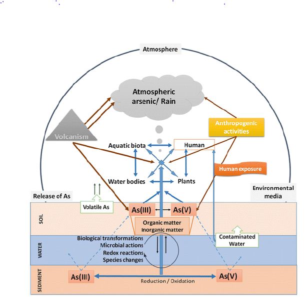
Figure 1: Arsenic biological cycle in nature.

Arsenic Accumulation in Water Biota

Plankton

Because of phytoplankton’s role in defining the fundamental properties of seawater, very little has been available about the total arsenic in plankton. They report the total arsenic concentration of three zooplankton species from seawater in the Northeast Atlantic Ocean to be 14 to 42 grams g dry weight [37]. I have reported similar or smaller attentions from other waters. Recently, arsenic is directly toxic to zooplankton. However, it is present in top levels in solution. Its effect on phytoplankton species structure (in favor of tolerant species) may be significant aspects of coastal phytoplankton and zooplankton—water. Arsenic data in echinoderms are minimal [38] found a high aggregation of total arsenic (varying depending on the sample’s location) in the lipids of many organisms. A study [37] reported that the asteroids Metaseries Glacier and Asterias Rubens of Portland, Dorset, had a dry mass of 5.8 and 10 grams, respectively.

The total arsenic level in most polycystic is not significant. For example, Neris has been reported to have a g-1 dry weight of 4 to 87 grams of the diverse color element, depending on Madis’s region.

The other thing, discrete data on arsenic are present in other aspects than those discussed above. For example, it is reported by [37], From the North Atlantic Ocean, 72 µg g-1 arsenic weight of telefelony from Southampton waters, but low concentrations (2.8–6.6 g -1 dry weight) in one sponge and two tunicate materials.

The Mechanism of Arsenic Poisoning in Modern Pregnancy Targets

Methods by which exposure to arsenic adversely affects pregnancy are not completely understood; however, some potential methods based on investigational evidence have been proposed. Arsenic occurs as part of organic compounds or volatile compounds in elemental form. The predominance of volatile species in water is [39]. By rapid urination or a continuous methylation procedure that occurs mostly in the liver, volatile arsenic is extracted from the body [40]. An association between arsenic venomousness in swallowing water and contrary effects on pregnancy has been found in numerous epidemiological studies.

Effect of Arsenic on the Skin

Most skin changes occur with chronic exposure. (Lyon et al., 1994), Dermatologic changes are communal, and the primary clinical analysis is often founded on hyperpigmentation, palmar, and solar keratosis. Keratosis appears as a single lump or separate nodule. Arsenic is a non-melanin pigment that can cause basal cell carcinoma in the skin [41].

Although human skin is more susceptible to arsenic poisoning and carcinogenicity, wildfire rat skin models with carcinogenicity do not respond well to arsenic. Mouse skin is sensitive to most chemical cancers; rat skin is not very sensitive to chemical carcinogens.

Arsenic Deficiency in the Nervous System

The main target of many metals’ toxic effects is the nervous system, especially heavy metals such as mercury, lead, and arsenic. Edgy products are many and varied. Peripheral neuropathy is the most common finding that mimics Guillain-Barre disorder with similar electromyographic results [42] Glove at the beginning of neuropathy and storage is sensitive to anesthesia.

Improved oxidative stress is an important component in arsenic-induced neurotoxicity. It is found that the main to cause oxidative damage to ecosystems is due to arsenic exposure by increasing free radical species [1], leading to enlarged lipid peroxidation and protein carbonyl. In addition, GSH levels decreased [1,43]. Glutathione is an important molecule involved in the fight against toxins. Decreases in GSH levels after arsenic exposure are related to higher arsenic interactions with GSH [44].

Because neurotransmitters play a significant role in modulating the behavioral and signaling cascade, several experimental studies have been conducted to investigate the arsenic effect on their levels and metabolic activity [45]. Changes in dopaminergic, cholinergic, serotonergic, and glutamatergic systems have been reported in arsenic-infected rats and mice. Differences in exposure pathways did not result in consistent changes in brain neurotransmitter levels, however, prolonged exposure to arsenic reduced dopamine levels in the striatum and brain [46].

Effect of Arsenic on the Liver

Arsenic treatment involves histological changes in hepatic tissues, including cytoplasmic vacuuming, cell degeneration, and focal necrosis. Accumulation of neutrophils and lymphocytes leads to deterioration of liver tissue and necrosis of the central vein. Similar results in liver pathology in mice confirm our products. In the current study, reduced Hb in the blood and increased bilirubin levels may be due to changes in hematopoietic function caused by arsenic intoxication, which corresponds to [47]. Arsenic compound [48] is a protoplasmic toxin. Arsenic property considered the mechanism for its toxic effect.

The onset of oxidative stress after contact with hepatotoxic agents is an essential factor in primary chronic liver disease associated with fibrosis. Membrane molecules that harm membrane lipids, building blocks of proteins (amino acids), sugars (carbohydrates), and nucleic acids are the major components of chemically persuaded hepatocellular injury. The liver [49] is the site of metabolism of many chemicals, counting mineral arsenic in many animals, including mammals. It acts as an essential antioxidant to reduce GSH in hepatocytes.

Biochemical and structural sign of arenite-induced changes in rat liver also with changes in development of rat has been found. Evidence includes GSH levels, hepatocyte nuclei, an intermittent vacation of hepatocytes, and sinusoidal dilatation in arsenic-fed animals. To recognize the exact mechanisms fundamental arsenic toxicity at the cellular and molecular levels, extensive studies using different doses of sodium arenite, and different lengths of exposure to diverse growing periods should be measured.

Effect of Arsenic on the Kidneys

Kidneys are prone to arsenic-induced damage due to extensive perfusion and increased concentration of excretory compounds in renal tubular cells [50]. Our results confirm increased serum urea, uric acid, and creatinine levels in the excretion of high levels of arsenic metabolism by renal tubular cells, which are more prone to damage and kidney problems.

Arsenic trioxide affects bone marrow, kidneys, and hemoglobin metabolism. [51] Any substance that significantly affects red blood cell values and associated parameters has been shown to affect the bone marrow, kidney, and hemoglobin metabolism.

In the present study, arsenic intoxication induced significant hepatic and renal oxidative stress, resulting in decreased cellular antioxidant enzyme activity, consistent with previous reports [52-56].

Conclusion

Arsenic compounds are toxic substances that can significantly affect the health of animals. Arsenic poisoning has been confirmed to be related to its chemical forms. Although most are known to affect mammals’ signaling pathways, in this evaluation, we associated phosphorylation spots in diverse mammal and nonmammalian species.

As a result, we can assume that the same effects found in mammalian specimens of aquatic life are likely to occur. Also, arsenic has been shown to cause oxidative tension in mammals and nearly marine organisms. Current work shows that animals intoxicated with arsenic trioxide show significant impairment of liver function and kidney function. There are reports that the effects of AS exposure on the development of sensory systems are minimal. It is also likely to cause significant changes in specific biochemical parameters, including the liver and kidneys. There is reliable and consistent evidence to support an optimistic relationship among high levels of inorganic arsenic exposure to drinking water, miscarriage, stillbirth, and low birth weight.

Acknowledgement

I want to give all my credit to my parents and all author’s whose article I have been considered for my publication.

References

  1. Flora SJS, Bhadauria TS, Panta SC, Dhakade RK (2005) Arsenic-induced blood, brain oxidative stress, and reaction to specific thiol chelators in rats. Life Sciences 77: 2324-2337. [crossref]
  2. Caceres DD, Pino P, Montesinos N, Atalah E, Amigo H, et al. (2005) Exposure to inorganic arsenic in drinking water and total urinary arsenic concentration in a Chilean population. Environmental Research 98: 151-159. [crossref]
  3. Tripoli E, La Guardia M, Giammanco S, Di Majo D, Giammanco M (2007) Citrus flavonoids: Molecular structure, biological activity and nutritional properties: A review. Food Chemistry 104: 466-479.
  4. Van Geen A, Radloff K, Aziz Z, Cheng Z, Huq MR et al. (2008) Comparison of arsenic concentrations in simultaneously-collected groundwater and aquifer particles from Bangladesh, India, Vietnam, and Nepal. Applied Geochemistry 23: 3244-3251.
  5. Jana K, Jana S, Samanta PK (2006) Effects of chronic exposure to sodium arsenite on hypothalamo-pituitary-testicular activities in adult rats: possible an estrogenic mode of action. Reproductive Biology and Endocrinology 4: 9. [crossref]
  6. Mukherjee S, Mukhopadhyay P (2009) Studies on arsenic toxicity in male rat gonads and its protection by high dietary protein supplementation. Al Ameen J Med Sci 2: 73-77.
  7. Manna P, Sinha M, Sil PC (2008) Arsenic-induced oxidative myocardial injury: protective role of arjunolic acid. Archives of toxicology 82: 137-149. [crossref]
  8. Guvvala PR, Sellappan S, Parameswaraiah RJ (2016) Impact of arsenic (V) on testicular oxidative stress and sperm functional attributes in Swiss albino mice. Environmental Science and Pollution Research 23: 18200-18210. [crossref]
  9. Shi H, Shi X, Liu KJ (2004) Oxidative mechanism of arsenic toxicity and carcinogenesis. Molecular and Cellular Biochemistry 255: 67-78. [crossref]
  10. Wang TC, John KY, Wang AS, Gurzar (2007) Trivalent arsenicles cause lipid peroxidation, protein carbonization, and oxidative DNA damage in human urothelial cells. Mutat Res 615: 75-86. [crossref]
  11. Balakumar B, Ramanathan K, Kumaresan S, Suresh R (2010) DNA damage by sodium arsenite in experimental rats: ameliorative effects of antioxidant vitamins C and E. Ind J Sci Tech 3: 322-327.
  12. Shen ZX, Chen Zik, Ni JH, X S Li, S M Xiong et al. (1997) Use of Arsenic Trioxide (AS2O3) in the Treatment of Acute Promyelocytic Leukemia (APL): II. Clinical efficacy and pharmacokinetics in rehabilitated patients. Blood 89: 3354-3360. [crossref]
  13. Lorenzo HK, Susin SA, Penninger J, G Kroemer et al. (1999) Apoptosis-Inducing Factor (AIF): The phylogenetically old, caspase-free effect of cell death. Cell Death Difference 6: 516-24. [crossref]
  14. Co RJ (1999) Causes of herbal poisoning, epidemiology, and clinical evaluation. Clin Toxicol 37: 697-708. [crossref]
  15. Ding Z, Zheng B, Long J, Belkin HE, Finkelman RB, et al. Guizhou, in the southwestern province of China. Applied Geochemistry 16: 1353-1360
  16. ATSDR (1990) NVSDR Case Studies in Environmental Medicine. Agency for Toxic Substances and Disease Registry, Atlanta GA, USA.
  17. Bates MN, Smith AH, Hoppenhein-Rich C (1992) Arsenic addiction and internal cancer: A review. Am J Epidemiol 135: 462-476. [crossref]
  18. Gochfeld M, (1995) Chemical agents. Of: Brooks S, Gochfeld M, Herstein J, et al. Environmental Medicine 592-614.
  19. Keldesburg cR, Ward HP (1972) Leukemia in arsenic poisoning. On Intern Medal 77: 935-937. [crossref]
  20. Simonato L, Malin Lynn JJ, Javalad B, Ferro G, Wilde P, et al. Refinery in France. Am J 25: 625-633
  21. De Wolf FA, Edelbrock PM (1994) Neurotoxicity of arsenic and its compounds. Vincennes & Bruin, Vol. Handbook of Clinical Neurology. Elsevier Science 1994: 283-291.
  22. Wilkinson SP, McHugh P, Horsley S, Tubbs H, Louis M, et al. (1975) Br Med J 3: 559-63.
  23. Pullan-James S, Woods SE (2006) Professional arsenic gas exposure. J Not Med Ashok 98: 1998-2001
  24. Edmunds JS, Francisco KR (1987) Arsenic is converted into the marine environment. Experiment 43: 553-557
  25. Bouchet JP, Liegen D, Ruggery M, et al. (1996) Estimating inorganic arsenic exposure to human cancer due to fish consumption. Arch Toxicol 70: 773-778.
  26. Thomas KW, Pellissery ED, Berry M (1999) Demographic Diet and Tap Water Density for Selected Factors for EPA Region v. National Human Exposure Assessment Survey (NHAS). J Expo Anal Environment Epidemiol 9: 402-413.
  27. Tomaki S, Frankenberger WT Jr (1992) Environmental Biochemistry of Arsenic. Rev Environment Condom Toxicol 124: 79-110. [crossref]
  28. Actor KF, Owens G, Davy DE, Naidu R (2005) Arsenic Specification and Toxicity in Biological Systems. Rev Environment Condom Toxicol 184: 97-149. [crossref]
  29. Thompson DJ (1993) A chemical theory for the arsenic methylation in mammals. Chem Biol Interact 88: 89-114. [crossref]
  30. Hoppenhein C, Ferrexio C, Browning SR, Huang B, Peralta C et al. (2003) Birth weight from drinking water, birth weight. Epidemiology 14: 593-602.
  31. Ghosh P, Banerjee M, De Chaudhuri S, Chowdhury R, Das JK, et al. (2007) Comparison of health effects between individuals with and without skin lesions in the population exposed to arsenic through drinking water in West Bengal, India. Journal of exposure science & environmental epidemiology 17: 215-223. [crossref]
  32. Kedelman p (1999) Environmental and Office Appreciation of the Semiconductor: Implications) The Future of the Labor Force and Society. The Environment T Wealth Perspective 6: 291 295.
  33. Knowles FC, Benson AA (1983) Arsenic Biochemistry. Trends Biochem Science. 8: 178 18
  34. Summer AO, Silver s (1978) Microbial transformations of metals. That day. Rev Microbiol 32: 637-672.
  35. Farmer JG, Johnson L (1990) Inorganic Arsenic Exposure Assessment Based on Urine Concentration and Arsenic Specification. Br J Med 47: 342-348.
  36. Hughes m, Pool RK (1989) Metals and microorganisms. Chapman & Hall, London.
  37. Leatherland TM, Burton JD, Kalkin F, McCartney MJ, Morris RJ (1973) Concentrations of certain metals and mercury in pelagic organisms in the waters of the Northeast Atlantic Ocean. Deep Sea Res 20: 679-685.
  38. Vaskovsky VE, Korochenko OD, Kosheleva LP, Lenin VS (1977) Arsenic in marine invertebrate lipid extract. Comp Biochem Physiol 41: 777-784.
  39. International Agency for Research on Cancer (2012) Arsenic, metals, fibers and dust particles. Review of human cancer factors; International Agency for Research on Cancer, World Health Organization: Geneva, Switzerland.
  40. Albiac J, Calvo E, Tapia J, Esteban E (2010) Water quality and nonpoint pollution: Comparative global analysis. Re-thinking Water and Food Security
  41. Abernathy CO, Liu YP, Long Fellow D, et al. (1999) Arsenic: health effects, mechanism of action, and research issues. Environment Health Perspective 107: 593-597. [crossref]
  42. Goddard MJ, Tanheko JL, Dow PC (1992) Massage of chronic arsenic poisoning as a laundry-Guillain-barre syndrome. Electromyography Clin Neurophysiole 32: 419-423. [crossref]
  43. Shila S, Kokilawani V, Subhadra M, Panneerselvam CB (2005) Local brain response in the antioxidant system to ని-lipoic acid in arsenic-mice. Toxicology 210: 25-36. [crossref]
  44. Apocalypse HV, Apocalypse MM (1989) New developments in arsenic poisoning. J Colonel Toxicol 8:1297-1305.
  45. Rodrıguez VM, Jimenez-Capdeville ME, Giordano M (2003) The effects of arsenic exposure on the nervous system. Toxicology Letters 145: 1-18. [crossref]
  46. Krishnan GM, Tripathi N, Dubey SN, Gupta M, Flora SJS. J Toxicol Clin Toxicol 39: 675.
  47. Patrick L (2003) Toxic metals and antioxidants: part II. The role of antioxidants in arsenic and cadmium toxicity. Alternative Medicine Review 8: 106-128. [crossref]
  48. Chattopadhyaya S, Bhumik S, Nagdhadhari A, Dasgupta S (2002) Arsenic changes in vivo, tissue culture, neonatal and adult brain cell growth and apoptosis. Toxicol Let 128: 73-84
  49. Wahr M (2009) Effects of arsenic on maternal and fetal health. Annu Rev Nutter 29: 381-399. [crossref]
  50. Verma RJ, Vasu A, Zayed AA (2004) Arsenic poisoning in rats and its possible improvement. J Environment Science 16: 447-453.
  51. Young NS, Masijevsky J (1997) The Path Physiology of Acquired Aplastic Anemia. New Engine J Med 336: 1365. [crossref]
  52. Wang TC, Jan KY, Wang AS, Gurr JR (2007) Trivalent arsenicals induce lipid peroxidation, protein carbonylation, and oxidative DNA damage in human urothelial cells. Mutation Research 615: 75-86. [crossref]
  53. Apocalyptic HV (1997) Enzymatic methylation of arsenic species and other mechanisms for arsenic poisoning. On Rev Pharmacol Toxicol 37: 397-419. [crossref]
  54. Majumder C (2018) Arsenic (V) removal using activated alumina: Kinetics and modeling by response surface. Journal of Environmental Engineering 144: 04017115.
  55. Nordman R (1994) Alcohol and antioxidant systems. Alcohol 29, 513-522. [crossref]
  56. Arsenic Biological cycle available at (https://images.app.goo.gl/JMbwE1ouWoQRcPuQA)

Influences of COVID-19 on Environment

DOI: 10.31038/IJVB.2021513

Abstract

This study shows positive and negative effects of coronavirus on the environment and also tests how individual mobility has affected by the spread of local diseases and homoeopathic regulations in the epidemic of coronavirus. Meanwhile, solitary confinement can affect intellectual strength and purpose of this study is to quantify the occurrence of sadness, nervousness, and sleep illnesses in the world during incarceration. I associate educated air pollution levels with unusually high atmospheric precipitation levels in different parts of the world. Electricity demand has significantly abridged because of the current COVID-19 epidemic. Administrations worldwide are being forced to cut business operations in comeback to reducing the hazard of coronavirus. This continuing condition because of coronavirus epidemic has altered the lifestyle around the world as people live at home and work at home when possible. Therefore, there is a noteworthy rise in the claim for real estate and also with this; there is a considerable reduction in trade and manufacturing things. That dire circumstance poses novel tasks to the power sector’s practical and economic services, which is why many resources around the world have embarked on a disaster risk management program to address these ongoing challenges/threats. Satellite data has shown declining air pollution levels in many areas of the earth. This review purposes to examine the global COVID-19 conditions worldwide.

Keywords

COVID-19, Electricity demand, Depression, Industrial goal, Pollution

Introduction

Negotiation research spans many disciplines [1] and 2019 Coronavirus (COVID-19) become the leading cause of shortest and unintended mental and public outcomes (M.H.) Not only during each epidemic but also in the upcoming era. This outcome of segregation has assessed throughout earlier epidemics, e.g., a respiratory syndrome (SARS) outburst in the year of 2003 and Ebola in the year of 2014, showing that the influence of M.H. will be extensive, significant, and lifelong [2]. In his report [2] explained that Between the effects of isolation there are severe depressive illnesses, fatigue, tetchiness, stress, low awareness and uncertainty, impaired work routine, stress disorder, severe depression, symptoms of depressive, and sleeplessness. The existing data that could predict M.H. conclusions are contradictory [2], e.g., Stage of development, schooling, gender, and childbearing has considered both [3] and outside [4] to meet psychological problems. Besides, M.H.’s principal stresses during separation sometimes resulted in a period of separation, fear of contagion, hindrance, insufficient purchases, and insufficient evidence [2]. In China, the physical reaction during the breakdown of COVID-19 has widely studied. Findings from China reported an increase in stress during the separation by up to 37% [3] and increased anxiety by up to 35% [5].

As mentioned, the main purpose of the current study to quantify the mental impact of COVID-19 and the corresponding prevention strategies in a nationwide study that has examined an increase in depressive, anxiety, and sleep disorders around the world.

A explained above, this study aims to show the positive and negative effects of COVID-19 on the environment and evaluate how different matters have been affected by the spread of local diseases and homoeopathic regulations during the COVID-19 epidemic.

The Consequence of Psychological Health

The coronavirus 2019 epidemic (COVID-19) and the international outreach programs they have developed have disrupted daily activities.

Negotiation research spans many disciplines [6]. The disease COVID-19 has led to unprecedented prevention in many cities and regions in China [7]. Further studied [8] that School habits are important ways to deal with the mental and physical health problem of young people. During the epidemic, when education is almost restricted to online system and institutions are closed, students lose balance in their lives and show the symptoms of restlessness. The infection of COVID-19 has characterized by respiratory depression, which has prompted Herculean efforts to increase respiratory equipment supply in the United States It has also reported that health care workers, whether directly or partially in contact with the health care system that provides care for people with SARS, experience significant levels of depression supported over one year after the outbreak, showing that the reaction is not just a correction problem [9]. This is unparalleled by people with limited resources and poor health. Communal loneliness measures can lead to separation of an individual in an offensive home, through trauma probable to increase during this period of commercial uncertainty and pressure [6].

Atmosphere Quality

The COVID-19 epidemic has certainly changed the issue, as, for instance, public devote extra time at home and fewer time on the road [10]. This effect has been widely studied [11,12] Low mobility reduces nitrogen oxides, a significant fire product. A total drop in atmospheric NO2 in the spring of 2020 indicates this emission’s result. NO2 is focused in town areas and is easily visible. In the space, satellite descriptions have provided a pure sign of declining population density in current months, encouraging observation on refining air superiority). Negotiation research spans many disciplines [13] Decreases have been severe in areas controlled by diesel automobiles, with more NOx emissions than in other petrol. At the same time, the relief of NO2 satellite imagery has led many to focus on changes in NOx discharges, which means that many factors complicate this reduction of COVID-19. It is reported [14] At the similar time, an analysis of variations in the atmosphere over the past few months, and in the coming months with the decrease (and redistribution) of COVID-19 grounded limitations, will deliver an original, complete understanding of chemicals, including secondary emissions and secondary pollution.

Commercial Outcomes

As a result of the COVID-19 epidemic, the worldwide economy has been collapsed almost overnight [15]. The worldwide economy is severely pretentious by these actions and rises redundancy and shortage. This poses threats to achieving the U.N.’s goal of Sustainable Development Goals (S.D.G. s). The COVID-19 epidemic has taken its toll on the world. Whenever there is a significant shift in economic activity, it will have an impact on the environment. The epidemic faces the tourism industry with an unprecedented challenge. COVID-19 curve strategies such as public closure, public evacuation, home instructions, travel, and travel boundaries have controlled the momentary closing of many hospitality trades and meaningfully abridged businesses’ necessity to continue operating that is exclaimed by [16]. All cafeterias were asked to shut down their shops during the severe condition. The restrictions imposed on travel and accommodation orders issued by the authorities have resulted in a significant reduction in hotel accommodation and revenue. To explore how government assistance can be prioritized in the tourism sector, we align [17] a security market-based approach to mimic the impact of COVID-19 on the hotel, aviation, boat, and rental industries. We see that shock is not a single event, but instead better followed as there is a changing nature.

Negotiation research spans many disciplines [18,19] As per there is no cure up till now, keeping a distance is the best way to overcome the spreading of disease, and many countries set for a country with a strong closure, social restrictions, travel restrictions, unemployment, and home plan have enforced most people to stay indoors, which has affected standard professional operations and condensed demand for power on the countrywide network. Industries have moved to less manual labour or limit their efficiency. The business declined their performance; travel restrictions almost collapsed in the flight business, small companies almost stagnant, schools, universities moving to an online method, and many other parts are embracing domestic policy.

This effect has been widely studied by [20-24]. The catastrophic event has affected the country’s social and economic spheres. Strict closures have halted industrial operations due to staff shortages and limited business due to travel restrictions. Although all community transportation modes are not electric, in many countries and essential parts of all transportation are electricity such as; Streetcar, train and common transportations so, limited traffic has affected the demand for electricity in the transport sector. Most governments and organizations around the world are putting forth effort and money to fight coronavirus disease. Therefore, there may be delays or reductions in funding for many research projects, such as reusable energy programs. On the other hand, this effect has been widely studied [25-27] the epidemic has created several diverse research gaps such as medical emergency management, mental health care [28,29]. Negotiation research spans many disciplines [30-32] economic recovery, and energy subdivision supervision to accomplish the electricity system in such critical situations, etc. Strict closures have halted industrial operations due to staff shortages and limited business due to travel restrictions. All of this has indirectly contributed to reducing pollution in the industrial sector, which positively affects the environment [33-41].

Conclusion

Coronavirus has affected humans and the environment directly or indirectly. It has affected the economy rate of every country, besides this also human health is affected badly. Due to coronavirus, only Air pollution has decreased.

Acknowledgement

I acknowledge my parents and also every author whose research I have considered in my review article.

References

  1. Holmes EA, et al. (2020) Multidisciplinary research priorities for the COVID-19 pandemic: a call for mental health science action. The Lancet Psychiatry.
  2. Brooks SK, Webster RK, Smith LE, Woodland L, Wessely S, et al. (2020) The psychological impact of quarantine and how to reduce it: Rapid review of the evidence. Lancet 395: 912-920.
  3. Taylor MR, Agho KE, Stevens GJ, Raphael B (2008) Factors influencing psychological distress during a disease epidemic: Data from Australia’s first outbreak of equine influenza. M.C. Public Health 8: 347. [crossref]
  4. Hawryluck L, Gold WL, Robinson S, Pogorski S, Galea S, et al. (2004) SARS control and psychological effects of quarantine, Toronto, Canada. Emerg Infect Dis 10: 1206-1212.
  5. Huang Y, Zhao N (2020) Generalized anxiety disorder, depressive symptoms, and sleep quality during COVID-19 outbreak in China: A web-based cross-sectional survey. Psychiatry Res 288: 112954. [crossref]
  6. Lee J (2020). Mental health effects of school closures during COVID-19. The Lancet Child & Adolescent Health 4: 421. [crossref]
  7. Gates B (2020) Responding to Covid-19: a once-in-a-century pandemic? N Engl J Med 382: 1677-1679. [crossref]
  8. Boyer J (2020) COVID-19, medication-assisted treatment, and increased risk for further respiratory depression. American Journal of Psychiatry 177: 636-636. [crossref]
  9. Lee AM, Wong JGWS, McAlonan GM, et al. (2007) Stress and psychological distress among SARS survivors one year after the outbreak. Can J Psychiatry 52: 233-240. [crossref]
  10. Quéré L, et al. (2020) Nat Clim Chang 8: 647-653.
  11. Bauwens M, et al. (2020) Geophys Res Lett 47: 2020GL087978.
  12. Liu, F. et al. (2020) Science Advances 6: 2992.
  13. Weiss M, Bonnel P, Hummel R, Provenza A Manfredi U (2011) Environ Sci Technol  45: 8575-8581.
  14. Le T, et al. (2020) Science 369: 702-706.
  15. UNWTO (2020) UNWTO world tourism barometer. 18.
  16. Bartik AW, Bertrand M, Cullen ZB, Glaeser EL, Luca M (2020) How are small businesses adjusting to COVID-19? Early evidence from a survey (No. w26989). National Bureau of Economic Research.
  17. Karafiath I (1988) Using dummy variables in the event methodology. The Financial Review 23: 351-357.
  18. Chintalapudi N, Battineni G, Amenta F (2020) COVID-19 virus outbreak forecasting of registered and recovered cases after a sixty-day lockdown in Italy: A data-driven model approach. J Microbiol Immunol Infect 53: 396-403. [crossref]
  19. Barkur G, Vibha Kamath GB (2020) “Sentiment analysis of nationwide lockdown due to COVID 19 outbreak: Evidence from India” (in eng). Asian J Psychiatry 51: 102089. [crossref]
  20. Chatterjee K, Chatterjee K, Kumar A, Shankar S (2020) Healthcare impact of COVID-19 epidemic in India: A stochastic mathematical model. Med J Armed Forces India.
  21. Zambrano-Monserrate MA, Ruano MA, Sanchez-Alcalde L (2020) Indirect effects of COVID-19 on the environment. Sci Total Environ 728: 138813. [crossref]
  22. Cao W, et al. (2020) The psychological impact of the COVID-19 epidemic on college students in China. Psychiatry Res 287: 112934. [crossref]
  23. Francis NN, Pegg S (2020) Socially distanced school-based nutrition program under COVID 19 in the rural Niger Delta. The Extractive Industries and Society.
  24. Haleem A, Javaid M, Vaishya R (2020) Effects of COVID 19 pandemic in daily life, (in eng). Curr Med Res Pract.
  25. Jin H, Lu L, Liu J, Cui M (2020) Complex emergencies of COVID-19: management and experience in Zhuhai, China. Int J Antimicrobial Agents 55: 105961. [crossref]
  26. Lau H, et al. (2020) Internationally lost COVID-19 cases. J Microbiol Immunol Infect 53: 454-458. [crossref]
  27. (2012) The American Journal of Emergency Medicine. Wilderness & Environmental Medicine 23: 89.
  28. Roy D, Tripathy S, Kar SK, Sharma N, Verma SK, et al. (2020) Study of knowledge, attitude, anxiety & perceived mental healthcare need in the Indian population during COVID-19 pandemic. Asian J Psychiatry.
  29. Rajkumar RP (2020) COVID-19 and mental health: A review of the existing literature. Asian J Psychiatry 52: 102066. [crossref]
  30. Zhang D, Hu M, Ji Q (2020) Financial markets under the global pandemic of COVID-19. Finance Res Lett 36: 101528. [crossref]
  31. Gallego V, Nishiura H, Sah R, Rodriguez-Morales AJ (2020) The COVID-19 outbreak and implications for the Tokyo 2020 Summer Olympic Games (in eng). Travel Med Infect Dis 34: 101604. [crossref]
  32. Holmes EA, O’Connor RC, Perry VH, Tracey I, Wessely S, et al. (2020) Multidisciplinary research priorities for the COVID-19 pandemic: A call for action for mental health science. Lancet Psychiatry 7: 547-560. [crossref]
  33. Australian energy market operator, quarterly energy dynamics Q1 2020, market insights and WA market operations. AEMO.
  34. Ahmed MZ, Ahmed O, Aibao Z, Hanbin S, Siyu L, et al. (2020) Epidemic of COVID-19 in China and associated Psychological Problems. Asian J Psychiatry 51: 102092. [crossref]
  35. Bates M (2020) Public transport authorities and COVID-19, impact and response to a pandemic. International Association of Public Transport Australia/New Zealand.
  36. “World Markets.”
  37. Kabir M, Afzal MS, Khan A, Ahmed H (2020) COVID-19 pandemic and economical cost; impact forcibly displaced people, (in eng). Travel Med Infect Dis.
  38. Krotkov NA, et al. Atmos Chem Phys 16, 4605-4629 (2016
  39. Martin RV, et al. J Geophys Res 108, 4537 (2003).
  40. Schneider P, van der ARJJ (2012) Geophys Res 117: 16309.
  41. UNESCO. COVID-19 educational disruption and response.
fig 1

Aortitis Associated to with Rheumatoid Arthritis: A Challenginge Rheumatoid Vasculitis Presentation: A Case Report

DOI: 10.31038/IJOT.2021412

Abstract

Rheumatoid Vasculitis (RV) is a rare but serious extra-articular manifestation of Rheumatoid Arthritis (RA). Its varied clinical presentation makes it hard to diagnose and treat. Hereby we describe a case of an aortitis revealing RV which is a rare presentation of a rare complication of RV. A 56-year-old man with rheumatoid arthritis treated with methotrexate presented with fever, chest pain and arthritis. Blood tests revealed inflammatory syndrome associated with cholestasis. The diagnosis of pericarditis associated with aortitis was retained. Cholestasis was mostly due to methotrexate. The patient was treated with cyclophosphamide pulses and high doses of prednisolone. The patient was in complete remission of articular and extra-articular manifestations after two months of treatment.

Keywords

Rheumatoid arthritis, Rheumatoid vasculitis, Aortitis, Immunosuppressive therapy, Case report

Introduction

Rheumatoid Arthritis (RA) is a connective tissue disease predominantly affecting the joints. Extra-articular manifestations develop in up to 40% of cases [1], of which Rheumatoid Vasculitis (RV) is the most serious. The widespread vascular involvement, which effects not only the synovia but also other organs such as the skin, eye and nerves, can be life threatening. Mortality can reach up to 40% within five years of disease onset [2]. Fortunately, RV is a rare complication that occurs in 1-5% of RA patients [3]. It commonly affects small and medium blood vessels [4]. Large vessel vasculitis is unusual during RA. Hereby we describe a case of an aortitis revealing RV which is a rare presentation of a rare complication of RV.

Case Description

Patient Information and Clinical Findings

A 54-year-old man, a North African policeman living in an urban environment with a personal history of smoking and pulmonary tuberculosis in 1991 was diagnosed in 2009 with seropositive and erosive RA associated with Sjögren’s syndrome. He was being treated with methotrexate (25 mg/week). On November 2019, the patient presented to our hospital with fever, fatigue and chest pain that had started one week prior. Physical examination found a high temperature of 38.5°C. Systolic blood pressure was 100 mmHg and diastolic blood pressure was 60 mmHg in both arms. He had tachycardia, with a heart rate of 115 beats per minute. There were no signs of heart failure and respiratory rate was normal. Mobilization of the wrists, elbows and shoulders was painful, with a swollen right wrist. He had a dislocation of the right ulnar styloid. He had rheumatoid nodules on the outer side of both elbows. There were no skin ulcerations no erythema nodosum. The rest of the physical examination was normal.

Diagnostic Assessment

An electrocardiogram showed atrial fibrillation with a heart rate of 115 beats per minutes associated with diffuse ST-segment elevation with upward concavity. Blood tests showed hyper leukocytosis at 11000/mm3, C-reactive Protein (CRP) levels of 117 mg/l, an Erythrocyte Sedimentation Rate (ESR) of 118 mm and cholestasis with increased gamma glutamyl transferase and phosphatase alkaline levels of 199 UI/L (four times the normal rate) and 348 UI/L (five times the normal rate), respectively. Transaminase levels were normal (ALAT level of 35 UI/L and ASAT level of 25 UI/L). A procalcitonin test was negative (<0.5 mg/L). Blood gas analysis was normal (pH level of 7.40, PaO2 level of 90 mmHg, PaCO2 level of 42 mmHg, HCO3- level of 25 mmol/l).

Blood cultures were negative even for fungal agents. A chest X-ray showed a flask-shaped enlarged cardiac silhouette. Transthoracic echocardiography confirmed a non-compressive large posterior pericardial effusion. Abdominal ultrasound was normal. A thoraco-abdominal Computed Tomography (CT) scan showed pericardial effusion with enhancement of the pericardium, compatible with pericarditis, and regular parietal hypodense circumferential concentric thickening of the aortic arch and supra aortic arterial trunk root, confirming aortitis (Figures 1 and 2). CT showed no bubbles in nor around the aorta wall. There was no mural thrombus nor aortic aneurysm nor signs of atherosclerosis. There was emphysema in pulmonary parenchyma but no evidence of active tuberculosis on chest-CT. Positron emission tomography was not perform due to its availability in our country.

fig 1

Figure 1: Thickening of the aorta (red arrow) associated with cardiac effusion (yellow arrow) in a sagittal chest CT.

fig 2

Figure 2: Thickening of the aorta (red arrow) associated with cardiac effusion (yellow arrow) in a frontal chest CT.

A biopsy of the cardiac effusion was not possible due to its posterior location. The sputum test for Koch’s bacillus was negative. Viral hepatitis B and C and syphilis serology were negative. Antinuclear antibodies were positive at 1/1200. Anti-LKM1, anti-CCP and rheumatoid factor levels were respectively of 1/80, 40 IU/ml and 50 IU/ml. Immunoglobulin levels were normal (Immunoglobulin G level of 14.3 g/L, immunoglobulin M level of 2.08 g/L and immunoglobulin A level of 1.8 g/L). Liver biopsy showed peliosis with no sign of auto-immune hepatitis nor auto-immune biliary cholangitis. The diagnosis of RV with large vessel and cardiac involvement was retained. Disease activity was evaluated as moderate by the DAS-28 CRP score.

Therapeutic Intervention

The patient was treated with a high dose of prednisone (60 mg/day) and IV pulses of 1000 mg cyclophosphamide every month for six months. Treatment with methotrexate was stopped. Because of a personal history of pulmonary tuberculosis and the prescription of corticosteroids and cyclophosphamide in an epidemic country of tuberculosis, we administered prophylactic antitubercular therapy. The patient was also by treated with 200 mg/day of amiodarone for the atrial fibrillation.

Follow-Up and Outcomes

The patient was apyretic after day 2 and their heartbeat became normal. Chest pain and articular manifestations decreased and disappeared after one month of treatment. CRP levels decreased to 12 mg/L after steroids and cyclophosphamide pulses. ESR became normal after two months of treatment. Cholestasis disappeared after one and a half months of methotrexate withdrawal.

Investigations of the cholestasis highlighted the iatrogenic involvement of methotrexate, confirmed by a report from the National Drug Safety Department in Charles Nicolle’s Hospital of Tunis.

Cardiac echography showed the disappearance of the cardiac effusion at two months. A chest CT scan showed a significant regression of the vasculitis.

Discussion

The epidemiology of RV is hard to define. Heterogeneous clinical presentation, paucity of specific data to confirm the diagnosis and lack of a unanimous definition of RV are some of the reasons why it is hard to define. Many authors claim that RV can be observed in up to 5% of RA patients [5]. Our patient was 54 years old. The mean age of patients at diagnosis of aortitis in a literature review was 56 ± 15.2 years old [6].

RV commonly affects small and medium sized vessels. The skin is the most commonly damaged organ (90% of patients). The association of aortitis with RV is not widely recognized. However, many authors reported an association of RA with aortitis [6]. Our patient had a lesion typical of vasculitis of the aorta on his chest CT scan. Differential diagnoses were ruled out such as syphilis, tuberculosis or other infectious causes (negative sputum test for Koch’s bacillus, negative serologies of viral hepatitis B and C and syphilis, negative blood cultures for bacteria or fongi).

Systemic erythematous lupus was unlikely. The patient didn’t have any suggestive skin lesions nor pleurisy. The direct coombs test was negative. He didn’t have any cytopenia and proteinuria was negative. Anti-DNAn, anti-Sm, anti-nucleosome, anti-histones were negative. Takayasu’s arteritis was also unlikely. The patient had no upper or lower limbs claudication. He had no dizziness nor ocular symptoms. Blood pressure was normal and symmetrical. Peripheral pulses were present and symmetrical. The lesions of the aorta on CT scan was not suggestive of Takayasu’s Arteritis.

Giant cell arteritis were excluded. The patient had no history of stroke, no headache nor ocular symptoms suggestive of Giant cell arteritis. Temporal artery was normal with a normal and symmetrical pulse. Aortitis was the only manifestation of RV in our patient. Other similar cases have been reported. In half of the cases, aortitis was isolated, with no other features of vasculitis [6]. In our patient, RV was revealed after 11 years of RA onset while the patient was treated with methotrexate and a low dose of corticosteroids. In a review of the literature, RV appeared after a mean disease duration of six years. Rheumatoid nodules were observed in up to half of patients with RV [6].

Our patient had poor articular manifestations with disease activity evaluated as moderate by the DAS-28 score. The concept of ‘burnt out’ disease is described by many authors, consisting of the contrast between benign articular presentation and severe life-threatening RV [4]. This leads us to insist on actively screening this rare but fatal complication. RV typically occurs in long-standing seropositive and erosive RA, especially in males, smokers and patients with rheumatoid nodules or rheumatoid pericarditis [1,5]. Our patient had all these conditions.

There are no randomized controlled studies to guide the management of RV. However, treatment must be guided by the severity of organ involvement. High doses of corticosteroids and cyclophosphamide have been known to be the treatment of severe forms of RV such as aortitis [1,5-9]. Our patient had a good response to high doses of prednisolone and cyclophosphamide. Biotherapy such as TNF inhibitors, rituximab, abatacept and anakinra could be a good alternative [4,10].

Another particularity of our patient is his liver injury. The patient had increased gamma glutamyl transferase and phosphatase alkaline levels with normal transaminases. This cholestasis is mostly due to methotrexate. This was confirmed by the complete normalization of liver enzyme levels after methotrexate withdrawal and the report from the drug safety department. However, our patient had Autoimmune Hepatitis (AIH) antibodies without any histological pattern of AIH and with normal levels of transaminase and immunoglobulins. There was not enough evidence to retain the diagnosis of AIH. Furthermore, surveillance of liver biology is recommended to assess the risk of developing AIH [11-13].

Conclusion

In the past years, the incidence of RV has decreased. Early diagnosis of RA, treat-to-target treatment strategies and the large use of methotrexate and biological molecules has improved the quality of life of RA patients [5]. Better management of the disease has led to a diminishing incidence of RV. However, clinical presentation remains unchanged. The mortality rate remains high, making RV a life-threatening condition that must be screened and treated early and aggressively. In addition, liver injury in RA patients varies from infectious (hepatitis B or C), toxic (paracetamol, methotrexate) and autoimmune.

Data Availability

All data underlying the results are available as part of the article and no additional source data are required.

Consent

Written informed consent for publication of their clinical details and images was obtained from the patient.

Competing Interests

No competing interests were disclosed.

Grant Information

The author(s) declared that no grants were involved in supporting this work.

References

  1. Makol A, Crowson CS, Wetter DA, Sokumbi O, Matteson EL, et al. (2014) Vasculitis associated with rheumatoid arthritis: A case-control study. Rheumatology (Oxford, England) 53: 890-899. [crossref]
  2. Turesson C, O’Fallon WM, Crowson CS, Gabriel SE, Matteson EL, et al. (2002) Occurrence of extrarticular disease manifestations is associated with excess mortality in a community based cohort of patients with rheumatoid arthritis. J Rheumatol 29: 62-67. [crossref]
  3. Cojocaru M, Cohocaru IM, Chico B (2015) New insight into the rheumatoid vasculitis. Romanian J Intern Med Rev Roum Med Interne 53: 128-132. [crossref]
  4. Kishore S, Maher L, Mjithia V (2017) Rheumatoid Vasculitis: A Diminishing Yet Devastating Menace. Current Rheumatology reports 19: 39. [crossref]
  5. Makol A, Matteson EL, Warrington KJ (2015) Rheumatoid vasculitis: An update. Curr Opin Rheumatol 27: 63-70. [crossref]
  6. Genta MS, Genta RM, Gabay C (2006) Systemic rheumatoid vasculitis: A review. Semin Arthritis Rheum 36: 88-98. [crossref]
  7. Kaneko S, Yamashita H, Sugimori Y, Takahashi Y, Kaneko H, et al. (2014) Rheumatoid arthritis-associated aortitis: A case report and literature review. Springer Plus 3: 509. [crossref]
  8. Scott DG, Bacon PA (1984) Intravenous cyclophosphamide plus methylpred­nisolone in treatment of systemic rheumatoid vasculitis. Am J Med 76: 377-384. [crossref]
  9. Nanatsaki E, Mooney J, Scott DGI, Watts RA (2014) Systemic rheumatoid vasculitis in the era of modern Immunosuppressive therapy. Rheumatology (Oxford, England) 53: 145-152. [crossref]
  10. Van der Bijl AE, Allaart CF, Van Vugt J, Van Duinen S, Breedveld FC (2005) Rheumatoid vasculitis treated with infliximab. J Rheumatol 32: 1607-1609. [crossref]
  11. Gatselis NK, Zachou K, Koukoulis GK, Dalekos GN (2015) Autoimmune hepatitis, one disease with many faces: Etiopathogenetic, clinico-laboratory and histological characteristics. World J Gastroenterol 21: 60-83. [crossref]
  12. Weinblatt ME, Tesser JR,Gilliam JH (1982) The liver in rheumatic diseases. Semin in Arthritis and Rheumatism 11: 399-405. [crossref]
  13. Utiyama S, Zenatti K, Nóbrega H, Soares J, Skare T, et al. (2016) Rheumatic disease autoantibodies in autoimmune liver diseases. Immunol Invest 45: 566-573. [crossref]
fig 1

A Novel Strategy for P. falciparum Malaria based on Syk Kinase Inhibitors to Design Triple Artemisininbased Combination Therapies (TACTs) to Counteract Delayed Parasite Clearance (Drug Resistance) Following Standard ACT Treatment

DOI: 10.31038/IMCI.2021411

Background

Since their introduction in 2002, Artemisinin-based combination therapies (ACTs) represent the most effective antimalarial drugs available for the treatment of uncomplicated P. falciparum malaria. ACTs clearly contributed to the 60% decline of malaria mortality rates observed between2000 and 2015, anyway, malaria eradication remains an elusive goal because from 2014 the decline of malaria incidence slowed dramatically, with some areas of the world now showing an increase in the incidence of malaria [1]. Although the precise causes of this trend are not easily definable, artemisinin resistance is considered a contributing factor [2,3].

The major feature of ACTs relies on the combination of two drugs with different mechanisms of action and with markedly different pharmacokinetics: a fast acting/short lived artemisinin derivative combined with a slow acting/long lived standard antimalarial drug such as mefloquine, amodiaquine or piperaquine, commonly defined as “partner drugs”.

The rationale of their antimalarial effectiveness is therefore based on the sequential action of two drugs: i) artemisinin derivatives acting within hours on most of parasite blood stages but relying on the formation of very short-lived radical intermediates (limited compliance); and ii) quinoline-based antimalarial drugs with a prolonged plasma half-life but affected by established forms of resistance. In addition, those partner drugs are prevalently active only on mature parasite stages. No synergistic interactions between the two components of ACTs have been observed, therefore ACTs rely on the sequential action of two different drugs.

Because the contribution of artemisinin to parasite clearance was dominant in the activities of the various ACTs, artesimisinin’s remarkable potency is thought to have obscured the effects of mutations that would normally have rendered the parasite resistant to the partner drugs. While this in effect acted to suppress re-emergence of resistance to piperaquine, mefloquine, and amodiaquine for many years, it also set up situation where the eventual advent of resistance to artemisinin would enable the rapid re-emergence of resistant mutations to the long-lived partner drugs. Indeed, resistance to ACTs is now being observed, and as no alternatives to ACTs are currently available, discovery of strategies for preserving and, possibly, improving the efficacy of the ACTs is increasingly urgent. For this reason, the efficacies of triple combination therapies (TACTs) involving artemisinin plus two former partner drugs are now being explored. [4].

The Emergence of Artemisinin Resistance

In 2009 the first evidence of artemisinin resistance was identified in a small cohort of patients in western Cambodia [5], where a delay in parasite clearance half-time was observed in patients treated with either artesunate monotherapy or artesunate plus mefloquine therapy. Increased recrudescence was observed only in the group treated with artesunate monotherapy, although, artemisinin as a single drug was always characterized by variable rates of treatment failure. It should be also considered that parasite clearance half-time has been demonstrated to be affected by different factors, including: the viability of the parasites detected by microscopy, the level of pre-existing anti-plasmodial immunity and the stage of parasite maturation at time of treatment [6-9].

In 2014 a well-defined mutation in the PF3D7_1343700 kelch propeller domain (‘K13-propeller’) was shown to be strongly associated with delayed parasite clearance in vivo and with drug resistance in vitro [10].

Although complex mechanisms of resistance have been proposed [11], the causal relationships between K13-propeller mutations and artemisinin resistance are still unclear. Interestingly, in a large cohort study, low levels of antibodies directed to P. falciparum antigens correlated both with the prevalence of K13 mutations and with prolonged parasite clearance half time following ACT treatment [12] confirming a substantial role of host immunity on parasite clearance and, possibly, on the deselection of the K13 mutations. In agreement with this htypothesis, 3.4-fold higher pretreatment parasitemia in patients presenting both the K13 (C580Y) mutation and high prevalence of delayed parasite clearance following ACT (dihydropiperaquine-dihydroartemisinine) treatment [13].

Considering that the mechanism of resistance to artemisinin remains unknown and that no unequivocal marker for artemisinin resistance currently exists, the indicator to identify “suspected artemisinin partial resistance” to artemisinin is the proportion of patients who are parasitaemic on day 3 [14]. The discrimination between artemisinin resistance and/or resistance to the partner drug remains, therefore, unprecise.

Clinical ACT Resistance

ACTs are currently the treatment of choice for uncomplicated malaria, anyway, recent reports showed high rates of treatment failure leading the change of treatment policies in SMS countries [14-16] and in some African countries [17]. In general, ACT failure have been reported in areas where both artemisinin resistance and resistance to the partner drug has been shown to be endemic. In conclusion, besides the sub-clinical effects of artemisinin resistance, available ACTs are showing decreased clinical effectiveness in different countries where established resistance to the partner drug was documented. While several new drugs are undergoing clinical development, no replacement for current ACTs will be available for several years, raising serious concern regarding the ability to successfully treat all strains of malaria in the future.

Triple Artemisinin Combination Therapies (TACTs)

To counteract the loss of efficacy of ACT, an apparently promising solution to prevent further diffusion of ACT resistance lies in the development of new TACTs [18,19].

In general, the design of TACTs has focused on either compensating for the insufficient activity of artemisinin adding a third fast-acting drug or compensating for the loss of activity of the slower acting partner drug by adding a related partner drug. Importantly, both strategies have been tested in clinical trials (Table 1).

Table 1: Different strategies for TACTs development

ADDED DRUG

EXPECTED EFFECTS

COMMENTS

1) Short-lived, free radical

drugs acting in parallel

with artemisinin

(methylene blue)

Enhancement of the

effectiveness of the ACT with

the potential to compensate

for artemisinin resistance

Free radical drugs can be

hemolytic in G6PD deficient

subjects. Further studies may be

needed to evaluate the risk

2) Long-lived drugs

currently utilized as

partner drugs

(mefloquine,

amodiaquine, etc)

Enhancement of the

effectiveness of the ACT

compensating for the

resistance to the original

partner drug

Established resistance to all

available partner drugs. Risk of

selecting multi-resistant

parasites (?)

3) Targeting a host enzyme

to prevent the selection

of resistant parasites

(Syk inhibitors)

Fast synergistic action on

artemisinin and delyed effect

on the egress phase to

potentiate the efficacy of both

components of ACTs. [26]

Need of further clinical data to

determine the clinical efficacy in

areas with demonstrated ACT

resistance.

In a test of the former approach, methylene blue, a fast-acting redox compound that is toxic to both intra-erythrocyte and gametocyte forms of P. falciparum, was shown to synergize with artesunate in vitro. While a subsequent clinical study in Burkina Faso of the triple combination of methylene blue plus artesunate plus amodiaquine showed fast parasite clearance, the therapy was compromised by some degree of hemolysis [20-22]. Consequently, this promising TACT may require further investigation to evaluate the risk of severe hemolysis in G6PD deficient individuals. In a recent example of the second approach, mefloquine, a commonly utilized partner drug in ACTs, was added to a dihydroartemisinin plus piperaquine ACT to augment the activity of the slower acting piperaquine [23]. Importantly, the triple combination therapy showed high efficacy in areas of Cambodia, Thailand and Vietnam known to have significant ACT resistance, and more importantly, the TACT was well tolerated. However, because both mefloquine and piperaquine resistant strains are diffuse in the same areas [14], in presence of delayd parasite clearance/artemisinin resistance, the selection of resistant strains to both partner drugs may occur.

Therefore, in our opinion, an ideal TACT, to prevent further resistance, should not only contain a fast-acting drug and a standard partner drug but also a totally novel third component with an orthogonal mechanism of action to which the parasite has never been exposed. Such a triple combination would also include the best properties of the existing ACTs plus the additional benefit of a new anti-malarial drug with a unique but potent mechanism of action.

New TACTs based on Syk Inhibitors

One possible TACT that could meet the above requirements would be comprised of dehydroartemisinin + piperaquine + an inhibitor of the erythrocyte tyrosine kinase, Syk, a red blood cell enzyme that plays a central role in the remodeling of the red cell membrane following P. falciparum invasion. From the early stages of parasite maturation, Syk phosphorylation of band 3 is markedly enhanced by the oxidative stress-induced inactivation of the erythrocyte’s tyrosine phosphatases and oxidative cross-linking of band 3 [24-26]. This elevated tyrosine phosphorylation of band 3 is then known to cause radical destabilization of the membrane, which in turn has been demonstrated to be essential for the effective egress of the merozoites and for the propagation of the infection to new erythrocytes [27]. Thus, treatment of P. falciparum-infected erythrocytes with a Syk kinase inhibitor has been demonstrated to prevent parasite egress from the red blood cell, thereby terminating the parasite’s life cycle.

On the other hand, Syk inhibitors act synergistically in combination with various artemisinin derivatives through the accumulation of redox active hemichromes [28]. Therefore, Syk inhibitors exert at least two relevant effects on infected erythrocytes: they quickly catalyze the activation of artemisinin in its pharmaceutically active radical form and, lately, they inhibit the parasite egress phase.

Our proposal for addition of a Syk kinase inhibitor as the third component of a TACT would successfully contribute to a totally orthogonal mechanism of action that should minimize the emergence of resistant strains to any of the TACT components (Figure 1).

fig 1

Figure 1: Scheme showing the complex mechanism of action of Syk inhibitors and their interactions with ACTs

A clinical trial to assess safety and effectiveness of a TACT composed by dihydroartemisinin, piperaquine and imatinib (a drug with a relevant inhibitory activity on Syk) is currently undergoing in Vietnam (in a site with demonstrated delayed parasite clearance) and Laos (no delayed parasite clearance). Good safety and effectiveness have been observed in a relatively small cohort of patients, the results will be published in short.

Concluding Remarks

  • ACT resistance is a tangible worldwide menace.
  • ACT effectiveness is based on the result of the sequential action of two different drugs. Any factor (artemisinin resistance and/or partner drug resistance, changes of the immune status of patients, etc.) capable to interfere with this mechanism of action may determine treatment failure.
  • New strategies to counterbalance the demonstrated default of both components of ACTs should be rapidly identified and tested.
  • TACTs have been introduced to counteract ACT resistance but the development of multiresistant parasites should be considered very carefully.
  • Human Syk inhibitors, acting on a non-parasite target and potentiating the action of artemisinin should not lead to the selection of resistant strains.

References

  1. World malaria report 2019.
  2. Ashley EA, Dhorda M, Fairhurst RM, Amaratunga C, Lim P, et al. (2014) Spread of artemisinin resistance in Plasmodium falciparum malaria. N Engl J Med 371: 411-23. [crossref]
  3. Imwong M, Suwannasin K, Kunasol C, Sutawong K, Mayxay M, et al. (2017) The spread of artemisinin-resistant Plasmodium falciparum in the Greater Mekong subregion: a molecular epidemiology observational study. Lancet Infect Dis 17: 491-7. [crossref]
  4. van der Pluijm RW, Tripura R, Hoglund RM, Aung Pyae Phyo, Dysoley Lek, et al. (2020) Triple artemisinin-based combination therapies vs artemisinin- based combination therapies for uncomplicated Plasmodium falciparum malaria: A multicentre, open-label, randomized clinical trial. The Lancet 395(10233): 1345-1360 [crossref]
  5. Dondorp AM, Nosten F, Yi P, Das D, Phyo AP, et al. (2009) Artemisinin Resistance in Plasmodium falciparum Malaria. N Engl J Med 361: 455-467. [crossref]
  6. Khoury D, Zaloumis S, Grigg M, Haque A, Davenport M. (2020) Malaria Parasite Clearance: What Are We Really Measuring? Trend in Parasitology. Volume 36 (5): 413-426. [crossref]
  7. Nicholas J. White, (2017) Malaria parasite clearance. Malaria Journal 16: 88.
  8. White NJ. (2011) Determinants of relapse periodicity in Plasmodium vivax malaria. Malar J 11; 10: 297. [crossref]
  9. Hastings, I.M., Hodel, E.M. (2014) Pharmacological considerations in the design of anti-malarial drug combination therapies – is matching half-lives enough? Malar J 13: 62. [crossref]
  10. Ariey F, Witkowski B, Amaratunga C, Beghain J, Langlois AC, et al. (2014) Jan A molecular marker of artemisininresistant Plasmodium falciparum malaria. Nature 2;505(7481): 50-5. [crossref]
  11. Mbengue A, Bhattacharjee S, Pandharkar T, Liu H, Estiu G, et al. (2015) A molecualar mechanism of artemisinin resistance in Plasmodium falciparum malaria. Nature 520(7549): 683-687 [crossref]
  12. Ataíde R, Powell R, Moore K, McLean A, Phyo AP, et al. (2017) Declining Transmission and Immunity to Malaria and Emerging Artemisinin Resistance in Thailand: A Longitudinal Study. J Infect Dis 216(6): 723-731. [crossref]
  13. Pau MC, Pantaleo A, Tsamesidis I, et al. (2019) Clinical impact of the two ART resistance markers, K13 gene mutations and DPC3 in Vietnam. PLoS One 14(4): e0214667. [crossref]
  14. WHO report – https: //www.who.int/docs/defaultsource/ documents/publications/gmp/who-cds-gmp-2018-26-eng.pdf?ua=1
  15. van der Pluijm, R. W., Imwong, M., Chau, N. H., Hoa, N. T., Thuy-Nhien, N. T., et al (2019) Determinants of dihydroartemisinin-piperaquine treatment failure in Plasmodium falciparum malaria in Cambodia, Thailand, and Vietnam: a prospective clinical, pharmacological, and genetic study. The Lancet. Infectious diseases, 19(9), 952-961. [crossref]
  16. Saunders DL, Vanachayangkul P, Lon C. (2014) Dihydroartemisinin-Piperaquine Failure in Cambodia. N Engl J Med 371: 484-485 [crossref]
  17. (2019) Resistance to Artemisinin Combination Therapies (ACTs): Do Not Forget the Partner Drug! Trop. Med Infect Dis 4: 26; [crossref]
  18. White NJ. (2019) Triple artemisinin-containing combination anti-malarial treatments should be implemented now to delay the emergence of resistance. Malar J 18(1): 338.
  19. Rosenthal JP, Are three drugs for malaria better than two? The Lancet 395(10233), 1316-1317.
  20. Zoungrana A, Coulibaly B, Sié A, Walter-Sack I, Mockenhaupt FP, et al. (2008) Safety and efficacy of methylene blue combined with artesunate or amodiaquine for uncomplicated falciparum malaria: a randomized controlled trial from Burkina Faso. PLoS One 3(2). [crossref]
  21. Coulibaly B, Pritsch M, Bountogo M, Meissner PE, Nebié E, et al. (2015) Efficacy and safety of triple combination therapy with artesunate-amodiaquinemethylene blue for falciparum malaria in children: a randomized controlled trial in Burkina Faso. J Infect Dis 211(5): 689-97.
  22. Müller O, Lu G, Jahn A & Mockenhaupt FP. (2019) How worthwhile is methylene blue as a treatment of malaria? Expert Review of Anti-infective Therapy 17(7): 471-473.
  23. van der Pluijm RW et al. (2020) Triple artemisinin-based combination therapies versus artemisinin-based combination therapies for uncomplicated Plasmodium falciparum malaria: a multicentre, open-label, randomised clinical trial. Lancet 395: 1345-60. [crossref]
  24. Bordin B, Ion-Popa F, Brunati AM, Clari G, Low PS. (2005) Effector-induced Syk-mediated phosphorylation in human erythrocytes. Biochimica et Biophysica Acta 1745: 20-28
  25. Pantaleo A, Ferru E, Pau MC, Khadjavi A, Mandili G, et al. (2016) Band 3 Erythrocyte Membrane Protein Acts as Redox Stress Sensor Leading to Its Phosphorylation by p (72) Syk. Oxid Med Cell Longev [crossref]
  26. Pantaleo A, Ferru E, Giribaldi G, Mannu F, Carta F, et al. (2009) Oxidized and poorly glycosylated band 3 is selectively phosphorylated by Syk kinase to form large membrane clusters in normal and G6PD-deficient red blood cells. Biochem J 1;418(2): 359-67.
  27. Pantaleo A, Kesely KR, Pau MC, Tsamesidis I, Schwarzer E, (2017) Syk inhibitors interfere with erythrocyte membrane modification during P falciparum growth and suppress parasite egress. Blood 130(8): 1031-1040. [crossref]
  28. Tsamesidis I, Reybier K, Marchetti G, et al. (2020) Syk Kinase Inhibitors Synergize with Artemisinins by Enhancing Oxidative Stress in Plasmodium falciparum- Parasitized Erythrocytes. Antioxidants 9(8): 753. [crossref]

Incobotulinumtoxin Diluted in Zinc Gluconate Solution for Facial Wrinkles: Randomized Clinical Trial

DOI: 10.31038/JCRM.2021414

Abstract

Background: We describe the results of the application of Incobotulinumtoxin A reconstituted in a solution composed of zinc gluconate 0.02% and we evaluated whether the presence of zinc would increase or not the duration of treatment.

Methods: The sample consisted of 48 female participants, over 50 years old, with no history of chronic gastrointestinal disease or diabetes, and who had not undergone treatment with botulinum toxin less than 6 months ago. All patients answered the questionnaire with socio-demographic and health questions in which the patient’s perception of her quality of life was assessed through validated WHOQOL-Bref questionnaire before and after application.

Results: The results obtained showed that there is no relationship between race, alcoholism, smoking and use of sunscreen with the effect of the toxin in the two study groups. Concerning the variables age and amount of weekly zinc intake, the result of the Manny-Whitney test indicated that there is no significant difference between the groups, p-value>0.05. All patients were evaluated with the frontalis muscle at rest and in movement. In the group of patients who received solubilized incobotulinumtoxine A in sodium chloride, within 14 weeks of application there was a 66% reduction in the effects in patients with frontal wrinkles at rest. In the group of patients who received incobotuliniumtoxin A solubilized in zinc gluconate, within 14 weeks of application, there was a 100% reduction in effects in patients with frontal wrinkles at rest.

Conclusions: The data identified that the patients’ lifestyle habits do not influence the final result of the procedure, and that the duration of the effect is not linked to the dilution of the product with a substance different from the classic dilution with physiological saline solution.

Keywords

Zinc, Botulinum toxins type A, Facial muscles, Efficiency, Side effects, Adverse drug-related reactions, Record of clinical trial: ISRCTN27486491

Introduction

Botulinumtoxin type A is a zinc-dependent endoprotease that acts on vulnerable cells to separate the polypeptides, which are essential for the cation. When the exogenous Zinc (Zn2+) cation is added to the toxin that was removed by soluble chelators, the molecule coats the cation and recovers the activity of catalytic and neuromuscular block. Exogenous Zn2+ can restore activity of the toxin, either when the toxin is free in solution outside the cell or when internalized in cytosol [1]. Because they are zinc-dependent proteases, the question arose whether the intracellular zinc concentration could influence the action of the toxin [2].

Materials and Methods

This is a prospective, double-blind study. The sample size was composed of 48 individuals and was calculated by use of the G-Power program to verify the association between variables. For this purpose, an effect of 0.5 was used, a test power of 80% and a confidence level of 95%. The sample was composed of female participants, over 50 years of age, without a history of chronic gastrointestinal disease or diabetes, and who had not undergone treatment with botulinum toxin less than 6 months ago. Every patient answered the questionnaire with socio-demographic and health questions, in which the patient’s perception of her quality of life was assessed through the validated WHOQOL-bref11 questionnaire before and after application. The study assessed the effectiveness and duration of the effect of botulinumtoxin type A (Incobotulinium) reconstituted in a 0.02% zinc gluconate solution versus dilution in 0.9% saline. The participants were photographed in similar lighting conditions before treatment in weeks 0, 2, 4 and 14 after the procedure.

Discussion

The results obtained showed that there is no relationship between race, alcoholism, smoking and use of sunscreen with the effect of the toxin in the two study groups. Concerning the variables age and amount of weekly zinc intake, the result of the Manny-Whitney test showed that there is no significant difference between the groups, p-value> 0.05. Before checking the duration of the relaxing effect of the frontalis muscle in the application of botulinum toxin solubilized with 0.9% sodium chloride and with zinc gluconate 0.02%, the individualized views of patients and the examining physician regarding the degree of front line wrinkles [3] that each participant presented before the treatment. The result indicated that there was agreement between the patients and the doctor, that is, when the participant looked at herself in the mirror and compared it to the scale of the lines the doctor also found similar results. Another study [4] which also used the same scale of front lines to check the duration of three types of botulinum toxin type A in 180 patients (incobotulinumtoxin A, onabotulinumtoxin A, and abobotulinumtoxin A) considered only the opinion of subjects and did not compare the doctor’s point of view. The beginning of the treatment was defined as the fourteenth day (or second week) after execution of the application, since the effect of botulinum toxin, according to the literature [5] is complete in two weeks, enough time for the observer and patient to find a decrease in frontal muscle activity compared to photographs of two weeks before treatment. The calculation also included the twenty-eighth day and finally the ninety-eighth day (or 14th weeks) after application. All patients were assessed with the frontalis muscle at rest and in motion. In the group of patients who received incobotulinumtoxin A solubilized in sodium chloride, within 14 weeks of application there was a 66% reduction of effects in patients with frontal wrinkles at rest. On the other hand, the patients who were asked to move the frontal muscle (movement) there was a reduction of 75% of the desired effect within 98 days of application. Moving wrinkles seem to tend to return to the initial moment earlier compared to the resting wrinkles. The results of this study are consistent with previous6 research that demonstrated that intramuscular treatment with botulinum toxin type improves the appearance of lines at rest and overall skin quality. One possible explanation for this effect is that muscle weakness eliminates the fold the repetitive fold of the skin and relieves chronic stress applied to overlapping skin, which causes elastin and collagen to be strengthened in these places over time [6]. In the group of patients who received incobotuliniumtoxin A solubilized in gluconate zinc, after 14 weeks of application there was a 100% reduction in the effects on patients with frontal wrinkles at rest. The patients who were asked to move the frontalis muscle there was also a 75% reduction in the effect within 98 days of application, corroborating with a study [7] that compared two presentations of Botulinum Toxin Type A (onabotulinium toxin A and incobotulinium toxin A). There are many myths that involve how to handle the botulinum toxin, many doctors use it only if they are reconstituted on the day of application, however, a study [8] confirmed that Botulinum Toxin Type A is an extremely stable molecule, and vigorous stirring does not impair its potency, even after 6 weeks of dilution. The results of the present study also showed that individuals who consume more zinc-rich foods, regardless of whether they were treated with incobotulinumtoxin A diluted in chloride sodium or zinc gluconate, had no more lasting effect. This variant was not evaluated by the author who described the oral use of zinc to increase the duration of effects of botulinum toxin [2]. This study evaluated whether some factors such as alcoholism, smoking, use of sunscreen and race could influence the results, considering the frontal wrinkles at rest and movement. It is known that wrinkles are also associated with aging, hormonal status, smoking and illness complications [9] and that there is a link between perioral wrinkles and smoking and that smoking is a risk factor for premature skin aging. Studies have proposed that cigarette exposure increases matrix-1 and 3 metalloproteinase activity and decreases the production of pro-collagen in the skin, possibly due to the production of free radicals induced by tobacco [10,11]. However, this study has not demonstrated therapeutic success or shorter duration in current smoking patients who received botulinum toxin applications with both dilutions.

Conclusions

The data identified that the duration of the effect of incobotulinumtoxin A diluted with Zinc gluconate was not superior to dilution with 0.9% sodium chloride. However, the study demonstrated that in patients who drink alcohol and who used botulinum toxin diluted in zinc gluconate 0.02% there seems to be a better efficiency. Another finding was that the habit of not using sunscreen does not influence the effect of Botulinum Toxin duration. Patients with low or high phototypes have the same duration of effect as botulinum toxin.

Acknowledgments

We thank all patients who participated in this study.

Financial Disclosure

None to declare.

Conflict of Interest

All cited authors declare that they have no conflict of interest to disclose.

Informed Consent

All participants agreed to the survey and signed a free and informed consent form.

Author Contributions

Leonardo Bianquini wrote the manuscript; Denise Cantarelli approved the final version; Paula Dellacqua collected data for review; Lúcia Pimassoni did a statistical analysis.

Data Availability

The data supporting the findings of this study are available from the corresponding author upon reasonable request.

References

  1. SIMPSON LL, et al. (2001) The role of zinc binding in the biological activity of botulinum toxin. J Biol Chem 20(29): 27034-41. [crossref]
  2. KOSHY JC, et al. (2012) Effect of dietary zinc and phytase supplementation on botulinum toxin treatments. Journal of Drugs in Dermatology 11(4): 507-512. [crossref]
  3. MERZ AESTHETICS. (2016) Upper face, Disponível. Acesso em:
  4. RAPPL T, et al. (2013) Onset and duration of effect of incobotulinumtoxinA, onabotulinumtoxinA, and abobotulinumtoxinA in the treatment of glabellar frown lines: a randomized, double-blind study. Clinical, Cosmetic and Investigational Dermatology 24(6): 211-9. [crossref]
  5. CHEN JJ; DASHTIPOUR, K. Mar. (2013) Abo-, inco-, ona-, and rima-botulinum toxins in clinical therapy: a primer. Pharmacotherapy 33(3): 304-18. [crossref]
  6. CARRUTHERS, A. Sep. (2016) Repeated OnabotulinumtoxinA treatment of glabellar lines at rest over three treatment cycles. Dermatol Surg 42(9): 1094-101. [crossref]
  7. INUI K (2012) Toxic and nontoxic components of botulinum neurotoxin complex are evolved from a common ancestral zinc protein. Biochem Biophys Res Commun 419(3): 500-4. [crossref]
  8. SHOME D, et al. (2010) Botulinum toxin A: is it really that fragile a molecule? Dermatologic Surgery 36(4): 2106-2110. [crossref]
  9. MANRÍQUEZ JJ, et al. (2014) Wrinkles. BMJ Clin Evid. [crossref]
  10. CHIEN AL, et al. (2016) Perioral wrinkles are associated with female gender, aging, and smoking: development of a gender-specific photonumeric scale. Journal of the American Academy of Dermatology, 74(5): 924-930. [crossref]
  11. Development of the World Health Organization WHOQOL-BREF quality of life assessment. The WHOQOL Group. Psychol Med 1998 28(3): 551-8. [crossref]

Travelers’ Diarrhea in the Era of COVID-19

DOI: 10.31038/JCRM.2021413

Abstract

The COVID 19 pandemic started as a cluster of unexplained pneumonia in Wuhan, China and till now more than 111 million cases have been reported. Due to stringent public health measures, including lockdown strategies, the international travels were tremendously reduced. Hopes rise for end of pandemic as Corona virus vaccinations proved to have high efficacy and the true real-world effectiveness is estimated to be very good. International travels will probably start and the many safety issues should be remembered and emphasized for all travelers and any destination. The most predictable and avoidable travel related illness is infectious diarrhea that may be reduced by simple measures as are hand hygiene, food and water safety and less by antibiotic use or other pharmacologic options.

Keywords

Traveler’s diarrhea, Guidelines, Antibiotics

The COVID-19 pandemic forced tourism to shift from the global to an idyllic local pattern within country’s borders but the Coronavirus vaccination strategies applied all over the world bring the expectation for less travel restrictions.

Travelers’ diarrhea (TD) was the most predictable travel-related illness with rates (30-70%) depending on destination, season, adventurous eating or sexual practices and resulting in unpleasant holiday, hospitalization and eventually prolonged recovery mainly in immunosuppresed patients [1-3].

The etiology is dominated by bacteria (80%-90%), less by viruses and protozoa. Guidelines are published in the Yellow Book by CDC, the International Society of Travel Medicine, providing relevant data, clinical evidence and consensus statements [2,3]. Diarrhea often occurs abruptly and is accompanied by abdominal cramping, fever, nausea, or vomiting. The previous severity definitions based on the number of unformed stools per day were revised by using the relevant criteria of functional impact. This therapeutic approach depending on severity, safety and the effectiveness of treatment classifies TD in: mild (acute diarrhea that is tolerable, is not distressing, and does not interfere with planned activities), moderate (acute diarrhea that is distressing or interferes with planned activities) and severe (acute diarrhea that is incapacitating or completely prevents planned activities). Acute severe diarrhea also includes dysentery (grossly bloody stools) and persistent diarrhea (lasting>2 weeks) [3].

The main exposures, epidemiological entities and etiologies are expressed as:

  • Foodborne outbreaks associated with many food items [noroviruses, nontyphoidal Salmonella, Clostridium perfringens, Bacillus cereus, Staphylococcus aureus, Campylobacter spp, coli pathotypes (enteroaggregative, enterotoxigenic, enteroinvasive), Listeria, Shigella, Yersinia, Cyclospora cayetanensis, Cryptosporidium spp, hepatitis A virus],
  • Waterborne (drinking or swimming) – Campylobacter, Cryptosporidium, Giardia, Shigella, Salmonella, STEC, Plesiomonas shigelloides,
  • Travel to resource-challenged countries – coli (enteroaggregative, enterotoxigenic, enteroinvasive), Shigella, Typhi and nontyphoidal Salmonella, Campylobacter, Vibrio cholerae, Entamoeba histolytica, Giardia, Blastocystis, Cyclospora, Cystoisospora, Cryptosporidium) [3,4].

Any traveler should be advised about the probable exposures, food, water safety and hygiene, and informed about individual and other population consequences related to the travel (dissemination of antimicrobial resistance), the self assessment of disease severity and treatment [3,5].

High-risk destinations (TD incidence >20%) include Africa (exception of South Africa), South and Central America, South and Southeast Asia, Mexico, Haiti, and the Dominican Republic, intermediate-risk destinations (TD incidence 8 to < 20%) include Southern and Eastern Europe, Central and East Asia (including China and Russia), the Middle East (including Israel), South Africa, and Caribbean Islands and low-risk destinations (TD incidence < 8%) include North America, Northern and Western Europe, Australia, New Zealand, Singapore, and Japan [1,6]. Foodborne outbreaks may occur in well developed countries affecting local population and travelers as it happened in Spain in 2019, Listeria monocytogenes was linked to the consumption of domestically produced chilled pork roast from a single manufacturer in the municipality of Sevilla [7].

The incubation period is short for viruses and bacteria (6-72 hours) and much longer for intestinal parasites (1-3 weeks). Untreated bacterial diarrhea usually lasts 3-7 days, viral diarrhea 2-3 days and parasitic enteritis lasts a couple of weeks. Usually, the microbiologic testing is not necessary except for persistent or severe diarrhea in returning travelers (strong recommendation, low/very low level of evidence). Molecular testing may confirm frequent or rare etiologies when needed [2-4]. Klem et al. found that the most frequent long term complication is postinfectious irritable bowel syndrome, 41.9% after enteritis caused by parasites and protozoa, and 13.8% after bacterial infection [8].

There were many studies and meta-analyses upon prophylaxis and treatment options in TD with the conclusion that antibiotics are not to be considered routinely in mild and moderate acute diarrhea [2,3]. No vaccines are available for common enteric pathogens causing TD, except for cholera and typhoid fever. Live attenuated cholera vaccines are recommended to adults, as a single dose given orally with a good efficacy (90% at 10 days and 80% at 3 months). The vaccines are not recommended to immunosuppressed patients, except for asplenic patients or those having chronic kidney disease. The commonly used typhoid fever vaccine is an inactivated Vi capsular polysaccharide vaccine given intramuscularly, in children ≥ 2 years and adults with an efficacy of 50-80%, the booster dose can be given after 2 years after primary vaccination. Both vaccines are recommended only for travelers to high risk regions, unconventional itineraries and housing (Table 1) [2].

Table 1: Treatment options in TD diarrhea [2,3].

Treatment

Grade Practice Recommendation/Level of Evidence

Acute mild diarrhea 1. Oral rehydration (sealed beverages).

2. Antibiotics are not recommended.

3. Self treatment may be considered with Loperamide* or bismuth subsalicylate (BSS)**.

2, 3. Strong recommendation, moderate level of evidence
Acute moderate diarrhea 1. Oral rehydration [sealed beverages, oral rehydration solution (ORS) if dehydration is severe].

2. Loperamide may be considered for use as monotherapy or adjunctive therapy.

3. Antibiotics may be used: azithromycin, fluoroquinolones, rifaximin.

2. Strong recommendation, high level of evidence.

3. Weak recommendation, moderate level of evidence.

Acute severe diarrhea 1. Oral or parenteral rehydration.

2. Antibiotics should be used.

3. Azithromycin is preferred.

4. Fluoroquinolones or rifaximin may be used.

2. Strong recommendation, high level of evidence.

3. Strong recommendation, moderate level of evidence.

4. Weak recommendation, moderate level of evidence.

*Loperamide, 4 mg (2 mg/tb), as soon as possible then 2 mg after each lost stool, maximum 12 mg/day. Not recommended for children <12 years, in febrile or bloody diarrhea [9].
**BSS 524 mg (262 mg/tb) every 30 minutes to 1 hour as needed (maximum of 8 doses/24 hours). Not recommended for children <12 years, pregnant women, travelers taking aspirine or methotrexate [10,11].

Antibiotics in TD

  • Azithromycin may be used to treat moderate TD and should be used in severe TD as a single dose regimen (1,000 mg) or two doses of 500 mg 12 hours apart (better tolerated) or 500 mg/day for 3 days (if no resolution in 24 hours). It is the preferred regimen for invasive and febrile diarrhea and in regions where there are suspected or demonstrated fluoroquinolone resistant coli pathotypes and Campylobacter. Can be given to pregnant women and children.
  • Fluoroquinolones (orally) may be used in moderate noninvasive TD and less in severe TD (weak recommendation, moderate level of evidence). There is some evidence about the emergence of antimicrobial resistance and the risk of dysbiosis beyond the well-known musculoskeletal adverse events that makes the benefit/risk ratio doubtful. Levofloxacin may be used as a single dose of 500 mg or in a 3 day course, ciprofloxacin 750 mg as a single dose or 500 mg in a 3 day course and ofloxacin 400 mg as a single dose or in a 3 day course. The 3 day course is considered when symptoms persist > 24 hours [3,4].
  • Rifaximin may be used in non-invasive moderate and severe TD (weak recommendation, moderate level of evidence). Rifaximin is not recommended in invasive TD (Campylobacter, Salmonella, Shigella, invasive coli). Since it is a non-absorbable oral antibiotic, the safety profile is excellent. In TD rifaximin is given as 200 mg three times daily for three days [9-12].

Antibiotic regimens may be combined with loperamide because the anti-motility action is the fastest then completed by the curative antibiotic treatment and there are no more adverse effects with the combined strategy [3,9]. Doxycycline might be recommended for malaria prophylaxis and was associated with lower TD risk, suggesting bacterial enteropathogen susceptibility similar to previous observations and additional benefit in infection prevention [13].

Some studies showed higher rates of extended-spectrum β-lactamase producing Enterobacteriaceae (ESBL-E) if combined therapy (loperamide and antibiotic) was used in TD [3,14]. Arcilla et al. found that the most important predictors for the acquisition of ESBL-E during international travel were: antibiotic use during travel (adjusted odds ratio 2.69, 95% CI 1.79-4.05), persistent TD after return (2.31, 1.42-3.76), and pre-existing chronic bowel disease (2.10, 1.13-3.90) [5]. Ghandi et al. evaluated the patterns of empiric antibiotic self-treatment in international travelers from US using 31 Global TravEpiNet (GTEN) sites (Centers for Disease Control and Prevention sponsored consortium of clinics that provide pretravel health consultations). Between 2009 and 2018 the rate of antibiotic prescription declined steadily from >75%, mainly for fluoroquinolones showing that doctors and travelers are less prone to antibiotic treatment or prevention [15].

TD Prevention

Beyond hand hygiene, sanitation and food safety recommendations, antimicrobial prophylaxis is not routinely considered in international travelers (strong recommendation, low/very low level of evidence) being prescribed only for travelers at high risk of health-related complications of TD (strong recommendation, low/very low level of evidence). The most commonly recommended antibiotic is rifaximin which has an excellent safety profile [3,4,12].

BSS (two tabs 4 times a day) may be used for prophylaxis and can reduce the incidence of travelers’ diarrhea by almost half, though it should be avoided in children and pregnant women due salicylate side effects (strong recommendation, high level of evidence) [3,10,11]. Regarding probiotics and prebiotics there is insufficient evidence to recommend their use as preventive or treatment measure in TD. A recent systematic Cochrane review found that probiotics may not affect the duration of diarrhea [16].

Foodborne and waterborne infections, may be severe in immunocompromised people. Travelers with liver disease should avoid direct exposure to salt water that may expose them to Vibrio spp., and all immunocompromised hosts should avoid raw seafood. Drug interactions should be evaluated before considering antibiotic prophylaxis or self treatment. Fluoroquinolones and rifaximin have significant interactions with antiviral HIV treatment. Macrolides may have significant interactions with antiviral HIV medication and transplant-related immunosuppressive drugs. Fluoroquinolones and azithromycin in combination with calcineurin inhibitors and mTor inhibitors (tacrolimus, cyclosporine, sirolimus, everolimus) may cause prolonged QT interval [2].

With or without COVID-19 vaccine passports, probably more and more people will travel all over the world in the next years needing protection for the most frequent unpleasant event during the travel, TD. Somehow, a blessing in disguise, the COVID-19 pandemic imposed the highest hygiene rules and probably lower rates of infectious diarrhea in international travelers will be observed.

Author Contributions

Concept and writing of the manuscript (A.R.). The author approved the final version of the manuscript.

Funding

No external financial support was received.

Conflict of Interest

Nothing to declare for the author.

References

  1. Steffen R, Hill DR, DuPont HL (2015) Traveler’s diarrhea: a clinical review. JAMA 313(1):71-80. [crossref]
  2. Centers for Disease Control and Prevention. CDC Yellow Book 2020: Health Information for International Travel. New York: Oxford University Press.
  3. Riddle MS, Connor BA, Beeching NJ, DuPont HL, Hamer DH, et al. (2017) Guidelines for the prevention and treatment of travelers’ diarrhea: a graded expert panel report. J Travel Med 24(suppl_1):S57-S74. [crossref]
  4. Shane AL, Mody RK, Crump JA, et al. (2017) Infectious Diseases Society of America Clinical Practice Guidelines for the Diagnosis and Management of Infectious Diarrhea. Clin Infect Dis 65(12):1963-1973. [crossref]
  5. Arcilla MS, van Hattem JM, Haverkate MR, Bootsma MCJ, van Genderen PJJ, et al. (2017) Import and spread of extended-spectrum β-lactamase-producing Enterobacteriaceae by international travellers (COMBAT study): a prospective, multicentre cohort study. Lancet Infect Dis 17(1):78-85. [crossref]
  6. LaRocque R, Harris J.B. Travelers’ diarrhea: Microbiology, epidemiology, and prevention.
  7. European Centre for Disease Prevention and Control, European Food Safety Authority, 2019. Multi-country outbreak of Listeria monocytogenes sequence type 6 infections linked to ready-to-eat meat products – 25 November 2019.
  8. Klem F, Wadhwa A, Prokop LJ, Sundt WJ, Farrugia G, et al. (2017) Prevalence, Risk Factors, and Outcomes of Irritable Bowel Syndrome After Infectious Enteritis: A Systematic Review and Meta-analysis. Gastroenterology 152:1042–1054. [crossref]
  9. Dunn N., Chika N. Okafor. Travelers Diarrhea. [crossref]
  10. Brum JM, Gibb RD, Ramsey DL, Balan G, Yacyshyn BR (2020) Systematic Review and Meta-Analyses Assessment of the Clinical Efficacy of Bismuth Subsalicylate for Prevention and Treatment of Infectious Diarrhea. Dig Dis Sci, [crossref]
  11. Budisak P, Abbas M (2020) Bismuth Subsalicylate. StatPearls Publishing, [crossref]
  12. Robertson KD, Nagalli S (2020) Rifaximin. StatPearls Publishing, [crossref]
  13. Lago K, Telu K, Tribble D, Ganesan A, Kunz A, et al. (2020) For The Infectious Disease Clinical Research Program TravMil Study Group. Doxycycline Malaria Prophylaxis Impact on Risk of Travelers’ Diarrhea among International Travelers. Am J Trop Med Hyg 103(5):1864-1870. [crossref]
  14. Kantele A, Mero S, Kirveskari J, Lääveri T (2016) Increased Risk for ESBL-Producing Bacteria from Co-administration of Loperamide and Antimicrobial Drugs for Travelers’ Diarrhea. Emerg Infect Dis 22(1):117-120. [crossref]
  15. Gandhi AR, Rao SR, Chen LH, Nelson MD, Ryan ET, et al. (2020) Prescribing Patterns of Antibiotics for the Self-Treatment of Travelers’ Diarrhea in Global TravEpiNet, 2009–2018, Open Forum Infectious Diseases, [crossref]
  16. Collinson S, Deans A, Padua-Zamora A, Gregorio GV, Li C, et al. (2020) Probiotics for treating acute infectious diarrhoea. Cochrane Database of Systematic Reviews, Art. No.: CD003048. [crossref]