Monthly Archives: June 2020

The Northwestern Abdominoplasty Model – A novel human scar model

DOI: 10.31038/JCRM.2020323

Keywords

Plastic surgery, scars, scar models, wound, wound healing, review

Editorial

Scarring is a cosmetically undesirable outcome of wound healing. Modern scar research investigates the processes behind scarring and, more importantly, the efficacy of medications and procedures. The wide variety of scar models ranges from animal models – red duroc pigs, athymic mice, rabbit ears – to virtual “in silico” models, to human cell models of varying complexity in regards to mimicking the true in vivo environment.  Current volunteer or patient human models of scarring are limited by ethical concerns regarding purposeful wounding, difficulty controlling factors such as location, scar size, and scar orientation in existing scars, and, in general, the challenges of recruitment for a human scar study.

The Northwestern Abdominoplasty Scar Model was introduced in 2016 and touted to be a streamlined and effective modern in vivo human scar model [1]. To better characterize the role of the model in current scar research, Hsieh and colleaguespublished a summary article comparing the Northwestern Abdominoplasty Scar Model to the existing gamut of scar models, especially new developments in animal models [2].

The Northwestern Abdominoplasty Scar Model comprises of patients undergoing elective abdominoplasty. After a detailed and thorough informed consent process, patients who agree are scarred or wounded in the pannus using local anesthesia in the clinic. As the pannus is typically large, 20 2-cm full-thickness horizontal wounds are able to be produced in a grid fashion. The wounds are created to mirror each other in location and, as a result, one side can test the trial intervention while the other serves as the control. Outcome measures include visual assessment of scarring with pictures and histological and biochemical analysis through biopsy. Weeks to months after the end of the study, the pannus removed as a part of the abdominoplasty.

The main benefit of the Northwestern Abdominoplasty scar model, compared to animal, in silico, or in vitro models is that the model utilizes scarring in humans. Compared to other volunteer or patient in vivo models, the Northwestern Abdominoplasty Scar Model overcomes the primary limitations of 1) control in wound characteristics, 2) patient recruitment and 3) ethical considerations surrounding purposeful wounding.

Factors such as the size, number, and types of wounds created can be altered according to the investigator’s specifications. Multiple interventions at various doses can be tested using the same patient. Since the abdominoplasty is a fairly common cosmetic procedure and that the procedure is cost-free should a patient choose to participate, recruitment is streamlined, and since each patient can be wounded multiple times, studies can require fewer patients. Ethical hurdles of purposeful scarring and potentially leaving patients with wounds or scars are bypassed given that the pannus is excised during the abdominoplasty.

The primary limitation of the model rests in performing a detailed informed consent process and emphasizing to the patient that should they opt out of the study, the scars created will remain. Another major limitation is that only low-tension, ventral abdominal wounds can be studied. It is difficult to assess pathological scarring in this model, as patients who present for elective abdominoplasty may or may not scar well, and patients with history of keloids are specifically excluded out of concern for poor scarring. Other limitations include efficacy in assessing deep, vertical, or curved wounds, as only shallow, linear scars have been studied using this model. Further studies of this model will characterize its role in studying these wounds, especially given that tension varies depending on the orientation. Overall, the Northwestern Abdominoplasty Model offers several critical improvements over existing in vivo human models, but will encourage greater interest in in vivo human model development, with potential utilization of cosmetic surgeries in other areas of the body for scar research.

References

  1. Lanier ST, Liu J, Chavez-Munoz C, Mustoe TA, Galiano RD (2016) The Northwestern Abdominoplasty Scar Model: A Novel Human Model for Scar Research and Therapeutics. PlastReconstrSurg Glob Open. 4(9):e867. [crossref]
  2. Ji-Cheng Hsieh, BA; Chitang J. Joshi, MBBS MS; Rou Wan, MD; Robert D. Galiano, MD, FACS (2020) The Northwestern Abdominoplasty Scar Model: A Tool for High-throughput Assessment of Scar Therapeutics. Advances in Wound Care.00(00):0-0 [c rossref]

Driving Patient Engagement in Exercise Oncology: A Patient’s Journey through Maple Tree Cancer Alliance

DOI: 10.31038/JCRM.2020322

Abstract

With advances in cancer treatment and intervention, people are living as cancer survivors longer than ever before. This comes with a host of long term and late effects of treatment. Therefore, it is clear that additional ways to care for these patients are warranted. Exercise intervention has shown to be a safe and effective way to mitigate some of the side-effects of cancer treatment. The Maple Tree Cancer Alliance is a non-profit organization that provides free supervised, individualized exercise programming for cancer survivors. What follows is the patient journey through this program, highlighting key components in an attempt to advocate for exercise to become a part of the standard of care in cancer.

Introduction

At present, cancer survivorship is at an all time high. People are living longer as cancer survivors than at any other point in history. Mortality rates from cancer are the lowest they have ever been [1]. This is all very positive news in the war against cancer.

However, there is a downside to this increase in survivorship. With more people living longer as cancer survivors, this means that more people are experiencing the side effects associated with treatment, including fatigue, nausea, cardiac abnormalities, lymphedema and more [2]. Though treatment is beneficial for fighting against the cancer, it can bring forth many different side-effects and hardships later in the life of a cancer survivor. More than 98% of patients experience these effects [3], sometimes even years after cessation of treatment. With increases in survivorship the care that is needed is always advancing [4].

We assert, and decades of research would support, that exercise is a safe and valid measure to mitigate these side effects [6-8]. Numerous studies and professional organizations recognize its safety and efficacy at any point along the cancer trajectory [6, 8].

However, despite these benefits, less than 5% of cancer patients exercise during cancer treatment, nationally [3]. Maple Tree Cancer Alliance is a non-profit organization that was founded in 2011 to try and increase this number, and encourage more patients to embrace the idea of exercise during treatment. Our nationally recognized model has been widely embraced by the medical community and has changed the lives of thousands of patients across the country. We are hoping to use our platform to advocate for exercise to become a part of the standard of care in cancer, as well as expand insurance coverage for these life-enhancing services [9,10].

To do so, we have established a unique-4 phase approach to cancer rehabilitation. Depending on where patients are in their treatment journey when they begin with Maple Tree, we classify them into one of four phases. Each phase is 12-weeks long, with the exception of phase 4, which is ongoing. A brief summary of this phase system is outlined below:

The purpose of this article is to present the patient journey through our program, including referral systems, assessment, exercise programming, and data analysis. Our hope is that through sharing this information, common questions may  be  answered  allowing  for more standardized exercise programs may be implemented into cancer centers throughout the country.

Patient Referral Systems

Patients connect with Maple Tree Cancer Alliance through several channels. Approximately 5-10% of our patients come through word-of-mouth. They may hear through friends, family and/or other patients, social media, health fairs, or other events that Maple Tree Cancer Alliance takes part in.

The vast majority of our patients are referred by their medical team member, which may include, but not exclude Oncologists, Medical Assistants, Physicians Assistants, Primary Care Physicians, and Physical Therapists.

It is important to note, regardless of how a patient initiates contact with us, a physician’s clearance release form is required to partake in our program. This clearance form will make sure that the patient is medically cleared to participate in an exercise program, and verify any restrictions or modifications that we would need to adhere to.

Initial Patient Appointment

After obtaining a medical release form from the physician, the initial appointment is scheduled. During this initial appointment, our team takes time to discuss the individualized aspects of our training, how the phases/intensities are structured, and go over paperwork.

Paperwork that is reviewed during the initial appointment are as follows:

a. Liability waiver– The patient understands  the  adherent  risk of injury when completing an exercise program. Our organization goes above and beyond to create the safest working environment, but the possibility for injury exists. This form prevents liable against Maple Tree in the event an injury does occur.

b. Attendance policy– This form informs our patients of our attendance, no-show, and cancellation policies.

c. Media release waiver– This optional form allows us to share on our social media platforms the success of patients as they participate in our program.

d. Physician release form– Part of the required paperwork before starting. Gives us medical clearance to work with the patient and allows us to understand any restrictions.

e. Health History– The health history form is used as a means to assess risk, not for diagnostic purposes. This form helps  us understand any current or previous condition that may impact a patient’s exercise prescription and programming.

i. Patient contact form– Basic patient information including phone number, email and address.

ii. Pre-existing conditions– Any co-morbidity that may impact the patients individualized plan that we create for them which include but is not limited to issues such as high blood pressure, diabetes, high cholesterol, thyroid problems, etc.

iii. Medications/prior surgeries (non-cancer related)- Helps us understand their medications and prior surgeries that are non-cancer related that may impact their program or certain aspects (such as measuring heart rate during exercise)

iv. Cancer history– everything about their cancer is disclosed on this page including treatments, surgeries, and any planned actions made by their physician.

v. General questions– On this page we can learn more about their concerns about their health and regarding exercise and their current exercise habits (this helps us understand the mindset they may have).

Comprehensive Fitness Assessment

Upon completing the review of paperwork, the patient undergoes a comprehensive fitness assessment. The first step to the fitness assessment is measuring patient vital signs, including weight, height, blood pressure, oxygen saturation levels, heart rate, and body mass index. Next, the Exercise Oncology Instructor performs a complete posture evaluation on the patient. For this assessment, head to toe issues regarding postural deviations are checked and noted. Third, body composition is measured via skinfold analysis. Muscular strength is measured via hand grip dynamometer. Cardiorespiratory fitness testing is completed using the treadmill protocol created by the Rocky Mountain Cancer Rehabilitation Institute. Muscular endurance testing involves a modified sit up test. Flexibility testing is completed with the modified sit and reach assessment.

Exercise Prescription

Once the fitness assessment is completed and scores are calculated, the exercise prescription is then completed by the instructor administering the testing. The exercise prescription serves as a 12- week outline of the exercise program. It includes goals that are unique to the patient with progress that can be easily obtained. We also give the patient a rating based on well-established norms by the American College of Sports Medicine and Senior Fitness Testing Protocol.

Depending on where the patient is in their treatment regimen, they are grouped into one of the phases outlined in Table 1. Each phase of our program is designed to last 12-weeks. In order to protect immunity, exercise intensity levels increase as the patient moves through each phase. Initially, Phase 1 begins at an exercise intensity of approximately 30-45%. However, when the patient reaches Phase 3, they are able to tolerate exercise intensities between 50-85%.

Phase 1

Patient is currently in chemotherapy/radiation

Phase 2

Patient has completed chemotherapy/radiation, or only received surgery and/or hormonal therapy

Phase 3

Successfully completed Phase 2

Phase 4

Successfully completed Phase 3 and is classified as “Apparently Healthy”

The intensity is calculated via the Karvonen Method and is monitored throughout the exercise program with an activity tracker. In cases where treatments or medications may blunt heart rate response, we utilize the Borg Rating of Perceived Exertion scale.

Although the 12-week plan maps out each week’s exercise intensity, each day a patient comes in is subject to change based on how the patient feels.

Exercise Programming

After review of the Fitness Assessment and Exercise Prescription, patients are assigned an Exercise Oncology Instructor to work with. This Exercise Oncology Instructor will be responsible for the execution of the Exercise Programming phase of the patient journey. Each patient is allowed one session per week with their Exercise Oncology Instructor. All exercise programming is individualized and tailored to the patient’s specific strengths, weaknesses, and goals set forth in the Exercise Prescription phase.

Every exercise program includes an aerobic component, strength training, and flexibility, adhering to ACSM’s guidelines for exercise oncology [6-11]. Tables 2-4 detail these recommendations.

Table 2. Exercise Modes for Exercise Oncology

Fitness Component

Exercise Mode

Comments

Cardiorespiratory Fitness

Walking, jogging, cycling, cross-trainers, swimming (if infection is not possible)

Large muscle groups attend to motor function ability dependent on type of
movement

Muscular Strength and Endurance

Free weights and machines, resistance balls and resistance bands

Total body work, weight machines starting weight is too heavy for most cancer
patients

Body Composition

Aerobic exercise

Same as for cardiovascular and muscular strength and
endurance.

Flexibility

Stretching exercise (static, PNF), Range of Motion wheels, pulleys, flex bands,
wall stretching

Attend to surgical and prosthetic areas

Neuromuscular tension/ stress

Progressive relaxation exercise, Tai Chi, movement to music

Depression, anxiety and
stress are prevalent in cancer patients.

Table 3. Frequency of Cancer Exercise Sessions

Status

Frequency

Sedentary, poor health and fitness

  • More than once per day for short bouts
  • Minimum 3 days per week
  • Daily exercise to improve health, alternate types of exercise

Active, good health and fitness

  • 2 to 4 days per week to maintain fitness

Table 4. Recommend Intensity Levels

Status

Recommended Intensity Levels

Sedentary, poor health and fitness

30%-45% HRR; RPE = 1-3

Active, moderate health, average fitness

50%-60% HRR; RPE = 4-5

HRR = Heart Rate Reserve; RPE = Rating of Perceived Exertion

As a general rule, we do not address a specific time frame for exercise progression (i.e. after one month increase treadmill duration to XX…). Rather, we focus on increasing exercise duration and frequency first, before increasing exercise intensity.

The role of the Maple Tree Cancer Alliance Exercise Oncology Instructor is to create a safe and effective protocol to help the patient improve their quality of life. The typical progression of strength training includes beginning with range-of-motion movements so the prescribed exercises are performed with optimal biomechanical form. In order to do this, the Exercise Oncology Instructor must perform manual muscle testing in different planes and take notes on where compensation occurs, or range-of-motion is sub-optimal.

Once optimal range-of-motion can be performed without any pain/negative symptoms, then the patient is assigned exercises to strengthen the motor pathway while maintaining the joint’s range-of- motion. Trainer should monitor for pain, pulling, stiffness, fullness, radiating pain, sharp pain, or dull pain during the exercise. When the patient has a well-established weekly exercise habit that allows them to no longer be trained as a “sedentary” individual, the trainer may safely increase exercise intensity.

The ultimate goal of each session is to have the patient feeling better than when they came in. Of course, there will be days when the patient may not be feeling well. On those days, and it is important to modify the workout, as needed.

Follow-up Assessments

At the completion of each 12-week phase, a follow-up assessment is performed. The goal of the follow-up assessment is to see how   the patient has progressed in each area of fitness. Each test that was performed in the original fitness assessment is  conducted  again. Any changes in performance, whether positive or negative, is noted. New normative values are obtained, and exercise intensity is changed for the new 12-week Phase Plan, accordingly. The patient is given a Certificate of Phase Completion to celebrate this milestone.

Reporting to Physicians

All patient metrics are stored in a master database which can be used to generate reports and forms. These reports and forms showcase:

• Patient treatment types

• Commonly served patient populations

• Age

• Gender

• Ethnicity

• Cancer type

• Patient progress across all phases completed

• Communication is  important  between  the  site-coordinators and the medical professionals. It is our goal to keep an open door of communication with the medical team. Specifically, we communicate when:

• A referred patient begins the Maple Tree Cancer Alliance Exercise Oncology program.

• The medical records for the patient have been received.

• The referred patient completes a Phase of our program, and we share the results from the re-assessment.

The referred patient leaves the Maple Tree Exercise Oncology program. Patients may exit the program for various reasons, the most commonly stated reasons include cancer remission, and the patient feels they have adapted to the health modifications that we have taught them, and they are self- sufficient regarding taking care of their own health.

Conclusion

With cancer survivorship increasing, the promotion of care after treatment is continually evolving. Patients and physicians are looking for ways to cope with side effects of cancer and cancer treatments. Several studies have shown that exercise is not only safe for people at several points along the cancer spectrum but can also aid in fighting against some of these side effects.

The Maple Tree Cancer Alliance has developed a phase program that is able to reach people at every part of their cancer journey so that they may start their journey with exercise intervention. The phase program at the MTCA is created with the guidelines set out  by the American College of Sports Medicine. Prior to starting an exercise program, the patient will go through a thorough screening and fitness assessment in order for their trainer to put together a personalized program. If the patient completes their phase, they have the opportunity to move up in their phases to increase their exercise capacity and possible physical function. The goal is that each patient will have the opportunity to move through each phase of our program on their way to increasing their exercise and physical activity habits while also relieving some of their side effects related to their diagnosis and treatment.

References

  1. American Cancer Society. Cancer Facts and Figures 2020. Atlanta: American Cancer Society; 2020.
  2. National Cancer Institute. Side Effects of Cancer Treatment. 2020.
  3. Smith SR, Zheng JY (2017) The Intersection of Oncology Prognosis and Cancer Rehabilitation. Curr Phys Med Rehabil Rep 5:46-54. [crossref]
  4. Shapiro, CL (2018) Cancer Survivorship. The New England Journal of Medicine. 379: 2438-2450. [crossref]
  5. Smith SR, Zheng JY (2017) The Intersection of Oncology Prognosis and Cancer Rehabilitation. Curr Phys Med Rehabil Rep 5:46-54. [crossref]
  6. Campbell KL, Winters-Stone K, Wiskemann J. et al. (2019) Exercise Guidelines  for Cancer Survivors: Consensus Statement from International Multidisciplinary Roundtable. Medicine and Science in Sport and Exercise. [crossref]
  7. Jones LW, Eves N, Mackey JR, et al. (2007) Safety and feasibility of cardiopulmonary exercise testing in patients with advanced cancer. Lung Cancer. 2:225-232. [crossref]
  8. Cormie P, Newton R, Spry N, et al. (2013) Safety and efficacy of resistance exercise in prostate cancer patients with bone metastasis. Prostate Cancer and Prostatic Disease. 16: 328-335. [crossref]
  9. Maple Tree Cancer Alliance: Services. https://www.mapletreecanceralliance.org/services/
  10. Wonders, K. Ondreka, D (2018) Wise, R. Supervised, individualized exercise mitigates symptom severity during cancer treatment. J Adeno & Osteo. 3(1):1-5.
  11. Irwin M. ACSM’s Guide to Exercise and Cancer Survivorship. Human Kinetics. 2012.

Three Criteria of Intrinsically Theoretical Categories in Biological System and Classification of Some Medical Plants

DOI: 10.31038/JMG.2020331

Abstract

In classical biology, different taxonomic categories are all decided based on empirical rules established on learning knowledge. In taxonomy, different classification systems are of diversified rules. Biology is often in different categories, although the concepts related to taxonomic categories are the same to each other. Whether there exist some absolute standards to classify biology, and to give unalterable results. This is of greatly scientific meaning. Common and variation, that is heredity and variation are the most elemental information in biology, and exist at multiply biology material levels. In this paper a generalized biological heredity and variation information theory was proposed based on previous works. Three typical heredity and variation models were analyzed by using this theory. They are unique asymmetric variation model, symmetric two variation model and extreme radial variation model. In the maximum information states, two biological constants Pg1= 0.69 and Pg2= 0.61 and a boundary similarity function 1/lnNdwere obtained. These Pg and Pg function can be defined as the three theoretically taxonomic category criteria of biological system. For 29 samples belonging to four kind plants, their chemical fingerprint-infrared (IR) fingerprint spectra (FPS) were analyzed depending on the theoretical criteria. The correct classification ratio was 96.6%. The results showed these samples could be ideally classified. A suggestion was proposed that biology should be absolutely classified relying on the three intrinsic theoretical criteria.

Keywords

Heredity and variation information, Genetics, Theoretical category, Taxonomy, Classification, Close relative, Medical plant, Fingerprint spectra

Introduction

Presently, taxonomy is a far from finished basically scientific research. All taxonomic categories are the classification grades in modern biology. These categories include Species, Genus (Genus), family (familia), Order (Ordo), Class (Classis), Phylum (Divisio), Kingdom (Regnum), which are all empirical rules determined relying on learning knowledge. In these rules, there is short of rigidly quantitative standards. Moreover, it lacks the support of mathematical principles. Currently, we can ask a question whether these categories really exist in biology, or whether there are some theoretical categories or grades in biology system. If some theoretical categories are deduced from some mathematical theories, whether they can correspond to taxonomic categories obtained by empirical knowledge, and whether these theoretical categories can be confirmed by experiments.

As we well known, biological common/heredity and variation are the most elemental information of biology, and exist at many material levels, including both biologically small molecules, macro- molecules, molecular structure, cell, organelle, organ, and individual, population, species and other higher levels, including Genus (Genus), family (familia),Order (Ordo), Class (Classis), Phylum (Divisio), Kingdom (Regnum). These can be named generally biological heredity and variation information. Whether a theory can be built up to describe generally biological heredity and variation information, to reveal some elemental laws and some particular laws, so as to classify biology accurately. This is a core theoretical problem in biological science. Research on common/heredity and variation information is the theoretical base of taxonomical science.

More than a century, the most important heredity and variation theory is population genetics [1-4] established grounded on Mondel laws and Hardy-Wenberger law at gene level in DNA sequence of the same biological species. In population genetics, the combination patterns or genotypes of allele and their distribution frequencies are investigated, and then their accompanying traits are researched. The effects of multiple genes on a biological characters are investigated too [5-9]. However, there is a short of study on heredity and variation laws at single base and base segments levels in DNA sequences so far.

Presently, statistical genetics and bioinformatics are the major theories to analyze heredity and variation at biological macro-molecule level besides population genetics. In bioinformatics, various variations are analyzed by sequence alignment, through comparing differences of structure units, such as single base in DNA, RNA sequences, various sequence segments in different genes, gene pools, and amino acid in protein sequences [8,10-15]. Grounded on above analysis, different kind phylogenetic trees of different biological samples, such as genes, proteins, organisms in different populations and species, can be achieved. Then taxonomy, classification, identification and cluster of biological individuals, genes, proteins are performed empirically [16- 25]. In these methods, only the difference information are accepted to form empirical theories. While these methods are not able to reach a unchangeable result, which can outline the accurate laws, and do not vary with samples. Biological laws should be determined by common property of biology, so it is impossible to achieve the accurate law merely depending on variations.

For a long time, in biological science, there is no any accurate theory to deal with both common/heredity and variation simultaneously at any material level. Recently, it is necessary to built up the heredity and variation theory grounded on multiple material levels, such as molecule, cell, organ, individual and population, in order to describe unalterable heredity and variation laws.

How can we build up a theory to discover the intrinsic laws of common and variation. As well known, biological system is a physical chemistry system composed of thousands of substances. For a physical chemistry system, its physical chemistry action is in relatively steady states with certain action characteristics, which determine biological characteristics objectively. In modern biological science, biological chemistry reactions are investigated completely based on physical chemistry at molecular levels, and some simple movement rules are revealed at cell level [26-29].

On the other hand, as we well known, Shannon’s information theory suits to describe random system, but can not precisely represent a nonrandom system, such as biological system.

Theoretically speaking, a correctly general heredity and variation information theory should be grounded on physical chemistry action of heredity and variation substances, that is common heredity action and variation action. Author ZOU has deeply investigated this subject and proposed a theory, biological common heredity and variation information equation based on simple physical chemistry action model of heredity and variation substances [30]. Relying on the previous research fundamental [30-35]. In this paper a generalized biological common heredity and variation information equation was further built up, theoretically which is qualified to describe heredity and variation at multiply material levels, such as various material structure units, biological molecules.

Based on the generalized biological common/heredity and variation information theory, three kind typical heredity and variation types were analyzed. Two biological constants and a boundary similarity function were obtained, which correspond to some biological categories. They can be defined as the theoretically taxonomic categories, and are tested by experiments in this research. The infrared fingerprint spectra, a kind of chemical fingerprint spectra of 29 samples of four kind medical plants were measured and analyzed. They could be divided into four classes ideally depending on these criteria, and their close relatives were correctly analyzed too.

The study showed these theoretical categories may correspond to some species, or to some genus, or other empirical categories. The boundary similarity function can be easily used as a rigid standard to determine which plants belong to close relative.

Methods

Instrument

Vector 22 FT-IR (Bruker scientific technology Co., Ltd., Germany), spectra range:4 000 cm-1-400 cm-1, Resolution 4 cm-1. High speed mill,

Analytical balance (METTLE TOLEDO), sensitivity 0.1 mg. Soxhlet extractor. Hot water bath.

Reagent

Chloroform (AR), ethanol (AR) (Kemiou chemical regent limited company, Tianjin, China ).

Preparation of samples and measurement condition

To dry the root samples of 29 plants at 60°C for 2 hours, then to grind these dried roots into powders and sieve these powders with 60 mesh. 2.00 g powders (3 parallels for each sample)of one sample were taken and wrapped with filter paper, which was put into soxhlet extractor. Then 50 ml of chloroform was put into the extractor. To extract sample for 2 hours at boiling point of chloroform, and pour out the extracted solution, in order to extract little of liposoluble substances.

The residual chloroform solved in powders was evaporated clearly by using hair dryer with hot air for 5 minutes. Then to put 60 ml of absolute ethanol into Soxhlet extractor, and extract sample for 1.5 hours. The extraction was poured out into an evaporator on hot water bath at 80°C, till the solvent was evaporated clearly.

After this above step, the dried extract was resolved by using dehydrated ethanol, treated with molecular sieve, to be a saturated solution. It was put into sample tube and kept at below 0°C.

The IR FPS of samples were measured with liquid film method based on KBr crystal flat. Each extract was measured three times, and each sample with three parallel extracts, then 9 IR FPS were yield. To take the mean wavenumbers of each group of common peaks in these 9 IR FPS as a combination peak, all combination peaks of a sample form its combination IR FPS. Then all combination IR FPS of these 29 samples were compared to form multiple groups of common peaks, which were analyzed by means of the approach showed in this paper. The sensitivity of vector 22 FT-IR was 2. For extracts kept at 0~4°C, they were of good repeatability.

Software

All data were analyzed by means of the software written by my students.

To establish generalized biological heredity and variation information theory

Heredity/common and variation wildly exist at many material levels, and result in generating and extincting of many biological systems. Because of the extreme complexity in biodiversity, which has a vast of variables, factors, the development in heredity and variation theory was limited greatly.

Whether there is a general theory to describe them? Author ZOU once investigated this subject based on physical chemistry principle systematically. Firstly, he independently defined and constructed the common peak (element, molecule) ratio Pg, and variation peak (element, molecule) ratio Pvi scientifically in biological classification field [36-39]. Then dual index grade sequence individual pattern recognition method was proposed [40,41], based on the two index. These researches proved that the two index Pg and Pvi are able to represent biological properties well. Grounding on these investigations, a novel theory was proposed to deal with common heredity and variation systematically.

Generalized biological heredity and variation information theory

Depending on common peak (element, molecule) ratio Pg, and variation peak (element, molecule) ratio Pvi, author ZOU first proposed common heredity and variation information theory, that is dual index information theory, or heredity and variation information theory in 2009 [30]. The equation was as follows. When there are two kind variations a, b, corresponding to two samples in a biological system,

JMG-3-3-313-e001

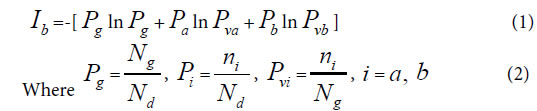
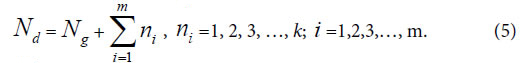
Generally, in the analysis of a sample set, one sample corresponds to one class of variation. If there are many kind variations in a biological system with a sample set, how to describe the action information. So a generalized equation need to be built up. Suggest there are m kind variations in a biological system, and each kind variation contain nielements or variables, then the generalized biological heredity and variation information equation was represented as follows.

JMG-3-2-313-e002new

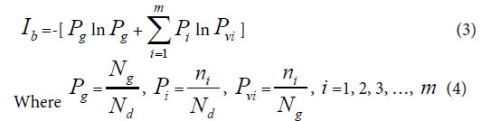
The meanings of variables in the equation (3), (4), as in [30-35] were listed below.

Pg, common (heredity) peak (element, molecule) ratio, it can be briefly expressed as P. This index is the same as the JaCcard and Sneath, Sokal coefficients.

Pi, the ratio of ni to Nd. Pvi is the variation peak (element, molecule) ratio of ni to Ng.

JMG-3-2-313-e003new

Nd , the independent peaks (elements, molecules) existed in a system.

Ng, common (heredity) peaks (elements, molecules) existed in a system.

ni , variation peaks (elements, molecules) belong to variation class i .

Relying on paper [30], – Pgln Pg represents the similarity action of the system based on common elements, and JMG-3-3-313-e004represents the action between common and variation elements. According to these generalized biological heredity and variation information equation, a biological system is usually of three typical variation models. They are listed below.

Considering multiple types of variation elements, there are three typical variation models.

Variation Model 1: n , =1, n >o, with only one variation class, the extreme asymmetric variation, or unique variation state in a system.

Variation Model 2: ni, i =1,2, and n1 = n2 >0, with two variation classes, or two symmetric variation states in a system.

Variation Model 3: ni=1, i =1, 2, 3, …, m, with m classes of
extreme radial variations. It means for each variation class, there is only one variation peak (element, molecule). This schematic was showed in Figure 1.

JMG-3-3-313-g001

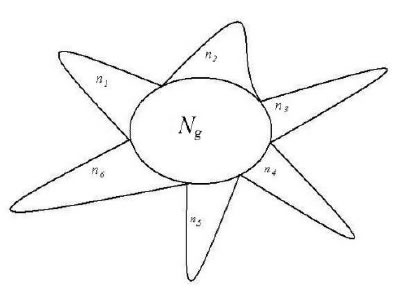
Figure 1. A system composed of Ng common heredity elements and ni variation elements, i =1, 2, 3,…, m. For example, This system poses 6 classes of variation elements.

A lot of experiments have proved that for equation (1), it is able to accurately reveal some significant properties in  complex biological systems. Two similarity constants P = 0.69, P = 0.61, can be

obtained in the maximum information states. Pg1= 0.69 corresponds to the unique variation state, that is variation model 1. Pg2= 0.61

corresponds to symmetric variation state, that is variation model 2.

These two similarity constants can be proved in below section. Based on the two similarity constants, very complex biological systems-the combination herbal medicines (traditional Chinese medicine, TCM), which consist of extracts of many medical plants, could be classified accurately relying on IR fingerprint spectra, which reflect structure unit information of biological small molecules [30-32,35]. Genus and Subgenus of pine could be discriminated precisely depending on information of molecular species in oleoresins [33], and four species of combination herbal medicines [33,35] were identified perfectly by means of the two constants. Three kinds of soybeans, black, green and yellow soybean proteomes were pattern recognized subtly by using the constant Pg2=0.61, together with structure information of macro-molecules [34].

All these previous researches express that the primarily established heredity and variation information equation is qualified to uncover some heredity and variation laws of biology.

Similarity constants corresponding to Variation Model 1 and Variation Model 2

Similarity constant Pg1: When there is only single variation class, that is extreme asymmetric variation state, corresponding Variation Model 1. ni>0, i =1, n1 = NdNg, generally biological heredity and variation information equation can be expressed as follows.

JMG-3-3-313-e005

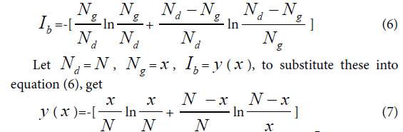
To take the derivative of equation (7), and let Ib to be in the maximum information state, then get

JMG-3-3-313-e006

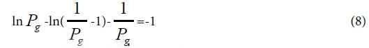
This equationghas a soglution P=0.692, approximately is 0.69=69%.

This theoretical standard has been successfully testified in researches [32,33,35]. Pg= 0.69 can be defined as similarity constant Pg1.

According to article [30-35], for any two samples in a sample set, we can view them is a biological system. Theoretically, when their Pg ≥ 69%, they are in the asymmetric variation state, and they are of the
identical properties. They belong to the same class. When their Pg < 69%, there are some distinct differences between them, they are not of identical property.

Similarity constant Pg2: In a biological system, when in symmetric mutation state, corresponding to Variation Model 2, n1 = n2 >0.

JMG-3-3-313-e007

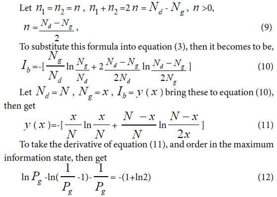
To solve this equation, one can obtain Pg=0.6085, approximately is 0.61= 61%. Pg= 61% can be defined as similarity constant Pg2.

According to article [30-35], for any two samples in a sample set, we can view them is a biological system. When their Pg ≥ 61%, they are of the identical properties, and they belong to the same class. when their Pg < 61%, there are some distinct differences between them, they are not of identical property.

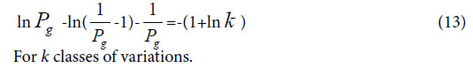
In the same way, one can get,

JMG-3-3-313-e008

Boundary Similarity function corresponding to Variation Model 3

For extreme radial variation model, ni =1, i =1,2,3,…, m. that is all variation elements are different from one another. Each variation element belongs to its own class. In this situation, generally biological heredity and variation information equation can be represented as follows.

JMG-3-2-313-e009new

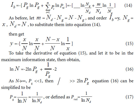
To take the derivative of equation (15), and let it to be in the maximum information state, then obtain,

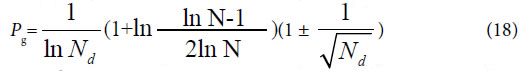
Considered some degree of randomness of heredity and variation in biology, we can deduce a formula for accurately and briefly calculating the common peak (molecules, or any elements ) ratio Pg in biology,

JMG-3-2-313-e010new

Since the theoretical criteria are similarity Pg, then these Pg show the ratios of common materials to total materials. The common characteristics reflect the identical properties, which are the ancestor characters of these samples in a biological system. So the value of Pg reflects high or low of ancestor traits between these samples. The higher the Pg, the closer the relative relationship of them. So to determine relative relationship far and near between samples relying on the high or low of Pg is scientifically reasonable.

Equation (17), (18) represent the relationship between Pg and N. That is there is no constant Pg in Variation Model 3. They are also the boundary condition or maximum variation range for close relatives, which originate from the same close ancestor. Since there is no limitation in the number and kinds of samples in a biological system, this theory is suitable for any open biological system.

The similarity function (17), (18) can be defined as the discrimination function of close relative biology. This function is the critical point for extreme variation of a biological system. For any two samples in a sample set, we can view them is a biological system. When their Pg < Prad, there are extremely distinct differences between them, they do not belong to close relative. Prad reflects the theoretical boundary of the close relative relationship.

Theoretically, these intrinsic criteria of Pg should be related to some biological ranks, that is theoretically taxonomic categories, such as species, genus, family and so on. Then these intrinsically characteristic Pg can be defined as differently theoretical criteria of some intrinsically taxonomic categories.

As we known, there are many biological levels or ranks, including species, genus, family as well as others in relative systems. Whether these ranks correspond to these Pg criteria for intrinsically taxonomic categories, or they can be theoretically discriminated by these criteria, previously classified by empirical knowledge. All these need to be verified by experiments.

Certainly, for the two constants, they may correspond to two intrinsically taxonomic categories, which should correspond to any two different biological ranks, such as species and subspecies, genus, subgenus, family and so on. These have been proved successfully by some researches [33,34]. These researches indicated the theoretically taxonomic categories, existed intrinsically in the equation, do not one-to-one correspond to empirical ranks of biology, obtained by means of empirical classification. From the view of scientific point, people should classify biology into some absolute ranks based on these theoretical criteria, and should offer strictly theoretical boundaries of close relatives.

Presently, The major methods in biological classification, cluster and pattern recognition are classical taxonomy [42-46], molecular taxonomy [16-25], Chemical taxonomy [42-44]. The major study manner is to discover new rules by means of experiments or depending on empirical knowledge, but not by some mathematical principles. On the other hand, many similarity and difference index were applied for the recognition, classification, cluster and evolution researches of traditional medical plants and combination herbal medicines, soybeans [30-32,34-41,47-52], depending on IR fingerprint spectra, one of the chemical fingerprint spectra, information of biological molecular structure units. This may be a good approach for building up new mathematical principles existed in biological ranks.

Classification of 29 medical plant samples based on three theoretical criteria

Plant samples

In this article IR fingerprint spectra of 29 plant samples were measured and analyzed, the experiments, seen Methods these samples belong to four kind medical plants: Baishao, Chishao, Huangqi, and Gancao. In these samples, Baishao, Chishao belong to Paeonia L.(genus), Huangqi belongs to Astragalus Linn.(genus), Gancao belongs to Glycyrrhiza Linn.(genus). The sources of these 29 samples were listed in Table 1.

JMG-3-3-313-t001

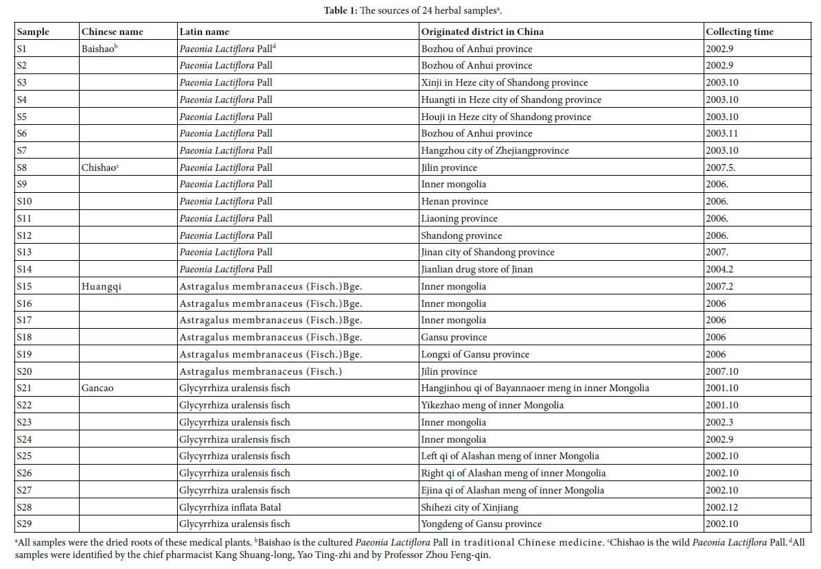
All samples were kept at bellow -18°C, after collected and dried.

Analysis on IR fingerprint spectra data of 29 plant samples

Determination and represent of common and variation peaks in IR fingerprint spectra: According to the methods to extract major compositions of samples with absolute ethanol, and to measure their IR fingerprint spectra. Then to determine the common and variation peaks of these IR FPS by means of Shapiro-Wilk test [53]. The common and variation peaks were listed in Table 2.

JMG-3-3-313-t002

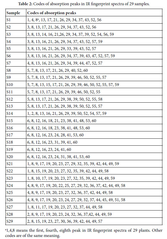
The averaged wavenumbers (cm-1) of all peaks are listed below:

± 6; 2,3406 ± 1; 3,3396 ± 2; 4,3385 ± 2; 5,3375 ± 3; 6,3363 ± 2; 7,2973 ± 1; 8,2928 ± 2; 9,2113 ± 2; 10, 2102 ± 1; 11,2090 ± 1; 12,1727 ± 3; 13,1714 ± 3; 14,1642 ± 1; 15,1635 ± 1; 16,1624 ± 2; 17,1615 ± 2; 18,1570 ± 1; 19,1512 ± 1; 20,1458 ± 1; 21,1450 ± 2; 22,1419 ± 1; 23,1408 ± 4; 24,1385 ± 4; 25,1372 ± 1; 26,1346 ± 3; 27,1333 ± 1; 28,1300 ± 1; 29,1278 ± 3; 30,1259 ± 1; 31,1252 ± 1; 32,1234 ± 1; 33,1227 ± 1; 34,1204 ± 1; 35,1131 ± 3; 36,1103 ± 2; 37,1073 ± 2; 38,1062 ± 1; 39,1051 ± 2; 40,1037 ± 1; 41,1029 ± 1; 42,997 ± 1; 43,938 ± 3; 44,926 ± 2; 45,892 ± 1; 46,880 ± 1; 47,871 ± 1; 48,856 ± 1; 49,835 ± 1; 50,821 ± 1; 51,777 ± 1; 52,714 ± 1; 53,667 ± 3; 54,632 ± 2; 55,623 ± 3; 56,613 ± 1; 57,596 ± 5; 58,582 ± 2; 59,553 ± 4; 60,526 ± 1. Peaks with the same code in different samples belong to a common peak group. There are 60 common peak groups or independent peaks in the 29 plant samples.

Characteristic sequences and classification of 29 plant samples

According to the two similarity constants Pg1= 69%, Pg2= 61%, obtained from generally biological heredity and variation information equation (3), and the analysis in previous studies [30-35], for any two samples in a biological system, when the Pg scale is Pg ≥ (61 ± 3)%, these two samples are similar to each other greatly and of the same inner quality. The similarities among these 29 samples were showed in Tables 3 and 4. In Tables 3 and 4, the same kind/class plants were showed in the same color area. From these tables, the similarities among the same kind plant samples were significantly higher than that among different kind plant samples. Common peak ratios among different kind plant samples are much less than Pg2 constant 61%.

JMG-3-3-313-t003

JMG-3-3-313-t004

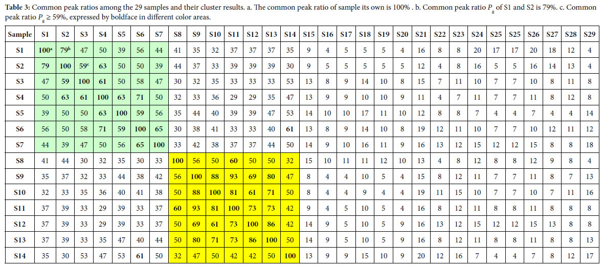
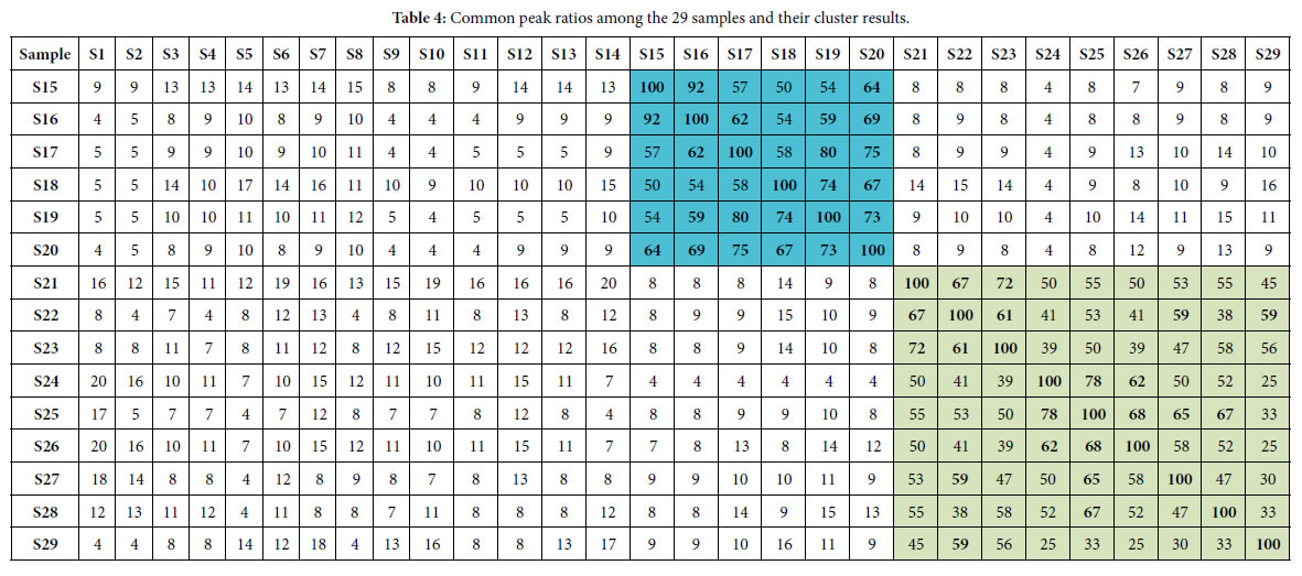
The definition of characteristic sequence:

For sample Si, its characteristic sequence is composed of these samples, whose common peak ratios related to Si fit to Pg scale Pg ≥ (61± 3)%. According to Tables 3 and 4, the characteristic sequences of 29 samples were obtained and displayed in Table 5. Their classification were performed based on their characteristic sequences.

JMG-3-3-313-t005

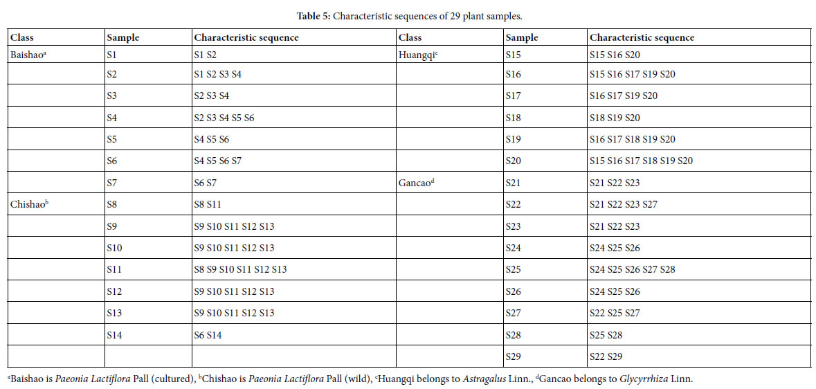
In accordance with the characteristic sequences of these 29 samples, the characteristic sequences in one class of samples were made up of themselves. Samples in one kind samples’ characteristic sequences form an independent sample set. There is no overlapped sample among different sample sets, expect for S14, whose characteristic sequence contains S6. In the 29 samples, 28 samples were classified correctly by means of their characteristic sequences. The correct ratio is 28/29 = 96.6%. Relying on the theoretical standard Pg scale Pg ≥ (61± 3)%, of the intrinsically taxonomic category, the four kind plants

Baishao S1-S7, Chishao S8-S13, Huangqi S15-S20, Gancao S21-S29, were accurately classified, or pattern recognized.

In particular, the significantly similar samples themselves in characteristic sequences are the best markers of their classes. This property is very important for classification, identification and pattern recognition of samples.

Summery, the similarities among Baishao and Chishao samples were much higher than that among any other two classes, such as Huangqi and Gancao samples, Baishao and Huangqi samples. It indicated that Baishao and Chishao samples should be close relatives.

These above results showed that similarity constant Pg2= 61% can be used as the quantitative standard to discriminate which plants are of the same efficiency, or used as the standard to determine the identical herbal medicines [30-32, 35].

On the other hand, suggesting m=4, that is there are four classes of plants, the theoretical standard Pg scale is Pg ≥ 53.2%, relying on the equation (13). To analyze the data in Tables 3 and 4, and get characteristic sequences of these samples. The results were very similar to that obtained by means of the standard Pg scale Pg ≥ (61 ± 3)%. There were trivial differences in their characteristic sequences. This also proved that generally biological heredity and variation information equation can represent intrinsic properties of biology exactly.

Analysis on close relatives among the four class plants

If these four kind plants are in the maximum variation state, that is at extreme radial variation state, accurate Pg scale Pg ≥ 24.4% (0.269=26.9%, according to equation (18)) could be obtained, when N=60, by equation (17). Pg scale Pg ≥ 24.4% is the theoretical boundary of close relatives of these 29 plant samples. Relying on this standard to determine characteristic sequences of these 29 samples, they could be classified into three classes perfectly, showed as follows.

Characteristic sequences of these 29 samples:

A class: Baishao and Chishao (Paeonia Lactiflora Pall)

S1:S1 S2 S3 S4 S5 S6 S7 S8 S9 S10 S11 S12 S13 S14a

S2:S1 S2 S3 S4 S5 S6 S7 S8 S9 S10 S11 S12 S13 S14

S3:S1 S2 S3 S4 S5 S6 S7 S8 S9 S10 S11 S12 S13 S14

S4:S1 S2 S3 S4 S5 S6 S7 S8 S9 S10 S11 S12 S13 S14

S5:S1 S2 S3 S4 S5 S6 S7 S8 S9 S10 S11 S12 S13 S14

S6:S1 S2 S3 S4 S5 S6 S7 S8 S9 S10 S11 S12 S13 S14

S7:S1 S2 S3 S4 S5 S6 S7 S8 S9 S10 S11 S12 S13 S14

S8:S1 S2 S3 S4 S5 S6 S7 S8 S9 S10 S11 S12 S13 S14

S9:S1 S2 S3 S4 S5 S6 S7 S8 S9 S10 S11 S12 S13 S14

S10:S1 S2 S3 S4 S5 S6 S7 S8 S9 S10 S11 S12 S13 S14

S11:S1 S2 S3 S4 S5 S6 S7 S8 S9 S10 S11 S12 S13 S14

S12:S1 S2 S3 S4 S5 S6 S7 S8 S9 S10 S11 S12 S13 S14

S13:S1 S2 S3 S4 S5 S6 S7 S8 S9 S10 S11 S12 S13 S14

S14:S1 S2 S3 S4 S5 S6 S7 S8 S9 S10 S11 S12 S13 S14

B class: Huangqi (Astragalus Linn.)

S15:S15 S16 S17 S18 S19 S20 S16:S15

S16 S17 S18 S19 S20 S17:S15 S16

S17 S18 S19 S20 S18:S15 S16 S17

S18 S19 S20 S19:S15 S16 S17 S18

S19 S20 S20:S15 S16 S17 S18 S19 S20

C class: Gancao (Glycyrrhiza Linn.)

S21: S21 S22 S23 S24 S25 S26 S27 S28 S29

S22: S21 S22 S23 S24 S25 S26 S27 S28 S29

S23: S21 S22 S23 S24 S25 S26 S27 S28 S29

S24: S21 S22 S23 S24 S25 S26 S27 S28

S25: S21 S22 S23 S24 S25 S26 S27 S28 S29

S26: S21 S22 S23 S24 S25 S26 S27 S28

S27: S21 S22 S23 S24 S25 S26 S27 S28 S29

S28: S21 S22 S23 S24 S25 S26 S27 S28 S29

S29: S21 S22 S23 S25 S27 S28 S29

a. S1:S1 S2 S3 S4 S5 S6 S7 S8 S9 S10 S11 S12 S13 S14, represents the characteristic sequence of sample S1.

Other sequences are of the same meaning.

From their characteristic sequences showed in the above classification results, these four kind plants could be divided into three close relatives. A class: Baishao +Chishao (Paeonia Lactiflora Pall). B class: Huangqi (Astragalus Linn.), C class: Gancao (Glycyrrhiza Linn.). This conclusion comply with the empirical classification. While for the classification based on medical efficiency and the kind of herbal medicines, theoretical standard Pg scale Pg ≥ (61 ± 3)% is more reasonable, see Table 5.

These results also indicated that for species, genus, they could be also clustered excellently by means of the theoretical boundary function(17),(18). This theoretical boundary can tolerate larger randomness of similarity in the same species, genus and so on. This theoretical boundary also suits to express elemental characteristics of biological evolution. Especially, the results of theoretical classification and close relative analysis both represented that common peak ratio Pg2=61%, indeed reflect the intrinsic characteristics of biological systems. Based on classical taxonomy, Baishao and Chishao both belong to Paeonia L.(genus), the same species, Paeonia Lactiflora Pall. However, there are some differences between them. Baishao is the plant Paeonia Lactiflora Pall having been cultured by human for thousand years. But Chishao is wild PaeoniaLactiflora Pall. Moreover, there exist great differences in their medical efficiency between Baishao and Chishao. This indicates their chemical compositions vary obviously. According to pharmacopoeia of P.R.China [54], two kinds of Huangqi Astragalus membranaceus(Fisch.)Bge. and Astragalus membranaceus (Fisch.), all belong to Astragalus Linn.(genus), their medical efficiency are identical. Two kind gancao :Glycyrrhiza uralensis fisch and Glycyrrhiza inflata Batal, they all belong to genus Glycyrrhiza Linn.(genus). Their medical efficiency are very similar to each other. The classification results relying on the theoretical standard Pg2 scale Pg ≥ (61 ± 3)% proved these two kind Gancao were in the same class, so were the two kind Huangqi medicine plants.

Baishao and Chishao are viewed as the same species, but their chemical compositions and medical efficiency are all different from each other distinctly. Theoretically, they should belong to different species, depending on researches in this article.

Huangqi and Gancao belong to the same family Leguminosae, but different genus. Two kind Huangqi samples belong to two species, and the same genus. In terms of the theoretical standard Pg scale Pg ≥ (61 ± 3)%, Huangqi samples are in the same class. So are the Gancao samples. These indicated for Glycyrrhiza Linn. and Astragalus Linn., Pg scale Pg ≥ (61 ± 3)% may be their theoretical standard of genus as well as that of herbal medicines, which are of the identical efficiency.

Depending on the above analysis, we know plant evolution is of some universal laws. Two similarity constants Pg1, Pg2, sometimes are suitable to be the theoretical standards of biological species, some time they are suitable for the theoretical standard of genus. Thus an hypothesis that biological system can be classified into some intrinsically taxonomic categories based on the three intrinsically theoretical criteria, can be proposed. Contrast to traditional classification, or taxonomy, only relying on empirical knowledge, these approaches can not reach the accurate and unchangeable results.

Nowadays, an reasonable classification scheme should be that all biology are firstly classified into the three intrinsic grade patterns, then divided these samples into sub-patterns in terms of the classical taxonomy methods.

Results and Conclusion on Intrinsic Taxonomic Categories and Their Theoretical Criteria

In plant systematics, there is no quantitative classification criteria for species, genus, and family, and other biological ranks. The results of this article showed there are three theoretical criteria corresponding to three intrinsically taxonomic categories, which correspond to three typically biological Variation Models. For Pg scales, Pg ≥ (61 ± 3)%, Pg≥ (69 ± 3)%, they can be accepted as the theoretical standards for some species, some genus, or some families. For Pg ≥ 1 ln Nd, it may be used as the theoretical standard to discriminate close relatives, such as Genus, Family (familia), Order (Ordo), Class (Classis), Phylumo (Divisi), Kingdom (Regnum) for some plants. This indirectly indicated that for different biological systems, their evolution speed vary significantly.

Most interestingly, these criteria may be a strong tool to investigate some laws in biologically macro molecules, such as DNA, RNA and protein sequences.

The research results also showed generally biological heredity and variation information theory, which was constructed based on the physical chemistry action model of heredity and variation materials, was able to accurately describe some heredity and variation laws in biological system, and suitable for many material levels. On the other hand, how to accurately determine the close relative among different biological systems is a very fundamental science subject. A theoretical boundary  function JMG-3-2-313-e011new, or is precisely equal to JMG-3-2-313-e012new 
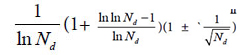
was derived from generally biological heredity and variation information equation. It can be used as a strong tool for determining close relatives.

The laws or rules of material actions are usually more universal and more rigorous than that obtained by statistics. Through actions of substances, which produce differently biological phenomena, the strictly, scientifically and universally theoretical standards for classification should be established. This progress may promote biological science to some extent in the future, and open the door towards quantitative researches on intrinsically scientific principles of biology.

Acknowledgement

I sincerely thank academician WANG yongyan of Chinese Engineering Academy for his moral encouragement and support.

References

  1. Zhao-Hai L, Hong Q, Hong Z (2008) Statistical methods in genetics. Beijing: Science Press.
  2. Zhi-Fa Y (2011) Population genetics, evolution and entropy. Beijing: Science Press.
  3. Gang-biao X (2009) Plant population genetics. Beijing: Science Press.
  4. Mendel GJ (2012) Selected works of genetics. Beijing: Peking University Press.
  5. Robert P, John C DeFries, Gerald E McClean, Peter McGuffin (2008) Behavioral Genetics (Firth edition). Shanghai: East China Normal University Press.
  6. Francisco JA, Kiger JJ Jr (1987) Modern Genetics. Changsha: Hunan Science & Technology Press.
  7. Zhi-fa YUAN, Zhi-jie CHANG, Man-cai GUO, Shi-duo SUN (2015) Genetic analysis of quantitative traits. Beijing: Science Press.
  8. Clark MS (2007) Comparative genomics. Beijing: Science Press 110-136.
  9. Thomas HM (2007) The theory of the gene. Beijing: Peking University Press.
  10. Michael RB (2009) Bioinfomatics for geneticists: A bioinformatics primer for the analysis of genetic data. Beijing: Science Press.
  11. Dale JW, Von Schantz M (2004) From genes to genomes: concepts and applications of DNA technology. Beijing: Science Press.
  12. Campbell M, Heyer JL (2005) Discovering genomics,proteomics & bioinformatics. Beijing: Science Press.
  13. Licinio J, Wang ML (2006) Pharmacogenomics. Beijing: Science Press.
  14. Baldi P, Brunak S (2003) Bioinformatics: the Machine learing approach. Beijing: CITIC Press.
  15. Song-nian HU, Qing-zhong XUE (2004) Handbook of genomic data analysis. Hangzhou: Zhejiang University Press.
  16. Yang ZHONG, Liang Z, Qiong ZHAO (2001) An introduction to bioinformatics. Beijing: Higher Education Press.
  17. David WM (2006) Bioinformatics: sequence and genome analysis (second edition). Beijing: Science Press.
  18. Pevsner J (2006) Bioinformatics and functional genomics. Beijing: Chemical Industry Press.
  19. Jones NC, Pevzner PA (2007) An introduction to bioinformatics algorthms. Beijing: Chemical Industry press.
  20. David WM (2003) Bioinformatics: sequence and genome analysis. Beijing: Higher education press.
  21. Xian -yong WANG, Zheng-hua WANG (2011) An introduction to bioinformatics- the algorithms and applications facing to high performance computing. Beijing: Tsinghua University press.
  22. Yang Z (2008) Computational molecular evolution. Shanghai: Fudan University Press.
  23. Setubol J, Meidannis J (2004) Introduction to computational molecular biology. Beijing: Science Press.
  24. De-shuang HUANG (2009) The data mining methods of gene expression profile.Beijing: Science Press.
  25. Jun WANG, Li-juan CONG, Hong-kun Z (2008) Commomly used biological data analytical software. Beijing: Science Press.
  26. Nelson P (2016) Biological physics: energy, information, life. Shanghai: Shanghai Science & Technical Publishers.
  27. Rob Phillips, Jane Kondev, Julie Theriot (2012) Physical biology of the cell. Beijing: Science press.
  28. Park S Nobel (2010) Physicochemical and Environmental plant physiology (fourth edition). Beijing: Science Press.
  29. Plonsey R, Barr RC (1992) Bioelectricity- A Quantitative approach. Shanghai: Fudan University Press.
  30. Hua-bin Z (2009) Dual index information markedly similar sequence clustering analysis on IR fingerprint spectra of extracts of Guifu Dihuang and Jingui Shenqi pills with ethanol. China Journal of Chinese Materia Medica 34: 2325-2330. [crossref]
  31. Huabin Z (2015) A systematically theoretical distinguish approach for traditional Chinese medicine with identical quality. WorldChinese Medicine 10: 1078-1082.
  32. Huabin Z (2017) Two Chinese medicine species constants and the accurate identification of Chinese medicines. bioRxiv.
  33. Huabin Z (2018) Two biological constants for accurate classification and evolution pattern analysis of Subgen.strobus and subgen. Pinus. bioRxiv.
  34. Huabin Z (2018) A theoretical approach for discriminating accurately intrinsic pattern of biological systems and recognizing three kind soybean proteomes. bioRxiv.
  35. Huabin Z (2018) Accurate identification of varieties constants and multiple pairs between 2 Chinese medicine species. WorldChinese Medicine 13: 3158-3165.
  36. Huabin Z, Yuan J, Aiqin D, Linlin S (2005) Dual-index sequence analytical method for IR fingerprint spectra of the chloroform extract of Radix Glycyrrhizae.China. Journal of Chinese Materia Medica 30: 16-20. [crossref]
  37. Hua‐Bin Z, Yuan J, Ai‐qin D, Lin‐lin S, Hassan Y (2005) Dual-Index Sequence Analytical Method for IR Fingerprint Spectra of Ethanolic Extract of Various Gylcyrrhizae’s Root Species components. Analytical Letters 38: 1167-1178.
  38. De-Xin K, Shu-shi H, Rong-shao H, Xiu-li C, Yi-Bing W et al. (2010) Comparative study on the infrared fingerprint of Abrus Cantoniensis based on the methoods of sequential analysis of Dual-index and cluster analysis. Spectroscopy and Spectral Analysis 30: 45-49. [crossref]
  39. Hong L, Chang-ri H, Hong-xia L, Yuan-feng L, Meng-xiong HE (2008) Study on IR Fingerprint Spectra of Alpinia Oxyphylla Miq. Spectroscopy and Spectral Analysis 28: 2557-2560. [crossref]
  40. Hua-bin ZOU, Zhi-feng H, Hong ZHAI, Ying-qin WEI, Fei WANG (2007) The First and Second Cluster Analysis of Dual Index Grade Sequence of IR Fingerprint Spectra of Guifudihuang Pill and Jinkuishenqi Pill Samples and Their Quality Evaluation. Spectroscopy and Spectral Analysis 27: 2432-2436. [crossref]
  41. Hua-bin ZOU, Xin-1ing Z, Hong ZHAI, Ai-qin DU (2008) Dual index grade sequence pattern recognition of extracts with ethanol of Mingmu Dihuang pills and Zhibai Dihuang pills. ChinaJournalofChineseMateriaMedica33: 1543-1549. [crossref]
  42. Gurcharan Singh (2008) Plant systematics: An integrated approach. Beijing: Chemical Industry Press.
  43. Chang-Fa ZHOU (2009) Biological evolution and taxonomical principle. Beijing: Science Press.
  44. Han-Chen Z, Hao Z, Sheng-Li PAN (2009) Pharmaceutical botany (fifths edition).Beijing: People’s Medical Publishing House.
  45. Jin-wu WANG (2011) Spermatophyte taxonomy (second edition). Beijing: Higher Education Press.
  46. Simpson MG (2012) Plant systmatics (second edition). Beijing: Science Press.
  47. Hua-bin ZOU, Jiu-rong YUAN, Ai-qin D, Lin-lin S (2005) Dual-index sequence analytical method for IR fingerprint spectra of the chloroform extract of Radix Glycyrrhizae. China Journal of Chinese Materia Medica 30: 16-20. [crossref]
  48. Hua‐Bin Zou, Guo‐Sheng Yang, Zheng‐Ran Qin, Wen‐Qiang Jiang, Ai‐Qin Du et al. (2005) Progress in quality control of herbal medicine with IR fingerprint spectra. Analytical Letters 38: 1457-1475.
  49. Tao-tao PANG, Li-ming DU (2007) Common and Variant Peak Ratios in IR Fingerprint of Ilex Kudingcha with Duel-Index Sequence Analysis. Spectroscopy and Spectral Analysis 27: 486-489. [crossref]
  50. Hua-bin Zou, Jiu-rong Y, Aiqin D, Lin-lin S, Hassan YA (2005) Dual-Index Sequence Analytical Method for IR Fingerprint Spectra of Ethanolic Extract of Various Gylcyrrhizae’s Root Species components. Analytical Letters 38: 1167-1178.
  51. Kevin Yi-LY, Yung CS, Lim CS (2007) Infrared-based protocol for the identification and categorization of ginseng and its products. Food Research International 40: 643- 652.
  52. Hong-xia L, Su-qin S, Guang-hua LV, Kelvin KC Chan (2006) Study on Angelica and its different extracts by Fourier transform infrared spectroscopy and two-dimensional correlation IR spectroscopy. Spectrochimica Acta Part A 64: 321-326. [crossref]
  53. Huabin Zou, Jiurong Y, Wang Wei (2004) Theoretical identification of common peaks in fingerprint of Chinese medicine -a W- testing and discriminatory method. World Science and Technology/Modernization of Traditional Chinese Medicine and MateriaMedica 6: 50-56.
  54. Chinese Pharmacopoeia Commission (2005) Chinese pharmacopoeia 2005 edition (I)[S], Beijing: Chemical Industry Press.

Can One Teach Old Drugs New Tricks? Reformulating to Repurpose Chloroquine and Hydroxychloroquine

Abstract

The outbreak of the novel corona virus disease, COVID-19, has presented health care professionals with the unique challenges of trying to select appropriate pharmacological treatments with little time available for drug testing. Given the development times and manufacturing requirements for new products, Value Added Medicines (repurposing – reformulation of existing drugs) could be one possibility to beat the COVID-19 outbreak. This review explores reformulation alternatives which could be progressed with chloroquine and hydroxychloroquine; two antimalarial drugs, that are being tested on a global scale as a potential therapeutic option. The key areas for improvement have been reviewed and the potential solutions to the problems and limitations of current formulations are discussed. The pharmaceutical challenges discussed are those of highly soluble drugs, needed to be given at high doses and presenting a real bitter taste challenge with significant gastrointestinal side effects that could be translated and repurposed into fit for purpose reformulations.

Introduction

COVID-19 is the infectious disease caused by the most recently discovered corona virus. This newly emerged virus and disease were unknown before the outbreak began in Wuhan, China, in December 2019. COVID-19 is now a pandemic affecting many countries globally and to date no antiviral or therapeutic has been approved for treating patients. As the number of cases continues to rise, the geographic range of the virus increases, and with the development of a vaccine being at least 12 months away, there is a growing urgency/pressure on pharmaceutical industry and regulatory agencies to expedite the development and approval of both experimental drugs and repurposing of existing therapeutics that have been already approved for human use by the health agencies. Among the landscape of therapeutics being analysed as potential repurposing candidates for COVID-19, the antimalarial and immunomodulatory drugs chloroquine (CQ) and hydroxychloroquine (HCQ), both 4-aminoquinolines, are being tested on a global scale as a potential treatment and prevention of COVID-19 [1]. Recent publications have drawn attention to the possible benefit of CQ sulphate and phosphate salts (CQ diphosphate) and HCQ for the treatment of SARS-CoV-2 infected patients [2-7]. CQ phosphate or sulphate is referenced on the World Health Organisation (WHO) Model List of Essential Medicines for the treatment of Plasmodium vivax infection (malaria) [8, 9]. In addition to their antimalarial use, both CQ and HCQ are used in continuous daily dosing for rheumatoid arthritis, systemic and discoid lupus erythematosus and psoriatic arthritis. CQ and HCQ are one of four potential treatments that WHO has included in the global SOLIDARITY Clinical Trial in 90 countries to generate the robust data needed to establish efficacy and safety in COVID- 19 treatments [10]. There are several other trials ongoing in different countries [11, 12] to name a few, a UK wide randomized, controlled trial in over 130 hospitals called Randomised Evaluation of COVID Therapy (RECOVERY) [13] is underway and in Europe its Trial of Treatments for COVID-19 in Hospitalized Adults (DisCoVeRy) [14]. In US, NYU and University of Washington has fast trackeda major clinical trial to determine role of HCQ in prevention of corona virus [15]. Both CQ and HCQ are primarily available as tablets for oral administration [16-18] and have been in clinical use for decades thus their safety profile is well established [19]. However, this oral formulation presents the following problems: swallow ability difficulties for certain patients groups such as the young, older people and patients in critical care, as well as extremely poor palatability due to the bitter taste of the drugs [20, 21]. In addition, the oral administration of CQ and HCQ frequently causes gastrointestinal side (GI) effects such as nausea and vomiting [22-24]. HCQ being reported to have better safety profile than CQ, better gastrointestinal tolerability, and less retinal toxicity [25]. This review sets the scene and explores promising reformulation alternatives which could be progressed with CQ and HCQ, for adults and children. Alternatives routes of administration are also explored to address oral administration challenges. The problems and limitations with existing formulations are discussed and the key areas for improvement are reviewed. It is important to note while CQ and HCQ are under investigation in clinical trials for use on COVID-19 patients, as of the date of this publication, none of these compounds and medications have been approved for the treatment of COVID-19. Considering it is a rapidly changing area with new conflicting outcomes coming up every day, the repurposing of CQ and HCQ for COVID-19 is still questionable as only limited evidence is available at the present time. However, this review will be useful in various scenarios 1) if the trials are successful and the use of CQ in HCQ for COVID -19 is recommended 2) if no good evidence on use of CQ and HCQ for COVID-19 is generated in time, the reformulation strategies proposed in this review will be still relevant for antimalarial and rheumatic disorders treatment 3) the approaches discussed could be translated to other Active Pharmaceuticals Ingredients (APIs) presenting similar pharmaceutical challenges.

Challenges with existing CQ and HCQ formulation for COVID-19 treatment

CQ and HCQ are both Biopharmaceutics Classification System Class 1 compound [26] and extremely bitter [20, 21]. One study found the threshold bitterness of the pure CQ to be at 40 μg/ml [27]. After oral administration both are rapidly and almost completely absorbed from the gastrointestinal tract. They have a long and variable plasma elimination half-life because of a high volume of distribution with about half the drug metabolites undergoing unmodified renal clearance. CQ has a low safety margin and is very dangerous in overdose situations or when combined with other medicines [28]. There is no firm evidence on the optimal dosing and duration treatment for CQ or HCQ, hence the range of regimens are used across trials. In general, the regimen of CQ and HCQ used is substantially more aggressive than that recommended as an antimalarial [29]. For instance, National Health Commission of the People’s Republic of China recommended dose of 500mg twice daily for seven days for oral administration in 18–65 years of infected adults [30]. In the RECOVERY Trial, the loading dose of HCQ (1860mg) is twice the normal dose for treating malaria. However, this dose has been selected based on the available data of the IC50for SARS-CoV-2 [13] Several dosing regimens are proposed based on PBPK simulation combined with known clinical exposure–response relationships [3, 31-33]. Based on PBPK model, the typical dose for HCQ for treating COVID-19 is 400 mg twice daily on the first day, followed by 200 mg twice daily for four more days [31, 33]. Although HCQ shows better safety and toxicity profiles than CQ, symptomatic effects at these high doses have not been explored in enough depth. The question remains, how will gastro-intestinal symptoms prevail, will the benefits outweigh the risks and symptomatic effects; especially for critically ill COVID-19 patients in ICU. A wide range of data from various patients groups needs to be gathered to reliably surmise this. Both CQ and HCQ are metabolised in the liver with renal excretion of some metabolites, hence should be prescribed with care in people with liver or renal failure [34-36]. Recently published Surviving Sepsis Campaign guidelines [37] on the management of critically ill patients with COVID-19 concluded that there was insufficient evidence to offer any recommendation on the routine use of these drugs in patients admitted to the intensive care unit (ICU).The ongoing trials will be able to answer whether antimalarial drugs could be effective in changing the disease course in patients with severe COVID-19—in particular, in cases requiring ICU admission. At present, the safety of CQ in the treatment of elderly patients with COVID‐19 is unclear. The rate of critical illness in this population is high and CQ could still be used as an alternative drug, although it should not be given to those elderly patients with underlying heart and other conditions [38]. Based on the published clinical guidelines and research results, Sun et.al (38) have proposed the pharmaceutical care for the elderly using CQ phosphate in the treatment of COVID‐19. This includes the administration method, dosage of CQ phosphate for elderly, adverse drug reactions and drug interactions of CQ phosphate. For elderly patients with a bodyweight of more than 50 kg, CQ phosphate 500 mg orally, bid, for 7 days is recommended. CQ and HCQ is licensed for use in children with malaria. The WHO recommended HCQ dose to treat COVID-19 infected children is 25mg/kg given over 3 days. However, this may not be optimal to treat COVID-19, as recent studies show that older infants and children may need a higher mg/kg dose to reach the similar concentrations as adults. In contrasts, it is likely that neonates and young infants will need lower doses per kg body weight. Paediatric CQ dose for COVID-19was determined by Verscheijden et.al. [39]. The study proposestotal cumulative doses: 35 mg/kg (CHQ base) for children 0-1month, 47 mg/kg for 1-6 months, 55 mg/kg for 6 months-12 years and 44 mg/kg for adolescents and adults, not to exceed 3300 mg in any patient. Currently, there is a CQ phosphate syrup [40] on the market, while no pediatric or easy to swallow formulations exist for HCQ. However, as HCQ is highly soluble compound, it is expected that manipulation of the formulation will have minimal impact on bioavailability. The European Paediatric Formulary (PaedF) Working Party at the European Directorate of the Quality of Medicines and Healthcare (EDQM) has compiled existing knowledge on pediatric formulations for active substances which are under investigation for the treatment of COVID-19 as well as known authorised medicinal products [41]. This includes the information on extemporaneous preparations of CQ and HCQ which may be suitable for treatment of pediatric patients with COVID-19. It suggests the preparation of a pediatric suspension formulation from a 200mg tablet. This instructs pharmacists to ‘strips’ the outer film coating, crush the tablet(s), and then suspend the powder in water with a flavouring agent such as Ora-plus®. In the USA, The Nationwide Children’s pharmacy [42] and Michigan Collaborative Standardization of Compounded Oral Liquids [43] have formerly investigated utilising a crushed standard 200mg tablet of HCQ in Ora-plus® to form an oral liquid suspension. The resultant suspension concentration is 25mg/ml (i.e. 800mg (32ml) of suspension). A liquid dosage form of HCQ would be applicable to both the younger and older generations to address both patient dysphagia and compliance respectively. However, Ansah EK et al. reported improved compliance in children in rural Africa who were treated with CQ tablets or segments (crushed and mixed with sugar or honey) rather than CQ syrup [44] Moreover, the preparation of this extemporaneous suspension, especially stripping or/and crushing process is cumbersome. The suspension media such as Ora-plus® is also costly and may not be easily available in some regions, and the process may result in a loss of active pharmaceutical ingredient. Batches could be prepared on a demand basis for these patient groups but it is time consuming to prepare large quantities of suspension that have a shelf life of only 30 days at 2-8°C protected from light. Besides, the same GI side effects would likely still be present with additional issues of the drug’s bitter taste [45, 46] which is unlikely to be concealed. Thus, for this suspension to obligate its benefits, taste masking seems essential [47].

Current approaches for taste masking of CQ and HCQ

Sensory based taste masking approaches in which sweeteners and flavours are added to obscure taste, have been commonly used for decades, but this approach does not work well for highly soluble, highly aversive APIs and/or for APIs with an intense lingering aftertaste, as any compounds dissolved in the saliva will interact with the taste receptors and elicit a response [48]. Alternatively, taste masking techniques are utilised to improve the palatability of formulations. The different complexation approaches that have been used to taste mask by creating ‘molecular’ barrier around the API CQ and HCQ were scoped as part of this review and are discussed below.

Ion pairing

Ion-pairing is a process that involves stoichiometric replacement of polar counter-ions (i.e., chloride, acetate, nitrate, etc.) in the drug with an ionic excipient of similar charge. Ion-pairing has been used in the pharmaceutical industry, mainly as an additional drug-delivery method, and has proven to be a very effective mean for controlled drug release and taste masking [49]. Pauli et al [47] created a prototype formulation using a Coni-Snap Sprinkle Capsule containing sodium carboxymethyl cellulose (Na-CMC) as the ion-pairing agent to investigate if the addition of ion-pairing excipients and the incorporation of a buffered system into the conventional tablet could overcome some of the bitter-taste issues of HCQ. They hypothesised that this would enable a dual use formulation: adults can swallow the capsule whole whereas children (or indeed any patients with swallowing difficulties) could be administered the capsule content in water. They compared the dissolution profile of this capsule to another prototype formulation in the same capsule but without any ion-pairing agent and concluded that both profiles were comparable to that of a commercially available tablet of HCQ. This a bridged research also presented how the ion-paring system as provided by Na-CMC, and by another ion-pairing agent; sodium citrate, buffered to pH 8, taste-masked the drug in vitro with an Astreeel ectronic (e-tongue) assay. However, no in vivo assessment has been made. Moreover, the concentrations tested with the e-tongue were not reflective of the clinical situation for adults with COVID-19 as it was to treat children with uncomplicated malaria, lupus erythematosus, and rheumatoid arthritis at much lower doses. This integrated system could still have promise with higher drug loads, but it is likely the ion-pairing agents would need to be present in much higher concentrations in order to match the 1:1 ratio used in the study. Independent of this, buffering the system alone to pH 8 seems to have a significant effect on taste-intensity so this could be a quicker avenue to explore, given the current urgent need. It would likely be time-consuming to re-test alternative amounts of ion-pairing agents and then carry out further compatibility testing alongside the other components of the formulation.

Ion – exchange resins

Taste masking by drug–resin complexation is achieved when an ionizable drug reacts with a suitable ion exchange resin to form a drug–resinate complex [48]. Ion exchange resins (IERs) are insoluble, pharmacologically inert, high molecular weight cross linked polymers with cationic and anionic functional groups. They bind to compounds that exchange mobile ions and ultimately form of a tasteless drug- resin complex or resinate [50]. Drugs are attached to the oppositely charged resin substrate, forming insoluble adsorbates or resonates through weak ionic bonding. The resinate needs to be stable in the drug formulation e.g. a suspension or a tablet formulation and the dissociation of the drug–resin complex should not occur under the salivary pH conditions (pH 6-7). However, at enteric pH conditions (pH<5), the drug should be rapidly and almost entirely released in order to prevent reduced bioavailability. This can suitably mask the unpleasant taste and odour of drugs [51]. Characterization of drug–resin complexes and taste masking of CQ phosphate by complexation using weak cation exchange resin have been described in the literature [27, 50, 52]. All studies showed some in vivo taste improvement. No taste masking study of HCQ using ion exchange resins was identified.

Simple and Supra molecular Complexation

The host-guest complexation is a common taste masking technology [53]. By embedding the drug molecule (guest) into the cavity of a host molecule, a stable complex is generated. Generally, there are 2 mechanisms for explaining the taste-masking effect of complexation. The first mechanism is that the complexing agent will hinder interactions between drug molecules and taste cells through forming the strong binding with drugs [54]. Secondly, the complexing agent may directly bind to taste cells to mask the unpleasant. Among all the complexing agents, cyclodextrin (CD) group is a typical example [53]. Derived from the starch, CDs are cyclic polymers composed of glucopyranoside units (n) that are linked by α-1,4-glycosidic bonds. With their doughnut-shaped structures, CDs are capable of fitting lipophilic drugs or lipophilic moieties of drugs inside their hydrophobic central cavities (Figure 1). The natural and well-known CDs are α- (n=6), β- (n=7), and γ- (n=8)CD, respectively (Figure 1).

JPPR-3-1-309-g001

Figure 1. Cyclodextrin structure and representation of an inclusion complex of a drug residing in the cavity formed by the cyclodextrins

There is limited literature regarding the complexation between CDs and HCQ (or CQ). Guo, Wu et al [55] built a 3D model to predict the taste masking effect of CDs in different drug-CD complexes, where the Euclidean distance using an α-Astree e-Tongue was adopted to quantitate a taste masking effect (100 being the smallest euclidean distance to show taste masking efficiency). The study concluded that taste masking of HCQ was not achieved as the Euclidian distance was only 91 and proposed that CDs were ineffective because the halogen group and the chlorobenzene of the4-aminoquinolines significantly increased their molecular size and hampered the complete encapsulation of the drugs inside CDs cavities or that the alkylamino side chain is not part of the inclusion complex, allowing the tertiary amine to participate in bitter taste. Therefore, to date, no published paper indicated that natural CDs were effective in masking the bitter taste of HCQ or CQ. Woertz et al (56)conducted an INSENT e-tongue experiment to investigate taste masking effects of CDs (α-CD, β-CD, γ-CD, hydroxypropyl-β-CD, maltodextrin as well as sulfobutyl ether-β-CD (SBE-β-CD) on quinine. All CDs failed to mask the bitter taste of quinine, except for the modified CD, SBE-β-CD. It was thought that the aliphatic ring of quinine was embedded inside the CD cavity, whereas the quinoline ring of quinine was left outside the cavity, generating an incomplete inclusion complex with CDs, thereby the quinoline part could still interact with taste receptors and cause the bad taste. The study concluded that SBE-β-CD was able to mask bitter taste of quinine because of the ionic interaction between its –SO 3 2- group and the deformed –NH 3 + group from the quinine [56] (Figure 2).

JPPR-3-1-309-g002

Figure 2. Chemical structures of Quinine, Chloroquine and Hydroxychloroquine.

Since the chemical structure of quinine is similar to CQ and HCQ (Figure 2), the potential of SBE-β-CD for taste masking offer a masking option. However, according to Ghateet al [57] the native SBE-β-CD is salty, which may impede it use for taste masking. Captisol® is an FDA approved SBE-β-CD. This enabling technology available to research and development through flexible licensing arrangements is claimed to be tastelessness in oral formulas. It is and has been used in drug products on the market [58, 59]. Remarkably, Joneset al [60] recently developed a novel CD, modified with mercaptoundecane sulfonic acids. This highly sulfonated CD could mimic heparan sulphates (HS) and kill most HS-dependent viruses, such as herpes simplex virus, respiratory syncytial virus, dengue virus, and Zika virus. The study indicated that the newly modified CD had a potential to act as a broad-spectrum antiviral agent. Although corona viruses were not tested, their discovery reveals a different choice, where the modified CD could mask bitter taste, and at the same time, kill viruses. It is to be noted that this is probably the furthest away from translation as this would require extensive studies to support the claim. Apart from CDs, the cucurbituril (CB) family is another emerging complexing strategy and believed to be promising and attractive for pharmaceutical development. CBs ares upramolecular host molecules or macrocycles consisting of 5 or more glycoluril units joint together by methylene linkages [61]. CBs can also accommodate lipophilic drug moieties inside their hydrophobic cavities [62]. Currently, there is no literature about the HCQ-CB complexation or the CQ-CB complexation. However, it was confirmed by Boraste, Chakraborty et al [63] that CB7, which connects 7glycoluril units, was able to form a stable and a complete inclusion complex with quinine at a ratio of 2:1 (CB7 to quinine). Compared with CDs, CB7 was demonstrated to not only encapsulate the small aliphatic ring of quinine, but also fit the large quinoline ring into its cavity. This process was accomplished by binding the aliphatic ring of quinine to one CB7, while the quinoline moiety of quinine entered into a second CB7 cavity, at a lower pH. Accordingly, it is hypothesized that it could be possible for CQ and HCQ to form complete inclusion complexes with CBs. In this way, interactions between bitter drugs and taste receptors could be inhibited and taste masking achieved. However, before they are ready to be used in practice or tested in humans, there are number of issues that needs to be addressed including safety (Figure 3).

JPPR-3-1-309-g003

Figure 3. Molecular structure of CB7

Crystal engineering strategies

Pharmaceutical cocrystals are molecular crystals that are multi-component crystalline substances, where one of the components is an API and the other component/components are crystalline substances that have been approved by the regulatory bodies within the jurisdiction of commercial use; these species are known as the cocrystal former or the conformer [64]. Co-crystallization is one of the emerging crystal engineering techniques for modulating pharmaceutical performance through controlling solid-state properties of APIs and expanding the access to new solid forms differing in structures. The approach relies on the self-assembly of a bitter-tasting drug and a taste-masking agent, whereby the molecules are held together by non-covalent interactions including hydrogen and halogen bonds. Co-crystallization can modify different physicochemical properties of the APIs, without any change in their activity, such as improving the solubility of poorly soluble drugs, masking the bitter taste, increasing chemical stability and decreasing hygroscopicity, enhancing manufacturability as well dissolution rate and bioavailability [65]. Investigations into the solid-state landscape of CQ and HCQ have demonstrated the advantages of complexation within the crystal lattice. They have distinct acid base properties and hydrogen bond abilities that makes them suitable to form salts and co-crystals. The only crystal form of HCQ currently on the market is in the form of the sulphate salt [66]. Whereas CQ is on the market as the stand-alone API or as the phosphate, sulphate or hydrochloride salt [67, 68]. One study demonstrated how improvements can be made to these solids using crystal engineering to enhance the physicochemical properties and pharmacological activity of these co-crystallised compounds over parent compound. CQ and HCQ are both part of the quinolone family and are closely related to quinolone. Baruah et al [69] showed that salts and cocrystal of quinoline with hydroxyaromatic carboxylic acids enhance antimalarial activities over parent compounds. However, there is no literature to support the application of crystal-engineering approach to taste masking neither of CQ or HCQ. Research into the preparation of multicomponent crystals involving other APIs containing quinoline moieties suggest that this group of compounds could either partake in salt formation or cocrystal formation to improve API taste depending on the type of cocrystal or salt former used [70-72].

Alternative routes of administration to address formulation challenges

Rectal drug delivery

To subsequently tackle at the same time the bitter taste issue and high dose administration whilst still being applicable to all patient groups with possible comorbidities/polypharmacy, the rectal route may be the most appropriate alternative administration route. Symptomatic relief would also be achieved and allow critically ill/unconscious patients in ICU to be treated equally. Suppositories are often used as drug delivery system in case of nausea or vomiting, in case of oral administration rejection due to the bad taste or in case a medication is readily decomposed in gastric fluid. Published studies showed that CQ given in suppositories with the same dose as oral formulations reached lower blood concentrations and it was slower to produce the same antimalarial effects than when administered orally [73-76]. Tjoenget al [77] performed comparative bioavailability studies of rectal and oral formulations of CQ in healthy volunteers. The study demonstrated that the relative bioavailability of CQ 500 mg suppositories varied between 10-53% compared to a tablet formulation in adults. Onyeji at al [75] demonstrated that the bioavailability of chloroquine 100 mg suppositories was 63.4 +/- 8.8% (mean +/- SEM) relative to the tablet formulation. Bruce-Chwatt et al [73] observed that only when the same dose of CQ 300mg tablets was administered rectally over a 5 days period, it was able to reach the same parasite clearance of the oral dose but more slowly. The study concluded that CQ given by mouth was better and faster absorbed compared to the rectal route. However, in all these studies, no consideration was drawn on formulation improvements that could enhance the rectal release and adsorption of CQ from the suppository. Suppository are made of relatively low-cost excipients but their manufacture can be more challenging than other common dosage forms (tablets, liquids): they may need temperature-controlled storage depending on melting point and humidity control may be required during manufacture [78]. The nature of the base and the surfactant content in the suppository composition need to be carefully chosen to obtain the optimal mechanical and drug release properties of CQ [79]. Redgon et al [80] assessed different lipophilic and hydrophilic bases for CQ phosphate. They found that the hard-fat base Witepsol H15 was the best in terms of disintegration times, a with good storage conditions, was also suitable for countries with a continental climate. Onyeji et al [81] on the other hand, studied the effect of absorption-enhancing agents, non-ionic surfactants and sodium salicylate, on the in vitro release characteristics of CQ from polyethylene glycol (1000:4000, 75:25%, w/w) suppositories. The study concluded that the incorporation of 4% Tween 20 or 25% sodium salicylate improved the invitro release of CQ from the suppository. Considering these adjuvants also have absorption-promoting properties, association of the improved in-vitro release with enhanced in vivo availability is envisaged but would require a PK study for confirmation. Thus, results of this study serve as a guide in the selection of an optimal formula regarding the type and concentrations of absorption enhancers required for optimization of CQ release and a possible enhancement of rectal absorption of the drug. Finally, Okubanjoet al [79] studied the effects of interacting variables on the mechanical and release properties of CQ phosphate suppositories. A23 factorial experiment was designed to study the effects of the type and nature of the base, the concentration of surfactant and storage conditions. The study demonstrated that the presence and concentration of surfactant was the main individual variable affecting the release properties of suppository formulations. The addition of surfactants increased the crushing strength and decreased the dissolution times of CQ suppositories. Also, the type of suppository base played a role in the modification of the mechanical and release properties. Witepsol H15, as previously highlighted by Redgon et al [80] was better than Suppocire AS2 in increasing the crushing strength and dissolution rates and decreasing the dissolution times while storage conditions had the lowest effect. In the past, the rectal administration of CQ, but not HCQ, was explored for the treatment of malaria. Evidence tends to suggest that the use of rectal formulations for the administration of CQ could be re-considered as an alternative route to the oral administration to overcome the problems associated with this route. Due to the similarity of the 2 compounds, it is speculated that HCQ could also be a good candidate to be reformulated for rectal administration. It would be interesting to explore more recent advances introduced for the release and adsorption of drugs form rectal suppositories, such as the use recto dispersible dosage forms with non-melting excipients [82] or the use of hollow-type suppositories which have been developed to enhance the adsorption of various drugs [47]. The use of rectal formulations could be particularly useful for the treatment of certain patients’ groups affected by COVID-19, such as those with swallowability difficulties, critically ill patients, unconscious or vomiting patients or pediatric patients from birth. Although speculatively, by this route, there may be the added advantages of attaining the necessary higher plasma concentrations with a lower drug dose (yet high doses of API can be delivered rectally) as it generally avoids first pass metabolism. Finally, it is suitable for APIs that are gastro-irritant and could speculatively again help with some GI side effects. Socio-cultural norms drive recommendations regarding the knowledge, attitude, preference, and behaviour of people. To implement rectal delivery of CQ and HCQ, beside positive pharmacological outcomes, it would be important to consider patients’ real barriers versus cultural, perceived barriers or lack of understanding of the potential of this mode of administration [83]. Two further routes of administration for CQ and HCQ have been recently re-proposed as alternatives to the oral drug delivery.

Pulmonary drug delivery

An aerosolized formulation of HCQ was developed and tested in early phase clinical trials by the American company APT Pharmaceuticals. For the anti-inflammatory effect of HCQ, this formulation was developed for the potential treatment of respiratory diseases such as asthma, chronic obstructive pulmonary disease (COPD), rhinitis and severe acute respiratory syndrome (SARS). The drug was delivered by using an aerosol generating system (AERx®) for pulmonary delivery developed by Aradigm Corporation of Hayward, to maximize drug delivery in a patient-friendly format. By using this targeted delivery system, the company believed that the aerosolized dosage form and the use of the pulmonary route could achieve a faster onset of action, within hours, and greater therapeutic effects than conventional oral therapy at substantially lower systemic doses [84]. However, despite the favourable results showed in a phase 1 clinical study, the subsequent phase 2a data failed on efficacy endpoints and the study was stopped. In light of the potential role that CQ and HCQ might have as potential candidate therapies for COVID-19, Klimke et Al [85] re-proposed the use of them as pulmonary aerosol in a dosage of 2-4 mg of HCQ per inhalation for reduction and prevention of severe symptoms after SARS-CoV-2 infection. They speculated that by administering the drug targeting directly the lung, a lower drug dosage would be required to reach the optimal concentrations, compared to the oral route and avoid the oral side effects. Based on their speculations, the 2 authors decided to inhaled themselves tolerability and safety of 1 mg of HCQ in 2ml sodium chloride 0.9%, b.i.d. increased to 4 mg daily over one week. The dose was deemed well tolerated, with after 4 days still the feeling of a transient bitter taste in the mouth, which lasted 2-3 hours after each dosing. However, no efficacy data are available to validate their hypothesis.

Transdermal Drug Delivery

A study by Musabayane et al [86] investigated the potential application of pectin hydrogel patch as a matrix polymer for transdermal administration of CQ with Dimethyl sulphoxide as a penetration enhancer. They tested on rats the effects of CQ via intravenous infusion and the patch applied on shaved area during the 1 h 20 min. The results (plasma profile) showed good potential for transdermal delivery of CQ. However, the loading efficiency was only 46% of the theoretical 10 g. Moreover, the dose administered (16 µg/kg) was much lower than those previously used in rats (20–25 mg/kg), and in man (300 mg/kg). Glan is Pharmaceuticals Inc. recently obtained the rights for a US provisional patent for a transdermal drug delivery system of HCQ as a potential treatment for COVID-19. They suggest that controlled transdermal delivery could provide constant drug plasma concentrations for pre-determined periods of time, potentially reducing side effects associated to the oral delivery. However, so far, it seems that only literature search and pre-formulation studies have been done in collaboration with Reformulation Research Laboratories Inc. but no related clinical information are available [87]. The development of transdermal formulations [88] of CQ and HCQ with novel strategies would need to be carefully studied in order to address some challenges such as the hydrophilic nature, the need to administer larger doses of drug, and potential skin irritation due to enhancers or other additives added to the transdermal formulation.

Conclusion

COVID-19, officially designated as severe acute respiratory syndrome-related corona virus SARS-CoV-2 currently represents a pandemic threat to global public health. Researchers are leaving no stone unturned in an effort to understand this new emergent disease and uncover existing drugs with therapeutic potential for COVID-19. CQ and HCQ, antimalarial drugs are among the existing drugs being investigated in clinical trials as a possible treatment protocol for COVID-19.The clinical evidence base is currently limited and there is hope that the ongoing clinical trials may unfold the missing evidence if these antimalarial drugs could be effective in changing the disease course in patients with COVID-19. Although it is the elderly and those with underlying health conditions find themselves most gravely affected by COVID-19, the virus does not discriminate by age. All age groups are at risk and more likely to require drugs such as CQ or HCQ in an hospital setting. A formulation which does not rely on swallowing a bitter tablet or suspension and that would suit both ambulatory and critical care settings, would be welcomed. CQ and HCQ, in currently available forms (tablet and syrup) are inundated with challenges in terms bitter taste, high dose etc. This review has outlined different reformulation approaches that could be utilised for taste masking the CQ and HCQ and alternative routes of administration to surpass the problems associated with the oral administration. We hope that teaching these old drugs new tricks may represent an opportunity to address the pharmaceutical challenges and deliver better health to patients with COVID-19 and/or even for antimalarial treatment.

References

  1. WHO. Landscape analysis of therapeutics. 2020.
  2. Gao J, Tian Z, Yang X (2020) Breakthrough: Chloroquine phosphate has shown apparent efficacy in treatment of COVID-19 associated pneumonia in clinical studies. Bioscience trends 14: 73-74. [crossref]
  3. Gautret P, Lagier J-C, Parola P, Meddeb L, et al. (2020) Hydroxychloroquine and azithromycin as a treatment of COVID-19: results of an open-label non-randomized clinical trial. International journal of antimicrobial agents. [crossref]
  4. Schlagenhauf P, Grobusch MP, Maier JD, Gautret P (2020) Repurposing antimalarials and other drugs for COVID-19. Travel Med Infect Dis. [crossref]
  5. Keyaerts E, Vijgen L, Maes P, Neyts J, Van Ranst M (2004) In vitro inhibition of severe acute respiratory syndrome coronavirus by chloroquine. Biochemical and biophysical research communications 323: 264-268. [crossref]
  6. Vincent MJ, Bergeron E, Benjannet S, Erickson BR, Rollin PE, et al. (2005) Chloroquine is a potent inhibitor of SARS coronavirus infection and spread. Virology Journal 2. [crossref]
  7. Wang M, Cao R, Zhang L, Yang X, Liu J, et al. (2020) Remdesivir and chloroquine effectively inhibit the recently emerged novel coronavirus (2019-nCoV) in vitro. Cell research. 30: 269-271. [crossref]
  8. WHO (2015) 19th WHO Model List of Essential Medicines.
  9. WHO (2020) Medical Product Alert N°4/2020.
  10. WHO (2020) “Solidarity” clinical trial for COVID-19 treatments.
  11. Overview of planned or ongoing studies of drugs for the treatment of COVID-19. 2020.
  12. ClinicalTrials.gov. (2020) OVID-19 Studies from the World Health Organization Database.
  13. Recovery Trial (2020) Randomised Evaluation of Covid-19 Therapy.
  14. Institut National de la Santé Et de la Recherche Médicale (2020) Trial of Treatments for COVID-19 in Hospitalized Adults (DisCoVeRy).
  15. ClinicalTrials.gov. (2020) Federally-funded clinical studies related to COVID-19.
  16. Agency EM (2016) List of nationally authorised medicinal products. London.
  17. BNF (2020) Chloroquine | Drug | BNF content published by NICE: NICE.
  18. BNF (2020) Hydroxychloroquine Sulfate | Drug | BNF content published by NICE: NICE.
  19. Devaux CA, Rolain J-M, Colson P, Raoult D (2020) New insights on the antiviral effects of chloroquine against coronavirus: what to expect for COVID-19?. International journal of antimicrobial agents. [crossref]
  20. Wallace DJ, Tse K, Hanrahan L, Davies R, Petri MA (2019) Hydroxychloroquine usage in US patients, their experiences of tolerability and adherence, and implications for treatment: survey results from 3127 patients with SLE conducted by the Lupus Foundation of America. Lupus Sci Med. [crossref]
  21. Weber JC, Alt M, Blaison G, Welsch M, Martin T, et al. (1996) [Changes in taste and smell caused by hydroxychloroquine]. Presse Med 25. [crossref]
  22. Braga CB, Martins AC, Cayotopa AD, Klein WW, Schlosser AR, et al. (2015) Side Effects of Chloroquine and Primaquine and Symptom Reduction in Malaria Endemic Area (Mancio Lima, Acre, Brazil). Interdiscip Perspect Infect Dis. [crossref]
  23. Deen JL, von Seidlein L, Dondorp A (2008) Therapy of uncomplicated malaria in children: a review of treatment principles, essential drugs and current recommendations. Trop Med Int Health. 13: 1111-1130. [crossref]
  24. Srinivasa A, Tosounidou S, Gordon C (2017) Increased incidence of gastrointestinal side effects in patients taking hydroxychloroquine: a brand-related issue?. The Journal of rheumatology 44. [crossref]
  25. Mc Chesney EW (1983) Animal toxicity and pharmacokinetics of hydroxychloroquine sulfate. Am J Med 75: 11-18. [crossref]
  26. Ploger GF, Hofsass MA, Dressman JB (2018) Solubility Determination of Active Pharmaceutical Ingredients Which Have Been Recently Added to the List of Essential Medicines in the Context of the Biopharmaceutics Classification System-Biowaiver. J Pharm Sci 107: 1478-1488. [crossref]
  27. Sharma T, MoonRajkumar S (2012) Formulation and Evaluation of Taste Masked Dispersible Tablets of Chloroquine Phosphate. International Research Journal of Pharmacy 3.
  28. European Medicines Agency (EMA) (2020) COVID-19: chloroquine and hydroxychloroquine only to be used in clinical trials or emergency use programmes.
  29. Wong YK, Yang J, He Y (2020) Caution and clarity required in the use of chloroquine for COVID-19. The Lancet Rheumatology 2.
  30. National Health Commission of the People’s Republic of China Interpretation of COVID-19 treatment guidelines (6th version). 2020.
  31. Cui C, Zhang M, Yao X, Tu S, Hou Z, et al. (2020) Dose selection of chloroquine phosphate for treatment of COVID-19 based on a physiologically based pharmacokinetic model. Acta Pharmaceutica Sinica B.
  32. Kapoor KM, Kapoor A (2020) Role of Chloroquine and Hydroxychloroquine in the Treatment of COVID-19 Infection- A Systematic Literature Review. medRxiv.
  33. Yao X, Ye F, Zhang M, Cui C, Huang B, et al. (2020) In Vitro Antiviral Activity and Projection of Optimized Dosing Design of Hydroxychloroquine for the Treatment of Severe Acute Respiratory Syndrome Coronavirus 2 (SARS-CoV-2). Clin Infect Dis. [crossref]
  34. Rismanbaf A, Zarei S (2020) Liver and Kidney Injuries in COVID-19 and Their Effects on Drug Therapy; a Letter to Editor. Archives of Academic Emergency Medicine 8. [crossref]
  35. Taccone FS, Gorham J, Vincent J-L (2020) Hydroxychloroquine in the management of critically ill patients with COVID-19: the need for an evidence base. The Lancet Respiratory Medicine. [crossref]
  36. Wang Y, Zhu LQ (2020) Pharmaceutical care recommendations for antiviral treatments in children with coronavirus disease. World Journal of Pediatrics 12: 1-4. [crossref]
  37. Alhazzani W, Møller MH, Arabi YM, Loeb M, Gong MN, et al. (2020) Surviving Sepsis Campaign: guidelines on the management of critically ill adults with Coronavirus Disease 2019 (COVID-19). Intensive Care Medicine. 46: 854-887 [crossref]
  38. Sun X, Li S, Li K, Hu X (2020) Pharmaceutical care of chloroquine phosphate in elderly patients with coronavirus pneumonia (COVID-19). Aging Medicine.
  39. Verscheijden LFM, van der Zanden TM, van Bussel LPM, de Hoop-Sommen M, Russel FGM, et al. (2020) Chloroquine dosing recommendations for pediatric COVID-19 supported by modeling and simulation. Clinical Pharmacology & Therapeutics. [crossref]
  40. Electronic Medicines Compendium (EMC), Malarivon Syrup. 2018.
  41. EDQM (2020) European Directorate for the Quality of Medicines and Healthcare, Products and extemporaneous preparation of paediatric formulations that may be useful in the treatment of COVID-19.
  42. Nationwide Children’s Pharmacy Services. Compound Evaluation Form for Hydroxychloroquine. 2020.
  43. Mipeds Compounds. Michigan Collaborative Standardization of Compounded Oral Liquids 2013.
  44. Ansah EK, Gyapong JO, Agyepong IA, Evans DB (2001) Improving adherence to malaria treatment for children: the use of pre-packed chloroquine tablets vs. chloroquine syrup. Trop Med Int Health 6: 496-504. [crossref]
  45. Jiménez-Alonso J, Sabio J, Carrillo-Alascio P, Jiménez-Jáimez J, Ortego-Centeno N, et al. (2004) Intolerance to hydroxychloroquine marketed in Spain (Dolquine) in patients with autoimmune conditions. Revista clinica espanola 204: 588-591. [crossref]
  46. Pauli E, Joshi H, Vasavada A, Brackett J, Towa L (2020) Evaluation of an Immediate-Release Formulation of Hydroxychloroquine Sulfate With an Interwoven Pediatric Taste-Masking System. Journal of Pharmaceutical Sciences.109: 1493-1497. [crossref]
  47. Hua S (2019) Physiological and Pharmaceutical Considerations for Rectal Drug Formulations. Frontiers in pharmacology 10. [crossref]
  48. Walsh J, Cram A, Woertz K, Breitkreutz J, Winzenburg G, et al. (2014) Playing hide and seek with poorly tasting paediatric medicines: do not forget the excipients. Adv Drug Deliv Rev 73: 14-33. [crossref]
  49. Agresti C, Tu Z, Ng C, Yang Y, Liang JF (2008) Specific interactions between diphenhydramine and α-helical poly(glutamic acid) – A new ion-pairing complex for taste masking and pH-controlled diphenhydramine release. European Journal of Pharmaceutics and Biopharmaceutics. 70: 226-233.
  50. Agarwal R, Mittal R, Singh A (2000) Studies of Ion-Exchange Resin Complex of Chloroquine Phosphate. Drug Development and Industrial Pharmacy 26: 773-776. [crossref]
  51. Sivaneswari S, Karthikeyan E, Veena D, Chandana PJ, Subhashree P, et al. (2016) Physiochemical characterization of taste masking levetiracetam ion exchange resinates in the solid state and formulation of stable liquid suspension for pediatric use. Journal of Basic and Applied Sciences 5: 126-133.
  52. Akshay B, Akshay M, Vaishali P (2014) Formulation and Evaluation of Taste Masked Oral Suspension of Chloroquine Phosphate. Journal of Drug Delivery & Therapeutics 4: 16-24.
  53. Arima H, Higashi T, Motoyama K (2012) Improvement of the bitter taste of drugs by complexation with cyclodextrins: applications, evaluations and mechanisms. Therapeutic delivery 3: 633-644. [crossref]
  54. Shah PP, Mashru RC (2010) Palatable reconstitutable dry suspension of artemether for flexible pediatric dosing using cyclodextrin inclusion complexation. Pharmaceutical development and technology 15: 276-285. [crossref]
  55. Guo Z, Wu F, Singh V, Guo T, Ren X, et al. (2017) Host-guest kinetic interactions between HP-β-cyclodextrin and drugs for prediction of bitter taste masking. Journal of pharmaceutical and biomedical analysis 140: 232-238. [crossref]
  56. Woertz K, Tissen C, Kleinebudde P, Breitkreutz J (2010) Rational development of taste masked oral liquids guided by an electronic tongue. International journal of pharmaceutics 400: 114-123. [crossref]
  57. Das O, Ghate VM, Lewis SA (2019) Utility of Sulfobutyl Ether beta-Cyclodextrin Inclusion Complexes in Drug Delivery: A Review. Indian Journal of Pharmaceutical Sciences 81: 589-600.
  58. DeDora DJ, Suhrland C, Goenka S, Mullick Chowdhury S, Lalwani G, et al. (2016) Sulfobutyl ether β‐cyclodextrin (Captisol®) and methyl β‐cyclodextrin enhance and stabilize fluorescence of aqueous indocyanine green. Journal of Biomedical Materials Research Part B: Applied Biomaterials 104: 1457-1464. [crossref]
  59. Beig A, Agbaria R, Dahan A (2015) The use of captisol (SBE7-β-CD) in oral solubility-enabling formulations: comparison to HPβCD and the solubility–permeability interplay. European Journal of Pharmaceutical Sciences 77: 73-78. [crossref]
  60. Jones ST, Cagno V, Janeček M, Ortiz D, Gasilova N, et al. (2020) Modified cyclodextrins as broad-spectrum antivirals. Science Advances 6. [crossref]
  61. Nau WM, Assaf K, Das D (2019) Applications of Cucurbiturils in Medicinal Chemistry and Chemical Biology. Frontiers in chemistry 7. [crossref]
  62. Wheate NJ, Limantoro C (2016) Cucurbit [n] urils as excipients in pharmaceutical dosage forms. Supramolecular Chemistry 28: 849-856.
  63. Boraste DR, Chakraborty G, Ray AK, Shankarling GS, Pal H (2017) pH‐Responsive Interaction of Fluorogenic Antimalarial Drug Quinine with Macrocyclic Host Cucurbit [7] uril: Modulations in Photophysical and Acid‐Base Properties. ChemistrySelect 2: 5128-5142.
  64. Berry DJ, Steed JW (2017) Pharmaceutical cocrystals, salts and multicomponent systems; intermolecular interactions and property based design. Advanced Drug Delivery Reviews 117: 3-24. [crossref]
  65. Karimi-Jafari M, Padrela L, Walker GM, Croker DM (2018) Creating Cocrystals: A Review of Pharmaceutical Cocrystal Preparation Routes and Applications. Crystal Growth & Design 18: 6370-6387.
  66. Semeniuk A, Kalinowska-Tluscik J, Nitek W, Oleksyn BJ (2008) Intermolecular interactions in crystalline hydroxychloroquine sulfate in comparison with those in selected antimalarial drugs. Journal of Chemical Crystallography 38: 333-338.
  67. Preston HS, Stewart JM (1970) The crystal structure of the antimalarial chloroquine diphosphate monohydrate. Journal of the Chemical Society D: Chemical Communications.
  68. Groom CR, Bruno IJ, Lightfoot MP, Ward SC (2016) The Cambridge Structural Database. Acta Crystallographica Section B  72: 171-179.
  69. Baruah J, Khakhlary P, Holland S, van Zyl RL (2016) Potent Anti-Malaria Salts, Co-Crystals and Derivatives of Aminoquinolines with Hydroxyaromatic Acids. Pharmaceutical Bioprocessing 4: 31-37.
  70. Ramon G, Davies K, Nassimbeni LR (2014) Structures of benzoic acids with substituted pyridines and quinolines: salt versus co-crystal formation. CrystEngComm 16: 5802-5810.
  71. Romañuk CB, Garro Linck Y, Chattah AK, Monti GA, Cuffini SL, et al. (2010) Crystallographic, thermal and spectroscopic characterization of a ciprofloxacin saccharinate polymorph. International Journal of Pharmaceutics 391: 197-202.
  72. Li J, Fu X, Li J, Kong M, Yu H, Wang J, et al. (2017) Quinine acesulfamates. Crystal Growth & Design 17: 58-66.
  73. Bruce-Chwatt LJ, Gibson FD (1959) Chloroquine per rectum for malaria in children. British medical journal 1: 894-896. [crossref]
  74. Westman L, Kamanda S, Hellgren U, Ericsson Ö, Rombo L (1994) Rectal administration of chloroquine for treatment of children with malaria. Transactions of The Royal Society of Tropical Medicine and Hygiene 88. [crossref]
  75. Onyeji CO, Osilana AO, Ogunbona FA, Akala E (1996) Chloroquine bioavailability following rectal administration in man. European journal of pharmaceutics and biopharmaceutics 42: 204-207.
  76. Antia-Obong OE, Alaribe AA, Young MU, Bassey A, Etim BV (1995) Chloroquine phosphate suppositories in the treatment of childhood malaria in Calabar, Nigeria. Current Therapeutic Research 56: 928-935.
  77. Tjoeng MM, Hogeman PHG, Kapelle H, De Ridder MLJ, Verhaar H (1991) Comparative bioavailability of rectal and oral formulations of chloroquine. Pharmaceutisch Weekblad 13:176-178. [crossref]
  78. Gerrard SE WJ, Bowers N, Salunke S, Hershenson S (2019) Innovations in Pediatric Drug Formulations and Administration Technologies for Low Resource Settings. . Pharmaceutics 11. [crossref]
  79. Okubanjo OO, Odeku OA (2009) Effect of interacting variables on the mechanical and release properties of chloroquine phosphate suppositories. Acta Pharmaceutica Sciencia 51: 281-288.
  80. Regdon JRG, Schirm I, Pittmann A, Regdon G (1995) Production technology and in vitro study of rectal suppositories containing chloroquine phosphate. Acta pharmaceutica Hungarica 65: 45-50. [crossref]
  81. Onyeji CO, Adebayo AS, Babalola CP (1999) Effects of absorption enhancers in chloroquine suppository formulations: I: In vitro release characteristics. European journal of pharmaceutical sciences 9: 131-136. [crossref]
  82. Kauss T, Langlois M-H, Guyonnet-Dupérat A, Phoeung T, Xie XY, et al. (2019) Development of rectodispersible tablets and granulate capsules for the treatment of serious neonatal sepsis in developing countries. Journal of pharmaceutical sciences 108: 2805-2813.
  83. Jannin V, Lemagnen G, Gueroult P, Larrouture D, Tuleu C (2014) Rectal route in the 21st Century to treat children. Advanced drug delivery reviews 73: 34-49. [crossref]
  84. Research Corporation Technologies, APT Completes Phase I Studies with Aerosolized Hydroxychloroquine for Asthma 2004.
  85. Klimke A, Hefner G, Will B, Voss U (2020) Hydroxychloroquine as an Aerosol Might Markedly Reduce and Even Prevent Severe Clinical Symptoms after SARS-CoV-2 Infection. Medical Hypotheses. [crossref]
  86. Musabayane C, Munjeri O, Matavire T (2003) Transdermal delivery of chloroquine by amidated pectin hydrogel matrix patch in the rat. Renal failure 25: 525-534. [crossref]
  87. Codebase Ventures Inc., Codebase to Invest in Company Developing Hydroxychloroquine Transdermal Treatment 2020.
  88. Prausnitz MR, Langer R (2008) Transdermal drug delivery. Nat Biotechnol 26: 1261-1268. [crossref]

Simplified Upper Airway Collapsibility Measurement for Uvulopalatopharyngoplasty (UPPP): Perspectives

DOI: 10.31038/OHT.2020113

Keywords

Airway volume, Obstructive sleep apnea, Pcrit, Uvulopalatopharyngoplasty.

Editorial

Obstructive sleep apnea (OSA) syndrome is a highly prevalent disease, with an estimated prevalence of approximately4%in men and 2%in women [1]. It isassociated with significant morbidity and mortality that increases with age and its prevalence peaks at approximately 55 years of age in men [2]. Therefore, effective treatment of patients with OSA is critical.Unfortunately,treatments that bypass the airway, or simply open the pharyngeal airway are either morbid or unsuccessful, respectively. Uvulopalatopharyngoplasty (UPPP), since Fujita et al[3] first described as a surgical procedure in 1981, has been developed as a surgical approach for treating adultswith OSA, with the aim of opening (dilating) the pharyngeal airway. It stillremains the most common surgical procedure performed to treatadults with OSA, with an overall success rate of approximately 40% in unselected patients [4, 5]. Therefore, OSA surgeonsdo not favor UPPPas a treatment of choice for all patients with OSA.UPPP could be effective, in appropriately selected patients, such as those with hypertrophic large tonsils, webbing of the posterior pillars, elongated and thickened uvulas, redundant pharyngeal folds and a normal tongue with a retro-displaced soft palate(“favorable” anatomic structures).OSA is characterized by upper airway collapse and/or occlusion during sleep, which mainly affects the middle pharyngeal area especially velopharyngeal and glossopharyngeal portions.Patients with OSA tend to have a more narrow middle pharyngeal space, smaller middle pharyngeal airway volume [6], that is characterized with the anatomical imbalance between the large volume of upper airway contents(i.e., tonsils and surrounding soft-tissues) andsmall volume of container (i.e., craniofacial bony structures)(Anatomical balance theory) [7] .This means if patients have a large volume of soft tissue content and/or small container volume, the residual pharyngeal air space mightresultin crowding and stuffing,which may cause airway occlusion during sleep. In applying this theory to sleep surgery, OSA surgeons often try to reduce the soft tissue contents and/or dilate the bony container surrounding the pharyngeal airway, which should increase airway volumeand enlarge the pharyngeal airway. UPPP is designed to resect large hypertrophic tonsils, removing the redundant excessive distal palatal tissue. It will dilate the airway lumenat the level ofvelopharyngeal area,which is expected to increaseupper airway volume.It is not clear, however, why UPPP does not always increaseupper airway volume as the OSA surgeon anticipates.Recently, the changes in velopharyngeal and glossopharyngeal airway morphology and volume after UPPP were examined in adult patients with OSA and bilateral large tonsils by three-dimensional computed tomography [8]. In this paper, morphology of the glossopharyngeal airway was compared before and after UPPP. In their three cases, patients’ apnea-hypopnea indices and daytime sleepiness had improved dramatically after UPPP, but interestingly enough,they found that the glossopharyngeal airway clearly dilated after UPPP, although the volume changes in the velopharyngeal and glossopharyngeal airways were negligible.

Just imagine two 250ml coca-cola or pepsi bottles, one is made of plastic and the other is made of aluminum. Consider their characteristics: is the crushability (collapsibility) of those two bottles equal? As we imagined, even if the size of two empty coca-cola or pepsicontainersis the same, the plastic bottle is crushable and aluminum can might be quite stiff.Even a plastic bottle of 500ml would be easy to be dent, whereas an aluminum can of similar or even smaller size would be hard to crush or collapse. Thus, the ultimate size of the airway impacts less on the success of surgery than the change in its mechanical properties (stiffness or collapsibility). It is worth recalling that OSA is characterized by upper airway collapse and/or occlusion during respiration and sleep,which is not static but dynamic phenomenon. Treating OSA is complicated,of course. Dilating of the upper airway might be necessary yet insufficient to open the upper airway. To be sure, dilating a small airway might be an important treatment component of therapy, but it might not be the main goal of therapy. Even if the airway becomes wide and dilated post-operatively, if it is still soft and collapsible (and easy to dent), the airway will dynamically collapse and/or occlude very easily during sleep.In a similar vein,just imagine a rubber band,whenstretched (dilated), it will become stiff or even rigid and less collapsible. It is possible that when we dilate (stretch) the patient’s pharyngeal airway with a UPPP procedure, it could become less collapsible. But anatomic factors (i.e., small airway) may not be the only reason a patient has OSA. Instead, a dynamicphenomenon (i.e., respiration and sleep),airway characteristics (i.e., not easy to dent), airway stiffness (collapsibility) must be also considered in evaluating the airway and the potential effects of surgery. It is likely that airway dilation (with a concomitant increase in volume) is not our primary surgical goal. Our goal must be to stabilize the upper airway (i.e., make it uncrushable: not easy to dent) against dynamiccollapse that threatens its patency in patients with OSA.Whereas investigators have identified both anatomic and neuromuscular control factors that increase pharyngeal collapsibility during sleep in patients with OSA [9], a physiologic basis for measuring pharyngeal collapsibility (critical pressure: Pcrit) would be useful to evaluate in patientswith OSA; the collapsibility of individuals with varying levels of pharyngeal airway obstruction during sleep could be examined before surgery; and the relationship between changes in pharyngeal collapsibility and changes in the severity of OSA before and after UPPP could be elucidated [10]. Evaluation of Pcritpre-operatively for individualpatients with OSA before surgery, might help predict success in treating a patient. Despite the potential utility of Pcrit measurement, Pcrit cannot be easily measured, especially in the outpatient clinic or in the hospital ward. Such measurements should be facilitated in the outpatient clinic or in the hospital ward for individual OSA patients before and after surgery, as if it were simply a blood pressure measurement.Recently, a simple, novel non-invasive streamlined approach for measuring Pcritwas published [11]. Additional research to extend this approach to Pcrit measurements in prospective UPPP patients is warranted.

References

  1. Young T, Palta M, Dempsey J, Skatrud J, Weber S et al. (1993)The occurrence of sleep- disordered breathing among middle-agedadu lts.N Engl J Med 328: 1230-1235. [crossref]
  2. Bixler EO, Vgontzas AN, Ten Have T, Tyson K, Kales A (1998) Effects of age on sleep apnea in men: I. Prevalence and severity.Am J Respir Crit Care Med157: 144-1488. [crossref]
  3. Fujita S, Conway W, Zorick F, Roth T (1981) Surgical correction of anatomic abnormalities in obstructive sleep apnea syndrome: uvulopalatopharyngoplasty.Otolaryngol Head Neck Surg89: 923-934. [crossref]
  4. Sher AE, Schechtman KB, Piccirillo JF.(1996) The efficacy of surgical modifications of the upper airway in adults with obstructive sleep apnea syndrome. Sleep.19: 156-177. [crossref]
  5. Senior BA, Rosenthal L, Lumley A, Gerhardstein R, Day R (2000)Efficacy of uvulopalatopharyngoplasty in unselectedpatients with mild obstructive sleep apnea.Otolaryngol Head Neck Surg123: 179-182. [crossref]
  6. Schwab RJ, Pasirstein M, Pierson R, Mackley A, Hachadoorian R et al. (2003) Identification of upperairway anatomic risk factors for obstructive sleep apnea with volumetric magnetic resonance imaging.Am J Respir Crit Care Med168: 522-530. [crossref]
  7. Watanabe T, Isono S, Tanaka A, Tanzawa H, Nishino T (2002) Contribution of body habitus and craniofacial characteristics to segmental closing pressures of the passive pharynx in patients with sleep-disordered breathing. Am J Respir Crit Care Med 165: 260-265. [crossref]
  8. Nishimura Y, Fujii N, Yamamoto T, Hamed MA, Nishimura M et al. (2016)Volumes of Velopharyngeal and Glossopharyngeal Airway Were Not Changed afterUvulopalatopharyngoplasty: Report of Three Cases.Case Rep Otolaryngol. [crossref]
  9. Patil SP, Schneider H, Schwartz AR, Smith PL (2007) Adult obstructive sleep apnea: pathophysiology and diagnosis.Chest132:325-37. [crossref]
  10. Schwartz AR, Schubert N, Rothman W, Godley F, Marsh Bet al. (1992) Effect of uvulopalatopharyngoplasty on upper airway collapsibility in obstructive sleep apnea.Am Rev Respir Dis 145: 527-532. [crossref]
  11. Nishimura Y, Arias RS, Pho H, Pham LV, Curado TF et al. (2018)A Novel Non-invasive Approach for Measuring Upper Airway Collapsibility in Mice.Front Neurol9. [crossref]

Endovascular Treatment for Carotid Blowout Syndrome with Acute Hemorrhage in Head and Neck Cancers: A Report of Two Cases

DOI: 10.31038/JCCP.2020322

Abstract

We present two cases of carotid blowout syndrome with a hemorrhagic shock treated with an endovascular approach. In the first case, the treatment was performed by a selective embolization, while in the other case, both embolization and positioning of a covered stent was performed in order to avoid bleeding of external carotid artery. These endovascular treatments allowed the resolution of bleeding and the survival of the patients. Unfortunately, both patients died six months after the procedure for the evolution of their neoplastic disease. The endovascular treatment of carotid blowout syndrome represents a valid therapeutic option to stop carotid bleeding, but it does not change the prognosis of the patients.

Keywords

Carotid blowout, Covered stent, Hemorrhage

Introduction

The incidence of significant hemorrhage in patients with Head and Neck cancer is approximately 6-14%, while terminal hemorrhages occur in approximately 3-12%of cases [1,2]. Head and neck cancer is the eighth most common cancer worldwide, and account for approximately 3% of all malignant tumors [3]. These are a broad category including diverse types originating from different anatomical structures like craniofacial bones, soft tissues, salivary glands, skin, and mucosal membranes with squamous cell carcinoma, which is the most common histological type (90%) [4]. the 5–years survival rate for head and neck cancers is around 60% [3]. Although chemotherapy and radiotherapy have improved the life expectancy of patients with head and neck cancers, they have also increased the risk of hemorrhage up to 7.6 times (4-6). These cancers are the most common cancers associated with hemorrhage, with an incidence rate ranging from 0.5-to 10% [2,6,7]. Carotid blowout syndrome is a rare, life threatening complication in patient with advanced head and neck cancer [8]. It refers to the ruptureof the carotid artery or its extracranial branches, usually secondary to tumor encasement or invasion. It can occur after good response to radical treatment, in which there is loss of tissue bulk surrounding and supporting an artery due to previous tumor invasion. Untreated carotid blowout syndrome is associated with mortality rates up to 60% and neurological morbidity of 40% [9]. Risk factors for carotid blowout syndrome in patients with head and neck cancer include post-radiotherapy necrosis, recurrent tumors, pharyngo-cutaneous fistulas, poor nutrition, diabetes mellitus and prolonged corticosteroid use [10]. When acute bleeding occurs from carotid blowout, treatment options are surgical ligation, endovascular embolization, or reconstruction with covered stents [11]. Surgery in a previously irradiated areamay be challenging, and emergent operative ligation can be associated with higher rate of major morbidity [9].

Cases Report

We reported two cases of carotid blowout syndrome with acute hemorrhage in patients with head and neck cancer.

Case 1

A 49-year old man was diagnosed in October 2017 with squamous carcinoma of the tongue. He had previous medical history and potus actively. After biopsy he was treated with neoadjuvant chemotherapy. The patient received 3 cycles of chemotherapy with DCFwithout complication. CT imaging showed partial response. The case was discussed at a multi-disciplinary tumor board and concomitant radiotherapy and chemotherapy with platinum was initiated. After the treatment the patient refused examinations. Seven month later, the patient was hospitalized due to an aggravation of the clinical conditions. CT imaging documented a local recurrence of cancer. During the hospitalization a respiratory disorder was presented. Tracheostomy was urgently performed. Unfortunately, a massive hemorrhage appeared with hemodynamic instability and hemorrhagic shock. The patientafter intensive care, the patient underwent the endovascular treatment urgent with embolization of the external carotid artery and its branches (Figure 1). There were no central neurological complications (Stroke ischemic or TIAs) and after 12 hours the bleeding ceased with restoration of a goodhaemodynamic compensation. There were no episodes of re-bleeding during the hospital stay. The patient was discharged and transferred to Hospice. The patient died after 6 months due to the progression of the neoplastic disease which led him to acute respiratory failure.

JCCP-3-2-309-g001

Figure 1. Endovascular treatment with embolization ECA and its branches.

Case 2

A 66-year-old patient underwent hemimandibulectomy with resection of the pterygoid and masseter muscles in July 2017. A histological examination showed a squamous carcinoma. After surgery, six cycles of chemotherapy comprising Cisplatin, 5-FUand Cetuximab was administered without complication. After 3 months a whole-Body 18F-FDG PET/CT showed lymph node progression. Second-line chemotherapy with the Carboplatin-Taxol was initiated without complication. After 3 cycles of chemotherapy, platinum-based radio-chemotherapy was started in December 2018. In January 2019 he showed rapid clinical progression of the disease and was admitted to our Department. During the hospitalization a massive hemorrhage appeared without hemodynamic instability. Therefore the patient underwent urgent CT of the neck which showed the presence of a pseudoaneurysm of the external carotid artery but a bleb of the common carotid artery immediately before the carotid bifurcation caused by neoplastic tissue sleeve around the bifurcation caused by neoplastic tissue sleeve around the bifurcation (Figure 2). Therefore it was necessary to undergo in urgency the patient not only to embolization of the pseudoaneurysm of the external carotid artery with controlled release spirals (Axium EV-3) but also to reconstruct the common carotid artery by positioning a stent coveredViabahn (Gore) between the common and internal carotid arteries in order to avoid the rupture of the vessel downstream of the pseudoaneurysm by the sleeve of neoplastic tissue present around (Figure 3). There were no central neurological complications (Stroke ischemic or TIAs) and the bleeding stopped immediately. The patient was immediately treated with dual anti-aggregation therapy to avoid intrastent thrombosis and there were not episodes of re-bleeding during the hospital stay. The patient was discharged and suspended chemo-radiation treatment and sent for supportive therapy. After 6 months the patient is still alive even though in progression with the neoplastic disease and did not present new episodes of bleeding or signs of capped stent infection.

JCCP-3-2-309-g002

Figure 2. Angio-CT vessels neck showing pseudoaneurysm ECA e neoplastic tissue around carotid bifurcation.

JCCP-3-2-309-g003

Figure 3. Endovascular treatment with embolization ECA and its branches e covered stent graft between ICA an CCA.

Discussion

Carotid Blowout Syndrome (CBS) is an uncommon complication of the advanced head and neck cancer. With continuing advancement and availability of endovascular techniques, an endovascular approach to be treatment of carotid blowout syndrome has been demonstrated tobe safe and has potential to control acute bleeding events and improve quality of life.The incidence has been estimated to be between 2% an 5% in patients receiving intervention for head and neck cancer, up to 10%in those receiving repeated courses of radiation. The morbidity of CBS has been–described, with perioperative mortality rate as high as 30% and a perioperative stroke rate as high as 15%, but remain highly variable owing to the rarty of the condition [12-13]. Both embolization and stentgrafts have been used in the management acute or impending carotid blowout. The most common locations or tumor were in the oral cavity and larynx and tho most common bleeding origin was the common carotid artery [14]. Both endovascular treatment were successful technically for immediate bleeding cessation in those with acute hemorrhage, but with a strokerate of around 11%. The risk of procedural stroke was lowest with embolizaion for CBS in the external carotid artery 1.7%, followed by stent graft of internal carotid artery / common carotid artery (ICA/CCA) 2,5% and then embolization of ICA/CCA 10,3% with post-treatment fatal re-bleeding occurring only 3,4% [11]. This compares favorably to the natural history of untreated carotid blowout syndrome, with associated risk of neurological morbidity up to 40% and mortality rate up to 60% [9].

Embolization of the ICA/CCa was generally preceded by the balloon test occlusion and stent graft was more likely to be performed in the presence of contralateral carotid artery occlusion, intolerance of balloon occlusion testing or angiographic evidence of an incomplete circle Willis [15]. Notably, despite the balloon test occlusion, the procedural stroke rate was higher after embolization of the ICA/CCA (10.4%) compared to the stent graft (2,5%). The rebreeding rate after stent graft was higher (31,9%) compared with embolization (9,1%), which may relate to the use of dual antiplatelet agents after stent grafts, which are typically not required after embolization . Importantly, 94% of patients treated for CBS involving the ICA/ACC did not experience fatal re-bleending, with no difference between embolisation and stent grafts [11]. Chang et all. Included 96 patients with CBSof which had lesion in the CCA, ICA, or bifucation: 38 patients were treated with embolization after passing a ballon occlusion test, and 18 patients underwent stent graft. Similarly, procedural stroke rates were similar (embolization 10.5%; stent graft 11,1%) and significantly higher rates of rebleeding in group stent graft 38.9% versus embolization 13,2%.

For embolization of CBS arising from branches of the External Carotid Artery, the procedural stroke rate was <2%. Although there were no fatal re-bleeds, another episode of bleeding did occur in 30% of the patients. This is similar to the largest case series by Chang. et all [15] in which35% of patients had rebleding [16]. The reason for rebleeding is not clear, and was not extractable from the literature. Potential reasonsbforrebleeding may relate to lack of identification of a target vessel, or retrograde flow from the rich collateral network of the external carotid artery when proximal occlusion of a target vessel is performed. Moreover, a new lesion of the ECA cranchmaya rise or the tumor bed could rebleed as the tumor continues to recruit neo-vascularity [16,17]. Moreover these was a higher rate of delayed stroke or TIA after stent grafts compared to embolization of the ICA/CCA. Although this coul relate to delayed stent thrombosis, the majority (two-third) of stent thrombosisi were asymptomatic. In addition, delayed stroke could relate to use post stent graft antiplatelet agents. The most common regimen was clopidogrelans aspirin foe 1-3 months, followed by life-long aspirin use. The antiplatelet regimens used are generally tailored to the clinical presentation, balancing the individual risk of rebleeding and ischemic stroke [11].

In the two cases treated, we first tried to stop the massive bleeding. In the first case where there was a hemorrhagic shock we immediately did the embolization of the external carotid artery not only of the main trunk but also of the branches of division, using spirals. In fact there was no neurological complication of ischemic type, nor new bleeding.in the second case there was no hemodynamic instability linked to bleeding which required to stop the bleeding immediately. Thus it was possible to perform a CT scan of the neck vessels which allowed to identify not only the origin of the bleeding but also the extent of the neoplastic disease. In fact, the simple embolization of the external carotid artery and its own pseudoaneurysm due to neoplastic infiltration of the wall would have temporarily solved the bleeding. In fact, the presence at CT of a neoplastic tissue sleeve around the carotid bifurcation with initial contour alteration would soon lead to a new bleeding. Therefore we changed surgical strategy proceeding both to the embolization of the pseudoanerysm of the external carotid artery and of the main trunk and to a reconstruction endovascular with the Stent graft covered Viabahn (gore) 7 x 10 mm, positioned between the internal carotid artery and the common carotid artery, covering the outer external carotid artery ostium and the neoplastic tissue sleeve around the carotid bifurcation. there was no neurological complication of ischemic type, nor new bleedingTherefore this work exposes a small and limited experience to only two cases but, however reliable, there were no complications and the patients survived the massive bleeding of Carotid Blowout Syndrome. important is the planning of endovascular strategy.

Conclusion

The endovascular approach appears to be, in our opinion, the gold standard for the treatment of bleeding from neoplasms, perhaps relapses, of head and neck after tumor removal with associated lymphadenectomy, or chemotherapy and subsequent radiotherapy. In fact, both the embolization of the external carotid artery or ICA / CCA, and the placement of a covered stent have proved and are reliable for the immediate control of the acute hemorrhage in progress. Complications can possibly be controlled if proper operative intervention is planned based on the location of the source of the bleeding and the extent of the neoplastic disease. For this reason it is fundamental to perform a CT scan of the neck vessels, if the patient’s haemodynamic conditions allow it, in order to evaluate the type of endovascular intervention to be carried out to not only stop bleeding but reduce the risk of perioperative complications.

References

  1. Ubogagu E, Harris DG (2012) Guideline for the management of terminal Hemorrhage in palliative care patients with advanced cancer discharged home for end-to-life care. BMJ Support Palliat Care2: 294-300. [Crossref]
  2. Harris DG, Noble SI (2009) Management of terminal hemorrhage in patients with advanced cancer; a systematic literature review. J Pain Symptom Manag38: 913-927. [Crossref]
  3. Jemal A, Siegel R, Xu J, Elizabeth Ward (2010) Cancer Statistics, 2010. CA Cancer J Clin60: 277-300. [Crossref]
  4. Pai SI, WestraWH (2009) Molecular Pathology of head and neck cancer: implications for diagnosis, prognosis, and treatment. Ann Rev. Pathol4: 49-70.[Crossref]
  5. Ringash J (2015) Survivorship and quality of life in head and neck cancer. J ClinOncol33: 3322-3327. [Crossref]
  6. Self EM, Bunpous J, Ziegler C, Liz Wilson, Kevin Potts (2013) Risk factors for hemorrahage after chemoradiation for oropahryngeal squamous cell carcinoma. JAMA Otolaryngol Head Neck Surg139: 356-361. [Crossref]
  7. HessénSoderman AC, Ericson E, Hemlin C, ClaesHemlin, Elisabeth Hultcrantz, IngemarMånsson, et al. (2011) Reduced risk of primary postoperartivehemorrhage after tonsil surgery in Sweden: results from The National Tonsil Surgery Register in Swedwe covering more tha 10 years and 54696 operations. Laryngoscope121: 2322-2326. [Crossref]
  8. Upile T, Triaridis S, Kirland P, Daniel Archer, Adam Searle, et al. (2005) The management of carotid artery rupture. Eur Arch Otorhinolaryngol262: 555-560. [Crossref]
  9. Chaloupka JC, Putman CM, Citardi MJ, D A Ross, C T Sasaki (1996) Endovascular Therapy for the carotid blowout syndrome in head and neck surgical patientsdiagnostic and managerial considerations. AJNR Am J Neuroradiol 17: 843-852. [Crossref]
  10. McDonald MW, Moore MG, Johnstone PA (2012) Risk of carotid blowout after reirradiation of the head and neck: a systematic review. Int J RadiatOncol Bio Phys 82: 1083-1089. [Crossref]
  11. Wong DJY, Donaldson C, Lai LT, Andrew Coleman, Charles Giddings,et al. (2017) Safety and effectiveness of endovascular embolization or stent-graft reconstruction for treatment of acute carotid blowout syndrome in patients with head and neck cancer: case series and systematic review of observational studies. Head & Neck40:846-854. [Crossref]
  12. Cohen J, Rad I (2004) Contemporary Management of carotid blowout. CurrOpinOtolaryngol Head Neck Surg 12:110-115. [Crossref]
  13. Morrisey DD, Andersen PE, Nesbit GM, S L Barnwell, E C Everts, et al. (1997) Endovascular management of hemorrhage in patients with head and neck cancer. Arch Otolaryngol Head Neck Surg123: 15-9. [Crossref]
  14. Liang NL, Guedes BD, Duvvuri U, Michael J Singh, Rabih A Chaer,et al. (2016) Outcomes of interventions for carotid blowout syndromein patients with head and neck cancer.J Vascsurg63:1525-1530. [Crossref]
  15. Lesley WS, Chaloupka JC, Weigele JB, SundeepMangla, Mohammad A Dogar(2003) Preliminary experience with endovascular reconstruction of the management of carotid blowout syndrome. AJNR AmJNeuroradiol 24: 975-981. [Crossref]
  16. Chang FC, Luo CB, Lirng JF, Chung-Jung Lin, Han-Jui Lee, et al. (2015) Endovascular management of post-irradiated carotid blowout syndrome. PLoS One 10: e0139821. [Crossref]
  17. Behzad SF, DongHlee, Ahmed Khalil, Sunil Abrol, Lucio Flores (2014) Carotid Blowout Syndrome: Endovascular management of acute Hemorrhage with Tapering Overlapped Covered Stents. Ann. VascSurg 28:1934.e7-11. [Crossref]

Overconfidence in Medical Research: The Role of P-Value and Hypothesis Testing Paradigm

DOI: 10.31038/JCCP.2020321

Abstract

Wediscusse theoverconfidence medical publications place on p value determinations. Commonly used statistical methods donot establish truth or causation.Determination of the P value is considered to bethe least helpful in assessing the uncertainty of a scientific statement. We suggest the confidence placed on p value determination as currently used in scientific literature reporting is not justified.

Keywords

H0 = Null hypothesis, H1 = Alternative hypothesis, NHST = Null hypothesis statistical testing

Introduction

About 300,000 biomedical articles are published per year in the US and Europe alone [1]. The vast majority of scientific medical communications, spoken or in print, commonly use unsupported language with certainty and statements of causation. The findings, once determined to be “statistically significant” by p value determination, are presented and treated as absolute truth.This publication argues that, for numerous reasons, these convictions are misleading.

Method

Experimental biomedical sciences normally formulate an inductive statement, gather particular observations or measurements and subsequently hypothesize an explanation of the resulting data. Deductive statements proceed from an axiom (a true rule),attempting to explain particularities derived from that rule, and thus arrive at a necessarily valid conclusion, thusmaking the premise and conclusion complete. There is no accepted set of rules that would assure correct, valid induction and therefore this type of statement cannot be certain. For that reason.The validity of inductive statements has been disputed since antiquity (for example SextusEmpiricus) and the discussion gained particular impetus in the 18th century due to publications by David Hume.What he coined as “the problem of induction” [2] is addressed in experimental sciences by statistics.This is helpful in gauging, but not dismissing, the uncertainty because statistics by itself cannot establish truth nor determine causation [3]. It is in this vein that p value, so often considered the detector of objective truth or causation, can in fact establish neither.Then, what is actually determined by the use of the p value? The most often utilized statistical paradigm in biomedical research is the falsification of null hypothesis through the utility of p value (Null hypothesis statistical testing, NHST).In fact, recent analysis of top scientific journals shows that reliance on p values increased by a factor of 14 from 1990 to 2017 [4,5].

This paradigm calls for two hypotheses: The Null hypothesis (H0) admits the property that would falsify a theory or claims that observations are the result of random effect. Its counterpart, Alternative Hypothesis (H1), admits that observations are the result of a non-random cause.Whether one or the other is admitted depends on the p value [6]. With the assumption that H0 is true, p value is an arbitrarily set probability of getting the observed or more extreme results when the experiment is repeated an infinite number of times [7]. Since the p value is based upon the assumption that the null hypothesis is true, it is not a statement of probability of H0.P value indexes incompatibility of data and H0 in a sense that it is a probability of obtaining a particular test statistics value when the Null hypothesis is true.Once that probability is shown to be below the set cutoff point, H0 is rejected and H1 is admitted [6]. Therefore p value works like a binary switch between assumed random and non-random cause. In order to illustrate the utility of p value we performed an experiment in which 20 tosses of a coin resulted in 17 tails. We may suspect that the coin was somehow altered to preferentially show tails after each toss. In order to confirm or dispel the suspicion one has to have a theoretical model of the process which in this case is the distribution of all possible outcomes of 20 tosses of a “fair coin”.Each toss can result in only two possible outcomes, heads or tails with a probability 0.5, thus constituting a discrete variable.Additionally, each toss and each series oftosses can be considered independent of the others.Taken together such outcomes form binomial distribution1 , of which the probability density function is illustrated below (Figure. 1).

JCCP-3-2-308-g001

Figure 1. Probability density function of binomial distribution, in this case the possible outcomes from 20 tosses of a coin.The Y axis represents probability.The model is a mathematical representation of our understanding of reality and doesn’t constitute the reality which in this case is the concrete result of the experiment: 17 tails out of 20 tosses of a coin.

H1 hypothesis claims that the coin is altered while H0 claims the opposite.The most commonly used value for p in hypothesis testing is 0.05. According to the paradigm all outcomes with probability less than that cutoff point allow us to dismiss H0 (fair coin) and to admit H1 (altered coin), as indicated by the model.In this case, and according to the definition, p value represented by cumulative probability of obtaining 17, 18, 19, and 20 tails equals 0.0013. While the paradigm suggests at this point the conclusion that the coin is altered (i.e., confirms hypothesis H1), we in fact still have two possibilities: 1. the coin is altered, and 2. the coin is “fair”, and a rare event happened.P value doesn’t help to decide between these two alternatives.Instead, it determines the probability of data not observed in the original experiment, but rather that predicted by the model with the assumption that H0 is true.Another way of looking at it is that p value is a probability of obtaining a particular result of calculated test statistic but it doesn’t cast light on either the Null or on the Alternative hypotheses. Since the procedure only allows to reject H0 it leaves us without any indication how well the data fit H1 and does not force us to explain why we chose this particular H1 and not one of credible alternatives. The deductive logical structure modus ponens has the form: if r then s; r; therefore s.The statement: “if rain then wet pavement; rain; therefore wet pavement” is true providing that pavement is not covered by a roof. If we obscure r and observe wet pavement we can form the hypothesis regarding the cause.The mechanics of NHST will allow to reject H0 (dry pavement) but will fail to help affirming the cause of wet pavement (rain, melting snow, flood, etc.). Thus the statement: “if wet pavement then rain” is false because rain is only one of many possible causes of wet pavement.The difficult to grasp and elusive meaning of the hypothesis testing paradigm lends itself to many misconceptions regarding the meaning of p value and to multiple inferential errors and is summarized by S. Goodman [7]. In the case of our coin tossing experiment the truth about the coin may be easier to ascertain if we repeat the original experiment and, probably more importantly, if we perform different experiments [8].

JCCP-3-2-308-e001

The logical problem of inverted conditional

The aim of experimental investigations is to establish the probability of hypothesis in light of data. This is symbolically noted as

Pr (H|D)

Let’s assume that an observation is made that leads to a hypothesis tying hip fracture and advanced age to increased mortality.Further, let’s assume that statistical analysis determined that the patient’s age is a significant factor at p=0.04. Is there justification of inductive statement that ties together hip fracture, patient’s age and probability of death? Based on the definition of p value it is possible to state that for p<0.05 the data are not likely if H0 is assumed to be true, and conversely for p> 0.05 the data are likely if H0 is assumed to be true.In either case the probability refers to data rather than the hypothesis. Instead of

Pr(H0 | D), i.e. probability of survival in light of fall (D)

the answer offered by the hypothesis testing refers to

Pr(D|H0), i.e. probability of fall (D) in light of survival (H0)

We see that the hypothesis testing using H1 and H0 leads to an inverted conditional.It tells us in this case that among many different causes of death, fall has some probability.

The following examples illustrate that reverse conditionals are not equivalent2 .The probability of hyperkalemia in the course of ketoacidosis, Pr (hyperkalemia | ketoacidosis), is high, it happens very often. On the other hand, Pr (ketoacidosis | hyperkalemia) assess the probability of ketoacidosis when hyperkalemia is present. The probability is low because there are many other reasons for hyperkalemia. Similarly, probability of pregnancy being a woman, Pr (pregnancy | woman) is about 0.02 assuming the average woman is pregnant about 2% of her life with 2 children; but Pr(woman | pregnancy) is 1 because men don’t get pregnant.More generally:the probability of obtaining a particular test statistic value (p value) given the null hypothesis is not the same as the probability of the null hypothesis given that a particular test statistic value was obtained.An interesting logical analysis of a statement made by R. Fisher regarding p value was made by W. Briggs [9]. He cites Fisher: “Belief in null hypothesis as an accurate representation of the population sampled is confronted by a logical disjunction: Either the null is false, or the p-value has attained by chance an exceptionally low value”.Briggs re-writes that statement maintaining its logical structure: “Either the null is false and we see a small p-value, or the null is true and we see a small p-value. In other words, either the null is true or it is false and we see a small p-value.Since “Either the null is true or it is false” is a tautology, and therefore necessarily true, we are left with, “We see a small p-value”. The p-value casts no light on the truth or falsity of the null.

JCCP-3-2-308-e002

The problem of irreproducibilityIf the discussed above method to gauge uncertainty of a hypothesis is dubious, then the question arises: how does it contribute to the reproducibility problem? The argument has been advanced that most published results in medical research may be false [10]. It has been further estimated that in some areas of biomedical research 75-90% of reported results are wrong [11]. Obviously the reasons for such estimation are much wider than deceiving analytical methodsand include a wide variety of factors termed together as a reproducibility problem [12-14]. Important for systematic analysis of the problem, it carefully defines components of the term “reproducibility”, distinguishing methods of reproducibility, results reproducibility, and inferential reproducibility [15]. The colloquium organized by Proceedings of National Academy of Sciences in March 2017 resulted in the whole series of articles published in vol 115 (11) discussing many responsible reasons revolving around methodological, cultural and policy aspects of that subject.

Discussion

To illustrate the problems discussed here, consider the study regarding the choice of anesthesia mode for joint replacement surgery [16]. In a retrospective study the authors analyzed 382,236 records of patients who underwent hip or knee arthroplasty comparing the impact of anesthesia type on 30 day mortality.They determined that when neuroaxial anesthesia was used, mortality was significantly lower.The reported p value was <0.001 and the relative difference of mortality in neuroaxial anesthesia group was lower by 0.08%.The finding is treated as an objective truth and in ensuing discussion the authors calculate lives possibly saved if all cases were performed under neuroaxial anesthesia.Aside from criticism of the article [17], it should be pointed out that the number of potentially saved lives is exactly what is worth proving but was not proved.The authors formulated a compelling hypothesis but demonstrating truth concerning this issue would require a prospective randomized study.The objections towards the unwarranted role ascribed to p value were succinctly summarized by American Statistical Association in its statement published in 2016 [18]. The erroneous conviction that the positive results of a study represent reality, as determined by p value, combined with a host of other methodological problems leads to a growing pool of conflicting or irreproducible reports (see PNAS Colloquium [19]) and when implemented on a wider, societal, scale may have serious adverse consequences [20].

Conclusion

Overconfidence in sciences arises from unwarranted conviction of the correctness of one’s findings.There is a widespread belief that results of a study reporting really low p value are iron clad truths while results supported by p=0.06 are not true.We argued here that this is not the case, that inductive statements, though necessary for development of knowledge, carry a varying degree of uncertainty.Statistical analysis helps to quantify it, but in itself cannot have a role of a proof.The dichotomous decision point offered by p value is imprecise at best and doesn’t help to resolve the ambiguity of an inductive statement.

References

  1. Soteriades ES, Falagas ME (2005) Comparison of amount of biomedical research originating from European Union and the United States. BMJ331: 192-194. [Crossref]
  2. Henderson L (2019) The problem of induction. In: Zalta EN (eds.). The Stanford Encyclopedia of Philosophy. Stanford. Metaphysics Research Lab, Stanford University.
  3. Hitchcock C (2018) Probabilistic causation. In: The Stanford Encyclopedia of Philosophy. Stanford. Metaphysics Reseaarch Lab, Stanford University.
  4. Chavalarias D, Wallach JD, Ho Ti Ling A, Ioannidis JPA (2016) Evolution of reporting p values in the biomedical literature 1990-2015. JAMA11: 1141-1148. [Crossref]
  5. Cristea IA, Ioannides JPA (2018) P values in display items are ubiquitous and almost always significant: a survey of top science journals. Plos one13:e0197440. [Crossref]
  6. Zar JH (1984) One sample hypothesis. In: Biostatistical analysis. Edition 2. Engelwoods Cliffs, USA: Prentice-Hall, 97-120.
  7. Goodman S (2008) A dirty dozen: twelve p-value misconceptions. SemHemat45: 135-140. [Crossref]
  8. Weisstein EW (2019) Binomial distribution. MathWorld – a Wolfram Web Resource. Available at: http://mathworld.wolfram.com/BinomialDistribution.html
  9. Briggs WM (2017) The substitute for p value. J Am Stat Assoc112:897-898.
  10. Ioannidis JPA (2005) Why most published research findings are false. PLoS Med 2: e124. Available: https://journals.plos.org/plosmedicine/article?id=10.1371/journal.pmed.0020124
  11. Begley CG, Ioannidis JPA (2015) Reproducibility in science. Improving the standard for basic and preclinical research.Circ Res116:116-126. [Crossref]
  12. Ioannidis JPA (2005) Contradicted and initially stronger effects in highly cited clinical research.JAMA249:218-228. [Crossref]
  13. Baker M (2016) Is there a reproducibility crisis? Nature Neurosci533: 452-454.
  14. Stupple A, Singerman D, Celi LA (2019) The reproducibility crisis in the age of digital medicine. NPJ Digit Med 2:2. [Crossref]
  15. Goodman SV, Fanelli D, Ioannidis JPA (2019) What does research reproducibility mean? SciTransl Med 8: 341. [Crossref]
  16. Memtsoudis SG, Sun X, Chiu Y-L, OttokarStundner, Spencer S Liu, et al. (2013) Perioperative comparative effectiveness of anesthetic technique in orthopedic patients.Anesthesiology118: 1046-1058. [Crossref]
  17. Raw RM, Todd MM, Hindman BJ (2014) The overpowered mega-study is a new class of study needing a new way of being reviewed.Anesthesiology120: 245-246. [Crossref]
  18. Wasserstein RL, Lazar NA (2016) The ASA statement statistical significance and p-values.Am Stat70: 129-133.
  19. Allison DB, Shiffrin RM, Stodden V (2018) Reproducibility of research: issued and proposed remedies. PNAS115: 2561-2562.
  20. Ziliak ST, McCloskey DN (2014) The cult of statistical significance.How the standard error costs us jobs, justice, and lives.Series: Economics, cognition, and society In: Kuran T. Ann Arbor (eds.). The University of Michigan.

Practice and Reflection on the Ethical Review on Covid-19 Pandemic

DOI: 10.31038/CST.2020522

Abstract

Since December 2019, Wuhan City, Hubei Province, has reported a cluster of pneumonia patients cause by a novel coronavirus infection with a history of exposure to the South China Seafood Market. Later, the disease has been stipulated by the law of the people’s republic of China on the prevention and treatment of infectious diseases as a B class infectious disease, and measures for the prevention and control of A-class infectious diseases have been taken [1]. The virus is airborne and spread through close contact. The main source of infection is the patients with the incubation period also highly infectious. The population is generally susceptible, and there are clusters of family aggregation [2]. During the epidemic, to protect the reviewers and researchers and reduce cross-infection, the principle of fewer meetings and avoiding gathering has been enforce and the traditional face-to-face on-site ethical review must be transformed to a contactless online review by remote online meetings. This study analyzes the background, review methods, review process, and review focus of our hospital’s ethical review during the epidemic period, and aims to provide ideas for the hospital’s ethical review for infectious disease during pandemic.

Keywords

COVID-19, Epidemic period, Contactless ethical review

Background of Ethical Review during the Epidemic Period

Ethical review is to standardize and review medical scientific research activities with the principle of ethics, thus to protect the interests and benefits of subjects, respect the rights and privacy of subjects and avoid damage to subjects [3,4]. With the increasing innovation of medical research, a growing number of researchers need to collect human specimens for research. The ethical and moral issues are more prominent during the research process, therefore more standardized and accurate ethical review works need to rely on legal regulations to protect the subjects as a premise [5]. Since December 2019, Wuhan City, Hubei Province has discovered a serial of new cases of coronavirus-infected pneumonia, and the epidemic has swept the country at an extremely rapid rate [6]. Human transmission situation of the COVID-19 is not optimistic, the number of new cases is growing every day, and has spread from large and medium-sized cities to third-tier cities and counties [1,7,8]. To control the epidemic situation, the General Office of the State Council issued documents to extend the Spring Festival holiday. Non-epidemic prevention and control related industries were recommended to work at home to avoid the occurrence of cluster infections. As a province with a large population base and a large number of infections, and various in basic conditions, Henan adopts traffic control in many districts prohibits the movement of people, and encourages people to wear masks, stay in-door, keep hygiene, and avoid massive gathering Thus to avoid cross-infection.

SARS-CoV-2 is a new virus, and there is no established clinical diagnosis and treatment method. The diagnosis and treatment of the diagnosed patients can only be carried out in accordance with the new coronavirus diagnosis and treatment guidelines issued by the National Health Construction Commission. During the outbreak of the epidemic, to support the clinical work of anti-epidemic, all regions urgently started the application of the COVID-19 related clinical research and encouraged researchers to actively declare COVID-19- related clinical research on the basis of ensuring the regular process of clinical diagnosis and treatment. These policies allow for the appropriate scientific evaluation of current interventions that are being explored or in progress, to provide corresponding countermeasures for clinical diagnosis and treatment. During the epidemic period, the number of applications for COVID-19-related projects has continued to increase. The novel coronaviruses are high-risk viruses. There   are certain risks in conducting relevant clinical studies. All relevant clinical studies should be subject to ethical review and approval by the ethics committee. As the review and supervision department, the ethics committee should conduct the review openly and transparently based on protecting the rights and benefits of the subjects to ensure the quality and efficiency of the review.

Methods of  Ethical Review during the Epidemic Period

The traditional ethical review method generally adopts face- to-face on-site review, that is, after the ethics committee secretary conducts the preliminary review of the research project, the  meeting time and venue are determined, then the ethics committee members are organized to arrive at the meeting site for the review according  to the quorum. Studies have shown that there are already some guidance documents supporting the adoption of modern information technology such as telephone conferences and video conferences to meet the timeliness and effectiveness of the review meeting. When the members cannot assemble on the meeting site due to irresistible factors such as an epidemic, remote meeting mode can be used to conduct an ethical review on the premise of accord with meeting review procedures [8]. During the epidemic, to protect the reviewers and researchers and avoid cross-infection, it is not appropriate to hold a concentrated meeting. In addition, due to traffic control, individual members or independent consultants are in a state of isolation due to irresistible factors and not reach the quorum for the on-site meeting review. It is not realistic to participate in face-to-face on-site reviews. At the same time, it is not realistic for some to participate in face- to-face on-site reviews, as the majority of medical staffs are fighting on the front line, and some researchers need to work in isolation wards. To cope with this situation, the ethics committee of our hospital broke the pattern of traditional ethical face-to-face on-site review and adopted a new review method based on electronic review materials and contactless online video conferences. Online contactless video conference enables contactless video conferences to be carried out without commissioners and researchers at the conference site, reducing the cross-infection rate, ensuring ethical review at anytime and anywhere which is a good strategy for ethical review during the epidemic.

We quickly selects 2-3 members to conduct a systematic and comprehensive review of the plan and informed consent and decides to agree, modify, or transfer to the meeting for review based on the final opinion. For projects that meet the rapid review, to avoid personnel contact, our hospital cancels the acceptance of paper-based materials and fully receives electronic versions of materials. Firstly, the secretary of the ethics committee conducts an initial review of the submitted electronic materials. After the initial review, the chairman of the committee will determine the 2-3 members for the rapid review, establish contact with the chief reviewer, and send all electronic materials to the members’ email address. After the review by the chief reviewer, the voting slip with the signature and voting results was scanned and sent to the mailbox of the ethics committee. The contactless office was carried out throughout the process, saving the time of researchers and ethics committee members by speeding up the efficiency of the review. For projects that do not meet the standards of the rapid review and must be reviewed by the meeting, the chairman will approve and decide, and our hospital will conduct a contactless ethical review of online video conferences. The ethical principles followed in the online video conference review would not change due to changes in the review format. All the conditions for review and approval of the research implementation, the research plan and the informed consent review and the face-to-face on-site review should be consistent and comply with existing laws and regulations as well as compatible with the current rules and procedures of the hospital, has certain timeliness and effectiveness, and would not change due to changes in the review method.

Ethical Review Process during the Epidemic

For projects that must undergo a conference review, our hospital conducts the contactless ethical review of online video conferences. The review process is as follows:

1. The secretary of the ethics committee conducts an initial review of all items that need to be reviewed by the meeting and sorts them according to the list of materials submitted for review. If there is a need for amendment, the researcher will be notified in time to amend. After meeting the criteria for submission, the examination materials will be sent to the referee committee members for pre-examination. The relevant materials are only used for review and should comply with the confidentiality agreement.

2. The secretary of the ethics committee determines the time of the remote online video conference and the meeting review committee establishes the review committee ethics review WeChat working group, informs the committees of the meeting timetable by phone or WeChat  and to ensure  the meeting committees meet the quorum, withdraw any members who have conflicts with the research project and sent all the electronic materials passed by the review and voting papers to the email addresses of the members in advance. At the same time, the secretary of the ethics committee formed  a researcher ethics report working for WeChat group and informed the researchers of the report schedule of the research project in advance in the group to prepare the PPT report.

3. The secretary of the ethics committee will notify all participating committee members and reporting researchers to download the remote network video conferencing software and reserve the meeting time in advance in the software. The remote network video conference standard is implemented regarding the face-to-face on-site ethical review conference, which will be chaired by the chairman of the committee. The secretary of the ethics committee will enable the remote network video conference function according to the schedule and ensure that all the participating members will join the group. Participants will ensure that both the mobile terminal and the computer terminal enable at the same time. The mobile terminal will connect to the remote network video connection to listen to the PPT report of the researchers. The computer terminal opened the electronic version of the research project materials and votes to vote. The secretary “invites” the investigators of the reports one by one on time according to the report schedule established in advance, and the members ask questions on the spot, the researchers answer the questions and exit after the report is completed. To ensure the impartiality and privacy of the review, ensure that no irrelevant personnel are present during the meeting.

4. After all the researchers have completed the reporting and Q&A sessions, the secretary ensures that the researchers, independent consultants and other unrelated personnel offline and the committee members will start full discussion and voting.

5. All participating members should complete the voting and voting during the meeting process. The secretary will conduct the on-site counting of votes and the announcement of the results, and promptly communicate the electronic version of the review decision. After the epidemic is lifted, the member’s vote and the formal review decision will be signed.

6. According to the review decision of the meeting, the chairman will issue a written ethical review opinion/approval, the scanned electronic version will be distributed to the secretary of the ethics committee, then the secretary will distribute it to the researcher’s ethics report working group for researchers to download.

7. Secretary organizes video materials after the meeting and guarantees the electronic version of conference documents and video materials for archival filing.

The Focus of Ethical Review during the Epidemic

During the COVID-19 epidemic, researchers of the COVID-19 epidemic should conduct clinical research in the same way as the non-epidemic period. All participants in the research including researchers, institutions, ethics committees, and national regulatory agencies should strictly follow the following Principles: The risks should be reasonable relative to expectations; the choice of subjects should be fair and voluntary (it is necessary to ensure that informed consent is obtained as most patients with COVID-19 are mild); the rights and health of the subjects are fully guaranteed; the research should be fully reviewed by an independent process. As the review and supervision department, the ethics committee should be fair and standardized. During the epidemic period, and the ethics review work should be focused and planned, mainly following the following points:

The review should be time-critical. The traditional face-to-face on-site review requires the submission of paper-based materials, and the reporting must be conducted on-site. After the epidemic broke out, the majority of medical staff worked hard on the front line, the clinical work to treat patients was heavy and urgent. It was necessary to conduct clinical research on COVID-19 while ensuring normal clinical work. The ethics committee should establish and improve the supervision mechanism under the premise of providing substantial protection to the subjects, simplify the ethics review process, and speed up the ethics review. The ethics committee of our hospital pre- examined the general scheme of the COVID-19 related research, adopted electronic version of the receiving materials, and no longer required the researchers to provide paper-based materials, saving time for the frontline medical staff. The research plan review follows the principle of rigor. The SARS-CoV-2 is a high-risk virus, it is risky to carry out relevant clinical research. The ethics committee as a review and supervision agency should be fair and rigorous to ensure the safety of the subjects, and no sloppiness is allowed. The focus is on whether the research plan is rigorously designed, whether the sample size selection is statistically justified, whether the grouping settings are accurate, whether the inclusion criteria and exclusion criteria are accurately demarcated. It is strictly forbidden to have subjects who meet both the inclusion criteria and exclusion criteria at the same time and effectively guarantee the research data scientific validity and subject’s acceptability of research methods. For the research involving the collection of SARS-CoV-2 specimens, focusing on examining whether the collection process is safe and whether the research is carried out in a qualified laboratory. It is not allowed to carry out experimental research on SARS-CoV-2 in unqualified and unconditional laboratories, to ensure laboratory biology safety and prevent laboratory contamination. Meanwhile, the research involving the collection and transportation of blood specimens should strictly follow the relevant national laws and regulations and be carried out after approval by the Human Genetic Resources Management Office of the Ministry of Science and Technology.

Informed consent review follows the principle of flexibility. Informed consent is the communication bridge between the researcher and the subject. It  should be focused on whether the collection of  the blood sample or biopsy sample ensures that the clinical routine diagnosis and treatment of the subject will be conducted normally and whether the right to be informed of the subject is guaranteed. The informed consent should indicate the collection method and quantity, clearly describe the rights of the subject, risks and discomfort. During the epidemic, the acquisition of informed consent should be flexible, and the forms of obtaining informed consent for different subjects can also be different. Most of the subjects involved in the clinical research of the novel coronavirus are confirmed or suspected patients of COVID-19. Many potential subjects who may be quarantined or have been quarantined are in isolation. For this group of subjects, they have the right to decide, and the signing of informed consent should adhere to the principle of self-signing. However, in the face of many severely ill subjects who are unconscious, cognitively impaired, or in critical condition, unable to comprehend the information, they should fully obtain the consent of their family member. For minor patients in quarantine, they should obtain the parent’s consent. After signing the informed consent, take special measures to keep it sealed to prevent infection caused by exposure.

Discussion

Review of an exploratory research review follows the principles of study design and clinical risk minimization. Exploratory research is mostly groping research by researchers in clinical work. After the outbreak, many critically ill patients suffer a high mortality rate. In emergencies, some researchers provide exploratory research treatments beyond clinical trials for individual patients. In the review work, the ethics committee must grasp the strength of the exploratory research review, focusing on the feasibility of the design and clinical operation of the exploratory research, whether the benefits and rights of the subjects are placed first, and to guarantee the safety is maximized and the risk is minimized in the process of exploratory research. At the same time, it is important to review whether the operation process meets the following points: there is no effective treatment for clinical treatment; it is impossible to carry out clinical research immediately; obtain preliminary support data on the effectiveness and safety of the intervention from laboratory or animal studies; approved for use by relevant national regulatory authorities; have sufficient resources to ensure that risks are minimized; fully obtain the informed consent of patients and their family members. Only when the above conditions are met can the real benefits and rights of the subjects be put in the first place, to ensure the safety of the subjects in the research process, and to avoid harm to the subjects.

Conclusion

In summary, the contactless ethical review of online video conferences during the epidemic period has effectively protected the safety of committee members and researchers and avoided cross- infection. As the review supervisory agency, the ethics committee ensures the safety of the subjects in the review process, the research protocol review follows the rigorous principle,  the  informed consent review follows the flexible principle, and the exploratory research review follows the study design and clinical operation risk minimization principles, to ensure that the ethical review work is completed with high efficiency and high quality.

References

  1. Liu Y, Ning Z, Chen Y, Guo M, Liu Y, et al. (2020) Aerodynamic analysis of SARS- CoV-2 in two Wuhan hospitals. Nature doi: 10.1038/s41586-020-2271-3.
  2. Li Q, Guan X, Wu P, Wang X, Zhou L, et al. (2020) Early Transmission Dynamics in Wuhan, China, of Novel Coronavirus-Infected Pneumonia. The New England Journal of Medicine 382: 1199-1207.
  3. DeBruin D, Leider JP (2020) COVID-19: The Shift From Clinical to Public Health Ethics. Journal of Public Health Management and Practice: JPHMP 26: 306-309.
  4. Gibney E (2020) The battle for ethical AI at the world’s biggest machine-learning conference. Nature 577: 609. [Crossref]
  5. Agich GJ (2018) Narrative and Method in Ethics Consultation. In: Peer Review, Peer Education, and Modeling in the Practice of Clinical Ethics Consultation: The Zadeh Project. Finder SG, Bliton MJ. (eds.). Cham (CH) 139-150.
  6. Wu JT, Leung K, Bushman M, Kishore N, Niehus R, et al. (2020) Estimating clinical severity of COVID-19 from the transmission dynamics in Wuhan, China. Nature Medicine 26: 506-510. [Crossref]
  7. Chan JF, Yuan S, Kok KH, To KK, Chu H, et al. (2020) A familial cluster of pneumonia associated with the 2019 novel coronavirus indicating person-to-person transmission: a study of a family cluster. Lancet 395: 514-523. [Crossref]
  8. Wang D, Hu B, Hu C, Zhu F, Liu X, et al. (2020) Clinical Characteristics of 138 Hospitalized Patients With 2019 Novel Coronavirus-Infected Pneumonia in Wuhan, China. JAMA 323: 1061-1069. [Crossref]

Trials of Low Dose Cytostatic Drugs in Severe Covid-19 Should Be Considered

DOI: 10.31038/CST.2020521

Editorial Letter

We three oncologists suggest that trials with low-dose cytostatic drugs be tested to counteract the severe lung and kidney reaction that patients may suffer from covid-19 infection.

It is now becoming a general notion that a hyperactive / dysregulated immune system, with an image that in an autoimmune response, is a significant factor behind the mortality of covid-19 disease [1]. The reaction often arrives late (1-3 weeks after illness) and rapidly in the course of the disease. So far, attempts to find effective drugs have had very limited success. Derivatives of anti-malaria drugs and anti-ebola drugs are under review, as is the supply of convalescent plasma from previously infected [1-3]. Autoimmune diseases and also autoimmune reactions in immune checkpoint inhibitors in tumor diseases are often treated with corticosteroids [4]. In severe autoimmune diseases, biological drugs such as TNF blockers, interleukin-1 and 6 inhibitors, and inhibitors of T and B cell surface markers have been used [5]. In very severe autoimmune disease, cytostatic drugs such as methotrexate and cyclophosphamide have been used successfully in, for example, rheumatoid arthritis or vasculitis [6-8]. We now propose that in more severe cases of covid-19 this possibility be tested for the following reasons:

In our clinical everyday lives, with the treatment of thousands of cancer patients, we have noted that patients with treatment with low-dose cytostatic drugs rarely show signs of viral infection, including influenza. This is also the case during current flu epidemics and in both vaccinated and unvaccinated patients.

Low dose cytostatics have few side effects and are a tried and tested treatment modality.

Low dose cytostatic therapy is a therapy modality that even older individuals tolerate well.

We propose that, in trials, low dose cytostatic drugs be given to covid-19 patients admitted to the intensive care unit (both respiratory and non-respiratory cases) either as a randomized study where admitted cases are allocated to +/- low dose cytostatic therapy or that therapy is given to everyone in an observational study with historical controls. The therapy program can be either by oral, intravenous, daily or at longer intervals eg. weekly. Outcome parameters are care time at intensive care units, need for respiratory care +/- and death in the disease. In analyzes comorbidity, gender, age and other experimental therapies are adjusted for. The proposal calls for increased cooperation between anesthesiologists, emergency care doctors, rheumatologists, infection disease doctors and oncologists. The attempts to limit the autoimmune response should, of course, not prevent / inhibit the possibility of combating the virus infection in general.

References

  1. Schoenfeldt Y (2020) Corona (COVID-19) time musings: Our involvement in COVID-19 pathogenesis, diagnosis, treatment and vaccine planning.Autoimmun Rev19:102538. [Crossref]
  2. Baden Lindsey R, Rubin EJ (2020) Covid-19 – The search for effective therapy. N Engl J Med382:1851-1852. [Crossref]
  3. Kalil AC (2020) Treating COVID-19-Off-label drug use, compassionate use, and randomized clinical trials during pandemics. JAMA. [Crossref]
  4. Martins F, Sofiya L, Sykiotis GP, Lamine F, Maillard M, et al. (2019) Adverse effects of immune checkpoint inhibitors: epidemiology, management and surveillance. Nat Rev ClinOncol16: 563-80. [Crossref]
  5. Carballido JM, Regairaz C, Rauld C, Raad L, Picard D, et al. (2020) The Emerging Jamboree of Transformative Therapies for AutoimmuneDiseases. Front Immunol11: 472. [Crossref]
  6. Silvagni E, Bortoluzzi A, Carrara G, Zanetti A, Govoni M, et al. (2028) Comparative effectiveness of first-line biological monotherapy use in rheumatoid arthritis: a retrospective analysis of the RECord-linkage On Rheumatic Diseases study on health care administrative databases. BMJ Open8:e021447. [Crossref]
  7. Paul D, Fazeli MS, Mintzer L, Duarte L, Gupta K, et al. (2020) Comparative efficacy and safety of current therapies for early rheumatoid arthritis: a systematic literature review and network meta-analysis.ClinExpRheumatol. [Crossref]
  8. Varbanova M, Schütte K, Kuester D, Bellutti M, Franke I, et al. (2011) Acute abdomen in a patient with ANCA-associated vasculitis. Dtsch Med Weekly136: 1783-1787. [Crossref]

COVID-19, Coagulopathy, and Neurovascular Complications: A Case Report

DOI: 10.31038/JNNC.2020311

Introduction

Coronavirus  disease  2019  (COVID-19) is considered a viral syndrome comprising of pneumonia, cough and dyspnea progressing towards acute respiratory distress syndrome. Recently,  a hypercoagulable state has been identified in moderate to severely ill patients, most commonly quantified by increased  D-dimer  levels. Additionally, prolonged prothrombin time and increased fibrinogen levels are associated with a poor prognosis [1]. This has led to concerns of COVID-19 patients being predisposed to clotting complications and discussions of early anticoagulation for pre- emptive management. We present the case of one such patient with complications of COVID-19 coagulopathy.

Case Report

A 43-year-old male presented to the emergency department for ongoing shortness of breath, having completed outpatient azithromycin. Past medical history was significant for hypertension, obesity, daily alcohol usage, and negative for recent travel or sick contacts. The chest x-ray revealed right lower lobe pneumonia and antibiotics were started. The oxygen saturation improved from 80% on 5 L nasal cannula to 95% after a non-rebreather was placed. A COVID-19 polymerase chain reaction (PCR) test was performed, which resulted negative. The patient was admitted to a progressive care unit, but transferred to the intensive care unit the same day for decreased oxygen saturation to 88%. Due to increased oxygen demands and new fever of 101.5°F, a repeat COVID-19 PCR test was performed. Ascorbic acid, chlordiazepoxide, hydroxychloroquine, prophylactic pantoprazole, and prophylactic enoxaparin were ordered. The repeat COVID-19 PCR resulted positive. On day 8, testing revealed a D-Dimer of 3055 ng/mL, fibrinogen >700 mg/dL and platelets of 546,000/mm3. Due to concern for increased risk of thrombosis, the patient was started on therapeutic enoxaparin. The D-dimer continued to trend up (day 11: 3,299 ng/mL; day 12: >3,680 ng/mL), while the fibrinogen and platelets decreased (day 11: 576 mg/ dL and 341,000; day 12: 561 mg/dL and 348,000). Additionally, the patient was intubated on day 6, and extubated on day 12.A transthoracic echocardiography was performed and reported to be normal.

On day 13 right hemiplegia, moderate aphasia, and moderate sensation loss to right upper and lower extremity were noted. Computed tomography imaging found an acute cortical left cerebral and right occipital infarct (Figure 1). A National Institutes of Health Stroke Scale was calculated at 20. Neurology was consulted and enoxaparin was held due to risk of hemorrhagic conversion. Due to the infectious risk of COVID-19, further diagnostic testing was limited. As the time of onset was unknown, thrombolytics and mechanical thrombectomy were not considered. Aspirin, atorvastatin, and prophylactic enoxaparin were ordered. On day 15 an ultrasound of the right upper extremity was performed due to unilateral edema which was positive for a right cephalic vein thrombus. Therapeutic enoxaparin was restarted and continued upon discharge to rehabilitation. Prior to discharge, the patient was regaining function in his right extremities.

JNNC-3-1-301-g001

Figure 1. Computed tomography imaging of the brain (non-contrast). A: Left frontoparietal infarct, B: Right occipital infarct, C: Left frontoparietal infarct.

Discussion and Conclusion

We describe the case of a patient with minimal risk factors for stroke and a Well’s criteria of zero. This patient originally presented with severe pulmonary complications of COVID-19 with no evidence of neurologic deficits. Proper prophylaxis and treatment with enoxaparin were utilized. No pharmacologic therapies provided to the patient increased risk of thrombosis. Nonetheless, the patient still developed an acute left cerebral infarct, a right occipital infarct and a right upper extremity thrombosis, clinically consistent with both arterial and venous thrombotic complications.

Limited literature exists which describes stroke as a presenting factor for patients who later tested positive for COVID-19 [2,3]. Our patient on the other hand originally presented with no neurologic symptoms. The late development of significant stroke in our case stresses the importance of a continued thorough neurological examination for severely ill COVID-19 patients throughout their hospital stay. In addition, there appears to be a correlation between stroke and d-dimer, with 7/8 patients having an elevated level. There have been discussions of early anticoagulation strategies for these new thrombotic complications. No anticoagulation was noted prior to stroke for the previously published cases; our patient was started on therapeutic anticoagulation during their hospital stay [2,3]. Tang et al. found that utilization of prophylactic anticoagulation reduced mortality by 19.6% (32.8% vs. 52.4%, p = 0.017) in patients with D-dimer levels greater than six times the upper limit of normal [4]. However, anticoagulation did not prevent thrombosis in our patient, who also had a severely elevated D-dimer. This may point to an alternative form of hypercoagulability due to platelet-rich clots, as was found in an autopsy series from New Orleans [5]. Enoxaparin inhibits factor Xa, which preferentially targets fibrin formation. Potential inclusion of early antiplatelet medications, like aspirin, should be considered.

References

  1. Han H, Yang L, Liu R, Liu F, Wu KL, et al. (2000) Prominent changes in blood coagulation of patients with SARS-CoV-2 infection. ClinChem Lab Med doi: 10.1515/ cclm-2020-0188. [Crossref]
  2. Oxley TJ, Mocco J, Majidi S, Christopher PK, Shoirah H, et al. (2020) Large-vessel stroke as a presenting feature of COVID-19 in the young. N Engl J Med 382. [Crossref]
  3. Avula A, Nalleballe K, Narula N, Sapozhnikov S, Dandu V, et al. (2000) COVID-19 presenting as stroke. Brain BehavImmun doi: 10.1016/j.bbi.2020.04.077. [Crossref]
  4. Tang N, Bai H, Chen X, Gong J, Li D, et al. (2000) Anticoagulant treatment is associated with decreased mortality in severe coronavirus disease 2019 patients with coagulopathy. Journal of Thrombosis and Haemostasis 18:1094-1099. [Crossref]
  5. Fox SE, Akmatbekov A, Harbert JL, Guang Li, Quincy BJ, et al. Pulmonary and cardiac pathology in COVID-19: The first autopsy series from New Orleans. medRxiv doi: https://doi.org/10.1101/2020.04.06.20050575.