Monthly Archives: March 2024

fig 1

Evaluation of Skin and Organ Dose of Patients Caused by Computed CT and Comparison with Monte Carlo Simulation Software GEANT4 (GATE)

DOI: 10.31038/NAMS.2024714

Abstract

Today, the use of CT scan as a type of diagnostic tool has increased dramatically. Therefore, controlled use and in accordance with protective regulations in order to reduce the harmful  effects of radiation, it is necessary. The purpose of this study was to measure the dose received by patients in computed CT scan protocols and compare it with Monte Carlo simulation using GEANT4 software. Radiation parameters were collected from 11 patients referred to Tohid Hospital in Sanandaj to measure DLP quantity in common protocols. In this study, DLP values for Chest Abdomen protocol were measured and compared with simulation values. Our results show Monte Carlo software outputs experimental data well and is a good benchmark for this software. Thus, the simulated and measured doses agreed well.

Keywords

Computed tomography, Chest CT scan, Monte Carlo, Dose during scan, Reference dose limit

Introduction

CT scan is an advanced imaging technique that provides cross- sectional and transverse images of body parts using X-rays using computer algorithms and calculations [1]. Today, use of CT scan as a type of diagnostic tool has increased dramatically. Specific information is required including activity distribution and organ boundaries for patient-specific dosimetry. CT data provides anatomical information which can be used for defining volume of interests specifying internal organs [2,3]. Nevertheless, using CT images for segmentation of anatomic structures of patient body, despite being more accurate, is time consuming. The alternative is using phantoms or Atlas data with already segmented organs and known organ boundaries. The anatomical structures are derived from these databases very easily [4]. In the United Kingdom, CT scans ranged from 250,000 to about 5 million from 1980 to 2013, representing a 20-fold increase, while in the United States, CT scans ranged from 2 million to 85 million. It has been shown to show a growth of approx. 43 [5]. In the United Kingdom and the United States, CT scans account for 11% and 17% of all medical X-ray tests and 67% and 49% of the cumulative effective dose, respectively. Absorption dose in tissues in CT scan is a higher component of the doses received by patients in diagnostic radiology methods [6,7]. Different parameters affect the dose received by patients in CT imaging. One of the most important factors influencing the dose received by patients is the intensity of the current in the tube (current generated in the tube due to the flow of electrons inside it) as a determinant of the amount of X-rays. For dosimetry calculations GATE (GEANT4) application to Tomographic Emission) [8], a Monte Carlo based script interface dedicated to nuclear medicine, was used. Different versions of this free open source toolkit are available on the open GATE collaboration website [9]. For dosimetry applications, GATE is capable to take either patient’s CT or a digital atlas phantom as input [10]. GATE has certain attractive features; some of them are inherited from GEANT4 [11] and some are additionally developed. These include flexible simulation geometry capable of accommodating a large variety of detector and source details and the physical events. In this study we review the evaluation skin and organ dose of patients caused by CT scan and comparison with Monte Carlo simulation software GEANT4 using DLP index.

Methods

Patient Study

This study was performed on 11 patients referred to Tohid Hospital in Sanandaj for chest CT scan. GE Light Speed RT, a third generation standard radiotherapy CT (GE Medical Systems, Milwaukee WI), was used in this study. The scanner has a large bore (80 cm), distance X-ray tube and isocenter 60.6 cm and performs 4-slice helical scanning. The tube voltage 80-140 kV step 20, tube current 10-440 mA step 5, rotation times of 1, 2, 3 and 4 seconds are available. Images were acquired with slice thicknesses of 2.5 mm on 10.0 mm collimation (4 × 2.5 mm) (GE Light Speed RT CT scanner technical evaluation November 2005). This scanner is used routinely for obtaining patient images for radiotherapy treatment planning at the Akdeniz University School of Medicine Department of Radiation Oncology. The regular quality assurance (QA) for image quality, 120-200 kV-mA measurement and mechanical tests based on national and international processes was performed. Three different body regions of the Rando phantom (head, chest and pelvic) were scanned by applying typical clinical protocols. The scan parameters kV, mA, pitch, FOV (field of view), rotation time, slice thickness of the CT examinations which were used in this study are given in Table 1. The scan length for each scanning protocol is also shown in Figure 1.

Table 1: Quality control tests include the accuracy and reproducibility of the parameters of each scan

Protocol

Mode KVp mAs P T (mm) I(mm)

L (cm)

Breast

Helical 120 200 1.5 10 10

33.26-1.5

fig 1

Figure 1: A typical transverse slice of CT image of two patients

Monte Carlo Simulation

For both simulations of patient-specific dosimetry with the CT and XCAT phantom, the simulations were performed in GATE Monte Carlo code (version 6.0.0). The data of SPECT, CT and XCAT phantoms were processed to prepare suitable input file formats for GATE. The results of the internal dosimetry for the real activity distribution in the patient body based on the computed CT data were calculated for the CT image and the XCAT phantom in skin as well as in the total body. Photon absorption, Compton and Rayleigh scattering, ionizations, multiple scattering photons were simulated. After completion of simulations, GATE produced two binary files, containing respectively the absolute absorbed dose delivered into the voxels as DLP index (mGy) and the corresponding uncertainties [12]. Dycom photos of each case with VV the 4D slicer software converted into an MHA file or in another way with Mimics Medical 21.0 software converted to 3D STL files. Then, dosimetry separate programs were written for each of these inputs, in MHA and STL formats, and the output of both was almost the same, but in the 2D mode the results were closer to reality.

Dosimetry Calculations

Dose length product (DLP) measured in mGy*cm is a measure of CT tube radiation output/exposure. It is related to volume CT dose index (CTDIvol), but CTDIvol represents the dose through a slice of an appropriate phantom. DLP accounts for the length of radiation output along the z-axis (the long axis of the patient).

DLP = (CTDIvol) * (length of scan, cm)

[units: mGy*cm]

DLP does not take the size of the patient into account and is not a measure of absorbed dose. If the AP and lateral dimensions of the patient are available, then the size specific dose estimate (SSDE) can be used to estimate the absorbed dose.

It is important to remember that the dose length product is not the patient’s effective dose. The effective dose depends on other factors including patient size and the region of the body being scanned. Some multipliers, called k-factors, have been estimated to convert DLPs into effective doses, depending on the body region. If interested, consult reference.

Results

Organ dose simulations were performed using the scan parameters for the chest and abdomen-pelvis CT examinations. The scan range used for the chest CT contained the entire pulmonary area and that used for the abdominal-pelvic CT extended from the diaphragm to the pubic symphysis. In each simulation the obtained results of DLP values for the dedicated GE Light Speed RT CT scanner for organ were about 250 mGy (Table 2). The reported values by manufacturer are 30.16 mGy and 23.9 mGy (GE Report 2005) so Commutated CT is used these values as standards at spreadsheet. The obtained results of DLP values from this study were less then reported values. As a general in the literature, the DLP value for conventional CT scanner is reported to be from 17 to 48 mGy [13-15]. For this dedicated CT scanner, the DLP values were in the range of values from conventional CT. In this study, the organ dose values were obtained by another measurement using the GATE Monte Carlo code (version 6.0.0) calculator and the two methods were compared for each scan protocol. The organs that were in the scanned region are blind listed in Table 3. First result of this study showed that the organ dose is relatively higher in helical mode by using GATE Monte Carlo simulation scanning.

Table 2: Results about dosimetry based on computed CT

Mode

DLP (mGy-cm) Number
helical 259.9

W .1

helical

226.5 M .2
helical 248.7

-W .3

helical

231.6 -M .4
helical 247.3

-M .5

helical

258.3 -M .6
helical 241.6

-M .7

helical

230.5 -M .8
helical 259.7

-W .9

helical

255.9 -M .10
helical 244/3

-M .11

helical

243/9

-W .12

Table 3: Comparison between dosimetry based on CT and GATE Monte Carlo simulation

(DLP) GATE Monte Carlo code

DLP (CT Scan) Number
267.4 259.9

1

232.5

226.5 2
296.1 248.7

3

264.7

231.6 4
270.8 247.3

5

280.5

258.3 6
255.2 241.6

7

266.3

230.5 8
298.5 259.7

9

276.5

255.9 10
264.9 244.3

11

282.3

243.9

12

Discussion

We observed similar organ dosimetry results based on phantom with and patient’s CT data (Table 2). The similarity of the whole body dosimetry shows that the phantom and the calculation/simulations are generally acceptable. Variation between the organ boundaries and geometry of organs between patient and phantom may cause the differences and affect the organ dosimetry. In this study we used the GATE Monte Carlo code for calculation of absorbed dose. GATE code is already validated for dosimetry in many clinical situations including brachytherapy, external beam radiotherapy with photons/electrons, systemic radiotherapy, and proton-therapy. One of the main privileges of GATE is the capability to support both imaging and therapy modeling procedures [16]. The method we used has been employed with variations in other studies [17] for example to study mathematical phantom derived from the MIRD-type adult phantom. The use of phantoms is already validated for internal dosimetry purposes. Another reports showed that the dosimetry based on phantom is different from those based on the Zubal phantom as well as different dosimetry estimations obtained from different BMIs. We showed, the calculated doses have a good approximation in the simulated software and the higher percentage of dose in the simulation can be attributed to the use of this approximation that the use of mono energy source in the simulated CT scan. the energy spectrum of the tube is not mono, and in a wide spectrum with a peak of one-third of energy, it sleeps like a rabbit. So in general, the computational results of DLS were similar.

Conclusion

In this study, we showed that the results of dosimetry Similar when the CT phantom is used in place of patient’s CT image and GATE Monte Carlo code simulation. Providing a simulation method could be an option to give less right to CT scams.

References

  1. Grimes J, Celler A, Birkenfeld B, Shcherbinin S, Listewnik MH, et al. (2011) Patient- specific radiation dosimetry of 99mTc-HYNIC-Tyr3-octreotide in neuroendocrine J Nucl Med 52: 1474-81. [crossref]
  2. Kolbert KS, Sgouros G, Scott AM, Bronstein JE, Malane RA, et al. (1997) Implementation and evaluation of patient-specific three-dimensional internal J Nucl Med 38: 301-308. [crossref]
  3. Saeedzadeh E, Sarkar S, Abbaspour Tehrani-Fard A, Ay MR, Khosravi HR, et al. (2012) 3D calculation of absorbed dose for 131I-targeted radiotherapy: A Monte Carlo study. Radiat Prot Dosimetry 150: 298-305. [crossref]
  4. Sgouros G, Kolbert KS, Sheikh A, Pentlow KS, Mun EF, et (2004) Patient-specific dosimetry for 131I thyroid cancer therapy using 124I PET and 3-dimensional- internal dosimetry (3D-ID) software J Nucl Med 45: 1366-72. [crossref]
  5. Tsougos I, Loudos G, Georgoulias P, Theodorou K, Kappas C (2010) Patient-specific internal radionuclide dosimetry. Nucl Med Commun 31: 97-106. [crossref]
  6. Dewaraja YK, Frey EC, Sgouros G, Brill AB, Roberson P, et al. (2012) MIRD pamphlet 23: quantitative SPECT for patient-specific 3-dimensional dosimetry in internal radionuclide therapy. J Nucl Med 2053: 1310-25. [crossref]
  7. Buck AK, Nekolla S, Ziegler S, Beer A, Krause BJ, et (2008) SPECT/CT. J Nucl Med 49: 1305-19.
  8. Segars WP, Sturgeon G, Mendonca S, Grimes J, Tsuen BMW (2010) 4D XCAT phantom for multimodality imaging research. Med Phys 37: 4902-15. [crossref]
  9. Bauman G, Charette M, Reid R, Sathya J. (2005) Radiopharmaceuticals for the palliation of painful bone metastasis-a systemic Radiother Oncol 75: 258-70. [crossref]
  10. Taschereau R, Chow PL, Cho JS, Chatziioannou (2006) A microCT X-ray head model for spectra generation with Monte Carlo simulations. Nucl Instrum Methods Phys Res A 569: 373-377.
  11. Parach AA, Rajabi H (2011) A comparison between GATE4 results and MCNP4B published data for internal radiation dosimetry. Nuklearmedizin 50: 122-133. [crossref]
  12. Díaz-Londoño G, García-Pareja S, Salvat F, Lallena AM (2015) Monte Carlo calculation of specific absorbed fractions: variance reduction Phys Med Biol 60: 2625-44. [crossref]
  13. Fallahpoor M, Abbasi M, Kalantari F, Parach AA, Sen A (2017) Practical Nuclear Medicine and Utility of Phantoms for Internal Dosimetry: XCAT Compared with Radiat Prot Dosimetry 174: 191-197. [crossref]
  14. Fallahpoor M, Abbasi M, Parach AA, Kalantari F (2017) Internal dosimetry for radioembolization therapy with Yttrium-90 J Appl Clin Med Phys 18: 176-180. [crossref]
  15. Fallahpoor M, Abbasi M, Parach AA, Kalantari F (2017) The importance of BMI in dosimetry of 153Sm-EDTMP bone pain palliation therapy: A Monte Carlo study. Appl Radiat Isot 124: 1-6. [crossref]
  16. Parach AA, Rajabi H, Askari MA (2011) Paired organs-should they be treated jointly or separately in internal dosimetry? Med Phys 38: 5509-21. [crossref]
  17. Loevinger R, Budinger TF, Watson EE (1988) MIRD primer for absorbed dose calculations. New York: Society of Nuclear Medicine.

Guidelines to Living with Schizophrenia, Humor

DOI: 10.31038/PSYJ.2024624


Schizophrenia is a nasty, vile, pernicious disease that you may hope to survive. Not much good ever came of it. However, with a few handy rules you can learn how to prosper as a mentally ill adult. Be courageous. You may find yourself up against the machinations of hidden enemies, who like Dr Mengele seek to experiment on you for their own amusement. But, having compiled this handy guide to living with schizophrenia, I can teach you how to pass for sane or at the very least fool most people about your aim, goals, and intentions, which are usually just simply a means of escape. Without further introduction, I present my thoughts on How to Understand and Communicate Effectively with Schizophrenia.

  1. Learn to read the world through symbols. Nothing means what it says, not magazines, newspapers, or journals. Beneath the text of a thing lies a meaning that you have to decode. It’s best if you begin to learn to draw inferences early in your psychiatric episode as it may take you many months to perfect your ability to draw conclusions from display cases in department stores and the stacking of fruits and vegetables in the produce aisle.
  2. Further, ignore the surface meaning of all communication. If someone tells you it’s a nice day be sure to figure out what nice means and if this is a cue for someone else to drop nuclear weapons on your town. Meaning lies in patterns that skim the surface. You will see patterns everywhere. It’s your job to learn as quickly as possible how to read those patterns so you know how to respond adequately and shape your actions accordingly. It’s up to you to decode those patterns and make sense of them so they aren’t just random noise or static of sounds and phenomena. This is why you make a great spy. You’re a breaker of codes! A seer of symbols. An oracle of hidden messages.
  3. Regard all means of communication as private signifiers beamed to you and for you only. Listen closely. Even other people having a conversation are words meant just for you to overhear. TV, movies, print, and just plain talk all have something important to impart. Go to movies to receive instructions in the text hidden in the script. Read between the lines of headlines in the newspaper every day so you learn what is really happening in the world. If you learn to read the medium correctly, you’ll soon realize that WWIII is imminent. Isn’t that what authorities are always trying to hide? Either that or aliens rule the planet and always will.
  4. To maintain physical safety, communicate as little as possible with other people. This way you will frustrate their attempts to torture you in small, dimly lit rooms. Other people are problematic. Who knows where their loyalties lie and to whom they report? Nefarious leaders always work through flunkies. Silence is golden and doctors may be CIA plants. Why else would they ask you so many inane questions? Isn’t the CIA after you to exploit your incredibly astute brain power anyway?
  5. If you can, lie! It’s a safety issue. Your safety.
  6. In learning how to understand your world that has just turned upside down and inside out, interpret everything according to rampant paranoia. Assume everyone is against you unless otherwise specified. Be suspicious of all communications. Facts in the paper aren’t real. That war in Ukraine? It’s a plant to help you understand you’re the one at war. Ordinary events are charged with meaning and threat. Even rodents like squirrels are suspect agents looking to gather data on you. The Russians have been known to use Beluga whales as spies. And squirrels are much more agile than whales. We have technology to make cameras pretty tiny now.
  7. Also, learn to interpret random events as a directly threatening action taken by enemies against you! This especially holds true for earthquakes, the rising and setting of the moon, and even the pull of gravity itself. People are hunting you. They are able to utilize the earth’s physical properties in their search. Flee while you still can.
  8. When you see advertising banners at department stores take them really personally. Sure, the sale is over-hyped. But that’s because store means to get your attention. Enter said store and buy lots of unneeded, unnecessary, but not unappealing or unattractive stuff. It’s about the most fun you’re going to have in a while. The more you buy, the more fun you’ll have and to be perfectly honest, you’ve been given permission by the giant banner to go on a shopping spree. Perhaps you already have a pair of sunglasses? But this pair makes you look really cool and maybe even sexy. So what if you drain your bank account? The shadowy organization for whom you work will magically replenish your finances. It’s best to stay away from your bank at this time.
  9. And now, to communicate with your overlords speak quietly in your head. Everyone else can read your mind anyway, though you tend to forget that most of the time. Try to stifle thought around risky individuals so they don’t learn how you plan to finalize your escape. Learn meditating tricks to quiet your brain around others which is churning at breakneck speed through your mental landscape.
  10. Finallly, be sly and crafty with all individuals. It just might save your life. Nurses have it out for you and aides are cranky with you for a reason. Never let them know of your interior life if you can help it.

If you follow this comprehensive plan, you too can take a stab at being a successful psychiatric patient. It won’t really be much fun, except for the shopping, but you will probably help save the planet. France will thank you. Germany will fete you. And England will award you an honorary degree. If you want, the US will in all probability dedicate the Macy’s Thanksgiving Day Parade to you. And that’s all the achievement anyone could ever ask for.

fig 3

Potato Virus Y-based Tumor In Situ Vaccination

DOI: 10.31038/NAMS.2024713

Abstract

In situ vaccination (ISV) is an established and growing cancer therapy strategy. Preclinical studies show that ISV antitumor efficacy is achievable by of plant virus nanoparticles (PVNPs), in which PVNPs are directly administered into the tumor. It had previously shown that some PVNPs are potent in inducing long-lasting antitumor immunity when used as an in situ vaccine. Here, we investigate a filamentous plant virus nanoparticle, potato virus Y (PVY), for in situ vaccination treatment of 4T1, the very aggressive and metastatic murine triple-negative breast cancer model. PVY used as ISV does not significantly slow down tumor progression. Data indicate that some PVNPs are more suitable for application as in situ vaccines than others; understanding the intricate differences and underlying mechanisms of immune-activation may set the stage for clinical development of these technologies.

Keywords

Immunotherapy, Cancer, In situ vaccination, Plant virus nanoparticle, Potato virus Y

Introduction

Over 120 years ago Dr. William Coley set the framework for cancer immunotherapy. It was directly administrated live and attenuated bacteria to tumor. This approach led to cancer regression and complete cures in subset of patients [1,2]. The mechanism is now partially understood to involve activating macrophages and lymphocytes, producing immune’s mediator such as cytokines and chemokines [3]. In this approach that known in situ vaccination (ISV), the tumor itself serves as the source of the antigen and what is introduced is an adjuvant [4]. One particular type of ISV involves the use of attenuated mycobacteria known as Bacille Calmette-Guérin (BCG), which has been the established standard of care for superficial bladder cancer for more than four decades [5]. Another type of ISV that has recently received FDA approval is talimogene laherparepvec (T-VEC) developed by Amgen. T-VEC is a therapeutic treatment that utilizes an attenuated herpes simplex virus. T-VEC is injected directly into identified tumors and its therapeutic effects are achieved through the recruitment and activation of immune cells through the secretion of granulocyte-macrophage colony-stimulating factor (GM-CSF) [6,7]. Recently, plant virus nanoparticles (PVNPs)-based nanotechnologies have provided evidence that can induce a strong immune response against tumors when administered as ISV [3,8,9]. The effectiveness of this approach has been demonstrated in mouse models of melanoma, breast cancer, ovarian cancer, and colon cancer [3,7,10,11]. The data suggests that antitumor effects leading to the development of immune memory and providing protection against tumor recurrence [11]. Investigations have revealed antitumor effects in these models was associated with activation of a broad spectrum of immune cells [3,12,13]. A subset of immune cell such as APCs, neutrophils have been become activated upon engulf PVNPs, resulting in an early inflammatory phase characterized by upregulation of pro-inflammatory cytokines, leukocyte recruitment [7,13]. Toward this end, have recently demonstrated that ISV with various shapes/sizes of PVNPs such as icosahedral cowpea mosaic virus (CPMV) [7], rod-like papaya mosaic virus (PapMV) [14] and tobacco mosaic virus (TMV) [7], bacillus alfalfa mosaic virus (AMV) [15] can induce immune-mediated antitumor responses. It has been demonstrated that filamentous such as potato virus X (PVX) also can exhibit antitumor effectiveness in the context of melanoma [16]. Here, we specifically asked whether other filamentous viruses qualify as in situ vaccine for cancer immunotherapy. In this regard, we employed a library of structurally similar plant virus; specifically, potato virus Y (PVY). PVY virions have a filamentous, flexuous form, with a length of 730 nm and a diameter of 12 nm and a single-stranded, positive sense RNA genome [17,18].

Material and Methods

Preparation and Characterization of PVY Nanoparticles

PVY was propagated within Nicotiana benthamiana plants and purified as previously reported [18]. Transmission electron microscopy (TEM) imaging was conducted to validate the integrity of PVY filaments. The Malvern Zetasizer (Malvern Instruments, Worcestershire, United Kingdom) was utilized to determine the electrostatic surface map and zeta potential measurements.

Determination of Inherent Immunogenicity

For the assessment of canonical pro-inflammatory cytokine IL-6 levels, the inherent immunogenicity was determined by previous method. Summary, purified PVY added in vitro cultures of peripheral human blood mononuclear cells (PBMCs) with a cell density of 106 cell/ml that were purified as previously reported.

Cell Culture and Cell Viability Assay

Cell culture and a cell viability assay were performed using 4T1 cells (ATCC). These cells were cultured in RPMI, supplemented with 10% (v/v) fetal bovine serum (FBS, Atlanta Biologicals) and 1% (v/v) penicillin-streptomycin (penstrep, Life Technologies). The cells were maintained at a temperature of 37 °C and a CO2 concentration of 5%. Upon reaching confluency, the cells were detached with 0.05% (w/v) trypsin-EDTA (Life Technologies), seeded at a density of 2 × 10³ cells/100 μL/well in 96-well plates, and incubated overnight at 37 °C and 5% CO2. On the following day, the cells were washed twice with PBS and exposed to PVY at concentrations of 0, 0.5, 1, 2.5, 5, 10, 25, 50, 75, 100 μg/ml for duration of 24 h, in triplicate. Cell viability was assessed using an MTT proliferation assay as suggested by the manufacturer (ATCC, catalog number: 30-1010K™).

Tumor Model

Female Balb/C mice of six weeks of age were acquired from the Pasteur Institute of Iran. Subcutaneous injection of the breast cancer cell line (4T1) was performed at the right side of the abdomen of a mouse with a seeding density of 106 cell/ml. The tumor volume was measured using a digital caliper and calculated using the equation volume=0.5 (length × width2). Mice were then randomly assigned to either the untreated group (PBS) or the group treated with PVY (n=4). In situ vaccination was conducted on day 10 post-tumor induction, where 100 μg of PVY in 50 μL of PBS was intratumorally injected with 72 h intervals. Tumor size was monitored daily, and mice were sacrificed when the tumor volume exceeded 1000 mm3, as per the approved protocol of the institutional animal care and use committee (IACUC) of Tabriz University of Medical Science.

Immunohistochemistry

Immunohistochemistry was performed using antibodies obtained from Santa Cruz Biotechnology, Inc., Santa Cruz, CA, USA. The tumors were fixed in PBS and sent to the Sara-Co lab (https://www.sara-co.com) for sectioning and staining. Optic densities (OD) were quantified using Fiji image analysis software.

Statistical Analysis

Data analysis and chart generation were performed using GraphPad Prism 8 software for Windows (GraphPad Software, San Diego, CA, USA). Statistical significance was determined using two-way or one-way analysis of variance (ANOVA). A significance level of P≤ 0.05 was considered for comparisons between groups.

Results

Characterization of PVY

In this study, our objective was to investigate whether PVY could be utilized as an in situ vaccine against 4T1 tumors. PVY is filamentous virus with a length of 730 nm and a diameter of 12 nm. The ratios of RNA to protein (A260/280=1.7) indicated that the preparation consisted of uncontaminated and intact PVY (Figure 1A). Figure 1B displays the zeta potential of the PVY particles, which is recorded as-4.4 millivolts (Figure 1B).

fig 1

Figure 1: PVY characteristics A) Transmission electron microscopy confirms the structural stability of PVY particles. B) Dynamic light scattering was used to measure the zeta potential of PVY.

PVY is Immunogenic and Non-cytotoxic for Cancer Cells

4T1 cell line for evaluating the anticancer efficacy of the PVY nanoparticles (100 μg/ml) was used for cytotoxicity assay, the results did not exhibit direct cell toxicity on treated cells (Figure 2A). PVY possesses immunogenic properties and does not exhibit cytotoxic effects on cancer cells. When Human PBMCs were exposed to PVY for 24 hours, there was a significant increase in the expression of interleukin-6 (IL-6), a well-known pro-inflammatory cytokine, compared to the control cells that were left untreated (Figure 2B). This observation suggests that PVY inherently stimulates the immune cells.

fig 2

Figure 2: A) Cell viability of 4T1 cancer cell line exposed to PVY. B) Human PBMCs exposed to PVY produce elevated levels of IL-6 pro-inflammatory cytokine in vitro. C) Therapeutic efficacy of PVY in a breast tumor model. Balb/C mice inoculated with 1 × 10⁶ 4T1 cells followed by 100 µg of PVY, once tumors were approximately 100> mm3, 10 days post-inoculation. Tumor growth followed by measuring volume (n=4).

PVY ISV Treatment has not Potent Efficacy in Treating the 4T1 Model

PVY treatment was done 5 times for each mouse in three days intervals starting 10 days after 4T1 cell injection and tumors well established. Control group mice were injected with PBS on the same schedule. The tumor growth (volume) was no significantly delayed in PVY treated groups compared to untreated control (Figures 2C). Inflammatory cytokines were assayed by Immunohistochemistry. Intra-tumor concentrations of pro-inflammatory cytokines, IFN-β, IL-1β, IL-12 andIL-6 don’t significantly increased in PVY-treated animals compared to untreated control animal tumors (Figures 3).

fig 3

Figure 3: Immunohistochemistry don’t reveal dramatic changes in cytokines associated with treatment. Tumor sections stained for INF-β, IL-1β, IL-12 and IL-6. Quantitative analysis was performed using Fiji software to determine relative optical densities of the stained sections (p<0.05), magnification 20.

Discussion

We have elected filamentous plant virus, namely PVY as ISV for the first time. Intratumoral injection of the viral nanoparticles (VNPs) achieved with the combination of virus genome and multivalent capsid proteins (wild virus) with diameters of approximately 12 nm and long 700nm (Figure 1A). In order to assess their inherent immunogenicity, we introduced of the PVYs into in vitro cultures of PBMCs extracted from human. After duration of 24 hours, we measured the level of pro-inflammatory cytokine, IL-6 secreted using ELISA (Figure 2B). We have observed that in the context of dermal 4T1 in mice, the utilization of PVY as an in situ vaccine don’t leads to a suboptimal antitumor immune response against breast tumors. In contrast, it could not be showed with the effectiveness of CPMV, AMV, and PapMV. However, it did observe a relative similarity with PVX. When comparing the formulation of PVY with PVX, no apparent differences in antitumor efficacy were observed [16]. It demonstrated that there was no statistical difference in tumor growth rate or survival time between PVX-DOX complex versus PVX or DOX alone, but PVX+DOX did significantly slow tumor growth rate versus PVX and DOX alone [16]. Furthermore, PVX in mixture with OVA did not prove to be an effective adjuvant. Unexpectedly, no differences in IgG titers between mice immunized with individual OVA and a composition of OVA+PVX were revealed [19]. Furthermore, TMV used as an in situ vaccine elicits a weak antitumor immune response against melanoma [7]. Analysis of cytokines indicated no statistical difference. Prominent cytokine included IL-6, this cytokine also play crucial roles in inducing pro-inflammatory responses to infection and inflammation. In stark contrast, PVY elicits an immune response at in vitro, characterized by the production of pro-inflammatory cytokine, primarily IL-6. It has been observed that IL-6 signaling is associated with attributes that promote tumor growth, such as the ability to control the differentiation of macrophages and neutrophils into their tumor-promoting phenotypes, specifically M2 and N2 [20]. Our results don’t show very significant stimulation of INF-β, IL-1β, IL-12 and IL-6cytokines by PVY in situ vaccination. These cytokines are particularly intriguing due to their significance in eliciting immune responses against tumors through the mediation of T cells [21]. Regardless of their origin, interferons possess the ability to potentially exert antitumor effects either directly or indirectly [22]. The cytokine IL-12 holds great importance in the realm of cancer immunotherapy as it has the capability to activate immune cells with antitumor properties, including those that combat melanoma, by stimulating IFN-γ signaling and production [23]. It can exhibit that PVY can be suitable for epitope presentation. It is demonstrated that the preS1 epitope displayed on PVY (chimeric PVY CP particles) can immunize of mice with a strong anti-preS1 immune response, even in the absence of adjuvants [24].

Conclusion

Plant virus-based materials have emerged as innovative in situ vaccines, potentially initiating an immune response against tumors. These nanoparticles interact with innate immune sensors, reprogramming the immunosuppressive tumor microenvironment to an immune-activated state. This reactivation of the cancer-immunity cycle leads to systemic elimination of cancer cells through the adaptive immune system. It has been documented that CPMV, PapMV, PVX, TMV, and AMV-based ISV can induce anti-tumor responses, reduce tumor growth, and improve survival rates when administered directly to the tumor site. In this study, we present evidences that PVY-based ISV, in compare with them, no efficacy or immune-stimulation in a mouse model. Nevertheless, it can exhibit that PVY can be suitable for epitope presentation. Further research is required to fully elucidate the mechanism of plant viral in situ vaccines.

References

  1. Coley WB (1914) The treatment of malignant inoperable tumors with the mixed toxins of erysipelas and bacillus prodigiosus. Weissenbruch.
  2. McCarthy EF (2006) The toxins of William B. Coley and the treatment of bone and soft-tissue sarcomas. The Iowa Orthopaedic Journal 26: 154. [crossref]
  3. Lizotte P, Wen A, Sheen M, Fields J, Rojanasopondist P, et al. (2016) In situ vaccination with cowpea mosaic virus nanoparticles suppresses metastatic cancer. Nature Nanotechnology 11: 295-303. [crossref]
  4. Shahgolzari M, Fiering S (2022) Emerging potential of plant virus nanoparticles (PVNPs) in anticancer immunotherapies. Journal of Cancer Immunology 4: 22. [crossref]
  5. Kamat AM, Flaig TW, Grossman HB, Konety B, Lamm D, et al. (2015) Consensus statement on best practice management regarding the use of intravesical immunotherapy with BCG for bladder cancer. Nature Reviews Urology 12: 225-35. [crossref].
  6. Johnson DB, Puzanov I, Kelley MC (2015) Talimogene laherparepvec (T-VEC) for the treatment of advanced melanoma. Immunotherapy 7: 611-9. [crossref].
  7. Murray AA, Wang C, Fiering S, Steinmetz NF (2018) In situ vaccination with cowpea vs tobacco mosaic virus against melanoma. Molecular Pharmaceutics 15: 3700-16. [crossref]
  8. Shukla S, Wang C, Beiss V, Cai H, Washington T, et al. (2020) The unique potency of Cowpea mosaic virus (CPMV) in situ cancer vaccine. Biomaterials Science 8: 5489-503. [crossref].
  9. Shahgolzari M, Pazhouhandeh M, Milani M, Yari Khosroushahi A, Fiering S (2020) Plant viral nanoparticles for packaging and in vivo delivery of bioactive cargos. Wiley Interdisciplinary Reviews: Nanomedicine and Nanobiotechnology 12: e1629. [crossref]
  10. Kerstetter-Fogle A, Shukla S, Wang C, Beiss V, Harris PL, et al. (2019) Plant virus-like particle in situ vaccine for intracranial glioma immunotherapy. Cancers 11: 515. [crossref]
  11. Gautam A, Beiss V, Wang C, Wang L, Steinmetz NF (2021) Plant viral nanoparticle conjugated with anti-PD-1 peptide for ovarian cancer immunotherapy. International Journal of Molecular Sciences 22: 9733. [crossref].
  12. Wang C, Fiering SN, Steinmetz NF (2019) Cowpea mosaic virus promotes anti‐tumor activity and immune memory in a mouse ovarian tumor model. Advanced Therapeutics 2: 1900003. [crossref]
  13. Shahgolzari M, Dianat-Moghadam H, Fiering S (2022) Multifunctional plant virus nanoparticles in the next generation of cancer immunotherapies. In: Seminars in Cancer Biology 86: 1076-85. [crossref]
  14. Lebel M-È, Chartrand K, Tarrab E, Savard P, Leclerc D, et al. (2016) Potentiating cancer immunotherapy using papaya mosaic virus-derived nanoparticles. Nano Letters 16: 1826-32.[crossref]
  15. Shahgolzari M, Pazhouhandeh M, Milani M, Fiering S, Khosroushahi AY (2020) Alfalfa mosaic virus nanoparticles-based in situ vaccination induces antitumor immune responses in breast cancer model. Nanomedicine 16: 97-107. [crossref]
  16. Lee KL, Murray AA, Le DH, Sheen MR, Shukla S, et al. (2017) Combination of plant virus nanoparticle-based in situ vaccination with chemotherapy potentiates antitumor response. Nano Letters 17: 4019-28. [crossref]
  17. Della Bartola M, Byrne S, Mullins E (2020) Characterization of potato virus Y isolates and assessment of nanopore sequencing to detect and genotype potato viruses. Viruses 12: 478. [crossref]
  18. Quenouille J, Vassilakos N, Moury B (2013) P otato virus Y: a major crop pathogen that has provided major insights into the evolution of viral pathogenicity. Molecular Plant Pathology 14: 439-52. [crossref]
  19. Evtushenko EA, Ryabchevskaya EM, Nikitin NA, Atabekov JG, Karpova OV (2020) Plant virus particles with various shapes as potential adjuvants. Scientific Reports 10: 10365.
  20. Chen L, Wang S, Wang Y, Zhang W, Ma K, et al. (2018) IL-6 influences the polarization of macrophages and the formation and growth of colorectal tumor. Oncotarget 9: 17443. [crossref]
  21. Lee S, Margolin K (2011) Cytokines in cancer immunotherapy. Cancers 3: 3856-93. [crossref]
  22. Parker BS, Rautela J, Hertzog PJ (2016) Antitumour actions of interferons: implications for cancer therapy. Nature Reviews Cancer 16: 131-44. [crossref]
  23. Seo S, Kim K, Park S, Suh Y, Kim S, et al. (2011) The effects of mesenchymal stem cells injected via different routes on modified IL-12-mediated antitumor activity. Gene Therapy 18: 488-95. [crossref]
  24. Kalnciema I, Skrastina D, Ose V, Pumpens P, Zeltins A (2012) Potato virus Y-like particles as a new carrier for the presentation of foreign protein stretches. Molecular Biotechnology 52: 129-39. [crossref]

Risky Business: A Comment on Nurse’s Exposure to Infectious Diseases at Work, the Experiences of WW1 Nurses from New Zealand

DOI: 10.31038/IDT.2024513


The impact of the COVID 19 pandemic on health professionals around the world highlighted that front line health care staff continue to pay a high price in the provision of care. It has been well documented as to how the lack of appropriate personal protective equipment (PPE), unequitable access to testing, vaccinations as well as the share volume of patients exposed frontline staff to infection[1]. In other smaller events such as Ebola, the early days of AIDS (Acquired immune Deficiency Syndrome) and tuberculosis as other examples where health care workers were unprotected. As the impact of COVID-19 seems to be waning, despite high numbers of infection circulating, it remains to be seen whether the lessons learnt about protecting and supporting frontline health care staff will continue to be upheld in future pandemics.

These modern-day experiences are mirroring what generations of health care professionals, and in particular nurses have experienced in the past. By the nature of their work, nurses are at a greater risk of infection than most other health professionals, as they tend to have more frequent and prolonged contact with patients, are involved in handling bodily fluids, and work close to patients performing any number of duties.

Between 1914-1919 of World War One and the immediate years afterwards, New Zealand send 20% of its nursing population overseas to provide care to the 1000s of wounded and ill soldiers of the Allied nations, amounting to approximately 540 registered nurses. Others such as Voluntary Aid Detachments (VADs) and Red Cross nurses also were sent however these are not accounted for in New Zealand Army service records. On their return to New Zealand, most were then sent straight to work at a new front line, that of the catastrophic influenzas epidemic, brough home by returning soldiers and ravaging through the population[2].

The NZANS left New Zealand fit and healthy, aged between 26-45, and were considered the cream of the crop of their provincial and city hospitals. Prior to departing nurses were inoculated for typhoid and paratyphoid, which caused deadly enteric fever. They were also inoculated against smallpox. The nurses were sent away in batches, starting with the first fifty in April 1915. It was thought at the time, that this would be all that was needed, however by June dozens more were sent and on it went over the next few years [1]. Those early nurses did not return to New Zealand until, after the war ended unless they were “invalided home” (as stated in individual medical files).

The nurses were often working in what could be considered austere environments, in temporary field hospital with poor sanitation. They may also be working in confined environments such as hospital trains dealing with men straight from the front, with infected wounds or infectious diseases such as dysentery. Hygiene measures may be difficult in such confined spaces. In the hotter countries like Egypt and Samoa, flies were also a problem. Another environment which was a breeding ground for infectious diseases was the hospital and troop ships. One of the most well-known New Zealand examples is the Troop Ship Tahiti which had over 1000 men on board, with ten passenger nurses heading to England in 1918. An unwelcome passenger came on board en route, and by the time the ship docked in Southampton over 100 men had died from influenzas, one passenger nurse and at least five others were quarantined for several weeks. Many more soldiers were ill and incapacitated with influenza, and those who died did so from pneumonia, untreatable in the pre antibiotic era[3].

As all the New Zealand war service files from World War one have been digitised, it has been possible to review each of the 550 service files. A data base was created collecting all the available information know about the nurses, age at service, age at death, length of service and locations, along with any details of sicknesses and their amount of time absent with these. This work has been reported elsewhere [1,2], as part of a bigger project. Rather, this commentary is going present the impact of the infectious disease the nurses experienced as well as some of the long-term consequences of their service. Some of these infectious disease ae now virtually non-existent in a well vaccinated population, and of course this can have some complications as the vaccine hesitant may not see the value in vaccinating against a disease they think has been non-existent for generations [3].

In analysing all the files of the nurses who served overseas, three main infectious diseases are prevalent, accounting for over 50% of the recorded sickness. These were influenza, with regular outbreaks through 1916, 1917 and the large global pandemic of 1918. The impact on nurses was at least three weeks off sick in a hospital and then they would have to convalesce in a nursing home to regain their strength before going back to duty. The next most common infectious disease was measles, which remains the most contagious infectious disease currently. The third main infectious disease is tuberculosis, which was often not labelled as such in the notes. For those nurses who ended up in a sanatorium on their return to NZ, their medical boards described firstly a period of chronic cough fatigue and weight loss for weeks and up to three months. They would then receive a diagnosis of “CPDI’, which is chronic pulmonary disease indeterminate. Then after about six months they were usually invalided back to New Zealand and sent to a sanatorium for further treatment. Once they had recovered sufficiently they were then often requested to stay on working in the sanatorium on “light duties”, ostensibly with the idea that they couldn’t get sick again with TB.

Another infectious disease mentioned in several files is diphtheria, a disease long gone from Western countries due to vaccination. This is a potentially fatal disease of the airways causing obstruction and death if the membrane occluding the airways is not removed [4]. Modern treatment of diphtheria is with antibiotics and an antitoxin. Other common infections reported in the files of nurses are skin infections such as boils, which often results in several weeks off sick, or the need for draining of boils. Whilst these nurses may not have been very ill with the skin infections (most likely staphylococcus), it would have been precarious for the soldiers if they received a wound infection.

Whilst the 1918 Spanish infauna pandemic is generally well known and the devastating impact it had on war depleted populations around the world, it was not the first influenza outbreak during world war one. Whilst influenza was known as an illness and that it was caused by a virus and spread via droplet infection, the actual virus was not isolated until 1932 [5]. Until then diagnosis was made by clinical symptoms only; very high temperature, muscle pain and severe debility all appearing suddenly. Often in nurses files the original record would state “PUO”, meaning pyrexia unknown origin followed by “NYD”, not yet diagnosed. Then a few days later the entry would read “influenza” (never “flu”) and then there would be a period of convalescence lasting up to three weeks. Prior to the 1918 influenza outbreak, which claimed the lives of several nurses bother overseas and in New Zealand, the most severe epidemic was in 1916 and is considered the precursor of the 1918 outbreak. The epidemic began in a large British camp in Etaples which had over 100, 00 soldiers and 24 hospitals. The onset of symptoms was rapid and severe, with a high mortality rate [6].

Conclusion

This discussion of the impact of infectious disease on New Zealand army nurses in World War One has highlighted historically the risk nurses have been in, whilst caring for patients. Despite advances in modern health, with improved vaccinations, PPE and understanding of transmission of infectious disease, the experiences of the early days of the COPVID-19 pandemic were eerily similar to historical events, leaving frontline staff under protected and many paying the ultimate price of losing their lives, to what has become essentially preventable through vaccinations and hygiene measures.

[1] https://www.who.int/news/item/03-03-2020-shortage-of-personal-protective-equipment-endangering-health-workers-worldwide

[2] https://nzhistory.govt.nz/culture/influenza-pandemic-1918

[3] https://ww100.govt.nz/influenza-on-the-ss-tahiti

References

  1. Maddocks WA (2023) Broken nurses: an interrogation of the impact of the Great War (1914-1918) on the health of New Zealand nurses who served-a cohort comparison study. BMJ Military Health, p. e002325. [crossref]
  2. Maddocks W (2022) Too Sick For Caring? An Analysis of The Health Impact of The Great War (1914-1918) on The First Cohort of New Zealand Nurses Who Served. Journal of Military and Veterans Health 32(2).
  3. Bullock J, JE Lane, FL Shults (2022) What causes COVID-19 vaccine hesitancy? Ignorance and the lack of bliss in the United Kingdom. Humanities and Social Sciences Communications 9(1): p. 87.
  4. Roddis LH (1957) A Short History of Diphtheria. Military Medicine 120(1): p. 51-54. [crossref]
  5. Potter CW (2001) A history of influenza. Journal of Applied Microbiology 91(4): p. 572-579. [crossref]
  6. Oxford JS, et al. (2002) World War I may have allowed the emergence of “Spanish” influenza. Lancet Infect Dis 2(2): p. 111-4.
fig 1

SARS-CoV-2 Infection and Multiple Sclerosis: Proactive Approach in a Vulnerable Patient Group through Daily Vitamin D Supplementation?

DOI: 10.31038/IDT.2024512

 
fig 1

Introduction

An explicit goal of multiple sclerosis (MS) therapy is the “best possible disease control” taking into account the “best possible quality of life” of the patient, with the option of using highly effective therapeutic agents early or as early as possible in response to disease activity [1], but also a to seek proactive therapy by making use of all therapy options [2].

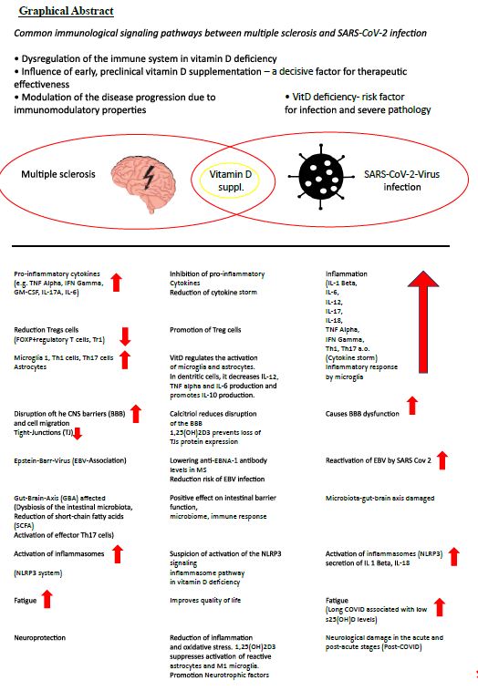
Multiple sclerosis (MS) is an inflammatory neurodegenerative disease with a suspected autoimmune origin. The disease begins earlier than current diagnostic criteria can detect. It affects the entire central nervous system and not just the white matter, as the original term-inflammatory demyelinating disorder-suggests [3]. It is characterized by a very heterogeneous course of the disease, which is represented by relapse-associated neurological deterioration, but also by an increase in disability that is independent of relapse [4]. It is generally accepted that infections in people with MS (PwMS) can have a negative impact on the course of the MS disease. This justifies that all potentially therapeutic and preventive options, especially for COVID-19, should be exploited.

MS is associated with reduced vitamin D status [5]. The molecular mechanisms in the pathogenic effect of vitamin D deficiency in MS are diverse and are orchestrated by encephalitogenic T cells with B cells, microglia, dendritic cells, interleukins (IL-1 beta, IL-6, IL-12, IL-17, TNF alpha, (tumor necrosis factor), MHCII, interferon gamma, among others) [6].

Not only is vit D deficiency associated with MS risk, but s25(OH)D levels are inversely correlated with risk of relapse, CNS lesions, and disability progression. Vitamin D suppl. reduces the number of new Gd+-enhancing or new/enlarged T2 lesions on MRI [7-10]. With MS, which is currently not curable, a high level of activity is required to prevent complications, especially infections of any kind. Severe acute respiratory syndrome coronavirus 2 (SARS-CoV-2) is an exceptionally transmissible and pathogenic coronavirus that emerged in late 2019, causing a pandemic of acute respiratory illness known as coronavirus infection 2019 (COVID-19). New omicron variants are constantly being discovered [11], for example BA.2.86 (Pirola),JN. New findings show that BA.2.86 efficiently enters lung cells and uses TMPRSS2 for entry into lung cells. The mutations S50L and K356T are for the efficient Lung cell entry of BA.2.86 is responsible. BA.2.86 has a high resistance to therapeutic antibodies and evades the antibodies induced by infection and vaccination [12,13]. COVID-19 can develop into a severe disease associated with immediate and delayed sequelae in various organs, including the central nervous system (CNS) [11].

Over the last 3 years, a complex connection between SARS-CoV-2 infection and MS has emerged [14].

Daily Vitamin D Supplementation is a Prerequisite for the Suppression of Inflammatory Processes

The risk of severe infection from COVID-19 should provide additional motivation for one daily high-dose vitamin D administration [15,16]. As part of prevention, it is worth mentioning that with circulating s25(OH)D values ≥ 55 ng/mL, the SARS-CoV-2 positivity rate was significantly lower than with values below or with deficiency [17]. Current data suggest a protective role for VitD, particularly with a lower risk of intensive care unit admission and a reduced risk of death [18,19]. In addition, the occurrence of Long-Covid is an aspect of implementing this simple, effective, safe and costeffective therapy with a broad therapeutic window for the prevention and treatment of COVID-19 disease [20-22]. Although there is still no indisputable evidence that Vit D supplementation (VitD suppl.) reduces the risk of SARSCov-2 infection in healthy individuals, there is collective evidence that it benefits vulnerable individuals [23]. PwMS with comorbidities, psychiatric illnesses, hypertension, obesity (an increased BMI may correlate with a severe course of Covid-19), age > 50 years, severe disability and methylprednisolone boost therapy as well as some DMTs (disease-modifying therapies) have a higher risk of infection and an increased risk of severe COVID-19 courses [24-26]. Infections (SARS-COV-2) can increase MS symptoms (pseudo-relapses) or cause real relapses [27]. In post-COVID syndrome (Long Covid), one in eight patients presents with symptoms such as fatigue, shortness of breath, cough, joint pain, chest pain, muscle pain, headache and paresthesia in the limbs after at least 3 months. The latter can also occur in PwMS per se [28,29]. if vitamin D administration results in a lower risk of infecton,severity of illness with admission to the intensive care unit or a reduced risk of death in people at risk,Long-Covid occurs less frequently [18,19,21,22,30-33],it is not ethically justifiable to withhold high dose vitamin-D administration from pleople at risk.

Mechanisms of Action of Vitamin D in COVID-19

Barrea et al. list in detail 14 mechanisms as described by Vit D suppl. the risk of COVID-19 infection can be reduced and sufficient Covid 19 vaccination is supported [19,29,32,34-38]. Vit D and its metabolites inactivate viruses (increase in antimicrobial peptide cathelicidin, defensins), lead to reduction of the risk of cytokine storm, reduce matrix metalloproteinase-9 concentration and thereby increase the host’s metabolic tolerance to damage, reduce the risk of pneumonia and myocarditis, lead to the reduction of the concentration of pro-inflammatory cytokines, especially interleukin 6 (IL-6), which promotes the permeability of the BBB, which leads to the potentiation of CNS damage in PwMS, is serious [29,39-42].

Vit D enables neuroprotection by reducing inflammation and oxidative stress. Low 25(OH)D levels were inversely correlated with high IL-6 levels and were independent predictors of COVID-19 severity and mortality [43]. 1,25(OH)2D3 inhibits immunoglobulin synthesis, regulates B cell activity and reduces auto-Ab production. It converts B cells into plasma cells [18]. Vit D reduces the risk of infection with EBV [29].

Current studies show evidence that chronic inflammation in Long Covid-19 Infection with reactivation of the latent Epstein-Barr virus (EBV) can lead to a worsening of the health status in PwMS [44-48]. In MS, there is a high level of molecular mimicry between the EBV transcription factor EBNA-1 and the CNS protein GlialCAM (glial cell adhesion molecule of the central nervous system [49]. Bernal et al. were able to detect EBV reactivation by detecting EBV DNA and antibodies against EBV-lytic genes [50].

In 66.7% of Long Covid patients, EBV reactivation could be demonstrated by a positive titer for EBV EA-D (early antigen-diffuse)-IgG or EBV-VCA (viral capsid-antigen)-IgM) can be provided [45]. Long COVID patients with fatigue and neurocognitive disorders were with serological evidence of recent EBV reactivation (early antigen-D [EA-D] IgG positivity) or high nuclear antigen IgG levels [51].

The triad of inflammatory markers IL-1ß, IL-6 and TNF can be found in both Long Covid and MS [52-54]. Low sun exposure acts synergistically with high EBNA-1 Ab levels and was associated with an increased risk of MS [55]. There is a connection between high EBNA-1 antibody levels and low s25(OH)D levels. On the other hand, a high dose of VitD suppl. the EBNA-1 antibody levels in PwMS [56-58]. Another parallel arises from the increase in GFAP (glial fibrillary acidic protein) as a dysfunction of the astrocytes about 4 months after the start of SARS-Cov2 infection [51]. The concentration of NfL (Neurofilament light chain), GFAP and total tau in CSF in patients with COVID-19 was often elevated with neurological symptoms [59]. Elevated sNfL has already been verified in mild to moderate COVID-19 disease [60]. On the other hand, the risk of mortality increased if sNfL and sGFAP levels were already elevated upon hospital admission [61].

Because there are no effective drugs that block EBV reactivation in Long Covid [62], there are multiple arguments for Vit D suppl. High-dose Vit D-Suppl (14,000 IU/day) for 48 weeks or 20,000 IU/week for 48 weeks selectively reduced anti-EBNA-1 antibody levels in PwMS (RRMS) [58,63].

Several mechanisms are under discussion:

  1. VitD could induce better clearance of EBV infected B cells,
  2. Vit D could directly target and impair viral replication in EBV-infected cells,
  3. Produce better control of inflammation in general,
  4. In an EBV-mediated inflammatory cascade, 1,25(OH)2D3 could suppress the activation of reactive astrocytes
  5. It is likely that at high s25(OH)D levels, the VitD receptor EBNA 2 (Epstein-Barr virus nuclear antigen 2) is displaced upon DNA binding [58,63-68].

Early Start of Therapy is a Crucial Factor

The early start of therapy with VitD suppl. is crucial for influencing influenza and COVID-19 infections [69]. The Corona-19 mortality risk correlates inversely with the VitD status and a mortality rate close to zero could theoretically be achieved at over 50 ng/mL s25(OH)D [70]. The importance of Vit-D metabolism as a potential prophylactic, immunoregulatory and neuroprotective treatment of infections (COVID-19) is increasingly being considered in clinical practice as part of a multitherapeutic approach [34,71,72].

Dosage Suggestions for Vitamin D Supplementation

Currently, there are no consensus guidelines suggesting an appropriate concentration of serum 25(OH)D to prevent COVID-19 or reduce its morbidity and mortality. It is becoming increasingly clear to start with a “loading dose” with high VitD doses over a few days and then continue with a “maintenance dose”, although various variants have been put up for discussion.

For example, one study used a weekly or fortnightly dose totaling 100,000-200,000 IU for 8 weeks (1800 or 3600 IU/day) [73].

To obtain 75 nmol/l s25(OH)D values, the following equation was described:

Dose (IU) = 40 x (75-serum 25(OH)D(3) [nmo/L] x body weight [73].

Over 30 ng/mL s25(OH)D values were also achieved with a single oral dose of 200,000-600,000 IU [38,74].

An s25(OH)D level of 40-60 ng/ml could be achieved by dosing up to 6,000 IU/day over several weeks [75,76]. A daily VitD intake of 10,000 IU/day for 4 weeks would lead to a faster optimal s25(OH)D level in the “status nascendi” of an infection [19].

Another dosage regimen was recommended: cholecalciferol 0.532 mg on day 1 and continued with 0.266 mg on days 3,7,14,21,28 (1 IU vitamin D3 = 0.025 µg vitamin D3 = 65.0 nmol Vitamin D3 [77]. The pharmacokinetic properties of calcifediol allow rapid absorption within hours, facilitating the immediate availability of 25(OH)D2 in target tissues.

This drastically reduced the need for intensive care unit admission and the mortality rate [77]. A key mechanism of 1,25(OH)D2 is its effect on Vit D receptors (VDR) on the adaptive immune system. The activity of TH1 and TH17 cells is reduced and the T regulator (Treg) cells are induced. This results in a reduced production of proinflammatory cytokines (IL-6, IL-8, IL-12, IL17, TNF alpha) and the cytokine storm is weakened [77,78].

The further daily VitD dose will depend on the s25(OH) values. The “maintenance dose” depends on the genetic polymorphism of the enzymes involved in VitD metabolism. Because interindividual differences in the organism’s response to Vit D, particularly in PwMS, are established, one of many explanations for the controversy surrounding the clinical results of Vit D suppl. [6,78,79].

An example of this individual reaction to a Vit D suppl. with 3,200 IU daily for 5 months showed a strong response to peripheral blood mononuclear cells in 60% of healthy individuals, while only a mild to moderate response was recorded in 40% despite reaching 25(OH)D values of 60-90 ng/ML was [80].

Up to Date 2024

Clinical Manifestation of a SARS-Cov-2 Infection in PwMS as Ulcerative Colitis – A Novum

Another challenge in the diagnostic diagnosis of gastroenterological symptoms is exclusively COVID-19-induced colitis (enteropathic infection) without pulmonary manifestation or as the first manifestation of COVID-19 disease [81-87]. In the ileum and colon, there is extensive expression of the angiotensin converting enzyme 2 (ACE2) on the enterocytes, to which the SARS-CoV-2 corona virus binds, penetrates the cells of the intestinal epithelium and causes the inflammation or aggravates existing one chronic inflammatory bowel disease (IBD) [84,87-90]. Molecular mimicry between SARS-CoV-2 and human proteins (enteric epitopes) promotes gut-associated autoimmune diseases [91].

SARS-CoV-2 as an autoimmunogenic virus is seen in association with another 10 autoimmune diseases and multidisciplinary management can be beneficial in long-COVID [92-94]. Vit D deficiency can promote autoimmune dysregulation [95].

PwMS are Predisposed to Comorbid Autoimmune Diseases

PwMS have a tendency to be polyautoimmune [96-98] and hundreds of common genetic susceptibility loci for autoimmune diseases have been identified [99,100]. Up to 18% of PwMS suffer from additional comorbid autoimmune disorders.

Inflammatory bowel diseases (IBD) [ulcerative colitis, Crohn’s disease]) are among the most common autoimmune diseases accompanying MS [101]. About beneficial and adverse effects of DMTs and comorbid autoimmune diseases details in [102]. Knowledge of the tendency towards polyautoimmunity is the key to the precise interpretation of symptoms, even in contrast to treatment-related undesirable side effects of anti-CD20 therapy.

Disease-Modifying Therapies (DMT) Can Increase the Risk of Infection

Long-term observation has shown an increased incidence of respiratory tract infections, urinary tract infections and SARS-CoV-2 during therapy with monoclonal anti-CD20 antibodies in MS (ocrelizumab, ofatumumab, ublituximab, rituximab) [103-105]. The predominant depletion of CD20+ B cells, but also CD20+ T cells and the effect on CD8 T cells by ocrelizumab as well as the additional reduction in immunoglobulins (IgG, IgA, IgM) explains the increased risk of infection [103,106].

Discussion

The connection between Vit D and COVID-19 has been critically examined in over 120 clinical studies, including 41 RCTs, and a strong connection between Vit D and clinical outcomes in Covid-19 has been proven.

Several mechanisms have been discussed:

  1. Affects 1,25(OH)2D3 antimicrobial peptides (cathelicidin), tigth junction proteins and adherenc junction proteins (ZO-1, occludin, claudin-10, ß-catenin, VE-cadherin) [107].
  2. 1,25(OH)2 D3 suppresses the activity of TH 1 and TH 17 cells and induces Treg cells. As a result, there is a reduced production of proinflammatory cytokines (IL-6, IL-8, IL-12, IL-17, TNF alpha) and a weakening of a cytokine storm [79].
  3. Vit D plays an important role in controlling the renin-angiotensin-aldosterone system. Details in [78].

Furthermore, genetic polymorphisms of the Vit D metabolism pathway and nongenetic reasons could explain the controversies surrounding the clinical results of Vit D supplementation [78,79,108]. If the physiological basis for the use of Vit D to improve the health of the general population has already been found with Vit D daily doses of 5000-7000 IU/day [109], it is biologically plausible to use a Vit D suppl. to be carried out preventively in the event of impaired immune homeostasis in PwMS to improve immune function. The daily dose of Vit D is crucial for the therapeutic success of broad gene expression. A daily dose of 10,000 IU leads to genomic changes that were several times higher than with 4000 IU/day [80].

Through the immunomodulatory effect of 25(OH)D and its anti-inflammatory mechanisms, immune-mediated colitis caused by anti-CD20 antibody therapy or ulcerative colitis caused by SARS-CoV-2 could be suppressed or alleviated. This form of manifestation of COVID-19 disease is particularly important in vulnerable people (PwMS) receive attention. [81-94].

Calcitriol may play a supportive role in neuroprotection particularly in PwMS by attenuating neuroinflammation and protecting the endothelial integrity of the blood-brain barrier (BBB) [110,111]. The steep learning curve in assessing clinical symptoms in LONG COVID-19 reveals new manifestations of autoimmune diseases, particularly after severe SARS-CoV-2 infections. In addition to the risk of rheumatic diseases, the occurrence of Crohn’s disease and ulcerative colitis must be taken into account in long-term care [112-116] and is a challenge in the future. Comorbidities affect PwMS more frequently than people without MS and are associated with greater physical and cognitive impairment,lower health-related quality of life,and increased mortality [117].In long-term management,one goal is to potentially avoid comorbidities.Due to the predisposition to polyautoimmunity,thyroid diseases (Hahimoto’s thyroiditis,Graves’disease) are not uncommon as comorbidities [118]. An infection of the endocrine system with SARS-CoV-2 (e.g. thyroid, adrenal gland, pituitary gland, etc.) is possible and the virus has been detected in post-mortem samples [119]. SARS -CoV-2 also mainly penetrates here the main receptor ACE2 and its co-receptor TMPRSS2 into the host cells. ACE2 protein expression was detected in about 87% of deceased COVID-19 patients. Pathological thyroid function tests correlated with the severity of the disease [119,120]. People with one already existing autoimmune disease and Covid-19 were 23% more likely to be diagnosed with another autoimmune disease [112]. In patients with comorbidities, advanced age and SARS-Cov-2, overactivation of T cells, overproduction of proinflammatory cytokines (IL-1 beta, IL-2R, IL-6, IL-8, IL-17, TNF alpha, IFN beta) and a reduction in Treg cells are confirmed [121].  Infections with SARS-CoV-2 and mRNA vaccines can trigger the clinical onset of an autoimmune disease [122]. So it must be during and after the SARS-CoV-2 infection, subacute thyroiditis, Graves disease and Hashimoto’s thyroiditis are expected [122] and the PwMS should be monitored accordingly in the event of clinical symptoms. The immunomodulatory function of vitamin D could be used as part of an early treatment strategy , as vitamin D deficiency increases the risk of autoimmune thyroid diseases [123]. There is a negative relationship between anti-thyroid antibodies (TPO-Ab, TgAb, TSHR Ab) and a sufficient serum 25(OH)D level. A Vit D suppl. led to a decrease in thyroid antibodies and in hypothyroidism, TSH levels decreased. Vit D positively influenced Hashimoto’s thyroiditis and graves disease [124-137].

A Covid-19 cohort showed a significantly higher risk of IBD and celiac disease [138]. Patients with ulcerative colitis were more likely to develop a severe form of Covid-19 than the general population [139]. Despite partly contradictory results of studies on the relationship between vitamin D, Covid-19 and IBD, it can be recognised that 25(OH)D levels above 30ng/mL can exert a protective function [140].

As Covid-19 is not a thing of the past and appears to be here to stay, an easy-to-use and inexpensive vitamin D supplement is needed and should be offered to at-risk groups. The active form of Vit D not only shows a dual effect on SARS-CoV-2 and MS, but also has a versatile spectrum of action on MS.

Summary

People with multiple sclerosis could proactively influence the course of their disease and reduce the risk of infections with possible complications through long-term prophylaxis with daily vitamin D supplementation. The immunomodulatory influence of vitamin D is undisputed and cytokine storms (COVID-19) as well as a severe course of the disease could be prevented. 25(OH)D serum values of over 50 ng/mL should be aimed for through individual daily vitamin D supplementation. The 25(OH)D serum values obtained in studies in the general population with daily doses of 5000-10,000 IU/day cannot be adequately transferred to people with multiple sclerosis and must be titrated individually. Due to the known immunopathological mechanisms of vitamin D and its benefits, it would be desirable to integrate this add-on therapy into standard clinical care.

References

  1. Wiendl H, Gold R, Berger T, Derfuss T, et al. (2021) ‘Multiple Sclerosis Therapy Consensus Group’ (MSTCG) Multiple Sclerosis Therapy Consensus Group (MSTCG): position statement on disease-modifying therapies for multiple sclerosis (white paper) Ther Adv Neurol Disord 14: 17562864211039648. [crossref]
  2. Giovannoni G, Popescu V, Wuerfel J, et al. (2022) Smouldering multiple sclerosis: the ‘real MS’. Ther Adv Neurol Disord 15: 17562864211066751. [crossref]
  3. Heming M, Wiendl H (2023) Learning multiple sclerosis immunopathogenesis from anti-CD20 therapy. Proc Natl Acad Sci 120: e2221544120. [crossref]
  4. Lublin FD, Häring DA, Ganjgahi H, et al. (2022) How patients with multiple sclerosis acquire disability. Brain 145: 3147-3161. [crossref]
  5. Rolf L, Damoiseaux J, Huitinga I, et al. (2018) Stress-Axis Regulation by Vitamin D3 in Multiple Sclerosis. Front Neurol 9: 263. [crossref]
  6. Anwar MJ, Alenezi SK, Alhowail AH (2023) Molecular insights into the pathogenic impact of vitamin D deficiency in neurological disorders. Biomed Pharmacother 162: 114718. [crossref]
  7. Miclea A, Bagnoud M, Chan A, Hoepner R (2020) A Brief Review of the Effects of Vitamin D on Multiple sclerosis. Front Immunol 11: 781. [crossref]
  8. Hupperts R, Smolders J, Vieth R,et al. (2019) Randomized trial of daily high dose vitamin D3 in patients with RRMS receiving subcutaneous interferon β-1a. Neurology 93: e 1906-e1916. [crossref]
  9. Correale J, Ysrraelit MC, Gaitán MI (2009) Immunomodulatory effects of vitamin D in multiple sclerosis. Brain 132: 1146-1160. [crossref]
  10. Wang C, Zeng Z, Wang B, Guo S (2018) Lower 25-Hydroxyvitamin D Is Associated with Higher Relapse risk in patients with Relapsing-Remitting Multiple Sclerosis. J Nutr Health Aging 22: 38-43. [crossref]
  11. Souza PFN, Mesquita FP, Amaral JL, et al. (2021) The human pandemic coronaviruses on the show: The spike glycoprotein as the Main actor in the coronaviruses play. Int J Biol Macromol 179: 1-19. [crossref]
  12. Zhang L, Kempf A, Nehlmeier I, et al. (2024) SARS-CoV-2 BA.2.86 enters lung cells and evades neutralizing antibodies with high efficiency. Cell. [crossref]
  13. Qu P, Xu K, Faraone JN, et al. (2024) Immune evasion, infectivity, and fusogenicity of SARS-CoV-2 BA.2.86 and FLip variants. Cell. [crossref]
  14. MacDougall M, El-Hajj Sleiman J, et al. (2022) SARSCoV-2 and Multiple Sclerosis: Potential for Disease Exacerbation. Front Immunol 13: 871276. [crossref]
  15. Liao S, Huang Y, Zhang J, Xiong Q, Chi M, Yang L, et al. (2023) Vitamin D promotes epithelial tissue repair and host defense responses against influenza H1N1 virus and Staphylococcus aureus infections. Respir Res 24: 175. [crossref]
  16. Mansur JL, Tajer C, Mariani J, et al. (2020) Vitamin D high doses supplementation could represent a promising alternative to prevent or treat COVID-19 infection. Clin Investigate Arterioscler 32: 267-277. [crossref]
  17. Kaufman HW, Niles JK, et al. (2020) SARS-CoV-2 positivity rates associated with circulating 25-hydroxyvitamin D levels. PLoS One 15: e 0239252. [crossref]
  18. Argano C, Mallaci Bocchio R, et al. (2023) Protective Effect of Vitamin D Supplementation on COVID-19-Related Intensive Care Hospitalization and Mortality: Definitive evidence from Meta-Analysis and Trial Sequential Analysis. Pharmaceuticals (Basel) 16: 130. [crossref]
  19. Grant WB, Lahore H, McDonnell SL, et al. (2020) Evidence that Vitamin D Supplementation Could Reduce Risk of Influenza and COVID-19 Infections and Deaths. Nutrients 12: 988. [crossref]
  20. Mercola J, Grant WB, Wagner CL (2020) Evidence Regarding Vitamin D and Risk of COVID-19 and Its Severity. Nutrients 12: 3361. [crossref]
  21. Gibbons JB, Norton EC, McCullough JS, et al. (2022) Association between vitamin D supplementation and COVID-19 infection and mortality. Sci Rep. 2022; 12: 19397. [crossref]
  22. Shah K, Varna VP, Sharma U, Mavalankar D (2022) Does vitamin D supplementation reduce COVID 19 severity?: a systematic review. QJM 115: 665-672. [crossref]
  23. Varikasuvu SR, Thangappazham B, et al. (2022) COVID-19 and vitamin D (Co-VIVID study): a systematic review and meta-analysis of randomized controlled trials. Expert Rev An Infect Ther 20: 907-913. [crossref]
  24. Longineti E, Bower H, McKay KA, et al. (2022) COVID-19 clinical outcomes and DMT of MS patients and populationbased controls . Ann Clin Transl Neurol 9: 1449-1458. [crossref]
  25. Subramanian S, Griffin G, Hewison M, et al. (2022) Vitamin D and COVID-19 Revisited. J Intern Med 292: 604-626. (crossref]
  26. Steenblock C, Toepfner N, Beuschlein F, et al. (2023) SARSCoV-2 infection and its effects on the endocrine system . Best Practice Res Clin Endocrinol Metab 37: 101761. [crossref]
  27. Cauchi M, Willis M, Andrews A, et al. (2022) Multiple sclerosis and the risk of infection: Association of British Neurologists consensus guidelines . Pract Neurol 003370. [crossref]
  28. Ballering AV, van Zon SKR (2022) Lifelines Corona Research Initiative. Persistence of somatic symptoms atier COVID-19 in the Netherlands: an observational cohort studies. Lancet 400: 452-461. [crossref]
  29. Barrea L, Verde L, Grant WB, et al. (2022) Vitamin D: A Role So in Long COVID-19? Nutrients 14: 1625. [crossref]
  30. Annweiler C, Beaudenon M, Gau er J, et al. (2022) COVIT TRIAL study group. High dose versus standard dose vitamin D supplementation in older people adults with COVID 19 (COVIT-TRIAL): A multicenter, open-label, randomized controlledsuperiority trial. PLoS Med 19: e 1003999. [crossref]
  31. Annweiler C, Cao Z, Sabatier JM (2020) Point of view: Should COVID-19 patients be supplemented with vitamin D? Maturitas 140: 24-26. [crossref]
  32. Shenoy S (2022) Gut microbiome, vitamin D, ACE2 interactions are critical factors in immune senescence and inflammatory: key for vaccine response and severity of COVID-19 infection. Inflamm Res 71: 13-26. [crossref]
  33. Bassatne A, Basbous M, Chakhtoura M, et al. (2021) The link between COVID-19 and VItamin D (VIVID): A systematic review and meta-analysis. Metabolism 119: 154753. [crossref]
  34. Peng MY, Liu WC, Zheng JQ, Lu CL, Hou YC, Zheng CM, et al. (2021) Immunological aspects of SARS-CoV-2 Infection and the Putative Beneficial Role of vitamin D. Int J Mol Sci 22: 5251. [crossref]
  35. Chiu SK, Tsai KW, Wu CC, et al. (2021) Putative role of Vitamin D for COVID-19 Vaccination. Int J Mol Sci 22: 8988. [crossref]
  36. Goncalves-Mendes N, Talvas J, et al. (2019) Impact of Vitamin D Supplementation on Influenza Vaccine Response and Immune Functions in Deficient Elderly Persons: A Randomized Placebo-Controlled Trial. Front Immunol 10: 65. [crossref]
  37. Chillon TS, Demircan K, Heller RA, et al. (2021) Relationship between Vitamin D Status and Antibody Response to COVID-19 mRNA Vaccination in Healthy Adults. Biomedicines 9: 1714. [crossref]
  38. Bae JH, Choe HJ, Holick MF, Lim S (2022) Association of vitamin D status with COVID-19 and its severity: Vitamin D and COVID-19: a narrativereview. Rev Endocrin Metab Disord 23: 579-599. [crossref]
  39. Gerhard A, Prüß H, Franke C (2022) [Manifestations of the central nervous system after COVID-19]. Nervenarzt 93: 769-778.[crossref]
  40. Orrù B, Szekeres-Bartho J, et al. [2020]. Inhibitory effects of Vitamin D on inflammation and IL-6 release. A further support for COVID-19 management?. Eur Rev Med Pharmacol Sci 24: 8187-8193. [crossref]
  41. Silberstein M (2020) Correlation between premorbid IL-6 levelsand COVID-19 mortality: Potential role for Vitamin D. Int Immunopharmacol 88: 106995. [crossref]
  42. White JH (2022) Emerging Roles of Vitamin D-Induced Antimicrobial Peptides in Antiviral Innate Immunity. Nutrients 14: 284. [crossref]
  43. Campi I, Gennari L, Merlo D, et al. (2021) Vitamin D and COVID 19 severity and related mortality: a prospective studying in Italy. BMC Infect Dis 21: 566. [crossref]
  44. Chen T, Song J, Liu H, Zheng H, Chen C (2021) Positive Epstein-Barr virus detection in coronavirus disease 2019 (COVID-19) patients. Sci Rep 11: 10902. [crossref]
  45. Gold JE, Okyay RA, Light WE, Hurley DJ (2021) Investigation of Long COVID Prevalence and Its Relationship to Epstein-Barr Virus Reactivation. Pathogens 10: 763. [crossref]
  46. Klein J, Wood J, Jaycox J, et al. (2023) Distinguishing features of Long COVID identified through immune profiling. Nature 623;139-148 [crossref]
  47. Su Y, Yuan D, Chen DG, et al. (2022) Multiple early factors anticipate post-acute COVID-19 sequelae. Cell 185: 881-895. [crossref]
  48. Cui J, Yan W, Xu S, et al. (2018) Anti-Epstein-Barr virus at VC bodies in Beijing during 2013-2017: What we have found in the different patients. PLoS ONE 13: e0193171. [crossref]
  49. Lanz TV, Brewer RC, Ho PP, et al. (2022) Clonally expanded B cells in multiple sclerosis binds EBV EBNA1 and GlialCAM. Nature 603: 321-327. [crossref]
  50. Bernal KDE, Whitehurst CB (2023) Incident of Epstein-Barr virus reactivation is elevated in COVID 19 patients. Virus Res 334: 199157. [crossref]
  51. Peluso MJ, Sans HM, Forman CA, et al. (2022) Plasma Markers of Neurologic Injury and Inflammation in People With Self-Reported Neurologic Postacute Sequelae of SARSCoV-2 infection. Neurol Neuroimmunol Neuroinflammation 9: e200003. [crossref]
  52. Bruno A, Dolceti E, Azzolini F, et al. (2022) Interleukin 6 SNP rs1818879 Regulates Radiological and Inflammatory Activity in Multiple Sclerosis. Genes (Basel) 13: 897. [crossref]
  53. Musella A, Fresegna D, et al. (2020) Prototypical ‘proinflammatory cytokine (IL-1) in multiple sclerosis: role in pathogenesis and therapeutic targeting. Expert opinion Ther Targets 24: 37-46. [crossref]
  54. Fresegna D, Bullitia S, Musella A, et al. (2020) Re-examination the Role of TNF in MS Pathogenesis and Therapy.Cells 9: 2290. [crossref]
  55. Hedström AK, Huang J, Brenner N, et al. (2021) Low sun exposure acts synergistically with high Epstein-Barr nuclear antigen 1 (EBNA-1) antibody levels in multiple sclerosis etiology. Eur J Neurol 28: 4146-4152. [crossref]
  56. Najafipoor A, Roghanian R, et al. (2015) The beneficial effects of vitamin D3 on reducing antibody titers against Epstein-Barr virus in multiple sclerosispatients. Cell Immunol 294: 9-12. [crossref]
  57. Ascherio A, Munger KL, Lünemann JD (2012) The initiation and preventionof multiple. Nat Rev Neurol 8: 602-612. [crossref]
  58. Rolf L, Muris AH, Mathias A, et al. (2018) Exploring the effect of vitamin D3 supplementa on the anti-EBV antibody response in relapsing-remitting multiple sclerosis. Mult Scler 24: 1280-1287. [crossref]
  59. Virhammar J, Nääs A, Fällmar D, et al. (2021) Biomarkers for central nervous system Injury in cerebrospinal fluid are elevated in COVID-19 and associated with neurologicalsymptoms and diseases severity. Eur J Neurol 28: 3324-3331. [crossref]
  60. Ameres M, Brandstetier S, Toncheva AA, et al. (2020) Association of neuronal injury blood marker neurofilament light chain with mild-to-moderate COVID-19. J Neurol 267: 3476-3478.
  61. Aamodt AH, Høgestøl EA, et al. (2021) Blood neurofilament light concentration at admitiance: a potential prognostic markers in COVID-19. J Neurol 268: 3574-3583. [crossref]
  62. Hashimoto K (2023) Detrimental effects of COVID-19 in the brain and therapeutic options for long COVID: The role of Epstein-Barr virus and the gut-brain axis. Mol Psychiatry. [crossref]
  63. Røsjø E, Lossius A, Abdelmagid N, et al. (2017) Effect of high-dose vitamin D3 supplementation on antibody responses against Epstein-Barr virus in relapsing remitting multiple sclerosis. Mult Scler 23: 395-402. [crossref]
  64. Ricigliano VA, Handel AE, et al. (2015) EBNA2 binds to genomic intervals associated with multiple sclerosis and overlaps with vitamin D receptor occupancy. PLoS One 10: e0119605. [crossref]
  65. Brüting C, Stangl GI, Staege MS (2021) Vitamin D, Epstein-Barr virus, and endogenous retroviruses in multiple sclerosis-facts and hypotheses.J Integr Neurosci 20: 233-238. [crossref]
  66. Marcucci SB, Obeidat AZ (2020) EBNA1, EBNA2, and EBNA3 link Epstein-Barr virus and hypovitaminosis D in multiple sclerosis pathogenesis.J Neuroimmunol 339: 577116. [crossref]
  67. Schwalfenberg GK (2021) Treatment of Infectious Mononucleosis with High Dose Vitamin D3 in Three Cases. Ann Nutr Disord & Ther 8: 1068.
  68. Sangha A, Quon M, Pfeffer G, Orton SM (2023) The Role of Vitamin D in Neuroprotection in Multiple Sclerosis: An update. Nutrients 15: 2978. [crossref]
  69. Malaguarnera L (2020) Vitamin D3 as Potential Treatment Adjuncts for COVID-19. Nutrients 12: 3512.[crossref]
  70. Borsche L, Glauner B, von Mendel J (2021) COVID-19 Mortality Risk Correlates Inversely with Vitamin D3 Status, and a Mortality Rate Close to Zero Could Theoretically Be Achieved at 50 ng / mL 25(OH) D3: Results of a Systematic Review and Meta-Analysis. Nutrients 13: 3596. [crossref]
  71. Xu Y, Baylink DJ, Chen CS, et al. (2020) The importance of vitamin D metabolism as a potential prophylactic, immunoregulatory and neuroprotective treatment for COVID 19. J Transl Med 18: 322. [crossref]
  72. Fiorino S, Zippi M, Gallo C, Sifo D, Sabbatani S, Manfredi R, et al. (2021) The rationale for a multi-step therapeutic approach based on antivirals, drugs and nutrients with immunomodulatory activity in patients with coronavirus-SARS2-induced disease of different severities. Br J Nutr 125: 275-293. [crossref]
  73. van Groningen L, Opdenoordt S, et al. (2010) Cholecalciferol loading dose guideline for vitamin D deficient adults. Eur J Endocrinol 162: 805-811. [crossref]
  74. Kearns MD, Alvarez JA, Tangpricha V (2014) Large, single-dose, oral vitamin D supplementation in adult populations: a systematic review. Endocrin Practice 20: 341-351. [crossref]
  75. Holick MF, Binkley NC, Bischoff-Ferrari HA, et al. (2011) Endocrine Society. Evaluation, treatment, and prevention of vitamin D deficiency: an Endocrine Society clinical practice guideline. J Clin Endocrinol Metab 96(7): 1911-30. [crossref]
  76. Fabbri A, Infante M, Ricordi C (2020) Editorial-Vitamin D status: a key modulator of innate immunity and natural defense from acute viral respiratory infections. Rev Med Pharmacol Sci 24: 4048-4052. [crossref]
  77. Quesada-Gomez JM, Lopez-Miranda J, et al. (2022) Vitamin D Endocrine System and COVID-19: Treatment with Calcifediol . Nutrients 14: 2716.
  78. Charoenngam N, Jaroenlapnopparat A, Mettler SK, Grover A (2023) Genetic Variations of the Vitamin D Metabolic Pathway and COVID-19 Susceptibility and Severity: Current Understanding and Existing Evidence. Biomedicines. [crossref]
  79. Gomaa AA, Abdel-Wadood YA, Thabet RH, et al. (2023) Pharmacological evaluation of vitamin D in COVID-19 and long COVID-19: recent studies confirm clinical validation and highlight metformin to improve VDR sensitivity and efficacy. Inflammopharmacology. [crossref]
  80. Shirvani A, Kalajian TA, Song A, Holick MF (2019) Disassociation of Vitamin D’s Calcemic Activity and Non-calcemic Genomic Activity and Individual Responsiveness: A Randomized Controlled Double-Blind Clinical Trial. Sci Rep 9(1): 17685. [crossref]
  81. Stawinski P, Dziadkowiec K N, Marcus A (2021) COVID-19-Induced Colitis: A Novel Relationship During Troubling Times. Cureus 13(6): e15870. [crossref]
  82. Pan L, Mu M, Yang P, et al. (2020) Clinical Characteristics of COVID-19 Patients With Digestive Symptoms in Hubei, China: A Descriptive, Cross-Sectional, Multicenter Study. Am J Gastroenterol 115(5): 766-773. [crossref]
  83. Carvalho A, Alqusairi R, Adams A, et al. (2020) SARS-CoV-2 Gastrointestinal Infection Causing Hemorrhagic Colitis: Implications for Detection and Transmission of COVID-19 Disease. Am J Gastroenterol 115(6): 942-946. [crossref]
  84. Rutigliani M, Bozzo M, Barberis A, et al. (2022) Case Report: A Peculiar Case of Inflammatory Colitis After SARS-CoV-2 Infection. Front Immunol 13: 849140. [crossref]
  85. Hutchinson KA, Karatzios C, Sant’Anna A, et al. (2023) Novel association between SARS-COV-2 infection and acute haemorrhagic colitis in a paediatric patient. J Paediatr Child Health 59(3): 563-564. [crossref]
  86. Gupta A, Madhavan MV, Sehgal K, et al. (2020) Extrapulmonary manifestations of COVID-19. Nat Med 26(7): 1017-1032. [crossref]
  87. Brunet E, Casabella A, Calzado S, et al. (2021) Ileitis as the exclusive manifestation of COVID-19. The first reported case. Gastroenterol Hepatol 44(8): 561-563. [crossref]
  88. Lerner A (2020) “Covid-19 and the Human Gut: A New Runner on the Tract.” International Journal of Celiac Disease. 8(2): 64-67.
  89. Dvornikova KA, Bystrova EY, Churilov LP, et al. (2021) Pathogenesis of the inflammatory bowel disease in context of SARS-COV-2 infection. Mol Biol Rep, (7): 5745-5758. [crossref]
  90. Vanella G, Capurso G, Burti C, et al. (2021) Gastrointestinal mucosal damage in patients with COVID-19 undergoing endoscopy: an international multicentre study. BMJ Open Gastroenterol 8(1): e000578. [crossref]
  91. Lerner A, Benzvi C, Vojdani A (2023) SARS-CoV-2 Gut-Targeted Epitopes: Sequence Similarity and Cross-Reactivity Join Together for Molecular Mimicry. Biomedicines 11(7): 1937. [crossref]
  92. Ailioaie LM, Ailioaie C, Litscher G (2023) Gut Microbiota and Mitochondria: Health and Pathophysiological Aspects of Long COVID. Int J Mol Sci 24(24): 17198. [crossref]
  93. Preziosi NA, Rizvi AH, Feerick JD, Mandelia C (2022) De Novo Pediatric Ulcerative Colitis Triggered by SARS-CoV-2 Infection: a Tale of 2 Sisters. Inflamm Bowel Dis 28(10): 1623-1625. [crossref]
  94. Dotan A, Muller S, Kanduc D, et al. (2021) The SARS-CoV-2 as an instrumental trigger of autoimmunity. Autoimmun Rev 20(4): 102792. [crossref]
  95. Abdelhamid L, Luo XM (2022) Diet and Hygiene in Modulating Autoimmunity During the Pandemic Era. Front Immunol 12: 749774. [crossref]
  96. Deretzi G, Kountouras J, Polyzos SA, et al. (2015) Polyautoimmunity in a Greek cohort of multiple sclerosis. Acta Neurol Scand 131(4): 225-30. [crossref]
  97. Perga S, Martire S, Montarolo F, et al. (2018) The Footprints of Poly-Autoimmunity: Evidence for Common Biological Factors Involved in Multiple Sclerosis and Hashimoto’s Thyroiditis. Front Immunol 9: 311. [crossref]
  98. Marrie RA, Reider N, Cohen J, Stuve O, et al. (2015) A systematic review of the incidence and prevalence of autoimmune disease in multiple sclerosis. Mult Scler 21(3): 282-93. [crossref]
  99. Brummer T, Ruck T, Meuth SG, et al. (2021) Treatment approaches to patients with multiple sclerosis and coexisting autoimmune disorders. Ther Adv Neurol Disord 14: 17562864211035542. [crossref]
  100. Gutierrez-Arcelus M, Rich SS, Raychaudhuri S (2016) Autoimmune diseases-connecting risk alleles with molecular traits of the immune system. Nat Rev Genet 17(3): 160-74. [crossref]
  101. Wang X, Wan J, Wang M, et al. (2022) Multiple sclerosis and inflammatory bowel disease: A systematic review and meta-analysis. Ann Clin Transl Neurol 9(2): 132-140. [crossref]
  102. Konen FF, Möhn N, Witte T, et al. (2023) Treatment of autoimmunity: The impact of disease-modifying therapies in multiple sclerosis and comorbid autoimmune disorders. Autoimmun Rev 22(5): 103312. [crossref]
  103. de Sèze J, Maillart E, Gueguen A, et al. (2023) Anti-CD20 therapies in multiple sclerosis: From pathology to the clinic. Front Immunol 14: 1004795. [crossref]
  104. Varley CD, Winthrop KL (2021) Long-Term Safety of Rituximab (Risks of Viral and Opportunistic Infections) Curr Rheumatol Rep 23(9): 74. [crossref]
  105. Smith JB, Gonzales EG, Li BH, Langer-Gould A (2022) Analysis of Rituximab Use, Time Between Rituximab and SARS-CoV-2 Vaccination, and COVID-19 Hospitalization or Death in Patients With Multiple Sclerosis. JAMA Netw Open 1;5(12): e2248664. [crossref]
  106. Abbadessa G, Maida E, Miele G, et al. (2022) Lymphopenia in Multiple Sclerosis patients treated with Ocrelizumab is associated with an effect on CD8 T cells. Mult Scler Relat Disord 60: 103740. [crossref]
  107. Chen H, Lu R, Zhang YG, Sun J (2018) Vitamin D Receptor Deletion Leads to the Destruction of Tight and Adherens Junctions in Lungs. Tissue Barriers 6(4): 1-13. [crossref]
  108. Holick MF, Mazzei L, García Menéndez S, et al. (2023) Genomic or Non-Genomic? A Question about the Pleiotropic Roles of Vitamin D in Inflammatory-Based Diseases. Nutrients 15(3): 767. [crossref]
  109. Wimalawansa SJ (2023) Physiological Basis for Using Vitamin D to Improve Health. Biomedicines 11(6): 1542. [crossref]
  110. Gotelli E, Soldano S, Hysa E, et al. (2023) Understanding the Immune-Endocrine Effects of Vitamin D in SARS-CoV-2 Infection: A Role in Protecting against Neurodamage. Neuroimmunomodulation 30(1): 185-195. [crossref]
  111. Zierfuss B, Larochelle, Prat A (2024) Blodd-brain barrier dysfunction in multiple sclerosis: causes, consequences, and potential effects of therapies. Lancet Neurol 23: 95-109. [crossref]
  112. Tesch F, Ehm F, Vivirito A, et al. (2023) Incident autoimmune diseases in association with SARS-CoV-2 infection: a matched cohort study. Clin Rheumatol 42 2905-2914. [crossref]
  113. Peng K, Li X, Yang D, Chan SCW, et al. (2023) Risk of autoimmune diseases following COVID-19 and the potential protective effect from vaccination: a population-based cohort study. EclinicalMedicine 63: 102154.
  114. Lim SH, Ju HJ, Han JH, et al. (2023) Autoimmune and Autoinflammatory Connective Tissue Disorders Following COVID-19. JAMA Netw Open 6(10): e2336120. [crossref]
  115. Gracia-Ramos AE, Martin-Nares E, et al. (2021) New Onset of Autoimmune Diseases Following COVID-19 Diagnosis. Cells 10(12): 3592. [crossref]
  116. Sharma C, Bayry J (2023) High risk of autoimmune diseases after COVID-19. Nat Rev Rheuma 19(7): 399-400. [crossref]
  117. Marrie RA,Fisk JD,Fitzgerald K,Kowalec K,Maxwell C,Rotstein D, et al. (2023).Etiology,effects and management of comorbidities in multiple sclerosis: recent advances.Front Immunol14: 1197195. [crossref]
  118. Edwards NC,Munsell M,Menzin J,Phillips AL (2018).Comorbidity in US patients with multiple sclerosis.Patient Relat Outcome Meas 9: 97-102. [crossref]
  119. Steenblock C, Toepfner N, Beuschlein F, Perakakis N, Mohan Anjana R, Mohan V, et al.(2023) SARS-CoV-2 infection and its effects on the endocrine system. Best Pract Res Clin Endocrinol Metab. 37(4): 101761. [crossref]
  120. Köhler VF, Knösel T, Hasmann SE, Scherer C, Hellmuth JC, Muenchhoff M, et al. (2023) Thyroidal Angiotensin-Converting Enzyme 2 Protein Expression and Thyroid Function Tests in Patients with COVID-19: Results from a Retrospective Case Series and a Prospective Cohort Study. Thyroid. 33(2): 177-185. [crossref]
  121. Mobasheri L, Nasirpour MH, Masoumi E, Azarnaminy AF, Jafari M, Esmaeili SA. (2022) SARS-CoV-2 triggering autoimmune diseases. Cytokine. ;154: 155873. [crossref]
  122. Staruszkiewicz M, Pituch-Noworolska A, Skoczen S. (2023) SARS-CoV-2 and thyroid diseases. J Transl Autoimmun 100214. [crossref]
  123. Zhao R, Zhang W, Ma C, Zhao Y, Xiong R, Wang H, et al. (2021) Immunomodulatory Function of Vitamin D and Its Role in Autoimmune Thyroid Disease. Front Immunol. 12: 574967. [crossref]
  124. Babić Leko M, Jureško I, Rozić I, Pleić N, Gunjača I, Zemunik T. (2023) Vitamin D and the Thyroid: A Critical Review of the Current Evidence. Int J Mol Sci. 24(4): 3586. [crossref]
  125. Mackawy AM, Al-Ayed BM, Al-Rashidi BM. (2013) Vitamin d deficiency and its association with thyroid disease. Int J Health Sci (Qassim) 7(3): 267-75. [crossref]
  126. Appunni S, Rubens M, Ramamoorthy V, et al. (2021). Association between vitamin D deficiency and hypothyroidism: results from the National Health and Nutrition Examination Survey (NHANES) 2007-2012. 21, 224. [crossref]
  127. Waterhouse M, Pham H, Rahman ST, Baxter C, Romero BD, Armstrong B, et al.(2023) The Effect of Vitamin D Supplementation on Hypothyroidism in the Randomized Controlled D-Health Trial. Thyroid. 33: 11, 1302-1310.[crossref]
  128. Galușca D, Popoviciu MS, Babeș EE, Vidican M, Zaha AA, Babeș VV, et al. (2022) Vitamin D Implications and Effect of Supplementation in Endocrine Disorders: Autoimmune Thyroid Disorders (Hashimoto’s Disease and Grave’s Disease), Diabetes Mellitus and Obesity. Medicina. 58(2): 194. [crossref]
  129. Villa A, Corsello A, Cintoni M, Papi G, Pontecorvi A, Corsello SM, et al. (2020) Effect of vitamin D supplementation on TSH levels in euthyroid subjects with autoimmune thyroiditis. Endocrine 70(1): 85-91. [crossref]
  130. Pankiv, V., Yuzvenko, T., Koval, S., Singh, K., Pankiv, I., Sehgal, T. et al. (2020) Correlation of vitamin D level with thyroid status and TSH antibody titers in patients with Graves’ disease. INTERNATIONAL JOURNAL OF ENDOCRINOLOGY (Ukraine). 16;305-309.
  131. Khozam SA, Sumaili AM, Alflan MA, Shawabkeh RAS. (2022) Association Between Vitamin D Deficiency and Autoimmune Thyroid Disorder: A Systematic Review. Cureus. 14(6): e25869. [crossref]
  132. Ma J, Wu D, Li C, et al. (2015) Lower serum 25-hydroxyvitamin D level is associated with 3 types of autoimmune thyroid diseases. Medicine (Baltimore). 94: e1639. [crossref]
  133. Kivity S, Agmon-Levin N, Zisappl M, et al. (2011) Vitamin D and autoimmune thyroid disease. Cell Mol Immunol 8: 243-247. [crossref]
  134. Tamer G, Arik S, Tamer I, Coksert D.(2011) Relative vitamin D insufficiency in Hashimotoʼs thyroiditis. Thyroid. 21: 891-896. [crossref]
  135. Wang X, Zynat J, Guo Y, et al.(2015) Low serum vitamin D is associated with anti-thyroid-globulin antibody in female individuals. Int J Endocrinol 2015: 1-6.
  136. Goswami R, Marwaha RK, Gupta N, et al. (2009) Prevalence of vitamin D deficiency and its relationships with thyroid autoimmunity in Asian Indians: a community-based survey. Br J Nutr. 102: 382-386. [crossref]
  137. Chailurkit LO, Aekplakorn W, Ongphiphadhanakul B. (2013) High vitamin D status in younger individuals is associated with low circulation thyrotropin. Thyroid 23: 25-30.
  138. Chang R, Yen-Ting Chen T, Wang SI, Hung YM, Chen HY, Wei CJ. (2023) Risk of autoimmune diseases in patients with COVID-19: A retrospective cohort study. EClinicalMedicine. 56: 101783. [crossref]
  139. Yang, J., Ke, J., Jiang, X. et al.(2024) The association between ulcerative colitis and COVID-19 severity: a systematic review and meta-analysis systematic review. Int J Colorectal Dis. 39, 5. [crossref]
  140. De Nicolò A, Cusato J, Bezzio C, Saibeni S, Vernero M, Disabato M, et al. (2022) Possible Impact of Vitamin D Status and Supplementation on SARS-CoV-2 Infection Risk and COVID-19 Symptoms in a Cohort of Patients with Inflammatory Bowel Disease. Nutrients. 15(1): 169. [crossref]
    Part of this publication appeared in:
    Goischke H.-K. (2023) What immunopathogenic similarities exist between SARS-CoV-2 infection and multiple sclerosis?: A plea for daily vitamin D supplementation to improve quality of life! J Neurol Transl Neurosci 8(1): 1093.
fig 1

Febrile Neutropenia in Pediatric Oncology: Prevalence and Risk-Factors for Bacterial and Fungal Infection

DOI: 10.31038/IDT.2024511

Introduction

Febrile neutropenia (FN) frequently complicates cancer treatment, contributing to overall morbidity and the burden of hospitalization in children with cancer [1-6]. Myelosuppression is a side-effect of cytotoxic chemotherapy, resulting in recurring episodes of neutropenia; fever complicates about 27-34% of neutropenic episodes among children receiving chemotherapy or undergoing hematopoietic stem cell transplant (HSCT) [1-3]. Due to the risk of serious bacterial or fungal infections in children with FN, the difficulty of localizing infections in neutropenic children, and the mortality rate associated with inadequate treatment [4,5] the historical standard in pediatric hematology-oncology was hospitalization with empiric broad-spectrum antibiotics until the fever resolves and the neutropenia improves [6-8]. More recently, however, there is evidence of greater practice variation [9,10] based on practice guidelines emphasizing risk-stratification of children with chemotherapy-induced FN and the benefits of decreasing inpatient hospitalization [9,11-27]. Most FN episodes resolve without diagnosis of a serious infection; bacteremia, the most common infection complicating FN episodes, has a prevalence of 20%-29 [11,28-32]. Invasive fungal infection (IFI) is less common, occurring in less than 5% of FN episodes [32] and data on bacterial infection of sites other than the bloodstream are more limited. There is no single approach to risk-stratification in pediatric FN [26] which necessitates ongoing analysis of risk factors for serious infection, which facilitate risk-stratification and step-down management of children at lower risk [25,27,32,33].

Materials and Methods

Study Design

To evaluate the prevalence and potential predictors of bacterial and fungal infection among pediatric oncology patients with FN at our institution, we conducted a retrospective cohort study containing a nested case-control study. Using hospital billing codes and electronic medical records, we obtained a consecutive 3-year sample of children admitted with FN to the pediatric hematology-oncology teams at UCSF Benioff Children’s Hospital Oakland (Oakland, CA, US), with the end of the sample period preceding the Covid-19 pandemic. Children receiving treatment for cancer were included in the cohort if they had an absolutely neutrophil count (ANC) <500 x109/L or (if no ANC was reported) a total white blood cell count (WBC) <500 x109/L, as well as a single temperature >38.3°C or a sustained temperature >38°C [34]. Participants were excluded if they were receiving or had previously received allogeneic or autologous HSCT or had an underlying syndrome (such as Fanconi anemia) associated with chronic neutropenia. This study was approved by our hospital’s institutional review board and conducted in accordance with the Declaration of Helsinki.

Statistical Methods

Continuous variables, including participants’ ages and days to infection diagnosis, were not normally distributed and are described using median and interquartile range (IQR). Children with multiple episodes of FN during the sample period reentered the cohort for each episode. For the case-control analysis, we randomly sampled one episode per participant. Cases were defined by culture-proven bacteremia, urinary tract infection (UTI), meningitis, cellulitis, osteomyelitis, neutropenic colitis (typhlitis), Clostridium difficile enterocolitis, or invasive fungal infection (IFI). Clinical and radiographic findings were accepted for diagnosis of typhlitis, osteomyelitis, and IFI if cultures were not available [35]. Per institutional standards of care, any positive blood culture from a central venous catheter (CVC) was considered infectious, including coagulase-negative staphylococci. Controls were sampled at a two-to-one ratio with cases. To compare clinical and laboratory findings between the case and control groups, we used rank-sum tests for continuous variables and standard two-by-two tables with Fisher exact tests for categorical variables. Associations were considered significant with an uncorrected p-value <0.05. We also report each association’s relative risk ratio (RR) with a 95% confidence interval. Data analysis was performed using Stata 13 (Statacorp, College Station, TX).

Results

Study Cohort

The cohort (Table 1) consisted of 199 FN episodes among 140 participants, 43% female, with a median age at cohort entry of 6.1 years (3.1-12.3). Most participants (71.4%) were hospitalized once for FN during the study period; among the rest, the number of hospitalizations ranged from 2 to 8. The most common diagnoses were acute leukemia and lymphoma. There were 5 participants (3.6%) with trisomy 21, all of whom had acute leukemia. Nearly all of the participants had a CVC, and 31 (22.1%) had a history of at least one prior infection, including bacteremia (N=21), another bacterial infection (N=7), or IFI (N=4). All participants received empiric intravenous antibiotics with antipseudomonal activity upon the onset of fever. Most of the FN episodes (81.9%) developed in outpatients who were then admitted; 36 FN episodes (18.1%) occurred in children who were already hospitalized, especially those receiving high-intensity chemotherapy for acute myeloid leukemia (AML) or brain tumors. Among participants with acute lymphoblastic leukemia (ALL), 26 FN episodes (13.1% of the total) occurred during the lower-intensity maintenance phase of therapy.

Table 1: Demographic and clinical characteristics

Parameter

N (%)

Sex

 Female

60 (42.9)

 Male

80 (57.1)

Age at onset (years), median (IQR)

6.1 (3.1-12.3)

Trisomy 21

5 (3.6)

Diagnosis
 ALL

61 (43.6)

 Brain tumor

19 (13.6)

 Sarcoma

18 (12.9)

 Lymphoma

13 (9.3)

 AML

7 (5.0)

 Neuroblastoma

7 (5.0)

 Wilms tumor

6 (4.3)

 Hepatoblastoma

5 (3.6)

 Other diagnosis*

4 (2.9)

History of cancer relapse

21 (15.0)

Central venous catheter

127 (90.7)

IQR: Interquartile Range; ALL: Acute Lympoblastic Leukemia; AML: Acute Myelogenous Leukemia; UTI: Urinary Tract Infection.
*Desmoplastic small round cell tumor (N=1), renal carcinoma (N=2), and rhabdoid liver tumor (N=1).

Infectious complications

Of the 199 FN episodes studied, 43 (21.6%) led to a diagnosis of bacterial or fungal infection (Figure 1), with 6 episodes (3%) involving multiple infections. The most common was bacteremia, of which there were 29 cases (14.6%); cultures were positive for Gram-positive organisms in 18 (including 8 with coagulase-negative staphylococci), Gram-negative organisms in 8, and mixed flora in 3. Bacteremia was diagnosed a median of 1 day (1-3) after fever onset. There were 16 cases (8%) of other bacterial infections, which were diagnosed a median of 4 days (2-6) after fever onset and included typhlitis (N=5), Clostridium difficile enterocolitis (N=4), UTI (N=3), and cellulitis (N=3); 5 of these infections occurred along with bacteremia. There were 5 cases of IFI, most commonly pulmonary aspergillosis, diagnosed a median of 5 days (0-9) after fever onset. Overall, the median time from fever onset to diagnosis was 2 days (1-4). Distributive shock requiring intensive care occurred in 4 FN episodes (2%) due to bacteremia or meningitis, and one of these children died.

fig 1

Figure 1: Overview of bacterial and fungal infections in the cohort. For episodes with multiple infections, the left panel categorizes the first diagnosed. UTI, urinary tract infection; C. difficile, Clostridium difficile enterocolitis.

Risk Factors for Bacterial or Fungal Infection

The case-control sample consisted of 40 cases and 80 controls (Table 2). There was not a statistically significant difference in age or sex between the cases and controls, although the case group contained a larger proportion of children <1 year of age and a larger proportion of children who were already hospitalized at fever onset. at the onset of FN. The relative risk of infection was markedly higher in children with trisomy 21 (RR 3.11 [2.39-4.03]) and those with AML (RR 2.11 [1.13-3.95]), although these p-values were >0.05. While cases were slightly more likely to have a temperature ≥39°C, presenting temperature and laboratory values were not significantly different between cases and controls, nor were clinical findings like mucositis and gastrointestinal upset. Cases were more likely to have fever recurrence after >24 hours afebrile and also to have fevers lasting ≥7 days, although these associations were not statistically significant. There was a significantly increased risk of infection (p<0.004) for participants with a prior history of prior bacterial or fungal infection (RR 2.16 [1.34 to 3.48]).

Table 2: Univariate analysis of a nested case-control sample of pediatric patients with febrile neutropenia (FN)

Risk factor, N (%)

Cases (N=40) Controls (N=80) p

Relative risk (95% CI)

Demographic and historical features
Sex

27 (67.5)

41 (51.2) 0.118 1.59 (0.91 to 2.77)

Relapsed

6 (15.0) 12 (15.0) 1.000

1.00 (0.49 to 2.03)

Age at onset (years)*

6.6 (4.2-15.9)

7.2 (3.1-12.3) 0.432

Age <1 year

3 (7.5) 2 (2.5) 0.332

1.86 (0.87 to 4.00)

Trisomy 21

2 (5.0)

0 (0.0) 0.109 3.11 (2.39 to 4.03)

Diagnosis of AML

4 (10.0) 2 (2.5) 0.095

2.11 (1.13 to 3.95)

Prior infection

18 (45.0)

15 (18.8) 0.004

2.16 (1.34 to 3.48)

Findings at FN onset
Temperature (oC)*

39 (38.6-39.7)

38.8 (38.4-39.3) 0.052

Presenting WBC (x109/L)*

0.3 (0-0.8) 0.5 (0.2-0.9) 0.106

Presenting ANC (x109/L)*

115.5 (0-250)

86.0 (11-348) 0.538

Already admitted

10 (25.0) 10 (12.5) 0.118

1.67 (0.98 to 2.83)

Rhinitis or rhinorrhea

5 (12.5)

16 (20.0) 0.445 0.67 (0.30 to 1.51)

Severe mucositis

7 (17.5) 12 (15.0) 0.793

1.13 (0.59 to 2.16)

Abdominal pain

8 (20.0)

11 (13.8) 0.430 1.33 (0.73 to 2.42)

Vomiting

13 (32.5) 15 (18.8)

0.111

1.58 (0.95 to 2.63)

Findings at reevaluation
Fever duration (days)*

2 (1.5-5)

2 (1-4) 0.346

Fever recurrence

12 (30.0) 13 (16.3) 0.097

1.63 (0.97 to 2.72)

Fever for ≥7 days

7 (17.5)

8 (10.0) 0.255

1.48 (0.81 to 2.73)

*Reported as median and interquartile range.
P-values are from Fisher exact tests for proportions and rank-sum tests for continuous variables. CI: Confidence Interval; AML: Acute Myeloid Leukemia; WBC: White Blood Cells; ANC: Absolute Neutrophil Count.

Discussion

In this consecutive sample of 199 FN episodes in a typical pediatric oncology population at a United States tertiary-care hospital, 21.6% were complicated by a bacterial or fungal infection, most frequently Gram-positive bacteremia. UTI was more common than expected, likely reflecting our emergency department’s practice of obtaining non-catheterized urine samples from most febrile children. Although undiagnosed UTI would likely be treated by empiric antibiotics, this source of pathology in children with FN warrants further investigation. In a nested case-control analysis, the relative risk of bacterial or fungal infection was higher in children with trisomy 21 and those with AML and considerably higher in those with a prior history of infection. Infections diagnosed during the study period were generally not relapses of prior infection; instead, infection risk may reflect cumulative person-level factors, including duration of chemotherapy, cumulative antibiotic exposure, and differences in the microbiome.

As with any observational retrospective study, these findings are not definitive. Our broadly inclusive definition of infection was designed to reflect clinical decision-making, with emphasis on clinical data that would indicate a change in management or a longer course of inpatient observation. The overall similarity between groups in the case-control analysis, as well as the fact that infections occurred during relatively low-intensity chemotherapy (like maintenance ALL therapy) emphasizes the challenge of risk-stratifying children with FN. Most infections in this cohort, however, were diagnosed within the first 4 days after the onset of fever. For children without trisomy 21, AML, or a prior infection history, who do not have overt signs of infection, there may be less benefit of hospitalization longer than through neutrophil recovery, as long as careful outpatient follow-up can be assured.

Conflict of Interest

The authors have no conflicts of interest or external funding sources to disclose.

References

  1. Bagnasco F, Haupt R, Fontana V, et al. (2012) Risk of repeated febrile episodes during chemotherapy-induced granulocytopenia in children with cancer: a prospective single center study. J Chemother 24(3): 155-160. doi: 10.1179/1973947812Y.0000000002
  2. Castagnola E, Fontana V, Caviglia I, et al. (2007) A prospective study on the epidemiology of febrile episodes during chemotherapy-induced neutropenia in children with cancer or after hemopoietic stem cell transplantation. Clin Infect Dis 45(10): 1296-1304. [crossref]
  3. Castagnola E, Garrè ML, Bertoluzzo L, et al. (2011) Epidemiology of febrile neutropenia in children with central nervous system tumor: results from a single center prospective study. J Pediatr Hematol Oncol 33(7): e310-315. [crossref]
  4. Davis K, Wilson S (2020) Febrile neutropenia in paediatric oncology. Paediatr Child Health (Oxford) 30(3): 93-97. [crossref]
  5. Boccia R, Glaspy J, Crawford J, Aapro M (2022) Chemotherapy-Induced Neutropenia and Febrile Neutropenia in the US: A Beast of Burden That Needs to Be Tamed? Oncologist 27(8): 625-636. [crossref]
  6. Pizzo PA (1993) Management of fever in patients with cancer and treatment-induced neutropenia. N Engl J Med 328(18): 1323-1332. [crossref]
  7. Freifeld A, Marchigiani D, Walsh T, et al. (1999) A Double-Blind Comparison of Empirical Oral and Intravenous Antibiotic Therapy for Low-Risk Febrile Patients with Neutropenia during Cancer Chemotherapy. New England Journal of Medicine 341(5): 305-311. [crossref]
  8. Basu SK, Fernandez ID, et al. (2005) Length of Stay and Mortality Associated With Febrile Neutropenia Among Children With Cancer. JCO 23(31): 7958-7966. [crossref]
  9. Boragina M, Patel H, Reiter S, et al. (2007) Management of febrile neutropenia in pediatric oncology patients: a Canadian survey. Pediatr Blood Cancer 48(5): 521-526. [crossref]
  10. Maxwell RR, Egan-Sherry D, Gill JB, et al. (2017) Management of chemotherapy-induced febrile neutropenia in pediatric oncology patients: A North American survey of pediatric hematology/oncology and pediatric infectious disease physicians. Pediatr Blood Cancer 64(12). [crossref]
  11. Agyeman P, Kontny U, Nadal D, et al. (2014) A Prospective Multicenter SPOG 2003 FN Study of Microbiologically Defined Infections in Pediatric Cancer Patients with Fever and Neutropenia. Pediatr Infect Dis J. [crossref]
  12. Brack E, Bodmer N, Simon A, et al. (2012) First-day step-down to oral outpatient treatment versus continued standard treatment in children with cancer and low-risk fever in neutropenia. A randomized controlled trial within the multicenter SPOG 2003 FN study. Pediatr Blood Cancer 59(3): 423-430. [crossref]
  13. Dommett R, Geary J, Freeman S, et al. (2009) Successful introduction and audit of a step-down oral antibiotic strategy for low risk paediatric febrile neutropaenia in a UK, multicentre, shared care setting. Eur J Cancer 45(16): 2843-2849. doi: 10.1016/j.ejca.2009.06.003 [crossref]
  14. Gupta A, Swaroop C, Agarwala S, et al. (2009) Randomized controlled trial comparing oral amoxicillin-clavulanate and ofloxacin with intravenous ceftriaxone and amikacin as outpatient therapy in pediatric low-risk febrile neutropenia. J Pediatr Hematol Oncol 31(9): 635-641. [crossref]
  15. Kern WV (2006) Risk Assessment and Treatment of Low-Risk Patients with Febrile Neutropenia. Clin Infect Dis 42(4): 533-540. [crossref]
  16. Lucas KG, Brown AE, Armstrong D, et al. (1996) The identification of febrile, neutropenic children with neoplastic disease at low risk for bacteremia and complications of sepsis. Cancer 77(4): 791-798. [crossref]
  17. Manji A, Beyene J, Dupuis LL, et al. (2012) Outpatient and oral antibiotic management of low-risk febrile neutropenia are effective in children—a systematic review of prospective trials. Supportive Care in Cancer 20(6): 1135-1145. [crossref]
  18. Mullen CA, Petropoulos D, Roberts WM, et al. (1999) Outpatient treatment of fever and neutropenia for low risk pediatric cancer patients. Cancer 86(1): 126-134. [crossref]
  19. Phillips B, Wade R, et al. (2010) Systematic review and meta-analysis of the discriminatory performance of risk prediction rules in febrile neutropaenic episodes in children and young people. European Journal of Cancer 46(16): 2950-2964. [crossref]
  20. Rackoff WR, Gonin R, Robinson C, et al. (1996) Predicting the risk of bacteremia in childen with fever and neutropenia. J Clin Oncol 14(3): 919-924. [crossref]
  21. Rondinelli PIP, Ribeiro K de CB, de Camargo B (2006) A proposed score for predicting severe infection complications in children with chemotherapy-induced febrile neutropenia. J Pediatr Hematol Oncol 28(10): 665-670. [crossref]
  22. Santolaya ME, Alvarez AM, Avilés CL, et al. (2002) Prospective evaluation of a model of prediction of invasive bacterial infection risk among children with cancer, fever, and neutropenia. Clin Infect Dis 35(6): 678-683. [crossref]
  23. Teuffel O, Ethier MC, Alibhai SMH, et al. (2011) Outpatient management of cancer patients with febrile neutropenia: a systematic review and meta-analysis. Ann Oncol 22(11): 2358-2365. [crossref]
  24. Cennamo F, Masetti R, Largo P, et al. (2021) Update on Febrile Neutropenia in Pediatric Oncological Patients Undergoing Chemotherapy. Children (Basel) 8(12): 1086. [crossref]
  25. Lehrnbecher T, Phillips R, Alexander S, et al. (2012) Guideline for the management of fever and neutropenia in children with cancer and/or undergoing hematopoietic stem-cell transplantation. J Clin Oncol 30(35): 4427-4438. [crossref]
  26. Lehrnbecher T, Robinson P, Fisher B, et al. (2017) Guideline for the Management of Fever and Neutropenia in Children With Cancer and Hematopoietic Stem-Cell Transplantation Recipients: 2017 Update. JCO 35(18): 2082-2094. [crossref]
  27. Lehrnbecher T, Robinson PD, Ammann RA, et al. (2023) Guideline for the Management of Fever and Neutropenia in Pediatric Patients With Cancer and Hematopoietic Cell Transplantation Recipients: 2023 Update. J Clin Oncol 41(9): 1774-1785. [crossref]
  28. Alexander SW, Wade KC, Hibberd PLMD, Parsons SKMD (2002) Evaluation of Risk Prediction Criteria for Episodes of Febrile Neutropenia in Children with Cancer. Journal of Pediatric Hematology 24(1): 38-42. [crossref]
  29. Ammann RA, Hirt A, Lüthy AR, Aebi C (2003) Identification of children presenting with fever in chemotherapy-induced neutropenia at low risk for severe bacterial infection. Med Pediatr Oncol 41(5): 436-443. [crossref]
  30. Hakim H, Flynn PM, Knapp KM, et al. (2009) Etiology and clinical course of febrile neutropenia in children with cancer. J Pediatr Hematol Oncol,(9): 623-629. [crossref]
  31. Hakim H, Flynn PM, Srivastava DK, et al. (2010) Risk Prediction in Pediatric Cancer Patients With Fever and Neutropenia: The Pediatric Infectious Disease Journal 29(1): 53-59. [crossref]
  32. Boeriu E, Borda A, Vulcanescu DD, et al. (2022) Diagnosis and Management of Febrile Neutropenia in Pediatric Oncology Patients-A Systematic Review. Diagnostics (Basel) 12(8): 1800. [crossref]
  33. Teuffel O, Sung L (2012) Advances in management of low-risk febrile neutropenia. [Miscellaneous Article]. Current Opinion in Pediatrics 24(1): 40-45. [crossref]
  34. Freifeld AG, Bow EJ, Sepkowitz KA, et al. (2011) Clinical practice guideline for the use of antimicrobial agents in neutropenic patients with cancer: 2010 update by the infectious diseases society of america. Clin Infect Dis 52(4): e56-93. [crossref]
  35. Ascioglu S, Rex JH, de Pauw B, et al. (2002) Defining opportunistic invasive fungal infections in immunocompromised patients with cancer and hematopoietic stem cell transplants: an international consensus. Clin Infect Dis 34(1): 7-14. [crossref]
fig 2

Morphological Variation of Permanent Mandibular First Molar (Radix Entomolaris) (In vivo Study)

DOI: 10.31038/JDMR.2024711

Abstract

Objectives: Successful endodontic treatment requires complete information about morphology of root canals of the tooth. The object of our research was to assess the prevalence of third root in permanent mandibular first molars between Iraqi people.

Methods: Two hundred fifty-seven patients (161 females and 96 males) were included in this study. Those patients required endodontic treatment for permanent mandibular first molar. Examination of teeth was done during a period of one and a half year. Digital radiograph was used to investigate the occurrence of radix entomolaris. Comparison of the percentage of third root between males and females were established. Data were statistically analyzed with chi-square test.

Results: Statistical evaluation was carried out for the presence of third root among males and females using chi-square test with SPSS version 20. In this test P > 0.05 (non-significant), P ≤ 0.05 (significant). Total incidence of radix entomolaris was five teeth (5/257) the percentage was 1.9 %. Statistical analysis revealed a non-significant difference in the incidence of third root between females (2/161) and males (3/96).

Conclusion: Endodontists should have thorough information about anatomical variations of the root and root canals of mandibular first molar during endodontic treatment. Correct examination clinically and radiographically is essential to discover the presence of any morphological variations of the root canal system.

Keywords

Digital radiograph, Distolingual root, Endodontic treatment, Radix entomolaris, Maxillary first molar

Introduction

Scientific knowledge of the morphology of the tooth roots and their canals plays an important role during endodontic treatment. This will ensure complete debridement of all infected pulp with complete instrumentation and obturation [1]. A periapical lesion may be result from failed endodontic treatment if any one of these steps are inadequately done [2]. Generally, permanent mandibular first molars have two roots: one root mesially and one root distally. Two root canals present in the mesial root (mesiobuccal and mesiolingual). One canal present in the root distally, sometime distal root may comprise a second canal. The presence of the further distolingual root in the mandibular first molar is considered the main variant of this tooth, named radix entomolaris (RE) and its occurrence is infrequent. There are numerous anatomical surveys established a racial origin for the occurrence of third distolingual root in the permanent mandibular first molar. It presents with an incidence of 5 to more than 30% in populations with Mongoloid traits, such as Chinese, Eskimos, and American-Indians [3,4], A maximum rate of 3% in African population [5,6], while its prevalence in Europeans was less. Cinically, it is of great importance to provide sufficient information about morphological variation of any tooth that can affect the success rate of the root canal therapy. Our research was aimed to determine the prevalence of third root distolingually in the permanent mandibular first molar in Iraqi people.

Materials and Methods

Two hundred fifty-seven patients with an average age of 20-45 years old (161 females and 96 males) were included in this study. Those patients attained private dental clinic for endodontic treatment. Two Preoperative radiographs were done for each patient with a digital X-ray sensor (Visiodent RVG Dental Sensor, France). Sign of the presence of third root is translucent lines defining the pulp space and the periodontal ligaments located in the superior part of the distal root. Firstly, anaesthetic solution was injected to patient. Rubber dam was applied for tooth isolation. Then prepared the access cavity. Pulp extirpation was accomplished. Apex locator (Woodpex III Golden Apex Locater, Zhengzhou Linker Medical Equipment Co., Ltd. China) was used to determine working length. Radiograph using digital X-ray sensor was taken to establish working length. The next step is the instrumentation of all canals by using a rotary endodontic system (protaper Gold, Dentsply, Maillefer, Switzerland), then obturation was done by using single cone with bioceramic sealer. After finishing, a radiograph was obtained in order to evaluate the efficiency of the obturation (Figures 1-3).

fig 1

Figure 1: Preoperative radiograph

fig 2

Figure 2: Cone fit checking

fig 3

Figure 3: Obturation with bioceramic sealer

Results

Statistical evaluation was carried out for the presence of third root among males and females using chi-square test with SPSS version 20. In this test P > 0.05 (Non-significant), P ≤ 0.05 (Significant) Two hundred fifty-seven patients (161 females and 96 males) were included in this study. Total incidence of radix entomolaris was five teeth (5/257) the percentage is 1.9 %. Statistical analysis revealed a non-significant difference (P > 0.05) in the incidence of third root between females (2/161) and males (3/96) as shown in Table 1.

Table 1: Percentage of radix entomolaris in the tested patients

tab 1

Discussion

The first Permanent teeth erupted is the Mandibular first molar. This tooth highly involved with tooth decay and commonly need to root canal treatment. Missed canals with the subsequent incomplete instrumentation of all infected pulp tissues are one of the most common reasons of unsuccessful endodontic treatment. Full familiarity with morphological variations of root plays an important role in increasing the chance of the successful root canal treatment. In the cases of incomplete treatment for all root canals such as the third root distolingually this will result in failure of the endodontic treatment. Radix entomolaris has been classified into the following types: Type I means straight root or canal. Type II means the beginning of the entry is curved and then continued as a straight root/root canal. Type III means the beginning of the entry is curved in the coronal third of the root canal with the presence of another curve started in the middle and continuing to the apical third [7]. Requirement of successful endodontic treatment is the preoperative radiograph with a correct examination of the tooth clinically [8]. Thorough information about the presence of additional root or root canals can be obtained by taking a radiograph with various angles [7]. The clinician can suspect the presence of additional third root when he noticed changing of the tooth form coronally, like a highly clear distolingual lobe with a convex cervical outline [9]. Many studies stated that taking two radiographs with mesial or distal shifting cone (30 degrees) very helpful for investigating occurrence of the of radix entomolaris [10]. Rectangular or trapezoidal outline access cavity preparation should be performed when the additional third root is established or suspected radiographically. The location of the orifice of radix entomolaris is disto to mesiolingually from the main distal canal. If the access to third root not clearly seen after the roof of the pulp chamber is removed, this need to complete detection of wall and floor of the pulp chamber, mainly distolingually by using sharp endodontic explorer [9]. In the current study, the total percentage of third root incidence in mandibular first molars was 1.9%, the result in accordance with other investigations that were accomplished on Middle East people [10,11]. In comparison with data of another studies collected for Asian origin our result is lower, they reported the incidence of additional root as follows: in Koreans 4.5% [12], in Chinese 32% [13], and in Taiwanese 25.6% [14]. Our result showed a non significant difference in the occurrence of radix entomolaris between males and females. Other studies showed similar results [15-18]. Previous in vivo study was accomplished by Mukhaimer and Azizi whom study the incidence of radix entomolaris in Palestinian people when they attained a dental center for endodontic treatment, the total percentage of third root incidence in mandibular first molars was 3.73%, which considered within the range of other researches done for the Middle East population, although it was significantly lesser than the range obtained for population of far east [19]. Whenever the additional root suspected to presence during endodontic treatment of permanent mandibular first molar, modification of the access cavity preparation should be performed in order to ensure complete cleaning and obturation of all root canals, Otherwise the presence of missed canal can result in failure of the treatment.

Conclusion and Recommendation

Endodontists should have thorough information about anatomical variations of the root and root canals of mandibular first molar during endodontic treatment. Correct examination clinically and radiographically is essential to discover the presence of any morphological variations of root canal system. Preoperative radiographs with certain angulation are a vital issue in this matter. Modification of access cavity is necessary to locate all root canal orifices to certify complete debridement and obturation.

Conflict of Interest

The authors declare that they have no conflicts of interest.

Source of Funding

This research did not receive any specific grant from funding agencies in the public.

Ethical Approval

This research was performed in accordance with Helsinki Declaration and approved by the Ethical Approval Committee at university of Anbar.
Date: 21/6/2021
No.83

Author Contribution

All authors have participated sufficiently in this work in order to have a public responsibility for its contents, including conception and design, or analysis and interpretation of the data, conscripting the article or revising it critically for important intellectual content; and final approval of the article. All authors agree to be accountable for all aspects of the work in ensuring that questions related to the accuracy or integrity of any part of the work are appropriately investigated and resolved.

NAH Concept and design of the study, research conduction, OHA collection and organization of data, analyzing and interpretation of data, HAS writing the initial and final draft of the manuscript, and all authors have critically reviewed and approved the final draft and are agreed for their responsibility for the contents and similarity index of the manuscript.

References

  1. Sjogren U, Agglund BH, Sundqvis Gt, Wing K (1990) Factors affecting the long-term results of endodontic treatment, J Endod 16: 498-504. [crossref]
  2. Siqueira JF (2001) Aetiology of root canal treatment failure: why well-treated teeth can fail. Int Endod J 34: 1-10. [crossref]
  3. Turner II CG (1971) Three-rooted mandibular first permanent molars and the question of American Indian origins. Amer J Phy Anth 34: 229-241. [crossref]
  4. Curzon ME, Curzon JA (1971) Three-rooted mandibular molars in the Keewatin Eskimo. J Can Den Ass 37: 71-72. [crossref]
  5. Sperber GH, Moreau J L (1998) Study of the number of roots and canals in Senegalese first permanent mandibular molars. Int Endod J 31: 117-122. [crossref]
  6. Ahmed HA, Abu-Bakr NH, Yahia NA, Ibrahim YE (2007) Root and canal morphology of permanent mandibular molars in a Sudanese population. Int Endod J 40: 766-771. [crossref]
  7. De Moor RJG, Deroose CA, Calberson FLG (2004) The radix entomolaris in mandibular first molars: an endodontic challenge. Int Endod J 37: 789-799. [crossref]
  8. Carlsen O, Alexandersen V (1999) Radix paramolaris and radix distomolaris in Danish permanent maxillary molars. Acta Odont Scan 57: 283-289. [crossref]
  9. Calberson FL, De Moor RJ, Deroose CA (2007) The radix entomolaris and paramolaris: clinical approach in endodontics. J Endod 33: 58-63
  10. Naoum HJ, Love RM, Chandler NP, Herbison P (2003) Effect of X-ray beam angulation and intraradicular contrast medium on radiographic interpretation of lower first molar root canal anatomy. Int Endod J 36: 12-19. [crossref]
  11. Al-Nazhan S (1999) Incidence of four canals in root-canal-treated mandibular first molars in a Saudi Arabian subpopulation. Int Endod J 32: 49-52. [crossref]
  12. Al-Qudah AA, Awawdeh LA (2009) Root and canal morphology of mandibular first and second molar teeth in a jordanian population. Int Endod J 42: 775-784. [crossref]
  13. Song JS, Choi HJ, Jung IY, Jung HS, Kim SO (2010) The prevalence and morphologic classification of distolingual roots in the mandibular molars in a korean population. J Endod 36: 653-657. [crossref]
  14. Gu Y, Lu Q, Wang H, Ding Y, Wang P, Ni L (2010) Root canal morphology of permanent three-rooted mandibular first molars—part I: pulp floor and root canal system. J Endod 2010; 36: 990-994. [crossref]
  15. Tu MG, Tsai CC, Jou MJ et al. (2007) Prevalence of three-rooted mandibular first molars among Taiwanese individuals. J Endod 2007;33: 1163-1166. [crossref]
  16. Schafer E, Breuer D, Janzen S (2009) The prevalence of three-¨ rooted mandibular permanent first molars in a German population. J Endod 35: 202-205. [crossref]
  17. Garg AK, Tewari RK, Kumar A, Hashmi SH, Agrawal N, et al. (2010) Prevalence of three-rooted mandibular permanent first molars among the indian population. J Endod 36: 1302-1306. [crossref]
  18. Olak HC, Ozcan E, Hamidi MM (2012) Prevalence of three-¨ rooted mandibular permanent first molars among the Turkish population. Nig J Clin Prac 15: 306-310. [crossref]
  19. Mukhaimer R, Azizi Z (2014) Incidence of radix entomolaris in mandibular first molars in Palestinian population: a clinical investigation. Int Schol Res Not. [crossref]

Is Methylphenidate Useful to Survivors of Childhood Brain Tumour? An Accessible Summary of Recent Research

DOI: 10.31038/PSYJ.2024623


Many children and young people experience long-term difficulties after having a brain tumour. This paper discusses interventions using the medication methylphenidate and describes the current research in this area being conducted at the Great North Children’s Hospital (Newcastle upon Tyne) and Newcastle University Centre for Cancer. The children, young people, and their families who use our hospital tell us that some of their most challenging difficulties only happened once the brain tumour treatment had finished. Some survivors of childhood brain tumour are left with lifelong disabilities that result from their cancer or treatment. These disabilities are often related to brain injury, and can be relatively invisible to the general public. Some disabilities only become apparent over the time following treatment. These are known as ‘long-term late effects’. The most common long-term late effect that is reported after childhood brain tumour is an acquired brain injury. We see signs of an acquired brain injury following brain tumour in behaviour such as reduced attention, a reduction in ability to remember things, a slowing down of how quickly the child can take in information, and an impact on their academic performance and overall intellectual ability (IQ). Depending on the location of the tumour, brain injury can present in a different way. It is important to remember that the term ‘brain injury’ refers to any injury affecting the brain, so information on brain injury on the internet may not apply to the specific injury a child has acquired. A child can develop brain injury after a brain tumour due to one or many different factors, including any build-up of fluid in the brain before the tumour was discovered (hydrocephalus), surgery, post-surgical complications (such as posterior fossa syndrome), certain chemotherapies, or radiotherapy. Generally, the younger a child is at the time of diagnosis, the more likely they are to experience significant late effects.

We have worked with a number of patients and their families in Newcastle at the Great North Children’s Hospital to help us understand what the role of methylphenidate might be in helping to reduce long-term late effects that affect intellectual and academic ability. Methylphenidate is a medication that is used for a few different reasons. Usually people have heard of methylphenidate by one of its brand names, ‘Ritalin’, which they know is used for children with Attention Deficit Hyperactivity Disorder (ADHD). We do not use this for ADHD in our centre, and in fact we do not assess our patients for ADHD. We use this medication to try to reduce the delayed effect on brain function that we see in many children following brain tumour. We read about some research using methylphenidate with children after brain tumour that was carried out in America in 2008 [1-4]. This research found that methylphenidate may be useful for some children and young people who experience late effects after a brain tumour or after acute lymphoblastic leukaemia. We knew that methylphenidate has been trialled also for children and young people who have attentional impairment or slowed cognitive function (slowed thinking speed) after a traumatic brain injury, such as following a serious head injury. We wanted to know what other groups experience was of methylphenidate to help us think about its potential benefit to our patients. We wrote two papers that looked at which other groups of children methylphenidate has been trialled with, and discussed whether these benefits might translate to our patients. Both papers were written with Alexander Hagan, one of our Research Assistants. Translating Methylphenidate’s Efficacy on Selective and Sustained Attentional Deficits to… Childhood Cancer Survivors: A Qualitative Review [5]. The Influence of Methylphenidate on Sustained Attention in Paediatric Acquired Brain Injury: A Meta-Analytical Review [6].

Both of these studies helped us to see that there may be benefit to using methylphenidate as part of more structured clinical use. Methylphenidate has been used in our hospital for patients with traumatic brain injury and with post-cancer brain injury before, but the effects of the medication were not measured fully. Working with our medical colleagues, we started measuring the benefit of methylphenidate with patients at the start of 2017. We have now been working with methylphenidate for nearly seven years and we have published some research about what we have learned. In 2017-2019 we asked children and young people about what they thought about using methylphenidate. We collected data on children’s views by talking with them and by asking them to complete some short questionnaires with us. We worked with an MSc postgraduate student, Lauren Bell, to help us share our patients’ experiences. You can read this in a paper titled “I feel happy again”: Methylphenidate Supports Health-Related Quality of Life in Survivors of Paediatric Brain Tumour [7]. Lauren looked at questionnaire data that we had collected from 12 of our patients. Analysing these questionnaires, Lauren found that children experienced benefit of methylphenidate in five key areas of life: social, emotional, academic, physical, and cognitive. We also asked parents and carers about their experiences of their child using methylphenidate in 2017-2019. We worked with a doctoral postgraduate student, now Dr Lauren Smith, to write a paper titled Parental Perceptions of the Efficacy of Methylphenidate on Health-Related Quality of Life in Survivors of Paediatric Brian Tumour [8]. Lauren gathered questionnaire data that we had collected in clinic from 10 parents/carers. Parents were generally positive about the use of methylphenidate and believed this to have a benefit on their child’s quality of life.

From talking with children, young people, and their parents and carers in these parent and patient studies, we learned that post-treatment fatigue was a significant problem in the long term. Patients faced fatigue during treatment, but were experiencing long-term fatigue due to the impact of the tumour and treatment on their cognitive function. We worked with a medical MRes postgraduate, now Dr Jennifer Wood, to look at the impact of fatigue on brain tumour survivors: Exploring Evidence of Fatigue in Survivors of Pediatric Brain Tumors [9]. Jenny found that there was a lot of studies that discussed fatigue in cancer survivorship, but many of these were poor quality and did not adequately distinguish between fatigue in the early days related to illness and treatment, and longer term fatigue related to brain injury. A Danish group led by Dr Michael Callesen at Hans Christian Andersen’s Children’s Hospital are now looking at the effect of methylphenidate on fatigue in children after brain tumour in something called a ‘randomised control study’. This sort of study is the gold standard for research as its findings are very robust. We are involved with this study as one of their scientific advisors, and have shared our own research protocol with Dr Callesen’s team. We look forward to seeing their results. All the existing research on the use of methylphenidate in cancer survivorship looks at the effects over a relatively short period of time. Most studies only continue for 2-3 weeks. Two studies looked at the impact of methylphenidate over a 12 month period. One of these studies was written by our team: Methylphenidate Improves Cognitive Function and Health-Related Quality of Life in Survivors of Childhood Brain Tumours [10]. We looked at anonymised data from 29 patients who were using methylphenidate. We found that methylphenidate had a significant impact on selective attention-that is, the ability to pay attention to a specific stimulus, rather than the other distractions in the room. This is important when listening to a teacher, or trying to pay attention to one’s lesson rather than the chatter in the room around one. This study also showed that benefits to quality of life that may be associated with methylphenidate were still present at 12 months.

We wanted to know whether methylphenidate has any benefit on academic attainment and to intellectual ability over the medium to long term. Dr Shauna Palmer explored the factors that cause reduced intellectual ability in long-term survivors of a brain tumour called a medulloblastoma [11]. One of the factors found to be associated with intellectual development was the speed at which an individual can take in information-an area that we know is affected in many survivors of brain tumour. We are interested in finding out whether methylphenidate-used to support the speed at which information is processed-might decrease the reduction in intellectual ability seen in some survivors. We have just completed writing a case series including six of our patients that have used methylphenidate for over three years: Key Questions on the Long-Term Utility of Methylphenidate in Paediatric Brain Tumour Survivorship: A Retrospective Clinical Case Series [12]. This study helped us to identify some unanswered questions about using methylphenidate after brain tumour. We hope to answer many of those questions in our ongoing longitudinal study. This study will run over a further three year period (2022-25) and may help us to answer some of the things that we don’t yet know about using methylphenidate with this group. Over the nearly seven years that we have been exploring methylphenidate with our patient group we have learned a lot. We are building experience in identifying which patients are more likely to have mild side-effects and which will be most likely to benefit from the treatment. We still need to discover more about methylphenidate, including gaining more evidence about how long it is useful to take methylphenidate for, and when to stop. We could not do this work without the support of our patients and their families, from whom we are gathering expert data. We look forward to finding out what more we have to learn from our patients, and to being one step closer to making survival from a tumour as positive an experience as possible.

Conflict of Interest

The author claims no conflict of interest.

Statement on Serial Publishing

The submitted paper is an accessible summary of a number of associated published studies by the same author.

Funding

No funding was received for this paper.

Acknowledgements

SJV wishes to thank the following colleagues and supervisees for their input into the studies discussed in the current paper: Jennifer Wood, Lauren Smith, and Lauren Bell (MRes and MSc, Newcastle University); Alexander Hagan, Rebecca Hill, Simon Bailey, and Jade Ryles (Newcastle Upon Tyne Hospitals NHS Foundation Trust). Thanks are due also to the patients and their families of our service, whose support of our research is greatly appreciated.

References

  1. Conklin HM, Khan RB, Reddick WE, Helton S, Brown R, et al. (2007) Acute neurocognitive response to methylphenidate among survivors of childhood cancer: A randomized, double-blind, cross-over trial. Journal of Pediatric Psychology 32: 1127-1139. [crossref]
  2. Conklin HM, Lawford J, Jasper BW, Morris EB, Howard SC, et al. (2009) Side effects of methylphenidate in childhood cancer survivors: A randomized placebo-controlled trial. Pediatrics 124: 226-233. [crossref]
  3. Conklin HM, Helton S, Ashford J, Mulhern RK, Reddick WE, et al. (2010) Predicting methylphenidate response in long-term survivors of childhood cancer: A randomized, double-blind, placebo-controlled, crossover trial. Journal of Pediatric Psychology 35: 144-155. [crossref]
  4. Conklin HM, Reddick WE, Ashford J, Ogg S, Howard SC, et al. (2010) Long-term efficacy of methylphenidate in enhancing attention regulation, social skills, and academic abilities of childhood cancer survivors. Journal of Clinical Oncology 28: 4465-4472. [crossref]
  5. Hagan AJ, Verity SJ (2022b) Translating methylphenidate’s efficacy on selective and sustained attentional deficits to those reported in childhood cancer survivors: A qualitative review. Applied Neuropsychology 12: 74-87 .[crossref]
  6. Hagan AJ, Verity SJ (2022a) The influence of methylphenidate on sustained attention in paediatric acquired brain injury: a meta-analytical review. Child Neuropsychology 1-32. [crossref]
  7. Verity SJ, Bell L, Ryles J, Hill RM (2022) “I Feel Happy Again”: Methylphenidate Supports Health-Related Quality of Life in Survivors of Pediatric Brain Tumor. Children 9: 1058. [crossref]
  8. Smith L, Verity SJ (2022) Parental Perceptions of the Efficacy of Methylphenidate on Health-Related Quality of Life in Survivors of Paediatric Brain Tumour. Psychoactives 31-44.
  9. Wood J, Verity S (2021) Exploring evidence of fatigue in survivors of paediatric brain tumours: a systematic review. BJPsych Open 7: S302-S302. [crossref]
  10. Verity SJ, Halliday G, Hill RM, Ryles J, Bailey S (2022) Methylphenidate improves cognitive function and health-related quality of life in survivors of childhood brain tumours. Neuropsychological Rehabilitation, 1-21. [crossref]
  11. Palmer SL (2008) Neurodevelopmental impact on children treated for medulloblastoma: A review and proposed conceptual model. Developmental Disabilities Research Reviews 14: 203-210. [crossref]
  12. Hagan AJ, Verity SJ (2024) Key Questions on the Long-Term Utility of Methylphenidate in Paediatric Brain Tumour Survivorship: A Retrospective Clinical Case Series. Children 11: 187.

D.A.R.E. Drug Prevention during the Pandemic: Response to COVID-19

DOI: 10.31038/PSYJ.2024622

Abstract

Background: The COVID-19 pandemic resulted in dramatic public health measures including school closures nationwide. This resulted in notable gaps in the delivery of evidence-based drug prevention as part of formal educational curriculum. The goal of the current study was to document police officers’ responses to curtailed activities surrounding delivery of D.A.R.E.’s elementary, middle and high school drug prevention programs.

Method: Respondents were 584 officers who completed an online survey between June 2020 and August 2020.

Results: Of those scheduled to teach during the spring semester, the largest share of officers (56.6%) were able to teach a portion of the lessons, a third (33.5%) were not able to teach any of the lessons, and nearly one in ten (9.9%) were able to teach all of the required lessons. Officers reported numerous strategies to circumvent the cessation of in-person instruction. Methods included teaching online, providing students with links to videos, and providing students with handouts.

Conclusion: Despite the interference in teaching posed by the COVID-19 pandemic, many officers were resourceful and found alternatives to continue delivery of the intervention.

Keywords

D.A.R.E., Pandemic, Alternative teaching methods, Drug prevention, Education

The SARS-CoV-2 coronavirus and the associated COVID-19 disease have disrupted many walks of life, contributing to a tremendous toll in human suffering. Although much attention has focused on the increased morbidity and mortality associated with health effects of the virus [1], and its overwhelming economic burden to our nation’s healthcare system [2-4], there were other more subtle effects. One of the more profound changes to our nation’s institutions was widespread school closure [5]. This was instituted to meet public health mandates for social distancing, sheltering in place, and mandated lockdowns. These decisions were made based on the airborne nature of the virus and the noted favorable effects of school closure during other H1N1 influenza outbreaks [6-8]. One consequence of school closure during the COVID-19 pandemic in the US was decreased mortality among younger school-age children [9,10]. The national lockdown and suspension of face-to-face instruction that occurred during the COVID pandemic was in response to CDC guidance and mitigation measures. School closures produced a new set of educational challenges including reliance on distance learning [11-13]. With online learning, teachers relied on synchronous meetings to hold live lectures using video and audio-conferencing platforms such as Zoom or Google Classroom [14]. Teachers also used asynchronous forms of communication with students relying on cloud-based storage (e.g., Google Drive), emails, and discussion boards so that students could readily access class materials (e.g., handouts, tests, and supplementary lesson materials) and upload homework assignments. In addition to their academic instructional role, schools are also a primary source of distributing various supplemental prevention and intervention services that affect the health and well-being of children [15]. In many cases, these programs were considerably curtailed if not completely reduced.

Beginning in March 2020 when stay-at-home orders were initiated nationwide, the implementation of drug prevention programs like D.A.R.E.’s keepin’ it REAL were cut back as police officers that teach this program had limited access to schools. This provided a rare opportunity to document and examine the impact of COVID-19 on officers’ delivery of the program. Such a focus falls in line with other efforts to examine the effects of the pandemic disruption on educational practices and the detrimental effects of a national lockdown on student academic performance [16,17] and mental health [18-20].

Concerns about the Disruption of In-class Instruction

There is a general consensus that school closure would have some adverse effect on students, particularly those who have specific definable needs. This might include students who are economically disadvantaged or reside in under resourced neighborhoods [21]. Schools often provide students with access to healthcare, federally subsidized lunches to offset food insecurity, and other forms of nonacademic support (i.e., mental health and emotional support through school counselors). Schools also provide prevention and intervention services including treating behavioral disorders, detecting at-risk students, and providing screening for learning problems. Long hiatuses from schools limit student access to important services and may contribute to the proverbial “summer slide,” a phenomenon associated with loss of academic proficiency during the summer when many students do not attend classes [22-24]. This effect is particularly noticeable with economically disadvantaged students who may lack social capital, have less access to physical resources (e.g., library books), be less engaged in school, and experience less parental support [25,26]. The absence from in-person instruction due to stay-at-home public health mandates and the increased reliance on distance education is expected to mimic setbacks in academic proficiency experienced during the summer months [27]. Recent research found learning attitudes of middle school students predicts academic performance [28]. Students who performed well prior to the pandemic continued to do well only when they had positive attitudes toward online learning. Students whose attitudes favored in-class education fared less well.

Challenges with Teaching during the Pandemic

Numerous studies have examined various challenges to teaching during the pandemic. Among the more salient concerns, teachers reported struggling with getting students to complete assignments, maintaining student engagement in coursework, familiarity with technology, inadequate resources, and finding alternative pedagogical strategies suitable for distance learning [29,30]. The latter issue is particularly relevant for teaching classes that include music, physical education, and visual arts where group participation or hands-on instruction are required [31,32]. Studies of a global nature have reported that at least initially both teachers and students were dissatisfied with online learning and teaching [33]. Bergdahl and Nouri surveyed Swedish teachers about their preparedness to deliver online distance education [34]. They examined school and teacher preparedness, strategies teachers used when shifting to distance education, learning activities teachers employed for distance education, and teachers’ positive experiences and challenges. While teachers provided reassurance of their technical ability, they also reported they lacked pedagogical strategies needed to make online learning successful. Among the issues teachers noted was that despite using technologies that allowed classes to interact, students nonetheless felt a great deal of social isolation. Ironically, teachers also reported that students working from home often concentrated better on learning tasks than they did when in the classroom.

Bhat and Shiva tested a model of teachers’ willingness to adopt technology in education during the COVID-19 pandemic’s requirement for distance learning. They found that perceived ease of use and the perceived usefulness of online technologies predicted teachers’ attitudes toward use, their intentions to use technology and their actual use. For many teachers, distance education is relatively new and, as a result, teachers may benefit from training and from being able to share with each other what they learn when new technologies are adopted [35].

A Brief History of D.A.R.E.

As a brief overview, the D.A.R.E. program was initially developed during the early 1980s. Then Los Angeles Police Commissioner Daryl Gates held strongly that police could gain a better foothold and beneficial presence in the communities they served through delivery of youth-oriented educational programs targeting drug prevention. Working in concert with the Los Angeles Unified School District, D.A.R.E. was instituted as an elementary school drug prevention program and quickly became the most widely distributed drug prevention program in America [36]. The core curriculum of D.A.R.E. was strongly aligned with contemporary drug prevention programs that favored social-cognitive theory [37]. Instructional modalities reinforced social pressure resistance training (i.e., drug refusal skills) combined with normative education that corrected misperceptions regarding the social acceptability of drug use. Additional core components presented information about the consequences of drug use (i.e., harmful effects of misuse) and included material to boost children’s self-confidence. Indeed, the original conceptual framework for D.A.R.E. borrowed heavily from several social-psychological drug prevention programs being tested at the time [38-41] and that produced favorable findings supporting both skills and norms thought to be integral to drug prevention. Historically, D.AR.E. has undergone several methodologically rigorous evaluations based on longitudinal prospective data [42-45]. Few of these studies were able to show favorable program effects on self-reported drug use, albeit some were able to show some positive effects on knowledge, attitudes, perceived peer norms, and in one case, media portrayal of drugs and assertiveness skills [46]. The lack of credible evidence for program efficacy coupled with meta-analysis findings [47,48] led to substantial changes in both program content and delivery.

In 2008, D.A.R.E. America adopted (and adapted) the middle school version of Keepin’ it REAL (kiR) for its use in community-based policing efforts [49] and followed this in 2012 by adapting the elementary school curriculum [50], the latter incorporating social-emotional learning theory [51]. The elementary school program was recently evaluated and found to have positive effects for past 30-day alcohol use, drunkenness, and vaping [52]. The 10-session kiR middle school program blends the principles of cultural grounding [53] and narrative communication theory [54] with a skills-based approach to drug prevention. The program incorporates effective messaging that reflects the experiences of the target audience, which, in its earliest stages of program development, captured the linguistic and cultural experiences of southwestern Mexican and Mexican/American youth [55]. The narrative component involves building a repository of examples provided by youths when they encounter drug offers and decisive situations that require the application of social communication, problem-solving, and decision-making skills. The building blocks of communication competence theory include knowledge, resistance skills and decision-making skills, and the promotion of conventional injunctive and descriptive normative beliefs. The intervention teaches four resistance skills: Refusing (saying “no”), Explaining (answering “no” with an explanation), Avoidance (not attending an event where alcohol or drugs might be available or being in a situation conducive to drug use), and Leaving (removing oneself from a situation where alcohol or drugs are being used), giving the program the moniker keepin’ it REAL [56].

As of 2020, more than 6,000 law enforcement agencies had officers trained to deliver D.A.R.E. programs to more than 1.2 million students who reside in more than 10,000 communities throughout the United States. In 2022 alone, D.A.R.E. launched the program in a record 212 new sites throughout 39 states and Canada. A total of 900 new law enforcement officers were trained and certified to deliver the kiR drug prevention program, with new modules addressing teen suicide prevention, vaping, internet and social media safety, and opioid drug abuse prevention. It has long been known that effective interventions include delivering evidence-based intervention programs with fidelity, embedding practices that support student engagement and motivation, and providing adequate intervention dosage [57]. It is particularly important to ensure instructors can facilitate class discussions, elicit students’ active thinking, and maintain norms about discipline and engagement.

The Current Study

The goal of the current study was to document D.A.R.E. officers’ responses to having schools closed down because of the COVID-19 pandemic. The goal was to learn how many of the officers were able to fully implement the drug prevention curriculum and what kinds of alternatives (if any) they pursued as schools transitioned to remote learning.

Method

Participants

Survey respondents were 584 D.A.R.E. officers. Respondents included 438 (75.0%) male and 146 (25.0%) female officers. Most, 505 (86.5%) were White, 36 (6.2%) were Black, 13 (2.2%) identified as being from multiple races, 10 (1.7%) were Asian, and 9 (1.5%) were Native American. The remainder 11 (1.9%) identified as “other.” In the sample, 52 (8.9%) identified as Hispanic (a non-exclusive ethnic category). Self-reported ages included 48 (8.2%) who were between 20 and 29 years old, 175 (30.0%) who were between 30 and 39, 201 (34.4%) who were between 40 and 49, and 160 (27.4%) who were 50 years old or older. Almost half (266; 45.5%) of the officers were from rural communities. Slightly more than a quarter (172; 29.5%) were from suburban communities. Smaller numbers of officers came from small urban communities (107; 18.3%) or large urban communities (39; 6.7%). Most of the respondents (403; 69.0%) reported being in law enforcement for 10 or more years. About a quarter (138; 23.6%) had been in law enforcement between five and 10 years. The remainder (43; 7.4%) had been in law enforcement fewer than five years. Involvement in delivering D.A.R.E. varied among the group with the largest group (255; 43.7%) having been involved from two to four years, about a quarter (134; 22.9%) involved for more than 10 years, 99 (17.0%) involved for one year or less, and 96 (16.4%) had been involved between five and 10 years.

A majority of the officers taught only elementary school (388; 66.4%). Fewer taught D.A.R.E. in elementary and middle school (122; 20.9%) or only in middle (55; 9.4%). Fewer still taught D.A.R.E. in high school (19; 3.3%). D.A.R.E. includes enhancement lessons that provide additional instruction about bullying, cyber security, a supplemental marijuana lesson, family talks, and opioid information about prescription drug abuse. A number of officers (84; 14.4%) also indicated they taught enhancement lessons. About half of the officers (284; 48.6%) reported that they were also School Resource Officers (SROs) assigned to a particular school on a long-term basis to help ensure order.

Procedure

Participants were recruited through an open invitation to participate in a web-based survey promoted by national and regional D.A.R.E. America staff. Surveys were administered via a Google forms survey and completed between June 26, 2020 and August 24, 2020. These dates coincide with the time frame when the World Health Organization first declared an official pandemic.

Results

Impact of the Pandemic

All but three officers (99.5% of officers scheduled to teach during the school year) reported that the schools in which they served were closed during the spring semester of 2020. Not all officers were scheduled to teach during the spring semester; 114; 19.5% were not scheduled to teach. Among those who did teach, 47 (10.0%) were able to teach all of the lessons, 269 (57.2%) were able to teach some lessons before their school was closed, and 159 (33.8%) were not able to teach any lessons. The use of a distance teaching/learning application was rarely used; reported by only 5 (1.1%) of those who taught all lessons and 36 (7.7%) of those who were able to teach some lessons, respectively. Only one officer reported using D.A.R.E. Mobile, a smartphone app that can be used for program delivery [58].

There was a statistically significant difference, χ2=21.922, df=2, p<0.001 between officers that were able to teach none, some, or all of the lessons and their willingness to send materials home, (17; 3.6% vs. 77; 16.4% vs. 16; 3.4%, for none, some, or all, respectively). Likewise, among the 470 officers assigned to teach, they differed significantly in their ability to maintain contact with students, χ2=5.617, df=2, p=0.060, [57 (12.1%), 126 (26.8%), and 22 (4.7%), for officers not able to teach, those who taught some of the D.A.R.E. lessons, and those able to completed teaching, respectively]. Officers who taught both elementary and middle school versions of D.A.R.E. were significantly more likely to send Family Talks and other lesson materials home (29.5%) compared to those who only taught elementary school (20.9%) or only middle school (7.3%), χ2=11.351, df=2, p=0.003. Dual grade officers were just slightly more likely to teach using an online meeting room such as Zoom (13.1%) than were officers who only taught elementary (9.0%) or only middle school (3.6%; χ2=4.195, df=2, p=n.s.). Grade of instruction (elementary, middle school, or both) did not affect how much of the program was delivered (some vs. all; χ2=0.423, df=2, n.s.).

Dual Roles: D.A.R.E. Instructor and SRO

D.A.R.E. officers often also serve as SROs in the schools in which they are assigned to teach. Older officers were significantly more likely to play the dual role of SRO and D.A.R.E. instructor, χ2=4.395, df=1, p=0.036 [50 years old or older (55.0%) vs. <50 years old (45.2%)]. There was also a significant difference in length of time teaching D.A.R.E. and what capacity officers played in the school (SRO), χ2=11.359, df=3, p=0.009, with officers teaching for one year or less more likely to be an SRO (62.63%) compared to those who had taught for 2 to 4 years (42.8%), 5 to 10 years (47.9%) or more than 10 years (47.0%).Dual role officers were significantly more likely to be from rural or small urban communities, χ2=28.442, df=3, p<0.0001 (57.5% vs. 52.3%, respectively) than from large urban (28.2%) or suburban communities (34.8%). D.A.R.E. officers indicating their ethnicity as Hispanic were significantly less likely than non-Hispanic officers to serve as SROs, χ2=4.064, df=1, p=0.043 (34.6% vs. 49.3%; 3.6%). There were no racial differences in the rate of participating as SROs for Black and non-Black and White and non-White officers. There was a significant difference between SRO and non-SRO officers in the amount of the program they could teach. Among those who attempted to teach during the spring semester, most (85.0%) taught only part of the program. However, whereas 42.5% of officers who were not SROs were able to teach all of the program, 57.5% those who performed the dual D.A.R.E. officer and SRO roles were able to do so, χ2=3.256, df=1, p=0.071.

Anecdotal Outcomes

Officers were asked to provide written anecdotes about program adaptations they made. In addition to online instructions and handouts noted above, 13 reported that they uploaded videos for students to watch. Several (4) reported doubling the number of lessons taught during any given week when the threat of school closure became apparent. Four of the officers emailed lessons home. Three of the officers provided teachers with lesson plans and asked them to complete lessons once they began remote instruction. A few officers noted that they perceived their regular classroom teachers to be overly burdened with the responsibilities of dealing with remote instruction and felt it inappropriate to ask more of them.

Discussion

D.A.R.E. continues to be among the most widely disseminated drug prevention programs. As a result, it provides an ideal case for studying the impact that school closures had on program delivery during the coronavirus pandemic. It should be noted that the national office and regional D.A.R.E. offices, much like the rest of American society and its educational institutions, had not anticipated school closure. As a result, it appeared that most officers were left to their own vices in order to seek creative alternatives for program delivery both individually and in collaboration with their host teachers. Officers that completed the survey indicated an almost complete shutdown of schools and cessation of in-person learning during the spring semester of 2020. At that point in time, there was tremendous variability in how officers handled the situation. A few were fortunate in that they had completed teaching prior to school closure. Slightly more than half had completed some teaching but were not able to complete the entire 10 session program. About a third of the officers reported that they were unable to teach any lessons.

A sizable minority of officers actively sought alternatives to in-person teaching. Interestingly, only one officer used the D.A.R.E. mobile app. The mobile app was new and very few officers had been trained to use it. Had the app been fully released, it may have provided a means for reaching students during school closure. On the other hand, some officers took it upon themselves to find workarounds, including sending home written materials, preparing videos, teaching via online meeting rooms such as Zoom, and working with teachers to disseminate program content. Among the cadre of officers who were able to teach, and found ways to structure workaround given school closures, those that taught both elementary and middle school were more likely to be resourceful and send lesson materials home compared to officers that taught in only one environment. They were also more likely to use an online forum compared to officers teaching at only one educational site. Officers that move back and forth from elementary to middle school may be able to capitalize on available resources and apply them regardless of age group taught. This may point to possibilities of educating officers into the use of technology for teaching, in the same manner as teachers are introduced to novel technology that enhances learning. There were also some noted differences between officers that are strictly committed to law enforcement in the communities they serve and officers that are attached to a school in the capacity of SRO. The officers serving as SRO’s are older, newer to D.A.R.E., from smaller communities, and were more likely to have greater coverage of the course content even when faced with restrictions during the pandemic. While these differences are not pronounced, they still point to the possibility that being an SRO carries with it certain responsibilities to maintain a safe environment in the school, but also to learn teaching strategies that benefit the students exposed to D.A.R.E. Here too, additional training may encourage non-SRO officers to blend in better and absorb teaching tactics that helps them to be more effective in program delivery.

It goes without question that the COVID-19 pandemic ushered in a new era in education. The lockdown ended up with massive school closures across the US, leading to dramatic if not radical changes in the way educational material is delivered. This created new opportunities as well as new challenges, for many teachers, let alone officers, were not skilled in the use of online meeting rooms like Zoom, Google Meet or Microsoft Teams. As a result, there was a learning curve to blend curricular demands with the novelty of delivering course content using digital technology. Offers teaching D.A.R.E. were no exception to this novelty and the pandemic forced them to face both new challenges as well as opportunities. The data we were able to gather from officers teaching the D.A.R.E. keepin’ it REAL drug prevention program clearly indicate there is a need to both adapt to the situation and also take advantage of alternative strategies for deploying prevention efforts.

Funding

This research was funded in part by a contract from D.A.R.E. America.

References

  1. Chow N, Fleming-Dutra K, Gierke R, Hall A, Hughes M, et al. (2020) Preliminary estimates of the prevalence of selected underlying health conditions among patients with coronavirus disease 2019-United States. Morbidity and Mortality Weekly Report 69: 382-386. [crossref]
  2. Cypress BS (2022) COVID-19: The economic impact of a pandemic on the healthcare delivery system in the United States. Nursing Forum 57: 323-327. [crossref]
  3. DeMartino JK, Swallow E, Goldschmidt D, Yang K, Viola M, et al. (2022) Direct health care costs associated with COVID-19 in the United States. Journal of Managed Care & Specialty Pharmacy 28: 936-947. [crossref]
  4. Di Fusco M, Shea KM, Lin J, Nguyen JL, Angulo FJ, et al. (2021) Health outcomes and economic burden of hospitalized COVID-19 patients in the United States. Journal of Medical Economics 24: 308-317. [crossref]
  5. Fowler JH, Hill S, Levin R, Obradovich N (2020) The effect of stay-at-home orders on COVID-19 infections in the United States.
  6. Cauchemez S, Ferguso, NM, Wachtel C, Tegnell A, Saour G, et al. (2009) Closure of schools during an influenza pandemic. The Lancet Infectious Diseases 9: 473-481. [crossref]
  7. Ferguson NM, Cummings DA, Fraser C, Cajka JC, Cooley PC, et al. (2006) Strategies for mitigating an influenza pandemic. Nature 442(7101): 448-452. [crossref]
  8. Stern AM, Cetron MS, Markel H (2009) Closing The Schools: Lessons From The 1918-19 US Influenza Pandemic: Ninety-one years later, the evidence shows that there are positive and negative ways to do it. Health Affairs 28: w1066-w1078.
  9. Auger KA, Shah SS, Richardson T, Hartley D, Hall M, et al. (2020) Association between statewide school closure and COVID-19 incidence and mortality in the US. Journal of the American Medical Association 324: 859-870. [crossref]
  10. Staguhn ED, Weston-Farber E, Castillo RC (2021) The impact of statewide school closures on COVID-19 infection rates. American Journal of Infection Control 49: 503-505. [crossref]
  11. Dhawan S (2020) Online learning: A panacea in the time of COVID-19 crisis. Journal of Educational Technology Systems 49: 5-22.
  12. Ford TG, Kwon KA, Tsotsoros JD (2021) Early childhood distance learning in the US during the COVID pandemic: Challenges and opportunities. Children and Youth Services Review 131: 106297.
  13. Francom GM, Lee SJ, Pinkney H (2021) Technologies, challenges and needs of K-12 teachers in the transition to distance learning during the COVID-19 pandemic. TechTrends 65: 589-601. [crossref]
  14. Bhatt S, Shiva A (2020) Empirical examination of the adoption of Zoom software during Covid-19 pandemic: Zoom tam. Journal of Content, Community & Communication 12: 70-88.
  15. Hoffman JA, Miller EA (2020) Addressing the consequences of school closure due to COVID-19 on children’s physical and mental well-being. World Medical & Health Policy 12: 300-310. [crossref]
  16. Harmey S, Moss G (2021) Learning disruption or learning loss: using evidence from unplanned closures to inform returning to school after COVID-19. Educational Review 75: 637-656.
  17. König C, Frey A (2022) The impact of COVID-19-related school closures on student achievement-A meta-analysis. Educational Measurement: Issues and Practice 41: 16-22.
  18. Loades ME, Chatburn E, Higson-Sweeney N, Reynolds S, Shafran R, et al. (2020) Rapid systematic review: the impact of social isolation and loneliness on the mental health of children and adolescents in the context of COVID-19. Journal of the American Academy of Child & Adolescent Psychiatry 59: 1218-1239. [crossref]
  19. Panchal U, Salazar de Pablo G, Franco M, Moreno C, Parellada M, et al. (2021) The impact of COVID-19 lockdown on child and adolescent mental health: Systematic review. European Child & Adolescent Psychiatry 1-27. [crossref]
  20. Zima BT, Edgcomb JB, Rodean J, Cochran SD, Harle CA, et al. (2022) Use of acute mental health care in US children’s hospitals before and after statewide COVID-19 school closure orders. Psychiatric Services 73: 1202-1209. [crossref]
  21. Masonbrink AR, Hurley E (2020) Advocating for children during the COVID-19 school closures. Pediatrics 146: e20201440. [crossref]
  22. Alexander KL, Entwisle DR, Olson LS (2007) Lasting consequences of the summer learning gap. American Sociological Review 72: 167-180.
  23. Downey DB, Von Hippel PT, Broh BA (2004) Are schools the great equalizer? Cognitive inequality during the summer months and the school year. American Sociological Review 69: 613-635.
  24. von Hippel PT, Workman J, Downey DB (2018) Inequality in reading and math skills forms mainly before kindergarten: A replication, and partial correction, of “Are schools the great equalizer?” Sociology of Education 91: 323-357.
  25. Banerjee PA (2016) A systematic review of factors linked to poor academic performance of disadvantaged students in science and maths in schools. Cogent Education 3: 1178441.
  26. Cooper H, Nye B, Charlton K, Lindsay J, Greathouse S (1996) The effects of summer vacation on achievement test scores: A narrative and meta-analytic review. Review of Educational Research 66: 227-268.
  27. Kuhfeld M, Soland J, Tarasawa B, Johnson A, Ruzek E, et al. (2020) Projecting the potential impact of COVID-19 school closures on academic achievement. Educational Researcher 49: 549-565.
  28. Lee J, Lim H, Allen J, Choi G (2021) Effects of learning attitudes and COVID-19 risk perception on poor academic performance among middle school Sustainability 13: 5541.
  29. König J, Jäger-Biela DJ, Glutsch N (2020) Adapting to online teaching during COVID-19 school closure: teacher education and teacher competence effects among early career teachers in Germany. European Journal of Teacher Education 43: 608-622.
  30. Phillips SA (2020) Middle school teachers making sense of education during the covid-19 pandemic. The Journal of Advancing Education Practice 1: 4.
  31. Hash PM (2021) Remote learning in school bands during the COVID-19 shutdown. Journal of Research in Music Education 68: 381-397. [crossref]
  32. Sabol FR (2022) Art education during the COVID-19 pandemic: The journey across a changing landscape. Arts Education Policy Review 123: 127-134.
  33. Zwilling M, Klein G, Lesjak D, Kohun F, Delorenzo GJ, et al. (2020) The attitude towards online education as a result of the Covid-19 outbreak: An international comparison among students and lecturers. Expanding Horizons: Business, Management and Technology for Better Society 609-610.
  34. Bergdahl N, Nouri J (2021) Covid-19 and crisis-prompted distance education in Sweden. Technology, Knowledge and Learning 26: 443-459.
  35. Canipe MM, Bayford A (2020) Lessons learned moving an elementary science methods course to emergency online delivery. Teaching, Technology and Teacher Education during the COVID-19 Pandemic: Stories from the Field. Association for the Advancement of Computing in Education (AACE), 65-69.
  36. Kumar R, O’Malley PM, Johnston LD, Laetz VB (2013) Alcohol, tobacco, and other drug use prevention programs in US schools: a descriptive summary. Prevention Science 14: 581-592. [crossref]
  37. Bandura A (1986) Social foundations of thought and action. Englewood Cliffs, NJ: Prentice-Hall.
  38. Botvin GJ, Eng A, Williams CL (1980) Preventing the onset of cigarette smoking through life skills training. Preventive Medicine 9: 135-143. [crossref]
  39. Hansen WB, Graham JW (1991) Preventing alcohol, marijuana, and cigarette use among adolescents: Peer pressure resistance training versus establishing conservative norms. Preventive Medicine 20: 414-430. [crossref]
  40. Hansen WB, Johnson CA, Flay BR, Graham JW, Sobel J (1988) Affective and social influences approaches to the prevention of multiple substance abuse among seventh grade students: Results from Project SMART. Preventive Medicine 17: 135-154. [crossref]
  41. Hansen WB, Malotte CK, Fielding JE (1988) Evaluation of a tobacco and alcohol abuse prevention curriculum for adolescents. Health Education Quarterly 15: 93-114. [crossref]
  42. Clayton RR, Cattarello AM, Johnstone BM (1996) The effectiveness of Drug Abuse Resistance Education (Project DARE): 5-year follow-up results. Preventive Medicine 25: 307-318. [crossref]
  43. Dukes RL, Stein JA, Ullman JB (1997) Long-term impact of Drug Abuse Resistance Education (DARE): Results of a 6-year follow-up. Evaluation Review 21: 483-500. [crossref]
  44. Lynam DR, Milich R, Zimmerman R, Novak SP, Logan T, et al. (2009) Project DARE: no effects at 10-year follow-up. Addictive behaviors: New readings on etiology, prevention, and treatment. 187-196)
  45. Rosenbaum DP, Hanson GS (1998) Assessing the effects of school-based drug education: A six-year multilevel analysis of project DARE. Journal of Research in Crime and Delinquency 35: 381-412.
  46. Ringwalt C, Ennett ST, Holt KD (1991) An outcome evaluation of Project DARE (drug abuse resistance education) Health Education Research 6: 327-337.
  47. Ennett ST, Tobler NS, Ringwalt CL, Flewelling RL (1994) How effective is drug abuse resistance education? A meta-analysis of Project DARE outcome evaluations. American Journal of Public Health 84: 1394-1401. [crossref]
  48. West SL, O’Neal KK (2004) Project DARE outcome effectiveness revisited. American Journal of Public Health 94: 1027-1029. [crossref]
  49. Gosin M, Marsiglia FF, Hecht ML (2003) keepin’ it REAL: A drug resistance curriculum tailored to the strengths and needs of pre-adolescents of the southwest. Journal of Drug Education 33: 119-142. [crossref]
  50. Day LE, Miller-Day M, Hecht ML, Fehmie D (2017) Coming to the new DARE: A preliminary test of the officer-taught elementary keepin’ it REAL curriculum. Addictive Behaviors 74: 67-73. [crossref]
  51. Weissberg RP, Durlak JA, Domitrovich CE, Gullotta TP (2015) Social and emotional learning: Past, present, and future. Handbook of social and emotional learning: Research and practice 3-19.
  52. Hansen WB, Beamon ER, Saldana S, Kelly S, Wyrick DL (2023) DARE/keepin’ it REAL elementary curriculum: Substance use outcomes. PLoS ONE 18: e0284457. [crossref]
  53. Hecht ML, Krieger JLR (2006) The principle of cultural grounding in school-based substance abuse prevention: The drug resistance strategies project. Journal of Language and Social Psychology 25: 301-319.
  54. Hecht ML, Graham JW, Elek E (2006) The drug resistance strategies intervention: Program effects on substance use. Health Communication 20: 267-276. [crossref]
  55. Hecht ML, Marsiglia FF, Elek E, Wagstaff DA, Kulis S, et al. (2003) Culturally grounded substance use prevention: An evaluation of the keepin’ it REAL curriculum. Prevention Science 4: 233-248. [crossref]
  56. Alberts J, Hecht ML, Miller-Rassulo M, Krizek RL (1992) The communicative process of drug resistance among high school students. Adolescence 27: 203-226. [crossref]
  57. Beach KD, Washburn EK, Gesel SA, Williams P (2021) Pivoting an elementary summer reading intervention to a virtual context in response to COVID-19: An examination of program transformation and outcomes. Journal of Education for Students Placed at Risk (JESPAR), 1-23
  58. Hecht ML, Miller-Day M, Ndiaye K (2019) Implementing face-to-face and mobilized versions of the evidence-based keepin’ it REAL substance use prevention curriculum through DARE police officers. Paper presented at the 12th Annual Conference on the Science of Dissemination and Implementation.