Monthly Archives: January 2019

The Morphological Features of a Cervical Cancer Cells Membrane under Reflected Light Microscope

DOI: 10.31038/AWHC.2018134


A technique for revealing surface morphology of human cervical cancer cells has been developed to facilitate early diagnostics of a pre-cancer and cancer cells under reflected light microscopy. The offered method was borrowed from optical microscopy of a solid state surface where the Metallographic Inverted Microscopy (MIM) are usually used. Unlike common accepted transmitted light microscopy for biological applications MIM technique allows to reveal a morphology and topology of a biological cells surface without any treatment by chemicals (fixing, staining, drying, freezing et al). The MIM method was demonstrated by analyzing fresh native smears from epithelium of uterine neck. MIM micrographs of 167 patients with diagnosis cervical cancer allow visualizing on the cancer cells surface numerous of the Light Reflective Formations (LRF). It is supposed that LRF are connected with exocytosis on the cell membrane. For smears of cervix epithelium throughout the field of view of a microscope numerous ballooning-outs , which have a mean size from 0.1-0.5 to 1.2 -1.3 mkm, are seen located on the cell surface. It is accepted that in result of a cancer cell metabolism a granules or vesicles originate inside of cell and move towards cell surface to release its contents. Visualization of such morphological formations has however been limited, partly due to the difficulties with imaging native living or structurally intact cells because convenient transmitted light microscopy technique do not reveal surface cell features which are usually removed after fixing, drying and other treatments of smears. We suppose that offered method to visualize cell topography in air without fixation and dehydration may be alternative and complementary to Pap-test.

Keywords

Cervical Cancer, Reflected Light Microscopy, Cell Membrane, Smears.

Introduction

Cervical cancer is the second most common cancer in women worldwide. More than 80% of cervical cancers occur in the developing world where the least resources exist for management. Most cases of cervical cancer can be prevented by screening measures which detects precancerous lesions and subsequent treatment. Indeed in many countries where screening programs have been established, the morbidity and mortality of cervical cancer have been sharply reduced. Unfortunately many developing countries have not necessary resources and infrastructure for screening program so the annual cases of cervical cancer accounts 493 000 and deaths about 273,500 [1].

Currently, optical microscopy techniques are the primary method for cell visualization, with microscopic characteristics of cells traditionally used for diagnosis and classification of cancers [1]. However, because the differences in characteristics can be subtle, accurate detection can be challenging and ambiguous [2,3]. Particularly at present time the key accepted technique in the world to detect cervical cancer cells is Papanicolau (Pap) test [1]. To realize Pap-test need to visualize pathological peculiarities of nuclear, cytoplasm and cell shape. For this purpose need to fix smears, and next many step treatments by chemicals (staining by dyes, washing, dehydration etc). This procedures lead to destruction of alive cells, removing very important diagnostic signs and artifacts emergence. Besides there is another problem with analyzing Pap smears, connected with drying changes in cells due to delays in applying the fixing procedure. It is very difficult to evaluate smears that have dried in air and still distinguish abnormal cells. Sample handling and its evaluation of Pap-test is a time consuming procedure and demands a sophisticated testing infrastructure and highly trained professionals to evaluate the cytological test as a result, the vast majority of women in the developing world do not have access to life-saving screening programs [4]. To reach the main goal of cervical cancer screening we need to find an accessible, simple, low cost, highly sensitive and fast complementary or substitutive method to detect cervical cancer cells instead of the Pap-test.

One of lacks of modern clinical cytology is use basically transmitted light optical microscopes. But clinical cytology almost does not pay attention to such important element of a cellular structure as features of membrane topography. The last can be result of infringement of a metabolism of all whole cell or only components of the membrane [4]. Structural changes of an external membrane can arise due to external physical and chemical factors, or thank to internal response to immune stresses. Meanwhile in the standard clinical microscopy techniques the surface of cells in smears is not investigated. According to the accepted rules smears of a patient undergo many step chemical processing. In so way rather valuable probably diagnostic signs of pathological cells will removed. Quite probably that as a result of processing occurrence in structure of objects of the various artifacts distorting the analysis [5].

For partial elimination of above mentioned shortcomings it is offered to use the optical microscopes working in reflected light. Besides it is possible to notice that received pictures of a cellular structure in an offered method of the analysis sometimes with such features which can be received only using a raster electronic microscope. At slanting illumination of a surface of analyzed cells, fine pattern of their structure get volume perception that facilitates decoding of results of the analysis.

Another high resolution tools such as the atomic force (AFM) or scanning electron microscopes (SEM) let us to see cell membrane and organelle more in detail but they operate in environments too harsh for living cells that leads to its damage. Typically, cells have to be treated with chemicals for fixation, dehydration and drying, and in the case of the SEM, coated with metal. Recently proposed Bioimprint method [6] is a soft lithography replication technique used to create a time-shot replica of biological cells in a polymer during curing. Cells at near-native states can be imprinted into a photo-curable polymer, and the imprint analyzed without imaging directly impacting on cell viability. A technique for permanently capturing a replica impression of biological cells has been developed to facilitate analysis using nanometer resolution imaging tools, namely AFM. The transfer of nanometer scale biological information is presented as an alternative imaging technique at a resolution beyond that of optical microscopy.

James K. Gimzewski, Jianyu Rao (Nat. Nanotechnol. DOI: 10.1038/nnano.2007.388) find that AFM can be used to distinguish metastatic cancer cells from normal cells on the basis of stiffness rather than shape. The cancer cells are significantly squishier than the normal cells, and the method promises to improve cancer diagnostics. The researchers find that metastatic cancer cells are nearly four times softer than normal cells.

This paper presents an alternative method for studying biological cells using a optical metallographic inverted microscope (MIM) technique, to enable imaging of the surface topography of human cervical cancer cells. Here we present a new optical-probe technique based on light-scattering microscopy that is able to detect precancerous and early cancerous changes in cell-rich epithelia.

Methods

Smears from uterine neck were collected for patients with diagnosis cervical cancer. Smears were collected by Ayre’s spatula after exposing the cervix by a Cusco’s speculum. Samples collected were transferred to glass slides. Glass slides were preliminarily cleaned thoroughly in a 2 vol.% detergent followed by repeated washing in pure deionized water. Two set of slides were prepared for each patient and fixed by 95% ethanol. Relevant information was obtained from the patient and recorded on a specially designed proforma. The first sets of marked slides were then sent to cytology laboratory to view at high magnification with MIM without fixing and any treatment by dyes. This native smears has been observed under optical reflected microscope “Neophot-2”. For each smear under investigation from 4 to 10-13 field of view was captured. In this case we watched a morphology and topology of cell membrane and captured these images on digital photocamera. This measurement has been performed in Institute of Electronics Uzbek Academy of Science. Both practitioner and patient can then see smears image in color. The results are then used as a basis for prescribing supplements.

The second set of smears was prepared on glass and fixed by 95% ethanol and stained with Papanicolaou dyes and were then sent to Pathology Institute and each slide was then carefully examined by a cytopathologist to distinguish normal cells from leased one. Relevant information was recorded on a specially designed proforma on PC and was marked on the slides. It was clearly specified whether smear was satisfactory or not. Slides showing some abnormal changes in the cellular pattern were further scrutinized by a cytopathologist. Images has been observed on optical microscope “JENAMED-2” and captured by digital high Resolution Microscopy Camera AxioCam MRc Rev. 3 FireWire.

Results and Discussion

Cervical normal and cancer cells was evaluated both traditional Pap-test and new MIM technique. On the figure 1 we can see image of normal cell (control smears) surface captured by MIM technique. with pronounced nuclear. Three kinds of multilayer epithelial cells were observed in control smears (Figure 1 b). Epithelial cells of upper layer had polygonal shape with smoothed cell surface and size about 30-60 mkm with small (about 6 mkm ) nuclear. Cells of more deep layers had smaller size. Parabasal cells of cervix epithelial layer had round shape with size 12-17 mkm and coarse nuclear enclosed by cytoplasm. Minor auto flora around cells was observed. Fine granularity was observed in cell cytoplasm of upper and deeper epithelial layers.

AWHC-18-115-Adkham Paiziev_ Uzbekistan_F1

Figure 1. Control cervical smears with normal epithelial cells. a-separate cells, b-parabasal cells of many layer pavement cervix epithelium.

In the case of traditional Pap-test cervical smears was fixed and stained by dyes (Figure 2). Here we can basically see content of cells (nuclear, nucleolus, cytoplasm, vacuole etc) and their shape. Hyperchromic pronounced enlarged nuclear rounded shape, anomalous ratio between nuclear and cytoplasm usually is main diagnostic peculiarity of Pap-test to distinguish cancer and normal cells. But Pap-test do not permit to see cancer cell membrane destruction due to removing any diagnostic signs on cell membrane. Offered here MIM technique deals with native smears and let us to see just membrane morphology in reflected light. Indeed for cervical cancer cells we observed ballooning–out of cell membrane with specific distribution around nuclear (Figure 2). This ballooning-out formations have high light reflectivity due to its content and smooth shape and named by us Light Reflecting Formations (LRF) (Figure 2.). They have rather small diameter (0.3-0.5 microns) and consequently were accurately observed only at big optical magnifications (800-1000×).

AWHC-18-115-Adkham Paiziev_ Uzbekistan_F2

Figure 2. Typical view of cervical cells after staining by dyes (Pap-smears).

For cervical epithelial cells two rinds of LRF arrangement on cell membrane are possible. The first variant is connected with presence of LRF only on polygonal cells from the top layer of multicellular epithelium (Figure 1). Thus LRF can settle down on a surface of cell membranes as by the piece (them it is possible even to count (Figure 3 a,b), and in the form of high density LRF associations (Figure 4а, b).

AWHC-18-115-Adkham Paiziev_ Uzbekistan_F3

Figure 3. Cervical cells of upper epidermal layer of patients with diagnosis Cr. coli uteri

AWHC-18-115-Adkham Paiziev_ Uzbekistan_F4

Figure 4. Assosiations of  LRF on  cervical cells of upper epidermal layer of patients  with diagnosis  а-Cr. coli uteri  and   b-Cr. corporis  uteri T1N0M0

The second really observable variant of LRF in cervical smears is connected with cells of intermediate and parabasal layers (Figure 5а). In intermediate roundish cells besides LRF it is sometimes shown clumpy granularity of cytoplasm and excentricly located nuclear (Figure 5а). In parabasal cells of the bottom epidermal layers from cervix LRF in size 12-15 microns basically settle down separately (Figure 5b). Thus, experimental data indicate that for cervical cancer cells LRF are observed on polygonal top layer epidermal cells or only on bottom layer cells. In the first case for some patients the surface of epithelial cells contained isolated LRF which was possible even to count them by the piece (Figure 3). But for other women similar cells have been entirely was choked up LRF (Figure 5а). It is not clear yet what is reason so difference in LRF arrangement on a cervical cancer cell surface, and density its distribution along membrane. The nature of LRF occurrence and a its chemical compound is not clear yet too The analysis of the literature let us to assume that observed structural phenomenon in cervical smears may be connected with strengthened vacuolisation of cytoplasms and intensive aerobic glycolysis (fermentation) in cancer cells [7].

AWHC-18-115-Adkham Paiziev_ Uzbekistan_F5

Figure 5. Neoplastic  intermediate (а) and parabasal (b) cells of many layer epithelium  of cervix.

AWHC-18-115-Adkham Paiziev_ Uzbekistan_F6

Figure 6. Ballooning–out of cervical cancer cells membrane.

Really, in the course of experiment we accurately observed intensive process of a cell membrane reorganization of cervical cancer cells. The location of the LRF on the cell membrane is shown in Fig. 4 too for patients with late stage of disease, where they are seen predominantly concentrated at areas around the nucleus. The diameter of non-dysplastic cell nuclei is typically 5 mkm, whereas dysplastic nuclei can be as large as 10 mkm and more.

The nature and mechanism formation these features of cancer cells are not understood up to now but they are potentially associated with exocytosis. It is accepted that in result of a cancer cell metabolism a granules or vesicles originate inside of cell and move towards cell surface to release its contents. Visualization of such morphological formations has however been limited, partly due to the difficulties with imaging native living or structurally intact cells because convenient transmitted light microscopy techniques do not reveal surface cell features which are usually removed after fixing, drying and other treatments of smears.

Much is yet to be known about the nature of cervical cancer cells and until now there has not been a reliable, simple method for cervical cell testing. We suppose that offered MIM method to visualize cell topography in air without fixation and dehydration may be alternative and complementary to Pap-test. Being able to directly view membrane structures regulated by exocytosis will enable researchers to analyze the secretory nature and response of cells, yielding insights into drug responses and effects [6]. Considerable variability in the sizes of LRF, as well as dynamic formation and grouping of these structures around the nucleus, illustrates that cells have diverse morphologies.

Conclusion

A technique for revealing surface morphology of human cervical cancer cells has been developed to facilitate early diagnostics of a pre-cancer and cancer cells under reflected light microscopy. The offered method was borrowed from optical microscopy of a solid state surface where the Metallographic Inverted Microscopy (MIM) is usually used. Unlike common accepted transmitted light microscopy for biological applications MIM technique allows to reveal a morphology and topology of a biological cells surface without any treatment by chemicals (fixing, staining, drying, freezing et al). The MIM method was demonstrated by analyzing fresh native smears from epithelium of uterine neck. MIM micrographs of 167 patients with diagnosis cervical cancer allow visualizing on the cancer cells surface numerous of the Light Reflective Formations (LRF). It is supposed that LRF are connected with exocytosis on the cell membrane. For smears of cervix epithelium throughout the field of view of a microscope numerous ballooning-outs, which have a mean size from 0.1-0.5 to 1.2 -1.3 mkm, are seen located on the cell surface. It is accepted that in result of a cancer cell metabolism a granules or vesicles originate inside of cell and move towards cell surface to release its contents. Visualization of such morphological formations has however been limited, partly due to the difficulties with imaging native living or structurally intact cells because convenient transmitted light microscopy techniques do not reveal surface cell features which are usually removed after fixing, drying and other treatments of smears. We suppose that offered method to visualize cell topography in air without fixation and dehydration may be alternative and complementary to Pap-test.

Acknowledgement

This work has been supported by Grant № X11-001 of Coordination Center for Science and Technology Republic of Uzbekistan.

References

  1. Koss LG (1989) The Papanicolaou test for cervical cancer detection. A triumph and a tragedy. JAMA 261: 737–743. [crossref]
  2. Hamby L (2002) Gene expression patterns and breast cancer. Cancer Genetics News, Spring 4: 1
  3. Palaoro LA, Blanco AM, Gamboni M, Rocher AE, Rotenberg RG (2007) Usefulness of ploidy, AgNOR and immunocytochemistry for differentiating benign and malignant cells in serous effusions. Cytopathology 18: 33–39.
  4. Goldie SJ, Gaffikin L, Goldhaber-Fiebert JD, Gordillo-Tobar A, Levin C, et al. (2005) Cost-effectiveness of cervical-cancer screening in five developing countries. N Engl J Med 353: 2158-2168. [crossref]
  5. DaCosta RS, Wilson BC, Marcon NE (2007) Fluorescence and spectral imaging. ScientificWorldJournal 7: 2046–2071. [crossref]
  6. Muys JJ, Alkaisi MM, Melville DOS, Nagase J, Sykes P, et al. (2006) Cellular transfer and AFM imaging of cancer cells using Bioimprint. Journal of Nanobiotechnology 4: 1–10.
  7. Mirabal YN, Chang SK, Atkinson EN, Malpica A, Follen M, et al. (2002) Reflectance spectroscopy for in vivo detection of cervical precancer. J Biomed Opt 7: 587–594. [crossref]

Social Determinants of Ideal Body Image: Comparing the Impact for Men and Women

DOI: 10.31038/AWHC.2018133

Abstract

Background: Traditional Ghanaian culture has held that the ideal body image for women favors a larger size as symbolic of wealth and status.

Objectives: This investigation evaluated the complex relationship between social and cultural variables and the body image perspectives of women and men residents of Cape Coast, Ghana in an attempt to provide a framework for assessing the diverse factors that influence body image.

Methods: An oral survey was administered by the investigators to 400 outpatients at the Regional Central Hospital. The survey included questions on body image; weight changes; media exposure; social and cultural influences on ideal image; food security; and select SF-36 questions.

Results: Both women and men selected an Ideal Body Image (IBI) figural silhouette that represented a size greater than a normal body mass index. Television was the most frequently identified source of media exposure and had the greatest influence on IBI. Some 21.1% of participants reported that their IBI was highly influenced by television, without a statistically significant difference between men and women. Women were significantly more likely to report being highly influenced by newspapers and magazines than men (18.3% versus 10.7%, p = 0.046). Women and men who were highly or moderately influenced by family opinions were significantly more likely to be dissatisfied with current body image than those not influenced by family (80.5% versus 64.8%, p = 0.017), (75.4% versus 48.0%, p = 0.001), respectively.

Conclusion: Social media exposure is common for the inhabitants of Cape Coast. Television was the most influential form of media on the body image. Social relationships were also an important determinant of body image dissatisfaction.

Keywords

Body Mass Index, Cultural Determinants, Current Body Image, Ghana, Ideal Body Image, Social Media

Financial Support: Scholars in Medicine Program, Harvard Medical School

Introduction

The influences on body image can be multifaceted and include social, cultural, media and relationship determinants. An understanding of such influences on women’s and men’s body image can assist health care providers to address the risks for noncommunicable illnesses such as hypertension and diabetes that are linked to an increased body mass index (BMI) as well as to introduce preventive measures or medical treatments 1,2].

One of the most broadly accepted theories of the determinants that influence body image is the sociocultural model, which suggests that standards of attractiveness set by Western society are unattainable for the vast majority of individuals, leading to a disparity between ideal and reality 3–8]. In a previous study assessing the ideal body image of women residing in Accra, Ghana and testing the hypothesis that the ‘traditional build’ is the ideal, the investigators instead found that the majority of women selected the IBI as one that represented a normal body mass index, and the least healthy image was that figure that represented morbidly obesity 2]. A direct relationship exists between media exposure and thin body size for women and body image dissatisfaction, with most finding a small-to-medium effect from media 9–12]. Studies focused exclusively on men have demonstrated a relationship between body image dissatisfaction and exposure to images of idealized bodies, many with a particular focus on the idea that exposure to images of enhanced male muscularity can modify male ideal body image [13–16]. Because body image and dissatisfaction appear to be highly specific to the cultural context in which they are developed, conclusions drawn in Westernized countries should not be applied directly to other countries or even across ethnic and racial lines [17, 18]. Studies in both developing and developed countries have documented body image dissatisfaction in various degrees across cultures and have confirmed the association of exposure to media images of so-called western ideals of attractiveness with such dissatisfaction [19]. This investigation evaluated the complex relationship between social and cultural variables and the body image perspectives of residents of Cape Coast, Ghana, a medium sized regional capital city [20].

Material and Methods

Study setting and population

The study was conducted at the Central Regional Hospital (now Cape Coast Teaching Hospital) in Cape Coast, Ghana, the capital of the Central Region of Ghana. The metropolis covers an area of about 122 square kilometers with a population of approximately 170.000. The Central Regional Hospital in Cape Coast serves as the main referral center for both primary and secondary healthcare facilities within the Central Region. The 400-bed hospital serves the residents within the Cape Coast municipality [21]. The study population included participants attending the Out-Patient Department (OPD) between June and July 2012. English-speaking Ghanaian men and women 18 years or older were approached as they entered the waiting area of the OPD. English is the official language of Ghana, and many regional languages exist in spoken form. Exclusion criteria included age less than 18 years, pregnancy or lactation status, residence outside the metropolis and inability to communicate sufficiently in English to complete the survey. Few patients approached ( < 5%) declined the survey or were consider ineligible because of language restrictions.

Survey

Participants were asked to complete survey administered by the investigators comprised of the following components: demographic information; exposure and impact of media and cultural influences including television, radio, print materials, billboards, Internet cell phones, family, spouse, friends and religion; food access and select questions from the Medical Outcome Short Study Form-36 [22–25]. Each participant was provided with a card indicating their personal measurements, their BMI and waist/hip ratio, and an explanation of each measurement (underweight, normal, overweight, obese) as a token of appreciation of their participation.

Figural Stimuli

Culturally appropriate figural stimuli for men and women were created based upon the mean height and weights obtained from the Women’s Health Study of Accra from 2003 [1]. Twenty-five individual silhouettes were created ranging from a representation of underweight figures to morbidly obese figures, uniformly changing in size by 10% increments (Figure 1, 2). Silhouette 8 represents the middle of the normal body mass index range. Participants were presented with two printed posters depicting images of men and women and asked to identify, by number, the figure that most closely matched their current body image (CBI), that of their spouse/partner, as well as an ideal body image (IBI) for self, spouse/partner, and Ghanaian men and women in general.

AWHC-18-114 - Rosemary B Duda_ USA_F1

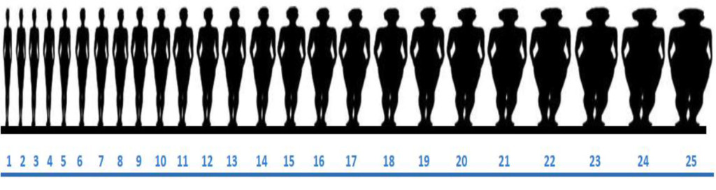
Figure 1. Figural stimuli silhouettes – Women (Figure 8 represents the mid-range of a normal body mass index).

AWHC-18-114 - Rosemary B Duda_ USA_F2

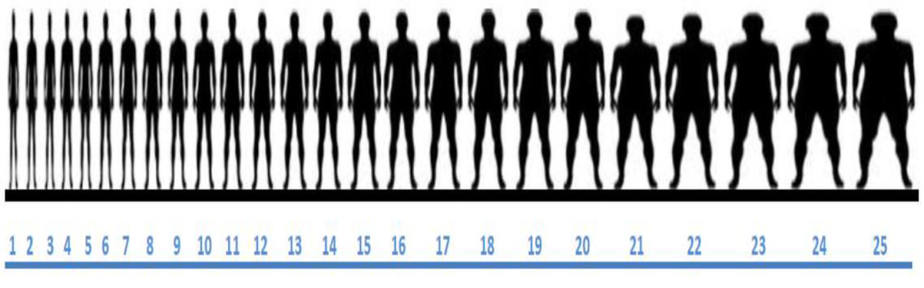
Figure 2. Figural stimuli silhouettes – Men (Figure 8 represents the mid-range of a normal body mass index).

Anthropomorphic Measures

Anthropometric measurements were taken with participants wearing light street clothes without shoes [2]. Height and weight were obtained and recorded to the nearest 0.1 centimeters and 0.1 kilograms, respectively, with a standing measuring stick and a calibrated, portable scale. Waist and hip measurements were collected to the nearest 0.5 centimeters with a flexible tape measure designed for body measurements. WHO STEPS protocols were followed, with the tape measure fitted snuggly, and with waist defined as the midpoint between the lower margin of the last palpable rib and the top of the iliac crest and hip defined as the widest portion of the buttocks [26].

Statistical Analysis

BMI was calculated for each participant and categorized according to WHO standards, with underweight ≤ 18.5, normal weight 18.6–24.9, overweight ≥ 25.0 and obese ≥ 30.0. A body image dissatisfaction score (DS) was calculated by subtracting the figure selected as the IBI for self from that selected as the CBI (DS = CBI – IBIself). Statistical analysis was performed using SPSS version 16.0 for Windows and included descriptive statistics for frequency and mean values Fisher’s Exact Test (FET) 1-sided, student T-test, chi-square test, Pearson correlation, binary logistic regression analysis and multinomial regression analysis.

Institutional Review Board Approval

The investigation was approved by the Institutional Review Boards at Harvard Medical School, Boston, MA, USA and the University of Cape Coast School of Medical Sciences, Cape Coast, Ghana. Verbal consent was considered sufficient for participation in the survey and an informed consent signature waiver obtained.

Results

Participant characteristics

A total of 400 patients agreed to participate in this study. There was an equivalent number of male and female participants (49% versus 51%) and no statistically significant difference between gender and mean age, highest level of education attained, marital status, religion, and environment of birth and current residence
(Table 1). Women, however, were two times more likely to be unemployed compared with men (14.2% versus 7.7%, p = 0.026 FET), univariate analysis Odds Ratio (OR) = 2.0, 95 CI % (1.03–3.90), p = 0.039. Responses to the Medical Outcome Short Study Form-36 revealed that most of the participants (81.7%) reported they were in good to excellent health, were in much or somewhat better health compared to last year (53.0%) and were somewhat to very happy at the time of survey (85.1%).

Table 1. Participant Characteristics.

Men

n (%)

Women

n (%)

p-value

Participants

196 (49)

204 (51)

NS

Mean Age

33.8

32.5

NS

Highest Education level

NS

No Formal Education

2 (1.0)

7 (3.4)

Primary

5 (2.6)

8 (3.9)

Junior High (JSS)

26 (13.3)

25 (12.2)

Senior High (SSS)

81 (41.3)

84 (41.0)

Tertiary

73 (37.2)

75 (36.6)

Graduate

8 (4.1)

6 (2.9)

Relationship Status

NS

None

62 (31.6)

41 (19.8)

Unmarried Relationship

43 (21.9)

71 (34.3)

Married

84 (42.9)

70 (33.8)

Divorced

5 (2.6)

13 (6.3)

Widowed

0 (0)

12 (5.8)

Employment Status

0.026

Employed

181 (92.3)

175 (85.8)

Not Employed

15 (7.7)

29 (14.2)

Children

NS

None

101 (51.5)

109 (52.7)

Religion

NS

Christian

177 (90.3)

189 (92.6)

Muslim

17 (8.7)

12 (5.9)

None/Other

2 (1.0)

3 (1.5)

Environment, birth

NS

Urban

98 (50.3)

114 (56.2)

Semiurban

26 (13.3)

22 (10.8)

Rural

71 (36.4)

67 (33.0)

Environment, current

NS

Urban

59 (30.1)

68 (33.3)

Semiurban

117 (59.7)

120 (58.8)

Rural

19 (9.7)

14 (6.9)

NS = Not Significant

Body Mass Index

Height and weight measurements were available for all 400 participants. The distribution of BMI and gender are shown (Table 2). Obesity was identified significantly more often in women compared with men (20.1% versus 6.1%, p < 0.001), OR 1.6, 95% CI (1.25–1.96), p < 0.001.

Table 2. Body Mass Index by Gender.

Body Mass Index

Men

n (%)

Women

n (%)

Overall

n (%)

Underweight

10 (5.1)

10 (4.9)

20 (5.0)

Normal Weight

132 (67.3)

92 (45.1)

224 (56.0)

Overweight

42 (21.4)

61 (29.9)

103 (25.8)

Obese

12 (6.1)

41( 20.1)

53 (13.2)

Mean

23.3

25.9

24.6

Range

16.9 – 38.1

14.7 – 45.6

14.7 – 45.6

Current Body Image

The most frequently selected CBI for women were figures 11, 13, and 14 (9.3% each), larger than the silhouette representing the middle range of a normal BMI. The mean ± standard deviation current body image (CBI) silhouette selected by women was figure 14.5 ± 4.5 (range 1 to 25). Only 8 women (3.9%) selected figure 8 (mid-range of normal BMI) as their CBI. There was a significant correlation (r = 0.727, p < 0.001) between BMI and CBI silhouette for women.

The most frequently selected CBI for men were figures 10, 12, 13, and 14 (9.2, 12.8, 14.8 and 11.2%, respectively), as found with the women, larger than the silhouette representing the middle range of BMI. Only 5 men (2.6%) selected figure 8 (mid-range of normal BMI) as their CBI. The mean ± standard deviation current body image (CBI) silhouette selected by men was figure 13 ± 3.7 (range 2 to 24). There was also a significant correlation between increasing CBI silhouette selected and increasing BMI for men (r = 0.603, p < 0.001).

Ideal Body Image for Women and Men

Both women and men selected a figure that represented a size larger than Silhouette 8, the middle of the normal body mass index range. The most frequently selected silhouette for women to represent their ideal body image (IBI) for themselves was figure number 14 (mean figure 13.6 + 3.6, range 1 – 25). The most frequently selected silhouette for IBI by men for themselves was figure number 13 (mean figure 13.8 + 3.6, range 2 – 25). There was also a significant correlation between BMI and IBI for both women and men. The Pearson correlation between BMI and IBI for women was (r = 0.260, p < 0.001) and between CBI and IBI for women was (r = 0.551, p < 0.001). The Pearson correlation between BMI and IBI was (r = 0.141, p = 0.049) and between CBI and IBI for men was (r = 0.596, p < 0.001).

Women and men most frequently selected figure 13 as the IBI for a spouse/significant (15.6% and 17.1%, respectively). The most frequently selected IBI for a Ghanaian woman in general by women was figure 14 (15.5%, range 7–25, r = 0.480, p < 0.001). Figure 15 (13.5%, range 2–23) was most commonly selected by men as the IBI for a Ghanaian woman. In comparison, women and men both selected figure 15 as the IBI for men (range 7 – 25 and 4 to 25, respectively). There was a significant correlation between a man’s IBI for himself and for a man in general (r = 0.553, p < 0.001).

Dissatisfaction Score

The dissatisfaction score (DS) was calculated by subtracting the IBI from the CBI for women and men. Less than one-third (32.6%) of participants had a DS = 0, indicating the CBI = IBI. There was no significant difference between women and men with a DS = 0 (29.4% versus 35.9%, OR 1.34, 95% CI (0.88, 2.0), p = 0.168) (Table 3). However, women were significantly more likely to select an IBI smaller than the CBI compared with men (42.2% versus 28.4%, OR 1.51, 95% CI 1.18–1.94, p = 0.001). On a multivariate analysis, the variables significantly associated with a DS = 0 included: children (any versus none), stable weight for the past year, anticipating a stable weight in the next year, being less likely to have been told as an adult to gain or lose weight, and being less likely to be influenced by spouse/partner (Table 4). In addition, 64.6% of participants with a DS = 0 had a normal BMI; however, for each BMI category, most participants were dissatisfied with CBI (p < 0.001) (Table 5).

Table 3. Dissatisfaction score = Current Body Image – Ideal Body Image by Gender.

Dissatisfaction

Women (%)

Men (%)

Total (%)

p-value

CBI = IBI

29.4

35.9

32.6

NS

CBI < IBI

28.4

42.1

35.1

NS

CBI > IBI

42.2

22.1

32.2

0.001

NS = Not Significant

Table 4. Variables significantly associated with Dissatisfaction Score (DS) = 0 on multivariate analysis.

Variable

DS = 0 (%)

DS = Any (%)

OR

95% CI

p-value

Children, any

40.1

25.6

2.6

1.5, 4.1

< 0.001

Stable weight past year

34.1

24.1

1.6

1.2, 2.3

 0.003

Anticipate stable weight next year

56.1

27.1

1.9

1.4, 2.5

< 0.001

Told to gain weight as an adult

27.2

35.9

1.8

1.1, 3.0

0.028

Told to lose weight as an adult

20.4

39.1

1.9

1.1, 3.4

0.027

Influenced by spouse

20.9

46.2

2.5

1.5, 4.1

< 0.001

Table 5. Body Mass Index and Dissatisfaction Score.

Body Mass Index

DS = 0

DS = Any

p-value

%

%

Underweight

10.5

89.5

<0.001

Normal weight

37.5

62.5

<0.001

Overweight

35.0

65.0

<0.001

Obese

15.1

84.9

<0.001

Total

32.6

67.4

DS = Dissatisfaction Score

On multivariate analysis, a smaller IBI (DS = CBI > IBI) versus a larger IBI (DS = CBI < IBI) was statistically associated with: attempt to lose weight by caloric restriction, attempt to lose weight by exercise, told as an adult to lose weight, expect weight to decrease over the next year, and agree that weight has a very large effect on health, report no television viewing. Variables associated with a larger versus a smaller IBI included: unmarried status, attempt to increase weight with food (51.1% versus 18.6%), and told as an adult to gain weight (Table 6). Pertinent variables not associated with DS include environment of birth, environment of current residence, and media (television, radio, billboards, and internet usage), family, friends, religion, and food security.

Table 6. Multivariate analysis for variables that influence a positive or negative Dissociation Score.

Variable

CBI > IBI (%)

CBI < IBI  (%)

OR

95% CI

p value

Decrease weight by restricting caloric intake

67.4

12.1

5.2

2.2, 12.3

<0.001

Advise to lose weight as an adult

75.0

9.3

14.5

6.0, 35.2

<0.001

Decrease weight with exercise

71.3

19.3

10.4

5.9, 18.4

<0.001

Effect of weight on health  is very large effect

47.3

34.3

1.6

1.1, 2.3

0.007

Expected weight to decrease next year

64.3

7.9

2.7

1.9, 3.9

<0.001

No television viewing

76.0

49.6

1.9

1.2, 8.3

0.016

Unmarried status

57.4

72.1

1.5

1.1, 2.4

0.045

Increased weight by increasing caloric consumption

18.6

51.1

3.0

1.3, 6.8

0.010

Told as an adult to increase weight

17.1

61.2

3.7

1.7, 8.1

0.001

CBI = Current Body Image; IBI = Ideal Body Image; OR = Odds Ratio; CI = Confidence Interval

Media Influence and Ideal Body Image

A majority of participants reported exposure or access to each form of media assessed (Table 7). There was a significant difference in exposure to radio and newspaper/print between women and men. Men were significantly more likely to listen to the radio (OR = 2.51, 95% CI 1.13, 5.59) and read the newspaper (OR = 1.10, 95% CI 1.02, 1.16, p = 0.004) compared with women. A radio and television was present in 92.3% and 92.5% of all households, respectively. In addition, 97% of all participants had access to a cell phone and 60.2 percent had access to the Internet, with no statistical significant difference between women and men (97.1% versus 96.9%, p = 0.587 and 58.3% versus 62.2%, p = 0.243, respectively).

Table 7. Media Exposure and Influence on Ideal Body Image for Self by Gender.

Variable

Radio

Television

Newspaper/Print

Access overall (%)

92.2

90.8

64.7

     Women

89.2

88.7

57.8

     Men

95.4

92.9

71.8

     p value

0.016

0.105

0.002

Exposed 7 days per week (%)

71.8

75.2

14.0

     Women

69.1

76.5

8.8

     Men

74.5

74.0

19.5

     p value

0.140

0.322

0.002

Highly Influenced (%)

18.0

21.1

14.6

     Women

16.2

24.0

18.3

     Men

20.0

17.9

10.7

     p value

0.363

0.142

0.046

Not Influenced (%)

41.4

36.8

51.8

     Women

45.1

38.2

50.0

     Men

37.4

35.4

53.6

     p value

0.128

0.536

0.425

p -value represents difference between women and men for each variable.
NA = Not Applicable. Access to billboards not asked as they are ubiquitous in the region.
Analysis performed by Fisher’s Exact test, (2-sided).

Television was the most frequently identified source of media exposure and had the largest influence on IBI. Television was viewed daily by 75.2% of participants and 21.1% of participants reported that their IBI was highly influenced by television, without a statistically significant difference between men and women. Television was significantly associated with dissatisfaction, with those watching television seven days a week having significantly higher dissatisfaction score compared with those not watching television (–0.70 v. 0.48, p < 0.001). Although they read less print media overall, women were significantly more likely to report being highly influenced by newspapers and magazines than men (18.3% versus 10.7%, p = 0.046). Women who were highly or moderately influenced by friend or family opinions were significantly more likely to be dissatisfied (any versus none) with CBI compared with women not influenced at all (77.8% versus 60.0%, p = 0.011, and 80.5% versus 64.8%, p = 0.017, respectively, FET 2 sided). Women’s dissatisfaction score was not significantly associated with influence of television, radio, print media, billboards, spouse, or religion. However, men who were highly or moderately influenced by spouse or friend opinions were significantly more likely to be dissatisfied with CBI compared with men not influenced at all (63.3% versus 44.4%, p = 0.049, FET, 1-sided, and 75.4% versus 48.0%, p = 0.001, FET 2 sided, respectively). Men’s dissatisfaction score was not significantly associated with influence of television, radio, print media, billboards, family, or religion.

Social Influences and Ideal Body Image

Cultural, social and family determinants also had modest influence on IBI for self (Table 8). A spouse/partner had the overall influence on IBI (29.2%), although there was no statistical difference based upon participant gender. Women were significantly more likely to be influenced by friends compared with men (OR = 1.07, 95% CI 1.02, 1.14, p = 0.013).

Table 8. Cultural, Social and Family Influences on Ideal Body Image for Self by Sex.

Variable

Family

Spouse*

Friends

Religion

Highly Influenced (%)

19.3

29.2

18.8

12.8

     Women

22.1

26.6

23.5

9.8

     Men

16.4

32.0

13.9

16.1

     p value

0.164

0.529

0.015

0.074

Not Influenced (%)

45.4

31.8

39.2

73.6

     Women

43.1

35.3

39.2

74.5

     Men

47.7

28.1

39.2

72.5

     p value

0.422

0.180

1.000

0.501

* Included only if married.
Analysis performed by Fisher’s Exact test, (2-sided).

Social and family influences on gaining/losing weight were assessed for childhood and adulthood. During childhood, most participants were not encouraged to lose weight (91.7%), but almost one-third were told to gain weight (34.5%). Women were significantly more likely to have been told to gain weight as a child compared with men (OR = 1.68, 95% CI 1.10, 2.55, p = 0.015). In contrast, adult women were more likely to be told to lose weight compared with adult men (OR = 2.52, 95% CI 1.64, 3.88). Women were much more likely to have attempted weight loss compared with men. Women were significantly more likely to attempt to lose weight by dieting, (47.1% versus 18.4%, OR = 3.9, p < 0.001, CI = 2.0–7.9), exercising (48.5% versus 31.1%, OR 2.1, p = 0.001, CI = 1.4–3.1) and diet pills (10.8% versus 5.6%, p = 0.045, FET 1-sided).

Additional Health Related Determinants Influence on IBI

An open-ended question inquired about other possible factors that may influence IBI. Most respondents provided no additional types of influence (72%). However, of the 113 (64 men and 71 women) who did provide an open ended response, 53.1% of men and 33.8% of women stated improved health-related reasons as an important influence on their body image, while 23.9% of men and 17.0% of women mentioned a desire to appear more attractive. Only 3.1% of men and 5.6% of women noted social pressures as an external influence on IBI.

Discussion

Ghana provides a particularly unique environment to study body image. First, traditional Ghanaian culture has favored a larger image as ideal that for women, symbolic of wealth and higher status [27–30]. Second, there exists interplay between traditional and modern cultures resulting from rapid economic development, urbanization, modernization and social changes such as shifts in gender roles and exposure to western cultural practices and norms [20]. Further, rural-urban migration continues to occur at a rapid pace, with the rural proportion of Ghana’s total population dropping each year since 1960 and recently dropping below 50% [31]. The implication is that more and more traditional Ghanaian values are being brought along with such migrants to the city, even as recent migrants are exposed to new cultural values in the urban environment. Finally, the growing prevalence of overweight and obesity coupled with increasing longevity mean a rapidly increasing burden of chronic disease for Ghana, a problem that is pervasive in much of the developing world [32, 33]. Because of its position as a key parameter in understanding overweight and obesity and in shaping emotional health, body image is an important public health concern [34, 35].

The choice of 25 figures with standard variation ordered by increasing size was based on the example of the BIAS-BD figural drawing scale, the original version of which depicted 17 human figures [36]. Subsequent research on effective use of figural stimuli has shown that figural stimuli products using a discrete number of images (rather than a size range) and with images arranged in ascending/descending size order [as opposed to random) are the most accurate and effective as research tools [37]. While static figures obviously limit the creativity with which participants can express their ideal body image perspectives, we did test for and confirm that they were accurate in representing a participant’s current body image in regards to BMI, and we limited our investigation of body image to size rather than including other considerations such as shape or build.

We investigated determinants in combination with body image perspectives to better understand the factors that shape Ghanaians’ weight and their ideas about weight. Our study found a notable disparity between men and women in terms of proportion overweight and obesity, with 50.0% of women categorized as overweight or obese compared with 27.5% of men. These numbers are consistent with previous findings in Cape Coast [38]. We found only a few correlates for overweight or obesity, none of which provided a satisfying explanation for this gender disparity. As expected, age correlated with weight for both genders, as did number of children. Men were significantly more likely than women to be employed, and work of all types has been found to contribute to a less sedentary lifestyle [39] and lower rates of overweight/obesity; however, employment status was not correlated with BMI for men or women in our study. Further study might delve deeper into the relationship between employment type and body weight. We also found no relationship between education level and BMI, although such a relationship has been found in previous research in the region [28, 40].

We found no correlation between location of residence and BMI, although this has also been shown to be an important correlate of BMI [41]. This may be accounted for by the fact that participants are relatively mobile as – approximately 28.5% had lived in at least four places in their lives—and that a large number (37.5%) had lived in one or both of the two largest cities in Ghana (Accra or Kumasi) at some point, suggesting that even semi-urban and rural residents may have significant exposure to urban environments. Studies on body image have shown that both men and women are influenced by the figures they see in the media and in particular television, and that media exposure often creates unrealistic expectations for body image, leading to increased body dissatisfaction [4,13, 42]. One of the most notable findings in our research was the high level of media exposure reported by participants. These high levels of media exposure suggest that Ghanaians are susceptible to media influences that may influence their body image perceptions. One limitation to this assumption is that we did not examine amounts of daily media exposure, nor did we attempt to document the type of content participants were exposed to, both of which might have helped us better understand our participants and the influences on them.

We observed a significant relationship between high levels of television viewership and body image dissatisfaction within our sample population, suggesting the plausible presence of media content that may influence people’s body image ideals. Interestingly, unmarried men who watched television were significantly more likely to choose an IBI larger than their CBI, indicating a desire to be larger than their current size. Other studies have reported similar findings, and have suggested that men may be under increasing pressure to develop greater degrees of muscularity [4]. Indeed, formal and informal comments from men during the course of the survey confirmed that many were interested in being larger and more muscular.

Social interactions and body dissatisfaction relationships with friends and family were shown to be influential determinants in the establishment of body image, and in determining how an individual is affected by their body image [43]. Research in the United States has shown the influence of social relationships and support organizations on body image can be incredibly powerful in both positive and negative ways [44, 45]. We found that both spouse/partner and friends had a negative rather than positive influence on female body image, with women who identified strong influences from these determinants having greater body image dissatisfaction scores. However, family influence was not correlated with dissatisfaction, which may suggest either that adults are less influenced by the opinions of their families, or that family members are more likely to be supportive, making them less apparent as a source of influence.

Although our question about self-reported influence did not reveal as strong a relationship between social forces and dissatisfaction as we found for television, our secondary social influences questions, which assessed whether participants had ever been told to change their weight, were incredibly telling. Among a wide variety of factors, being told to change weight as an adult was the most strongly associated with body image dissatisfaction, and what participants had been told was tied to the type of dissatisfaction they experienced, with those who had been told to gain weight favoring a smaller ideal, and those who had been told to lose weight favoring a smaller one. This supports the idea that negative messages about weight from social networks are very powerful.

The concept of influence from media and relationships seemed to be an idea that some participants were familiar, while others appeared to have a much more difficult time contextualizing the way in which images, ideas or other people could affect their personal body image concept. This may have led to some participants providing arbitrary or inaccurate answers to our influences questions. Further study into body image in this context is warranted. In particular, drawing out more detailed information about the types of influences present in Ghanaian media, adding qualitative inquiry methods to our work and perhaps determining novel ways to inquire about the concept of influence would all be helpful in revealing more about body image attitudes in Ghana.

References

  1. Duda RB, Jumah NA, Hill AG, Seffah J, Biritwum (2006) Interest in healthy living outweighs presumed cultural norms for obesity for Ghanaian women. Health and Quality of Life Outcomes 4: PMC 1544332.
  2. Duda RB, Jumah NA, Hill AG, Seffah J, Biritwum R (2007) Assessment of the ideal body image of women in Accra, Ghana. Trop Doct 37: 241–244. [crossref]
  3. Thompson J, Heinberg M, Altabe M, Tantleff-Dunn S (1999) Exacting Beauty: Theory, assessment and treatment of body image disturbance. American Psychological Association, Washington DC. USA.
  4. Thompson JK, Heinberg LJ (1999) The Media’s Influence on Body Image Disturbance and Eating Disorders: We’ve Reviled Them, Now Can We Rehabilitate Them? Journal of Social Issues 55: 339–353.
  5. Cash TF, Smolak L (2011) Understanding body image: historical and contemporary perspectives. In: Cash TF, Smolak L (eds.). Body image: a handbook of science, practice, and prevention. Guilford Press, New York, USA Pg No: 3–11.
  6. Cash TF (2012) Cognitive-behavioral perspectives on body image. In: Cash TF (ed.). Encyclopedia of body image and human appearance. Oxford, Elsevier Pg No: 334–342.
  7. Laus MF, Kakeshita IS, Costa TM, Ferreira ME, Fortes Lde S, et al. (2014) Body image in Brazil: recent advances in the state of knowledge and methodological issues. Rev Saude Publica 48: 331–346. [crossref]
  8. Holland G, Tiggemann MA (2016) systematic review of the impact of the use of social networking sites on body image and disordered eating outcomes. Body Image 17: 100–110.
  9. Grabe S, Ward LM, Hyde JS (2008) The role of the media in body image concerns among women: a meta-analysis of experimental and correlational studies. Psychol Bull 134: 460–476. [crossref]
  10. Want SC (2009) Meta-analytic moderators of experimental exposure to media portrayals of women on female appearance satisfaction: Social comparisons as automatic processes. Body Image 6: 257–269.
  11. Fernandez S, Pritchard M (2012) Relationships between self-esteem, media influence and drive for thinness. Eat Behav 13: 321–325. [crossref]
  12. Cafri G, Yamamiya Y, Brannick M, Thompson JK (2005) The Influence of Sociocultural Factors on Body Image: A Meta-Analysis. Clinical Psychology: Science and Practice 12: 421–433.
  13. Blond A (2008) Impacts of exposure to images of ideal bodies on male body dissatisfaction: a review. Body Image 5: 244–250. [crossref]
  14. Leit RA, Gray JJ, Pope HG (2002) The media’s representation of the ideal male body: A cause for muscle dysmorphia? International Journal of Eating Disorders 31: 334–338.
  15. Daniel S, Bridges SK (2010) The drive for muscularity in men: media influences and objectification theory. Body Image 7: 32–38. [crossref]
  16. Agliata D, Tantleff-Dunn S (2004) The impact of media exposure on males’ body image. Journal of Social and Clinical Psychology 23: 7–22.
  17. Kronenfeld LW, Reba-Harrelson L, Von Holle A, Reyes ML, Bulik CM (2010) Ethnic and racial differences in body size perception and satisfaction. Body Image 7: 131–136. [crossref]
  18. Yates A, Edman J, Aruguete M (2004) Ethnic differences in BMI and body/self-dissatisfaction among Whites, Asian subgroups, Pacific Islanders, and African-Americans. J Adolesc Health United States 34: 300–307.
  19. Swami V, Frederick DA, Aavik T, Alcalay L, Allik J, et al. (2010) The Attractive Female Body Weight and Female Body Dissatisfaction in 26 Countries Across 10 World Regions: Results of the International Body Project I. Personality and Social Psychology Bulletin 36: 309–325.
  20. Agyei-Mensah S, de-Graft Aikins A (2010) Epidemiological transition and the double burden of disease in Accra, Ghana. J Urban Health 87: 879–897. [crossref]
  21. Central Regional Hospital (2008) Central Regional Hospital, Cape Coast: 2008 Annual Report. Cape Coast Central Regional Hospital, interberton road, Cape Coast, Ghana.
  22. Ware JE Jr (2008) Improvements in short-form measures of health status: introduction to a series. J Clin Epidemiol 61: 1–5. [crossref]
  23. Ware JE Jr (2003) Conceptualization and measurement of health-related quality of life: comments on an evolving field. Arch Phys Med Rehabil 84: 43–51. [crossref]
  24. Ware JE, Kosinski M (2001) Interpreting SF-36 summary health measures: a response. Qual Life Res 10: 405–413. [crossref]
  25. Stewart A, Ware J (1992) Measuring Function and Well-Being: The Medical Outcomes Study Approach. 1st  (edn). Duke University Press Books, Durham, USA.
  26. WHO (2011) Waist circumference and waist-hip ratio: report of a WHO expert consultation, Geneva, 8–11 December 2008. WHO Press, Geneva.
  27. Smith AD (2009) Girls being force-fed for marriage as fattening farms revived. The Observer.
  28. Addo J, Smeeth L, Leon DA (2009) Obesity in urban civil servants in Ghana: association with pre-adult wealth and adult socio-economic status. Public Health 123: 365–370. [crossref]
  29. Cogan JC, Bhalla SK, Sefa-Dedeh A, Rothblum ED (1996) A Comparison Study of United States and African Students on Perceptions of Obesity and Thinness. Journal of Cross-Cultural Psychology 27: 98–113.
  30. Cassidy CM (1991) The good body: when big is better. Med Anthropol 13: 181–213. [crossref]
  31. Bank W (2013) World dataBank: World Development Indicators. Washington DC, USA.
  32. Amuna P, Zotor FB (2008) Epidemiological and nutrition transition in developing countries: impact on human health and development. Proc Nutr Soc 67: 82–90. [crossref]
  33. Caballero B (2001) Introduction. Symposium: Obesity in developing countries: biological and ecological factors. J Nutr 131: 866–870.
  34. Krentz AJ (2006) Self-image in obesity: clinical and public health implications. J R Soc Med 99: 215–217. [crossref]
  35. Muennig P, Jia H, Lee R, Lubetkin E (2008) I think therefore I am: perceived ideal weight as a determinant of health. Am J Public Health 98: 501–506. [crossref]
  36. Gardner RM, Jappe LM, Gardner L (2009) Development and validation of a new figural drawing scale for body-image assessment: the BIAS-BD. J Clin Psychol 65: 113–122. [crossref]
  37. Gardner RM, Brown DL (2011) Method of presentation and sex differences when using a revised figural drawing scale to measure body size estimation and dissatisfaction. Percept Mot Skills 113: 739–750. [crossref]
  38. Amegah A, Lumor S, Vidogo F (2011) Prevalence and Determinants of Overweight and Obesity in Adult Residents of Cape Coast, Ghana: A Hospital-Based Study. African Journal of Food, Agriculture, Nutrition and Development 11: 4828–4846.
  39. Van Domelen DR, Koster A, Caserotti P, Brychta RJ, Chen KY, et al. (2011) Employment and physical activity in the U.S. Am J Prev Med 41: 136–145. [crossref]
  40. Dake FA, Tawiah EO, Badasu DM (2011) Sociodemographic correlates of obesity among Ghanaian women. Public Health Nutr 1285–1291.
  41. Ziraba AK, Fotso JC, Ochako R (2009) Overweight and obesity in urban Africa: A problem of the rich or the poor? BMC Public Health 9: 465. [crossref]
  42. Derenne JL, Beresin EV (2006) Body image, media, and eating disorders. Acad Psychiatry 30: 257–261. [crossref]
  43. Field AE, Austin SB, Camargo CA, Taylor CB, Striegel-Moore RH, et al. (2005) Exposure to the Mass Media, Body Shape Concerns, and Use of Supplements to Improve Weight and Shape Among Male and Female Adolescents. Pediatrics 116: 214–220.
  44. Pole M, Crowther JH, Schell J (2004) Body dissatisfaction in married women: the role of spousal influence and marital communication patterns. Body Image. Netherlands 267–278.
  45. Eisenberg ME, Berge JM, Fulkerson JA, Neumark-Sztainer D (2010) Weight comments by family and significant others in young adulthood. Body Image. Elsevier Ltd Netherlands 2011: 12–19.

Dental Injuries Following Segmented Le Fort I Osteotomy – A Retrospective Radiographic Study of 101 Patients

DOI: 10.31038/JDMR.2018122

Abstract

Segmented Le Fort I osteotomy is a surgical procedure which allows the posterior segments to be repositioned more coronally to close an open bite, as well as correcting transverse discrepancies. The aim of this retrospective study was to assess the prevalence of injuries to teeth and surrounding hard tissues in patients who had undergone the Le Fort I osteotomy procedure with maxillary segmentation. In total, 101 patients were included according to predetermined criteria. Radiographs from the 6-month postoperative follow-up were reviewed in all patients. Complications such as root fractures, root resorptions, periodontal defects, sclerotic and osteolytic processes were noted. Intraoral periapical radiographs were primarily reviewed and where unavailable, panoramic radiographs were instead used. In addition, preoperative and postoperative radiographs up to 30 months were reviewed in patients with found radiographic changes at 6 months. In cases with root fracture, the medical record was reviewed in search of any additional treatment. Seven root fractures were noted in total. Only one tooth required replacement, in this case with a dental implant. Clinically significant complications to teeth adjacent to vertical osteotomies from the Le Fort I osteotomy with maxillary segmentation were uncommon.

Keywords

Complications; Interdental Osteotomy; Le Fort I Osteotomy; Maxillary Segmentation; Orthognathic Surgery

Introduction

Transverse discrepancies and anterior open bite can be successfully treated with a combination of fixed orthodontic appliances and maxillary segmentation [1, 2]. If an open bite exists on a level occlusal plane, it can be surgically corrected in one piece by reducing the height of the maxilla, mostly in the posterior portion, after a one piece Le Fort I osteotomy. A step in the occlusal plane, however, necessitates segmentation of the maxilla for proper correction of the discrepancy [3]. The segmented Le Fort I osteotomy is a surgical procedure which allows the surgeon to reposition each segment into the desired positions so that a narrow maxilla can be widened or to close an open bite. In such operations, vertical interdental osteotomies are performed in order to segment the maxilla. There are to our knowledge relatively few studies reporting on complications to teeth and surrounding bone tissue in conjunction with vertical interdental osteotomies. Kahnberg and colleagues reported a low incidence of hard tissue complications and iatrogenic damage to teeth [4] while another study by Schultes et al. indicaes that interdental osteotomies may result in severe periodontal tissue breakdown and segmental loss of teeth [5]. Schou et al. and Morgan and Fridrich did not find that interdental osteotomies lead to significant marginal bone destruction [6, 7] which was later confirmed in two studies published more recently [8, 9]. The aim of this retrospective study was to assess the prevalence of injuries to teeth and surrounding hard tissues following the segmented Le Fort I osteotomy.

Materials and Methods

Patients subjected to segmented Le Fort I osteotomy between January 2005 and December 2015 at the Department of Oral and Maxillofacial Surgery, The Sahlgrenska Academy, University of Gothenburg were included in the study according to following inclusion and exclusion criteria:

Inclusion criteria

  • Undergone segmented Le Fort I osteotomy with or without simultaneous mandibular surgery between January 2005 and December 2015.
  • Combined orthodontic and surgical treatment.
  • Available intra-oral and/or panoramic radiographs 6 months postoperatively.

Exclusion criteria

  • Undergone segmented Le Fort I osteotomy without preoperative orthodontic treatment.
  • No available intra-oral or panoramic radiographs 6 months postoperatively.
  • Undergone the operation more than once.

A total of 101 patients (mean age 22.8 years, median age 21 years, range 17–61 years, SD = ±7.56) were included (Table 1). The most common diagnosis necessitating maxillary segmentation was an anterior open bite, which meant the maxilla was typically segmented into four pieces. Radiographs were analysed under dimmed lighting on computer monitors (HP Elite Display E222, 1920×1080) by two authors (G.M and J.M.W). When agreement between authors was not reached, the radiographs in question were presented to an oral and maxillofacial radiologist (H.L) for decision. Primarily, intraoral periapical radiographs were examined and secondarily panoramic radiographs. Radiographs for all 101 patients were analysed at 6 months after surgery with respect to teeth and surrounding tissues adjacent to the interdental osteotomies. The following radiographic changes were noted: osteolytic processes (i.e. widened periodontal ligament spaces (WPLs) and periapical osteolyses), sclerotic processes, marginal bone loss, angular bony defects, root resorptions and root fractures. The preoperative and all follow up radiographs up to 30 months postoperatively were analysed in cases with found radiographic changes at radiographs taken at the 6-month recall.

Table 1. Collected data at the 6-month recall.

Sex

Male

60

Female

41

Radiographs

Intra-oral and panoramic

42

Panoramic only

59

Interdental osteotomies

Unilateral

10

Bilateral

91

Surgical procedure

The surgical procedure was carried out as follows [1, 10]: Incisions were made in the buccal sulcus, extending from the second premolar to the inferior aspect of the nasal spine. The periosteum was then elevated at the superior aspect of the incision, extending posteriorly to expose the lateral wall of the maxillary sinus. Nasal mucosa was then elevated from the piriform aperture and osteotomies were then carried out, beginning with a cut extending from the piriform rim to the zygomatic buttress and posteriorly through the lateral wall of the maxillary sinus. After sectioning the lateral nasal wall, the maxilla was then disengaged from the pterygoid bone using an osteotome inferior to the pterygomaxillary cleft. The lateral wall of the sinus was then separated using an osteotome in a posterior direction. At this point in the operation, maxillary segmentation was carried out. This was done by elevating the periosteum in the region of the inter dental osteotomy, most commonly between the canine and the first premolar bilaterally. The buccal cortical bone was cut with a bur and the osteotomy was then completed with an osteotome. The maxilla was at this stage down fractured with inferior pressure on the anterior teeth. Complete mobilization of the maxilla was obtained with an osteotome applying force in an anterior direction from the maxillary tuberosity. The maxillary segmentation was completed proceeding from the superior aspect of the maxilla. Stainless steel wires were used to fixate the maxilla to the mandible, with a surgical splint for guidance. The maxillomandibular complex was then fixated with titanium plates and screws in the desired position. Soft tissue was closed beginning with appropriate positioning of the facial muscles, concluding with closure of the mucosa.

Statistical analysis

The Two-Proportion Z-Test (SPSS Inc., Chicago, Illinois, USA) was utilised in order to assess the correlation of the proportion of the radiographic changes with the type of radiograph analysed and also comparing unilateral with bilateral osteotomies. p < 0.05 was considered statistically significant.

Results

The results are summarised in (Table 2). Ninety-one patients underwent bilateral interdental osteotomies and 11 patients unilateral. 384 teeth were therefore examined at the 6-month follow-up. All 35 patients with radiographic changes at 6 months had preoperative radiographs available, one of which underwent the unilateral osteotomy, meaning 138 teeth were examined at this point. At the 18-month recall, radiographs for 25 patients were available and 98 teeth were included. At 30 months, only one patient had radiographs available, meaning the inclusion of 4 teeth. Overall, 35% of patients were found to have some form of radiographic changes at the 6-month recall. Male and female patients had an equal likelihood of having radiographic changes at 6 months. Fifty percent of patients with intraoral radiographs were found to have radiographic changes and 24% in the group with only panoramic radiographs available. This difference was statistically significant (z = 2.7, p < 0.01). Ten percent of patients with unilateral osteotomies and 37% of patients with bilateral osteotomies exhibited radiographic changes, but this difference was not statistically significant (z = 1.7, p > 0.05).

Table 2. Summary of radiographic changes.

Preop.

+6 months

+18 months

+30 months

(n = 138)

(n = 384)

(n = 98)

(n = 4)

WPL

7

4

1

0

Periapical osteolysis

3

5

2

0

Sclerotic process

2

5

1

0

Marginal bone loss

1

3

2

0

Angular bony defect

0

7

4

0

Root resorption

20

29

21

0

Root fracture

0

7

2

1

Total defects

33

60

33

1

WPL: widened periodontal ligament space

Osteolytic processes

The osteolytic processes registered were periapical radiolucencies and widened periodontal ligament spaces (WPLs). At the initial 6-month follow-up, 4 of teeth adjacent to the osteotomies were found to have WPLs, two of which were seen on the preoperative radiographs. The remaining two teeth had no visible WPLs at 18 months. However, an additional WPL was found on a different tooth at this point. Five teeth were found exhibiting periapical osteolytic processes at the 6-month mark, one of which was present preoperatively. At the 18-month follow-up, only two of the patients (two of the teeth) had available radiographs, where one tooth exhibited a remaining periapical radiolucency. Another of the patients, with an affected tooth, who lacked 18-month radiographs did have a 30-month follow-up, however, where the periapical radiolucency was no longer visible.

Sclerotic processes

Five teeth were found to have sclerotic processes at 6 months, two of which were visible preoperatively. At the 18-month recall, one of the teeth with postoperative sclerosis lacked radiographs and in the remaining two, the scleroses were no longer visible.

Marginal bone loss and angular bony defects

Three osteotomy sites were found to have a marginal bone level located ≥ 3 mm from the CEJ 6 months postoperatively, two of these sites had a reduced marginal bone level compared with radio- graphs taken preoperatively. At 6 months, the marginal bone level at these two sites was measured at 7 mm and 4.5 mm respectively, compared to 1.5 mm and 2 mm at radiographs taken before surgery. Two of these patients were examined with panoramic radiographs at 18 months and no further bone loss could be detected. The third patient, with marginal bone loss of 2.5 mm, did not have available radiographs at 18 months. Seven interproximal tooth surfaces were found exhibiting adjacent angular bony defects at 6 months, none of which were visible on the preoperative radiographs. At the 18-month followup, one of the bony defects lacked radiographs, and of the remaining 6, two were no longer present.

Root resorptions

Twenty-nine teeth were found with root resorptions at 6 months, 20 of which were present preoperatively, indicating that 9 had arisen after surgery. Two of these teeth lacked radiographs at 18 months, and an additional 6 teeth were registered as having root resorptions that were not noted in earlier radiographs.

Root fractures

Seven teeth (5 premolars and 2 canines) were found with root fractures at 6 months, none of which were present preoperatively (Figure 1). At 18 months, three of these teeth lacked radiographs and in one other case the intraoral radiograph did not capture the area with the root fracture. Of the three remaining teeth examined at 18 months, one was still visible and had healed into the bone, one was examined only with panoramic imaging and was no longer discernible. The third tooth was eventually extracted and replaced with an implant. There was a 30-month follow-up for one of the fractures not examined at 18 months, where it could still be seen to be healed into the bone.

Discussion

The aim of this study was to assess the prevalence of injuries to teeth and surrounding hard tissues adjacent to vertical interdental osteotomies in patients who had undergone the Le Fort I osteotomy with maxillary segmentation. The main drawback of this study was the relatively small proportion of intraoral radiographs covering the interdental osteotomy area. A majority of the patients had only panoramic radiographs available, hindering the detection of the subtle radiographic changes which were the subject of this study. There was significantly fewer radiographic changes found on panoramic radiographs (z = 2.7, p < 0.01). Intraoral radiographs are more appropriate in the detection of these radiographic changes due to their higher resolution [11]. We suggest therefore that intraoral radiographs are taken in cases where interdental osteotomies have been performed, as a complement to extraoral imaging techniques. Panoramic radiographs are taken routinely during the follow up of patients undergoing orthognathic surgery for evaluation of healing of bone segments and fixation material. Overlapping in the upper premolar region and superimposition of osteosynthesis plates was not uncommon in some of the panoramic radiographs included in this study, making it difficult to distinguish radiographic changes in these cases. According to Lofthag Hansen and co-workers, periapical lesions not detected on intraoral radiographs may be visible when using CBCT [12]. Routine use of three-dimensional imaging techniques in the follow-up of patients undergoing orthognathic surgery is however not justified, due to the relatively high radiation exposure.

JDMR-18-109-Lars Rasmusson_ Sweden_F1

Figure 1. Periapical radiograph of a patient presenting with root fracture of 24.

To this study’s advantage is the relatively large patient sample included, increasing the generalisability of the results. No differences were found between the sexes with regards to the proportion of radiographic changes found. Ten percent of patients that underwent a unilateral interdental osteotomy exhibited radiographic changes at 6 months compared to 37% in the group with bilateral osteotomies. This difference was not found to be statistically significant, perhaps owing to the small population in this study that underwent the unilateral variant. A higher prevalence in the bilateral group would be expected in that a patient is more likely to receive dental injury when two interdental osteotomies are performed compared to one.

Four teeth in this study were found to have periapical osteolytic processes not present on preoperative radiographs, three of these teeth had additional follow-up radiographs, where two of the periapical osteolyses were no longer visible, indicating that healing had occurred in these cases. It is possible that the presence of periapical osteolyses is due to the teeth being devitalised by the operation. A study by Bell showed that horizontal osteotomy cuts within 5 mm of the apices of adjacent teeth could disturb pulpal blood circulation and thus risk devitalising the teeth [13]. In the present study however, the horizontal osteotomies were seen radiographically to be of adequate distance from the apices. Another possibility is that vertical osteotomies in close proximity to the apex of the adjacent teeth have disturbed the blood circulation in a similar manner. Nevertheless, none of the affected teeth underwent endodontic treatment within the confines of this study. Kahnberg et al. proposed that endodontic treatment should be considered in cases where the tooth is symptomatic. They also recommend regular clinical follow-up in cases where a periapical osteolysis is radiographically visible [4].

In this patient material, two percent (7 of 384) of the teeth were damaged during the vertical interdental osteotomy procedure, resulting in root fractures, one of which was subsequently replaced by an implant. The root fracture that was replaced with an implant was due to the fracture line reaching the level of the marginal bone, thwarting healing into the bone. Horizontal root fractures located within the coronal third of the root are associated with poor prognosis [14]. Orthodontic tipping of the roots of adjacent teeth away from the planned vertical osteotomy site is important to reduce the risk of iatrogenic root fracture [10] and could perhaps also serve to reduce the risk of compromised pulpal blood flow. Our results regarding postoperative loss of teeth are in line with what was reported by Kahnberg et al., Ho et al. and Rodrigues et al. [4, 8, 9]. Schultes et al. reported a higher incidence of postoperative tooth loss, likely due to the high prevalence of periodontal injury in that study [5]. Root resorption was seen in 29 teeth at the 6 month follow up, 20 of which were visible preoperatively. These findings imply that the vast majority of root resorptions in this study were due to the orthodontic pretreatment [15, 16]. In a study utilising CBCT, it has been shown that root resorptions can be detected in varying degrees in almost all teeth following treatment with fixed orthodontic appliances [17]. Root resorptions that were detected postoperatively may be influenced by surgical trauma and/or orthodontic treatment. Marginal bone loss was detected in two osteotomy sites which is comparable to what is reported by Schou et al. and Kahnberg et al. [4, 6]. Small, subclinical alterations in marginal bone level were however not registered in this study. One limitation of intraoral and panoramic radiographs in assessing the marginal bone level is that measurements can only be performed at the interproximal sites. Orthodontic forces have been shown to lead to a significant reduction of the crestal height at the buccal, lingual and palatal aspects of teeth. [18].

Radiographic changes present on preoperative radiographs indicate a non-surgical etiology. When taking this into account, the proportion of patients with radiographic changes falls from 35% to 28%. On the level of individual teeth adjacent to the osteotomy sites, the corresponding risk for a tooth adjacent to the osteotomy site would be 9% in this study, as 34 teeth exhibited radiographic changes at the 6-month follow-up which were not present preoperatively. However, had patients with only panoramic radiographs available at 6 months been excluded from this study, due to the aforementioned lower resolution of panoramic radiographs, the risk of incurring radiographic changes to an individual tooth adjacent to the vertical osteotomy could be said to be 15% (excluding findings present preoperatively).

Conclusion

Overall it can be concluded from this study that the risk posed to teeth adjacent to vertical osteotomies in conjunction with Le Fort I osteotomies is low, with 1 in 384 teeth included in this study requiring extraction and replacement.

Conflict of interest: We have no conflicts of interest to disclose.

Ethical approval: Ethical approval was not required.

References

  1. Kahnberg KE (2007) Transverse expansion of the maxilla using a multisegmentation technique. Scand J Plast Reconstr Surg Hand Surg 41: 103–108.
  2. Silva I, Suska F, Cardemil C, Rasmusson L (2013) Stability after maxillary segmentation for correction of anterior open bite: a cohort study of 33 cases. J Craniomaxillofac Surg 41: e154–158. [crossref]
  3. Hupp JR, Ellis E, Tucker MR (2014) Contemporary Oral and Maxillofacial Surgery. St Louis MO: Elsevier Pg No: 520–539.
  4. Kahnberg KE, Vannas-Löfqvist L, Zellin G (2005) Complications Associated with Segmentation Of The Maxilla: A Retrospective Radiographic Follow Up Of 82 Patients. International Journal of Oral and Maxillofacial Surgery 34: 840–845.
  5. Schultes G, Gaggl A, Kärcher H (1998) Periodontal disease associated with interdental osteotomies after orthognathic surgery. J Oral Maxillofac Surg 56: 414–417. [crossref]
  6. Schou S, Vedtofte P, Nattestad A, Stoltze K (1997) Marginal bone level after LeFort I osteotomy. British Journal of Oral and Maxillofacial Surgery 35: 153–156.
  7. Morgan TA, Fridrich KL (2001) Effects of the multiple-piece maxillary osteotomy on the periodontium. Int J Adult Orthodon Orthognath Surg 16: 255.
  8. Ho MW, Boyle MA, Cooper JC, Dodd MD, Richardson D (2011) Surgical complications of segmental Le Fort I osteotomy. Br J Oral Maxillofac Surg 49: 562–566. [crossref]
  9. Rodrigues DB, Campos PSF, Wolford LM, Ignácio J, Gonçalves JR (2018) Maxillary Interdental Osteotomies Have Low Morbidity for Alveolar Crestal Bone and Adjacent Teeth: A Cone Beam Computed Tomography-Based Study. J Oral Maxillofac Surg S0278–2391: 30111–30113.
  10. Andersson L, Kahnberg KE, Pogrel MA (2010) Oral and Maxillofacial Surgery 2010; Chichester: John Wiley & Sons Ltd Pg No: 1009–1012.
  11. White S, Pharoah M (2014) Oral radiology 2014; St. Louis, Missouri: Elsevier Pg No: 177.
  12. Lofthag-Hansen S, Huumonen S, Gröndahl K, Gröndahl HG (2007) Limited cone-beam CT and intraoral radiography for the diagnosis of periapical pathology. Oral Surg Oral Med Oral Pathol Oral Radiol Endod 103: 114–119. [crossref]
  13. Bell WH (1969) Revascularization and bone healing after anterior maxillary osteotomy. A study using adult rhesus monkeys. Journal of Oral Surgery 27: 249.
  14. Cvek M, Andreasen JO, Borum MK (2001) Healing of 208 intra-alveolar root fractures in patients aged 7–17 years. Dent Traumatol 17: 53–62. [crossref]
  15. Goldson L, Henrikson CO (1975) Root resorption during Begg treatment; a longitudinal roentgenologic study. Am J Orthod 68: 55–66. [crossref]
  16. Linge L, Linge B (1991) Patient characteristics and treatment variables associated with apical root resorption during orthodontic treatment. American Journal of Orthodontics and Dentofacial Orthopedics 99: 35–43.
  17. Lund H, Gröndahl K, Gröndahl HG (2012) Cone beam computed tomography evaluations of marginal alveolar bone before and after orthodontic treatment combined with premolar extractions. Eur J Oral Sci 120: 201–211. [crossref]
  18. Lund H, Gröndahl K, Hansen K, Gröndahl HG (2012) Apical root resorption during orthodontic treatment. A prospective study using cone beam CT. Angle Orthod 82: 480–487. [crossref]

Endocrine Disruptors-caused Faulty Hormonal Imprinting: Focus on Women

DOI: 10.31038/AWHC.2018132

Abstract

Hormonal imprinting is a physiological process, when hormone receptors and the target hormone meet in the first occasion, perinatally. This process is needed for the normal function of the receptor-hormone complex and valid for life. However, hormone-like molecules also can bind to the developing receptor causing faulty imprinting and its consequences: altered binding of hormones, inclination to diseases, manifestation of diseases, disturbing the physiological hormonal regulation. The faulty imprinting also has a lifelong effect. Industrial, communal, nutritional and medical endocrine disruptors are faulty imprinters and their variables as well, as amounts are enormously growing in the human environment. The faulty imprinting influences also the microsomal enzyme system. Numerous diseases, manifested at adult age can be deduced to perinatal faulty hormonal imprinting and the higher sensitivity of women to drugs (more adverse reactions) could be explained by perinatal events. The extremely growing variants and amount of endocrine disruptors could rearrange the whole endocrine system, which could be disastrous or useful alike in the future.

Keywords

Bisphenol A, DOHaD, Endocrine Regulation, Steroid Hormones, Perinatal Period, Functional Teratogens

Introduction

During fertilization the genom of the women’s egg contain the maternal informations and the paternal informations are brought by the sperm. After the fusion of two germ cells the zygote is existing, which is able to develop further to the complete organism (individuum), controlled by the fused genome (ontogeny). The zygote is totipotent which means that it is able for developing to any organs (cells) of the organism however, during the ontogenetic development there is a continuous loss of potencies, to pluripotent (multipotent) and at last to unipotent cells, which are able to produce cells (by cell division) similar to the mother-cell, or are unable to divide (e.g. nerve cells) at all. This means that during the ontogenetic development a continuous narrowing of potencies happens which is resulted in diffent types of cells, with different structure and function. Therefore, in different cell types of the organism different genes are manifested (are working, giving information for function), while others are closed by methylation of the cytosin nucleotids of DNA. The organization of these different units is the duty of the neuroendocríne system in which the direct transmitter humoral components are the hormones. However, the neuroendocrine system can regulate only such functions, which are permitted by the genes, which are open for giving information to the given functions.

The Physiological and Faulty Hormonal Imprinting

The hormones are present in the blood circulation, where any cells of the organism can meet them. However, only such cells can decipher the message contained by the hormone, which have cell membrane or nuclear receptors for the given hormone, which can bind the hormone and after that can transmit the coded information into the cytoplasm or into the nucleus. These receptors are specific for a given hormone, nevertheless this specificity also develops gradually during the ontogenetic development. During the embryonal and early fetal period of human life maternal hormones (passed across the placenta) are dominating however, at the end of the fetal and during the perinatal period (prenatally, at birth and early postnatally) the „homegrown” hormones appear and hormonal imprinting is taking place, conforming the receptors for themselves. This imprinting is necessary, without it the fitting of receptor-hormone system is not working well [1]. The setting is valid for life and inherits to cell to cell inside the cell line as well as to the progenies of the individuum, as it is an epigenetic process, which alters the methylation pattern of a given gene (the expression of the gene), without disturbing the nucleotid sequences [2]. However in this critical perinatal period the receptors’ specificity is weak, what means that other members of a hormone family or synthetic hormones, hormone-like molecules also can be bound by them and the amount of imprinter is also very important. In this case a faulty hormonal imprinting can develop, which is also have a lifelong validity and causes disturbed functions, with abnormal binding of hormones in adult age [3]. Consequently, disturbed functions appear in behavior [4–6], sexuality [6–8], body composition [9], in bone development [10]; the neurotransmitter production of brain is also altered and immune functions are changed (e.g. autoimmunity develops). One single encounter with very low doses of hormone-like molecules in the critical perinatal period is enough for the provocation of faulty imprinting and for the manifestation of their consequences in adult age. The hormonal imprinting which was observed, described and experimentally justified by us (at first in 1980) [11] was the first in the series of new theories which lead to metabolic imprinting [12], immunological imprinting [13] and to the developmental origin of health and disease (DOHaD) [14]. As hormonal imprinting is an epigenetic process, the alterations happened in the perinatal period can be manifested at any time of life and also can be manifested in the progenies and by this, epigenetic alterations of the future generations are settled on the already changed genetic arrangements. This is important as the response of a faulty imprinted cell or organism would be different from which is observed in the present time.

The Endocrine Disruptors

Endocrine disruptors are molecules similar to (first of all, steroid) hormones, which are present in our environment and can enter into our organism by air, water, food, drug consumption and can be bound by hormone receptors influencing cell functions, stimulating or hindering them. Endocrine disruptors has been always present during the evolution of men in our environment as e.g. aromatic hydrocarbons produced by volcanic eruptions, food components (phytoestrogens, present in soy and other vegetables), smoke etc. However, they were not named to „endocrine disruptors” as their such effects were not known and their amount was insignificant. At present, in our modern (industrial) age their variety is high, their amount is large and both are enormously growing. They causes a crisis in the endocrine system as well in the systems, which are seriously influenced by the distorted endocrine system.

Endocrine disruptor exposures are believed harmful in adult age and this is demonstrated by many research data and statistics. However, they are more harmful in the critical periods of development, mainly in the time of hormonal imprinting. In this case they are causing faulty hormonal imprinting with lifelong consequences. In animal experiments single treatment with aromatic hydrocarbons (benzpyrene or dioxin) are decreasing the binding capacity of glucocorticoid receptor as well as estrogen receptor. Vitamin D3 (which is not a vitamin in reality, but a steroid-like hormone having receptors in the cytoplasmic-nuclear steroid receptor family [15], lifelong decreases the sexual activity (libido) of female and male rats, similar to industrial or communal endocrine disruptors [16].

Faulty Hormonal Imprinting and Functional Teratogenicity of Endocrine Disruptors

Teratogen materials, which evoke morphological alterations which are visible at birth are known from immemorial time and these are most effective during the embryonal period and their effects gradually decreases whith the time passed and vanishes after birth. Faulty hormonal imprinting does not provoke morphologically observable important alterations, but changes in functions of cells organs or systems which are manifested in diseases. They are not diagnosed at birth however, manifested later, mostly in adults. This is a functional teratogenity, without organic changes. An other difference to the morphological (real) teratogens that the possibility for exposure is longer: the perinatal period is the late phase of intrauterine development for morphological teratogens however, faulty imprinting can be provoked at any time e.g. also postnatally, few days or weeks after birth and other critical periods of life. The absolute need for taking place is the multiplication and differentiation of the touched cells.

Faulty hormonal imprinting inclines to diseases, alters behaviors, influences cell-responses to attacks, etc. In the scientific literature there are extreme amount of experimental data and also human observations can be found however, in the frame of a short review paper only a selection of them can be shown. Nevertheless they are represantatively demonstrate the importance and amount of the problems caused by it in the present modern age.

Sexual and Behavioral Problems Caused by Faulty Hormonal Imprinting

Perinnatal exposure to TCDD (2,3,7,8-tetrachlorodibenzo-p-dioxin) an environmental contaminant (originated from volcanic eruptions as well as from exhaust gas of cars) and industrial or agricultural imprinters (bisphenol A, vinclozolin) pushes the boy-adventageous sex-ratio (more boys are delivered than girls) for the adventage of girls after paternal exposure (in human [16] and also to maternal exposure [17]) (in rats and mice). Bisphenol A (the well known plasticizer and representative of endogen disruptors) by prenatal and lactational exposure, changes the behavior of adults. Prenatal bisphenol A exposure provokes externalizing behavior in 2 years old children, especially in females [18] and abolishes the differences between male and female sexual behavior [19] and also modifies sexual behavior in female mice. It also alters sex difference in affective disorders. There is such opinion which calls attention to the future problems with school age. Some experiments and observations point to the possible association between early bisphenol A exposures (perinatal imprinting) and autism spectrum disorders)[19,20]. Pubertal bisphenol A exposures caused long term alterations in microglia of the prefrontal cortex when this was lower in males and higher in females [21]. There were also alterations in body weight and composition. Perinatal treatment and adolescent reexposure exacerbated adverse effects in females and reduce differences in males. Symptoms of sex dependent anxíety/depression appeared in 7–9 years girls without expression in boys after gestational bisphenol A exposures [22]. Altered sociosexual and mood disorders were observed in animal models and children after developmental exposure by bisphenol A [23].

Other Problems Caused by Faulty Hormonal Imprinting

Reproductive organs and puberty

First in the US and later in Europe an alteration in the timing of puberty (decline) was observed and also the growing number of obesity for which environmental factors, mainly endocrine disruptors was believed as responsible, by influencing genetic factors [24] These problems were observed after imprinting with polychlorinated biphenyls, polibrominated biphenyls, DDT and phtalate esters [25]. Both puberty age and menarche age were influenced [26, 27]. The perinatal endocrine exposures can provoke central precocious puberty or isolated breast development in 2 months to 4 year old girls [28].

Growing number of human (male and femele) infertility and decreased fertility were observed [29]. In the case of perinatal exposure by bisphenol A decreased methylation of DNA and increased histone H3 acetylation were observed in the cerebral cortex and hippocampus, affecting the liver, gut, adipose tissue endocrine pancreas, mammary gland and reproductive tract functions [30]. Maternal programming was also altered [31]. Neonatal exposure to diethylstilbestrol (DES) caused early or delayed puberty, depending on the dose [32] by affecting hypothalamus and hippocampus. This is supported by rodent and human data [33]. Exposure in people is a result of contamination of foods or inhalation (e.g.house or occupational dust). Endocrine disruptors (bisphenol A and phtalates) negatively influence the function of the immune system, altering T cell subsets, B cell functions, dendritic cell and macrophage biology, and provoking autoimmune diseases [34,35]. They have a controversial role in influencing longevity [36]. The developing immune system, the cells of which have steroid (estrogen) hormone receptors is very sensitive to the presence of endocrine disruptors [35]. Faulty imprinting by endocrine disruptors could be responsible for pathological development of bones as well, as for changes in bone mineralization and osteoporotic fractures or other bone problems [37–41].

Gender differences in adverse drug reactions between men and women

It is very difficult to compare data of gender differences in the past-time adverse reaction to drugs, with relatively fresh (which were won in the time of endocrine disruptors) data as earlier always mens’data were studied and dosages of drugs were also prescribed exclusively for men [42–44]. However, new data show that women have 1.5 to 1.7-fold greater risk for adverse drug reactions than men [43]. For example, anti-bacterial and anti-inflammatory drugs cause more adverse reactions in females, as compared to males [42]. Cardiovascular diseases are the first case in mortality and disability of women [44]. There is a similar situation in case of addictions: considering alcohol intoxication compared to men, women metabolize alcohol less than men, it become intoxicated drinking half as much, develop cirrhosis more rapidly and have a greater risk of dying from alcohol-related accidents [45]. Drug abuse and dependence are not identical in males and females and females are less successful in quitting of alcoholism and nicotinism.

Conclusion

As most of the known endocrine disruptors are steroid hormone-like molecules, the functions [46], which are regulated by physiological steroid hormones are disturbed by them. The palette of these functions is broad, from sexuality to immunity and the period in which their effects are manifested is also very broad. They already disturb the sexual (male-female) ratio at birth, pushing it to the adventage of females, cause malformations of sexual organs (micropenis, cryptorchidism and hypospadias), provoke, as functional teratogens, faulty hormonal imprinting. However there are other critical periods of life (development) at weaning, during adolescence and for continuously dividing cells, during the whole life. In these cases endocrine disruptors are also able to provoke hormonal imprinting, which can modify the perinatal setting however, the effect of them less serious, than the perinatal one.

The difference between males and females in adverse drug reactions can be explained by sex differences in pharmacokinetics and pharmacodynamics, in which the liver microsomal enzyme system has an important role. The hormonal imprinting touches not only the hormonal system but also the (microsomal) enzyme system [47,48] and this interferes into the different adverse drug reactions by males and females. There is a gender-dependent metabolism which is influenced by microsomal enzymes which are different in the two sexes [49]. This influences pharmacokinetics and consequently toxicity. As was mentioned, this factor can give some explanation to the differences in adverse reactions by males and females, and faulty hormonal imprinting can disturb this process. This means that in contrast to the real (morphological ) teratogens faulty hormonal imprinting does not causes morphological alterations, which are visible to the naked eye, however alterations in the hormonal or receptorial system as well, as in the enzyme system, durably changing the function of the cells, which are regulated by hormones. This renders likely (though the animal and human data are modest) that women (females) are more sensitive to faulty imprinters and some of their adverse reactions (and the overweight of women in adverse reactions) can be wrote to the expense of faulty perinatal hormonal imprinting.

It was mentioned that a single low dose of an endocrine disruptor seems to be enough for the provocation of faulty hormonal imprinting if it arrives in the optimal time for doing it (e.g.perinatal critical period). It is not surprizing if somebody consider that during the differentiation of the brain, when the direction of sexual development is determined, a part per billion of testosterone and about twenty parts per trillion of estradiol (endogeneous estrogen) actually predict entirely different brain structures, behavioral traits, enzyme levels and receptor levels in tissues [50]. It must be consider that the brain is basically female and it must be exposed to testosterone and related hormones for transforming to male . This means, that the receptorial system is ready to accept the imprinters, the developmental window is open for setting by single minimal doses of hormone-like materials (endocrine disruptors) and this is „exploited” by them (faulty imprinters). The gender differences in sensitivity and its epidemiological importance should be considered.

The late manifestation of alterations after the faulty hormonal imprinting makes more difficult to recognize the participation of faulty imprinting in the manifestation of a disease, or in the changes of behavior. It is supposed, that prenatal exposure to bisphenol A may be related to increased behavioral problems in school age boys, but not girls. However, although serious problems are expectable very difficult to forecast their quality and quantity. At any rate, thorough observations would be needed, for avoiding the problems. However, it seems to be difficult really to avoid, as the variants and amount of different endocrine disruptors are enormously growing because of the claims of the industry, which wants to earn a lot of money, and the people, who want to live more comfortable. In additon, when a new molecule justifies its usefulness in the industry or as a medicament, it is not known (however sometimes could be guessed, but the expected pleasant effects suppresses anxiety), that it will be an endocrine disruptor. Fortunately, it is known that during pregnancy new exogeneous molecules must be avoided however, it is not known by lay medical practitioners and laywomen, that this regulation -in the light of faulty imprinting- must be extended to the whole period of pregnancy and also postnatally.

The task of medicine is to help people in the avoidance of illnesses or to heal ill people. This is done by giving advices or after diagnosing a disease, giving therapy for healing, which is mostly done by prescribing drugs. However, some drugs are endocrine disruptors themselves, for example anticoncipients and some vitamins. Anticoncipients are used for avoiding pregnacy, consequently they do not cause faulty imprinting in the person (women), who intakes it. However, the wast products of metabolized anticoncipients are present in the urine and contaminates drinking water, which is drinked by other, (pregnant) women. If we know, that minute amounts of an endocrine disruptor is able to provoke faulty imprinting, this could be done by the communal water. This is obvious, however can not be declared without exact measurements. Other medicaments are not tested for imprintership before running to circulation and can also contaminate the waters, or influence the endocrine system of developing fetus, directly. Drugs entering into breastmilk also can be faulty imprinters [51] and it is also known that most of baby foods are soy-based, consequently contain phytoestrogens (coumestrol, genistein and daidzein), which are known faulty imprinters [52–54]. Lipid soluble vitamins are prescribed by doctors and can be bought in pharmacies however, also can be purchased without prescription in self-service shops [55]. This shows that mankind is not prepared for the invasion and effects of faulty imprinters.

Afterwords

Pessimistically grasping, the growing of disruptor-inventar and the amount of disruptors, the future of mankind seems to be dangerous and threatening, as -endocrine disruptors -by faulty hormonal imprinting- can attack the whole endocrine system and the desorganization of the gene-level determined well- functioning system could cause hitherto unknown diseases or proliferation of known malignancies. In addition, the alterations are inherited to the progenies, epigenetically. However, there is -optimistically- an other version [55]: some of the endocrine disruptors internalized into the present endocrine system and new (better) functions will be manifested, which will be better suited to the continuously transformed world. Some examples had been observed to such transformation in the past, e.g. the infiltration of lipid soluble vitamins (e.g.vitamin A and D -hormones) and their prominent function in animal (human) life [56] . As can not be known what scenario will be the winner, the protection of women (as mothers) from endocrine disruptor effects is a very important mission of the present day mankind.

References

  1. Csaba G, Nagy SU (1985) Influence of the neonatal suppression of TSH productiion (neonatal hyperthyroidism) on response of TSH production in adulthood. J Endocrinol Invest 8: 557–559. [crossref]
  2. Csaba G (2011) The biological basis and clinical significance of hormonal imprinting, an epigenetic process. Clin Epigenetics 2: 187–196. [crossref]
  3. Csaba G (1984) The present state in the phylogeny and ontogeny of hormone receptors. Horm Metab Res 16: 329–335. [crossref]
  4. Nakamura K, Itoh K, Dai H, Han L, Wang X, et al. (2012) Prenatal and lactational exposure to low-doses of bisphenol A alters adult mice behavior. Brain Dev 34: 57–63. [crossref]
  5. Wolstenholme JT, Taylor JA, Shetty SR, Edwards M, Connelly JJ, et al. (2011) Gestational exposure to low dose bisphenol A alters social behavior in juvenile mice. PLoS One 6: e25448. [crossref]
  6. Chen F, Zhou L, Bai Y, Zhou R, Chen L (2014) Sex differences in the adult HPA axis and affective behaviors are altered by perinatal exposure to a low dose of bisphenol A. Brain Res 1571: 12–24. [crossref]
  7. Evans SE, Kobrosly RW, Barrett ES, Thurston SW, Calafat AM, et al. (2014) Prenatal bisphenol A exposure, maternally reported behavior in boys and girls. Neurotoxicology 45, 91–99.
  8. Csaba G (2017) The Present and Future of Human Sexuality: Impact of Faulty Perinatal Hormonal Imprinting. Sex Med Rev 5: 163–169. [crossref]
  9. Rubin BS, Paranjpe M, DaFonte T, Schaeberle C, Soto AM, et al. (2017) Perinatal BPA exposure alters body weight and composition in a dose specific and sex specific manner: The addition of peripubertal exposure exacerbates adverse effects in female mice. Reprod Toxicol 68: 130–144.
  10. Csaba G (2018) Bone manifestation of faulty perinatal hormonal imprinting: a review. Curr Pediatr Rev 25. [crossref]
  11. Csaba G (1980) Phylogeny and ontogeny of hormone receptors: the selection theory of receptor development and hormonal imprinting. Biol Rev Camb Philos Soc 55: 47–63.
  12. Hanley B, Dijane J, Fewtrell M, Grynberg A, Hummel S, et al. (2010) Metabolic imprinting, programming and epigenetics – a review of present priorities and future opportunities. Br J Nutr 104: 1–25.
  13. Lemke H, Tanasa RI, Trad A, Lange H (2009) Benefits and burden of the maternally-mediated immunological imprinting. Autoimmun Rev 8: 394–399.
  14. Barker DJ (2007) The origins of the developmental origins theory. J Intern Med 261: 412–417. [crossref]
  15. Lorenzen M, Boisen IM, Mortensen LJ, Lanske B, Juul A, et al. (2017) Reproductive endocrinology of vitamin D. Mol Cell Endocrinol 453: 103–112. [crossref]
  16. Ishihara K, Warita K, Tanida T, Sugawara T, Kitagawa H, et al. (2007) Does paternal exposure to 2,3,7,8-tetrachlorodibenzo-p-dioxin (TCDD) affect the sex ratio of offspring? J Vet Med Sci 69: 347–352. [crossref]
  17. Yonemoto J, Ichiki T, Takei T, Tohyama C (2005) Maternal exposure to 2,3,7,8-tetrachlorodibenzo-p-dioxin and the body burden in offspring of long-evans rats. Environ Health Prev Med 10: 21–32. [crossref]
  18. Kubo K, Arai O, Omura M, Watanabe R, Ogata R, et al. (2003) Low dose effects of bisphenol A on sexual differentiation of the brain and behavior in rats. Neurosci Res 45: 345–356. [crossref]
  19. Hashemi F, Tekes K, Laufer R, Szegi P, Tóthfalusi L, et al. (2013) Effect of a single neonatal oxytocin treatment (hormonal imprinting) on the biogenic amine level of the adult rat brain: could oxytocin-induced labor cause pervasive developmental diseases? Reprod Sci 20: 1255–1263.
  20. Stein TP, Schluter MD, Steer RA, Guo L (2015) Bisphenol A Exposure in Children With Autism Spectrum Disorders. Autism Res 8: 272–283. [crossref]
  21. Wise, LM, Sadowski RM, Kim T, Willing J, Juraska JM (2016) Long-term effects of adolescent exposure to bisphenol A on neuron and and glia number in the rat prefrontal cortex: Differences between the sexes and cell type. Neurotoxicology 53: 186–192.
  22. Perera F, Nolte ELR, Wang Y, Margolis AE, Calafat AM, et al. (2016) Bisphenol A exposure and symptoms of anxiety and depression among inner city children at 10–12 years of age. Environ Res 151: 195–202. [crossref]
  23. Hicks HD, Sullivan AV, Cao J, Sluzas E, Rebuli M, et al. (2016) Interaction of bisphenol A (BPA) and soy phytoestrogens on sexually dimorphic sociosexual behaviors in male and female rats. Horm Behav 84: 121–126.
  24. Parent AS, Franssen D, Fudvoye J, Pinson A, Bourgignon JP (2016) Current changes in pubertal timing: Revised vision in relation with environmental factors including endocine disruptors. Endocr Dev 29: 174–184.
  25. Den Hond E, Schoeters G (2006) Endocrine disrupters and human puberty. Int J Androl 29: 264–271. [crossref]
  26. Massart F, Parrino R, Seppia P, Federico G, Saggese G (2006) How do environmental estrogen disruptors induce precocious puberty? Minerva Pediatr 58: 247–254.
  27. Schoeters G, Den Hond E, Dhoge W, van Larebeke N, Leijs M (2008) Endocrine disruptors and abnormalities of pubertal development. Basic Clin Pharmacol Toxicol 102: 168–175.
  28. Leonardi A, Cofini M, Rigante D, Lucchetti L, Cipolla C, et al. (2017) The effect of bisphenol A on puberty: a critical review of the medical literature. Int J Environ Res Public Health 14.
  29. Tomza-Marciniak A, StÄ™pkowska P, Kuba J, Pilarczyk B (2018) Effect of bisphenol A on reproductive processes: A review of in vitro, in vivo and epidemiological studies. J Appl Toxicol 38: 51–80. [crossref]
  30. Kumar D, Thakur MK (2017) Effect of perinatal exposure to Bisphenol-A on DNA methylation and histone acetylation in cerebral cortex and hippocampus of postnatal male mice. J Toxicol Sci 42: 281–289. [crossref]
  31. Schneider, JE, Brozek JM, Keen-Rhinehart E (2014) Our stolen figures: the interface of sexual differentiation, endocrine disruptors, maternal programming, and energy balance. Horm Behav 66: 104–119.
  32. Bourguignon JP, Franssen D, Gerard A, Jannsen S, Pinson A, et al. (2103) Early neuroendocrine disruption in hypothalamus and hippocampus: developmental effects including female sexual maturation and implications for endocrine disrupting chemical screening. J Neuroendocrinol 25: 1079–1087.
  33. Rasier G, Toppari J, Parent AS, Bourgignon JP (2006) Female sexual maturation and reproduction after prepubertal exposure to estrogens and endocrine disrupting chemicals: a review of rodent and human data. Mol Cell Endocrinol 25: 254–255.
  34. Rogers JA, Metz L, Yong VW (2013) Review: Endocrine disrupting chemicals and immune responses: a focus on bisphenol A and its potential mechanisms. Mol Immunol  53: 421–430.
  35. Csaba G (2014) Immunoendocrinology: faulty hormonal imprinting in the immune system. Acta Microbiol Immunol Hung 61: 89–106. [crossref]
  36. Csaba G (2018) Immunity and longevity. Acta Microbiol Immunol Hung 65: 1–17.
  37. Karabélyos C, Horváth C, Holló I, Csaba G (1999) Effect of perinatal synthetic steroid hormone (allylestrenol, diethylstilbestrol) treatment (hormonal imprinting) on the bone mineralization of the adult male and female rat. Life Sci 64: 105–110.
  38. Karabélyos C, Horváth C, Holló I, Csaba G (1998) Effect of neonatal glucocorticoid treatment on bone mineralizaton of adult non-treated, dexamethasone-treated or vitamin D3-treated rats. Gen Pharmacol 31: 789–791.
  39. Karabélyos C, Horváth C, Holló I, Csaba G (1998) Effect of neonatal vitamin D3 treatment (hormonal imprinting) on the bone mineralization of adult non-treated and dexamethason-treated rats. Hum Exp Toxicol 17: 424–429.
  40. Karabélyos C, Horváth C, Holló I, Csaba G (1999) Effect of perinatal synthetic steroid hormone (allylestrenol, diethylstilbestrol) treatment (hormonal imprinting) on the bone mineralization of the adult male and female rat. Life Sci 64: 105–110.
  41. Curtis EM, Krstic N, Cook E, D’Angelo S, Crozier SR, et al. (2018) Gestational vitamin D supplementation leads to reduced perinatal RXRA DNA methylation: Results from the MAVIDOS trial. J Bone Miner Res 2018 Oct 15. [crossref]
  42. Zopf Y, Rabe C, Neubert A, Janson C, Btune K, et al. (2009) Gender-based differences in drug prescription: relation to adverse drug reactions. Pharmacology 84: 333–339.
  43. Rademaker M (2001) Do women have more adverse drug reactions? Am J Clin Dermatol 2: 349–351. [crossref]
  44. Baggio G, Corsini A, Floreani A, Giannini S, Zagonel V (2013) Gender medicine: a task for the third millennium. Clin Chem Lab Med 51: 713–727. [crossref]
  45. Greenfield SF (2002) Women and alcohol use disorders. Harv Rev Psychiatry 10: 76–85. [crossref]
  46. Lymperi S, Giwercman A (2018) Endocrine disruptors and testicular function. Metabolism 86: 79–90. [crossref]
  47. Csaba G, Dobozy O, Szeberényi S (1987) A single neonatal treatment with methylcholanthrene or benzo(a)pyrene alters microsomal enzyme activity for life. Acta Physiol Hung 70: 87–91. [crossref]
  48. Csaba G, Szeberényi S, Dobozy O (1986) Influence of single neonatal treatment with allylestrenol or diethylstilbestrol on microsomal enzyme activity of rat liver in adulthood. Med Biol 64: 197–200. [crossref]
  49. Waxman DJ (1984) Rat hepatic cytochrome P-450 isoenzyme 2c. Identification as a male-specific, developmentally induced steroid 16 alpha hydroxylase and comparison to a female-specific cytochrome P-450 isoenzyme. J Biol Chem 25: 15481–15490.
  50. Hood E (2005) Are EDCs blurring issues of gender? Environ Health Perspect 113: 670–677. [crossref]
  51. Csaba G (2018) Lifelong impact of breastmilk transmitted hormones and endocrine disruptors. J Clin Endocrinol Res In Press 2018.
  52. Bar-El DS, Reifen R (2010) Soy as an endocrine disruptor: cause for caution? J Pediatr Endocrinol Metab 23: 855–861. [crossref]
  53. Csaba G (2018) Effect of endocrine disruptor phytoestrogens on the immune system: Present and future. Acta Microbiol Immunol Hung 65: 1–14. [crossref]
  54. Bennetau-Pelissero C (2016) Risks and benefits of phytoestrogens: where are we now? Curr Opin Clin Nutr Metab Care 19: 477–483. [crossref]
  55. Csaba G (2018) The role of endocrine disruptors in future human endocrine evolution: The ED-exohormone system. Current Trends Endocrinol under publication.
  56. Csaba G (2017) Vitamin-caused faulty perinatal hormonal imprinting and its consequences in adult age. Physiol Int 104: 217–225. [crossref]

Applicability of Digital Platform for Evaluation of Fine Motor Skills in Young Children

DOI: 10.31038/IJOT.2018115

Abstract

Fine motor control is important for object manipulation in daily living. Children with developmental delay, cerebral palsy or congenital upper limb abnormalities may have fine motor deficits. In recent years, digital platform for cognitive and motor assessment has been introduced to the field of health and education to optimize development in early childhood. In this study, content validity and construct validity of a new instrument named iDevChild (a tablet application) were evaluated. The iDevChild is a prototype of fine motor test in digital platform. Four occupational therapists were recruited to review the content validity of the test items of iDevChild. Twenty-five typically developing children were recruited from three kindergartens to receive two clinical fine motor tests and two subtests of iDevChild. All of the children were able to complete these tests successfully. Moderate and significant correlations were found between the scores of the tests. Further study on other subtests (e.g. bilateral coordination) and the reliability of the tool with larger sample size is recommended.

Keywords

Fine Motor, Instrument, Digital Platform, In-Hand Manipulation, Visual Motor Integration, Children

Introduction

Fine motor control is important for object manipulation in daily living (e.g. buttoning, screwing a bottle or drawing with a pencil). Fine motor skills include reaching, grasping, visual motor integration, in-hand manipulation and bilateral coordination [1]. Children with developmental delay, cerebral palsy or congenital upper limb abnormalities may have fine motor deficits. Most of the fine motor assessment batteries for young children are rated by examiner according to the assessment criterion. In recent years, digital platform for cognitive and motor assessment has been introduced to the field of health and education to optimize development in early childhood [2]. The digital platform with standardized procedures can help the health care professionals to collect reliable data and to reduce the cost (e.g. time and money) in administration of the test and training. The current study examined the applicability of a prototype of digital platform to evaluate fine motor control in children aged from 3 to 6 years old.

Material and Method

The digital platform of fine motor assessment applied in this study is named iDevChild (Innodimension, Hong Kong). The iDevChild is a tablet application (App) offering assessment and training of fine motor skills. Its subtests of fine motor assessment are: visual motor integration, in-hand manipulation and bilateral coordination. The examinee is required to follow the standardized procedures of each test item. By using a tablet and associated accessories (e.g. touch screen pencil and conductive rotational knob), performance of the examinee can be recorded and then uploaded to the Cloud. For instance, one of the test items of visual motor integration requires the examinee to draw within boundary (figure 1). The speed of completion and the area out-of-boundary could be retrieved from the report in form of CSV file. Another test item of in-hand manipulation requires the examinee to rotate a rotational knob for 3 consecutive times of 360 degree, and visual feedback on the extent of completion of the task will be shown to the examinee on the screen of tablet (figure 2). The speed of rotation and the number of time of releasing the rotation knob would be stated on the CSV file. Other than obtaining quantitative data, clinician can download a file from the Cloud to review the performance of the examinee.

In this study, content validity and construct validity of two subtests of iDevChild (visual motor integration and in-hand manipulation) were evaluated. To examine the content validity, a panel of 4 experts (occupational therapists) was interviewed. They completed a questionnaire of a 5-point Likert scale (totally agree = 5; strongly agree = 4; agree = 3; disagree = 2; strongly disagree = 1) to rate the representativeness and relevance of the test items of iDevChild. The panel members were also required to comment on the feasibility of applying the tool on children aged from 3 to 6 years old. For the construct validity, correlations between the scores of iDevChild and two clinical fine motor tests (Hong Kong Preschool Fine Motor Screening Test and Beery-Buktenica Developmental Test of Visual-Motor Integration–Sixth Edition) were examined. In this study, 25 typically developing children were recruited from 3 kindergartens. Informed consent form from their parents were obtained prior to the study. The children completed all the tests within 60 minutes in two or more sessions. Short breaks were offered between the sessions to avoid physical and mental fatigue of the children.

IJOT 18 - 105_F1

Figure 1. Subtest item of visual motor integration: Drawing within boundary.

IJOT 18 - 105_F2

Figure 2. Subtest item of in-hand manipulation: Rotation.

Results

There are several key findings. First, the area of out-of-boundary (test item of visual motor integration of iDevChild) had significant negative correlation with age, the score of Hong Kong Preschool Fine Motor Screening Test and the score of Beery-Buktenica Developmental Test of Visual-Motor Integration (correlation coefficients, rs ≥ –0.55; ps < .01). Second, the number of times of releasing the rotation knob (test item of in-hand manipulation reflecting the angle of rotation) of iDevChild had significant negative correlations with age and the score of the Hong Kong Preschool Fine Motor Screening Test (rs ≥ –0.55; ps < .01). Third, the expert panel members responded positively toward the feasibility of the iDevChild in evaluating the fine motor control of the young children. All the children were able to complete the test items of the iDevChild successfully.

Discussion and Conclusion

Older children and children having better performance in the clinical fine motor tests were found to perform better in iDevChild (subtests of visual motor integration and in-hand manipulation). These findings showed that the iDevChild has similar construct to the clinical fine motor tests. The iDevChild is a prototype of fine motor assessment tool in digital platform. By using tablet accessories, accurate assessment results and big data on fine motor performance of children could be obtained. It may provide a new way of measurement for in-hand manipulation since there is limited current evaluation tools in addressing in-hand manipulation of young children.[3] Furthermore, clinician can evaluate the fine motor control of client remotely through the digital platform. It may be applicable to other age group (e.g. adult or elderly) or clinical condition (e.g. before or after receiving hand surgery). This may foster the delivery or monitoring of health care service in remote area. In this study, we examined the content validity and construct validity of two subtests (visual motor integration and in-hand manipulation) of iDevChild on 25 children only. Further study on other subtests (e.g. bilateral coordination) and the reliability of the tool with larger sample size is recommended.

Acknowledgement

We would like to thank the children, schools and occupational therapists for their participation in this project. We are thankful to Dr. Vincent Lau and the staff of Innodimension for their technical advice and material support to this project.

Funding

The travelling expenses of researchers in this project are partially sponsored by Department of Rehabilitation Sciences, The Hong Kong Polytechnic University.

References

  1. Siu AMH, Lai CYY, Chiu ASM, Yip CCK (2011) Development and validation of a fine-motor assessment tool for use with young children in a Chinese population. Res Dev Disabil 32: 107–114. [Crossref]
  2. Pitchford N, Outhwaite LA (2016) Can touch screen tablets be used to assess cognitive and motor skills in early years primary school children? A cross-cultural study. Front Psychol 7: 1666. [Crossref]
  3. Raja K, Katyal P, Gupta S (2016) Assessment of in-hand manipulation: Tool development. International Journal of Health & Allied Sciences 5: 235–246.