Monthly Archives: September 2017

CC-motif Chemokine Ligand 21: immune targeting, head and neck oncology, and future prospective

DOI: 10.31038/CST.2017251

Abstract

Investigations in immunomodulating therapies for cancer treatment over the past 20 years have flourished. Given the complex tumor microenvironment and differential signaling pathways a wide variety of potential targeting mechanisms have come to attention. Herein we review the immunomodulating potential of Chemokine 21 in pre-clinical and clinical studies, as well as examine the novel multi-faceted immune-based treatments in advanced head and neck cancer.

Introduction

Head and neck squamous cell carcinoma (HNSCC) is the sixth most common cancer in the world, accounting for more than 300,000 deaths annually [1]. Despite advances in surgical techniques and chemo-/radiation treatment strategies, patients with advanced T3 or T4 HNSCC continue to demonstrate 20%-30% survival with failure of primary surgical or medical (radiation and chemotherapy) management. Salvage therapies, in recalcitrant or recurrent disease, are limited by systemic toxicity or operative morbidity owing to nearby vital structures (carotid artery, skull base). Immunotherapy and modulation in HNSCC is a promising venture given recent advances in cancer biology and investigations in the tumor microenvironment.

Historically studied in the contexts of inflammation and immune response, chemokines have increasingly been studied in the context of tumor proliferation [2, 3]. Chemokines are a family of small chemotactic cytokines involved in cellular signaling and inflammation. Through interactions within a subset of G-protein- coupled transmembrane receptors (GPCRs), secreted cellular chemokines regulate cellular homing, activation, and recruitment of a variety of diverse leukocytes and activated adhesion molecules [2, 4-6]. In the tumor microenvironment, responses to specific chemokines result in the migration of different immune cell subsets and regulate tumor immune responses; these are both pro- and anti- inflammatory in nature. Chemokines, have been directly associated with cancer immunity, proliferation, and metastases [7,8]. In this review, we address the role of Chemokine 21 (CCL-21) in tumor progression, its role as an immunotherapy platform, and the potential within head and neck oncology.

CCL-21 chemotaxis and lymphocyte recruitment

Divided into four subfamilies, chemokine nomenclature is related to the location of their first N-terminal cysteine (C) residues; classified as: C-, CC-, CXC-, and CX3C-chemokines [9]. CCL-21 (also known as Thymus-derived chemokine 4, 6Ckine, or Exodus-2) is a CC chemokine expressed in high endothelial post-capillary venules, T-cell stromal zones of the spleen, Peyer’s patches, and afferent peripheral lymph nodes that strongly attracts naïve T-cells and mature dendritic cells (DC) including NK T-cells [10-13]. The effects of CCL-21 on chemotaxis have been demonstrated in neutralization studies, where the homing of T-cells and DCs has been shown to be significantly reduced [14, 15]. Acting through the GPCR CCR7, a convergence of the immune response elements to sites of CCL-21 production have been demonstrated to co-stimulate the expansion of CD4+ and CD8+ T cells and induce Th1 polarization [16]. Additionally, chemokine bound DCs form T cell adhesions resulting in a hyper-responsive T-cell on subsequent exposure to CCL-21 and antigen-presenting cells [17].

Role in cancer immunotherapy

The cancer immunosuppressant microenvironment results in ineffective antigen processing and presentation; resulting in poor host response. The CCL-21/CCR-7 axis has been studied in efforts to create a more immunogenic environment through chemotaxis of DCs, NK cells, and lymphocytes. By recruiting host antigen presenting cells (APC) for tumor antigen presentation, T cells within lymphoid organs have the ability to prime anti-tumor specific activity. Of note, while CCL-21 aids in the polarization of Th1 lymphocytes, the secondary lymphoid chemokine has also been shown to have minimal effect on the proliferation of suppressor cell population; CD4+CD25+ T-regulatory cells are hypo responsive to CCL-21 induced migration and unresponsive to CCL-21 co-stimulation [16]. High levels of T-regulatory cells within the tumor microenvironment has demonstrated poor prognosis in many cancers, dependent on the type and location of neoplasm [18].

Pre-clinical animal models

The ability to process and present a high ratio of activated tumor-antigen-APCs has long been the goal of immunotherapy in the immunosuppressive cancer microenvironment. The first established model for the creation of a CCL-21 chemotactic gradient in restoring tumor-antigen presentation was undertaken by Sharma and Dubinett [19]. In an immune competent murine lung cancer model, intratumoral injection of CCL21 induced infiltration of CD4+ and CD8+ T cells and DC in both tumor and draining lymph nodes. Additionally, a potent antitumor response was observed as complete tumor eradication was found in 40% of treated mice [19]. Furthermore, the tumor microenvironment displayed a concomitant decrease in immunosuppressive molecules such as PGE-2 and TGF-ß [19]. When introduced to the afferent axillary lymph nodes, CCL-21 injections resulted in a marked infiltration of lymphocytes and DCs into the lung tumor microenvironment, associated with a significant reduction in tumor burden [20]. The anti-tumor efficacy of CCL-21 has further been confirmed in a variety of transgenic lung, ovarian, melanoma, and liver cancer models [21-25]. Despite the promising results, in vivo studies have utilized specific cancer cell lines, with greater success observed in low-malignant weakly metastatic clones as compared to highly-metastatic clones [26]. Further research on the efficacy within specific tumor phenotypes, locations, timing, and dosing of CCL-21 continues to be underway.

Antigen presenting cells as a platform

APCs are fundamental in the activation of specific immunity and have been investigated as adjuvants to cancer immunotherapy to stimulate tumor-specific antigen presentation for promotion of T cell activation and anti-cancer immunity [27, 28]. Prior investigations have generated DCs with enhanced immune stimulatory and T-cell activity through pulsed electroporation of antigens or viral transduction of cytokines [29-33].

A platform to transduce DCs with CCL-21 (DC-CCL21) through an adenovirus vector has also recently been developed demonstrating viable in vitro chemotaxis of activated lymphocytes [34, 35]. The anti- tumor efficacy of DC-CCL21 has been studied in the murine lung and melanoma cancer models. These studies demonstrated a significant reduction in melanoma tumor growth and complete eradication of lung tumor burden in 60% of mice with lung cancer [32, 34]. Clinical trials are currently underway in late stage non-small cell lung cancer patients based on the promising results of this preclinical data [36].

Head and neck oncology

Recurrent and advanced HNSCC has been extraordinarily challenging to treat, with stagnant survival and cure rates over the past 20 years [37]. Depending on the tumor site, reoccurrence rates may range from 25%-50%, and the incidence of subsequent reoccurrences similarly fall within this broad range [38-40]. Morbidity from current salvage treatment strategies are unfortunately common and include chronic pain, respiratory distress, and dysphagia oftentimes resulting in tracheotomy, or, gastrostomy tube dependence [41]. Despite cancer outcomes, cosmetic and functional deficits may negatively impact the head and neck cancer patient’s quality of life [42-44]. Limited surgically by nearby structures and systemically by toxic effects, advancements in locoregional therapies in the tumor microenvironment are strongly desired.

Recent advances in material science, have developed biocompatible, functional, three dimensional polymer systems for molecular and cellular delivery in cancer care [45]. A novel implantable polymer- based system for the delivery of chemokines to interact directly with tumor cells, has the potential to integrate with a wide range of anti- cancer treatments. Recently, a biodegradable poly-ε-caprolactone (PCL), polylactide-co-glycolide (PLG), co-polymer seeded with DC- CCL21 and/or cisplatin has been tested in an animal model resembling unresectable head and neck squamous cell carcinoma (HNSCC) [46, 47]. Tumor cells from the squamous cell carcinoma (SCCA) VII/ SF were injected, grown, and debulked in the flanks of C3H/HeJ mice. The animals then underwent debulking surgery to replicate an unresectable cancer setting. The flexible seeded PCL/PLCL polymer was then applied to contour the cancer tissue bed. In the first study, the cisplatin-polymer group effectively reduced tumor volume by over 16-fold when compared to control polymer with intratumoral cisplatin injection groups [47]. When combined with radiation, the cisplastin-seeded polymer group enhanced the efficacy of radiation therapy by tumor volume reductions of 53% when compared to surgery and radiation alonel [47]. Given the well-established literature in the role of cytokines in tumor regression, the HNSCC murine polymer model was then seeded with DC-CCL21 using a fibrin gel delivery mechanism. After implantation to the partially resected tumor, DC-CCL21 secreting polymer significantly reduced SCC VII/ SF tumors by 41% as compared to control groups [46]. Additionally, the DC-CCL21 polymer resulted in increased CD4+ and CD11+ DCs as well as a marked decrease in T-regulatory cells within the tumor microenvironment.

Current studies are underway to assess the anti-tumor efficacy of DC-CCL21 with combination of cisplatin and radiation therapy in the HNSCC murine model. Additionally, the promising findings of APC and T cell recruitment provide the rationale for combination with immune checkpoint blockade therapy to enhance the frequency of activated T cells in the tumor micro-environment for improved patient outcome.

Conclusion

Research in the field of immunology has evolved tremendously as the complex immunosuppressive tumor microenvironment continues to unravel. The role of CCL-21 is critical in promoting DC homing and T-lymphocyte activation. Tumor antigen presentation is significantly amplified both locally and peripherally following introduction of CCL-21. Head and neck cancer therapies, previously limited by morbidity,
may now have an alternative for locoregional control in unresectable tumors. A multi-faceted approach to innovative cancer treatment is necessary in order to maximize the immunogenic potential for tumor cell death. As described, it is clear that CCL-21 is a potent chemokine in eliciting an immune response with substantial evidence supporting its use in future oncologic therapies. Collaborations with materials scientists, immunologists, and physicians can not be underestimated in the ongoing pursuit for cancer cure.

References

  1. Ferlay J, Shin HR, Bray F, Forman D, Mathers C, et al. (2010) Estimates of worldwide burden of cancer in 2008: GLOBOCAN 2008. Int J Cancer 127: 2893–2917. [crossref]
  2. Zlotnik A, Yoshie O (2000) Chemokines Immunity 12: 121–127.
  3. Nagarsheth N, Wicha MS, et al. (2017) Chemokines in the cancer microenvironment and their relevance in cancer immunotherapy. Nat Rev Immunol 17: 559–572. [crossref]
  4. Luster AD, Chemokines — Chemotactic Cytokines That Mediate Inflammation. Epstein FH, ed. N Engl J Med 338: 436–445.
  5. Stoolman LM (1989) Adhesion molecules controlling lymphocyte migration. Cell 56: 907–910. [crossref]
  6. Elices MJ, Osborn L, Takada Y, et al. (1990) VCAM-1 on activated endothelium interacts with the leukocyte integrin VLA-4 at a site distinct from the VLA-4/ Fibronectin binding site. Cell 60: 577–584.
  7. Zou W, Chen L (2008) Inhibitory B7-family molecules in the tumour microenvironment. Nat Rev Immunol 8: 467–477. [crossref]
  8. Zou W (2006) Regulatory T cells, tumour immunity and immunotherapy. Nat Rev Immunol 6: 295–307. [crossref]
  9. Zou W (2005) Immunosuppressive networks in the tumour environment and their therapeutic relevance. Nat Rev Cancer 5: 263–274.
  10. Willimann K, Legler DF, Loetscher M, et al. (1998) The chemokine SLC is expressed in T cell areas of lymph nodes and mucosal lymphoid tissues and attracts activated T cells via CCR7. Eur J Immunol 28: 2025–2034.
  11. Nagira M, Imai T, Hieshima K, et al. (1997) Molecular cloning of a novel human CC chemokine secondary lymphoid-tissue chemokine that is a potent chemoattractant for lymphocytes and mapped to chromosome 9p13. J Biol Chem 272: 19518–19524.
  12. Hromas R, Kim CH, Klemsz M, et al. (1997) Isolation and characterization of Exodus-2, a novel C-C chemokine with a unique 37-amino acid carboxyl-terminal extension. J Immunol 159: 2554–2558.
  13. Hedrick JA, Zlotnik A (1997) Identification and characterization of a novel beta chemokine containing six conserved cysteines. J Immunol 159: 1589–1593.
  14. Cyster JG (1999) Chemokines and the homing of dendritic cells to the T cell areas of lymphoid organs. J Exp Med 189: 447–450.
  15. Gunn MD, Kyuwa S, Tam C, et al. (2010) Mice lacking expression of secondary lymphoid organ chemokine have defects in lymphocyte homing and dendritic cell localization. J Exp Med 189: 451–460.
  16. Flanagan K, Moroziewicz D, Kwak H, Hörig H, Kaufman HL (2004) The lymphoid chemokine CCL21 costimulates naive T cell expansion and Th1 polarization of non-regulatory CD4+ T cells. Cell Immunol 231: 75–84. [crossref]
  17. Friedman RS, Jacobelli J, Krummel MF (2006) Surface-bound chemokines capture and prime T cells for synapse formation. Nat Immunol 7: 1101–1108. [crossref]
  18. Oleinika K, Nibbs RJ, Graham GJ, Fraser AR (2013) Suppression, subversion and escape: the role of regulatory T cells in cancer progression. Clin Exp Immunol 171: 36–45.
  19. Sharma S, Stolina M, Luo J, et al. (2000) Secondary Lymphoid Tissue Chemokine Mediates T Cell-Dependent Antitumor Responses In Vivo. J Immunol 164: 4558–4563.
  20. Sharma S, Stolina M, Zhu L, et al. (2001) Secondary lymphoid organ chemokine reduces pulmonary tumor burden in spontaneous murine bronchoalveolar cell carcinoma. Cancer Res 61: 6406–6412.
  21. Kar UK, Srivastava MK, Andersson A, Baratelli F, Huang M, et al. (2011) Novel CCL21-vault nanocapsule intratumoral delivery inhibits lung cancer growth. PLoS One 6: e18758. [crossref]
  22. Nomura T, Hasegawa H, Kohno M, Sasaki M, Fujita S (2001) Enhancement of anti-tumor immunity by tumor cells transfected with the secondary lymphoid tissue chemokine EBI-1-ligand chemokine and stromal cell-derived factor-1alpha chemokine genes. Int J cancer 91: 597–606.
  23. Novak L, Igoucheva O, Cho S, Alexeev V (2007) Characterization of the CCL21- mediated melanoma-specific immune responses and in situ melanoma eradication. Mol Cancer Ther 6: 1755–1764.
  24. Briles EB, Kornfeld S (1978) Isolation and metastatic properties of detachment variants of B16 melanoma cells. J Natl Cancer Inst 60: 1217–1222.
  25. Liang C, Zhong C, Sun R, et al. (2007) Local expression of secondary lymphoid tissue chemokine delivered by adeno-associated virus within the tumor bed stimulates strong anti-liver tumor immunity. J Virol 81: 9502–9511.
  26. Shields JD, Kourtis IC, Tomei AA, Roberts JM, Swartz MA (2010) Induction of lymphoidlike stroma and immune escape by tumors that express the chemokine CCL21. Science 328: 749–752. [crossref]
  27. Steinman RM (1991) The dendritic cell system and its role in immunogenicity. Annu Rev Immunol 9: 271–296. [crossref]
  28. Steinman RM (1996) Dendritic cells and immune-based therapies. Exp Hematol 24: 859–862.
  29. Wilgenhof S, Van Nuffel AMT, Benteyn D, et al. A phase IB study on intravenous synthetic mRNA electroporated dendritic cell immunotherapy in pretreated advanced melanoma patients. Ann Oncol Off J Eur Soc Med Oncol 24: 2686–2693.
  30. Van Nuffel AMT, Benteyn D, Wilgenhof S, et al. (2012) Dendritic cells loaded with mRNA encoding full-length tumor antigens prime CD4+ and CD8+ T cells in melanoma patients. Mol Ther 20: 1063–1074.
  31. Miller PW, Sharma S, Stolina M, et al. (2000) Intratumoral administration of adenoviral interleukin 7 gene-modified dendritic cells augments specific antitumor immunity and achieves tumor eradication. Hum Gene Ther 11: 53–65.
  32. Yang S, Hillinger S, Riedl K, et al. (2004) Intratumoral Administration of Dendritic Cells Overexpressing CCL21 Generates Systemic Antitumor Responses and Confers Tumor Immunity Intratumoral Administration of Dendritic Cells Overexpressing CCL21 Generates Systemic Antitumor Responses and Confers Tu 10: 2891–2901.
  33. Sharma S, Batra RK, Yang SC, et al. (2003) Interleukin-7 gene-modified dendritic cells reduce pulmonary tumor burden in spontaneous murine bronchoalveolar cell carcinoma. Hum Gene Ther 14: 1511–1524.
  34. Kirk CJ, Hartigan-O’Connor D, Nickoloff BJ, et al. (2001) T cell-dependent antitumor immunity mediated by secondary lymphoid tissue chemokine: augmentation of dendritic cell-based immunotherapy. Cancer Res 61: 2062–2070.
  35. Riedl K, Baratelli F, Batra RK, et al. (2003) Overexpression of CCL-21/secondary lymphoid tissue chemokine in human dendritic cells augments chemotactic activities for lymphocytes and antigen presenting cells. Mol Cancer 2: 35.
  36. Sharma S, Zhu L, Srivastava MK, et al. (2013) CCL21 Chemokine Therapy for Lung Cancer. Int Trends Immun 1: 10–15.
  37. Goodwin WJ (2000) Salvage surgery for patients with recurrent squamous cell carcinoma of the upper aerodigestive tract: when do the ends justify the means? Laryngoscope 110(3 Pt 2 Suppl 93): 1–18.
  38. Bernier J, Domenge C, Ozsahin M, et al. (2004) Postoperative irradiation with or without concomitant chemotherapy for locally advanced head and neck cancer. N Engl J Med 350: 1945–1952.
  39. Cooper JS, Pajak TF, Forastiere AA, et al. (2004) Postoperative concurrent radiotherapy and chemotherapy for high-risk squamous-cell carcinoma of the head and neck. N Engl J Med 350: 1937–1944.
  40. Agra IM, Carvalho AL, Ulbrich FS, de Campos OD, Martins EP, et al. (2006) Prognostic factors in salvage surgery for recurrent oral and oropharyngeal cancer. Head Neck 28: 107–113. [crossref]
  41. Argiris A, Karamouzis MV, Raben D, Ferris RL (2008) Head and neck cancer. Lancet 371: 1695–1709. [crossref]
  42. Ho AS, Kraus DH, Ganly I, Lee NY, Shah JP, et al. (2014) Decision making in the management of recurrent head and neck cancer. Head Neck 36: 144–151. [crossref]
  43. Rathod S, Livergant J, Klein J, Witterick I, Ringash J (2015) A systematic review of quality of life in head and neck cancer treated with surgery with or without adjuvant treatment. Oral Oncol 51: 888–900.
  44. Ko C, Citrin D (2009) Radiotherapy for the management of locally advanced squamous cell carcinoma of the head and neck. Oral Dis 15: 121–132. [crossref]
  45. Shikani AH, Domb AJ (2000) Polymer chemotherapy for head and neck cancer. Laryngoscope 110: 907–917. [crossref]
  46. Lin Y, Luo J, Zhu WE, et al. (2014) A Cytokine-Delivering Polymer Is Effective in Reducing Tumor Burden in a Head and Neck Squamous Cell Carcinoma Murine Model. Otolaryngol Neck Surg 151: 447–453.
  47. Lau OD (2012) A Novel Modular Polymer Platform for the Treatment of Head and Neck Squamous Cell Carcinoma in an Animal ModelNovel Modular Polymer Platform. Arch Otolaryngol Neck Surg 138: 412.
Effect of the Stage of Sexual Cycle, Harvesting Technique and Season on Follicular Dynamics and Oocyte Quality of Zebu Cattle under Sudano-Sahelian Climate

DOI: 10.31038/IJVB.2017111

Abstract

This study was carried out at the Ngaoundere Slaughterhouse (Adamawa region, Cameroon) on 353 normal cyclic zebu cows to investigate the effects of the stage of sexual cycle, harvesting technique and season on follicular dynamics and quality of cumulus-oocyte complexes (COCs). The stages of the sexual cycle were divided into estrus, metestrus, diestrus and proestrus. The oocytes were retrieved using the following techniques: slicing, aspiration, puncture, aspiration followed by slicing and puncture followed by slicing and classified into grades I, II, III and IV. The results indicated that the follicular population was 27.26±1.95 follicles per cow. Oocytes recovery rate was 10.14±8.37 per cow. The oocyte quality index and oocytes grade I and II acceptable for in vitro embryo production (IVEP) were 2.35 and 58.28%, respectively. The stage of sexual cycle of cows has a significant effect (P < 0.05) on the large follicles (> 8 mm) but has no effect on the yield and oocytes quality. The rainy season offers a higher number of follicles and oocyte quality than the dry season. Slicing technique allows the best yield of oocyte acceptable for IVEP. The ascending hierarchical classification revealed that, to obtain the best rate of oocyte yield and quality, they should be retrieved in Gudali from the Vina by slicing or aspiration in the rainy season at estrus or metestrus stage, lower than 6 years old with a live weight of 368.53 kg, body condition score of 3 and ovaries weight > 5g with medium follicles (3 to 8 mm).

Key words

follicular population, harvesting techniques, oocyte quality, season, stage of sexual cycle.

Introduction

The Adamawa region is an excellent pole for livestock (Bos indicus) in Cameroon. With more than 10% of the total bovine livestock [1], this region also represents the favorable area for convergence of the majority of bovine breeds present in the three northern region (Far North, Adamawa and North) and most of the cows are slaughtered at the municipal slaughterhouse of Ngaoundere. Despite the diversity of breeds, animal productivity remain low and is not enough to cover the demand of the population. For several years, the results of artificial insemination (AI) in sub-Saharan Africa and in Cameroon remains low, less than 30% [2]. The problems could be either zootechnical, nutritional, sanitary or poor control of reproductive pathologies [3]. Assisted Reproductive Technologies (ARTs) such as AI, IVEP and multiple ovulation and embryo transfer (MOET) have been used to increase reproductive efficiency and accelerate genetic gain [4]. An intense interest exists internationally in investigating in vitro procedures for the maturation, fertilization, and culture of immature oocytes from domestic mammalian species. In cattle, IVEP is established as a commercial enterprise that includes the salvage of genetics from infertile females, as well as large-scale embryo production from slaughterhouse material [5]. The method of IVEP of embryos involves several steps: in vitro maturation of oocytes (IVM), in vitro fertilization of oocytes (IVF) with capacitated sperm and in vitro culture (IVC) of embryos up to blastocyst stage.

Collection of good-quality oocytes is the initial step for IVEP. The ovaries obtained at the slaughterhouse constitute the best source of affordable oocytes for large scale production of bovine embryos through IVM and IVF [6]. The follicular population and oocyte quality of local breeds depends on several factors such age, weight, body score condition, physiological status of the female, nutritional and pathological status [3, 7]. What about the stage of the sexual cycle, the harvesting technique and the season in zebu (Bos indicus) rearing in tropical environment? It is in this context that this study was carried out with the main objective to evaluate the effects of stage of sexual cycle, collection technique and season on the follicular dynamic and oocyte quality in zebu cows. Specific objectives were to characterize the slaughtered zebu cows, determine the follicular population and oocyte quality and study the correlated effects between the characteristics of the cows, the stage of the sexual cycle, the harvesting technique and season on the follicular population and oocyte quality.

Materials and Methods

Study area and animals characteristic

The study was carried out in Ngaoundere, Adamawa region of Cameroon (Latitude 7°19′39″N and Longitude13°35′04″E), characterized by a sudano-sahelian climate. Average annual precipitations oscillate between 900 and 1500 mm with a short dry season of 4-5 months (November to March) and long rainy season of 7 to 8 months (April to October). The samples were collected at the Ngaoundere municipal slaughterhouse from August 2016 to June 2017 and analyzed in the Veterinary Research Laboratory of IRAD Wakwa.

353 cyclic local zebus of different breeds [Gudali (158), Djafoun (77), Akou (112) and Bokolo (6)] were randomly selected for this study. Cows were originated from the Mayo Rey (52.69%) and Vina (47.31%) division. The mean live weight was estimated from thoracic circumference (THC) as follows: 124.69 – 3.171 x THC + 0.0276 x THC² [8]. Body condition score (BCS) and age of cows have been determined as described by [9, 10] Natumyana et al. and Moussa Garba et al. respectively.

Determination of follicular population and stage of sexual cycle

Ovaries (706) were obtained within 2 h from the Ngaoundere municipal slaughterhouse (NMSH) and were transported to the laboratory in a thermos flask containing sterile warm (34-36°C) physiological normal saline solution (0.9%) supplemented with antibiotics (0.5 mg/ml penicillin-streptomycin sulfate). All ovaries were cleared off the attached tissue and mesovarium (trimming) and weighed using an electronic scale, Mettler PC 2000. The trimmed ovaries were subject to washings (5-6 times) with warm saline fortified with antibiotics and transferred into the laminar flow. The apparent follicles on each ovary were measured using an electronic Stainless Hardened caliper then counted and classified according to their diameter into small (<3 mm), medium (3-8 mm) and large (>8 mm) follicles [9]. Then the size and colour of corpus luteum present on the ovary were noted and permitted to distinguish four stages of the sexual cycle (proestrus, estrus, metestrus and diestrus) as described by Nguyen-Kien and Hanzen [11,12].

Oocytes recovery

Oocytes were harvested by five techniques in a graded plastic Petri dish containing oocyte collection medium (Dulbecco’s phosphate-buffered saline): Aspiration [13] slicing [14] puncture aspiration+slicing [15] and puncture+slicing with an 18 Gauge needle. The collected oocytes were finally graded as excellent (I), good (II), fair (III), and poor (IV) quality under the stereo microscope (×10) depending on their cumulus investment and cytoplasmic distribution [16]. The overall quality was calculated as an index using the formula [(G I x 1 + G II x 2 + G III x 3 + G IV x 4) / Total number of oocytes recovered] as described by Duygu et al. [17]. Index values that approache one reflected good quality oocytes.

Correlated effects of stage of sexual cycle, collection techniques, season and cow characteristics (origin, breed, body score condition, age, average ovary weight) on follicular population and oocyte quality

The correlation was realized by multiple correspondence analysis and 11 variables were retained: nine were nominal actives (origin, breed, age, BCS, mean weight of ovaries, stage of sexual cycle, season, collection technique, follicles) with two to five modalities and two were continuous illustrative variables (oocyte quality and cow weight). After the selection of these variables and their coding, the following analysis was carried out: multiple correspondence analysis, description of factorial axes, hierarchical ascending classification, tree break, class description and characterization of typology classes as recommended by Lebart et al. [18].

Statistical analysis

The collected data were analyzed using the Statfgraphic software plus version 5.0. The Wilcoxon and Kruskal-Wallis tests were used to compare different group. All the data were represented in mean ± SEM (Standard Error of the Mean) and percentage at 5% level of confidence.

Results

Characterization of slaughtered cows

The BCS and age (min- max) of animals were 2.80±0.04 (2-4) and 6.56±2.34 years (3-5), respectively. The mean weight of the ovaries was 1.91±0.07 g with 1.91±0.07g and 1.80±0.07 g for the right and left ovaries, respectively. The mean weight (MW) of ovaries in % (number) regrouped in classes [(I < 3g), (3 ≤ II ≤ 5g) and (III > 5g)] were 29.75 (105), 49.29 (174) and 20.96 (74), respectively. The weight of cows significantly increases (p < 0.05) with BCS. The right ovaries were heavier than the left one whenever the cow has a BCS ≥ 3, aged more than 5 years and in the rainy season (Table 1).

Table 1. Effects of origin, breed, BCS, age and season on weight of cows and ovaries (mean ± SEM)

Factors   N Mean weight of cows (Kg) Left ovary Weight (g) Right ovary Weight (g) Ovary weight (g)
Origin of cows Mayo Rey 186 362.11±3.98a 1.72±0.10a 1.91±0.11 1.82±0.05a
  Vina 167 367.45±4.20a 1.89±0.11 b 2.05±0.11 2.01±0.05 b
  p-value   0.28 0.02 0.06 0.01
Breed Gudali 158 368.53±4.31a 1.89±0.06a 2.03±0.06a 2.0±0.06a
  Djafoun 77 365.76±6.17a 1.77±0.08a 196±0.08a 1.83±0.08a
  Akou 112 360.63±5.12a 1.70±0.07a 1.91±0.07a 1.84±0.07a
  Bokolo 6 322.39±22.10a 1.67±0.29a 2.0±0.30a 2.0±0.30a
  p-value   0.11 0.21 0.61 0.19
BCS Thin [1-2] 70 342.67±6.38a 1.54±0.08a 1.76±0.09a 1.66±0.08a
  Good [3] 282 370.02±3.18 b 1.87±0.04 b 2.0 ±0.04 b 1.98±0.04 b
  Fat [4-5] 1  383.26±53.34 a, b 1.0±0.70 a, b 2.0±0.73 a, b 2.0±0.7 a, b
  p-value   0.00 0.00 0.02 0.00
Age (Years) [3-5] 161 361.77±4.29a 1.65±0.06a 1.8 ±0.06a 1.76±0.05a
  [6-9] 147 368.1±4.48a 1.96±0.06 b 2.06±0.06 b 2.03±0.06 b
  [10-15] 45 363.59±8.11a  1.82±0.10 a, b 2.18±0.11 b 2.07±0.10 b
  p-value   0.56 0.00 0.01 0.00
Season Rainy 162 369.43±4.26a 1.93±0.11a 2.07±011a 2.03±0.06a
  Dry 191 360.57±3.92a 1.69±0.1b  1.90±0.10 b 1.81±0.05 b
  p-value   0.05 0.00 0.03 0.00

a,b,c : In each column different letters indicated significant difference between group (p<0.05). N=number of cows. SE=standard error

Follicular population

Of the 706 ovaries collected, 9623 follicles were counted. The average total numbers of follicles per cow was 27.26±1.95. Small (< 3mm), medium (3 – 8 mm) and large (> 8 mm) follicles were 17.6±1.67 (64.56%), 9.18±0.88 (33.69%) and 0.48±0.08 (1.75%) per cow, respectively. Only the season of the year has a significant effect (P< 0.05) on the medium and large follicles as well as on the total follicular population. Follicles were more abundant in rainy than dry season (Table 2).

Table 2. Population and follicular size following the stage of the sexual cycle and season (mean±SEM)

N

Number of follicles

Average number of follicles

Factors   Small (<3mm) Medium (3-8mm)  Large  (>8mm)  
Stage of the sexual cycle Metestrus 9 11.33±5.29a 12.00±2.79 a  1.22±0.25 a, b 24.56±6.21a
  Diestrus 164 18.14±1.24a 10.26±0.65 a 0.37±0.06 c 28.77±1.45a
  Proestrus 148 18.49±1.30a 8.24±0.69 a  0.36±0.06 c, d 27.09±1.53 a
  Estrus 32 12.5±2.81 a 8.24 ±0.69 a  1.34±0.13a 21.09±3.29 a
  p-value   0.14 0.27 0.00 0.21
Season Rainy 162 17.78±2.72a 11.34±1.42a 0.67±0.15a 29.8±3.21a
  Dry 191 17.45±2.07 b 7.36±1.04 b 0.31±0.08 b 25.11±2.35 b
  p-value   0.65 0.00 0.00 0.03

a,b,c,d : In each column different letters indicated significant difference between group (p<0.05). N=number.

Oocyte quality

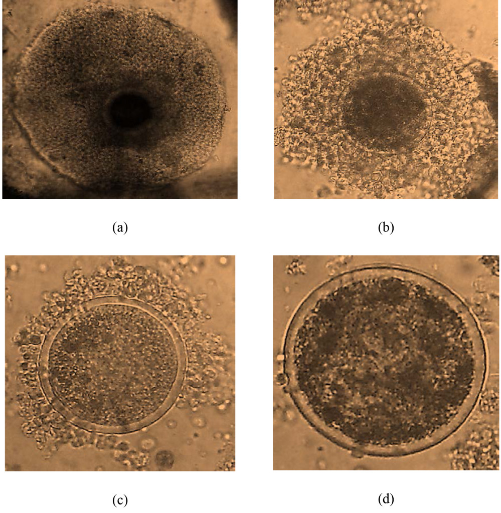
From 706 ovaries, 3581 oocytes were harvested. The mean oocyte yield per cow was 10.14±8.37. The oocytes quality grade I, II, III and IV were 2.93±3.07 (28.85%), 2.98±3.08 (29.41%), 1.95±2.32 (19.24%) and 2.28±2.53 (22.51%), respectively (Figure 1). The number of cultivable oocyte for IVEP (G I and II) was 9.00±5.52 (58.28%). Oocyte index was 2.35. Slicing enables to collect a higher number of oocyte especially cultivable oocyte as compared to other techniques (P=0.00). The oocyte yield and quality, grade I and II were higher (P=0.00) in the rainy than dry season (Table 3).

IJVB2017-106-JustinKouamoAfrica_Figures-1

Figure 1. oocyte quality: (a) grade I ; (b) grade II ; (c) grade III ; (d) grade IV.

Table 3. Effect on the stage of sexual cycle, harvesting techniques and season on the yield and oocyte quality (Mean ± SEM)

Oocyte grades
Factors   N Average number of oocytes I II III IV Selected oocytes for IVEP. I and II (%)
Stage of the sexual cycle Metestrus 9 12.56±2.79 a 3.00±1.03a 3.67±1.03a 2.89±0.77a 3.00±0.84a 6.67±1.85 a (53.1)
  Diestrus 164 10.47±0.65a 2.77±0.24a 3.08±0.24a 2.07±0.18a 2.50±0.20a 5.85±0.43a (56.14)
  Proestrus 148 10.16±0.69 a 3.18±0.25a 2.94±0.25a 1.93±0.19a 2.12±0.21a 6.11±0.46a(60.17)
  Estrus 32 7.94±1.48a 2.53 ±0.54a 2.50±0.55a 1.19±0.41a 1.79±0.45a 5.03±0.98a (63.39)
  p-value   0.42 0.24 0.57 0.10 0.43 0.55
Harvesting technique Slicing 83 15.61±0.83a 4.67±0.31a 4.80±0.32a 2.88±0.24a 3.27±0.27a 9.47±0.55a (60.65)
  Puncture 85 7.28±0.82 b, e 2.18±0.30 b 2.19±0.31 e,b 1.3 ±0.24 b, d 1.67±0.26 b, d 4.31±0.54 b (59.13)
  Aspiration 81 5.77±0.84 c, b 1.23±0.31 c 1.77±0.32 c, b 1.32±0.25 c, b 1.44±0.27 c, b 3.00±0.56 c, b (52.03)
  Aspiration + Slicing 80 11.94±0.84 d 3.49±0.31 d 3.13±0.32 d 2.48±0.25a 2.85±0.27 a, e 6.61±0.56 d (55.39)
  Puncture + Slicing 24 10.17±1.53 e, d  3.58±0.57 a, d  3.17±0.59 e, d 1.42±046 d, c  2.00±0.50 d, c, e 6.75±1.02 e, d (66.39)
  P-value   0.00 0,00 0,00 0.00 0.00 0.00
Season Rainy 162 11.91±1.41a 3.01±0.44 a 3.34±0.48a 2.54±0.43a 3.01±0,46a 6.35±0.82a(53.34)
  Dry 191 8.65±1.05 b 2.85±0.46a 2.68±0.44 b 1.45±0.24 b 1.66±0.27 b 5.53±0.81b (53.98)
  p-value   0.00 0.19 0.00 0.00 0.00 0.03

 a, b, c, d, e : In each column different letters indicated significant difference between group (p<0.05). N=number.

Correlated effects on the characteristics of cows (origin, race, BMI, average weight of ovaries), state of sexual cycle, harvesting technics and season on the follicular population and oocyte quality

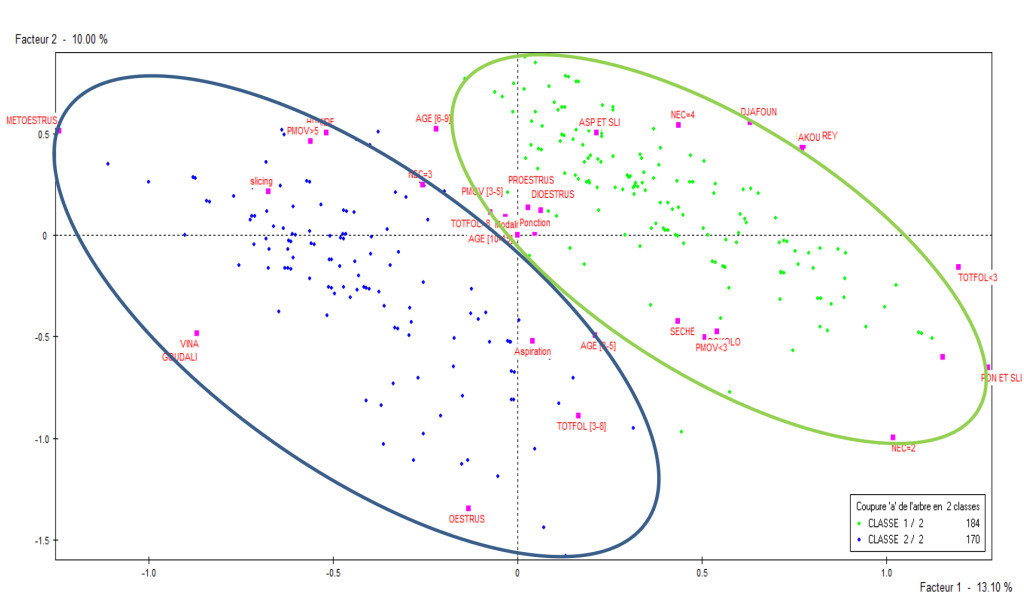
From the hierarchical ascendant correspondence, two classes were identified as shown in Figure 2 following the factorial axe 1 and 2. Class 1 was constituted of Djafoun (38.59%), Akou (58.15%) and Bokolo  breeds from the Mayo Rey (99.46%); BCS 2- 4 ; 6-9 and 10-15 years old; mean weight of ovaries < 3g and 3-5g; sexual cycle in the stage of diestrus and proestrus; collection technique by puncture, aspiration-slicing, puncture-slicing; dry season (60.87%); diameter of follicles < 3 mm and > 8mm. Class 2 was constituted of cows from Vina (98.24%); Gudali breed (92.94%); BCS 3 (85.29%); 3-5 years old; average weight of ovary >5g; sexual cycle in the stage of estrus and metestrus; harvesting technique by slicing (32.94%) and aspiration; rainy season (53.53%) ; mean diameter of follicles 3-8 mm.

IJVB2017-106-JustinKouamoAfrica_Figures-2

Figure 2. Distribution of the two classes representing the individuals in a factorial plan 1 and 2.

The class 2 (n=170; 48.02%) presents a better yield and oocyte quality than class 1 (n=183; 51.98%) as illustrated in Table 4.

Table 4. Variation of the total follicles, yield and oocyte quality per cow the two classes (Mean ± SEM)

Factors N Mean weight of cows (Kg)      Oocytes grades Selected oocytes for IVEP. I and II (%)
Average number of oocytes I II III IV
Average number of follicles
Classe I 183 349.59±11.13 29.00±19.00 8.50±7.00 2.41±2.66 2.46±2.64 1.66±2.13 1.97±2.32 4.87±5.30 (57.3)
Classe II 170 368.53±4.31 26.00±18.00 11.91±9.00 3.48±3.37 3.54±3.42 2.27±2.47 2.62±2.67 7.02±6.79 (59.00)
P – value   0.10 0.12 0.75 0.52 0.61 0.92 0.68 0.17

Discussion

Follicular population

The average follicular population counted on the surface of ovaries was similar to those obtained by Carvalho, et al. [19] on Nellore Zebus and inferior in Moroccan zebus [20]. The high number of small follicles is due to the high numbers of reserve follicles recruited to evolve until one arrives the stage of dominant follicle. Cows of class 1 have mostly small (<3mm) and large (>8mm) follicles whereas those of class 2 have more medium (3-8mm) follicles. The differences might be due to several factors which constitutes each class like the stage of sexual cycle. In this study, many follicles were observed during diestrus and proestrus (class1) than metestrus and estrus (class 2). A high number of punctured follicles was reported at the onset of the cycle (4.9) than the middle (3.4) or the end (3.9) [12]. In estrus stage, the follicles are large in size and only one becomes dominant (12-13mm) while the others are atresia [21]. This might explain the fact that more large follicles were observed in estrus stage in this study. The higher number of follicles accounted on the ovary in the rainy season was reported by Fassi Fihiri [19] and Amer and Moose [22] who indicated that ovarian activity is better in the rainy season (class 2). The reproductive performance in the dry season is low and affects the follicular growth due to thermic stress [23, 24]. Barati et al and Al-katanani et al. explained that heat increase the number of small follicles as we observed in class 1 of this study and transferable embryos are in bad quality [25]. Armstrong [26] reported that fertility of all species reduce with age. This is in accordance with this study where the number of follicles reduce with age. Older cows in the dry season are lighter in weight due to the scarcity of forage and their ovaries are less heavy (<5g) as in the class 1 [27].

Oocyte quality

The oocyte yield and quality are similar to those obtained by Mutha and Uma [28] and superior to that reported by several authors [9, 26, 29]. The difference might be due to the harvesting techniques and other factors (breed, age, BCS, weight of ovary and the origin of the cow). Different factors between class 1 and 2 explained the variation of the oocyte production. The number of oocytes graded I, II, III and IV as well as cultivated oocytes (I and II) reduces in class 1 with respect to the global population compared to class 2. The best quality of oocyte is obtained from follicles with average size and there is a linear growth relationship between the diameter of oocytes and the size of ovarian follicle [30]. Acceptable oocytes for IVM (G I and II) are in the interval of 30 to 60 % oocytes as reported by Natumyana et al. [9]. Considering the multifactorial analysis, metestrus and estrus (class 2) offers a better yield and quality oocytes than proestrus and diestrus (class 1). Hanzen [11] reported that the level of oocytes recovered (50 and 53 %) is the same at all the stages of the sexual cycle. Meanwhile Mutha and Uma [28] showed that the oocyte yield and the quantity of cultivable oocytes are greater when the cows are in the follicular (proestrus and estrus) than (metestrus and diestrus) luteal stage. At the onset of the sexual cycle of the cow, numerous reserve follicles are continuously recruited to develop to the stage of De Graafian follicles. Thus, estrus and metestrus constitute the favourable period of the sexual cycle for the emergence of quality oocytes.

The oocyte yield and quality as well as the cultivable oocytes (I and II) are higher using the slicing technique compared to others (class 2). These results are similar to those obtained by several authors Das and Santra Mutha and Uma, Bohlooli et al.,) [31, 28, 15]. The slicing technique collects free oocytes at the surface as well as the cortex from ovaries. Moreover, certain oocytes lost by the aspiration and puncture methods can be recovered by slicing and could increase the quantity of retrieved oocytes [15]. The low yield of oocyte collected by aspiration may be due to the method which retrieves only the superficial follicles and to the pressure applied by aspiration as well as the size of the needles [32]. In an experiment in which recovered oocytes were cultured to blastocysts, Wikland et al. [33] showed that the percentage of recovered oocytes that became blastocysts decreased as pressure increased. Fry et al. [34] aspirated 5827 follicles from 720 ovaries with 17 and 20-gauge needles. More than 5000 of these follicles were 2–4 mm in diameter. The rest were 5–15 mm. With 17-gauge needles, 56 % of the follicles yielded oocytes, but with 20-gauge needles, recovery dropped to 45 %. The highest recovery also occurred with the highest aspiration pressures. The pressures evaluated ranged from 25 to 100 mm Hg. Recovery was 46 % at 25 mm Hg and 59 % at 100 mm Hg. They concluded that the optimal pressure to maximize recovery of bovine COCs was 55 mm Hg with a 17-gauge needle and 77 mm Hg for a 20-gauge needle. As aspiration pressures increased, the recovered oocytes were increasingly denuded of cumulus cells [35]. This loss of cumulus cells also occurred at lower aspiration machine pressures using larger diameter needles compared to smaller diameter needles, suggesting that needle gauge and pressure interdependently contributed to outcome. Dominguez [36] and Fassi Fihri [7] noted a significant difference in terms of oocyte yield in the European and zebu breeds as well as crossbreeds. Gudali breeds from the Vina division, of at least 6 years old during estrus and metestrus associated with other factors like slicing or aspiration techniques in the rainy season offers a better oocyte yield (class 2). This finding confirms the adaptability of the Gudali breed in its natural environment (class 2). Moreover, Natumanya et al. [9] reported that oocyte yield and quality are reduced in tropical zebus aged more than 10 years, thin (BCS 1 and 2) and having small ovaries (< 5g) (class 1).

Ovarian activity was best in the rainy season [22]. The rainy season allows emergency of medium follicles (class 2). The slicing technique, when practiced in the rainy season (class 2), optimises the oocyte yield [36, 22]. Kubovicova et al. [47] obtained a high oocyte yield when the cows had an average BCS (class 2). They confirmed that the nutritional status of the animal had an influence on the ovarian function in zebu cows, likewise the season of the year [37]. In fact, there is abundant and quality pasture in rainy season resulting in well fed cows. The metabolic and hormonal changes between the rainy and dry season affect follicular dynamics and oocyte development. During oogenesis and folliculogenesis, several dynamic processes that are regulated by endocrine, paracrine and autocrine signals have been shown to be linked with energetic status and the quality of oocyte depends on the nutritional condition under which the follicle began its development [38]. Moreover, ooocyte growth and maturation are believed to be particularly sensitive to changes in nutritional, chemical, and endocrine environments.

This study indicates that the oocyte yield and quality are high when they are retrieved in class 2 conditions. To improve the IVEP, it is recommended to harvest the oocytes in zebu cattle by slicing in the rainy season at estrus or metestrus.

Acknowledgements: The authors acknowledge contribution of NMSH.

References

  1. MINEPIA (Ministry of Livestock, Fisheries and Animal Husbandry) (2009). Schéma directeur pour le développement des filières de l’élevage au Cameroun, volume II: cartographie des filières. 82.
  2. Kouamo J, Sow A Leye A, Sawadogo GJ and Ouedraogo GA (2009) Amélioration des performances de production et de reproduction des bovins par l’utilisation de l’insémination artificielle en Afrique Sub-saharienne et au Sénégal en particulier: état des lieux et perspectives. RASPA 7: 139–148.
  3. Kouamo J, Meyoufey B and Zoli AP (2016) Pathological study of female reproductive organs of local zebus in Adamawa region. Bull Anim Hlth Prod Afr 64: 119–128.
  4. Manik RS, Singla SK and Palta P (2003) Collection of oocytes through transvaginal ultrasound-guided aspiration of follicles in an Indian breed of cattle. Anim Reprod Sci 76: 155–161.
  5. Abraham MC, Gustafsson H, Ruete A, Brandt YC (2012) Breed influences on in vitro development of abattoir-derived bovine oocytes. Acta Vet Scan 54: 1–6.
  6. Agrawal KP, Sharma T, Sexana C and Sharma N (1995) Chronology of first meiotic events of caprine oocytes matured in vitro. Indian J Anim Sci 65: 285–288.
  7. Fassi Fihri AH, Lakhdissi L, Derqaoui Kh, Hajji M, Naciri A and Goumari (2005) Genetic and non-genetic effects on the number of ovarian follicles and oocyte yield and quality in the bovine local (Oulmes Zaer), exotic breeds and their crosses in Morocco. African J Biotech 4: 9–13.
  8. Njoya A, Bouchel D, Ngo Ntama AC, Moussa CA, Martrenchar A and Leteneur L (1997). Systèmes d’élevage et productivité des bovins en milieu paysan au nord-Cameroun. World Anim Review 89: 12–23.
  9. Natumanya R, Owiny D and Kugonza R (2008) The potential of Ankole cattle abattoir ovaries for in vitro embryo production. African J Anim Biomed Sci 3: 1–5.
  10. Moussa Garba MH, Marichatou M, Issa ML, Abdoul Aziz C and Hanzen C (2013) Tractus génital des vaches zébus (Bos indicus) au Niger Rev Elev Med Vet Pays Trop 66: 137–142.
  11. Hanzen C (2009) La production d’embryons in vitro. 21: 3.
  12. Satrapa RA, Nabhan T, Silva CF, Simoens RAL, Razza EM, et al. (2010). In?uence of sire breed (Bos indicus versus Bos taurus) and interval from slaughter to oocyte aspiration on heat stress tolerance of in vitro-produced bovine embryos. Theriogenology 76: 1162–1167.
  13. Nguyen-Kien C and Hanzen C (2009) Ante mortem and post mortem examination of the genital tract of dairy cows in South Vietnam Rev Elev Med Vet Pays Trop 66: 85–90.
  14. Wang ZG, Song-Dong Y and Zi-Rong X (2007) Effects of collection methods on recovery efficiency, maturation rate and subsequent embryonic developmental competence of oocytes in holstein cow. Asian-Aust J Anim Sci 20: 496–500.
  15. Bohlooli BS and Cedden F (2015) Effect of Different Harvesting Techniques on the Recovery and Quality of Bovine Cumulus Oocyte Complexes. Iranian J Applied Anim Sci 5: 741–74.
  16. Alves BG, Alves KA, Lucio AC, Martins, MC and Silvas TH (2014) Ovarian activity and oocyte quality associated with the biochemical profil of serum and follicular fluid from girolando dairy cows postpartum. Anim Reprod Sci 146: 89–236.
  17. Duygu BA, Muhammed KB, Dogan N and Hander G (2013) Effect of the stage of oestrus cycle on follicular population oocyte yield and quality, and biochemical composition of serum and follicular fluid in Anatolian water buffalo. Anim Repro Sci 137: 8–114.
  18. Lebart L, Morineau A and Piron M (2000) Statistique exploratoire multidimensionnelle. DUNOD 3e éd 439.
  19. Carvalho JB, Carvalho NA, Reis EL, Nichi M, Souza AH and Baruselli PS (2008) Effect of early luteolysis in progesterone-based timed ai protocols in bos indicus, bos indicus x bos taurus, and bos taurus heifers. Theriogenology 69: 167–175
  20. Fassi Fihri A (2006) Collecte et maturation des ovocytes bovins: Effet de l’état nutritionnel sur le rendement et la qualité des ovocytes. Thèse de Doctorat d’Etat Es-Sciences Biologiques 163.
  21. Fieni F, Tainturuer D, Bruyas JF and Battut I (1998). Examen échotomographique des ovaires chez la vache. Journées nationales des GTV 411–415.
  22. Amer H and Moose A (2008) Relationship between season of the year, culture medium and in vitro oocyte competence in dromedary camels. Bulg J Vet Med 11: 195–204.
  23. Barati F, Niasari-naslaji A, Bolourchi A, Sarhaddi F, Razavi K, et al. (2006) Superovulatory response of Sistani cattle to three different doses of FSH during winter and summer. Theriogenology 66: 1149–1155.
  24. Al-Katanani YM1, Paula-Lopes FF, Hansen PJ (2002) Effect of season and exposure to heat stress on oocyte competence in Holstein cows. J Dairy Sci 85: 390–396. [crossref
  25. Taneja M, Totey SM and Ali A (1995). Seasonal variation in follicular dynamics of superovulated Indian water buffalo. Theriogenology 43: 451–464.
  26.  Armstrong DT (2001) Effects of maternal age on oocyte developmental competence. Theriogenology 55: 1303–1322. [crossref]
  27. Nandi S, Chauhan MS and Palta P (2001) Effect of environmental temperature on quality and developmental competence in vitro of buffalo oocytes. Vet Record 148: 278–279.
  28. Mutha RM and Uma MY (2012) Efficacy of different harvesting techniques on oocyte retrieval from buffalo ovaries. Buffalo Bulletin 31: 4.
  29. Zheng-Guang W, Song-Dong Yu and Zi-Rong X (2007) Effects of Collection Methods on Recovery Efficiency, Maturation Rate and Subsequent Embryonic Developmental Competence of Oocytes in Holstein Cow. Asian-Aust. J Anim Sci 20: 496–500.
  30. Yadav EN, Kharche SD, Goel AK, Jindal SK and Johri DK (2007) Comparative efficacy of different techniques for oocytes recovery from pre pubertal goat ovaries. Indian J Anim Sci 77: 988–990.
  31. Das SK and Santra A (2008) Comparative the efficiency of different oocyte recovery methods from cattle ovaries. Indian J Anim Sci 78: 277–278.
  32. Maina VA, Muktar A and Sabo YG (2008). Effects of Body Condition Score on ovarian activity of Bos indicus (Zebu) Cow. Asian J Sci Res 1: 421–428.
  33. Wikland M, Blad S, Bungum L, Hillensjö T, Karlström PO (2011) A randomized controlled study comparing pain experience between a newly designed needle with a thin tip and a standard needle for oocyte aspiration. Hum Reprod 26: 1377–83.
  34.  Fry RC, Niall EM, Simpson TL, Squires TJ, Reynolds J (1997) The collection of oocytes from bovine ovaries. Theriogenology 47: 977–987. [crossref]
  35. Bols PE, Ysebaert MT, Van Soom A and de Kruif A (1997). Effects of needle tip bevel and aspiration procedure on the morphology and developmental capacity of bovine compact cumulus oocyte complexes. Theriogenology 47: 1221–36.
  36. Davachi DN, Shahneh Zare A, Kohram H, Zhandi M, Dashti S, et al. (2014). In Vitro Ovine Embryo Production: the Study of Seasonal and Oocyte Recovery Method Effects. Iran Red Crescent Med J 16 (9): e20749.
  37. Ali S, Ahmad N, Akhtar N, Zia-ur R and Sarwar M (2007) Effect of season and age on the ovarian size and activity of one-humped camel (Camelus dromedarius). Asian-Aus J Anim Sci 20: 1361–1366.
  38. Monniaux D, Caraty A, Clement F, Dalbiès-Tran R, Dupont J, et al. (2009) Développement folliculaire ovarien et ovulation chez les mammifères. Inra Prod Anim 22: 59– 76.

Surrogate Markers of Liver Fibrosis in Primary Sclerosing Cholangitis (PSC)

DOI: 10.31038/IMROJ.2017231

Introduction

Primary Sclerosing Cholangitis (PSC) is a chronic inflammatory cholangiopathy that results in fibrotic strictures and dilations of the intra- and extrahepatic bile ducts. The pathogenesis of PSC has not been fully elucidated, the disease is uncommon, occurs predominantly in young males and has a strong association with Inflammatory Bowel Disease (IBD). There are significant variation in clinical course of PSC associated with age at diagnosis, sex, and ductal and IBD subtypes [1]. There is no medical treatment of proven benefit on survival; most liver-related morbidity and mortality is the results of portal hypertension and chronic liver failure. However, the course of PSC is highly variable, and so far no prognostic markers have been shown to predict outcomes in asymptomatic, early-stage patients.

The prognosis of chronic cholestatic liver disease depends at least in part on the extent of fibrosis in the liver parenchyma [2]. Semi-quantitative evaluation of nodular size and fibrotic septal width in respect to hepatic venous pressure gradient (HVPG) were proposed by Laennec based on the original histological description of the cirrhosis [3-6]. HVPG is the gold standard to estimate the severity of portal hypertension in liver cirrhosis. It correlates with structural and functional changes in liver parenchyma and gives valuable prognostic information to stratify the mortality risk [7]. Liver cirrhosis should be regarded as a multistage liver disease [8]; it can be accurately sub-classified using quantification of fibrosis with collagen proportionate area (CPA) as the predictor of clinical decompensation [9].

Liver biopsy remains the “gold standard” in evaluation of necroinflammation activity and fibrosis of the liver parenchyma. However, it has limitations due to invasiveness, small tissue samples, patchy distribution of fibrotic areas in parenchyma and inter- and intra-observer error. Moreover, liver biopsy is not appropriate to regularly monitor fibrosis progression or response to treatment [10]. Thus, ultrasound-based shear wave elastography methods enabling liver stiffness measurements (LSM) have been implemented for noninvasive evaluation of fibrosis of the liver, with biopsy reserved for uncertain cases.

The various elastography methods differ with respect to what they do with these displacement data to create an elastogram or elasticity measurement. There are three options for the property to be displayed:

1. Display of displacement without further processing, as in acoustic radiation force impulse (ARFI) imaging. Tissue displacement is associated with shear deformation. The greater the force, the greater the displacement, but stiff tissues are displaced less than soft tissues. ARFI remains the proprietary imaging technology Siemens Virtual Touch™, and it is not used for assessment of diffuse liver conditions.

2. Display of tissue strain or strain rate, calculated from the spatial gradient of displacement or velocity,

3. Display of shear wave speed, calculated by using the time varying displacement data to measure the arrival time of a shear wave at various locations. All such methods are grouped under the heading shear wave elastography (SWE), and include transient elastography (TE), point shear wave elastography (pSWE) and multidimensional shear wave elastography (2D‑SWE and 3D-SWE).

Shear wave elastography (SWE) is a method that use shear wave speed and includes:

1.Transient elastography (TE, FibroScan, Echosens, France): shear wave elastometry by measurement of the speed of a shear wave that has been generated using a surface impulse,

2.Point shear wave elastography (pSWE): shear wave elastometry at a location by measurement of the speed of a shear wave generated using acoustic radiation force,

3.Multidimensional shear wave elastography (2D-SWE, 3DSWE): quantitative SWE imaging (and elastometry) by measuring the speed of shear waves generated using acoustic radiation force.

The major potential confounding factors (liver inflammation indicated by AST and/or ALT elevation >5 times the normal limits, obstructive cholestasis, liver congestion, acute hepatitis and infiltrative liver diseases) should be excluded before performing LSM with SWE, in order to avoid overestimation of liver fibrosis [11].

Ultrasound-based methods

In chronic liver disease LSM accurately reflects liver fibrosis, which is the major component of increased intrahepatic vascular resistance leading to portal hypertension. LSM improves the noninvasive risk stratification of patients with compensated advanced chronic liver disease as a possible surrogate for portal hypertension [12]. More than 90% of patients with an LSM > 20-25 kPa ( evaluated by transient elastography ) will have clinically significant portal hypertension. In advanced chronic liver disease of non-cholestatic aetiology, endoscopy can be safety avoided by using LSM and platelet count in combination: LSM of < 20 kPa and PLT > 150 g/L pointed to < 5% risk of esophageal varices needing treatment [12].

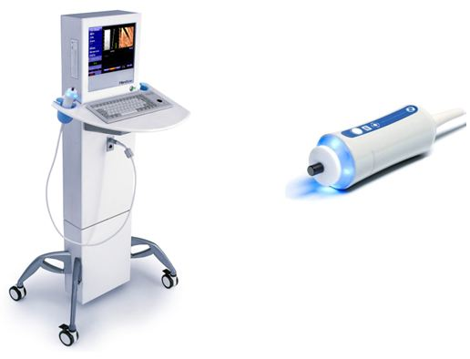
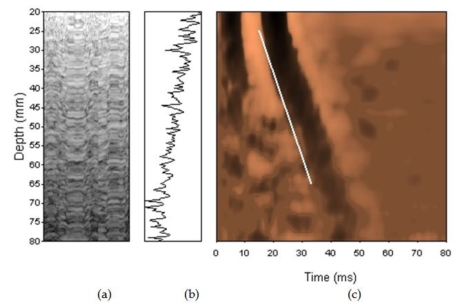
Transient elastography (TE) (FibroScan, Echosens, France) is currently the most widely used technique, validated in chronic hepatitis C [13], in primary biliary cholangitis (PBC) [14, 15] and primary sclerosing cholangitis [16]. TE measures the speed of propagation of an elastic shear wave in the liver, and the harder the tissue, the faster the shear, which is measured in kilopascals (kPa). The examination is performed on the right lobe of the liver, and the measurement depth trough intercostal space is 25-65 mm using standard M-probe, and 35-75 mm with XL-probe (Figure 1). Liver stiffness measurement based on TE has been shown to correlate with histological fibrosis stage and severity of portal hypertension [17, 18]. TE seems to be a predictor of clinical outcomes in relationship to liver-related complications and mortality [19, 20]. Additionally, TE is able to predict clinically significant portal hypertension in patients with compensated chronic liver disease or cirrhosis [21]. However, early compensated liver cirrhosis can be overlooked in up to 30% of patients and transient elastography seems to be better at excluding advanced fibrosis rather than confirming liver cirrhosis. Fibrosis stage F > 2 is diagnosed with 84-87% accuracy, and F> 3 with 88-89%. Diagnostic accuracy is excellent – 93-96% for the diagnosis of liver cirrhosis, with sensitivity and specificity of 70-79%, 78-84% for F > 2 and 83-87% and 89-95% for the diagnosis of F = 4. Cut-offs were in the range of 7.3-7.9 kPa for F > 2, and 13.0-15.6 kPa for the diagnosis of liver cirrhosis.

IMROJ 2017-208 Figure1A

IMROJ 2017-208 Figure1B

Figure 1. Transient elastrography

In the newest study of Krawczyk et al. TE correlated with Laennec stages of fibrosis, collagen contents and with diameter of thickest septa in explanted livers in PSC patients. In multivariate model liver fibrosis according to either Leannec score or collagen contents was significantly associated with TE. PSC cirrhotics patients had increased liver stiffness and the TE cut-off of 13.7 kPa showed the best predictive value (AUC=0.90, 95%CI 0.80–1.00, P<0.0001) for detecting liver cirrhosis [57].

The measurement failure rate is low (5-10%) with obesity (BMI > 30 kg/m2), ascites, congestive heart failure, postprandial time and the presence of narrow intercostal space considered to be limiting factors. However, obstructive cholestasis also influenced the results of TE [22].

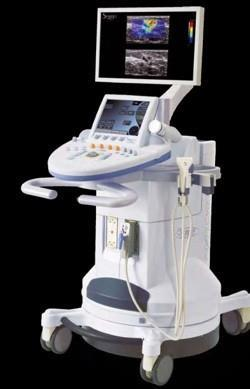
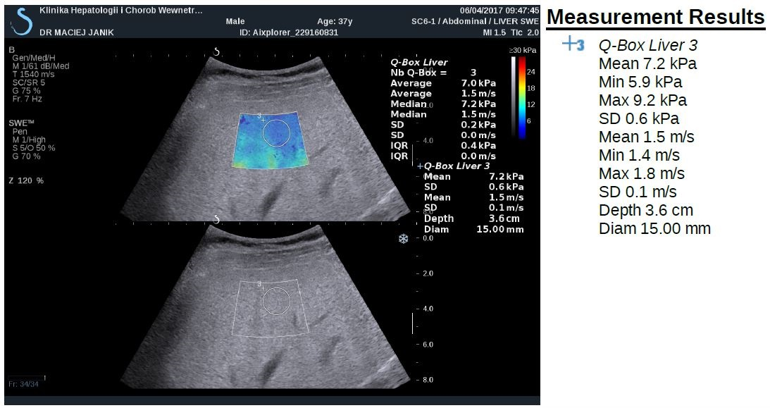
Newer elastography methods based on the measurements of shear wave velocity include point share wave elastography (pSWE) and two-dimensional SWE (2D- SWE). SWE is usually integrated into conventional ultrasonography system (Figure 2). The region of interest (ROI) can be positioned under brightness-modulation (B-mode), and a single acoustic impulse is used to induce a share wave within a ROI of 1.0 x 0.5 cm or 2 x 2 cm in 2D SWE. The examination should be performed at least 1 cm below the liver capsule on the right lobe, and can be displayed in m/s and/or kPa. ROI can be positioned manually in different depths of the liver. However, there are no clear interpretation of point SWE and 2D SWE recommended to date.

IMROJ 2017-208 Figure2A

IMROJ 2017-208 Figure2B

Figure 2. Shear-wave elastography

The probability of correctly diagnosing EV following a positive measurement did not exceed 70% [21]. Thus, LSM-spleen diameter to platelet ratio score and simplified combination of LSM and platelet count were also assessed with good results of ruling out varices needing treatment [23, 24]. LSM can be also used to predict clinical decompensation in the patients with compensated cirrhosis of the liver. On the other hand, spleen undergoes parenchymal modeling in patients with portal hypertension, and spleen stiffness measurement (SSM) is closely associated with portal hypertension, its severity and complications [25]. SSM is promising parameter for use in predicting the presence and size of EV [12]. Validated cut-off values in PSC are not available yet.

Magnetic-resonance based method

With magnetic resonance elastography (MRE) mechanical shear waves are sent into the tissue and displayed as elastograms using phase-contrast image sequences. MRE can examine the very large areas of the right lobe of liver. The limitation of MRE are obesity, claustrophobia and iron overload. Recently, in the study of Wang et al. the performance of MRE was significantly better than laboratory tests for detection of advanced fibrosis, and cirrhosis and better than conventional MRI for diagnosis of cirrhosis in patients with autoimmune hepatitis [26]. In a retrospective review of 266 PSC patients to examine whether liver stiffness (LS) was associated with the primary endpoint of hepatic decompensation (ascites, variceal hemorrhage and hepatic encephalopathy), MRE was able to detect cirrhosis with high specificity and LS obtained by MRE was predictive of hepatic decompensation in PSC patients in Eaton et al study. Liver stiffness of 4.93 kPa was the optimal point to detected F4 fibrosis, with sensitivity 1.00 (95% confidence interval (CI), 0.40-1.00) and specificity of 0.94 (95%CI, 0.68-1.00). LS was associated with the development of decompensated liver disease (Hazard ratio, 1.55; 95%CI, 1.41-1.70). The optimal LS thresholds that stratified patients at a low, medium and high risk for hepatic decompensation were <4.5, 4.5-6.0 and >6.0 kPa, respectively [27]. However, MRE seems to be promising modality for detection of advanced fibrosis and liver cirrhosis, with superior diagnostic accuracy compared to laboratory assessment and MRI, but not precirrhotic stages of chronic liver diseases. On the other hand, MRE is very expensive and time-consuming.

Serum biomarkers

Prospective studies demonstrated that single markers e. g., α2-macroglobulin [28], procollagen III N-peptide [29], apolipoprotein A1 [28], haptoglobin [30], hyaluronic acid [31], metalloproteinases [32] allow discrimination between advanced and absent fibrosis.

The enhanced liver fibrosis (ELF) test is a promising panel, incorporating three direct serum markers of fibrosis in an algorithm: hyaluronic acid, tissue inhibitor of metalloproteinases-1 (TIMP-1), and amino-terminal pro-peptide of type III pro-collagen (PIIINP) [33]. The ELF test accurately predicted significant liver fibrosis and furthermore predicted clinical outcome in several independent populations and in patients with various aetiologies of chronic liver disease [34] as well as with PSC. The ELF test consistently predicted liver transplant-free survival in PSC patients independently of other risk factors or risk scores [35]. The ELF test distinguished between mild and severe disease defined by clinical outcome (transplantation or death) with an area under the curve of 0.81 (95% confidence interval [CI] 0.73-0.87) and optimal cutoff of 10.6 (sensitivity 70.2%, specificity 79.1%). In multivariate Cox regression analysis ELF score was associated with transplant-free survival independently of the Mayo risk score. The ELF test correlated also with ultrasound elastography in separate assessments [35]. In a large multicenter cohort, EFL test predicts prognosis in PSC and may be used for risk stratification in clinical follow up; optimally together with clinical prognostic scores may add incremental prognostic value [36].

Placental growth factor (PLGF), growth differentiation factor-15 (GDF-15) and hepatic growth factor (HGF) are involved in hepatic fibrogenesis. The panel of these three serum markers was useful for the detection of patients with advanced fibrosis and the risks described by the combinations of these markers were independent from other classical fibrosis risk factors. The set of markers may be a useful tool to monitor patients with chronic liver diseases during and after therapy [37] .

Inflammatory protein, i.e. IL-8 in bile and serum was an important indicator of disease severity and prognosis in patients with primary sclerosing cholangitis, and associated with transplant-free survival in multivariable analyses independently of age and disease duration, indicating an independent influence on PSC progression [38]. This is also in line with the results of the study of Buck et al [39]. Hepatic venous pressure gradient (HVPG) can reflect progression of disease in the precirrhosis stage. Portal hypertension is pathogenically related to liver injury and fibrosis [40] and that in turn these are associated with the activation of inflammatory pathways [41]. The novel inflammatory serum biomarkers (e.g. Il-1, Fas-R, VCAM, CD163) were significantly correlated with HVPG in patients with compensated cirrhosis in this study.

Autotaxin (ATX), which is involved in the synthesis of lysophosphatidic acid, is not only associated with pruritus but also indicates impairment of other health-related quality of life (HRQoL) aspects, liver dysfunction, and can serve as a predictor of survival [42]. Impairment of HRQoL might be also associated with vitamin D receptor (VDR) gene polymorphisms (rs1544410-BsmI; rs7975232-ApaI). ApaI polymorphisms in VDR may exert an effect on disease-related symptoms and quality of life in the study of 275 patients with PSC [43].

However, none of the proposed markers or panels have gained as much acceptance as the invasive approach [44]. This may be due to relatively high costs of marker measurements, and low sensitivity to discriminate between fibrotic, cirrhotic or steatotic liver lesions. As a result, no scores based on serum levels of hepatic fibrosis markers are actually regarded as definite methods upon which therapeutic decisions can be based. It might be that the combination of markers reflects the presence of significant liver fibrosis detected by elastography and histology and may also identify patients at risk presenting with low elastography values as proofed by Krawczyk M, et al. [37].

Simple laboratory tests

Laboratory-based methods for staging liver fibrosis include the FibroTest® [45], the serum aspartate aminotransferase/platelet ratio index (APRI) [46], the Fibrosis 4 (FIB-4) test [47], and the enhanced liver fibrosis test [48]. AST/ALT ratio [49] can also allow to discriminate between advanced and absent fibrosis.

However, these tests may detect cirrhosis, but their ability to reflect the stages of fibrosis in AIH is uncertain [50-54]. The result of the recent study of Anastasiou et al. showed that TE, NAFLD fibrosis score and FibroQ might help in evaluation of liver fibrosis in AIH, but without differentiating mild form from advanced stages of fibrosis in autoimmune hepatitis [55].

In the study of Krawczyk et al. TE correlated with Laennec stages of fibrosis, and with serum indices of liver injury, namely AST, bilirubin as well as FIB-4 and APRI scores in patients with PSC [57].

Conclusion

Primary sclerosing cholangitis (PSC) is a progressive biliary disease lacking medical treatment with currently no established tools to predict prognosis in the individual patient. The lack of biomarkers for risk stratification is an important obstacle to the development of therapy.

Liver fibrosis seems to be the strongest predictor of liver stiffness assessed with TE. TE correlates with liver fibrosis, markers of liver injury and portal hypertension in patients with PSC. It might be that TE is a reliable tool for non-invasive monitoring of PSC. It seems also that the combination of serum profibrotic biomarkers with evaluation of liver fibrosis with elastography may improve the non-invasive diagnostic utility for clinically significant fibrosis [56]. However, still the Enhanced Liver Fibrosis (ELF®) test and Mayo risk score proved to be stronger predictors of transplant-free survival in PSC [38].

Conflict of interest: Nothing to declare

References

  • Weismuller TJ, Trivedi PJ, Bergquist A, Imam M, Lenzen H, Ponsioen CY, et al. (2017) Patient Age, Sex, and Inflammatory Bowel Disease Phenotype Associate With Course of Primary Sclerosing Cholangitis. Gastroenterology 152:1975-84 e8.
  • Dickson ER, Murtaugh PA, Wiesner RH, Grambsch PM, Fleming TR, Ludwig J, et al. (1992) Primary sclerosing cholangitis: refinement and validation of survival models. Gastroenterology 103:1893-901.
  • Kim G, Lee SS, Baik SK, Cho YZ, Kim MY, Kwon SO, et al. (2016) The need for histological subclassification of cirrhosis: a systematic review and meta-analysis. Liver Int 36:847-55.
  • Kim SU1, Oh HJ, Wanless IR, Lee S, Han KH, et al. (2012) The Laennec staging system for histological sub-classification of cirrhosis is useful for stratification of prognosis in patients with liver cirrhosis. J Hepatol 57: 556-563. [crossref]
  • Rastogi A, Maiwall R, Bihari C, Ahuja A, Kumar A, Singh T, et al. (2013) Cirrhosis histology and Laennec staging system correlate with high portal pressure. Histopathology 62:731-41.
  • Wang W, Li J, Pan R, A S, Liao C (2015) Association of the Laennec staging system with degree of cirrhosis, clinical stage and liver function. Hepatol Int 9:621-6.
  • La Mura V, Nicolini A, Tosetti G, Primignani M (2015) Cirrhosis and portal hypertension: The importance of risk stratification, the role of hepatic venous pressure gradient measurement. World J Hepatol 7:688-95.
  • Garcia-Tsao G, Friedman S, Iredale J, Pinzani M (2010) Now there are many (stages) where before there was one: In search of a pathophysiological classification of cirrhosis. Hepatology 51:1445-9.
  • Tsochatzis E, Bruno S, Isgro G, Hall A, Theocharidou E, Manousou P, et al. (2014) Collagen proportionate area is superior to other histological methods for sub-classifying cirrhosis and determining prognosis. J Hepatol 60:948-54.
  • Rockey DC, Caldwell SH, Goodman ZD, Nelson RC, Smith AD; American Association for the Study of Liver Diseases (2009) Liver biopsy. Hepatology 49: 1017-1044. [crossref]
  • Dietrich CF, Bamber J, Berzigotti A, Bota S, Cantisani V, Castera L, et al. (2017) EFSUMB Guidelines and Recommendations on the Clinical Use of Liver Ultrasound Elastography, Update 2017 (Long Version). Ultraschall Med.
  • Berzigotti A (2017) Non-invasive evaluation of portal hypertension using ultrasound elastography. J Hepatol 67: 399-411. [crossref]
  • Ziol M, Handra-Luca A, Kettaneh A, Christidis C, Mal F, et al. (2005) Noninvasive assessment of liver fibrosis by measurement of stiffness in patients with chronic hepatitis C. Hepatology 41: 48-54. [crossref]
  • Corpechot C, Carrat F, Poujol-Robert A, Gaouar F, Wendum D, Chazouilleres O, et al. (2012) Noninvasive elastography-based assessment of liver fibrosis progression and prognosis in primary biliary cirrhosis. Hepatology 56:198-208.
  • Corpechot C1, El Naggar A, Poujol-Robert A, Ziol M, Wendum D, et al. (2006) Assessment of biliary fibrosis by transient elastography in patients with PBC and PSC. Hepatology 43: 1118-1124. [crossref]
  • Corpechot C, Gaouar F, El Naggar A, Kemgang A, Wendum D, Poupon R, et al. (2014) Baseline values and changes in liver stiffness measured by transient elastography are associated with severity of fibrosis and outcomes of patients with primary sclerosing cholangitis. Gastroenterology 146:970-9; quiz e15-6.
  • Berzigotti A, Seijo S, Arena U, Abraldes JG, Vizzutti F, Garcia-Pagan JC, et al. (2013) Elastography, spleen size, and platelet count identify portal hypertension in patients with compensated cirrhosis. Gastroenterology 144:102-11 e1.
  • Talwalkar JA, Kurtz DM, Schoenleber SJ, West CP, Montori VM (2007) Ultrasound-based transient elastography for the detection of hepatic fibrosis: systematic review and meta-analysis. Clin Gastroenterol Hepatol 5:1214-20.
  • Vergniol J, Foucher J, Terrebonne E, Bernard PH, le Bail B, et al. (2011) Noninvasive tests for fibrosis and liver stiffness predict 5-year outcomes of patients with chronic hepatitis C. Gastroenterology 140: 1970-1979. [crossref]
  • Singh S, Fujii LL, Murad MH, Wang Z, Asrani SK, Ehman RL, et al. (2013) Liver stiffness is associated with risk of decompensation, liver cancer, and death in patients with chronic liver diseases: a systematic review and meta-analysis. Clin Gastroenterol Hepatol 11:1573-84 e1-2; quiz e88-9.
  • Shi KQ, Fan YC, Pan ZZ, Lin XF, Liu WY, Chen YP, et al. (2013) Transient elastography: a meta-analysis of diagnostic accuracy in evaluation of portal hypertension in chronic liver disease. Liver Int 33:62-71.
  • Millonig G, Reimann FM, Friedrich S, Fonouni H, Mehrabi A, Buchler MW, et al. (2008) Extrahepatic cholestasis increases liver stiffness (FibroScan) irrespective of fibrosis. Hepatology 48:1718-23.
  • Abraldes JG, Bureau C, Stefanescu H, Augustin S, Ney M, Blasco H, et al. (2016) Noninvasive tools and risk of clinically significant portal hypertension and varices in compensated cirrhosis: The “Anticipate” study. Hepatology 64:2173-84.
  • Augustin S, Millan L, Gonzalez A, Martell M, Gelabert A, Segarra A, et al. (2014) Detection of early portal hypertension with routine data and liver stiffness in patients with asymptomatic liver disease: a prospective study. J Hepatol 60:561-9.
  • Colecchia A, Montrone L, Scaioli E, Bacchi-Reggiani ML, Colli A, Casazza G, et al. (2012) Measurement of spleen stiffness to evaluate portal hypertension and the presence of esophageal varices in patients with HCV-related cirrhosis. Gastroenterology 143:646-54.
  • Wang J, Malik N, Yin M, Smyrk TC, Czaja AJ, Ehman RL, et al. (2017) Magnetic resonance elastography is accurate in detecting advanced fibrosis in autoimmune hepatitis. World J Gastroenterol 23:859-68.
  • Eaton JE, Dzyubak B, Venkatesh SK, Smyrk TC, Gores GJ, Ehman RL, et al. (2016) Performance of magnetic resonance elastography in primary sclerosing cholangitis. J Gastroenterol Hepatol 31:1184-90.
  • Imbert-Bismut F, Ratziu V, Pieroni L, Charlotte F, Benhamou Y, Poynard T, et al. (2001) Biochemical markers of liver fibrosis in patients with hepatitis C virus infection: a prospective study. Lancet 357:1069-75.
  • van Zanten RA, van Leeuwen RE, Wilson JH (1988) Serum procollagen III N-terminal peptide and laminin P1 fragment concentrations in alcoholic liver disease and primary biliary cirrhosis. Clin Chim Acta 177:141-6.
  • Adams LA, Bulsara M, Rossi E, DeBoer B, Speers D, George J, et al. (2005) Hepascore: an accurate validated predictor of liver fibrosis in chronic hepatitis C infection. Clin Chem 51:1867-73.
  • Wong VS, Hughes V, Trull A, Wight DG, Petrik J, Alexander GJ (1998) Serum hyaluronic acid is a useful marker of liver fibrosis in chronic hepatitis C virus infection. J Viral Hepat 5:187-92.
  • Walsh KM, Timms P, Campbell S, MacSween RN, Morris AJ (1999) Plasma levels of matrix metalloproteinase-2 (MMP-2) and tissue inhibitors of metalloproteinases -1 and -2 (TIMP-1 and TIMP-2) as noninvasive markers of liver disease in chronic hepatitis C: comparison using ROC analysis. Dig Dis Sci 44:624-30.
  • Xie Q, Zhou X, Huang P, Wei J, Wang W, et al. (2014) The performance of enhanced liver fibrosis (ELF) test for the staging of liver fibrosis: a meta-analysis. PLoS One 9: e92772. [crossref]
  • Parkes J, Roderick P, Harris S, Day C, Mutimer D, et al. (2010) Enhanced liver fibrosis test can predict clinical outcomes in patients with chronic liver disease. Gut 59: 1245-1251. [crossref]
  • Vesterhus M, Hov JR, Holm A, Schrumpf E, Nygard S, Godang K, et al. (2015) Enhanced liver fibrosis score predicts transplant-free survival in primary sclerosing cholangitis. Hepatology 62:188-97.
  • de Vries EM, Farkkila M, Milkiewicz P, Hov JR, Eksteen B, Thorburn D, et al. (2017) Enhanced liver fibrosis test predicts transplant-free survival in primary sclerosing cholangitis, a multi-centre study. Liver Int.
  • Krawczyk M, Zimmermann S, Hess G, Holz R, Dauer M, Raedle J, et al. (2017) Panel of three novel serum markers predicts liver stiffness and fibrosis stages in patients with chronic liver disease. PLoS One 12:e0173506.
  • Vesterhus M, Holm A, Hov JR, Nygard S, Schrumpf E, Melum E, et al. (2017) Novel serum and bile protein markers predict primary sclerosing cholangitis disease severity and prognosis. J Hepatol 66:1214-22.
  • Buck M, Garcia-Tsao G, Groszmann RJ, Stalling C, Grace ND, Burroughs AK, et al. (2014) Novel inflammatory biomarkers of portal pressure in compensated cirrhosis patients. Hepatology 59:1052-9.
  • Bellot P, Garcia-Pagan JC, Frances R, Abraldes JG, Navasa M, Perez-Mateo M, et al. (2010) Bacterial DNA translocation is associated with systemic circulatory abnormalities and intrahepatic endothelial dysfunction in patients with cirrhosis. Hepatology 52:2044-52.
  • Frances R, Rodriguez E, Munoz C, Zapater P, De la ML, Ndongo M, et al. (2005) Intracellular cytokine expression in peritoneal monocyte/macrophages obtained from patients with cirrhosis and presence of bacterial DNA. Eur J Gastroenterol Hepatol 17:45-51.
  • Wunsch E, Krawczyk M, Milkiewicz M, Trottier J, et al. (2016) Serum Autotaxin is a Marker of the Severity of Liver Injury and Overall Survival in Patients with Cholestatic Liver Diseases. Sci Rep 6: 30847. [crossref]
  • Kempinska-Podhorodecka A, Milkiewicz M, Jablonski D, Milkiewicz P, Wunsch E (2017) ApaI polymorphism of vitamin D receptor affects health-related quality of life in patients with primary sclerosing cholangitis. PLoS One 12:e0176264.
  • Poynard T (2011) First-line assessment of patients with chronic liver disease with non-invasive techniques and without recourse to liver biopsy. J Hepatol 54:586-7.
  • Poynard T, Imbert-Bismut F, Ratziu V, Chevret S, Jardel C, Moussalli J, et al. (2002) Biochemical markers of liver fibrosis in patients infected by hepatitis C virus: longitudinal validation in a randomized trial. J Viral Hepat 9:128-33.
  • Angulo P, Bugianesi E, Bjornsson ES, Charatcharoenwitthaya P, Mills PR, Barrera F, et al. (2013) Simple noninvasive systems predict long-term outcomes of patients with nonalcoholic fatty liver disease. Gastroenterology 145:782-9 e4.
  • Shah AG, Lydecker A, Murray K, Tetri BN, Contos MJ, et al. (2009) Comparison of noninvasive markers of fibrosis in patients with nonalcoholic fatty liver disease. Clin Gastroenterol Hepatol 7: 1104-1112. [crossref]
  • Parkes J, Guha IN, Roderick P, Harris S, Cross R, Manos MM, et al. (2011) Enhanced Liver Fibrosis (ELF) test accurately identifies liver fibrosis in patients with chronic hepatitis C. J Viral Hepat 18:23-31.
  • Sheth SG, Flamm SL, Gordon FD, Chopra S (1998) AST/ALT ratio predicts cirrhosis in patients with chronic hepatitis C virus infection. Am J Gastroenterol 93: 44-48. [crossref]
  • Gutkowski K, Hartleb M, Kacperek-Hartleb T, Kajor M, Mazur W, Zych W, et al. (2013) Laboratory-based scoring system for prediction of hepatic inflammatory activity in patients with autoimmune hepatitis. Liver Int 33:1370-7.
  • Abdo AA (2006) Clinical presentation, response to therapy, and predictors of fibrosis in patients with autoimmune hepatitis in Saudi Arabia. Saudi J Gastroenterol 12: 73-76. [crossref]
  • Abdollahi M, Pouri A, Ghojazadeh M, Estakhri R, Somi M (2015) Non-invasive serum fibrosis markers: A study in chronic hepatitis. Bioimpacts 5: 17-23. [crossref]
  • Anastasiou J, Alisa A, Virtue S, Portmann B, Murray-Lyon I, Williams R (2010) Noninvasive markers of fibrosis and inflammation in clinical practice: prospective comparison with liver biopsy. Eur J Gastroenterol Hepatol 22:474-80.
  • Luth S, Herkel J, Kanzler S, Frenzel C, Galle PR, Dienes HP, et al. (2008) Serologic markers compared with liver biopsy for monitoring disease activity in autoimmune hepatitis. J Clin Gastroenterol 42:926-30.
  • O EA, Buchter M, H AB, Korth J, Canbay A, Gerken G, et al. (2016) Performance and Utility of Transient Elastography and Non-Invasive Markers of Liver Fiibrosis in Patients with Autoimmune Hepatitis: A Single Centre Experience. Hepat Mon 16:e40737.
  • Friedrich-Rust M, Poynard T, Castera L (2016) Critical comparison of elastography methods to assess chronic liver disease. Nat Rev Gastroenterol Hepatol 13: 402-411. [crossref]
  • Marcin Krawczyk, Joanna Ligocka, Mariusz Ligocki, Joanna Raszeja-Wyszomirska, Małgorzata Milkiewicz, et al. (2017) Does transient elastography correlate with liver fibrosis in patients with PSC? Laennec score-based analysis of explanted livers. DOI: 10.1080/00365521.2017.1370009

Additive Effect of Oral Insulin and an Oral GLP-1 Agonist on Postprandial Glucose Excursions in a Porcine Model

DOI: 10.31038/EDMJ.2017131

Abstract

Combination therapy of insulin and a GLP-1 agonist injections is an attractive therapeutic approach in patients with type 2 diabetes mellitus, offering robust glycemic control, a reduced risk of hypoglycemia and weight gain and complementary mechanisms of action. Available combinations of basal insulin preparations and GLP-1 agonists have been shown to be effective and well tolerated in clinical trials. An oral alternative for such combination therapy may improve patient comfort and adherence while potentially offering the added value of liver directed insulin. The aim of this study was to investigate the concomitant pre-prandial delivery of enteric-coated oral insulin (ORMD-0801) and oral exenatide (ORMD-0901) capsules in pigs. The capsules were delivered directly to the duodenum under endoscopic guidance and blood glucose concentrations were monitored over the ensuing three hours. Preprandial delivery of ORMD-0901 or ORMD-0801 fully prevented a post-meal glycemic excursion (p=0.002 and p=0.086 respectively). When given together, a sharp decline >50% of mean baseline values in glycemia was observed, suggesting an additive effect. Peak glycemia was 5.2-fold lower than mean peak values measured in control animals at ~75 min after feeding (p<0.0001). We conclude that co-administration of oral insulin and oral exenatide compared with its components was more effective in controlling postprandial glycemia. The robust additive effect suggests a potential for complementary and non-attenuating mechanisms of action of this oral combination.

Keywords

oral insulin, oral exenatide, ORMD-0801, ORMD-0901, combination therapy, T2DM

Introduction

Type 2 diabetes is characterized by a relative insulin deficiency in combination with hyperglucagonemia, insulin resistance and often obesity [1-3]. A common therapeutic approach to address these metabolic defects combines basal insulin analogs with glucagon-like peptide-1 (GLP-1), concomitantly providing an insulin replacement with the added and complementary benefit of incretin-mediated increases in post-prandial insulin, inhibition of glucagon secretion and weight loss. [2, 4-6]. Several such combinations are currently in different stages of clinical development, including the combination of insulin degludec and the GLP-1 agonist liraglutide (marketed under the brand name Xultophy), [7-9] and the GLP-1 agonist lixisenatide combined with insulin glargine (LixiLan) [10].

Both GLP-1 analogs and insulin are protein-based drugs, currently marketed as injectable preparations only. An oral preparation, whereby the drug is absorbed through the gastrointestinal tract (GIT) and directly into the porto-hepatic venous system, may be better received by patients and perhaps offer a more physiologic mechanism of action, mimicking the hepatic first-pass effect of endogenously secreted insulin and reducing the relative hyperinsulinemia associated with subcutaneous injections. We have previously reported on a specially designed base formulation of this nature that has been shown to enhance GIT absorption of the active proteins and protect them from hydrolysis in the gut [11]. The basis of the enabling technology lies in protein encapsulation, which protects the proteins during transit through the harsh acidic gastric environment and enables them to reach the small intestines, where the capsule is disintegrated in a pH-dependent manner. The plant-derived protease inhibitors (PIs) incorporated into the formulation, provide for further protection from pancreatic and brush border proteases in the small intestines. The approach mimics that observed in neonates, where PIs are naturally incorporated in the maternal colostrum to facilitate transport of critical macromolecules, such as immunoglobulins, growth factors and antigens across the intestinal epithelial barrier [12]. We present here the results of a pre-clinical factorial study examining the post-prandial pharmacodynamic effect in pigs of co-administration of oral human insulin (ORMD-0801) and oral Exenatide, a short-acting GLP-1 agonist (ORMD-0901), as compared to no treatment and the individual agents alone.

Methods and Materials

This study was approved by the Israeli Council of Animal Experimentation, Ministry of Health. Three fasting female, commercial pigs (Ibelin Farm, Israel; age: 3-4 months-old; weight 25-30 kg) were treated in up to eight sessions each, with a minimum two-day washout period between treatments. Animals were housed individually in concrete pens, in a temperature-controlled room (18–24 °C), on a 12:12 light-dark cycle (light hours [07:00 – 19:00]), with olfactory contact between animals. The pigs had free access to water by low pressure drinking nipples. Fasting animals were anesthetized with isoflurane (2L O2 per minute and 5% isoflurane), intubated and mechanically ventilated. Pigs were positioned on their left side while enteric-coated capsules were administered directly to the duodenum under endoscopic guidance. On test days when both ORMD-0801 and ORMD-0901 were administered, ORMD-0901 (150 mg) was delivered first, followed by the ORMD-0801 (8 mg) capsule within 2-10 minutes. The pigs were fed Denkavit powdered milk for pigs (10 g/kg body weight) 30 min after drug administration and after recovering from anesthesia (10-15 minutes). Blood glucose was monitored via blood samples periodically drawn from a central line catheter over the 240-min post-dosing period. Piglets were intravenously treated with gentamycin (100 mg/10 kg) after every experiment day, to avoid infection. In cases where glucose concentrations dropped below 30 mg/dL, pigs were administered pig chowder and glucose concentrations were monitored for 30 minutes thereafter. Upon completion of the set of experiments, animals were euthanized with intravenously administered pentobarbital 1ml/1.5kg body weight.

Results

When preprandially treated with ORMD-0901 alone or ORMD-0801 alone, blood glucose excursions were significantly curbed, as compared to untreated meals (Figure 1). When ORMD-0901 and ORMD-0801 were administered together, a robust drop in blood glucose which was significantly greater than following treatment with each drug alone, was observed (Figure 1; p-value < 0.001). Blood glucose concentrations dropped to hypoglycemic levels (~20mg/dL) which were 5.2-fold lower than mean peak values measured in control meals at ~75 min after feeding (p<0.0001) and remained at a low of >50% below baseline values until the end of the 180-minute monitoring session.

Figure 1. Blood glucose profiles following concomitant delivery of oral exenatide and oral insulin to pigs.

Figure 1. Blood glucose profiles following concomitant delivery of oral exenatide and oral insulin to pigs.

Fasting, commercial pigs were treated with ORMD-0901 (150 mg exenatide; squares), ORMD-0801 (8 mg insulin; triangles) or both ORMD-0901 and ORMD-0801 (circles) capsules, 30 minutes before caloric intake. Blood samples (1 mL) were periodically drawn throughout the 180-minute observation period to determine glucose concentrations.

Discussion

This pre-clinical factorial study assessed the feasibility of simultaneous oral delivery of two hormones with complimentary modes of action [13]. Oral co-administration of human insulin and exenatide, provided a robust antihyperglycemic effect, curbing postprandial glucose excursions. The extent of the combined effect suggests an additive effect, resulting in a glycemic drop to a significantly hypoglycemic range. In the proposed combination regimen, insulin deficiency is supplemented by exogenous insulin administration, which is expected to increase glucose utilization and retard hepatic glucose production through direct actions in muscle, adipose tissue and the liver. At the same time, the GLP-1 analog was integrated to stimulate endogenous glucose-responsive insulin secretion, inhibit glucagon secretion and slow gastric emptying [2]. In addition, the GLP-1 component has been reported to induce satiety, lead to decreased food intake and eventually reduce body weight [14]. Moreover, owing to the glucose-dependent insulinotropic effect of GLP-1 and because glucagon-like peptide-1 receptor agonists (GLP-1RAs) preserve or even augment the glucagon response to hypoglycemia [2,15] the GLP-1RA-insulin combination is likely to allow for a reduction in insulin doses.

The importance of the hepatoportal system and liver, as they relate to insulin, is well known, [16] but existence of a hepato-preferential effect of incretin hormones is less clear at this time. As with insulin, there is a hepato-systemic GLP-1 gradient in the portal circulation, with several fold higher GLP-1 concentrations than in the systemic circulation [17]. Accumulating data suggest a role for GLP-1 in the liver and portal system in glucose and fat metabolism, as well as in satiety [18]. Among its known hepatic effects, GLP-1 stimulates glycogen synthesis in isolated rat hepatocytes, [19] and suppresses hepatic glucose production independently of plasma insulin, glucagon and glucose levels [20]. In both animal models [21] and humans [22] treatment with exendin-4 or liraglutide decreased hepatic lipid content and inflammation and decreased hepatic steatosis. While the GLP-1R is expressed on hepatic portal vagal afferents, suggesting indirect mediation of GLP-1 effects by central nervous system (CNS) activity, evidence regarding the presence of GLP-1R on hepatocytes remains conflicting.

Several injectable combinations of long-acting basal insulin and long- or short-acting GLP-1 analogs are in different stages of clinical development. This study is unique in that we show a robust antihyperglycemic effect of the combination of both an oral short-acting insulin and an oral short-acting GLP-1 agonist. The oral absorption of insulin and GLP-1 analog is enabled by Oramed’s core technology and formulation. This proprietary technology is comprised of a combination of encapsulation to shield the polypeptide (GLP-1) during transit in the gastrointestinal tract and protease inhibitors that reduce its proteolytic degradation. The protease inhibitors are plant derived and comprised of α-chymotrypsin inhibitors. The encapsulation consist of a polymer with pH-dependent solubility that relies on pH gradients in the intestine for site-specific dissolution [23]. Taken together, the encouraging results presented here, along with the projected long-term clinical benefits of the combined therapy, warrant clinical studies with such short – prandial combinations in patients with diabetes.

The limitations of the study include the absence of a true placebo/sham endoscopic procedure, the lack of pharmacokinetic (PK) analyses for both exenatide and insulin and the small number of animals examined. Given that the study was a preliminary study, it was deemed acceptable to withhold administration of a placebo before the meal challenge. As for insulin PK, the clear hypoglycemic effect suggests a significant increase in peripheral insulin. Whether this is the result of the administered oral insulin or endogenously secreted insulin in response to the effect of exenatide in the context of a meal and absorbed glucose remains to be determined. Since up to 80% of secreted insulin is sequestered within the liver upon first-pass metabolism, resulting in minimal doses in the peripheral circulation [24, 25] estimation of insulin bioavailability and PK analyses would not have provided useful information, unless there is direct sampling from the portal vein . Furthermore, under physiologic circumstances, endogenous secretion of insulin is a dynamic process, whereby the levels of secreted insulin change from moment to moment depending on ambient glucose levels [26]. As for exenatide PK analyses, non-proprietary analytical methods for the quantitation of exenatide, such as immunoassay (ELISA) and high-performance liquid chromatographic–tandem mass spectrometry (HPLC/MS/MS) method are plagued by low precision, poor repeatability and specificity and also potential interferences by endogenous compounds [27]. Only recently has a commercial and validated enzyme-linked immunosorbent assay kit (Phoenix Pharmaceuticals, Inc., Burlingame, CA, USA) become available for procurement. Nonetheless, the pharmacodynamic observations of the additive effect of the combination treatment compared to no treatment or to treatment with insulin or exenatide alone are compelling and it is unlikely that this significant effect could be attained short of the combined effect of insulin and exenatide.

Authorship

MK: Conception and design of study, drafting and revising manuscript
YGS: Acquisition of data, analysis and interpretation
RE: Conception and design of study, drafting and revising manuscript
EA: Conception and design of study, drafting and revising manuscript

Acknowledgements

The authors would like to acknowledge and thank Yehudit Posen, Ph.D for her invaluable assistance in preparing this manuscript.

Abbreviations

CNS: central nervous system
ELISA: enzyme-linked immunoabsorbent assay
GIT: gastrointestinal tract
GLP-1: glucagon-like peptide-1
GLP-1RA: glucagon-like peptide-1 receptor agonist
HPLC: high performance liquid chromatography
MS: mass spectrometry; PI: protease inhibitor
PK: pharmacokinetics

Competing Interests

MK is the CTO and hold shares at Oramed Pharmaceuticals.
YGS, RE and EA are consultants at Oramed.

References

  • Inzucchi S, Bergenstal R, Buse JB, Diamant M, et al. (2012) Management of hyperglycaemia in type 2 diabetes: a patient-centered approach. Position statement of the American Diabetes Association (ADA) and the European Association for the Study of Diabetes (EASD).
  • Holst JJ, Vilsbøll T (2013) Combining GLP-1 receptor agonists with insulin: therapeutic rationales and clinical findings. Diabetes, Obesity and Metabolism.
  • Ahrén B (2013) Incretin dysfunction in type 2 diabetes: clinical impact and future perspectives. Diabetes & Metabolism.
  • Balena R, Hensley I, Miller S, Barnett A (2013) Combination therapy with GLP-1 receptor agonists and basal insulin: a systematic review of the literature. Diabetes, Obesity and Metabolism.
  • Eng C, Kramer CK, Zinman B, Retnakaran R (2014) Glucagon-like peptide-1 receptor agonist and basal insulin combination treatment for the management of type 2 diabetes: a systematic review and meta-analysis.
  • Young LA, Buse JB (2014) GLP-1 receptor agonists and basal insulin in type 2 diabetes. Lancet 384: 2180-2181. [crossref]
  • Gough SC, Bode B, Woo V, Rodbard HW, et al. (2014) Efficacy and safety of a fixed-ratio combination of insulin degludec and liraglutide (IDegLira) compared with its components given alone: results of a phase 3, open-label, randomised, 26-week, treat-to-target trial in insulin-naive patients with type 2 diabetes. Lancet Diabetes Endocrinol.
  • Buse JB,  Vilsbøll T, Thurman J, Blevins TC, Langbakke IH, et al. (2014) Contribution of liraglutide in the fixed-ratio combination of insulin degludec and liraglutide (IDegLira). Diabetes Care 37: 2926-2933. [crossref]
  • Gough SC, Bode BW, Woo VC, Rodbard HW, Linjawi S, et al. (2015) One-year efficacy and safety of a fixed combination of insulin degludec and liraglutide in patients with type 2 diabetes: results of a 26-week extension to a 26-week main trial. Diabetes Obes Metab 17: 965-973. [crossref]
  • Riddle MC, Aronson R, Home P, Marre M, et al. (2013) Adding once-daily lixisenatide for type 2 diabetes inadequately controlled by established basal insulin: a 24-week, randomized, placebo-controlled comparison (GetGoal-L). Diabetes Care.
  • Kidron M, Bar-On H, Berry EM, Ziv E (1982) The absorption of insulin from various regions of the rat intestine. Life Sci 31: 2837-2841. [crossref]
  • Drozdowski LA, Clandinin T, Thomson A (2010) Ontogeny, growth and development of the small intestine: Understanding pediatric gastroenterology. World J Gastroenterol 16:787-99.
  • Ahrén B (2014) Insulin plus incretin: A glucose-lowering strategy for type 2-diabetes. World J Diabetes 5: 40-51. [crossref]
  • Steinert RE, Beglinger C, Langhans W (2016) Intestinal GLP-1 and satiation: from man to rodents and back. Int J Obes (Lond) 40: 198-205. [crossref]
  • Farngren J, Persson M, Schweizer A, Foley J, et al. (2014) Glucagon dynamics during hypoglycaemia and food-re-challenge following treatment with vildagliptin in insulin-treated patients with type 2 diabetes. Diabetes Obes Metab.
  • DeFronzo RA, Ferrannini E (1987) Regulation of hepatic glucose metabolism in humans. Diabetes Metab Rev 3: 415-459. [crossref]
  • Holst JJ (2007) The physiology of glucagon-like peptide 1. Physiol Rev 87: 1409-1439. [crossref]
  • Sandoval DA, D’Alessio DA1 (2015) Physiology of proglucagon peptides: role of glucagon and GLP-1 in health and disease. Physiol Rev 95: 513-548. [crossref]
  • Valverde I, Morales M, Clemente F, López-Delgado MI, Delgado E, et al. (1994) Glucagon-like peptide 1: a potent glycogenic hormone. FEBS Lett 349: 313-316. [crossref]
  • Seghieri M, Rebelos E, Gastaldelli A, Astiarraga BD, Casolaro A, et al. (2013) Direct effect of GLP-1 infusion on endogenous glucose production in humans. Diabetologia 56: 156-161. [crossref]
  • Ding X, Saxena NK, Lin S, Gupta NA, Anania FA (2006) Exendin-4, a glucagon-like protein-1 (GLP-1) receptor agonist, reverses hepatic steatosis in ob/ob mice. Hepatology 43: 173-181. [crossref]
  • Eguchi Y, Kitajima Y, Hyogo H, Takahashi H, et al. (2015) Pilot study of liraglutide effects in non-alcoholic steatohepatitis and non-alcoholic fatty liver disease with glucose intolerance in Japanese patients (LEAN-J). Hepatol Res.
  • Karsdal MA,  Riis BJ, Mehta N, Stern W, Arbit E, et al. (2015) Lessons learned from the clinical development of oral peptides. Br J Clin Pharmacol 79: 720-732. [crossref]
  • Eaton RP, Allen RC, Schade DS (1983) Hepatic removal of insulin in normal man: dose response to endogenous insulin secretion. J Clin Endocrinol Metab 56: 1294-1300. [crossref]
  • Meier JJ, Holst JJ, Schmidt WE, Nauck MA (2007) Reduction of hepatic insulin clearance after oral glucose ingestion is not mediated by glucagon-like peptide 1 or gastric inhibitory polypeptide in humans. Am J Physiol Endocrinol Metab 293:E849-E856.
  • Bergman R (2000) Non-esterified fatty acids and the liver: why is insulin secreted into the portal vein? Diabetologia 43:946-952.
  • Zhang J-F, Sha C-J, Sun Y, Gai Y-Y, et al. (2013) Ultra-high-performance liquid chromatography for the determination of exenatide in monkey plasma by tandem quadrupole mass spectrometry. J Pharm Analysis.

Melanin and Genomic Instability in Human Cancer

DOI: 10.31038/CST.2017247

Abstract

Nothing makes sense except considering evolution, by which understanding how life originates it is important to be able to decipher the mysteries that lead to the abnormal behavior of the cancer cell. So far, the emergence of life is explained in religious terms, but in scientific terms, it was not possible. The various existing theories had gaps that could not be resolved. The discovery of unsuspected capacity of melanin transform light into chemical energy, such as chlorophyll in plants; It is a watershed in biology, it turns out that melanin is the missing link in the chain of events that leads to the emergence of life. Now the Outlook is different, we can advance faster in the study and treatment of diseases that currently constitute a scourge to mankind, such as cancer. We are on the threshold of a new era in biology and in medicine.

Introduction

The unsuspected bio-energetic role of melanin has been described previously [1]. Electromagnetic radiation, melanin and water are substances that do not present any data that allow us to think that they have some thought as an evolution process. You could say that its main physical and chemical characteristics are the same since the beginning of time.

What we call evolution, can realize to the biochemical scaffolding that nature has patiently developed over eons of years, to increasingly optimize the properties of light, melanin and water, which have not changed a whit.

Eukaryote cell is becoming more sophisticated, biochemical gear is increasingly complex, and what is most striking, the power supply remains the same since the beginning of time. This is: the first living entities, had similar amount of available chemical energy derived from the melanin that human beings today.

It is something that has not changed despite the passage of time. What, has changed continuously over time, is the number of molecules, organelles, enzymes, etc., have been bolted together to make increasingly more sophisticated, increasing the complexity of the cell. And the term sophisticated, applied to any system; means that it contains many parts.

Continues to call me the attention that the power source is still, on the one hand, the sunlight mainly, on the other hand, melanin, which has not changed in the slightest despite the course of millions of years; and so on the perfect substrate which is water.

It is interesting that one of the elements, melanin; is completely dark and we can’t see through it, either with the naked eye or with instruments, and to this we attribute the fact that its formula is not known. But, on the other hand; water is completely transparent, we observe it with the naked eye, and yet we don’t understand as the hydrogen and oxygen atoms that compose it are organized, due to diverse factors, i.e. because its arrangement is dynamic and not uniform.

The nature of light, we neither speak, don’t even understand if it is a wave or a particle, or both. So, from three natural phenomena which we know little or nothing, life originated. Melanin is the missing link between the different theories that try to explain how emerged those we named life.

Light, Melanin, and Water

The physical and chemical properties of light, melanin and water whether we understand them or not, they possess characteristics that seem to be immutable, they inner dynamic had not change in the slightest despite the passage of time. And it seems paradoxical that the union of these quite stable elements generates the very dynamic phenomenon that we call life; which is characterized by constant changes, in all times, everywhere.

Charles Darwin said that life originated in warm water, with low oxygen levels, and without any other form of present life. And it is all spent out there, because the amniotic fluid is warm water with low oxygen levels and without any other way of life.

The evolution of the human eukaryote cell, requiring billions of years of evolution, based on primitive cells, is repeated in each pregnancy, and reducing days. Thus, the initial cells during pregnancy are primitive cells, whose structure is not as complex as mature or specialized eukaryote cell.

But despite differences that may be, at least in appearance; different cell lines retain the same fundamental process of generation and distribution of energy from the melanin.

In eukaryotes normal cells, of any cell line; organelles are very similar, even in size; which reflected a surprisingly uniform pattern, which cannot be a result of chance. And that consistency comes from the origin of life, as both light, melanin, and water, are very dynamic elements but do not change, do not seem to evolve in the least; It seems that they reached perfection and do not need to go further.

What if it evolves, and does so constantly, all the time, is what we call life. And one of the features of the evolution, is the increasingly complex management, in many respects, from the chains of carbon mainly from photosynthesis-derived glucose, this is: a homogenously arrangement of carbon, oxygen, and hydrogen atoms, with CnH2nOn as general formula, and whose most soluble example is glucose. While more advanced is a cell in the evolution, more efficient is the way in which atoms and carbon chains entwine each other forming increasingly intricate and complicated processes and structures, but in the end, reflects an increasingly efficient use of the energy that emanates from the melanin in the form of molecular hydrogen and high-energy electrons. A kind of energy astonishing accurate.

The proliferative cell

It is a deep-rooted dogma that normal differentiated cells, rely primarily on mitochondrial oxidative phosphorylation to generate energy needed to impel cellular processes.

So, it is not surprising that the metabolism of embryonic cells present findings consistent with the Warburg effect [2], or aerobic glycolysis. Which is an inefficient way to generate adenosine 5´-triphosphate (ATP), allowing to incorporate nutrient into biomass, rather than efficient and too much complicated, ATP production.

However, mitochondria and ATP biological functions are regulation of temperature and phosphate levels, but no energy production. Therefore, ATP biological function is regarding temperature control and phosphate levels, compounds that are chemically unstable but thermodynamically stables.

Thereby, embryonic cells require generating biomass so quickly, and gradually more and more complicated processes appear inside these cells, reflected, i.e.; by the increasing mitochondria number, whose function is the temperature control as much as possible, due to the chemical reactions inside normal differentiated cells are astonishing accurate, and temperature is no exception.

By other hand, uncontrolled proliferation is due to the cells that turn back in evolution, being the explanation due to the generation and distribution of melanin´s energy has been affected chronically.

Cancer cells are not generally controlled by normal regulatory mechanisms [3]. Thereby, the characteristic disordered tumor growth is dependent of difficult-to-understand mixture of local factors. A good example are the processes involved in angiogenesis and vascular remodeling implied in the vascularization of malignant tumors.

Total spectrum of morphogenic and molecular events required to form a neovascular network are way beyond our abstraction capacity, because casualness is a significant factor. And worst, those events are significantly different of normal vasculogenesis.

But in the light of the bio-energetic unsuspected role of melanin, we can reconsider such processes initiating the analysis as follows: the blood cannot carry energy, and on the other hand, the amount of chemical energy available in a cancer cell, is significantly lower than in a normal cell, therefore we have a series of events that reflect a step backwards in the evolution of the diseased cell and their chemical and anatomical, physical characteristics indicate that address.

While more evolved it is a cell, its operation is farther from random, and vice versa.

The intricacy of the processes that make up a specialized cell, they make these increasingly more and more precise, and one of the purposes of evolution is gradually reducing the interference of random in intracellular biochemical processes. Since any alteration in its sequence, temporality, or spatial location, would have devastating effects on perfectly than a specialized eukaryote cell represents.

Anti-vascular therapy of cancer

It has been over 30 years since Judah Folkman hypothesized that tumor growth is angiogenesis dependent [4]. But after years of clinical trials based in Dr. Folkman theory, results have been disappointing. Inhibition of VEGF, the main molecular mediator in capillary sprouting has been insufficient to halt tumor progression permanently in many cancer types.

The reasons can be diverse. i.e. the existence of multiple vascularization mechanisms and additional growth factor pathways. In our experience, the main cause of this failed therapeutic focus is that blood cannot carry on energy, just cell´s building blocks as glucose, aminoacids, lipids, etc., and CO2.

Cancer cells

Most solid tumors display distinct aneuploid karyotypes (abnormal chromosomal numbers) and frequently miss-segregate whole chromosomes in a phenomenon called chromosomal instability (CIN). CIN positively correlates with poor patient prognosis, indicating that reduced mitotic fidelity contributes to cancer progression by increasing genetic diversity among tumor cells [5].

Mechanisms leading to the loss of mitotic fidelity in CIN are not known. A common mitotic defect in tumor cells with CIN is the persistence of erroneous attachments of chromosomes to spindle microtubules (merotely).

In normal diploid cells erroneous attachments arise spontaneously and are efficiently corrected to preserve genomic stability. Paradoxically, kinetochore microtubule attachments in cancer cells with CIN are more stable than those in normal diploid cells, thereby, accounting for the persistence of mal-attachments into anaphase, causing chromosome miss-segregation. Seems that cancer cells have a diminished capacity to correct erroneous kinetochore-microtubules attachments; a dysfunction with a widespread occurrence of CIN in tumors.

The mechanisms involved in ploidy protection and genomic integrity are highly complex, not understood, and astonishingly accurate. In example, initiation of a new round of DNA replications should be restricted until after completion of the previous mitosis. Thereby DNA replication depends of biochemical pathways that occur exactly in time, space, location, amount; etc., in an amazing way; but are largely unknown.

The failures in these poor-understood regulatory mechanisms are generalized. Therefore, the plausible explanation is energy. Other theories proposed in the scientific literature, trying to explain abnormalities observed in cancer cells, has been proved elusive, given the inherent complexity of specialized eukaryotic cell after four billion yeas of evolution.

However, we are in front of a new era in the molecular biology of cancer cells; thanks to unsuspected bio-energetic role of melanin. And the explanation seems as quite simple: a specialized eukaryotic cell whose levels of generation and distribution of energy (from melanin) are impaired by contaminated water, polluted air, pesticides, herbicides, fertilizers, transition metals, heavy metals, addictive drugs, solvents of diverse types, industrial waste, stress, etc., tends to turn back in evolution. Simply because the complicated biochemical cell scaffolding that gives vital and highly-ordered support to the poorly understood specialized eukaryotic cell, requires astonishingly accurate amounts of energy.

Insofar as the available quantities of energy inside the cell to reduce enough, for example, in time and form, the very complicated biochemical pathways start to collapse in a disorderly way. Apparently, keeping only the most basic functions; or at least the first that appeared during the evolution, which gives us an idea that cell not is being able to keep relatively recent occurrence mechanisms.

In humans, aneuploidy is linked to pathological defects such as development abnormalities, mental retardation, or cancer, but the underlying mechanisms remain elusive. There are many types of aneuploidy whose origin remains unknown. A common response independent of the type of aneuploidy can be a novel target for cancer therapy.

Genomic stability requires that genetic material must be equally distributed between the two daughter cells during mitosis. An apparently straightforward process, by far beyond our abstraction capacity. By other side, an aberrant number of chromosomes, or aneuploidy, has been recognized as a feature of human malignancies for over a century, but until today without compelling evidence of causality.

Molecular logic underlying proper and aberrant chromosome segregation are extremely complex. It is undoubtedly that chromosome instability is detrimental for the fitness and survival of normal cells, being also the hallmark of cancer cells. Paradoxically, cells with an elevated proliferative potential, are also highly aneuploid.

Theoretically, Aneuploidy is a direct consequence of chromosome segregation errors in mitosis, while structural aberrations are caused by improperly repaired DNA breaks, but also exists a cross-talk between them. The long-lasting efforts to selectively inhibit proliferation of tumor cells has been infructuous.

Toxins against microtubules exert anti-tumor effects in some patients, but it is unclear its action´s mechanism. Apparently, these compounds acts interfering with microtubule dynamics during mitosis activates the spindle checkpoint, causing a prolonged mitotic arrest. Thereby, cells either then undergo death in mitosis or slippage; returning to interphase without dividing; but it is unclear what dictates the balance between these two fates.

Therapeutic inhibition of major mitotic kinases and kinesins has given a modest clinical activity at best.

Thereby, the maintenance of chromosomal stability involves the whole cell, not only genes. So, generation and distribution of energy from melanin has a fundamental role in cell biology (Figure 1).

Figure 1. Diagram of the hitherto unknown intrinsic capacity of melanin to transform visible and invisible light into chemical energy, dissociating the water molecule, as chlorophyll in plants.

Figure 1. Diagram of the hitherto unknown intrinsic capacity of melanin to transform visible and invisible light into chemical energy, dissociating the water molecule, as chlorophyll in plants.

The energy that melanin provides incessantly, night and day, in a quite consistently form, both in and outside the cell, explains finally, the origin of life.

In chlorophyll, the water dissociation is irreversible; but in melanin is reversible. The process is as follows:

2H2O → 2H2 + O2 →2H2O + 4e

The liquid water is sublimated by melanin into its gaseous components molecular Hydrogen (H2) and Oxygen (O2) in astonishing and accurate way, this is: always the product of the water dissociation is molecular hydrogen and molecular oxygen. Thereby, it is a quite precise process even in a diversity of environmental conditions, because melanin acts also as attemperator of surrounding sudden changes, for instance, a significant increase or decrease in the amount of light; keeping the output of H2 and O2 between narrow ranges.

The biochemical process of cell (and body) is amazing accurate, otherwise life is not possible; but this astonishing precise start since the very first step of life that is the energy production. Evolution can be interpreted as the increasing and gradual organization of the distribution of energy that is more and more exquisite, efficient; and precise as time pass by. Recall that is the same energy since the beginning of time, both in nature and amount.

The specialized eukaryotic cell does not generate more energy from melanin, because melanin energy generation has a top, a guarded proportion; unless more melanin is added to the system. Instead it is much more efficient in the distribution and thereby using of H2 and e- that the melanin dissociation and re-formation of water produces constant and exactly during night and day.

Eukaryote cell, mature, specialized; It is a whole that works perfectly in every one of its parts, starting from the generation and distribution of energy; which has been patiently optimizing over millions of years of evolution. You could say that nothing or almost nothing of what happens inside a specialized cell is the result of chance.

In any system, not only in biology; energy, defined as everything that produces a change; It is the most sensitive segment as an alteration in this area produces widespread failures, such as that seen in cancer cells.

Hence how difficult that has been to develop a successful therapy in cancer cells, it seems virtually impossible, because when designing strategies that act on a certain part of the cell, for example microtubules; the cell simply changes its behavior, which is now largely influenced by random, totally opposed to a normal cell; and quickly becomes resistant to instituted treatment.

And the explanation is that simply the imbalance now is expressed differently, but the actual background of the problem doesn´t change, and the proof is that proliferation does not diminish. Many of the deviations observed in a cancer cell should be taken care at the same time, so cell tends to function normally, as it has millions of years, millions of times.

Experiments in this regard have shown that partial reparation of the altered intracellular processes is not the solution, it is necessary to address the entire e ideally at the same time.

This is not currently possible, except to increase the generation and distribution of energy that comes from the melanin, and since all the biochemical processes that occur inside the eukaryote cell depend entirely on the H2 and e, to normalize these levels, all processes tend to operate normally, because ultimately, they are chemical reactions quite exact, that may not happen or inadequately occurs if the power level is not suitable.

QIAPI 1® and Cell Proliferation

The following Tables (1-4) show results of experiments carried out in cultured human cells. The results are part of the development of medication implemented in our laboratory, and which is in phase of patent in several countries, which has already been granted in several of them.

Table 1. WI-38 (human diploid cell line from normal embryonic (3 months gestation) lung tissue)

Table 1. WI-38 (human diploid cell line from normal embryonic (3 months gestation) lung tissue)

Table 2. A549 (human lung adenocarcinoma epithelial cell line)

Table 2. A549 (human lung adenocarcinoma epithelial cell line)

Table 3. HS 683 (human neuronal glioma cell line)

Table 3. HS 683 (human neuronal glioma cell line)

Table 4. Inhibitory concentrations of QIAPI 1®.

Cell line Concentrations of  QIAPI 1 inhibit 50% proliferation[mg/ml] Concentrations of  QIAPI 1 inhibit 99% proliferation[mg/ml]
WI-38 1,0 4,5
A549 2,0 5,5
HS-683 2,0 5,5

These results present them as a proof of concept, since the mechanism of action of QIAPI 1® is intensifying the dissociation and reformed the water, by the melanin molecule. (Tables 1-4)

The similarity in the results, although cultures of different cell lines, support our theory that acting on the generation and distribution of energy, cells, regardless of human classifications, they tend to recover complex order that is required so that their behavior is normal, proper; as millions of years, has done millions of times.

QIAPI 1® is a new therapeutic agent [6] that gradually will appear in the market, as legal requirements should be fitted.

Conclusion

The apparent chaos that reigns in efforts to decipher the bases of the anomalous behavior of cancer cells, appears to take a totally different direction when we do aside dogma that the main source of cell energy is glucose, then the discovery of unsuspected intrinsic property of melanin make visible and invisible light into chemical energy, such as chlorophyll in plants; through the dissociation of the water molecule, it is a watershed in cell biology.

We have a new view in the study of the biology of cancer, and while we move faster in that sense, the benefit that can give every day to numerous patients affected by the disease will be higher.

Acknowledgement: This work was supported by Human Photosynthesis® Research Center.

References

  • Solis-Herrera, A. Arias-Esparza, MC. Solis-Arias, RI. Solis-Arias, PE. Solis Arias, Martha P (2010) The unexpected capacity of melanin to dissociate the water molecule, fills the the gap between the life before and after ATP. Biomed Res 21: 224-227.
  • Vander Heiden, Matthew Cantley, Lewis, Thompson, Craig B (2009) Understanding the Warburg effect: The Metabolic Requirements of Cell Proliferation. Science 324: 1029-1033.
  • Döme, Balázs Hendrix, Mary JC, Paku, Sändor Tóvári, József, Timár, Jozsef (2013) Biological perspectives. Alternative Vascularization Mechanisms in Cancer. Pathology and Therapeutic implications. Am J Path.
  • Folkman J (1971) Tumor angiogenesis: therapeutic implications. N Engl J Med 285: 1182-1186. [crossref]
  • Compton, Duane (2013) Mechanism of chromosomal instability in human tumor cells. Geisel School of Medicine at Dartmouth, Hanover, USA.
  • Solis-Herrera, A Ashraf, Ghulam M, Arias-Esparza, MC Solis-Arias, et al. (2015) Biological Activities of QIAPI 1 as Melanin Precursor and its therapeutic effects in Wistar Rats exposed to Arsenic Poisoning. CNS Agen in Med Chem 2: 99-109.

Targeting of the Wnt/β-Catenin Pathway in Chronic Lymphocytic Leukaemia may adversely affect CTLA-4 expression and function

Abstract

In chronic lymphocytic leukemia, overexpression of CTLA-4 may be associated with a good outcome, whereas the Wnt/β-catenin-regulated transcription factor LEF1 is a pro-survival factor and is markedly overexpressed compared to normal B cells. In this study, peripheral blood B cells from 20 patients with CLL were purified and a strong correlation between gene expression levels of CTLA-4 and LEF-1 was found. This suggests that CTLA-4 expression in CLL may be a target of Wnt/β-catenin signalling.

Keywords:

CLL; CTLA-4; Wnt/β-catenin pathway; LEF1; CD38

Highlights

Percentage surface expression of CD38 and CTLA-4 and gene expression levels of CTLA-4, CCND1, LEF1 and STAT3 were measured in 20 patients with chronic lymphocytic leukemic. A strong positive correlation was found between gene expression levels of CTLA-4 and LEF-1.

Targeting of the Wnt/β-catenin pathway in CLL may result in unwanted effects on CTLA-4 expression and function.

Introduction

Chronic lymphocytic leukemia (CLL) is a clonal proliferation of mature CD5+ CD19+ CD23+ B lymphocytes, characterized by progressive accumulation of leukemic cells in peripheral blood, bone marrow and lymphoid tissues. Cytotoxic T-lymphocyte-associated antigen 4 (CTLA-4, CD152) is a member of the CD28 receptor family and is mainly expressed on CD4+ T-cells. In CLL, overexpression of the CTLA-4 gene is associated with lower CD38-expression and, therefore, perhaps a better outcome [1]. CLL cells also exhibit aberrantly active Wnt signaling and Wnt/β-catenin-regulated transcription factor lymphoid enhancer binding factor-1 (LEF1) has been shown to be a pro-survival factor in CLL [2]. In this study, we wished to further investigate the relationship between CD38 and CTLA-4 in CLL and their potential relationship with transcription factors LEF1, signal transducer and activator of transcription 3 (STAT3) and cyclin D1. In purified peripheral blood B cells from 20 patients with CLL, a strong positive correlation between gene expression levels of CTLA-4 and LEF1 was found, suggesting that CTLA-4 expression in CLL may well be a target of Wnt/β-catenin signalling.

Material and methods

After ethical approval and signed written consent, 20 patients with CLL (9 previously treated, 11 untreated) donated peripheral blood for this study. No patient had active therapy for CLL in the 3 months prior to blood donation. All patients had FISH analysis performed. CD19+ B lymphocytes were isolated using a magnetic bead separation technique (Invitrogen-Dynabeads). The percentage surface expression of CD38 and CTLA-4 was measured by flow cytometry. Total RNA was isolated from the B cells by the RNeasy Mini Kit (QIAGEN). Gene expression levels of CTLA-4, cyclin D1 (CCND1), LEF1 and STAT3 were measured using RT-PCR (ABI 7500 Fast-Applied Biosystems). GAPDH was used as a reference gene. Statistical analyses of data were performed using Spearman rank correlation and Mann-Whitney U tests. Differences of P < 0.05 were considered statistically significant.

Results

Median (range) CD19+ B cell purity was 93.8 (84.8-98.5) %, with CD19+ B cell purity > 90% in 19/20 cases. Median (interquartile range) percentage surface expression of CD38 and CTLA-4 was 8.36 (26.45) % and 43.32 (50.22) % respectively. Median (range, interquartile range) ∆CT gene expression levels of CCND1, CTLA-4, LEF1 and STAT3 were 11.89 (1.81), 4.79 (2.35), 4.82 (0.89) and 9.12 (0.75) respectively. Gene expression of LEF1 showed significant positive correlations with gene expression levels of CTLA-4 (rs=0.572, p=0.008), CCND1 (rs=0.61, p=0.004) and STAT3 (rs=0.587, p=0.006). There was also a significant positive correlation between gene expression of CCND1 and of STAT3 (rs =0.486, p=0.03). No significant correlations were found between percentage surface expression of CTLA-4 and gene expression levels of either CTLA-4 or of LEF1. Although we found a negative correlation between percentage surface expression of CTLA-4 and CD38, this was not statistically significant. Comparing untreated and previously treated patients or comparing patients with poor risk cytogenetics (17p or 11q deletions: n = 6) to those without, there was no significant difference in gene expression levels of CTLA-4, CCND1, LEF1 and STAT3 or in surface expression of CTLA-4 and CD38.

Discussion

The Wnt signalling pathway has been shown to be activated in CLL cells and uncontrolled Wnt/β-catenin signalling contributes to defective apoptosis in CLL [3]. Importantly, Wnt pathway activation leads to upregulation of β-catenin and subsequently LEF1 activation, which is markedly overexpressed in CLL compared to normal B cells [4] and appears to play an essential role in the leukaemogenesis of CLL [2]. Furthermore, cyclin D1, a downstream target of LEF-1, is overexpressed in CLL. Targeting of LEF-1 has been shown to induce apoptosis in CLL cells both in vitro and in vivo [5].

In CLL, CTLA-4 expression is higher on the leukemic cells that on their normal B cell counterparts. A recent study has shown that CTLA-4 inhibits the proliferation/survival of CLL cells via regulation of the expression/activation of STAT1, NFATC2, Fos, Myc and Bcl-2 [6] and CTLA-4 blockade induces pro-survival signals in leukemic cells from CLL patients exhibiting high CTLA-4 expression [7]. However, CTLA-4 expression was also found to be the most highly induced gene following treatment with recombinant Wnt-3a in melanoma cell lines and CTLA-4 expression appeared to be directly regulated by the Wnt/β-catenin pathway as the β-catenin responsiveness of CTLA-4 promoter region required a T-cell factor-1/LEF-1 consensus site [8]. In our study, CTLA-4 and LEF-1 gene expression levels were strongly correlated, suggesting that CTLA-4 expression in CLL may well also be a direct target of Wnt/β-catenin signalling. Although the relationship between CTLA-4 and the Wnt/β-catenin pathway in CLL requires further study, the findings of this study suggest that targeting of the Wnt/β-catenin pathway in CLL may result in unwanted effects on CTLA-4 expression and function.

This research did not receive any specific grant from funding agencies in the public, commercial, or not-for-profit sectors.

Conflicts of interest: none

References

  1. Joshi AD, Hegde GV, Dickinson JD (2007) ATM, CTLA4, MNDA and HEM1 in high versus low CD38 expressing B-cell chronic lymphocytic leukemia. Clin Cancer Res 13: 5295–5304. [Crossref]
  2. Gutierrez Jr A, Tschumper RC, Wu X (2010) LEF-1 is a prosurvival factor in chronic lymphocytic leukemia and is expressed in the preleukemic state of monoclonal B-cell lymphocytosis. Blood 116: 2975–2983. [Crossref]
  3. Lu D, Zhao Y, Tawatao R (2004) Activation of the Wnt signaling pathway in chronic lymphocytic leukemia. Proc Natl Acad Sci USA. 101: 3118–3123. [Crossref]
  4. Gandhirajan RK, Poll-Wolbeck SJ, Gehrke I, Kreuzer KA (2010) Wnt/β-catenin/LEF-1 signaling in chronic lymphocytic leukemia (CLL): a target for current and potential therapeutic options. Curr Cancer Drug Targets 10: 716–727. [Crossref]
  5. Gandhirajan RK, Staib PA, Minke K (2010) Small molecule inhibitors of Wnt/β-carenin/Lef-1 signaling induces apoptosis in chronic lymphocytic leukemia cells in vitro and in vivo. Neoplasia 12: 326–335. [Crossref]
  6. Mittal AK, Chaturvedi NK, Rohlfsen RA (2013) Role of CTLA4 in the proliferation and survival of chronic lymphocytic leukemia. PLoS ONE 8: e70352. [Crossref]
  7. Ciszak L, Frydecka I, Wolowiec D, Szteblich A, Kosmaczewska A (2016) Patients with chronic lymphocytic leukemia (CLL) differ in the pattern of CTLA-4 expression on CLL cells: the possible implications for immunotherapy with CTLA-4 blocking antibody. Tumour Biol 37: 4143–4157. [Crossref]
  8. Shah KV, Chien AJ, Yee C, Moon RT (2008) CTLA-4 is a direct target of Wnt/β-catenin signaling and is expressed in human melanoma tumors. J Invest Dermatol 128: 2870–2879. [Crossref]

Nanuristemogenea™: Advanced Biotechnology Applications Utilizing Next Generation Biocompatible Complexes of Stem and Hybridoma Immune Cells with Nanoparticles in the Field of Magnetic Guided Regenerative Medicine

DOI: 10.31038/CST.2017246

Commentary

The “biogenea pharmaceuticals” being the first biotech company in Southeast Europe, has the task to systematically promote the Science of Pharmaceutical Biotechnology, including the production of advanced therapy medicinal products ATMPs as defined by the European Medicines/Agency EMEA. On behalf of “biogenea pharmaceuticals” and its scientific board we are pleased to announce our innovative biotechnological services Nanostemogenea™ for the collection, processing, cryopreservation and therapeutic use of our advanced biotechnology applications utilizing next generation biocompatible complexes of stem, immune cells and nanoparticles in the field of magnetic guided regenerative medicine. Our GENEA cells ™ are totally safe and obtained from different sources of the human body for autologous cell therapy purposes in Renaissance medicine.

Biogenea Pharmaceuticals™ is the first Inter-Balkan Pharmaceutical Biotechnology Company since leading the way since 2005 in Red Biotechnology applications, in Cryobiology and in Autologous Cellular Therapy of Degenerative Diseases (cardiological diseases, neurological conditions and metabolic disorders).

Biogenea Pharmaceuticals focuses

  • On the collection, processing, cryopreservation and cGMP (according to Good Manufacturing Practice) production -for solely autologous use – of cellular therapeutical solutions from blood (bone marrow, peripheral blood, cord blood) or blood compounds for human use.
  • In collaboration with Regenetech on stem cell expansion technologies, which were created in the research laboratories of NASA (National Aeronautics and Space Administration).
  • On the cGMP production of advanced medicinal products (1394/2007/ΕC) for solely autologous use from skin, dental pulp, cord tissue). (In preclinical-research phase: 2008-2009).
  • On certified genetic analyses in collaboration with International Referral Centers.
  • On copyright protection according to the American and/or European Copyright Agency. The “biogeneapharmaceuticals” is the only European bank that has been recognized by the European Medicines Agency EMEA as a pharmaceutical company and has the possibility of cryopreservation of hematopoietic stem cells and conducting clinical trials (EMEA/Qualification of an enterprise as an SME – GrigoriadisBros – Biogenea- Cellgenea Ltd, with registration number: EMA/SME/084/10).

In “biogenea pharmaceuticals” taking advantage of the unique properties of super paramagnetic nanoparticles as this high magnetic moment and susceptibility but also the existence yperparagnitikis behavioural development multitude biological applications agglomerate formation with these petidika molecular and different kinds STEM CELL for autologous use in the Renaissance medicine.

Biogenea Pharmaceuticals Ltd combines excellent trained scientific personnel with the most modern techniques, concerning cell expansion, that are used today in the field of biotechnology and have been developed by NASA. Thus our company is able to verge into the demanding field of clinical trials concerning stem cell treatments. One can understand the big potential of these stem cells to play a role in Regenerative Medicine by looking at the amount of clinical trials all over the world that use stem cells as a way to treat an increasing number of diseases in a supportive manner.

The “biogenea pharmaceuticals” provides the following services:

  • Cardiogenea™: Autologous intracoronary, intraarterial or intracardiac injection of autologous blood, bone marrow and heart stem cells derive active cardiopoeitic spheres coated with superparamagneticnanoparticles for the restoration of myocardial infarction.
  • Dendrigenea™: Autologous adjuvant immune hybridomatic therapy for cancer patients using advanced complexes of superparamagnetic iron oxide nanoparticles coated mature dendritic and dendritic tumor cell fusions as a cancer ‘cell vaccine’.
  • Cartigenea™: Autologous and exvivo expanded chondrogenic cells coated with super paramagnetic nanoparticles for their intended use in Cartilage Defects.

Biogenea Pharmaceuticals Ltd is in the process of developing novel therapies for the effective treatment of several diseases. These therapies make use of Stem Cells (ADSCs) and a special class of nanoparticles, referred to as Super paramagnetic Iron Oxide Nanoparticles (SPIONs). SPIONs have recently attracted the interest of the scientific community, due to several attributes, such as their paramagnetism and their potential us in a number of therapeutic approaches. SPIONs are small synthetic γ-Fe2O3 (maghemite), Fe3O4 (magnetite) or α-Fe2O3 (hermatite) particles with a core ranging from 10 nm to 100 nm in diameter. In addition, mixed oxides of iron with transition metal ions such as copper, cobalt, nickel, and manganese, are known to exhibit superparamagneticproperties and also fall into the category of SPIONs. However, magnetite and maghemite nanoparticles are the most widely used SPIONs in various biomedical applications. SPIONs have an organic or inorganic coating so that they can be tolerated by cells and tissues.

Our Personalized cancer therapy is determined by the needs and specificities of a particular oncology patient to provide the optimum desired therapeutic effect with minimal toxicity, strengthening the immune system of cancer patients with the use TARGETED complex dendritic cells and super-paramagnetic nanoparticles in simultaneous increase the concentration of magnetic particles into the tumor.

Our technique serves the philosophy of personalized cancer treatment based on tumor pluripotent cells (cancer stem cells), consisting of four specialized areas in the prevention, diagnosis, treatment and rehabilitation of cancer patients while capitalizing on the therapeutic effects of magnetic hyperthermia the implementation of magnetic field for the local heating of the cancer cells with a view to the disaster.

Preventive Cryopreservation Insurance of Primordial Cells & Therapeutic Applications

  • Continuous update and prompt training of interested Donors and Clinicians of Public and Private Health Centers and Hospitals, about the progenitor cells applications.
  • Immediate availability of the stored samples 365 days a year, 7 days a week, 24 hours a day.
  • Complete attunement with the specifications set by the European Accreditation Organization for cellular therapy (FACT/JACIE/ NETCORD), the American Association of Blood Banks (AABB) and the relevant Greek.
  • Presidential Decretal 2004/23 for the tissue/cell banks, with the use of cGMP methods.
  • Control of Plasticity of progenitor cells based on the detailed Validation Master Plan of the Standard Operating Procedures (SOPs) regarding their hematopoietic origin (Methocult).
  • Fully Automated Viability Control of the cryopreserved progenitor cells with automatic luminometric device, which increases the reproducibility and the accuracy of the results in contrast to the common laboratory techniques for detecting dead cells microscopically (Trypan Blue staining).
  • Validated Molecular Diagnostics service provision (detection of HIV1/2, HBV, HCV, CMV, Syphillis, Toxoplasma) applying Real- Time PCR technology (Roche LightCycler).
  • Validated ex-vivo cellular and/or tissue expansion of the recently processed cells/tissues, as well as of the cryopreserved cell/ tissues in advanced technology Bioreactors, created in the NASA research laboratories, offering unique micro-gravity conditions in alternating electromagnetic field! Cellgenea is the UNIQUE company in Greece able to expand and to use ex-vivo expanded cord blood and adult progenitor cells for therapeutic reasons and clinical trials thanks to the relevant know-how transfer from the research laboratories of NASA and Regenetech Biotechnology Company.
  • Storing of progenitor cells in two-chambered bags and cryovials in complete (24 hours a day) controlled and automated liquid nitrogen tanks.
  • Continuous control and cross-tracking of the cryopreserved samples with validated LIS-ERP software which ensures the ability to track down and to identify the sample during all the steps of its supply, the processing, the control, the storage and the distribution. The tracking is also used for controlling and identifying all the relevant data about the products and the materials that come in contact with these samples (2004/23/ΕΚ).
  • Immediate availability and distribution of the sample inside a validated cryotank, in case of therapeutic application.
  • Strict security concerning personal data, confidentiality and safety according to the Personal Data Protection Agency.
  • Complete bacteriological and serological control of the samples using the automatic analyzers BacT/ALERT and Architect i1000 without any extra rate.
  • Life insurance provided to all the members of the different programs of preventive cryopreservation (Cellgenea, Dentogenea, Dermigenea, Angiogenea, DΝΑgenea, Neurogenea, Cardiogenea) in cooperation with insurance companies.
  • DNA & pharmacogenetic control services for personalized treatment without side effects.
  • Offer of validated prenatal and DNA/RNA control for chromosomal anomalies screening in a variety of biological materials (adult peripheral blood, cord blood, tissue biopsies) in cooperation with the Technological Park of Ioannina.
  • Continuous training and schooling of the scientific staff on the new

Nanuristemogenea™

Advanced biotechnology applications utilizing next generation biocompatible complexes of stem and hybridoma immune cells with nanoparticles in the field of magnetic guided regenerative medicine.

The “biogenea pharmaceuticals” being the first biotech company in Southeast Europe, has the task to systematically promote the Science of Pharmaceutical Biotechnology, including the production of advanced therapy medicinal products ATMPs as defined by the European Medicines/Agency EMEA.

On behalf of “biogenea pharmaceuticals” and its scientific board we are pleased to annpounce our innovative biotechnological services Nanostemogenea™ for the collection, processing, cryopreservation and therapeutic use of our advanced biotechnology applications utilizing next generation biocompatible complexes of stem, immune cells and nanoparticles in the field of magnetic guided regenerative medicine. Our GENEAcells™ are totally safe and obtained from different sources of the human body for autologous cell therapy purposes in Renaissance medicine.

Biogenea Pharmaceuticals™ is the first Inter-Balkan Pharmaceutical Biotechnology Company since leading the way since 2005 in Red Biotechnology applications, in Cryobiology and in Autologous Cellular Therapy of Degenerative Diseases (cardiological diseases, neurological conditions and metabolic disorders).

Biogenea Pharmaceuticals focuses

  • On the collection, processing, cryopreservation and cGMP (according to Good Manufacturing Practice) production -for solely autologous use – of cellular therapeutical solutions from blood (bone marrow, peripheral blood, cord blood) or blood compounds for human use.
  • In collaboration with Regenetech on stem cell expansion technologies, which were created in the research laboratories of NASA (National Aeronautics and Space Administration).
  • On the cGMP production of advanced medicinal products (1394/2007/ΕC) for solely autologous use from skin, dental pulp, cord tissue). (In preclinical-research phase: 2008-2009).
  • On certified genetic analyses in collaboration with International Referral Centers.
  • On copyright protection according to the American and/or European Copyright Agency. The “biogeneapharmaceuticals” is the only European bank that has been recognized by the European Medicines Agency EMEA as a pharmaceutical company and has the possibility of cryopreservation of hematopoietic stem cells and conducting clinical trials (EMEA/Qualification of an enterprise as an SME – GrigoriadisBros – Biogenea- Cellgenea Ltd, with registration number: EMA/SME/084/10).
  • In “biogenea pharmaceuticals” taking advantage of the unique properties of super paramagnetic nanoparticles as this high magnetic moment and susceptibility but also the existence yperparagnitikis behavioral development multitude biological applications agglomerate formation with these petidika molecular and different kinds STEM CELL for autologous use in the Renaissance medicine.
  • Biogenea Pharmaceuticals Ltd combines excellent trained scientific personnel with the most modern techniques, concerning cell expansion, that are used today in the field of biotechnology and have been developed by NASA. Thus our company is able to verge into the demanding field of clinical trials concerning stem cell treatments. One can understand the big potential of these stem cells to play a role in Regenerative Medicine by looking at the amount of clinical trials all over the world that use stem cells as a way to treat an increasing number of diseases in a supportive manner.
  • The “biogenea pharmaceuticals” provides the following services:
  • Cardiogenea™: Autologous intracoronary, intra-arterial or intracardiac injection of autologous blood, bone marrow and heart stem cells derive active cardiopoeitic spheres coated with super paramagnetic nanoparticles for the restoration of myocardial infarction. 
  • Dendrigenea™: Autologous adjuvant immune hybridomatic therapy for cancer patients using advanced complexes of super paramagnetic iron oxide nanoparticles coated mature dendritic and dendritic tumor cell fusions as a cancer ‘cell vaccine’. 
  • Cartigenea™: Autologous and ex-vivo expanded chondrogenic cells coated with super paramagnetic nanoparticles for their intended use in Cartilage Defects.

Biogenea Pharmaceuticals Ltd is in the process of developing novel therapies for the effective treatment of several diseases. These therapies make use of Stem Cells (ADSCs) and a special class of nanoparticles, referred to as Super paramagnetic Iron Oxide Nanoparticles (SPIONs). SPIONs have recently attracted the interest of the scientific community, due to several attributes, such as their paramagnetism and their potential us in a number of therapeutic approaches. SPIONs are small synthetic γ-Fe2O3 (maghemite), Fe3O4 (magnetite) or α-Fe2O3 (hermatite) particles with a core ranging from 10 nm to 100 nm in diameter. In addition, mixed oxides of iron with transition metal ions such as copper, cobalt, nickel, and manganese, are known to exhibit super paramagnetic properties and also fall into the category of SPIONs. However, magnetite and maghemite nanoparticles are the most widely used SPIONs in various biomedical applications. SPIONs have an organic or inorganic coating so that they can be tolerated by cells and tissues.

Our Personalized cancer therapy is determined by the needs and specificities of a particular oncology patient to provide the optimum desired therapeutic effect with minimal toxicity, strengthening the immune system of cancer patients with the use TARGETED complex dendritic cells and super-paramagnetic nanoparticles in simultaneous increase the concentration of magnetic particles into the tumor.

Our technique serves the philosophy of personalized cancer treatment based on tumor pluripotent cells (cancer stem cells), consisting of four specialized areas in the prevention, diagnosis, treatment and rehabilitation of cancer patients while capitalizing on the therapeutic effects of magnetic hyperthermia the implementation of magnetic field for the local heating of the cancer cells with a view to the disaster.

NanuristemogeneaTM Fig1

Preventive Cryopreservation Insurance of Primordial Cells & Therapeutic Applications

  • Continuous update and prompt training of interested Donors and Clinicians of Public and Private Health Centers and Hospitals, about the progenitor cells applications.
  • Immediate availability of the stored samples 365 days a year, 7 days a week, 24 hours a day.
  • Complete attunement with the specifications set by the European Accreditation Organization for cellular therapy (FACT/JACIE/ NETCORD), the American Association of Blood Banks (AABB) and the relevant Greek Presidential Decretal 2004/23 for the tissue/cell banks, with the use of cGMP methods.
  • Control of Plasticity of progenitor cells based on the detailed Validation Master Plan of the Standard Operating Procedures (SOPs) regarding their hematopoietic origin (Methocult).
  • Fully Automated Viability Control of the cryopreserved progenitor cells with automatic luminometric device, which increases the reproducibility and the accuracy of the results in contrast to the common laboratory techniques for detecting dead cells microscopically (Trypan Blue staining).
  • Validated Molecular Diagnostics service provision (detection of HIV1/2, HBV, HCV, CMV, Syphillis, Toxoplasma) applying Real- Time PCR technology (Roche Light Cycler).
  • Validated ex-vivo cellular and/or tissue expansion of the recently processed cells/tissues, as well as of the cryopreserved cell/tissues in advanced technology Bioreactors, created in the NASA research laboratories, offering unique micro-gravity conditions in alternating electromagnetic field! Cellgenea is the UNIQUE company in Greece able to expand and to use ex-vivo expanded cord blood and adult progenitor cells for therapeutic reasons and clinical trials thanks to the relevant know-how transfer from the research laboratories of NASA and Regenetech Biotechnology Company.
  • Storing of progenitor cells in two-chambered bags and cryovials in complete (24 hours a day) controlled and automated liquid nitrogen tanks.
  • Continuous control and cross-tracking of the cryopreserved samples with validated LIS-ERP software which ensures the ability to track down and to identify the sample during all the steps of its supply, the processing, the control, the storage and the distribution. The tracking is also used for controlling and identifying all the relevant data about the products and the materials that come in contact with these samples (2004/23/ΕΚ).
  • Immediate availability and distribution of the sample inside a validated cryotank, in case of therapeutic application.
  • Strict security concerning personal data, confidentiality and safety according to the Personal Data Protection Agency.
  • Complete bacteriological and serological control of the samples using the automatic analyzers BacT/ALERT and Architect i1000 without any extra rate.
  • Life insurance provided to all the members of the different programs of preventive cryopreservation (Cellgenea, Dentogenea, Dermigenea, Angiogenea, DΝΑgenea, Neurogenea, Cardiogenea) in cooperation with insurance companies.
  • DNA & pharmacogenetic control services for personalized treatment without side effects.
  • Offer of validated prenatal and DNA/RNA control for chromosomal anomalies screening in a variety of biological materials (adult peripheral blood, cord blood, tissue biopsies) in cooperation with the Technological Park of Ioannina.
  • Continuous training and schooling of the scientific staff on the new scientific developments.