Author Archives: author

Impact of End-effector Device on Gait Restoration in Neurological Adult Patients

DOI: 10.31038/JNNC.2025813

Abstract

Background: Neurological conditions are among the leading causes of long-term disability, with gait and balance impairments affecting up to 60% of patients and significantly limiting their mobility. Conventional gait therapies often lack the intensity required to promote neuroplasticity, are physically demanding for the therapist, and often require the presence of more than one qualified professional. Robot-assisted gait training (RAGT) presents a viable alternative, addressing these limitations by enabling high-volume, consistent rehabilitation.

Objective: This study aims to evaluate the efficacy of a novel end-effector RAGT system in adult patients with gait impairments resulting from neurological disorders of various etiologies. The primary objective is to quantify gait improvements using key spatiotemporal parameters and to explore potential differences in rehabilitation outcomes across diagnostic subgroups.

Material and methods: Twenty-eight adult neurological patients with gait impairments were recruited from two rehabilitation centers and classified into three groups based on the etiology of their condition. Each patient underwent 12 sessions of end-effector-based RAGT. Gait improvements were evaluated by comparing step count, walked distance, and cadence between the first and final therapy sessions.

Results: Statistically significant improvements were observed across all gait parameters and diagnostic groups. The greatest relative gains were seen in patients with cerebrovascular disorders, while the smallest improvements were noted in those with spinal cord-related neuropathies. On average, step count increased by approximately 45%, walked distance by 59%, and cadence by 48%.

Conclusion: The end-effector-based RAGT system demonstrated a significant positive effect on gait improvement in adults with various neurological conditions, reflecting the heterogeneity typically encountered in routine physiotherapy practice. These findings suggest that this technology may serve as a viable alternative to conventional gait training offering the advantages of high-intensity repetitive therapy that can be delivered in a safe environment even in the early stages of neurological recovery.

Keywords

RAGT, end-effector, Gait training, Neurological disorders, Stroke, Cerebral palsy, Spinal cord injury

Introduction

Neurological disorders are a major cause of long-term disability, with up to 60% of patients experiencing mobility impairments due to gait and balance dysfunction. Loss of independent walking is one of the most serious consequences, driven by muscle weakness, limited joint mobility, and poor coordination, leading to asymmetry and instability [1-3].

The most common neurological etiologies associated with gait impairment include stroke, cerebral palsy (CP), spinal cord injury (SCI), multiple sclerosis (MS), and Parkinson’s disease [2,4]. Stroke remains the most prevalent, affecting over 100 million people globally, with more than 80% of survivors experiencing gait limitations [5,4]. CP, although congenital, presents persistent motor symptoms into adulthood, with 25–58% of adults reporting a progressive decline in mobility [5]. SCI affects approximately 15 million people worldwide [4]. The severity of gait impairment and the potential for recovery depend on the type and extent of the lesion. More than half of SCI cases are incomplete, where some level of walking recovery is possible, unlike in complete injuries, where the chance of regaining ambulation is minimal [6]. In MS and Parkinson’s disease, gait impairments typically develop gradually due to the progressive nature of these disorders [2].

Beyond motor dysfunction, gait impairment significantly affects patients’ psychosocial and physical health. It is associated with increased rates of depression, anxiety, reduced social participation, lower health-related quality of life and higher unemployment rates [7]. In conditions such as stroke and SCI, limited mobility also reduces physical activity, negatively affecting modifiable cardiovascular risk factors. Up to 60% of stroke survivors present with comorbidities like hypertension, coronary artery disease, diabetes, or obesity. Individuals with SCI are nearly three times more likely to develop cardiovascular disease compared to age-matched healthy controls [8]. Finally, gait disturbances are a major risk factor for falls across all these conditions, often leading to additional complications, hospitalizations, and further decline in independence [9].

Conventional gait therapies, such as overground training and body-weight-supported treadmill training (BWSTT), are widely used in neurorehabilitation; however, their application is limited by several factors [10-12]. These include an insufficient number of step repetitions to promote neuroplasticity, high physical demands on therapists, frequent involvement of multiple physiotherapists simultaneously, and an increased risk of patient falls, which can reduce both safety and therapeutic intensity [13-15]. For example, in the study by Duncan et al., which examined the effects of BWSTT in stroke patients, falls were the most commonly reported adverse event, occurring in over 40% of participants [13].

Robotic-assisted gait training (RAGT) has emerged as a promising intervention that overcomes key limitations of conventional gait therapies. By enabling high-intensity, repetitive, and task-specific training, RAGT promotes motor recovery through principles of neuroplasticity [16]. Two main types of RAGT systems exist: end-effector and exoskeleton-based devices. While exoskeletons provide joint-specific guidance and support, end-effector systems guide the movement of the distal segments (typically the feet), promoting a more physiological gait pattern and often allowing for greater step repetition, which may enhance training intensity and motor recovery [7,15]. Its efficacy has been demonstrated across various neurological conditions, including spinal cord injury, stroke, multiple sclerosis, and Parkinson’s disease [9,11,17-19], with some studies showing superior effectiveness compared to conventional therapies [9,17,18]. The majority of studies investigating RAGT systems primarily focus on specific neurological diagnoses. Research involving heterogeneous patient groups is notably limited. Within the context of end-effector RAGT, only a singular study addresses feasibility, with effectiveness being assessed as a secondary outcome [7].

This study aims to evaluate the efficacy of a novel end-effector RAGT system in adult patients with gait impairments resulting from neurological disorders of various etiologies. The primary objective is to quantify improvements in key spatiotemporal parameters including gait cadence, walking distance and step count, and to explore potential differences in rehabilitation outcomes across diagnostic subgroups.

Material and Methods

This multicenter prospective interventional study was conducted between February 2024 and April 2025 at two rehabilitation centers, one located in Austria and the other in the Czech Republic. A total of 30 neurological patients were enrolled and received end-effector RAGT therapy as part of their rehabilitation program.

Eligible participants were adults (aged 18 or older) with gait impairments resulting from neurological conditions, including both diseases and injuries, who were medically stable and capable of participating in active rehabilitation. All participants had sufficient cognitive and physical capacity to engage in robotic gait training and were selected for therapy by a healthcare professional.

Exclusion criteria included conditions contraindicating RAGT, such as major joint contractures, unhealed fractures, osteoporosis or osteopenia, severe cardiovascular disease, epilepsy, open wounds in the device contact area, acute infections, cognitive or psychological impairments preventing safe or effective participation in therapy, or any acute medical condition that could compromise therapy safety.

Before participation, all patients received detailed information about the therapy and its possible outcomes, and voluntarily signed an informed consent form. Anonymity was maintained by assigning each patient a unique identification number. The study adhered to the ethical principles of the 1975 Declaration of Helsinki and relevant guidelines (Council of Europe, 1997; WMA, 1997–2000).

Each participant completed 12 gait training sessions using the end-effector-based RAGT system (R-Gait, BTL Industries Ltd.). Sessions were administered at a minimum frequency of three times per week, with each session lasting 30 minutes. At the start of each session, a trained therapist carefully positioned the patient and securely fitted them into the harness and footplates, allowing unrestricted movement of the lower limbs and pelvis. Therapy parameters such as a gait cadence, step length, and weight support were personalized to the patient’s abilities and adjusted as needed during the session. A dynamic support system continuously adjusted body weight unloading in synchronization with the gait cycle to promote a natural walking pattern. The footplates simulated key walking phases, while integrated sensors monitored foot activity and weight support levels. The device automatically recorded therapy progress, including key walking parameters.

Gait parameters, specifically the number of steps, distance walked (in meters), and walking cadence (steps per minute), were evaluated. These parameters were recorded by a device during therapy sessions and automatically saved. Data from the first and last therapy sessions were compared. Subsequent data analysis was performed using a custom script in MATLAB (MatLab R2010b, MathWorks, Inc., Natick, MA, USA). The normality of the data was assessed using the Shapiro-Wilk test. As the data did not meet the assumption of normality, a Wilcoxon signed-rank test was performed to determine the statistical significance of differences between the initial and final therapy sessions.

Results

A total of 30 patients participated in the study, with a mean age of 46 ± 18.64 years. Two patients did not complete the full course of treatment due to health complications unrelated to the therapy being investigated. Overall, the therapy was well tolerated, and no serious adverse effects were observed, aside from common muscle fatigue. Patients were categorized into three groups based on the etiology of their neurological condition: cerebrovascular disorders, neurodevelopmental disorders, and spinal cord-related neuropathies. Detailed information about the patients is presented in Table 1.

Table 1: Participant demographics and the distribution of study indications.

Indication group

Total number Gender male/female

Age, mean (SD)

Cerebrovascular disorders

8

2/6

60.25 (17.9)

Neurodevelopmental disorder

10

4/6

32.2 (12.35)

Spinal cord-related neuropathies

10

6/4

49.1 (15.45)

Total

28

12/16

46.25 (18.64)

Across all evaluated parameters (steps walked, walking distance, and walking cadence) statistically significant improvements were observed in each diagnostic group (see Table 2). On average, patients demonstrated an increase of approximately 45% in the number of steps, 59% in walking distance, and 48% in walking cadence (all p< 0.001). The most pronounced relative improvements were recorded in the cerebrovascular disorder group, whereas the spinal cord–related neuropathy group exhibited the smallest, though still statistically significant, gains. These within-group distributions and changes are detailed in the box plots shown in Figures 1-3. Notably, walked distance showed the greatest average improvement across all groups. The percentage-improvement bar chart (Figure 4) further illustrates these relative changes, facilitating direct comparisons between diagnostic categories.

Table 2: Mean (±SD) values for walked steps, distance, and cadence measured during the first and last therapy sessions across diagnostic groups. Percentage differences represent relative change.

Parameter

Indication Before After Difference (%)

P-value

Walked steps Cerebrovascular disorders

1040.00 ± 301.85

1606.50 ± 454.63 54.47

0.014

Neurodevelopmental disorder

935.00 ± 328.39

1368.00 ± 379.68 46.31

0.004

Spinal cord-related neuropathies

1071.00 ± 425.36

1450.00 ± 468.10 35.39

0.009

Total

1015.33 ± 351.86

1474.83 ± 434.14 45.26

< 0.001

Walked distance (m) Cerebrovascular disorders

390.65 ± 158.66

664.50 ± 173.03 70.10

0.014

Neurodevelopmental disorder

349.50 ± 171.34

548.20 ± 260.46 56.85

0.004

Spinal cord-related neuropathies

561.00 ± 142.86

850.00 ± 170.90 51.52

0.009

Total

433.72 ± 157.62

687.66 ± 201.46 58.53

< 0.001

Cadence (steps/min) Cerebrovascular disorders

32.89 ± 7.51

52.27 ± 4.65 58.95

0.014

Neurodevelopmental disorder

32.13 ± 10.10

48.53 ± 11.98 51.04

0.004

Spinal cord-related neuropathies

38.13 ± 11.78

51.43 ± 10.37 34.88

0.009

Total

34.38 ± 9.79

50.74 ± 9.00 47.59

< 0.001

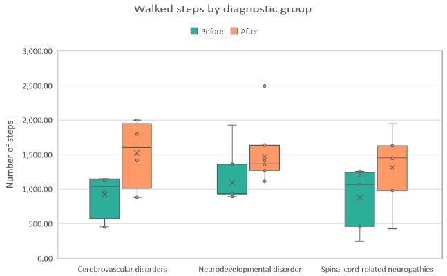
Figure 1: Distribution of steps walked during the first (Before) and last (After) therapy sessions across three diagnostic groups. Each boxplot shows the median, interquartile range, and any outliers.

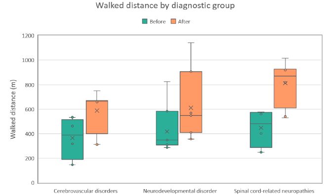
Figure 2: Distribution of walked distance values (m) during the first (Before) and last (After) therapy sessions across three diagnostic groups. Each boxplot shows the median, interquartile range, and potential outliers.

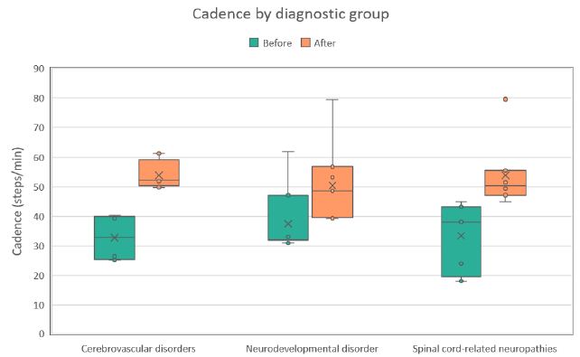
Figure 3: Distribution of walking cadence values (steps/min) during the first (Before) and last (After) therapy sessions across three diagnostic groups. Each boxplot shows the median, interquartile range, and any outliers.

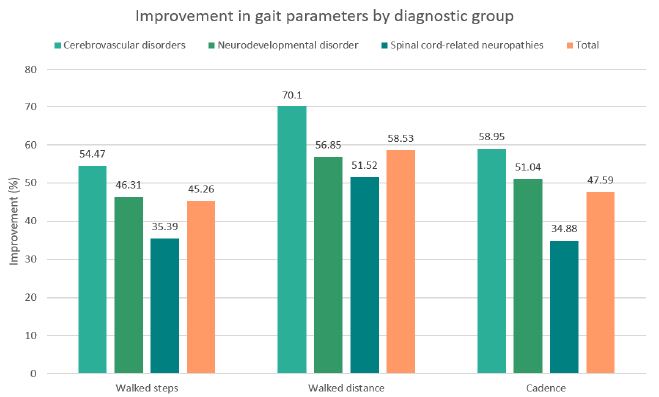
Figure 4: Percentage improvement in walked steps, walked distance, and walking cadence across diagnostic groups following the intervention, including the overall mean across all groups.

Discussion

This study aimed to assess the benefits of end-effector-based RAGT in adults with diverse neurological diagnoses by monitoring gait parameters, including step count, walking cadence, and distance covered. Furthermore, the study explores potential patterns and differences in rehabilitation potential across distinct neurological etiologies. To the best of the author’s knowledge, this is the first study primarily focusing on the impact of an end-effector-based RAGT system on gait parameters in such a heterogeneous patient population, as most previous research has been limited to single-diagnosis cohorts.

Following 12 RAGT sessions, a statistically significant improvement in gait parameters was observed across all diagnostic groups. The greatest relative gains were noted in patients with cerebrovascular disorders, followed by those with neurodevelopmental conditions. Although patients with spinal cord-related neuropathies demonstrated the lowest magnitude of improvement, their outcomes remained statistically significant. For comparison, a previous study by Hotz et al. also examined the effects of end-effector-based RAGT in patients with various neurological disorders, although gait outcomes were only reported as secondary endpoints. Clinically meaningful improvements (≥20%) in walking speed and distance were observed in a subset of participants. The limited overall effect may be due to the inclusion of conditions with low rehabilitation potential, such as motor neuron disease [7].

Interpreted within the context of individual diagnostic groups, indicate that despite fewer sessions and a relatively shorter therapy duration, our results fall within the range of improvements observed in previously published studies. RAGT outcomes in both stroke and spinal cord injury (SCI) cohorts exhibit significant variability, influenced by the recovery phase, specific therapy protocol, and the type of robotic system employed.

For stroke patients in the acute and subacute phases, typical 10MWT improvements range from 20% to 60%, with 6MWT distance gains often greater [40–85%), sometimes involving up to 36 sessions [18,21,22]. In the chronic phase, outcomes tend to be more limited. For instance, Aprile et al. reported a 21% increase in walking speed and a 25% improvement in distance [23]. Even smaller effects were observed by Kelly C.P. et al., with minimal changes in 10MWT and only a 7% gain in 6MWT using an exoskeleton-based RAGT device [24].

Similarly, in SCI, studies report a range of outcomes: Varoqui et al. noted a modest 14% increase in walking speed with no significant 6MWT improvement after 12 sessions [25], while Chang et al. observed a 23% gain in speed and a 34% increase in distance after 15 sessions [26]. Conversely, Wirz et al. demonstrated substantial improvements exceeding 50% in both parameters after 36 sessions [27]. Beyond the significant differences in the number of sessions, this variability in SCI outcomes may also be attributed to the extent and location of the injury [28].

Research on RAGT in adult cerebral palsy (CP) patients is limited, with most studies focusing on pediatric populations. The findings of this study, however, are consistent with studies utilizing the same RAGT device in pediatric patients [29]. A potential explanation for this alignment is the relatively low mean age [(32.2 ± 12.35 years) of the CP cohort, suggesting a population with greater neuroplastic potential and functional adaptability compared to older adults, which may have positively influenced their training response despite typical concerns about limited adaptability in adult CP populations.

In addition, studies have demonstrated that RAGT is more effective than conventional gait therapy, particularly in individuals with moderate to severe motor impairments [9,17,18,21,23]. Beyond the core principles of RAGT, such as high-intensity, repetitive, and precisely guided gait training that drives neuroplasticity, a key advantage is the possibility to start rehabilitation early, even in the acute phase after injury. This is especially important in cases like stroke, where the greatest potential for recovery occurs within the first three months [14,18]. End-effector devices in particular offer several advantages, including a more physiological gait pattern, increased freedom of movement, and a more demanding postural environment, as users are required to actively maintain balance. These characteristics may contribute to findings from some studies showing that end-effector systems lead to better performance in functional tests and higher rates of independent walking compared to, especially, stationary exoskeleton-based systems [7,15,18,22].

The experimental outcomes of the present study align with the theoretical recovery potential inherent to each condition targeted by RAGT. Post-stroke patients, particularly in the acute and subacute phases, demonstrate the greatest potential for gait restoration. This is primarily due to preserved spinal cord function and the brain’s robust capacity for neuroplastic reorganization, where new neural connections are formed and existing ones strengthened following injury. Furthermore, RAGT actively stimulates central pattern generators (CPGs) in the spinal cord, which control the basic rhythmic patterns of walking, and may also activate dormant but viable motor units, thereby facilitating improved motor output and functional recovery [30,31]. In contrast, patients with spinal cord–related neuropathies may exhibit a lower recovery potential. Damage to spinal pathways directly disrupts crucial brain–body communication and impairs CPG function. This, combined with the spinal cord’s inherently limited plasticity and the frequent presence of severe structural lesions, further constrains functional recovery. Consequently, improvements in this group tend to be smaller and slower [28,32,33].

This study has several limitations that should be considered. The lack of a control group prevents direct comparison with conventional therapy and limits conclusions about the specific effects of RAGT. The small sample size in each subgroup reduces statistical power, while the high heterogeneity of the participants complicates interpretation. Important clinical variables such as lesion extent, time since injury, and baseline functional status (e.g. FAC score) were not clearly defined or stratified, which may have introduced confounding factors. Moreover, the study would benefit from the inclusion of objective functional assessments or patient-reported outcome measures to better capture the impact of RAGT on daily functioning and quality of life.

Despite the mentioned limitations, particularly the small sample size and high heterogeneity within groups, the study demonstrated a positive effect of the novel end-effector based RAGT system on walking ability in patients with various neurological conditions, reflecting the complexity of real-world rehabilitation populations. Moreover, the results suggested a potential correlation between the type of neurological impairment and the capacity for gait recovery with RAGT.

Conclusion

The study demonstrated statistically significant improvements in gait parameters, specifically step count (an average increase of 45%), walking distance [(59%), and walking cadence (48%) in adults with various neurological conditions. The best results were observed in patients with cerebrovascular disorders, while the lowest improvements occurred in those with spinal cord-related neuropathies. These findings confirm the positive impact of the end-effector RAGT system across a wide spectrum of neurological diagnoses, reflecting the heterogeneity encountered in everyday clinical practice and indicating the potential of this technology as an effective alternative to conventional therapies, which often require significant personnel resources and may have limited efficacy.

References

  1. Gjesdal B, Jahnsen R, Morgan P, Opheim A, Mæland S (2020) Walking through life with cerebral palsy: Reflections on daily walking by adults with cerebral palsy. International Journal of Qualitative Studies on Health and Well-being. [crossref]
  2. Jankovic J. GAIT disorders. Neurologic Clinics. 2014.
  3. Wuehr M, Huppert A, Schenkel F, Decker J, Jahn K, Schniepp R. Independent domains of daily mobility in patients with neurological gait disorders. Journal of Neurology. 2020 [crossref]
  4. Institute for Health Metrics and Evaluation (IHME) Global Burden of Disease 2021: Findings from the GBD 2021 Study. Seattle (2024)
  5. Khalid NS, Malik NAN, Siddiqi NFA, Rathore NFA. Overview of gait rehabilitation in stroke. Journal of the Pakistan Medical Association. 2023 [crossref]
  6. Awai L, Bolliger M, Ferguson AR, Courtine G, Curt A. Influence of spinal cord integrity on GAIT control in human spinal cord injury. Neurorehabilitation and Neural Repair. 2015 [crossref]
  7. Hotz I, Mildner S, Stampfer-Kountchev M, et al. Robot-assisted gait training in patients with various neurological diseases: A mixed methods feasibility study. PLoS ONE. 2024. [crossref]
  8. Bonnevie T, Moily K, Barnes S, McConaghy M, Ilhan E. People with spinal cord injury or stroke are able to reach moderate-to-vigorous intensity while exercising on an end-effector robot assisted gait trainer: A pilot study. Journal of Rehabilitation and Assistive Technologies Engineering. 2025 [crossref]
  9. Bonanno M, De Pasquale P, Facciale AL, et al. May Patients with Chronic Stroke Benefit from Robotic Gait Training with an End-Effector? A Case-Control Study. Journal of Functional Morphology and Kinesiology. 2025 [crossref]
  10. Ada L, Dean CM, Morris ME, Simpson JM, Katrak P. Randomized trial of treadmill walking with body weight support to establish walking in subacute stroke. Stroke. 2010. [crossref]
  11. Capecci M, Pournajaf S, Galafate D, et al. Clinical effects of robot-assisted gait training and treadmill training for Parkinson’s disease. A randomized controlled trial. Annals of Physical and Rehabilitation Medicine. 2019 [crossref]
  12. Butt Y, Saghir M, Waseem I, Akmal S, Khan UA, Wdood A. Effects of Treadmill Training on Gross Motor Function, Spasticity and Gait Speed in Ambulatory Children with Spastic Cerebral Palsy. Journal of Riphah College of Rehabilitation Sciences. 2024
  13. Duncan PW, Sullivan KJ, Behrman AL, et al. Body-Weight–Supported Treadmill Rehabilitation after Stroke. New England Journal of Medicine. 2011 [crossref]
  14. Pichardo A, Malovec M. Enhancing post-stroke gait rehabilitation with robot-assisted therapy: a focus on step repetitions and neuroplasticity. Arch Clin Biomed Res. 2025.
  15. Choi S, Kim SW, Jeon HR, Lee JS, Kim DY, Lee JW. Feasibility of Robot-Assisted Gait Training with an End-Effector Type Device for Various Neurologic Disorders. Brain & Neurorehabilitation. 2019 [crossref]
  16. Park YH, Lee DH, Lee JH. A Comprehensive review: Robot-Assisted Treatments for GAIT Rehabilitation in Stroke patients. Medicina. 2024 [crossref]
  17. Yang FA, Chen SC, Chiu JF, et al. Body weight-supported gait training for patients with spinal cord injury: a network meta-analysis of randomised controlled trials. Scientific Reports. 2022 [crossref]
  18. Lee J, Kim DY, Lee SH, et al. End-effector lower limb robot-assisted gait training effects in subacute stroke patients: A randomized controlled pilot trial. Medicine. 2023 [crossref]
  19. Bonanno M, Maggio MG, Ciatto L, et al. Can Robotic Gait Training with End Effectors Improve Lower-Limb Functions in Patients Affected by Multiple Sclerosis? Results from a Retrospective Case–Control Study. Journal of Clinical Medicine. 2024 [crossref]
  20. Mehrholz J, Pohl M. Electromechanical-assisted gait training after stroke: A systematic review comparing end-effector and exoskeleton devices. Journal of Rehabilitation Medicine. 2012 [crossref]
  21. Aprile I, Iacovelli C, Goffredo M, et al. Efficacy of end-effector Robot-Assisted Gait Training in subacute stroke patients: Clinical and gait outcomes from a pilot bi-centre study. Neurorehabilitation. 2019 [crossref]
  22. Neves MVM, Furlan L, Fregni F, Battistella LR, Simis M. Robotic-Assisted GAIT Training (RAGT) in stroke Rehabilitation: a pilot study. Archives of Rehabilitation Research and Clinical Translation. 2023 [crossref]
  23. Aprile I, Iacovelli C, Padua L, et al. Efficacy of Robotic-Assisted Gait Training in chronic stroke patients: Preliminary results of an Italian bi-centre study. Neurorehabilitation. 2017. [crossref]
  24. Kelley CP, Childress J, Boake C, Noser EA. Over-ground and robotic-assisted locomotor training in adults with chronic stroke: a blinded randomized clinical trial. Disability and Rehabilitation Assistive Technology. 2012 [crossref]
  25. Varoqui D, Niu X, Mirbagheri MM. Ankle voluntary movement enhancement following robotic-assisted locomotor training in spinal cord injury. Journal of NeuroEngineering and Rehabilitation. 2014 [crossref]
  26. Chang SH, Afzal T, Berliner J, Francisco GE. Exoskeleton-assisted gait training to improve gait in individuals with spinal cord injury: a pilot randomized study. Pilot and Feasibility Studies. 2018 [crossref]
  27. Wirz M, Zemon DH, Rupp R, et al. Effectiveness of automated locomotor training in patients with chronic incomplete spinal cord injury: A multicenter trial. Archives of Physical Medicine and Rehabilitation. 2005 [crossref]
  28. Park JM, Kim YW, Lee SJ, Shin JC. Robot-Assisted GAIT training in Individuals with Spinal Cord Injury: A Systematic Review and Meta-Analysis of Randomized Controlled Trials. Annals of Rehabilitation Medicine. 2024 [crossref]
  29. Malovec M (2024) Impact of End-Effector Device on Gait Restoration in Children Suffering from Neurological Disorders. Archives of Clinical and Biomedical Research.
  30. Huang H, Su X, Zheng B, Cao M, Zhang Y, Chen J. Effect and optimal exercise prescription of robot-assisted gait training on lower extremity motor function in stroke patients: a network meta-analysis. Neurological Sciences. 2024. [crossref]
  31. Huang DF, Xu Y, Hou QH, et al. Neuroplasticity in post-stroke gait recovery and noninvasive brain stimulation. Neural Regeneration Research. 2015 [crossref]
  32. Bareyre FlorenceM, Loy K. Rehabilitation following spinal cord injury: how animal models can help our understanding of exercise-induced neuroplasticity. Neural Regeneration Research. 2018 [crossref]
  33. Harkema S, Dobkin B, Edgerton VR (2000) Pattern Generators in Locomotion: Implications for recovery of walking after spinal cord injury. Topics in Spinal Cord Injury Rehabilitation.

Diamond in Pegmatites and Related Rocks in theUpper Crust

DOI: 10.31038/GEMS.2025764

Abstract

We show that the general absence of the graphite G-band typically characterizes classic diamonds formed in Earth’s mantle. Diamonds, which are transported via supercritical fluids or melts from the mantle region into the crust, generally show the typical graphite G-band. The intensity of this band is clearly dependent on the laser energy used on the sample. That also applies to diamonds formed directly in crustal regions (e.g., in pegmatites) or for diamonds grown in grey cast iron.

Keywords

Diamonds, Raman spectroscopy, Differences in the Raman spectra, Origin of the G-band

Introduction

In a row of papers, the author, with his coauthors [1,2], has shown that diamonds or DLCs (diamond-like carbon) can be brought with supercritical fluids or melts (SCF or SCM) from mantle depths to the Earth’s crust, primarily as spherical crystals. Later, we have seen [3] that DLC can form directly at crustal regions, often as small crystals or whiskers in many different minerals (beryl, quartz, topaz, cassiterite, fluorite, zinnwaldite, and others). By the finding of diamonds in grey cast iron [4,5], formed at an apparent low pressure, we saw significant differences in the Raman spectrometric behavior of such diamonds, or better, DLCs. The main difference in behavior is seen in the FWHM (Full-Width at Half Maximum) and in the position of the first-order diamond line, which is about 1332.5 cm-1 for the undoped and undisturbed diamond lattice [6]. According to the same author, MWCVD diamond powder has a double peak at 1325 and 1330 cm-1 for a mixture of hexagonal and cubic phases, respectively. MWCVD refers to fine diamond particles, or diamond powder, that is created or used in conjunction with a Microwave Plasma Chemical Vapor Deposition (MWCVD) process. Large values for FWHM are typical for an increase in the defect density and for nanodiamonds [7]. However, most diamonds found in mineralization in the crust region are greater than 2 µm in diameter; therefore, alone, the origin of the FWHM increase by small nanodiamonds is not the origin. According to Prawer et al. (1998) [7], the Raman cross-section for sp2 clusters (C=C) carbon is significantly greater (by a factor of 50) than for sp3-bonded structures. That means graphite, as a simplification, is easy to excite.

Samples and Methods

Natural Samples and Diamonds are Used for Cutting, Grinding, and Polishing

To demonstrate that diamonds, which are used for cutting, grinding, polishing, and jewelry, do not show such energy-dependent behavior, some such diamonds are also used (see Thomas et al., 2023). Generally, we used for the study of natural samples (beryl, quartz, topaz, cassiterite, fluorite) free double-polished thick sections (300 – 500 µm thick). In the case of the mica zinnwaldite, we used simple cleavages of mica. The samples come from granites, pegmatites, and vein mineralizations from the Variscan Erzgebirge. Another sample is a quartz ship from the Froland pegmatites in Norway. With these diamonds (DLCs), we can show the influence of the laser power on the sample, which classic diamonds do not show.

Microscopy, Raman Spectroscopy

Besides a polarization microscope for transmission and reflection (JenaLab Pol), which is equipped with a universal stage and different microscope rotary compensators, sophisticated mineralogical studies are possible. We performed all routine microscopic and Raman spectroscopic studies with a petrographic polarization microscope (BX 43) with a rotating or XY stages coupled with the EnSpectr Raman spectrometer R532 (Enhanced Spectrometry, Inc., Mountain View, CA, USA) in reflection and transmission. The Raman spectra were recorded in the spectral range of 0–4000 cm-1 using an up to 50 mW single-mode 532 nm laser, an entrance aperture of 20 µm, a holographic grating of 1800 g/mm, and a spectral resolution of 4 cm-1. Detailed descriptions of the methods used are given by Thomas et al. (2025a and 2025b) [4,5]. From our studies [5], we found that the measuring spot at the sample using the high-quality 100x objective is ~ 1µm. From this, a high energy density results (converted: 4000 W/ cm2) at 30 mW. To prevent intense heating during the measurement and formation of sp2 carbon, we used 0.5 to 4 mW on the sample and, for fast routine measurements, 30 mW.

Experimental Section

To demonstrate the laser energy-dependent behaviours of some diamonds, we take the Raman spectra of the first-order diamond line at 30 and 4 mW, if a graphite band is visible. A row of natural and synthetic diamonds does not show any signs of a graphite band around 1580 cm-1 (Table 1).

More information is in Table 1 in Thomas et al. (2023a). All listed diamonds show no graphite band.

Table 1: Natural and synthetic diamonds without a graphite band (small selection), 30 mW on sample.

Origin

First-order diamond line (cm-1) FWHM(cm-1)

n

Natural diamonds
Brazil 2453/37

1332.2 ± 0.4

4.3 ± 0.4

20

Koffiefontein mine/South Africa

1328.0 ± 2.7

23.1 ± 9.3

15

Sierra Leone, Sewa River

1332.0 ± 0.4

5.4 ± 0.1

10

Udatshnaya, Sibiria

1329.7

10.4

1

Red Diamond, Sibiria

1332.6 ± 0.2

4.8

10

Red Diamond, Sibiria, black incl.

1332.2 ± 0.5

5.2 ± 0.4

10

W-Australia, Argyle Mine

1332.3 ± 0.5

5.8 ± 0.9

10

Synthetic diamonds
HDAC-diamond (Bassett, 2009) [8]

1331.6 ± 0.6

5.2 ± 0.2

10

ZIPE Potsdam (black), GDR

1333.1 ± 0.9

8.9 ± 1.9

10

Diamond cutting disks
Belgium

1332.8 ± 0.3

4.9 ± 0.1

10

Russia

1331.8 ± 0.3

4.6

10

Diamond Paste for metallography (Struers B)
 

1331.5 ± 0.6

5.5 ± 0.2

8

Results and Discussion

Generally, all diamonds found in the upper Earth crust formed there or brought with supercritical fluids or supercritical melts show, when using laser energy of 30 mW (see Table 1), a strong Raman band of graphite near 1580 cm-1. We have also seen in the case of the formation of diamonds, in grey cast iron (Thomas et al. 2025), at low bulk pressure, and, possibly, high temperatures, that they have a huge FWHM and exhibit metastable behaviour under the laser light (e.g., Figure 2 in Thomas et al. (2025b) [9]. It is very instructive to see this in the two Raman spectra from the Froland pegmatite quartz (Figure 1a and 1b) – here as a natural sample.

Figure 1a: Raman spectrum of DLC in pegmatite quartz from Froland/S-Norway taken at 30 mW.

Figure 1b: Raman spectrum of the same DLC in pegmatite quartz from Froland/S- Norway, here, however, taken at 4 mW.

From both figures, we see the influence of laser energy on the sample. The diamond is black and obviously contains carbon. Therefore, the graphite band does not vanish completely. In Table 2 are some results arranged that show a developed, mostly strong graphite G-band beside the diamond band.

Table 2: Diamonds transported via SCF/SCM into the upper Earth crust or formed there (a selection). [~30 mW]. For comparison, the results for the grey cast iron sample No. 2 are given.

Origin

D-line (cm-1) FWHM (cm-1) n G-line (cm-1) FWHM (cm-1) n

References

Bornholm, Pegmatitic quartz

1315.6 ± 0.2

65.0 ± 1.2 30 1564.8 ± 7.3 76.0 ± 6.02

10

Thomas (2024) [10]
Zinnwald Granite ZW 212

1328.5 ± 4.3

63.1 ± 18.0 19 1578.7 ± 7.2 57.5 ± 13.5

19

Thomas (2025)
Zinnwald, Pegmatitic quartz

1321.6 ± 7.0

1321.6 ± 3.8

14.3 ± 0.8

51.6 ± 6.7

14

12

1575.6 ± 6.5

1552.9 ± 7.1

51.7 ± 13.2

47.9 ± 4.3

14

12

Thomas (2025c) [11]
Zinnwald, Cassiterite

1313.9 ± 6.1

1332.7 ± 0.4

59.4 ± 19.1

4.3 ± 0.4

18

20

1521.5 ± 8.5

1581.5

70.0 ± 26.0

3.5

10

1

Thomas (2025a) [4]
Ehrenfriedersdorf, Beryl-vein

1328.6 ± 5.6

1322.8 ± 5.5

60.0

61.4 ± 18.9

14

10

1580.2

1571.8 ± 7.1

52.0

64.0 ± 14.4

10

10

Thomas et al. (2023b) [2]
E-Thuringia, Minette

1332.9 ± 10.2

1323.5 ± 2.4

71.6 ± 28.8

75.6 ± 10.9

10

15

1585.2 ± 6.2

1575.3 ± 5.8

68.0 ± 5.3

73.8 ± 9.9

10

15

Thomas and Recknagel (2024) [12]
Grey cast iron No. 2

1324.6 ± 11.8

1317.8 ± 10.0

66.9 ± 13.8

77.7 ± 15.8

12

12

1572.3 ± 9.5

1580.2 ± 7.1

42.0 ± 21.1

62.1 ± 22.7

12

12

Thomas et al. (2025a) [4]

Thomas has shown [9] that graphite is widespread in many minerals formed during the Variscan time in the Erzgebirge. In the past, that has been ignored up to now. Furthermore, Thomas et al. (2023a) [1] have shown that supercritical fluids (SCFs) and melts (SCMs) are very active in the Variscan time in the Erzgebirge and other places and have brought water, methane, hydrogen, and also different ore elements (e.g., B, Be, Cs, Sn) into the Earth’s crust. That is also valid for other times, such as the Precambrian (Bornholm, Norway, Ukraine). Opposite to gray cast iron, methane and hydrogen are not rare components in nature at high temperatures [13] and in supercritical fluids. So, the supercritical phases are rich in methane [2], which is also supercritical [14,15]. Such fluids can penetrate all minerals, their channels, or grain boundaries. If they meet, for example, carbon/graphite, they can then form moissanite (in beryl) and/or diamonds [2]. As an example, we show here a case of a diamond-like compound found in fluorite in zinnwaldite from Zinnwald. Figures 2a and 2b show the spectrum of those spherical lonsdaleite crystals [3] taken at different laser energies.

In Figure 2a, the Raman band of graphite at 1586 cm-1 is clear to see. Reducing the laser power to 4 mW on the sample allowed the graphite band to disappear totally (Figure 2b).

Figure 2a: Raman spectrum of lonsdaleite in fluorite in zinnwaldite from Zinnwald, taken at 30 mW on the sample [3].

Figure 2b: Raman spectrum of a diamond-like compound taken at 4 mW on the fluorite sample from Zinnwald. The FWHM is 51.8 cm-1. The “graphite band” is completely missing. Note that at about 30 mW on the sample, a strong graphite band at 1599 cm-1 becomes visible [3].

Initially, we have interpreted the black spherical and half-spherical crystals as lonsdaleite. The other lonsdaleite crystals from Sadisdorf/ Erzgebirge are more or less colorless and oblong [3]. The new Raman spectroscopic studies on the black crystals make it probable that they reflect a random substitution of B and N atoms in the diamond lattice [16] or contain amorphous diamond. Boron is present in the fluorite sample too [9]. Another reason for the black coloring is maybe the participation of more complex hydrocarbons like naphthalene [C10H8] [17] instead of methane in fluid inclusions. The cubic boron nitride (cBN) has its main band at 1304 cm-1. Also, boron-doped diamond is a possibility [18]. Those new results (mixing of diamond with cBN) change nothing in the interpretation of the formation of this diamond-like compound in the upper crust. According to Zinin et al. (2009) [19], there are different mixtures in the B-C-N triangle possible. During new Raman measurements, we also found spherical boron crystals in the immediate vicinity of the diamond or diamond- like carbon (DLC) in fluorite from Zinnwald with an extreme band in the low-frequency range [20] at 73.8 cm-1 (similar to the natural diamond from Brazil with 76.6 cm-1). Note, DLC is usually black and, in rare cases, colorless.

A more detailed description of the nature cases (without the new interpretations) is described in Thomas et al. (2025b) [9] and Thomas (2025a) [4] and references therein. Supercritical conditions for a substance (e.g., H2, H2O, CH4, CO2) occur when its temperature and pressure exceed its critical point, which leads to unique properties of fluids if they arrive at supercritical conditions (1000 to 2000°C – see Ni (2023) [21] and Ni et al., 2017) [22]. Above the critical point, substances possess the density of a liquid but the diffusivity and viscosity of a gas. This hybrid nature enables them to dissolve a broad range of compounds, and slight variations in pressure and temperature can often tune their solvation powers. That means that at temperatures over 550°C, the end of the magmatic and pegmatitic stage, all just called compounds are in the supercritical stage and are generally highly mixable, especially when the substances involved have similar molecular characteristics and are at the appropriate temperature and pressure conditions. However, not all combinations are fully miscible. That is an excellent possibility of a fractionated separation and extraction [14,23]. Supercritical fluids are superior to gases in their ability to dissolve materials like liquids or solids. Near the critical point, small changes in pressure or temperature result in significant changes in density, allowing many properties of a supercritical fluid to be “fine-tuned”. Fine-tuning is the key to strong element fractionation during the crossing of the supercritical to the critical and undercritical stages. Samples of the “fine-tuning” we find in melt inclusions in granites, pegmatites, and high-temperature mineralizations (e.g., cassiterite mineralization). Some elements take extreme values at the solvus crest of a pseudobinary silicate-melt-water system and are then mainly Lorentzian distributed [24].

Discussion

We have shown in a couple of papers [25-27] that the processes of the formation of diamonds or diamond-like carbon (DLC) in cast iron, and also different minerals of the Earth’s crust, have a certain similarity: formation apparent far away from the equilibrium conditions for diamond formation from graphite at about 5 GPa or more, and high temperatures. Supercritical fluids/melts may be the key medium for the formation of diamond and moissanite under non- equilibrium conditions. We call this process the supercritical-initiated CVD process, which may vary somewhat in nature or technique. That means the intentional growth of a defined diamond coating is disturbed by the very different targets. Through this process, the diamonds are also affected by the supercritical fluid (maybe also by hydrogen [28], which is responsible for the significant broadening of the first-order Raman band, indicated by a substantial increase in the FWHM (from about 4 to 50 cm-1 or more). That water-pure silicate melts do not influence the transport of diamond in zircon inclusions coming from the mantle region shows the small FWHM values for the diamonds found at the Saidenbach-reservoir (near Ehrenfriedersdorf/ E-Erzgebirge), Rötzler (GFZ Potsdam, oral communication) – see Table 3. Also, the graphite band which is generally at about 1580 cm-1, which is here completely absent (see Table 1).

Table 3: Characteristics of diamond from the Saidenbach-reservoir/Erzgebirge.

Diamond in Zrn

First order (cm-1) FWHM (cm-1)

n


1330.51 ± 0.55

5.81 ± 1.23

41

In addition, we show ways for effective fractionation and enrichment of different ore-forming elements in the Earth’s crust by the supercritical fluid state (e.g., Thomas et al. 2025b) [9]. For example, a lot of tin is transported with supercritical fluids as orthorhombic cassiterite.

Fazit: There are different ways to form diamonds: formation in the mantle depths (at high pressure and high temperatures), transport via supercritical fluids or supercritical melts into the Earth’s crust, and formation at relatively low bulk pressure and high temperatures by reaction of supercritical CH4– and H2-bearing fluids with graphite during melting of cast iron or in nature by reaction of supercritical methane and H2 with carbon. The possibility of the formation of diamond from metastable cementite (see Bhadeshia 2020) [29] should not be left out of sight. The formation of diamond and boron whiskers in high quartz from Zinnwald cannot be explained clearly. However, the primary presence of graphite is a prerequisite. Reaction of supercritical CH4 moving along channels to primary graphite is a possible way. More sophisticated studies are necessary. It is important to emphasize that it is possible to differentiate between diamonds grown at equilibrium conditions at high pressure and high temperature and diamonds transported by SCFs or SCMs from mantle deeps to crustal levels or formed directly at the last region or formed at the productiosn of grey cast iron.

Acknowledgment

The author thanks Gregor Brümmer (Alzheim/Germany) and Adolf Rericha (Falkensee, Germany) for the intensive discussion on the problems of diamond genesis.

References

  1. Thomas R, Davidson P, Rericha A, Recknagel U (2023a) Ultrahigh-pressure mineral inclusions in a crustal granite: Evidence for a novel transcrustal transport mechanism. Geosciences 13: 1-13.
  2. Thomas R, Recknagel U, Rericha A (2023b) A moissanite-diamond-graphite paragenesis in a small beryl-quartz vein related to the Variscan tin-mineralization of the Ehrenfriedersdorf deposit, Geoscience 13: 1-13.
  3. Thomas R, Trinkler M (2024) Monocrystalline lonsdaleite in REE-rich fluorite from Sadisdorf and Zinnwald/E-Erzgebirge, Germany. Geology, Earth and Marine Sciences 6: 1-5.
  4. Thomas R (2025a) Diamond, diamond whisker, graphite, carbon, and coesite in a quartz crystal from Zinnwald, E-Erzgebirge. Geology, Earth and Marine Sciences 7: 1-6.
  5. Brümmer G, Thomas R, Scheiblauer K, (2025a) The formation of tiny diamonds inside grey iron Castings SA. 26(3): 30.
  6. Zaitsev AM (2001) Optical Properties of Diamond. A Data Handbook. Springer. I-XI and 1-502 pages.
  7. Prawer S, Nugent KW, Jamieson DN (1998) The Raman spectrum of amorphous Diamond and Related Materials 7: 106-110.
  8. Bassett WA (2009) Diamond anvil cell, 50th High Pressure Research 29: 163- 186.
  9. Thomas R, Brümmer G, Scheiblauer K (2025b) Paradigm change of pegmatite formation -where does the water come from?. Geology, Earth and Marine Sciences 7: 1-7.
  10. Thomas R (2024) 13C-rich diamond in a pegmatite from Rønne, Bornholm Island: Proofs for the interaction between mantle and crust (2024). Geology, Earth and Marine Sciences 6: 1-3.
  11. Thomas R (2025c) Diamond, diamond whisker, graphite, carbon, and coesite in a quartz crystal from Zinnwald, E-Erzgebirge. Geology, Earth and Marine Sciences 7: 1-6.
  12. Thomas R, Recknagel U (2024) Lonsdaleite, diamond, and graphite in a lamprophyre: Minette from East-Thuringia/Germany. Geology, Earth and Marine Sciences 6: 1-4.
  13. Thomas R, Webster JD (2000) Strong tin enrichment in a pegmatite-forming Mineralium Deposita 35: 570-582.
  14. Proctor JE (2021) The Liquid and Supercritical Fluid State of CRC Press. Taylor Francis Group. 276 pages.
  15. Sengers J L (2002) How fluids Discoveries by the Shool of Van der Waals and Kamerlingh Onnes. Amsterdam. 302 pages.
  16. Hubble HW, Kudryashov I, Solozhenko VL, Zinin PV, Sharma SK, et (2004) Raman studies of cubic BC2N, a new superhard phase. Journal of Raman Spectroscopy 35: 822-825.
  17. Hurai V, Huraiova M, Slobodnik M, Thomas R (2015) Geofluids – Developments in Microthermometry, Spectroscopy, Thermodynamics, and Stable Isotopes. Elsevier. 489.
  18. Baker PA, Catledge SA, Harris SB, Ham KJ, Chen W, et al. (2018) Computational prediction and microwave plasma synthesis of superhard boron-carbon materials. Materials 11: 1-12.
  19. Zinin PV, Liu XR, Ming LC, Sharma SK, Liu Y, et (2009) Ultraviolet and visible Raman spectroscopies of the graphitic BCx phases. Diamond & Related Materials 18: 1123-1128.
  20. Beghi MG, Bottani CE (2004) Low-frequency Raman and Brillouin spectroscopy from graphite, diamond and diamond-like carbons, fullerenes and nanotubes. Trans. R. Soc. Lond. A 362: 2513-2535.
  21. Ni H (2023) Introduction to advances in the study of supercritical geofluids. Science China: Earth Science 66(10): 2391-2394.
  22. Ni H, Zhang L, Xiong X, Mao Z, Wang J (2017) Supercritical fluids at subduction zones: Evidence, formation conditions, and physicochemical Earth- Science Reviews 167: 62-71.
  23. Kortüm G, Buchholz-Meisenheimer (1952) Die Theorie der Destillation und Extraktion von Flüssikeiten. Springer. Pg:381.
  24. Thomas R, Rericha A (2024) Extreme element enrichment, according to the Lorentzian distribution at the transition of supercritical to critical and under-critical melt or Geology, Earth and Marine Sciences 6: 1-6.
  25. Brümmer G, Thomas R, Scheiblauer K (2025b) Wie wächst er nun?. Der Graphit im Gußeisen unter dem Raman-Spektroskop. In press.
  26. Thomas R, Brümmer G, Scheiblauer K (2025a) Unexpected carbon phases in grey cast iron – diamond, calcite, and methane. Geology, Earth and Marine Sciences 7: 1-6.
  27. Thomas R (2025b) Boron in some Variscan deposits in the German Geology, Earth and Marine Sciences.
  28. Rakha SA, Guojun Yu, Jianqing C (2012) Correlation between diamond grain size and hydrogen incorporation in nanocrystalline diamond Journal of Experimental Naniscience 7: 378-389.
  29. Bhadeshia HKDH (2020) International Material Reviews 65: 1-27.

Boron in Some Variscan Deposits in the German Erzgebirge

DOI: 10.31038/GEMS.2025762

Abstract

In this contribution, we show that boron can be found, beside in grey cast iron also in nature, transported via supercritical phases from mantle regions into the Earth’s crust of the Variscan Erzgebirge/Germany. This boron is, as rule, contaminated by different boron compounds like boron carbide, boron nitride and others. We show further evidence that in supercritical phases transported or at low pressure formed diamond show most a large FWHM (>50 cm-1), which is typically for this unusual diamond.

Keywords

Boron, Boron Whisker, Diamond, Raman Spectroscopy, Variscan Erzgebirge

Introduction

Boron is the fifth element of the periodic system and occurs in the Earth’s crust only in its compounds (e.g., the simplest compound, boric acid [H3BO3] or more complex minerals as tourmaline, etc. In granite rocks, the average concentration of B is 15 ppm [1]. Boron is generally dependent on the content of impurities, mostly black. Thin boron crystals are transparent and orange- to yellow colored [2]. Boron has a very high affinity to carbon [3] and has, therefore, a bright black color. During the study of dissolution residues of the gray cast iron (Thomas et al., 2025), we found, besides diamonds, also nearby colorless boron in spheres or plate-like crystals. Table 1 shows the measured Raman data.

Table 1: Measured important Raman lines of a-boron extracted from ductile cast iron [4,5] using the Raman line 532 nm, (the modes are according to Werheit et al., 2010 [3]).

α-Boron

[cm-1] Mode n

Werheit et al. (2010) [cm-1] [3]

First order

553.6

  3

552


575.2 ± 1.50

Eg 14

589


591

Eg 1 589
  771.5 Eg 1

778


795.4 ± 1.99

A1g 11 795
  938.6 A1g 1

934


1096.2 ± 5.35

  15

1094

Second order

1403.2

  5

1409


1583.6

  3 1582
  1708.1   3

1710

The Raman spectrum of boron from grey cast iron [4] is shown in Figure 1. E. Weintraub [7] first prepared pure elementary boron in 1909-1911 after a lot of misunderstanding by his predecessors [8,9]. According to Oganov (2010) [10], most of the discoveries related to pure boron were done in two “stages”: 1957–1965 and 2001–2009. Boron does not exist in the Earth’s high-oxygen environment. That is the state today. We will show that α- and β-Boron, together with boron oxides, carbides, and nitrides, can be found in Earth’s material, which has significant importance and is the first observation ever. The rare appearance in different samples documents the strong reducing conditions of the supercritical fluid or melt [11-13]. Figure 2 shows a schematic phase diagram of boron according to Organov (2010) [10], taken from Thomas et al. 2025b [5].

Figure 1: Raman spectrum of α-rhombohedral boron, contaminated by b-rhombohedral boron and boron carbide, shown by the strong and broad Raman band (A1g + Eg) at 1093.7 cm-1, typical for the b-rhombohedral boron [3] and the main peak of boron carbide around 1100 cm-1 [6].

Besides diamond and boron, there are a couple of minerals (stishovite, coesite, cristobalite-X-I, orthorhombic cassiterite) in more crustal-formed as spherical minerals that demonstrate an interaction between the Earth’s mantle and crust via supercritical fluid and/or melts. Because we find such minerals first and foremost in pegmatites and related mineralization, we will summarize here the data. Important are also the formation of boron and diamond whiskers in the crustal regions.

Figure 2: Schematic phase diagram of boron from Organov (2010) [10]. The γ-B28 and α-Ga types are special high-pressure phases, which are not crucial in our viewing and are not important here.a,b, and d stand for α-, β-, and δ-boron and L for liquid or molten boron.

Natural Occurrence of Boron in the Upper Crust of the Variscan Erzgebirge

Boron in nature has not been found up to now. Only a lot of boron- bearing minerals are well known (e.g., boric acid, tourmaline, and many others). The formation of boron requires strong reducing conditions. Therefore, the occurrence of boron as smooth spherical inclusions in some minerals (cassiterite, quartz, topaz, fluorite, zinnwaldite) of the Variscan Erzgebirge is a surprising finding. Such spherical crystals occur in different minerals in Ehrenfriedersdorf, Sadisdorf, and Zinnwald. Of course, the boron is mainly a mixture of boron, boron carbide, and other minor phases, making the identification very difficult. In Figure 3, such an ellipsoid-shaped boron crystal in cassiterite from Ehrenfriedersdorf (Sn-58 Magdalena vein, second gangway (Mining Academy Freiberg, No. 11814) is shown.

Figure 3: Spheroid of boron in cassiterite from Ehrenfriedersdorf (Sample: Sn-58). Well, to see their transparency.

The Raman spectrum is depicted in Figure 4. The main line at 478 cm-1 corresponds to A1g + Eg of β-rhombohedral boron; also, the bands at 630 (A1g) and 773 cm-1. The median strong band at 1106 cm-1 is, according to Werheit et al. (2010) [3], from β-rhombohedral boron with about 0.11 at% carbon. The 1082 cm-1 Raman band, according to Roma et al. (2022), is attributed to the boron carbide. The classification by the mixture of different β-phases according to Roma alone is not possible, especially since traces of boron carbides and β-Si3N4can be present [14].

Figure 4: Raman spectrum of the boron crystal shown in Figure 3 (Sn-58). The strong lines 478 and 1106 cm-1 are from the β-rhombohedral boron [3]. The last one contains about 1.1 at. -% C.

The measured Raman data are in Table 2 and are completed by some B compounds like BN, B4C, and β-Si3N4 phases, which are not shown. The last phases are typical for boron from Zinnwald, especially in the mica mineral zinnwaldite.

The Raman spectrum from cassiterite Sn-58 shows signs of δ-boron. However, the line at 632.7 cm-1 is the main line of cassiterite, and the interpretation of this as a fingerprint line of the δ-boron is therefore doubtful. Difficulty is also the determination of δ-boron in quartz by the strong quartz line at 464 cm-1. Spherical boron inclusions in cassiterite (Sn-81 from Ehrenfriedersdorf) show the typical δ-boron lines. According to Figure 2, the formation of δ-boron at about 30 GPa is quite possible and is typical for spherical inclusion in cassiterite brought by supercritical phases (Figure 5).

Table 2: Measured important Raman lines of a-, b-, and d-boron inclusion in cassiterite Sn-58 and Sn-81 using the Raman line 532 nm, (the modes are according to Werheit et al., 2010) as well as data for B-carbide and B-nitride from Roma et al., 2022, and Weringhaus, 1997, and Parakhonskoy, 2012.

α-Boron

[cm-1] Mode

Werheit et al. (2010) [cm-1] [3]

First order

352.3

A1g + Eg

357.0


391.2

  394.0
  576.2 Eg

575 (B4.3C)


591

Eg 589
  712.9 Eg

713


773.4

Eg 774
  795.4 ± 1.99 A1g

795


869.0

Eg 873
  1081.5 A1g

1094

Second order

1391.6


1409


1460.6

  1464
  1583.6  

1582


1708.1


1710

β-Boron  
 

406.4

  407
  477.5  

480


632.7

A1g 627
  773.4  

773


1106.7

A1g + Eg

1106 (0.1at.-% C)

β-Boron

Sn-81 E-Dorf, 6th level

Parakhonskoy (2012) [2]


360.2

  361.0
  489.6  

491.0


629.2

  631.0
  919.0  

918.0


1080.4


1078.0

Boron-carbide  

Roma et al. (2022)


319.2

  320.0
  478.0  

480.0

cBN  

Weringhaus (1997) [14]


941.3

  950.7
  1016.9  

1012.1


1045.4

  1045.6
  1045.4 TO (F2g)

1056.4


1125.7

  1123.1
  1337.6  

1338.1


1377.2

E2g

1366.2

Figure 5: Raman spectrum of a mixture of boron and B4C of a spherical crystal in zinnwaldite from Zinnwald/E-Erzgebirge.

Typical for boron carbides [B4C] are, according to Roma et al. (2022), the strong Raman bands at 319.2 and 478.0 cm-1. Besides the spherical shape, there are also black whisker-like crystals in α-quartz from Zinnwald present (Figure 6). If the crystals are thin enough, the needles are transparent with a yellow shade. Often, we observe beside boron diamonds also as whiskers. The largest diamond crystal, beside a large boron crystal, has a diameter of 20 µm. In the α-quartz crystal, there are zones with hundreds of diamond and boron whiskers.

Figure 6: A mesh of diamond (D), bright boron (B) whiskers and graphite (Gr) in pegmatite quartz from Zinnwald/E-Erzgebirge.

Figure 7 shows the spectrum of the boron needles as shown in Figure 6. There are also orthorhombic cross sections from boron present.

Figure 7: Raman spectrum of the boron needle in Figure 6. The bands at 694, 800, (1088), 1160, 2238 cm-1 are, according to Werheit et al. (2010) [3], typical for α-rhombohedral boron.

The α-quartz crystal from Zinnwald contains a lot of whisker or needle-like α-boron and diamond crystals (Figure 8), which include constant small amounts of β-boron. Particularly, the ends of those boron needles show a strong Raman band at 456 cm-1 (A1g + Eg) characteristically for β-rhombohedral boron [3]. In Table 2 are the measured Raman bands of natural α- and β-boron summarized. Because the small needles are in quartz, the Raman measurements in the low-frequency range are difficult due to the strong quartz bands in the low frequency range between 50 and 300 cm-1 and at 464 cm-1). The tetragonal metastable δ- and ε-boron was not found up to now. Those remarks on boron should be enough to show the strong reducing conditions during the interaction of supercritical fluids with the Variscan mineralization. If we use the pressure-temperature diagram for SiO2 polymorphs from Frondel (1962) [15] and use the results from Zinnwald quartz [16], the coexistence of α-quartz with coesite and boron results in a more or less exact temperature value of 1300°C and a pressure of nearly 3.4 GPa, corresponding to about the triple point of high-quartz, low-quartz, and coesite. The triple point of α-boron, β-boron, and γ-B28 boron is very near the called SiO2 triple point [14]. Such values are realistic for supercritical fluid if they meet the crustal granitic rocks. However, these values are too low according to the experimentally determined equilibrium curve for diamond- graphite [17]. Figure 8 is a typical Raman spectrum of a diamond whisker in quartz from Zinnwald.

Figure 8: Typical Raman spectrum of a diamond whisker beside boron (see Figure 6) in quartz from Zinnwald/E-Erzgebirge. The FWHM are for the diamond line 69.8 and for the graphite line 72.8 cm-1, respectively. FWHM – Full-Width at Half Maximum).

In a couple of works [4,5,16,18,19], we have shown many diamond crystals formed at low pressure (in grey cast iron or in nature) or transported into a high crusal level via supercritical fluids or melt. All such diamonds typically have a large FWHM. According to Ferrari and Robertson (2004) [20], the incredible versatility of carbon is explained by the strong dependence of its physical properties on the ratio of sp2 (graphite-like carbon) to sp3 (diamond-like carbon). They show this in their ternary phase diagram (Figure 1 in this). A mostly dark color characterizes the diamonds formed or deposited at low pressure (technical processes, nature). Also, the boron crystals are mostly very dark due to contamination. The boron whiskers in the Zinnwald quartz are typical bright black. In this contribution, we have restricted ourselves to examples from the Variscan mineralisations in the German Erzgebirge. However, we know that in a lot of other pegmatites and granites, boron and diamonds, and similar HP and HT minerals are present.

Discussion

We have found, as mineral inclusions of the Variscan Erzgebirge, a lot of high-pressure and high-temperature minerals like beryl- II, α-, β- and δ-boron, boron carbide, diamond, coesite, cristobalite X-I, graphite, silicon carbide, stishovite, as well as gas phases CH4, H2 and D2 at high density [21-23]. That means at least that the supercritical fluids and melts will feed a large amount of water into the crust coming from the mantle regions. The high velocity of the supercritical fluids also brings primarily smooth spherical crystals. The supercritical fluids/melt have a large amount of energy, which can, at the transition to the under critical stage, make the necessary room for pegmatites and vein mineralizations and also bring a large amount of ore-forming elements with it. That means at least a lot of observations in the past are of secondary meaning. At that place, the extreme element enrichment according to Lorentzian-like curves as proof of the meaning of supercritical fluids is foregone, because we have discussed this point enough [24]. Because elemental boron in an oxidized surrounding and high temperatures is not stable, it must form more stable compounds, like boric acid and many other B-bearing minerals. Another critical point is that a large part of boron comes directly from the mantle region and not from the Earth’s surface, as is often assumed.

Appendix: Microscopy and Raman Spectroscopy of Boron- rich Phases in Variscan Minerals of the Erzgebirge

Besides a polarization microscope for transmission and reflection (JenaLab Pol), we performed all microscopic and Raman spectroscopic studies with a petrographic polarization microscope (BX 43) with a rotating stage coupled with the EnSpectr Raman spectrometer R532 (Enhanced Spectrometry, Inc., Mountain View, CA, USA) in reflection and transmission. The Raman spectra were recorded in the spectral range of 0–4000 cm-1 using an up to 50 mW single-mode 532 nm laser, an entrance aperture of 20 µm, a holographic grating of 1800 g/mm, and a spectral resolution of 4 cm-1. Detailed descriptions of the methods used are given by Thomas et al. (2025a and 2025b) [4,5]. For the identification of mineral phase, we used Hurai et al. (2015) [25] and the RRUFF database by Lafuente et al. (2015) [26,27]. From our studies [5], we found that the measuring spot at the sample using the high-quality 100x objective is ~1µm. From this, a high energy density results. To prevent intense heating during the measurement and formation of sp2 carbon in diamond, we used 4 mW on the sample and, for fast routine measurements, 30 mW.

Acknowledgment

Thanks go to many colleagues who inspired this Raman work on high-pressure and temperature minerals in the Earth’s crust.

References

  1. Rösler HI, Lange H (1975) Geochemische Leipzig. Pg: 675.
  2. Parakhonskiy G (2012) Synthesis and investigation of boron phases at high pressure and Dissertation at the University of Bayreuth. Pg: 117.
  3. Werheit H, Filipov V, Kuhlmann U, Schwarz U, Armbrüster M, et al. (2010) Raman effect in icosahedral boron-rich Science and Technology of Advanced Materials. 11.
  4. Thomas R, Brümmer G, Scheiblauer K (2025a) Unexpected carbon phases in grey cast iron – diamond, calcite, and methane. Geology, Earth and Marine Sciences. 7: 1-6.
  5. Thomas R, Brümmer G, Scheibblauer K (2025b) Paradigm change of pegmatite formation – where does the water come from?. Geology, Earth and Marine Sciences. 7: 1-7.
  6. Roma G, Gilles K, Jay A, Vast N, Gutierrez G (2021) Understanding first order Raman spectra of boron carbides across the whole stochiometry range. Physical Review Materials. 5: 1-31.
  7. Weintraub E (1911). On the properties and preparation of the element boron. Ind. Eng. Chem. 3: 299-301.
  8. Meyer RJ (1926) Gmelins Handbuch der Anorganischen Chemie – Verlag CChemie, Leipzig-Berlin. Pg: 142.
  9. Pietsch EHE (1954) Gmelins Handbuch der Anorganischen Chemie – Bor. Pg: 253.
  10. Organov AR (2010) Boron under pressure: Phase diagram and novel high-pressure NATO Science for Peace and Security Series B: Physics and Biophysics – Boron rich solids. (Eds) By Orlovskaya N and Lugovy M. Springer. Pg: 207-225.
  11. Ni H (2023) Introduction to advances in the study of supercritical geofluids. Science China: Earth Science. 66: 2391-2394.
  12. Ni H, Zhang L, Xiong X, Mao Z, Wang J (2017) Supercritical fluids at subduction zones: Evidence, formation condition, and physicochemical Earth-Science Reviews. 167: 62-71.
  13. Sun Y, Liu X, Lu X (2021) Structures and transport properties of supercritical SiO2– H2O and NaAlSi3O8-H2O American Mineralogist. 108: 1871-1880.
  14. Werninghaus T (1997) MicroRaman spectroscopy investigations of hard Dissertation, TU Chemnitz-Zwickau. Pg: 163.
  15. Frondel C (1962) The System of Volume III, Silicate Minerals. John Wiley and Sons, INC. Pg: 334.
  16. Thomas R (2025) Diamond, diamond whisker, graphite, carbon, and coesite in a quartz crystal from Zinnwald, E-Erzgebirge. Geology, Earth and Marine Sciences. 7: 1-6.
  17. Day HW (2012) A revised diamond-graphite transition curve. Mineralogist. 97: 52-62.
  18. Thomas R (2023) Diamond in pegmatitic sillimanite from Reinbolt Hills/East Geology, Earth and Marine Sciences. 5: 1-3.
  19. Thomas R (2024) 13C-rich diamond in a pegmatite from Rønne, Bornholm Island: Proofs for the interaction between mantle and crust (2024). Geology, Earth and Marine Sciences. 6: 1-3.
  20. Ferrari AC, Robertson J (2004) Raman spectroscopy of amorphous, nanostructured, diamond-like carbon, and Phil. Trans. R. Soc. Lond. 362: 2477-2512.
  21. Thomas R, Webster JD (2000) Strong tin enrichment in a pegmatite-forming Mineralium Deposita. 3: 570-582.
  22. Thomas R, Davidson P, Rericha A, Voznyak DK (2022b) Water-rich melt inclusions as “frozen” samples of the supercritical state in granites and pegmatites reveal extreme element enrichment resulting under non-equilibrium Mineralogical journal (Ukraine). 44: 3-15.
  23. Thomas R, Davidson P, Rericha A, Recknagel U (2023) Ultrahigh-pressure mineral inclusions in a crustal granite: Evidence for a novel transcrustal transport mechanism. Geosciences. 13: 1-13.
  24. Thomas R, Rericha A (2024) Extreme element enrichment, according to the Lorentzian distribution at the transition of supercritical to critical and under-critical melt or Geology, Earth and Marine Sciences. 6: 1-6.
  25. Hurai V, Huraiova M, Slobodnik M, Thomas R (2015) Geofluids – Developments in Microtermometry, Spectroscopy, Thermodynamics, and Stable Isotopes. Elsevier. Pg:489.
  26. Lafuente B, Downs RT, Yang H, Stone N (2016) The power of database: The RRUFF In: Highlights in MineralogicalCrystallography, Armbruster T, Danisi RM (Eds) De Gruyter: Berlin, München, Boston. Pg: 1-30.
  27. Thomas R, Trinkler M (2024) Monocrystalline lonsdaleite in REE-rich fluorite from Sadisdorf and Zinnwald/E-Erzgebirge, Germany. Geology, Earth and Marine Sciences. 6: 1-5.

Heritage Littoral Landscapes in African Ancestry Coastal Communities of Bath Plantation Land, St.John, Barbados

DOI: 10.31038/GEMS.2025761

Abstract

African Ancestry communities in Barbados have established, through time and space, deep heritage ties to places along nearby littorals; that is, cultural zones along the shore referred to here and in previous publications as The Sea’s Edge. An analysis of these areas is based on ethnographic field interviews, participant observation, and surveys conducted during eleven field events over a 20-year time frame on the island of Barbados. The cultural attachments to the sea’s edge, spanning the terrestrial and marine, began during the period of enslavement and continue to be maintained and celebrated today as a viable part of cultural identity and heritage pride. This paper explores how these places became functionally integrated into a landscape and central to the culture of these coastal peoples. Individual places and natural resources continue to provide important ecological and cultural services, including hundreds of plants, dozens of shallow marine resources, and areas of historical and spiritual significance. This analysis documents the ongoing value of natural/cultural coastal heritage landscapes, an area known as the littoral zone adjacent to the Bath Plantation lands. It is an area utilized and celebrated, protected, and informally managed by the surrounding communities and is an example of the way places like this fit the littoral landscape model, recommending that local involvement is appropriate in cooperative management with the national government. This example is applicable to other similar coastal communities throughout the world and provides a framework to understand local connection to the littoral zone and both maritime and terrestrial natural and cultural resources.

Keywords

Heritage cultural landscapes, International Union for the Conservation of Nature Heritage Guidelines, Barbados West Indies, Enslaved African people, culture of marine littoral edges

Introduction

The Sea at its best is a place where I can walk in the water up to my knees and look down and see conch, fish and other foods for life. It should all be there for you – Lester Flowers, a Professor of Biology at the College of Bahamas [1].

Heritage landscapes encompass landscapes of scenic, cultural, and scientific value [2]. The International Union for Conservation of Nature (IUCN) [2,3] argues for the importance of world heritage landscapes in conservation to encourage holistic approaches to conservation. This analysis is based on research conducted in communities surrounding the Bath Plantation Lands (known as Bath), Barbados, and supports the integration of heritage landscapes by providing information about the social and cultural importance of the littoral zone. This paper provides national planners and international developers with the information needed to meaningfully involve local Barbadian communities in the decisions that impact the future of their homelands and culture. It also provides insight into the need for collaborative management practices that can be utilized in similar settings in coastal areas throughout the World. Barbadian law stipulates that the littoral is for the public (National Conservation Commission Act, Cap. 393), and belongs to all the people of the country; however, it does not recognize the rights associated with local African Ancestry (Afro-Caribbean) heritage cultural landscape that has formed over hundreds of years. As a result, current coastal development and use of natural resources has and potentially will threaten the community- based natural and human resources located at the sea’s edge [1,4-6]. Using the heritage littoral landscape model, a conceptual or analytical framework used to understand and manage coastal (littoral) zones that have both natural and cultural heritage significance, this paper explores those culturally connected to the sea’s edge and the rationale for their involvement in its management and interpretation. These data and analysis were the foundation for sustainability impact assessments [7] and Community-Based Human Rights Impact Assessments [8]. This analysis also documents the complexity of conducting ethnographic studies of heritage in littoral landscapes, which span both the marine and terrestrial locales and resources.

Background

African Ancestry History at the Sea’s Edge

The coastal lands of the Caribbean are special heritage places for the ancestors of the formerly enslaved peoples who were forcibly transplanted from ecosystems they understood in Africa to ones with new natural and social ecologies (Figure 1). While beyond the scope of this analysis, many of the people would arrive at Caribbean plantations that were established near coastal areas. The history of industrial agriculture of Caribbean plantations focused on monocrop production (in most areas this was sugarcane cultivation), processing, and shipment from local ports to markets elsewhere in Europe and the New World. There are hundreds of former coastal enslavement plantations throughout the Caribbean. Barbados alone had hundreds of industrial agricultural plantations (Figure 2). The island is on a flat coral limestone plateau, different from the topography of steep volcanic islands like St. Lucia and large mountainous locations such as Jamaica, Cuba and the Dominican Republic. Volcanic islands share the topographical characteristic of little to no underwater shelves, and they rise steeply to mountains that support hundreds of inches of rainfall per year. Given that the localized weather in the Caribbean is directly impacted by land elevation, flat islands experienced consistent levels of annual rainfall, scant surface water, and dependable weather conditions that made Barbados suitable for viable large-scale commercial sugarcane agriculture for approximately 400 years.

Figure 1: Barbados in the Lesser Antilles (Google Earth).

The ecological and cultural shifts from the old traditional societies of Africa to the new Caribbean societies posed a human adaptation challenge that was experienced by hundreds of thousands of African people. Subsequent successful ecological co-adaptations resulted in these people defining their own heritage places and events of cultural importance. Taken together these new heritage places became coherent and functionally integrated cultural landscapes. Given the location of many plantations near the sea to facilitate commercial shipment of products elsewhere, the enslaved and later freed African Ancestry peoples made a way of life that involved both land and sea. Their home often was centered on a heritage littoral landscape located at the sea’s edge [1]. The heritage littoral landscape which is the focus of this research is operationally defined as being composed of near shore marine and terrestrial places and environments. Each is defined in space, time, and function by the people who have socially constructed the landscape through use and experience. Together the land and sea habitats and their associated human interactions and adaptations constitute a heritage littoral landscape. The analysis argues that such integrated landscapes define the culturally appropriate ways (that is, frames of analysis) to understand the heritage of the coastal people and understand and apply the research findings to terrestrial and marine management as well as cultural interpretation [1,6,9-13].

The adaptations of people with African Ancestry varied by their cultural backgrounds and unique ecological contexts of where they landed and ultimately resided. As enslaved people they lived where and how others demanded; however, within these constraints, they expressed their agency and increasingly created their own cultural spaces and meanings. It is argued here that the littorals of their new coastal lands would become a primary place for the expression of their agency [1]. African Ancestry people would establish their places in areas that were largely not useful to the export-oriented colonial industrial plantations and in ecosystems that remained largely influenced by natural variables. Being close to plantations, these areas were most accessible to the people during the long period of their enslavement. The littoral, in other words, became for many people of African Ancestry “our place” at the sea’s edge and thus a foundation for heritage places and landscapes. Caribbean research documents how for many African Ancestry people, the sea’s edge has historically represented freedom and survival [14-18]. The sea’s edge has always provided marine and terrestrial resources for food, fertilizer and medicine, in addition to diverse items for trading and crafts (shells and corals). Also, the sea served as a mode of transportation. The water itself can be seen as something that simultaneously separates and connects people with other places. In these special locations, knowledge of the terrestrial resources has grown apace with knowledge of the marine resources and sea, thus allowing people to survive and thrive in environments with little external support. The “fruits” of the sea were combined with those of the land, such as in the building of boat ribs from native trees and the mixing of seaweed with land plants to make medicines. Because much of the traditional natural vegetation was removed for the planting of export crops like sugar and cotton, soon natural vegetation occurred only at the sea’s edge where sea spray salted the land thus precluding cash cropping.

Knowledge and appreciation of the sea appears to have been traditionally expressed through a respect that is grounded in experience, not only through collection but use of the resources. Fishermen have learned to “read the language” of the sea and the coastal marine environment in overt as well as subtle ways. Collective and individual experience has led to an understanding of the ways that marine life interacts and sustains itself and nearby land plants and animals. For example, fishermen have learned to gauge the wind and the tides, the seasonality of fish populations, and how to follow dolphins to find certain schools of fish. Likewise, farmers have learned that some marine life can be used as a medicine for skin rashes, that the hundreds of coastal plants have a special ecology that can be managed, and that seaweed can be dried for food and fertilizer. Today, people keep traditional knowledge of the sea’s edge alive through their occupations, social relations, and the cultural transmission of knowledge through instruction and participation with the local youth. Residents have built the sea into their lives through stories, songs, lessons and games. Many discuss the ways in which knowledge of the sea was kept alive in old fishermen’s songs, songs that were sung while undergoing trials at sea and later again over rum and ale in ports where knowledge of these events were shared by turning such occasions into social affairs. Through sailing stories, shipwreck stories, and even mythical stories about sea animals helping boaters in distress, the centrality of the sea is once more realized. Nationally today, in Barbados, attachments to the sea’s edge are evidenced in the expressive arts. People today do not live as they did in the past but choose to value the protection and interpretation of heritage places, natural resources, and storied places because these represent how they thrived during the best and harshest of times.

Cultural Landscapes and the Edge Effect

According to the International Union for Conservation of Nature [3]:

Cultural landscapes are seen as ‘cultural heritage’ in the framework of the World Heritage Convention, yet they have considerable overlap with one of the heartland issues of nature conservation, the global coverage of protected areas. Recent research reveals the overlaps and synergies between World Heritage cultural landscapes and IUCN’s global category system for recognizing protected areas, and some of the possible implications.

In the western world, the term ‘landscape’ has been used in many vernacular and academic ways. It has been used in painting, writing, and poetry since the late 19th and early 20th centuries, and it is a term used by natural and social science scholars to frame many shared analytical perspectives. This concept contrasts with the alternative notion of nature and landscape as a living integrated landscape in the Far East. The early German geographer Otto Schluter in 1908 defined two forms of landscapes: the Urlandschaft (original landscape) or landscape that existed before major human induced changes and the Kulturlandschaft (cultural landscape) a landscape created by human culture. Schlüter argued that the major task of geography was to trace the changes in these two landscapes. Carl Sauer wrote that cultural landscapes are made up of human forms and actions superimposed on physical landscapes [19]. He was probably the most influential in promoting and developing the idea of culture as a force in shaping the visible features of the Earth’s surface in delimited areas. Within his definition, the physical environment retains a central significance, as the medium with and through which human cultures act [20]. He maintained that objects which exist together in the landscape exist in interrelation. His classic definition of a ‘cultural landscape’ reads as follows: “The cultural landscape is fashioned from a natural landscape by a cultural group. Culture is the agent, the natural area is the medium, the cultural landscape is the result.”

Since Schlüter’s first formal use of the term, and Sauer’s effective promotion of the idea, the concept of cultural landscapes has been variously used, applied, debated, developed and refined within academia. By 1992, the World Heritage Committee elected to convene a meeting of landscape specialists to advise and assist redrafting the Committee’s Operational Guidelines to include ‘cultural landscapes’ as an option for heritage listing properties that were neither purely natural nor purely cultural in form (i.e., mixed heritage) [21]. This analysis is focused on defining and explaining African Ancestry places and landscapes in the Caribbean and thus adds to expanding definitions used in western academic circles of natural and cultural landscapes by detailing how these locations can also span marine and the terrestrial resources, freshwater and saltwater, and sea and the sand. The heritage objects, natural resources, and heritage places that were identified in our studies are more broadly framed as components of heritage littoral landscapes. When considered as a functionally integrated cultural phenomenon they can more clearly be preserved, managed, and interpreted in more holistic ways. Cultural landscapes in the littoral are special because they define a boundary between habitats. Ecologists have termed this the edge effect because where two different habitats meet, they form an edge of nature and thus increase biological diversity and complexity. McCay has discussed the diverse and productive nature of edges, proposing that the “edge effect” may be used as a metaphor for the bringing together of people, ideas and institutions [12]. In a data-based summary of the concept, Turner, Davidson-Hunt, and O’Flaherty [22] argue that, Like ecological edges, cultural knowledge systems can integrate, producing a richness of knowledge and practices that enhance the resilience of local societies. Cultural edges, rather than being border zones between discrete social entities, are zones of social interaction, cross-fertilization, and synergy wherein people not only exchange material goods but also learn from one another.

The edge effect defines the location and cause of new food webs and trophic levels. Expanding the ecology concept of edge to include human dimensions results in considerations of edges as stimulating cultural diversity, centering social activities, and serving as a foundation of heritage [23]. The littoral landscape is operationally defined, for this analysis, as a social, natural, and biological zone adjacent to the edge of the sea [1,6]. When stable communities use such areas for generations, they often become heritage cultural landscapes that are locally perceived as belonging to the people who have used and protected them for hundreds of years. The heritage littoral landscape extends onto the land through food webs that depend on both salt and freshwater habitats. It extends inland along small rivers and up estuaries into mangrove wetlands. It can be defined as far as amphibious animals crawl inland. The littoral contains shallow coral reefs, seagrass beds, exposed beach rock, sand dunes, and mud flats. Socially and culturally the littoral extends on land and in the sea through related activities, historic places, and the cultural patterns of contemporary communities.

Methods

A key aspect of this research is its focus on heritage cultural landscapes, particularly those developed during slavery and subsequently shaped over time by African Ancestry communities to be a presence in contemporary time. These landscapes are often inherently private, and local community ownership or attachment to these areas is typically unrecognized by national authorities. As such, community members are frequently excluded from discussions surrounding the management and future of these landscapes. This study underscores how long-term participant observation and iterative ethnographic fieldwork can foster mutual trust and rapport, ultimately enhancing the depth and richness of ethnographic data on heritage landscapes. This study examines the Bath littoral zone through both ethnographic and participatory research methods, incorporating a range of data sources to capture the social, cultural, and environmental dimensions of the region. While the littoral landscape is often considered as a distinct physical and cultural space, this research demonstrates its interconnectedness with other marine and terrestrial environments, including deeper oceanic areas and adjacent hill zones. The analysis is primarily based on a combination of formal surveys, ethnographic interviews, and participant observation with residents from all over the island, with the majority of the interactions focused specifically on the Bath Plantation area [24]. Ethnographic methods were employed in a comprehensive, triangulated approach to data collection and analysis [25]. This strategy combines multiple techniques, including structured interviews, semi-structured key informant interviews, informal interviews, group discussions, participant observation, and transect walks. Previous literature was also reviewed to contextualize findings and enhance the depth of analysis. The interviews spanned a diverse demographic, including individuals from 8 to 94 years of age, with men and women from various educational, geographic (rural and urban) and economic backgrounds. The research also involved extensive participant observation conducted over 12 field visits between 1996 and 2024, with site visits ranging from 7 days to 5 months in duration. These visits enabled immersion in daily life and direct engagement with local communities, especially those who utilized the Bath Littoral zone’s marine and terrestrial resources [1,24].

This research spanned over two decades, with an initial focus on traditional microeconomic systems of exchange and its role within the larger societal context of interaction within various types (familial, communal and occupational) of social networks. Many of these surveys and interviews during this time frame focused in rural areas examining the role of these saving and credit associations within the larger framework of household food security and financial well being, this lending insight into the interplay between formal and informal economics as well as natural resource use. These associations are called Rotating Savings and Credit Associations (ROSCAs), known locally as “meeting turn,” and throughout the Caribbean as “sousou,” “asu,” “box hand,” and “partner” [26]. This component of the research was conducted over five field sessions and included 500 ethnographic interviews, 120 of which were conducted in 1999 with individuals from the Bath Plantation area on the rural east coast of Barbados. The other 380 interviews were conducted between 1996 and 2024 with people throughout the island, including but not limited to individuals from the Bath Plantation area. Many of these interviews include discussions with fisheries officers, local NGOs, fishermen of all ages and from all sectors (commercial, recreational, for-hire, and subsistence), as well as restaurant owners, chefs and fish cleaners and vendors from the Fish Markets at Conset Bay, Oistins and Bridgetown. These interviews primarily explored informal economic systems in the context of fishing, coastal harvesting, and resource management. In addition, interviews also focused on the changing physical environment and development of eco-tourism related to the littoral and creation of the old railway walking trail (from Bath through Martins Bay to Bathsheba).

Participant observation formed the backbone of data collection, spanning 20 plus years of fieldwork and interaction with the residents and resources of the Bath littoral zone. During the field site visits, the lead author engaged in daily activities with the community, participating in fishing trips and documenting the local use of marine and terrestrial resources. This observational approach also allowed for the gathering of traditional ecological knowledge, particularly related to food webs, trophic levels, and lunar, tidal and seasonal changes in the littoral environment [1]. Local residents also shared their concerns about the protection and preservation of key cultural places within the Bath Littoral zone. During extended stays in Bath Beach, the lead author developed close relationships with local fishermen, engaging in participant observation and free-diving fishing expeditions. These experiences provided a detailed understanding of fishing techniques and species targeted in both inshore and offshore environments. When weather permitted the researcher spent five to six days per week freediving with local fishers, building rapport and learning about traditional ecological knowledge, especially regarding the seasonal availability of key marine species. These insights were supplemented by continued visits to the area, including 12 additional trips between 2000 and 2024, where the researcher continued to engage in fishing activities and document changes in the local environment, particularly the rising abundance of sargassum.

This study shifts the environmental focus from discrete geographical locations to socially constructed landscapes, emphasizing the need to understand human adaptations to natural systems and the persistence of sustainable heritage practices. This methodological shift recognizes the importance of adjusting traditional ethnographic techniques—both in terms of detail and temporal scope—when applied to landscape-scale research. This means examining multiple locations and resources associated with an area, not a specific building or landscape. It moves beyond a single time frame as well examines the relationship over generations. It specifically asks how these places and these resources played and play a part in the identity of a local population as well as served as a place for cultural transmission of cultural and environmental knowledge, especially in relation to food security and the creation of social stability through the strengthening of social networks. This study draws heavily on the insights of Fred Watson, a local fisherman born in Bath Beach 97 years ago, whose deep knowledge of the region’s fishing practices contributed greatly to the research. Known as “the Best Fisherman in all of Barbados,” Watson played a pivotal role in both the community and the broader fishing industry, serving as a mentor to younger generations of fishers and providing expert advice to government fishery managers. His personal experiences, documented in an oral history conducted over several years, serve as a foundational resource for understanding the cultural and ecological significance of the Bath Littoral zone.

The ethnographic approach used in this study is consistent with and builds upon previous research on rural Barbadian communities [26-29] and similar studies from nearby islands [30,31]. These studies document the co-adaptations of African-descended peoples and the landscapes where they live, highlighting the importance of cultural landscapes as dynamic, socially constructed spaces. Additionally, this research draws on the work of Cumberbatch and Hinds [32] on Flying Fish as an aspect of Barbadian bio-cultural heritage, further illustrating the interconnectedness of people, place, and resource management. In addition to the ethnographic data, we utilize photographic analysis of early 20th-century images to strengthen the argument for recognizing the Bath Littoral zone as a heritage site. These photographs provide visual evidence of the cultural connection between local Bath residents and the littoral landscape, further substantiating its significance as a heritage area and its continuing importance from the past to the present. Various findings from the Barbados study have been reported and published elsewhere and these will not be repeated here [1,33]. Instead, new heritage sea and land interactions are discussed in this analysis, thus expanding what is known of these littoral landscapes. Through this multi-method approach, the study contributes to a deeper understanding of the Bath Littoral zone as a dynamic and culturally significant heritage landscape.

Study Site

Littoral landscapes are difficult to study because of their bathymetric characteristics. The sea portions are largely under water often with some protruding rock formations, the littoral landscapes included in this study are shallow thus permitting fishing, lobster gathering, shellfish collecting, and the picking of seaweeds by hand. During enslavement the movements of enslaved people were restricted to prevent escape and or revolt, so they were not permitted to either make or use boats or rafts. Sea resource use was thus limited to shallow and calm waters. The Barbados coastal segment studied here is called for purposes of this analysis the Bath Littoral Landscape. It is located on the eastern edge of this 14-mile wide by 21-mile-long island (Figure 2). The landscape is 2.21 miles along the windward north shore extending from Congor Bay to past the Bath Beach House to Conset Bay (Figure 2). This landscape includes, (1) a beach used for all aspects of life including social gathering, tourism, sea baths (both recreational and healing activities), plant gathering (for subsistence and medicinal purposes), (2) a spring with spiritual dimensions as well as healing properties, (3) the Quamins river, (4) the old railway and historic train station, (5) the Bath plantation, a location that has existed from enslavement in the 1700s to modern times with many of the relatives of former unfree laborers, and (6) an underwater marine habitat used for cultural transmission of knowledge regarding resource use as well as gathering marine and terrestrial resources for both food and medicinal purposes, an activity that dates back to the earliest of the areas inhabitants (Amer-Indians) and people brought through forced migration (slaves and indentured laborers).

Figure 2: Island of Barbados, West Indies. The narrow coastal limestone shelf and coral reef are abutted by deep ocean (Google Earth images).

Topographically the Bath Heritage Landscape has hills to the south and a relatively quiet beach area, with a wide shallow sea shelf extending to a deep drop off to the north. The limestone shelf drop has a vertical ledge with a 40-foot drop, that is marked above by large limestone rocks that rise above the ocean even at high tide. The shelf can be walked at low tide with only light swimming needed over deep holes. Bath is on the windward north side of Barbados, so the Bath littoral experiences the persistent Northeast Trade Winds, which make for active ocean waves beyond the shallows and strong winds on the uplands (see Windy Ridge in Figure 3).

Figure 3: Bath Heritage Landscape (Google Earth).

The Bath Heritage Landscape by definition includes both land and water dimensions. Each has contributed to the development of local coastal African Ancestry communities, especially Bath. Key to the formation of this as a littoral landscape, rather than just a coastal landscape, is the special contribution of the local sea to the lives of local African Ancestry people. In this case, key was the presence of a shallow, flat, shelf that extends to an underwater drop off. The presence of an enslaved population and their space restrictions and limited access to natural resources made both land and the sea components special for the production of this landscape.

Historic and Contemporary Connections to the Bath Heritage Landscape

One of the most useful images for this analysis is the historic Bath littoral zone (Figure 5) curated by Jim Webster [34] who, building on the oral history of Richard Goddard, used it to illustrate an article regarding the earliest photographs of the people and coast of Bath [34]. The photos used in the article were taken by professional photographer Manuel Auguste Nunes Siza in 1896-1897. They are located at the “Anglo-Luzo Photographic Gallery” in Beckwith Place, Bridgetown [35]. The photos illustrate some cultural dimensions of the littoral, and for this analysis these largely remain important unvoiced images of the past. Some of the Siza photos are presented here along with additional ethnographic information acquired by Stoffie during his research and supported and built upon by a local elder Fred Watson (and others) who was born in Bath Beach in the early 20th century.

Figure 5: Part of the Bath Littoral Zone: From Beach to Great Rocks [34].

The Bath Plantation

The Bath plantation (Figure 4) was owned in 1791 by Edward Day. During this early time it had 212 enslaved people and 400 workers. The number of enslaved workers remained about the same until near the end of slavery in 1832 when there were 245 enslaved people. In 1913 there were a total of 627 workers on the plantation [36]. Given the location of the Bath plantation, near what is called the Scotland District, it is assumed that the workers were a combination of African Ancestry slaves mixed with individuals of Scots-Irish Ancestry. Scots- Irish workers were among the first unfree laborers in Barbados. Bath workers lived on and around Bath plantation lands during enslavement and continue in today’s districts such as Welch Town, Pothouse and Sergeant Street. At the end of slavery, about 1837, most plantations forced formerly enslaved workers to move their homes to small villages located along nearby roads. The tenantries were on public lands along these roads. This action removed the former enslaved people from both the plantation and the notion that the plantation was obligated to them for food, medicine, and housing as it had been during slavery. After slavery, Barbados government laws were passed to prevent African Ancestry people from owning small plots of land. Soon after their forced removal, the house pads in their former living areas on the plantation were plowed by the plantation owners, thus breaking long term relations (in some cases hundreds of years) with these home locations.

Figure 4: The Bath Plantation [34].

The Tenantry Freehold Purchase Act (1980-53-2022) was passed as a modification of the Barbados Constitution in an effort to legalize the land ownership of people who were forced to move from their homes at the end of slavery. After forced removal to a tenantry only the littoral continued to be a place considered to be their own.

Marine and Terrestrial Resources

Traditionally, Amer-Indians (beginning in 350AD) occupied and regularly used the marine and terrestrial resources of Barbados. In fact they continued to be a part of Barbadian life for some decades after the island became a part of the British Empire in 1627 [37]. Richard Ligon [38] who lived in Barbados from 1647 to 1650 provided a map of the Indian area located inland near Bath. He observed that: Indian women were primarily, if not solely, employed in household tasks. On the other hand, the men…were used for footmen and the killing of fish, which they are good at. With their own bows and arrows they will go out and in a day’s time kill as much fish as will serve a family of a dozen persons two or three days, if you can keep the fish so long.

This is an intriguing insight into the pristine condition of the littoral at that time. It is not clear how long Indian men were employed as fishers with their six-foot bows and long arrows, but it is unlikely that people of African Ancestry were permitted to make and use such weapons. The Native canoes used to travel to other islands were not available to enslaved people.

Crabs were fished at night using torches to provide both light for movement in the shallow waters and to illuminate the crabs and lobsters. Torches combined with nets were used to catch flying fish and McKinnen in 1802 recorded that local people were very successful at catching flying fish using this method. At night they spread their nets before a light and would disturb the water at a small distance. The fish would rise and eagerly fly toward the light and become intercepted by the nets. This pattern of using torches and nets to catch flying fish was observed again by another visitor in 1818 [33,37]. Use of torches to collect resources in the littoral is in keeping with the need for enslaved people to be out of sight at night when they are away from the plantation. The Siza photos (1896-1897) reveal much about life for average people and the environment of the littoral. Several scholars have written about the Siza photo of nine fishermen along Bath Beach as it visually represents issues of ethnicity, class, food security, social networks, and littoral use (Figure 6). Fred Watson was born and raised in the Bath Littoral Zone and his family members are among the nine fishermen. The following discussion weaves Mr. Watson’s life into the discussion of a cultural heritage area designation.

Figure 6: Nine Bath Fishermen in 1896 (Siza Photo 110). From left to right in back is George Watson, Aubrey King, then Joe Watson (holding fish net), Gerald Goddard (smoking clay pipe) and Thomas Henry Goddard, Joseph Josiah Goddardʼs brother. Sitting in the front row is Simeon Goddard, Ben Watson, Althard Watson and Robert Haynes [34].

These fishers, like many people in Bath, were of both African Ancestry and Scots-Irish descent. The visible handmade fishing equipment (wooden poles and woven cotton nets) were used on the shallow bank and the bags for holding fish were made from local vines. Current fishing methods in the Bath littoral zone are in ways similar to those used by the men pictured here. In fact, some of them have descendants who still today collect plants and fish in the same manner as in 1896. Key informant Fred Watson was born at Bath Beach not 100 yards from where this photo was taken. It was here he kept his little boat that he would row out to the drop offs to fish for his family. Like the fishermen in the photo, he also walked the littoral collecting both marine and terrestrial resources (throwing cast nets for fray and sprats much like the fishermen in the photo) as a means of subsistence, food and health security, and small-scale commercial production. From the collection of plants for “bush tea” and consumption of items such as sea grapes, to marine resources such as fish and crabs (both land and sea) this place was and has always been an important part of creating stability and security for the people of the area. Both younger and older men are present in this photograph which represents a common feature of the area, that its environmental features make it accessible and suitable for both shore-based and inshore fishing for people of all ages. The area is protected from strong currents and high wave activity because of the Great Rocks and structure of the littoral. It is still an area where fathers and uncles bring sons and nephews to learn how to handline and freedive to collect fish and other marine resources. It is also an area where youth and elders can collect octopus and whelks and walk their way out more than 200 yards at certain tide and moon phases. While these strategies continue to be a part of their efforts to provide food security as well as means of recreational activity, for many of the youth from the area, this is their environmental classroom and the elders their teachers. In addition, the coastal marine environment is productive which makes it a viable location for resource gathering both at night and day and many still use it as an area to collect food and medicinal resources. The marine area is exceptional for night diving for lobsters and sea eggs (Tripneustesventricosus) and not only locals but people from all over the island come in different seasons and moon phases to target specific species.

Bath Beach and its littoral zone was and is an important social, cultural and economic relevant component in the economics of both African Ancestry people and the descendants of the first Irish and Scottish indentured workers. There is little doubt that the resources did and still do today provide for people in the area and now reach out beyond community boundaries to those from other parts of the island. Those who moved away from their homes in Bath to foreign countries like England and Canada still sit and tell stories with pride and joy about their youth growing up in and around the Bath Plantation Area and about how it never is far from their thoughts or their hearts. These emotions are due in part to social relationships that were developed as youth, but also are a component of their personal, national, and ethnic identity. In a conversation with a gentleman who was headed back to England after his month-long holiday in Bath he stated with tears streaming down his face, “I will leave here tomorrow to return to England, but ya know I never leave here…my home.” The littoral is culturally important today because it retains many of its traditional uses, but it also has heritage value for the people who have lived here for hundreds of years. People continue to come to experience all it has to offer. From a fishing standpoint the coral and hard bottom shelf of the littoral provides a productive environment. It is common, based on this research, to observe that during both day and night bait fish (Clupeidae) and other species of jacks (Caranx hippos; Caranxignobilis; Caranxcrysos), parrotfish (Scaridae), grunts (Haemulidae) and lobsters (Panulirusargus) move in and out of the area on the tides, using segments of the inshore areas as a means of aggregation and protection from larger species. This accumulation of species, be they fish, black and white sea urchins (Diademaantillarumand Tripneustesventricosus; the white urchins are called sea eggs), and octopus (Octopus briareus; locally known as seacats) all entice larger predator species to come in during certain tides, moon phases and especially at night.

Stoffie’s community residence and subsequent fishing experiences in the area meant he was able to fish at all times of the day/night using a variety of targeting strategies (hook and line from shore and boat, both bottom fishing and trolling). He also did free diving in deeper water using hooks and spears. He observed a wide variety of desired species that could be targeted utilizing these multiple methods. Some extraordinary catches he observed included an 11-foot-long Tiger Shark (Carcharhinidae), Large Barracuda (Sphyraenidae), Giant Trevally (Caranxignobilis) and a 70-pound Cubera Snapper (Lutjanuscyanopterus). These are just a few of the larger predator species that enter this area to feed at night. In addition, at night there are lobsters found while walking across the flats or in tidal pools and it is common to find large parrotfish sleeping in the seagrass. The productivity of the area is special and has always been a key component of community life. Fishermen today confirm that the area from Conger Bay east to Conset Bay is a culturally special heritage area to them because of the way the marine portion of the littoral extends out to the Great Rocks. At this point there is the beginning of a drop off into the deep water of the open ocean (Figure 5). The 40-foot shear drop off shelf edge creates an opportunity for artisanal fishers to target certain species otherwise not accessible with simple net and pole. Because of this shelf, fishers have over the last 400 years developed a special relationship and traditional ecological knowledge of this portion of the marine littoral, which holds special heritage meanings as a place for the free use by the people who have been largely without property and a safe place where they can always turn for food, medicine, recreation, education, and solace.

Tourism

Tourism has always been an important feature of the area. Be it local tourism, people from the area spending time on the beach with family and friends fishing, playing cricket in the sand, having a picnic or a nightly sea bath; island tourism, the busing (or as it was in the past when the Barbados railway was in use and brought people from Bridgetown to and through Bath ending just north in Bathsheba) of individuals from all over the island to “lime” the day away in the soft Bath sand; or International tourism, people from all over the world who come and rent houses or stay in luxury resorts across the island and are transported either by personal vehicle or bused in as a part of a regular visit to the area. Resorts like Almond Beach Resort would on every Tuesday and Thursday bring busloads of tourists to spend a day picnicking and swimming in the quiet and safe water of the area. While the resort supplied drinks and food, many of the local youth would sell various items to the tourists, be it small bags of sea grapes, Bajan fruits like mangos, Bajan Apples, and Aki. Of course, a favorite among the often-sunburned tourists is the Aloe grown on the plantation lands above the littoral that is cut open and made into jelly to be used to cool their burning skin. It is also common to see that same aloe consumed by locals, eaten raw or crushed into a liquid aloe shake said to be good for digestion and general stomach health. Figure 7 shows a photo circa 1910 of the Train Station in Bath, which is still present today and marks the beginning of the East Coast Walking Trail between Bath and Bathsheba. You can see the large rum barrels in the background. These and other items were moved between the “country” and “town” on these railways, in addition to local and foreign tourists.

Figure 7: The Old Bath Beach Train Station [34].

Today the train station is maintained but not operational. It is now a part of a nature trail that travels north as the train once did but is now used for hiking and recently developed for bicycling utilized by locals and tourists alike. On the trail, guides share stories of the area in addition to talking about the use of the local natural resources and their uses in the past and present (Figure 8).

Figure 8: The former Barbados East Coast Railroad has since been made into a very popular walking trail. In the photo, tourists are following the Old Railroad walking trail [34].

The trail begins in Bath Beach at the train stop (a house that used to be the Bath Train Station) and follows a coastal route through Martin’s Bay to Bathsheba (Figure 8). There are guided tours for tourists and many local walking clubs select this path for their group hikes/outings. It crosses many important historical sites in addition to all of the littoral resources that created food security and social stability in the communities for centuries.

The River and the Spring

Drewel [39] states that, “Diverse African cultures have stressed the value and power of water not only as a source of sustenance but as the focus of spiritual expression”. The following is an examination of the Bath Plantation Spring system which includes both Pothouse and Bath Beach Springs (Figure 9) (connected above and below ground) and the historic and contemporary spiritual connection and role in the lives of African Ancestry people of the Bath Plantation.

Figure 9: Spring that Connects the Land and Sea in Bath, St. John Barbados.

The Quamins River, named by past Amer-Indian inhabitants, is likely to be the above and below ground connection of a Spring that runs from Pothouse to Bath Beach, pooling up in some locations with the creation of natural and man-made waterfalls, and then in places disappearing under the ground only to reappear further downhill towards the beach below. This “river” or spring has likely long had a significant role in the spiritual and everyday prerequisite of life for people of native American and African Ancestry backgrounds. The following examines both the spiritual and utility of the Spring and its water.

In Drewel’s examination of a water spirit known throughout Africa and the African diaspora (especially in the Caribbean) as Mami Wata, not only did the water have a spiritual meaning but so did the deities associated with it [39]. Drewel states that:

Sacred waters bathe the histories of African peoples, sometimes as tears of deep sorrow, sometimes as drops of soothing and cooling liquid sustaining life and hope. Water connects—world with otherworld, life with afterlife—for many African and African Atlantic peoples – Yoruba, Kong o, Fon, and others. And among Africans dispersed across vast oceans, those waters are emblematic of the ultimate journey back home to all those distant yet living ancestors.

Water divinities of Africa and the African Atlantic are regularly invoked to maintain, refresh, and strengthen the spirit needed to endure the hardships and challenges of lives scattered and shattered by the avarice, arrogance, and brutality of those who would enslave others for their own benefit.

It is a fairly simple line to draw from people of African Ancestry in and around the Bath Plantation to a spiritual connection with the Spring. The Spring today, as in the past, offers a cleansing and a “cooling down” calming component to participation (much in the way going to confession or church may provide for Christians and Catholics). Perhaps it was in this meditative state of tranquility that enslaved people were able to transcend themselves back home to loved, distant yet living, ancestors as suggested by Drewel [39]. From a historical context it is evident based on other examples throughout the diaspora to see why under strict persecution of the English/Anglican Church that this type of spirituality was hidden from those outside of the group, much in the way people of African Ancestry maintained and hid cultural and religious institutions and beliefs (African Gods/ Spirits and practices) even in the face of Christianity and Catholicism in the New World (e.g Santeria). These kinds of verbal interactions are not readily visible and accessible in daily discourse. In fact, like other aspects of daily life hidden from vertical society, Wilson [31] makes this point in his discussion of vertical versus horizontal society in the Caribbean and Stoffie [24] demonstrates this with his findings on Rotating Savings and Credit in the Caribbean/Barbados. The Spring is a part of the diaspora during slavery and maintains an important spiritual component in contemporary life.

There is historic and contemporary use as means of bathing or washing away physical and psychological ailments affecting individuals in everyday life. This is something that in the past may have been guarded but with certain individuals has been shared, as it was with Stoffie in 1997. After a day’s spearfishing and due to a number of cuts and abrasions from diving on and around the reef, it was suggested by local fishermen that they go for a “fresh.” Skin burning and agitated, Stoffie and the others walked to Pothouse Spring (part of the Bath Plantation and Bath Beach Spring) and went to bathe in the cooling and healing waters (Figure 10). After about 20 minutes of standing under the makeshift waterfall the cuts and abrasions had been cleaned, the burnt skin cooled, the body temperature reduced, and overall, well being restored. In this moment the mind and body had reached the place where the Spring had completely altered the physical and psychological in a short period of time.

Figure 10: Pothouse Spring: A place for gathering, healing and spirituality.

Currently there are examples of others being brought to the Spring as means of healing. People have heard of the Pothouse Spring, and it is not uncommon for them to come from other parts of the island as well as from the local communities to take part in a “fresh.” In fact, there is an account of a Bajan man from Bridgetown who, with the guidance of one of the local Bath residents, was taken to the Spring because of its healing properties. The two went together to the Spring and the local Bath resident mixed the Spring water with a collection of local plants. This was formulated into a mixture/tea with the “nutrients” of the Spring water and plants creating a medicinal tonic used to rejuvenate, remove the toxins and stimulate the body. In addition to the spiritual component of the Spring, it is likely that from a practical standpoint the access to water for survival, cooking, cleaning and washing reduced health risks among the people of the Bath Plantation area. The Bath Plantations Spring was historically one of the most important resources as a means of hydration and access to clean water for consumption and cooking, in addition to being a source to draw upon to carry home for irrigation of house gardens. Handler [40] suggests that during slavery, vulnerability to waterborne diseases was a significant problem. This was in large part a hierarchical class/power issue where the plantation had access to the best water and slaves were forced to drink from natural and man-made ponds/ watering holes, often infected with various forms of bacteria due to human and animal waste. In addition, these open water holes were the breeding ground for mosquitoes and the transmission of yellow, dengue and other types of severe fevers.

Hauser [41] suggests that there are three dimensions of water; (1) salt versus sweet, (2) clear versus pond, and (3) cool versus hot. To have access to a source that hits the mark in all three categories is something revered and protected. The Springs of the Bath Plantation area hit these three marks in that they are sweet (cold and fresh) and not brackish, they are clear and not clouded or muddied with pollution of “sickness”, and they are cool and not hot, providing an enjoyable and refreshing source of hydration. During pre and post slavery it is perceived that this would make those who had the knowledge and access to the Spring in a more advantageous position of mental and physical health and well-being compared to those on other plantations who are forced to struggle with hydration and health issues because of a lack of this type of water. This water is what would be classified by Sloave in 1707-1725 as “sweet.” In this account he states that Spring water is preferred to all others.

Discussion

The Bath Heritage Littoral Landscape was and is a place for people of all ages, economic backgrounds, ethnicities and religions. It is an important location for health reasons, such as sick children that come to sit in the tranquil waters for a sea bath, clearing their head and chest congestion. In addition, it is a place where older men and women collect specific plants to make their Bush Tea to fight fevers, in addition retaining a level of independence and food security through collection of marine resources for household consumption. It is a place of cultural transmission of ecological knowledge where elders instruct children on different types of fishing strategies and species to target, in addition to being able to share general cultural values of appropriate use and conservation of marine and terrestrial resources. It is a place where young men can fish as means of creating food security within their social networks in addition to offsetting the cost of food for family households; even being able to engage in the informal economy with their small-scale commercial production. It is a place of social gathering for big events as well as nightly conversations. It is a place that has and continues to be a special heritage environment where a person can gather what is needed for life and share time with family and community, something that has been part of the social fabric since slavery. It is a place of importance. This analysis is responsive to contemporary research that argues for the incorporation of traditional heritage communities in the future management of the ecological zones; areas they have used and conserved for hundreds of years [42]. The analysis builds on ethnographic and ethnological comparative studies some of which have been conducted by the author in Barbados, the Dominican Republic, and the U.S. Virgin Islands [24,43-46].

Other ethnographic studies suggest that the findings can be extrapolated elsewhere in the Caribbean [33,47]. When generalizing about heritage littoral landscapes, however, it is important to take into consideration that the coastal littorals vary ecologically, as do the people and their communities. A key human dimension of any coastal study is that the people involved have resided in a stable coastal community for long periods. African Ancestry people, for example, typically have heritage landscapes associated with their community which involve hundreds of years. A heritage littoral landscape emerges from generations of co-adaptations between the people and the sea. These co-adaptations are incorporated into the peoples’ cultural systems of belief and practice. Many coastal heritage landscapes have been radically modified in structure, function, and the human services they provide (Lopez-Angarita et al. 2016; Jennings et al. 2012). Changes have occurred due in part to the following (1) mangrove removal for industry, (2) coastal aquaculture like shrimp farming found throughout the Americas and Asia, (3) coastal farming like the U.S. Carolinian paddy rice farms, (4) tourism development especially all-inclusive resorts which own, modify, and reserve the beaches for tourists, and (5) recreational boating harbors which involve mangrove removal, dredging, and construction. The professional journal Coastal Zone Management contains numerous systematic studies of coastal littoral damage and contemporary efforts to restore it and sets the criteria time and space frame for restoration, including when it was sustainably used and managed by African Ancestry communities. Guidelines from the United Nations IUCN [48] argue for the restoration of these places including protection of heritage community relationships with the environment. A general principle of management and restoration provided was recommended by a team of scholars based on their research [48]. They maintain that ecological restoration is facilitated by documenting and recognizing in policy the resident people, their societies, and their traditional role in conserving natural systems [49]. Increasingly involving traditional people in coastal management has proved a viable conservation strategy [43,44,50]. While working together local communities and management agencies can both find common conservation goals [51], but heritage littoral conservation requires a holistic identification of the marine and terrestrial environments. Both land and sea are functionally and culturally interrelated, so to persist they must be conserved as a holistic littoral landscape.

Author Contributions

Conceptualization, B.S.; methodology, B.S.; formal analysis, B.S.; investigation, B.S.; resources, B.S.; data curation, B.S.; writing— original draft preparation, B.S. and A.D.S.; writing—review and editing, B.S. and A.D.S.; supervision, B.S.; project administration, B.S.; funding acquisition, B.S. All authors have read and agreed to the published version of the manuscript.

Funding

This research was funded by WennerGren (1996), in addition to small travel grants from the University of South Florida Graduate School and School for Latin American and Caribbean Studies (1999). The research conducted after 2000 was all self-funded.

Data Availability Statement

Not applicable.

Acknowledgment

We would like to acknowledge the contributions of those that assisted in the creation of this article. Dr. Amanda Stoltz was instrumental in her efforts to guide this process and ignite new energy towards the creation and completion of this effort. Dr. Kathleen Van Vlack and Dr. Richard Stoffie were both instrumental by lending their expertise on fisheries in the Caribbean, Caribbean History and issues related to cultural heritage in places throughout the world. In Barbados we would like to thank the people of the Bath Plantation Area, who have always been more than friends and willing to share their lives and knowledge with us about a wide variety of topics. While Fred Watson will never read this, I wish somehow to let him know how instrumental he was in all of this effort, from my dissertation to the authorship of this article. It was Fred who taught me about the Bath Littoral and all its uses. I would not be who I am today without having been fortunate enough to have known him and shared time together. Finally, to Dr. Trevor Purcell, who passed away in 2007. You are the reason I returned to Barbados in 1996 and re-awoke in me my Caribbean connection.

Conflicts of Interest

The authors declare no conflict of interest. The funders had no role in the design of the study; in the collection, analyses, or interpretation of data; in the writing of the manuscript, or in the decision to publish the results.

References

  1. Stoffie B, Stoffie R (2007) At the Sea’s Edge: Elders and Children in the Littorals of Barbados and the Human Ecology 35: 547-558.
  2. Zube EH, Pitt DG (1981) Cross-Cultural Perceptions of Scenic and Heritage Landscape Planning 8: 69-87.
  3. Cultural landscapes and protected areas: Unfolding the linkages and synergies. International Union for the Conservation of Nature. Cultural landscapes and protected areas: Unfolding the linkages and synergies. International Union for the Conservation of Nature.
  4. Bomhauer-Beins L, de Guttry C, Ratter BMW (2019) When Culture Materializes: Societal Dynamics in Resilience of Social-Ecological Systems in the Case of Conch Management on Abaco, The Bahamas. Sustainability 11.
  5. Jennings DE, Di Battista JD, Stump KL, Hussey NE, Franks BR, et al. (2012) Assessment of the Aquatic Biodiversity of a Threatened Coastal Lagoon at Bimini, Journal of Coastal Conservation 16: 405-428.
  6. McCay B (v) The Littoral and the Liminal: Challenges to the Management of the Coastal and Marine MAST 7: 7-25.
  7. Bond A, Morrison-Saunders A, Howitt R (2013) Sustainability Assessment: Pluralism, Practice and Routledge London.
  8. Getting It Right: Community-Based Human Rights Impact Assessment Tool. 2022.
  9. Antrop M (2005) Why Landscapes of the Past Are Important for the Landscape and Urban Planning 70: 21-34.
  10. Contested Landscapes: Movement, Exile and (2001) Bender B, Winer M (Eds). Routledge London.
  11. Fairclough G, Harrison R, Jameson J, Schofield J (2007) The Heritage Routledge London.
  12. McCay Presidential Address Part II: Edges, Fields, and Regions. The Common Property Resource Digest 54: 6-8.
  13. Westerdahl C (1992) The Maritime Cultural Landscape. International Journal of Nautical Archaeology, 21: 5-14.
  14. Bilby K (2005) True-Born Maroons; University Press of Florida: Gainesville.
  15. Geggus DP, Fiering N (2009) The World of the Haitian Revolution; Indiana University Press: Bloomington IN.
  16. Price R (1996) Maroon Societies: Rebel Slave Communities in the America; Johns Hopkins University: Baltimore MD.
  17. Thompson A (2006) Flight to Freedom: African Runaways And Maroons in the West Indies University Press: Bridgetown, Barbados.
  18. Walker C (2020) Jamaica Ladies: Female Slaveholders and the Creation of Britain’s Atlantic Empire ; University of North Carolina Press: Chapel Hill.
  19. Denevan W, Mathewson K (2009) Carl Sauer On Culture and Landscape. Louisiana University Press: Baton Rouse.
  20. Sauer C (1925) The Morphology of Landscape. University of California Publications in Geography 22; University of California: Berkley, CA, USA. Pg: 19-53.
  21. Fowler PJ (2003) World Heritage Cultural Landscapes; UNESCO World Heritage Centre: Paris, Pg: 1992-2000.
  22. Turner N, Davidson-Hunt I, O’Flaherty M (2003) Living on the Edge: Ecological and Cultural Edges as Sources of Diversity for Social-Ecological Resilience. Human Ecology 31: 439-461.
  23. Clift Greatecology, Living on the Edge. Greatecology, Living on the Edge.
  24. Stoffie Brent (2001) We Don’t Put All of Our Eggs in One Basket: An Examination of Meeting Turn, A Rotating Savings and Credit Association in PhD Dissertation, University of South Florida, Tampa FL.
  25. Bernard HR (2005) Research Methods in Anthropology: Qualitative and Quantitative Approaches (4th Ed); Altamira Press: Walnut Creek, CA, USA.
  26. Gmelch G, Gmelch S (2005) The Parish Behind God’s Back: The Changing Culture of Rural Barbados; Waveland Press: Long Grove, IL.
  27. Beckles H (1989) Natural Rebels: A Social History of Enslaved Women in Barbados ; Rutgers University Press: New Brunswick NJ.
  28. Beckles H. A History of Barbados from Amerindian Settlements to Nation-State. England ; Cambridge University Press: London.
  29. Reilly M, Norris A (2019) Collaborative Archaeology in ‘Redleg’ In Pre- Colonial and Post-Contact Archaeology in Barbados. Waal M, Finneran N, Reilly M, Armstrong D, Farmer K (Eds) Sidestone Press: Leiden, Netherlands. Pg: 337-352.
  30. Olwig KF (1993) Global Culture, Island Identity: Community and Change in the Afro-Caribbean Community of Routledge: London.
  31. Wilson P (1973) Crab Antics: A Caribbean Study of the Conflict Between Reputation and Respectability ; Waveland Press: Long Grove, IL.
  32. Cumberbatch J, Hinds C (2013) Barbadian Bio-Cultural Heritage: An Analysis of the Flying International Journal of Intangible Heritage 8: 118-134.
  33. Stoffie B, Stoffie R, Van Vlack K (2020) Sustainable Use of the Littoral by Traditional People of Barbados and Sustainability 12: 1-26.
  34. Burton Bajan Things.
  35. Siza MAN (2021) 1896-1897. Photos Archived in the Anglo-Luzo Photographic Gallery.
  36. Centre for the Study of the Legacies of British The Bath, Barbados, St. John.
  37. Handler JS (1970) Aspects of Amerindian Ethnography in 17th Century Barbados. Caribbean Studies 9: 50-72.
  38. Ligon R (1657) A True and Exact History of the Island of Barbados, 5th Smith DC (Ed) Wilfrid Laurier University: London.
  39. Drewal HJ, Mami Wata (2008) Arts for Water Spirits in Africa and Its African Arts 41: 60-83.
  40. Handler JS (1969) English Rustics in Black Skin: A Study of Modern Family Forms in a Pre-Industrialized Society. Sidney M. Greenfield. American Anthropologist 71: 335-337.
  41. Hauser MW (2017) A Political Ecology of Water and Enslavement: Water Ways in Eighteenth-Century Caribbean Plantations. Current Anthropology 58: 227-256.
  42. Brosius J, Tsing A, Zerner C (2005) Introduction: Raising Questions about Communities and Conservation. In Communities and Conservation: Histories and Politics of Community Based Natural Resource Management. Brosius J, Tsing A, Zerner C (Eds) Altamira Press: New Pg: 1-34.
  43. Stoffie B (1993) Indigenous Marine Conservation: A Case Study from the Dominican East Carolina University, Greenville, NC.
  44. Stoffie R, Halmo D, Wagner T, Luczkovich J (1994) Reefs from Space: Satellite Imagery, Marine Ecology, and Ethnography in the Dominican Republic. Human Ecology 22: 355-378.
  45. Stoffie B, Waters S, Abbott-Jamieson S, Kelley S, Grasso D, et (2009) Can an Island Be a Fishing Community: An Examination of St. Croix and Its Fisherie; U.S. Department of Commerce, National Oceanic and Atmospheric Administration National Marine Fisheries Service Southeast: Miami.
  46. Stoffie B, Grace C, Snodgrass D (2011) The Socio-Economic Importance of Fishing in Thomas, USVI: An Examination of Fishing Community Designation; Technical Memorandum NMFS-SEFSC-623; NOAA: Miami.
  47. Purcell T, Onjoro E (2003) Indigenous Knowledge, Power and Parity Models of Knowledge Integration. In Participating in Development. Bicker A, Pottier J, Sillitoe P (Eds). Routledge London. Pg: 27.
  48. World Commission on Protected Areas 2021-2025. World Commission on Protected Areas 2021-2025.
  49. Löfqvist S, Kleinschroth F, Bey A, de Bremond A, DeFries R, et (2023) How Social Considerations Improve the Equity and Effectiveness of Ecosystem Restoration. BioScience 73: 134-148.
  50. Doehring K, Cole C, Young RG, Longnecker N (2023) Collective Storytelling as a River Restoration Tool: The Role of Catchment Communities in Inspiring Environmental Change. Frontiers in Communication 7.
  51. Agrawal A, Gibson C (2001) The Role of Communities in Natural Resource Conservation. In Communities and the Environment: Ethnicity, Gender, and the State in Community-based Conservation; Agrawal A, Gibson C (Eds) Rutgers University Press: New Brunswick, NJ.

Commentary for ‘Rare Seizure Presentation in 3-year-Old Male: Case of Focal Epilepsy Associated with Squatting and Running’

DOI: 10.31038/PSYJ.2025744


The case report by Musa et al. (2025) highlights the challenges faced by pediatricians in diagnosing pediatric epilepsy, especially when focal epileptic episodes present similarly to benign childhood behavior. This case of a 3-year-old male initially presumed to have breath-holding spells (BHSs) but later diagnosed with focal seizures illustrates how atypical clinical features in children can lead to misinterpretation and delayed diagnosis. This case further demonstrates the importance of electroencephalography and neuroimaging as critical diagnostic tools. A notable strength of the report is its emphasis on atypical seizure presentations and the need for timely neurological workups. Unlike adult seizures, pediatric episodes do not present with the expected motor features, stiffness, twitching, and urinary incontinence, which typically necessitates early electroencephalogram and neuroimaging. Focal seizures in children may manifest with subtle behaviors that resemble benign childhood phenomena such as tantrums or breath-holding spells. This overlap complicates timely diagnosis and may delay treatment initiation.

Breath-holding spells, while distressing to parents, are benign and often self-limiting. They are usually triggered by frustration or pain and characterized by transient apnea, cyanosis, and possible transient loss of consciousness. Importantly, BHSs lack postictal phenomena and sustained neurological sequelae. By contrast, the patient described in this case began to display additional features, including gaze deviation, urinary incontinence, and spontaneous crying. These symptoms went beyond the scope of BHSs, illustrating the need for providers to maintain a high suspicion when evaluating prolonged or recurrent paroxysmal childhood events. The MRI findings of periventricular leukomalacia (PVL) further reveal the neurological complexity underlying this child’s seizures. While PVL is often associated with prematurity and hypoxic-ischemic injury, consequences, such as heightened seizure risk, may not appear until later during development. Early identification of atypical seizures not only helps provide appropriate treatment but also prevents potential long-term neurological effects. Uncontrolled seizures can result in structural and functional brain changes leading to cognitive impairment, memory deficit, and behavioral challenges. Therefore, prompt imaging may help provide an explanation for subtle seizure phenotypes.

This case report contributes meaningfully to the literature by illuminating how atypical epilepsy can present as benign conditions, thereby delaying care. General pediatricians should be vigilant when BHSs present with additional neurologic features. Referral for EEG and neuroimaging should occur without hesitation in order to provide adequate treatment for these pediatric patients. However, it is important to acknowledge that in underserved regions, the turnaround time between a pediatrician’s initial recognition of atypical events and formal evaluation by a neurologist may be significantly prolonged. Such a delay arises due to limited access to subspecialists and extended scheduling intervals for neurology evaluations. This highlights the importance of expanding subspecialty resources and capacity. For these reasons, providers must stay attentive for early and accurate diagnosis in order to ensure equitable and timely care for underserved pediatric populations.

Neuroprotective Serotonin 2A Receptor Peptide Significantly Reduced Hippocampal Inflammation in Rats Exposed to Mild Traumatic Brain Injury

DOI: 10.31038/EDMJ.2025943

Abstract

Aims: Neuroinflammation complicates traumatic brain injury predisposing to long-lasting neurologic impairment. The aim of the present study was to test whether parenteral administration a small peptide mimic (SN..8) of the receptor- activating- region of the human serotonin 2A receptor (1-, 3- and 5-days) after traumatic brain injury suppresses hippocampal inflammation in the rat compared to a scrambled peptide sequence of the same eight amino acids.

Methods: Adult male Sprague-Dawley rats were exposed to lateral fluid percussion (LFP)-induced, traumatic brain injury (TBI) vs. sham injury. An identical 2 mg/kg concentration of SN..8 vs LD..8 (scrambled peptide) was administered via intraperitoneal route 1-, 3- and 5-days after injury. Two weeks post injury, the bilateral hippocampal, dorsal and ventral brain regions were examined by RT-PCR for altered gene expression. Comparisons were made between TBI vs sham injury; and active vs scrambled peptide treatment.

Results: Two weeks after injury, the novel SN..8 peptide (vs scrambled peptide) significantly reduced (more than 3-fold) CD68 mRNA relative expression in ventral hippocampus in adult male Sprague-Dawley rats subjected to TBI (N=22) (1.65 ± 0.83 vs 5.27 ± 4.1; P=0.012).

Conclusion: These results suggest that the neuroprotective effects of SN..8 peptide may be due in part to its ability to substantially suppress subacute inflammation in the ventral hippocampus.

Introduction

Neuroinflammation complicates traumatic brain injury predisposing to long-lasting neurologic impairment. The serotonergic receptor system is highly expressed in certain brain regions, dentate gyrus, ventral and dorsal hippocampus involved in the normal regulation of mood and spatial learning and memory [1]. A novel peptide identical to the receptor- activating- region of the human serotonin 2A receptor (SN..8) suppressed long-lasting 5HT2AR activation in vitro [2], and had neuroprotective effects on spatial learning and memory paradigms in adult male rats exposed to traumatic brain injury (lateral fluid percussion) [3]. Since TBI is associated with inflammation, here we tested whether peripheral injections of the 5HT2A receptor peptide immediately following TBI can suppress subacute hippocampal inflammation.

Methods

Animals

All procedures were conducted in accordance with the NIH Guide for the Care and Use of Laboratory Animals and were approved by the Institutional Animal Care and Use Committee of the Veterans Affairs Medical Center (East Orange, New Jersey). Male SD rats (11-14-weeks- old) n=33 were obtained from Charles River Laboratories (Kingston, NY) and were individually housed with modest enrichment (wooden block). Rats were provided ad libitum access to food and water and maintained in a 12 h light/dark cycle with lights on at 0700. At approximately 17 weeks of age, rats underwent surgery (craniectomy) and injury.

Peptides

The linear synthetic peptide, corresponding to a fragment of the serotonin 2a receptor, SCLLADDN (SN..8) and a scrambled version LASNDCLD (LD.8) were both synthesized at Lifetein, Inc. (Hillsborough, NJ). Each peptide was provided as the hydrochloride salt and had purity > 95%. The lyophilized peptides were stored (in the presence of dessicant) at −40 degrees C prior to use. Before each experiment, peptide was reconstituted fresh in sterile saline at the indicated concentration.

Injections

Peptide (SN..8 or LD.8) was dissolved in sterile saline (2 mg/kg) and administered via intraperitoneal (IP) route 1-, 3- and 5-days after TBI vs sham injury.

Surgery/Injuries

Craniectomy and delivery of a pressure wave (lateral fluid percussion) procedures were carried out as previously reported [3,4].

Tissue Extraction and Sectioning

Whole brains were extracted at 2 weeks post-injury and snap frozen in 2-methylbutane. Tissue micropunches (1 mm diameter) were extracted from the dorsal and ventral hippocampus, bilaterally.

RT-PCR

Total RNA was extracted from the tissue using the RNAeasy Plus Mini Kit (Qiagen, Germantown, MD). The RNA concentration and A260/280 ratio of samples will be measured using a Nanodrop spectrophotometer (ThermoFisher Scientific). Total RNA was reverse transcribed, and cDNA was stored at -20 C. Taqman primer/probe sets (ThermoFisher Scientific) were used to assess changes in gene expression of the following genes: CD68 (Rn01495634_g1), Vimentin (Rn00667825_m1), BAX (Rn01480161_g1), Bcl2 (Rn99999125_m1), PCNA (Rn01514538_g1), Htr2A (Rn00568473_m1).

RT-PCR was performed using a LightCycler 380 (Roche Diagnostics); each reaction contained 3 ul of cDNA, 10 ul Taqman Universal PCR master mix, 1 ul of appropriate Taqman primer/probe set and 6 ul of dH20. The crossing point (Cp) value was determined for each primer/probe using the 2nd derivative maximum method. Fold change values were calculated using the delta-delta CT method using the highest Cp value as the reference value. All gene expression values were normalized to Actin β (Rn00667869_m1). Data for each target gene were assayed in duplicate and averaged; Cp values were normalized to the mean of the housekeeping gene Actin β.

Fold changes between samples for each gene product were calculated as follows:

2(Sample with highest Cp value for target gene-individual sample’s Cp value for target gene)/2(Sample with highest Cp value for Actin β-individual sample’s Cp value for Actin β)

Statistics

Two-way ANOVA was used to test for a main effect of drug (SN..8 vs scrambled peptide), injury (TBI vs sham) or drug x injury interaction on relative gene expression. Post-hoc testing was performed using Fischer’s least significant difference (LSD) test.

Results

Mean apnea time and mean righting reflex time were significantly longer in rats subjected to TBI vs sham-injury (Table 1). There was no statistically significant difference in mean apnea time or mean righting reflex time following lateral fluid percussion in rat subgroups randomized to treatment with SN..8 vs scrambled peptide injections on days 1, 3 and 5 after injury. The mean peak pressure (PSI, pounds per square inch) applied during the fluid percussion wave did not differ significantly between rats treated with SN..8 vs. scrambled peptide following TBI (Table 1).

Table 1: Acute measures following mild TBI (lateral fluid percussion) vs sham injury.

Cohort 1&2

Apnea (s) Righting Reflex (s) Startle

Peak PSI

Males, TBI, SN.8 (n=11)

16.90 ± 2.58

416.81 ± 27.226 Yes

15.56 ± 0.79

Males, Sham, SN.8 (n=6)

N/A

50.16 ± 14.736 N/A

N/A

Males, TBI, Scrambled (n=11)

12.2 ± 1.21^

447.27 ± 29.078^* Yes

18.40 ± 1.36^

Males, Sham, Scrambled (n=5)

N/A

79.6 ± 19.314^ N/A

N/A

^P > 0.05 for mean apnea or righting reflex time: TBI, SN.8 vs TBI scrambled peptide.
^Peak PSI was not significantly different for TBI, SN.8 vs TBI, scrambled peptide groups.
*P< 0.001 mean righting reflex time was significantly longer for TBI vs sham groups.

Gene Expression Changes in the Dorsal and Ventral Hippocampus Two Weeks Post-Injury

Gene expression of six genes: Bax, BcL2, Vim, 5HT2AR, PCNA, and CD68 was evaluated in pooled bilateral dorsal and ventral hippocampal brain regions from sham vs. TBI rats treated with either SN..8 vs scrambled peptide. CD68 is a marker of activated microglia in the brain. There was a significant interaction (injury x drug) effect (F(1,29)=4.0: P=0.05) in CD68 mRNA relative expression in ventral hippocampus. In post-hoc analysis, among scrambled peptide-treated rats (N=11), mean CD68 relative expression was significantly higher (P=0.011) in TBI vs. sham-injury consistent with injury-induced chronic neuro-inflammation [5]. In TBI rats (N=22) mean CD68 relative expression was significantly higher (P=0.0032) in scrambled vs. SN..8 peptide-treated rats (Figure 1) consistent with a significant anti-inflammatory effect of the SN..8 peptide (Figure 2).

Figure 1: In mTBI (vs sham-injured) rats, SN.8 vs scrambled peptide was associated with significantly reduced gene expression of CD68 two weeks after injury.
^TBI (SN.8 vs Scr) P =0.0032;
^^Scr: TBI vs Sham P =0.011; N=33 rats; 22 TBI, 11 sham injury

Proliferating cell nuclear antigen is a marker of cell proliferation in non-post- mitotic cells in the central nervous system. The cell types potentially include: DG neural progenitor cells, astrocytes and microglia. There was a significant interaction (injury x drug) effect (F(1,29)=11.5: P=0.0023) on PCNA mRNA relative expression in ventral hippocampus. In post-hoc analysis, among scrambled peptide-treated rats (N=11), mean PCNA relative expression was significantly higher (P=0.005) (Figure 2) in TBI vs. sham-injury consistent with injury- induced cell proliferation. In TBI rats (N=22) mean PCNA relative expression was significantly higher (P=0.016) in scrambled vs. SN..8 peptide-treated rats (Figure 2) consistent with a significant anti- proliferative effect of the SN..8 peptide on unknown cell population(s).

Figure 2: TBI (vs sham injury) was associated with a significant increase in PCNA gene expression in the ventral hippocampus in scrambled peptide-treated rats (Figure 2, P=0.005). In SN.8-treated rats, however, TBI (vs sham injury) was associated with significantly reduced PCNA gene expression (Figure 2, P =0.0016) in the ventral hippocampus.
Scr: TBI vs sham P=0.005
TBI: SN.8 vs Scr; P=0.0016

In two-way ANOVA there was no significant main or interaction effect of injury, drug treatment on relative gene expression of PCNA or CD68 in dorsal hippocampus. And there was no significant main or interaction effect of injury, drug treatment in ventral or dorsal hippocampal regions on relative expression of any of the other four genes (Blc2, Bax, 5ht2ar, Vim) tested (data not shown). Yet there was a borderline significant main effect of drug treatment F(1,29)=3.9; P=0.056) on the Vim relative gene expression in ventral hippocampus favoring reduced Vim expression in TBI rats treated with SN..8 vs. scrambled peptide P=0.023 (Figure 3).

Figure 3: Serotonin 2A receptor peptide (SN.8) (vs scrambled peptide) treatment significantly reduced mean relative level of Vimentin gene expression in the ventral hippocampus at 14 days post injury in TBI rats (P=0.023).

Discussion

Dentate gyrus neural progenitor cells (NPC), astrocytes and microglia may all increase in the hippocampus following TBI, however the acute increase in DG NPCs in rodents following TBI was reported to be transient (lasting less than 7 days) [6,7]. For this reason, it is unlikely that increased PCNA mRNA expression in the hippocampus (14 days post injury) primarily reflects NPC proliferation. The PCNA mRNA expression change in the ventral hippocampus mirrored changes in CD68 mRNA expression. Since CD68 is a marker of activated microglia, this suggests that SN..8 (vs scrambled peptide) significantly suppressed proliferation in activated microglia in the ventral hippocampus in TBI rats. This may be consistent with a recent report that a 5HT2AR antagonist mitigated pathology in a mouse model of Alzheimer’s disease via reduced microglial dysfunction [8]. The 5HT2A receptor is expressed on both microglia and astrocytes [9,10]. Activation in both kinds of cell types is associated with increased expression of pro-inflammatory cytokines, i.e. neuroinflammation. Vimentin gene encodes intermediate filaments which increase in activated astrocytes. We cannot exclude the possibility that SN..8 may have also suppressed astrocyte activation in TBI rats even though the effect of drug (SN.8 vs scrambled) peptide treatment only reached borderline statistical significance.

The present data are the first to suggest that SN..8 had a (subacute) significant anti-inflammatory effect in ventral hippocampus in rats subjected to mild TBI (LFP). The striking absence of a similar significant effect(s) in the dorsal hippocampus is of interest. The mammalian ventral hippocampus receives a denser serotonergic input compared to the dorsal hippocampus [11]. The 5HT2AR expression (in situ hybridization) was also reported to be ventrally-enriched in CA3c subregion of the hippocampus [12]. Together with DG, CA3c subregion is involved in spatial memory processing [13] in which the CA3c supports the DG in modulating pattern separation [14].

In our prior report, systemic administration of SN..8 (2 mg/kg) (vs. scrambled) significantly prevented early loss of behavioral pattern separation (BPS)(2 weeks post injury) in adult male Sprague-Dawley rats exposed to lateral fluid percussion (LFP)-induced, traumatic brain injury (TBI). Taken together, the SN..8-mediated suppression of neuroinflammation (in the ventral hippocampus which includes CA3c neurons enriched in 5HT2AR) may play a permissive role in preventing loss of behavioral pattern separation two weeks post-injury [3]. Neuroinflammation was reported to cause hyperexcitability in DG [15] which is predicted to interfere with pattern separation. These data provide the proof-of-principle that a novel 5HT2AR mimic (SN.8), which is thought to act by preventing harmful long-lasting receptor activation (leading to neuron death) has additional significant neuroprotective, anti-inflammatory effect(s).

Acknowledgement

Supported in part by a grant from the New Jersey Commission on Brain Injury Research NJCBIR PIL022 to MBZ; and a grant from the Department of Veterans Affairs, Office of Research and Development, Technology Transfer Program (Wash, DC) to MBZ.

Dr. Zimering is Inventor on a patent (Assigned to the U.S Department of Veterans Affairs) with claims about the neuroprotective effects of the serotonin 2A receptor peptide reported here.

References

  1. Xu T and Pandey SC (2000) Cellular localization of serotonin (2A) (5HT (2A)) receptors in the rat Brain Res Bull 51: 499-505. [crossref]
  2. Zimering MB (2019) Autoantibodies in Type-2 Diabetes having Neurovascular Complications Bind to the Second Extracellular Loop of the 5-Hydroxytryptamine 2A Endocrinol Diabetes Metab J [crossref]
  3. Agbolou XM, Yoe CW, Cominski TP, Zimering MB (2024) Effects of a Serotonin Receptor Peptide on Behavioral Pattern Separation in Sham- vs. Mild Traumatic Brain Injured Endocrinol Diabetes Metab J. [crossref]
  4. Stamper CE, Cominski TP, Hoisington AJ, Yoe CW, Agbolou XM, Stiritz VA, Interian A, Goodman M, Hazlett EA, Myers CE, Beck KD, Brenner LA (2025) J Neurotrauma.
  5. Navabi SP, Badreh F, Khombi Shooshtari M, Hajipour S, Moradi Vastegani S, Khoshnam SE (2024) Microglia-induced neuroinflammation in hippocampal neurogenesis following traumatic brain injury. Heliyon.[crossref]
  6. Komoltsev IG, Tret’yakova LV, Frankevich SO, Shirobokova NI, Volkova AA, Butuzov AV, Novikova MR, Kvichansky AA, Moiseeva YV, Onufriev MV, Bolshakov AP, Gulyaeva NV (2022) Neuroinflammatory cytokine response, neuronal death, and microglial proliferation in the hippocampus of rats during the early period after lateral fluid percussion-induced traumatic injury of the neocortex. Mol Neurobiol. [crossref]
  7. Clark LR, Yun S, Acquah NK, Kumar PL, Metheny HE, Paixao RCC, Cohen AS, Eisch AJ (2021) Mild Traumatic Brain Injury Induces Transient, Sequential Increases in Proliferation, Neuroblasts/Immature Neurons, and Cell Survival: A Time Course Study in the Male Mouse Dentate Gyrus. Front Neurosci. [crossref]
  8. Lu J, Zhang C, Lv J, Zhu X, Jiang X, Lu W, Lu Y, Tang Z, Wang J, Shen X (2021) Antiallergic drug desloratadine as a selective antagonist of 5HT2Areceptor ameliorates pathology of Alzheimer’s disease model mice by improving microglial dysfunction. Aging Cell. [crossref]
  9. Xu, T., and Pandey, S. C. (2000). Cellular localization of serotonin(2A) (5HT(2A)) receptors in the rat Brain Res Bull [crossref]
  10. Glebov, , Lochner, M., Jabs, R., Lau, T., Merkel, O., Schloss, P., et al. (2015). Serotonin stimulates secretion of exosomes from microglia cells. Glia [crossref]
  11. Bjarkam CR, Sørensen JC, Geneser FA (2003) Distribution and morphology of serotonin-immunoreactive axons in the hippocampal region of the New Zealand white rabbit. I. Area dentata and hippocampus. Hippocampus. [crossref]
  12. Tanaka KF, Samuels BA, Hen R (2012) Serotonin receptor expression along the dorsal-ventral axis of mouse Philos Trans R Soc Lond B Biol Sci. [crossref]
  13. Hunsaker MR, Rosenberg JS, Kesner RP (2008) The role of the dentate gyrus, CA3a,b, and CA3c for detecting spatial and environmental novelty. Hippocampus. [crossref]
  14. Kesner RP (2013) A process analysis of the CA3 subregion of the Front Cell Neurosci. [crossref]
  15. Kurki SN, Srinivasan R, Laine J, Virtanen MA, Ala-Kurikka T, Voipio J, Kaila K (2023) Acute neuroinflammation leads to disruption of neuronal chloride regulation and consequent hyperexcitability in the dentate gyrus. Cell Rep. [crossref]

A Case of Pneumothorax After Acupuncture

DOI: 10.31038/JCRM.2025832

Abstract

The patient was a 51-year-old woman. After receiving acupuncture and moxibustion, she felt dyspnea. By examination she was diagnosed with right pneumothorax caused by acupuncture and moxibustion. We immediately inserted chest tube. In the beginning, her lung was not full expansion, so we considered Video-assisted thoracic surgery (VATS). But the right lung gradually expanded, and air leaks lowered. So she left hospital. Through this case, we think that acupuncture and moxibustion is risky. It is required that reducing the number of the patient of pneumothorax after acupuncture and moxibustion. We also think that enlightenment activities are necessary.

Keywords

Pneumothorax, Acupuncture, Moxibustion, Dyspnea, Chest tube, Video-assisted thoracic surgery

Introduction

Acupuncture is the therapy using specific needles and stimulation for physical points called “keiketu or tsubo”. We reported a case of pneumothorax after acupuncture and moxibustion.

Case Study

Patient was a 51-year-old woman

Chief complaint was dyspnea

Medical history has no special notes

No Allergy

Never smoked

History of Present Illness

After receiving acupuncture and moxibustion on X-1 day, right chest pain appeared on the way home. She also had a cough and difficulty breathing. In addition, she could not sleep on that day. When she visited a nearby doctor on X day, she was diagnosed with right pneumothorax, and was referred to our department on the same time.

Presenting Symptoms

Height was 155.5 cm. Weight was 47.5 kg. Blood pressure was 106/68 mmHg. Heart Rate is 83/min. Body Temperature was 36.4°C. SpO2 was 99% (room air). However, dyspnea and shortness of breath worsened over time. There were many acupuncture and moxibustion on the anterior chest and back. And right lung sound was decreased.

Examination Finding

Blood test showed no anemia or coagulation abnormalities. Chest X-ray showed right severe pneumothorax (Figure 1). And CT-scan showed pneumothorax, but there were no lesions caused pneumothorax, such as bulla or bleb (Figure 2).

Figure 1: Illustrates the overall workflow.

Figure 2: Outlines the retrieval-to-generation flow.

Clinical Course

We diagnosed right severe pneumothorax caused by acupuncture and moxibustion, and immediately inserted chest tube. On X+1 day, her lung was not full expansion, so we considered Video-assisted thoracic surgery (VATS). But the right lung gradually expanded, and air leaks lowered. So, on X+5 day, we removed chest tube. On X+6 day, she left the hospital (Figure 3). And after discharge from the hospital, she is doing well.

Figure 3: Progress.

Discussion

In our case, acupuncture and moxibustion caused right severe pneumothorax. There are other cases of pneumothorax after receiving acupuncture and moxibustion, also the case of bilateral pneumothorax has been reported [1]. And the patients of bilateral pneumothorax often have a fetal course, furthermore the patients sometimes die [2,3]. In addition to pneumothorax, adverse events such as infections [4,5] and organ damages including the heart [6] have been reported. As other adverse events, neurological damage [7], skin damage [8] and chylothorax [9] have been known.

As examples of pulmonary disease, which is easy to cause pneumothorax by acupuncture and moxibustion, asthma, emphysema and pulmonary fibrosis are mentioned [1]. Also, it has been reported that the safety length of acupuncture penetration is 8-26 mm in the anterior chest and 20 mm on the back 1. In our case, because we heard that it was approximately 40 mm deep from the patient, and we could not find a primary illness which could cause pneumothorax on CT scan, we predicted that over safety length of acupuncture penetration cause pneumothorax. Although our patient has been doing well by insertion chest tube, it is generally known that pneumothorax caused by acupuncture is more severe than other pneumothorax because of the inflow of air into the chest cavity through the acupuncture hole and the larger hole in the lung [1]. Also, there are reports that the use of lung ultrasound is useful for rapid diagnosis and therapeutic intervention for pneumothorax at the bedside [10,11]. In our case, the patient had a unilateral pneumothorax, and tension pneumothorax or hemopneumothorax could not be detected, so a chest X-ray could be taken. But if there was an urgent need, there was a way to consider the use of a lung echocardiogram. In addition, if it was a tension pneumothorax, it would have been necessary to consider degassing based only on examination findings. In order to reduce pneumothorax after acupuncture and moxibustion like our case, we think that enlightenment activities for people are also important. For example, one way is to create a consent form and a explanation sheet used in medical practice.

Conclusion

We experienced a case of pneumothorax after acupuncture and moxibustion. In order to reduce the number of the patient of pneumothorax after acupuncture and moxibustion, we think that enlightenment activities are necessary by using a consent form and a explanation sheet used in medical practice.

Conflict of Interest

There are no companies, organizations or groups with COI status to disclose related to this paper.

References

  1. Shindo H, Yamagishi T, Nagaoka T, Tsuboi K, et al. (2020) Acupuncture-related bilateral pneumothorax: a case report. KANTO Journal of Japanese Association for Acute Medicine 41: 271-273.
  2. Akcam TI, Kavurmaci O, Ergonul AG, Aydin S, Turhan K, et al. (2008) Analysis of the patients with simultaneous bilateral spontaneous pneumothorax. Clin Respir J 12: 1207-1211. [crossref]
  3. Jian J, Shao Y, Wan L, Zhang M, Liu N, et al. (2018) Autopsy diagnosis of acupuncture-induced bilateral tension pneumothorax using whole-body postmortem computed tomography: A case report. Medicine (Baltimore) 97: e13059. [crossref]
  4. Kim D, Lee S (2017) An autopsy case of fatal acute peritonitis complicated by illegal acupuncture therapy. Forensic Sci Int 276: e13-e15. [crossref]
  5. Alexandre AR, Raimundo P (2018) Epidural, paravertebral and bilateral psoas abscess after lumbar acupuncture. BMJ Case Rep 11: e228047. [crossref]
  6. Llamas Fuentes R, Valle Alonso J, Larrasa Soriano S (2020) Cardiac tamponade and right ventricle perforation after an acupuncture procedure. Med Clin (Barc) 154: 416-417. [crossref]
  7. Eghbal K, Ghaffarpasand F (2016) An Acute Cervical Subdural Hematoma as the Complication of Acupuncture: Case Report and Literature Review. World Neruosurg 95: 616. e11-616.e13. [crossref]
  8. Park MY, Lee JS, Jin HJ, You HS, Kim GW, Ko HC, et al. (2018) Localized argyria: troublesome side effect of acupuncture. J Eur Acad Dermatol Venereol 32: e62-e65. [crossref]
  9. Furuse N, Yamashita H (2021) Literature Review on Adverse Events (2016-2019) Associated with Acupuncture and Moxibustion 71: 245-264.
  10. Harriott A, Mehta N, Secko M, Romney ML (2014) Sonographic diagnosis of bilateral pneumothorax following an acupuncture session. J Clin Ultrasound 42: 27-29. [crossref]
  11. Frankel HL, Kirkpatrick AW, Elbarbary M, Kirkpatrick AW, Su E, et al. (2015) Guidelines for the Appropriate Use of Bedside General and Cardiac Ultrasonography in the Evaluation of Critically Ill patients-Part Ⅰ: General Ultrasonography. Critical Care Med 43: 2479-2502. [crossref]

Automated qEEG Case Study Generation with Retrieval-Augmented AI and Clinical Data Integration

DOI: 10.31038/JCRM.2025831

Abstract

Quantitative electroencephalography (qEEG) offers objective biomarkers of brain function across neuropsychiatric conditions, but clinical EEG case reports are traditionally labor-intensive to produce. We describe a reproducible Python-based pipeline that automatically processes raw BrainVision EEG data, extracts spectral qEEG features, integrates patient clinical scores (e.g. Brief Psychiatric Rating Scale, BPRS), retrieves relevant literature via Europe PMC, and uses a retrieval augmented large language model (RAG-LLM) to generate structured narrative case reports. EEG preprocessing (filtering, artifact removal, referencing) and feature computation (power in delta, theta, alpha, beta bands, etc.) are implemented using open-source MNE-Python tools in a BIDS-compliant [1,2] framework. Patient metadata such as age, diagnosis, and BPRS severity provide clinical context alongside EEG features (e.g. the known increase in theta power and theta/beta ratio in schizophrenia) [3]. Key EEG findings are combined with dynamically retrieved evidence from Europe PMC – an open access repository of ~36 million biomedical abstracts and 5 million full-text articles – to ground the report in up-to-date knowledge [4]. Using a RAG-LLM approach, the system formulates context-aware prompts that guide the model to cite recent studies and summarize findings. For example, prior work has shown retrieval-augmented LLMs significantly improve accuracy in clinical question answering [5] compared to base models, and in dedicated frameworks (e.g. EEG-MedRAG) unify EEG domain [6] knowledge and patient data for diagnostic guidance. Our pipeline yields a draft case report that mimics the structure of a clinician’s report: background, methods, results (EEG summary and clinical scores), and an evidence-supported discussion.

Keywords

qEEG, RAG, Clinical Data, EEG Reports, AI Neuropsychiatry, EEG Preprocessing, Spectral Features, BPRS, Case Automation, Neuroinformatics

Introduction

EEG remains an indispensable tool in neuroscience and psychiatry, providing noninvasive recordings of brain activity. Quantitative EEG (qEEG) – the analysis of EEG power spectrum bands – has been studied [3] as a putative biomarker in disorders such as schizophrenia, ADHD, depression and bipolar disorder [7]. For example, schizophrenia is often associated with increased delta/theta and reduced alpha [3] power. Clinical context is captured by symptom scales like the BPRS, which quantify severity of [3] psychiatric symptoms and are routinely collected in research studies. Integrating EEG features with clinical metrics can enhance interpretation (e.g. correlating theta increase with BPRS depression subscore). However, manually generating a cohesive case report that synthesizes EEG analyses with relevant literature is time-consuming and subjective.

Recent advances in large language models (LLMs) and retrieval-augmented generation (RAG) offer a new paradigm: an AI-driven pipeline can automatically assemble multimodal data and external knowledge into an explanatory narrative. LLMs have demonstrated capability in medical domains, but [5,8] they are prone to hallucination unless grounded by real data. RAG addresses this by coupling an LLM with a knowledge base: the model retrieves pertinent documents (here from Europe PMC) and [9] conditions its output on this evidence. Studies in healthcare have shown that RAG-augmented systems yield more accurate, up-to-date answers than base LLMs alone. For instance [5,8] Masanneck et al. tested multiple LLMs on neurology guidelines and found that a fixed-document RAG setup markedly improved accuracy over unfettered models, though caution remains for hallucinations and case-based scenarios. Similarly, Kuo et al. describe a hierarchical RAG pipeline that retrieves [5] heterogeneous clinical trial data and generates reports with higher factual consistency and greatly [8,10] reduced authoring time compared to manual methods. Inspired by such successes, we propose applying RAG to qEEG.

EEG Data Preprocessing and Spectral Feature Extraction

Raw EEG data (e.g. BrainVision.eeg/.vhdr files) are ingested and organized according to the Brain Imaging Data Structure (BIDS) for neurophysiology. We use MNE-Python to apply a standard [1] preprocessing chain: band-pass filtering (e.g. 1–50 Hz), removal of line noise, and artifact correction (automatic identification of bad channels, Independent Component Analysis for ocular/muscle artifacts). It is critical to document each step for reproducibility: we leverage the MNE-BIDS-Pipeline [2] framework, which provides scripted execution of preprocessing steps with caching and provenance tracking. This ensures that the exact filtering, referencing (common average or linked1 [11] mastoids), and artifact-rejection parameters are recorded for audit and reuse. Recent work emphasizes [12] that even such preprocessing choices can dramatically affect downstream analyses, so consistency is vital in a clinical research context.

From the cleaned continuous EEG, we compute spectral power in canonical bands (delta 1–4 Hz, theta 4–8 Hz, alpha 8–13 Hz, beta 13–30 Hz, etc.) using Welch’s method or multitapering. Relative band power and ratios (e.g. theta/beta) are computed per channel and averaged over regions of interest. These qEEG features are saved in a structured format (CSV or JSON) along with metadata such as channel montages and patient demographics. This numeric summary forms the quantitative core of the report (for example, “Global theta power was elevated to 150% of the normative mean, consistent with prior findings in schizophrenia”). We also compute EEG complexity or connectivity metrics (entropy, coherence) as advanced optional features. Importantly, the feature extraction is coded in Python using open-source libraries (MNE, SciPy), and the entire pipeline from raw data to feature table can be re-run end-to-end, fulfilling reproducibility standards in bioengineering.

Clinical Data Integration

In addition to EEG, we integrate patient-specific clinical information to contextualize findings. For example, we include diagnosis, medication status, age, and standardized scores such as the BPRS (for psychiatric symptoms), HAM-D (for depression), or MoCA (for cognition). These data may come from an electronic health record or study database. In our framework, clinical scores are merged with EEG results so that the LLM can mention them (e.g. “The patient’s BPRS score was 28, indicating moderate [3] schizophrenia symptoms”). Prior studies often correlate EEG power changes with symptom scales. As one example, Newson & Thiagarajan note that schizophrenia severity was assessed using PANSS and BPRS in most EEG studies. By including such measures, the generated report can explain how EEG abnormalities align (or do not align) with clinical severity. This multimodal integration also allows RAG to retrieve literature linking EEG markers and clinical metrics. For instance, a search combining “theta power schizophrenia BPRS” may yield studies discussing EEG predictors of symptom improvement, which the narrative can cite.

Literature Retrieval and RAG-based Report Generation

To produce an evidence-based narrative, the system queries Europe PMC for relevant literature. Europe PMC is an open-access life sciences repository containing ~36 million article abstracts and 5 million full texts. We construct search queries using patient context (e.g. “schizophrenia EEG theta”), methodological terms (e.g. “qEEG spectrum analysis software”), and any novel findings (e.g. “delta power increase clinical meaning”). Using the Europe PMC RESTful API (or associated Python libraries), the pipeline retrieves top-ranking abstracts and open-access full-texts matching these queries. The selection is filtered for recency and relevance (for example, the last 10 years, human studies, English language).

The core of report generation uses a Retrieval-Augmented Generation model. Retrieved documents (titles, snippets, or passages) form an evidence bank. We then prompt a large language model (e.g. GPT-4 or a fine-tuned domain model) with both the structured patient/EEG data and key excerpts from the literature. The prompt instructs the LLM to write a structured report, ensuring each statement is grounded in the retrieved evidence. For example, in the LLM prompt we include: “Patient is a 30-year-old with schizophrenia (BPRS 30) whose EEG shows elevated theta power (mean 8 µV²). According to [Smith et al. 2022], increased theta power correlates with positive symptoms. Summarize these findings in a report with 9 references.” The RAG approach has demonstrated improved factual accuracy in medical summaries. Our application is analogous to systems like Alzheimer RAG, which fuse textual and imaging data 13 for case studies, and the EEG-MedRAG framework which builds hypergraphs of EEG knowledge and 6 patient data for causal diagnosis generation.

The output is a draft report comprising sections: Background (patient demographics, clinical history), EEG Acquisition (recording details, preprocessing), Results (qEEG features with normative comparisons), and Discussion (interpretation citing literature). In Discussion, the LLM references specific studies (e.g., “The observed theta increase aligns with reports of frontal slowing in schizophrenia 3”) and notes if findings contradict literature (e.g. no alpha slowing despite expectation). Each cited fact is traced to a reference from Europe PMC to maintain transparency. The language model is steered to a “report-writing” style, using sentence templates extracted from sample case studies.

Reproducibility and Open Pipeline Implementation

A key design goal is full reproducibility. All code is in Python and managed with version control. The data flow is modular: BIDS validation ensures input conformity, MNE/BIDS-Pipeline handles [1,11] preprocessing with cacheable steps, and feature computation scripts log their parameters. We containerize the environment (e.g. with Docker or Conda) so others can recreate the exact software setup. The RAG component is also documented: the retrieval queries and LLM prompts are saved alongside the results. This allows independent verification of the report content.

To facilitate reuse by researchers, we leverage community standards. Using BIDS format means that any 1 EEG dataset following BIDS-EEG can be plugged into the pipeline. We provide example Jupyter notebooks that walk through each step on sample data. The pipeline can run in parallel for multiple subjects, supporting large-scale studies (as advertised by the MNE-BIDS-Pipeline for hundreds of 11 datasets). Summaries of processing (filter logs, artifact rejection rates, feature distributions) are automatically compiled into a report PDF, enabling quick quality checks. By being open-source, this framework advances the ethos of reproducible neuroengineering practice.

Clinical Research Utility, Education, and Future Directions

This automated reporting tool has several utilities. In clinical research, it accelerates the generation of case studies and cohort summaries. Investigators can use it to standardize EEG report content across studies, reducing variability. The inclusion of RAG ensures that reports cite current literature, keeping interpretations up to date, which is crucial in fields where biomarker validity is evolving. In neuroengineering education, the pipeline serves as a teaching aid: students can explore how 12 preprocessing choices affect features, and how AI can assist in interpreting neurophysiological data.

The system can generate example cases for training, highlighting how spectral changes relate to diagnosis and literature. Looking forward, the framework can be extended. Future versions might incorporate other modalities (e.g. MRI or genetics) into the RAG context, enabling truly multimodal case reports. Improving the LLM’s domain specificity (through fine-tuning on neuroengineering literature) could reduce errors. There is also potential for real-time use: integrating with EEG acquisition software to update reports as data are collected. From a bioengineering perspective, such tools illustrate how AI can bridge raw data and clinical insight, embodying the translational promise of neuroinformatics.

Strengths, Limitations, and Outlook

This approach offers major strengths: automation greatly reduces expert time, promotes consistency, and ties findings to evidence. By using open pipelines and data standards, it encourages [8,5] reproducible research and democratizes complex EEG analysis for non-experts. However, limitations remain. EEG preprocessing is sensitive; suboptimal filtering or artifact correction can mislead analysis [12-15]. The quality of the LLM report hinges on retrieval: if relevant literature is missed or irrelevant documents are retrieved, the narrative may be skewed or incomplete. As noted in prior RAG studies, LLMs can still hallucinate or oversimplify in clinical contexts, so reports must be reviewed by experts. Data privacy is also a concern: patient data used in prompts should be de-identified and handled under appropriate governance.

In future work, evaluation is critical: we plan systematic testing of report accuracy by comparing AI generated reports with those by neurophysiologists. Advances in domain-specific LLMs and larger EEG text corpora will likely improve performance. Overall, merging automated EEG analytics with retrieval augmented AI represents a promising direction in bioengineering — one that could transform how we synthesize physiological data, clinical scores, and biomedical knowledge into actionable insights.

References

  1. Newson JJ, Thiagarajan TC (2019) EEG frequency bands in psychiatric disorders: a review of resting state studies. Front Hum Neurosci. 12: 521. [crossref]
  2. Kessler R, Enge A, Skeide MA (2025) How EEG preprocessing shapes decoding performance. Commun Biol 8: 1039.
  3. Wang Y, Luo H, Meng L (2025) EEG-MedRAG: Enhancing EEG-based clinical decision-making via hierarchical hypergraph retrieval-augmented generation. arXiv 2508: 13735.
  4. Masanneck L, Meuth SG, Pawlitzki M (2025) Evaluating base and retrieval-augmented LLMs with document or online support for evidence-based neurology. NPJ Digit Med 8: 137.
  5. Kuo SM, Tai SK, Lin HY, Chen RC (2025) Automated Clinical Trial Data Analysis and Report Generation by Integrating Retrieval Augmented Generation (RAG) and LLM Technologies. AI 6: 188.
  6. Gramfort A, Luessi M, Larson E, Engemann DA, Strohmeier D, et al. (2013) MEG and EEG data analysis with MNE-Python. Front Neurosci 7: 267. [crossref]
  7. EMBL-EBI Literature Services. Europe PMC database and text mining infrastructure (2025). MNE-BIDS-Pi pipeline https://mne.tools/mne-bids-pipeline/stable/
  8. How EEG preprocessing shapes decoding performance | Communications Biology
  9. https://www.nature.com/articles/s42003-025-08464-3?error=cookies_not_supported&code=3c2c65df-d6ec-42bf-b872- dd2b7e5a3d96
  10. EEG Frequency Bands in Psychiatric Disorders: https://pmc.ncbi.nlm.nih.gov/articles/PMC6333694/A
  11. Review of Resting State Studies – PMC Literature Services – Europe PMC database and text mining infrastructure https://www.ebi.ac.uk/about/teams/literature-services/
  12. Evaluating base and retrieval augmented LLMs with document or online support for evidence based neurology | npj Digital Medicine
  13. https://www.nature.com/articles/s41746-025-01536 y?error=cookies_not_supported&code=529c2d4a-150c-43b9-a98c-625f5a07fd01 [2508.13735]
  14. EEG-MedRAG: Enhancing EEG-based Clinical Decision-Making via Hierarchical Hypergraph Retrieval-Augmented Generation https://www.arxiv.org/abs/2508.13735
  15. Automated Clinical Trial Data Analysis and Report Generation by Integrating Retrieval. Augmented Generation (RAG) and Large Language Model (LLM) Technologies https://www.mdpi.com/2673-2688/6/8/188 AlzheimerRAG: Multimodal Retrieval Augmented Generation … – arXiv https://arxiv.org/html/2412.16701v1

A Commentary on “Better Treatment Outcomes with Aripiprazole Long-acting Injection in Community and Incarcerated Patients with Serious Mental Illness”

DOI: 10.31038/JDMR.2025813


Dear Editor,

Non-adherence to antipsychotic medication is a primary reason for treatment inefficacy in psychotic patients, particularly those with co-occurring substance use disorders (SUD) or alcohol use disorders (AUD). Poor adherence increases relapse risk, hospitalization rates, treatment costs, and the likelihood of legal issues, perpetuating a “revolving door” cycle of hospitalization and incarceration. In Greece, the impending implementation of Law 5129/2024, effective February 1, 2025, will integrate psychiatric services with the penitentiary system, prompting this study to assess aripiprazole LAI benefits for community and incarcerated patients in Eastern Crete. The findings aim to inform health policy, optimize resource allocation, and support psychiatric reform. As a third-generation antipsychotic, aripiprazole acts as a partial D2 and 5-HT1A agonist and 5-HT2A antagonist, approved by the FDA in 2002 [1]. Its unique mechanism [2] may reduce hyperdopaminergic activity in the mesolimbic system (antipsychotic effect) while enhancing hypodopaminergic activity in the prefrontal cortex, potentially alleviating negative symptoms and cognitive deficits [3-5]. Partial 5-HT1A agonism may also provide anxiolytic benefits [6]. Studies, including a 2015 trial in the UK and Canada, demonstrated aripiprazole’s ability to enhance dorsolateral prefrontal cortex (DLPFC) activation during working memory tasks, suggesting improved processing speed [7].

The study enrolled 55 patients: i) Community Patients (n=34): 70.6% male, mean age 42.3 ± 11.9 years, 44.1% with F20.0, 94% with Cluster C personality, 34.1% with psychoactive substance use, mean treatment duration 23.2 ± 18.3 months, ii) Incarcerated Patients (n=21): All male, mean age 37.6 ± 7.7 years, all with F29.0, 57.1% with Cluster B personality, 100% with psychoactive substance use, mean treatment duration 14.5 ± 11.3 months, iii) Significant differences were noted between groups in most demographic and medical history parameters, except for traumatic brain injury, mental retardation, age, and treatment duration.Participants were assessed using the WHOQOL-BREF and CGI-S scales, with outcomes compared pre- and post-aripiprazole LAI treatment over a minimum six-month period.

This study is the first in Greece to evaluate aripiprazole LAI for community patients with psychosis and SUD, and the first in Europe for incarcerated patients with unspecified psychosis and SUD. Observed outcomes may reflect temperamental traits, as noted by Favaretto et al. (2024) [8], which influence psychopathology severity and treatment engagement, especially in dual-diagnosis or Cluster B populations [9]. Our findings confirm significant improvements in quality of life, functionality, and hospitalization rates, aligning with Sampogna et al. (2023) on LAI benefits (10).Aripiprazole LAI also shows promise for AUD, with four community patients achieving abstinence and others reducing consumption. Animal studies support its efficacy in reducing ethanol-related behaviors [11,12]. Further research is needed to quantify cost savings and explore off-label use in AUD.

Ethical approval was granted by the General Hospital of Agios Nikolaos (decision 514/19-07-2023), Scientific Council of the General Hospital of Agios Nikolaos Lasithi of Crete – National Health System of Greece (Prot. No. 10/15-02-2023), 7th Sanitary Region of Crete (Prot. No. 28386/30-06-2023), and Ministry of Public Order (Prot. No. 10456/10-04-2023) under Law 4812/2021, Article 87. The studies were conducted in accordance with the local legislation and institutional requirements.

Based on: 1) Our study under comment, 2) our clinical experience and at the same time on the data of a larger sample that we have already collected and are studying in recent months, we have been led to the conclusion that it would be extremely interesting for our research to study the therapeutic stabilization of our patients in the first three months of treatment with aripiprazole LAI. The recent results to which our constantly evolving research has led us have given us all the important indications of an effective and at the same time faster response to therapy, which could occur before 6 months from the first day of administration of the medication.

Evaluating the results of our studies [13-15] and guided by our clinical examination based on our medical skill and experience, we observe a gradual improvement in the clinical status of patients (patients in community and incarcerated patients) after the first two months of administration of the Depot antipsychotic therapy, such as their daily functionality, their affective and cognitive response to any form of everyday stimuli [16,17], in combination with the improvement of their quality of life (as evidenced by the systematic medical clinical examination and medical observation of patients, by the management of the crisis in their daily life and by the interactive relationship of the patients with familiar persons in different environments) and the reduction of their hospitalizations due to the hard core of their disease. All these lead us to think about the possibility of having better clinical outcomes and satisfactory indication of stabilization from the treatment even in the first three months.

Such a possibility would provide to patients faster relief from the symptoms of their illness and would reduce more quickly the possibility of dangerous exacerbations of the disease, which are associated with the triggering of inflammatory processes in the body and the sensitization of the immune system. Moreover, such a possibility would reduce the possible threat of the patients’ integrity and the integrity of the people around them from clinical manifestations of inappropriate behaviors of the patients that characterize the disease (for example, positive symptoms of the disease). Finally, would encourage the compliance of the patients in the faithful adherence and continuation of their treatment and would restore the general pathogenesis created in their environment by the effects of their illness.

References

  1. Di Sciascio G, Riva MA (2015) Aripiprazole: from pharmacological profile to clinical Neuropsychiatr Dis Treat 11: 2635.
  2. Abilify 5 mg/ml solution for injection (intramuscular) – summary of product characteristics (SmPC)
  3. DeLeon A, Patel NC, Crismon ML (2004) Aripiprazole: a comprehensive review of its pharmacology, clinical efficacy, and Clin Ther 26: 649-66.
  4. Millan MJ (2003) The neurobiology and control of anxious states. Prog Neurobiol 70: 83-244.
  5. Murphy A, Dursun S, McKie S, Elliott R, Deakin JF (2016) An investigation into aripiprazole’s partial D2 agonist effects within the dorsolateral prefrontal cortex during working memory in healthy volunteers. Psychopharmacol (Berl) 233: 1415-26.
  6. Lee JS, Lee JD, ParkH-J, Oh M-K, Chun JW, et (2013) Is the GABA system related to the social competence improvement effect of aripiprazole? An (18) F-fluoroflumazenil PET study. Psychiatry Investig 10: 75-80.
  7. Hahn M, Roll SC (2016) Dosing recommendations of aripiprazole depot with strong cytochrome P450 3A4 inhibitors: A relapse risk. Drug Saf Case Rep 3: 5.
  8. Favaretto E, Bedani F, Brancati GE, De Berardis D, Giovannini S, et (2024) Synthesising 30 years of clinical experience and scientific insight on affective temperaments in psychiatric disorders: State of the art. J Affect Disord 362: 406-415.
  9. Burnette EM, Nieto SJ, Grodin EN, Meredith LR, Hurley B, et (2022) Novel agents for the pharmacological treatment of alcohol use disorder. Drugs 82: 251-74.
  10. Sampogna G, Di Vincenzo M, Giuliani L, Menculini G, Mancuso E, et al. (2023) A systematic review on the effectiveness of antipsychotic drugs on the quality of life of patients with Brain Sci 13: 1577.
  11. Ingman K, Kupila J, Hyytia P, et (2006) Effects of aripiprazole on alcohol intake in an animal model of high-alcohol drinking. Alcohol 41: 391-8.
  12. Jerlhag E (2008) The antipsychotic aripiprazole antagonizes the ethanol- and amphetamine-induced locomotor stimulation in Alcohol 42: 123-7.
  13. Koiliari EI, Mouzas I, Alevizopoulos G, Lesch O, Walter H, et (2024) Long-acting injectable aripiprazole in patients with psychosis is associated with improved quality of life, better general clinical outcome and fewer hospitalizations. European Psychiatry 67: S729-S729.
  14. Pasparakis EL, Mouzas I, Detorakis GI, Koiliari EI (2025) Analysis of the therapeutic effect of the long-acting injectable form of aripiprazole in incarcerated adult males in European Psychiatry 68: S385-S385.
  15. Koiliari EI, Mouzas I, Detorakis GI, Pasparakis EL (2025) The use of depot antipsychotics in patients with dual diagnosis of psychosis and substance use disorder is associated with improved quality of life, better general clinical outcome and fewer European Psychiatry 68: S429-S430.
  16. Koiliari E, Roussos P, Pasparakis E, Lencz T, Malhotra A, et (2014) The CSMD1 genome-wide associated schizophrenia risk variant rs10503253 affects general cognitive ability and executive function in healthy males. Schizophr Res 154: 42-7.
  17. Pasparakis E, Koiliari E, Zouraraki C, Tsapakis EM, Roussos P, et (2015) The effects of the CACNA1C rs1006737 A/G on affective startle modulation in healthy males. Eur Psychiatry 30: 492-8.

Design, Synthesis, and Anticancer Activity of 1-(2-(Adamantan-1-yl)-1H-indol-5-yl)-3-Substituted Thiourea Derivatives as CDK9 Inhibitors

DOI: 10.31038/JPPR.2025814

Abstract

Objective: A novel Cdk9 inhibitor 1-(2-adamantane-1-yl-1H indole-5-yl)-3-substituted thiourea derivative was designed and synthesized and their anti-gastric cancer activities were studied.

Methods: A series of target compounds 7a-7m were synthesized from amantadane formyl chloride by 6-step reactions. The structures of the target compounds were identified by 1H NMR, 13C NMR and HRMS. MTT assay was used to detect the inhibitory effect of synthetic compounds on the growth of gastric cancer cells, and Western blot assay was used to detect the regulatory effect of hit compound on downstream signaling pathways. Results: The results showed that the target compounds had certain inhibitory activity on the growth of gastric cancer cells, among which compound 7l had the best activity on the gastric cancer cell line (SGC-7901) with IC50 value of 2.26 ± 0.04 μM, and 7I had little toxicity on normal gastric epithelial cells (IC50 > 100 μM). In addition, 7I can bind to CDK9 and inhibit pSer2 expression in gastric cancer cells in a concentration dependent manner. Finally, molecular docking study showed that 7l can stably bind to the active site of CDK9 and has a high binding affinity.

Conclusion: This series of compounds has good anti-gastric cancer activity and has the significance of further study.

Keywords

Adamantane derivatives; Indole; Gastric cancer; CDK9

Gastric cancer is one of the most common cancers worldwide and ranks as the third leading cause of cancer-related deaths [1]. Middle-aged and elderly individuals are at high risk, and gastric cancer is often diagnosed at an advanced stage, with a five-year survival rate of less than 30% [2,3]. Therefore, understanding the occurrence and progression of gastric cancer, along with the potential discovery of novel diagnostic and prognostic biomarkers, is crucial for improving clinical outcomes. Clinically, the primary treatment for gastric cancer is surgical intervention, supplemented by pharmacological therapy [4]. Although chemotherapy can extend the survival of patients to some extent, conventional chemotherapeutic agents suffer from poor specificity, significant toxicity, side effects, and a tendency to induce drug resistance, all of which adversely affect treatment efficacy and patient survival [5]. Thus, identifying effective drugs with favorable therapeutic profiles for gastric cancer is of great importance. Cyclin-dependent kinases (CDKs) are a family of serine/threonine protein kinases that play vital roles in cell cycle progression and transcriptional regulation [6-9]. Recent studies have shown that CDKs are overexpressed in various cancers, leading to uncontrolled cell proliferation and drug resistance [10]. CDK9, a key member of the CDK family, is essential for stable RNA transcriptional elongation. CDK9 and cyclin T form the positive transcription elongation factor b (P-TEFb) complex, which promotes transcriptional elongation through phosphorylation of RNA polymerase II (RNAPII) [11,12]. Recent evidence indicates that CDK9 plays a critical role in numerous human cancers, including cervical, prostate, and lung cancers [13-18]. Dual-luciferase reporter assays have demonstrated that miR-613 inhibits the metastasis and progression of gastric cancer cells by downregulating CDK9 gene expression [19]. Therefore, targeting CDK9 to interfere with the development and progression of gastric cancer holds therapeutic promise. Building on previous work, this study designed and synthesized a novel series of CDK9 inhibitors (Figure 1). The influence of different substituted R groups on the antitumor activity of these thiourea compounds was investigated to identify more potent 1-(2-(adamantan-1-yl)-1H-indol-5-yl)-3-substituted thiourea derivatives. The synthetic route for target compounds 7a–7l is illustrated in Figure 1.

Figure 1: Synthetic route of compounds 7a~7l.

Materials and Methods

Chemicals and Reagents

Starting materials were purchased from Shanghai Titan Scientific Co., Ltd. All reagents were of analytical grade and used without further purification. Solvents were used as received. Thin-layer chromatography (TLC) silica gel (Qingdao Marine Chemical Factory, 60–100 mesh) and MTT powder (ST1537, Beyotime) were used.

Instruments

NMR spectra were recorded on a Bruker Avance 600 spectrometer using DMSO-d6 or CDCl3 as the solvent with TMS as the internal standard. Mass spectra were obtained using an Agilent 6230 mass spectrometer. A Thermo microplate reader was used for absorbance measurements.

Methods

MTT Assay for Cell Viability

The MTT assay was used to assess cell survival and growth. Cells were digested and seeded into 96-well plates at a density of 3,000 cells per well. After cell attachment, they were treated with the respective compounds or solvent, with cisplatin used as a positive control. After 48 hours of incubation, the medium was replaced with fresh medium containing MTT solution (1.2 mg/mL), and the cells were incubated for another 3 hours. The MTT formazan product was dissolved in DMSO, and the absorbance was measured at 492 nm using a microplate reader.

CDK9 Inhibition Assay

The ADP-Glo Kinase Assay was employed to determine the inhibitory activity of the compounds against CDK9 protein, conducted by Innovative CRO + Explorer (Beijing, China). Kinase activity was assessed in a buffer containing 50 mM Hepes, 10 mM MgCl2, 0.01% Brij 35, 1 mM EGTA, 2 mM DTT, and ddH2O. Test compounds were prepared in DMSO. The inhibition rate against CDK9/cyclin T1 (ATP: 20 μM) was tested for all compounds at a concentration of 1 μM.

Molecular Docking

Molecular docking was performed to explore the interaction mode between compound 7l and CDK9. The crystal structure of CDK9/cyclin T1 (PDB ID: 7NWK, resolution: 2.81 Å) was downloaded from the Protein Data Bank (https://www.rcsb.org/structure/7NWK). The protein structure was prepared using the Protein Preparation Wizard module in Schrödinger software (V 2023-1) under default parameters, including removal of water and solvent molecules, addition of hydrogen atoms, assignment of charges, completion of missing amino acid residues, optimization of the hydrogen bond network, and energy minimization using the OPLS4 force field. The structure of 7l was prepared using LigPrep (Schrödinger, LLC, New York, NY, 2023) under the OPLS4 force field with default parameters, including hydrogen addition, charge assignment, and generation of possible protonation states at pH 7.0 ± 2.0. Molecular docking was performed using the Ligand Docking module in Schrödinger software, docking 7l into the ATP-binding site of CDK9. The docking box was centered on the centroid of the original crystal ligand, and extra precision (XP) docking was employed. The binding energy of the 7l-CDK9 complex was calculated using the MM-GBSA module. Maestro (Version 12.5, Schrödinger) was used for visualization, and PyMOL (Version 2.5.4, Schrödinger, LLC) was used for rendering and presentation.

Experimental Methods and Results

Synthesis of Intermediate N-(o-Tolyl)adamantane-1-Carboxamide (2)

In a dried 50 mL round-bottom flask, toluene (10 mL), adamantane-1-carbonyl chloride (0.99 g, 5 mmol), o-toluidine (0.54 g, 5 mmol), and potassium carbonate (0.69 g, 5 mmol) were added successively. The mixture was stirred and heated to 80°C, and the reaction progress was monitored by TLC. After completion, the reaction mixture was cooled, filtered, washed with water, and dried to afford a crude white solid, which was recrystallized from ethanol to yield N-(o-tolyl)adamantane-1-carboxamide (1.17 g, 87%). 1H-NMR(600 MHz, CDCl3) δ: 7.88 (d, J=8.1 Hz, 1H), 7.24-7.18 (m, 2H), 7.17 (d, J=7.5Hz, 1H), 7.05 (dt, J=1.1, 7.4 Hz, 1H), 2.26 (s, 3H), 2.11 (brs, 3H), 1.99 (d, J=2.6 Hz, 6H), 1.77 (m, 6H).

Synthesis of Intermediate 2-(Adamantan-1-yl)-1H-indole (3)

A dried 25 mL three-necked flask was charged with N-(o-tolyl)adamantane-1 carboxamide (0.285 g, 1.0 mmol) and THF (15 mL). The mixture was cooled to 0–5°C under nitrogen, and n-butyllithium (2 mL) was added dropwise. The reaction was monitored by TLC. Upon completion, the pH was adjusted to 7.0–7.5 with 5% dilute hydrochloric acid. The mixture was extracted, and the organic phase was dried over anhydrous Na2SO4, filtered, and concentrated under reduced pressure. The crude product was purified by column chromatography (eluent: petroleum ether/ethyl acetate=10: 1, v/v) to yield 2-(adamantan-1-yl)-1H-indole (0.166 g, 66.1%). 1H-NMR (600 MHz, CDCl3) δ: 7.30 (d, J=7.9 Hz, 1H), 7.18 (dd, J=0.5, 8.1 Hz, 1H), 6.88 (dt, J=1.1, 7.5 Hz, 1H), 6.83-6.77 (m, 1H), 5.98 (d, J=0.6 Hz, 1H), 1.97 (d, J=2.6 Hz, 3H), 1.93 (d, J=2.9 Hz, 6H), 1.73 (brs, 6H).

Synthesis of Intermediate 2-(Adamantan-1-yl)-5-nitro-1H-indole (4)

A 25 mL three-necked flask was charged with 2-(adamantan-1-yl)-1H-indole (0.25 g, 1.0 mmol) and concentrated sulfuric acid (0.8 mL). A solution of sodium nitrate (0.085 g, 1.0 mmol) in sulfuric acid (0.8 mL) was added dropwise at 0°C. After addition, the reaction was maintained for 1–2 hours and monitored by TLC. Upon completion, a large amount of ice water was added, and a yellow solid precipitated. The solid was filtered, washed with water, and dried. The crude product was purified by column chromatography (eluent: petroleum ether/ethyl acetate=5: 1, v/v) to yield 2 (adamantan-1-yl)-5-nitro-1H-indole (0.24 g, 82.8%) as a yellow solid. 1H-NMR (600 MHz, DMSO-d6) δ: 11.65 (brs, 1H), 8.44 (d, J=2.2 Hz, 1H), 7.93 (dd, J=2.2 ,9.0 Hz, 1H), 7.44 (d, J=8.8 Hz, 1H), 6.38 (d, J=1.5 Hz, 1H), 2.07 (brs,3H), 1.98 (brs, 6H), 1.81-1.67 (m, 6H);13C-NMR (150 MHz, DMSO-d6) δ: 154.0, 140.8, 139.9, 127.7, 116.9, 116.3, 111.4, 98.2, 41.9, 36.6, 34.1, 28.2; HRMS (ESI) m/z: 297.1599 [M+H]+, Calt: 297.1598 [M+H]+.

Synthesis of Intermediate 5-Amino-2-(adamantan-1-yl)-1H-indole (5)

In a 250 mL three-necked flask, ethanol (100 mL), acetic acid (10 mL), water (20 mL), and iron powder (8.62 g, 0.15 mol) were added. The mixture was heated to 75°C, and 2-(adamantan-1-yl)-5-nitro-1H-indole (11.4 g, 0.038 mol) was added in portions. The reaction was maintained for 2 hours and monitored by TLC. After completion, the reaction mixture was hot-filtered, and the filtrate was concentrated under reduced pressure to remove the solvent. Water (80 mL) was added, followed by ethyl acetate (80 mL) with stirring. The pH was adjusted to 7–8 with sodium bicarbonate. The organic phase was separated, dried over anhydrous Na22SO44, filtered, and purified by column chromatography (eluent: petroleum ether/ethyl acetate=2: 1, v/v) to yield 5 amino-2-(adamantan-1-yl)-1H-indole (6.9 g, 67.0%) as a red solid. 1H-NMR (600 MHz, CDCl3) δ: 7.80 (brs, 1H), 7.10 (d, J=8.4 Hz, 1H), 6.86 (d, J=2.2 Hz, 1H), 6.57 (dd, J=2.2, 8.4 Hz, 1H), 6.05 (dd, J=0.7, 2.2 Hz, 1H), 3.23-3.65 (m, 2H), 2.09 (brs, 3H), 1.96 (d, J=2.38 Hz, 6H), 1.78 (q, J=12.2 Hz, 6H);13C-NMR (150 MHz, CDCl3) δ: 149.9,139.3, 130.3, 129.4, 111.6, 110.8, 105.3, 95.5, 42.6, 36.8, 33.7, 28.5; HRMS (ESI) m/z: 267.1858 [M+H]+, Calt: 267.1856 [M+H]+.

Synthesis of Intermediate 5-Isothiocyanato-2-(adamantan-1-yl)-1H-indole (6)

In a dried 100 mL three-necked flask, a solution of 5-amino-2-(adamantan-1-yl)-1H indole (4.10 g, 15.5 mmol) in toluene (20 mL) and triethylamine (3 mL) was added. Carbon disulfide (3.50 g, 46.5 mmol) was added dropwise slowly. The reaction was stirred at room temperature for 8 hours. The mixture was filtered and dried, then dissolved in dichloromethane (30 mL). Triphosgene (5.0 g, 17 mmol) was added dropwise at 0–5°C. After 2 hours, the reaction was monitored by TLC. Upon completion, the product was purified by column chromatography (eluent: petroleum ether/ethyl acetate=10: 1, v/v) to yield 5-isothiocyanato-2-(adamantan-1-yl)-1H-indole (2.89 g, 64.0%) as a white solid. 1H-NMR(600 MHz, CDCl3) δ: 7.40 (d, J=1.8 Hz, 1H ), 7.23 (d, J=8.4 Hz, 1H, ), 6.98 (dd, J=2.0, 8.4 Hz, 1H), 6.20 (d, J=1.5 Hz, 1H ), 2.14 2.08 (m, 3H), 1.99-1.94 (m, 6H), 1.85-1.74 (m, 6H); 13 C-NMR (150 MHz, CDCl3) δ: 151.4, 134.1, 131.8, 128.7, 122.6, 119.0, 117.3, 111.2, 96.8, 42.5, 36.7, 33.9, 28.4; HRMS (ESI) m/z: 309.1422 [M+H]+, Calt: 309.1420 [M+H]+.

Synthesis of 1-(2-(Adamantan-1-yl)-1H-indol-5-yl)-3-Substituted Thiourea Derivatives (7a–7l)

In a 25 mL reaction flask, 5-isothiocyanato-2-(adamantan-1-yl)-1H-indole (0.145 g, 0.5 mmol) and toluene (10 mL) were added. The appropriate amine (0.5 mmol) was added, and the mixture was heated to 80°C. The reaction was monitored by TLC. After cooling, the mixture was filtered and dried to afford the target compounds as white solids in yields of 70.0–88.6%.

7a: white solid, yield 70.1%, mp 198~200℃, 1H NMR (400 MHz, DMSO-d6) δ: 10.92 (s, 1H), 9.25 (brs, 1H), 7.27 (dd, J=3.2, 4.95 Hz, 2H), 6.83 (dd, J=1.7 8.5 Hz, 1H), 6.08 (d, J=1.71 Hz, 1H), 3.47-3.40 (m, 2H), 2.06 (brs, 3H), 1.96 (d, J=1.96 Hz, 6H), 1.76 (br, 6H), 1.54-1.41 (m, 2H), 1.32-1.19 (m, 6H), 0.87 (t, J=6.79 Hz, 3H);13C NMR (100 MHz, DMSO-d6): δ 181.0, 152.5, 151.1, 135.3, 134.6, 128.4, 116.8, 111.5, 95.8, 44.5, 42.2, 36.8, 33.9, 31.5, 29.1, 28.3, 26.5, 22.5, 14.4; HRMS (ESI) m/z: 410.2625 [M+H]+, Calt: 410.2624 [M+H]+.

7b: white solid, yield 78.4%, mp 201~203℃, 1H NMR (400 MHz, DMSO-d6) δ: 10.90 (s, 1H), 9.21 (s, 1H), 7.32 (s, 1H), 7.25 (d, J=8.44 Hz, 1H), 6.96 (brs, 1H), 6.87 (dd, J=1.7, 8.4 Hz, 1H), 6.07 (d, J=1.3 Hz, 1H), 2.06 (brs, 3H), 1.96 (brs, 6H), 1.89-1.83 (m, 2H), 1.80-1.71 (m, 6H), 1.64 (d, J=12.6 Hz, 2H), 1.54 (d, J=12.7 Hz, 1H), 1.32-1.04 (m, 5H); 13C NMR (100 MHz, DMSO-d6) δ: 180.0, 151.0, 134.4, 130.1, 128.3, 118.8, 116.4, 111.3, 95.7, 52.9, 42.2, 36.8, 33.9, 32.4, 28.4, 25.6, 25.1; HRMS (ESI) m/z: 408.2469 [M+H]+, Calt: 408.2468 [M+H]+.

7c: white solid, yield 80.1%, mp 221~223℃, 1H NMR (400 MHz, DMSO-d6) δ: 10.91 (s, 1H), 9.66 (s, 1H), 9.35 (brs, 1H), 7.50 (d, J=7.8 Hz, 2H), 7.42 (s, 1H), 7.33-7.25 (m, 3H), 7.13-7.07 (m, 1H), 6.98 (dd, J=1.8, 8.5Hz, 1H), 6.10 (d, J=1.6Hz, 1H), 2.07 (brs, 3H), 1.97 (s, 6H), 1.84-1.66 (m, 6H); 13C NMR (100 MHz, DMSO-d6) δ: 180.4, 151.0, 140.3, 134.5, 130.5, 128.7, 128.3, 124.6, 124.3, 119.0, 116.6, 111.1, 95.8, 42.3, 36.8, 33.9, 28.4; HRMS (ESI) m/z: 402.1999 [M+H]+, Calt: 402.1998 [M+H]+.

7d: white solid, yield 78.8%, mp 225~226℃; 1H NMR (400 MHz, DMSO-d6) δ: 10.91 (s, 1H), 9.54 (s, 1H), 8.87 (brs, 1H), 7.42 (d, J=1.5 Hz, 1H), 7.30-7.24 (m, 2H), 7.22 (dd, J=1.5, 6.9 Hz, 1H), 7.19-7.09 (m, 2H), 6.98 (dd, J=1.9, 8.5 Hz, 1H), 6.10 (d, J=1.7 Hz, 1H), 2.24 (s, 3H), 2.07 (brs, 3H), 1.97 (d, J=2.0 Hz, 6H), 1.82 1.72 (m, 6H); 13C NMR (100 MHz, DMSO-d6) δ: 181.1, 151.0, 138.8, 135.3, 134.6, 130.6, 128.7, 128.3, 126.6, 126.3, 119.2, 116.9, 111.2, 95.8, 42.3, 36.8, 33.9, 28.4, 18.4;HRMS (ESI) m/z: 416.2152 [M+H]+,Calt: 416.2155 [M+H]+.

7e: white solid, yield 75.6%, mp 234~236℃;1H NMR (400 MHz, DMSO-d6) δ: 10.91 (s, 1H), 9.62 (s, 1H), 9.27 (brs, 1H), 7.41 (s, 1H), 7.32-7.24 (m, 3H), 7.19 (t, J=8.1 Hz, 1H), 6.97 (dd, J=1.8, 8.5 Hz, 1H), 6.92 (d, J=7.5 Hz, 1H), 6.09 (d, J=1.5 Hz, 1H), 2.28 (s, 3H), 2.07 (brs, 3H), 1.97 (brs, 6H), 1.83-1.63 (m, 6H);13C NMR (100 MHz, DMSO-d6) δ: 180.3, 151.0, 140.2, 137.9, 134.5, 130.5, 128.5, 128.2, 125.3, 124.9, 121.5, 119.1, 116.6, 111.1, 95.8, 42.3, 36.8, 33.9, 28.4, 21.5; HRMS (ESI) m/z: 416.2150 [M+H]+,Calt: 416.2155 [M+H]+.

7f: white solid, yield 79.5%, mp 227~228℃; 1H NMR (600 MHz, DMSO-d6) δ: 10.92 (brs, 1H), 9.59 (brs, 1H), 9.26 (brs, 1H), 7.41 (s, 1H), 7.35 (d, J=8.0 Hz, 2H), 7.27 (d, J=8.4 Hz, 1H), 7.11 (d, J=8.0 Hz, 2H), 6.97 (d, J=7.7 Hz, 1H), 6.09 (s, 1H), 2.28 (s, 3H), 2.07 (brs, 3H), 1.97 (brs, 6H), 1.81-1.70 (m, 6H); 13C NMR (150 MHz, DMSO-d6) δ: 180.4, 151.0, 137.7, 134.5, 133.8, 130.5, 129.1, 128.2, 124.6, 119.1, 116.6, 111.1, 95.7, 42.3, 36.8, 33.9, 28.4, 21.0;HRMS (ESI) m/z: 416.2151 [M+H]+,Calt: 416.2155 [M+H]+.

7g: white solid, yields 80.1%, mp 244~246℃; 1H NMR (400 MHz, DMSO-d6) δ: 10.90 (s, 1H), 9.50 (s, 1H), 9.14 (brs, 1H), 7.41 (d, J=0.9 Hz, 1H), 7.33 (d, J=8.9 Hz, 2H), 7.27 (d, J=8.4 Hz, 1H), 6.97 (dd, J=1.8, 8.5 Hz, 1H), 6.88 (d, J=8.9 Hz, 2H), 6.09 (d, J=1.5 Hz, 1H), 3.74 (s, 3H), 2.07 (brs, 3H), 1.97 (brs, 6H), 1.82-1.71 (m, 6H);13C NMR (100 MHz, DMSO-d6) δ: 180.7, 156.8, 151.0, 134.5, 133.1, 130.5, 128.3, 126.7, 119.1, 116.7, 113.9, 111.1, 95.7, 55.7, 42.3, 36.8, 33.9, 28.4;HRMS (ESI) m/z: 432.2106 [M+H]+,Calt: 432.2104 [M+H]+.

7h: white solid, yields 73.1%, mp 208~210℃; 1H NMR (600 MHz, DMSO-d6) δ: 10.93 (s, 1H), 9.70 (brs, 1H), 9.31 (brs, 1H), 7.46 (dd, J=5.1, 8.6 Hz, 2H), 7.40 (s, 1H), 7.27 (d, J=8.6 Hz, 1H), 7.14 (t, J=8.8 Hz, 2H), 6.96 (dd, J=1.4, 8.5 Hz, 1H), 6.09 (d, J=1.5 Hz, 1H), 2.07 (brs, 3H), 1.97 (brs, 6H), 1.80-1.72 (m, 6H); 13C NMR (150 MHz, DMSO-d6) δ: 180.7, 159.5 (d, J=238.5 Hz), 151.0, 136.7, 134.6, 130.3, 128.3, 126.9, 119.0, 116.6, 115.2(d, J=21.0 Hz), 111.2, 95.8, 42.2, 36.8, 33.9, 28.4; (ESI) m/z: 420.1904 [M+H]+,Calt: 420.1904 [M+H]+.

7i: white solid, yields 79.1%, mp 245~246℃; 1H NMR (600 MHz, DMSO-d6) δ: 10.94 (brs, 1H), 9.78 (brs, 1H), 9.44 (brs, 1H), 7.52 (d, J=8.4 Hz, 2H), 7.41 (brs, 1H), 7.35 (d, J=8.8 Hz, 2H), 7.27 (d, J=8.4 Hz, 1H), 6.96 (d, J=8.0 Hz, 1H), 6.09 (s, 1H), 2.07 (brs, 3H), 1.97 (brs, 6H), 1.81-1.72 (m, 6H); 13C NMR (150 MHz, DMSO d6) δ: 180.4, 151.1, 139.4, 134.6, 128.7, 128.4, 128.2, 126.0, 125.8, 119.0, 116.6, 111.2, 95.8, 42.2, 36.8, 33.9, 28.4; (ESI) m/z: 436.1605 [M+H]+,Calt: 436.1609 [M+H]+.

7j: white solid, yield 82.3%, mp 218~220℃; 1H NMR (400 MHz, DMSO-d6) δ: 10.93 (s, 1H), 9.95 (s, 1H), 9.71 (brs, 1H), 7.78 (d, J=8.4Hz, 2H), 7.65 (d, J=8.6 Hz, 2H), 7.43 (s, 1H), 7.28 (d, J=8.4 Hz, 1H), 6.99 (dd, J=1.7, 8.5 Hz, 1H), 6.10 (d, J=1.5Hz, 1H), 2.07 (brs, 3H), 1.97 (d, J=2.0 Hz, 6H), 1.82-1.69 (m, 6H);13C NMR (100 MHz, DMSO-d6) δ: 180.3, 151.1, 144.3, 134.6, 130.3, 128.3, 125.7, 123.5, 118.8, 116.5, 111.2, 95.8, 42.2, 36.8, 33.9, 28.4; (ESI) m/z: 470.1872 [M+H]+,Calt: 470.1872 [M+H]+.

7k: white solid, yield: 88.6%; mp 242-243℃; 1H NMR (600 MHz, DMSO-d6) δ: 10.78-10.94 (m, 1H), 9.42 (brs, 1H), 8.77 (brs, 1H), 7.40 (s, 1H), 7.26 (d, J=8.4 Hz, 1H), 7.10 (d, J=7.8 Hz, 1H), 7.01 (s, 1H), 6.98-6.92 (m, 2H), 6.14-5.94 (m, 1H), 2.25 (s, 3H), 2.19 (s, 3H), 2.05 (brs, 3H), 1.96 (brs, 6H), 1.82-1.70 (m, 6H);13C NMR (150 MHz, DMSO-d6) δ: 151.0, 135.8, 135.1, 134.6, 131.2, 129.4, 128.7, 128.6, 128.3, 126.9, 119.2, 116.9, 95.8, 42.3, 36.8, 33.9, 28.4, 21.0, 18.3;(ESI) m/z: 430.2310 [M+H]+,Calt: 430.2311 [M+H]+.

7l: white solid, yield 72.8%, mp 228~230℃;1H NMR (400 MHz, DMSO-d6) δ: 10.94 (s, 1H), 9.85 (brs, 1H), 8.96 (brs, 1H), 7.52 (d, J=6.7 Hz, 1H), 7.42 (s, 1H), 7.31-7.23 (m, 2H), 7.07-7.01 (m, 1H), 6.97 (dd, J=1.8, 8.5Hz, 1H), 6.11 (d, J=1.6 Hz, 1H), 2.07 (brs, 3H), 1.97 (d, J=2.2 Hz, 6H), 1.83-1.69 (m, 6H);13C NMR (100 MHz, DMSO-d6) δ: 181.6, 161.7, 156.2, 151.1 (d, J=25.0 Hz), 134.7, 131.0, 130.2 (d, J=25.0 Hz), 129.4, 128.7, 128.3, 124.9, 119.0, 116.8, 111.3, 111.1, 104.5, 95.8, 42.2, 36.8, 33.9, 28.4; (ESI) m/z: 438.1812 [M+H]+,Calt: 438.1810 [M+H]+.

Results and Discussion

Structure-Activity Relationship (SAR) Discussion

The target compounds were synthesized starting from adamantane-1-carbonyl chloride and o-toluidine to yield N-(o-tolyl)adamantane-1-carboxamide (compound 2), which was then reacted with n-butyllithium to give 2-(adamantan-1-yl)-1H-indole (compound 3). Nitration of 3 afforded 2-(adamantan-1-yl)-5-nitro-1H-indole (compound 4), which was reduced to 5-amino-2-(adamantan-1-yl)-1H-indole (compound 5). Treatment of 5 with triphosgene yielded 5-isothiocyanato-2 (adamantan-1-yl)-1H-indole (compound 6). Reaction of 6 with various amines gave the target compounds 7a–7l, whose structures were confirmed by 1H NMR, 13C NMR, and HRMS. The in vitro inhibitory activities of the synthesized compounds against the gastric cancer cell line SGC-7901 were evaluated using the MTT assay, and the results are summarized in Table 1. The effects of different R groups (alkyl, cycloalkyl, substituted aryl, and fused aromatic rings) on the inhibitory activity against SGC-7901 were investigated. According to Table 1, the order of potency for R=hexyl, cyclohexyl, phenyl, and naphthyl was 7c (phenyl) > 7b (cyclohexyl) > 7a (hexyl). For monosubstituted phenyl derivatives (7d–7j), the order was 4-Cl (7i) > 4-F (7h) > 4-OCH3 (7g) > 4-CF3 (7j) > 2-CH3 (7d) > 3-CH3 (7e) > 4-CH3 (7f). For disubstituted phenyl derivatives (7k–7l), 2,4-diCl (7l) > 2,4-diCH3 (7k). Compound 7l exhibited the most potent activity, with an IC50 value of 2.26 ± 0.04 μM. Moreover, 7l showed low toxicity toward normal gastric epithelial cells (IC50>100 μM).

Table 1: Structure, kinase and cell inhibitory activity of the targeted compounds.

Analysis of the Binding Mode of 7l with CDK9

Molecular docking studies were performed to predict the binding mode of 7l with Compound Structure SGC-7901 (IC50/μM) CDK9 inhibiton (@ 1μM) R 7a 15.35 ± 0.23 50.1% 7c 10.11 ± 0.12 58.1% 7e 14.56 ± 0.20 52.3% 7g O 8.45 ± 0.07 73.8% 7i Cl 7.35 ± 0.11 79.5% 7k 13.18 ± 0.47 54.1% 7m 24.16 ± 0.14 45.9% 7b 21.24 ± 0.34 40.2% 7d 12.10 ± 0.45 54.3% 7f 17.23 ± 0.11 49.7% 7h F 8.09 ± 0.37 75.5% 7j CF3 9.76 ± 0.20 60.1% 7l F F 2.26 ± 0.04 85.6% CDK9. The results indicated that 7l binds within the ATP-binding pocket of CDK9, and its three-dimensional structure fits well into the active site (Figure 2A). As shown in Figure 2B, the indole core and thiourea moiety of 7l form stable interactions with key residues of CDK9. The hydrogen atom on the indole nitrogen acts as a hydrogen bond donor, forming a strong hydrogen bond (1.80 Å) with the carbonyl oxygen of GLU107 in the hinge region, indicating that the core of 7l is well-anchored in the hinge region of the CDK9 active site. Additionally, the hydrogen atoms on the thiourea moiety provide further opportunities for interactions with amino acid residues in the active site. The docking results showed that a hydrogen atom on the thiourea group acts as a hydrogen bond donor, forming a hydrogen bond (2.20 Å) with GLY28 of CDK9. Furthermore, the binding free energy of 7l with CDK9, calculated using the MM-GBSA method, was –32.86 kcal/mol, further confirming the high binding affinity of 7l for CDK9.

Through a molecular hybridization strategy, twelve new 1-(2-(adamantan-1-yl) 1H-indol-5-yl)-3-substituted thiourea derivatives (7a–7l) were synthesized and characterized by 1H NMR, 13C NMR, and HRMS. The MTT and CDK9 inhibition assay demonstrated that these compounds inhibit the growth of gastric cancer cells, with compound 7l exhibiting the most potent activity. Molecular docking studies indicated that 7l binds stably to the active site of CDK9 with high binding affinity. Future work will focus on further evaluation of 7l.

Figure 2: Binding pattern of compound 7l to CDK9. A. Schematic representation of the surface of 7l binding to CDK9; B. The three-dimensional interaction diagram of 7l binding to the protein.

References

  1. Yan X, Song X, Wang Z (2017) Construction of specific magnetic resonance imaging/optical dual-modality molecular probe used for imaging angiogenesis of gastric cancer. Artificial Cells, Nanomedicine, and Biotechnology 45: 399-403. [crossref]
  2. Musavi shenas SMH, Mansoori B, Mohammadi A, et al. (2017) SiRNA-mediated silencing of Snail-1 induces apoptosis and alters micro RNA expression in human urinary bladder cancer cell line. Artificial Cells, Nanomedicine, and Biotechnology 45: 969-974. [crossref]
  3. Liang SH, Yan XZ, Wang BL, Shima S, Behzad K, et al. (2013) Increased expression of FOXQ1 is a prognostic marker for patients with gastric cancer. Tumor Biol 34: 2605-2609. [crossref]
  4. Shaw RJ, Cantley LC. Ras (2006) PI(3)K and mTOR signalling controls tumour cell growth. Nature 441: 424-430.
  5. Shibue T, Weinberg RA (2017) EMT, CSCs, and drug resistance: the mechanistic link and clinical implications. Nat Rev Clin Oncol 14: 611-629. [crossref]
  6. Malumbres M (2014) Cyclin-dependent kinases. Genome Biology 15: 1-10.
  7. Malumbres M, Barbacid M (2005) Mammalian cyclin-dependent kinases. Trends in Biochemical Sciences 30: 630-641. [crossref]
  8. Loyer P, Trembley JH, Katona R, Vincent JK, Jill ML (2005) Role of CDK/cyclin complexes in transcription and RNA splicing. Cell Signal 17: 1033-1051. [crossref]
  9. Konecny GE (2016) Cyclin-dependent kinase pathways as targets for women’s cancer treatment. Current Opinion in Obstetrics and Gynecology 28: 42-48.
  10. Wang X, Gao Y, Li Y, Yuqing H, Yawen Z, et al. (2020) Roseotoxin B alleviates cholestatic liver fibrosis through inhibiting PDGF-B/PDGFR-β pathway in hepatic stellate cells. Cell Death & Disease 11.
  11. KOHOUTEK J (2009) P-TEFb- the final frontier. Cell Division 4: 1-15. [crossref]
  12. Wang S, Fischer PM (2008) Cyclin-dependent kinase 9: a key transcriptional regulator and potential drug target in oncology, virology and cardiology. Trends in Pharmacological Sciences 29: 302-313. [crossref]
  13. Whittaker SR, Barlow C, Martin MP, Caterina M,, Steve W, et al. (2018) Molecular profiling and combinatorial activity of CCT068127: a potent CDK2 and CDK9 inhibitor. Molecular Oncology 12: 287-304. [crossref]
  14. Brägelmann J, Dammert MA, Dietlein F, Johannes MH, Axel C, et al. (2017) Systematic Kinase Inhibitor Profiling Identifies CDK9 as a Synthetic Lethal Target in NUT Midline Carcinoma. Cell Reports 20: 2833-2845. [crossref]
  15. Rahaman MH, Kumarasiri M, Mekonnen LB, Mingfeng Y, Sarah D, et al. (2016) Targeting CDK9: a promising therapeutic opportunity in prostate cancer. Endocrine-Related Cancer 23: T211- T226. [crossref]
  16. Mitra P, Yang RM, Sutton J, Robert GR, Thomas JG (2016) CDK9 inhibitors selectively target estrogen receptor positive breast cancer cells through combined inhibition of MYB and MCL-1 expression. Oncotarget 7: 9069-9083. [crossref]
  17. Baker A, Gregory GP, Verbrugge I, Lev K, Joshua JH, et al. (2016) The CDK9 Inhibitor Dinaciclib Exerts Potent Apoptotic and Antitumor Effects in Preclinical Models of MLL-Rearranged Acute Myeloid Leukemia. Cancer Research 76: 1158-1169. [crossref]
  18. Ajiro M, Sakai H, Onogi H, Makoto Yamamoto, et al. (2018) CDK9 Inhibitor FIT-039 Suppresses Viral Oncogenes E6 and E7 and Has a Therapeutic Effect on HPV-Induced Neoplasia. Clinical Cancer Research 24: 4518-4528. [crossref]
  19. Lu Y, Tang L, Zhang Q, et al. (2018) MicroRNA-613 inhibits the progression of gastric cancer by targeting CDK9. Artificial cells, nanomedicine, and biotechnology 46: 980-984. [crossref]