Monthly Archives: January 2025

Treacher Collins Syndrome Type II with Cleft Palate: A Case Report

DOI: 10.31038/JDMR.2024723

Abstract

Treacher Collins syndrome (TCS) is a rare congenital craniofacial dysplasia characterized by malformations of the jaw, eyes, and ears, with an incidence of approximately 1 in 50000.This study reported a case of a 4-year-old male TCS patient presenting with cleft palate. The patient exhibited facial asymmetry, flat zygomatic bone and zygomatic arch, mandibular hypoplasia, and eye and ear abnormalities. Genetic testing confirmed a POLR1 D gene mutation, leading to a diagnosis of TCS type II. The patient underwent cleft palate repair surgery and demonstrated significant recovery postoperatively. Follow-up evaluations showed significant improvement in speech and palatal function. The diagnosis of TCS relies on clinical manifestations, imaging examinations, and genetic testing. Effective treatment necessitates multidisciplinary collaboration, encompassing craniofacial reconstruction, hearing enhancement, and speech therapy. A comprehensive treatment plan should be tailored to the patient’s age and severity of deformities to address the physiological function and psychological needs. Future efforts should focus on enhancing the application of molecular genetics in the diagnosis and treatment of TCS to improve prenatal diagnostic capabilities.

Keywords

Treacher Collins syndrome, Cleft palate

Introduction

Treacher Collins syndrome (TCS) is a rare congenital craniofacial dysplasia characterized by malformations of the jaw, eyes, and ears, with an incidence of approximately 1 in 50000 [1]. First reported by Edward Treacher Collins in 1900 [2], the syndrome can be classified into four clinical subtypes. Its primary clinical features include blepharophimosis, zygomatic dysplasia, conductive deafness, and mandibular dysplasia or micrognathia, with cleft palate cases being particularly rare [3,4]. This paper presents a case of a TCS patient with cleft palate, detailing the symptoms, diagnosis, treatment process, and follow-up results to provide a reference for clinicians in the diagnosis and treatment of this condition.

Case Report

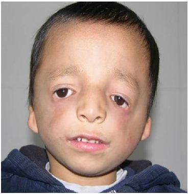
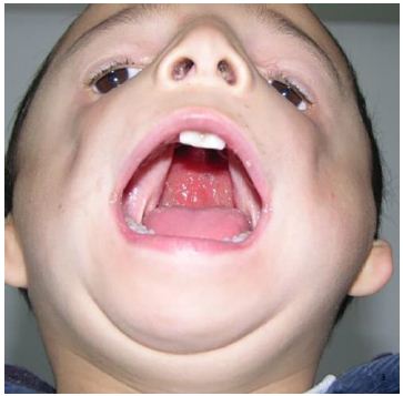
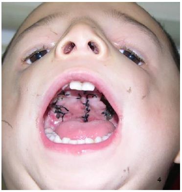
A 4-year-old male patient was identified with facial deformity and cleft palate at birth. Examination revealed facial asymmetric, flat bilateral zygoma and zygomatic arch, a short mandibular ramus, wide orbital spacing, oblique palpebral fissures, absence of the lower eyelid, and missing lower eyelid eyelashes (Figure 1). The patient also exhibited bilateral auricle deformity, external auditory canal atresia with preauricular fistula (Figure 2), and a cleft palate extending from the uvula to the incisive foramen, with a maximum width of 2.5 centimeters (Figure 3). Hearing tests indicated moderate conductive hearing loss in both ears. Genetic analysis identified a heterozygous spontaneous mutation in the POLR1 D gene, leading to a diagnosis of TCS type II. The patient’s speech was unclear due to the cleft palate. Upon admission, the patient underwent cleft palate repair surgery under general anesthesia with tracheal intubation. Postoperative recovery was successful (Figure 4), and voice training commenced three months post-surgery. Follow-up evaluations at 6 months, 12 months, and 24 months post-operation showed well-recovered palate morphology, normal soft palate movement, significantly improved pronunciation, and no coughing during meals.

Figure 1: Facial asymmetric, flat bilateral zygoma and zygomatic arch, a short mandibular ramus, wide orbital spacing, oblique palpebral fissures, absence of the lower eyelid, and missing lower eyelid eyelashes.

Figure 2: Bilateral auricle deformity, external auditory canal atresia with preauricular fistula.

Figure 3: A cleft palate extending from the uvula to the incisive foramen, with a maximum width of 2.5 cm.

Figure 4: The palate healed well

Discussion

TCS is a rare craniofacial malformation with autosomal dominant inheritance, also known as maxillofacial dysplasia and deafness syndrome. It presents a highly variable phenotype, with about 40% of patients having a family history [3]. The cause of TCS is attributed to disrupted ribosome synthesis in cranial neural crest cells and neuroepithelial cells between the 2nd and 8th weeks of embryonic development. This disruption leads to a reduction in the number of neural crest cells migrating to the craniofacial region, resulting in hypoplasia of the first and second branchial arches [5].

TCS can present with various clinical types. According to studies by Splendor (2000), Teber (2004), and Vincent [6-8], the clinical features of TCS patients can be summarized as follows: (1) Craniofacial abnormalities: These include facial asymmetry, a low hairline, and facial hypoplasia, particularly affecting the mandibular and zygomatic complex. (2) Eye abnormalities: These are characterized by oblique palpebral fissures and lower eyelid defects. (3) Ear abnormalities: These include atresia of the external auditory canal, microtia, and conductive hearing loss, contributing to a characteristic ‘fish-like ‘ facial appearance. (4) Other rare clinical features: Dental abnormalities such as missing teeth (tooth dysplasia), tooth discoloration (enamel opacity), excessive tooth spacing, abnormal permanent teeth (eg, ectopic maxillary first molars), and occlusal disorders. Palatal abnormalities such as high-arched palate and cleft palate. Respiratory and feeding difficulties may arise from posterior nostril stenosis or atresia. Cardiac malformations are also possible in some cases [9,10]. Patients with a mild phenotype of TCS may exhibit almost no obvious clinical features and may require genetic testing for identification. Conversely, those with a severe phenotype may experience life-threatening ventilatory disorders due to obstruction of the posterior nasal foramen, glossoptosis, and other complications [11].

The clinical diagnosis of TCS is primarily based on clinical manifestations, imaging examinations, and pathogenic gene detection. X-ray imaging typically reveals several characteristic features: increased density of small mastoid bones, nasal protrusion with a wide, flat frontonasal angle, maldevelopment or defects in the zygomatic bone and zygomatic arch, narrow maxillary protrusion, small maxillary sinus, mandibular dysplasia with a short body and ascending ramus, and a deepened anterior corner notch. Ultrasound examination is valuable for the intrauterine diagnosis of TCS, with the fetus often presenting with polyhydramnios, absence of fetal swallowing activity, and poor development of the bilateral parietal diameter and head circumference. Known pathogenic genes associated with TCS include TCOF1, POLR1C, and POLR1D. Mutations in these genes can lead to reduced ribosomal RNA transcription and ribosome synthesis, which subsequently affect the development and differentiation of neural crest cells during the embryonic stage [6,7,12].

The clinical manifestations of TCS are similar to those of several other syndromes, necessitating differential diagnosis. These syndromes include Nager syndrome, Miller syndrome, Goldenhar syndrome, and Pierre Robin syndrome [13,14]. The facial features of Nager syndrome are similar to those of TCS, but Nager syndrome also presents with typical limb deformities such as thumb dysplasia or absence, polydactyly, and radial ulna bony fusion [15]. Miller syndrome is characterized by asymmetric upper and lower eyelid ectropion and defects, dysplasia of the fifth finger (toe), and a higher prevalence of cleft lip and palate compared to TCS [16]. Goldenhar syndrome is marked by hemifacial atrophy affecting the development of the ears, mouth, and mandible, and may also include vertebral abnormalities and dermoid cysts on the outer layer of the eye [14]. Pierre Robin syndrome is distinguished by micrognathia, glossoptosis, dyspnea, and cleft palate [17].

The comprehensive sequential treatment of TCS typically begins at birth and continues until growth and development are complete. Treatment should be tailored to the patient’s growth pattern, physiological function, and psychosocial needs. It is recommended to ensure and maintain basic life functions before the age of 2 years. If there is persistent corneal exposure between the ages of 2 and 5 years, orbital wall reconstruction should be performed for correction. If the mandible is underdeveloped between the ages of 6 and 10 years, mandibular traction should be performed. Speech therapy, craniofacial fracture reconstruction, and external ear reconstruction should be completed before the age of 12. Orthodontic-orthognathic treatment, correction of upper and lower jaw and nasal deformities, and social psychotherapy should be carried out between the ages of 13 and 18 years. Future treatment should incorporate molecular genetics, utilizing genetic tests for prenatal examinations in high-risk groups and monitoring fetal growth and development during the first three months of pregnancy.

The phenotypes of TCS vary greatly, ranging from mild to severe deformities. For example, airway obstruction caused by mandibular deformity can affect breathing, and eyelid defects can expose the cornea. Therefore, TCS treatment should follow a multidisciplinary comprehensive sequence approach, involving oral and maxillofacial surgery, plastic surgery, orthodontics, ophthalmology, otolaryngology, speech therapy, psychology, genetics, and nursing. Individualized treatment measures should be selected based on the patient’s age and degree of deformity.

Data Availability Statement

The raw data supporting the conclusions of this article will be made available by the authors, without undue reservation.

Declaration of Conflicting Interests

The author(s) declared no potential conflicts of interest with respect to the research, authorship, and/or publication of this article.

Funding

The author(s) received no financial support for the research, authorship, and/or publication of this article.

Ethics Statement

The studies involving human participants were reviewed and approved by the ethics committee of Qingdao Women and Children’s Hospital

Informed Consent

The patient ‘s legal guardian provided written informed consent to participate in this study.

Author’s Contribution

Ting li: Conception and design of study, Acquisition of data, Data analysis and interpretation, Drafting of manuscript and critical revision, Approval of final version of manuscript.

Yuelin qin: Conception and design of study, Acquisition of data, Data analysis and interpretation, Drafting of manuscript and critical revision, Approval of final version of manuscript.

Ziyan lu: Acquisition of data.

Xuecai yang: Drafting of manuscript and critical revision, Approval of final version of manuscript.

Junwei wang: Drafting of manuscript and critical revision, Approval of final version of manuscript.

References

  1. Nguyen PD, Caro MC, Smith DM, et al. (2016) Long-term orthognathic surgical outcomes in Treacher Collins patients. J Plast Reconstr Aesthet Surg 69: 402-408. [crossref]
  2. Treacher Collins E (1900) Case with symmetrical congenital notches in the outer part of each lower lid and defective development of the malar bones. Trans Opthalmol Soc 20: 190-192.
  3. Shinde CV, Kohli S (2018) Treacher Collin’s syndrome: a case report with an augmented review! International Journal of Advanced Research 6: 141-146.
  4. Renju R, Varma BR, Kumar SJ, et al. (2014) Mandibulofacial dysostosis (Treacher Collins syndrome): A case report and review of literature. Contemporary Clinical Dentistry 5: 532-534.
  5. van Gijn DR, Tucker AS, Cobourne MT (2013) Craniofacial development: current concepts in the molecular basis of Treacher Collins syndrome. Br J Oral Maxillofac Surg 51: 384-388.
  6. Splendore A, Silva EO, Alonso LG, et al. (2000) High mutation detection rate in TCOF1 among Treacher Collins syndrome patients reveals clustering of mutations and 16 novel pathogenic changes. Hum Mutat 16: 315-322. [crossref]
  7. Teber OA, Gillessen-Kaesbach G, Fischer S, et al. (2004) Genotyping in 46 patients with tentative diagnosis of Treacher Collins syndrome revealed unexpected phenotypic variation. Eur J Hum Genet 12: 879-890. [crossref]
  8. Vincent M, Geneviève D, Ostertag A, et al. (2016) Treacher Collins syndrome: a clinical and molecular study based on a large series of patients. Genet Med 18: 49-56. [crossref]
  9. Campos PSSL, Taitson PF, Pinto da Silva LC, et al. (2022) Dental and health aspects in the co-occurrence of Treacher Collins and Down syndromes: Case report. Spec Care Dentist 43: 94-98. [crossref]
  10. Kadakia S, Helman SN, Badhey AK, et al. (2014) Treacher Collins syndrome: the genetics of a craniofacial disease. Int J Pediatr Otorhinolaryngol 78: 893-898. [crossref]
  11. Islam F, Afroza A, Rukunuzzaman M, et al. (2008) Treacher Collins Syndrome-A Case Report. Bangladesh Journal of Child Health 32: 33-36.
  12. Bowman M, Oldridge M, Archer C, et al. (2012) Gross deletions in TCOF1 are a cause of Treacher–Collins-Franceschetti syndrome. European Journal of Human Genetics 20: 769-777. [crossref]
  13. Thomas P, Krishnapillai R, et al. (2019) Treacher Collins Syndrome: A Case Report and Review of Literature. Oral Maxillofac Patho J 10: 90-94.
  14. Shete P, Tupkari J, Benjamin T, et al. (2011) Treacher Collins syndrome. Journal of Oral and Maxillofacial Pathology 15: 348-351.
  15. Rosa RF, Guimarães VB, Beltrão LA, et al. (2015) Nager syndrome and Pierre Robin sequence. Pediatrics international: official journal of the Japan Pediatric Society 57: e69-72. [crossref]
  16. Trainor PA, Andrews BT (2014) Facial dysostoses: Etiology, pathogenesis and management. American Journal of Medical Genetics Part C Seminars in Medical Genetics 163: 283-294.
  17. Shen YF, Vargervik K, Oberoi S, et al. (2012) Facial Skeletal Morphology in Growing Children With Pierre Robin Sequence. Cleft Palate Craniofac J 49: 553-560.

Reform of the National Institute of Mental Health: A Proposal

DOI: 10.31038/JNNC.2025811

Abstract

In the United States, the primary federal agency for funding mental health research is the National Institute of Mental Health (NIMH). For decades, the NIMH has prioritized research on genetics, biomarkers and related aspects of biological psychiatry, with no meaningful yield. It is time for a radical reorganization, restructuring and reconceptualization of NIMH spending.

Keywords

National Institute of Mental Health, Mental health, Research funding

The 2025 budget of the NIMH will be over 2 billion dollars [1]. After decades of focus on genetics, brain chemistry, biomarkers and related elements of biological psychiatry, and tens of billions of dollars spent, biological psychiatry in its current form has yielded zero findings of direct clinical relevance. The idea that there is an “underlying pathophysiology” to mental illness has been countered by a large body of NIMH-funded, published evidence – by the failure to find anything. Rather than continuing to fund endless efforts to identify biological causes of mental illness, it is time to rethink the paradigm and set a distinctly different research agenda.

This view is consistent with a statement made on the American Psychiatric Association website: “many factors contribute to the risk of mental illness, such as depression. Except in rare cases, genes determine just a small percentage of the risk of illness or response to medication. Age, lifestyle, general health, psychiatric symptoms and severity, and co-occurring conditions are usually more important factors in drug response.” [2]

Simple logic and common sense can tell you that the search for genes contributing to mental illness is futile. For example, genome-wide association studies (GWAS) currently involve tens of thousands of patients and tens of thousands of controls. Rather that demonstrating the advanced nature of such research, these numbers demonstrate that is time to give up on that line of investigation. The huge numbers are required in order to find anything of statistical significance in a given study. The findings are difficult or impossible to replicate from one GWAS to another, and the overall conclusion is that there are hundreds of risk genes, each contributing less than 2% to the clinical picture, as stated in DSM-5 [3]. The same set of risk genes has been identified for schizophrenia, bipolar disorder, depression and autism, proving that there is no genetic specificity to DSM-5 diagnostic categories.

The promise in grant applications and the psychiatric literature is the hope that – with just a few more years of research – something will be found, resulting in a truly scientific personalized psychiatry in which medications will be prescribed to target specific genetic dysregulations. If there are hundreds of risk genes then hundreds of medications targeting the functions and products of those genes would be required and an individual patient would require dozens of medications given that each genetic abnormality only accounts for under 2% of the clinical picture. Each medication will cost the patient thousands or tens of thousands of dollars per year.

The entire enterprise is guaranteed to fail. It is time to give up on it.

If asked, I would recommend the following reforms to the NIMH:

  1. Stop funding biological psychiatry in its current form.
  2. Prioritize psychological and social causes of mental illness, and psycho-social treatments.
  3. Stop all efforts to de-stigmatize mental disorders by saying they are brain diseases.
  4. Mandate that measures of childhood trauma be included in all funded research. This should include psychological, social, cultural and economic forms of trauma.
  5. Set ICD-11 complex-PTSD as the ruling paradigm: a poly-diagnostic response to complex psychological and social trauma accounts for a substantial proportion of serious mental illness.
  6. Dismantle negative attitudes towards dissociative disorders by requiring them, and dissociative symptoms, to be measured in the majority of funded studies. Pair this with explicit efforts to de-stigmatize borderline personality disorder and conceptualize it as an adaptation to psychological trauma – set this as a research funding priority (see 2 above).
  7. Provide extensive public education about the reforms.
  8. If the reforms are met with bureaucratic, committee and procedural barriers, withdraw funding until the NIMH and its bureaucracy complies.
  9. No reduction in the overall NIMH budget, only a re-allocation of resources.

If a reform at all resembling the above was adopted, the predicted response of organized psychiatry would be to decry it as anti-scientific and to say it was setting mental health back 50 years. Actually, it would transfer the focus to scientific study of the psychosocial aspects of mental health – and thereby correct an extreme imbalance that has dominated psychiatric research funding for decades [4].

References

  1. Torrey F, Simmons WW, Dailey L (2023) The NIMH research portfolio: An update. Primary Care Companion CNS Disorders 25(4), 23m03486. [crossref]
  2. https://www.psychiatry.org/news-room/apa-blogs/genetic-testing-to-improve-psychiatric-medication.
  3. American Psychiatric Association (2013) Diagnostic and Statistical Manual of Mental Disorders, 5th American Psychiatric Association, Washington, DC, p. 494.
  4. Ross CA, Pam A (1995) Pseudoscience in Biological Psychiatry. Blaming the Body. John Wiley & Sons, New York.

Incidence and Risk Factors of Sepsis in Adult Patients with Trauma: A Systematic Review and Meta-Analysis

DOI: 10.31038/IJOT.2025811

Abstract

Objective: To conduct a systematic review and analysis of the risk factors linked to sepsis in adult trauma patients, providing evidence-based medical evidence for reducing the incidence of sepsis following trauma.

Methods: Literature searches were conducted in the total of 9 databases from their inception to December 2023 on factors influencing sepsis in trauma patients. Meta-analysis was conducted using the meta package in R, and the model’s heterogeneity was assessed using the I² value.

Results: A total of 10 literatures were included, involving 65,866 adult patients admitted for trauma, with 5,165 cases of sepsis following trauma. The meta-analysis results showed that advanced age (MD=1.31,95%CI: 0.51~ 3.12), male gender (OR=1.21, 95%CI: 0.95~1.54), Injury Severity Score (ISS) (MD=5.99, 95%CI: 3.05~8.93), Glasgow Coma Scale (GCS) score (MD=-1.75, 95%CI: -2.68~-0.81), Acute Physiology and Chronic Health Evaluation (APACHE II) score (MD=4.37, 95%CI: 2.56, 6.17), Sequential Organ Failure Assessment (SOFA) score (MD=2.51, 95%CI: 2.30~2.73), mechanical ventilation (OR=4.71, 95%CI: 3.44, 6.45), blood transfusion (OR=2.20, 95%CI: 1.63~2.96), central venous catheterization (OR=2.74, 95%CI: 1.93~3.89), concurrent shock (OR=2.30, 95%CI: 1.70~3.10), and emergency surgery within 24 hours (OR=2.85, 95%CI: 2.00~ 4.07), were identified as independent risk factors for sepsis among trauma patients.

Conclusion: Sepsis in trauma patients is influenced by a variety of risk factors. Clinical medical staff should intervene early in High-risk patients with these factors should be targeted to reduce sepsis incidence among trauma patients.

Keywords

Trauma, Sepsis, Risk factors, Meta-analysis

Introduction

Trauma represents a major global health burden, accounting for around 9% of annual deaths and ranking among the leading causes of mortality worldwide [1]. The advent of advanced medical technologies has successfully curbed the early mortality rate among trauma patients. However, a significant number of survivors are at risk of developing sepsis in the days or weeks following the initial trauma [2]. Sepsis, a complex clinical syndrome arising from a dysregulated host response to infection, not only can precipitate septic shock and multiple organ failure but also substantially worsens the prognosis [3]. The development of sepsis is associated with an overactive and persistent inflammatory response in trauma patients and is a prevalent complication [4]. Existing studies have reported that the mortality rate among trauma patients with sepsis hovers between 17% and 23% [5], highlighting the gravity of this complication. Despite a plethora of research efforts, the majority of which are based on single-center data, there remains a lack of consensus regarding the identification of specific risk factors for sepsis in trauma patients.

Meta-analysis, a powerful tool that aggregates and quantifies the effect sizes of individual studies through systematic review, emerges as a promising approach to address this issue [6]. By comprehensively reviewing and dissecting the extant literature on the risk factors associated with post-traumatic sepsis, this study aims to systematically organize and deliberate upon these factors. The ultimate goal is to furnish a robust evidence-based foundation for clinical practice, thereby facilitating the early detection and prevention of sepsis in trauma patients and potentially ameliorating their outcomes.

Methods

Protocol and Registration

This research adhered to the PRISMA (Preferred Reporting Items for Systematic Reviews and Meta-Analyses) guidelines [7] and our systematic review protocol was recorded on PROSPERO (International Prospective Register of Systematic Reviews, with the registration number CRD42024537479). As the data utilized were publicly accessible, ethical committee approval was not pursued.

Retrieval Strategy

Literature Sources and Search Strategy Literature was retrieved from databases including China National Knowledge Infrastructure, Wanfang Data, China Science and Technology Journal Database, China Biology Medicine Literature Database, PubMed, Embase, Web of Science, Cochrane, CINAHL, and Scopus from their inception to December 2023. We utilized the keywords included trauma, traumatic, post-traumatic, multiple injuries, polytrauma, septic, sepsis, septicemia, multiple organ failure, factor, and risk. Databases for dissertations and trial registries were not searched. The specific search strategies employed for English databases are detailed in Appendix 1.

Inclusion and Exclusion Criteria

Inclusion criteria: 1) Age ≥18 years; 2) Study population consisting of trauma patients; 3) Sepsis diagnosed according to Sepsis-1, Sepsis-2, or Sepsis-3 criteria; 4) Independent risk factors determined through multivariate regression analysis. Exclusion criteria: 1) insufficient patient baseline data; 2) reviews, meta-analyses, commentaries, case reports, guidelines, letters, conference abstracts, and literature related to animal experiments; 3) abnormal data and/or not conforming to statistical rules. The predominant literature reviewed comprised case- control and retrospective cohort studies, predominantly authored in either English or Chinese. We excluded smaller studies (those with fewer than 50 patients) to avoid potential false negative results. Additionally, patients with burns were excluded because they have distinct risk factors, such as a compromised skin barrier, which could potentially elevate the risk of developing sepsis [8].

Literature Screening and Data Extraction

Search results were imported into EndNote X9 software (Clarivate Analytics, London, UK) for management. Two independent reviewers (Wang B and Shi Y) screened titles and abstracts against predefined inclusion/exclusion criteria, following Cochrane guidelines. Potentially relevant citations were subjected to full-text review. Data extraction was performed independently from all eligible studies using a standardized form, with a third researcher consulted to resolve any discrepancies. The main extracted content included: principal investigator, study design, publication region and year, sample size, characteristics of the study population (age, sex), follow-up period, identified risk factors, and outcomes of multifactorial regression analysis.

Quality Assessment of Literature

Two researchers (Zhu X and Dong C) independently assessed the quality of the literature using the Newcastle-Ottawa Scale (NOS) [9]. NOS scores categorized the literature into three quality levels: ≥7 (high quality), 4-6 (moderate quality), and <4 (lower quality). In the event of any disagreements during the assessment process, the opinion of a third researcher (Cao S) will be sought to resolve them.

Statistical Analyses

Statistical analyses were conducted using Review Manager (version 5.3), STATA (version 12.0), and the ‘meta’ package in R software. The categories of subgroup analyses of incidence included: age, year of publication, research region, diagnostic criteria for sepsis, and duration of follow-up. For categorical variables, the Odds Ratio (OR) and 95% Confidence Intervals (CI) were used to express the statistical effect size, while the Mean Difference (MD) and 95% CI were used for continuous variables. Heterogeneity across studies was evaluated with the intraclass correlation index (), which quantifies the proportion of total variation in study outcomes due to between-study variance (τ²) rather than chance [10]. ≥ 50% was considered indicative of significant heterogeneity. In these instances, a random-effects model was employed for meta-analysis; otherwise, a fixed-effects model was applied. Publication bias was assessed via Egger’s regression test and funnel plots, with P < 0.05 considered statistically significant.

Results

Study Characteristics

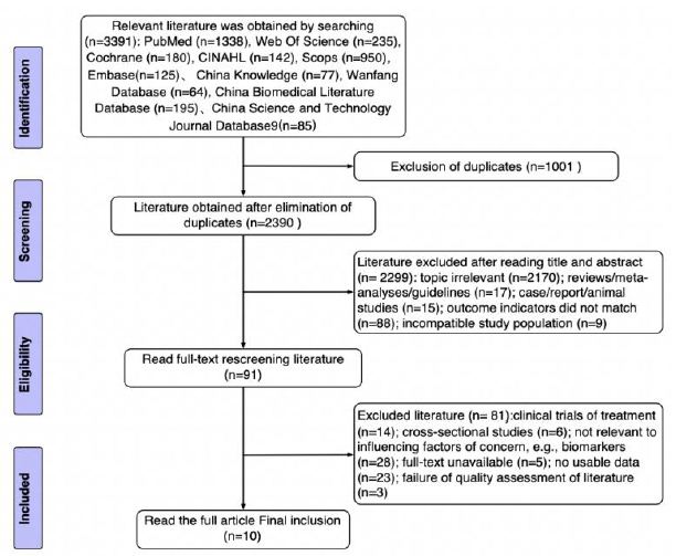
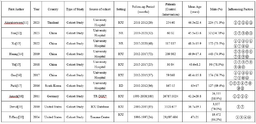
A preliminary collection of 3391 articles was obtained, and a total of 10 articles were ultimately included (Figure 1). The 10 articles included in this study were all retrospective cohort studies [11-20], published between 2004 and 2023. Upon summarizing the literature, there were 12 risk factors with ≥2 articles, including 10 articles on age [11-20] as a risk factor; 9 articles on male gender [11-19] as a risk factor; 8 articles on Injury Severity Score (ISS) [12-18,20] as a risk factor; 5 articles each on Glasgow Coma Scale (GCS) [13,14,17,18,20] Sequential Organ Failure Assessment (SOFA) [11,13-16], mechanical ventilation [13-17], and shock [12,14-17]as risk factors; 4 articles each on Acute Physiology and Chronic Health Evaluation II (APACHE II) [11,13,14,16], number of blood transfusions [13,15-17], and emergency surgery within 24 hours [13,15-17] as risk factors; 3 articles each on central venous catheterization [14-16] and diabetes [11,12,20] as risk factors. The study characteristics are shown in Table 1.

Figure 1: PRISMA diagram for identification of relevant studies. PRISMA, Preferred Reporting Items for Systematic Reviews and Meta-Analyses.

Table 1: Baseline characteristics of studies included for analysis

Note:① Age (years); ② Sex (Male); ③ Injury Severity Score, ISS; ④ Glasgow Coma Scale, GCS; ⑤ Acute Physiology and Chronic Health Evaluation II, APACHE II; ⑥ Sequential organ failure assessment, SOFA; ⑦ mechanical ventilation, MV; ⑧ blood transfusion; ⑨ Central venous catheterization, CVC; ⑩ Shock: SBP < 90 mmHg at hospital; ⑪ Diabetes; ⑫ Emergency surgery: surgery within 24 hours.
aTR-DGU: Trauma Registry of the German Society for Trauma Surgery
NR: not reported

Outcomes of Incidence and Subgroup Analyses

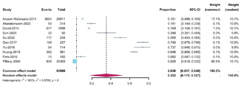
There were 65,866 trauma inpatients, with 5,165 cases of sepsis and 60,701 cases without sepsis. The  was 100%, so a random-effects model was used. The results showed that the incidence of sepsis in adult trauma patients was 35.2% (95% CI: 17.8%, 52.7%) (Figure 2). Subgroup analyses were conducted based on age, publication year, study region, sepsis diagnosis criteria, and follow-up duration. The results showed: subgroup analysis by continent demonstrated a pooled incidence rate for the age group 30≤Age < 69 years was the highest at 37.9% (95% CI: 19.8%, 58.0%); when grouped by publication year, the incidence rate for the group after 2020 was 34% (95% CI: 15%, 56%), lower than the incidence rate for the group before 2020, which was 38.6% (95% CI: 22.9%, 55.6%); subgroup analysis by study region, the incidence rate was 60.1% (95% CI: 48.0%, 71.5%) in China, higher than the incidence rate in other regions was 10.3% (95% CI: 4.2%, 18.5%); when grouped by sepsis diagnosis criteria, the incidence rate for the group using the third edition of sepsis diagnosis criteria was 60.1% (95% CI: 48.0%, 71.5%), higher than the group using the first and second editions of sepsis diagnosis criteria; when grouped by follow-up duration, the incidence rate for the group with a follow-up duration of 32 to 72 months was the highest at 45.2% (95% CI: 22.5%, 68.9%). Supplemental Table 1 for details.

Figure 2: Forest plot of the incidence of sepsis in trauma patients

Outcomes of Sepsis Influencing Factors

An analysis was conducted on the 12 included influencing factors. For SOFA, mechanical ventilation, number of blood transfusions, central venous catheterization, shock, and diabetes, the was ≤30, so a fixed-effects model was chosen for analysis. For the remaining factors, the  was ≥50%, so a random-effects model was used. The study results indicated that, except for diabetes, all other factors were statistically significant (P<0.05) (Table 2).

Table 2: Meta-Analysis of Influencing Factors

Sensitivity Analysis

The pooled effect size and heterogeneity for the 12 influencing factors were estimated using both random-effects and fixed- effects models. The statistical results showed that, except for the ISS, the other influencing factors demonstrated good consistency, indicating a high level of reliability in the results of this study (Table 3).

Table 3: Sensitivity Analysis of Influencing Factors

Quality Evaluation and Publication Bias

The NOS scoring results showed that 7 articles scored ≥7 points [11,13-15,17-19] and 3 articles scored 6 points [12,16,20]. Quality evaluation is provided by Supplemental Table 2. Egger’s regression test was used to assess publication bias for the 10 articles that considered age as a risk factor for sepsis. The Egger’s regression test for funnel plot asymmetry supports this observation, yielding a non-significant result (p = 0.32), which indicates a low level of bias in the published findings. The contour-enhanced funnel plot as shown in Supplement Figure 1. For the other 11 influencing factors, the number of included articles did not reach 10, hence no publication bias analysis was conducted for them.

Discussion

The pooled average incidence of sepsis in adult trauma patients calculated from the studies was 35.2%, which is higher to the 31.1% reported by Amina Abliz et al. [21]. The estimation of incidence rates varies by region. This study found that the incidence rate in China is 60.1%, which is significantly higher than the 10.3% in other countries. The reason for this difference may be related to the data sources. Among the five foreign studies included, the data of three studies come from public databases. Such data sources may have a more representative sample of the general population, but issues such as data collection methods and quality control may lead to an underestimation of the incidence rate. In contrast, the data of the five domestic studies all come from hospitals, which means that the data mainly come from patients seeking medical treatment, and there may be selection bias. Hospital – based data tend to be biased towards patients with more severe or symptomatic conditions, which may overestimate the incidence rate. Furthermore, the decrease in the incidence of post – traumatic sepsis over time may be attributed to early diagnosis and intervention, continuous strengthening of hospital infection control measures, improvement of the trauma treatment system, and enhanced self – health awareness of patients after trauma. Finally, compared with previous standards, the Sepsis – 3 diagnostic criteria may have improved sensitivity. This comprehensive assessment method may lead to the diagnosis of more sepsis patients in early or sub – clinical states, thereby resulting in an increase in the incidence rate.

Advanced age is an important risk factor for sepsis in adult trauma patients. The elderly are more susceptible to sepsis due to factors such as immunosenescence, weakened cardiovascular function, poor nutritional status, and comorbidities [22]. Epidemiological studies have shown that the incidence of sepsis is higher in males than in females [23]. The results of this study indicate that the risk of sepsis in male trauma patients is 1.21 times higher than in females, which is close to the 1.3 times reported in a study from the United States [24]. This may be related to differences in sex hormone levels [25]. Although demographic-related influencing factors cannot be directly intervened, the development of sepsis in elderly male trauma patients should be closely monitored. Additionally, APACHE II and SOFA scores are used to assess the severity of patients’ conditions. The risk of sepsis is positively correlated with these scores. The lower the GCS score, the higher the risk of sepsis. Particularly, patients with severe brain injuries and coma have a higher incidence of sepsis and septic shock [26]. The ISS score provides a quantitative measure for assessing soft tissue injuries in trauma patients. This study found that ISS, APACHE II, SOFA, and GCS scores are all helpful for early identification of sepsis in adult trauma patients. Medical staff need to closely monitor patients with abnormal scores and take timely intervention strategies to prevent the occurrence of sepsis. Furthermore, the number of blood transfusions, mechanical ventilation, central venous catheterization, and emergency surgery are associated with an increased risk of sepsis in trauma patients. Blood transfusion may increase the risk of sepsis by suppressing immune responses [27]. Patients on mechanical ventilation are more likely to develop VAP, leading to sepsis [28]. Central venous catheterization increases the risk of central venous catheter-related bloodstream infections, especially in the intensive care unit, where such infections are common and potentially life- threatening [29]. Emergency surgery is also a risk factor for sepsis after trauma. The OR value of this study is 2.55, indicating that the risk of sepsis in patients undergoing emergency surgery is 2.55 times that of those undergoing elective surgery, which is close to the 2 times reported in previous studies [30].

Therefore, in the management of trauma patients, the necessity of blood transfusions, mechanical ventilation, central venous catheterization, and emergency surgery should be carefully assessed to reduce the risk of sepsis. Lastly, shock can predispose patients to sepsis by damaging microcirculation and reducing tissue perfusion, while sepsis can exacerbate shock by triggering widespread inflammatory responses and cardiovascular dysfunction [31]. Therefore, when trauma patients have shock, it should be promptly recognized and treated to reduce the incidence of sepsis.

Limitations

This meta-analysis has several limitations. Firstly, there is a certain degree of heterogeneity in the combined effect sizes of some risk factors, which may be related to factors such as the race, age distribution of the study subjects, and the quality of diagnosis and treatment in different medical institutions, and thus needs further improvement; Secondly, risk factors with less than 10 studies were not assessed for publication bias, so the possibility of bias cannot be ruled out. Moreover, the study did not categorize sepsis by severity, which could affect treatment strategies and outcome predictions.

Conclusion

In conclusion, the incidence of sepsis in adult trauma patients is high and influenced by various factors including age, gender, clinical scoring systems, invasive procedures, as well as comorbid conditions. Clinical medical staff can refer to the results of this study, deal with and prevent risk factors in a targeted manner, reduce the occurrence of sepsis, and thus improve the prognosis and quality of life of trauma patients.

CRediT Authorship Contribution Statement

Bingsheng Wang conceived and designed the study, independently completed database search. Wenhao Qi screening and data extraction and writing. Bing Wang, Xiaohong Zhu and Chaoqun Dong conducted statistical analysis, interpreted the analytical results, and provided technical support for methodological refinement. Yankai Shi, Jiani Yao and Xiajing Lou assisted in optimizing the research. Aili Shi and Shihua Cao reviewed and edited the manuscript.

Acknowledgements

This research was supported by the Medical and Health Technology Plan of Zhejiang Province (grant 2022507615); Key Research Project for Laboratory Work in Zhejiang Province Colleges, ZD202202; Zhejiang Province Traditional Chinese Medicine Inheritance and Innovation Project 2023ZX0950. 2024 research project of Engineering Research Center of Mobile Health Management System, Ministry of Education; First – class Undergraduate Course in Zhejiang Province, 2022, sponsored by the Education Department of Zhejiang Province (No.1133).

References

  1. Skelton JK, Purcell R (2021) Preclinical models for studying immune responses to traumatic injury. Immunology 162: 377-388. [crossref]
  2. Zeng L, Du J, Gu W, Zhang AQ, Wang HY, et (2015) Rs1800625 in the receptor for advanced glycation end products gene predisposes to sepsis and multiple organ dysfunction syndrome in patients with major trauma. Crit Care 19: 6. [crossref]
  3. M SH, Gs P, Ml L, Seymour CW, Liu VX, et (2016) Developing a New Definition and Assessing New Clinical Criteria for Septic Shock: For the Third International Consensus Definitions for Sepsis and Septic Shock (Sepsis-3). JAMA 315. [crossref]
  4. Sauaia A, Moore FA, Moore EE (2017) Postinjury Inflammation and Organ Crit Care Clin 33: 167-191. [crossref]
  5. Liang Huaping, Zhu Junyu, Hu Mingdong (2021) Several Key Issues in the Field of Traumatic Sepsis Chinese Journal of Traumatology 37: 385-389.
  6. Khan S (2020) Introduction to Meta-analysis. In: Khan S, ed. Meta-Analysis: Methods for Health and Experimental Studies. Statistics for Biology and Health. Springer 15-36.
  7. Page MJ, McKenzie JE, Bossuyt PM, Boutron I, Hoffmann TC, et al. (2021) The PRISMA 2020 statement: an updated guideline for reporting systematic reviews. BMJ 372: n71. [crossref]
  8. Bahemia IA, Muganza A, Moore R, Sahid F, Menezes CN (2015) Microbiology and antibiotic resistance in severe burns patients: A 5 year review in an adult burns Burns 41: 1536-1542. [crossref]
  9. Stang A, Jonas S, Poole C (2018) Case study in major quotation errors: a critical commentary on the Newcastle-Ottawa scale. Eur J Epidemiol 33: 1025-1031. [crossref]
  10. Nakagawa S, Noble DWA, Senior AM, Lagisz M (2017) Meta-evaluation of meta- analysis: ten appraisal questions for BMC Biol 15: 18.
  11. Akaraborworn O, Chaiwat O, Chatmongkolchart S, et al. (2019) The intensive care unit admission predicting the factors of late complications in trauma patients: A prospective cohort Archives of Trauma Research 8: 203.
  12. Sun Shuying, Wen Dalin, Chen Guosheng (2023) Analysis on the Related Risk Factors for Sepsis in Patients with Severe Multiple Injuries and Their Early Warning Chinese Journal of Traumatology. Wound Pract Res 39: 443-449.
  13. Xu Jing, Jiao Xiaoqin, Guan Hong, Wang Pengsen, Du Junkai (2022) Mining on the Risk of Sepsis in Patients with Severe Journal of Clinical Emergency 23: 847- 853.
  14. Qi H, Yu S, Li L, Meng S, Chen T, et al. (2019) A new warning scoring system establishment for prediction of sepsis in patients with trauma in intensive care unit. Chinese Critical Care Medicine 31: 422-427. [crossref]
  15. Yu Yaofeng, Zhang Liqun (2018) Clinical Factor Analysis of Post-traumatic Infection Complicated with Sepsis in the Intensive Care Zhejiang Journal of Traumatic Surgery 23: 671-673.
  16. Gao Min, Sun Yu, Wang Yudi, Xia Wanqiu, Liang Huaping, et al. (2017) Risk Factor Analysis of Sepsis Complicated in Post-traumatic Infection Patients in the ICU. Acta Academiae Medicinae Militaris Tertiae 39: 367-372.
  17. Park JH, Choi SH, Yoon YH, Park SJ, Kim JY, et (2016) Risk factors for sepsis in Korean trauma patients. Eur J Trauma Emerg Surg 42: 453-458. [crossref]
  18. Wafaisade A, Lefering R, Bouillon B, et (2011) Epidemiology and risk factors of sepsis after multiple trauma: an analysis of 29,829 patients from the Trauma Registry of the German Society for Trauma Surgery. Crit Care Med 39: 621-628. [crossref]
  19. Plurad DS, Lustenberger T, Kilday P, Zhu J, Green DJ, et (2010) The association of race and survival from sepsis after injury. Am Surg 76: 43-47. [crossref]
  20. Osborn TM, Tracy JK, Dunne JR, Pasquale M, Napolitano LM (2004) Epidemiology of sepsis in patients with traumatic Crit Care Med 32: 2234-2240. [crossref]
  21. Amina Abulizi, Ahmatjan Pakhatin, Huang Yangmei, et (2021) Diagnostic Value of Biomarkers Combined with Trauma Scores for Post-traumatic Sepsis. Journal of Traumatic Surgery 23: 384-390.
  22. He W, Yao C, Wang K, Duan Z, Wang S, et al. (2024) Single-cell landscape of immunological responses in elderly patients with sepsis. Immun Ageing 21: 40.
  23. Lakbar I, Einav S, Lalevée N, Martin-Loeches I, Pastene B, et (2023) Interactions between Gender and Sepsis—Implications for the Future. Microorganisms 11: 746. [crossref]
  24. Martin GS, Mannino DM, Eaton S, Moss M (2003) The epidemiology of sepsis in the United States from 1979 through N Engl J Med 348: 1546-1554. [crossref]
  25. Min SY, Yong HJ, Kim D (2024) Sex or gender differences in treatment outcomes of sepsis and septic shock. Acute Crit Care 39: 207-213. [crossref]
  26. Lu HX, Du J, Wen DL, Sun JH, Chen MJ, et (2019) Development and validation of a novel predictive score for sepsis risk among trauma patients. World J Emerg Surg 14: 11. [crossref]
  27. Blajchman MA (2005) Transfusion immunomodulation or TRIM: what does it mean clinically? Hematology 10: 208-214. [crossref]
  28. Rubano JA, Paccione MF, Rutigliano DN, Vosswinkel JA, McCormack JE, et al. (2015) Outcomes following prolonged mechanical ventilation: analysis of a countywide trauma J Trauma Acute Care Surg 78: 289-294. [crossref]
  29. Pitiriga V, Bakalis J, Theodoridou K, Kanellopoulos P, Saroglou G, et (2022) Lower risk of bloodstream infections for peripherally inserted central catheters compared to central venous catheters in critically ill patients. Antimicrob Resist Infect Control 11: 137. [crossref]
  30. Moore LJ, Moore FA, Todd SR, Jones SL, Turner KL, Bass BL (2010) Sepsis in general surgery: the 2005-2007 national surgical quality improvement program Arch Surg 145: 695-700. [crossref]
  31. Gorecki G, Cochior D, Moldovan C, Rusu E (2021) Molecular mechanisms in septic shock (Review). Exp Ther Med 22: 1161. [crossref]

Mycobacterium tuberculosis, Tuberculosis and Cancer

DOI: 10.31038/CST.20251011

Abstract

Tuberculosis (TB) is an infectious disease caused by mycobacteria, principally Mycobacterium tuberculosis. This disease can affect all organs but mainly the lungs, remains a major public health challenge, particularly in low and middle-income countries. TB disease can occur after infection and can cause death. There is evidence of an increased risk of cancer, particularly of the lung, in people with a history of TB or with an active form of the disease. The BCG vaccine, derived from a weakened strain of Mycobacterium bovis, offers protection against severe forms of tuberculosis in children and is also employed in the treatment of bladder cancer. The objective of this article is to examine the associations between Mycobacterium tuberculosis, tuberculosis and cancer.

Keywords

Mycobactium tuberculosis, Tuberculosis, Risk factors, Lung cancer

Introduction

Tuberculosis is an infectious disease caused by a mycobacterium of the tuberculosis group, mainly Mycobacterium tuberculosis, whose usual reservoir is man, more rarely Mycobacterium bovis or africanum. The incidence of tuberculosis remains high in countries with low or middle economic income, and this potentially fatal infectious disease remains a global public health issue. The infection initially remains latent, linked to airborne transmission of bacilli and can progress to tuberculosis disease, mainly in the lungs, with the possibility of poly-visceral extension, particularly via the haematogenous route, in immunodeficient patients. It can also cause sequelae and encourage the development of cancer, particularly lung cancer. However, this mycobacterium can play a role in the treatment of other cancers, and represents an area of research in this field.

Epidemiology

The causative agent of tuberculosis is Mycobacterium tuberculosis. In 2023, tuberculosis (TB) remains a major public health issue worldwide, although it is no longer one of the top ten causes of death on a global scale. However, it is the leading cause of death from infectious agents, ahead of HIV infection, having caused 1.3 million deaths. Of the new cases diagnosed, 70% were of the pulmonary variety. The eight countries accounting for more than two-thirds of global TB cases are India (26%), Indonesia (10%), China (6.8%), Philippines (6.8%), Pakistan (6.3%), Nigeria (4.4%), Bangladesh (3.6%) and Democratic Republic of Congo (2.9%). More than 400,000 people have developed a form of TB resistant to rifampicin, almost 80% of these have developed a multi-drug resistant form [1].

More than 80% of TB cases and 90% of induced deaths occur in low- and middle-income countries. The mortality rate due to tuberculosis is falling by around 3% a year, and an overall decline of 42% was observed over the period 2000-2017 [2]. It is estimated that more than 1.5 billion people (23% of the world’s population) are infected with the tubercle bacillus, and are thus at risk of developing TB [2].

The Tubercle Bacillus

Bacteriological Aspects

Mycobacterium tuberculosis (MBT) is a strict aerobic bacillus. This pathogenic agent has an outer lipid membrane bilayer ; it divides very slowly (16 to 20 hours) and is either very weakly ‘Gram-positive’ or does not retain its colour due to the high lipid and mycolic acid content of its wall. In nature, the bacterium can only develop inside the cells of a host organism. This acid-fast bacillus can be identified under the microscope. The most common staining method is the Ziehl-Nielsen stain, which highlights MBT in bright red. Auramine staining and luminescence microscopy can also highlight it [1-3].

The Mycobacterium tuberculosis complex (MBTC) includes four other mycobacteria responsible for tuberculosis: M. bovis, M. africanum, M. canetti and M. microti [3]. M. africanum is not very widespread, but it is a major cause of tuberculosis in Africa. M. bovis used to be a common cause of tuberculosis, but the pasteurisation of milk has virtually eliminated its responsibility in developed countries. M. Canetti is mainly responsible for tuberculosis in the Horn of Africa, while M. Microti can be implicated in immunocompromised individuals [3].

The other known pathogenic mycobacteria are M. leprae (Hansen’s bacillus), which causes leprosy, and M. avium and M. kansasii, classified as ‘non-tuberculous mycobacteria’ (NTM), which can cause pulmonary infections similar to tuberculosis. [4,5].

Calmette and Guérin bacillus developed from an attenuated strain of Mycobacterium bovis, is the basis of the tuberculosis vaccine. It is used to protect (80% efficacy for more 5 years) young children against serious forms of tuberculosis, such as tuberculous meningitis and miliary tuberculosis [2]. It is much less effective against other forms of TB, particularly pulmonary. It stimulates the immune system so that it can identify and fight the mycobacteria responsible for tuberculosis in the event of exposure. In countries where the incidence of TB is low (United States of America, Canada, Western Europe, Australia) the vaccine is less widely used or reserved for high-risk population groups. On the other hand, it is given to newborns in countries with a high incidence of tuberculosis, and to children living in environments where active cases of tuberculosis may be present. Health professionals or those working in high-risk environments (laboratories, etc.) may also be vaccinated. A single dose by intradermal injection is recommended in the first few weeks of life ; in older children, the absence of tuberculosis infection should be verified by a tuberculin or IGRA test prior to vaccination. This vaccine has a high rate of tolerance, with minor ulceration at the injection site being the only common side effect. Complications are rare, and include local abscesses and adenopathy in the drainage area. In exceptional cases, disseminated infections may occur in immunocompromised individuals [6].

Transmission of Infection

The risk of transmission of the infection from one person to another depends on several factors: the number of small (0.5 and 5 µm) contagious droplets (Flügge droplets) which can remain suspended in the air for up to 9 hours after they are emitted. The transmission of the same clone of bacteria from one patient to another has been proven by genotyping studies of mycobacteria [6].

People who are in frequent, close contact with people suffering from pulmonary, laryngeal or tracheobronchial tuberculosis, especially in a small, poorly ventilated space, are particularly exposed to the risk of infection when patients emit MBT during verbal exchanges, episodes of coughing, spitting or sneezing. It is estimated that a person with active tuberculosis can infect at least 10 to 20 people. People suffering from tuberculosis must therefore be isolated while their sputum is sterilised, and wear a protective mask, as must their carers [6] and any infection should be detected in people contacts.

Natural History of Tuberculosis Infection

Transmission of Infection

Defence mechanism against MBT. Once inhaled, the tubercle bacilli are deposited in the distal alveolar spaces, mainly in the upper parts of the lungs. They are phagocytosed by alveolar macrophages (AMs), accompanied by a local cell-mediated inflammatory response involving CD4 T lymphocytes (Th1), which activate AMs and stimulate the production of cytotoxic CD8 lymphocytes, thereby facilitating the response against intracellular MBT. Numerous cytokines are released, including interferon gamma (INFγ), interleukin 2 (IL2), tumour necrosis factor (TNFα) and the recruitment of circulating mononuclear cells, all of which play a part in the defence mechanism against this infection. Phagocytic dendritic cells carrying antigenic peptides reach the lymph node relays and present these antigens to CD4 T lymphocytes, which return to the lung to organize the formation of an inflammatory granuloma with a satellite lymph node reaction, leading to the formation of a lymph node-lung complex [6,7].

Latent tuberculosis infection (LTI) is characterised by a delayed hypersensitivity reaction to MBT, leading to positive tuberculin and IGRA tests (Interferon Gamma Release Assays), whether Quantiferon- TB Gold Plus or T-SPOT.TB. In 90% of cases, the body’s immune response prevents MBT proliferation and controls the infection in less than 10 weeks, resulting in latent tuberculosis infection. This is generally clinically asymptomatic and poses no risk of contagion. However, MBTs may persist in a quiescent state in macrophages for a long time [6,7].

In the context of tuberculosis (TB), it is important to note that 70% of cases progress to tuberculosis disease within two years of initial infection. Following this, the risk gradually decreases, though it appears to be lifelong. TB disease can occur as a result of a decline in cellular immunity or reinfection with MBT. This risk is increased at the extremes of life, particularly in children under the age of five who have not been vaccinated with the Mycobacterium bacille Calmette-Guérin (BCG) vaccine, and in people over the age of 65. People at particular risk may also develop TB disease, the main clinical symptoms of which are asthenia, weight loss, fever and coughing in the case of respiratory infection. Radiologically, the lesions predominantly affect the upper lungs and can take a variety of forms, including infiltrates, excavations, nodules, disseminated forms, mediastinal lymph node involvement and pleurisy. Extra-thoracic sites may or may not be associated (e.g. laryngeal, peripheral lymph nodes, bone, genitourinary, digestive) An inflammatory biological picture with anaemia is most often identified. Diagnosis is made by detecting MBT in sputum, or by bronchial aspiration using bronchoalveolar lavage. Culture on liquid or solid medium (Löwenstein-Jensen) is used to identify the type of MBT and its resistance to anti-tuberculosis drugs. Once treatment has been initiated, and provided the patient complies and the MBT are not drug-resistant, sputum and cultures can be sterilised in less than 2 months [6,7].

Risk Factors for TB Infection and TB Disease

The development of tuberculosis is influenced by various internal or external risk factors that increase the likelihood of infection or progression from latent infection to disease.

Internal Risk Factors: The innate deficiency of defences against MBT, with deficient production of INFγ or IL4, IL10, IL12, must be taken into account before vaccination with BCG vaccine [8].

Diabetes, which doubles the risk of LTI and quadruples the risk of TB disease, often with severe presentations (pulmonary excavations, disseminated forms, recurrences or resistance to anti-tuberculosis drugs). The explosion in the number of cases of diabetes worldwide could worsen the epidemiological situation for TB [8].

External Risk Factors: The poor socio-economic conditions and social insecurity in which people live, including precarious housing, detention, malnutrition, migration and difficulty in accessing healthcare, favour the development of TB [8-10].

Exposure to outdoor or indoor air pollution involving various pollutants (e.g. CO, CO2, SO2, O3, PM2.5 microparticles, PAHs) are risk factors for MBT infection [8].

Exposure to active or passive smoking causes dysfunction of the mucociliary escalator, which promotes the persistence of germs in the respiratory tract, and impairment of the mechanisms of anti- infectious immunity (reduced function of AMs, reduced release of TNF-α, imbalance in the CD4/CD8 ratio and reduced production of IFN-γ). Tobacco smoke could stimulate grow and/or the virulence of tuberculosis bacilli. In smokers suffering from TM, a reduction in IFN-γ response was noted, which may affect the performance of IGRA tests. In active smokers, the risk of pulmonary tuberculosis was assessed (OR= 2.6; 95% CI: 2.1 – 3.4). This risk is dose-dependent (CR = 4.4 for 10 cigarettes smoked daily; CR = 5 for 10 years of smoking). Smoking increases the risk of death from TB (RR =2.15 ; 95% CI: 1.38- 3.3), of recurrence of TB and of anti-tuberculosis drug-resistant forms (OR = 1.70; 95% CI: 1.3-2.23) [11].

Alcohol abuse, often associated with socio-economic disadvantage, is a risk factor for MBT infection and TB disease. Alcohol alters the immune response and phagocytic function of macrophages. The risk is dose-dependent ; four drinks a day quadruples the risk of developing TB [12].

HIV infection is a major driver of the TB epidemic, particularly in Africa, where almost 10% of TB patients are thought to be living with HIV. Widespread use of screening and antiretroviral treatment (ART) has led to a significant reduction in TB mortality in HIV-infected patients [8,13].

The use of corticosteroids to treat chronic inflammatory diseases is associated with an excess risk of developing TB; this risk is dose- dependent: prednisolone ≤ 15 mg per day (OR=2.8; 95%CI: 1.0-7.9), dose ≥ 15 mg per day (OR=7.7; 95% CI: 2.8-21.4), and increases with prolonged treatment [8].

Immunomodulatory drugs (anti-TNF) used to treat chronic inflammatory diseases may favour the development of TB; screening for LTI and prophylactic treatment are essential prior to their use [8]. Immunosuppressive drugs used during visceral transplantation may also favour the development of TB [8].

Renal failure, with or without dialysis, increases the risk of TB [8].

Finally, there is good evidence that people exposed to silica crystals or suffering from silicosis have a higher risk of TB [14].

Tuberculosis and Cancer

Studies show an association between tuberculosis and cancers. Patients with a history of pulmonary tuberculosis have a higher relative risk of cancer than the general population. Increased rates of lung cancer have been reported in regions where TB is endemic [15,16]. However, the association between TB and cancer is often multifactorial and depends on many factors (environment, comorbidities, smoking, immune status of the patient). In many cases, the co-occurrence of TB and cancer makes it impossible to determine the nature of the association between these two types of disease [15,16].

Tuberculosis is a Risk Factor for Cancer

Tuberculosis can promote the development of certain cancers, the mechanisms involved are complex and multifactorial.

General Mechanisms

Tuberculosis causes chronic inflammation in tissues, especially the lungs. This inflammation can damage the DNA of cells, promoting carcinogenesis. Tuberculosis granulomas cause fibrosis and tissue remodelling, creating a microenvironment conducive to cancer development. AM and immune cells produce reactive oxygen species (ROS) to fight infection, but these damage DNA and increase the risk of carcinogenesis [16-18].

Cofactors may be involved in carcinogenesis. People living with HIV, who are more susceptible to tuberculosis, have a weakened immune system, which can increase the risk of lung cancer, as well as Kaposi’s sarcoma, lymphoma and anogenital cancers. Exposure to substances such as silica, asbestos and polycyclic aromatic hydrocarbons all promote carcinogenesis. Smoking, which is common among TB patients, is a major risk factor for cancer, especially lung cancer [19,20].

Different Types of Cancer

Lung Cancer

Mycobacterium tuberculosis infection may increase the risk of lung cancer, which may be twice as common after TB as in the general population. In particular, squamous cell carcinoma, although other types such as adenocarcinoma and large cell carcinoma have also been reported. The prolonged chronic inflammation, oxidative stress with excessive production of pro-inflammatory cytokines (TNF-α, IL-6) and free radicals observed in TB cause DNA damage that is a factor in carcinogenesis. After TB disease, fibrotic lesions often form in the damaged areas, which are conducive to the growth of cancer cells (‘scar cancer’). By evading the immune system, Mycobacterium tuberculosis can cause local immunosuppression ; the imbalance in the immune response can create a microenvironment favourable to the growth of cancer cells. Finally, certain metabolites or molecules released by MBT may facilitate cell transformation by interfering with normal cell regulation mechanisms [20-23].

Extrapulmonary Cancers

Cancers can develop in the viscera affected by TB (e.g. lymph nodes, bones, peritoneum) by the mechanisms described above. In Denmark, a cohort study of 15,024 patients with tuberculosis (median follow-up 8.5 years) showed the occurrence of 1747 cancers. The risk of cancer 3 months after tuberculosis was 1.83%, reflecting a high standardised incidence ratio (SIR=11.09 ; 95% CI: 9.82- 12.48), particularly for malignant pleural mesothelioma (368.4), lung cancer (40.9), but also Hodgkin’s lymphoma (30.6), ovarian cancer (26.4) and malignant non-Hodgkin’s lymphoma (23.8) [24]. Other studies suggest an association between urogenital TB and an increased risk of bladder cancer, although this association is less well documented [16].

Practical Attitudes

Differential Diagnosis of Cancer and Tuberculosis

Patients with active or past tuberculosis must be carefully monitored, especially if they are smokers or have been exposed to carcinogens, to differentiate recurrent tuberculosis from lung cancer. Symptoms of lung cancer may mimic those of tuberculosis and require a thorough diagnostic work-up. In some cases, the two conditions may occur simultaneously [25-28].

Effects of Anti-tuberculosis or Cancer Chemotherapy

Anti-tuberculosis treatments are effective and cancer chemotherapies have made considerable progress. Both can be a source of immunosuppression or adverse events that interfere with the simultaneous treatment of cancer and TB [29,30]. TB can occur during cancer treatment [31].

Prevention and Patient Follow-up

Primary prevention. It is essential to ensure early diagnosis and treatment of TB, to monitor adherence to treatment and to follow the course of the disease to reduce the incidence of chronic infection and pulmonary sequelae. BCG vaccination in regions where tuberculosis is endemic. Socio-economic conditions and access to health care need to be improved [2,32].

Secondary prevention. Regular monitoring of patients allows early detection of signs of cancer. Chest CT scans are used to monitor scarring, and pulmonary function tests are used to detect dysfunction, especially in persistent smokers who should be helped to quit. Screening and management of diabetes is essential [33-35].

Mycobacteria in Cancer Treatment

The approach is to use the immunostimulatory properties of mycobacteria to activate the body’s immune defences against cancer cells. MBT are not used because of their pathogenicity, but attenuated strains such as the BCG vaccine are used for their anti-tumour effects [36,37].

BCG and Bladder Cancer

Mechanism of Action

Mycobacteria strongly activate macrophages, which secrete pro- inflammatory cytokines (such as TNF-α and IFN-γ), recruit other immune cells and activate T lymphocytes, which are essential for the anti-tumour response. Stimulation of the immune system in the vicinity of the tumour can induce a generalised immune response against cancer cells (bystander effect) [37,38].

Methods of Use

BCG is injected directly into the bladder (intravesical instillation). It causes local inflammation that attracts immune cells (macrophages, T lymphocytes) to the bladder. These immune cells then attack the cancer cells, reducing the risk of recurrence or progression.

Main Indications

Non-invasive bladder cancer (urothelial carcinoma) or following transurethral resection of the bladder. BCG significantly reduces the risk of recurrence and progression to an invasive form of this cancer [38].

Limitations

This treatment is well tolerated, although it can cause adverse effects such as cystitis and haematuria. Local or disseminated infection with BCG is rare [37,39]. Its efficacy is essentially limited to bladder cancer, but its value in other cancers is being investigated [40].

Therapeutic Prospects

Research is underway into genetically modifying mycobacterial strains to enhance their immunotherapeutic potential, while reducing their infectious risks. Genetically modified strains of MBT or M. smegmatis are being studied as experimental therapeutic agents. Components of mycobacteria could improve the efficacy of anti- cancer vaccines. Mycobacteria could be combined with immune checkpoint inhibitors (anti-PD-1 or anti-CTLA-4) to enhance their anti-tumour effect. Finally, the use of certain components, such as the lipids in the cell wall of mycobacteria, could make it possible to develop nanomedicines or targeted delivery systems [41-43].

Conclusion

Mycobacterium tuberculosis is the main cause of tuberculosis, which remains a major public health issue. The International Agency for Research on Cancer (IARC) has classified it as a carcinogen. There is evidence of an increased risk of lung cancer in patients with a history or active form of tuberculosis. The chronic inflammation, immunological disorders and genomic abnormalities induced by MBT infection underline the fact that it is a precursor to carcinogenesis [44]. One hundred years ago, the BCG vaccine against tuberculosis was developed from an attenuated strain of Mycobacterium bovis, and subsequently proved effective in treating bladder cancer. Research is needed to clarify the relationship between Mycobacterium tuberculosis, tuberculosis and cancer in order to improve our knowledge of oncogenesis and cancer prevention and treatment.

Contribution to the Article: All authors contributed to the writing and correction of this article.

Conflict of Interest: The authors declare that they have no conflict of interest.

References

  1. WHO (2024). Global report on trends in prevalence of tobaccouse 2000–2030. Geneva: World Health Organization.
  2. Fraisse P, Maillet M, Janssen C (2024). Mycobacterium tuberculosis. Revue des Maladies respiratoires Actualités.
  3. Perriot J. Tuberculosis: An Overview on J Respir Med 2022.
  4. Koch A, Mizrahi Mycobacterium tuberculosis. Trends Microbiol. 2018
  5. Gopalaswamy R, Shanmugam S, Mondal R, Subbian S. Of tuberculosis and non- tuberculous mycobacterial infections – a comparative analysis of epidemiology, diagnosis and treatment. J Biomed Sci. 2020. [crossref]
  6. Toujani S, Ben Salah N, Cherif J, Mjid M, Ouahchy Y, Zakhama H, et al. Primary infection and pulmonary tuberculosis. Rev Pneumol Clin. 2015. [crossref]
  7. Cardona Pathogenesis of tuberculosis and other mycobacteriosis. Enferm Infecc Microbiol Clin (Engl Ed). 2018 [crossref]
  8. Zellwegger JP, duarte R, Torrico MM. Tuberculosis, Alcohol, Smoking, Diabetes, Immune deficiencies and Immunomodulating In Migliori GB and Raviglione MC « Essential Tuberculosis ». Cham Springer, 2021.
  9. Lönnroth K, Jaramillo E, Williams BG, Dye C, Raviglione M. Drivers of tuberculosis epidemics: the role of risk factors and social Soc Sci Med. 2009 [crossref]
  10. Lönnroth K, Williams BG, Cegielski P, Dye C. A consistent log-linear relationship between tuberculosis incidence and body mass index. Int J Epidemiol. 2010 [crossref]
  11. Perriot J, Peiffer G, Underner M. Smoking and tuberculosis. Rev Prat. 2024 [crossref]
  12. Arvers Alcohol consumption and lung damage: Dangerous relationships. Rev Mal Respir. 2018 [crossref]
  13. Bruchfeld J, Correia-Neves M, Källenius G. Tuberculosis and HIV Coinfection. Cold Spring Harb Perspect Med. 2015 [crossref]
  14. Jamshidi P, Danaei B, Arbabi M, Mohammadzadeh B, Khelghati F, Akbari Aghababa A, et al. Silicosis and tuberculosis: A systematic review and meta-analysis. Pulmonology. 2025 [crossref]
  15. Malik AA, Sheikh JA, Ehtesham NZ, Hira S, Hasnain SE. Can Mycobacterium tuberculosis infection lead to cancer? Call for a paradigm shift in understanding TB and cancer. Int J Med Microbiol. 2022 [crossref]
  16. Luczynski P, Poulin P, Romanowski K, Johnston JC. Tuberculosis and risk of cancer: A systematic review and meta-analysis. PLoS One. 2022
  17. Molina-Romero C, Arrieta O, Hernández-Pando Tuberculosis and lung cancer. Salud Publica Mex. 2019 [crossref]
  18. Bickett TE, Karam SD. Tuberculosis-Cancer Parallels in Immune Response Int J Mol Sci. 2020. [crossref]
  19. Cabrera-Sanchez J, Cuba V, Vega V, Van der Stuyft P, Otero L. Lung cancer occurrence after an episode of tuberculosis: a systematic review and meta-analysis. Eur Respir Rev. 2022 [crossref]
  20. Moon SM, Choi H, Kim SH, Kang HK, Park DW, Jung JH, et al. Increased Lung Cancer Risk and Associated Risk Factors in Tuberculosis Survivors: A Korean Population-Based Study. Clin Infect Dis. 2023 [crossref]
  21. Bhowmik S, Mohanto NC, Sarker D, Sorove AA. Incidence and Risk of Lung Cancer in Tuberculosis Patients, and Vice Versa: A Literature Review of the Last Decade. Biomed Res Int. 2022 [crossref]
  22. Abdeahad H, Salehi M, Yaghoubi A, Aalami AH, Aalami F, Soleimanpour Previous pulmonary tuberculosis enhances the risk of lung cancer: systematic reviews and meta-analysis. Infect Dis (Lond). 2022 [crossref]
  23. Ferdosnejad K, Zamani MS, Soroush E, Fateh A, Siadat SD, Tarashi Tuberculosis and lung cancer: metabolic pathways play a key role. Nucleosides Nucleotides Nucleic Acids. 2024 [crossref]
  24. Simonsen DF, Farkas DK, Søgaard M, Horsburgh CR, Sørensen HT, Thomsen Tuberculosis and risk of cancer: a Danish nationwide cohort study. Int J Tuberc Lung Dis. 2014 [crossref]
  25. Sun W, Zhang L, Liang J, Li X, Yang Y, Sun W, et al. Comparison of clinical and imaging features between pulmonary tuberculosis complicated with lung cancer and simple pulmonary tuberculosis: a systematic review and meta-analysis. Epidemiol Infect. 2022 [crossref]
  26. Bhowmik S, Mohanto NC, Sarker D, Sorove Incidence and Risk of Lung Cancer in Tuberculosis Patients, and Vice Versa: A Literature Review of the Last Decade. Biomed Res Int. 2022. [crossref]
  27. Preda M, Tănase BC, Zob DL, Gheorghe AS, Lungulescu CV, Dumitrescu EA, et The Bidirectional Relationship between Pulmonary Tuberculosis and Lung Cancer. Int J Environ Res Public Health. 2023 Jan 10;20(2):1282. doi: 10.3390/ijerph20021282.
  28. Ho JC, Leung CC. Management of co-existent tuberculosis and lung cancer. Lung Cancer. 2018 [crossref]
  29. Liang SF, Li WM, Liu Progress and challenges in clinical diagnosis and treatment of co-existent lung cancer and pulmonary tuberculosis. Zhonghua Jie He He Hu Xi Za Zhi. 2024. [crossref]
  30. Tamura A, Ikeda M, Shimada M, Yamane A. Tuberculosis during lung cancer treatment-A case series. J Infect Chemother. 2022 [crossref]
  31. Underner M, Perriot J, Peiffer G, Meurice JC, Dautzenberg Smoking and adherence to anti-tuberculosis treatment. Rev Mal Respir. 2016 [crossref]
  32. Nordholm AC, Andersen AB, Wejse C, Norman A, Ekstrøm CT, Andersen PH, at Mortality, risk factors, and causes of death among people with tuberculosis in Denmark, 1990-2018. Int J Infect Dis. 2023 [crossref]
  33. Liu Y, Zheng Y, Chen J, Shi Y, Shan LY, Wang S, et al. Tuberculosis-associated mortality and its risk factors in a district of Shanghai, China: a retrospective cohort Int J Tuberc Lung Dis. 2018 [crossref]
  34. Perriot J, Underner M, Peiffer G, Flaudias V. Strategy and stopping smoking interventions in smokers with tuberculosis. Rev Med Liege. 2020 [crossref]
  35. Singh AK, Netea MG, Bishai WR. BCG turns 100: its nontraditional uses against viruses, cancer, and immunologic diseases. J Clin Invest. 2021 [crossref]
  36. Lamm DL, Morales A. A BCG success story: From prevention of tuberculosis to optimal bladder cancer treatment. Vaccine. 2021 [crossref]
  37. Han J, Gu X, Li Y, Wu Q. Mechanisms of BCG in the treatment of bladder cancer- current understanding and the prospect. Biomed Pharmacother. 2020 [crossref]
  38. Pérez-Jacoiste Asín MA, Fernández-Ruiz M, López-Medrano F, Lumbreras C, Tejido Á, San Juan R, et al. Bacillus Calmette-Guérin (BCG) infection following intravesical BCG administration as adjunctive therapy for bladder cancer: incidence, risk factors, and outcome in a single-institution series and review of the literature. Medicine (Baltimore). 2014 [crossref]
  39. Mukherjee N, Julián E, Torrelles JB, Svatek RS. Effects of Mycobacterium bovis Calmette et Guérin (BCG) in oncotherapy: Bladder cancer and beyond. Vaccine. 2021 [crossref]
  40. Singh AK, Srikrishna G, Bivalacqua TJ, Bishai WR. Recombinant BCGs for tuberculosis and bladder cancer. Vaccine. 2021 [crossref]
  41. Dey B, Dey RJ, Cheung LS, Pokkali S, Guo H, Lee JH,et al. A bacterial cyclic dinucleotide activates the cytosolic surveillance pathway and mediates innate resistance to tuberculosis. Nat Med. 2015 [crossref]
  42. Ward Grados DF, Ahmadi H, Griffith TS, Warlick Immunotherapy for Bladder Cancer: Latest Advances and Ongoing Clinical Trials. Immunol Invest. 2022 [crossref]
  43. Roy D, Ehtesham NZ, Hasnain SE. Is Mycobacterium tuberculosis carcinogenic to humans? FASEB J. 2021. [crossref]

Summary About Health Education and Strategies on HIV Prevention Among Adolescents in Schools

DOI: 10.31038/IJNM.2025611


When analyzing the general vulnerability of the population, we realize that sexual transmission is among the most well-known forms of contagion among adults from 1980 to the present day. Some data from UNAIDS (the joint United Nations program on HIV/AIDS) indicate that there were around 37.7 million people worldwide living with HIV in 2020, of which 36 million were adults and 1.7 million were children (0-14 years old).

In Brazil, in addition to these statistics used as a basis for a study on the subject, some research was carried out by the Ministry of Health itself, which showed that AIDS is growing much more among young people (15-24 years old) than among adults in the last 11 years, confirming that everyone in the health area, specifically nurses, must be able to care for these young people and generate more consistent and comprehensive sexual health education for them. In Brazil, there is a very strong culture stemming from religion, where by simply talking about sex education, parents and guardians judge health professionals as influencing their children to engage in sexual acts, when in reality, we professionals only want to educate them, showing them how to prevent this disease and other events in their lives, such as an unwanted pregnancy. Another barrier found here in Brazil, due to religion, is that many parents and guardians do not talk to their children about sex education at home, leaving them with doubts and fear of talking to responsible adults. As a result, they look for answers on the internet, with friends of the same age, or even older, and end up not receiving all the prevention methods correctly.

In Brazil, we do not have nurses working in schools, as is the reality in many other countries.We believe that if a nurse were on standby in a school, which is where most of these young people (14-19) begin to have their first sexual contacts, we could educate them in a professional manner based on the prevention of sexually transmitted infections. Campaigns using youthful language would also make it much easier to share these prevention measures, thus allowing for easier and continuous communication to clear up any doubts that many people are unable to do at home.With these small changes and actions, the number of adolescents with HIV/AIDS would certainly decrease, as young people would have a safe space and source to talk about their sexual health without suffering prejudice from society or their own family.

Extreme Element Enrichment, According to the Lorentzian Distribution at the Transition of Supercritical to Critical and Under-Critical Melt or Fluids

DOI: 10.31038/GEMS.2024675

Abstract

We show the relationship between supercritical fluids or melts from mantle regions with the Variscan tin mineralization in the Ehrenfriedersdorf region in Central Erzgebirge, Germany. The formation of the primary tin mineralization and the granite pegmatites are immediately triggered or generated by such fluids, which by their load of high-pressure and high-temperature minerals such as diamond, lonsdaleite, graphite, and moissanite, as well as by the orthorhombic cassiterite (CaCl2-type) show clearly his origin. At least that means that the old ideas of the formation of the tin deposit Ehrenfriedersdorf (and others) need a critical reassess.

Keywords

Supercritical fluids, Pegmatites, Melt inclusions, Immiscibility, Extreme element enrichment, Lorentzian element distribution

Introduction

From the study of silicate melt inclusions, mostly in quartz, of evolved Variscan granites and pegmatites of the Erzgebirge region, Germany, we have found in both rock types melt inclusion which forms pseudo-binary solvus curves [1]. Both curves of granites and pegmatites in the coordinates of the water content of the silicate melt versus homogenization temperature are similar. The main difference between both rocks is the frequency of the relative water-rich melt inclusions. In granites, these inclusions are significantly rarer than in pegmatites; maybe the α-β-transition of quartz caused an influx of hydrothermal water and the destruction of melt inclusions by that. However, the solvus crests of both curves lie about in the same order of water concentration, slightly lower for the pegmatites. The temperature for the granite solvus is noticeably higher, ~760 vs. 700°C for pegmatites. Figures 1a and 1b show the solvus curves for granites (a) and pegmatites (b) in the coordinates of water content versus temperature. The asymmetry of the solvus curve for the pegmatites is striking and typically for most studied pegmatites [1]. Both solidus curves for granites and pegmatites suggest a relatively simple formation process. According to Vogel (1992) [2], the immiscibility process can proceed in different steps (with different composed droplets and matrix composition). Figures 6.18, 7.27, 7.28, and 7.29 in Vogel (1992) [2] show that impressive. Using hydrothermal diamond anvil cell (HDAC) experiments on synthetic pegmatite systems, such multi-stage immiscibility processes could be visually demonstrated (see also further below).

Figure 1: Solvus curves for the Variscan granites (a) and pegmatites (b) belonging to the Ehrenfriedersdorf region in central Erzgebirge, Germany. CP is the critical point.

By the rarity of melt inclusions in granites, we could, up to now, obtain no element distribution curves similar to the pegmatites for this rock. However, in some granite melt inclusions, there are also higher values of rare elements. Table 1 shows exemplary data for the Greifenstein granite near Ehrenfriedersdorf (Li, V, Zn, Rb, Nb, Cs, Ta) – see Borisova et al. 2012 [3].

Table 1: Enrichment of some elements in melt inclusions in quartz of the Greifenstein granite near Ehrenfriedersdorf, Central Erzgebirge, Germany. Clarke values according to Rösler and Lange (1975) [4].

Element

Clarke for granitic rocks MI E2 in Greifenstein granite

Enrichment

Li

40

4353 109
V 20 63.2

3.2

Zn

40 905 22.6
Rb 200 3999

20

Nb

20 1063 53
Cs 5 526

105

Ta

3.5 488

139

The high concentration of trace elements in the accompanying granite (Greifenstein) makes it possible that the high concentration of trace elements in the granite and those of the pegmatites have a common source. The proof for that statement is the relicts of high- pressure and high-temperature minerals (diamond, lonsdaleite, moissanite, and others coming from mantle depths.

Sample Material and Methodology

Sample Materials

The used sample material is mentioned in the text and cited in the corresponding references. Generally, we used quartz crystals from pegmatites of the Variscan Ehrenfriedersdorf tin-tungsten deposit in the Central Erzgebirge/Germany. Most samples come from pegmatites of the Sauberg mine in the South of Ehrenfriedersdorf. A detailed description is in Hösel (1994) – [5-7]. Thermometrische data on fluid inclusions are in Thomas 1982 – [8]. Studies on melt inclusions in pegmatite, mainly from the Ehrenfriedersdorf mining district, are being stimulated by Thomas (2024a) [9].

Methodology

Details for the preparation (homogenization and analyses of the melt inclusions) of the used samples are in numerous publications of the first author [8,10-13], Thomas and his coauthors [1,14-17]. For homogenization of the melt inclusions, we generally used the conventional rapid hydrothermal quenching technique at 100, 300, and 500 MPa [15] as well as the cold-seal pressure vessel homogenization technique, using CO2 (± Ar) up to temperatures, starting at 500°C, to 800°C. For the chemical analysis (main and trace elements) of the melt inclusions, we used different micro-techniques [3,14,15,18,19].

Raman Spectroscopy

From 1993, the first author used a Dilor XY Laser RamanTriple 800mm spectrometer, and starting in 2005, the Jobin-Yvon LabRam HR800 spectrometer (grading: 1,800 g/mm) equipped with an Olympus optical microscope. We used the intern 633 nm and the 325, 488, and 514 nm excitation of a Coherent Ar+ laser Model Innova 70-3, and for the UV excitation (325 nm), the 35 mW HeCd-laser. After 2022, we performed all microscopic and Raman spectroscopic studies with a petrographic polarization microscope with a rotating stage coupled with the EnSpectr Raman spectrometer R532 for reflection and transmission. The Raman spectra were recorded in the spectral range of 0–4000 cm-1 using an up to 50 mW single-mode 532 nm laser, an entrance aperture of 20 μm, a holographic grating of 1800 g/mm, and a spectral resolution ranging from 4–6 cm-1. Generally, we used an objective lens with a magnification of 100x – the Olympus long-distance LMPLFLN100x objective. The laser power on the sample is adjustable down to 0.02 mW. The Raman band positions were calibrated before and after each series of measurements using the Si band of a semiconductor-grade silicon single-crystal. The run-to-run repeatability of the line position (based on 20 measurements each) is ± 0.3 cm-1 for Si (520.4 ± 0.3 cm-1) and 0.4 cm-1 for diamond (1332.7 ± 0.4 cm-1 over the range of 80–2000 cm- 1). The FWHM = 4.26 ± 0.42 cm-1. FWHM is the Full-Width at Half Maximum. We also used a water-clear natural diamond crystal (Mining Academy Freiberg: 2453/37 from Brazil) as a diamond reference (for more information, see Thomas et al. 2022) [17].

Results

In the papers of Thomas et al. [1,20], we have shown the general results on melt inclusions in pegmatite quartz from different locations. The main results are the specific distribution of some trace and main elements, which show Gaussian or Lorentzian distributions with the water content of the pegmatite melt. The maximum of the Gaussian or Lorentzian distribution curves is mainly related to the solvus crest of the pseudo-binary melt-water system. A schematic picture shows Figure 2.

Figure 2: Schematic diagram of the solvus curve of melt-H2O systems (red) and the Lorentzian distribution of some trace and main elements (green) according to Thomas and Rericha (2024) [21]. CP is the critical point of the pseudo-binary solvus curve. Water content, temperature, and concentration are in reduced coordinates (see Guggenheim 1945) [22].

That is typical for pegmatites primarily associated with the Variscan tin-specialized granite Erzgebirge. In the case of different species of an element (e.g., phosphate or borate of Be), the maxima of molecule species are distinct. Figure 3 shows such a case for the relationship of water versus Be concentration for some pegmatites of the Ehrenfriedersdorf tin deposit.

Figure 3: Distribution of Be (in ppm) in some melt inclusions in pegmatite quartz from Ehrenfriedersdorf. The sum curve (grey) results from the overlapping of two Lorentzian components caused by different Be-complexes in the melt inclusions: Peak 1 (red) for beryllonite [NaBePO4], and peak 2 (blue) for hambergite [Be2BO3(OH,F)] as daughter phases in the melt inclusions.

In Figure 3, the center of peak 1 is at 25.5 (%(g/g)) H2O, the height of the Lorentzian curve is at 12840 ppm Be, and the center of peak 2 is at 31.0 (%(g/g)) H2O and 4280 ppm Be. The highest Be value measured in a melt inclusion was 71500 ppm Be (from a large daughter crystal hambergite in a melt inclusion. Such runaway data destroy the nearby ideal Lorentzian into a pseudo-Lorentzian curve. Such runaway data are not only related to Be. We have found such a relationship, for example, for Sn and B, too (Figure 4).

Figure 4: Tin distribution in the pegmatite system Ehrenfriedersdorf (Sauberg mine). The Lorentzian maximum corresponds to about 0.6 (% (mol/mol)) SnO2. The up to now found maximal value corresponds to 1.39 (% (mol/mol)) SnO2, probably as Sn2+.

Figure 4 shows the Lorentzian plot of Sn (in ppm) versus the H2O concentration for water-rich melt inclusions in pegmatite quartz from Ehrenfriedersdorf: Area 54207, Center 25.8 (% (g/g) H2O, width 5.2 (% (g/g)) H2O, offset 547 ppm Sn, height 6606 ppm Sn. With cooling, there is also a change in the tin speciation, starting from the critical point in the direction of lower water concentration, an increase of Sn4+, and in the direction of higher water concentration, the Sn2+ bearing species increase, indicated by the arrows. Studies also show that not only species (cations and anions) are separated during the formation of the immiscibility curve, but also isotopes. At this place, we must remember that obviously, a lot of tin, in the case of Ehrenfriedersdorf, is supplied by the supercritical fluid or melt, solved in these phases or suspended as orthorhombic high-pressure and high-temperature cassiterite [21-24].

Figure 5 shows the B content of melt inclusions from pegmatite quartz, which plots a nice solvus curve. More details are given by Thomas (2002) [25]. The point over the solvus crest (CP) – 16.0 % B2O3 (= 28.42 % H3BO3) – represents an early value beyond the bulk equilibrium conditions. Similar runaway concentration data for elements (over the solvus crest) are not rare. A typical case describes Borisova et al. (2012) [3] for the extreme enrichment of zinc (75,258 ppm) and wolfram (4617 ppm) and further elements in two adjoining pegmatite inclusions. Figures 1 and 5 show pseudo-binary solvus curves. They represent equilibrium conditions obtained from homogenization experiments up to 760°C. The runaway data in Figures 3, 4, and 5 represent data trapped at higher temperatures and pressures. These are single points (melt inclusions) representing higher temperatures because, at conditions of the critical point of the solvus, the solubility of elements at higher temperatures must also be significantly higher. If they would form another complete solvus curve, more points around the critical temperature would be found. If we plot the B-concentration versus the temperature of melt inclusions from the Erzgebirge region, we obtain the following graph (Figure 6).

Figure 5: Boron versus water concentration in conjugate type-A (green) and type-B melt inclusions (blue) in the Ehrenfriedersdorf pegmatite quartz. Both points portray a solvus curve (melt-water system). CP is the critical point.

The plot (Figure 6) shows that the solvus curves represent the magmatic equilibrium end state and that the beginning of the evolution is at significantly higher temperatures (supercritical state) and under non-equilibrium conditions. Up to a temperature of about 500°C, the points represent the hydrothermal state, and from 500°C upwards start the pegmatitic and magmatic stages.

Figure 6: Correlation between B2O3 concentration in pegmatite melt inclusions and the homogenization temperature, mainly for the Ehrenfriedersdorf tin deposit (simplified, without standard deviations for B2O3 and T). The pegmatite and the magmatic stages end at about 500°C. Lower temperatures are typically for hydrothermal mineralizations.

Discussion

The origin of this type of element distribution is up to now unknown. Further studies have shown that the samples that show such exceptional element distribution contain minerals like diamond, lonsdaleite, moissanite, and orthorhombic cassiterite transported as solid aggregates, which are totally foreign to the parageneses in a more crustal region see Thomas et al. 2023) [26]. We interpret these as remnants of minerals coming from high-pressure and high-temperature areas – from the Earth’s mantle. Besides these solid aggregates suspended in the supercritical fluid or melt, the supercritical medium contains a lot of chemical ions. The content of solid and solved load depends on the way in which the supercritical phase moves from the mantle to the crust, which varies from place to place. If older ore deposits in the mantle by subduction are present, the origin of ore-forming elements or their more substantial enrichment is explainable. The processes of element enrichment at the transition from supercritical to critical and under-critical states and high temperatures are not well understood. Because the elements and compounds are in the melt inclusions, there are a lot of different processes working more or less simultaneously for the enrichment of elements. To obtain an idea of the complexity of pegmatite-forming processes, look at Figures 7 and 8.

Figure 7: HDAC experiment on a synthetic pegmatite melt (740°C). All bubble-like bodies are melt droplets (see insert). The large bubble contains smaller ones with different compositions. The white field is an aqueous pegmatite phase. The gasket diameter is 400 µm (according to unpublished data by Veksler et al. 2002) [25].

From the experiences obtained in the last 55 years on the Variscan tin-tungsten deposit Ehrenfriedersdorf and the related pegmatites, the following generalizations are possible:

  1. Quartz crystals start to grow and trap droplets of the surrounding (quasi-supercritical) fluid.
  2. At high temperatures, the trapping of rare elements (B, Be, Cs, Sn) depends only on the water content level, which the solvus crest determines – that is, outside the system equilibrium.
  3. With further cooling, the incorporation of rare elements, according to the Lorentzian distribution,
  4. Further growth of the quartz crystals is related to forming a thin semipermeable quartz film closing the trapped inclusion from the surrounding, which locks up the trapped droplet.
  5. Diffusion exchange between inclusion and surrounding via that semipermeable film, which increases in thickness up to the end of diffusion.
  6. The increasing thickness of the semipermeable film determines the change in the molecules passing this barrier, too.
  7. At the same time, a liquid boundary layer forms around the growing quartz crystal. In this boundary layer, the concentration of SiO2 is strongly reduced, and other elements are very enriched.
  8. Maybe microcrystals of different minerals grow in this boundary layer via Ostwald ripening and are trapped together with droplets into the growing quartz crystal.
  9. Multi-step immiscibility at falling temperatures leads to a heterogenic enrichment and distribution of some elements and compounds that are trapped at different temperatures.
  10. The micro-crystals growing in the boundary layer also receive the ions from the bulk fluid reservoir.
  11. The high diffusivity and low viscosity favor the Ostwald ripening, beginning at the transition from supercritical to critical and under critical stages.
  12. According to HDAC experiments, we know that immiscibility processes down to the microscale work during the whole crystallization of quartz and the connected trapping process of
  13. At high temperatures, the forming and trapping of rare compounds, g., beryllonite and hambergite, is energetically preferred because the solubility of beryllium is also very high in supercritical fluids.
  14. If the temperature falls under 500-400°C, an extremely fast crystallization starts.

Figure 8: HDAC experiment on a synthetic pegmatite melt (up to 900°C, now at room temperature). All bubble-like grey globules are at room-temperature glass (the former melt 2 is suspended in matrix glass – melt1). The fluid inclusion in the lower photomicrograph, produced by a rapid-quench cold-seal pressure vessel experiment (900°C, 0.2 GPa) contains a vapor bubble (dark) and an H3BO3 crystal (white) in a boric acid-rich solution (see Veksler et al. 2002 [25]). The bulk H3BO3 concentration is about 8.6 [%(g/g)].

Let’s consider the results and the generalization of the complex processes related to the formation of granite pegmatites, probably triggered by supercritical fluids coming from mantle depths. We see that the old ideas of the formation of the tin deposit Ehrenfriedersdorf (and others) need a critical reassess.

Acknowledgment

We thank many colleagues very much who have supported the work on pegmatites in the last 55 years (O Leeder, J D Webster, R Bodnar, W Heinrich, P Davidson, I Veksler, E Badanina, and many others).

References

  1. Thomas R, Davidson P, Appel K (2019) The enhanced element enrichment in the supercritical states of granite-pegmatite systems. Acta Geochim 38: 335-349.
  2. Vogel W (1992) Springer. Pg: 548.
  3. Borisova AY, Thomas R, Salvi F, Candaudap F, Lanzanova A, et al. (2012) Tin and associated metal and metalloid geochemistry by femtosecond LA-ICP-QMS microanalysis of pegmatite-leucogranite melt and fluid inclusions: new evidence for melt-melt-fluid Mineralogical Magazine 76: 91-113.
  4. Rösler JH, Lange H (1975) Geochemische Leipzig. Pg: 675.
  5. Hösel G (1994) Das Zinnerz-Lagerstättengsebiet Ehrenfriedersdorf/Erzgebirge (1994). Freiberg. Pg: 195.
  6. Schröcke H (1954) Zur Paragenese erzgebirgischer Zinnlagerstätten. Neues Jahrbuch Mineralogie, Abhandlungen 87: 33-109.
  7. Schütze H, Stiehl G,Wetzel K, Beuge P, Haberlandt R, et al. (1983) Isotopen- und elementgeochemische sowie radiogeochronologische Aussagen zur Herkunft des Ehrenfriedersdorfer Granites. – Ableitung erster Modellvorstellungen. ZFI- Mitteilungen Leipzig 76: 232-254.
  8. Thomas R (1982) Ergebnisse der thermobarometrischen Untersuchungen an Flüssigkeitseinschlüssen in Mineralen der postmagmatischen Zinn-Wolfram- Mineralisation des Erzgebirges. Freiberger Forschungshefte C370, Pg: 85.
  9. Thomas R (2024a) Vom Schmelzeinschluss zum superkritischen Fluid – Ergebnisse und Folgen der Befahrung der Grube Sauberg bei Ehrenfriedersdorf. Veröffentlichungen Museum für Naturkunde Chemnitz 47: 59-66.
  10. Thomas R (2000) Determination of water contents of granite melt inclusions by confocal laser Ramanan microprobe American Mineralogist 85: 868- 872.
  11. Thomas R (2002) Determination of the H3BO3 concentration in fluid and melt inclusions in granite pegmatites by laser Raman microprobe spectroscopy. American mineralogist 87: 56-68.
  12. Thomas R, Klemm W (1997) Microthermometric study of silicate melt inclusions in Variscan granites from SE Germany: Volatile contents and entrapment conditions. Journal of Petrology 38: 1753-1765.
  13. Thomas R, Webster JD (2000) Strong tin enrichment in a pegmatite-forming Mineralium Deposita 35: 570-582.
  14. Thomas R, Förster, H-J, Rickers K, Webster JD (2005) Formation of extremely F-rich hydrous melt fractions and hydrothermal fluids during differentiation of highly evolved tin-granite magmas: a melt/fluid inclusion study. Contrib Mineral Petrol 148: 582-601.
  15. Thomas R, Davidson P, Rhede D, Leh M (2009) The miarolitic pegmatites from the Königshain: a contribution to understanding the genesis of Contribution to Mineralogy and Petrology 157: 505-523.
  16. Thomas R, Webster JD, Davidson R (2011) Be-daughter minerals in fluid and melt inclusions: Implications for the enrichment of Be in granite-pegmatite systems Contrib Mineral Petrol 161: 483-495.
  17. Thomas R, Davidson P, Rericha A, Recknagel U (2022a) Water-rich coesite in prismatine-granulite from Waldheim/Saxony. Veröffentlichungen Naturkunde Museum Chemnitz 45: 67-80.
  18. Rickers K, Thomas R, Heinrich W (2004) Trace element analysis of individual synthetic and natural fluid inclusions with synchrotron radiation XFR using Monte Carlo simulation for European Journal of Mineralogy. 16: 23-35.
  19. Rickers K, Thomas R, Heinrich W (2006) The chemical evolution of a water-, B- and F-rich granite-pegmatite system related to a Sn-W mineralization: a melt/fluid inclusion study. Mineralium Deposita. 41: 229-245.
  20. Thomas R, Davidson P, Rericha A, Voznyak, DK (2022b) Water-rich melt inclusions as “frozen” samples of the supercritical state in granites and pegmatites reveal extreme element enrichment resulting under non-equilibrium conditions. Mineralogical Journal (Ukraine). 44: 3-15.
  21. Thomas R, Rericha A (2024) Meaning of supercritical fluids in pegmatite formation and critical-element Geology, Earth and Marine Sciences. 6: 1-5.
  22. Guggenheim EA (1945) The principle of corresponding The journal of chemical physics. 13: 253-261.
  23. Thomas R (2024b) The CaCl2-to-rutile phase transition in SnO2 from high to low pressure in Geology, Earth and Marine Sciences. 6: 1-4.
  24. Thomas R (2024c) Rhomboedric cassiterite as inclusions in tetragonal cassiterite from Slavkovský les – North Bohemia (Czech Republic). Geology, Earth and Marine Sciences. 6: 1-6.
  25. Veksler IV, Thomas , Schmidt C (2002) Experimental evidence of three coexisting immiscible fluids in synthetic granitic pegmatite. American Mineralogist. 87: 775-779.
  26. Thomas R, Davidson R, Rericha A, Recknagel U (2023) Ultra-high pressure mineral inclusions in crustal rocks: Evidence for a novel trans-crustal transport mechanism. Geosciences. 94: 1-3.

Long COVID: A Diagnostic Methodology (An Observational Study)

DOI: 10.31038/JCRM.2025811

Abstract

Importance: Long COVID has been defined as a chronic medical condition that occurs after a SARS-CoV-2 infection and is present for at least three months [1-3]. Long COVID includes a wide range of symptoms or conditions that may improve, worsen or to be ongoing, per criteria published by the Centers for Disease Control and Prevention. However, criteria confirming the diagnosis has yet to be developed. Several others have, though, identified a clinical overlap between Long COVID and fibromyalgia [4-10]. Their theories, however, lacked any objective documentation for the existence of fibromyalgia and therefore, the use of a well-established diagnostic fibromyalgia blood test was the basis for analyzing a cohort of patients with Long COVID.

Objective: To determine whether Long COVID infections could be objectively diagnosed via a blood test based upon the shared and proven characteristics between Long COVID and fibromyalgia.

Design, sefling and participants: This cohort study recruited individuals who historically were diagnosed to have Long COVID based upon evidence of a past COVID-19 infection and related persistent symptoms. None of the individuals had experienced previous similar symptoms prior to the onset of their COVID-19 infection. Each individual had only experienced a single COVID-19 infection.

We recruited test-positive SARS-CoV-2 patients who had no chronic medical complaints prior to the onset of this infection and who post-SARS-CoV-2 had at least a 6 month history of chronic medical complaints, the nature of which have been recognized as common manifestations which define a Long COVID medical status. Those manifestations also classically define what afflicts fibromyalgia patients. The latter relationship has been recognized by multiple other researchers. These patient volunteers underwent blood testing for established immune system biomarkers which document and confirm the diagnosis of fibromyalgia.

Main outcomes and measures: A total patient population of 21 individuals was recruited. An analysis via blood testing looking for established criteria for the diagnosis of fibromyalgia which included peripheral blood mononuclear cell related deficiencies regarding the chemokines and cytokines of MIP- 1alpha, MIP-1beta, IL-6 and IL-8 of the patients documented that 18 now had evidence of these fibromyalgia diagnostic criteria.

Results: Clinical data regarding 21 volunteers was obtained to confirm their development of a SARS-CoV-2 infection. They were individually interviewed and they completed related health questionnaires. All volunteers had evidence of a positive COVID-19 test score. All of the volunteers denied having a pre-existing background of similar symptoms. The volunteers were 13 females and 8 males. The symptoms reported were: Headaches (78%), Brain Fog (72%), Fatigue (67%), Depression/Anxiety (61%), Joint/Muscle Pain (50%), Sleep Disturbance (50%), Dizziness (44%).

Conclusions and relevance: A percentage of individuals who contract a COVID-19 infection will develop a “Long Haul” residual set of symptoms which they did not previously experience. These symptoms mirror a medical disease termed fibromyalgia, which is actually an immune deficiency medical disorder. Consequently, a recognized, peer-reviewed, highly sensitive and highly specific diagnostic fibromyalgia blood test was performed on these 21 Post COVID-19 infection individuals. Of this group, 18 (86%) tested positive for fibromyalgia. Therefore, a potential explanation for their persistent symptoms was objectively indentified and a potential origin of fibromyalgia was detected and linked to a Corona virus(es).

Introduction

The Sars-CoV-2 virus (COVID-19) induced a worldwide pandemic commencing in 2019. Though its primary manifestations concerned the human respiratory tract, significant and major immune system effects were also identified. A percentage of these patients went on to experience residual and persistent symptoms and rarely had there been a pre-existing nature of those medical complaints. According to the CDC Household Pulse Survey, those with these Long Haul traits can amount to 20% of those who developed a confirmed COVID-19 infection. The major symptoms the latter group reported included neurologic (brain fog, headaches, sleep problems, dizziness, depression or anxiety), fatigue, and joint or muscle pain. These symptoms are also classical manifestations of the medical disease known as fibromyalgia. However, no etiology of fibromyalgia has ever been identified, though estimates of the incidence of this medical condition are estimated to range as high as 6% of the population. Further, fibromyalgia has been reported in the medical literature for decades and long before the Corona virus of Sars-CoV-2 was known to exist.

We therefore decided to explore a potential set of relationships between Long Haul Post-COVID-19 patients and fibromyalgia patients. To do so, we relied on a peer-reviewed, highly sensitive and highly specific blood test which was developed by the Department of Pathology at the University of Illinois College of Medicine at Chicago. This test has been commercialized under the names of the FM/a® Test, and the 100Sure Test and the BSURE Test.

We recruited a cohort of 21 Long Haul patients and they were screened not only for evidence of a previous COVID-19 infection but to also confirm the residual medical complaints they were experiencing. Our goals included whether there was an objective manner to document their persistent symptoms, to learn whether there could be a link to fibromyalgia and to explore the possible role of a human Corona virus in the origin of fibromyalgia.

Methods

Via multi-media, patients who had objective evidence of a previous COVID-19 infection and were experiencing the characteristics of Long Haul COVID in the Chicago, IL area were recruited. They were screened in-person to confirm that they met the latter criteria and were documented to have never had such symptoms on a prior basis. After receiving informed consents (UIC IRB), a total of 21 patient volunteers were identified. All then underwent the FM/a® Test diagnostic blood test to determine whether they had proof of fibromyalgia. The FM/a® test determines whether there are specific deficits in the chemokines and cytokines of MIP-1alpha, MIP-1beta, IL-6 and IL-8 [11,12]. All participants additionally were personally interviewed and they individually completed related health questionnaires.

Results

The volunteers were asked via their self-reporting questionnaires whether they were experiencing not merely established Long Haul COVID-19 symptoms but also whether they had any chronic or pre-existing medical complaints. All were over the age of 18. They consisted of 13 females and 8 males. All had persistent symptoms for 180 days or longer. The questionnaires listed these symptoms:

  • Chronic Fatigue
  • Brain Fog
  • Anxiety/Nervousness Feeling Depressed
  • Trouble Concentrating
  • Headaches
  • Restless Legs
  • Poor Sleep
  • Muscle/Joint Pain
  • Leg Cramps
  • Numbness
  • Ringing of the ears
  • Dizziness
  • They reported symptoms of:
  • Headaches 78%
  • Brain Fog 72%
  • Fatigue 67%
  • Depression or Anxiety 61%
  • Joint/Muscle Pain 50%
  • Poor Sleep 50%
  • Dizziness 44%
  • Regarding the FM/a® Test results, 18/21 (86%) of the patients had a positive test score for fibromyalgia.

Discussion

Since the advent of the SARS-CoV-2 (COVID-19) pandemic, a significant percentage of these post-infectious patients have gone on to experience residual persistent symptoms. An objective source for these symptoms has avoided to be discovered. However, the classic post-COVID-19 (Long Haul) symptoms are essentially identical to those which have been attributed to the medical disease that has been labeled as fibromyalgia. Yet, the medical condition of fibromyalgia has been reported and diagnosed for many decades and long before COVID-19 infections were known to occur in humans.

We had particular interest in not merely potentially objectively identifying a basis for the persistence of the symptoms in Long Haul COVID-19 patients. We also desired to investigate if there was a source for why there was the development of fibromyalgia. We used multi-media sources to attract Long Haul COVID-19 patients whom we could document had experienced a COVID-19 infection, were willing to be personally interviewed, would complete related medical questionnaires and would also submit to undergo an established, highly sensitive and highly specific diagnostic blood test for fibromyalgia. While we secured such a cohort of volunteers, the number who were willing to meet all of these criteria proved limited.

Nevertheless, the findings were significant. Of the volunteers, 86% tested positive for fibromyalgia and for the related immune system deficiencies concerning the peripheral blood mononuclear reductions of the chemokine and cytokine proteins of MIP-1 alpha, MIP-1 beta, IL-6 and IL-8.

The origins of fibromyalgia have been debated for years. They have included hypothetical pathways stemming from trauma, emotional affiictions, post-surgical complications and chemical sensitivities among others. However, the only objective criteria that have ever been proven to exist in fibromyalgia is documentation that these individuals suffer with immune system deficiencies and in particular, an inability of their peripheral blood mononuclear cells to produce normal quantities of two chemokines, MIP-1 alpha, MIP-1 beta and two cytokines, IL-6 and IL-8.

Clearly, the extremely high percentage of Long Haul COVID-19 volunteers who tested positive for fibromyalgia far exceeds anything coincidental, circumstantial or fortuitous.

There are seven known human Corona viruses. Their role in being the leading origin of upper respiratory tract infections has been well- established.

Viruses can elicit epigenetic changes. According to recently published whole exome analyses, documentation of unique DNA pathways have been authenticated to occur in 100% of fibromyalgia/ FM/a® test positive patients and in 0% of healthy, matched control patients [13].

It is our hope and desire to promote further investigations of Long Haul COVID-19 patients in sufficient quantity to verify the findings our initial investigation have detected and confirmed.

References

  1. Long COVID Basics; CDC COVID-19; July 11, 2024.
  2. Thaweethai T, Jolley S, Karlson EW, Levitan EB, Levy B, et al. (2023) Development of a Definition of Postacute Sequelae of SARS-CoV-2 JAMA 29: 1934-1946. [crossref]
  3. Roth A, Pan SC, Jonas W (2021) Addressing the Long COVID Crisis: Integrative Health and Long Global Advances in Health and Medicine [crossref]
  4. Goldenberg D (2024) How to Understand the Overlap of Long COVID, Chronic Fatigue Syndrome/Myalgic Encephalomyelitis, Fibromyalgia and Irritable Bowel Seminars in Arthritis and Rheumatism 152455. [crossref]
  5. Mariette X (2023) Long COVID: A New Word for Naming Fibromyalgia. BMJ Journals 83: 12-14. [crossref]
  6. Claw D, Calabrese L (2024) Rheumatology and Long COVID: Lessons from the Study of Fibromyalgia. Ann Rheum Dis 82: 136-138. [crossref]
  7. Akel A, Almanasyeh B, Abo Kobaa A, Aljabali A, Al-Abadleh A, et al. (2023) A Cross- Sectional Study of Fibromyalgia and Post-Acute COVID-19 Syndrome: Could There be a Relationship. Cureus 1-13. [crossref]
  8. Martinez-Lavin M, Miguel-Alvarez A (2023) Hypothetical Framework for Post- COVID-19 Condition Based on a Fibromyalgia Pathogenetic Model. Clin Rheumatol 42: 3167-3171. [crossref]
  9. Ursini F, Ciaffi J, Mancarella L, Lisi L, Brusi V, et (2021) Fibromyalgia: A New Facet of the Post-COVID-19 Syndrome Spectrum? Results from a Web-Based Survey. RMD Open. [crossref]
  10. Hackshaw K, Yao S, Bao H, de Lamo Castellvi S, Aziz R, et al. (2023) Metabolic Fingerprinting for the Diagnosis of Clinically Similar Long COVID and Fibromyalgia Using a Portable FT-MIR Spectroscopic Combined with Chemometrics. Biomedicine 11: 2704. [crossref]
  11. Behm, F, Gavin, I, Karpenko O, Lindgren V, Gaitonde S, et al. (2021) Unique Immunologic Patterns in Fibromyalgia. BMC Clinical Pathology 1-7. [crossref]
  12. Wallace D, Gavin I, Karpenko O, Barkhordar F, Gillis BS (2015) Cytokine and Chemokine Profiles in Fibromyalgia, Rheumatoid Arthritis and Systemic Lupus Erythematosus: A Potentially Useful Tool in Differential Diagnosis. Rheumatology International 35: 991-996. [crossref]
  13. Mohapatra G, Dachet F, Coleman LJ, Gillis B, Behm FG (2024) Identification of Unique Genomic Signatures in Patients with Fibromyalgia and Chronic Pain. Nature- Scientific Reports 2024. [crossref]

Pyricularia Blast Disease Associated to the Wheat Production Losses in the Lowland Tropics of Santa Cruz, Bolivia

DOI: 10.31038/MIP.2024522

Abstract

Cultivation of wheat (Triticum aestivum L.) in the lowlands (270 meters above sea level) tropics of Santa Cruz department is a strategic rotation crop and is the most important bread wheat producing region in Bolivia. However, phytosanitary limitations such as fungal diseases are a problem current. Pyricularia disease of wheat, registered in 1996 in this region, is one of the main limitations. Damage to production has been recurring in recent years and, according to reports, can cause severe reductions in production. Research objectives were: i. Identification of Pyricularia fungus from symptoms at the base of the spike rachis; ii. Estimate the incidence of Pyricularia disease, and, iv, Yield estimate (t.ha-1). One m2 was collected samples (with three repetitions) from three localities in the integrated north, Okinawa 1, Okinawa2, and Cuatroañadas, from the same municipalities and Warnes and Nuflo de Chavez provinces, respectively samples were taken to the plant pathology laboratory and processed for the Pyricularia fungus identification, symptomatology description, incidence and yield estimation. Results show that Pyricularia disease is caused by the fungus Pyricularia oryzae, in the field, it occurs with different levels of incidence and severity, being the highest in the localities of Okinawa 1 (57%) and Okinawa 2 (25.8%). Obtained yields show that, being affected by the disease, they are acceptable averages for the region. Correlation analysis it, under the relative humidity conditions of the 2023 winter crop, reaching 60% severity, the disease can cause drastic losses in wheat production.

Keywords

Yield losses, Mitosporic fungi, Intensity disease

Introduction

In the lowlands tropics of Santa Cruz department, the cultivation of wheat (Triticum aestivum L.) began to expand between 1985-1990 (INE, 2024). According to official statistics, since 1984, it has grown from 6,400 hectares to 120,000 to 140,000 hectares in 2022 [1]. Inter-Andean Cochabamba valleys were the main suppliers, in ’80 years, of semi-dwarf wheat varieties seeds such as “Chane and Saguayo” varieties for the east of Santa Cruz [2]. Subsequently, the lowland region of Santa Cruz began a self-sufficiency of improved wheat seeds supported by the Tropical Agriculture Research Center (CIAT), the Association of Oilseed and Wheat Producers (ANAPO), and private companies. Until then, the main diseases causing wheat production losses in the inter-Andean valleys (2000-3000 meters above sea level) of Bolivia and globally were considered traditional diseases, such as stem rust (Puccinia graminis tritici), leaf rust (Puccinia triticina) and stripe rust (Puccinia striiformis), Septoria leaf and glume spots (Septoria tritici and Septoria nodorum) and spot blotch (Cochlibolus sp.). On the other hand, for the eastern. Santa Cruz region, it was considered that they could be the same due to the “introduction” cultivation condition. However, later, in 1996, Barea and Toledo, it was reported by Santa Cruz Department of Bolivia as Pyricularia wheat blast disease [3]. So, this report helped guide subsequent phytosanitary research for wheat. At present, Pyricularia blast wheat is a generalized and important disease in wheat production in the Santa Cruz lowland tropics.

Wheat blast disease was first discovered in the state of Paraná, Brazil in 1985 (Igarashi et al., 1986), Since then, has become a major disease across central and southern Brazil and is now well established in tropical regions of South America [4]. At present wheat blast disease is not restricted to the tropical and sub-tropical regions of South America (north-eastern Argentina, lowlands of Bolivia, central and south-central Brazil, and Paraguay) only [3,5,6]. According to Metha (2014), the grain yield losses caused by Pyricularia blast can vary from very low to almost 100 % and the highest losses occur when the fungus attacks the rachis at the base of the spike affecting total or partial grain filling depending upon the time of infection. In Bolivia, in 1996, infections of wheat blast resulted in a loss of almost 80 % of the production, in 1997, the disease was devastating in the early seeded fields, causing a 100% loss [3]. The highest losses occur when the fungus attacks the rachis at the base of the spike thereby limiting the development of the grains and killing the spike completely [4]. Actually, after more than 27 years since the Pyricularia blast disease emerged, new varieties with genetic resistance, new management strategies, and new technologies have been introduced, but, the disease continues are cause losses in production. Research objectives were: i) Symptoms Description, ii) Incidence of Pyricularia disease estimation, and, iii) Yield estimation (t.ha-1).

Material and Methods

In August 2023, wheat sampling was carried out in commercial production plots in the harvest phase (‘Motacu’ variety) in two winter (April-August) wheat-producing municipalities in the lowlands Santa Cruz department. Localities were: Okinawa 1 (262 meters above sea level, 20K 510442 and UTM 8091262) and Okinawa 2 (271 meters above sea level, 20K 517888 and UTM 8085843), in the Okinawa municipality, Warnes Province and Cuatro Cañadas locality (267 meters above sea level, 20k 531485 and UTM 809). 8189) of the same name municipality, Ñuflo de Chávez Province, department of Santa Cruz (Figure 1). Sampling consisted of taking One m2 with three repetitions per plot completely at random. Each sample was cut manually with a sickle at the height of the stem base, was identified, and transferred for processing to the laboratory. Each sample was evaluated independently for incidence of disease according to Campbell and Madden (2011) [% Incidenced (Total diseased plants/total number plants*100)] and severity disease was estimated following an arbitrary criterion consisting into two categories: mild (Figure 2B, C), and high severity (Figure 2D, E). The evaluation was carried out on each stem by the stem for each sample under a stereomicroscope, checking from the base to the top of the spike rachis. Causal agent identification was carried out by mounting in lactophenol solution on the symptom and sign based. Conidiophores and conidia were observed according to the Ellis (1976) criteria, and the yield estimation manually threshing and weighing carried out was.

Figure 1: Wheat sampling areas location, in the north of the Santa Cruz department, Warnes and Ñuflo de Chavez Provinces. Bolivia. Map elaboration: CISTEL. Engineering department. FCAyP, UMSS. Cochabamba, Bolivia.

Results and Discussion

Characteristic symptom of Pyricularia disease, in the field, is a dark brown to blackish spot present at the rachis or base (Figure 2A) and its whitish color spike (Figure 2A, B). Under the stereoscope observed, the severity of disease degrees at the spike base and different spine parts are differentiated (Figure 2B, C, D, E). In the mildest severity degrees (Figure 2B, C), the disease is very little visible to the naked eye; On the other hand, in the more intense coloration degrees, the typical symptoms are evident (Figure 2D, E). At all severity degrees conidia and conidiophores of Pyricularia mass powdery are formed (Figure 2A). Conidiophores are mononematous, slender, strait, geniculate toward the ápex, brown, smooth. Conidiogenous cells are polyblastic, and integrated. Conidia solitary, dry, simple, obpyriform, ob clavate, pale olivaceous brown, smooth, 1-3 septate, hilum protuberant (Figure 2F, G, H) according description to of Pyricularia oryzae [7].

Figure 2: Symptom, sign and causal agent of Pyricularia blast wheat. A: fungus attacks the rachis; B: at the base of the spike at different phenological stage; C: Mild severity; D: High severity; E; Mass Pyricularia conidia; F-G: cell conidiogenuous; H: Conidiophore and conidia of Pyricularia oryzae tipically inserted. Motacu Variety. Santa Cruz, Bolivia. Year 2023.

Pyricularia disease was recorded in the three locations sampling, in different degrees incidence and severity (Figure 3A). Okinawa1 is recorded as the highest incidence (71%), and Cuatro Canadas sector 34% and Okinawa 2 (35%) (Figure 3A). Incidence of severity is highest in Cuatro cañadas locality (27%) followed by Okinawa1 (13.8%) and Okinawa2 (9.5%). With higher grain yields were Okinawa2 (1.9 t.ha-1) then Okinawa1 (1.7 t.ha-1) and Cuatroañadas (1.3 t.ha-1) (Figure 3B). According to INE (2023) and ANAPO (2022), wheat yield average for Santa Cruz is 1.5 txha-1. The yields of the present study are slightly higher than the averages reported by them. However, this could be explained because the data are departmental averages (INE and ANAPO) and, on the other hand, results are on specific localities (Cuatro Cañadas, Okinawa2, and Okinawa1).

Figure 3: A: Incidence and severity of Pyricularia wheat; B: Yield (txha-1) of wheat in different localities of the northern of Santa Cruz, Bolivia; C: Correlation analysis; D: Relative humidity of Okinawa 1 locality; E-F: April-September Tempeature variation, 2023. https://es.weatherspark.com/m/28522/8/TiempopromedioenagostoenOkinawaN%C3%BAmeroUnoBolivia#FiguresTemperature. Santa Cruz, Bolivia. Year: winter crop, 2023.

The first severe infections of wheat blast in Bolivia were observed in the lowland Santa Cruz region in 1996 and resulted in the loss of almost 80% of the production (Barea & Toledo 1996). But, yield to this year (1996) was 1.36 t.ha-1 (ANAPO 1996). According to Metha (2014), the grain yield losses caused by Pyricularia blast can vary from very low to almost 100 % and the highest losses occur when the fungus attacks the rachis at the base of the spike affecting total or partial grain filling depending upon the time of infection. A negative correlation is observed for severity vs yield, under the conditions of the present winter 2023 growing season, in Santa Cruz, it can be predicted that reaching a 60% disease severity loss could be drastic or up to 100% (Figure 3C).

According to climate data, relative humidity for Okinawa 1 (Figure 3D), apparently the optimal months for disease development could have occurred between May to June months (70-90% RH) (flowering and spike phenological stage), and fruiting between July and part of August month (grain filling and maturation phenology stage) (Figure 3D). In this same period, May to July, the temperature ranged between 26 to 28 oC (Figure 3E-F). Wheat Blast (WB) caused by the ascomycetes fungus Magnaporthe oryzae pathotype triticum (MoT) is one of the devastating diseases in the warm and humid growing region (Xinyao He et al. 2022) [5]. According to Perello et al. (2020) [8], indicate that climatic conditions are frequent rainy periods, temperatures ranging from 21°C to 27°C, cloudy days, and high relative humidity as most favorable for the occurrence of blast epidemics.

In conclusion, Pyricularia disease affects wheat production in the lowland tropics of Santa Cruz, Bolivia, and is caused by the fungus Pyricularia oryzae, in the field, it occurs with different levels of incidence and severity, being the highest in the localities of Okinawa 1 (57%) and Okinawa 2 (25.8%). Yields show that, being affected by the disease, they are acceptable averages for the region. Correlation analysis it, under the relative humidity conditions of the 2023 winter crop, reaching 60% severity, the disease can cause drastic losses in wheat production [9,10].

Acknowledgments

The authors thank the cooperation to Syngenta Crop Protection S.A. Santa Cruz, Bolivia, for his cooperation in collecting samples of wheat from locations of the department of Santa Cruz. To the Agronomist Jr. Juan Daniel Vargas, temporary intern, for his help in several field activities, and also to the producers of the sampled locations.

Funding

This study was funded by its institutional funds, the UMSS project.

References

  1. INE (2024) https://www.ine.gob.bo/index.php/estadisticas-economicas/agropecuaria/agriculturacuadros-estadisticos/ (January 21, 2024)
  2. CESAT (1985) Technical report. Centro de Estudios y Servicios a la Producción Triguera. Cochabamba, Bolivia. 25.
  3. Kohli MM, Mehta YR, Guzmán L, Viedma LD, Cubilla LE (2011) Pyricularia blast-a threat to wheat cultivation. Czech J Genet Plant Breed 47: S00-S04.
  4. Ceresini PC, Vanina Lilián Castroagudín, ávil a Rodrigues F, Ríos JA, Aucque-pérez CE, (2019) Review Wheat Blast: from its origins in South America to its emergence as a global threat. Molecular Plant Pathology 20: 155-172.
  5. Xinyao He, Navin C, Gahtyari, Chandan Roy, Abdelfattah A Dababat, Gurcharn Singh Brar, et al. (2022) Chapter 9, Globally Important Non-rust Diseases of Wheat. Pp: 143-158. In: M. P. Reynolds, H.-J. Braun (eds.), Wheat Improvement. Food Security in a Changing Climate. 658 p.
  6. Metha YR (2014) Chapter 3 Spike Diseases Caused by Fungi. Pp: 65-97. In: Wheat Diseases and Their Management. Springer International Publishing Switzerland 253.
  7. Ellis MB (1971) Dematiaceous Hyphomycetes. Commonwealth Mycological Institute. Kew, Surrey, England 218.
  8. Perelló AE, Consolo V, Martínez I (2020) Chapter 6 Ecology and Epidemiology of Wheat Blast. pp: 105-130. In: Wheat Blast (Eds. Sudheer Kumar, Prem Lal Kashyap, and Gyanendra Pratap Singh). CRC Press Taylor & Francis Group, Boca Raton London New York. 197 p.
  9. Campbell CL, Madden LV (1990) Introduction to Plant Disease Epidemiology. John Wiley & Sons, New York.
  10. Okinawa 1 (2023) https://es.weatherspark.com/m/28522/8/Tiempo-promedio-en-agosto-enOkinawa-N%C3%BAmero-Uno-Bolivia#Figures-Temperature (noviembre 14, 2023).

Hybrid Closed-loop Systems for the Treatment of Type 1 Diabetes: Narrative Review

DOI: 10.31038/EDMJ.2025913

Abstract

Introduction/Objective: The scope of this review is a critical appraisal of the efficacy and safety of regulatory authorities-approved, commercially available Hybrid Closed-Loop Systems compared to conventional treatments in individuals with Type 1 diabetes

Methods: Medline and Embase databases were searched for Randomized Controlled Trials (RCT), meta-analyses of RCTs and Real-World studies using the terms hybrid closed-loop systems, automated insulin delivery systems and artificial pancreas.

Results: Limited data from Randomized Controlled Trials and meta-analyses and growing evidence from real-world use support the superiority of Hybrid Closed-Loop Systems in improving all the Ambulatory Glucose Profile metrics compared to Sensor Augmented Pumps or Multiple Daily Injections with Continuous Glucose Monitoring.

Conclusion: Commercially available Hybrid Closed-Loop Systems are effective in reducing HbA1c, increasing Time In Range and decreasing time in the hypoglycemic range in individuals with Type 1 diabetes.

Keywords

Hybrid closed-loop systems, Artificial pancreas, Type 1 diabetes, Hypoglycemia, Time in range, Severe hypoglycemia, Diabetic ketoacidosis

Introduction

Despite progress in the treatment of Type 1 Diabetes (T1D), less than one third of patients achieve optimal glycemic control [1,2]. Emerging technologies by means of newer insulin pumps, more reliable glucose sensors and efficient control algorithms drive a paradigm shift in the treatment of diabetes. Hybrid Closed-Loop Systems (HCLS), or else Automated Insulin Delivery (AID) systems represent the most advanced currently available treatment for T1D. These systems integrate data from Continuous Glucose Monitoring (CGM), a control algorithm and an insulin pump into an automated glucose-responsive subcutaneous insulin infusion. Mimicking basal endogenous insulin production, HCLS automatically modify insulin infusion rates during fasting state thus eliminating patients’ involvement with the self-management of diabetes to prandial boluses that are still given manually through a user-initiated procedure [3]. Three main classes of control algorithms are currently used to determine the insulin infusion rates in HCLS: Model Predictive Control (MPC) uses inputs such as Insulin to Carbohydrates Ratio (ICR), Active Insulin Time (AIT), glucose target and insulin sensitivity to build and update an individually customized algorithm. The Proportional-Integral-Derivative (PID) algorithm modifies insulin infusion rates in response to glucose increments (proportional component), difference from preset glycemic target (integral component), and the rate of glucose fluctuation (derivative component). Finally, Fuzzy logic algorithms combining elements of the other two mimic the decision-making of diabetes clinicians based on the current state of the user and accommodating day-to-day variations [4]. Fully closed-loop systems that require no user intervention are under investigation.This narrative review investigates the efficacy and safety of the commercially available HCLS. Medline and Embase databases were searched for Randomized Controlled Trials (RCT), meta-analyses of RCTs and Real-World studies published up to 31.01.2024 using the terms hybrid closed-loop systems, automated insulin delivery systems and artificial pancreas. The psychosocial impact and the cost effectiveness of HCLS are beyond the aims of this review.

Commercially Available Regulatory Authorities Approved HCLS

The Medtronic 670G was the first HCLS cleared by the U.S. Food and Drug Administration (FDA) and the Conformitè Européenne (CE) for ages above 7 years [5]. It was initially upgraded to the 770G (FDA approved, licensed for age 2 and above) and finally to the 780G (CE marked, licensed for age 7-80 years). Also known as the Advanced Hybrid Closed Loop (AHCL), the 780G incorporates Bluetooth connectivity and remote software updates. In addition, 780G automatically deliver correction boluses while maintenance in auto mode is substantially increased compared to 670G [6]. Medtronic’s PID algorithm is installed in the pump. Initiation of the auto-mode requires Insulin to Creatinine Ratio (ICR), Active Insulin Time (AIT) and glucose target. Two glucose sensors are compatible with the 780G: Guardian 3 lasts up to 7 days and requires at least 2 calibrations per day ,while the recently launched Guardian 4 requires no calibrations.

CamAPS FX (CamDiab, Cambridge, UK) is a HCLS using a MPC algorithm embedded into an Android smartphone. Both Dexcom G6 glucose sensors which last for 10 days and require no calibration and Libre 3 CGM devices are compatible with the algorithm. Insulin infusion is mediated by Dana RS, Dana I or YpsoPump insulin pumps. CamAPS FX is, for the time being, the only HCLS licensed by CE for use from 1 year upwards and in pregnancy [7,8]. It is also the HCLS where both rapid and ultra-rapid insulin analogues have been tested in clinical studies. In addition, CamAPS FX allows for multiple glucose targets to be set at different times.

Control IQ HCLS combines the Tandem t: slim X2 insulin pump with a MPC algorithm incorporated, the Dexcom G6 glucose sensor, and the Control-IQ technology. It is approved by both FDA and CE for use in ages 6 and above, but not during pregnancy. Augmented by total daily insulin dose and a preset basal program the Control-IQ algorithm predicts glucose value thirty minutes in advance adapting insulin infusion rate to achieve a preset glucose target which can be differentiated during nighttime and before announced exercise [6].

The Insulet Omnipod 5 combines a patch insulin pump, operated by a wireless handheld device with the Dexcom G6 CGM. It is the first tubeless HCLS cleared by FDA and CE marked for T1D patients aged 2 years or older. It is not approved for use during pregnancy. The adaptive MPC algorithm installed in the Omnipod 5 pump and Omnipod 5 application is initiated using total daily insulin dose and delivers insulin micro-boluses every 5min [9].

The Diabeloop Generation 1 (DBLG1) HCLS is a combination of Kaleido patch pump or Roche Accu-check pump, with Dexcom G6 glucose sensor, and a command module running the system’s MPC algorithm. It has received the CE mark for use in adults with T1D and is available in some countries in Europe [3]. A partly differentiated version of DBLG1, the Diabeloop for Highly Unstable Type 1Diabetes (DBLHU) has been recently approved in Europe for use by individuals with unstable diabetes [10].

Data from RCTs

The efficacy and safety of HCLS have been tested in a limited number of RCTs. In most of these crossover trials the number of participants was small, and the duration of intervention did not exceed six months.

Mc Auley et al. compared 670G HCLS to conventional treatment with Multiple Daily Injections (MDI) or insulin pump in adults with T1D. After 6 months intervention HbA1c was lower (-0.4%; -4mmol/mol, p<0.0001) and Time In Range (TIR) 70-180mg/dl; 3.9-10mmol/l was 15% higher (p<0.0001) with HCLS [11]. In a 4-week periods, crossover study, in AID naïve patients with T1D aged 7-80 years Collyns at al. compared 780G or Advanced Hybrid Closed-Loop System (AHCL) to therapy with Sensor Augmented Pump (SAP) with a Predictive Low Glucose Suspend (PLGS) algorithm. At the end of the study TIR was higher with AHCL (70.4% ± 8.1% vs. 57.9% ± 11.7%) by 12.5% ± 8.5% (p< 0.001), The improvement in TIR was even greater overnight (18.8 ± 12.9%, p<0.001) and in adolescents and young adults group (14-21 years) (14.4%±8.4%). During AHCL therapy, time with glucose <70 mg/dL,3.9mmol/l significantly decreased from 3.1%±2.1% to 2.1% ± 1.4% (p= 0.034) [12]. In the ADAPT study, 82 adults with T1D were randomly assigned to AHCL treatment or continuation of the conventional treatment with MDI combined with CGM. At 6 months, mean HbA1c decreased by 1.54%, from 9.0% to 7.32%, in the AHCL group and by 0.20%, from 9.07% to 8.91%, in the MDI plus CGM (between AHCL and MDI mean difference −1.42%, 95% CI −1.74% to −1.10%, p<0.0001) [13]. In a small, 12-week periods, crossover study 780G was superior to 670G in reducing HbA1c (mean difference -0.2%, p=0.03) and in increasing TIR (4%, p<0.0001) with no difference in hypoglycemia [14].

The CamAPS FX HCLS has been tested in a broad population of patients with T1D from children 1 year old, to elderly individuals and pregnant women. Tauschmann et al compared HCLS to treatment with SAP with the threshold suspend and PLGS features inactivated in individuals with T1D from the age of 6 years. After 12 weeks intervention, HbA1c was significantly lower (mean difference 0.36%, 95% CI 0.19% to 0.53%, p<0.0001) and TIR was significantly higher (65%± 8% vs 54%± 9%, mean difference 10.8%, 95% CI: 8.2% to 13.5%, p<0.0001) with HCLS compared to SAP. The time with glucose values within the ranges of hypoglycemia (<70mg/dl;3.9mmol/l) and hyperglycemia (>180mg/dl;10.0mmol/l) was also significantly reduced by -0.83%, 95% CI -1.40% to -0.16%, p=0.0013 and -10.3%, 95% CI -13.2% to -7.5%, p<0.0001, respectively with HCLS treatment compared to SAP. Severe adverse events were restricted to one episode of Diabetic Ketoacidosis (DKA) due to infusion set occlusion in the HCLS group, while no episodes of severe hypoglycemia were reported with either treatment Adverse events were numerically more in the HCLS group (13 vs 3) [15]. In another randomized crossover study adults previously treated with an insulin pump were assigned to HCLS therapy or continuation of insulin pump treatment for periods lasting 4 weeks. Compared to conventional insulin pump treatment, HCLS increased TIR by 10.5 percentage points;95% CI 7.6% to 13.4%, p<0.0001 and reduced time in the hypoglycemia range <3.5 mmol/L and <2.8 mmol/L by 65% and 76%,respectively (p<0.0001 for both comparisons), without increasing the risk of severe hypoglycemia or DKA [16]. Similarly, Thabit et al. reported decrease in HbA1c (mean difference −0.3%; 95% CI −0.5% to −0.1%, p=0.002) and 11% increase in TIR (95% CI, 8.1% to 13.8%) in adults treated with HCLS compared to SAP therapy [17]. In another multicenter, crossover trial,74 children 1 to 7 years old with T1D previously on insulin pump were randomized to receive HCLS or SAP treatment for two 16-week periods. During the closed-loop treatment TIR increased by 8.7 percentage points (95% CI, 7.4% to 9.9%, p<0.001) and HbA1c decreased by 0.4 percentage points (95% CI, −0.5% to −0.3%), while time spent in hypoglycemia was similar with the two treatments (p =0.74). One episode of severe hypoglycemia occurred during treatment with HCLS [18]. The efficacy and safety of CamAPS FX HCLS was also tested in16 pregnant women with T1D and gestational age 8-24 weeks randomized to receive closed-loop treatment or therapy with SAP without the option of PLGS. HbA1c, TIR and Time in Hyperglycemia>140mg/dl were comparable between HCLS and SAP. However, the incidence of hypoglycemia (median number of episodes over 28 days treatment: 8 vs 12, p=0.04) as well as the time with glucose values below 63mg/dl (1.6% vs 2.7%, p=0.02) and below 50mg/dl (0.24% vs 0.47%, p=0.03) favoured treatment with HCLS. Nocturnal hypoglycaemia (23: 00-07: 00 h) was also lower with HCLS treatment (1.1% vs 2.7%; p=0.008) [19]. Finally, in a randomized crossover trial 37 patients≥60 years old were enrolled to receive treatment with CamAPS FX HCLS or SAP therapy. After two 16-week periods, individuals assigned to HCLS treatment achieved significantly higher TIR (79.9% vs 71.4% p<0.0001). Severe hypoglycemia occurred twice during SAP period [20].

Four RCTs investigated the performance of the Control-IQ HCLS in a broad population of individuals with T1D. In a 6-month, multicenter trial,168 patients, at least 14 years old, with T1D were randomized to therapy with HCLS, or SAP. At the end of intervention the results for all the prespecified endpoints favoured treatment with HCLS. The TIR 70-180mg/dl increased by 11% (95%CI, 9% to 14%, p<0.001) with concomitant decrease in time with glucose below 70mg/dl by 0.88% (95% CI, −1.19% to −0.57%, p<0.001) and in HbA1c by 0.33% (95% CI, −0.53% to −0.13%, p = 0.001). Treatment with HCLS was safe with no episodes of severe hypoglycemia and one episode of DKA [21]. In another 16-week, multicenter trial, 101 young children between 6 and 13 year-old with T1D were randomized to treatment with HCLS or SAP. Compared to SAP, HCLS treatment increased the TIR 70-180mg/dl by 11% (95% CI, 7% to 14%, p<0.001) adding 2.6 hours of euglycemia per day, with no episodes of DKA or severe hypoglycemia [22]. Recently, in a trial lasting 13 weeks, 102 children with T1D between 2 and 6-year-old were randomized to receive treatment with HCLS or conventional treatment with either an insulin pump or MDI plus a CGM. HCLS treatment resulted in an increase in TIR 70-180mg/dl by 12.4% (95% CI, 9.5% to 15.3%, p<0.001) adding about 3 hours of euglycemia per day. HbA1c and time with glucose values below 70 mg/dl were comparable between the two interventions. Two episodes of severe hypoglycemia and one episode of DKA occurred during treatment with HCLS, while one case of severe hypoglycemia occurred during conventional treatment [23]. Finally, Control-IQ HCLS was compared with insulin pump and CGM treatment in 72 adults with impaired hypoglycemia perception defined as Clarke score >3 and/or history of severe hypoglycemia within the last 6 months. After 12 weeks intervention HCLS treatment resulted in significant reduction in time with glucose below 70mg/dl (TBR) by 23.7% (95% CI 24.8% to 22.6%, p < 0.001). In addition, TIR 70-180mg/dl increased by 8.6% (95% CI 5.2% to 12.1%, p < 0.001), and Time in hyperglycemia above 180mg/dl (TAR) decreased by 25% (95% CI 87.7% to 1.8%, p=0.004) [24].

The DBLG1 HCLS was compared to SAP in 63 adults withT1D and preserved hypoglycemia awareness. After 12-week periods interventions TIR 70-180mg/dl increased by 9.2% (95% CI 6.4% to 11.9%, p<0.0001) with HCLS compared to SAP treatment [25]. In another RCT, DBLG1 HCLS was compared to SAP in children aged 6-12. After 13 weeks, treatment with HCLS decreased time in the hypoglycemic range below 70mg/dl (2.04% with HCLS vs 7.06% with SAP, p<0.001), without episodes of severe hypoglycemia or DKA [26]. The DBLHU HCLS, derived from DBLG1, was tested in a randomized, controlled study that comprised 2 circles of N-of-1 trials in 5 adults with TID with severe glucose instability that could lead to eligibility for islet transplantation. Compared to SAP with PLGS feature activated, DBLHU treatment resulted in significantly higher TIR 70-180mg/dl (73.3%±1.7% vs 43.5%±1.7%, p<0.0001) and lower time with glucose<70mg/dl (0.9%±0.4% vs 3.7%±0.4%, p<0.0001) with no adverse events reported [10].

Data from Meta-analyses of RCTs

Six meta-analyses reported data from RCTs comparing intervention with a HCLS to other standard treatments for T1D such as MDI with Self-Monitoring of Blood Glucose (SMBG), flash or Continuous Glucose Monitoring, Continuous Subcutaneous Insulin Infusion (CSII), SAP and SAP with PLGS [27-32]. In all meta-analyses the intervention with HCLS was associated with significant increase in TIR 70-180mg/dl for sensor glucose. This increase ranged from 6.2% when HCLS was used during exercise to 17.9% when HCLS was compared to MDI with SMBG [29,30]. Time Below the Range of 70mg/dl (TBR) was significantly reduced by 1.09%, 1.49% and 2.45% in three meta-analyses remaining unchanged in the rest of them [27,31,32]. Similarly, a significant decrease in Time Above the Range of 180mg/dl (TAR) 8.5% and 8.9% was reported in two of the meta-analyses [27,31]. Overall, the existing meta-analyses comprising data from a wide range of patients and interventions in outpatient settings have shown the superiority of HCLS over conventional treatments in increasing time in euglycemia and reducing time in hypoglycemia in individuals with T1D.

Real-World Data

As commercial availability and affordability of HCLS increases, more and more people with T1D use technology for their treatment. However, reimbursement status, and socioeconomic criteria may still limit the access to advanced technology treatments to a large number of individuals with diabetes that could potentially benefit from it [1,33]. Evidence from real-world use of HCLS capture information from a broader patient population, such as those with bad glucose control and hypoglycemia unawareness, often under-represented in clinical trials. In addition, longer use of HCLS under real-life conditions may reveal adverse events and potential interactions with comorbidities that could not emerge during short-time intervention in a clinical trial.

Recently, Arunachalum et al. reported glycemic outcomes during real-world 670G HCLS use by a large cohort of 123,355 individuals with T1D in the United States. Compared with pre-670G initiation, HCLS users with a baseline Glucose Management Index (GMI) above 7% showed significant decrease in GMI from 7.3%± 0.6% to 7.1%± 0.5% (p <0 .001), in TBR<70 mg/dL, from 2.11%±2.4% to 2.07%± 2.25% (p = 0.002), and in TAR>180 mg/dL from 36.3%±15.7% to 29.8%±12.2% (p<0 .001), while TIR substantially increased from 61.5%± 15.1% to 68.1% ±11.9% (p <0.001). In users previously well-controlled with GMI<7%, TIR remained unchanged with HCLS treatment [34]. These results are in accordance with outcomes reported from numerous previous real-world studies with the use of 670G [35-45].

Outcomes from real-world use of 780G AHCLS from 4,120 individuals with T1D were reported by Da Silva et al. Treatment with AHCLS resulted in multiple glycemic targets achievement in almost 80% of individuals, with mean GMI 6.8%±0.3%, TIR 70-180mg/dl 76.2%± 9.1%, TBR<70mg/dl 2.5%± 2.1%, and TAR >180mg/dl 21.3%±9.4%. Compared to the previous treatment (data available for 812 individuals) AHCLS further reduced GMI by 0.4% ± 0.4% (p = 0.005) and increased TIR by 12.1%±10.5% (p < 0.0001). Almost 75% of AHCLS users achieved both the glycemic targets of GMI <7.0% and TIR>70% [46]. In another real-world study, treatment with 780G resulted in significant improvement in all ambulatory glucose profile metrics with decrease in mean GMI from 7.9 ± 2.1% to 6.95 ± 0.58%, increase in TIR from 63.48 ± 10.14% to 81.54 ± 8.43%, and substantial decrease in time spent in the hyperglycemic (>180mg/dl) and in the hypoglycemic (<70mg/dl) range [47].

The performance of CamAPS FX HCLS was analyzed with real-world evidence from 1,805 users across different age groups and countries. TIR (70-180mg/dl, 3.9-10 mmol/L) ranged from 66.9±11.7% in children younger than 6 years to 81.8± 8.7% in elderly above 65 years with the mean TIR for all users being 72.6±11.5%. TBR (<70mg/dl, 3.9 mmol/L) was 2.3% while mean sensor glucose and GMI were 151±20mg/dl, 8.4± 1.1 mmol/L and 6.9%, respectively. Adherence to closed loop use was as high as 94.7% [48]. Ng et al. reported also significant improvement in HbA1c (pre-HCLS: 7.9±3.2%, 63±12mmol/mol, at 3 months: 7.3±3.0%, 56.6 ± 9.3mmil/mol, p=0.03), TIR (at baseline 50.5±17.4%, at 3 months 67.0± 14%,p=0.001), and TBR (at baseline 4.3±1.6%, at 3 months 2.8±1.4%,p=0.004) after 3-months real life use of CamAPS FX HCLS from a small cohort of individuals with T1D [49].

Results from real-world performance of Control-IQ HCLS were reported by Breton and Kovatchev analyzing retrospectively data from 9,451 individuals using the HCLS for at least 12 months. Median TIR 70–180 mg/dL increased from 63.6 % (IQR: 49.9%–75.6%) to 73.6% (IQR: 64.4%–81.8%) after 12 months use of Control-IQ technology remaining stable thereafter. Median TBR <70 mg/dL was 1% at baseline and did not change with HCLS treatment [50]. In the Control-IQ Observational (CLIO) study almost 3,000 individuals with T1D older than 6 years initiated treatment with theControl-IQ HCLS and were longitudinally observed in real-world focusing primarily on adverse events (AE) such as severe hypoglycemia and DKA. AEs were reported every month over a period of 12 months and were compared to data available from the participants in the T1D Exchange cohort. Rates of severe hypoglycemia were significantly lower than those expected from conventional treatment both for children (9.31 vs. 19.31 events/100 patient years, p< 0.01) and adults (9.77 vs. 29.49 events/100 patient years, p< 0.01). DKA incidence was also significantly lower in all HCLS users. AEs incidence was lower for all the range of baseline HbA1c and was independent to prior treatment. TIR 70–180mg/dL was 70.1% for adults, 61.2% for ages 6–13, 60.9% for ages14–17, and 67.3% overall. Less self-involvement in the management of diabetes was steadily reported by most of the users [51].

Real-world performance of DBLG1 HCLS was assessed in a small cohort of T1D individuals. After 6 months, HCLS therapy resulted in decrease in HbA1c from 7.9%, 63 mmol/mol, to 7.1%,54 mmol/mol (p<0.001), increase in TIR 70-180 mg/dL from 53% to 69.7% (p<0.0001), and decrease in TBR <70 mg/dl from 2.4% to 1.3% (p=0.03), without episodes of severe hypoglycemia or DKA [52]. In a retrospective observational study, real world use of Omnipod 5 HCLS from a cohort of 179 individuals with T1D resulted in reduction of HbA1c by a mean of -0.2±1.0%, p=0.005 [53].

Recently, Crabtree et al. reported data from 520 HCLS users with T1D followed-up for a median of 5.1 months after initiation of any of the available in England HCLS. Treatment with HCLS reduced HbA1c by 1.7%,18.1mmol/mol (95% CI 1.5%,16mmol/mol to 1.8%,19.6mmol/mol p < 0.0001), and increased TIR 70–180 mg/dl from 34.2% to 61.9% (p< 0.001). More users on HCLS treatment achieved optimal glycemic control defined as HbA1c≤7.5%,58 mmol/mol (from 0% at baseline to 39.4%, p < 0.0001) and TIR 70-180mg/dl≥70% with TBR 70mg/dl <4% (from 0.8 at baseline to 28.2%, p < 0.0001). Almost all participants reported improvement in the quality of their life with HCLS therapy [54].

Future Perspectives

Several other closed-loop systems, such as Tidepool Loop MPC algorithm, Inreda PID algorithm and the iLet bionic pancreas, are under clinical investigation, or at the final stage to receive approval by regulatory authorities [55]. Compared to other HCLS, iLet bionic pancreas allows for a qualitative approach to meal announcement defining a scheduled meal as usual, bigger or smaller than usual thus alleviating the burden of accurate carbohydrates counting [56]. Do-It-Yourself (DIY) Artificial Pancreas Systems are based on the combination of existing CGMs and pumps with open-source algorithms engineered mostly by individuals experienced in self-management of their diabetes and embedded within a smart device. While there are still concerns about safety, preliminary data demonstrate an efficacy comparable to that of licensed systems [57].

Dual Hormone (DH) artificial pancreas combines insulin with glucagon or pramlintide. Although DH systems seem to better mimic normal pancreatic function, results from clinical trials have not so far shown superiority over single hormone systems in outpatient settings [58,59]. New algorithms that incorporate more detailed data such as pulse rate, sweat, movements and step count in glucose management are under development and may be a step ahead to the fully closed-loop systems that require no intervention from the user [60,61]

Conclusions

Commercially available HCLS are effective in reducing HbA1c, increasing TIR and decreasing time spent in hypoglycemia in individuals with T1D. Although data from RCTs are limited for some of these systems, real-world data from thousands of current users confirm the efficacy and safety already established in the environment of clinical trials in a broad age population from early childhood to older adults. Areas that need further investigation include the use of HCLS in pregnancy and during exercise as well as the management of meals. Technology can alleviate much of the daily burden of people with T1D. However, the cost of HCLS and the existing reimbursement disparities may discourage many people with T1D and suboptimal glycemic control from using technology, contributing to socioeconomics and geographical inequities in the treatment of T1D.

Disclosures and Declarations

All the authors declare no conflict of interest for this review. There was no funding for this work. Konstantinos Kitsios had the idea for the article, performed the literature search and wrote the initial draft. Christina-Maria Trakatelli and Maria Sarigianni participated in literature search and revised the work. All the authors vouch for the accuracy of the data presented in this review and approve the submission.

References

  1. Foster NC, Beck RW, Miller KM, et al. State of Type 1 diabetes management and outcomes from the T1D Exchange in 2016-2018. Diabetes Technol Ther 2019. [crossref]
  2. NHS Digital. National Diabetes Audit 2017-18. Report 1: Care processes and treatment targets, full report2019, England and Wales,13th June 2019. Leeds: NHS Digital; 2019.
  3. Boughton CK, Hovorka R. New closed-loop insulin systems. Diabetologia 2021. [crossref]
  4. Youssef JE, Castle J, Ward WK. A review of closed-loop algorithms for glycemic control in the treatment of type 1 diabetes. Algorithms 2009
  5. Bergenstal RM, Garg S, Weinzimer SA, et al. Safety of a hybrid closed-loop insulin delivery system in patients with type 1 diabetes. JAMA. 2016. [crossref]
  6. Sherwood JS, Russell SJ, Putman MS. New and emerging technologies in type 1 diabetes. Endocrinol Metab Clin N Am. 2020. [crossref]
  7. Ware J, Allen JM, Boughton CK, et al. Randomized trial of closed-loop control in very young children with type 1 diabetes. N Engl J Med. 2022. [crossref]
  8. Stewart ZA, Wilinska ME, Hartnell S, et al. Day-and-night closed-loop insulin delivery in a broad population of pregnant women with type 1 diabetes: a randomized controlled crossover trial. Diabetes Care. 2018. [crossref]
  9. Cobry EC, Berget C, Messer LH, Forlenza GP. Review of the Omnipod 5 automated glucose control system powered by Horizon TM for the treatment of type 1 diabetes. Ther Deliv. 2020. [crossref]
  10. Benhamou PY, Lablanche S, Vambergue A, Doron M, Franc S, Charpentier G. Patients with highly unstable type 1 diabetes eligible for islet transplantation can be managed with a closed-loop insulin delivery system: a series of N-of-1 randomized controlled trials. Diabetes Obes Metab. 2021. [crossref]
  11. McAuley SA, Lee MH, Paldus B, et al. Six months of hybrid closed-loop versus manual insulin delivery with fingerprick blood glucose monitoring in adults with type 1 diabetes: a randomized, controlled trial. Diabetes Care. 2020. [crossref]
  12. Collyns O, Meier R, Betts Z, et al 199-OR: Improved Glycemic Outcomes with Medtronic Minimed Advanced Hybrid Closed-Loop Delivery: Results from a Randomized Crossover Trial Comparing Automated Insulin Delivery with Predictive Low Glucose Suspend in People with Type 1 Diabetes. Diabetes 2020. [crossref]
  13. Choudhary P, Kolassa R, Keuthage W, et al. Advanced hybrid closed loop therapy versus conventional treatment in adults with type 1 diabetes (ADAPT): a randomised controlled study. Lancet Diabetes Endocrinol. 2022;10(10): 720-731. [crossref]
  14. Bergenstal RM, Nimri R, Beck RW, et al A comparison of two hybrid closed-loop systems in adolescents and young adults with type 1 diabetes (FLAIR): a multicentre, randomised, crossover trial. The Lancet 2021. [crossref]
  15. Tauschmann M, Thabit H, Bally L, et al. Closed-loop insulin delivery in suboptimally controlled type 1 diabetes: a multicentre, 12-week randomised trial. 2018. [crossref]
  16. Bally L, Thabit H, Kojzar H, et al. Day-and-night glycaemic control with closed-loop insulin delivery versus conventional insulin pump therapy in free-living adults with well controlled type 1 diabetes: an open-label, randomised, crossover study. Lancet Diabetes Endocrinol 2017. [crossref]
  17. Thabit H, Tauschmann M, Allen JM, et al (2015) Home Use of an Artificial Beta Cell in Type 1 Diabetes. New Engl J Med 2015. [crossref]
  18. Ware J, Allen JM, Boughton CK, et al. Randomized trial of closed-loop control in very young children with type 1 diabetes. N Engl J Med. 2022. [crossref]
  19. Stewart ZA, Wilinska ME, Hartnell S, et al. Day-and-night closed-loop insulin delivery in a broad population of pregnant women with type 1 diabetes: a randomized controlled crossover trial. Diabetes Care. 2018. [crossref]
  20. Boughton CK, Hartnell S, Thabit H, et al. Hybrid closed-loop glucose control compared with sensor augmented pump therapy in older adults with type 1 diabetes: an open-label multicentre, multinational, randomised, crossover study. Lancet Healthy Longev. 2022. [crossref]
  21. Brow SA, Kovatchev BP, Raghinaru D, Lum JW, Buckingham BA, Kudva YC, et al. Six-month randomized, multicenter trial of closed-loop control in type 1 diabetes. N Engl J Med 2019. [crossref]
  22. Breton MD, Kanapka LG, Beck RW, Ekhlaspour L, Forlenza G, Cengiz E, et al. A randomized trial of closed-loop control in children with type 1 diabetes. N Engl J Med 2020. [crossref]
  23. Wadwa PR, Reed ZW, Buckingham BA, DeBoer MD, Ekhlaspour L, Forlenza GP. Trial of hybrid-closed-loop control in young children with type 1 diabetes. N Engl J Med 2023. [crossref]
  24. Renard E, Joubert M, Villard O, Dreves B, Reznik Y, Farret A, et al. Safety and Efficacy of Sustained Automated Insulin Delivery Compared With Sensor and Pump Therapy in Adults With Type 1 Diabetes at High Risk for Hypoglycemia: A Randomized Controlled Trial. Diabetes Care 2023. [crossref]
  25. Benhamou PY, Franc S, Reznik Y, et al. Closed-loop insulin delivery in adults with type 1 diabetes in real-life conditions: a 12-week multicentre, open-label randomised controlled crossover trial. Lancet Digit Health 2019. [crossref]
  26. Kariyawasam D, Morin C, Casteels K, et al. Hybrid closed-loop insulin delivery versus sensor-augmented pump therapy in children aged 6–12 years: a randomised, controlled, cross-over, non-inferiority trial. Lancet Digit Health 2022. [crossref]
  27. Jiao X, Shen Y, Chen Y. Better TIR, HbA1c, and less hypoglycemia in closed-loop insulin system in patients with type 1 diabetes: a meta-analysis. BMJ Open Diabetes Res Care. 2022. [crossref]
  28. Fang Z, Liu M, Tao J, Li C, Zou F, Zhang W. Efficacy and safety of closed-loop insulin delivery versus sensor-augmented pump in the treatment of adults with type 1 diabetes: a systematic review and meta-analysis of randomized-controlled trials. J Endocrinol Invest. 2022. [crossref]
  29. Eckstein ML, Weilguni B, Tauschmann M, et al. Time in range for closed-loop systems versus standard of care during physical exercise in people with type 1 diabetes: a systematic review and meta- analysis. J Clin Med. 2021. [crossref]
  30. Pease A, Lo C, Earnest A, Kiriakova V, Liew D, Zoungas S. Time in range for multiple technologies in type 1 diabetes: a systematic review and network meta-analysis. Diabetes Care. 2020. [crossref]
  31. Bekiari E, Kitsios K, Thabit H, et al. Artificial pancreas treatment for outpatients with type 1 diabetes: systematic review and meta- analysis. 2018. [crossref]
  32. Weisman A, Bai JW, Cardinez M, Kramer CK, Perkins BA. Effect of artificial pancreas systems on glycaemic control in patients with type 1 diabetes: a systematic review and meta-analysis of outpatient randomized controlled trials. Lancet Diabetes Endocrinol. 2017. [crossref]
  33. Rytter K, Madsen KP, Andersen HU, et al. Insulin pump treatment in adults with type 1 diabetes in the capital region of Denmark: design and cohort characteristics of the steno tech survey. Diabetes Ther. 2022. [crossref]
  34. Arunachalum S, Velado K, Vigersky RA, Cordero TL. Glucemic outcomes during real-world hybrid closed-loop system use by individuals with type 1 diabetes in the United States. J Diabetes Sci Technol 2023. [crossref]
  35. Petrovski G, al Khalaf F, Campbell J, Umer F, Almajaly D, Hamdan M, Hussain K One-year experience of hybrid closed-loop system in children and adolescents with type 1 diabetes previously treated with multiple DAILY injections: drivers to successful outcomes. Acta Diabetol 2020. [crossref]
  36. Salehi P, Roberts AJ, Kim GJ. Efficacy and safety of real-life usage of MiniMed 670G automode in children with type 1 diabetes less than 7 years old. Diabetes Technol Ther 2019. [crossref]
  37. Stone MP, Agrawal P, Chen X, Liu M, Shin J, Cordero TL, Kaufman FR Retrospective analysis of 3-month real-world glucose data after the minimed 670G system commercial launch. Diabetes Technol Ther 2018. [crossref]
  38. Beato-Víbora PI, Gallego-Gamero F, Lázaro-Martín L, Romero-Pérez M del M, Arroyo-Díez FJ. Prospective Analysis of the Impact of Commercialized Hybrid Closed-Loop System on Glycemic Control, Glycemic Variability, and Patient-Related Outcomes in Children and Adults: A Focus on Superiority over Predictive Low-Glucose Suspend Technology. Diabetes Technol Ther 2020. [crossref]
  39. Akturk HK, Giordano D, Champakanath A, Brackett S, Garg S, Snell-Bergeon J. Long-term real-life glycemic outcomes with a hybrid closed-loop system compared with sensor-augmented pump therapy in patients with type 1 diabetes. Diabetes Obes Metab 2020.[crossref]
  40. Usoh CO, Johnson CP, Speiser JL, Bundy R, Dharod A, Aloi JA. Real-World Efficacy of the Hybrid Closed-Loop System. J Diabetes Sci Technol 2021. [crossref]
  41. Lal RA, Basina M, Maahs DM, Hood K, Buckingham B, Wilson DM. One year clinical experience of the first commercial hybrid closed-loop system. Diabetes Care 2019. [crossref]
  42. Lepore G, Scaranna C, Corsi A, Dodesini AR, Trevisan R. Switching from Suspend-Before-Low Insulin Pump Technology to a Hybrid Closed-Loop System Improves Glucose Control and Reduces Glucose Variability: A Retrospective Observational Case-Control Study. Diabetes Technol Ther 2020. [crossref]
  43. Faulds ER, Zappe J, Dungan KM. Real-world implications of hybrid close loop (HCl) insulin delivery system. Endocrine Practice 2019.
  44. Berget C, Messer LH, Vigers T, Frohnert BI, Pyle L, Wadwa RP, Driscoll KA, Forlenza GP. Six months of hybrid closed loop in the real-world: An evaluation of children and young adults using the 670G system. Pediatr Diabetes 2020.
  45. Duffus SH, Ta’ani Z al, Slaughter JC, Niswender KD, Gregory JM. Increased proportion of time in hybrid closed-loop “Auto Mode” is associated with improved glycaemic control for adolescent and young patients with adult type 1 diabetes using the MiniMed 670G insulin pump. Diabetes Obes Metab 2020. [crossref]
  46. Silva J da, Lepore G, Battelino T, Arrieta A, Castañeda J, Grossman B, Shin J, Cohen O Real-World Performance of the MiniMedTM 780G System: First Report of Outcomes from 4120 Users. Diabetes Technol Ther 2022. [crossref]
  47. Elbarbary NS, Ismail EAR. MiniMed 780G™ advanced hybrid closed-loop system performance in Egyptian patients with type 1 diabetes across different age groups: evidence from real-world users. Diabetology & Metabolic Syndrome (2023)[crossref]
  48. Alwan H, Wilinska ME, Ruan Y, DaSilva J, Hovorka R. Real-world evidence analysis of a hybrid closed-loop system. J Diabetes Sci Technol 2023. [crossref]
  49. Ng SM, Katkat N, Day H, Hubbard R, Quinn M, Finnigan L. Real‐world prospective observational single‐centre study: Hybrid closed loop improves HbA1c, time‐in‐range and quality of life for children, young people and their carers. Diabetic Medicine 2022.
  50. Breton MD, Kovatchev BP. One Year Real-World Use of the Control-IQ Advanced Hybrid Closed-Loop Technology. Diabetes Technol Ther 2021.
  51. Graham R, Mueller L, Manning M, Habif S, Messer LH, Pinsker J, Aronoff-Spencer E. Real-World use of Control-IQ Technology is associated with a lower rate of severe hypoglycemia and Diabetic Ketoacidosis than historical data: Results of the Control-IQ Observational (CLIO) Prospective Study. Diabetes Technol Ther 2024. [crossref]
  52. Amadou C, Franc S, Benhamou P-Y, Lablanche S, Huneker E, Charpentier G, Penfornis A. Diabeloop DBLG1 Closed-Loop System enables patients with Type 1 Diabetes to significantly improve their glycemic control in real-life situations without serious adverse events: 6-month follow-up. Diabetes Care 2021. [crossref]
  53. Brown RE, Vienneau T, Aronson R. Canadian Real‐World Outcomes of Omnipod Initiation in People with Type 1 Diabetes (COPPER study): Evidence from the LMC Diabetes Registry. Diabetic Medicine 2021. [crossref]
  54. Crabtree TSJ, Griffin TP, Yap YW, Narendran P, Gallen G, Furlong N, et al. Hybrid Closed-Loop Therapy in adults with Type 1 Diabetes and above-target HbA1c: A real-world observational study. Diabetes Care 2023. [crossref]
  55. Nwokolo M, Hovorka R. The Artificial Pancreas and Type 1 Diabetes. The Journal of Clinical Endocrinology & Metabolism, 2023. [crossref]
  56. Bionic Pancreas Research Group; Russell SJ, Beck RW, et al. Multicenter, randomized trial of a bionic pancreas in type 1 diabetes. N Engl J Med. 2022.
  57. Burnside MJ, Lewis DM, Crocket HR, et al. Open-Source automated insulin delivery in type 1 diabetes. N Engl J Med. 2022. [crossref]
  58. Zeng B, Jia H, Gao L, Yang Q, Yu K, Sun F. Dual-hormone artificial pancreas for glucose control in type 1 diabetes: A meta-analysis. Diabetes Obes Metab. 2022.
  59. Peacock S, Frizelle I, Hussain S. A systematic review of commercial hybrid closed-loop Automated Insulin Delivery Systems. Diabetes Ther 2023.
  60. Hettiarachi C, Daskalaki E, Desborough J, Nolan CJ, O’Neal D, Suominen H. Integrating multiple inputs into an artificial pancreas system: narrative literature review. JMIR Diabetes. 2022.
  61. Tsoukas MA, Majdpour D, Yale JF, et al. A fully artificial pancreas versus a hybrid artificial pancreas for type 1 diabetes: a single- centre, open-label, randomised controlled, crossover, non- inferiority trial. Lancet Digit Health. 2021.

Changes in Metabolic Markers During Ramadan Fasting According to the IDF-DAR Risk Score 2021 in Patients with Type 1 and Type 2 Diabetes: A Multicentre Study in Algeria

DOI: 10.31038/EDMJ.2025912

Abstract

We aimed to assess changes in the metabolic markers of patients with diabetes between the pre-Ramadan and Ramadan periods according to the IDF-DAR risk stratification. We conducted a prospective observational study in 22 centres across Algeria. The IDF-DAR risk-stratification tool was used to categorize patients at the pre-Ramadan assessment.

A total of 1647 patients (1541 patients with type 2 diabetes and 106 patients with type 1 diabetes) were included. Of the 1324 patients who fasted, 42.1%, 26.9% and 31% were categorized as low-risk, moderate-risk and high-risk respectively. Hypoglycemia was more common in the high-risk (37.8%), compared with the moderate-risk (27.2%) and the low-risk (18.3%), p-value < 0.001. Compared with the pre-Ramadan period, during Ramadan glycemia increased by 33.6 ± 55.2 mg/dL in the low-risk category; 19.3 ± 60.6 mg/dL in the moderate-risk and 10.8 ± 70.9mg/dL in the high-risk category, while the mean reduction in HbA1c was significantly higher in the high-risk category (-0.4 ± 2.4%) compared with the moderate-risk (-0.3 ± 2.2%) and low (0.3 ± 1.9%) categories.

Conclusion: Markers of glycemic control between the pre-Ramadan and Ramadan fasting periods varied according to the IDF-DAR risk categories with less favourable changes observed in the low-risk category.

Keywords

Diabetes, Ramadan, Metabolic markers

Introduction

Fasting during Ramadan is an obligatory duty for Muslims as it is one of the five pillars of Islam. Ill people such as patients with diabetes are exempted from fasting. Diabetes is highly prevalent in the Middle East and North Africa where the majority of Muslims reside [1]. The highest prevalence of diabetes globally at 16.2% in 2021 was found in the Middle East and North Africa region. Of the 73 million people estimated to live with diabetes in Africa, 17 million were from North Africa [2]. Despite being exempted from fasting during Ramadan, many patients with diabetes choose to fast, sometimes against medical advice. Fasting during the month of Ramadan involves not eating or drinking anything from dawn to sunset. The duration of fasting depends on the period of the sacred month, and can range from 12 to 18 hours daily for 29 or 30 consecutive days. This prolonged fasting in patients with diabetes leads to an increased risk of adverse events such as hypoglycemia, hyperglycemia and hospitalizations [3]. This risk is higher in patients with type 1 diabetes who tend to be on multiple injections of insulin treatment and in patients with type 2 diabetes on certain medications [4]. In 2021, the International Diabetes Federation (IDF) in collaboration with Diabetes and Ramadan (DAR) international alliance suggested a risk score to minimize the risk of complications during fasting for patients with diabetes (IDF-DAR 2021 risk stratification tool) [5]. This risk stratification tool categorizes patients with diabetes into three risk categories based on their likelihood of safely fasting during Ramadan with recommendations about fasting: Low-risk individuals (score of 0–3) are considered safe to fast; moderate-risk (score of 3.5–6) may fast with caution, and high-risk individuals (score of >6) should not fast. Previous studies show that high risk patients according to the IDF DAR 2021 risk stratification are more likely to present with adverse events especially hypoglycemia during Ramadan fasting [6-8]. However, none of these studies reported on the changes in HbA1c a robust measure of glycemic control before and during Ramadan by IDF-DAR risk category. Moreover, it is unclear how the IDF-DAR 2021 guidelines are currently implemented in clinical settings by treating physicians and the patients’ responsiveness to risk-category-based recommendations for fasting during Ramadan. In this large prospective study in Algeria, we aimed to assess the changes in metabolic parameters (markers of glycemic control, cholesterol and triglycerides levels) before and during Ramadan according to the different IDF-DAR risk categories in patients with type 1 and type 2 diabetes.

Methods

Study Design and Participants

This was a prospective hospital-based observational study conducted in 2021 in 22 counties, Algeria. Participants were adults aged above 18 years with type 1 diabetes or type 2 diabetes regardless of whether they intended to fast during Ramadan. Patients with any severe disorder needing special care were not included. Patients with cancer or any other severe illness requiring specific follow-up were not included. Inclusion of participants in this study started 6 weeks before Ramadan and ended one week before the beginning of Ramadan. Recruitment of participants into this study and data collection was done by their treating physicians which included general practitioners trained in diabetes management, and specialists working in the public or private sector who had previously received training on patients’ education during Ramadan (training the trainers) been trained on pin the data collection.

Data for this study was collected using a pre-designed questionnaire administered using Google Forms in the pre-Ramadan and post-Ramadan periods.

Ethical Approval

Ethical approval for this study was obtained from Setif University ethical committee and all participants provided written informed consent before inclusion.

Pre-Ramadan Period

Patients were seen before Ramadan for a full assessment of their diabetes, complications and paraclinical check-up, as well as to receive therapeutic education focused on Ramadan in line with IDF-DAR recommendations (risk score, whether or not to fast, how to adapt their treatment, self-monitoring of blood glucose and how to stop fasting in the event of significant hyperglycemia or hypoglycemia). Specifically, data was collected on past medical history including type of diabetes and presence of comorbidities and current treatment, capacity of the patient to conduct Self-monitoring blood glucose (SMBG) and self-manage diabetes, experience during the previous Ramadan and all clinical and biological parameters included in the calculation of the risk score. This was used to categorize patients at the pre-Ramadan assessment into 3 categories of risk according to the IDF-DAR risk stratification tool: low-risk (score <3), moderate-risk (score 3.5–6) and high-risk (score >6). Anthropometric parameters and the most recent biochemical data available were also collected. A second visit was scheduled after Ramadan for data collection on the fasting experience, complications and biochemical results available.

Ramadan

During the Ramadan period which lasted 30 days, participants were contacted by phone by their treating physician to remind them about self-monitoring blood glucose and collect the recorded self-blood glucose monitoring (SBGM) results and data on the Ramadan fasting progress. Ramadan glycemia was obtained by calculating the mean of all glycemia recorded during the Ramadan fasting period.

Post Ramadan Period

In the post-Ramadan period, data was collected on aspects related to fasting or no fasting reasons for breaking fasting, fasting experience, self-reported adverse events, SMBG results noted during Ramadan, 2-months post-Ramadan HbA1c noted

Outcomes

Our primary outcome changes in HbA1c between the pre-Ramadan and post-Ramadan periods according to the IDF-DAR risk categories. Secondary outcomes were changes in glycemia, and lipid profile in the pre-Ramadan and post-Ramadan periods according to the IDF-DAR risk categories; and differences in the proportion of patients in the different risk categories who experienced adverse events.

Statistical Analyses

Statistical analyses were performed using Stata 15. Descriptive statistics are presented as means and standard deviations ± SD for continuous data (or median and [25th-75th percentile] for non-normally distributed continuous data) or numbers and percentages for categorical variables. We tested differences in means between the three risk categories using a one-way ANOVA (or differences in medians using the Kruskal Wallis test) and performed a Tukey’s post hoc test for comparing the possible group pairings, when the ANOVA test was significant. Differences in proportions were tested using the chi-squared test. We also compared differences in changes in the metabolic parameters between the pre-Ramadan and Ramadan periods and derived linear trends across the risk categories by fitting linear regressions and including the IDF-DAR risk categories as an ordinal variable. Throughout, a p-value < 0.05 was considered statistically significant

Results

Baseline Characteristics of Participants

Table 1 shows the descriptive characteristics of participants stratified by risk score according to the IDF-DAR 2021 risk stratification tool. Using the IDF-DAR risk criteria at the pre-Ramadan assessment, 675(41%) were categorized as low-risk (score <3), 437(26.5%) as moderate-risk (score 3.5–6) and 535(32.5%) as high-risk (score >6). Of the 1647 participants with mean age 57.8 ± 12.7 years, 57.3% were women. There was no difference in age and sex between participants in the different risk strata. The number of years of known diabetes and the proportion of smokers was lower in the low risk category compared with the moderate and high-risk categories. A total of 1012 patients were authorized to fast by their treating physicians of which 63.4% in the low-risk category, 28.1% in the moderate risk category and 8.5% in the high risk category. More patients in the low risk category self-reported intending to fast compared with the moderate and high-risk category.

Table 1: Socio-demographic characteristics of study population by risk score (n=1647).

Characteristics

Low risk Moderate risk High risk

p-value

n (%)

675 (41)

437 (26.5) 535 (32.5)

Age

57.5 ± 10.02 58.7 ± 11.68 57.5 ± 16.1

0.207

SexFemale, n(%)

377 (55.9)

257(58.8) 309(57.8) 0.598

Type 2 diabetes

673(99.7) 425(97.3) 443(82.8)

< 0.001

Duration diabetes (years)

5[2-8]

7[2-11] 8[4-13] 0.0001

Smoking

No

Yes

648(96.0)

27(4.0)

411(94.1)

26(6.0)

487(91.0)

48(9.0)


0.002

Education level, n(%)NonePrimary

Middle

Secondary

University

156(23.1)

159(23.7)

113(16.7)

152(22.5)

95(14.1)

127(29.1)102(23.3)

80(18.3)

81(18.5)

47(10.8)

181(33.8)110(20.6)

83(15.5)

97(18.1)

64(12.0)


0.007

Marital statusSingleLives with family

06(0.9)

669(99.1)

04(0.9)433(99.1) 16(3.0)519(97.0)

0.006

Profession, n(%)UnemployedCivil servant

Faculty staff

Retired

Others

348(51.6)

117 (17.4)

02(0.3)

170(25.2)

37(5.5)

229(52.5)61(14.0)

03(0.7)

110(25.2)

33(7.6)

301(57.2)58(11.0)

08(1.5)

125(23.8)

34(6.5)


0.01

Authorized to fast

642(95.1)

284(64.1) 86(16.1)

< 0.001

Intention to fastWill not fastWill fast

Don’t know

07(1.04)

658(97.5)

10(1.5)

44(10.1)365(83.5)

28(6.4)

298(55.7)192(35.9)

45(8.4)


< 0.001

The clinical and biochemical characteristics of participants in the pre-Ramadan period are shown in Table 2. Last HbA1c and glycemia were lower in the low risk category compared with the moderate and high-risk categories. There was no difference in the cholesterol levels between the different strata.

Table 2: Clinical and biochemical characteristics of participants according to risk score (n=1647).

Characteristics

Low riskn=675 Moderate riskn=437 High riskn=535

p-value

Weight (Kg)

79.8 ± 13.6

81.1 ±15.7 78.4 ± 14.6 0.01

BMI (Kg/m2)

29.2 ± 4.9 29.5 ± 5.5 28.6 ± 5.4

0.029

BMI categoriesUnder weightNormal weight

Overweight

Obese

10(0.61)

341(20.70)

683(41.47)

613(37.22)

2(1.89)61(57.55)

27(25.47)

16(15.09)

8(0.52)280(18.17)

656(42.57)

597(38.74)


< 0.001

Waist circumference (cm)

100.6 ± 12.1

101.5 ± 14.0 99.1 ± 13.8

0.01

Systolic blood pressure (mmHg)

128.4 ± 13.7

130.6 ± 15.4 129.3 ± 16.9 0.08

Diastolic blood pressure (mmHg)

76.0 ± 8.7 76.3 ± 8.7 76.7 ± 9.5

0.32

Last HbA1c (%)

7.0 ± 1.1

7.8 ± 1.8 8.2 ± 1.9 < 0.0001

Mean glycemia pré-ramadan (mg/dL)

143.1 ± 32.9 157.2 ± 37.4 173.0 ± 48.9

< 0.0001

Number of days for glycemic measurement (days)37

14

30

90

392(58.1)

131(19.4)

58 (8.9)

51(7.3)

43(6.4)

248(56.8)

104(23.8)

40(9.2)

28(6.4)

17(3.9)

289(54.0)

122(22.8)

52(9.7)

40(7.5)

32(6.0)


0.45

GFR (ml/mn), n(%)≥9060-89

45-59

30-44

15-29

<15

350(51.9)

322(47.7)

03(0.4)

0

0

0

209(47.8)202(46.2)

23(5.3)

03(0.7)

0

0

207(38.7)180(33.6)

92(17.2)

41(7.7)

13(2.43)

2(0.37)


<0.001

Total cholesterol (mg/dL)

164.6 ± 40.0

163.9 ± 40.7 161.6 ± 43.9 0.44

HDL cholesterol (mg/dL)

42.3 ± 9.3 42.0 ± 8.9 42.0 ± 9.9

0.76

LDL cholesterol (mg/dL)

93.1 ± 31.2

93.9 ± 32.2 93.7 ± 33.9 0.89

Triglycerides (mg/dL)

144.3 ± 65.3 147.1 ± 69.3 146.6 ± 68.5

0.04

Fasting Practice and Complications

Of the 1647 participants included in this study, 1324 (80.4%) fasted (58.2% were women). Patients who fasted were significantly younger (57.5 ± 12.6 years) than patients who did not fast (59.2 ± 13.2 years), p-value = 0.026. There was no difference in sex or BMI between patients who fasted and those who did not fast. According to the IDF-DAR risk category, 42.1%, 26.9% and 31% of those who fasted were in the low, moderate and high-risk category respectively (Table 3).

Table 3: Factors related to fasting in those who fasted (n=1324).

Characteristics

Low riskn=557 Moderate riskn=357 High riskn=410

p-value

n (%)
Authorized to fast

528(94.8)

232(65.0) 77(18.8)

<0.001

Patient’s initial decisionWill not fastWill fast

Don’t know

06(1.08)

543(97.5)

08(1.4)

34(9.5)

301(84.3)

22(6.2)

215(52.4)

161(39.3)

34(8.3)


< 0.001

Fasted against medical advice

29(5.2)

125(35.0) 333(81.2)

< 0.001

Patient acted differently from initial decision

131(19.4)

126(28.8) 291(54.4)

< 0.001

Reasons for fasting,n (%)Don’t know

Religious beliefs

Vertus du jeûne

Scared of stigmatisation

Don’t feel ill

Doctor authorized

Religious beliefs + other reasons

11(1.97)

155(27.8)

16(2.87)

1(0.18)

1(0.18)

05(0.90)

368 (66.1)

07(1.96)

123(34.5)

09(2.5)

0

05(1.4)

1(0.28)

212(59.4)

25(6.1)

117(28.6)

cfcfcc08(1.96)

01(0.06)

08(1.96)

0

251(61.4)


< 0.001

Fasted 30 days

346(62.1)

229(64.2) 200(48.8) <0.001

Days fasted

30[25-30] 30[25-30]

29[21-30]

Fasting broken

209(37.5)

128(35.9) 217(52.9) <0.001

Reason for breaking fasting:

Hypoglycemia

Acute disease

Hypoglycemia and acute disease

Hyperglycemia

Other complications

11(1.6)

08(1.2)

07(1.0)

0

183(27.1)

16(3.7)

01(0.2)

02(0.5)

0

109(24.9)

31(5.8)

0

09(1.7)

0

177(33.1)

Hypoglycemia was defined as blood glucose < 70 mg/dL; Hyperglycemia was defined as blood glucose > 300 mg/dL Overall 487 (36.8%) of those who fasted were not authorised to fast by their treating physician. 81.2% of patients who were not authorized to fast were in the high risk category, compared with 35% in the moderate risk category and 5.2% in the low risk category (Table 3). Amongst those who fasted, 55.7% of those in the high-risk category had said they would not fast in the pre-Ramadan period compared with 10.1% in the moderate risk category and 1% in the low risk category (p-value < 0.001). 62.1%, 64.2% and 48.8% fasted for the full 30 days in the low risk, moderate and high-risk category respectively.

In addition, 43.4% of those with type 1 diabetes broke their fasting, compared with 33.0% of those with type 2 diabetes (p-value= 0.028). Regardless of the IDF-DAR risk category, 57.6% of the patients authorised to fast broke their fast compared with 37.0% of patients not authorised to fast (p-value = 0.02).

SMBG: Self-Monitoring Blood Glucose

The median number of time points recommended for self-monitoring blood glucose during Ramadan was significantly higher for the high risk category 4 [4,5] compared with the low risk category 3 [2-4] (Table 4). There was no difference in the median number of time points of SBGM done during Ramadan between the categories. Mean HbA1c (2 months after Ramadan) was lower in the low risk category compared with the high-risk category. The number of glycemic results between 0.70 and 1.80 g/L was higher in the moderate category compared with the high-risk category. There was no difference in the mean of glycemia during Ramadan between the categories. A lower proportion of patients in the low risk category (18.3%) reported any hypoglycemia compared with the moderate (27.2%) and high-risk category (37.8%), and severe hypoglycemia was reported by 10.2%, 19.1% and 25.9% in the low, moderate and high-risk category respectively. Hyperglycemia (> 3g/L) was also more prevalent in the high-risk category (48.8%) compared with the moderate (38.7%) and low risk category (35.0%). There was a difference between the categories in the how patients perceived their glycemia during Ramadan fasting. A higher proportion of patients in the high-risk category (52.9%) reported breaking their fast compared with the moderate (35.9%) and low risk (37.5%) categories (p-value < 0.001). The main reason for breaking fasting was hypoglycaemia. More patients in the low risk category reported following dietary advice, and having a good experience during Ramadan compared with those in the high-risk category (Table 5).

Table 4: Glycemic control and complications during Ramadan in those who fasted.

Characteristics

Low riskn=557 Moderate riskn=357 High riskn=410

p-value

SMBG recommended

3[2-4]

4[3-5] 4[4-5] < 0.001

SMBG done

1.4[1-2.2] 1.5[1.1-2.3] 1.6[1.2-2.5]

0.07

HbA1c 2 months after Ramadan

7.3 ± 1.5

7.5 ± 1.5 7.8 ± 1.7 < 0.001

Mean glycemia during Ramadan (mg/dL)

175.8 ± 46.8 175.3 ± 46.7 182.2 ± 54.4

0.07

Number of glycemia between 0.70 and 1.80 g/L

23.4[16.8-35.1]

24.9[18-37.8] 23.4[16.0-32.0]

0.006

Total number of glycemia during Ramadan

38[27-51]

41[30-58] 40[30-58]

0.01

Total hypoglycemia

102(18.3)

97(27.2) 155 (37.8) < 0.001

Symptomatic hypoglycemia

87(15.6) 88(24.7) 152(37.1)

< 0.001

Documented hypoglycemia

118(21.2)

79(22.1) 410(30.4) < 0.001

Severe hypoglycemia

57(10.2) 68(19.1) 106(25.9)

< 0.001

Hyperglycemia > 3 g/L

195(35.0)

138(38.7) 200(48.8)

< 0.001

Hospitalisation n(%)NoKetosis coma

COVID 19 infection

Infection

Hyperosmolar coma

Acute condition

Diabetic foot

Others

546(98.0)

02(0.36)

06(1.08)

02(0.36)

0

01(0.18)

0

0

346(96.9)01(0.28)

07(1.96)

01(0.28)

0

0

02(0.56)

0

385(94.1)03(0.73)

11(2.69)

02(0.49)

03(0.73)

0

04(0.98)

01(0.24)


0.115

Perception about glycemiaWithin normal rangeHigh

Too high

Don’t know

436(78.4)

81(14.6)

10(1.8)

29(5.2)

269(75.4)62(17.4)

09(2.5)

17(4.8)

290(70.7)78(19.0)

16(3.9)

26(6.3)


0.14

Table 5: Dietary adherence and experience during Ramadan according to the risk score (n=1324).

Characteristic

Low riskn(%) Moderate riskn(%) High riskn(%)

p-value

Did you follow dietary adviceNoYes

Don’t know

84(15.1)

428(76.8)

45(8.1)

63(17.7)268(75.1)

26(7.3)

84(20.5)279(68.2)

46(11.3)


0.03

How many meals per day?23

4

539(96.8)

18(3.2)

0

341(95.5)16(4.5)

0

385(93.9)24(5.9)

1(0.2)


0.18

Family support in following dietary adviceNoYes

Don’t know

168(30.2)

360(64.6)

29(5.2)

98(27.5)242(67.8)

17(4.8)

150(36.6)234(57.1)

26(6.3)


0.035

Experience during RamadanGoodFair

Bad

Don’t know

447(80)

37(6.6)

45(8.1)

30(5.4)

263(73.9)33(9.3)

41(11.5)

19(5.3)

273(67.2)47(11.6)

59(14.5)

27(6.7)


0.001

Changes in Metabolic Parameters Before and During Ramadan

We also examined changes in metabolic parameters before and during Ramadan (Tables 6 and 7). Mean glycemia during Ramadan fasting was higher in the high-risk category than the low and moderate risk categories. Compared with the pre-Ramadan period mean glycemia was significantly higher during Ramadan fasting in all three categories. Being in the high-risk category was associated with a lower mean increase in glycemia (10.8 ± 70.9mg/dL) compared with the moderate (19.3 ± 60.6 mg/dL) and low risk (33.6 ± 55.2 mg/dL) categories, p-value for linear trend < 0.001. The mean reduction in HbA1c was significantly higher in the high-risk category (-0.4 ± 2.4%) compared with the moderate (-0.3 ± 2.2%) and low (0.3 ± 1.9%) risk categories. HDL cholesterol increased significantly in the high-risk category (1.7 ± 13.1 mg/dL) during Ramadan fasting compared with the pre-Ramadan period. Still, there was no evidence of a significant difference in the low and moderate categories. Total cholesterol and LDL cholesterol did not change during Ramadan compared with the pre-Ramadan period in any of the categories.

Table 6: Change in metabolic parameters before and after Ramadan in the different risk categories in those who fasted (n=1324).

Markers

Pre-RamadanMean ± SD Post RamadanMean ± SD Mean difference95 % [CI]

p-value

Low risk
Glycemia (mg/dL)

142.1 ± 32.1

175.8 ± 46.8 33.6 ± 55.2 < 0.0001

HbA1c (%)

7.0 ± 1.1 7.3 ±1.5 0.3 ± 1.9

< 0.0001

Total cholesterol (mg/dL)

163.2 ± 39.0

163.9 ± 46.2 0.7 ± 43.4 0.69

HDL cholesterol (mg/dL)

42.2 ± 0.4 41.8 ± 10.0 -0.4 ± 12.3

0.42

LDL cholesterol (mg/dL)

92.2 ± 30.1

97.8 ± 41.8 5.7 ± 40.9 0.001

Triglycerides (mg/dL)

143.5 ± 64.4 142.1 ± 67.3 -1.5 ± 66.8

0.61

Moderate risk
Glycemia (mg/dL)

155.9 ± 38.2

175.3 ± 46.7 19.3 ± 60.6 < 0.0001

HbA1c (%)

7.8 ± 1.8 7.5 ± 1.5 -0.3 ± 2.2

0.003

Total cholesterol (mg/dL)

165.0 ± 40.8

163.6 ± 46.9 -1.4 ± 45.2 0.56

HDL cholesterol (mg/dL)

42.1 ± 8.7 40.8 ± 9.6 -1.4 ± 12.4

0.03

LDL cholesterol (mg/dL)

94.4 ± 32.1

96.9 ± 39.6 2.5 ± 39.7 0.23

Triglycerides (mg/dL)

148.3 ± 71.4 144.8 ± 68.0 -3.5 ± 69.9

0.34

High risk
Glycemia (mg/dL)

171.4 ± 48.3

182 ± 54.4 10.8 ± 70.9 0.002

HbA1c (%)

8.2 ± 1.9 7.8 ± 1.7 -0.4 ± 2.4

0.0008

Total cholesterol (mg/dL)

162.0 ± 45.8

167.3 ± 51.4 5.3 ± 45.9

0.02

HDL cholesterol (mg/dL)

41.9 ± 9.9

43.6 ± 11.9 1.7 ± 13.1

0.007

LDL cholesterol (mg/dL)

93.0 ± 2.1

98.2 ± 42.1 5.2 ± 41.8

0.01

Triglycerides (mg/dL)

145.5 ± 64.6

150.7 ± 67.0 5.2 ± 71.0

0.14

Table 7: Comparison of changes in metabolic parameters before and after Ramadan between the different risk categories (n=1324).

Markers

Low riskMean difference (95% CI) Moderate riskMean difference (95% CI) High riskMean difference (95% CI)

p-value for linear trend

Glycemia (mg/dL)

33.7(29.1, 38.3)

19.3(13.0, 25.7) 10.9(4.0, 17.8)

< 0.001

HbA1c (%)

0.3(0.2, 0.5)

-0.3(-0.6, -0.1) -0.4(-0.6, -0.2)

 0.001

Total cholesterol (mg/dL)

0.7(-2.9, 4.4)

-1.4(-6.1, 3.3) 5.3(0.8, 9.7)

0.12

HDL cholesterol (mg/dL)

-0.4(-1.4, 0.6)

-1.4(-2.7, -0.1) 1.7(0.5, 3.0)

0.008

LDL cholesterol (mg/dL)

5.7(2.2, 9.1)

2.5(-1.6, 6.6) 5.2(1.2, 9.3)

0.87

Triglycerides (mg/dL)

-1.5(-7.0, 4.1)

-3.5(-10.8, 3.8) 5.2(-1.7, 12.1)

0.14

Discussion

In this prospective observational study including 1647 patients living with diabetes recruited from outpatient private and publics clinics in the public and private healthcare sectors in Algeria, we showed differences in metabolic parameters according to risk categories during the Ramadan fasting compared to the pre-Ramadan period. Specifically, we observed that changes in glycemic markers and cholesterol levels between the pre-Ramadan and Ramadan fasting periods varied by risk categories. Our study reinforces the importance of applying the risk score and of understanding the impact of fasting on biological parameters, notably glycaemic and lipid balance. Our study showed a higher proportion of smokers in the high-risk category compared with the low and moderate risks. The presence of a cardiovascular disease is one of the IDF-DAR criteria for severity and smoking is a major risk factor for CVD [9]. Therefore, the higher proportion of smokers in the high-risk category may be reflective of their CVD state. Our findings showed that there has been improvement in the application of the IDF-DAR recommendations for fasting according to the risk categories by the treating physicians. While our study shows that 16% of patients in the high-risk category were authorized to fast, this represents an improvement as our previous study in 2017 showed that up to 43.2% of the patients were authorized to fast [10]. Despite this, 36% of the patients in the high-risk category reported intending to fast. Ramadan fasting is of significant importance to many Muslims and although religious beliefs was the major reason reported in our study shows that there are other reasons why Muslims choose to fast beyond religious beliefs. However, in the low risk category, 642 patients were authorised to fast, but only 557 patients fasted, whereas up to 77% of patients in the high risk group fasted. In the two previous situations, our hypothesis is that these 2 changes may be linked to parameters not covered by the current score, which may overestimate or underestimate the risk score. We observed that patients in the high-risk category were more likely to break their fast compared with those in the low and moderate category and the leading factor associated with breaking fasting was hypoglycemia. This is consistent with previous studies showing that patients in the high-risk category are ~ 8 fold more likely to develop adverse events than those in the low risk category [6]. Overall, the proportions of patients in the high-risk category reporting hypoglycemic events (total, symptomatic or severe hypoglycaemia) were significantly higher in the high-risk category than the moderate and low risk category. However, about half of patients in the high-risk category were able to fast for the full 30 days. This suggests that the education provided to patients may have been effective with a high proportion of patients even in the high-risk category being able to fast safely and improve glycemic control. Patients in the high risk category presented with a poorer metabolic profile during Ramadan than the moderate and low risk categories. Specifically, glycemia, HbA1c, total and LDL cholesterol and triglycerides were higher in the high-risk category, than in the low and moderate risks categories. Also, mean changes between the Ramadan and pre-Ramadan periods were higher in the low risk categories than in the high and moderate risks. For instance, HbA1c significantly increased in the low risk categories, while it dropped in the moderate and high risk categories. Previous studies reporting the changes in metabolic parameters before and during Ramadan fasting show mixed results with some studies showing a better metabolic profile during Ramadan [11,12] than before Ramadan and others showing a worse metabolic profile or no difference [10-14]. However, these studies did not examine the metabolic changes according to risk levels. Therefore, it is possible that the beneficial effects observed during Ramadan were driven by the patients in the high-risk category whose metabolic baseline parameters tend to be very poor.

Strengths and Limitations

This is the largest prospective study to date in patients with diabetes comparing the metabolic parameters of patients before and during Ramadan fasting according to the IDF-DAR risk categories. Patients were recruited by probability sampling from many centres (both private and public healthcare sector) leading to high external validity. We did not record any lost-to-follow up as we used lessons learned from our previous study in this population to ensure high retention. We recorded 2 months post-Ramadan HbA1c a robust measure of glycemic control and compared it with the pre-Ramadan HbA1c. Despite these strengths, the main limitation of our study resides in the lack of a control arm. Therefore it is unclear whether the changes observed in the post Ramadan period are associated with Ramadan fasting, or the education provided or both. In addition, complications recorded in this study such as hypoglycemic events were self-reported which may be subject to recall bias and social desirability bias leading to misclassification. Lastly, post-Ramadan data collection happened by telephone call due to the COVID-19 pandemic.

Conclusion

In this study, we showed that metabolic control during Ramadan varied according to the IDF-DAR risk categories, with worse metabolic parameters during Ramadan in the high-risk category than in the moderate and low risks categories. However, mean changes in glycemia and HbA1c between the pre-Ramadan period and during Ramadan (2 months post-Ramadan for HbA1c) were less favourable in the low risk category compared with the moderate and high risk categories, suggesting that patients in the low risk category may also benefit from monitoring during Ramadan fasting. Still, a high proportion of patients in all 3 risk categories including the high-risk category were able to fast for all the 30 days suggesting that the education provided in the pre-Ramadan period may have been effective. Concerning the change of mind of patients authorized or not to fast, and according to their risk score, the work of pre-Ramadan education remains important, but it could be that revaluations or the addition of certain risk score parameters would provide some solutions.

Declarations of Interest

None

Funding

The authors declare not receiving funding for this study.

References

  1. Wormald B. The Future of World Religions: Population Growth Projections, 2010-2050. [Internet]. Pew Research Center. (2015)
  2. International Diabetes Federation. IDF Diabetes Atlas, 10th edn. Brussels, Belgium: 2021.
  3. Al-Arouj M, Assaad-Khalil S, Buse J, Fahdil I, Fahmy M, Hafez S, et al. (2010). Recommendations for Management of Diabetes During Ramadan. Diabetes Care. [crossref]
  4. Jemai C, Nouira M, Htira Y, Ali ZH, Ben Mami F (2024). Description of clinical profile, acute complications and glycemic control after Ramadan fasting in a Tunisian population of patients with diabetes. Human Nutrition & Metabolism.
  5. Hassanein M, Afandi B, Ahmedani MY, Alamoudi RM, Alawadi F, Bajaj HS, et al. (2022). Diabetes and Ramadan: Practical guidelines 2021. Diabetes Research and Clinical Practice. [crossref]
  6. Shamsi N, Naser J, Humaidan H, Al-Saweer A, Jaafar M, Abbas F, et al. Verification of 2021 IDF-DAR risk assessment tool for fasting Ramadan in patients with diabetes attending primary health care in The Kingdom of Bahrain: The DAR-BAH study. Diabetes Research and Clinical Practice. 2024. [crossref]
  7. Mohammed N, Buckley A, Siddiqui M, Al Ahmed S, Afandi B, Hassanein M, et al. Validation of the new IDF-DAR risk assessment tool for Ramadan fasting in patients with diabetes. Diabetes & Metabolic Syndrome: Clinical Research & Reviews. 2023. [crossref]
  8. Kamrul-Hasan ABM, Alam MS, Kabir MdA, Chowdhury SR, Hannan MA, Chowdhury EUR, et al. Risk stratification using the 2021 IDF-DAR risk calculator and fasting experience of Bangladeshi subjects with type 2 diabetes in Ramadan: The DAR-BAN study. Journal of Clinical & Translational Endocrinology. 2023. [crossref]
  9. Banks E, Joshy G, Korda RJ, Stavreski B, Soga K, Egger S, et al. Tobacco smoking and risk of 36 cardiovascular disease subtypes: fatal and non-fatal outcomes in a large prospective Australian study. BMC Med. 2019. [crossref]
  10. Malek R, Hannat S, Nechadi A, Mekideche FZ, Kaabeche M. Diabetes and Ramadan: A multicenter study in Algerian population. Diabetes Res Clin Pract. 2019. [crossref]
  11. Elmajnoun HK, Faris ME, Abdelrahim DN, Haris PI, Abu-Median AB. Effects of Ramadan Fasting on Glycaemic Control Among Patients with Type 2 Diabetes: Systematic Review and Meta-analysis of Observational Studies. Diabetes Ther. 2023. [crossref]
  12. Hassanein M, Al Awadi FF, El Hadidy KES, Ali SS, Echtay A, Djaballah K, et al. The characteristics and pattern of care for the type 2 diabetes mellitus population in the MENA region during Ramadan: An international prospective study (DAR-MENA T2DM). Diabetes Res Clin Pract. 2019. [crossref]
  13. Al-barha NS, Aljaloud KS. The Effect of Ramadan Fasting on Body Composition and Metabolic Syndrome in Apparently Healthy Men. Am J Mens Health. 2018. [crossref]
  14. Chowdhury A, Khan H, Lasker SS, Chowdhury TA. Fasting outcomes in people with diabetes and chronic kidney disease in East London during Ramadan 2018: The East London diabetes in Ramadan survey. Diabetes Res Clin Pract. 2019. [crossref]