Monthly Archives: July 2022

Commentaries on the Nature of Virus Species and Viral Vaccines and on Anglicized and New Latinized Species Names Used in Viral Taxonomy

DOI: 10.31038/JCRM.2022533

Abstract

Although one often talks of immunogenic viruses as being capable of generating protective antibodies against viral infections, it is actually the immune system of vaccinees that triggers in the host a series of reactions with B cell and T cell receptors that eventually leads to immune protection.

The chemical nature of antigenicity is often confounded with the biological nature of immunogenity and instead of designing a vaccine immunogen capable of generating protective Abs, investigators are sometimes only improving the binding reactivity (i.e. antigenicity) of a single viral epitope.

It is now well-established that the X-ray crystallographic structures of bound epitopes-paratopes visualized in an antigen-Ab complex are usually very different from the structures in the free binding sites before they had been altered by the mutual adaptation and induced fit that always occurs when the two partners interact. This means that the structure of the epitope that is required for inducing neutralizing antibodies by vaccination must be that of the free unbound epitope site, although investigators often opt for using an engineered bound epitope structure for vaccination purposes.

Keywords

ICTV proposals, Immune systems but not viruses elicit protective antibodies, Definition of virus species, Anglicized non-latinized virus names, Latinized binomial names

The Nature of Virus Species

Viruses are chemical objects which parasitize the genomes of animals, plants and microbial organisms that they have infected, and it is these living infected host cells that reproduce the viruses since viruses themselves are not alive [17-23]. Viruses are classified by using the hierarchical conceptual taxa known as species, genera, families and orders created by taxonomists which are used in all biological classifications [4,17,23,33]. The members of the lowest virus species class are also members of the classes above it and the relation between a lower taxon and higher ones is called class inclusion. Class inclusion avoids the need to repeat the properties used for defining higher taxa in the definition of the lower taxa that are included in them. Because of class inclusion, higher taxa such as genera and families always have more members than species which means that they require fewer properties (for instance the type of genome replication) to meet the qualification for membership. The logical principle which requires that it is necessary to increase the number of qualifications for defining a species actually invalidates the widespread belief among virologists that it is possible to define a species by the presence of a single short nucleotide in the viral genome [1,11,28], Since a virus species always has fewer members than genera and families, it is actually imperative to use several different properties for demarcating a new species [36]. Since the concept of a polythetic species cannot be described, it can only be defined by listing the number of the species-defining properties of its members which are not all necessarily present is every member of a polythetic species. Only monothetic species are defined by a few properties that are both necessary and sufficient for membership in the class, whereas the members of a polythetic class do not have a common property present in all its members. The term polythetic refers to a particular distribution of properties in the class and the members of the class do not themselves possess polythetic properties [3,15]. Gibbs & Gibbs et al 2006 argued that the term polythetic should be removed from the species definition because they viewed a virus species as a monothetic class whose members necessarily share a common property inherited from its ancestors and they removed the term polythetic from the definition of a virus species [11]. The species concept has remained controversial in biology [22] and in 1989 the following definition of virus species was proposed: “A virus species is a polythetic class of viruses that constitute a replication lineage and occupy a particular ecological niche [31]. This definition was approved by the International Committee on Taxonomy of Viruses (ICTV) [26].

The ICTV is a committee created in 1966 by the Virology Division of the International Union of Microbiologial Societies which is responsible for the development of a viral taxonomy and nomenclature and it has so far published ten Reports describing thousands of viral taxa [2,7,8,10,16,20,21,24,32,42]. The first Reports advocated a Latinized viral nomenclature for virus species which was abolished after a few years and was replaced by Anglicized species names.

It is important to differentiate between properties useful for defining a virus species and properties used for identifying individual viruses. Species taxa are defined intentionally by what is called the intention of the class which refers to the properties that provide the qualification for membership in the species. The so-called extension of the species class refers to the set of all the concrete members of the class. Since the intention of a class determines its extension, the extension of a class can only be determined if it is possible to distinguish members from non-members, which means that the intention must precede the extension [18]. A species taxon must therefore be established by taxonomists before it become possible to ascertain if a sufficient number of species-defining properties are present in an individual virus to make it a member of the species. Since monothetic species classes are defined by one of very few properties that are both necessary and sufficient for membership in the class, the claim of Gibbs& Gibbs (2006) that it is possible to rely on the presence of a single nucleotide motif for demarcating a new monothetic species is not realistic because it would be necessary to know beforehand that this motif is present in all the members of the species and absent in other species; this means that the extension would need to precede the intension which is of course impossible [11].

Bionominalism in taxonomy views species as individual and historical entities [15] that form cohesive wholes and accepts that a species lineage is a concrete object although it is a case of logical reification (i.e. viewing an abstract concept as if it were an object). The relational concepts of ancestry and lineage are actually not real objects and they cannot act upon each other unless they exist at the same time [18] Species also cannot descent from each other in a literal sense since only concrete organisms and viruses can do this. A species must therefore first be established and defined by taxonomists before it becomes possible to allocate a virus to a species by using so-called diagnostic properties. Such specific tools can be obtained by developing polyclonal or monoclonal antibodies against viruses that are members of the species although such antibodies are not species-defining properties that could have been used for demarcating the species taxon initially.

The term species in virology is used to refer 1) to the many individual species classes created by virologists that have viruses as their members and 2) to the lowest “category” in a virus classification which is the class of all the species that virologists have demarcated. The 1989 polythetic species definition actually refers to the species category which is of little help to virologists when they attempt to allocate viruses to a new species taxon.

The members of a polythetic virus species always share several relational phenotypic properties that arise by virtue of relations between the virus and its hosts and vectors which become actualized only during the transmission and infectious processes. These species-defining properties are easily altered by a few mutations which could modify the host range, the pathogenicity and cell and tissue tropism, and taxonomists often have to create species by drawing boundaries across a continuum of phenotypic and genetic variability.

Anglicized Non-Latinized Virus Species Names and Latinized Linnaean Binomial Names

Anglicized non-Latinized binomial names (NLBNs) for species were initially introduced by Fenner [8] by replacing the terminal word virus that occurs in all English virus names with the name of the genus to which the virus belongs and will also ends in virus. Measles virus for instance became a member of the italicized species Measles morbillivirus and thousands of such names became very popular since genus names and English names of viruses are well known to all virologists. This is due to the fact that the major reference books in virology as well as the numerous ICTV Reports published during the last 45 years were written in English which is the predominant communication language used by scientists. In 2016, the ICTV initiated a so-called thought exercise in which they converted currently existing 175 NLBNs into an inverse Latinized Linnaean binomial format (LLBNs) that consists of the genus name followed by a Latinized epithet [25]. Adelaide River virus for instance became the NLBN Adelaide River Ephemerovirus while the LLBN was Ephemerovirus fiumeadelaidense. NLBNs are easily recognized by virologists and quarantine officials whereas the epithets may be less obvious. The ICTV nevertheless approved the introduction of LLBNs [42] and the ICTV Study Groups were given the task of converting thousands of NLBNs into the new LLBN format which is expected to be completed in 2013. No explanation was presented for removing thousands of popular Anglicized NLBNs for non-living virus species and for following the Linnaean format used for living organisms. Virus species were redefined as groups of living physical isolates which is not in line with the definition of all the biological species of organisms as being abstract conceptual classes [42].

Adrian Gibbs has for years been a regular critic of ICTV proposals and decisions [11-13] and in a recent review [14] he analyzed two proposals that the ICTV had presented as a consensus statement [29] and a consultation [27]. With the rapid development of high-throughput sequencing methods for viral genomes, large numbers of virus-like gene sequences called metagenomes had been obtained from a variety of living materials.

The ICTV Executive Committee reacted to this avalanche of sequences by organizing a workshop attended by viral taxonomists who produced a so-called consensus statement that accepted that these virus-like metagenomes corresponded to viral genomes that should be incorporated in the existing ICTV taxonomy, in spite of the absence of any known biological properties of what were nevertheless referred to as sequence-viruses [47]. Since the hosts and vectors linked to most of these sequences had not been identified these sequence-viruses were indeed only sequences. Gibbs [14] reminded virologists that the ICTV taxonomy should be a taxonomy of viruses but not of virions nor of gene sequences. Gibbs endorsed the view that viruses are subcellular organisms with a two-part so-called life cycle, namely virions and virus-infected host cells which is a terminology proposed earlier by Forterre [9], in spite of the fact the majority of virologists still consider viruses to be non-living genetic parasites [17] devoid of any metabolic activity [36]. Many virologists remain convinced that species and other taxonomic classes in virology and biology are not abstract constructs of the human mind and they do not accept that conceptual taxonomic classes can have tangible, material objects and organism as their members. In 2013 the ICTV has ratified the following species definition: A species is a monophyletic group of viruses whose properties can be distinguished from those of other species by multiple criteria [1]. This definition which is applicable to any taxonomic class is incompatible with the logic of classes based on class inclusion used in all biological classifications and this has given rise to numerous debates [16,36].

The Immune System of Vaccinees rather than Immunogenic HIV Viruses are Able to Elicit Anti-viral Protective Antibodies against AIDS

Although one often talks of immunogenic viruses as being capable of generating protective antibodies against viral infections, Although It is actually nearly always the immune system of vaccinees that triggers in the host a series of reactions with B cell and T cell receptors which eventually leads to immune protection [30], many vaccinologists have for many years elucidated the structure of the antigenic epitopes in virions because they assumed that these epitopes when used as immunogens would be able to induce protective antibodies against viral infection. They used an approach [5] called structure-based reverse vaccinology (SBRV) to determine the structure of complexes between viral epitopes and neutralizing Monoclonal Antibodies (nMAbs) obtained from patients infected for instance with HIV, in an attempt to design HIV immunogens by reverse molecular engineeing that would elicit neutralizing antibodies (nAbs) [39].This approach was called reverse vaccinology because investigators assumed that if an antigenic epitope did bind strongly to an nMA, it would also be able to induce similar nAbs when used as a vaccine [34]. They also assumed that when an epitope binds to a free antibody molecule, the recognition process is exactly the same as when that epitope (which is now called an immunogen) binds to a cognate B cell epitope receptor embedded in a lipid membrane.

An additional problem with the SBRV approach was that it ignored the fact that all Abs are always polyspecific or even heterospecific [35] and that antigenic and immunogenic regions in a protein antigen are often located in different parts of the molecule [39].

Many constituents of immune systems are known to control the types of Abs that are produced, such as the host Ab gene repertoire, as well as other regulatory mechanisms, although investigators may only pay attention to individual recognition processes between single epitope and paratope pairs. When it was found that HIV Env epitopes recognized by affinity-matured Abs obtained from HIV-infected individuals did not bind the germline predecessors of these Abs [30,44] it became obvious that potential vaccine immunogens would only be discovered if one took into account the extensive Ab affinity maturation that is required for obtaining Abs that neutralize HIV. A huge research effort was then initiated to analyze the innumerable maturation pathways that can lead to protective Abs [19].

The chemical nature of antigenicity is often confounded with the biological nature of immunogenity and instead of designing vaccine immunogens capable of generating protective Abs, investigators may only attempt to improve the binding reactivity (i.e.antigenicity) of a single viral epitope.

Epitopes in antigens and paratopes in immunoglobulins are rather flexible and dynamic binding sites and their plasticity has been compared to flexible keys and adjustable locks [6]. It is now well-established that the X-ray crystallographic structures of bound epitopes and paratopes visualized in an antigen-Ab complex that are mostly very different from the structures in the free binding sites before they have been altered by the mutual adaptation and induced fit that always occurs when the two partners interact [43]. The epitope structure observed in the epitope-paratope complex is therefore a poor experimental model for trying to elicit again the type of Ab that was used in the crystallograhic binding experiment. It is in fact even difficult to comprehend why adepts of SBRV continued for many years to try to develop HIV vaccines using that approach, since the crystallographic structure of the bound model epitope clearly showed that it was unlikely to be able to elicit the type of protective Abs that is aimed for [35-37,40].

References

  1. Adams MJ, Lefkowitz EJ, King AMQ, Carstens EB (2013) Recently agreed changes to the international code of virus classification and nomenclature. Arch Virol 158: 2633e9. [crossref]
  2. Adams MJ, Lefkowitz EJ, King AMQ, et al. (2017) 50 years of the International Committee on Taxonomy of Viruses: progress and prospects. Arch Virol 162: 1441-1446. [crossref]
  3. Beckner M (1959) The biological way of thought. New York: Columbia University Press.
  4. Buck R, Hull D (1966) The Logical Structure of the Linnaean Hierarchy. Systematic Zoology 15: 97.
  5. Burton D (2002) Antibodies, viruses and vaccines. Nature Reviews Immunology 2: 706-713. [crossref]
  6. Edmundson A, Ely K, Herron J, Cheson B (1987) The binding of opioid peptides to the Mcg light chain dimer: Flexible keys and adjustable locks. Molecular Immunology 24: 915-935.
  7. Fauquet C, et al. (2005) Virus Taxonomy. Eighth Report of the ICTV. Elsevier, Academic Press. London, San Diego.
  8. Fenner F (1976) Classification and Nomenclature of Viruses. Second Report of the ICTV. Intervirology 7: 1-115.
  9. Forterre P (2103) The virocell concept and environmental microbiology. ISME J 7: 233-236. [crossref]
  10. Francki RIB, et al. (1991) Classification and Nomenclature of Viruses Fifth Report of the ICTV. Arch Virol 2.
  11. Gibbs A, Gibbs M (2006) A broader definition of ‘the virus species’. Archives of Virology 151: 1419-1422. [crossref]
  12. Gibbs AJ (2003) Virus nomenclature: where next. Arch Virol 148: 1645e53.
  13. Gibbs AJ (2013) Viral taxonomy needs a spring clean; its exploration era is over 10: 254.
  14. Gibbs AJ (2020) Binomial nomenclature for virus species: a long view. Arch Virol 165: 3079-3083. [crossref]
  15. Hull DL (1976) Are species really individuals? Syst Zool 25: 174e91.25
  16. King AMK, et al. (2012) Virus Taxonomy. Ninth Report of the ICTV. Elsevier, Academic Press, London, San Diego.
  17. Lwoff A (1957) The Concept of Virus. Microbiology 17: 239-253.
  18. Mahner M, Bunge M (1997) Foundations of biophilosophy. Berlin: Springer-Verlag.
  19. Mascola J, Haynes B (2013) HIV-1 neutralizing antibodies: understanding nature’s pathways. Immunological Reviews 254: 225-244. [crossref]
  20. Matthews REF (1979) Classification and Nomenclature of Viruses. Third Report of the ICTV. Intervirology 12: 129-296.
  21. Matthews REF (1982) Classification and Nomenclature of Viruses. Fourth Report of the ICTV. Intervirology 17: 1-199.
  22. Mayden RL (1997) A hierarchy of species concepts: the denouement in the saga of the species problem. In: Claridge MF, Dawah HA, Wilson MR, editors. Species: The Units of Biodiversity. Chapman and Hall, London. 381-424.
  23. Moreira D, López-García P (2009) Ten reasons to exclude viruses from the tree of life. Nature Reviews Microbiology 7: 306-311. [crossref]
  24. Murphy FA, et al. (1995) Virus Taxonomy Sixth Report of the ICTV. Springer, Vienna, New York.
  25. Postler TS, Clawson AN, Amarasinghe GK, Basler CF, Bavari S, et al. (2017) Possibility and challenges of conversion of current virus species names to Linnaean binomials. Syst Biol 66: 463-473. [crossref]
  26. Pringle CR (1991) The 20th Meeting of the Executive Committee of the ICTV. Virus species. higher taxa and other matters. Arch Virol 119: 303-304.
  27. Siddell SG, Walker PJ, Lefkowitz EJ, Mushegian AR, Dutilh BE, et al. (2020) Correction to: Binomial nomenclature for virus species: a consultation. Arch Virol 165: 1263-1264. [crossref]
  28. Simmonds P (2015) Methods for virus classification and the challenge of incorporating metagenomic sequence data. Journal of General Virology 96: 1193-1206. [crossref]
  29. Simmonds P, Adams M, Benkő M, Breitbart M, Brister JR, et al. (2017) Virus taxonomy in the age of metagenomics. Nat Rev Microbiol 15: 161-168. [crossref]
  30. Van Regenmortel MHV (2013) An oral tolerogenic vaccine protects macaques from SIV infection without eliciting SIV-specific antibodies nor CTLs. J AIDS Clin Res 4: e112.
  31. Van Regenmortel MHV (1990) Virus species, a much overlooked but essential concept in virus classification. Intervirology 31: 241-254. [crossref]
  32. Van Regenmortel MHV, et al. (2000) Virus Taxonomy. Seventh Report of the ICTV. Academic Press, New York.
  33. Van Regenmortel M (2010) Logical puzzles and scientific controversies: The nature of species, viruses and living organisms. Systematic and Applied Microbiology 33: 1-6.
  34. Van Regenmortel MHV (2012b) Basic research in HIV vaccinology is hampered by reductionist thinking. Front Immunol 3: 194. [Crossref]
  35. Van Regenmortel M (2014) Specificity, polyspecificity, and heterospecificity of antibody-antigen recognition. Journal of Molecular Recognition 27: 627-639. [crossref]
  36. Van Regenmortel MHV (2016) Classes, taxa and categories in hierarchical virus classification: a review of current debates on definitions and names of virus species. Bionomina 458: 101-121.
  37. Van Regenmortel M (2016) Structure-Based Reverse Vaccinology Failed in the Case of HIV because it Disregarded Accepted Immunological Theory. International Journal of Molecular Sciences 17: 1591. [crossref]
  38. Van Regenmortel M (2016) Commentary: Basic Research in HIV Vaccinology Is Hampered by Reductionist Thinking. Frontiers in Immunology 7: 7875. [crossref]
  39. Van Regenmortel M (2019) HIV/AIDS: Immunochemistry, Reductionism and Vaccine Design, A Review of 20 Years of Research. Springer Verlag, Switzerland.
  40. Van Regenmortel M (2011) Two meanings of reverse vaccinology and the empirical nature of vaccine science. Vaccine 29: 7875. [crossref]
  41. Walker PJ, Siddell SG, Lefkowitz EJ, Mushegian AR, Adriaenssens EM, et al. (2021) Changes to virus taxonomy and to the International Code of Virus Classification and Nomenclature ratified by the International Committee on Taxonomy of Viruses. Arch Virol 166: 2633-2648. [crossref]
  42. Wildy P (1971) Classification and Nomenclature of Viruses. First Report of the ICTV. Monographs in Virology 5.
  43. Wilson I, Stanfield R (1994) Antibody-antigen interactions: new structures and new conformational changes. Current Opinion in Structural Biology 4: 857-867. [crossref]
  44. Xiao X, Chen W, Feng Y, Zhu Z, Prabakaran P, et al. (2009) Germline-like predecessors of broadly neutralizing antibodies lack measurable binding to HIV-1 envelope glycoproteins: Implications for evasion of immune responses and design of vaccine immunogens. Biochemical and Biophysical Research Communications 390: 404-409. [crossref]
  45. Van Regenmortel MHV (2017) Immune systems rather than antigenic epitopes elicit and produce protective antibodies against HIV. Vaccine 35: 1985-1986. [crossref]
  46. Hull DL, Rima B (2020) Virus Taxonomy and classification: naming of virus species Arch Virol 165: 2733-2736.
fig 1

Fearing Now, Fearing Later: A Mind Genomics Cartography

DOI: 10.31038/ASMHS.2022652

Abstract

A total of 405 respondents evaluated different vignettes (combinations of messages) in four separate but parallel studies, these studies dealing with the breakdown of the healthcare system, the breakdown of the environment, the spread of infectious disease, and terrorist incidents, respectively. The combinations of messages were created by experimental design, allowing statistical deconstruction of the messages into additive models, with each element generating a coefficient showing how the element drives the rating of ‘can’t deal with the situation’. Ten messages were the same across the studies, and were extracted for comparative analysis. Clustering the pattern of these ten coefficients across the four studies, independent of study, suggested three groups; Mind-Set 1: Low basic anxiety but sensitive to specific stressors”; Mind-Set2: “Not particularly discriminating but also possibly anti-religious”; Mind-Set 3:“Overwhelmed and obsessive”. The analysis provides a new approach to understanding how people respond to anxiety-provoking situations, an approach emerging from experimentation rather than from personality-oriented psychological research.

Introduction

As this century proceeds, we are increasingly accustomed to news which increases our anxiety. One need only listen for a half hour of news to hear of unexpected failures of the government to protect its citizens, the fear in the population caused by terrorists who deliberately destroy property and people alike, the rampant diseases which can shut down entire nations as did the Covid-19 virus, and of course those who proclaim that the environment is on its way to making the world inhabitable. One does not need a set of published references for these and many other causes of anxiety. The newspapers will do. But for those who are interested, a sense of the importance of the topics can be seen in Table 1. Table 1 shows the number of ‘hits’ from a search of the four topics, first using Google (up to and including 2022), then Google Scholar (up to and including 2022), and finally Google Scholar only for 2003, the year that the study was run. What is interesting is the focus on the heath system and the environment as most important. Both of these may be said to be future rather than immediate.

Table 1: ‘Hits’ produced by a Google® and Google Scholar® search

table 1

The four studies reported here come from an attempt undertaken almost two decades ago, in 2003, to understand the way people think about problem situations. The approach was rooted in background of consumer research, experimental psychology, and statistical design. Rather than asking people to talk about problems, something that is commonly done by qualitative researchers, the focus was to systematically create combinations of messages (vignettes), dealing with issues presumed to drive anxiety (e.g., issues about the destruction of the environment), present these vignettes to respondents, obtain a rating of the vignettes and then deconstruct the ratings into the part-worth contribution of each element as it drives the feeling of ‘can’t deal with it.’

The approach just described above is a process which began as a standard research approach called conjoint analysis [1], and evolved into a variation called Mind Genomics [2,3]. The difference is simple. Both methods, conjoint analysis and Mind Genomics, work with a set of basic ideas or messages, which messages are combined by an underlying procedure known as experimental design [4]. Conjoint Measurement creates one set of combinations, and presents this one set of combinations to many respondents, each respondent evaluating the same combinations but of course in a different order to reduce so-called order bias. One of the benefits of Conjoint Measurement was the fact that it required the researcher to think deeply about the topic, and to create the single set of vignettes, the combinations of messages, in such a way that they made sense.

Some years after the introduction of Conjoint Measurement in its mathematical psychology form, viz., theory, by mathematical psychologists [1] and the popularization by Wharton School professors Paul Green and Jerry Wind [5], it became obvious that one improvement might alleviate the problem of requiring the ‘right guess’ about the vignettes to test. This improvement was to create a basic experimental design, as does Conjoint Measurement, but then permute the design, so each respondent evaluates vignettes created according to the same design structure, but the actual combinations would change [6]. In simple terms, this meant that each respondent would evaluate what turns out to become a totally separate set of combinations.

The experimental design ensures that the elements or messages are statistically independent of each other, allowing for analysis by standard, off-the-shelf methods like OLS (ordinary least squares) regression. The analysis enables the researcher to estimate the contribution of each element in the vignette as a ‘driver’ of the response. Equally important was the realization that no one had to know ‘the answers’ ahead of time, nor spend time identifying the ‘best combinations’ to test. By having each respondent evaluate different permutation of the design, in effect the strategy makes conjoint measurement into an exploratory tool, not a confirmation of one’s best guess. One could go into the study without any knowledge, and still identify ‘what works’.

The foregoing leap, from one design to many designs, is reminiscent of the advances made by the MRI, which takes many ‘pictures’ of a single underlying object, such as body tissue, each picture from a different angle. Afterwards, the computer program recombines these pictures into a three-dimensional representation of the underlying object. In a like manner, the Mind Genomics approach takes many ‘pictures’ of the topic, each picture dictated by the specific combinations in a single permuted design. The result is that for say 100 respondents, one can create a much more detailed, more inclusive picture of the underlying topic, testing the response to many different combinations, rather than testing the response to one combination many times.

The It! Studies and specific ‘Deal With It!’

The development of Mind Genomics software during the late 1990’s and early 2000’s allowed the researcher to set up studies using the Mind Genomics platform. During that time, the author’s business (Moskowitz Jacobs, Inc.) expanded the use of Mind Genomics, moving to studies about the everyday. The ability to set up studies with as many as 36 messages (elements), run the studies using the permuted design, and return in a few hours with the results allowed Mind Genomics to deal with topics on a wider scale. By ‘wider scale’ is meant that that a research project would not be limited to one specific topic, such as the best messages for coffee but might comprise 15-30 related studies, e.g., on foods or beverages [7]. These studies would constitute ‘foundational studies’ of a topic, studies deal with the ordinary facets of daily life, and in their entirety constituting a new form of integrated database about human decision in the ‘everyday’ world.

The four studies reported here come from a of 15 studies called Deal With It!, studies dealing with anxiety-provoking topics. The specific focus in this paper is the response to a set of messages, each of which was the same but in four topics: Environment Degradation, Infectious Disease, Breakdown of the Health Care System and Terrorist acts, respectively. The objective was to understand the degree to which messages about these 15 different anxiety-provoking situations would be perceived as most disturbing. The studies comprise two involving the person in an intimate way (terrorism, spread of infectious disease, particularly relevant in an age of Covid-19), and two involving a breakdown in external structure, (breakdown of the health care system; breakdown of the environment).

The topics were selected from a variety of issues current in the early years of this millennium. The respondents were invited to participate, and selected the one topic which interested them. All 15 topics were available for choice. Figure 1 shows the ‘wall’ of studies available to the respondent. The respondents were invited by a Canadian company, Open Venue Ltd. Which provided respondents from the United States.

fig 1

Figure 1: The ‘wall’ of the 15 available studies for the ‘Deal With It! Project

Elements (Messages) The Raw Materials for the Study

A hallmark of the It! studies, such as Deal With It! comes from the fact that for the most part the elements or messages in the studies are either parallel or often the same. Table 2 shows the array of 36 elements used in the Terrorism study. The underlying structure of the study comprises four questions, these questions remaining the same across all 15 studies (e.g., Question 1, What Happens), etc.

Table 2: The 36 elements for the Deal With It! study on terrorism, showing the rationale for each element and the actual element

table 2

The left side of Table 1 shows the ‘rationale’ for the element. The right side of Table 1 shows the specific text for the answer. Each of the four questions generates nine answers, the elements. The actual questions and answers are left to the researcher, with the Mind Genomics process providing only the design and research template. Across all 15 studies, only Question 3 used the same elements. The three remaining questions comprised answers appropriate for the topic. The analysis in this paper will use only the nine elements or answers generated from Question 3 (elements E19-E27), and the God answer, E28. The texts of these elements were almost identical across the studies.

The Mind Genomics process works by combining elements (answers to questions), according to an underlying experimental design. The combination is put together so the elements appear stacked, one atop the other, without connectives, as shown in Figure 2. The actual experimental design comprises 60 vignettes or combinations with the number of elements in each vignette varying from as few as two to as many as four. The elements appear equally often among the set of 60 vignettes. Finally, each respondent evaluates a unique set of 60 vignettes, different from the combinations evaluated by any other respondent. This is the permuted design [6]. It encourages the researcher to experiment, because the researcher need not select a single set of combinations to test. Rather, one can throw the ideas into the Mind Genomics ‘hopper’, and the strongest elements will emerge.

fig 2

Figure 2: Example of a four-element vignette and the rating scale

It is important to emphasize here that the vignettes are not ‘polished’, nor do they have to be complete sentences. They can be phrases, presumably written to paint a mental picture. The reality of Mind Genomics experiments is that the respondent does not take the time to read the entire vignette, but rather ‘grazes’ for information. Recent studies during the past five years have incorporated response time, defined as the time elapsed from the presentation of the vignette on the computer and the time that the response is assigned. The various elements are not processed equally rapidly. Responses take the time to read the vignette, as shown by the fact that some elements are characterized by short response times (read quickly), and other elements are characterized by long response times (read slowly; see www.BimiLeap.com).

The instructions at the start of the study, along with the rating scale at the bottom of Figure 2, require the respondent to consider all the messages as belonging to one idea, and to use the scale to rate one’s feeling. The scale is anchored at both ends with 1 representing ‘easy to deal with’, and 9 representing ‘unable to deal with’ this situation.

The 60 vignettes evaluated by each respondent on the 9-point scale were incorporated into a database. Each respondent thus generated 60 rows of data. The first set of columns contain data about the respondent, information such as study, panelist identification number, and information about the panelist obtained by a self-profiling classification. That information is not reported here, simply because it is off-topic, and generally does not correlate with mind-set membership, the topic of this paper.

The second set of 36 columns code the presence/absence of the element. The only information relevant for the analysis at this point is whether the element was absent from the vignette (coded 0) or present in the vignette (coded 1). No metric information about the elements is relevant. The coding is called ‘dummy variable’ coding because of the registration no/yes. The final column was the nine-point rating assigned to the vignette. This nine-point scale was transformed to create a second dependent variable. Ratings of 1-6 denoting ‘can deal well or at least somewhat with the situation described’ were transformed to 0, and a vanishingly small random number added to ensure that the regression modeling would have variation in the dependent variable. Ratings of 7-9, denoting ‘cannot deal with the situation described’ were transformed to 100, and again a vanishingly small random number was added to the transformed variable.

The data matrix described above is configured for straightforward statistical analysis. Recall that each respondent evaluated the precisely correct set of vignettes, combinations of elements, so that all 36 elements were statistically independent of each other. The result is the straightforward estimation of the parameters of the equation or model describing the relation between the presence/absence of each of the 36 elements and the binary transformed rating. The equation is expressed as the simple formula:

Binary Rating (Top 3 → 100) = k0 + k1(E01) + k2(E02) … k36                              (E36)

For each respondent, the OLS (ordinary least-squares) regression estimated the contribution of each of the 36 elements to the transformed (binary) rating, as well as estimating the additive constant, k0. The additive constant represents the expected binary value that would be observed in the case of no elements present in the test vignette. Clearly all vignettes comprised a minimum of two and a maximum of four elements, so the additive constant is a computed, purely theoretical correction factor, but one which will allow for interpretation.

For our analysis, we will work with the individual level models, after all 36 coefficients and the additive constant were estimated from the original experiment. For the specific analysis in this paper, we focus only on the additive constant, and the ten common elements across the four studies (E19-E28). These elements have virtually the same wording. The remaining 26 elements are more topic specific. They are discarded from the subsequent analyses presented here, but were necessary for the initial analyses that generated the coefficients E19-E28.

Results – Total Panel

Table 3 shows the models for the four studies, by total panel. Keep in mind that these elements are comparable across the four conditions (H=Health system breaks down I = Infectious disease breaks out; T = Terrorism, E = Environment breaks down).

Table 3: Additive constant and coefficients for the total panel, for four separate studies (H = breakdown of the health care system; I =breakout of an infectious disease; E = breakdown of environment, T = terrorist attack)

table 3

Our first analysis looks at the additive constant. Keep in mind that the high numbers for the additive constant mean that it is hard to cope with the problem, viz., that the respondent simply ‘cannot deal with it.’ The additive constant is the expected inability to ‘deal with it’ in the absence of specific elements. The magnitude of the additive constant suggests that the breakdown of the health-care system is far more ‘anxiety provoking’ than is terrorism or environment breakdown. Anxiety about health can either be manifest in the breakdown of the health-care system (additive constant 36), or an infectious disease (much lower additive constant, 27). Both are higher than the additive constant for environment breakdown (additive constant 24), and for terrorism (additive constant 22), respectively.

Keep in mind that the respondents in this study represent a cross-section of individuals in the United States, most of whom had not been exposed to disease, to environment issues like global warming, or to the problem of terrorism. The results of the study might differ were the same study to be run with today’s population.

Moving beyond the additive constant, Section A of Table 3, we see the coefficients for the total panel, for the 10 ‘common’ elements E19-E28. The positive coefficients give us a sense of the proportion of respondents who would vote 7-9 on this scale if the element were included in the vignette. Looking at the first column, labelled H (pertaining to breakdown in the health care system) we see only one positive coefficient, and indeed a small one, for E25: You experience temporary memory loss because there’s just too much to take in... The coefficient is quite low (+3) but positive meaning that were we to include this element in a vignette, an additional 3% of the respondents would rate the vignette 7-9, viz., unable to deal with the problem. The additive constant for H (breakdown of the health care system) is 36, viz. a baseline without any elements. In turn, incorporating element E25 into the vignette would add 3%, so that the sum would be 39%. This is, we expect 39% of the respondents to say that they cannot deal with the breakdown of the health care system when we present the message: You experience temporary memory loss because there’s just too much to take in…

Table 3 shows a large number of elements which are 0 or negative. The negative values do not mean that these elements ‘reduce anxiety’, but rather mean simply that these elements do not increase anxiety, do not lead the respondent to say ‘I cannot deal with this.’ These elements may do nothing at all, viz., be irrelevant. They are just not anxiety-drivers.

Section B of Table 3 shows the positive coefficients. The convention is to shade all coefficients of values 8 or higher, because from OLS regression this magnitude of coefficient emerges as statistically significant (viz., about two standard errors above 0). The sheer absence of strong performing elements becomes obvious when we look at the preponderance of empty cells, corresponding to of +1 or lower (viz., 1, 0 and negative coefficients, respectively).

Clustering to Create Mind-sets

A hallmark of Mind Genomics is the ability to pull out segments or clusters of respondents with similar patterns of coefficients, doing so by well-accepted statistical procedures. All the individual level coefficients from the four different studies were entered into a common database. Each row comprised information about the respondent, the actual study topic, the additive constant, and the 10 coefficients from the respondent’s own model for the 10 common elements E19-E28.

The clustering procedure [8], k-means clustering, estimates the distance between pairs of respondents based upon the expression: D = (1-Pearson R). The Pearson R, correlation coefficient, shows the strength of the linear relation between two sets of variables. When the relation is perfect, the Pearson R is +1, and the distance, D, is 0. When the relation is perfectly inverse, the Pearson R is -1, and the distance, D, is 2.

The clustering program, k-means, place the respondents first into two mutually exclusive clusters (mind-sets), and then into three mutually exclusive clusters. The objective of clustering is to reduce a set of ‘cases’, here respondents, into a set of groups, such that the groups are parsimonious (fewer groups or mind-sets are better than more groups), and interpretable (the groups should ‘make sense’ in terms of the coefficients which score the highest in the group). The two-cluster solution (viz., mind-sets) was hard to interpret, even though it was the more parsimonious solution. The three-cluster was solution was easier to interpret. Indeed, as the number of clusters increases, the cluster becomes easier to understand, but the results may be less instructive, and solution becomes far less parsimonious. For mind-sets, fewer mind-sets are more instructive than many mind-sets. Fewer mind-sets may be a more general solution, and thus more appropriate as a foundation on which to build deep knowledge.

Table 4 present the coefficients for the three clusters or mind-sets, these mind-sets emerging when all of the data across all respondents in the four studies were combined into one data set. The only data used for the clustering were the coefficients of elements E19 to E28, the ten common elements, across all respondents and across all four studies. In this way it becomes possible to combine the data to find general patterns, and to see how these patterns ‘play out’ in the individual studies once the patterns are established independent of study.

Table 4: Additive constant and coefficients for the three emergent mind-sets, across the four separate studies (H = breakdown of healthcare system; I =breakout of an infectious disease; E = breakdown of environment, T = Terrorist attack)

table 4

Table 4 shows three sections, one for each mind-set. After the mind-sets were established, each respondent was assigned to one of these mind-sets. The averages of the coefficients in Table 4 were computed for all respondents from the specific mindset, and the specific study. With three mind-sets and with four studies, there are 12 sets of data, each set comprising the 10 averages corresponding to the 10 common elements (E19 – E28). Thus, Section 1 in Table 4 refers to the average coefficients of all respondents in Mind-Set 1, first for the breakdown of the healthcare system (H1), then a breakout of an infectious disease (I1), then the breakdown of the environment (E1), and finally a terrorist attack (T1).

Mind-Set 1

Section A of Table 3 suggests a relatively modest level of anxiety for H (breakdown of health care system), I (breakout of infectious disease disease) and E (breakdown of environment). All three additive constants are in the mid-20’s. In contrast, Mind-Set 1 does not respondent with as much basic anxiety when it comes to terrorism, with an additive constant of 14.

Looking at Mind-Set 1 shows us 20 out of 40 study-element combinations generate strong anxiety, suggesting that Mind-Set 1 comprises individual subject to anxiety. That patterns are similar across the four anxiety-provoking situations.

Mind-Set 1 shows most anxiety to four elements:

E19           You think about it when you are all alone…and you feel so helpless

E22           You are scared … inside and out

E25           You experience temporary memory loss because there’s just too much to take in…

E27           At a turning point in your life…

Mind-Set 1 shows minimal anxiety for three statements:

E20              When you think about it, you just can’t stop…

E21              You’d drive any distance to get away from it…

E28              You trust that God will keep you safe

Mind-Set 2

Section B shows us a group of respondents with substantially different patterns. From the additive constants we get a sense that at a basic level, Mind-Set 2 is quite anxious about the breakdown of the healthcare system. The additive constant is 44, meaning that in the absence of elements, but just knowing that the topic is the breakdown of the health care system 44% of the responses will be 7-9, viz cannot deal with it. Mind-Set 2 is modestly concerned in general about infectious disease (additive constant 34) and terrorism (additive constant 32). Mind-Set 2 is far less concerned about the breakdown of the environment (additive constant 23).

Although Mind-Set 2 seems to be more anxious than Mind-Set 1, at least at a basic level as shown by the additive constants, Mind-Set 2 does not respond strongly nine of the ten messages chosen for analysis. The only exception is the mention of God, which causes strong anxiety in all four studies. It may be that Mind-Set 2 represents those individuals with an anti-religious bent, or at least agnostics, and who do not want to deal with the issue of religion in anxiety-provoking situations.

Mind-Set 3

When we look at the elements which drive the greatest anxiety among respondents in Mind-Set 3, we get a sense of Mind-Set 3 being overwhelmed and obsessive. Element E20 summarizes this mind-set best, and is a strong performer across the four studies: When you think about it, you just can’t stop…

Distribution of Mind-sets across Studies

These data were taken from four studies, each study posing a different problem. Table 5 shows clearly that the three mind-=sets appear approximately equally across the four studies. The distributions are remarkably similar.

Table 5: The distribution of mind-sets 1-3 across the four studies

table 5

Worthy of note is the fact that the breakdown of the health care system was the study most frequently chosen by the respondents. Recall from Figure 1 that the respondents were presented with the full set of 15 studies. Most of the 15 Deal With It! studies ended up having 100 or so respondents. The 110 respondents for the failure of the health care system is on the high side, suggesting that in 2003 this topic interested the respondent more than did the other studies, like terrorism, which ended up with 100 respondents.

Discussion and Conclusions

The notion of emotions and anxiety has long been a topic of interest for researchers as well as clinicians. Clinicians working with people suffering from anxiety understand the nuances of anxiety, and can adjust their response to their clients in accordance with the nature of the anxiety presented by the individual client. It was this recognition which motivated the original studies two decades ago. The desire was to marry the power of Mind Genomics experimentation to situations that would be considered anxiety provoking.

At that time there were some studies emerging from experimental psychology, dealing with anxiety. These studies, however,, these studies did not combine the power of language, experimentation, and human experience to understand the nuances of anxiety. Anxiety was at best either a general topic in the area of clinical, school, or performance psychology [9-11]. The focus in these clinical studies was the nature of anxiety, the way people become anxious, the approaches to reduce anxiety, and for the world of physiology, the neurological underpinnings of anxiety. There are papers dealing with about anxiety as a response to the everyday stressors of life (e.g., [12-15], as well as the standard clinically-oriented papers focusing on the psychological causes and behavioral manifestations of anxiety (e.g., [16]).

In contrast to the psychology underpinnings of anxiety, the It! studies had started out after success with the approach studying foods and beverages [17]. With food and drink, three overwhelmingly clear mind-sets emerged in topic study after topic study (viz., a study on potato chips versus a study on beer). These three mind-sets were the Elaborates, Imaginers, and Traditionals, respectively. The emergence of these basic mind-sets was clear, perhaps because food is tangible, and simple to understand.

As the Mind Genomics system became more familiar, and as results began to accumulate, it was clear that the approach of giving messages need not be limited to studies of products themselves, but could be expanded to situations such as ‘buying in a store’ (Buy It!), ‘insurance’ (Protect It!), and afterwards to the topic of anxiety in daily life (Deal with It!, from which these data are taken). When the Deal With It! studies were first run, the objective was to discover whether or not anxiety as expressed when one reads about everyday stressors could be deconstructed into different major mind-sets, as was the case with food [18].

The data as reported here confirms that it both straightforward and enlightening to study different topics with Mind Genomics, with some but not all of the elements created to be appropriate for the specific topic. As long as the researcher can incorporate the same elements in different studies, and use the same rating scale, it becomes straightforward to compare similar test stimuli across conditions. In the present study the comparison is made with th 10 common elements, across four studies run in the same way, but dealing with a different cause of anxiety.

Given the simplicity of the approach, now templated albeit with 16 elements rather than with 36 elements (see www.BimiLeap.com), it is becoming increasingly possible to ‘map’ the nature of anxiety across countries, times, and situations, as well as identify people by their individual patterns. Whether there are two, three or more mind-sets in anxiety is not the issue. That question can be answered by ongoing research, studies that are easy, fast, and inexpensive to implement. The real topic is whether from these studies we can begin to create a new science of society, one created from the inside out, from the mind of the person outwards. This approach, almost an inner psychophysics of mind in society, if done expeditiously and without overthinking, might well become a major direction for social science in the coming decades. A beginning effort in that direction is represented by recently published books on Mind Genomics applied to the law [19], and Mind Genomics applied to societal issues in the United States [20].

Acknowledgment

The author would like to acknowledge the cooperation of It! Ventures, LLC which sponsored the studies, especially, and the efforts of the late Hollis Ashman of the Understanding and Insight Group, Inc.

References

  1. Luce RD, Tukey JW (1964) Simultaneous conjoint measurement: A new type of fundamental measurement. Journal of Mathematical Psychology 1: 1-27.
  2. Moskowitz HR (2012) ‘Mind genomics’: The experimental, inductive science of the ordinary, and its application to aspects of food and feeding. Physiology & Behavior 107: 606-613. [crossref]
  3. Moskowitz HR, Gofman A, Beckley J, Ashman H (2006) Founding a new science: Mind Genomics. Journal of Sensory Studies 21: 266-307.
  4. Mead R (1990) The Design of Experiments: Statistical Principles for Practical Applications. Cambridge University Press.
  5. Green PE, Wind Y, Carmone FJ (1972) Subjective evaluation models and conjoint measurement. Behavioral Science 17: 288-299.
  6. Gofman A, Moskowitz H (2010) Isomorphic permuted experimental designs and their application in conjoint analysis. Journal of Sensory Studies 25: 127-145.
  7. Moskowitz HR, Gofman A (2007) Selling Blue Elephants: How to Make Great Products that People Want Before They Even Know They Want Them. Pearson Education.
  8. Likas A, Vlassis N, Verbeek JJ (2003) The global k-means clustering algorithm. Pattern recognition 36: 451-461.
  9. Cattell RB, Scheier IH (1958) The nature of anxiety: A review of thirteen multivariate analyses comprising 814 variables. Psychological Reports 4: 351-388.
  10. Lader MH (1972) The nature of anxiety. The British Journal of Psychiatry 121: 481-491.
  11. Eysenck MW (2013) Anxiety: The cognitive perspective. Psychology Press.
  12. Beattie J (2003) Environmental anxiety in New Zealand, 1840-1941: Climate change, soil erosion, sand drift, flooding and forest conservation. Environment and History 9: 379-392.
  13. Bosco SM, Harvey D (2003) Effects of terror attacks on employment plans and anxiety levels of college students. College Student Journal 37: 438-447.
  14. King NB (2003) The influence of anxiety: September 11, bioterrorism, and American public health. Journal of the History of Medicine and Allied Sciences 58: 433-441.
  15. Sunstein CR (2003) Terrorism and probability neglect. Journal of Risk and Uncertainty 26: 121-136.
  16. Berde C, Wolfe J (2003) Pain, anxiety, distress, and suffering: interrelated, but not interchangeable. The Journal of Pediatrics 142: 361-363. [crossref]
  17. Moskowitz HR, Beckley J (2005) Large scale concept-response databases for food and drink using conjoint analysis, segmentation, and databasing. The Handbook of Food Science, Technology, and Engineering, 2.
  18. Gofman A (2009) Extending Psychophysics Methods to Evaluating Potential Social Anxiety Factors. Medicine 346: 1337-1342.
  19. Moskowitz HR, Wren J, Papajorgji P (2020) Mind Genomics and the Law. Lambert Academic Publishers, Germany.
  20. Moskowitz H, Kover A, Papajorgji P (2022) Applying Mind Genomics to Social Sciences. IGI Global.
fig 4

Digital Dynamization Strategies in Orofacial Harmonization Clinical Case

DOI: 10.31038/JDMR.2022521

Abstract

The harmony between the teeth and their adjacent structures is decisive in the construction of a smile with esthetics and function. Objective: to present a clinical case of a 58-year-old patient-client where the use of a digital tool to complement the diagnosis and treatment plan, and at the same time the combination of non-surgical esthetic facial and dental procedures, is evidenced. Methodology: after the corresponding evaluation, with prior approval of the informed consent, techniques were performed for each third. Results: the client-patient recovered his self-esteem with the reorganization of his tissues with the methods applied to improve his smile. Conclusion: It is concluded that the combination of techniques in orofacial harmonization together with a correct facial analysis, and the use of the DENTAL APP as a tele odontology tool allowed us to verify that it generates accessibility to information, promotes mutual feedback (client-patient and dentist/dentist and client-patient) for the diagnosis and future treatment plan, in order to achieve an intelligent orofacial beauty.

Keywords

Smile, Harmonization, Orofacial, Tele odontology, Beauty

Introduction

The arrangement of the dental arches is fundamental, since they influence facial appearance when units are lost and there are gaps, signs of aging and facial asymmetry appear, therefore, it is a requirement that the face be evaluated as a whole. In this perspective, dentistry procedures are in search of achieving the facial beauty of the dental patient-client, defined by Herrera & Aguirre [1] as the user who requests and receives oral health services that include the diagnosis, treatment and prevention of diseases of the stomatognathic apparatus to accommodate the altered functions, and whose key to the good management of the relationship with him/her is the ability of the dentist to manage his/her needs and expectations with respect to the product or service that is offered. This, in order to obtain the symmetry, harmony and balance of the desired face. Traditional stomatology has as its main focus the intraoral region and to provide all these characteristics, its configuration must be determined not only by the dental elements, but also by the bones, muscles, adipose tissue and skin.

Hence, dentistry is a great ally in the restoration of function and well-being, in addition to the search for a smile that is in harmony with a balanced face, which is defined as beauty and joviality. That is why the established techniques of routine use have already had a great impact on the composition of this facial harmony, such as, for example, the augmentation and anatomization of teeth promoted by restoration techniques, as well as the modification of the facial profile, with orthodontic movements and oral-maxillofacial surgeries, with the certainty that the intraoral modifies the extra oral.

For this reason, the profession has currently expanded by integrating with other disciplines to extend its action to the surrounding tissues in a non-surgical way, having new angles of observation that have the ability to provide even more tools in the assessment of facial aesthetics. Therefore, more and more people are learning to contemplate the face as a whole, giving prominence to the smile, which is one of the most dynamic and striking expressions presented by the human being.

In this sense, it should be noted that the face is the part of the body that is most directly related to the world and it is through it that the individual expresses himself. Therefore, changes in biopsychoemotional behavior are a consequence of the innumerable modifications that occur in it, since character and form have a direct influence on each other [2]. Hence, the exhaustive facial analysis based on the facial thirds converges today in the techniques of orofacial harmonization, which together with the use of digital dynamization strategies confer benefits before a face-to-face approach, since a previous evaluation of the client-patient can be carried out, by systematizing the process through the registration of a video, of his data and front and profile photographs; This will presumably lead to reveal techniques and methods that will be planned for their future treatment plan, in addition to optimizing the managerial functionality of dental organizations.

Orofacial Harmonization

In this perspective, the latest trends in techniques and methods on rejuvenation and facial harmony, have a new direction, a new paradigm, which is called orofacial harmonization, which is based on a set of actions aimed at restoring the aspects of balance, beauty and youthfulness covering the treatment of teeth, skin, muscles, muscle-aponeurotic system, fat and even bone tissue; taking into account that the etiology can be linked to aging, which is a multiple process that includes extrinsic and intrinsic factors that affect not only one tissue, but multiple facial structures and leave sequels of different nature [3].

Thus, dentistry has acquired the right to act in the region between the hyoid bone and the hairy area of the forehead, as well as between the lines that pass over the anatomical point of the tragus on each side of the face [2,4]. Thus, it has implications for facial appearance rejuvenation treatments in order to provide a natural appearance and to achieve conformity to the needs and desires of the client-patients. Taking into account the latest discoveries related to harmony, symmetry and orofacial aesthetics, which shows that today it is possible to complete aesthetic treatments with the necessary refinement, so it is possible to make small adjustments, correct imperfections, minimize and avoid the signs of aging, as well as to provide the aesthetic and functional balance of the face [5].

Digital Dynamization Strategies

For the reasons described above, it is imperative that today’s dentists acquire management skills that will enable them to manage the expectations and needs of clients and patients as an intangible relational asset, taking into account the fundamental bioethical principles that are universally recognized by the professional codes in dentistry.

It should be noted that understanding these needs through systematic feedback will contribute to the management of a long-term mutual working relationship and, at the same time, will enhance loyalty, so that its constant monitoring is and will be fundamental in any strategic planning of any dental organization. From this perspective, the use of one of the dynamization strategies, specifically CRM (customer relationship management), has special emphasis on the relationship that exists between the company and the client or a supplier (dentist/client-patient), since it includes software, hardware, communication networks, among others [6].  It should be emphasized that the client’s personalization must be present, that is, to discover and know the client in detail, with the purpose of achieving both the approach and the interpretations that the client-patient has regarding the identity he/she intends to achieve through the service he/she seeks to hire.

In particular, tele odontology, which is supported by telemedicine, creates a way of making distant consultations possible, sharing digital information such as images, cooperative work, documents, x-rays, among others [7]. In other words, the use of electronic information, image and communication technologies, which include interactive audio, video and data communications, as well as storage and forwarding technologies, facilitates, contributes to and supports the provision, diagnosis, consultation, transfer of dental information and education on dental care, This has increased significantly with the emergence of the Covid-19 pandemic and the new SARS-CoV2 coronavirus (Severe acute respiratory syndrome coronavirus 2), making it clear that they are not substitutes for face-to-face consultation, but a support manager in the management of dental patient-clients.

This being so, it is evident that today there is an increase in the search for tools such as health, wellness and beauty Apps that allow interaction and optimization of remote care with the client-patient, being used as the first line of action in cases where face-to-face contact is not possible [8]. For this reason, in this article we explain that the use of one of the dynamization strategies called DENTAL APP created by the author for the specialty of Orofacial Harmonization, approved today in Brazil and Venezuela, is a complementary tool to allow the professional to immerse himself in a world full of possibilities with a wide range of information and thus produce more satisfaction and meet the expectations of his client-patients.

It should be noted that it is a data repository, that is, a centralized space where digital information is stored, organized, maintained and disseminated, which will be used by the dentist-assembler to carry out an exhaustive preliminary analysis that will lead to a future diagnosis. Therefore, it should be noted that it is part of an information management system (IMS) that is defined as a group of interrelated components that work together towards a common goal by accepting inputs and generating outputs in an organized transformation process [9]. In short, its implementation in this new area of dentistry is intended for the purpose of managing the dental client-patient based on the analytical balance from the orofacial harmonization, through the fusion of the field of dentistry and facial aesthetics, conferring benefits at the time of having an approach to perform a clinical evaluation of the client-patient, after the systematization of the process obtained through the registration of all the data requested. Thus, the decision making of the managers has to be the most productive and efficient [10].

Case Presentation

The 58-year-old female dental client-patient sent through the digital tool: DENTAL APP (Figure 1) images of her face and smile along with a video referring that “she wants to improve her teeth, her skin and of some sagging structures that she does not like on her face”.

fig 1

Figure 1: Dental App entrance

Image Exploration

We proceeded to verify the information received (Figure 2), evaluating the representations obtained with the support of a rubric originated where the characteristics present in each facial third and in her smile were determined.

fig 2

Figure 2: Menu to obtain data from client-patients for verification of the information received

Diagnostic Summary and Treatment Plan

In order to build the treatment objectives we determined with the digital evaluation (Figure 3) the presence of a mesofacial or misoprostol somatotype, among its clinical signs in the upper facial third: frontal rhytides, rhytides in the orbicularis oculi, descent of right and left temporal fatty compartment, upper panniculopathic dermatochalasis (Roof). In the middle facial third: presence of nasojugal sulcus, panniculopathic descent of the nasolabial area, descent of the tip of the nose. In the lower facial third: descent of the lateral supramandibular and lower mandibular compartment, loss of definition of the mandibular contour, inferior displacement of the floor of the mouth. And finally, among the dental characteristics: presence of anterosuperior veneers and anterior deep bite. Consequently, when complemented with her face-to-face clinical evaluation, the characteristic signs evidenced in the digital evaluation were verified and the following were determined as definitive diagnosis: active aging, deflation and lipomatosis.

fig 3

Figure 3: Verification of the images requested from the registered patient-client

fig 4

Figure 4: Before and after the application of Orofacial Harmonization techniques

Afterwards, with prior approval of the informed consent of the client-patient, the treatment planning was carried out using a combination of techniques or common procedures in orofacial harmonization with the objective of rejuvenating and restructuring each facial third, which included dermal redensification with High Intensity Focused Ultrasound (HIFU) in the three thirds and floor of the mouth, redensification of the right and left temporal fossa with Organic Silicon and Dimethylaminoethanol (DMAE), neuromodulator of the upper third (forehead, glabella and orbicular lines) with Botulinum Toxin (BOTOX), five (5) sessions of Facial  adipostructure which is a technique aimed at the panniculopathic reorganization of the facial fat compartments according to their structure without removing them [11]. Next, collagen bio stimulation in zone 3 with a vector system using (REVERSAL) which is a CE certified product based on polylactic acid (PLA) that stimulates the synthesis of collagen and hyaluronic acid. Finally, a lip moisturizer with non-cross-linked hyaluronic acid. It was indicated to change some of her veneers because they were defective and the placement of invisible orthodontics after the radiological study. To conclude, 3-month maintenance was recommended with dermal redensification and facial adipostructure to maintain her structures.

Discussion and Conclusion

The new image of dentistry, orofacial harmonization, opens up new horizons for solving cases that would have limited results with interventions restricted to traditional dentistry. Therefore, by understanding that, in addition to the teeth and gums, the lips and facial muscles also participate in the esthetics of the smile, it is possible to broaden the diagnosis and the range of treatments to achieve the best results. For this reason, dentistry today is concerned with the health and well-being of the client-patient as a whole and more than just treating the dental elements, they are also interested in restoring balance and proportion to the face, body and mind.

Therefore, considering dynamization strategies, including the teleodontology tool within the facial analysis provides relevance within the planning of a treatment that involves its aesthetics; since it allows us to evaluate factors that influence the interpretation and success of the results to be achieved without neglecting factors such as age, race, sex, body habitus and personality of the individual [12].

In fact, the use of the DENTAL APP evidenced through the photographic record clinical findings that showed obvious changes, so that, when performing the respective clinical analysis of the facial thirds and the conditions of the oral cavity conditioned the professional to understand that suggesting multiple therapies would make achieve a more natural appearance. That is to say that the correct choice of procedures benefited the client-patient, thus achieving an intelligent orofacial beauty that is defined as the harmonious and symmetrical balance that is provided to the complex organs integrating the orofacial system as the nervous, anatomical and physiological unit that is located in the cervicofacial skull.

Territory constituted by its different structures, which will make it possible to achieve large doses of perfection based on the stimulation of the cellular rhythm by means of intelligent products and technologies that are both minimally invasive and multifunctional in order to combat the causes of aging under a philosophy based on prevention, correction and preservation [13].

Finally, it should be noted that the tool made it possible to verify that it generates accessibility to information, which promotes mutual feedback (client-patient and dentist/dentist and client-patient), in addition to complementing the diagnosis and future treatment plan by acting as an information service. Finally, ultimate success requires a series of well-planned actions that together with holistic dentistry provide the comprehensive understanding to obtain wellness, function and esthetics with all areas of the orofacial system2. In effect, harmonization represents a complement to promote health, beauty, self-esteem and the consequent change in the life of the client-patient, that is, we have in our hands the power to awaken or establish a new way of life within each one of them.

Ethical Responsibility

Data confidentiality: the author declares that she has followed the protocols of her work center on the publication of client-patient data, and therefore, they have received sufficient information and have given their written informed consent to participate in this study.

Conflict of Interest

The author declares that she does not have any type of interest

References

  1. Herrera A and Aguirre N (2021) Management of the Dental Client-Patient as a Dimension of Relational Capital. Milestones of Economic and Administrative Sciences. 27(79): 345-370.
  2. Lobo M (2020) Orofacial Harmonization based on Visages and Facial Analysis. In C. &. Collaborators, Orofacial Harmonization Case. 1: 133-160. Bogota, Colombia: Amolca.
  3. Largura L, Ubaldo M, and Martins PA (2020) Ristow’s space. Key point in the treatment of the midface. In A. Carbone, & Collaborators, Orofacial Harmonization. Case. 1:106-132. Bogota: Amolca.
  4. Ministry DS (2009) Secretaria de Vigilancia em Saude. Departamento de Analise de Situacao de Saude Brasil. Retrieved from https://bvsms.saude.gov.br/bvs/publicacoes/saude_brasil_2009.pdf
  5. Carbone A, Brito A, Dames TA, et al (2020) Orofacial Harmonization Clinical Cases Volume 1. Bogota Colombia: Amolca.
  6. Gummesson E (2011) Total relationship marketing. Routledge.
  7. Marquez V (2020) Teleconsultation in the Coronavirus pandemic: challenges for telemedicine post-COVID-19. Colombian Rev. Gastroenterology. 35: 5-16.
  8. Arellan AM (2022) Dental App as a Customer Relationship Management (CRM) Strategy for Relational Capital within the Orofacial Harmonization. Arellan, Angela Maria Herrera (2022). Dental App as a Customer Relationship Management Rev. Int J Dent Med. 8(1):18-28.
  9. James AO and Marakas GM (2006) Management Information Systems (seventh Ed.). Mc Graw Hill.
  10. Information Technologies T (2018) Management information systems. Retrieved on January 17, 2021, from https://www.tecnologias-informacion.com/sigerencial.html
  11. Velazco G (2020). FACIAL ADIPOSTRUCTURE. Acta Bioclínica 10(20): 25-46.
  12. Burgue J (2004) The face, its aesthetic proportions. Habana, Cuba: CIMEQ.
  13. Herrera A and Soto N (2022) Intelligent orofacial beauty: an epistemic reflection from the Venezuelan dental client. SCIENCE ergo-sum. 29(2).
fig 1

Effects of African Walnut (Tetracarpidium conophorum) Leaf Powder on Growth Performance and Histopathology of African Catfish (Clarias gariepiuns)

DOI: 10.31038/AFS.2022442

Abstract

The growing concerns of consumers on the use of antibiotics a growth promoter in fish feed have fueled the interest in alternative products. An eight week study was carried out to evaluate the effects of African walnut (Tetracarpidium conophorum) leaf powder on the growth performance and histopathology of African catfish (Clarias gariepinus). Three hundred and twenty (320) juvenile catfish were individually weighed and randomly distributed into four dietary treatments; each treatment had four replicates each with a total of twenty per replicate on the basis of their weight. Four experiment diets were formulated with the inclusion of African walnut (Tetracarpidium conophorum) leaf powder (TCLP). The diets were made as followings: (T1) basal diets, (T2) T1+10 g TCLPKg/diet; (T3) T1+20 g TCLE (T4) T1+30 Gtclp. Data were collected on growth performance and subjected to one way of analysis of variance in a Completely Randomized Design. Histopathological examinations were carried out on the liver of the catfish. The result of the growth performance showed that the highest mean value weight gain was observed in T3 (3.47 g), followed by T4 (2.83 g) T2 (2.25 g) and lowest weight gain was in T1 (1.83 g). Feed intake was significantly (p>0.05) reduced catfish fed with T1 and T2 with the value (5.17 g) and (5.57 g) respectively. Feed intake of T3 catfish however were higher, although they achieved the best feed conversion ratio (1.74) as compared to (2.88) and (2.74) observed for T1 and T2 respectively. Result of the histopathology showed that there was presence of diffuse vacuolar degeneration of hepatocyts in fish fed varying levels of African walnut (Tetracarpidium conophorum) leaf powder. It can be concluded that dietary supplementation of African walnut (Tetracarpidium conophorum) leaf powder improved the growth performance of African catfish (C. gariepiunus).

Keywords

Catfish, African walnut, Juvnile, Feedintake and histopathology

Introduction

Aquaculture has grown by 6.9% per annum since 1970 [1] and now provides half of global fish supply [2]. As global demand continues to grow, there are opportunities for aquaculture to expand sustainably [3]. In Nigeria, it remains the only option that may ensure the maintenance of the current levels of per capita supply of fish of especially with the declining situation of capture fisheries [4].

The African catfish (Clarias gariepinus) is the major species of catfish cultured in Nigeria due to its high growth rate, good flesh quality, tolerance to poor water, ability to withstand high stocking densities and good taste [5].

Aquaculture as an emerging industrial sector requires continued research with scientific, technical developments and innovations [6,7] in different aspects of production including the search for natural alternative growth promoters to be used as feed production including the search for natural alternative growth promoters to be used as feed supplements. To develop alternative practices for growth promotion and disease management in aquaculture, attention has been focused in identifying novel drugs, especially from plant sources. These drugs may be delivered to the cultivable organisms through feed supplementation [8]. Several herbs have been confirmed as growth promoters in aquatic animals [9].

African walnut (Tetracarpidium conophorum), like many plants in Africa and other parts of the world has been proven to have nutritive, medicinal, agricultural and industrial values over the years. Phytochemical analysis of Africa walnut indicates that it contains bioactive compounds such as oxalates, phytates, tannins, saponins and alkaloids which partly show the use of the seeds, leaves and roots in herbal medicine [10]. The presence of tannin supports its anti- inflammatory property [11]. As a rich source of alkaloids, coupled with the presence of the essential vitamins and minerals, T. conophorum can be seen as a potential source of useful food and drugs. Its seed is rich in fat, nearly eighty per-cent of unpolysaturated fat with proven cholesterol lowering walnut extracts properties. [12] reported that walnut extracts t which is rich in dietary omega -3-fatty acids play a role in the prevention of some disorders including depression as well as dementia. Studies have shown that T. conophorum possess some properties that are required for wound healing like antibacterial and antioxidant activities [13] and immune stimulating activities [14]. Extracts of T. conophorum leaves have been shown to possess good antibacterial activities especially against Gramm positive organisms.

Based on the aforementioned bioactive compounds and properties of (T. conophorum) plant, there is need to know whether (T. conophorum) leaf could be used as feed additives for catfish for producing safe and cost effective fish.

Materials and Methods

The experiment was carried out at the fisheries unit, federal college of agriculture moor plantation, Ibadan, Nigeria. It is located on longitude 03/051E, Latitude 07/023N and altitude 650” lies in the humid zone of the rain forest belt 07/0 3.25 of south western Nigeria with mean annual rainfall of 122 mm and mean temperature of 20°C.

Sourcing and Processing of Test Ingredients

Tetracarpidum conophorum leaves were obtained from a farm in Ondo, Akure. They were washed with clean water and allowed to air dry under shade until they were crispy to touch, while retaining the greenish coloration. The leaves were then milled using a hammer mill into powder and stored in the dark in airtight plastic bags at ambient temperature. The proximate analysis of the test ingredient was analyzed in the laboratory according to [15].

Management of the Experiment

Three hundred and twenty (320) juvenile African catfish were purchased at a reputable farm, they were randomly assigned to four treatments of 80 juveniles per treatment with four replicates of twenty juvenile catfish each and were fed with commercial feed for two weeks for acclimatization before the commencement of the experiment. During the experiment fish were fed with 2 mm of formulated fed with inclusion of African walnut (Tetracarpidium conophorum) leaf powder at 4% of their body weight. Plastics were used for rearing the experimental fish. Water in the plastic was changed biweekly to avoid the building up of nitrates and nitrites as influence leaching was not possible due to the use of plastic materials, and also to prevent predators from entering the plastic tanks.

Experimental Diet

Four experimental diets were formulated with the inclusion of Tetracarpidium conophorum leaf Powder (TCLP) (Table 1).

The diets were made as followings:

T1: Control/basal diet

T2: Basal diet + 10 g TCLP Kg/diet

T3: Basal diet + 20 g TCLP Kg/diet

T4: Basal diet + 30 Kg TCLE Kg/diet.

Table 1: Gross composition (g/100 g dry matter) of the experiment diet

Ingredients

Quantity (kg)

Maize

20

SBM

30

F/M

25

GNC

20

DCP

2

Starch

2

Salt

1

Total

100

Calculated Analysis
Metabolizable energy (kcal/kg)

2,750

Crude protein (%)

39.79

Moisture content (%)

9.88

Ether extract (%)

2.81

Ash content (%)

8.96

Dry matter (%)

90.13

NFE (%)

36.38

Key: FM: Fish Meal; SBM: Soya Beans Meal; GNC: Groundnut Cake; NFE: Nitrogen Free Extract

Data Collection

Growth Performance Evaluation

Records of live weight, fed intake, weight gain and mortality and feed conversion ratio (FCR) were determined.

Weight Gain

The fish were weighed at the commencement of the experiment and subsequently on weekly basis

Feed Conversion Ratio (FCR)

The FCR of each of the group of fish was determine by calculating the ratio of feed intake to weight gain and thus calculated as:

Feed conversion ratio (FCR) = Total feed intake (g)/Total body weight gain (g)

Histopathology

The histopathology examinations were carried out on the liver of the fish at the department of veterinary pathology, university of Ibadan Nigeria. The organs were carefully removed from the body of the fish so as to avoid damage and were preserved in 10% formalin solution. The fixed tissues were processed routinely for histological analysis as described by [16]. The necrotized areas were then photographed and read accordingly to determine the histopathological effects of Tetracarpidum conophorum leaves.

Statistical Analysis

Data obtained were subjected to analysis of variance (ANOVA) using generalize model of SAS Moonscape programmed version significant differences among mean for treatment were portioned by Duncan multiple range test at 5% level of probability (Tables 2-3).

Table 2: Proximate composition of African walnut (Tetracapidum conophorum) leaf

Proximate composition

Values (%)

Moisture content

26.46

Ash content

9.66

Crude protein

15.71

Crude fibre

13.63

Dry matter

73.8

Total carbohydrate

29.58

Table 3: Phytochemical Analysis of (Tetracarpidum conophorum) leaf

Content

Leaf

Tannin

0.56

Saponin

1.03

Flavonoid

0

Soluble carbohydrate

1.07

Reducing sugar

1.74

Result and Discussion

Table 4 showed effects of Tetracarpidum conophorum leaves on growth performance of experimental fish. There were no significant different in final weight, weight gain and feed intake but there was significant different in initial weight and survival rates. The highest mean value weight gain was observed in T3 (3.47 g), followed by T4 (2.83 g), T2 (2.25 g) and lowest weight gain was in T1 (1.83 g). Feed intake was significant (p<0.05) decreased catfish on T1 and T2 respectively (5.17 and 5.57 g) compared to T3 (6.04 g). T3 however achieved the best feed conversion (FCR) (1.74 g) followed by T4 (2.30 g) and T2 (2.88 g).

In the final weight gain, T3 and T4 had the highest weight gain of 9.72 g followed by (8.97 g) and T2 and T1 WITH (8.42 g) and (8.00 g) respectively. There was significant difference (p<0.005) in the survival rate where T3 (77.00) had the highest followed by T4 (74.00%), T2 (68.00%) and T1 (63.00%) respectively.

Table 4: Effects of Tetracarpidum conoohorum leaf on performance on experimental catfish

Parameters

T1

T2 T3 T4

SEM=+-

IW(g)

6.15

6.15 6.15 5.15

0

FW(g)

8.00b

8.42b 9.72b 8.97b

0.22

WG(g)

1.85b

2.27b 3.57b 2.83b

0.22

FI(g)

5.17b

5.57b 6.04b 5.20b

0.1

FCR

2.88

2.74 1.74 2.3

0.2

Sur rate

63

68 77 74

2.44

ab Means on the same row having different superscript were significantly different (p<0.05)
T1: Control 0g Tetracarpidium conophorum
T2: 10 g T. conophorum
T3: 20 g T. conophorum
T4: 30 g T. conophorum
IW: Initial Weight
FW: Final Weight
WG: Weight Gain
FI: Feed Intake
FCR: Feed Conversion Ratio
SUR Rate: survival Rate

Liver Histopathology of the Experimental Catfish

Plate 1: reveals that there was a mild portal congestion in the liver, with mild periportal vacuolar degeneration of hepatocytes. Plates 2 shows that there was a moderate to severe vacuolation of the hepatocytes, but the nuclei were still centralized. Plates 4 reveals moderate diffuse vacuolation of hepatocytes, with mild portal congestion. Plates 3 shows there was a very severe diffuse vacuolation of hepatocytes, most nuclei were marginated (Figures 1-4).

fig 1

Figure 1: Plates 1: Histological section of liver of fish fed with control (H&E 40X)

fig 2

Figure 2: PLATES 2: Histological section of liver of fish fed 10 g/kg of Tetracarpidum conophorum leaf powder (H&E 40X)

fig 3

Figure 3: PLATE 3: Histological section of liver of fish fed 20 g/kg of Tetracarpidum conophorum (H&EX40)

fig 4

Figure 4: PLATE 4: Histological section of liver of fish fed 30 g/kg of Tetracarpidum conophorum leaf powder (H&E 40X)

Discussion

The higher values obtained in the treatments fed T. conophorum leaf powder (TCLP) could be due to the presence of growth stimulants or constituents in walnut leave (alkaloids and tannins). These properties could contribute to improving the digestion and nutrient absorption with a subsequent increase in the fish – weight. Walnut leaf can be used as a growth promoter and it is known to have the ability to improve the absorptive capacity of the intestine via structural alteration [17]. This results agrees with those obtained by [18] who obtained highest growth performance in Oreochromis niloticus with 30 g garlic/kg diet, [19] also obtained the highest growth performance in O. niloticus with 2.5% garlic/kg diet.

The increased feed intake observed in this experiment in fish fed 20 g/kg of TCLP could be attributed to change in feed taste and stimulated appetite [20]. This result is in agreement with [21] who reported increased feed intake in the supplemented groups which were treated by 2% aloe vera gel dissolved in water. FCR was best with 20 g/kg TCLP inclusion and the result revealed that diet was better utilized by the C. gariepinus juveniles. This result is in agreement with those obtained by [18] who recorded increase in FCR and FER on O. niloticus with 30 g garlic/kg diet compared to the control which had the least value and [22] who found that the dietary of Biogen (R) increased feed intake, FCR, PER and body composition (crude protein, ether extract, ash and moisture) in fish. The increased feed intake observed in the experiment in diet supplemented groups could be attributed to change in feed taste and stimulated appetite [20]. This result is in agreement with [21] who reported increased feed intake in the supplemented groups which were treated by 2% aloe vera gel dissolved in water.

The presence of diffuse vacuolar degeneration of hepatocytes in fish fed varying levels of Tetracarpidum conophorum leaves and may be as a result of excessive work required by the fish’s liver to get rid of the plant toxicant from its body during the process of detoxification. This is corroborated by the work of [23] who revealed similar effect on the fish liver.

The present study showed that supplementation of T. conophorum leaf powder improved the growth performance of African catfish (C. gariepiuns), due to the growth promoting and immune – stimulation properties.

References

  1. FAO (2009) State of food insecurity in the world, 10th progress report on world hunger since 1996. Text by Rakocy JE. In: FAO Fisheries and Aquaculture Department.
  2. Naylor RL, Hardy RW, Bureau DP (2009) Feeding aquaculture in an era of finite resources. Proceeding of the National Academy of Sciences 106: 15103-15110. [crossref]
  3. FAO (2009) The state of World Fisheries and Aquaculture 2008. FAO, Rome, Italy 176.
  4. Ojutiku RO (2008) Comparative survival and growth rate of Clarias gariepinus and Hetreo clarias hatchling fed live and frozen Daphnia. Pakistan Journal of Nutrition 7: 527-529.
  5. Fang YX, Guo XZ, Wang JK, Liv ZY (2007) Effects of different animal manure on fish farming in the Asian fisheries forum (J.I maiden Ed). Philippines. Asian Fisheries Society, Manila 117-120.
  6. Alicia ET, Magarinos B, Romalde JL (2005) A review of the main bacterial fish disease mariculture system. Aquaculture 246: 37-61.
  7. Galib S.M, Naser SMA, Mohin ABM, Chaki N, Fahad MFA (2013). Fish diversity of the river choto jamuna Bangladesh present status and conservation needs. International Journal of Biodiversity and Conservation 5: 389-395.
  8. Denev SA (2008) Ecological alternative of antibiotic growth promoters in the animal’s husbandry and aquatuculture. Bsc Thesis, Department of Biochemistry microbiology.
  9. Rao YV, Das J, Pradhana BK, Chakrabarthi R (2006) Effects of Achyranthes aspera on the immunity and survival of labeo rohita infected with Aeromonas hydrophila. Shell Immunology 20:263-273. [crossref]
  10. Ayoola PB, Onawumi OO, Faboya OP (2011) Chemical evaluation and nutritive values of Tetracarpidum conophorum ( Black walnut). Int J of Herbal Medicine 1: 122-126.
  11. Ojobor CC, Anosike CA, Ani CC (2015) Studies on the phytochemical and nutritional properties of Tetracarpidium conophorum (Black walnut) seeds. J Global Biosci 4: 1366-1372.
  12. Chauhan B, Akin W, Uche FI (2004) The effects of aqueous extracts of Tetracarpidum conophrum seeds on the hormonal parameters of male guinea pigs. Asian pacific Journal of Tropical Medicine 3: 21.
  13. Amaeze UO, Ayoola GA, Sodiya MO, Adepoju-Bello AA, Adegoke AO, et al. (2011) Evaluation of anti-oxidant activity of Tetracrapidium conophorum (Mull. Arg) Hutch and Dalziel leaves. Oxidative Med cell long 2011: 976701. [crossref]
  14. Animashun T, Togun RA, Hughes CR (1994) Characterization of isolectins in conophorum seeds (Nigerian walnut). Glycoconjugation J 11: 299-303.
  15. AOAC (1990) Association of official Analytical Chemists, official methods of Analysis. 15th edition, Washington, D.C. Tetracarpidum.
  16. Samuelson A (2007) Textbook of Veterinary histology. Elsevier Health Sciences Publishing Company London, United Kingdom. 560.
  17. Oladiji AT, Abodunrin TP, Yakubu MT (2010) Some physiochemical characteristics of the oil from Tetracarpidum conophorum (Mull. Arg). Nigeria Journal of biochemistry and molecular biology 22: 93-98.
  18. Shalaby AM, Parveen MSH, Sorwar SG, Yousuf AR (2006)
  19. Diab AS, El-nagar GO, Abd-El-hady YM (2002) Evaluation of Nigella sativa (black seeds; baraks). Allium sativm (garlic) and Biogen as feed additives on growth performance and immostimulants of O.niloticus fingerlings. Suez Cana Vet. Med. J 745-751.
  20. Windisch W, Schedle K, plitzner C, Kroismayr A (2008) Use of phytogenic products as feed additives for swine and poultry. Journal of Animal sciences 86: 140-148. [crossref]
  21. Darabighane B, Abolfazl Z, Ahmad ZS, Akhtar (2008) Using aqueous extract of aloe vera gel as anticoccidial and immunostimulant agent in broiler production. Sarhad Journal of Agriculture 24: 665-669.
  22. Khattab YA, Shalaby AME, Sharaf SM, EL-Marakby HI, Rizkalla EH (2004) The physiological changes and growth performance of the Nile tilapia Oreochromis niloticus after feeding with Biogen as growth promoter. Egyptian Journal of Aquatic Biology and Fish 8: 145-158.
  23. Bamidele NA, Obasa S, Ikeiwenwe N, Abdulraheem I, A deoye AA, et al. (2015) Effects of dried moringa (moringa oleifera) seed meal based diets on growth, haematological, biochemical parameter and histopathology of African catfish, Clarias gariepinus International Journal of Fisheries and Aquatic studies 2: 27-34.

Successful Aging and Its Effective Factors

DOI: 10.31038/ASMHS.2022644

Abstract

Background: Due to the increase in the quality of life of people and access to health care in most communities, the elderly population has increased significantly and becomes a major challenge if not properly planned.

Objective: The purpose of this study is to investigate successful aging and the factors affecting it as a basic solution to reduce the challenges of aging.

Results: The findings of the studies indicate that there are obstacles in the way of achieving successful aging that should be addressed with proper planning by the community and the elderly.

Conclusion: Successful aging has multiple dimensions that it is not possible for most older people to access all of its dimensions. And it may vary from person to person. It is better to plan for achieving a successful old age based on the condition of the elderly, because many older people have chronic illnesses and disabilities. And some of them have a negative attitude towards old age.

Keywords

Aging, Active aging, Elderly, Successful aging

Introduction

The growth of the elderly population has increased in most communities. And it is estimated that by 2050 the world’s elderly population will reach 2 billion. As the aging population grows, chronic diseases such as diabetes, high blood pressure and cognitive impairment may increase. One of the solutions for countries to solve this problem is to strengthen successful aging. In this article, a review of successful aging and the factors affecting it has been done [1].

Successful Aging

Defining successful aging is not an easy task. The first definition of successful aging was provided by Ro and Kahn. And this definition includes relief from disease – high physical and cognitive function and no cognitive and neurological disorders – active interaction and life expectancy. These are just some of the definitions above.  Successful seniors are people who look older but have the ability to take care of themselves like ordinary seniors [2]. But the problem with this definition is that not all seniors meet all three of these criteria and may be affected by illness and other problems. What is important is that the elderly return to the community after a period of recovery and have the ability to take care of themselves. The best definition that can be given to successful aging is “a combination of active and healthy aging in order to achieve a sense of satisfaction in all aspects of life from the perspective of the elderly.” However, all definitions may vary from one elderly person to another.

Stressors and Its Effect on Successful Aging

Sometimes you may see elderly people who are physically, mentally and socially involved without any prior planning and effort. Researchers call such people lucky old people. It seems that despite the elderly man’s efforts, there are variables in his life that lead him to a successful old age. Such as components (income/job/family existence/correct lifestyle/lack of family history of disease, etc.) [3]. But most seniors do not have such conditions and need to know in advance how to cope with stress.

Stressors Include

Chronic illness/Death of loved ones/Social injuries/Incompatibility/Disability/Lack of medical support. To maintain the quality of life of the elderly in stressful processes, this adaptation needs to be done in principle. An elderly person needs internal resources (attitudes-beliefs and individual skills) and external resources (social-therapeutic and economic support) to adapt to stressors [4].

Factors Affecting the Process of Successful Aging and Aging

Aging is a natural phenomenon. After a while, the body’s stem cells fail and the aging process begins. But some issues affect the aging process and make it faster or slower. These include economic and social status, as well as lifestyle. Aging happens to everyone, and it is a natural phenomenon, but what is important is to avoid bad aging [5]. They will be briefly reviewed below

A. Social Factors Affecting the Aging Process

Numerous studies have determined the role of variables of social support, financial strength and literacy level in achieving successful aging. Elderly people who have the support of family, friends and the community have a sufficient level of education and adequate health resources to reach successful aging more easily.

A

B. Environmental Factors Affecting the Aging Process

Longevity and quality of life are also largely determined by what a person does to maintain their physical and mental health. Examples of this may include the above. In fact, successful aging is the result of actions that people take at a young age. Some environmental factors can be adjusted and eliminated and some of them are not preventable [6]. The goal is to reduce environmental risk factors as much as possible so that they have fewer side effects. Elderly people who are obese, smoke, do not have access to health care, do not have a positive outlook on life, and are more likely to have difficulty achieving successful aging.

B

Lifelong Intervention Strategies

As people reach old age, many of them experience feelings of worthlessness, diminished social well-being, loneliness, depression, fear of death, and the end of life. As a result, they make fewer efforts to maintain physical and mental health. A number of solutions have been suggested that can be helpful.

Participate in Volunteer Work

Make sure the elderly person engages in challenging activities throughout the day, rather than watching TV or watching movies. This may include traveling to interesting places, visiting various malls, presenting challenging games or puzzles, volunteering, and providing opportunities to participate in the community.

Give them responsibility for caring for pets such as dogs, cats or birds. In addition, let them take care of the plants if possible. This strategy is often used in nursing homes to reduce depression in the elderly and actually improve their health, and it is really effective [7].

If the caregiver of an elderly person is unable to attend these steps, arrange for a nurse or therapist to be present. The work given to the elderly must be able to be done, otherwise it will not have a good result.

Educating the Elderly for Successful Aging

Teach the basics of social media. Facebook and Instagram are great ways for seniors to connect with family and stay up-to-date on communities and the world.

Provide opportunities for the elderly to interact, educate children such as grandchildren or children in a day care center. This is a very effective strategy to help older people who feel they have a meaningful existence. And it has a great effect on improving and maintaining their health [8].

Presence of the elderly in school alongside other young people in order to use the two-way experience and reduce the intergenerational gap. Building aging schools and gathering the elderly in one place can greatly meet their social needs.

The Effect of Healthy Nutrition and Exercise on Successful Aging

Design or organize an exercise program and provide a way to encourage older people to follow it. Know your nutritional needs, especially your need for vitamins and minerals, including iron. Get some books on the subject or get help from the internet.

Make sure the person takes care of themselves and eats properly. Eating special meals, serving delicious food, eating out or having dinner can be fun and exciting for anyone regardless of age. Many older people neglect their nutrition. Improper nutrition can cause a variety of mental and physical problems in the elderly. Some seniors suffer from malnutrition due to oral problems and caregivers should consider these.

Helping to Improve Their Feelings and Relationships and Successful Aging

Make sure that an elderly person has the opportunity to look beautiful and have beautiful clothes. Make sure the person goes out in public and tries to eat or attend a public event and feel good about their appearance [6].

Also, involve your elderly loved ones in important family discussions. Give them the opportunity to share their ideas and experiences with you as you make financial, life, education and job decisions.

The Movement of the Elderly towards Active Aging

According to the definition of active aging, which is the process of turning opportunities into health, participation and security in order to increase the quality of life of the elderly, community leaders should provide facilitation programs to achieve this. One of these solutions is the Age-friendly city, which is very effective in achieving active aging and subsequent successful aging [5].

The Movement of the Elderly towards Healthy Aging

WHO defines healthy ageing as “the process of developing and maintaining the functional ability that enables wellbeing in older age.” Functional ability is about having the capabilities that enable all people to be and do what they have reason to value [9] This includes a person’s ability to:

  1. Meet their basic needs;
  2. Learn, grow and make decisions;
  3. Be mobile;
  4. Build and maintain relationships
  5. Contribute to society.

Another way to achieve successful aging is to move towards healthy aging. These concepts have multiple dimensions and need to be planned based on the abilities of the elderly. Because general planning, regardless of the circumstances of the elderly, leads to failure [10].

Conclusion

Successful aging has multiple dimensions that it is not possible for most older people to access all of its dimensions. And it may vary from person to person. It is better to plan for achieving a successful old age based on the condition of the elderly, because many older people have chronic illnesses and disabilities. And some of them have a negative attitude towards old age.

References

  1. United Nations. World Population Prospects 2019: Highlights: Report. United Nations, Department of Economic and Social Affairs, Population Division; New York, NY, USA: 2019.
  2. Jeong YJ, Chong YS, Yu NY, Kim BA, Shin HJ (2015) A meta-analysis of moderating effects and sub dimensions of successful aging. J Korean Gerontol Nurs 35: 627-642.
  3. Rowe JW, Kahn RL (1997) Successful aging. The Gerontologist 37: 433-440.
  4. Araujo L, Ribeiro O, Teixeira L, Paul C (2015) Successful aging at 100 years: The relevance of subjectivity and psychological resources. Psychogeriatr 24: 1-10.
  5. Young Y, Frick KD, Phelan EA (2009) Can successful aging and chronic illness coexist in the same individual? A multidimensional concept of successful aging. Am. Med. Dir. Assoc 10: 87-92. [crossref]
  6. Cai J Coyte PC, Zhao H (2017) Determinants of and socio-economic disparities in self-rated health in China. Int J Equity Health 16: 1-27. [crossref]
  7. Baker J, Meisner B, Logan A, Kungl A, Weir P (2009) Physical activity and successful aging in Canadian older adults. J Aging Phys Act 17: 223-235. [crossref]
  8. Amirzadeh Iranagh J, Motalebi S, Mohammadi F (2017) A theoretically based behavioral nutrition intervention for elderly women: A cluster randomized controlled trial. Int J Gerontol.
  9. Eaton NR, South SC, Gruenewald TL, Seeman TE, Roberts BW (2012) Genes, environments, personality, and successful aging: toward a comprehensive developmental model in later life. J Gerontol A Biol Sci Med Sci 67: 480-488. [crossref]
  10. Park JH, Park PY (2018) a systematic review on factors influencing the healthy aging: A Korean perspective. J Aging Res Clin Pract 7: 3-8.
fig 1

Effects of Tai Chi Practice on Brain as Assessed with Neuroimaging Techniques – A Scoping Review

DOI: 10.31038/ASMHS.2022651

Abstract

Introduction: Tai Chi (TC) has been often prescribed by geriatric clinicians to patients with a variety of neurological disorders. In the last 10 years, there has been an increase in the number of studies examining the effects of TC on brain morphology as assessed by neuroimaging including near infrared spectroscopy (NIRS) and structure and functional magnetic resonating imaging (sMRI & fMRI). Thus, the purpose of this scoping review is to evaluate how TC practice may affect the brain morphologically as assessed by neuroimaging techniques.

Methods: A comprehensive literature search was conducted using a variety of key words with different search engines to search from the last 10 years until May 2022. Studies were included if they investigated topographic brain responses after TC practice. A total of 24 original studies met the criteria and were included for the review process.

Results: The results showed increased oxygenation and volume of cortical grey matter, improved neural activity, and altered neural connectivity and homogeneity within and/or between different neural regions. These regions include the frontal, parietal, temporal, occipital lobes, cerebellum, basal ganglia, and/or limbic system. Such neural findings after TC practice are often associated with neurobehavioral improvements in cognition, memory, emotion, and functional integration and/or specialization.

Conclusions: TC is a promising exercise that is able to improve morphological capability and neurofunctional activity in the brain in both healthy people and patients with different medical diagnoses.

Keywords

TC exercise, Brain, Neuroimaging, Rehabilitation

Introduction

Clinically, mind-body exercises are frequently recommended by clinicians and mental health counselors among which Tai Chi (TC) is one of the most commonly used one [1]. As an ancient Chinese Martial art, TC integrates breathing, meditation, and body movement to achieve a great sense of inner peace and well-being in a calm, relaxed, and meditative way. During TC practice, the practitioner shifts their body weight or makes steps from one leg to the other through its coordinated and controlled slow, gentle, and graceful movements that emphasize smooth rotation of the trunk and arms as well as coordination between breathing and body part movements [2-4]. Its intensity is moderate and approximately equivalent to a walking speed of 6 kilometers or 3.7 miles per hour [5], but the intensity can vary depending on the training style, performance posture, and exercise parameters [6].

Currently, TC is recognized as an effective intervention for improving health, increasing physical performance and social participation, preventing falls, and enhancing posture for both the general population and for patients with neurological dysfunctions [1,2,5]. For example, TC has played a significant role in the recovery of patients who suffered from stroke, Parkinson disease, traumatic brain injury, and multiple sclerosis [6,7]. Because of its beneficial effects on health promotion and improvement of human dysfunctions including neurological disorders, TC has been considered as one of the most promising exercise programs that people with neurological diagnoses can practice to improve their physical and mental conditions [1,6]. Extensive research studies have demonstrated the beneficial effects of TC programs on different aspects, including flexibility, range of motion, muscle tone, strength, posture, balance, walking, psychological well-being, stress reduction, and quality of life [1,6].

In the last decade, an increasing number of studies have been conducted to investigate whether and how the human brain might respond to TC practice, assessed by using a variety of neuroimaging techniques which include the following. Functional near-infrared spectroscopy (FNIRS) is a cost-effective, wearable neuro-imaging technology that can safely assess the real-time brain activity during physical performance by monitor the hemodynamic response in the brain cortex using near-infrared light sources and detectors placed over the scalp of an individual [8]. Structural magnetic resonance imaging (sMRI) is a non-invasive imaging technique that can examine the morphological characterization of the brain in normal or pathological conditions [9]. Functional Magnetic Resonance Imaging (fMRI) is an imaging technique often used to assess two or more different states in an experimental functional condition in comparison to a control condition [10]. Magnetic resonance spectroscopy (MRS) is a companion MRI technique that is often used to non-invasively measure and evaluate the concentrations of different chemical components of the scanned tissue, and consequently provides metabolic and biochemical information within the tissues [11]. Also in fMRI, the voxel-mirrored homotopic connectivity (VMHC) is a method of resting state fMRI that is designed to directly compare the interhemispheric resting-state functional connectivity of two brain hemispheres and can be used to enquire and analyze functional homotopic (geometrically corresponding) connectivity and functional integration (­VMHC) or specialization (¯VMHC) in each hemisphere or between two hemispheres [12]. Regional homogeneity (ReHo) and fractional amplitude of low-frequency fluctuations (fALFF), two different resting state fMRI parameter maps, have been introduced as well to study the brain. fMRI (ReHo) can be used to assess functional homogeneity between neural regions; increased homogeneity indicates improved functional integration, while decreased homogeneity indicates increased functional specialization between neural structures [10,12]. Further, fMRI (fALFF) can be used to assess local spontaneous neural activity of the brain [10,12]. Therefore, the purpose of this literature article was to review and examine if TC exercise might affect the brain as assessed through these neuroimaging techniques, and consequently to help healthcare professionals understand the possible implication of TC’s effect on morphology and neural activity of the human brain.

fig 1

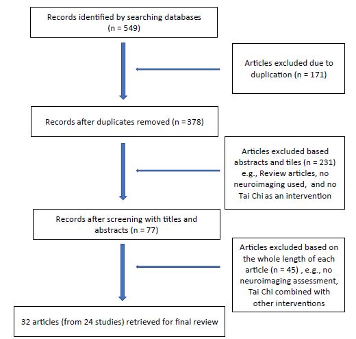
Figure 1: Flow chart of articles searched for analyses

Methods

Search Strategy

TC-related literature that investigated TC’s effects on morphological responses of the brain was searched. The following sources were included in the literature search process: Pubmed, Scopus, Medline (US National Library of Medicine), the Physiotherapy Evidence Database (PEDro), the Cochrane Controlled Trials Register (Cochrane Library), Cumulative Index of Nursing and Allied Health Literature (CINAHL), and the oversea English version of China National Knowledge Infrastructure (CNKI), up to May 2022. The search strategy used the following keywords and variations: Tai Ji, TC, TCh, TC Quan, Tai Ji Quan, Tai Ji Chuan, Chinese martial arts, Chinese fitness exercise, neuroimaging, functional near-infrared spectroscopy (FNIRS), magnetic resonance spectroscopy (MRS), magnetic resonance imaging (MRI), voxel-mirrored homotopic connectivity (VMHC), fMRI Regional homogeneity (ReHo), and fractional amplitude of low-frequency fluctuations (fALFF). Published reviews and all relevant studies and their reference lists were also reviewed manually in search for other pertinent publications.

Study Selection

Studies identified in the search were screened for inclusion. Articles that met the following criteria were selected: (1) studies investigating the effects of TC on brain response; (2) studies assessing the responses with FNIRS, sMRI, fMRI, MRS, and/or VHMC as the primary results; (3) participants were adults (age ≥18 years or older); (4) randomized control trials, single-group pre- and post- comparison, and cross-sectional studies comparing TC practitioners and non-practitioners; and (5) studies published in peer‐reviewed English or Chinese journals from last 10 years until May 2022.

Data Extraction

Initially, all identified articles were assessed independently by two reviewers by scanning the titles and abstracts to determine whether it met the predetermined eligibility criteria. When there was uncertainty or disagreement between the two reviewers, the lead author was involved in the discussion until a consensus decision was reached. Data extracted from each of these studies included study design, participant characteristics, exercise program characteristics, neuroimaging techniques, and morphological changes identified by these techniques.

Quality Assessment

The quality of all studies in this scoping review were assessed based on the type of study. Physiotherapy Evidence Database (PEDro) scale was used for randomized controlled trials [13]. Newcastle–Ottawa Scale (NOS) was conducted for cohort or cross-sectional studies [14].

Data Analysis

Study designs, participants’ characteristics, TC interventional parameters, neuroimaging, neurobehavior, and other functional assessments were all shown in Table 1. As the purpose of this review was to discuss TC’s effects on brain morphology changes in humans, neuroimaging data and their associations with neuroimaging changes in these included studies were extracted, summarized, and synthesized in Tables 2-4.

Table 1: Tai Chi Studies Assessed with Neuroimaging Techniques

Authors

Research

Design

Subjects Interventional Parameters Assessment instruments

Quality Assessment

           
Shen, Watkins, Kahathuduwa, et al (2022)[15] Single group Pre-and post- comparison 12 postmenopausal females with osteoarthritis, 40-50 yrs old Yang style 24 forms, 60 mins, 3/wk for 8 wks rs-fMRI, Pain Visual analog scale, WOMAC, plasma metabolites 6/10


Cui, Tao, Yin et al (2021)[16]


RCT 36 young healthy adults (18-25 y.o): 12 in each of 3 groups:  TC, brisk walking, and usual care (as control TC: Bafa Wubu,, 50-60 mins, 3/wk for 8 wks


rs-fMRI, Pain Visual analog scale, WOMAC, plasma metabolites


8/11


Kong, Huang, Liu et al, (2021)[17]


RCT IG – 24 with fibromyalgia

CG – 24 health subjects

All subjects> 21 years old


TC; Yang style, 10 forms

60 mins each, 2/wk for 12 wks


fMRI

More-odd shifting task for cognitive flexibility


6/11


Shen, Yin, Cui, et al, (2021)[18]


RCT IG – n=12, TC (Yang style, 24 forms)

CG – n=12, brisk walking

Young health adults (<25 y.o)


50-60 mins, 3/wk for 8 wks


fMRI and modified Flanker Test


7/11


Xu, Zimmerman, Lazae et al (2020) [19] Single group Pre-and post- comparison 16 adult patients with major depression 60 min each, 2/wk for 10 wks fMRI

Beck Depression Inventory

SF-36

5/10


Adcock, Fankhauser et al (2020) [20] RCT IG – n =15 (77±6.4 y.o.)

·                     CG – n=16 (70.9±5.0 y.o.)

·                     All healthy elderly individuals

IG – TC + dancing +step-based cognitive games at home; 3/wk, 30-40 min each for 16 wks

·                     CG – normal daily living

sMRI

Victoria Stroop test for cognition

·                     Trail Making test for psychomotor speed and executive function;

·                     Wechsler Memory Scale for memory

7/11**
Yue, Zou, Mei et al (2020) [21]


Yue, Yu, Zhang et al (2020) [22]

Cross-sectional 42 healthy elderly females

·                     IG – TC, n=20 (62.9±2.38 y.o.)

·                     CG – walking. n=22, (63.27±3.58 y.o.)

·                     90 min/each, 5/week, over 6 yrs


NA fMRI (VHMC) 7/10*
Chen et al (2020) [23] Cross-sectional TC – 22 (aged: 52.4 ±6.8; TC experience 14.6±8.6 y.o.;

Control – 18 (aged: 54.8 ±6.8)


All healthy adults

NA fMRI


Attention network test (ANT)


8/10*
Yang, Chen, Shao et al  (2020) [24] RCT 13 TC vs 13 Control


All healthy elderly individuals

TC: 45 min/each, 3/wk, for 8 wks

Yang style, 24-form

Control: routine and general daily activity

fNIRS


Flanker task test

8/11**
Cui, Yin, Lyu et al (2019) [25] RCT with 3 groups


36 young healthy college students (18-25 y.o.):

TC: 12

Brisk walking: 12

Control: 12


IG1  – TC: 8 hand movement techniques and 5 TC foot-works based on Yang-style TC.

IG2 – brisk walking

Both TC and brisk walking groups: 60 min/each, 3/wk for 8 wks

CG –  routine daily activities

sMRI and fMRI 9/11**
Tsang et al (2019) [26] Cross-sectional 8 practitioners (over 7 years of experience) and 8 non-practitioners

All: 60-75 y.o.

NA fNIRS 6/10*
Liu, Chen, Chen et al, (2019) [27]


Liu, Chen, Tu et al (2019) [28]

RCT IG1 – TC – n = 28

IG2 – BDJ – n = 29

IG3 – Stationary bike – n=27

CG – Health ed – n = 24

All (n = 108) 40-70 y.o.

60 min each, 5/wk for 12 wks sMRI and fMRI

Knee injury and osteoarthritis outcome score (KOOS)

7/11**
Xie, et al (2019) [29] Cross-sectional 32 ordinary vs 25 long-term (>5 years) Chen-style TC practitioners (all 60-70 y.o.) NA fNIRS 7/10*
Liu, Li, Liu, Sun et al (2020) [30]


Liu Li, Liu, Guo et al (2019) [31]

Cross-sectional 52 community-dwelling older adults (60-70 y.o.)

IG – TC – 26 (10 years or more TC experience)

VG – 26 (non-TC practitioners, but matched in physical activity level)


Both groups were asked to accomplish a sequential risk-taking task sMRI and fMRI

Beck Depression inventory

NEO five-factor inventory

Five facets mindfulness questionnaire,

Mindful Attention Awareness Scale Barratt Impulsiveness Scale

9/10*
Kong et al (2019) [32] RCT 21 patients with fibromyalgia (± 21 y.o.)

20 healthy matched participants

TC – 60 min/each, 2/wk, 12 wks, Yang style

CG – no TC experience

fMRI

Fibromyalgia  Impact questionnaire (FIQR)

6/11**
Wu, Tang, Goh et al (2018) [33] RCT Community living older adults (60-69 y.o.)

IG – n = 16

CG – n = 10

TC: 60 min each, 3/wk, 12 wks; Yang (10 min warm-up and 10 min cool down)

CG – telephone consultation biweekly without changing lifestyle

fMRI 7/11**
Port, Santaella et al (2018) [34] Cross-sectional 8 TC practitioners (>60 y.o.)


8 water aerobics practitioners (> 60 y.o.)

NA fMRI during attention time

·                     Stroop Word Color Task – SWCT

·                     Working memory with N-back task

7/10*
Zhou, Liao, Sreepada et al (2018) [35] Single group pre-and post- comparison 6 healthy elderly individuals (> 55 y.o.) TC: 60 min each, ³ 2/wk, 12 wks MRS

NAA: N-acetylaspartate;

Cr: creatine

PCr: phosphocreatine

5/10*
Liu, Wu, Li, Guo (2018) [36] Cross-sectional IG – TC, n = 26 (10.44±5.48 yrs TC experience) (65.19±2.30 y.o.)

CG – matched group (63.92±2.87 y.o.) (no TC experience)

NA fMRI 8/10*
Wei et al (2017) [37]


Wei, Dong, Yang et al (2014) [38]


Wei, Xu, Fan et al (2013) [39]

Cross-sectional IG – TC, n = 22 (aged: 52.4 ±6.8; TC experience 14.6±8.6 years;

CG – n = 18 (aged: 54.8 ±6.8)

NA sMRI and fMRI

Attention network test (ANT)


7/10*
Tao, Liu, Liu et al (2017) [40]


Tao, Chen, Liu et al (2017) [41]


Tao, Liu, Egorova et al (2016) [42]

RCT TC-21 (62.38±4.55 y.o.)

BDJ-15 (62.33±3.88 y.o.)

CG – 25 (59.76±4.83 y.o.)

IG1 – TC: 60min, 5/wk, 12 wks; Yang-style, 24-form

IG2 – BDJ: 60min, 5/wk, 12 wks;

CG – health education

sMRI and fMRI (fALFF)

Wechsler Memory Scale (WMS)


9/11**
Zheng et al (2015) [43] RCT Community dwellers

IG  – n = 17 (68.59 y.o.)

CG – n = 17 (71.65 y.o.)

IG – combined interventions: 3/week for 6 wks

1.                   cognitive training – 18 hrs

2.                   TC 18 hrs, Yang-24

3.                   Group counseling (6 90-min sessions)

CG – two 120-min health-related lectures

fMRI


Paired associative learning test (PALT) (to examine episodic memory)


Category Fluency test (CFT) (to examine speech production)


8/11**
Yin et al (2014) [44]


Li, Zhu, Yin, Niu et al (2014) [45]

RCT 45 older community-dwellers

IG – Multimodal intervention (TC + cognitive training + counseling  – 26

CG – 19

Multimodal intervention include

IG 1 – TC (Yang style, 24 form, 60 min each, 3/wk for 6wks) +

IG 2 – Cognitive training: (60 min each, 3/wk, for 6wks) +  counseling (90,im each, 1/wk for 6 wks)

CG – daily routine, 2 120-min healthcare education

MRI

MoCA

Associative Learning Test (ALT)

Digital Span forward and Backward Tasks

Category Fluency Test

Train Making Test

Social Support Rating Scale

Satisfaction with Life Scale

8/11**
Mortimer et al (2012) [46] RCT 120 community-living older adults (primary females) – 30 in each group

·                     IG1 – TC: 67.3±5.3 y.o., 19/30 females

·                     IG2 – Walking: 67.8±5.0 y.o., 19/30 females

·                     IG3 – Social: 67.9±6.5 y.o., 21/30 females

·                     CG – No interventions: 68.2±6.5, 21/30 females


3/week for 40 wks


IG1: TC: 50 mins – 20min warm-up, 20 min TC and 10 min cool-down), 3

IG2: Walking: 50 mins – 10 warm-up, 30 min brisk walking, and 10 min cool-down in a 400-meter track

IG3: Social interaction: 60 min for any topics

CG: No interventions

sMRI and fMRI 9/11**

CG: control group; fMRI: functional magnetic resonance imaging; IG: interventional group with Tai Chi as the intervention; MME: mini-mental status exam; MRS: magnetic resonance spectroscopy; NA: not applicable; RCT: randomized control trial; rsFC: resting state functional connectivity; sMRI: structural magnetic resonance imaging; TC: Tai Chi; y.o.: years old; min: minute(s); wk: week; wks: weeks; WOMAC: Western Ontario & McMaster Universities Osteoarthritis Index.*: Newcastle-Ottawa Scale assessment; **: PEDro scale assessment;

Results

Thirty-two articles from 24 studies were qualified for analysis (Table 1) [15-46]. There were 13 randomized control trials with 17 articles [16-18,20,24,25,27,28,32, 33,40-46], 8 cross-sectional studies with 12 articles [21-23,26,29-31,34,36-39], and 3 single group pre- and post- comparisons with 3 articles [15,19,35]. Among these 24 studies, 17 of them with 21 articles had elderly subjects who were 60 years and older [20-24,26,29-31,33-36,40-46], 4/24 studies with 8 articles had mixed age groups with subjects 21-70 years old [17,19,27,28,32,37-39], and 3 studies had healthy young subjects [16,18,25]. The majority of these studies used healthy subjects [15,16,18,20-31,33-46], but only four had subjects with a medical diagnosis of osteoarthritis [15], depression [19] or fibromyalgia [17,32]. Among 13 RCTs (17 articles), activities for the control groups or other intervention groups included normal daily activities [17,20,24,32], brisk walking [16,18,25,46], Baduanjin exercise [27,29,40-42], stationary biking [27, 29], health education [27,29,40-45] and social gathering interactions [46]. In 8 cross-sectional studies (12 articles), practitioners with 5 or more years of experiences in TC were compared with comparable subjects who walked every day [21, 22] or just non-TC practitioners [23,26,31,33,36-39].

With respect to quality assessment, 13 randomized controlled trials ranged from 6-9/11 in PEDro scale, indicating good to excellent studies [13]. Newcastle-Ottawa scale [14] showed 6-9 stars/10 in 8 cross-sectional studies (suggesting good to very good), and 5/10 in two sing-group cohort studies (indicating satisfactory), respectively.

Exercise Parameters Related to TC Effects on Neuroimaging Assessments

Based on 16 prospective studies (20 articles) in this review, including 13 randomized control trials (17 articles) and 3 pre-and post-intervention comparison studies (3 articles), the TC exercise parameters varied from study to study, but some of the parameters were commonly prescribed by many TC providers. The length of each TC practice session ranged from 30-40 minutes [20], 45 minutes [24], 50-60 minutes [15-19,27,28,32,33,35,40-46] with the most commonly used one being 50-60 minutes. The exercise frequencies were 2/week [17,19,32], 2-3/week [35], 3/week [15,16,18,20,24,25,33,43-46] and 5/week [27,28,40-42] with the most common one being 3 times a week. The duration for these studies varied from 6 weeks [43-45], 8 weeks [15,16,18,24,25], 10 weeks [19], 12 weeks [17,27,28,32,33,35,40-42], 16 weeks [20] to 40 weeks [46] with 12 weeks as the most commonly used. Put together, 60 minutes per session, 3 times a week for 12 weeks are the most commonly used TC parameters by TC researchers to investigate TC effects on the human brain. However, none of these 16 prospective studies did a follow-up after their TC interventions were completed, but the effects from a longer duration of TC practice are available from 8 cross-sectional studies (12 articles), in which subjects had been practicing TC for a minimum of 5 years and showed greater changes than the control groups [21-23,26,29-31,34,36-39].

TC Effects on Different Regions of Brain

As shown in Tables 1 and 2, TC practice is able to affect the whole brain by increasing total brain volume [46], the oxygenated hemoglobin (HbO2) in the motor cortex [29], and the white matter network connectivity locally and globally in the brain [21]. In each individual brain region, TC can affect many brain areas including the frontal, parietal, temporal, and occipital lobes, insula, basal ganglia, and cerebellum, among which the frontal lobe is the area that has been studied more than others (Table 2) [15-46].

Table 2: Effects of Tai Chi on Brain Assessed with Neuroimaging Techniques
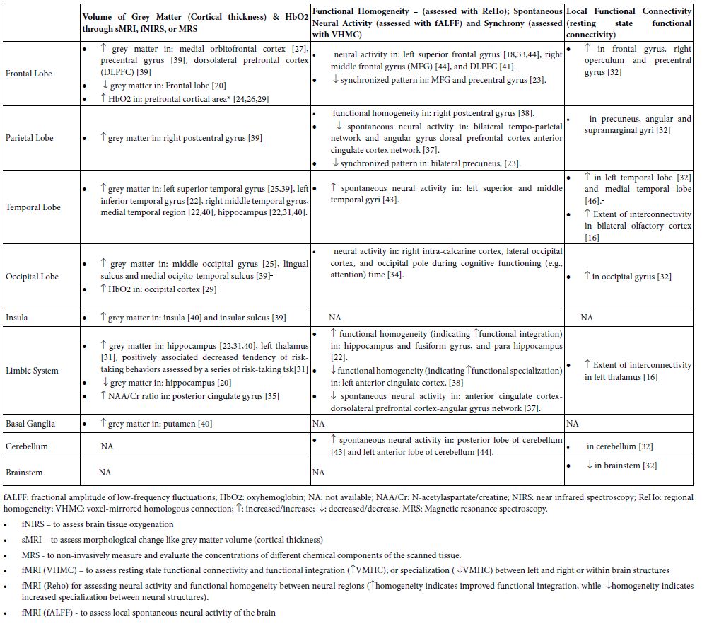
table 2

Frontal Lobe

TC practice is able to affect many regions of the frontal lobe (Table 2) by increasing 1) grey volume in the medial orbital prefrontal cortex [27], precentral gyrus [38], and dorsolateral prefrontal cortex (DLPFC) [39]; 2) oxyhemoglobin (HbO2) in the prefrontal cortical area [24,26,29]; and 3) neural activity in the left superior frontal gyrus [18,33;44]; right middle frontal gyrus (MFG) [44], and DLPFC [41]. The increased neural activity in the left superior frontal gyrus [33] and the dorsolateral prefrontal cortex [41] have been found to be associated positively and respectively with decreased error-making rates in switch/non-switch tasks [33], or with improved memory [41] in older community dwellers.

Further, increased connectivity was also reported locally in the frontal gyrus, right operculum, and precentral gyrus [32]. Decreased synchronized pattern in MFG and precentral gyrus as assessed by the VHMC technique was also found [23].

Parietal Lobe

The right postcentral gyrus shows increased cortical thickness and improved neural integration as indicated by increased functional homogeneity, which are positively associated with practitioners’ time length of TC experience and improvement of cognitive attention [39]. Increased functional connectivities were identified locally in precuneus, angular, and supramarginal gyri [32], and the middle frontal gyrus [25]. Decreased synchrony, as indicated by decreased VMHC, was seen in the precuneus, which is correlated with years of TC practice experience [23]

Temporal Lobe

Increased thickness of the cortex was identified in the left superior temporal gyrus [25,39], left inferior temporal gyrus [22], right middle temporal gyrus, and medial temporal region [22,40]. More spontaneous neural activities through fALFF assessment were detected in the left superior and middle temporal gyri [43]. The rsFC fMRI technique exerted greater functional connectivity locally in the left temporal lobe [32], medial temporal lobe [45], and bilateral primary olfactory cortex (in the lower temporal lobe) [16].

Occipital Lobe

Randomized controlled trials showed that 8-week TC practice can enhance the volume of grey matter in the middle occipital gyrus [25] and a 12-week TC program can increase resting state functional connectivity in the occipital gyrus [32]. Moreover, cross-sectional studies revealed that TC practitioners with a minimum of 5-year experience demonstrated 1) more volume of grey matter in the lingual sulcus and medial occipito-temporal sulcus [39], 2) increased HbO2 in occipital cortex [29], and 3) less activation of the right intra-calcarine cortex, lateral occipital cortex, and occipital pole during cognitive functioning (e.g., attention) time [34].

Insula and Limbic System

Grey matter can become thicker in the insula [39,40], hippocampus [20,31,40] and left thalamus [31]. Increase of the NAA/Cr (N-acetyl aspartate/creatine) ratio, a biomarker of brain functionality, was found in posterior cingulate gyrus [35]. Functional homogeneity was increased in the hippocampus, fusiform gyrus, and para-hippocampus [22], but decreased in the left ACC which is associated with years of TC experience [38]. Also increased extent of interconnectivity was identified within the left thalamus [16].

Basal Ganglia, Thalamus, Cerebellum, and Brainstem

After TC practice, neuroimaging techniques showed more grey matter in the putamen [40], and more spontaneous neural activity in the anterior and posterior lobes of the cerebellum [43,44], but decreased neural activity in brainstem [32].

Inter-regional Connectivity

As shown in Table 3, changes of inter-regional functional connectivity after TC practice were identified between different brain regions, among which the frontal lobe [15,19,25,27,30,32,37,42,45] has many more structures to make such inter-regional connections than any other neural lobes or brain areas, followed by the temporal lobe [19,42,45], limbic system [17,19,27,32,37], parietal lobe [19,25,37], insula [19], basal ganglia [30], and brainstem [32]. In the frontal lobe, the following structures, including superior, middle, and inferior frontal gyri, dorsolateral prefrontal cortex, medial prefrontal cortex, medial orbito-frontal cortex, and supplementary motor cortex, have either increased or decreased inter-regional connectivity with structures outside the frontal lobe (Table 3). In the temporal lobe and limbic system, the superior temporal gyrus, medial temporal lobe, hippocampus, cingulate gyrus, amygdala, and hypothalamus are involved in the TC-caused inter-regional connectivity. Further, the superior parietal lobule and angular gyrus in the parietal lobe, the ventral striatum in basal ganglia, the insula, and the ventral tegmentum and periaqueduct grey in the brainstem are involved as well (Table 3). Many of these connectivities are increased after TC exercise as seen in the left column of Table 3, while some of these connectivities are decreased in the right column of the table.

Table 3: Tai Chi Effects on Inter-Regional Resting-State Functional Connectivity

Increased Connectivity between

Decreased Connectivity between

  • Left superior frontal gyrus — posterior insula [19]
  • Left superior frontal gyrus — ventral striatum [30]
  • Left middle frontal gyrus — left superior parietal lobule [25]
  • Dorsolateral prefrontal cortex — anterior cingulate cortex [27,32,37]
  • Dorsolateral prefrontal cortex  — medial prefrontal cortex [32]
  • Dorsolateral prefrontal cortex  — angular gyrus [37]
  • Bilateral prefrontal cortex —– bilateral hippocampus [43]
  • Medial prefrontal cortex (mPFC) — anterior cingulate cortex [32]
  • Medial prefrontal cortex — medial temporal lobe [45]
  • Left superior parietal lobule — right posterior insula & left superior temporal gyrus – right posterior insula [22]
  • Superior temporal gyrus — right anterior cingulate cortex [22]
  • Right anterior insula — superior temporal gyrus [22]
  • Bilateral amygdala — medial prefrontal cortex (mPFC) [15]
  • Medial hypothalamus — thalamus and amygdala in fibromyalgia [17]
  • Medial orbito-frontal cortex — periaqueduct grey (PAG) and ventral tegmental area [27]
  • Dorsolateral prefrontal cortex  — supplementary motor area and anterior cingulate cortex (ACC) [28]
  • Dorsolateral prefrontal cortex — left thalamus and ventral striatum and right middle frontal gyrus [36]
  • Bilateral angular gyrus — dorsal prefrontal cortex — anterior cingulate cortex network [37]
  • Hippocampus/PAG  — ACC/mPFC [32]

Neuropsychological Functional Assessments

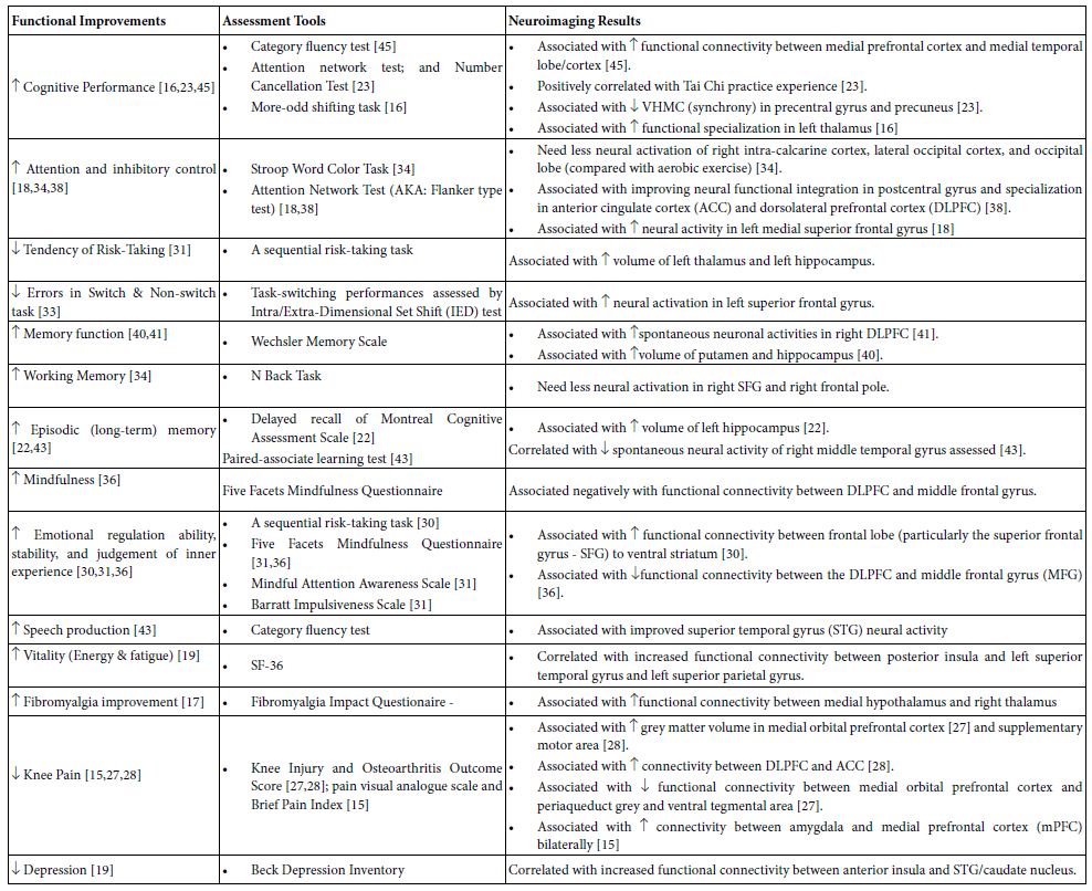
Neuropsychological functions were assessed in some studies and their associations with TC-caused neuroimaging changes in the brain were presented in Table 4. These studies used a variety of instruments to assess neuropsychological functions and to see how they may associate with neuroimaging changes after the TC intervention. As seen in Table 4, changes of inter-regional neural connectivity and cortical thickness (grey matter volume) were found to be associated with neuropsychological improvements such as cognition [16,18, 22, 23, 30, 31, 33, 34, 36, 38,40,41,43,45], vitality [19], depression [19], pain [15,27,28], and even the overall aspects of the fibromyalgia [17]. The cognition-related improvements may include general cognitive performance [16,23,45], attention and increased inhibitory control during attention [18,34,38], decreased errors in switch and non-switch task [33] and decreased tendency of risk-taking [31], different memory performance [22,34,40,41,43], mindfulness [36], emotional stability and judgement of inner experience [30,31.36], and speech production [43]. For examples, emotion regulation ability [30], cognitive performance [45], depression [19] and vitality [19] improvements are positively and respectively correlated with increased connectivities between the left superior frontal cortex and ventral striatum [30], between the medial prefrontal cortex and medial temporal lobe [45], between the right ACC and superior temporal gyrus [19], or between the right posterior insula and both the left superior temporal gyrus and left superior parietal gyrus [19]. Other the other hand, decreased tendency of risk-taking behavior [31] seems to parallel with increased cortical thickness in the hippocampus and thalamus [31]. In patients with knee arthritis, decreased knee pain was positively associated 1) with increased volume of grey matter in the medial orbitofrontal cortex [24] and supplementary motor cortex [28], 2) with increased connectivity between the dorsolateral prefrontal cortex (DLPFC) and anterior cingulate cortex (ACC) [27,28], but 3) with decreased connectivity in the medial orbito-frontal cortex and both periaqueduct grey and ventral tegmental area [27,28]. In addition, patients with fibromyalgia have significant improvement in functional, overall, and symptoms aspects as assessed with the Fibromyalgia Impact Questionnaire after 12-week TC practice. The improvement is positively correlated with the increased connectivity between the medial hypothalamus and the right thalamus [17].

Table 4: Function Improvements in Association with Neuroimaging Results after Tai Chi (TC) Practice

table 4

Discussion

Local Neural Responses in Different Brain Areas following TC Practice

In the frontal lobe, increased grey matter thickness, spontaneous neural activities, and/or local connectivities of the medial prefrontal cortex, dorsolateral prefrontal cortex (DLPFC), medial orbitofrontal cortex (mOFC), precentral gyrus, superior and middle frontal gyri, and frontal operculum were all reported in TC practitioners (Table 2). Functionally, the medial prefrontal cortex is responsible for memory and decision making [47], the mOFC is for goal-directed decision-making [48], DLPFC is for top-down attentional and cognitive control [49], the precentral gyrus is the primary center for voluntary motor movement [50], the superior frontal gyrus is for self-awareness of individual personality [50], and the right middle frontal gyrus is a convergence site of attention networks for higher order cognition and motor-related information processing [51]. The frontal operculum plays an important role in a network controlling the process of cognitive tasks [52]. With these together, TC practice might be able to improve neuropsychological and related neuromuscular behaviors such as memory, attention, cognitive attention, decision-making, sensorimotor integration, motor execution and control, and individual self-awareness through influence on these structures in the frontal lobe. How these different structures within the frontal lobe work in a coordinated way is not clear, but an improved functional specialization in frontal lobe structures might be an explanation. For instance, a VHMC study showed reduced homogeneity in the middle frontal gyrus and precentral gyrus [23], which may indicate an increased functional specialization of these two structures after TC practice.

In the parietal lobe, the right post-central gyrus is the only area that showed increased cortical thickness and spontaneous neural activity [38,39], while increased local connectivities were seen in the precuneus and inferior parietal lobule (angular and supramarginal gyri) [32]. Functional considerations of these structures, like the post-central gyrus for primary somatosensory processing, the precuneus for executive function, default network of self-consciousness, and mental imagery strategies and episodic memory retrieval [53], and inferior parietal lobule for recognition memory, language, and perception of emotion [54], may indicate that TC practice is able to improve sensory integration of higher-order motor execution, memory, emotion, and mental imagery strategies for motor actions [32,37,38]. Further, these TC studies [32,37,38] showed that TC experience is positively associated with increased neural activity in the right post-central gyrus [38], but negatively with decreased synchrony in bilateral precuneus as indicated by decreased VMHC value [23]. So, it is possible to assume that the longer one practices TC, the more improved general sensory is shown in certain parts (e.g., the left side) of the body, which might be through decreased synchrony between the left and right precuneus. However, whether and how such an assumption is holdable surely needs more future studies.

The temporal lobe and its superior, middle, and inferior temporal gyri showed increased volume of grey matter [22,25,39,40],spontaneous neural activity [43], and local functional connectivity [32] after TC practice. The same changes were also seen in the medial temporal lobe [22,40,45]. Comparatively the left temporal lobe showed more changes [22,25,32,39,43] than the right one (mainly the right middle temporal gyrus) [40]. The temporal lobe is functionally responsible for emotion, memory, and awareness of special sensation, and the left (dominant) side is more involved in language understanding [55,56]. Thus, it is understandable that TC could be a good exercise choice to improve emotion, memory, auditory and visual sensory, and even language perception. The medial temporal lobe that includes the hippocampus and para-hippocampus will be discussed in the paragraph of the limbic system below.

The occipital lobe showed increased oxyhemoglobin (HbO2), total hemoglobin (cHb) [29] and local connectivity [32] following TC intervention. Also, increased thickness of grey matter was noticed in the middle occipital gyrus [25], lingual sulcus, and medial occipito-temporal gyrus [42]. With consideration of functions of the occipital lobe [57], reactions to TC practice in the middle occipital gyrus and occipital cortex in general may hint that the occipital lobe could be morphologically changed to some extent [36,39], and likely participate in improving cognition and anti-memory decline [26,29], as well as improving spatial recognition and perception of objects [57] after TC practice.

In parts of the limbic system, studies demonstrated increased 1) grey matter in the insula [39,40], medial temporal gyrus [40], left thalamus [31], and hippocampus [22,31,40]; 2) increased spontaneous neural activity in hippocampus [22], fusiform gyrus [22], and para-hippocampus [22]; 3) increased extent of interconnectivity in left thalamus [16]; 4) increment of N-acetylaspartate/creatine ratio (indicating neuronal growth) in posterior cingulate gyrus [35]; and 5) increased functional specialization in anterior cingulate cortex [38]. On the other hand, decreased grey matter was found decreased [20] and decreased neural connectivity was identified in the anterior cingulate cortex and dorsolateral prefrontal cortex-angular gyrus network [37]. The insula has been regarded as a limbic system structure in respect to visceral sensation and autonomic control, but it also takes part in functions of pain processing, empathy, social cognition, attention, and decision making [60]. The medial temporal lobe (including hippocampus, para-hippocampus, and amygdala) is associated with emotion learning and behavior, as well as memory encoding, consolidation, storage, and retrieval [59,60], particularly for episodic and spatial memory [60]. A decrease in size of the posterior cingulate gyrus has been reported to play a role in cognition by influencing attentional focus by ‘tuning’ whole-brain metastability [61]. Additionally, the fusiform gyrus is responsible for object and face recognition [62] and semantic memory [63], and the thalamus is a relay hub for multiple sensory information and even memory [64,65]. However, reduction of grey matter in the hippocampus was recently reported when TC intervention was combined in an exercise program (30-40 minutes per total session) including TC-inspired exercise, dancing, and cognitive game [20], in which the TC time for each session was not long enough [20]. With respect to these studies about TC effects on the limbic system [20,22,31,35,39,40], generally speaking, they may indicate that TC is likely able to improve the practitioners’ emotion, memory, visceral and somatosensory capability, decision making, pain processing, and attention. TC may also be able to subsequently reduce the cognitive decline through the limbic system including the medial temporal lobe [66] if the TC practitioners have practiced over 5 years [22,31] or have practiced in longer duration (60 minutes each) on a daily basis [38].

In the basal ganglia and cerebellum, responses to TC exercise include increased grey matter in the putamen [40], and increased neural activity [43,44] and local connectivity in the cerebellum [32]. Literature has suggested that the putamen is functionally responsible for movement execution, working memory [67], and cognition [68]. Besides motor learning and coordination, the cerebellum is also for cognition and emotional processing (particularly the posterior cerebellar lobe) [69]. Injury to the cerebellum may cause cerebellar cognitive affective syndrome [70]. This information indicates that TC may improve motor execution, working memory and cognition through the putamen and cerebellum.

Inter-regional Connectivity after TC Practice

In addition to increased functional connectivity in each individual brain region (Table 2), there are also many inter-regional connectivities influenced by TC practice (Table 3, among which there are more increased such connectivities (see the left column of Table 3) than those decreased (see the right column of Table 3). We speculate that the increased inter-regional connectivities may suggest functional integration of different brain regions while the decreased inter-regional connectivities may indicate functional specialization of different regions. With consideration of morphological changes in each individual brain region after TC exercise, these individual brain regions might be able to functionally respond to TC practice differently depending on functions executed by these regions.

Functional Consideration

Given involved brain regions detailed in Tables 2 and 4, we can easily see that many of them are functionally and positively associated with neuropsychological behaviors. These behavior improvements were assessed with different neuropsychological instruments. For examples, following TC interventions, positive correlations were reported in improvement of 1) reduced risk-taking behavior as assessed by a series of risk-taking tasks [31]; 2) decreased error-making assessed with the switch-non-switch task [41]; 3) cognitive performance assessed by Category fluency test and attention network test [23,45]; 4) emotional regulation and stability assessed by Five Facets Mindfulness Questionnaire and Barratt Impulsiveness scale [31,36]; 5) depression by Beck Depression Inventory [19]; 6) vitality (energy and fatigue) assessed with SF-36 [19]; 7) memory as assessed by Wechsler Memory Scale [40,41] including working memory and episodic (long-term) memory [22,43]; and even knee pain as assessed by Knee Injury & Osteoarthritis Outcome Score [15,27,28]. These findings suggest that TC can be utilized not only as a physical but also a cognitive exercise, which may work by modulating both the local regional morphologies and inter-regional brain connectivity networks to improve the brain’s neural functions.

Study Limitations

There are several limitations that should be mentioned. First, due to the barrier to resources in non-English language, we were not able to access articles that were published in non-English literatures. Second, 10 out of 21 qualified studies are randomized control trials, but others include 8 cross-sectional studies, 2 single group pre- and post-TC comparisons, and 1 single-case report may reduce the level of evidence for this review study. Third, seed-based analysis is often used in resting state MRI in which a neural region of interest (ROI) is selected to determine how other regions interested by the investigators may correlate to the ROI. However, the obvious downside of such a method is that it depends on the investigators’ assumption for the ROI selection [71]. If a different ROI was picked, the involved brain regions might vary.

Conclusions

In the last 10 years, as neuroimaging techniques develop, more morphological changes of the human brain after TC practice have been investigated and identified in the frontal, temporal, parietal, and occipital lobes, insula, limbic system, basal ganglia, cerebellum, and brainstem, with the frontal and temporal lobes having more changes than other regions. These changes include increased cortical thickness or grey matter volume, altered local spontaneous neural activity, as well as changed inter-regional functional connectivities. Also, many of these changes are associated with improvements of many neuropsychological behaviors such as cognitive attention, memory, depression, vitality, risk-taking task, error-making tests, and even pain reduction. All of these imply that TC can be a great exercise program to improve the practitioners’ neural dysfunctions. However, so far, many brain structures have been found to be affected by TC exercise, but why and how only these structures are involved in response to TC practice are still not fully understood. Future studies are needed to assess how the structures are involved and how functionally some of these structures are integrated and/or specialized post-TC interventions.

Author Contributions

All five authors had substantially contributed to the conception and design of the article and interpreting the relevant literature. HL drafted the article and revised it critically with YS, SA, CN and CH for important intellectual content.

References

  1. Symms M, Jager HR, Schmierer K, Yousry TA (2004) A review of structural magnetic resonance neuroimaging. J Neurol Neurosurg Psychiatry 75: 1235-1244. [crossref]
  2. Ayaz H, Izzetoglu M, Izzetoglu K, Onaral B (2019) The use of functional near-infrared spectroscopy in neuroergonomics. In: H. Ayaz, F. Dehais: Neuroergonomics: The brain at Work and in Everyday Life. Academic Press 17-25.
  3. Liu H (2019) Tai Chi. In Chapter 39: Complementary and Integrative Therapies. (In: Umphred’s Neurological Rehabilitaion.7th Lazaro RT, Reina-Guerra SG, Quiben Q). Page 1071-1074. Mosby; Maryland Heights, Missouri.
  4. Lan C, Chen SY, Lai JS, Wong AMK (2013) Tai Chi Chuan in medicine and health promotion. Evid Based Complement Altern Med 2013: 502131. [crossref]
  5. Zhou D, Shepard RJ, Plyley MJ, Davis GM (1984) Cardiorespiratory and metabolic responses during TC Chuan exercise. Can J Appl Sport Sci 9: 7-10. [crossref]
  6. Liu H, Connors M, Grando V, Liu H (2012) Tai Chi intervention for older adults using assistive devices in a senior living community: A pilot study. Int J Ther Rehab 19: 136-142.
  7. Bonifonte P (2004) T’Ai Chi for Seniors: How to Gain Flexibility, Strength, and Inner Peace. New Page Book, Franklin Lakes, NJ.
  8. Liu H, Frank A (2010) Tai Chi as a Balance Improvement Exercise for Older Adults: A Systematic Review. J Geriat Phys Ther 33: 103-109. [crossref]
  9. Winser SJ, Tsang WWN, Krishnamurthy K, Kannan P (2018) Does Tai Chi improve balance and reduce falls incidence in neurological disorders? A systematic review and meta-analysis. Clin Rehabil 32: 1157-1168. [crossref]
  10. Glover GH (2011) Overview of functional magnetic resonance imaging. Neurosurg Clin N Am 22: 1333-1339. [crossref]
  11. Soares DP, Law M (2009) Magnetic resonance spectroscopy of the brain: review of metabolites and clinical applications. Clin Radiol 64: 12-21. [crossref]
  12. Wei J, Wei SB, Yang RX, Yang Lu, Yin Q, et al. (2018) Voxel-mirrored homotopic connectivity of resting state functional magnetic resonance imaging in blepharospasm. Front Psychol 9: 1620. [crossref]
  13. Verhagen AP, de Vet HCW, de Bie RA, kessels AG, Boers M, et al. (1998) The Delphi list: a criteria list for quality assessment of randomised clinical trials for conducting systematic reviews developed by Delphi consensus. J Clin Epidemiol 51: 1235-1241. [crossref]
  14. Wells G, Shea B, O’Connell D, et al. (2017) The Newcastle-Ottawa Scale (NOS) for assessing the quality of nonrandomized studies in meta-analysis.
  15. Shen CL, Watkins BA, Kahathuduwa C, Chyu MC, Zabet-Moghaddam M, et al. (2022) Tai Chi improves brain functional connectivity and plasma lysophosphatidylcholines in postmenopausal women with knee osteoarthritis: An exploratory pilot study. Front Med 8: 775344. [crossref]
  16. Cui L, Tao S, Yin HC, Shen QQ, Wang Y, et al. (2021) TCChuan Alters Brain Functional Network Plasticity and Promotes Cognitive Flexibility. Front Psychol 22: 665419. [crossref]
  17. Kong J, Huang YT, Liu J, Yu SY, Ming C, et al. (2021) Altered functional connectivity between hypothalamus and limbic system inMol Med 14: 17. [crossref]
  18. Shen QQ, Yin HC, Cui L, Zhang JY, Wang DL, et al. (2021) The Potential Advantages ofTCChuan in Promoting Inhibitory Control and Spontaneous Neural Activity in Young Adults. Front Behav Neurosci 15: 747733.
  19. Xu A, Zimmerman CS, Lazar SW, Ma Y, Kerr CE (2020) Distinct insular functional connectivity changes related to mood and fatigue improvements I major depression disorder following Tai Chi training: a pilot study. Front Integra Neurosci 14: 25. [crossref]
  20. Adcock M, Fankhauser M, Post J, Lutz K, Zizlsperger L, et al. (2020) Effects of an In-home multicomponent exergame training on physical functions, cognition, and brain volume of older adults: A randomized controlled trial. Front Med 6: 321.
  21. Yue C, Zou L, Mei J, Moore D, Herold F, et al. (2020) Tai Chi training evokes significant changes in brain white matter network in older women. Healthcare 8: 57.
  22. Yue C, Yu Q, Zhang Y, Herold F, Mei J, et al. (2020) Regular Tai Chi practice is associated with improved memory as well as structural and functional alterations of the hippocampus in the elderly. Front Aging Neurosci 12: 586770. [crossref]
  23. Chen LZ, Yuan X, Zhang Y, Zhang S, Zou L, et al. (2020) Brain functional specialization is enhanced among Tai Chi Chuan practitioners. Arch Phys Med Rehabil 101: 1176-1182. [crossref]
  24. Yang Y, Chen T, Shao M, Yan S, Yue GH, et al. (2020) Effects of Tai Chi Chuan on inhibitory control in elderly women: An fNIRs study. Front Human Neurosci 13: 476. [crossref]
  25. Cui L, Yin HC, Lvu SJ, Shen QQ, Wang Y, et al. (2019) Tai Chi Chuan vs General aerobic exercise in brain plasticity: a multimodal MRI study. Sci Rep 9: 17264.
  26. Tsang WWN, Chan KK, Cheng CN, Hu FSF, Mak CTK, et al. (2019) Tai Chi practice on prefrontal oxygenation levels in older adults: a pilot study. Complement Ther Med 42: 132-136. [crossref]
  27. Liu J, Chen L, Chen X, Hu K, Tu Y, et al. (2019) Modulatory effects of different exercise modalities on the functional connectivity of the periaqueductal grey and ventral tegmental area in patients with knee osteoarthritis: a randomized multimodal magnetic resonance imaging study. British J Anaesthesia 123: 506-518. [crossref]
  28. Liu J, Chen L, Tu Y, Chen X, Hu K, et al. (2019) Different exercise modalities relieve pain syndrome in patients with knee osteoarthritis and modulate the dorsolateral prefrontal cortex: A multiple mode MRI study. Brain Behav Immunity 82: 253-263. [crossref]
  29. Xie X, Zhang M, Huo C, Xu G, Li Z, et al. (2019) Tai Chi Chuan exercise related change in brain function as assessed by functio0nal near-infrared spectroscopy. Sci Rep 9: 13198. [crossref]
  30. Liu Z, Li L, Liu S, Sun Y, Li S, et al. (2020) Reduced feelings of regret and enhanced fronto-striated connectivity in elders with long-term Tai Chi experience. Soc Cogn Affect Neurosci 15: 861-873.
  31. Liu S, Li L, Liu Z, Guo X (2019) Long term Tai Chi experience promotes emotional stability and slows gray matter atrophy for elders. Front Psychol 10: 91. [crossref]
  32. Kong j, Wolcott E, Wang Z, Jorgenson K, Harvey WF, et al. (2019) Altered resting state functional connectivity of the cognitive control network in fibromyalgia and the modulation effect of intervention. Brain Imaging Behav 13: 482-492. [crossref]
  33. Wu MT, Tang PF, Goh JOS, Chou TL, Chang YK, et al. (2018) Task-switching performance improvements after Tai Chi Chuan training are associated with greater prefrontal activation in older adults. Front Aging Neurosci 10: 280. [crossref]
  34. Port AP, Santaella DF, Lacerda SS, Speciali DS, balardin JB, et al. (2018) Cognition and brain function in elderly Tai Chi practitioners: A case-control study. Explore 14: 352-356. [crossref]
  35. Zhou M, Liao H, Sreepada LP, Ladner JR, Balschi JA, et al. (2018) Tai Chi improves brain metabolism and muscular energetics in older adults. J Neuroimaging 28: 359-364. [crossref]
  36. Liu Z, Wu Y, Li L, Guo X (2018) Functional connectivity within the executive control network mediates the effects of long-term Tai Chi exercise on elders’ emotion regulation. Front Aging Neurosci 10: 315. [crossref]
  37. Wei GX, Gong XQ, Yang Z, Zuo XN (2017) Mind-body practice changes fractional amplitude of low frequency fluctuations in intrinsic control networks. Front Psychol 8: 1049. [crossref]
  38. Wei GX, Dong HM, Yang Z, Luo J, Zuo XN (2014) Tai Chi Chuan optimizes the functional organization of the intrinsic human brain architecture in older adults. Front Aging Neurosci 6: 74. [crossref]
  39. Wei GX, Xu T, Fan FM, Dong HM, Jiang LL, et al. (2013) Can Taichi reshape the brain? A brain morphometry study. PLosOne 8: e61038. [crossref]
  40. Tao J, Liu J, Liu W, Huang J, Xue X, et al. (2017) Tai Chi Chuan and Baduanjin increases grey matter volume in older adults: a brain imaging study. J Alzheimers Dis 60: 389-400. [crossref]
  41. Tao J, Chen X, Liu J, Egorova N, Xue X, et al. (2017) Tai Chi Chuan and Baduanjin mind-body training changes resting-state low-frequency fluctuations in the frontal lobe of older adults: A resting-state fMRI study. Front Human Neurosci 11: 514. [crossref]
  42. Tao J, Liu J, Egorova N, Chen X, Sun S, et al. (2016) Increased hippocampus-medial prefrontal cortex resting-state functional connectivity and memory function after Tai Chi Chuan practice in elder adults. Front Aging Neurosci 8: 25. [crossref]
  43. Zheng Z, Zhu X, Yin S, Wang B, Niu Y, et al. (2015) Combined cognitive-psychological-physical intervention induces reorganization of intrinsic functional brain architecture in older adults. Neural Plasticity 2015: 713104.
  44. Yin SF, Zhu XY, Li R, Niu Y, Wang B, et al. (2014) Intervention-induced enhancement in intrinsic brain activity in healthy older adults. Rep 4: 7309. [crossref]
  45. Li R, Zhu X, Yin S, Niu Y, Zheng Z, et al. (2014) Multimodal intervention in older adults improves resting-state functional connectivity between the medial prefrontal cortex and medial temporal lobe. Front Aging Neurosci 6: 39. [crossref]
  46. Mortimer JA, Ding D, Borenstein AR, Carli CD, Guo Q, et al. (2012) Changes in brain volume and cognition in a randomized trial of exercise and social interaction in a community-based sample of non-demented Chinese elders. J Alzheimer’s Dis 30: 757-766. [crossref]
  47. Euston DR, Gruber AJ, McNaughton BL (2012) The role of medial prefrontal cortex in memory and decision making. Neuron 76: 1057-1070. [crossref]
  48. Gourley SL, Zimmermann KS, Allen AG, Taylor GR (2016) The medial orbitofrontal cortex regulates sensitivity to outcome value. J Neurosci 36: 4600-4613. [crossref]
  49. Brosnan MB, Wiegand I (2017) The dorsolateral prefrontal cortex, a dynamic cortical area to enhance top-down attentional control. J Neurosci 37: 3445-3446. [crossref]
  50. Petrides M, Pandya DN. The Frontal Cortex. In: Mai JK, Paxinos G: The Human Nervous System (3rd). Academic Press, Cambridge, MA. 2012. Page 988-1011.
  51. Japee S, Holiday K, Satyshur MD, Mukai I, Ungerlieder LG (2015) A role of right middle frontal gyrus in reorienting of attention: a case study. Front Syst Neurosci 9: 23. [crossref]
  52. Higo T, Nars RB, Boorman ED, Buch ER, Rushworth MFS, et al. (2011) Distributed and causal influence of frontal operculum in task control. Proc Natl Acad Sci USA 108: 4230-4235. [crossref]
  53. Cavanna AE, Trimble MT (2006) The precuneus: a review of its functional anatomy and behavioral correlates. Brain 129: 563-584. [crossref]
  54. O’Connor AR, Han S, Dobbins IG (2010) The inferior parietal lobule and recognition memory: Expectancy violation or successful retrieval? J Neurosci 30: 2924-2934. [crossref]
  55. Kiernan JA (2012) Anatomy of the temporal lobe. Epilepsy Res Treat 2012: 176157.
  56. Aversi-Ferreira TA, Tamaishi-Watanabe BH, Magri MPF, Ferreira RAGMFA (2019) Neuropsychology of the temporal lobe – Laria’s and contemporary conceptions. Dement Neuropsychol 13: 251-258. [crossref]
  57. Renier LA, Anurova I, De Volder AG, Carlson S, Vanmeter J, et al. (2010) Preserved functional specialization for spatial processing in the middle occipital gyrus of the early blind. Neuron 68: 138-148. [crossref]
  58. Uddin LQ, Nomi JS, Hebert-Seropian B, Ghaziri J, Boucher O (2017) Structures and function of the human insula. Clin. Neurophysiol 34: 300-306. [crossref]
  59. Squire LR, Stark CEL, Clark RE (2004) The medial temporal lobe. Annu Rev Neurosci 27: 279-306. [crossref]
  60. Cutsuridis V, Yoshida M (2017) Memory processes in medial temporal lobe: experimental, theoretical and computational approaches. Front Syst Neurosci 11: 00019.
  61. Leech R, Sharp DJ (2014) The role of the posterior cingulate cortex in cognition and disease. Brain 137: 12-32. [crossref]
  62. Weiner KS, Zilles K (2016) The anatomical and functional specialization of the fusiform gyrus. Neuropsychol 83: 48-62. [crossref]
  63. Ding JH, Chen K, Chen Y, Fang Y, Yang Q, et al. (2016) The left fusiform gyrus is a critical region contributing to the core behavioral profile of semantic dementia. Front Hum Neurosci 10: 215. [crossref]
  64. Fama R, Sullivan EV (2015) Thalamic structures and associated cognitive functions: relations with age and aging. Neurosci Biobehav Rev 54: 29-37. [crossref]
  65. Wolff M, Vann SD (2019) The cognitive thalamus as a gateway tp mental representations. J Neurosci 39: 3-14. [crossref]
  66. Ferreira LK, Diniz BS, Forlenza OV, Busatto GF, Zanetti MV, et al. (2011) Neurostructural predictors of Alzheimer’s disease: A meta-analysis of VBM studies. Neurogiol Aging 32: 1733-1741. [crossref]
  67. Arsalidou M, Duerden EG, Taylor MJ (2013) The center of the brain: Topographical model of motor, cognitive, affective, and somatosensory functions of the basal ganglia. Human Brain Mapp 34: 3031-3054. [crossref]
  68. Middleton FA, Strick PL (2002) Basal ganglia “projections” to the prefrontal cortex of the primate. Cereb Cortex 12: 926-935. [crossref]
  69. Stoodley CJ, Valera EM, Schmahmann JD (2012) Functional topography of the cerebellum for motor and cognitive tasks: an fMRI study. Neuroimage 59: 1560-1570. [crossref]
  70. Schmahmann JD (2004) Disorders of the cerebellum: ataxia, dysmetria of thought, and the cerebellar cognitive affective syndrome. J Neuropsychiat Clin Neurosci 16: 367-378. [crossref]
  71. Lee MH, Smyser CD, Shimony JS (2013) Resting-state fMRI: a review of methods and clinical applications. Am J Neuroradiol 34: 1866-1872. [crossref]