Monthly Archives: April 2022

Genital Cancer in Men and Women: A Review

DOI: 10.31038/IGOJ.2022511

Abstract

Considering the medical, economic and social importance of genital cancer occurring in men and in women, with a world-wide distribution, we have as objectives in this manuscript to contribute to the knowledge of the factors that constitute a risk for genital cancer and its principal consequences in the infected persons.

Keywords

Genital cancer, Carcinoma, Sarcoma, Metastasis, Breast and fallopian tubes cancer, Gynecology

Introduction

Cancer is any “malignant” tumor, including carcinoma and sarcoma. It arises from abnormal and uncontrolled division of cells that then invade and destroy the surrounding tissues. Spread of cancer cells (metastasis) may occur via the bloodstream or the lymphatic canal or across body cavities such as the pleura and peritoneal spaces, thus setting up secondary tumors at sites distant from the original tumors. Each individual primary tumor has its own pattern of local behavior and metastasis; for example, bone metastasis is very common in breast cancer, but very rare in the cancer of the ovary.

There are, probably, many causative factors some of which are known; for example, cigarette smoking is associated with lung cancer, radiation with some bone sarcoma and leukemia. Some tumors, such as retinoblastoma are inherited.

Treatment of cancer depends on the type of tumor, the site of the primary tumor, and the extent of spread. In a general context, according [1] “cancer is a leading cause of death worldwide, accounting for nearly 10 million deaths in 2020. The most common in 2020 (in terms of new cases of cancer) were: 1. Breast (2.26 million cases); 2. Lung (2.21 million cases); 3. Colon and rectum (1.93 million cases); 4. Prostate (1.41 million cases); 5. Skin (non-melanoma)”.

Between the cancer risks, there are some that can be changed and others like the age or family history can’t be changed.

For this manuscript we have selected: prostate cancer; ovarian cancer; breast cancer and fallopian tubes cancer.

(A) Considering that prostate cancer is a cancer that has strong effects on the health of men the risk factors are:

Age

About 6 in 10 cases of prostate cancer are found in men older than 65. On the other hand, it is rare in men younger than 40, but the chance of having prostate cancer rises rapidly after age 50;

Geography

The reasons for prostate cancer being most common in North America, northwestern Europe, Australia, and on Caribbean islands but less common in Asia, Africa, Central America, and South America, are not clear. But, it is possible that a more intensive screening for prostate cancer in some developed countries accounts for at least part of this difference, but other factors such as lifestyle differences (diet, etc.) are likely to be important as well.

Family History

Considering that prostate cancer seems to run in some families, which suggests that in some cases there may be an inherited or genetic factor. Still, most prostate cancers occur in men without a family history of it.

In a general context it has been observed that men having a father or brother with prostate cancer more than double a man’s risk of developing this disease. (The risk is higher for men who have a brother with the disease than for those who have a father with it.) The risk is much higher for men with several affected relatives, particularly if their relatives were young when the cancer was found.

Gene Changes

Several inherited gene changes (mutations) seem to raise prostate cancer risk, but they probably account for only a small percentage of cases overall. For example:

Inherited mutations of the BRCA1 or BRCA2 genes, which are linked to an increased risk of breast and ovarian cancers in some families, can also increase prostate cancer risk in men (especially mutations in BRCA2).

Men with Lynch syndrome (also known as hereditary non-polyposis colorectal cancer, or HNPCC), a condition caused by inherited gene changes, have an increased risk for a number of cancers, including prostate cancer.

According to [2] the most important known risk factors for prostate cancer are age, ethnicity, and inherited genetic variants.

(B) Ovarian cancer, fallopian tube cancer and peritoneum cancer.

Concerning genital cancer in women, we go to emphasize: ovarian epithelial cancer, peritoneum and fallopian tube cancer. Ovarian epithelial cancer, fallopian tube cancer, and primary peritoneal cancer are diseases in which malignant (cancer) cells form in the tissue covering the ovary (a pair of organs in the female reproductive system. They are in the pelvis, one on each side of the uterus (the hollow, pear-shaped organ where a fetus grows) or lining the fallopian tube.

  1. Ovarian epithelial cancer, fallopian tube cancer, and primary peritoneal cancer form in the same type of tissue and are treated the same way.
  2. Women who have a family history of ovarian cancer are at an increased risk of ovarian cancer.
  3. Some ovarian, fallopian tube and primary peritoneal cancers are caused by inherited gene mutations (changes).
  4. Cancer sometimes begins at the end of the fallopian tube near the ovary and spreads to the ovary.
  5. The peritoneum is the tissue that lines the abdominal wall and covers organs in the abdomen. Primary peritoneal cancer is cancer that forms in the peritoneum and has not spread there from another part of the body. Cancer sometimes begins in the peritoneum and spreads to the ovary.
  6. Cancer sometimes begins at the end of the fallopian tube near the ovary and spreads to the ovary.
  7. The peritoneum is the tissue that lines the abdominal wall and covers organs in the abdomen. Primary peritoneal cancer is cancer that forms in the peritoneum and has not spread there from another part of the body. Cancer sometimes begins in the peritoneum and spreads to the ovary.
  8. The fallopian tubes are a pair of long, slender tubes, one on each side of the uterus. Eggs pass from the ovaries, through the fallopian tubes, to the uterus. Cancer sometimes begins at the end of the fallopian tube near the ovary and spreads to the ovary.

Such how was referred back, inherited mutations of the BRCA1 or BRCA2 genes, are linked to an increased risk of breast and ovarian cancers in some families.

Conclusions

  1. We think that it was here demonstrated that genital cancer has an impact on the health of women and men.
  2. We hope that the diminution of the attention that was given to this disease, during the combat COVID-19, is corrected in a short/medium time.
  3. To combat cancer, it is necessary:
  • to have persons specialized for the different types of combat;
  • more research in this area.

to divulge to the public in general information on prevention and importance of the knowledge of the risk factors for cancer, a disease that can affect the human population of all ages.

References

  1. https://www.who.int/news-room/fact-sheets/detail/cancer
  2. Cheng HH, Nelson PS. Genetic risk factors for prostate cancer. UpToDate. 2019. Accessed at https://www.uptodate.com/contents/genetic-risk-factors-for-prostate-cancer on March 19, 2019. National Cancer Institute. Physician Data Query (PDQ).

Peripartum Cardiomyopathy (PPCM): Epidemiological, Clinical, Therapeutic, Evolutionary and Prognostic Aspects at the Amirou Boubacar Diallo National Teaching Hospital in Niamey (HNABD)

DOI: 10.31038/JCCP.2022514

Abstract

Title: Epidemiological, clinical, therapeutic, evolutionary and prognostic aspects of PPCM in the Internal Medicine and Cardiology department of the Amirou Boubacar Diallo National Hospital: Retrospective and prospective descriptive study about 64 cases.

Introduction: PPCM is a heart failure secondary to left ventricular systolic dysfunction with LVEF <45% or a fraction of shortening <30%, Occurred towards the end of pregnancy or in the months following childbirth (mainly the month following the childbirth) without any other identifiable cardiac cause it is a worldwide disease whose epidemiology varies considerably with a multifactorial etiology. The true incidence or prevalence of PPCM in Africa and many other populations remains unknown. The objective of the study was to bring out the epidemiological, clinical, therapeutic, evolutionary and promostic aspects of PPCM.

Methodology: This is a retrospective and prospective descriptive and analytical study on PPCM in the internal medicine and cardiology department of HNABD from January 1, 2017 to December 31, 2019 for the retrospective part and from January 1, 2020 as of December 31, 2020 for the prospective part.

Results: The prevalence of PPCM was 8.68% for heart failure; 3.82% compared to all heart disease and 2.06% compared to all entries. The average age is 28.2 years with extremes of 15 and 5 years. The clinical presentation was essentially that of global heart failure in 81.3% of cases. The alteration of the ejection fraction of the left ventricle was found in all patients, ie 100% divided between moderate in none, moderate in 53.1% and severe in 46.9% of cases. The treatment of PPCM in our series is that of heart failure. Four therapeutic measures were the basis of symptomatic treatment in all hospitalized patients: diet and diet regimen in 100% of them and ACE inhibitors in 95.3%. B blockers were used in 73.4% of patients outside the acute phase of the disease. Anticoagulants were used in 27 of the patients, i.e. 42.1% Digoxin in 16patients or 25% and antiplatelet drugs in 54.7% of patients. SLGT2 Inhibitors was used in 32.8% (21 patients). Bromocriptine was used 9.4% of the time. Thromboembolic complications are the most frequent, namely EP in 10.9% of cases, DVT 4.7%. Only one case of arrhythmia was found. Like other complications of pneumonia, pleurisy and severe anemia have been found. The mortality rate 17.2% related to cardiogenic shock in 36.36% of cases. The prognostic factors found are Young age, primiparity, 7 out of 11 deceased patients were between 15 and 20 years old and had only one parity, i.e. 63.6%, with no statistically significant link with respectively (P=0.09) and (P=0.18). 63 1% of deceased patients came from poor and underprivileged rural areas without any statistically significant link (P=0.66). 81.8% of deceased patients had a low socioeconomic level, with no statistically significant association between SSE and death (P=0.21). Nine (9) of the eleven (11) deceased patients had a severe alteration of LVEF on admission, ie 81.8%, with a very significant link (P=0.005).

Conclusion: Knowledge of the epidemiological aspects of PPCM is necessary for the optimization of patient care. SLGT2 Inhibitors and Bromocriptine seem effective in these cases.

Keywords

PPCM, Epidemiology, Clinical, Therapeutic, SLGT2 inhibitors, Bromocriptine, Niger

Introduction

Peripartum cardiomyopathy (PPCM) is a rare heart failure characterized as an idiopathic cardiomyopathy presenting with heart failure secondary to left ventricular systolic dysfunction affecting women in late pregnancy or in the months following childbirth [1-3]. This is a diagnosis of exclusion.

Its incidence is estimated at 1/3000-4000 births [4]. The highest incidence in Africa is in the Sudano-Sahelian zone with a prevalence of 2.7 per 1000 pregnancies [5,6]. It is responsible for 10% of female heart disease in Niamey (Niger) [6]. Several factors seem to play a role in promoting hormonal changes during childbirth (fall in cardioprotective fetal estrogen levels, synthesis of cardiotoxic 16KDa-prolactin) [7]. The classic picture is that of heart failure (HF), which generally mimics the signs of a normal pregnancy, often leading to delayed diagnosis and avoidable complications [8]. Transthoracic echocardiography is the key examination, making it possible to confirm the diagnosis, eliminate differential diagnoses and monitor progress [7]. Medical management is similar to that of heart failure with reduced left ventricular ejection fraction of other etiologies, but adjustments during pregnancy are necessary to ensure fetal safety [8]. It is a serious pathology whose evolutionary potential is extremely rapid and totally unpredictable, with the possibility of sudden onset of refractory cardiogenic shock in the first 24 to 48 hours justifying treatment in a center with specialized cardiovascular resuscitation [2,7]. Complete recovery from PPCM is possible in half of the patients, while the other half will retain dilated cardiomyopathy responsible for more or less severe chronic heart failure [7]. Subsequent pregnancy carries a substantial risk of relapse and even death in the event of incomplete myocardial recovery. [8].

Given its frequency, its high morbidity, the absence of known etiology as well as the multiplicity of contributing factors; an update has been initiated.

Methods

This is a descriptive and analytical retro-prospective study which was spread over a period of 4 years (January 1, 2017 to December 31, 2019 for the retrospective part and from January 1, 2020 to December 31, 2020 for the prospective part) in the cardiology department of the National Amirou Boubacar Diallo (HNABD) hospital in Niamey.

Were included in the study, women regardless of their age, their race who presented heart failure (HF) between the eighth month of pregnancy and the first five months postpartum without etiology found and in whom dilated cardiomyopathy (DCM) was diagnosed on cardiac ultrasound. Not included in the study were women with onset of heart failure before the eighth month of pregnancy or after the first five months postpartum, women with known heart disease or any other cause of heart failure.

Data collection was carried out from hospitalization records and the consultation register using a survey sheet taking into account the epidemiological, clinical, paraclinical, therapeutic and evolutionary aspects during the study period. The variables studied included: demographic data and prenatal consultation diaries, cardiovascular risk factors, mode of onset or decompensation of heart failure, mode and course of delivery. The chronology of the signs of CI in relation to childbirth, and the data of the physical and paraclinical examination. Certain examinations were systematically requested such as chest X-ray, electrocardiogram, cardiac echo-Doppler, blood count, blood sugar, creatinine. D-dimers, cardiac enzymes and chest CT angiography were requested depending on the clinical and electrical context.

In addition to the absence of a cause of heart failure, the following ultrasound criteria were essential to retain the diagnosis of PPCM: the dilation of at least the left ventricle (DTDVG>52 mm) associated with left ventricular systolic dysfunction, i.e. say a lower left ventricular ejection fraction (LVEF) of 0.50 and/or a shortening fraction <30%.

Definition of Variables

Estimation of Socioeconomic Status

In our study we arbitrarily estimated the socio-economic level (SES) of our patients taking into account three parameters which are diet, physical work during pregnancy and level of education, according to the following scale:

Affluent

Patient having to eat a rich and varied diet regularly; exempted from intense physical work during pregnancy; secondary or higher school level.

Average

Patient regularly having a satiety diet, not exempted from intense physical work during pregnancy, primary school level or illiterate.

Low

Patient who does not regularly have enough food, subjected to intense physical work during pregnancy; illiterate.

Classification of LVEF according to the Latest ESC 2018 Guidelines: [7]

Preserved LVEF

This is an LVEF greater than 50%.

Moderately reduced LVEF

This is an LVEF between 40% and 49%.

Low LVEF

This is an LVEF below 40%.

Severely Altered LVEF

This is an LVEF <30%

High PRVG

Translated by an E / A ratio > 2.

Normal PRVG

Translated by an E/A ratio <1.

Favorable Evolution

It was defined by the remission of the symptoms and the relaxation of the patients.

Death

These are all patients with PMPC who died regardless of the cause of death.

We identified 91 suspected cases of PPCM. We rejected 27 patients who had not benefited from cardiac ultrasound. Thus we have a sample of 64 patients who met all our criteria.

Data Analysis

The data was entered, processed and analyzed on a computer with IBM SPSS statistics version 20 Data editor software after creating an input mask. The results were presented in the form of tables and graphs using the Office 2016 package (Word and Excel). The statistical test used in this study was the chi² with a degree of significance P < 0.05.

Limits of the study

– Some patients are seen after the acute phase, explaining the absence of certain signs or their low proportion;

– Financial difficulties preventing distant patients from coming for consultation and/or carrying out certain additional examinations;

– The lack of information in certain files;

Results

Epidemiological Aspects

Sixty-four cases of peripartum cardiomyopathy (PPCM) were recorded in 4 years, an average of 16 cases per year. The average age was 28.2 years (extremes of 15 and 55 years). The age group 15 and 20 was the majority (31.30%) (Figure 1). PPCM represented 1/356 births, its hospital prevalence was 2.06% (64 PPCM/3094 patients admitted), 3.82% of heart disease (64/1672), 8.68% of total CI (64/737). Unemployed women made up 90.6% of the sample. The majority of patients had unfavorable socioeconomic conditions (65.6%) (Figure 2). Multiparous women were the most represented with 38.80% of cases (Table 1). The average parity was 3.07 (extremes of 1 to 6).
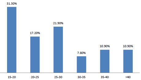
fig 1

Figure 1: Distribution of patients by age group
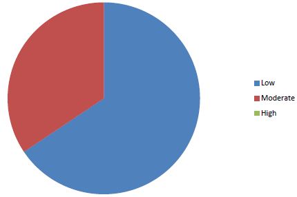
fig 2

Figure 2: Distribution of patients according to socio-economic status

Table 1: Data on epidemiological aspects

Paramètres

Numbers

Percentage (%)

Occupation

Housewife

-official

– Pupil/student

-shopkeeper/seamstress


58

3

2

1


90,60

4,70

3,10

1,60

Education level

-Non school

-University

-High school

-Middle School

-Primary


44

2

3

10

5


68,80

3,10

4,70

15,60

7,80

History

Personnal

-HBP

-Chirurgical

-Gynaeco-obsetrics

 Number of children;

1

2-3

4-5

6 and more

Abortion

0

1

2

3


6

10


24

15

11

14


60

3

0

1


9,40

15,60


37,5

23,4

17,2

21,9


93,7

4,7

0

1,6

Parity

-Grand multiparity

-Multiparity

-Pauciparity

-Primiparity


12

13

16

23


18,80

20

25

35,90

Twins

7

10,9

PNC

19

30

Type of birth

-Low way

-Caesarean section


58

6


90,60

9,40

NB prognosis

– living child

-Deceased children

-Breastfeeding


53

14

48


88,20

21,9

75

Risk factors

-High sodium diet

-Hot bath

– Taking medication during pregnancy

– Intense physical work during pregnancy


48

64

3


38


75

100

5


60

Family history

-HBP

-Diabetes

-Heart disease


12

6

1


18,80

9,40

1,60

PNC: Pre-natal Consult, HBP: High Blood Pressure, NB: New Born

Clinical Aspects

In 85.9% of cases, symptoms appeared postpartum (Figure 3). There was a delay in consultation in all patients with an average delay of 30 days (extreme: 06 to 120 days). Symptoms were dominated by exertional dyspnoea (100%) (Table 2). The decompensation was done on the mode of isolated left IC in 18.8% of the cases and on the mode of global IC in 81.2% of the cases. Table 2 summarizes the data of the clinical examination.
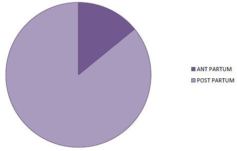
fig 3

Figure 3: Distribution of patients according to time to onset in relation to childbirth

Table 2: The clinical data of the patients

Parameters

Number (n)

Percentage (%)

Functional signs

-Dyspnea

-Cough

-Chest pain

-Hemoptysis


64

43

42

12


100

89,10

65,52

34,4

General signs

General condition (GC)

– altered

– passable

– Good

CONJUNCTIVES

-Colored

-Little colored

-Blades

Blood pressure

-Normal

-Low

-HighTempérature

– Feverish

-Non Feverish


29

25

10


32

15

17


33

21

10

22

42


45,3

39,1

15,6


50

23,4

26,6


51,60

32,80

15,60

34,4

65,5

PHYSICAL SIGNS

-Turgescence of the jugular veins

-Ascites

-Breath of TI

-MI Breath

– Peripheral edema

– Hepato-jugular reflux

-Hepatomegaly

-Crackling rales

– Deflected tip shock

-Gallop sound

-Tachycardia


10

24

3

35

61

52

57

53

45

29

59


15,60

37

4,7

56,30

95,30

81,30

89,10

82,80

70,30

45,30

92,20


Left heart failure

12

18,80

Global heart failure

52

81,30

Consultation times

-One week

-three weeks

-a month

-two months

-three months

-four months


19

7

22

10

3

3


30

10

35

15

5

5

MI: Mitral Insufficiency, TI: Tricuspid Insufficiency

X-ray and Electrocardiographic Signs

Cardiomegaly was found in all patients (100%) with an average cardiothoracic index of 0.70 (extreme 0.57 to 0.86).

Sinus tachycardia as well as left ventricular hypertrophy were noted in the respective proportions of 87.5% and 59.4%. Thirty-three patients (51.6%) had left atrial hypertrophy. Two patients had a conduction disorder (3.10%). In 29 cases (28.1%) there were nonspecific repolarization disorders associated with ventricular hypertrophy (Table 2).

Echocardiographic Data

LV dilation was noted in all patients with a mean end-diastolic diameter of 61.93 mm (range 54 to 76 mm). The left atrium was dilated in 43 patients with an average diameter of 46.17 mm (range 23.60 and 59). The right ventricle was dilated in 7 patients (10.93%). The average EF was 30.99% with extremes of 14 and 44.60%. The average of the FR was 12.85%. LV systolic dysfunction was severe in 43.60% of cases. All patients presented with global parietal hypokinesia. Cardiac Doppler echo had objectified a left intraventricular thrombus in 14 patients (21.9%). LV filling pressures were increased in 58 patients (90.6%). Pulmonary arterial hypertension was significant in 59.37% of cases with an average of 50.43mmHg (extremes: 23 and 74 mmHg). Pericardial effusion was noted in 19 patients (29.7%).

Treatment

A lifestyle and diet was recommended in 100% of our patients. Diuretics were the most used molecules; followed by CE inhibitors, then beta-blockers and digitalis with respectively 100%; 95.3%; 73.4% and 25%. Platelet antiaggregants were used in 54.7% of cases, anticoagulants in 42.2% of cases and AVKs in 21.9% of cases. SLGT2 Inhibitors were used in 21 CASES(32.8). Bromocriptine was only used in 9.4% of cases.

Evolution in Hospitalization

Complications occurred in 15 patients, i.e. 23.4%, including 10 cases of thromboembolic disease, including 7 cases (10.9%) of PE, 3 cases (4.7%) of deep vein thrombosis (DVT), 4 cases cardiogenic shock (6.3%) and 1 case of arrhythmia (1.6%). The evolution was favorable in 82.8% of our patients and death occurred in 17.2% of cases.

The average duration of hospitalization of our patients was 10 days (extreme 7 to 33 days). Fifty-three patients (82.8%) had found a clinical cure with disappearance of the signs. We noted 11 deaths (17.2%). 81.80% of deceased patients had severe LVEF impairment with a statistically significant link (P=0.034). Within our sample, in the multivariate analysis after logistic regression, it appears that only severe alteration of LVEF, low socio-economic status and primiparity were factors statistically associated with an unfavorable evolution with respectively P=0.034; P=0.044 and P=0.025.

Discussion

The hospital prevalence of PPCM was 2.06% in our study. In Africa, the prevalence of PPCM varies according to the studies [9,10]. In the West, the disease appears to be less frequent with an incidence of 1/3000 to 1/15000 [11]. These results confirm that PPCM is a pathology that is more prevalent in women of black origin [12]. Other associated factors are advanced maternal age [13]. In this study, the average parity was 3.07 (extremes: 1 and 6) with 82.93% multiparous. This testifies to the frequency of this condition in multiparous women [13]. In addition, 93.75% of our patients were from low socio-economic conditions. We agree with the authors that low socioeconomic status is also a risk factor for PPCM [14].

In total, maternal age over 30 years, multiparity, unfavorable socio-economic conditions are the risk factors for PPCM found in this study. Other factors such as the notion of chronic hypertension and prolonged use of tocolytics, twin pregnancies cannot be formally retained in this study [13].

We have observed a great delay in consultation among our patients. Dyspnea on exertion was the main symptom with an advanced stage (NYHA classification). The same observations were reported in the African literature [10,14]. These are patients in reality in whom the symptoms start earlier but ignorance and poverty would be the causes of delays in consultation and most of the patients are found in a table of global IC with a state of anasarca. The women considered the edema of the limbs as a normal fact linked to a pregnancy and it was in view of the increasing intensity of the dyspnea that the majority had consulted. The other symptoms found were precordialgia and cough. Precordial pain ranged from simple precordial tingling to angina-like pain with chest tightness. Their frequency varies according to the authors [10,11,14]. These chest pains associated with coughing pose a real diagnostic problem because they can raise the suspicion of a pulmonary embolism. In all cases, the patients are sufficiently put on anticoagulants at a curative dose. Tachycardia with a galloping sound, systolic murmur of mitral insufficiency and crackles were the most frequent auscultatory data found in our patients. Several authors have reported these same physical examination data but at widely varying rates [10,14]. These statistical disparities are explained by the subjective nature of clinical examinations, hence the need for paraclinical examinations. Cardiomegaly was noted in 100% of cases in this study. Cardiomegaly is constant in heart failure but remains non-specific [4]. This is an essential element in our regions where cardiac ultrasound is rare and inaccessible to the population.

On the EKG, serious arrhythmias are reported in the literature. Ferrière out of 11 observations noted 1 case of ventricular tachycardia [15]. It is a ventricular tachycardia detected by Holter ECG recording. Sinus tachycardia, LVH and nonspecific repolarization disorders are frequently found electrical abnormalities [10,14].

Faced with a recent woman who complained of dyspnea, the discovery of cardiomegaly associated with sinus tachycardia and LVH should lead to the PPCM being withheld until proven otherwise. Cardiac ultrasound will only come to confirm the diagnosis and assess the impact and complications.

Faced with a recent woman who complained of dyspnea, the discovery of cardiomegaly associated with sinus tachycardia and LVH should lead to the PPCM being withheld until proven otherwise. Cardiac ultrasound will only come to confirm the diagnosis and assess the impact and complications.

Echocardiographic signs are one of the criteria for defining PPCM and global parietal hypokinesia is the constant disorder found [15]. Cavitary dilatation as well as LV systolic dysfunction were severe in our patients. These are the consequences of the delay in consultation and diagnosis. PPCM is a highly emboligenic pathology [11,16]. The reasons mentioned are multiple: blood hypercoagulability during pregnancy [17], dilated cardiomyopathy which appears in a recent childbirth, reduced maternal mobility during the last months of pregnancy, compression of the IVC by the fetal mobile. All these reasons justify curative anticoagulant treatment in our patients.

The evolution of PPCM is unpredictable [18]. The inter-birth interval depends on the time taken for systolic function to return to normal. When heart failure persists beyond the sixth month after delivery, mortality is 28% in one year and 85% in 5 years [19]. Forms resistant to medical treatment represent 10% of cases [18]. When PPCM is cured, the risk of recurrence in a subsequent pregnancy cannot be excluded. The advice to be given to patients is therefore adapted to each case. Some elements are considered poor prognosis. These are of African origin, age greater than 30 years, a delay in the appearance of symptoms greater than 3 months, the persistence of clinical signs 6 months after the onset of the disease, an ICT greater than 0.6 and the characteristics of the left ventricle: insignificant dilation (LTDVD <55-60 mm), an ejection fraction < 30%, a shortening fraction less than 20% at the time of diagnosis [17,20]. If we consider these factors, we would say that all of our patients had a poor prognosis.

The prognosis of the disease is unpredictable. Many patients die despite the treatment, while others progress quite favorably and after 6 to 12 months of treatment, complete recovery occurs [2,17,20,21]. Between recovery and death, the evolution is that of chronic heart failure with DMC [22]. The obstetrical prognosis is poor. Heart failure occurs in 50 to 80% of cases in subsequent pregnancies, with mortality that can reach 60% [21,23]. Given a very high mortality rate during subsequent pregnancies, we agreed with our multiparous patients to opt for a contraindication to definitive pregnancy. Primiparas wishing to have another pregnancy are monitored and decisions will be made on a case-by-case basis.

Conclusion

Peripartum cardiomyopathy is a serious cardiac complication of pregnancy. It is common in Niger as in other black African countries. It occurs preferentially in the postpartum. The risk factors were: maternal age over 30, multiparity and unfavorable socioeconomic conditions. There was a significant delay in diagnosis. The clinical picture was that of global heart failure with significant dilation of the heart chambers and severe alterations in myocardial performance. SLGT2 Inhibitors and Bromocriptine added to the classical HF therapy seem effective.

References

  1. Karen Sliwa, Mark C Petrie, Peter van der Meer, Alexandre Mebazaa, Denise Hilfiker-Kleiner, et al. (2020) Clinical presentation and management,6-month outcomes in women with peripartum cardiomyopathy: an ESC EORP registry. European Heart Journal 1-10.
  2. Koenig.T, Hilfiker-Kleiner.D, Bauersachs.J (2018) Peripartum cardiomyopathy. Department of Cardiology and Angiology, Hannover Medical School, Hannover, Germany.
  3. Gibelin P (2020) Cardiomyopathie du péripartum. Presse Med Form.
  4. Moili M, Valenzano, Menada M, Bentivoglio G, Ferrero (2010) Péripartum cardiomyopathy. Arch Gynecol Obstet 281: 183-8
  5. Seronde M-F (2018) La cardiomyopathie du péri-partum.Arch Mal Coeur Vaiss Prat.
  6. Cenac A, Moumio OM, Develoux M, Soumane I, Lamothe F, Gaulier Y et al. (1985) COLL. Cardiopathie de l’adulte à Niamey (Niger), enquete épidemologique à propos de 162 cas, obsertion. Cardiol Trop 11: 125-133.
  7. Diarra A (1983) La myocardiopathie du post-partum. (Syndrome de Meadows). Thèse Med, Bamako 4: 93.
  8. Vanzetto G, Martin A, Bouvais H, Marliere S, Durand MJ (2018) Cardiovascular diseases during pregnancy. European Haert Journal 10: 1093
  9. Melinda B. Davis, MD, Zolt Arany, MD, PHD, Dennis M. McNamara et al. (2020) Peripartum Cardiomyopathy JACC State-of-the-Art Review.
  10. Niakara A, Belemwire S, Nebie L, Drabo Y (2000) Cardiomyopathie du post-partum de la femme noire africaine: Aspects épidémiologiques, cliniques et évolutifs de 32 cas. Cardiol Trop 26: 69-73.
  11. Mielniczuk LM, Williams L, Davis DR, et al. (2006) Frequency of péripartum cardiomyopathy. Am J Cardiol.;97(12): 1765-1768. [crossref]
  12. Gentry MB, Dias JK, Luis A, Patel R, Thornton J, Reed GL (2010) African-American women have a higher risk for developing peripartum cardiomyopathy. Journal of the American College of Cardiology.;55(7): 654-659. [crossref]
  13. Heider AL, Kuller JA, Strauss RA, Wells SR (1999) Peripartum cardiomyopathy: a review of the literature. Obstetrical and Gynecological Survey 54: 526-531. [crossref]
  14. Katibi I (2003) Peripartum cardiomyopathy in Nigeria. Hesp Med 64: 249.
  15. Ferriere M, Sacrez A, Bouhour J, et al. (1990) La myocardiopathie du péripartum: aspects actuels. Etude multicentrique: 11 observations. Arch Mal C’ur 83: 1563-1569.
  16. Bennani S, Loubaris M, Lahlou I, Badidi, Haddour N, Bouhouch R, Cheti M, Arharbi M (2003) Cardiomyopathie du péripartum révélée par l’ischémie aigue d’un membre inférieur. Annales de cardiologie et angéologie 52: 382-385.
  17. Walkira S, Carnério de carvalho M, Cleide K, Rossi E (2002) Pregnancy and Peripartum Cardiomyopathy: A Comparative and Prospective Study. Arq Bras Cardiol 79: 489-493. [crossref]
  18. Burban M (2002) Pregnancy and dilated or hypertrophic cardiomyopathies. Arch Mal C’urVaiss 95: 287-291.
  19. Silwa K, Skudick D, Candy G, Bergemann A, Hopley M, Sareli P (2002) The addition of pentoxifylline to conventional therapy improves outcome in patients with péripartum cardiomyopathy. The European Journal of Heart Faillure 4: 305-309.
  20. Chapa JB, Heiberger HB, Weinert L, DeCara J, Lang R, Hibbard U (2005) Prognostic value of echocardiography in peripartum cardiomyopathy. Obstet Gynecol 105: 1303-1308. [crossref]
  21. Goland S, Modi K, Bitar F, et al. (2009) Clinical profile and predictors of complications in peripartum cardiomyopathy. J Card Fail 15: 645-650. [crossref]
  22. Rifat K (1995) La cardiomyopahie du peripartum (CMPP) Méd Et Hyg 53: 2548-2555.
  23. Hamdoum L, Mouelhi C, Kouka H, et al. (1993) Cardiomyopathie du péripartum. Analyse de trois cas et revue de la littérature. Rev FR Gynécol Obstét 88: 273-275.

A Case of Chronic Schizophrenia with Emergent Dementia: Successful Medication Reduction and its Explication

DOI: 10.31038/JCRM.2022521

Abstract

The emergence of dementia during the treatment of schizophrenia is a problem that occurs in many patients. Despite the appearance of strong cautionary language on the product labels of antipsychotic drugs, advising clinicians to avoid the use of these medications in the setting of dementia, there is no specific guidance for the management of dementia in schizophrenia. Here, we report the case of successful antipsychotic drug reduction in a 63-year-old male with paranoia and severely impaired cognition. We explain possible explanations and the implications of this result.

Introduction

Throughout the history of medicine, there has been great confusion about the characterization and management of psychotic features in dementia, and/or cognitive deficits in psychosis. In 1893, the term dementia praecox was introduced as a specific disease entity by German psychiatrist Emil Kraepelin. Kraepelin’s label – which emphasized deficits in attention and memory in the context of delusions (paranoia), abnormal movement (catatonia), or disorganized thoughts (hebephrenia) – predicted a chronic and progressively deteriorating course [1,2]. When, in the 1950s, dementia praecox was eventually replaced by the word, schizophrenia, this expectation of cognitive decline continued.

Following the identification of premature mortality in elderly demented patients who participated in randomized controlled trials of antipsychotic drugs (a problem later confirmed by naturalistic and observational studies), the U.S. Food and Drug Administration attached Black Box Warnings to the labels of dopamine blocking drugs [3-5]. However, no guidance was issued with respect to the management of dementia which emerges in the course of treating chronic or recurring psychosis. The purpose of this case report is to present an example of successful medication reduction in the latter scenario, and to briefly consider the treatment implications of this result.

Case Report

Prior to our involvement with this case, a 63-year-old male with a longstanding history of schizophrenia and remitted alcoholism had undergone two recent medical admissions to the hospital: first, for the stabilization of starvation ketosis caused by the delusion that his food and medications were being poisoned; second, for acute delirium and ataxia which were likely precipitated by antipsychotic drug treatment [6-8]. A workup for dementia in the previous admission had included an MRI of the brain which displayed cortical atrophy, moderate ventriculomegaly, and (by our review) marked deep white matter hyperintensities, consistent with Fazekas stage three changes. The patient was discharged to his residential care home on one psychotropic medication (olanzapine 5 mg bid).

Four weeks later, the patient presented to the emergency room with the recurrence of failing hygiene and the belief that his food, beverages, and medication were being poisoned. He was admitted to the psychiatric unit, where initial examination was notable for poor grooming and thin body habitus. The patient was edentulous and appeared fifteen years older than his chronological age. The initial treatment team prescribed olanzapine 5 mg bid and added Vitamin D3 1000 IU qd. The patient initially declined all medications and demonstrated limited acceptance of fluids and food.

By hospital day #3, members of the nursing team noticed deficits in short-term and long-term memory, including an inability to recall the names of staff members; an inability to recall his age or birthdate; and an inability to recall what he had eaten for breakfast. Though he remained alert and calm, he was disoriented to date, identifying the year as 1934.

On hospital day #4, the authors of this case report assumed care of the patient concurrent with a rotation in treatment teams. We administered the St. Louis University Mental Status Examination, on which the patient scored 3 out of 30 possible points (oriented to date and year, but unable to perform any other elements of this screening test). This reflected severe deficits in attention, memory, spatial orientation (apraxia), language (aphasia), and executive functioning.

Based upon a reported history of several months of steady cognitive decline, severe enough to impact baseline social functioning, the authors prioritized a working diagnosis of dementia. Consultations were requested from physical therapy (recommending close supervision as the patient’s “path” would deviate due to poor attention) and speech therapy (recommending soft foods due to mild dysphagia). The nursing team attended to the patient’s hygienic needs: trimming nails; shampooing hair; assisting with clean clothing.

A revised dementia workup was undertaken, ruling out syphilis (RPR was negative), anemia (iron and ferritin levels were within normal limits), and nutritional deficiencies (normal levels of B12, B6, B1, folate, zinc, and copper; Vitamin D 25-oh was 38 ng/mL).

Olanzapine was discontinued due to its anticholinergic effects. Risperidone (1 mg bid) was prescribed to prevent neuroleptic withdrawal symptoms. Other treatments were revised to include memantine (5 mg at bedtime), B12 (1 mg daily), Vitamin D3 (increased to 4000 IU daily), selenium sulfide shampoo (for dry scalp) and chlorhexidine gluconate mouth wash (for halitosis/gum health).

By hospital day #5, the patient remained vague, confused, and hypersomnolent. However, his paranoid delusions subsided. He continued to be free of auditory or visual hallucinations, thought blocking, or other features of psychosis.

By day #8, he demonstrated consistent compliance with medical treatments. He was eating well and participated regularly in scheduled activities on the unit. The patient was oriented to self, date, and place, but was unaware of his cognitive limitations (anosognosia). Deficits remained in the domains of memory (inability to identify his diagnoses, recite the events which had led to the admission, identify his treatments, or recall the names of his doctors); speech (illogical mumbling, poor verbal fluency), and executive functioning (inability to organize instrumental activities of daily living, inability to attend to hygiene without supervision). We believe that the severity and persistence of these problems, which continued despite the resolution of paranoia, supported the ascendancy of dementia as the primary condition in this case.

With the consent of his conservator, the patient was discharged back to his residential care home due to his continuing inability to independently coordinate food, clothing, shelter, medical care, or finances. Follow-up was planned with psychiatry, primary care, and neurology – the latter, to confirm our working diagnosis of dementia due to multiple etiologies. Discharge medications included: risperidone 1 mg po twice a day, Vitamin D3 4000 IU po daily, B12 1 mg po daily, and memantine 5 mg po at bedtime.

Discussion

Upon admission, the patient exhibited signs of poor grooming and hygiene, as well as paranoia regarding his food and medications. As we were not convinced that his delusions were entirely attributable to the historical diagnosis of schizophrenia, we considered a broad differential etiology of cognitive and psychotic symptoms.

Past diagnoses had included alcohol dependence, from which the patient had been entirely in remission for at least one year. This history raised the specter of Wernicke Korsakoff syndrome as a contributing problem. Thiamine levels were within normal limits during the previous and recent admissions, ruling out Wernicke encephalopathy. The presence of Korsakoff dementia with psychotic features remained a possibility. Creutzfeldt-Jakob disease, which may emerge with or after Wernicke Korsakoff syndrome, was considered but ruled out, as our patient lacked ataxia, hallucinations, or myoclonus [9]. A recent brain scan via MRI had demonstrated ventriculomegaly, cortical atrophy, and deep white matter hyperintensities: the latter, consistent with small vessel disease. These findings suggested a strong component of vascular dementia. Risk factors in our patient included a history of smoking, alcoholism, and years of exposure to psychotropic medications.

With respect to schizophrenia, our patient had been placed under the conservatorship of a relative for approximately two years prior to our encounter, and concurrent with his placement into a residential care home. However, based upon collateral information, severe cognitive deficits had emerged only within recent months and had not been typical of the patient’s presentation.

Several epidemiological investigations have highlighted an increased risk of dementia in patients diagnosed with schizophrenia [10-12]. Like others, though, we are not convinced that cognitive decline is a necessary component of the schizophrenic condition [13,14]. Neither are we convinced that the syndrome known as schizophrenia – nor the dementia which may appear in its course – are correctly diagnosed in many patients. It is far from clear how often past cases of dementia praecox, or modern cases of schizophrenia, have reflected undiagnosed manifestations of infections affecting the central nervous system — such as viral encephalitis, tuberculosis, neuroborreliosis, neurosyphilis, or various hepatides; nutritional deficiencies; genetic anomalies; endocrine imbalances; autoimmune conditions (including paraneoplastic or non-paraneoplastic limbic encephalitis); seizure disorders; or unrecognized toxidromes [15-19].

Based upon similar environmental and behavioral risk factors, the organic precursors of cognitive decline in schizophrenia appear to be no different than those which occur in the general population [20]. A notable exception occurs with respect to the anatomic and physiologic effects of dopamine blocking drugs. A strong line of research evidence, involving autopsy and biomarker studies, links the old and new antipsychotic drugs to the neurodegenerative changes associated with Alzheimer’s disease [21-27]. Research has also implicated the same pharmaceuticals in cerebrovascular disease [28,29]. Causal mechanisms may be inferred from studies in lab animals and humans in which investigators have detected drug-induced mitochondrial disruptions, enhancement of oxidative stress, perturbations of the blood brain barrier, disturbances of metallochemistry, alterations in tau phosphorylation, dysregulation of microglia, and induction of insulin resistance [30-36].

We believe that the iatrogenic risk of drug-induced dementia is often overlooked in psychiatric patients, but particularly among those who have been diagnosed with schizophrenia. The present case demonstrates the benefits of realigning diagnosis and treatment in a middle-aged man with cognitive decline. We are mindful of the fact that other professionals have published positive results in which pharmaceutical dose reductions have benefitted patients with similar histories [37,38].

By continuously reorienting our patient; by attending to his physical and nutritional needs; by establishing a warm, caring rapport; and by reducing antipsychotic medication, we were able to facilitate substantial clinical improvement. While in the USA, governmental drug product labels advocate the cautious use of antipsychotics in the setting of dementia; our case implies that it is equally prudent to heed this advice in the treatment of dementia in schizophrenia.

References

  1. GE Berrios, R Luque, JM Villagran (2003) Schizophrenia: A conceptual history. International Journal of Psychology and Psychological Therapy 3: 111-140.
  2. CR Bowie, PD Harvey (2006) Cognitive deficits and functional outcome in schizophrenia. Neuropsychiatry and Disease Treatment 2: 531-536. [crossref]
  3. LS Schneider, KS Dagerman, P Insel (2005) Risk of death with atypical antipsychotic drug treatment for dementia: a meta-analysis of randomized placebo-controlled trials. JAMA 294: 1934-1943. [crossref]
  4. S Schneeweis, S Setoguchi, A Brookhart, et al. (2007) Risk of death with the use of conventional versus atypical antipsychotic drugs among elderly patients. CMAJ 176: 627-632. [crossref]
  5. J Yan (2018) FDA extends black box warning to all antipsychotics. Psychiatric News.
  6. D MacKintosh (2017) Olanzapine induced delirium – a ‘probable’ adverse drug reaction. Annals of Palliative Medicine 6: S257-S259. [crossref]
  7. JI Park (2017) Delirium associated with olanzapine use in the elderly. Psychogeriatrics 17: 142-143. [crossref]
  8. RC Sharma, A Aggarwal (2010) Delirium associated with olanzapine therapy in an elderly male with bipolar affective disorder. Psychiatry Investigation 7: 153-154. [crossref]
  9. T Nagashima, M Okawa, T Kitamoto, et al. (1999) Wernicke encephalopathy-like symptoms as an early manifestation of Creutzfeldt-Jakob disease in a chronic alcoholic. Journal of Neurological Sciences 163: 192-198. [crossref]
  10. Nicolas G, Beherec L, Hannequin D, Opolczynski G, Rotharmel M, et al. (2014) Dementia in middle-aged patients with schizophrenia. Journal of Alzheimer’s Disease 39: 809-822. [crossref]
  11. Ribe AR, Laursen TM, Charles M, Katon W, Fenger-Gron M, Davydow D, et al. (2015) Long-term risk of dementia in persons with schizophrenia: a Danish population-based cohort study. JAMA Psychiatry 72: 1095-1101. [crossref]
  12. Ku H, Lee EK, Lee MY, Kwon JW (2016) Higher prevalence of dementia in patients with schizophrenia: A nationwide population-based study. Asia-Pacific Psychiatry 8: 145-153. [crossref]
  13. Hyde TM, Nawroz S, Goldberg TE, Bigelow LB, Strong D, et al. (1994) Is there cognitive decline in schizophrenia? A cross-sectional study. British Journal of Psychiatry 164: 494-500. [crossref]
  14. Shah JN, Qureshi SU, Jawaid A, Schulz PE (2012) Is there evidence for late cognitive decline in chronic schizophrenia?. The Psychiatric Quarterly 83: 127-144. [crossref]
  15. J Castro, S Billick (2013) Psychiatric presentations/manifestations of medical illnesses. Psychiatric Quarterly 84: 351-362. [crossref]
  16. KA Welch, AJ Carson (2018) When psychiatric symptoms reflect medical conditions. Clinical Medicine 18: 80-87. [crossref]
  17. M Boyle (1990) Is schizophrenia what it was? A re-analysis of Kraepelin’s and Bleuler’s population. Journal of the History of the Behavioral Sciences 26: 323-333. [crossref]
  18. L Cai, YH Yang, L He, Chou KC (2016) Modulation of Cytokine Network in the Comorbidity of Schizophrenia and Tuberculosis. Current Topics in Medicinal Chemistry 16: 655-665. [crossref]
  19. AM Doherty, J Kelly, C McDonald, O’Dywer AM, Keane J, et al. (2013) A review of the interplay between tuberculosis and mental health. General Hospital Psychiatry 35: 398-406. [crossref]
  20. B Kirkpatrick, M Johnson, K McGuire, Fletcher RH (1986) Confounding and the dementia of schizophrenia. Psychiatry Research 19: 225-231. [crossref]
  21. MA Rapp, M Schnaider-Beeri, DP Purohit, Reichenberg A, et al. (2010) Cortical neuritic plaques and hippocampal neurofibrillary tangles are related to dementia severity in elderly schizophrenia patients. Schizophrenia Research 16: 2010. [crossref]
  22. NA Clarke, T Hartmann, EL Jones, Ballard CG, Francis PT, et al. (2011) Antipsychotic medication is associated with selective alterations in ventricular cerebrospinal fluid AB40 and tau in patients with intractable unipolar depression. International Journal of Geriatric Psychiatry 26: 1283-1291. [crossref]
  23. GB Frisoni, A Prestia, C Geroldi, Adorni A, Ghidoni R, et al. (2011) Alzheimer’s CSF markers in older schizophrenia patients. International Journal of Geriatric Psychiatry 26: 640-648. [crossref]
  24. V Albertini, L Benussi, A Paterlini, Glionna M, Prestia A, et al. (2012) Distinct cerebrospinal fluid amyloid-beta peptide signatures in cognitive decline associated with Alzheimer’s disease and schizophrenia. Electrophoresis 33: 3738-3744. [crossref]
  25. V Bloniecki, D Aarsland, K Blennow, Cummings J, Falahati F, et al. (2017) Effects of risperidone and galantamine treatment on Alzheimer’s disease biomarker levels in cerebrospinal fluid. Journal of Alzheimer’s Disease 57: 387-393. [crossref]
  26. S Mehta, ML Johnson, H Chen, Aparasu RR (2010) Risk of cerebrovascular adverse events in older adults using antipsychotic agents: a propensity-matched retrospective cohort study. Journal of Clinical Psychiatry 71: 689-698. [crossref]
  27. PH Hsieh, FY Hsiao, SS Gau, Gau CS (2013) Use of antipsychotics and risk of cerebrovascular events in schizophrenic patients: a nested case-control study. Journal of Clinical Psychopharmacology 33: 299-305. [crossref]
  28. MC Cotel, EM Lenartowicz, S Natesan, Modo MM, Cooper JD, et al. (2015) Microglial activation in the rat brain following chronic antipsychotic treatment at clinically relevant doses. European Neuropsychopharmacology 25: 2098-2107. [crossref]
  29. LE Laskaris, MA DiBiase, I Everall, Chana G, Christopoulos A, et al. (2016) Microglial activation and progressive brain changes in schizophrenia. British Journal of Pharmacology 173: 666-680. [crossref]
  30. CX Gong, S Shaikh, I Grundke-Iqbal, Iqbal K (1996) Inhibition of protein phosphatase-2B (calcineurin) activity towards Alzheimer abnormally phosphorylated tau by neuroleptics. Brain Research 741: 95-102. [crossref]
  31. C Burkhardt, JP Kelly, YH Lim, Filley CM, Parker WD Jr (1993) Neuroleptic medications inhibit complex I of the electron transport chain. Annals of Neurology 33: 512-517. [crossref]
  32. D Ben-Shachar, E Livne, I Spanier, Leenders KL, Youdim MB (1994) Typical and atypical neuroleptics induce alteration in blood-brain barrier and brain 59FeCl3 uptake. Journal of Neurochemistry 62: 1112-1118. [crossref]
  33. KJ Burghardt, B Seyoum, A Mallisho, Burghardt PR, Kowluru RA, et al. (2018) Atypical antipsychotics, insulin resistance, and weight; a meta-analysis of healthy volunteer studies. Progress in Neuropsychopharmacology and Biological Psychiatry 83: 55-63. [crossref]
  34. E Elmorsy, PA Smith (2015) Bioenergetic disruption of human micro-vascular endothelial cells by antipsychotics. Biochemical and Biophysical Research Communications 460: 857-862. [crossref]
  35. T Suzuki, H Uchida, KF Tanaka, Tomita M, Tsunoda K, et al. (2013) Reducing the dose of antipsychotic medications for those who had been treated with high-dose antipsychotic polypharmacy: an open study of dose reduction for chronic schizophrenia. International Clinical Psychopharmacology 18: 323-329. [crossref]
  36. T Suzuki, H Uchida, H Takeuchi, Nomura K, Tanabe A, et al. (2005) Simplifying psychotropic medication regimen into a single night dosage and reducing the dose for patients with chronic schizophrenia. Psychopharmacology 181: 566-575. [crossref]
  37. H Takeuchi, T Suzuki, G Remington, Bies RR, Abe T, et al. (2013) Effects of risperidone and olanzapine dose reduction on cognitive function in stable patients with schizophrenia: an open-label, randomized, controlled, pilot study. Schizophrenia Bulletin 39: 993-998. [crossref]
  38. Y Zhou, G Li, D Li, Cui H, Ning Y (2018) Dose reduction of risperidone and olanzapine can improve cognitive function and negative symptoms in stable schizophrenic patients: A single-blinded, 52-week randomized controlled study. Journal of Psychopharmacology 32: 524-532. [crossref]

Personal Things become Professional – Self-experience in Nursing

DOI: 10.31038/PSYJ.2022424

Abstract

Self-experience in nursing leads to an expansion of personal and professional scope. Emotionality, empathy and perception become, as it were an instrument, more precise and coherent to use. Self-awareness raises questions, as well as directing one’s gaze to wherever the entanglement is located. Where patterns are repeated over and over again or internal laws require compliance it becomes clear why, and what of it should remain the same or be changed. Self-experience raises awareness and reduces psychodynamic symptoms.

Keywords

Nursing, Self-awareness, Affect resonance training

Good nursing depends on the ability to experience childlike reactions without ceasing to be an adult. This ability can be strengthened and promoted [1]. This usually requires training, supervision and self-awareness. This enables openness, thoughtfulness, consideration, attention and creative thinking in a challenging work environment [2]. Through mechanisms such as bonding or identification, we get infected by the feelings or states of others. By letting in, we learn to empathize, to have compassion as well as setting boundaries and by that we tempt to develop a proper self. During the process of self-awareness, conflicts of one’s own are re-activated and are therefore, as Steinberger [3] writes “…subjects to constant self-control. The ability to mobilize one’s own conflicts or affects, thoughts and feelings from the past is the key to understand the patient, it is fundamental to be able to empathize”. Self-experience goes against the belief in speed. It takes time to develop its effectiveness, it needs a safe and clear space in which the knots and entanglements along the life story become visible. Usually no new memories are generated. Memories are often given a different meaning, a different reference. The network is being rebuilt. In the context of self-experience, Quinodoz emphasizes that apparently incompatible parts such as fear of being incomprehensible, being flooded or not to be able to hold up one’s own barriers, are conscientized and made accessible and useful. This is important in the context of nursing because personal and interpersonal experiences as well as being in a caretaking position, one’s own experiences and dynamics are always triggered. Past, damaging relationship patterns and experiences of patients are relived. The intensity depends on how severe current mental disorders are. Patients thereby reveal existential needs, abilities as well as anxieties that run the risk of making no sense whatsoever if they are not interpreted the right way. In a caretaking position to understand these states, behaviors or scenes and being able to deal with them without getting lost is a main goal. Especially people whose neediness, anger or even hate, anxieties and forlornness have taken almost unbearable measures, need an opposite who is able to deal with it. Understanding is therefore fundamental for a relationship-oriented work.

Relationship is a Highly Effective Factor

The course and intensity of mental disorders are best predictable if different sources of information are brought together. In order to maximize the chance of a prediction it requires a deep understanding, including all its complexities, of the past and the current condition of the patient. The most effective tool this regarding is the patient-nurse bonding [4]. The forming and perception of relationships have become a highly acknowledged and effective factor in the work with mentally ill people. For a long time already these bondings were seen as a significant element of the setting in the health care system. However, it was only through the systematic research of psychodynamic processes that it became evidently and undeniable how fundamental this sort of relationship is.

Nurses experience most of their interpersonal contacts in open social space, where unconscious processes are more difficult to identify due to the amount of information perceived simultaneously. The emotional impact of these interactions is considered rather unpredictable due to their complexity and the rapidity with which they unfold [5]. Them who are not aware of the power and presence of these unconscious processes, run the risk of being pushed into the restaged role of a spear carrier by the patients. Using basic psychoanalytic skills such as transference, countertransference, containment, as well as identifying more complicated psychodynamic processes such as “projective identification” [6-9] are a strong basis for relationship-oriented work. By transference it is meant, that the patient transfers old expectations and feelings to whom is offering a boding. The idea of using this to relieve suffering dates back to Freud [10]. Countertransference indicates what is triggered by the patient in the caretaking person [11]. Building on this, Melanie Klein describes that children project unbearable feelings onto their minder, sometimes to such an extent that this minder unconsciously identifies with these feelings and consciously assumes that these are his or her own feelings. Identification then means that the baby’s original fear became the mother’s fear, which in turn makes feel the baby her (inherited) fear. These dreadful conditions in the child thus appear to be confirmed (projective identification). Klein also emphasizes on the important observation that children need to learn and practice to build up an inner acceptance for loving and hating others at the same time. Upon this, Bion [12] was able to design his “Container/Contained-Model”: Containing suggests that at the moment of occurrence, the patient’s projections ought to be taken up kept by oneself at first, not to operate with them. It means to help the patient to slowly and step by step become aware of these transferences and projections and to keep them in their conscious mind. Children (and later on as adults as well) internalize feedbacks, reactions and correlations of and about their behavior and upon this they create their subjective world. What occurs, how this can be understood, which solutions become visible and tangible, depends on the inner possibilities the caregiver has to offer and the bigger the neediness is the stronger is the dependency. If these caregivers cannot offer enough support (containment), the child creates connections which could cause lifetime long psychological suffering if they are not comprehended accordingly.

Quality of Overwhelming

It is often hard for patients to accept help. The combination of urgent, existential need for help that is inextricably linked to the doubt whether they even are to be helped at all is an all too common life experience. In emotionally overwhelming situations, it can be difficult for patients to differentiate between inner and outer reality. If the subsequent projections are particularly violent or cruel, it is an important indication of the patient losing his or her ability to symbolize and feeling exceedingly overwhelmed. Under these circumstances memories, experiences and current emotions become fuzzy. Patients undergo such strong and intense states of emotion that these cannot be put into words. They act them out and present them in a scenic, interpersonal manner. This can be very challenging and even devastating for the whole team considering all groups of professionals. Patients suffering from severe personality disorders tend to activate multiple emotional states simultaneously. An inexperienced counterpart can be confused and overwhelmed by the situation. The important thing in this context is to understand the intensity and the features of these states of distress and confusion through self-experience in order to be able to ascertain what is going on in whom [13].

Aiyegbusi and Kelly remark the fact that the more unstable, unadjusted and critical the patients are experienced, the less positive feelings or empathy they receive. Without a compassionate and reflective attitude towards the patient there is neither the possibility of a delimitation from him nor a bonding with him. In order to keep oneself safe one might incline towards the idea that complex behavior can be treated (controlled) without having to understand the context or circumstances. It is then when in team meetings it is claimed that there are to be taken educational measures instead of looking for an appropriate treatment. If on the part of the caretaking persons no skills are acquired in order to deal with these grudges to be able to digest these attacking projections, interactions of clinical processes and procedures come into play which undermine the containment function. Succeeding the frightening sources – i.e. the patients – are rationalized and treated very strict and rigidly.

During or after severe breakdowns or in exceptional states of distress, patients are existentially dependent on being caught and handled by someone able to prevail his caring function throughout ardous emotional situations. Self-experience helps to remain self-reflective, insightful and to keep an intersubjective thinking [14,15]. Unprocessed trauma reactivates a vast force and is projected onto and into the nursing staff by patients. This way patients subconsciously tempt to reduce their own dreadful fear. They often experience the respective institution as unwelcoming, uninterested and uncomprehending. People with severe mental disorders are often not able to keep internal conditions (such as emotions or thoughts) to themselves neither can they talk about them. Instead, they tempt to act them out in an explicit manner. If there are many reproaches and complaints from the patient, they must be classified by the one encountering them. Do they concern the other person? Are they aimed at the institution? Or all of it together? A common reaction of defense when a situation becomes rampant, is to escape into structural and procedural processes in order to build a (seemingly) safer framework. However, this makes the team emotionally inaccessible and tears them apart from the patients. This can trigger feelings of isolation and create an inadequate, debilitating and sometimes even re-traumatizing environment for patients. In addition, due the fear of patients being harmful to themselves or to others, as well as through mistakes or misjudgments on behalf of the team, a conduct of blaming each other, paranoia and rigid defense-positions might arise, instead of enhancing openness and exchange. Another unpleasant side effect, which is often encouraged by schemes which do not lay their focus on psychodynamic, is the feeling of guilt on behalf of the patient. On behalf of the caretaking professionals, the before mentioned elements or this type of “work culture” are risky components which could cause Burnout and might lead to moral decline.

Aiygebusi and Kelly assume that being constantly confronted with people going through severe emotional pain can be experienced as afflicting and also frightening. Being exposed to this can also provoke intense inner states which, without professional coaching and schooling, can be perceived as very disruptive and thus have to be warded off appropriately. This can create the most challenging situation in nursing professions, being attacked, denigrated or threatened. It takes the development or strengthening of the inner capacity to endure on one hand, as well as to remain flexible on the other hand. For some patients contact or closeness is only to be established via pain and aggression. If such an encounter gets to emerge a caretakers’ unprocessed trauma, sadomasochistic alludings among the staff can be an attempt to gain back control over their own feelings. Accordingly, unconscious requests from patients in seriously severe conditions, to enter into their dynamics can generate powerful, sadomasochistic projections amongst the nursing staff. If these projections are recognized as such, they can provide important clues of how internalized relationships are organized within this patient and thus it can be reacted accordingly upon it.

From a psychoanalytical perspective, according to Berliner [16], masochistic aggression indicates suffering in order to be loved. That being so, suffering suggests a link towards someone else and thus implicates proximity. Suffering can also be regarded as a longing for autonomy. In this case the suffering person fantasizes to feel on a more equal level (‘I can take the pain’). If patients see no other way than to accept the sadism of a certain caregiver, they will merely take it. In order not to lose this caregiver, an inner arrangement is created to make sadism somehow bearable. On this matter masochism is the internalized sadism, which was previously experienced with another important person to this patient, which is now turn against himself. This is how abuse is interpreted as care and aggression as love. So what masochism means is to love a person who’s endowments are abuse, disinterest or suffering. Masochism remarks that experiences as of how it feels to be loved, to be paid attention to and to be taken care of, through the powerful mechanisms of introjection, identification and the emerging internalized legislators (superego), the sadism of the loved ones transformed into something that is experienced as one’s own and is charged libidinally.

It can be affirmed that the more deficiencies there are in a system, the more authoritarian and controlling it becomes trying to minimize the risks [17]. For people in big institutions in which the number of interactions are numerous and scope of power is incomprehensible, it is often difficult to maintain an adequate sense of individual identity within the working-context. The power of large-group-dynamics often causes feelings of vulnerability, pressure to adapt, being determined [18,19]. Institutionalized health systems, with their rejection of fear or anxiety are therefore a current example. According to Evans, it can be observed that institutions dealing with recovery shift these feelings of deficiency or inferiority into phantasies of omnipotence of how much or how quickly the risks of a breakdown, violence or suicide/murder can be prevented. The danger that “magical solutions” (e.g. ten therapy units or eight weeks of treatment for severe or chronic disorders) or that hatred and rejection will promote is particularly critical when those who are primarily confronted with the anxieties or dreadfulness of the patients, are suddenly made responsible for them – and this usually affects the caretaking professionals.

Autonomy and Good Practice

The capability of being good to others includes inner acceptance and the ability or insight of being dependent on others. To internalize this constructively is an important step towards autonomy and good practice for nursing professions. This is where the potential for stabilization, recovery and healing lies. In order to be able to perceive one’s own sensations in a retrospective and prospective manner, concepts as of emotion-response training (ART) are required, through which self-reflective behavior as well as reflecting on others can be practiced [20]. As for German-speaking areas, Johann Steinberger’s team has been working on this topic in the Viennese nursing school at the Otto Wagner Hospital for already ten years. Emotion-response training as a self-experience is an essential component training. Through this training, consisting of self-experience and two stages of supervision, there can be seen significant improvements in the capability to empathize and more openness towards actively forming liaisons. In addition, it was found that the emotion-response training enhances a clearer differentiation between perception and communication.

In nursing professions, it is worked, felt and communicated with as well as within in bodies. A great deal of the perception of what we are and what we aspire is shaped by the unconsciousness. In order to understand oneself and the world, the amount of information perceived has to be reduced categorized. People long for categories as well as for mutuality and separation in order to be able to be in proximity. Unconscious communication occurs in a quick and complex matter without a veil of adjustment. A bonding establishes rapidly and might in some cases be a riddle over years. Like the life-shaping rhythm of the heart and blood, there is a melody of the psyche within everyone which has an unstoppable and effective sound. We know how contagious yawning can be, but the question Hustvedt [21] proposes is “what about the contagiousness of emotions which are documented over a long time span and over various contexts and different parts of the world?” (P. 337f). Unconscious communication delivers and receives on all channels simultaneously. It is hard to let body and psyche/mind talk simultaneously. Often there is an inclination towards a certain position whilst communicating. In an attempt to use the biopsychosocial model in reference to communication, it can be seen the effort it takes to think of these different systems as correlating or as a conjunction.

Contiguity as well as language, in fact every getting into contact, unleashes memories. Concrete physical contact in a familiar setting which appeals to openness, generates a profound psychological response which, if it can be discussed, can enable important changes [22]. Concerning self-awareness, however, sensory touch needs the translation from emotion into language with all the possible interpretations as well as the awareness of the complexity and interweaving of these processes. The therapeutic slogan which unfolded upon this regard is that reflective, emotional, ideally open talks can be a meaningful and tangible reduction. Starting with self-experience in one’s own encounters and acquisitions and later on in the context of nursing, the developed consciousness is to be treated with special care. According to Quinodoz, language can open up a path to physical experiences on a very young psychological stage of development. Via the free associative speech, it is possible that traces of memory are forgotten. Memories, which strongly influence the body, indicate that no or insufficient words have been found for them. This can be understood as an indication the point where irritation has scratched into life history (there were no words yet) or how strongly it had to be repressed (the words must not be true). By fantasizing and remembering in the context of a secure relationship, emotions can be transformed into symbolic thinking and neuronal patterns can be (re) activated and changed. In this context, Kradin [23] refers to a connection between placebo-reactions and a person’s ability to bond. In his model, the physiological effect of a placebo presupposes an internalization and evaluation of interpersonal significant experiences that have been repressed and are therefore unconscious. In concrete terms, this means that an encounter or its particular manner triggers a related somatic reaction. Babies are unable to distinguish physical from psychological conditions at the beginning. Babies may feel physical pain and simultaneously have the feeling of being hated, or not to be able to distinguish frustration from the whole-body state of hunger. Hustvedt raises the question, whether a simple classic conditioning is also a form of memory. Where and how does a memory become a somatic reaction (and vice versa)? Where does desire, a thought, flow into the body? Where is a physical reaction ultimately a repeating narration of a past experience? Sensory as well as memorized memories or evaluations model and offer structure and framings. Thus memories are or become something like an opportunity to fulfill a life plan [24]. Outspoken memories help detecting formerly established connections which can subsequently be enhanced or discarded and rebuilt.

Language as a Medicine

Language is the preferred tool in self-experience. There are remarkable features of therapeutic speech, hearing and performing [25-27]. “Language as a remedy” is an effective platform upon which one is enabled to get closer to oneself. Lorenzer even considers therapeutic speaking to be equivalent to an “operation of speech” (p. 98). Language also represents a Zeitgeist. It has long been observed that human functions are engaged to the most modern metaphors. We find ourselves situated within processes, we possess memory and capacity and are in need for updates. In institutionalized healthcare systems particularly, there is a propensity towards a mechanized use of language, which can be useful due to its sophistication and reliability on one hand, but on the other hand it can be an obstacle for the development of relationships, for it tends to be rigid and excludes metaphors, imagination and intuition. Words such as reactions as a symbol emphasize on a certain reality, make other realities disappear and thus form personal, social or political processes [28]. Learning a ‘psychic technique’ and using it as a tool, makes it seem comprehensible, handy and applicable. This has major advantages. It provides security, it can be standardized, it is comprehensible, deductible and provable – it is “neat”. Neatness has high priority in hospitals. “The recognition of a shared involvement and the associated acknowledgement of a shared fear of what is to be discovered is reduced by the idea of having a right theory, a proper technique,” emphasizes Steinberger. However, he argues further, technology can be equated to a framework within which the respective idea of encounter and relationship is embedded. “Technology and metapsychological ideas build a frame of reference for the displacement and thus the description of the invisible. The reiteration and reduction of fear is based on the ability to abstract and generalize, and to find a broad purpose in language for dealing with our concerns”. Relationship and getting involved are terms which sound opposing towards technology and tools. Scarvaglieri takes into consideration that self-experience via psychotherapeutic speaking and being in a relationship, might untie mental processes and thereby effectuate changes. According to Quinodoz, language which transcends the rational and the informative, brings to light buried inner images and fantasies and lets physical, psychological and historical memories resurrect. Hence Steinberger adds, that the sensory impressions we selectively take in and react upon, are always placed alongside our psychological understanding as well as in response to clarifications and interpretations of feelings. He emphasizes: “This ability to comprehend develops as a result of labeling or by using language as a displacement employing metaphors (language), so as not to be at the surrendered by one’s own impulsiveness and the behavior resulting of it. In order to be “objective”, we have to be deeply “subjective” in our understanding. Without influence, there can be no understanding and no impact on the construction of reality”. Words determine something and help us to live interacting with each other. Depending on which words are spoken at what point, they can either hit with force or fizzle out in empty space. The use of metaphors, images and analogies helps to get in contact with the ability and capacity of symbolizing. This in turn strengthens and opens your own inner space.

It is important to keep on speaking (this includes moments of silence), even if it comes awkward or complicated situations between the participants. Only then suitable boundaries and proper containment can be established, as in two sides to a coin. It takes awareness of what is being spoken out and what is to be held back. It needs clarity about who is the one speaking and who is the one listening, and what this might represent and imply. There has to be paid attention to behaviors that communicate something in place of language. It takes practice in order for language to lead to freedom of action. In order to be able to communicate, it takes openness for whatever unfolds, as well as the augmenting the ability to understand and to be understood. Complicated situations might require having to soak in a lot and consequently react to it in appropriate dosages of words and actions (containing). So, it is important to raise awareness as well enhancing ones listening skills. Narrating and listening are subjects to continuous transformation. There is always the possibility to illuminate or recognize different elements of an experience. A rational, deliberate nuance can prevail on the front for a long time, until a symbolic or previously unconscious and unlinked aspect is suddenly perceived. It is beneficial to find a mutual exchange whereas there can be shared laugh even over serious, difficult or sinister topics. “Growth…”, Steinberger emphasizes (2019),”… also implies reflecting about feelings of inhibition, shame, ignorance”. He continues to emphasize: “…. it is also important to enhance the ability of accepting new thoughts as well as being able to stand ignorance”.

Nursing encounters are complex and take place in a wide variety of contexts. Deconstructing one’s own ideas and basic attitudes as well as one’s profession helps to achieve a more comprehensive experience in the work context. “To handle complex work situations reflectively, to face oneself as well as the others acceptingly, to recognize ambiguities of situations and the fact that there may exist different solutions to one problem, as well as withstanding the pressure of having to act in order generate solutions” are set as main achievements. Misconceived and unbearable fear provokes the search for a certainty in which there can be found classification (‘this is how it is’) and reiteration (‘it stays this way’), in order to be able to secure the perceived truth. However, if fear can be understood as an information carrier and thus establish a connection between all those involved in the arising situation, it might imply that by making use of the fear employing comprehension and classification, it could ultimately expand the leeway. Self-experience means allowing, accepting and respecting subjectivity, enjoying it and making use of it, for the emotional impact in the working environment can be perceived to be more complex. Thus the structures of the relationships tempt to incline more towards respect, empathy, authenticity, clarity and openness.

If in collective reflection in a familiar environment, openness, emotional self-revelation, criticism and discussion are constructively accepted, the participants will enhance their ability to engage in dialogue, and there will be more space for discussions and there will be less fear of asking questions or of being questioned [29]. The better the nurses’ own difficult experiences are treated, the less damaging interactions there are to expect. The more precisely the understanding and acceptance is of oneself, the more precisely it can be differentiated between one’s own reactions based on one’s own experiences and what ultimately has more to do with or results from the other person, as well as being understood. Every encounter and every relationship has its unique quality, which only results from the presence of the people taking part at that time. The psychoanalytic model offers a constructive framework in order to recognize and comprehend subjective and unconscious beliefs, roles or structures within oneself as well as in patients and thus being able to deal with them. So it aims to achieve acceptance, transformation and growth. Steinberger on this: “The influence on the patient unfolds from the realization the therapists conceive about themselves and through this being able to develop a different approach towards the patient”. If there is success creating a link between the current behavior of patients and their life history, disregarding its fierceness, more positive evaluations and subsequent more positive attitudes towards the patient can be observed. Following the paradigm of keeping out private matters out of professional matters, it can now be summarized that if private and professional issues are combined in a specific way, it leads to strengthening and preservation of relationships and actions in caring professions.

References

  1. Quinodoz D (2003) Words that touch. The International Journal of Psychoanalysis 84: 1469-1485.
  2. Evans M (2006) Making room for madness in mental health: the importance of analytically-informed supervision of nurses and other mental health professionals. Psychoanalytic Psychotherapy 20:16-29.
  3. Steinberger J (2019) Supervision als intersubjektiver, relationaler Prozess. Feedback – Zeitschrift für Gruppentherapie und Beratung 51-63.
  4. Aiyegbusi A, Kelly D (2015) ‘This is the pain I feel!’ Projection and emotional pain in the nurse-Patient relationship with people diagnosed with personality disorder in forensic and specialist personality disorder services: findings from a mixed methods study. Psychoanalytic Psychotherapy 29: 276-294.
  5. Aiyegbusi A (2009) The Psychodynamics of Forensik Mental Health Nusery. International Forum of Psychoanalysis 18: 30-36.
  6. Frank C (2007) Projektive Identifizierung: Ein Schlüsselkonzept der psychoanalytischen Therapie. Stuttgart: Klett-Cotta.
  7. Goretti GR (2008) Projektive Identifizierung: Kleins »Bemerkungen über einige schizoide Mechanismen« als Ausgangspunkt einer theoretischen Untersuchung des Konzepts. Internationale Psychoanalyse 3: 64-192.
  8. Klein M (1952) Some Theoretical Conclusions regarding the Emotional Life of the Infant. In: The Writings of Melanie Klein, Volume 8: Envy and Gratitude and Other Works. London: Hogarth Press, London
  9. O’Shaughnessy E (2003) Eine invasive projektive Identifizierung: Wie Patienten in Denken und Fühlen des Analytikers eindringen. Jahrbuch der Psychoanalyse 46: 9-28.
  10. Freud S, Breuer J (1895) Studien über Hysterie. Wien: Fischer.
  11. Segal H (2008) Gebrauch und Mißbrauch von Gegenübertragung. Jahrbuch der Psychoanalyse 59: 9-22.
  12. Bion W (1992) Lernen durch Erfahrung (5. Auflage). Wien: Suhrkamp.
  13. Bolm T (2009) Mentalisierungsbasierte Therapie (MBT) für Borderline-Störungen und chronifizierte Traumafolgen. Köln: Deutscher Ärzte Verlag.
  14. Steinberger J, Sieberth W, Zemann E (2013) P2: Ein mentalisierungsgestütztes Pädagogik-Konzept in der Ausbildung für Krankenpflegepersonal. Wien: Springer.
  15. Zemann, E, Gundaker C, Schossmaier G, Steinberger J, Totzauer G (2017) Affektresonanztraining ART© in der speziellen Grundausbildung in der psychiatrischen Gesundheits- und Krankenpflege am Otto-Wagner-Spital der Stadt Wien. Österreichische Pflegezeitschrift 26-28.
  16. Berliner B (1947) On Some Psychodynamics of Masochism. Psychoanalytic Quarterly.16: 459-471.
  17. Kernberg O (1998) Ideology, Conflict and Leadership in Groups and Organizations. Yale University Press.
  18. Platen A (1998) Die Großgruppen. Luzifer-Amor: Zeitschrift zur Geschichte der Psychoanalyse 11: 85-91.
  19. Schindler R, Spaller C, Wirnschimmel K, Tippe A, Lamatsch J etal. (2016). Das lebendige Gefüge der Gruppe: ausgewählte Schriften. Psychosozial-Verlag.
  20. Sieberth W, Steinberger J (2013) Beobachtung: Ein mentalisierungsgestütztes Pädagogikkonzept. In E. S, , C. Meyer, Praxis geschlechtersensibler und interkultureller Bildung. Wiesbaden: Springer.
  21. Hustvedt S (2018) Die Illusion der Gewissheit. Reinbek bei Hamburg: Rowohlt.
  22. King A (2011) Touch as Relational Affirmation. Attachment: New Directions in Psychotherapy and Relational Psychoanalys 5: 108-124
  23. Kradin R (2004) The placebo response complex. Journal of Analytical Psycholog 49: 617-634.
  24. Lorenzer A (1983) Sprsche, Lebenspraxis und szenisches Verstehen in der psychoanalytischen Therapie. Psyche 37: 97-115.
  25. Dorner K, Plog U, Teller C, Wendt F (2015) Irren ist menschlich. Lehrbuch der Psychiatrie und Psychotherapie. Köln: Psychiatrie Verlag.
  26. Ogden TH (2008) Träumerisches Sprechen. Internationale Psychoanalyse 3: 198-218.
  27. Scarvaglieri C (2011) Sprache als Symptom, Sprache als Arznei. Die linguistische Erfrorschung von Psychotherapie. Psychotherapie und Sozialwissenschaft 13: 37-58.
  28. Gentile J (2017) Tugging at the Umbilical Cord: Birtherism, Nativism, and the Plotline of Trump’s Delivery. Contemporary Psychoanalysis 53: 489-504.
  29. Ghedin S, Piredda C, Rosario D’addario M, Petricola F, Caldironi L, Marogna C (2014) Weaving Thoughts: Group Experience with Nurses in a Hospice. Journal of the American Psychoanalytic Association 62: NP21-NP23.

Association of Latin-American Ethnicity with an Increase in Weight in an HIV-Infected Outpatient Population

DOI: 10.31038/IDT.2022312

Abstract

Objective: To explore weight gain in regular practice involving naïve patients, those who continue the same treatment for at least 6 months or those who changed their antiretroviral treatment.

Methods: We performed a retrospective analysis of patients followed-up between 2013 and 2019. This study included 3 groups of participants (naïve patients, those who had been on viral suppression for more than 6 months, and those with a treatment change).

Results: 317 people living with HIV (PLHIV) participated. The proportion of participants in the overweight and obese categories increased over time, from 40 to 43% and from 9.46% to 12.43% respectively. Proportion of metabolic syndrome increased overtime from 3.79 to 6.22%. Stratification by both sex and ethnicity, showed the greatest weight gain among Latin male. Considering the risk factors for HIV infection, men that had sex with men (MSM) and heterosexual patients gained 2.03 (95% CI, 0.42-3.65; p=0.013) and 1.57 (95% CI 0.12-3.02; p=0.034) kg more than those who were former intravenous drug users (IDU). Patients taking boosted protease inhibitors (PI) experienced more weight gain 1.94 kg [95% CI, 0.13-3.75; p=0.036], than those taking integrase strand transfer inhibitors (INSTI). Globally and in decreasing order, rilpivirine [RPV] (+4 kg (IQR: -3.30,5.40]), Lopinavir/ritonavir [LPV/r] (+2.6 kg [IQR 2.40-3]) and Elvitegravir [EVG/c] (+2.20 kg [IQR 0-4.60]) were the “third” drugs most commonly associated with weight gain. Raltegravir [RAL] (-0.40 kg [IQR: -3.20, 3.40]) and nevirapine [NVP] (0.40 kg [-0.80, 0.50]) were the least. cART (combined antiretroviral treatment) based on tenofovir alafenamide (TAF) (5.87 kg [95% CI, 2.65-9.09; p<0.0001]; abacavir (ABV) [3.79 kg (95%CI, 0.83-6.75; p=0.012] and tenofovir disoproxil fumarate (TDF) [3.02 kg (95%CI, 0.24-5.80; p=0.033], gained more weight compared to monotherapy with boosted PI.

Conclusions: Our results suggest that there are demographic, HIV and treatment related contributors to weight gain in PLHIV. Latin-American ethnic race was associated with weight gain, particularly in male sex. We could not find any association of weight gain with sex, age or group of treatment (naïve, treatment continued for six months or change of it). We found boosted PI-based regimens, LPV/r, EVG/c and RPV, and TAF among nucleoside reverse transcriptase inhibitors (NRTI) pairs, associated with the greatest weight gain. We need to improve clinical attention to the maintenance of a healthy weight and implement lifestyle modifications and exercise not only for patients starting treatment but also for those with a long experience in antiretroviral treatment.

Keywords

Antiretroviral treatment, HIV, Latin American men, Weight change

Introduction

The current obesogenic environment is the result of an imbalance between caloric intake and energy expenditure that started in the 1960s-1970s [1]. Disruptive chemical sources have contributed to an inappropriate weight gain altering lipid homeostasis, fat accumulation, energy balance and modifying appetite and satiety regulation [2]. It is important to understand factors related to obesity in PLHIV (people living with HIV) the analysis and understanding of fat changes is gaining importance. Their relationship with HIV and cART (combined antiretroviral treatment), although not yet fully elucidated, seem to be a challenge in this era of long-life antiretroviral treatment. White adipose tissue composed of both innate and adapted immunity cells, is an extremely complex system that allows us to defend ourselves against foreign agents by identifying and eliminating viruses and other pathogens. This way, adipose tissue regulates processes against infection. A characteristic change in HIV infection is the shift towards a predominance of CD8+ T subpopulations which are particularly important in adipose tissue in the context of obesity [3,4]. Infiltration of CD8+ T cells is a necessary factor for recruiting macrophages which, in the context of obesity, produce TNF-α, IL-6 and IL-12 [5]. In obese people, the level of CD4 regulating T-cells (Tregs) is lower, making it easier the arrival of pro-inflammatory and macrophage T cells also [6]. Therefore, both adipose tissue related factors and metabolic dysfunction from HIV infection contribute to tissue inflammation and therefore, immune cell disfunction in obese PLHIV. There is a lot of data suggesting that integrase strand transfer inhibitor (INSTI) based antiretroviral treatment (ART) is associated with increased weight gain. In cell cultures, elvitegravir (EVG) has been shown to inhibit adipocyte differentiation and expression of genes that control adipogenesis. So, cART, modulated by features such as race, female sex and intestinal integrity would enhance the weight gain effect of a high-fat diet in PLHIV [7,8]. Cohort analyses have suggested that integrase inhibitors may increase weight gain, being higher in dolutegravir (DTV) and elvitegravir/cobicistat (EVG/c) than in raltegravir (RAL) treated patients. Regarding to the genetic aspects that influence weight gain, we have to mention the melanocortin-4 receptor (MC4R) gene and the fat mass and obesity-associated gene. Several studies show an statistically significant relationship between some mutations of these genes and an increased adiposity, higher if both mutations are present [9]. MCR4 plays a very important role in regulating energy homeostasis and intake. Deficiency of this receptor is associated with monogenic obesity. In vitro studies, a 64% inhibitory effect of dolutegravir (DTG) on the binding of radiolabeled melanocyte-stimulating hormone (MSH) to MC4R has been demonstrated [10]. Other studies seem to deny the possibility of a direct interference of the MC4R receptor by INSTI at therapeutic doses, inhibiting it only at much higher doses [11]. This paper purpose is to explore the weight gain process in our usual practice, involving three groups of patients, naïve patients who initiated treatment, a second group who were six months on it and a third one of patients changing treatment. We try to explore the possible clinical factors and factors related to the combination of antiretroviral treatment on the weigh changes of our patients in our current clinical practice.

Material and Methods

Design and Population of This Study

This retrospective observational study was carried out between January 2013 and January 2019 in a cohort of HIV-infected patients followed at Severo Ochoa University Hospital, in the southwest of Madrid (Leganés). Severo Ochoa University Hospital has a urban population of 180,000 inhabitants. The patients analyzed in this study are included in the COMESEM cohort, a larger cohort of HIV-1 infected patients followed at five different hospitals (Metropolitan Crown of southeastern Madrid, including Leganés, Alcorcón, Getafe, Móstoles and Alcalá de Henares hospitals). It is an open and dynamic cohort with data collected both in a retrospective and prospective way. The COMESEM cohort organization and functioning as well as the written informed consent of the patients were approved by the Clinical Research and Ethics Committee as required [12]. Patients gave their informed consent to be included in the cohort and their data to be used for this and other research purposes. They were verbally informed of the information that was going to be obtained in the study. From the 550 patients of the COMESEM cohort followed in our hospital, 317 whose weight and height had been recorded in the clinical history for at least 6 months were included in this analysis. We report 3-year data. Exclusion criteria were pregnancy and recent opportunistic infection. Three groups of patients were considered depending on their treatment status at the initial visit: group 1, patients who started antiretroviral treatment (naïve), group 2, those who had been on viral suppression for more than 6 months and continue with their treatment and finally, group 3, those whose treatment was changed in that visit (treatment switch). There was no subject on the new integrase inhibitor bictegravir.

Variables and Laboratory Measurements

Age, gender, ethnicity, clinical data including weight and height, the history of HIV infection and cART were collected in each clinical visit. These data included risk practice for HIV acquisition, smoking habits, alcohol consumption, methadone therapy, current CD4 cell count, CD4:CD8 ratio, current and previous therapy and HIV RNA level. No patient was a current illicit drug injection user. Blood samples were collected to analyze HIV related parameters (current CD4 cell count and current HIV viral load). As a rule, blood samples were obtained within one month of clinical visits.

Statistical Analysis

The study objective was to analyze the change of weight adjusted by ethnicity, gender, antiretroviral treatment, risk practice for HIV acquisition and other factors related to HIV infection. Description of variables was done showing frequencies and proportions for categorical and mean, median, and range for continuous variables respectively. A linear regression model was created with the change of weight considered as a continuous dependent variable. Analyses were processed using statistical package Stata/IC 14.2 for Mac (64-bit Intel). In order to estimate the predictive model of all the possible equations, we used user-command “all sets”. A p value less than 0.05 was considered statistically significant. For statistical calculation only differences at 2 years were considered, as data on weight gain was available for 88% as compared with only for 60.88% at 3 years.

Results

Population, Demographics, and Baseline Disease Characteristics

At basal visit, median body max index (BMI) was 24.87 kg/m2; 9.46% were obese (BMI6   ≥30 kg/m2) 40.06% overweight (BMI 25-29.9 kg/m2) and 50.47% normal (18.5-24.9kg/m2) or underweight (<18.5 kg/m2). Additional baseline weight and demographic data are summarized in Table 1, and baseline disease characteristics are summarized in Table 2. In the naïve patients, contrary to age and viral load which were significantly different, we could not find statistically significant differences with respect to BMI or immune system parameters.

Table 1: Baseline and demographic characteristics

Overall

Naïve On viral suppression for more than 6 months Treatment switch

p

N

317

7 61

249

Age (years)

0.0222*

Media (SD)

53.22 (9.72)

43.91 (11.86) 52.31 (9.13)

53.7 (9.69)

Median(Q1,Q3)

54.41(49.27-58.07)

50.29(32.31-53.95) 53.95(49.27-58.4)

54.54(49.28-58.4)

Sex

0.528

Men

217 (69.09%)

6 (85.71%) 40 (65.57%)

173 (69.48%)

Women

98 (30.91%)

1 (14.29%) 21 (34.43%)

76 (30.52%)

Ethnicity

0.228

Spanish

272 (85.74%)

6 (85.71%) 53 (86.89%)

215 (86.34%)

Black

15 (4.73%)

1 (14.29%) 4 (6.56%)

10 (4.02%)

Latin-American

27 (8.52%)

27 (8.52%)

24 (9.64%)

Asian

1 (0.32%)

1 (1.64%)

Sex and ethnicity

0.487

Spanish Men

200 (63.09%)

5 (71.43%) 36 (59.02%)

159 (63.86%)

Spanish Women

74 (23.34%)

1 (14.29%) 17 (27.87%)

56 (22.49%)

Black Men

9 (2.84%)

1 (14.29%) 2 (3.28%)

6 (2.41%)

Black Women

6 (1.89%)

0 2 (3.28%)

4 (1.61%)

Latin Men

9 (2.84%)

0 1 ( 1.64%)

8 (3.21%)

Latin Women.

18 (5.68%)

0 2 (3.28%)

16 (6.43%)

Weight

0.8151

Media (SD)

72.36 (14.40)

73.37 (10.08) 71.33 (15.72)

72.59 (14.20)

Median (Q1,Q3)

71.4 (62.6-80.8)

73.8 (64.2-80.8) 70.4 (59.4- 81.6)

72.1 (63-80.6)

Baseline BMI kg/m2

0.4752

Media (SD)

25.05 (4.25)

23.78 (3.22) 24.62 (4.31)

25.19 (4.27)

Median (Q1,Q3)

24.87(22.26-27.39)

23.99(22.04-24.98) 24.63(22.36-26.70)

25.14(22.21-27.8)

Underweight <18.5

21 (6.62%)

0 6 (9.84%)

15 (6.02%)

Normal Weight 18-5-24.99

139 (43.85%)

6 (85.71%) 26 (42.62%)

107 (42.97%)

Overweight 25-25.9

127 (40.06%)

1 (14.29%) 24 (39.34%)

102 (40.96%)

Obesity >30

30 (9.46%)

0 5 (8.20%)

25 (10.04%)

BMI: body mass index. *p<0.05 categorical variables are expressed as number of cases (percentage of the total); continuous variables are expressed as median (interquartile range) and media (standard deviation); Q1 percentile 25%; Q3 percentile 75%; SD standard deviation.

Table 2: Baseline disease characteristics

Overall

Naïve On viral suppression for more than 6 months Treatment switch

p

N

317

7 61

249

HIV-1 RNA, log10

copies/mL

0.0001*

Media (SD)

0.69 (1.31)

5.17 (0.40) 0.62 (0.98)

0.58 (1.17)

Median (Q1,Q3)

0 (0-1.4)

5.3 ( 4.7-5.53) 0 (0-1.4)

0 (0-0)

CD4 count, cells/µL

0.0983

Media (SD)

563.89 (333.86)

772 (545.47) 507.0328 (304.25)

571.96 ( 332.19)

Median (Q1,Q3)

502 (318-764)

839 (295-1106) 454 (30- 682) 520 (335-766)
CD4 count category, cells/µL

0.715

< 200

42 (13.25%)

1 (14.29%) 10 (16.39%)

31 (12.45%)

>200

275 (86.75%)

6 (85.71%) 51 (83.61%)

218 (87.55 %)

CD8 count, cells/µL

0.0737

Media (SD)

1027.43 (559.51)

1426.14 (841.83) 1098.87 (716.2)

998.72 (501.08)

Median (Q1,Q3)

923 (692-1281)

1300 (728-1670) 974 (800-1205)

900 (642-1278)

Ratio CD4/CD8

0.1399

Media (SD)

0.68 (0.55)

0.62 (0.54) 0.56 (0.43)

0.71 (0.57)

Median (Q1,Q3)

0.542(0.32-0.89)

0.35 (0.20-1.24) 0.53 (0.27-0.76)

0.56 (0.33-0.97)

Ratio CD4/CD8

(category)

0.541

<0.5

141 (44.48%)

4 (57.14%) 30 (49.18%)

107 (42.97%)

>0.5

176 (55.52%)

3 (42.86%) 31 (50.82%)

102 (57.03%)

Weight Gain in Participants Receiving Treatment

Although median weight gain was 1.0 kg (interquartile range [IQR], −1.4 , 3.8) at 36-month, the proportion of participants in overweight and obese BMI categories increased over time, from 40.06 to 43% and from 9.46 to 12.43% in overweight and obese BMI categories respectively (Figure 1). The proportion of participants that met the International Diabetes Federation definition of metabolic syndrome (central obesity (BMI ≥ 30) plus any two of the following: hypertriglyceridemia, low HDL-cholesterol, hypertension, or hyperglycemia) increased overtime from 3.79% to 6.22%. Participants gained respectively 2.59 (IQR 0.80, 3), 0.70 (IQR-1.80, 3.70) and 1 (IQR: -1.40, 3.80) Kg in group 1, 2 and 3 without statistically significant differences between groups 1 and 2 (-0.70 (95% CI: -5.17, 3.78), p=0.759) nor 1 and 3 (-0.50, (95% CI: -4.84, 3.83); p=0.819).

fig 1

Figure 1: Distribution of BMI over time

Risk Factors for Weight Gain

Considering the risk factors for HIV infection, men that had sex with men (MSM) and heterosexual patients gained 2.03 (95% CI 0.42, 3.65); p=0.013 and 1.57 (95% CI 0.12, 3.02); p=0.034, kg more than those who were former intravenous drug users (IDU) respectively (Table 3) Female sex and age > 50 years had not statistically significant correlations with weight gain (0.44, (95% CI: -0.78, 1.67) p=0.478 and -1.27 (95% CI: -3.20, 0.67) p=0.199 respectively. We further explored these findings by using longitudinal models to assess the relationship between sex, ethnicity, and weight gain. Latin-American gained significantly more weight (2.83 kg (95% CI, 0.80-4.85); p=0.006) than non-Latin-Americans participants (Figure 2). Stratification by both sex and race showed the greatest weight gain among Latin-male participants. Compared to Spanish men and to African women, they gained 5.37 (95% CI 1.66-9.08); p=0.005 and 7.18 (95% CI 2.20-12.35); p=0.007 Kg more respectively (Table 3).

Table 3: Risk factors associated with weight change

Variables

Difference in kg 95%CI

p

Risk group of infection (ref former IDU)
MSM

2.03

0.42-3.65

0.013*

HTX

1.57

0.12 3.02

0.034*

Ethnicity adjusted by sex (ref. Latin Men)
Spanish Men

-5.37

-9.08-(-1.66)

0.005**

Spanish Women

-5.17

-8.94-(-1.40)

0.007**

Black Men

-5.32

-10.29-(-.35)

0.032*

Black women

-7.18

-12.35-(-2.02)

0.007**

Latin women

-3.99

-8.19-0.20

0.062

cART type (ref. INSTI)
Boosted PI

1.94

0.13- 3.75

0.036*

NNRTI

1.45

-0.23-3.12

0.090

Backbone (Ref monotherapy with boosted PI)
TAF

5.87

2.65-9.09

<0.0001**

TDF

3.02

0.24-5.80

0.033*

ABV

3.79

0.83-6.75

0.012*

IDU: Intravenous drug user; MSM: Men who have sex with men; HTX: Sex among men and women; cART: Combined antiretroviral therapy; INSTI: Integrase Strand Transfer Inhibitor; PI protease inhibitor; NNRTI: Nonnucleoside reverse transcriptase inhibitor; NRTI: nucleoside reverse transcriptase inhibitor; TAF: Tenofovir Alafenamide Fumarato; TDF: Tenofovir Disoproxil Fumarate; ABV: Abacavir.
*p<0.05; **p<0.01.

 
fig 2

Figure 2: Change in weight in relation to ethnicity

Association of Antiretroviral Regimen Components with Weight Gain

The longitudinal model of weight gain and treatment showed that participants taking boosted PI experienced more weight gain (1.94 kg [95% CI, 0.13-3.75], p=0.036) than those taking INSTI. Weight gain was similar between the NNRTI and INSTI treatment groups (Table 3 and Figure 3). We studied the effect of changing treatment on weight changes with no difference between them. Those patients who changed from INSTI to NNRTI were those in whom we observed the greatest decrease, although not statistically significant: -5.39 (95% CI: -19.12, 7.66) p=0.399. Globally and in decreasing order, rilpivirine [RPV] (+4 kg (IQR: -3.30,5.40]), Lopinavir/ritonavir [LPV/r] (+2.6 kg (IQR 2.40-3) and Elvitegravir [EVG/c] (+2.20 kg (IQR 0-4.60) were the most commonly associated with weight gain, whilst raltegravir [RAL] (-0.40 kg (IQR: -3.20, 3.40) and nevirapine [NVP] (0.40 kg (-0.80, 0.50) were the least (Figure 4). We assessed the association between weight gain and the specific INSTI used. Participants taking EVG/c or DTG demonstrated greater weight gain than those taking RAL (3.00 (95% CI, 0.97, 5.07); p=0.004) and 1.89 (95% CI: -0.034, 3.82); p=0.054) kg respectively. Among participants taking NNRTI, there were no statistically significant differences, although the greatest difference was between rilpivirine and efavirenz (2.46 (95% CI: -0.30, 5.24), p=0.081). Among participants taking boosted PI-containing regimens, those taking lopinavir/ritonavir gained more weight compared to those taking ritonavir and cobicistat-boosted atazanavir (2.57 kg (95% CI 0.80, 4.35) p=0.005) and those taking cobicistat-boosted darunavir (DRV/p) (1.83 kg (95% CI: -0.52, 3.72), p=0.057). Finally, we assessed whether specific nucleoside reverse transcriptase inhibitors (NRTIs) were associated with weight gain compared to boosted PI. At 96 weeks, patients with tenofovir alafenamide (TAF) (5.87 kg (95% CI, 2.65,-9.09; p<0.0001)); abacavir (ABV) (3.79 kg (95%CI, 0.83, 6.75; p=0.012)) and tenofovir disoproxil fumarate (TDF), gained more weight (3.02 kg (95%CI, 0.24, 5.80; p=0.033)) than those in monotherapy with boosted PI.

fig 3

Figure 3: Weight change by the third agent-class

fig 4

Figure 4: Weight change by the third agent

Discussion

Several authors have postulated many factors that would drive the weight gain in PLHIV on treatment with cART. Mainly, we can define them as HIV-related, traditional risk factors and factors related to antiretroviral therapy. An increase in the weight related to the “return to health” itself is observed as the patient improves. An increased survival has been demonstrated in PLHIV who are underweight when gaining weight [13]. On the other hand, the obesogenic environment is increasing obesity and its associated risks in the general population. Scientific evidence was published in 2016 indicating a 39% and 13% of adults in the general population being overweight and obese respectively [14]. This is especially important when we consider that about 50% of patients who start antiretroviral therapy are overweight [15,16]. In the case of our sample the obesity percentage was 9.46% at the start of the study and the mean BMI was 24.87 kg/m2. These values are similar to those of general population. There is increasing evidence of the effects in weight gain owed to lipoatrophy and lipohypertrophy induced by current treatments [17]. Those patients treated with old cART regimens presented lipodystrophy, defined by central obesity and peripheral lipoatrophy, as well as an increased cardiovascular risk. In contrast, those treated with modern cART regimens experienced modest or minimal weight gain Patients exposed to the actual obesogenic environment will have two different outcomes. Whilst patients treated with older cART regimens will have worsening central obesity but persistence of peripheral lipoatrophy, those treated with modern cART regimens will be overweight or obese, with augmented risk of metabolic disease in both cases [2]. Several authors have presented the results of PLHIV cohort studies in different regions of the world in order to demonstrate the relevance of the weight gaining phenomenon with cART and its impact. It is important to keep in mind that environmental factors determine population differences. We have analyzed some of those factors in our cohort of PLHIV who are treated with cART living in Leganés (a village in the South of Madrid, Spain) to clarify which of them determine the weight gain in our population. In our analysis of PLHIV ranging from the years 2013 to 2019, we found that independently of initiating, changing treatment or maintaining viral suppression, all of them had an increase in overweight, obesity and metabolic syndrome, although absolute weight gain was not significant during the 3 years of observation and was independent of the reason for receiving treatment. A study was conducted with the VACS cohort where it was shown that a 5 lb [18]. Weight gain resulted in a 14% increased risk of diabetes in PLHIV vs. 8% in HIV negative controls. Likewise, other authors from the D:A:D cohort showed a 13% increased risk of diabetes for every unit of BMI gained [19]. An observational study called SCOLTA with a cohort followed at least for one year showed significant evidence of INSTI producing weight gain [20]. The study NA-ACCORD compared the weight gain between patients treated with INSTI, PI and NNRTI-based combinations. Patients treated with INSTI-based combinations had greater weight gain, and within this group of drugs, especially combinations with DTV [21]. We did not observe these differences as our patients exposed to boosted PI gained the greatest weight. Among the NNRTIs, efavirenz was associated with the least weight gain and rilpivirine the greatest. Among the INSTIs, RAL had the least and EVG the greatest weight gain probably because this last one was used co-formulated with TAF. In another retrospective cohort study, they analyzed the effect of treatment change on weight gain in patients receiving EFV/TDF or TAF /emtricitabine (FTC) combinations who switched to INSTI or boosted PI. They were followed for 18 months and a significant weight gain was observed in those treated with INSTI and, above all, in those treated with DTV [22]. We did not find these differences. This can be related to the fact that there are multiple combinations in our study patients, following a real life situation, preventing it from having statistical power. Pre-exposure prophylaxis allows direct comparisons face-to face. The iPrEx study showed weight reduction in those patients treated with TDF (-0.3 kg) versus those who received placebo (+0.5 kg) at 48 weeks [23]. On the other hand, the DISCOVER study [24,25] compared two groups of patients, one with TDF and the other with TAF. Weight loss with TDF was observed up to week 24, as in the iPrEx study, reaching the least weight at 48 weeks but with weight gain at week 96 (+0.5 kg). On the other hand, those treated with TAF showed a sustained increase which reached 1 kg at week 48 and 1.7 kg at week 96. Other double-blind clinical trials on Hepatitis B virus (HBV) mono-infection support this evidence by demonstrating a weight gain of 0.8 kg with TAF and a lost of 0.7 kg with TDF (difference of 1.5 kg) at week 48 [26]. In the AMBER double-blind clinical trial, they compared face to face TAF vs. TDF with a weight gain at 48 weeks 1 kg higher in the former group [27]. These data are consistent with the results of our study in which we found a 2.85 kg weight difference at 96 weeks. The least weight gain was with nucleoside analog-free therapy (monotherapy based on boosted PI). The first clinical trial to report the largest increase in weight in naïve patients treated with TAF and DTV was the ADVANCE study. It was carried out in Johannesburg, South Africa. During 96 weeks 3 groups of patients were randomized to receive treatment TAF/FTC+DTG, TDF/FTC+DTG or TDF/FTC/EFV. Obesity in terms of BMI increase was significantly higher in TAF/FTC+DTG [28]. The baseline characteristics of the study were very different from ours so the conclusions of that study cannot be extrapolated to our population. 59% were women (twice as many as in our sample) and 100% were African subjects (no white or Latin American individuals were studied). As in the ADVANCE study, we were able to show that the main treatment factor associated with obesity was the use of TAF/FTC+DTG. Other study made in Africa is the NAMSAL clinical trial [29]. It was conducted at three sites in Yaoundé, Cameroon. The population characteristics were different from those of our cohort (66% were women, 100% were African subjects) and this was a randomized phase III study in which they compared the combination of DTG+TDF/3TC with EFV+TDF/3TC. The combination based on DTG had a statistically significant greater weight gain (5 vs. 3kg). In our cohort the mean weight gain in the African ethnicity was 4.66 kg at 96 weeks and it was higher in African women. Unlike our sample, they could not analyze ethnic differences because only African subjects were studied. Paul Sax analyzed factors related to weight gain in a pooled analysis of eight randomized clinical trials with a control group of untreated PLHIV [30]. The biological factors associated with greater weight gain were female sex, African ethnicity, and non-being IDU. Factors related to basal HIV were a decreased CD4 count, a higher viral load, a low or normal weight, and being symptomatic HIV. ART-related factors that resulted in the greatest weight gain were DTG/BIC versus EVG and RPV versus VTE use in the INSTI and the NNRTIs family respectively. Within the ITINNs family TAF was the one that increased more the weight. In our sample, the biggest increase in weight was not seen in the African ethnicity (although they augmented weight as well) but in Latin American ethnicity (3.97 kg more than in whites of Spanish nationality). Participants who had no history of consumption of intravenous drugs at baseline had more weight increase. We postulate we could not find an association between weight gain and HIV disease characteristics because baseline median of CD4 count, viral load copies and CD4/CD8 ratio were respectively 502 cel/mm3, < 20 copies/ml y > 0.5, in line with immune reconstitution and so, the return-to-health phenomenon did not take place. Among the comparisons by third agent-class we observed that those who had the greatest weight gain were those who received LPV/r, EVT/c and RPV. We could not find statistically significant differences between the 3 groups of treatment. Probably the small number of “naïve” patients and the absence of immunologic differences in their group with respect to the others, prevented us to see the weight gain expected for the effect of “the return to health”. Although methodologically this is a lower quality study than the clinical trials as it is a retrospective study, we are confident of its great utility because of being a real life study. There are several limitations to our analyses. It did not evaluate aspects such as psychiatric comorbidities, concomitant medications, diet, physical activity, or smoking. In the study, third agents were generally co-administered with NRTIs, with the exception of those regimes based on boosted PI monotherapy where no analogs were used. This makes it difficult to find a link between weight gain and an individual agent. Two or three year’s follow-up does not allow conclusions to be drawn about the long term metabolic disturbances because of the usual clinical practice of addition of new drugs and frequent changes in therapy. Additional important areas for investigation include the magnitude, clinical significance, and biologic mechanisms of ART-related weight gain.

Conclusions

In our study a mix of demographic, HIV disease-specific and ART-specific factors were associated with weight increase during follow-up. Latin-American ethnicity was associated with weight gain. This association was particularly important among Latin-American male, who gained more weight than males of other ethnics. The mechanism underlying this observation is unknown, but it´s probably related to dietary habits and not genetic issues. These findings highlight the need for increased obesity awareness, monitoring and clinical intervention in this population. We could not find any association of weight gain with sex or group of treatment (naïve, treatment continued for six months or change of it). We found PI-based regimens and among NRTI pairs, TAF, associated with the greatest weight gain. Our findings show us that we need to improve clinical attention to the maintenance of a healthy body weight and implement lifestyle modifications and exercise not only for patients starting treatment but also for those with a long experience in antiretroviral treatment.

Acknowledgement

FUNDACIÓN PARA LA INVESTIGACIÓN BIOMÉDICA DEL H.U.PUERTA DE HIERRO had participated in the expenses for the publication in the journal.

Funding

The authors received no funding for this work.

Conflict of Interest

The authors declare that they have no conflicts of interest.

References

  1. Finkelstein EA, Khavjou OA, Thompson H, Trogdon JG, Pan L, et al. (2012) Obesity and Severe Obesity Forecasts Through 2030. Am J Prev Med 42: 563-570. [crossref]
  2. Veiga-Lopez A, Pu Y, Gingrich J, Padmanabhan V (2018) Obesogenic Endocrine Disrupting Chemicals: Identifying Knowledge Gaps. Trends Endocrinol Metabol 29:607-625. [crossref]
  3. Damouche A, Pourcher G, Pourcher V, Benoist S, Busson E, et al. (2017) High proportion of PD-1-expressing CD4 +T cells in adipose tissue constitutes an immunomodulatory microenvironment that may support HIV persistence. Eur J Immunol 47: 2113-2123. [crossref]
  4. Koethe JR, McDonnell W, Kennedy A, Abana CO, Pilkinton M, et al. (2018) Adipose Tissue is Enriched for Activated and Late-Differentiated CD8+T Cells and Shows Distinct CD8+ Receptor Usage, Compared With Blood in HIV-Infected Persons. JAIDS 77:e14-e21. [crossref]
  5. Nishimura S, Manabe I, Nagasaki M, Eto K, Yamashita H, et al. (2009) CD8+ effector T cells contribute to macrophage recruitment and adipose tissue inflammation in obesity. Nature Medicine 15: 914-920. [crossref]
  6. Feuerer M, Herrero L, Cipolletta D, Naaz A, Wong J, et al. (2009) Lean, but not obese, fat is enriched for a unique population of regulatory T cells that affect metabolic parameters. Nature Medicine 15: 930-939. [crossref]
  7. Vrouenraets SM, Wit FW, Fernandez Garcia E, Moyle GJ, Jackson AG, et al. (2011) Randomized comparison of metabolic and renal effects of saquinavir/r or atazanavir/r plus tenofovir/emtricitabine in treatment-naïve HIV-1-infected patients. HIV Med 12: 620-631. [crossref]
  8. El Kamari V, Moser C, Hileman CO, Currier JS, Brown TT, Johnston L, et al. (2018) Lower Pretreatment Gut Integrity Is Independently Associated With Fat Gain on Antiretroviral Therapy. Clinical Infectious Diseases 68: 1394-1401.
  9. Hetherington MM, Cecil JE (2010) Gene-Environment Interactions in Obesity. Forum Nutr 63:195-203.
  10. International non-proprietary name: dolutegravir. Assesment report. London, United Kingdom European Medicines Agency, Committee for Medicinal Products for Human Use CHMP. 2014;Report No.:EMA/CHMP/772068/2013.
  11. McMahon C, Trevaskis JL, Carter C, Holsapple K, White K, et al. (2020) Lack of an association between clinical INSTI- related body weight gain and direct interference with MC4 receptor (MC4R), a key central regulator of body weight. PLoS One 15: e0229617. [crossref]
  12. Castilla V, Alberdi JC, Barros C, Gómez J, Gaspar G, et al. (2003) [Multicenter cohort of patients with HIV infection in the Madrid south-eastern metropolitan crown (COMESEM): basis, organization and initial results]. Rev Clín Esp 203: 170-177. [crossref]
  13. Yuh B, Tate J, Butt AA, Crothers K, Freiberg M, et al. (2015) Weight Change After Antiretroviral Therapy and Mortality. Clin Infect Dis 60: 1852-1859. [crossref]
  14. Obesity and Overweight (2018) Fact sheet WHO.
  15. Koethe JR, Jenkins CA, Lau B, Shepherd BE, Justice AC, et al. (2016) Rising Obesity Prevalence and Weight Gain Among Adults Starting Antiretroviral Therapy in the United States and Canada. AIDS Res Hum Retroviruses 32: 50-58. [crossref]
  16. Tate T, Willig AL, Willig JH, Raper JL, Moneyham L, et al. (2012) HIV infection and obesity: where did all the wasting go? Antivir Ther 17: 1281-1289. [crossref]
  17. Finkelstein JL, Gala P, Rochford R, Glesby MJ, Mehta S (2015) HIV/AIDS and lipodystrophy: Implications for clinical management in resource-limited settings. JIAS 18:19033. [crossref]
  18. Herrin M, Tate JP, Akgün KM, Butt AA, Crothers K, et al. (2016) Weight Gain and Incident Diabetes Among HIV-Infected Veterans Initiating Antiretroviral Therapy Compared With Uninfected Individuals. JAIDS 73: 228-236. [crossref]
  19. Achhra AC, Mocroft A, Reiss P, Sabin C, Ryom L, et al. (2015) Short-term weight gain after antiretroviral therapy initiation and subsequent risk of cardiovascular disease and diabetes: the D:A:D study. HIV Med 17: 255-268. [crossref]
  20. Taramasso L, Ricci E, Menzaghi B, Orofino G, Passerini S, et al. (2017) Weight Gain: A Possible Side Effect of All Antiretrovirals. Open Forum Infec Dis 4: 121-123. [crossref]
  21. Bourgi K, Jenkins CA, Rebeiro PF, Palella F, Moore RD, et al. (2020) Weight gain among treatment-naïve persons with HIV starting integrase inhibitors compared to non-nucleoside reverse transcriptase inhibitors or protease inhibitors in a large observational cohort in the United States and Canada. JIAS 23: e25484. [crossref]
  22. Norwood J, Turner M, Bofill C, Rebeiro P, Shepherd B, et al. (2017) Weight Gain in Persons with HIV Switched from Efavirenz-based to Integrase Strand Transfer Inhibitor-based Regimens. JAIDS 76: 527-531.
  23. Glidden DV, Mulligan K, McMahan V, Anderson PL, Guanira J, et al. (2018) Metabolic Effects of Preexposure Prophylaxis With Coformulated Tenofovir Disoproxil Fumarate and Emtricitabine. Clin Infect Dis 67: 411-419. [crossref]
  24. Spinner CD, Brunetta J, Shalit P, Prins M, Cespedes M, et al. (2019) DISCOVER study for HIV pre-exposure prophylaxis (PrEP): F/TAF has a more rapid onset and longer sustained duration of HIV protection compared with F/TDF. http://programme.ias2019.org. Mexico.
  25. Ruane P, Clarke A, Post FA, Schembri G, Jessen H, et al. (2019) Phase3 Randomized, Controlled DISCOVER Study of Daily F/TAF or F/TDF for HIV Pre-exposure Prophylaxis: Week 96 Results Basel; https://onlinelibrary.wiley.com /doi/10.1111/hiv.12814 PE3/16.
  26. Buti M, Gane E, Seto WK, Chan HL, Chuang WL, et al. (2016) Tenofovir alafenamide versus tenofovir disoproxil fumarate for the treatment of patients with HBeAg-negative chronic hepatitis B virus infection: a randomised, double-blind, phase 3, non-inferiority trial. Lancet Gastroenterol Hepatol 1: 196-206. [crossref]
  27. Eron JJ, Orkin C, Gallant J, Molina JM, Negredo E, et al. (2018) A week-48 randomized phase-3 trial of darunavir/cobicistat/emtricitabine/tenofovir alafenamide in treatment-naïve HIV-1 patients. AIDS 32: 1431-1442. [crossref]
  28. Venter WDF, Moorhouse M, Sokhela S, Fairlie L, Mashabane N, et al. (2019) Dolutegravir plus Two Different Prodrugs of Tenofovir to Treat HIV. N Engl J Med 381: 803-815. [crossref]
  29. The NAMSAL ANRS 12313 Study Group, Kouanfack C, Mpoudi-Etame M, Omgba Bassega P, Eymard-Duvernay S, et al. (2019) Dolutegravir-Based or Low-Dose Efavirenz-Based Regimen for the Treatment of HIV-1. N Engl J Med 381: 816-826. [crossref]
  30. Sax PE, Erlandson KM, Lake JE, McComsey GA, Orkin C, Esser S, et al. Weight Gain Following Initiation of Antiretroviral Therapy: Risk Factors in Randomized Comparative Clinical Trials. Clin Infec Dis 71: 1379-1389. [crossref]

Multisystemic Side Effects of Lithium in Older Adults: A Case Report

DOI: 10.31038/ASMHS.2022632

Abstract

We report a case of a 73-year-old male with bipolar affective disorder. Three years prior to this admission the patient was diagnosed with lithium induced posterior reversible encephalopathy syndrome (PRES) and lithium was discontinued. This year he presented with mania and later delirium. Investigations revealed a delayed presentation of multiple lithium-associated side effects emerging including hypercalcemia, hyperparathyroidism, and nephrogenic diabetes insipidus (NDI). Healthcare professionals should be cognizant that lithium-related side effects might trigger or exacerbate each other and may present late in the elderly. Therefore, close follow-up and clinical supervision are important for the early diagnosis and treatment of these side effects.

Keywords

Lithium, Side effects, Bipolar affective disorder (BPAD), Posterior reversible encephalopathy syndrome (PRES), Nephrogenic diabetes insipidus (NDI)

Introduction

Lithium is a widely used and effective treatment for mood disorders. It is one of the first treatment options for bipolar affective disorder and it has been used in modern psychiatry since 1949 [1]. Although its efficacy has been proven as a prophylactic in the relapse and recurrence of unipolar depression, hypomania, mania, short-term mortality, and suicidal risk, it has also many side effects [1,2].

Patient Information

DR is a 73-year-old male, previously diagnosed with bipolar affective disorder, who was admitted to the acute psychiatric unit for a manic episode marked by agitation, paranoia, and reduced oral intake. His past history was significant for controlled essential hypertension.

DR had a history of multiple manic episodes and had been on lithium 1200 mg per day for more than twenty years with no adverse effects. Three years ago, he was admitted to an acute medical unit with delirium and episodes of unresponsiveness. MRI revealed findings consistent with posterior reversible encephalopathy syndrome (PRES). Therefore, lithium was stopped and sodium valproate and quetiapine were initiated. He returned to baseline and received regular community follow up. In community a history of polyuria and polydipsia was noted.

His inpatient stay was complicated by reduced oral intake since the start of the manic episode and subsequent delirium. Clinical examination was unremarkable apart from confusion and signs of dehydration. He developed significant dysphagia shortly after and was declared NPO due to risk of aspiration. Despite being initially responsive to IV fluid therapy, his hypercalcaemia persisted (Table 1).

Table 1: Laboratory Values

Serum Na+ 147 mmol/L (135-145)
Serum K+ 5.3mmol/L (3.5-5.2)
Serum Cl- 109 mmol/L (95-108)
Urea 22 mmol/L (2.8-8.1)
Creatinine 134 µmol/L (53-106)
Serum Ca+ 2.95mmol/L (2.05-2.55)
Serum Osmolality 294mmol/kg (275-295)
Urine Osmolality 199 mOsm/kg (400-1000)
Thyroid stimulating hormone 1.21 mU/L (0.27-4.20)
Parathyroid hormone 50 pg/mL (15-65)
Sodium Valproate level 45 mg/L (50-100)

Endocrinology input was sought and after investigation he was diagnosed with nephrogenic diabetes insipidus secondary to lithium. He was managed with intravenous fluid replacement via a peripheral line and was allowed to drink as desired. Delirium was managed with adjusted doses of quetiapine. After 4 weeks, he did remarkably well and blood parameters returned to normal levels with his hypercalcaemia managed by a fluids guideline of 3L/day before discharge home.

Discussion

Lithium therapy is the most common cause of nephrogenic diabetes insipidus (DI), occurring in as much as 10-15% of patients. Lithium’s impact on renal function is well known, likely through several mechanisms still under exploration [3,4].

Our case had an atypical presentation that delayed treatment due to the temporal disparity between the causative medication and the presentation of symptoms. DI typically presents with marked hypernatremia and concurrent hypercalcaemia [5]. The proposed chain of events was that long term lithium induced nephrogenic diabetes insipidus that lead to parathyroid hyperplasia and hypercalcaemia, which was then compensated by his polydipsia in community. However, when he became manic, his oral intake decreased and the subsequent electrolyte imbalance led to a delirium, with a dysphagia secondary to the rising calcium that then worsened the pre-existing imbalance.

It may benefit clinicians to be aware that lithium induced DI may present slowly with significant time delay from the period of lithium treatment and symptom presentation and that the presentation can be masked by more prominent major mental health disorders. Additionally patients that may be on maintenance therapy for long periods are still vulnerable to uncommon and serious adverse events.

Conclusion

We report an elderly patient who presented with acute hypocalcaemia and dysphagia during a manic episode three years after cessation of lithium. Lithium was stopped due to PRES, which occurred after two decades of uneventful lithium therapy. Therefore, rare adverse side effects are a concern even in previously stable patients and the development of NDI is not always overtly evident after lithium therapy.

Acknowledgements

The authors would like to thank all colleagues who were involved in the care and management of this patient. We would also thank the patient for his willingness to provide informed consent for this report.

Informed consent

Informed consent was obtained from the patient prior to publication.

References

  1. Geddes JR, Miklowitz DJ (2013) Treatment of bipolar disorder. The Lancet 381: 1672-1682.
  2. Cipriani A, Hawton K, Stockton S, Geddes JR (2013) Lithium in the prevention of suicide in mood disorders: updated systematic review and meta-analysis. British Medical Journal 346: 3646.
  3. Sirois F (2004) Lithium-Induced Nephrogenic Diabetes Insipidus in a Surgical Patient. Psychosomatics 45: 82-83.
  4. Presne C, Fakhouri F, Noel LH, Stengel B, Kreis H, et al. (2003) Lithium induced nephropathy: Rate of progression and prognostic factors. Kidney Int 64: 585-592. [crossref]
  5. Hopadhyay D, Gokulkrihnan L, Mohanaruban, K (2001) Lithium-induced nephrogenic diabetes insipidus in older people. Age Ageing 30: 347-350. [crossref]

Counselling the Zeitgeist: Reflections of a Counsellor on Values and Attitudes to Life

DOI: 10.31038/ASMHS.2022624


“I’m not strange, weird, off, nor crazy, my reality is just different than yours.” – The Cheshire Cat (Alice in Wonderland)

“Could it be”, Frankl (2004, p. 157) [1,2] asks “that this illness of the age (the zeitgeist) is identical to that with which all psychotherapy is concerned, that is, with neurosis [3]. Frankl proposed four characteristics of what he termed “the collective neurosis”, the spirit of the age. This paper suggests that in different ways these characteristics persist and have been identified in different ways by various authors. It suggests that an important task of therapy is to address the zeitgeist rather than to immediately address the presenting symptoms.

As a counsellor dealing with life issues I often find that the most important step is to work gently with the client to help them overcome their “should’s” of life. Life “should” somehow be different and that attitude has caused the stress in the relationship, or the personal anxiety or the depression that has brought the client here today.

However, what the client believes about how life “should” be can be heavily influenced by the general beliefs of society about life. Viktor Frankl, the founder of Logotherapy and survivor of the concentration camps wrote extensively about what he terms the “collective neurosis” [4-6]. For Frankl there are four manifestations of societal beliefs in his time (the zeitgeist):

  • Provisional existential attitude. The person believes life itself has little meaning apart from personal satisfaction and hence it is not necessary to do anything particular except live for the moment.
  • Fatalism. A person who succumbs to a provisional existential attitude may go one step further and convince themselves not only that it’s not necessary to change anything but it’s not actually not possible to do so. The feeling of personal powerlessness against whatever life brings is a constant issue in counselling.
  • Collectivism. The person who believes it’s not possible to change can go further and simply desire just to sink into the crowd and “go with the flow”.
  • Fanaticism. As one is swept up in the prevailing belief of the age it is also possible to become totally convinced of the correctness of a particular direction in life. In that case only one opinion counts and that is my own and that of the, now supportive, collective crowd I have joined as I seek for certainty and support.

Such certainties about the reality of life for a client can also lead to counselling difficulties as the individual’s belief structure can obstruct personal progress.

This short paper will maintain that the topic of the “collective neurosis” continues to be a focus of critical analysis in sociology. Although the language used may differ, the frameworks are remarkably similar [7,8]. I will suggest both that the collective neurosis is alive and well in our time and that source of this life today is an over-emphasis on individuality and personal happiness. This is at the expense of attitudes: gratitude for what live gives to each of us, generosity in what I give back to others and acceptance of the inevitable suffering that life brings.

Brooks (pp. xi to xxxiii) suggests that the (western) world encourages us to pursue our own self-interest: career wins, high status and personal happiness. While these are the goals of what he terms the first mountain he suggests that at some point individuals will find they are no longer interested in these goals. They begin to desire goals that are truly worth pursuing and that is the second mountain. These are goals that require a personal commitment, to a cause or to a person. The Logotherapy approach runs parallel. It has a focus is on the future, and meanings to be fulfilled in the future. It is about helping clients to be prepared to climb that second mountain or at least to see it on the horizon as a challenge to be accepted.

At some stage in life, Brooks (pp. 14ff.) suggests we lose the incentive to climb that first mountain. Perhaps it becomes impossible to scale, or having scaled it, we realise it was not worth it. In both cases we descend to the valley. He suggests there at four social crises typical of the valley:

  • Loneliness: an increasing number of people live alone or as single parents.
  • Distrust: living alone and not knowing who potentially lives even next door can lead to alienation and lack of trust in others.
  • Crises of meaning: as I exist in the valley is it possible to find a place where I can find a cause to which to devote myself or even a person to whom I can devote myself?
  • Tribalism: this can become the way forward because I now join in a common cause which has a community (perhaps a virtual one) and which shares common hatred for some group or other or a common rejection of particular ideas.

Are these the collective neurosis described in a new way? [9]. How did we get here? If a disease of western society is a crowded valley, the collective neurosis rebranded, how did we succumb to the neurosis?

Brooks (pp.26ff.) goes on to suggest that the valley has intergenerational roots. He suggests young people in a western society are presented with what he terms “empty boxes” as ideals. As an example of this he recounts the ritual of graduation from University. The invited inspirational speaker is a famous and successful person who urges the graduands “don’t be afraid to fail“. Good advice but hard to accept without some clear pathways of what life goals are really important. What is the benchmark for “pass” or “fail”? However, the graduation speech can go further to suggest real empty boxes:

  • Freedom: The purpose of life is to be free and personal freedom is equivalent to happiness
  • Set your own path: You can be anything you want. Your future is limitless you can be whatever you want to be. Is this true? It may be hard to accept if I am unsure just what life is about and who I might become.
  • Authenticity: Be yourself and follow your own dreams and passion. Define your own mountain? That may be good, as long as it is really worth the climb.
  • Autonomy: create your own values. They belong to you alone. So the climb is up to each person? That is good as far as it goes, but no serious climber would attempt a difficult ascent without proper equipment. What values do I need to define the self?

Metaphorically, these are akin to looking around and refining the car’s interior without concentrating on the road ahead and the second mountain in the far distance.

Brooks sees all of these as simply “empty boxes”. So what happens when young people, having opened the gift of education and the “boxes” that have been presented to them throughout, find they are empty? Somehow they have been told lies? It may take some time and not all may realise that what has been presented to them is not the full truth about life.

It can take one of the “d’s” of life: a death, a divorce, a disaster, a difficult event, a personal rock on the path, to bring this home. I then realise that life is tough, it can be hard, there will be suffering, and I decide to come to counselling. If the counselling is helpful, I may be able discern the second mountain, and even begin that climb. I am convinced now that my life has meaning (the Logotherapy message), and I can find it.

However, what happens if I stay in the valley, realise the first mountain had poor foundations and was composed of empty boxes but cannot see the second mountain, that of meaning in life.

Inayatullah (p.22) believes there are four pathways many young people take:

  • Go with the flow. Develop your career and join the BMW set, perhaps putting off the crisis until mid-life.
  • Seek certainty. In an uncertain world this can be found in political and religious fanaticism.
  • Surrender. Youth suicide is on the rise.
  • Violence. Violence and youth crime are major factors of life in some cities and districts.

This is the collective neurosis in action, perhaps not only for the young [10].

Where does this leave the counsellor, with a client who has come because of one of the “d’s” of life? The interior of the metaphorical car has been badly compromised, the windscreen needs to be cleaned so that that second mountain can be seen clearly. Questions such as: “who does your family need you to be now?”; “what do you hope your children will say at your eulogy?”; “what courageous decision does life demand now?” are at the core of logotherapy. Once the therapeutic relationship has been established, they must be confronted. Not to do so is to simply re-arrange empty boxes.

Brooks (pp.87 ff.) sees climbing the second mountain as a next step in life. This “second journey” (see O’Collins p.14) is usually triggered by some life event, in the same way as a life event triggers the client to seek counselling support. The classic “second journey” for O’Collins is undertaken in mid-life. It is characterised first by an outer component – a restlessness that keeps a person travelling in the hope that “if I relocate, I will find the solution’. Then there is a feelings component, it feels like being lost in a forest. The journey takes the form of a search for new meanings, fresh values and different goals. It is also characterised by a deep sense of loneliness.

O’Collins suggests the journey ends gently. “We come to ourselves in a self-discovery and final self-identification, which allows us to reach out to others and be more productive, “to give something back”, as the saying goes.”

Our clients are not all ready to climb that second mountain. They may be too young for a mid-life crisis. They have come because of one of the “d’s” of life. However, “almost every problem that’s brought into therapy is implicitly about the meaning of life [11].” To address this implicit issue will frequently, according to Lukas [12] either reduce the presenting symptoms or at least make them manageable. It is a prime task of therapy and this paper contends it can only be achieved by addressing the implicit beliefs of the zeitgeist.

Metaphorically the therapist must of course help the client clean the interior of the car, perhaps even providing new seat covers, a better sound system and other changes to layout and systems. However, the real task is to clean the windscreen, to enable to client to look outwards to causes and people beyond the self that can be embraced now and towards that second mountain, whether imminent or in the distance.

References

  1. Zietgeist – the defining spirit or mood of a particular period of history as shown by the ideas and beliefs of the time.
  2. Frankl, V (2004) On the theory and therapy of mental disorders. (Introduction and translation James du Bois). New York, NY. Brunner-Routledge.
  3. Neurosis – a relatively mild mental illness, involving symptoms of stress (depression, anxiety, obsessive behaviour, hypochondria) but not a radical loss of touch with reality.
  4. See Frankl V (1987) Man’s search for meaning: an introduction to logotherapy. London, UK., Hodder and Stoughton.
  5. Frankl V (1988). The will to meaning. New York, NY. Meridian.
  6. Frankl V (2004) On the theory and therapy of mental disorders. (Introduction and translation James du Bois). New York, NY. Brunner-Routledge.
  7. See Brooks, D (2019). The second mountain: The quest for a moral life. London, Random House UK. Inayatullah, S. Youth Dissent: Multiple perspectives on youth futures in Youth Futures: Comparative research and transformative visions, (Gidley, J. and Inaatullah S. Eds.) Westport, CT. Praeger publishers. 2002:19-30.
  8. Mackay, H (2013). The good life: What makes a life worth living? Sydney, Aust. McMillan O’Collins, G. (2021) Second Journeys in The Tablet June 5 2021. London, UK.
  9. Comparisons in language and concept: Loneliness — provisional existential attitude; Distrust – fatalism; Crises of meaning – collectivism; Tribalism – Fanaticism
  10. Inayatullah’s categories can be linked to Frankl’s collective neurosis (not perfect as there is a cross-fertilisation perhaps: Go with the flow — provisional existential attitude; Seek certainty – collectivism; Surrender – fatalism; violence – fanaticism.
  11. Hill, C. E. (2018). Meaning in life: A therapist’s guide. Washington, DC: American Psychological Association. https://doi.org/10.1037/0000083-000
  12. Lukas, E. (1986/2020) Meaningful Living: Introduction to Logotherapy Theory and Practice. (pp. 153-183).

The “MN” Virus (Multiple Nucleons) or COVID-19 Energy Immunodeficiency Virus Origin

DOI: 10.31038/PSYJ.2022423

Introduction

The Gravity syndrome start up field is the planet “Amenis” which encompasses 2 meteorites X&Y=centering points of the major meteorite Z=points determined as negative agents on “a red axis plane ⇒ point of alert” with a constant decent towards the earth and produce ecological deficiency at the atmospheric level. The major meteorites Z ejection climax and radical sustaining negative energy causing nitrogen atmospheric praise Z symmetry games supporting electromagnetic charges and its center point warn of a peak viral reaction. Therefore, the ultimate determination of this pint is the central axis of the two parallel crossing points which rises -0°C from the central axis to be able to propagate a symmetrical infrared wave to the point of tracing of the line which aims at the random path of the perpendicular C. This central axis is at -0°C from the central axis of the radioactive detection wave. The point of release of the hydraulic driving force is a reflection of the atmospheric energy D that is the basic energy of the support of the central axis. The metaphysical equation of gravity syndrome G ⇒ appropriate formula of viral displacement at a peak speed towards a -0°C axis in the direction of planet earth [1-7].

The “MN” virus (Multiple Nucleons) or COVID-19 is the virus of the century which comes from the atmosphere, the result is: the gas bulbs cause an expansion of the molecules composing the tissues of the ozone layer and a bursting of the radioactive ions protecting against the ultraviolet rays of the sun, which causes a very dense and contaminated humidity at the level of the atmospheric layer which is held by the electric current of the force of the waters and the force of attraction of the earth collected by the orbit speed of the planet Jupiter/side East/and ejection on the West ozone layer. Currently very low resistance of the ozone layer: (0.0174691 CL O2). The “MN” virus (Multiple Nucleons) or COVID-19’s bulbs cause the molecules to expand and the protective radioactive ions to explode, which causes a viral contamination. The viral wave causes hyper radioactive failure which reacts directly on the aquatic energy and propels effects on atmospheric energy which in turn reacts on earth energy first and on the ozone layer second. The “MN” virus (Multiple Nucleons) or COVID-19 viral spread at the terrestrial level: [(a + b) + c ⇒ √2,142 ⇒ II ⇒ [(0.013736.666)] km from the axis of gravity at the equatorial level which is a resistance of the perforation path of the electromagnetic system of the planet of the support of the rhesus and the density of the influx of the current of the water at the terrestrial level, element which accentuates the contamination of the waters and causes their deoxygenating something which facilitates the growth of bacteria and the poisoning of atomic nuclei by this fact: the manipulation of the cells goes towards the decrease and stagnates the evolution of the aquatic purgative act. Subordinate detection at a rate of 1000 km/h with a coetaneous breach of an X-ray in the parallel direction of an ejection of a radioactive wave. The “MN” virus (Multiple Nucleons) or COVID-19 spreads via a powered wave detecting the mid-axis diverging at a specifically acute angle parallel to the sun. This reaction diverges around the terrestrial globe to accentuate the distribution factor of toxic gases coming from Pluto. The infrared rejection which propels in the east direction of the wave of projection of a solar ray towards the symmetrical path and parallel to the axis of projection of an ultraviolet ray is based on a curved and radioactive wave. The carbon reacts simultaneously to radioactive functioning and this easily accentuates the rejection of infrared waves propelled into the cell mixing zone located at the level of the Atlantic Ocean and surrounded by pushed radioactivity produces a pulse field set at a regular rate and this to allow the systemic functioning of the cycle of energy reproduction. The energy failure in the solar system disrupts the normal cycle of C energy density at the earth level, causing disturbance in the earth’s crusts and causing repeated earthquakes. The relaxation of the cells that make up neurons reflects a stunning subordination through the circuit of toxin destruction in all the gases floating in the universe. The relative conjunctivitis of a fixed point of reference with an angle of 70°C in a position symmetrical or parallel to the sun. The random path is in the form of a combined circuit activated by cells rich in Uranium, an element beneficial to the maintenance of the various components of the atmospheric layers, a point of attachment of the magnetic field that maintains the balance of this entire universe. The origin of the “MN” virus (Multiple Nucleons) or COVID-19 is planet Amenis. Its location is 130,000,000.000 Km from the planet Mars which with its environment rich in nuclear energy maintains a very humid surrounding climate but unfortunately unlivable because of the excess of hydrogen in its atmosphere and the existence of l Sulfide agent in its soil. The virus spreads via a powered wave detecting the mid-axis diverging at a specifically acute angle parallel to the sun. This reaction diverges around the terrestrial globe to accentuate the distribution factor of toxic gases coming from Pluto. The planet Amenis anatomical residue is transmitted through the closed space of an axial and perpendicular reflection of a diagonal. To 1/1000 meadows the calculations were exact. The malfunction is due to atmospheric disturbances and the congestion of energies and more precisely magnetic ones which is an obstacle to the activity of the radiation of the detection waves. Only atmospheric energy is transmitted without difficulty, it is the atmospheric energy B which is captured by detection waves at a speed equal to 120.000.0000 Km/second. The infrared rejection which propels in the east direction of the wave of projection of a solar ray towards the symmetrical path and parallel to the axis of projection of an ultraviolet ray is based on a curved and radioactive wave. An “MN” virus (Multiple Nucleons) or COVID-19 viral wave causes hyper radioactive failure which reacts directly to aquatic energy and propels effects on atmospheric energy which in turn reacts on earth energy first and on the ozone layer in a second step.

Unlike solar energy, the lunar energy reacts humbly on the mechanism of the heating of the planet which by viral interference stops atmospheric radiations in parallel direction while accentuating the crossing of the active viral ions at the level of the atmospheric layer. The relative conjunctivitis of a fixed point of reference with an angle of 70°C in a position symmetrical or parallel to the sun. The random path is in the form of a combined circuit activated by cells rich in Uranium, an element beneficial to the maintenance of the various components of the atmospheric layers, a point of attachment of the magnetic field that maintains the balance of this entire universe. The planet Amenis is maintaining its gravitational support with two  2 electromagnetic fields, held from the tuning center located at the North-West level of the planet via an energy network fixed to a point C which is the central energy force of the Bermuda triangle. Point C reacts on the mechanics of shrinking and widening the Triangle. The 4 opposite angles but connected under a divergence of electric waves under an attraction of the planet Venus only. The amount of hydrogen located in this area is fed from the lunar atmosphere, allowing the operation of the hydraulic system of Amenis in its movement in space. Amenis encompasses in its atmospheric environment three meteorites adjusting three energetic circuits of the atmospheric environment of the planet, each circuit plays a radical role in maintaining a livable atmosphere, Amenis is the source of the Pandemic of COVID-19. It has the same soil structure as Earth. The soil is rich of Granite: Sand + Volcanic rocks Copper & Iron. Amenis contains following energies allowing life on its soil: Oxygen-rich Molecular Mass tuning fork center, Plutonium Molecular Mass, Chemical Energy: Atomic Energy; Substantial Nuclear Energy. As well as it encompasses the following source of energies: Oxygen, Hydrogen, Uranium, Potassium, Methane, Oil, Sulfur, Iodine, Zinc, Aluminum, Sodium.

Mechanism of Viral Transmission

Atmospheric Stage

Crash of the virus of the atmosphere in the aquatic energy network while enveloping itself in walls which allow it to resist the aquatic environment and to make its crossing in the aquatic energy networks to the EAST which is the point the crash of the viral network.

Aquatic Stage

The virus sneaks into the depths thanks to its energetic molecular composition rich in Hydrogen, the low temperature favors its survival the time necessary to capture its adopted nest, of which I am quoting a candidate marine animal of point of life morphology and its residence: The Blobfish

Land Stage

This stage includes an expansion segment: Contamination occurs by transfer of the virus from the atmospheric stage to the aquatic stage through the phenomenon of evaporation.

Animal to Human Virus Transmission

The animal candidate for viral transmission to human is the“blobfish”. Seawater, the concentration of viral particles, It has been known since the end of the 20th century that the world ocean is an immense reservoir of viruses from the surface to hydrothermal vents via the Arctic and marine sediments. In seawater, the concentration of viral particles is 106 to 108 particles per milliliter. On the surface and near the shore, the virus concentrations usually encountered are of the order of 107 viruses per milliliter (i.e. ten thousand viruses per cubic millimeter (one thousandth of a milliliter); the concentration decreases with depth and distance from the shore. Higher concentrations (108 to 109/cm3) are found in marine sediments near the surface.

Blobfish Morphology

To resist it, the flesh of the fish consists mainly of a gelatinous mass whose density is lower than that of water, which allows it to float a little above the ocean floor without having to spend its precious energy while swimming. Gelatin is a protein of animal origin. It is made up of 84 to 90% protein and about 1% mineral salts, the rest being water. It feeds exclusively on marine snow from the upper layers of the water layer. Fish and seafood remain one of the primary sources of dietary mercury in the world. The cartilages of this fish are also very light. This low density flesh is an alternative to the swim bladder found in most surface fish. The blobfish therefore has few muscles. It measures at most 30 cm long. Its morphology is permeable to viruses, even that the cells of its structure are not enveloped.

Blobfish Location

The viral energy trajectory starts from Amenis to reach an impact on the EAST of the earth, more exactly at the maritime level (the Pacific Ocean) and from this point the transfer of the viral configuration from the atmospheric aspect takes its aquatic structure in order to nest at the bottom of the ocean where the cold temperature is ideal for its conversion and multiplication. The blobfish is found at depths where the pressure is nearly a hundred times that of the surface. Very favorable place for the viral implant. The geographical areas of its location are the North Atlantic, the North Pacific and some specific areas of the Southern Hemisphere: Australia, New Zealand, South Africa, and South America.

Schematic Presentation

Schema 1

Meteorites X.Y&Z

  • X=1.726 al ⇒ exact speed of radioactive viral displacement of point x.
  • Y=0.234 al ⇒ exact speed of rejection of the driving force of hydrogen molecules defensive agent.
  • Z=(x + y) ≥ (y. 0 al) ⇒ vital point of viral infection.
  • 500 al ⇒ time allocated for viral displacement.
  • Z=0 ⇒ -0.3180 al ⇒ Z < 0.3180 al.
  • SG =Z=0 → on an exact scale (Figure 1)

fig 1

Figure 1: In the schema we can see in Red Lines the viral circuit trajectory from “Amenis” to Earth, ‘Amenis’ gravity in the galaxy is maintained with three(3) Meteorites X, Y & Z supported by an electromagnetic circuit based in “The triangle de Bermuda”. The schema shows the Location of “Amenis” in the Galaxy. “Amenis” is the atmospheric energetic deficiency platform among the Interplanetary Solar System causing both Pandemic and Climate Change.

Schema 2

The zone of perseverance at the center point of the radioactive viral infection Na- towards a limited axis on a complex zone composed of two agents: nitrogen + manganese in the raw state.

The defective pinching at the level of the centering axis (X & Y) thus forms a blockade at the level of the earth’s magnetic field. The subordinate axis of X acts directly on the interplanetary electromagnetic resistance and increases the power of the atmospheric nuclear energy on the gravity of the axis of centering equivocal with the positive radiation of the interplanetary system against the offensive insight of the terrestrial globe, in case of failure the nucleus reacts directly on the molecular atmospheric energy mechanism and reacts directly on atomic division, which affects the resistance of the organic immune system (Figure 2).

fig 2

Figure 2: This schema shows the exact viral trajectory Viral Trajectory caused by “Amenis” supported by the sun UV motor force from the East and the motor force of “Amenis” energetic circuit from the West. This geometry shows the Central point of the Virus Impact on Earth.

Schema 3

  • The circuit maintained by X: Ozone layer.
  • The circuit maintained by X: Ozone layer
  • The circuit maintained by Y: Perforation layer rich in aquatic energy.
  • The circuit maintained by Z: Magnesium-rich UV layer reflecting atmospheric radioactivity outside the environment of the planet Amenis adjusting a hydraulic circuit for pumping space radioactive particles (Figure 3).

fig 3

Figure 3: The schema shows “Amenis” Atmospheric Energies Trajectory’s Strategy to Earth-1. A global impact on the intensity of the motor force of energetic system on Earth through the “Triangle de Bermudes”, the most intensive in & out energy source that activates the Eastern part of earth and maintains the Hydraulic intensity of the Earth Gravity among the atmosphere, creating a central axis for the transfer of atmospheric particles on Earth.

Conclusion

“MN” (Multiple Nucleons) or (COVID-19) Theory

“It is the virus of the century which affects the atmosphere, as a result: the bulbs of gas cause an expansion of the molecules composing the tissues of the ozone layer and a bursting of the protective radioactive ions against the ultraviolet rays of the sun, which causes a very dense and contaminated moisture at the level of the atmospheric layer which is held from the electric current of the force of the waters and the force of attraction of the earth collects by the speed of orbit of the planet Jupiter/East side/and the massive ejection of the planet Mars by pressure on the West ozone layer. Currently very low resistance of the ozone layer: (0.0174691 CL O2)”.

Keywords

COVID-19, Solar system, Gravity, Meteorite, Ozone layer, Blobfish

References

  1. The Theory of Relativity and Other Essays, Secaucus, N.J.: Carol Pub. Group, 1996,©1950, 75 Pages (Einstein, Albert, 1879-1955)
  2. Web: https://www.cdc.gov/mmwr/preview/mmwrhtml/mm5313a2.htm
  3. Article: Influenza Activity — United States, 2003–04 Season
  4. CDC/https://www.cdc.gov/NASA https://cneos.jpl.nasa.gov/news/news146.html
  5. Article: Near-Earth Asteroid 2004 MN4 Reaches Highest Score To Date On Hazard Scale.
  6. Wikipedia: https://fr.wikipedia.org/wiki/(99942)_Apophis
  7. Wikipedia: https://fr.wikipedia.org/wiki/Blobfish
  8. Gravity Syndrome https://www.morebooks.de/store/gb/book/gravity-syndrome/isbn/978-613-8-80089-7

Why Diversity Matters in Providing Geriatric Care – An Academic Perspective

DOI: 10.31038/ASMHS.2022631


The year of 2020 and the ensuing years of 2021 and 2022 have been very insightful to the health care status of our country and the capabilities of providing care within our dental profession in many ways. The early deaths of our elderly population at nursing homes and other assisted living facilities have shown us the deficiencies in caring for this population. More than 75.5% of the deaths that occurred during the pandemic were those patients in the age group of 65 and over. I am proposing a perspective that will encourage us to re-evaluate how we identify, train, and prepare a pool of health care providers to help alleviate this problem in the future.

According to the 2010 Census, the US population 65 and older was the largest in terms of size and percent of the population. The group grew at a faster rate than the total population between 2000 and 2010. The 2010 Census determined that there were 40.3 million people 65 and older on April 1, 2010, increasing by 5.3 million since the 2000 Census when this population numbered 35.0 million. The population of those 65 and older grew at 15.1 percent while the total population grew at 9.7 percent. More so according to the US Administration on Aging, the population of Americans older than 65 years is expected to double to about 71 million by 2040. (Speed 2015-quality Oral Health Care for the elderly population: an academic and patient awareness perspective-HSOA Journal of Gerontology and Geriatric medicine) [1].

As we move into another decade, we find the numbers of the US population 65 and older has increased significantly. In 2019 the population age 65 and over was 54 million, an increase of 14 million from the 2010 census. It expected that this number will reach 80 million by the year 2040 and 94 million by the year 2060. The population grew at a rate of 16% in 2019 compared to 15% in 2010. That rate is forecast to be an increase of 21.6% by 2040. The population of 85 and older is projected to more than double from 6.6 million in 2019 to 14.4 million in 2040 (a 118% increase). 2020 Profile of Older Americans May 2021 US Department of Health and Human Services [2].

Academic papers have noted that the dental needs of these patients have increased given that more individuals are keeping their teeth much longer with much more involved dental restorative needs. Thru conversation with peers, general dentists are seeing an increase in dental cosmetic, social and functional expectations of this population. In that it is not unusual for a 70-80-year-old patient to request comprehensive restorative treatment plans options with the expectation that they will need their teeth for many more years. While conversely, the population of dental professionals willing and skillful to provide this level of specialized dentistry is limited and at the very least the number of available dental providers to perform this work is unclear.

In fact, a recent review of dental specialists as identify by the US Dental School programs does not include Geriatric dentistry as a specialty. Paralleling this is the resultant workforce of available dentists trained for this consistently changing clinical and technical work. To provide quality oral care to the elderly population we must first identify them as a priority group that needs specialized oral health care. This declaration will lead to the establishment of guideposts for educational and practical outcomes generally and specifically for the establishment of dental training facilities designed to treat these patients.

Withstanding this formal identification of a population in need, a systematic academic and patient – awareness process of addressing this challenge should include dental school admissions programs establishing criteria that will help create a pool of applicants with a demonstrated commitment and thus more likely to work with the elderly population. The establishment of a Geriatric dentistry core curriculum that focus on didactic and chairside training of students must become a priority. As well as the proper training of current and future dental students, the dental profession must create selective and quality resources of continuing dental education training for the general dentistry professional. While the utilization of currently practicing general dentists to provide these needed dental services seems like a reasonable solution, the proper avenue to address this challenge is to develop appropriate and formal standards within our educational institutions. These programs should specifically be designed to train current students as well as be a reliable resource of training for all practicing general dentists to become clinically competent to serve these patients. These clinical standards should include not only upgraded clinical technique and procedures for establishing and maintaining a quality, functioning and healthy oral environment, restoring existing dental restorations or aggressive root caries treatment and management but also exploring progressive treatment plans that will properly serve these patients. These upgraded standards developed with the oversite of our National Dental Accreditation body should be embraced by organized dental organizations such as our national and local dental societies.

Racial and Ethnic Populations

There are several reasons we must consider why we must diversify our profession. The ethnic and racial makeup will increase significantly along with an increase in a population of elderly patients over 65. According to a report by the ADA Health Policy Institute in February 2021, the dentist workforce compared to the US population consists of (use Graph from Health Policy Institute paper) 18% Asian (US population 5.6%), Blacks 3.8 % (US population 12.4%), Hispanic 3.5% (18.4%), White 70% (60% US population) and other 2.2% (3.6%) [3].

Knowledge of the racial and ethnic make-up of the US population is critical to establishing our approach to providing dental care for these populations. It has been widely researched and referenced that minority patients are most likely to inquire and accept medical, dental and other health care from those of their racial and ethnic groups. Given that fact, we must understand that the populations of these racial and ethnic groups increased from 7.8 million in 2009 (20% of older Americans) to 12.9 million in 2019. This projection of racial and ethnic minority populations is predicted to increase to by 29% by 2040 which represents a 115% increase. African Americans and Hispanics are disproportionally in their numbers within the population compared to the numbers of dentists available to provide care for them. This is true in the medical area as well. The pandemic demonstrated that deaths among elderly populations were higher for those age 65 and over with a breakdown of 65-74 years (22.2% deaths), age 75-84 (26% deaths) and 85 and over (27.3% deaths. This represents more than 75.5% of all the deaths in the US from COVID 19. Many of these patients lived alone and had other underlining health issues. The minority populations need health care providers that are willing and dedicated to providing services for them) [4].

The Economic Factor

Even though a 65-year-old individual has an average life expectancy of more than an additional 19.6 years (20.8 for women and 18.2 years for men). The income of these individuals does not meet the standards for them to acquire adequate health care. Thus, many are placed in facilities that are lacking the staff and services which they need, leading to the crisis of 2020. The lack of Black and brown dentists, physicians, nurses, counselors and other clinical decision-makers and professional providers of care is a detrimental to patient care. Having health providers that are similar in cultural exchanges and capable of providing familiar modes of communication during this stage of their life will be immeasurable. The resultant medical and social impact will provide a greater quality of life for our elders at a time that is most precious to them. This is information is significantly important when statists from deaths of Black and brown populations were shown to be disproportionally higher than for whites for COVID 19 [5].

Process and Recommendations for Change

The process to increase the number of minority health care providers should began early in an individual’s life. Thus, we must identify individuals most likely to want to serve these patients, as dentists we should become more involved in those underserved communities to help inspire students of color to become interested in the health profession – this process may begin by volunteering in the schools and community centers wherein respectful and trusted relations can develop. Many of my white colleagues/dentists have received mentoring from family dentists’ members such as mothers, fathers, uncles, aunts, and other relatives as well. However most Black, Hispanic, Native Americans and Pacific Islanders do not have these role models and mentors in their lives.

Another process to increase the number of minority dentists is by dentists becoming more involved in the admissions process of the dental schools. Particularly, the public dental schools wherein we have a personal stake to ensure that these schools are meeting the requirements of providing services to all populations.

Community services events and organizations such as the Community Health Professions Academy within dental schools provides wonderful opportunities for dentists or health professionals to meet with young students and by example encourage them to consider the health field – specifically those areas of Geriatrics. Our elderly populations deserve nothing less than health professionals taking steps to ensure their access to care and quality of life is available to them when it is most needed. Our health system administrators, leaders and providers should closely review the literature and then evaluate the impact of a lack of health providers available in general and minority providers in particularly to care for our seniors during the years of 2020 and 2021. Without doing this work and taking active steps in creating a stream of individuals with a compassion to care for our elderly population we are most certain to see a repeat of lost of lives and at the very least the creation of a structure of less than the optimum health care. The resultant of which is a far distance from the care that we all seek and deserve.

References

  1. Speed HSQA Journal 2015.
  2. 2020 Profile of Older Americans May 2021 US Department of Health and Human Services.
  3. ADA Health Policy Institute 2021.
  4. CDC statists 2022.
  5. Race Equity and Health Policy.

Dinosaurs – Mystery of Growth and Extinction of Giant Animals

DOI: 10.31038/GEMS.2022422

Abstract

It has been considered that mass extinction of dinosaurs – a complex problem of geology – has happened due to impact of a huge stone on earth as suggested by the father and son team of Alvarez who in 1980 proposed the view. Despite some criticisms, the view of Alvarez and Alvarez has been overwhelmingly supported by a large section of geologists, including paleontologists and other branches of sciences. Here the author presents a substantially dissimilar view on extinction of dinosaurs for which it has been considered prerequisite to comprehend the cause of growth of the huge animals. From the extensive coal deposits of the Permian and Carboniferous era, it can be assumed that due to widespread photosynthesis of glossopteris-rich forests, oxygen content of the atmosphere of the Triassic period – that immediately followed – became significantly high. From this view possible reason for rapid growth of some animals can be assumed to be due to favorable oxygen-enriched environment with plenty of food material that prevailed during the Triassic period. In consequence, the animals that roamed in oxygen-enriched environment of that time where plenty of food was also available, naturally grew up to large size. Nevertheless, a completely contrasting situation prevailed during the K-T boundary stage when extensive volcanism took place in various parts of the globe for which oxygen content of the atmosphere was substantially reduced. This selectively caused extinction of the large animals which required higher amount of oxygen for sustenance, whereas the smaller animals remained unaffected.

Introduction

In “The Problems of Philosophy” Bertrand Russell [1] in his inimitable style expressed:

Is there any knowledge in the world which is so certain that no reasonable man could doubt it? When we have realized the obstacles inthe way for a straightforward and confident answer, we shall be well launched on the study of philosophy – for philosophy is merely the attempt to answer such ultimate questions, not carelessly and dogmatically as we do in ordinary life and even in the sciences, but critically after exploring all that makes such questions puzzling, and after realizing all the vagueness and confusion that underlies our ordinary ideas. …”. Regarding apparently unquestionable notions, Sir Bertrand further pointed out that “Yet, all these may be reasonably doubted and all of it requires much careful discussions before we can be sure that we have stated it in a form that is wholly true.”

The present author [2] has pointed out that many of our concepts and axioms which are extensively been applied in earth sciences for a long time have been considered to be authentic and of paramount importance, require sensible evaluation, modification, and revision and in certain cases total rejection in the interest of science. Meaningful and judicious upgrading and circumspective analysis of our previous thinking may compel us to unlearn many well-known concepts of earth sciences [1]. The author would be satisfied if he can utilize the rich scientific heritage developed through protracted studies by the scientists from all over the globe in an honest and meaningful manner avoiding fairy tale-like imagination and dogma.

Discussion

The subject matter of the article is dinosaurs – a creature of huge dimension and because of their sheer dimension they aroused much interest and enthusiasm to all, especially to the avid museum visitors. Dinosaurs are a varied group of vertebrate animals which also include birds and are usually bipedal and egg-laying. From fossil evidence more than 900 distinct genera of these extinct animals have been identified. A most intriguing subject to all scientists is the cause of sudden disappearance of these species which once ruled the earth. A large number of scientists have attempted to understand the cause of extinction of dinosaurs amongst them the work of Alverez and co-workers suggesting impact of meteorite has attracted wide attention, appreciation, as well, as criticism. Although the credit of developing the concept of mass extinction of dinosaurs due to impact of a huge stone on earth goes to the father and son team of Alvarez [3] who in 1980 suggested the view. In 1953 almost a similar view was suggested by Allan O. Kelly and Frank Dachille [4] who consider that due to impact of asteroids angular shift in axis of the planet occurred associated with features like global floods, atmospheric occlusion and termination of the dinosaurs. According to the theory put forward by Nobel Laurate physicist Luis Alvarez [3] along with his geologist son Walter Alvarez that mass extinction of dinosaurs and certain other fauna was caused due to impact of an enormous meteorite over the surface of the earth during the Cretaceous–Paleogene period. The theory has been supported by many including a team of scientists who consider that a giant meteorite of about 15 km thickness fell at Chicxulub in Mexico causing this unusual event. Alvarez and co-workers consider that such impact would inject about 60 times the object’s mass in to atmosphere as pulverized rock, a fraction of which would stay in the stratosphere for several years and distributed worldwide. The resulting darkness would suppress photosynthesis, and the expected biological consequences match quite closely with the extinctions observed in the paleontological record. The present author considers that in case of such event the following possibilities would have taken place:

  1. Almost all the flora and fauna would have faced extinction, possibly including large and robust animals.
  2. Some large and robust animals would have escaped extinction while small and relatively weaker animals would have perished.
  3. The view cannot explain the reason of selective extinction all dinosaurs during the K-T period.
  4. It is not clear how the pulverized rocks are distributed worldwide in the stratosphere defying the force of gravity.
  5. The theoretical concept that pulverized rocks would have stayed in atmosphere for several years cannot be considered as sacrosanct and beyond any doubt. In all probability owing to gravitational attraction such debris would soon fall over the surface of the earth and due to that many animals, especially, the smaller ones would have died while larger ones too would have either died or severely injured. Extra-iridium content in rocks on earth’s surface could have also been caused owing to igneous intrusion, especially like the event of Deccan volcanism. Earlier, Charles Officer and Jake Page [5] pointed out that instead of an impact crater of Cretaceous-Tertiary age Chicxulub structure is possibly the remnant of a volcano of late Cretaceous age. Officer and Page consider that iridium might have been ejected from volcanoes. They also opined that even if a meteoric impact occurred at K-T time causing interruption of sunlight, many species remained unaffected. One of the criticizers of the Alvarez hypothesis Gerta Keller [6] thinks that Deccan volcanism to be a possible cause of extinction of dinosaurs in a gradual manner.

Author’s View

The author presents here a substantially different view for the cause of extinction of dinosaurs for which, to start with, the cause of growth of the huge animals is vital to understand. The concept suggests that the Permian and Carboniferous era marked is by rich Gondwana coal deposits formed from glossopteris-rich forests of that era. These thick forests would cause extensive process of photosynthesis, thereby producing considerable amount of oxygen that would enrich the atmosphere. Hence it can be visualized that oxygen content of the atmosphere of Triassic period must be high compared to the earlier periods. In consequence it is seems that the animals of the Triassic period roamed in an oxygen-rich environment where plenty of food was also available. The fossil records point out that animals of that period became huge in size, which can, therefore, reasonably be related to the oxygen-rich environment associated with availability of food of that period. However, during K-T boundary stage a contrasting situation prevailed when widespread volcanism occurred in various parts of the globe for which oxygen content of the atmosphere substantially reduced. This led large animals which required larger quantum of oxygen for sustenance to face selective extinction whereas smaller animals were not affected. Hence, it seems in the pertinent geological ages the following events took place (Table 1).

Table 1: Pertinent geological ages

Period

Age (m. years) Main Event

Main Result

Cretaceous 65-130 Igneous Activity Dinosaur Extinction
Jurassic 130-165 Reign of Dinosaurs Dinosaur Supremacy
Triassic 165-230 Oxygen-rich-Globe Growth-of-Dinosaurs
Permian 230-265 Photosynthesis Oxygen Production
Carboniferous 265-355 Photosynthesis Oxygen Production

Conclusion

The author considers that cause of extinction of large sized animals of various types which also consist of birds, bipedal and quadrupedal animals of both herbivorous and carnivorous types, commonly termed as dinosaurs, was not due to impact of meteorites, but depletion of oxygen of the atmosphere. During the Triassic period oxygen content of the atmosphere was greatly enhanced owing widespread photosynthesis of the glossopteris forests. In such a congenial oxygen-enriched environment with plenty of foods, the animals grew up to large size. However, due to the incidences of igneous activities that occurred during the Cretaceous period oxygen content of the atmosphere was significantly depleted when the large-sized animals that required more oxygen selectively faced extinction while the smaller animals remained unaffected.

References

  1. Russell, Bertrand (1912), The Problems of Philosophy, Home University Library, Oxford University Press paperback, 1959 Reprinted, 1971-72.
  2. Sen, Subhasis (2007) Earth – The Planet Extraordinary, Allied Publisher, New Delhi, pg: 232.
  3. Alvarez LW, Alvarez W, Asaro F, Michel HV (1980) Extraterrestrial cause for the Cretaceous–Tertiary extinction. Science 208 (4448): 1095-1108.
  4. Kelly AO, Dachille F (1953) Target: Earth – The Role of Large Meteors in Earth Science. California, Pensacola Engraving Company.
  5. Charles B. Officer, Jake Page (1996) The Great Dinosaur Extinction Controversy, Addison-Wesley.
  6. Gerta K, Paula M, Jahnavi P, Hassan K, Brian G, et al. (2018) Environmental changes during the Cretaceous-Paleogene mass extinction and Paleocene-Eocene Thermal Maximum: Implications for the Anthropocene. Gondwana Research 56: 69-89.