Monthly Archives: November 2020

fig 1

Risk Factors for Early and Late Onset Preeclampsia in Women without Pathological History: Confirmation of the Paramount Effect of Excessive Maternal Pre-Pregnancy Corpulence on Risk for Late Onset Preeclampsia

DOI: 10.31038/IGOJ.2020331

Abstract

Objectives: Several major risk factors for preeclampsia being internationally consensual, we investigated risk factors for EOP and LOP in a “knock-out (KO) population” where we excluded 8 risks factors: all women with multiple pregnancies, pre-existing diabetes mellitus, chronic hypertension, history of previous preeclampsia, “coagulopathies”, renal or thyroid diseases and smokers.

Study design: South-Reunion University’s maternity (Reunion Island, Indian Ocean). 19 year-observational population-based cohort study (2001-2019). Epidemiological perinatal data base with information on obstetrical and neonatal risk factors. All consecutive singleton pregnancies (>21 weeks) compared with all preeclamptic pregnancies delivered in the south of Reunion island.

Main outcome measures: Comparing crude risk factors between EOP and LOP, and logistic regression model between EOP and LOP women with the general “KO population”.

Results: The 56,570 women belonging to the “knock-out population” comprised 72% of all women having delivered singleton babies during the 19- year survey and 63% of all preeclamptic cases. In this “virgin population”, we over-confirm that overweight and different classes of obesities are linearly and increasingly linked with only LOP, and completely disconnected with EOP. For EOP, this KO population revealed that history of previous perinatal death (mainly intra-uterine fetal deaths) have a tendency to be an independent factor (aOR 1.69, p=0.07). “New paternity” was an independent factor for both EOP and LOP (aOR 3.5 for EOP, p=0.006, and aOR 4.3, p<0.0001 for LOP).

Conclusion: Besides the indications of aspirin prevention as soon as the 16th week of gestation to prevent EOP (some 60% possible decreased risk), new paternity could be further investigated. Concerning the LOP risk, maternal pre-pregnancy high BMIs should be monitored through adequate gestational weight gains since the first prenatal visit to lower the incidence of LOP possibly by 30-40%.

Keywords

Preeclampsia, Epidemiology, Early onset preeclampsia, Late onset preeclampsia, Gestational weight gain

Introduction

This is the fourth study of a tetralogy on our population-based preeclamptic singleton cohort in Reunion island (Ocean Indian, French overseas department). First [1], on this same population we have described that ‘Placental preeclampsia’ (defective placentation) being linked to early onset preeclampsia (EOP, <34 weeks gestation) while ‘maternal preeclampsia’ (maternal cardiovascular predisposition) being typically manifesting as the late form of the disease LOP is not systematically verified: As a matter of fact: EOP women were older than LOP 29.5 vs. 28.6 years, p=0.009, primigravidas were prone to LOP. History of preeclampsia (aOR 12.8 vs. 7.1), chronic hypertension (aOR 6.5 vs. 4.5) had much higher adjusted odds ratios for EOP than for LOP, p<0.001. Specific to EOP: coagulopathies (see methods for definitions, aOR 2.95, p=0.04), stimulated pregnancies (aOR 3.9, p=0.02). Specific to LOP: renal diseases (aOR 2.0, p=0.05) and protective effect for smoking (aOR. 0.75, p=0.008). EOP women were prone to have a lower BMI [1]. This was somehow unexpected that the strongest factors associated with EOP are those concerning multiparas, although preeclampsia is particularly considered as a disease of the first pregnancy [2]. On the other hand, first pregnancies (primigravidity) and younger maternal age (especially <25 years) were rather associated with LOP, and not as expected with EOP. These findings (confirming a first study in 2017 [3], with similar results in a cohort in Madagascar [4]) completely disowned our proposed model in 2007 based on maternal ages [5]: the model we proposed then was that older ages should be more prone to LOP (by a “physiological” looming out of vascular and metabolic predispositions), while at younger ages women should be more prone to EOP (first pregnancies, less vascular and metabolic predispositions at these ages).

Then, and second, we verified if these unexpected results for us were not an effect of a “international bad choice” for the consensual cut-off of 34 weeks to discriminate between EOP and LOP internationally adopted since 2013 [6]. We tested different definitions of EOP-LOP (simulating different cut-offs from 30 weeks gestation to 37 [7]), and fundamental results remained quite identical whatever the cutoff chosen, especially the specific effect of rising maternal ppBMI on LOP [7]. Third, we deepened our analysis [8] and showed that in a multivariate analysis with EOP or LOP as outcome variables compared with controls (normotensive), maternal age and pre-pregnancy BMI were independent risk factors for both EOP and LOP. However, analyzing by increment of 5 (categories of 5 years for the ages, categories of 5 kg/m² for BMI) rising maternal ages and incidence of preeclampsia were similar for EOP and LOP, while increment of BMI was more specifically associated with LOP [8]. Also, and very important, controlling for maternal ages and booking/pre-pregnancy BMI, gestational diabetes mellitus was no more an independent risk factor neither for EOP nor for LOP. Further, smoking during pregnancy was protective only on LOP (30% decrease) and not on EOP [8].

After these three studies [1,7,8], we noticed that women with the 8 major well-known risk factors for preeclampsia that we confirmed (multiple pregnancies, chronic hypertension, diabetes, smoking, renal and thyroid diseases, “coagulopathies” and multiparous having a previous history of preeclampsia) represented indeed only 28% of our parturients. What about the other 72%? Therefore, we sought to explore what are the risk factors for EOP and LOP in women “without morbidities and past” (after excluding the 8 major risk factors). This is the purpose of the present study.

Materials and Methods

From January 1st, 2001, to June 30, 2019, the hospital records of all women delivered at the maternity of the University South Reunion Island (ap. 4 300 births per year) were abstracted in standardized fashion. The study sample was drawn from the hospital perinatal database which prospectively records data of all mother-infant pairs since 2001. Information is collected at the time of delivery and at the infant hospital discharge and regularly audited by appropriately trained staff. These epidemiological perinatal data base which contained information on obstetrical risk factors, description of deliveries and neonatal outcomes. For the purpose of this study records have been validated and have been used anonymously. As participants in the French national health care system, all pregnant women in Reunion Island have their prenatal visits, biological and ultasonographic examinations, and anthropological characteristics recorded in their maternity booklet.

Preeclampsia, gestational hypertension and eclampsia were diagnosed according to the definition issued by the International Society for the Study of Hypertension in Pregnancy (ISSHP) relatively to the guidelines in force at the year of pregnancy.

Design and Study Population

The maternity department of Saint Pierre hospital is a tertiary care centre that performs about 4 300 deliveries per year, thus representing about 80% of deliveries of the Southern area of Reunion Island, but is the only level 3 maternity (the other maternity is a private clinic, level 1 which is not allowed to follow/deliver preeclamptic pregnancies). Reunion Island is a French overseas region in the Southern Indian Ocean. The entire pregnant population has virtually access to maternity care. This is provided free of charge by the French healthcare system, which combines freedom of medical practice with nationwide social security.

Definition of Exposure and Outcomes

Renal diseases were defined as patients with known pre-existing nephropathies (glomerulopathies,tubulopathies, renal failure, diabetic nephropathies) and urological pathologies were excluded. Thyroid diseases were defined as hypo/hyperthyroidy, goitre, thyroiditis, thyroidectomy. Coagulopathies were defined as antiphospholipid syndrome, protein C/protein S deficit, factor 5 Leyden or other coagulation factors deficits at any time they were reported in the records (they were not systematically screened in all women as in a case-control study).

Preeclampsia was defined according to the World Health Organization recommendations [9-11] and the International Society for the study of Hypertension in Pregnancy [12] as the new onset of hypertension (BP ≥140 mmHg systolic or ≥90 mm Hg diastolic) at or after 20 weeks’ gestation and substantial proteinuria (>0.3 g/24 hours). Early onset preeclampsia was defined as preeclampsia that developed before 34 weeks of gestation.

The “primipaternity” item (changing father for the index pregnancy) has been added in the database in 2018 and has been prospectively recorded since then. It is the sum of all primigravidas (and not primiparas) plus multiparous having changed partner for the index pregnancy. For the other years (2001-2017), we retrospectively looked at all free commentaries (possible in each record) for “changing father, changing paternity, new father, new partner etc….” (therefore probably non-exhaustive), but we retrieved hundreds of cases (N=552).

Statistical Analysis

Data is presented as numbers and proportions (%) for categorical variables and as mean and standard deviation (SD) for continuous ones. Comparisons between groups were performed by using χ2-test; odds ratio (OR) with 95% confidence interval (CI) was also calculated. Paired t-test was used for parametric and the Mann-Whitney U test for non-parametric continuous variables. P-values <0.05 were considered statistically significant. Epidemiological data have been recorded and analysed with the software EPI-INFO 7.1.5 (2008, CDC Atlanta, OMS), EPIDATA 3.0 and EPIDATA Analysis V2.2.2.183. Denmark.

Further, to validate the independent association of maternal pre-pregnancy BMI, or maternal ages and other confounding factors on EOP or LOP we realized a multiple regression logistic model. Variables associated with in bivariate analysis, with a p-value below 0.1 or known to be associated with the outcome in the literature were included in the model. A stepwise backward strategy was then applied to obtain the final model. The goodness of fit was assessed using the Hosmer-Lemeshow test. A p-value below 0.05 was considered significant. All analyses were performed using MedCalc software (version 12.3.0; MedCalc Software’s, Ostend, Belgium).

Results

During this 19 year period (1st January 2001-31st of December 2019), there were 2007 preeclamptic women (PE) in the south of the island of Reunion, of which 115 multiple pregnancies. Out of 76,591 singleton pregnancies, the baseline population consisted of 1,892 singleton preeclamptic pregnancies, incidence 2.5% (614 EOP and 1,278 LOP).

For the purpose of the present study, we excluded from all our database 1) multiple pregnancies 2) chronic hypertension 3) diabetic women 4) smokers 5) renal diseases 6) thyroid diseases 7) “coagulopathies” (see methods) and 8) multiparous having a previous history of preeclampsia. What we propose to call the “knock-out (KO)” population.

The KO population (preeclamptics and controls) became 1,198 preeclamptics/56,570 singleton pregnancies (incidence 2.1%). The KO population represents then 72% of all parturients and 63% of all preeclamptics.

We tested first in Table 1 crude risk factors’ comparisons between women presenting EOP or LOP (Odds ratios being EOP vs. LOP). In bold are the results of the entire population (1,892 singleton preeclamptics/76,591, detailed in preceding studies [1,7]), in italic the same risk factors in women belonging to the KO population. Plus or minus, the comparisons are similar. We have added in the present study the item “primipaternity” which did not existed in the preceding studies [1,7,8]. Like primiparity, primipaternity is associated slightly more with LOP than with EOP (p=0.04 and 0.07). It is of note that women declaring to live single appear in KO women to be a risk factor rather for LOP (OR 0.69 for EOP, p=0.004). Therefore, we have included this item in the logistic model.

Table 1: Crude differences between EOP and LOP. In bold, crude results in the entire 19-year cohort (N=76,0000), already detailed in preceding studies [1,7]. In italic, crude results in the “knock-out” population (N=56,570)

Non significant results

Left numbers EOP N=662, knock-out KO N=378

Right numbers LOP N=1345, knock-out KO N=820


P value

Significant results

EOP vs. LOP

ODDS ratios

[95% CI]


P value

Gestity (mean, SD)       2.91 vs.  2.73

       KO                               2.49 vs. 2.31


0.10

0.10

Mother Age (years, SD)  29.5 vs. 28.6

        KO                 28.2 vs. 26.9

0.009

0.002

Parity (mean, SD               1.28 vs. 1.18

      KO                                 0.93 vs. 0.85

0.25

0.37

Primigravidity   

31.5% vs. 37.2% OR=0.78 [0.63-0.96]

KO  40.1% vs. 46.2%   0.78 [0.61-1.0]


0.02

0.05

  Primiparity    45.8% vs. 49.7%    OR 0.85 [0.70-1.0]

     KO  56.6% vs. 58.8% OR 0.91


0.05

0.24

Adolescents (<18y)       3.0% vs.  3.4% OR 0.89

     KO                                  4.3% vs. 4.8%        OR 0.88

0.67

0.68

First couple’pregnancy (“primipaternity”)

34.9% vs. 39.0% OR 0.84 [0.69-1.0]


KO First couple’s pregnancy    

42.9%vs 47.3%   OR=0.83 [0.65-1.06]

0.04


0.07

35 years +                        25.8 vs. 23.8%       OR=1.10

     KO                          19.8% vs. 16.1%     OR=1.28

0.39

0.12

Pre-pregnancy/booking BMI 26.4 vs. 27.1 Kg/m²

         KO        25.42 vs. 26.0 Kg/m²


0.06

0.19

Grand multiparae  (5+)    10.8% vs. 9.6%    OR=1.14

     KO                                  7.5% vs. 5.7%        OR=1.34

0.41

0.24

Atcd perinatal. Deaths 12% vs. 7.4%      1.78   [1.1-2.6]

KO 9.5% vs. 4.9%  2.06 [1.09-3.9]


0.008

0.02

Single            34.7% vs.  38.2%   OR=0.86

    KO          32.1% vs. 40.8%     OR=0.69 [0.53-0.89]


0.14

0.004

Stimulated pregnancies     0.8% vs. 0.2%       OR=3.4

KO .1% vs. 0.4%    OR=2.83


0.07

0.15

Years school ≥ 10.     56.9% vs. 55.6%     OR=1.06

      KO                           61.4% vs. 59.7%          OR=1.08

0.60

0.58

 
BMI ≥ 25 kg/m²         54.5% vs. 53.7%     OR=1.03

 KO           46.8% vs. 45.1%          OR=1.07

0.76

0.60

 
BMI ≥ 30 kg/m²         27.7% vs. 30.6%    OR=0.87

    KO        22.6% vs. 24.2%          OR=0.91

0.23

0.57

 
Atcd miscarriage        30.9 % vs. 31.4% OR=0.98

  KO                      30.4% vs. 31.1%     OR=0.96

0.85

0.83

 
Atcd abortion          27.0% vs. 23.3%  OR=1.22

       KO                      30.4% vs. 24.3%     OR=1.36

0.15

0.09

 
In vitro fecundation    1.3% vs. 1.0%     OR=1.36

  KO      0.8% vs. 0.9%           OR=0.90

0.50

0.88


                                               Excluded from this study population [results in 1,7]
Pre-existing diabetes     3.9% vs. 4. %9    OR=0.79

                   (knock out excluded)

0.37  
Smoking                    9.7% vs.  8.7%     OR=1.10

                   (knock out excluded)

0.58  
Coagulopathy*             1% vs. 0.5%           OR=2.04

                   (knock out excluded)

0.21 Gestational diabetes   11.9% vs. 17.9% 0.68  [0.51-0.90]

                         (knock out excluded)

0.009
Atcd thyroid disease#   2.5 % vs. 1.6%    OR=1.53

                   (knock out excluded)

0.21 Chronic hypertension 12.7% vs. 9.2%   1.43   [1.05-1.9]

                          (knock out excluded)

0.02
Atcd renal disease      1.5% vs. 0.9%      OR=1.67

                   (knock out excluded)

0.25 Atcd preeclampsia       18.1% vs. 11.4%    1.70 [1.2-2.5]

                          (knock out excluded)

0.002

#Goitre, hypo-hyperthyroidy, thyroidectomy, thyroid node, thyroiditis * antiphospholipid syndrome, protein C/protein S deficit, factor 5.

Leyden or other coagulation factors deficits.

In Table 2, logistic model, we adopted a different strategy: with the outcomes EOP and LOP, we compared cases and all the general KO population (n=56,270), taking into account the crude results comparing EOP with LOP (Table 1) to choose the selected risk factors . The upper table of the model includes primiparity (therefore also women with possible previous miscarriages or abortions). Below, the lower table includes instead “primipaternity”: primigravidas (therefore no possible previous miscarriages or abortions) and multiparas having changed the male partner for the index pregnancy.

Table 2: Adjusted Odds ratios. “Knockout preeclamptics” vs. all women (“knock-out” singleton pregnancies N=56,570).

Primiparity in the model EOP Knockout

aOR

P val LOP Knockout

aOR

P val
Age5 (increment 5 years) 1.046

[1.02-1.07]

0.002 1.042

[1.02-1.04]

0.001
BMI5 (increment 5kg/m²) 1.05

[1.03-1.07]

<0.0001 1.055

[1.04-1.07]

<0.0001
Primiparity 2.9

[2.0-4.3]

<0.0001 2.59

[1.98-3.4]

<0.0001
Atcd  abortion 1.17

[0.83-1.7]

0.31 0.95 0.72
Atcd miscarriage 1.06

[0.76-1.5]

0.70 1.01

[0.80-1.27]]

0.89
Single 0.85 0.33 1.05 0.64
ART stimulated 3.6

[0.87-15.1]

0.07 0.82 0.85
IVF 0.96 0.97 0.90 0.86
Primipaternity# (instead of primiparity) in the model EOP Knockout

aOR

P val LOP Knockout

aOR

P val
Age5 (increment 5 years) 1.026

[1.00-1.05]

0.03 1.025

[1.01-1.04]

0.002
BMI5 (increment 5kg/m²) 1.046

[1.02-1.07]

<0.0001 1.05

[1.04-1.07]

<0.0001
Primipaternity# 3.5

[1.4-8.6]

0.006 4.3

[2.4-7.6]

<0.0001
Atcd  abortion 1.49

[1.09-2.05]

0.01 1.19 0.13
Atcd miscarriage 1.33

[0.97-1.81]

0.07 1.26

[1.02-1.55]

0.03
Single 0.88 0.44 1.11 0.27
ART stimulated 4.7

[1.15-19.5]

0.03 1.06 0.95
Atcd perinatal deaths 1.69

[0.96-2.99]

0.07 1.15 0.54
IVF 0.96 0.97 0.90 0.86

#Primipaternity : primiparous and multiparous having changed partner for the index pregnancy.

First, primiparity and primipaternity are independent factors for both EOP and LOP (OR ≈ 3/4, p<0.0001).

Second, increase of maternal age (by increment of 5 years) is also an independent factor: increase of 4% per 5 years of age for primiparity and increase of 2% per 5 years for primipaternity ( both EOP and LOP).

Third, increase of maternal pre-pregnancy BMI (by increments of 5kg/m²): increase of 5% per 5 kg/m² for primiparity and primipaternity ( both EOP and LOP).

Fourth, associated only with EOP: antecedents of perinatal deaths (mainly intrauterine fetal deaths) and medically induced pregnancies by stimulation of ovulation (stronger effect in primipaternity than with primiparity OR 4.7 vs. 3.6, p=0.03). In vitro fecundations are not associated neither with EOP nor with LOP.

Fifth, history of abortion and miscarriages is not associated with preeclampsia risk in the primiparity model. In the primipaternity model, history of abortions is specifically associated with the risk of EOP. History of miscarriages is slightly associated (OR≈ 1.3, p=0.03) with both EOP and LOP.

Figures 1 and 2 show the comparisons between our entire population (N ≈ 76,591 singleton pregnancies, already detailed in preceding publications [1,7]) and our “knock-out” population (N ≈ 56,570).

Figure 1 depicts the effect of increasing maternal ages: in both cases, EOP and LOP increase with maternal ageing.

fig 1

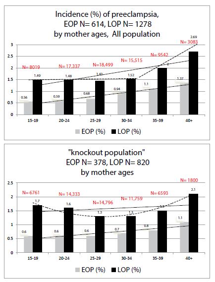
Figure 1: Comparisons of preeclampsia Incidences (%) by mother ages in 1) all our population 2) in the “knock-out” population.

Figure 2 depicts the effect of increasing maternal pre-pregnancy BMI: in the KO population (women without past and morbidities) the paramount effect of increase of BMI is specific with the increase of only LOP. In the KO population, the ppBMI has a nil effect on the occurrence of EOP (stronger effect than in the entire population).

fig 2

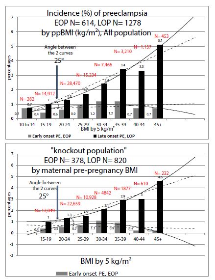
Figure 2: Comparisons of preeclampsia Incidences (%) by maternal pre-pregnancy Body Mass Index (BMI) in 1) all our population 2) in the “knock-out” population.

Discussion

First of all, “women without morbidities and past” (multiple pregnancies, chronic hypertension, diabetes, smoking, renal and thyroid diseases, “coagulopathies” and multiparous having a previous history of preeclampsia, or “knock-out population”, KO) comprise 72% of a female reproductive community. It is also of note that they also comprise 63% of all preclampsia cases (PE incidence of 2.1% vs. 2.5% in the general population).

Second, Late Onset Preeclampsia (LOP): this study on a “pure population” confirms the paramount effect of increased pre-pregnancy BMI targeted mainly on late onset preeclampsia (≥ 34 weeks gestation) that we had already previously described [8]. In KO women “without past and morbidities”, the effect is absolutely stronger than in our entire population (see comparisons in Figure 2). The BMI increase has a very poor effect on the early onset (EOP) form. Obesity is a well-known risk factor for late-onset preeclampsia [9], but this effect varies within different classes of obesities (ClassI to III) [8]. Very recently Bicocca, Sibai et al. [10] also thought to have a look at these 3 classes of obesity in a large cohort in the USA and noticed also that rising classes of obesities are significantly and linearly associated with the risk of hypertensive disorders of pregnancy (HDP). They found that the slope for EOP was different than with LOP (with an angle of 16°). We have found in 2019 similar results in a preeclamptic population-based in Reunion island, but our association was quite only associated with LOP and poorly with EOP, and our slopes made an angle of 25° [8], see also Table 2. However, there were two major methodological differences with Bicocca et al. and our study [8]: we took only preeclamptic women (and not all HDP), and used the PRE-PREGNANCY BMI, which is then predictive before any pregnancy, Bicocca et al. used maternal BMI AT DELIVERY which includes then the gestational weight gain (GWG). As a matter of fact, GWG is different if you deliver at 29 or at 38 weeks, and GWG comprises also edemas.

This specific association between maternal pre-pregnancy corpulence and LOP is not a detail, as LOP is by far the predominant form of preeclampsia (90% in literature from developed countries, some 70% in the rest of the world [11]). This confirmation is the major findings of this “knock-out” epidemiological study and, there, we might have an immediate leverage of action (prevention) very soon [12,13].

Third, concerning Early Onset Preeclampsia (EOP), this study made on a “virgin-risk population” may reveal some tracks. Besides a predictive screening by what we may call the “Nicolaides-Poon’s algorithms” [14-17], some clinical items may be added to the EOP risk: the involvement of a new male partner for the index pregnancy and history of previous perinatal death (mainly intra-uterine fetal deaths).

A)  Primipaternity and History of Abortions and Miscarriages in Multigravidas

In this study, we have indirect approach of a male partner involvement, and, interestingly rather a risk for EOP (therefore a possible target for aspirin prevention?). First of all, history of abortions or miscarriages in multiparas were not associated with any preeclampsia risk in the general population (Table 1, bold results), and in our logistic model including primiparity (Table 2, upper Table) and in the results of preceding studies on this same population [1]. In the present study of a KO population, crude results (Table 1, italic results) and in the primipaternity model (Table 2, lower Table), history of abortions is specifically associated with the risk of EOP, and miscarriages equally between EOP and LOP. At first, the well-known effect of primiparity (cornerstone of all epidemiological studies on preeclampsia) is also confirmed in primipaternity (Table 2). For primipaternity, we may assume that women having changed male partner for the index pregnancy may have had preceding abortions with different partner(s) [18].

B)  Primipaternity and Medically Induced Pregnancies

It is of note (Table 2) that in vitro-fecundations (including ICSI) are not independently associated with any kind of preeclampsia risk (EOP & LOP) in primiparas and “primipaternity-multiparas”, Table 2. In our ART centre in Reunion, 88% of our IVF are made with the habitual male partner of the couple, with very few oocyte donations (N=31 in 19 years) and very few with unknown donor sperm. Contrary to IVF, in our experience, medically induced pregnancies by stimulation of ovulation were a strong independent factor specifically associated with EOP. We have verified and 2/3 (66.6%) of our stimulated pregnancies were primiparas, and 48% primigravidas. Antecedents of abortions as risk factors specific to EOP in women having a new partner suggest the “male effect” as possible etiology of preeclampsia, and more specifically in this study for EOP [19-22].

C)  Antecedents of Perinatal Deaths

Controlling for primipaternity (Table 2, lower Table), previous perinatal deaths have a tendency to be an independent factor for the EOP risk, p=0.07 (NB: not associated with previous history of preeclampsia, as these women have been removed from the study population).

The Centre Hospitalier Universitaire Sud-Reunion’s maternity (Level 3, European standards of care) is the only public hospital in the southern part of Reunion Island (Indian Ocean, French overseas department). It serves the whole population of the area (ap. 360,000 inhabitants, and 5,100 births per year). With 4,300 births per year, the university maternity represents 82% of all births in the south of the island. But, as a level 3 (the other maternity is a private clinic, level 1), we are sure all the preeclampsia cases were referred to our hospital during the 19- year period. This is therefore a real population-based study. As a limitation of the study, we have to consider the retrospective nature of the study that, although the number of information that is recorded, some characteristics may miss like length of sexual relationship and/or primipaternity. The “primipaternity” item, being quoted mainly retrospectively for the period 2001-2017, is not completely reliable. “Coagulopathies” were not systematically screened in all women (cases and controls). However, every time that a woman was known to have one of these characteristics, they were scrupulously included in the database. The strengths of this study are mostly related to the homogeneity of data in such a large cohort as they were collected in a single center (no intercenter variability) and not based on national birth registers but directly from medical records (avoiding inadequate codes).

Conclusion

This study made on a population exempt from the eight internationally consensual major risk factors for preeclampsia may be of some interest. 1) for late onset preeclampsia LOP: it confirms the specific association between high maternal BMI and the immense burden of late-onset preeclampsia (LOP) [8]. This is of major importance in a planet where the overweight-obesity problem is constantly rising. Here, there is a reasonable hope to have a positive intervention by a monitored management of gestational weight gain since the beginning of any pregnancy allowing to lower the LOP incidence by some 30-40% [12,13]. 2) For early onset preeclampsia EOP (here we may expect a 60% decrease by aspirin prevention [14,15]), this KO population revealed underlying risk factors: history of preceding perinatal deaths (intra-uterine fetal deaths) and arguments for “new paternity”. International efforts should be focused on asking to all first-couple’s pregnancies (primiparas and multiparas having changed the male partner) the length of cohabitation before conception. A sexual cohabitation of less than 6 months could be a risk factor for EOP and, if confirmed, beneficiate of early aspirin prevention since the 16th week of gestation [14-17].

References

  1. Robillard PY, Dekker G, Scioscia M, Bonsante F, Iacobelli S, et al. (2020) The blurring boundaries between placental and maternal preeclampsia: a critical appraisal of 1800 consecutive preeclamptic cases. J Matern Fetal Neonatal Med 6: 1-7. [crossref]
  2. Burton GJ, Redman CW, Roberts JM, Moffett A (2019) Pre-eclampsia: pathophysiology and clinical implications. BMJ 366: 2381. [crossref]
  3. Iacobelli S(1), Bonsante F(2), Robillard PY(2) (2017) Comparison of risk factors and perinatal outcomes in early onset and late onset preeclampsia: A cohort based study in Reunion Island. J Reprod Immunol 123: 12-16. [crossref]
  4. Ratsiatosika AT, Razafimanantsoa E, Andriantoky VB, Ravoavison N, Andrianampanalinarivo Hery R, et al. (2019) Incidence and natural history of preeclampsia/eclampsia at the university maternity of Antananarivo, Madagascar: high prevalence of the early-onset condition. J Matern Fetal Neonatal Med 32: 3266-3271. [crossref]
  5. Robillard PY, Dekker G, Chaouat G, Hulsey TC (2007) Etiology of preeclampsia: maternal vascular predisposition and couple disease–mutual exclusion or complementarity? J Reprod Immunol 76: 1-7. [crossref]
  6. Tranquilli AL, Brown MA, Zeeman GG, Dekker G, Sibai BM (2013) The definition of severe and early-onset preeclampsia. Statements from the International Society for the Study of Hypertension in Pregnancy (ISSHP). Pregnancy Hypertens 3: 44-77. [crossref]
  7. Robillard PY, Dekker G, Scioscia M, Bonsante F, Iacobelli S, et al. (2020) Validation of the 34-week gestation as definition of late onset preeclampsia:Testing different cutoffs from 30 to 37 weeks on a population-based cohort of 1700 preeclamptics. Acta Obstet Gynecol Scand.
  8. Robillard PY, Dekker G, Scioscia M, Bonsante F, Iacobelli S, et al. (2019) Increased BMI has a linear association with late-onset preeclampsia: a population-based study. PLoS One 14: 0223888. [crossref]
  9. Lisonkova S, Joseph KS (2013) Incidence of preeclampsia: risk factors and outcomes associated with early-versus late-onset disease. Am J Obstet Gynecol 209: 544. [crossref]
  10. Bicocca MJ, Mendez-Figueroa H, Chauhan SP, Sibai BM (2020) Maternal Obesity and the Risk of Early-Onset and Late-Onset Hypertensive Disorders of Pregnancy. Obstet Gynecol 136: 118-127. [crossref]
  11. Robillard PY, Dekker G, Chaouat G, Elliot MG, Scioscia M (2019) High incidence of early onset preeclampsia is probably the rule and not the exception worldwide 20th anniversary of the reunion workshop. A summary. J Reprod Immunol 133: 30-36.
  12. Robillard PY, Dekker G, Boukerrou M, Boumahni B, Hulsey T, et al. (2020) Gestational weight gain and rate of late-onset preeclampsia: a retrospective analysis on 57 000 singleton pregnancies in Reunion Island. BMJ Open 10: 036549.
  13. Robillard PY, Dekker GA, Boukerrou M, Boumahni B, Hulsey TC, et al. (2020) The urgent need to optimize gestational weight gain in overweight/obese women to lower maternal-fetal moribidities: a retrospective analysis on 59,000 singleton term pregnancies. Archives Women Health Care 3: 1-9.
  14. Roberge S, Bujold E, Nicolaides KH (2018) Aspirin for the prevention of preterm and term preeclampsia: systematic review and metaanalysis. Am J Obstet Gynecol 218: 287-293. [crossref]
  15. Rolnik DL, Nicolaides KH, Poon LC (2020) Prevention of preeclampsia with aspirin. Am J Obstet Gynecol 21: 30873-30875.
  16. Poon LC, Shennan A, Hyett JA, Kapur A, et al. (2019) The International Federation of Gynecology and Obstetrics (FIGO) initiative on pre-eclampsia: A pragmatic guide for first-trimester screening and prevention. Int J Gynaecol Obstet 145: 1-33. Erratum in: Int J Gynaecol Obstet 146: 390-391. [crossref]
  17. Wright D, Nicolaides KH (2019) Aspirin delays the development of preeclampsia. Am J Obstet Gynecol 220: 580. [crossref]
  18. Saftlas AF, Levine RJ, Klebanoff MA, Martz KL, Ewell MG, et al. (2003) Abortion, changed paternity, and risk of preeclampsia in nulliparous women. Am J Epidemiol 157: 1108-1114. [crossref]
  19. Robillard PY, Dekker GA, Hulsey TC (1999) Revisiting the epidemiological standard of preeclampsia: primigravidity or primipaternity? Eur J Obstet Gynecol Reprod Biol 84: 37-41. [crossref]
  20. Dekker GA, Robillard PY (2005) Preeclampsia: a couple’s disease with maternal and fetal manifestations. Curr Pharm Des 11: 699-710. [crossref]
  21. Dekker GA (2014) Pre-eclampsia – A disease of an individual couple. Pregnancy Hypertens 4: 242-243.
  22. Dekker G, Robillard PY, Roberts C (2011) The etiology of preeclampsia: the role of the father. J Reprod Immunol 89: 126-132. [crossref]

Letter to the Editor – Open Bite Malocclusion: Analysis of the Underlying Components

DOI: 10.31038/JDMR.2020333


Anterior open bite (AOB) can be considered one of the most challenging malocclusions to treat. Its complexity arises from the multi-factorial etiology, the involvement of various components and the uncertain stability.

There is abundance of literature on the AOB classifications, the different etiologies and the possible treatment options. However, most of the articles focus on treatment of AOB malocclusions, neglecting the deeper search into the components involved into that malocclusion which is essential for proper treatment planning.

AOB can be attributed to skeletal or dental underlying causes/components, yet it may result from a combination of both. Analysis of these components would allow the orthodontist to depict the underlying causative factor of the presenting AOB malocclusion and customize a proper treatment plan. This is expected to result in a successful outcome, better stability, improved esthetics and more importantly enhance patient satisfaction.

Treatment of AOB should not be viewed as simple as placing a tongue crib for growing individuals, and treating adult patients with posterior segments intrusion or orthognathic surgery. Tongue size & position, incisal show at rest and on smiling, mandibular plane steepness and lip competence should all be closely monitored before planning what should be done. There is still a need for a detailed systematic analysis of all these components and relating these findings to the possible treatment options to serve as a guide for orthodontists in dealing with such difficult cases. This is currently our area of interest and we are working as a team to establish a schematic approach, aiming to specifically recognize the exact components of AOB malocclusion and target our treatment accordingly.

fig 1

Italian and Lombardy County Art Post-Covid Pandemic Scenarios

DOI: 10.31038/EDMJ.2020451

Abstract

Purpose: Aim of the present study is to report two possible scenarios, one optimistic and one cautelative, on ART activity in Italy and Lombardy County after the COVID-19 lockdown.

Methods: Based on historical data from the ART Italian National Register in general and Lombardy County in particular, we derived two ipothesis, on how the lockdown in Italy after Covid-19 epidemic, could affect the reduction in ART activity. The first one is considered a “cautelative” hypothesis and we modeled a substantial ART activity reduction.

Results: In both proposed scenarios there is data evidencing a reduction in the number of cycles and of babies born. In the cautelative hypothesis there is a reduction of the number of cycles by 55% and in the optimistic one by 35.5%. The total National loss in babies born will be between 6,719 and 5,913 and in Lombardy between 2,093 and 1,256 according to the 2 different scenarios.

Conclusions: The impact of Covid-19 epidemic on ART treatments will greatly reduce the number of babies born in the foreseable future. This not only to lockdown measures but also to reduced accessibility to treatments, even in a region like Lombardy carries out 30% of all the ART cycles in the country, mostly supported by public resources. Italian ART centres will face a great challenge in the management of the new policies for covid 19 containment. It will be difficult to find the right balance between maintaining enough available procedures, keeping both couples and staff safe.

Keywords

Covid-19 infection, Assisted reproductive techniques, Gamete donation, Lombardy county, ART National Register

Introduction

On January 30, 2020 the first case of Covid-19 was detected in Italy, probably from a couple of tourists arriving from China; but it was February 22, 2020 the date when the infection spread dramatically, and a lot of cases were discovered especially in Lombardy County [1-4]. A National integrated surveillance system of Covid-19 has been created, coordinated by the Istituto Superiore di Sanità, analyzing data beginning on the 27th of February, and it suddenly showed a pattern of infection breakouts in Lombardy, and then in other northern regions [5].

The government, in a first round, decided to close only some specific municipality in Lombardy County, where most of the cases were diagnosed, calling these area “the red zone”. In these territories a strict lockdown was enforced. Due to emergency regulations people could go out only for specific activities. School were closed, including universities, factories, shops, cinemas, theaters, and every activity not considered essential, like pharmacies or supermarkets, was closed. This strict lockdown procedures were implemented in Italy 4 to 6 weeks ahead of other European countries. Subsequently, from March 9th, 2020, the entire Italian territory was put under the same quarantine regimen.

Consequently, all health treatments considered not urgent were stopped, including ART procedures, in order not to overload hospitals and medical staff already engaged in the handling of covid-19 pandemic [6-13].

Many doctors working in ART units were reallocated in Covid-19 department, and spaces and facilities were quickly re adapted to create places for infected patients [14,15].

Recommendations from the major Italian Scientific Societies for Reproductive Medicine and from the Ministry of Health suggested stopping all new ART treatments and to continue only procedures for patients who had already begun ovarian stimulation. In these cases, freeze all strategy has been recommended. Only fertility preservation for cancer patients was continued [16-19].

In Italy since 2005 a National Register collecting information on Assisted Reproductive Technologies is in force by law since 2005. Data collection is mandatory, and all the 201 Centres, performing ART procedures, plus 160 performing only IUI (Intra Uterine Insemination), send data to the Register. A retrospective Data collection is made every year on a web site, www.iss.it/rpma with a dedicated access for each Centre and for each Region.

Comparing temporal trends of data collected from 2010 till 2018 from the Italian ART National Register [20,21], we have tried to make a reliable estimation of the impact of Covid-19 pandemic on ART procedures in Italy for the 2020 data set. All Italian ART Centers must report their activity to the Register and 100% comply with this requirement. Data were considered for all the Italian Centers and for Lombardy County, which represent around 30% of the country ART activity.

Materials and Methods

We analyzed the temporal trend of all the data collected on ART cycles, from the Italian ART National Register from year 2010 till 2018, to compare them with estimated projections on the percentages of ART activity reduction that will occur during 2020, according to 2 different scenarios: a cautelative hypothesis (a) and an optimistic one (b). First, we analyzed the all National data, then specifically for the Lombardy County. Donation cycles were excluded and in line with the 2019 total number of ART procedures, our prediction models were taking in consideration already a reduced number of fresh cycles and a higher number of frozen ones. We considered various scenarios and after observing the ongoing situations in public and private centers and different opening policy in different regions of the country, we formulated what could be the most suitable hypothesis. Assuming that the activity for the first two months of 2020, January and February has remained quite stable, then, for March that the activities were reduced by the 40% than the 2019 number, due to a progressive amount of interrupted procedures, and that in April and May all the activity has been suspended with a residual 10% of procedures performed due to different closing policy, then from June on, we hypotise an activity resumption with different reductions scenarios. We have speculated for the residual seven months, (June/December 2020) that in a 2020 precautionary scenario (a), there will be a 40% recovery in activity compared to 2019, while in a 2020 optimistic scenario (b), we consider a 70% recovery in activity. Considering the different steps of ART treatments, from the number of started cycles to the oocyte retrieval, fresh and frozen embryo transfer, pregnancies, deliveries obtained, and babies born, we calculated the delta value of the variables examined according to the two-different hypothesis for the different techniques categories, fresh cycles and frozen cycles.

Some measures such as comulative pregnancy rate and pregnancy lost to follow up rate were tested with a z-test to find difference. A p-value of 0.05 was considered significant. The statistical analysis was performed with IBM SPSS Statistics 26.

Results

We considered first the National data and then the Lombardy County performance. The Lombardy region, due probably to higher reimbursement than other regions of the country has an over 99% of the cycles covered by public resources, most of the larger volume centers concentrated in this area and in the 2018, even with all the bias of aggregated data analysis, a significantly higher (p<0.01) cumulative pregnancy rate per retrieval (34.1% with IC95% 33.4-35.0) than the overall rest of the country (31.3% IC95% 30.9-31.7) with a significantly lower (p<0.01) lost to follow up rate (3.4% IC95% 2.9-4.0) versus a general higher national percentage (9.1% IC95% 8.6-9.5). Public reimbursement is important in our country since few or no insurances cover ART procedures [22].

Therefore, many infertile couples in Italy, (27% of all the cycles are applied on couples coming from another County report ISS) move from their County to another one, to achieve reimbursed ART treatments [23].

Comparing the 2019 data set with the two scenarios, the conservative with a 40% recovery in activity, and the optimistic with 70% recovery, we have calculated the delta value for each step of the treatments: number of cycles, retrievals, and transfer for fresh plus frozen cycles, and for each technique alone, we have considered the number of pregnancies obtained and the number of babies born [24].

We estimated that the total number of ART cycles, fresh plus frozen in 2019 will be 71,991 (the final data is not available yet), and that in 2020 we will have 32,396 cycles for the worst hypothesis and 44,994 for a more favorable scenario, with a delta value respectively of 39,595 and 26,997, considering all Italian cycles (Table 1).

Table 1: Number of procedures performed in Italy in the period 2018-2019 and according to a cautelative (- 55%) and optimistic (- 37.5%) and optimistic hypothesis calculation the possible loss in the 2 described scenarios.

Italy 2018-2019 2020 Cautelative

hypothesis

2020 Optimistic hypothesis Delta Cautelative hypothesis loss Delta Optimistic hypothesis loss
Fresh + Frozen Cycles 71.991 32.396 44.994 39.595 26.997
Fresh + Frozen Transfers 50.636 22.786 31.648 27.850 18.989
Fresh Cycles 51.086 22.989 31.929 28.097 19.157
Oocytes Retrievals 46.387 20.874 28.992 25.513 17.395
Fresh Transfers 30.584 13.763 19.115 16.821 11.469
Frozen Transfers 20.052 9.023 12.533 11.029 7.520
Pregnancies (fresh + frozen) 14.525 6.536 9.078 7.989 5.447
Babies Born 10.751 4.838 6.719 5.913 4.032

Considering the transfers, we estimated 50,636 for 2019 and 22,786 for scenario a, and 31,648 for scenario b, with delta values of 27,850 and 18,989 for the two models, respectively. For fresh cycles the estimated 2019 number was 51,086, while 22,989 in the 2020 first scenario, and 31,929 in the second one, delta values of 28,097 and 19,157, respectively. Transfers in 2019 fresh cycles were 30,584, compared with 13,763 for 2020 (hypothesis a) and 19,115 for 2020 (hypothesis b). Delta values will be 16,821 and 11,469, respectively. Oocyte retrievals will be 46,387 in 2019 versus 20,874 and 28,992 for the cautelative and the optimistic model of 2020 with a delta value of 25,513 and 17,395, respectively. The number of frozen transfers was 20,052 in 2019 compared with 9,023 for 2020 hypothesis and 12,533 for hypothesis b, delta values of 11,029 and 7,520 respectively.

The activity performed in 2020 will be then reduced by 45%, compared to the one of the previous year, with a loss of 55% of total activity in the cautelative hypothesis, and of the 62.5% of 2019 total with a loss of 37.5% in the optimistic scenario (Table 1).

Concerning pregnancies, we had 14,525 pregnancies in 2019 with a reduction to 6,536 in the precautionary scenario and 9,078 in the optimistic for the year 2020. Delta values will be 7,989 and 5,447 respectively. The number of babies born for year 2019 is expected to be (most of pregnancies still ongoing) 10,751 with a prevision for the 2020 hypothesis of only 4,838 newborns and of 6,719 for the 2020 hypothesis b; relative delta values will be 5.913 and 4.032 (Figure 1).

fig 1

Figure 1: In Italy Post Covid 2020 cautelative and optimistic hypothesis loss in pregnancies and babies born.

Analyzing in more details the number of babies born we observed a mean reduction of newborns of 896 per month, specifically we will have 1,792 newborns in January/February, only 358 in March, 179 for April/May, and 4,390 from June to December 2020 for the optimistic scenario.

Lombardy county data 2019, always estimating 2018 comparable data, had 21,367 fresh + frozen cycles, 13,658 retrievals, 16,169 fresh + frozen transfers (9,770 fresh and 6,399 frozen). The general yearly loss will be of 11,752 cycles in the cautelative hypothesis and of 8,013 in the opstimistic option (Table 2).

Table 2: Number of procedures performed in Lombardy in the period 2018-2019 and according to a cautelative (-55%) and optimistic (- 37.5%) hypothesis calculation the possible lost in the 2 described scenarios.

Lombardy 2018-2019 2020 Cautelative Hypotesys 2020 Optimistic Hypotesys Delta Cautelative Hypotesys loss Delta Optimistic Hypotesys loss
Fresh + Frozen Cycles 21.367 9.615 13.354 11.752 8.013
Fresh + Frozen Transfers 16.169 7.276 10.106 8.893 6.063
Fresh Cycles 14.968 6.736 9.355 8.232 5.613
Oocytes Retrevals 13.658 6.146 8.536 7.512 5.122
Fresh Transfers 9.770 4.397 6.106 5.374 3.664
Frozen Transfers 6.399 2.880 3.999 3.519 2.400
Pregnancies (fresh + frozen) 4.665 2.099 2.916 2.566 1.749
Babies Born 3.348 1.507 2.093 1.841 1.256

Considering the 2019 estimates of 4,665 pregnancies and 3,348 babies born, the two 2020 models will project 2,099 pregnancies and 1,507 born babies according to the cautelative hypothesis and 2,916 pregnancies and 2,093 born babies according to the 2020 optimistic prevision. In 2020 there will be a loss of 2,566 pregnancies and 1,841 born babies in the cautelative and 1,749 and 1,256 in the optimistic prevision (Figure 2).

fig 2

Figure 2: In Lombardy Post Covid 2020 cautelative and optimistic hypothesis loss in pregnancies and babies born.

Discussion

In the so-called phase 2 of the epidemic in Italy, that has started May 4th, in which ART centres could restart their activity, couples will have fewer financial resources to support the birth of a child and less resources to access ART costs on a private scheme. Even if most scientific authorities support a more widly coverage of ART treatment [22,25] this is still poorly undertood in most regions of our country. So we are offering less ART cycles to our infertile population in comparison with some northen European Countries The public and contracted facilities will have to reconvert spaces and staff now dedicated to other activities and will experience a difficult phase and a longer period for restarting even with a reduced number of cycles [16,17]. Private facilities will be more likely to resume faster, but the number of couples with available resources to afford ‘out of pocket’ cost will be less than previously, even if gonadotrophins are financially supported by a National regulation until 45 years of age and for an FSH level < to 30 UI/ml. In 2019 > 99% of treatments in Lombardy compared to a National average of about 69% (2018 data) were paid for by the Regional and/or National Health System. Couples moving from the Regions of the country to access to ART cycles has been always high in Italy reaching rates over 50% of performed cycles in some counties, in Lombardy it was the 33.1% only in 2018. This rate is significantly lower than the rate of out of county patients treated for other pathologies. Everyone will experience the transformation of work for the protection measures of couples and staff [26,27].

Many things are still unknown and studies on the effects of COVID 19 on reproductive cells are ongoing, even if reasssuring data on pregnancy outcome have been published [13,18,28-30].

Recent real world data from the Humanitas Fertility until October 31th show a 31% of total number of 2020 performed procedures with a possibile reduction due to the probable new lockdown at least in some Italian Regions as Lombardy to over 50% of the 2019 perfomed procedures.

Conclusions

Whatever scenario will really appear at the end of year 2020 [14], regarding the reduction on ART activity, the impact of Covid-19 pandemic will be really strong for all the Italian ART Centers, not only because of the reduction of cycles performed and subsequently babies born, but because of the diminished availability of procedures [31], even in a Region like Lombardy that carries out 30% of all the ART cycles in the Country, mostly supported by public resources. Being compliant with new rules to protect couples and staff from the risk of infection, will determine a shortage in the number of patients treated. Only a strong willingness and a great organizing capacity of the ART Centers could partially overcome the burden of the impact of Covid-19 epidemic that will continue in the next months. In this emergency situation, in a Country with the lowest natality rate in Europe, where the Assisted reproductive techniques contribute for the 3% of the babies born annually, and plays an important role answering to the need of infertile couples, Government and Regional local authorities should encourage and support ART activity and promote access for infertile couples with dedicated actions [32,33].

Ultimately, given the ethical concerns raised by public health recommendations regarding pregnancy avoidance, strong justification for any such advice is needed and the criteria to be fulfilled during certain public health emergencies (e.g., a radiation emergency with continuing exposure), we don’t believe that the risks associated with Covid-19 meet the bar [1].

Data Availability

The datasets generated for this study are available on request to the corresponding author.

Conflict of Interest

Conflict of Interest Statement: The authors declare that the research was conducted in the absence of any commercial or financial relationships that could be construed as a potential conflict of interest.

Author Contributions Statement

PELS wrote the research project, analyzed data and prepared the final draft, RDL analyzed data, GS and MB and RDL contributed to the data, the manuscript and references preparation.

We would like to thank Pasquale Patrizio, Yale Fertility Center, for his support in manuscript revision.

Acknowledgments

The authors thank all the Italian and Lombardy County Centers contributing with their aggregated data to this work.

References

  1. Grasselli G, Pesenti A, Cecconi M. et al. (2020) Critical Care Utilization for the COVID-19 Outbreak in Lombardy, Italy: Early Experience and Forecast during an Emergency Response. JAMA. [crossref]
  2. Wang R, Pan M, Zhang X, Fan X, Han M, Zhao F, et al. (2020) Epidemiological and clinical features of 125 Hospitalized Patients with COVID-19 in Fuyang, Anhui, China. Int J Infect Dis. [crossref]
  3. Wakam GK, Montgomery JR, Biesterveld BE, Brown CS, et al. (2020) Not Dying Alone – Modern Compassionate Care in the Covid-19 Pandemic. N Engl J Med. [crossref]
  4. Nicastri E, Petrosillo N, Bartoli TA, Lepore L, Mondi A, Palmieri F, et al. (2020) National Institute for the Infectious Diseases “L. Spallanzani”, IRCCS. Recommendations for COVID-19 clinical management. Infect Dis Rep 12(1): 8543. [crossref]
  5. Guerci C, Maffioli A, Bondurri AA, Ferrario L, Lazzarin F, Danelli P, et al. (2020) COVID-19: How can a department of general surgery survive in a pandemic? Surgery 167(6): 909-11. [crossref]
  6. American Society for Reproductive Medicine (ASRM). Patient management and clinical recommendations during the Coronavirus (COVID-19) pandemic. Update 3. April 24, 2020.
  7. Gruppo di Interesse Speciale sulla Sterilità (GISS) della Società Italiana di Ginecologia ed Ostetricia (SIGO). Raccomandazioni sulla gestione contestuale dell’accesso e della circolazione dei pazienti presso le strutture dedicate alle procedure di P.M.A. April 4, 2020.
  8. Vaiarelli A, Bulletti C, Cimadomo D, Borini A, Alviggi C, Ajossa S, et al. (2020) COVID-19 and ART: the view of the Italian Society of Fertility and Sterility and Reproductive Medicine. Reprod Biomed Online 40(6): 755-9. [crossref]
  9. Centro Nazionale Trapianti (CNT) – Registro Nazionale Procreazione Medicalmente Assistita (PMA) Istituto Superiore di Sanità (ISS). misure di prevenzione della trasmissione dell’infezione da nuovo Coronavirus (SARS-CoV-2) in Italia per le cellule riproduttive e i trattamenti di PMA (procreazione medicalmente assistita). www.trapianti.salute.gov.itAggiornamento del 29/4/2020 e 5/5/2020.
  10. De Santis L, Anastasi A, Cimadomo D, Klinger FG, Licata E, Pisaturo V, et al. (2020) COVID-19: the perspective of Italian embryologists managing the IVF laboratory in pandemic emergency. Hum Reprod. [crossref]
  11. Rodriguez-Wallberg KA, Wikander I. (2020) A global recommendation for restrictive provision of fertility treatments during the COVID-19 pandemic. Acta Obstet Gynecol Scand 99(5): 569-70. [crossref]
  12. Rome BN, Avorn J. Drug (2020) Evaluation during the Covid-19 Pandemic. N Engl J Med. [crossref]
  13. Di Mascio D, Khalil A, Saccone G, Rizzo G, Buca D, Liberati M, et al. (2020) Outcome of Coronavirus spectrum infections (SARS, MERS, COVID 1 -19) during pregnancy: a systematic review and meta-analysis. Am J Obstet Gynecol MFM
  14. Remuzzi A, Remuzzi G. (2020) COVID-19 and Italy: what next? Lancet 395(10231): 1225-8.
  15. Ferrario L, Maffioli A, Bondurri AA, Guerci C, Lazzarin F, Danelli, et al. (2020) P. COVID-19 and surgical training in Italy: Residents and young consultants’ perspectives from the battlefield. Am J Surg. [crossref]
  16. La Marca A, Niederberger C, Pellicer A, Nelson SM. et al. (2020) COVID-19: lessons from the Italian reproductive medical experience. Fertil Steril. [crossref]
  17. Alviggi C, Esteves SC, Orvieto R, Conforti A, La Marca A, Fischer R, et al. (2020) COVID-19 and assisted reproductive technology services: repercussions for patients and proposal for individualized clinical management. Reprod Biol Endocrinol 18(1): 45. [crossref]
  18. Liang H, Acharya G. (2020) Novel corona virus disease (COVID-19) in pregnancy: What clinical recommendations to follow? Acta Obstet Gynecol Scand 99(4): 439-42. [crossref]
  19. Sirohi B, Rohatgi TB, Lambertini M. et al. (2020) Oncofertility and COVID-19-cancer does not wait. Ecancermedicalscience 14: ed101. [crossref]
  20. Scaravelli G DLR, Vigiliano V, Bolli S, Spoletini R, Fiaccavento S, Bertini A, Speziale L, et al. (2019) ATTIVITÀ DEL REGISTRO NAZIONALE ITALIANO DELLA PROCREAZIONE MEDICALMENTE. DATI 2017. Istituto Superiore di Sanità, Centro Nazionale di Epidemiologia, Sorveglianza e Promozione della Salute, Centro operativo adempimenti Legge 40/2004, Registro Nazionale della Procreazione Medicalmente Assistita. salute.gov.it2019.
  21. Scaravelli G, Levi-Setti PE, Livi C, La Sala G, Ubaldi FM, Greco E, et al. (2019) Contribution of cryopreservation to the cumulative live birth rate: a large multicentric cycle-based data analysis from the Italian National Registry. J Assist Reprod Genet 36(11): 2287-95. [crossref]
  22. Chambers GM, Hoang VP, Sullivan EA, Chapman MG, Ishihara O, Zegers-Hochschild F, et al. (2014) The impact of consumer affordability on access to assisted reproductive technologies and embryo transfer practices: an international analysis. Fertil Steril 101(1): 191-8.e4. [crossref]
  23. Scaravelli G, Vigiliano V, Bolli S, Spoletini R, Fiaccavento S, Bertini A, et al. (2018. 2020) ATTIVITÀ DEL REGISTRO NAZIONALE ITALIANO DELLA PROCREAZIONE MEDICALMENTE. DATI in press.
  24. Requena A, Cruz M, Vergara V, Prados N, Galliano D, Pellicer A, et al. (2020) A picture of the covid-19 impact on IVIRMA fertility treatment clinics in Spain and Italy. Reprod Biomed Online 41(1): 1-5. [crossref]
  25. Vélez MP, Connolly MP, Kadoch IJ, Phillips S, Bissonnette F, et al. (2014) Universal coverage of IVF pays off. Hum Reprod 29(6): 1313-9. [crossref]
  26. Colenda CC, Applegate WB, Reifler BV, Blazer DG, et al. (2020) COVID-19: Financial Stress Test for Academic Medical Centers. Acad Med. [crossref]
  27. El Kassas M, Al Shafie A, Abdel Hameed AS, Mahdi M, et al. (2020) Emergency endoscopic variceal band ligation in a COVID-19 patient presented with hematemesis while on mechanical ventilation. Dig Endosc. [crossref]
  28. Paoli D, Pallotti F, Turriziani O, Mazzuti L, Antonelli G, Lenzi A, et al. (2020) SARS-CoV-2 presence in seminal fluid: Myth or reality. Andrology. [crossref]
  29. Younis JS, Abassi Z, Skorecki K, et al. (2020) Is there an impact of the COVID-19 pandemic on male fertility? The ACE2 connection. Am J Physiol Endocrinol Metab 318(6): E878-E80. [crossref]
  30. Segars J, Katler Q, McQueen DB, Kotlyar A, Glenn T, Knight Z, et al. (2020) Prior and novel coronaviruses, Coronavirus Disease 2019 (COVID-19), and human reproduction: what is known? Fertil Steril 113(6): 1140-9. [crossref]
  31. De Geyter C, Calhaz-Jorge C, Kupka MS, Wyns C, Mocanu E, Motrenko T, et al. (2020) ART in Europe, 2015: results generated from European registries by ESHRE. Hum Reprod Open. (1): hoz038. [crossref]
  32. Chambers GM, Sullivan EA, Ishihara O, Chapman MG, Adamson GD, et al. (2009) The economic impact of assisted reproductive technology: a review of selected developed countries. Fertil Steril 91(6): 2281-94. [crossref]
  33. Rasmussen SA, Lyerly AD, Jamieson DJ, et al. (2020) Delaying Pregnancy during a Public Health Crisis – Examining Public Health Recommendations for Covid-19 and Beyond. N Engl J Med. Epub 2020/09/30. doi: 10.1056/NEJMp2027940. PubMed PMID: 32997931. [crossref]
fig 1

Applying Linear Elastic Glucose Behavior Theory and AI Auto-Correction to Predict A1C Variances over the Ninth Period Using GH-Method: Math-Physical Medicine (No. 354)

DOI: 10.31038/EDMJ.2020452

Abstract

In this case study, the author analyzed, predicted, and interpreted a type 2 diabetes (T2D) patient’s hemoglobin A1C variance or A1C value based on nine time periods, ~5 months each, utilizing the GH-Method: math-physical medicine (MPM) approach. He utilized the same method and calculation formulas since 1/1/2014. This particular article emphasizes the ninth period (Period I) ranging from 5/20/2019 to 10/21/2020.

Unlike the previous eight periods, in the ninth period, he used the continuous glucose monitor (CGM) sensor collected glucose data, which he implemented with the recently developed linear elastic glucose behavior theory (Reference 7, No. 352) to calculate his postprandial plasma glucose (PPG) difference.

Utilizing an auto-learning and auto-correction technique of artificial intelligence (AI), his software system would learn from lab-tested A1C values and then make necessary corrections. Therefore, in Period I, he utilized a higher conversion factor of 17.13 from glucose to HbA1C instead of 12.5 in the previous seven periods (Period A throughout Period G). He wrote two articles for the eighth period (Period H) using 12.5 (No. 329) and 17.13 (No. 329B), respectively. The difference on the final HbA1C results is a mere 0.01% which increased his predicted HbA1C from 6.31% to 6.39%, while the lab-tested HbA1C was 6.4%.

There are 10 hemoglobin A1C lab-tested results, with his 10 predicted HbA1C values using his developed methods:

  • 6.7% on 4/9/2017 (predict 6.7%)
  • 6.1% on 9/12/2017 (predict 6.1%)
  • 6.9% on 1/26/2018 (predict 6.9%)
  • 6.5% on 6/29/2018 (predict 6.5%)
  • 6.6% on 10/22/2018 (predict 6.6%)
  • 6.8% on 4/4/2019 (predict 6.8%)
  • 6.6% on 9/25/2019 (predict 6.6%)
  • 6.6% on 12/20/2019 (predict 6.6%)
  • 6.4% on 5/20/2019 (predict 6.4%)
  • 6.2% on 10/21/2020 (predict 6.2%).

It should be pointed out that all of his 10 predicted HbA1C values match 100% with his lab-tested HbA1C values.

The author focused on nine periods over 1,299 days. It contains 3,897 meal data, including key contribution factors such as carbs/sugar intake, post-meal exercise, weather, and more. This study demonstrated a high degree of accuracy for the calculation and prediction of the patient’s forthcoming HbA1C value by using the GH-Method: math-physical medicine approach. In this article, the author also utilized CGM sensor glucose data with AI-tuned conversion factor between glucose and HbA1C. Furthermore, he also implemented his developed linear elastic glucose behavior theory (Reference 7) for his PPG calculation in order to obtain a better estimation for the final HbA1C value.

Once the healthcare professionals and T2D patients understand the HbA1C mathematical prediction method, then the overall diabetes condition for the patient can be easier to control. The purpose for this research paper is to help people with T2D by preventing further damage to their internal organs caused by elevated A1C values, before taking the laboratory test.

If healthcare professionals and diabetes patients have an interest to delve deeper regarding the formation of tested glucose and mathematical predicted A1C, they should focus on the influential factors and their respective weighted contribution percentages described in the author’s previous papers.

Here is the summary:

  1. The most important month which contributes to the A1C is the month prior to the lab test.
  2. PPG contribute >2/3 of HbA1C.
  3. Body weight controls ~77% or more of the fasting plasma glucose (FPG) which contributes <1/3 of HbA1C; therefore, it is important to keep BMI below 25.
  4. Carbs/sugar amount contributes ~39% to PPG. For T2D patients, it is safe to keep carbs/sugar intake amount below 15 grams per meal.
  5. Post-meal walking steps contributes ~41% to PPG. It is recommended to maintain post-meal walking exercise around 4,000 steps after each meal.
  6. A combined effort of diet and exercise controls ~80% to PPG formation.

Introduction

In this case study, the author analyzed, predicted, and interpreted a type 2 diabetes (T2D) patient’s hemoglobin A1C variance or A1C value based on nine time periods, ~5 months each, utilizing the GH-Method: math-physical medicine (MPM) approach. He utilized the same method and calculation formulas since 1/1/2014. This particular article emphasizes the ninth period (Period I) ranging from 5/20/2019 to 10/21/2020.

Unlike the previous eight periods, in the ninth period, he used the continuous glucose monitor (CGM) sensor collected glucose data, which he implemented with the recently developed linear elastic glucose behavior theory (Reference 7, No. 352) to calculate his postprandial plasma glucose (PPG) difference.

Utilizing an auto-learning and auto-correction technique of artificial intelligence (AI), his software system would learn from lab-tested A1C values and then make necessary corrections. Therefore, in Period I, he utilized a higher conversion factor of 17.13 from glucose to HbA1C instead of 12.5 in the previous seven periods (Period A throughout Period G). He wrote two articles for the eighth period (Period H) using 12.5 (No. 329) and 17.13 (No. 329B), respectively. The difference on the final HbA1C results is a mere 0.01% which increased his predicted HbA1C from 6.31% to 6.39%, while the lab-tested HbA1C was 6.4% [1-7].

Method

As shown in Figure 1, there are 10 hemoglobin A1C lab-checkup results, with his 10 predicted HbA1C values using his developed methods:

fig 1

Figure 1: HbA1C history during a long period of 4/1/2017 through 10/21/2020.

  • 6.7% on 4/9/2017 (predict 6.7%)
  • 6.1% on 9/12/2017 (predict 6.1%)
  • 6.9% on 1/26/2018 (predict 6.9%)
  • 6.5% on 6/29/2018 (predict 6.5%)
  • 6.6% on 10/22/2018 (predict 6.6%)
  • 6.8% on 4/4/2019 (predict 6.8%)
  • 6.6% on 9/25/2019 (predict 6.6%)
  • 6.6% on 12/20/2019 (predict 6.6%)
  • 6.4% on 5/20/2019 (predict 6.4%)
  • 6.2% on 10/21/2020 (predict 6.2%).

It should be pointed out that all of his 10 predicted HbA1C values match 100% with his lab-tested HbA1C values.

The author selected nine periods of almost equal length with ~5 months each, then observed their measured A1C changes (variances) against the previous period as follows:

  • Period A (4/1/2017 – 8/31/2017): -0.6%
  • Period B (9/1/2017 – 1/31/2018): +0.8%
  • Period C (2/1/2018 – 6/30/2018): -0.4%
  • Period D (6/29/2018 – 10/22/2018): +0.1%
  • Period E (10/22/2018 – 4/4/2019): +0.2%
  • Period F (4/4/2019 – 9/25/2019): -0.2%
  • Period G (9/25/2019 – 12/20/2019): +0.0%
  • Period H (12/20/2019 – 5/20/2020): -0.2%
  • Period I (5/20/2020 – 10/21/2020): -0.2%.

He applied his developed GH-Method: math-physical medicine approach along with the following seven contribution factors of HbA1C:

  1. A1C variances contributed by FPG between 15% to 35%, where he used 25% in his calculation for this article.
  2. FPG variance due to weight change with ~77% contribution.
  3. Colder weather impact on FPG with a decrease of each Fahrenheit degree caused 0.3 mg/dL decrease of FPG.
  4. A1C variances contributed by PPG between 65% to 85%, where he used 75% in his calculation for this article.
  5. PPG variance due to carbs/sugar intake with ~39% weighted contribution on PPG.
  6. PPG variance due to post-meal walking with ~41% weighted contribution on PPG.
  7. Warm weather impact on PPG with an increase of each Fahrenheit degree caused 0.9 mg/dL increase of PPG.

It should be noted that his developed mathematical HbA1C prediction model is based on different weighted ratio for the previous 4-month glucose data, instead of the standard concept of the three-month average glucose for A1C with equal weighting factors. He chose 120 days for his HbA1C calculation which is based on the fact that the average human red blood cells (RBC), after differentiating from erythroblasts in the bone marrow, are released into the blood and survive in circulation for approximately 115 days.

In 2019, he developed the following simple linear equation for PPG prediction:

Predicted PPG = (measured FPG * 0.97) + (carbs/sugar intake amount * GH-modulus) – (post-meal walking K-steps * 5).

Where 97% of FPG can be served as the baseline PPG.

In 2020, after re-arranging the above equation into the following two new terms with a newly defined coefficient, the “GH-modulus”:

X = carbs/sugar amount

Y = measured PPG – (FPG * 0.97) + (post-meal walking k-steps * 5)

where X functions as the input, stress, or stimulator on the liver, and Y functions as the output, strain, or consequence from the liver. Both X input and Y output are related to each other through the coefficient of GH-modulus, as shown below:

GH-modulus = (Y output)/(X input)

in endocrinology of biomedical science

and

Young’s modulus (E) = stress/strain

in linear elasticity of engineering strength of materials.

The author has applied this linear elastic glucose theory in his PPG calculation for estimating his HbA1C value in the ninth period.

Please note that other than his acquired biomedical knowledge and his findings from previous research work, there are no other complicated or sophisticated mathematical tools being used in this analysis.

Results

Based on the author’s numerous publications of HbA1C contributions by FPG and PPG, along with the prediction models of these two glucoses and HbA1C, a summarized chart of these HbA1C values over nine periods from 4/1/2017 to 10/21/2020 are observed in Figure 1.

This long period of ~3.5 years, ~43 months, 1,299 days are divided into nine periods of almost equal length of 4.6 months. Although his calculated daily A1C data and curves are fluctuating, the 10 lab-checkup dates with two sets of A1C values (predicted A1C and lab-tested A1C) are identical to each other.

Figure 2 shows the step-by-step background data table of Period I. During this time, his primary input data for the “linear elastic glucose equation” are:

fig 2

Figure 2: HbA1C step-by-step calculation table during 9 periods (4/1/2017 – 10/21/2020).

Average FPG = 100.84 mg/dL

Average carbs = 12.24 grams

GH-modulus = 3.9 (Reference 7)

Averaged walking = 4.122 k-steps

Predicted PPG = (measured FPG * 0.97) + (carbs/sugar intake amount * GH-modulus) – (post-meal walking K-steps * 5)

Therefore,

Predicted average PPG in Period I

= (100.84*0.97)+(12.24*3.9)-(4.122*5)

= 125 mg/dL

This value has been used in the calculation of obtaining the 6.2% HbA1C value in the ninth period.

Figure 3 depicts his weight, carbs/sugar intake, post-meal walking, sensor FPG, and sensor PPG in Period I from 5/20/2020 to 10/21/2020.

fig 3

Figure 3: Weight, carbs/sugar intake, post-meal walking, sensor FPG, and sensor PPG during the ninth period (period I from 5/20/2020 to 10/21/2020).

Conclusion

The author focused on nine periods over 1,299 days. It contains 3,897 meal data, including key contribution factors such as carbs/sugar intake, post-meal exercise, weather, and more. This study demonstrated a high degree of accuracy for the calculation and prediction of the patient’s forthcoming HbA1C value by using the GH-Method: math-physical medicine approach. In this article, the author also utilized CGM sensor glucose data with AI-tuned conversion factor between glucose and HbA1C. Furthermore, he also implemented his developed linear elastic glucose behavior theory (Reference 7) for his PPG calculation in order to obtain a better estimation for the final HbA1C value.

Once the healthcare professionals and T2D patients understand the HbA1C mathematical prediction method, then the overall diabetes condition for the patient can be easier to control. The purpose for this research paper is to help people with T2D by preventing further damage to their internal organs caused by elevated A1C values, before taking the laboratory test.

If healthcare professionals and diabetes patients have an interest to delve deeper regarding the formation of tested glucose and mathematical predicted A1C, they should focus on the influential factors and their respective weighted contribution percentages described in the author’s previous papers.

Here is the summary:

  1. The most important month which contributes to the A1C is the month prior to the lab test.
  2. PPG contribute > 2/3 of HbA1C.
  3. Body weight controls ~77% or more of the fasting plasma glucose (FPG) which contributes <1/3 of HbA1C; therefore, it is important to keep BMI below 25.
  4. Carbs/sugar amount contributes ~39% to PPG. For T2D patients, it is safe to keep carbs/sugar intake amount below 15 grams per meal.
  5. Post-meal walking steps contributes ~41% to PPG. It is recommended to maintain post-meal walking exercise around 4,000 steps after each meal.
  6. A combined effort of diet and exercise controls ~80% to PPG formation.

References

  1. Hsu, Gerald C. eclaireMD Foundation, USA. “Biomedical research methodology based on GH-Method: math-physical medicine (No. 310).”
  2. Hsu, Gerald C. eclaireMD Foundation, USA. “A Case Study on the Prediction of A1C Variances over Seven Periods with guidelines Using GH-Method: math-physical medicine (No. 262).”
  3. Hsu, Gerald C. eclaireMD Foundation, USA. “A Case Study on the Investigation and Prediction of A1C Variances Over Six Periods Using GH-Method: math-physical medicine (No. 116).”
  4. Hsu, Gerald C. eclaireMD Foundation, USA. “A Case Study of Investigation and Prediction of A1C Variances Over 5 Periods Using GH-Method: math-physical medicine (No. 65).”
  5. Hsu, Gerald C. eclaireMD Foundation, USA. “Segmentation and pattern analyses for three meals of postprandial plasma glucose values and associated carbs/sugar amounts using GH-Method: math-physical medicine (No. 326).”
  6. Hsu, Gerald C. eclaireMD Foundation, USA. “Using GH-Method: math-physical medicine to Conduct Segmentation Analysis to Investigate the Impact of both Weight and Weather Temperatures on Fasting Plasma Glucose (No. 68).”
  7. Hsu, Gerald C. eclaireMD Foundation, USA.  “Investigation of linear elastic glucose behavior with GH-modulus linking carbohydrates/sugar intake and incremental PPG via an analogy of Young’s modulus from theory of elasticity and engineering strength of materials using GH-Method: math-physical medicine, Parts 1, 2, and 3 (No. 352).”

Revisiting the Paper on “Prediction of Human Immunodeficiency Virus Protease Cleavage Sites in Proteins”

DOI: 10.31038/AMM.2020112

HIV Protease Cleavage Sites

About 25 years ago a very important paper on prediction of human immunodeficiency virus protease cleavage sites in proteins [1] was published.

Ever since then, a series of papers for predicting HIV protease cleavage sites in proteins have been stimulated (see, e.g., [2-36]). All these papers are very useful for developing new drugs against human immunodeficiency.

References

  1. C. Chou (1996) Review: Prediction of human immunodeficiency virus protease cleavage sites in proteins, Anal. Biochem 233 1-14.[crossref]
  2. D. Cai, K.C. Chou (1998) Artificial neural network model for HIV protease cleavage sites in proteins, Advances in Engineering Software, 29 119-128. [crossref]
  3. D. Cai, H. Yu, K.C. Chou (1998) Using neural network for prediction of HIV protease cleavage sites in proteins, J. Protein Chem 17 607-615. [crossref]
  4. W. Cameron, A.J. Japour, Y. Xu, A. Hsu, J. Mellors, C. Farthing, C. Cohen, D. Poretz, M. Markowitz, S. Follansbee, J.B. Angel, D. McMahon, D. Ho, V. Devanarayan, R. Rode, M. Salgo, D.J. Kempf, R. Granneman, J.M. Leonard, E. Sun, et al (1999) Ritonavir and saquinavir combination therapy for the treatment of HIV infection, Aids, 13 213-224. [crossref]
  5. W. Doms, J.P. Moore (2000) HIV-1 membrane fusion: targets of opportunity, J. Cell Biol 151 F9-14. [crossref]
  6. W. Doms (2001) Chemokine receptors and HIV entry, Aids, 15 Suppl 1 S34-35. [crossref]
  7. M. Eckert, P.S. Kim (2001) Design of potent inhibitors of HIV-1 entry from the gp41 N-peptide region, Proc. Natl. Acad. Sci. U. S. A 98 11187-11192. [crossref]
  8. J. Root, M.S. Kay, P.S. Kim (2001) Protein design of an HIV-1 entry inhibitor, Science, 291 884-888. [crossref]
  9. M. Zorzenon dos Santos, S. Coutinho (2001) Dynamics of HIV infection: a cellular automata approach, Phys. Rev. Lett 87 168102. [crossref]
  10. A. Bewley, J.M. Louis, R. Ghirlando, G.M. Clore (2002) Design of a novel peptide inhibitor of HIV fusion that disrupts the internal trimeric coiled-coil of gp41, J. Biol. Chem 277 14238-14245. [crossref]
  11. D. Cai, X.J. Liu, X.B. Xu, K.C. Chou (2002) Support Vector Machines for predicting HIV protease cleavage sites in protein, J. Comput. Chem 23 267-274.
  12. Fellay, C. Marzolini, E.R. Meaden, D.J. Back, T. Buclin, J.P. Chave, L.A. Decosterd, H. Furrer, M. Opravil, G. Pantaleo, D. Retelska, L. Ruiz, A.H. Schinkel, P. Vernazza, C.B. Eap, A. Telenti, et al (2002) Response to antiretroviral treatment in HIV-1-infected individuals with allelic variants of the multidrug resistance transporter 1: a pharmacogenetics study, Lancet, 359 30-36. [crossref]
  13. Rognvaldsson, L. You (2004) Why neural networks should not be used for HIV-1 protease cleavage site prediction, Bioinformatics, 20 1702-1709. [crossref]
  14. R. Yang, A.R. Dalby, J. Qiu (2004) Mining HIV protease cleavage data using genetic programming with a sum-product function, Bioinformatics, 20 3398-3405. [crossref]
  15. Sirois, T. Sing, K.C. Chou (2005) Review: HIV-1 gp120 V3 loop for structure-based drug design, Current Protein and Peptide Science, 6 413-422. [crossref]
  16. Sirois, C.M. Tsoukas, K.C. Chou, D.Q. Wei, C. Boucher, G.E. Hatzakis, et al (2005) Selection of Molecular Descriptors with Artificial Intelligence for the Understanding of HIV-1 Protease Peptidomimetic Inhibitors-activity, Medicinal Chemistry, 1 173-184.
  17. Gray, S.S. Karim, T.N. Gengiah (2006) Ritonavir/saquinavir safety concerns curtail antiretroviral therapy options for tuberculosis-HIV-co-infected patients in resource-constrained settings, AIDS, 20 302-303. [crossref]
  18. N. Gao, D.Q. Wei, Y. Li, H. Gao, W.R. Xu, A.X. Li, K.C. Chou, et al (2007) Agaritine and its derivatives are potential inhibitors against HIV proteases, Medicinal Chemistry, 3 221-226. [crossref]
  19. Kontijevskis, J.E. Wikberg, J. Komorowski (2007) Computational proteomics analysis of HIV-1 protease interactome, Proteins, 68 305-312. [crossref]
  20. Rognvaldsson, L. You, D. Garwicz (2007) Bioinformatic approaches for modeling the substrate specificity of HIV-1 protease: an overview, Expert Rev Mol Diagn, 7 435-451.
  21. Sirois, M. Touaibia, K.C. Chou, R. Roy (2007) Review: Glycosylation of HIV-1 gp120 V3 loop: towards the rational design of a synthetic carbohydrate vaccine, Current Medicinal Chemistry, 14 3232-3242.
  22. Kim, Y. Zhang, Y.S. Heo, H.B. Oh, S.S. Chen (2008) Specificity rule discovery in HIV-1 protease cleavage site analysis, Comput Biol Chem, 32 71-78. [crossref]
  23. Nanni, A. Lumini (2008) Using ensemble of classifiers for predicting HIV protease cleavage sites in proteins, Amino Acids, Accepted Mar-27-2008. [crossref]
  24. B. Shen, K.C. Chou (2008) HIVcleave: a web-server for predicting HIV protease cleavage sites in proteins, Anal. Biochem 375 388-390. [crossref]
  25. M. Andrianov (2009) Immunophilins and HIV-1 V3 loop for structure-based anti-AIDS drug design, J. Biomol. Struct. Dyn 26 445-454. [crossref]
  26. M. Andrianov, I.V. Anishchenko (2009) Computational model of the HIV-1 subtype A V3 loop: study on the conformational mobility for structure-based anti-AIDS drug design, J. Biomol. Struct. Dyn 27 179-193. [crossref]
  27. Nanni, A. Lumini (2009) A Further Step Toward an Optimal Ensemble of Classifiers for Peptide Classification, a Case Study: HIV Protease, Protein & Peptide Letters, 16 163-167. [crossref]
  28. Nanni, A. Lumini (2009) Using ensemble of classifiers for predicting HIV protease cleavage sites in proteins, Amino Acids, 36 409-416. [crossref]
  29. H. Wong, T.B. Ng, Y. Jiang, F. Liu, S.C. Sze, K.Y. Zhang (2010) Purification and characterization of a Laccase with inhibitory activity toward HIV-1 reverse transcriptase and tumor cells from an edible mushroom (Pleurotus cornucopiae), Protein & Peptide Letters, 17 1040-1047.
  30. Huang, Z. Xu, L. Chen, Y.D. Cai, X. Kong (2011) Computational Analysis of HIV-1 Resistance Based on Gene Expression Profiles and the Virus-Host Interaction Network, PLoS ONE, 6 e17291. [crossref]
  31. Maria Velasco, A. Becerra, R. Hernandez-Morales, L. Delaye, M.E. Jimenez-Corona, S. Ponce-de-Leon, A. Lazcano, et al (2013) Low complexity regions (LCRs) contribute to the hypervariability of the HIV-1 gp120 protein, J. Theor. Biol 338 80-86.
  32. Dev, D. Park, Q. Fu, J. Chen, H.J. Ha, F. Ghantous, T. Herrmann, W. Chang, Z. Liu, G. Frey, M.S. Seaman, B. Chen, J.J. Chou, et al (2016) Structural Basis for Membrane Anchoring of HIV-1 Envelope Spike, Science 353 172-175. [crossref]
  33. Chen, J.J. Chou (2017) Structure of the transmembrane domain of HIV-1 envelope glycoprotein, FEBS J, 284 1171-1177. [crossref]
  34. Piai, J. Dev, Q. Fu, J.J. Chou (2017) Stability and Water Accessibility of the Trimeric Membrane Anchors of the HIV-1 Envelope Spikes, J. Am. Chem. Soc 139 18432-18435. [crossref]
  35. Fu, M.M. Shaik, Y. Cai, F. Ghantous, A. Piai, H. Peng, S. Rits-Volloch, Z. Liu, S.C. Harrison, M.S. Seaman, B. Chen, J.J. Chou, et al (2018) Structure of the membrane proximal external region of HIV-1 envelope glycoprotein, Proc. Natl. Acad. Sci. U. S. A 115 E8892-E8899. [crossref]
  36. J. Mei, J. Zhao (2018) Prediction of HIV-1 and HIV-2 proteins by using Chou’s pseudo amino acid compositions and different classifiers, Sci Rep, 8 2359.
fig 1

Prevalence of Soil Transmitted Helminth Infections in the Rural North of Ghana

DOI: 10.31038/IJVB.2020424

Abstract

Soil transmitted helminth infections are still prevalent in many villages in Northern Ghana despite several interventions such as school-based deworming programmes. The study assessed the prevalence and socio-demographic characteristics of soil transmitted helminth (STH) infections among the people of Bunkpurugu in Northern Ghana. A sample size of 396 stool samples were collected from respondents and analyzed using the Kato-Katz technique (cellophane faecal thick smear) to determine the level of intestinal ova/eggs in collected stool samples. The overall prevalence for STH was 20.86%. The prevalence of hookworm was 19%, followed by Taenia at 1.4%, other soil transmitted helminths at 0.4%. There was a statistical relationship between sex and hookworm (P<0.001). Respondents between the ages of 11-15 years (OR 7.125, CI: 0.640-79.267) were seven times more likely to be tested positive for hookworm, those between 16-20 years (OR 5.55, CI: 0.541-56.907) were five times more likely to test positive for hookworm and those between the ages of 21-25 years (OR 10.87, CI: 1.058-111.757) were 10 times more likely to test positive for hookworm. Several helminths were recorded in this study with hookworm being the most predominant. The study therefore recommends that proper education and regular de-worming programmes should be organized in the study area.

Keywords

Helminths, Socio-demographic, Prevalence, Kato-Katz, Socio-demographic

Introduction

Soil transmitted helminth infections represent a significant burden on the developing world. The affected populations are typically the marginalized living in squalid conditions and representing the bottom billion people of the world [1]. In sub-Sahara Africa, approximately 250 million people are estimated to be infected with one or more helminths, thus polyparasitsm is a high possibility. Children of school going age usually bear the greatest brunt of helminthic infections. In these children, helminthic infections affect the cognitive development and hence accounting for regular school based de-worming programmes [2]. The World Health Organization estimates that approximately 1.5 billion people are infected with soil-transmitted helminths worldwide [3]. Hookworm infections represent the most prevalent among the soil transmitted helminth infections [4]. Hookworm disease caused by Ancylostoma duodenale and Necator americanus can cause iron deficiency and protein deficiency [5,6]. Most studies have neglected the role of socio-demographic factors and their influence on these helminthic infections. To properly target interventions to reduce the burden of these helminth infections, understanding of demographic factors such as age and sex distribution is very critical. This study therefore looked at the prevalence and socio-demographic determinants of helminth infections among the people of Bunkpurugu in the Northern part of Ghana.

Materials and Methods

Study Area

The study was conducted between April 2015 and September 2010 with the approval of the Ethics Committee of the Nugochi Memorial Institute for Medical Research, University of Ghana in the Bunkpurugu Constituency in the Bunkpurugu-Yunyoo District of the Northern Region of Ghana. The district occupies an area of about 70,383 square kilometers and is the largest region in Ghana in terms of land area.

Study Design and Sample Size

The sample size for this study was determined taking into consideration the estimated prevalence of the variable of interest, the acceptable margin of error (5%) and the desired level of confidence [95% Confidence level]. The sample size required was calculated as follows:

formula

Where;

N = required sample size

Z = confidence level at 95%

p = estimated prevalence of Hookworm in the study district

m = margin of error at 5%.

At the end of the field work, the realized sample size was 396 for the parasitological studies.

Stool Sample Collection and Examination

Stool samples were collected over a five-month period from a random representative sample of 278 community members aged 10 years and above from the constituency.

Stool sample collection was done by recruited and trained research staff. All eligible participants were identified through a random selection of compounds/houses in selected communities using community registers. Stool sample containers were distributed to study participants in their homes a day before sample collection for them to provide samples the next morning. Fifty to sixty stool samples were collected in a day to allow for processing within 24 hours. Samples were appropriately labeled with the date of collection, identification and house numbers.

The collected samples were transported in ice-chests on ice parks to the Bimbagu Junior High School and processed the same day. Prepared slides were stored in a refrigerator and later transported to the parasitology laboratory of the Department of Animal Biology & Conservation Science, University of Ghana for examination by qualified laboratory personnel.

The Kato-Katz technique (cellophane faecal thick smear) was employed for the determination of the level of intestinal hookworm ova/eggs in collected stool samples [6]. The infestation was determined by microscopically examining 41.7mg of faecal material and systematically counting the eggs in the faecal specimens. To increase the visibility of the parasite eggs, the cellophane was soaked in a 3% methylene blue for 24 hours before usage. Quality control on 10% of the prepared slides (both positive and negative) was later done by an independent technologist.

Results and Discussion

Soil transmitted helminth infections are very common in resource-poor settings of the world where access to sanitation and water facilities is very problematic. This study affirms a study from Ethiopia which recorded that helminth infections account for the second most predominant causes of outpatient morbidity primarily due to lack of access to safe drinking water and improved sanitation facilities [7]. The major sources of drinking water for households in the District are borehole/pump/tube well, river/stream and unprotected well [8]. According to the 2010 Population and Housing Census, majority of households (80.5%) in the District do not have toilet facilities [8]. Most households resort to open defecation which leads to a high faecal load in the environment. The risk therefore of re-infection is very high even after deworming programmes [9]. The current study assessed and analysed various helminths and it was found that, hookworm was the predominant parasite (19%), followed by Taenia (1.4%) and other soil transmitted helminth infections (0.4%). No Ascaris and H. nana were recorded in this study. Soil transmitted helminth infections especially hookworm account for a global burden of 3.2 million disability adjusted life years [10,11]. It can be observed and emphasized that, out of the various helminths analysed, most of the respondents (19%) tested positive for hookworm eggs while the other helminths had less than 2% read among respondents. Despite the apparent importance of these helminths and the greater number of people infected, helminthic infections are classified as part of the Neglected Tropical Diseases and not part of the routinely diagnosed diseases in our public health system. These diseases are not less important as those of malaria, HIV/AIDS and tuberculosis (Figure 1 and Tables 1-6) [12].

fig 1

Figure 1: Frequency of various helminths from the study. From the table, 19% representing 53 respondents out of 278 tested positive for hookworm eggs, a percentage tested positive for Taenia while 99% tested negative. All 278 respondents representing 100% tested negative for H. nana. Another 100% tested negative for Ascaris while 1 respondent tested positive for other soil transmitted helminth infections.

Table 1: General Description of participants.

Variables

Frequency

Percentage

Gender    
 Males

176

44

 Females

220

56

 Total

396

100

Participants with laboratory read

278

70

Participants without laboratory read

118

30

From the table, a little above half (56%) females and 44% were males. In all, 396 respondents participated in the study. Out of this, 278 respondents had laboratory read and 118 respondents had no laboratory read.

while 1 respondent tested positive for other soil transmitted helminth infections.

Table 2: Analysis for Participants with laboratory read (278).

Variable

Frequency

Percentage %

Hookworm eggs

   
 Positive

53

19.1

 Negative

225

80.9

 Total

278

100

Taenia

   
 Positive

4

1.4

 Negative

274

98.6

 Total

278

100

Ascaris

   
 Positive

 Negative

278

100

 Total

278

100

H. nana

   
 Positive

 Negative

278

100

 Total

278

100

Others

   
 Positive

1

0.4

 Negative

277

99.6

 Total

278

100

From the table, 19% representing 53 respondents out of 278 tested positive for hookworm eggs, a percentage tested positive for Taenia while 99% tested negative. All 278 respondents representing 100% tested negative for H. nana. Another 100% tested negative for Ascaris while 1 respondent tested positive for other soil transmitted helminth.

Table 3: Prevalence of soil transmitted helminth infections.

Prevalence= Number of positive test X 100%

                                                              Total number tested (278)

Parasitic organism

Positive Test

Prevalence (%)

Hookworm

53

19

Taenia

4

1.4

Other soil transmitted helminths

1

0.4

From the table, the prevalence of hookworm was 19%, Taenia was 1.4% and other soil transmitted helminth was 0.4%.

Table 4: Relationship between Hookworm, Taenia and socio-demographic characteristics of participants.


Hookworm


Attributes

Yes; n (%)

No; n (%)

P-value

Gender

     
 Male

53 (30.1)

123 (69.9)

.001

 Female

0 (0)

102 (100)

Age

   
 5-10

8 (21.1)

30 (78.9)

.223

 11-15

20 (20)

80 (80)

 
 16-20

16 (14.7)

93 (85.3)

 
 21-25

6 (31.6)

13 (68.4)

 
 26+

3 (25)

9 (75)


  Taenia

Gender

 
 Male

4 (2.3)

172 (97.7)

.063

 Female

0 (0)

102 (100)

Age

 5-10

2 (5.3)

36 (94.7)

.093

 11-15

2 (2)

98 (98)

 
 16-20

0 (0)

109 (100)

 
 21-25

0 (0)

19 (100)

 
 26+

4 (1.4)

274 (98.6)


A bivariate analysis was conducted to ascertain the association between the outcome variable and various independent variables. The results indicate that gender (P<0.000) had a relationship with whether respondents will test positive or negative for hookworm. Age (P=0.223) had no relationship with been tested with hookworm. It was also found that, gender (P=0.063) and age (P=0.093) had no relationship as to whether a respondent will test positive or negative for Taenia.

Table 5: Odds ratio between Hookworm and age distribution of participants.

Hookworm

Adjusted Odds Ratio


95% CI

Age

     
 5-10

Ref

 11-15

7.125

.640

79.267

 16-20

5.550

.541

56.907

 21-25

10.875

1.058

111.757

 26+

4.000

.329

48.656

In order to control for confounders and determine the predictors of hookworm, a logistic regression was calculated. The model took into consideration all significant variables at the simple logistic regression level, of which age was the only significant variable. The result indicates that, respondents between the ages of 11-15 years (OR 7.125, CI: 0.640-79.267) were seven times more likely to be tested with hookworm, those who were between 16-20 years (OR 5.55, CI: 0.541-56.907) were five times more likely to be tested with hookworm and finally those between the ages of 21-25 years (OR 10.87, CI: 1.058-111.757) were 10 times more likely to be tested with hookworm.

Table 6: Gender and Age distribution of Hookworm eggs. Laboratory analysis on hookworm only (n=53).

Gender

Hookworm eggs

Total

Low counts

High counts

 Male

13

13

26

 Female

27

0

27

 Total

40

13

53

Age
 5-10

2

6

8

 11-15

15

5

20

 16-20

15

1

16

 21-25

5

1

6

 26+

3

0

3

 Total

40

13

53

Table 5 shows the gender distribution and the count of hookworm eggs for respondents who tested positive for hookworm. Out 0f 53 respondents, 27 females had low count of hookworm eggs, 13 males had high counts and the other 13 had low hookworm counts. On the age distribution, 15 respondents who were between the ages 11-15 years had low counts of hookworm, another 15 respondents between the ages of 16-20 years had low counts and 6 respondents between the ages of 5-10 years had high counts of hookworm eggs.

The overall prevalence of soil transmitted helminth infection found in this study was 20.86%. This is particularly corroborated by a study in Ethiopia who also found the prevalence of STH to be 20.9% [13]. Similarly high prevalence was recorded in Uttar Pradesh in India [14]. High prevalence means that interventions such as school-based deworming programmes have failed to yield the needed result.

The results of the study demonstrated that hookworm infection in the study area varies with demographic factors such as age and sex. The study established a statistical relationship between gender and hookworm (P<0.001). Males were more likely to test positive for hookworm than females. A study in Thailand also found similar results [3]. This could be attributed to the high-risk behaviours of males. With respect to hookworm and age, respondents between the ages of 11-15 years (OR 7.125, CI: 0.640-79.267) were seven times more likely to be tested with hookworm, those who were between 16-20 years (OR 5.55, CI: 0.541-56.907) were five times more likely to be tested with hookworm while those between the ages of 21-25 years (OR 10.87, CI: 1.058-111.757) were over seven times more likely to be tested with hookworm. Similar studies have also shown strong correlations of hookworm infections with age [15,16].

People become infected with hookworm when they get into contact with the soil contaminated with the hookworm larvae. Hookworm infection is also common among people who walk barefoot in especially places with warm climate and compromised sanitation. This is exactly the situation in the study area. Access to improved sanitation in the area is difficult and people resort to open defecation [17].

Conclusions and Recommendations

The study recorded an overall prevalence of STH was 20.86%. The predominant helminth was hookworm (19%). No H. nana and Ascaris were recorded in the study. This could be due to the low sensitivity of technique used. Demographic variables such as age and gender influenced whether a person became infected with hookworm or not.

Acknowledgement

The authors want to sincerely thank all the participating schools and communities for their immense support during this survey.

References

  1. Boatin BA, Basanez MG, Prichard RK, Awadzi K, Barakat RM, et al. (2012) A Research Agenda for Helminth Diseases of Humans: Towards Control and Elimination. PLoS Negl Trop Dis 6: 1547. [crossref]
  2. Addo HO, Ako-Nnubeng IT (2014) The Impact of the School Based Deworming Program on Education in the Kwahu West Municipality of Ghana. Env and Earth Sci
  3. Punsawad C, Phasuk N, Bunratsami S, Thongtup K, Siripakonuaong N, et al. (2017) Prevalence of intestinal parasitic infection and associated risk factors among village health volunteers in rural communities of Southern Thailand. BMC Public Health 17: 564. [crossref]
  4. Forrer A, Vounatsou P, Sayasone S, Vonghachack Y, Bouakhasith D, et al. (2015) Risk Profiling of Hookworm infection and Intensity in Southern Lao People’s Democratic Republic Using Bayesian Models. PLoS Negl Trop Dis 9: 0003486. [crossref]
  5. Bethony J, Brooker S, Albonico M, Geiger SM, Loukas A, et al. (2006) Soil-transmitted helminth infections: ascariasis, trichuriasis, and hookworm. Lancet 367: 1521-32. [crossref]
  6. Katz N, Chaves A, Pellegrino JA (1972) Simple Device for the quantitative stool thick-smear technique in Schistosoma mansoni. Rev Inst Med Trop Sao Paulo 14: 397-400. [crossref]
  7. Alemu A, Atnafu A, Addis Z, Shiferaw Y, Teklu T, et al. (2011) Soil transmitted helminths and Schistosoma mansoni infections among school children in Zarima town, northwest Ethiopia. BMC Infect Dis 11: 189. [crossref]
  8. Ghana Statistical Service, Population and Housing Census, 2010.
  9. Jia TW, Melville S, Utzinger J, King CH, Zhou XN (2012) Soil-transmitted helminth re-infection after drug treatment: a systematic review and meta-analysis. PLoS Negl Trop Dis 6: 1621. [crossref]
  10. Murray CJL, Vos T, Lozano R, Naghavi M, Flaxman AD, et al. (2012) Disability-adjusted life years (DALYs) for 291 diseases and injuries in 21 regions, 1990-2010: a systematic analysis for the Global Burden of Disease Study 2010. Lancet 380: 2197-2223. [crossref]
  11. Pullan RL, Smith JL, Jasrasaria R, Brooker SJ (2014) Global numbers of infections and disease burden of soil transmitted helminth infections in 2010. Parasit Vectors 7:37. [crossref]
  12. Hotez PJ, Molyneux DH, Fenwick A, Kumaresan J, Sachs SE et al. (2007) Control of Neglected Tropical Diseases. N Engl J Med 357: 1018-1027. [crossref]
  13. Shiferaw MB, Mengistu AD (2015) Helminthiasis: Hookworm Infection Remains a Public Health Problem in Dera District, South Gondar, Ethiopia. PLoS ONE
  14. Ganguly S, Barkataki S, Karmakar S, Sanga P, Boopathi K, et al. (2017) High Prevalence of soil-transmitted helminth infections among primary school children, Uttar Pradesh, India. Infect Dis Poverty 6: 139. [crossref]
  15. Gandhi NS, Jizhang C, Khoshnood K, Fuying X, Shanwen L, et al. (2001) Epidemiology of Necator americanus hookworm infections in Xiulongkan village, Hainan Province, China: high prevalence and intensity among middle-aged and elderly residents. J Parasitol 87: 739-743. [crossref]
  16. Jardim-Botelho A, Brooker S, Geiger SM, Fleming F, Souza Lopes AC, et al. (2008) Age patterns in undernutrition and helminth infection in a rural area of Brazil: associations with ascariasis and hookworm. Trop Med Int Health 13: 458-467. [crossref]
  17. Halpenny CM, Paller C, Koski KG, Valdes VE, Scott ME (2013) Regional, household and individual factors that influence soil transmitted helminth reinfection dynamics in preschool children from rural indigenous Panama. PLoS Negl Trop Dis 7: 2070. [crossref]
fig 2

Isolation and Cultivation of a Phaeodactylum tricornutum Strain from the East Coast of Australia for EPA Production

DOI: 10.31038/GEMS.2020222

Abstract

P. tricornutum has been found in several locations worldwide. This study first isolated a P. tricornutum strain from the east coast of Australia. The newly isolated strain grew well at salinity levels from 25 ppt to 45 ppt. However, it could not survive when the temperature was higher than 30°C. Ammonia was toxic to this species when the concentration was higher than 2 mM, and ammonium can be used as an alternative nitrogen source for this species. The main fatty acids are C16:0, C16:1 and C20:5 (eicosapentaenoic acid; EPA), together accounting for 85% of total fatty acids, and the EPA content was about 4% per dry weight. After nutrient starvation, the total fatty acid accumulated in the newly isolated strain at 25 ppt (by 110%) and 35 ppt (by 76%) salinity levels, as well as EPA content per dry biomass. P. tricornutum has the potential for the EPA production.

Keywords

Phaeodactylum tricornutum, Salinity, Temperature, Eicosapentaenoic acid

Introduction

With the increasing emissions into the atmosphere, the concentration of CO2 increased not only in the air but also in the ocean [1]. The greenhouse effect may affect the nutrient content in the ocean as well as the distribution of marine diatoms. The cell size of P. tricornutum is significantly smaller by approximately 15% under N-limited conditions. However, with simulated increased CO2 concentrations (expected by the end of this century), the growth rate was not significantly increased [1]. Brisbane is located in the southeast corner of Queensland, Australia, and has a humid subtropical climate. The minimum mean temperature is 16.6°C and the maximum mean temperature is 26.6°C. East coast sea water temperatures peak in the range of 26°C to 28°C around early February and the lowest in about mid August, in the range 20°C to 22°C. P. tricornutum has been found in several places around the world, typically in coastal areas with wide fluctuations in salinity [2]. Therefore, it is reasonable to consider that the local water system may harbour this species. This part of the research isolated a local strain of P. tricornutum and discovered new properties from it, such as ammonia tolerance. Growth and EPA content comparisons were made between local strains and control strains (Tasmania originated strains).

Materials and Methods

Sample Collection and DNA Extraction

Water samples were collected from the Brisbane River, Gold Coast, Moreton Bay, and Yamba. All samples were stored in 500 mL sealed bottles. A light microscope (OLYMPUS CX21LEDFS1) was used to identify the morphtypes of the cells. After that, a 100 µL sample was used for the DNA extraction by using a DNA extraction kit (DNeasy Plant Mini Kit) according to the manufacturer’s instruction. The extracted DNA was stored at -20°C. The control strain used in this study for benchmarking is P. tricornutum CS-29/8, which originates in Tasmania and was obtained from the Commonwealth Scientific and Industrial Research Organisation (CSIRO) and stored in the Queensland Microalgae Culture Collection.

Specific Primers Design

Specific primers were designed for P. tricornutum. The length of the primers should be around 20 bases, and the G-C composition should be 40%-60%. Before submitting requests for synthesis, the designed primers were checked for self-annealing and potential primer dimer formation. In addition, if homologies to non-target regions higher than 70% were found, those primers were not used. Table 1 shows the primers used in this study. The primers were synthesised by Integrated DNA Technologies, Inc.

Table 1: Primers used in this study.

Name

Sequence 5’-3’

Tm GC content

length

e-cls1-F

TCGGCAGTTACAATCCCCAC 57.4°C 55.0%

20

e-cls1-R

AATGCCCACGCCAAGAGTAA 57.1°C 50.0%

20

5.8s-F

TCGGCGTCTTTTTACCACGA 56.8°C 50.0%

20

5.8s-R

GTATCGCATTTCGCTGCGTT 56.4°C 50.0%

20

PCR and Electrophoresis

After DNA extractions, PCR assays were performed. The total reaction volume for PCR was 25 µL, which contains 1 µL extracted DNA templates, 5 µL 5xBuffer (Mg2+-free), 0.5 µL dNTPs (10 mM), 0.5 µL MgSO4 (100 mM), 0.5 µL primers (10 µM, forward and reverse), 0.125 µL Taq DNA polymerase, and 16.875 µL PCR grade water. The program for PCR was set as follows: stage 1, 95°C for 5 min; stage 2, 35 cycles of 95°C for 30 s, 52°C for 30 s, and 72°C for 1 min; stage 3, 72°C for 10 min and hold on 10°C. For gel electrophoresis, 5 µL of PCR products were mixed with 1 µL DNA Gel Loading Dye (6X). Then the mixtures were injected into the slots of an agarose gel (2% TAE buffer, with 1 drop of ethidium bromide). The current was set to 110 mA (BioRAD PowerPac300). After 30 min, the gel was placed onto the gel reader (UVItec BIS-26.LM).

Single Colony Isolation

For this part of the research, a 1.5% agar plate was used to cultivate water samples, which contained F/2 medium with 35 ppt sea salts. 100 µL samples were spread onto the surface of the agar plate. The cells grew slowly in the solid medium. After the formation of a single colony (approximately 30 days), a sterilised loop was used to pick up the single colony and then suspended in liquid medium.

Scale Up and Optimise Growth Conditions

After the newly isolated P. tricornutum strains had been successfully grown in the liquid medium, growth conditions were measured under different temperatures and salinities, and a comparison was made between newly isolated strains and previously stored strains. For the temperature test, temperature pads (heat mats) were used and set to 25°C, 30°C, and 35°C. The growth conditions were measured using the optical density (OD) value at 750 nm. A spectrophotometer (UV-1800, Shimadzu, Japan) was used in this study. For the salinity test, 25 ppt, 35 ppt, and 45 ppt sea salt levels were used to cultivate both strains (distilled water mixed with commercial sea salt). The ammonia tolerance was also tested in this study: different concentrations of ammonium sulfate were prepared in the culture. The pH was adjusted to 8 and 10 by using 1 N NaOH and HCl. Nitrate and phosphate concentrations were tested every day during the experiment by using API Nutrient testing kits according to the user’s manual. Algae were cultivated in 500 mL flasks in this study, and grown in a light-controlled room with 16 h light and 8 h dark cycle. The light intensity was set to 100 μmol m−2 s−1. Filtered (0.45 µm) air was pumped into the flasks with 137.2 kPa.

Fatty Acid Analysis

For fatty acids analysis, a previously reported method was used [3,4].

Results and Discussion

Samples Collection and DNA Extraction

Samples were collected from different areas around Brisbane. They were from Brisbane River (in the Brisbane city), Gold Coast (south-east of Brisbane), Moreton Bay (north of Brisbane), and Yamba (further south of Brisbane). Figure 1 shows the microscopic images of all water samples. All samples contained microorganisms, however, different samples had different dominant microbes. The fusiform microbes account for the majority in the Yamba sample (bottom right). P. tricornutum shows the fusiform shape when grown in liquid medium, however, oval cells occupy the bulk part when grown in solid medium [5]. Therefore, it is reasonable to suspect that the Yamba sample contains P. tricornutum, however, further DNA evidence is required.

fig 1

Figure 1: Microscopic imaging of samples from Brisbane River, Gold Coast, Moreton Bay, and Yamba with 100x magnification.

PCR Results

Figure 2A shows the electrophoresis result of using 5.8S rDNA primers for PCR using DNA from the water samples as template. The 5.8S ribosomal RNA gene is widely found in microalgal species and regulates the synthesis of the large subunit of ribosomes. Samples from the Gold Coast, Moreton Bay, and Yamba had a positive result, which indicated that all these three samples contained detectable microalgal species. However, samples from Brisbane River showed no bands, which indicates that the microalgal content might be lower than the detection limit. In order to further confirm whether the water samples contain P. tricornutum, specific primers (e-cls1) were used to identity this species. P. tricornutum eukaryotic-type cardiolipin synthase 1 (e-cls-1) gene is a specific gene of this species [5]. When subjecting the gene sequence to the Basic Local Alignment Search Tool (BLAST) it was confirmed that only P. tricornutum contains this gene in Genbank data. Figure 2B shows the electrophoresis result of using e-cls1 primers for PCR. Just as expected, Yamba samples (line 7 & 8) gave positive results, which indicates that the P. tricornutum content from Yamba is higher than that from other samples. The sample from Yamba was hence selected for further isolation.

fig 2

Figure 2: Electrophoresis results. A. The use of 5.8S rDNA primers. Lines 1, 2, 3 and 4 indicate the different DNA templates from water samples. Line 1 is from the Brisbane River; Line 2 is from the Gold Coast; Line 3 is from Moreton Bay; Line 4 is from Yamba; Line M is the marker; B. The use of using e-cls1 primers. Line 1 to 8 indicate the different DNA templates from different water samples. Line 1 and 2 are from the Brisbane River; Line 3 and 4 are from the Gold Coast; Line 5 and 6 are from Moreton Bay; Line 7 and 8 are from Yamba; Line M is the marker.

Single Colony Isolation

Solid medium and semi-solid medium could be used for the isolation of microalgae, and it is easy to pick up individual colonies [6]. The water sample from Yamba was spread onto the surface of an agar plate (solid medium). The cells grew slowly in the solid medium. In solid medium, the main shape of the cells was oval. However, in liquid medium, the majority of the shapes was fusiform. Only P. tricornutum possesses this property. The ovoid cell walls contain silicified frustules, and are five times stiffer than the other two shapes. And the maximal growth rate of fusiform cells is 1.4 times higher than oval cells [5]. After approximately 30 days’ cultivation, single colonies were formed (Figure 3A). Five different colonies were selected to continue growth in liquid medium (F/2 nutrients with 35 ppt sea salt). After another 15 days’ cultivation, two flasks showed a brown colour, which indicated successful growth of P. tricornutum (Figure 3B). This (4th) flask had more biomass and displayed dark brown colour, which suggested that P. tricornutum was successfully isolated in this flask. Figure 3C shows a microscopic photo of P. tricornutum in the 4th flask after 15 days of cultivation. The shape of the cells underwent a transition from oval (in the solid medium) to fusiform (in the liquid medium), which further confirmed that P. tricornutum was successfully isolated. The newly-isolated strain was named “Yamb” in this study.

fig 3

Figure 3: Colonies isolation. A. Single colonies on solid medium after 30 days of cultivation; B. 5 different colonies grew in liquid medium after 15 days; C. Microscopic picture of a cell in the 4th flask with 400x magnification.

Growth Test

After this local P. tricornutum strain had been successfully isolated, a comparison was made to the previously stored strain CS-29/8 under different growth environments. Temperature is an important factor that can strongly affect the growth of this species. Jiang [7] tested the growth conditions of P. tricornutum at different temperatures (10, 15, 20, 25 and 30°C). They found that the optimum temperature for P. tricornutum growth was 20°C, and it grew slower at higher or lower temperatures, and it hardly showed any growth at 30°C. Bernard [8] constructed a model for the prediction of the growth rate of P. tricornutum, according to the published datasets. In this model, the optimal temperature for growth of P. tricornutum is around 23°C. According to the model, the growth rate decreases sharply at higher temperatures, however, it decreases gradually towards lower temperatures. In the present study, 25°C, 30°C, and 35°C were used to test the growth conditions of the newly isolated strain (Yamb), as well as the previously obtained strain CS-29/8 (as a control), as it was hypothesised (based on its original habitat) that the new strain may display better growth tolerance at higher temperatures. Figure 4A shows the growth conditions of both strains under different temperatures. However, both strains could not survive at 30°C or higher, which is consistent with former results [8]. This phenomenon could be explained by the decrease of photosynthesis rate as well as the carbon assimilation when the temperature is higher than 25°C [9]. Moreover, the relative expression level of small heat-shock protein (shsp) gene is 566 folds higher under thermal stress [10], which may further reduce the growth of this species. Although isolated from a location with warm climate, the local strain could still not survive when the temperature was 30°C or higher, which suggested that P. tricornutum cannot grow in open ponds during the hottest months of the year (From December to February). As for the salinity test, the growth-permitting salinity of P. tricornutum ranges broadly from 5 ppt to 70 ppt [2]. There were no significant differences in growth conditions of salinity levels ranging from 25 ppt to 45 ppt for both strains (Figure 4B). 35 ppt (sea water salinity) was the best growth environment for this species, and was selected to use throughout this study.

fig 4

Figure 4: Growth conditions of P. tricornutum under different temperatures (A); and salinities (B). Yamb: newly isolated strain; Control: Tasmania-originating strain CS-29/8. Shown are mean values ± SD of three separately-grown cultures, each.

Ammonia (NH3) can be used as an additional nitrogen source for microalgae. When dissolved in water, there is an equilibration between ammonia and ammonium (NH4+):

NH3 + H2O↔NH4+ + OH

If the pH decreases, the equilibration moves to the right side, and more ammonia molecules are converted into ammonium. On the contrary, if the pH increases, the equilibration moves to the left side, and more ammonia molecules are in the solution. It has been reported that, when ammonia was used as the main nitrogen source without adjusting the pH, the growth rate P. tricornutum was very low, about 10% of cell numbers per millilitre compared to a nitrate-based culture, and the pH dropped to below 5 [11]. If the pH was adjusted to 8.2, the higher concentration of ammonia could also inhibit the growth of P. tricornutum. It has been reported that 545 gene transcripts altered in P. tricornutum under ammonia treatment [12]. In the present study, different concentrations of ammonium sulfate were used, and the pH was adjusted to 8 and 10. Figure 5A shows the growth conditions of P. tricornutum in different ammonia concentrations at pH 8. Growth was certainly inhibited by the increasing concentrations of ammonia, which in accordance with previous research [12]. And the growth conditions decreased sharply when the ammonia concentration was higher than 2 mM after 4 days of cultivation. Figure 5B shows the growth curves of P. tricornutum in different ammonia concentrations at pH 10. The cultures became cloudy when the pH was adjusted to 10, due to the precipitation of Ca(OH)2 in the culture. These results also indicate that ammonia was toxic to P. tricornutum when the concentration was higher than 2 mM, and that ammonium can be used as an alternative nitrogen source to grow this species. Knowledge of the ammonia tolerance of P. tricornutum is also important when using ammonia to control predators in the culture.

fig 5

Figure 5: Growth conditions of P. tricornutum in different ammonia concentrations under pH 8 (A); and pH 10 (B).

Fatty Acids Analysis

Different strains of P. tricornutum may have different lipid profiles, as well as total fatty acid compositions. In this study, fatty acid profiles were analysed. Figure 6A shows the fatty acid content per dry biomass of both strains at different salinity levels on day 5 and day 8 after inoculation. The nitrate ran out on day 5. After this, the cells went into the stationary growth phase, and day 8 was in the early stationary growth phase. EPA contents increased from day 5 to day 8 under 25 ppt salinity level for both strains, as well as the Yamb strain under 35 ppt. However, there were no significant differences in the control strain CS-29/8 from day 5 to day 8 under the 35 ppt salinity level. As for 45 ppt, the EPA content decreased from day 5 to day 8 for both strains. The total fatty acid content per dry biomass showed a similar trend for both strains (Figure 6B). After nutrient starvation, the total fatty acid contents per dry biomass increased in the Yamb strain under 25 ppt (by 110%) and 35 ppt salinity (by 76%) levels. The fatty acid composition of both strains under different salinity levels displayed no significant difference on day 5 and on day 8 (Figure 6C). However, a decrease of EPA percentage was observed from day 5 to day 8 in both strains. Accordingly, the proportion of saturated fatty acids and monounsaturated fatty acids (C16:0 and C16:1) increased. Taken together, this indicates that, after nutrient starvation, both strains could accumulate EPA and total fatty acids under a low salinity level (25 ppt). However, the contents of EPA and total fatty acid per dry biomass decreased under a higher salinity level (45 ppt). The percentage of EPA of total fatty acids decreased from day 5 to day 8; however, the proportion of saturated fatty acids and monounsaturated fatty acids (C16:0 and C16:1) increased in both strains. There were no significant differences in the EPA content between both strains from 25 ppt to 45 ppt. C16:0, C16:1, and C20:5 (EPA) are the main fatty acids for this species, and together account for approximately 85% of the total fatty acids. The acyl-CoA pool composition of P. tricornutum also indicated that C16:0, C16:1, and EPA were the most abundant fatty acids (Hamilton et al., 2014). The EPA content per dry biomass could reach 5% in the present experiment, which indicates that P. tricornutum is a promising candidate that could be used for EPA production.

fig 6

Figure 6: Fatty acid profile of P. tricornutum at different salinity levels on day 5 and day 8 after inoculation. Graphs show A, Fatty acid contents per dry biomass; B, Total fatty acid contents per dry biomass; C, Fatty acid composition. Shown are mean values ± SD of three separately-grown cultures.

Conclusion

A local P. tricornutum strain had been successfully isolated from the east coast of Australia and named Yamb in this study. Growth comparisons between P. tricornutum Yamb and P. tricornutum CS-29/8 showed that there are no significant differences. Both strains cannot survive when the temperature is higher than 30°C. It seems that P. tricornutum has a better living strategy when exposed to different salinity levels. Both strains grew well from 25 ppt sea salt level to 45 ppt sea salt levels. Ammonium could be used as an additional nitrogen source for this species, and it could facilitate the growth of P. tricornutum when the pH is at 8. However, ammonia is toxic to this species when the concentration is higher than 2 mM at pH 10. C16:0, C16:1, and C20:5 (EPA) are the main fatty acids for this species, together accounting for about 85% of the total fatty acids. The EPA content per dry biomass could reach 5% in both strains, which makes P. tricornutum a potential source for EPA production.

Acknowledgement

This work was supported by a Cooperative Research Centre Project (CRC-P50438) jointly funded by the Australian Government, Qponics Limited, Nutrition Care Pharmaceuticals and The University of Queensland, and an Advance Queensland Biofutures Commercialisation Program (AQBCP00516-17RD1) jointly funded by the Queensland Government, Woods Grain Pty Ltd and The University of Queensland.

References

  1. Li W, Gao K, Beardall J (2012) Interactive Effects of Ocean Acidification and Nitrogen-Limitation on the Diatom Phaeodactylum tricornutum. PLoS ONE
  2. Cui Y, Thomas-Hall SR, Schenk PM (2019) Phaeodactylum tricornutum microalgae as a rich source of omega-3 oil: Progress in lipid induction techniques towards industry adoption. Food Chemistry
  3. CuiY, Thomas-Hall SR, Chua ET, Schenk PM (2020) Development of high-level omega-3 eicosapentaenoic acid (EPA) production from Phaeodactylum tricornutum. Journal of Phycology.
  4. Sabir JSM, Theriot EC, Manning SR, Almalki AL, Khiyami MA, et al. (2018) Phylogenetic analysis and a review of the history of the accidental phytoplankter, Phaeodactylum tricornutum Bohlin (Bacillariophyta). PLOS ONE
  5. Tian HF, Feng JM, Wen JF (2012) The evolution of cardiolipin biosynthesis and maturation pathways and its implications for the evolution of eukaryotes. BMC Evolutionary Biology [crossref]
  6. Cho JY, Choi JS, Kong IS, Park SI, Kerr, et al. (2002) A procedure for axenic isolation of the marine microalga Isochrysis galbana from heavily contaminated mass cultures. Journal of Applied Phycology 14: 385-390.
  7. Jiang H, Gao K (2004) Effects of lowering temperature during culture on the production of polyunsaturated fatty acids in the marine diatom Phaeodactylum tricornutum (bacillariophyceae). Journal of Phycology 40: 651-654.
  8. Bernard O, Rémond B (2012) Validation of a simple model accounting for light and temperature effect on microalgal growth. Bioresource Technology 123: 520-527.
  9. Li KW, Morris I (1982) Temperature adaptation in Phaeodactylum tricornutum Bohlin: Photosynthetic rate compensation and capacity. Journal of Experimental Marine Biology and Ecology 58: 135-150.
  10. Egue F, Chenais B, Tastard E, Marchand J, Hiard S, et al. (2019) Expression of the retrotransposons Surcouf and Blackbeard in the marine diatom Phaeodactylum tricornutum under thermal stress. Phycologia 54: 617-627.
  11. Fidalgo JP, Cid A, Abalde J, Herrero C (1995) Culture of the marine diatom Phaeodactylum tricornutum with different nitrogen sources: Growth, nutrient conversion and biochemical composition. BioI. Mar 36: 165-173.
  12. Osborn HL, Hook SE (2013) Using transcriptomic profiles in the diatom Phaeodactylum tricornutum to identify and prioritize stressors. Aquatic Toxicology 138-139. [crossref]

Communicable Diseases in Homo sapiens for Immunologists

DOI: 10.31038/IDT.2020122

Introduction

In 1917 the American Public Health Association, (APHA), began to publish a manual dedicated to the Control of Communicable Diseases in man. The publication has been updated about every five years. The 20th edition was published in 2015 and the next edition is scheduled to come out in 2021. What follows is largely based on the 20th edition [1] though over the years the present author has looked at the manual in its 11th, 17th and 18th editions, this last in disc format. This remarkable and extremely useful document is intended primarily for Public Health practitioners and includes detailed guidance for them in relation to the steps to be taken in the event of an outbreak of any of the diseases considered. For immunologists interested in infectious disease it offers a wealth of opportunities for research and could help them to gain a better perception of the biological significance of the immune response. It also could help them better to recognise the two systems of immunity, innate and adaptive, the former an attribute of all animals with an alimentary canal and the latter found only in vertebrates. The functional relationships between the two systems of immunity are important but differ in relation to the scale of generation of immunopathological effects regulation of which is increasingly seen as important in dealing with infectious disease.

The recent outbreak of disease associated with Covid-19, the worst symptoms of which are associated with so-called cytokine storms, suggests that the simple model of thinking of immunity, as consisting of antigens originating from infectious organisms, eliciting the formation of specific antibodies which can help the rejection of the invader, needs revision. Much high quality immunological research conducted since the Second-World War has had a reductionist flavour, very sensibly, as cutting down uncontrollable variables is seen as an integral part of experimental biological research. For example, measurement of adaptive immunity has often involved analysis of antibody arrays following the introduction of highly simplified antigenic epitopes often perched on the backs of carrier proteins which are supposedly not involved actively in the specification of the reaction to the target epitope which they convey. A problem also not addressed by the immunological communities is how the immunologically responding organism deals with the complex array of epitopes capable, using analytic methodology, of eliciting a specific antibody response without overloading the responsive system. There has also often been only restricted means of following the physiological consequences of the activation of the large numbers of the cells which are part of the overall immunological processes including, importantly, non-specific activation of the innate system following the creation of dead dying and damaged cells as a consequence of infection.

Many experimental immunologists faced with the measurement of a response to an antigenic array which is increasing exponentially from the time of first contact, as happens with many infections, will be hard pushed to predict the outcome. Their experiences will often be largely concerned with non-living antigenic arrays given once or twice to demonstrate the scale of a response and that by evocation, it is argued, of various memory cells the adaptive immune response is usually more reactive on second contact with the antigens concerned. That this paradigm is useful it not to be denied as it supports the whole fabric of the vaccinologists and has led to the eradication of small pox as a highly dangerous disease world-wide and in many countries the extirpation of what can be the unpleasant consequences of contact with poliomyelitis virus. Nevertheless, it is becoming increasingly apparent that the proportion of the total array of potentially infectious organisms which cause disease, particularly viruses and bacteria, is tiny. In addition it is being argued that humans and similar triploblastic animals carry thousands of species of bacteria and an unknown multitude of viruses. Most of which these foreign organisms should probably be regarded as commensal or symbiotic though it also includes a few potential pathogens. This enormous array of organisms, which are an integral part of us, can under some circumstances do harm but, in the sense that overall they do good rather than damage, should perhaps not all be termed infections which, by definition, carries the intent to do harm. It is becoming increasingly important that we better understand the precise terms of the often stable and symptom free relationship between these foreign invaders and the host organisms.

The APHA manual, from which much can be learned, deserves more attention from the immunologists interested in infectious disease. The present paper, intending to extract from the manual generalisations and initiate explorations of the mechanisms of disease processes, will be of help to immunologists interested in the field of infectious disease. Whether it will be of help to the public health practitioners, for whom the manual is primarily intended, remains to be seen. It should be made clear that what follows is in no way intended to replace the Manual but simply to draw the attention of immunologists to read through it and to pay attention to some issues which should interest them and which, otherwise, they might not be aware of. The Manual is compiled from the writings of many specialists in relation to the 250 or so diseases that are considered in outline. Often the experts on, say, malaria know little about diseases caused by viruses and this compartmentalisation can restrict the development of useful generalisations about the processes involved in disease. What will be offered is intended for immunologists to help them, not only better to frame such generalisations and, hopefully, to better understand the complexity of their subject as a mutually reactive device acting at the interface between many living organisms and, in the broad sense, their environment.

It should be noted that all infectious organisms will be labelled parasites which often, by parasitologists is a nomenclature reserved for multicellular eukaryotic organisms. The implication is that if the host of an invader can be damaged at the expense of the host whatever the nature of the invader a state of parasitism exists. In addition it should be made clear that what follows has a large subjective element emerging from the mind of an individual who, with the support of many scientific colleagues, over the last sixty years or so, has been an experimental immunobiologist.

Methods

The APHA manual is presented in a stylised manner with diseases and groups of diseases presented alphabetically. Within each of the listed diseases or groups of diseases are given their WHO ICD 9 and ICD 10 categorisations, their names with some of the synonyms are given followed by eight sets of basic information labelled, respectively, Identification, Infectious agent, Occurrence, Reservoir, Mode of Transmission, Incubation period, Period of communicability and Susceptibility and Resistance. There follows what is often a more extensive ninth section on Methods of Control aimed primarily at Public Health workers in relation to diseases with major social impact that can be epidemic or even pandemic. The present author elected to draw out, largely from information given in sections one to eight of the diseases in the manual, a categorisation based primarily on the causal infective organisms, i.e. Bacteria, Viruses, Fungi, Protoctista/Protozoa, Trematodes, Nematodes and a Miscellaneous group with such causal ‘organisms’ as arachnids, prions and diseases in some instances where the taxonomic classification of the causal agency is not certain. For each class of infective organism tables were constructed with a matrix listing each disease in relation to the Identity of the causal organism, the Geographical location of the disease occurrence(s), the Class of Disease on the WHOICD lists of diseases, an indication of its Impact, Susceptibility, Special risk factors, Immunity, Vaccines where used, primary Reservoir, Vectors and general Comments with in some instances a brief indication of the Treatments available.

Each of the Tables 1-7 is accompanied by a short summarising statement drawing attention to items of special interest. Table 8 labelled Reconciliation, aims to enable immunologists, to gain a better outline of the scale and range of the infective diseases which, ideally, are the targets of activity their immunological discipline aspires to understand and learn how to regulate. Sheep red cells, which have been a common feature of immunological investigations in mice, do not, usually, cause disease neither do they grow exponentially in the organism to which they have been placed.

Results

Bacteria

There are sixty or so diseases written about which are associated with bacterial infection. This number does not include the often numerous related species of bacteria that cause disease in the category mainly dealt with but for all that it is clear that, although there are millions of bacterial spp. Known, only very few of them are pathogenic. Perhaps more importantly it should be noted that the microbiome array in man of several thousand species often includes only very few that become pathogenic. Selected information derived from the Manual concerning bacteria is given in Table 1.

Table 1: Bacteria.

Disease Infectious agent. Target organ if any. Location ICD 10 entry.
Impact
Susceptibility Special risk factors Immunity Vaccines Primary reservoir and vectors Comments, treatment
Actinomycosis Actinomyces israelii and others of the same genus


Chronic disease located in orocervical facial,, thoracic and abdominopelvic regions

Sporadic throughout the world A42

rare disease small impact

low Frequency maximal between 20 and 60 years of age. Mucosal barrier disruption caused by surgery or irradiation, and immunocompromising conditions. Not demonstrated N/A humans Reasonably common component of oral flora. Can cause problems following trauma that allows access. Prolonged administration of penicillin can be effective. No spontaneous recovery.
Anthrax, Woolsorter disease, ragpicker disease. Bacillus anthracis. Three forms depending on route if introduction. Cutaneous, inhalation, gastrointestinal occasionally among drug users. S and Central America, S and E Europe, Asia and Africa A22

Primarily a disease of herbivores

Uncertain A zoonosis. An infrequent or sporadic disease among veterinarians, wild life workers and agricultural workers. Second attacks rare. Immunisation for individuals at high risk because of their location or occupation using a cell free isolate is said to be effective. For animals and those humans at occupational risk Animals and viable spores can persist in soil for decades Widely bruited as a weapon for terrorism though it has probably not yet been deployed. Complex regimens of post exposure prophylaxis are deployed.
Bartonellosis (Oroya fever, Verruga Peruana, Carrion Disease) Bartonella bacilliformis

Either a life threatening febrile anaemia (Oroyo fever) or a benign dermal eruption (Verruga Peruana). There are many spp. Of Bartonella with much more complex patterns of infection than shown in the manual. See also Cat Scratch fever and Trench Fever.

Mountain valleys of Peru, Ecuador and Southwest Columbia between altitudes of 2000 and 9,200 feet where sand flies are present A44.0, A44.1

Mortality with untreated oroya fever can be as high as ninety per cent

General More severe in adults than in children. Most common in tourists, i.e. immunologically naïve individuals. Inapparent infections and carriers are known(up to 5% in endemic areas). Recovery from untreated Oroya fever almost invariably leads to permanent immunity though the Verruga stage may recur. Asymptomatic infections and a carrier state are known. N/A Humans, no known animal reservoir. Vector sand flies. Treatment with antibiotics can be partly successful.
Intestinal Botulism, Infant Botulism Clostridium botulinum is the source of botulinum neurotoxin that causes the disease. Other spp of the genus can be involved. Severe neuropathogenic disease. Respiratory failure common cause of death. Worldwide, sporadic, family and general outbreaks are associated with imperfect food preparation A05.1 cumulative cases world wide of which 1400 were from the USA. Nevertheless regarded as a major hazard presumably because of its high lethality. General Almost all hospitalised patients were between two weeks and one year of age. 94% were less than six months!. Adults with bowel problems or treated with antibiotics can be at special risk. The use of botulinum toxin has been associated with the development of iatrogenic disease. ? Antitoxin is administered presumably as a passively given antibody. There seems to have been no attempt at active immunisation. Spores in soil ubiquitous In effect a disease of the food industry. Most problems it is written are caused not by ingestion of preformed toxin but of bacterial spores that germinate to give rise to more organisms that secrete the toxin. Important as a potential bioterrorism tool. I.V. antitoxin
Disease Infectious agent.
Target organ if any.
Location ICD 10 entry.
Impact
Susceptibility Special risk factors Immunity Vaccines Primary reservoir and vectors Comments, treatment
Brucellosis, undulant fever, Malta fever, Mediterranean fever Brucella abortus in a variety of strains


A systemic disease. Very complex pattern of symptoms

Worldwide A23

Less now than heretofore. Incidence in USA 120 current cases. In other parts of the world probably unreported and undiagnosed.

Severity and duration of clinical illness subject to wide (unexplained) variation. A disease of those working with farm animals as abattoir, vets or direct contact with animals. Persons eating uncooked meat are at higher risk. Unknown None in man but active successful immunisation of cattle is practised. Cattle, swine, goats and pigs. It has been suggested that infection with Brucella was a negative indication for cancer. Equally a number of suggestions have been made that brucella antigens could help suppress cancer. The evidence so far is slender. Anti Biotics
Campylobacter enteritis, Vibrionic enteritis Campylobacter jejunis

Diarrhea.

World wide A04.514% of diarrhoea worldwide caused by these organisms. Many infections are asymptomatic Children under five and young adults are at higher risk. Immunocompromised individuals at higher risk. None Poultry and cattle mainly but many other animals. Most raw poultry meat contaminated! Common disease with considerable impact. Treatment not generally indicated! Rehydration and electrolytes.
Cat Scratch Disease, benign lymphoreticulosis Bartonella benselae

Subacute usually self limiting disease. Affecting lymphoid system and often causing fever

Worldwide but uncommon A28.1


Slight

unknown Immuno-compromised host most infected but some evidence that younger children and younger adults are more affected Diagnosis sometimes based on serological evidence of anti-Bartonellaantibody. None Domestic cats. There is no evidence of adverse effects on cats even when they are bacteraemic Interesting example though too little is known about it to place much weight on it. The fact that infected cats are asymptomatic is noteworthy. Antibiotics
Chancroid, ulcus molle, soft chancre


Haemophilus ducreyi

STD

Sporadic. Less in temperate regions A57

small

, no natural resistance, the circumcised are at less risk than the uncircumcised. Men who frequent prostitutes! None recorded None Humans Unpleasant condition with too little known Antibiotics
Chlamydial infections, Genital (psittacosis and respiratory disease dealt with separately) Chlamydia trachomatitis


STD

Common A56

Nuisance rather than major threat

General,majority of infected women are asymptomatic, up to 25% of infected males the same. None given No acquired immunity has been demonstrated repeated infections common. N/A humans Common, seems to have little if any impact on immunological mechanisms. Antibiotics can render patients non-infectious.
Disease


Infectious agent.
Target organ if any.
Location ICD 10 entry.
Impact
Susceptibility Special risk factors Immunity Vaccines Primary reservoir and vectors Comments, treatment
Vibrio parahemyticus infection Vibrio parahemolyticus

Enteritis

Sporadic in many parts of the world. Marine coastal environments A05.3

A disease of moderate severity. Rarely systemic or lethal

Various anterior medical conditions such as liver disease, decreased gastric acidity or immunosuppression. oysters No indication given None Marine silt Rehydration, antibiotics
Vibrio vulnificus Vibrio vulnificus.

Septicaemia commonly but other symptoms encountered.

Marine environments particularly but not exclusively in N. America. A05.3

Septicaemia fatal in 50% of casess

Characteristically in patients with chronic liver disease, alcoholism, hemochromatosis or immunosuppression. Oysters. Sea water exposure of open wounds. No indication given None Free living organism in estuarine environments. Uncooked sea food can be source of infection Rehydration, antibiotics
Cholera (serotypes other than 01 and 0139) Vibrio choleraEnteritis and otitis media, and cellulitis. 2-3% of cases of diarrhoea (including travellers) in tropical countries A05.81Small relative to the pathogenic 01 and 0139 serotypes All humans said to be susceptible Wound infections, malnutrition and immunosuppression NK N/A brackish waters where they are part of the normal flora associated with outbreaks of enteritis. Fluid replacement used.
Disease


Infectious agent.
Target organ if any.
Location ICD 10 entry.
Impact
Susceptibility Special risk factors Immunity Vaccines Primary reservoir and vectors Comments, treatment
Bacterial conjunctivitis, pink eye, sticky eye, and Brazilian purpuric fever. Many organisms can be involved the most important are Hemophilus influenzae and Streptococcus pneumoniae. Viral causes dealt with under Viral Disease heading.

Eyes.


Widespread and common A48.4

Warmer climates seasonal epidemics in the main non fatal but systemic fatal disease has been reported (Brazilian purpuric fever)

Probably general Children under five most affected. The debilitated and the aged are particularly susceptible to staph. Infection. Low grade after infection and varies with the infectious agent Clearly lack of knowledge here N/A Humans Sulfacetamide plus or minus antibiotics
Chlamydial conjunctivitis, inclusion conjunctivitis, paratrachoma Chlamydia trachomatis

Eyes. An STD

Sporadic throughout the world. A74.0


? Affects new born infants in particular otherwise a complication of genital infection in adults No evidence of resistance to reinfection though severity of disease is variable N/A Humans Often acquired by infants during birth process. Antibiotics
Diarrhoea caused by E. coli, Enterohaemorrhagic strains. Shiga toxin producing strains. complex of pathogens STEC initially Intestine but can create massive renal and other potentially lethal problems. Important problems in N America, Europe, S Africa, Japan Australia. A04.3 Outbreaks associated with a variety of poorly cooked foods infectious dose is very low. Little is known about susceptibility or immunity Old age, achlorhydria and infants under five.

Diabetics and infants of infected mothers.

None reported


none Cattle and perhaps deer, more rarely humans Fluid and electrolyte replacement. Antibiotic treatment uncertain and potentially dangerous.
Diarrhoea caused by E. coli, Enterotoxigenic strains. ETEC Primarily in developing countries A04.1A major cause of travellers diarrhoea. In developing countries multiple infections of infants occur Probably universal though it is not so stated Less frequent in adults. Children <4years of age in developing countries can have up to 32% mortality. WHO reports up to 380,000 deaths of such children annually. Contaminated food particular risk factor. Serotype specific immunity is acquired following infection. Problem is that there are so many serotypes none humans Fluid replacement, rehydration salts. Anti-microbial agents often deemed dangerous.
Disease


Infectious agent.
Target organ if any.
Location ICD 10 entry.
Impact
Susceptibility Special risk factors Immunity Vaccines Primary reservoir and vectors Comments, treatment
Diarrhoea caused by E. coli, Entero-invasive strains EIEC Endemic in developing countries A04.2

Cause about 1-5% of cases visiting treatment centres

NK Visitors and children in endemic regions NK None Humans Fluid replacement. Few centres treat this somewhat more rare disease.
Diarrhoea caused by E. coli, enteropathogenic strains


EPEC Oldest recognized form of largely infant diarrhoea, largely disappeared from the Western world.. A04.0

Still a major problem in many other places in the developing world where fatality rates can be high

Susceptibility is confined to young infants but why is not known. It could be immunity but that is not established. Experiments on adults suggest that immunity is the answer. Disease uncommon in breast fed infants. Often associated with contaminated infant formula. Outbreaks due to contaminated water or rice have been reported. Likely but not certain None Humans Fluid replacement
Intestinal E. coli infections and others EAEC,, DAEC Cause of sporadic out breaks associated with acute and persistent diarrhoea in infants. In developing and developed countries. A04.4

EAEC can be a cause of traveller’s diarrhoea in as many as 20% o cases in some reports.

DAEC in some reports more pathogenic in children but information is sparse. Contaminated food and drink. Infants in particular are susceptible. NK Likely humans possibly animals Rehydration treatment and anti-microbials are said to be useful.
Disease


Infectious agent.
Target organ if any.
Location ICD 10 entry.
Impact
Susceptibility Special risk factors Immunity Vaccines Primary reservoir and vectors Comments, treatment
Diphtheria Corynebacterium diphtheria

Mucous membranes of upper respiratory tract, more rarely other mucous membranes. A potent exotoxin causes the problems. Carried by some strains of Corynebacteria themselves infected with the toxin generating phage.

A disease of colder months in temperate climes A36

Major outbreaks have occurred in number of areas of the world in recent years in unvaccinated individuals

Not stated Infants born to immune mothers are protected for up to six months by passively acquired antibody. Lifelong immunity is usually but not always acquired after infection. Immunisation with toxoid also produces lifelong immunity (non toxigenic bacteria rarely cause disease) Very effective Humans Presence of a phage as is true for some other bacterial spp dictates capacity to produce a toxin that is the main cause of pathogenesis. Anti toxin + sometimes antibiotics
helicobacter pylori infection Helicobacter pylori

Causes acute and chronic gastritis.

Worldwide Said to be present in 50% of the human population K29 usually no symptoms but for some gastritis and gastric Carcinoma can follow infection Universal it is supposed. Increasing prevalence with increasing age. Not identified but supposed that there must be identifiable risk factors. Lower socioeconomic status appears to be associated with higher prevalence. None recognized None presently available Humans probably though it has been found in other primates Treatment with antibiotics can be successful in reducing gastritis stopping continuation to malignancy. Controversial antibiotics
Ehrlichiosis,
Anaplasmosis, Senetsu Fever, Neoehrlichosis
Ehrlichia sennetsu

Anaplasma cytophylum

Neorickettsia senesu,

Neoehrlichia misurensis

Acute febrile illnesses with small intracellular bacteria that survive inside a variety of phagocytic white blood cells.

Four diseases here one Sennetsu fever the other three different forms of ehrlichiosis.. Distribution mainly in north and south America, Europe, Western Japan and Malaysia A79.8


Range from mild illnesses to severe life threatening disease. Diagnosis tricky to differentiate it from a wide variety of viral illnesses.

General Older, debilitated or immunosuppressed people more susceptible NK Re-infection rare. implication is derived adaptive immunity. Consumption of raw fish suspected cause with Senetsu fever Not certain but a variety of vertebrate hosts are involved. Ticks can be vectors. Doxycycline
Disease


Infectious agent.
Target organ if any.
Location ICD 10 entry.
Impact
Susceptibility Special risk factors Immunity Vaccines Primary reservoir and vectors Comments, treatment
Gonococcal infection, (a separate category of gonococcal conjunctivitis is here ignored), clap, strain, gleet, dose, G.C. Neisseria gonorrhoeae

STD particularly in the commercial sector.


Common worldwide A54.0 A54.2, Rarely lethal but with many unpleasant symptoms General Not given Humoral and secretory antibodies have been demonstrated but the bacterium is antigenically heterogeneous and reinfection is common none Strictly a human disease Major STD. Antibiotics but many resistant plasmids exist.
Granuloma inguinale (Donovan osis) Calymmactobacterium granulomatis

Genitalia in 90% of cases

Rare in industrialised countries but even there small occasional epidemics are recorded. A58

Slight fortunately but cluster outbreaks have been recorded in tropical and semi-tropical countries

Most common in 20-30 year old males but known also in 1-4 year old children and it is suggested that non-sexual transmission can occur. Bought sexual activity None it appears, ie second attacks occur (presumably after treatment of the first attack) none Humans Horrid condition not easily brought under control that essentially erodes the genital regions. We are clearly short of information on the disease. Antibiotics
Legionellosis (there is also non pneumonic legionellosis, Pontiac fever, which is here ignored). Legionnaires disease Various legionellae Widespread but sporadic more common in summer and autumn A48/1

Regarded as dangerous case fatality rate can be 15%.

Age related Males more than females usually in patients over 50 years of age. Patients who smoke or who have diabetes mellitus are at special risk. immunocompromised people especially those on corticosteroids special. Infected cooling towers and warm but not hot water Iimplication is that there is an immune response in that in a few locations antibodies have been detected in 1-20% of the general population. None stated Aqueous primarily, hot water systems not properly maintained. Why is it called legionnaires disease? Some antibiotics are effective. Disinfection of suspected water supplies is effective.
Disease


Infectious agent.
Target organ if any.
Location ICD 10 entry.
Impact
Susceptibility Special risk factors Immunity Vaccines Primary reservoir and vectors Comments, treatment
Leprosy (two forms tuberculoid and lepromatous of which the latter is more severe), Hansen’s disease. Mycobacterium leprae

Cannot be grown in culture.

Chronic disease of the skin and peripheral nerves.

Chief endemic areas are S and S Eastern Asia, Indonesia, tropical Africa and parts of latin America. A30

Considerably over a million cases worldwide but it should be stressed that probably only a small proportion of those infected develop symptoms.

Rate of lepromin positive tests increases with age but as this can give false positives it is not clear that this represents a build up of asymptomatic infections. In childhood, rarely seen under three. (not long enough for the disease to develop?) Incubation time can be anything from nine months to twenty years! Immunity said to depend on a cell mediated response though antibodies are produced. It is argued that 95% of the population are naturally immune. In the manual this is termed innate immunity but this terminology is incorrect. BCG may have some use in this context! Humans and armadillos. Still a major disease not easily cured. Prolonged treatment with a variety of antibiotics but resistance is a problem.
Leptospirosis, Weil’s Disease, Swineherd fever, mud fever, Haemorrhagic jaundice and other names. Organisms from the genus Leptospira . Large number of serotypes.

First phase of infection can be high fever. Second phase coincident in time with development of antibodies Recovery of untreated cases can take several months.

Worldwide except polar regions. Most prevalent in tropical and sub-tropical regions. A27

Asymptomatic or mild infections are common but occasional epidemics have killed many of those infected. In general 5-10% of cases progress to severe illness.

general Case fatality rate is generally low but can reach twenty percent in those with renal damage. Largely an occupational disease for those working in sugar plantations and rice fields Often a disease of bathers and campers no particular other predilections given Serovar specific immunity arises In both workers at risk and the local domestic animals this has been attempted. The results are either not known or simply not given Wide variety of wild and domestic animals. Prompt specific, early treatment with antibiotics can be effective.
Disease


Infectious agent.
Target organ if any.
Location ICD 10 entry.
Impact
Susceptibility Special risk factors Immunity Vaccines Primary reservoir and vectors Comments, treatment
Listeriosis Listeria monocytogenes

Can present as an invasive disease with septicaemia and meningitis.

An uncommonly diagnosed infection in USA but frequency elsewhere in the world not given. Outbreak cases are reported associated with contaminated food. A32

Accounts for a small fraction of all blood borne diseases. Despite this it is regarded as an important cause of severe illness.

Most children and young adults are resistant Adults over the age of 40 become more sensitive and Almost all the debilitating and immunosuppressive conditions, (including pregnancy) confer heightened sensitivity NK None Solid forage water mud and silage plus domestic animals and asymptomatic human (fecal) shedders. Commonly associated with manufacture of soft cheeses of which it is part of the bacterial array. Antibiotics work
Lyme disease, Lyme borreiosis, tickborne meningopoly neuritis Borelia burgdoreferi and others


Distinctive skin lesions and a variety of other systemic manifestations over a long time period.

Found in many places particularly well known in USA but also in Europe, China and Japan A69.2, L90.4Difficult to say on evidence presented. Clearly an uncomfortable and chronic disease that can usually be successfully treated, Universal apparently None stated Re-infection has occurred in those treated early with antibiotics, the implication is either that immunity is not a result of infection or that antibiotic treatment prevents the development of lasting immunity. Vaccines have been developed and used with up to 76% success but this is not a clear story. Disease is maintained in an enzootic transmission cycle that involves ixodid ticks wild rodents and deer. A zoonosis Treatment with antibiotics
Disease


Infectious agent.
Target organ if any.
Location ICD 10 entry.
Impact
Susceptibility Special risk factors Immunity Vaccines Primary reservoir and vectors Comments, treatment
Lymphogranuloma venereum, climatic or tropical bubo Chlamydia trachomatis genotypes.


STD in both sexes. In men who have sex with men proctitis can develop.

Worldwide especially in the tropical and subtropical areas A55


Disease untreated is debilitating but not usually fatal.

General Male homosexuals Not clear None Humans often asymptomatic females. Antibiotics can be effective.
Melioidosis, Whitmore disease

Glanders

Burkholderia pseudomallei causative agent for Melioiodosis, Burkholderia mallei for Glanders.


Cutaneous or visceral abscesses with subsequent development of a wide range of potentially lethal systemic symptoms.

A significant cause of community acquired sepsis in the tropics. A24.1, A24.4, A24.0

In a number of largely tropical places

Problem here of definition. Disease is uncommon even in parts of the world where the infective organism exists and the (rural) population are in frequent contact with the soil in which the bacterium exists. The implication is that infection as a common disease is rare Those with abraided or burned skin who also have intimate contact with soil Not clear. Change of environment, eg development of diabetes mellitus can give recrudescence of what is probably a long term latent infection None Soil and water. A saprophyte. Various animals can become infected but there are no known vectors to which they transfer the organism but they can spread it around passively TMP-SMX is effective treatment ( a mixture of trimethoprim and sulphamethoxazole)
Meningitis, cerebrospinal fever Neisseria menigitidis various strains/serotypes exist that define different epidemics


Inflammation of the meninges is the defining feature of this disease and here three bacterial causes of the condition will be considered. A petechial rash often present in Europe and N. America but rarely in Africa.

Ubiquitous A39.0

Nowadays in developed country case-fatality rate is 8-15%. 5-10% of those in endemic countries may be asymptomatic carriers of whom very few progress to disease.

Susceptibility to disease is low and decreases with age. Disease is primarily of very young children and young adults. More common in males than in females. Highest burden of diseasein African meningeal belt. Splenectomy, certain complement components Group specific immunity of unknown duration follows even subclinical infection Dead vaccines are available and have been reasonably successfully applied Humans Epidemics tend to crop up in those inhabiting crowded communal quarters. A variety of antibiotics can be effective treatment.
Disease


Infectious agent.
Target organ if any.
Location ICD 10 entry.
Impact
Susceptibility Special risk factors Immunity Vaccines Primary reservoir and vectors Comments, treatment
Hemophilus meningitis, Hemophilus influenza In industrialised countries before widespread use of Hib conjugate, vaccines meningitis was the most common presentation, epiglottitis and bacteremia were the next most common. In developing countries lower respiratory tract infection was the most common first symptom. Pneumonia of this kind has been said to cause 480,000 deaths per year among children under five years of age. Worldwide G00.0

Most prevalent among children three months to three years. Vaccine use has cut down the disease in the USA and a higher proportion of cases is now seen in adults

Universal Age Immunity usually associated with presence of circulating anti-capsular antibodies acquired transplacentally or by immunisation. Or a prior infection Yes with polysaccharides Humans Antibiotics but resistance is now a problem.
Disease


Infectious agent.
Target organ if any.
Location ICD 10 entry.
Impact
Susceptibility Special risk factors Immunity Vaccines Primary reservoir and vectors Comments, treatment
Nocardiosis,

Actinomycetoma

Nocardia asteroidsand others of that ilkPulmonary infection Occasional sporadic disease in all parts of the world B47.1

Difficult to say on evidence presented

Unknown Endogenous or iatrogenic adrenal hypercorticism and probably primary alveolar proteinosis Opportunistic infection can occur in immunosuppressed individuals. Implication is that there is an immune mechanism. None A saprophyte found in soil, water and organic material. TMP-SMX depending on serotyope specificity.
Pertussis, Whooping cough

Parapertussis, a milder version of the disease

Bordetella pertussis


Bordetella parapertussis.


A respiratory disease with occasional systemic complications.

An endemic disease common especially young children everywhere A37.0, A37.9

A37.1Schemes of immunisation have reduced the prevalence. This disease is among the most lethal of all the childhood diseases in unimmunizd individuals. In recent years it is increasingly recognized in older children and adults even when they have been immunized as infants.

Universal among non-immunised individuals. Milder and atypical cases occur in all groups (the hundred day cough!) Malnutrition and enteric infections can be predisposing conditions. Interestingly, transplacental transfer of immunity has never been demonstrated. Note comment in Vaccines column. One attack usually confers prolonged immunity although second attacks can occur. A killed vaccine is widely used. Maternal antibodies are carried across the placenta which observation has led several countries to adopt immunisation prior to pregnancy but whether this stratagem works is not stated. Humans ? Erythromycin reduces the period of communicability but does not affect symptoms except when given very early.
Disease


Infectious agent.
Target organ if any.
Location ICD 10 entry.
Impact
Susceptibility Special risk factors Immunity Vaccines Primary reservoir and vectors Comments, treatment
Pinta, Carate Treponema caroteum a spirochaete. A chronic non venereal skin disease. Found only among crowded rural populations living in poor conditions in the American tropics A67

Physical disability does not occur. Organ systems are not involved Not fatal. Said to be on its way to eradication!

Not defined presumably as in other treponematoses (various syphilitic diseases in relation to which immunity can develop) Mainly a disease of children Not stated None Humans. Various biting flies are suspected of being vectors. A none venereal disease. Antibiotics fix it.
Plague, Pestis Yersinia pestis


Three presentations, bubonic, pneumonic and septicemic.

Almost everywhere that there are wild rodents.

Foci of infection exist in the Americas particularly in N. Eastern Brazil.

A20

Both bubonic and pneumonic forms can be lethal and in the past have been responsible for major epidemic mortality. Untreated the mortality rate is 50-60% These days it is clearly less of a problem than it was.

General None given Some immunity after recovery. Yes both living and dead. They can be efficient for about three months Wild Rats with their fleas as the vector. A potential terrorist weapon. Streptomycin is the drug of choice.. The pathogenicity is associated with a mutation. P. Unusual example of a mutation conferring increased pathogenesis.
Disease


Infectious agent.
Target organ if any.
Location ICD 10 entry.
Impact
Susceptibility Special risk factors Immunity Vaccines Primary reservoir and vectors Comments, treatment
Pneumonia, There are many organisms which can cause pneumonia. Here only four will be dealt with 1,pneumococcal pneumonia Streptococcus pneumoniae (twenty three capsular types account for ninety per cent of the infections that cause bacterial pneumonia in the USA)

Often sudden onset high fever with a wide variety of complications.

Essentially worldwide though increasing control was being developed. Now resistance to antibiotics is becoming a problem. J13

A major cause of death in developing countries among new born children. The disease can be associated with influenza infection.

General in the sense that I suspect the organism concerned is always present. Not general in the sense that only few get the disease! The definition of susceptibility is here strained. In young infants the mortality even using antibiotics can be 60%, malnutrition and low birth weight are contributory factors. Any harm to the lower respiratory tract is predisposing. In adults almost any co-morbidity can predispose. Serotype specific immunity can be long lasting. A vaccine with all 23 capsular types is available, it is not effective in children under the age of 2 but it can be useful Prophylaxis in the elderly Humans (many normal individuals have the organism concerned as part of their respiratory tract flora.) Splenectomy is a predisposing factor.. Antibiotic resistance now common.
Pneumonia, primary atypical pneumonia Mycoplasma pneumoniae

The taxonomic designation of this organism is uncertain being either virus or bacterium. Here it is included among the bacterial causes of the symptom.


Worldwide sporadic and epidemic J15.7

Fatalities rare, differential diagnosis difficult there being at least ten other infectious causes of pneumonia! Clinical disease occurs in 3-30% of infections

Susceptibility not mentioned! None given Second infections do occur. Immunity correlated with antibodies that can remain for a while None Humans Impression given is of an occasional infection that elicits only little immunity perhaps because the causal organism simply does not really like it in man. There are many species that infect domestic animals but there is no record here of zoonotic infection. Antibiotics work.
Disease


Infectious agent.
Target organ if any.
Location ICD 10 entry.
Impact
Susceptibility Special risk factors Immunity Vaccines Primary reservoir and vectors Comments, treatment
Pneumonia, neonatal eosinophilic pneumonia, Congenital pneumonia due to Chlamydia Chlamydia trachomatis particular immunotypes

A sub-acute pulmonary disease


Probably coincides with the worldwide distribution of the causative organism as a genitally transmited infection P23.1

Illness usually moderate

? Infants born to mothers who have chlamydial genital infection. Unknown. Maternal antibody is not protective None Humans Oral erythromycin.
Pneumonia

pneumonia due to Chlamydia

Chlamydia pneumoniae

An acute respiratory disease.

Presumably worldwide J16.0

Death rare in uncomplicated cases

Universal Increased likelihood of clinical disease with pre-existing chronic disease. Some suggestion of immunity after infection however second episodes of pneumonia are not unusual None Humans probably Oral tetracyclines
Psittacosis, Ornithosis, Parrot fever, Avian Chlamydiosis. Chlamydophila psittaci

An acute disease with systemic presentations and respiratory symptoms.

World wide A70

Usually mild

Universal Exposure to birds and old age Immunity after infection incomplete and transitory none Parakeets, parrots and love birds mainly. Birds that appear healthy can become shedders under conditions of stress.
Q fever, Query fever. Coxiella burnettii

An acute febrile disease. Various complications sometimes involving the liver.

Worldwide, under reported It should be noted that fatality in untreated cases can be as high as 2.4%. A78


General A variety of occupations particularly veterinarian and abattoir workers. are associated with this disease. Immunity probably lifelong after recovery from disease. Cell mediated immunity lasts longer than humoral (does the organismpersist?) Not commercially available but for those at high risk vaccines that are effective have been prepared. Sheep, cattle., goats and dogs. Tetracyclines for acute disease Antibiotics.
Disease


Infectious agent.
Target organ if any.
Location ICD 10 entry.
Impact
Susceptibility Special risk factors Immunity Vaccines Primary reservoir and vectors Comments, treatment
Relapsing fever, Borrelia recurrentis


Fever often recurrent.

Worldwide except Australia and New Zealand A68

Untreated case fatality can be 2-10%

general None stated Unknown but second attacks are rare. None Humans and wild rodent. There are some differences between the tick and louse borne forms of the disase. Tetracyclines
Rickettsioses, tick borne, rocky mountain spotted fever.


Some twelve fevers are recorded under this heading from specific geographical locations, here only two will be dealt with

Rickettsia rickettsii Throughout USA and some S American states A77

Case fatality 13-25% if not recognized and treated

general Patients older than 40 Immunity not stated in 20th Edition. In earlier editions it is stated that one attack confers life time immunity. none Maintained in nature by ticks can be transferred to for example dogs in which infection is usually subclinical tetracyclines
Rickettsioses, tick borne, Boutonneuse fever Rickettsia conori and related organisms. Widely distributed in Africa and India and Eastern Europe A77.1

Mild to severe febrile illness

General Travellers! Immunity not stated in 20th Edition. None Ticks and dogs (travellers dogs pick up infected ticks that are taken home with the owners who subsequently acquire the infection. Tetracyclines
Disease


Infectious agent.
Target organ if any.
Location ICD 10 entry.Impact Susceptibility Special risk factors Immunity Vaccines Primary reservoir and vectors Comments, treatment
Salmonellosis, Salmonella bongori and S enterica


More than 2000 serotypes are recorded.


Severe enteritis

Worldwide, can occur in massive epidemics AA02


million cases reported annually in the USA alone! Not usually fatal

General, severity of condition related to ‘dosage’ of infection. The very young, achlorhydria, AIDS patients, malnutrition and other debilitating conditions. None recorded None available Predominantly an infection of food but commonly carried by a wide variety of animals. A major disease the only causes problems in high concentrations No treatment generally indicated except rehydration. In the very young and very old antibiotics can be given. Patients with AIDS may require lifelong therapy
Shigellosis, bacillary dysentery Shigella various spp


Distal small intestine and colon.


Worldwide A03

Estimated that shigellosis causes14,000 deaths annually but mild and asymptomatic infections occur and the illness is usually self-limiting

General, infection can follow ingestion of a small no of bacteria. Very young and the elderly and debilitated patients of many kinds. Breast feeding is protective for young infant. Homosexual men where conditions are poor such as in jails. Not recorded. Secondary attack rates can be up to 40% in specific households. Vaccines with some short term efficacy have been deployed. There is a clear need for an effective long term vaccine. Humans  A major disease with far too little said about it. Particularly the issue of immunity is not addressed perhaps because there is not any although the experimental vaccines have had some success. Symptomatic treatment except in severe cases. Antibiotics can then work but there are major and complex problems with resistance.
Staphylococcal diseases in the community, boils, carbuncles,, sepsis, infected lacerations. Staphyllococcus aureus various coagulase positive strains are involved, identified

Skin

Worldwide, highest incidence of disease where standards of hygiene are lowest. L02, B95.6, B 95.8 A41.0 A 41.2


Universal. 20-30% of general population are nasal carriers of the relevant organisms. Auto-infection responsible for at least two thirds of infection with disease. Newborn and all sorts of generally debilitated patients Immune mechanisms said to depend on the instruments of innate immunity. None recorded Humans more rarely animals Local disease does not warrant treatment. Treatment of systematized infection with antibiotics is undertaken.
Disease


Infectious agent.
Target organ if any.
Location ICD 10 entry.
Impact
Susceptibility Special risk factors Immunity Vaccines Primary reservoir and vectors Comments, treatment
Staphylococcal diseases in the community, boils, carbuncles sepsis, infected lacerations. Staphyllococcus aureus various coagulase positive strains are identified.

Skin

World wide, highest incidence of disease where standards of hygiene are lowest. L02, B95.6, B 95.8 A41.0 A 41.2


Universal 20-30% of general population are nasal carriers of the relevant organisms. Auto-infection responsible for at least two thirds of infection with disease. Newborn and all sorts of generally debilitated patients Immune mechanisms said to depend on the instruments of innate immunity i.e. not adaptive. None recorded Humans more rarely animals Local disease does not warrant treatment. Treatment of systematized infection with antibiotics is undertaken.
Staphylococcal diseases, in hospital nurseries, impetigo neonatorum, scaled skin syndrome, abscess of the breast As above

Impetigo


Skin.

Worldwide exacerbated by laxity in hygiene precautions and emergence of antibiotic resistance. L 01

Big problem quantification of it is given

In the new born susceptibility seems to be universal Infected infants remain at risk for the duration of infection with a pathogenic strain. ? None As above Antibiotics for both local and systemic infections can be effective.
Staphylococcal diseases, in medical and surgical wards. As above plus the problem that 90% of the strains causing problems are antibiotic resistant (MRSA).

A wide variety of conditions including endocarditis, osteomyelitis, pneumonia, meningitis,

J15.2, M86, M00.0, 133.0.

Probably the most serious problem of hospitals that have surgery, implants and so on.

Universal? Any sick people ? None As above The organism concerned is essentially ubiquitous and seems on the face of it to elicit little or no immune response Appropriate antimicrobials with great problems of resistsnce..
Streptococcal infection, caused by group A Hemolytic streps, a large no of diseases including scarlet fever, sore throat, erysipelas, puerperal fever, rheumatic fever necrotising fasciitis and so on. Streptococcus pyogenes group A


A wide variety of conditions mimicking sometimes the conditions caused by Staphylococci.

The diseases concerned need separate treatment as they differ in distribution across the world. Again treatment of the diseases as one category is not easy for example rheumatic fever is much less than it was but in 1985 there were outbreaks in the USA. The highest incidence of impetigo occurs in young children in the late fall and so on. General None quoted For some of the diseases long lasting type specific immunity follows infection for others it does not. For example rheumatic disease has a significant risk of recurrence. It seems that although we are dealing with the same basic organism its many disease manifestations are relatively ill understood None Humans It seems extraordinary that such a common set of diseases should be lumped together despite clear differences between them in terms of mechanisms of disease. Antibiotics.
Disease


Infectious agent.
Target organ if any.
Location ICD 10 entry.
Impact
Susceptibility Special risk factors Immunity Vaccines Primary reservoir and vectors Comments, treatment
Streptococcal infection, caused by group A Hemolytic streps, a large no of diseases including scarlet fever, sore throat, erysipelas, puerperal fever, rheumatic fever necrotising fasciitis and so on. Streptococcus pyogenes group A


A wide variety of conditions mimicking sometimes the conditions caused by Staphylococci.

The diseases concerned need separate treatment as they differ in distribution across the world. Again treatment of the diseases as one category is not easy for example rheumatic fever is much less than it was but in 1985 there were outbreaks in the USA. The highest incidence of impetigo occurs in young children in the late fall and so on. General None quoted For some of the diseases long lasting type specific immunity follows infection for others it does not. For example rheumatic disease has a significant risk of recurrence. It seems that although we are dealing with the same basic organism its many disease manifestations are relatively ill understood None Humans It seems extraordinary that such a common set of diseases should be lumped together despite clear differences between them in terms of mechanisms of disease. Antibiotics.
Streptococcal infection, caused by group B, streptococcal sepsis of the new born. (and dental caries of the new born), baby bottle tooth decay. Streptococcus aagalacticae

Serious invasive diseases of the newborn.


Same as above except group B

P36.0

Thought to occur worldwide. Information here lacking.. Most studies from N. America and Europe

Babies born prematurely, particularly when there is rupture of the membranes more than 18 hours prior to delivery. A vaccine for pregnant women to stimulate antibody production to restrict invasivc disease is said to be under production. Hunans. Commonly found in GI and urinary tracts. Anti microbial preparations. Given particularly to infected pregnant women prior to labour.
Disease


Infectious agent.
Target organ if any.
Location ICD 10 entry.
Impact
Susceptibility Special risk factors Immunity Vaccines Primary reservoir and vectors Comments, treatment
Syphilis. Treponema pallidum


STD. Extremely complex disease with three distinct phases.

Widespread among sexually active individuals A50-52 Extremely nasty consequences can arises in untreated chronic disease although ilatency is known. Universal although only 30% of exposures result in disease.. Immunosuppression, particularly HIV Immunity to re- or further- infection usually develops in time but paradoxically it often fails to develop because of early treatment.. None available Humans Complex treatment protocols often involving penicillin
Tetanus, lockjaw, Obstretrical tetanus, tetanus neonatorum. Clostridium tetani


Acute disease caused by an exotoxin. A variety of disease forms can emerge after contact with the causal organism.

Worldwide A35, A 33. A.34.

Relatively uncommon in industrialised countries case fatality 2.3% for those aged under 20-39 and 18% for those over 60. Case fatality can up to 80% depending on quality of care.

General Infants and elderly at higher risk. Members of service groups such as armed forces and police and those in contact with sewage. Paradoxically recovery from infection does not guarantee immunity and there is no detectable antibody (this is somewhat of a paradox in that anti-toxin immunisation is effective for long periods of time). Active long lasting immunity is elicited by toxoid Intestines of cattle and soil in which human and or animal faeces are found. The organism is essentially everywhere. Prohylactic antibiotics in those by culture felt to obe at risk
Trachoma, Chlamydia trachomatis, specific serovars.


Initially conjunctivitis,

Can resolve spontaneously but repeated reinfection can lead to blindness.

Worldwide occurring as an endemic disease largely in poorer communities A71

A major cause of development of blindness over a long period of time.

General. Active disease tends not to be seen in older children and adults Poor living conditions,

Dust and fine sand may exacerbate the condition.

No evidence for immunity None successful Humans A major disease but seemingly curable. Topical tetracylines can be effective. Repositioning of eyelashes so they no longer abrade the cornea can be effective.
Disease


Infectious agent.
Target organ if any.
Location ICD 10 entry.
Impact
Susceptibility Special risk factors Immunity Vaccines Primary reservoir and vectors Comments, treatment
Trench fever, Quintana fever Bartonella quintana


A typically febrile non fatal septicaemia.


Scattered but in many places A79

Particularly prevalent in world war one trenches

General Immunocompormised patients have a variety of severe symptoms unknown None recorded Humans but vector is the body louse. Tetracyclines
Tuberculosis, TB


Mycobacterium tuberculosis and to far lesser extent M. bovis.

It is estimated that 1/3 of the world population is presently infected. Active disease can be pulmonary or extra pulmonary.

There is given in the manual a brief account of non-tuberculous mycobacterial disease. Here it is not considered.

Worldwide AA15-19

Probably the biggest single cause of mortality and disability associated with infection. Despite this it is likely that the majority (90%) of those infected enter a latent condition from which there is always a danger of reactivation.

Ostensibly risk of infection is related to degree of exposure. The first six to twelve months after infection are the most dangerous for development of full blown disease. Risk of developing disease highest under the age of 3, lowest in later childhood and high again among young adults the aged and the immunosuppressed HIV in particular. Other debilitating diseases can contribute to the likelihood of reactivation. The utility in some circumstances of BCG vaccination suggests that there is a degree if immunological control but one wonders if this is niche occupation rather than immunity per se. There is little in the text about immunity except in relation to the PPD skin testing that is usually positive in infected people. This is a very complex story BCG has been deployed but in some circumstances it seems not to work in terms of avoiding disease. The whole issue is complicated by the differences in frequency of wild type challenge in some of the regions that are being compared. Humans primarily. The argument in relation to badgers and cattle still rages. Antibimicrobials. Whether latent TB should be treated seems not to have been addressed. Also the latent status seems to be little understood
Disease


Infectious agent.
Target organ if any.
Location ICD 10 entry.
Impact
Susceptibility Special risk factors Immunity Vaccines Primary reservoir and vectors Comments, treatment
Tularaemia, Rabbit fever, Deer-fly fever, Ohara disease, Francis disease. Francisella tularensis


Skin and lymphadenopathy, or the latter without the former.

N America, former Soviet Union China and Japan A21

Complex set of diseases with a wide variety of symptoms..

All ages susceptible, and presumably all people Closely linked to occupational and recreational activities. Long term immunity follows recovery from infection. None Numerous wild animals, with a tick vector usually or, less commonly, deer fly Streptomycin.
Typhoid, paratyphoid fever, enteric fever, typhus abdominalis. Salmonella typhi Worldwide.,major diseases of which paratyphoid is the milder. A01.0,A01.4

No of cases annually estimated at 17 million cases annually with estimated 600,000! Deaths. Many mild and inapparent infections occur

General Achlorhydria, HIV infection, IN endemic areas disease is most common in children up to 19 years of age. Relative specific immunity follows infection with disease, unapparent infection or active immunisation A double vaccine is available, one part live and the other a coat polysaccharide (from paratyphus). They are not uniformly successful. Humans for typhoid, and paratyphoid. More rarely animals for paratyphoid. Some chronic carriers. Antibiotics but resistance is becoming an increasingly difficult problem.
Typhus fever, epidemic louse borne typhus fever Rickettsia prowazekii

A wide variety of systsemic symptoms with a specifically recognized (Brill-Zinser) disease occurring year after the primary attack.

In colder areas where people may live in unsanitary conditions and are infested with lice. A75,Case fatality untreated varies from 10-40%. Mild infections can occur without eruptions especially in children and those partially immunized General None stated One attack gives lifelong immunity This is stated in earlier editions of the manual but not repeated in the 20th edition. None Humans and to a limited extent flying squirrels. The vector is the body louse. Antibiotic treatment, doxycycline, usually effective.
Disease


Infectious agent.
Target organ if any.
Location ICD 10 entry.
Impact
Susceptibility Special risk factors Immunity Vaccines Primary reservoir and vectors Comments, treatment
Typhus fever, epidemic flea borne typhus, murine typhus, shop typhus. Rickettsia typhi

Manifestations of disease similar to those associated with louse borne disease.

Worldwide A75.22

Milder than the louse borne equivalent

General None given One attack confers immunity. None Rats mice and probably other small mammals, vector infected rat fleas. Tetracyclines
Scrub typhus, tsutsugamushi disease. Miteborne typhus fever Orientia tsutsugamushi with many serotypes. and a wide variety of symptoms often dermal initially. Central and South East Asia A75.3

Case fatality rate untreated as high as 60%

General Bigger problems with older people, occupational particularly military troops Prolonged immunity against the homologous strain. Unpredictable for heterologous challenge None successful Thrombiculid mites are the reservoir Tetracyclines
Yaws, Frambesia tropica. Treponema pallidum

Highly unpleasant skin disorders.

A disease of children in moist tropical regions A66Rarely fatal but can be very disfiguring and maiming. No evidence of natural or racial resistance More frequent in male children Infection results in immunity and sometimes resistance to other pathogenic treponemes None Humans Pencillin
Yersiniosis, Yersinia enterocolitica, Y. pseudotuberculosis


Typically manifest as acute febrile diarrhoea with abdominal pain.

World wide A04.6Complex pattern of susceptibility, post infection arthritis is more severe in adolescents and young adults No statement but inference is that susceptibility is universal HLa-B27 positive patients more susceptible to reactive arthritis and Reiters syndrome. Septicaemia occurs more often in those with an iron overload or with underlying immunosuppression. Nothing stated None Animals particularly the pig, ie a zoonosis Organisms are sensitive to many antibiotics but not to penicillin.

About one third of bacterial diseases are labelled chronic. Clearly there are problems of definition with this epithet. It is evident that a considerable number of diseases can either be acute with no persistence of the causal infecting organism or chronic with persistence. The issue of whether when there is no persistence of the infective organism but retention of an immunological memory will be discussed elsewhere in this paper. Sometimes when a parasite does not persist, perhaps as consequence of treatment, there is no obvious residual immunological memory (for example some Chlamydial infections, Lyme disease, listeriosis). On the other hand the diseases caused by certain Rickettsia spp seem to lead to lifelong immunity even post treatment aimed at the causal organism. Is this a clear example of persistence of immunological memory? If it is, what is the difference between, say, Listeria and Rickettsia that leads to the differences in post hoc immunological memory? In cases of salmonellosis, many of which recover spontaneously albeit with periods of discomfort in between, there seems to be no immunological memory. In fact there is no evidence given in the manual for any immune response. How was the parasite brought under control? In the instance of Salmonella infections it is clear that the initial dose of infection can determine the severity of the disease that arises. With some other bacteria, Shigella, for example, only very few organisms are required to initiate an infection the severity of which is not determined by the starting dose but by other factors. It is not clear whether Shigella activates the immune response. The manual says only that up to 40% of households show second attacks. On this basis using the standard immunological paradigms if there is immunity it is sometimes transient and relatively ineffective. Later in this paper evidence will be presented that the normal gut flora normally attracts little if any attention from the adaptive immune apparatus and this might be the situation in relation to Salmonella and Shigella which are primarily infections of the gut.

In some instances as with Meloidosis there is no evidence of immune mechanisms but it does seem that the infecting organism persists in patients infected years previously who, subsequently, have recrudescence of infection on becoming diabetic. The Streptococcal, Pneumococcal and Staphylococcal organisms are all common components of the normal flora associated with skin and upper respiratory tract. All are associated with a variety of unpleasant diseases but we are not sure in mechanistic terms why what was ostensibly a balanced equilibrium (13) between host and parasite becomes unbalanced, in favour, as it were, of the parasite and active disease ensues. All the organisms concerned are numerous in relation to the varieties they display and for none of them is there a clear immunological controlling mechanism. If, for example, illness leads to alteration of the equilibrium between host and parasite what aspect of the various immune mechanisms is affected and is there any way we can identify this and perhaps intervene purposefully?

Direct zoonotic infections are found particularly among those who, as part of their occupation or as carers of companion animals, frequently come in contact with animals. In other instances a non-human vertebrate or non-vertebrate reservoirs of infection can be the source of transfer to humans by vector organisms. The disease status or not of the vectors is of considerable interest but not often considered. Invertebrate vectors, for example, do not possess the adaptive immune mechanisms which the vertebrates do possess as a class. If the transferred organisms capable of causing disease, which is sometimes controlled by adaptive immune mechanisms, what if any is the control mechanism for the invertebrate vectors? Also the immune status of non-human vertebrate organisms that serve as reservoirs of infection after transfer to humans is worth exploration. It does seem that many organisms which can potentially cause disease in man exist in an asymptomatic state in the reservoir host animals. Could it be that the symptoms of disease in the vertebrate organisms are attributable to some facet of the adaptive immune response rather than being necessarily prevented by it?

Clearly in relation to bacterial infection there are many strategies of interaction. Rather little has been done with vaccines with bacterial infections largely for the reason that the majority, though not all, of dangerous bacterial infections can readily be controlled by antibiotics. Where vaccination against a bacterial infection has been deployed it has sometimes been against a toxoid liberated by the infecting bacterium, cholera for example, or sometimes derived from phage in the bacterium rather than a product of the parasite genome, vide diphtheria. It looks as if each organism as an infection and as a cause of disease has to be explored in detail if what is referred to as evidence-based medicine is properly to be practised.

Viruses

The absolute number of viral infections in man is of the order of two hundred the majority of which are arboviruses which reach human hosts via an arthropod vector. Taxonomically viruses are diverse and it could be advantageous to group them according to the taxons they belong to. This has not been done consistently in the Table 2 which in the main lists them alphabetically by the name of the disease or group of diseases they are responsible for.

Table 2: Viruses.

Disease


Infectious agent.

Target organ if any.

Location ICD 10 entry.

Impact

Susceptibility Special risk factors Immunity Vaccines Primary reservoir and vectors Comments and Treatment
 Arenoviral, Haemorrhagic fevers, Junin, Matupo, Guanarito, Sabia, chapare.(this last not ICD listed). New world.


Tachibe tribe of arenoviruses


Acute febrile illnesses typically lasting for up to 14 days

South America particularly Bolivia, Argentina, Venezuela and Brazil A96.0,A96.1, A96.2,A96.8,

Occasional epidemics with lethality up to 30%. Variable according to location and causal agent

All ages susceptible Largely occupational with particular reference to laboratory workers. Protective immunity of unknown duration follows infection. A live virus for the Argentinian disease Various wild rodents Sub-clinical infections occur. Immune plasma only remedy but it is effective in some instances in the Argentine form of the diseases. Ribavirin also useful across the board.
Arboviruses There are more than 100 arbovuruses which constitute the biggest single category of disease causing organisms. Their common feature is their transmission to humans by arthropod vectors, mosquitos, ticks, sandflies and biting midges. Arboviruses are variously RNA viruses largely of the families Bunyaviridae, Flaviviridae, Roeviridae, and Togaviridae Here four categories of disease will briefly be considered., Arboviral arthritis and rash., Arboviral encephalitidies, Arboviral fevers and Arboviral hemorrhagic fevers. Most arboviruses are maintained in zoonotic cycles between birds or small mammals and insect vectors. Humans tend to be infected incidentally. In many but not all instances infected individuals do not develop or sustain a high enough viraemia to infect arthropod vectors. Recovery with immunity is usual but does not always occur. No vaccines mentioned. Birds and small mammals more rarely infected humans.
Disease

­

Infectious agent.

Target organ if any.

Location ICD 10 entry.

Impact

Susceptibility Special risk factors Immunity Vaccines Primary reservoir and vectors Comments, treatment
Arbo-virus encephalitides; Mosquito borne – at least 25 separate diseases with very similar features but occurring in different localities and associated with different local strains of virus. Japanese encephalitis virus and West Nile virus are listed separately. Local viruses, alpha viruses, flaviviruses, and Bunya viruses


Most infections are asymptomatic or result in undifferentiated febrile illness. Many forms of later complication.

Almost anywhere that mosquitoes are found. A83.8,A83.2, A83.5,A83.4, A83.6, A84.1, A92.2, A83.1.

Hugely variable some rarely lethal others with significant mortality.

Many adults have acquired immunity Children, visitors and those new to the area tend to be most diseased. The young and the old are more susceptible. Various medical conditions predispose to severity of disease. Infection usually results in lifelong homologous immunity A variety of live and dead vaccines are used for a few of the diseases in this group. Birds, small mammals


Inapparent infections usual in younger adults. No treatment available.
Disease


Infectious agent.

Target organ if any.

Location ICD 10 entry.

Impact

Susceptibility Special risk factors Immunity Vaccines Primary reservoir and vectors Comments, treatment
Arboviral fevers, vectors, Mosquitos, sand flies and biting midges. 25 listed in the 20th edition. Rift Valley fever listed separately. Single stranded RNA viruses of various families primarily Flaviviridae, and Bunyaviridae or double stranded RNA viruses of the family Reoviridae

Febrile illnesses lasting a week or so rarely fatal

Global but tropical or sub tropical in the main. A92.8, A93.8, A 93.2, A93.0, A93.1, B33.8,

Rarely lethal (less than ten per cent were serious in an American outbreak)

General, mild infections and subsequent immunity occur frequently in endemic areas Children at higher risk of CNS infection.. Immunocompromised individuals at higher risk of symptomatic infection. Lifelong immunity after infection is usual No vaccines mentioned. Complex patterns of transmission. No specific treatments
Arboviral hemorrhagic fevers. 4 listed, Yellow fever and Dengue are listed separately. Causal agents Flaviviridae and Bunyaviridae. Specific regions for the different diseases. Often seasonal relating to vector prevalence. A98.0, A98.1, A98.2. serious diseases with distressing symptoms and very variable mortalities ranging from 1-30-% Often health care workers, abattoir workers owners of livestock. Occupational risks. Lifelong immunity after infection is usual Some vaccines have been developed but overall use controversial at present. Wide range of reservoirs which vary according to the specific diseases. No specific treatments
Corona Virus Respiratory Infections.,

All enveloped RNA viruses that infect a wide range of mammals and birds. In man they can cause severe and sometimes lethal respiratory disease. Single stranded positive strand RNA viruses, three reported here the first two in the 20th edition of the manual the third in 2020 the cause of a pandemic which will doubtless be reported in the 21st edition of the manual to be published in 2021.

Corona virus respiratory infections MERS, Middle East respiratory syndrome MERS –CoV

As above

All cases known are linked to the Middle East. None presently allocated. By mid 2014 that 699 laboratory confirmed cases were reported to WHO with at least 209 deaths. Sporadic human cases are considered likely to continue. Not known except for common co-morbidities and contact with infected individuals. A variety of co-morbidities such as diabetes immunosuppression and heart disease most commonly reported. ? Use of convalescent plasma has been considered. Direct contact and fomites recognised with camels found to have high titres of neutralising antibodies. No anti-virals have been deployed but with some patients prophylactic antibiotics have been used.
Disease


Infectious agent.

Target organ if any.

Location ICD 10 entry.

Impact

Susceptibility Special risk factors Immunity Vaccines Primary reservoir and vectors Comments, treatment
Corona virus respiratory infections Covid-19 SARS-CoV-2

Huge impact worldwide with many millions of cases and a significant mortality.

Worldwide though major differences have been found between incidences in countries. Emergency code of U07 allocated.

Massive impact on health services and great economic impact as a consequence of the steps enforced by national governments in their attempts to prevent the spread of infection.

Older ages at special risk but cases known in all ages. Age and prior health conditions. A significant proportion of those hospitalised and dying had prior bad health. Antibodies are produced in severe cases but it is recorded (ref) that asymptomatic cases do not show antibody. Many presently in preparation as the authorities are wedded to the idea that vaccines are the answer. The causal agent is said to be new. Arguments rage about where the first cases appeared. In late 2020 we are desperately short of hard information about the biology of the disease and why some 70% of those infected remain asymptomtic. Declared pandemic by WHO with subsequent enormous attempts to stop it.
Keratoconjunctivitis, adenoviral, shipyard eye. Bacterial conjunctivitis dealt with in bacterial tables. Adenoviruses types 8, 19 and 37 in the USA

Eyes.

Presumably worldwide. Outbreaks have occurred in Asia, Hawaii, North America and Europe B30.0

Unusually residual scarring appears

Not stated presumably universal Any trauma can increase the risk of infection Usually long lasting type specific immunity after infection. N/A Humans Often associated with eye clinics! Clearly a very contagious agent. No treatment during acute phase.
Adenoviral hemorrhagic conjunctivitis, Pharyngoconjunctival fever Adenoviruses and picornaviruses Summer epidemics associated with swimming pools B30.0

Usually mild but severe chronic cases with residual neurological complications are recorded.

All ages can be affected. Associated with institutional overcrowding. ? Reinfections and relapses are reported the role and duraion of immunity if any is not clear N/A Humans Swimming pool disease. no specific treatment
Disease


Infectious agent.

Target organ if any.

Location ICD 10 entry.

Impact

Susceptibility Special risk factors Immunity Vaccines Primary reservoir and vectors Comments, treatment
Coxsackievirus diseases, vesicular pharyngitis, vesicular stomatitis with exanthema (hand foot and mouth disease), lymphonodular pharyngitis. Not recorded in 20th edition. Various coxsackieviruses Worldwide sporadic particularly the lymphonodular pharyngitis ICD 9, 074, ICD 10, B34.

In general self limiting

Universal Not stated Type specific Immunity probably acquired by clinical or inapparent infection, duration unknown N/A Humans associated with juvenile diabetes No specific treatment
Cytomegalovirus infections adult and congenital, Human (beta) herpes virus 5 inter alia.


Febrile illness

Ubiquitous B25, P35.1


Very drastic on affected infants and many of those who are immunosuppressed

Ubiquitous Immaturity and immunopareses of many kinds. It causes enormous problems in transplant patients. It produces symptoms relatively rarely. ‘t is the classic example of the persistent in apparent infection. As many as 100% of people in developing countries have antibodies and all have the infection! None generally available Man the animal CMVs do not infect humans Transmission of the disease is by intimate contact with infected mucosal surfaces. CTX ganciclovir
Dengue fever, break bone fever Flaviviruses of specific serotypes

Acute febrile illness

Endemic in most tropical countries A90.

Rarely fatal except in instances of hemorrhagic complications

universal Children affected less than adults Serotype specific and effective immunity develops that is lifelong. None Human with mosquito vectors Although rarely lethal it can cause large scale epidemics when the virus and the vector coincide.. Supportive treatment aspirin
Disease


Infectious agent.

Target organ if any.

Location ICD 10 entry.

Impact

Susceptibility Special risk factors Immunity Vaccines Primary reservoir and vectors Comments, treatment
Dengue hemorrhagic fever As above Outbreaks recorded from many tropical locations A91.

Case fatality rates of up to 50% are reported in which fluid replacement has not been effected.

Universal in that susceptibility to causal viruses is universal Antibodies to heterologous infection acquired either passively or as a result of a previous attack are the major risk factors. (see comments) In this instance stimulation of the immune apparatus has a detrimental effect None As above Heterologous antibody apparently enhances uptake of newly entered virus into mononuclear cells. The consequences seem to be over secretion of cytokines.
Ebola-Marburg virus diseases, African hemorrhagic fever, Negative stranded Filoviridae. Five Ebola viruses have so far been discovered of which four cause human disease. The fifth can cause fatal hemorrhagic disease in non-human primates. Marburg virus at the moment has only one discovered Species High fever is a starting symptom with a variety of detrimental systemic conditions following Initially recognized as a zoonosis from monkeys in Europe. Subsequently has been recognized in Africa in a number of epidemics with high fatality A98.4,A98.3

Extremely nasty. With outbreaks in many centres recorded from 1976 for Ebola and from 1967 for Marburg.,

All ages susceptible Main groups at risk are patients infected with contaminated needles and care givers of in affected communities, laboratory workers processing infected specimens. People working with wild life in particular non-human primates in Central Africa and bats Antibodies have been found but it is not yet clear what their relationship is to infection by any of the relevant viruses. Watch this space. None so far Cynomolgus monkeys, bats and infected patients. The viruses are highly contagious. These viruses are highly pathogenic.. Seem often apart from monkey bites and contact with direct or indirect contact with infected patients to be spread by sexual intercourse. Clearly in relation to Ebola and Marburg disease these re early days and we are short of information.
Erythema infectiosum (fifth disease) Human parvovirus B19


A mild childhood exanthematous disease associated with low grade fever.

worldwide B08.3

Generally benign

Universal in those with blood group P antigen. 50-80% of adults in the USA have antibodies indicating contact with the organism. Patients with underlying anaemia particularly if they get pregnant are at higher risk, as are immunosuppressed individuals Anti-B19 virus antibodies seem to confer resistance to disease One said to be under development Humans
Exantheum subitum, sixth disease, roseola infantum Human herpes virus HHV6Usually mild like glandular fever Worldwide B08.2Slight except in immunosuppressed individuals General Restricted almost to children under the age of five but over six months Latent infection with immunity to further infection Not developed but thought about Humans with latent infection. Symptomatic care but nothing given as specific treatment.
Disease


Infectious agent.

Target organ if any.

Location ICD 10 entry.

Impact

Susceptibility Special risk factors Immunity Vaccines Primary reservoir and vectors Comments, treatment
Gastroenteritis acute,viral, rotaviral enteritis Reoviridae, many types involved


Vomiting, fever and watery diarrhea

World wide A08.0

Huge, one third of all hospitalised cases of diarrhoeal illness in infants and young children due to rotavirus. It is said to be responsible for up to 900,000 deaths of children per year!

Greatest between 6 and 24 months of age beyond that most have acquired antibody. Immunocompromised individuals at special risk for prolonged rotavirus secretion and intermittent rotaviral diarrhoea Neonatal rotavirus infections are usual in certain settings but most are asymptomatic. Immunity in most instances is complete and seems to follow (asymptomatic?) infection. One being tested presently. Much work has gone into attempt passive immunisation with food containing neutralising antibodies. Result NK. Humans One of the major diseases of the world. The virus can survive for long periods on dry surfaces and is resistant to some standard disinfectants. A major cause of nosocomial infection in infants. Symptomatic treatment only –rehydration for example.
Norovirus infection Norwalk like viruses (small RNA). Commonly known as Norovirus. Worldwide and common (in parts of the USA 60% of the population had antibodies) A08.1


Usually a self- limiting mild to moderate disease

widespread Adults >65 and children < 5year at special risk of developing a severe disease. Antibody levels did not correlate with resistance or susceptibility. Short lived immunity up to 14 weeks was shown in volunteers but not always thereafter none Humans Associated particularly with the consumption of raw shellfish. Fluid replacement only. Can be a nosocomial infection
Hanta virus disease, hemorrhagic fever with renal syndrome Hantaviruses of various strains. This and the, pulmonary syndrome are both Acute zoonotic diseases with a febrile prodrome, thrombocytopenia. leukocytosis and capillary leakage. widespread. A98.5

In its worst manifestation up to 15% mortality

Uniform susceptibility except perhaps those who have evidence of previous infection! Occupational risks for hospital personnel and animal handlers Yes, second attacks not documented None Field rodents, humans are an accidental host Obvious zoonosis, fluid management
Disease


Infectious agent.

Target organ if any.

Location ICD 10 entry.

Impact

Susceptibility Special risk factors Immunity Vaccines Primary reservoir and vectors Comments, treatment
Hanta virus disease, pulmonary syndrome As above Restricted depends on distribution of carrier rodents B33.4


Rare but lethality up to 50%

All susceptible without prior infection None No second cases identified, mild infections. If immunity exists its duration and strength are not known None Deer mouse Another zoonosis, symptomatic management only.
Hendra and nipah viral diseases Paramyxoviruses


Nipah manifests primarily as a encephalitis, Hendra as a respiratory illness or a prolonged and initially mild encephalitis.

Very restricted B33.8


Very rare but mortality up to 50%, subclinical infections may be common!

All ages suceptible Those in contact with swine. Or infected horses. Possibly people in contact with palms sap from trees with roosting bats. Recurrent infection known. none Fruit bats and a variety of small wild animals, horses and some companion animals. Potentially very nasty zoonosis, No specific treatment.
Viral Hepatitis Diseases

Some five recognized all affecting the liver but with some very different viruses. Collectively they have a huge global impact

Viral Hepatitis A, infectious hepatitis, Epidemic jaundice. HAV a picornavirus (RNA)


All the hepatidides are hepatropic and in various ways on liver function.

Worldwide (33% of adults in industrialised couuntries are said to have had contact with it.


B15

Usually usually mild infection which resolves in time

Case fatality low. Can reach 1.8% in adults over the age of 50.

General Number of epidemics among school children particularly. Homosexual males tend to get it. Food handlers. Those with chronic liver disease t higher risk of bad outcome if infection. Homologous immunity after infection probably life long Two inactivated vaccines are available and said to be effective Humans, more rarely chimpanzees and other animals but an animal reservoir in nature has not been recognized. A major disease but not in the same league as some of its alphabetic colleagues. Passive immunisation with Ig is used.
Viral Hepatitis B, serum hepatitis, homologous serum jaundice, Australia antigen hepatitis HBV a double stranded DNA virus


Fewer than 10%of children and 30-50% of adults having acute infection with HBV show icteric disease. In infants it is usually asymptomtic.

World wide.

It is written that 2 billion people are infected and that approximately 600,000 die each year of the disease with four million new cases a year.

B16


General though only a small proportion of cases are recognized.(by icteric disease usually) Immunosuppressed individuals tend to get the chronic disease with liver complications. It is the prime cause of hepatocellular carcinoma that is second world wide to tobacco induced lung cancer. Vaccination is widespread whether spontaneous immunity develops following a recovered infection is dependent on the quality of antibodies discovered. Very effective vaccines have been available for more than twenty years. One of these is a recombinant yeast which expresses the appropriate antigen. Humans perhaps occasionally chimpanzees. Closely related viruses from other animals do not affect humans. The goliath among diseases. Alpha interferon and lamivudine but nothing which is a guaranteed cure.
Disease


Infectious agent.

Target organ if any.

Location ICD 10 entry.

Impact

Susceptibility Special risk factors Immunity Vaccines Primary reservoir and vectors Comments, treatment
Viral Hepatitis C, parenterally transmitted nonA nonB hepatitis, tranfusion associated hepatitis. HCV, an enveloped RNA virus


Onset insidious with only 20-30% of active infections being symptomatic. 75-85% of infections become chronic.

Worldwide B17.1

Limited by the number of people who share injection equipment! For all that about 1% of the world population are infected! (ie 60 million).

General. The enormous problem here is that 90% of infections are asymptomatic. Of those infected a significant number will get cirrhosis of the liver and sometimes hepatocellular carcinoma. Both these conditions are gravely life threatening. Drug users., Those using non-sterile needles, recipients of transfused blood, unprotected sex among male homosexuals. Health care workers. None recognized None Humans. In chimpanzees experimental re-infection can be brought about. but there is no evidence that non-human primates act as reservoir of. Human infection An effective treatment with direct acting anti viral treatment is now available.(not referred to in the 20th edition of the manual but recorded widely on the web.
Viral Hepatitis D, Delta hepatitis HDV a single stranded RNA virus Worldwide B17.1 About 10million people affected worldwide always in conjunction with HBV General Severe disease can occur even in children Not stated HBV can lead immunity to HDV. Humans None given. For those with established HBV infection there are presently no methods to prevent HDV
Viral Hepatitis E, epidemic,,

nonA nonB

HEV a non- enveloped single stranded RNA worldwide B17.2

Small in comparison with A and B

Susceptibility unknown Pregnant women in the third trimester of pregnancy are particularly susceptible Not stated. None ?domestic animals including swine None recorded
Disease


Infectious agent.

Target organ if any.

Location ICD 10 entry.

Impact

Susceptibility Special risk factors Immunity Vaccines Primary reservoir and vectors Comments, treatment
Herpes simplex, anogenital herpesviral infections, Congenital herpes viral infection.

In addition two other herpes infections are listed which will dealt with separately below.

Herpes simplex virus 1and 2 (roughly 1 for the top end in kids and 2 for the bottom in adults, it being largely an STD)

Neurovirulence, latency and a tendency to local recurrence characterize these infections

Worldwide, 50-90% of adults possess antibodies against HSV 1 A60, B00, P35.2

Many primary infections are inapparent and the symptomatic cases vary a great deal in severity in both instances

Universal Immunosuppressed individuals Antibodies are elicited but they seem not to be affected by reactivation of latent infection (or vice versa!) None quoted Humans Remarkably contagious, Acyclovir IV seems to be reasonably effective.
Kaposi’s sarcoma KS associated herpes virus or Herpes virus 8 believed to be causal.

STD. though it is argued that non sexual means of transmission also occur

Where AIDS patients are to be found, ie worldwide. C46.0, C46.9 ? Immunosuppression Antibodies to the virus are recognised but it is not clear what their relationship is to clinical status. Associated largely with HIV infections.
Influenza, human diseases and zoonoses are recognised.


Three types of virus some of which are further broken down but a main characteristic of the organism is its capacity to produce new variants.


Infection of the respiratory tract

In pandemics in which a variable proportion of the population is affected J10, J11.

as said to have killed up to 50 million in the 1918 outbreak

Complex picture, implication is that all are susceptible except those who have had a previous encounter with the same or similar organism Again very variable. In some epidemics more than 80% of those who died were over the age of 65 but in relation to the biggest epidemic (in 1918) young adults were the most affected! Type specific immunity is strong Inactivated virus used widely and it does give protection. Humans primary reservoir but other spp aquatic birds in particular have widely been mooted as capable of initiating a pandemic. A major disease the impact of which can be lessened by vaccination. Oseltamivir and Zanamivir both associated with potential side effects
Other influenza., infections circulating in and causing diseases in animals H5N1, avian influenza virus and H9N2, swine influenza virus.

Mortality can high among humans infected with the relevant viruses from (diseased?) infected poultry are the main focus in the 20th edition of the manual.

World wide J09

All potential causes of pandemics. The influenza viruses are highly mutable and always generate antigenically new variants which greatly complicates the invention of prophylactic vaccine preparation.

Initially occupational in that close contact with infected animals is a major risk factor but pandemic spread to the general population is always regarded as a potential hazard. Potentially all ages can become diseased. Immunity post infection does occur and to a limited extent can be induced either with attenuated live vaccines or non-living vaccines against extracted components of the virus responsible. Inducible Some vaccines against H5N1 have been prepared and licensed for use. Uncertain whether migratory birds are capable of spreading pathogenic viruses either to domestic animals or to man. As above.
Disease


Infectious agent.

Target organ if any.

Location ICD 10 entry.

Impact

Susceptibility Special risk factors Immunity Vaccines Primary reservoir and vectors Comments, treatment
Lymphocytic chorio-meningitis virus, LCM, a viral infection of rodents mainly mice. An enveloped ambisense RNA arena virus closely related to lassa and a number of others

Variety of symptoms can be influenza like. Rarely fatal.

Not uncommon in Europe and America A87.2

Rarely serious, extremely variable in its presentation

Not stated! Implication is that susceptibility is universal Surprisingly immunosuppressed patients who become infected may often not show the usual symptoms. Though in them the disease can be fatal. Occupational risk. Human foetuses can acquire the infection vertically. recovery leads to lifelong infection. Cell mediated immune responses may play a primary role antibodies are of lesser importance none Mus musculus, the house mouse. The disease in man is a zoonosis Nude, immunologically incompetent mice become chronic high level shedders but do not show disease? Ribavirin has been proposed as treatment for humans. A fascinating disease for immunologists.
Measles, rubeola Measles virus, a member of the genus Morbillivirus. A paramyxovirus Prodromal fever and pathognomonic rash. Rarely sub acute sclerosing panencephalitis develops as late complication. Complex pattern due to successful vaccination programs. Initially it seemed to be a disease of large metropolitan aggregates with a two to three year cycle. In rural populations it was less frequent but more severe B05

Unimmunuized populations can be extremely badly effected with a high case-fatality rate. World wide it was estimated that prior tow global vaccination programs six million measles deaths occurred each year!. However it is recorded by WHO that in 2011 157,700 children died. The anti-vaccine activists should look carefully these figures.

Universal (pace immunisation) More severe in infants and adults than in children Malnutrition, vitamin A deficiency predispose to severe disease. vaccination not recommended in those with immunodeficiency disease. Lifelong immunity after illness (issue of development of SSPE as a long term outcome of measles infection is not here considered in detail) Immunisation with a live attenuated virus is very effective. Humans In the developed world essentially under control until recently when resistance to vaccination has begun to reduce those who are immune. In days gone by before vaccination more than ninety per cent of people had been infected before the age of twenty. No specific treatment.
Disease


Infectious agent.

Target organ if any.

Location ICD 10 entry.

Impact

Susceptibility Special risk factors Immunity Vaccines Primary reservoir and vectors Comments, treatment
Molluscum contagiosum.


Molluscipox virus a member of the Poxviridae family.

Skin.

Worldwide B08.1

More nuisance value

General but specially in children Lesions tend to disseminate in HIV patients ? none Humans No specific treatment
Epstein-Barr infections

Also associated with infection by this virus are a number of diseases of which Burkitt’s lymphoma, nasopharyngeal carcinoma, Hodgkin’s disease and various non-Hodgkins lymphomas are mentioned in the 20th edition of the manual

. Monoucleosis infectious, (glandular fever), EBV, a herpesvirus 4.


It affects and transforms B cells.

Worldwide very common, widespread in early childhood B27Usually mild or asymptomatic, about 50% of those infected develop symptoms. All those infected retain the virus in latent (non-lytic) form for life and the possession of the virus is associated with many malignant diseases. This issue will not be dealt with in any detail here General May recur or be extremely serious in immunodeficient patients. (particularly X-linked recessive immunoproliferative disorder) Paradoxically recurrence can be associated with the development of anti EB antibodies but not the array of heterophile antibodies that has been used as pathognomonic for the acute disease. Infection incurs a immunity, despite which the organism persists in latent form for life. None yet Humans Extraordinary capacity to infect and transform B-lymphocytes (rather as can Theileria parva in cattle). Also associated with the development of nasopharyngeal carcinoma and Burkitt’s lymphoma.

NST though the tumours, particularly Burkitt’s lymphoma can be successfully treated in most cases.

Disease


Infectious agent.

Target organ if any.

Location ICD 10 entry.

Impact

Susceptibility Special risk factors Immunity Vaccines Primary reservoir and vectors Comments, treatment
Mumps, infectious parotitis


A paramyxovirus

An acute fever and tenderness of one or more of the salivary glands.

Worldwide, about 85% of unvaccinated people have antibodies by adulthood. B26

About a third of those infected remain asymptomatic. Under the age of two infections are almost always subclinical.

Not stated but presumably universal None indicated Lifelong immunity very often as a result of inapparent /latent? infection A live attenuated virus is widely used Humans None
Enterovirus Diseases

five diseases are considered under this heading, enteroviral vesicular pharyngitis, enteroviral vesicular stomatitis with exanthema, enteroviral lymphonodular pharyngitis, myalgia epidemic and viral carditis. Other symptoms such a conjunctivitis and meningitis are mentioned as associated with the coxsackie virus infections here considered. The issue of immunity to these coxsackie/enterovirus diseases is ignored in the 20th edition of the manual but in the 17th edition there is a powerful statement that immunity to some of these diseases can be powerful either following active infection or as consequence of latent/inapparent disease. These diseases include poliomyelitis disease which is an extremely serious paralytic condition that can be largely prevented by oral vaccines and it would be surprising if this were not true for the diseases caused by the far less serious enterovirus diseases considered here in the 29th edition. Perhaps the issues is simply a matter of finance it not being worthwhile to consider immunity for the non=-polio virus diseases.

enteroviral vesicular pharyngitis,, herpangina Enterovirus A serotypes CV-A1 to 19, 18, 22 and EV-A71.

Herbangina mildly febrile illness with multiple painful mouth ulcers.

B08.5Both this and the following illness have possibility of CNS involvement with a potentially fatal outcome


Young children Attendance at nursery schools, day care centres, households with many young children. No mention of immunity and as recurrence is stated to be common None Humans Hygienic management
Enteroviral vesicular stomatitis with exanthema, hand foot and mouth disease  Enterovirus A serotypes, CV-A16 and EV71, less frequently with enterovirus B serotypes. Skin, lesions usually heal spontaneously without scarring Worldwide In temperate regions peak incidence is in summer and early autumn but throughout the year in the tropics. B08.4

Typically brief and mild disease however see above

Young children Attendance at nursery schools, day care centres, households with many young children. No mention of immunity. as recurrence is common None Humans Hygienic management
Disease


Infectious agent.

Target organ if any.

Location ICD 10 entry.

Impact

Susceptibility Special risk factors Immunity Vaccines Primary reservoir and vectors Comments, treatment
enteroviral lymphonodular pharyngitis Enterovirus A serotype CV-A10

skin

Worldwide B08.8


Young children The entries for the 17th and 20t edition of the manual differ lymphonodular pharyngitis though mentioned in the latter is not further considered. Whereas in the 17th edition there is come information given Furthermore the accounts of three of the diseases mentioned in 20th edition differ greatly in relation to the issue of immunity ? ? ?
Myalgia, epidemic, devil’s grippe, Bornholm’s disease


Group B coxsackie virus

Pleurodynia with paroxysmal pain in chest and abdomen

uncommon B33.0

Outbreaks in various parts of the world, Europe, Australia, New Zealand and N Armerica

Probably general None indicated Specific immunity presumably results from infection 17th edition, none mentioned in the 020th edition. None Humans None
Poliomyelitis, infantile paralysis Poliovirus, genus Enterovirus, types 1, 2 and 3Paralysis of varying degrees. Can occur. Infection starts in the intestinal tract with spread to regional lymph nodes and more rarely to the CNS. It was worldwide now it is becoming confined to India and to a lesser extent to parts of Africa A80 Has been a major cause of partial paralysis. About 90% of infections are unapparent or result in nonspecific fever. Universal. . High risk is for those who for various reasons are not immunized. (immigrants, refugees rural and urban poor for example and more recently those influenced by the anti-vaccine activists. Type specific lifelong immunity. Very effective Humans with inapparent infections. (also a few chronic shedder) Nearly eradicated but not quite. The neurotropism demonstrated is extraordinary. No treatment
Disease


Infectious agent.

Target organ if any.

Location ICD 10 entry.

Impact

Susceptibility Special risk factors Immunity Vaccines Primary reservoir and vectors Comments, treatment
Rabies, hydrophobia, Lyssa Rabies virus a rhabdovirus of the genus Lyssavirus


Progressive viral encephalitis almost always fatal.

World wide A82

Estimated 55,000 deaths annually almost all in developing countries. The disease is almost invariably fatal.

All mammals are susceptible though humans are less so than other animal species; in the 17th edition of the manual it is stated that only 40% of Iranians bitten by known rabid animals developed the disease. This statement is not in the 20th edition.? None recorded If it is uniformly lethal the issue of immunity does not arise. However it is possible that the virus can be carried asymptomatically by a number of animal spp. Both passive and active immunisati is practised for those at high risk. Human rabies anti immune globulin is administered after bites. Many wild and domestic canidae. There are also several spp of insectivorous bats that carry the virus. Their disease status is not stated. Control over this disease has been maintained in some countries by strict quarantine regulations for imported animals. Nowadays in some instances vaccination has allowed movement again under strict control. The lethality of the disease in man is so high that it is almost the most feared of zoonoses.

No treatment

Respiratory disease, acute viral rhinitis, the common cold, rhinitis or coryza Rhinoviruses of which there are more than 100 serotypes account for 20-40% of cases, corona viruses account for 10-15% and influenza accounts of 10-15%. The etiology of common colds has only been identified in 50% of cases! Worldwide both endemic and epidemic, seasonal J100

Many people have 1-6 colds annually, incidence highest up to five years of age then declining

Universal, inapparent and abortive infections occur Nothing indicated apart from age ? with so many known causes and roughly an equal number of unknown ones the issue of immunity is complex Specific anti-adenovirus vaccination has proved effective but there is nothing available generally Humans NST
Respiratory disease, acute febrile respiratory disease (excluding Streptococcal pharyngitis). Para-influenza virus or, more rarely RSV and a large number of other agents. Worldwide, seasonal J01-06, J1.

Nuisance except in the elderly and very young

Universal, reinfection is common Age, compromised cardiac, pulmonary or immune systems. Transient immunity in the form of circulating antibodies None recorded Humans Antibiotic treatment is to be avoided!

NST

Rubella, German measles Rubella virus.

Usually a mild fever.

Worldwide universally endemic B06.

Usually a mild febrile disease

Universal after loss of maternal antibodies Fetuses Long lasting immunity Effective Humans Important because of its capacity to elicit fetal abnormalities. NST.
Disease


Infectious agent.

Target organ if any.

Location ICD 10 entry.

Impact

Susceptibility Special risk factors Immunity Vaccines Primary reservoir and vectors Comments, treatment
Human Papilloma Virus Infections.

There are more than 100 species of HPV with more than 40 types that can infect the human anogenital tract in both males and females. Cancers associated with HPV are of cervix, vulvar, vaginal, penile, anal and oropharyngeal. The 20th edition does not deal with these cancers as communicable diseases and in this document they will not be further considered.

Warts, viral, common wart, verruca vulgaris HPV

Skin and mucous membranes,

 Worldwide B07A wide variety of conditions most non fatal Common Young children for flat warts, genital warts in sexually active teenagers Incidence of warts higher in immunosuppressed individuals None Humans NST, cryotherapy effective.
Yellow fever A flavivirus

Variable outcome of infection from mild fever to severe disease with jaundice and haemorrhage.

 A complex pattern of disease falling into two categories, sylvatic (jungle)and urban cycles A95

a sporadic disease with high but very variable lethality. At worst 50%. The overall fatality rate in endemic regions among native populations is 5%. Mild inapparent infections are common.

Presumably everybody, it does not say Young adult males at most occupational risk unvaccinated travellers, forest workers in endemic areas. Disease is highly communicable Single attack can confer lifelong immunity. Antibodies appear in the first week of infection. Transient passive immunity in new born infants. Extremely effective live vaccine. Humans and mosquits in urban areas in forest areas almost anything on four legs. No treatment cited.


Of the viruses, of which account has been taken here roughly one third lead to chronic often asymptomatic infection. The remainder generate acute infections in many instances with life-long immunity to further infection by homologous organisms. As to whether this immunity is built upon a germ free immunological memory is a matter for discussion. There are few treatments for viruses that are effective and those there are tend to have adverse side effects. On the other hand some vaccines have been deployed successfully and, in some instances, attenuated live organisms have proved superior to dead vaccines. It is interesting to speculate how, where there is long term immunity but no evidence of persistence in intact form of the infective organism concerned, the immunity is maintained. The gut bacterial flora has something of the order of one hundred times more genes than are present in the human genome and there are in addition an unknown but probably large numbers of species of viruses. It could be that material detached from the organisms in the alimentary canal does gain access to the main body of the host and offers a massive library of cross reacting antigens that can mimic or are the same as antigens on the offending parasite. Such material could serve to maintain a low level of active immunity it is not certain whether food constituents often stimulate immune responses in the gut but evidence will be presented later that this is possible. If it happens regularly this in another way, aside from persistence of a parasite, that a state low level active immunity could be maintained. Whether this speculation in time proves to have a factual basis it could be that the vaccinologists could tailor their efforts to include the maintenance of active immunity based on materials from introduced genetically manipulated micro-organisms rather than entirely on what could be a fiction that long lasting memory cells maintain long lasting adaptive immune response.

The arboviruses constitute the biggest single group of infective organisms and probably overall the least known scientifically. By definition they are transmitted by vector through skin puncture and the great majority of them can engender disease free infections with lasting immunity to superinfection and the development of active disease. In evolutionary terms these organisms must surely collectively be the most important in influencing the human condition during the millions of years that our remote ancestors were hunter-gatherers with no urbanisation. At a guess it is from the ranks of the arboviruses that the 8% of the human genome that is thought to be of viral origin was recruited [2]. The significance of this is not immediately apparent except perhaps in relation to HIV that is possibly in the process of being established in time with less pathology than is now apparently the case in the human species [3].

AIDS is regarded at present as one of the most dangerous of pathogens though in fact presently it annually kills fewer individuals than does, for example, hepatitis B virus or Mycobacterium tuberculosis. The menace of HIV derives probably from its relative newness and initially its apparent inexorability. One facet of this virus which needs attention is its presently demonstrated capacity for mutability leading to switches of antigenicity that enrage those seeking appropriate vaccines. It is tempting to wonder whether, from the viral viewpoint, switching antigenicity is not an attempt to evade what seems to be an ineffective host immune response but part of a strategy to seek rapprochement with a host that has not, presently, got the capacity to make an accommodation response that would lead to a relatively peaceful co-existence. This is not particularly helpful to those presently infected with HIV who may die before the rapprochement formula is achieved but there is little in Nature to suggest that survival of all individuals by right driven partly by the remarkable successes of our medical profession, is actually what happens in what could be termed the wild world.

When the myxomatosis virus was introduced into Australia in the 1950s, after a first failed trial, it began to kill the offending rabbits on such a massive scale that it would have been predictable that there would soon be no rabbits in Australia. In fact this is not what emerged. Although it might seem likely that the surviving rabbits had been selected for resistance to the relevant virus, those who have investigated the phenomenon have shown that it is certain that the virus that changed. It is not as pathogenic as the form that was introduced into the continent [4]. Thus in host/parasite interactions it has perhaps to be recognized that there are two life strategies involved of which mutability of the parasite is perhaps the more rapid in achieving co-existence. The immune response of the hosts in these circumstances could be seen as orchestrated by the parasite as part of an overall accommodation system between host and potential parasite rather than the development, selectively, of genetically based resistance to the virus by the host.

CMV is a rather different example of viral infection from HIV or the arboviruses. In developing countries it is usual for all the adult human population to be infected. It can cause problems in immunosuppressed individuals and one wonders why infants in utero who ostensibly, before the age of say twelve weeks of gestational age are immunologically incompetent, are not all killed by the viruses of their mothers. As it is a small proportion of infants are infected in utero and of them only a small proportion (10-15%) develop disease caused by the virus. What are the factors that lead to the harm done to a very small proportion of perinatal infants? It is likely that the mother transmits anti CMV antibodies across the placenta and that these are passively effective in preventing infection of the developing foetus. Presumably the protective effect of such antibodies in inhibiting the establishment of infection is lost as maternal antibody declines during the six months post-partum. Alternatively the relationship with CMV may under normal circumstances in the mother be such that infectious viral particles are rare. In terms of the Topley and Wilson ‘equation’ (see the text below the reconciliation document) the dynamic equilibrium in this instance usually favours the host. Whatever the answer to these conundrums it is clear that there seems to be an immune response to CMV but it is not associated with rejection of the parasite. It is intriguing to note that related CMV organisms found in animals simply are not found in man. Is it that human beings have learned how to reject these potential invaders or more simply those animal viruses simply do not have the capacity, perhaps lacking appropriate receptors to enable them to enter human cells? A comparable example would be the total incapacity of diphtheria toxin to kill mouse cells to which it cannot bind whereas human cells are acutely sensitive to the same material. It is important to consider these issues of host predilection in coming to an understanding of the interface between man and those organisms that can invade him with potentially detrimental effect. The immune response has been created by immunologists as an all-purpose reaction system capable of responding to any foreign intruders which it probably is not. But, more importantly, most ‘foreigners’ almost certainly simply do not have the capability of living in the human body. This is not an example of a host defence system but rather more a demonstration of what is self- evident that not all organisms are able to react with all other organisms.

Hepatitis C virus is an extraordinary example of menace to humans. Firstly, it is said by WHO to infect at least 1% of all living humans (i.e. 60 millions or so, cf HIV with 40 millions). Secondly, few (less than 10%) of those infected realise they are infected. Thirdly, a high proportion of those infected (80% perhaps) become chronically infected essentially for the duration of their natural lives. Fourthly, major problems in a proportion (roughly 30%) of those chronically infected take between ten and forty years to become clinically apparent. Fifthly, we know virtually nothing about the immunological interface between man and HCV. Antiviral antibodies are produced but, in the present state of our knowledge they have no significance in relation to the staging of the disease. This raises an intriguing issue as far as intervention is concerned. Most vaccines are developed in order the better to prevent infection. Whether infection is in fact prevented or whether what happens is that disease, as a consequence of infection, is prevented is an issue that needs to be argued out in relation to each host/vaccine combination. We simply have little idea how to vary an ongoing immune response. In this age of molecular biology when almost any conceivable vaccine can be produced there are, nevertheless, presently few positive leads on so called therapeutic vaccination. In terms of the Topley and Wilson ‘equation’ (see the text below the reconciliation Table 8) we have a long way to go to balance the equilibrium in favour of the host as far as HCV infections are concerned. Prevention is a possibility as HCV infections is closely correlated with poor parenteral practices in hospitals and in the back streets, essentially reuse of poorly sterilised needles. It is thus a modern and preventable disease, recognized only fifteen years ago (after a long period as non-A, non-B hepatitis virus). HCV is also, paradoxically, one of the few viral infections which is ‘cleared’ by the host for reasons that are far from apparent. Recently effective treatments for the condition using sofosbuvir or simeprevir have been applied and thus the issue of their immune status from a practical point of view is less pressing.

Before leaving the hepatitis viruses, that have no taxonomic affinities, it should be noted that before successful vaccination against HAV and HBV was started they infected roughly half the population of the world and the proportion even now will not be much less. Collectively the viral hepatitides constitute what is one of the biggest single health hazards to our global populations. A point that emerges from an overall look at viruses is that relatively few of them kill the majority of those infected. HIV presently is an example of a slow viral infection that is lethal in many instances though commonly many years after initial infection. Rabies virus is thought always to be lethal but, according to the (17th edition of) the manual, only forty per cent of those bitten by rabid dogs in Iran died as a consequence. Were the majority immune? If they were immune was this an active process involving activation of the host immune response or was it failure of the virus to grow in an environment with which it has little experience. In many animals rabies viruses exist in an asymptomatic state. Did this happen in the lucky Iranians?

Along the same lines Lassa fever viruses were seen as likely to cause the most enormous havoc when the first American cases were taken to the States. There seemed no way of stopping the virus [5]. And yet it did stop and, when those parts of the world in which the virus is endemic were looked at more carefully, it was discovered that some ninety per cent of those infected remained asymptomatic. Again the question arises as to whether the population of people concerned have been genetically selected for resistance to the virus. To my jaundiced eye this seems unlikely but, clearly, the resistance of the human populations to the viruses concerned in certain parts of Africa is greater than that of those few unfortunate Americans who initially came in contact with the virus in the USA. The mechanisms involved would merit investigation.

Protozoa

The protozoa offer a very mixed bag of chronic infections including malaria that is one of the major killers in those countries with infected mosquitoes. All the diseases seem to be treatable by chemotherapeutic attack upon the parasite though clearly those with a higher parasite burden and more complex invasion patterns are more difficult than the more superficial infections. (Leishmania, dermal and visceral indicates the contrast though there is also the issue of different species of infection being involved.). In a few instances immunity that is solid and long lasting seems to be acquired, i.e. the patients are disease free. As to whether they are chronically infected seems not actually to have been seriously addressed. In the instance of such a disease as malaria it seems likely that chronic persistent and asymptomatic infection is the usual condition among the indigenous population. Infection, it is assumed, from the prevalent sp of malaria occurs in almost all infants soon after birth and in perhaps 15% of instances proves fatal without intervention. The great majority who survive these early infections probably remain chronically infected for the remainder of their lives or are perhaps continually re-infected without usually adverse effects. Is this state held at bay or maintained by the immunological apparatus? It depends how you think about it. Is anything known about the precipitating factors in an episode of malaria among the indigenous population most of whom will be demonstrably parasitaemic throughout their lives? This seems to be the kind of question like trying to find out why individuals in more temperate climates suffer quite often from the so-called common colds. It is not that there are no answers but there are many reputed risk factors often based on totally fallacious thinking (Table 3).

Table 3: Protozoa (Protoctista).

Disease Infectious agent.

Target organ if any.

Location ICD 10 entry.

Impact

Susceptibility Special risk factors Immunity Primary reservoir and vectors Comments, treatment?
In the 0th edition of the manual two amoebic disease patterns are given the first called Amoebiasis, amoebic dysentery and the second rather different group listed below all caused by free living organisms Entomoeba histolytica

Initially gut but with chronic infection more commonly and sometimes liver abscess.

Ubiquitous more prevalent in tropical regions with poor sanitation. A06

Most infections said to be symptomatic. Problems presumably more for none indigenous populations.

General, persons of all ages are susceptible. . Conditions favouring move to heightened pathogenicity stated to exist but not specified Reinfection is rare implication is that immunity once acquired is long lasting. Man, asymptomatic cyst carriers are more frequent than cyst carriers with disease. Disease unusual in the very young Largely a disease of young adults. Most infections are asymptomatic, CTX effective.
Naegleriasis,

acanthamoebiasis and

balamuthiasis


Naegleria fowleri

Acanthamoeba (various spp,)and

Balamuthia mandrillari

Brain, encephalitis

Global B60.2, B60.1 NK both immunocompetent and immunodeficiet individuals have been recorded with these diseases. Swimming in warm fresh water. Individuals with contact lenses NK. No indication is given of asymptomatic infected individuals Free living Amoebae in soil and water. Diseases caused have over 90% lethality but seem to be fairly rare.
Disease Infectious agent.

Target organ if any.

Location ICD 10 entry.

Impact

Susceptibility Special risk factors Immunity Primary reservoir and vectors Comments, treatment?
Babesiosis, no synonym Babesis microtis is most common of several spp involved.

Symptoms very variable but non-specific fever common

Worldwide in scattered locations, most in USA. B60.0

Restricted largely it seems by prevalence or control of tick vectors

universal Asplenic, immunocompromised and elderly people are at highest risk. Not written about. Rodents and cattle., tick vectors. Recrudescence of symptoms after prolonged asymptomatic parasitaemia can occur. CTX but not always easy
Balantidiasis,balantidial dystentery Balantidium coli Colon with dysentery.

Symptoms very variable

WorldwideIncidence of human disease low. A07.0


Humans seem to have high natural resistance. Other medical conditions can predispose to fatal disease. what is the basis of the resistance. Swine and rats laboroatory pigs and non-human primates. Antibiotics of various kinds. Keep away from hog faeces!
Cryptosporidiosis Cryptosporidium himinis and C. parvum

Epithelial cells of the GI tract. Diarrhea most usual symptom

Worldwide. A07.2 Low incidence of disease but occasional epidemics General but particularly immmundeficent individuals. Children younger than 2 years. Animal handlers. Men who have sex with men. Close contacts with infected individuals None indicated. Humans and various animals. The organisms infect a large no. of spp. . Nitazoxanide can be used. For children older than one.
Disease Infectious agent.

Target organ if any.

Location ICD 10 entry.

Impact

Susceptibility Special risk factors Immunity Primary reservoir and vectors Comments, treatment?
Giardiasis, giardia enteritis Giardia lamblia

Mainly of the upper intestine.

Worldwide A07.1

Not major

Asymptomatic carrier rate is high and most infections are asymptomatic. Infections are commonly self limiting.

Infected individuals commonly have long asymptomatic periods but reactivation can occur.

Inapparent and subclinical infections are common

.

Children rather than adults. Persons with AIDS may have a more severe and prolonged infection Not mentioned! Implication from HIV association is that immunity is involved but not indication that this is adaptive or innate. Humans possibly beaver and other wild and domestic animals. Contaminated water is usual source of primary infections. Metronidazole usually works.
Leishmaniasis, Aleppo evil, Baghdad or Delhi boil Leishmania tropica and others

cutaneous mucosal here recognized.

Large number of tropical and semi-tropical locales, B55.1, B55.2, Reasonably common – about a million new cases per year reported. Not usually very dangerous Factors relating to late development of severe disease not known. Antibody levels low or nonexistent but lifelong immunity can be created that is said to have a powerful cellular component Many, humans, rodents, hyraxes, sloths. unknown host in many areas CTX usually effective.
Leishmaniasis, visceral, Kala-Azar


Leishmania donovanii and others Fever with a wide variety of subsequent symptoms. A rural disease occurring in foci in India and Bangladesh B55.0. Usually fatal if untreated


Infection can predispose to lifelong homologous immunity


Humans and wild and domestic dogs CTX available but it may require prolonged application.
Disease Infectious agent.

Target organ if any.

Location ICD 10 entry.

Impact

Susceptibility Special risk factors Immunity Primary reservoir and vectors Comments, treatment?
Malaria, Plasmodium spp

An acute febrile illness with a wide variety of subsequent outcomes depending mainly on the infective sp. P. falciparum in particular is usually lethal if untreated. Disease due to P. vivax, P. malariae and P. ovale are considered less dangerous.

In all tropical and some subtropical regions B50-B54

Major cause of disease where it occurs.. in 2010 99 countries report on going malaria transmission with an estimated 219m cases and 600,00 deaths mostly in young children in Africa

except in some

human beings with the sickle cell gene, in relation to P falciparum or with absence of Duffy factor (P. vivax)

None quoted Tolerance or

refractoriness to subsequent clinical disease is present in adults in endemic regions. This could mean they are immune? Are they free from infection? Certainly repeated infections are normal for many of the indigenous population.

Humans, vector mosquitoes This is a massive disease and the account given leaves many questions unanswered.
Pneumonia, immunodeficient pneumonia Pneumocystis carinii (is it a protozoan or a fungus?) Acute,sub acute condition which can be fatal All continents B59

Persists sub clinically in many instances. Can be lethal in immunosuppressed individuals

Again there is the question of whether we are talking about susceptibility to infection or disease. The infection is usual the disease is not. May be endemic and epidemic in debilitated infants. It affected 60%

of AIDS patients in the USA before prophylactic measures were instituted.

If  immunodeficiency predisposes then it must be supposed that normal control is exercised by the immune response. Humans. It is found in many animal species but this is not considered significant in relation to human disease. It is now believed that the organism is a fungus. CTX available..
Disease Infectious agent.

Target organ if any.

Location ICD 10 entry.

Impact

Susceptibility Special risk factors Immunity Primary reservoir and vectors Comments, treatment?
Toxoplasmosis,, and congenital toxoplasmosis. Toxoplasma gondii, a coccidian. Worldwide in birds and mammals, infection in humans is common. B 58 (congenital version acquired transplacentally from infected mothers P37.1)

Infections frequently asymptomatic

General Immunosuppression can lead to reactivation of infection. Immunity is readily acquired and most infections are asymptomatic. The indication is that the parasite persists Cats are the definitive host Treatment for healthy individuals not normally indicated. AIDS patients treated prophylactically.
Trichomoniasis Trichomonas vaginalis

STD

Widespread a disease of all continents and races particularly in A59

Frequently asymptomatic

Universal Frequently asymptomatic. Immunocompromised individuals at greater risk. None written of Humans


Metronidazole usually effective


Trypanosomiasis, African, sleeping sickness Trypanosoma brucei gambiensis and T Rhodesiense


Systemic.

Confined to regions of tropical Africa where lurks the tsetse fly B56 In endemic regions arasitaemia present in 0.1-0.2% of population but during an epidemic the figure can reach 70%. Both forms of the disease are fatal General. Though spontaneous recovery has been claimed it has not been validated. No particular risk factors given. Congenital transmissions can occur. Various manifestations of activation of the immune apparatus are in evidence but immunity does not seem to arise. Humans for gambiense but wild ungulates for Rhodesiense. Vector is various spp of Glossinia CTX available but relapses can occur. Part of the problem seems to be that the drugs used do not cross the blood brain barrier.
Disease Infectious agent.

Target organ if any.

Location ICD 10 entry.

Impact

Susceptibility Special risk factors Immunity Primary reservoir and vectors Comments, treatment?
Trypanosomiasis, American, Chagas Disease. T cruzi

systemic

S. and Central America B57No figures are given on prevalence but the disease is potentially very nasty with a whole series of complex essentially autoimmune manifestations. All ages susceptible (and all people it must be supposed) but the disease is more severe in the young. Proportion clearing the infection not quoted. Immunosuppressed individuals are said to have greater chance of severe disease Nothing said about it. Humans and 150 spp of animals. Vector for man is the cone nosed bug. In the bug the parasite persists for life. CTX available for acute disease. Nothing indicated for chronic infections.


Trypanosomiasis attributable to the transfer of an infectious flagellate organism from another human or animal by means of various invertebrate vectors can be a most unpleasant disease but an interesting characteristic of the organisms concerned is that many of them can vary their antigenicity [6]. The usual interpretation of this fascinating property is that the invading organism is trying to avoid the immune responses of the host and of course this is a possibility. It can however be argued that the parasite if trying to persuade the host that it can produce an immune response capable of sustaining the relationship between the host and the parasite and incidentally providing edible and nutritious antibody for the parasite to take in. Such a notion does not fit with the standard paradigm of many immunologists and clinicians who commonly suppose that the parasite is trying to harm the host rather than to seek an accommodation slip. It is worthy of note that for example T. musculi which can ostensibly live in perfect harmony with its natural host mice [7] shows no capacity for antigenic variation but it might if transferred to a host where neither of the two components of the interaction had experience of each other.

Is disease potentially an immunopathological event and if it is should we better target the immunological apparatus rather than the parasite? It depends perhaps on a comparison with side effects and what if any are the collateral effects of interference with the immunological processes. In Ian Clark’s experiments in mice [8] in which he successfully converted a potentially lethal plasmodial infection into an asymptomatic inapparent interaction by simple prior reduction in the capacity of the host to make TNF, there seemed, in the short term, to be no adverse effects. If immunopathology constitutes the major mechanism of the symptoms of diseases, as will be urged later, these tables contain interesting clues as to how inapparent infections are created and or maintained. For example in relation to amoebiasis the very young seem only rarely to be affected by disease. Do they acquire the parasite without immunopathology or can the parasite for some reason not prosper in young organisms? Whatever the answer, there is, perhaps, a clue here to symptomless host/parasite interactions.

In eight of fourteen of the protozoan diseases immunosuppression is a major risk factor in rendering disease more severe. This suggests that immunity is a major defence mechanism but this conclusion, unalloyed, must be regarded carefully. In almost all circumstances that immunosuppression occurs there are many collateral effects any one of which can have major physiological consequences that could affect host/parasite interfaces. It is intriguing to note that so far there are no vaccines available for any human protozoan disease despite the strong evidence that immune mechanisms are important in relation to the majority of protozoan parasite/host interfaces. Surely those indigenous human host populations in Africa who live with malaria all their lives are essentially ‘vaccinated’ and there only arise major problems (idiopathic tropical splenomegaly, for example) in relatively small numbers of those concerned. Of course the problem is made more complex in relation to malaria by the existence of at least three major pathogenic species but is it beyond our wit genetically to tailor a parasite having epitopes common to all three that could serve as components of a live ‘attenuated’ vaccine? The fancy methods that are being proposed to vaccinate against malaria (point of entry molecules and so on) perhaps will not succeed against such a complex organism as are protozoans with a very reactive plasma membrane capable of many changes when attacked or indeed ingestion and digestion of attached antibody molecules. Antibody can of course inactivate and kill parasites but it is not difficult to imagine scenarios where the rate of production of antibody and parasite is in balance or, as has been shown in mice infected with T. musculi, the parasite is restricted by antibody (?) to a particular region of the body [7].

Fungi

There are some fifteen main fungal diseases, all chronic with often different presentations and pathogenicity according to the organism responsible. Three of the fungal diseases (candidiasis, aspergillosis and cryptococcosis) almost only occur as secondary to other debilitating conditions all of which could be said to involve immunosuppression. The clear inference is that the immune response normally keeps the relevant invaders out or prevents them creating disease. This needs looking at on a case-by-case basis. For example, contact with cryptococcal spores occurs almost daily in those crossing Time Square in New York but cryptococcosis, the disease, is only seen in few already sick individuals. The ‘resistance’ to infection is almost certainly primarily the physical barrier of the lung mucosa that is capable of capture and exclusion, perhaps by ciliary action, of considerable numbers of the spores. Is this equivalent to resistance based on a particular capacity of the adaptive immune system? In my view it is not largely because, in normal mice, if the physical barrier is circumvented by giving very few spores as an intravenous injection, death is likely. A similar argument could be applied to the entry of Aspergillus conidia though experimental evidence in support is not, as far as I know available. A rather similar argument could be used for apparent resistance (so-called) of humans to the infective agencies of Chromomycosis and Mycetoma, ie if the physical barrier to entry is disrupted or defective by for example cuts on the feet then the fungi concerned can occasionally get a toehold and cause harm (Table 4).

Table 4: Fungi (no vaccines recorded).

Disease


Infectious agent. Target organ if any. Location ICD 10 entry.

Impact

Susceptibility Special risk factors Immunity Primary reservoir and Vectors Comments, treatable?
Aspergillosis Aspergillus fumigatus and A. flavus 40 spp have been reported as infectious.

Lungs, initially.

Worldwide though uncommon and sporadic B44

Naturally, slight.

. The disease is usually secondary to a primary condition. This it is claimed, in conjunction with the ubiquity of the fungal spores indicates that most individuals are resistant Immunosuppression, particularly prolonged neutropenia.


Contact with aflotoxins

Natural immunity claimed. There is no indication of activation of adaptive immune mechanisms. Aspergillus spp are ubiquitous in nature Invasive forms of the disease in immuno-impaired individuals become the problem.. Treatment, of Allergic disease is by prolonged corticosteroid suppression. Treatment far from simple.
Blastomycosis, Gilchrist disease Blastomyces dermatidis


lungs

Rare B40Sporadic in Central and Southern USA, Canada, Africa India Israel and Saudi Arabia. Untreated chronic infections often eventually lethal Unknown but rarity of the natural disease and of laboratory infections suggests most people are relatively resistant

Immunocompromised patients at higher risk of disease.

Rare in children, more common in males than in females. Disease in dogs is frequent also in such rarities as a horse as captive African Lion and a sea lion. In apparent pulmonary infections are probable. There is evidence that CMI plays a role in controlling lung infection Moist soil. Companion animals susceptible but zoonosis not recorded. Acute disease often lasts three weeks before resolving. Evolution into a chronic disease is common. CTX available
Disease


Infectious agent.

Target organ if any.

Location ICD 10 entry.

Impact

Susceptibility Special risk factors Immunity Primary reservoir and Vectors Comments, treatable?
Candidiasis, moniliasis, thrush. Candida albicans +other spp.


Skin, mucous membranes

Worldwide. Candida is a part of the normal flora, is this to be called an in apparent infection? B37Big problems in those with illness of a wide variety of kinds, diabetes mellitus therapy with broad spectrum antibiotics HIV infection and so on. Either high immunity or low level pathogenicity! Many debilitating diseases can predispose to creating candida problems. Most adults and older children have delayed type hypersentivity and humoral antibodies. humans Fascinating organism. It clearly occupies a niche probably in balance with other organisms.
Chromomycosis,

dermatitis verrucosa.

About ten species involved including Phialophora verrucosa


skin

World wide B43


Suggestion is that relative rarity indicates a degree of resistance. The absence of laboratory infections is regarded as confirmation. Associated with men aged 30-50 only rarely seen in women. Barefoot workers are at higher risk ? Rotting wood and in general decaying vegetation A minor problem except for those that get it CTX available plus sometimes surgery to remove large lesions.
Coccidioidomycosis, valley fever, San Joaquin fever, Desert fever. Coccidioides immitis

<1% of symptomatic cases becme disseminatedLung probably site of entry

Only in arid regions of Western Hemisphere B38

Important disease among archaeologists, migrant workers and military personnel from non endemic areas

Affects all ages, all races and both genders. More than half patients are between 15 and 25 years of age. Susceptibility to dissemination is greater among African Americans, Filipinos and other Asians. Pregnant women are at particular risk. AIDS is a risk factor Males more frequently affected than females (occupational exposure is suggested). Reactivation of latent infection can occur if immunosuppression arises High frequency of subclinical infections felt to be the case. Recovery from infection is associated with solid lifelong immunity Soil especially around Indian middens and rodent burrows. Infects almost any animal it seems but not transmitted from them to other animals or to man or vice versa.. Primary infection usually responds without treatment but in severe cases CTX does exist.
Disease


Infectious agent.

Target organ if any.

Location ICD 10 entry.

Impact

Susceptibility Special risk factors Immunity Primary reservoir and Vectors Comments, treatable?
Cryptococcosis, Torula. Cryptococcus neoformans


Lungs, point of entry. Secondaries in the brain can be lethal.

Sporadic infections in all parts of the world. Classic example of an organism that very rarely causes problems except in impaired individuals B45

Slight except with immunosuppressed individuals

Suggestion is that there is resistance. This is based on rarity of the disease and the ubiquity of the organism Males more than females, Immunosuppression particularly AIDS is a risk factor. Hodkins disease and sarcoidosis No immunity written of in the manual, but there are many papers published that argued both innate and adaptive immune mechanisms are active. Antibodies are however not mentioned. Pigeons and old pigeon roosts Secretes poly mannose that presents problems for recognition by macrophages via the mannose receptor. In AIDS patients can be a major problem. Indefinite treatment often the mode.
Derrnatophytosis tinea barbae and tinea capitis (ringworm). This and the following three diseases in the 20th edition are tabulated together as relatively minor fungal diseases of skin hair and nails. Various spp of microsporum and Trichophyton skin Endemic in urban areas in Eastern USA, Puerto Rico and Australia B35.0

Slight

All ages susceptible Children below the age of puberty are particularly susceptible Reinfections not recorded? Immunity? Humans and a variety of animal spp. Treatment can in time give complete recovery
Disease Infectious agent.

Target organ if any.

Location ICD 10 entry.

Impact

Susceptibility Special risk factors Immunity Primary reservoir and Vectors Comments, treatable?
Dermatophytosis, tinea cruris and tinea corporis (groin and body ringworm) As above with a few other possible spp.

Skin

worldwide B35.6

Inconvenient

Widespread, all ages affected, hot groins a starting point Males more frequently affected than females None recorded Soil and humans. The groin infection is almost exclusively male. CTX
Dermatophytosis, tinea pedis, Athletes foot Trichophyton rubrum and others

Foot skin

Worldwide B35.3

Inconvenient

Variable, infection may be inapparent Males more affected than females. Repeated attacks are frequent Humans CTX initially by local fungicides
Onchomycosis, Tinea unguium (ringworm of the nails) Various

Trichophyton spp

Nails usually of the feet

Common B35.1

Inconvenient

Variable ? Repeated attacks frequent Humans, rarely soil CTX over long periods
Disease


Infectious agent.

Target organ if any.

Location ICD 10 entry.

Impact

Susceptibility Special risk factors Immunity Primary reservoir and Vectors Comments, treatable?
Histoplasmosis (two diseases here of which only the more common is considered), number of causal agents but no common names given Histoplasma capsulatum can be a respiratory disease but can also present in a disseminated form.


Lungs

Common in particular foci, rare in Europe. In some parts of central and Eastern USA 80% of the population are histoplasmin sensitive indicative of contact with the organism. B39.5

Of varying severity. Can be lethal

Infection is common but overt clinical disease is not. Possibly opportunistic in those with immunosuppressant In apparent infections that are common seem to give increased resistance to (further?)infection Soil with undisturbed bird or bat droppings CTX, more difficult in immunosuppressed individuals.
 Eumycotic Mycetoma Devil’s grippe, Bornholm Disease. See also nocardiosis in bacterial diseases Madurella mycematomatis plus others and a set of bacteria that give comparable lesions.

skin

Rare in continental USA but common in Mexico B47.0

Nasty

Causal organism’s common but disease is relatively rare. Implication is that resistance is usual. Barefoot workers, ie infection is secondary to wounding. None recorded Soil CTX and or surgery in extreme cases.
Disease


Infectious agent.

Target organ if any.

Location ICD 10 entry.

Impact

Susceptibility Special risk factors Immunity Primary reservoir and Vectors Comments, treatable?
Paracoccidiomycosis, South American blastomycosis. Paracoccidioides braziliensis


Lungs site of primary infection

Endemic in tropical and subtropical regions of S America and to a lesser extent in Central America B41

A serious and sometimes fatal disease

Unknown ? ? Presumably soil or fungus laden dust CTX
Sporotrichosis, Sporothrix schenckii

Skin.

All parts of the world B42

An occupational disease of gardeners and horticulturists. Fatalities uncommon

unknown ? ? Soil CTX
 Mucormycosis. Zygomycosis,

A complex of diseases with a number of causal organisms.

Various zygomycete fungi including Rhizopus arrbizus Worldwide, incidence may be increasing because of longer survival of various kinds of patient such as those with diabetes mellitus, and certain blood dyscrasias B46.0

Extraordinary disease that in susceptible patients causes really nasty facial problems with erosion of many nasal structures. In the lung it can cause blood clotting and lung in farcts.

The argument put forward is that as the disease is rare but the fungi concerned are common there must be resistance to the disease. A variety of debilitating conditions predispose to the disease. What is not stated is whether it ever occurs in normal individuals. It is certainly very rare. Difficult to say. If immunosuppression predisposes to the disease it could be correctly supposed that the immune response is the basis of the resistance in non-immunosuppressed individuals. This is not the only possible explanation however. Common saprophytes. CTX and/or surgery.


For the other disease that occurs as secondary to a pre-existing disease condition, Candidiasis, it is less easy to point to a particular portal of entry or specific weakness related to allowing fungal proliferation. The adaptive immune response may be the main defence against Candida but the argument for any significant adaptive immunological controlling mechanism is not strong. Candidal infections for example are ubiquitous and, as is related in the manual, Candida is regarded as part of the normal skin and mucosal surfaces which any way are presumably the portal of entry of the organism. Is this normality imposed by the immune responses of the host or more simply due to failure of the infecting yeast particles to gain entry to the body except when it is in various ways debilitated? Whatever the answer it is clear that there is a point of dispute on the present evidence as to whether the adaptive immune responses maintain the normal symptom free condition.

For only one of the fungal infections, Coccidioidomycosis, is it evident that there is a powerful and long lasting immune response capable of rejecting systemic infection. In Blastomycosis the case is not made, as the evidence is incomplete as presented in the manual. In Coccidioidomycosis, infections that have no clinical consequences are common but pathological disease is rare. Further some disease arises as a consequence of reactivation of latent infection consequent upon acquired immunosuppression. Here it can be argued that the adaptive immune response is an important regulatory mechanism at the interface between the fungus and its host. But it also has to be considered that, under normal circumstances, the immune response comes to an accommodation with the invading fungus that can be stable and maintained over long periods of time. There is no evidence that the quoted long lasting immunity is associated with the rejection of the parasite. It betokens simply resistance to the development of the disease. It would be intriguing to investigate this particular fungus/host relationship to see exactly how the putative accommodation is achieved, what element of the fungus is recognized by the adaptive immune system of the host and what exactly goes wrong with it when overt disease erupts? It at least has to be considered that the fault lies not with the parasite itself, as it is difficult to see why it should have changed, but with the development of immunopathological changes consequent upon alteration of the physiological controlling mechanisms of the immunological apparatus of the host. If this is the case attack upon the fungus may not necessarily be the only or even the most satisfactory way of reducing the disease condition.

Dermatophytoses seem to cause inconvenient conditions which are not life threatening. The implication is that the potential invaders are common and that repeated contact with them often in circumstances of slight abrasion can lead to the development of largely minor infections in which it may be that the adaptive immune response is in no way implicated. The barrier to entry to the skin is keratin and under most circumstances this is enough to prevent access to any deeper tissues. In such circumstances attack upon the fungus to reduce the irritation would seem to be the right strategy. Mucormycosis is perhaps a different case. There the attack upon underlying tissues almost seems to be directly due to erosion by the fungal hyphae rather than consequent upon any immunopathology. The details given in the manual of the lesions are insufficient to make any proper comment. The evidence for any immunological control of the fungal/host interface is, as presented, weak. Attack on the fungus would seem to be reasonable and probably the only way of control of the disease.

Histoplasmosis is a relatively common condition with evidence that there is recognition by the adaptive immune response of the causal organism (80% of some individuals in endemic areas can be histoplasmin positive on testing). In the manual the phrase ‘inapparent infections are common and usually give increased resistance to infection’ is somewhat ambiguous. It could mean that there arises a state of immunity from inapparent infections that involve the adaptive immune response and this immunity inhibits the development of disease as a consequence of (further?) infection. It could perhaps mean that the mucosal surfaces are protected by, say, secreted IgA antibodies that reduce or inhibit entry to the various organs systems that can be harmfully affected by the fungus. If this is the case a specific mechanism can be postulated and searched for and, if found used to monitor susceptibility to disease or indeed used as a target for maintaining resistance, by for example transmucosal vaccination. Clearly here, as is true for paracoccidiomycosis, coccidiomycosis and sporotrichosis – we are too short of information to make any satisfactory conclusions about mechanisms of resistance.

Reasonably effective fungicides exist for most but not all of the infective fungi. What is however to be noted is that whereas with the fungal infections the word resistance is used in many instances in relation to the fungal/ host interface this is not so for the overwhelming majority of other host/parasite interfaces. It should be noted that the scientific literature presently contains many papers arguing that both innate and adaptive immune mechanism are active against fungal invaders and it may be that they contain significant truths nevertheless in experiments conducted by the present author and colleagues expert with the organisms in question, many years ago, it was possible to show that overall T-cell deficiency affected neither the speed nor final result of experimental infection of mice with either Candidaalbicans or Cryptococcus neoformans. Since those times (mid 1970s) the sophistication of immunological experiments has much improved and it may be that the more simple experimental models we adopted at the time are no longer useful.. A recent review summarises what are regarded as presently tried methods of manipulating the immune response to offset the consequences of fungal invasion [9]. Having read the review, which is thorough, it gives many indications of improvement of immunological status in sick patients which in a variety of ways have been incapable of exercise of immune functions as found in normal individuals. It is clear that it is not easy in the patients considered to define exactly how the complex immune functions are impaired and, perhaps for this reason, there are even now no clear strategies put forward for the evidence-based amelioration of the immunodeficient immune states involved. It would nevertheless be useful for those who write the Manual to look carefully at the evidence presented by Loreto and his colleagues.

Trematodes

The trematodes induce a variety of chronic diseases one of which, schistosomiasis, figures on the ‘wanted’ list of WHO. No vaccines are quoted as available as is nearly true for all the eukaryotic (and fungal) parasites of man. Effective CTX exists in most circumstances. The transfer of infection often arises due to eating habits, usually either raw fish or improperly cooked meat. Immunity if any seems a variable feast. These organisms seem to exist in the host independent of immunity with the severity of infection determined simply by the number of infective organisms that have got in. Adverse consequences in some circumstances are due to redistribution of the parasite from an adult form to larval forms that in various encysted conditions can be dangerous as space occupying lesions with the additional problems in some instances of eventual disruption with liberation of a variety of pharmacologically active substances (Table 5).

Table 5: Flat Worms, Trematodes, Including Tapeworms, Non-Segmented.

Disease


Infectious agent.

Target organ if any.

Location ICD 10 entry.

Impact

Susceptibility Special risk factors Immunity Primary reservoir and vectors Comments, including treatment
 

Liver fluke disease Clonorchiasis, Opisthorchiasis

Clonorchis sinensis, Opisthorchis felinensis and viverrini.

Diseases of the bile ducts, Cholangiocarcinoma can be a late side effect of infection with O. verrini.

Endemic in South East China and other parts of SE Asia. Opisthorchiasis is also frequent in the old states of the USSR. It is estimated that globally some 700m people are at risk of infection. B66.1, B66.0 (C22.1, cholangiocarcinoma) Often completely asymptomatic. Very slow chronic diseases often lasting for 30 or more years only rarely a cause of death but a major risk factor for cholangiocarcinoma Universal In endemic areas highest prevalence in adults over age of 30 None cited but fact that infection is often and probably usually asymptomatic is interesting Humans, cats, dogs, swine, rats and other animals A danger from raw or undercooked freshwater fish. Treatable with CTX. No person to person transmission
Tapeworms, in the 20th edition of the manual, following the ICD listing, considers somenine different tapeworm infections. Here only three of the most common will be considered Hymenolepis nana.

Dwarf tapeworm

Intestinal infection,

Cosmopolitan B71.0

Rarer in colder climates

Universal Children more susceptible than adults, immunosuppressed or malnourished people at particular risk Infection leads to resistance to (?further) infection Humans and possibly mice (in which it grows well under experimental circumstances) A very small addition to man!, autoinfection occurs. CTX effective.
Disease


Infectious agent.

Target organ if any.

Location ICD 10 entry.

Impact

Susceptibility Special risk factors Immunity Primary reservoir and Vectors Comments, treatable?
Diphyllothriasis, Broad or fish tapeworm infection Diphyllobothrium latum and several other spp. of the genus. Intestinal infection of long duration symptoms commonly trivial. Lake regions in N Hemisphere and subarctic regions where eating raw fish is practised B70.0 slight Universal None given Reinfection can occur thus immune resistance does not apparently develop Humans and a variety of animals including polar bears Another raw fish problem CTX will work.
Taeniasis (pork and beef tapeworm) and Cysticercosis Taenia solium, porkand T. saginata beef. Initially intestinal infections but development and dissemination of cysticerci can create massive later complications Worldwide wherever pork and beef are eaten insufficiently cooked and sanitary conditions allow cattle access to human faeces B68

Consequences of infection grave if cysticercosis arises

General Those living in poor and insanitary circumstances. No immunity reported though more than one tapeworm per host is rare Pigs for T. solium and cattle for T. saginata are the intermediate hosts. Humans are the definitive hosts CTX or surgery. Treatment complicated by consequences of killing cysticerci.
Echinococcosis, cystic hydatid disease. Three grades or the disease are recognized, cystic, alveolar and polycystic, here only cystic form will be considered. Echinococcus granulosus

Slowly developing disease commonly in liver or lungs but can be found elsewhere

All continents except antarctica B67.0

Not stated although note relatively high grading.

None given presumably universal Children more likely to be infected but they are also more likely to play with dogs. Really no evidence of greater susceptibility in children None written of Dogs and a variety of other animals Even multiply infected animals are often totally asymptomatic! Problems arise largely because of hydatid cysts Either surgical treatment of cysts or CTX can work
Disease Infectious agent.

Target organ if any.

Location ICD 10 entry.

Impact

Susceptibility Special risk factors Immunity Primary reservoir and Vectors Comments, treatable?
Fascioliasis, a zoonotic liver fluke Fasciola hepatica and to a lesser extent F gigantica (sheep liver fluke) Reported in various cattle rearing areas in all continents except antarctica B66.3

Accidental in man

All ages susceptible, infection persists indefinitely None given None written of Sheep cattle and snails Transfer to man commonly by consumption of aquatic plants with attached metacercariae. Nasty disease in that there is no sure fire cure!
Fasciolopsiasis, no synonyms Fasciolopsis buski (a large liver fluke up to 7cm in length) Rural Southeast Asia  B66.5

Light infections commonly asymptomatic

Universal In malnourished individuals effects of infection can be more pronounced. Severity of disease determined by number of worms None indicated Swine and humans Another aquatic plant with snail problem especially in the vicinity of pig faeces. CTX.
Disease


Infectious agent.

Target organ if any.

Location ICD 10 entry.

Impact

Susceptibility Special risk factors Immunity Primary reservoir and Vectors Comments, treatable?
Paragonimiasis, lung fluke disease Paragonimus westermani

And other spp. of the genus Lungs and elsewhere

Reported from various places particularly far East B66.4

Said to be 22.3 million people infected in China

General Infection may last for years and the infected person would seem well None indicated Humans and various domestic animals, fish become associated with the faeces or sputum that has been spat out Another uncooked fish disease in which the lung is affected. Often found in snails and edible crustacea. The larvae can even survive pickling! CTX indicated as successful
Schistosomiasis (bilharziasis) Schistosoma mansonii and others

A blood fluke.

Various tropical countries, Africa, China and so on. Not indigenous to N America B65

Primary disease is insignificant the consequences of chronic infection can be serious

General None given except of course for those who bath in infected waters Any immunity is variable and poorly defined Humans and all sorts of other animals. ‘Vector’ often snails. CTX treatable. No direct person to person transmission but indirect contamination can occur via eggs discharged in faeces into water. A major hazard of swimming in tropical fresh water

Nematodes

All the infections included are chronic a few of which are really nasty. Most are to be found in tropical countries, a few worldwide. Universal susceptibility seems to be the rule. In no instance is immunity recognized and quantified. Our own work on Trichinella spiralis indicated clearly that in experimental mice a T-cell dependent immune response could regulate both the number of muscle encapsulated larvae and the thickness of the capsule wall – it was much thinner without T-cells [10]. My guess is that vaccination could have an effect on some of those organisms that gain entry but that others such as pin worms that are relatively benign and remain outside the body it would be of no significance. There are some five quite serious diseases among this collection on several of which the WHO has declared war. The Onchocerciasis efforts seem to have been reasonably successful. In general there exist successful chemotherapeutic strategies of elimination. With the exception of pin worms no person to person transmission, pace strongyloidiasis in relation to which auto-infection is common. For all, except anisakiasis, reasonably effective anti-helminthic drugs are available. There seems not to exist any information on immunity or otherwise in treated patients (Table 6).

Table 6: Nematodes (Non-Segmented Round Worms).

Disease


Infectious agent. Target organ if any. Location  ICD 10 entry.

Impact

Susceptibility Special risk factors Immunity Primary reservoir and vectors Comments, treatment?
Anisakiasis, no synonyms quoted Anisakis and Pseudoterranova, larvae

Gastro intestinal mucosa.

Where ‘uncooked’ fish is eaten

Most cases recorded in Japan,

B81.0

slight

universal NK NK Marine mammals various intermediate hosts such as small crustaceans and a variety of fish On the increase, as eating of raw fish increases in the Western world. Surgical removal is all that is quoted by way of treatment
Ascariasis, roundworm infection Ascaris lumbricoides


Intestine but can give rise to symptoms I many parts of the body.

Moist tropical countries B77Common and worldwide, in many countries prevalence 50%!, complications of chronic infection can be severe, General Young children, 3-8 years in whom prevalence and intensity of infection is highest. NK Humans, often as a consequence of defaecation in soil. Ascarid eggs in soil can be viable for many years. CTX that can be complicated.
Capillariasis, intestinal capillariasis Capillaria phillipinensis Variable intestines, liver and respiratory tract Far East particularly Phillipines and Japan where it is endemic B81.1Sporadic. On Luzon some 1800 cases have been recorded. Case fatality rates of 10% have been recorded. General Perhaps males between the ages of 20 and 45 but this could be an occupational issue. ? Unknown, perhaps aquatic birds fish are intermediate hosts it is believed. Associated with eating poorly cooked fish, CTX available
Hepatic capillariasis Capillaria hepatica


Rare only recognized as a human disease in 1924. Worldwide but not common. B83.8

Occasionally fatal

General Malnourished children are particularly at risk, 3 years of age in particular. ? Rats but a wide variety of domestic and other animals. A liver disease about which because of its rarity little seems to be known. CTX effective.
Capillariasis,

Pulmonary capillariasis


C. aerophila


Larvae from the intestine migrate to the lungs.

Worldwide but rare. Too few cases so far for any sensible generalistions. B83.8 General Children ? Cats, dogs and other carnivorous mammals. Mebendazole, albendazole and this bendazole.
Dranunculiasis, Guinea worm infection Dranunculus medinensis


Subcutaneous or deeper tissues.. often initially on a foot.

Africa and in Asia, particularly in countries with dry climates B72

Prognosis generally good unless secondary bacterial infection occurs

Universal In some locales all people are infected, mainly young adults in other areas far fewer carry the organism. None recorded, multiple infections occur in the same person Humans WHO agreed to abolish the disease in 1991 but they have not yet succeeded. CTX not always effective often difficult.
Disease


Infectious agent. Target organ if any. Location  ICD 10 entry.

Impact

Susceptibility Special risk factors Immunity Primary reservoir and vectors Comments, treatment?
Enterobiasis, (pinworms) Enterobius vermicularis


Intestine.

Worldwide B80

Irritant but rarely with serious consequences

Universal Differences in severity of infection are due to different levels of exposure. Ranges from asymptomatic to recurrently symptomatic. None recorded Humans, none of the animal pinworms communicate to man One of the most common parasites of school children. Parents occasionally infected and infections in domiciliary institutions can be common. The only nematode disease that shows person to person transmission. Easily treated.
Filariasis, a variety of local or eponymous namings, eg Bancroftian filariasis, Malayan filariasis, Timorean filarisis

.

Wucheria bancrofti, Brugia malayi and Brugia timori.


All infections of lymphatics

Variously distributed, bancofti wide spread particularly in warm humid regions the other two more restricted B74.0, B74.1, B74.2

Repeated infections in endemic regions can lead to elephantiasis

Universal ?very long persistent condition None recorded


Repeated infections occur

Humans with microfilariae. Mosquitos are the vector Not easy successfully to treat. Drugs are available and have been used on a mass basis but side effects can be bad. Watch this space.
Disease


Infectious agent. Target organ if any. Location ICD 10 entry.

Impact

Susceptibility Special risk factors Immunity Primary reservoir and vectors Comments, treatment?
Hookworm disease, ancylostimiasis, uncinariasis, necatoriasis. Ancylostoma duodenale, A. ceylanicum, A. braziliensis, A caninum and

Necator americanus


Intestine.

Widely endemic in tropical and subtropical regions B76

Variable light infections are often asymptomatic

Universal None given No evidence that immunity develops with infection Humans for one two spp in cats and dogs for two others Big disease in cattle but no indication that there is any transfer to man. Note also in cattle that an irradiated vaccine was successfully developed, perhaps it was a different kind of hookworm. CTx will usually work.
Loiasis, Loa loa infection, Eye worm disease of Africa, Calabar swelling Loa loa Any body part but

Particularly nasty in the eye.

Widely distributed in the African rain forest B74.3 In some parts of central Africa ninety per cent of the population are infected Universal Chronic disease None apparent Humans, deer fly vector Treatment often complicated by hypersensitivity effects. Difficult in many ways.
Disease


Infectious agent. Target organ if any. Location  ICD 10 entry.

Impact

Susceptibility Special risk factors Immunity Primary reservoir and vectors Comments, treatment?
Onchercerciasis, river blindness Onchocerca volvulus


Skin and eye.

Restricted distribution in various tropical regions but 97% of cases in sub-saharan Africa/ B73

Situation now very different in that the Onchocerciasis control program in W Africa has proved effective but previously a major disease causing blindness among many other unpleasant symptoms

Universal None given Reinfection can occur Humans, transmitted by infected black flies of the genus simulium Has been a major disease but the use of ivermectin as a larvicide in patients and black fly control measures are achieving good control it seems. Surgery sometimes used as an adjunct treatment.
Strongyloidiasis Strongyloides stercoralis and fulleborni


Skin. And elsewhere.

Throughout tropical and temperate areas more usual in areas of high humidity B78

Often asymptomatic

Universal Immunosuppressed patients Acquired immunity has been demonstrated in experimental animals but not in man. Humans mainly Ivermectin, often repeated will be effective.
Creeping eruption Cutaneous larva migrans. A.       caninum. A braziliensis Dermatitis, commonly feet and buttocks. Most tropical and sub-tropical countries world wide B876.0,B876.9 ? Those who regularly come into contact with soil. Contaminated with dog or cat faeces. Commonly self limiting but no indication given of how. Dogs and cats Freezing infected areas
Disease


Infectious agent. Target organ if any. Location ICD 10 entry.

Impact

Susceptibility Special risk factors Immunity Primary reservoir and vectors Comments, treatment?
Toxocariasis,, visceral larva migrans Toxocara cansi and T. cati

Eosinophilia a common manifestation of disease can affect ceyes.

Worldwide B83.0

Usually relatively mild, chronic rarely fatal. In some parts of the world the majority of children are infected

Lower incidence in older people largely because of lower exposure Can be severe in children 4-14 months Reinfection can occur. Viable larvae can stay asymptomatically in tissues for many years Dogs and cats but infection often by ingestion of eggs from contaminated soil A latent infection in a female dog can result in reactivation of infection and its passage to the pups through the milk. CTX but as the text remarks effectiveness of anti-helminthics is questionable at best!
Trichinellosis, trichiniasis, trichinosis Trichinella spiralis

And other spp. of the genus

Worldwide but very variable in incidence B75Varies from inapparent infection to fulminating fatal disease depending on level of intake. General Commonly consequent upon eating poorly cooked pork. A major outbreak occurred during the first world war in a army group of whom some 1800 were killed. Partial immunity is acquired (what does this mean, I suppose that there are no active worms but the encysted organisms in tissues remain intact) A wide variety of domestic and wild animals, often in arctic regions Effective treatment is available for both the intestinal and muscle stages In mice T-cell deprivation leads to increase in worm burden and reduction of thickness of cyst walls. (10).

Miscellaneous

There is little to be said about this miscellaneous category except that it includes a disease that in cattle and in man has led in parts of the world, particularly in the UK to huge losses of money and some loss of life, largely in cattle. The disease concerned is one of four recognized prion diseases that are, ostensibly caused by accumulation of deformed protein molecules that in some way or for some reason have evaded, or overwhelmed the standard mechanisms that exist for shaping proteins in a specific and appropriate way [11]. The reason for the infectiousness of the prions is not clear although a Nobel prize has been awarded for their discovery. Prions in the sense of deformed protein molecules are common occurrences. What is less clear is why under certain, ill-defined circumstances things ‘go wrong’. The issue of immunity in relation to prions seems on the face of it sensible as deformed proteins can elicit immune responses that relate to the deformity but, so far, immunity to BSE seems not to be on the cards (Table 7).

Table 7: Diseases That Do Not Readily Fall Into The Categories Already Considered.

Disease Infectious agent, target if any Location ICD entry,

Impact

Susceptibility Special risk factors Immunity Primary reservoir and vectors Comments and Treatment?
transmissible spongiform

Encephalopathies,

Four examples are given in the 20th edition of the manual Creuzfeld-Jakob disease.

Variant CJD

Gerstmann-Straussler-Scheinker syndrome Fatal Familial insomnia

Prions


Nervous system

Worldwide  


A81.0

.

A81.01

A81.09

Potentially enormous

Genetic differences in susceptibility exist in families that resemble autosomal dominants NK except the genetic factors.

All the ones quoted here are sporadic and not believed to be exogenously acquired though KURU and Iatrogenic CJD are of exogenous origin.. Sometimes long incubation times.

None demonstrable either in relation to adaptive or innate immune processes. An extraordinary collection of diseases that has created great controversy particularly in relation to the human consumption of prions contained in bovine products.
Disease Infectious agent, target if any Location ICD entry,

Impact

Susceptibility Special risk factors Immunity Primary reservoir and vectors Comments

and Treatment?

Pediculosis and phthiriasis Pediculus humanus capitis (head louse) Worldwide B85Common infestation among schoolchildren everywhere Universal None given None given Humans Tends to spread in families all of whom require CTX treatment.
Scabies, sarcoptic itch. Sarcoptes scabiei a mite Widespread, endemic in many developing countries but recent epidemics in the ‘developed’ world. B86

Irritant wih intense itching

Universal In immunodeficient and senile patients can present as a general dermatitis Fewer mites succeed in establishing themselves on previously infected individuals. Hyperinfestation tends to occur in immunosuppressed individuals. Humans Usually treatable

Reconciliation

As far as distribution is concerned about half of all recognized disease causing organisms are worldwide in their distribution. In these days when travelling between continents is common this proportion may increase in time. The restrictions on distribution in part relate to differences in distribution of vectors such as mosquitoes, ticks and sand flies of particular kinds. It is also clear that many diseases are more common and rampant in tropical developing countries than in the temperate regions. The class of organism was put in for the sake of completeness and will not comment further on here except to point out the obvious fact that the more severe and dangerous diseases are largely caused by viruses or bacteria. If we were to look at domestic animals that in the tropics are ravaged by a wide variety of protozoan parasites a different story would emerge. Diseases in cattle in Africa include, on a potentially enormous scale, Trypanosome and Theilerial infections. For this table the old WHO classification which gave an indication of the reporting category to both local and global health authorities has been retained (Table 8).

Table 8: Approximate Percentage of Diseases in Various Categories.

Miscellaneous Trematodes Nematodes Fungi Protozoans Viruses Bacteria
Approximate Number of diseases in the various categories according to the taxonomic classification of the causal organisms 4 9 14 15 14 160 65
Miscellaneous Trematodes Nematodes Fungi Protozoans Viruses Bacteria
Distribution Worldwide 100 34 28 53 64 56 57
Regional 66 72 47 36 44 43
The old classes given here relate to the issue of whether an outbreak of disease has to be reported to WHO. The old classification is described in detail on pp XXJV-XXIX of the 17th edition. The new insertion of ICD listing in the 20thedition gives no indication of the severity of the diseases which to get some idea of what proportion are major threats is helpful. The severity classifications have in some instances changed over the years.
Class. 1 is most most serious, 5 least harmful. The classifications relate largely to reporting requirements. 1 2 3
1A 7 4 3
2A 7 22 12
2B 7 6 2 31
3A 6 2
3B 11 7 6 7 26 12
3C 33 7 6 50 4
4 18 7 16 21
5 100 55 72 60 28 18 8
Impact


Large 25 7 20 11
Miscellaneous Trematodes Nematodes Fungi Protozoans Viruses Bacteria
Medium 22 50 26 50 44 57
 Small 75 77 50 74 43 36 32
Primary Target organ if any. Intestine 66 57 36 8 20
Lung 20 7 8 17
Liver 22 7 7 10 2
Lymphatics and Lymph nodes 7 2 6
Lymphocytes 4
Nervous system 25 7 26 9
Skin 50 7 66 7 8 31
Eyes 14 6 6
Others including sex organs 11
Urinary tract 7
systemic 25 7 33 36 32 34
Susceptibility Universal 75 100 100


60 64 100 95
Resistance 25 40 14 5
Special risk factors Malnutrition 11 7 14 2 6
Miscellaneous Trematodes Nematodes Fungi Protozoans Viruses Bacteria
Immuno-Suppression 25 11 7 27 50 14 15
Pregnancy 4
Gender 8
Embryos 4
Achlorhydria 6
neonates 9
young children 25 11 14 7 18 28
Older children 4 9
Adults 7 8 14
Old people 7 4 12
Occupational 13 6 9
Other, such as diabetes general illness 7 7 2 18
Trauma


2 12
Prior antibiotic treatment 2
Miscellaneous Trematodes Nematodes Fungi Protozoans Viruses Bacteria
N/k 64 7 44 5
 

Immunity

N/k 25 22 57 64 16 34
Cell mediated 2 8
General immunity 25 11 14 20 36 80 38
None or adverse effect 50 66 28 2 18
Vaccines + 44 25
100 100 100 100 56 75
Point of access Skin 12 14 80 43 24 17
Anus 7
Mouth 88 78 20 50 16 25
STD 7 7 8 12
Transmission Vector to man 21 21 24 17
Animal to man 7 10 15
Man to man 75 33 36 6 20
Autoinfection 14 7 2
Food 25 88 78 7 6 23
Miscellaneous Trematodes Nematodes Fungi Protozoans Viruses Bacteria
Air 20 28 8
Soil 3
Secretions, mucosal contact except sexual, but including respiratory droplets 28 17
Water 7 21 11
Inanimate penetration 7
Syringes 2
Inapparent infection + 25 100 50 20 86 34 14
75 50 14 66
Recrudescence of latent infection. + 25 N/A N/A 7 57 6 2
75 N/A N/A 93 43 94


The impact classification indicates roughly how important a disease was but the one plus, two plus, three plus categories adopted are too crude and of necessity applied arbitrarily. In some instances the numbers of people dying annually are recorded but in some instances this is not stated in the Manual. The various diseases in addition to their lethality and morbidity, that impose strains on the health care services, have economic consequences on for example the labour force. There are other sources of information dealing with these issues but overall figures of economic impact would perhaps be useful for PH officials to get some idea of the import of the diseases in question. These things are not written in criticism of the Manual, that clearly is written for American practitioners primarily but, faut de mieux, there seems little else enabling a global look at communicable diseases and the Manual does not confine its approach to diseases that are only significant in N America.

The target organ category looks at in broad terms what seem to be the primary disease sites. Clearly with many of the diseases in their worst and later manifestations major organ failure will be common often starting with the lung and heart. There are one or two things that stand out, ie the neurotropicity of some viruses emerges as does the predilection of bacteria to cause skin disease. Many of the diseases are recorded as systemic in relation to protozoa, bacteria and viruses. One of the most striking things that emerged is the high frequency with which it is recorded that susceptibility is universal or widespread. With the exception of the fungi and to some extent protozoan infections almost all human hosts, as far as we know, are susceptible to invasion by all the disease causing organisms. There are very few exceptions. All the organisms to which attention is drawn cause disease in the broad sense the human species is, ipso facto, susceptible to the diseases in question. What the manual shows many times is that susceptibility to invasion is in many instances not a good indication of whether disease will develop. Nor, if disease does develop, what will be the outcome. Rabies virus, for example, in Iran it is written in the 17th edition , only caused disease in 40% of those known to have been bitten by rabid dogs. On the other hand the statement that when the disease caused by rabies virus develops it is always lethal indicates clearly the difference between being infected and developing disease. It should also be noted that a comparable statement does not exist in the 20th edition. In foxes epidemiological studies [12] showed that disease was more rampant when the fox population density was highest.. Is it ridiculous to see such organisms as rabies being part of the strategy for population density control at least in some wild animals!? There are many organisms with which we come in contact that live within us without causing disease or which do cause disease relatively rarely but not in a manner that leads suspicion to fall on a named organism? CMV is not far off such an organism nor, perhaps, is EBV. Both the organisms in question are essentially ubiquitous but only rarely cause disease.

There is a lovely statement in the 1964 Topley and Wilson, Vol 11 [13] as follows:

‘The scientist shares with Humpty Dumpty the privilege of making words mean what he/(she) likes. But if we are to avoid confusion we must at least give our words definite orders and see that they are obeyed. It happens that the state suggested by the word that we have chosen as a generic label for the phenomenon we wish to study is one about which we know very little; because the study of true immunity, in the sense of complete natural insusceptibility to infection, does little to illuminate the relations of host and parasite that are our chief concern’.

It is a minor paradox that since 1964 our understanding of the genetic basis of susceptibility to infection on the one hand and development of disease once infection has occurred seems to have evolved relatively little. (pace the nice work that has been done largely in experimental systems on the genetic basis of the quantity of immune response to certain defined antigens. As far as I know this kind of work has not been extended to the more earthy fields of parasitic disease). Perhaps the genes we are looking for are for susceptibility to infection that really does seem ubiquitous. Perhaps we should be looking for other genes or other environmental factors that make this susceptibility to infection at least occasionally hazardous as a consequence of the development of disease. In standard thinking for susceptibility to infection to be a genetic characteristic it would seem sensible to suppose that there are advantages to the organisms that will become infected. Is this an outrageous suggestion? Man, for example, is a huge conglomerate of organisms with bacterial genes in all the mitochondria and 8% of the whole genome initially of extrinsic retro-viral origin. As Todaro [2] suggested many years ago the possession of viral genetic material may be highly advantageous particularly in enabling constant replacement of epithelia, even if it carries the occasional disadvantage of making development of cancer, usually in old age, more likely. Surely we did not acquire all these other organisms that are an integral part of our make up without susceptibility to infection being genetically determined? Can it seriously be suggested that infectious disease does not have a largely negative evolutionary impact. The hackles of all the immunologists and parasitologists will rise with such an idea as the war game within which they plot their experiments and house their thinking is itself attacked as not necessarily the natural way of thinking about host/parasite interactions.

Nevertheless, as Topley and Wilson in the same introductory context go on to write ‘Our main business is with those interactions between host and parasite that are characterised by a fluctuating equilibrium, and with the factors that shift this equilibrium, so that sometimes the parasite, sometimes the host, gains the upper hand’. Special risk factors, again out of curiosity. It reveals some of the factors that shift the Topley Wilson equilibrium referred to above.

As far as immunity is concerned in some instances there are no known immune mechanisms afoot. This corresponds to what I, to my surprise, found in the experimental systems which my colleagues and I developed in the ‘70s. I also note that most but not all viruses seem to elicit immune responses. The view among many immunologists is that the adaptive immune response is all purpose and designed to respond to any antigen but this seems not to be entirely true. Not all infective organisms elicit immune responses. Perhaps the most evocative experience which justifies such an alarming statement derives from some studies fifty or so years ago which gave what we thought at the time was a totally negative result and would therefore be unlikely to be accepted for publication. In retrospect the study provides much information which is fascinating. My colleagues and I had conducted a reasonably systematic study of the histopathological consequences of stimulating, or not as the case turned out, the adaptive immune response. We used sheep red cells as tried and tested means of eliciting a response in mice and with a slightly different intent, oxazolone a powerful inducer of delayed type hypersensivity. We used pneumococcal polysaccharide as a third material as the response of T-cells to that was probably negligible. These T-cells, which we had played a large part in discovering, were, not surprisingly, a main interest for us. We discovered that there was a regular repeatable sequence of histopathologically perceptible stimulation of cells in responding lymphoid organs. Mitotic activity involving T-cells was followed by the development of germinal centres and the appearance of antibody forming cells (ref 23 leads to the published versions of this work). The picture was clear and being ambitious we elected to look at what happened when gnotobiotic mice were taken out of their bacteria proof containers and exposed to bacteria for the first time. It there was an adaptive immune response to such a huge influx of antigenic material we felt it would have spectacular consequences putting what we had found with sheep red blood cells and oxazolone to shame. We found that when put into clean boxes the retrieved gnotobiotic animals died overnight. Our microbiologist said it was due to opportunistic infection by Clostridium difficile or a similar organism. This finding offers another obvious lesson which will not be spelled out here. We then took out the gnotobiotic mice and put them into boxes that had been used by mice and which were festooned with mouse faeces. They all lived and seemed very happy in their new environment. We culled five off these mice each day for ten days using gnotobiotic and normal mice as negative and positive controls. From each mouse we retrieved the Peyer’s patches, the mesenteric lymph nodes and the spleen all of which were prepared for histopathological examination. The 450 or so sets of slides were coded and read blind by an expert in the histopathological consequences of elicitation of an adaptive immune response. The result was to us, at the time, felt to have been a total waste of , effort . Aside from a few metamyelocytes in the spleens of the ‘contaminated’ mice there was not the slightest sign of any immune activity. As a slight bonus, of which we thought nothing at the time, the germinal centres in the Peyers patches of gnotobiotic mice were the same in number as those from normal (laboratory bred) mice, We seemed to have an answer to a conundrum which had puzzled us for some time – does a mouse, or a human being for that matter, make a vigorous adaptive immune response to and keep control by such means of its gut flora. As far as we could see from our massive, properly controlled and blindly read study the answer was no. The humble earth worm that, along with the other triploblastic invertebrates, does not have an adaptive immune response, must be grateful, nevertheless, for being able to ingest, every day, a diet largely consisting of microorganisms without adverse consequences. Huge contemporary interest in the microbiome is illuminated in part from the negative outcome of our study. The signs of adaptive immunity in the gut associated lymphoid aggregates of gnotobiotic mice suggests perhaps that standard feed components play a role in the lymphoid activity in the normal GALT (gut associated lymphoid tissue). There are other indications that the immune response to introduced biochemically complex living organisms is only a pale imitation of what might have happened if responses to all the potential epitopes capable of eliciting an adaptive response had been enabled but, sadly, this is not the place to pursue such a line of thought.

As far as vaccines are concerned the lack of some of these in relation to disease is related to the fact that it is has not been a commercial proposition to produce them. I have not seriously addressed the issue of whether live or dead vaccines are to be preferred. This is an argument that needs more space than is available here. There is however something that needs to be written. Some dead vaccines lead to lifelong immunity; for example diphtheria toxoid and to a great extent tetanus toxoid. The immunologists are in the main happy with this as they suppose, perhaps rightly, that the immunological orchestra (Lancet leader 1967, 185-186) has a clear memory of the music it has played in the past. If that is true then, all the various categories of memory T and memory B cells with which the immunologists play games are secure and reasonably part of the mantras with which they punctuate their days. Also it could seem quite ridiculous to suppose that there is an ongoing response to the injected toxoid when it was administered sixty years previously.

There are nevertheless various possibilities other than the immune response, like the nervous system, possessing a long memory. For example after administration of the toxoid the immunised individual might have encountered the relevant bacterium (carrying the relevant phage that codes for the toxin) and taken it on board as a chronic asymptomatic infection in relation to which there is a continuous ongoing immune response that, under normal circumstances is stable; the fluctuating equilibrium that Topley and Wilson allude to but with no significant amplitude to the fluctuations. There is another explanation Corynebacteria are not uncommon neither are bacterial toxins. Also the host organism has an enormous library of bacterial species with which it is in continuous intimate contact. Is it impossible that, given the initial priming injection with toxoid, the response is maintained as an ongoing affair by epitopes that are found in the host but which without the priming injection will not elicit sufficient immunity to negate any marauding toxins? If this is the case can vaccinologists, as suggested previously in this document, tailor the gut microbiota to deliver the required trickle of cross reacting antigenic material? In veterinary practice the concept of trickle infections, particularly in relation to multicellular parasites, is thought to play a role in maintaining resistance to further infection.

The design of vaccines could perhaps be influenced by such considerations as these. At the moment it seems largely to be determined by the classic view of the immunologists that sterile immunity is an achievable state. The evidence in the manual suggests that it is not common nor one to which is it necessarily reasonable to aspire. As far as the point of access category it is not particularly helpful. In many instances in a parasite gains access through the skin and that at the site of introduction there is an inflammatory lesion. If I were a parasite and I wanted to creep in to the host in the dead of night and kill him or her stone dead and quickly I would not advertise my presence. I would not be antigenic. I would hide my light under a bushel (or whatever is handy). I would certainly not alert the host to my presence by bringing all his various inflammatory foot soldiers to the shore on which I had landed. Have we got it wrong? Is the parasite quite deliberately trying to persuade the host to produce a reaction that if it is successful will lead to a state, as Topley and Wilson put it, of (safe?) fluctuating equilibrium?

As an experimentalist I was much influenced by some experiments we undertook with mice some of which were variously immunologically impaired. Basically the animals concerned with either normal or T-cell deprived with all the relevant controls that I will ignore as they did not detract from our conclusions. I want to draw particular attention to some work we [7] did with Trypanosoma musculi. This is a parasite that is found in various species of small rodent. As far as I know in the wild it does little harm. In the laboratory in normal mice injection of as few as one live tryps would give substantial otherwise asymptomatic parasitaemia evident from twelve days or so and declining quite quickly twelve days later. The infected animals gained weight at the normal rate and, as far as we could tell were hale and hearty. Their spleens, unusually for laboratory mice, were replete with very healthy looking germinal centres indicative of an ongoing systemic immune response. The infected animals were solidly resistant to further challenge. For the parasitologists, with whom I was working, this was an indication that the parasite had been exterminated and that the immune response was doing what it should do. In T-cell deprived mice a different story pertained. The parasitaemia developed at the same rate but continued to grow often over many months with eventually a fulminant condition arising and death following soon afterwards. This shows clearly that without T-cells control over the growth of the parasite cannot be achieved. ‘Cell-mediated immunity in action’ mutter the immunologists sagely. But was it?

I persuaded my parasitological colleagues to look at recovered normal mice for live parasites as we had a very sensitive infectivity test. Eventually they found them in the kidney in very healthy condition and ostensibly doing no damage. The tryps would be spending their days bathed in antibody (dare it be said that this was probably their main food?). Thus in the absence of T-cells the infection eventually led to disease and death. In the presence of T-cells antibody was produced in buckets full and the parasite, albeit on a restricted scale, lived on. I found it difficult, with this example, not to suppose that the immune response functioning properly was responsible for safe retention not rejection of the parasite. It was in fact probably the most important factor in maintaining the equilibrium between host and parasite [13] without, in this instance, any obvious pathology. My hard working parasitological colleagues, for whom I should say I had and still have the greatest respect, went on to try to persuade the parasite to ‘come out’ from the kidney otherwise there would not be any continuity of infection. In ‘hyperimmune’ mice with infected kidneys they tried steroids and, post thymectomy, total body irradiation and restorative implantation of fresh syngeneic bone marrow to produce animals with few T-cells but nothing disturbed the equilibrium until, one day, in a burst of genius, they discovered that pregnancy in long term infected normal mice led to parasitaemia(personal communication). There were no adverse consequences noted of this disturbance of the equilibrium which could anyway be designed to pass the parasite to the foetuses. Sadly we did not look for this. This small example coupled with much other evidence inadvertently provided by APHA began to push me to the view that the war paradigm of sterile immunity in relation to parasites was not totally satisfactory.

The last categories in the reconciliation table show that many infections can be asymptomatic and that some can recrudesce after long periods probably of equilibrium. The immune status of organisms with latent infections is not adequately researched but such research could be helpful in enabling us to decide which patients are likely to be stable with their latent infections. It is already known in AIDS patients in which latent infections, often of Pneumocystis carini or Mycobacterium tuberculosis, are thought to become active but the exact reasons why, except to show that in the broad sense the immune mechanisms probably regulate the equilibrium, is obscure. Also, as far as Mycobacteria are concerned, it may also be that a different sp. to that of the original infection causes the later problems. The main diseases that kill many humans globally of which, incidentally, there are very few, all show the majority of the infected individuals have few if any symptoms. Particularly for tuberculosis the statement that 90% of those in regions of the world where the disease is indigenous show no symptoms when first encountering the organism but are nevertheless probably carrying the Mycobacterium which causes the disease for life illustrates what seems to be counter intuitive.

Overall Discussion

Biological Advantage

The most usual method of teaching Biology without evocation of Special Creation, which will not be done here, is to suppose that a process of evolution of living organisms has occurred. The fossil record supports such a way of proceeding and the monumental work of Darwin, on the selection of advantageous forms of life capable of better succeeding in our diverse global climate, provides a framework for the process to occur. The relatively recent discovery of the genetic apparatus in the early days of the last century [14] offered a mechanistic basis for evolution to occur by selection of genetic constructs, point mutations as they are often called, offering survival advantage to their possessors. The role of point mutations in the process of natural selection will not be queried but it will be suggested, on the basis of the survey of the APHA handbook that it is far from the only method of genetic selection that exists. Most recent biological teaching, to make sense of the tremendous morphological diversity of living organisms particularly the multicellular eukaryotes of which the human species is an example, has continually posed the questions involved in deciding on the possession of properties of biological advantage. For example, the fins of fish facilitate their movement in a liquid environment, the wings of birds enable them to move in air, the possession of chlorophyll by many plants enables them to fix solar energy, a process on which humans are presently totally reliant, and the hair of mammals helps to keep an homoiothermic organism such as Homo sapiens warm in cold climates. Students of biology have laboured over such issues ever since Darwin published his ground breaking study on diversity as the basis of biological evolution but it will be suggested that the concept of monophyletic evolution of single species should be queried and replaced by an alternative with many species of organisms working together to constitute the units for evolutionary advancement.

Communicable Diseases and Vaccination

It is in reference to such studies, exploring the biological advantage of possession of an immune response, that the present review of communicable diseases in man has been conducted. On the face of it, organisms that can invade and clearly in some instances harm humans seem to constitute a selective disadvantage and the immune response seems tailored to overcome this disadvantage. The contemporary social, disruption following the emergence and depredations made on the human species of the SARS-CoV-2 virus which can give rise to the Covid-19 severe respiratory disease, and the apparent failure of the immune mechanisms always to act protectively, underlines the importance of this manner of thinking. The standard way of getting round the problem is to evoke prophylactic vaccination, a process known in outline for centuries. Edward Jenner is often given great credit for showing, in 1798, that vaccination, in humans, using a form of small pox ‘vaccinia’ virus derived from cattle, could offer protection from the great harm that the wild type human pox virus could wreak [15]. That it took till October 1977 to extirpate the disease from human populations [16], by a combination of vaccination and contact identification using the immunisation technology invented by Jenner, inter alia, shows that highly beneficial changes in medical practice can take considerable time to become established but, nevertheless, vaccination worked. The immune response is certainly in part protective but it will be proposed that there are other aspects of its functioning. It is useful to amplify such a statement by reference to the information given in the American Public Health Association Manual of Control of Communicable Diseases which aims to review nearly all the presently known such diseases with attention to the kind of information needed by Public Health practitioners to reduce their effects when they harm humans.

The Evolutionary Perspective of the Immune Apparatus

Humans live in an increasingly sanitised world in which as far as possible micro-organisms are given short shrift as they are believed to be potentially dangerous and to be capable of causing disease. There is no doubt that there is a sound basis for such a belief in that there are some such organisms that can be pathogenic i.e. capable of causing harm in human populations. It is, nevertheless, worthwhile noting, using an evolutionary perspective, that the selective mechanisms by which the human body effects an immune response were probably put in place more than five hundred millions of years ago when vertebrates were first found in the fossil record. This is well before the advent of what we now know as the medical profession. The point about the medical profession is that the mores of the present age regard all human life to be valuable and that in those circumstances, where for medical reasons it seems to be threatened, it is the duty of the profession to do their best to reduce harm due to infection and where possible save lives. No such remediable measures were available at the time that Homo sapiens or his earlier progenitors roamed the earth. Life then, for the extant humans, was much as it is now for wild animals and plants. It can reasonably be argued that if we wish to estimate the biological significance of immune mechanisms in an evolutionary perspective it is to the ‘wild’ situation that we should look. This in no sense to argue that the medical profession are not doing a good job or that their mores are to be changed but simply to assert that the medical profession and the public health experts have moved the demographic structure of human populations massively in the last hundred or so years and that this demographic change can distort the way we think about an ancient interface system, between man and his complex environment, such as that constituted by the various facets of the immune response.

The Adaptive and the Innate Immune Responses

For historical reasons our thinking about immunity in recent years has been almost totally dominated by our pre-occupation with what immunologists call adaptive immunity. This property is possessed only by vertebrates [17] and its pursuit has largely led us to ignore the fact that the innate immune response is also an integral part of the total mechanisms of immunity. Innate immunity is not only a property of vertebrates but also functions in multicellular invertebrates [18] many of which have, in their adult forms, an exoskeleton, an alimentary canal and three basic blast layers in the early stages of their ontogenesis – so called triploblasts. The innate immune response in all such organisms has a system of cells that can become phagocytic and which, operating inside the body which contains them, in part if not exclusively, using a system of toll receptors [19], can find, ingest and destroy living invaders and act, in circumstances in which cells have been damaged or killed in the body, not necessarily as consequence of infection, to perform a clean- up operation.

The differences between adaptive and innate immunity in terms of their mechanisms and overall purpose have been dealt with extensively elsewhere [20]. Here it only needs to be said that the adaptive immune response is specific and capable of tailoring an immune response to relate to the inducing antigenic stimulus by the production of antibodies which have specific binding sites for the inducing antigen. In addition it is widely argued that specifically cytotoxic cells are produced. It must also be noted that adaptive immunity depends to a great extent on the existence and activities of two populations of lymphocytes terms T and B cells, the former of Thymic origin [21] the latter from Bone marrow. In vertebrates T-cells have a variety of ascribed functions of which one of the first to be discovered was to work synergistically to facilitate the production of antibodies by B cells [22]. These two populations of lymphocytes are often credited with a potentially long term memory of past activities perhaps comparable to memory possessed by organisms with a brain. This notion of immunological memory will be queried not as necessarily wrong but one which should be argued about.

The Adaptive Immune Response, Reject or Acceptor of Invaders

The adaptive immune response is seen by most medical practitioners, by many contemporary immunologists and often by parasitologists as primarily a defensive mechanism ideally capable of rejecting living invaders and dealing with foreign bodies of any kind. This view has been criticised [23-25] and the counter suggestion made that the adaptive immune response in evolutionary terms exists to assist accommodation of invaders. The name adaptive against this background can suggest either rejection or accommodation with, in the views of its protagonists, the latter being commonly possible and the former less often observed. These notions are not in general popular either with immunologists or parasitologists but nevertheless deserve consideration here. Probably the vertebrates appeared later in evolutionary time than the invertebrate organisms which are generally supposed to be their precursors. Accepting this view it is worth considering briefly why the adaptive immune response, characteristic of the vertebrates, was added, in response to a selective advantage, to the innate immune response of their precursors. The innate immune response has no specific memory such as is seen as a property of the adaptive response. It could be that the acquisition of a specific memory is a selective advantage. Equally as a consequence of the existence of the substantial array of isotypes of immunoglobulin antibody and their even greater array of idiotypic diversity associated with the adaptive immune response it could be seen as providing an all- purpose far more extensive protective reaction to invasion than the innate immune response alone. It seems to make good sense but it can be argued that if that were the case vertebrates would have been selected for their capacity to reject pathogenic organisms capable of reducing their viability. In fact a significant finding from the APHA manual is that humans and incidentally their domestic animals very often live for long periods of time asymptomatically with latent disease forming organisms. Also most disease causing organisms are found to be symptom free in species other than the target of interest. For example Theileria parva which can cause a disastrous and almost always lethal disease in humped cattle in East Africa is to be found in native species of asymptomatic [26] cattle (buffalo). That the great majority of viruses and bacteria do not cause disease in man could be attributed more to the organisms concerned not having a necessary biochemical affinity with hosts that they do not invade. The APHA manual makes it clear that susceptibility to the infections known to be capable of causing disease is almost universal. If such susceptibility were dangerous perhaps the all-purpose apparatus of the adaptive immune response will deal with the consequences of the ‘free’ entry of invaders. Of course there are many non-immunological barriers to entry such as mucosal membrane secretions with anti-microbial properties and physical barriers such as provided by sturdy keratinised skin but when these barriers, for whatever reason, have been by-passed successfully invasive organisms often do not cause disease symptoms or only those that can readily be dealt with by the various responsive systems possessed by the vertebrates, particularly the elements of the innate immune response which act as very effective sentries in locating and exterminating foreign organisms intent on immigration. Without doubt the adaptive immune response can control the scale of invasion which for various reasons has not been dealt with by the innate system [7] but it is not too difficult to show experimentally that long term persistence can be associated with the presence of circulating antibody specifically capable of binding to and inactivating, though not exterminating invaders [7]. It has even been argued in relation to eukaryotic single celled organisms that antibody bound by specific idiotypic ligands to their target cells could offer a high level nutritious proteinaceous diet [7]. Fie!

Recent studies on Covid-19 patients claimed that high antibody levels were present in seriously ill patients but far less frequently in the majority of infected individuals who were asymptomatic [27]. Is it totally ridiculous to argue that elements of the adaptive immune response contributed in some way to the sickness of the patients with severe disease but in those who were asymptomatic no such harmful effects were seen for the simple reason that the adaptive immune response was not so active? Alternatively the asymptomatic patients, using their adaptive system or their innate immune response, had restricted the prevalence of the invading virus and thus only a minor response in terms of inactivating circulating antibody was observed.

Invertebrate vectors which facilitate the introduction of potentially pathogenic invaders of vertebrates do not themselves seem to suffer disease symptoms or at least do not have sufficient restriction of their movements to prevent them being capable of gaining entry to the vertebrate host usually with the aim of getting a blood meal. Is the apparent general lack of suffering from disease in infected invertebrates because they do not have adaptive immune mechanisms? There are no easy ways of resolving these apparently conflicting interpretations of observations. The prevailing view is that pathogenic microbes exist to harm their hosts and must be avoided or killed. The terminology adopted by the human protagonists of this kind of approach is that used in waging wars and it is difficult not to be sympathetic but it must be pointed out that the possibility of mutual benefit as a consequence of infection should be considered. The benefit to the potential parasite is that it gets somewhere safe to live. The long term advantage to the host could be that it gains an indwelling source of genetic material. If this seems bizarre it should be remembered that two major steps in evolution, firstly the formation of eukaryotic organisms from fusion of a number of species of archaea and bacteria with subsequent simplification of the genomes involved as Lynn Margulis [28] has so elegantly pointed out, led to the emergence of the nucleated cells of which all the living organisms that not microbial are now composed. The mitochondria which are the relics of these long ago events but still retain a small fraction of genetic material known to be of microbial origin. The mitochondria are essential vital energy managing organelles of which the much simplified genetic material derived from what were initially free living organisms enacts the required energy managing function.

Is it too difficult to imagine a protracted evolutionary sequence involving contact between living organisms leading sometimes to invasion, the initial stages of which in evolutionary time could be tempestuous and dangerous, facultative parasitism it could be thought of, followed by period of accommodation, obligate parasitism, and perhaps in time genetic simplification of the invaders and total loss of their independence. The microbial flora which all triploblastic organisms possess, the acquisition of which was a major step in evolution, shows convincingly that organismal relationships can be mutually beneficial despite the massive differences in the life styles and genetic constitutions of the organisms concerned. Several billion years ago almost the first fossil organisms to be discovered, stromatolites, were complex symbiotic associations. Symbiosis is essentially a universal phenomenon and not one easily predicted by a theory of evolution only advancing by selection of advantageous point mutations. The disadvantage to host of harm or death as consequence of invasion of course exists but it can be argued that this is irrelevant in evolutionary terms however abhorrent it is to a species such as Homo sapiens which prides itself on being able to keep all the individuals of its species in good health. The innate immune response is swift and lethal. Impose on this assassin the mechanism for adoption and rehabilitation of immigrants and it can be argued that this potentially accelerates evolution by creating greater gene pools that have the capacity over time for responding to a greater degree of environmental vicissitudes than can be arrived at by monophyletic notions of evolution.

Non-Immunological Consequences of Activation of the Immune Apparatus – Inflammation

Over the years it has become apparent that immune cells of all kinds activated in vitro or in vivo produce a wide variety of proteins other than the specific antibodies, which are derived from B-cells transformed into plasma cells. The various cytokine products, in addition to antibodies, are exported into the surrounding medium whether this is a tissue culture dish or extracellular spaces. In this way activation can be, in addition to be concerned with the response to an antigenic stimulus , part of a signalling process having consequences on surrounding cells often in vivo on adjacent tissues and systemically. The secretions include what are called paracrine and autocrine hormones which by definition act locally. Wikipedia has this to say:

“When macrophages are exposed to inflammatory stimuli, they secrete cytokines such as tumor necrosis factor (TNF), IL-1, IL-6, IL-8, and IL-12. Although monocytes and macrophages are the main sources of these cytokines, they are also produced by activated lymphocytes, endothelial cells, and fibroblasts.”

The cytokine secretions from activated cells of the immunological apparatus, whilst they can also act locally in the paracrine sense, can also have systemic endocrine effects. The relationship between these cell products and the rather better known products of the endocrine organs, the thyroid, the adrenals, the pituitary and the sex organs, for example, is less well understood. There are other organs which secrete hormones such as the alimentary canal and again the relationship between these and the other secretions which can act hormonally in the sense that they have effects remote from their sites of production is far from clear though in the years to come research will sharpen up our knowledge. The cleaning operation after damage to cells in the previously whole body, referred to above, is an aspect of inflammation executed by elements of the innate immune response operating internally. This mopping up is comparable in some ways with the external debridement of wounds surfaces by the medical profession to expedite the commencement of the healing process. The clean- up operation can involve what are called pro-inflammatory constituents of the innate immune system. All being well they give way in time to anti-inflammatory components associated with the healing process. This transition often goes smoothly but if the pro-inflammatory influences do not give way in time to the next phase symptomatic problems can arise which, to those suffering from them, can be unpleasant, dangerous and potentially lethal. Presently we are not fully capable of easing the transition process but it seems likely that we can begin to identify the internal mechanisms involved. It must be made clear that the account given here is an oversimplification in that some of the cytokines identified as pro-inflammatory can be anti-inflammatory and vice versa. The whole array of inflammatory molecules is highly complex and perhaps for this reason the general analytic methods which will need to be applied to far to its control have not yet been fully discovered but a start has been made.

The Wikipedia entry summarises these issues and points particularly to a phenomenon, the secretion of TNF, tumour necrosis factor, which, as Ian Clark has pointed out for over forty years, [29] is vastly important in relation to disease symptoms including lethality. His argument based on an elegant series of experiments showed initially that the lethality of an infection of a species of Plasmodium in mice could be abolished prophylactically by reduction of the capacity of the recipient animals to produce TNF. Clark went on to show that TNF minus mice did not get sick and die, and if he injected normal animals with TNF, he could elicit disease symptoms. The issue is whether such a mechanism relates to the symptoms of some or all infectious diseases and whether it can be predicted which organisms will be most affected by the pathological effects which can derive from immune activation. Clark’s work with infection of various strains of mice with Babesia points a way forward in this respect. He found that the pathogenesis of Babesia could be predicted from a knowledge of the sensitivity of the species or subspecies strain of host animals to LPS, a bacterial product which has powerful physiological consequences due to its capacity to induce massive and dangerous inflammation [30]. Beyond such a finding if we could identify by some clinical biochemical markers those individuals most at risk from what is in fact immunopathology, lies the issue of how we can intervene to reduce such processes which, ex hypothesi, can be very harmful and should if possible be controlled.

Some of these issues are considered in a recent paper [31] summarising what I, an immunobiologist, believe is how we should think of infectious disease as a step in the evolution of a greater degree of genetic complexity. Such a message to those who die as a consequence of infection is not helpful and of course the medical profession should continue to strain to keep sick people alive but it is likely that some of their efforts are not mimicking the ways that over the ages man has emerged from the evolutionary swamp.

In summary:

  1. Genetically based resistance to disease is uncommon compared with susceptibility to invasion by potential pathogens.
  2. Immune activities are both helpful and potentially harmful to humans we should seek better to understand the mechanisms by which the harm element is brought about in addition to learning more about the general physiological benefits of immune processes.
  3. Only to think of attacking and killing invaders is to disregard the immunopathological processes that often and probably always are involved in creating the symptoms of disease.
  4. Inflammation arising from activation of immune cells occurs and it could often be better to attempt control of the host response than necessarily waging war on the invading organism. Most interaction between living systems are complex and involve activities which could be thought akin to playing table tennis i.e. the players change their activities according to the way that their partner in the game has played. An organism that can replicate in minutes rather than years and where the process of replication is prone to mutational error has a much better chance of creating advantageous genetic diversity.
  5. It should be better recognised that both adaptive and innate immunity exist in humans and the roles that each of these mechanisms both in relation in the broad sense to infection and the maintenance of good health should be taken into account.
  6. There are many problems susceptible to solution by active research by immunologists that arise from perusal of the APHA manual on Control of Communicable diseases in man.
  7. The reasons why so many diseases are more severe in those who are said to immunosuppressed or physiologically at a disadvantage , diabetic say, should be better investigated than is presently the case. In balanced ecosystems predation occurs but the loss of life due to it is easily balanced in a number of ways. Man himself is a complex example of a whole series of ecosystems and their structure and maintenance might be facilitated by better recognition of this fact.
  8. Particular attention should be given to the role that our microbiome friends play in the maintenance of health and how this property can be augmented and maintained perhaps by pro- and pre-biotics which have defined effects on the gut flora [32-34].

Acknowledgements

I wish first and foremost to thank my friend and erstwhile colleague Professor Page Faulk for bringing the APHA Manual to my attention. Over the years I was involved in active experimentation my colleagues and I published many papers and their contributions were recognized by joint authorship. I thank them here for their skill, support and enjoyment of the work we did together. As far as parasitic disease is concerned Professor Ian Clark offered an insight into the significance of Innate Immune mechanisms with his powerful studies of TNF and I thank him for his work and continued support. Prof Michael Doenhoff initiated me into the arcane world of Schistsome infections. I thank him for scientific discussions and the great fun we had together. I am grateful to Prof Lee Nelson who told me of the phenomenon of Microchimaerism which offers the possibility of all humans to have beneficial activity of immunological mechanisms they contain which they acquired following movement of cells from their mother during pregnancy. Cells acquired in this way offer generational continuity over and above the standard Weissman model of inheritance entirely through the genetic apparatus of the zygote. The importance of microchimaerism is probably presently understated. Professors Pierre Viens and Geoff Target produced results of experiments which revolutionised my understanding of adaptive immune responses and I thank them for their patience, friendship and collaboration. Professor Max Murray and his colleagues in the Veterinary College of Glasgow University led me gently into the world of parasite disease in cattle and from them I learned and continue to learn a great deal. More recently Dr Chris Owens and Professor Bruce Reid have had considerable influence on my way of thinking and I thank them for that. Dr Aamir Ahmed is a top rate physiologist with expertise in the electrophysiology of cell membranes, His regular discussions with me have both encouraged me to think and offered methodological insight which has been most valuable. My friend and wife, Agneta Lando has been unstinting in her support, checking manuscripts and offering encouragement to continue. She has been throughout invaluable.

References

  1. APHA handbook 20th Edition (2015) Editor Daniel L Heymann, APHA Press.
  2. Todaro GJ, Huebner RJ (1972) The viral oncogene hypothesis: newevidence. PNAS 69: 1009-1015.
  3. Kevin K Ariën, Guido Vanham, Eric J Arts (2007) Is HIV-1 evolving to a less virulent form in humans? doi: 10.1038/nrmicro1594PMCID: PMC7097722.
  4. Ricklefs, Robert E (1979) Ecology, Chiron Press, NY, 2nd Edition, 632-633.
  5. “Fever: Hunt for a New Killer Virus” (1975) John G. Fuller (eds.). Ballantine Books.
  6. Vickerman K (1978) Antigenic variation in trypanosomes. Nature 273: 613-617.
  7. Viens P, Targett GAT, Leuchars E, Davies AJS (1974) The immunological response of CBA mice to Trypanosome musculi.I. Initial control of the infection and the effect of T-cell deprivation. ClinexpImmunol 16: 279-294.
  8. Clark I (2009) Parasitology 136: 1457-1468.
  9. Érico S Loreto, Juliana SM Tondolo, Sydney H Alves, Janio M (2017) Santurio Immunotherapy for Fungal Infections DOI: 10.5772/66164.
  10. Walls RS, Carter RL, Leuchars E, Davies AJS (1973) The immunopathology of trichiniasis in T-cell deficient mice. Clinexpimmunol 13: 231-242.
  11. Prion disease Wikipedia, 2020.
  12. Michelle K Morters, Olivier Restif, Katie Hampson, Sarah Cleaveland, James L N Wood, et al. (2013) Evidence-based control of canine rabies: a critical review of population density reduction. J AnimEcol 6-14.
  13. Principles of Bacteriology and Immunity (1964) Topley and Wilson, Fifth Ed, Edward Arnold.
  14. The Nobel Prize in Physiology or Medicine 1933 was awarded to Thomas Hunt Morgan “for his discoveries concerning the role played by the chromosome in heredity.”
  15. Riedel, Stefan (2015) “Edward Jenner and the history of smallpox and vaccination”. Proceedings (Baylor University. Medical Center). Baylor University Medical Center. 18: 21-25. doi:1080/08998280.2005.11928028.
  16. Smallpox WHO Factsheet. Archived fromthe originalon 21 September 2007.
  17. Gary W Litman, Jonathan P Rast, Sebastian D Fugmann (2010) The origins of vertebrate adaptive immunity. Nat Rev Immunol 10: 543-553.
  18. Janeway C, Travers P, Walport M, Shlomchik M (2001) Immunobiology (Fifth ed.). New York and London: Garland Science. ISBN 0-8153-4101-6.
  19. Thierry Vasselon, Patricia A (2002) Detmers, Toll Receptors: a Central Element in Innate Immune Responses. Infection and Immunity 1033-1041.
  20. Sagar Aryal (2018) Difference between Innate and Adaptive ImmunityMicrobiology Info.com.
  21. Leuchars E, Cross AM, Davies AJS, Wallis VJ (1964) A cellular component of thymic function. Nature 203: 1189.
  22. Davies AJS, Leuchars E, Wallis V, Marchant R, Elliot EV (1967) The failure of thymus-derived cells to produce antibody Transplantation 5: 222-231.
  23. Davies AJS, Hall JG, Targett GA, Murray M (1980) The Biological Significance of the Immune Response with special reference to Parasites and Cancer. J Parasitol 66: 705-721.
  24. Davies AJS (2012) Immigration control in the Vertebrate Body with special reference to Chimerism. Chimerism 3: 1-8.
  25. Davies AJS (2008) Immunological Tolerance and the Autoimmune Response. Autoimmunity Reviews7: 538-544.
  26. W Ivan Morrison, Johanneke D. Hemmink, Philip G. Toye Theileriaparva: a parasite of African buffalo, which has adapted to infection and undergo transmission in cattle. International Journal for Parasitology 50:403-412.
  27. Orlowski EJW, Goldsmith JA (2020) Four months into the COVID-19 epidemicSweden’s prized herd immunity is nowhere in sight. JRSM 113: 292-297.
  28. Sagan D, Margulis L (1986) Origins of sex: three billion years of genetic recombination. New Haven, Conn: Yale University Press.
  29. Ian A Clark (2007) How TNF was recognized as a key mechanism of disease. Cytokine Growth Factor Rev 18: 335-343.
  30. Clark I (1982) Correlation between susceptibility to malaria and babesia parasites and to endotoxicity. Trans RS Trop med
  31. Davies AJS (2020) Thoughts of an Immunobiologist about Covid-19. Infect Dis Ther 1: 1-6.
  32. The Diet Myth, The Real Science behind what we Eat. Tim Spector (2016) Wiedenfeld and Nicholson.
  33. I contain Multitudes (2016) The microbes within us and a grander viewof life. Ed Yong, Vintage.
  34. International Scientific Association for Pro and Pre biotics, web site where in 2017 the meaning and significance of these entities was defined. Nature Reviews Gastroenterology & Hepatology 14: 491-502.

Commentary: Suggested Questions for Reporters Concerning COVID-19

DOI: 10.31038/JNNC.2020332


A great deal of inconsistent and misleading information about COVID-19 has been provided by the CDC and leading medical authorities in the United States [1-3]. For example, in February, 2020 the Surgeon General stated that there was no need to wear facemasks in public. Within a few months, the CDC, governments and leading medical authorities were strongly recommending wearing facemasks in public or instituting mandates. This change in policy was framed as being based on science, data and intervening new research. In fact, there was no new research. Medical authorities claim that their recommendations are based on science when, in fact, the evidence they cite often provides no data on facemasks at all, and ignores the existing meta-analyses of randomized controlled trials, all of which show no difference in viral transmission and infection rates with and without facemasks being worn in public [4-7]. Statements about COVID-19 made by medical authorities should not be accepted at face value and should be questioned critically. Such questioning is a standard procedure in science. What follows is a list of questions that reporters or members of the public could ask their governments, doctors and medical authorities:

  1. Do you know that there are four meta-analyses of randomized controlled trials of facemasks for reducing viral infection rates in public? Have you read those papers? Do you know that all randomized controlled trials of facemasks in public demonstrated that they do not reduce infection rates?
  2. Have you looked at the references on the CDC website that support the recommendation that people wear facemasks in public? Do you know that none of those references provide any evidence that facemasks actually work in public? Do you know that none of those references are to the published meta-analyses?
  3. Do you know that in February, 2020 the Surgeon General of the United States said very emphatically that there is no need to wear facemasks in public? This policy was changed based on the claim that new research and data had been gathered since. Do you know that there was in fact no such research or data? Can you provide any references for new research on facemasks in public published between February and May, 2020?
  4. Can you provide any data on how much of the increase in COVID-19 cases in any given time frame is due to increased testing and how much is due to an actual increase in infection rates?
  5. Can you explain why the official death rate from COVID-19 was initially cited as being as high as 13% when it is actually under 1%?
  6. Can you explain the pore sizes of facemasks (50-100 microns) compared to the size of viruses (0.1 microns) and respiratory aerosols (2-3 microns)? Why would you expect face masks to have any effect on transmission by asymptomatic carriers who are not coughing or sneezing and not emitting droplets?
  7. What would you say if a farmer was worried about mice coming onto his property: to protect himself he puts a post in the ground every 40 feet? He sees on the Department of Agriculture website that a post every 40 feet can protect you from mice. He feels safe and reassured. Surely you would agree that a post every 40 feet cannot block out mice. True? This is exactly the same as a face mask pore of 50-100 microns blocking out virus particles and aerosols. What is the difference between facemasks and the mice example? Why do you believe that facemasks can work?
  8. It has been said many times by medical authorities that the purpose of facemasks is to protect other people from you if you are an asymptomatic carrier, not to protect you from others? How is that possible? Why would facemasks protect person A from person B but not person B from person A? Do you agree that this statement by medical authorities makes no sense?
  9. Since serious COVID-19 disease and death are rare in healthy people under 50, why is it necessary to lock down the entire population? Why couldn’t we just have a voluntary lockdown of at-risk populations who are old, infirm or have serious medical problems?
  10. Why is it often not allowed to compare COVID-19 to the flu? This is a logical comparison since both are viral illnesses. COVID-19 killed far more people than the flu in 2020 but far less than the flu killed in 1918-1919. How many deaths would you guess there have been in people under 50 in 2020 due to COVID-19 and how many deaths from the flu? Do you think that fear of COVID-19 in healthy people under 50 is increased out of proportion to the risk? Do you think this is true of their fear of the flu but in the opposite direction?
  11. How many people have been infected with the coronavirus in the United States in 2020? Why don’t we know this number exactly? Do you agree that randomized testing in selected areas of the country could give us an accurate number? Why hasn’t this been done? Would you agree that the number may be around 5%? 5% of 330,000,000 million = 16.5 million. Let’s say there have been 220,000 deaths. That would mean a death rate of about 1.3% in infected people – correct? If 20% of people have been infected then the death rate is about 0.3% – correct? If 70% of deaths are in people over 70, then the death rate under 70 is at most about 0.4%, and possibly under 0.1% – correct? This is about the same as the annual death rate from the flu – correct?
  12. According to the CDC, flu vaccines average about a 40% effectiveness and some years are as low as 9%. Correct? Authorities agree that to get herd immunity from vaccines you need an effectiveness of at least 70%. Isn’t it therefore true that the odds of vaccines providing herd immunity for COVID-19 are pretty much zero? Do you agree that there has never been a successful coronavirus vaccine so far, despite many efforts?
  13. Wouldn’t the correct science and data-based policy be that there is no need to wear facemasks in public? If you disagree, what is the science and what are the data supporting your viewpoint?

References

  1. Ross CA (2020) Thoughts on COVID-19. Journal of Neurology and Neurocritical Care 3: 1-3.
  2. Ross CA (2020) Facemasks are not effective for preventing transmission of the coronavirus. Journal of Neurology and Neurocritical Care 3: 1-2.
  3. Ross CA (2020) How misinformation that facemasks are effective for reducing is transmitted. Journal of Neurology Neurocritical Care 3: 1-2.
  4. Brainard JS, Jones N, Lake I, Hooper L, Hunter P (2020) Face masks and similar barriers to prevent respiratory illness such as COVID-19: A rapid systematic review. Medrxiv. doi:10.1101/2020.04.01.20049528.
  5. Cowling BJ, Zhou Y, Ip DK, Leung GM, Aiello AE (2010) Face masks to prevent transmission of influenza virus: a systematic review. Epidemiology of Infections 138: 449-456.
  6. Xiao J, Shiv EYC, Gao H, Wong JY, Fong MW, et al. (2020) Nonpharmaceutical measures for pandemic influenza in nonhealthcare settings – personal protective and environmental measures. Emerging Infectious Diseases 26: 967-975.
  7. Aggarwhal N, Dwarakananthan V, Gautham N, Ray A (2020) Facemasks for prevention of viral respiratory infections in community settings: A systematic review and meta-analysis. Indian Journal of Public Health 64: 192-200.
fig 9

Self-Recovery of Pancreatic Beta Cell’s Insulin Secretion Based on 10+ Years Annualized Data of Food, Exercise, Weight, and Glucose Using GHMethod: Math-Physical Medicine (No. 339)

DOI: 10.31038/IMROJ.2020541

Abstract

The author was inspired from reading two recently published medical papers regarding pancreatic beta cells insulin secretion or diabetes reversal via weight reduction. The weight reduction is directly related to the patient’s lifestyle improvement through diet and exercise. He has published six medical papers on beta cells based on different stages in observations of his continuous glucose improvements; therefore, in this article, he will investigate food ingredients, meal portions, weight, and glucose improvement based on his 10+ years of collected big data.

Here is the summary of his findings:

  1. His successful weight reduction, from 220 lbs. in 2010 to 171 lbs. in 2020, comes from his food portion reduction and exercise increase.
  2. His lower carbs/sugar intake amount, from 40 grams in 2010 to 12 grams in 2020, is resulted from his learned food nutrition knowledge and meal portion reduction, from 150% in 2010 to 67% in 2020.
  3. His weight reduction contributes to his FPG reduction, from 220 mg/dL in 2010 to 104 mg/dL in 2020. His carbs/sugar control and increased walking steps, from 2,000 steps in 2010 to ~16,000 steps in 202, have contributed to his PPG reduction, from 300 mg/dL in 2010 to 109 mg/dL in 2020. When both FPG and PPG are reduced, his daily glucose is decreased as well, from 280 mg/dL in 2010 to 108 mg/dL in 2020.
  4. His damaged beta cell’s insulin production and functionality, most likely, have been repaired about 16% for the past 6 years or 27% in the past 10 years at a self-repair rate of 2.7% per year.

The conclusion from this paper is a 2.7% annual beta cells self-repair rate which is similar to his previously published papers regarding his range of pancreatic beta cells self-recovery of insulin secretion with an annual rate between 2.3% to 3.2%.

To date, the author has written seven papers discussing his pancreatic beta cell’s self-recovery of insulin secretion. In his first six papers [1-7], he used several different “cutting angles” or “analysis approaches” to delve deeper into this complex biomedical subject and achieved consistent results within the range of 2.3% to 3.2% of annual self-recovery rate.

He used a quantitative approach with precision to discover and reconfirm his pancreatic beta cell’s health state by linking it backwards step-by-step with his collected data of glucose, weight, diet, and exercise. He has produced another dataset for a self-repair rate of 2.7% which is located right in the middle between 2.3% and 3.2% from his previous findings.

In his opinion, type 2 diabetes (T2D) is no longer a non-reversible or non-curable disease. Diabetes is not only “controllable” but it is also “self-repairable”, even though at a rather slow rate. He would like to share his research findings and his persistent efforts from the past decade with his medical research colleagues and to provide encouragement to motivate other T2D patients like himself to reverse their diabetes conditions.

Introduction

The author was inspired from reading two recently published medical papers regarding pancreatic beta cells insulin secretion or diabetes reversal via weight reduction. The weight reduction is directly related to the patient’s lifestyle improvement through diet and exercise. He has published six medical papers on beta cells based on different stages in observations of his continuous glucose improvements; therefore, in this article, he will investigate food ingredients, meal portions, weight, and glucose improvement based on his 10+ years of collected big data.

Methods

Background

To learn more about his developed GH-Method: math-physical medicine (MPM) research methodology, readers can review his article, Biomedical research methodology based on GH-Method: math-physical medicine (No. 54 and No. 310), in Reference [1] to understand his MPM analysis method.

Diabetes History

In 1995, the author was diagnosed with severe type 2 diabetes (T2D). His daily average glucose reached 280 mg/dL with a peak glucose at 398 mg/dL and his HbA1C was at 10% in 2010. Since 2005, he has suffered many kinds of diabetes complications, including five cardiac episodes (without having a stroke), foot ulcer, renal complications, bladder infection, diabetic retinopathy, and hypothyroidism.

As of 9/30/2020, his daily average glucose is approximately 106 mg/dL and HbA1C at 6.1%. It should be mentioned that he started to reduce the dosage of his three different diabetes medications (maximum dosages) in early 2013 and finally stop taking them on 12/8/2015. In other words, his glucose record since 2016 to the present is totally “medication-free”.

Beginning on 1/1/2012, he started to collect his weight value in the early morning and his glucose values four times a day: FPG x1 in the early morning and PPG x3 at two hours after the first bite of each meal. Since 1/1/2014, he also started to collect his carbs/sugar amount in grams and post-meal walking steps. Prior to these two dates, especially during the period of 2010 to 2012, the manually collected biomarkers and lifestyle details were scattered and unorganized. Therefore, those annualized data from 2010 to 2012 or 2014 were guesstimated values with his best effort. It should be further mentioned that on 1/1/2013, he began to reduce his dosages of three diabetes educations step by step. By 1/1/2015, he was only taking 500 mg of Metformin for controlling his diabetes conditions. Finally, he completely ceased taking Metformin on 12/8/2015; therefore, since 1/1/2016, his body has been completely free of any diabetes medications.

Other Research Results

Recently, a Danish medical research team has published an article on JAMA which emphasizes a strengthen lifestyle program can reverse” T2D. This program includes a weekly exercise (5-6 times and 30-60 minutes each time), daily walking more than 10,000 steps using smart phone to keep a record, personalized diet and nutritional guidance by healthcare professionals, etc. The observed results from this Danish report are patientsoverall HbA1C reduction of 0.31%, and their diabetes medication dosage reduction from 73% to 26%.

DiRECT research report from UK also indicated that an aggressive weight reduction program can induce improvement on diabetes conditions. This UK program includes low-calories diet for 3-5 months with 825-853 K-calories per day, plus daily walking of 15,000 steps per day. The observed results from this UK report are patientsoverall HbA1C reduction of 0.9%, weight reduction of 10 kg (or 22 lbs.), and reduced diabetes medication dosage as well.

The Author’s Approach

Inspired by the results from the two European studies and based on his own collected big data over the past 10+ years, from 2010 to 2020, he decided to conduct a similar research on his own. He has separated his 10+ years data into two periods. The first period of 5 years, from 2010 to 2014, with partially collected and partially guesstimated data under different degrees of medication influence, and the second period of 6 years, from 2015 to 2020, with a complete set of collected raw data stored in software and severs without any medication influence.

His trend of thoughts include a sequence from cause to consequence as listed below from top to bottom:

  • Food and meal’s portion %
  • K-calories per day
  • Weight (lbs.)
  • FPG (mg/dL)
  • Carbs/sugar intake (grams)
  • Walking
  • PPG (mg/dL)
  • Daily glucose (mg/dL)

He has further conducted nine calculations of correlation coefficient based on the above parameters to examine the degree of connections between any 2 elements of these total 8 parameters. It should be mentioned that the correlation coefficients can only be done between two data sets, or two curves.

More importantly, in addition to examining the raw data, he also placing an emphasis on the annual change rate percentage, its trend, and their comparisons of these 8 parameters.

Results

Figure 1 shows his background data table which includes his calculated annual averages of the 8 parameters plus proteins, fat, and daily K-calories, based on his daily data collected during 2010 to 2020.

fig 1

Figure 1: Background data table.

Figure 2 depicts the annual change rate percentage of his food (meal portion %, K-calories, and carbs/sugar) and his weight. In this figure, meal portion and weight have similar change rates which means the less he eats, the lighter his weight. Also, carbs/sugar amount and K-calories have similar change rates which means the less his K-calories, the less his carbs/sugar intake amount.

fig 2

Figure 2: Annual change rates of Weight and Food (meal portion, K-calories, and carbs/sugar).

Figure 3 illustrates the similar trend of annual data of his weight and three food components (meal portion, K-calories, and carbs/sugar amount).

fig 3

Figure 3: Annual change rates of Weight and Food (meal portion, K-calories, and carbs/sugar).

Exercise is a missing component from this figure which is also essential on weight reduction. The more he eats, the higher intake amounts of his K-calories and his carbs/sugar as well. During the past decade on his effort for weight reduction, he has focused on reducing both of his meal portion percentage and carb/sugar intake amount. As a result, he was able to reduce his weight from 220 lbs (100 kg) and his average glucose from 280 mg/dL in 2010 to 171 lbs. (78 kg) and 106 mg/dL in 2020 (without any medication).

Figure 4 reflects the annual change rate percentage of his daily glucose, weight and carbs/sugar amount. In this figure, the change rates of his glucose and weight are remarkably similar, almost a mirror image, which indicates the lower his weight, the lower his glucose. This finding matches the two European studies and the common knowledge possessed by healthcare professionals. The reason for the obviously mismatched change rates between carbs/sugar and glucose or weight is due to the missing component of exercise which is equally important on glucose reduction.

fig 4

Figure 4: Annual change rates of Weight, Glucose, and Carbs/sugar.

Figure 5 focuses exclusively on the relationships among data of glucose, carbs/sugar, and exercise. The positive correlation coefficient between glucose and carbs/sugar is expressed by these two similar moving trends. On the other hand, the negative correlation coefficient between glucose and exercise (walking) is expressed by these two opposite moving trends.

fig 5

Figure 5: Annual data of Weight, Glucose, and Carbs/sugar.

Figures 6-8 collectively collective together to show the 9 sets of calculated correlation coefficients among those 8 listed elements in above section of Methods. A better illustration of these three figures can be found in a table, where all of the calculated correlations are above 90%, which means they are highly connected to each other (Figure 9). Even the correlation of -89% between glucose and walking exercise is also extremely high in a negative manner.

fig 6

Figure 6: Correlation coefficients among Weight, K-calories, meal portion.

fig 7

Figure 7: Correlation coefficients among Weight, Glucose, Carbs/sugar.

fig 8

Figure 8: Correlation coefficients among PPG, Carb/sugar, Walking, FPG, Weight.

fig 9

Figure 9: A combined data table of 9 correlation coefficients among 8 elements.

Figure 10 reveals the detailed annual change rates of 8 elements for a 10+ year period from 2010 to 2020. It should be pointed out that his average change rates within 6 years from 2015 through 2020 are 2.7% per year for both FPG and PPG, and 3.4% for daily glucose. This conclusion is similar to his six previously published papers regarding his pancreatic beta cell’s self-recovery rate of insulin secretion. Most likely, his beta cells insulin production and functionality have been repaired about 16% during the past 6 years or 27% during the past 10 years at a self-repair rate of 2.7% per year.

fig 10

Figure 10: A combined data table of annual change rates of 7 elements, especially glucose change rates of 2.7%.

Here is the summary of his findings:

  1. His successful weight reduction, from 220 lbs. in 2010 to 171 lbs. in 2020, comes from his food portion reduction and exercise increase.
  2. His lower carbs/sugar intake amount, from 40 grams in 2010 to 12 grams in 2020, is resulted from his learned food nutrition knowledge and meal portion reduction, from 150% in 2010 to 67% in 2020.
  3. His weight reduction contributes to his FPG reduction, from 220 mg/dL in 2010 to 104 mg/dL in 2020. His carbs/sugar control and increased walking steps, from 2,000 steps in 2010 to ~16,000 steps in 202, have contributed to his PPG reduction, from 300 mg/dL in 2010 to 109 mg/dL in 2020. When both FPG and PPG are reduced, his daily glucose is decreased as well, from 280 mg/dL in 2010 to 108 mg/dL in 2020.
  4. His damaged beta cell’s insulin production and functionality, most likely, have been repaired about 16% for the past 6 years or 27% in the past 10 years at a self-repair rate of 2.7% per year.

Summary

To date, the author has written seven papers discussing his pancreatic beta cell’s self-recovery of insulin secretion. In his first six papers [2-7], he used several different “cutting angles” or “analysis approaches” to delve deeper into this complex biomedical subject and achieved consistent results within the range of 2.3% to 3.2% of annual self-recovery rate.

He used a quantitative approach with precision to discover and reconfirm his pancreatic beta cell’s health state by linking it backwards step-by-step with his collected data of glucose, weight, diet, and exercise. He has produced another dataset for a self-repair rate of 2.7% which is located right in the middle between 2.3% and 3.2% from his previous findings.

In his opinion, type 2 diabetes (T2D) is no longer a non-reversible or non-curable disease. Diabetes is not only “controllable” but it is also “self-repairable”, even though at a rather slow rate. He would like to share his research findings and his persistent efforts from the past decade with his medical research colleagues and to provide encouragement to motivate other T2D patients like himself to reverse their diabetes conditions.

References

  1. Hsu, Gerald C. eclaireMD Foundation, USA. “GH-Method: Methodology of math-physical medicine, No. 54 and No. 310.”
  2. Hsu, Gerald C. eclaireMD Foundation, USA. “Changes in relative health state of pancreas beta cells over eleven years using GH-Method: math-physical medicine (No. 112).”
  3. Hsu, Gerald C. eclaireMD Foundation, USA. “Probable partial recovery of pancreatic beta cells insulin regeneration using annualized fasting plasma glucose via GH-Method: math-physical medicine (No. 133).”
  4. Hsu, Gerald C. eclaireMD Foundation, USA. “Probable partial self-recovery of pancreatic beta cells using calculations of annualized fasting plasma glucose using GH-Method: math-physical medicine (No. 138).”
  5. Hsu, Gerald C. eclaireMD Foundation, USA. “Guesstimate probable partial self-recovery of pancreatic beta cells using calculations of annualized glucose data using GH-Method: math-physical medicine (No. 139).”
  6. Hsu, Gerald C. eclaireMD Foundation, USA. “Relationship between metabolism and risk of cardiovascular disease and stroke, risk of chronic kidney disease, and probability of pancreatic beta cells self-recovery using GH-Method: Math-Physical Medicine (No. 259).”
  7. Hsu, Gerald C. eclaireMD Foundation, USA. “Self-recovery of pancreatic beta cell’s insulin secretion based on annualized fasting plasma glucose, baseline postprandial plasma glucose, and baseline daily glucose data using GH-Method: math-physical medicine (No. 297).”