Monthly Archives: April 2020

Applying First-Order Perturbation Theory of Quantum Mechanics to Predict and Build a Postprandial Plasma Glucose Waveform (GH-Method: Math- Physical Medicine)

DOI: 10.31038/IMROJ.2020522

Introduction

In this paper, the author presents his techniques of applying firstorder perturbation theory of quantum mechanics to predict and build a Postprandial Plasma Glucose (PPG) waveform based on the “perturbation factor” of carbs/sugar intake amount. This is a part of his GH-Method: math-physical medicine research methodology.

Methods

Initially, he applied segmentation pattern analysis to analyze his 1,825 meals with 23,725 PPG Sensor data collected during a period of 5/5/2018- 12/13/2019. Initially, his two segments were based on both “first factor” of meal’s carbs/sugar intake amounts and “second factor” of post-meal walking steps. His low-carb meals occupy about 2/3 of the total meals (1,209 meals with 8.5 grams per meal) and high-carb meals occupy about 1/3 of the total meals (615 meals with 27.1 grams per meal). A standard waveform (curve) contains 13 data points for each PPG curve and one input data for each 15-minute time segment. His post- meal walking steps are comparable (4,238 vs. 4,282 steps). Therefore, he decided to focus on the first factor of carbs/sugar intake amount only.

Next, he applied the first-order perturbation theory of quantum mechanics to continue and extend his glucose prediction research work. The perturbation equation is expressed in the following:

A = ~ ( A0 + ε * A1 )

Where A0 would be the known solution to a simpler but solvable initial problem and A1 represents the first-order term which may be found interactively by some systematic procedure. For small ε (epsilon), this higher-order term in the series becomes successively smaller and derives to an approximate solution.

Since the second factor of post-meal walking steps are almost equal (4,238 vs. 4,282 steps) between the low-carb case and high-carb case, he will only focus on the first factor of carbs/sugar intake amount. The author conducted the two following perturbation analysis cases:

(1) Using a combination of weighted carbs/sugar amount, 14.6 grams, which is equal to (1/3 * high-carbs + 2/3 * low-carbs).

(2) Using an average carbs/sugar amount, 17.8 grams, which is equal to 1/2 * (high-carbs + low-carbs)

He will then be able to construct two new separate PPG waveforms (curves) between 0-minute throughout 180-minutes by applying the perturbation theory.

Finally, he used his collected data to calculate and construct a waveform with a gram of carb following very closely to the perturbed waveform with 17.8 grams of carb.

Results

Figures 1 and 2 display both the data table and waveforms chart of low-carb pattern vs. high-carb pattern. Although their opening glucoses at 0-minute (129mg/dL vs. 131mg/dL) and PPG curve shapes are quite similar (two “mountain” shapes with 58% correlation), their peak glucoses (140mg/dL vs. 156mg/dL) and closing glucoses at 180-minutes (127mg/dL vs. 140mg/dL) have different results. These differences have resulted from varying glucose decaying speeds after 60-minutes, which have deeper biomedical meanings, and are extremely critical to a patient’s risk probabilities of having diabetes complications. The significance of these differences from a segmentation analysis has already been discussed in his previous publications and presentations.

IMROJ-5-2-508-g001

Figure 1. Data table of low-carbs and high-carbs PPG values.

IMROJ-5-2-508-g002

Figure 2. Graphic chart of low-carbs and high-carbs waveforms.

Figures 3 and 4 illustrate both data table and two additional “perturbed waveforms” between low-carbs and high-carbs. It should be noted that the weighted combination PPG curve (14.6g) almost completely matches with the original PPG curve generated with real data. However, the average carb PPG curve (17.8g) is a newly generated waveform by using the perturbed factor, carbs amount, which is similar to those two original curves, low-carbs and high-carbs, but are not the same if you examine them closely. Through application of perturbation theory concept of quantum mechanics, the author could generate a predicted PPG waveform entirely based on the selected “perturbation factor” of 17.8 grams of carbs/sugar intake amount. Of course, this perturbed waveform is only an approximated curve based on the first perturbation factor, carbs/sugar intake amount.

IMROJ-5-2-508-g003

Figure 3. Data table of low-carbs, high-carbs, and two perturbed PPG values.

IMROJ-5-2-508-g004

Figure 4. Graphic chart of low-carbs, high-carbs, and two perturbed PPG waveforms.

For clarity of waveform comparison, Figure 5 further demonstrates these two newly generated perturbed waveforms by using two slightly different perturbed carb values.

IMROJ-5-2-508-g005

Figure 5. Waveforms comparison.

In Figures 6 and 7, the author selected 84 meals with an averaged carb amount of 18g and constructed a new waveform between 0-180 minutes. This measured PPG waveform with 18g and 138.21mg/dL is compared against the perturbed PPG waveform with 17.8g and 138.81mg/dL to achieve a combined (adjusted) prediction accuracy of 98.4%.

IMROJ-5-2-508-g006

Figure 6. Verification data comparison between measured PPG @ 18g and perturbed PPG @ 17.8g.

IMROJ-5-2-508-g007

Figure 7. Verification waveform comparison between measured PPG and perturbed PPG plus a 98.4% combined accuracy using perturbation prediction theory.

The deviation amount of PPG is 0.5% and the deviation amount of carbs gram is 1.1% (in an opposite direction). Therefore, the combined deviation is 1.6%, which yields an accuracy rate of 98.6%.

Conclusion

Glucose variance is an extremely complex biochemical and biophysical phenomenon. After a diabetes patient measures and establishes two separate initial waveforms with one low-carb meal and another high-carb meal separately, we can then collect the patient’s PPG data and draw two separate PPG waveforms accordingly. As a result, we can predict the glucose behavior by using the perturbation theory of quantum mechanics to obtain an approximated PPG waveform according to this selected carbs/sugar intake. Of course, the same method can also be applied using the second per nation factor, post-meal waking steps. In this way, a patient will have the ability to predict his/her own PPG behavior before consuming a meal or initiate post-meal exercise.

Applying the First-Order Interpolation Perturbation Method to Establish Predicted PPG Waveforms Based on Carbs/Sugar Intake Amounts (GH-Method: Math- Physical Medicine)

DOI: 10.31038/IMROJ.2020521

Introduction

In this paper, the author presents his numerical techniques of applying the first-order interpolation perturbation method to establish and predict a new Postprandial Plasma Glucose (PPG) waveform based on the “perturbation factor” of carbs/sugar intake amount. This is part of his GH-Method: math-physical medicine research methodology. He also uses two previously measured PPG datasets (waveforms) of high-protein breakfasts to validate this numerical methodology.

Methods

The exact solution of many nonlinear problems encountered in the biomedical field cannot be achieved analytically for most situations. Normally, a given complex function can get certain approximated solutions via a class of simpler operations. Most of the general complex problems can be expressed by the following polynomial function of nth degree:

IMROJ-5-2-507-e001

This nth degree polynomial function could be solved by approximating the values outside the available data table with the help of the calculating points that correspond to the approximate locations within the proximity of the available data table. This approach could be achieved via function approximation simplification and interpolation perturbation methods.

First, in many cases, this nth degree polynomial function could be further simplified via truncating off the higher order terms to achieve the following first-order polynomial function:

Y = f(X) = A0 + A1*X

Second, the above first-order polynomial function’s approximate solution could be obtained via a specific “interpolation or extrapolation” method.

Interpolation is implemented within the range covered by data of both the PPG due to high-carbs amount (“high glucose”) and PPG due to low-carbs amount (“low glucose”). The interpolation method replaces Y (glucose level) with an easily calculated function, usually a polynomial and a simple straight line. In short, the interpolation method, also known as the intermediate value, is a scientific term that could be defined as arriving at an unknown intermediate values (e.g. glucose level Ymg/dL) of a function by using known values (e.g., carbs amount X grams). For the complex problem of glucose variation study, this simplified equation can be expressed in the following format of Equation 1: New Glucose Ymg/dL at new X carbs gram

= function of carbs amount, i.e. f(X)

= Y1 + slope * (Y2 – Y1)

Where:

Slope = (new X – low carbs) / (high carbs – low carbs)

Y1 = low glucose

Y2 = high glucose

The above-described steps of the calculation (Equation 1) have utilized an applied mathematics methodology of “first-order interpolation perturbation method” which has been frequently used in quantum mechanics, fluid dynamics, and solid mechanics.

Results

The author has selected a period of 601 days (5/5/2018 – 12/26/2019) as the time window of his segmented PPG pattern analysis associated with two separate meal groups. The first one has 240 breakfasts with either an egg or McDonald’s breakfast, including egg, sausage, hash brown or muffin occasionally, and the other has 228 breakfasts at McDonald’s restaurant exclusively.

A summarized data table of breakfast PPG analysis is listed below with the format of (average carbs/sugar grams; average post-meal walking steps; average finger PPG; averaged sensor PPG):

The major difference between these two breakfast groups is the first perturbation factor of carbs/sugar intake amount, 7.5 grams for Egg vs. McDonald’s and 10.0 grams for McDonald’s

He was then able to construct two separated PPG waveforms (curves) between 0-minute and 180-minutes, for high-carbs input and low-carbs input. The data table and waveforms are shown in Figure 1.

IMROJ-5-2-507-g001

Figure 1. Low-carbs PPG and High-carb glucose.

Finally, he used these two breakfast cases (7.5g and 10g) as known values (X1, X2, Y1, Y2) to construct two new approximate waveforms associated 7.5g and 10g, respectively using interpolation perturbation methods (Equation 1).

Figures 2 depicts two data tables of these two-interpolation perturbation calculated results. Figure 3 shows the comparison between measured waveform versus perturbed waveform for 7.5g case and 10g case, respectively. Peaks are at 94% and 96% of measured PPG peaks, while the perturbed average PPG value is at 96% of measured average PPG value. Although these two perturbed breakfast PPG values are only approximated values, both of them still have ~ 95% degree of accuracy.

IMROJ-5-2-507-g002

Figure 2. Interpolation perturbation method to generate two datasets for both egg breakfast and McDonalds’ breakfast.

IMROJ-5-2-507-g003

Figure 3. Comparison between perturbed and measured PPG for both egg and McDonalds’ breakfasts.

Conclusion

Glucose variance is an extremely complex biochemical and biophysical phenomenon. In addition, glucose testing using finger piercing is both troublesome and painful. Most diabetes patients do not like to measure their glucose constantly.

The authors paper numbers 153-2019 and 154-2019 describe his application of perturbation theory to develop a 3-hour approximate PPG waveform based on one single input data, the carbs/sugar intake amount, with high accuracy.

Based on this technique and his developed artificial intelligence glucometer’s estimated carbs/sugar intake amount (via optical physics), a diabetes patient can predict and control his PPG in a much easier way.

After a diabetes patient measures and establishes two separate initial waveforms with low-carb meal and high-carb meal respectively, we can then apply this interpolated perturbation method to predict and plot out this patient’s 3-hour PPG waveform (curve) prior to eating. Even though these approximated PPG values sacrifice some degree of prediction accuracy, this prediction method is fast, easy, painless, and at no cost to diabetes patients to control their glucose levels.

Mondor’s disease of the breast concerning two cases collected in the Senology unit of the Gynecological and Obstetric Clinic of Aristide Le Dantec Hospital

DOI: 10.31038/IGOJ.2020312

Abstract

Mondor’s disease of the breast is a superficial thrombosis most often affecting the subcutaneous vessels of the anterior or lateral chest wall.

It is a rare pathology with less than 500 cases described in the literature.

The diagnosis is essentially based on physical examination. Ultrasound confirms diagnosis by visualizing the affected vein. Spontaneous evolution is always favorable.

Apart from the breast, other localizations are possible, particularly in the penis, abdomen and arm.

Through two cases supported in our structure, we present a review of the literature on clinical characteristics and on management.

Keywords

Mondor – Breast – Thrombosis

Introduction

Mondor’sDisease (MD) of the breast is characterized by superficial sclerosing thrombophlebitis of the subcutaneous veins of the anterior or lateral thoracic wall first described in 1939 by Henry Mondor. It generally involves one of this three veins: lateral thoracic, superior epigastric or thoracoepigastric (the most common) [1]. It has also been described in the arm, abdomen or penis [1, 2].

Its etiopathogenesis is not clearly defined. Referring to the few cases described in the literature, the development of this pathology would be reported after intense physical activity, pregnancy, trauma, surgical intervention (breast reconstruction, breast biopsy, oncological surgery, etc.) and various diseases (neoplasia , breast infection, inflammatory process) [1, 2, 3].

The diagnosis is essentially based on physical examination. We generally find a painful cord with the form of a groove, a straight gutter or, on the contrary, a linear relief. There is a particular “reticular” shape with the coexistence of two more or less parallel cords. The cord size is variable, from a few millimeters to several centimeters. It has a fibrous, indurated consistency and is located under the dermis [3, 4]. Breast ultrasound is the reference examination to confirm the diagnosis and finds the usual signs of venous thrombosis (non-compressibility of the vein under the probe and hypoechogenicendoluminal image) [4]. These symptoms persist for one to two weeks and disappear spontaneously or under symptomatic treatment.

Due to the rarity of this pathology in our countries, we report two cases collected from the Senology unit of the Gynecological and Obstetric Clinic of Aristide Le Dantec Hospital and a review of the literature to better understand this pathology.

Case report

Case 1: Mrs. S.B., 43 years old with no particular thrombotic history, had consulted for right mastodynia occurring intermittently following a fall from its height with reception on the right breast two years ago.

After her trauma, the patient would have noted recurrent pushes in the form of a very apparent and painful tubular formation in the breast which had motivated a consultation. The ultrasound found a superficial tubular formation with pseudo-cystic dilation above the nipple corresponding to a probable Mondor phlebitis. A breast MRI performed subsequently found a dilation of a supero-external superficial vein close to the nipple associated with a thickening of the skin suggesting Mondor’s disease. The spontaneous evolution was characterized by a progressive volume increase of the right breast passing from a cup B to a cup D.

The examination found a right breast increased in volume; the right supra-nipple superficial vein was very apparent, indurated and very sensitive. There was a sensitive homolateral axillary lymphadenopathy. Spontaneous evolution was favorable.

Case 2: Mrs. G.F., 34 years old has been followed in our service for 03 years for left breast cancer. The tumor was initially classified T2N1M1 (pulmonary).

Chemotherapy had been initiated with an anthracycline-based protocol and then she subsequently benefited from a conservative surgery such as tumorectomy with axillary dissection.Fifteen months after the intervention, there was a cancer progression which motivated the resumption of chemotherapy followed by a total mastectomy.

She presented, 15 days after the left mastectomy, a right mastodynia sitting at the external quadrant.

The physical examination found a painful cord measuring approximately 7 cm, located at the Supero-External Quadrant (SEQ) of the right breast (Figure 1) very suggestive of Mondor’s disease. Lymph nodes were free.

IGOJ-3-1-1-g001

Figure 1. Painful indurated cord of the SEQ of the right breast in favor of Mondor’s disease

The ultrasound performed found a dilated incompressible superficial breast vein without any endoluminal image (Figure 2). There was no associated breast mass identified by mammography.

IGOJ-3-1-1-g002

Figure 2. Ultrasound appearance in favor of a Mondor’s disease of the right breast with the presence of an incompressible superficial vein.

Treatment with non-steroidal anti-inflammatory (NSAIDs) drugs had been recommended with resolution of the pain and disappearance of the indurated cord after 10 days.

Discussion

Mondor’s disease, more commonly known as subacute subcutaneous trunculitis of the anterolateral chest wall, is a rare superficial thrombosis most often affecting the subcutaneous vessels of the anterior or lateral chest wall [1, 5].

The first reported case dates back to the end of the 19th century, but it was only in 1939 that Henry Mondor clearly reported a series of cases with a precise description of the physical symptomatology [6].

It is a rare affection with less than 500 cases described in the literature. The actual prevalence is unknown, only a few authors have reported an incidence of this affection which is between 0.5 and 0.8%. However, this affection is probably underestimated due to its mild nature with spontaneous resolution within a few days which means that patients do not always consult [6, 7].

The clinical form the most frequently found is chest wall and breast vessels damage as it was the case for our two patients.

Other localizations have been described like Mondor’s disease of the penis, the abdominal wall and the axillary space [1, 2, 8].

Despite the delay, since its first description, the etiopathogenesis of this affection stays unclear. Among the risk factors there are: intense physical activity, pregnancy, trauma, surgery (breast reconstruction, breast biopsy, oncology surgery, etc.), various diseases (neoplasia, breast infection, inflammatory process) and thrombophilia [5 , 6, 8, 9]. For the first patient, the likely aetiology was a direct trauma to the breast. Another likely risk factor is breast neoplasia, even if the cause and effect link has not yet been clearly etablished yet. One study found a rate of 12.7% of MD with no obvious cause occurring in the field of neoplasia. Another study by Hasegawa and Okita found a case of MD occurring on the left breast after a right breast neoplasia as it was the case for our second patient.

However, for this specific case, the aetiology found was a direct trauma resulting from the placement of an intravenous catheter for chemotherapy [5]. For this second case, this aetiology could also be retained because the left mastectomy and axillary dissection was done 15 days ago, with venous approach on the right.. The 2005 Lhoeststudy of the complications of breast surgery found a frequency of 1.58% of MD without specificity in relation to the type of surgery with patients who developed the symptomatology 10 to 15 days after the intervention as it was the case for our patient [3, 10].

The clinical symptomatology found in our two patients was similar to what was described in the literature [4]. We discovered a cord with the form of a groove, a rectilinear gutter or, on the contrary, a linear relief better visible during breast elevation or arm abduction.

The topography is variable depending on the venous involvement [12] (Figure 3)

IGOJ-3-1-1-g003

Figure 3. Superficial veins of the anterolateral chest wall
A: Lateral thoracic avein
B: Thoraco epigastric vein
C: Superior epigastric vein

1. If the lateral thoracic vein is thrombosed, there is an oblique cord in the SEQ running up and out towards the axillary hollow along the outer edge of the pectoralis major;

2. If the thoraco-epigastric vein is affected, the cord located in the lower quadrant takes a direction rather down and out;

3. Finally, if it is the upper epigastric vein, it takes a direction from the infero-internal quadrant below and inside.

The size of the cord is variable, from a few millimeters to several centimeters; it extends over 20 to 30 cm sometimes less and can touch the upper limb by contiguity. In our two patients, the clinical examination was sufficient to recognize the pathology, the ultrasound associated with venous Doppler confirmed the diagnosis. Several authors believe that ultrasound associated to venous doppler is currently the gold standard for the diagnostis. We found the usual signs of venous thrombosis (non-compressibility of the vein under the probe and hypoechogenicendoluminal image) [1,4,11]. However, since the link with breast neoplasia is still not clearly established; we will recommend a mammogram even an MRI in the follow-up, especially in cases where no risk factors have been found. For our first patient, we did a breast MRI and a mammogram to find out an etiology to the increase of breast volume  observed after the trauma.

Concerning care, wherever the MD is located, it is necessary to treat the cause or the predisposing factors if they are found to promote healing and avoid recurrence.  Generally it is recommended either to abstain from therapy, than a simple analgesic or anti-inflammatory treatment, especially in cases where the pain is very significant. In a study carried out by Shirah which tested two therapeutic modalities namely a treatment based on NSAIDs by oral and local way, he found that the local way traetment was more effective because of its speed of action [1]. Some authors have reported the efficiency of an anticoagulant therapy in the acute phase, but this remains controversial [1,4].

Our first patient had spontaneous recovery without treatment. For the second patient, due to the pain and the stress caused by contralateral breast neoplasia, treatment with oral NSAIDs was started, with symptoms disappearing in less than 10 days. This further confirms the benign nature of this affection.

Conclusion

Mondor’sDisease (MD) is a rare and benign affection. It is a venous vascular pathology most often affecting the vessels of the anterolateral chest wall. Its real incidence and its etiopathogenesis are still unclear due to the few cases described in the literature. The clinical examination is generally sufficient to make the diagnosis. It is a pathology which is most often spontaneously resolved within a few days even in the absence of treatment.

References

  1. Shirah BH, Shirah HA, Alonazie WS(2017) The Effectiveness of Diclofenac Sodium in the Treatment of Mondor’s Disease of the Breast: The Topical Patch Compared to the Oral Capsules. The Breast Journal 23 : 395-400.[crossref]
  2. Zidani H, Foughali M, Laroche JP (2010) Superficial venous thrombosis of the penis: penile Mondor’s disease? A case report and literature review. Journal des Maladies Vasculaires35: 352-354.[crossref]
  3. Lhoest F, Grandjean FX, Heymans O (2005)Mondor’s disease: a complication of breast surgery. Annals of cosmetic plastic surgery50: 197-201.
  4. Quéhé P, Saliou AH, Guias B, Bressollette L (2009) Mondor’s disease in 3 cases and review of the literature. Journal of vascular diseases34 : 54-60.[crossref]
  5. Olarinoye-Akorede SA, Silas BT (2017) Mondor’s disease of the breast in a Nigerian woman previously treated for invasive ductal carcinoma in the contralateral breast: A case report. Niger J Clin Pract20:1040-1043.[crossref]
  6. Amano M, Shimizu T (2018) Mondor’s disease: A review of the literature. Intern Med 57: 2607-2612. [crossref]
  7. Pasta V, D’Orazi V, Sottile D, Del Vecchio L, Panunzi A et al (2015) Breast Mondor’s disease: Diagnosis and management of six new cases of this underestimated pathology. Phlebology30: 564-568.[crossref]
  8. Ouattara A, Paré AK, Kaboré AF, Yaméogo C, Botcho G et al. (2019) Subcutaneous Dorsal Penile Vein Thrombosis or Penile Mondor’s Disease: A Case Report and Literature Review. Case Reports in Urology. [crossref]
  9. Wong SN, Lai KL, Chan PF, Chao DVK (2017) Mondor’s disease:sclerosing thrombophlebitis. Hong Kong Med J23: 311-312.
  10. Goldman A, Wollina U.Mondor’s (2018) Disease after Aesthetic Breast Surgery: A Case Series and Literature Review. J Ctan Aesthet Surg11: 132-135.[crossref]
  11. Rountree KM, Barazi H, Aulick NF. Mondor Disease. [Updated 2019 May 19]. In: StatPearls [Internet]. Treasure Island (FL):StatPearls publishing; 2020 Jan-. Available from: https://www.ncbi.nlm.nih.gov/books/NBK538282/.

Special challenges related to persecution and imprisonment for Woman in Syria – aspects of neglected problems in the support of survivors

DOI: 10.31038/AWHC.2020313

Abstract

Women are at present experiencing unique challenges in the war in Syria and in neighbouring countries with autocratic regimes, especially in two areas so far at least partly neglected in research and humanitarian interventions. Prisons especially in Syria and Iran are not only a risk factor for the present spread of the present Covid-19 pandemic, but have exposed women to torture, sexual violence, forced disappearances, and other traumatic events, that are further aggravated by factors such as separation, and impact on the family. Perpetrators usually go unpunished. In our paper, we discuss problems and health implication, the context of international human rights and humanitarian standards, and measures to address redress and rehabilitation based on women survivors initiatives qualitative research we had conducted in several countries.

Keywords

human rights, gender, torture, war, forensic medicine, torture, rehabilitation

Imprisonment

Women who are imprisoned because of actual crimes, false allegations or as in Syria and Iran frequently due to political abuse of the legal or prison system, are in an especially vulnerable situation. This is in spite of the fact, that the international community, specifically the UN, has created a framework of special guidelines to protect women. These guidelines are safeguarding women’s humanitarian and human rights during this critical times [1, 2], independent from the reason for their imprisonment. These special rules are in the latest version called the “Bangkok” rules (named after the place where they had been drafted during an international expert meeting). They should be seen as a framework for conditions in all places of detention of women and their accompanying children, in addition to the more general “Minimum Standard Rules for the Treatment of Prisoners” (in the latest, revised version called also the “Mandela rules”). The Bangkok rules include specific provisions for the psychological, physical and medical needs of women such as those related to menstruation, protection against sexual violence, and others, and do not replace but extend the provisions of important further standards such as the Mandela rules or the UN Convention against Torture. Compliance with these rules is supervised by international bodies and organizations, such as the International Committee of the Red Cross, the UN Committee against Torture and the UN Special Rapporteur on Torture. Local NGO networks [1] in Syria have reported seven thousand women detainees, including 435 children, that are detained after arrest or kidnapping. They give also an estimate of about 8 thousand prison survivors with children under 10 years.

Torture is of course the probably most serious human rights offense, and is unfortunately highly common in prisons in many countries such as Syria [3] and Iran [4–6], in spite of an absolute prohibition of all forms of torture in all international standards (7). Torture is permitted under no circumstances whatever, even in national emergencies (such as war, “war on terror” or pandemics like Corona) and as such, the absolute prohibition of torture is a non “non-derogable” human right. Still, the reports of prison visits by the UN parties mentioned above and present scientific research have demonstrated that torture is frequently used [1], specifically to oppress women activists [8]. It frequently includes or is associated also with sexual violence [8], that has been demonstrated to have the most serious long-term psychological impact, in addition to additional physical sequels such as infections [9] or unwanted pregnancies that in turn again themselves lead to severe psychological suffering including increased suicide rates [10, 11]. Besides torture, witnessing atrocities including sexual abuse and torture, or the killing of other inmates, lack of access to health care with resulting chronic health problems, must be expected to contribute to both immediate and long-term physical and psychological suffering [7].

Psychological suffering is not only caused by these factors, but also by the indirect results such as awareness of the impact on the family, the inability to take care of one’s family members during imprisonment and the destruction of one’s professional career,- that is already difficult in many countries for women to maintain [8]. Survivors are also frequently stigmatized in their society and even in their families and in close relationships [7].

Medical doctors and other health care personnel in prisons are by their professional ethical standards obliged to recognize, document, report on and try to stop human rights violations such as torture or inhuman and degrading treatment, but are frequently unwilling or unable to do so [12,13]. This process is explained for example in a joint medical and legal standard by the United Nations, the Istanbul Protocol [14,15]. This task is obviously dangerous for health care professionals and is reportedly frequently neglected in Iran and Syria. In these countries doctors who dare to act are frequently persecute by the authorities [3, 12, 13, 16–19]. The present selective policy of releasing only non-political prisoners in a situation where the COVID 19 pandemic endangers especially prisoners under the adverse prison conditions in these countries must be seen as an aggravated form of persecution, especially as it affects especially political and women activists and imprisoned doctors as observed for example by Amnesty and by prominent Syrian human rights lawyer Anwar Al. Buni [1].

Survivors of human rights abuses but also their family members and communities and health care professionals who take care of them should keep in mind that psychological symptoms resulting from this long list of problems encountered by women in prison are in principal a normal reaction to an abnormal and abusive situation, and not a sign of weakness, stupidity or “madness” even if psychological symptoms in general are stigmatized in a society. The emotional and practical, unconditional support of survivors by their family members, partners and communities is therefore of crucial importance for the recovery of women prison survivors. Justice, redress and compensation, and the protection against future abuse are also important factors for psychological recovery (“therapeutic justice”), though in the face of the limited options of international bodies it might take a principal regime change to achieve this aims, that promoted by international organizations such as “Redress” in the UK.

In regard to psychological reactions, posttraumatic stress disorder (PTSD) related to specific events during imprisonment, such as torture, with intrusive memories, repeated nightmares, loss of normal sleep, anxiety, and avoidance of normal activities, is the most common specific reaction observed in many survivors [7, 11]. Depression [7] is also common, and both psychological reaction patterns can become illness with severe impact on the life of the survivor and indirectly also on other family members, which can mean that support might not be enough, but treatment by psychotherapy or for limited time by medication might be required. This would best be provided by specialized experts, in if possible multi-disciplinary treatment centers that have been set up in many countries. Chronic abuse of tranquilizers, pain medication, and, in some countries even alcohol or other drugs can be part of ill advised self help and are complications of the prison related reactive symptoms [7].

Further problems include chronic pain, especially in joints, as head-ache, or pain in the genital area, sexual problems especially after sexual violence or rape [6], and problems with blunt brain injuries after beatings, falls, or after having been pushed against walls and against objects [20–22].

Missing persons

The uncertain fate of those imprisoned and of other family members that frequently become “missing persons” forever, is an additional stress factor in this situation, also for those not imprisoned themselves that are “indirect victims”. Groups of persons listed as “missing” also of course include those abducted by both state actors as well as non-state actors, such as ISIS/DAESH, but also those killed in the war or (inter)national armed conflict.

Taking care of the surviving relatives of missing persons, mothers, wives, siblings and children, is a special challenge to be addressed especially in regions with a high number of missing persons such as Syria. Information on those who have been killed under torture, or died because of factors related to bad prison conditions, including COVID-19, by extra -legal executions, or also in war action, is frequently seen as an important supportive factor to provide psychological closure, and numerous forensic projects have been implemented to provide forensic evidence of persons killed, especially by the International Committee of the Red Cross. This is substantially supported by recent developments in DNA analysis [23] and new databases [24]. The special services of “naming the dead” of course also are a basis for accountability of perpetrators, and for the recovery of community history [25]. The identification of those killed and the circumstances of their deaths play an important role in transitional justice. This last process is often necessary to address and make public what has happened, and find a solution to offer justice to victims in the face of the often large numbers of perpetrators present in the aftermath of widespread human rights violations such as in Rwanda, Iran, or Syria.

In qualitative research we have conducted through focus groups with altogether 80 survivors in countries such as Peru and Uganda(26), three factors (categories) have been identified that surviving family members of those killed in prisons or massacres describe as helpful in psychological healing:

  1. confirmation, that it actually happened (which is important, as responsible governments or parties frequently deny that abuses ever happened, and in turn blame family members for false allegations of the government being responsible for disappearances),
  2. confirmation, that the action leading to the death was incorrect and not justified,
  3. Confirmation, that all steps will be taken, that it doesn’t happen again (which might include persecution of the perpetrators and an end to impunity).

International organizations such as the International Committee of the Red Cross (ICRC) have at least since the second world war developed strategies for the forensic identification of those missing persons killed, but also for those still alive, and recently have made use of the Internet and social networks to collect and distribute information on living survivors [1] bringing families and loved ones together again even in disorganised situations such as the war in Syria. While the identification of victims killed and discovered for example in mass the action leading to the death was incorrect and not justified in fact be an important factor in recovery and closure, psychological support should always be offered in addition to notification of victims identified on their manner of death. It must take into consideration that the process of mourning is usually a longer and complex one and re-confrontation with this type of information is not helpful in all steps of this process.

In general, mutual support and solidarity between direct and indirect survivors, is probably the most efficient supportive tool to be provided, as exemplified in the historical movement of the “mothers” (“madres de la plaza mayo”) (now grandmothers) in Argentine [27]. Similar organizations have now been set up by women prison survivors in Syria [2] to provide information, testimony and other forms of support in a critical situation. This has the benefit of reconfirming an active identity that does not depend on help received from third parties, well intentioned as it that might be. Further, new strategies such as “Universal Jurisdiction” have been developed, that provide for the option that criminal charges against perpetrators will be brought to third countries courts, for example in Europe, in a situation where a fair process, investigation or an international court cannot yet be implemented in a country where atrocities happened, as presently in Syria or Iran [28]. This process is intended to address the issues of impunity, redress, and serve the prevention of further abuses [29–31].

Conclusions

Imprisonment of women, especially of political activists and human rights defenders, should be closely monitored by independent bodies.

In cases where it constitutes part of political persecution, it should be stopped immediately especially in countries like Syria and Iran, where violations of human rights standards are common or even extreme.

Consistent support and protection for individuals but also survivor NGOs are a task also for the international community, especially in a situation where those imprisoned are suffering from sexual abuse and torture, and further are in prison endangered by selective exposure to COVID-19 pandemic. A comprehensive understanding of the problems and solutions as outlined in this article should in our opinion guide this process.

Acknowledgement

We are grateful to the Syrian Women’s International Initiative (Detained women’s) initiative for advice and information on the situation in Syrian prisons and to Nobel woman’s initiative for additional input.

References

  1. Sawasdipanich N, Puektes S, Wannasuntad S, Sriyaporn A, Chawmathagit C, Sintunava J, et al. (2018) Development of healthcare facility standards for Thai female inmates. Int J Prison Health. 14: : 163–74.
  2. Paynter MJ (2018) Policy and Legal Protection for Breastfeeding and Incarcerated Women in Canada. J Hum Lact 34: 276–81.
  3. Torture and ill treatment in Syria’s prisons. Lancet 388(10047): 842.
  4. Dehghan R (2018) The health impact of (sexual) torture amongst Afghan, Iranian anad Kurdish refugees: A literature review. Torture 28: 77–91.
  5. Busch J, Hansen SH, Hougen HP (2015) Geographical distribution of torture: An epidemiological study of torture reported by asylum applicants examined at the Department of Forensic Medicine, University of Copenhagen. Torture 25: 12–21.
  6. Mirzaei S, Hardi L, Wenzel T (2011) How to combat torture if perpetrators are supported by a religious “justification”. Torture 21: 173–7.
  7. Wenzel T (2007) Torture Curr Opin Psychiatry. 20: 491–6.
  8. Alsaba K, Kapilashrami A (2016) Understanding women’s experience of violence and the political economy of gender in conflict: the case of Syria. Reprod Health Matters 24: 5–17.
  9. Todrys KW, Amon JJ, Malembeka G, Clayton M (2011) Imprisoned and imperiled: access to HIV and TB prevention and treatment, and denial of human rights, in Zambian prisons. J Int AIDS Soc 14: 8.
  10. McColl H, Higson-Smith C, Gjerding S, Omar MH, Rahman BA, Hamed M, et al. (2010) Rehabilitation of torture survivors in five countries: common themes and challenges. Int J Ment Health Syst 4: 16.
  11. Wenzel T, Griengl H, Stompe T, Mirzaei S, Kieffer W (2000) Psychological disorders in survivors of torture: exhaustion, impairment and depression. Psychopathology 33: 292–6.
  12. Torture in Syria’s hospitals. Lancet 378(9803): 1606.
  13. Jones P (2019) Medical involvement in torture in Syria. Torture 29: 77–9.
  14. Robertson BW, Berger CE (2019) Interpreting Evidence of Torture. Med Law Rev 27: 687–95.
  15. R JH, Lin J, Modvig J, Nee J, Iacopino V (2019) The Istanbul Protocol: A global stakeholder survey on past experiences, current practices and additional norm setting. Torture 29: 70–84.
  16. Iran denies medical care to quell dissent (2012) Lancet. 379(9827): 1691–2.
  17. Ronaghy (1986) Persecution of doctors in Iran. Lancet 2(8505): 518.
  18. Nightingale EO, Stover E, Flockhart DA, Goering C (1984) Support urged for Syrian doctors. N Engl J Med 310: 803–4.
  19. Hampton T (2013) Health care under attack in Syrian conflict. JAMA 310: 465–6.
  20. Keatley E, d‘Alfonso A, Abeare C, Keller A, Bertelsen NS (2015) Health Outcomes of Traumatic Brain Injury Among Refugee Survivors of Torture. J Head Trauma Rehabil 30: E1–8.
  21. Mollica RF, Chernoff MC, Megan Berthold S, Lavelle J, Lyoo IK, Renshaw P (2014) The mental health sequelae of traumatic head injury in South Vietnamese ex-political detainees who survived torture. Compr Psychiatry 55: 1626–38.
  22. Keatley E, Ashman T, Im B, Rasmussen A (2013) Self-reported head injury among refugee survivors of torture. J Head Trauma Rehabil 28: E8-E13.
  23. Turingan RS, Brown J, Kaplun L, Smith J, Watson J, Boyd DA, et al. (2019) Identification of human remains using Rapid DNA analysis. Int J Legal Med.
  24. Hofmeister U, Martin SS, Villalobos C, Padilla J, Finegan O (2017) The ICRC AM/PM Database: Challenges in forensic data management in the humanitarian sphere. Forensic Sci Int 279: 1–7.
  25. Ubelaker DH, Shamlou A, Kunkle AE (2019) Forensic anthropology in the global investigation of humanitarian and human rights abuse: Perspective from the published record. Sci Justice 59: 203–9.
  26. Wenzel Tea (2020) Tools in transitional justice in human rights violations In: Wenzel T, Alksiri, R., editor. Women, safety and health in Asia. Cambridge Scholars Press: Newcastle
  27. MGB (2002) Revolutionizing Motherhood: The Mothers of the Plaza de Mayo. London: Rowman & Littlefield Publisher
  28. Wenzel T, Alksiri, R (2020) Folter und Menschenrechte im interdisziplinären Rahmen. In: Six-Hohenbalken M, editor. Vulnerabilität in Fluchtkontexten. Wien: Verlag der Akademie der Wissenschaften

Online Resources

https://www.unodc.org/documents/justice-and-prison-reform/Bangkok_Rules_ENG_22032015.pdf

https://www.ohchr.org/Documents/Publications/training8Rev1en.pdf

https://www.penalreform.org/issues/prison-conditions/standard-minimum-rules/

An Uncommon Cause of Bilateral Pulmonary Nodules in a Long-Term Smoker

DOI: 10.31038/IMROJ.2020512

Abstract

Pulmonary Langerhans Cell Histiocytosis (LCH) in adults is a rare disease and no precise epidemiological data are available concerning its prevalence. Bilateral diffuse nodular infiltration without cystic changes could be one of the Langerhans Cell Histiocytosis (LCH) presentation depend on the stage of evolution of the process. We are reporting in this case an early possible manifestation of LCH that can mimic other wide deferential diagnosis especially among smokers.

Keywords

Smoking related lung disease, Interstial lung disease, Langerhans cell histiocytosis, Smoking

Case Report

58-year-old Irish male with previous medical history of psoriasis, which is controlled on topical treatment, was referred by his GP for Chronic cough and abnormal chest Xray. Patient gave history of chronic dry cough for the last 2 years. No dyspnea or wheezing and his mMRC score was Zero. Review of system was negative for weight loss, hemoptysis, joint pain or night sweat. Significant history of 80 pack-year smoking. Negative exposure to TB or asbestoses.

On examination, his temperature was 36.9°C, his blood pressure was 121/82 mmHg and his oxygen saturation was 97% in room air. There was no evidence of clubbing. No palpable lymphadenopathy. Normal breath sound.

Initial chest Xray showed multiple nodular densities in the both lungs. All his blood tests were normal, including: liver, renal function, calcium, white cell count, hemoglobin, platelet count, Anti-Nuclear Antibody (ANA), Anti-Neutrophil Cytoplasmic Antibodies (ANCA), Erythrocyte sedimentation rate (ESR) and ACE level. Quantiferon was negative.

Pulmonary Function test showed: FEV1: 92% (2.95L), 106% (4.2L), FEV1/FVC: 68%. Lung volumes: RV 116%, TLC 104%. DLCO 82% Kco 88%.

Urgent CT chest (figure 1a) showed: Innumerable predominantly less than 1cm pulmonary nodules in both lungs. The largest nodule in the left lower lobe 7mm.No significant hilar or mediastinal lymph nodes.

IMROJ-5-1-502-g001

Figure 1a. CT chest View.

Given his extensive smoking history, the likelihood of these abnormalities representing metastatic solid organ malignancy was very high. Other differential diagnoses to be considered include: multiple abscesses, septic emboli, fungal infection, non-inflammatory conditions like sarcoidosis, pneumoconiosis and inflammatory conditions like rheumatoid arthritis.

Bronchoscopy with BAL was done and that was negative for bacterial and MTB culture with normal differentials.

After Respiratory MDM discussion, CT guided biopsy of 7mm nodule in the left lower lobe was done. The core biopsy showed foci of perivascular and peribronchiolar interstitial cellular infiltrate of histiocytic cells with moderate amounts of pale cytoplasm and irregular nuclei. Also present were prominent eosinophils and some small lymphocytes. The large histiocytic cells were strongly positive for S100 and CD1a (figure 1b) and for Langerin confirming the morphological impression of Langerhan’s Cell Histiocytosis.

IMROJ-5-1-502-g002

Figure 1b.The histiocytic cells are positive for CD1a which also highlights the perivascular distribution of the infiltrate.

Discussion

Pulmonary LCH in adults is a rare disease and no precise epidemiological data are available concerning its prevalence. Pulmonary involvement with LCH can be observed in patients of any age. Systemic forms of the disease are usually seen in infants and children and pulmonary involvement is often not a prominent feature [1]. In contrast, isolated pulmonary LCH occurs predominantly in young adults with a peak frequency between 20 and 40 years of age. Patients with pulmonary LCH also tend to be heavy smokers [2, 3]. Approximately two thirds of patients present with respiratory symptoms, usually a dry cough, often associated with dyspnea on exertion [4].

The abnormalities seen on the chest radiograph are generally bilateral and symmetrical .More commonly, bilateral widespread cystic lesions may be identifiable.

The most striking characteristic on HRCT of this patient was bilateral diffuse nodular infiltration without cystic changes. That raises another presentation of pulmonary LCH depend on the stage of evolution of the process. Serial studies of individual patients with pulmonary LCH indicate that the lesions evolve as follows: nodules in early stage, cavitary nodules, thick walled cysts then thin walled cysts in late stages [5, 6].

References

  1. Howarth DM, Gilchrist GS, Mullan BP, et al. (1999) Langerhans cell histiocytosis: diagnosis, natural history, management, and outcome. Cancer 15:2278-2290.
  2. Crausman RS, Jennings CA, Tuder RM, Ackerson LM, Irvin CG, et al. (1996) Pulmonaryhistiocytosis X: pulmonary function and exercise pathophysiologyAm J RespirCrit Care Med 153:426-435. [Crossref]
  3. Hance AJ, Basset F, Saumon G, Danel C, Valeyre D, et al. (1986) Smoking and interstitial lung disease.Ann NY AcadSci 465:643-656. [Crossref]
  4. Schönfeld N, Frank W, Wenig S, Uhrmeister P, Allica E, et al. (1993) Clinical and radiological features, lung function and therapeutic results in pulmonary histiocytosis X. Respiration 60: 38-44. [Crossref]
  5. Brauner MW, Grenier P, Moeulhi MM,Mompoint D, Lenoir S(1989) Pulmonary histiocytosis X: evaluation with high-resolution CT.Radiology172:255-258. [Crossref]
  6. Moore ADA, Godwin JD, Müller NL, Naidich DP, Hammar SP, et al. (1989) Pulmonary histiocytosis X: comparison of radiographic and CT findingsRadiology 172:249-254. [Crossref]

Does a commensal relationship exist between coronaviruses and some human populations?

DOI: 10.31038/JMG.2020323

Abstract

Coronaviruses enter lung tissue via the ACE2 receptor, which varies structurally among human populations. In particular, the Chinese population has fewer variants that bind weakly to the coronavirus S-protein. This global variation suggests that the ACE2 receptor has coevolved with different environments, some of which have favored susceptibility to infection of lung tissue by coronaviruses. It has been argued that respiratory viruses boost the immune response of lung tissue and thereby prevent more serious pulmonary diseases, like tuberculosis, pneumonia, and pneumonic plague. This preventive effect has been shown withother viral pathogens, notably γherpesvirus 68 and cytomegalovirus. Some human populations may have therefore gained protection from severe respiratory infections by becoming more susceptible to mild respiratory infections, such as those normally caused by coronaviruses. This commensal virus-host relationship would have been especially adaptive wherever respiratory pathogens could easily propagate, i.e., in crowded environments, where manypeople live in proximity not only to each other but also to animal sources of infection. In regions that have long had crowded environments, natural selection may have favored susceptibility to infection by coronaviruses, which are normally mild in their effects, as a means to maintain a strong immune response to deadly pulmonary diseases.

Keywords

ACE2, China, coronaviruses, respiratory viruses, tuberculosis


Coronaviruses were not considered highly pathogenic until the emergence of SARS in 2002. Although previous strains could be highly infectious, the infection itself was normally mild, i.e., a common cold. The current “novel” strain has raised concern because it is as contagious as the common cold but much more pathogenic.

Coronaviruses infect lung tissue via the ACE2receptor. This receptor varies structurally among human populations, notably in its ability to bind to such viruses and facilitate their entry into lung tissue. A study of 1,700 alleles in the ACE2 gene region found major differences in allele frequency not only between Asians and other human groups but also between different Asian groups. In particular, the Chinese population has fewer alleles that code for weak binding to the coronavirus S-protein [1]. Different ACE2 alleles are also associated with differences in susceptibility to diabetic retinopathy, an eye disease with a distinct global pattern of prevalence: 22% in Italy, 23% in China, 30% in the United Kingdom, and 40% in the United States [2].

Chinese lung tissue may therefore be especially susceptible to coronavirus infection, although the evidence remains controversial. One study, after identifying certain cells with high concentrations of the ACE2 receptor, showed that such cells were over five times more numerous in the lung tissue of an Asian donor than in the lung tissue of Euro American or African American donors; however, the entire sample had only one Asian donor [3]. Another study failed to find significant differences in ACE2 gene expression between Asian and Caucasian lung tissue [4]. Both studies suffer from the broadness of the term “Asian,” which covers a wide range of populations that differ from each other in many ways, notably in the structure of the ACE2 receptor.

Ethnic differences are also suggested by data on the prevalence of bronchiectasis, which is often caused by respiratory viruses [5]. In the United States, the prevalence is 2.5 to 3.9 times higher among Asian Americans than among Euro or African Americans [6]. Again, the term “Asian” is problematic. A high prevalence has likewise been found in Korean adults [7].

While it is not surprising that some human populations have adapted to the presence of certain pathogens by becoming more resistant, the population in this case has become less resistant, as if it actually benefits from infection by respiratory viruses. Some immunologists have suggested that such viruses boost the immune response of lung tissue and thereby prevent more serious pulmonary diseases, like tuberculosis, pneumonia, and pneumonic plague [8]. This preventive effect has been shown with other viruses. When mice are infected with γherpesvirus 68, which is similar to Epstein-Barr virus, there is production of large quantities of IFN-γ and activation of macrophages that protect against subsequent infection by Listeria monocytogenes, Mycobacterium tuberculosis, and Yersinia pestis [9,10]. Infection with cytomegalovirus likewise protects against Listeria monocytogenes and Yersinia pestis [9]. Other viruses may have similar commensal relationships with human hosts, but little is still known about the benefits the host would gain from their presence [11,12]. Recent work suggests that commensal viruses contribute to intestinal health [13].

Some human populations may have therefore gained protection from severe respiratory infections by becoming more susceptible to infection by coronaviruses, which are normally mild in their effects. This commensal virus-host relationship would have been especially adaptive wherever respiratory pathogens posed a major threat to health. As one team of researchers suggested: “human γHV-infection may be an important but unrecognized factor which modifies TB [tuberculosis] outcome, particularly in high TB burden countries where most children acquire EBV [Epstein-Barr virus] by 3 years of age” [10].

Tuberculosis has historically caused much mortality, particularly in crowded social environments:

Crowd diseases are generally highly virulent and depend on high host population densities to maximize pathogen transmission and reduce the risk of pathogen extinction through exhaustion of susceptible hosts. Many crowd diseases emerged during the Neolithic Demographic Transition (NDT) starting around ten thousand years ago (kya), as the development of animal domestication increased the likelihood of zoonotic transfer of novel pathogens to humans, and agricultural innovations supported increased population densities that helped sustain the infectious cycle. The marked expansion of MTBC [Mycobacterium tuberculosis complex] during the NTD, but not during earlier human expansion events, suggests that the success of this pathogen was primarily driven by increases in human host density, which is typical of crowd diseases [14].

Tuberculosis became prevalent at an early date in China, approximately six to eleven thousand years ago [14]. This time frame is consistent with China’s expansion of agriculture, domestication of animals for food, and emergence of large communities. In a crowded environment, where many people live in proximity not only to each other but also to animal sources of infection, natural selection would favor different ways to boost the immune response of lung tissue. One way would be to increase susceptibility to mild respiratory infections, such as those normally caused by coronaviruses. This commensal relationship may explain why China was less affected by the Spanish flu of 1918-1920 [15]. Since that time, the Chinese population may have unknowingly become less resistant to severe respiratory infections because mild respiratory infections have become less prevalent, through improvements in public health and reduction of household size.

This kind of gene-culture coevolution probably happened not only in China but also in other regions with a long history of animal domestication and crowded environments, such as the Indo-Gangetic Plain, the Fertile Crescent of the Middle East, and the Mediterranean Basin [16]. In all of these regions, natural selection may have increased susceptibility to infection by coronaviruses, which are normally mild in their effects, as a means to maintain a strong immune response to deadly respiratory pathogens.

Perhaps this commensalism explains why COVID-19 has been more severe in southern Europeans than in northern Europeans. One might expect the opposite: the severity of infection would increasewith increasing latitude. After all, arespiratory virus should be more contagious under conditions of lower temperature, lower humidity, and lower solar UV. Northern Europeans, however, have coevolved with animal domestication and crowded environments for a shorter time.The virus may be more contagious among them, but its entry into lung tissue is not facilitated to the same extent.

References

  1. Cao Y, Li L, Feng Z, Wan S, Huang P et al. (2020) Comparative genetic analysis of the novel coronavirus (2019-nCoV/SARS-CoV-2) receptor ACE2 in different populations. Cell Discov6:11.[crossref]
  2. Adams N (2020) Cracking the code to the 2019 novel coronavirus (COVID-19): Lessons from the eye. Eye Reports 6 : 1.
  3. Zhao Y, Zhao Z, Wang Y, Zhou Y, Ma Y et al. (2020) Single-cell RNA expression profiling of ACE2, the putative receptor of Wuhan 2019-nCoV.bioRxiv2020.01.26.919985
  4. Cai G. (2020) Bulk and single-cell transcriptomics identify tobacco-use disparity in lung gene expression of ACE2, the receptor of 2019-nCov.medRxiv2020.02.05.20020107
  5. Gao YH, Guan WJ, Xu G, Lin ZY, Tang Y et al. (2015) The role of viral infection in pulmonary exacerbations of bronchiectasis in adults: a prospective study. Chest 147: 1635-1643. [crossref]
  6. Seitz AE, Olivier KN, Adjemian J, Holland SM, Prevots DR (2012) Trends in bronchiectasis among medicare beneficiaries in the United States, 2000 to 2007. Chest142: 432-439. [crossref]
  7. Kwak HJ, Moon JY, Choi YW, Kim TH, Sohn JW et al. (2010) High prevalence of bronchiectasis in adults: analysis of CT findings in a health screening program. Tohoku J Exp Med222: 237-242. [crossref]
  8. Shekhar S., Schenck K., Petersen F.C. (2017) Exploring host-commensal interactions in the respiratory tract. Front Immunol8: 1971. [crossref]
  9. Barton ES, White DW, Cathelyn JS, Brett-McClellan KA, Engle M et al. (2007) Herpesvirus latency confers symbiotic protection from bacterial infection. Nature 447: 326-329. [crossref]
  10. Miller HE, Johnson KE, Tarakanova VL, Robinson RT (2019) γ-herpesvirus latency attenuates Mycobacterium tuberculosis infection in mice. Tuberculosis 116: 56-60. [crossref]
  11. Griffiths P (1999) Time to consider the concept of a commensal virus? Rev Med Virol 9: 73-74. [crossref]
  12. Vu DL, Kaiser L (2017) The concept of commensal viruses almost 20 years later: redefining borders in clinical virology. ClinMicrobiolInfec 23: 688-690. [crossref]
  13. Minton K (2019) Commensal viruses contribute to gut health. Nat Rev Immunol19: 721. [crossref]
  14. Comas I, Coscolla M, Luo T, Borrell S, Holt K.E et al. (2013) Out-of-Africa migration and Neolithic coexpansion of Mycobacterium tuberculosis with modern humans. Nat Genet 45: 1176-1182. [crossref]
  15. Langford C. (2005) Did the 1918-19 influenza pandemic originate in China? PopulDev Rev31: 473-505.
  16. McNeill WH (1998) Plagues and Peoples. New York, Anchor Books.

Bier Block Regional Anesthesia versus Conscious Sedation in Pediatric Forearm Fracture Management: Clinical Outcomes and Costs

DOI: 10.31038/IJOT.2020312

Abstract

Background: Bier block, or intravenous regional block (IVRB), and Conscious Sedation (CS) can be used for pediatric forearm fracture reductions. This study compares the two.

Questions/Purposes: Of the two options (IVRB vs CS) of anesthesia for pediatric fracture reduction, is one safer and more cost efficient?

Patients and Methods: Arkansas Children’s Hospital charts were reviewed for pediatric forearm fractures treated in the ED between 2005 and 2014. Patient age, sex, fracture type, mechanism of injury, need for further reduction, initial complications, long-term complications, number of follow-up visits, need for further operative reduction, and total weeks of care were gathered. Patient from ages 4–7 were included in the study due to the tendency of using CS for younger patients. ED room costs were compared between 18 IVRB patients and 19 CS patients.

Results: Total length of care for IVRB was 5.9 weeks versus 5.6 for CS with 2.8 follow-up appointments for IVRB versus 2.7 for CS. IVRB cost $423 less than CS. There were no complications in either group.

Conclusion-IVRB: Is safe and cost effective method for pediatric forearm fracture reduction compared to CS.

Clinical Relevance: Eliminating the need for sedation and stream-lining fracture treatment in the pediatric ED is both safe and efficient when using IVRB. Patients are not required to be NPO and do not require prolonged recovery in the ED.

Introduction

Developed in 1908, the Bier block technique, also known as Intravenous Regional Block (IVRB), utilizes the retrograde intravenous flow in an extremity with a tourniquet in place proximal to the fracture to deliver local anesthetic to the extremity for regional anesthesia. Our institution is an academic children’s medical center where the Bier block is routinely used as the sole anesthesia for closed reduction of pediatric forearm fractures. This method of anesthesia has a long historical record of safety and efficacy, and does not require the patient to have an empty stomach as is required for conscious sedation [1–4, 5]. Conscious sedation is also well-described and widely used in managing pediatric forearm fractures requiring a closed reduction in the emergency room [3, 4] The aim of this study is to compare the short- and long-term clinical and radiographic outcomes of Bier block anesthesia and conscious sedation in the setting of pediatric patients treated for isolated forearm fractures treated with closed reduction in the emergency department. A cost comparison between the two anesthesia methods is also performed.

The Bier block has proven to be a very safe method of regional anesthesia for over 100 years [1–4, 5]. The method involves placement of an Intravenous (IV) catheter in the patient’s injured extremity while another IV catheter is placed in an unaffected extremity. Routine monitoring of blood pressure, heart rate and rhythm monitoring are instituted. A tourniquet is then placed proximal to the fracture site on the affected extremity. Some have advocated using a double tourniquet to address tourniquet pain [6]. By injecting local anesthetic intravenously in the affected extremity, the retrograde blood flow allows the anesthetic to be distributed throughout the extremity without entering the systemic vasculature. Some have advocated exsanguinating the extremity prior to the procedure although this is not routinely done at our institution. [7]. Once the tourniquet is inflated, 0.5% lidocaine without epinephrine is injected into the IV catheter in the affected extremity. The dose is determined based on patient weight. The average dosing is 3mg/kg of 0.5% lidocaine without epinephrine in the pediatric patient.

We currently use the formula [weight (kg) × 0.6 = volume (ml) of 0.5% lidocaine]. The maximum recommended dose is 30ml. Often the lidocaine injection is followed by an injection of 10 mL of injectable saline as this can help push the volume of lidocaine into the tissues to improve the anesthesia. The tourniquet must be left inflated for 30 minutes and then slowly released monitoring for any signs or symptoms of lidocaine toxicity. This has proven to be a very simple, effective and safe procedure for fracture reduction. The benefits include adequate analgesia, simplicity of technique, low cost, low complication rate, and decreased post-procedure monitoring time [1–4, 5]. Blasier et al. found ninety-nine percent of patients undergoing Bier block anesthesia in upper-extremity fracture care had adequate anesthesia for closed fracture reduction. There were no complications noted. Specifically, there were no incidents of hypotension, tachycardia, seizures or arrhythmias, which have been reported as adverse events in past series [8]. Less than 2% required a general anesthetic in the operating room for further treatment [3]. Still, this procedure is not widely utilized in the U.S. A survey of 63 orthopedic surgeons and 69 emergency medicine physicians in the U.S. and Canada found that only 20% use IVRB routinely for closed reduction of pediatric forearm fractures [9]. However, it is gaining popularity with recent publications presenting the safety and benefits of the procedure along with the relatively lower cost and decreased time spent in the emergency department as compared to conscious sedation [1].

Materials and Methods

IRB approval was granted for a retrospective review of patient charts at our institution. Patient charts from 2005–2014 were reviewed, and those with patients who had isolated closed forearm fractures that required only closed reduction in the emergency department under either Bier block regional anesthesia or conscious sedation were selected for possible inclusion in the study. Patients were excluded if there were other fractures or injuries noted or there was inadequate follow-up. Patient age, sex, fracture type, mechanism of injury, need for further reduction, initial complications, long-term complications, number of follow-up visits, need for further operative reduction, and total weeks of care were gathered.

Due to the tendency of our institution to conduct Bier blocks for most forearm fractures and reserve conscious sedation for younger patients, we further limited eligibility of study patients to those between 4 and 7 years of age. Statistical analyses were conducted with SAS v 9.4 (The SAS Institute, Cary, NC) and Excel 2013 (Microsoft Corporation, Redmond, WA). Patient characteristics at presentation (age, sex, year of injury, bone fractured, position of fracture on the bone, mechanism of injury, and days to reduction) were compared between anesthesia groups via Cochran-Armitage trend tests and chi-square tests. The same patient characteristics at presentation were entered together into a logistic-regression model to estimate each subject’s probability or “propensity” to receive Bier block instead of conscious sedation, and the resulting propensity scores were then used to stratify subjects into quintiles. To examine how propensity-score stratification affected the differences in patient characteristics between anesthesia groups, we calculated each characteristic’s standardized difference as the difference in group means divided by the pooled estimate [7] of the groups’ common Standard Deviation (SD). Unadjusted standardized differences were calculated this way across the entire study population, whereas propensity-adjusted standardized differences were calculated as the average across propensity-score quintiles of the standardized difference within each quintile. To compare outcomes between anesthesia groups, we used Fisher’s exact test, the Cochran-Mantel-Haenszel (CMH) correlation chi-square test, and the Wilcoxon Rank-Sum (WRS) test for unadjusted comparisons, and stratified versions of the CMH and WRS tests (with propensity-score quintiles as strata) for propensity-adjusted comparisons. An alpha=0.05 significance level was employed for all statistical comparisons. From the patients who met all eligibility criteria, we gathered the emergency department’s total visit cost for two randomly selected subsamples consisting of 19 conscious-sedation patients and 18 Bier-block patients. This data was used for average cost comparisons between the groups via WRS test.

Results

A total of 1616 patient charts were initially reviewed, and 128 charts met all eligibility criteria. This included 66 patients (52%) who received Bier block anesthesia and 62 patients (48%) who received conscious sedation. Table 1 shows the distribution of patient characteristics at presentation in each group. On average, Bier-block patients were 1.1 years older than conscious-sedation patients (P<0.0001). Additionally, the median year of injury was 2013 in the Bier-block group compared to 2010 in the conscious-sedation group (P=0.0003), due in part to the fact that no Bier blocks (versus 10 conscious sedations) were performed in 2005 or 2006 in the study population. None of the other patient characteristics at presentation (sex, bone fractured, fracture position, injury mechanism, and days to reduction) were significantly different between groups.

Table 1. Patient Demographics

Baseline Characteristic

Overall
(N=128)

Bier Block
(N=66)

C. Sedation
(N=62)

P*

Age in years, N (%A):
4
5
6
7
       Mean (SDB)

28 (22%)
36 (28%)
36 (28%)
28 (22%)
5.5 (1.1)

3 (5%)
14 (21%)
27 (41%)
22 (33%)
6.0 (0.9)

25 (40%)
22 (35%)
9 (15%)
6 (10%)
4.9 (1.0)

<0.0001

Sex, N (%):
Female
Male

51 (40%)
77 (60%)

30 (45%)
36 (55%)

21 (34%)
41 (66%)

0.18

Year of Injury, N (%A):
2005–06
2007–08
2009–10
2011–12
2013–14
       Median

10 (8%)
37 (29%)
9 (7%)
29 (23%)
43 (34%)
2011

0 (0%)
20 (30%)
1 (2%)
10 (15%)
35 (53%)
2013

10 (16%)
17 (27%)
8 (13%)
19 (31%)
8 (13%)
2010

0.0003

Bone+Position, N (%A):
Radius, Proximal
Radius, Mid-
Radius, Distal
BBFAC, Proximal
BBFAC, Mid-
BBFAC-, Distal

4 (3%)
6 (5%)
18 (14%)
2 (2%)
20 (16%)
78 (61%)

3 (5%)
3 (5%)
12 (18%)
0 (0%)
10 (15%)
38 (58%)

1 (2%)
3 (5%)
6 (10%)
2 (3%)
10 (16%)
40 (64%)

–––‡‡

Bone fractured, N (%A):
Radius
BBFAC

28 (22%)
100 (78%)

18 (27%)
48 (73%)

10 (16%)
52 (84%)

0.13

Fracture position, N (%A)
Distal
Mid- or Proximal

96 (75%)
32 (25%)

50 (76%)
16 (24%)

46 (74%)
16 (26%)

0.84

Mechanism of Injury, N (%A):
FOOSHD
All other mechanisms

99 (77%)
29 (16%)

48 (73%)
18 (20%)

51 (82%)
11 (11%)

0.20

Days to Reduction, N (%)
zero days
one or more days

120 (94%)
8 (6%)

63 (95%)
3 (5%)

57 (92%)
5 (8%)

0.41

Table 2 shows the distribution of outcomes between groups, and shows both the unadjusted and propensity-adjusted P-values for the outcome differences. There were no initial or long-term complications in either group. Only one Bier-block patient (2%) and three conscious-sedation patients (5%) required more than one attempt at closed reduction. One patient from each group required an operative intervention. The Bier-block patient required Open Reduction Internal Fixation (ORIF) and the conscious-sedation patient required closed reduction under general anesthesia in the operating room. Both additional interventions were needed due to loss of reduction during follow up. The two groups had nearly equal lengths of total care, with an average of 5.9 weeks in the Bier-block group versus 5.6 weeks in the conscious sedation group (propensity-adjusted P=0.82; Table 2 and Figure 2). The number of follow-up visits were also nearly equal between groups, with an average of 2.8 visits in the Bier-block group versus 2.7 visits in the conscious sedation group (propensity-adjusted P=0.54; Table 2 and Figure 1). Table 3 shows that, when the ED visit costs were compared, Bier block was found, on average, to be $423 (26%) less expensive than conscious sedation. The average ED visit cost was $1,601 for conscious sedation versus only $1,177 for Bier block (P=0.0003) [Table 3].

IJOT-2020-303_F1

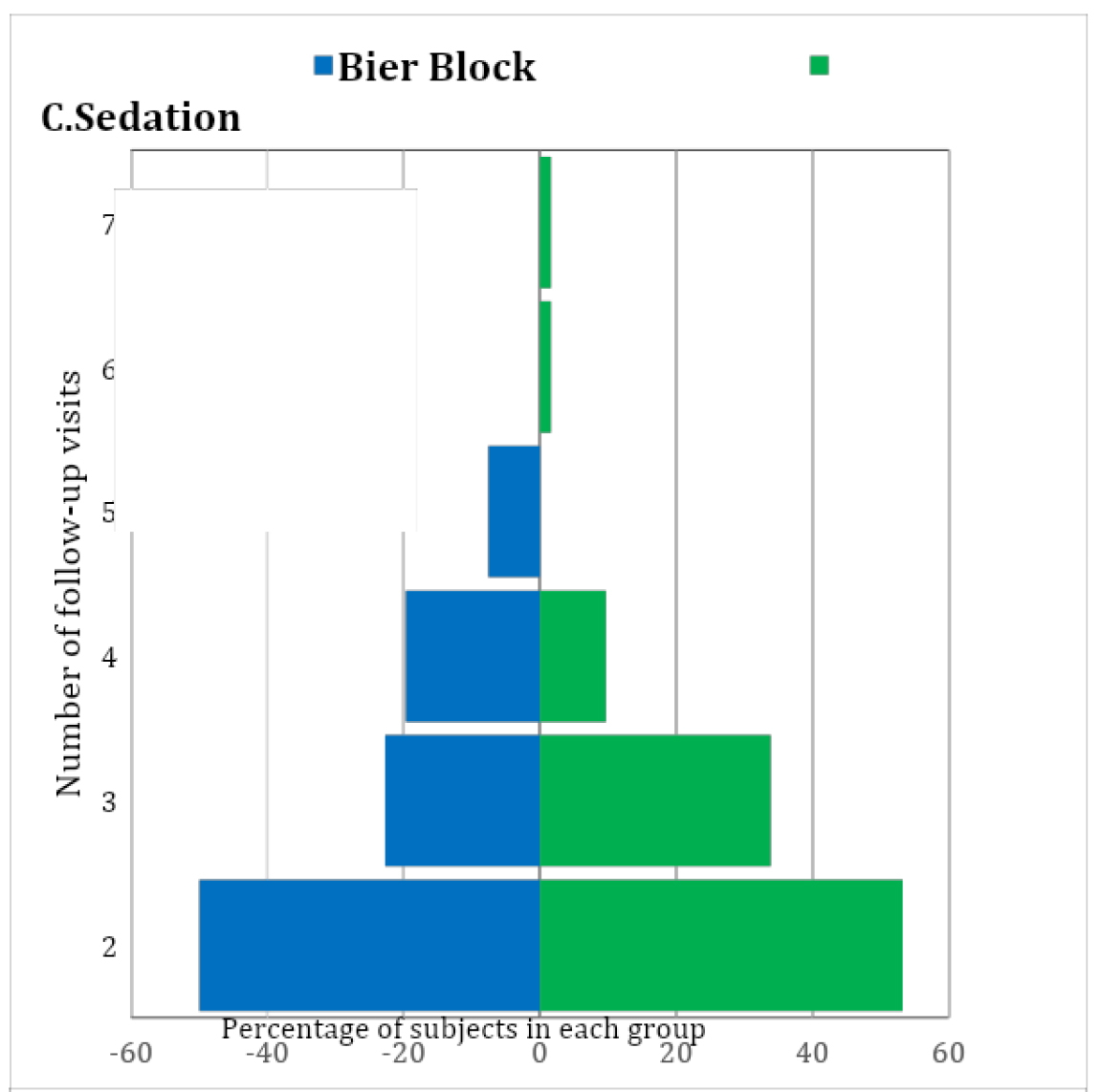
Figure 1: Number of visits distributions between both smethods.

IJOT-2020-303_F2

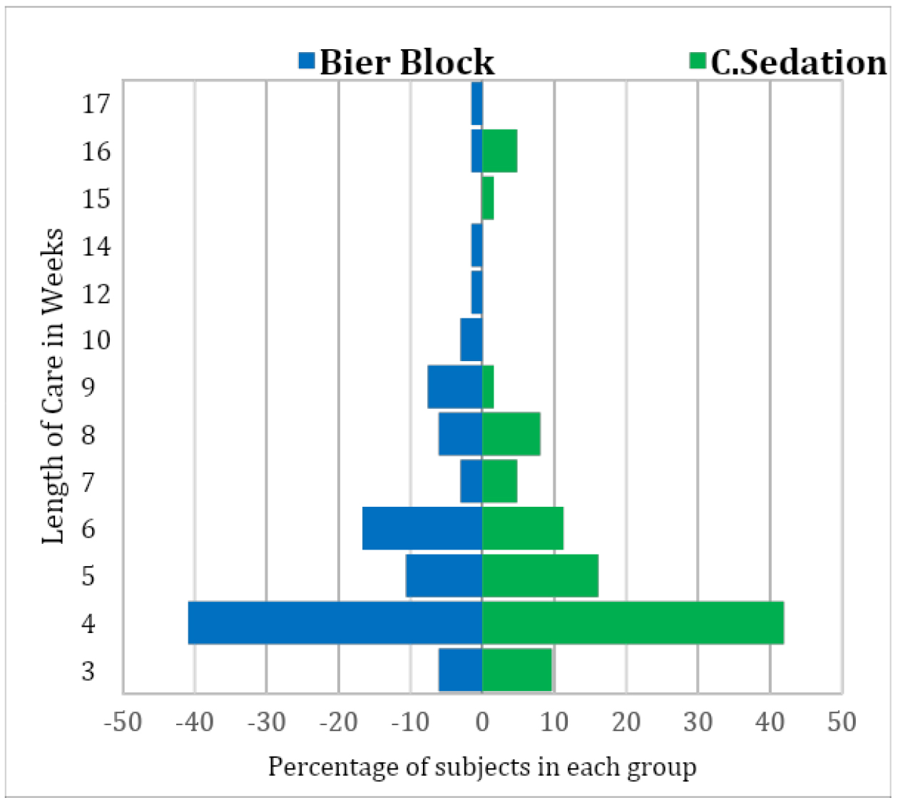
Figure 2: Length of care distribution between both methods.

Table 2. Patient Outcomes between the Two Groups.

Outcome

Overall
(N=128)

Bier Block
(N=66)

C. Sedation
(N=62)

Unadjusted P*

Propensity-adjusted P*

Initial Complications, N (%A):
None

128 (100%)

66 (100%)

62 (100%)

–––

–––

Long-term Complications, N (%A):
None

128 (100%)

66 (100%)

62 (100%)

–––

–––

Number of attempts, N (%A):
1 attempt
2 attempts
3 attempts

124 (97%)
3 (2%)
1 (1%)

65 (98%)
0 (0%)
1 (2%)

59 (95%)
3 (5%)
0 (0%)

0.66

0.36

Need for OR, N (%A):
No
Yes

126 (98%)
2 (2%)

65 (98%)
1C (2%)

61 (98%)
1D (2%)

1.00

–––

Number of follow-ups, N (%A):
2 visits
3 visits
4 visits
5 visits
6 or 7 visits
#visits, Mean (SDB)
#visits, Range

66 (52%)
36 (28%)
19 (15%)
5 (4%)
2 (2%)
2.8 (1.0)
2.0–7.0

33 (50%)
15 (23%)
13 (20%)
5 (8%)
0 (0%)
2.8 (1.0)
2.0–5.0

33 (53%)
21 (34%)
6 (10%)
0 (0%)
2 (3%)
2.7 (1.0)
2.0–7.0

0.32§

0.54§

Total length of care, N (%A):
3 weeks
4 weeks
5 weeks
6 weeks
7 weeks
8 weeks
9–12 weeks
13–17 weeks
       #weeks, Mean (SDB)
#weeks, Range

10 (8%)
53 (41%)
17 (13%)
18 (14%)
5 (4%)
9 (7%)
9 (7%)
7 (5%)
5.8 (3.0)
3.0–17.0

4 (6%)
27 (41%)
7 (11%)
11 (17%)
2 (3%)
4 (6%)
8 (12%)
3 (3%)
5.9 (3.0)
3.0–17.0

6 (10%)
26 (42%)
10 (16%)
7 (11%)
3 (5%)
5 (8%)
1 (2%)
4 (6%)
5.6 (3.1)
3.0–16.0

0.32§

0.82§

Table 3. Cost Distribution Analysis

IVRB1

CS2

Mean (SD3)

$1,177.34 ($253.61)

$1,600.98 ($339.19)

Median

$1,101.71

$1,531.77

Quartiles

$982.26 – $1,318.48

$1,309.91 – $1,923.62

Range

$846.75 – $1,726.27

$1,063.76 – $2,233.47

WRS4 test result

P=0.0003

Discussion

The two main aims of this study were to determine if Bier block regional anesthesia is a safe, effective, and cost-efficient method of anesthesia for pediatric forearm fracture reduction in the emergency department, and to compare the short- and long-term complications and outcomes of Bier-block patients with those of conscious-sedation patients chosen for their overlapping age range. Our institution has a long experience with using Bier blocks in these patients, and we have had found the procedure in children to be both safe and effective. Bier block anesthesia was found to be as safe as conscious sedation in our final study group. Neither final study group had any instance of short-term or long-term complications; specifically, no instances of lidocaine toxicity, compartment syndrome, need for hospital admission for pain control after the procedure, nerve palsy or growth arrest. No child required conversion from Bier block anesthesia to conscious sedation due to inadequate pain control or anxiety despite having a younger group of patients ranging from 4 to 7 years of age. This is a common concern with using Bier block anesthesia in the younger awake child. In our institutional experience, the need for conversion from Bier block to conscious sedation due to inadequate anesthesia or anxiety is very rare. Our emergency department has child life specialists available who can assist in the procedure if needed in the more anxious children by providing distraction and entertainment in the form of reading or tablet usage for games.

One of the aims of this study was to assess follow up data for these two groups. With the rising costs in providing medical care, minimizing the need for, and number of, follow up clinic appointments is valuable. We found that both groups had nearly equal follow up time length and number of follow up visits and both were quite low. Certainly patients in our study’s age group are considered to have very wide tolerances for what constitutes an acceptable fracture reduction due to their tremendous ability to remodel deformity but our data shows that these two methods are equally effective at preventing patients from requiring surgical intervention. One patient in each group did require operative intervention due to inadequate reduction in the emergency department or loss of reduction in follow up, resulting in a 2% rate in each group of a need for surgical intervention in the operating room. The Bier block patient required ORIF and the conscious sedation patient required further closed reduction without internal fixation under general anesthesia.

Some weaknesses of our study include the small patient group sizes. We found it necessary to restrict the age range from the original data that included all patients with forearm fractures, and limit the study to patients who were between 4 and 7 years old. This was due to several factors. First, the majority of patients at our institution receive a Bier block as their form of anesthesia for forearm fracture reduction in the emergency department. If conscious sedation is performed, it is usually reserved for younger patients or the more anxious patients who we perceive may not tolerate a Bier block as well. We were unable to effectively compare the two larger, more inclusive groups due to the large number of Bier-block anesthesia patients and low number of conscious-sedation patients overall and the age differences between the two groups. By lowering and narrowing the age range, we were able to obtain an average age of 6 years in the Bier block group and 5 years in the conscious sedation group. This allowed for more clinically useful comparable data points, but limited the number of patients we were able to compare.

Distributions of the total number of follow-up visits in the Bier-block and conscious-sedation groups, showing that the two groups have very similar distributions. See Table 2 for the means, SDs, and ranges of the distributions.

Distributions of the total length of care in weeks for the Bier-block and conscious-sedation groups, showing that the two groups have similar distributions. See Table 2 for the means, SDs, and ranges of the distributions.

An additional weakness of the study was the probable violation of the “no unmeasured confounders” assumption required for valid propensity-score-based analysis. In addition to each patient’s age and year of injury, we collected their sex, the bone fractured and position of the fracture on the bone, the mechanism of injury, and the number of days to reduction, but not the patient’s race. Race is a pervasive confounder in health-care research that can lead to treatment disparities, not only through provider biases or income disparities, but also through the perceptions and comfort levels of the patients and their parents. Thus, race could easily have been confounded with the choice of anesthesia method in our study. However, it should be said that ours is an equal-access institution where all patients are treated the same regardless of race or socioeconomic background, and this fact should reduce some (if not all) of the unmeasured confounding of race with anesthesia method.

Emergency department visit cost analysis was included in this study for the reason of fiscal responsibility. Bier block anesthesia has been shown to be safe and effective with less total time in the emergency department compared to conscious sedation [7]. We also show an average cost savings of $423 in using Bier block anesthesia compared to conscious sedation. Bier block anesthesia also has the added benefit of not requiring significant specialized post-procedural monitoring that requires trained emergency-department staff that could otherwise be treating another patient. Bier block anesthesia patients do not require any further monitoring after the reduction, which allows for staff to be freed up and, in theory, diminish patient room utilization time. In a high-volume pediatric hospital, decreasing visit time is essential for having an efficient emergency department. We attempted to prove this theory in our study comparing time data between the conscious sedation and Bier block groups, but there were significant limitations with our ability to do that accurately. These limitations included incomplete charting regarding admit and discharge time and NPO status of those patients receiving conscious sedation affecting the wait time before reduction.

Bier block anesthesia is a safe and cost-effective form of anesthesia for pediatric forearm fracture closed reduction in the emergency department in patients between 4 and7 years of age. It has continued to be proven to be safe throughout the years and continues to be shown to be more time and cost effective. Short-term and long-term complication rates are low and are similar to those seen in patients treated with conscious sedation. Follow up time and number of visits are similar between the two groups as well. However, Bier block anesthesia was found to cost significantly less than conscious sedation in our series. We theorize that there are additional indirect cost savings with Bier block compared to conscious sedation as a result of (1) the diminished need for specialized and lengthy monitoring of the patient after the procedure, (2) the NPO status of the patient having no effect on our ability to perform and timing of proceeding with the Bier block, and (3) the efficiency with which one can perform the Bier block in an emergency department setting, although we did not attempt to prove that in this study. Continued research in this field will continue to shed light on this useful method of emergency room treatment of pediatric forearm fractures.

Conflict of Interest

The authors declare that they have no conflict of interest.

Statement of Human and Animal Rights

All procedures followed were in accordance with the ethical standards of the responsible committee on human experimentation (institutional and national) and with the Helsinki Declaration of 1975, as revised in 2008 (5). No identifying information was included in this study.

Statement of Informed Consent

Informed consent was not obtained due to the retrospective nature of this study with no identifying patient information presented in the study. All data was collected under IRB institutional guidelines.

Statement of Funding

No funding was received by any authors for this study.

References

  1. Aarons CE, Fernandez MD, Willsey M, Peterson B, Key C, et al., (2014) Bier block regional anesthesia and casting for forearm fractures: safety in the pediatric emergency department setting. J Pediatr Orthop 34: 45–49.
  2. Barnes CL, Blasier RD, Dodge BM (1991) Intravenous regional anesthesia: a safe and cost-effective outpatient anesthetic for upper extremity fracture treatment in children. J Pediatr Orthop 11: 717–720. [Crossref]
  3. Blasier RD, White R (I1996) Intravenous regional anesthesia for management of children’s extremity fractures in the emergency department. Pediatr Emerg Care 12: 404–406.
  4. Colbern E (1970) The Bier block for intravenous regional anesthesia: technique and literature review. Anesth Analg 49: 935–940.
  5. Mohr B (2006) Safety and effectiveness of intravenous regional anesthesia (Bier block) for outpatient management of forearm trauma. CJEM 8: 247–250.
  6. Perlas A, Peng PW, Plaza MB, Middleton WJ, Chan VW, et al., (2003) Forearm rescue cuff improves tourniquet tolerance during intravenous regional anesthesia. Reg Anesth Pain Med 28: 98–102. [Crossref]
  7. Yang E (2017) Green’s Operative Hand Surgery. 2: 10–11.
  8. Constantine E, Steele DW, Eberson C, Boutis K, Amanullah S, et al., (2007) The use of local anesthetic techniques for closed forearm fracture reduction in children: A survey of academic pediatric emergency departments. Pediatr Emerg Care 23: 209–211. [Crossref]
  9. Guay J (2009) Adverse events associated with intravenous regional anesthesia (Bier block): A systematic review of complications. J Clin Anesth 21: 585–594.
  10. Mendenhall W, Beaver RJ, Beaver BM (2009) Introduction to Probability and Statistics 13th Edition, Cengage Learning, [multiple cities and countries], ISBN-10: 0495389536, Page 403.

GNRB (Medical Device) vs MRI on Anterior Cruciate Ligament (ACL) Tears with Arthroscopic Validation

DOI: 10.31038/IJOT.2020311

Florian Beaurain

SMQ engineer

Master 2 « Medical Device: Design and assessment » (University of Lille, France)

Objective: Comparison of GNRB® versus MRI in the diagnosis of different patterns of anterior cruciate ligament tears.

Requirements: Patients operated for ACL tears or ACL tears + meniscus.

Exclusion Criteria: all patients without isolate ACL tears (without other ligament and bone injuries), patients were not get primary surgery.

Collection of Data

Database of Dr Henri ROBERT (surgeon, specialist on ACL surgery: Operative report, MRI (1.5 T) report and GNRB database for all patients).

Group of Patients

2 groups:

  • Patients with complete ACL tears
  • Patients with partial ACL tears

Statistical Test

We use sensibility like an indicator for average method

Binary Criteria: ACL tears (partial or complete)

Acceptability

For MRI report, if it required interpretation, it shall be null. It must be clearly mentioned complete or partial tears in the conclusion report.

For GNRB, if delta for both knees >3 mm = complete tears and if 1.5 mm ≤ delta <3 mm, partial tears.

Non Inferiority Test

Estimate value: Pr (MRI’s sensibility [1]) by Πr = 0.57

Estimate value: Pe (GNRB’s sensibility [2,3]) by Πe = 0.84

It set α = 5 % unilateral, β = 10 % and δ = 10%.

IJOT-2020-302_e1

For estimation by confidence interval (CI) of difference of proportions

With Pe = GNRB’s sensibility and Pr = MRI’s sensibility and Ne = Nr

nr, ne ≥ 30

nrpr, nr(1–pr), nepe, ne(1–pe) ≥ 5

IJOT-2020-302_e2

Pattern

IJOT-2020-302_f1

Difference of Proportions Test

– Difference test at δ ≠ 0

IJOT-2020-302_e3

Results

This study was performed on data from previous years and two years before for 200 operated patients in total. After exclusion of 64 medical files (one of the 3 data is missing: GNRB, MRI or arthroscopic report), 62 tears were partial and 74 complete with arthroscopy report [Table 1, 2, 3].

Table 1: Table of IRM’s and GNRB’s sensibility with arthroscopy for reference.

MRI vs Arthroscopy for Complete ACL

MRI vs Arthroscopy for Partial ACL

GNRB vs Arthroscopy for Complete ACL

GNRB vs Arthroscopy for Partial ACL

Number

47

22

45

46

Number of Subject

62

74

62

74

Sensibility

0,76

0,30

0,73

0,62

Table 2: Sensibility and specificity of GNRB in the literature.

Complete ACL

Partial ACL

Sensibility

Specificity

Sensibility

Specificity

Robert H [5]

70%

99 %

80%

87%

Klouche S [3]

92%

96 %

92%

98%

Di Ioro A

72%

85%

Lefevre N

84%

81%

87%

87%

Beldame J

62%

75%

Beaurain F

73%

 62%

Table 3: Sensibility of MRI in the literature.

Complete ACL

Partial ACL

Beldame J [1]

 57%

Steltzlen C [4]

32%

For complete tears, MRI’s sensibility was 0.76 and GNRB’s sensibility 0.73. For partial tears, MRI’s sensibility was 0.30 and GNRB’s sensibility 0.62.

For Complete Tears

For estimation by Confidence Interval (CI) of difference of proportions

Conditions for application are verified.

IJOT-2020-302_e4

For Partial Tears [4]

For estimation by Confidence Interval (CI) of difference of proportions

Conditions for application are verified.

IJOT-2020-302_e5

Discussion

This results shows equivalence for ACL’s complete diagnostics (for MRI and GNRB reports) with the literature and for incomplete ACL tears, it’s slightly lower than literature.

Sensibility’s results (for MRI and GNRB reports) for this study are equivalent for complete and partial tears diagnostic in the literature.

Conclusion

Sensibility of GNRB laximetry is quite the same than MRI for complete tears but superior for partial tears.

References

  1. Beldame J (2009) Etude radio-clinique du ligament croisé antérieur [Thèse de Doctorat en Médecine]. [France]. Université de Rouen Normandie.
  2. Lefevre N, Bohu Y, Naouri JF, Klouche S, Herman S (2014) Validity of GNRB® arthrometer compared to TelosTM in the assessment of partial anterior cruciate ligament tears. Knee Surg Sports Traumatol Arthrosc 22: 285–290. [Crossref]
  3. Klouche S, Lefevre N, Cascua S, Herman S, Gerometta A, Bohu Y (2015) Diagnostic value of the GNRB® in relation to pressure load for complete ACL tears: A prospective case-control study of 118 subjects. Orthop Traumatol Surg Res 101: 297–300. [Crossref]
  4. Steltzlen C, Lefevre N, Bohu Y, Herman S (2011) Évaluation clinique d’une série continue de 55 cas de ligamentoplastie partielle du ligament croisé antérieur par la technique TLS (greffe courte aux ischio-jambiers). Rev Chir Orthopédique Traumatol 97: 493.
  5. Robert H, Nouveau S, Gageot S, Gagnière B (2009) A new knee arthrometer, the GNRB: Experience in ACL complete and partial tears. Orthop Traumatol Surg Res 95: 171–176. [Crossref]

Current Treatment of Traditional Chinese Medicine for Chronic Prostatitis/Chronic Pelvic Pain Syndrome and Our Research

DOI: 10.31038/JCRM.2020314

Introduction

Chronic prostatitis/chronic pelvic pain syndrome (CP/CPPS) is a male pelvic floor dysfunction, which usually manifests as urogenital pain, lower urinary tract symptoms, sexual dysfunction and psychological problems [1]. In China, a national survey shows that the prevalence of CP/CPPS in 2009 was 4.5% [2]. The treatment options of CP/CPPS includes antibiotics, α-blockers, anti-inflammatory medications and so on, but we have to admit that patients and doctors are highly dissatisfied with the treatment of this disease [3]. Therefore, it is not surprising that patients often seek other forms of treatment.

In China, it is becoming more and more popular for patients to seek Traditional Chinese Medicine (TCM) treatment. To our knowledge, a systematic review of acupuncture treatment of CP/ CPPS was published in 2016 [4]. A systematic review of the efficacy and safety of moxibustion in the treatment of CP/CPPS was published in 2019[5]. In terms of drug treatment, although phytotherapy such as quercetin [6] and pollen extract [7] are reported to have a certain effect in the treatment of CP/CPPS, Chinese doctors use more compound prescriptions of TCM. There are few reports on oral TCM in the treatment of CP/CPPS, so we focuses on the current treatment of TCM in the treatment of CP/CPPS, as well as our research on the use of GuiHuang prescription in the treatment of CP/CPPS.

Treatment of CP/CPPS with TCM

In an open, multicenter, pre-and post-controlled clinical trial, 240 patients with type III prostatitis who met the diagnostic criteria of the National Institutes of Health (NIH) were treated with Longjin Tonglin capsule, 3 tablets per time, 3 times a day for 12 weeks. Taking NIH chronic prostatitis symptom index (NIH-CPSI) as the main curative effect index, the curative effect was compared before and after treatment. It was found that the total CPSI scores of patients with type III A prostatitis were 23.12 ±6.99(before treatment), after treatment4, 8 and 12 weeks were 18.22 ±6.39,14.12 ±5.88,12.36 ±6.04respectively (P< 0.0l). Before treatment and 4, 8 and 12 weeks after treatment, the total CPSI scores of patients with type III B prostatitis were 23.12 ±6.99,18.22 ±6.39,14.12 ±5.88,12.36 ±6.04 respectively. No abnormal liver and renal function and adverse events were found in the test.

It shows that Longjin Tonglin capsule is safe and effective in the treatment of type III prostatitis [8].

A systematic review on the efficacy and safety of the compound prescription of TCM for clearing away heat and promoting diuresis in the treatment of chronic prostatitis.Meta analysis showed that the compound prescription of traditional Chinese medicine for clearing away heat and promoting diuresis was superior to Prostat(RR1.26,95%CI1.13~1.41), and subgroup analysis showed that this compound was superior to Qianliekang(RR1.32,95%CI1.19~1.45) and quinolone antibiotics (RR1.34,95%CI1.15~1.57). There was no significant difference between heat-clearing and diuresis-promoting TCM and quinolone antibiotics alone (P< 0.01), and there was no serious adverse reaction reported [9]. No serious adverse reactions were reported.

A study was conducted to evaluate the safety and efficacy of Qianlie Shule granule in the treatment of chronic prostatitis. 66 patients with chronic prostatitis were enrolled in a multicenter, open, self-controlled clinical study. The patients were treated with Qianlie Shule granule 1 bag per time, 3 times a day for 6 weeks. The efficacy of the treatment was evaluated with the NIH-CPSI as the main evaluation index, TCM syndrome as the secondary efficacy index, urine routine and liver and kidney function indexes before and after treatment to evaluate its safety, and adverse events were recorded. The Results showed that NIH-CPSI before treatment was significantly higher than that after 6 weeks treatment (P<0.05), and the score of TCM syndrome waiting score was 11.15 ±8.54 before treatment and 3.56 ±3.83 after 6 weeks treatment, and the difference was also statistically significant (P<0.05). Qianlie Shule granule can relieve the urinary system related symptoms of patients with chronic prostatitis (kidney and spleen deficiency, qi stagnation and blood stasis syndrome), reduce NIH-CPSI and TCM syndrome score, the clinical effect is significant [10].

Our research

The rich clinical experience handed down by the ancestors of TCM for thousands of years, should also seriously evaluate the curative effect of TCM in accordance with the principle of evidence-based medicine. For this reason, we carried out the clinical study of GuiHuang prescription in the treatment of CP/CPPS. This study was randomly divided into two groups: treatment group (GuiHuang prescription group, n=33) and control group (Tamsulosin group, n=33). The patients were treated for 6 weeks and followed up for 2 weeks according to NIH-CPSI score and TCM symptom score. To observe the safety and efficacy of GuiHuang prescription in the treatment of CP/CPPS with dampness-heat stasis. The project was reviewed by the Medical Ethics Committee of Xiyuan Hospital on June 21, 2019, and obtained the ethical approval (batch number: 2019XLA019-3). The project was registered by the China Clinical trial Center (Registration No.: ChiCTR1900026966) on October 27th, 2019. At present, the project is still recruiting. The composition of GuiHuang prescription includes: Angelica 12g, Phellodendron chinense 12g, honeysuckle 15g, turmeric 10g, frankincense 5g, myrrh 5g, Angelica dahurica 10g, tangerine peel 10g, plantain 15g, Hedyotis diffusa 15g, which has the effect of clearing heat and removing dampness, removing blood stasis and relieving pain.

Comment

The best treatment for CP/CPPS has yet to be determined, and the basic strategy is based on symptom control and anxiety relief. The UPOINT phenotypic system (Urinary symptoms, Psychosocial dysfunction, Organ specific, Infection, Neurologic dysfunction and Tenderness of muscles ) play an important role in guiding the clinic [11]. At the same time, it reflects the individual differences of patients, with different phenotypes of different drugs, which is similar to the syndrome differentiation and treatment of TCM. It appears that a tailored treatment strategy addressing individual patient characteristics is more effective than one single therapy[12].

The use of TCM compound prescription, need syndrome differentiation addition and subtraction, according to the different physique and symptoms of patients, will use different prescriptions, there will be a patient a treatment prescription, which is not conducive to evidence-based research. In order to avoid insufficient, we fixed the composition of GuiHuang prescription, studied the patients in accordance with this prescription (damp-heat stagnation type), and we did not add or subtract the prescription. This can ensure the accuracy of the research results, and we are full of expectations for the results.

The compound prescription of TCM, which has been used clinically, such as Ningmitai capsule[13], which is a commercial formula, has been reported to have a good therapeutic effect and is safe. In clinical practice ,the use of TCM compound alone, or combined with western medicine is our Strategy for the purpose of clinical efficacy. There are also external uses of TCM, such as enema, TCM sitz bath.

The use of TCM compound prescription in the treatment of CP/ CPPS is very common. In our department, the proportion of patients taking oralTCM is more than 80%, but unfortunately, our reports are relatively few. The characteristics of TCM are often the first clinical use, such as GuiHuang prescription, we have used for more than a decade, the clinical effect is accurate, and then we are in clinical observation and research, have obtained a higher level of evidence, and then carry out mechanism research. However, we have some shortcomings, there are 10 herbs in the prescription, and the exact mechanism of the effect is not clear. At present, many studies are published in Chinese, which is not conducive to international promotion, and the recognition of research is affected. More published English-language clinical trials are needed to prove its effectiveness. We believe that TCM can be used as a supplementary treatment option for CP/CPPS.

Acknowledgement

This work was financially supported by Xiyuan Hospital Project, Grant number 2019XYMP–23

References

  1. Rees J, Abrahams M, Doble A, et al. (2015) Diagnosis and treatment of chronic bacterial prostatitis and chronic prostatitis/chronic pelvic pain syndrome: a consensus guideline[J]. BJU Int, 116(4):509-525.[crossref]
  2. Liang CZ, Li HJ, Wang ZP, et al. (2009)The prevalence of prostatitis-like symptoms in China. J Urol182:558–63.[crossref]
  3. Anothaisintawee T , Attia J , Nickel J C , et al. (2011) Management of Chronic Prostatitis/Chronic Pelvic Pain Syndrome A Systematic Review and Network Meta-analysis[J]. JAMA The Journal of the American Medical Association, 305(1):78-86.[crossref]
  4. Qin Z, Wu J, Zhou J, et al. (2016) Systematic review of acupuncture for chronic prostatitis/chronic pelvic pain syndrome. Medicine (Baltimore) 95:e3095[crossref]
  5. Cao Q, Zhou X, Chen J, et al. (2019) Efficacy and safety of moxibustion in patients with chronic prostatitis/chronic pelvic pain syndrome: A systematic review protocol[J]. Medicine, 98(20):e15678.[crossref]
  6. Shoskes DA, Zeitlin SI, Shahed A, Rajfer J. (1999) Quercetin in men with category III chronic prostatitis.Urology.54(6):960-963. [crossref]
  7. Wagenlehner FM, Schneider H, Ludwig M, et al. (2009) A pollen extract (Cernilton) in patients with inflamma-tory chronic prostatitis-chronic pelvic pain syndrome.EurUrol. 56(3):544-551. [crossref]
  8. Shang X J, Geng Q, Duan J M, et al. (2014) [Efficacy and safety of Longjintonglin Capsule for the treatment of type III prostatitis].[J]. 20(12):1109-12.[crossref]
  9. Ming-xingQiu, Guo-bingXiong, Shi-yi Zhou, et al. (2007)Qingrelishi-category Chinese medicine for chronic prostatitis: a systematic review[J]. National Journal of Andrology, 13(4):370-377.
  10. Zhoushaohu,Guojun,Cuigang,et al. (2019) Qianlieshule Granules for chronic prostatitis: A multicenter self-controlled clinical trial[J]. Chinese Journal of Andrology.33(05):48-51.
  11. Shoskes D, Robert D, Nickel C.(2010) 798 phenotypically directed multimodal therapy for chronic prostatitis/chronic pelvic pain syndrome: a prospective study using upoint[J]. The Journal of Urology, 183(4):e312.[crossref]
  12. ThunyaratAnothaisintawee, John Attia, J Curtis Nickel,et al.(2011) Management of Chronic Prostatitis/Chronic Pelvic Pain Syndrome A Systematic Review and Network Meta-analysis[J]. Jama, 305(1):78-86.[crossref]
  13. Jin C, Chen Z, Zhang J.(2018) Meta-analysis of the efficacy of Ningmitai capsule on the treatment of chronic prostatitis in China.Medicine (Baltimore). 97(33):e11840.[crossref]

On the Muricid snail, Plicopurpura pansa, in the Pacific coast of Central America

DOI: 10.31038/AFS.2020213

Abstract

The Muricid snail, Plicopurpura pansa, in Central America has been studied, and the secretion of hypobanchial gland and its utilization are discussed. P. pansa exudes a few drops of secretion in the gland at being picked up. The secretion has been used historically in various forms for dyeing. There is no other region where the dyeing by drops has been carried out. It is noteworthy that the shell is not hammered or cracked, and the snail is released or put back at the same place where it was. The name of “Pacific purple” is proposed for the dye and its color. A decreasing the stock population and the smaller individuals in size are reported. The Pacific coast of Central America is a unique area of artisanal textile culture, and intensive investigations on the species are now required for biological and cultural conservation.

Keywords

Muricid, Plicopurpura pansa, Central America, purple dye, dyeing method, Pacific purple, secretion, hypobranchial gland, 6,6’-dibromoindigo

Secretion of the gland and dyeing

The present study of Plicopurpura pansa focuses on the secretion in hypobranchial gland and a utilization of that, and reviews the result of field studies in El Salvador. Little comparative study among the species in other regions was conducted.

 The Muricid snails are characterized by the hypoblanchial gland, and its secretion makes it possible to dye textiles. The dying activities are reported historically in the Pacific coast of Central America and in Mediterranean region, especially at Phoenicia [1, 2]. In European countries, the dye and its color has been called Tyrian purple, Royal purple, Phoenician purple, and so on [2,4]. The color pigment was revealed to be 6, 6’-dibromoindigo, and the chemical structure is shown (Figure 1) [3].

AFS_2020-Hiroshi KITANI_F1

Figure 1. Chemical structure of 6,6’-dibromoindigo, and the process of reduction, leuco- 6,6’-dibromoindigo (Sawada 2014)

The distribution of P. pansa in El Salvador is limited at headland area with breaking water and rocky shore, not sand beach or stone beach, and the dyeing activities have been carried out in there.

The present study groups the dying methods into the two (Figure 2). The one (A) is the method by the drops of secretion and the second (B) by the tissues cut out of the gland. The former has been introduced only in Central America.

AFS_2020-Hiroshi KITANI_F2

Figure 2. Dyeing methods by the secretion of Muricid snail

It is observed that P. pansa exudes a white liquid of secretion at being picked up, which means that the white liquid flows out in the shell aperture, and it’s volume is about 0.3ml or 3 drops per individual of 3cm/shell length. However no comparative study about the relation between the volume and the body weight/size was conducted, the volume of the secretion of P. pansa is supposed to be too much than that of other Muricid. It is a characteristic of the species, and other Muricid requires a cracking the shell and cutting the tissue out of the gland for extracting dye pigment.

The method by drops (A) is very efficient for dyeing directly. The present study confirmed a stocking of fresh drops in refrigerator for a few days, which is very convenient for laboratory works. At flowing out the white liquid into the aperture, it is possible then to drop the liquid and dye directly the cotton yarn in the hand. The color of drops changes gradually from milky white, white green, and finally to purple color in 15 minutes under the sunlight.

The second (B) requires a process of cutting the tissue out of hypobranchial gland. After grinding the tissue in a bowl to be a watery paste, it is now possible to use it directly for painting or dying textiles (B1). Furthermore, the cut-out tissue is applicable for extracting the dye by chemical reductant (B/B2, B3) (Figure 2). After obtaining the paste (B), an adding water makes it possible to filter the fluid, and then the dye pigment, 6,6’-dibromoindigo, is extracted in the liquid filtered. It is now possible to reduce the liquid by adding the reductant such as sodium hydrosulfite (Figure 1). Then, the dyeing textile is possible in a dark room and then after under the sunlight for coloring. In this case, a volume of textile can be dyed smoothly. Powdered dye is also available after drying the filtered liquid, and then the dyeing is introducible at any place and at any time under the same procedure of B1. The method by reduction (B) has not been introduced in Central America.

The dye and its color have been called in general Tyrian purple [2,4], but it is considered to be a product by reduction. That product has not been introduced in Central America, neither Tyrian purple. The dyeing method by the pure secretion and that by reduction are not the same, however the color pigment 6,6’-dibromoindigo is the same among the species. The present study names it “Pacific purple” for the dye and its color originated from the drops of P. pansa, which shows a better distinctiveness of the dyeing.

Biology

The functions of the secretion have been unclear. Some observations reported a toxic or anesthetic substance against the prey. Muricid such as oyster drills is a well known predator and a possible user of secretion to oysters. It is observed in laboratory that Rapana sp. uses the secretion against bivalves. The present study observed P. pansa preying on a smaller snail, Littorina sp.. It is experienced that the whole meat stimulates or irritates the tongue at eating and is inadequate for seafood, however some large sized snails such as Conchalepas sp. in Chile and Peru, and also Repana sp. in Turkey are important sea foods, but the secretion of these species are not utilized currently.

The habitat of P. pansa in El Salvador is limited on the rock surface or in a crack of rock, and stays for days at 3–5 m over the water line, where is a zone of without or less water. Video movies in Mexico show the collecting snails and dyeing at water line, which is a remarkable difference of the habitat. The behavior and habitat induce an assumption that the secretion would effective for a respiration while staying over the water line, because it is supposed that the water stocked inside the shell may be insufficient for staying days over the water line. It is supposed that Muricid in the water does not need additional oxygen, and secretes successively little volume of secretion. No conclusive study has been conducted on the secretion, but the present observation on P. pansa concludes that the releasing snails after dyeing should be returned to around the water line, not the same place where it was.

It is reported also that the Muricids lay eggs in capsules and it is reported that damaged capsules show a dark purple color, which suggests that the liquid in the capsule would contain the color pigment or the secretion. The biological study on egg capsule would be effective for larval production in laboratory.

There are little biological and ecological studies such as growth, reproduction, larval development, habitat, daily and seasonal movement, stock population, regional difference of the species, ad so on, which seem to be obstacles for the advanced developments and studies of P. pansa.

Development and conservation

Central America is the only place where the dyeing by secretion of Muricid is carrying out. It is a traditional culture for a long time [1], and the Pacific purple has been popular among the local people, however a limited production due to unstable dyeing works at seashore. No evidence of symbol color of authority or status has been reported as was Tyrian purple in Europe.

The dyeing textile by drops is very efficient and there would be no local need for improving the method or developing a new utilization. The present study has observed that a general interest in the natural purple is fading away, but a high interest exists also in enjoying the dye and its color by different ways from the past, specially in mysterious color changing every second, saying “palm-top miracle and wonder”.

The present study suggests new utilizations such as a body painting, nail art, painting on the T-shirts, tourisms, traditional dyeing, biological/chemical study for younger generations, as well as a possible pharmaceutical and cosmetic use, and so on. The chemical product of 6,6’-dibromoindigo has been developed synthetically in Japan and it is reported that the examination of the fatness such as rubbing, washing, perspiration, and against light shows a better grade than those of indigo [3].

There seems to be little technical obstacles for utilizing the drops, but exists biological obstacles mentioned previously. Furthermore, some articles [1, 5] reported a possible fade-out of the culture and a heavy exploitation of the resources in the past.

The textile culture in Central America is very unique, and the tradition of living together with the resources of Pacific purple is highly praised. The present study emphasizes the necessity of effective biological and cultural conservation programs for the regional assets before being forgotten.

References

  1. Secretaría de Educación Pública(SEP) (1988) El caracol púrpura, Una tradición Milenaria en Oaxaca, Mexico,ISBN 968-29-1867-7 (in Spanish)
  2. Takako Terada (2005) Fieldwork on shellfish purple, Kwassui bulletin 48, p51–61. Kwassui Women’s University. (in Japanese)
  3. Tadanobu Sawada, Hiroyuki Ishii, Harue Senou, Toyotoshi Ueda (2014) Color Fastness of Ancient Purple 6,6’-Dibromoindigo after Dyeing. The Journal of Silk Science and Technology of Japan.  22: 57–63.
  4. Ludwig CA, Naegel, Federico A. Garcia-Dominguez (2006) Reproductive cycle of the purple snail Plicopurpura pansa (Gould 1853) from two locations at Baja California Sur, Mexico. Journal of Shellfish Research 25: 925–933