Monthly Archives: August 2019

Effect of N-Methyl-D-Aspartate Receptor Blockade on Anxiety-Like Behavior Induced in Rats by Postnatal Chronic Exposure to the Endocrine Disruptor Di-2 (Ethyl-Hexyl Phthalate) in Elevated plus Maze Test

DOI: 10.31038/EDMJ.2019343

Abstract

Di-2-Ethylhexyl Phthalate (DEHP) is the widely used to convey flexibility and transparency to plastic products made of polyvinyl chloride and also in the manufacture of medical devices. DEHP disrupts reproductive tract development in an antiandrogenic manner and also may induce neurobehavioral changes. In previous works, we demonstrated that chronic postnatal exposure to DEHP alters the neuroendocrine regulation of the testicular axis, modifying the hypothalamic concentration of excitatory neurotransmitters and therefore induces an anxiogenic effect. In the Elevated Plus Maze (EPM) test, dizocilpine (MK-801) induces a decrease in anxiety-related behaviors throughout NMDA receptor blockade. The objective of this work was to investigate whether the blockade of NMDA receptors of glutamate by the non-competitive antagonist MK-801 could modify the anxiety-like behavior induced by chronic postnatal exposure to DEHP (30 mg/kg body weight/day, orally from birth) in young adult male rats in the EPM test. The results show that NMDA receptor blockade by MK-801 (0.1 mg/kg body weight, i.p.) in DEHP exposed animals is able to produce a significant decrease in time spent in closed arms (TSC) and in Freezing Time (FT) as well as an increase in time spent in open arms (TSO) in the EPM test, indicating an anxiolytic effect. In conclusion, our results suggest: 1) NMDA receptor blockade by MK-801 can reverse anxiety-like behavior induced by exposure to DEHP during the early period of life. 2) The glutamatergic system is involved in the anxiogenic effect of phthalate, which is probably triggered by its known antiandrogenic action.

Keywords

DEHP; Endocrine Disruptor; Dizocilpine (MK-801); Glutamate Receptors; Anxiety like-behavior

Introduction

N-Methyl-D-Aspartate (NMDA) receptor-mediated glutamate transmission is one of the most significant mechanisms during multiple stages of brain development and has been implicated in cognitive functions and emotional responses [1]. It has been suggested that during the early neurodevelopmental period the treatment with non-competitive antagonist of NMDA receptors such as dizocilpine (MK-801), can cause long-term effects in the anatomical, neurochemical, neurophysiological and behavioral features of rodents [2,3]. Indeed, there are evidences that MK-801 has an anxiolytic potential [4–6]. During the critical period of brain maturation, MK-801 induces locomotor hyperactivity and decreases anxiety levels in adolescent rats.6 In the elevated plus maze (EPM) test a decrease was observed in anxiety-related behaviors caused in adult male mice by MK-801 in the early developmental period [7]. NMDA receptor blockade could reduce neuronal activity in pathways which lead to the release of GABA or monoamines, and which have themselves been implicated in anxiety-related processes [8].

Di-2(ethyl-hexyl phthalate) (DEHP) is an endocrine disruptor with antiandrogenic action, which is used as a plasticizer in many products, especially in medical devices and in manufacturing a wide variety of consumer products made with Polyvinyl Chloride (PVC) [9]. The brain has been determined to be at risk of DEHP exposure. Gestational and postnatal exposure to DEHP can affect neurodevelopment and lead to anomalies by disrupting normal brain development and function [10]. In previous works, we demonstrated that chronic postnatal exposure to DEHP alters the neuroendocrine regulation of the testicular axis, modifying the hypothalamic concentration of excitatory and inhibitory neurotransmitters [11,12]. DEHP could act on anxiety in different ways depending on the treatment, age, and sex of the animals [13–16]. It has been reported that postnatal exposure to DEHP can induce anxiogenic effect in pre and peripubertal male rats but not in females in the same stages of sexual maturation. Moreover, it has been proposed that the decrease in testosterone levels induced by postnatal exposure to DEHP could be one possible mechanism underlying DEHP anxiogenic-like behavior in immature male rats [17]. Recently, we have demonstrated that GABA agonists, muscimol and baclofen, can reverse DEHP neuroendocrine effects as well as its anxiogenic action in young adult male rats, supporting the notion that GABAergic system may be one of the neurotransmitter systems involved in the effects produced by DEHP exposure during early periods of neurodevelopment [18].

On these bases we hypothesized those DEHP-induced modifications in the concentration of excitatory amino acids in the brain of immature rats [11,12] could also have a modulatory role in the anxiogenic behavior induced by exposure to this endocrine disruptor during early periods of neurodevelopment. Therefore, the objective of this study was to investigate whether the blockade of NMDA receptors of glutamate by the non-competitive antagonist MK-801 could modify the anxiety-like behavior induced by chronic postnatal exposure to DEHP in young adult male rats in the elevated plus maze (EPM) test.

Materials and Method

All animal procedures were performed following the protocols of the National Institute of Health—Guide for the Care and Use of Laboratory Animals. The approval to conduct the study was granted by the Animal Care and Ethics Committee of the School of Medicine, Universidad de Buenos Aires (UBA; CICUAL).

Animals

Wistar rats used were provided by the Department of Physiology, School of Medicine, UBA, Argentina. Animals were kept under a controlled environment (temperature 22–24C; lights on from 7.00 h to 19.00 h) and they had free access to food and filtered water, until the time of killing. All animals were fed with balanced food for laboratory rodents (Cooperation, ACA-16014007, Asociación de Cooperativas Argentinas-División de Nutrición Animal). The diet contained 15% of soy, but as the food used and the quantity of food intake by control and DEHP-treated groups were similar, we assumed that all animals were exposed to equivalent levels of food-borne phytoestrogens. Moreover, the same lots of diet were provided to animals from all groups at the same time, during the course of the study, to control across groups for possible variation in the diet content. We used ultrapure-filtered water (obtained from EDS-Pack, Millipore Merck, installed in the Milli-Q water system) that was presumed to be free of phthalates and other Endocrine Disruptors (EDs). To minimize additional exposures to substances that may act as EDs, rats were housed in stainless steel cages with wood beddings and water was supplied in glass bottles.

Drugs and Doses

DEHP was purchased (1 g/ml>99% pure; Aldrich Chemical Company, Inc., Milwaukee, Wisconsin, USA) and the final solution administered to the animals was made up fresh daily by adding 200 ml of DEHP to 1 l of filtered water to reach a concentration of 0.2 mg/ml and sonicating for 30 min to ensure a permanent and homogenized solution. An oral route in DEHP administration was chosen intending to mimic best the most common route of human exposure to the ED. The estimated average DEHP dose of exposure was 30 mg/kg body weight (BW)/day, based on the daily intake of DEHP solution and the weight of the related animal. Liquid consumption was measured calculating the difference between the amount of liquid placed in the bottle every day and the remaining amount on the following day to assess the intake. It was assumed that all DEHP solution missing in the bottle had been consumed by the animals. Assessments did not contemplate possible leakage or evaporation of the solution or potential loss of DEHP activity during the 24-h period. No significant differences between the amount of liquid consumed by dams and pups receiving DEHP and those which did not receive this chemical were found. The dose was chosen based on prior studies published by us, in which we demonstrated alterations in the reproductive axis and in the behavior in immature male rats exposed to DEHP at a dose of 30 mg/kg BW/day but not in animals exposed to a lower dose (3 mg/kg BW/day) [11,12,17,18].

On the other hand, MK- 801 ([(+)-3 (2-carboxypiperazine-4-yl) propyl-1-phosphonic acid]; Research Biochemicals Inc., Natick, Mass, USA) was dissolved in saline and injected at dose of 0.1 mg/kg BW, i.p., 1 h before performing the behavioral test. MK-801 can be administered intraperitoneally because it crosses easily the blood-brain barrier. The chosen dose does not produce toxic effects [19].

Experimental Design

Pregnant dams were placed individually in metallic cages and upon delivery, pups were sexed; male pups were separated and distributed with one surrogate dam (n= eight male pups per dam). On postnatal day (PND) 1, surrogate dams with their male pups were randomly assigned into control (C) and DEHP exposure groups. Dam’s exposure to DEHP began on PND 1 and continued until weaning. On PND 21, pups of each group continued receiving the same treatment until PND 60. On this day, control and DEHP-exposed male pups were randomly assigned (n= 8 animals per group) to the following treatments: (1) W + S: controls that were given water and injected with saline; (2) DEHP + S: animals that were given DEHP and injected with saline; (3) W+ MK-801: animals that were given water and injected with MK-801; (4) DEHP + MK-801: animals that were given DEHP and injected with MK-801. After receiving the MK-801 or the vehicle, animals were submitted to the behavioral test.

EPM test

The elevated plus maze (EPM) test is a widely used behavioral assay for rodents, and it has been validated to assess the antianxiety effects of pharmacological agents and steroid hormones and to define mechanisms underlying anxiety-related behavior [20]. The EPM apparatus consists of two open arms (10 x 50 cm), alternating in right angles with two closed arms (10 x 50 x 10 cm), delimiting a central area. The maze was elevated 50 cm above the floor. Before starting the test, animals were individually placed in a rectangular plastic glass area (40 × 40 cm) for 5 min in order to habituate them to the test environment. After that, the rats were placed in the central area of the maze, facing one of the closed arms, and were allowed to explore it for 5 min. The maze’s arms were equally illuminated so that the animals did not perceive lighting differences. Behavioral tests were performed from 12: 00 h to 14: 00 h. We used 10% ethanol to clean each arm of the maze and to remove olfactory cues every time between trials. Each rat was tested only once. The animal’s behavior was videotaped and the number of entries and the time spent in both, open and closed arms, were measured by an observer. The parameters measured were: Time Spent in Open (TSO) and Closed (TSC) arms, Total number of Entries (TE) and Time of Freezing (FT). These parameters were measured following a four-paw criterion; entry into the arm of the EPM was defined as the animal placing all four paws in that particular part of the maze. It is considered that anxiety-like behavior is characterized by a decrease in TSO and an increase in TSC. On the other hand, TE provides a built-in control measure for general hyperactivity or sedation. TSO, TSC and FT were expressed in seconds.

Statistical Analysis

Results are presented as means ± SEM. The statistical analysis was based on individual offspring numbers. The surrogate dam was not used as the unit. Therefore, we did not consider the potential effects of the surrogate dam in the statistical analysis, as pups were exposed to this chemical through her milk. All data were checked for normality by the Kolmogorov–Smirnov test and then, they were analyzed using Kruskal-Wallis test (non-parametric ANOVA); posthoc Mann Whitney (Bonferroni correction) to compare unpaired groups. The statistical software used was GraphPad InStat 3 and a difference was considered statistically significant when p < 0.05.

Results

Figure 1 shows the effect of MK-801 on TSO in adult male rats exposed to DEHP from birth. Significant differences between treatments were found (K (3) =25.860, p < 0.0001). When compared with the control group, a significant decrease in TSO were observed in rats exposed to DEHP (DEHP+S vs W+S, p < 0.01) while MK-801 treatment increased this parameter (W+MK vs W+S, p <0.001). MK-801 also produced an increase in TSO with respect to DEHP-exposed group (W+MK vs DHEP+S, p < 0.001) and was able to reverse the effect of the endocrine disruptor (DEHP+MK vs DEHP+S, p<0.001). No significant differences were found between W+MK and DEHP+MK groups, p=0.7984).

EDMJ 2019-119 - Rodolfo A. Cutrera Argentina_f1

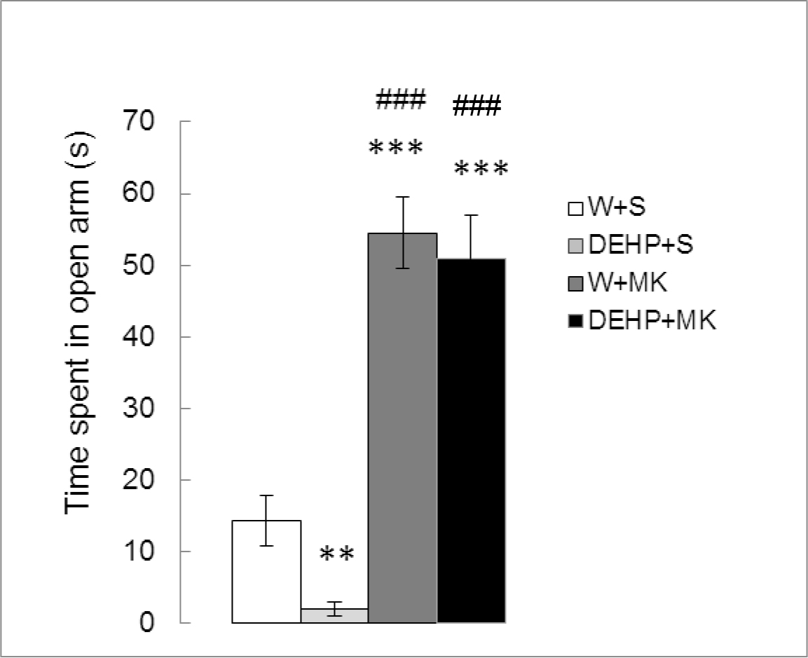
Figure 1. Effect of acute administration of MK-801 on the time spent in open arms in adult male rats exposed postnatally to DEHP. W: water; S: saline; DEHP: di-2(ethyl-hexyl phthalate; MK: MK-801. ** p<0.01 vs W+S; *** p<0.001 vs W+S; ### p<0.001 vs DEHP+S. Each value represents the mean ± SEM of eight animals per group.

As can be seen in Figure 2, there were significant differences in TSC between treatments (K (3) = 14.809, p<0.01). DEHP significantly increased the TSC compared to the control group (DEHP+S vs W+S, p < 0.05) while MK-801 produced a significant decrease (W+MK vs W+S, p < 0.05) and reversed the effect of DEHP (DEHP+MK vs DEHP+S, p<0.01).

EDMJ 2019-119 - Rodolfo A. Cutrera Argentina_f2

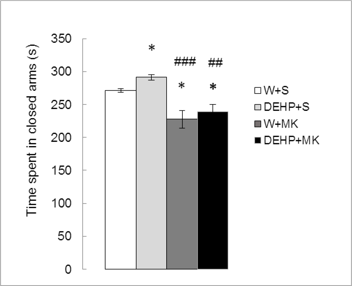
Figure 2. Effect of acute administration of MK-801 on the time spent in closed arms in adult male rats exposed postnatally to DEHP. W: water; S: saline; DEHP: di-2(ethyl-hexyl phthalate; MK: MK-801. * p<0.05 vs W+S; ## p<0.01 vs DEHP+S; ### p<0.001 vs DEHP+S. Each value represents the mean ± SEM of eight animals per group.

Figure 3 shows the number of total entries in both arms of the maze. Significant differences were detected between treatments (K (3) = 23.642, p<0.001). As expected, there were no significant differences in DEHP-treated animals with respect to the control group (DEHP+S vs W+S, p =0.2627). MK-801 produced a significant increase in TE (W+MK vs W+S, p < 0.001). The same effect was observed with DEHP + MK (DEHP+MK vs DEHP+S, p<0.001). No differences were found between both groups treated with MK-801 (DEHP+MK vs W+MK, p=0.2475).

EDMJ 2019-119 - Rodolfo A. Cutrera Argentina_f3

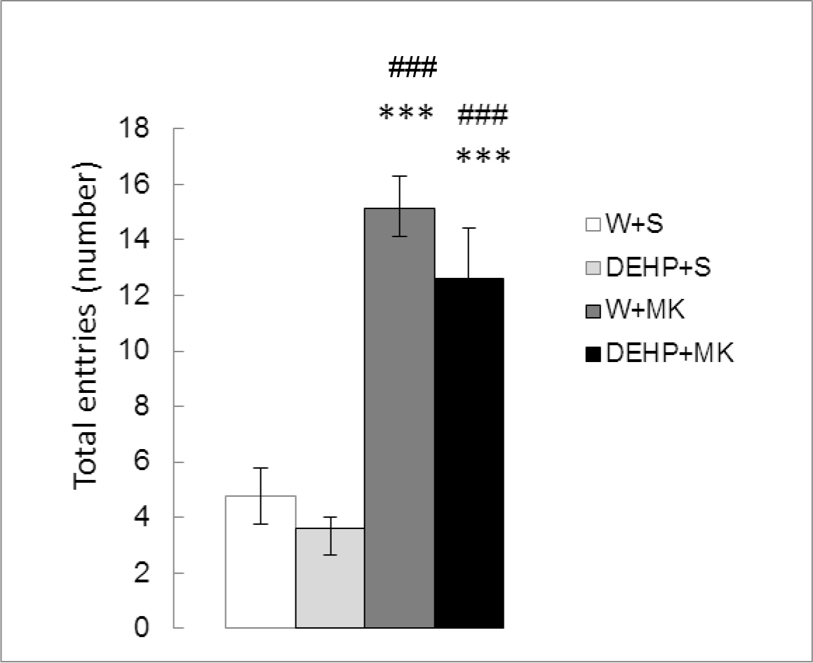
Figure 3. Effect of acute administration of MK-801 on the number of total entries in adult male rats exposed postnatally to DEHP. W: water; S: saline; DEHP: di-2(ethyl-hexyl phthalate; MK: MK-801. *** p<0.001 vs W+S; ### p<0.001 vs DEHP+S. Each value represents the mean ± SEM of eight animals per group.

Time spent in freezing behavior (Figure 4) showed significant differences between groups (K(3) = 26.042, p<0.001). There was a significant increment in DEHP-exposed rats (DEHP+S vs W+S, p<0.001) and a significant decrease in animals injected with MK-801 (W+MK vs W+S, p<0.001). In addition, MK-801 antagonized the effect of DEHP ((DEHP+MK vs DEHP+S, p<0.001).

EDMJ 2019-119 - Rodolfo A. Cutrera Argentina_f4

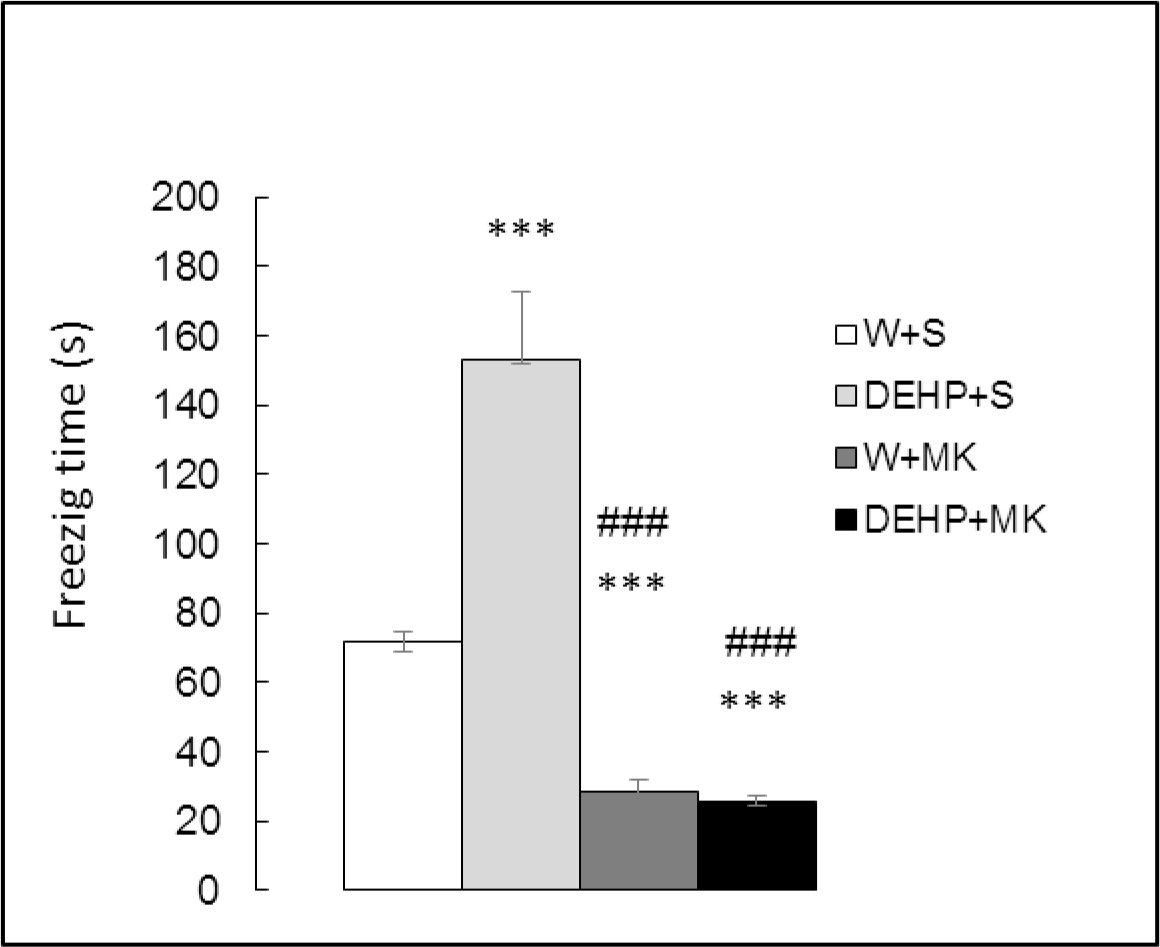
Figure 4. Effect of acute administration of MK-801 on freezing time in adult male rats exposed postnatally to DEHP. W: water; S: saline; DEHP: di-2(ethyl-hexyl phthalate; MK: MK-801. *** p<0.001 vs W+S; ### p<0.001 vs DEHP+S. Each value represents the mean ± SEM of eight animals per group.

Discussion

In order to investigate if the NMDA receptors of glutamate are involved in the changes in the anxiety-like behavior induced by early exposure to endocrine disruptor DEHP, we compared the performance in the Elevated plus Maze of adult male rats exposed to DEHP postnatally and treated or not with MK-801, a non-competitive antagonist of glutamate receptors. Our results showed that NMDA receptor blockade by MK-801 was able to reverse anxiety-like behavior induced by exposure to DEHP during the early period of life.

As we expected, our results showed an anxiogenic effect of DEHP, which was evidenced by a significant increase in TSC and a decrease in TSO in the EPM test. These results were consistent with the findings reported in immature rats and mice exposed perinatally to DEHP (10, 30, 50, and 200 mg/kg) [17,21] as well as in young adult male rats chronically exposed to DEHP (30 mg/kg) from birth [18].

However, the mechanism underlying the enhanced anxiety-like behavior induced by DEHP is not clear. In one way DEHP may influence anxiety-like behavior by disturbing the hypothalamic-pituitary-adrenal axis (HPA). In fact, exposure of male rats to DEHP leads to an age-dependent activation of the HPA ‘in vivo’ and of adrenocortical steroidogenesis ‘ex vivo’, showing a specific susceptibility of immature animals to this phthalate [22]. Perturbations of hormonal homeostasis of the HPA axis were found in adolescent female rats exposed to DEHP during lactation [23]. Direct and transgenerational effects of perinatal DEHP exposure on social behaviors and anxiety-like behavior in mice, as well as transgenerational effect of DEHP on corticosterone levels were reported by other authors [24,25]. Another way in which DEHP could influence anxiety is by altering the hypothalamic-pituitary-gonadal axis. It has been reported that gestational and lactational DEHP exposure is able to disrupt the neuroendocrine control of the gonadal axis during sexual maturation in rats [11,12]. The changes induced by DEHP in the hypothalamic concentration of GABA, the inhibitory neurotransmitter involved in the neuroendocrine control of the gonadal axis as well as in the development of anxiety, support the idea that the GABAergic system could participate in the behavioral effects of the DEHP [18]. Moreover, in the adulthood perinatal DEHP-exposed males displayed anxiogenic status, which correlate with a decrease in testosterone levels [7]. Given that the treatment with testosterone was able to reverse the disruptive effect of chronic postnatal exposure to DEHP on the testicular axis of rats, it was suggested that the antiandrogenic action of this chemical could be a probable mechanism underlying its anxiogenic effect [17]. In addition, Xu et al [21]. have suggested that the inhibition of the mitogen-activated protein kinase/extracellular signal-regulated kinases (MAPK/ERKs) activity, which mediates the non-genomic actions of testosterone in the brain cells, as well as the down-regulation of androgen receptors expression in hippocampus could be involved in the anxiogenic-like effect produced by perinatal DEHP exposure during puberty and even after in adult life.

In agreement with other authors [17,18], in the animals exposed to DEHP we did not detect changes in TE in both the open and closed arms. Considering that TE provides an approximate measure for the locomotor activity, these results could indicate that the increase in the anxiety-like behavior induced by DEHP was not influenced by probable changes in locomotor activity. Furthermore, we observed a significant increase in freezing time in DEHP-exposed rats. It is known that freezing response is a behavioral index of fear and that the deficit in testosterone may have a significant effect of increasing freezing response [26]. On this basis, we hypothesized that the increase in freezing time induced by DEHP could be due to the decrease in circulating blood testosterone levels previously found in immature and adult male rats exposed postnatally to this endocrine disruptor at the same dose used in the present work [17,18]. Therefore, it should be considered the possibility that the increase in freezing time by DEHP may be due to either direct or indirect alterations in the neuroendocrine gonadal and adrenal axes modulators, which play a critical role in the regulation of stress responses.

It is known that glutamate is the main excitatory neurotransmitter in the human Central Nervous System. A growing body of evidence suggests that glutamatergic neurotransmission may be involved in the biological mechanisms underlying stress response and anxiety-related disorders [27]. Glutamate mediates its effects via stimulation of ionotropic and metabotropic receptors [28]. During development in the rat the brain is highly sensitive to the effects of the glutamate ionotropic receptor NMDA modulation [29]. The non-competitive antagonists of glutamate receptors have been extensively studied for their anxiolytic action. In particular, in rodent models it has shown that MK-801, a non-competitive antagonist of NMDA receptors, may have anxiolytic like effects on the EPM test [30].

In the present work, we observed that MK-801-treated rats showed lower anxiety level in the EPM and increased spontaneous locomotor activity in comparison with the control group. In fact, consistent with the reported anxiolytic potential of MK-801[5,6,29] we observed that this NMDA receptor antagonist was able to produce a significant decrease in TSC as well as an important increase in TSO, suggesting that NMDA glutamate receptors are involved in the development of anxiety-like behavior. In addition, MK-801 treated animals showed a significant increase in TE, indicating that the locomotor activity can be induced by MK-801 [23].

When young adult male rats exposed to DEHP from birth were treated with the NMDA receptor antagonist, they showed an increase in TSO which was correlated with a decrease in TSC. The number of total entries in both arms of the labyrinth was increased, showing greater locomotor activity compared to animals treated only with DEHP. Freezing response in DEHP-exposed animals and treated with MK-801 was significantly lower than in the control and DEHP groups and showed no difference with the animals treated with MK-801 alone. These findings indicated that MK-801 was able to reverse the anxiogenic action of DEHP, leading the freezing time to values even lower than those observed in the control group.

However, it is difficult to explain the probable mechanisms that could mediate the anxiogenic effect of DEHP through an interaction with NMDA glutamate receptors. It has been reported that blockade of NMDA receptors causes a reduction of neuronal activity in pathways that lead to the release of GABA or monoamines, which themselves have been implicated in anxiety-related processes [8]. In this sense, we demonstrated the participation of the GABAergic system in the anxiety-like behavior induced by chronic postnatal exposure to DEHP in young adult male rats [17]. On the other hand, androgens modulate the structure and functions of the hippocampus related with behavior and exert profound effects on the up-regulation of NMDA receptors in hippocampus of males. DEHP markedly down-regulates the expression of androgen receptors which, in turn, are involved in the regulation of behavior mediated by the hippocampus of pubertal males. Therefore, it may be possible that chronic exposure to DEHP during critical periods of neurodevelopment is capable of inducing anxiogenic behavior in adult male rats, decreasing androgen levels and the expression of their receptors as well as interacting with NMDA glutamate receptors.

In conclusion, our results suggest: 1) NMDA receptor blockade by MK-801 can reverse anxiety-like behavior induced by exposure to DEHP during the early period of life. 2) The glutamatergic system is involved in the anxiogenic effect of phthalate, which is probably triggered by its known antiandrogenic action.

Highlights

  • Chronic postnatal exposure to DHEP induces anxiogenic effect in adult male rats
  • MK-801 reverses anxiety-like behavior induced by chronic postnatal exposure to DEHP
  • The glutamatergic system is involved in the anxiogenic effect of phthalate

Funding

The author(s) disclosed receipt of the following financial support for the research, authorship, and/or publication of this article: This work was supported by the Universidad Buenos Aires under grants UBACYT20020130100439BA (OJP) and UBACYT 20020170100151BA (RAC and SC).

References

  1. Pryce C, Mohammed A, Feldon J (2002) Environmental manipulations in rodents and primates: insights into pharmacology, biochemistry and behaviour. Pharmacol Biochem Behav 73: 1–5.
  2. Nemeth H, Varga H, Farkas T, et al. (2002) Long-term effects of neonatal MK-801 treatment on spatial learning and cortical plasticity in adult rats. Psychopharmacology 160: 1–8.
  3. Latysheva NV and Rayevsky KS (2003) Chronic neonatal N-Methyl-D-Aspartate receptor blockade induces learning deficits and transient hypoactivity in young rats. Prog Neuro- Psychopharmacol Biol Psychiatry 27: 787–794.
  4. Sharma AC and Kulkarni SK (1993) Evidence for benzodiazepine receptor interaction with MK-801 in anxiety related behaviour in rats. Indian J Exp Biol 31: 191–193.
  5. Poleszak E, Wlaz P, Wróbel A, et al. (2008) NMDA/glutamate mechanism of magnesium induced anxiolytic like behavior in mice. Pharmacol Rep 60: 655–663.
  6. Kocahan S, Akillioglu K, Binokay S, et al. (2013) the effects of N-Methyl-D-Aspartate receptor blockade during the early neurodevelopmental period on emotional behaviors and cognitive functions of adolescent Wistar rats. Neurochem Res 38: 989–96.
  7. Pınar N, Akillioglu K, Sefil F, et al. (2015) Effect of clozapine on locomotor activity and anxiety-related behavior in the neonatal mice administered MK-801. Bosn J Basic Med Sci 15: 74–9.
  8. Fraser CM, Fisher A, Cooke MJ, et al (1997) The involvement of adenosine receptors in the effect of dizocilpine on mice in the elevated plus-maze. Eur Neuropsychopharmacol 7: 267–273.
  9. Rowdhwal SS and Chen J (2018) Toxic Effects of Di-2-ethylhexyl Phthalate: An Overview. Biomed Res Int Article ID 1750368, 10.
  10. 10.Lin H, Yuan K, Li L, et al. (2015) In Utero Exposure to Diethylhexyl Phthalate Affects Rat Brain Development: A Behavioral and Genomic Approach. Int J Environ Res Public Health 12: 13696–710.
  11. Carbone S, Szwarcfarb B, Ponzo O, et al. (2010) Impact of gestational and lactational phthalate exposure on hypothalamic content of amino acid neurotransmitters and FSH secretion in peripubertal male rats. Neurotoxicology 31: 747–751.
  12. Carbone S, Samaniego YA, Cutrera R, et al. (2012) Different effects by sex on hypothalamic-pituitary axis of prepubertal offspring rats produced by in utero and lactational exposure to di-(2-ethylhexyl) phthalate (DEHP). Neurotoxicology 33: 78–84.
  13. Liang T, Ouyang J, Yi L, et al. (2013) Behavioral changes of rats after short-term exposure to di-(2-ethyl hexyl) phthalate. J South Med Univ 33: 401–405.
  14. Park S, Cheong JH, Cho SC, et al. (2015) Di-(2-Ethylhexyl) phthalate exposure is negatively correlated with trait anxiety in girls but not with trait anxiety in boys or anxiety-like behavior in male mice. J Child Neurol 30: 48–52.
  15. Quinnies KM, Doyle TJ, Kim KH, et al. (2015) Transgenerational effects of di-(2-ethylhexyl) phthalate (DEHP) on stress hormones and behavior. Endocrinology 156: 3077–3083.
  16. Wang R, Xu X, Weng H, et al. (2016) Effects of early pubertal exposure to di-(2-ethylhexyl) phthalate on social behavior of mice. Horm Behav 80: 117–124.
  17. Carbone S, Ponzo OJ, Gobetto N, et al. (2013) Antiandrogenic effect of perinatal exposure to the endocrine disruptor di-(2-ethylhexyl) phthalate increases anxiety-like behavior in male rats during sexual maturation. Horm Behav 63: 692–69.
  18. Carbone S, Ponzo OJ, Gobetto N, et al. (2018) Effect of di(2-ethylhexyl) phthalate on the neuroendocrine regulation of reproduction in adult male rats and its relationship to anxiogenic behavior: Participation of GABAergic system. Human and Experimental Toxicology 38: 25–35.
  19. Wozniak DF, Olney JW, Kettinger L et al. (1990) Behavioral effects of MK-801 in the rat. Psychopharmacology (Berl) 101: 47–56.
  20. Walfi AA and Frye ChA (2007) The use of the elevated plus maze as an assay of anxiety-related behavior in rodents. Nat Protoc 2: 323–328.
  21. Xu X, Yang Y, Wang R, et al. (2015) Perinatal exposure to di-(2-ethylhexyl) phthalate affects anxiety- and depression-like behaviors in mice. Chemosphere 124: 22–31.
  22. Supornsilchai V, Söder O, Svechnikov K (2007) Stimulation of the pituitary-adrenal axis and of adrenocortical steroidogenesis ex vivo by administration of di-2-ethylhexyl phthalate to prepubertal male rats. J Endocrinol 192: 33–9.
  23. Wang DC, Chen TJ, Lin ML, et al. (2014) Exercise prevents the increased anxiety-like behavior in lactational di-(2-ethylhexyl) phthalate-exposed female rats in late adolescence by improving the regulation of hypothalamus-pituitary-adrenal axis. Horm Behav 66: 674–84.
  24. Quinnies KM, Harris EP, Snyder RW, et al. (2017) Direct and transgenerational effects of low doses of perinatal di-(2-ethylhexyl) phthalate (DEHP) on social behaviors in mice. PLoS One 12: 0171977.
  25. Quinnies KM, Doyle TJ, Kim KH, et al. (2015) Transgenerational Effects of Di-(2-Ethylhexyl) Phthalate (DEHP) on Stress Hormones and Behavior. Endocrinology 156: 3077–83.
  26. King JA, De Oliveira WL, Patel N (2005) Deficits in testosterone facilitate enhanced fear response. Psychoneuroendocrinology 30: 333–40.
  27. Riaza Bermudo-Soriano C, Perez-Rodriguez MM, Vaquero-Lorenzo C, et al. (2012) New perspectives in glutamate and anxiety. Pharmacol Biochem Behav 100: 752–74.
  28. Kew JN, Kemp JA (2005) Ionotropic and metabotropic glutamate receptor structure and pharmacology. Psychopharmacology (Berl) 179: 4–29.
  29. Haberny KA, Paule MG, Scallet AC, et al. (2002) Ontogeny of the N-methyl-D aspartate (NMDA) receptor system and susceptibility to neurotoxicity. Toxicol Sci 68: 9–17.
  30. Kocahan S, Akillioglu K (2013) Effects of NMDA receptor blockade during the early development period on the retest performance of adult Wistar rats in the elevated plus maze. Neurochem Res 38: 1496–500.

An Extensive Research about Nano-Treatments for the Restoration and Conservation of Stone Monuments in Europe Considering Representative Lithotypes

DOI: 10.31038/NAMS.2019214

Abstract

A systematic research was developed in European countries regarding the use of nano-structured suspensions in stone monuments for their consolidation and protection. The treatments were based on nanoparticles suspended in water or alcohol, also in combination with alkoxy silanes. Five cathedrals, in different climatic contests, and one contemporary building were involved in the validation of products. The effectiveness evidenced not only a dependence on the lithotype, but also on environmental constrains. In general, the nano-particles presence enhanced the capacity of the treatments to adhere to stone grains, thus contributing to their effectiveness.

Nano-cathedral general achievements

On the European contest the development of liquid nano-treatments for deteriorated stones was carried out up to the validation in relevant environment in the framework of the H2020 NANO-CATHEDRAL project, concluded on 2018.

The aim of NANO-CATHEDRAL project was developing, on an European scale, new nano-materials, technologies and procedures for the conservation of deteriorated stones in monumental buildings and cathedrals and high value contemporary architecture, with a particular emphasis on the preservation of the originality of materials and on the development of a tailor-made approach to tackle the specific problems related to the different lithotypes. Aggregation of stone monuments representatives of different European climatic regions and different styles and lithotypes were considered.

Nano-additives and nano-structured composites were developed suitable for:

  • Surface consolidation [1-3], occurring thanks to a material that penetrating by capillarity into the micro-structure of the decayed stone, is able to strengthen it by replacing lost original mineral bridges, and partially recovering lost mechanical properties.
  • Surface protection [4-5], occurring thanks to a material that penetrating by capillarity into the micro-structure of the stone, is able to prevent the ingress of liquid water, without any change in aesthetical, optical and chemical properties of the substrate.

The preparation and selection of the nano-structured treatments was carried out with the aim of providing the best technological answer for the preservation of different types of stones found in the selected cathedrals representative of the diversity of European architectural heritage. For reaching the goal of consolidation, water-based formulations based on nano-inorganic or nano-hybrid dispersions such as nano-silica, nano-titania, nano-hydroxyapatite as well as their synergic combinations with organic (e.g. fungal hydrophobins) and inorganic compounds were considered. Stone specimens, both as aged in situ (small or micro-samples) and as obtained on purpose from the original quarries (taken as such or after accelerated aging), was tested before and after application of the consolidation and protection materials to evaluate the effectiveness of the treatment, according to relevant European standard protocols [6], current scientific literature and the development of new in situ testing procedures.

To achieve the goal of stone protection, innovative composites were developed consisting of polymers and nano-fillers; the use of hydrophobins, nano-assembled hydrophobic proteins extracted from fungi [7], was also considered. The nano-structured protective materials were designed to achieve improved soil and water-repellence. In addition, photocatalytic nano-particles were employed in order to favour the decomposition of volatile organic molecules carried by polluted atmosphere and to prevent biofilm growth. The specific requirements for consolidants and protectives are reported in Figure 1. The innovative products were applied on 6 lithotypes (Ajarte fossil limestone, Balegem sandy limestone, Obernkirchen and Schlaitdorf sandstones, St. Margarethen calcareous arenite and Apuan marble) (Figure 2), selected among the stones used in five medieval cathedrals (Vitoria-Gasteiz, Ghent, Cologne, Vienna and Pisa) and a contemporary opera theatre (Oslo Opera House). The six places are also representative of the different European climates and environments (Figure 3).

NAMS 2019-105 - Maria-Beatrice Coltelli Italy_F1

Figure 1. Characteristics of consolidants and protectives

NAMS 2019-105 - Maria-Beatrice Coltelli Italy_F2

Figure 2. Representative stones selected for the experimentation of Nano-Cathedral project

NAMS2019-105-Maria-BeatriceColtelliItaly_F3

Figure 3. Involved European towns and respective monuments

NAMS 2019-105 - Maria-Beatrice Coltelli Italy_F41

Figure 3. Above: SEM micrographs of dried powders of CCNPs from samples P9 (left) and P10 (right) with the corresponding particle size distributions (below; the red line connects the weight averaged frequency, the blue line the volume averaged frequency, from particle counts and sizing obtained through image analysis. Circles evidence “chain-like” assemblies of nano-particles.

Regarding the identification and development of nano-materials, it had the objective to make available nanomaterials formulations suitable as consolidants and protectives for each of the selected lithotypes to the laboratories for the characterization and to the cathedrals for the application (in small scale before and on site later). A list of consolidants and protectives has been preliminary identified and sent to all the monuments foundations responsible of their preservation for further validation. Then, taking into consideration the results provided by the laboratories dedicated to the testing of consolidants and protective treatments participating to the project, a selection was done. The selected products are reported in Table 1.

Table 1. Products selected for experimentations on the monument stones

Products selected for the validation in situ on specific lithotypes

Product internal code

Producer

Type

ZG 12 (SiO2 in Ethanol/Water)

NC-12C

COLOROBBIA

CONSOLIDANT

ZR110 (ZrO2 in water)

NC-29C

TECNAN

CONSOLIDANT

HFES 70 (Si Polymer matrix with TiO2 from Tecnan))

NC-25C

CHEMSPEC

CONSOLIDANT

HFES 90 (Si Polymer matrix with TiO2 from Tecnan))

NC-27CP

CHEMSPEC

CONSOLIDANT

PFA (Si Polymer matrix with TiO2 from COLOR))

NC-21P

CHEMSPEC

PROTECTIVES

photocatalytic

PFMS5 (Si Polymer matrix with TiO2 from COLOR)

NC-22P

CHEMSPEC

PROTECTIVES

photocatalytic

Antibacterial TECNAN (ZnO/Ag)

NC-32P

TECNAN

PROTECTIVES

Anti-fouling

P01_INSTM

NC-36P

INSTM

PROTECTIVES

hydrorepellent

Four consolidants were tested onto monuments based on nano-silica (ZG 12), nano-zirconia (ZR110), or nanotitania in presence of alkoxy silanes less (HFES 70) or more (HFES90) hydro repellent. They were compared with tetra theoxy silane (TEOS) as it is the most known and used consolidant. Four protectives were selected for testing onto monuments: two photocatalytic products acting against pollution to maintain clean the stones Some more nano-structured materials were also developed in the project activities. Regarding consolidants, nano-calcite was produced using a pilot reactor [8]. The preparation of water suspensions of calcite nanoparticles (CCNPs) was carried out using a pilot-scale reactor by controlled carbonation of slaked lime. A simplified design of experiment was adopted for product optimization. Calcite nanoparticles of narrow size distribution averaging about 30 nm were successfully obtained, the concentration of the interfacial agent and the size of CaO being the most critical parameters. Primary nanoparticle aggregation causing flocculation could be substantially prevented by the addition of polymeric dispersants. Copolymer-based dispersants were produced in situ by controlled heterophase polymerisation mediated by an amphiphilic macro-RAFT (reversible addition-fragmentation transfer) agent. The stabilized CCNP aqueous dispersions were then applied on carbonate and silicate substrates; Scanning Electron Microscopy (SEM) analysis of cross-sections allowed the evaluation of pore penetration, interfacial binding, and bridging (gap-filling) properties of these novel consolidants.

The effectiveness of the obtained nanocalcite suspensions as potential consolidants for deteriorated calcareous and siliceous stone substrates was investigated in preliminary experiments, in which the CCNP aqueous dispersions were applied onto model porous substrates. Stratigraphic SEM analyses have shown only partial and irregular product penetration within the porous network, but also good adhesion of the precipitated CCNPs, as shown by the effective formation of bridges efficiently connecting stone grains. Shrinkage of the consolidating film upon drying was almost negligible in the treated Au limestone and only moderate in the silicate samples. The uneven CCNP distribution throughout the porous stone network, along with local accumulation of the consolidant at the edges of the treated stone specimens, was ascribed to rapid evaporation of the treating solution; in order to overcome this problem, a more thorough investigation to define the best application protocol will be required.

It is worth pointing out that further improvements concerning both the CCNP production process and their colloidal stabilization or even encapsulation with suitably designed copolymers may further improve the elasticity and cohesivity of the nanoparticle-built bridges within the pores. The results obtained so far, while limited to the application on only two reference substrates, do not yet allow the anticipation of a significant short-term strengthening effect of the CCNPs, in particular because of the observed low penetration depth and the lack of scientific studies regarding the chemical bonding of calcite nanoparticles with either carbonate or siliceous substrates. Thus, while some reduction of the overall porosity could be achieved, proper mechanical strengthening still needs to be confirmed.

Regarding Hydrophobic treatments, semi fluorinated water born polymers, designed and prepared to induce a nanostructured internal morphology in the polymer particles, with a hydrophobic semi fluorinated core and a hydrophilic-shell rich in either nonionic oligoethyleneoxide or anionic oligo(acrylic acid) chain end segment, were characterized (P01_INSTM). The Laboratory application and validation produced an extensive and sound information on the performance of the most promising nano systems (consolidants and protective treatments) applied on the selected lithotypes. After a pre-selection of the products and the preparation of the laboratory samples, the application of the most promising nano systems as well as selected reference products and their full validation was carried out within the second period of the project.

As laboratory-produced specimens can never mimic the full range of decay phenomena found on-site, small-scale consolidation and protective treatments have been performed by experienced conservators from the project team on pilot areas of the five cathedral sites and of the Oslo Opera House (here only protective treatments).

Artificial ageing tests were carried out on stone specimens treated with selected consolidants and protective coatings. Besides, the evaluation of the durability of these treatments, conducted through artificial ageing protocols, allowed to make an experimental assessment of their response towards environmental exposure, with overall encouraging results.

All necessary information from the sites, including the results gained by the laboratory analysis of samples extracted as small scaled drill cores by in situ lithotype sampling was collected to prepare detailed operational guidelines for the preparation of sites. These guidelines should ensure a structured cleaning procedure for various stone surfaces before starting the in situ application of the products. In order to have the stone surfaces freshly prepared for application, the preparation of the trial areas was performed in timely proximity to the availability of the products determined as the final selection of testing materials. The validation of in situ treatments at each site started one month after the application process. In beforehand, to assess the performance of the selected products before the product application, a protocol of control tests was set up, including colour measurements, water absorption by contact sponge and karsten tubes, drilling resistance, microscopic and macroscopic observations and microclimatic monitoring. After application, the treatment evaluation at the trial areas was carried out according to this protocol.

To summarize the outcome of the extensive validation work is complex. An overall conclusion towards the new developed products and nano composites can be, that the nano particles in solution confer slighter improvements in the treated substrates than the micro particles; nevertheless, the acceptable results obtained during the project are in need of further research. In general, the validation results achieved after the products application on site are only partially in accordance with the validation results from the laboratory tests; moreover, application of identical products on the same lithotype but in different areas of the building, delivered results. For each lithotype the outcome must be evaluated on its own, considering the decay extension and the physical features, as well as the allocation and background parameters. HFES70 (based on nano-titania particles and TEOS) seemed the consolidant suitable for general use, as it resulted acceptable onto silicate stones but it scarcely formed bridges between stone grains, whereas nano-silica in ethanol (NC-12C) has the general defect of less penetration but is more efficient in bridges formation.

Regarding metamorphic stone consolidation, only the combination of nano-silica and TEOS consolidants achieved very good results. As for the sedimentary stones, the results vary considerably from site to site and must be assessed individually; for this type of stones the nano silica in combination with TEOS appeared to be the most suitable product in terms of cohesion, nonetheless the visual compatibility, for instance, must be improved. Apart from this product, the other combinations of TEOS with nano titania showed only good results in Ajarte. The selected protectives based on nano-titania resulted very promising, both on sedimentary and metamorphic stones. The anti-fouling product was compatible with all lithotypes where it was applied (some sedimentary stones). The innovative protective based on acrylic semi-fluorinated copolymer resulted only partially effective on the sedimentary stones.

Conclusion

The use of nano-structured liquid treatments containing nanoparticles was investigated for application in decayed stones of cultural heritage on an European scale in the framework of H2020 Nano-Cathedral project. New consolidants and protectives were produced for stone monuments and tested onto selected representative lithotypes in laboratory. Many of them were used in successive validation studies onto monuments, where their effectiveness evidenced not only a dependence on the lithotype, but also on environmental constrains. Some other nanostructured treatments were developed up to a lower readiness level, but resulted promising, such as nano-calcite nano-particles. In general, the nano-particles presence enhanced the capacity of the treatments to adhere to stone grains, thus contributing to the effectiveness of the treatments.

Acknowledgements

This research was carried out in the frame of the EU-H2020 project “Nanomaterials for conservation of European architectural heritage developed by research on characteristic lithotypes (NANO-CATHEDRAL)”, GA 646178.

References

  1. Dohene E, Price CA, Stone Conservation. (2010) An Overview of Current Research, Getty Conservation Institute, LA (USA).
  2. Mosquera M.J, de los Santos D.M, Montes A, Valdez-Castro L. (2008) New nanomaterials for consolidating stones. Langmuir, 2772–2778.
  3. Ban M, Mascha E, Weber J, Rohatsch A, Delgado Rodrigues J. (2019) Efficiency and Compatibility of Selected Alkoxysilanes on Porous Carbonate and Silicate Stones, Materials 156.
  4. Gherardi F, Goidanich, S., Toniolo, L. (2018) Improvements in marble protection by means of innovative photocatalytic nanocomposites, Progress in Organic Coatings, 13–22.
  5. Roveri M, Raneri S, Bianchi S, Gherardi F, Castelvetro V, et al. Electrokinetic Characterization of Natural Stones Coated with Nanocomposites for the Protection of Cultural Heritage, Applied Sciences, 194.
  6. CEN/TC 346WG3 Evaluation conservation works methods & products for on porous inorganic materials constituting cultural heritage.
  7. Boeuf S, Throm T, Gutt B, Strunk T, Hoffmann M, et al. (2012) Acta Biomaterialia 1037–1047.
  8. Coltelli MB, Paolucci D, Castelvetro V, Bianchi S, Mascha E, et al. (2018) Preparation of Water Suspensions of Nanocalcite for Cultural Heritage Applications, Nanomaterials 254.

Renal Mucinous Tubular and Spindle Cell Carcinoma: A Report of Two Cases and Literature Review

DOI: 10.31038/CST.2019443

Abstract

Mucinous tubular and spindle cell carcinoma (MTSCC) is a rare type of epithelial renal tumor considered as a low-grade neoplasm by the World Health Organization. Therefore, partial or radical nephrectomy may be curative whether in early stages. Less than a hundred of these tumors have been reported with gross and microscopic features, immunohistochemical profile and clinical findings. Here we report two cases of MTSCC with emphasis on the clinicopathological correlations according to postoperative findings. Both patients underwent a video-assisted resection of the kidney. No recurrence or metastasis was observed during the follow-up.

Keywords

Renal Mucinous Tubular and Spindle cell Carcinoma, Renal Cell Carcinoma, Pathology

Introduction

Mucinous tubular and spindle cell carcinoma (MTSCC) is a histological type of renal carcinoma endorsed as an entity in 2004 by the World Health Organization (WHO) [1]. It accounts for less than 1% of all kidney tumors and shows a higher prevalence in women. Until 2015, there were less than 100 cases reported in the literature [2–6]. MTSCC may be associated with monosomy 1, 4, 6, 8, 13 or 14 and trisomy 7, 11, 16 or 17. Microscopically, the neoplasm is characterized tubular growth pattern of uniform cuboidal cells with eosinophilic cytoplasm which transition to anastomosing spindle cells in a myxoid and bubbly with abundant extracellular mucin stroma [1,2,4–11]. MTSCC is a low malignant potential cancer that rarely relapses or metastasizes [5,12].

We present two cases of mucinous tubular and spindle cell carcinoma diagnosed and treated in a high complexity Brazilian hospital.

Case Presentation

Case One

A 61-year-old, previously healthy woman attended to the urology outpatient clinic due to an incidental image finding. An abdominal CT scan had shown a 2-cm nodular mass in the upper pole of the left kidney. The physical examination was normal. Laboratory studies and renal function tests showed no changes. Video-assisted left radical nephrectomy and perihilar lymphadenectomy were performed. The gross examination revealed a well-circumscribed grayish-white tumor confined to the atrophic kidney (figure 1). No renal vein invasion was detected. Microscopic examination of the mass showed low nuclear grade cells with focally vacuolated eosinophilic cytoplasm and spindle cells organized in long tubular profiles. Some clusters of foamy macrophages were present and extracellular mucin was highlighted by Alcian blue stain (figure 2). No sarcomatoid features were seen. Immunohistochemistry reactions were performed and the cells stained for CK7 and alphamethylacyl-CoA racemase (AMACR) and showed negativity for vimentin and CD10 (table 1). In conclusion, the diagnosis of a mucinous tubular and spindle cell carcinoma was stated. No metastasis was present in the lymph node submitted for evaluation. The patient has been on follow-up for 2 years and had no complaints. Imaging exams have not detected abnormalities.

CST 2019-120 - Bruno de Carvalho Brazil_f1

Figure 1. Gross appearance of MTSCC. The tumor is well-circumcribed with a homogeneous, well-circumscribed grayish-white cut surface. Notice the atrophic parenchima.

CST 2019-120 - Bruno de Carvalho Brazil_f2

Figure 2. Microscopic appearance of MTSCC.

Case Two

A 71-year-old woman was admitted in the Emergency Room. She has had moderate left flank pain that radiated to the chest and left leg over the past 6 months. On a scale of 1 to 10, the pain was 6. She also complained about dysuria. No weight-loss had been noticed. She had a long-term smoking history and had received irregular treatment for systemic arterial hypertension. On physical examination, her temperature was normal and blood pressure was 140/90 mmHg. There was pain on deep left flank palpation. An abdominal ultrasound showed a 4.7-cm nodular mass suspected of malignancy in the left kidney. A partial video-assisted nephrectomy was performed. On the specimen’s gross examination, a pushing border nodular tumor was found. It measured up to 3.8 cm in diameter and was yellowish. No renal vein invasion was detected. Microscopic examination of the tumor revealed Fuhrman grade 1 nucleus low cuboidal and spindle cells arranged in tubular or cord-like growth pattern (figure 3). Stroma was myxoid and bubbly (figure 4). No sarcomatoid features were present. Immunohistochemistry reactions were performed and the cells stained for CK7 and AMACR and vimentin (table 1). The pathological findings sustained the diagnosis of a mucinous tubular and spindle cell carcinoma. The patient has been on annual follow-up for 5 years and imaging exams have not detected signs of relapsed disease or metastasis.

CST 2019-120 - Bruno de Carvalho Brazil_f3

Figure 3. Microscopic features of MTSCC.

CST 2019-120 - Bruno de Carvalho Brazil_f4

Figure 4. Stroma microscopic findings.

Table 1. Immunophenotypic Profile of Renal Mucinous Tubular and Spindle Cell Carcinoma

VIM

CD10

CK7

HMWCK

AMACR

Case 1

+

+

+

Case 2

+

+

+

+

VIM, vimentin; HMWCK, high-molecular weight cytokeratin; AMACR, alpha-methyl CoA racemase

Discussion

We present two cases of mucinous tubular and spindle cell renal cell carcinoma affecting two elderly women. MTSCC has a wide age distribution which ranges between 13 to 82 years old and an average of 53 years old. There are a female predominance and a 1: 4 man to women ratio [4,6,13,14,15].

MTSCC is a renal epithelial neoplasm characterized by tubular formations merging with bland spindle cells and a myxoid stroma [5,16]. Proper histological classification is essential for a better understanding of this tumor which has already been characterized and distinguished from other renal cell carcinomas (RCC) through immunohistochemical and cytogenetic studies [17]. The vast majority of these renal neoplasms are low grade [13]. However, sarcomatoid differentiation implies in a deleterious prognosis and requires a closer clinical and radiological follow-up [18]. Clinically, most patients present with asymptomatic masses found accidentally by ultrasound [19]. Even though, some MTSCC can manifest as flank pain, hematuria, abdominal mass and systemic repercussions such as fever, anorexia and weight loss [4,15,18 20].

On gross examination, mostly occur as a solid mass in the renal cortex and are partially encapsulated. The cut surface is usually shiny, bulging and mucoid and shows pale, grey or homogenous tan colors [5,6,14].

Microscopic sections feature a renal epithelial neoplasm with variable components of tubular or cord-like growth architecture, spindle cell areas and extracellular basophilic mucinous stroma [6,10,13]. Necrosis, mucin-poor stroma, cellular pleomorphism, and high nuclear grade are uncommon. Hardly, MTSCC can present aggressive local growth and spread distant metastasis to liver and retroperitoneal lymph nodes [6,14,20]. Based on extracellular mucin, MTSCC splits into two categories: classic (abundant extracellular mucin stroma) and mucin-poor (little or no extracellular mucin stroma). Mucinous stroma stains blueish to Alcian blue and colloidal iron [4,14].

Immunohistochemical studies confirm that both cuboidal and spindle cells are positive for PAX2/8, epithelial membrane antigen (EMA), low molecular weight cytokeratins (CK8/18, CK19, and CK7), E-cadherin and AMACR. Vimentin and high molecular weight cytokeratin (34BE12) staining show variable expressions. CD10, CD15, and RCC marker are usually positive in other RCC variants and frequently negative in MTSCC [4–6,8,9,14,15,18]. Immunohistochemical characteristics of both cases we reported are summarized in table 1.

Classic MTSCC offers no diagnosis challenges. On the other hand, mucin-poor MTSCC is often misdiagnosed as mesenchymal neoplasms (e.g. inflammatory myofibroblastic tumor) which tend to present elongated nuclei, distinct fascicular arrangement and rarely label cytokeratins strongly. Papillary RCC is another differential diagnosis to be considered which usually has round cells, variable atypia and predominant tubulopapillary pattern and no extracellular mucin. Immunocytochemistry is usually useless [13,15,21]. Classical morphology and complete surgical excision correlate with favorable prognosis. Nevertheless, sarcomatoid features must be reported because they may indicate an aggressive course [5,6,10,15]. A close follow-up is recommended for all MTSCC [5,6].

Conclusion

In summary, we report two cases of mucinous tubular and spindle cell renal cell carcinoma. We consider MTSCC to be a low-grade carcinoma. Although the follow-up for our two cases was favorable, precaution is warned given the small number of reported MTSCC and the fact that some have metastasized. The current study also highlights that additional clinicopathological expertise with these renal tumors is necessary to comprehend their biological behavior.

Authorship

Queiroz, GA and Silva, WNT collected data, reviewed the literature, analyzed the data and wrote the manuscript; Sudo, RSL and Oliveira, TJ participated in the literature review and helped to write the manuscript; Dornelas, BC designed the project, collected data, reviewed the literature and revised the manuscript. All authors have read and approved the final manuscript.

Funding information: The authors declare that they have no funding

Units of Measurement: Measurements of length: centimeter (cm)

Abbreviations & Symbols

MTSCC: mucinous tubular and spindle cell carcinoma

WHO: World Health Organization

RCC: renal cell carcinomas

VIM: vimentin

HMWCK: high-molecular weight cytokeratin

AMACR: alpha-methyl CoA racemase

References

  1. Lopez-Beltran, Scarpelli M, Montironi R, Kirkali Z. (2006) 2004 WHO classification of the renal tumors of the adults. European urology 5: 798–805.
  2. Moch H, Cubilla A. L, Humphrey P. A, Reuter V. E, Ulbright T. M. (2016) The 2016 WHO classification of tumours of the urinary system and male genital organs. European urology 1: 37.
  3. Trabelsi A, Stita W, Yacoubi M. T, Rammeh S, Hmissa S, et al. (2008) Renal mucinous tubular and spindle cell carcinoma. Canadian Urological Association Journal 6: 635.
  4. Sun N, Fu Y, Wang Y, Tian T, An W, et al. (2013) Mucinous tubular and spindle cell carcinoma of the kidney: A case report and review of the literature. Oncology letters 3: 811–814.
  5. Zhao M, He X. L, Teng X. D. (2015). Mucinous tubular and spindle cell renal cell carcinoma: a review of clinicopathologic aspects. Diagnostic pathology 1: 168.
  6. Nathany S, Monappa V. (2019) Mucinous Tubular and Spindle Cell Carcinoma: A Review of Histopathology and Clinical and Prognostic Implications. Archives of pathology & laboratory medicine.
  7. Chen Q, Gu Y, Liu B. (2015) Clinicopathological characteristics of kidney mucinous tubular and spindle cell carcinoma. International journal of clinical and experimental pathology 1: 1007.
  8. Shen S, Ro J. Y, Tamboli P, Truong L. D, Zhai Q, et al. (2007) Mucinous tubular and spindle cell carcinoma of kidney is probably a variant of papillary renal cell carcinoma with spindle cell features. Annals of diagnostic pathology 1: 13–21.
  9. Kuroda N, Nakamura S, Miyazaki E, Hayashi Y, Taguchi, T, et al. (2003) Low-grade tubular-mucinous renal neoplasm with neuroendocrine differentiation: A histological, immunohistochemical and ultrastructural study. Pathology international 3: 201–207.
  10. Rakozy C, Schmahl G. E, Bogner S, Störkel, S. (2002) Low-grade tubular-mucinous renal neoplasms: morphologic, immunohistochemical, and genetic features. Modern Pathology 11: 1162.
  11. Marks-Jones D. A, Zynger D. L, Parwani A. V, Cai, G. (2009) Fine needle aspiration biopsy of renal mucinous tubular and spindle cell carcinoma: report of two cases. Diagnostic cytopathology 1: 51–55.
  12. Pacchioni D, Volante M, Casetta G, Sapino A, Marchiò C, et al. (2007) Myxoid renal tumor with myoepithelial differentiation mimicking a salivary gland pleomorphic adenoma: description of a case. The American journal of surgical pathology 4: 632–636.
  13. Owens C L, Argani P, Ali S. Z. (2007) Mucinous tubular and spindle cell carcinoma of the kidney: cytopathologic findings. Diagnostic cytopathology 9: 593–596.
  14. Ursani N. A, Robertson A. R, Schieman S. M, Bainbridg T, Srigley J. R. (2011) Mucinous tubular and spindle cell carcinoma of kidney without sarcomatoid change showing metastases to liver and retroperitoneal lymph node. Human pathology 3: 444–448.
  15. Du J. H, Zhang L, Liang Z. (2018) Huge mucinous tubular and spindle cell carcinoma of kidney: A rare case report and literature review. Medicine 43.
  16. Eble, J. N. (2004) World Health Organization classification of tumours. Pathology and Genetics. Tumours of the Urinary System and Male Genital Organs 10.
  17. Ferlicot S, Allory Y, Compérat E, Mege-Lechevalier F, Dimet S, et al. (2005) Mucinous tubular and spindle cell carcinoma: a report of 15 cases and a review of the literature. Virchows Archiv 6: 978–983.
  18. Simon R. A, di Sant’Agnese, P. A, Palapattu, G. S, Singer, E. A, Candelario, et al. (2007) Mucinous tubular and spindle cell carcinoma of the kidney with sarcomatoid differentiation. International journal of clinical and experimental pathology 2: 180.
  19. Eble JN and Young RH (2007) Tumors of the urinary tract. In: Diagnostic Histopathology of Tumors. Fletcher CDM (ed), 3rd edition. Churchill Livingstone, Edinburg 537.
  20. Parwani A. V, Husain A. N, Epstein J. I, Beckwith J. B, Argani, P. (2001) Low-grade myxoid renal epithelial neoplasms with distal nephron differentiation. Human pathology 5: 506–512.
  21. Fine S. W, Argani P, DeMarzo A. M, Delahunt, B, Sebo, et al. (2006) Expanding the histologic spectrum of mucinous tubular and spindle cell carcinoma of the kidney. The American journal of surgical pathology 12: 1554–1560.

Metastatic Prostate Cancer to the Renal Pelvis and Proximal Ureter: A Case Report and Review of the Literature

DOI: 10.31038/CST.2019442

Abstract

Prostate cancer metastases are commonly seen in the skeleton, lymph nodes, lungs, or liver, and are associated with a poor five-year survival rate. Renal pelvis and ureteral metastasis are exceedingly uncommon and can present with obstructive symptoms or as an asymptomatic mass on imaging. We report the case of a 60-year-old patient who was initially diagnosed with prostate adenocarcinoma and experienced eventual metastasis to the right renal pelvis and proximal ureter. Following the diagnosis, he was started on docetaxel and pembrolizumab as part of a clinical trial protocol. A high index of suspicion and thorough metastatic work-up is necessary when patients with prostate cancer present with symptoms of obstructive uropathy or new visceral disease is identified.

Key Words

Prostate Cancer, Hydronephrosis, Ureteral metastasis, Renal Pelvis Metastasis, Metastatic Prostate Cancer

Introduction

Prostate adenocarcinoma (PCa) is currently the second most common cause of cancer in men worldwide. In 2019, there were an estimated 174,650 new cases and 31,620 deaths attributed to prostate cancer in the United States [1]. Although the incidence of low-risk prostate cancer between 2007 and 2013 decreased to 37% less than that of 2004, the annual incidence of metastatic prostate cancer (mPCa) during the same period increased by 72% [2]. At diagnosis, 12% of prostate cancer cases have spread to regional lymph nodes, and 5% have distant metastases. Distant spread portends a poor prognosis, with a 5-year relative survival rate of only 29.8% [3]. Classically, prostate cancer is associated with skeletal metastases. Visceral organ involvement is most often seen in the lungs and liver, with other sites quite rare. In this light, we report the case of a patient who was diagnosed with prostate adenocarcinoma, with eventual metastasis to the right renal pelvis and proximal ureter.

Case Report

The patient is a 60-year-old African American man who was found to have an elevated prostate specific antigen (PSA) of 34.0 ng/mL during routine screening by his primary care physician in early 2009. He did not follow-up until re-screening in 2011, at which time he was found to have a PSA of 315.86. Transrectal ultrasound guided prostate biopsy at that time revealed high-risk adenocarcinoma of the prostate with a Gleason score 4 + 5 = 9 involving 40% of the submitted tissue (Figure 1). Metastatic workup consisting of bone scan showed a questionable area in the cervical spine, but follow-up magnetic resonance imaging (MRI) revealed simply degenerative disk disease in the cervical spine and no evidence of metastatic disease. He was then started on androgen deprivation therapy with leuprolide and bicalutamide with a PSA nadir of 0.63 ng/mL in April 2012. Later that year, his PSA values began to rise, but restaging scans remained negative for metastatic disease. At this time, he received external beam radiation therapy to the pelvis, prostate, and seminal vesicles to a total dose of 7920 cGy over 4 months, completing in March 2013. Despite an initial improvement, he experienced subsequent increase in his PSA, and antiandrogen withdrawal (bicalutamide) was performed at the end of 2013.

CST 2019-119 - Eric A_f1

Figure 1. Prostate needle core biopsy with prostatic adenocarcinoma, Gleason score 4+5=9, involving 40% of submitted tissue (original magnification x200)

In 2014, the patient presented to the ED with gross hematuria. Computed tomography (CT) urogram was unremarkable. On cystoscopy, the bladder mucosa showed changes consistent with radiation cystitis. At this time, his PSA was 0.78 ng/mL, where it remained stable for approximately 2 years. When it rose to a level of 7.19 ng/mL in January 2017, he was started on enzalutamide. On restaging scan in July 2017, a new right renal pelvis mass was identified, which was confirmed with a subsequent MRI of the abdomen in September. The 3.7 cm enhancing mass was seen in the right renal pelvis extending and obstructing the proximal right ureter, causing severe right hydronephrosis. A contiguous 1.6 cm nodule was also seen along the inferior aspect of the right renal pelvis (Figure 2). A nephrostomy tube was placed to relieve the hydronephrosis. Ureteroscopic biopsy of this lesion was attempted in early October 2017 but was not feasible due to stenosis of his right distal ureter, likely due to his prior external beam radiation. A right ureteral stent was placed at that time with the goal of removing his nephrostomy tube. He underwent CT guided fine needle aspiration and core biopsy of this right renal pelvis mass on 10/30/17. The biopsy showed poorly differentiated metastatic prostate adenocarcinoma (Figure 3). By immunohistochemistry, the tumor cells are positive for NKX3.1, PSA and Prostein, while negative for CK7, CK20, PAX8 and GATA3, consistent with mPCa. Following this diagnosis, he was started on docetaxel and pembrolizumab as part of a clinical trial.

CST 2019-119 - Eric A_f2

Figure 2. Transverse (A) and coronal (B) images of MRI of the abdomen revealing the right renal pelvis mass causing severe hydronephrosis of the right kidney

CST 2019-119 - Eric A_f3

Figure 3. (A) Right renal pelvis core needle biopsy showing mPCa on H&E, confirmed by NKX3.1 immuno histochemical stain (B) (original magnification x200)

Discussion

In 74,826 patients with metastatic prostate cancer from 1998 to 2010, the most common sites of spread were to the bone (84%), distant lymph nodes (10.6%), liver (10.2%), and thorax (9.1%) [4]. At a more local level, prostate cancer can also invade the seminal vesicles, bladder, and rectum, in addition to regional lymph nodes [5]. Renal pelvis and ureteral metastases are quite rare, with less than 50 such cases reported in the literature [6]. The low incidence is hypothesized to be a result of the lymphatic circulation of the ureter is segmental and drains diagonally or transversely, with no continuous longitudinal lymphatic network draining directly from the prostatic region [7]. More commonly, metastases to the ureters and renal pelvis originate from breast, gastric, or colon cancer [8].

Before the development of advanced imaging and ureteroscopic techniques, ureteral metastases were most often incidental findings on autopsies [9]. Approximately 85% of these ureteral metastases were asymptomatic at diagnosis and most of them are identified in patients with known prostate cancer [10]. In symptomatic patients, the symptoms mimic other renal pelvic and ureteral tumors, with flank pain and hematuria being the most common. The diagnosis can be made via a traditional ureteroscopic evaluation and biopsy [11]. However, this was not feasible in our patient due to ureteral stenosis. Zhang et al. reported their experience with a similar patient having a distal ureteral stricture which prevented a biopsy done via an endoscopic approach [12]. Although a nephroureterectomy was performed in that patient given the concern for urothelial carcinoma of the ureter, the final pathology revealed mPCa. This highlights the importance of establishing a pathologic diagnosis from a biopsy when there is a possibility that a ureteral lesion is of metastatic origin.

The low incidence of mPCa to the ureters and renal pelvis is hypothesized to be a result of the lymphatic circulation of the ureter, which is segmental and drains diagonally or transversely, with no continuous longitudinal lymphatic network draining directly from the prostatic region.7 One proposed mechanism for metastasis involves dissemination of malignant cells to the retroperitoneal lymph nodes near the ureter via the periureteral lymphatic pathway [13]. Yonneau et al. further added that the secondary tumor develops from the adventitia prior to invading the ureteric wall [14]. This differs from the proposed mechanism for prostate cancer metastasis to the renal parenchyma, which is attributed to the spread of tumor emboli in the arterial system to the highly vascular kidneys [5]. Singh et al. mention preceding genitourinary tract instrumentation, including urethral catheterization or cystoureteroscopy, as a risk factor for urothelial seeding [15]. Thus, in patients with pCa and clinical signs such as hydronephrosis or symptoms such as flank pain and hematuria, workup for ureteral metastases may be warranted, especially when there is a history of prior urinary tract instrumentation.

In 2004, Milbank et al. described percutaneous resection to manage symptomatic obstruction from mPCa to the renal pelvis in two patients, a technique that resulted in complete relief of symptoms and obstruction [16]. Open operative intervention is not ideal in a patient population with metastatic disease, and angioinfarction is of unlikely benefit as these lesions are not of a parenchymal origin. Chronic internal stenting or nephrostomy tube placement are viable low-risk options, but they can compromise patients quality of life [17]. While the two patients in Milbank’s report ultimately died of mPCa, percutaneous resection may be a worthwhile consideration when performed by experienced surgeons in carefully selected patients.

Despite progression to castration resistance, a subset of patients may not harbor detectable metastasis by traditional imaging techniques, and therefore are categorized as having non-metastatic castration resistant prostate cancer (M0 CRPC). This was initially the case with our patient. Similar patients with M0 CRPC are at high risk for progression to metastatic disease, as 15% to 33% develop metastasis within 2 years [18]. In 2017, the American Society of Clinical Oncology issued a provisional clinical opinion that stated patients with radiographic evidence of metastases and minimal symptoms should be offered enzalutamide or abiraterone plus prednisone after weighing potential harms, benefits, costs, and patient preferences [19]. However, newer positron emission tomography (PET) imaging-based tracers were not used in these studies, and it is possible that a subset of these men had metastases that were not detectable by the limits of conventional CT imaging and nuclear bone scan [18]. This may have been the case in our patient, who eventually developed metastasis to the renal pelvis within a year of starting enzalutamide.

Conclusions

A small portion of men with advanced prostate cancer may develop metastasis to atypical sites, such as the renal pelvis and ureter. Thus, oncologists must have a high index of suspicion when prostate cancer patients are found to have ureteral/renal pelvis masses causing hydronephrosis. Biopsy of the metastatic site should be considered if extirpative surgery, or a change in hormonal/systemic therapy, is being considered [20]. While a considerable amount of research has been devoted to the treatment of mPCa, the response of these treatments in the setting of metastatic disease to the renal pelvis and ureter is not clear.

Funding: This work is supported by a grant from the National Cancer Institute (P30CA072720).

Disclosure: EA Singer receives research support from Astellas/Medivation.

References

  1. Siegel RL, Miller KD, Jemal A (2019) Cancer statistics, CA Cancer J Clin 69: 7–34.
  2. Weiner AB, Matulewicz RS, Eggener SE, Schaeffer EM (2016) Increasing incidence of metastatic prostate cancer in the United States (2004–2013). Prostate Cancer Prostatic Dis 19: 395–397.
  3. Institute NC. (2018) Cancer stat facts: prostate cancer. Surveillance, Epidemiology, and End Results (SEER) Program Research Data (1973–2015).
  4. Gandaglia G, Abdollah F, Schiffmann J (2014) Distribution of metastatic sites in patients with prostate cancer: A population-based analysis. Prostate 74: 210–216.
  5. Khan F, Mahmalji W, Sriprasad S, Madaan S (2013) Prostate cancer with metastases to the kidney: a rare manifestation of a common disease. BMJ Case Rep.
  6. Morales I, Bassa C, Pavlovic A, Morales C (2016) Ureteral Metastasis Secondary to Prostate Cancer: A Case Report. Urol Case Rep 5: 4–5.
  7. Zhang T, Wang Q, Min J (2014) Metastasis to the proximal ureter from prostatic adenocarcinoma: A rare metastatic pattern. Can Urol Assoc J 8: E859–861.
  8. Haddad FS (1999) Metastases to the ureter. Review of the world literature, and three new case reports. J Med Liban 47: 265–271.
  9. Schneider S, Popp D, Denzinger S, Otto W (2012) A rare location of metastasis from prostate cancer: hydronephrosis associated with ureteral metastasis. Adv Urol 656023.
  10. Hulse CA, O’Neill TK. (1989) Adenocarcinoma of the prostate metastatic to the ureter with an associated ureteral stone. J Urol 142: 1312–1313.
  11. Otta RJ, Gordillo C, Fernandez I (2015) Ureteral metastasis of a prostatic adenocarcinoma. Can Urol Assoc J 9: E153–155.
  12. Zhang D, Li H, Gan W (2016) Hydronephrosis associated with ureteral metastasis of prostate cancer: A rare case report. Mol Clin Oncol 4: 597–598.
  13. Chung HS, Kim MS, Cho YH (2017) A rare presentation of metastatic prostate cancer, initially a suspect for urothelial cell carcinoma of the ureter: a case report. BMC Urol 17: 37.
  14. Yonneau L, Lebret T, Herve JM, Barre P, Lugagne PM, et al. (1999) [Isolated ureteral metastasis of prostatic adenocarcinoma. Apropos of a case]. Prog Urol 9: 118–121.
  15. Singh G, Tiong HY, Kalbit T, Liew L (2009) Urothelial metastasis in prostate adenocarcinoma. Ann Acad Med Singapore 38: 170–171.
  16. Milbank AJ, Savage SJ, Angermeier KW, Ng CS, Streem SB (2004) Metastatic cancer to the renal pelvis: a novel approach to management. Urology 64: 807–809.
  17. Farber N, Salib A, Shinder B, Modi P, Elsamra S, Singer E (2017) American College of Surgeons Evidence-Based Decisions in Surgery: Management of Malignant Upper Urinary Tract Obstruction. Committee on Surgical Palliative Care.
  18. Ritch C, Cookson M (2018) Recent trends in the management of advanced prostate cancer. F1000Res 7.
  19. Virgo KS, Basch E, Loblaw DA (2017) Second-Line Hormonal Therapy for Men With Chemotherapy-Naive, Castration-Resistant Prostate Cancer: American Society of Clinical Oncology Provisional Clinical Opinion. J Clin Oncol 35: 1952–1964.
  20. Mottet N, Bellmunt J, Bolla M (2017) EAU-ESTRO-SIOG Guidelines on Prostate Cancer. Part 1: Screening, Diagnosis, and Local Treatment with Curative Intent. Eur Urol 71: 618–629.

Nucleolin: a prognostic marker in cancer?

DOI: 10.31038/CST.2019441

Abstract

Nucleolin, a major protein of the nucleolus which is required for cell proliferation is the subject of numerous studies in the field of oncology. Many studies suggest that the level of expression of nucleolin and its cellular localization could be an indication of the clinical outcome in several cancers. Quite logically for a protein required for the cancer cells to increased protein synthesis, a correlation between the high level of nucleolin expression and the poor prognosis of the patients is often reported. Unexpectedly, in several studies it was also found that a low nucleolin expression could be also associated with an unfavorable prognosis. In these commentary I discuss these studies and the possible implications for using nucleolin as a prognostic marker in oncology.

Keywords

Nucleolin, Cancer, Prognostic Marker

Nucleolin, an abundant multifunctional protein with multiple cellular localization

Nucleolin was first described as an abundant phopho-nucleolar protein [1]. The protein is heavily modified post-translationally [2] and can interact with numerous proteins [3] and nucleic acids [4, 5]. The nucleolar localization of nucleolin suggested that it could be involved in some aspects of ribosome biogenesis as the nucleolar structures are the sites of transcription of the pre-ribosomal RNA and of its assembly with ribosomal proteins to form the pre-ribosomes that are then exported in the cell cytoplasm for the mRNA translation. Indeed, it was shown that nucleolin expression was required for the formation of functional nucleolar structure [6, 7]. Nucleolin is required for RNA polymerase I transcription [8, 9] probably through its chaperone activity that facilitates transcription of chromatin [10–12]. It has been suggested that the transient interaction of nucleolin with the pre-rRNA during transcription might be involved in the correct folding of the long pre-RNA which is required for the processing in mature ribosomal RNA [13–17] but it remains to be demonstrated if the RNA binding specificity of nucleolin towards pre-rRNA is important as deletion of the RNA binding domains responsible for this specificity do not seems to be crucial for the vital function of nucleolin [7]. By analogy to this co-transcriptional role of nucleolin in pre-rRNA maturation and assembly with proteins, it has been recently suggested that acetylated nucleolin could also be involved in the assembly of pre-catalytic spliceosomes and in splicing reaction [18, 19]. Several reports suggest also that nucleolin could be involved in the regulation of transcription of some ANP polymerase II genes [20–24]. Whether there is a functional link between this RNAPII regulation and the implication in the co-transcriptional splicing of some pre-mRNA remains to be determined. In addition to this fundamental role in the regulation of gene expression, there has been a renewed interest in the study of this protein because it has been discovered that nucleolin is also present on the cell surface. Initially described as a HIV gp120 V3 loop binding protein which could be involved in the binding of HIV particles to CD4(+) cells [25, 26], it was then suggested that cell surface nucleolin could be a receptor for diverse ligands [27–29].

Cell surface nucleolin was then described as a marker of endothelial cells in angiogenic blood [30] and this specific localization was associated with cancer cells [31–36].Whether the localization of nucleolin on the cell surface is specific to the cancerous nature of the cell or is the result of a high proliferation rate of these cells remains to be determined. One major difficulty to study the function of cell surface nucleolin and its link with cancer cells is that the detection of cell surface nucleolin is difficult. Initial studies were performed by incubating cells with a monoclonal antibody (mAb D3) at room temperature before paraformaldehyde (PFA) fixation [37]. The beautiful images showing a regular localization of nucleolin on the surface of HeLa cells are very difficult to reproduce with other anti-nucleolin antibodies. Nevertheless, in other studies, immunoelectron microscopy [38] or cell fractionation [37, 39] confirmed the presence of nucleolin on the cell surface.

Is Nucleolin expression a marker for cancer cells ?

Since several decades, it is known that cells that have a high proliferation rate express more nucleolar proteins (including nucleolin) as reflected by the silver staining method (AgNOR) [40]. It is therefore not surprising that, in general, cancer cells are also characterized by a higher level of detection of AgNOR proteins [41, 42]. This can be easily explained by the need of the cancer cells to produce more proteins to support the higher proliferation of these cells, and therefore by the need to make more ribosomes to produce these proteins.

Indeed, in many studies, it is described that nucleolin is over-expressed in tumors and cancer cells as detected by RT-QPCR, western blot or immunocytochemistry. For instance, an overexpression of nucleolin was detected in glioblastoma cells [43], hepatocellular carcinoma [44], pancreatic cancer [45], non-small cell lung cancer [46], acute myeloid leukemia [47], breast cancer [48].  However, it is very rare if in these studies the different pools of nucleolin (nucleoplasmic, nucleolar, cytoplasmic and at the cell surface) were discriminated. As the nucleolar pool of nucleolin represents the vast majority of total nucleolin protein, it is very likely that the increased expression of nucleolin results in an increased of this nucleolar pool (which participates to the increased in RNA polymerase I transcription of pre-ribosomal RNA) and also to the other cellular pools. Stating that nucleolin is overexpressed in cancer cells is however an unfortunate simplification found in many research papers and reviews. The real situation is indeed more complicated and finer studies on the level of expression of nucleolin should be done especially using large series of biological samples.

In many studies, the level of expression of nucleolin at the mRNA or protein level is examined in a limited number of biological samples or could even limited to established cell lines representing models for a specific cancer. In these cases, the results should be taken with care, as the expression of nucleolin could greatly fluctuate and be very sensitive to the growth conditions of culture cells.

When large number of samples are used, a wide range of nucleolin fold change is usually observed.  Then, it can become a challenge to determine which expression level represent a “low” versus a “high” expression. The discrimination between “low” and “high” can be done only if enough samples are analyzed and if a good control group is also examined at the same time. Then a “low” nucleolin expression may represent the normal expression found in control samples, or an expression that is lower than in the control group.

In human non-small cell lung cancer (NSCLC), immunohistochemistry was used to measure nucleolin expression in tissues from 225 NSCLC patients [46]. Higher expression of nucleolin was observed in 62,8% of the patients and the authors tried to discriminate the level of nuclear versus cytoplasmic nucleolin expression. If about equal number out of the 225 samples show either a low or high expression of cytoplasmic nucleolin, about 68% of the tumors had a low expression of nuclear nucleolin (and therefore 32% had a high expression of the nuclear pool of nucleolin). In that example, higher nucleolin nuclear level was associated with better survival while high level of cytoplasmic nucleolin was associated with poorer prognosis [46].  The same conclusions was drawn in a study on gastric cancer [49]. Among 124 gastrectomy samples, 68,5 % showed a high nucleolin expression. It was found that a high nucleolar nucleolin expression was associated with a better prognosis while tumors from patients showing a high cytoplasmic detection had the worse prognosis. In most of these studies the elevated total nucleolin expression is associated with a poor prognosis of patients like in gastric cancer, hepatocellular carcinoma, аcute myeloid leukemia, non-small cell lung cancer, pancreatic ductal adenocarcinoma [44, 47, 49–51]. Interestingly, in AML, the higher nucleolin mRNA expression level, compared to normal blasts, was associated with a poor survival only in elderly patients, and was found to be an independent marker [47].

The prognostic value of nucleolin expression was analyzed in acute myeloid leukemia (AML). In this study, the 75th percentile was used to form the two group of low and high expression [47] using different series of biological samples or TGCA data sets representing 270 samples in total. In this type of cancer, the nucleolin expression was found to cover a very large dispersion. Using this patient stratification, it was found that high nucleolin expression was associated with a poor survival in elderly patients. In a study on the correlation between nucleolin expression and patient outcome with hepatocellular carcinoma, it was found in a series of 130 patients that about 60% of tumors expressed “high” nucleolin expression and 40% a “low” expression [44] with very little explanation on how the cut off was done. The “high” nucleolin expression was then associated with poor prognosis of the patient.

However, it seems that there is not always a strict correlation between the level of nucleolin expression and the aggressiveness of the tumor and the outcome of the prognosis.

In another study in breast cancer, a quartile distribution analysis was first performed and highlighted that two quartiles can be grouped. Therefore, the expression levels could be stratified in three group representing a “low”, “medium” and “high” nucleolin expression [48]. Unexpectedly, using this stratification it was found that “low” and “High” expression levels of nucleolin were both markers of poor survival in triple negative breast cancer (TNBC). Interestingly, transcriptomic analysis of these two groups of samples in the TCGA data base revealed different gene expression profiles suggesting that the tumors in these two groups are different allowing a better stratification of TNBC patients. In agreement with these data, in another independent study, it was found by immunohistochemistry on a panel of 70 TNBC that nucleolin was undetectable in about 20% of these tumor which could correspond to the “low” expression while the expression was “high” in 30% of the tumors [52]. In this study, the authors did not determine if there was a correlation between this patient stratification based on nucleolin expression and the prognostic outcome.

In a study using 69 biological samples from patients with stage II pancreatic ductal adenocarcinoma (PDAC), the authors determined nucleolin expression using immunohistochemistry and scored the nucleolin expression levels using the median nucleolin labeling index as cut off [50]. They found that low level of nucleolin expression was a marker of poor prognosis (19.5 ±3.3 months versus 65.2 ±16.3 months for patients having tumor with a higher nucleolin expression).

Conclusion

In most of the studies, the expression of nucleolin is not homogeneous in the different tumors samples. If overall, the expression of nucleolin seems higher in the majority of the tumors it remains that a substantial number of tumors show a low or intermediate expression of nucleolin. In several cases, the tumors showing a low expression have been also associated with a poor prognosis. In addition, the localization of nucleolin expression within the cells (nuclear versus cytoplasmic) seems to be also important to predict the outcome of the patient. It is therefore important to be able to discriminate the level of nucleolin expression compared to normal tissue (which cutt off is used to make the distinction between high and low expression) and to have enough biological samples to be able to draw any conclusion on the prognostic value of nucleolin expression. As recently shown with breast TNBC series, the stratification of patients according to nucleolin expression level could be an additional criteria to choose  a therapeutic strategy and to develop clinical trials with available molecules that target nucleolin.

Competing interests

The authors declare that he has no competing interests.

Funding information

The research in author team is funded by the Ligue contre le Cancer (Allier and Saone et Loire, France), the Foundation pour la recherche sur le Cancer (ARC) the CNRS, the Ecole Normale Supérieure de Lyon, the Agence Nationale de la Recherche, project ANR Theranuc, ANR-16-CE17-0023. These funding agencies have no roles in the design of the studies, analysis and interpretation of the data, in the writing of the report; and in the decision to submit the paper for publication.

References

  1. Orrick L.R, Olson M.O, Busch H. (1973) Comparison of nucleolar proteins of normal rat liver and Novikoff hepatoma ascites cells by two-dimensional polyacrylamide gel electrophoresis. Proc Natl Acad Sci 1316–1320.
  2. Ugrinova I, Petrova M, Chalabi-Dchar M, Bouvet P. (2018) Multifaceted Nucleolin Protein and Its Molecular Partners in Oncogenesis, Adv Protein Chem Struct Biol 133–164.
  3. Salvetti A., Coute Y., Epstein A., Arata L., Kraut A. (2016)  Nuclear functions of nucleolin through global proteomics and interactomic approaches, J Proteome Res.
  4. Abdelmohsen K., Tominaga K., Lee E.K., Srikantan S., Kang M.J. (2011)  Enhanced translation by Nucleolin via G-rich elements in coding and non-coding regions of target mRNAs, Nucleic Acids Res 8513–8530.
  5. Ginisty H., Sicard H., Roger B., Bouvet P. (1999) Structure and functions of nucleolin, J Cell Sci 761–772.
  6. Ugrinova I., Monier K., Ivaldi C., Thiry M., Storck S. (2007) Inactivation of nucleolin leads to nucleolar disruption, cell cycle arrest and defects in centrosome duplication, BMC Mol Biol  66.
  7. Storck S., Thiry M., Bouvet P. (2009) Conditional knockout of nucleolin in DT40 cells reveals the functional redundancy of its RNA-binding domains, Biol Cell 153–167.
  8. Cong R., Das S., Ugrinova I., Kumar S., Mongelard F. (2012) Interaction of nucleolin with ribosomal RNA genes and its role in RNA polymerase I transcription, Nucleic Acids Res 9441–9454.
  9. Durut N., Saez-Vasquez J., Nucleolin: dual roles in rDNA chromatin transcription, Gene 556 (2014) 7–12.
  10. Angelov D., Bondarenko V.A., Almagro S., Menoni H., Mongelard F. (2006) Nucleolin is a histone chaperone with FACT-like activity and assists remodeling of nucleosomes, EMBO J 25 1669–1679.
  11. Pontvianne F., Abou-Ellail M., Douet J., Comella P., Matia I. (2010)  Nucleolin is required for DNA methylation state and the expression of rRNA gene variants in Arabidopsis thaliana, PLoS Genet 6 e1001225.
  12. Rickards B., Flint S.J., Cole M.D., LeRoy G. (2007) Nucleolin is required for RNA polymerase I transcription in vivo, Mol Cell Biol 937–948.
  13. Allain F.H., Bouvet P., Dieckmann T., Feigon J. (2000) Molecular basis of sequence-specific recognition of pre-ribosomal RNA by nucleolin, EMBO 6870–6881.
  14. Bouvet P., Allain F.H., Finger L.D., Dieckmann T., Feigon J. (2001) Recognition of pre-formed and flexible elements of an RNA stem-loop by nucleolin, J Mol Biol 763–775.
  15. Bouvet P., Jain C., Belasco J.G., Amalric F., Erard M. (1997) RNA recognition by the joint action of two nucleolin RNA-binding domains: genetic analysis and structural modeling, EMBO J 16 5235–5246.
  16. Ginisty H., Amalric F., Bouvet P. (1998) Nucleolin functions in the first step of ribosomal RNA processing, EMBO J 17 1476–1486.
  17. Serin G., Joseph G., Faucher C., Ghisolfi L., Bouche G. et al. (1996) Localization of nucleolin binding sites on human and mouse pre-ribosomal RNA, Biochimie 530–538.
  18. Das S., Cong R., Shandilya J., Senapati P., Moindrot B. (2013) Characterization of nucleolin K88 acetylation defines a new pool of nucleolin colocalizing with pre-mRNA splicing factors, FEBS Lett 417–424.
  19. Ugrinova I., Chalabi-Dchar M., Monier K., Bouvet P. (2019) Nucleolin Interacts and Co-Localizes with Components of Pre-Catalytic Spliceosome Complexes, Sci 1 33.
  20. Allinne J., Pichugin A., Iarovaia O., Klibi M., Barat A. et al. (2014) Perinucleolar relocalization and nucleolin as crucial events in the transcriptional activation of key genes in mantle cell lymphoma, Blood 123 2044–2053.
  21. Cong C., Das S., Bouvet P. (2011) The Multiple Properties and Functions of Nucleolin, Protein Reviews series,  Nucleolus,  Springer Editor-in-Chief is M. Zouhair Atassi 185.
  22. Grinstein E., Du Y., Santourlidis S., Christ J., Uhrberg M. (2007) Nucleolin regulates gene expression in CD34-positive hematopoietic cells, J Biol Chem 12439–12449.
  23. Hanakahi L.A., Dempsey L.A., Li M.J., Maizels N. (1997) Nucleolin is one component of the B cell-specific transcription factor and switch region binding protein, LR1, Proc Natl Acad Sci 94 (1997) 3605–3610.
  24. Kumar S., Gomez E.C., Chalabi-Dchar M., Rong C., Das S. (2017) Integrated analysis of mRNA and miRNA expression in HeLa cells expressing low levels of Nucleolin, Sci Rep 7 9017.
  25. Callebaut C., Blanco J., Benkirane N., Krust B., Jacotot E. (1998) Identification of V3 loop-binding proteins as potential receptors implicated in the binding of HIV particles to CD4(+) cells, J Biol Chem 21988–21997.
  26. Nisole S., Krust B., Callebaut C., Guichard G., Muller S. et al. (1999) Hovanessian A.G., The anti-HIV pseudopeptide HB-19 forms a complex with the cell-surface-expressed nucleolin independent of heparan sulfate proteoglycans, J Biol Chem 27875–27884.
  27. Harms G., Kraft R., Grelle G., Volz B., Dernedde J. (2001) Identification of nucleolin as a new L-selectin ligand, Biochem J 360 531–538.
  28. Said E.A., Krust B., Nisole S., Svab J., Briand J.P. (2002) The anti-HIV cytokine midkine binds the cell surface-expressed nucleolin as a low affinity receptor, J Biol Chem 277 37492–37502.
  29. Sinclair J.F., O’Brien A.D., Cell surface-localized nucleolin is a eukaryotic receptor for the adhesin intimin-gamma of enterohemorrhagic Escherichia coli O157:H7, J Biol Chem 277 (2002) 2876–2885.
  30. Christian S., Pilch J., Akerman M.E., Porkka K., Laakkonen P. (2003) Nucleolin expressed at the cell surface is a marker of endothelial cells in angiogenic blood vessels, J Cell Biol 163 871–878.
  31. Fujiki H., Watanabe T., Suganuma M. (2014) Cell-surface nucleolin acts as a central mediator for carcinogenic, anti-carcinogenic, and disease-related ligands, J Cancer Res Clin Oncol 689–699.
  32. Galzio R., Rosati F., Benedetti E., Cristiano L., Aldi S. et al. (2012) Glycosilated nucleolin as marker for human gliomas, J Cell Biochem 113  571–579.
  33. Joo E.J., ten Dam G.B., van Kuppevelt T.H., Toida T., Linhardt R.J. et al. (2005) Nucleolin: acharan sulfate-binding protein on the surface of cancer cells, Glycobiology 1–9.
  34. Reyes-Reyes E.M., Akiyama S.K. (2008) Cell-surface nucleolin is a signal transducing P-selectin binding protein for human colon carcinoma cells, Exp Cell Res 314 2212–2223.
  35. Watanabe T., Hirano K., Takahashi A., Yamaguchi K., Beppu M. et al. (2010) Nucleolin on the cell surface as a new molecular target for gastric cancer treatment, Biol Pharm Bull 796–803.
  36. Watanabe T., Tsuge H., Imagawa T., Kise D., Hirano K. (2010) Nucleolin as cell surface receptor for tumor necrosis factor-alpha inducing protein: a carcinogenic factor of Helicobacter pylori, J Cancer Res Clin Oncol 136 911–921.
  37. Hovanessian A.G., Puvion-Dutilleul F., Nisole S., Svab J., Perret E. et al. (2000) The cell-surface-expressed nucleolin is associated with the actin cytoskeleton, Exp Cell Res 261 312–328.
  38. Dumler I., Stepanova V., Jerke U., Mayboroda O.A., Vogel F. et al. (1999) Urokinase-induced mitogenesis is mediated by casein kinase 2 and nucleolin, Curr Biol 9 1468–1476.
  39. Losfeld M.E., Khoury D.E., Mariot P., Carpentier M., Krust B. et al. (2009) The cell surface expressed nucleolin is a glycoprotein that triggers calcium entry into mammalian cells, Exp Cell Res 315 357–369.
  40. Derenzini M., Sirri V., Pession A., Trere D., Roussel P. et al. (1995) Hernandez-Verdun D., Quantitative changes of the two major AgNOR proteins, nucleolin and protein B23, related to stimulation of rDNA transcription, Exp Cell Res 219 276–282.
  41. King J., Thatcher N., Pickering C., Hasleton P. (2006) Sensitivity and specificity of immunohistochemical antibodies used to distinguish between benign and malignant pleural disease: a systematic review of published reports, Histopathology 49 561–568.
  42. Pich A., Chiusa L., Margaria E.  (2000) Prognostic relevance of AgNORs in tumor pathology, Micron 31  133–141.
  43. Balca-Silva J., do Carmo A., Tao H., Rebelo O., Barbosa M. (2018) Nucleolin is expressed in patient-derived samples and glioblastoma cells, enabling improved intracellular drug delivery and cytotoxicity, Exp Cell Res 370 68–77.
  44. Guo X., Xiong L., Yu L., Li R., Wang Z. (2014) Increased level of nucleolin confers to aggressive tumor progression and poor prognosis in patients with hepatocellular carcinoma after hepatectomy, Diagn Pathol 175.
  45. Gilles M.E., Maione F., Cossutta M., Carpentier G., Caruana L., et al. (2016) Nucleolin Targeting Impairs the Progression of Pancreatic Cancer and Promotes the Normalization of Tumor Vasculature, Cancer Res 76 7181–7193.
  46. Xu J.Y., Lu S., Xu X.Y., Hu S.L., Li B., et al. (2016)  Prognostic significance of nuclear or cytoplasmic nucleolin expression in human non-small cell lung cancer and its relationship with DNA-PKcs, Tumour Biol 37 10349–10356.
  47. Marcel V., Catez F., Berger C.M., Perrial E., Plesa A. et al. (2017) Expression Profiling of Ribosome Biogenesis Factors Reveals Nucleolin as a Novel Potential Marker to Predict Outcome in AML Patients, PLoS One 12 170160.
  48. Nguyen Van Long F., Lardy-Cleaud A., Bray S., Chabaud S., Dubois T. et al. (2018) Druggable Nucleolin Identifies Breast Tumours Associated with Poor Prognosis That Exhibit Different Biological Processes, Cancers (Basel) 10.
  49. Qiu W., Zhou F., Zhang Q., Sun X., Shi X. et al. (2013)  Over expression of nucleolin and different expression sites both related to the prognosis of gastric cancer, APMIS 121 919–925.
  50. Peng L., Liang J., Wang H., Song X., Rashid A. et al. (2010) High levels of nucleolar expression of nucleolin are associated with better prognosis in patients with stage II pancreatic ductal adenocarcinoma, Clin Cancer Res 16 3734–3742.
  51. Zhao H., Huang Y., Xue C., Chen Y., Hou X. et al. (2013) Prognostic significance of the combined score of endothelial expression of nucleolin and CD31 in surgically resected non-small cell lung cancer, PLoS One 8 e54674.
  52. Pichiorri F., Palmieri D., De Luca L., Consiglio J., You J. et al. (2013) In vivo NCL targeting affects breast cancer aggressiveness through miRNA regulation, J Exp Med 210 951–968.

Open Space Knowledge and Data and Their Influence on Natural Products Based Self-Medication Trends

DOI: 10.31038/CST.2019435

Introduction

Contemporary academic (conventional) medicine, based on current advancements in life sciences and applying evidence based efficacy and safety principles, has evolved from ancient cultures of well-being and healing practices, which originated in prehistoric times, and it co-exists with their ethno-medicine remnants conveniently described as complementary and alternative medicine (CAM) [1, 2]. Global healthcare crisis, which has led to acceptance and even encouragement of CAM practices as an aid to conventional medicine, is a phenomenon of great complexity, comprising processes of various speed and dynamics. In this mini-review we’ll limit discussion to influence of IT and novel scientific knowledge dissemination systems, such as Open Science, to self-medication applied with legally available biologically active substances, but not limited to the OTC drugs.

Discussion

The 21st century societies differ dramatically from an earlier populations in respect to availability of sound scientific knowledge and specific technical information in every field imaginable, due to widespread IT access (www) to primary source scientific publications and reasonable coverage of scientific achievements through the media. Considerable rise in general education level and computer literacy allowed for massive, profiled individual internet searches, and using their results for informed choice of medicinal and/or CAM treatments. Consequently, a new type of medical patients and pharmacy customers have been created, with considerable potential of modifying future trends on medicinal product markets. Inefficiency of state healthcare systems became notorious not only in the “poor South” regions, but also in the most affluent societies of the industrialized West. In the first case, even relatively small number of WHO selected essential drugs are scarcely available; in the second – thousands of innovative, proprietary active pharmaceutical ingredients (API of the ethical or brand drugs), supplemented by experimental therapies with new molecules in advanced clinical trials, are extremely expensive, with their costs only fractionally covered by insurance. Moreover, efficacy of modern medicines in general is far from satisfactory. This is well exemplified by mortality and morbidity statistics from such principal non-transmittable disease killers as cardiovascular disorders, tumors, and metabolic syndrome, including diabetes [3]. It should be reminded that API substances, estimated as a collection of a few thousand molecules of low molecular weight (biological drugs like vaccines or antibody conjugates form much smaller assembly), consist of a very small fraction of known molecules (ca. 108 chemical entities recorded by CAS database), while natural product collection (e.g.: alkaloids, antibiotics, flavonoids, glycosides, isoprenoids, phenolics, vitamins, etc.) is estimated at 200 thousand, with a fair chance of further growth with advancement on microbial, plants and aquatic organisms metabolome studies. It is therefore not surprising that natural products and especially secondary metabolites (SM), which were developed and genetically encoded as evolutionary traits which provide hosts with some marginal environmental advantages, formed at first a solid base for ethnopharmacology and specifically herbal medicine, and in more recent times afforded countless leads for modern drugs [4,5]. Particularly, among drugs applied in oncology, SM-derived substances account for approximately 60 % of APIs (well-known examples include: taxanes, dimeric indole alkaloids from Catharantus (former Vinca), anthracyclines, camptothecins, podophylotoxins, etc.). These compounds feature selective toxicities, but in general they are biocompatible and follow predictable metabolic pathways, in contrast to many synthetic compounds obtained by combinatorial synthesis for high-throughput screening. Pharmacognosy, a science of SM medical application, which started at the beginning of 19th century with discovery of alkaloids, was for a long time very close to herbal medicine, which concerned itself mainly with whole plants extracts and concoctions, while individual chemical entities pharmacology development was considerably limited by progress in chemical separation techniques and advancement in analytical chemistry. Currently, legal access to biologically active compounds for nonprofessionals is widely differentiated: from ban on tightly controlled markets of the prescription drugs, through regulations concerning registered over the counter medicines (OTC), to herbal medicines and materials, including high chemical purity isolates of SM, for which various local regulations may apply. The last category is particularly interesting from the point of view of CAM and self-medication, in view of general availability. The herbal medicine markets are already well developed and characterized by dynamic growth. In the US alone, the value of herbal medicines market topped 8 billion $ in 2017, with over 12% growth from year 2016, while the global market value is currently estimated at ca. 100 bln $ [6]. Herbal materials can be segmented into: herbal pharmaceuticals (phytopharmaceuticals; the dominant category), herbal functional foods, herbal dietary supplements and herbal cosmetic products. From the formulation point of view, extracts accounts for the largest markets, and are likely to retain the leading position for a while. In terms of the botanical classification, products derived from the following plants are considered the most significant: Marrubium vulgare, Cinnamomum spp., Vaccinium macrocarpon, Echinacea, Camellia sinensis, Curcuma longa, Actaea racemosa, Aloe vera, Zingiber officinale, Cocos nucifera, Silybum marianum, Gingko biloba, Malpighia punicifolia. Hospital and retail pharmacies are the main distribution channels thus far (over 55% market share) but the internet (e-commerce) sector already shows great potential.

The leader of the herbal products market value classification is, by a large margin – curcumin (turmeric), which is unique in many respects and therefore deserves few comments. The main phenolic constituent of Curcuma longa rhizome (traditional Indian spice and medicine deeply rooted in Ayurveda system and used throughout Asia under various names) was identified as diferuloylmethane at the beginning of 20th century, and its synthesis was completed thereafter to study a new type of plant pigments capable of dyeing cellulose fibers directly [7]. Turmeric powder, a spice and pigment known as a constituent of food additive popularized in the West as curry, which contain some closely related curcuminoids, is produced in India in multi-ton quantities and distributed by major producers of herbal medicines (encoded E 100 as a food additive and granted the GRAS status by Joint FAO/WHO Expert Committee on Food Additives). High chemical purity curcumin, which became a very popular biochemical and pharmacological molecular probe after initial pharmacological screening, is also freely available. First recognized as an efficient antioxidant, inhibiting COX, LOX and cytochrome P450 promoted metabolism of arachidonic acid, soon attracted attention as an agent which performed extremely well in antiproliferative and antitumor tests [8, 9].

An extensive discussion of curcumin molecular targets, which include transcription factors, growth factors, protein kinases, apoptosis-related proteins, etc., based on the compound structure and chemical reactivity was expertly summarized by concluding that nearly all human cell lines can be inhibited by the compound in vitro; however a big gap exists between its unquestionable pharmacodynamics potential and its feeble pharmacokinetic efficiency, which results from poor solubility, limited metabolic stability and rapid biotransformation in biological media [10]. Such conclusions are in keeping with results of numerous clinical trials, recorded by NIH and reported on PubMed. Meanwhile, search for “curcumin” on Google Scholar (July, 2019) returns ca. 375 000 hits in a small fraction of a second, reflecting a hype of great expectations generated by an avalanche of investigations which suggest medicinal applications of the compound, in particular for prevention and treatment of cancer related ailments. This, obviously is not without influence on a hypothetical well informed customer, interested in a self-care continuum activities, which consists not only of self-managed ailments in minor cases, but includes lifestyle choices (e.g. diet and its supplementation) aimed at long term disease prevention, or intervention in an acute condition. Curcumin seems to provide a good example to illustrate thesis, that an interest in natural products (SM), supplied in form of high purity chemical entities, may soon transform herbal material markets beyond recognition, since all technical means are in place to satisfy such demands. In particular, for many SM with less complicated structures, synthetic chemistry alternatives to isolation exists; next, biotechnology is in line, with growing assembly of gene sequences and clusters which can be expressed in microorganisms already validated for industrial use. In case of a therapeutic indication applications, various sources of the active ingredient may evoke some formal controversies, calling for high-tech arguments based on sophisticated analytical technologies [11] but for “established use” natural products marketing authorizations are usually more freely available. “Adulteration” of natural products with the same substances of synthetic origin are likely to be treated case by case since precedences exist: e. g. some of the curcumin clinical trials were carried out with use of synthetic origin substance.

While the curcumin story stands out among other herbal medicines likely to be applied in CAM procedures, in terms of great abundance of pharmacological results already amassed, it certainly is not the only one plagued with problems of low bioavailability which results in lack of in vivo efficacy. To this point, some facts from the rich history of ascorbic acid (Vit. C) medical applications should be reminded. Recognition of antiscorbutic action of Vit. C isolated from citrus fruits, was followed by Albert Szent-Győrgyi observation that it worked much better in the presence of green pepper flavonoids, temporarily advanced to a vitamin status [12]. The value of ascorbic acid for fighting cancers is disputable and necessity for exorbitantly large doses delivered by intravenous injections is discouraging [13], but the idea of “enhancers”, which are not active per se but significantly improve bioavailability of other API substances is alive and well. For curcumin, alkaloid piperine is indicated as the effective enhancer, and co-formulations of these compounds are believed to secure progress towards improved BA. Flavonoids, which are a large family of SM, closely related by phenylpropanoid biogenetic pathways, have not recovered vitamin P status, but many compounds from that category feature extremely interesting biological activities with prospective medicinal applications: in practically all structural sub-categories – chalcones, flavanones, flavonols, flavones, isoflavones, catechins and anthocyanes there are numerous examples of such health promoting activities, which are attractive from the point of view of CAM practices and metabolic syndrome prevention [14].

Genistein, the isoflavone with the most publicized record of research in oncological pharmacology [15, 16] should in principle be available in multi-ton quantities, since one of the main agricultural crops in global scale – the soybeans (over 300 mln tons harvested annually) contain on average ca. 0.1% of the compound, but thus far has not made it to natural origin chemicals distribution channels at affordable prices. Despite of its poor oral bioavailability, interest in genistein chemopreventive potential is high, GRAS status secures entry into modified food segment, and the compound is relatively easily available from chemical synthesis. Like in the case of “Indian gold” – curcumin, there is no shortage of sound, scientific information on flavonoids pharmacological activity, propagated through Open Access channels. In summary, a number of individual chemical entities of natural origin ready to join traditional pool of herbal medicines is large, which offers an outstanding opportunity for healthcare related innovations, focused on opening markets for new type of customers seeking specified, high quality materials from SM category, for self-managed healthcare.

Acknowledgment

The author is involved in the ORBIS project that received funding from the European Union’s Horizon 2020 research and innovation programme under the Marie Skłodowska-Curie grant agreement No 778051 and the Ministry of Science and Higher Education of Poland fund for supporting internationally co-financed projects in 2018–2022 (agreement No 3898/H2020/2018/2). The views expressed in this article are those of the author and the Research Executive Agency is not responsible for any use that may be made of the information it contains.

References

  1. Rivera JO, Loya AM, Ceballos R. (2013) Use of Herbal Medicines and Implications for Conventional Drug Therapy Medical Sciences. Altern Integ Med 2: 130.
  2. Fundamentals of Complementary, Alternative, and Integrative Medicine (Micozzi M.S., Ed.) Elsevier, (2019) Saint Louis, MO, USA ; Sixth edition.
  3. WHO | Download the raw data files of the WHO Mortality Database; WHO (2014) from:  http://www.who.int/healthinfo/statistics/mortality_rawdata/en/
  4. Newman DJ, Cragg GM. (2007) Natural products as sources of new drugs over the last 25 years. J Nat Prod 70: 461–477.
  5. Newman DJ, Cragg GM. (2016) Natural products as sources of new drugs from 1981 to 2014. J Nat Prod 79: 629–661.
  6. https://www.marketresearchfuture.com/reports/herbal-medicine-market-3250 (accessed July 1st, 2019).
  7. Grynkiewicz G, Slifirski P. (2012) Curcumin and curcuminoids in quest for medicinal status. Acta Biochim Pol 59: 201–212.
  8. Basnet P, Skalko-Basnet N. (2011) Curcumin: an anti-inflammatory molecule from a curry spice on the path to cancer treatment. Molecules 16: 4567–4598.
  9. Priyadarsini KI. (2014) The chemistry of curcumin: from extraction to therapeutic agent. Molecules 19: 20091–20112.
  10. Heger M, van Golen RF, Broekgaarden M, Michel MC (2014) The Molecular Basis for the Pharmacokinetics and Pharmacodynamics of Curcumin and Its Metabolites in Relation to Cancer. Pharmacol Rev 66: 222–307.
  11. Wang X-M, Zhang Q-Z, Yang J , Zhu R-H , Zhang J , et al. (2012) Validated HPLC–MS/MS Method for Simultaneous Determination of Curcumin and Piperine in Human Plasma. Tropical Journal of Pharmaceutical Research  11: 621–629.
  12. Vitamin C in Health and Disease (Packer L., Fuchs J., Eds.) Marcel Dekker, New York, USA 1997.
  13. Verrax J, Calderon PB. (2008) The controversial place of vitamin C in cancer treatment. Biochem Pharmacol 76: 1644–1652.
  14. Panche AN, Diwan AD, Chandra SR. (2016) Flavonoids: an overview. J Nutr Sci 5: 1–15.
  15. Spagnuolo C, Russo GL, Orhan IE, Habtemariam S, Daglia M, Sureda A, Nabavi SF, Devi KP, Loizzo MR, Tundis R, Nabav SM (2015) Genistein and Cancer: Current Status, Challenges, and Future Directions. Adv Nutr 6: 408–419.
  16. Grynkiewicz G. (2017) Plant isoflavones, their impact on life sciences, medicine and industry. Chapt. 6 in: Frontiers in Natural Products Chemistry, vol. 3: 221–292.

The Construction of Power in Nepal: Menstrual Restriction and Rape

DOI: 10.31038/AWHC.2019241

Abstract

The widespread use of gender-based violence, especially against women, has been identified not only as a legal, but also as a major public health problem. Especially in Asia, traditional practices reflecting local health belief models lead to a number of major has related partitions, which include gender-based violence. Not only in India, but also in Nepal such traditional practices are wide spread. In Nepal, the most common problem is the “Chaupadhi” practice of menstrual segregation that leads to the death of women every year.

Methodology: in our qualitative study we used different sources including political party programs and focus interviews in the Bharatpur region of Nepal to explore the cultural and health belief models that could contribute to lead different population groups to use menstrual segregation and other forms of gender-based violence such as rape. Results we identified a number of categories [health belief systems] that we are part of gender-based identities and this forms of gender-based violence as reflected in interviews and the public discourse. Conclusions: willingness to commit or permit violence is reflected in health belief systems and identity building, and a number of recommendations will be presented that we are drawn up as a reassignment of our project to reduce gender-based violence in Nepali communities.

Keywords

Women, Public Health, Menstruation, Rape, Gender Based Violence, Nepal

Background

One in three women worldwide experience some form of violence in their lifetime. Women are two-thirds of the global illiterate population, work one to three hours and two to ten times more respectively per day compared to men the “second shift” (household chores and caring for children, elderly and sick) and earn 10 to 30 percent less than men in the employment sector (ICRW, 2018) [1]. These exemplify the effects of pervasive patriarchal norms, and inequitable society which constraints women to fill specific gender roles which are attributed with low value, little power and put them at a higher risk for violence, poor health and disease, poverty, and other inequitable adversities (USAID, 2015) [2] One of the worst examples of gender inequity and inequality with long term mental health impact is exemplified through gender-based violence perpetrated by men and boys to women and girls (ICRW, 2018) [3]

GBV is a type of violence that is based on, and propelled by the defining genders and their perceived normative roles within society. It disproportionately impacts women both in prevalence and its consequences due to women’s inferior position in society [4]. GBV has mental, physical, psychological, social, and sexual consequences which are exacerbated due to their gender. Within the various types of GBV, we can distinguish between highly visible and “invisible” GBV; with visible including sexual harassment, domestic violence, physical violence, and rape and invisible Menstrual Restriction (MR).

The relationship between the visible violence, like rape and the invisible menstrual restriction is a reciprocal relationship where MR aids in the construction of behavior like rape. In Nepal, the majority of girls and women (89%) follow MR practices and close to half of all women (48%) reported experiencing violence in their lifetime, while majority of young women (74%) report experiencing sexual violence (UNFPA Nepal, 2014). MR (in Nepali in the most dramatic form as “Chaupadhi” [5]) includes dangerous practices that leads to the death of women every year due to infections or snakebite during segregation in inadequate housing [6] and is usually enforced by the family and community [7]. Cardoso reported high exposure rates in a recent study [8], and reported that “nearly three out of four women (72.3%) reported experiencing high menstrual restriction, or two or more types of menstrual restriction”. Rhanabat reported that is his household sample 21% of households used Chaupadi. And that the “conditions of livelihood, water facility, and access during menstruation and the Chaupadi stay was associated (P < .001) with the reproductive health problems of women” [5].

Objective: The general objective was to examine the construction of men’s attitudes, perceptions, and behavior in relation with Menstrual Restriction and Rape to ensure Gender Equality and Human Rights. The specific objectives were:

  • To understand the construction of attitudes, perception, and behavior of men
  • To explore the relationship of men’s attitudes, perception, and behavior regarding gender-based violence: Menstrual Restriction (MR) and Rape
  • To identify “Health belief models” about menstruation in men that might justify MR.

Methodology: The qualitative approach was used with primary and secondary sources were used. Primary sources included in-depth interviews, focus groups, observational research (participant observation) and interviews performed with a sample of key position holders of key political leaders of the Nepal Communist Party (CPN), Nepali Congress (NC) and Rastirya Prajatantra Party (RPP). As secondary sources we analysed the manifestos of respective political parties used for the last election 2017 and 2018. Research was conducted in July to October, 2018 in Bharatpur Metro Municipality, Bharatpur, Chitwan, Nepal.

We followed the assumption that the observational and field research would be contradicted or confirmed by secondary sources, and combination of both strategies would strengthen the validity and representative nature of observational and focus group data. We choose manifestos of respective political parties based on the assumption that the language, and implicit and explicit concepts expressed in those public populist documents would best reflect a key part of the guiding principles, popular discourse and planned strategies in the Nepalese society, which we see as a nation state, not as a mere conglomerate of the multiple small ethnic minorities that are part of the countries` population. Further, Nepali as a “common national” language was used reflecting the country wide developments that have installed Nepali as a shared common language of everyday live. The chosen samples include both people in everyday situations as also persons in “official” roles where statements could be expected to be more considered then in “free” everyday live conversations.

Findings and Discussion

Objective 1: To understand the construction of attitudes, perception, and behavior of men

Unconsciously gender roles learned from their male and female members at family and neighborhood and gradually expanded to their public spaces such as school, party or any social activities. The most common age of learning is 6-8, ranging from a young child at age 5 to oldest at age 12. The use of Gendered Language in the home and outside initially separates “boys” from “girls”, establishing direct and indirect gender norms.

“I was about 5-6 years old, I knew from “parivarik (family culture)” language where boys are called “bhai (little brother),and girls are called “nani (little girl)” my brother called me “bhai” so I knew I was like him, a boy” (IDI_Nepali Congress_2).

Physical Differences as well as appearances also separate the genders, which are “god given.”

“I saw with my eyes, the way that we look is very different. The way we walk, talk, appearance. I saw that women wore phulis (nose rings) and tika (forward ornament),  and had long hair, and that their body parts grew differently, softer. I saw and observed these differences and decided that I was not a girl, but a man.”  (IDI_Nepali Congress_1).

“After I was 16-17 years old, I learned that women were of “uthpadan shristi (Production origin) from my teachers and older sisters, they could get pregnant, give birth, and raise babies”  (IDI_RPP_6)

The Unequal Division of Household Labor differentiates women from men and constructs women as weak and soft and men as strong and powerful.

“Girls, they did house work, like sweeping and cooking. As a man, we went out into the woods, went to graze the animals, went to school that was farther away from our house, this was not the way of the girls.”(IDI_Communist Party of Nepal_4).

“I thought that men could do anything and had more power – “mero didi haru lay mero kura manuparthiyo, hamilai khana banauno parthiyo (My sisters had to listen to us brothers, they had to make us food.” (IDI_Nepali Congress_2)

“I knew that nari sobhav – kaam hundoraicha (women nature was less weak) because I saw that they could not do the tough things, could not withstand when someone yelled at them, could not defend themselves when they are getting yelled at and often cried. (IDI_Communist Party of Nepal__4).

Construction of Men’s Belief on Supremacy (physical, mental, and emotional) because they were allowed to work and play far away from home

“The “garo garo (hard)” work was given to me and my brothers and my sisters were given work that was “easier”. If we had to go to the bazzar (market) then the brothers would go, if it was household chores, “bansay ko kaam (kitchen work)” “baba ama sanga kam (work with my parents)” then my sisters used to do it.”  (IDI_Nepali Congress_2).

“I thought that women were given house work because it was easier and it was safer for them. And that men were stronger and braver so we were allowed to be outside.” (IDI_RPP_6)

“Our “sanskriti (our traditions), our “parampara (rituals)” say that man is more, they have more responsibility and status in society. I think that women feel a sense of relief and less responsibility when men are around.”(Group 1_Nepali Congress)

Gender inequality is Reinforced and Sustained through Differences in Societies Perception, Expectation, and Regulation between the sexes serve as a structural inequality framework

“In my house, we never let the women – mother, older sister, younger sister, or any female – out of the house alone or to far away places bc we were always worried about their safety and we were worried about what society would think. The family was worried about “sanskar (traditions)” and what it would say about this daughter who left the house too much, and how that would look later for marriage.”(IDI_Communist Party of Nepal_4)

Men also use specific language which denotes women as property. In reference to a attractive women riding in a public vehicle, a passing driver in another vehicle exclaims to the driver “ahmama , yesto maal paish ( Wow, a sexy thing like that)?”.

“Some things boys can talk about like talking about girls we laugh and say its normal but if girls said that then they would be considered “Uttaulo (loose women)”. I have heard my family talk about “uttaulo” girls when they are seen talking too much or beyond what is appropriate but never about boys. I think this is due to our traditions and the our society.”(IDI_Nepali Congress_1)

In all of the observations, men are the only passengers observed using obscene language like “maha sale ( mother fucker)” and “Randi ko chora ( son of a bitch)”. Upon hearing these words, female passengers look disturbed and afraid, they visually shift away or turn their bodies away from these men. In one instance where men are confronted and criticized about their use of obscene language they respond “ K bholayko xa? Jasto manlakcha, test tai (What have I said? Whatever I want to say I can”.)

Schools serve as Primary Grounds for Reinforcing Gender roles, Inequality, and Stereotypes, starting with separation.

“I did not know why boys and girls were separate but I knew that we were not supposed to talk to, touch, or really interact with girls.” (IDI_Nepali Congress_1).

“We (Boys and girls) were separated so we never saw them. But sometimes we interacted, and during this time, when the girls came we would tease them – “hami seeti bajaun thiyo (we would blow whistle when we saw them)” and “ekdam chalkhayl hundiyo (we were very restless and rowdy to see the girls).” (IDI_Nepali Congress_2)

Schools, both at lower (high school) and higher (college) levels, and society Normalizes the Teasing of Girls as a Rite of Passage for Males as Entertainment.

“I used to tease girls, and I think that in college age, this is very normal and the way youths develop. Especially, in our college it was located in the bazzar (market) area so when we were allowed to wear outdress which was on fridays, all the other college male friends used to come to see our female friends. “malai ramilo lagthiyo.”  (IDI_NepaliCongress_1)

During public transportation ride, a young male conductor stares and teases a attractive female passenger as she boards the vehicle, the older male driver notices the interaction, laughs, and shouts “ Oho, tero pani taruni jiskauna bela bhaisakiyo hai ( Oh wow, you are already at that age to tease women huh)?” Conductor laughs in response and says “ malai pani jiskauna auuxa ni ( Yes, even I have learned how to tease)”.

Teachers, Family, and other Society members serve as critical agents who normalize discrimination against women when one participant questioned the low number of female students and high number of early marriages, he was told

“chori ko jat bhia garayra jana jat ho kancha (Daughters caste are the type to get married and send away, my dear) and nobody else educates their daughter so why should we? After this, I accepted it and took it as a fact of life”  (IDI_RPP_6)

School curriculum and extracurricular separated based on gender reinforce gender roles, and stereotypes which construct perception of girls with less capabilities than boys

“While there were probably equal amounts of students in the school, there were only male students who rose up as political leaders in the student protest for fee hike. Girls did not seem to want to participate. They used to complain about their studies getting disturbed, as well as being late to get home. Girls were only concentrated in their studies. They were not concerned about “das ko pariberthan (improving our country)” while “keta haru lai das ko pariberthan pani garnu parche jasto lagthiyo (boys were concerned with improving the country, doing work for the country.”(IDI_Nepali Congress_2)

“When I was in high school, I was put into Home science by my head teacher, I did not want to first because it was a “girls subject” but I had to go. There were only 9 boys in the class and all other girls. The girls also teased us and laughed and asked why we were in a girls class, I was ashamed myself.” (IDI_Communist Party of Nepal_4)

Men Believe women are incapable of taking a public position because of their “Inherent Differences”

“Purus ani istri (men and women) are different in almost everything. Like I said from the physical to our capabilities, to our worldly responsibilities. This is not a bad or “unequal” thing. This is simply different things which should not be taken as “bad or “good” they just simply are and they are set so this world can continue to work; women become pregnant, they give birth to and raise children, take care of the house while men take care of the family financially, and lead to improve society.” (IDI_Communist Party of Nepal _5).

“When we think of “Purus (man)”, we imagine a certain distinct status that nature or god has given us. Women also have some special characteristics that god or nature has given them that men do not hold. Even though men and women are “ two wheels of the same bike” they have different characteristics which are special to them.” (Group 1_NCP)

“It is fine to see women politicians but only, if they have taken care of their house duties, raised their children efficiently. I don’t think it is good if her house is a mess, if her children are running wild, if her house is not taken care of then she should not be running around outside.” (IDI_Communist Party of Nepal_5)

“You see, Maila haro ko surumai ant hudaina ( Women are never courageous to start anything) We have 33% women members in the trade union, and women hold lower level leadership positions, but they dont have “ant (courage)” to ever apply for the president positions, to be a president you need “ant” and they do not have it, otherwise what is stopping them?” (IDI_Nepali Congress_2)

Most of the participants believe that “we have already achieved 90% gender equality in Nepal with a small percent of inequality left”.

“We have achieved equality as you can see legislatively with the 33% quota, and in terms of legality, there are lots of rules protecting women. Honestly, there isn’t a lot of things left in terms of inequality. I do not think that gender equality needs to be specially paid attention to anymore or be a political issue anymore.”  (IDI_Nepali Congress_2)

“I think that 25% is not equal because of uneducation, unawareness, poverty and also because men are “gamandi (proud)” and “murkha (idiot).” (IDI_RPP_6)

“I think that we are already equal but with different responsibilities. Women have household responsibilities and men outside, I think if we fulfill our different responsibilities then we can live in a equal society, especially now that we have equality in education and property rights.” (IDI_Communist Party of Nepal_5)

“In terms of society, there has been lots of “Paribatan (progress)” in these last 10 years. I think that there will always be differences in our social status and value due to our fundamental differences in the way we were created. Our “Bhouthik (fundamental)” differences has designated males with more roles, responsibilities, and status.”  (Group 1_NCP).

Objective 2: Explore the relationship of men’s attitudes, perception, and behavior regarding gender-based violence: Menstrual Restriction (MR) and Rape

Most common age of learning about Menstrual Restriction is 6-13, ranging from a young child at age 6 to oldest at age 13.Most common age of learning about Menstruation function and meaning is later at 16-18 years older with higher education.Men initially understand Menstruation as a “Girls Things” where they are “ Xunuhudaina (Untouchable)” through observations of menstrual restriction practices in the home.

“I did not really know what it was, I was 8 – I just knew that girls had this restriction and that they could not cook for me. I knew that they were “ separate”, and “ different” but did not know what menstruating was.”  (IDI_NepaliCongress_1)

“I was 6, when I first started noticing that my mother would not cook for a week every month and I would first argue with her and tell her to cook and she would say that she couldn’t touch the food. I did not really understand but I did not really ask. I honestly thought that my father had yelled at her so she was sitting separately.” (IDI_Communist Party of Nepal_4)

Men’s primary understanding of menstruation after school at 17-20 years is one of a natural, biological process which women go through in their bodies, a symbol of maturity/ ability to be pregnant, and a Expulsion of “fohori” dirty blood leaving a woman’s body.

“I learned it was some sort of a system that god has given women where blood collects and when they are on their period it breaks and the women have a period every month. I also understand it to be something that signals not being pregnant but the ability to be pregnant.”  (IDI_RPP_6)

Although all participants report they do not believe in menstruation restriction practices and would like to abolish it, they reported to still follow practices due to traditions, fear of social exclusion, and family members

“I would tell my wife to cook for me too but my mother lives with us and I don’t want to make hersad so due to circumstances she does not cook for me. My wife and I both agree that this is a normal thing that does not need to be restricted but i do not talk about it with my parents, they will be sad that their son is breaking traditions” (IDI_Nepali Congress_1)

“My family does not listen to me. I often tell her to stop all of these restrictions but she does not want to leave her parampara. It is due to the print on her brain from our parampara and sanskar. (IDI_Communist Party of NepalL_4)

Majority participants report they did not know why MR took place. Some thought it was due to sanitary reasons to avoid blood leakage, while others did not question the reasons but took it as a fact of life and culture.

Since the elders of my family followed the traditions, and my neighbors follow the traditions, then I just thought it was a way of life. I think that we have to follow society bc society is the biggest thing like popular Nepali saying “jtay hawa tatai bagnuparxa – you have to flow where the wind is flowing”.  (IDI_Communist Party of Nepal_5)

“MR practices are per our rituals and our sanskriti so we must follow them. Nothing will really happen if we don’t….BUT we must and we should follow our sanskar. This does not mean that we should blindly follow Chhaupadi or something like go and live somewhere else or live in animal goats. We at least have that much improvement.” (Group 1_NCP)

The majority of men in our sample did not believe that menstrual restriction is a critical issue adversely affecting society

“I do not think that this is a political issue, this is a “parampara (culture)” that will slowly end by itself – “testo khas issue hoina (It’s not a super important issue)” it’s not like the far west where there is chhaupadi, at least here, in the city the women are sleeping in the same room even if isolated.” (IDI_Nepali Congress_2)

“In cities it is not really a problem but still it exists in the Brawhan/chetri societies. Especially this is prevalent in the villages and they are isolated outside their house into huts which are susceptible to snakes, animals, and danger of violence.” (IDI_Communist Party of Nepal_5)

Only the Nepal communist manifesto mentions specific provisions against discrimination against menstruation. Furthermore, almost all men share a belief that this is a women’s only issue and responsibility which is created, followed, and sustained by women

“purus ko kai haath xaina ( men have no hand in this issue.) Honestly women have taken this MR as some sort of a right. This was started by women and can only end by women.” (IDI_NepaliCongress_2)

“This is a “ama-chori = mother-daughter” talk because this is a “laag-manu parne subject (subject they should be ashamed of)” so daughters and women don’t talk about it with us.” (Group 1_NCP)

The majority of men in our sample had initial exposure to observed rape in their late teens to early adulthood age, from 17-22 years old although many recall earlier incidents which would fall under rape but were not defined as such and there were no media outlets for news to spread. They define rape as “jabarjasti ( without consent)” sexual act.

“I think that rape used to happen before but not at the rate it happens now, and before if someone raped someone then they used to marry them immediately by putting sindoor on the women so they would be their wife.” (IDI_Communist Party of Nepal _5)

All participant believe that rape is primarily caused by unreciprocated, uncontrollable sexual urges, often provoked by women’s clothing, and behavior

“Rape is popular mostly around 15-16 to 20-15 which is when they are at a “chancal (restless)” stage or “khishor aauas” (teenage) which is when they have sexual urges and they cannot control” (IDI_Nepali Congress_1)

“When someone is beautiful, we often will look at them once, then want to look at them again so when some girls wear “ uthaulo (revealing)” clothing then they influence men’s thinking and their actions.”  (IDI_Communist Party of Nepal _4)

“Why else do you think rape does not happen or occurs in very small cases in countries such as Saudi Arabia where the women are covered from head to toe? The way that women’s bodies look in certain clothes heavily influences a man’s thoughts and can provoke these thoughts and actions and people with wrong intentions can be born from it.”  (IDI_NepaliCongress_2)

Men also believe that rape cases between “of sexual age” men and women happen only after women give some type of “hint”:

“Women have a big effect on men. Women always give men hints, they should not give these hints like: they should not talk to, keep relations with men they do not know, or talk to strangers in FB, go on dates, take car/motorcycle rides. Women have to protect themselves.” (IDI_NepaliCommUML_5)

“Kati lai kai nai kai aayo, bhau dakhako huncha, rape huna lai (Girls give some kind of hint before rape happens, you can tell by some part of the body).” Then the man takes the hint and acts aggressively on it.” (IDI_NepaliCommUML_4)

Some men believe that rape is more than sexual urges and is connected to power and vengeance.

“Rape is when one is exacting revenge, showing their power over someone, either political vengeance, or personal feelings. If it was a boy they would beat him but if it’s a girl, or if the revenge is taken through the women then the rape”
(IDI_Communist Party of Nepal _5)

Others believed that it is caused by mental illness, or “evil” individual choice which can be provoked by alcohol and drug use

 “lots of alcohol and drug usages messes with their mind and cannot figure out difference between right and wrong so then they want to do wrong things. And they would prefer to do wrong thing, their heart becomes mischievous.” (IDI_NepaliCongress_2)

“In the case of child and elderly rape, the person clearly no longer knows the right from wrong, so they have crossed normal society bounds and have gone crazy” (Group_1_NCP)

All participants believe that prevention and elimination of rape is primarily through sexual education, stronger government laws and enforcement with heavy emphasis on individual level prevention and responsibility of safety

“we need heavy sex education, esp in the 8-12 classes which is the age range of most people committing these crimes. We should teach our youths about sex, health, about consent, about violence against women.” (IDI_Nepali Congress_1)

“The laws have to be stricter and people have to be scared of raping. There is also lots of corruption which need to be fixed where we do not let convicted rapist come out of jail or use their network for release of jail time.”  (IDI_Nepali Congress_2)

“afno ijat safe rakh nu parcha ( we have to keep our dignity safe ourself)”  (IDI_Communist Party of Nepal_4)

All participants claim there is absolutely no present connection between menstrual restriction and rape. The majority believe that MR is a “natural” tradition which is not violent in the ways that rape is

“MR is a natural, “parampar thing” which was passed down by our culture but rape is not a natural thing and should never happen – it is also not passed down by culture” (IDI_Nepali Congress_2)

“MR is a system in which if women would just follow the system it would not be hard. But rape is not a system, even animals don’t do that.” (IDI_Communist Party of Nepal_5)

Conclusion

The problems mentioned are not restricted to Nepal, and are part of a general problem with menstrual practices, as demonstrated by Hennegan [9].

Education of all involved groups, including families and young adults of both genders on reproductive health has been seen as crucial need by many authors [10,11] and has been a key recommendation as a result of our research as perceptions and health belief models are shaped early and peer groups and parents play an important role in this process. MR is as rape a necessary focus of future violence prevention strategies especially in Nepal [12].

AWHC-19-134 - Thomas Wenzel_Austria_F1

Figure 1. Construction of Visible and Invisible Power and Relationship between Menstrual Restriction and Rape.

Summary of recommendations based on project results

1. Home

  • Reconstruct the language, behavior, division of labor, and other family dynamics inside the home for gender equality and human rights.
  • Abolish any bias division of household labor
  • Parents lead by example by sharing all household duty, abolish “kitchen/cleaning work as women’s work” and outside work as a mans
  • Create new division of chores within your household which breaks stereotypical division of work and propels gender inequality
  • Abolish any menstrual restriction practice at home
  • Men should advocate for elimination of any restriction practices at home and eeducate self and other family members about menstruation, especially male family members
  • Women should reject menstruation restriction practices at home
  • Educate self (if needed) and other family members about menstruation, focusing on younger members, teaching girls to self-advocate and menstruate with dignity and boys to advocate for girls dignity and respect menstruation
  • Change rules and regulations inside the home to reflect equal ideals
  • Apply same restrictions apply to both male and female members e.g. time to come home, how far one can go outside the home
  • Abolish imposing gender rules and norms regarding appearance on family members
  • Allow girls and boys to choose their appearance and apparel according to their choice not their gender
  • Girls can have short hair/not wear dresses/not have phulies (nose ring)

2. School

  • Revise school curriculum and activities to institutionalize gender equality and human rights e.g. holding teachers and school staff accountable as gender equality role models
  • Create enabling environment for dignified menstruation
  • Reinforce gender equality and human rights by organizing series of extracurricular activities including parents and students

3. Political Parties/Leader

  • Demonstrate as accountable state agents who propel gender equality and human rights as they committed in their political manifesto e.g. eeducate within the political party about gender equality, human rights, menstrual restriction, gender-based violence, and holding accountability at personal and party level.
  • Regardless of their political party/power, support victims and stand against any violence against women and men

4. NGO

  • NGOs demonstrate downward and upward accountability and role model for gender equality and human rights e.g. create zero tolerance for discrimination against gender
  • Demonstrate as a champion for dignified menstruation
  • Act as impartial agent for justice
  • Create strong network with local government to connect communities with government for open access to resources

5. MEDIA

  • Play as critical agent to transform gender norms, roles, and stereotypes e.g. take primary imitative to educate and empower public about gender equality and human rights, especially the impact of menstrual restriction on gender-based violence and dignity

6. STATE

  • Bharatpur metro municipality guarantee the safety, security of each individual e.g. transparency of roles, mechanisms, and activities for justice committee
  • Ensure access to justice committee including vice mayor
  • Create direct mechanism to access resources and services especially for victims
  • Establish hotline for victims of violence to access services
  • Education appears to be the most important of these factors

References

  1. http://citeseerx.ist.psu.edu/viewdoc/ download?doi=10.1.1.866.5866&rep=rep1&type=pdf
  2. https://www.usaid.gov/sites/default/files/documents/1865/Men_VAW_report_Feb2015_Final.pdf
  3. https://nepal.unfpa.org/sites/default/files/pub-pdf/ICRW.pdf
  4. The United Nations (1988) Convention on the Elimination of All Forms of Discrimination against Women. Treaty Series 1249: 13.
  5. Ranabhat C, Kim CB, Choi EH, Aryal A, Park MB, et al. (2015) Chhaupadi Culture and Reproductive Health of Women in Nepal. Asia Pac J Public Health 27: 785–795.
  6. Dahal K (2008) Nepalese woman dies after banishment to shed during menstruation BMJ 337: 2520.
  7. Atreya A, Nepal S (2019) Menstrual exile – a cultural punishment for Nepalese women. Med Leg J 87: 12–13.
  8. Cardoso LF, Clark CJ, Rivers K, Ferguson G, Shrestha B, et al. (2018) Menstrual restriction prevalence and association with intimate partner violence among Nepali women. BMJ Sex Reprod Health 2018.
  9. Hennegan J, Shannon AK, Rubli J, Schwab KJ, Melendez-Torres GJ (2019) Women’s and girls’ experiences of menstruation in low- and middle-income countries: A systematic review and qualitative metasynthesis. PLoS Med 16: 1002803.
  10. Sah AK, Shrestha N, Joshi P, Lakha R, Shrestha S, et al. (2018) Association of parental methylenetetrahydrofolate reductase (MTHFR) C677T gene polymorphism in couples with unexplained recurrent pregnancy loss. BMC Res Notes 11: 233.
  11. Sharma M, Gupta S (2003) Menstrual pattern and abnormalities in the high school girls of Dharan: a cross sectional study in two boarding schools. Nepal Med Coll J 5: 34–36.
  12. Thapa B, Powell J, Yi J, McGee J, Landis J, et al. (2017) Adolescent Health Risk and Behavior Survey: A School Based Survey in Central Nepal. Kathmandu Univ Med J (KUMJ) 15: 301–307.