Monthly Archives: January 2018

Trends in Management of Premature Ovarian Failure: The Role of Anti-Mullerian Hormone (AMH) and Tibolone

DOI: 10.31038/IGOJ.2018111

Abstract

Introduction: Premature Ovarian Failure (POF) is cessation of ovarian functions before the age of 40 years with consequent cessation of menstruation.

Objective of study: This paper aims to resolve two important issues about Premature ovarian Failure (1) To adopt a holistic evaluation of the clinical features and aetiology of POF and (2) To evaluate the outcome of replacement therapy with Tibolone in women with POF.

Patients and methods: Forty-two women seen at the outpatient clinic of Maternity Hospital form the subjects of the study. At the first consultation, clinical evaluation with history, physical examination and investigations were carried out. Blood samples were taken for determination of, Follicle Stimulating Hormone (FSH), Luteinizing Hormone (LH), prolactin, testosterone and lipid profiles, vitamin D and autoimmune antibodies. Bone density was evaluated with QCT Scan. Ovarian reserve was estimated with serum levels for Anti-Mullerian Hormone.

Replacement therapy was with Tibolone 2.5 mg daily for at least 6 months

Results: Climacteric symptoms were common and patients had high serum levels of FSH, LH and low levels of estradiol and AMH. Aetiological factor included bilateral oophorectomy and Ovariectomy, autoimmunity including anti-thyroid and antiovarian antibodies, Chemo-radiotherapy and idiopathic. AMH was undetectable in patients with bilateral ablative ovarian surger and low in the remaining patients. Tibolone was well tolerated by patients with significant relief of vasomotor symptoms (P<0.001) but no effect on psychological reaction.

Conclusion: POF has multifactorial origin and needs a holistic management

Key words

Premature ovarian failure, Low ovarian reserve, Holistic management

Introduction

Premature ovarian Failure (POF) is sometimes referred to as Premature Ovarian Insufficiency (POI), Premature Ovarian Aging, Premature Menopause or Early Menopause. Menopause is considered to be premature when it occurs spontaneously in a woman before the age of 40 years. The prevalence of POF is 1/10,000 in women below the age of 20, 1/1,000 below 30 and 1% in women before the age of 40 years [1,2]. The European Society of Human Reproduction and Embryology (ESHRE) defined POF as “a clinical syndrome defined by depletion of follicular activity before the age of 40, characterized by menstrual disturbance (amenorrhea or olig0menorrhea), raised gonadotropins and low estradiol”. Its prevalence is said to be about 1% with long term health consequences [3]. It involves biochemical, physical and emotional perspectives. Physically, it is characterized by primary or secondary amenorrhea for at least four months, sex steroid deficiency and elevated serum gonadotropin concentrations. The diagnosis of premature ovarian failure is based on the finding of oligo/amenorrhea before age 40 associated with follicle-stimulating hormone levels in the menopausal level of > 40 mlU/L [1-3]. The diagnosis is confirmed by the detection of menopausal FSH levels on at least two occasions a few weeks apart in a woman before the age of 40 years. Screening for associated autoimmune disorders [4] and karyotyping, particularly in early onset disease, constitute part of the diagnostic work up. The classic etiologies are: Turner syndrome, pelvic surgery, radiotherapy and/or chemotherapy. Although new genetic etiologies have been found in the past 10 years, the cause of POF is unknown in more than 75% of cases [5]. There is no role for ovarian biopsy or ultrasound in making the diagnosis. Regardless of cause, women who experience oestrogen deficiency at an early age before the natural menopause are now recognized to be at increased risk for premature morbidity and mortality [6]. In addition to the loss of fertility, women with POF are at increased risk of developing early atherosclerotic cardiovascular disease in midlife as well as being at increased risk of osteoporotic fractures at an early age. Women with spontaneous premature ovarian failure perceive a need for more aggressive evaluation because the associated estrogen deficiency is a well-established risk factor for osteoporosis. The earlier the loss of normal levels of ovarian hormones, the greater the risk of bone loss.

The role of Anti-mullerian hormone (AMH) in the diagnosis of women with menopause especially Premature Ovarian Failure has recently attracted some attention [7,8]. In the female, AMH is solely produced by the granulosa cells of preantral and small antral follicles, and regulates ovarian activity and follicular steroidogenesis. Because of this exclusive source of production in the adult female, AMH is a potentially useful marker of ovarian function, and there have been increasing reports on its clinical utility.

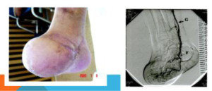
Management essentially involves hormone replacement and infertility treatment, the most successful being assisted conception with donated oocytes. The chance to conceive spontaneously after premature ovarian failure is estimated at 5-10 percent [9,10]. Embryo cryopreservation, ovarian tissue or oocyte cryopreservation and in vitro maturation of oocytes hold promise in cases where ovarian failure is foreseeable as in women undergoing cancer treatments [11]. In the near future cryopreservation of ovarian tissue will offer some hope to women at risk to develop premature ovarian failure, e.g. women from families with familial premature ovarian failure and women scheduled to undergo chemotherapy or radiotherapy at a young age [11] (Figure 1).

IGOJ2017-101-OmuKuwait_F1

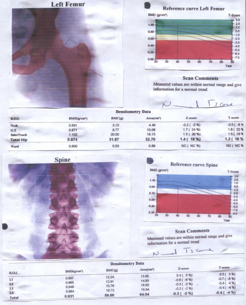
Figure 1. Bone Mineral Densitometry of 36 year old lady with Premature Ovarian Failure

Most women who experience POF require long term hormone therapy (HT) in order to maintain good overall health. Tibolone is a synthetic hormone and its actions are similar to oestrogen, progesterone and testosterone. It may help to improve mood, help the patient to sleep better, decrease hot flushes and improve general wellbeing. Tibolone may also help sex drive by improving vaginal secretions, increasing free testosterone and mood and it is not associated with an increased risk of breast cancer [5].

Objective of study: This paper aims to resolve two important issues about Premature ovarian Failure (1) To adopt a holistic view in the evaluation of the clinical features and aetiology of POF and the use of Anti-Mullerian Hormone for diagnosis (2) To evaluate the outcome of replacement therapy with Tibolone, in women with POF.

Subjects and Methods

Forty-two women seen at the outpatient clinic of Maternity Hospital fulfilled the study criteria and were recruited into the study.

Study design

This is a cross sectional study. Information on the background clinical features leading to premature menopause /ovarian failure were collated and serum levels of Anti-Mullerian Hormone evaluated according to the aetiological factors:

  1. Iatrogenic causes of POF –operation-bilateral oophorectomy or Ovariectomy
  2. Chemo/radio-Therapy
  3. Gradual progression to POF
  4. Auto immune antibodies

The second part of the study investigates the effects of Tibolone 2.5 mg and compared the symptoms of POF posttherapy effects 6 months after, using a scale of 1 to 5 ascendancy in the amelioration of symptoms.

At the first clinic consultation, clinical evaluation which included history pertaining to menstrual cycle characteristics, the occurrence of menopause, use of hormones or other medication, as well as reproductive history, physical examination and investigations, were carried out.

Ethical consideration

Verbal informed consent was received from all women and the study was approved by the Institutional Review Board of the Maternity Hospital, Kuwait. To obtain the participants’ informed consent, the objectives and general procedures of the research was explained to them as well as their right to drop out at any given moment with no ensuing change in the quality of the medical care they would continue to receive.

Investigations

The investigations included blood samples taken for determination of hormone profile –FSH, LH, prolactin, testosterone and thyroid function test by radioimmunoassay and lipid profile, autoimmune antibodies, Anti-mullerian Hormone and Vitamin D3. Bone density evaluation using QCT Scan was carried out in 17 of the patients.

Hormone profile: Estimation of serum concentration of FSH, LH, Prolactin, Testosterone, Estradiol, TSH and FT4 were carried out with radioimmunoassay.

Lipid profile The concentration of total and HDL-cholesterol as well as triglyceride were measured directly after 12-14 hours fast. LDL-cholesterol concentration were estimated using Friedwald equation thus: LDL-C=Total Cholesterol-HDL- [triglyceride/5].

Auto antibodies Both anti-thyroid and anti-ovarian were determined by use of indirect immunofluorescence assay.

Anti-mullerian hormone: In order to have an idea of ovarian reserve, serum levels of Anti-Mullerian Hormone were estimated using sandwich ELISA as previously described [12] with the immunosorbent assay from Immunotech Coulter (Marseille, France) with a detection limit of 0.05 ng/ml and intra and inter-assay coefficients of variation were <5 and 8%, respectively.

Vitamin D3: The 25-hydroxy Vitamin D test, in which the normal concentration of vitamin D was measured as nanogram per milliliter (ng/ml) with normal range 20-40 ng/ml.

Genetic testing: Chromosomal analysis of the 15 patients peripheral blood was used for mutational analysis, with the banding technique.

Mammography: Full field digital mammography (FFDM) was used because it uses computerized data recording, tele –reporting and automatic display on monitor of previous results.

Statistical Analysis

SPSS version 22 was used for the statistical analysis. We only analyze fully completed questionnaires. Comparisons with the Wilcoxon Rank Sum, proportions by Chi Square, and correlations with the Spearman Rank Order was done reported results as mean (SD) or median (range). We considered a p value of less than 0.05 as statistically significant.

Results

As shown in table 1, about 38% of the women with POF were below 30 years and about 60 % below 35 years, and the median age of the women was 28 years, 19% were single-and 8 of the women could not get married because of POF, while 21.4% got divorced since the problem of POF started probably as a result of infertility or cessation of menstruation. The women were well educated with 81% having secondary school education and 19.1% benefitted from university education. A third of the women had no children, although half of them were married and the mean parity was 1.2 ± 0.6; 14 (34.2%) were nulliparous and 22 (53.7%) had 1 to 2 children, thus highlighting a major problem of infertility.

Table 1. Characteristics of Patients with Premature Ovarian Failure

1. Age (years) No. Percent
< 20  2  4.8
21-29 14 33.3
30-34 11 26.2
35-<40 15 35.7
2. Marital Status
Single 8 19.1
Married 24 57.1
Divorced/ widowed 10 23.8 (21.4)
3. Educational Attainment
No formal education 3 7.1
Primary School 5 11.9
Secondary School  26 61.9
University 8 19.1
4. Parity 0 14 33.3
 1-3 25 59.5
 ≥ 4  3  7.2

The presenting symptoms are summarized table 2. Climacteric symptoms were common in women with POF with hot flushes 81%, night sweats 38%, vaginal dryness 21% and dyspareunia 16.7%. Others included itching 11.9 % and depression and anxiety in 38.1% and 29% respectively.

Table 2. Investigations Checklist

 Variable  Number of Patients Involved  Percentage
Hormone profile/ Lipid profile  42  100
Auto immune antibodies  28  66.7
Anti-Mullerian Hormone (AMH)  24  57.1
Mammography  24  57.1
Vitamin D3  19  45.2
Bone Mineral Density  17  42.0
Genetic Testing (Chromosomal Analysis)  15  35.7

Table 3, shows the holistic approach to the investigations. The hormone profile and lipid profile were mandatory in all 42 women. The confirmation of menopause is usually through high levels of FSH and LH. All patients had high FSH (52 ± 4 IU/L) and LH levels (28 ± 6) in the natural menopausal age and low serum levels of oestradiol. Similarly, baseline lipid profile is necessary for monitoring cardiovascular risks of the patient before commencing them on hormone replacement therapy. In the present study, 6 women (about 14%) had elevated total cholesterol and LDL-C.

Table 3. Hormone and Lipid Profiles

1. Hormone Profile Normal Range
LH (1U/L) 28 ± 6 10.9 -58.7
FSH(1U/L) 52 ± 4 16-114
Testosterone (nmol/L) 1.2 ± 0.6 0.22-2.9
Prolactin (mlu/L 31.5 ± 75 72-511
Estrogen (pmol/L) 10.5 ± 40 73-324
FT4 14 ± 3.2 12-22
2. Lipid
T. Cholesterol (mmol/L) 4.4 ± 1.2 3.1-5.2
HDL-C (g/dl) 2.8 ± 0.8 1.01-2.49
LDL-C (mmol/L) 3.2 ± 1.2 3.9-4.9
Triglyceride (mIU/L 2.1 ± 0.8 0.34-2.28
3.  Vitamin D3 ng/ml  10.6 ± 4.2 20-40

As shown in table 4, ablative operations with removal of the ovaries (Oophorectomy and ovariectomy) was the commonest single cause of POI. Two women had POF at the age of 18 and 19 years respectively as a result of bilateral torsion of the ovarian cyst and ischaemia and gangrene formation. Another 2 had ruptured inflammed appendix mass with resultant generalized morbid pelvic adhesions with amalgamation of both ovaries. At operation, the ovaries were inadvertently removed. A 21 year old women that had malignant thecoma involving both ovaries, had them removed with extensive metastasis. It is of paramount importance to distinguish between premature ovarian failure (POF) and premature menopause. This is why we evaluated the serum levels of AMH in the four groups as shown in table 4 which summarizes the causative factors of POF: (1) POF from ablative operation of bilateral oophorectomy or ovariectomy (2) POF from gonadotoxicity of chemotherapy / radiotherapy (3) POF from autoimmunity from anti-thyroid and anti-ovarian antibodies and (4) idiopathic POF which in 48% of the women. For comparison, an internal control group of ten women with regular periods was recruited and also had serum AMH assays as shown in figure 2. serum Anti-mullerian hormone was undetectable in group 1, detectable in group 2 at very low levels(p<0.05). There was significant difference between groups 3 and 4 (P<0.01). Although conditions like Turner’s Syndrome and Trisomy 21 were excluded from the present study, Genetic testing was carried out in 15 women with POF under 30 years of age. The only finding was “46, XX, 9(phqh, normal variant” which was normal for the female.

Table 4. Aetiological Factors of Premature Ovarian Failure

No. Percent
Operation with removal of ovaries 8  19.0
Chemotherapy and Radiotherapy 7  16.7
Autoantibodies

Thyroglobulin Antibodies

Thyroid Microsomal Antibodies

Antiovarian Antibodies

7

3

2

2

 16.7
Idiopathic 20 47.6

The use of Tibolone for replacement therapy, was well tolerated by majority of the women. The only side effect was break-through which was mainly due to inability of the woman to take the drug as advised. Concerns about deep vein thrombosis (DVT) and stroke made the team monitor Tibolone with lipid profile. No abnormality was detected.

In Table 5, the outcome of replacement therapy is evaluated using the scale of 1 (No change) to 5 (Total improvement). Hot flushes, night sweats, vaginal dryness, dyspareunia and vulva itching have significant improvement and total elimination of symptoms. Urinary frequency has some improvement but not significant. Tibolone has no effect on emotional reactions like depression and anxiety.

IGOJ2017-101-OmuKuwait_F2

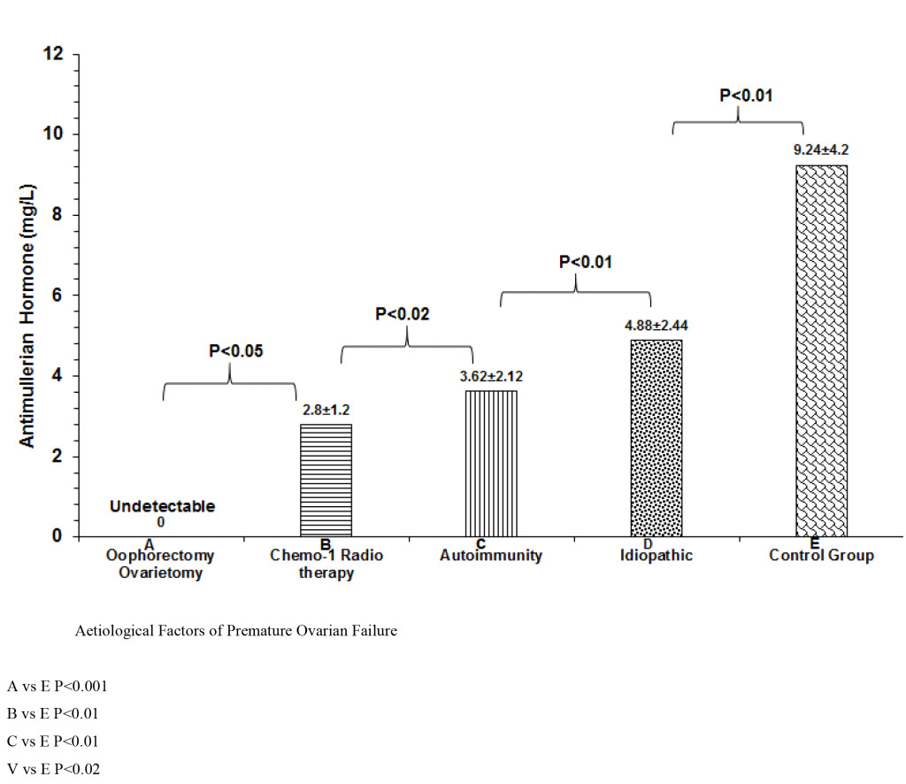
Figure 2. Comparative serum levels of AMH.

Table 5. Effects of Replacement Therapy with Tibolone.

 Symptoms No of Patients Percent 1  2  3  4  5 CR P value
Hot flushes  34 81.0 0  0  5 11 18 0.824 0.001
Night Sweat  16 38.1 0  0  2  4 10 0.642 0.01
Vaginal dryness  9 21.4 0  1  1  2  5 0.482 0.02
Dyspareunia  7 16.7 0  1  2  1  3 0.412 0.05
Vulva itching  5 11.9 0  1  0  2  2 0.386 0.05
Urinary Frequency  5  11.9 2  1  2  0  0 0.122 0.08
Emotional R Depression Anxiety  16 12  38.1 28.6 66  4 4  6 1  0 1  0 0 0.0210.011 1.021.12

1= No Change, 2=Little improvement, 3= Moderate improvement, 4= great improvement, 5= Total improvement

Discussion

The present study has shown that patient with POI are normally developed individuals well prepared for exigencies of life with appropriate education and social marital setting. However, a diagnosis of POI, has both short and long term health problems. In the short term, many of them have hot flushes, insomnia and problems of oestrogen deficiency like dyspareunia and bladder problems [1]. Long term health concerns as a result of oestrogen deficiency may include cardiovascular disorders and osteoporosis [13].

Auto-immune diseases with autoantibodies have been associated with Premature ovarian Insufficiency in the present study. Many other autoimmune conditions have been similarly implicated. They include Endocrine diseases such as hypothyroidism,,addison’s disease,, hypophysitis, hypoparathyroidism, and Type 1 diabetes, all in association with their autoantibodies [14-17]. In addition there are also non-endocrine conditions like Pernicious anaemia, Myasthenia gravis, Chronic candidiasis (in type 1 autoimmune polyglandular failure syndrome), Idiopathic thrombocytopenic purpura, Vitiligo, Alopecia, Autoimmune haemolyticanaemia, Systemic lupus nephritis, Crohn’s disease, Sjögren’s syndrome, Rheumatoid arthritis, Primary biliary cirrhosis, Coeliac disease, Chronic active hepatitis [18,19].

Genetic basis of Premature ovarian Failure was not revealed in the present study, may be because of the small number of women with that had genetic testing. They may include 45X0 (Turners syndrome) that gives rise to gonadal dysgenesis and primary amenorrhoea. Blocking mutations of FSH receptors leads to primary Amenorrhoea. Fragile X Occurs when number of trinucleotide repeats is in excess of 200 and gene transcription fails and FMRI (Xq27.3) protein is not expressed with increased prevalence of POF [20]. Other candidate genes for POI Chromosome 3–long arm (syndrome of blepharophimosis, ptosis and POI). The absence of Connerin 37 between ovum and granulosa cells, leads to infertility in mice [21]. The present study has clearly demonstrated that Tibolone relieves vasomotor symptoms such as hot flushes, night sweats, vaginal dryness and dyspareunia. Tibolone is a synthetic hormone and its actions are similar to oestrogen, progesterone and testosterone [5]. It may help to improve mood, help you sleep better, decrease hot flushes and improve general wellbeing. Tibolone may also help sex drive by improving vaginal secretions, increasing free testosterone and mood. Tibolone is not associated with an increased risk of breast cancer, in women without breast cancer [5].

Conversely, Tibolone has little effect on urinary symptoms like frequency and urge incontinence and psychological reactions such as depression and anxiety, which are common in in patients diagnosed with premature ovarian failure.

Managing menopause in women with cancer involves a number of options (Shover et al). These include lifestyle changes, which can help reduce depression, anxiety, cardiovascular and osteoporosis risks, psychological support and medication

Counselling and education to diagnose impending POF presenting with irregular menstrual periods. And before chemotherapy and/or radiotherapy, you should investigate your options for trying to preserve eggs for conception. There are a number of options including, egg preservation, embryo freezing, ovarian preservation and ovarian biopsy and freezing [22].

Limitations of the study

Small sample. There is need for larger sample size with high cronbach factor.

 Not all 42 women benefitted from all the investigations.

Conclusion

This study has articulated a new paradigm in the management of POF with holistic attitude towards diagnosis and treatment with monitoring of treatment.

References

  1. Omu AE, Al-Qattan N. Premature menopause. Sing J. Obstet Gynaecol 1996; 27: 23–8
  2. Conway G.S. Premature Ovarian failure. Curr Opin Obstet Gynecol 1997; 9: 202 – 206
  3. ESHRE (2015) Management of women with Premature Ovarian Insufficiency: Guideline of the European Society of Human Reproduction and Embryology; 8–16.
  4. Christin-Maitre S, Braham R.General mechanisms of premature ovarian failure and clinical check-up. Gynecol Obstet Fertil. 2008; 36: 857–61.
  5. Vandborg M, Lauszus F. F., Premature ovarian failure and pregnancy Arch Gynecol Obstet. 2006; 273: 387–8.
  6. van Kasteren YM. Premature ovarian failure Ned Tijdschr Geneeskd.2000; 144: 2142– 6.
  7. Goswami D, Conway GS. Premature ovarian failure. Horm Res. 2007; 68(4): 196–202.
  8. Groff AA, Covington SN, Halverson LR, Fitzgerald OR, Vanderhoof V, Calis K, Nelson LM. Assessing the emotional needs of women with spontaneous premature ovarian failure. Fertil Steril. 2005; 83: 1734–41.
  9. Petras, K. (1999) The Premature Menopause Book: When the change of life comes too early. (1st edi) HarperCollins, New York. USA.
  10. DeAngelo D (2000) Sudden Menopause: Restoring Health and Emotional Well-Being.(1st edi) Hunter House, Salt Lake city, USA.
  11. Sterling E W & Best-Boss A (2010) Before your Time: The Early Menopause Survival Guide. (1st edi). Simon & Schuster, New York, USA.
  12. Sterling, E. W. & Nelson, L.M. (2011). From Victim to Survivor to Thriver: Helping Women with Primary Ovarian Insufficiency integrate Recovery, Self-Management, and Wellness. Semin Reprod Med. 29 (4): 353–361. Doi: 10.1055/s-0031–128092.
  13. Rafique, S., Sterling, E. W., Nelson, L. M. (2012).A New Approach to Primary Ovarian Insufficiency.Obstet GynaecolClin North America 39(4): 567–586.Doi: 10.1016/j.ogc.2012.09.007
  14. Monnier, P. (2013). Primary Ovarian Insufficiency: The Words to Say. Reproductive endocrinologist Winter 2013/14
  15. Judge, T. A., Locke, E. A. & Durham, C. C. (1997) The dispositional causes of job satisfaction: A core evaluations approach. Research in Organizational Behavior.19: 151–188
  16. Bandura, A. (1977). Self-efficacy: Towards a Unifying Theory of Behavioral Change. Psychological Review, Vol. 84, No. 2, 191–215.
  17. Klein, J. & Wasserstein-Warnet, M (1999).Predictive validity of the locus of control test in selection of school administrators.Journal of Educational Administrators, 38(1), 7–24. http: //dx.doi.org/10.1108/09578230010310957
  18. Burish, T. G., Carey, M. P., Wallston, K. A., Stein, M. J., Jamison, R. N. & Lyles, J. N. (1984). Health Locus of control and chronic disease: An external orientation may be advantageous. Journal of Social and Clinical Psychology.Vol 2, No 4, pp.326–332.
  19. Smith, E. R..& Mackie, D. M. (2007). Social Psychology (3rded) Hove: Psychology press
  20. Maslow, A. H. (1987). Motivation and Personality (3rded) New York: Harper & Row.
  21. Wickman, S. A. & Campbell, C. (2003). An Analysis of how Carl Rogers enacted client-centered conversation with Gloria. Journal of counseling & development 81: 178-184
  22. Schacter, D. L., Gilbert, D. T. & Wegner, D. M. (2009). Self Esteem. Psychology (2nded) Barnes & Noble.New York: Worth publishers.
  23. Baumeister R & Tierney J (2011). Willpower: Rediscovering the Greatest Human Strength. Penguin press.
  24. Bouchard G (2003) Cognitive Appraisals, neuroticism, and openness as correlates of coping strategies: An integrative model of adaptation to marital difficulties. Canadian Journal of Behavioural Science, Vol 35 (1) http://dx.doi.org/10.1037/h0087181.
  25. Costa , P T; & McCrae, R R., (1980) Influence of Extraversion and Neuroticism on Subjective Well-Being: Happy and Unhappy people. Journal of Personality and Social Psychology, Vol 38. 668–678.
  26. Ventura J L, Fitzgerald O R; Koziol D E, Deloris E , et al (2007) Functional well-being is positively correlated with spiritual well-being in women who have spontaneous premature ovarian failure. Fertility and Sterility, Vol.87 (3): 584–590, doi: 10.1016/j.fertnstert.2006.07.1523.
  27. Stanford Chronic Disease Self-Management Study. Psychometrics reported in: Lorig K, Stewart A, Ritter P, González V, Laurent D, & Lynch J, Outcome Measures for Health Education and other Health Care Interventions. Thousand Oaks CA: Sage Publications, 1996, pp.24–25,41–45.
  28. Shuster L T, Rhodes D J,. Gostout B S,Grossardt B R, RoccaW A. Premature menopause or early menopause: long-term health consequences. Maturitas. 2010; 65: 161–165.
  29. Persani L, Rossetti R, Cacciatore C, Bonomi M. Primary Ovarian Insufficiency: X chromosome defects and autoimmunity. J Autoimmun. 2009; 33: 35–41.
  30. Mlčochova H,. Hořejši Martinek J J. Vetešnikova-Koubova R.&Halaška M. Treatment of autoimunne ovarian damage in adolescent girls. Neuroendocrinology Letters 2005. No.2 Vol.26,
  31. Santoro N. Mechanisms of premature ovarian failure Ann Endocrinol 2003; 64: 87–92.
  32. Kauffman RP, Castracane VD. Premature ovarian failure associated with autoimmune polyglandular syndrome: patophysiological mechanism and future fertility. J Women’s Health (Lachmt) 2003; p. 513–20.
  33. Jacobs PA Fragile X-syndrome. J Med Genet 1991; 28: 809–810
  34. Conway G S.,.Payne1 N N, Webb J, Murray A and.Jacobs P A. Fragile X premutation screening in women with premature ovarian failure. Human Reproduction 1998; 13: 1184–1187.
  35. Carmody et al. Mindfulness training for coping with hot flashes: Results of a randomized trial. Menopause. 2011; 18(6): 611–620. doi: 10.1097/gme.0b013e318204a05c
  36. Ikeme ACC, Okeke TC, Akogu SPO, Chinwuba N. Knowledge and perception of menopause and climacteric symptoms among a population of women in Enugu, South East, Nigeria. Ann Med Health Sci Res. 2011; 1: 31–6.
  37. Dolleman et al. Anti-Müllerian hormone is a more accurate predictor of individual time to  menopause than mother’s age at menopause. Hum. Reprod. (2014; 29: 584-591. doi: 10.1093/humrep/det446 First published online: January 15, 2014
  38. Luborsky J. Ovarian Autoimmune Disease and Ovarian auto antibodies. J. Women Health and Gender Based Medicine 2002; 11: 585–599.
  39. Sayakhot et al. Potential adverse impact of ovariectomy on physical and psychological function of younger women with breast cancer. Menopause, 2011; 18: 786-793.
  40. Broer SL, Eijkemans MJ, Scheffer GJ, van Rooij IA, de Vet A, Themmen AP, Laven JS, de Jong FH et al Anti-mullerian hormone predicts menopause: a long-term follow-up study in normoovulatory women. J Clin Endocrinol Metab. 2011; 96: 2532–9.

Conflict of Clinical and Public Health Viewpoint in Colorectal Screening

DOI: 10.31038/CST.2017282

Summary

Mortality rates from colorectal cancer are dramatically high, therefore the reduction by population screening as a public health measure is considered as one of the priorities of national public health programmes worldwide. In Hungary, in the beginning a human-specific immunological test was applied in “model programmes” as a screening tool, to detect the occult blood in the stool; compliance was 32% on average. However, the objectives of the model programmes have not been achieved, because – among other reasons – debates on method of choice and the strategy to follow have divided the professional public opinion. In this paper, the debated issues are critically discussed, being convinced that – at present –population screening seems to be the most promising way to alleviate the burden of colorectal cancer.

Keywords: colorectal cancer, gFOBT, iFOBT, flexible sigmoidoscopy, colonoscopy

Colorectal cancer is a major health problem worldwide. It represents almost 10% of the global cancer incidence burden in 2012. It is the third most common cancer in men (an estimated 746 000 cases), the second most common in women (614 000 cases), and the fourth most common cause of death from cancer worldwide, with an estimated 694 000 deaths. Incidence varies 10-fold between countries worldwide.In the industrially developed countries, colorectal cancer is the second most common cancer and – behind lung cancer – the most common cause of cancer death in both sexes. More than 65% of new cases occurred in countries with high or very high levels of human development (HDI). Almost half of the estimated new cases occurred in Europe and the Americas [1]. The highest incidence rates are in men in central Europe: Slovakia, Hungary, and the Czech Republic [2]. The global burden of colorectal cancer is expected to increase by 60% to more than 2.2 million new cases and 1.1 million deaths by 2030 [3].

The majority of well operable tumours are discovered in an advanced, inoperable stage, the consequence of which is high disease specific mortality. Early detection and early treatment of this type of cancer would most likely alleviate the health burden caused by colorectal cancer. It is widely agreed that the reduction of mortality is indispensable and the most promising way for this is the organized screening of asymptomatic persons.

The Health Ministers of the European Union unanimously adopted a set of recommendations on cancer screening on 2 December 2003 [4]. The recommendation spelled out the fundamental principles of the best practices in cancer screening, and encouraged the Member States to take common action to implement cancer screening programmes, particularly, for colorectal cancerusing faecal occult blood test as screening tool. The importance of colorectal screening is emphasized by the “Brussels Declaration” that was signed by the representatives of a number of scientific societies, cancer leagues, patient organizations, foundations, health insurance companies, and several members of the European Parliament. The Declaration urged the European Council to prepare a plan of action to alleviate the burden of colorectal cancer on the societies (“Europe against Colorectal Cancer”), and to support by all means the governments of the Member Countries to establish call-and-recall based, organized screening programmes for colorectal cancer [5]. Similarly, the urgency of the implementation of colorectal screening was emphasized by the “Budapest Declaration” by the European Association of Gastroenterologists [6]. In 2011, the European Commission – in collaboration with the International Agency for Research on Cancer (WHO/IARC) – published a detailed guideline on quality assurance in colorectal screening [7].

In Hungary, the National Public Health Programme set up in 2002, like numerous other countries, proposed the reduction of mortality from colorectal cancer in up to 20% in 8-10 years’ time by the implementation of organized screening programmes for early detection of the disease and its premalignant lesions using human-specific faecal occult blood tests [8]. However, in 2008, an investigation by the National Audit Office on the utilization of public money spent for organized colorectal screening had stated that “the goals set were not met” [9]. One of the reasons for the failure was a sharp debate, and divided the professional public on the strategy and screening methods of colorectal screening. The debated issues were: whether the “two-step strategy” in which the faecaloccult blood test is the “first step”, and in case of non-negative blood test colonoscopy should follow, or, the “one step” strategy: colonoscopy alone is the method of choice.

This paper intends to explore this debate in depth, and to arrive at a clear recommendation: which of the two “strategies” is to be implemented in organized population screening.

Pathway to malignancy (natural history)

Due to its biological and pathological characteristics, colorectal cancer is particularly suitable for screening, as benign adenomatous polyps, or inflammatory bowel diseases regularly proceedthe development of colorectal cancer. The prevalence of colorectal polyps widely varies. Among asymptomatic, average-risk population, the prevalence of adenoma of colon and rectumis approximately 5-10%.In a study offering total colonoscopy screening to individuals without any lower gastrointestinal symptoms, the prevalence of colorectal polyps, including potentially premalignant lesions in asymptomatic persons aged 40-59 years, was somewhat higher (12-16%) [10]. A meta-analysis resulted in much higher prevalence for non-advanced and advanced adenoma (30% or above) [11]. In first-degree relatives of those having family occurrence of adenomas, the prevalence of adenoma is four-times higher as compared to the average-risk population [12].

The aetiology of colorectal cancer involves the complex interaction of environmental carcinogen exposure and genetic factors in the population. Indeed, most colorectal cancer cases are sporadic, occurring in individuals without any known familial predisposition. Approximately 10–30% of cases have a positive family history of this neoplasm [13].

Transformation of normal colorectal epithelium to an adenoma and ultimately to an invasive and metastatic tumour has been well known for a long time as “adenoma-carcinoma sequence” [14]. Today, the genetic alterations taking place during the malignant transformation are also described in detail [15]. The correlation is supported by the observation that following the colonoscopicremoval of adenomatous polyps the incidence of colorectal cancer decreases [16]. There is a correlation between the size of the polyp and the likelihood of malignant transformation. [17].

Screening methods

In 1968, the WHO drew up the criteria of a population screening; they are valid up to the present day.(18)The most important criteria are the following:

  • the target disease should be of public health importance;
  • the natural history of the target disease is known, and it has a rather long preclinical detectable phase (PCDP);
  • the detected target disease should have an established treatment, and the referral routs should be well determined;
  • The screening tool should be cost-effective.

The expectations from screening methods suitable for early detection of asymptomatic polyps and colorectal cancers are: that they should be inexpensive, simple to perform and reliable. Furthermore, the methods should be sensitive, specific, and have appropriate predictive values. Further expectations are that the screening method should be harmless, it should not cause any complication, and only acceptable burden and discomfort for the persons under investigation. The provider should do his/her best to optimise the social acceptance of the offered screening (“compliance”). It is an advantage if the method is not invasive. In order to ease the avoidable psychological side effects, the quick disclosure of test results is desirable.

Methods of colorectal screening

The primary aim of colorectal screening is the detection, removal and early treatment of adenomatous polyps of 10 mm in diameter which develops in average risk persons, and are considered as premalignant lesion of colorectal cancer (removal a premalignant polyps may be seen as primary prevention of colorectal cancer), and those of non-invasive colorectal cancers. A secondary aim is to detect and treat the sources of bleeding in bowels.

Although the methodological arsenal of colorectal seems to be plentiful, the fact is that to this very day we do not have such a screening method which would satisfy all the needs, because either its sensitivity and specificity are limited, or it is potentially harmful, or social acceptance is far from being optimal [19- 21]. Currently, many tools are used for colorectal screening and can be grouped into two categories: [a]. tests that primarily detect colorectal cancer, which include tests that look for blood, such as guaiac faecal occult blood test and faecal immunochemical test, and a couple of other markers in stools; and [b]. tests that can detect cancer and advanced lesions, which include endoscopic and radiological exams, i.e., colonoscopy, doublecontrast barium enema (DCBE), and computed tomography colonography (CTC) (or virtual colonoscopy) [6]. However, these tests all have certain limitations.

Detection of occult blood from the stool

The methods are based on the assumption that the premalignant adenomatous polyps of large bowels and the early colorectal cancer are intermittently bleeding, one or the other component of the blood, as a marker, are detectable in the occult, invisible to the naked eye [22]. As the bleeding is discontinuous, in order to improve the chances, samples should be taken from more than one consecutive bowel movement. Faecal occult blood testing is what is known as a qualitative test. It only detects the presence or absence of blood in the sample, but does not indicate the site and quantity of bleeding. The non-negative test (positive FOBT) could indicate  colorectal cancer, but not diagnose it. If blood is detected, additional  testing  by colonoscopy would be required. At present, guaiac-based chemical and immunochemical methods are used for colorectal screening.

A number of screening methods can be used including stool based tests every 3 years, sigmoidoscopy  every 5 years and  colonoscopy  every 10 years.

Guaiac Faecal Occult Blood Test (gFOBT)

In fact, this is a chemical reaction to detect the haemoglobin component of the blood in the stool: hem-component of haemoglobin has a peroxidase-like activity, therefore when the hydrogen peroxide is dripped onto the guaiac paper, in the presence of blood, yields a blue reaction product within seconds.

(These test are collectively known as “haemoccult test”.) The reaction is not specific for human haemoglobin; therefore, to avoid false positive reaction, dietary restrictions are necessarily (e.g. red meals, some vegetables, some pharmacuticals). Recent position is that because patient adherence can be an issue with FOBTs, and dietary restrictions can affect adherence in some populations, it is reasonable to abandon these recommendations without fear of substantially affecting specificity [23]. The American College of Gastroenterology has recommended abandoning gFOBT testing as a colorectal cancer screening tool, in favour of the faecal immunochemical test (FIT), [24].

ImmunochemicalFaecalOccultBlood Test (iFOBT, FIT)

This is a newer and more sensitive test that the guaiac-based FOBT. The immunochemical test detects the globin component of haemoglobin rather than the heme component: antigen-antibody reaction takes place against the globin component of human haemoglobin. The immunological reaction is specific for human haemoglobin, therefore, it does not require dietary restrictions before collecting the sample [25]. The test itself requires less effort because it involves simply brushing the surface of the stool in the toilet water and dabbing the brush on a test card. The test result can be read in an automated device; the investigator can chose the antibody concentration (“cut-off point”), over which the test is considered positive. In Japan, the iFOB test has been used for population screening since 1992 [26].

For average-risk adults older than 50 years of age, evidence from multiple well-conducted randomized trials supported the effectiveness of faecal occult blood testing in reducing colorectal cancer incidence and mortality rates compared with no screening. Data from well-conducted case–control studies supported the effectiveness of sigmoidoscopy and possibly colonoscopy in reducing colon cancer incidence and mortality rates. A non-randomized, controlled trial examining colorectal cancer mortality rates and randomized trials examining diagnostic yield supported the use of faecal occult blood testing plus sigmoidoscopy. Data is insufficient to support a definitive determination of the most effective screening strategy.

Screening by endoscopy

By the endoscopic methods, the lumen of entire colon and rectum can be rendered visible to the eye, so the target condition of screening can be scrutinised.

Flexible sigmoidoscopy is an exam used to evaluate the rectum and most of the sigmoid colon, i.e about 60 cm of the large intestine. It does not allow one to see the entire colon. As a result, any cancers or polyps farther into the colon cannot be detected with flexible sigmoidoscopy alone. If necessary, tissue samples (biopsies) can be taken through the scope during a flexible sigmoidoscopy exam. In the case of a positive test result, colonoscopy must be performed.

A longer version of a sigmoidoscopy is colonoscopy, by which the entire length of the colon and rectum can be broughtinto the field of view. It makes it possible to take samples for histology (biopsy) or to remove any suspicious-looking areas, if needed. It requires sedation. Several factors affect the outcome of a successful colonoscopy, including cecal intubation, careful mucosal inspection, and withdrawal time [27]. Colonoscopy is a time-consuming, technically challenging procedure, its effectiveness in diagnosing and removing polyps depends on the technical aspects of the procedure. The other requirements of “quality colonoscopy” are described by Hungarian authors [28]. However, colonoscopy has several limitations that relate to the mechanics of the procedure, such as perforation, bleeding, or adverse consequences of sedation. Perhaps this is why the patients’ perceptions regarding colonoscopy frequently drives patients non-adherence recommended testing, both screening and diagnostic.

Virtual colonoscopy or CT colonography is a  medical imaging  procedure which uses  x-rays  and  computers  to produce two- and  three-dimensional  images of the large intestine from the lowest part, the  rectum, all the way to the lower end of the  small intestine  and display them on a screen [29, 30].

Other screening approaches

There are few other tests that are not yet routinely used for screening purposes. It would seem reasonable to make immunological tests more sensitive by using a second marker, such as transfer rindipstick test [31], lactoferrin[32]. ,alfa-1-antitripsin [33]. At present, most evidence a tour disposal is for albumin as a second marker [34]. (In Hungary, this bi specific immunochemical method had been tested in model programmes. The compliance was 32%. The yield of the test were encouraging, however, because of the lack of automation and that of European marketing authorisation subject experts considered the test as being in the experimental stage, and suggested omission of its use is population screening) [35]. The detection of cancer-associated biomarkers is not yet applied in population screening programmes.

There are a few molecular-biological methods, most of them in experimental phase, that can be seen as the methods of future, for example faecal DNA testing [36, 37, 38]. However, the detection of cancer-associated biomarkers is not yet applied in population screening programmes.

Effectiveness, sensitivity, specificity

Colorectal cancer screening reduces death from colorectal cancer and can decrease the incidence of disease through the removal of adenomatous polyps. Several available screening options seem to be effective, but the single best screening approach cannot be determined because of insufficient data [39].

The sensitivity of faecal occult-blood testing for colorectal cancer and especially for colorectal adenomas is low because neoplasms may bleed intermittently, and thus cannot be detected in this way. Comparison between guaiac and immunochemical FOBT in screening for colorectal cancer provides evidence that iFOBT is superior to gFOBT [40]. The high quality evidences for non-invasive screening exist for guaiac-based faecal occult blood tests (gFOBTs), for which the disease-specific incidence and mortality reductions are modest.

The guaiac-based chemical detection of faecal occult blood is the only non-invasive screening method with proven effectiveness: annual or biannual screening reduces mortality by 15-33%through randomised controlled trials [41- 44]. On the other hand, in the only randomised controlled test, Chinese authors found that immunological haemagglutination test (iFOB) was effective in reducing mortality from rectal cancer but not in reducing mortality from colon cancer or the incidence of colorectal cancer [45]. . In case-control studies, 21-81% mortality reduction was published [46, 47]. Faecal immunochemical tests (FITs) offer better sensitivity and comparable specificity [48]. In addition, the participation and detection rates for advanced adenomas and cancer were significantly higher for immunological, as compared to guaiac-based FOBT, which significantly underestimates the prevalence of advanced adenomas and cancer in the screening population compared with iFOBT [49].

Flexible sigmoidoscopy is a valid screening tool for the early detection of colorectal cancer. Recently published long-term data from UK Flexible Sigmoidoscopy Screening randomised controlled trial (UKFSST) demonstrate a 33% reduction in colorectal cancer incidence and a 43% decrease in colorectal cancer mortality with once-in-a-lifetime screening, and reported long term effects of only-once sigmoidoscopy screening after 17 years of follow-ups [50]. It is a resource-conserving strategy. Long-term follow-up of participants in the trial will be required [51]. Faecal occult blood test and flexible sigmoidoscopy have been proven to reduce colorectal cancer mortality by approximately 30%. [52]. Patients with a positive screen will be referred for colonoscopy with once-in-a-lifetime screening. [53].

Although colonoscopy screening is used in some countries, no randomized trials have been conducted to estimate its benefit. The standard (optical) Colonoscopy has the potential to be superior to FOBT and flexible sigmoidoscopy, but needs to be evaluated in randomized trials before any recommendation can be provided; such investigations are under way in several countries. The available data is of limited value. In observational studies, the incidence colorectal cancer was reduced by 76-90% in those screened, as compared to the reference population [54]. Colonoscopy may find more cancers in the proximal part of the colon but is associated with greater cost and more complications. The availability of a simple, non-invasive test that detects tumour-specific products with reasonable sensitivity and specificity might overcome barriers to screening among patients who are not willing to undergo more sensitive but more invasive tests, such as colonoscopy.

Despite technical advances in computed tomographic (“virtual”) colonoscopy, there is a lack of consensus about its role in screening. The low sensitivity of the faecal DNA panel for detecting clinically significant neoplasia might limit its value as a one-time test for cancer, since it misses most lesions identified on colonoscopy. However, the use of a less sensitive test at frequent intervals (e.g. biannually) may be as effective for the detection of colorectal anomalies as a more sensitive test that is used infrequently, such as colonoscopy.

Compliance with the screening programmes

The public acceptance of the offered screening is perhaps the most important prerequisite of the effectiveness and cost-effectiveness of colorectal screening. The tolerance of the target population should be kept in mind: the average-risk, asymptomatic, healthy or apparently healthy men and women between 50 and 70 years of age is much lower as compared to those who turn to a doctor because of their complaints. In the case of occult blood tests, the compliance of those participating in clinical trials is relatively higher (54-67%) as in case of population screening (30-50%) [41- 44]. In observational studies the great majority of subjects (97%) refused total colonoscopy but accepted a non-invasive blood test (83%), because it was a more convenient and less time-consuming procedure. [55]. Asymptomatic persons are reluctant to accept an invasive method, such as sigmoidoscopy and colonoscopy; colonoscopy is considered particularly unpleasant [55, 56]. Because of fear of pain and anesthesia, fewer people undergo colonoscopy even where they are strongly recommended (3-10%) [57]. It is fair to say that it is the compliance of the offered screening modality strategy that decides the applicability of a screening tool as a public health measure in favour of the “two-step” strategy.

Discussion

Current guidelines in the European Union include recommendations for stool-based tests – faecal occult blood test (FOBT) and faecal immunochemical test (FIT) – and flexible sigmoidoscopy, whereas most US guidelines include those tests as well as colonoscopy [58].

In the United States, clinically-oriented scientific societies, such as the American College of Gastroenterology, the American Cancer Society, the American College of Radiology, the US Multi-Society Task Force on Colorectal Cancer, all take a stand on the screening practice, and the US Preventive Services Task Force develop consensus guidelines and recommend the strategy to follow, which is basically a “one step” strategy. Although colonoscopy is superior to other tests in some respects, the US Preventive Services Task Force has determined that no single test or strategy for colorectal-cancer screening can be endorsed on the basis of currently available data. Several approaches (faecal occult-blood testing, sigmoidoscopy, colonoscopy, and barium enema examination) are included as options in the screening guidelines. [59]. Therefore, all recommended tests are acceptable options and may be chosen based on individual risk, personal preferences, and access. The prevention of colorectal cancer should be the primary goal of screening, which is opportunistic in nature.

The “two step” strategy is characteristic to the European practice of colorectal screening, where the screening strategy is of public health orientation. The guidelines are set by authoritative professional organizations, such as the International Agency for Research of World Health Organisation (IARC/WHO), and International Union against Cancer (UICC), and, the provider initiated, personal invitation-based “mass” or population screening is the recommended practice. The guidelines issued by the European Commission argue for the “two steps” strategy: as a first step, the detection of the occult blood in the stool with a suitable screening method (gFOBT, FIT) should take place and, as a second step, patients with a positive screen would be referred for colonoscopy in order to clarify whether the likely target disease can be justified or not [60].

In 2017, all the countries of the European Union – with a few exceptions – have a colorectal cancer screening policy mandated by a law, or at least, a governmental recommendation. The programme is public funded, a test provided free of charge in all but a few countries [61]. Out of the 28 member states, 20 members have piloting, or rollout complete for population-based colorectal screening programmes; in addition, three member states were planning to start population-based programmes in 2016. In most of the countries, the screening activity covers the entire country; in a few, they are being limited to one or more regions. The widest recommended target age, of 50 to 74 years, has been adopted by most countries.In most of the countries g/FOBT/FIT is used as a screening test.

Colposcopy is offered once in a lifetime in one county (Poland), as is the case for flexible sigmoidoscopy in two countries (Italy and United Kingdom).

The practicability of screening strategies are being scrutinised by other international organisations such as the International Colorectal Research Screening Network (ICRSN); [62]. they confirm that for colorectal screening as a public health measure, the generally accepted screening strategy is the “two-step” strategy, based on the detection of occult blood in the stool (gFOBT/iFIT).The “one step” strategy of colorectal screening for country-wide population screening has not been used.(The only exception is Poland, where those who opportunistically turn up in one of the approximately 40 endoscopy centres would undergo colonoscopy) [63].

In Hungary, according to the current protocol, the pilot programmes for colorectal screening are conducted under “two-step” strategy.The Public Health Authority has decided to use the detection of occult blood as screening tool because – although it is oflimited sensitivity and specificity as compared to the endoscopic methods – and relatively frequent repetition is needed – it is a non-invasivemethod, therefore the public acceptance of the offered screening (“compliance) is much better, rendering it more suitable for organised population screening.

In the meantime, the Society of Surgery and that of Gastroenterology has taken a firm stand in favour of the one-step strategy of colorectal screening, and suggested that a national screening program using sole colonoscopy as the method of screening be introduced (64). They argued with the heavy burden of colorectal cancer on the Society, the expected benefit of a screening programme. They stressed that the primary aim of colorectal screening is the detection and removal of the adenomatous polyps, by which colorectal cancer is preventable, and the mortality from the disease can be reduced by 20-30 %. Furthermore, “colonoscopy as the sole screening method is more promising than anything else”, because at the same time the polyps can be removed so the colonoscopy is also a therapeutic intervention. They argue for the “one-step” strategy, saying that the “once in a lifetime” colonoscopy seems to be a suitable method for a nation-wide colorectal screening programme. In conclusion, they think that “the reconsideration of strategy is fully justified.”

Beyond any doubt, total colonoscopy is the “golden standard” of colorectal screening:  even when using the “two-step” strategy, each case of non-negative test result needs to undergo colonoscopy in order to verify the positive result of occult blood test. Total colonoscopy is the only detection method that can verify or rule-out the suspect or malignant target disease and, in this way, it can significantly contribute to the mortality reduction from colorectal cancer. Accordingly, the validation of the “two-step” strategy is also attributable to colonoscopy.

Nevertheless, colonoscopy, as such, is a complex diagnostic method and by no means a screening method to be used for public health purposes, i.e. population screening.

Colonoscopy makes compulsory the intensive examination of the physical status of the “patient” to be “screened”, and the determination of laboratory and blood coagulation parameters. It makes necessary the clean-up of the large bowel, sedation, sometimes anaesthesia. Colonoscopy requires proficiency, therefore between 1000-2000 examinations need to be performed each year; in some countries, it is subject to proficiency examination or accreditation. The examination itself is time-consuming; just the withdrawal time itself must not be shorter than 6 minutes. According to estimates, complications (bleeding, perforation) might occur in 1: 1000 cases, therefore post-intervention observation and sometimes hospitalisation is necessary. [65].

In Hungary, the clash of the two kinds of strategy has intensified over time: there is an obvious conflict between the clinical and public health positions. The clinical viewpoint supported by the clinical community seems to discredit the public health viewpoint. In order to find a new focus on the topic, a consensus conference had been convened with broad participation of all those concerned. The Conference has discussed all the contested issues, and made the following recommendations:

  • The burden and the public health importance of colorectal cancer, and the suitability of screening for it, is urgent for the continuation of colorectal screening. The cost of delay or discontinuation of the programme would mean a great deal of salvable life years.
  • According to the evidence-based health policy and evidence-based public health, the recommended methods of population screening, i.e. that of average-risk men and women between 50-70 years of age: the immunochemical detection of faecal occult blood (iFOBT or FIT), the effectiveness of which is scientifically justified. In the case of non-negative (positive) test results, the total colonoscopy needs to be performed at the source of bleeding in the bowel.
  • In the personal invitation letter the attention of the invitee might be drawn to total colonoscopy as possible method of choice, stressing all the advantages and as well as the discomfort of it.

Furthermore, the Conference voiced the importance of the stimulation and motivation of the target population in the acceptance of the offered screening, while pointing out that the high compliance rate is the prerequisite of both effectiveness and cost-effectiveness in screening. Finally, the incorporation of endoscopy into the system of population screening, technical preconditions, and the required proficiency of providing total colonoscopy were stressed. Scarcity of colonoscopy capacity is a frequent problem. [66].

In conclusion, the implementation of colorectal screening as a public health measure had been delayed by a heated debatebetween proponents of the “one-step” vs ” two-step” strategy. Clinical experts argued in favour of colonoscopy only to be used as the sole screening method of asymptomatic persons, saying that it is more sensitive and specific, as compared to the occult blood tests, and able to remove adenomatous polyps, if necessary and as such, it is a primary preventive measure at the same time. On the other hand, those who represented the viewpoints of public health, argued that for population screening, the detection of faecal occult blood is the method of choice, as it is more simple, cheaper, and more accepted by the public at large; furthermore, if necessary, in the second step, as a verification test, it can be followed by colonoscopy.

Beyond any doubt, total colonoscopy is the “golden standard” of colorectal screening. However, it is a resource-demanding, time consuming intervention that requires special proficiency from the provider. Possible complications might occur; therefore it may not go beyond the medical practice. In addition, because of the discomfort it causes to the screened persons, the public acceptance of it is far from optimal; unacceptably low compliance is the main reason why colonoscopy does not get a place on the public health agenda.

According to the “state-of-the-art”, detection of faecal occult blood is the sovereign method of any organised colorectal screening programme on a public health scale, as being recommended by the European Council, in full agreement with the authentic professional organizations, such as the International Agency for Research on Cancer [4, 7].

References

  1. Stuart BW (2014) Wild ChP (eds). World Cancer Report.
  2. Ferlay J, Steliarova-Foucher E, Lortet-Tieulent J, Rosso S, Coebergh JW, et al. (2013) Cancer incidence and mortality patterns in Europe: estimates for 40 countries in 2012. Eur J Cancer 49: 1374–1403. [crossref]
  3. Arnold M, Sierra MS, Laversanne M, Soerjomataram I, Jemal A2, et al. (2017) Global patterns and trends in colorectal cancer incidence and mortality. Gut 66: 683-691. [crossref]
  4. Council recommendation of 2 December on cancer screening. Official J Eur Union 34–38.
  5. Europe Against Colorectal Cancer: Declaration of Brussels 9 May 2007. Z Gastroenterol 46 (Supl 1): S2–S3.
  6. Wittmann T, Stockbrugger R, Herszényi L, Jonkers D, et al. (2012) New European initiatives in colorectal cancer screening: Budapest Declaration. Official appeal during the Hungarian Presidency of the Council of the European Union under the Auspices of the United European Gastroenterology Federation, the European Association for Gastroenterology and Endoscopy and the Hungarian Society of Gastroenterology. Dig Dis 30: 320–322.
  7. Segnan N, Patnick J, von Karsa L. (eds) European Guidelines for quality assurance in colorectal guidance and diagnosis.1st ed. European Commission. WHO/IARC.2011.
  8. “For Healthy Nation” Public Health Programme 2001-2010” Eü. Közlöny 2001/16.200, augusztus. 21[in Hungarian].
  9. National Audit Office. Report on utilisation of financial resources spent for oncology programmes. 2008. május 8. [In Hungarian].
  10. Hemmasi G, Sohrabi M, Zamani F, Ajdarkosh H, Rakhshani N, et al. (2015) Prevalence of colorectal adenoma in an average-risk population aged 40-50 versus 50-60 years. Eur J Cancer Prev 24: 386–390. [crossref]
  11. Heitman SJ, Ronksley PE, Hildsen RJ, Manns BJ, et al. (2009) Prevalence of adenomas and colorectal cancer in average risk individuals: a systematic review and meta-analysis. Clin Gastroenterol Hepatol 7: 1272–1278.
  12.  Aitken JF, Bain CJ, Ward M, Siskind V, MacLennan R (1996) Risk of colorectal adenomas in patients with a family history of colorectal cancer: some implications for screening programmes. Gut 39: 105–108. [crossref]
  13. Castells A, Castellví-Bel S, Balaguer F (2009) Concepts in familial colorectal cancer: where do we stand and what is the future? Gastroenterology. 137: 404–409.
  14. Hill MJ, Morson BC, Bussey HJ (1978) Aetiology of adenoma–carcinoma sequence in large bowel. Lancet 1: 245–247. [crossref]
  15. Armaghany T, Wilson JD, Chu Q, Mills G (2012) Genetic alterations in colorectal cancer. Gastrointest Cancer Res 5: 19–27. [crossref]
  16. Løberg M, Kalager M, Holme Ø, Hoff G, Adami HO, et al. (2014) Long-term colorectal-cancer mortality after adenoma removal. N Engl J Med 371: 799–807. [crossref]
  17. Bujanda L, Cosme A, Gil I, Arenas-Mirave JI (2010) Malignant colorectal polyps. World J Gastroenterol 16: 3103–3111. [crossref]
  18. Wilson JMG, Junger G (1968) Principles and practice of screening for disease. Report no. 34. WHO, Geneva.
  19. U.S. Preventive Services Task Force. Grossman DC, Curry SJ, Owens DK, Barry MJ, et al. (2016) Screening for colorectal cancer: U.S. Preventive Services Task Force recommendation statement. J Amer Med Assoc 315: 2564–2575.
  20. Whitelock EP, Lin J, Liles E, Beil TL, et al. (2008) Screening for colorectal screening: targeted, updated systematic review for the US Preventive Services Task Force. Ann Intern Med 149: 638–658.
  21. Song L. Li Y-M (2016) Current non-invasive tests for colorectal cancer screening: An overview of colorectal cancer screening tests. World J Gastrointest Oncol 8: 793–800.
  22. Benton SC, Seaman HE, Halloran SP (2015) Faecal occultblood testing for colorectal cancer screening: the past or the future. Curr Gastroenterol Rep 17: 428.
  23. Fletcher RH (2001) Diet for fecal occult blood test screening: help or harm? Eff Clin Pract 4: 180–182. [crossref]
  24. Rex, DK, Johnson, DA, Anderson, JC, Schonenfeld PS et al. (2009) American College of Gastroenterology guidelines for colorectal cancer screening [corrected].American College of, Gastroenterology (Mar 2009). Amer J Gastroenterol 104: 739–50.
  25. Park DI, Ryu S, Kim YH, Lee SH et al. (2010) Comparison of guaiac-based and quantitative immunochemical faecal occult blood testing in a population at average risk undergoing colorectal cancer screening. Am J Gastroenterol 1052017–2025.
  26. Saito H, Soma Y, Koeda J. Wada T. et al. (995) Reduction in the risk of mortality from colorectal cancer in adults by faecal occult blood screening with immunochemical haemagglutination test. A case-control study. Int J Cancer 61: 465–469.
  27. Young PE, Womeldorph CM (2013) Colonoscopy for colorectal cancer screening. J Cancer 4: 217–226. [crossref]
  28. Herszényi L, Lakatos G, Tulassay Z (2010) [Quality colonoscopy: assumptions and expectations]. Orv Hetil 151: 1331–1339. [crossref]
  29. Pickhardt PJ, Choi JR, Hwang I. Butler JA, et al. (2003) Computed tomography, virtual colonoscopy to screen for colorectal neoplasia in asymptomatic adults. N Engl J Med 349: 2191–2200.
  30. Summers RM, Yao J, Pickhardt PJ, Franaszek M, et al. (2005) Computed tomographic, virtual colonoscopy computer-aided poly pdetection in a screening population. Gastroenterol 29: 1832–1844.
  31. Takashima Y, Shimada T and Yokozawa T (2015) Clinical benefit of measuring both haemoglobin and transferrin concentrations in faeces: demonstration during a large-scale colorectal cancer screening trial in Japan. Diagnosis 53–59. 2015.
  32. Hirata I, Hoshimoto M, Saito O, Kayazawa M, Nishikawa T, et al. (2007) Usefulness of fecal lactoferrin and hemoglobin in diagnosis of colorectal diseases. World J Gastroenterol 13: 1569–1574. [crossref]
  33.  Hogarth DK, Rachelefsky G (2008) Screening and familial testing of patients for alpha 1-antitrypsin deficiency. Chest 133: 981–988. [crossref]
  34.  Otto S, Nemeth M (1993) Double immunochemical screening test (hemoglobin and albumin) for the detection of occult intestinal bleeding. J Clin Lab Anal 7: 301–306. [crossref]
  35. Ottó S, Döbrössy L (2004) Screening for colorectal cancer with immunological FOBT. Br J Cancer 90: 1871–1872. [crossref]
  36. Imperiale TF, Ransohoff DR, Itzkowitz SH, Turnbull BA et al. (2004) for the Colorectal Cancer Study Group. Fecal DNA versus Fecal Occult Blood for colorectal-cancer screening in an average-risk population. N Engl J Med 351: 2704–2714.
  37. Dhaliwal A, Vlachostergios PJ, Oikonomou KG, Moshenyat Y (2015) Fecal DNA testing for colorectal cancer screening: Molecular targets and perspectives. World J Gastrointest Oncol 7: 178–183.
  38. Battaglia P, Baritono E, Remo A, Vendraminelli R (2014) KRAS mutations and M2PK regulation in stool samples from individuals with positive faecal occult blood tests screened for colorectal cancer. Tumori 100: 122–127.
  39. Pignone M, Rich M, Teutsch SM, Berg AO (2002) Screening for colorectal cancer in adults at average risk: a summary of the evidence for the U.S. Preventive Services Task Force. Ann Intern Med 137: 132–141.
  40. Faivre J, Dancourt V, Denis B, Piette C, et al. (2012) Comparison between a guaiac and three immunochemical faecal occult blood test in screening for colorectal cancer. Eur J Cancer 48: 2969–2976.
  41. Mandel JS, Bond JH, Church TR, Snover DC (1993) Reducing mortality from colorectal cancer by screening fecal occult blood. N Engl J Med 328: 1365–1371.
  42. Kronborg O, Fenger C, Olsen J. Jørgensen OD, et al. (1996) Randomised study of screening for colorectal cancer with faecal occult blood test. Lancet 348: 1467–1471.
  43. Hardcastle JD, Chamberlain JO, Robinson MHE, Moss SM, et al. (1996) Randomised controlled trial of faecal-occult-blood screening for colorectal cancer. Lancet 348: 1472–1477.
  44. Mandel JS, Church TR, Ederer F, Bond JH.et al. (1999) Colorectal cancer mortality: effectiveness of biennial screening for fecal occult blood. J Natl Cancer Inst 91: 434–437.
  45. Zheng S, Chen K, Liu X, Ma X. et al. (2003) Cluster randomization trial of sequence mass screening for colorectal cancer. Dis Colon Rectum 46: 51–58.2003.
  46. Elmunzer BJ, SingalAG,Sussman JB, Deshpande AR (2015) Comparing the effectiveness of competing tests for reducing colorectal cancer mortality: a network meta-analysis. 81: 700–709.
  47. Nakajima M, Saito H, Soma Y, Sobue T, et al. (2003) Prevention of advanced colorectal cancer by screening using the immunochemical faecal occult blood test: a case-control study. Brit J Cancer 89: 23–28.
  48. Imperiale TF (2012) Non-invasive screening tests for colorectal cancer. Dig Dis 30 Suppl 2: 16–26.
  49. van Rossum LG, van Rijn AF, Laheij RJ, Ojen MG. et al. (2008) Random comparison of guaiac and immunochemical fecal occult blood tests for colorectal cancer in a screening population. Gastroenterol. 135: 82–90.
  50. Atkin WS, Wooldrage K, Parkin DM, Kralj-Hans I. et al. (2017) Long term effects of once-only flexible sigmoidoscopy screening after 17 years of follow-up. Lancet.
  51. Atkin WS, Edwards R, Kralj-Hans I. Wooldrage K. et al. (2002) Only-once flexible sigmoidoscopy screening in prevention of colorectal cancer: a multicentre randomised controlled trial. Lancet 359: 1291–300.
  52. Stracci F, Zorzi M, Grazzini G (2014) Colorectal Cancer Screening: Tests, Strategies, and Perspectives. Front Public Health 2: 210.
  53. Boltin D, Niv Y (2012) Is there a place for screening flexible sigmoidoscopy? Curr Colorectal Cancer Rep 8: 16–21.
  54. Ransohoff DF (2009) How much does colonoscopy reduce colon cancer mortality? Ann Intern Med 150: 50–52.
  55. Adler A, Geiger S, Keil A, Bias H. et al. (2014) Improving compliance to colorectal cancer screening using blood and stool based tests in patients refusing screening colonoscopy in Germany. BMC Gastroenterol 14: 183–194.
  56. Frederici A, Miarinacci C, Mangia M. Borgia P. et al. (2006) Is the type if test used for mass screening an determinant of compliance? A cluster-random controlled trial comparing faecal occult blood testing with flexible sygmoidoscopy. Cancer Detect. Prev. 30: 347–353.
  57. McGegor SE, Hildsen RJ, Li Fx. Bryant HE et al. (2007) Low uptake of colorectal cancer screening 3 yr after release national recommendation for screening. Am J Gastroenterol 102: 1712–1735.
  58. Pinsky PF (2017) Flexible sigmoidoscopy screening: is once enough? Lancet 389: 1275–1277.
  59. US Preventive Services Task Force (2016) Bibbins-Domingo K, Grossman DC, Curry SJet al. Screening for Colorectal Cancer US Preventive Services Task Force. Recommendation Statement. J Amer Med Assoc 315: 2564–2575.
  60. European Colorectal Cancer Screening Guidelines Working Group, von Karsa L, Patnick J, Segnan N, et al. (2010) European guidelines for quality assurance in colorectal cancer screening and diagnosis: overview and introduction to the full supplement publication. Endoscopy 45: 51–59.
  61. International Agency for Research on Cancer (2017) Cancer screening in the European Union: second report of the implementation on the Council Recommendation on cancer screening. Lyon. France.
  62. Benson VS, Patnick J, Davies AK, Nadel MR, et al. (2008) Colorectal cancer screening: A comparison of 35 initiatives in 17 countries. J Int Cancer 122: 1367–1367.
  63. Regula J, Rupinski M, Kraszewska E.Polkowski M, et al. (2008) Colonoscopy in colorectal cancer screening for detection of advanced neoplasia. N Eng J Med 355: 1863–1872.
  64. Board of Surgery and Gastroenterology. Statement on colorectal screening (2009) Eur J Gastroenterol (Hungarian edition) 13: 30.
  65. ASGE Standards of Practice Committee. Fisher DA, Maple JT, Ben-Menachem T, Cash BD, et al. Complications of colonoscopy 74: 745–752.
  66. Wilschutja AJDF, Habbema JDF, van Leerda et al. (2011) Fecal occult blood testing when colonoscopy capacity is limited. J Natl Cancer Inst 103: 1741–1751.

Let’s Repair Peripheral Arterial Disease PAD-affected Blood Vessels by Distal Vein Arterialization

DOI: 10.31038/JCCP.2018111

Short communication

One human foot is surgically cut off each 30 seconds in the world because of the artery destruction by peripheral artery disease (PAD) in the world [1]. To rescue PAD-affected ischemic feet, arterial blood should be retrogradely introduced into venous blood vessels in the foot. But it may be difficult for readers to accept such a strange statement, since oxygen is normally supplied only by the arterial blood flowing through arterial micro-vessels.

However, since in PAD patients their arterial system is destroyed by the disease, blood cannot flow into the arterial micro-vessel system in the foot and fails to supply oxygen to foot muscles. On the other hand, veins remain still open in PAD-affected patients. It can repeat cell recruitment and branching, anastomosing to reform the oxygen supply route fine venular networks and effective in reforming as oxygen supply route for surrounding muscle tissues. The venous system has the capacity to rearrange the oxygen supply route.

1.  Blood flow and Oxygen can be transported to the living tissues via venous route as suggested by a thermal camera

CSRJ2017-101-Japan_F1

Figure 1. Examples of thermo-camera recordings obtained in the control A and DVA feet B.

A:  The right femoral artery and vein remained untouched, 30.6°C, while the left femoral artery was ligated in the left foot, 28.7°C for control. The left femoral artery was ligated but the left untouched.

B:  Venous valves in the left femoral vein were destroyed. The skin temperature on the right foot was 24.6 °C. The femoral artery blood was forced to flow retrograde through the left femoral vein. The white colouring of the left foot indicated the rise in the skin temperature to 32.5°C i.e. the increase in the local blood flow. More oxygen is brought to the left foot tissue by the retrograde blood flow.

2.  How to anastomose the femoral artery and vein in rats

CSRJ2017-101-Japan_F2

Figure 2. After destruction of the venous valves the incision in the femoral vein and artery were opposed in a side-to-side manner and the vessels stitched together under microscopic control, using 11-0 monofilament nylon in a continuous running suture. The vein was closed in the proximal position. This procedure formed an opening from artery to vein. The removal of the clamps caused an immediate flow of arterial blood into the femoral vein and its dilation (quick warming of the skin). The surrounding skin became pink

3.  A cine angiographic confirmation of the no inflow of the arterial blood to the vein with intact femoro/popliteal valves and the inflow to the periphery with destructed valves in the left hind limb. The arterio-venous anastomosis formation and valve destruction were made.

CSRJ2017-101-Japan_F3

Figure 3. A cine angiographic confirmation of the no inflow of the arterial blood to the vein with intact femoro/popliteal valves and the inflow to the periphery with destructed valves in the left hind limb. The arterio-venous anastomosis formation and valve destruction were made.

4.  What is the final result? Recovery of the foot!

An example of a rescued foot and re-established micro vascular networks in a human PAD patient. (Cited from Sasajima et al. [2-5]

CSRJ2017-101-Japan_F4

Figure 4. An example of a rescued foot and re-established micro vascular networks in the foot of a human PAD patient and the angiographic confirmation of the blood flow into the re-established vascular network (Sasajima et al.). A branch of the tibial artery was grafted to a branch of saphenous vein. The re-established arterial net could be confirmed.

The wound was covered with the healthy skin flap prepared on the patient’s healthy abdominal skin. New blood vessels grew up from the vascular endothelial cells. Probably endothelial growth factor released from the fresh skin flap stimulated the growth of new micro-vessels and vascular growth factor for foot skin and muscle.

Calculation of the diffusion front in the venular network [6, 7] suggested that the oxygen diffusion front covered the tissue cylinder.

References

  1. Boulton AJ, Vileikyte L, Ragnarson-Tennvall G, Apelqvist J (2005) The global burden of diabetic foot disease. Lancet 366: 1719–1724. [crossref]
  2. Sasajima T, Azuma N, Uchida H, et al. (2010) Combined distal venous arterialization and free flap for patients with extensive tissue loss. Annals of Vascular Surgery 24: 373–81
  3. Koyama T, Sasajima T, Kikuchi S (2016) Oxygen transport to diseased hind limb: A comparison of medical treatments. Medical Research Archives 4: 2–16.
  4. Sasajima T, Koyama T2 (2013) Biological maintenance of distal vein arterialization. Adv Exp Med Biol 765: 245–250. [crossref]
  5. Koyama T, Sasajima T (2012) Retrograde perfusion of the hind leg in diabetic patients suffering from arteriosclerotic obliterance: theoretical considerations of oxygen. Adv Exp Med Biol 737: 259–262.
  6. Engelson ET, Schmid-Schönbein GW, Zweifach BW (1985) The microvasculature in skeletal muscle. III. Venous network anatomy in normotensive and spontaneously hypertensive rats. Int J Microcirc Clin Exp 4: 229–248. [crossref]
  7. Koyama T, Sugihara-Seki M, Sasajima T, Kikuchi S (2014) Venular valves and retrograde perfusion. Adv Exp Med Biol 812: 317–323. [crossref]

The Role of mTOR and Ubiquitin in Plaque and Tangle Formation in Alzheimer’s Disease Pathogenesis: A Report of Co-Localization in Two Alzheimer’s Autopsy Cases

DOI: 10.31038/ASMHS.2018211

Abstract

The brains from two patients that died with Alzheimer’s disease were examined to determine the role of mTOR and ubiquitin in the pathogenesis of beta-amyloid plaque and Tau-tangle formation. The brain cortex was examined H&E sections, Sevier-Munger silver staining, and IHC stains for B-amyloid, tau, and ubiquitin. Beta-amyloid plaques and Tau-tangles were then double stained for mTOR and ubiquitin to identify their association of Alzheimer’s disease in clinical specimens. In beta-amyloid plaques and Tau-tangles, and in neurons associated with beta-amyloid plaque and Tau-tangles, we observed aggregation of both mTor and ubiquitin. The presence of ubiquitin in aggresomes has been observed in neurodegenerative diseases previously and correlates with proteasome dysfunction. Likewise, increased mTOR signaling has been associated with decreased autophagy and has been observed in Alzheimer’s models. Both of these important molecules were found to co-localize in the aggresomes in lesional areas in the Alzheimer’s brains in our study.

Keywords

Alzheimer’s disease, mTOR, proteasome, aggresome, beta-amyloid

Introduction

Protein synthesis and protein degradation are under tight control in most cells of the body. Under normal homeostatic conditions, misfolded proteins and damaged cellular organelles are cleared by either the unfolded protein response (UPR) or autophagy [1]. The unfolded protein response can down-regulate overall protein synthesis while up-regulating molecular chaperons and stress response genes. Soluble unfolded proteins are targeted to the juxta-nuclear quality control (JUNQ) where folding chaperones can promote refolding or the proteins are degraded by the proteasome [2]. On the other hand, insoluble proteins are targeted to the lysosome via autophagy, and if this fails aggresomes form [2].

The liver has one of the highest protein production and turnover rates of any organ in the body given that it must produce all of the albumin formed as well as the clotting factors found in plasma [1]. It is well known in liver pathology that impairment of metabolism can lead to failure of the protein quality control mechanism, misfolding of proteins, and accumulation of intra-cellular protein aggregates termed Mallory-Denk bodies (MDBs) such as are seen in non-alcoholic steatohepatitis (NASH), Hepatitis C infection, hepatocellular carcinoma, primary biliary cirrhosis, Wilson’s disease, a beta-lipoproteinemia, and alcoholic liver disease (ALD) [2]. Conditions of ongoing stress can lead to a failure of normal homeostatic mechanisms, mTOR signaling dysregulation, and accumulation of protein aggregates including cytokeratins covalently bound to multimers of ubiquitin [2, 3]. Ubiquitin is a signal protein used to target proteins to the proteasome, however these Cytokeratin-ubiquitin aggregates lead to ubiquitin-proteasome dysfunction and accumulation of protein aggregates [2, 3].

By contrast, neurons do not have the same synthetic demands as liver cells, however they are highly metabolically active cells which must, in some cases, transport signals over long distances [1, 4]. Neurons also do not undergo mitosis and are rarely replaced [1]. These limitations make the process of autophagy similarly important for neurons survival [1, 5]. Failure of protein clearance, likely through autophagy, can lead to protein aggregation as seen in neurodegenerative conditions such as Alzheimer’s disease and Parkinson’s disease [5, 6]. The early pathogenesis of Alzheimer’s disease correlates with ubiquitin-proteasome failure [6-8]. Later in the pathogenesis of Alzheimer’s disease, ubiquitin bound to proteins accumulates in aggregates [6-8]. Finally, ubiquitin is known to accumulate in beta-amyloid plaques and Tau-tangles co-localize with ubiquitin which is covalently bound to proteins in the insoluble complexes [9].

There is abundant support in the literature that indicates that mTOR plays a role in beta-amyloid plaque and Tau-tangle formation in Alzheimer’s disease as well [10]. The mTOR signaling complexes mTorc1 and mTorc2 respond to cellular changes in homeostasis including nutritional status and growth factor signaling [4, 10, 11]. Increased mTOR signaling leads to a decrease in the autophagosome mediated degradation of proteins [10-13]. Early in the pathogenesis of Alzheimer’s disease soluble misfolded proteins are targeted to the UPR for refolding, but as protein accumulates, the proteins are bound with ubiquitin and targeted for autophagy [12-14]. Increased levels of mTOR signaling block the autophagy process and this leads to further accumulation of proteins, including mTOR, and these proteins form the large aggregates characteristic of Alzheimer’s disease [14-17].

Methods

Formalin-fixed, Paraffin-embeded tissue blocks and slides were obtained from autopsies obtained at the Harbor-UCLA hospital. The neuropathologic evaluation at autopsy evaluated midbrain, medulla, pons, cerebellum and denate nucleus, thalamus, basal ganglia, cingulate gyrus, frontal cortex, temporal cortex, and occipital lobe with H&E staining. Sections were also evaluated with modified Bielschowsky silver staining. Standard H&E, Sevier-Munger, and IHC stained slides of the hippocampus, temporal, and frontal lobes were reviewed along with the original neuropathology reports. Standard HRP-IHC was performed for antibodies to ubiquitin, amyloid, and tau. Additionally, IHC immunofluorescence for mTOR and ubiquitin was performed. The slides were double stained with commercially obtained antibodies to mTOR and ubiquitin, and visualized on a fluorescent microscope.

Observations

For our study we utilized autopsy brain tissue from two clinically and pathologically confirmed cases of Alzheimer’s disease. Review of records showed a neuropathologic reports identified cases which are ranked per NIA-AA along three parameters amyloid plaque, neurofibrillary tangle, and neuritic plaque. Although both cases had clinical records indicating cognitive impairment without other explanation, neuropathologic assessment was scored as A3, B3, C3 or “High” level of neuropathologic change, which is considered sufficient for an explanation of dementia [18]. Alzheimer’s patients showed co-localization of mTOR and ubiquitin within cells associate with both beta-amyloid plaques and Tau-tangles. To our knowledge, this is the first time in which mTor and has been shown to co-localize with ubiquitin in beta-amyloid plaques and neurofibrillary Tau-tangles.

We identified beta-amyloid plaques and Tau-tangles in two patients in the hippocampus consistent with previous neuropathologic diagnoses of Alzheimer’s dementia. These patients for example, in patient 1 (Figure 1, A) shows a beta-amyloid plaque with IHC stain for B-amyloid. In Figure 1 (C), Sevier-Munger silver stain highlights Tau-tangles, including several Tau-tangles which are noted to run along a prominent axon. In figure 1 (B), immunofluorescence highlights mTOR (green) in a perinuclear distribution and and ubiquitin (red) in extra cellular clusters. In Figure 1 (D), several neurons associated with Tau-tangles were noted to show colocalization of mTOR and ubiquiting within cells. These cells showed co-localization of mTOR and cytoplasmic-ubiquitin and extensive cytoplasmic perinuclear ubiquitin with co-localized mTOR.

Similarly, in patient two (Figure 2) we observed beta-amyloid plaques stained with IHC to B-amyloid (A) and aggregates of protein within beta-amyloid plaques contain mTOR and ubiquitin (B). Part C shows neurofibrillary Tau-tangles with Sevier-Munger stain. Part D shows two cells which have colocalization of mTOR and ubiquitin. In these cells the ubiquitin stains red and was found within the cytoplasm, while the mTOR (green) is polarized to one side of the cell, and is seen primarily as yellow due to co-localization of mTOR and ubiquitin. These proteins accumulated within in the diseased neurons associated with beta-amyloid plaques and Tau-tangles, and can be found in the beta-amyloid plaques and Tau-tangles of later stages.

ASMHS2017-110-Joshua_f1

Figure 1. Patient 1 (A) Beta-amyloid plaque with IHC staining of B-amyloid. (B) A beta-amyloid plaque in Patient 1 specimen triple-stained for DAPI (blue) , Ubiquitin (red), and mTOR (green). 959x (C) Neurofibrillary Tau-tangles with IHC staining for Tau. 436x (D) Nerve cells (Patient 1) showing aggregation of ubiquitin (red) and mTOR (green). 950x

ASMHS2017-110-Joshua_f2

Figure 2. Patient 2 (A) Beta-amyloid plaque with IHC stain for Beta-amyloid. 436x (B) A Beta-amyloid plaque in Patient 2 double stained for Ubiquitin (red), and mTOR (green). 959x (C) Patient 2 Sevier-Munger stain highlighting Tau-tangles. 436x (D) Nerve cells with colocalization of mTOR (green) and ubiquitin (red) in neurons with Tau-tangle formation. 959x

Conclusions

For the first time, we have demonstrated that mTOR and ubiquitin co-localize to beta-amyloid plaques and Tau-tangles found in Alzheimer’s disease. These findings further supports the already existing evidence that the mTOR/autophagy system is likely involved in the pathogenesis of Alzheimer’s disease, and perhaps is involved in other neurodegenerative diseases as well. Protein homeostasis including synthesis, folding, and degradation are essential to their cellular function and must be maintained under tight control. Dysfunction at any of the steps in these pathways can lead to a backup of the cyclic cellular assembly line and the development of additional dysfunction that is not easily resolved. It will be interesting to determine the way each of these pathways fit together in vivo for a wide range of neurologic conditions and how these processes are similar and dissimilar between organ systems. Our future efforts will be directed at further characterization of the molecular pathogenesis of protein misfolding and mTor signaling in patients with Alzheimer’s disease.

References

  1. Kumar V, Abbas AK, Aster JC (2015) Robbins and Cotran pathologic basis of disease (9th edn). Philadelphia, Elsevier/Saunders, USA.
  2. Afifiyan N, Tillman B, French BA, Masouminia M, Samadzadeh S, et al. (2017) Over expression of proteins that alter the intracellular signaling pathways in the cytoplasm of the liver cells forming Mallory-Denk bodies. Exp Mol Pathol 102: 106–114. [crossref]
  3. Hay N, Sonenberg N (2004) Upstream and downstream of mTOR. Genes Dev 18: 1926–1945. [crossref]
  4. French SW, Mendoza AS, Peng Y(2016) The mechanisms of Mallory-Denk body formation are similar to the formation of aggresomes in Alzheimer’s disease and other neurodegenerative disorders. Exp Mol Pathol 100: 426–433. [crossref]
  5. Ghavami S, Shojaei S, Yeganeh B, Ande SR, Jangamreddy JR5, et al. (2014) Autophagy and apoptosis dysfunction in neurodegenerative disorders. Prog Neurobiol112: 24–49. [crossref]
  6. Perluigi M, Domenico DF, Butterfield DA (2015) mTOR signaling in aging and neurodegeneration: At the crossroad between metabolism dysfunction and impairment of autophagy. Neurobiol Dis 84: 39–49. [crossref]
  7. Li Q, Liu Y, Sun M (2017) Autophagy and Alzheimer’s Disease. Cell Mol Neurobiol 37: 377–388. [crossref]
  8. Zare-Shahabadi A, Masliah E, Johnson GV, Rezaei N (2015) Autophagy in Alzheimer’s disease. Rev Neurosci 26: 385–395. [crossref]
  9. Love S (2015) Greenfields neuropathology. Boca Raton: CRC Press, USA.
  10. Cai Z, Chen G, He W, Xiao M, Yan LJ (2015) Activation of mTOR: a culprit of Alzheimer’s disease? Neuropsychiatr Dis Treat 11: 1015–1030. [crossref]
  11. Chong ZZ, Shang YC, Wang S, Maiese K (2012) A Critical Kinase Cascade in Neurological Disorders: PI 3-K, Akt, and mTOR. Future Neurol 7: 733–748. [crossref]
  12. Pei JJ, Hugon J (2008) mTOR-dependent signalling in Alzheimer’s disease. J Cell Mol Med 12: 2525–2532. [crossref]
  13. O’ Neill C (2013) PI3-kinase/Akt/mTOR signaling: impaired on/off switches in aging, cognitive decline and Alzheimer’s disease. Exp Gerontol 48: 647–653. [crossref]
  14. Hoozemans JJ, van Haastert ES, Nijholt DA, Rozemuller AJ, Eikelenboom P, et al. (2009) The unfolded protein response is activated in pretangle neurons in Alzheimer’s disease hippocampus. Am J Pathol 174, 1241–1251. [crossref]
  15. Switon K, Kotulska K, Janusz-Kaminska A, Zmorzynska J, Jaworski J (2017) Molecular neurobiology of mTOR. Neuroscience 341: 112–153. [crossref]
  16. Talboom JS, Velazquez R, Oddo S (2015) The mammalian target of rapamycin at the crossroad between cognitive aging and Alzheimer’s disease. NPJ Aging Mech Dis 1: 15008. [crossref]
  17. Yates SC, Zafar A, Hubbard P, Nagy S, Durant S, et al. (2013) Dysfunction of the mTOR pathway is a risk factor for Alzheimer’s disease. Acta Neuropathol Commun 1: 3. [crossref]
  18. Hyman BT, Phelps CH, Beach TG, Bigio EH, Cairns NJ, et al. (2012) National Institute on Aging-Alzheimer’s Association guidelines for the neuropathologic assessment of Alzheimer’s disease. Alzheimers Dement 8: 1–13. [crossref]