Monthly Archives: November 2017

Presence of Zoonotic Helminths in Dog Feces and Soil in the Chapultepec Forest of Mexico City

DOI: 10.31038/IJVB.2017121

Abstract

Some intestinal dog parasites represent a worldwide problem for human health due to their biotic and zoonotic potential. Metropolitan areas with high concentrations of dogs can be a health risk because of soil contamination with fecal matter. The objective of this study was to determine the presence of zoonotic intestinal parasites in dog feces and soil in the Chapultepec Forest of Mexico City. 210 fecal samples were examined by using the flotation method and direct smear stained with Zhiel Neelsen. Simultaneously, 150 soil samples were studied by means of the Ferreira method. The results were analyzed by using SPSS, version 21, and a chi-square test. The prevalence of fecal parasites was 51.5%. The prevalences of zoonotic parasites were: T. canis 34.8%, A. caninum 16.7%, and Cryptosporidium spp. 2.4%. The prevalence of eggs of T. canis in soil samples was 9.3%. Contamination of the forest surface with infectious forms of zoonotic parasites can be a source of infection for visitors, especially children. It is important to implement information and prevention campaigns for zoonotic infection control through health education of the population, in general, and of pet owners, in particular, as well as canine population control.

Key words

zoonoses, dogs, environmental contamination, soils, parks.

Introduction

Dogs are considered to be reservoirs, dissemination agents, and infection sources of a large number of infectious agents 1,2]. According to the World Health Organization (WHO), a dog can transmit to a human up to 53 zoonotic diseases caused by viruses, bacteria, fungi, and parasites of medical and veterinary importance. Among these infectious agents, canine intestinal helminths and protozoos stand out. Therefore, dogs are considered to be the main responsible for environmental contamination with fecal matter of forests, parks, gardens, and sidewalks of public spaces in rural and urban areas. 3-5].

Canine intestinal parasites represent a problem for human health due to their biotic and zoonotic potential, since people who live in urban areas such as large cities and megapolises are exposed to canine zoonotic parasite infections. [1,2]. In these areas of human concentration, forests, parks and green areas are an ideal place of recreation for inhabitants, but at the same time a danger of being infected with parasitic zoonoses transmitted by dogs. They represent an increasing risk because of the presence of these animals in recreation sites who are homeless or with an owner and who daily defecate in these public spaces [6, 2].

Different epidemiological studies conducted in developed and developing countries in both rural and urban areas report the presence of eggs, larvae, cysts and oocysts of canine intestinal parasites in the samples of contaminated soil obtained from playgrounds, parks, and gardens. Hence, soil is considered a way of transmitting different etiological agents that cause canine zoonotic disease [7, 4, 8].

Profuse environment fecal contamination with eggs, larvae, cysts and oocysts excreted by infected dogs boosts the transmission of zoonoses. Different parasite infectious forms can survive and remain viable in the environment over a long period of time. Thus, there is an increasing risk of developing cryptosporidiosis or visceral and ocular migrans larva syndromes caused by Toxocara canis or migrans cutaneous larva produced by Ancylostoma caninum and Ancylostoma brasiliensis as well as other diseases produced by helminths transmitted through a direct contact with contaminated soil [4, 9-12].

The Chapultepec Forest of Mexico City is an eminent place for family recreation that does not avoid this situation. There live stray dogs that have found in it an ideal place for reproduction, in this way increasing canine population. In addition, many pets are brought to this place who defecate there contaminating the soil, and this a fundamental epidemiological factor in transmitting zoonotic parasites that infect the human population who daily go to this site [13, 5, 14].

Objective

This work was carried out in order to determine the presence of eggs of helminths and oocysts of canine zoonotic parasites in the soil and fecal matter samples collected in the Chapultepec Forest, Mexico City.

Material and methods

Study design

In the first semester of 2015, descriptive cross-section exploratory sampling was performed with the aim of detecting canine zoonotic enteroparasites in the soil samples and fecal matter collected in the Chapultepec Forest of Mexico City.

Study area

The Chapultepec Forest is in Mexico City that has 8,851,080 inhabitants and is located at parallels 19° 36’ and 19° 03’ north of the equator, and at 98° 57’ and 99° 22’ west of the Greenwich Meridian, at an altitude 2,240 meters (7,350 ft), with humid temperate climate and average annual temperature 16° C. The city consists of 16 administrative delegations distributed on the territory of 1,485 square kilometers (573 sq mi). The Chapultepec Forest is an urban park located in the borough (delegación) of Miguel Hidalgo, and is one of the largest of its type in the Western Hemisphere with the land area of 677 hectares. It is divided into three sections. The first one occupies 274 hectares, 182 of which correspond to green areas; there are two lakes. The second section area is 160 hectares. The third section has the total area of 243 hectares, 53.5 hectares of which belong to green areas [15].

Universe and Samples

Collection of soil samples

Samples were obtained through a simple random method. In each of the three forest sections, 50 soil samples were collected, preferably before 12 p.m. in shaded areas of family recreation. Initially, the topsoil was removed with a metal shovel. Each sample was obtained from a 10 × 20 × 2 cm surface area with a soil mass of approximately 200 grams. The obtained samples were deposited in polyethylene bags with the data corresponding to each collection section. All the samples were kept cool at 4° C before being analyzed.

The soil analysis was conducted through the Ferreira method. Glass beakers were used to deposit samples separately by homogenizing them with 250 ml of distilled water. The homogenates were sieved using cotton gauze and were collected in Ferreira tubes [16]. The tubes were centrifuged at 2,000 revolutions per minute for three minutes, discarding the floating material. The sediments were resuspended in 35 ml of zinc sulfate at a density of 1: 200; the suspension was homogenized by a second centrifugation at 2000 rpm during five minutes. The floating material was treated through fresh preparations with Lugol solution. The observation was performed by using Carl Zeiss brightfield microscopes at 100 and 400 magnifications. The identification of parasite forms was fulfilled according to their morphology.

Collection of fecal samples

In three sections of the forest, 210 samples of fecal matter were collected into polyethylene containers with a hermetic lid that were marked with the corresponding section and were placed in a cold net for their transfer to the laboratory of Medical Parasitology of the Metropolitan Autonomous University (Universidad Autónoma Metropolitana), Campus Xochimilco, where they were kept cool at 4° C until the moment of their study.

Medical tests

Each sample was examined macroscopically to detect adult worms and/or cestode proglottids. The identification of oocysts of Cryptosporidium spp. was carried out by a direct fecal spread stained with modified Ziehl Neelsen [17]. Simultaneously, the stool ova and parasite exam was fulfilled through the floatation-concentration technique with zinc sulfate at 1: 18 [18]; the floating material was treated through fresh preparations with Lugol solution. The observation was performed by using photonic microscopy through Carl Zeiss brightfield microscopes at 100, 400 and 1000 magnifications. The identification of parasitic forms was done according to their staining affinity and morphology. In the case of detecting ancylostomatidae eggs, the morphological identification was carried out by using a stool culture through the Harada-Mori method, and by observing the larva characteristics in order to identify the species.

Statistical analysis

The sampling information was organized and coded by using SPSS for Windows, version 21.0 (SPSS Inc., Chicago IL, USA). The data obtained from the variables such as the presence of Cryptosporidium spp. and other enteroparasites in three sections of the Chapultepec Forest was used to construct association relationships, graphs, and contingency tables that allowed to have a quantitative description of the collected samples. Chi-square statistical tests and the Fisher exact test were applied to determine the association among the variables with the significance level of 0.05%.

Results

The study consisted in analyzing 210 fecal samples collected in three forest sections (68 in the first section, 62 in the second one, and 80 in the third one) as well as 50 soil samples from each of three sections. Table 1 shows the general prevalence of 51.5% of helminth eggs found in 210 fecal samples, the infection by T. canis was present in 34.8% of them, and by A. caninum in 16.7%. Table 2 displays the general prevalence of helminth eggs that were present in the fecal samples collected in each of three sections of the forest. In the first section, the eggs of T. canis prevailed and in the third section the eggs of A. caninum. Table 3 shows that the general analysis of the results of the medical tests in three sections of the forest reveals the presence of helminth eggs in these areas with an estimated p of < 0.001. Through the microscopic analysis of 210 fecal samples stained by using the Zhiel Neelsen technique, Cryptosporidium spp, oocysts were spotted in five samples, i.e. in 2.4% of all samples. Of 150 soil samples analyzed through the Ferreira method, in 14 (9.3%), T. canis eggs were found, and free-living larvae were observed in 12 samples (7.3%).

Table 1. General prevelance of helminth eggs in 210 fecal samples obtained in the Capultepec Forest, Mexico

Eggs

Frequency

Porcentage

35

16.7

T. canis

73

34.8

Total

210

100.0

Negative

102

51,5

Table 2. Prevalence of canine helminth eggs in 210 feces analyzed by Faust´s method in three section of the Chapultepec Forest, Mexico City

Helminth eggs

Section

Total

1

2

3

A. caninum

10

10

15

35

T. canis

32

28

13

73

Negative

26

24

52

102

Total

68

62

80

210

Table 3. General prevalence of canine helminth eggs in 210 feces analyzed by Faust´s method in three section of the Chapultepec Forest, Mexico City

Negative

Faust´s method


Total

Negative

Positive

Section

1

26

42

68

2

24

38

62

3

52

28

80

Total

102

108

210

 * p < 0.001

Discussion

The Chapultepec Forest, the oldest urban park in the Americas and one of the oldest in the world, has been visited by 15 million people annually and by 200,000 every weekend. It is one of the largest urban parks in the world, on a par with Central Park in NY, Hyde Park in London, or Le Bois de Boulogne in Paris. The forest green areas are a favorite place for city resident recreation. However, a large number of homeless dogs that live there present a danger of physical aggression toward the visitors and also a risk to them of getting different zoonotic diseases such as rabies, syphilis; the stray canine population as well as parasitized dogs that accompany visitors indiscriminately contaminate the soil with helminth eggs and zoonotic protozoan cysts that are present in their feces, and in this way the contaminated soil represents an important source of infection and a significant impact on public health [6, 7]. The species of zoonotic nematodes identified in this study were T. canis and A. caninum. The first type is considered to be the main etiological agent of toxocariasis, a parasitic zoonosis that causes significant morbidity and a serious public health problem in the world, mainly in developing countries [7]. The morbidity of human toxocariasis is related to the following factors: a close contact of humans with dogs and cats, an increase of the dog and cat population, a shortage of sanitary control of the fecal matter evacuated by these animals both in public sites and at home, scarce knowledge that people have of this zoonosis, and, especially, a lack of a timely diagnosis that could prevent the development and evolution of the disease before it could produce cases as serious as loss of vision or death [12].

In this study, the rate of soil contamination with this zoonosis in the Chapultepec Forest was 9.3%. Similar studies of soil contamination with T. canis eggs in different countries reported frequencies similar to those obtained by us, for example: 6.8% in Poland [14] 16.4% in Madrid, Spain [16]; 6.6% in Italy [20], and 9.2% in Chile [21]. However, some developing countries have reported frequencies that are higher than those obtained in our study, such as 20.3% in Iran [10], or the one in a study of 25 state parks in Presidente Prudente, Brazil where T. canis was reported in 96.6% of sampled parks [22].

From the epidemiological point of view, the biotic potential of T. canis is enormous because a female can produce up to 200,000 eggs per day. The Pan American Health Organization (PAHO) has estimated that 1 gram of dog fecal matter can contain up to 15,000 Toxocara eggs that are deposited in the ground by walking people, rain, wind or vectors, [23, 24]. The thick layer of Toxocara eggs makes them resistant to cold and environmental changes; therefore, under adequate humidity and temperature conditions, they can survive in the earth surface for several years. Thus, despite the fact that the soil may appear to be clean because the feces were collected by the health personnel or because the fecal material has disintegrated and there is no odor, it can be contaminated with microscopic infectious forms of parasites.

Soil contaminated with T. canis larvae is the main source of infection for humans. In humans as well as in dogs, the infecting form is a larval egg. T. canis larval eggs affect different organs in both humans and dogs; however, adult parasites develop only in dogs. Thus, people who visit recreation sites with contaminated soil are exposed to infection. The most vulnerable people are those under eight years old because their play activities put them in greater contact with soil. Infection in children is caused mainly by geophagy or ingestion of T. canis infectious larvae from contaminated soil, or ingestion of larval eggs that are present in dogs’ hair [25-28]. In developed countries, despite the fact that the prevalence of parasitic diseases is usually low there, toxocariasis is the most common helminth disease [3, 29].

The ingested eggs hatch in the digestive tract, and the larvae enter the intestinal mucosa, then reach mesenteric vessels and spread to different organs and tissues where the host’s response traps them by producing granulomas, whose degree of disease will be in direct proportion to the number of infected ingested larval eggs. In the clinical form of the disease known as visceral migrant larva, the symptoms are nonspecific and consist of abdominal pain, anorexia, malaise, behavioral disorders, cervical adenitis, hepatomegaly, pain in extremities, and fever. Children under 5 years old are the most affected, and the main risk factors in this population are geophagy and close contact with dogs. The most severe form of the disease occurs in the eyes, where it produces ocular migrant larva syndrome (LMO), [30]. It has been reported that a single larva can cause unilateral blindness. Vision loss occurs because of the acute inflammatory reaction of the retina and optic nerve. The risk of being in contact with soils contaminated with T. canis eggs is very severe because a case of congenital ocular toxocariasis in a newborn has been recently documented [31].

In the case of A. caninum, human infection is caused by penetration of a filariform larva into the skin, but as it is unable to invade deeper tissues, it produces a skin lesion that is several centimeters long and is very itchy due to carving a tunnel in the epidermis known as migratory cutaneous larva or verminous creeping dermatitis [11, 32, 33].

The prevalence rates of this helminth reported by some authors of similar studies vary from 4.2% in Chile [21] 6.8% in Italy [20], 13.5% in Brazil [34]. A clear example of the severity of soil contamination with geohelminths of canine origin is beach sand in Brazil where the presence of A. caninum in 82.5% of its samples was reported [35].

The detection of Cryptosporidium spp. in fecal samples of examined dogs supports the idea that infected canines are a potential source of human infection [36]. Dog infection with Cryptosporidium spp. reported in this study is less than that obtained in the studies with similar characteristics performed in France 2.6% [37], Sao Paulo, Brazil, 3.1% [38], and is similar to infection reported in Zaragoza, Spain, 5.5% [39], Egypto 5.4% [40] Niagara, Canada, 7.4% [41] Prague, Czech Republic, 9.3%; Japan, 7.2% [42], and the Netherlands, 8.7% [25]. However, the infection data obtained in the present study were less than 40% reported in the city of Campos dos Goytacases, Brazil [43].

The discrepancy in the above-mentioned results is due to different factors such as climatic conditions of the place of study, soil characteristics, soil contamination degree, sociocultural factors, and laboratory procedures.

The presence of Cryptosporidium spp. in infected animal feces plays an important role in transmitting these microorganisms, since dogs constantly eliminate a small number of sporulated oocysts that is, however, sufficient to infect humans and other animals. The problem gets worse if we consider the concatenation of some biological and climatic factors inherent to the parasite such as low dosage of oocysts (10 oocysts) required to develop infection [44]. The resistance of oocysts to environmental changes as well as to the conventional water treatment becomes epidemiologically important given that forest and garden irrigation is generally performed with water which contributes to soil contamination and oocyst dissemination and viability. In damp environments such as public parks, C. parvum oocysts can remain viable for 2-6 months [45, 40, 11].

Another factor that contributes to dogs´ infection with Cryptosporidum is the biological cycle of the latter that consists of three autoinfectious reproductive phases as well as the production of thin-walled oocysts that embed themselves deeply in the host intestine originating chronic infections without a need of new infections. As a result of this autoinfection, during defecation a dog releases constant infectious oocysts that contaminate soil as in the case of experimentally infected dogs who had up to 10,000 oocysts per one gram of their fecal material [46].

It is necessary to advise people with high risk such as immunocompromised or of extreme ages of the life and who visit parks to maximize their hygienic habits as, for example, washing hands after touching the hair of the dogs that go to these places regardless of whether they are stray or with an owner [28].

Despite a small number of samples, the rates of infection with zoonotic enteroparasites detected in the present work are important from the epidemiological and public health point of view because of soil contamination by enteric zoonotic species of parasitized animals that live or are brought to parks with ecological characteristics similar to those of the Chapultepec Forest in Mexico City [47-49].

With the aim of preventing zoonotic diseases transmitted by dogs, it is vital to implement actions such as daily collection of fecal matter in recreation areas; the control of synanthropic flies that participate in the dissemination of different zoonoses [23, 24], the direct participation of dog owners that visit the forest in collecting and eliminating stool evacuated by their pets; promotion of semi-annual de-worming; and scheduled sterilization in order to contribute to the reduction of street canine population, since many puppies are released to streets at an early age. Implementing these actions, undoubtedly, will help to get within a short time healthier environment for the inhabitants of Mexico City [50].

Finally, we believe that it is necessary to conduct more studies in order to detect the presence of intestinal parasites in animals as family members and, thereby, to evaluate the impact that these agents have on human health. In addition, such studies will provide the basis for recommending control measures in health programs.

Conclusions

The results of the study highlight the risk of transmitting canine zoonotic parasitosis to the human population, especially children, during their public park visits.

The presence of canine fecal matter in urban area soil is the main source of enteric pathogen infection of stray and domestic animals as well as humans. Preventing soil contamination is the most important way in the prevention of zoonoses transmitted by dogs together with other actions such as social responsibility of a strict control of canine fecal matter elimination not only in the Chapultepec Forest, but also in Mexico City streets, gardens and parks that include dog owners´ participation and health-authorities´ participation in the case of stray dogs. In addition to this, it is crucial to encourage quarterly de-worming of captured dogs, especially those under one-year-old. Through these simple measures, in a short term it would be possible to reduce soil contamination and minimize human and animal health risk.

References

  1. Abu Samra N, Jori F, Cacciò SM, Frean J, Poonsamy B, et al. (2016) Cryptosporidium genotypes in children and calves living at the wildlife or livestock interface of the Kruger National Park, South Africa. Onderstepoort J Vet Res 83: e1-1e7. [crossref]
  2. Felsmann M, Michalski M, Felsmann M, Sokól R, Szarek J, Strzyewska-Worotynka E (2017) Invasive forms of canine endoparasites as a potential threat to public health – A review own studies. Ann Agric Environ Med 24: 245–249.
  3. Szwabe K, Blaszkowska J2 (2017) Stray dogs and cats as potential sources of soil contamination with zoonotic parasites. Ann Agric Environ Med 24: 39–43. [crossref]
  4. Studznska MB, Demkowska-Kutrzepa M, Borecka A, Meisner M, Tomczuk K, et al. (2017) Variation in the Rate of Infestation of Dogs with Zoonotic Nematodes and the Contamination of Soil in Different Environments. Int J Environt Res Public Health 14: Pii: E1003
  5. Feng Y, Xiao L (2017) Molecular Epidemiology of Cryptosporidiosis in China. Front Microbiol 8: 1701. [crossref]
  6. Fletcher SM, Stark D, Harkness J, Ellis J (2012) Enteric protozoa in the developed world: a public health perspective. Clin Microbiol Rev 25: 420–449. [crossref]
  7. Anh NT, Thuy DT, Hoan DH, Hop NT, Dung DT (2016) Levels of Toxocara infect ions in dogs and cats from urban Vietnam together with associated risk factors for transmission. Helminthol 90: 508–10.
  8. Otero D, Alho AM, Nijsse R, Roelfsema J, Overgaauw P, et al. (2017) Environmental contamination with Toxocara spp. Eggs in public parks and sandpits of Greater Lisbon, Portugal. J Infect Health pii: S1876-0341(17)30134.
  9. Martínez-Barbabosa I, Gutiérrez CEM, Aguilar VJM, Pimienta LJJ, Shea M (2011) Frecuencia de geohelmintos en canes domiciliados en siete delegaciones de la Ciudad de México. Vet Méx 42: 83–91.
  10. Berenji F, Movahedi Rudy AG, Fata A, Tavassoli M, Mousavi Bazaz M, et al. (2015) Soil Contamination with Toxocara Spp. Eggs in Public Parks of Mashhad and Khaf, North East of Iran. Iran J Parasitol 10: 286–289. [crossref]
  11. Díaz-Anaya AM, Pulido-Medellín MO, Giraldo-Forero JC (2015) Nematodes with zoonotic potential in parks of the city of Tunja, Colombia. Salud Publica Mex 57: 170–6
  12. Lötsch F, Vingerling R, Spijker R, Grobusch MP (2017) Toxocariasis in humans in Africa – A systematic review. Travel Med Infect Dis. [crossref]
  13. Zanzani SA, Di Cerbo AR, Gazzonis AL, Genchi M, Rinaldi L, et al. (2014) Canine fecal contamination in a metropolitan area (Milan, Nothh-western Italy): prevalence of intestinal parasites and evaluation of health risk. Scientific World Jounal 2014: 132.361.
  14. Blaszkowska J, Góraiska K, Wojcik A, Kumatowski P, Szwabe K (2015) Presence of Toxocara spp. eggs in children´s recreation areas with varing degrees of access for animals. Ann Agric Environ Med 22: 23–7.
  15. INEGI (2015) Instituto Nacional de Geografía Estadística e Informática Centro de Población y Vivienda.
  16. Dado D, Izquierdo F, Vera O, Montoya A, Mateo M, et al. (2012) Detection of zoonotic intestinal parasites in public parks of Spain. Potential epidemiological role of microsporidia. Zoonoses Public Health 59: 23–8.
  17. Aquino MJM, Vargas SGB, López MB, Neri SE, Bernal RR (2012) Comparación de dos nuevas técnicas de sedimentación y métodos convencionales para la recuperación de parásitos intestinales. Rev Latinoamer Patol Clin 59: 233–242.
  18. Henriksen SA, Pohlenz JF (1981) Staining of cryptosporidia by a modified Ziehl-Neelsen technique. Acta Vet Scand 22: 594–596. [crossref]
  19. Faust EC, DÁntoni JS, Odom V, Miller NJ, Pares C, Sawitzl E (1938) A critical study of clinical laboratory techniques for the diagnosis of protozoans cysts and helminth eggs in feces. Am J Trop Medicine 18: 169–193.
  20.  Paoletti B, Traversa D, Iorio R, De Berardinis A, Bartolini R, et al. (2015) Zoonotic parasites in feces and fur of stray and private dogs from Italy. Parasitol Res 114: 2135–2141. [crossref]
  21. Luzio A, Belmar P, Troncoso I, Luzio P, Jara A (2015) Fernandez I. Parasites of zoonotic importance in dogs’ feces collected in parks and public squares of the city of Los Angeles, BÍo-Bío, Chile. Rev Chilena Infectol 32: 403–7.
  22.  Santarém VA, Pereira VC, Alegre BC (2012) Contamination of public parks in Presidente Prudente (São Paulo, Brazil) by Toxocara spp. eggs. Rev Bras Parasitol Vet 21: 323–325. [[crossref]
  23. Racewicz M, Kruminis-Å ozowska W, Gabre RM, StaÅ„czak J (2009) The occurrence of Cryptosporidium spp. in synanthropic flies in urban and rural environments]. Wiad Parazytol 55: 231–236. [crossref]
  24. Kebede N (2011) Cryptosporidium recovered from Musca domestica, Musca sorbens and mango juice accessed by synanthropic flies in Bahirdar, Etiopia. Zoonoses Public Health 58: 69–75. MOSCAS
  25.  Overgaauw PA, van Zutphen L, Hoek D, Yaya FO, Roelfsema J, et al. (2009) Zoonotic parasites in fecal samples and fur from dogs and cats in The Netherlands. Vet Parasitol 163: 115–122. [crossref]
  26. Fetene T, Worku N, Huruy K, Lötsch F, Vingerling R, et al. (2017) Toxocariasis in humans in Africa – A systematic review. Travel Med Infect Dis pii: S1477–8939.
  27.  Tavassoli M, Javadi S, Firozi R, Rezaei F, Khezri A, et al. (2012) Hair Contamination of Sheepdog and Pet Dogs with Toxocara canis Eggs. Iran J Parasitol 7: 110–115. [crossref]
  28. Merigueti et al. (2017) YFFB, Santarém VA, Ramirez LM, da Silva Batista Batista A, da Costa Beserra LV, Nuci AL, de Paula Esposte TM. Protective and factors associated with the presence of Toxocara spp. in eggs in dog hair. Vet Parasitol 244: 39–43.
  29. Inpankaew T, Jiyipong T, Sunanta C, Kengradomkii C, Pinyopanuwat N, et al. (2017) Prevalence and molecular characterization of bovine Cryptosporidium from dairy cows in Northern Thailand. Acta Parasitol 62: 772–774
  30. Chen Q, Gu J, Jiang R, et al. (2017) Role of ultrasound biomicroscopy in diagnosis of ocular toxocariasis. Br J Ophthalmol [[crossref]
  31. Maffrand R, Avila-Vázquez M, Princich D, Alasia P (2006) Congenital ocular toxocariasis in a premature neonate]. An Pediatr (Barc) 64: 599–600. [crossref]
  32. Veraldi S, Cuka E, Pontini P, Vaira F (2017) Bullous cutaneous larva migrans: case series and review of atypical clinical presentations. G Ital Dermatol Venereol 152: 516–519
  33. Manikat R, Kannangara S2 (2017) Cutaneous Larva Migrans. J Glob Infect Dis 9: 125. [crossref]
  34.  Moura MQ, Jeske S, Vieira JN, Corrêa TG, Berne ME, et al. (2013) Frequency of geohelminths in public squares in Pelotas, RS, Brazil. Rev Bras Parasitol Vet 22: 175–178. [crossref]
  35. Rocha S, Pinto RM, Floriano AP, Teixeria LH, Bassili B, et al. (2011) Environmental analyses of the parasitic profile found in the Sandy soil from the Santos municipality beaches, SP, Brazil. Rev Inst Med Trop Sao Paulo 2011 Sep-Oct: 5385): 277–81
  36. Gil H, Cano L, de Lucio A, Bailo B, de Mingo MH, Cardona GA, et al. (2017) Detección and molecular diversity of Giardia duodenalis and Cryptosporidium spp. In Sheltered dogs and cats in Northern Spain. Infect General Evol 50: 62–69
  37. Osaman M, Bories J, El Safadi D, Poirel MT, Gantois N, et al. (2015) Prevalence and genetic diversity of the intestinal parasites Blastocystis sp. And Cryptosporidium spp. In household dogs in France and evaluation of zoonotic transmission risk. Vet Parasitol 214: 167–170.
  38. Katagiri A, TC Oliveira-Sequeira (2008) Prevalence of dog intestinal parasites and risk perception of zoonotic infection by dog owners in Sao Paulo State, Brazil. Zoonoses Public Health 55: 406–413.
  39. de Lucio A, Bailo B, Aguilera M, Cardona GA, Fernández-Crespo JC, et al. (2017) No molecular epidemiological evidence supporting household transmission of zoonotic Giardia duodenalis and Cryptosporidium spp. From pet dogs and cats in the province of Álava, Northern Spain. Acta Trop 170: 48–56.
  40.  Awadallah MA, Salem LM (2015) Zoonotic enteric parasites transmitted from dogs in Egypt with special concern to Toxocara canis infection. Vet World 8: 946–957. [crossref]
  41. Shukla R, Giraldo P, Kraliz A, Finnigan M, AL Sanchez (2006) Cryptosporidium spp. And other zoonotic enteric parasites in a sample of domestic dogs and cats in the Niagara region of Ontario. Can Vet J 47: 1179–84.
  42. Itoh N, Oohashi Y, Ichikawa-Seki M, Itagaki T, Ito Y, et al. (2014) Molecular detection and characterization of Cryptosporidium species in household dogs, pet shop puppies, and dogs kept in a school of veterinary nursing in Japan. Vet Parasitol 200: 284–8.
  43. Ederil BB, MF Rodrigues, CB Carvalho (2005) Oocysts of the genus Cryptosporidium in domiciliated dogs from the city of Campos dos Goytacases, the State of Rio de Janeiro. Rev Bras Parasitol Vet 14: 129–31.
  44. de Graaf DC, Vanopdenbosch E, Ortega-Mora LM, Abbassi H, Peeters JE (1999) A review of the importance of cryptosporidiosis in farm animals. Int J Parasitol 29: 1269–1287. [crossref]
  45. Pereira VJ, Marquez R, Marques M, Benoliel MJ, Barreto Crespo MT (2013) Free chlorine inactivation of fungi in drinking water sources. Water Res 47: 517–23.
  46. Lloyd S, J Smith (1997) Pattern of Cryptosporidium parvum oocyst excretion by experimentally infected dogs. Int J Parasitol 27: 799–801.
  47. Avcioglu H, Balkaya I (2011) The relationship of public park accessibility to dogs to the presence of Toxocara species ova in the soil. Vector Borne Zoonotic Dis 11: 177–180. [crossref]
  48.  Blaszkowska J, Wojcik A, Kurnatowski P, Szwabe K (2013) Geohelminth egg contamination of children’s play areas in the city of Lodz (Poland). Vet Parasitol 192: 228–233. [crossref]
  49. Blaszkowska J, Góraiska K, Wojcik A, Kumatowski P, Szwabe K. Presence of Toxocara spp. eggs in children´s recreation areas with varing degrees of access for animals. Ann Agric Environ Med 22: 23–7.
  50. Hackett T, MR Lappin (2003) Prevalence of enteric pathogens in dogs of north-central Colorado. J Am Anim Hosp Assoc 39, 52–6.
Peripheral Tryptophan – Kynurenine Metabolism Associated with Metabolic Syndrome is Different in Parkinson’s and Alzheimer’s Diseases

DOI: 10.31038/EDMJ.2017141

Abstract

Insulin resistance (IR), obesity and other components of metabolic syndrome [MetS] are highly associated with Alzheimer’s (AD) and Parkinson’s (PD) diseases. Dysregulation of kynurenine (Kyn) pathway (KP) of tryptophan (Trp) metabolism was suggested as major contributor to pathogenesis of AD and PD and MetS. KP, the major source of NAD+ in humans, occurs in brain and peripheral organs. Considering that some, but not all, peripherally originated derivatives of Kyn penetrate blood brain barrier, dysregulation of central and peripheral KP might have different functional impact. Up-regulated Kyn formation from Trp was discovered in central nervous system of AD and PD while assessments of peripheral KP in these diseases yield controversial results. We were interested to compare peripheral kynurenines in AD and PD with emphasis on MetS-associated kynurenines, i.e., kynurenic (KYNA) and anthranilic (ANA) acids and 3-hydroxykynurenine (3-HK). Serum concentrations of KP metabolites were evaluated (HPLC-MS method). In PD patients Trp concentrations were lower, and Kyn: Trp ratio, Kyn, ANA and KYNA were higher than in controls. 3-HK concentrations of PD patients were below the sensitivity threshold of the method. In AD patients. ANA serum concentrations were approximately 3 fold lower, and KYNA concentrations were approximately 40% higher than in controls. Our data suggest different patterns of KP dysregulation in PD and AD: systemic chronic subclinical inflammation activating central and peripheral KP in PD, and central, rather than peripheral, activation of KP in AD triggered by Aβ1–42. Dysregulation of peripheral KP in PD and AD patients might underline association between neurodegenerative diseases and MetS.

Key words

anthranilic acid; kynurenic acid; kynurenine; Parkinson’s disease; Alzheimer’s disease; insulin resistance; obesity

Introduction

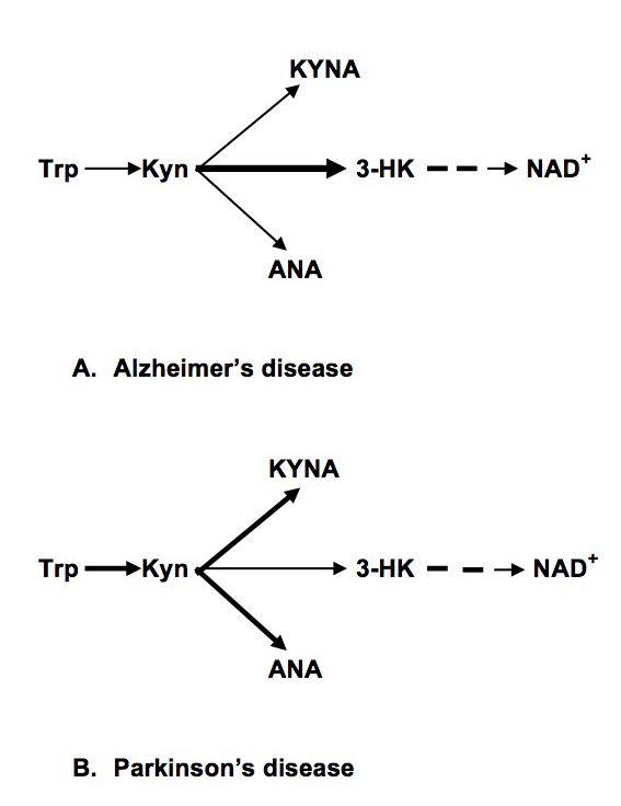
Insulin resistance (IR), obesity and other components of metabolic syndrome [MetS] are highly associated with Alzheimer’s (AD) and Parkinson’s (PD) diseases [1,2]. Dysregulation of kynurenine (Kyn) pathway (KP) of tryptophan (Trp) metabolism was suggested as major contributor to pathogenesis of AD [3], PD [4] and MetS [5,6]. KP, the major source of NAD+ in humans, occurs in brain and peripheral organs (e,g., monocytes/macrophages, liver, pancreas, kidney, intestine, muscles) [7,8]. Initial phase of KR, conversion of Trp into Kyn (via N-formyl-kynurenine), is catalyzed by indoleamine-2,3-dioxygenase 1 (IDO) or tryptophan-2,3-dioxygenase 2 (TDO); Kyn is further converted into 3-hydroxykynurenine (3HK), kynurenic (KYNA) and anthranilic (ANA) acids; further metabolism of 3-HK resulted in production of NAD+ (Fig.1). Considering that some, but not all, peripherally originated derivatives of Kyn penetrate blood brain barrier (BBB) [9], dysregulation of central and peripheral KP might have different functional impact. Thus, increased production of KYNA in brain was suggested to underline psychotic symptoms [8] while elevation of peripheral KYNA (not penetrating BBB) might contribute to mechanisms of IR and obesity in schizophrenia [10].

Up-regulation of Kyn formation from Trp was discovered in central nervous system of AD and PD [11] while assessments of peripheral KP in these diseases yield controversial results.

In PD patients, elevated serum Kyn: Trp ratio, a clinical index of IDO or TDO activity, was reported [12]. Concentration of KYNA and activity of enzyme, catalyzing Kyn conversion into KYNA, were elevated in red blood cells (but not in plasma) of PD patients [13]). The third available study revealed no differences in Kyn and 3-HK plasma concentrations between PD and control subjects, and elevated KYNA and ANA in PD patients without dyskinesia in comparison with PD patients with dyskinesia [14].

In AD most studies compared concentrations of peripheral kynurenines in serum or plasma of patients with probable AD in comparison with healthy controls (or patients with major depressive disease and subjects with subjective cognitive impairment [15]. Only one study evaluated plasma kynurenines in histopathologically confirmed AD in comparison with age-matched and non-matched healthy subjects [16]. Plasma Trp metabolism was found to discriminate between AD and control group in metabolomic study [17]. Trp concentrations were lower in patients with probable [18-20] and histopathologically confirmed AD [16]. Kyn concentrations were unchanged in probable AD [15,18,20] and significantly lower in histopathologically confirmed AD [16]. Kyn: Trp ratio was higher in probable [18,19] but not in histopathologically confirmed AD [16]. Among derivatives of intermediate KP phase, KYNA concentrations were reduced or unchanged in probable [15,20,21] and histopathologically confirmed [16] AD patients. 3-HK levels were elevated in probable AD patients [15] but lower in plasma of histopathologically confirmed AD patients [16]. Strong tendency to reduced ANA was reported in histopathologically confirmed AD [16]. Notably, significant association of plasma ANA and risk of incident dementia with risk increased by 40% for an increase of one standard deviation was observed in participants of Framingham offspring cohort study [22].

We were interested to compare peripheral kynurenines in AD and PD with emphasis on MetS-associated kynurenines, i.e., KYNA, ANA, 3-HK and xanthurenic acid (XA) [23].

Methods

PD patients

Overnight fasting blood samples were collected from 7 female and 11 male PD patients (age range: 50 to 74). At the time of sampling five patients did not take any anti-Parkinson’s medications; and thirteen patients were treated with L-dopa.

AD patient

Blood samples from 12 female and 8 male patients (age range 60 to 75) with probable AD were studied. All AD patients had MMSE between 20 and 23. They were treated with Aricept or Namenda.

Healthy Subjects (Controls)

There were 24 age- and gender- matched (12 females and 12 males) healthy subjects. Study was approved by Tufts Medical Center IRB.

Assessment of Kynurenine metabolites

Plasma samples were stored at −80°C until analysis. Trp, Kyn, ANA, KYNA and 3-HK concentrations were analyzed by high performance liquid chromatography coupled to mass spectrometry (HPLC-MS) as described elsewhere [10].

Statistical analysis

Results are presented as mean ± standard error (Trp and Kyn in μM and ANA, KYNA and 3-HK in nM). Statistical significance was assessed by unpaired t test with Welch correction, two-tailed.

Results

PD patients

There was no difference in plasma concentrations of Trp, Kyn and all studied Kyn metabolites of not treated and treated with L-DOPA patients (data not shown). Therefore, combined data of not-treated and L-DOPA – treated PD patients were used for the further analysis. Trp concentrations were lower, and Kyn: Trp ratio was higher in PD patients than in controls (Table 1). Serum concentrations of Kyn, and its down-stream metabolites, ANA and KYNA, were approximately two fold higher than in control subjects (Table 1). 3-HK concentrations of PD patients were below the sensitivity threshold of the method. XA concentrations were not different between PD and control group (11.87 ± 1.3 and 11.74 ± 1.25, resp).

Table 1. Tryptophan – kynurenine metabolites in serum of Parkinson’s and Alzheimer’s disease patients.

Means ± sem
 Trp
(μM)
 Kyn
(μM)
Kyn: Trp
(x100)
 ANA
(nM)
 KYNA
(nM)
 3-HK
(nM)
Control
(n=24)
68.9 ± 2.49 1.76 ± 0.09 2.55 ± 0.14 70.54 ± 17.9 35.78 ± 3.59 19.55 ± 3.14
PD
(n=18)
48.56 ± 2.4* 2.34 ± 0.11# 4.82 ± 0.18* 156.68 ± 20.46# 65.97 ± 7.2# Not detectable
AD
(n=20)
64.64 ± 3.4 1.77 ± 0.11 2.74 ± 0.15 19.51 ± 3.5* 34.35 ± 2.59 27.52 ± 2.3#
*p<0.001 in comparison with all other groups; #) p<0.001 in comparison with control (except for 3HK p=0.04). Unpaired t test with Welch correction, two tailed.
Abbreviations: Trp: tryptophan; Kyn: kynurenine; KYNA: kynurenic acid; ANA: anthranilic acid; 3-HK: 3-hydroxykynurenine; PD: Parkinson’s disease; AD: Alzheimer’s disease

AD patients

ANA serum concentrations were approximately 3 fold lower, and KYNA concentrations were approximately 40% higher in AD than in controls (Table 1). XA concentrations were not different between AD and control group (10.97 ± 1.07 and 11.74 ± 1.25, resp).

Discussion

Serum levels of KP metabolites might reflect the activity of their formation in peripheral organs [4,8]. Notably, KP in fatty tissue does not express kynurenine-2-monooxygenase (KMO), enzyme converting Kyn into 3-HK, and, therefore, KYNA and ANA are the end products of KP in fatty tissue [24].

Literature data suggested increased conversion of Kyn into 3-HK in PD-related brain structures with consequent formation of neurotoxic metabolites [4]. Present results suggest an increased conversion of Kyn into KYNA and ANA in peripheral organs (in difference with KP in PD-related brain structures [25,26].

In PD patients we confirmed literature data on decreased Trp and increased Kyn concentrations, and, consequently, increased Kyn: Trp ratio, suggesting activation of the initial phase of KP, i.e., conversion of Trp into Kyn [12]. Some discrepancies between literature data (see Introduction) and present results may depend on studied tissues, i.e., serum VS plasma VS RBC; and analytical methods; as well as differences in the other factors potentially affecting KP such as length of disease and age of patients [2,27].

In PD patients we found as increased (about two-fold) plasma concentrations of KYNA and ANA. Considering that KYNA, ANA and 3-HK compete for Kyn as a common substrate in both central and peripheral organs (Figure 1), our data suggest a shift of down-stream Kyn metabolism from 3-HK production towards formation of ANA and KYNA.

EDMJ2017-113-GregoryFOxenkrugUSA_f1

Figure 1. Abbreviations: Trp: Tryptophan; Kyn: kynurenine; KYNA: Kynurenic Acid; ANA: Anthranilic acid; 3-HK: 3-hydroxykynurenine; NAD+: Nicotinamide Adenine Dinucleotide

In AD patients, we found no differences in Trp, Kyn concentrations and Kyn: Trp ratios. Present data are in agreement with the study of histopathologically confirmed AD and age-matched controls that did not find changes of Kyn: Trp ratio [16]. We found elevated 3-HK serum concentration that might suggest decreased availability of Kyn as a substrate for formation of KYNA and ANA in agreement with present results of drastic reduction of ANA concentrations. Peripheral production of ANA deserves further studies, especially considering significant association of plasma ANA and risk of incident dementia in Framingham offspring cohort study [22].

Present study suggested different patterns of dysregulation of the intermediate phase of peripheral KP in PD and AD: increased formation of KYNA and ANA (and reduced production of 3-HK) in PD and reduced formation of ANA (and increased production of 3-HK) in AD (Figure 1). Our data suggest different mechanisms of KP dysregulation in PD and AD: systemic chronic subclinical inflammation activating central and peripheral KP in PD, and central, rather than peripheral, activation of KP in AD triggered by Aβ 1–42 [28]. Notably, there was no association between KP changes and plasma concentrations of neopterin, KP related marker of inflammation, in AD patients [16].

Literature and our data suggest that up-regulation of peripheral KYNA, ANA and Kyn production might contribute to development of obesity and IR, conditions highly associated with early (contrary to late) stages of PD [2,27]. KYNA concentrations positively correlated with BMI in clinical studies [29]. We reported elevation of blood concentrations of KYNA, ANA and Kyn in Zucker obese rats (ZFR) [30]. KYNA elevation in obesity may be a consequence of KMO deficiency in fatty tissue that does not express KMO genes rending KYNA and ANA as the end products of KP in human fatty tissue [24]. KYNA, ANA and Kyn might promote the development of obesity via activation of aryl hydrocarbon receptor (AHR) that regulates xenobiotic-metabolizing enzymes. ANA, KYNA and Kyn are the endogenous human AHR ligands [31,32]. Over-activation of AHR promoted [33] while AHR deficiency protected mice from diet-induced obesity [34].

PD is highly associated not only with obesity but with IR. Increased risk of PD among subjects with T2D was independent from obesity (BMI) [35]. We have previously reported elevation of serum KYNA and ANA in T2D [36,37] and correlation of Kyn with HOMA-IR in HCV patients [38]. Metabolomics analysis revealed 1.8 fold increase of urine KYNA in spontaneously and naturally diabetic rhesus macaques [39]. Successful treatment of IR was associated with down-regulation of KP, including inhibition of KYNA production [40]. One of the possible mechanisms of KYNA involvement in diabetes is activation of G-protein-coupled receptor 35 (GPR35) located primarily in peripheral, including pancreas, tissues [41]. KYNA is an endogenous agonist of GPR35 [41]. Exogenous GPR35 agonists were patented as agents reducing blood glucose levels in oral glucose tolerance tests, stimulate glucose uptake in differentiated 3T3-L1 adipocytes [42].

Significance of ANA elevation has been explored only in a few papers. Serum ANA was positively associated with neopterin, Kyn, Kyn: Trp ratio, and negatively with Trp in healthy young adults [43]. ANA was reported to significantly increase glucose uptake and inhibited 14CO2 production from [U-14C] glucose in in vitro studies [44].

On the other hand, AD is characterized by developing of brain IR [45] while weight loss preceded the diagnosis of dementia in community-dwelling older adults even after controlling for other factors associated with weight [46]. It was suggested that decline in BMI that precedes the diagnosis of AD may be related to neurodegeneration in areas of the brain involved in homeostatic weight regulation [1].

Therefore, we suggest that up-regulated peripheral formation of KYNA, ANA and Kyn contribute to increased risk of PD among subjects with diabetes, and, that contrary to PD, central, rather than peripheral, KP dysregulation contribute to association of IR with AD.

Present data warrant further studies of dysregulations of peripheral KP in PD and AD patients as one of the mechanisms (and potential biomarkers) of association between neurodegenerative disease and MetS.

Acknowledgement

GF Oxenkrug is a recipient of MH104810. Paul Summergrad is a non-promotional speaker for CME outfitters, Inc., and consultant and non-promotional speaker for Pri-med, Inc. Authors appreciate Bioreclamation IVT, NY, USA, for help in collection of serum samples.

References

  1. Alford S, Patel D, Perakakis N, Mantzoros CS (2017) Obesity as a risk factor for Alzheimer’s disease: weighing the evidence. Obes Rev. [crossref]
  2. Vikdahl M, Carlsson M, Linder J, Forsgren L, Håglin L (2014) Weight gain and increased central obesity in the early phase of Parkinson’s disease. Clin Nutr 33: 1132–1139. [crossref]
  3. Baran H, Jellinger K, Deecke L (1999) Kynurenine metabolism in Alzheimer’s disease. J Neural Transm (Vienna) 106: 165–181. [crossref]
  4. Lim C, Fernández-Gomez FJ, Braidy N, Estrada C, Costa C, et al. (2017) Involvement of the kynurenine pathway in the pathogenesis of Parkinson’s disease. Prog Neurobiol 155: 76–95. [crossref]
  5. Oxenkrug GF (2010) Metabolic syndrome, age-associated neuroendocrine disorders and dysregulation of tryptophan – kynurenine pathway metabolism. Ann N Y Acad Sci 1199: 1–14.
  6. Mangge H, Summers LL, Meinitzer A, Zelzer S, Almer G, et al. (2014) Obesity-related dysregulation of the Tryptophan–Kynurenine metabolism: Role of age and parameters of the metabolic syndrome. Obesity.
  7. Paluszkiewicz P, Zgrajka W, Saran T, Schabowski J, Piedra JL, et al. (2009) High concentration of kynurenic acid in bile and pancreatic juice. Amino Acids 37: 637–641. [crossref]
  8. Schwarcz R, Bruno JP, Muchowski PJ, Wu HQ (2012) Kynurenines in the mammalian brain: when physiology meets pathology. Nat Rev Neurosci 13: 465–477. [crossref]
  9. Fukui S, Schwarcz R, Rapoport SI, Takada Y, Smith QR (1991) Blood-brain barrier transport of kynurenines: implications for brain synthesis and metabolism. J Neurochem 56: 2007–2017.
  10. Oxenkrug G, van der Hart M, Roeser J, Summergrad P. Peripheral kynurenine-3-monooxygenase deficiency as a potential risk factor for metabolic syndrome in schizophrenia patients Interg Clin Med 2017: 1: 1–3
  11. Török N, Majláth Z, Fülöp F, Toldi J, Vécsei L (2016) Brain Aging and Disorders of the Central Nervous System: Kynurenines and Drug Metabolism. Curr Drug Metab 17: 412–429. [crossref]
  12. Widner B, Leblhuber F, and D. Fuchs. Increased neopterin production and tryptophan degradation in advanced Parkinson’s disease. J Neural Transm, 2002; 109: 181–189
  13. Hartai Z, Klivenyi P, Janaky T, Penke B, Dux L, et al. (2005) Kynurenine metabolism in plasma and in red blood cells in Parkinson’s disease. J Neurol Sci 239: 31–35. [crossref]
  14. Havelund JF, Andersen AD, Binzer M, Blaabjerg M, et al. (2017) Changes in kynurenine pathway metabolism in Parkinson patients with L-DOPA-induced dyskinesia. J Neurochem. [Epub ahead of print]
  15. Schwarz M, Guillemin GJ, Teipei SJ, Buerger K, Hampei H (2013) Increased 3-hydroxykynurenine serum concentrations differentiate Apzheimer’s disease patients from controls. Eur Arch Psychiatry Clin Neurisci 263: 345–352.
  16. Giil LM, Midttun Ø, Refsum H (2017) Kynurenine Pathway Metabolites in Alzheimer’s Disease. J Alzheimers Dis 60: 495–504. [crossref]
  17. Trushina E, Dutta T, Persson XM, Mielke MM, Petersen RC (2013) Identification of altered metabolic pathways in plasma and CSF in mild cognitive impairment and Alzheimer’s disease using metabolomics. PLoS One 20: 8(5): e63644.
  18. Widner B, Leblhuber F, Walli J, Tilz GP, Demel U, et al. (2000) Tryptophan degradation and immune activation in Alzheimer’s disease. J Neural Transm (Vienna) 107: 343–353. [crossref]
  19. Greiberger J, Fuchs D, Leblhuber F, Greoberger M, Wintersteiger R, et al. (2010) Carbonyl proteins as a clinical marker in Alzheimer’s diasease and its relation to tryptophan degradation and immune activation. Clin Lab 56: 441–448.
  20. Gulaj E, Pawlak K, Bien B, Pawlak D (2010) Kynurenine and its metabolites in Alzheimer’s disease patients. Adv Med Sci 55: 204–211. [crossref]
  21. Hartai Z, Junasz A, Rimannocyz A, Janaky T, Donko T, Dux L, Penke B, Toth GK, Janka Z, Kalman J. Decreased serum and red blood cells kynurenic acid levels in Alzheimer’s disease. Neurochem Int, 2007, 50: 308–13
  22. Chouraki V, Preis SR, Yang Q, Beiser A, Li S, et al. (2017) Association of amine biomarkers with incident dementia and Alzheimer’s disease in the Framingham Study. Alzheimers Dement. S1552-5260(17)30200–5.
  23. Oxenkrug G (2015) 3-hydroxykynurenic acid and type 2 diabetes: implications for aging, obesity, depression, Parkinson’s disease and schizophrenia. In: Engin A, Engin EB (eds) Tryptophan Metabolism: Implications for Biological Processes, Health and Diseases, Molecular and Integrative Toxicology pp.173–195.
  24. Favennec M, Hennart B, Caiazzo R, Leloire A, Yengo L, et al. (2015) The Kynurenine Pathway is Activated in Human Obesity and Shifted Toward Kynurenine Monooxygenase Activation. Obesity 23: 2066–2074.
  25. Ogawa T, Matson WR, Beal MF, Myers RH, Bird ED, et al. (1992) Kynurenine pathway abnormalities in Parkinson’s disease. Neurology 42: 1702–1706. [crossref]
  26. Lewitt PA, Li J, Lu M, Beach TG, Adler CH, Guo L; Arizona Parkinson’s Disease Consortium (2013) 3-hydroxykynurenine and other Parkinson’s disease biomarkers discovered by metabolomic analysis. Mov Disord 28(12): 1653–1660.
  27. Hu G, Antikainen R, Jousilahti P, Kivipelto M, Tuomilehto J (2008) Total cholesterol and the risk of Parkinson disease. Neurology 70: 1972–1979. [crossref]
  28. Guillemin GJ, Smythe GA, Veas LA, Takikawa O, Brew BJ (2003) A beta 1-42 induces production of quinolinic acid by human macrophages and microglia. Neuroreport 14: 2311–2315. [crossref]
  29. Zhao J, Shen D, Djukovic C, Daniel-MacDougall H, Gu X, et al. (2016) Metabolomics identified metabolites associated with body mass index and prospective weight gain among Mexican American women. Obes Sci Pract 2: 309–317
  30. Oxenkrug G, Cornicelli J, van der Hart M, Roeser J, Summergrad P (2016) Kynurenic acid, an aryl hydrocarbon receptor ligand, is elevated in serum of Zucker fatty rats. Integr Mol Med 3(4): 761–763.
  31. DiNatale BC, Murray IA, Schroeder JC, Flaveny CA, Lahoti TS, et al. (2010) Kynurenic acid is a potent endogenous aryl hydrocarbon receptor ligand that synergistically induces interleukin-6 in the presence of inflammatory signaling. Toxicol Sci 115: 8997
  32. Schrenk D, Riebniger D, Till M, Vetter S, Fiedler HP (1999) Tryptanthrins and other tryptophan-derived agonists of the dioxin receptor. Adv Exp Med Biol 467: 403–408. [crossref]
  33. Kerley-Hamilton JS, Trask HW, Ridley CJ, Dufour E, Ringelberg CS, et al. (2012) Obesity is mediated by differential aryl hydrocarbon receptor signaling in mice fed a Western diet. Environ Health Perspect 120: 1252–1259.
  34. Moyer BJ, Rojas IY, Kerley-Hamilton JS, Hazlett HF, Nemani KV, et al. (2016) Inhibition of the aryl hydrocarbon receptor prevents Western diet-induced obesity. Model for AHR activation by kynurenine via oxidized-LDL, TLR2/4, TGFß, and IDO1. Toxicol Appl Pharmacol 300: 13–24.
  35. Gang H, Jousilahti P, Bidel S, et al. Type 2 diabetes and the risk of Parkinson’s disease. Diabetes Care 2007; 30(4): 843–7. Format: Abstract Diabetes Care. 2007;30(4):842-7. J Neurochem. 2017 Jun 19. doi: 10.1111/jnc.14104.
  36. Oxenkrug GF (2015) Increased plasma levels of xanthurenic and anthranilic acids in type 2 diabetes. Mol Neurobiol 52;805-810
  37. Oxenkrug G, van der Hart M, Summergrad P (2015) Elevated anthranilic acid plasma concentrations in type 1 but not type 2 diabetes mellitus. Integr Mol Med 2(5): 365–368.
  38. Oxenkrug GF, Turski WA, Zgrajka, J. V. Weinstock, and P. Summergrad (2013). Tryptophan-Kynurenine Metabolism and Insulin Resistance in Hepatitis C Patients. Hepatitis Research and Treatment, (Vol. 2013), Article ID 149247, 4 pages http: //dx. doi.org/10.1155/2013/149247
  39. Patterson AD, Bonzo JA, Li F, Krausz KW, Eichler GS, Aslam S, et al. (2017) Metabolomics reveals attenuation of the SLC6A20 kidney transporter in nonhuman primate and mouse models of type 2 diabetes mellitus. J Biol Chem 286(22): 19511–22.
  40. Muzik O, Burghardt P, Yi Z, Kumar A, Seyoum B, et al. (2017) Successful metformin treatment of insulin resistance is associated with down-regulation of the kynurenine pathway. Biochem Biophys Res Comm 488: 29–32
  41. Wang J, Simonavicius N, Wu X, Swaminath G, Reagan J, et al. (2006) Kynurenic acid as a ligand for orphan G protein-coupled receptor GPR35. J Biol Chem 281: 22021–22028. [crossref]
  42. Leonard JN, Chu ZL, Unett DJ, Gatlin JE, Gaidarov I, et al. (2007) GPR35 and modulators thereof for the treatment of metabolic-related disorders. United States 20070077602
  43. Deac OM, Mills JL, Gardiner CM, Shane B, Quinn L, et al. (2015) Neopterin and Interleukin-10 Are Strongly Related to Tryptophan Metabolism in Healthy Young Adults. J Nutr 145(4): 701–7.
  44. Schuck PF, Tonin A, da Costa Ferreira G, Viegas CM, Latini A, et al. (2007) Kynurenines impair energy metabolism in rat cerebral cortex. Cell Mol Neurobiol 27(1): 147–160.
  45. Talbot K (2014) Brain insulin resistance in Alzheimer’s disease and its potential treatment with GLP-1 analogs. Neurodegener Dis Manag 4: 31–40. [crossref]
  46. Barrett-Connor E, Edelstein SL, Corey-Bloom J, Wiederholt WC (1996) Weight loss precedes dementia in community-dwelling older adults. J Nutr Health Aging 44: 1147–1152.

Androgen Deprivation Therapy and Cardiovascular Risk

DOI: 10.31038/CST.2017272

Abstract

Background: Several studies have suggested that patients with prostate cancer who undergo androgen deprivation therapy (ADT) with a GnRH agonist have an increased risk of experiencing a cardiovascular event. GnRH antagonists have a different mode of action to GnRH agonists and may be a safer alternative to GnRH agonists in ADT.

Objectives This review article aims to discuss potential mechanisms underlying the development of cardiovascular events associated with ADT using GnRH agonists and to explain the differences in mode of action between GnRH agonists and GnRH antagonists. Additionally, relevant studies are presented and practical recommendations for clinical practice are provided.

Methods: A literature research was performed. Full publications and abstracts published in the last 10 years until September 1st 2015 were considered to be eligible.

Conclusions: Prostate cancer patients undergoing ADT with either cardiovascular disease or an increased risk of experiencing a cardiovascular event should be evaluated for their cardiovascular risk and preferentially treated with a GnRH antagonist.

Keywords

Androgen deprivation therapy, cardiovascular risk, GnRH agonist, GnRH antagonist

Introduction

Androgen deprivation therapy (ADT) with GnRH agonists, GnRH antagonists, and orchiectomy play an important role in the treatment of patients with prostate cancer. ADT therapy has been shown to induce adverse effects including obesity, insulin resistance, hyperglycaemia, dyslipidaemia, and hypertension. All of these adverse effects are associated with the consecutive incidence of diabetes and cardiovascular events under ADT [1]. Therefore, it is important to evaluate potential correlations of cardiovascular adverse-effects and ADT, and furthermore to develop practical recommendations for urologist and cardiologist, as the majority of patients with prostate cancer die of non-cancer related diseases [2].

This review article presents an overview of the various functions of androgens and the resulting pathogenesis of cardiac events/diseases that can be caused by ADT. Moreover, the potential relationships between GnRH agonists / antagonists and cardiac events under ADT are explained, including the type of testosterone deprivation of both substance classes, and the relevant clinical studies are summarized. Moreover, practical recommendations for clinical practice are provided.

Hormonal effects of androgens on the cardiovascular system

Androgens play a decisive role in the energy supply and in various metabolic pathways of cells besides their fundamental role in reproductive and sexual function. Androgens have a systemic indirect effect on the cardiovascular system and a direct effect on the cardiovascular system.

Androgens promote growth and preservation of muscle mass and promote fat metabolism, thereby regulating the body composition [3]. Expression of androgen receptors in fat tissue suggests that androgens are involved in the accumulation and distribution of fat tissue. Androgens promote lipolysis in adipose tissue and inhibit the absorption of triglycerides, thus increasing levels of circulating triglycerides and cholesterol. Moreover, androgens ensure a faster conversion of triglycerides into subcutaneous abdominal fat tissue, and less into gluteal-femoral fat [4].

Testosterone, the most prominent member of the androgens, has been shown to have direct positive and negative effects on the cardiovascular system. Testosterone has been shown to have an antiarrhythmic effect on the heart [5, 6], protect the cardiomyocytes against ischemic insults, thereby reducing the myocardial infarction size [7, 8], and its atheroprotective effects have been proven, as well [9-11].

On the other hand, testosterone is reported to have negative effects on the endothelium [12], on vasorelaxation [13], and it promotes apoptosis [14].

These discrepancies of biological effects may explain the differential clinical results of the type and dosage of testosterone deprivation on the cardiovascular system. For instance, patients who underwent orchiectomy suffer less often from coronary heart disease, myocardial infarction, sudden cardiac death and stroke as compared to patients who are treated with GnRH agonists [15].

Indirect and direct effects of GnRH agonists on the cardiovascular system

The U.S. Food and Drug Administration (FDA) issued a warning for GnRH agonists on the basis of a number of published data which associated GnRH agonist treatment with an increased risk of cardiovascular events [15]. The effects GnRH agonist treatment on the cardiovascular system can be explained with its indirect and direct effects on the cardiovascular system.

Indirect mechanisms

Therapy with GnRH agonists aims at reducing the androgen level resulting in an induced state of hypogonadism. The effects of GnRH agonist treatment include effects on sexual function including reduced libido, impotence and systemic effects including anaemia, and osteoporosis [16].

The changes in the body composition, which are also observed in patients treated with GnRH agonists, characterised by a loss of muscle mass and strength and increase in fat tissue and weight gain can be attributed to the fact that androgen-mediated effects on adipogenesis are inhibited [17].

The observed increase in the fat mass has been shown to be associated with increased insulin levels [18, 19] which in turn might also promote the production of adipokines and inflammatory cytokines [20]. These changes lead to increased plasma insulin concentrations, insulin resistance, increased HDL and LDL levels, and higher triglyceride levels [3, 21]. These changes promote the incidence of diabetes. Diabetes and the metabolic disorders are independent risk factors for the development of atherosclerosis, which in turn increases the risk for the incidence or progression of cardiovascular diseases.

Direct mechanisms

Binding of GnRH to its receptor has been shown to occur at several sites of the body, including the hypothalamus, pituitary gland, gonads, breast, and prostate. The expression of the GnRH receptor outside the hypothalamus-pituitary gland-reproduction-axis, such as the cerebellum, kidney, and heart is currently under investigation [22].

Studies suggest that GnRH agonists have a direct effect on the cardiomyocytes which might affect the cardiac function negatively. GnRH agonists are believed to regulate the heart contractility and the concentration of intracellular calcium ions by activating the protein kinase A (PKA) through the GnRH receptor. Cardiomyocytes contain substrates of PKA such as phospholamban, L-type calcium channel, and components of the contractile apparatus. Thus, the PKA could play a decisive role in the GnRH associated cardiac reaction [23].

The GnRH receptor is also present on lymphocytes that even produce GnRH endogenously to regulate the immune function [24]. The transendothelial migration of infiltrates is mediated by interactions of cell adhesion molecules, which are induced by cytokines [25]. Binding of GnRH or GnRH agonist to the GnRH receptor on the lymphocytes leads to an increased expression of the IL2γ receptor [24]. This results in an increased proliferation and inflammation as well as the release of cytokines like interferon γ. Inflammatory processes impair the normal function of the endothelium so that the development of atherosclerotic plaques, instabilities, and plaque ruptures are promoted [26, 27]. These are supposed to be the main cause for acute myocardial infarctions and strokes.

Differences in the mode of action between GnRH agonists and antagonists

Since the FDA assumed a class effect regarding cardiovascular events, they also issued 3 years after the initial warning for GnRH agonists, a black-label-warning for GnRH antagonists [28]. However, the FDA did not take into account the fact that GnRH agonists and antagonists were not considered separately in studies, and they did not consider that their differential mode of action:

GnRH agonists act like the natural ligand, GnRH, of the GnRH receptor. By binding to the receptor, they induce activation so that the luteinising hormone (LH) and follicle-stimulating hormone (FSH), which are initially released in increased amounts. This results in a temporary testosterone surge (flare up). However, by continuous administration of a GnRH agonist, the GnRH receptor is permanently stimulated and thus down-regulated. This down-regulation in turn causes a permanent reduction of LH and FSH hormones as well as of testosterone levels to the orchiectomy level.

GnRH antagonists do not act like a ligand that stimulates the GnRH receptor, but block it competitively, and thus inhibit the release of LH and FSH. There is no flare-up, as due to the inhibition of the GnRH receptor all subsequent LH- and FSH-mediating signal pathways downstream of the GnRH receptor are also blocked.

Several non-randomised studies have shown that ADT with GnRH agonists is associated with an increased cardiovascular risk [15, 29-37]. In contrast, several randomised studies report no correlation between the administration of GnRH agonists and an increased cardiovascular risk [38-41] (see table 1). However, the contradictory results can be attributed to several potential sources of error in the respective study design [11].

In contrast to these results, a large meta-analysis of 16 prospective phase II/III studies and one phase III study, analysing 1,704 patients treated with the GnRH antagonist degarelix showed no correlation between the treatment and cardiovascular events [42].

Table 1. Non-randomised trial for the evaluation of the incidence of cardiovascular events under GnRH agonists in men with prostate cancer.

Ref. n Reference group ADT Result HR (95% CI)1
[17] 73,196 no ADT GnRH agonist and/or antiandrogen Coronary heart disease, myocardial infarction, sudden cardiac death 1.16 (1.10–1.21)1.11 (1.01–1.21)1.16 (1.05–1.27)
[36] 4,892 no ADT GnRH agonist and/or antiandrogen Cardiovascular mortality with radical prostatectomy, cardiovascular mortality with EBRT, brachytherapy or chemotherapy, 2.6 (1.4–1.7)1.2 (0.8–1.9)
[37] 22,816 no ADT Medical ADT Cardiovascular morbidity 1.20 (1.15–1.26)
[40] 19,097 no ADT GnRH agonist and/or antiandrogen; orchiectomy Acute myocardial infarction, sudden cardiac death, diabetes 0.92 (0.84–1.00)0.96 (0.83–1.10)1.24 (1.15–1.35)
[33] 37,443 WW/AS GnRH agonist, orchiectomy, antiandrogen, combined androgen blockade Coronary heart disease, myocardial infarction, sudden cardiac death, stroke 1.17 (1.06–1.39)1.21 (1.01–1.44)1.28 (1.05–1.57)1.18 (1.02–1.36)
[34] 76,601 RP, WW/AS GnRH agonist, antiandrogen, GnRH + antiandrogen, orchiectomy, medical or surgical ADT Ischemic heart disease, myocardial infarction, heart failure, stroke 1.34 (1.25–1.43)1.47 (1.35–1.60)1.67 (1.54–1.80)1.27 (1.17–1.38)
[38] 182,757 no ADT GnRH agonist, orchiectomy Peripheral arterial disease, venous thromboembolism 1.15 (1.11–1.19)1.10 (1.04–1.16)
[35] 31,571 no ADT Antiandrogen, orchiectomy Myocardial infarction, stroke 1.31 (1.16–1.49)1.19 (1.06–1.35)
[39] 140,474 no ADT GnRH agonist, orchiectomy Acute myocardial infarction, coronary artery disease, sudden cardiac death 1.09 (1.04–1.15)1.11 (1.07–1.15)1.18 (1.12–1.24)
[41] 50,384 no ADT GnRH agonist, orchiectomy Coronary heart disease 1.13 (1.09-1.17)-1.17 (1.13-1.21) dose-dependent

1 If several types of ADT are evaluated separately, HRs refer to GnRH agonists vs. control.

In order to clearly attribute the incidence of cardiovascular events under medical ADT to one substance class, a direct comparison between GnRH agonists and GnRH antagonists in clinical studies is required.

Such a comparison was performed in the recently published meta-analysis by Albertsen et al. between the GnRH antagonist degarelix and the GnRH agonists goserelin and leuprolide [33, 43]. Data of 2,328 patients from six prospective RCTs were pooled. 1,491 patients received degarelix and of the remaining 837 patients, 458 patients were treated with goserelin and 379 patients with leuprolide, respectively. Patients treated with the GnRH antagonist degarelix had a significantly lower risk of experiencing a cardiovascular event as compared to patients under GnRH agonist therapy (HR: 0.597; 95% CI: 0.380-0.938; p=0.0253) [44]. A subsequent analysis using a Cox model confirmed these results. Treatment with the GnRH antagonist degarelix resulted in a 40% lower risk of experiencing a cardiovascular event or death compared to treatment with GnRH agonists (HR: 0.60; 95% CI: 0.41-0.87; p = 0.008) [43].

Treatment of patients with known cardiovascular disease

Several studies suggest that patients with a history of cardiovascular disease have a higher risk of experiencing a cardiovascular event under ADT.

In the above mentioned meta-analysis of 16 prospective phase II/III studies and one phase III study, analysing 1,704 patients treated with the GnRH antagonist degarelix, the patients were stratified according to their cardiovascular history. Patients in group 1 (n=337) had no cardiovascular risk factors, patients in group 2 (n=803) had one cardiovascular risk factor, but no cardiovascular disease, and patients in group 3 (n=112) had a known cardiovascular disease. Cardiovascular events were most frequent in patients with the most severe cardiovascular history in group 3 (20%), decreasing in the other groups (group 2: 8% and group 1: 7%). While the presence of a single risk factors only resulted in a 1.3 fold increased risk of experiencing a cardiovascular event (p=0.28), an existing cardiovascular event resulted in a 3.1 fold increased risk (p<0.0001) [42].

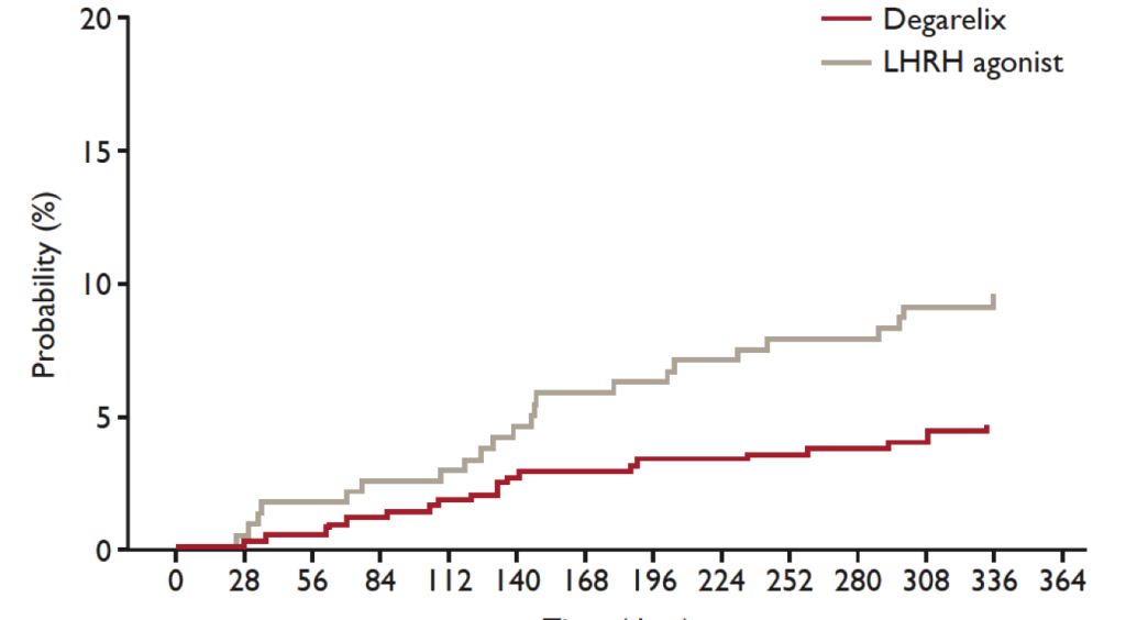
A direct comparison between GnRH agonists and the GnRH antagonist degarelix revealed that degarelix was associated with a significantly lower risk of experiencing a cardiovascular event in patients with a history of cardiovascular disease. Thus, there were significantly fewer cardiovascular (HR: 0.476; 95% CI: 0.260-0.871; p=0.0160) or severe cardiovascular events (HR: 0.367; 95% CI: 0.174-0.775; p=0.0086) under degarelix compared to LHRH agonists
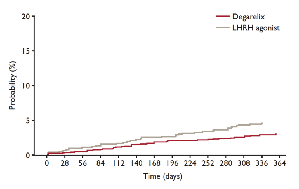
(Figure 1) [44]. A landmark analysis of the first treatment year with GnRH antagonists in patients with known cardiovascular disease revealed a 56% lower risk (HR: 0.44; 95% CI: 0.26-0.74; p=0.002) [43] for experiencing cardiovascular events (arterial embolic and thrombotic events, haemorrhagic or ischemic cerebrovascular events, myocardial infarction, or other ischemic heart diseases) or death as compared to GnRH agonists (Figure 2). In patients without cardiovascular history, no different cardiovascular risk was observed depending on the respective ADT.

CST2017-236-Behrouz Denmark_F1

Figure 1. Cardiovascular risk in all patients

A direct comparison between GnRH agonists and the GnRH antagonist degarelix revealed that degarelix was associated with a significantly lower risk of experiencing a cardiovascular event in patients with a history of cardiovascular disease. Thus, there were significantly fewer cardiovascular (HR: 0.476; 95% CI: 0.260-0.871; p=0.0160) or severe cardiovascular events (HR: 0.367; 95% CI: 0.174-0.775; p=0.0086) under degarelix compared to LHRH agonists (modified from [44]).

CST2017-236-Behrouz Denmark_F2

Figure 2. Landmark analysis of first treatment year.

A landmark analysis of the first treatment year with GnRH antagonists in patients with known cardiovascular disease revealed a 56% lower risk (HR: 0.44; 95% CI: 0.26-0.74; p=0.002) for experiencing cardiovascular events or death as compared to GnRH agonists (modified from [43]).

One possible explanation for the lower risk of GnRH antagonists as compared to GnRH agonists in patients with history of cardiovascular disease might be the low rate of vascular occlusions which is probably associated with FSH. FSH receptors play a role in the lipid metabolism and fat accumulation so that their inhibition might reduce the risk of experiencing a repeated cardiovascular event [45]. GnRH antagonists suppress both LH and FSH hormones [28, 46, 47]. In contrast, GnRH agonists primarily inhibit the release of LH and therefore do not act sufficiently on the signal pathways downstream of FSH [43]. Another possible cause could be the destabilisation of vascular lesions under ADT. Destabilisation might be achieved by activating GnRH receptors on the T-cells atherosclerotic plaques with a GnRH agonist. This mechanism does not apply for GnRH antagonists, as these do not induce activation of the GnRH receptor.

Consequences for therapy management

With regard to therapy management it seems obvious that patients with a history of cardiovascular disease or the risk of developing a cardiovascular disease should be preferentially treated with a GnRH antagonist over a GnRH agonist. Alternatively, a dose reduction of the GnRH agonist could be useful [37]. However, this should be carefully weighed, as a dose reduction can always impair treatment efficacy.

According to a recently published meta-analysis, the benefits of a GnRH antagonist can not only have a positive effect on the side-effect profile, but also improve the overall survival as compared to therapy with a GnRH agonist. Klotz et al. report that patients under GnRH antagonist therapy had a significantly longer progression-free survival. Moreover, these patients also have a more favourable side-effect profile regarding urinary and musculoskeletal tract as compared to patients under a GnRH agonist [48]. Although these findings provided first indications for the benefits of treatment with a GnRH antagonist, they must be further validated.

Outlook and recommendations

Cardiovascular diseases are the major cause of death in the male population of advanced age. In order to avoid any additional life-threatening risks for this patient population, different therapeutic approaches to ADT should be evaluated for their potential cardiovascular side-effects. Current evidence shows that treatment using GnRH agonists provides better cardiovascular tolerability and should thus be preferred. However, further clinical studies are required to directly compare the incidence of cardiovascular events under GnRH agonists as compared to GnRH antagonists. One trial currently still recruiting patients who is comparing cardiovascular safety of degarelix versus leuprolide in patients with advanced prostate cancer and cardiovascular disease is the PRONOUNCE Trial (NCT02663908).

Thus, patients with prostate cancer are mainly elder patients with additional traditional cardiovascular risk factors. If ADT becomes necessary, the cardiovascular risk is further adversely affected. Therefore, the patient’s cardiovascular risk should be taken into consideration before the appropriate treatment option is selected and treatment initiated.

Patients with a high cardiovascular risk or who have already experienced a cardiovascular event might particularly benefit from treatment with a GnRH antagonist. Nevertheless, these patients should be monitored closely in coordination with the attending cardiologists. This will enable optimal adjustment of the treatable risk factors, like the lipid profile as well as blood pressure. The same applies for patients suffering from diabetes or a prediabetic metabolic status. These parameters should be verified and – if necessary – adjusted prior to therapy. The next step should be cessation of nicotine abuse. The highest risk of experiencing a cardiovascular event is at the beginning of the therapy. It is very important that the patient is screened for potential cardiovascular risk factors or known risk factors optimally adjusted, respectively.

We suggest that all patients scheduled to undergo ADT therapy should have a cardiovascular assessment.

Cardiovascular assessment should include a thorough physical examination, assessment of any cardiac related symptoms (angina, dyspnea, syncope and palpitations), history taking of any known cardiovascular disease, and determination of the presence of cardiovascular risk factors such as hypertension, diabetes, smoking habit, adiposities, hyperlipidemia, myocardial infarction, heart failure and stroke. In patients with known diabetes, we recommend to determine the HbA1c before the start of the ADT treatment. We furthermore recommend monitoring the HbA1c levels during the therapy. In patients with known hyperlipidemia we recommend to further monitor the levels of cholesterol, triglyceride, LDL and HDL.

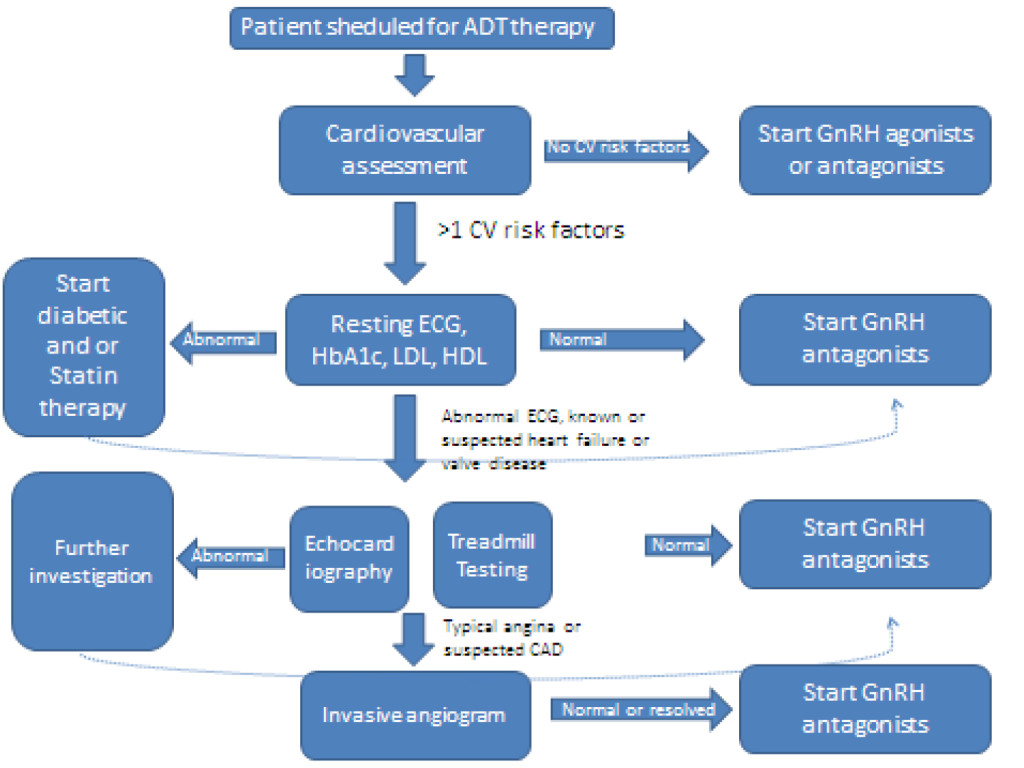
Patients with one or more cardiovascular risk factors should be assessed furthermore. We recommend a resting-ECG for all patients with more than 1 risk factors or known coronary artery disease, echocardiography for patients with known/suspected heart failure and/or known/suspected valve disease, a treadmill-testing for patients with >3 cardiovascular risk factors and/or unstable angina and invasive angiogram for patients with suspected coronary artery disease (see Figure 3).

CST2017-236-Behrouz Denmark_F3

Figure 3. Clinical recommendations.

Patients with known cardiovascular disease should be on optimal doses of beta blocker, statin, ACE-inhibitor/ ARB and diuretic therapy.

References

  1. Collier A, Ghosh S, McGlynn B, Hollins G: Prostate cancer, androgen deprivation therapy, obesity, the metabolic syndrome, type 2 diabetes, and cardiovascular disease: a review. American journal of clinical oncology 35: 504–509.
  2. Feuer EJ, Merrill RM, Hankey BF (1999) Cancer surveillance series: interpreting trends in prostate cancer–part II: Cause of death misclassification and the recent rise and fall in prostate cancer mortality. Journal of the National Cancer Institute 91: 1025–1032.
  3. Smith MR, Finkelstein JS, McGovern FJ, Zietman AL, Fallon MA, et al. (2002) Changes in body composition during androgen deprivation therapy for prostate cancer. J Clin Endocrinol Metab 87: 599–603. [crossref]
  4. Collins L, Mohammed N, Ahmad T, Basaria S (2012) Androgen deprivation therapy for prostate cancer: implications for cardiometabolic clinical care. Journal of endocrinological investigation 35: 332–339.
  5. Edelman S, Butler J, Hershatter BW, Khan MK (2014) The Effects of Androgen Deprivation Therapy on Cardiac Function and Heart Failure: Implications for Management of Prostate Cancer. Clinical genitourinary cancer 12: 399–407.
  6. Brouillette J, Rivard K, Lizotte E, Fiset C (2005) Sex and strain differences in adult mouse cardiac repolarization: importance of androgens. Cardiovascular research 65: 148–157.
  7.  Tsang S, Wu S, Liu J, Wong TM (2008) Testosterone protects rat hearts against ischaemic insults by enhancing the effects of alpha(1)-adrenoceptor stimulation. Br J Pharmacol 153: 693–709. [crossref]
  8. Herring MJ, Oskui PM, Hale SL, Kloner RA (2013) Testosterone and the cardiovascular system: a comprehensive review of the basic science literature. Journal of the American Heart Association 2: e000271.
  9. Bourghardt J, Wilhelmson AS, Alexanderson C, De Gendt K, Verhoeven G, et al. (2010) Androgen receptor-dependent and independent atheroprotection by testosterone in male mice. Endocrinology 151: 5428–5437.
  10. Kelly DM, Jones TH (2013) Testosterone: a vascular hormone in health and disease. J Endocrinol 217: R47–71. [crossref]
  11. Tivesten A, Pinthus JH, Clarke N, Duivenvoorden W, Nilsson J (2015) Cardiovascular risk with androgen deprivation therapy for prostate cancer: potential mechanisms. Urol Oncol 33: 464–475. [crossref]
  12. Rettew JA, Huet-Hudson YM, Marriott I (2008) Testosterone reduces macrophage expression in the mouse of toll-like receptor 4, a trigger for inflammation and innate immunity. Biology of reproduction 78: 432–437.
  13. Ammar EM, Said SA, Hassan MS (2004) Enhanced vasoconstriction and reduced vasorelaxation induced by testosterone and nandrolone in hypercholesterolemic rabbits. Pharmacological research: the official journal of the Italian Pharmacological Society 50: 253–259.
  14. Cavasin MA, Tao ZY, Yu AL, Yang XP (2006) Testosterone enhances early cardiac remodeling after myocardial infarction, causing rupture and degrading cardiac function. American journal of physiology Heart and circulatory physiology 290: H2043–2050.
  15. Keating NL, O’Malley AJ, Smith MR (2006) Diabetes and cardiovascular disease during androgen deprivation therapy for prostate cancer. Journal of clinical oncology: official journal of the American Society of Clinical Oncology 24: 4448–4456.
  16. Taylor LG, Canfield SE, Du XL (2009) Review of major adverse effects of androgen-deprivation therapy in men with prostate cancer. Cancer 115: 2388–2399.
  17. Bhasin S, Taylor WE, Singh R, Artaza J, Sinha-Hikim I, Jasuja R, et al. (2003) The mechanisms of androgen effects on body composition: mesenchymal pluripotent cell as the target of androgen action. The journals of gerontology Series A, Biological sciences and medical sciences 58: M1103–1110.
  18. Traish AM, Saad F, Guay A (2009) The dark side of testosterone deficiency: II. Type 2 diabetes and insulin resistance. J Androl 30: 23–32. [crossref]
  19. Smith MR, Lee H, Fallon MA, Nathan DM (2008) Adipocytokines, obesity, and insulin resistance during combined androgen blockade for prostate cancer. Urology 71: 318–322.
  20. Shahani S, Braga-Basaria M, Basaria S (2008) Androgen deprivation therapy in prostate cancer and metabolic risk for atherosclerosis. The Journal of clinical endocrinology and metabolism 93: 2042–2049.
  21. Dockery F, Bulpitt CJ, Agarwal S, Donaldson M, Rajkumar C (2003) Testosterone suppression in men with prostate cancer leads to an increase in arterial stiffness and hyperinsulinaemia. Clinical science (London, England : 1979) 104: 195–201.
  22. Skinner DC, Albertson AJ, Navratil A, Smith A, Mignot M, et al. (2009) Effects of gonadotrophin-releasing hormone outside the hypothalamic-pituitary-reproductive axis. Journal of neuroendocrinology 21: 282–292.
  23. Dong F, Skinner DC, Wu TJ, Ren J (2010) The heart: a novel gonadotrophin-releasing hormone target. Journal of neuroendocrinology 23: 456–463.
  24. Chen HF, Jeung EB, Stephenson M, Leung PC (1999) Human peripheral blood mononuclear cells express gonadotropin-releasing hormone (GnRH), GnRH receptor, and interleukin-2 receptor gamma-chain messenger ribonucleic acids that are regulated by GnRH in vitro. The Journal of clinical endocrinology and metabolism 84: 743–750.
  25. Noutsias M, Seeberg B, Schultheiss HP, Kuhl U (1999) Expression of cell adhesion molecules in dilated cardiomyopathy: evidence for endothelial activation in inflammatory cardiomyopathy. Circulation 99: 2124–2131.
  26. Knutsson A, Hsiung S, Celik S, Wigren M, Nilsson J, et al. (2015) Treatment with an LRHR agonist, but not the LHRH antagonist degarelix, induces atherosclerotic plaque instability in ApoE-/- mice. In: EAU 2015, Poster 559. Madrid.
  27. Knutsson A, Hsiung S, Celik S, Rattik S, Mattisson IY, et al. (2016) Treatment with a GnRH receptor agonist, but not the GnRH receptor antagonist degarelix, induces atherosclerotic plaque instability in ApoE(-/-) mice. Scientific reports 6: 26220.
  28. Fachinformation (2014) FIRMAGON 80 mg Pulver und Lösungsmittel zur Herstellung einer Injektionslösung. Ferring GmbH. 011697–7981 – Stand der Information Oktober.
  29. Keating NL, O’Malley A, Freedland SJ, Smith MR: Diabetes and cardiovascular disease during androgen deprivation therapy: observational study of veterans with prostate cancer. Journal of the National Cancer Institute 104: 1518–1523.
  30. Van Hemelrijck M, Garmo H, Holmberg L, Ingelsson E, Bratt O, et al. (2010) Absolute and relative risk of cardiovascular disease in men with prostate cancer: results from the Population-Based PCBaSe Sweden. Journal of clinical oncology: Official journal of the American Society of Clinical Oncology 28: 3448–3456.
  31. Jespersen CG, Norgaard M, Borre M (2014) Androgen-deprivation therapy in treatment of prostate cancer and risk of myocardial infarction and stroke: a nationwide Danish population-based cohort study. European urology 65: 704–709.
  32. Tsai HK, D’Amico AV, Sadetsky N, Chen MH, Carroll PR (2007) Androgen deprivation therapy for localized prostate cancer and the risk of cardiovascular mortality. Journal of the National Cancer Institute 99: 1516–1524.
  33. Saigal CS, Gore JL, Krupski TL, Hanley J, Schonlau M, et al. (2007) Androgen deprivation therapy increases cardiovascular morbidity in men with prostate cancer. Cancer 110: 1493–1500.
  34. Hu JC, Williams SB, O’Malley AJ, Smith MR, Nguyen PL, et al. (2012) Androgen-deprivation therapy for nonmetastatic prostate cancer is associated with an increased risk of peripheral arterial disease and venous thromboembolism. European urology 61: 1119–1128.
  35. Gandaglia G, Sun M, Popa I, Schiffmann J, Abdollah F, et al (2014) The impact of androgen-deprivation therapy (ADT) on the risk of cardiovascular (CV) events in patients with non-metastatic prostate cancer: a population-based study. BJU international 114: E82–89.
  36. Alibhai SM, Duong-Hua M, Sutradhar R, Fleshner NE, Warde P, et al. (2009) Impact of androgen deprivation therapy on cardiovascular disease and diabetes. Journal of clinical oncology: official journal of the American Society of Clinical Oncology 27: 3452–3458.
  37. Schmid M, Sammon JD, Reznor G, Kapoor V, Speed JM, et al. (2015) Dose-dependent effect of androgen deprivation therapy for localized prostate cancer on adverse cardiac events. BJU international
  38. Efstathiou JA, Bae K, Shipley WU, Hanks GE, Pilepich MV, et al. (2009) Cardiovascular mortality after androgen deprivation therapy for locally advanced prostate cancer: RTOG 85-31. Journal of clinical oncology : official journal of the American Society of Clinical Oncology 27: 92–99.
  39. Bolla M, Van Tienhoven G, Warde P, Dubois JB, Mirimanoff RO, et al. (2010) External irradiation with or without long-term androgen suppression for prostate cancer with high metastatic risk: 10-year results of an EORTC randomised study. The Lancet Oncology 11: 1066–1073.
  40. Wilcox C, Kautto A, Steigler A, Denham JW (2012) Androgen deprivation therapy for prostate cancer does not increase cardiovascular mortality in the long term. Oncology 82: 56–58.
  41. Nguyen PL, Je Y, Schutz FA, Hoffman KE, Hu JC, et al. (2010) Association of androgen deprivation therapy with cardiovascular death in patients with prostate cancer: a meta-analysis of randomized trials. JAMA : The journal of the American Medical Association 306: 2359–2366.
  42. Smith MR, Klotz L, van der Meulen E, Colli E, Tanko LB (2011) Association of baseline risk factors with cardiovascular (CV) events during long-term degarelix therapy in men with prostate cancer. Journal of clinical oncology : Official journal of the American Society of Clinical Oncology 2011.
  43. Albertsen PC, Klotz L, Tombal B, Grady J, Olesen TK, Nilsson J (2014) Cardiovascular morbidity associated with gonadotropin releasing hormone agonists and an antagonist. European urology 65: 565–573.
  44. Tombal B, Miller K, De Reijke TM, Persson BE (2013) Degarelix versus luteinising hormone-releasing hormone (LHRH) agonists: Safety outcomes from six comparative randomised clinical trials. European Journal of Cancer 2: 681.
  45. Radu A, Pichon C, Camparo P, Antoine M, Allory Y, et al. (2010) Expression of follicle-stimulating hormone receptor in tumor blood vessels. The New England journal of medicine 363: 1621–1630.
  46. Hanke H, Lenz C, Hess B, Spindler KD, Weidemann W (2010) Effect of testosterone on plaque development and androgen receptor expression in the arterial vessel wall. Circulation 103: 1382–1385.
  47. Ng MK, Quinn CM, McCrohon JA, Nakhla S, Jessup W, et al. (2003) Androgens up-regulate atherosclerosis-related genes in macrophages from males but not females: molecular insights into gender differences in atherosclerosis. Journal of the American College of Cardiology 42: 1306–1313.
  48. Klotz L, Miller K, Crawford ED, Shore N, Tombal B, et al. (2014) Disease control outcomes from analysis of pooled individual patient data from five comparative randomised clinical trials of degarelix versus luteinising hormone-releasing hormone agonists. European urology 66: 1101–1108.

Sexuality in Patient with Breast Cancer Hospitalized in Professional Nursing Vision

DOI: 10.31038/CST.2017271

Abstract

The study seeks to investigate aspects of sexuality of women with breast cancer admitted. The emergence of the study took place at the time of my professional practice where I came across patient dialogues, and health professionals with the behavior of women in different times, having sexual practice and professional assistants, on the other hand the kind of suppressed such an attitude as well as the patients more “brawling”. I realized the need to discuss such issues: women who are hospitalized for treatment that theoretically move with biopsicoemocional, self-image and self-esteem, should have “head for such behavior? There is a need of nurses to direct a group for care to sexuality. Aiming to analyze the psychophysical need, using the philosophical approach of Jean Watson. Methodology the study presents a review and synthesis of the literature on the theory of nursing sexual need. Final considerations the nurse has the key role of facilitating communication of sexuality, not to become fragmented, to emphasize the importance of the role of the sexual partner. Thus discussion related to sexual health.

Introducion

The study seeks to investigate aspects of sexuality of women with breast cancer interned. The emergence of the study  took place at the time of my professional practice where I came across patient dialogues, and health professionals with the behavior of women in different times, having sexual practice and professional assistants, on the other hand the kind of suppressed such an attitude as well as the patients more “excited”.

In the context of health, the educational process consists much more than the simple act of teaching. The client, who is often mistakenly called a passive individual, is a key player in the care process, since we already know that the process of health care is dynamic and requires the participation of both parties, whether caregiver or individual who will receive the care.

Justification

I realized the need to discuss such issues: women who is hospitalized for treatment that theoretically move with biopsicoemocional, self-image and self-esteem,should have “head for such behavior? There is a need of nurses to direct a group for care to sexuality.

By identifying the knowledge of the caregivers, we can analyze the quality of the orientation group, in which the study may contribute to the educational interventions to be proposed to this group, with the aim of improving the caregiver’s vision to young patients with cancer breastfeeding, thus favoring better qualified care.

Methodology

Aiming to analyze the psychophysical need, using the philosophical approach of Jean Watson.

Methodology the study presents a review and synthesis of the literature on the theory of nursing sexual need.(possible adaptation) [Figure 1]

CST2017-233-Lilian Brazil_F1

Figure 1.

Objective

General objective

To analyze the process of health education in the approach of the group of guidance by the nurse in hospitalized patients with breast cancer in the perception of the caregiver.

Specific Objectives

To identify the communication between the nurse practitioner and the patient with the partner.

Final Consideration

The nurse has the key role of facilitating communication of sexuality, not to become fragmented, to emphasize the importance of the role of the sexual partner. Thus discussion related to sexual health, through the guidance group.

References

  1. Ahmad EC, Coler MS, Nóbrega MML (2005) Jean Watson’s nursing theoryfocused on human sexuality. Braz J Nurs
  2. SMA, Panobianco MS, Ferreira Gozzo TO, Abdulla AM (2013) Sexualityof women with breast cancer: analysisof nursing science. Florianópolis: text Context Nurses 30: 835–42
  3. National Cancer Institute (2013) Actions directed to improved appearance of patients have strong impact on the quality of the treatment-Self-esteem is fundamental. Rev Cancer Network 24–27
  4. Junqueira LCU, Vieira Giami, Saints in MA (2013) Analysis of communication about sexuality, established by nurses, with patients in the health care context of breast cancer. Interface 17: 89–101