Abstract
The cutaneous clinical manifestation characterized by deep dermatitis with granulomatous reaction, appearance of an edematous, circular and alopecic nodule was found abruptly in a 6-year-old female canine patient. Diagnosed with Lupus Erythematosus due to the presence of major symptoms, the causes of kerion were excluded by the species Microsporum canis, M. gypseum and Trichophyton mentagrophytes, which were found to be absent in the cytology, fungal and bacterial cultures of the present case. Using Causticum 30 cH, three globules, twice a day, the patient’s inflammatory process ceased on the third day, turning the lesion into a wound that healed on the fifteenth day. Complete rehairing of the affected area was observed on the thirtieth day. Immunological parameters that affected skin morphometry in the initial phase, showed positive results after the use of homeopathy, reflecting the magnitude of the therapy adopted in dermatopathies.
Keywords
Alopecia, Autoimmune, Scabs, Dermatitis, Homeopathy
Introduction
Characterized by erosive lesions on the face, systemic lupus erythematosus (SLE) is an ancient disease, first mentioned in the Middle Ages 400 years BC by Hippocrates. In 1846, the Viennese physician Von Hebra described a “butterfly wing” pattern of dermatitis, but it was named by Pierre Cazenave in 1851 when he described spontaneously arising skin lesions that resembled wolf bites.
Lupus erythematosus is a benign autoimmune condition, with a low occurrence in veterinary routine. Affected patients produce antibodies against normal skin components. The clinical presentation is varied, producing lesions mainly on the snout. There is no 100% specific test for detecting Lupus . They consider the test called FAN (antinuclear factor or antibody), with high titers, in symptomatic animals [1], but definitively the diagnosis is made through histopathological examination and the treatment is based on immunosuppression and non-exposure to solar radiation [2-11].
A spectrum of unique and often characteristic clinical signs allows the early implementation of an effective treatment [9]. In contrast, canine variants regroup therapeutic possibilities. At this time, we would also regroup under Vesicular cutaneous lupus erythematosus, Exfoliative cutaneous lupus erythematosus, Localized (facial) or generalized discoid lupus erythematosus and Mucocutaneous lupus erythematosus, the currently recognized subtypes.
If all homeopathy and its principles were not enough to demonstrate Hanemmann’s genius, a mixture of quicklime and porcelain, after the drying process, prepared in homeopathic medicine, in a fabulous therapeutic resource for the sub regent case of inflammation and burning sensation in the snout [7], has its characteristics of infinitesimal dilution, with absence of residues, being widely used [2].
Case Report
A female, spayed, mixed breed dog weighing 32 kg and 6 years old was treated for a superficial skin infection on the dorsal region of the muzzle that had been developing for less than 24 hours (she woke up like this). There was no possibility of trauma or contact with chemicals. A fragment of the lesion on the muzzle was collected for laboratory analysis and cytology revealed purulent inflammation. There was no bacterial or fungal growth in samples sent for culture. Causticum 30 cH, three globules, twice a day for fifteen days was used as treatment.
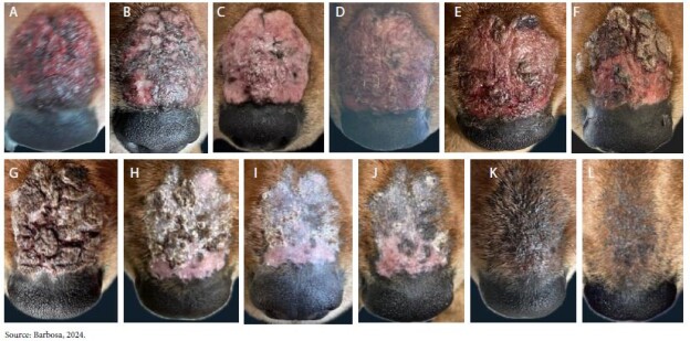
When diagnosing LES, biopsy or PCR should be considered. However, the treatment was effective, with reliable improvement 24 hours after the use of the homeopathic medicine (Figures 1C, 1D and 2C) and therefore the other tests were not performed. It can be seen that Causticum reduced the inflammation, making the lesion crusty and dry. The beginning of hair regrowth was seen on the sixth day (Figures 1E and 3). The region had collagen deposition due to its pink appearance, and it was possible to observe characteristics of remodeling of the lesion to the scarring process (Figure 1I), then evolving to a pigmented area (Figure 1K) until its complete hair regrowth.

Figure 1: Frontal view of the muzzle, dermatological lesion with alopecic, erythematous and nodular characteristics. (A) Day 1 – onset of symptoms; (B) Day 2 – start of treatment with Causticum (C) Day 3 (D) Day 4 (E) Day 6 (F) Day 10 (G) Day 12 (H) Day 14 (I) Day 16 – medication discontinued (J) Day 18 (K) Day 20 (L) Day 30.

Figure 2: Lesion lateral view (A) Day 2 (B) Day 3 (C) Day 4 (D) Day 6.

Figure 3: Source: Barbosa, 2024.
Dermatological appearance of the frontal region of the muzzle 5 days after the use of Causticum 30 cH, showing the beginning of hair regrowth in the affected area.
Discussion
Separating skin diseases specific to Lupus erythematosus from those that are nonspecific is a challenge [10], mainly due to their characteristics on physical examination and stages of complementary examinations.
Clinical signs involving alopecia are variable and mainly related to scaling and crusting, which can be focal, multifocal or generalized.
The kerion-type presentation (Figure 1), also called nodular dermatophytosis, is the clinical manifestation compatible with an infectious skin disease frequently detected in small animal clinics and has the fungus Microsporum canis as its main causative agent.
After excluding this hypothesis by fungal and bacterial culture, Lupus erythematosus was considered the clinical diagnosis.
Ferreira et al. (2021) describe the treatment of a senile canine patient with dermatophytic kerion caused by Microsporum canis using Itraconazole (10mg/kg/day) for 45 days. Due to the potential side effects related to the use of itraconazole, the adopted homeopathic therapy favors the patient’s organic function due to the absence of harm to health through pharmacodynamics.
In the case reported by [5], the canine patient with erythematous, scaly and ulcerated lesions in the nasal region, lips and gums, perianal region and caudal abdominal region had SLE confirmed by histopathological examination. The treatment was prednisolone 2mg/ kg, BID, for 10 days, later reduced to 1mg/kg, BID, for 10 days and then 1mg/kg, on alternate days and sun restriction. The animal responded positively to the treatment with improvement in clinical signs.
A senior mixed-breed dog patient with ulcers and crusts on the bridge of the nose, which had gradually evolved over a two- month period, received a therapeutic protocol consisting of topical medication based on hydrocortisone (1%), vitamin E (0.5%), and SPF 45 every 12 hours for 20 days; systemic therapy was administered with prednisolone at an initial dose of 1 mg/kg, followed by weaning, until its suspension, which lasted 80 days, in addition to liver protection for 30 days and precautions regarding sun exposure. Complete remission occurred after four months [4]. In another case, an ulcer and crust between the junction of the nasal plane and the skin of a mixed- breed dog was treated with 0.1% tacrolimus ointment, sunscreen on the muzzle, and tacrolimus ophthalmic ointment in the left eye, three times a day. There was partial improvement in the third week and complete remission of the lesions occurred after twelve weeks [11].
In the present case report, partial improvement occurred after 24 hours of starting treatment, with complete remission in the second week, a rapid result when compared to other reports. Currently, most of the medications used in treatment are: high doses of corticosteroids, anti-inflammatories and immunosuppressants, which have many uncomfortable side effects. Even with all this, some patients do not show the expected response [1].
Homeopathic therapy for dermatopathies in dogs is based on the principles of homeopathy, which involve diluted and dynamized substances to stimulate the body’s natural healing capacity. Causticum was the one that showed the best health support for harlequin-type ichthyosis [8]. Its applicability was also proven in a lactating Jersey cow, with several papillomas on the teats. The tumors reduced in size with the use of Causticum 18 cH twice a day before milking [2].
The possibility of treating Lupus Erythematosus with medicines from the Homeopathic Pharmacopoeia, based on the mental and physical symptoms of the disease, found through the meticulous approach of the homeopath, shows that guilt, stress and repressed emotions influence the alteration of the Immune System that gives rise to the disease [12].
Conclusion
While conventional treatment focuses on pharmacological approaches, homeopathy offers a promising alternative based on individuality and the stimulation of the body’s natural defenses. Continuous research and the link between therapies are essential to improving dermatological conditions in dogs. It is concluded that the use of the drug Causticum can be started immediately after the appearance of the lesion, the treatment is effective and fast when compared to the conventional use of immunosuppressants.
References
- Arias MB, Guimarães FC Conceição RT, Flaiban KKMC (2022) Estudo retrospectivo em 18 cães com lúpus eritematoso sistêmico (2008-2018) Pubvet, v. 16, n.
- Ferreira T, Wagner W, Ficagna VC (2017) Rev Acad Ciênc Anim 15 (2): S355-356.
- Ferreira et al. Quérion dermatofítico em cadela: Relato de caso (2020) Pubvet, 15 (01)
- Leal SRLS, Silva JG, Tertulino MD, Barreto GMF, Noronha JA, Rodrigues LMN, Medeiros NC (2021) Aspectos clínicos e histopatológicos do Lúpus Eritematoso Discoide canino: relato de caso. Medicina Veterinária (UFRPE), Recife, v.15, n.3, 209-215.
- Lima RC, Lavor CTB, Santos KMM, Vago P B, Viana DA (2022) Lúpus eritematoso discoide em cão. Ciência Animal 30, n. 2, p. 51-57.
- Macedo CM, Silva WC, Camargo Junior RNC (2021) Dermatofitose em cães e gatos: aspectos clínicos, diagnóstico e tratamento. Vet e Zootec v28: 001-013.
- Mcclellan C (2015) The Homeopathy Remedy: Causticum. Int J Complement Alt Med 1 (5): 00027.
- Oliveira SGM, Martins VAG, Rabello GM, Beier M, Astoni Júnior ÍMB (2014) Abordagem homeopática de uma criança portadora de ictiose tipo Revista de homeopatia 77 (3/4): 28.
- Olivry T, Rossi MA, Banovic F, Linder KE (2015) Mucocutaneous lupus erythematosus in dogs (21 cases) Veterinary Dermatology, 26 (4), 256-e55. [crossref]
- Olivry T, Linder KE, Banovic F (2018) Cutaneous lupus erythematosus in dogs: a comprehensive BMC Veterinary Research, 14 (1) [crossref]
- Pereira P, Oyafuso MK, da Cunha O, Nunes ACB, Paulino JA (2014) Medvep Dermato- Revista de Educação Continuada em Dermatologia e Alergologia Veterinária; 3 (11); 390-393.
- Pereira LL (2016) Associação da terapêutica homeopática no tratamento do Lúpus Eritematoso Sistêmico. Monografia apresentada ao curso de Especialização em Homeopatia do Instituto Hahnemanniano do Brasil Departamento de Ensino, Rio de Janeiro.
