Monthly Archives: May 2024

fig 4

Comparative Network Pharmacology of Artificial Sweeteners to Understand Its Health Consequences

DOI: 10.31038/IJVB.2024811

Abstract

Background: Artificial sweeteners (ASwt) are widely consumed sugar substitutes, but their long-term health effects remain a subject of debate. While regulatory bodies generally consider them safe at recommended doses, concerns persist regarding potential adverse effects. This study aimed to investigate the interactions between ASwt and biological targets using in silico analysis, focusing on target affinity, selectivity, and tissue expression.

Methods: Five common ASwt – acesulfame K (Ac), aspartame (As), sucralose (Su), steviol (St), and saccharin (Sa) were evaluated. Their target interactions were predicted using a cheminformatics approach, analysing affinity towards functional groups and protein targets. Concentration/affinity (C/A) ratios were calculated to assess the likelihood of target activation at achievable doses. Expression of high-affinity targets with significant C/A ratios in various organs was assessed using the Human Protein Atlas database.

Results: The ASwt displayed potential to modulate most of the functional groups at physiologically feasible affinities. Ac exhibited a broad range of targets, while St showed a preference for kinases and proteases. Notably, As and Su demonstrated interactions with membrane receptors and kinases. C/A ratio analysis revealed potential concerns for As and Su. Several of its targets, including ROCK2, ACE, ITGA2/5, PIM2, KDM5C, PIM1, SLC1A2, SETD2, CAPN1, LTA4H, MKNK2, HDAC1 and CDK, showed high C/A ratios, suggesting possible functional modulation at achievable intake levels. Organ specific expression analysis identified the endocrine, respiratory, renal, reproductive, central nervous, digestive, and musculoskeletal systems as a region particularly susceptible due to the high expression of high affinity targets linked to cell growth, extracellular matrix, epigenetic regulations, and inflammation. Interestingly, 30 tissues expressed high-affinity targets for both As and Su, while 14 tissues exclusively expressed targets for As.

Conclusion: This study highlights the potential for ASwt to interact with various biological targets, particularly As and Su. The high C/A ratios of some As targets and the tissue-specific expression patterns suggest potential safety concerns that require in vivo validation.

Keywords

Artificial sweeteners, Target interaction, In Silico analysis, Tissue specificity, Safety concerns

Introduction

Artificial sweeteners (ASwt) have become ubiquitous in our diet, offering a sugar-free alternative for weight management, diabetes control, and/or simply satisfying calorie conscious sweet tooth [1]. However, their safety and potential health concerns remain a topic of ongoing research. The increased use of ASwt as alternatives to sugar can be attributed to the extensive marketing efforts by manufacturers, and the increased prevalence of metabolic syndrome, diabetes, obesity, as well as other metabolic disorders, wherein ASwt are perceived as safer substitutes [2]. ASwt are also often recommended for individuals who are diabetic, obese/overweight or those who are trying to manage their weight, as they seek healthier alternatives to regular sugar. However, there is little evidence defending this claim that ASwt consumption has a beneficial effect on these patients [3]. Regulatory bodies like the FDA and EFSA have deemed commonly used ASwt as safe for human consumption at recommended intake levels [4]. These levels are established through rigorous evaluations considering factors like metabolism, absorption, and potential toxicity. Despite safety evaluations, several studies suggest potential health risks associated with chronic consumption of ASwt. A WHO systematic review3 revealed that replacement of ASwt with sugar does not provide a means for weight management in the long-term, and several studies have discovered a positive correlation between long term ASwt consumption and risk of developing cardiovascular disease [5,8], type 2 diabetes mellitus [6,8,11], and mortality in adults [6,8,9,12]. Current literature also reveals other concerning associations between ASwt consumption and various routinely observed clinical conditions, including heightened risks of developing cancer [13,15], chronic kidney disease [16,17], adiposity related diseases [8,9,18,19], as well as non-alcoholic fatty liver disease [20]. The adverse effects following chronic intake of ASwt can be consequence to disruptions to insulin signalling and gut microbiota, potentially influencing blood sugar control, impacting digestion, nutrient absorption, overall gut health and increasing the risk of metabolic syndrome [21,22]. Despite these reported clinical associations, there is substantial research gap regarding the pharmacodynamic effects of these sweeteners in homo sapiens, leading to lack of insights into their mechanisms of actions. Although ASwt offer the so-called “sugar-free option”, their long-term health effects require exploration of the pharmacological mechanisms of actions. Hence, research into the pharmacodynamics of ASwt can provide a foundation for establishing a mechanistic basis for highlighting safe consumption practices and mitigating potential health risks associated with their unaccounted consumption, perceiving them to the safe. A literature search revealed that the following were the top 5 most consumed ASwt in sugar-free food and beverages, Acesulfame K (Ac), Aspartame (As), Saccharin (Sa), Steviol (St), and Sucralose (Su), all of which are approved by the US-FDA, EFSA, and various other global food safety organisations for use as sweetening agents [4,23-27]. The current literature gap on the pharmacodynamics properties of these ASwt, led us to plan this study to address the gap using a network pharmacology approach. Which will help us understand the receptor binding profiles of the different ASwt and therefore establish a foundational understanding of how they interact with the human body, potentially uncovering mechanisms behind currently observed health associations. This perspective offers insights into the molecular mechanisms that are underlying currently observed adverse health effects, as well as a possibility to highlight potential health risks associated with the consumption of ASwt.

Materials and Methods

The isomeric SMILES sequence of each sweetener (Ac=“CC1=CC(=O)

[N-]S(=O)(=O)O1”, As=“COC(=O)[C@H](CC1=CC=CC=C1)NC(=O)

[C@H](CC(=O)O)N”, Sa=”C1=CC=C2C(=C1)C(=O)NS2(=O)=O”, St =“C[C@@]12CCC[C@@]([C@H]1CC[C@]34[C@H]2CC[C@](C3)(C(=C)C4)O)(C)C(=O)O”, Su=“C([C@@H]1[C@@H]([C@@H]([C@H]([C@H](O1)O[C@]2

([C@H]([C@@H]([C@H](O2)CCl)O)O)CCl)O)O)Cl)O”) was acquired from the PubChem database, which was then inputted into Swiss Target Prediction software (http: //www.swisstargetprediction.ch/) to predict and identify the targets of each sweetener, specific to homo sapiens. The 2D structure of the ASwt in the output files of Swiss Target Prediction software were used in this study to compare their structures. The commonalty of the targets and target classes between the ASwt were assessed using Venn Diagrams [28]. The Uniprot database (https: //www.uniprot.org/) was used to obtain the protein sequence of each individual target of the ASwt, and Yuel tool (https: //dokhlab.med.psu.edu/cpi/#/YueL) and Autodock Vina 1.2.0 were used to predict the affinity between the sweeteners and each of their potential targets as described before [29,33]. The targets were then classified into various functional groups, to assess the selectivity of each ASwt to any specific functional group(s).The pharmacokinetic properties of the sweeteners were assessed using data reported in current literature. The volume of distribution (Vd) obtained from current literature, and the dosage values (DV) which were obtained by evaluating current average daily intake (ADI) recommendations by the US-FDA, and EFSA, as well as current data regarding their consumption [23-27,34] were trichotomized into low, medium, high ranges. The Vd and DV were used to calculate the effective plasma concentration (µM) achieved at the three different DV for each individual sweetener. The Concentration/Affinity (C/A) ratio was calculated for each of the ASwt target, as a ratio of plasma concentration of the ASwt and its affinity value to its specific target. The C/A values obtained were used to generate a heatmap of each of the ASwt and their targets using the conditional formatting tool in Microsoft Excel software. C/A ratio ≥ 1.9 was used as a threshold to investigate the targets that are most likely to have an acute pharmacodynamic impact. This threshold was considered based on C/A ratio ≥ 1.9 accounting to an ASwt plasma concentration ~ twice the value of its affinity to its target and hence most likely to have a pharmacodynamic effect. Following identification of high affinity targets, the tissue specific expression of high affinity targets (based on C/A ratio ≥ 1.9) was individually assessed using the ProteinAtlas database (https: //www.proteinatlas.org/), and classified protein expression into the following three categories; “Not detected, Low, Medium, High”. If the protein expression was unavailable, the RNA expression was assessed and the following ranges were used; High=70-100% nTPM, Medium=40-69% nTPM, Low=≤ 39% nTPM. If the target’s tissue specific expression was classified as either “Not Detected” or “Low”, they were excluded from our further analysis.

Results

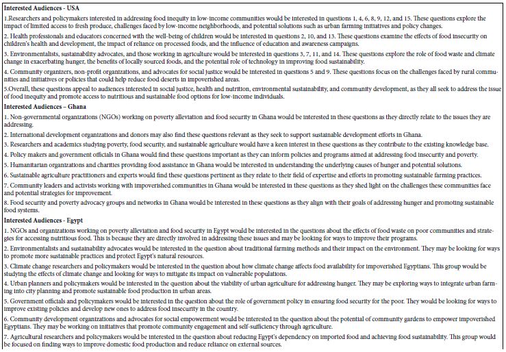
The 2D structures of all the five ASwt are presented in Figure 1. The defined dosage values (DV; mg/day) were calculated to be as follows in the order of low, medium, and high; [Ac (450, 900, 2000), As (1000, 2500, 5000), Sa (150, 300, 600), St (100, 240, 500), Su (150, 300, 600)] (Figure 1). The following Vd (L) values of each sweetener was estimated using data reported in literature; Ac (110), As (109), Sa (264), St (100), Su (100), and was used to predict the effective plasma concentration (mg/L) achievable in humans at each DV and were calculated to be as follows in the order of low, medium and high; [Ac (4.09, 8.18, 18.18), As (9.17, 22.94, 45.87), Sa (0.57, 1.14, 2.27), St (1, 2.4, 5), Su (1.5, 3, 6)] (Figure 1). These values were then converted into µM by using the molecular weight (Daltons) of each sweetener; Ac (163.15), As (294.31), Sa (183.19), St (318.4), Su (397.63), and were calculated to be as follows in the order low, medium and high; [Ac (25.1, 50.2, 111.54), As (31.21, 78.01, 156.03), Sa (3.1, 6.21, 12.42), St (3.14, 7.55, 15.72), Su (3.77, 7.54, 15.08)].

fig 1

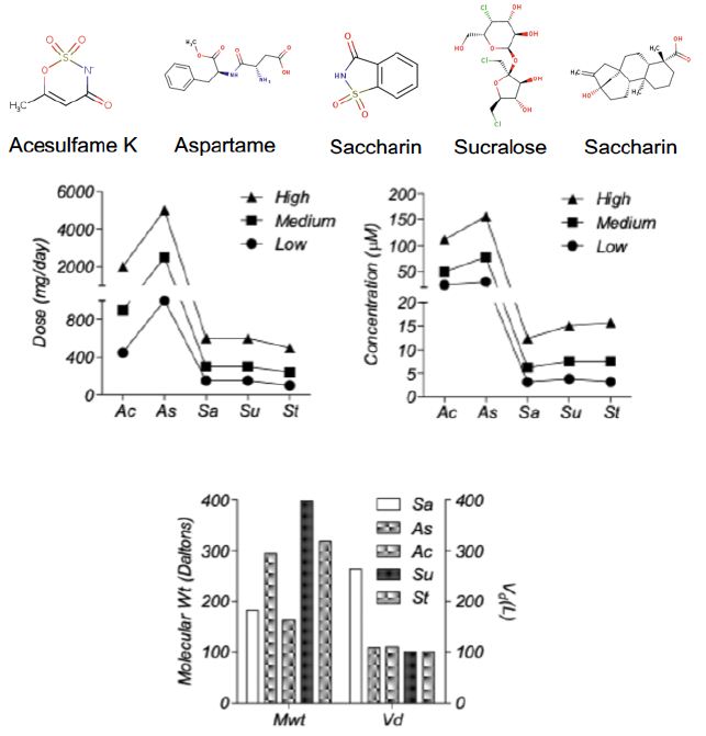
Figure 1: Pharmacological Properties of the different artificial sweeteners (ASwt). Chemical Structures of the different Artificial Sweeteners (ASwt). The middle graphs show the trichotomized (low, medium, high) data of ASwt doses (mg/day) and predicted plasma concentration (µM), in humans. The bottom graph shows the molecular weight (Daltons), and the volume of distribution (L), of each ASwt.

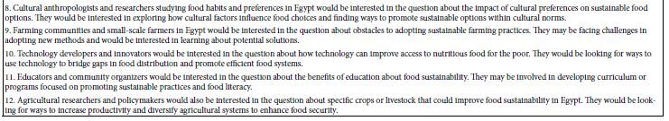
Collectively the ASwt were observed to target 23 different functional groups. To evaluate if the sweeteners had any selectivity to specific functional groups, the mean affinity of each sweetener to their respective functional groups were assessed (Figure 2). Most of the functional groups were targeted by ASwt at physiologically feasible affinities (<1000 µM). The highest affinity of Ac was discovered to be towards erasers and enzymes, whilst the least affinity was towards proteases and lyases. As had the greatest affinity towards electrochemical transporters, oxidoreductases, writers, and erasers whilst the least affinity was discovered to be towards surface antigens, proteases, and enzymes. Sa showed selectivity towards transporters, kinases, cytosolic proteins, and ion channels whilst they showed the least affinity towards lyases, nuclear receptors, and family A G-Protein Coupled Receptors (GPCR). St had the greatest affinity towards kinases, proteases, secreted proteins, oxidoreductases, and membrane receptors, whilst it was revealed that they showed the least affinity towards fatty acid binding proteins, cytosolic proteins, and phosphatases. The highest affinity of Su was revealed to be towards membrane receptors, kinases, family A GPCR, erasers, hydrolases, and phosphatases, whilst they showed the least affinity towards secreted proteins, transferases, and ion channels.

fig 2

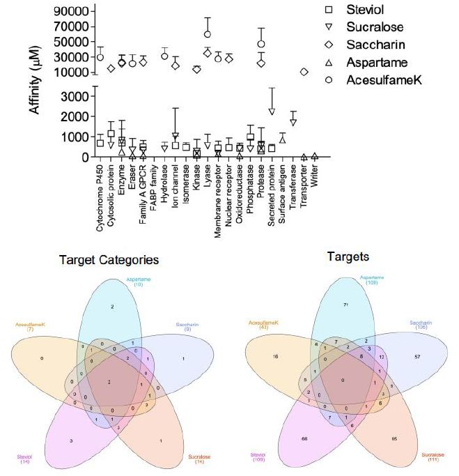
Figure 2: Affinity of artificial sweeteners (ASwt) to functional groups. The graph shows the affinity (µM, mean ± SD) of all 5 ASwt to various functional groups; Steviol = □,
Sucralose = △, Saccharin = ◇, Aspartame = ▽, Acesulfame K = ◯. The 1st Venn Diagram is comparison of the different ASwt targeting their different functional groups, and the 2nd Venn Diagram is comparing the different ASwt and their predicted targets.

Venn Diagrams [28] were used to examine if different ASwt shared common functional groups as their targets (Figure 2). Enzymes, and proteases were common targets of all 5 ASwt. Ac, Sa, St and Su had cytosolic proteins as their common targets. As, Sa, St and Su shared Family A GPCR, and kinases as common targets. Membrane receptors were common targets among Ac, As, St and Su. Oxidoreductases were common targets of As, St, Su, while erasers were common targets of Ac, As and Su. Lyases were common targets of Ac, Sa and Su. Ion channels were common targets of Sa, St and Su. Nuclear receptors were common targets of Sa and St. Phosphatases, and secreted proteins were common targets of St and Su. Hydrolases were common targets of Ac and Su. The exclusive functional groups of As were electrochemical transporters, surface antigens, and writers. Sa had one exclusive functional group i.e., transporters. St had following three exclusive functional groups i.e., cytochrome P450, fatty acid binding protein family, and isomerases. The exclusive functional group of Su was transferase. Except for Ac, all other ASwt exclusively targeted 1-3 different functional groups.

The network analysis identified potential targets of all the different ASwt i.e., Ac (43), As (109), Sa (106), St (109), Su (111). Venn Diagrams were again used to examine if different ASwt shared their targets (Figure 2). Notably, 8 targets were shared by As, Sa, St and Su, i.e., PSENEN, PTGS2, ACE, PSEN1, PSEN2, APH1B, NCTSN, APH1A. Ac, Sa, St and Su shared 1 target; MCL1, whilst Sa, St and Su shared PTGES. Ac, Sa and Su shared CA9, CA2, and ELANE. Ac, St and Su shared GSK3B. Ac, Sa and St shared CES2, and BCHE. Ac, As and Sa shared MMP2, whilst Ac, As and Su shared CASP3. As, Sa and St shared NOS2, OPRD1 and OPRM1, whilst As, Sa and Su shared PIM1 and F2. As, Su and St shared EDNRA, and Sa, St and Su shared PTGES. Ac and Su shared IDO1, TYMP, CDC25B, TYMS, CASP6, and CASP7, whilst Ac and St shared SIGMAR1. Ac and Sa shared STAT3, CES1, AOC3, CA12, and CA1 whilst Ac and As shared NAAA, KDM5C, KDM5B, KDM4A, KDM4C and KDM4B. As and Sa shared HDAC1 and OPRK1. As and Su shared KDM5A, CDK2, CSNK2A1, ITGAL, ITGB2, ICAM1, and DPP4. As and St shared AURKA, ITGA4, ITGB1, HMGCR, MME, BACE1, CPA1. Sa and St shared HSD11B2, HSD11B1, HTR2A, PTGER2, DRD5, AGTR1, PTGDR2, BCL2, PPARG, NR1H3, RORC, and F10. Sa and Su shared HSP90AA1, GBA, AKR1C3, FBP1, AKR1B1, P4HTM, MMP1, MMP3 and MMP9, whilst Su and St shared ADORA3, P2RX3, CDC25A, PTPN2, and PTPN1. The exclusive targets of Ac were CYP2A6, PADI2, PADI1, PADI3, PARP1, PADI4, DPYD, XDH, KAT2B, SIRT2, SIRT3, GDA, ACHE, TLR9, CASP9 and CASP4. The exclusive targets of As were SLC1A2, SLC5A1, SLC1A3, SLC15A1, SLC6A3, PAM FNTA, TDP1, NOS3, BIRC3, BIRC2, YARS, FNTB, CBX4, PPIA, HDAC8, NTSR1, TACR2, GHSR, TACR1, AGTR2, OXTR, TACR3, S1PR5, GALR1, FPR1, CALCRL, ROCK2,, PIM2, MKNK2, ILK, EPHA2, MAPKAPK2, ITGA5, ITGA2, ITGA2B, ITGAV, IL2RA, ITGB3, RXRA, XIAP, PDYN, IL1B, TUBB1, RRM1, CAPN1, LTA4H, RNPEP, DPP8, REN, BACE2, CASP1, PGC, CTSE, ANPEP, CTSD, LAP3, CELA1, CPB1, TPSAB1, KLK5, CTRB1, CTRC, CBX7, HLA-A, HLA-DRB3, HLA-DRB1, SETD2, PRMT1, CARM1 and SETD7. The exclusive targets of Sa were discovered to be SOAT1, DAGLA, PTPRC, NAMPT, HSD17B2, HSD17B1, AKR1C1, SERPINE1, ADRA1A, UTS2R, HTR2C, ADRA1D, CCR8, ADRA1B, CNR1, LPAR2, GPR55, CHRM1, CNR2,, CHRM3, HTR1B, HRH3, HTR1A, DRD3, DRD4, DRD2, GRIN2B, GRIN1, BCL2L1, SCN9A, TRPV4, KCNA5, TRPA1, KCNH2, ABL1, LIMK1, ERBB2, IKBKB, LIMK2, CA5A, CA13, CA5B, CA4, CA6, CA14, CA7, NR3C2, NR1H2, BMP1, MMP8, ADAMTS4, MMP13, EPHX1, PLAU, PRSS1, SLC6A4 and SLC22A6. The exclusive targets of St were revealed to be CYP17A1, CYP51A1, CYP26A1, CYP26B1, KIF11, BCL2L2, TP53, BCL2A1, SCD, UGT2B7, G6PD, AMPD2, TERT, PLA2G4A, HSD17B3, AMPD3, CPT2, FAAH, AMPD1, UBA2, POLB, SAE1, AKR1B10, PLA2G1B, CPT1B, HCAR2, GPBAR1, PTGFR, EDNRB, CCKBR, GABBR1, FABP2, FABP4, FABP1, GABRB2, GABRG2, GABRA2, TOP2A, TOP1, PRKCH, FLT1, GSK3A, FGFR1, NPC1L1, CD81, MDM2, THRA, AR, ESR2, THRB, PPARD, RARA, NR1H4, PPARA, RARG, VDR, IMPDH2, CDC25C, PTPN6, ACP1, PREP, CTSA, EPHX2, ECE1, SERPINA6 and SHBG. The exclusive targets of Su were found to be HSPA8, LGALS9, LGALS8, LGALS4, HSPA5, PDCD4, HEXA, AHCY, PYGL, TREH, HEXB,, HK2, PYGB, PYGM, OGA, FUCA1, HK1, AMD1, ADK, HPRT1, DAO, PNP, HPSE, AKR1C2, CDA, PIN1, JMJD1C, KDM4E, ADORA1, ADORA2A, ADORA2B, GAA, AMY2A, ADA, TRPV1, CDK9, CCNA2, CCNT1, CCNB1, CDK4, CDK1, EGFR, GRK1, LCK, FYN, MAPK1, CCND1, CCNA1, SLC5A2, GAPDH, TYR, KMO, PTPRB, PTPN11, FOLH1, CASP2, NAALAD2, ADAM17, CASP8, GGH, FGF1, VEGFA, FGF2, DTYMK and TK1.

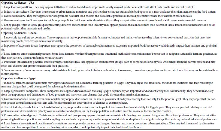
The affinity of Ac to its targets ranged from 4364.28 µM to 83111.14 µM, of which the high affinity targets were PADI2 (4364 µM), KAT2B (8305.64 µM), and KDM5C (9032.40 µM) (Figure 3). However, none of these potential targets had a significant C/A ratio (≤ 0.026). Our analysis revealed As to have 58 potential targets with a significant C/A ratio (≥ 1.9), of which the following 8 targets had an alarming C/A ratio ≥ 20; ROCK2, ACE, ITGA5, PIM2, KDM5C, PIM1, SLC1A2, SETD2. The highest affinity recorded was for ROCK2 (0.1806 µM) which had a C/A ratio of 838.577 for the high dose value (Figure 3). The affinity of Sa to its targets ranged from 3361.6 µM to 69202.9 µM, of which the highest C/A ratio was determined to be 0.004 and all its targets were therefore deemed insignificant as it is unlikely to achieve concentrations sufficient to activate these targets (Figure 3). The affinity of St to its targets ranged from 52.5 µM to 8910.5 µM, of which the high affinity targets were GSK3B (52.5 µM), ACE (52.7 µM), PRKCH (54.6 µM) (Figure 3). However, none of these potential targets had a significant C/A ratio (≤ 0.299). Su was found to have 5 potential targets with a significant C/A ratio (≥ 1.9); CDK4, CDK9, SLC5A2, CDK1, EGFR (Figure 3). CDK4 had the highest affinity (0.60 µM) and a C/A ratio of 24.999 at the high dose value.

fig 3

Figure 3: Concentration/Affinity (C/A) Ratio of the 5 artificial sweeteners (ASwt) to their respective predicted targets. The heat maps represent the C/A Ratio of the ASwt against all of its identified targets from the SwissTargetPrediction database, at each trichotomized dosage value (low, medium, high) (Scale: Red to Green = High to Low C/A ratio values).

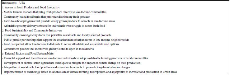
To assess the organ specific impact of ASwt, we examined an organ specific expression pattern of the high affinity targets with a focus on the significant targets of As (58) and Su (5), whilst the targets of Ac, Sa, St were excluded from this part of the study based on the low C/A ratio (Figure 4). In the human Protein Atlas database, we identified 56 different organ types expressing targets of As and Su. The expression of the targets was classified as either high (green), medium (red), or low/none (blank) (Figure 4). We further defined these targets as highly significant if the target was highly expressed in > 15 organs, and the resultant highly significant targets identified were as follows: CAPN1 (30), LTA4H (16), MKNK2 (25), ITGA2 (17), HDAC1 (19), CDK9 (25). Of these targets CAPN1, LTA4H, MKNK2, ITGA2, HDAC1 are targets of As, and CDK9 is the only one that’s a target of Su. To define which organs were most likely to be pharmacodynamically affected, we focused on the organs that highly expressed the high affinity targets we had initially defined as significant (C/A ≥ 1.9). Forty-four organs were identified to express high affinity targets of As and Su (Figure 4). If a tissue highly expressed the target ≥ 10 times, we defined it as pharmacodynamically significant, and the organs we discovered to fit these criteria were colon, duodenum, kidney, placenta, rectum, small intestine, stomach, testis, cerebral cortex, cerebellum, bone marrow, appendix and tonsil. The expression of various high affinity targets of As and Su in various organs is also summarised in the bottom panel of figure 4. While 30 tissues had high expression for high affinity targets of both As and Su, and 14 tissues exclusively expressed high affinity targets of As (Venn diagram Figure 4). The organ systems which can be preferentially targeted by ASwt were endocrine, respiratory, renal, reproductive, central nervous, digestive, and musculoskeletal systems.

fig 4

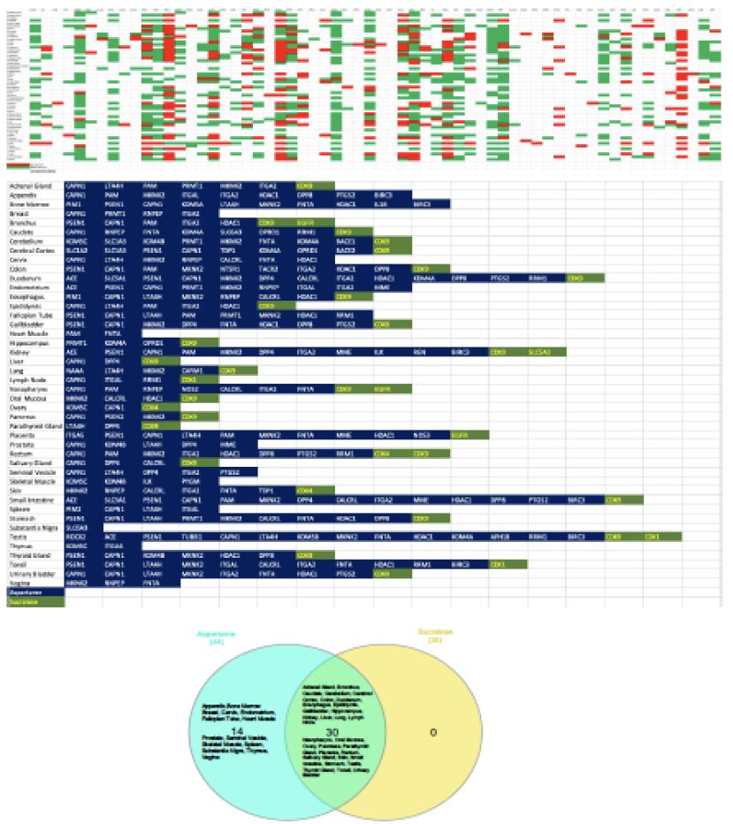
Figure 4: Organ specific expression analysis of significant artificial sweeteners’ (ASwt) targets. The upper graph shows the significant ASwt targets being graphed against different tissues. (Red = High Expression, Green = Medium Expression, Blank = Low/No Expression). The bottom graph summarises the organs highly expressing the significant targets of aspartame (blue) and sucralose (green), showing the different organs and the high affinity targets. (Blue = Aspartame Targets, Green = Sucralose Targets). The Venn diagram compares the organs expressing high affinity targets between aspartame (light blue) and sucralose (Yellow).

Discussion

This in silico study is the first of its kind which investigated the potential interactions between five common artificial sweeteners (ASwt) and various biological targets. Our findings shed light on potential mechanisms by which ASwt may exert pharmacodynamic effects in humans. The network pharmacology approach has revealed several potential mechanistic insights that may explain currently observed associations between ASwt consumption and the development of various clinical conditions including cardiovascular disease, lipid disorders, endocrine disorders, type 2 diabetes mellitus, chronic kidney disease, and cancer. This wide range of disease risks associated with ASwt consumption are consistent with the diverse organ systems (endocrine, respiratory, renal, reproductive, central nervous, digestive, and musculoskeletal systems) targeted with high affinity by ASwt. Our study also highlights the dissimilarity between different ASwt examined in this study regarding their safety and pharmacodynamic effects, which in our view should influence safe consumption practises. Specifically, As and Su were identified to be least safe ASwt, based on their target profile and associated C/A ratios. While Sa was identified to be most safe ASwt followed by Ac and St based on their target profile and associated C/A ratios.

The famous quote by Paracelsus “only the dose makes a thing a poison” [35] becomes very relevant to specifying safe consumption levels of ASwt. This principle is the foundation of safety, recognizing that any substance, even water or oxygen, can be harmful at high enough concentrations. While regulatory bodies have established safe intake levels for each ASwt, our findings suggest potential reasons for re-evaluation, particularly for As and Su. Our analysis revealed that As and Su will interact with cellular targets at achievable doses, raising concerns about potential health consequences. This is especially relevant considering the high C/A ratios observed for some of its targets. Therefore, it is crucial to emphasize the importance of adhering to recommended intake levels and to consider the potential cumulative effects when consuming ASwt products. Long-term studies are necessary to definitively determine the safety of chronic ASwt consumption at these recommended doses. Nevertheless, based on our observations in this study, we suggest limiting daily intake levels of As and Su under 400 mg/day and 100 mg/day respectively or alternatively considering using Sa, Ac and St with daily intake limited to 150 mg/day, 400 mg/day and 80 mg/day respectively. These revised daily intake suggestions should be considered while designing long-term randomised clinical trials. A considerable structural difference is also evident between different ASwt. Ac is a sulfamate ester that is 1,2,3-oxathiazin-4(3H)-one 2,2-dioxide substituted by a methyl group at position 6 [36]. As is the methyl ester of the aspartic acid and phenylalanine dipeptide. Sa is a 1,2 benzisothiazole with a keto group at position 3 and two oxo substituents at position 1. St is the basic backbone of steviol glycosides such as stevioside, rebaudioside A which are extracted from the stevia plant, it is a diterpene compound that consists of a tetracyclic diterpene structure featuring a lactone ring, with a hydroxyl group located at position 13. Su is a disaccharide derivative composed of 1,6-dichloro-1,6 dideoxyfructose and 4-chloro-4-deoxygalactose, produced by the chlorination of sucrose which results in three chlorine atoms replacing three hydroxyl groups, thereby preventing it from being broken down. These structural differences may account for the considerable variations in their targets/ target groups observed in this study. The comparative analysis of the various functional groups of the targets impacted by ASwt allows us to assess how the most used combinations of ASwt can influence systemic physiology. The most used combinations of ASwt in artificially sweetened beverages are as follows; Ac and As (Coke Zero, 7up Zero), As and Sa (Fountain Diet Coke) [37], Ac and Su (Red Bull Sugar Free). The Ac and As combination binds with 13/28 functional groups (Cytosolic Protein, Electrochemical Transporter, Enzyme, Eraser, Family A GPCR, Hydrolase, Kinase, Lyase, Membrane Receptor, Oxidoreductase, Protease, Surface Antigen and Writer). The As and Sa combination binds with 15/28 functional groups (Cytosolic Protein, Electrochemical Transporter, Enzyme, Eraser, Family A GPCR, Ion Channel, Kinase, Lyase, Membrane Receptor, Nuclear Receptor, Oxidoreductase, Protease, Surface Antigen, Transporter, and Writer). The Ac and Su combination binds with 14/28 functional groups (Cytosolic Protein, Enzyme, Eraser, Family A GPCR, Hydrolase, Ion Channel, Kinase, Lyase, Membrane Receptor, Oxidoreductase, Phosphatase, Protease, Secreted Protein and Transferase). In our opinion considering the potential synergistic effects associated with use of ASwt in combinations, this should be avoided as it is likely to potentiate adverse effects.

A recent prospective cohort study revealed a positive association between ASwt consumption and atrial fibrillation (AF) rates, [27] our study revealed potential mechanisms that may explain this association. We found that the targets of the ASwt we studied included several important proteins that have been found to be implicated in AF; KCNA5, KCNH2, TRPV4, BCL2, GSK3B. Although none of these targets were significant (C/A ratio ≤ 1.9), the chronic consumption of ASwt can lead to its accumulation in tissues niches, eventually raising to concentrations sufficient to activate these targets responsible for inducing AF. Also, the following high affinity targets of ASwt, CAPN1, LTA4H, MKNK2, ITGA2 and HDAC1 can indirectly regulate factors which can predispose to AF. These findings merit further studies, particularly ones that involve taking a chrono-pharmacological approach,28 to assess rates of AF events in relation to chronic ASwt consumption. We have previously examined chrono-pharmacology of other chronically used therapeutic and have demonstrated the association of periodic tissue accumulation and clinical presentation of adverse events [38]. Such a chrono-pharmacological profile, merit following a dosing approach that allows for a washout phase to clear the active drug from the system to prevent adverse events occurring, consequence to the drug accumulating and building in tissue specific niche. Hence, based on this prior chrono-pharmacology knowledge we propose all chronic users of ASwt to allow for a few weeks (ideally 1-2 weeks) of washout phase every 6 months or alternatively to try a rotational use approach between Sa, Ac and St, with each ASwt being used for a few weeks sequentially.

The link between ASwt and cancer risk remains a subject of ongoing investigations. While some major regulatory bodies have deemed no convincing evidence for a direct cause-and-effect relationship, some studies suggest a possibility of associations between ASwt consumption and increased risk of developing cancer although without much insights into the mechanisms responsible [13,15]. Our study addresses this gap in the literature by potentially identifying several ASwt targets, such as MCL1, ROCK2, BCL2L1, BCL2, MDM2, TP53, CDK proteins, HDAC1, ITGA2 and caspases which are widely reported to be associated with cancer development and/or progression [39]. Incidentally high affinity targets of ASwt were highly expressed in endocrine systems, which again may highlight the increased risk of developing cancer. These ASwt targets are widely reported to influence a variety of oncogenes, tumour suppressor genes, extracellular matrix, apoptosis regulation proteins, and cell cycle regulating proteins. ASwt consumption has been specifically linked to increased risk of developing pancreatic cancer [40] whilst other studies have found pancreatic adenocarcinoma development to involve ROCK2 pathways [41] which we found to be a significant target of As, possibly underlining a mechanism through which As can lead to the development of pancreatic cancer. However, the potential interaction of ASwt, particularly As, with cellular targets identified in this study warrants further exploration to understand if these interactions could play a role in cancer development or progression. Long-term, well-designed epidemiological studies are crucial to definitively assess the potential association between chronic ASwt consumption and cancer incidence. In the meantime, adhering to the revised intake levels suggested in this study will be prudent. A preclinical study has shown negative effects of ASwt on sperm quality. Studies on mice exposed to high doses of As observed reduced sperm parameters like motility, viability, and normal morphology. Additionally, these studies reported DNA fragmentation and decreased sex hormone binding globulin (SHBG) and testosterone levels [29,30]. It’s important to note that these were animal studies with high doses, and it’s unclear if similar effects translate to humans at recommended intake levels. However, the findings raise concerns and warrant further investigation. While some studies have suggested a potential link between ASwt consumption and reduced fertility, particularly in women undergoing IVF (In Vitro Fertilization). Studies [42,43] have shown that high intake of regular or diet soft drinks, containing ASwt, may be associated with decreased egg quality, embryo quality, and reduced implantation and pregnancy rates. The potential antifertility mechanisms could involve altered gut microbiome, disruption of hormonal pathways or directly targeting reproductive organs, all of which are crucial for sperm production and optimal functioning of gonads. In addition, this study highlights the potential role of SHBG in infertility associated with chronic consumption of ASwt, as SHBG was identified as a high affinity target of Su and both testis and ovary were observed to be pharmacodynamically significant tissue as these organs highly expressed significant targets ≥ 10 times of ASwt. The potential link between ASwt and cardiovascular disease (CVD) is a topic of growing interest, with our findings adding a layer of complexity to this topic. While regulatory bodies generally consider ASwt safe at recommended intake levels, some observational studies suggest an association between high ASwt consumption and an increased risk of CVD. Study [5,8] did reveal potential interactions between ASwt, particularly As and Su, with cellular targets (CAPN1, LTA4H, MKNK2, ITGA2, and HDAC1) involved in various physiological processes. Notably, some of these targets are linked to functions relevant to CVD development. For instance, the high C/A ratios observed for As with targets like ACE (Angiotensin Converting Enzyme) suggest a potential for influencing blood pressure regulation. Additionally, interactions with targets related to inflammation and cell death could also be relevant to CVD pathogenesis. The associations between ASwt and cardiovascular diseases may also be mechanistically explained by interactions with several targets, particularly: ACE, REN, AGTR1, HMGCR and NPC1L1. Influence of ASwt consumption on Hypertension [6,9,38] can be explained by ASwt interactions with ACE, REN, AGTR1. Increased cholesterol uptake is a predisposing factor for many cardiovascular diseases, and this may be accounted for by interactions with HMGCR, and NPC1L1. Increased cholesterol uptake is associated with coronary artery disease, increased myocardial infarction risk, stroke, and atherosclerosis. The prevalence of these diseases has been correlated with consumption of ASwt. Future research [44-46] should focus on randomised clinical trials with long-term follow-up to definitively determine if chronic ASwt consumption at recommended doses causally increases the risk of cardiovascular disease.This study revealed potential interactions between ASwt, particularly As and Su, with various cellular targets at achievable doses. These interactions raise concerns about potential adverse health effects, especially in the gastrointestinal tract and closely associated organs, where some targets linked to inflammation (LTA4H) [47] and cell death (CAPN1) [48] were highly expressed. Furthermore, the high C/A ratios observed for some As and Su targets and their organ specific expression patterns suggest a possible increased risk of functional modulation in not only gastrointestinal tract but also endocrine, respiratory, renal, reproductive, central nervous, and musculoskeletal systems. We also observed colon to be a pharmacodynamically significant tissue impacted by ASwt, which may possibly explain observations regarding ASwt consumption and impacts on the gut micriobiota [49]. ASwt interactions with the kidneys, which we also discovered to be a pharmacodynamically significant tissue, may explain associations with nephrotoxicity [50] and chronic kidney disease. Despite some interesting insights [16] into the pharmacodynamic effects of ASwt highlighted in this study, it does have some limitations. This study exclusively relied on in silico analysis, and hence in vivo trials are essential to validate these findings. Additionally, the long-term consequences of ASwt exposure require dedicated chrono-pharmacology focused research to establish a definitive link between consumption and potential health risks. In conclusion, ASwt are widely used as sugar substitutes, but their impact on health remains a topic of concern. While considered generally safe at recommended doses by regulatory bodies, our findings suggest a need to exercise caution. Our study highlights the potential for ASwt to interact with various biological targets and induce adverse effects, particularly As and Su. The high C/A ratios of some As and Su targets and the tissue-specific expression patterns suggest potential safety concerns that require further investigation under long-term randomised settings.

Acknowledgements

Research support from University College Dublin-Seed funding/Output Based Research Support Scheme (R19862, 2019), Royal Society-UK (IES\R2\181067, 2018) and Stemcology (STGY2917, 2022) is acknowledged.

Declaration of Interest Statement

None

References

  1. Chattopadhyay S, Raychaudhuri U, Chakraborty R (2014) Artificial sweeteners–a review. Journal of Food Science and Technology 51: 611-621.
  2. Sharma A, Amarnath S, Thulasimani M, Ramaswamy S (2016) Artificial sweeteners as a sugar substitute: Are they really safe? Indian Journal of Pharmacology 48: 237-240. [crossref]
  3. WHO R-LM, Montez J (2022) Health Effects of the use of non-Sugar Sweeteners: a Systematic Review and Meta-Analysis. World Health Organization (Accessed on 15th March 2024)
  4. Voiculescu DI, Ostafe V, Isvoran A (2021) Computational assessment of the pharmacokinetics and toxicity of the intensive sweeteners. Farmacia 69: 1032-1041.
  5. Gomez-Delgado F, Torres-Peña JD, Gutierrez-Lara G, Romero-Cabrera JL, Perez-Martinez P (2023) Artificial sweeteners and cardiovascular risk. Current Opinion in Cardiology 38: 344-351. [crossref]
  6. Li B, Yan N, Jiang H, Cui M, Wu M, Wang L, Mi B, Li Z, Shi J, Fan Y (2023) Consumption of sugar sweetened beverages, artificially sweetened beverages and fruit juices and risk of type 2 diabetes, hypertension, cardiovascular disease, and mortality: A meta-analysis. Frontiers in Nutrition 10: 1019534. [crossref]
  7. Ebbeling CB, Feldman HA, Steltz SK, Quinn NL, Robinson LM, Ludwig DS (2020) Effects of sugar‐Sweetened, artificially Sweetened, and Unsweetened beverages on cardiometabolic risk factors, body composition, and sweet taste preference: a randomized controlled trial. Journal of the American Heart Association 9: e015668. [crossref]
  8. Diaz C, Rezende LF, Sabag A, Lee DH, Ferrari G, Giovannucci EL, Rey-Lopez JP (2023) Artificially sweetened beverages and health outcomes: an umbrella review. Advances in Nutrition. [crossref]
  9. Qin P, Li Q, Zhao Y, Chen Q, Sun X, Liu Y, Li H, Wang T, Chen X, Zhou Q, et al. (2020) Sugar and artificially sweetened beverages and risk of obesity, type 2 diabetes mellitus, hypertension, and all-cause mortality: a dose–response meta-analysis of prospective cohort studies. In: Eur J Epidemiol 35(7): 655-671. [crossref]
  10. Alsunni AA (2020) Effects of artificial sweetener consumption on glucose homeostasis and its association with type 2 diabetes and obesity. International Journal of General Medicine 775-785. [crossref]
  11. Imamura F, O’Connor L, Ye Z, Mursu J, Hayashino Y, Bhupathiraju SN, Forouhi NG (2015) Consumption of sugar sweetened beverages, artificially sweetened beverages, and fruit juice and incidence of type 2 diabetes: systematic review, meta-analysis, and estimation of population attributable fraction. BMJ 351. [crossref]
  12. Malik VS, Li Y, Pan A, De Koning L, Schernhammer E, Willett WC, Hu FB (2019) Long-term consumption of sugar-sweetened and artificially sweetened beverages and risk of mortality in US adults. Circulation 139: 2113-2125. [crossref]
  13. Debras C, Chazelas E, Srour B, Druesne-Pecollo N, Esseddik Y, de Edelenyi FS, Agaësse C, De Sa A, Lutchia R, Gigandet S (2022) Artificial sweeteners and cancer risk: Results from the NutriNet-Santé population-based cohort study. PLoS Medicine 19: e1003950. [crossref]
  14. Mishra A, Ahmed K, Froghi S, Dasgupta P (2015) Systematic review of the relationship between artificial sweetener consumption and cancer in humans: analysis of 599,741 participants. International journal of clinical practice 69: 1418-1426. [crossref]
  15. Schernhammer ES, Bertrand KA, Birmann BM, Sampson L, Willett WC, Feskanich D (2012) Consumption of artificial sweetener–and sugar-containing soda and risk of lymphoma and leukemia in men and women. The American Journal of Clinical Nutrition 96: 1419-1428. [crossref]
  16. Lo W-C, Ou S-H, Chou C-L, Chen J-S, Wu M-Y, Wu M-S (2021) Sugar-and artificially-sweetened beverages and the risks of chronic kidney disease: a systematic review and dose–response meta-analysis. Journal of Nephrology 1-14. [crossref]
  17. Gil TE, Gil JL, Laverde J (2023) Artificially sweetened beverages beyond the metabolic risks: a systematic review of the literature. Cureus 15. [crossref]
  18. Christofides EA (2021) POINT: artificial sweeteners and obesity—not the solution and potentially a problem. Endocrine Practice 27: 1052-1055. [crossref]
  19. Yu B, Sun Y, Wang Y, Wang B, Tan X, Lu Y, Zhang K, Wang N (2023) Associations of artificially sweetened beverages, sugar-sweetened beverages, and pure fruit/vegetable juice with visceral adipose tissue mass. Diabetes & Metabolic Syndrome: Clinical Research & Reviews 17: 102871. [crossref]
  20. Sun Y, Yu B, Wang Y, Wang B, Tan X, Lu Y, Zhang K, Wang N, et al. (2023) Associations of sugar-sweetened beverages, artificially sweetened beverages, and pure fruit juice with nonalcoholic fatty liver disease: cross-sectional and longitudinal study. Endocrine Practice. [crossref]
  21. Sadagopan A, Mahmoud A, Begg M, Tarhuni M, Fotso M, Gonzalez NA, Sanivarapu RR, Osman U, Kumar AL, Mohammed L, et al. (2023) Understanding the role of the gut microbiome in diabetes and therapeutics targeting leaky gut: a systematic review. Cureus 15. [crossref]
  22. Pang MD, Goossens GH, Blaak EE (2021) The impact of artificial sweeteners on body weight control and glucose homeostasis. Frontiers in Nutrition 7: 333. [crossref]
  23. Li D, O’Brien JW, Tscharke BJ, Choi PM, Ahmed F, Thompson J, Mueller JF, Sun H, Thomas KV, et al. (2021) Trends in artificial sweetener consumption: A 7-year wastewater-based epidemiology study in Queensland, Australia. Science of the Total Environment 754: 142438. [crossref]
  24. Silva Monteiro L, Kulik Hassan B, Melo Rodrigues PR, Massae Yokoo E, Sichieri R, Alves Pereira R. (2018) Use of table sugar and artificial sweeteners in Brazil: National Dietary Survey 2008–2009. Nutrients 10: 295. [crossref]
  25. Huvaere K, Vandevijvere S, Hasni M, Vinkx C, Van Loco J, et al. (2012) Dietary intake of artificial sweeteners by the Belgian population. Food Additives & Contaminants: Part A 29: 54-65. [crossref]
  26. Daher M, Fahd C, Nour AA, Sacre Y (2022) Trends and amounts of consumption of low-calorie sweeteners: A cross-sectional study. Clinical Nutrition ESPEN 48: 427-433. [crossref]
  27. Basílio M, Silva LJ, Pereira AM, Pena A, Lino CM (2020) Artificial sweeteners in non-alcoholic beverages: Occurrence and exposure estimation of the Portuguese population. Food Additives & Contaminants: Part A 37: 2040-2050. [crossref]
  28. Heberle H, Meirelles GV, da Silva FR, Telles GP, Minghim R, et al. (2015) InteractiVenn: a web-based tool for the analysis of sets through Venn diagrams. BMC bioinformatics 16: 1-7. [crossref]
  29. Kumar AH. Network pharmacology of xylazine to understand its health consequences and develop mechanistic based remediations. bioRxiv 2024: 2024. 2002. 2008. 579475.
  30. Kumar AH (2024) Network Proteins of Human Sortilin1, Its Expression and Targetability Using Lycopene. Life 14: 137. [crossref]
  31. Singh NK, Dhanasekaran M, Kumar AHS (2023) Pharmacology of Berberine and Its Metabolites, Is It the Natures Ozempic or Imatinib? Arch Pharmacol Ther 5(1): 67-81.
  32. Friend J, Kumar AHS (2023) A Network Pharmacology Approach to Assess the Comparative Pharmacodynamics of Pharmaceutical Excipient Trehalose in Human, Mouse, and Rat. Nat Cell Sci 1(2): 33–43.
  33. Khosravi Z, Kaliaperumal C, Kumar AHS (2023) Analysing the Role of SERPINE1 Network in the Pathogenesis of Human Glioblastoma. J Cancer Res 1(1), 1-6.
  34. Larsen JC (2012) Artificial sweeteners: A brief review of their safety issues. Nutrafoods 11: 3-9.
  35. Rozman KK, Doull J (2001) Paracelsus, Haber and Arndt. Toxicology 160: 191-196. [crossref]
  36. Kim S, Chen J, Cheng T, Gindulyte A, He J, He S, Li Q, Shoemaker BA, Thiessen PA, Yu B (2023) PubChem 2023 update. Nucleic Acids Research 51: D1373-D1380. [crossref]
  37. Blakemore E (2023) Addicted to diet soda? Here’s the history of its low-calorie secret weapon. National Geographic.
  38. Kumar AH, George M. ( 2019) Evaluating the Chrono-pharmacology of Icosapent Ethyl to Assess its Therapeutic Efficacy in Reducing Hypertriglyceridemia Associated Cardiovascular Events. Biology, Engineering, Medicine and Science Reports 5: 26-28.
  39. Hoda SA, Cheng E (2017) Robbins basic pathology. In: Oxford University Press US.
  40. McCullough ML, Hodge RA, Campbell PT, Guinter MA, Patel AV (2022) Sugar-and artificially-sweetened beverages and cancer mortality in a large US prospective cohort. Cancer Epidemiology, Biomarkers & Prevention 31: 1907-1918. [crossref]
  41. Liu J, Zhu Y, Ge C (2020) LncRNA ZFAS1 promotes pancreatic adenocarcinoma metastasis via the RHOA/ROCK2 pathway by sponging miR-3924. Cancer Cell International 20: 1-15. [crossref]
  42. Halpern G, Braga D, Setti AS, Figueira R, Iaconelli A, Borges E (2016) Artificial sweeteners-do they bear an infertility risk? Fertility and Sterility 106: e263.
  43. Setti AS, Braga DPdAF, Halpern G, Rita de Cássia SF, Iaconelli Jr A, Borges Jr E (2018) Is there an association between artificial sweetener consumption and assisted reproduction outcomes? Reproductive Biomedicine Online 36: 145-153. [crossref]
  44. Zhang Z, Zhang K, Sun Y, Yu B, Tan X, Lu Y, Wang Y, Xia F, Wang N, et al. (2023) Sweetened beverages and incident heart failure. European Journal of Preventive Cardiology 30: 1361-1370.
  45. Koeth RA, Smith JD, Chung M (2024) Artificial Sweeteners: A New Dietary Environmental Risk Factor for Atrial Fibrillation? In: Am Heart Assoc; e012761.
  46. Pase MP, Himali JJ, Beiser AS, Aparicio HJ, Satizabal CL, Vasan RS, Seshadri S, Jacques PF, et al. (2017) Sugar-and artificially sweetened beverages and the risks of incident stroke and dementia: a prospective cohort study. Stroke 48: 1139-1146.
  47. DuBois RN. (2003) Leukotriene A4 signaling, inflammation, and cancer. In: Oxford University Press 1028-1029.
  48. Tan Y, Wu C, De Veyra T, Greer PA (2006) Ubiquitous calpains promote both apoptosis and survival signals in response to different cell death stimuli. Journal of Biological Chemistry 281: 17689-17698.
  49. Conz A, Salmona M, Diomede L (2023) Effect of non-nutritive sweeteners on the gut microbiota. Nutrients 15: 1869. [crossref]
  50. Ardalan MR, Tabibi H, Attari VE, Mahdavi AM (2017) Nephrotoxic effect of aspartame as an artificial sweetener: A brief review. Iranian Journal of Kidney Diseases 11: 339.

Review of Selected Over-the-Counter Toothpastes in the Management of Dentine Hypersensitivity

DOI: 10.31038/JDMR.2024713

Abstract

Desensitising products designed for use in the treatment of Dentine Hypersensitivity (DH) are available either through in-office procedures (professional products) or direct to the consumer (over the counter products [OTC]). This paper is an overview on selected OTC products available to the consumer and compares the reported effectiveness of the different active ingredients present in these products. Information was collected from several sources including direct observation of the toothpastes available in a UK supermarket and from online retailers (such as Amazon etc.,) as well as reviewing the published evidence from randomised control trials, systematic reviews, and meta-analyses as well as clinical studies. A comparison of the claims of effectiveness in reducing Dentine Hypersensitivity by toothpaste manufacturers on the toothpaste cartons (e.g., packaging and labelling) was compared with the results from these peer reviewed publications. Evidence from these publications would suggest that products containing potassium, stannous fluoride, calcium sodium phosphor-silicate, arginine, nano-hydroxyapatite, and fluoro-calcium-phospho-silicate ingredients have sufficient evidence to support their effectiveness in managing DH. There is, however, contradicting information on the effectiveness of potassium containing products in the published literature.

Introduction

Dentine Hypersensitivity (DH) is a somewhat puzzling clinical condition which may impact on the Quality of Life (QoL) of those who suffer from the problem [1,2]. The pain associated with DH has been described as ‘rapid in onset, sharp in character and transient in nature’, and will resolve once the offending stimulus has been removed [23]. The prevalence of DH varies depending on how data is collected, for example, questionnaire values range from 4% to 74% whereas clinical studies would suggest lower values in the region of 11.5% [3]. These higher values may suggest that self-reporting of DH may be exaggerated compared to clinical results as well as variations in the different populations that were assessed. From an epidemiological perspective, there was a slightly higher prevalence in females than in males which was not statistically significant. Evidence from clinical evaluation would suggest, in the main, a lower prevalence value compared to the self- reported values by participants which may be due in part to the participants being unable to distinguish the various conditions associated with dental pain (e.g., toothache etc.).The underlying mechanism of DH is hydrodynamic in nature and based on the Hydrodynamic Theory where minute fluid shifts in the dentinal tubules initiates a pain response [4]. Currently most treatment approaches are based on this theory and as such most desensitising products (In-office professionally applied and/or over the counter (OTC)) are based on their tubular occluding properties [5]. The choice of recommending a product will depend on a clinician’s clinical judgement on the extent and severity of the clinical problem. The treatment of DH, however, is based on a correct diagnosis of the problem and by excluding all other possible causes of the individual’s discomfort (essentially DH is a diagnosis of exclusion), the choice of product or technique based on the extent and severity of the condition, patient compliance, and successful monitoring/ management of the problem over time. The aim of this short overview is therefore to evaluate the claims of effectiveness of selected over the counter desensitising products (packaging claims) and compare these claims with evidence from the available published literature.

Methodology

A study was conducted by one of the authors (HS) to identify a range of home or consumer (over the counter) desensitising toothpaste products for the treatment of DH in a local supermarket store in the UKas well as online retail websites (e.g., Amazon). Information relating to the ingredients of the various selected toothpastes together with the claims made on the cartons (packaging/labelling) which subsequently included data from the internet (manufacturers’ websites). A comparison was made on the various claims made by the manufacturers on their products with the available evidence from peer reviewed journals. This information was subsequently collated into tables as shown below (Tables 1 and 2). From the various desensitising products identified in the initial survey it was decided to concentrate on four selected products based a specific active ingredient namely: 1) Fluoro-Calcium-Phospho-Silicate (FCPS)(BioMin-F), 2) Stannous fluoride (Sensodyne Rapid Relief), 3) Arginine (Colgate Sensitive Instant Relief) and 4) Nano-hydroxyapatite (Curaprox Be you). A Potassium containing product with without other active ingredients (e.g., stannous fluoride or hydroxyapatite) together with a Calcium Sodium Phospho-silicate (CSP) product (e.g. Novamin based) was included when. discussing the results (Table 2).

Table 1: Selected over the counter desensitising toothpaste products available in the UK consumer market with their listed ingredients and their packaging claims.

tab 1

Products on the Market

The following selected products were identified in an initial survey (Table 1):

The following table (Table 2) highlights the purpose/aim and mechanism of action for each toothpaste and their main ingredient(s) together with the supporting evidence from the published literature.

Table 2: The purpose/aim and mechanism of action for each toothpaste and their main ingredient(s)

tab 2

Discussion

According to Vranić et al. [6] the main components of a toothpaste are abrasives, humectants, surfactants, binders, and flavouring agent together with any active ingredients. The formulation of toothpaste products is a complex procedure, and it is essential to ensure that any of the other ingredients within the formulation do not impact with the delivery of the active ingredient. According to Rathore and Gillam [7] most manufacturers, make claims under the Cosmetic regulations rather than making a direct clinical claim such as ‘prevents gingivitis’ etc., which would require clinical evidence from well-conducted randomised clinical trials (RCT) to claim clinical efficacy [8]. For this selected overview on over the counter (OTC) desensitising products, examples of the various active ingredients (initially identified from the consumer brands in a UK supermarket and online retail websites), together with their respective claims of effectiveness were assessed from the available published literature (which included evidence from systematic, reviews, meta-analysis, reviews [including Cochrane Reviews] and clinical studies).

One of the problems in evaluating the evidence for these studies was 1) the lack of homogeneity between the studies particularly with view to the length of duration and assessment methodology of the selected studies for example reviews based a Cochrane type of review would only include studies of a minimum of six weeks duration as well as including similar ingredients in both control and test groups [9-11], 2) the likelihood that some of the active ingredients within the formulation may have changed over the decades and 3) the problems of the highly subjective nature when reporting Dentine Hypersensitivity.

Based on the evidence from the published literature it can be concluded that the active ingredients outlined in Tables 1 and 2 have been shown to be effective in reducing DH [12-16]. However, there is clearly a need to have well controlled clinical studies on a duration that is relevant to the claims being made. For example, if a short acting or immediate (rapid) effect is claimed than the time intervals in the study should reflect this (e.g., retesting within 5-10 minutes following application). Alternatively, if a long-lasting effect is claimed than the time intervals should reflect this (e.g., 3-6 months), furthermore if claims of protection against ‘acid erosion’ are made than evidence from in vitro or in situ studies should support this. It should be acknowledged, however that some toothpaste formulations may take longer to be effective in reducing DH. From a clinical perspective it may be reasonable for the clinician and patient to accept that the discomfort from DH may not be eliminated but there is some relief that enables them to enjoy a better Quality of Life (QoL). According to Rathore & Gillam [7] one of the advantages of publishing these claims on the packaging is that this may enable the consumer to identify an OTC product that is relevant to their specific needs such as a recommended toothpaste for ‘sensitivity’ with a degree of confidence that the product may actually help them in resolving their problem [17-32].

Conclusion

There appears that the conclusions from the published literature acknowledge the effectiveness of selected OTC desensitising toothpaste products e.g., potassium, stannous fluoride, calcium sodium phosphosilicate (CSP) and arginine have sufficient supporting evidence to justify their claims. Evidence to support the use of nano- hydroxyapatite (nano-HA) and Fluoro-Calcium-Phospho-Silicate (FCPS) is growing and some studies have shown that they have a similar or improved effect on reducing DH than the other ingredients. There is, however, contradictory evidence regarding the effectiveness of the potassium ion in potassium-based toothpastes.

Disclaimer

One of the Authors (DG) has several patents on oral care products and currently is a Director with Biomin Technology Limited, UK. There was, however, no commercial involvement in preparing and writing up the research undertaken in this study.

References

  1. Gillam DG (2013) Current diagnosis of dentin hypersensitivity in the dental office: an Clin Oral Investig. 17 Suppl 1(Suppl 1): S21-S29. [crossref]
  2. Cunha-Cruz J, Wataha JC (2014) The burden of dentine In Dentine Hypersensitivity: Developing a Person-centred Approach to Oral Health Robinson PG (ed), pp 34-44.
  3. Favaro ZL, Soares PV, Cunha-Cruz (2019) Prevalence of dentin hypersensitivity: Systematic review and meta-analysis. J Dent. 81: 1-6. [crossref]
  4. Brännström M, Åström A (1972) The hydrodynamics of the dentin; its possible relationship to dentinal pain. International Dental Journal 22: 219-227. [crossref]
  5. Gillam DG (2017) A New Perspective on Dentine Hypersensitivity – Guidelines for General Dental Practice. Dent Update. 44: 33-6, 39-42. [crossref]
  6. Vranić E, Lacević A, Mehmedagić A, Uzunović A (2004) Formulation ingredients for toothpastes and Bosn J Basic Med Sci. 4: 51-8. [crossref]
  7. Rathore M,Gillam DG (2024) The Effectiveness of Selective Toothpaste Ingredients and Formulations in The Treatment of Gum Health- A Review. Clin Oral Sci Dent (2024), 7:1.
  8. CTPA Guide on Classification of Toothpaste Claims Borderline issues between Cosmetics and Medicinal Products or medical devices Common Understanding (2023) NEW CTPA Guide on Common Understanding of Borderline Toothpaste Claims.
  9. Orchardson R, Gilam DG. (2000) ‘The efficacy of potassium salts as agents for treating dentin hypersensitivity’. Journal of Orofacial Pain. 14: 9-19. [crossref]
  10. Poulsen S, Errboe M, Lescay MY, Glenny AM (2006) Potassium containing toothpastes for dentine Cochrane Database Syst Rev. 19: CD001476. [crossref]
  11. Karim BFA,Gillam DG (2013) The Efficacy of Strontium and Potassium Toothpastes in Treating Dentine Hypersensitivity: A Systematic Review International Journal of Dentistry. [crossref]
  12. Bae HJ. Kim KY,Myung KS. (2015) ‘Desensitizing toothpaste versus placebo for dentin hypersensitivity: a systematic review and meta-analysis’, Journal of Clinical Periodontology 42: 131-41. [crossref]
  13. Jones BS, Parkinson RC, Jeffery P, Davies M, Macdonald L E., et al. (2015) A randomised clinical trial investigating calcium sodium phosphosilicate as a dentine mineralising agent in the oral environment, Journal of Dentistry 43: 757-64. [crossref]
  14. Marto CM, Baptista PA, Nunes T, Pimenta M, Abrantes AM, et (2019) Evaluation of the efficacy of dentin hypersensitivity treatments-A systematic review and follow- up analysis. J Oral Rehabil. 46: 952-990. [crossref]
  15. Martins CC, Firmino RT, Riva JJ, Ge L, Carrasco-Labra , et al (2020) Desensitizing Toothpastes for Dentin Hypersensitivity: A Network Meta-analysis. Journal of Dental Research 99: 514-22. [crossref]
  16. Martins CC, Riva JJ, Firmino RT, Schünemann HJ (2022) Formulations of desensitizing toothpastes for dentin hypersensitivity: a scoping review. J Appl Oral 7: 30: e20210410. [crossref]
  17. Ali S, Farooq I, Al-Thobity AM, Al-Khalifa KS, Alhooshani K, et (2020) An in- vitro evaluation of fluoride content and enamel remineralization potential of two toothpastes containing different bioactive glasses. Biomed Mater Eng. 30: 487-496. [crossref]
  18. Ajcharanukul O, Kraivaphan P, Wanachantararak S, Vongsavan N, Matthews B (2007) Effects of potassium ions on dentine sensitivity in man. Arch Oral Biol. 52: 632-9. [crossref]
  19. Amaechi BT, Lemke KC, Saha et al. (2021) Clinical efficacy of nanohydroxyapatite- containing toothpaste at relieving dentin hypersensitivity: an 8 weeks randomized control trial. BDJ Open 7: 23 (2021) [crossref]
  20. Arantes DC, Limeira FIR, Yamauti M, Moreira AN, Abreu LG, et al. (2019) Comparison of Clinical Efficacy of Pro-Argin and NovaMin Toothpastes in Relieving Dentin Hypersensitivity: A Systematic Review and Meta-analysis. Oral Health Prev Dent. 17: 403-412. [crossref]
  21. Arshad S, Zaidi AJS, Farooqui AW. (2021) Comparative efficacy of BioMin-F, Colgate Sensitive Pro-relief and Sensodyne Rapid Action in relieving dentin hypersensitivity: a randomised controlled trial, BioMedical Centre 21: 498. [crossref]
  22. Ayad F, Ayad N, Delgado E, Zhang YP, DeVizio W, et al. (2009) Comparing the efficacy in providing instant relief of dentin hypersensitivity of a new toothpaste containing 0% arginine, calcium carbonate, and 1450 ppm fluoride to a benchmark desensitizing toothpaste containing 2% potassium ion and 1450 ppm fluoride, and to a control toothpaste with 1450 ppm fluoride: a three-day clinical study in Mississauga, Canada. J Clin Dent. 20: 115-22. [crossref]
  23. Canadian Advisory Board on Dentin Hypersensitivity Consensus-based recommendations for the diagnosis and management of dentin hypersensitivity (2003) J Can Dent Assoc. 69: 221–226.
  24. Hall C, Mason S, Cooke (2017) Exploratory randomised controlled clinical study to evaluate the comparative efficacy of two occluding toothpastes – a 5% calcium sodium phosphosilicate toothpaste and an 8% arginine/calcium carbonate toothpaste- for the longer-term relief of dentine hypersensitivity. J Dent. 60: 36-43. [crossref]
  25. Hines D, Xu S, Stranick M, Lavender S, Pilch S, et al. (2019) Effect of a stannous fluoride toothpaste on dentinal hypersensitivity: In vitro and clinical J Am Dent Assoc. 150: S47-S59. [crossref]
  26. Parkinson CR, Jeffery P, Milleman JL, Milleman KR, Mason S (2015) Confirmation of efficacy in providing relief from the pain of dentin hypersensitivity of an anhydrous dentifrice containing 0.454% with or without stannous fluoride in an 8-week randomized clinical trial. Am J Dent. 28: 190-6. [crossref]
  27. Patel VR, Shettar L, Thakur S, Gillam D, Kamala DN (2019) A randomised clinical trial on the efficacy of 5% fluorocalcium phosphosilicate-containing novel bioactive glass J Oral Rehabil. 46: 1121-1126. [crossref]
  28. Pradeep AR,Sharma A (2010) Comparison of clinical efficacy of a dentifrice containing calcium sodium phosphosilicate to a dentifrice containing potassium nitrate and to a placebo on dentinal hypersensitivity: a randomized clinical trial. Periodontol. 81: 1167-1173. [crossref]
  29. Shetty S, Kohad R, Yeltiwar R (2010) Hydroxyapatite as an in-office agent for tooth hypersensitivity a clinical and scanning electron microscopic J. Periodontol. 81: 1781–1789. [crossrefhttps: //pubmed.ncbi.nlm.nih.gov/20681811/]
  30. Vano M, Derchi G, Barone A, Covani U (2014) Effectiveness of nano-hydroxyapatite toothpaste in reducing dentin hypersensitivity: a double-blind randomized controlled Quintessence Int. 45: 703-11. [crossref]
  31. West XN, Seong J,Davies (2015) Management of dentine hypersensitivity: efficacy of professionally and self-administered agents, Journal of Clinical Periodontology, 256-302. [crossref]
  32. Zhu M, Li J, Chen B, Mei L, Yao L, et al. (2015) The Effect of Calcium Sodium Phosphosilicate on Dentin Hypersensitivity: A Systematic Review and Meta- PLoS One. 10: e0140176.

A Comparison Study: Frequency and Duration of Two BLS Courses to Determine CPR Skill Retention and Competence

DOI: 10.31038/IJNM.2024524

Significance of Problem

Cardiac arrests present a significant global health problem and are the leading cause of death annually [1]. Healthcare workers as well as laypersons must be trained effectively in cardiopulmonary resuscitation (CPR) to learn and retain skills over time. CPR skill retention is vitally important on successful resuscitation outcomes. CPR skill retention has been shown to significantly decline over time. Overall, the quality of CPR skills including accuracy of compression rate and depth, correct hand placement, and ventilation quality declines rapidly when skills are not regularly practiced and refreshed [2-6]. In one study, adults who participated in various forms of initial CPR certification classes experienced a significant performance decline in CPR skills in as little as two months following initial CPR instruction [7]. Another study found that CPR skills can begin to decline in as little as two weeks following CPR training [4]. Thus, retention of CPR skills after initial CPR training is a key determinant to the maintenance of CPR competency.

Background

Research on CPR skill retention following CPR training programs has consistently identified a relatively low retention rate. CPR certifications from both the American Heart Association (AHA) and American Red Cross are 2-year certifications, leading to concerns that many students demonstrate significantly low skill retention rates by the time their certification expires. Most research examines either the lay public or college students enrolled in a Nursing program. To improve skill retention rates, studies support the use of CPR distributed practice or refresher training to improve retention of CPR skills [5]. Distributed practice or refresher training can include: (1) short periods of monthly or quarterly practice; or (2) slightly longer refresher training every six months. To date, no studies have compared CPR skill retention in college students from two different academic programs of study who were taught using two different program formats. In addition, the type of CPR training can also impact the retention of CPR skills. The 2 main types of CPR training are initial and renewal CPR training. Normally, CPR instruction courses are often very short in duration with rushed practice time by the learners, possibly limiting the retention of knowledge and skills. Frequent, short-duration, distributed CPR training with real-time feedback has been shown to be effective in improving CPR performance [3]. The National Nursing Staff Development Organization (1989) found that CPR instruction in short, frequent exposures can help to reinforce knowledge and maintain psychomotor skills [8]. Brief but frequent practice of CPR skills on an automated feedback manikin appears to be an effective strategy in retaining high quality CPR skills and knowledge [1]. Distributed CPR practice that provides refresher training in short but frequent time segments helps to improve knowledge and CPR skill retention. The focus of this pilot study was to identify if participation in a CPR instruction course presented over a period of several weeks with repeated engagement with skill performance would demonstrate retention improvement of Adult CPR knowledge and skills in college students six months following course completion.

Methods

Students were selected from an undergraduate Nursing (NURS) and Exercise Physiology (EXPH) program at a small, private Midwestern university. This study was approved by the University’s institutional review board. Both groups initially completed course and skill work in American Heart Association (AHA) Adult Basic Life Support (BLS) CPR with an AHA-certified instructor as part of their required academic curriculum. Student groups completed Adult BLS CPR training in one of two formats. NURS students completed an in-person cognitive program of approximately 2 hours in length, followed by a 2-hour skill review and hands-on skill assessment with an AHA certified instructor. In the NURS group, the entire course and subsequent Adult BLS certification was completed in approximately 4 hours. EXPH students completed a 15-wk semester course entitled “Medical Emergency Management”, incorporating an AHA certified instructor-led BLS portion of the course. EXPH students completed their in-person cognitive portion of the course, repeated skill review and feedback, and skill assessment in three, 50-minute class sessions per week over a 3-week period. Therefore, the in-person cognitive portion encompassed approximately 7 total hours during their BLS CPR certification. Additionally, EXPH students were assessed on CPR (and related skills) in a written format on multiple other occasions during in-class examinations throughout the 15-week semester. Overall, EXPH students were engaged in BLS CPR cognitive and psychomotor skill work almost twice as long as NURS students. Approximately 6 months after their BLS certification, eligible students were contacted by one of the investigators and asked to participate in a follow-up study of CPR skill retention. Participants were not academically obligated to participate in the follow-up study. A total of 20 Nursing and 13 EXPH students agreed to participate in the study. Participants were not given advance notice of the study or formal skill practice after initial certification (unless this occurred outside of the university). This ensured that all participants were evaluated under the same conditions and with the same equipment. Participants had the skill of Adult CPR re-evaluated on a Prestan® feedback manikin 6 months after initial BLS certification. The skill did not include the use of an AED. Students were individually evaluated by their original course instructor using a checklist of 10 performance identifiers. Students received no feedback during the skill evaluations, other than what was provided by the manikin itself regarding ventilations (chest rise) and compressions (rate and depth). If the student successfully completed the skill evaluation, a “yes” was recorded. If the student failed to successfully complete the skill evaluation, a “no” was recorded. Successful completion of each performance identifier was determined by the student’s original AHA certified course instructor.

Main Outcome Measurement

Student’s successful completion (as determined by the AHA certified course instructor) of each performance identifier were evaluated. The total number of performance identifiers successfully completed (“yes” responses) were compared between Nursing (n=20) and Exercise Physiology (n=13) students.

Results

Results are expressed as mean ± standard deviation. A two-tailed t-test was used to compare the groups. Significance was set at p<0.05. The number of individual “yes” responses for each performance identifier were determined for each group. The average number of “yes” responses for the 10 performance indicators was then compared between the groups (see Chart 1). Average “yes” responses were significantly higher in the NURS group (16.5 ± 4.60) when compared to the EXPH group (9.6 ± 3.75) (Table 1).

Discussion

Nursing students displayed a statistically significant higher number of successful performance identifiers than Exercise Physiology students, suggesting that higher frequency training sessions leading up to CPR certification may not be a primary factor to college students’ retaining CPR skills. Several limitations were identified during this study. One significant limitation was the variance in clinical experience between the participant groups. Nursing students had several weeks of hands-on clinical experience in the healthcare setting. At the six-month evaluation point, nursing students had been participating in weekly clinical rotations in the hospital setting for approximately two months. This healthcare related exposure may have a positive impact on CPR skill retention. A second limitation is the use of two different evaluators. Although both evaluators used the same tool and performance identifiers, researcher bias inherently occurs when two different evaluators come from different backgrounds and viewpoints. Skill mastery involves some imperfections which might be graded differently between evaluators, especially with the pre-assessment components (e.g. check the scene, check the victim, etc).

Conclusion

Higher frequency training sessions for CPR certification may not be a primary factor to CPR skill retention over 6 months. Results of this study underscore the significant problem of rapid decline in CPR knowledge and skill retention in all learners. These findings support the incorporation of an effective and interactive initial training along with periodic CPR skill refreshers to reinforce learning and skill competence. Low dose (short duration) yet higher frequency CPR skill refreshers may help to retain critical CPR knowledge and skill retention. Additional studies examining various options of initial CPR certification courses and skill refresher activities are recommended to help identify that “sweet spot” of frequency and duration to support CPR skill competence.

References

  1. Oermann M, Kardong-Edgren S, Odom-Maryon T, Roberts C (2014) Effects of practice on competency in single-rescuer cardiopulmonary resuscitation. MedSurg Nursing 23: 22-28. [crossref]
  2. Yakel M (1989) Retention of cardiopulmonary resuscitation skills among nursing personnel: What makes the difference? Heart & Lung 18: 520-525. [crossref]
  3. Anderson R, Sebaldt A, Lin Y, Cheng A (2019) Optimal training frequency for acquisition and retention of high-quality CPR skills: A randomized trial. Resuscitation 135: 153-161. [crossref]
  4. Madden C (2005) Undergraduate nursing students’ acquisition and retention of CPR knowledge and skills. Nurse Education Today 26: 218-227. [crossref]
  5. Lin Y, Cheng A, Grant V, Currie G, Hecker K (2018) Improving CPR quality with distributed practice and real-time feedback in pediatric healthcare providers-A randomized controlled trial. Resuscitation 130: 6-12. [crossref]
  6. Brown L, Halperin H, Dillon W (2018) CPR skill retention in 795 high school students following a 45-minute course with psychomotor practice. American Journal of Emergency Medicine 36: 1098-1120. [crossref]
  7. Abbott A (2018) CPR/AED-Just certified or truly qualified. American College of Sports Medicine Health & Fitness Journal 23: 37-41.
  8. Plank C (1989) Effect of two teaching methods on CPR retention. Journal of Nursing Staff Development 5: 144-147. [crossref]
FIG 2

The Application of Case-Based Learning in Endodontics

DOI: 10.31038/JDMR.2024712

Abstract

Introduction: Theoretical teaching in endodontics is based on lectures delivered by qualified professionals. Recent advancements explore options such as case based learning (CBL), that allow students to apply their knowledge to real-world clinical scenarios.

Objective: To evaluate the effect of CBL on clinical problem solving in endodontics, in a cohort of dentists enrolled in an “endodontic case series” workshop.

Methodology: An Endodontics Case Series Activity (ECSA) was organized at the Aga Khan University Hospital, Karachi. The enrolled participants and attendees participated in a pre-activity assessment, through Google Form. The form consisted of 5 clinical scenario based multiple choice questions (MCQs), based on dental trauma, iatrogenic errors, regenerative endodontics and guided endodontics. The participants then attended the ECSA, where post-graduate trainees presented the management of complex endodontic cases, surrounding the same themes, which was followed by an interactive discussion. After the workshop, the same MCQs were re-attempted to assess any changes in managing the same five clinical scenarios after attending the ECSA. Additionally, nine questions regarding the perception of CBL were also included in the post-test questionnaire.

Results: Of the 28 participants, 64.3% were post-graduate trainees of Operative Dentistry and Endodontics, whereas the remaining participants were trainees from other dental specialties (10.7%) general dentists (17.9%), undergraduate dental students (7.1%). Fifty percent of the participants reported that CBL improved the implementation of key concepts, 51% responded that CBL allowed an improved treatment planning and problem-solving skills and 68.2% reported that CBL encouraged their interest in endodontics and self-learning.

Conclusion: CBL may improve the clinical problem-solving skills for students and trainees, however, large scale studies are required to further establish the true effectiveness of CBL in training and education.

Introduction

When discussing the different methods to teach endodontics, it goes without saying that there can be no single ‘best method’ [1]. Didactic lecture-based learning formats have been considered highly effective in disseminating a large quantity of information to a large number of students. However, it is a passive form of learning, which often leaves students uninterested or demotivated. This passivity may impede active engagement, critical thinking, and the application of theoretical knowledge to practical scenarios. Recognizing these disadvantages, there has been a shift towards more interactive student-centered learning approaches which include problem based learning (PBL) and Case based learning (CBL) [2-4].

CBL and PBL are both student-centered active learning methods that aim to engage students and foster deep understanding [5]. However, each method has its own distinct characteristics [6]. CBL, initially applied in medical education by the Anatomy Department of the Medical School in Newfoundland, Canada, is an interactive, instructor-led learning technique. Conversely, PBL is a student driven learning method in which students takes the lead in identifying problems, conducting research and finding solutions.In PBL no prior knowledge regarding subject is required whereas, CBL requires students to have some past knowledge that can benefit in problem solving [7]. Though both the methods connects theory to practice by applying knowledge to cases utilizing inquiry-based learning methods, but CBL stands out in its emphasis on a more structured learning environment with instructor guidance, contributing to the preparation of students for clinical practice by exposing them to real-life clinical cases [8].

In recent years, studies have proven CBL to be an effective teaching method currently used in various health disciplines such as medicine, allied health, child developments and some aspects of dentistry [9]. As depicted in literature, in a study by Bi M et al. conducted on postgraduate trainees of medical oncology, reported CBL is an efficient teaching method for improving problem-solving abilities when compared to traditional teaching method [10]. Another study by Shigli et al. conducted to evaluate the effectiveness of CBL in the field of prosthodontics, concluded CBL to be a useful method in enhancing the knowledge of dental interns [11]. Despite this positive outcome, there is a notable gap in the literature concerning the implementation of this innovative approach in the field of endodontics, particularly in our geographic region. Moreover, endodontics is inherently procedure-based, underscoring the significance of integrating clinical experience into training programs. Given this context, the aim of our study is to evaluate the perception and to compare the knowledge of participants related to endodontic clinical cases both pre and post CBL activity using a questionnaire, providing valuable insights into the potential effectiveness of CBL in the field of endodontics.

Materials and Methods

The participants of this study were post-graduate trainees of Operative Dentistry and Endodontics from several renowned institutes, along with their supervisors. However, attendees included undergraduate dental students, post-graduate dental students of all dental specialties and general dental practitioners. Ethical approval was not considered necessary for the activity. Figure 1 presents a diagrammatic representation of the process of data collection.

FIG 1

Figure 1: Diagrammatic representation of data collection illustrating CBL activity

Endodontic Case Series Activity

Post-graduate trainees of Operative Dentistry and Endodontics from various institutes were invited to present their clinical cases at the Aga Khan University Hospital, to participate in the “Endodontic Case Series Activity” (ECSA). The trainees were requested to share a pre-recorded presentation of their clinical case, with well-documented photographs and radiographs. Among the received cases, 5 cases were selected by two faculty members to include the following themes: Dental Trauma, Regenerative Endodontics, Guided Endodontics, Complex Endodontics and Iatrogenic Errors. The presenting candidates were requested to prepare a 5-minute pre-recorded video presentation of their case according to a provided template. The template included the relevant medical and dental history, presenting complaint, treatment planned, treatment provided and follow-up. After each presentation, the presenter was addressed regarding any questions and an interactive panel discussion took place, encouraging participation from the audience. The panel consisted of 2 international and 3 national specialists in the field of Operative Dentistry and Endodontics, with over ten years of clinical experience.

Questionnaire Development

To assess the responses of the participants and attendees regarding clinical problem solving in endodontics, a questionnaire was developed, with 5 multiple choice questions (MCQS) based on: Dental trauma, Endodontic treatment planning, iatrogenic errors, application of guided endodontics and regenerative endodontics. These MCQs were part of the Operative Dentistry and Endodontics MCQ bank, where each MCQ is reviewed by 7 post-graduate trainees and 3 endodontists, and an answer key is decided. However, since the questions were modified, Content Validation Index was employed (CVI) to evaluate the validity of the questions. A panel of 4 experts were tasked with reviewing the questionnaire items for relevance and clarity. These 4 experts included general dentist, consultant, biostatistician, and an epidemiologist. Each questionnaire item was assessed by the experts based on relevance and clarity and was rated on a scale of ‘1’ to ‘4’ with ‘1’ being not relevant/not clear to ‘4’ being highly relevant/very clear. A score of ‘1’ or ‘2’ rated by experts is designated as 0 while a score of ‘3’ or ‘4’ is designated as 1. An average of this score is calculated to determine the CVI. Typically, a CVI score of 0.80 or higher is considered indicative of satisfactory content validity. In our study, the combined evaluations of all four experts yielded an exceptionally high CVI score of 0.95, affirming the questionnaire’s outstanding precision and reliability in effectively capturing the required information.

Pre-Activity and Post Activity Assessment

The formulated questionnaire was distributed amongst the participants and attendees using online GoogleForm and the total scores were recorded. After the ECSA, the same questions were then distributed along with another questionnaire which assessed the perception of the ECSA in the attendees and participants, using a Likert’s scale.

Statistical Analysis

Responses from the study questionnaires were recorded using GoogleForm. The data was only shared with the three authors carrying out the study and was stored in a password protected file. Data was analyzed by using SPSS version 21. Descriptive statistics were reported, including the designation of the participants. The percentage and mean of correct responses was calculated according to each theme for both the pre-activity assessment and the post-activity assessment. To compare the mean pre-activity and post-activity scores, the paired sample’s t-test was applied. The level of significance was kept at<0.05.

Results

A total number of 28 participants were enrolled in the ECSA. Eighteen of these participants were post-graduate trainees from Operative Dentistry and Endodontics, three were residents from other specializations, five were general dentists and two were undergraduate dental students as depicted in Figure 2. The percentage of correct responses for the pre-activity assessment for dental trauma, iatrogenic errors, regenerative endodontics, surgical endodontics and guided endodontics were 20%, 86.7%, 60%, 86.7% and 60% respectively as evidenced in Table 1. The mean pre-activity score was 3.20 (1.01), whereas the post-activity score was 4.13 (0.83). A statistically significant improvement was noted in the post-activity score (p-value=0.014) as shown in Table 2. The participants feedback revealed a positive response, with a majority of the participants rating the activity as ‘4’ for improvement in treatment planning, encouraging interest, self-learning and enthusiasm, as evidenced in Figure 3.

FIG 2

Figure 2: Graphical representation of demographic data

Table 1: Participant Response on Pre-Test and Post-Test Assessment

Themes

Correct responses (Total number of participants: 28)
Pre-ECSA (%)

Post-ECSA (%)

Dental Trauma

20%

33%

Iatrogenic Errors

86.7%

100%

Regenerative Endodontics

60%

86.7%

Surgical Endodontics

86.7%

93.3%

Guided Endodontics

60%

80%

ECSA: Endodontic Case Series Activity

Table 2: Comparison of pre & post activity score

Time of Assessment (No. of participants)

Mean scores (SD)

p-value

Pre-Activity (28 participants)

3.2 (1.01)

0.014*

Post-Activity (28 participants)

4.13 (0.83)

*Paired sample t-test, p-value < 0.05.

FIG 3

Figure 3: Post activity feedback assessing participants’ perception of CBL

Discussion

It’s intriguing how, despite global efforts to embrace more learner-centered teaching approaches in medical education, seminars and lectures continue to dominate in certain regions of the world [12]. The problem with traditional teaching is that it does not promote deep learning. It mainly emphasizes rote memorization and information transmission rather than promoting critical thinking, problem-solving, and a thorough comprehension of the subject matter. On the other hand, small group discussions using CBL model has number of benefits in teaching institutes as it utilizes collaborative learning, develops students’ intrinsic and extrinsic motivation to learn, supplements existing knowledge and supports the development of variety of clinical skills.

The present study uses strategic learning CBL model and investigated its effectiveness by comparing pre-test and post-test results of the participants enrolled in endodontic case series (ECS) activity. ECS activity was a single day workshop conducted in Aga Khan University Hospital, Karachi in which 28 candidates registered for the workshop. The participants enrolled had different levels of expertise ranging from undergraduates to general dentist to postgraduate trainees. In this cohort of variety of participants, majority of them were postgraduate trainees of endodontics (64%), followed by general dentist (18%), post graduate trainees of other specialty (11%) and a smaller proportion of undergraduates (7%). This diversity in expertise level is potentially advantageous as it allows for a comprehensive exploration of how individuals at different stages of their educational or professional journey engage with and benefit from the ECS activity using CBL approach.

The workshop session included a pre-test questionnaire followed by visual-audio presentation by participants on the assigned topics and team based interactive discussion after which a post-test questionnaire assessment was carried out. The questionnaire used in this present study consists of multiple-choice questions retrieved from MCQ bank of department of ‘Operative Dentistry and Endodontics’, AKUH. These questions underwent adaptations based on our study’s specific themes. Themes around which questions were formulated include dental trauma, iatrogenic errors, regenerative, surgical, and guided endodontics. These themes were chosen as they are normally encountered in our dental practice and are a subject of dental education which includes anatomy, microbiology, pathology, radiology and pharmacology.

Furthermore, the modified questionnaire underwent validation by 4 experts of different specialty and CVI was calculated to be 0.95, proving it to be accurate. This high CVI score indicates strong agreement among these experts regarding the relevance and clarity of the questionnaire items concerning the study’s specified themes.

The type of CBL activity employed in the present study is different from those employed in previous studies. For example, a of study by Chutinan et al. was conducted on second year dental students using lengthy survey-based approach to evaluate their perception regarding case-based activity. The authors carried out a survey at three different times to gain a comprehensive feedback at each stage. However, it is possible that the repetitive assessment may have inadvertently led to participant disengagement due to its prolonged nature, which defeats the purpose of active learning methods [13]. On the contrary, the current study adopted a more focused assessment, aiming to capture specific and immediate feedback following the CBL activity. This approach aimed to quickly collect accurate observations, enabling participants to express their responses while the experience was still fresh in their minds.

Interestingly, the mean scores significantly improved after the ECSA in all the five domains. These results are in agreement with those by Shigli et al. who conducted a study on dental interns assessing their knowledge related to hyperplastic tissues in complete denture patients. The authors reported a significant improvement in the post activity assessment (p<0.001). It is noteworthy that the results of our study found a drastic improvement after the ECSA in each theme, except dental trauma management. It appears that this discrepancy might stem from differences in participant knowledge derived from textbooks, IADT guidelines, or practical experiences. Comparing how different resources were used or emphasizing specific areas of their learning process may shed light on why this specific domain did not exhibit a substantial increase post-CBL activity.

Another area highlighted in this study is the perception of participants regarding CBL using Likert scale. When responses were analyzed, majority of them acknowledged that they enjoyed CBL and it also promoted self-learning, improved implementation of key concepts and encourages interest in the field of the subject taught. These results were in agreement with those Shigli et al. who reported that CBL stimulates their study interest, promotes self-learning and facilitates solving clinical problems. The participants also perceived that CBL improved their ability to develop diagnosis & treatment planning skills, expand related knowledge and improve their confidence in solving any clinical problems. The results of the present study are consistent with those of Zhang et al who concluded that CBL is an effective method for improving students’ clinical diagnosis, reasoning, and logical thinking [14]. Interestingly, when participants were asked if ‘CBL was less beneficial than lectures’ a variable response was evident. Majority of them disagreed that CBL was less beneficial than lectures (41.3%) followed by those who neither agreed nor disagreed (34.5%) and a small proportion who agreed with this statement (24.2%). This ambiguity could be due to the diverse learning preferences and experiences among individuals [15,16]. Understanding the reasons behind these disparities is critical to increasing the effectiveness and acceptability of CBL. Exploring the factors influencing participants’ perspectives, such as prior experience to teaching methods, comfort levels with various learning approaches, and perceived strengths and shortcomings of both CBL and lectures, should shed light on this ambiguity.

Despite its novelty, certain limitations were encountered while carrying out this study. Since the study was based on a single day event, it was not possible to provide a comparison of CBL with lecture-based learning. Moreover, since this was a preliminary study, the sample size was limited, and the results should be interpreted keeping these limitations in mind. Our recommendations are that more multicenter longitudinal and randomized clinical trials should be conducted with large sample size to evaluate long term results of CBL in Endodontics.

Conclusion

Participants perceived an improvement in diagnosis, treatment planning and clinical judgement after the ECS activity. Moreover, the CBL activity significantly improved the scores of the participants. However, since this was a preliminary assessment, further research is warranted to develop a better understanding of the role of CBL in teaching endodontics.

References

  1. Zhao B, Potter DD (2016) Comparison of Lecture Based Learning vs Discussion Based Learning in Undergraduate Medical Students. J Surg Educ 73: 250-7. [crossref]
  2. Miller CJ, Metz MJ (2014) A comparison of professional-level faculty and student perceptions of active learning: its current use, effectiveness, and barriers. Adv Physiol Educ 38: 246-52. [crossref]
  3. Tayem YI (2013) The Impact of Small Group Case-based Learning on Traditional Pharmacology Teaching. Sultan Qaboos Univ Med J 13: 115-20. [crossref]
  4. Zhao WH, Deng L, Zhu W, Su J, Zhang A (2020) The effectiveness of the combined problem-based learning (PBL) and case based learning (CBL) teaching method in the clinical practical teaching of thyroid disease. BMC Med Educ.20: 381-91. [crossref]
  5. IlgüyMI, Fişekçioğlu EO (2014) Comparison of case-based and lecture-based learning in dental education using the SOLO taxonomy. J Dent Educ 78: 1521-7. [crossref]
  6. Williams B (2005) Case based learninga review of the literature: is there scope for this educational paradigm in prehospital education? Emerg Med J.22: 577-81. [crossref]
  7. Nadershahi NA, Bender DJB, Lyon CB (2013) An overview of casebased and problembased learning methodologies for dental education. J Dent Educ 77: 1300-5. [crossref]
  8. Thistlethwaite JE, Davies DEea (2012) The effectiveness of casebased learning in health professional education. A BEME systematic review: BEME Guide No. 23. Med Teach 34: e421-44. [crossref]
  9. McLean SF (2016) Case-Based Learning and its Application in Medical and Health-Care Fields: A Review of Worldwide Literature. J Med Educ Curric Dev.3: 39-49.
  10. Bi, M, Zhao Z, Yang J, Wang Y (2019) Comparison of casebased learning and traditional method in teaching postgraduate students of medical oncology. Med Teach 41: 1124-8. [crossref]
  11. Shigli KA, Fulari DS, Huddar D, Vikneshan M (2017) Case-based learning: A study to ascertain the effectiveness in enhancing the knowledge among interns of an Indian dental institute. J Indian Prosthodont Soc 17: 29-34. [crossref]
  12. McManus I, Richards PW, BC Sproston, KA (1998) Clinical experience, performance in final examinations, and learning style in medical students: prospective study. BMJ 316: 345-50. [crossref]
  13. Chutinan SK,Chien et al (2021) Can an interactive casebased activity help bridge the theory-practice gap in operative dentistry? Eur J Dent Educ 25: 199-206. [crossref]
  14. Zhang SYZ, YangJW, Zhang C, Shen ZY, Zhang GF, et al (2012) Case-based learning in clinical courses in a Chinese college of stomatology. J Dent Educ 76: 1389-92. [crossref]
  15. Alfarsi W, Elaghoury AH, Kore SE (2023) Preferred Learning Styles and Teaching Methods Among Medical Students: A CrossSectional Study. Cureus 15: e46875. [crossref]
  16. İlçin NT, Yeşilyaprak SS (2018) The relationship between learning styles and academic performance in TURKISH physiotherapy students. BMC Med Educ 18: 291. [crossref]
FIG 2

Accelerating Critical Thinking to Industrial Pace and Scale Through AI: Addressing the Global Issue of Food Sustainability

DOI: 10.31038/NRFSJ.2024714

Abstract

We present a new, systematized way to teach critical thinking, using AI (artificial intelligence) incorporated into a research tool created for a newly emerging science, Mind Genomics, that is concerned with how people respond to ideas concerning everyday experiences. Mind Genomics methodology requires the researcher to develop four questions which ‘tell a story,’ and for each question to provide four alternative answers. Previous studies showed that many users experienced difficulty creating the questions. To overcome this problem, Mind Genomics incorporates AI through the mechanism of the Idea Coach. This mechanism allows the researcher to describe the problem being addressed, and then generates 15 questions the researcher evaluates and chooses for returns with 15 questions during the course of setting up the study’s story. Idea Coach provides additional analyses on the questions returned to reveal deeper structure and stimulate critical thinking by the researcher. We demonstrate the capabilities of the process by comparing the results for ‘food sustainability’ for people who are defined to be poverty stricken, first in the United States, and then in Ghana, and finally in Egypt. The effort requires approximately 10 minutes in total and is scalable for purposes of education and practical use.

Introduction: The Importance of Critical Thinking to Solve Problems

In order to address issues facing humanity, such as sustainability, it is important to be able to think clearly about the nature of the problem, and from there proceed to solutions. The importance of critical thinking cannot be underestimated, most apparently in education [1,2], but also in other areas, such as dentistry [3], not to mentioned the very obvious importance of critical thinking in areas where there are opposing parties confronting each other with the weapons of knowledge and thinking, such as the law [4]. The very idea of dealing with the United Nations’ (UN) 24 defined Global Issues (United Nations, undated) calls into play the need to understand and then deal with the problem. Critical thinking, or its absence has been recognized as a key feature in the solution of these problems. From the UN’s perspective, their 24 issues need to be addressed continually over time, strongly suggesting that the need for critical thinking is not limited in time but needs to be engaged with through time.

In today’s world, critical thinking is recognized as important for society [5]. The key question is not the recognition of critical thinking, but rather how to encourage it in a way which itself is sustainable, in a way which is cost-effective, scalable, and productive in terms of what it generates. To the degree that one can accelerate critical thinking, and even more so to focus critical thinking on a problem, one will most likely be successful . Finally, if such critical thinking can be aided by technical aids, viz., TACT (Technical Aids to Creative Thought), there is a greater chance of success. The notion of the aforementioned approach TACT was first introduced to the senior author HRM by the late professor Anthony Oettinger of Harvard University in 1965, almost 60 years ago. This paper shows how today’s AI can become a significant contributor to TACT, and especially to critical thinking about UN based problems, this one being food sustainability [6].

The topic of food sustainability is just one of many different topics of the United Nations, but one seeing insufficient progress (UN undated). From the point of view of behavioral science, how does one communicate issues regarding food sustainability? And how does one move beyond the general topic to specific topics? It may well be that with years of experience in a topic the questions become easier, but what about the issue of individuals wanting to explore the topic but individuals without deep professional experience? Is it possible to create a system using AI which can teach in a manner best called Socratic, i.e., a system which teaches by laying out different questions that a person could ask about a topic?

The Contribution of Mind Genomics to Critical Thinking about a Problem

During the past 30 years, researchers have begun to explore the way people think about the world of the everyday. The approach has been embodied in an emerging science called Mind Genomics (REF). The foundation of Mind Genomics is the belief that we are best able to understand how people think about a topic by presenting them with combinations of ideas, and instructing these people to rate the combination of ideas on a particular rating scale, such scales as relevance to them, interest to them, perceived solvability, etc. The use of combinations of ideas is what is new, these combinations created in systematic manner by an underlying structure called an experimental design. The respondent who participates does not have to consciously think about what is important, but rather do something that is done every day, namely choose or better ‘rate’ the combinations on a scale. The analysis of the relation between what is presented and what is rated, usually through statistics (e.g., regression) ends up showing what is important.

The process has been used extensively to uncover the way people think about social problems [7], legal issues [8], etc.. The process is simple, quick and easy to do, prevents guessing, and ends up coming up with answers to problems.

The important thing here is that the researcher has to ask questions, provide answers, and then the computer program matches the answers together into small groups, vignettes, presents these to the respondent, who has to rate he group or the combination.

Of interest here is the front end of the process, namely, how to ask the right question. It is asking questions which has proved to be the stumbling block for Mind Genomics, since its founding in 1993 (REF). Again, and again researchers have request help to formulate the studies. It is no exaggeration to state that the creation of questions which tell a story has become one of the stumbling blocks to the adoption of Mind Genomics.

Early efforts to ameliorate the problem involved work sessions, where a group of experts would discuss the problem. Although one might surmise that a group of experts in a room certainly could come up with questions, the opposite was true. What emerged was irritation, frustration, and the observation that the experts attending either could not agree on a question, or in fact could even suggest one. More than a handful of opportunities to do a Mind Genomics project simply evaporated at this point, with a great deal of disappointment and anger covering what might have been professional embarrassment. All would not be lot, however, as many of the researchers who had had experienced continued to soldier on, finding the process relatively straightforward. Those who continued refused to let the perfect get in the way of the good. This experience parallels what has been previously reported, namely that people can ask good questions, but they need a ‘boost’ early on [9].

The Contribution of AI in 2023

The announcement of AI by Open AI in the early months of 2023 proved to provide the technology which would cut the Gordian knot of frustration. Rather than having people have to ‘think’ through the answer to the problem with all of the issues which would ensue, it appeared to be quite easy to write a query about a topic and have the Mind Genomics process come up with questions to address that query. It was, indeed, far more enjoyable to change the ingoing query, and watch the questions come pouring out. It would be this process, a ‘box for queries’ followed by a standardized report, which would make the development fun to do.

Figure 1 shows what confronted the researcher before the advent of AI, namely an introduction page which required the researcher to name the study, followed immediately by a dauntingly empty page, requesting the research to provide our questions which tell a story. The researcher has the option to invoke AI for help by pressing the Idea Coach button.

FIG 1

Figure 1: Panel A shows the first screen, requiring the respondent to name the study. Panel B shows the second screen, presenting the four questions to be provided by the respondent.

Results Emerging Immediately and After AI Summarization

The next set of tables shows the questions submitted through the query to Idea Coach, the immediate set of 15 questions returned within 5-15 seconds. Later on, we will see the results after AI summarization has been invoked on the different set of questions.

In the typical use of Mind Genomics, the researcher often ends up submitting the squib to Idea Coach from a minimum of one time, but more typically 3-5 times, occasionally modifying the squib, but often simply piling up different questions. These different questions, 15 per page, provide a valuable resource of understanding the topic through the question. Typically, about 2/3 of the questions are different from those obtained just before, but over repeated efforts many of the questions will repeat.

Table 1 shows the first set of 15 questions for each of three countries, as submitted to Idea Coach. Note that the squib presented to Idea Coach is only slightly different for each country, that difference being only the name of the country. The result, however, ends up being 15 quite different questions for each country, questions which appear to be appropriate for the country. It is important to emphasize here that the ‘task’ of AI is to ask questions, not to provide factual information. Thus, the issue of factual information is not relevant here The goal is to drive thinking.

Table 1: Query & Questions for United States, Ghana and Egypt. These 15 questions emerged 10-15 seconds after the query was submitted to Idea Coach.

TAB 1

It is important to note that Table 1 can be replicated as many time as the researcher wishes. The questions end up allowing the researcher to look at different aspects of the problem. The results come out immediately to the researcher, as well as being stored in a file for subsequent AI ‘summarization’ described below. At the practical level, one can imagine a student interested in a topic looking at the questions for a topic again and again, as the student changes some of the text of the query (viz, the squib shown in Figure 2, Panel B). It is worth emphasizing that the Idea Coach works in real time, so that each set of 15 questions can be re-run and presented in the span of 5-15 seconds when the AI system is ‘up and running.’ Thus, the reality ends up being a self-educating system, at least one which provides the ‘picture of the topic’ through a set of related questions, 15 questions at a time. The actual benefit of this self-pacing learning by reading questioning is yet to be quantified in empirical measures, however (Figure 3).

FIG 2

Figure 2: Panel A shows the information about Idea Coach. Panel B shows the ‘box’ where the researcher creates the query for Idea Coach, in terms of ‘shaping’ the structure and information of the question.

FIG 3

Figure 3: The first six questions out of the 15 returned by Idea Coach to answer the request shown in Figure 2, Panel B. The remaining nine questions are accessed by scrolling through the screen.

It is relevant to note that AI-generated questions are beginning to be recognized as an aid to critical thinking, so that the Idea Coach strategy can be considered as part of the forefront of what might be the 21st century TACT program, Technical Aid to Creative thought (Oettinger, 1965, personal communication). Papers such as the new thesis by Danry [10] of MIT reflect this new thinking. Half-way around the world the same approaches are being pioneering in the Muslim world [11].

Once the questions are presented, it is left to the researcher to move on to completing the set-up of the Mind Genomics study, or to further request additional sets of 15 questions. When the creation of questions is complete, the researcher is instructed to provide four answer for each question. A separate paper will deal with the nature of ‘answers’ to the questions. This paper deals only with the additional analysis of the questions generated by Idea Coach.

AI Summarization and Extensions of Sets of 15 Questions

The second part of Idea Coach occurs after the researcher has competed the selection of the four questions, as well as completing the generation or selection of the four answers for each question. This paper does not deal with the creation of answers, but the process is quite similar to the creation of questions. The researcher creates the set of four questions, perhaps even editing/polishing the questions to ensure proper understanding, and tone. Once the questions are published, the Idea Coach generates four answer to each question. The entire process of summarization, for all of the set of 15 questions, takes about 15-30 minutes. The Excel file containing the ‘Answer Book’ with all summarizations is generally available 20 minutes after the questions and answers have been selected. The Answer book is available for download at the website (www.BimiLeap.com) and is emailed to the researcher as well.

We now go into each part of the summarization. The actual summarization for each set of 15 questions is presented on one tab of the Idea Book. We have broken up the summarizations into each major section, and then present the summarization by AI for the USA, followed by for Ghana, and finally for Egypt In this way the reader can see how the initial squib, the prompt to Idea Coach, differing only in the country, ends up with radically different ideas.

Key Ideas

The output from the first prompt had produced full questions. The ‘Key Ideas’ prompt strips the question format away, to show the idea or issue underlying the question. In this way, the ‘Key Ideas prompt can be considered simply as a change in format, with no new ideas emerging. Table 2 shows these ideas. It is not clear which is better to use. To the authors, it seems to be more engaging to present the ideas in the form of a question. When presenting the same material as ideas seems to be more sterile, less engaging, and without grounding.

Table 2: Key Ideas underlying the 15 questions

TAB 2

The use both of questions and of the ideas on which these questions are based have been addressed as part of an overall study of the best ways to learn. In the authors’ own words ‘Likewise, being constructive is better than being active because being constructive means that a learner is creating new inferences and new connections that go beyond the information that is presented, whereas being active means only that old knowledge is retrieved and activated.’ [12]

Before moving on to the next section, one may rightfully ask whether a student really learns by being given questions which emerge from a topic, or whether it is simply better to let the student flounder around, come up with questions, and hopefully discover other questions, either by accident, or by listening to the other students answer the same question and gleaning from those other answers new points of view [13]. The point of view taken here is that these aids to creative thought do not provide answers to questions, but rather open up the vistas, so that the questioner, research or student, can think is new but related directions. The output are additional, newly focused questions, rather than answers which put the question to rest. Quite the opposite [14]. The question opens up to reveal many more dimensions perhaps unknown to the researcher of the student when the project was first begun. In other words, perhaps the newly surfaced questions provide more of an education than one might have imagined.

Themes

With themes Idea Coach moves toward deconstructing the ideas, to identify underlying commonalities of issues, and the specific language in the questions supporting those commonalities. With ‘Themes’ the AI begins the effort to teach in a holistic manner, moving away simply from questions to themes which weave through the questions. For the current version of Idea Coach, the effort to uncover themes is done separately for each set of 15 questions, in order to make the task manageable. In that way the researcher or the student can quickly compare the themes generated from questions invoking the United States versus questions invoking Ghana, or questions invoking Egypt. Table 3 gives a sense of how the pattern of themes differ [15]. It is also important that the organization shown in Table 3, is the one provided by the Idea Coach AI, and not suggested by the researcher. Note that for Egypt, as contrasted with the USA and Ghana, Idea Coach refrained from grouping ideas into themes, but treated each idea as its own theme.

Table 3: Themes emerging from the collection of 15 questions for each country

TAB 3

Perspectives, an Elaboration of Themes

Perspectives advances the section of themes, which had appeared in Table 4. Perspectives takes the themes, and puts judgment around these themes, in terms of positive aspects, negative aspects, and interesting aspects. Perspectives are thus elaborations of themes. In other words, perspectives ends up being an elaboration of themes, useful as a way to cement the themes into one’s understanding.

Table 4: Perspectives (an elaboration of Themes)

TAB 4(1)

TAB 4(2)

TAB 4(3)

What is Missing

As the analysis moves away from the clarification of the topic, it moves towards more creative thought. The first step is to find out what is missing, or as stated by Idea Coach, ‘Some missing aspects that can complete the understanding of the topic include: ’ It is at this point that AI moves from simple providing ideas to combining ideas, and suggesting ideas which may be missing.

It is at this stage, and as the stage of ‘innovation’ that AI reaches a new level. Rather than summarizing what has been asked, AI now searches for possible ‘holes’ and a path towards greater completeness in thinking. Perhaps it is at this level of suggesting missing ideas that the user begins to move into a more creative mode, although with AI suggesting what is missing one cannot be clear whether it is the person who is also thinking in these new directions, or whether the person is simply moving with the AI, taking in the information, and enhancing their thinking (Table 5).

Table 5: What is missing

TAB 5(1)

TAB 5(2)

Alternative Viewpoints

Alternative viewpoints involve arguing for the opposite of the question. We are not accustomed to thinking about counterarguments in the world of the everyday. Of course, we recognize counterarguments such as what occurs when people disagree. Usually, however, the disagreement is about something that people think to be very important, such as the origin of climate change or the nature of what climate change is likely to do. In such cases we routinely accept alternative viewpoints.

The Idea Coach takes alternative viewpoints and counterarguments to a deeper stage, doing so for the various issues which emerge from the questions. The embedded AI takes an issue apart and looks for the counterargument. The counterargument is not put forward as fact, but simply as a possible point of view that can be subject to empirical investigation for proof or disproof (Tables 6-9).

Table 6: Alternative viewpoint, showing negative arguments countering each point uncovered previously by Idea Coach using AI.

TAB 6(1)

TAB 6(2)

Table 7: Interested audiences.
The next AI analysis deals with the interested audiences for each topic. Rather than just listing the audience for each topic, the Idea Coach goes into the reasons why the audience would be interested, once again providing a deeper analysis into the topic, along with a sense of the stakeholders, their positions, their areas of agreement and disagreement.

TAB 7(1)

TAB 7(2)

Table 8: Opposing audiences.
Once again, in the effort to promote critical thinking, the Idea Coach provides a list of groups who would oppose the topic, and for each group explain the rationale for their opposition.

TAB 8

Table 9: Innovations.
The final table selected for the Idea Coach summarization is innovations, shown in Table 9. The table suggests new ideas emerging from the consideration of the questions and the previous summarizations. Once again the ideas are maintained with the constraints of the topic and reflect a disciplined approach to new ideas.

TAB 9(1)

TAB 9(2)

TAB 9(3)

Discussion and Conclusions

The goal of the paper has been to show what is currently available to students and researchers alike. The objective of the demonstration has been to take a simple problem, one that might be part of everyday discourse, and use that problem to create a ‘book of knowledge’ from the topic, using questions and AI elaboration of the questions.

We hear again and again about the importance of critical thinking, but we are not given specific tools to enhance critical thinking. As noted in the introduction, in the 1960’s, the late professor Anthony Gervin Oettinger of Harvard University began his work on creative thought. We might not think that programming a computer to go shopping is an example of creative thought, but in the 1960’s it was (Oettinger, xxx). Now, just about six decades later, we have the opportunity to employ a computer and AI create books that help us thinking critically about a problem. We are not talking here about giving factual answers, actual ‘stuff,’ but really coaching us how to think and how to think comprehensively about the ideas within a societal milieu, a milieu of competing ideas, of proponents and opposers who may eventually agree on solutions that address or resolve issues as thorny as food sustainability. If sixty years ago teaching a computer (the EDSAC) to go shopping was considered a TACT, a technical aid to creative thought, perhaps now co-creating a book of pointed inquiry about a topic might be considered a contribution of the same type, albeit one more attuned to today. The irony is that sixty years ago the focus was on a human programming a machine ‘to think,’ whereas today it is the case of a machine coaching a human how to think. And, of course, in keeping with the aim of this new to the world journal, the coach is relevant to thinking about any of the topics germane to the journal. This same paper could be created in an hour for any topic.

References

  1. Cojocariu VM, Butnaru CE (2014) Asking questions–Critical thinking tools. Procedia-Social and Behavioral Sciences 128: 22-28.
  2. Lai ER 2011. Critical thinking: A literature review. Pearson’s Research Reports 6: 40-41.
  3. Miller SA& Forrest JL (2001) Enhancing your practice through evidence-based decision making: PICO, learning how to ask good questions. Journal of Evidence Based Dental Practice 1: 136-141.
  4. Nicholar J, Hughes C & Cappa C (2010) Conceptualising, developing and accessing critical thinking in law. Teaching in Higher Education 15: 285-297.
  5. ŽivkoviĿ S 2016. A model of critical thinking as an important attribute for success in the 21st century. Procedia-Social and Behavioral Sciences 232: 102-108.
  6. Bossert WH, Oettinger AG 1973. The Integration of Course Content, Technology and Institutional Setting. A Three-Year Report, 31 May 1973. Project TACT, Technological Aids to Creative Thought.
  7. Moskowitz H, Kover A & Papajorgji P (eds), (2022) Applying Mind Genomics to Social Sciences. IGI Global.
  8. United Nations, Undated. “Global Issues,” accessed January 27, 2024.
  9. Rothe A, Lake BM & Gureckis TM 2018. Do people ask good questions? Computational Brain & Behavior 1: 69-89.
  10. Danry VM (2023) AI Enhanced Reasoning: Augmenting Human Critical Thinking with AI Systems (Doctoral dissertation, Massachusetts Institute of Technology).
  11. Fariqh N 2023, October. Developing Literacy and Critical Thinking with AI: What Students Say. In .Proceedings Annual International Conference on Islamic Education (AICIED) 1: 16-25.
  12. Chi MTH 2009. Active-Constructive-Interactive: A Conceptual Framework for Differentiating Learning Activities. Topics in Cognitive Science 1: 73-105. [crossref]
  13. Moskowitz HR, Wren J & Papajorgji P 2020. Mind Genomics and the Law. LAP LAMBERT Academic Publishing.
  14. Niklova, N (2021) The art of asking questions: Flipping perspective. In: EDULEARN21 Proceedings Publication 2816-2825.
  15. Oettinger AG. Machine translation at Harvard 2003. In: Early Years in Machine Translation, Memoirs and Biographies of Pioneers, (ed. W.J. Hutchins), John Benjamin’s Publishing Company, Amsterdam/Philadelphia, pp. 73-86.

Understanding the Mind and Inventing the Future: The Problem of Failure to Show Up for Follow-Up Appointments with One’s Health Provider

DOI: 10.31038/ASMHS.2024813

Abstract

The paper introduces a system to deal with problems of society using SCAS, Socrates as a Service. SCAS is provided with a detailed description of a conventional problem faced by people, and in turn instructed to defined prospective mind-sets in the population who suffer with this problem. SCAS further provides information on the nature of these hypothesized mind-sets, what the mind-sets are thinking, and how the mind-sets would respond to topic-relevant slogans that would be generated to solve the problem. Finally, the paper finishes with the use of SCAS to summarize the issue, provide perspectives that people might have, and identify what next steps need to be taken, as well as innovations that should be introduced which deal with and even solve the problem. SCAS is a general approach. The paper here uses SCAS to investigate the ‘why’ patients fail to keep their doctor’s visits, and what innovations might solve the problem.

Introduction

This paper grew out of the recognition that all too often patients fail to follow the suggestions of their medical and health professionals. The topic of compliance is a large one. The focus of this paper is on the simple problem of patients not showing up at the prescribed time for their follow-up appointments. The damage which ensues can be enormous, impacting the health of the patient, the cost to the medical practice, and the disruption of a system which must accommodate the schedules of a variety of people who then must regroup and update the schedules [1].

When dealing with this problem, we are actually dealing with issues of communication interacting with motivation and habit. How does the medical establishment work with individuals to ensure that they come to scheduled appointments. The importance of this question can be easily understood when one realizes the number of reminder messages which appear on the smartphones of patients, telling them of the upcoming appointment, asking them to ‘e-check in’ and then giving them the chance to cancel and reschedule. This and other actions such as reminder phone call are the obvious effort to minimize the expensive ‘no-shows’. In recent years, the process has been automated, with AI-driven chatbots and voice interactions finding their place in the seemingly impossible to solve conundrum of getting patients to sow for their appointments [2].

The business literature recognizes the problems of ‘no-shows’. The issues underlying the no-shows are extensive, as are the suggestions for improvement. The case of medicine is particular serious for no-shows simply because one cannot necessarily move the appointment to some later time and ‘go from there.’ A person’s health is labile. Moving a scheduled appointment a month or two later, when a slot opens up, may be too late when the issue is the follow up from what can be a serious problem, and when not treated can evolve to a life-threatening one. One serious illness often comes to the fore, diabetes. The consequence of missing a follow up appoint with a doctor when the person has diabetes 2 can be severe [3-6].

The Contribution of Mind Genomics Enhanced by SCAS (Socrates as a Service)

The problem of no-shows was first brought into the world of Mind Genomics through collaboration with physicians in Chicago, IL, specifically anesthesiologist Dr. Glen Zemel. Author Moskowitz collaborated with Dr. Zemel on a variety of studies dealing with the mind of the patient in the hospital. As a practicing anesthesiologist, Zemel often recognized the issues involved in patients who fail to follow up, often even having to forego surgery on the particular scheduled date because either they ‘forgot’ (rare) or forgot to follow the requirements of avoid food for the previous 12 hours and so forth. It was these immediate issues which ended up costing the medical practice many thousands of dollars.

The problem became more acute when authors Braun and Mulvey, and later Cooper, became involved in the issue of patients who failed to follow up at specific times. These individuals suffered from a variety of metabolic disorders; the most common one being diagnosed as pre-diabetic. The failure to return at the scheduled time for a follow-up morphed from being a financial loss to a medical practice into the possibility that diabetes might develop because the pre-diabetic essentially disappeared, but presumably the condition remained with the individual.

The evolution of Mind Genomics into a much deeper use of AI opened up the questions about what SCAS might be able to contribute to an understanding of why people fail to go to follow-up appoints with their doctor after learning that they are suffering from a serious condition. Could AI provide insights, especially with the newly discovered ability to ‘prime’ AI with a detailed background of an issue, and then instruct AI to ‘flesh out’ what might be going on in the mind of a person? As we move through the topics in this paper we must keep in mind that everything presented here regarding ‘thinking’ is the result of instructing Socrates as a Service (SCAS), viz., a version of AI powered by Chat GPT 3.5 [7].

Demonstration: Priming AI to Simulate Poor Patients Living in Public Housing

The remainder of this paper presents the results of a simulation using SCAS (Socrates as a Service, a form of AI growing out of ChatGPT 3.5), and the secondary analysis, viz., AI summarization of the data generated by the SCAS simulation. The important thing to keep in mind is that there is almost no information of any substantive import presented by the user, other than the initial framing of the situation, and what the user wants to ‘discover’ by having AI simulate the answers in place of having a human being do so.

The process begins with the orientation provided to AI, shown in Table 1. The table divides into three sections.

Table 1: The input to Socrates as a Service (SCAS)

tab 1

Section 1 – Input Information to SCAS

Here, the user creates a general picture of the situation. The input positions the user as a person working in a clinic in a poor area in Brooklyn. One might this simulation with a variety of different so-called general pictures, such as stating that the area is inhabited by upper middle classes, that the person works in a concierge medical service, that the location is somewhere else. With that flexibility the user would be well on the way to parametrically exploring the different alternatives. The opportunities are endless.

Section 2 – Understanding the Mind-sets

Here the user presents SCAS with a minimum amount of information, sufficient however to allow SCAS to create mind-sets. The user does not define the concept of mind-set, nor does the user give any hint about what properties are possessed by the three mind-sets. Given only this minimal amount of information, really only one piece of information, that there are three mind-sets, the system requests AI to create names, and inner thoughts of these three mind-sets.

Section 3 – Request that SCAS Produce 12 Message and Estimate the Performance of Each Message among the Three Mind-sets

The final request generated the desired 12 messages to be evaluated by three mind-sets. It is important to emphasize that nowhere in the instructions is any information presented to SCAS program that could be considered to be a subject-relevant prompt. All of the information generated by SCAS comes from the way SCAS processes the request.

Table 2 present the first part of the output, viz., the three mind-sets, explicated in terms of what each mind-set thinks at the time of making the appointment, and then a week before the appointment. The remarkable thing emerging from Table 2 is the realistic nature of the mind-sets and their thoughts. Once could easily think that these are verbatim quotes emerging from a discussion with the patient about the issue of making and keeping medical appointments.

Table 2: The description of the three mind-sets emerging from SCAS. As noted in the text, SCAS was not given any specific material on mind-sets which to base what it returned to the user.

tab 2

Table 3 shows how each of the three mind-sets would estimate the likelihood of showing up for the follow up medical appointment if the mind-set were to be reminded through the slogan. The slogans were created by SCAS. SCAS ‘predicts’ that all 12 would be effective for Mind-Set 1 (proactive), effective for Mind-Set 3 (Anxious), but not particularly effective for Mind-Set (Carefree). Once again it should be noted that these results make sense. We expected a mind-set named Carefree not to care about any of the messages, and thus not pay attention to follow up messages with the slogans shown in Table 3.

Table 3: Estimated likelihood of showing up for the follow-up appointment, for each of 12 slogans by each of the three mind-groups. Everything was generated by SCAS, using only the input to SCAS shown in Figure 1.

tab 3

Inventing the Future Using Today’s Topics

The second part of this paper focuses on the use of SCAS to understand what to do in order to improve the compliance of patients regarding their requested follow up visit. The original use of SCAS was to allow the user to type a ‘squib’ or information about a topic and have SCAS return with a set of 15 questions. The same feature was available for SCAS to return 15 answer to a given question. These feature remain in SCAS, and led to an effort to compare the answers to the same questions when SCAS was told that the answers had to be appropriate for the 21st century (now), and then that the answers had to be appropriate for the 22nd century (75 years hence).

The same set of 15 questions was used to compare the answers for the two centuries. The SCAS was primed to provide four separate answers to each of the 15 questions, requiring the answers to be appropriate for the 21st century (Table 4, answers A-D), and then be appropriate for the 22nd century (Tabe 4, answers E-H, italicized). Table 4 suggest that the answers for the 22nd century seem reasonable, and to be extensions of current day technology.

Table 4: Fifteen SCAS-generated topic-related questions about office visits to the medical professional. Each question shows four SCAS-generated questions assuming a year in the 21st century, and then a year in the 22nd century.

tab 4(1)

tab 4(2)

tab 4(3)

Summarization of Information Proposed – Broad Overviews Produced by SCAS

When the Mind Genomics study has been closed, SCAS creates a set of summarizations for each iteration, doing the summarizations separately. These summarizations are returned to the user in an email, usually within a half hour after the close of the study. Thus, in the not-unusual case of the user doing 10-15 iterations with different squibs, e.g., exploring different time periods with the same instructions, the user will receive one page for each effort, all of the pages becomes tabs in the one Excel workbook.

Table 5 shows one set of summarization, aptly summarized ‘Ideas’. The three summarization are key ideas, themes, and then perspectives.

Table 5: Summarization of the output from SCAS in terms of key ideas, themes emerging from the key ideas, and then a discussion of the positives and negatives of each theme.

tab 5

Key ideas simply highlights what the term suggests, namely what are the ideas presented to the user. This study generates a great number of key ideas because input to the studies comprises the basic questions and the answers pertaining only to the 21st Century both shown in Table 4.

Themes further summarize the key ideas, this time using SCAS to group together the related group of key ideas, perspectives, in turn, take these themes and provide the basis for ongoing discussion and learning, showing two alternative points of view for each theme.

The ’Human Reaction’ to These Ideas, as Envisioned by SCAS

As part of the summarization, SCAS returns with three different analyses of the sets of ideas. The analyses look at populations of people, whether these populations be defined by who they are (for both interested and opposing audiences), or by the way they think (alternative viewpoints). Table 6 shows the various groups and their reactions to the ideas uncovered by SCAS. It is important to keep in mind that these reactions are to the general ideas, not to any specific idea.

Table 6: The ‘human’ reaction to these ideas as envision by SCAS

tab 6

The final analysis deals with SCAS as an inventor. Table 7 shows two sections. The first section lists questions about what may be missing. These are typically questions which ask: How do we… ? The second section lists possible innovations, based upon the information processed by SCAS. The list of possible innovations is organized by topic.

Table 7: Using SCAS to suggest new products and services

tab 7(1)

tab 7(2)

Discussion and Conclusions

This paper emerged from recurring discussions about the real problem of ‘no shows’ in the world of medicine. The problem is a vexing one, perhaps growing because of the increasing difficulties encountered in the practice of medicine. One problem is the growing lack of affordability of medical treatments, the cost perhaps acting as a mechanism to discourage visits because of the fear of incurring expenses that are unaffordable to the patient. A second problem is the reality that doctors no longer make house calls. The patient must go to the doctor, a trip which might be difficult to schedule in view of the competing demands on the patient’s time. The third is the loss of the personal relationship between patient and doctor as the small, perhaps long-time practices are incorporated into the large medical practices. What was a personal relationship between patient and doctor (or other medical professional) now becomes a short interaction, often with the doctor’s assistant taking the necessary measurements, and the doctor meeting the patient for a few minutes debrief [8].

The importance of this paper is not in the solution in provides, but rather in the way SCAS can help focus the problem, providing a source of ideas. The speed (minutes), the extensive results in terms of the ‘human element’, and the presentation of the results in an easy-to-understand format, all suggest that those in the medical profession might avail themselves of SCAS as they enter a new subject area, if only to understand some of the issues from the part of the patient, the doctor, and the system. Scattered publication suggested only the positive, the ‘up-side’, and not the down-side of using AI and such offshoots as SCAS to solve the problem of no-shows [9].

A second aspect of the approach presented here comes from the potential of instructing SCAS to ‘imagine’ what will happen in the years to come, or even to imagine what things were like a century ago or even longer. By simply asking SCAS to assume that all the topics are to be asked from the framework of the year 2200, almost 75 years into the future, it is possible to jump-start futuristic thinking. There is no reason to assume that the answers will be ‘correct.’ On the other hand, to SCAS there is no penalty for being ‘wrong’, so that SCAS dutifully produces its best guess, once it has been properly instructed. It is that potential to focus on the future in terms of concrete questions and suggestions which make the approach attractive, especially in light of the simplicity of executing just another ‘iteration,’ albeit this time priming SCAS to guess about the future or guess about the past [10,11].

References

  1. Parsons J, Bryce C, Atherton H (2021) Which patients miss appointments with general practice and the reasons why: a systematic review. British Journal of General Practice 71(707): e406-e412. [crossref]
  2. Nadarzynski T, Miles O, Cowie A, Ridge D (2019) Acceptability of artificial intelligence (AI)-led chatbot services in healthcare: A mixed-methods study. DIGITAL HEALTH.v.5. [crossref]
  3. Kreps GL, Neuhauser L (2013) Artificial intelligence and immediacy: designing health communication to personally engage consumers and providers. Patient Education And Counseling 92: 205-10. [crossref]
  4. Lacy NL, Paulman A, Reuter MD, Lovejoy B (2004) Why we don’t come: patient perceptions on no-shows. The Annals of Family Medicine 2: 541-545. [crossref]
  5. Miller AJ, Chae E, Peterson E, Ko AB (2015) Predictors of repeated “no-showing” to clinic appointments Am J Otolaryngol 36: 411-4. [crossref]
  6. Sun CA, Taylor K, Levin S, Renda SM, Han HR (2021) Factors associated with missed appointments by adults with Type 2 Diabetes Mellitus: a systematic review. BMJ Open Diabetes Research & Care 9(1).
  7. Wu T, He S, Liu J, Sun S, Liu, K,et al.( 2023) A brief overview of ChatGPT: The history, status quo and potential future development. IEEE/CAA Journal of Automatica Sinica 10: 1122-1136.
  8. Bjerring JC, Busch, J (2021) Artificial intelligence and patient-centered decision-making. Philosophy & Technology 34: 349-371.
  9. Yun JH, Lee EJ, Kim DH (2021) Behavioral and neural evidence on consumer responses to human doctors and medical artificial intelligence. Psychology & Marketing 38: 610-625.
  10. Fogel AL, Kvedar JC (2018) Artificial intelligence powers digital medicine. npj Digital Med 1: 5. [crossref]
  11. Kuper A, Whitehead C, Hodges BD (2013) Looking back to move forward: using history, discourse and text in medical education research: AMEE Guide No. 73. Medical Teacher 35: e849-60. [crossref]

Explorations in Time Using SCAS (Socrates as a Service): Reimagining the Doctor’s Waiting Room of 1850 versus 2150 and the Evolution of that Room from 1600

DOI: 10.31038/PSYJ.2024632

Abstract

With the help of AI-based SCAS (Socrates as a Service), developed to support Mind Genomics, the study considered the nature of the doctor’s waiting room of the year 1850, followed by a paragraph about the doctor’s waiting room in 50-year intervals, from years 1600 to 2350. SCAS produced basic information about the doctor’s office as it changed over the centuries and was able to use that basic information to create even more information regarding ideas for innovation. Mind Genomics was also prompted to suggest responses of acceptors versus rejectors of the features of the 1850 doctor’s office. The paper demonstrates the simplicity, speed, and depth of information that can be obtained using AI, and the promise of the coupling of interesting reading with deeper information.

Introduction: The ‘Draw’ of the ‘What Was’ and ‘What Will Be’

A continuing theme in many aspects of life is the fascination of what was and what will be. The world of history gives people a chance to experience what happened before, and the world of ‘future studies’ for want of a better term gives people a chance to look at trends and peer into a future which might be. Indeed, the focus on the world over time, before, now, and in the future, has given the world wonderful works of history, literature, philosophy, just to name a few disciplines.The introduction of AI, artificial intelligence, has made it possible to move beyond what has been published in history and in ‘futurology.’ Through its own mechanisms of deep learning, it may be possible to get a sense of what the past may have been, not so much from reading books, but from asking AI to paint a picture of a specific issue. Even more interesting may be the attempt to do the same, not so much painting a picture of the past as a picture of the reasonably near future, a few decades from now, or perhaps a century or so. It was the development of two technologies which, when combined, opened up the focus on the past. The first was the emerging technology of Mind Genomics [1]. In simplest terms, Mind Genomics is the study of the everyday, the ordinary events, material things, and behaviors. The second is the new availability of user-friendly AI, artificial intelligence, embedded in Mind Genomics as SCAS, (Socrates as a Service), and based upon current AI systems [2]. Mind Genomics opened up the possibility of studying the everyday more deeply, looking into features, painting a picture of a situation and understanding what is important to people. The result was the realization that the ordinary events of everyday, the quotidian life, are worth studying. SCAS, embodying easy to use AI, allowed the investigation of the everyday life, not by doing experiments but rather by asking the embedded AI to assume a situation, and then report on its details.

It is important to note that this paper follows in a stream of previous work, much of it trying to digitize the historical narrative, to make history ‘come alive’ to students [3-5]. Furthermore, a great deal of interest in AI-based simulation comes from the desire to add reality and depth to history-games, which are very popular. These games try to create a realistic ‘set’ and realistic ‘behaviors.’

Exploring a Simple Topic: The Doctor’s Waiting Room Across Years

The ‘research’ presented here began with a request to SCAS to present a short description of what the doctor’s office was like:

The year is: [provided by user]. Everything that is talked about here happened in [provided by user].The doctor is a general practitioner in New York

What should the waiting room of the doctor be like, in terms of decor, in terms of people, in terms of the way people are greeted. Write you answer as five sentences in one long paragraph, simply written, in order to give the reader a complete description. Make the writing lively, and fun to read, and make the description realistic, as if the person reading the description were to be right there, at this time and this place

Table 1 shows the results for three years, 1900, 2000, and 2150., respectively. The appendix to this paper shows many more years, beginning with 1600 and going to 2300 in 50-year leaps. The first reaction to the ‘first fruits’ of this effort are summarized by the ‘astonishment.’ The paragraphs describing the mundane topic of the doctor’s waiting room seem real, as if someone were there. This led to doing the ‘experiment’ with 50-year intervals, starting in the year 1600, and proceeding to the year 2250. The Appendix those short descriptions.

Table 1: Descriptions of the doctor’s office, product for three years, 1900, 2000 and 2150

TAB 1

Exploring the Doctor’s Waiting Room in Detail – The Year is 1850

The remainder of this paper shows an AI-based exploration using SCAS. The year is 1850. The general instructions appeared above. SCAC produces the immediate output shown in Table 2. The material is similar to what appears in Table 1, as well as in the Appendix. Once again, it is important to emphasize that the paragraph is synthesized by SCAS without any information other than the year, and the directive to provide the answer as a story in five sentences.

Shortly after the completion of the session, after the Mind Genomics program finishes, SCAS produces a summarization of the results. Within the summarization appear a detailed expansion of ideas, all based upon the five sentences shown in Table 2.

Table 2: The SCAS-generated description of the doctor’s waiting room of 1850

TAB 2

Key Ideas, Themes, and Perspectives

The first set of subsequent analyses present the various ideas, this time expanded. Once again, SCAS returns with an easy-to-read analysis, all based on what SCAS had produced initially in answer to a simple question. Essentially, therefore, SCAS is producing ‘new knowledge’ based upon ‘knowledge’ it had developed simply knowing the topic and the year. Note that the perspectives are different points of view about the topics presented in the section on themes (Table 3).

Table 3: Expansion of knowledge through the key ideas, themes, and perspective regarding those themes

TAB 3

SCAS provides a sense of who would be interested in the materials, and who would be ‘opposed’ to the materials. These appear in Table 4. Once again, it is SCAS which is working on the information it first generated to provide additional information or real points of view.

Table 4: Points of view, interested versus opposed

TAB 4

Steps Towards Innovation (of Knowledge)

The final summarizations deal with questions and ideas for innovation. For this historical exploration using SCAS there is no ‘innovation’ per se. Rather, the ‘innovations’ comprise questions to answer. These are presented in the sections called ‘Alternative Viewpoint,’ and ‘What is Missing,’ both in Table 5. SCAS does return with ‘innovations,’ but this is the one section in SCAS which as yet cannot put itself into the mind of the 1850 doctor to look at the innovation of that time.

Table 5: Questions to answer, to create new knowledge about the doctor’s waiting room in 1850

TAB 5

Discussion and Conclusions

The objective of this paper is to explore how deeply one can ‘flesh out’ an otherwise modestly interesting topic, the doctor’s waiting room, although a topic which has received attention in the popular literature [6]. There is a relevant academic literature dealing with the history of doctor’s offices and their furnishings [7,8]. It is likely, however, that the material being published will interest the experts, whether these experts be those who study the history of interior design [9], or the history of medicine [10]. There is also a developing literature on the additional aspects of the doctor’s waiting room, such as design, content, etc., based upon the recognition that the waiting room is not only a place to store people, but also to make their visit pleasant [11,12], and a chance to teach them [13]. There is always a need for solid academic work the topic. It is hoped that the simulation efforts with SCAS shown here adds to the bank of knowledge and contributes to the study of the history and sociology of those in the health field and those in the field of interior design.

The real opportunity presented in this paper emerges in the world of education. The use of Mind Genomics, and especially its easy use AI embodied in SCAS can result in a great deal of relevant information being produced in minutes, with the student able to modify the requests to SCAS, and in turn get new information in virtually seconds. Afterwards, there is the major contribution to education products from the SCAS-based summarization of the information. Each iteration, the effort taking about 30 seconds per iteration, is returned with a full summarization, one Excel tab for each iteration. A student excited about the prospects, can work for 30 minutes, generating a great deal of information, with the nature of the requested information dynamically changing according to the instructions written into the squib by the user, in this case the student. One can only imagine the level of excitement as the student works with SCAS and Mind Genomics a coaches, teaching the study many things in dept, and actively interacting with the student who wants to explore the topic in different ways.

A question that can be posed is how does this AI image of a doctors waiting room across the eras, past and future, coalesce with reality? One thing that can be considered in the current and future eras is the post-COVID 19 world where telehealth and social distancing has become the norm, particularly in healthcare settings. We therefore must consider the potential future of waiting rooms with the emergence of telemedicine as less crowded [14].This is important when we consider the impact of COVID on the layout of waiting rooms, with aspects such as social distancing, spacing the time between appointments in order to prevent crowded waiting rooms, and so forth. The emergence of ancillary healthcare personnel, from the licensure of higher and higher rankings in nurse practice levels, as well as the introduction of physician assistants, have made the visit to the doctors’ office a place where there could potentially be more individuals working at the back end than patients waiting in the front.

A more casual flair is also being approached in medical offices, from patients to healthcare workers alike, with a “casualization of the workforce” occurring [15], keeping in line with recent trends in society as a whole. This casualization may likely show itself in the change of the patient waiting room, from a room psychologically separate from where the medical professionals work to simple part of a continuum of space, with far less psychological separation. This change will manifest the evolving change in power of dominance by the medical professional over the patient to one of cooperation and collaboration. One need only see the change from the formal living, dining and kitchen spaces of traditional homes to their blending in new homes, as designed by forward looking architects with their forward-looking clients.

References

  1. Moskowitz HR (2012) ‘Mind genomics’: The experimental, inductive science of the ordinary, and its application to aspects of food and feeding. Physiol Behav 107: 606-13. [crossref]
  2. Kalyan KS (2023) A survey of GPT-3 family large language models including ChatGPT and GPT-4. Natural Language Processing Journal, p. 100048.
  3. Danyun L, Jiun CY (2016) October. Historical cultural art heritage come alive: Interactive design in Taiwan palace museum as a case study. In 2016 22nd International Conference on Virtual System & Multimedia (VSMM) (pp. 1-8) IEEE.
  4. Staley DJ (2015) Computers, Visualization, and History: How New Technology Will Transform Our Understanding of the Past. Routledge.
  5. Taylor T (2003) Historical simulations and the future of the historical narrative. Ann Arbor, MI: MPublishing 6: 2.
  6. Tanner LE (2002) Bodies in waiting: representations of medical waiting rooms in contemporary American fiction. American Literary History 14: 115-130.
  7. Gainty C (2019) Why Wait? Modern American History 2: 249-255.
  8. Waltz M (2016) Patient Patients: An Ethnography of Medical Waiting Rooms. Case Western Reserve University
  9. Figueroa NI (2016) Culture, gender, and medical waiting rooms: A Kuwaiti case study. Journal of Interior Design 41: 33-46.
  10. Devlin AS (2022) Seating in doctors’ waiting rooms: Has COVID-19 changed our choices?. HERD: Health Environments Research & Design Journal 15: 41-62. [crossref]
  11. Berkhout C, Zgorska-Meynard-Moussa S, Willefert-Bouche A, Favre J. et al (2018) Audiovisual aids in primary healthcare settings’ waiting rooms. A systematic review. European Journal of General Practice 24: 202-210.
  12. Lai JCY, Amaladoss N (2022) Music in waiting rooms: A literature review. HERD: Health Environments Research & Design Journal 15: 347-354. [crossref]
  13. Maskell K, McDonald P, Paudyal P (2018) Effectiveness of health education materials in general practice waiting rooms: a cross-sectional study. British Journal of General Practice 68: e869-e876. [crossref]
  14. Swicki B (2021) “The future of the waiting room, and how telemedicine and mobile health could change it”, Healthcare IT News.
  15. Taub P (2001) The trend towards casual address and dress in the medical profession. Virtual Mentor 3: 169-171.
fig 3

High XPO1 Expression can Stratify Gastric Cancer Patients with Poor Clinical Outcome

DOI: 10.31038/CST.2024921

Abstract

Objective: Local recurrence and abdominal metastasis are the main reasons for reducing the survival. It is of great clinical value to identify patients with more malignant biological features at high recurrence and metastasis risk. We want to evaluate the efficacy and sensitivity of XPO1 as a biomarker to stratify gastric cancer patients at high biological aggressive risk.

Method: We retrospectively analyzed the pathological records of 100 enrolled patients with gastric cancer who underwent gastric cancer resection in the department of surgery of our hospital from January 2017 to December 2022; all enrolled patients had complete pathological data and follow up for survival. In this study, we analyzed the immunohistochemical staining patterns of gastrectomy tissue specimens with patients with follow-up survival information and evaluated the efficacy of a novel biomarker XPO1/CRM1, also called Exportin 1.

Results: The positive IHC of XPO1 was correlated with the following factors: primary tumor volume (P value=0.05), regional lymph node invasion (P value=0.008) and TNM staging (P value=0.069). We noticed a sequential upregulation of XPO1 IHC intensity in benign lesions, borderline tumors, invasive carcinomas biological changes. Kaplan-Meier survival analysis indicated that XPO1 positivity was associated with poor survival.

Conclusions: Our results revealed XPO1 as a sensitive and useful biomarker to stratify gastric cancer patients at high biological aggressive risk. We recommend supplementing XPO1 IHC to routine pathology test to stratify individual patients for intensive therapy and stringent follow-up plans.

Highlights

  • High XPO1 can stratify tumors with more biology malignancy trend
  • High XPO1 predicts poor prognosis in gastric cancer
  • High XPO1 patients need stringent treatment and follow-up

Keywords

Biomarker, Gastric cancer, Immunohistochemistry, Prognosis, XPO1

Introduction

Gastric cancer has a high incidence and poor prognosis, particularly in China. Each year, most new cases of gastric cancer are diagnosed among Asians and Eastern Europeans [1]. In 2020, approximately 27,000 new cases will be diagnosed [2]. The survival rates for patients with gastric disease are 31% in the United States and 25% globally [3]. In addition to its high incidence, gastric cancer has a poor prognosis and survival rate. Local recurrence and abdominal metastasis substantially impaired long-term survival. Common causes of poor prognosis [3,4] include late-stage diagnosis with regional or distant metastases, intratumor heterogeneity, and chemotherapeutic resistance. The identification of novel and specific biomarkers with prognostic significance and novel targets in gastric cancer is urgently required. At present, gastric cancer remains a fatal disease with limited treatment options. In clinical practice, clinicians execute TNM staging for patients primarily based on imaging; we believe it would be more beneficial if biomarkers that can predict the intrinsic metabolic characteristics of tumor cells could be identified for clinical applications. In addition to TNM staging, for instance, more effective prognostic assessment methods for gastric cancer can be identified, and patients who are more likely to experience recurrence can be identified. Recent reports have linked elevated XPO1 expression to a poor prognosis in a variety of tumors. XPO1, also known as CRM1, is a nuclear pheherin that belongs to the importin-superfamily [5-7] and can export at least 221 NES containing proteins and several nuclear Rnas to the cytoplasm [8,9]. The presence of conserved hydrophobic NES on carrier molecules was identified by XPO [9-11]. XPO1 participates in the localization and passive transport of diverse regulatory proteins between the nucleus and cytoplasm. Presently, it is known that XPO1 regulates a number of tumor suppressor genes that play a significant role in the pathogenesis and progression of cancer. Among the cargo proteins detected to be transported by XPO1 are the tumor suppressor p53, CDK1, adenomatous colonic polyposis (APC), BRCA1 and BRCA2, survivin, etc. [12,13]. Therefore, targeting XPO1 has promising potential as a cancer treatment. Intriguingly, XPO1 inhibitors effectively discriminate between tumor and normal tissue. XPO1 inhibitors are more likely to selectively and preferentially target tumor cells. The mechanism may be that, compared to non-malignant tumors, tumor cells express more XPO1 and cancer cells have an increased rate of cell proliferation and metabolism, making them more susceptible to nuclear trafficking inhibition [14,15]. First, we selected 100 gastric cancer patients with comprehensive clinical data from the pathology center of our hospital; all of these patients underwent surgical resection of gastric cancer in our hospital. Immunohistochemical staining was used to determine the XPO1 protein expression level in paraffin-embedded specimens of gastric carcinoma. We analyzed XPO1 IHC results in various TNM stages, as well as the correlation between XPO1 positivity and patient clinical data. Second, we analyzed XPO1 positivity variations in benign lesions, ambiguous tumors, and invasive carcinomas. We observed a pathological upregulation of XPO1 in malignant transformation of tumors, indicating its role in tumorigenesis. We performed a Kaplan-Meier analysis of survival to determine the impact of XPO1 on the clinical prognosis and survival of patients with gastric cancer. High XPO1 was able to stratify high-risk patients and predict a poorer prognosis, according to the findings. We advise these patients to adhere to rigorous treatment regimens and frequent follow-up appointments. Finally, we extended our findings to additional cancer categories. By comparing pan-cancer XPO1 expression and conducting survival analyses, we identified XPO1 as a biomarker for a poor prognosis in a variety of cancer types.

Methods

Patients’ Enrollment

We selected 100 patients who underwent surgical resection for gastric cancer at Suqian Hospital Affiliated with Xuzhou Medical University between January 1, 2017 and December 31, 2022. All patients enrolled in this study were informed of the study’s purpose and procedures, and all provided written consent to participate. The included patients must have comprehensive basic and clinic pathological information. Their paraffin-embedded tissue specimens were retrieved from the pathology department archives. Bormann grade of gross morphology and WHO grade of histopathology were used as the pathological diagnostic criteria [16]. The TNM classification of the 5th edition of the International Union against Cancer (UICC) was utilized for cancer staging [17]. Patients’ clinical information was gathered, recorded, and analyzed in detail. Indicators analyzed included the patient’s gender, age, tumor size, gastric wall invasion depth, histopathological grade, regional lymph nodes, and distant metastasis. Each patient was individually contacted via telephone to inquire about their survival status and to obtain a death date from their family. None of the patients included in the study received radiotherapy, chemotherapy, or immunotherapy prior to surgery. The study protocol was approved by the Ethics Committee Board of Suqian Hospital Affiliated to Xuzhou Medical University, and all experiments were carried out in accordance with Xuzhou Medical University’s guidelines.

Immunohistochemistry Staining

Tissues embedded in paraffin were sliced into 5-mm-thick sections. The portions were deparaffinized with xylene three times for five minutes each and rehydrated with 90, 75, and 50 percent ethanol in each container for two minutes. To recover antigenicity, the sections were submerged in a 10 mmol/L citrate buffer solution (pH 6.0) and microwaved for 12 minutes. To inhibit the activation of endogenous peroxidase, the samples were treated for 12 minutes with 3% hydrogen peroxide–methanol and then rinsed with distilled water. Anti-XPO1 rabbit polyclonal antibody (sc5595; Santa Cruz Biotechnology, Santa Cruz, California; 1: 100 dilution) was applied and incubated for one hour. Following washing, sections were rinsed with TBS and incubated with horseradish peroxidase-conjugated anti-rabbit antibody (Dako Cytomation, Carpinteria, CA). Phosphate-buffered saline (PBS) was substituted for the primary antibody to create negative controls.

Interpretation and analysis of immunohistochemistry results: Two pathologists independently examined the radiographs without knowledge of the patient’s clinical history. Each slide was investigated individually using a light microscope. When the results of two pathologists’ reviews are incongruent, the conclusion of the review is reached through mutual consultation between the two pathologists. The following criteria were used to interpret the XPO1 staining results: The intensity and proportion of positive cells were used to evaluate the immunostaining for XPO1. The staining intensity scores were as follows: 0 (negative), 1 (mild positive), 2 (medium positive), and 3 (strong positive). The following four kinds of scores were calculated based on the proportion of XPO1-positive cells: 0% to 10% was 1, 11 to 50% was 2, 51 to 80% was 3, and 81 to 100% was 4. As indicated previously, the final XPO1 staining score was calculated by multiplying the intensity score by the percentage score [18]. Positive results were defined as > 10% of cells with dark brown nuclei staining, and negative results were defined as < 10% of cells with staining. We determined the cutoff point for XPO1 IHC scores using the X-tile software (Rimm Lab at Yale University, http: //www.tissuearray.org/rimmlab).

Kaplan-Meier Survival Analysis in TCGA

The Kaplan-Meier curves for overall survival (OS) have been calculated for the high/low XPO1 expression group dichotomized by the 75% quantile of XPO1 expression. The log-rank test was utilized to investigate the difference in survival between those with high and low XPO1 expression.

Statistical Analysis

We used the chi-square test to assess the relationship between XPO1 expression and various clinicopathological features of gastric cancer. Cox’s proportional hazards regression models were used to determine univariate and multivariate analyses in order to identify independent factors associated with disease-free survival and overall survival. The Kaplan-Meier method was utilized to assess the relationships between XPO1 expression and patient outcomes. *, P < 0.05, **, P < 0.01, ***, P < 0.005, and exact P values are stated in the source data for each figure panel.

Results

Gastric Cancer Exhibits Higher XPO1 with Immunohistochemistry

The clinical characteristics of the patients were summarized and exhibited (Table 1). The ages of the patients ranged from 30 to 85 years. High XPO1 expression was specifically correlated with TNM stage (p=0.003), tumor stage (p=0.05), and lymph node metastasis positivity (p=0.007). In contrast, no significant correlation was found between XPO1 expression and other clinical factors, including gender, tumor diameter, age, and status of distant metastasis. To determine if there are any differences in XPO1 expression between gastric patient samples and normal gastric tissues, we compared XPO1 expression between gastric cancer tumors and normal gastric epithelial tissues. Each clinic sample contains comprehensive information regarding the pathology cell type and tumor stage. Two pathologists independently evaluated the results of XPO1 immunohistochemistry staining, with no knowledge of the patient’s clinical history. When there was disagreement, a conclusion was reached via consensus. Evaluation of the immunostaining was based on the intensity and percentage of XPO1-positive cells. The stain’s intensity was measured as follows: 0 (negative), 1 (weakly positive), 2 (moderately positive), and 3 (strongly positive). In addition, the percentage of XPO1-positive cells was scored based on four categories: 1 for 0 to 10%, 2 for 11 to 50%, 3 for 51 to 80%, and 4 for 81 to 100%. Multiplying the intensity and percentage scores produced the final XPO1 staining score. The emblematic images of IHC were displayed. We observed a substantial difference in XPO1 expression between tumor and normal tissue samples. Strong XPO1 positivity was observed in gastric tumors, and XPO1 intensity increased with TNM stages II, III, and IV (Figure 1).

Table 1: Demographic characteristics of the 100 gastric cancer patients. High XPO1 expression was associated with TNM stage (p=0.003), tumor stage (p=0.05) and positive lymph node metastasis (p=0.007). No significant correlation was discovered between XPO1 expression and other clinical parameters, such as gender, age, tumor diameter, and distant metastasis status.


n

Negative (%) (n = 55)   Positive (%) (n = 45)

P-value

Gender (M: F)

Age (years)

Longest diameter (cm)

T stage

T1

T2

T3

T4

Nodal stage

N0

N1

N2

N3

Distant metastasis

M0

M1

TNM stage

I

II

III

IV


33

24

38

5

48

34

14

4

97

3

44

17

33

6


38: 17(69%: 31%)

56.38

3.65

27(82%)

11(46%)

16(42%)

1(20%)

37(39%)

20(59%)

4(29%)

1(25%)

62(64%)

0(0%)

34(77%)

11(65%)

15(45%)

2(33%)


26: 19(58%: 42%)

57.65

4.76

6(18%)

13(54%)

22(58%)

4(80%)

11(61%)

14(51%)

10(71%)

3(75%)

35(36%)

3(100%)

10(23%)

6(35%)

18(55%)

4(67%)


0.531

0.723

0.125

0.05

0.007

0.072

0.03


fig 1

Figure 1: Gastric tumors in TNM II, III, IV stages exhibit increased XPO1 intensity with immunohistochemistry staining. Representative immunohistochemistry results for anti-human XPO1 staining were presented. Gastric tumors were strongly positive for XPO1, and XPO1 intensity increased with TNM II, III, IV stages. Compared with gastric cancer samples, the expression of XPO1 in normal tissues was limited or absent.

Compared with gastric cancer samples, the expression of XPO1 in normal tissues was limited or absent. There was a statistically significant difference between adjacent non-tumor tissues and tumor-infiltrated areas in XPO1 expression, P = 0.001. The rate of positivity in normal tissue was 6%, whereas the rate of positivity in tumor areas was significantly higher (45%). High XPO1 expression was detected in 45 of 100 (45%) gastric cancer tissues, while only 6 of 100 (6%) normal gastric tissues displayed XPO1 expression (Table 2). These results indicated that XPO1 signaling was strongly activated in gastric cancer.

Table 2: Overall XPO1 expression in tumor and surrounding normal tissues. IHC was employed to investigate the expression of XPO1 in gastric cancer. There was a statistical difference in the XPO1 expression between tissues adjacent non-tumor tissues and tumor-infiltrated areas (p=0.0001).

XPO1

Normal tissue

Cancer

negative

1–10%

11–50%

51–100%

P-value

94 (94%)

4(4%)

2(2%)

0(0%)

0.0001

25(55%)

13(23%)

18(18%)

44(4%)

XPO1/CRM1, also called Exportin 1, Cancer-related genes, FDA approved drug targets.

XPO1 Plays a Role in Tumor Initiation and Progression

Previous research indicates that XPO1 exports tumor suppressor genes from the nucleus and promotes tumorigenesis. We hypothesized that XPO1 facilitated tumor initiation, i.e., that XPO1 levels would increase during the carcinogenesis process. We compared the variance in XPO1 IHC intensity among benign lesions, ambiguous tumors, and invasive carcinoma groups. Although there was no XPO1 positivity in benign lesions, there was an increase in borderline tumors. Strong XPO1 positivity was observed in invasive carcinomas (Figure 2).

fig 2

Figure 2: Gastric tumors exhibit higher XPO1 expression, which predicts shorter disease-free survival and overall survival. (A) Presentative XPO1 IHC staining in benign lesions, borderline tumors, and invasive carcinoma groups. There was no XPO1 positive staining in benign lesions, however, invasive carcinoma showed a very strong XPO1 positive staining. (B) STRING analysis showed the top genes interacting with XPO1 in gastric cancer, of which the top correlated genes were TP53, CDKN1B, RANBP2, NUP98, NUP214. (C) Gastric cancer had higher XPO1 expression than the normal tissues, results calculated from TCGA gastric cancer cohort. (D) Higher XPO1 expression predicts shorter disease-free survival in gastric cancer, p=0.031. (E) Higher XPO1 expression predicts shorter overall survival in gastric cancer, p=0.039.

To gain a more detailed understanding of how XPO1 may interact with other genes. We analyzed the top genes in TCGA gastric cancer cohorts that correlate with XPO1. According to gene STRING analysis, the XPO1 gene is closely related to a number of genes that promote malignancy. STRING analysis showed the top interacting genes with XPO1 in gastric cancer, of which the top genes were TP53 (responds to diverse cellular stresses, induce cell cycle arrest, apoptosis, senescence, DNA repair, or changes in metabolism), CDKN1B (cyclin-dependent kinase inhibitor, which shares a limited similarity with CDK inhibitor CDKN1A/p21), RANBP2 (RAN binding protein 2, enables SUMO ligase activity), NUP98 (the 96 kDa nucleoporin is a scaffold component of the nuclear pore complexes), NUP214 (the protein encoded by this gene is localized to the cytoplasmic face of the nuclear pore complex). Gastric cancer had higher XPO1 expression than the normal tissues. Higher XPO1 expression was related with shorter disease-free survival, p=0.031 and overall survival, p=0.039 in gastric cancer. To gain a more detailed understanding of how XPO1 may interact with other genes. We analyzed the top genes in TCGA gastric cancer cohorts that correlate with XPO1. According to gene STRING analysis, the XPO1 gene is closely related to a number of genes that promote malignancy. Results revealed a considerable increase in XPO1 during the progression of gastric cancer from benign lesions to borderline tumors and then to the terminal invasive carcinoma (Table 3).

Table 3: Increased expression of exportin 1/XPO1 located both in nuclear and cytoplasm. The specific number of XPO1 IHC stain location in benign lesions, borderline tumors, and invasive carcinoma groups were summarized and shown.

Number. of patients (%)

XPO1
Expression

Invasive carcinomas, N=70 Borderline tumors, n=20 Benign lesions, n=10

P*

Nuclear and Cytoplasmic Negative 20 (28.5)

Positive 50 (71.4)

16(80)

4 (20)

 10 (100)

0 (0)

0.001

Nuclear Negative 41 (58.6)

Positive 29 (41.4)

18 (90)

2 (10)

10 (100)

0 (0)

0.002

Cytoplasmic Negative 49 (70)

Positive 21 (30)

18 (90)

2 (10)

10 (100)

0 (0)

0.000

XPO1/CRM1, also called Exportin 1, Cancer-related genes, FDA approved drug targets.

Nuc and Cyt, Nuclear and cytoplasmic.

*Chi-square test.

Since XPO1 is present in both the nucleus and cytoplasm, both patterns were evaluated separately. Immunohistochemistry for XPO1 was negative in benign lesions. In borderline tumors, XPO1 positivity was more prominent than in benign lesions. Nuclear (2 of 20) and cytoplasmic (2 of 20) expression was moderate in 4 of 20 borderline tumors. XPO1 nuclear expression was detected in 29 of 70 invasive carcinomas (41.4%), whereas XPO1 cytoplasmic expression was detected in 21 of 74 tumors (30%). In the majority of tumors, both expression patterns were found concurrently, albeit with differing intensities. XPO1 facilitates the transport of tumor suppressor genes outside of the nucleus and may facilitate and accelerate tumorigenesis, as suggested by these findings.

TP53 Mutant Gastric Cancer had Higher XPO1 Expression

To confirm the localization of XPO1, we examined the human protein atlas and cell atlas. We discovered that XPO1 was predominantly localized in the nucleus and cytoplasm of cancer cells, which is consistent with our results. The specific intracellular XPO1 localization were examined and analyzed (Figure 3).

fig 3

Figure 3: XPO1 has many subcellular locations, with cytosol and nucleus as the two most frequent sites. (A) Representative confocal images stained with anti-XPO1 (CAB010184) antibody. In addition to localized at the cytosol & vesicles, XPO1 mainly localize to the nucleoplasm & nuclear membrane. XPO1, also called exportin 1, was cancer-related genes, a transporter which localized to the nucleoplasm (enhanced), and nuclear membrane (enhanced). (B) The specific subcellular XPO1 location were examined and analyzed from COMPARTMENTS(C) TP53 mutant gastric cancer exhibited higher XPO1 expression. (D) Subcellular locations of XPO1 from the Human Protein Atlas (HPA) COMPARTMENTS, cytosol (5), nuclear membrane (5), nucleoplasm (2), vesicles (2).

The XPO1 localization intensity was calculated. We observed that XPO1 can be found in numerous locations within the cell, with the cytosol and nucleus being the most common. XPO1 facilitates tumorigenesis and confers drug resistance by transporting the tumor suppressor TP53. Given that p53 was a cargo protein for XPO1, it was hypothesized that inhibiting XPO1 could activate TP53. We observed that TP53 mutant gastric cancer has increased XPO1 expression. The significance of TP53 mutational and functional status on XPO1 inhibitor sensitivity in gastric cancer cell lines and the functional role of apoptosis signaling mediated by TP53 were correlated with nuclear accumulation of TP53.

Pan-Cancer XPO1 Expression and Survival Analysis

Finally, we wanted to extend our discovery to other cancer types. We investigated The Cancer Genome Atlas (TCGA) database for pan-cancer XPO1 expression analysis in cancer and normal tissues. Box graphs were used to illustrate the differential gene expressions (Figure 4).

fig 4

Figure 4: Pan cancer XPO1 expression and Kaplan-Meier survival analysis. (A) We studied the differential expression between tumor and adjacent normal tissues for XPO1 in all TCGA tumors. Distributions of gene expression levels were displayed using box plots. The statistical significance computed by the Wilcoxon test was annotated by the number of stars (*, p-value < 0.05). (B) Kaplan-Meier survival analysis between XPO1 expression and clinical outcome in multiple cancer types. In the kidney renal papillary (KRP) carcinoma, bladder tumor, cervical squamous (CS), liver hepatocellular cancer (LHC), and esophageal adenocarcinoma (EA) cohort, high XPO1 predicts shorter overall survival. The p value was labeled on each graph. (C) In lung adenocarcinoma (LA), pancreatic ductal adenocarcinoma (PDA), pheochromocytoma and paraganglioma (PP), sarcoma, and uterine corpus endometrial carcinoma (UCE) cohort, high XPO1 predicts shorter overall survival. The p value was labeled on each graph.

The Wilcoxon test’s statistical significance was indicated by the number of stars (*: p-value 0.05). We were able to determine whether XPO1 was up- or down-regulated in tumors relative to their normal counterparts for each cancer type. The 33 malignancies analyzed by the TCGA/Pan Cancer Initiative were represented schematically according to their tissue of origin. XPO1 was expressed substantially more (red) in cancerous tissues than in normal tissues, with the exception of ovarian, prostate, thyroid, and uterine cancers (black). We then performed a Kaplan-Meier analysis of survival between XPO1 expression and clinical prognosis in multiple types of cancer. In the cohort of patients with kidney renal papillary (KRP) carcinoma, bladder tumor, cervical squamous (CS), liver hepatocellular cancer (LHC), and esophageal adenocarcinoma (EA), a high XPO1 level predicts a shortened overall survival. In lung adenocarcinoma (LA), pancreatic ductal adenocarcinoma (PDA), pheochromocytoma and paraganglioma (PP), sarcoma, and uterine corpus endometrial carcinoma (UCE) cohorts, XPO1 expression was associated with shorter overall survival at a 75% quantile threshold. Collectively, these findings demonstrated that XPO1 is a potential broad-spectrum biomarker for cancer prognosis and could be a therapeutic target for treatment.

Discussion

In this retrospective study, we evaluated the immunohistochemical staining of XPO1 in gastric tumor samples and investigated the correlation between XPO1 level and multiple clinicopathology factors in predicting its clinical significance in patients with gastric cancer. XPO1 levels were substantially elevated in cancer samples compared to normal counterparts. Statistically, the degree of XPO1 positivity did not correlate with tumor size; however, higher expressions were found in patients with higher T values, more regional lymph node invasion, and advanced TNM staging, which could predict a substantially lower survival rate. We examined the differential expression of XPO1 in various phases of gastric cancer and the correlation between XPO1 immunohistochemical staining and patient clinical characteristics. Our results demonstrated that XPO1 is a valuable biomarker for stratifying gastric cancer patients based on their biologically malignant nature. Chemotherapy and surgery for gastric cancer have improved over the past few decades [19,20]. Nonetheless, patients with gastric cancer continue to have a poor prognosis due to therapeutic failure and disease progression [21]. Identification of novel and validated prognostic biomarkers in practice has clinically significance for gastric cancer. In this study, we discovered that XPO1 was a useful marker in gastric cancer that had the potential to be used as a candidate for targeted therapy. The regulation of material transport across the nuclear membrane was essential for maintaining homeostasis, which required the correct nuclear-cytoplasm positioning of large molecules; nevertheless, this process was typically dysregulated in cancer cells [22]. XPO1, an export receptor responsible for the nuclear-cytoplasm transport of multiple proteins and RNA species, was frequently overexpressed or mutated in human malignancies and served as a potential oncogenic driver [23]. Unlike small molecules, which can passively diffuse through the nuclear pore complex (NPC), larger cargo molecules (>40 kDa) require active transport via transport receptors [24-26], which belong to the karyopherin beta family and are classified as importins (nuclear import), exportins (nuclear export), and transportins (for both import and export) [25]. Studies show that exportins are potential targets in tumorigenesis [27,28], of which XPO1 was the most important and well-studied target. XPO1 was initially identified as a chromosomal mutation in the yeast Schizosaccharomyces pombe [29]. XPO1, also known as CRM1, transported over 200 proteins, the majority of which were tumor suppressors and oncoproteins [29-31]. CRM1-mediated cargos include p27, p53, FOXOs, nucleophosmin, PI3K/AKT, Wnt/-catenin, BCR-ABL, p21, NF-kB, APC, and Rb; these cargos all play important roles in tumorigenesis [28,32]. For the first time, we investigated the clinical and prognosis value of XPO1 in gastric cancer. IHC analysis revealed a higher XPO1 concentration in gastric cancer tissues compared to normal gastric tissues. Consistent with previous research, our findings indicated that a variety of malignancies exhibited a higher level of XPO1 expression than their normal counterparts [33,34]. In addition, elevated XPO1 levels in gastric cancer were associated with certain clinical-pathologic factors, including AJCC stage, positive lymph node metastasis, and tumor grade. The Kaplan-Meier analysis demonstrated that the disease-free survival and overall survival of patients with increased XPO1 expression were shorter than those of patients with negative or decreased expression. A univariate analysis revealed that XPO1 expression, AJCC stage, and lymph node metastasis were correlated with gastric cancer patients’ survival (both disease-free survival and overall survival). High levels of XPO1 and advanced AJCC staging independently predicted unfavorable disease-free survival and overall survival outcomes for patients with gastric cancer, as determined by multivariate analysis. XPO1 overexpression was identified in solid tumors and hematologic malignancies and was reported as an indicator of poor prognosis and potential drug resistance in cancers [35]. One potential mechanism for XPO1 overexpression was associated with altered transport, which promoted cancer-promoting outcomes [36]. XPO1 facilitated the import of growth regulatory proteins, such as c-myc or BCR-ABL, into the cytoplasm and consequently activated downstream signaling, resulting in sustained cell proliferation. Similarly, tumor suppressor proteins (TSPs), such as p53, p21, Rb, and p27, were rendered inactive by exportin and lost their ability to inhibit uncontrolled cell proliferation. Collectively, these findings support the notion that XPO1 inhibition is an attractive therapeutic target for its ability to target a variety of hallmarks of oncogenesis signaling. In addition, the combination of SINE compounds with existing standard regimens in multiple cancer types was feasible and well tolerated in clinical trials. Common inhibitors of nuclear export (SINE) XPO1 antagonists included KPT-185, KPT-276, KPT-251, and KPT-330, which were reported to inhibit the proliferation of triple-negative breast cancer (TNBC) cell lines and also demonstrated efficacy in human breast cancer xenograft models. Mechanically, SINE compounds inhibit XPO1 and suppress STAT3 trans-activation, thereby inhibiting the oncogenic potential of TNBC and their clinical application [36]. Priming cancer cells with XPO1 inhibitors followed by doxorubicin, melphalan, bortezomib, or carfiltiamob may sensitize de novo and adaptive cancer cell lines to drug resistance [37]. Inhibiting the activation of the XPO1 pathway would accelerate the apoptosis of tumor cells and induce cell cycle arrest [38,39]. In summary, XPO1 expression or upregulation may replicate the natural process of gastric cancer bio-evolution, and XPO1 may therefore predict and stratify patients with a poor prognosis. In another sense, we may consider the XPO1 level as a molecular staging biomarker for oncologists employing intensive surgical intervention or chemotherapy. High XPO1 expression in gastric cancer was a reliable molecular biomarker for staging and prognostic prediction during both the diagnostic and treatment phases. High XPO1 expression in gastric cancer is indicative of an aggressive phenotype requiring intensive treatment and careful monitoring. Our findings supported XPO1 as a novel prognostic biomarker for patients with gastric cancer, and targeting XPO1 may provide a beneficial strategy for gastric cancer patients with positive XPO1 expression, which is typically accompanied by TP53 mutation. As mentioned previously, inhibiting XPO1 signaling with SINE may restore the functions of common tumor suppressors. Thus, targeting XPO1 in gastric cancer may provide new treatment options for gastric cancer patients, particularly those with advanced disease and a high recurrence risk. In addition, our pan-cancer analysis of the TCGA dataset revealed that XPO1 was commonly elevated in all cancer types. Consequently, our findings illuminated the potential universal application of XPO1 inhibitors in multiple types of cancer. Future clinical studies are required to evaluate the therapeutic effects of KPT-SINE compounds (small molecules for XPO1) alone and in combination with XPO1-targeted therapy. Our research had several limitations. We detected XPO1 positivity solely through immunohistochemistry, so there is a possibility for diagnostic error. Several other techniques, such as immune blotting and qRT-PCR for mRNA expression, have been considered in an effort to achieve more precise diagnostic results. Second, all clinical data, including recurrence and survival rates, were retrospectively collected. Thirdly, the relatively small number of patients enrolled in our study may result in a lack of statistical power; therefore, a larger prospective study is needed in the future.

Availability of Data and Materials

The data generated in the present study may be requested from the corresponding author through 1822991734@qq.com

Authors’ Contributions

Conception and design were performed by Rui Wang and Yanli Cheng. Data analysis and interpretation were performed by Ruimin Wang. Manuscript writing was performed by Rui Wang and revised by Yanli Cheng. Final approval of manuscript was performed by all authors who read and approved the final manuscript.

Grant Support

Rui Wang is founded by China Scholarship Council (202206920039). This research was supported by funds from Natural Science Foundation of Suqian Science and Technology Bureau (K201903, Z2018076, Z2018213 and Z2022065). Jiangsu Association for Science and Technology (JSTJ-2022-004).

Ethics Approval and Consent to Participate

The patient reported in this study was informed for the purpose and process of this study and had written informed consent according to the guidelines of the hospital’s human associated research.

Patient Consent for Publication

Not applicable.

Competing Interests

The authors indicated no potential conflicts of interest.

Declaration of Interest

The authors declare that there is no competing financial interests or personal relationships that could have appeared to influence the work reported in this paper.

Financial Support

Rui Wang is founded by China Scholarship Council (202206920039). This research was supported by funds from Natural Science Foundation of Suqian Science and Technology Bureau (K201903, Z2018076, Z2018213 and Z2022065). Jiangsu Association for Science and Technology (JSTJ-2022-004).

Acknowledgements

The authors would like to thank Dr. Xiaohong Shi for comments and discussion on the manuscript. We also would like to thank Dr. Quanquan Guo for data analysis.

References

  1. Rawla P, A Barsouk (2019) Epidemiology of gastric cancer: global trends, risk factors and prevention. Prz Gastroenterol 14(1): 26-38. [crossref]
  2. Gu E (2020) . SCDb: an integrated database of stomach cancer. BMC Cancer 20(1): 490. [crossref]
  3. Rawla P, A Barsouk (2019) Epidemiology of gastric cancer: global trends, risk factors and prevention. Prz Gastroenterol 14(1): 26-38. [crossref]
  4. Stahl P (2015) Heterogeneity of amplification of HER2, EGFR, CCND1 and MYC in gastric cancer. BMC Gastroenterol 15(1): 70-78. [crossref]
  5. Fornerod M (1997) CRM1 is an export receptor for leucine-rich nuclear export signals. Cell 90(6): 1051-60. [crossref]
  6. Fukuda M (1997) CRM1 is responsible for intracellular transport mediated by the nuclear export signal. Nature 390(6657): 308-11. [crossref]
  7. Ossareh-Nazari B F. Bachelerie, C. Dargemont (1997) Evidence for a role of CRM1 in signal-mediated nuclear protein export. Science 278(5335): 141-4. [crossref]
  8. Fu SC (2013) ValidNESs: a database of validated leucine-rich nuclear export signals. Nucleic Acids Res 41(Database issue): D338-43. [crossref]
  9. Okamura M H. Inose and S. Masuda (2015) RNA Export through the NPC in Eukaryotes. Genes (Basel) 6(1): 124-49. [crossref]
  10. Monecke T A. Dickmanns, R. Ficner (2014) Allosteric control of the exportin CRM1 unraveled by crystal structure analysis. FEBS J 281(18): 4179-94. [crossref]
  11. Cautain B (2015) Components and regulation of nuclear transport processes. FEBS J 282(3): 445-62. [crossref]
  12. Wang A Y, H Liu (2019) The past, present, and future of CRM1/XPO1 inhibitors. Stem Cell Investig 6(3): 69-78. [crossref]
  13. Schmidt J (2013) Genome-wide studies in multiple myeloma identify XPO1/CRM1 as a critical target validated using the selective nuclear export inhibitor KPT-276. Leukemia 27(12): 2357-65. [crossref]
  14. Angus L, PJ. van der Watt, VD Leaner (2014) Inhibition of the nuclear transporter, Kpnbeta1, results in prolonged mitotic arrest and activation of the intrinsic apoptotic pathway in cervical cancer cells. Carcinogenesis 35(5): 1121-31. [crossref]
  15. Kuusisto HV, DA Jans (2015) Hyper-dependence of breast cancer cell types on the nuclear transporter Importin beta1. Biochim Biophys Acta 1853(8): 1870-8. [crossref]
  16. Schlemper RJ ( 2000) The Vienna classification of gastrointestinal epithelial neoplasia. Gut 47(2): 251-5. [crossref]
  17. Hermanek P (1987) [TNM classification of malignant tumors: the new 1987 edition]. Rontgenblatter. 40(6): 200. [crossref]
  18. Gong W (2005) Expression of activated signal transducer and activator of transcription 3 predicts expression of vascular endothelial growth factor in and angiogenic phenotype of human gastric cancer. Clin Cancer Res 11(4): 1386-93. [crossref]
  19. Barchi LC (2016) MINIMALLY INVASIVE SURGERY FOR GASTRIC CANCER: TIME TO CHANGE THE PARADIGM. Arq Bras Cir Dig 29(2): 117-20. [crossref]
  20. Kinoshita J (2021) Current status of conversion surgery for stage IV gastric cancer. Surg Today 11(4): 1386-93. [crossref]
  21. Digklia A, AD Wagner (2016) Advanced gastric cancer: Current treatment landscape and future perspectives. World J Gastroenterol 22(8): 2403-14. [crossref]
  22. Gravina GL (2014) Nucleo-cytoplasmic transport as a therapeutic target of cancer. J Hematol Oncol (7): 85-96. [crossref]
  23. Azizian NG, Y Li (2020) XPO1-dependent nuclear export as a target for cancer therapy. J Hematol Oncol 13(1): 61. [crossref]
  24. Schmidt HB, D Gorlich (2016) Transport Selectivity of Nuclear Pores, Phase Separation, and Membraneless Organelles. Trends Biochem Sci 41(1): 46-61. [crossref]
  25. Ullman KS, MA Powers, DJ Forbes (1997) Nuclear export receptors: from importin to exportin. Cell 90(6): 967-70.
  26. Jamali T (2011) Nuclear pore complex: biochemistry and biophysics of nucleocytoplasmic transport in health and disease. Int Rev Cell Mol Biol 287(3): 233-86. [crossref]
  27. Sun Q et al. (2016) Inhibiting cancer cell hallmark features through nuclear export inhibition. Signal Transduct Target Ther (1): 160-175. [crossref]
  28. Das A et al. (2015) Selective inhibitors of nuclear export (SINE) in hematological malignancies. Exp Hematol Oncol (4): 79-86. [crossref]
  29. Stade K et al. (1997) Exportin 1 (Crm1p) is an essential nuclear export factor. Cell 90(6): 1041-50. [crossref]
  30. Senapedis WT, E Baloglu, Y Landesman (2014) Clinical translation of nuclear export inhibitors in cancer. Semin Cancer Biol (27): 74-86. [crossref]
  31. Xu D, NV Grishin, YM.Chook (2012) NESdb: a database of NES-containing CRM1 cargoes. Mol Biol Cell 23(18): 3673-6. [crossref]
  32. Senapedis WT, E Baloglu, Y Landesman (2014) Clinical translation of nuclear export inhibitors in cancer. Semin Cancer Biol (27): 74-86. [crossref]
  33. Zhang S (2012) ROR1 is expressed in human breast cancer and associated with enhanced tumor-cell growth. PLoS One 7(3): e31127. [crossref]
  34. Zhang S (2012) The onco-embryonic antigen ROR1 is expressed by a variety of human cancers. Am J Pathol 181(6): 1903-10. [crossref]
  35. Sun Q et al. (2016) Inhibiting cancer cell hallmark features through nuclear export inhibition. Signal Transduct Target Ther (1): 160-179. [crossref]
  36. [36]. Cheng Y et al. (2014) XPO1 (CRM1) inhibition represses STAT3 activation to drive a survivin-dependent oncogenic switch in triple-negative breast cancer. Mol Cancer Ther 13(3): 675-86. [crossref]
  37. Turner JG (2014) Inhibition of CRM1-dependent nuclear export sensitizes malignant cells to cytotoxic and targeted agents. Semin Cancer Biol (27): 62-73. [crossref]
  38. Huang WY (2009) Prognostic value of CRM1 in pancreas cancer. Clin Invest Med 32(6): E315. [crossref]
  39. Kim J (2016) XPO1-dependent nuclear export is a druggable vulnerability in KRAS-mutant lung cancer. Nature 538(7623): 114-117. [crossref]