Monthly Archives: August 2023

Extra-Pulmonary Mycoplasma pneumoniae Infection in a Healthy 25-Year-Old Female: A Case Report

DOI: 10.31038/JCRM.2023623

Summary

Here we report a case of Mycoplasma pneumoniae (M. pneumoniae) infection in a young, previously fit and healthy female, consisting of multi-system manifestations but no pulmonary symptoms at time of presentation. M. pneumoniae was confirmed by serology testing. The patient made a full recovery after 6 weeks. Simultaneous presentation of acute hepatitis, neutropenia, thrombocytopenia, erythema multiforme, arthralgia, and vomiting is rare and to our knowledge, this is the first case report of this presentation.

Abstract

Introduction: M. pneumoniae is a respiratory pathogen, which commonly causes upper and lower respiratory infections. It primarily affects children and young adults. Respiratory symptoms are well recognised, but extrapulmonary involvement is also common. Other systems that have been implicated in the disease include: skin, mucus membranes, central and peripheral nervous systems, cardiovascular, haematological, renal, musculoskeletal systems. Here, we report a case of an otherwise healthy, young female with M. pneumonia, who presented with right upper quadrant abdominal pain.

Case presentation: A healthy 25-year-old female was referred to A&E by her general practitioner, after presenting with fever, malaise and right upper quadrant pain. M. pneumoniae was confirmed retrospectively by serology. The patient made a full recovery after a six-day course of doxycycline 100 mg.

Conclusion: M. pneumonia is a well-established cause of respiratory infections in children and young adults. A febrile illness with multisystem involvement, even in the absence of respiratory symptoms, should raise suspicion of M. pneumoniae infection in healthy, young adults. Our case illustrates the multi-system involvement of M. pneumoniae, which was initially missed, due to paucity of respiratory symptoms at presentation.

Introduction

pneumoniae is a respiratory pathogen in the class of Mollicutes, which commonly causes upper and lower respiratory tract infection. The bacterium lacks a cell wall and is the smallest self-replicating organism in nature [1-4]. M. pneumoniae is most commonly seen in children and young adults and is transmitted by cough and aerosols, with infected individuals carrying the organism in the nose, trachea and sputum [4]. It has an incubation period of 1-3 weeks and is reported to represent ~15-20% of community acquired pneumonias in adults [2,4]. In England, epidemics peak roughly every 4 years, with the highest prevalence amongst 5-14 year olds [1].

Common upper respiratory tract manifestations include a sore throat, hoarseness, fever, cough, coryza and malaise. Lower respiratory tract infections may manifest with dyspnoea, wheezing and in severe cases, respiratory failure [4].

Extra-pulmonary organ involvement, which have been implicated include cardiovascular, gastrointestinal, haematological, dermatological, renal, musculoskeletal, ocular and neurological systems [1-3]. The exact incidence of extra-pulmonary manifestations is unknown, but some reports estimate that these may occur in up to 25% of cases [1].

A systemic presentation involving several organ systems, with no pulmonary symptoms at presentation, has rarely been reported. We report a case of M. pneumoniae in a previously healthy individual, with no pulmonary symptoms at presentation, but manifested extra-pulmonary symptoms involving several organ systems. Furthermore, routine bloods demonstrated neutropenia, which is an extremely rare extra-pulmonary finding associated with M. pneumoniae.

Case Presentation

A 25 year-old female medical student was referred to A&E by her general practitioner for nausea, vomiting, fever and Right Upper Quadrant (RUQ) pain. The patient complained of a febrile illness, which started 10 days ago, with right upper quadrant pain, which started 24 hours before presentation. The patient’s symptoms initially began with severe headache, photophobia, nausea and one episode of vomiting. These settled overnight and were replaced by a fever that spiked at 40.3°C with malaise, myalgia, tiredness and nausea.

On examination, there was no cough, coryza or pharyngeal changes. Abdominal examination revealed guarding in the right upper quadrant of the abdomen, but spleen and liver were not palpable. The pain was worse on lying down and leaning to the ipsilateral side.

Past medical history included polycystic ovarian syndrome. There was no recent history of travel, vaccinations were all up to date, and to her knowledge, the patient had not been in contact with unwell individuals. The patient was taking regular ibuprofen and paracetamol for symptomatic relief.

On presentation to A&E, the patient was afebrile at 37.3°C, heart rate 97/min, respiratory rate 18/min, blood pressure 110/80 and oxygen saturation 96% on room air. She was alert and comfortable at rest. General examination revealed no pallor, icterus or lymphadenopathy. Throat examination was unremarkable and chest was clear, with equal entry on both sides.

Urine analysis showed very dark urine, with ketones 2+, trace blood and leukocytes+. The patient was treated for suspected cholecystitis with intravenous fluids, antibiotics and analgesia.

Laboratory testing on initial admission demonstrated: white blood cells 2.5 × 10⁹/L, platelets 137 × 109/L, CRP 38 mg/L, bilirubin 14 μmol/L, ALT 83 IU/L, ALP 162 IU/L. Blood tests 3 days later demonstrated a further fall in white blood cells and platelets: white blood cells 3 × 10⁹/L, neutrophils 0.5 × 10⁹/L and platelets 100 × 109/L. On the other hand, hepatic enzymes rose, demonstrating: ALT 294 IU/L and ALP 197 IU/L, GGT 106 IU/L. CRP was 18 mg/L.

Liver Ultrasound Demonstrated No Stones or Cholecystitis

On day 4 of admission, the patient developed a patchy, erythematous rash on her chest, which was neither itchy nor painful. The patient complained of new-onset breathlessness, so a chest X-ray was performed. This was unremarkable. In light of a raised D-dimer and breathlessness, a CTPA was carried out. This demonstrated ground glass opacities in the right lower lobe, prominent hilar lymphadenopathy and multiple sub-centimetre axillary nodes.

A blood film demonstrated reactive lymphocytes and platelet anisocytosis, consistent with a viral infection. Viral screen was negative for HIV, Hepatitis B, Hepatitis C and Hepatitis A. It also demonstrated prior infection with CMV and EBV, but no evidence of acute infection.

A diagnosis of an unspecified viral infection was made. After 3 days, the patient was able to tolerate oral fluids. Blood tests demonstrated an increase in platelets, white blood cells and neutrophil count, with a decline in liver enzymes and CRP. The patient was discharged with a course of oral doxycycline (100 mg per day for 6 days).

Based on the clinical presentation and later serology, a diagnosis of M. pneumoniae was made retrospectively. The patient developed polyarthralgia, enthesopathy and a widespread erythematous rash, consistent with erythema multiforme. These were treated with ibuprofen and 0.1% topical mometasone cream, applied twice a day. The rash responded well to the steroid, and the arthralgia and enthesopathy resolved after 2-3 weeks. The patient made a full and uneventful recovery.

Discussion

We illustrate a case of serologically confirmed M. pneumoniae, which manifested with predominantly extra-pulmonary symptoms. To our knowledge, this is the only reported case presenting with acute hepatitis, bicytopenia (neutropenia and thrombocytopenia), erythema multiforme, arthralgia, and vomiting.

Previously reported extra-pulmonary manifestations include cardiovascular (pericarditis, endocarditis, myocarditis, cardiac thrombi), hepatic, haematological (autoimmune haemolytic anaemia, thrombocytopenic purpura, disseminated intravascular coagulation), dermatological (erythema nodosum, cutaneous vasculitis, erythema multiforme, Steven-Johnsons Syndrome), glomerulonephritis, arthritis, conjunctivitis and neurological symptoms (encephalitis, meningitis, Guillain Barre syndrome) [1-3]. These manifestations may occur before, during or after resolution of respiratory symptoms and usually fully resolve 2-3 weeks after eradication of the respiratory disease. Respiratory symptoms may be minimal or even absent [1-3].

The exact incidence of extra-pulmonary manifestations is unknown, but some reports estimate that these occur in up to 25% of cases [1].

The mechanism behind extrapulmonary involvement remains incompletely understood. Various theories have been postulated, including:

  1. direct attack from the bacterium, involving damage due to host cytokines (especially interleukin-6 and interleukin-8) and secretion of toxic molecules and proteins by the bacterium (including H2O2 and nucleases) [2,4,5]
  2. an indirect autoimmune attack by antibodies and immune complexes [2,4,5],
  3. vascular occlusion involving vasculitis and/or thrombosis [2,4,5],
  4. molecular mimicry between mycoplasma cell wall components and host tissues [2,4].
  5. It has also been suggested that some manifestations may be the result of post-infectious inflammation [4].

In vitro studies have shown that M. pneumoniae can adhere to red blood cells, which may promote dissemination of the organism into other tissues [5].

Our patient developed lymphopenia (0.3 × 10⁹/L) and neutropenia (0.5 × 10⁹/L), which were noted on the day of admission. Whilst haemolytic anaemia has been well documented [1-7], M. pneumoniae associated neutropenia and leukopenia remain extremely rare. To our knowledge, there are currently only 3 other published case reports of this phenomenon and no such phenomenon in an otherwise healthy, young adult.

Barge et al. [8] report a case of an 85-year-old male with exacerbation of COPD and positive serological test for M. pneumoniae. The patient developed transient agranulocytosis. Granulocyte autoantibody testing showed positive IgG and IgM autoantibodies against neutrophils. This was found to produce significant agglutination. The agranulocytosis responded well to granulocyte colony-stimulating factor and the infection was successfully treated with azithromycin. Like our patient, L. Barge et al. describe a mild thrombocytopenia relative to the neutropenia.

The main mechanisms postulated for the haematological manifestations is antibody cross reaction with red blood cells, platelets and white blood cells. The detection of antibodies in patients’ serum supports such autoimmune mechanism [8].

Chen et al. [9] report a case of a 4-year-old, who presented with upper respiratory tract infection symptoms. Serological testing demonstrated M. pneumoniae. The patient was also found to have neutropenia, thrombocytopenia and acute hepatitis. Despite normal haemoglobin on laboratory testing, erythrocyte-bound C3d was strongly positive, as was Coomb’s test. The team also found antiplatelet and antineutrophil antibodies.

Haemolytic anaemia in M. pneumoniae is thought to be due to cold agglutinins [8]. Usually IgM antibodies, these bind to the erythrocyte cell membrane at temperatures below 5°C. This leads to agglutination and haemolysis of the cell, precipitating anaemia. Our patient was not tested for these antibodies.

Thrombocytopenia associated with M. pneumoniae is thought to be due to two main mechanisms: thrombotic thrombocytopenic purpura and direct antibody effects [9]. Chen et al. report a case of M. pneumoniae associated with anti-platelet antibodies and thrombocytopenia. These antibodies were directly associated with platelets. The finding of increased megakaryocyte count in the patient’s bone marrow suggested increased peripheral platelet destruction and thus further supported an autoimmune mechanism.

To our knowledge, this is the first report of neutropenia with a systemic manifestation of M. pneumonia infection. These findings may further our understanding of the heterogenous presentation of the infection.

Our patient also developed transient transaminitis, in keeping with an acute hepatitis picture. This has been estimated to occur in 2-21% of cases [3]. Changes in hepatic enzymes are usually transient, with complete recovery after eradication of the organism. The exact pathogenesis of M. pneumoniae-induced hepatitis is still not understood, but the major mechanisms that are thought to be implicated include molecular mimicry between mycoplasma cell components and hepatic cell surface molecules and direct invasion of the liver by the pathogen [2,4]. It has been further suggested that early-onset hepatitis may be more likely due to direct mechanisms, whereas late-onset hepatitis may be more likely due to vascular injury [5].

Skin changes may be seen in 10%-25% of cases and is thought to be due to a combination of immune complex-mediated vascular injury, cell-mediated damage and autoimmune mechanisms [4,6]. Cutaneous manifestations are heterogeneous and can be confluent or confined to specific areas. Most common cutaneous presentations include maculopapular, vesicular, erythema multiforme and urticarial lesions [1-3,6]. These are generally self-limiting and associated with excellent clinical prognosis. Rarer and more serious presentations include Stevens-Johnson syndrome and toxic epidermal necrolysis [6].

The pathogenesis of cutaneous manifestations associated with M. pneumoniae is not fully understood, but some have postulated a combination of mechanisms including type III immunological hypersensitivity, immune complex deposition and immune cell infiltration, fragmentation and nuclear debris deposition [6]. Another mechanism has suggested a micro-vessel disease involving multiple thrombi and cold agglutinins [6]. M. pneumoniae has also been isolated from the cutaneous lesions, suggesting involvement of a direct mechanism [5].

The majority of M. pneumoniae-associated dermatological conditions respond to eradication of the bacterium and topical steroids. Commonly used antibiotics to treat M. pneumoniae include erythromycin, azithromycin and co-amoxiclav. All of these have been associated with cutaneous eruptions and it can therefore be difficult to determine whether the eruptions are due to the bacterium or indeed the antibiotics. Our patient developed widespread erythema multiforme, which responded well to 0.1% topical mometasone cream, applied twice a day.

Conclusion

In conclusion, we report a multi-system presentation of M. pneumoniae presenting with acute hepatitis, leukopenia, neutropenia, erythema multiforme, arthralgia, and vomiting. There was a remarkable absence of respiratory symptoms at primary presentation. M. pneumoniae is a common pathogen affecting children and young adults. It should be considered as a differential diagnosis in a febrile young patient with multisystem involvement, even in the absence of respiratory symptoms.

References

  1. Brown RJ, Nguipdop-Djomo P, Zhao H, Stanford E, Spiller OB, et al. (2016) Mycoplasma pneumoniae Epidemiology in England and Wales: A National Perspective. Frontiers in microbiology 7: 157. [crossref]
  2. Narita M (2016) Classification of Extrapulmonary Manifestations Due to Mycoplasma pneumoniae Infection on the Basis of Possible Pathogenesis. Frontiers in microbiology 7: 23. [crossref]
  3. Shin SR, Park SH, Kim JH, Ha JW, Kim YJ, et al. (2012) Clinical characteristics of patients with Mycoplasma pneumoniae-related acute hepatitis. Digestion 86: 302-308. [crossref]
  4. Sánchez-Vargas FM, Gómez-Duarte OG (2018) Mycoplasma pneumoniae-an emerging extra-pulmonary pathogen. Clinical microbiology and infection: the official publication of the European Society of Clinical Microbiology and Infectious Diseases 14: 105-117. [crossref]
  5. Poddighe D (2018) Extra-pulmonary diseases related to Mycoplasma pneumoniae in children: recent insights into the pathogenesis. Current opinion in rheumatology 30: 380-387.
  6. Greco F, Sorge A, Salvo V, Sorge G (2007) Cutaneous vasculitis associated with Mycoplasma pneumoniae infection: case report and literature review. Clinical paediatrics 46: 451-453. [crossref]
  7. Schalock PC, Dinulos JG (2009) Mycoplasma pneumoniae-induced cutaneous disease. International journal of dermatology 48: 673-681. [crossref]
  8. Barge L, Pahn G, Weber N (2018) Transient immune-mediated agranulocytosis following Mycoplasma pneumoniae infection. BMJ case reports 2018: bcr2018224537. [crossref]
  9. Chen CJ, Juan CJ, Hsu ML, Lai YS, Lin SP, Cheng SN (2004) Mycoplasma pneumoniae infection presenting as neutropenia, thrombocytopenia, and acute hepatitis in a child. Journal of Microbiology, Immunology, and Infection = Wei mian yu gan ran za zhi 37: 128-130. [crossref]
Single Point Adjustment

DOI: 10.31038/GEMS.2023544

Abstract

Single point adjustment is the special case in triangulation net adjustment where only the coordinates of one point are unknown. In practice, the coordinates of the points established for densification are calculated in the form of mixed resections. Coordinates of the point to be estimated in mixed resection; It is calculated by taking the average of the coordinates found by the method of projecting more than Intersection and Resection. This solution cannot be said to be suitable for error theory. The solution of the problem in accordance with the error theory is to make adjustment calculations as in all problems with excessive measurements. In single point adjustment, the solution becomes easier if there is no edge measure and the directions are equally weighted. In practice, a single point adjustment calculation is made on the clichés developed for this purpose. In this study, we will show how the single point adjustment can be done according to the least squares (LSQ) theory on a cliché.

Keywords

Intersection, Resection, Single point adjustment, Least square

Introduction

There are explanations for single point adjustment in various sources in the literature [1,2]. But the calculation methods in those sources were complex, difficult to understand, and included long calculation steps but their accuracy was also low. The method we propose here is a shorter, more understandable and more accurate method.

Intersection and Resection

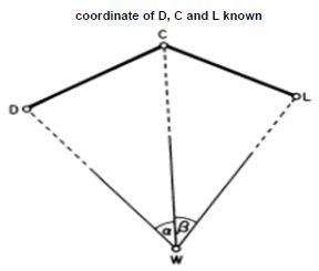
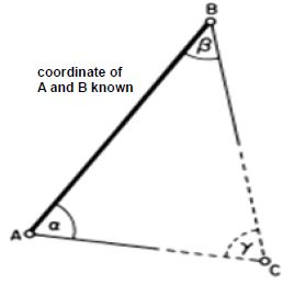
Two techniques commonly employed in extending horizontal control surveys and in setting out are intersection and resection [3-8]. Intersection is a method of locating a point without actually occupying it. In Figure 1, points A and B are stations in a control network already surveyed and, in order to coordinate unknown point C which lies at the intersection of the lines from fixed A and B points, angles α and β are observed. Resection is a method of locating a point by taking angle observations from it to at least three known stations in a network . In Figure 2, in order to coordinate unknown point W can be fixed by observing angles α and β subtended at resection point W by control stations fixed D, C and L points.

fig 1

Figure 1: Intersection

fig 2

Figure 2: Resection

Solution Of Single-Point Adjustment On Clichés

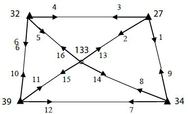
Let’s now see this single point adjustment step by step on a numerical application. In order to make single point adjustment on the clichés, first chart is arranged for all direction measurements. The procedures to be done to fill the chart and clichés are summarized below in Figure 3.

fig 3

Figure 3: Numerical Example

In the triangulation network in the figure, fixed points 27, 32, 34, 39 are given with (Y, X) coordinates. The directions indicated by arrows were observed in the wax and given in the chart. Calculate the adjusted coordinates of the 133 point.

In example;

Number of measurements n=16 (number of observed directions)

Unknown number u=7 (2 coordinates + 5 orientation unknowns)

Redundancy measures f=n-u=9

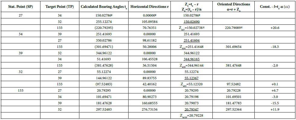
– Firstly, the station point (SP), the point of target (TP), the observed directions (r) columns and the bearing angle (to) columns calculated from the coordinates are filled.

– Approximate coordinates (Yo, Xo) of the point to be estimated are required to calculate the bearing angles about the two point. For this purpose, the point to be estimated from the fixed points (Na: 32 and Nb: 39) two bearing angles.

ϕa=Zo.32 + r32-133=55.12320 + 42.40162=97.52482

ϕb=Zo.39 + r39-133=344.96144 + 36.51504=381.47648

The approximate coordinates of the point (133) to be estimated using the cliché are calculated in the cliché and written in the calculation section of the approximate coordinates in the cliché.

– Two different ways are followed to calculate the approximate value (Zo) of the oriented direction unknowns belonging to the station points. For the calculation of Zo at fixed points, only the arithmetic mean of the (to-r) differences calculated from the directions from the fixed point to the fixed point is taken. At the point to be estimated (adjusted), Zo is made by taking the arithmetic mean of the (to-r) differences to be calculated from all directions. Calculated Zo values are written in the column they belong to in the chart.

– The oriented direction column is calculated and filled as (α=r + Zo) for the directions to the points to be estimated from the fixed points and the direction observations made at the point to be estimated.

– The constant term (-∫) column is filled by computing (-∫=to – α) in units of [cc] for directed directions only.

Point adjustment on the cliché:

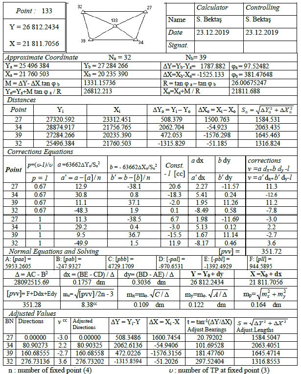
 – Approximate coordinate calculation section of the cliché was filled in while preparing the chart.

– The point numbers and Y, X coordinates of all fixed points are written in the edge calculation section of the cliché. The coordinate differences and lengths of ∆Yo=Yi – Yo and ∆Xo=Xi – Xo from fixed points to the point to be estimated are calculated and written in the relevant columns.

– In the section of establishment of correction equations; First, the fixed point numbers are written in order up to the double-lined line, and for the values in the p (weight) column, they are filled as p=(u-1)/u . Here u: number of target point at fixed point. The weights here are not the weights of the directions, but the p coefficients come to reduce the orientation unknown at fixed points. The direction coefficients a and b are calculated in the form of

a=63662 ∆Yo/So2                 b=-63662 ∆Xo/So2

in [cc/dm] unit by using the values in the edge calculation section of the cliché and written on the lines they belong to. After the double-lined line, the next section is filled. Again, the fixed point numbers and p weight values ​​are written as 1 this time. The pre-filled and shifted values are written under the b columns in the form of

a’=a – [a]/n             b’=b-[b]/n

– ∫ constant terms are taken from the chart and transferred to the cliché.

– In the section of establishment and solution of normal equations; Normal equation coefficients (A, B, C, D, E and F) are calculated from the values in p, a, a’, b, b’, -∫ columns in the upper part of the cliché and written in their places. Unknowns dx, dy are found and adjusted coordinates (Y, X) are calculated by adding them to approximate coordinates. In the upper part of the cliché, using dx, dy values, a.dx, b.dy, a’.dx, b’.dy columns are calculated. Go to the corrections section to the right of the vertical double-lined part of the cliché. In this section, corrections for the direction observations made from fixed points up to the double-lined are calculated from the equation v=a dx + b dy -∫, the corrections for the direction observations made from the point to be estimated point to the next section from the double-lined are from the equation v=a ‘dx + b’ dy -∫. are calculated and written in their places. The weighted sum of squares [pvv] of all corrections is calculated and replaced. As a control, the value of [pvv] is also calculated from the equation [pvv]=F + D dx + E dy. The average error of the unit measure mo, the average error of the coordinates mx, my, the point position error mp are calculated.

– Adjusted values section; The columns of point numbers viewed from the estimated point, observed direction and corrections are filled in (corrections were previously calculated). Corrections are added to the observed directions and adjusted directions are calculated so that the first gaze direction is zero. Using the adjusted coordinates of the point to be estimated, the adjusted bearing, adjusted lengths are calculated and written in the columns they belong to. In order to control the point adjustment made with cliché, the following control operations are carried out in the last calculated adjusted values section (Tables 1 and 2).

Table 1: Chart

tab 1

Table 2: Single Point Adjustments Cliché

tab 2

Control Steps

  1. The two calculated values of [pvv] must be equal within the limit of rounding errors.
  2. The sum of corrections must be zero. This total should not be greater than 0.1cc due to rounding errors.
  3. The angles between adjusted directions and the angles between adjusted bearing should be equal. The difference between the angle values calculated in two different ways should not be greater than 0.1cc.

Warning: The value of estimated point’s coordinates (133) always is exactly correct. But the value of [pvv] is different from its actual value. Because corrections are not calculated for the direction observations made from fixed points in the cliché. Therefore, the calculated values of mo, my, mx, mp are not exactly correct. The actual values that should be are: [pvv]=489.48, mo=7.38 cc, mx=0.096 dm, my=0.107 dm and mp=0.144 dm.

Conclusion

When this problem is solved in the form of mixed resection without performing point adjustment, there are differences smaller than centimetre between the coordinates of the point 133 found by mixed resection and the coordinates found by adjustment. Although this difference is easily negligible for most practical needs, it is necessary to adjustment it in studies that require high precision.

In this study, we will show how the single point adjustment can be done according to the least squares (LSQ) theory on a cliché. Moreover, with today’s advanced calculation tools, all geodetic problems can be adjusted very quickly in accordance with the error theory by means of appropriate software.

References

  1. Allan A, Hollwey J, Maynes J (1968) Practical Field Surveying and Computations, American Elsevier Publishing Co, Inc, New York.
  2. Anderson J, Mikhail E (1998) Surveying: Theory and Practice, 7th edition, WCB/McGraw-Hill, New York.
  3. Bannister A, Raymond S, Baker R (1984) Surveying, 6th edition, Longman Scientific & Technical, Essex, England.
  4. Bektas S (1992) “3D triangulation network” Turkey III. Map of technicians and Services Conference “13-16 April 1992 in Ankara.
  5. Bektaş S (1997) Space Resection, Map and Cadastre Engineering Journal, issue: 83, p.72-79, October 1997, Ankara.
  6. Bektaş S (2016) Practical Geodesy II. Edition, OMU publications, Samsun.
  7. Öztürk,E – Serbetçi M (1989) “Adjustment Calculation Volume II”, KTÜ publications, publication number: 144, page: 310, Trabzon.
  8. Uren J, Price WF (1985) Intersection and Resection. In: Surveying for Engineers. English Language Book Society student editions. Palgrave, London.

Ultrahigh-Pressure and -Temperature Mineral Inclusions in More Crustal Mineralizations: The Role of Supercritical Fluids

DOI: 10.31038/GEMS.2023543

Abstract

Supercritical fluids or melts create an essential connection between the lower mantle and upper crust. Examples demonstrate these interactions.

Examples

Sensitized by the finding of stishovite and coesite as inclusions in the Waldheim granulite in Saxony, the author found, in cooperation with his coauthors [1,2] in evolved granites and related tin-mineralizations, a couple of ultrahigh-pressure and -temperature minerals, like diamond, moissanite, stishovite, coesite, and cristobalite-XI [3]. These minerals are mostly spherical crystals with a very smooth surface. They are, as a rule, minerals entirely out of place. That means these trapped crystals have no equilibrium faces. Thomas and coauthors have interpreted these in growing crystals trapped phases as transported via fast-rising supercritical melt or fluid from the Earth’s mantle region into the crust, here granites and related mineralizations [1-3]. The crystallization velocity of the host must be very high to prevent equilibrium forms of the trapped spheres.

A careful study of the beryl-quartz veins related to the cassiterite mineralization revealed that the ordinarily tetragonal cassiterite contains a high portion of orthorhombic cassiterite [4,5]. There are isometric crystals of orthorhombic cassiterite in muscovite, water-pure topaz, or more significant remnants (cores) of orthorhombic cassiterite in the tetragonal cassiterite crystals. Also, this topaz, as well as the orthorhombic cassiterite, are of high-pressure and high-temperature origin. Supported is this statement by sub-spherical inclusion in the OH-rich topaz composed of carbonic material (graphite, moissanite, and nanodiamond), as well as spherical kumdykolite crystals [NaAlSi3O8] with carbonic material. Kumdykolite is the orthorhombic polymorph of albite formed at high temperatures followed by rapid cooling [6].

The next surprise was the finding of moissanite whiskers with nanodiamonds grown in beryl and quartz in a small hydrothermal beryl-quartz vein in the Sauberg mine of the Ehrenfriedersdorf tin district of the Erzgebirge, Germany [7]. Here the crystallization of the whiskers happens directly at the place of vein crystallization at about 720°C and pressures ≤2 kbar. Thomas [4,5] tried an explanation for this remarkable beryl-quartz-moissanite-nanodiamond paragenesis by a natural supercritical vapor-liquid-solid (VLS) mechanism and a low-pressure heteroepitaxial chemical vapor deposition process (CVD). In each case, the crystallization of moissanite at such low PT conditions is unusual and should be a challenge for further experimental studies. Supercritical phases like spherical beryl-II-moissanite intergrowing in these parageneses’ quartz show the supercritical phases’ participation in this crystallization process.

Another important point for further sophisticated studies in relationship to the unusual mineral inclusions is the chemical and physical character of the supercritical phases. Thomas [8] shows a new type of fluid inclusion that probably transported stishovite (and other mineral phases) from the lower mantle to the crust. Methane (CH4) and water are miscible at ultrahigh pressure and temperature. The transport properties of such fluid and the solubility for different elements are of particular interest because, in the crustal region, we observe a lot of extreme element enrichments in relationship to the melt-water solvus [1,2,9,10]. If we look at the whole story, we see a continuous evolution of the system of thought. Conventional physicochemical processes cannot explain this Gaussian or Lorentzian element enrichment in melt inclusions related to such solvus curves. Further studies are necessary to illuminate these complex processes in more detail.

References

  1. Thomas R, Davidson P, Rericha A, Recknagel U (2022) Discovery of stishovite in the prismatine-bearing granulite from Waldheim, Germany: A possible role of supercritical fluids of ultrahigh-pressure origin. Geosciences 12 : 1-13.
  2. Thomas R, Davidson P, Rericha A, Voznyak DK (2022) Water-Rich Melt Inclusion as “frozen” samples of the supercritical state in granites and pegmatites reveal extreme element enrichment resulting under non-equilibrium conditions. Mineralogical Journal (Ukraine), 44 : 3-15.
  3. Thomas R, Davidson P, Rericha A, Recknagel U (2023) Ultrahigh-pressure mineral inclusions in a crustal granite: Evidence for a novel transcrustal transport mechanism. Geosciences 13 : 1-13.
  4. Thomas R (2023a) Unusual cassiterite mineralization, related to the Variscan tin-mineralization of the Ehrenfriedersdorf deposit, Germany. Aspects in Mining & Mineral Science 11 : 1233-1236.
  5. Thomas R, Recknagel U, Rericha A (2023a) A moissanite-diamond-graphite paragenesis in a small beryl-quartz vein related to the Variscan tin-mineralization of the Ehrenfriedersdorf deposit, Germany (under review).
  6. Hwang SLS, Chu HTY, Sobolev NV (2010) Kumdykolite, an orthorhombic polymorph of albite, from the Kokchetav ultrahigh-pressure massif, Kazakhstan. European Journal of Mineralogy 21 : 1325-1334.
  7. Thomas R (2023b) Growth of SiC whiskers in beryl by a natural supercritical VLS process. Aspects in Mining & Mineral Science 11 : 1292-1297.
  8. Thomas R (2023c) A new fluid inclusion type in the hydrothermal-grown beryl. Geology, Earth and Marine Sciences (GEMS) accepted.
  9. Thomas R, Webster JD, Heinrich W (1999) Melt inclusions in pegmatite quartz: Complete miscibility between silicate melts and hydrous fluids. ECROFI Abstracts, Terra Nostra 99/6, 305-307.
  10. Thomas R, Davidson P, Appel, K. (2019) The enhanced element enrichment in the supercritical states of granite-pegmatite systems. Acta Geochim. 38 : 335-349.